USE OF HAMULAR NOTCHES AND INCISIVE PAPILLA AS ...
68
University of Khartoum Graduate collage Postgraduate Medical Studies Board Faculty of Dentistry USE OF HAMULAR NOTCHES AND INCISIVE PAPILLA AS GUIDES FOR SELECTION OF MAXILLARY ANTERIOR TEETH WIDTH A thesis submitted in partial fulfillment for requirements of Master Degree in Removable Prosthodontics (December 2007) By: Omelnassr Mahamed Elbsheir Abd Allah B.D.S (U of K) Supervisor : Dr. Nadia Khalifa B.D.S, MSc (UK)
-
Upload
khangminh22 -
Category
Documents
-
view
3 -
download
0
Transcript of USE OF HAMULAR NOTCHES AND INCISIVE PAPILLA AS ...
University of Khartoum
Graduate collage
Postgraduate Medical Studies Board
Faculty of Dentistry
USE OF HAMULAR NOTCHES AND INCISIVE PAPILLA AS
GUIDES FOR SELECTION OF
MAXILLARY ANTERIOR
TEETH WIDTH
A thesis submitted in partial fulfillment for requirements of Master Degree in Removable
Prosthodontics (December 2007)
By:
Omelnassr Mahamed Elbsheir Abd Allah B.D.S (U of K)
Supervisor :
Dr. Nadia Khalifa B.D.S, MSc (UK)

DedicationDedicationDedicationDedication
To my family….
Who always support
me.
I

Acknowledgement
Great thank' to my very kind and helpful
supervisor Dr. Nadia Khalifa who always helped
and supported me. Also I want to thank Dr.
Magdi Wadae (the Head of (Prosthodotics
Department) And special thank' to my colleague
dr. Ahmed Khalid (Department of Orthodontics-
University of Khartoum) who help me with th
statistical analysis. Thanks to all dental students
–University of Khartoum among whom this
research had been done.
Last but not least thank' to my colleagues in
the (Prosthodontics department and
Prosthodontics Laboratory) for their help.
II
Abstract
This study was conducted to evaluate the use of specific anatomical
landmarks and their relationships, in determination of the width of
maxillary anterior teeth. Eighty maxillary dentate casts from dental
students and postgraduate students from University of Khartoum were
used in the measurements. The age of the study sample ranged from 25-30
years and comprised of 43 females and 37 males. Distances from the center
of the right hamular notch to the center of the left hamular notch (HD),
and from the left hamular notch to the incisive papilla (FBI-IP), as well as
from the right hamular notch to the incisive papilla (1+1-IP) were
measured using an electronic caliper gauge. The widths of maxillary
anterior teeth were measured from distal end of right canine to the distal
end of left canine, using a flexible ruler. The ( HN-IP-HN) distance, when
divided by 3 (the predicted value), had a mean of 57.7mm and standard
deviation of 2.0 among males, while among females, the mean of the
predicted value was 52.9mm with standard deviation of 2.3. The mean of
the actual value (the width of maxillary anterior teeth) was 56.7mm with
standard deviation of 1.8 among males, while between females the mean
of the actual value was 52.9mm with standard deviation 2.2. The P- Value
III

of the correlation between the predicted value and actual value
was0.00, and a correlation between the predicted value and the
actual value was found.
The P-Value of the difference between the actual and predicted
values was 0.74, and there was no or little difference between the
actual and predicted values. The predicted and actual values were
higher in males than in females. The results of this study showed
that the measurement of I-N-IP-I-BI divided by 3, is
approximately equal to the width of maxillary anterior teeth from
the distal end of the right canine to the distal end of the left canine,
indicating that these measurements can be of value when setting
teeth in edentulous patients.
IV

ـحــثـــخــص البـمل
أعدت ھذه الدراسة لتقييم إستعمال بعض المعالم التشريحية وعIقتھا بحجم
اcسنان اcمامية بالفك اcعلى .
أخذت ثمانين طبعة سنية من طIب البكالوريوس وطIب الدراسات العليا بكلية طب
سنة , وتتكون ھذه العينة من 25-30جامعة الخرطوم تتراوح أعمارھم بين -اcسنان
ذكر.37أنثى و 43
قيست المسافة بين الحديبة الفكية في الجزء اcيمن والحديبة الفكية في الجزء اcيسر
ين كل من الحديبتين الفكيتين وبين الحليمة القاطعة بواسطة مقياس إلكتروني والمسافة ب
(برجل), كما قيس عرض اcسنان اcمامية بواسطة مسطرة مرنة.
وجد أن البعد بين الحديبتين الفكيتين والبعد بينھما وبين الحليمة القاطعة مقسوماً على
بين 2ف المعياري لھذا القياس ھوملم , ا�نحرا 57.7يكون الناتج المتوسط 3الرقم
, 2.3ملم بإنحراف معياري 52.9الذكور, أما بين ا�ناث فوجد ھذا القياس بمتوسط
ملم 5.6بينما وجد متوسط قياس عرض اcسنان اcمامية في الفك اcعلى يساوي
للعIقة بين القيمتين التقديرية ) P.value(قيمة 2.2بإنحراف معياري يساوي
(P.value, مما يعني وجود عIقة قوية بين القيمتين. و قيمة ال 0.00والحقيقة تساوي
مما يعني أنه قليل اcھمية أي أنه ليس ھناك 0.74) ل�ختIف بين القيمتين تساوي
إختIف واضح بين القيمتين, أي أنه يمكن إستعمال الحديبة الفكية والحليمة القاطعة
ية في الفك اcعلى للمرضى الذين يعانون من الفقدان الكامل بحساب اcسنان اcمام
ل�سنان.
V
List of Contents
Page no Title Index
I Dedication
II Acknowledgement
111 Abstract (English)
V Abstract (Arabic)
VI List of contents
X List of tables
XI List of Photographs
XI List of Appendices
Chapter One
1 Introduction 1-1
3 Justification 1-2
4 Literature review 1-3
4 Photographs and Radiographs 1-3-1 7 Interalar width 1-3-2
8 Interpupillary distance
1-3-3
9 Intercanthal distance 1-3-4

9 Bizygomatic width 1-3-5
10 Intercondylar width 1-3-6
1-3-7 Intercanine distance 10
1-3 -8 Anterior teeth width 11
1-3-9 Hamular distance 15
1-3-10 Incisive papilla 19
1-3-11 Effect of Bone resorption on anatomical land marks
20
1-4 Obj ctives 22
1-4-1 General objectives 22
1-4-2 Specific objectives 22
Chapter Two
2 Materials and methods
23
2-1 Study design 23
2-2 Study population 23
2-3 Including criteria 23
2-4 Excluding criteria 23
2-5 Study variables 24
2-6 Sampling 24
2-7 Equipments and materials
25
2-8 Impression taking 26
2-9 Measurements 26

VIII
2-10 Data collection 27
2-11 Data processing 28
2-12 Data analysis 28
Chapter Three: Results
3 Results 29
3-1 Gender distribution 29
3-2
Age distribution 31
3-3 Measurement of HN-1P-HN/3 33
3-4
Measurements of maxillary anterior teeth 35
3-5 Predicted and actual value correlations 37
3-6 Predicted and actual values differences 38
3-7 Group statistics 39
Chapter Four: Discussion and Conclusion
4-1 Discussion 40
4-2 Conclusion 44
4-4 Recommendations 45

List of Tables
Index Title Page
no
1 Gender Distribution 29
2 Age Distribution 31
3 Measurements of HN-IP-HN/3 33
4 Measurements of maxillary anterior teeth width
35
5 Paired Sample Correlations 37
6 Predicted and actual Values differences
38
7 Group Statistics 39
X

List of Photographs
Index Title Page no
1 Measurement of hamular distance with digital caliper 58
2 HN-IP-HN triangle on the cast 59
3 Standardization of cast base using surveyor 60
Appendices
XI
index Title
1 References
2 Data collection form

Chapter One
Introduction ,
Justification,
Literature review and
Objectives

I-l. lntroduction
One of the objectives of complete dentures is to satisfy esthetics. The
success or failure of a denture frequently depends on how successfully the
operator has fulfilled the patient's esthetic requirements. The patient
who is unhappy with the facial contours or with arrangement and
appearance of the teeth may not cooperate during the early insertion
periods.(1) .
A denture usually is received as esthetic when the teeth and
the bases are in harmony with the facial musculature as well as the
size of the head.(2). The goals of prosthodontic treatment for
edentulous patients are to construct complete dentures that function
well, allow patients to speak normally, are esthetically pleasing and
will not abuse the tissues over the residual ridge.(3) .
The most important teeth from the point of view of appearance
are the maxillary anterior teeth especially the central incisors which
are the most exposed during conversation when an individual smiles
or laughs.(4)
Photographs, radiographs, study casts and extracted teeth are pre
extraction records that can be utilized as guides for selection of
artificial teeth(5) . Various guide lines have been suggested for
determining the width of the maxillary anterior teeth when pre
extraction records are not available, such as bizygomatic width,
intercommissural width, Interalar width and canthus distance.
Many intraoral anatomical landmarks have fixed position that is
not altered after missing the teeth, thus, they can be used as reliable
guides to select the maxillary anterior teeth e.g. incisive papilla,
residual ridges, the comers of the mouth, and the maxillomandibular
relations .
1

In this study specific anatomical land marks and their
relationships obtained from dental casts of dentate patients had been
examined.
Measurement of the distance from left hamular notch to the right
hamular notch (hamular distance) was found in other studies to be
equal to the width of the six maxillary anterior teeth (from distal of
the left canine to the distal of the right canine), and the right hamular
notch-incisive papilla- left hamular (HN-IP-HN) notch distance
divided by factor 3 in previous studies found to be equal to the width
of the six maxillary n anterior teeth. In this way the hamular notches
and incisive papilla can be used as guides for measurement of the
width of the six anterior teeth and according to the maxillary anterior
teeth other teeth can be selected.
2

3
1-2.Justification
Several methods have been proposed for determining the
width of the maxillary anterior teeth. When there is a pre-
extraction record, the width of the anterior teeth can be easily
obtained, while in the absence of pre extraction records it is
difficult to determine the width of the anterior teeth. Use of
facial measurements and anatomical land marks approximately
give the width of the anterior teeth. Use of facial
measurements and anatomical land marks approximately give
the width of the anterior teeth. It is of value to use hamular
distance and HN-IP-HN distance to measure the width of the
six maxillary anterior teeth
as and they are reliable anatomical land marks and easy to be
measured.
In this country there is no study that had been done to use
the hamular distance or hamular notch- incisive papilla-
hamular notch distance as guides for selection of the
maxillary anterior teeth.

1-3.Literature Review
Teeth size and form selection can be obtained from a variety of
methods. The easiest is from prior extraction record. Other
methods include the Trubyte Tooth Indicator, anatomical
measurements, marking
the canine eminence on the casts, or simply a subjective evaluation of
face size and tooth form by the clinician.(6)
Douglas A.Hock (1992) stated that different qualitative guidelines are
cited, the best method for the selection of the maxillary
anterior teeth is the use of pre-extraction records or old
photographs.
Numerous qualitative and quantitative guidelines are
cited in the literature for the proper placement of the maxillary
anterior teeth.(7) .
1-3-1.Photographs and radiographs:
The use of photographs is to be strongly recommended. Particularly
useful are those of a patient that were taken when the subjects was
dentate or wore dentures which were admired by the patient.
4
The photographs should realistically show head-on facial views of the
patient smiling; failure to do this may not reveal any sign of the anterior
teeth. Such views should enable the clinician to see and to measure
carefully the ratio of the patient's horizontal intercanine distance, and
relate that to the interpupillary distance in the photograph. In the clinic,
the clinician may then measure the patient's interpupillary distance and it
should be possible establish the horizontal width of the upper six
anterior teeth.(8) .
Wehner et al (1967) stated that a photograph of a patient in which the
natural anterior teeth are visible of great help in the selection of the size
of the artificial teeth. Radiographs can be meaningful guides for the
selection anterior teeth, in the absence of other records they can be useful
in selecting crown form, width, and length.(9) .
Bindra et al (200 1) examined a method described by Wehner et al for
calculating the width of a missing central incisor using pre-
extraction photographs; three photographic views were
obtained for each of dentate subjects: full face, oblique, and
reduced-size full face. The width of the maxillary right central
incisor (MRI) was calculated using a formula. The difference
between the actual width and calculated width of MRI was
determined for each subject.
5

The median difference and inter quartile range were
determined because the data were skewed. They found that, the
width of MRI calculated using the larger full-face view was
typically smaller than the actual width, with a median
difference of -0.18 mm. The interquartile range of the
difference was from -0.42 to 0.05 mm. For both the oblique and
reduced-size views, the calculated width was typically larger,
with a median difference of 1.19 mm with an interquartile
range from 0.82 to 1.76 mm and a median difference of 0.84
mm with an interquartile range from 0.59 to 1.41 mm, res
ectively. The technique described by Wehner et al is-of
proven/value in calculating the width of a central incisor when
the only available evidence is a pre extraction photograph.
However, it is of value only when the photograph is a full1 face
portrait of sufficient size.(10) .
Gomes VL et al (2M) considerÅfacial analysis with digital
photography as h' practical and efficient application to verify
the relation between the combined mesiodistal width of the six
maxillary anterior teeth and the facial segments( i.e. the width
of the eyes, the inner canthal distance (ICD), the interpupillary
distance (IPD), the interalar width, and the intercommissural
width (IC)). Standardized digital images of 81 dentate
Brazilian subjects were used to measure both facial and oral
segments when viewed from the frontal aspect through an
image processing program.
6

To measure the distance between the upper canines on a curve,
accurate cast were made from the upper right first premolar to
the upper left first premolar. The results showed a significant
correlation between all facial elements and the combined
mesiodistal width of the six teeth, when observed from the
frontal aspect. The ICD,IPD, and IC showed the highest
probability of being correlated to the mesiodistal width of the
teeth.(11)
1-3-2.1nteralar width:
It is determined by measuring the external width of the nose
at the widest point. Different views have been reported on the
significance of the interalar width in the selection of the
anterior teeth. Latta et al observed that the width varied widely
even when the population was separated into groups by sex and
or race .(12)
Smith (1965) reported a low relationship between radiographic
measurement of the interalar width of the nose and the distance
between the maxillary canine tips.(13) . Lee(1962) .(14)and Whener et al
(1967 ) .(15) suggested extending parallel lines from the lateral surface
of the ala of the nose on the labial surface of the maxillary occlusion
rim to estimate the intercanine cusp tip
7

Marokouf and Richie demonstrated some relationship between the
nasal width and intercanine distance which suggested its use to
establish the width of the anterior teeth.(16)
Scandrett (1982) reported a significant correlation between the
interalar width and the width of the maxillary anterior teeth.(17) .
Fabiana M V and Sergio S N (2006) conducted a study to evaluate
the use of the nasal width as a guide for the selection of proper width
maxillary anterior denture teeth in four racial groups of the Brazilian
population. One hundred and sixty subjects (Whites, mixed, Blacks,
and Asians) were selected. Using a sliding caliper, the nasal width
and the intercanine distance were measured. A prediction was made
of the percentage of subjects of the White, Mulatto, Black, and Asian
populations' .The four racial groups showed a weak correlation
between the intercanine distance and the nasal width. The correlation
found between the intercanine distance and the nasal width was not
high enough to be used as a predictive factor. The relationship
between natural tooth width and artificial tooth width as predicted by
the nasal width showed that the nasal width method is not accurate
for all the studied .(18)
8
1-3-3.1nterpupillary distance
It is measured from the mid pupil to the other mid pupil.
Alsheikh et al conducted study and found a significant correlation between
1interpupillary width and the width of the anterior teeth of the entire
sample but when the samples was divided in to gender, correlation found
only in females.(19) Cesario et al (1984) reported that interpupillary distance
could be used reliably in selecting maxillary anterior teeth width.(20) .
Latt et al (1991) found no correlation between interpupillary
distance and the width of the mouth for the whole population.(21)
1-3-4 .lntercanthal distance: It is the distance between the median angles of palpaberal.
AlWazzan Khalid.A.(2002) found a significant relationship between
Intercanthal distance and the measurements of the four maxillary anterior
teeth. It is found that biometric ratio of 1:0.267 and 1:0.426 could be used
to estimate the central incisor width and the width of the combined six
anterior teeth respectively.(22)
Abdullah (2002) described a significant relationship between
intercanthal distance and maxillary central incisor mesiodistal width (23)
Another method, used by the authors, is to ask the patient to smile
and to extend a line from the inner canthus of the eye via the lateral
border of the alar cartilage and extend that onto the upper rim. This
may be done with a ruler or by the use of dental floss This equates,
in a high proportion of cases, to the position of the tip ofthe upper
canine teeth. .(24)
9
I -3-5 .Bizygomatic width: Scandrett,F.R et al (1982) showed that, the ratio of
bizygomatic width to the maxillary central incisor width is
16:1.(25)
Hasanreisoglu.et al (2005) stated Proportional relationships
between the bizygomatic width and the width of the central
incisor, and the intercanine distance and the interalar width in
women were observed.( 26)
.
I -3-6.1ntercondylar width:
Keshaved et al designed a study to investigate the relationship
between intercondylar width and interdental width of the upper and
lower canines and first molar, to aid in denture teeth positioning. The
results showed that a strong correlation existed between intercondylar
width and interdental measurements.(27) .
I -3-7 .lntercanine distance:
To determine the position of the canine teeth. Earlier reference
has been made to the use of pre-extraction records. Where these are
not present, some authorities advocate using the position of the
corners of the mouth, at rest. Another method, used by the authors, is
to ask the patient to smile and to extend a line from the inner
10

canthus of the eye via the lateral border of the alar cartilage and
extend that onto the upper rim. This may be done with a ruler or by
the use of dental floss this equates, in a high proportion of cases, to
the position of the tip of the upper canine teeth.(24)
Keng SB (1995) conducted study on Chinese subjects to
determine if there was any relationship between the interalar
nose width and intercanine distance. The results show! that
men have wider noses and slightly greater intercanine distance
compared to women. These dimensions have been shown to
be generally greater than similar studies conducted on
Caucasian subjects. However there was no demonstrable
correlation between interalar width of nose and intercanine
distance.
Dharap A.S. and Tanuseputro H (1997) compared interalar
width and intercanine distance in Malay males and females.
The mean interalar width of the nose in male subjects was
larger than in female subjects. The mean maxillary intercanine
distance in male subjects was 36.7 ± 2.6 mm (range 30-42 mm)
and in female subjects 36.2 ± 2.3 mm (range 30-42 mm) .(29)
11
H.-J. AHN et al (2002) examined correlation between the width of
the maxillary central incisor (WMCI), the intercanine distance (ICD), the

o facial width (FW), and the interalar nasal width (IAW) in Koreans. The
ratio of FW/WMCI, FW/ICD, and IAW/ICD was 17.4, 3.7, and 1.0
respectively.
There were significant correlations between WMCI, ICD, FW, and
IAW in Pearson's correlation analysis (p<O.01). The FW and IAW could
be very reliable guides for the selection of the maxillary o anterior
artificial teeth.(30)
1-3-8.Anterior teeth width:
Lundström (1956) studied the relationship between the mandibular and the
maxillary anterior sum and named it the anterior inde.(31)
A definite relationship between the form of the maxillary arch and the alignment form
of the upper anterior teeth.(32).
The combined width of the six maxillary anterior teeth has been slightly less than one
third of the bizygomatic breadth of the face.(33) .
Lavelle (1973) noted fewer gender differences in the primary dentition than in the
permanent dentition.(34) .
Male teeth generally recognized to be larger than female teeth In both the primary and
permanent dentitions, the upper canines and upper central incisors show/the greatest
gender differences, whereas the upper lateral incisor and lower central incisor are the
most homogenous (34). Offman et aI suggested that multiplying the interalar width by
a factor of 1.31 can help in estimating the combined width of the six anterior maxillary
teeth.(35) .
12
Richardson and Malhotra (1975) reported no differences in upper and lower anterior tooth
size proportions, indicating that there is a constant 77% ratio for both genders.

More recently, other studies have reported significant differences in tooth size
between males and females but no evidence of a significant difference in upper to
lower anterior tooth size proportion.( 36) .
Richardson E R (1975) conducted study of mesio distal crown dimension on teeth
of 162 Afro American, equally divided between males and females.
The teeth of males were larger than those of females for each type of tooth in both
arches, although they exhibited the a similar pattern of tooth size. The ratio of the
sum of the widths of canines and incisors of the mandibular demtition to those of the
maxillary dentition was 77 per cent. Also, the ratio of the mandibular incisors to the
maxillary incisors was 71 per cent in both sexes.(37).
Marvin (1992) compared mesiodistal widths of maxillary anterior natural
teeth with the widths of the most commonly used artificial denture teeth 40%
of the women and 67% of men had anterior teeth that measured 55 mm or wider
In contrast, the mean width of the most widely distributed artificial maxillary
denture teeth (Bioblend) is 50. I mm, with a range of 44 to 58 mm Of the 42
molds available, only 5 have a width of 55 mm Natural teeth were found to be
larger than artificial teeth. These findings suggest that one reason for the
inappropriate selection of comparatively small maxillary denture teeth is the
lack of physiologically sized tooth molds
13
LavereAM et al (1994) compared the mesiodistal widths of the six maxillary
anterior teeth with the widths of denture teeth from six different denture tooth
manufacturers, Cast of dental students were measured from the distal aspect of
each canine across the facial surfaces of the six anterior teeth with a flexible
plastic millimeter rule Denture teeth from six manufacturers were
compared The results indicated that denture teeth are predominantly smaller
and natural teeth are larger.
Sellen P. N. et al (1999) investigated the variability in choice of dental staff
to select teeth appropriate to the age and sex of the individual with the aid
of a series of three-dimensional guides! Four three-dimensional guides were
produced for use in the study. Fifty dentists were asked to complete a
questionnaire designed to assess the variability in selection of anterior teeth
appropriate for the age and sex of an individual. The study concluded that
there was little consistency in the selection of the shade, mould and
arrangement of anterior teeth appropriate for the age and sex of the
individual by qualified dental staff The development and implementation of
an aesthetic perform to guide dental staff, dental undergraduates and patients
through the process of choosing tooth mould, shade and arrangement based
on age and sex may be helpful. Eustaquio Arognio and Mæ:eelo Souki
(2003) investigated the correlation between anterior tooth size discrepancies
and Anglers Class I, Il, and Ill malocclusions, as well as their prevalence in
the Brazilian population The mesiodistal width of six anterior teeth in 300
patients, who were selected randomly, was assessed The important
conclusions of this study were that individuals with Angle Class I and Class
Ill malocclusions showed significantly greater prevalence of tooth size
discrepancies than individuals with Class Il malocclusions; and mean
anterior tooth size discrepancy for Angle Class Ill subjects was significantly
greater than for Class I and Class Il subjects.(40).
14
Hasan Reisoglu Uof uk et al (1995) analyzed the clinical crown dimensions of
maxillary anterior teeth to determine whether consistent relationships exist between
tooth width and several facial measurements.(41). The dimensions of the anterior

teeth, the occurrence of the golden ratio, the difference between the actual and
perceived sizes, and the relationship between the anterior teeth and several facial
measurements by gender were analyzed using the information obtained from both
the computer images and the casts ,the dimensions of the central incisors and
canines varied by gender. They found that existence of the so-called "golden
proportion" for the maxillary anterior teeth as a whole was not found .The maxillary
central incisor and canine dimensions of men were greater than those of women in
the Turkish population studied.(42)., with the canines the greatest gender variation
Neither a golden proportion nor any other recurrent proportion for all anterior teeth
was determined. V Pioneer investigations on tooth sizes were conducted by lack in
1902.(43) and Neff ( in 1949) .(44). These studies were followed by the classic work
of Bolton.(45,46)., who quantified the maxillary-to-mandibular tooth size relationship
and provided the accepted normative data, Bolton selected 55 cases with optimal
occlusions and compared the sums of the mesiodistal widths of the maxillary and
mandibular teeth, including the first molars An overall ratio of 91. 3 was obtained,
with a standard deviation of 1. 91 He also calculated that the ratio for the anterior
teeth from canine to canine was 77. 2, with a standard deviation of 1. 65 This means
that the maxillary anterior teeth width was larger than the mandibular anterior teeth
width.
Gillen et , in their study to determine the average dimension of the six maxillary
anterior teeth in a targeted population, concluded the golden proportion was not
found to correlate with any of the calculated ratios. However; Al-Wazan suggested
the use of intercanthal distance as a preliminary method for determining the width
of the maxillary anterior teeth.(47).
15
Mohammed AJi-E.ayyad (2006) investigated the existence of the golden
proportion, the recurring esthetic dental (RED) proportion, and the golden

percentage between the widths of the maxillary anterior teeth in individuals with
natural dentition„ The values of the RED proportion were not constant, and the
farther the one moves distally from the midline the higher the values Furthermore,
the results revealed the golden percentage was rather constant in terms of relative
tooth width, Both the golden proportion and the RED proportion are unsuitable
methods to relate the successive widths of the maxillary anterior teeth However; the
golden percentage theory seems to be applicable to relate the successive widths of
the maxillary anterior teeth if percentages are adjusted taking into consideration the
ethnicity of the population(47)
I -3-9Hamular distance:
It is a distance between right and left Hamular notches Hamular notch is the
palpable notch formed by the junction of the maxilla and the pterygoid hamulus of
sphenoid bone
Petricevie Nikolå et al (2005) .(48) conducted study to determine the relationship between
the width of frontal maxillary teeth and the width of the hard palate and also to determine
the possibility of reconstructing maxillary frontal teeth dimensions, based on hard palate
dimensions. Teeth and hard palate dimensions were measured on maxillary casts of fully
dentate individuals of Angle class I occlusal relationship The maxillary central incisor
is the widest among the frontal maxillary teeth and canines are wider than second
incisors.
The width of the maxillary frontal teeth arch, measured with a flexible ruler is 52. 05
mm, hamular distance 47.1 mm. distal maxillary width 46.1 mm, sum of the widths of
all maxillary frontal teeth 46. 04 mm, frontal maxillary width 35. 8 mm, and finally
16
the width between canine cusp tips is 34.19 mm Based on the results of this study, the
sum of all maxillary frontal teeth widths is equivalent to hamular distance dimension, as

well as distal maxillary width, as there were no statistically significant differences
between them.
After extraction of all teeth, distal maxillary width is lost, which is not possible to
reconstruct because of the individual rate of alveolar bone resorption. On the other
hand, hamular distance remains the same dimension during the lifetime, because it
is not determined by teeth position but by anatomical structures, Therefore, the
hamular distance dimension is a suitable reference for determination of the
dimension of the sum of all maxillary frontal teeth widths.(49) .
Byron A Davis (2005) suggested technique using anatomic landmarks for
determining the size of the maxillary anterior teeth. The purpose of this study is to
determine the accuracy of using 3 anatomical areas of the oral cavity the" right
hamular notch-incisive papilla- left hamular notch (HN-IP-HN) measurement" to
calculate the width from the distal surfåce of the canine to the distal surface of the
opposite canine.
Measurements were made on stone casts of post-orthodontic patients to determine
if the anatomical measurements correlated to the actual tooth measurements The
prosthodontic patient measurements were predicted using the mg-IPHN
measurement and dividing by three, giving width of the six maxillary anterior
teeth.(50) .
17
1-3-10.lncisive papilla:

It is the elevation of soft tissue covering the foramen of the incisive or
nasoplatine canal.(51) . It is situated on the median line of the palate, and in dentate
it is just back to the central incisors„ On edentulous ridge where resorption has
occurred, it is located distally, or on the center of the ridge
Harlod Ortman has shown that most of incisive papillae are pear shaped with their
slender ends pointing at the anterior reference point between the maxillary central
incisors.(52)
Harper was the first to assume that the incisive papilla is a stable anatomical landmark
The significance of the relationship between the maxillary anterior teeth and the incisive
papilla in prosthetic dentistry is that it is one of biometric guides used for the design of
complete dentures.(53) .
Walt DM and Likeman PR stated that, the incisive papilla and the palatal gingival
margin works as landmarks för estimating pre-extraction dimensions of the ridge
After teeth are extracted resorption is most rapid in the labial and buccal bone It can
be used as guide to set 10-12mm anterior to it.(54) .
A study by F M Varjao (2004) conducted that the incisive papilla was not a very
accurate guide föl' selection of width of anterior teeth but could be used to establish
the initial selection of artificial teeth size.(55) .
18
1-3-11 .Effect of Bone resorption on anatomical land marks:

Resorption of residual ridge is a common occurrence after the extraction of teeth
However the total amount of bone loss and the rate of resorption varied among
different patients In addition for a given patient at different times. Doglass Atwood
discussed the factors influencing the rate of residual ridge resorption and divided
them into anatomic, metabolic, functional and prosthetic factors.(56) . Atwood and
Coy found that the mean ratio of anterior maxillary residual ridge resorption to
anterior mandibular ridge resorption was1:4.(57) . Lam studied the resorption of
anterior maxillary tooth sockets the resorption was rapid in the first fifth months
post extraction and there was no further change between fifth and twelfth post-
extraction months.(58).
Atkinson and Johnson showed no change in the posterior part of the vault of the
palate over 3 years of examination to a serial of cephalometric radiographs.(59).
Walt and Likeman studied the pattern of alveolar resorption and found that
anterior and superior movement of the incisive papilla must be considered when the
papilla is used as a guide for anterior teeth positioning. To compensate for this, they
suggested the use of the posterior border of the papilla as the reference They claimed
that the anterior portion may be affected or damaged during extraction of maxillary
anterior teeth or because of the resorption that take place following loss of teeth.(60)
. The average post extraction change appeared to be slightly less in males than in
females The atrophy of residual ridges affected the shape of the palate and its
contour in a given plane widened as the atrophy of the ridges progressed The area
of the palate affected by change extended towards the center of the palate until only
a small median area remain unchanged.(61) .
20
1-4.0biectives

1-4-1.General objectives:
To study the usefulness of the hamular notches and incisive papilla as biometric
guides for measurement of the artificial maxillary anterior teeth in a sample of
Sudanese population
1-4-2.Specific objectives:
1. To measure the width of:
-Maxillary anterior teeth (from distal of right canine to the distal of left canine) -
Hamular distance
- HN-IP-IIN distance
2. To compare the HD to the width of six maxillary anterior teeth
3. To compare HN-IP-HN distance to the width of six maxillary anterior teeth
4. To correlate these measurements to certain parameters (age, gender)
5.To compare results with other studies
21

Chapter Two
Material and Methods
2.Materials and methods
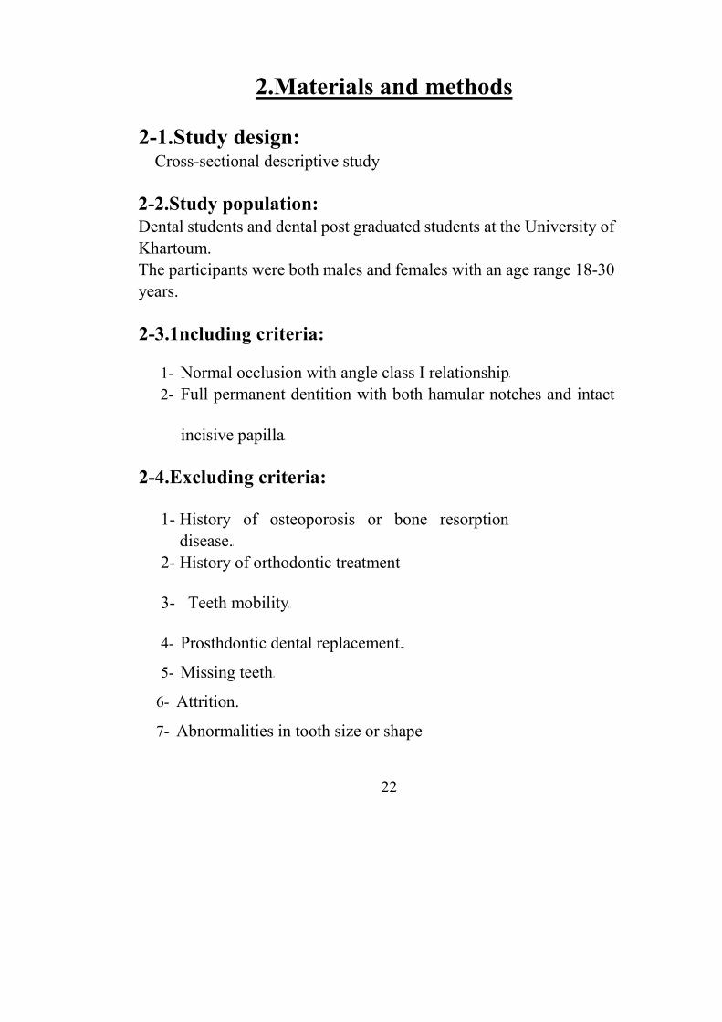
2-1.Study design: Cross-sectional descriptive study
2-2.Study population: Dental students and dental post graduated students at the University of Khartoum. The participants were both males and females with an age range 18-30 years.
2-3.1ncluding criteria:
1- Normal occlusion with angle class I relationship 2- Full permanent dentition with both hamular notches and intact
incisive papilla
2-4.Excluding criteria:
1- History of osteoporosis or bone resorption disease.
2- History of orthodontic treatment
3- Teeth mobility
4- Prosthdontic dental replacement.
5- Missing teeth
6- Attrition.
7- Abnormalities in tooth size or shape
22

8-History of surgical treatment involving the area of hamular notches or
incisive papilla
2-5.Study variables:
Variables in this research were defined as following: a- Universal variables: name, age, gender; and index of the study
cast. b- Dependent variables: hamular distance, hamular notch-incisive
papilla distance, and maxillary anterior teeth width. c- Independent variables: the intercanine distance
2-6.Sampling: The sample was representative The method was systematic random selection. The sample size with help from previous study( 62) found to be 80
2-7.Equipments and materials:
• Perforated Stock trays (maxillary) • Rubber bowls
• Stainless steel spatula
• Modeling wax • Alginate irreversible hydrocolloid impression material
(Zhermack Povigo-ltaly) • Dental stone • Plaster of paris. • Wax knives • Dental mirrors and Periodontal probes
23

• Vibrating table • Flat long table • Electronic digital caliper (Narex-Czechoslovakia) • White papers • Blue pens. • Indelible pencil. • A flexible millimeter ruler
2-8.1mpression taking:
Maxillary perforated stock trays were modified using modeling wax in the labial, buccal flanges , palatal area and posterior end of the trays. According to manufacture instructions, a measured amount of alginate was mixed with measured amount of water in a rubber bowl using stainless steel spatula. The impression material had been loaded on to the stock tray and seated in the patient mouth while the patient was seated in an upright position on the dental chair, with the head being parallel to the floor . The impression was removed after setting had been completed and rinsed with water to remove excess of saliva The impression then was poured using dental stone The cast was removed after setting and a base was made using plaster of paris.
2-9.Measurements:
Marking both right and left hamular notches Marking the incisive papilla
The base of the cast had been flat and parallel to the occlusal plane to standardize the measurement and reduce the measurement
bias that can take place if the base was inclined or with irregular surface. The cast was seated with its flat base on a flat table.
24
Using an electronic caliper' the distance from the center of the
buccal side of the right hamular notch to the center of the buccal side of
left hamular notch was measured. Also the distance between the right
hamular notch and incisive papilla, and between the left hamular notch
and incisive papilla from the center of the buccal side of the hamular
notch to the center of the posterior border of the incisive papilla were
measured using electronic caliper gauge
The width of the six anterior maxillary teeth were measured from distal
of left canine to the distal of right canine at the widest area using flexible
millimeter ruler Inter canine distance was measured from the tip of the
right canine to the tip of the left canine using electronic caliper gauge.
Then the width of each one of the anterior teeth was measured
individually from its widest area using a flexible millimeter ruler All
measurement had been made and recorded by one operator.
2-10.Data collection:
Data are collected by:
I-Data collection form
2- Diagnostic casts
2-11.Data processing:
I- Categorizing the data
2- Coding 25

3- Summarizing the data in master sheet
2-12.Data analysis:
-Descriptive data analysis
-All statistical values were calculated (mean, standard deviation,
mode, etc) for several readings and the frequency by cross
tabulation for each variable was compared
-T-test was used to assess the statistical significance.
All this had been achieved by the statistical package for social
sciences (SPSS) version 15
.
26

Chapter Three
Results

3. Results
3-1. Gender distribution
The sample consisted of 43 females(53.8%) and 37 males (46.2%)
Gender Frequency Percentage %
Male 37 46.2
Female 43 53.8
Total 80 100
Gender Distribution
(Table 1)
27
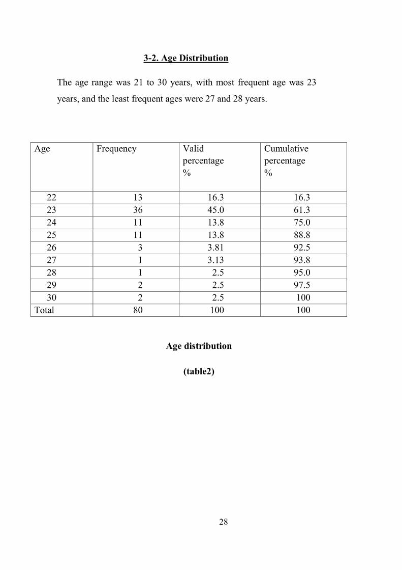
3-2. Age Distribution
The age range was 21 to 30 years, with most frequent age was 23
years, and the least frequent ages were 27 and 28 years.
Age Frequency Valid percentage %
Cumulative percentage %
22 13 16.3 16.3 23 36 45.0 61.3 24 11 13.8 75.0 25 11 13.8 88.8 26 3 3.81 92.5 27 1 3.13 93.8 28 1 2.5 95.0 29 2 2.5 97.5 30 2 2.5 100
Total 80 100 100
Age distribution
(table2)
28

3-3.Measurement of HN-1P-HN/3 (The predicted values)
Measurements carried out in 80 dentate maxillary casts
revealed that the distance (hamular notch- incisive papilla- hamular
notch) divided by three had a mean predicted value of 55.064 mm,
with a minimum value of 46.7 mm, maximum value of 61.3 mm and
standard deviation of 3.245.
Predicted value
No 80
Range 14.6
Minimum 46.70
Maximum 61.30
Mean 55.064
Std ,Erorr 0.3674
Std .D 3.245
Measurements of HN-IP-HN/3
(Table 3)
29

3-4.Measurements of maxillary anterior teeth
(The actual values)
The width of maxillary anterior teeth fiom the distal end of the right canine
to that of the left canine revealed the mean of the actual value to be
55.079mm with a minimum value 47.30mm, a maximum value 60 80mm
and standard deviation of 3. 104.
No Range Minium Maximum Mean Std
.Erorr
Std .D
Actual
value
80 13.50 47.30 60.80 55.086 0.3514 3.1039
Measurements of Maxillary anterior teeth width
(Table4)
30
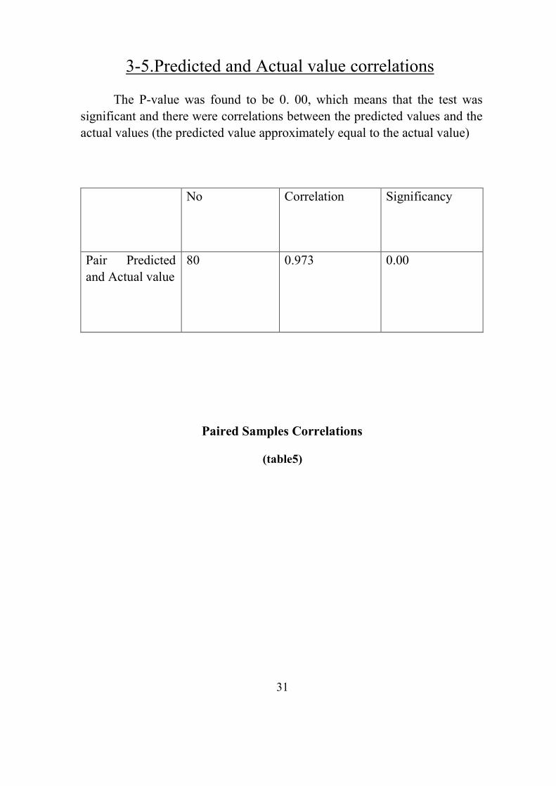
3-5.Predicted and Actual value correlations The P-value was found to be 0. 00, which means that the test was
significant and there were correlations between the predicted values and the actual values (the predicted value approximately equal to the actual value)
No Correlation Significancy
Pair Predicted and Actual value
80 0.973 0.00
Paired Samples Correlations
(table5)
31

3-6.Predicted and Actual value differences
The differences between the actual and the predicted values had a P-value
of 0.740, which wasn't significant This means that there was no difference or
there was a little difference between the actual and the predicted values
Paired Differences
Sig
Mean
Std
Deviation
Std
Error
Mean
Confidence
Interval of the
Difference Mean
Std
Deviation
Std
Error
Mean
Pair Predicted
value -
Actual value
0.02855 0.74670 08565
0.14208
0.19918 0.333 75 0.740
Predicted and actual values differences
(Table 6)
32

3333----7.Group statistics7.Group statistics7.Group statistics7.Group statistics
The mean of the predicted value among females was 52. 909mm, with a
standard deviation of 2 328(the predicted value — 52. 909 ± 2. 328mm)
The mean among males was 57.71 mm with a standard deviation of 2.016 (the
predicted value 57 711±2 016 mm).
The mean of the actual value among females was 52.860mm with a
standard deviation of 2.117( the actual value 52 860±2.117mm).
The mean of actual value among males was 57. 667 with a standard
deviation of 1 .775 (the actual value =57 667±1 775mm) Sex
Distribution N Mean
Std.
Deviation
Std. Error
Mean HD Male 37 57.124 3.3804 0.556
Female 43 52.288 2.8126 0.429
ICD Male 37 37.446 1.5835 0.2603
Female 43 35.347 1.8765 0.2862
Predicted
value
Male 37 57.711 2.0158 0.3407
Female 43 52.909 2.32766 0.3549
Actual value Male 37 57.667 1.7749 0.2958 Female 43 52.860 2.1169 0.3266
Group Statistics
(table7)
33

Chapter Four
Discussion
Conclusions
Recommendations

4-1. Discussion Selection of artificial teeth is very important in removable prosthodontics
because of its esthetic value Decision on the selection of artificial teeth has to
be based on the proper shape and exact dimensions„ Central position of the
frontal teeth, especially maxillary frontal teeth, has the strongest influence on
esthetics
The mesio-distal width is more important than the inciso-gingival length(62)
and the former measurement has attracted much debate, Research has focused
on measurements of extracted teeth, racial and gender differences, together
with facial landmarks such as the bizygomatic width House and Loop(63)
postulated that the mesio-distal measurement of the central incisor was 1/16
of the bizygomatic width Other studies have also sought to assign geometric
values for the mesiodistal width of the centrals, e g 1/16 of the face height or
the width of the iris.
Latta et al (1991) suggested the use of more than one facial measurement to
estimate the combined width of the maxillary anterior teeth(21) .
Measurements of the combined width of the maxillary anterior teeth were
reported by several investigators.
This study found that the width of maxillary anterior teeth among females
was 52. 909mm. and larger among males (57.667mm)
Scadrett et al (1982) (25) föund the width of the six maxillary anterior teeth to
be 53. 61 mm similar to McArthur (1985) whose results wer 54.6 mm
among males and 52. 3 mm among females and Nikola Petr éevié et al
found the width of maxillary frontal teeth arch (measured with a flexible
ruler) was 52. 05 m(48) All these results were approximately in
agreement with the results of this study.
34
Shilligburg et al (19'72 ) found that the width of maxillary anterior teeth was
45 8 mm(60) , Aleem et al (1997) found it was 43.0 mm(65.)
Hoffman et al(1986) result was 44. 85 mm(66) These results are lower than
this study result, which might be due to race or environmental variations or to
variation in the methods of measurements or in the sampling.
Mafgkeri$a Santoro et a1(67) found that male teeth width measurements were
slightly larger and showed a higher variability than the female measurements
but follow the same distribution pattern This supports this study results which
revealed higher measurements in males than in females.
Byron A Davis used the I-IN-IP-HN line divided by three to give a predicted
value and the width of maxillary six anterior teeth as an actual value, and
fbund that the correlation between the two values was 97% which illustrated
strong correlation between the predicted measurement and the patients actual
teeth size , these results e in agreement with the results of this study.
Even Petriéevié, Nikola et al showed that the sum of all maxillary frontal
teeth widths is equivalent to hamular distance dimension They mentioned that
after extraction of all teeth. distal maxillary width is lost, which is not possible
to reconstruct because of the individual rate of alveolar bone resorption They
pointed out that, on the other hand, the hamular distance remains the same
dimension during the lifetime, because it is not determined by teeth position
but by anatomical structures, Therefore, the hamular distance dimension is a
suitable reference for determination of the dimension of the sum of all
maxillary frontal teeth widths(48) .
The process of measuring HN-IP-I-IN on the cast in the absence of the patient
is a simple enough technique taking in to consideration that a high correlation
exists between measurements taken from the cast and the actual
measurements fiom patients with ideal tooth positioning.
The problems that may arise might be due to measurements taken from
inaccurate casts which can have several causes.
35

Some of the problems that may be due to faulty clinical or laboratory
procedures leading to measurements taken from inaccurate casts Even though
the methods used in this study relatively easily applied, other studies using
facial anatomical land marks such as interalar width, bizygomatic width,
interpupillary line etc , are more accessible and may be better and quicker
methods of choice.
35

4-2 Conclusion
At the absence of pre-extraction records, post extraction records such as
anatomical land marks, can be used to select a suitable sizes of the artificial
teeth that are in harmony with the patient's face, gender and personality„
This study showed is strong correlation between the width of maxillary
anterior teeth and (the hamular notch to hamular notch to incisive papilla
distances? divided by three ere was little or no difference between the two
values, which led to the conclusion that (the hamular notch to hamular
notch to incisive papilla distances) can be used in selection of the width of
artificial teeth in edentulous patients.
36
4-3 Recommendations
• Although this study revealed that the hamular notches and
incisive papilla are of value in determining the width of
maxillary anterior teeth, further studies of this method using
larger and more varied sample size, could be of benefit.
• Computerized analysis of these anatomical landmarks from casts
might help avoid errors of obtaining manual measurements
37
References

References
1. Mavroskoufis F, Ruichie GM Nasal width and incisive papilla as guides for
the selection and arrangement of maxillary anterior teeth (Dent 1981;
45(6): 592-7.
2. ..Engelmeier RL Complete denture esthetics Dent Clin Nam 1996;
40:71-86
3. Aaron H, Fenton and Brien R Lang. Selecting and arranging prosthetic teeth
Boucher's prosthodontic treatment of edentulous patient 1 edst. Louis: The C V
Mosby co; 1997 P. 251-2,
4. Stewart KL, Ruebker WA. Preliminary jaw relation and esthetic tryin for some
anterior replacement teeth Clinical removable partial prosthodontics 2nd ed St
Louis: Ishiyaku Euro America, Inc 1997; 317-26
5. Moustafa A Hassaballa. Selection of artificial teeth for edentulous patients
Clinical complete denture prosthodontics. King Fahad national library 2003;
2.35-7
6. Marunick, MT, Chamberlain, B B Robinson„ Denture aesthetics :an evaluation
of laymens preferences J Oral Rehail„1983; 10,399
7. Douglas A Hock Qualitative and Quantitative Guides to the Selection and
Arrangement of the Maxillary Anterior Teeth J
Prosthodont 1992; 1(2):106
8. McCord J F, Grant A A, Quayle A A Treatment options for the edentulous
mandible Eur J Prosthodont Rest Dent 1992; l : 19-2.3 9 Wehner PI, Hickey
CJ, Boucher CO Selection of artificial teeth J
Prosthet Dent 1967; 18(3): 222
10 Bindra B, Basker RM, Besford JN A study of the use of photographs fbr denture
tooth selection Int J Prosthodont
I l Gomes VL, Gongalves LC, do Prado CJ, .Junior IL, de Lima Lucas B Correlation
between facial measurements and the mesiodistal width of the maxillary
anterior teeth .J EsthetRestor
Dent 2006;
12. Latta GH Jr, Weaver JR, Conkin JE Relationship of the width of the mouth,
Interalar width, bizygomatic width, and interpupillary distance in edentulous
patients J Prosthet Dent 1991; 65(2): 250-4 13 Smith BJ The value of the nose width
as an esthetic guide in prosthodontics. J Prosthet Dent 1965; 34(5): 562-73
14 Lee L 14 dental esthetics Bristol, 1962, John Wright and Sons, Ltd
15 Wehner P J, Hickey C.J, Boucher CO Selection of artificial teeth .J Prosthet
Dent 1967; 18(3): 222-32
16 Mavroshoufis F, Ritchie GM The face-form as a guide for the selection of
maxillary central incisors .J Prosthet Dent„ 1980; 43(5): 501-5
17 Scandrett FR, Kerber ER, Umrigar ZR A clinical evaluation of techniques to
determine the complained width of the maxillary anterior teeth and the
maxillary central incisors. J Prpsthet Dent 1982; 48(1)•. 15-22
18 Varjäo, Fabiana M, Nogueira, S. S Nasal Width as a Guide for the Selection of
Maxillary Complete Denture Anterior Teeth in Four Racial Groups J
Prosthodont, 353-358(6)
19 Al-Sheik HM, al-Athel MS The relationship of interalar width, interpupillary width
and maxillary anterior teeth width in Saudi population Odontostomatol Trop. 1998;
21(84): 216-8

20 Cesario VA, Latta GH Relationship between the mesiodistal width of the maxillary
central incisor and interpupillary distance. .J Prosthet
Dent 1984;
21 Latta GH Jr, Weaver JR Conkin .JE„ The relationship between the width of the
mouth, interalar width, bizygomatic width, and interpupillary distance in edentulous
patients. J Prosthet Dent 1991;
22 Alwazzan KA The relationship between intercanthal dimension and the widths of
maxillary anterior teeth J Prosthet Dent 2001; 86(6):
608-12
23 Abdullah MA. Innercanthal distance and geometric progression as a predictor of
maxillary central incisor width .J Prosthet Dent 2002;
88(1): 16-20
24 Grant A, .Johnson W introduction to removable denture
prosthodontics, London: Churchill- Livingstone, 2nd ed 1992
25 Scandrett FR, Kerber ER, Um-rigar ZR A clinical evaluation of techniques to
determine the complained width of the maxillary anterior teeth and the maxillary
central incisors J Prpsthet Dent
1982; 48(1): 15-22
26 Hasanteisoglu U, BerksunSAras K, Arslan L An analysis of maxillary anterior teeth:
facial and dental proportions. J Prosthet
Dent
27 Keshvad A, Winstanley RD, Hooshmand T. Intercondylar width as a guide to setting
up complete denture teeth J Oral Rehabil 2002;27(3):217-2

28 Keng SB, Foong KW Maxillary arch and central incisor dimensions of an ethnic
Chinese population in relation to complete denture prosthodontics, Int Dent J. 1996;
46(2): 103-7
29 Dahrap AS, Tansequtro H A comparison of interalar width and intercanine
distance in Malay males and females„ Athropol Anz1997; 55(1): 63-8
30 H -J AHN, H -S YANG and KIM 2593 A study on the selection of the maxillary anterior artificial teeth in Korean adults Prosthodontics research program 2002
31 Gillen RJ, Schwartz RS, Hilton T J, Evans DB An analysis of selected
normative tooth proportions. Int .J Prosthodont 1994; 7 (5):410-7
32 Nelson A.A the esthetic triangle in the arrangement of teeth Nat, Dent Assoc 1922; 392-401
33 Heartwell, C and Rahn A, syllabus of Complete Dentures. 3 rd ed Lea and Febriger
1980; 297
34 LaVere AM, Marcroft KR, Smith RC, Sarka RL Denture tooth selection: size
matching of natural anterior tooth width with artificial denture teeth. J Prosthet Dent
1994; 72(4):381-4 .
35 Murchison D F WJ, Broome J cv Esthetics: Patient perception of dental
attractiveness J Prosthodont 1996; 5: 166—171.
36 Richardson ER, Malhotxa SK Mesiodistal crown dimension of the permanent
dentition of American Negroes Am J Orthod 5:68 (2):157-67
37 Marvin L Baer; Mark A Reynolds„ Comparison of Anterior Tooth Width in Natural
and Artificial Dentitions .J of Prosthodont„ 1992; 1:
38 Lavere AM, Macroft KR, Smith RC, Sarka RJ Denture tooth selection: an analysis
of the natural maxillary central incisor compared to the length and width of the face
Part L .J Prosthet Dent. 1992; 67(5): 661-3

39 Sellen PN, -Jagger DC, Harrison A. methods used to select artificial anterior teeth
for edentulous patient: a historical overview. Int T Prosthodont 1999; 12(1): 51-8
40 Eustaquio Araujo, Marcelo Souki Bolton Anterior Tooth Size Discrepancies
Among Different Malocclusion Groups The Angle Orthodontist 200@•, 73(3):30
41 HasanreisogluUfuk, BerksunSemih, Aras Kerem , Arslan Ilker An analysis of
maxillary anterior teeth: Facial and dental proportions„ .1 Prosthet Dent 2005;
94: 530-538
42 Bolton WA Disharmony in tooth size and its relation to the analysis and treatment
of malocclusion Angle Orthod„ 1958; 28: 113—130
43 Neff CW Tailored occlusion with the anterior coefficient. Am .J Orthod
44 Bolton WA The clinical application of tooth-size analysis„ Am .J Orthod 1962; 48:504-529
45 Arthur DR Are anterior replacement teeth too small? .T Prosthet Dent 1987; 57: 462-465
46 Ali Fayyad M, Jamani KD, Aqrabawi L Geometric and Mathematical Proportions and their Relations to Maxillary Anterior Teeth J Contemp Dent 2006N:5(7):0
47 Richard K (1994) hamular notch The glossary of the prosthodontic terms, 6th ed,
Mosby: Buffålo NY, pp 76
48 Nikola P, Asja C, Maja B, Robert A. Importance of Hamular Distance for
Calculation of the width of Maxillary anterior teeth Acta Stomat Croat 2005; 291-
294
49 Byron A Davis, Anatomical measurements of orthodontic and edentulous casts to
determine the width of the maxillary anterior teeth. School of dentistry West
Virginia University„ 2005
50 Richard K (1994) Incisive papilla The glossary of the prosthodontic terms, 6th ed,
Mosby: Buffalo NY, pp 78
51 Ortman H R, Tsao DH Relationship of the incisive papilla to the maxillary central
incisors. J Prosthet Dent 1979; 42: 492-6
52 Harper RN Incisive papilla. J Dent Res 1948; 27:661-68
53 Walt DM, Likeman PR Morphological changes in the denture bearing areas
following the extraction of maxillary teeth Br Dent .J 1974; 1.36: 225-35
54. F.M VARJÄO Incisive papilla for selection of denture teeth in Brazilian
population Ataraquara Dental School, Araraquara, Brazil prosthodontics
research program 2004
55. Atwood D A, Coy W A Clinical cephalometric and densitometric study of
reduction of residual ridges. .J Prosthet Dent 1971; 26:28095
56. Atwood D A A cephalometric study of the clinical rest position of the mandible
The variability in the rate of bone loss following removal of occlusal contacts.
Oral Sur, Oral Med, Oral Path 1957; 7:544-52
57. Lam R V Resorption of anterior maxillary tooth sockets. J Prosthet Dent 1960; 10:25
58. Atkinson H F, Johnson K A three year study of the dimensional changes
occurring in the maxilla following extraction of teeth Aust Dent.) 1962; 7310
59. Tallgren A Positional changes of complete dentures; A 7-year longitudinal study.
Act OdontScand 1967; 27:539-61
60. SHILLINGBURG, H T , KAPLAN, M J, GRACE, C S Tooth dimensions A comparative study .J. South Calif Dent. Asso. 1972, 40-83
61. Black GV Descriptive Anatomy of the Human Teeth 4th ed Philadelphia, Pa: SS White Dental Mfg Co 1902;
62.Eduardo M,Garcia-Godoy Diametromesiodistalcoronario de losdientes
permanentesenninos de Santo Domingo Acta OdntPediat 1980;1:70-76
63 Housemm, Loop JL Forum and colour harmony in Dental Alt Whittier; Calf: mm House 1939
64 Marquad S EStheticfåcial analysis. 30th Annual USC Periodontal and
Implant Symposium, Los Angeles, California January 21-23,2005
65 Aleem, MA Stipho, H.D , ralic Y F, Khan N The significance of inner canthal
distance in prosthodontics. The Saudi Dental Journal 1997 ;
66 Hoffman W , Bomberg T,J and I-larch RA Interalar width as a guide in
denture tooth selection L Prosthet. Dent. 1986 ; 55-219-21
67 Margherita Santoro et al Mesiodistal Crown Dimensions and Tooth Size
Discrepancy of the Permanent Dentition of Dominican Americans The Angle
Orthodontist: 70: 4: 303—307

Appendices
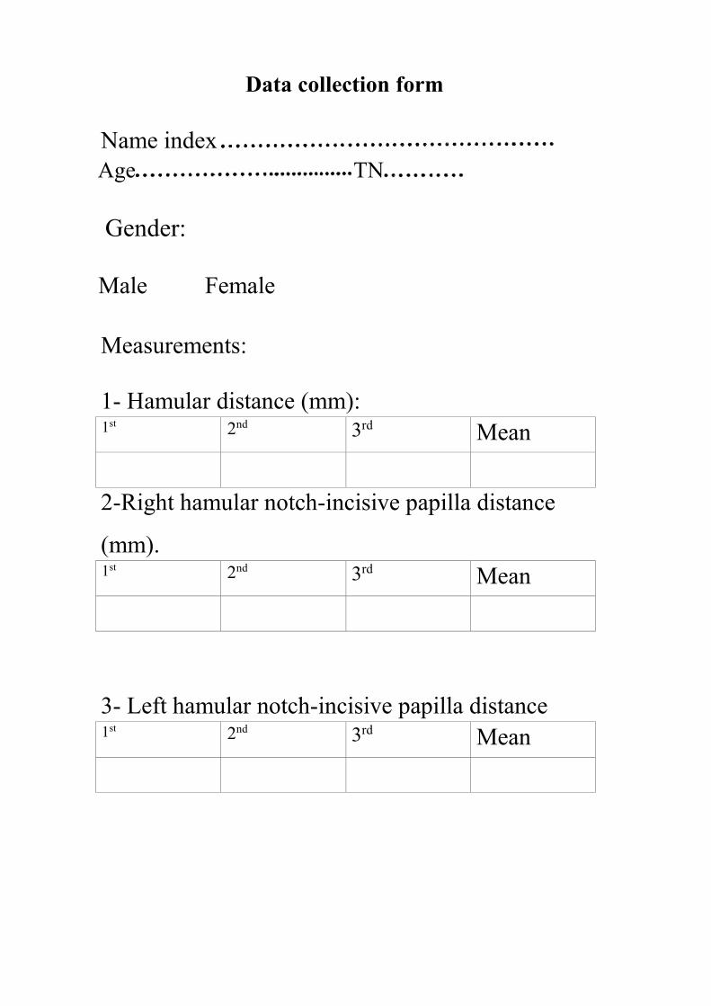
Data collection form
Name index
Age TN
Gender:
Male Female
Measurements:
1- Hamular distance (mm):
1st 2nd 3rd Mean
2-Right hamular notch-incisive papilla distance
(mm). 1st 2nd 3rd Mean
3- Left hamular notch-incisive papilla distance
1st 2nd 3rd Mean

4-Anterior teeth width (mm):
Right Left
Measurements 3 2 1 1 2 3 sum
Mean
6-The total width of six maxillary anterior teeth:
1st 2nd 3rd Mean

Measurement of hamular distance with digital caliper
Photograph 1

HN-IP-HN triangle on the cast
Photograph 2


Standardization of cast base using dental surveyor
Photograph 3