Uptake and presentation of exogenous antigen and presentation of endogenously produced antigen by...
13
Uptake and presentation of exogenous antigen and presentation of endogenously produced antigen by skin dendritic cells represent equivalent pathways for the priming of cellular immune responses following biolistic DNA immunization Introduction Gene gun-mediated bombardment of the skin with naked antigen-encoding plasmid DNA adsorbed on gold micro- particles – an in vivo gene transfer technique known as particle-mediated epidermal delivery (PMED) – has been established in various preclinical animal models as an effi- cient and reliable method of genetic vaccination to induce transgene-specific immune responses. 1 Moreover, PMED was shown in clinical phase I studies to represent a DNA Stephan Sudowe, Sabine Dominitzki, Evelyn Montermann, Matthias Bros, Stephan Grabbe and Angelika B. Reske-Kunz Clinical Research Unit Allergology, Department of Dermatology, Johannes Gutenberg- University Mainz, Mainz, Germany doi:10.1111/j.1365-2567.2008.02947.x Received 23 June 2008; revised 1 August 2008; accepted 12 August 2008. Correspondence: Dr Stephan Sudowe, Clinical Research Unit Allergology, Department of Dermatology, Johannes Gutenberg-University Mainz, Obere Zahlbacher Str. 63, D-55131 Mainz, Germany. Email: [email protected] Senior author: Professor Angelika B. Reske-Kunz, email: [email protected] Summary Gene gun-mediated biolistic DNA vaccination with b-galactosidase (bGal)- encoding plasmid vectors efficiently modulated antigen-induced immune responses in an animal model of type I allergy, including the inhibition of immunoglobulin E (IgE) production. Here we show that CD4 + as well as CD8 + T cells from mice biolistically transfected with a plasmid encoding bGal under the control of the fascin promoter (pFascin-bGal) are capable of inhibiting bGal-specific IgE production after adoptive transfer into naı ¨ve recipients. Moreover, suppression of IgE production was dependent on interferon (IFN)-c. To analyse the modalities of activation of CD4 + and CD8 + T cells regarding the localization of antigen synthesis following gene gun-mediated DNA immunization, we used the fascin promoter and the keratin 5 promoter (pK5-bGal) to direct bGal production mainly to dendritic cells (DCs) and to keratinocytes, respectively. Gene gun-medi- ated DNA immunization with each vector induced considerable activation of bGal-specific CD8 + cytotoxic T cells. Cytokine production by re-stimu- lated CD4 + T cells in draining lymph nodes and immunoglobulin isotype profiles in sera of immunized mice indicated that immunization with pFa- scin-bGal induced a T helper type 1 (Th1)-biased immune response, whereas immunization with pK5-bGal generated a mixed Th1/Th2 immune response. Nevertheless, DNA vaccination with pFascin-bGal and pK5-bGal, respectively, efficiently inhibited specific IgE production in the mouse model of type I allergy. In conclusion, our data show that uptake of exogenous antigen produced by keratinocytes and its presentation by untransfected DCs as well as the presentation of antigen synthesized endogenously in DCs represent equivalent pathways for efficient priming of cellular immune responses. Keywords: dendritic cells; fascin promoter; gene gun-mediated DNA vac- cination; keratin 5 promoter; keratinocytes; type I allergy Please cite this article in press as: Sudowe S. et al. Uptake and presentation of exogenous antigen and presentation of endogenously produced antigen by skin dendritic cells represent equivalent pathways for the priming of cellular immune responses following biolistic DNA immunization, Immunology (2009) doi: 10.1111/j.1365-2567.2008.02947.x Abbreviations: alum, aluminiumhydroxide; APC, antigen-presenting cell; CMV, cytomegalovirus; CTL, cytotoxic T lymphocyte; DC, dendritic cell; EGFP, enhanced green fluorescent protein; LNC, lymph node cell; pCMV, plasmid vector harbouring the CMV immediate early promoter; pFascin, plasmid vector harbouring the murine fascin promoter; pK5, plasmid vector harbouring the bovine keratin 5 promoter; PMED, particle-mediated epidermal delivery; Th, T helper; bGal, b-galactosidase. Ó 2008 Blackwell Publishing Ltd, Immunology, 128, e193–e205 e193 IMMUNOLOGY ORIGINAL ARTICLE
Transcript of Uptake and presentation of exogenous antigen and presentation of endogenously produced antigen by...

Uptake and presentation of exogenous antigen and presentationof endogenously produced antigen by skin dendritic cells represent
equivalent pathways for the priming of cellular immune responsesfollowing biolistic DNA immunization
Introduction
Gene gun-mediated bombardment of the skin with naked
antigen-encoding plasmid DNA adsorbed on gold micro-
particles – an in vivo gene transfer technique known as
particle-mediated epidermal delivery (PMED) – has been
established in various preclinical animal models as an effi-
cient and reliable method of genetic vaccination to induce
transgene-specific immune responses.1 Moreover, PMED
was shown in clinical phase I studies to represent a DNA
Stephan Sudowe, Sabine
Dominitzki, Evelyn Montermann,
Matthias Bros, Stephan Grabbe
and Angelika B. Reske-Kunz
Clinical Research Unit Allergology, Department
of Dermatology, Johannes Gutenberg-
University Mainz, Mainz, Germany
doi:10.1111/j.1365-2567.2008.02947.x
Received 23 June 2008; revised 1 August
2008; accepted 12 August 2008.
Correspondence: Dr Stephan Sudowe,
Clinical Research Unit Allergology,
Department of Dermatology, Johannes
Gutenberg-University Mainz, Obere
Zahlbacher Str. 63, D-55131 Mainz,
Germany. Email: [email protected]
Senior author: Professor Angelika
B. Reske-Kunz,
email: [email protected]
Summary
Gene gun-mediated biolistic DNA vaccination with b-galactosidase (bGal)-
encoding plasmid vectors efficiently modulated antigen-induced immune
responses in an animal model of type I allergy, including the inhibition of
immunoglobulin E (IgE) production. Here we show that CD4+ as well as
CD8+ T cells from mice biolistically transfected with a plasmid encoding
bGal under the control of the fascin promoter (pFascin-bGal) are capable
of inhibiting bGal-specific IgE production after adoptive transfer into
naıve recipients. Moreover, suppression of IgE production was dependent
on interferon (IFN)-c. To analyse the modalities of activation of CD4+
and CD8+ T cells regarding the localization of antigen synthesis following
gene gun-mediated DNA immunization, we used the fascin promoter and
the keratin 5 promoter (pK5-bGal) to direct bGal production mainly to
dendritic cells (DCs) and to keratinocytes, respectively. Gene gun-medi-
ated DNA immunization with each vector induced considerable activation
of bGal-specific CD8+ cytotoxic T cells. Cytokine production by re-stimu-
lated CD4+ T cells in draining lymph nodes and immunoglobulin isotype
profiles in sera of immunized mice indicated that immunization with pFa-
scin-bGal induced a T helper type 1 (Th1)-biased immune response,
whereas immunization with pK5-bGal generated a mixed Th1/Th2
immune response. Nevertheless, DNA vaccination with pFascin-bGal and
pK5-bGal, respectively, efficiently inhibited specific IgE production in the
mouse model of type I allergy. In conclusion, our data show that uptake
of exogenous antigen produced by keratinocytes and its presentation by
untransfected DCs as well as the presentation of antigen synthesized
endogenously in DCs represent equivalent pathways for efficient priming
of cellular immune responses.
Keywords: dendritic cells; fascin promoter; gene gun-mediated DNA vac-
cination; keratin 5 promoter; keratinocytes; type I allergy
Please cite this article in press as: Sudowe S. et al. Uptake and presentation of exogenous antigen and presentation of endogenously produced antigen by skin dendritic cellsrepresent equivalent pathways for the priming of cellular immune responses following biolistic DNA immunization, Immunology (2009) doi: 10.1111/j.1365-2567.2008.02947.x
Abbreviations: alum, aluminiumhydroxide; APC, antigen-presenting cell; CMV, cytomegalovirus; CTL, cytotoxic T lymphocyte;DC, dendritic cell; EGFP, enhanced green fluorescent protein; LNC, lymph node cell; pCMV, plasmid vector harbouring theCMV immediate early promoter; pFascin, plasmid vector harbouring the murine fascin promoter; pK5, plasmid vectorharbouring the bovine keratin 5 promoter; PMED, particle-mediated epidermal delivery; Th, T helper; bGal, b-galactosidase.
� 2008 Blackwell Publishing Ltd, Immunology, 128, e193–e205 e193
I M M U N O L O G Y O R I G I N A L A R T I C L E

vaccine delivery method that was well tolerated and con-
sistently induced significant humoral and cellular immune
responses,2 whereas intramuscular and intradermal injec-
tion of DNA in saline as alternative routes of DNA
immunization often proved insufficient to provide protec-
tive immunity in human trials and is therefore considered
to be clinically ineffective.3
Numerous reports have substantiated the notion that
dendritic cells (DCs) of the skin are crucial for the induc-
tion of immune responses in draining lymph nodes after
cutaneous DNA immunization irrespective of the inocula-
tion technique,4–7 although it has become a matter of dis-
cussion whether epidermal Langerhans cells or dermal
DC populations are the principal antigen-presenting cells
(APCs) under these circumstances.8–10
We obtained evidence supporting the notion of the piv-
otal role of DCs by focussing antigen production primar-
ily to DCs after biolistic transfection of mouse skin using
the promoter of the gene of the cytoskeletal actin-bun-
dling protein fascin to drive transgene expression. During
the differentiation process from immature DCs to pri-
mary stimulatory mature DCs, expression of the fascin
gene was strongly upregulated.11,12 As a consequence,
fascin, which is mandatory for the formation of den-
drites,11 was synthesized in large amounts by mature
DCs.11–13 In contrast, expression of the fascin gene in
non-transformed cell types other than DCs could rarely
be detected and was restricted to neuronal tissues, capil-
lary endothelial cells and follicular DCs.11,14,15 In earlier
studies we verified that the activity of the promoter of the
fascin gene reflects the endogenous production of fascin,
being high in mature DCs but negligible in immature
DCs and in keratinocytes.16,17 Hence, gene gun immuni-
zation of mice with plasmid DNA carrying the fascin pro-
moter as the regulatory element transcriptionally targeted
transgene expression to skin-derived DCs.17 Subsequently,
distinct type 1 immune responses were induced, including
the potent activation of interferon (IFN)-c-producing
CD4+ T helper type 1 (Th1) cells as well as IFN-c-pro-
ducing CD8+ cytotoxic T lymphocytes (CTLs).17,18
These results are consistent with a number of publica-
tions which suggest that endogenous production of anti-
gen by directly transfected DCs of the skin is sufficient to
induce immune responses, in particular the generation of
CD8+ CTLs.4,5,19 In contrast, several authors have con-
cluded from their data that cross-presentation of antigen,
produced and released by transfected cutaneous non-DCs,
represents the main pathway of CD8+ T-cell priming fol-
lowing gene gun immunization.20–22 In an attempt to
resolve this apparent discrepancy, we compared the
immune responses initiated by biolistic transfection of
skin with plasmids expressing the transgene b-galactosi-
dase (bGal) under the control of the fascin promoter
(pFascin), the keratinocyte-specific keratin 5 promoter
(pK5) or the ubiquitously active cytomegalovirus immedi-
ate early promoter (pCMV). Employing this approach, we
restrict bGal production selectively to DCs as APCs or to
keratinocytes as non-APCs or to a rather broad variety of
epidermal/dermal cells. In this paper, we show that pre-
sentation of antigen endogenously synthesized by directly
transfected DCs and presentation of antigen by DCs that
have taken up and processed exogenous protein are
equivalent pathways for maximum stimulation of cellular
immune responses. However, the quality of the Th cell
immune response was different following DC-targeted
and keratinocyte-specific biolistic transfection. Further-
more, we demonstrate that the inhibitory activity of bio-
listic transfection on the induction of an antigen-specific
immunoglobulin E (IgE) response, which we show here
to be dependent on IFN-c production and exerted by
CD4+ and CD8+ T cells, can be mediated by either of the
two pathways of antigen presentation.
Materials and methods
Mice
BALB/c mice, C57BL/6 mice, interleukin (IL)-12p40)/)
mice and IFN-c)/) mice were bred and maintained in the
Central Animal Facilities of the University of Mainz in
specific pathogen-free conditions on a standard diet. The
‘Principles of laboratory animal care’ (NIH publication
no. 85-23, revised 1985) were followed. The experiments
were approved by the Ethics Commission according to
the German Law on the Protection of Animals.
Plasmid vectors and DNA immunization protocol
The construction and purification of plasmid vectors
encoding the transgene bGal under control of the fascin
promoter (pFascin-bGal) and the cytomegalovirus (CMV)
promoter (pCMV-bGal), respectively, have been described
in detail previously.17,18
To obtain pK5-bGal and pK5-EGFP, parental vector
pUC19-K523 was digested with SalI, which cuts imme-
diately downstream of the bovine K5 promoter in this
construct. Expression cassettes for bGal and enhanced
green fluorescent protein (EGFP) encompassing the
respective reporter gene and an SV40 polyadenylation
signal were excised by restriction digest from pCMV-
bGal (BamHI) and pCMV-EGFP (Acc65I/BamHI).
50-protruding ends of linearized pUC19-K5 and of
excised expression cassettes were filled in using Klenow
enzyme, and expression cassettes were ligated with
pUC19-K5. The functional activity of derived reporter
expression constructs pK5-bGal and pK5-EGFP was
validated by in vitro transfection.
Genetic immunization was performed by biolistic trans-
fection with a total of 4 lg of plasmid DNA using the
helium-driven Helios gene gun system (Bio-Rad, Munich,
e194 � 2008 Blackwell Publishing Ltd, Immunology, 128, e193–e205
S. Sudowe et al.

Germany) as described previously.18 Mice were immu-
nized at weekly intervals three to five times as indicated.
Immunization protocol for the induction of IgEproduction
High and persistent bGal-specific IgE production was
induced as previously described.24 Briefly, female BALB/c
mice, 6–8 weeks of age at the beginning of the experi-
ments, were sensitized by multiple intraperitoneal injec-
tions of 1 lg of recombinant bGal (Sigma-Aldrich,
Deisenhofen, Germany) at intervals of 2 weeks. bGal pro-
tein was dissolved in 100 ll of phosphate-buffered saline
(PBS) and adsorbed to an equal volume of Imject� Alum
(Pierce, Rockford, IL) as adjuvant (bGal/alum) according
to the manufacturer’s instructions.
Adoptive transfer of lymphocytes
BALB/c mice were immunized via the gene gun as
described above by five consecutive applications of pFa-
scin-bGal at weekly intervals. Two weeks after the last
immunization spleen cells were prepared as described pre-
viously25 and pooled. CD4+ and CD8+ T cells in spleen cell
suspensions from gene gun-immunized as well as from
bGal/alum-injected control mice were enriched by mag-
netic separation as previously described.26 The purity of
positively selected T-cell populations was verified by cyto-
fluorometric analysis and was 83 ± 4% for CD4+ T cells
and 89 ± 5% for CD8+ T cells; the frequency of the remain-
ing T cells of the reciprocal subpopulation was below 2%.
Unseparated spleen cells (107) or isolated CD4+ and CD8+
cells (2�5 · 106), respectively, were injected intravenously
into naıve syngeneic mice. After 24 hr, recipients and con-
trol mice, which did not receive donor cells, were sensitized
with bGal/alum as described above.
Measurement of serum antibodies
At the indicated time-points, immunized mice were bled by
puncture of the retro-orbital plexus. Sera were collected
and kept frozen at )20� until thawed for determination of
bGal-specific IgG1, IgG2a and IgE by antigen capture
enzyme-linked immunosorbent assay (ELISA) as reported
previously.24 For comparison of samples obtained at differ-
ent times in the course of immunization, IgG1, IgG2a and
IgE contents in sera were standardized using high-titre
reference sera, collected from mice repeatedly immunized
with bGal/alum (IgE) or from mice initially vaccinated
with pCMV-bGal and subsequently boosted with bGal/
alum (IgG1, IgG2a). The antibody titre was defined as the
reciprocal serum dilution yielding an absorbance reading of
optical density (OD) = 0�2 after linear regression analysis.
The titre of the reference serum was designated as 1 U/ml
and the titres of the serum samples are presented as equiva-
lent units. In experiments in which the relative contents of
specific IgG1 and IgG2a in sera were directly compared, the
titres were determined in ELISAs performed in parallel and
the ratio of the two IgG isotypes was calculated as the quo-
tient of their titres.
Measurement of CD8+ CTLs
The frequency of IFN-c-producing CD8+ effector T cells
recognizing the H-2Ld-binding bGal-derived nonamer
peptide TPHPARIGL (bGal876–884; Sigma-Aldrich) in sin-
gle-cell suspensions of spleen cells was determined by an
enzyme-linked immunosorbent spot-forming cell assay
(ELISPOT) as previously described24 with the modifica-
tion that 100 ng/ml bGal876–884 was used as the standard
concentration to activate CD8+ T cells. Spots were
counted and evaluated using the EliSpot Reader System
(AID, Strasberg, Germany).
The cytotoxic activity of spleen cells, prepared from
immunized mice, against LacZ-transfected P13�1 target
cells and untransfected P815 control cells was determined
using the JAM test as described previously.18 The differ-
ence between the two values was calculated and desig-
nated as specific lysis.
Determination of IL-5 and IFN-c in culturesupernatants
Draining lymph node cells (LNCs) of biolistically trans-
fected mice (axillary and inguinal lymph nodes) and of
mice immunized with bGal/alum (mesenterial lymph
nodes) were prepared and incubated for 72 hr on 24-well
tissue culture plates (5 · 106 LNCs/well) (Corning Costar,
Bodenheim, Germany) in a volume of 1 ml of culture
medium with or without recombinant bGal (25 lg/ml).
Subsequently, contents of IL-5 and IFN-c were quantified
in culture supernatants using a sandwich ELISA following
a standard procedure as described previously.18
Statistical analysis
Statistical evaluation of the experimental data was per-
formed by Student’s t-test using SIGMASTAT software (SPSS
Inc., Chicago, IL). Statistically significant differences
(P < 0�05) between control groups and experimental
groups are designated by symbols.
Results
Inhibition of specific IgE production by gene gunimmunization is mediated by CD4+ as well as CD8+
T cells and depends on IFN-c production
We previously showed that gene gun-mediated DNA
vaccination with plasmid vectors encoding bGal under
� 2008 Blackwell Publishing Ltd, Immunology, 128, e193–e205 e195
Pathways of T-cell priming after biolistic DNA immunization

the control of the fascin promoter (pFascin-bGal) effec-
tively inhibited subsequently induced specific IgE immune
responses in a mouse model of type I allergy.27 The sup-
pression of IgE production was associated with a shift
towards a Th1-biased immune response as well as with
the recruitment of large numbers of CD8+ CTLs.27 The
inhibitory capacity can be adoptively transferred to naıve
recipients using spleen cells from BALB/c mice previously
immunized with pFascin-bGal (Fig. 1a). Despite sensitiza-
tion with bGal/alum following adoptive transfer, the reci-
pient mice produced strongly reduced levels of IgE and
also IgG1 as compared with mice without cell transfer.
Furthermore, IgG2a levels were substantially increased
although the differences did not reach statistical signifi-
cance. As a consequence, the ratio of bgal-specific IgG1
versus IgG2a titres dropped drastically (IgG1:IgG2a ratio:
no transfer 76�7 ± 16�5 versus transfer of pFascin-bGal
spleen cells 0�8 ± 0�2; P < 0�01). This finding is consistent
with the situation prevailing in mice prophylactically vac-
cinated with pFascin-bGal and subsequently sensitized
with bGal protein.27 In contrast, spleen cells collected
from BALB/c mice sensitized with bGal/alum and thus
exhibiting high titres of IgE in the sera (data not shown)
were not able to significantly suppress the formation of
IgE or to change the pattern of IgG isotypes (IgG1:IgG2a
ratio: transfer of bGal/alum spleen cells 64�5 ± 29�2;
P > 0�05 versus no transfer) upon adoptive transfer. In
order to determine whether CD4+ or CD8+ T cells are
the principal mediators of regulation, the two T-cell sub-
populations were enriched by magnetic separation from
spleen cells of mice biolistically transfected with pFascin-
bGal and were subsequently transferred into naıve mice.
Injection of either CD4+ or CD8+ T cells prior to sensiti-
zation with bGal/alum was sufficient to significantly
reduce bGal-specific IgE production in the recipients
(Fig. 1a). However, the inhibitory activity of each sub-
population was lower than that provided by total spleen
1·0
0·8
0·6
0·4
0·2
0
0·4
0·5
(a)
(b)
βGal
-spe
cific
IgG
2a (
U/m
l) βG
al-s
peci
fic Ig
E (
U/m
l) βG
al-s
peci
fic Ig
G1
(U/m
l) βG
al-s
peci
fic Ig
E (
U/m
l)
n = 17
Wildtype IFN-γ–/– IL-12p40–/–
n = 15 n = 18 n = 16 n = 12
**
**
**
**
**
*
***
Control Vaccination with
pFascin-βGal (3x)
n = 10
Adoptive transfer
SC CD4 SC CD4 CD8
pFascin-βGal βGal/alum
No transfer
0·3
0·2
0·1
0·0
15
10
5
0 1·5
1·2
0·9
0·6
0·3
0
Figure 1. Inhibition of specific immunoglobulin E (IgE) production
by gene gun-mediated DNA vaccination is mediated by CD4+ and
CD8+ T cells and depends on interferon (IFN)-c production.
(a) BALB/c mice were immunized five times at weekly intervals by
biolistic transfection with pFascin-bGal or by intraperitoneal injection
of b-galactosidase (bGal)/alum. Fourteen days after the last
application, spleen cells (SC) and splenic CD4+ and CD8+ T cells
were prepared and injected intravenously into naıve BALB/c mice.
Recipients and control mice without transfer of cells (n = 4–5) were
sensitized by intraperitoneal injection of bGal/alum every 2 weeks.
Ten days after the fifth injection mice were bled and bGal-specific
IgE, IgG1 and IgG2a titres were determined by enzyme-linked
immunosorbent assay (ELISA). Data represent mean titres ± the
standard error of the mean (SEM) for individual mice. Significant
differences are indicated by asterisks (*P < 0�05, **P < 0�01 versus
control mice). (b) C57BL/6 wild-type, IFN-c)/) and interleukin
(IL)-12p40)/) mice were vaccinated three times at weekly intervals by
biolistic transfection with pFascin-bGal. Vaccinated mice and unvac-
cinated controls were sensitized with bGal/alum as described. Mice
were bled after the fifth injection of bGal/alum and bGal-specific IgE
titres in sera were determined by ELISA. Data represent mean
titres ± SEM of the indicated numbers of individual sera obtained in
three independent experiments. Significant differences are indicated
by asterisks (**P < 0�01 and ***P < 0�001 versus unvaccinated mice).
e196 � 2008 Blackwell Publishing Ltd, Immunology, 128, e193–e205
S. Sudowe et al.
cells, suggesting that both CD4+ and CD8+ T cells were
required to efficiently down-modulate the IgE immune
response. In contrast, modulation of IgG production
towards a Th1-skewed isotype pattern could mainly be
attributed to CD4+ T cells (IgG1:IgG2a ratio: transfer of
pFascin-bGal CD4+ T cells 3�1 ± 0�3; P < 0�01 versus no
transfer; transfer of pFascin-bGal CD8+ T cells
26�6 ± 10�2; P < 0�05 versus no transfer). CD4+ T cells
isolated from mice immunized with bGal/alum did not
show any potency in suppression of the IgE response and
did not affect IgG formation (IgG1:IgG2a ratio: transfer
of bGal/alum CD4+ T cells 90�2 ± 23�0; P > 0�05 versus
no transfer) following adoptive transfer (Fig. 1a).
CD4+ and CD8+ T cells from mice biolistically trans-
fected with pFascin-bGal secrete large amounts of IFN-
c.18,27 To check whether the production of this cytokine is
crucial for the inhibitory effect of gene gun vaccination
with pFascin-bGal on bGal/alum-induced IgE production,
C57BL/6 wild-type, IFN-c)/) and IL-12p40)/) mice were
biolistically vaccinated three times with pFascin-bGal, and
the amount of IgE in the sera after the fifth subsequent
immunization with bGal/alum was determined. Although
C57BL/6 wild-type mice produced lower amounts of bGal-
specific IgE compared with BALB/c mice (data not shown),
they proved to be susceptible to the inhibitory effect of bio-
listic transfection with pFascin-bGal on the IgE immune
response (Fig. 1b). Similarly, in gene gun-vaccinated
IL-12p40)/) mice, which were not capable of producing
bioactive IL-12, bGal-specific IgE levels in sera were signifi-
cantly reduced. In contrast, suppression of IgE production
was not observed in IFN-c-deficient mice, indicating that
this cytokine plays a crucial role in this regulation process.
Presentation of endogenous antigen andcross-presentation of exogenous antigen areequivalent pathways for induction of CD8+ CTL aftergene gun immunization
Our previous data, obtained by gene gun-mediated DNA
immunization with vectors encoding an antigen under
the control of the fascin promoter, suggest that endoge-
nous antigen production by directly transfected DCs of
the skin is sufficient to induce potent CD4+ and CD8+
T-cell responses.18 However, these studies did not clarify
whether this pathway was the only one or at least the
principal one responsible for the priming of immune
responses after biolistic transfection with pCMV, which
instructs broad cutaneous antigen synthesis by DCs as
well as by non-DCs. Thus the uptake of exogenous anti-
gen released by transfected non-DCs and its processing
and presentation by untransfected DCs might represent
an alternative pathway to stimulate cellular immune
responses. To analyse the modalities of activation of
CD4+ and CD8+ T cells with inhibitory potential for IgE
production with respect to the localization of antigen
synthesis following gene gun-mediated DNA immuniza-
tion, we sought to direct transgene expression selectively
to non-DCs of the skin by constructing plasmids that
encode bGal or EGFP under the control of the keratin 5
promoter (pK5-bGal and pK5-EGFP), which is specifi-
cally active in keratinocytes.23,28 After biolistic transfec-
tion of abdominal skin with these vectors we observed
strong and extensive transgene expression in hair follicles
and, at a lower frequency, weakly transgene-positive areas
in the basal epidermis (data not shown). In contrast to
application of particle bombardment with pFascin or
pCMV18,25,27 we did not detect transgene-expressing cells
in the draining lymph nodes (data not shown).
To measure the induction of bGal-specific CD8+ T cells
in mice immunized via the gene gun with the different
plasmid constructs, spleen cells were stimulated with the
bGal-derived H-2Ld-restricted nonamer peptide TPHPA-
RIGL. The secretion of IFN-c by activated CD8+ T cells
was determined by an ELISPOT. Immunization of mice
with each of the three plasmids pFascin-bGal, pCMV-bGal
and pK5-bGal induced substantial numbers of IFN-c-pro-
ducing CD8+ effector T cells, although in the case of
pFascin-bGal their frequency tended to be somewhat lower
(Fig. 2a). However, the difference in the number of CD8+
effector T cells arising from gene gun-mediated immuniza-
tion with pFascin-bGal or pK5-bGal was not statistically
significant (P = 0�137). In contrast, repeated injection of
bGal/alum did not generate bGal-specific CD8+ T-cell
responses, and neither did gene gun-mediated immuniza-
tion with an EGFP-encoding control plasmid. Further-
more, in vitro activation of CD8+ T cells from mice primed
in vivo by biolistic transfection with the three different
bGal-encoding vectors, using decreasing peptide concen-
trations, resulted in comparably reduced numbers of
IFN-c-producing CD8+ effector T cells, suggesting similar
avidity of these cells (Fig. 2b). Moreover, co-application of
pFascin-bGal and pK5-bGal, resulting in presentation of
endogenous antigen by directly transfected DCs and cross-
presentation of exogenous antigen produced by non-DCs,
did not lead to enhanced induction of IFN-c-producing
CD8+ effector T cells, indicating that each of the antigen
presentation modalities led to maximum stimulation of
the CD8+ T-cell response (Fig. 2b).
In addition, the presence of cytolytically active CTLs
among spleen cells of biolistically transfected mice was veri-
fied by determination of the bGal-specific cytotoxic poten-
tial of re-stimulated CD8+ T cells, expanded by cultivation
of splenocytes with the cognate major histocompatibility
complex (MHC) class I-restricted peptide. Consistent with
the data obtained by the ELISPOT, spleen cells from mice
immunized with the three bGal-encoding plasmids all dis-
played high specific cytolytic activity, whereas spleen cells
from mice immunized with bGal/alum and with the con-
trol vector pK5-EGFP, respectively, did not mediate killing
of bGal-expressing target cells (Fig. 2c). Specific lysis
� 2008 Blackwell Publishing Ltd, Immunology, 128, e193–e205 e197
Pathways of T-cell priming after biolistic DNA immunization

induced by spleen cells obtained after co-application of
pFascin-bGal and pK5-bGal was slightly higher than the
lysis induced by spleen cells from mice immunized with
either of the two vectors; however, this difference did not
reach statistical significance (Fig. 2c).
T-cell responses induced by gene gun-mediatedDC-targeted and keratinocyte-targeted transgeneexpression, respectively, differ in their cytokine profileand antibody isotype pattern
We evaluated the quality of the antigen-specific T-cell
response after biolistic DNA immunization with the three
bGal-encoding plasmids by measuring the production of
the cytokines IFN-c and IL-5, representative for type 1
and type 2 responses, respectively, in cultures of draining
LNCs activated in vitro by the addition of exogenous
bGal protein. Because the isotype pattern of the humoral
response serves as an indicator for the type of T-cell help
supporting an immune reaction, we verified the cytokine
data by determining the relative distribution of bGal-
specific IgG subtypes and directly comparing IgG2a versus
IgG1 titres in the sera of immunized animals.
LNCs from axillary and inguinal lymph nodes of mice
immunized with pFascin-bGal produced large amounts of
IFN-c but no IL-5 after re-stimulation with bGal (Fig. 3).
In addition, specific antibody production in these mice
was low and mainly of the IgG2a isotype (Fig. 4), which
together are indicative of a distinct Th1 immune response.
In contrast, LNCs from animals primed by biolistic
transfection with pK5-bGal or pCMV-bGal in addition to
IFN-c secreted considerable amounts of IL-5 into culture
supernatants, which points to a mixed Th1/Th2 immune
response (Fig. 3). Similarly, co-application of pFascin-
pFascin-βGal pFascin-βGal
pFascin-βGal
0·1
60 (c)
(a) (b)
40
Spe
cific
lysi
s (%
)
20
0
–20
Effector cells target cells
0·5 1 5
**
*
*
*
**
§§§
§§§
* *
pFascin-βGal
pK5-βGal
+ pK5-βGal
pFascin-βGal + pK5-βGal
pK5-βGal
pK5-EGFP
pK5-βGal
pK5-EGFP
0 500
###
###
###
##
####
##
## #
#
###
1000 1500 2000 0 250 IFN-γ-producing CD8+ effector T cells
per 106 spleen cellsIFN-γ-producing CD8+ effector T cells
per 106 spleen cells
500 750 1000
TPHPARIGL:
TPHPARIGL: 0 ng/ml 0·1 ng/ml 1 ng/ml 10 ng/ml 100 ng/ml
0 ng/ml 100 ng/ml
βGal/alum
pCMV-βGal
pCMV-βGal
βGal/alum
pCMV-βGal
Figure 2. Efficient priming of cytotoxic T lymphocyte (CTL) responses following gene gun-mediated DNA immunization targeting predominantly
dendritic cells (DCs) or keratinocytes. BALB/c mice (n = 4) were immunized three times by biolistic transfection with the indicated plasmids or by
intraperitoneal injection of b-galactosidase (bGal)/alum. (a, b) Frequencies of interferon (IFN)-c-producing CD8+ effector T cells among pooled
splenocytes were determined in triplicate by enzyme-linked immunosorbent spot-forming cell assay (ELISPOT) 7–14 days after the last application
by stimulation of the collected spleen cells for 22 hr with (closed and hatched bars) or without (open bars) the indicated concentrations of the
H-2Ld-specific bGal peptide TPHPARIGL. Data represent the mean ± standard error of the mean (SEM) for cell numbers obtained in five (a) and
two (b) independent experiments. Significant differences are indicated by symbols (*P < 0�05 and **P < 0�01 versus mice immunized with pCMV-
bGal; §§§P < 0�001 versus mice immunized with bGal-encoding plasmids). (c) bGal-specific cytolytic activity of pooled spleen cells from immu-
nized mice was determined in triplicate 7–14 days after the last application by JAM test. Data represent means of specific lysis obtained in three to
six independent experiments. Significant differences are indicated by symbols (#P < 0�05, ##P < 0�01 and ###P < 0�001 versus mice immunized with
pK5-EGFP). EGFP, enhanced green fluorescent protein; pCMV, plasmid vector harbouring the cytomegalovirus immediate early promoter.
e198 � 2008 Blackwell Publishing Ltd, Immunology, 128, e193–e205
S. Sudowe et al.
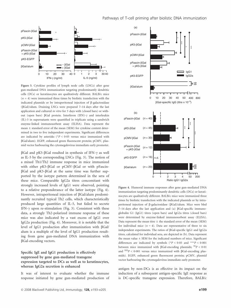
bGal and pK5-bGal resulted in synthesis of IFN-c as well
as IL-5 by the corresponding LNCs (Fig. 3). The notion of
a mixed Th1/Th2 immune response in mice immunized
with either pK5-bGal or pCMV-bGal or with pFascin-
bGal and pK5-bGal at the same time was further sup-
ported by the isotype pattern determined in the sera of
these mice. Comparable IgG2a titres concomitant with
strongly increased levels of IgG1 were observed, pointing
to a relative preponderance of the latter isotype (Fig. 4).
However, intraperitoneal injection of bGal/alum predomi-
nantly recruited typical Th2 cells, which characteristically
produced large quantities of IL-5, but failed to secrete
IFN-c upon re-stimulation (Fig. 3). Consistent with these
data, a strongly Th2-polarized immune response of these
mice was also indicated by a vast excess of IgG1 over
IgG2a production (Fig. 4). Moreover, it is notable that the
level of IgG1 production after immunization with bGal/
alum is a multiple of the level of IgG1 production result-
ing from gene gun-mediated DNA immunization with
bGal-encoding vectors.
Specific IgE and IgG1 production is effectivelysuppressed by gene gun-mediated transgeneexpression targeted to DCs as well as to keratinocytes,whereas IgG2a secretion is enhanced
It was of interest to evaluate whether the immune
response initiated by gene gun-mediated production of
antigen by non-DCs is as effective in its impact on the
induction of a subsequent antigen-specific IgE response as
is DC-specific transgene expression. Therefore, BALB/c
pFascin-βGal (n = 40)
0 10 20 30 40 50 400
IgG1 IgG2a
0 1 2 Ratio lgG1 IgG2a
3 4 50
*** *** ***
***
***
* *
*
§§§ §§
200
800
(n = 45)
(n = 30)
(n = 12)
(n = 16)
(n = 20)
n.d.
pFascin-βGal + pK5-βGal
pK5-EGFP
pK5-βGal
βGal/alum
pCMV-βGal
pFascin-βGal
(a)
(b)
pFascin-βGal + pK5-βGal
pK5-EGFP
pK5-βGal
βGal/alum
βGal-specific IgG (titre x 10–3)
pCMV-βGal
Figure 4. Humoral immune responses after gene gun-mediated DNA
immunization targeting predominantly dendritic cells (DCs) or kerati-
nocytes are qualitatively different. BALB/c mice were immunized three
times by biolistic transfection with the indicated plasmids or by intra-
peritoneal injection of b-galactosidase (bGal)/alum. Mice were bled
7–14 days after the last application and (a) bGal-specific immuno-
globulin G1 (IgG1) titres (open bars) and IgG2a titres (closed bars)
were determined by enzyme-linked immunosorbent assay (ELISA).
Data represent the mean titre ± the standard error of the mean (SEM)
for individual mice (n = 4). Data are representative of three to six
independent experiments. The ratios of bGal-specific IgG1 and IgG2a
titres, calculated for individual sera, are depicted in (b). Data represent
the mean value ± SEM for the indicated numbers of mice. Significant
differences are indicated by symbols (*P < 0�05 and ***P < 0�001
between mice immunized with bGal-encoding plasmids; §§P < 0�01
and §§§P < 0�001 versus mice immunized with bGal-encoding plas-
mids). EGFP, enhanced green fluorescent protein; pCMV, plasmid
vector harbouring the cytomegalovirus immediate early promoter.
pFascin-βGal
pFascin-βGal + pK5-βGal
pK5-EGFP
pK5-βGal
βGal/alum – βGal + βGal
0 10 20 30 40 0 1 2
IL-5 (ng/ml) IFN-γ (ng/ml)
*
*
*
*
*
3 30 40
pCMV-βGal
Figure 3. Cytokine profiles of lymph node cells (LNCs) after gene
gun-mediated DNA immunization targeting predominantly dendritic
cells (DCs) or keratinocytes are qualitatively different. BALB/c mice
(n = 4) were immunized three times by biolistic transfection with the
indicated plasmids or by intraperitoneal injection of b-galactosidase
(bGal)/alum. Draining LNCs were prepared 7–14 days after the last
application and cultured in vitro for 3 days with (closed bars) or with-
out (open bars) bGal protein. Interferon (IFN)-c and interleukin
(IL)-5 in supernatants were quantified in triplicate using a sandwich
enzyme-linked immunosorbent assay (ELISA). Data represent the
mean ± standard error of the mean (SEM) for cytokine content deter-
mined in two to five independent experiments. Significant differences
are indicated by asterisks (*P < 0�05 versus mice immunized with
bGal/alum). EGFP, enhanced green fluorescent protein; pCMV, plas-
mid vector harbouring the cytomegalovirus immediate early promoter.
� 2008 Blackwell Publishing Ltd, Immunology, 128, e193–e205 e199
Pathways of T-cell priming after biolistic DNA immunization

mice were vaccinated with the bGal-encoding vectors as
indicated in Fig. 5 prior to sensitization by repeated intra-
peritoneal injection of bGal/alum. Sera were taken at dis-
tinct days throughout the sensitization period and
contents of bGal-specific IgE as well as IgG1 and IgG2a
in sera were designated as reference serum titre equiva-
lents (U/ml), which allowed for proper comparison of
samples obtained at different points in time and tested in
multiple ELISAs. As depicted in Fig. 5a and 5b, produc-
tion of bGal-specific IgE was suppressed by vaccination
with each of the bGal-encoding plasmids as compared
with the control plasmid (pK5-EGFP) and the untreated
controls. The suppression was manifest already after the
second injection of bGal/alum and was maintained
throughout the sensitization period (Fig. 5a). Conse-
quently, IgE titres after the fifth application of bGal/alum
were significantly reduced in mice biolistically transfected
with bGal-encoding plasmid DNA to 20–30% of IgE titres
measured in unvaccinated control mice (Fig. 5b).
As already outlined, repeated injection of bGal/alum
induced strong IgG1 production during the course of
immunization, but only minor formation of IgG2a
(Fig. 5c, d). Biolistic transfection with the EGFP-encoding
control vector had no significant impact on the elicitation
of bGal-specific IgG1 by subsequent sensitization with
bGal/alum, but led to a modest increase in the produc-
tion of specific IgG2a (days 10 and 54, P < 0�05 versus
control mice) (Fig. 5c, d). In contrast, previous vaccina-
tion with bGal-encoding plasmids had profound modula-
tory effects on IgG subtype formation during the course
of sensitization. Preceding vaccination with pK5-bGal
similar to vaccination with pCMV-bGal led to a boost in
IgG1 production immediately after the beginning of
immunization with bGal/alum (pK5-bGal: days 10 and
26, P < 0�001 versus control mice; pCMV-bGal: day 10,
P < 0�001, day 26, P < 0�01 versus control mice). How-
ever, in the long term IgG1 levels were considerably
reduced (pK5-bGal: days 54 and 64, P < 0�01 versus con-
trol mice; pCMV-bGal: days 54 and 64, P < 0�05 versus
control mice) (Fig. 5c), whereas production of IgG2a was
substantially enhanced for the most part (pK5-bGal: days
10 and 40, P < 0�05, days 26, 54 and 64, P < 0�01 versus
control mice; pCMV-bGal: days 10 and 26, P < 0�01, day
54, P < 0�05 versus control mice) (Fig. 5d). As compared
pFascin-βGal
Control plasmid
pFascin- pK5- pCMV-
*** ***
**
120
100
80
60
40
20
0
Biolistic transfection with Immunization period (days)
Immunization period (days) Immunization period (days)
–28 –2 10 26 40 54 64
–28 0
2
4
6
8
βGal
-spe
cific
IgG
1 (U
/ml)
βGal
-spe
cific
IgG
2a (
U/m
l)
βGal
-spe
cific
IgE
(U
/ml)
βGal
-spe
cific
IgE
(%
of u
nvac
cina
ted
cont
rol)
–2 10 26 40 54 64 –28 –2 10 26 40 54 64
βGal βGal βGal
DNA vaccination with:
DNA vaccination with:
1·2 (a) (b)
(d) (c)
0·9
0·6
0·3
0
2·0
1·5
1·6
0·5
0
---
---
pK5-EGFP
pK5-βGal
pCMV-βGal
pFascin-βGal (control)
--- (control)
pK5-EGFP
pK5-βGal
pCMV-βGal
Figure 5. Prophylactic gene gun-mediated DNA vaccination targeting predominantly dendritic cells (DCs) and keratinocytes, respectively, effi-
ciently reduces immunoglobulin E (IgE) and IgG1 production, but enhances IgG2a production in a mouse model of type I allergy. BALB/c mice
(n = 5) were vaccinated by three biolistic transfections (closed arrows) with the indicated plasmids or were left untreated as a control. Sub-
sequently mice were sensitized by intraperitoneal injection of b-galactosidase (bGal)/alum every 2 weeks (open arrows). On various days of the
immunization period bGal-specific IgE titres (a), IgG1 titres (c) and IgG2a titres (d) in sera were determined by enzyme-linked immunosorbent
assay (ELISA). Data represent mean titres of individual mice. Data are representative of three to four independent experiments. (b) Inhibition of
bGal-specific IgE production following biolistic transfection with bGal-encoding plasmids or the enhanced green fluorescent protein (EGFP)-
encoding control plasmid was determined in comparison with unvaccinated mice, set to 100%, after the fifth injection of bGal/alum. Data repre-
sent the mean ± standard error of the mean (SEM) for inhibition determined in three to four independent experiments. Significant differences
are indicated by asterisks (**P < 0�01 and ***P < 0�001 versus unvaccinated mice).
e200 � 2008 Blackwell Publishing Ltd, Immunology, 128, e193–e205
S. Sudowe et al.

with these groups the increase in IgG1 production soon
after the beginning of the sensitization period was much
less apparent after vaccination with pFascin-bGal (day 10,
P < 0�001, day 26, P < 0�05 versus control mice; days 10
and 26, P < 0�01 versus mice vaccinated with pK5-bGal
or pCMV-bGal), whereby IgG1 synthesis in the late sensi-
tization phase was also significantly inhibited (days 54
and 64, P < 0�01 versus control mice) (Fig. 5c). The stim-
ulatory effect on IgG2a production was more pronounced
in mice vaccinated with pFascin-bGal (days 10, 26, 40, 54
and 64, P < 0�01 versus control mice), although the dif-
ferences in IgG2a levels compared with mice vaccinated
with pK5-bGal or pCMV-bGal did not yield statistical
significance (Fig. 5d). The production of IgG1 and IgG2a
in vaccinated mice 2 days before the first administration
of bGal/alum was very low in relation to antibody pro-
duction measured after subsequent immunization with
bGal/alum, but the humoral response at that time (day –2
in Fig. 5) was absolutely consistent in magnitude as well
as quality with the data shown in Fig. 4a.
Discussion
Allergen gene transfer using naked plasmid DNA repre-
sents a promising approach for the treatment of allergic
diseases.29 We have previously shown in a mouse model
of type I allergy that biolistic transfection of the skin using
the gene gun device represents an alternative mode of
DNA application which is suitable for interference with
antigen-specific IgE production,25 although it is conceiv-
able that intrinsic properties of allergens might modify
the effectiveness of this therapy.30 Using the murine
fascin promoter, which allows transcriptional targeting of
transgene expression to DCs, for gene gun-mediated DNA
vaccination we have improved this technique. We have
demonstrated that this experimental approach, which was
associated with the generation of potent Th1/T cytotoxic
type 1 (Tc1) immune responses, was sufficient to exert
inhibitory activity for IgE formation.27 In this report we
have investigated the mechanisms underlying the suppres-
sion of the production of bGal-specific IgE antibodies,
employing prophylactic biolistic transfection with the plas-
mid vector pFascin-bGal encoding bGal under control of
the fascin promoter. CD8+ T cells from mice immunized
with pFascin-bGal showed the capacity to suppress IgE
production in transfer experiments. This finding is consis-
tent with publications that identified this T-cell subpopu-
lation as effector cells in the control of IgE production
following intradermal or intramuscular injection of
DNA.31–34 Furthermore, CD4+ T helper cells induced by
biolistic transfection with pFascin-bGal substantially con-
tribute to the suppression of IgE responses, probably by
converting the allergen-specific Th2 response into a Th1-
biased response, as was reported previously for successful
genetic immunization using naked plasmid DNA.33,35–38
This assumption is supported by the observation that the
IgG isotype pattern after transfer of CD4+ T cells from
mice biolistically transfected with pFascin-bGal was Th1-
biased in the recipients. As typical Th1 cells, these cells
possess the potential to produce high levels of IFN-c. It is
therefore tempting to speculate that the inhibitory activity
of the CD4+ T cells and probably also of the IFN-c-
producing CD8+ CTLs was mediated by this cytokine,
either through directing the differentiation of newly acti-
vated CD4+ T cells into Th1 cells39 or through directly
affecting the immunoglobulin class-switch and antibody
synthesis in antigen-specific B cells.40 Consistent with this
notion, negative regulation of IgE production by gene gun-
mediated DNA immunization was not seen in IFN-c)/)
mice, confirming the central role of IFN-c in the observed
effect. Inhibition of IgE synthesis was not dependent on
the presence of IL-12, because in IL-12p40)/) mice down-
regulation of IgE production following treatment of mice
with pFascin-bGal was marked.
In the present paper we show for the first time that the
restriction of transgene expression to keratinocytes, i.e. to
non-DCs after prophylactic DNA immunization, is effec-
tive in the prevention of antigen-specific IgE responses.
To ensure that antigen production in the skin was strictly
limited to keratinocytes, we used the plasmid construct
pK5-bGal containing the bovine keratin 5 promoter to
control transgene expression. Consistent with the trans-
gene expression pattern we observed following biolistic
transfection with pK5-bGal, in transgenic mice the keratin
5 promoter has previously been shown to be highly active
in the skin, predominantly in the outer root sheath of
hair follicles and also in basal epidermal keratinocytes,41–44
but not in cells of haematopoietic origin.42,45
The issue of whether the presentation of endogenous
antigen produced by directly transfected DCs of the skin
or the presentation of antigen produced and secreted by
transfected non-DCs and then taken up and processed by
untransfected DCs represents the main pathway for the
induction of transgene-specific CTL responses following
gene gun immunization is still a matter of controversy.
Timares et al.19 used an inducible vector system to demon-
strate, by adoptive transfer of migratory DCs derived
from biolistically transfected skin, that the immune
response generated in the recipients was attributable to
directly transfected DCs. Porgador et al.5 showed, by
depletion of directly transfected DCs from the LNC pop-
ulation of gene gun-immunized mice, that those cells are
crucial for full functional capacity to stimulate CD8+ T
cells in vitro. Cho et al.20 performed biolistic transfections
with plasmids containing the APC-specific human CD11b
promoter and mouse MHC II-Ea promoter, respectively,
in order to transcriptionally target DCs in the skin, which
resulted in poor induction of CD8+ T cells as compared
with gene gun immunization using pCMV or control
constructs. In contrast, our results show potent and
� 2008 Blackwell Publishing Ltd, Immunology, 128, e193–e205 e201
Pathways of T-cell priming after biolistic DNA immunization

long-lasting induction of CD8+ CTLs after biolistic trans-
fection employing the fascin promoter. As the activities of
the isolated human CD11b promoter and murine Ea pro-
moter have been documented solely in macrophages/
monocytes,20,46–49 and not in Langerhans cells or DCs, it
cannot be ruled out that the efficiency of transgene
expression following transfection of DCs of the skin has
been low in the study of Cho et al.20 Hence, the discrep-
ancy might be explained by the notion that the targeting
of transgene expression mainly to macrophages led to
suboptimal activation of naıve CD8+ T cells in the lymph
nodes. Moreover, because biosynthesis of MHC class II
molecules is down-regulated during DC maturation,50
promoter activity and concomitantly transgene expression
were probably also down-regulated in the directly trans-
fected DCs. Similarly, gene gun-mediated expression of
ovalbumin (OVA) under the control of the CD11c pro-
moter failed to induce efficient CD8+ T-cell responses,
lending support to the interpretation that under those
experimental conditions DCs as exclusive APCs were
insufficient to trigger CTL responses.21,22 However, con-
sistent with the data reported in the present study, we
were recently able to detect significant numbers of
OVA-specific CD8+ T cells by ELISPOT after gene gun-
mediated transfection of mouse skin with pFascin-OVA
(S. Sudowe, unpublished results). Thus, it appears that
substantial differences in the activities of the two promot-
ers employed might account for the differing results.
Accordingly, the CD11c promoter is constitutively active
at basal levels in immature DCs,51,52 but, because the
expression of the CD11c molecule on the surface of
mouse DCs after stimulation is not modulated,53–55 it is
not likely that the activity of the promoter is enhanced
after differentiation into mature DCs. In contrast, the
activity of the fascin promoter strongly increases during
maturation of DCs,16,17 assuring that upon transfection
with pFascin endogenous production of the antigen is
restricted to DCs with unique primary stimulatory activ-
ity, which then permits optimal priming of CTL
responses to develop.
Cho et al.20 as well as Hon et al.21 reported that
transgene expression limited to epidermal keratinocytes by
performing biolistic transfection with vectors harbouring
the keratin 14 (K14) promoter was sufficient to initiate
considerable CTL responses. This led to the assumption
that uptake and cross-presentation of antigen, released by
or associated with apoptotic transfected cutaneous cells,
represent the major mechanism of CD8+ T-cell priming
following gene gun-mediated DNA immunization. The
keratin proteins 5 and 14 are produced in the same epi-
thelial cells and form dimers in keratin filaments. There-
fore, co-expression of K5 and K14 genes is tightly
regulated at the transcriptional level, implying that the
activities of their promoters are congruent.28,42 The results
of our experiments using the K5 promoter for in vivo
transfection are consistent with the data obtained with the
K14 promoter. Actually, the magnitude of the CTL
response following DNA immunization with pK5-bGal
was slightly, yet not significantly higher than that after
bombardment with pFascin-bGal, suggesting that cross-
presentation of exogenous antigen and the presentation of
antigen synthesized endogenously represent equivalent
pathways for CD8+ T-cell priming following biolistic
transfection of the skin. We assume that the relative con-
tributions of direct priming versus cross-priming to the
elicitation of CD8+ T-cell responses following DNA
immunization may vary, being dependent on several fac-
tors relating to the method of immunization as well as the
identity of the antigen, in particular the type of transgene-
expressing cells (DC versus non-DC), the form of antigen
(cell-associated versus soluble)22,56,57 and the dose of avail-
able exogenous antigen (low versus high).58 Nevertheless,
we are not able to determine which pathway is predomi-
nantly operative in the induction of CTL responses when
skin DCs as well as non-DCs are transfected concomi-
tantly, as is the case after gene gun-mediated DNA immu-
nization with pCMV vectors that are ubiquitously active.
For the priming of CD4+ T cells it is, however, likely
that the presentation of exogenous antigen dictates the
quality of the resulting immune response because the pat-
tern of cytokines secreted by in vitro stimulated CD4+ T
cells as well as the immunoglobulin isotype profile
detected in the sera of immunized mice showed similar
polarization after biolistic transfection with pCMV-bGal
and pK5-bGal, respectively. Similarly, co-application of
pFascin-bGal and pK5-bGal changed the quality of the
Th1-skewed immune response that is induced by pFascin-
bGal, and biased the CD4+ T-cell response towards a
mixed Th1/Th2 response as immunization with pK5-bGal
alone did. Our finding that immunization of mice with
pFascin-bGal and pK5-bGal induced specific IgG2a anti-
body titres to a similar extent is in contrast with the
results of Cho et al.20 The authors reported that signifi-
cant levels of specific IgG2a antibodies were induced by
immunization of mice with plasmids encoding nucleo-
protein under the control of the K14 promoter, but
hardly any production of IgG2a was noted following
immunization with plasmids harbouring the human
CD11b or the mouse Ea promoter. This discrepancy
strengthens the notion that the latter two promoters
lacked optimal activity under the experimental conditions
employed. The question of why gene gun-mediated
immunization with pFascin-bGal and pK5-bGal, respec-
tively, leads to such different outcomes in terms of the
CD4+ T-cell response remains to be resolved. It is con-
ceivable that processing of endogenous antigen with sub-
sequent loading of antigenic peptides onto MHC class II
molecules and their presentation by directly transfected
DCs might generate a Th1-promoting signal. It was
previously shown that DCs, which were transfected in vitro
e202 � 2008 Blackwell Publishing Ltd, Immunology, 128, e193–e205
S. Sudowe et al.

with antigen-encoding plasmid DNA, had the capacity
to instruct in vitro59 or following adoptive transfer
in vivo60,61 the differentiation of Th1 cells. However, the
differential T helper cell polarization seen might be
dependent on the amount of protein antigen released by
or leaking from transfected non-DCs. With respect to the
latter notion, our data are consistent with a report of
Thompson,62 who demonstrated that the immune
response is biased towards a Th1-dominated response by
low doses of antigen whereas high antigen doses favour a
Th2-orientated response. Thus, in the case of pFascin, a
limited quantity of protein produced by only a small
number of directly transfected DCs would be expected,
while with pK5 extensive transgene expression in many
transfected keratinocytes is likely to occur.
In conclusion, our data suggest that presentation of
antigen endogenously synthesized by directly transfected
cutaneous DCs and presentation of exogenous antigen,
produced by non-DCs and ingested by untransfected
DCs, represent equivalent pathways to efficiently induce
transgene-specific immune responses following biolistic
transfection of the skin. Both mechanisms are effective in
inhibiting an antigen-specific IgE response induced in a
mouse model of type I allergy.
Acknowledgements
The authors would like to thank Prof. Manfred Blessing
(Center for Biotechnology and Biomedicine, Faculty of
Veterinary Medicine, Leipzig University) for providing the
keratin 5 promoter and Dr Franz Petry (Institute of Medi-
cal Microbiology and Hygiene, Johannes Gutenberg-Uni-
versity Mainz) for providing IFN-c)/) and IL-12p40)/)
mice. This work was supported by the Deutsche Fors-
chungsgemeinschaft, SFB548 (Project B5).
References
1 Chen D, Maa YF, Haynes JR. Needle-free epidermal powder
immunization. Expert Rev Vaccines 2002; 1:265–76.
2 Fuller DH, Loudon P, Schmaljohn C. Preclinical and clinical
progress of particle-mediated DNA vaccines for infectious dis-
eases. Methods 2006; 40:86–97.
3 Ulmer JB, Wahren B, Liu MA. Gene-based vaccines: recent tech-
nical and clinical advances. Trends Mol Med 2006; 12:216–22.
4 Condon C, Watkins SC, Celluzzi CM, Thompson K, Falo LD Jr.
DNA-based immunization by in vivo transfection of dendritic
cells. Nat Med 1996; 2:1122–8.
5 Porgador A, Irvine KR, Iwasaki A, Barber BH, Restifo NP, Ger-
main RN. Predominant role for directly transfected dendritic
cells in antigen presentation to CD8+ T cells after gene gun
immunization. J Exp Med 1998; 188:1075–82.
6 Bouloc A, Walker P, Grivel JC, Vogel JC, Katz SI. Immuniza-
tion through dermal delivery of protein-encoding DNA: a
role for migratory dendritic cells. Eur J Immunol 1999;
29:446–54.
7 Bot A, Stan AC, Inaba K, Steinman R, Bona C. Dendritic cells at
a DNA vaccination site express the encoded influenza nucleo-
protein and prime MHC class I-restricted cytolytic lymphocytes
upon adoptive transfer. Int Immunol 2000; 12:825–32.
8 Chen D, Payne LG. Targeting epidermal Langerhans cells by epi-
dermal powder immunization. Cell Res 2002; 12:97–104.
9 Garg S, Oran A, Wajchman J, Sasaki S, Maris CH, Kapp JA,
Jacob J. Genetic tagging shows increased frequency and longevity
of antigen-presenting, skin-derived dendritic cells in vivo. Nat
Immunol 2003; 4:907–12.
10 Stoecklinger A, Grieshuber I, Scheiblhofer S et al. Epidermal
Langerhans cells are dispensable for humoral and cell-mediated
immunity elicited by gene gun immunization. J Immunol 2007;
179:886–93.
11 Ross R, Ross XL, Schwing J, Langin T, Reske-Kunz AB. The
actin-bundling protein fascin is involved in the formation of
dendritic processes in maturing epidermal Langerhans cells.
J Immunol 1998; 160:3776–82.
12 Ross R, Jonuleit H, Bros M et al. Expression of the actin-bun-
dling protein fascin in cultured human dendritic cells correlates
with dendritic morphology and cell differentiation. J Invest
Dermatol 2000; 115:658–63.
13 Mosialos G, Birkenbach M, Ayehunie S, Matsumura F, Pinkus
GS, Kieff E, Langhoff E. Circulating human dendritic cells differ-
entially express high levels of a 55-kd actin-bundling protein.
Am J Pathol 1996; 148:593–600.
14 Mosialos G, Yamashiro S, Baughman RW, Matsudaira P, Vara L,
Matsumura F, Kieff E, Birkenbach M. Epstein-Barr virus infec-
tion induces expression in B lymphocytes of a novel gene encod-
ing an evolutionarily conserved 55-kilodalton actin-bundling
protein. J Virol 1994; 68:7320–8.
15 Pinkus GS, Pinkus JL, Langhoff E, Matsumura F, Yamashiro S,
Mosialos G, Said JW. Fascin, a sensitive new marker for Reed-
Sternberg cells of Hodgkin’s disease. Evidence for a dendritic or
B cell derivation? Am J Pathol 1997; 150:543–62.
16 Bros M, Ross XL, Pautz A, Reske-Kunz AB, Ross R. The human
fascin gene promoter is highly active in mature dendritic cells
due to a stage-specific enhancer. J Immunol 2003; 171:1825–34.
17 Ross R, Sudowe S, Beisner J et al. Transcriptional targeting of
dendritic cells for gene therapy using the promoter of the cyto-
skeletal protein fascin. Gene Ther 2003; 10:1035–40.
18 Sudowe S, Ludwig-Portugall I, Montermann E, Ross R, Reske-
Kunz AB. Transcriptional targeting of dendritic cells in gene
gun-mediated DNA immunization favors the induction of type 1
immune responses. Mol Ther 2003; 8:567–75.
19 Timares L, Safer KM, Qu B, Takashima A, Johnston SA. Drug-
inducible, dendritic cell-based genetic immunization. J Immunol
2003; 170:5483–90.
20 Cho JH, Youn JW, Sung YC. Cross-priming as a predominant
mechanism for inducing CD8+ T cell responses in gene gun
DNA immunization. J Immunol 2001; 167:5549–57.
21 Hon H, Oran A, Brocker T, Jacob J. B lymphocytes participate
in cross-presentation of antigen following gene gun vaccination.
J Immunol 2005; 174:5233–42.
22 Lauterbach H, Gruber A, Ried C, Cheminay C, Brocker T. Insuf-
ficient APC capacities of dendritic cells in gene gun-mediated
DNA vaccination. J Immunol 2006; 176:4600–7.
23 Casatorres J, Navarro JM, Blessing M, Jorcano JL. Analysis of
the control of expression and tissue specificity of the keratin 5
� 2008 Blackwell Publishing Ltd, Immunology, 128, e193–e205 e203
Pathways of T-cell priming after biolistic DNA immunization

gene, characteristic of basal keratinocytes. Fundamental role of
an AP-1 element. J Biol Chem 1994; 269:20489–96.
24 Sudowe S, Montermann E, Steitz J, Tuting T, Knop J, Reske-
Kunz AB. Efficacy of recombinant adenovirus as vector for aller-
gen gene therapy in a mouse model of type I allergy. Gene Ther
2002; 9:147–56.
25 Ludwig-Portugall I, Montermann E, Kremer A, Reske-Kunz AB,
Sudowe S. Prevention of long-term IgE antibody production by
gene gun-mediated DNA vaccination. J Allergy Clin Immunol
2004; 114:951–7.
26 Sudowe S, Arps V, Vogel T, Kolsch E. The role of interleukin-4
in the regulation of sequential isotype switch from immuno-
globulin G1 to immunoglobulin E antibody production. Scand J
Immunol 2000; 51:461–71.
27 Sudowe S, Ludwig-Portugall I, Montermann E, Ross R, Reske-
Kunz AB. Prophylactic and therapeutic intervention in IgE
responses by biolistic DNA vaccination primarily targeting
dendritic cells. J Allergy Clin Immunol 2006; 117:196–203.
28 Jiang CK, Epstein HS, Tomic M, Freedberg IM, Blumenberg M.
Functional comparison of the upstream regulatory DNA sequen-
ces of four human epidermal keratin genes. J Invest Dermatol
1991; 96:162–7.
29 Weiss R, Scheiblhofer S, Gabler M, Ferreira F, Leitner WW,
Thalhamer J. Is genetic vaccination against allergy possible? Int
Arch Allergy Immunol 2006; 139:332–45.
30 Scheiblhofer S, Stoecklinger A, Gruber C, Hauser-Kronberger C,
Alinger B, Hammerl P, Thalhamer J, Weiss R. Gene gun immu-
nization with clinically relevant allergens aggravates allergen
induced pathology and is contraindicated for allergen immuno-
therapy. Mol Immunol 2007; 44:1879–87.
31 Hsu CH, Chua KY, Tao MH, Huang SK, Hsieh KH. Inhibition
of specific IgE response in vivo by allergen-gene transfer. Int
Immunol 1996; 8:1405–11.
32 Lee DJ, Tighe H, Corr M, Roman M, Carson DA, Spiegelberg
HL, Raz E. Inhibition of IgE antibody formation by plasmid
DNA immunization is mediated by both CD4+ and CD8+ T
cells. Int Arch Allergy Immunol 1997; 113:227–30.
33 Maecker HT, Hansen G, Walter DM, DeKruyff RH, Levy S,
Umetsu DT. Vaccination with allergen-IL-18 fusion DNA pro-
tects against, and reverses established, airway hyperreactivity in a
murine asthma model. J Immunol 2001; 166:959–65.
34 Peng HJ, Su SN, Chang ZN, Chao PL, Kuo SW, Tsai LC. Induc-
tion of specific Th1 responses and suppression of IgE antibody
formation by vaccination with plasmid DNA encoding Der f 11.
Vaccine 2002; 20:1761–8.
35 Adel-Patient K, Creminon C, Boquet D, Wal JM, Chatel JM.
Genetic immunization with bovine b-lactoglobulin cDNA indu-
ces a preventive and persistent inhibition of specific anti-BLG
IgE response in mice. Int Arch Allergy Immunol 2001; 126:59–67.
36 Toda M, Kasai M, Hosokawa H et al. DNA vaccine using invari-
ant chain gene for delivery of CD4+ T cell epitope peptide
derived from Japanese cedar pollen allergen inhibits allergen-
specific IgE response. Eur J Immunol 2002; 32:1631–9.
37 Hochreiter R, Stepanoska T, Ferreira F, Valenta R, Vrtala S,
Thalhamer J, Hartl A. Prevention of allergen-specific IgE pro-
duction and suppression of an established Th2-type response by
immunization with DNA encoding hypoallergenic allergen deiva-
tives of Bet v 1, the major birch-pollen allergen. Eur J Immunol
2003; 33:1667–76.
38 Gabler M, Scheiblhofer S, Kern K et al. Immunization with a
low-dose replicon DNA vaccine encoding Phl p 5 effectively pre-
vents allergic sensitization. J Allergy Clin Immunol 2006;
118:734–41.
39 Seder RA, Paul WE. Acquisition of lymphokine-producing pheno-
type by CD4+ T cells. Annu Rev Immunol 1994; 12:635–73.
40 Snapper CM, Paul WE. Interferon-c and B cell stimulatory fac-
tor-1 reciprocally regulate Ig isotype production. Science 1987;
236:944–7.
41 Missero C, Serra C, Stenn K, Dotto GP. Skin-specific expression
of a truncated Ela oncoprotein binding to p105-Rb leads to
abnormal hair follicle maturation without increased epidermal
proliferation. J Cell Biol 1993; 121:1109–20.
42 Ramirez A, Bravo A, Jorcano JL, Vidal M. Sequences 50 of the
bovine keratin 5 gene direct tissue- and cell-type-specific expres-
sion of a lacZ gene in the adult and during development. Differ-
entiation 1994; 58:53–64.
43 Amendt C, Schirmacher P, Weber H, Blessing M. Expression
of a dominant negative type II TGF-b receptor in mouse
skin results in an increase in carcinoma incidence and an
accelaeration of carcinoma development. Oncogene 1998;
17:25–34.
44 Paulson QX, McArthur MJ, Johnson DG. E2F3a stimulates pro-
liferation, p53-independent apoptosis and carcinogenesis in a
transgenic mouse model. Cell Cycle 2006; 5:184–90.
45 Lomada D, Liu B, Coghlan L, Hu Y, Richie ER. Thymus
medulla formation and central tolerance are restored in IKKa)/)
mice that express an IKKa transgene in keratin 5+ thymic
epithelial cells. J Immunol 2007; 178:829–37.
46 Shelley CS, Arnaout MA. The promoter of the CD11b gene
directs myeloid-specific and developmentally regulated expres-
sion. Proc Natl Acad Sci USA 1991; 88:10525–9.
47 Dziennis S, Van Etten RA, Pahl HL, Morris DL, Rothstein TL,
Blosch CM, Perlmutter RM, Tenen DG. The CD11b promoter
directs high-level expression of reporter genes in macrophages in
transgenic mice. Blood 1995; 85:319–29.
48 Kouskoff V, Fehling HJ, Lemeur M, Benoist C, Mathis D. A vec-
tor driving the expression of foreign cDNAs in the MHC class
II-positive cells of transgenic mice. J Immunol Methods 1993;
166:287–91.
49 Groux H, Cottrez F, Rouleau M et al. A transgenic model to
analyze the immunoregulatory role of IL-10 secreted by antigen-
presenting cells. J Immunol 1999; 162:1723–9.
50 Becker D, Reske-Kunz AB, Knop J, Reske K. Biochemical prop-
erties of MHC class II molecules endogenously synthesized and
expressed by mouse Langehans cells. Eur J Immunol 1991;
21:1213–20.
51 Brocker T, Riedinger M, Karjalainen K. Targeted expression of
major histocompatibility complex (MHC) class II molecules
demonstrates that dendritic cells can induce negative but not
positive selection of thymocytes in vivo. J Exp Med 1997;
185:541–50.
52 Brocker T, Gulbranson-Judge A, Flynn S, Riedinger M, Raykun-
dalia C, Lane P. CD4 T cell traffic control: in vivo evidence that
ligation of OX40 on CD4 T cells by OX40-ligand expressed
on dendritic cells leads to the accumulation of CD4 T cells in B
follicles. Eur J Immunol 1999; 29:1610–6.
53 Ban E, Dupre L, Hermann E et al. CpG motifs induce Langer-
hans cell migration in vivo. Int Immunol 2000; 12:737–45.
e204 � 2008 Blackwell Publishing Ltd, Immunology, 128, e193–e205
S. Sudowe et al.

54 Kaisho T, Takeuchi O, Kawai T, Hoshino K, Akira S. Endo-
toxin-induced maturation of MyD88-deficient dendritic cells.
J Immunol 2001; 166:5688–94.
55 Delgado M, Gonzalez-Rey E, Ganea D. The neuropeptide vaso-
active intestinal peptide generates tolerogenic dendritic cells.
J Immunol 2005; 175:7311–24.
56 Li M, Davey GM, Sutherland RM, Kurts C, Lew AM, Hirst C,
Carbone FR, Heath WR. Cell-associated ovalbumin is cross-pre-
sented much more efficiently than soluble ovalbumin in vivo.
J Immunol 2001; 166:6099–103.
57 Rush C, Mitchell T, Garside P. Efficient priming of CD4+ and
CD8+ T cells by DNA vaccination depends on appropriate tar-
geting of sufficient levels of immunologically relevant antigen to
appropriate processing pathways. J Immunol 2002; 169:4951–60.
58 Kurts C, Miller JF, Subramaniam RM, Carbone FR, Heath WR.
Major histocompatibility complex class I-restricted cross-presen-
tation is biased towards high dose antigens and those released
during cellular destruction. J Exp Med 1998; 188:409–14.
59 Klostermann B, Bellinghausen I, Bottcher I, Petersen A, Becker
WM, Knop J, Saloga J. Modification of the human allergic
immune response by allergen-DNA-transfected dendritic cells in
vitro. J Allergy Clin Immunol 2004; 113:327–33.
60 Manickan E, Kanangat S, Rouse RJ, Yu Z, Rouse BT. Enhance-
ment of immune response to naked DNA vaccine by immuniza-
tion with transfected dendritic cells. J Leukoc Biol 1997; 61:
125–32.
61 Timares L, Takashima A, Johnston SA. Quantitative analysis of
the immunopotency of genetically transfected dendritic cells.
Proc Natl Acad Sci USA 1998; 95:13147–52.
62 Thompson CB. Distinct roles for the costimulatory ligands B7-1
and B7-2 in T helper cell differentiation? Cell 1995; 81:979–82.
� 2008 Blackwell Publishing Ltd, Immunology, 128, e193–e205 e205
Pathways of T-cell priming after biolistic DNA immunization