Untitled - Digital Library - Avicenna Medical and Dental College
323
-
Upload
khangminh22 -
Category
Documents
-
view
1 -
download
0
Transcript of Untitled - Digital Library - Avicenna Medical and Dental College


HABE:L08:BPRRR30VOR.78 FF: RESF E1: RESS 22.10.1999
OOOOOOOOOOOOOOOOOOOOOOOOOOOOOOOOOOOOOOOOOOOOOOOOOOOOOOOOOOOOOOOOOOOOOOOOOOOOOOOOOOOOOOOOOOOOOOOOOOOOOOOOOOOOOOOOOOOOOOOOOOO
Interventional Bronchoscopy

HABE:L08:BPRRR30VOR.78 FF: RESF E1: RESS 22.10.1999
OOOOOOOOOOOOOOOOOOOOOOOOOOOOOOOOOOOOOOOOOOOOOOOOOOOOOOOOOOOOOOOOOOOOOOOOOOOOOOOOOOOOOOOOOOOOOOOOOOOOOOOOOOOOOOOOOOOOOOOOOOO
Progress inRespiratory ResearchVol. 30
Series Editor C.T. Bolliger, Cape Town
Basel W Freiburg W Paris W London W New York W
New Delhi W Bangkok W Singapore W Tokyo W SydneyABC

HABE:L08:BPRRR30VOR.78 FF: RESF E1: RESS 22.10.1999
OOOOOOOOOOOOOOOOOOOOOOOOOOOOOOOOOOOOOOOOOOOOOOOOOOOOOOOOOOOOOOOOOOOOOOOOOOOOOOOOOOOOOOOOOOOOOOOOOOOOOOOOOOOOOOOOOOOOOOOOOOO
InterventionalBronchoscopy
Volume Editors C.T. Bolliger, Cape TownP.N. Mathur, Indianapolis, Ind.
171 figures, 60 in color, and 80 tables, 2000
Basel W Freiburg W Paris W London W New York W
New Delhi W Bangkok W Singapore W Tokyo W SydneyABC

HABE:L08:BPRRR30VOR.78 FF: RESF E1: RESS 22.10.1999
OOOOOOOOOOOOOOOOOOOOOOOOOOOOOOOOOOOOOOOOOOOOOOOOOOOOOOOOOOOOOOOOOOOOOOOOOOOOOOOOOOOOOOOOOOOOOOOOOOOOOOOOOOOOOOOOOOOOOOOOOOO
Christoph T. BolligerDepartment of Internal MedicineFaculty of MedicineUniversity of StellenboschTygerberg, Cape Town(Republic of South Africa)Tel. +27 21 938 9247, Fax +27 21 932 3105,E-Mail [email protected]
Praveen N. MathurPulmonary, Critical Care, andOccupational MedicineIndiana UniversitySchool of MedicineIndianapolis, Ind. (USA)Tel. +1 317 630 7596, Fax +1 317 630 7487,E-Mail [email protected]
Bibliographic Indices. This publication is listed in bibliographicservices, including Current Contents:®
Drug Dosage. The authors and the publisher have exerted everyeffort to ensure that drug selection and dosage set forth in this text arein accord with current recommendations and practice at the time ofpublication. However, in view of ongoing research, changes in govern-ment regulations, and the constant flow of information relating to drugtherapy and drug reactions, the reader is urged to check the packageinsert for each drug for any change in indications and dosage and foradded warnings and precautions. This is particularly important whenthe recommended agent is a new and/or infrequently employed drug.
All rights reserved. No part of this publication may be translatedinto other languages, reproduced or utilized in any form or by anymeans, electronic or mechanical, including photocopying, recording,microcopying, or by any information storage and retrieval system,without permission in writing from the publisher.
© Copyright 2000 by S. Karger AG,P.O. Box, CH–4009 Basel (Switzerland)Printed in Switzerland on acid-free paper byReinhardt Druck, Baselwww.karger.comISBN 3–8055–6851–7

HABE:L08:BPRRR30VOR.78 FF: RESF E1: RESS 22.10.1999
OOOOOOOOOOOOOOOOOOOOOOOOOOOOOOOOOOOOOOOOOOOOOOOOOOOOOOOOOOOOOOOOOOOOOOOOOOOOOOOOOOOOOOOOOOOOOOOOOOOOOOOOOOOOOOOOOOOOOOOOOOO
Contents
VII Foreword
IX Preface
General Aspects of Interventional Bronchoscopy
2 History of the Rigid BronchoscopeBecker, H.D.; Marsh, B.R. (Heidelberg)
16 History of the Flexible BronchoscopeMiyazawa, T. (Hiroshima)
22 Modern Use of Rigid BronchoscopyBeamis, J.F., Jr. (Burlington, Mass.)
31 Bronchoscopy Unit, Expertise, Equipment and PersonnelPrakash, U.B.S. (Rochester, Minn.)
44 Anesthesia for Interventional BronchoscopyStuder, W. (Basel); Bolliger, C.T. (Cape Town); Biro, P. (Zurich)
55 Functional Evaluation before and after Interventional BronchoscopyColt, H.G. (San Diego, Calif.)
Diagnostic Bronchoscopy
66 Transbronchial Needle Aspiration of Central and Peripheral LesionsMinai, O.A. (Cleveland, Ohio); Dasgupta, A. (Houston, Tex.); Mehta, A.C. (Cleveland, Ohio)
80 Endobronchial Ultrasound of the Airways and the MediastinumBecker, H.D.; Herth, F. (Heidelberg)
Therapeutic Bronchoscopy
96 Foreign Body Removal in Adults and ChildrenMarquette, C.-H.; Martinot, A. (Lille)

HABE:L08:BPRRR30VOR.78 FF: RESF E1: RESS
VI Contents
108 Laser BronchoscopyCavaliere, S. (Brescia); Dumon, J.-F. (Marseille)
120 Endobronchial Electrocautery and Argon Plasma CoagulationSutedja, G. (Amsterdam); Bolliger, C.T. (Basel)
133 Cryotherapy for Endobronchial DisordersVergnon, J.M. (St. Etienne); Mathur, P.N. (Indianapolis, Ind.)
146 Endoluminal Brachytherapy in Central Lung CancerFischer, R.; Huber, R.M. (Munich)
159 Photodynamic Therapy: Palliative and Curative AspectsHäussinger, K. (Gauting); Diaz-Jiménez, J.P.; Rodriguez, A.N. (Barcelona); Stanzel, F. (Gauting);Huber, R.M.; Pichler, J.; Baumgartner, R. (Munich)
171 Tracheobronchial StentsFreitag, L. (Hemer)
187 Multimodality Treatment of Advanced Pulmonary MalignanciesBolliger, C.T. (Basel)
Other Procedures
198 Percutaneous Imaging-Guided Interventional Procedures in the ThoraxGhaye, B.; Dondelinger, R.F. (Liège)
215 Percutaneous Dilatational TracheostomyNoppen, M. (Brussels)
226 Emergency Transtracheal Oxygenation Techniques and Long-TermTranstracheal Oxygen TherapyBiro, P.; Russi, E.W. (Zurich)
Interventional Bronchoscopy beyond the Year 2000
236 Autofluorescence Bronchoscopy: The Laser Imaging Fluorescence EndoscopeNakhosteen, J.A.; Khanavkar, B. (Bochum)
243 Autofluorescence Bronchoscopy: The D-Light SystemHäussinger, K. (Gauting); Pichler, J. (Munich); Stanzel, F.; Markus, A. (Gauting); Stepp, H. (Munich);Morresi-Hauff, A. (Gauting); Baumgartner, R. (Munich)
253 Virtual BronchoscopyGladish, G.W.; Haponik, E.F. (Winston-Salem, N.C.)
267 Optical Diagnostic and Therapeutic Technologies in Pulmonary MedicineVo-Dinh, T. (Oak Ridge, Tenn.); Mathur, P.N. (Indianapolis, Ind.)
280 Gene Therapy for Lung CancerRochlitz, C.F. (Basel)
291 Author Index
293 Subject Index
OOOOOOOOOOOOOOOOOOOOOOOOOOOOOOOOOOOOOOOOOOOOOOOOOOOOOOOOOOOOOOOOOOOOOOOOOOOOOOOOOOOOOOOOOOOOOOOOOOOOOOOOOOOOOOOOOOOOOOOOOOO
Foreword
VII
When choosing the topic for a particular volume in ascientific book series, the editor is faced with severalimportant questions: should one always choose a ‘hot’topic, which is of high interest at the moment, or group-related topics in consecutive books, are the volume edi-tors and the potential chapter authors experts in theirfields as well as reliable in submitting their work on time,and of course, is the book sellable, or affordable from thebuyer’s point of view?
For the first three books as the new series editor of Pro-gress in Respiratory Research, I have chosen somewhatrelated topics. Volume 28 ‘The Tobacco Epidemic’, was avery timely book treating all aspects of smoking, includingthe wide range of smoking-related disorders. Among thesedisorders, lung cancer stands out as one of the majorthreats. As the tobacco companies concentrate their mar-keting efforts on developing countries, an unprecedentedrise in lung cancer can be expected on a global level. Newinsights into the treatment of this deadly disease haveprompted the inclusion of J.H. Schiller’s book ‘Updates inAdvances of Lung Cancer’ as volume 29 of the series. Newchemotherapeutic agents, and combined-modality treat-ments involving chemotherapy, surgery and radiationtherapy are discussed in that book among other interest-ing topics.
Despite these advances, the overall prognosis of lungcancer is still dismal, with the 5-year survival rate remain-ing at about 13%. Apart from metastases, many lung can-cer patients die of local intrathoracic complications, ob-struction of the central airways being a frequent cause.Therefore, local tumour control often leads to efficient
palliation in lung cancer patients with advanced disease.Apart from the tumour-specific treatment options de-scribed in Schiller’s volume, the rapidly emerging field ofinterventional bronchoscopy adds a new dimension to thetherapeutic armamentarium of lung cancer. Choosing thetopic of ‘Interventional Bronchoscopy’ was therefore anatural extension to volume 29. Treatment of central air-way obstruction is, however, just a small part of this newbook which covers all aspects of modern diagnostic andtherapeutic interventional bronchoscopy, and true to thevision of this book series to promote ‘progress’ in respira-tory research, volume 30 also includes some techniqueswhich are still at an investigational stage. I have undertak-en to co-edit the book myself and am very grateful to Prof.Praveen Mathur from Indianapolis for his help in com-bining all the chapters from authors of both sides of theAtlantic into a state-of-the-art book on bronchoscopywhich is of interest to the general reader as well as to thetop specialist in the field.
A final comment about the format of this volume. Ingeneral, identical format is a top priority of book series.One recognises the series by it, the faithful reader of theseries has chosen a certain shelf height where each consec-utive volume fits perfectly. So why break all the rules forthe current issue? Very simply because of the extensiveillustrations, which are necessary for the current topic. Wetherefore have decided to put function over form for thisvolume, and make it journal size. We are convinced, theresult is a fantastic book.
C.T. Bolliger, Series Editor, Cape Town

OOOOOOOOOOOOOOOOOOOOOOOOOOOOOOOOOOOOOOOOOOOOOOOOOOOOOOOOOOOOOOOOOOOOOOOOOOOOOOOOOOOOOOOOOOOOOOOOOOOOOOOOOOOOOOOOOOOOOOOOOOO
Preface
IX
Among the many areas in pulmonary medicine whichhave been revolutionised by technological advances,bronchoscopy is one of the most obvious examples. Justover 30 years ago, Ikeda introduced the fibre-optic instru-ment which at the turn of this millennium has practicallyreplaced the rigid instrument for diagnostic procedures inmany countries. Parallel to the development of ever finerflexible bronchoscopes and working tools, dramatic ad-vances in imaging techniques of the organs of the chesthave become essential for localisation and identificationof structures such as lymph nodes, tumours adjacent tobronchial walls, cysts etc., which can be assessed endo-scopically to an extent physicians a mere generation agowould not have thought possible. For instance, transbron-chial needle aspiration has made sampling of mediastinallymph nodes a routine endoscopic procedure obviatingthe need for mediastinoscopies in many patients undergo-ing pre-operative lung cancer staging.
Apart from this development with the flexible instru-ment, therapeutic bronchoscopy has come of age, withprocedures such as laser resection, electrocautery, argonplasma coagulation, cryotherapy, brachytherapy, photo-dynamic therapy and stenting, which have all becomerecognised treatment modalities. For many of the thera-peutic procedures, the rigid bronchoscope has witnessed arevival in recent years, as many leading bronchoscopistsprefer to use it in circumstances where perfect airway con-trol is mandatory, often when bleeding is a potentialthreat. Quite often, the rigid and the flexible instrumentsare used in combination.
C.T. Bolliger P.N. Mathur
Understandingly, a new term to encompass all thesetechniques was due to be coined: ‘interventional bron-choscopy’. There is emerging consensus that intervention-al bronchoscopy includes both diagnostic as well as thera-peutic procedures which go beyond simple bronchoscopy.The term interventional pulmonology, on the other hand,usually includes interventional bronchoscopy as well asthoracoscopy.
It was the purpose of this book to bring together mostof the currently leading interventional bronchoscopists inthe world to contribute their knowledge for a state-of-the-art book on interventional bronchoscopy. Contrary tosome other books which have appeared recently, this vol-ume, which is volume 30 in the series Progress in Respira-tory Research, has the role to serve as a textbook on the

X Foreword
one hand, but has to provide the reader with the latestdevelopment in the field as well. Thus, the book startswith a historical introduction to bronchoscopy, then dealswith the classic chapters on bronchoscopy covering all thetopics mentioned above. True to the ‘progress’ vision ofthe series some chapters cover emerging areas, such as flu-orescence bronchoscopy for the detection of early lungcancer, endobronchial ultrasound, optical diagnostic andtherapeutic technologies, virtual bronchoscopy and endo-bronchial gene therapy. These chapters herald the futureof bronchoscopy.
Another two chapters we thought very important for acomplete book cover the role of bronchoscopy as an inte-gral part of a patient’s management. In the treatment oflung cancer, for instance, endoscopic modalities are oftendiscussed in the bronchology literature, whereas non-endoscopic options appear in the oncological literature,but the relative place and timing of both during the courseof the disease is rarely found. The chapters on ‘functionalevaluation before and after interventional bronchoscopy’and ‘multi-modality treatment of advanced pulmonarymalignancies’ are attempts to make up for this gap in theliterature. All in all, the book really covers bronchoscopyfrom Killian, a good century ago, up to the latest fascinat-ing concept of endoscopic gene therapy for pulmonaryneoplasms.
We, the European and the North American editor ofthis book, were fortunate in basically getting the best peo-ple from all over the world to write for us. Many of theseauthors are regarded as the ultimate authorities in theirfield of expertise, and the result is this outstanding book.There are some minor overlaps in some topics, i.e. in thetwo chapters describing the different systems used for flu-orescence bronchoscopy which have been accepted delib-erately, because some of the statements made are basedon preliminary experiences and brand new results. Manyof the chapters have been written in the first 6 months of1999, which will make this book relevant for some yearsinto the new millennium!
‘Interventional Bronchoscopy’ will appeal to a broadreadership, some who look for the latest textbook willhave their questions answered, as well as the experiencedbronchoscopist, who would like to get the latest informa-tion on the cutting edge of research. The publishing house,S. Karger AG, Switzerland, has yet again brought out abook of outstanding quality of print, especially withregard to the many important figures which makes read-ing this volume an intellectual as well as a visual plea-sure.
C.T. Bolliger, Cape TownP.N. Mathur, Indianapolis, Ind.

OOOOOOOOOOOOOOOOOOOOOOOOOOOOOOOOOOOOOOOOOOOOOOOOOOOOOOOOOOOOOOOOOOOOOOOOOOOOOOOOOOOOOOOOOOOOOOOOOOOOOOOOOOOOOOOOOOOOOOOOOOO
General Aspects of InterventionalBronchoscopy

Bolliger CT, Mathur PN (eds): Interventional Bronchoscopy.Prog Respir Res. Basel, Karger, 2000, vol 30, pp 2–15
OOOOOOOOOOOOOOOOOOOOOOOOOOOOOOOOOOOOOOOOOOOOOOOOOOOOOOOOOOOOOOOOOOOOOOOOOOOOOOOOOOOOOOOOOOOOOOOOOOOOOOOOOOOOOOOOOOOOOOOOOOO
History of the Rigid BronchoscopeHeinrich D. Beckera Bernhard R. Marshb
aEndoskopie, Thoraxklinik, Heidelberg, Germany, bJohns Hopkins University, Baltimore, Md., USA
2
‘Doing easily what others find difficult istalent – doing what is impossible for talentis genius.’
H. Cushing 1928,H.J. Bigelow lecture in honor toCh. Jackson [1]
Summary
At the end of the 19th century, three crucial inventionsled to the development of the rigid bronchoscope: (1) thedetection of local anesthesia, (2) the invention of electricityas a light source and (3) the development of instruments forinspection of the upper digestive and respiratory tract. Gus-tav Killian of the University of Freiburg combined thesetechniques and after experimentation dared to apply thenew method in man for the first time in 1897. Thereby, hewas the first to change the grim fate of those who all toooften fell prey to the sequels of foreign body aspiration. Hetaught generations of physicians the new technique of bron-choscopy, thereby spreading his beneficial invention all overthe world. Throughout his life, he continued to improveinstruments and applications. In the following years, schoolswere installed in most continents by pioneers like ChevalierJackson in the US and Ino Kubo in Japan. They continuedKillian’s efforts of improving the instruments and technolog-ies for diagnosis and treatment of tracheobronchial and pul-monary diseases. These included the first glass fiber bundles
for illumination, special optics for photography and videodocumentation. It was after Shigeto Ikeda had introducedthe flexible fiberscope in 1966 that bronchoscopy spreadwidely beyond the specialized centers, and the use of therigid bronchoscope experienced a sharp decline. Only veryrecently, the advent of interventional techniques such asNd:YAG laser application and stenting has led to a renais-sance of rigid techniques that are widely advised for theseand other interventional techniques by the professionalsocieties. Thus, nowadays in worldwide training courses, theuse of the rigid bronchoscope is taught again.
In his article entitled: ‘Entfernung eines Knochen-stücks aus dem rechten Bronchus auf natürlichem Wegeund unter Anwendung der directen Laryngoskopie’ in vol-ume 38, September 1897, of the Münchner MedicinischeWochenschrift, O. Kollofrath, assistant to Gustav Killianat the Poliklinik of Freiburg University, Germany, wrotein the introduction to his report on the first bronchoscopicextraction of a foreign body: ‘On March 30th of this year Ihad the honor to assist my admired principal, Herrn Prof.Killian in extraction of a piece of bone from the rightbronchus. This case is of such peculiarity with respect toits diagnostic and therapeutic importance that a moreextensive description seems justified’ [2]. In order tounderstand this statement one must consider the state ofthe art of airway inspection at that time.

BOIS:L08:BPRR3011XA.77 FF: RESF E1: RESS
History of the Rigid Bronchoscope 3
The Preendoscopic Era
Access to the airways in the living patient was triedalready by Hippocrates (460–370 BC), who advised theintroduction of a pipe into the larynx in a suffocatingpatient. Avicenna of Buchara (about 1000 AD) used asilver pipe for the same purpose. Vesalius’ observationaround 1542 that the heartbeat and pulsation of the greatvessels stopped when he opened the chest of an experi-mental animal, but returned again after he introduced areed into the airway and inflated the lungs by the use ofbellows mistakenly made him assume that the trachea waspart of the circulating system, from which it carried thename ‘ÙÚ·¯˘Û’ (‘rough’ in Greek language) or arteriaaspera (‘the rough artery’ in Latin) [3, 4].
Desault (1744–1795) advised nasotracheal intubationfor treatment of suffocation and removal of foreign bod-ies. For ages the inhalation of a foreign body in over halfof the accidents caused the death or chronic illness of thepatient due to purulent infection, abscess, fistulas andmalnutrition. Diverse instruments have been designed toremove them blindly from the airways via the larynx or atracheotomy, called ‘bronchotomy’, which was also usedfor treatment of subglottic stenosis such as caused bydiphtheria. As until late into the second half of the lastcentury tracheotomy also had a high mortality of up tomore than 50% [5], methods were developed for blindintubation. When he presented his ‘Treatise on the Dis-eases of the Air Passages’ in 1846, Horace Green howeverwas blamed by the Commission of the New York Acade-my of Medical Sciences as presenting ‘... a monstrousassumption, ludicrously absurd, and physically impossi-ble, ... an anatomical impossibility and unwarrantableinnovation in practical medicine’ and was removed fromthe society [6, 7]. But Joseph O’Dwyer persisted andintroduced the method for emergency intubation ofdiphtheric children.
The Development of Endoscopy
Although instruments for the inspection of the bodycavities such as the mouth, nose, ear, vagina, rectum, ure-thra and others had been in use for ages, Porter in 1838still stated: ‘There is perhaps no kind of disease coveredby greater darkness or posing more difficulties to the prac-titioner than those of the larynx and the trachea’ [quot. in8]. Because up to then the larynx could be only insuffi-ciently inspected by forcible depression of the tongue witha spatula, a so called ‘Glossokatochon’, nobody had ever
looked into the living trachea. It was only after the adventof three major inventions that direct inspection of the air-ways and visually controlled treatment became possible:(1) instruments for inspection, (2) suitable light sourcesand (3) sufficient anesthesia.
The Laryngeal MirrorExperiments for the inspection of the larynx with the
help of mirrors had been performed among others byLatour (1825), Senn (1829), Belloc (1837) Liston (1840)and Avery (1844). However, it was not a physician, but asinging teacher in London, Manuel Garcia, who in 1854,first observed his own larynx by the help of a dental mir-ror that he had bought from the French instrument makerCharrière in Paris. [9, 10] Without knowing his workalmost at the same time, in 1856, the laryngologist Lud-wig Türck in Vienna made his first experiments with asimilar device, which he lent to the physiologist Czermakof Budapest, when in winter the illumination was no lon-ger sufficient for continuation of his studies. Czermakreported about his findings before Türck, which resultedin a long fight over rights of priority, the so called ‘Tür-kenkrieg’ (Turk’s war) [11, 12].
By the use of these instruments, diagnosis and treat-ment of laryngeal diseases became much easier, so thatG.D. Gibb in 1862 said [13]:
‘It has fallen to my lot to see cases of laryngeal disease ... that haveexisted for ten or twenty years, and submitted to every variety oftreatment, without the slightest benefit, at the hands of some of theforemost amongst us, wherein the symptoms have depended upon alittle growth attached to one or both vocal chords, which was recog-nized in as many seconds as the complaints had existed years. Thenature of the malady thus being made out, the plan of treatment to bepersued became obvious’.
And it was also in 1862 that the German surgeon Vic-tor von Bruns in Tübingen, by the help of this laryngos-copic mirror, could remove the first polyp from a vocalchord in his own brother. Without suitable anesthetics,the procedure needed weeks of preparation by gradualdesensitizsation on the patient’s side and much trainingon anatomical preparations and living larynxes of volun-teers by the surgeon. Also his report was rejected as ‘... adaring deed that should not be imitated and the practicalimportance of which seems less as there would be hardlyanother opportunity for its repetition.’ One of the majorproblems was the indirect and reverse view of the image,which added to the difficulties [14].

BOIS:L08:BPRR3011XA.77 FF: RESF E1: RESS
4 Becker/Marsh
The First Endoscopes and Light SourcesIn contrast to other fields of endoscopy, where daylight
or candlelight could be introduced for inspection of thevagina, rectum or urethra, it was only after Philipp Boz-zini, a general practitioner in Frankfurt had developed his‘illuminator’ in 1805 that a suitable light source for theinspection of the trachea came within sight. The stillsomewhat clumsy device consisted of a box containing acandle, the light of which was reflected by a hollow mirrorinto a ‘conductor’, a split metallic tube that could bespread by a simple mechanism. For the inspection oforgans that could not be visualized by direct inspection,he used a tube with a mirror for reflection of the light andimage. [15].
The first suitable successor was the instrument ofDesormeaux, in 1853, who also introduced the word ‘en-doscope’ for his instrument to inspect the body cavities. Itwas by Desormeaux’ endoscope that A. Kussmaul, in1867/68, performed the first esophagoscopies. [16]. Theillumination by spirit, however, was insufficient for theinspection of the stomach. The first suitable gastroscopein 1881 by Mikulicz/Leiter was a closed optic with lensesand prisms that were electrically illuminated at the distalend by a glowing platinum wire which had to be cooled bya constant flow of water and thus was not suitable forapplication in the airways [17].
Esophagoscopy was performed mainly by the use ofhollow tubes and spatulas that were connected to proxi-mal illumination sources. It was also the Viennese endo-scope maker Leiter who in 1886 produced the first socalled panelectroscope, a tube that was connected to ahandle that contained an electric bulb and a prism for illu-mination. The instrument was modified by many special-ists, such as Gottstein, who was the first to attach a metaltube in 1891, Rosenheim, who accidentally first passedinto the trachea, and Kirstein in Berlin. Kirstein inten-tionally started to intubate the larynx with the esophago-scope and after his first experience in 1894 began system-atic direct inspection, which he called ‘autoscopy’ (Greek‘·˘ÙÔÛ’, himself, meaning directly without help of a mir-ror). ‘... I convinced myself ... that one can pass the vocalchords intentionally with a middle sized esophagoscopeinto the cocainized trachea and right down to the bifurca-tion; this experience should be eventually fructified.’ Butas ‘The region of the lower trachea is a very dangerousplace! ... The rhythmic protrusion of its wall is ... a regularand awe inspring phenomenon, which gives cause forutmost care in introducing rigid instruments’, he did not‘fructify’, i.e. expand, his experiments [18]. It was the rhi-nolaryngologist Gustav Killian of Freiburg University
who on June 4th, 1895, attended Kirstein’s lecture in Hei-delberg at the 2nd Congress of the Southern GermanLaryngologists, who immediately recognized the impor-tance of Kirstein’s observation for the diagnosis and treat-ment of laryngotracheal diseases and began his experi-ments with the new method.
In 1877, the urologist Nitze of Dresden and the instru-ment maker Leiter of Vienna had constructed the firstlens optic in which electrical illumination was performedby a glowing platinum wire at the distal end which had tobe cooled by a constant flow of water when not used insidethe urinary bladder such as in von Mikulicz’ first gastro-scope. Only after in 1879, T.A. Edison had invented theelectric bulb which was further miniaturized by Mignon,could distal electric illumination be applied to endoscopyof the airways.
The Development of Local AnesthesiaIn his first report on the invention of direct bronchos-
copy Killian said: ‘Whether one stops inspection with therigid tube at the bifurcation or passes on for some distanceinto a major bronchus does not matter for the patient. Ifhe is sufficiently cocainized he does not even realize it’[19]. Before the detection of cocaine many attempts hadbeen made to anesthesize the airways by the use of potas-sium bromide, ammonia, belladonna, iodine solution,chloroform, morphine and others. Nothing proved suffi-cient and the patients had to be desensitized by weeks ofrehearsing touching of the pharynx and the vocal chordsby themselves before a procedure could be performed.The examiner had to be extremely skilled and swift asoperations had to be performed within seconds before theview disappeared. Von Bruns advised training on anexcised larynx and on a head that had been severed from acorpse and hung from a hook before training on a volun-teer ‘... who certainly could be found rather easily for alittle amount of money and would suffer such not reallypleasant but not at all painful or dangerous experiments’[14].
Although Morton in Boston had introduced generalanesthesia by chloroform already in 1848, its use was sodangerous that it was only rarely applied in laryngoscopicoperations. In 1882, a young scientist at the pharmacolog-ical institute of Vienna, Sigmund Freud, experimentedwith cocaine, a sample of which he had bought fromMerck Co. [20]. He was eager to make a fortune by abreak-through invention in science to be able to marry hisfiancé. But to his later dismay, his experiments in with-drawing morphinists from their addiction resulted in dis-aster. Although he had advised his colleague Koller, an

BOIS:L08:BPRR3011XA.77 FF: RESF E1: RESS
History of the Rigid Bronchoscope 5
Fig. 1. Gustav Killian demonstrating the technique of direct bron-choscopy in a half-dissected frozen corpse sitting on his speciallydesigned examination chair which keeps the patient’s back in astraight position to enhance introduction of the endoscope. A laryn-goscopic spatula is in situ, and light is introduced from an electrichead lamp (a). In the supine position he demonstrates how localanesthesia is applied to the trachea by a cotton swab before the bron-choscope is introduced (b).
eye specialist, to use cocaine solution for pain relief whenhe suffered from severe conjunctivitis, he failed to recog-nize the importance of his observation himself that co-caine caused numbness when he put it to his tongue. Koll-er, however immediately realized the importance of thisobservation and after feverishly experimenting with thisnew ‘miracle drug’ on rabbits and patients inauguratedlocal anesthesia in his lecture on September 15th, 1884, atthe Annual Congress of German Ophthalmologists inHeidelberg. At the same time the Viennese laryngologistJellinek introduced cocaine as local anesthetic for theinspection of the airways [quot. in 8]:
‘By eliminating the reflexes of the pharynx and the larynx it waspossible to perform some of the operations in which even the mostskillful artists in surgery had failed. The procedure completelychanged. Virtuosity gave way to careful methodology, skill to exact-
ness and the former almost endless preparation that so often tried thepatience of the physician as well as of the patient could be almostcompletely abandoned’.
Thus the way was paved for Gustav Killian to pursuehis experiments with bronchoscopy after he had attendedKirstein’s lecture in Heidelberg.
Gustav Killian and the Invention of Bronchoscopy
Gustav Killian was born on the 2nd of June, 1860, inMainz on the Rhine. After graduation from high school in1878 he began to study medicine at the university ofStrassburg, where one of his teachers was Adolf Kuss-maul. After 1880, he continued clinical education at Frei-
a
b

BOIS:L08:BPRR3011XA.77 FF: RESF E1: RESS
6 Becker/Marsh
Fig. 2. Brünings’ modification of the bronchoscope. The electroscop-ic handle is connected to the intubation endoscope through which thebronchoscope proper is then introduced. To prevent inadvertentdamage by uncontrolled sliding of the bronchoscope, a spring isattached which keeps it fixed in the chosen position and also serves asmeasure for depth of introduction.
burg, Berlin and Heidelberg, where he passed his finalexamination in 1882. Afterwards, he started practicalwork at the municipal hospital of Mannheim close to Hei-delberg and later in Berlin to get an education in ENTmedicine by Hartmann and Fraenkel. As he could notfind employment Killian settled down as practitioner inMannheim in 1887. Four months later he already leftagain when he was offered to become head of the sectionof rhinolaryngology at Freiburg that was part of the largefaculty of internal medicine. [3, 21].
At the meeting of the South German LaryngologicalSociety in Heidelberg in 1889, he gave a short report on anew technique for examination of the dorsal wall of thelarynx. Killian learned about Kirstein’s new technique atthe meeting of the Laryngological Society in Heidelberg in1895. Because of the experiences of Pieniazek [22] at Kra-
kau, who had introduced direct lower tracheoscopy viatracheostomy without any complications, Killian at oncerealized the potentials of this new method of directinspection of the trachea and in 1896 began experimentalwork. In tracheotomized patients he passed the bifurca-tion with the ‘bronchoscope’, a somewhat modified eso-phagoscope of Rosenheim and noticed that the bronchiwhere elastic and flexible and he was ‘stopped only whenthe diameter of the tube was surpassing that of the bron-chi’.
After he had confirmed his findings in corpses withouttracheotomies as well (fig. 1) he dared to perform the firstdirect endoscopy via the larynx in a volunteer. He noticedthe flexibility of the trachea and how easily he couldadjust it to the angle of the main bronchi and introducethe endoscope down to the lobar level. ‘I think I havemade an important discovery’ he noted afterwards. Bron-choscopy was born. In the same year, 1897, he removedthe first foreign body via the translaryngeal route onwhich his pupil Kollofrath [2] reported in his paper.
After further experience and removal of two more for-eign bodies, Killian felt safe to present his new method of‘direct bronchoscopy’ at the 6th meeting of the Society ofSouth German Laryngologists at Heidelberg on May 29th1898, and in the same year, his first publication on directbronchoscopy was printed (Münchner Medicinische Wo-chenschrift vol 27, Juli 5th, 1898] [19]. The followingyears at Freiburg were full of technical improvements ofthe new method and with the quest for more and moreindications of its use (fig. 2) He published 34 papers con-cerning discovery, technique and clinical application ofhis invention. In 1900, he received the award of theWiener Klinische Wochenschrift for his paper on ‘Bron-choscopy and its Application in Foreign Bodies of theLung’. Due to his publications and many lectures, he wasvery famous, and Freiburg became the Mekka of bron-choscopy. Hundreds of physicians came from all over theworld (the list of participants notes 437 foreign guestsfrom all continents, more than 120 from the US), and upto 20 training courses had to be held every year (fig. 3). Hewas invited as a very popular speaker all over Europe, andpatients were sent to him from as far as South America fortreatment of foreign bodies [23].
In order to fully understand the importance of endo-scopic removal of foreign bodies, one has to consider thestate of thoracic surgery at Killian’s time. Most of thepatients fell chronically ill after the aspiration of a foreignbody, suffering from atelectasis, chronic pneumonia andhemorrhage to which half of them succumbed if un-treated. Surgical procedures were restricted to pneumoto-

BOIS:L08:BPRR3011XA.77 FF: RESF E1: RESS
History of the Rigid Bronchoscope 7
Fig. 3. G. Killian and his assistant and later successor as head of thedepartment in Berlin, C. von Eicken, demonstrate bronchoscopy to acolleague.
my when the bronchus was occluded by extensive solidscar tissue, the foreign body could not be reached by thebronchoscope, and it had a very high mortality rate.Lobectomy or pneumonectomy could not be performedbefore Brunn and Lilienthal developed the surgical tech-niques for lobectomy after 1910 and Nissen, CameronHaight and Graham introduced pneumonectomy after1930 because techniques for safe closure of the bronchialstump were missing [24].
Thus for those who where confronted with these pa-tients, it must have seemed like a miracle that alreadyshortly after the introduction of bronchoscopy, almost allpatients could be cured. According to a statistical analysisby Killian’s pupil Albrecht of 703 patients with aspirationof foreign bodies during the years 1911–1921, in all but 12cases the foreign body could be removed bronchoscopical-ly, although many had remained inside the airways for aconsiderable time, a success rate of 98.3% [25]. In thelight of this situation, Killian’s triumphant remarks be-come understandable when he states [22]:
‘One has to be witness when a patient who feels himself doomedto death can be saved by the simple procedure of introducing a tubewith the help of a little cocaine. One must have had the experience ofseeing a child that at 4 pm aspirated a little stone, and that, after thestone has been bronchoscopically removed at 6 pm, may happilyreturn home at 8 pm after anesthesia has faded away. Even if bron-choscopy was ten times more difficult as it really is, we would have toperform it just for having these results’.
Besides numerous instruments for foreign body extrac-tion, other devices as for example a dilator and even thefirst endobronchial stent where constructed [26] (fig. 4).Although the development of bronchoscopy was Killian’smain interest in the years at Freiburg he pushed ahead inother fields too. He developed the method of submucosalresection of the septum and a new technique for radicalsurgery of chronic empyemata of the nasal sinuses withresection of the orbital roof and cover by an osseous flap[27]. In about 1906, he began intensive studies of theanatomy and the function of the esophageal orifice andfound the lower part of the M. cricopharyngeus to be theanatomical substrate of the upper esophageal sphincter.According to his observations it was between this lowerhorizontal part and the oblique upper part of the musclethat Zenkers pulsion diverticulum developed, where themuscular layer was thinnest. One of his scholars, Seiffert,later developed a method of endoscopic dissection of themembrane formed by the posterior wall of the diverticu-lum and the anterior wall of the esophagus.
In 1907 he received an invitation by the AmericanOtorhinolaryngological Society, and it was on his trium-phant journey through the US that on July 3rd 1907, hegave a lecture on these findings at the meeting of the Ger-man Medical Society of New York, which was also pub-lished in Laryngoscope in the same year [28]. Lectureswere followed by practical demonstrations of his bron-choscopic and surgical techniques and by banquets atnight. On his journey he visited Washington, where hehad a brief encounter with Theodore Roosevelt. At Pitts-burgh he met Chevalier Jackson, then already outstandingpioneer of esophagobronchology at the University ofPennsylvania. He was awarded the first honorary mem-bership of the Society of American Otorhinolaryngologyand also became an honorary member of the AmericanMedical Association and received a medal in commemo-ration of his visit [22].
As Killian was the most famous laryngologist of Ger-many, when Fraenkel at Berlin retired in 1911, he becamesuccessor to the most important chair of rhinolaryngolo-gy. Although bronchoscopy seemed to have reached itspeak, he felt that visualization of the larynx was unsatis-

BOIS:L08:BPRR3011XA.77 FF: RESF E1: RESS
8 Becker/Marsh
Fig. 4. Implantator and hard rubber stents for treatment of bronchialstenosis after removal of foreign bodies (a) and a stent in situ in theleft main bronchus (b).
factory. Using Kirstein’s spatula, Killian realized thatinspection of the larynx in a hanging position of the headwas much easier and had a special laryngoscope con-structed that could be fixed to a supporting constructionby a hook, a technique he called ‘suspension laryngosco-py’ by which he could use both hands for manipulation[29]. His assistant Seiffert improved the method by usinga chest rest, a technique that later was brought to its per-fection by Kleinsasser and is still used for endolaryngealmicrosurgery.
In 1911, Killian had been nominated Professor at theKaiser Wilhelm Military Academy of Medicine, and asduring World War I he had to treat laryngeal injuries, hevisited the front line in France where he also met his twosons who were doing service there. After his return, hefounded a center for the treatment of injuries of the larynxand the trachea. During this era, he was very much con-cerned with plastic reconstruction of these organs, espe-cially as he could refer to the work of Dieffenbach andLexer, two of the most outstanding plastic surgeons of
their times, who had also worked in Berlin. His article onthe injuries of the larynx was to be his last scientific workbefore he died from gastric cancer in 1921.
During his last years, Killian prepared several publica-tions on the history of laryngotracheobronchoscopy [30].For teaching purposes, already in 1893, he began illustrat-ing his lectures by direct epidiascopic projection of theendoscopic image above the patient’s head. Phantoms ofthe nose, the larynx and the tracheobronchial tree wereconstructed according to his suggestions [31]. Due to hisalways cheerful mood, he was called the ‘semper ridens’(always smiling) and in his later years, his head beingframed by a tuft of white hair, his nickname was ‘SantaClaus’. He created a school of laryngologists and his pupilsdominated the field of German laryngology and broncho-logy for years. Albrecht and Brünings [25] published theirtextbook of direct endoscopy of the airways and esophagusin 1915. Like von Eicken in Erlangen and Berlin and Seif-fert in Heidelberg, they had become heads of the mostimportant chairs of otorhinolaryngology in Germany. It
a
b

BOIS:L08:BPRR3011XA.77 FF: RESF E1: RESS
History of the Rigid Bronchoscope 9
Fig. 5. Rigid bronchoscopy under X-ray examination for probingperipheral bronchi.Fig. 6. Metallic capsule containing mesothorium as radioactivesource which is kept in place inside the airway by wrapping a rubbersponge around it.
was through him that the separate disciplines of rhinolar-yngology and otology were combined. When Killian diedon February 24th, 1921, his ideas had spread around theworld. Everywhere skilled endoscopists developed newtechniques, and bronchoscopy became a standard proce-dure in diagnosis of the airways. His work was the basis forthe new discipline of anesthesiology as well, providing theidea and instruments (laryngoscope by Macintosh) for theaccess to the airways and endotracheal anesthesia.
Through all his professional life Gustav Killian kept onimproving and inventing new instruments and looking fornew applications. He applied fluoroscopy, which hadbeen detected by K. Roentgen of Würzburg in 1895, forprobing peripheral lesions and foreign bodies [32] (fig. 5).To establish the X-ray anatomy of the segmental bronchi,he introduced bismuth powder [33]. He drained pulmo-nary abscesses and instilled drugs for clearance via the
bronchial route, and he even used the bronchoscope for‘pleuroscopy’ (thoracoscopy) and transthoracic ‘pneu-moscopy’ when abscesses had drained externally [34].Foreign bodies that had been in place for a long time andhad been imbedded by extensive granulations were suc-cessfully extracted after treatment of the stenosis by ametallic dilator and in case of restenosis, metallic or rub-ber tubes were introduced as stents. Although cancer thenwas a comparatively rare disease (31 primary and 135 sec-ondary cancers in 11,000 postmortems), he pointed outthe importance of pre- and postoperative bronchoscopy[35]. Already in 1914, he described endoluminal radio-therapy in cancer of the larynx by mesothorium [36] andin the textbook of his coworkers Albrecht und Brünings[25] published in 1915, we find the first description ofsuccessful curing of a tracheal carcinoma after endolumi-nal brachyradiotherapy (fig. 6). In taking special interest
5
6

BOIS:L08:BPRR3011XA.77 FF: RESF E1: RESS
10 Becker/Marsh
in teaching his students and assistants to maintain highstandards in quality management by constantly analyzingthe results of their work and always keeping in mind thathe was standing on the shoulders of excellent pioneers, hekept up the tradition among the most excellent in his pro-fession, like Billroth of Vienna. In his inaugural lecture inBerlin on November 2nd, 1911, he pointed out that it wasinternal medicine from which the art spread to the otherfaculties, and that patience and empathy should be themain features of a physician, but on the other hand persis-tence in following one’s dreams was important because ‘tolive means to be a fighter’. He ignited the flame of enthu-siasm in hundreds of his contemporaries who spread thetechnique to other specialties, thus founding the roots forcontemporary interventional procedures like microsurge-ry of the larynx (Kleinsasser) and intubation anesthesia(Macintosh, Melzer and Kuhn).
Rigid Bronchoscopy in the 20th Century
Main SchoolsDue to the enthusiastic activities of Killian and his
assistants in teaching and spreading the new technique,hundreds of specialists all over the world had learnedbronchoscopy, and many improvements were added tothe instrument. Thus, already by 1910, Killian had col-lected 1,116 papers on esophagoscopy (410), gastroscopy(34) and laryngotracheobronchoscopy (672) for his paperon the history of bronchoscopy and esophagoscopy [30]. Itwas hardly possible to follow all traits in every continent,where soon after the introduction by pioneers separateschools developed.
Killian’s coworkers von Eicken, Albrecht, Brünings,Seiffert and others for decades held the chairs of allimportant departments in Germany for decades. Theyimproved Killian’s instruments and introduced newmethods, such as endoscopic treatment of Zenker’s diver-ticulum by Seiffert, who also developed the chest rest forlaryngoscopy (1922) which was perfected by Kleinsasserto the current device for microlaryngoscopy (1964). Un-fortunately, after World War II the development took sep-arate ways until recently. In Western Germany, Huzly inStuttgart was the most prominent proponent of rigidbronchoscopy, who in 1961 edited his photographic atlasof bronchoscopy [37] (fig. 7). Riecker introduced relax-ation by curare in 1952, which was replaced by succinyl-choline by Mündnich and Hoflehner in 1953. Maassenintroduced bronchography via double lumen catheter in1956. Two companies, Storz and Wolf became the most
Fig. 7. Rigid bronchoscopes with distal flash and photographic appa-ratus used by A. Huzly.
important instrument makers in Germany and intro-duced new technologies such as the Hopkins telescopeand television cameras (fig. 8). In our days among othersDierkesmann, Freitag, Häussinger, Macha and Becker arethe proponents of rigid bronchoscopy for the develop-ment and performance of interventional procedures suchas laser treatment, stenting and photodynamic laser thera-py. In Eastern Germany, Friedel developed the first venti-lation bronchoscope (1956) which was modified byBrandt (1963) (fig. 9), who edited an extensive textbookon endoscopy of the air and food passages in 1985, inwhich he reported on more than 100 successful treat-ments by endobronchial stenting which he already beganin the early 70ies [38]. In the same year as E. Schiepatti ofBuenos Aires wrote about transtracheal puncture of thecarinal lymph nodes, Euler reported on pulmonary andaortic angiography by transbronchial puncture in 1948/49and later on the technique of rigid transbronchial needleaspiration for mediastinal masses in 1955 which was fur-ther perfected by Schiessle in 1962 [39].
In the US where A. Coolidge on May 11th, 1898, per-formed the first lower tracheobronchoscopy at the Mass.General Hospital [6], it was Chevalier Jackson in Phila-delphia, whom Killian had met on his visit to the US in1907, who, together with his instrument maker Pillings,made many improvements in instruments for bronchos-copy and esophagoscopy and became the ‘father of Ameri-can bronchoesophagology’. During his training to become

BOIS:L08:BPRR3011XA.77 FF: RESF E1: RESS
History of the Rigid Bronchoscope 11
Fig. 8. Bronchoscopy with television camera by Wolf Co. in 1958.Fig. 9. Bronchoscope set by Friedel with special ventilation portwhich is combined with the connector for illumination.Fig. 10. Minimum endoscopic instrument set in a transportable casemade by Pillings as suggested by Ch. Jackson.
a laryngologist, he had visited London in 1886, where hewas shown the ‘impractical device designed by MorelMackenzie in an effort visually to inspect the esophagus’[40]. In 1890, he constructed the first endoscope ‘worthyof the name’ for esophagoscopy, and in 1904, he con-structed the first American bronchoscope. After Einhornin New York had added an integrated light conductor andFletcher Ingals of Chicago had introduced distal illumina-tion to the esophagoscope, Jackson equipped his bron-choscope with a light carrier with a miniaturized electricMignon bulb at the distal end and with an additional suc-tion channel (fig. 10). Confronted by many patients suf-fering from aspiration of foreign bodies he invented manyinstruments for retrieval. In 1907, he published the first
systematic textbook on bronchoesophagology which hededicated to Gustav Killian, the ‘father of bronchoscopy’.In this book, he already addressed modern issues of quali-ty management such as analysis and prevention of com-plications and rational construction of bronchoscopysuites and arrangement of equipment and staff (fig. 11).Being a thorough philanthropist he constantly refused tohave his inventions patented, as he wanted them to bespread as widely as possible, and by his persistence withthe government he pushed a law for the prevention ofaccidents by ingestion of caustic agents. He was a perfec-tionist in techniques and totally convinced that teachinghad to be performed on animals before treating patients.Therefore, he always refused to go back to England, where
8
9
10

BOIS:L08:BPRR3011XA.77 FF: RESF E1: RESS
12 Becker/Marsh
Fig. 11. Rigid bronchoscopy as performed by Jackson. Note the assis-tant holding the patient’s head throughout the procedure.Fig. 12. Shigeto Ikeda demonstrates introduction of the rigid bron-choscope in the sitting position on one of his assistants who isstrapped on Killian’s examination chair.
animal rights activists prevented such training courses. In1928, in recognition of his ‘conspicuous achievements inthe broad field of surgical science’ he was awarded theBigelow Medal by the Boston Surgical Society which waspresented to him by H. Cushing ‘for his eminent perfor-mances and creative power by which he opened new fieldsof endeavor’ and in acknowledgement of his ‘indefinablegreatness of personality’ [1]. He simultaneously held fivechairs of laryngology at different hospitals in his home-town Pittsburgh and in Philadelphia. His son Ch. L. Jack-son also became a laryngologist and was his successor atthe Temple University of Philadelphia. He was the foun-der of the Pan American Association of Otorhinolaryngo-logy and Bronchology and of the International Broncho-esophagological Society and cofounder of the World Med-ical Association. Together with his father, he edited thelast issue of the textbook [41].
Their school extends well into our time, as many oftoday’s specialists’ teachers were trained by the Jacksons,such as E. Broyles in Baltimore, who after additionaltraining by Haslinger in Vienna introduced the telescopeoptic for bronchoscopy in 1940, the optical forceps in1948 and fiber illumination for the rigid bronchoscope in1962. His scholar G. Tucker became professor at Jeffer-son in Philadelphia, where he trained B. Marsh who keepsthe tradition into our days together with Ch. M. Norris.P. Hollinger and Brubaker, who became specialists inpediatric bronchoscopy, introduced color photography inthe 40ies. Hollinger’s son today is a famous pediatriclaryngologist. Andersen was the first to perform broncho-scopic transbronchial lung biopsy via the rigid broncho-scope in 1965. Sanders, in 1967, introduced jet ventila-tion for rigid bronchoscopy.
11
12

BOIS:L08:BPRR3011XA.77 FF: RESF E1: RESS
History of the Rigid Bronchoscope 13
Fig. 13. Prototype of a flexible bronchoscope and the orotrachealtube that is straightened by a locking mechanism for introduction ofrigid instruments.Fig. 14. Bronchoscopy combining the rigid bronchoscope with jetventilation and flexible instruments for interventional proceduresusing video documentation and fluoroscopy.Fig. 15. First cold light illumination system by v. Schrötter of Vienna.The light from four electric bulbs at the proximal end is transportedvia a ring shaped plexiglass light guide to the distal end of the endo-scope.
After staying with Killian in Freiburg, it was InokichiKubo of Kyushu University in Fukuoka who first intro-duced bronchoscopy to Japan in 1907. He was joined byS. Chiba, who, after training with Brünings, stayed inTokyo from 1910. Joe Ono, who was trained by Jacksonin 1934, founded the Japan Bronchoesophagological Soci-ety in 1949. Shigeto Ikeda, who later developed the flexi-ble fiberscope, introduced glasfiber illumination for therigid bronchoscope in 1962. When Ikeda, who found rigidbronchoscopy under local anesthesia in the sitting posi-tion on ‘Killian’s chair’ cumbersome (fig. 12), introducedthe flexible bronchoscope, he used it in combination witha flexible tube that could be straightened by a lockingmechanism so that he was still able to introduce the rigidoptic in the same session (fig. 13). In the era of expandinginterventional procedures, this method of combining boththe rigid and the flexible endoscope today regains newattention (fig. 14) [42].
Technical DevelopmentsIllumination. After the advent of the electrical bulb,
illumination became sufficient for the illumination of theairways. At first, the lamps were installed separately onstatives or fixed to a head rest, from where the light wasreflected into the endoscope. Connection of the lightsource to the endoscope improved handling considerably.Thus, Killian and his coworkers preferred to use Casper’spanelectroscope, in which the light bulb was integratedinto the handle, from where it was reflected by a prism tothe endoscope because it was not so easily soiled by secre-tions. Jackson, however, used distal illumination via alight guide with a Mignon bulb at its tip. Already in thelate 1880ies von Schrötter in Vienna developed a rigidcold light guide made of plexiglass (fig. 15) which wasimproved by introducing quartz by K. Storz. After Tyn-dalls first description of the optical properties of glassfibers in 1872, patents for glassfibers as transport mediumwhere almost simultaneously given to Baird in England
13 14
15

BOIS:L08:BPRR3011XA.77 FF: RESF E1: RESS
14 Becker/Marsh
(1926), Hansell in the US (1927) and Marconi in England(1930). The first prototype of a fiberscope was presentedby Lamm in Munich (1930). After Hansen in Denmarkdescribed the first fiber bundles for light transportation in1930, Van Heel in the Netherlands and O’Brian in the USdeveloped the first endoscopes for bronchoscopy and gas-troscopy in 1953 and 1954. The rod lens and fiberopticlighting device by Hopkins in London were adopted by K.Storz as cold light illumination source for his rigid endo-scopes in 1963. The transition to fully flexible endoscopeswith image transport by glassfibers was performed by Hir-schowitz and ACMI in 1958 after Curtiss of Ann Arborhad described the first medical fiber instrument in 1955.
Photo-, Film- and Videodocumentation. The first (evenstereoscopic) endophotographies were performed byCzermak by use of a giant laryngeal mirror. Stein inFrankfurt used magnesia illumination for his photograph-ic apparatus, the ‘heliopictor’ ca. 1875, technically thepredecessor of the Polaroid-Land camera of 100 years lat-er. Stein’s camera was improved by Nitze and Kollmann.In 1907, Benda used color photography which was firstintroduced to bronchoscopy by P. Hollinger in 1941. Sou-las (1949) and Hollinger (1956) also introduced endoscop-ic film documentation. The first television transmissionof a bronchoscopy was performed by Dubois de Monter-naud in 1955. Wittmoser constructed an angulated opticfor improvement of image transfer and produced the firstvideo documentation in 1969.
Prospect
With the advent of the flexible bronchoscope after1966, two developments took place: bronchoscopy rapid-ly spread beyond otorhinolaryngological and specializedthoracic clinics, and the overall number of rigid bronchos-
Fig. 16. Teaching rigid bronchoscopy on a pig in a training course oninterventional bronchoscopy. C.T. Bolliger is demonstrating thetechnique to the participants while L. Freitag is watching closely.
copies declined rapidly until the late 80ies and early 90iesbecause flexible bronchoscopy had become so much easi-er and more acceptable to the patients. But then again, theincreasing number of interventional techniques de-manded use of the rigid bronchoscope for safety reasons.Special rigid devices were developed by J.F. Dumon forapplication of the Nd:YAG laser and placement of his ‘de-dicated stent’ and by L. Freitag for his ‘dynamic stent’.Several consensus task forces, e.g. of the Scientific Sectionon Endoscopy of the German Society for Pulmonologyand of the ERS/ATS agreed that for many interventionalprocedures, the bronchoscopist and staff should at least betrained in the technique of rigid bronchoscopy and shouldhave the instrument at hand in case of an emergency.Thus, in training courses all over the world handling ofthe rigid instrument is taught again (fig. 16).
References
1 Boston Surgical Society: The Presentation ofthe Henry Jacob Bigelow Medal. New Engl JMed 1928;199:16.
2 Kollofrath O: Entfernung eines Knochenstücksaus dem rechten Bronchus auf natürlichemWege und unter Anwendung der directen La-ryngoscopie. MMW 1897;38:1038–1039.
3 Becker HD: Gustav Killian – A BiographicalSketch. J Bronchol 1995;2:77–83.
4 Killian G: Zur Geschichte der Bronchoskopieund Ösophagoskopie. DMW 1911;35:1585–1587.
5 Trousseau A, Belloc H: Traité pratique de laphtisie laryngeé. Paris, Baillière, 1837.
6 Marsh BR: Historic development of broncho-esophagology. Otolaryngol Head Neck Surg1996;114:689–716.
7 Elsberg L: Laryngoscopical Medication or theLocal Treatment of the Diseases of the Throat,Larynx, and Neighboring Organs, under Sight.New York, Wood, 1864.
8 von Eicken C: Zur Geschichte der Endoskopieder oberen Luft- und Speisewege. Giessen, v.Münchow’sche Universitätsdruckerei, 1921.
9 Richard P: Notice sur l’invention du laryngos-cope ou miroirs du larynx (Garcia’s Kehlkopf-spiegel du Dr. Czermak). Paris, Claye, 1861.
10 Garcia M: Beobachtungen über die menschli-che Stimme. Wien, Braunmüller, 1878.
11 Czermak J: Physiologische Untersuchungenmit Garcia’s Kehlkopfspiegel. Wien, Gerold’sSohn, 1858.
12 Türck L: Klinik der Krankheiten des Kehlkop-fes und der Luftröhre nebst einer Anleitungzum Gebrauch des Kehlkopfrachenspiegelsund zur Localbehandlung der Kehlkopfkrank-heiten. Wien, Braunmüller, 1866.

BOIS:L08:BPRR3011XA.77 FF: RESF E1: RESS
History of the Rigid Bronchoscope 15
13 Gibb GD: The Laryngoscope: lllustrations ofits Practical Application, and Description of ItsMechanism. London, Churchill & Sons, 1863.
14 von Bruns V: Die Laryngoskopie und die laryn-goskopische Chirurgie. Tübingen, Laupp’scheBuchhandlung, 1865.
15 Reuter HJ, Reuter MA: Philipp Bozzini unddie Endoskopie des 19. JH. Stuttgart, Loenick-er 1988.
16 Kluge F: Die Erstanwendung der Ösophago-und Gastroskopie durch A. Kussmaul und sei-ne Assistenten 1868. Fortschr GastroenterolEndoskopie 1986;15:5–9.
17 Mikulicz J: Über Gastroskopie und Ösopha-goskopie. Wien, Urban & Schwarzenberg,1881.
18 Kirstein A: Autoskopie des Larynx und derTrachea (Besichtigung ohne Spiegel). Klin Wo-chenschr 1895;22:476–478.
19 Killian G: Ueber directe Bronchoscopie.MMW 1898;27:844–847.
20 Byck R: Cocain Papers by Sigmund Freud.New York, Stonehill, 1974.
21 Killian H: Gustav Killian. Sein Leben. SeinWerk. Remscheid-Lennep, Dustri, 1958.
22 Pieniazek: Die Tracheoskopie und die tra-cheoskopischen Operationen bei Tracheoto-mierten. Arch Laryng 1896;28:210–230.
23 Killian H: Hinter uns steht nur der Herrgott.Aufzeichnungen eines Chirurgen. Sub umbradei. München, Kindler, 1957.
24 Naef AP: The story of thoracic surgery. Toron-to, Hogrefe and Huber, 1990.
25 Brünings W, Albrecht W: Direkte Endoskopeder Luft- und Speisewege. Neue DeutscheChirurgie. Stuttgart, Enke, 1915, vol 16.
26 Killian G: Über die Behandlung von Fremd-körpern unter Bronchialstenosen. Zschr Oh-renheilk 1907;15:334–370.
27 Killian G: Description abrégée de mon opéra-tion radicale sur le sinus frontal. Ann MalOreille Larynx 1902;28:205–209.
28 Killian G: Über den Mund der Speiseröhre.Zschr Ohrenheilk 1907;55:1–41.
29 Killian G: Die Schwebelaryngoskopie und ihrepraktische Verwertung. Berlin, Urban undSchwarzenberg, 1920.
30 Killian G: Zur Geschichte der Endoskopie vonden ältesten Zeiten bis Bozzini. Arch Laryngol1915;29:247–393.
31 Killian G: A Model for Bronchoskopy (sic!).Translation of a Paper in Archiv für Laryngo-logie, 13:1, Berlin, 1902. Harvard, Derby,1902.
32 Killian G: Bronchoskopie und Lungenchirur-gie. Verh Verein Südd Laryngologen. Würz-burg, Stuber’s Verlag, 1905.
33 Killian G: Tracheo-bronchoscopy in its diagno-stic and therapeutical aspects. Laryngoscope1906;12:3–15.
34 Killian G: Bronchoskopie und Lungenchirur-gie. Verh Verein Südd Laryngologen. Würz-burg, Stuber’s Verlag, 1905.
35 Killian G: Die directen Methoden in den Jah-ren 1911 und 1912. Semon’s Internat CentralblLaryngol Rhinol 1913;30:1–28.
36 Killian G: Zwei Fälle von Carcinom. Berl KlinWochenschr 1914;7:1–3.
37 Huzly A: Atlas der Bronchoskopie. Thieme,Stuttgart, 1960.
38 Brandt RH: Endoskopie der Luft- und Speise-wege. Berlin, Springer, 1985.
39 Wiesner B: Die Entwicklung der Bronchosko-pie und der Bronchologie. Ein geschichtlicherÜberblick. Atemw Lungenkrankh 1995;21:541–547.
40 Jackson Ch: The life of Chevalier Jackson. Anautobiography. New York, Macmillan, 1938.
41 Jackson Ch and Jackson ChL: Bronchoesopha-gology. Philadelphia, Saunders, 1950.
42 Ohata M: History and Progress of Bronchologyin Japan. JJSB 1998;20:539–546.
Dr. H.D. BeckerEndoskopie, ThoraxklinikAmalienstrasse 5D–69126 Heidelberg (Germany)Tel. +49 6221 396 302, Fax +49 6221 396 246E-Mail [email protected]

OOOOOOOOOOOOOOOOOOOOOOOOOOOOOOOOOOOOOOOOOOOOOOOOOOOOOOOOOOOOOOOOOOOOOOOOOOOOOOOOOOOOOOOOOOOOOOOOOOOOOOOOOOOOOOOOOOOOOOOOOOO
General Aspects of InterventionalBronchoscopy

Bolliger CT, Mathur PN (eds): Interventional Bronchoscopy.Prog Respir Res. Basel, Karger, 2000, vol 30, pp 16–21
OOOOOOOOOOOOOOOOOOOOOOOOOOOOOOOOOOOOOOOOOOOOOOOOOOOOOOOOOOOOOOOOOOOOOOOOOOOOOOOOOOOOOOOOOOOOOOOOOOOOOOOOOOOOOOOOOOOOOOOOOOO
History of the FlexibleBronchoscopeTeruomi Miyazawa
Department of Pulmonary Medicine, Hiroshima City Hospital, Hiroshima, Japan
16
Summary
In the spring of 1964, Shigeto Ikeda requested Machida toproduce a prototype of the first flexible bronchofiberscope,and then a similar request was made to Olympus OpticalCompany around the end of 1965. The first prototype flexi-ble bronchofiberscope was completed on July 23, 1966, anddelivered to him by Machida. They succeeded in the com-mercial production of flexible bronchofiberscopes in April,1968. Olympus made their first prototype bronchofiber-scope on August 13, 1966. Ikeda introduced and popularizedflexible bronchofiberscopy throughout the world. Endo-scopic examination of the tracheobronchial tree has pro-gressed from the rigid technique originally described by Kil-lian to flexible fiberoptics applied by Ikeda. Ikeda has alsobeen the forerunner in the development of the videobron-choscope. With the rapid progress in electronic devices,Asahi Pentax Corp. developed a prototype videobronchos-cope in February, 1987. This device offered a very clearimage suitable for examination of the endoscopic image on acolor screen, and, therefore, the videobronchoscope be-came common. Ikeda is still interested in the development ofbronchoscopy in the next millennium.
Shigeto Ikeda is the inventor of the flexible broncho-scope. Nowadays, this instrument is one of the mostimportant tools for diagnosis and treatment of pulmonarydiseases.
In the endoscopic examination at that time, a smallelectric lamp was set at the tip of the telescope only forobservation, but it was not sufficient for dynamic endo-scopic image recording. To overcome this defect, a planwas made to replace the small lamp with optical glassfibers for transmission of brighter light from an outsidesource. In 1962, Ikeda asked the Machida EndoscopeCompany to produce an esophagoscopic telescope basedon the above plan. A long glass fiber bundle from the grippart of the telescope was connected to a more powerfuland brilliant light source to provide sufficient illumina-tion inside the esophagus to make an esophageal motionpicture. Later, he made a smaller bronchoscopic telescopewith similar specifications. With this success, he obtainedthe idea for developing a rigid type bronchoscopic tele-scope with a long glass fiber bundle for illumination.
In the spring of 1964, he requested Machida to pro-duce a prototype of the first flexible bronchofiberscope,and then a similar request was made to Olympus OpticalCompany around the end of 1965. In manufacturing theflexible bronchofiberscope, the most serious problem wasthe image resolution of the fiberoptic image bundle, andto satisfy this requirement, it was necessary to make thefiberoptic size of each optical glass fiber as thin as possi-ble, namely the fiberoptic size of 16 Ìm in the gastrointes-tinal fiberscopes had to be reduced to 14 Ìm. Since 1964starting with the first prototype, experimental productionhas continued, and the seventh model of the scopebecame the first flexible bronchofiberscope available inpractice. In this way, the first prototype flexible broncho-

Date of manufacture
BOIS:L08:BPRR3014PD.96 FF: RESF E1: RESS
History of the Flexible Bronchoscope 17
Fig.1. Prototypes of the first flexible bron-chofiberscope. A = Machida EndoscopeCompany; B = Olympus Optical Company.
Table 1. Specifications of Machida’s bronchofiberscope
Type Prototype 1 Prototype 3 Prototype 4 Prototype 5/6 Prototype 7
July 1966 October 1966 December 1966 March andJune 1967
September 1967
Field of view 90° 80° 80° 80° 80°Direction of view forward
viewingforwardviewing
forwardviewing
forwardviewing
forwardviewing
Depth of field, mm 5–30 5–30 5–30 5–30 5–30Distal end outer
diameter, mm 5.0 5.0 5.0 5.0 5.0Angulation range no angulation
mechanismup 100°down 10°
up 150°down 10°
up 180°down 10°
up 200°down 10°
fiberscope was completed on July 23, 1966, and deliveredto him by Machida (fig. 1). He attended the 9th Interna-tional Congress on Diseases of the Chest in Copenhagen,Denmark, August 17–19, 1966, and presented this instru-ment for the first time to the world. This news was imme-diately transmitted and published in the New YorkTimes, surprising the world.
After returning from Copenhagen, he made use of thefirst model in clinical examination and found that the fol-lowing points should be considered for improvement. Hefound that if the scope was too flexible, it was difficult todetermine when it was inserted into the bronchus, wheth-
er the scope tip was in the left lung or the right lung. Thefiberscope should not be too soft, but should be slightlystiff to transmit the controlling power from the grip partto the tip. He reported the points in detail to Machida,asking for further improvements. He also found out thatthe image resolution was not satisfactory because of irreg-ular alignment of the glass fiber image bundle. To makethe scope insertion into the upper lobe bronchi of the leftand right lungs easier, the control mechanism was builtinto the grip part for manipulation of the tip, the tip partwas divided into two parts, apical lens and image fiberbundle, and the angle section part, which was the line of

BOIS:L08:BPRR3014PD.96 FF: RESF E1: RESS
18 Miyazawa
Fig. 2. The third model of the Machida bron-chofiberscope.
Fig. 3. Ikeda using seventh model of the Machida bronchofiberscopein a clinical examination.
circular rings, was connected with two wires. By pullingthese wires, it became possible to make a U-turn angula-tion. In the seventh model of the flexible bronchofiber-scope, the size and alignment of the image bundle becameimproved to 14 Ìm for each optical glass fiber, andalthough the rigid part, of the scope tip was shortened toonly 8 mm, angulation was possible in a U turn with abuilt-in channel of 1 mm in the flexible part for aspirationand biopsy (table 1). They succeeded in the commercialproduction of flexible bronchofiberscopes in April 1968(fig. 2, 3) [1–3]. Technical achievements stimulated theprogress in gastrointestinal fiberscopes with U-turn angu-lation and in colonofiberscopes of smaller diameters.
Olympus made their first prototype bronchofiberscopeon August 13, 1966. The first prototype had no angulationmechanism or working channel (fig. 1). They started re-search afterwards to develop a new method to assemblefiberoptic image bundles in a quite different manner fromthe conventional way.
In the autumn of 1968, Olympus commercialized threetypes of bronchofiberscope which proved better in han-dling as well as in image resolution. The popular type BF-

Date of manufacture
BOIS:L08:BPRR3014PD.96 FF: RESF E1: RESS
History of the Flexible Bronchoscope 19
Table 2. Specifications of Olympus’s bronchofiberscope
Type Prototype BF-3A BF-5B BF-4B
August 1966 May 1968(commercial)
May 1968(commercial)
August 1968(commercial)
Field of view 45° 66° 74° 72°Direction of view forward
viewingforwardviewing
forwardviewing
forwardviewing
Depth of field, mm 5–50 3–50 5–50 3–50Distal end outer diameter, mm 3.3 3.2 5.0 4.0Angulation range no angulation
mechanismup 180°down 30°
up 130°down 30°
up 160°down 30°
Working channel inner diameter, mm no workingchannel
no workingchannel
1.4 0.6
5B had a working channel through which a cytology brushcould be inserted from the channel port on the controlbody (table 2). Olympus had a wide sales networkthroughout the world, and although they started theirdevelopment later than Machida, they overcame the de-lay in distributing the instruments throughout the worldand contributed to the progress of bronchology.
In September 1968, Ikeda was invited by the NationalInstitute of Health (NIH) in the USA and gave a lecture atthe Johns Hopkins University which the staffs of NIH,Mayo Lung Project Group and American Optical Compa-ny attended. He presented a speech on the ‘Flexible bron-chofiberscope’, showing an endoscopic motion pictureusing the fiberscope in the tracheobronchial tree. Theaudience were excited, clapping their hands and stampingtheir feet, when they saw the image of the fiberscope tipgoing into the bronchus and then into a very deep part ofthe bronchus. Later in October, he presented a paper con-cerning flexible bronchofiberscopy at the 10th Interna-tional Congress on Diseases of the Chest in Washinton,D.C., with the same motion picture and attracted theattention of the participants. In 1970, he offered the tech-nique of flexible bronchofiberscopy to the staff at MayoClinic (Rochester, Minn., USA). Flexible bronchofiber-scopy using a double-jointed curette technique under flu-oroscopic guidance was performed in patients with smallperipheral lung carcinoma [4]. He introduced and popu-larized flexible bronchofiberscopy throughout the world.Endoscopic examination of the tracheobronchial tree hasprogressed from the rigid technique originally describedby Killian to flexible fiberoptics applied by Ikeda [5]. InJuly 1978, Ikeda established the World Association forBronchology. Also, he was elected as a Chairman of the
Board of Regents of the World Association for Broncholo-gy, and he continues in that role today.
About the year 1980, the flexible bronchofiberscopebecame common and its use widespread among doctors inthe world. The original purpose of the flexible bronchofi-berscope at the beginning was to observe lung cancerlesions in the tracheobronchial tree, to perform biopsy,curettage and brushing, so that those findings were con-sidered for the definitive diagnosis of the case. Since thedevelopment of the equipment, however, bronchofiber-scopy has been utilized not only for diagnostic but alsotherapeutic procedures.
Jean F. Dumon developed the Nd:YAG laser photore-section via flexible bronchofiberscopy as a preferred mo-dality for palliative treatment of obstructive malignanttumors of the airway [6]. At the Second World Congressfor Bronchology, Düsseldorf, 1980, Ikeda was impressedby Dumon’s presentation ‘Endoscopic fiber laser irradia-tion of tracheobronchial stenosis’. Immediately after that,Ikeda visited and stayed in Marseille to observe Dumon’sendoscopic Nd:YAG laser surgery. Ikeda and RyosukeOno have applied Nd:YAG laser treatment to patientswith airway stenoses since August 1980 [7]. The Nd:YAGlaser equipment became common in addition to photody-namic therapy (PDT) with hematoporphyrin derivative.Yoshihiro Hayata and Harubumi Kato treated the firstclinical case of early stage lung cancer by PDT with argondye laser via flexible bronchofiberscopy in March 1980[8]. Based on the successful results of their experimentalstudies, they then employed this method in human lungcancer cases. Ikeda and Ono started treating patients withroentgenographically occult lung carcinoma by PDT inJuly 1981 [9].

BOIS:L08:BPRR3014PD.96 FF: RESF E1: RESS
20 Miyazawa
Fig. 4. a Prototypes of the first and secondvideo bronchoscopes by Asahi Pentax Corp.b Sizes of image sensors.
Ikeda has also been the forerunner in the developmentof the videobronchoscope [5, 10]. With the rapid progressin electronic devices, Asahi Pentax Corp. developed aprototype videobronchoscope in February, 1987, replac-ing the fiberoptic image bundle with a small charge-cou-pled device sensor built in the tip part, which made it pos-sible to obtain better image resolution and processing. Forexample, the endoscopic findings could be printed out
and pasted in the patient’s record file (fig. 4). With thevideobronchoscope, instead of looking through the bron-chofiberscope eyepiece, doctors looked at the monitorscreen during the diagnostic and therapeutic procedures(fig. 5). At the end of the 1980s, a TV endoscope with asmall camera at its tip, the so-called ‘Videobronchoscope’,was developed by Pentax, and then Olympus and Machi-da-Toshiba also made their models. These offered a very
a
b

BOIS:L08:BPRR3014PD.96 FF: RESF E1: RESS
History of the Flexible Bronchoscope 21
Fig. 5. Ikeda looking at the video monitorinstead of looking into the bronchoscopeduring a procedure using videobronchos-copy.
clear image suitable for examination of the endoscopicimage on a color screen, and, therefore, the videobron-choscope became common in universities and hospitals assoon as the manufacturers started commercial produc-tion. The connection of medical fiberscopes with televi-sion equipment affected the industrial applications ininspection of airplane engines and inside the public watersupply tubes.
In March 1991, Ikeda retired from the National Can-cer Center Hospital, Tokyo, but still is interested in thedevelopment of bronchoscopy in the next millennium,and he remains active himself. For instance, after the
Tenth World Congress for Bronchology, Budapest, 1998,Ikeda visited Heinrich D. Becker at the Thoraxklinik,Heidelberg, which is a new endoscopy unit, to observeimplantation of the Ultraflex stent and endobronchialultrasound using a flexible bronchoscope.
Acknowledgment
The author thanks Dr. Shigeto Ikeda (Chair of Board of Regents,The World Association for Bronchology) for his helpful commentsand data on the manuscript.
References
1 Ikeda S, Yanai N, Ishikawa S: Flexible bron-chofiberscope. Keio J Med 1968;17:1–16.
2 Ikeda S: Flexible bronchofiberscope. Ann OtolRhinol Laryngol 1970;79:916–924.
3 Ikeda S: Atlas of Flexible Brochofiberoscopy.Tokyo, Igaku Shoin, 1974, pp 6–10.
4 Tsuboi E, Ikeda S: Transbronchial biopsysmear for diagnosis of peripheral pulmonarycarcinomas. Cancer 1967;20:687–698.
5 Edell ES, Sanderson DR: History of bronchos-copy; in Prakash USB (ed): Bronchoscopy.New York, Raven, 1994, pp 7–11.
6 Dumon JF, Reboud E, Garbe L, Aucomte F,Meric B: Treatment of tracheobronchial lesionsby laser photoresection. Chest 1982;81:278–284.
7 Ikeda S, Ono R, Kurihara M: Endoscopicaltreatment using laser energy. J Jap Soc Bron-chol 1981;3:47–51.
8 Hayata Y, Kato H, Konaka C, Ono J, TakizawaN: Hematoporphyrin derivative and laser pho-toradiation in the treatment of lung cancer.Chest 1982;81:269–277.
9 Ono R, Ikeda S, Suemasu K: Hematoporphyrinderivative photodynamic therapy in roentge-nographically occult carcinoma of the tracheo-bronchial tree. Cancer 1992;69:1698–1701.
10 Ikeda S: The development and progress of en-doscopes in the field of bronchoesophagology. JJap Bronchoesophagol Soc 1988;39:85–96.
Teruomi Miyazawa, MD, PhD, FCCPDepartment of Pulmonary DiseaseHiroshima City HospitalHiroshima-shi, 730-8518 (Japan)Tel. +81 82 221 2291, Fax +81 82 223 1447E-Mail [email protected]

OOOOOOOOOOOOOOOOOOOOOOOOOOOOOOOOOOOOOOOOOOOOOOOOOOOOOOOOOOOOOOOOOOOOOOOOOOOOOOOOOOOOOOOOOOOOOOOOOOOOOOOOOOOOOOOOOOOOOOOOOOO
General Aspects of InterventionalBronchoscopy

Bolliger CT, Mathur PN (eds): Interventional Bronchoscopy.Prog Respir Res. Basel, Karger, 2000, vol 30, pp 22–30
OOOOOOOOOOOOOOOOOOOOOOOOOOOOOOOOOOOOOOOOOOOOOOOOOOOOOOOOOOOOOOOOOOOOOOOOOOOOOOOOOOOOOOOOOOOOOOOOOOOOOOOOOOOOOOOOOOOOOOOOOOO
Modern Use of Rigid BronchoscopyJohn F. Beamis, Jr.
Section Pulmonary and Critical Care Medicine, Lahey Clinic, Burlington, Mass., USA
22
Summary
In the field of bronchoscopy, the rigid bronchoscope hasboth historical significance and important modern applica-tions. A century ago, it provided physicians the first look intothe lower airways; now, as in the past, its primary impor-tance is its use as a therapeutic tool. This historic instrumenthas adapted extremely well to the introduction of moderntherapeutic tools such as lasers, cryotherapy, electrocauteryand stents. Stradeling has stated that ‘no physician specializ-ing in respiratory medicine today is considered adequatelytrained unless competent with the bronchoscope’. Onecould paraphrase Prof. Stradeling by stating that no pulmo-nologist specializing in bronchology today is considered ade-quately trained unless competent with the rigid broncho-scope. Successful rigid bronchoscopy requires expertise inflexible bronchoscopy, a certain amount of hand-eye coordi-nation, cooperation with and support of the anesthesiologistand a well-trained bronchoscopy team. For decades, therigid bronchoscope has been felt to be a safe and effectiveinstrument for management of airway disorders. Modernanesthetic techniques, video technology and adjuvant thera-pies have only enhanced its value in the management ofthese disorders. The rigid bronchoscope is indeed a remark-able instrument that has truly stood the test of time. Themodern rigid bronchoscopist can certainly experience thesame enthusiasm for this procedure as that described byJackson: ‘The bronchi enlarge and elongate at each inspira-tion, diminish and shorten during expiration. The heart ateach beat dings in the bronchial wall or pushes the wholebronchial tube sideways; the thumping is transmitted to thefingers holding the inserted (rigid) bronchoscope. One getsthe impression of being in the midst of the machinery of lifeitself.’ [The Life of Chevalier Jackson: An Autobiography.New York, MacMillan, 1938.]
The title of this chapter is somewhat of an oxymoron.While the rigid bronchoscope can now be used in conjunc-tion with modern high-technology equipment, the tech-nique of modern rigid bronchoscopy is based on time-ordered principles outlined a century ago. The introduc-tion of the flexible bronchoscope 30 years ago and thealmost simultaneous beginning of the epidemic of lungcancer revolutionized the field of bronchoscopy. Theflexible bronchoscope has become the standard instru-ment for diagnostic bronchoscopy, and is a most valuabletool for many therapeutic procedures.
The rigid bronchoscope was the sole instrument toaccess the airway from its introduction by Killian in 1897until the flexible bronchoscope was developed in the late1960s. Although its use has diminished in numbers, rigidbronchoscopy is not a ‘forgotten art’ as suggested by some.Modern interventional procedures such as laser bronchos-copy, stent insertion, cryotherapy and electrocautery haverekindled interest in rigid bronchoscopy among physi-cians who felt that the instrument had only limited indica-tions, and has reinforced the importance of the procedureamong physicians who were already familiar with theinstrument. Once a procedure only performed by sur-geons, it is now a procedure commonly performed byinterventional pulmonologists.
Historical Background
Gustav Killian, Professor of Otolaryngology at theUniversity of Freiburg, Germany, is considered the ‘Fa-ther of Bronchoscopy’ [1]. He performed the first transla-ryngeal rigid bronchoscopy in 1887, and quickly becameworld-famous for his expertise in removing foreign bodiesfrom the airway. One of his American pupils, Chevalier

BOIS:L08:BPRR3024XA.73 FF: RESF E1: RESS
Modern Use of Rigid Bronchoscopy 23
Fig. 1. a Modern rigid bronchoscope (mid-dle) with telescope and semirigid suctioncatheter. b Operating end of bronchoscopewith telescope and suction catheter in place.c A tracheoscope with end caps that allowinsertion of laser fiber and suction catheter.
a
b
c

BOIS:L08:BPRR3024XA.73 FF: RESF E1: RESS
24 Beamis
Fig. 2. Proximal end of universal head of Dumon bronchoscopeshowing ports for suction catheters, laser fiber, telescope and ventila-tion.
Jackson, made many further advances in rigid bronchos-copy technique and equipment [2]. Jackson, also an oto-laryngologist, was a superb clinician, a tool designer andmaker, and an excellent medical artist. He made manyimprovements to the bronchoscope and accessory equip-ment. Through his lectures, books, illustrations and train-ing programs in Philadelphia medical schools, he fostereda whole generation of American and international bron-choscopists. The legacy of Killian and Jackson forms thebasis of modern rigid bronchoscopy practice.
The Instrument
The modern rigid bronchoscope (fig. 1) is a straight,hollow metal tube usually with a uniform diameterthroughout its length and is quite similar to that designedby Jackson nearly a century ago. The lumen shape is closeto circular, and the wall thickness is 2–3 mm. Adult bron-choscopes range in diameter from 9 to 13.5 mm and areapproximately 40 cm in length. The distal end is beveled,which permits easier passage through the vocal cords andacross areas of stenosis. The beveled edge also serves as achisel allowing resection of tumor from the airway wall.The distal one third of a bronchoscope barrel containsopenings that permit ventilation of the opposite lung ifthe tube is passed deep into the airway on one side. Atracheoscope, on the other hand, is a shorter instrumentwhich is similar to the bronchoscope except that there areno side holes on the barrel.
The operator end of the bronchoscope may contain sev-eral ports. The open end can be capped or can allow pas-
sage of telescopes or other instruments. A large side portusually comes off at 90° and can be connected to an anes-thesia apparatus or can be left open to entrain room air.Often there is also an angled side port which can receivesuction catheters or can be connected to ventilating equip-ment for use with Venturi Jet ventilation (fig. 1).
A new bronchoscope designed by Dumon (Efer Com-pany, LaCiotot, France; Bryan Corporation, Woburn,Mass., USA) is the first major redesign of the rigid bron-choscope since the Jackson era (fig. 2). The bronchoscopeconsists of a universal head, which has an open end andhas side ports for the anesthesia apparatus and for instru-ment and suction equipment. Multiple bronchoscope bar-rels ranging from pediatric size to adult (largest 13.5 mmOD) can be attached to this head. Similar sized tracheos-copes are also available. An introducer system for siliconstents is designed specifically for this bronchoscope. Rigidbronchoscopes specifically designed for laser use havebeen marketed recently [3, 4]. These offer little advantageover standard rigid bronchoscopes.
The pioneers of rigid bronchoscopy were required tovisualize the airways by looking directly down the innerbarrel of the bronchoscope with monocular vision aidedby reflected light. Modern rigid bronchoscopes have lightcarriers incorporated into the wall of the scope which pro-vide adequate visualization for intubation, suctioning orremoval of foreign bodies or tumor tissue. However, theexcellent views of the airway associated with modern rigidbronchoscopy rely on the use of optical telescopes whichare passed through the bronchoscope. These deliverbright light to the airway and provide a magnified wide-angled view of the airway lumen. The telescope can beconnected to a CCD chip camera, thus allowing the entirebronchoscopy team to visualize the procedure on a videomonitor and permitting video recording. Many otherancillary instruments, including biopsy forceps, alligatorforceps, foreign body retrieval instruments and rigid andsemirigid suction catheters can also be placed down thebarrel of the bronchoscope [5].
Indications for Rigid Bronchoscopy
For nearly seven decades, the rigid bronchoscope wasthe sole instrument available for diagnostic and thera-peutic procedures on the airway. In the last 30 years, theflexible bronchoscope has become the instrument ofchoice for nearly all diagnostic airway procedures and alsohas proven useful for many therapeutic uses. However,the rigid bronchoscope remains an important instrument

BOIS:L08:BPRR3024XA.73 FF: RESF E1: RESS
Modern Use of Rigid Bronchoscopy 25
primarily for therapeutic procedures including laser bron-choscopy, cryotherapy, electrocautery, foreign body re-moval, stent insertion and dilatation of stenosis [5–8].Table 1 lists current indications for rigid bronchoscopy.Many of these procedures are discussed in detail in otherchapters of this monograph. In general terms, the mainindications for rigid bronchoscopy involve foreign bodyremoval, relief of malignant airway obstruction, manage-ment of massive hemoptysis and pediatric bronchoscopy.The need for deep bronchial biopsy with the rigid bron-choscope has been obviated with the wider application oftransbronchial needle aspiration for diagnosis of submu-cosal and mucosal lesions.
Foreign Body RemovalForeign body removal was the initial indication for
rigid bronchoscopy, as performed by Killian and Jackson.Today, foreign body aspiration is an uncommon eventand is often considered as an amusing anecdote. Becauseof familiarity with the flexible bronchoscope, many physi-cians initially attempt foreign body removal with it usingvarious accessory instruments such as biopsy forceps andbasket snares. Proponents of flexible bronchoscopy usefor foreign body removal emphasize that the procedurecan be performed under topical anesthesia and that moredistal foreign bodies can be retrieved. Rigid bronchosco-py, however, remains the preferred technique for remov-ing large, central foreign bodies. Although the procedurerequires general anesthesia, it is often quicker and itassures control of the airway as the foreign body isremoved. Mehta and Dasgupta [9] and Diaz-Jimenez [10]have recently debated the merits of rigid and flexiblebronchoscopy for foreign body removal.
Malignant Airway ObstructionIn the last two decades, central airway obstruction
from carcinoma of the lung and other malignancies hasbecome a common clinical problem encountered by pul-monologists. A less common cause of central airwayobstruction is benign stenosis, either idiopathic, iatrogen-ic or posttraumatic. These conditions can often be effec-tively treated with methods that employ the rigid bron-choscope. Mathisen and Grillo [11] have reaffirmed thetime honored technique of ‘coring out’ obstructing airwayneoplasms with the rigid bronchoscope. These authorsfeel that palliation of symptoms and establishment of anadequate airway can be carried out effectively with therigid bronchoscope alone. Many other authors, however,appreciate the advantages of adjunctive therapies such aslaser [12], electrocautery [13], cryotherapy [14] or other
Table 1. Current indications for rigid bron-choscopy
DiagnosticDeep biopsyPhotographic documentationPediatric bronchoscopy
TherapeuticMassive hemoptysisDilation of stenosisLaser therapyStent insertionForeign body removalTumor resectionCryotherapyElectrocautery
Adapted from [7].
techniques that either ablate tumor or provide photoco-agulation of the tumor’s blood supply. Most modern rigidbronchoscopists feel that tumors can be more safelyresected with less chance of hemorrhage if one of thesetechniques is employed prior to resection. The large-diameter barrel of the rigid bronchoscope easily allowsuse of these adjunctive techniques. Once the vascular sup-ply of the tumor has been reduced, large pieces of tumorcan be removed through the barrel of the bronchoscopewith various suction or grasping tools.
The placement of silicon endotracheal and endobron-chial stents in the management of malignant and benigncentral airway obstruction mandates the use of the rigidbronchoscope. Straight silicon tubes can be either placedover a rigid bronchoscope, folded within a bronchoscope,or placed inside dedicated delivery tubes and then re-leased into the airway with the rigid bronchoscope [15,16]. Y stents for bilateral mainstem obstruction and peri-carinal pathology require the use of the rigid instrument[17]. Attempts have been made to insert silicon stentswith a flexible bronchoscope, but these techniques arecumbersome, time-consuming and likely to result in im-proper placement when compared to rigid bronchoscopeinsertion.
HemoptysisThe role of bronchoscopy in management of massive
hemoptysis continues to be controversial. Many bron-choscopists first approach patients with massive hemop-tysis using the flexible bronchoscope. However, the rigidbronchoscope remains the classic instrument for bron-
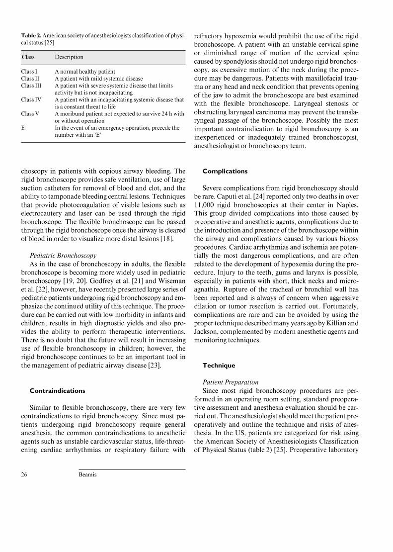
Class I
BOIS:L08:BPRR3024XA.73 FF: RESF E1: RESS
26 Beamis
Table 2. American society of anesthesiologists classification of physi-cal status [25]
Class Description
A normal healthy patientClass II A patient with mild systemic diseaseClass III A patient with severe systemic disease that limits
activity but is not incapacitatingClass IV A patient with an incapacitating systemic disease that
is a constant threat to lifeClass V A moribund patient not expected to survive 24 h with
or without operationE In the event of an emergency operation, precede the
number with an ‘E’
choscopy in patients with copious airway bleeding. Therigid bronchoscope provides safe ventilation, use of largesuction catheters for removal of blood and clot, and theability to tamponade bleeding central lesions. Techniquesthat provide photocoagulation of visible lesions such aselectrocautery and laser can be used through the rigidbronchoscope. The flexible bronchoscope can be passedthrough the rigid bronchoscope once the airway is clearedof blood in order to visualize more distal lesions [18].
Pediatric BronchoscopyAs in the case of bronchoscopy in adults, the flexible
bronchoscope is becoming more widely used in pediatricbronchoscopy [19, 20]. Godfrey et al. [21] and Wisemanet al. [22], however, have recently presented large series ofpediatric patients undergoing rigid bronchoscopy and em-phasize the continued utility of this technique. The proce-dure can be carried out with low morbidity in infants andchildren, results in high diagnostic yields and also pro-vides the ability to perform therapeutic interventions.There is no doubt that the future will result in increasinguse of flexible bronchoscopy in children; however, therigid bronchoscope continues to be an important tool inthe management of pediatric airway disease [23].
Contraindications
Similar to flexible bronchoscopy, there are very fewcontraindications to rigid bronchoscopy. Since most pa-tients undergoing rigid bronchoscopy require generalanesthesia, the common contraindications to anestheticagents such as unstable cardiovascular status, life-threat-ening cardiac arrhythmias or respiratory failure with
refractory hypoxemia would prohibit the use of the rigidbronchoscope. A patient with an unstable cervical spineor diminished range of motion of the cervical spinecaused by spondylosis should not undergo rigid bronchos-copy, as excessive motion of the neck during the proce-dure may be dangerous. Patients with maxillofacial trau-ma or any head and neck condition that prevents openingof the jaw to admit the bronchoscope are best examinedwith the flexible bronchoscope. Laryngeal stenosis orobstructing laryngeal carcinoma may prevent the transla-ryngeal passage of the bronchoscope. Possibly the mostimportant contraindication to rigid bronchoscopy is aninexperienced or inadequately trained bronchoscopist,anesthesiologist or bronchoscopy team.
Complications
Severe complications from rigid bronchoscopy shouldbe rare. Caputi et al. [24] reported only two deaths in over11,000 rigid bronchoscopies at their center in Naples.This group divided complications into those caused bypreoperative and anesthetic agents, complications due tothe introduction and presence of the bronchoscope withinthe airway and complications caused by various biopsyprocedures. Cardiac arrhythmias and ischemia are poten-tially the most dangerous complications, and are oftenrelated to the development of hypoxemia during the pro-cedure. Injury to the teeth, gums and larynx is possible,especially in patients with short, thick necks and micro-agnathia. Rupture of the tracheal or bronchial wall hasbeen reported and is always of concern when aggressivedilation or tumor resection is carried out. Fortunately,complications are rare and can be avoided by using theproper technique described many years ago by Killian andJackson, complemented by modern anesthetic agents andmonitoring techniques.
Technique
Patient PreparationSince most rigid bronchoscopy procedures are per-
formed in an operating room setting, standard preopera-tive assessment and anesthesia evaluation should be car-ried out. The anesthesiologist should meet the patient pre-operatively and outline the technique and risks of anes-thesia. In the US, patients are categorized for risk usingthe American Society of Anesthesiologists Classificationof Physical Status (table 2) [25]. Preoperative laboratory

BOIS:L08:BPRR3024XA.73 FF: RESF E1: RESS
Modern Use of Rigid Bronchoscopy 27
testing is determined by the patient’s condition, age, per-ceived risk and hospital requirements. Careful evaluationof the oral cavity, teeth, and jaw and neck mobility shouldbe made.
AnesthesiaThe bronchoscopist must have excellent communica-
tion with the anesthesiologist. Both consider the airwayexclusive territory, but in the case of bronchoscopy, theymust learn to share both the locale and the responsibilityof the procedure. Standard anesthetic monitoring shouldinclude pulse oximetry, electrocardiographic and bloodpressure monitoring, and some attempt at monitoring ofventilation. This might include end tidal CO2 monitoringwhen a closed system is employed or simple observationof chest wall excursion with an open system and jet venti-lation.
Detailed description of anesthesia for rigid bronchos-copy is beyond the scope of this chapter. Originally, rigidbronchoscopy was performed using topical anesthesia andminimal sedation. This often resulted in much discomfortfor the patient. Modern rigid bronchoscopy is almost uni-versally carried out under general anesthesia (table 3).Occasionally, patients can be bronchoscoped using topi-cal lidocaine anesthesia and intravenous conscious seda-tion. Intravenous propofol is the preferred agent for rigidbronchoscopy at this time. This agent produces rapidonset of anesthesia and permits rapid reawakening. Pro-pofol does not have ideal amnesic or analgesic properties,therefore preoperative use of an anxiolytic and amnesticagent such as midazolam is recommended. Intermittentboluses of an intravenous narcotic such as fentanyl oftenreduce the propofol requirements and treat any acute painexperienced during the procedure.
VentilationMultiple options exist for ventilating a patient during
rigid bronchoscopy. Ideally, the patient should be allowedto breathe spontaneously while being at a plane of anes-thesia that provides comfort as the bronchoscope ispassed. Often this is not possible or is insufficient to sup-press cough during delicate manipulations within the air-way. Spontaneous/assisted ventilation, as described byPerrin et al. [26], provides deeper anesthesia to the pointwhere spontaneous ventilation, although preserved, isquite shallow. Frequent assisted breaths must be providedby the anesthesiologist. This method leads to rapid re-awakening after the procedure, but is often insufficient tocontrol cough. Performing rigid bronchoscopy during ap-nea after breathing 100% oxygen may provide sufficient
Table 3. Ventilation and anesthesia for rigidbronchoscopy [7]
Ventilation optionsApneaSpontaneousSpontaneous/assistedControlled
Closed systemOpen system – jet ventilation
Via endotracheal tube
Anesthetic techniquesTopical/sedationGeneral
InhalationalIntravenous
time for an expert to perform a short procedure, such asremoval of a foreign body, but it is usually not appropriatefor most indications.
Modern rigid bronchoscopy involving use of lasers orother interventional techniques requires longer proceduretimes which are often facilitated by controlled ventilation[27]. Controlled ventilation with a closed system requirescapping of the proximal end of the bronchoscope and anysmall side ports and connecting the large side port to ananesthesia machine. Some method of preventing air leak-ing through the vocal cords must be provided. A totallyclosed system allows the use of inhalational anesthetics.Another method of closed ventilation involves placing asmall endotracheal tube through the larynx to ventilatethe patient, and then passing the rigid bronchoscopealongside the endotracheal tube. The size of the larynxlimits the diameter of the rigid tube that can be used withthis technique. Venturi jet ventilation permits the use ofan open system and has been shown to maintain effectivegas exchange during long procedures. Godde et al. [28]have described the Venturi jet technique, which involvesinjecting 100% oxygen at 50 psi into the side port of thebronchoscope. Adequacy of ventilation is determined byobserving movement of the chest with each injectedbreath. Since room air is entrained through the open endof the bronchoscope, the exact fraction of inspired oxygenis unknown. Oxygenation is monitored with pulse oxime-try. Providing 10–20 breaths per minute with this tech-nique, which often requires the use of muscle relaxants,results in adequate ventilation. The Venturi techniquecan also be used with high-frequency ventilation usingrates up to 100 per minute [29].

BOIS:L08:BPRR3024XA.73 FF: RESF E1: RESS
28 Beamis
Since bronchoscopy is considered a clean but not asterile procedure, bronchoscopists classically have worngloves, a mask and a standard short-sleeve operatingscrub suit. However, in this era of universal precautions,when there is potential risk from contact with bodilysecretions, it is prudent for the bronchoscopist to wear afull operating gown, glasses or other eye protection equip-ment, and an approved facial mask that limits inhalationof laser plume and aerosolized infectious agents (fig. 3).
Insertion TechniquesThere are several methods of insertion of the rigid
bronchoscope. The method chosen depends on the exper-tise of the bronchoscopist and the patient’s anatomy andclinical status and on discussion with the anesthesiologist.There are a number of detailed reviews of various rigidbronchoscopy insertion techniques both classic [30] andmodern [5–7, 31]. Some of the techniques will be summa-rized below. All begin with the patient pre-medicated,well oxygenated and comfortably lying supine on theoperating table. A general anesthetic is administered andmuscle relaxants, if needed, are infused. A disposabletooth guard is placed on the upper teeth and the patient’seyes are protected. The bronchoscopist then chooses oneof the following methods.
Classic TechniqueThe left thumb is placed over the upper teeth and the
left index finger is placed in the mouth protecting the low-er teeth and opening the jaw. The bronchoscope is held inthe right hand with the bevel anteriorly (fig. 4). Thebronchoscope is passed perpendicular to the table exactlyin the midline. Once the uvula is passed, the right handlowers the operator end of the bronchoscope and the baseof the tongue is gently raised. The epiglottis usually comesinto view at this point. The bevel of the bronchoscope isused to carefully raise the epiglottis bringing the larynxand vocal cords into view. At this point, the bronchoscopeis turned 90° to the side and advanced through the vocalcords. Once the scope is passed into the trachea, the bron-choscope is then turned back 90° so that the bevel is againanterior. Using a gentle twisting motion and holding thebarrel of the bronchoscope with the fingers of the left handin a fashion similar to holding a pool cue, the broncho-scope is advanced into the lower airways.
Introduction Using a LaryngoscopeWith this technique, a straight laryngoscope is used to
visualize the epiglottis (fig. 5). Once the epiglottis comesinto view, the laryngoscope blade is used to lift the base of
Fig. 3. Bronchoscopist, properly gowned, performing a procedureusing a video monitor.
the tongue raising the epiglottis slightly. With the righthand holding the bronchoscope, the tip of the scope ispassed just under the epiglottis. At this point, the bron-choscopist looks through the bronchoscope and guides thebronchoscope toward the larynx and simultaneously re-moves the laryngoscope. The vocal cords are passed in amanner similar to that of the classic technique.
Intubation Insertion along an Endotracheal TubeMany patients requiring interventional bronchoscopy
procedures have developed respiratory failure and cometo the bronchoscopy suite intubated receiving mechanicalventilation. After general anesthesia is administered, thebronchoscope can be passed along the endotracheal tubefrom the right side of the mouth. The bronchoscope iskept slightly anterior to the endotracheal tube and ad-vanced under the epiglottis to the point where both theleft and right vocal cords can be visualized. At this pointthe anesthesiologist releases the endotracheal balloon cuffand slowly pulls out the endotracheal tube. Once the tipof the endotracheal tube passes through the vocal cords,the bronchoscope is rotated 90° and quickly advancedthrough the cords.
This approach is recommended for any novice rigidbronchoscopist. Nonintubated patients coming into theoperating room can be intubated by the anesthesiologistafter induction of general anesthesia and muscle relax-ation. The bronchoscopist can then advance the bron-choscope to the level of the vocal cords in an unhurriedpace knowing that oxygenation and ventilation will bemaintained.

BOIS:L08:BPRR3024XA.73 FF: RESF E1: RESS
Modern Use of Rigid Bronchoscopy 29
Fig. 4. Initial position of bronchoscopist, patient and bronchoscopefor the classic insertion technique.
Fig. 5. Insertion of rigid bronchoscope using a laryngoscope. Thelaryngoscope was used to expose and lift the epiglottis and isremoved as the bronchoscope is guided through the larynx.
Bronchoscopic Insertion through Laryngectomy StomaIn this situation, a tracheoscope is preferred. The
patient’s neck is turned to one side and the tracheoscopeis then inserted through the tracheostomy stoma. Thisprocedure can often be carried out with topical lidocaineanesthesia and mild intravenous sedation. Muscle relax-ation is rarely required.
Once the bronchoscope passes the midtracheal level,the remainder of the procedure is similar for all intuba-tion techniques. To enter the right bronchial tree, thepatient’s head is turned to the left and the bronchoscopegently advanced past the carina in a twisting manner. Therigid bronchoscope can be easily passed into the distalbronchus intermedius in most patients. To enter the left
bronchial tree, the patient’s head is turned toward theright shoulder and again the bronchoscope is advanced ina twisting motion. Unless there is major distortion of theanatomy related to past surgery or radiation therapy, theleft upper and left lower lobe bronchi can be visualized inmost patients.
Removal of the bronchoscope from the airway shouldalways be done visually, again using a gentle twistingmotion. Patients are usually observed in a recovery areafor several hours after the procedure. Because the effectsof modern intravenous anesthetic agents and musclerelaxants resolve quickly, rigid bronchoscopy can often beperformed on an outpatient basis [32]. The patient’s over-all condition and not the procedure determine the needfor hospital admission.

BOIS:L08:BPRR3024XA.73 FF: RESF E1: RESS
30 Beamis
Training for Rigid Bronchoscopy
In many European centers training in rigid bronchos-copy is an integral part of pulmonary fellowship training.This is not the case in the US and Japan, where rigid bron-choscopy remains an uncommon, sometimes forgottenprocedure performed by less than 10% of bronchosco-pists. In these countries achieving training in rigid bron-choscopy is often difficult as experience with the proce-dure is not a requirement of current pulmonary and criti-cal care medicine fellowships, and access to rigid bron-choscopic equipment and operating room privileges maybe difficult to obtain. Years ago, Jackson emphasized thatrigid bronchoscopy is ‘not an undergraduate subject’. It is
advisable that one have extensive experience in flexiblebronchoscopy before attempting to use the rigid instru-ment. Jackson also states that ‘a fundamental knowledgeof technique, positions, and landmarks is necessary, afterwhich only continued manual practice is needed for profi-ciency’. Fortunately there are now a number of postgrad-uate courses during which experienced thoracic endosco-pists can at least achieve a rudimentary knowledge ofrigid bronchoscopy technique. These courses often in-clude practice on animal models and intubation manne-quins. Following this initial experience, practice on hu-mans under the guidance of an expert should allow mostserious thoracic endoscopists enough experience to even-tually perform the procedure alone.
References
1 Becker HD: Gustav Killian: A biographicalsketch. J Bronchol 1995;2:77–83.
2 Boyd AD: Chevalier Jackson: The father ofamerican bronchoesophagology. Ann ThoracSurg 1994;57:502–505.
3 Moghissi K, Jessob T, Dench M: A new bron-choscopy set for laser therapy. Thorax 1986;41:485–486.
4 Shapshay SM, Beamis JF, MacDonald E: Anew rigid bronchoscope for laser fiber applica-tion. Otolaryngol Head Neck Surg 1987;96:202–204.
5 Prakash UBS, Diaz-Jimenez JP: The rigidbronchoscope; in Prakash UBS: Bronchoscopy.New York, Raven Press, 1994, pp 53–69.
6 Helmers RA, Sanderson DR: Rigid bronchos-copy: The forgotten art. Clin Med 1995;16:393–399.
7 Beamis JF: Rigid bronchoscopy; in Beamis JF,Mathur PN: Interventional Pulmonology. NewYork, McGraw-Hill, 1999.
8 Miller JI Jr: Rigid bronchoscopy. Chest SurgClin N Am 1996;6:161–167.
9 Mehta AC, Dasgupta A: Bronchoscopic ap-proach to tracheobronchial foreign bodies inadults: Pro flexible bronchoscopy. J Bronchol1997;4:168–172.
10 Diaz-Jimenez JP: Bronchoscopic approach totracheobronchial foreign bodies in adults: Prorigid bronchoscopy. J Bronchol 1997;4:168–172.
11 Mathisen DJ, Grillo HC: Endoscopic relief ofmalignant airway obstruction. Ann ThoracSurg 1989;48:469–475.
12 Fenton JJ, Beamis JF: Laser bronchoscopy; inBeamis JF, Mathur PN: Interventional Pul-monology. New York, McGraw-Hill, 1999.
13 Sutedja TG, Boxem TJ, et al: Endobronchialelectrocautery is an excellent alternative forNd:YAG laser to treat airway tumors. J Bron-chol 1997;4:101–105.
14 Mathur PM, Wolf KM, et al: Fiberoptic bron-choscopic cryotherapy in the Management oftracheobronchial obstruction. Chest 1996;110:718–723.
15 Colt HG: Silicon airway stents; in Beamis JF,Mathur PN: Interventional Pulmonology. NewYork, McGraw-Hill, 1999, pp 97–112.
16 Petrou M, Goldstraw P: The management oftracheobronchial obstruction: A review of en-doscopic techniques. Eur J Cardiothorac Surg1994;8:436–441.
17 Heitz M, Bolliger CT: Tracheobronchial inser-tion of the dynamic (Rüsch) stent. J Bronchol1997;4:250–254.
18 Prakash UBS, Freitag L: Hemoptysis and bron-choscopy – Induced hemorrhage; PrakashUBS: Bronchoscopy. New York, Raven Press,1994, pp 227–252.
19 Wood RE, Postma D: Endoscopy of the airwayin infants and children. J Pediatr 1998;112:1–6.
20 Wood RE: Pediatric bronchoscopy: Rigid vsflexible bronchoscopy – Pro flexible bronchos-copy. J Bronchol 1996;3:156–160.
21 Godfrey S, Springer C, Maayan C, et al: Is therea place for rigid bronchoscopy in the manage-ment of pediatric lung disease. Pediatr Pul-monol 1987;3:179–184.
22 Wiseman NE, Sanchez I, Powell RE: Rigidbronchoscopy in the pediatric age group: Diag-nostic effectiveness. J Pediatr Surg 1992;27:1294–1297.
23 Holinger LD: Pediatric bronchoscopy: Rigid vsflexible bronchoscopy – Pro rigid bronchosco-py. J Bronchol 1996;3:153–155.
24 Caputi M, Bellissimo U, DiMatteo L, AlibertiM, Perna B, Speranza A: Complications duringbronchofiberscopy and rigid bronchoscopy.Panminerva Med 1986;28:271–277.
25 American Society of Anesthesiologists: Newclassification of physical status. Anesthesiology1963;24:111.
26 Perrin G, Colt HG, Martin C, et al:. Safety ininterventional rigid bronchoscopy using intra-venous anesthesia and spontaneous assistedventilation: A prospective study. Chest 1992;102:1526–1530.
27 Conacher ID, Paes LL, McMahon CC, MorrittGN: Anesthetic management of laser surgeryfor central airway obstruction: A twelve yearcase series. J Cardiothorac Vasc Anesth 1998;12:153–156.
28 Godden DJ, Willey RF, Fergusson RJ, WrightDJ, Compton GK, Grant IW: Rigid bronchos-copy under intravenous general anesthesia withoxygen Venturi ventilation. Thorax 1987;37:532–534.
29 Vourc’h G, Fischler M, Michon F, MalchiorJC, Seigneur F: Manual jet ventilation. V. Highfrequency jet ventilation during laser resectionof tracheal-bronchial stenosis. Br J Anaesth1983;55:973–975.
30 Jackson C, Jackson CL: Bronchoscopy, Eso-phagoscopy, and Gastroscopy: A Manual ofPerioral Endoscopy and Laryngeal Surgery.Philadelphia, Saunders, 1934.
31 Stradeling P: Diagnostic Bronchoscopy: ATeaching Manual, ed 5. London, Churchill Liv-ingston, 1986.
32 Lee CM, Heinrichs PA, Terris DJ: Routinehospital admission for patients undergoing up-per airway digestive tract endoscopy is unwar-ranted. Ann Otol Rhinol Laryngol 1998;107:247–253.
John F. Beamis, Jr.Section Pulmonary and Critical CareMedicine, Lahey Clinic, 41 Mall RoadBurlington, MA 01805 (USA)Tel. +1 781 744 8480, Fax +1 781 744 3443E-Mail [email protected]

OOOOOOOOOOOOOOOOOOOOOOOOOOOOOOOOOOOOOOOOOOOOOOOOOOOOOOOOOOOOOOOOOOOOOOOOOOOOOOOOOOOOOOOOOOOOOOOOOOOOOOOOOOOOOOOOOOOOOOOOOOO
General Aspects of InterventionalBronchoscopy

Bolliger CT, Mathur PN (eds): Interventional Bronchoscopy.Prog Respir Res. Basel, Karger, 2000, vol 30, pp 31–43
OOOOOOOOOOOOOOOOOOOOOOOOOOOOOOOOOOOOOOOOOOOOOOOOOOOOOOOOOOOOOOOOOOOOOOOOOOOOOOOOOOOOOOOOOOOOOOOOOOOOOOOOOOOOOOOOOOOOOOOOOOO
Bronchoscopy Unit, Expertise,Equipment and PersonnelUdaya B.S. Prakash
Mayo Medical Center, Rochester, Minn., USA
31
Summary
A special unit dedicated to bronchoscopy and relatedprocedures may not be practical at all medical centers. How-ever, comprehensive practice of interventional pulmonologywill require a dedicated unit where a large number of spe-cialized bronchoscopy procedures such as special bronchos-copies, laser therapy, stent placement, pediatric bronchos-copy, complicated rigid bronchoscopy, thoracoscopy, trans-thoracic needle aspiration of pulmonary centers and percu-taneous tracheostomy are performed on a regular basis. Themajor advantages of a dedicated bronchoscopy unit are theavailability of any type of instrument and the facility to per-form all types of bronchoscopy-related procedures. Certainprocedures, for example bronchoscopic lung biopsy underfluoroscopy, may necessitate the use of the radiology suite, ifa mobile fluoroscope is not available. The need for a dedi-cated bronchoscopy unit should be determined by the num-ber of procedures performed in a given time period at anymedical center as well as the expertise of physicians whopractice bronchoscopy and related procedures. A dedicatedbronchoscopy unit consists of a predefined physical locationwhere the procedures are performed, all equipment neces-sary to provide bronchoscopy and related procedures, facili-ties to administer general anesthesia, immediately availablepathology services, and a bronchoscopy team composed ofwell-trained bronchologists, anesthesiologists, and nurses/surgical technicians. It is important to recognize that anoverwhelming number of ‘routine’ bronchoscopy proce-
dures can be performed safely in other areas, including thepatient’s room, intensive care unit, emergency ward and inan outpatient setting. Therefore, the bronchoscopy teamshould be mobile enough to perform bronchoscopy proce-dures in almost any location within the medical center at ashort notice. Irrespective of the location where the proce-dure is performed, the basic maxim is that the bronchoscopyteam is optimally trained to perform the procedure and han-dle complications.
Bronchoscopy and related procedures are commonlyperformed in most medical centers that provide tertiarymedical care. The sheer number of procedures performedin such medical centers necessitates organization andmaintenance of a devoted bronchoscopy suite or unit.However, a special unit dedicated to bronchoscopy andrelated procedures may be impractical at all medical cen-ters. The need for a dedicated bronchoscopy unit isdependent on the overall medical care provided by themedical center and the types of bronchoscopy servicespracticed, such as special bronchoscopies, laser therapy,stent placement, pediatric bronchoscopy and complicatedrigid bronchoscopy. The last decade has witnessed growthof departments specializing in interventional pulmonolo-gy in which not only bronchoscopy but also other proce-dures such as thoracoscopy, transthoracic needle aspira-tion and percutaneous tracheostomy are performed on a

HABE:L08:BPRR3019XA.78 FF: RESF E1: RESS
32 Prakash
regular basis. Therefore, comprehensive practice of inter-ventional pulmonology will require a dedicated unit forsmooth performance of these procedures. Most impor-tantly, the need for a dedicated bronchoscopy unit is dic-tated by the number of bronchoscopy procedures per-formed in a given time period at any medical center aswell as the expertise of physicians who practice bronchos-copy and related procedures.
A dedicated bronchoscopy unit consists of not only apredefined physical location where the procedures areregularly performed, but also readily available equipmentnecessary to provide bronchoscopy and related proce-dures, and a team composed of well-trained broncholog-ists, anesthesiologists and nurses/surgical technicians.While it is convenient to have a dedicated operating roomor other facility, an overwhelming number of ‘routine’bronchoscopy procedures can be performed safely in oth-er areas, including the patient’s room, intensive care unit,emergency ward and in an outpatient setting. Thus, adedicated bronchoscopy unit cannot be stationary. Thebronchoscopy service should be mobile enough to per-form bronchoscopy procedures in almost any locationwithin the medical center at a short notice. Irrespective ofthe location where the procedure is performed, the funda-mental tenet is that the bronchoscopy team is optimallytrained to perform the procedure, and it includes the per-sonnel and equipment to deal with complications, shouldthey develop [1, 2].
Certain procedures require specific amenities to enablethe bronchologist to provide optimal bronchoscopy ser-vices. For instance, bronchoscopic lung biopsy underfluoroscopy may call for using the radiology suite, if amobile fluoroscope is not available. Another example iswhere the laser bronchoscopy has to be performed in alaser unit located in a separate area, away from the loca-tion where routine bronchoscopies are done. As indicatedabove, a patient in the intensive care unit may requirebedside bronchoscopy. This necessitates the availabilityof a mobile bronchoscopy unit. At major medical centerswith busy practice of thoracic surgery, it is a commonpractice to perform intraoperative bronchoscopy to assistthe thoracic surgeon [3]. This requires the availability ofthe bronchoscopist and bronchoscopy equipment in theoperating room. Before the introduction of the flexiblebronchoscope into clinical practice, evaluation of the tra-cheobronchial tree of infants required transportation tothe operating room for rigid bronchoscopy. Presently,however, the availability of the small-diameter (ultra-thin) flexible bronchoscope allows safe performance ofbronchoscopy in the neonatal intensive care unit [4, 5].
Nevertheless, rigid bronchoscopy can be performed in theneonatal intensive care unit, provided appropriate equip-ment and personnel are available [2, 6].
The American College of Chest Physicians (ACCP)survey, in 1989, of 871 bronchoscopists in the US andCanada revealed that bronchoscopy was performed invarious physical locations: the operating room (50%),patient’s room (56%), physician’s office (11%), in a bron-choscopy suite/laboratory or pulmonary function test lab-oratory (17%) and intensive care unit (2.5%) [7]. Outpa-tient bronchoscopy (in the hospital) was practiced by 63%of physicians. A more recent postal survey by the Ameri-can Association for Bronchology (AAB) in 1999 elicitedresponses from 744 of the 3,000 North American bron-chologists who received the questionnaire, and of therespondents, only 28% indicated that they had a dedi-cated unit for bronchoscopy, which was also used as thepostbronchoscopy recovery area [8]. In 1995, the pediat-ric bronchology group of the European Respiratory Soci-ety surveyed 125 European medical centers, of whom 51(41%) responded. The survey observed that flexible bron-choscopy was carried out in the operating room in 19 cen-ters, in the intensive care unit in 22 centers and in special-ly equipped rooms in 30 centers, whereas the rigid bron-choscopy was performed in the operating room in 23 cen-ters, in the intensive care unit in 7 centers and in aequipped room in 15 centers [9].
The Bronchoscopy Unit
The physical requirements of an ideal bronchoscopyunit include adequate space for the following: Storage ofbronchoscopy-relaetd equipment, prebronchoscopy prep-aration of the patient, performance of the procedure and apostbronchoscopy area to observe the patient. As indi-cated by the surveys by the ACCP and the AAB, this idealis far from reality in North America [1]. The dimensionsof the bronchoscopy unit will depend on the types of pro-cedures performed and the daily case load of each bron-choscopist and medical center. The major advantages of adedicated bronchoscopy unit are the availability of anytype of instrument and the facility to perform all types ofbronchoscopy-related procedures. At medical centerswhich perform a significant number of complicated bron-choscopic procedures such as laser bronchoscopy, stentplacement, pediatric bronchoscopy and complicated rigidbronchoscopy, it may be convenient to locate the bron-choscopy unit adjacent to surgical operating rooms so thatthe expertise of the thoracic surgeons and the anesthesiol-

HABE:L08:BPRR3019XA.78 FF: RESF E1: RESS
Bronchoscopy Unit, Expertise, Equipmentand Personnel
33
Fig. 1. The architect’s drawing of the floor plan of the present bronchoscopy suite at Mayo Medical Center, Rochester,Minn., USA, seen from above, and from four different perspectives (a–d). The black arrow and the letter N provideorientation.ABCD
E
====
=
Preparation (and induction) roomBronchoscopy suiteSurgical bedCeiling-mounted power source for bronchoscopylight sources and anesthetic gasesView box for imaging studies
FGHIJK
======
Nd:YAG laserArgon-dye laserStorage spacesWash basinSupply core and storage areaPaper work area
ogists is readily available [2]. Other interventional pul-monology procedures, such as thoracoscopy, transthorac-ic needle aspiration, percutaneous tracheostomy, endo-scopic ultrasound-guided procedures will require specialequipment and personnel. Irrespective of where the bron-
choscopy is performed, personnel and equipment to pro-vide resuscitation should be readily available.
An example of a fully equipped bronchoscopy room atthe Mayo Medical Center, Rochester, Minn., is providedin figures1–3. The room was designed by the bronchosco-
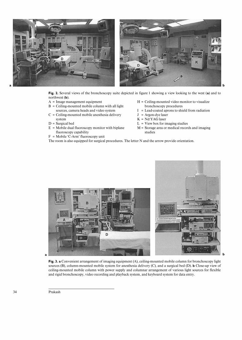
HABE:L08:BPRR3019XA.78 FF: RESF E1: RESS
34 Prakash
Fig. 2. Several views of the bronchoscopy suite depicted in figure 1 showing a view looking to the west (a) and tonorthwest (b).AB
C
DE
F
==
=
==
=
Image management equipmentCeiling-mounted mobile column with all lightsources, camera heads and video systemCeiling-mounted mobile anesthesia deliverysystemSurgical bedMobile dual fluoroscopy monitor with biplanefluoroscopy capabilityMobile ‘C-Arm’ fluoroscopy unit
H
IJKLM
=
=====
Ceiling-mounted video monitor to visualizebronchoscopy proceduresLead-coated aprons to shield from radiationArgon-dye laserNd:YAG laserView box for imaging studiesStorage area or medical records and imagingstudies
The room is also equipped for surgical procedures. The letter N and the arrow provide orientation.
Fig. 3. a Convenient arrangement of imaging equipment (A), ceiling-mounted mobile column for bronchoscopy lightsources (B), column-mounted mobile system for anesthesia delivery (C), and a surgical bed (D). b Close-up view ofceiling-mounted mobile column with power supply and columnar arrangement of various light sources for flexibleand rigid bronchoscopy, video recording and playback system, and keyboard system for data entry.
a b
a b

HABE:L08:BPRR3019XA.78 FF: RESF E1: RESS
Bronchoscopy Unit, Expertise, Equipmentand Personnel
35
pists and nonphysician bronchoscopy personnel and ar-chitects. The dedicated suite is equipped for general anes-thesia and all types of bronchoscopic procedures in adultsand children. Such a dedicated unit next to other operat-ing rooms provides the bronchoscopists with an opportu-nity to offer optimal services to patient. Clearly, such anexclusive unit is neither necessary nor realistic in all medi-cal centers. However, a busy bronchoscopy practice willbenefit from a dedicated unit that is fully equipped for alltypes of bronchoscopy procedures. Ultimately, severalfactors contribute to the choice of location for the proce-dure and choice of assistants such as personal preference,convenience, available facilities and availability of per-sonnel or paramedical training programs [1]. Financialconsiderations play the determining role in these deci-sions.
Patient Preparation AreaThis area should be adjacent to the procedure area and
is primarily designed for administration of premedica-tions and instillation of topical anesthesia prior to bron-choscopy. A distinct patient area is not an absoluterequirement. However, a separate preparation locale istimesaving in a busy bronchoscopy practice when onepatient can be prepared in this area by assistants as thebronchoscopist completes an earlier case. The bronchos-copist and the bronchoscopy team should assemble theminimum equipment required to prepare the patient.Easy accessibility of supplemental oxygen and resuscita-tion equipment is imperative. The bronchoscopist shouldbe able to prepare the patient single-handedly unless thepatient is too sick to adequately cooperate and requiresadditional help from a nurse, surgical technician or anoth-er physician.
The Procedure AreaThe procedure area should remain uncluttered so that
the bronchoscopist and the assisting personnel can movearound the patient’s head area and be able to perform var-ious bronchoscopic maneuvers and manage unforeseencomplications. The patient can be in the seated or supineposition on an operating table, in a patient bed, a chair orin a chair similar to the one used by dentists. Irrespectiveof the type of table or chair used, the bronchoscopistshould be able to place the patient in the reverse Trendel-enburg position in case of severe bronchoscopy-inducedbleeding or hypotension. The instruments used should beeasily accessible to the bronchoscopist and the main assis-tant. If general anesthesia is required, appropriate equip-ment should be available. Adequate space should be avail-
able to bring in special equipment such as a fluoroscopeand laser unit.
The Postbronchoscopy AreaThis area is primarily for observation of the patient
after bronchoscopy is completed. Since almost all patientsrequire a period of observation to ensure that there are nocomplications from the pharmaceutical agents used dur-ing the procedure or the procedure itself, a designatedarea for observation is necessary. Depending on the setupat each medical center, this area may include the bron-choscopy unit itself, a postsurgical recovery unit, a roomadjacent to the bronchoscopy unit, a waiting room or thepatient’s room itself if bedside bronchoscopy is per-formed.
In the survey of North American bronchoscopists, 56%reported that they used an area adjacent to the bronchos-copy unit as the postbronchoscopy recovery area, whereas43% used a separate area such as an outpatient waitingarea, the procedure room itself or the emergency room forthis purpose [7]. In the AAB survey mentioned above,only 28% of the 744 North American bronchologists indi-cated that they had a dedicated unit for bronchoscopywhich was also used as the postbronchoscopy recoveryarea [8]. Postbronchoscopy observation should be under-taken by the bronchoscopist, another physician or a nursetrained to recognize postbronchoscopy complications.
The Bronchoscopy Equipment
The types of bronchoscopes required depend on theneeds and the type of bronchoscopy practice. The mini-mum requirements to perform flexible bronchoscopy toobtain biopsies of the lesions in the tracheobronchial treeand therapeutic bronchoscopy in adults include an adult-size flexible bronchoscope, a light source, several cytologybrushes and biopsy forceps, specimen containers, suctionapparatus, and supplemental oxygen and equipment forresuscitation (fig. 4). Premedications, sedatives, and topi-cal anesthetic agents are also necessary for most proce-dures. Even in the absence of a large-volume bronchosco-py practice, the bronchoscopist is well advised to have anextra bronchoscope and several biopsy forceps in case ofbreakdown of the equipment [2]. All other types of bron-choscopic procedures such as rigid bronchoscopy, laserbronchoscopy, brachytherapy, cryotherapy and stentplacement will require additional instruments (fig. 5). Li-kewise, pediatric bronchoscopy will require an entirelyseparate set of instruments and accessories [10, 11].

HABE:L08:BPRR3019XA.78 FF: RESF E1: RESS
36 Prakash
Fig. 4. Minimal requirements for performing flexible bronchoscopy.1 =2 =
3 =4 =
5 =
6 =7 =
Flexible bronchoscopeSoft, uncuffed and noncollapsibleendotracheal (ET) tubeBite-block (mouthpiece)ET tube adapter for supplementaloxygen delivery20-cm-long adhesive tape to fasten ETtubeFlexible biopsy forcepsSterile glass container to collectbronchial lavage
8 =9 =
10 =11 =12 =13 =
14 =
15 =
Glass slides for cytologic smearsPick-up forceps to remove biopsyspecimens from biopsy forcepsBronchial brushSpecimen jar with formaldehydeSlide container with 95% alcoholLactated Ringer’s solution forbronchial lavageMetal container with warm water todefog and clean the bronchoscope tipEye pads and towel clip
16 =
17 =
18 =19 =20 =21 =22 =
Medicine jar with lactated Ringer’ssolutionSyringes – one with lactated Ringer’ssolution and the other containing4% lidocaineMetal cup containing 4% lidocaine4% lidocaineLecture scopeCold-light sourceElectrocardiographic monitor
Not shown in the figure are a tube of lubricating jelly and gauze pads. Figure reproduced from Seminars in Respiratory Medicine [43].
The BronchoscopesThe bronchoscopes, both flexible and rigid, are manu-
factured by various manufacturers. The bronchoscopistshould acquire the appropriate equipment based on whatthe practice requires. The number and types of the bron-choscopes required will depend on the needs of each indi-vidual bronchoscopist. The bare minimum required isdescribed above. While comparison of various types ofbronchoscopes by different manufacturers is beyond thescope of this writing, certain general recommendationscan be provided. The reliability of the manufacturer forprompt delivery of service, accessories, repairs and re-placement of damaged equipment is very important. Thebronchoscopists should be cognizant of the fact thatinstruments made by one manufacturer may not be com-patible with those made by a different manufacturer.Therefore, it is better to purchase the bronchoscope andrelated equipment made by the same manufacturer.
A bronchoscopist involved in all aspects of bronchos-copy may require flexible and rigid bronchoscopes of var-ious sizes to accommodate adults as well as pediatric
patients. Several types of both flexible and rigid broncho-scopes will be required if laser bronchoscopy, stent place-ment and removal of foreign bodies are to be undertaken.Pediatric bronchoscopy will require ultrathin flexiblebronchoscopes and rigid bronchoscopes of smaller diame-ters.
Cytology Brushes for the Flexible BronchoscopeBoth disposable and reusable brushes are available for
obtaining cytological specimens from airways and pulmo-nary parenchymal lesions. Brushes are sheathed or un-sheathed and lined with bristles that have varying degreesof stiffness or flexibility. Models with varying bristle sizesare also available. The efficacies of various cytologybrushes have been studied. One study examined the cellu-lar yield expressed as the number of cells recovered withtwo different types of sheathed cytology brushes, one withslightly longer and wider bristles. The latter providedgreater yield in total number of cells recovered per brush[12]. On the other hand, another study used sheathedbrushes of 1.00, 1.73 and 3.0 mm diameter and showed

HABE:L08:BPRR3019XA.78 FF: RESF E1: RESS
Bronchoscopy Unit, Expertise, Equipmentand Personnel
37
Fig. 5. Minimal requirements for performing rigid bronchoscopy.1 =
2 =3 =4 =5 =6 =
Rigid bronchoscope with light supplycord below and oxygen delivery tubeaboveStraight-view telescope90° view telescopeBiopsy forcepsEye shieldSyringe and long rubber-tipped metaltube for bronchial lavage through rigidbronchoscope
7 =
8 =9 =
10 =11 =
Atomizer containing 4% lidocaine – fortopical anesthetic application throughrigid bronchoscope to anesthetizebronchial mucosaPair of scissors‘Umbilical tape’ to pack severelyhemorrhaging bronchusMedicine cup containing lavage fluidContainer with warm water to cleanbiopsy forceps, telescope tips etc.
12 =13 =14 =15 =
16 =17 =18 =
Specimen jar with formaldehydeEye padsTowel clipContainer to collect bronchial lavagefluidLong suction cannulaBronchial lavage containerLight source
Figure reproduced from Seminars in Respiratory Medicine [43].
no significant difference in cell recovery among the threebrushes [13]. It is important to recognize that the tech-nique of brushing and preparation of cytology smears isfar more significant than the type of brushes used [14].
Biopsy Forceps for the Flexible BronchoscopeThe standard adult flexible bronchoscopes have work-
ing channels that range from 2.0 to 2.8 mm in diameter.Biopsy forceps that can traverse this channel come in sev-eral sizes and shapes. There are no specially designed for-ceps that can traverse the working channel of pediatric(ultrathin) flexible bronchoscopes. The three basic typesinclude those with serrated edge (alligator), smooth edgewith fenestrated or unfenestrated cups and ‘spiked’ (im-paler needle between the cups). Some forceps are manu-factured with longer blades. The latter type of forceps ishelpful in obtaining a biopsy from mucosal abnormalitiesalong the tracheal wall or from lesions from which the reg-
ular forceps easily slip or slide. All forceps have a proxi-mal control part and the forceps itself at the distal tip.
The choice of biopsy forceps depends on the individualbronchoscopist and the performance of the forceps. Wanget al. [15] studied the efficacy of three different types offorceps to obtain bronchoscopic lung biopsy and reportedthat larger forceps and serrated forceps provided largerspecimens, but the size of the biopsy specimen itself didnot significantly alter the diagnosis. The nondisposableforceps have finite life, and a study observed that biopsyforceps become dull after 20 or 25 applications [16].
Biopsy Forceps for the Rigid BronchoscopeMany types of forceps are available for use with the
rigid bronchoscope. The numerous types of forceps per-mit handling almost all types of problems within the air-ways. Bronchoscopic lung biopsy can be obtained withspecial forceps via a rigid bronchoscope [17]. Specialized

HABE:L08:BPRR3019XA.78 FF: RESF E1: RESS
38 Prakash
forceps are available for removal of various types of for-eign bodies. In design, the forceps are usually much largerthan those used with the flexible bronchoscope, slightlyrigid in performance and stronger. The forceps for usewith the pediatric rigid bronchoscope are identical instructural details but smaller in size.
Bronchoscopy NeedlesBronchoscopic needle aspiration and biopsy are com-
monly used in the staging of lung cancers. They are fre-quently employed to sample paratracheal, hilar and sub-carinal lymph nodes. Almost all needles available on themarket are disposable. Two sizes (19 and 21 gauge) areavailable; the larger needle is used to obtain tissue biopsy,whereas the smaller needle is designed for obtaining cytol-ogy aspiration. Retractable and nonretractable needles aswell as metallic and plastic needles are available. Manymanufacturers have discontinued the sale of nonretracta-ble needles because of the high risk of injury to the innerlining of the flexible bronchoscope.
Baskets and ClawsThese are used to extract aspirated tracheobronchial
foreign bodies, thick mucous plugs and clots. These acces-sories are rather flimsy and not as effective as instrumentsavailable for removal of foreign body with a rigid bron-choscope.
Protected CathetersThe protected catheter brushes are used for obtaining
respiratory specimens for bacterial culture [18–20]. Var-ious types are available and all are meant for single use.The preference for these catheters depends on individualbronchoscopist and the performance of the catheter. Inrecent years, bronchoalveolar lavage has almost totallyreplaced the protected specimen brush for obtaining spec-imens for bacterial cultures.
Specimen TrapsSeveral types of specimen traps are available to collect
bronchial washings and bronchoalveolar lavage effluent.Special containers to hold cytology slides and biopsy spec-imens are commercially available. Many bronchoscopistsimprovise their own system with spare parts. Major medi-cal centers may have these manufactured to their specifi-cations. The bronchoscopist is well advised to discuss her/his needs with the personnel in the pathology and micro-biology laboratories so that a mutually acceptable equip-ment is designed and procured.
FluoroscopyThe fluoroscopes used as adjuncts to bronchoscopy
machines are either fixed or mobile (portable). If themobile unit is not available, the patient will have to betransported to the location of fluoroscopy unit, generallyin the chest roentgenology unit. The mobile units aregreatly helpful in obtaining bronchoscopic lung biopsy inpatient rooms and other areas (fig. 2). The fluoroscopyunits are expensive and therefore, a fluoroscopy unit dedi-cated only to bronchoscopy is not realistic for most bron-choscopists. The mail survey of 871 bronchoscopists inNorth America noted that fluoroscopy facility dedicatedto bronchoscopy was reported by 20.9% of the respon-dents and fluoroscopy was shared with nonbronchosco-pists by 74.1%.
Fluoroscopic guidance assists in reaching localizedlesions in the lung parenchyma. It also helps reduce theincidence of pneumothorax following bronchoscopic lungbiopsy. The safety of outpatient bronchoscopy, includingbronchoscopic lung biopsy, is well documented [21–24].Even though bronchoscopic lung biopsy can be performedwithout fluoroscopic guidance, the incidence of pneumo-thorax associated following bronchoscopic lung biopsy isless than 1.8% when fluoroscopy is used and the incidencesignificantly increases to 2.9% without fluoroscopy [25].Another use for the fluoroscope is in the placement of air-way stents.
Endotracheal TubesEndotracheal tubes to facilitate easy insertion of the
flexible bronchoscope into the airways are not used by allbronchoscopists. However, many insert endotrachealtubes routinely prior to flexible bronchoscopy. To permiteasy passage of most adult flexible bronchoscopes, theminimum inner diameter of the endotracheal tube re-quired is 7.5 mm. While any type of endotracheal tubecan be used, soft latex wire-spiral tubes, without cuff, areeasier to use over a flexible bronchoscope and better toler-ated by patients [26]. As the ultrathin flexible broncho-scopes used in pediatric practice have varying diameters(1.8–3.2 mm), endotracheal tubes of different diametersshould be available if endotracheal intubation is plan-ned.
The Monitoring EquipmentNoninvasive monitoring equipment such as sphygmo-
manometry, electrocardiographic monitoring and pulseoximetry are routinely used by most bronchoscopists.Pulse oximetry is useful to assess adequacy of oxygenationwhether or not supplemental oxygen is given [27–29]. In

HABE:L08:BPRR3019XA.78 FF: RESF E1: RESS
Bronchoscopy Unit, Expertise, Equipmentand Personnel
39
Fig. 6. Special storage cupboard for flexible bronchoscopes (a), and a close-up view (b). Hanging them as shown pre-vents development of curves in the shaft of the instrument. Figure reproduced with permission from Prakash et al. [2].
the ACCP mail survey [7], routine use of pulse or otheroximetry was practiced by 84.2% of the respondents andelectrocardiographic monitoring by 74.6%. Supplementaloxygen should always be available.
Other EquipmentSpecial equipment required for laser bronchoscopy,
electrocautery, cryotherapy, airway stent placement, bra-chytherapy, airway dilatation, phototherapy, broncho-scopic ultrasound and other emerging technologies arediscussed elsewhere in this book.
Suction apparatus, extra tubings, cleaning materialsand commonly used pharmaceutical agents are among themiscellaneous equipment necessary for the optimal prac-tice of bronchoscopy. All equipment and medicationsnecessary for cardiopulmonary resuscitation should bereadily available at bronchoscopy location.
The Storage AreaThe size of the storage for the bronchoscopy equip-
ment area depends on the diversity of bronchoscopy prac-tice. If a bronchoscopist limits her/himself to simple bron-choscopy (only flexible bronchoscopy and no pediatricbronchoscopy or other special procedures), then all thebronchoscopy equipment can be stored in a mobile cart ina corner of a storage room in the hospital or office. Bron-choscopists specializing in laser bronchoscopy, rigid bron-choscopy and other complex procedures will require morespacious areas to store their equipment [2]. The flexiblebronchoscope is a delicate instrument and requires properhandling and storage (fig. 6).
Expertise
Bronchoscopy should be performed by physicians whoare skilled and appropriately trained in the procedure. Itis unclear how many procedures one should performunder tutelage to achieve proficiency in routine bronchos-copy. The eligibility requirements to obtain certificationin pulmonary diseases by the American Board of InternalMedicine do not specify the performance of a minimumnumber of bronchoscopies during the training [30]. TheAmerican Board of Thoracic Surgery requires its candi-dates to have performed at least ‘25 bronchoscopy andesophagoscopy’ procedures [31]. An individual who com-pleted training in pulmonary diseases oberved that atleast 100 bronchoscopies were needed to become profi-cient [32]. Others have indicated that to become profi-cient in bronchoscopy, performance of 50 to 100 bron-choscopies is necessary [33]. Some physicians, in spite of‘performing’ many more than 50 bronchoscopies, mayremain inept and lack confidence in achieving proficiencyin the procedure. Therefore, the decision whether some-one is competent to perform bronchoscopy should not bebased on the basis of the number of bronchoscopies per-formed. The program director or the director of bronchos-copy at the training institution should judge and certifythe competence of each candidate and recommend andprovide remedial training if necessary [7]. It is also impor-tant that the training of the bronchoscopists should in-clude assisting the bronchoscopist during bronchoscopyand related procedures.
a b

HABE:L08:BPRR3019XA.78 FF: RESF E1: RESS
40 Prakash
A national mail survey of 736 members of the Ameri-can College of Physicians who were identified as practic-ing pulmonologists in the USA observed that flexiblebronchoscopy was performed by 96% pulmonologists,bronchoscopic needle aspiration by 77%, foreign bodyremoval by 83%, rigid bronchoscopy for biopsy or remov-al of foreign body by 8% and Nd:Yag laser therapy by13% [34]. When asked about (1) number of proceduresthey did in the previous year, (2) number of proceduresper year needed to attain proficiency and (3) number ofprocedures per year required to maintain proficiency, therespondents answered (median numbers shown for thethree variables) in the following way: flexible bronchosco-py 100, 50 and 24, bronchoscopic lung biopsy 25, 20 and10, and laser phototherapy 5, 10 and 5. Among these pul-monologists, bronchoscopy training was obtained throughformal training by 53%, through attending course/s by 7%and on their own by 42% of the respondents [34]. An edi-torial urged the major pulmonary and critical care socie-ties to enact comprehensive guidelines for granting hos-pital privileges to perform the major procedures. It alsorecommended well-designed ‘mini-fellowships’ of 2–16weeks duration for introducing physicians in clinical prac-tice to major new procedures that were not taught duringformal fellowship training [35]. This will be difficult toaccomplish because of the problems associated with or-ganizing ‘on hands’ training for large groups of physicians.Short didactic courses (without actual practice) may notprovide sufficient supervised training to attain profi-ciency. Strict guidelines will undoubtedly generate dis-agreements among clinicians. Nevertheless, well-definedguidelines are likely to prevent improper and conceivablysuperfluous (as well as life-threatening) procedures [36].
At least 50 bronchoscopy procedures may be necessaryto become competent in routine flexible bronchoscopy [7].This should include biopsy of visible tracheobronchiallesions, bronchoalveolar lavage and therapeutic bronchos-copy. At least 50 bronchoscopies per year may be necessaryto remain competent in the procedure. One study thatassessed the extent of training required to attain competen-cy in bronchoscopic needle aspiration observed that toachieve acceptable results from this procedure, a trainingperiod that includes about 50 procedures is required [37].Another study over a 3-year period during which serialmultifaceted educational interventions directed towardbronchoscopists and their technical staff were providedobserved that diagnostic yield from bronchoscopic needleaspiration increased significantly from 21 to 48% [38].
In the ACCP survey, when asked about the number ofbronchoscopies required to become and remain compe-
tent in the procedure, more than 60% of respondentsrecorded that at least 50 procedures were necessary tobecome competent. More than half the survey partici-pants noted that at least 25 bronchoscopies per year wererequired to remain competent [1]. In the more recent sur-vey conducted by the AAB, when asked about bronchos-copy training during their fellowship, the responses wereas follows: training in flexible bronchoscopy (95%), rigidbronchoscopy (27%), laser procedures (24%), stent inser-tions (23%), brachytherapy (31%), cryotherapy (22%) andbronchoscopic electrocautery (22%). Many had receivedtraining ‘on the job’, particularly in brachytherapy, rigidbronchoscopy, laser procedures, stent insertion, cryother-apy and electrocautery. Another source of training wasthrough the 2-day courses in interventional broncholo-gy[8].
The ACCP survey indicated that bronchoscopists aredivided on whether or not rigid guidelines for bronchos-copy should be established [1]. In the AAB survey, only2% of the respondents felt they needed additional instruc-tion or education in flexible bronchoscopy, but manymore expressed a need for additional information on stentinsertion (59%), laser procedures (43%), cryotherapy(39%), electrocautery (37%) and brachytherapy (31%).Over one-fifth of the respondents felt no need for addi-tional instruction or education in any of the procedures[8].
These surveys reveal that over the previous decade,about a quarter of the surveyed physicians had receivedtraining in therapeutic bronchoscopic procedures duringtheir felloships, compared to only 8% of bronchoscopistssurveyed in 1989. When asked whether the AAB, ACCPand American Thoracic Society should provide detailedguidelines pertaining to the practice of flexible bronchos-copy, 87% answered affirmatively and 11% negatively. Asignificant number of respondents (71%) mentioned thatlaser procedures and stenting should be performed only intertiary care facilities, and 51% of the respondents feltthat interventional techniques should be taught duringpart of a routine pulmonary fellowship. The two majorprocedures respondents believed should be taught werebrachytherapy (52%) and stent insertion (51%) [8]. Theseresponses indicate that many bronchologists feel the needfor practice guidelines and specialized training in certainprocedures. It is obvious that some novices attain profi-ciency sooner than others do, and some may never feel‘comfortable’ enough to perform certain complicated pro-cedures. Maintenance of competence is more dependenton performance of the procedure on a regular basis ratherthan the numbers alone.

HABE:L08:BPRR3019XA.78 FF: RESF E1: RESS
Bronchoscopy Unit, Expertise, Equipmentand Personnel
41
The Bronchoscopy Personnel
The mail survey described above reported that nursesalone assisted 39% of the bronchoscopists during the pro-cedure whereas 26% of the bronchoscopists employednurses and other assistants; respiratory therapists werethe sole assistants to 14.7% of the bronchoscopists, and7.6% of physicians were assisted by respiratory therapistsand others (nurses, laboratory technicians, residents). Pul-monary function technicians and other paramedical per-sonnel assisted 12% of the bronchoscopists. Physicianswere mentioned as bronchoscopy assistants by 15% [7].Since many bronchoscopy units are adjacent to or part ofa pulmonary function testing laboratory or respiratorytherapy department, many train the technicians in theseareas to assist in bronchoscopy. As in any surgical proce-dure, the team concept is vital to the performance of bron-choscopy.
Training of Paramedical PersonnelMany routine flexible bronchoscopy procedures can be
carried out by a well-trained bronchoscopist without oth-ers to help during the procedure. However, most compli-cated procedures and a busy bronchoscopy practice war-rant additional assistance. Ideally, 2 trained persons, oth-er than the bronchoscopist, will help in achieving optimalbronchoscopy practice. The assistants can be physicians,nurses, surgical technicians or other paramedical person-nel trained in bronchoscopy assistance and maintenanceof the equipment. Paramedical personnel who wish toassist during bronchoscopy procedures should be pro-vided education and instructions in the basics of bron-choscopy and related procedures. This can include a peri-od of observation of the procedure and assisting the alrea-dy trained bronchoscopy nurse/surgical technician. Ini-tially, the trainee should be taught the basic anatomy ofthe tracheobronchial tree, the reasoning behind the proce-dures, a working knowledge of the equipment, indicationsand contraindications, potential complications, paperwork related to the procedures, and administrative poli-cies and procedural details pertaining to the institution.
Next, the trainee should be encouraged to observe allaspects of bronchoscopy and related procedures, begin-ning with the preparation of the patient and ending withthe care of the patient after bronchoscopy and final dis-missal. The period of observation and training should alsoinclude maintenance and cleaning of the bronchoscopyequipment.
The third phase of the training consists of active partic-ipation of the trainee in assisting the bronchoscopy nurse/
technician. The responsibilities are gradually increased,and the trainee is able to comfortably assist the chargenurse/technician in complex procedures. During this peri-od, the bronchoscopist and the charge nurse/technicianshould correct any deficiencies and provide remedialtraining if necessary.
The final phase consists of the fully trained nurse/tech-nician being in charge of the organization of the bronchos-copy procedures and in ascertaining that each procedureis smoothly organized and completed. Periodic didacticsessions in continuing education in the procedure andnew developments should be encouraged to maintaincompetence.
As observed above in the discussion of training ofbronchoscopists, the personnel assisting the bronchosco-pist in specialized procedures such as laser, brachythera-py, stenting and other procedures should be familiar withthe equipment and instructed in its care and mainte-nance.
The Anesthesiologist and the Nurse AnesthetistRoutine assistance from the anesthesiologist or the
nurse anesthetist is seldom required for routine flexiblebronchoscopy procedures in adults because almost all ofthese procedures can be performed without general anes-thesia. Nearly 8% of the participants in the mail surveyroutinely used assistance from the anesthesiologist/anes-thetist during bronchoscopy and 16.5% used general anes-thesia for bronchoscopy [7]. A mail survey of the Britishbronchoscopists reported that general anesthesia was usedby 12% [25]. Technically difficult procedures, most rigidbronchoscopies, most time-consuming laser bronchosco-pies and extreme patient apprehension are situationswhere general anesthesia may be required. Pediatric rigidbronchoscopy in children warrants general anesthesia.Currently, most bronchoscopists use intravenous sedationfor flexible bronchoscopy. A nurse or a physician to assistwith intravenous access and to administer sedation asneeded is highly desirable.
Mobile or Portable Bronchoscopy Unit
Dedicated bronchoscopy units or operating rooms arenot necessary to perform all types of bronchoscopy proce-dures. Indeed, 55% of the bronchoscopists in the NorthAmerican survey reported that they employed bronchos-copy at the bedside in the patient’s room [7]. The bron-choscopists who prefer to provide the bronchoscopy ser-vice in the patient’s room should develop and maintain a

HABE:L08:BPRR3019XA.78 FF: RESF E1: RESS
42 Prakash
Fig. 7. The mobile bronchoscopy unit equipped to help the bronchos-copist perform most of the commonly required flexible bronchoscop-ic procedures. This unit can be easily taken to the intensive care unitor to the patient’s room for bedside bronchoscopy. Figure repro-duced with permission from Prakash et al. [2].
mobile bronchoscopy unit to facilitate the optimal perfor-mance of the procedure. The major disadvantages andadvantages of doing bronchoscopy in patient’s room arediscussed above. In our practice, although a fullyequipped bronchoscopy unit is available 24 h a day(fig. 1–3), my colleagues and I use a mobile cart to per-form many flexible bronchoscopy procedures in the inten-sive care unit, non-bronchoscopy operating rooms andoccasionally in non-intensive care patients’ rooms (fig. 7).Such mobile bronchoscopy carts should be stocked withequipment necessary to carry out the most commonly per-formed flexible bronchoscopy procedures such as diag-nostic bronchoalveolar lavage and therapeutic removal ofsecretion, mucous plug and blood clots. If bronchoscopiclung biopsy and endobronchial biopsies are planned, it isperhaps prudent to do these in special areas where facili-ties and personnel are available to handle complications.
Most critical care or intensive units in major medicalcenters are equipped with mobile bronchoscopy cartsdedicated for use in the intensive care unit, and respirato-
Table 1. Equipment required for bronchoscopy in the critical careunit1
Flexible bronchoscopeLight source for the bronchoscopeTopical anestheticSedatives for intravenous administrationEndotracheal tube (17.5 mm OD)2
SphygmomanometerElectrocardiographic monitorPulse oximetrySupplemental oxygenSuction apparatusSpecimen trapSterile salineIced sterile salineEpinephrine (1:1,000)Biopsy forceps (for removal of mucous plugs and blood clots)Syringes, gauze, lubricant, plastic tubing
Table modified from Prakash [39]. OD = Outer diameter.1 Minimal requirements are listed.2 Bronchoscopes with larger OD (16.0 mm) may require an endo-tracheal tube with an OD 18.0 mm.
ry therapists are trained in the care and set up of theequipment. Minimum requirements to perform bron-choscopy in the critical care unit are listed in table 1 [39].Patients admitted to the intensive or critical care unitmay require transportation to another area for certainbronchoscopic procedures such as laser therapy and bron-choscopic lung biopsy.
Safety
The organization of a dedicated bronchoscopy unitand training of the bronchoscopy team also includesestablishment of well-defined rules to ensure the safety ofall personnel and patients. A precaution against spread ofinfections is an important component of this philosphy.Universal infection control precautions as well as propermaintenance of equipment will prevent the occurrence oftrue as well as pseudoinfections associated with broncho-scopic procedures [40, 41].
Bronchoscopists and assistants are subject to radiationexposure during bronchoscopy procedures such as bron-choscopic lung biopsy, bronchoscopic brushing or need-ling or peripheral lesions, brachytherapy, insertion of air-way stents and fluoroscopy after lung biopsy to exclude

HABE:L08:BPRR3019XA.78 FF: RESF E1: RESS
Bronchoscopy Unit, Expertise, Equipmentand Personnel
43
pneumothorax. The fluoroscopic equipment used forthese procedures must be maintained in proper condition,and all personnel inside the bronchoscopy suite must beinstructed to wear appropriate radiation shields. Radia-tion safety is part of bronchoscopy practice [42].
Acknowledgment
Portions of the this paper are borrowed and modified from theauthor’s earlier paper [2].
References
1 Prakash UBS, Stubbs SE: The bronchoscopysurvey: Some reflections. Chest 1991;100:1660–1667.
2 Prakash UBS, Stelck MJ, Kulas MJ: The bron-choscopy suite, equipment, and personnel; inPrakash UBS (ed): Bronchoscopy. New York,Raven Press, 1994, pp 43–51.
3 Prakash UBS, Pairolero PC: Bronchoscopy andthoracic surgery; in Prakash UBS (ed): Bron-choscopy. New York, Raven Press, 1994,pp313–328.
4 Myer CM, Thompson RF: Flexible fiberopticbronchoscopy in the neonatal intensive careunit. Int J Pediatr Otorhinolaryngol 1988;15:143–147.
5 Fan LL, Sparks LM, Dulinski JP: Applicationsof an ultrathin flexible bronchoscope for neo-natal and pediatric airway problems. Chest1986;89:673–676.
6 Muntz HR: Therapeutic rigid bronchoscopy inthe neonatal intensive care unit. Ann Otol Rhi-nol Laryngol 1985;94:462–465.
7 Prakash UBS, Offord KP, Stubbs SE: Bron-choscopy in North America: The ACCP sur-vey. Chest 1991;100:1668–1675.
8 Colt HG, Prakash UBS, Offord KP: A surveyof bronchoscopy practice in North America. JBronchol, in press.
9 Barbato A, Magarotto M, Crivellaro M, Novel-lo A Jr, Cracco A, de Blic J, Scheinmann P,Warner JO, Zach M: Use of the paediatricbronchoscope, flexible and rigid, in 51 Euro-pean countries. Eur Respir J 1997;10:1761–1766.
10 Prakash UBS, Holinger LD: Pediatric rigidbronchoscopy; in Prakash UBS (ed): Bronchos-copy. New York, Raven Press, 1994, pp 329–343.
11 Wood RE, Prakash UBS: Pediatric flexiblebronchoscopy; in Prakash UBS (ed): Bronchos-copy. New York, Raven Press, 1994, pp 345–356.
12 Michaelson ED, Serafini SM: Quantitative dif-ferences in the cellular yield of two bronchialbrushes. Am Rev Respir Dis 1975;112:267–268.
13 Hanson FN, Wesselius LJ: Effect of bronchialbrush size on cell recovery. Am Rev Respir Dis1987;136:1450–1452.
14 Parker RL, Haesart SP, Kovnat DM, Bachus B,Snider GL: Bronchial brushing in bronchogen-ic carcinoma. Factors influencing cellular yieldand diagnostic accuracy. Chest 1977;71:341–345.
15 Wang KP, Wise RA, Terry PB, et al: Compari-son of standard and enlarged forceps for trans-bronchial lung biopsy in the diagnosis of lunginfiltrates. Endoscopy 1980;12:151–154.
16 Saltzstein SL, Harrell JH II, Cameron T:Brushings, washings, or biopsy? Obtainingmaximum value from flexible fiberoptic bron-choscopy in the diagnosis of cancer. Chest1977;71:630–632.
17 Andersen HA, Fontana RS: Transbroncho-scopic lung biopsy for diffuse pulmonary dis-eases: Technique and results in 450 cases.Chest 1972;62:125–128.
18 Bartlett JG, Alexander J, Mayhew J, Sullivan-Sigler N, Gorbach SL: Should fiberoptic bron-choscopy aspirates be cultured? Am Rev Res-pir Dis 1976;114:73–78.
19 Wimberley NW, Bass JB Jr, Boyd BW, Kirkpa-trick MB, Serio RA, Poolock HM: Use of bron-choscopic protected catheter brush for the diag-nosis of pulmonary infections. Chest 1982;81:556–562.
20 Faling LJ: A tale of two brushes. Chest 1981;79:155–156.
21 De Fenoyl O, Capron F, Lebeau B, RochmaureJ: Transbronchial lung biopsy: A five year ex-perience in outpatients. Thorax 1989;44:956–959.
22 Ahmad M, Livingston DR, Golish JA, MehtaAC, Wiedemann HP: The safety of outpatienttransbronchial lung biopsy. Chest 1986;90:403–405.
23 Ackart RS, Foreman DR, Klayton RJ, DonlanCJ, Munzel TL, Schuler MA: Fiberoptic bron-choscopy in outpatient facilities. Arch InternMed 1983;143:30–31.
24 Aelony Y: Outpatient fiberoptic bronchosco-pies (letter). Arch Intern Med 1983;143:1837.
25 Simpson FG, Arnold AG, Purvis A, BelfieldPW, Muers MF, Cooke NJ: Postal survey ofbronchoscopic practice by physicians in theUnited Kingdom. Thorax 1986;41:311–317.
26 Hodgkin JE, Rosenow EC III, Stubbs SE: Oralintroduction of the flexible bronchoscope.Chest 1975;68:88–90.
27 Eichhorn JH, Cooper JB, Cullen DJ, MaierWR, Philip JH, Seeman RG: Standards forpatient monitoring during anesthesia at Har-vard Medical School. JAMA 1986;256:1017–1020.
28 Tyler IL, Tantisira B, Winter PM, MotoyamaEK: Continuous monitoring of arterial oxygensaturation with pulse oximetry during transferto the recovery room. Anesth Analg 1985;64:1108–1112.
29 Temper KK, Barker SJ: Pulse oximetry. Anes-thesiology 1989;70:98–108.
30 Hudson LD, Benson JA Jr: Evaluation of clini-cal competence in pulmonary disease. Am RevRespir Dis 1988;138:1034–1035.
31 The American Board of Thoracic Surgery Inc:Booklet of Information. 1991. Chicago, Ameri-can Board of Thoracic Surgery, January 1991,p 13.
32 Dull WL: Flexible fiberoptic bronchoscopy. Ananalysis of proficiency. Chest 1980;77:65–67.
33 Faber LP: Bronchoscopy training. Chest 1978;73:776–778.
34 Tape TG, Blank LL, Wigton RS: Proceduralskills of practicing pulmonologists. A nationalsurvey of 1,000 members of the American Col-lege of Physicians. Am J Respir Crit Care Med1995;151:282–287.
35 Hansen-Flaschen J: Pulmonary and criticalcare procedures. Under the spotlight. Am JRespir Crit Care Med 1995;151:275–277.
36 Prakash UBS: Bronchology in Other Journals:Commentary on Selected Publications. J Bron-chol 1995;2:333–338.
37 Rodriguez de Castro F, Diaz-Lopez F, SerdaGJ, Lopez AR, Gilart JF, Cabrera-Navarro P:Relevance of training in transbronchial fine-needle aspiration technique. Chest 1997;111:103–105.
38 Haponik EF, Cappellari JO, Chin R, Adair NE,Lykens M, Alford PT, Bowton DL: Educationand experience improve transbronchial needleaspiration performance. Am J Respir Crit CareMed 1995;151:1998–2202.
39 Prakash UBS: Bronchoscopy in the critically illpatient. Semin Respir Med 1997;18:583–591.
40 Prakash UBS: Pseudoepidemics of infectionscaused by bronchoscopy. J Bronchol 1998;5:4–8.
41 Mehta AC, Minai OA: Infection control in thebronchoscopy suite. Clin Chest Med 1999;20:19–32.
42 Jain P, Fleming P, Mehta AC: Radiation safetyfor health care workers in the bronchoscopysuite. Clin Chest Med 1999;20:33–38.
43 Prakash UBS, Stubbs SE: Bronchoscopy: Indi-cations and techniques. Semin Respir Med1981;3:17–24.
Udaya B.S. Prakash, MD, Mayo MedicalCenter, Rochester, MN 55905 (USA)Tel. +1 507 284 4162, Fax +1 507 266 4372E-Mail [email protected]

OOOOOOOOOOOOOOOOOOOOOOOOOOOOOOOOOOOOOOOOOOOOOOOOOOOOOOOOOOOOOOOOOOOOOOOOOOOOOOOOOOOOOOOOOOOOOOOOOOOOOOOOOOOOOOOOOOOOOOOOOOO
General Aspects of InterventionalBronchoscopy

Bolliger CT, Mathur PN (eds): Interventional Bronchoscopy.Prog Respir Res. Basel, Karger, 2000, vol 30, pp 44–54
OOOOOOOOOOOOOOOOOOOOOOOOOOOOOOOOOOOOOOOOOOOOOOOOOOOOOOOOOOOOOOOOOOOOOOOOOOOOOOOOOOOOOOOOOOOOOOOOOOOOOOOOOOOOOOOOOOOOOOOOOOO
Anesthesia for InterventionalBronchoscopyW. Studera C.T. Bolligerb P. Biroc
aDepartment of Anesthesiology, University Hospital Basel, Switzerland, bDepartment of Internal Medicine,Faculty of Medicine, University of Stellenbosch, Cape Town, Republic of South Africa andcDepartment of Anesthesiology, University Hospital Zurich, Switzerland
44
Summary
Perioperative anesthesiologic management of patientspresenting for interventional bronchoscopy requires a mul-tidisciplinary approach with optimal communication, ade-quate preoperative evaluation and preparation of patients,experience in alternative ventilatory techniques, availabilityof extended monitoring, and access to postoperative inten-sive care treatment including mechanical ventilation. Preop-erative evaluation aims at identifying patients at risk for peri-operative adverse events, especially regarding cardiovascu-lar and pulmonary morbidity and optimizing ongoing thera-pies. Premedication strategies as well as guidelines concern-ing preoperative fasting and prevention of pulmonary aspira-tion are discussed. Interventional bronchoscopy is usuallyperformed using topical local anesthesia for awake fiberopticbronchoscopy and general anesthesia for rigid bronchosco-py. Ventilatory support can be provided by various modes,including spontaneous assisted ventilation using the laryngealmask airway, endotracheal tubes or the rigid bronchoscope,intermittent positive pressure ventilation, and jet ventilation.During interventional bronchoscopy, short episodes of hyp-oxemia and hypercarbia are frequent problems which usual-ly may be controlled with modification of the applied ventila-tion technique, whereas major hemorrhage and pneumotho-rax are more serious but fortunately rare complications.
Perioperative management of patients presenting forinterventional bronchoscopy is a challenging task requir-ing a multidisciplinary approach with optimal communi-cation between team members, distinct strategies forworst case scenarios and sufficient personnel resources(fig. 1). Rigid bronchoscopy has been superseded byflexible fiberoptic bronchoscopy for the assessment ofairway, lung or thoracic disease, but is still a widely usedtechnique for preoperative assessment before stagingprocedures and further thoracic interventions. Besides,rigid bronchoscopy is also preferred in pediatric bron-choscopy, massive hemoptysis, removal of foreign bod-ies, stent insertion, endosonography and endoscopic tu-mor resection. The application of this technique requiresthat the responsibility for the management of the airwayis shared between the endoscopist and the anesthesiolo-gist [1]. Anesthetic management has to account for sometypical characteristics of the surgical procedures: (1) in-terventions within the airways which may compromiseairway patency and require alternative airway manage-ment techniques; (2) utilization of laser devices associat-ed with fire hazard; (3) prevalence of localized or gener-alized lung disease, presenting with the potential of lim-ited pulmonary function and an increased risk of intra-and postoperative pulmonary complications, and (4) ur-gency of intervention and lack of time for thorough pre-operative evaluation and preparation (dyspnea, immi-nent asphyxia).

BOIS:L08:BPRR3017PD.96 FF: RESF E1: RESS
Anesthesia for Interventional Bronchoscopy 45
Fig. 1. Bronchoscopist performing video-assisted endobronchial laser surgery throughthe rigid bronchoscope. A fairly large theatreis necessary to accommodate equipment (la-ser device, video equipment, X-ray appara-tus, anesthesia machine, jet ventilator, mon-itoring devices) and personnel (bronchosco-pist, anesthetist, assisting nurses).
Table 1. The American Society of Anesthesiologists’ (ASA) physicalstatus classification
ASA I A normal healthy patientSA II A patient with mild systemic diseaseASA III A patient with a severe systemic disease that limits
activity but is not incapacitatingASA IV A patient with an incapacitating systemic disease that is
a constant threat to lifeASA V A moribund patient not expected to survive 24 h with
or without operation
Therefore, successful performance of interventionalairway endoscopy requires adequate preoperative evalua-tion and preparation of patients, experience in alternativeventilatory techniques, availability of extended monitor-ing devices and access to postoperative intensive caretreatment including mechanical ventilation.
Preoperative Evaluation
The preoperative meeting of patient and anesthestio-logist aims to satisfy the following requirements:
(1) Pertinent information about the medical history,current and previous medication, and physical and men-tal conditions of the patient. Patients should be asked togive details regarding untoward reactions to previousanesthetics, allergic reactions and familial disorders suchas malignant hyperthermia and cholinesterase abnormali-ties. Records of previous surgical interventions should beconsulted if available.
(2) Information of the patient concerning the plannedanesthetic procedure and the postanesthetic strategy, in-structions about the preoperative fasting period and thepreoperative medications.
(3) Written informed consent on the anesthesiologicmanagement which was previously discussed.
Preoperative evaluation allows to classify patients ac-cording to their physical status as suggested by the Ameri-
can Association of Anesthesiologists (table 1), and maygive an estimate of the anesthesia-related morbidity andmortality. It aims at reducing perioperative morbidity byoptimal preparation of patients for the surgical interven-tion, by helping to decide which anesthetic technique tobe used, and by determining baseline organ functions,with which intra- and postoperative changes can be com-pared.
Based on patient’s history and physical status, mea-surements of hemoglobin, white blood cell count, plateletcount, coagulation parameters, analysis of blood chemis-try (sodium, potassium, creatinine, glucose, liver en-zymes) and arterial blood gas analysis are performed. Pro-cedures which may be complicated by considerable bleed-

BOIS:L08:BPRR3017PD.96 FF: RESF E1: RESS
46 Studer/Bolliger/Biro
ing may benefit from preoperative blood typing andsearch for irregular antibodies. A 12-lead electrocardio-gramm (ECG) is performed in patients at risk for cardiacmorbidity and in all patients with a clear indicative histo-ry. A chest radiograph may indicate areas of consolida-tion, bullae and signs of congestive heart failure. Pulmo-nary function tests (PFT) may be useful, but are no longerrecommended as routine preoperative assessment of pa-tients with respiratory disease [2]. However, pulmonaryfunction and exercise tolerance may be considered todetermine the postoperative strategy. Patients with anFVC below 15 ml/kg bodyweight, an FEV1 below1,000 ml and an FEV1/FVC ratio below 35% of the pre-dicted value as well as patients with preoperative hyper-carbia are at increased risk for postoperative complica-tions [3] and may benefit from monitored postoperativecare for the first 24 h. Assessment of the reversibility ofbronchial obstruction is important to guide perioperativetherapy with inhaled ß2-receptor agonists and oral corti-costeroids.
In patients with suspected or confirmed central airwayobstruction, PFTs are essential before undergoing inter-ventional bronchoscopy as the degree and sometimes thelocation of the obstruction can be detected by flow vol-ume loops [4, 5].
Cardiac morbidity is increased in patients with a histo-ry of smoking, diabetes mellitus, arterial hypertensionand hypercholesterolemia. The perioperative risk for cor-onary artery disease increases considerably with numberand severity of recognized risk factors. Preoperative eval-uation may detect patients at high risk for adverse intra-and postoperative cardiac events. Recently publishedguidelines for perioperative cardiovascular evaluation fornoncardiac surgery [6] help to estimate the individualpatient’s cardiac risk using clinical markers (comprisingmajor, intermediate and minor clinical predictors) andexercise tolerance or functional capacity in daily life. If apatient of advanced age having an abnormal ECG (as aminor clinical predictor) is able to climb a flight of stairsor walk up a hill without becoming short of breath, hisprobability of extensive coronary artery disease is small.Alternatively, if a patient becomes dyspneic and feelschest pain after minimal exercise, he may be considered athigh risk for perioperative myocardial ischemia, tachyar-rhythmias and especially right heart failure if concomi-tant pulmonary hypertension is severe. These patientsbenefit from extensive intra- and postoperative monitor-ing, aiming at preventing the adverse effects of hypercar-bia and sympathoadrenergic stimulation due to the surgi-cal procedure. Symptomatic patients or patients with a
high cardiovascular risk may also benefit from optimizingan ongoing therapy (antihypertensive and antianginalmedication, diuretics, ACE inhibitors), which aims at pre-operative improvement of cardiovascular function.
Preoperative assessment of the airway anatomy is ofconsiderable interest as patients may present with signs ofhypoxemia or hypercarbia already at rest. Time for ma-naging an unanticipated distorted airway anatomy maybe dramatically shortened. Evaluation includes previoushistory of general anesthesias and physical examination ofthe interincisor distance, oropharyngeal classification ac-cording to the grading of Mallampati [7, 8], mandibularspace length [9], motion range of the cervical spine, pres-ence of beard, large breast and intraoral pathologicalstates (infection, bleeding tumor).
Preoperative Fasting
The recommended preoperative fasting period has todifferentiate between solid food and clear liquids. The tra-ditional approach of ‘nil after midnight’ resulting in com-plete fasting periods of 8 h and longer has recently beenquestioned [10, 11]. There is no controversy that solidfood should not be ingested for 8 h preoperatively to allowsufficient time for gastric emptying. Recent studies [12–14] evaluated the effects of preoperative drinking of clearliquids on gastric residual fluid amount and pH. As nodifference compared with traditional overnight fastingwas evident, it seems to be safe to permit liquid ingestionup to 2 h preoperatively [11, 15] in healthy, compliantpatients scheduled for elective surgery and in the absenceof known risk factors for aspiration.
Patients traditionally considered to be at risk for peri-operative pulmonary aspiration include emergency pa-tients (gastric contents of increased volume and/or acidi-ty), obese and obstetric patients (increased intragastricpressure and decreased tone of the lower esophagealsphincter), patients with delayed gastric emptying (dia-betics, patients on narcotics use, emergency patients) orpatients with hiatal hernia or gastroesophageal reflux.They are treated prophylactically with H2-receptor antag-onists (ranitidine, cimetidine, famotidine) or protonpump inhibitors (omeprazole) and gastrokinetics (meto-clopramide) [15].

BOIS:L08:BPRR3017PD.96 FF: RESF E1: RESS
Anesthesia for Interventional Bronchoscopy 47
Table 2. Premedication for interventionalbronchoscopy (adults)
Sedatives1
Midazolam (7.5 mg p.o.)Diazepam (5–10 mg p.o.)Lorazepam (1–2 mg p.o.)Temazepam (15–30 mg p.o.)
AntisialagoguesAtropine (0.5 mg i.m., 1.0 mg p.o.)Glycopyrrolate (0.1–0.3 mg i.m.)Scopolamine (0.3 mg i.m.)
1 Reduce dose in elderly or debilitated pa-tients, and in patients at considerable risk ofhypoxemia or hypercarbia.
Table 3. Monitoring during interventional bronchoscopy
Pulse oxymetryElectrocardiography, ST-segment analysisBlood pressure, noninvasive intermittent or continuously
intra-arterialCapnographyNerve stimulator (neuromuscular monitoring)Transcutaneous PCO2 and PO2
Premedication
Regular cardiovascular medication (especially antihy-pertensive drugs) and respiratory medication should becontinued until the day of intervention. Patients on oralantidiabetics should omit these for the day of the proce-dure, accompanied by pre- and postinterventional controlof the serum glucose levels. Patients on insulin medica-tion receive a combination of intravenous glucose/insulin,together with frequent controls of the serum glucose levelin order to prevent accidental hypoglycemia.
Specific procedure-related premedication (table 2)aims primarily at reducing anxiety and perioperative dis-tress and secondarily at drying the airway. This medica-tion has to be adapted to the individual needs of thepatient as well as to the surgical procedure. Routineanxiolytic or amnestic premedication for bronchoscopicbiopsy or bronchoalveolar lavage may not be necessary[16, 17], although some improvement in patient percep-tion of the bronchoscopic procedure has been demon-strated using oral lorazepam which may be of advantagewhen a second bronchoscopy becomes necessary [18].Antisialagogues like atropine, glycopyrrolate and scopol-amine may have favorable effects on the duration andefficacy of topical airway anesthesia. If given 60–90 minbefore the procedure, the contact of the topical local anes-thetic with the mucosa will be less impaired by secretions[19]. Glycopyrrolate is as effective in preventing respira-tory tract secretion as atropine [20]. The pronounced sed-ative effect of scopolamine limits its use in patients at riskfor hypoxemia. Prevention of parasympathetically me-diated bradycardia and hypotension during the procedure
is not sufficiently achieved with anticholinergic drugdoses given to achieve dry airways. Intravenous supple-mental doses (e.g. atropine 0.5 mg, glycopyrrolate 0.1 mgin adults) given immediately prior to the intervention aretherefore needed to block vagal effects completely. Moreinvasive procedures such as laser resections and stentplacement may definitely profit from anxiolytic andamnestic premedication, especially when performed un-der general anesthesia. In our institution, adults usuallyreceive atropine (0.5–1 mg) and midazolam (7.5 mg) oral-ly 90 min before the intervention, except if at consider-able risk for hypoxemia and hypercarbia. Potential alter-natives for adults are oral treatment with diazepam (5–10 mg), lorazepam (1–2 mg) or temazepam (15–30 mg).These dosages should be reduced in elderly or debilitatedpatients.
Monitoring
Adequate monitoring (table 3) is critical to recognizeand prevent respiratory or cardiovascular complications[21–23]. Intraoperative monitoring should at least includecontinuous pulse oxymetry during every procedure that iscarried out under sedation [24]. Furthermore, electro-cardiography, intermittent noninvasive measurement ofblood pressure, capnography and monitoring of neuro-muscular relaxation are considered to be minimal stan-dard, if general anesthesia and neuromuscular relaxationare performed. Transcutaneous measurement of PCO2
and PO2 [25, 26] are helpful during interventionalbronchoscopic procedures, especially when jet ventila-tion is used. Better hemodynamic surveillance may beachieved by continuous invasive blood pressure monitor-ing in patients at risk for adverse cardiac events or in thepresence of critically narrowed carotid arteries, when hy-potension and cerebral hypoperfusion must be avoided.Myocardial ischemia may be detected using computer-

BOIS:L08:BPRR3017PD.96 FF: RESF E1: RESS
48 Studer/Bolliger/Biro
assisted ST-segment analysis, which is nowadays incorpo-rated in most advanced ECG monitors. Patients withseverely impaired cardiac function may benefit from inser-tion of a pulmonary artery catheter allowing to optimizehemodynamics during the intervention and recovery.
Anesthetic Techniques
Local AnesthesiaTopical local anesthesia for flexible bronchoscopy can
be achieved using lidocaine applied by nebulizer [27, 28],spray [27], transcricoid injection [29] and via the workingchannel of the bronchoscope using a ‘spray as you go’technique [29, 30]. Application by a nebulizer requiresmore time than all the other routes, but is favored bypatients [27] and results in lower systemic lidocaine levels[31, 32]. Topical local lidocaine applied into the tracheo-bronchial tree is rapidly absorbed, and therefore, a maxi-mum dose of 4 mg/kg is recommended [32, 33]. A recentreport [34] demonstrated plasma levels below 5 Ìg/mlusing higher doses (500–700 mg) of lidocaine, appliedtopically as a mixture of 2% solution and 2% gel for fiber-optic bronchoscopy. Peak plasma levels occurred 20–30 min after the beginning of topical anesthesia. Signs andsymptoms of local anesthetic toxicity are due to centralnervous stimulation manifesting as restlessness, hyperac-tivity, tinnitus, vertigo, seizures, followed by central ner-vous system depression with slurred speech, mentalclouding and loss of consciousness. Hypotension and bra-dycardia or cardiac arrest are indicative of massive intoxi-cation.
General AnesthesiaInhalational anesthesia using volatile anesthetics such
as halothane, isoflurane or sevoflurane can be performedwith the patient breathing spontaneously or controlledthrough the side-port of the rigid bronchoscope [35, 36].While these techniques were frequently applied in thepast [37, 38] and are still state of the art in neonates andchildren [35, 36, 39], general anesthesia for adult rigidbronchoscopy is performed using total intravenous anes-thesia in most of the recently published studies [40–44].
Our standard general anesthesia technique (table 4) forinterventional rigid bronchoscopy is a total intravenousanesthesia, consisting of hypnotic action, analgesia andneuromuscular relaxation. Propofol is a hypnotic drugwith good quality of recovery, which is given for induc-tion (1–2 mg/kg) as well as maintenance (6–10 mg/kg/h)of anesthesia. Alternatively, midazolam titrated in
Table 4. Intravenous general anesthesia for interventional bronchos-copy
Effect Medication
Hypnotic action propofol, midazolam, ketamineAnalgesia fentanyl, alfentanil, remifentanilMuscle relaxation succinylcholine, atracurium, mivacurium
0.05 mg/kg boluses or ketamine (0.5–1 mg/kg) is added.Analgesia and attenuation of cardiovascular responses isprovided with intermediate or short acting opioids suchas fentanyl, alfentanil or remifentanil. Muscle relaxationperformed with succinylcholine, atracurium or mivacu-rium is usually provided to prevent coughing or buckingwith inherent risks of tracheal injury. A recent study pro-spectively evaluated the safety of intravenous anesthesiaand spontaneous assisted ventilation during rigid bron-choscopy with topical anesthesia of the vocal cords andthe larynx provided with intense spraying with lidocaineinstead of general anesthesia and neuromuscular relax-ation [42]. An incidence of 12% reversible perioperativehypoxemic events related to the procedure was noted.
Ventilatory Support during Flexible FiberopticBronchoscopy
Ventilatory support can be continued in intubatedpatients through the tracheal tube during flexible fiberop-tic bronchoscopy, if an adequate size of the tube has beenchosen [45, 46]. Alternatively, a laryngeal mask airwaycan be used during flexible fiberoptic bronchoscopy inanesthetized [47] and awake adults [48, 49], anesthetizedchildren [50, 51] and neonates [52].
Ventilatory Support during Rigid Bronchoscopy
During rigid bronchoscopy, ventilation may be sponta-neous, assisted, controlled (IPPV) or manual by hand bagand performed while high flow of air/oxygen is appliedthrough the side port of the rigid bronchoscope (fig. 2).Alternatively, gas exchange may be achieved by the utili-zation of high-frequency jet ventilation (HFJV) via a spe-cial luer-lock connector at the proximal end of the bron-choscope (fig. 3).

BOIS:L08:BPRR3017PD.96 FF: RESF E1: RESS
Anesthesia for Interventional Bronchoscopy 49
Fig. 2. Rigid bronchoscope (Dumon-Harrel)with a 15-mm side port (1) which can be fit-ted to a conventional anesthesia circle sys-tem enabling regular ventilation. The lightsource (2) is connected to the telescope lens(3); separate ports (4) for suction catheterand laser probe.
Fig. 3. Rigid bronchoscope (Storz) with aLuer-lock adapter which is directed towardsthe bronchoscope axis in a 35° angle andconnected to the jet ventilator line. Alterna-tively, the 15-mm perpendicularly posi-tioned side port (4) can be fitted to a conven-tional anesthesia circle system enabling regu-lar ventilation. Light sources (2) are con-nected to the bronchoscope as well as to thetelescope lens (3).
Spontaneous, assisted, or intermittent positive pres-sure ventilation through the rigid bronchoscope is easilyperformed, if inspiratory oxygen concentration can beincreased to levels that allow adequate oxygenation. How-ever, if inspiratory oxygen concentration is below 40%, asrecommended during laser interventions, hypoxemiamay occur, especially if a main bronchus is selectivelyintubated (fig. 4), and ventilation through the side holes of
the bronchoscope barrel is not sufficient [42]. In such situ-ations, cooperation between endoscopist and anesthetist isessential. If desaturation (!90%) occurs, the endoscopistmust temporarily interrupt the procedure and pull backthe bronchoscope until its tip is located above the maincarina to provide bilateral ventilation at increased inspira-tory oxygen concentration. Additionally, this allows theendoscopist to inspect the contralateral lung for secretions

BOIS:L08:BPRR3017PD.96 FF: RESF E1: RESS
50 Studer/Bolliger/Biro
Fig. 4. Schematic representation of a rigid bronchoscope entering the right main bronchus, where an obstructingtumor is located (a). Inadequate ventilation and desaturation may occur due to secretions and bleeding into the leftmain bronchus. Pulling back the bronchoscope above the carina, aspiration of secretions and blood (b), and tempora-ry hyperoxygenation usually result in rapid recovery of saturation. Unilateral intubation is reattempted, and theprocedure may be continued [reproduced with permission: Dumon JF, Corsini A, Jacomi JP/C (eds): Aspects prati-ques de la résection endoscopique au laser en bronchologie. Marseille, Dumon, Corsini & Jacomi, 1988, pp 98–99].
or blood, which might have spilled over inadvertently. Pull-ing back the bronchoscope above the carina and suctioningsecretions or blood (fig. 4) usually results in rapid recoveryof the saturation. With a stable saturation of 190% on 40%inspiratory oxygen concentration, unilateral intubation isreattempted and the procedure may be continued.
In critical situations where active bleeding takes placeduring desaturation, an inflatable ballon or cotton swabdrenched in a vasoconstricting solution (epinephrine, va-sopressin) can be inserted into the affected bronchus,while the bronchoscope is pulled back and kept above themain carina.
This technique during endobronchial laser surgerymay slow down the procedure, but remains a very safe
approach by preventing endobronchial fires, which mayhave devastating consequences with severe tracheobron-chial burn injuries.
Jet Ventilation
The application of jet ventilation via rigid broncho-scopes is very convenient, since it offers optimal visibilityand easy access for diagnostic and surgical instrumentsinto the airway lumen (table 5). This technique has beenintroduced into clinical practice by Sanders in 1970 [53],and therefore, to the present time, it is often called San-der’s injector technique. Another common denomination
a b

BOIS:L08:BPRR3017PD.96 FF: RESF E1: RESS
Anesthesia for Interventional Bronchoscopy 51
Table 5. Advantages and disadvantages of jet ventilation
Advantages of jet ventilationOptimal visibility and access to the surgical fieldNo fire hazard during laser surgeryLower airway pressureEffective ventilation in stenotic and/or unsealed airwaySupport of mucociliar clearanceLess impairment of hemodynamics in hemodynamically
compromised patients
Disadvantages of jet ventilationMore difficult carbon dioxide eliminationMore difficult monitoring of carbon dioxide eliminationLess predictable effectivity of gas exchangeUnsuitable for application of volatile anestheticsLimited efficiency of gas heating and humidificationHazard of barotrauma if passive gas exhalation is precludedHigher risk for aspiration of gastric content1
1 If jet ventilation is interrupted or frequency is below 100/min.
is ‘Venturi principle’, which actually covers only one ofthe involved mechanisms for gas transport and exchangein the airway.
Most rigid bronchoscopes are equipped with two dif-ferent connectors (fig. 3) for breathing systems. A 15-mmperpendicularly positioned standard side port can be fit-ted to a conventional anesthesia circle system enablingregular ventilation, while a Luer-lockTM adapter which isdirected towards the bronchoscope axis in a 35° angle isconnected to the jet ventilator line. This mode of gasrelease entrains ambient air through the open proximalend of the bronchoscope, while passive gas exhalation isachieved through both, via the bronchoscope lumen, aswell as outside its circumference via the partially openglottis. Side holes down the bronchoscope body allowbilateral ventilation even when the tip of the tube is intro-duced into one of the main bronchi.
Jet ventilation implies the intermittent insufflation ofgas with high pressure through a narrow outlet into theopen and unsealed airway [54]. This technique is indi-cated, either if the utilization of a regular endotrachealtube is not possible (inconvenient or even contraindicatedlike in the presence of laser surgery), or if the continuity ofthe airway cannot be maintained during the entire surgi-cal procedure (like during tracheal resection). Anotherindication derives from the fact that even in high-gradeairway obstruction, a sufficient gas exchange may bemaintained with jet ventilation [55]. This is the case if thebronchoscope axis is directed towards the remaining air-
way lumen opening, and the insufflated oxygen is not nec-essarily conducted by any means of tubing such as a cathe-ter through the stenosis. The applied gas portions areaccelerated by the high-pressure gas source and intrudedeeply into the tracheobronchial system. If the impulsefrequency is lower than 60 cpm, low-frequency jet ventila-tion is applied, while during ventilation with 60–600 cpm, there is HFJV [56, 57], which is commoner inthe operative use of this method. Gas exchange is depen-dent on several factors which act simultaneously (molecu-lar diffusion, augmented diffusion, Taylor-type disper-sion, forced convection, Venturi effect, regular alveolarventilation) and at the same time counteract the unfavor-able factors such as dead space, ‘pendelluft’ and lunginhomogeneity [58]. This results in the fact that the deliv-ered tidal volume may be as large or even smaller thandead space, and carbon dioxide elimination is still present[59]. Another characteristic feature of jet ventilation is thesimultaneous in- and outflow of the insufflated gas in-stead of the sequential order during conventional respira-tion and ventilation. The pressure in the jet line has to behigh enough to overcome the specifically high resistanceof the jet outlet, which in turn can be a narrow cannula, aflexible catheter or a narrow nozzle at the proximal end ofa rigid bronchoscope. Intermittent insufflation is gener-ated by a manually or electronically controlled interrup-tion of the pressure-generated jet flow. This driving pres-sure must be 20–50 times higher than during convention-al ventilation (appr. 1.5–4.0 atm). Other parameter set-tings which may be adjusted in order to achieve ideal gasexchange are inspiratory oxygen concentration (in air),ventilation frequency, inspiration duration, while result-ing parameters are gas flow (minute volume), tidal vol-ume and airway pressure. Due to unavoidable air entrain-ment, the exact magnitude of the resulting parameters ispractically unpredictable. This is also true for the inspira-tory oxygen concentration, since the delivered oxygen isfurther diluted by ambient air.
During HFJV, monitoring of gas exchange is more dif-ficult than during conventional ventilation and requiresspecial equipment. While on-line noninvasive monitoringof oxygenation is simply and sufficiently achieved bypulse oxymetry, the assessment of the actual carbon diox-ide status and its dynamics is still a challenge. The inser-tion of an arterial line may not always be indicated or fea-sible, and capnography either delivers invalid values orrequires special equipment. In contrast, modern transcu-taneous PCO2 monitoring allows noninvasive and contin-uous carbon dioxide surveillance with a fair degree of pre-cision and an acceptable latency period [25].

BOIS:L08:BPRR3017PD.96 FF: RESF E1: RESS
52 Studer/Bolliger/Biro
Postoperative Care
At the end of the surgical procedure, patients may beintubated with a cuffed endotracheal tube until adequateand sufficient spontaneous respiration is established. Al-ternatively, when spontaneous ventilation can be ex-pected, the rigid bronchoscope can be removed, and ven-tilation assistance can be given via a face mask or laryn-geal mask. All patients are continuously monitored withpulse oxymetry for the following 2–4 h in the recoveryroom. Following this period, most patients may be trans-ferred to the ward, provided analgesia and coughing iswell controlled, gas exchange has normalized to preopera-tive values and the patients feel comfortable.
Complications
Complications associated with flexible fiberoptic bron-choscopy result from airway obstruction, oversedationand hypoxemia, mechanical trauma (bleeding, pneumo-thorax) and potentially from local anesthetic toxicity [60,61]. The overall rate of complications ranges between 0.5and 6.8% and depends largely on the type of intervention[62]. Recent studies demonstrated significant broncho-constriction [63] following topical anesthesia, excessivetachycardia and myocardial ischemia [64, 65] as well ascardiac arrhythmias [66] in some patients during flexiblefiberoptic bronchoscopy. Occult severe hypercarbia [67]may occur unrecognized.
Rigid bronchoscopy under general anesthesia was re-ported to be associated with a higher incidence of life-
Table 6. Complications during laser surgery for central airwayobstruction
Incidence, % References
Hypoxemia (SpO2 !90%)Hypercarbia (PCO2 16 kPa)Major hemorrhagePneumothoraxMortality
15–352.4–7.10.4–0.70.3–0.8
0.45–3.2
38, 4238, 4041, 43, 4442, 43, 4438, 41, 43, 44
threatening complications than flexible fiberoptic bron-choscopy, mainly due to inadequate general anesthesiaand bronchoscopic interventions [68]. Associated cardio-vascular and respiratory complications increase with ageand duration of the procedure [69]. Short episodes of hyp-oxemia [42], hypercarbia [40], major hemorrhage andpneumothorax [43] are the most serious complications(table 6). The choice to use flexible fiberoptic or rigidbronchoscopy primarily depends on the personal prefer-ences of the endoscopist. However, the majority of endos-copists who are trained in both techniques favor rigidbronchoscopy instead of flexible fiberoptic bronchoscopyfor all difficult or potentially dangerous indications [70].Under these circumstances, the rigid bronchoscope, beingitself a valuable working tool, may better help to providecontrol of the airway.
References
1 Plummer S, Hartley M, Vaughan RS: Anaes-thesia for telescopic procedures in the thorax.Br J Anaesth 1998;80:223–234.
2 Zibrak JD, O’Donnell CR: Indications for pre-operative pulmonary function testing. ClinChest Med 1993;14:227–236.
3 Wong DH, Weber EC, Schell MJ, Wong AB,Anderson CT, Barker SJ: Factors associatedwith postoperative pulmonary complicationsin patients with severe chronic obstructive pul-monary disease. Anesth Analg 1995;80:276–284.
4 Miller RD, Hyatt RE: Evaluation of obstruct-ing lesions of the trachea and larynx by flow-volume loops. Am Rev Respir Dis 1973;108:475–481.
5 Bolliger CT, Sopko J, Maurer P, Soler M, Per-ruchoud AP: The flow-volume loop in bilateralvocal cord paralysis. Chest 1993;104:1302–1304.
6 Eagle KA, Brundage BH, Chaitman BR, et al:Guidelines for perioperative cardiovascularevaluation for noncardiac surgery. Report ofthe American College of Cardiology/AmericanHeart Association Task Force on PracticeGuidelines. Committee on Perioperative Car-diovascular Evaluation for Noncardiac Sur-gery. Circulation 1996;93:1278–1317.
7 Mallampati SR, Gatt SP, Gugino LD, DesaiSP, Waraksa B, Freiberger D, Liu PL: A clinicalsign to predict difficult tracheal intubation: Aprospective study. Can Anaesth Soc J 1985;32:429–434.
8 Samsoon GL, Young JR: Difficult tracheal in-tubation: A retrospective study. Anaesthesia1987;42:487–490.
9 Lewis M, Keramati S, Benumof JL, Berry CC:What is the best way to determine oropharyn-geal classification and mandibular space lengthto predict difficult laryngoscopy? Anesthesiolo-gy 1994;81:69–75.
10 Cote CJ: NPO after midnight for children – Areappraisal (comment). Anesthesiology 1990;72:589–592.
11 Strunin L: How long should patients fast beforesurgery? Time for new guidelines (editorial). BrJ Anaesth 1993;70:1–3.
12 Shevde K, Trivedi N: Effects of clear liquids ongastric volume and pH in healthy volunteers.Anesth Analg 1991;72:528–531.

BOIS:L08:BPRR3017PD.96 FF: RESF E1: RESS
Anesthesia for Interventional Bronchoscopy 53
13 Read MS, Vaughan RS: Allowing pre-operativepatients to drink: Effects on patients’ safetyand comfort of unlimited oral water until 2 hbefore anaesthesia. Acta Anaesthesiol Scand1991;35:591–595.
14 Phillips S, Hutchinson S, Davidson T: Preoper-ative drinking does not affect gastric contents.Br J Anaesth 1993;70:6–9.
15 Kallar SK, Everett LL: Potential risks and pre-ventive measures for pulmonary aspiration:New concepts in preoperative fasting guide-lines. Anesth Analg 1993;77:171–182.
16 Colt HG, Morris JF: Fiberoptic bronchoscopywithout premedication. A retrospective study.Chest 1990;98:1327–1330.
17 Hatton MQ, Allen MB, Vathenen AS, MellorE, Cooke NJ: Does sedation help in fibreopticbronchoscopy? BMJ 1994;309:1206–1207.
18 Maltais F, Laberge F, Laviolette M: A random-ized, double-blind, placebo-controlled study oflorazepam as premedication for bronchoscopy.Chest 1996;109:1195–1198.
19 Watanabe H, Lindgren L, Rosenberg P, Ran-dell T: Glycopyrronium prolongs topical anaes-thesia of oral mucosa and enhances absorptionof lignocaine. Br J Anaesth 1993;70:94–95.
20 Gronnebech H, Johansson G, Smedebol M,Valentin N: Glycopyrrolate vs. atropine duringanaesthesia for laryngoscopy and bronchosco-py. Acta Anaesthesiol Scand 1993;37:454–457.
21 Cote CJ, Rolf N, Liu LM, Goudsouzian NG,Ryan JF, Zaslavsky A, Gore R, Todres TD,Vassalo S, Polaner D, et al: A single-blind studyof combined pulse oximetry and capnographyin children. Anesthesiology 1991;74:980–987.
22 Rolf N, Cote CJ: Frequency and severity ofdesaturation events during general anesthesiain children with and without upper respiratoryinfections. J Clin Anesth 1992;4:200–203.
23 Cote CJ: Pulse oximetry during conscious seda-tion (letter; comment). JAMA 1994;271:429;discussion 429–430.
24 Anonymous: The use of pulse oximetry duringconscious sedation. Council on Scientific Af-fairs, American Medical Association. JAMA1993;270:1463–1468.
25 Rohling R, Biro P: Clinical investigation of anew combined pulse oximetry and carbondioxide tension sensor in adult anaesthesia. JClin Monit Comput, in press.
26 Palmisano BW, Severinghaus JW: Transcuta-neous PCO2 and PO2: A multicenter study ofaccuracy. J Clin Monit 1990;6:189–195.
27 Keane D, McNicholas WT: Comparison of ne-bulized and sprayed topical anaesthesia forfibreoptic bronchoscopy. Eur Respir J 1992;5:1123–1125.
28 Gjonaj ST, Lowenthal DB, Dozor AJ: Nebu-lized lidocaine administered to infants andchildren undergoing flexible bronchoscopy.Chest 1997;112:1665–1669.
29 Webb AR, Fernando SS, Dalton HR, Arrow-smith JE, Woodhead MA, Cummin AR: Localanaesthesia for fibreoptic bronchoscopy:Transcricoid injection or the ‘spray as you go’technique? Thorax 1990;45:474–477.
30 Jones M, Bramhall J: Airway anaesthesia dur-ing fibreoptic endoscopy (letter). Can JAnaesth 1997;44:785.
31 Kirkpatrick MB, Sanders RV, Bass JB Jr: Phys-iologic effects and serum lidocaine concentra-tions after inhalation of lidocaine from a com-pressed gas-powered jet nebulizer. Am RevRespir Dis 1987;136:447–449.
32 Korttila K, Tarkkanen J, Tarkkanen L: Com-parison of laryngotracheal and ultrasonic nebu-lizer administration of lidocaine in local anaes-thesia for bronchoscopy. Acta AnaesthesiolScand 1981;25:161–165.
33 Ovassapian A: Fiberoptic airway endoscopy inanesthesia and critical care. New York, RavenPress, 1990.
34 Katsoulis KLS, Tsarpalis K, Panagou P, BastasA, Kalogeropoulos N, Polyzogopoulos D: Plas-ma concentrations of lignocaine during andafter fibreoptic bronchoscopy (abstract). EurResp J 1998;12:304s.
35 Meretoja OA, Taivainen T, Raiha L, KorpelaR, Wirtavuori K: Sevoflurane-nitrous oxide orhalothane-nitrous oxide for paediatric bron-choscopy and gastroscopy. Br J Anaesth 1996;76:767–771.
36 Bailey AG, Valley RD, Azizkhan RG, WoodRE: Anaesthetic management of infants requir-ing endobronchial argon laser surgery. Can JAnaesth 1992;39:590–593.
37 Warner ME, Warner MA, Leonard PF: Anes-thesia for neodymium-YAG (Nd-YAG) laserresection of major airway obstructing tumors.Anesthesiology 1984;60:230–232.
38 Perera ER, Mallon JS: General anaestheticmanagement for laser resection of central air-way lesions in 85 procedures. Can J Anaesth1987;34:383–387.
39 Parsons DS, Lockett JS, Martin TW: Pediatricendoscopy: Anesthesia and surgical techniques.Am J Otolaryngol 1992;13:271–283.
40 Schneider M, Probst R: High frequency jet ven-tilation via a tracheoscope for endobronchiallaser surgery. Can J Anaesth 1990;37:372–376.
41 Bolliger CT, Probst R, Tschopp K, Soler M,Perruchoud AP: Silicone stents in the manage-ment of inoperable tracheobronchial stenoses.Indications and limitations. Chest 1993;104:1653–1659.
42 Perrin G, Colt HG, Martin C, Mak MA, Du-mon JF, Gouin F: Safety of interventional rigidbronchoscopy using intravenous anesthesiaand spontaneous assisted ventilation. A pro-spective study. Chest 1992;102:1526–1530.
43 Cavaliere S, Venuta F, Foccoli P, Toninelli C,La Face B: Endoscopic treatment of malignantairway obstructions in 2,008 patients (pub-lished erratum of serious dosage error appearsin Chest 1997;111(5):1476). Chest 1996;110:1536–1542.
44 Conacher ID, Paes LL, McMahon CC, MorrittGN: Anesthetic management of laser surgeryfor central airway obstruction: A 12-year caseseries. J Cardiothorac Vasc Anesth 1998;12:153–156.
45 Jolliet P, Chevrolet JC: Bronchoscopy in theintensive care unit. Intensive Care Med 1992;18:160–169.
46 Hertz MI, Woodward ME, Gross CR, Swart M,Marcy TW, Bitterman PB: Safety of bronchoal-veolar lavage in the critically ill, mechanicallyventilated patient. Crit Care Med 1991;19:1526–1532.
47 Dich-Nielsen JO, Nagel P: Flexible fibreopticbronchoscopy via the laryngeal mask. ActaAnaesthesiol Scand 1993;37:17–19.
48 Brimacombe J, Tucker P, Simons S: The laryn-geal mask airway for awake diagnostic bron-choscopy. A retrospective study of 200 consec-utive patients. Eur J Anaesthesiol 1995;12:357–361.
49 Brimacombe J, Newell S, Swainston R,Thompson J: A potential new technique forawake fibreoptic bronchoscopy – Use of thelaryngeal mask airway. Med J Aust 1992;156:876–877.
50 Bandla HP, Smith DE, Kiernan MP: Laryngealmask airway facilitated fibreoptic bronchosco-py in infants. Can J Anaesth 1997;44:1242–1247.
51 Tunkel DE, Fisher QA: Pediatric flexible fi-beroptic bronchoscopy through the laryngealmask airway. Arch Otolaryngol Head NeckSurg 1996;122:1364–1367.
52 Hinton AE, O’Connell JM, van Besouw JP,Wyatt ME: Neonatal and paediatric fibreopticlaryngoscopy and bronchoscopy using the la-ryngeal mask airway. J Laryngol Otol 1997;111:349–353.
53 Sanders RD: Two ventilating attachments forbronchoscopes. Del Med J 1967;39:170–192.
54 Biro P, Schmid S: Anästhesie und Hochfre-quenz-Jetventilation (HFJV) für operative Ein-griffe an Larynx und Trachea. HNO 1997;45:43–52.
55 Schragl E, Donner A, Kashanipour A, Grad-wohl I, Ullrich R, Aloy A: Anästhesie bei aku-ten Atemwegsobstruktionen infolge hochgradi-ger laryngealer und tracheo-bronchialer Steno-sen. Anästhesiol Intensivmed NotfallmedSchmerzther 1994;29:269–277.
56 Fischler M, Seigneur F, Bourreli B, MelchiorJC, Lavaud C, Vourc’h G: Jet ventilation usinglow or high frequencies during bronchoscopy.Br J Anaesth 1985;57:382–388.
57 Smith RB: Ventilation at high respiratory fre-quencies. High frequency positive pressureventilation, high frequency jet ventilation andhigh frequency oscillation. Anaesthesia 1982;37:1011–1018.
58 Aloy A, Schragl E: Jet-Ventilation. TechnischeGrundlagen und klinische Anwendungen.Wien, Springer, 1995.
59 Biro P, Eyrich G, Rohling R: The efficiency ofCO2 elimination during high-frequency jet ven-tilation for laryngeal microsurgery. AnesthAnalg 1998;87:180–184.
60 Reed A: Anesthetic techniques; in FeinsilverSH, Fein AM (eds): Textbook of Bronchosco-py. Baltimore, Williams & Wilkins, 1995, pp11–30.

BOIS:L08:BPRR3017PD.96 FF: RESF E1: RESS
54 Studer/Bolliger/Biro
61 Randell T, Hakala P: Fibreoptic intubationand bronchofibrescopy in anaesthesia and in-tensive care. Acta Anaesthesiol Scand 1995;39:3–16.
62 Pue CA, Pacht ER: Complications of fiberopticbronchoscopy at a university hospital. Chest1995;107:430–432.
63 Inoue H, Aizawa H, Takata S, Koto H, Matsu-moto K, Shigyo M, Hara N: Ipratropium bro-mide protects against bronchoconstriction dur-ing bronchoscopy. Lung 1994;172:293–298.
64 Matot I, Kramer MR, Glantz L, Drenger B,Cotev S: Myocardial ischemia in sedated pa-tients undergoing fiberoptic bronchoscopy.Chest 1997;112:1454–1458.
65 Davies L, Mister R, Spence DP, Calverley PM,Earis JE, Pearson MG: Cardiovascular conse-quences of fibreoptic bronchoscopy. Eur Res-pir J 1997;10:695–698.
66 Vasic N: The role of ECG monitoring duringbronchoscopy in lung cancer patients. SupportCare Cancer 1995;3:402–408.
67 Reilly PM, Anderson HL 3rd, Sing RF, SchwabCW, Bartlett RH: Occult hypercarbia. An unre-cognized phenomenon during percutaneousendoscopic tracheostomy. Chest 1995;107:1760–1763.
68 Lukomsky GI, Ovchinnikov AA, Bilal A: Com-plications of bronchoscopy: Comparison of ri-gid bronchoscopy under general anesthesia andflexible fiberoptic bronchoscopy under topicalanesthesia. Chest 1981;79:316–321.
69 Hanowell LH, Martin WR, Savelle JE, Foppia-no LE: Complications of general anesthesia forNd:YAG laser resection of endobronchial tu-mors. Chest 1991;99:72–76.
70 Chan AL, Tharratt RS, Siefkin AD, AlbertsonTE, Volz WG, Allen RP: Nd:YAG laser bron-choscopy. Rigid or fiberoptic mode? Chest1990;98:271–275.
Wolfgang Studer, MDDepartment of AnesthesiologyUniversity Hospital BaselCH–4031 Basel (Switzerland)Tel. +41 61 265 25 25, Fax +41 61 265 73 20E-Mail [email protected]

OOOOOOOOOOOOOOOOOOOOOOOOOOOOOOOOOOOOOOOOOOOOOOOOOOOOOOOOOOOOOOOOOOOOOOOOOOOOOOOOOOOOOOOOOOOOOOOOOOOOOOOOOOOOOOOOOOOOOOOOOOO
General Aspects of InterventionalBronchoscopy

Bolliger CT, Mathur PN (eds): Interventional Bronchoscopy.Prog Respir Res. Basel, Karger, 2000, vol 30, pp 55–64
OOOOOOOOOOOOOOOOOOOOOOOOOOOOOOOOOOOOOOOOOOOOOOOOOOOOOOOOOOOOOOOOOOOOOOOOOOOOOOOOOOOOOOOOOOOOOOOOOOOOOOOOOOOOOOOOOOOOOOOOOOO
Functional Evaluation before andafter Interventional BronchoscopyHenri G. Colt
Division of Pulmonary and Critical Care Medicine, University of California, San Diego, Calif., USA
55
Summary
In this section, an overview of the clinical findings, labora-tory investigations and physiology testing and complementa-ry radiographic imaging studies pertinent to the bronchos-copist evaluating patients with presumed central airwayobstruction is presented. Although many of these tests mayseem superfluous, especially when patients present with life-threatening airway obstruction, others are warranted inorder to preserve and maintain the safety profile of bron-choscopic interventions. How these investigations impactinterventional strategies, technical success rates, and out-comes is also discussed. Issues requiring further researchare also identified.
Despite the widespread utilization of interventionalbronchoscopy, standard protocols pertaining to function-al assessments of patients before and after these proce-dures have not been developed. In fact, there has beenopen debate among specialists in this area as to whether athorough patient evaluation is even necessary. After all,some argue, many patients referred for emergency proce-dures have acute, life-threatening central airway obstruc-tion warranting intervention regardless of procedure-related risks, chance for success, overall prognosis orseverity of the patient’s underlying medical condition.
If this is the case, it is possible that the results of chestimaging and cardiopulmonary physiology studies would
be of little value. Inherent procedure-related risks lie, infact, at the procedural level: that is, what the interventionalbronchoscopist can and cannot accomplish with the equip-ment and instrumentation available in his or her institu-tion. Because many of the classically accepted high-riskfactors [1] for surgery, such as forced expiratory volume in1 s (FEV1) less than 1,000 ml, hypercapnia, severe exer-tional dyspnea and advanced age, are frequently encoun-tered in patients referred for therapeutic bronchoscopicinterventions, further elucidation of surgical risks and fac-tors potentially affecting outcome are superfluous.
A confounding issue, quite frankly, is that interven-tional bronchoscopists are often both daring and coura-geous. Recognizing they represent the last barrier separat-ing patients from death by suffocation, procedures willoften be attempted regardless of severity of underlyingdisease and extent of tracheobronchial obstruction. Withincreased experience, interventional bronchoscopistslearn which lesions are most amenable to technicallyeffective endoscopic treatment, and will therefore turndown selected patients. At the same time, however, thetrue expert will be capable of taking on increasingly chal-lenging cases, and a patient ‘contraindicated’ by one inter-ventional bronchoscopist may be relatively straightfor-ward for the expert.
With these issues in mind, the following paragraphs aredevoted to a description of the types of patients and situa-tions encountered by the interventional bronchoscopist, adiscussion of the potential value of the preprocedureassessment and postprocedure decision making process,

HABE:L08:BPRR3021XA.84 FF: RESF E1: RESS
56 Colt
and finally, a review of generally accepted results pertain-ing to the impact these procedures have on symptom reso-lution and quality of life, pulmonary function and surviv-al. For the sake of clarity and timeliness, references havebeen intentionally limited to selected studies publishedsince 1990.
Patient Profiles
The one thing patients referred for interventionalbronchoscopy usually have in common is clinically sus-pected or bronchoscopically evident central airways ob-struction. The interventional bronchoscopist’s first mis-sion, therefore, is to confirm the appropriateness of thereferral. This is not as simple as it initially may appear.
In fact, there is substantial anecdotal evidence thatresults of imaging and pulmonary physiology studies areunreliable in diagnosing tracheobronchial abnormalities.Often, patients with suspected airway masses on imagingstudies have normal-appearing airways on flexible bron-choscopic examination. In addition, the bronchoscopicassessment made by a noninterventional bronchoscopistmay be vastly different from that made by the interven-tionist, raising the delicate question whether all patients,regardless of the results of a previous bronchoscopy,should undergo repeat flexible bronchoscopy by the inter-ventional bronchoscopist (this question will be addressedin greater detail later in this chapter). If one presumes,however, that patients referred for interventional bron-choscopy have central airway obstruction, how mightthese patients differ, and does it actually matter to theoperator?
Acute Airway Obstruction from Malignant DiseasePatients with acute airway obstruction from malignant
disease almost always have a known history of cancer.Usually these patients have bronchogenic carcinoma, al-though other malignancies commonly involve the air-ways. These include, but are not limited to, melanoma,colon cancer, renal carcinoma, breast cancer, esophagealcancer, thyroid cancer, adenoid cystic carcinoma, carci-noid tumors and sarcomas.
Patients with malignant airway obstruction presentacutely when the bulk of their tumor significantly ob-structs the tracheobronchial lumen. Although some pa-tients present in this fashion and have no previous histo-ries of cancer, most have undergone systemic or local-regional therapy for their primary neoplasm. Others haveknown or suspected metastatic disease to other organs,
including the lung. Usually, evidence of metastatic dis-ease is visible on body imaging studies. Patients may ormay not be receiving systemic therapies.
Symptoms of acute airway obstruction include rapidlyincreasing dyspnea or tachypnea, cough, hemoptysis,chest pain, hemodynamic instability, respiratory failure,hypoxemia and/or hypercapnia. These symptoms may beassociated with acute respiratory failure necessitating in-tubation, impending respiratory failure, loss of conscious-ness, altered mental status, depression, failure to thrive,dysphagia or high-grade anxiety.
In my opinion, the purpose of interventional bronchos-copy in most cases of acute airway obstruction from can-cer is palliation, not cure. Reestablishment of patent air-ways may avoid hospitalization in a critical care unit, pro-longed intubation and mechanical ventilation, and en-hance a patient’s ability to accept and undergo systemicchemotherapy, immunotherapy or radiation therapy.
Because some patients with known malignanciespresent with symptoms requiring emergency intubationor hospitalization, the interventional bronchoscopistmust rapidly determine whether temporary reversal of thelife-threatening process is possible, and whether the riskspotentially related to the procedure are acceptable. Proce-dures may be required in patients with extensive intralu-minal disease with or without associated extrinsic com-pression. In many instances where patients are referred inextremis, often directly to the operating room or interven-tional suite, there may not be time for an extensive pre-procedure assessment.
Acute Airway Obstruction from Benign DiseaseAcute airway obstruction from benign airway disease
is usually manifest when the diameter of the tracheobron-chial lumen narrows to below a critical level. Unfortu-nately, there is probably great interindividual variabilityregarding this critical diameter. In addition, early symp-toms may go unnoticed because patients become increas-ingly accustomed to exertional dyspnea or cough, gradual-ly decreasing their physical activities accordingly. Inmany of these cases, presenting symptoms include dys-pnea at rest or with minimal exertion, hemoptysis orchronic dyspnea and wheezing poorly responsive to bron-chodilators and corticosteroids.
Benign diseases causing central airways obstructioninclude, but are not limited to, adenomas and otherbenign, often slow growing tumors (such as lipomas andhemartomas), infectious diseases such as tuberculosis,histoplasmosis, coccidioidomycosis, Klebsiella rhinoscle-roma, papillomatosis and aspergillosis), postintubation

HABE:L08:BPRR3021XA.84 FF: RESF E1: RESS
Functional Evaluation before and afterInterventional Bronchoscopy
57
injuries (fibrous strictures and edema), posttracheostomyinjuries (bleeding, fibrous strictures, edema), inhalation,chemical or thermal injury (edema, fibrous strictures,necrotic slough), systemic or local-regional manifestationsof illnesses such as Wegener’s granulomatosis, amyloid-osis, goiter and compressive mediastinal adenopathy, ex-trinsic compression of the trachea or left main bronchusfrom an indwelling esophageal stent, post-lung transplan-tation, foreign body inhalation and, of course, excessivesecretions or granulation tissue formation in patients withindwelling airway stents.
Patients with benign causes of airway obstruction mayalso present in extremis. In my opinion, the intervention-al bronchoscopist’s mission is first to restore airwaypatency, and second to assess the potential for curativebronchoscopic or open surgical treatment in these pa-tients. Emergency interventional procedures in this set-ting are usually life saving, allowing patients to calmlyparticipate in subsequent decision making regarding fur-ther treatment options.
Serendipitous Finding of Central Airways ObstructionRegardless of EtiologyPatients may be referred to the interventional bron-
choscopist for symptoms or radiographic findings thatprompt a diagnostic flexible bronchoscopy. During thisprocedure, central airway obstruction can be discovered.Many patients may be undergoing treatment for pre-sumed gastrointestinal reflux, asthma or recurrent bron-chitis. Others will have symptoms presumed to be relatedto a systemic illness. In some, airway obstruction is not aconsideration until offered as a potential explanation ofsymptoms and findings by the consulting interventionalbronchoscopist.
The interventional bronchoscopist is thus placed into acomplex and potentially self-serving situation. On the onehand, if airway obstruction is suspected, the intervention-al bronchoscopist must discover it. On the other hand,this incites the proceduralist to perform potentially un-warranted or unnecessary procedures based on experi-ence, patient and referring physician desires, generalindex of suspicion, need for increased procedural activityand requirements for budget justification or fiscal reward.Regardless, if airway obstruction is discovered in theotherwise stable and non-life-threatened patient with orwithout airway symptoms, the interventional bronchosco-pist must also determine the appropriateness of addition-al diagnostic tests before proceeding to a scheduled airwayintervention or be able to justify emergency interventionto restore airway patency.
Preprocedure Evaluation
Patient HistoryA patient history should be a part of any interventional
bronchoscopy consultation. If the history cannot be ob-tained from the patient, it should be obtained from familymembers, friends or the referring physician. One canargue, of course, that history is unimportant: all that mat-ters is to restore airway patency. This again brings intoplay the fact that interventional bronchoscopists are,above all, technicians whose first role is to restore andmaintain airway caliber. Subsequently, critical care spe-cialists, surgeons, otolaryngologists, anesthesiologists, on-cologists and primary care physicians will contribute tomaintaining the patient’s hemodynamic stability and gen-eral medical condition. Using a multidisciplinary ap-proach to complex airway management, the intervention-al bronchoscopist will be instrumental in developing addi-tional treatment plans.
I believe that patient history can assist the bronchosco-pist in determining the chronicity or acuteness of theproblem, predict potential risks for recurrent airway ob-struction, and provide the bronchoscopist with cluesabout survivability (performance status, chronicity of theprocess).
The history, therefore, should include elucidation ofsymptoms, questions about positional changes affectingcough or dyspnea, localization of chest discomfort in caseof hemoptysis, and presence of resting or exertional dys-pnea.
The patient should be asked about past medical historyincluding previous cancers, benign systemic illnesses,heart disease (which might influence anesthesia proto-cols), respiratory disease (history of wheezing, broncho-spasm or laryngealspasm), chemo- or radiation therapy,presence of renal failure (patients undergoing renal dialy-sis may be anticoagulated or be at risk of periprocedurebleeding) and intubation history.
Other questions might be directed towards discoveringhoarseness or coughing with deglutition (often indicativeof subglottic stenosis, vocal cord paralysis or laryngealdysfunction), obstructive sleep apnea (excess tissues of theposterior pharynx can make rigid bronchoscopic intuba-tion more difficult), cervical arthritis (also affects rigidbronchoscopic intubation), angina and medication usage(anticoagulants, immunosuppressants, antidepressives,narcotics, antihypertensives, antianginals, antiseizuremedications).

HABE:L08:BPRR3021XA.84 FF: RESF E1: RESS
58 Colt
Physical ExaminationA routine part of any preprocedural evaluation, the
physical examination should focus on condition of theteeth (dentures, loose or broken teeth, oral hygiene), evi-dence of tracheal deviation, focal or diffuse wheezing,ronchi, decreased breath sounds, presence of subcuta-neous emphysema, evidence of superior vena cava syn-drome such as upper extremity edema and dilated super-ficial chest veins, heart rate and rhythm and blood pres-sure. Hypotension, particularly in the elderly, might in-crease anesthetic risks using propofol and prompts achange in anesthetic management. Obviously, this list isincomplete. Any and all factors that could potentiallyaffect ease of intubation with either an endotracheal tubeor a rigid bronchoscope, hemodynamic stability, peripro-cedural anesthetic management and operative risk shouldbe carefully evaluated.
Chest Imaging StudiesIt is rare indeed for the results of imaging studies to
affect preprocedural planning or bronchoscopic manage-ment. In fact, even the chronicity of atelectasis, oncethought to preclude attempts at restoring airway patency,has been disregarded by experienced interventional bron-choscopists. Because the interventional airway procedureis often the only chance patients have at recovering satis-factory airway caliber, many would argue that interven-tion be attempted regardless of radiographic findings.
To my knowledge, there are no studies demonstratingutility of radiographic studies, and investigators have notexamined correlations of radiographic abnormalities withextent of bronchoscopically visualized airway obstruc-tion, discovered extrinsic or exophytic disease or chancefor success of interventional bronchoscopy. There is nodoubt that studies such as these are difficult to designbecause of the lack of a common nomenclature describingand grading airway abnormalities, and because diverseinterventional procedures (laser resection, stenting, bra-chytherapy, cryotherapy, electrocautery) are employed. Inaddition, outcome analysis (measures of success) can per-tain to results of follow-up imaging studies, symptomresolution or improvement, technical resolution of theairway obstruction which may or may not be accompa-nied by symptomatic or functional improvement, surviv-al or change in hospitalization status.
Regardless, the chest radiograph can be reviewed insearch of subglottic stenosis, tracheal deviation, reductionof the caliber of the tracheal air column, bronchial air col-umn deviation, volume loss, lung masses, atelectasis, pul-monary infiltrates, postobstructive pneumonia, consoli-
dation, elevation of a hemidiaphragm, pleural or pericar-dial effusion, mediastinal adenopathy and subcutaneousemphysema. Most operators prefer having a preprocedurechest radiograph available, if only for comparison pur-poses with subsequent postprocedure studies.
Chest computed tomography (CT) scans, although use-ful for staging malignant disease, are not absolutely neces-sary for most therapeutic bronchoscopic procedures. Ofcourse, many would argue that CT measurements are ben-eficial in patients undergoing expandable or self-expand-ing metal stent insertion. Neither the accuracy nor thenecessity of these measurements have yet been demon-strated by prospective studies. The extent of disease notedon CT scans does not necessarily correlate with what iseventually discovered bronchoscopically. In addition,volume loss, atelectasis and pulmonary infiltrates mayresolve once a patent airway is restored. Contrast CTallows better identification of vascular structures in prox-imity to the airway obstruction, but again, avoidance ofthese structures and of the potentially lethal consequencesin case of laceration or perforation of these blood vessels,is primarily a technical issue (angle of insertion of therigid bronchoscope, power density used during laser re-section, dilatation and stent insertion technique). It isnoteworthy, however, that contrast CT can be helpful inidentifying neoplastic occlusion of the pulmonary artery,which, in the presence of severe atelelectasis, might sug-gest that opening of the ipsilateral bronchus is unwar-ranted. Also, CT scanning might reveal complete obstruc-tion of distal bronchi, particularly of the lower lobe, inpatients with a large central obstructing mass. In thesecases, it is likely that endoscopic treatment will be unsuc-cessful and should probably be avoided.
Although magnetic resonance imaging and 3D recon-struction images are beautiful to watch, especially whendigital technology allows us to project images over theWorld Wide Web, their utility has not been demon-strated. For the moment, it is unclear how a 3D recon-struction can replace findings of direct bronchoscopicvisualization. I predict, however, that virtual reality re-constructions will eventually allow preprocedure plan-ning, as well as provide interventional bronchoscopists anopportunity to perform a ‘practice run’ through the proce-dure itself before taking the patient to the interventionalsuite.
The utility of the ventilation perfusion lung scan hasnever been demonstrated, although some suggest thesescans can be used to help assess the degree of ventilationand perfusion of the lung distal to the obstructed airway.Hypothetically, a lung that is neither ventilated nor per-

HABE:L08:BPRR3021XA.84 FF: RESF E1: RESS
Functional Evaluation before and afterInterventional Bronchoscopy
59
fused would be unlikely to become functional, even if air-way patency were restored. To my knowledge, no investi-gations of this hypothesis have been published.
The issue raised by the availability of imaging studies isagain that the more experienced the interventional bron-choscopist, the more likely he or she is to ‘attack’ even themost involved and potentially dangerous lesions. Withexperience, one learns which lesions can and cannot beeffectively treated. Often, this cannot be determined untilthe therapeutic procedure has been attempted and thelesion expertly probed with suction catheters, forceps,dilating balloons, pediatric flexible bronchoscopes or otherinstruments. Eventually, the conscientious interventionalbronchoscopist recognizes his or her limitations; definedby technical expertise, anatomic configuration, location ofthe abnormality and equipment availability (some mightargue that certain lesions should not be approached with-out photocoagulative or stenting capabilities). Ideally, con-traindications to the therapeutic procedure can be reliablyposed after the assessment bronchoscopy.
Flexible Fiberoptic BronchoscopyThe bronchoscopic examination is probably the most
important component of the pre-interventional bronchos-copy workup. For this reason, many interventional bron-choscopists prefer to repeat the bronchoscopy in order tosee with their own eyes, so to speak, the tracheobronchialabnormalities described. Also, this allows the bronchosco-pist to elaborate a strategy for the therapeutic procedureto follow.
During this assessment, the location and extent of theobstruction are noted. It is important to determine ap-proximate, if not exact distances from the vocal cords,main carina and take-offs of lobar bronchi. Is the abnor-mality in close proximity to or surrounded by large ves-sels? How fragile is underlying tracheobronchial mucosa?Is mediastinal perforation a risk? What is the overalllength of the abnormality, and what is the diameter of thestricture? Does the lesion bleed easily? Is it friable, necrot-ic or grossly hemorrhagic? Is there evidence of extrinsiccompression, is the lesion purely exophytic and intralumi-nal, or is there evidence of mucosal infiltration or extrin-sic compression? If stenting is a consideration, is thelesion amenable to a silicone stent, and if so, how long andhow wide should that stent be? Sometimes it may be nec-essary to have stents custom made. Perhaps a nonsiliconestent is being considered. If so, have the benefits, as wellas potential drawbacks of these stents been explained tothe patient and considered by the physician: what will bethe result of excess granulation tissue formation, stent
rupture or tracheobronchial wall perforation? Is this alesion that is likely to respond to stenting or other thera-pies, and therefore, should a permanent or temporarystent be considered? These are just a few of the issues thatmust be addressed by the bronchoscopist.
If a decision is made to proceed immediately to a ther-apeutic procedure without first doing an assessment bron-choscopy, the interventional bronchoscopist risks en-countering an abnormality completely different from theone described by the referring physician, encountering anabnormality that does not require therapeutic interven-tion, encountering an abnormality beyond his or her abili-ties to repair, and exposing the patient unnecessarily tothe costs of laser standby and general anesthesia.
Upper EndoscopyAn upper endoscopy should only be necessary if the
patient has esophageal cancer or if there are suspicions ofesophageal stenosis (dysphagia, for example) or esphago-airway fistula. In these cases, double stenting should beconsidered, and in many institutions procedures can beperformed sequentially during the same anesthesia if theinterventional bronchoscopist and gastroenterologists arecollaborating. Some interventional bronchoscopists aretrained in both bronchoscopy and upper endoscopy, mak-ing the simultaneous evaluation of the airway and theesophagus in these instances much easier.
Pulmonary Function TestsPulmonary function testing, especially spirometry and
flow volume loops can rarely be obtained in patientsreferred in extremis. I would argue, also, that diagnosis ofairway obstruction is usually based on history, physicalexamination, chest radiographs and emergent flexiblebronchoscopy. The value, therefore, of preprocedure pul-monary function testing outside of research protocols isunclear. Of course, results are used to demonstrate poorairflow, although many patients have impaired baselineventilatory function that may confound interpretation.
That is not to say that flow volume loops are not occa-sionally helpful. For example, if patients have the typicalpicture of fixed airway obstruction (coffin or box appear-ence to the inspiratory/expiratory limbs of the flow vol-ume curve), tracheal obstruction should be suspected.Patients with central airway obstruction of intrathoracicorigin may have a plateau during forced exhalation in-stead of the normal rise and fall from peak flow. Patientswith stridor or suspected extrathoracic obstruction, on theother hand, will have greater flow impairment duringinhalation.

HABE:L08:BPRR3021XA.84 FF: RESF E1: RESS
60 Colt
It is noteworthy, however, that airflow obstruction atmultiple sites can produce atypical flow volume loops,and it is well recognized that patients with chronicobstructive pulmonary disease (COPD) (many patientswith central airways obstruction from cancer have un-derlying COPD) may not have typical flow volume loopabnormalities despite central airway obstruction. [2] Inpatients with benign causes of central airway obstruction,such as in patients with strictures at the level of a bron-chial anastomosis after lung transplantation, spirometricdetection of the obstruction may also be difficult. It is pre-sumed that if native lung function is severely impaired,the contralateral main bronchus functions as a single con-duit in series with the supracarinal airway [3]. Also, thesepatients may have combined fixed and dynamic stenosis(from fibrous strictures and associated focal malacia).
Incremental exercise testing in patients with suspectedcentral airway obstruction has a role only in clinicalresearch protocols. It is likely the data obtained will be ofpoor and incomplete quality in patients with severe tra-cheobronchial obstruction. In addition, it is unlikely thatexercise will be well tolerated. This exposes patients tohypoxemia, bronchospasm and respiratory distress. I be-lieve that the energies wasted by the patient to run on atreadmill or pedal an exercise bicycle are better used toprepare for and undergo an assessment bronchoscopy.
Ancillary TestsBlood tests should be obtained depending on the
requirements of each institution. Usually they can beindividualized. Some tests will be requested as part of adiagnostic workup (such as ANCA levels). Others, such ashematocrit and chemistry profiles and platelets, may bedesired in patients with cancer. Coagulation profiles arerecommended, especially if patients are on anticoagu-lants. A careful history in these cases is essential. I havefrequently seen patients, as well as family members andreferring physicians, neglect to mention that the patient isreceiving anticoagulants. It is far wiser to discover hypo-coagulability before intervention, than to be wonderingwhy the patient’s tracheobronchial lesion is oozing relent-lessly despite laser photocoagulation. Abnormal bleedingparameters should be reversed by administration of freshfrozen plasma, platelet transfusions or vitamin K. It isnoteworthy that many cancer patients may have a historyof pulmonary embolus. In these cases, hospitalization,reversal of Coumadin anticoagulation with vitamin Kand administration of intravenous heparin (stopped 4 hbefore bronchoscopic intervention) are probably war-ranted.
A question that often arises is whether patients shouldbe typed and screened for possible transfusion in case ofmassive bleeding. It is extremely unlikely that the patientwill lose enough blood during a therapeutic bronchoscop-ic intervention to warrant transfusion. Unfortunately, inthe event of massive hemorrhage during the procedurecaused, for example, by spontaneous or iatrogenic perfo-ration of a pulmonary artery, large pulmonary vein, inom-inate artery, aberrant bronchial artery or aorta, patientsare likely to expire on the procedure table regardless ofwhether transfusions are administered.
Postprocedure Considerations
Arousal from General Anesthesia or Deep IntravenousSedationAs a procedure is terminated, the following questions
are invariably asked: (1) can the patient be extubated;(2) should the patient receive a bolus of intravenous corti-costeroids, and (3) should a chest radiograph be obtained.Interestingly, there are few data supporting answers to anyof these three questions.
One suspects that in the absence of a procedure-relatedadverse event such as substantial bleeding or severe bron-chial or laryngeal spasm, patients are almost always extu-bated after intervention. It is important to realize thatonce the tracheobronchial obstruction has been treated,patients breath more comfortably, with less effort andhave improved oxygenation. One exception to this is thepatient who has been tachypneic for many days prior tothe procedure, and who may have little or no respiratoryreserve. In this case, a short period of positive pressureventilation can be beneficial. Another exception is thepatient presenting emergently and already intubated. Inthese cases, extubation can and should be consideredimmediately after the procedure. Often, especially whenthe obstruction is the result of a benign process, imme-diate extubation is possible, resulting in lesser hospital-related costs. In patients with malignant airway obstruc-tion, however, extubation may not be possible. In myopinion, this should prompt a discussion about institutingcomfort care measures.
The issue of periprocedure administration of intrave-nous corticosteroids has not been addressed in the inter-ventional bronchoscopy literature. It is common practiceto deliver at least one dose of corticosteroids before rigidbronchoscopic intubation. It appears, however, that manyoperators are beginning to cease this practice, choosinginstead to individualize their orders; reserving corticoste-

HABE:L08:BPRR3021XA.84 FF: RESF E1: RESS
Functional Evaluation before and afterInterventional Bronchoscopy
61
Fig. 1. Medical alert document describingthe level of airway obstruction and locationof any tracheobronchial stent inserted.
roid use to patients with a history of chronic obstructiveairway disease, hyperreactive airway disease, laryngealspasm, subglottic stenosis or those requiring intubationwith a large-diameter rigid bronchoscope. Audible wheez-ing before or after any procedure will usually promptadministration of corticosteroids and inhaled bronchodi-lators.
The value of postprocedure chest radiographs is alsounclear. If the patient had radiographic evidence of atel-ectasis, volume loss or pulmonary infiltrates before inter-vention, a follow-up film may be useful to document reso-
lution of the process after reestablishing airway patency.If, during the bronchoscopy, removal of the airway ob-struction resulted in the uncovering and eventual evacua-tion by aspiration of a large amount of postobstructivemucoid, purulent or bloody secretions, the postoperativefilm may uncover a new pulmonary infiltrate or spread ofan infiltrate to the contralateral lung. In these settings,gram stains, cultures and initiation of appropriate antibi-otic coverage are warranted.
Another reason for obtaining postoperative radio-graphs is to document the absence of procedure-related

HABE:L08:BPRR3021XA.84 FF: RESF E1: RESS
62 Colt
adverse events such as pulmonary edema, pneumothoraxor pneumomediastinum. If an airway stent was inserted,the postprocedure film will also document stent positionand can be compared with films obtained during follow-up visits or future emergency consultations to determinestent migration, recurrent obstruction and atelectasis, ordevelopment of a new infectious process.
Hospitalization or Same-Day SurgeryMost patients can be treated and discharged the same
day. Practices vary, of course, based on the country ofpractice, reimbursement incentives and operator biases.Patients with comorbid illnesses are often hospitalizedovernight or until their medical problems have been satis-factorily addressed. Patients with poor ventilatory reservewarrant hospitalization for observation. Also, patientswith severe subglottic stenosis may also warrant a peri-od of observation, because procedure-related laryngealspasm is not always immediate. Patients with stentsplaced in their subglottis are at risk to the consequences ofstent migration. These include hoarseness, laryngeal ede-ma, chronic aspiration, recurrent stenosis and respiratorydistress, severe cough, dyspnea and stridor.
The appropriateness of hospitalization or same-daysurgical practice for interventional bronchoscopic proce-dures has not been clearly addressed by scientific studies.To my knowledge, no standard of care pertaining to post-procedure management of the patient with treated centralairway obstruction has yet been established.
Discharge PlanningIf the patient is medically stable, breathing comforta-
bly, has satisfactory oxygenation, heart rate and bloodpressure, and no new pulmonary infectious process is sus-pected, discharge can be considered. Instructions in caseof emergency should be explained. I provide patients witha medical alert document describing the level of their air-way obstruction and location of any tracheobronchialstent inserted (fig. 1). This document can be folded andplastified to fit into a wallet or purse. A copy of the alertdocument is sent to the referring physician.
Follow-up outpatient visits are usually scheduled withthe patient’s referring physician, and with the interven-tional bronchoscopist. The timing of these follow-up vis-its, future chest imaging studies or flexible fiberopticbronchoscopies for surveillance can and should be indi-vidualized. To my knowledge, despite a common practiceworldwide of performing frequent follow-up flexiblebronchoscopic examinations after interventional bron-choscopic treatment, the role for surveillance bronchosco-
py in patients with obstructive airway lesions treated bybronchoscopic resection or stent insertion has not beeninvestigated.
Impact of Interventional Bronchoscopy
Studies devoted to determining the impact of interven-tional bronchoscopy are, for the most part, case series per-formed in institutions with experiences in either brachy-therapy, laser resection or stent insertion. Major flaws ofseveral of these studies are poor study design, substantialnumbers of patients lost to follow-up, simultaneous studyof patients with benign and malignant obstructive airwayabnormalities, and imprecise elucidation of outcomemeasures.
It is likely, however, that many of the conclusionsdrawn from these studies are generalizable to most pa-tients with central airway obstruction. Although prospec-tive studies are desperately needed to determine theappropriate indications for each interventional broncho-scopic procedure, there is no doubt that restoring centralairway patency results in improved lung function andquality of life for patients with either benign or malignantdisease.
Survival, on the other hand, is generally poor inpatients with cancer. In these instances, successful resto-ration of airway patency allows patients to die of the sys-temic effects of their neoplasm, rather than from gradualor acute suffocation. Demonstrating the benefit of inter-ventional bronchoscopy in these instances is difficult.Today, most would agree that a randomized study ofinterventional bronchoscopy using a no treatment arm asa control is unethical.
In the following paragraphs, I will briefly describe themajor findings of studies performed in the last few yearsin which outcome measures pertaining to symptoms,quality of life, pulmonary function and survival were suc-cinctly reported. Methodologies used in these studies,many of which are retrospective reviews, vary greatly,making a meta-analysis of the subject both difficult andpotentially quite limited.
Effect of Interventional Bronchoscopy on Dyspnea,Other Symptoms and Quality of LifeImmediate symptomatic relief was demonstrated in 83
of 86 patients requiring bronchoscopic intervention foreither benign (35 patients) or malignant (51) lesions [4].Pierce et al. [5] had previously shown that symptomaticimprovement had failed to occur following laser resection

HABE:L08:BPRR3021XA.84 FF: RESF E1: RESS
Functional Evaluation before and afterInterventional Bronchoscopy
63
in only 3 of 28 patients, although mean time to retreat-ment or death was only 72 days. Symptomatic improve-ment has also been shown to occur in 77% of patientsundergoing expandable metal stent insertion [6], and in76% of patients undergoing high-dose intraluminal bra-chytherapy for endobronchial malignancies [7].
Mehta et al. [8] found that of a total of 166 symptomspresent in a group of 52 patients undergoing endobron-chial radiation therapy, 131 symptoms resolved and ap-proximately 70% of a patient’s lifetime was renderedsymptom improved. In another study, Bolliger et al. [9]reported that dyspnea indices and Karnofsky perfor-mance status scores improved most significantly one dayafter insertion of polyurethane-covered Wallstents com-pared to preinsertion measurements. Additional statisti-cally significant differences at 30 and 90 days after stentinsertion were not noted.
Impact of Interventional Bronchoscopy onPulmonary FunctionNot surprisingly, ventilatory function improves in
many patients once patency of a central airway has beenrestored. In a study of 9 patients undergoing metal stentinsertion for benign etiologies, Eisner et al. [3] demon-strated mean improvements of 388 ml for forced vitalcapacity (FVC), peak expiratory flow (1,288 ml), andFEV1 (550 ml) compared to prestent insertion values.Gelb et al. [10] had previously demonstrated improve-ments in lung function in 17 patients. This study involvedpatients with either metal or silicone stents. Mean FVCincreased from 64 to 73% predicted, and FEV1 increasedfrom 49 to 72% predicted.
Vergnon et al. [11] demonstrated that improvementsin lung function after silicone stent insertion occurred ingreater amplitude in patients with intrathoracic or extra-thoracic tracheal airway flow obstruction than in patientswith only bronchial obstruction. Improvements in FEV1,peak expiratory flow, forced inspiratory volume and air-way resistance occurred without any significant variationin either forced vital capacity or total lung capacity.
Oxygenation status has also been shown to improvefollowing stent insertion. Hauck et al. [12] reported anincrease in PaO2 from 65 to 71 mm Hg (p ! 0.01) in agroup of 10 patients after insertion of nitinol or Streckerstents, and Jack et al. [13] showed a significant improve-ment in PaO2 from 8.6 kPa to 10 kPa in a group of 30patients also requiring metal stent insertion.
Impact of Interventional Bronchoscopy on SurvivalSurvival, often used as an outcome measure after ther-
apeutic bronchoscopic interventions, is often discourag-ing, particularly in patients with cancer. In fact, moststudies demonstrate relatively poor survival, regardless ofwhether patients undergo brachytherapy [14–16], laserresection [17, 18], or stent insertion [19].
Median survivals varying from 2 to 9 months, how-ever, have been reported. In one study, the 30-day mortal-ity was as high as 17% [20], suggesting that althoughpalliative treatment may be warranted in patients withmalignant airway obstruction, especially in patients pre-senting in extremis, it is important that bronchoscopistsrecognize that overall long-term outcome will often beunsatisfactory, and that many procedures may even beunwarranted.
These concepts were additionally investigated by twoseparate investigators. In 1993, in a study of laser resec-tion in 17 patients requiring mechanical ventilation forrespiratory failure from malignant airway obstruction,bronchoscopic intervention allowed removal from me-chanical ventilation and extubation in 9 (53%) patients[21]. The authors suggested that bronchoscopic evalua-tion should be considered in all patients with respiratoryfailure from cancer, and concluded that laser resectioncan be successful for removing patients from mechanicalventilation if an endobronchial abnormality is seen, canbe satisfactorily treated, and if the abnormality is theprinciple cause of the patient’s respiratory failure.
In 1997, this hypothesis was taken one step further[22]. This was a study of 32 patients with respiratory fail-ure (18 with benign lesions, 14 with bronchogenic carci-noma) requiring hospitalization in an intensive care unit,and referred for emergent rigid bronchoscopic interven-tion with laser or stent insertion. Bronchoscopic treat-ment resulted in removal from mechanical ventilation in19 patients, and prompted an immediate decrease in thelevel of care (transfer from the ICU to the wards) in 63%of patients overall. Outcomes were least satisfactory, how-ever, in cancer patients with respiratory failure. In thesecases, the authors suggested that although it may be rea-sonable to attempt bronchoscopic intervention, therapy,if unsuccessful, should incite physicians to discuss institu-tion of comfort care measures with patients or families.On the other hand, in patients with benign disease andcentral airway obstruction, rapid referral for broncho-scopic intervention will result in less need for mechanicalventilation, decreased stay in the intensive care unit andhospital, and consequently, lesser charges related tohealth care.

HABE:L08:BPRR3021XA.84 FF: RESF E1: RESS
64 Colt
Conclusions
As interventional bronchoscopy moves towards be-coming a specialty in its own right, it is essential that glob-al leaders in the field work together to develop standardapproaches to the preinterventional evaluation. Thesetests and procedures will allow better selection of patientsfor particular interventions, and potentially, better pre-dictions of outcomes.
In addition, standard protocols, decision trees andmanagement algorithms need to be developed and inves-
tigated as they pertain to many postintervention issues (apotential example is proposed in the chapter on multimo-dality treatment of advanced pulmonary malignancy byBolliger). Studies of outcome measures, in addition to sur-vival, should be well designed and implemented so thatpractitioners can further justify the performance of theseexpensive, but often life-saving procedures to hospitaladministrators who are often unwilling to invest in thenecessary equipment, as well as to physician groups andthird party payers who may be reluctant to approvepatient referrals because of cost-of-care concerns.
References
1 Jackson M: Preoperative pulmonary evalua-tion. Arch Intern Med 1998;148:2120–2127.
2 Gelb AF, et al: Nd-YAG laser surgery forsevere tracheal stenosis physiologically andclinically masked by severe diffuse obstructivepulmonary disease. Chest 1987;91:166–170.
3 Eisner MD, et al: Pulmonary function im-proves after expandable metal stent placementfor benign airway obstruction. Chest 1999;115:1006–1011.
4 Petrou M, Goldstraw P: The management oftracheobronchial obstruction: A review of en-doscopic techniques. Eur J Cardiothorac Surg1994;8:436–441.
5 Pierce RJ, et al: Endobronchial resection withthe Nd-YAG laser – Two years experience inan Australian unit. Aust N Z J Med 1990;20:120–126.
6 Wilson GE, Walshaw MJ, Hind CR: Treat-ment of large airway obstruction in lung cancerusing expandable metal stents inserted underdirect vision via the fibreoptic bronchoscope(see comments). Thorax 1996;51:248–252.
7 Zajac AJ, et al: High-dose-rate intraluminalbrachytherapy in the treatment of endobron-chial malignancy. Work in progress. Radiology1993:187:571–575.
8 Mehta M, et al: Effect of endobronchial radia-tion therapy on malignant bronchial obstruc-tion. Chest 1990;97:662–665.
9 Bolliger CT, et al: An Airway Wallstent for thetreatment of tracheobronchial malignancies(see comments). Thorax 1996;51:1127–1129.
10 Gelb AF, et al: Physiologic studies of tracheo-bronchial stents in airway obstruction. Am RevRespir Dis 1992;146:1088–1090.
11 Vergnon JM, et al: Efficacy of tracheal andbronchial stent placement on respiratory func-tional tests. Chest 1995;107:741–746.
12 Hauck RW, et al: Implantation of Accuflex andStrecker stents in malignant bronchial stenosesby flexible bronchoscopy. Chest 1997;112:134–144.
13 Jack CI, et al: The use of expandable metalstents for large airway obstruction in olderpatients. J Am Geriatr Soc 1995;43:543–545.
14 Gustafson G, et al: High dose rate endobron-chial brachytherapy in the management of pri-mary and recurrent bronchogenic malignan-cies. Cancer 1995;75:2345–2350.
15 Raju PI, et al: IR-192, low dose rate endobron-chial brachytherapy in the treatment of malig-nant airway obstruction. Int J Radiat OncolBiol Phys 1993;27:677–680.
16 Macha HN, et al: Endobronchial radiationtherapy for obstructing malignancies: Tenyears’ experience with iridium-192 high-doseradiation brachytherapy afterloading tech-nique in 365 patients. Lung 1995;173:271–280.
17 Lee CM, Wu JL: Fiberoptic bronchoscopic Nd-YAG laser therapy in obstructive airway dis-eases. Chung Hua i Hsueh Tsa Chih (Taipei)1994;53:135–140.
18 Suh JH, et al: Endobronchial radiation therapywith or without neodymium yttrium alumin-um garnet laser resection for managing malig-nant airway obstruction. Cancer 1994;73:2583–2588.
19 Noppen M, et al: Interventional bronchoscopy:5-year experience at the Academic Hospital ofthe Vrije Universiteit Brussel (AZ-VUB). ActaClin Belg 1997;52:371–380.
20 Slonim SM, et al: Transbronchial Palmaz stentplacement for tracheo-bronchial stenosis. JVasc Intervent Radiol 1998;9:153–160.
21 Stanopoulos IT, et al: Laser bronchoscopy inrespiratory failure from malignant airway ob-struction. Crit Care Med 1993;21:386–391.
22 Colt HG, Harrell JH: Therapeutic rigid bron-choscopy allows level of care changes in pa-tients with acute respiratory failure from cen-tral airways obstruction. Chest 1997;112:202–206.
Henri G. Colt, MDInterventional Pulmonology, UCSD MedicalCenter, 6350 Campus Point Drive mc 0976La Jolla, CA 93027 (USA)Tel. +1 619 543 5840, Fax +1 619 657 7075E-Mail [email protected]

OOOOOOOOOOOOOOOOOOOOOOOOOOOOOOOOOOOOOOOOOOOOOOOOOOOOOOOOOOOOOOOOOOOOOOOOOOOOOOOOOOOOOOOOOOOOOOOOOOOOOOOOOOOOOOOOOOOOOOOOOOO
Diagnostic Bronchoscopy

Bolliger CT, Mathur PN (eds): Interventional Bronchoscopy.Prog Respir Res. Basel, Karger, 2000, vol 30, pp 66–79
OOOOOOOOOOOOOOOOOOOOOOOOOOOOOOOOOOOOOOOOOOOOOOOOOOOOOOOOOOOOOOOOOOOOOOOOOOOOOOOOOOOOOOOOOOOOOOOOOOOOOOOOOOOOOOOOOOOOOOOOOOO
Transbronchial Needle Aspirationof Central and Peripheral LesionsOmar A. Minaia Asok Dasguptab Atul C. Mehtaa
aDepartment of Pulmonary and Critical Care Medicine, Cleveland Clinic Foundation, Cleveland, Ohio, andbPulmonary and Critical Care Medicine, Kelsey-Sebold Clinic, Houston, Tex., USA
66
Summary
Transbronchial needle aspiration (TBNA) is a modalitythat allows us to sample tissue from the deeper submucosaas well as from the close extraluminal areas of the endo-bronchial tree. Tissue can be obtained for either cyto- orhistological examination, and the diagnosis of malignant aswell as benign conditions, including mycobacterial infectioncan be made, increasing the yield of the flexible bronchosco-py. The procedure reduces need for mediastinoscopy, and insome cases that for thoracotomy, improving patient welfareand reducing cost of the medical care. Despite its provenvalue, the modality remains underutilized by the modern daybronchoscopists. In this chapter, we provide an up-to-datereview of the TBNA literature and discuss reasons for per-ceived concerns, indications, the technique itself and precau-tions against damage to the flexible bronchoscope. Webelieve that the reader of the chapter would get a betterunderstanding, greater confidence and improved yield relat-ed to this important diagnostic tool.
Merely a curiosity at inception, today flexible bron-choscopy has emerged as an essential diagnostic and ther-apeutic modality for a variety of lung diseases. The addi-tion of transbronchial needle aspiration (TBNA) has notonly improved its diagnostic yield, it has further extended
its role in the evaluation of mediastinal pathology, and thediagnosis and staging of bronchogenic carcinoma (BCa).Aspiration of mediastinal lymph nodes was initially de-scribed by Schieppati [1] in 1949 and was subsequentlyevaluated by European investigators [2–5]. Initially per-formed through the rigid bronchoscope, it was in 1983that Wang and Terry [6] first described the procedure ofTBNA for mediastinal lymph node sampling, using theflexible bronchoscope (FB). Subsequent publicationshighlighted its utility in the diagnosis of endobronchialand peripheral lesions [7–9] and the ability of TBNA toprovide a diagnosis even in the absence of endobronchialdisease, in a nonsurgical fashion, confirmed its usefulnessto bronchoscopists [10–12]. In this chapter, we review therole of TBNA in the management of central and peripher-al lesions and address the anatomical and technical con-siderations essential to the TBNA procedure.
Anatomical Considerations
Prior to TBNA of mediastinal lesions, it is essential tohave a clear understanding of the relationship of the tra-cheobronchial tree to surrounding lymph nodes and ma-jor vascular structures to allow safe sampling [13, 14]. Theneedle must reach the core of the lymph node to obtain anadequate specimen while avoiding nearby vascular struc-tures.

BOIS:L08:BPRR3008PD.96 FF: RESF E1: 0000
Transbronchial Needle Aspiration ofCentral and Peripheral Lesions
67
Fig. 1. Schematic diagram of 11 nodal stations accessible to TBNA, as described by Wang [14]. Positive aspiration ofstations 1–4 and 8 could impact surgical management of the lung cancer. Ao = Aorta; Pa = pulmonary artery.
Anteriorly and to the right of the distal third of the tra-chea are found the superior vena cava and azygos vein.Directly anterior to the trachea, above the level of the pri-mary carina, lie the innominate artery and the aortic arch.They cross the origin of the left main stem bronchus andthen lie anterior and to the left of the distal third of thetrachea making an easily recognizable pulsatile imprint[13]. The main pulmonary artery divides into the rightand left branches within the concavity of the aortic arch.The left pulmonary artery runs anterosuperiorly, in closeapproximation (3–5 mm) with the left main bronchus,while the right pulmonary artery lies anterior to the rightmain bronchus and the origin of the upper lobe bronchus.The esophagus lies in close approximation (2–3 mm) withthe posterior wall of the trachea and the left main bron-chus [15]. While performing TBNA, these areas should beavoided unless a clear-cut pathologic process is docu-mented by radiologic evaluation.
Of the 11 nodal stations accessible to TBNA, aspira-tion of only 5 stations is useful for staging of BCa (fig. 1)[14, 16]. Aspiration of the remaining is to improve the
diagnostic yield of the procedure. The location of thelymph nodes in relation to the tracheobronchial tree isbest visualized by imagining the interior of the airway as aclockface and using the carina as a reference point. Withthe bronchoscopist standing behind the patient in thesupine position, the subcarinal nodes (ATS station 7) areeasily accessible from a point 3–5 mm below on eitherside of the primary carina, with the needle pointed in aninferomedial direction. Direct puncture of a normal-look-ing carina is usually avoided. The right paratracheallymph nodes (ATS station 4R) are best sampled 2 cm orfrom the second or third intercartilaginous space proxi-mal to the carina at 1–2 o’clock position, avoiding themediastinal reflection of the pleura and the azygos vein atthe 3 o’clock position (fig. 2) [13, 17]. The aortopulmona-ry window or left paratracheal nodes (ATS station 4L) arebest sampled from the lateral wall of the left main bron-chus at the level of the carina at the 9 o’clock position. Theright hilar nodes (ATS station 11R) are best approachedthrough the bronchus intermedius at the 3 o’clock posi-tion, just below the level of the right upper lobe bronchus,

BOIS:L08:BPRR3008PD.96 FF: RESF E1: 0000
68 Minai/Dasgupta/Mehta
Fig. 2. Schematic diagram depicting usuallocation of the right paratracheal lymphnodes (see text).
or proximal to the origin of the superior segment of theright lower lobe bronchus [18]. The left hilar nodes (ATSstation 11L) may be approached at the origin of the leftlower lobe bronchus at the 9 o’clock position.
Technical Aspects
EquipmentAll needle systems for transbronchial aspiration con-
sist of: (1) a retractable sharp beveled flexible needle, (2) aflexible catheter and (3) a proximal control device tomanipulate the needle, the stylet, or both, and a sideportthrough which suction can be applied [19]. Due to thewide variety of needles available, we would recommendfamiliarizing oneself and the support staff with only 1 or 2needles for each indication. For lesions located outsidethe airway wall, the needle should be at least 10 mm inlength. The 20- to 22-gauge needles are usually used toobtain cytology specimens [19, 20] while the 19-gaugeneedles are needed to obtain a ‘core’ of tissue for histology
[19, 21]. Due to the nature of the procedure, the catheterused should be flexible enough to be maneuverable intomore peripheral locations yet stiff enough to exert force topenetrate the thick proximal bronchial wall for the centrallesions. A metal stylet provides the necessary stiffness tothe flexible catheter. A needle, 15 mm in length and 21-gauge in diameter is necessary for the aspiration of amediastinal abscess or cyst. Since nonretractable needlesmay damage the working channel of the bronchoscope,only retractable needles should be used.
Histology specimens are commonly obtained using theMW-319 histology needle (Mill-Rose Lab Inc., Mentor,Ohio, USA). It is a dual needle system involving 21- and19-gauge bevelled, retractable needles [19]. The 21-gauge,5-mm-long needle behaves as a trocar for the 19-gauge,15-mm-long needle. The former also prevents inadvertentpuncture of the major vessels by the latter.
ProcedureIt is not necessary to obtain a coagulation profile on a
routine basis, in the absence of a history of a bleeding dia-

BOIS:L08:BPRR3008PD.96 FF: RESF E1: 0000
Transbronchial Needle Aspiration ofCentral and Peripheral Lesions
69
Fig. 3. Schematic diagram of different techniques used for tracheobronchial wall penetration by TBNA. A Jabbingmethod. B Pushing or piggyback method. C Cough method. D Hub against the wall method [19].
thesis [22]. Selection of the proper site for needle insertionto increase diagnostic yield may be facilitated by review-ing the computed tomographic (CT) scan of the chest.TBNA can be safely and successfully performed for unex-pected endobronchial lesions encountered during routineflexible bronchoscopy. Fluoroscopy is invaluable andshould routinely be used for sampling peripheral lesions.
To prevent damage to the working channel of the FBby the needle, the FB should be kept as straight as possi-ble, with its distal tip in the neutral position during cathe-ter insertion. The bevelled end of the needle must besecured within the metal hub during its passage throughthe working channel [23]. The needle is advanced and
locked in place only after the metal hub is visible beyondthe tip of the FB. The catheter can then be retracted keep-ing the tip of the needle distal to that of the FB. The FB isthen advanced to the target area, and the tip of the needleis anchored in the intercartilaginous space in an attemptto penetrate the airway wall as perpendicular as possible.We recommend against protruding the catheter beyondthe FB, as this may kink the catheter leading to difficultyin proper needle position, penetration and aspiration.Using the working channel to splint the distal end of thecatheter further facilitates sampling.
The following techniques may be used to insert theneedle through the airway wall (fig. 3): (1) ‘Jabbing meth-
A
C
B
D

BOIS:L08:BPRR3008PD.96 FF: RESF E1: 0000
70 Minai/Dasgupta/Mehta
Fig. 4. Schematic representation of histology specimen retrieval by 19-gauge TBNA. A Steps 1 + 2. B Steps 3 + 4.Reproduced with permission [26].
od’: the needle is thrust through the intercartilaginousspace with a quick, firm jab to the catheter, while thescope is fixed at the nose or mouth; (2) ‘hub against thewall method’: with the needle in the retracted position,the distal end of the catheter (the metal hub) can be placeddirectly in contact with the target and held firmly whilethe needle is pushed out of the catheter for its sponta-neous penetration through the tracheobronchial wall;(3) ‘piggyback method’: once the needle is advanced andlocked in position, the catheter is fixed against the proxi-mal end of the insertion port, using the index finger in asingle port scope or the little finger in a dual port scope, toprevent recoil when resistance is met; the bronchoscope
and catheter are then pushed forward as a single unit,until the entire needle penetrates the tracheobronchialwall; (4) ‘cough method’: while applying the jabbing orpiggyback technique, the patient is asked to give a hardcough for the spontaneous penetration of the needle [24].All of these techniques can be used alone or in combina-tion for successful penetration of the needle through thetracheobronchial wall.
With the needle inserted, suction is applied at the prox-imal sideport using a 60-ml syringe. Aspiration of bloodindicates inadvertent penetration of a major intrathoracicvessel, suction is released, the needle is retracted, and anew site is selected for aspiration. When there is no blood
A B

BOIS:L08:BPRR3008PD.96 FF: RESF E1: 0000
Transbronchial Needle Aspiration ofCentral and Peripheral Lesions
71
in the aspirate, the catheter is agitated to and fro, withcontinuous suction, in an attempt to shear off cells fromthe mass or lymph node. The needle is withdrawn fromthe target site after releasing suction. The tip of the FB isstraightened and the needle assembly is pulled out of theFB in a single, smooth motion. Sampling of peripherallesions is best performed by placing the metal hub of theneedle against the lesion, under fluoroscopic guidance,before pushing the needle into the lesion. For lesions moredifficult to reach, the needle may be advanced in a majorairway and then guided into the lesion under fluoroscopy.Sampling of endobronchial lesions may be performed byembedding the needle in the lesion, to the metal hub, par-allel to the airway wall, and agitating to and fro repeated-ly, with continuous suction. Submucosal lesions may besampled by incomplete penetration of the airway wall atan acute angle, with suction. Proper handling of theobtained specimen is a crucial and underappreciatedaspect of the procedure. The specimen for cytology is pre-pared by using air from a 60-ml syringe to ‘blow’ the spec-imen onto the slide (smear technique) [25] before smear-ing it using another slide and immediately placing it in95% alcohol. Delay of even a few seconds may result indrying artifacts on the cells. The specimen is then flushedfrom the needle using 3 ml of saline or Hank’s solutionand sent to the cytology lab for processing (fluid tech-nique).
The technique of obtaining a histology specimen viaTBNA requires use of the 19-gauge needle assembly and isa variation on the technique used to obtain cytology speci-mens. Before inserting the needle assembly through theworking channel of the FB, ensure that the 21-gauge nee-dle is inside the 19-gauge needle which should be insidethe metal hub (fig. 4A, Step 1). The assembly is passedthrough the FB until only the distal metal hub is seenbeyond the tip of the FB, taking the same precautions aswith the cytology needle. Once the metal hub is visiblebeyond the tip of the FB, the 19-gauge needle is advancedbeyond the metal hub and locked in place (fig. 4A, Step 2).The automatically advanced 21-gauge needle is used topuncture the airway wall and anchored at the target siteusing any of the techniques previously described (fig. 4B,Step 3). The 21-gauge needle acts as a trocar for the 19-gauge needle and prevents its plugging by bronchial walltissue. Using a 60-ml syringe containing 3 ml of normalsaline solution, suction is applied at the proximal port toascertain the safety of the location. This is followed by theinsertion of the 19-gauge needle to its fullest extent(fig. 4B, Step 4). Under continuous suction, the 19-gaugeneedle is moved to and fro, partially, 4 to 5 times, to
Table 1. Cytologic criteria for an ‘adequate’ specimen during TBNA
1 Bronchial epithelial cells should be absent or rare.2 A preponderance of lymphocytes should be found.3 Strict criteria for cytologic diagnosis of malignancy should be
established and adhered to, and all samples should be classified asnegative, suspicious or definitely malignant.
4 Only samples showing no evidence of pathology in the presenceof lymphocytes should be considered true negatives.
Table 2. Recommendations to avoid false-positive results
1 The FB should not be connected to the suction apparatus until allTBNA samples have been obtained. The area of interest shouldbe washed with saline or lidocaine, if it is covered with secretions,before needle insertion.
2 The TBNA procedure should be performed prior to airway exam-ination or acquisition of other samples.
3 Lymph nodes denoting worse prognosis should be aspirated first,i.e. N3 before N2 before N1, using the same needle, in patientswith multiple sites of nodal involvement.
4 Suction should be released prior to needle withdrawal from thelymph node.
5 Histology specimens should be obtained when appropriate.
Adapted from Dasgupta and Mehta [22]
obtain a core of tissue. The needle is flushed into the pre-servative, to obtain the specimen, although on occasionthe 21-gauge needle may be required to push the core outof the 19-gauge needle. Successful sampling is indicatedby visualizing a definite core of tissue at the bottom of thepreservative container. The specimen is sent for cell blockpreparation which provides either cytologic or histologicexamination, depending on its size and integrity [26]. Atleast two satisfactory core specimens are obtained at eachlocation, and multiple passes may be required for this[27]. Rapid on-site evaluation of the specimen by thecytopathologist for sample adequacy has been shown toincrease diagnostic yield [28]. Table 1 lists some of thecriteria that may be used to judge the ‘adequacy’ of a sam-ple obtained for cytology. Samples containing rare malig-nant cells, rather than clumps of malignant cells, shouldnot be classified as malignant and should not be reliedupon for a diagnosis [29–32]. Table 2 highlights the stepsthat may be taken to reduce false positive results andavoid overstaging of BCa.

BOIS:L08:BPRR3008PD.96 FF: RESF E1: 0000
72 Minai/Dasgupta/Mehta
Clinical Applications and Results
Indications
Table 3 summarizes the indications for TBNA. Al-though diagnosis and staging of BCa remains by far itscommonest application, there is considerable potentialfor growth in other realms such as endobronchial, submu-cosal and peripheral lesions. Use of the 19-gauge histologyneedle, to obtain a core of tissue, allows diagnosis of lym-phoma and granulomatous inflammation, where cytologyalone is felt to be less accurate.
Mediastinal DiseaseStaging of Bronchogenic Carcinoma. To provide cura-
tive surgical resection for BCa preoperative detection ofmediastinal spread is invaluable in patient selection. Posi-tive transcarinal aspirates have been used since the 1960sto assess resectability [12]. Lymph node enlargement onCT scan does not constitute proof of malignant spread,and the options available for lymph node sampling in-clude cervical mediastinoscopy, mediastinotomy andTBNA. TBNA offers the ability to sample lymph nodes onboth sides of the tracheobronchial tree, in the same sit-ting, that are inaccessible to cervical mediastinoscopyand/or mediastinotomy. Demonstration of positive N2 orN3 lymph nodes by TBNA would avoid unnecessary sur-gical exploration for staging and/or resection, thus pre-venting morbidity and providing substantial cost savings[15, 17, 18, 26, 33]. There are reports of patients with neg-ative mediastinoscopies found later to have subcarinaland pretracheal tumors, making them inoperable [16, 34],such situations can be avoided by TBNA. Besides permit-ting the staging of BCa, TBNA often provides the onlyclue for diagnosis with a single procedure [35, 36]. Thesensitivity of TBNA in diagnosing small cell carcinoma ishigher than for non-small cell carcinoma [33, 37, 38] anddiagnosis of small cell carcinoma would preclude surgicalintervention in most cases. TBNA can also be performedin situations where mediastinoscopy is difficult, such aspatients with tracheostomy or those with severe cervicaldeformities.
Identifiable predictors of positive TBNA include: (1)presence of endoscopically visible endobronchial tumor[17, 37, 39], particularly involving the right upper lobe[39]; (2) subcarinal lymph node size greater than 2 cm[39], and (3) carinal involvement (visible tumor, wideningor erythema) [17, 36, 37, 39] (table 4). Table 5 depictsdiagnostic yield from TBNA staging of lung cancer pa-tients. The addition of CT scanning to identify enlarged
Table 3. Indications for TBNA
1 Mediastinal and/or hilar lymphadenopathyTo establish the diagnosisTo stage known or suspected bronchogenic carcinoma
2 Endobronchial lesionsEspecially in case of necrotic tumor, hemorrhagic tumor or topredict the line of surgical resection (see text)
3 Extrinsic compression of the airway by peribronchial process4 Submucosal disease5 Peripheral nodules/masses6 Follow-up of small-cell tumors, lymphoma7 Diagnosis and/or drainage of mediastinal cysts and/or
mediastinal abscesses
lymph nodes could enhance the acquisition of biopsyspecimens [36, 41] (table 6); however, a normal imagingstudy (nodes !1 cm) or a normal-looking carina do notpreclude positive aspirates. In our own experience (un-publ.) TBNA of nodes !1 cm were often positive andindicative of mediastinal spread. Even though the posi-tive predictive value of TBNA approaches 100% in mostpublished series, a negative result should prompt furthertesting since the negative predictive value is low.
Schenk et al. [26] studied the sensitivity of 22-gaugeand 19-gauge needles in 55 patients with proven malig-nant mediastinal adenopathy, for staging of lung cancers.The sensitivity of the 19-gauge needle was statisticallyhigher than the 22-gauge needle (p ! 0.0001) when com-pared as a staging tool with the 22-gauge needle; however,combining both 22-gauge and 19-gauge needle aspirationsin the same patient provided the best diagnostic yield[26]. The sensitivity of cytological analysis of the flushsolution was 64% for the 19-gauge needle and 53% for the22-gauge needle. In 20 patients, only the 19-gauge needlewas diagnostic while the 22-gauge needle was exclusivelydiagnostic in only 2 patients.. The sensitivity of the com-bined cytology and histology samples obtained with the19-gauge needle was higher (86%) than either individualsampling or the 22-gauge needle aspirates. Overall, the 22-gauge needle correctly identified only 29 patients, whilethe 19-gauge needle correctly identified 47 patients. Ourrecommendations regarding the use of TBNA for the pur-poses of staging are outlined in table 7.
Sarcoidosis. The availability of the 19-gauge histologyneedle has expanded the indications for TBNA to includediseases such as sarcoidosis, in which cytology specimensalone have had limited success. Considering that in thepast, patients with negative transbronchial lung biopsy

Utz et al. [36]
Wang and Terry [6]
BOIS:L08:BPRR3008PD.96 FF: RESF E1: 0000
Transbronchial Needle Aspiration ofCentral and Peripheral Lesions
73
Table 4. TBNA yield based on endoscopic evidence of carinal abnormality
Totalpatients
Carinal abnormality
present
n TBNA+ %
absent
n TBNA+ %
88 30 13 43 58 19 33Shure and Fedullo [17] 110 21 8 38 89 8 9Wang et al. [40] 40 14 10 71 26 9 35Harrow et al. [37] 465 99 52 53 366 105 29Total 703 164 83 51* 539 138 26*
* p ! 0.001 (¯2). Reproduced with permission from Dasgupta and Mehta [22].
Table 5. TBNA for staging of lung cancersas reported in the literature Authors Patients with
proven CAPatients withproven mediastinaldisease
PositiveTBNA
Comments PPV
10 6 5 (83) 1 SMCC 100Wang et al. [40] 40 25 19 (76) 3 false +;
all SMCC100
Schenk et al. [41] 88 44 19 (43) 2 false + 89Schenk et al. [42]1 25 25 20 (80) 100
Figures in parentheses are percentages. 1 18-gauge needle TBNA for histologic analysis.SMCC = Small cell carcinoma; PPV = positive predictive value; CA = carcinoma. Repro-duced with permission from Dasgupta and Mehta [22].
Table 6. TBNA yield based on evidence of mediastinal adenopathyby imaging techniques
Mediastinal adenopathy
present
patients TBNA+
absent
patients TBNA+
Utz et al. [36] 67 29 (43) 21 2 (10)Shure and Fedullo [17] 12 2 (17) 98 14 (14)Shields et al. [43] 2 2 (100) 13 1 (8)Schenk et al. [41] 44 19 (43) 0 0 (0)Harrow et al. [37] 124 52 (42) 176 17 (10)Total 249 104 (42)* 308 34 (11)*
* p ! 0.001 (¯2). Figures in parentheses are percentages. Repro-duced with permission from Dasgupta and Mehta [22].
Table 7. Recommendations for the routine performance of TBNA
1 All patients presenting with mediastinal or hilar adenopathy orboth should have 22- and/or 19-gauge needle TBNA performed asthe initial procedure. These results should provide a diagnosis ofboth malignant and nonmalignant diseases, as well as help in thestaging of lung cancers.
2 In patients with visible endobronchial disease, 22-gauge needleTBNA should be performed in case of a necrotic or a hemorrhagictumor, or in a patient with a bleeding diathesis.
3 All patients with evidence of submucosal and peribronchial dis-ease should have 22-gauge needle cytology sampling.
4 TBNA should be the initial diagnostic procedure in all patientswith type 3 and 4 peripheral lesions.
Adapted from Dasgupta and Mehta [22].

BOIS:L08:BPRR3008PD.96 FF: RESF E1: 0000
74 Minai/Dasgupta/Mehta
were referred for mediastinoscopy, the availability ofTBNA provides a less invasive, safe and economicalmeans for obtaining a pathologic diagnosis. Morales et al.[41], in a study of 51 consecutive patients, found thatalthough the TBNA, through a 19-gauge needle, was lesssensitive than transbronchial lung biopsy alone, the addi-tion of TBNA increased the diagnostic yield from 60% to83% for stage I and from 76 to 86% for stage II diseasewhen compared; 23% of stage I and 10% of stage IIpatients were diagnosed only by TBNA increasing theoverall yield of flexible bronchoscopy to 84%. Pauli et al.[45] found that the yield increased to 78% when TBNAwas combined with forceps biopsy. They identified non-caseating granulomas in 66% of 193 patients with sus-pected sarcoidosis, irrespective of the stage of the disease.Using the 18-gauge needle through the FB, Wang et al.[46], showed noncaseating granulomas in 18 of 20 (90%)patients with sarcoidosis. TBNA was the only diagnostictechnique in 6 patients who also had other diagnostic pro-cedures performed. They felt that the high sensitivity wasdue to the fact that the granulomas are more denselypacked in lymph nodes than in lung tissues.
Infections. The literature reveals several anecdotal re-ports of infections diagnosed via TBNA (see below). Diag-nosis of mediastinal mycobacterial adenitis (both Myco-bacterium tuberculosis and Mycobacterium avium-intra-cellulare) has been described using TBNA in immuno-competent as well as immunocompromised patients [21,47–53]. TBNA has also been involved in the diagnosis ofhistoplasmosis [21], pneumocystis carinii pneumonia [21]and cryptococcal infection in patients with AIDS [54]. Arecent report by Harkin et al. [55] highlights the useful-ness of this technique in patients with HIV disease. Forty-one HIV-positive patients with intrathoracic adenopathyof unclear etiology underwent 44 TBNA (19 G) proce-dures. Of 23 procedures performed on patients subse-quently shown to have mycobacterial disease, aspirationsshowed smears positive for AFB in 11 (48%), 14 (61%)specimens grew mycobacteria in cultured material, andcaseous necrosis or necrotizing granulomatous lesionswere seen in 15 (65%). In 11 of 23 procedures (48%),TBNA was the exclusive means of diagnosing mycobac-terial disease. No major complications were reported. Thereport found TBNA to be less successful in patients withlymphoma and Kaposi’s sarcoma. Further, larger studiesare needed to better define the place of TBNA in patientswith HIV disease.
Miscellaneous. Submucosal needle aspiration proxi-mal to an endobronchial tumor to detect local spread mayhelp predict the line of surgical resection in patients with
non-small cell carcinoma [56]. It bears emphasizing thatin patients with a bleeding diathesis, where mediastinos-copy is contraindicated, TBNA may provide the onlymeans for diagnosis [57]. The clinical utility of TBNA inthe diagnosis of lymphoma has been somewhat limitedsince this usually requires larger samples of nodal tissuethan are normally obtained by the 22-gauge needle. Theappearance of the 19-gauge histology needle, along withthe use of flow cytometry to enhance diagnostic yieldwhere standard cytologic analyses have been negative[58], promises to change this. The diagnosis of lymphoma,using both cytology and histology needles, has beenreported [20, 32, 59, 60]. Subcarinal needle biopsy hasidentified leiomyoma of the esophagus [21] and malig-nant pleural mesothelioma [61] while endobronchial aspi-rates have diagnosed both carcinoids [62, 63], and malig-nant melanoma [64].
Mediastinal masses have been diagnosed and thera-peutically aspirated using the transbronchial route [65–69]. Hirano et al. [70] reported a sclerosing hemangioma,a relatively benign tumor, after a 21-gauge needle aspira-tion. Wang et al. [71] reported a patient with right paratra-cheal mass on CT scans consistent with a diagnosis of car-cinoma. TBNA revealed serosanguinous fluid suggestiveof a sterile abscess with no recurrence on later scans. Inanother report, decompression of a subcarinal cyst usingTBNA allowed for safe anesthesia and the subsequentresection of the cyst [72].
Peripheral Nodules or MassesTBNA of peripheral nodules and masses, using the
cytology needle, has also emerged as an extremely usefultechnique in establishing a diagnosis. Most studies indi-cate that the yield from TBNA is higher than either fromforceps biopsy alone or from a combination of conven-tional procedures. Tsuboi et al. [73] classified solitary pul-monary nodules (SPN) into four types according to thetumor-bronchus relationship: type 1: the bronchial lumenis patent up to the tumor; type 2: the bronchus is con-tained in the tumor mass; type 3: the bronchus is com-pressed and narrowed by the tumor, but the bronchialmucosa is intact, and type 4: the proximal tree is nar-rowed by peribronchial or submucosal spread of thetumor or by enlarged lymph nodes (fig. 5). TBNA is espe-cially useful in increasing the diagnostic yield of flexiblebronchoscopy in type 3 and 4 lesions in view of their sub-mucosal/peribronchial location, making them relativelyinaccessible to other diagnostic procedures performedthrough the FB, such as washing or brushing.

BOIS:L08:BPRR3008PD.96 FF: RESF E1: 0000
Transbronchial Needle Aspiration ofCentral and Peripheral Lesions
75
Fig. 5. Schematic representation of tumor-bronchial relationship. Modified from Tsu-boi et al. [73].
Twenty-four to 30% of peripheral lesions can only beidentified by TBNA (table 8). Lesion characteristics pre-dicting increased diagnostic yield include: (1) lesion largerthan 2 cm (76–83 vs. 33–58%), (2) concurrent mediasti-nal disease (89 vs. 46%) and (3) if the lesion is a hematoge-nous metastasis [7, 74–76, 78]. Although the yield from acombination of TBNA and forceps biopsy is lower thantransthoracic needle aspiration (TTNA), it can be as highas 75%, thus eliminating the use of TTNA and its atten-dant complications [77]. TBNA should be the procedureof choice for all nodules and masses, except for a smallsubset of patients with lesions smaller than 2 cm, forwhom TTNA may have a diagnostic advantage.
Airway LesionsEndobronchial Lesions. The diagnostic yield of TBNA
for visible endobronchial lesions has varied between 65and 87% [9, 75, 80, 81], and has often been the only posi-tive sample providing diagnosis [72]. Despite this, due tothe reported high yield (67–100%) with forceps biopsy[38, 80–85] for visible endobronchial lesions, indicationsfor TBNA are less clear. Clinical situations where TBNAof an endobronchial mass can be useful include: (1) whenan endobronchial lesion is likely to bleed, such as a carci-noid, TBNA provides a safe method of obtaining a tissuediagnosis [62, 63]; (2) extensive crush artifacts on anendobronchial biopsy specimen have been reported insmall-cell cancer (needle aspirations have been critical inestablishing a diagnosis in these cases [86]); (3) a visible
Table 8. Diagnostic yield of TBNA for peripheral nodules andmasses as reported in the literature
Authors n TBNA+n
TBNA+%
TBNAexclusively
Wang et al. [74] 20 11 55 7*Shure and Fedullo [7]1 42 22 52 8*Castella et al. [75]2 45 31 69 12Katis et al. [76]3 37 23 62 8Wang and Britt [79] 22 8 36 2Gasparini et al. [77] 349 242 69 67Wang et al. [78] 160 73 46 –
Reproduced with permission from Dasgupta et al. [22].1–3 Addition of TBNA increased diagnostic yield by 21, 27 and24%.* At least.
lesion with surface necrosis could lead to a negative for-ceps biopsy result (TBNA can help establish a diagnosisby obtaining samples from within the viable tumormass).
Peribronchial and Submucosal Disease. The yield ofconventional procedures such as forceps biopsy andbrushing tends to be much lower in submucosal and peri-bronchial disease than for exophytic lesions for a varietyof reasons: (1) peribronchial lesions are inaccessible to the

BOIS:L08:BPRR3008PD.96 FF: RESF E1: 0000
76 Minai/Dasgupta/Mehta
Fig. 6. A Correct position of the metal needleprior to TBNA: the entire beveled end isplaced inside the metal hub. B Incorrectposition of the metal needle: the sharp end ofthe needle is extending beyond the metalhub, which can potentially lacerate the work-ing channel of the bronchoscope. C, D In-correct position of the metal needle: the nee-dle is placed too proximal to the metal huband can perforate its own plastic catheterduring forward thrust and potentially dam-age the working channel of the broncho-scope. Reproduced with permission [88].
biopsy forceps by virtue of being located outside the air-way; (2) submucosal infiltration by tumor may make tis-sues firmer causing the forceps to slide off the lesion, and(3) the abnormal lesion may be covered by normal epithe-lium causing suboptimal sampling. Under such circum-stances, addition of TBNA to obtain submucosal or peri-bronchial cytology samples could increase diagnosticyield [8, 9, 27]. Shure and Fedullo [8] studied 31 patientswith submucosal and peribronchial disease; the sensitivi-ty of biopsies obtained by forceps, TBNA, combination ofboth and combination of all conventional procedures withTBNA was 55, 71, 89 and 97%, respectively.
Complications
The most important complication following TBNA isrelated to damage to the working channel of the FB. Thiscomplication is related to the use of nonretractable nee-dles as well as the 19-gauge needle [59, 87], and care needsto be taken while manipulating the catheter through theFB [23] (fig. 6). The incidence of fever and bacteremia hasbeen debated, and there are no firm recommendationsabout antibiotic prophylaxis [89–91]. Transient bacter-emia, 6 h after the procedure with prompt defervescenceafter antibiotic therapy has been reported. After TBNA ofa subcarinal mass, 1 patient experienced a purulent peri-carditis with polymicrobial mouth flora and requiredpericardiocentesis and catheter drainage [92].
Oozing of a minimal amount of blood from the punc-ture site may be encountered and usually representsdilated blood vessels in the tracheobronchial wall ratherthan invasion of a major vascular structure. Markedbleeding has not been reported even in anticoagulatedpatients [35, 60]. Although patients with superior venacava obstruction have been felt by some to be at anincreased risk of bleeding, a retrospective study of 15patients encountered inconsequential bleeding in only 2patients. The superior vena cava may actually be dis-placed anteriorly by enlarged paratracheal nodes, makinginadvertent puncture less likely [93].
Rare complications include inadvertent liver biopsy ina patient with a raised right hemidiaphragm that did notlead to any adverse clinical consequences [94], 2 cases ofpneumothorax and 1 each of pneumomediastinum andhemomediastinum [40, 77, 95, 96].
Conclusion
Despite its proven usefulness, TBNA remains under-utilized. An American College of Chest Physician (ACCP)survey showed that only 11.8% of pulmonologists useTBNA [97]. Most pulmonologists in the 80s decade werenot formally trained with TBNA during their fellowshipprogram. This has unfortunately translated to minimalemphasis on TBNA in current programs in a large num-ber of institutions [98]. Technical problems with the pro-

BOIS:L08:BPRR3008PD.96 FF: RESF E1: 0000
Transbronchial Needle Aspiration ofCentral and Peripheral Lesions
77
cedure (faulty site selection, incomplete needle penetra-tion, catheter kinking preventing adequate suction), con-fusing array of needles, low yields, unproven concernsregarding safety of the procedure, inadequate cytopatho-logy support and bronchoscopic damage have all perpe-tuated the limited utilization of this procedure. ‘Handson’ experience with TBNA, familiarity and expertise withfew needles only, careful attention to the anatomy, proce-dural technicalities and specimen acquisition may all helpto increase the yield. Acquisition of skills with cytologyneedles should precede the use of the histology needle.Increasing education and experience can also increasediagnostic yields [39, 99]. Table 7 lists our recommenda-tions for the routine use of TBNA in specific patient pop-ulations.
There remains no doubt about the diagnostic utility ofTBNA. Formulating guidelines to ensure adequacy ofTBNA exposure and training amongst existing pulmo-nary fellows needs to be implemented. Regional work-shops, with hands on experience targeted to practicingpulmonologists organized under the auspices of theACCP would go a long way to popularize the procedure.Initial low yields should not discourage or deter furtheruse. Collaboration between thoracic surgeons, oncologistsand pulmonary physicians is essential to set up a TBNAprogram within each institution. With time, it is expectedthat more and more pulmonologists would attain exper-tise with TBNA, and the full potential of this nonsurgical,cost-effective and safe procedure would be realized.
References
1 Schieppati E: La puncion mediastinal a travesdel espolon traqueal. Rev As Med Argent 1949;663z:497–499.
2 Brouet G, Paley PY, Marche J, Lavergne H: Laponction pour cyto-diagnostic des adénopa-thies peri-trachéo-bronchiques isolées. J FrMed Chir Thor 1953;7:393–398.
3 Euler HE, Strauch J, Witte S: Zur Zytodiagnos-tik mediastinaler Geschwulste. Arch Ohr-UswHeilk Hals-Usw Heilk 1955;167:376–383.
4 Schieppati E: Mediastinal lymph node punc-ture through the trachial carina. Surg GynecolObstet 1958;107:243–246.
5 Schiessle W: La ponction transbronchique ettranstrachéale des adénopathies péri-trachéo-bronchiques. J Fr Med Chir Thor 1962;16:551–569.
6 Wang KP, Terry PB: Transbronchial needleaspiration in the diagnosis and staging of bron-chogenic carcinoma. Am Rev Respir Dis 1983;127:344–347.
7 Shure D, Fedullo PF: Transbronchial needleaspiration of peripheral masses. Am Rev Res-pir Dis 1983;128;1090–1092.
8 Shure D, Fedullo PF: Transbronchial needleaspiration in the diagnosis of submucosal andperibronchial bronchogenic carcinoma. Chest1985;88:49–51.
9 Dasgupta A, Jain P, Minai OA, Meli Y, Arroli-ga AC, Mehta AC: Utility of transbronchialneedle aspiration in the diagnosis of endobron-chial lesion (abstract). Am J Respir Crit CareMed 1998;157:A703.
10 Bridgman AH, Duffield GD, Takaro T: Anappraisal of newer diagnostic methods for in-trathoracic lesions. Dis Chest 1968;53:321–327.
11 Fox RT, Lees WM, Shields TW: Transcarinalbronchoscopic needle biopsy. Ann Thorac Surg1965;1:92–96.
12 Versteegh RM, Swierenga J: Bronchoscopicevaluation of the operability of pulmonary car-cinoma. Acta Otolaryngol (Stockh) 1963;56:603–611.
13 Kavuru MS, Mehta AC: Applied anatomy ofthe airways; in Wang K, Mehta AC (eds): Flexi-ble Bronchoscopy. Cambridge, BlackwellScience, 1995, pp 6–17.
14 Wang KP: Staging of bronchogenic carcinomaby bronchoscopy; in Wang KP, Mehta AC(eds): Flexible Bronchoscopy. Cambridge,Blackwell Science, 1995, pp 206–214.
15 Malenka DJ, Colice GL, Jacobs C, Beck JR:Medistinal staging in non-small-cell lung can-cer. Med Decis Making 1989;9:231–242.
16 Carlens E, Larsson S: Mediastinoscopy in lungcancer. Semin Respir Med 1982;3:176–183.
17 Shure D, Fedullo PF: The role of transcarinalneedle aspiration in the staging of bronchogen-ic carcinoma. Chest 1984;86;693–696.
18 Jain P, Arroliga A, Mehta AC: Cost-effective-ness of transbronchial needle aspiration in thestaging of lung cancer. Chest 1996;110:24s.
19 Dasgupta A, Mehta AC, Wang KP: Transbron-chial needle aspiration. Semin Respir Crit CareMed 1997;18:571–581.
20 Wang KP: Flexible bronchoscopy with trans-bronchial needle aspiration: Biopsy for cytolo-gy specimen; in Wang KP (ed): Biopsy Tech-niques in Pulmonary Disorders. New York,Raven Press, 1989, pp 63–71.
21 Wang KP: Transbronchial needle aspiration toobtain histology specimen. J Bronchol 1994;1;116–122.
22 Dasgupta A, Mehta AC: Transbronchial needleaspiration. Clin Chest Med, in press.
23 Mehta AC, Curtis PS, Scalzitti ML, MeekerDP: The high price of bronchoscopy. Chest1990;98:448–454.
24 Olsen JD, Thomas DA, Young MB, Perry ME:Cough and transbronchial needle aspiration(letter). Chest 1986;89:315.
25 Ndukwu I, Wang KP, Davis D, Welk P, SutulaM: Direct smear for cytological examination oftransbronchial needle aspiration specimens(abstract). Chest 1991;100:888.
26 Schenk DA, Chambers SL, et al: Comparisonof the Wang 19 gauge and 22 gauge needles inthe mediastinal staging of lung cancer. Am RevRespir Dis 1993;147:1251–1258.
27 Mehta AC, Meeker DP: Transbronchial needleaspiration for histology specimens; in WangKP, Mehta AC (eds): Flexible Bronchoscopy.Cambridge, Blackwell Science, 1995 pp 199–205.
28 Davenport RD: Rapid on-site evaluation oftransbronchial aspirates. Chest 1990;98:59–61.
29 Baker JJ, Solanki PH, Schenk DA, Van Pelt C,Ramzy I: Transbronchial fine needle aspirationof the mediastinum. Importance of lympho-cytes as an indicator of specimen adequacy.Acta Cytol 1990;34:517–523.
30 Bhat N, Bhagat P, Pearlman ES, Kane G, Fig-ueroa W, Kannan V, Kline TS: Transbronchialneedle aspiration biopsy in the diagnosis of pul-monary neoplasms. Diagn Cytopathol 1990;6:14–17.
31 Horsley JR, Miller RE, Amy RWM, King EG.Bronchial submucosal needle aspiration per-formed through the fiberoptic bronchoscope.Acta Cytol 1984;28:211–217.
32 Wagner ED, Ramzy I, Greenburg SD, Gonza-lez JM: Transbronchial fine needle aspiration:Reliability and limitations. Am J Clin Pathol1989;92:36–41.
33 Harrow EM, Oldenburg FA, Lingenfelter MS,Smith AM: Transbronchial needle aspirationin clinical practice; in Wang KP, Mehta AC(eds): Flexible Bronchoscopy. Cambridge,Blackwell Science, 1995, pp 222–234.

BOIS:L08:BPRR3008PD.96 FF: RESF E1: 0000
78 Minai/Dasgupta/Mehta
34 Hashim SW, Baue AW, Geha AS: The role ofmediastinoscopy and mediastinotomy in lungcancer. Clin Chest Med 1982;3:353–359.
35 Harrow EM, Oldenburg FA, Lingenfelter MS,Smith AM: Transbronchial needle aspirationin clinical practice: A five year experience.Chest 1989;96:1268–1272.
36 Utz JP, Patel AM, Edell ES: The role of trans-carinal needle aspiration in the staging of bron-chogenic carcinoma. Chest 1993;104:1012–1016.
37 Harrow E, Halber M, Hardy S, Halteman W:Bronchoscopic and roentgenographic corre-lates of a positive transbronchioal needle aspi-ration in the staging of lung cancer. Chest 1991;100:1592–1596.
38 Schenk DA, Bryan CL, Bower JH, Myers DL:Transbronchial needle aspiration in the diag-nosis of bronchogenic carcinoma. Chest 1987;92:83–85.
39 Haponik EF, Cappellari JO, et al: Educationand experience improve transbronchial needleaspiration performance. Am J Respir Crit CareMed 1995;151:1998–2002.
40 Wang KP, Brower R, Haponik EF, SiegelmanS: Flexible transbronchial needle aspiration forstaging of bronchogenic carcinoma. Chest1983;84:571–576.
41 Schenk DA, Bower JH, et al: Transbronchialneedle aspiration staging of bronchogenic car-cinoma. Am Rev Respir Dis 1986;134:146–148.
42 Schenk DA, Strollo PJ, Pickard JS, et al: Utilityof the Wang 18 gauge transbronchial histologyneedle in the staging of bronchogenic carcino-ma. Chest 1989;96:272–274.
43 Shields GW, Coggeshall JW, Witt WS, LoydJE, Sheller JR: Transbronchoscopic needle as-piration in the diagnosis and staging of lungcancer. South Med J 1986;79:694–695.
44 Morales CF, Patefield AJ, Strollo PJ, SchenkDA: Flexible transbronchial needle aspirationin the diagnosis of sarcoidosis. Chest 1994;106:709–711.
45 Pauli G, Pelletier A, Bohner C, Roeslin N,Warter A, Roegel E: Transbronchial needle as-piration in the diagnosis of sarcoidosis. Chest1984;85:482–484.
46 Wang KP, Johns CJ, Fuenning C, Terry PB:Flexible transbronchial needle aspiration forthe diagnosis of sarcoidosis. Ann Otol RhinolLaryngol 1989;98:298–300.
47 Baran R, Tor M, Tahaoglu K, Ozvaran K, KirA, et al: Intrathoracic tuberculous lymphade-nopathy: Clinical and bronchoscopic featuresin 17 adults without parenchymal lesions. Tho-rax 1996;51:87–89.
48 Baron KM, Aranda CP: Diagnosis of mediasti-nal mycobacterial lymphadenopathy by trans-bronchial needle aspiration. Chest 1991;100:1723–1724.
49 Calpe JL, Chiner E, Larramendi CH: Endo-bronchial tuberculosis in HIV infected pa-tients. AIDS 1995;9:1159–1164.
50 Pastores SM, Naidich DP, Aranda CP, McGui-ness G, Rom WN: Intrathoracic adenopathyassociated with pulmonary tuberculosis in pa-tients with human immunodeficiency virus in-fection. Chest 1993;103:1433–1437.
51 Sesda GJ, Rodriguez de Castro F, FernandezSanchez-Alarcos JM, Luna JC, Diaz Lopez F,Navarro PC: Transcarinal needle aspiration inthe diagnosis of mediastinal adenitis in a pa-tient infected with the human immunodefi-ciency virus. Thorax 1990;45:414–415.
52 Simecek C: Diagnosis of mycobacterial me-diastinal lymphadenopathy by transbronchialneedle aspiration (letter). Chest 1992;102:1919.
53 Wang KP, Harrow EM: Transbronchial needleaspiration; a decade of experience; in Feinsilv-er SH, Fein AM (eds): Textbook of Bronchos-copy. Baltimore, Williams & Wilkins, 1995, pp85–92.
54 Malabonga VM, Basti J, Kamholz SL: Utilityof bronchoscopic sampling techniques for cryp-tococcal disease in AIDS. Chest 1991;99:370–372.
55 Harkin TJ, Ciotoli C, Addrizzo-Harris DJ,Naidich DP, Jagidar J, Rom WN: Transbron-chial needle aspiration (TBNA) in patients in-fected with HIV. Am J Respir Crit Care Med1998;157:1913–1918.
56 York EL, Jones RL, King EG, Chaput MR,Nguyen GK: A value of submucous needleaspiration in the prediction of surgical resec-tion line of bronchogenic carcinoma. Chest1991;100:1028–1029.
57 Chan CC, Mehta AC: Niche for the needle:TBNA and coagulopathy. J Bronchol 1996;3:169.
58 Ketai L, Chauncey J, Duque R: Combinationof flow cytometry and transbronchial needleaspiration in the diagnosis of mediastinal lym-phoma (letter). Chest 1985;88:936.
59 Wang KP, Haponik EF, Gupta PK, Erozan YS:Flexible transbronchial needle aspiration:Technical considerations. Ann Otol Rhinol La-ryngol 1984;93:233–236.
60 Wang KP, Britt EJ, Haponik EF, Fishman EK,Siegelman SS, Erozan YS: Rigid transbronchialneedle aspiration biopsy for histological speci-mens. Ann Otol Rhinol Laryngol 1985;94:382–385.
61 Selcuk ZT, Hafiz MA, Wang KP: Malignantpleural mesothelioma diagnosed by transbron-chial needle biopsy. J Bronchol 1997;4;136–138.
62 Puzo C, Castella J, Romero A, Gomez J, Serra-no J, Burgues C, Vazquez N: Bronchoscopicneedleaspiration in the diagnosis of bronchialcarcinoid (abstract). Eur Respir J 1996;9(suppl23):56s.
63 Givens CD, Marini JJ: Transbronchial needleaspiration of a bronchial carcinoid tumour.Chest 1985;88:152–153.
64 Das RK, Dasgupta A, Tewari S, Mehta AC:Malignant melanoma of the bronchus. J Bron-chol 1998;5;59–60.
65 Barzo P: Transbronchial mediastinal cystogra-phy. Chest 1988;93;431–432.
66 Kuhlman JE, Fishman EK, Wang KP, Zerhou-ni EA, Siegelman SS: Mediastinal cysts: Diag-nosis by CT and needle aspiration. AJR 1988;150:75–78.
67 Scatarige JC, Wang KP, Siegelman SS: Trans-bronchial needle aspiration biopsy of the medi-astinum; in Siegelman SS (ed): ContemporaryIssues in Computed Tomography, Vol 4, Com-puted Tomography of the Chest. New York,Churchill Livingstone, 1984, pp 59–79.
68 Schwartz AR, Fishman EK, Wang KP: Diagno-sis and treatment of a bronchogenic cyst using atransbronchial needle aspiration. Thorax 1986;41;326–327.
69 Schwartz DB, Beals TF, Wimbish KJ, Ham-mersley JR: Transbronchial fine needle aspira-tion of bronchogenic cysts. Chest 1985;88:573–575.
70 Hirano H, Miyagawa Y, et al: Transbronchialneedle aspiration in the diagnosis of pulmonarysclerosing hemangioma. Respir Med 1993;87:475–477.
71 Wang KP, Nelson S, Scatarige J, Siegelman S:Transbronchial needle aspiration of a mediasti-nal mass: Therapeutic implications. Thorax1983;38:556–557.
72 McDougall JC, Fromme GA: Transcarinal as-piration of a mediastinal cyst to facilitate anes-thetic management. Chest 1990;97:1490–1492.
73 Tsuboi E, Ikeda S, Tajima M, et al: Transbron-chial biopsy smear for diagnosis of peripheralpulmonary carcinoma. Cancer 1967;20:687–698.
74 Wang KP, Haponik EF, Britt EJ, Khouri N,Erozan Y: Transbronchial needle aspiration ofperipheral pulmonary nodules. Chest 1984;86:819–823.
75 Castella J, Buj J, Puzo C, Anton PA, Burgues C:Diagnosis and staging of bronchogenic carcino-ma by transtracheal and transbronchial needleaspiration. Ann Oncol 1995;6(suppl 3):S21–24.
76 Katis K, Inglesos E, Zachariadis E, PalamidasP, Paraskevopoulos I, Sideris G, et al: The roleof transbronchial needle aspiration in the diag-nosis of peripheral lung masses or nodules. EurRespir J 1995;8:963–966.
77 Gasparini S, Ferratti M, et al: Integration oftransbronchial and percutaneous approach inthe diagnosis of peripheral pulmonary nodulesor masses. Chest 1995;108:131–137.
78 Wang KP, Gonullu U, Baker R. Transbron-chial needle aspiration versus transthoracicneedle aspiration in the diagnosis of pulmonarylesions. Journal of bronchology 1994;1:199–204.
79 Wang KP, Britt EJ: Needle brush in the diagno-sis of lung mass or nodule through flexiblebronchoscopy. Chest 1991;100:1148–1150.
80 Buirski G, Calverley PMA, Douglas NJ, LambD, McIntyre M, Sudlow MF, White H: Bron-chial needle aspiration in the diagnosis of bron-chial carcinoma. Thorax 1981;36:508–511.
BOIS:L08:BPRR3008PD.96 FF: RESF E1: 0000
Transbronchial Needle Aspiration ofCentral and Peripheral Lesions
79
81 Lundgreen R, Bligman F, Angstrom T: Com-parision of transbronchial fine needle aspira-tion biopsy, aspiration of bronchial secretion,bronchial washing, brush biopsy and forcepsbiopsy in the diagnosis of lung cancer. Eur JRespir Dis 1983;64:378–385.
82 Kvale PA, Bode FR, Kini S: Diagnostic accura-cy in lung cancer: Comparision of techniquesused in association with flexible fiberopticbronchoscopy. Chest 1976;69:752–757.
83 Popovich J, Kvale PA, Eichenhorn MS, RadkeJR, Ohorodnik JM, Fine G: Diagnostic accura-cy of multiple biopsies from flexible fiberopticbronchoscopy. Am Rev Respir Dis 1982;125:521–523.
84 Shure D, Astarita RW: Bronchogenic carcino-ma presenting as an endobronchial mass: Opti-mal number of biopsy specimens for diagnosis.Chest 1983;83:865–867.
85 Zavala DC: Diagnostic fiberoptic bronchosco-py: Techniques and results of biopsy in 600patients. Chest 1975;68:12–19.
86 Jones DF, Chin R, Cappellari JO, Haponik EF:Endobronchial needle aspiration in the diagno-sis of small cell carcinoma. Chest 1994;105:1151–1154.
87 Sherling BE: Complication with a transbron-chial histology needle. Chest 1990;98:783–784.
88 Stelck MJ, Kulas MJ, Mehta AC: Maintenanceof the bronchoscope and the bronchoscopyequipment; in Prakash UBS (ed): Bronchosco-py. New York, Raven Press, 1994, p 386.
89 Prakash UBS: Prophylactic antibacterial thera-py for bronchoscopy: Indications. J Bronchol1997;4;281–285.
90 Watts WJ, Green RA: Bacteremia followingtransbronchial fine needle aspiration. Chest1984;85:295.
91 Witte MC, Opal SM, et al: Incidence of feverand bacteremia following transbronchial nee-dle aspiration. Chest 1986;89:85–87.
92 Epstein SK, Winslow CJ, Brecher SM, FalingLJ: Polymicrobial bacterial pericarditis aftertransbronchial needle aspiration. Am Rev Res-pir Dis 1992;146:523–525.
93 Kelly PT, Chin R, Adair N, Burroughs A, Ha-ponik EF: Bronchoscopic needle aspiration inpatients with superior vena caval disease. JBronchol 1997;4;290–293.
94 Mehta AC, Kathawalla SA, Fischler D, Demet-er SL: Bronchoscopic liver biopsy. J Bronchol1994;1:173–174.
95 Kucera RF, Wolfe GK, Perry ME: Hemome-diastinum after transbronchial needle aspira-tion. Chest 1986;90;466.
96 Wang KP, Marsh BR, Summer WR, Terry PB,Erozan YS, Baker RR: Transbronchial needleaspiration for diagnosis of lung cancer. Chest1981;80:48–50.
97 Prakash UB, Offord K, Stubbs SE: Bronchosco-py in North America: The ACCP survey. Chest1991;100:1668–1675.
98 Haponik EF, Shure D: Underutilization oftransbronchial needle aspiration: Experience ofcurrent pulmonary fellows. Chest 1997;112:251–253.
99 de Castro FR, Lopez FD, Serda GJ, Lopez AR,Gilart JF, Navarro PC: Relevance of trainingin transbronchial fine-needle aspiration tech-nique. Chest 1997;111:103–105.
Atul C. Mehta, FCCPPulmonary and Critical Care MedicineDesk A-90, Cleveland Clinic Foundation9500 Euclid AvenueCleveland, OH 44195 (USA)Tel. +1 216 444 2911, Fax +1 216 445 8160E-Mail [email protected]

OOOOOOOOOOOOOOOOOOOOOOOOOOOOOOOOOOOOOOOOOOOOOOOOOOOOOOOOOOOOOOOOOOOOOOOOOOOOOOOOOOOOOOOOOOOOOOOOOOOOOOOOOOOOOOOOOOOOOOOOOOO
Diagnostic Bronchoscopy

Bolliger CT, Mathur PN (eds): Interventional Bronchoscopy.Prog Respir Res. Basel, Karger, 2000, vol 30, pp 80–93
OOOOOOOOOOOOOOOOOOOOOOOOOOOOOOOOOOOOOOOOOOOOOOOOOOOOOOOOOOOOOOOOOOOOOOOOOOOOOOOOOOOOOOOOOOOOOOOOOOOOOOOOOOOOOOOOOOOOOOOOOOO
Endobronchial Ultrasound of theAirways and the MediastinumHeinrich D. Becker Felix Herth
Endoskopie, Thoraxklinik, Heidelberg, Germany
80
Summary
As all imaging procedures of the parabronchial and me-diastinal structures have considerable limitations, we investi-gated the use of endobronchial ultrasound. Therefore, weadapted miniaturized 20-MHz probes to the special require-ments inside the airways. After establishing a normal sono-graphic anatomy of the bronchial structures and the medias-tinum, we investigated the feasibility of clinical application.Endobronchial ultrasound proved to be useful in high-reso-lution imaging of the multilayer structures of the bronchialwall and the adjacent mediastinal structures at a distance ofup to 4 cm. In many instances, it was superior for staging oflung cancer and other pathologies. Lymph nodes could beeasily localized for transbronchial/transtracheal needle biop-sy, but tumor infiltration could not be predicted. Also, otherpathologies such as vascular malformations, mediastinalmasses, pathologies of neighboring organs and pulmonarylesions could be correctly diagnosed. Here, application ofthe probes via the esophagus also proved useful. Thus, wecome to the conclusion that in the near future endobron-chial ultrasound may play an important role in bronchologyat feasible costs. Especially in on the spot decision makingduring diagnostic and interventional procedures it provedextremely useful. In ongoing prospective studies, we arecurrently investigating the place of this new technology incomparison to conventional imaging procedures.
The view of the endoscopist is limited to the lumen andthe internal surface of the airways. Processes within thewall or on the outside in the vicinity of the airways canonly be suspected from indirect signs such as discolora-tion and swelling of the mucosa, pathological vasculariza-tion, leveling of the cartilage relief, impression, displace-ment or destruction of the airway wall [1, 2]. Many pa-thologies of the airways include the parabronchial struc-tures. As especially in malignancies this may be crucial forthe fate of the patient, there is a need to expand the viewbeyond the confinement of the bronchial wall.
This is all the more true as recent radiological diagnos-tic procedures such as cat scan and MR have proved to beinsufficient in mediastinal staging of lung cancer [3–5]. Ina prospective study for evaluation of the TNM classifica-tion at our institution, we found preoperative clinicalstaging in only 60% congruent with postoperative staging.Especially, assessment of the mediastinal structures iscomparatively poor [3]. Involvement of the lymph nodesby tumor is correctly diagnosed in only 50%; 25% arefalse positive (overstaged) and 25% false negative (under-staged), which has also been confirmed by others [6]. Asroutine mediastinoscopy is negative in 60%, and 16% ofthose are also false negative, some authors advise trans-bronchial needle aspiration of the hilar and mediastinallymph nodes to diagnose extensive mediastinal tumorinfiltration and prevent unnecessary explorative thora-

BOIS:L08:BPRR3020PD.97 FF: RESF E1: RESS
Endobronchial Ultrasound of the Airwaysand the Mediastinum
81
cotomies [7, 8]. In conclusion, there is a need to improvethe diagnostic procedures for these structures.
Endosonography has been proven to be superior toradiological imaging procedures and is well established inother fields of medicine [9]. This refers especially to gas-trointestinal endoscopy, where it has its place in staging ofcarcinoma of the esophagus, cardia and colon-rectum andinfluences therapeutic decisions [10–16]. Thus, it hasdecisive influence on therapeutic decisions. For staging ofmediastinal and parabronchial structures, external me-diastinal ultrasonography is used in anterior mediastinaland subcarinal lesions, but is insufficient for staging of theinferior tracheal and the perihilar regions [17–19]. Bytransesophageal endosonography, the pretracheal, rightand anterior left hilar structures are out of reach becauseof the interposition of the airways or lack of anatomicalcontact.
As the technical problems of endobronchial ultrasoundare much more complex as compared to other organs, thetechnique could not be established for a long time. Wehave been investigating endobronchial ultrasound at ourinstitution since 1989. Only after introducing significanttechnical improvements to devices that seemed suitablecould we gain more extensive experience in clinical appli-cation. Some results have already been published in pre-liminary reports [20–23] and in an overview [24]. In thisphase, besides development of suitable instruments, wewere interested in establishing a sonoanatomy of the air-ways and mediastinal structures as well as the investiga-tion of the clinical feasibility and its cost-effectiveness.Although the results have yet to be confirmed by prospec-tive multicenter studies, endosonography of the airwaysat our institution is well established as diagnostic routineprocedure besides conventional methods and frequentlyhas considerable influence on therapeutic decisions.
Technical Considerations
In contrast to radiological exploration of the chest,where the imaging process depends mainly on the differ-ence of absorption of the X-rays by water as compared toair, imaging by ultrasound depends on differences intransmission, absorption, scattering and reflection of ul-trasonic waves by tissues of different impedance. Theultrasound waves are created by the so-called ‘piezo-elec-tric’ effect. In a gramophone, mechanical vibrations of acrystal caused by sound waves are used to set off electricalsignals that are retransformed to sound by the loudspeak-er. If conversely, a crystal is connected to an electric alter-
nating current, vibrations are exited. By its vibration, thecrystal is emitting sound waves. The frequency of thewaves corresponds to the frequency of the electric current.Frequencies beyond the hearing range of more than20,000 Hz are defined as ultrasonic waves. In medicalapplication, frequencies in the Mega Hz range are used forcreating images. Ultrasound waves may be reflected ifthey hit an obstacle, which is used by the bat for orienta-tion. The time from emission to reception of the signaldepends on the distance to the obstacle. The intensity ofthe signal corresponds to impedance. In application formedical imaging, the crystal functions as sender andreceiver, as during pauses between the signals it is emit-ting, it serves as receiver for the reflected sound waves.The reflected sound waves are transformed into electricalsignals which are transported to an electronic ultrasoundprocessor that transforms these signals into an image ofdifferent gray scales. Generally speaking, the intensity ofthe received waves is transformed into brightness (echo-geneity), and the time elapsed from sending to reception isdepicted as distance from the ultrasonic probe. Water hasvery low resistance and ultrasound waves travel with highspeed, whereas air and bony structures are almost impen-etrable and reflect the ultrasonographic waves almostcompletely. The processor is set to the speed of 1,540 m/sin average water containing tissue of 37°C as standard forits calculation. Differences from this standard are shownas brightness or darkness. Thus, if a tissue contains a lot ofwater, the sound waves travel faster and it appears darker.The effect of deeper penetration through fluid is calledenhancement. The opposite effect is complete reflectionby impenetrable tissues which causes a shadow behind theobstacle.
Three major factors influence the quality of the ultra-sonic image: contact of the ultrasonic probe with the tis-sue, depth of penetration of the ultrasonic wave and spa-tial resolution of the different structures. In order toenhance contact to surfaces, most of the probes areequipped with some kind of water cushion in front of thetransducer. The lower the frequency of the ultrasonicwave, the higher the depth of penetration and vice versa;the higher the frequency, the higher the resolution ofstructures. These technical implementations have to betaken into account when applying ultrasound for diagnos-tic purposes and in interpretation of ultrasonic images.

BOIS:L08:BPRR3020PD.97 FF: RESF E1: RESS
82 Becker/Herth
Fig. 1. First ultrasound system by Olympus Co. consisting of proces-sor (EU-M 30), driving unit (MH-240), keyboard and monitor. Theminiature probe (UM-3R) and balloon catheter (MH-246R) areintroduced via the instrumentation channel of the fiberscope (proto-type BF-ST 30), and the balloon is inflated by aqua dest.
Ultrasound Devices
Obviously, gastrointestinal sonoendoscopes could notbe applied inside the airways due to their caliber. There-fore, some authors applied special ultrasonic bronchofi-berscopes that have a curvilinear scanner with 7.5 MHz atone side of the tip [25, 26]. The maximum diameter ofthese devices is 6.3 mm. The image is very limited to asectorial view of low resolution. For 360° imaging of themediastinal structures, the instrument has to be rotatedaround its axis during the procedure, and the differentplanes have to be reconstructed like cat scan images. As bythis procedure a direct coherent circular image of themediastinal structures is impossible, we looked for a dif-ferent solution by applying miniaturized probes that areused for endoscopic [27, 28] and cardiovascular endoso-nography [29, 30] with which we had gained preliminaryexperience in endovascular sonography of the pulmonaryartery [31].
Among six systems tested in vitro, only two producedsuitable images. Since 1990, we have mainly applied min-
iaturized Olympus probes of 7.5 MHz. Later, also probesof 12 and 20 MHz were available (UM-2R/3R, drivingunit MH-240 and processor EU-M 20 and 30; fig. 1). Thefirst probe had a diameter of 3 mm, and the mechanicalsingle transducer at the tip was rotating at approx.400 rpm and produced a 360° image perpendicular to itsaxis. Because of its large diameter, it was mainly appliedduring rigid bronchoscopy using a metallic tube as guide.Later, models of 12 and 20 MHz with a diameter of2.5 mm could be introduced via biopsy channels of atleast 2.8 mm with regular flexible fiberscopes.
The second system is the endovascular ultrasonicprobe by CIVIS of Sonotron Company which had beentested by other authors, and with which we had alreadypreliminary experience in ultrasonic investigation of thepulmonary artery. These probes have an outer diameter of10 F (12 MHz) and 8 F (20 MHz). The axial resolution is0.12 mm and the radial resolution 0.23 mm. Rotation ofthis mechanical transducer is 600 rpm. Therefore, it pro-duces less motion artifacts than the early Olympus probes.The ultrasonic wave is reflected from the transducer by amirror with an angulation of its surface of 30°. Thus, theultrasonic image is slightly tilted forward from the tip ofthe ultrasonic probe.
We do not have any experience so far with the intra-cavitary scanner SSD-550 of Aloka Company which issold for exploration of the gastrointestinal tract.
Technique of Endobronchial Ultrasound
The main problem of application inside the airways iscoupling of the ultrasonic probe to the tracheobronchialwall. For this purpose, lobar and smaller peripheral bron-chi can be completely filled with water or saline solution.This is impossible in the central airways. In these, with thenaked probe, one gets only a very limited sectorial view.This is why we developed flexible introducer catheters forthe Olympus probes that are equipped with a balloon atthe tip (MH-246R). Once this balloon is filled with water,it completely fills the airway and provides a complete360° view of the mediastinal structures. The water simul-taneously serves as enhancing medium for the ultrasonicwaves. Thus, under favorable conditions, the depth ofpenetration even for the 20 MHz waves may be up to5 cm. This is why we now prefer these probes to the 7.5-and 12-MHz probes, because of their higher resolution. Sofar we were not able to equip the CVIS catheter with asimilar device. Thus, their use is restricted to the periph-eral airways and to the lung tissue.
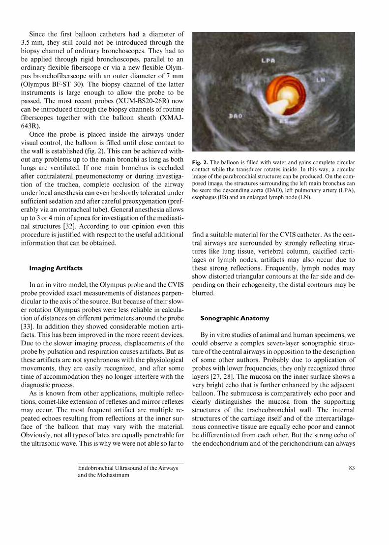
BOIS:L08:BPRR3020PD.97 FF: RESF E1: RESS
Endobronchial Ultrasound of the Airwaysand the Mediastinum
83
Since the first balloon catheters had a diameter of3.5 mm, they still could not be introduced through thebiopsy channel of ordinary bronchoscopes. They had tobe applied through rigid bronchoscopes, parallel to anordinary flexible fiberscope or via a new flexible Olym-pus bronchofiberscope with an outer diameter of 7 mm(Olympus BF-ST 30). The biopsy channel of the latterinstruments is large enough to allow the probe to bepassed. The most recent probes (XUM-BS20-26R) nowcan be introduced through the biopsy channels of routinefiberscopes together with the balloon sheath (XMAJ-643R).
Once the probe is placed inside the airways undervisual control, the balloon is filled until close contact tothe wall is established (fig. 2). This can be achieved with-out any problems up to the main bronchi as long as bothlungs are ventilated. If one main bronchus is occludedafter contralateral pneumonectomy or during investiga-tion of the trachea, complete occlusion of the airwayunder local anesthesia can even be shortly tolerated undersufficient sedation and after careful preoxygenation (pref-erably via an orotracheal tube). General anesthesia allowsup to 3 or 4 min of apnea for investigation of the mediasti-nal structures [32]. According to our opinion even thisprocedure is justified with respect to the useful additionalinformation that can be obtained.
Imaging Artifacts
In an in vitro model, the Olympus probe and the CVISprobe provided exact measurements of distances perpen-dicular to the axis of the source. But because of their slow-er rotation Olympus probes were less reliable in calcula-tion of distances on different perimeters around the probe[33]. In addition they showed considerable motion arti-facts. This has been improved in the more recent devices.Due to the slower imaging process, displacements of theprobe by pulsation and respiration causes artifacts. But asthese artifacts are not synchronous with the physiologicalmovements, they are easily recognized, and after sometime of accommodation they no longer interfere with thediagnostic process.
As is known from other applications, multiple reflec-tions, comet-like extension of reflexes and mirror reflexesmay occur. The most frequent artifact are multiple re-peated echoes resulting from reflections at the inner sur-face of the balloon that may vary with the material.Obviously, not all types of latex are equally penetrable forthe ultrasonic wave. This is why we were not able so far to
Fig. 2. The balloon is filled with water and gains complete circularcontact while the transducer rotates inside. In this way, a circularimage of the parabronchial structures can be produced. On the com-posed image, the structures surrounding the left main bronchus canbe seen: the descending aorta (DAO), left pulmonary artery (LPA),esophagus (ES) and an enlarged lymph node (LN).
find a suitable material for the CVIS catheter. As the cen-tral airways are surrounded by strongly reflecting struc-tures like lung tissue, vertebral column, calcified carti-lages or lymph nodes, artifacts may also occur due tothese strong reflections. Frequently, lymph nodes mayshow distorted triangular contours at the far side and de-pending on their echogeneity, the distal contours may beblurred.
Sonographic Anatomy
By in vitro studies of animal and human specimens, wecould observe a complex seven-layer sonographic struc-ture of the central airways in opposition to the descriptionof some other authors. Probably due to application ofprobes with lower frequencies, they only recognized threelayers [27, 28]. The mucosa on the inner surface shows avery bright echo that is further enhanced by the adjacentballoon. The submucosa is comparatively echo poor andclearly distinguishes the mucosa from the supportingstructures of the tracheobronchial wall. The internalstructures of the cartilage itself and of the intercartilage-nous connective tissue are equally echo poor and cannotbe differentiated from each other. But the strong echo ofthe endochondrium and of the perichondrium can always

BOIS:L08:BPRR3020PD.97 FF: RESF E1: RESS
84 Becker/Herth
Fig. 3. Sonoanatomy of the tracheobronchial wall in vitro (a), showing the seven layers of: mucosa/submucosa,cartilage and adventitia (thickness in mm), and in vivo where usually they can only be recognized by higher magnifi-cation (b).
be seen in the intact structure. The adjacent external sono-graphic double layer structure of the supporting connec-tive tissue and of the adventitia are easily missed underlow power magnification due to the bright reflex of theperichondrium. Differentiation of these structures is im-portant for the diagnosis of destruction by inflammatoryprocesses or by submucosal and intramural malignantinfiltration (fig. 3a, b).
Orientation within the mediastinum is difficult [34].Besides the complex anatomy of the mediastinal struc-tures and motion artifacts by respiration and pulsation,this is mainly due to the frequently unusual plains of thesonographic image as we have to follow the path of theairways. Inside the trachea, the ultrasonic image equalsthe horizontal plane we are used to in CT scan of themediastinum. Passing into the main bronchi, the imagecontinuously tilts towards a more sagittal plane, especiallyas we enter the left main bronchus. Passing into theperipheral part of the upper lobe bronchi, we even get aninverse horizontal plane.
During clinical use, recognition of distinct anatomicalstructures and their relationship usually is more helpfulthan localization of the probe itself (fig. 2). The vessels areeasily recognized by their low internal echo and by theirpulsation. But even after application of echo contrastmedium, differentiation of arteries and veins may be dif-ficult due to a lot of anatomical variations. By generally
using pulse-oxymetry, arteries may be recognized becauseof their synchronous pulsation. Lymph nodes, in contrast,are more echo dense and can be detected down to a size ofa few millimeters.
As the depth of penetration may extend up to severalcentimeters, the left atrium and even the mitral valve maybe visualized from the distal left main bronchus. Closer tothe bifurcation, the pulmonary trunk and left and rightmain pulmonary arteries can be seen ventrally to the leftmain bronchus, as well as the ascending aorta and the aor-tic arch. The descending aorta, the oval-shaped multilayerstructure of the esophagus and the vertebral column aredistinct landmarks for orientation. The small but impor-tant area between left pulmonary artery and aortic arch –the so-called aortopulmonary window – can be visualizedfrom the roof of the left main bronchus. Sometimes, inaddition, passage to the apical segmental bronchus of theleft upper lobe proves to be helpful.
Ventrally to the right main bronchus, the pulmonarytrunk, the aortic root and the vena cava are seen as well asthe right pulmonary artery. Closer to the bifurcation, dor-sally, the esophagus is found and laterally, to the rightside, the vena azygos, which crosses at the level of theright tracheobronchial angle, ventrally of which its junc-tion with the vena cava can be seen.
From the lower trachea, in addition, the aortic archand some of its branches, as well as the pulmonary arteri-
a b

Endobronchial carcinoma Primary cancer
Mechanical desobliteration
BOIS:L08:BPRR3020PD.97 FF: RESF E1: RESS
Endobronchial Ultrasound of the Airwaysand the Mediastinum
85
Table 1. Indications of endobronchial ultrasound 1994–6/1996(1,020 procedures in 869 patients)
% n
Intraluminal extent 19 193Penetration 2 24Early cancer? 3 26
Posttherapy control 3 27Mediastinal infiltration
Trachea 6 61Pulmonary artery 3 33Large vessels 3 30Esophagus (sep. table)
Lymph nodes (incl. guided biopsy) 29 299DD carcinoma/benign lesion 18 183Intramural lesion 4 43Mediastinal tumor
Malignancy 2 22Others 2 23
Perioperative control (anastomosis/recurrence) 5 51Other 1 5
Total 100 1,020
DD = Differential diagnosis.
al trunk, are easily seen. Pathological structures of soft tis-sue or cystic appearance are clearly differentiated fromthese organs by their different echo structure.
Inside the peripheral bronchi, conditions for echogra-phy are not so favorable, as the surrounding air-contain-ing structures reflect the ultrasonic waves. In some in-stances, solid structures or cystic formations can be differ-entiated within the lung parenchyma, which may be help-ful in guiding diagnostic and bioptic procedures [35].
Results of Clinical Application
Already during the first phase of our investigations indeveloping the sonographic system, we gained enoughexperience in clinical application to give first preliminaryreports [2, 20, 21]. In a first nonprospective feasibilitystudy, we investigated whether by use of the new opti-mized system we were able to gain additional informationthat we could not obtain by other conventional methods.In addition, we analyzed cost-effectiveness by resteriliz-ing and reusing the originally single-use vascular trans-ducers until they broke [36]. With the system of Olympus
Table 2. Indications of endoesophageal ultrasound 1994–6/1996(188 procedures)
% n
6 11Control therapy 4 8
Benign intramural lesion 3 6Infiltrating lung cancer 15 28
Mediastinal extension 6 12Infiltration of large vessels 2 4
Mediastinal lymph node 28 52Exclusion of tumor 24 45Mediastinal tumor 4 7
Benign process 7 14Control of anastomosis 1 1
Total 100 188
Table 3. Endobronchial ultrasound in interventional bronchoscopy1994–6/1996 (210 procedures)
% n
28 58Stent implantation 16 34Laser resection 12 25Brachytherapy 11 24PDT 2 5Guided biopsy 8 16Abscess drain 11 23Mucoid impaction 7 14Perforations/fibrin 5 11
Total 100 210
PDT = Photodynamic therapy.
Company, we performed more than 1,600 examinations.In a preliminary retrospective study, 1,418 of these, per-formed during the period from June 1994 to December1996 in 869 patients, have been analyzed; 1,020 examina-tions were performed in the tracheobronchial system (ta-ble 1), 188 in the esophagus (table 2). In 210 cases, endo-sonography was applied for interventional procedures (ta-ble 3). The different indications may be drawn from thetables.

BOIS:L08:BPRR3020PD.96 FF: RESF E1: RESS
86 Becker/Herth
Fig. 4. Early cancer in the middle lobe. Laterally to the middle lobe ostium, a small protrusion of the bronchial wallmay be recognized which represents a small squamous cell carcinoma. The slightly bleeding defect on the carina to themiddle lobe (S 4/5), ventrally to the lower lobe ostium, resulted from biopsy (DEF) (a). The tumor was neither visibleon plain X-ray nor on CT scan, which is a common definition for early lung cancer. Ultrasound examination of themiddle lobe bronchus (ML) demonstrates a lesion of 7 mm (TU) in diameter which is confined to the bronchial wall.Pulmonary artery (PA) and vein (PV) are intact (b). The patient was treated by lower bilobectomy. UL/RLL = Rightlower lobe.
Staging of Bronchial Carcinoma
The goal of staging is the exact diagnosis of the TNMformula as base for rational therapy. The different criteriafor extent of tumor growth are defined by the latest TNMclassification according to UICC and the supplement of1997 [37]. For the bronchoscopist, this means in smallradiologically invisible tumors detection and localizationof early carcinoma in the case of positive sputum cytolo-gy. Therapeutic decisions in these cases are made on thebasis of endobronchial extension, infiltration of the tra-cheobronchial wall and last but not least on the pathoana-tomical diagnosis. For the assessment of infiltration of thehilar and mediastinal lymph nodes, exact localization isnecessary prior to transbronchial and transtracheal needlebiopsy [38]. Defining size according to longitudinal andcross diameter, analysis of the internal structure and rela-tion to the central airways and the mediastinal structuresare important features that are analyzed by endosonogra-phy. Some types of metastasis localized within the medi-
astinum, in the central airways, inside the lung or thepleura can be within reach of the bronchoscopist. Espe-cially in regions which are not accessible to the direct viewof the endoscopist, we expected additional information byendobronchial ultrasound.
Primary TumorEarly Carcinoma. Frequently, tumors that are not visi-
ble by high-resolution CT scan are defined as early carci-noma. This is not in accordance with our experience. Theusual pathoanatomical definition of early cancer is lack ofinfiltration beyond the submucosa. According to our ex-perience and other studies, it is well known that only 75%of tumors that are visible by bronchoscopy are detected byradiology [6]. Among those not detected, there are manytumors with extensive local infiltration. In several pa-tients sent for endobronchial treatment of so-called earlybronchial carcinoma, we could demonstrate deep infiltra-tion of the bronchial wall and sometimes even regionallymph node metastasis which had escaped all other meth-
a b

BOIS:L08:BPRR3020PD.97 FF: RESF E1: RESS
Endobronchial Ultrasound of the Airwaysand the Mediastinum
87
ods of diagnosis. This is why we believe that the diagno-sis of early bronchial carcinoma can only be establishedfrom the resection specimen by the pathologist. By clini-cal methods, we can only assume the depth of infiltra-tion.
In every case of macroscopic alteration of the mucosa,we found concomitant alterations in the sonographicstructure. The wall was more or less thickened and thedelicate structures no longer visible. The altered sono-graphic structure was very variable, ranging from very lowto very high echodensity [16]. In many instances, even ifthe mucosa seemed to be intact macroscopically, wefound submucosal tumor spreading by endobronchial ul-trasound. In some instances, it could be followed extend-ing beyond the bronchial wall into the parabronchialstructures. If we do not see any signs of infiltration of thedeeper layers of the tracheobronchial wall or the adjacentlymph nodes, this strongly suggests a localized tumorwhich may be treated endoscopically with curative intentif the patient is considered to be inoperable for local orgeneral reasons (fig. 4a, b) [36, 39].
Intraluminal Extension and Involvement of the Tra-cheobronchial Wall. Also in advanced bronchial carcino-ma, endoluminal ultrasound provides valuable informa-tion for therapeutic decisions. In complete airway ob-struction, we were able to differentiate the basis and thesurface of the tumor, and we could assess depth of inva-sion into the bronchial wall and into the mediastinum. Bypassing the stenosis, we could assess patency of the distalairways (fig. 5a–f). This information was of importancefor endobronchial desobliteration, especially if we couldprove simultaneously that the pulmonary artery was notoccluded [39]. In occlusion of a central airway, perfusionof one lung may be completely shut down due to the Lilje-strand-Euler reflex. In this case, perfusion scan is unableto prove organic occlusion of the pulmonary artery, andusually pulmonary angiography has to be performed,because bronchial recanalization in cases of completeocclusion of the pulmonary artery would only increasedead space ventilation. In some patients, on the otherhand, we could demonstrate direct invasion of the pulmo-nary artery or complete occlusion which could be con-firmed by angiography or postoperative pathological ex-amination.
Infiltration of Mediastinal Structures. Even more im-portant in preoperative staging is the diagnosis of infil-tration of the organs in the mediastinum such as the aor-ta, vena cava or the main pulmonary artery which byradiological methods frequently proves to be difficult.Tumors of the left hilum are situated in close vicinity to
the esophagus, and in several instances, we could dem-onstrate direct infiltration of the esophageal wall byechography of the left main bronchus or the distal tra-chea. If either of these organs is infiltrated by a tumor,surgery is no longer possible (fig. 6a–c). If we can demon-strate a clear sonographic demarcation of the tumor tothese structures, and if signs of infiltration of the wall aremissing, we can assume that the patient will be operable.This could be repeatedly confirmed by the intraoperativesitus. Intratracheal ultrasound was especially helpful indiagnosing external infiltration of the tracheal wall bytumors of the mediastinal surface of the lung or by pri-mary mediastinal tumors. Here, in our experience, endo-bronchial ultrasound proved to be superior to radiologi-cal examination.
Involvement of Lymph NodesTherapeutic decisions in bronchial carcinoma are even
more dependent on lymph node involvement than onextension of the primary tumor. This is why exact stagingof lymph nodes is extremely important. Under favorableconditions, we could visualize lymph nodes down to a sizeof 2–3 mm, and we were able to recognize the internaldelicate architecture of lymph follicles and sinuses as wellas small lymph vessels of the hilum. But in opposition toother localizations, for example in ENT tumors [40, 41],we were not able to find reliable signs for diagnosis ofmalignant infiltration. In a limited prospective in vitrostudy, we therefore examined 84 lymph nodes of 15patients which had been surgically resected for the treat-ment of bronchial carcinoma. But even under these favor-able conditions, we could find no reliable criteria for themalignant infiltration, neither with regard to the echo-geneity nor to homogeneity of the internal structure orwith respect to boundary to the surrounding structures,although the resolution was below 1 mm. We assume thatthis is due to the high rate of unspecific alterations, suchas inflammatory reactions in postobstructive pneumonia,silicotic or specific scar formation and other pathologicalreactions which cause alterations in the internal structure.As the importance of transbronchial/transtracheal needleaspiration has been proven for staging of regional lymphnodes [7, 8, 38] the addition of endobronchial ultrasoundfor improvement of these procedures has been investi-gated. Preliminary results showed that prior or simulta-neous ultrasonography may reduce the number of at-tempts and improve results [42]. As by the Olympus sys-tem simultaneous control of the biopsy needle is very lim-ited, echography mainly serves for localization of thelymph nodes (fig. 7). Transbronchial/transtracheal needle

BOIS:L08:BPRR3020PD.97 FF: RESF E1: RESS
88 Becker/Herth
Fig. 5. a Complete obstruction of the left main bronchus by exophytic bronchial carcinoma. b Perfusion and ventila-tion scan demonstrate complete loss of function of the left lung. c Endosonography shows the base of the tumor (TU)to the right at the mediodorsal wall (LMB). The lateral and ventral wall is intact. The left pulmonary artery (PA) ispatent, but shows a smaller caliber due to reflectory reduction of perfusion (Euler-Lijestrand mechanism). Also thedescending aorta (DAO) is unharmed. d Beyond the tumor, the bronchi of the lower lobe (BR) are filled by secretionsbut patent. The lung is atelectatic (ATEL) and some pleural fluid is seen (EFF). e After laser resection, the mainbronchus is opened. f Ventilation of the lung is restored.
a b
c d

BOIS:L08:BPRR3020PD.97 FF: RESF E1: RESS
Endobronchial Ultrasound of the Airwaysand the Mediastinum
89
Fig. 6. An enlarged lymph node is localizedby endobronchial ultrasonography dorsallyto the bifurcation (a) and in this case, a nee-dle (N) is inserted into the node parallel tothe ultrasonic probe (b). It can be clearlyseen as a bright dot with a dorsal shadowwithin the lymph node (b).
biopsy is performed sequentially after prior sonographiclocalization. This is easy as the cartilages and branchesallow exact anatomical localization and guidance of thebiopsy needle. In larger malignant lymph nodes, in whichinvasion of the bronchial wall can be sonographicallyproven, so-called ‘button-hole’ biopsies can be performedby removing the mucosa and taking biopsies of the deeperlayers of the bronchial wall without the risk of inducingpneumothorax or pneumomediastinum.
Mediastinal Masses
Mediastinal masses can only be visualized by endo-bronchial ultrasound and subsequently approached bytransbronchial/transtracheal needle biopsy if they are lo-calized in close contact to the central airways or if somefluid-containing structure provides an acoustic window.Retrosternal masses or those which are localized in fardistance within the dorsal mediastinum cannot be ap-proached in this way due to lack of penetration of the
a b
5e
5f

BOIS:L08:BPRR3020PD.97 FF: RESF E1: RESS
90 Becker/Herth
Fig. 7. Bronchoscopic desobliteration of obstructing tracheal tu-mor. Bronchoscopy shows an exophytic tumor of the lower tracheawith almost complete obstruction. After emergency treatment byNd:YAG laser, coagulation and partial resection (a), the extent isexplored by endoesophageal ultrasound for suspicion of infiltrationof the esophagus (b). Although endoscopically the esophageal mucosaseemed completely intact besides compression, endoluminal ultra-sound reveals extensive tumor infiltration (TU) which can be diag-nosed from the extinction of the layers of the ventral wall as com-pared to the intact dorsal wall. AO = Aorta; VC = vertebral column;ES = esophagus. Surgery in this situation is out of question and addi-tional radiotherapy after complete laser resection bears a high risk foresophagotracheal fistula. Therefore, after partial resection, a NitinolUltraflex stent is implanted to keep the lumen patent during radio-therapy (c).
ultrasound through the surrounding tissue. Primary me-diastinal or bronchogenic cysts are easily differentiatedfrom solid tumors by their echo-poor internal structurewhich is frequently septated, provided that they are inclose contact to the airways. In solid masses, according toour experience, endosonography is superior to radiologi-cal procedures in differentiation of compression and infil-tration of the wall of the airways. This applies for exampleto goitre and thyroid carcinoma. In tumors of the medias-tinum, which are usually localized ventrally to the trachea
and the left main bronchus, access via the apical segmentof the left upper lobe is preferable due to its close relation.Sometimes tumors of adjacent mediastinal organs, suchas leiomyomas of the esophagus, can be diagnosed fromthe left main bronchus according to the typical extensionbetween the different layers of the esophageal wall.
a
b
c

BOIS:L08:BPRR3020PD.97 FF: RESF E1: RESS
Endobronchial Ultrasound of the Airwaysand the Mediastinum
91
Large Intrathoracic Vessels
The large intrathoracic vessels adjacent to the centralairways can be recognized easily due to their low echostructure and their pulsation. Due to a lot of variations,differentiation between arteries and veins can be difficult,even with additional application of echo-contrast media.The importance of sonographic diagnosis in direct tumorinvasion has been mentioned. Especially in early infancyand childhood, compression of the airways by vascularanomalies is comparatively frequent. We repeatedly de-tected such anomalies by endobronchial sonography,even if extensive previous cardiovascular diagnostic pro-cedures did not show the pathology. This is why we areconvinced that in the future endosonography will be astandard procedure in the diagnosis of pediatric malfor-mations. This applies also to extensive thoracic deformi-ties where compression of the airways by deviation of theheart, the large intrathoracic blood vessels and the thorac-ic wall can be diagnosed easily by endobronchial ultra-sound preventing more invasive radiological procedureslike angiography. This, in our experience, was also true forcompression due to aortic aneurysms or for internal dis-placement after extensive surgical procedures. In onecase, we were even able to diagnose the rupture of a vascu-lar flap for treatment of an aortic isthmic stenosis and theresulting aneurysm as cause for severe hemoptysis fromthe left upper lobe. The diagnosis was confirmed by surgi-cal revision. Diagnosis of pulmonary embolism via theendobronchial route is a rare event, as suspicion of pul-monary embolism is no indication for an invasive proce-dure like bronchoscopy. If considered necessary after all,diagnosis is achieved more easily via the transesophagealroute. This is also true for thrombosis of the pulmonaryvein which we were able to diagnose in rare cases of severepostoperative hypoxemia, as the left atrium and the pul-monary veins are easily visible from the esophagus orfrom the left main bronchus.
Intrapulmonary Lesions
According to our experience, endobronchial ultra-sound will not play a major role in diagnosis of intrapul-monary lesions as it is almost completely reflected by thesurrounding air, and radiological imaging here is far supe-rior [27, 28]. Only in some cases of atelectasis were weable to gain further information by endosonography indifferentiation of bronchial obstruction by lymph nodesor tumors from compression by pleural fluids or masses.
Some authors reported that endobronchial ultrasoundmay be helpful in guiding the biopsy needle in puncture ofperipheral coin lesions. One group described an ultrasonicprobe with integrated biopsy channel [35]. In some cases,we were able to explore intrapulmonary cavities by theultrasonic probe and demonstrate internal fungus balls ortumors in the wall. In these cases, endobronchial ultra-sound might be helpful in directing interventional ma-neuvers like the insertion of pigtail catheters for drainageof intrapulmonary abscesses.
Pleural Space and the Neighboring Organs
If an atelectasis or pleural effusion provides an acous-tic window, the pleural surface and mediastinal structuresmay be visualized. Thus, we were able to see solid forma-tions of the visceral and mediastinal pleura or on the peri-cardium. Diagnosis of organs localized in the direct vicin-ity of the airways such as the thyroid and the heart havebeen mentioned. Endobronchial ultrasound proved to beextremely valuable in diagnosis of infiltration of theesophagus by bronchogenic carcinoma. In this case, surgi-cal procedures usually are no longer indicated. Also endo-bronchial procedures like ND:Yag laser therapy in combi-nation with high-dose endoluminal radiation bear a highrisk of fistulas. In these cases, prior assessment of the localsituation by additional transesophageal exploration is ex-tremely helpful.
Transesophageal Exploration
In addition to endobronchial application, we exploredthe application of the miniaturized probes in the esopha-gus. The images we obtained were equal to those gener-ated by ultrasonic endoscopes [29]. The depth of penetra-tion for the 20 MHz probes is obviously limited. This,however, was not so important since our major interestwas assessment of infiltration of the esophageal wall bybronchial carcinoma or that of the airways by esophagealcarcinoma (fig. 7a–c). Also, benign intramural processessuch as leiomyomas and intramural diverticulosis couldbe clearly diagnosed and differentiated from malignantprocesses. Sometimes enlarged lymph nodes of the aorto-pulmonary window are detected more easily via theesophageal route. This is also true for the left atrium andthe pulmonary vein. Detailed analysis of the chambers ofthe heart and its valves, however, is not possible becauseof the limited range of the 20 MHz probe.
BOIS:L08:BPRR3020PD.97 FF: RESF E1: RESS
92 Becker/Herth
Endobronchial Ultrasound in InterventionalBronchoscopy
Some indications for the use of endobronchial ultra-sound in making therapeutic decisions have been men-tioned before. This was the case in 20% of our examina-tions (table 3). For example, in exploration of central air-way stenosis, sonography was useful in assessment of itsextension and cause in order to choose the best methodfor interventional procedures, i.e. dilatation, laser or stentimplantation as well as control of the therapeutic effect(fig. 6a–c).
Especially in bronchoscopic treatment of malignanttumors with curative intent such as photodynamic thera-py and endoluminal high-dose radiation, it is importantthat the lesion is restricted to the bronchial wall. Here,endobronchial sonography proved superior to all otherdiagnostic procedures.
Correct endoscopic judgement of the healing process ofan anastomosis after a so-called bronchoplastic surgicalprocedures (sleeve resection) can be problematic. Thus, itis sometimes difficult to differentiate superficial necrosiswith mucosal swelling and fibrin deposition from immi-nent dehiscence. Endobronchial ultrasound proved to bevery useful in assessment of the surrounding structures. Insome cases, beginning perifocal abscess adjacent to thepulmonary artery was diagnosed by ultrasound before theonset of hemoptysis, and thus a timely decision for surgi-cal intervention could be made.
Cost-Effectiveness
By reusing the ultrasonic probes repeatedly after rester-ilization and carefully avoiding too much pressure againstthe bronchial wall which causes fracture of the wire, withthe CVIS probe we could examine 40–50 consecutivepatients before mechanical defects occurred. Calculatingthe price of USD750 for one catheter, the single proce-dure comes to about USD15–20 in addition to the costsof the bronchoscopy, the investment of the processor ofabout USD100,000 not included. The probes of Olympuscompany are somewhat more expensive, but they aremuch more resistant, so that we could use them a fewhundred times. In conclusion, we consider the endolumi-nal ultrasound as a cost-effective and very rewardingadditional diagnostic procedure.
Conclusion and Future Aspects
In conclusion, with regard to the technique, the clinicalapplication and its diagnostic results, and the cost-effec-tiveness of endobronchial ultrasonography in addition toconventional diagnostic procedures, in our institution,endobronchial ultrasound already at this stage proved tobe of the same value as in other disciplines, where it isalready a routine procedure. In a currently ongoing pro-spective multicenter study, we intend to establish soundindication for endobronchial ultrasonography as com-pared to conventional methods of ultrasound, radiologi-cal and other diagnostic procedures. In addition, weintend to develop further technical improvements for theendobronchial application. This could include additionof Doppler sonography for analysis of vascularization,which could be helpful for example in the control of bron-chial anastomosis after surgical procedures. Further, com-puterized analysis of tissue characteristics could be usefulfor the detection of malignancy of early carcinoma orlymph node metastases [43–45].
Addition of a biopsy channel would enable simulta-neous sonographically guided needle biopsy [35], and firstintegrated systems are in planning. Since the results oftreatment of advanced bronchial carcinoma have beendisappointing so far, detection and treatment at earlystages have gained new interest. Especially new methodslike automated sputum cytology analysis in persons at riskand localization of radiologically and macroscopicallyinvisible early carcinoma by fluorescence methods will beused more widely. In this concept, endobronchial ultra-sonography is an ideal instrument for assessment of localextension and involvement of lymph nodes for stagingbefore the decision for curative treatment by broncho-scopic means is made [36, 39]. We therefore stronglybelieve that endobronchial ultrasound will be a routineprocedure in the future and play an important role indiagnostic and interventional bronchoscopy.

BOIS:L08:BPRR3020PD.97 FF: RESF E1: RESS
Endobronchial Ultrasound of the Airwaysand the Mediastinum
93
References
1 Becker HD, Kayser K, Schulz V, TuengerthalS, Vollhaber H-H (eds): Atlas of Bronchoscopy.Technique, Diagnosis, Differential Diagnosis,Therapy. Philadelphia, Decker, 1991.
2 Becker HD: Bronchoscopy for airway lesions;in Wang K-P, Haponik EF, Mehta AC (eds):Flexible Bronchoscopy. Oxford, Blackwell,1995, pp 136–159
3 Bülzebruck H, Bopp R, Drings P, Bauer E,Krysa S, Probst G, van Kaick G, Müller K-M,Vogt-Moykopf I: New aspects in the staging oflung cancer. Cancer 1992;70:1102–1110.
4 Layer G, van Kaick G: Staging des nicht-kleinzelligen Bronchialkarzinoms mit CT undMRT. Radiologie 1990;30:155–163.
5 Lewis JW Jr, Pearlberg JL, Beute GH, AlpernM, Kvale PA, Gross BH, Milligan DJ Jr: Cancomputed tomography of the chest stage lungcancer? Yes and no. Ann Thorac Surg 1990;49:591–595.
6 Naidich DP: Staging of lung cancer: Controver-sy: Computed tomography versus broncho-scopic needle aspiration – Pro computed to-mography. J Bronchol 1996;3:69–73.
7 Harrow EM, Millard PS, Wight G, Patterson J:Survival duration among patients with lungcancer staged by bronchoscopic needle aspira-tion. J Bronchol 1996;3:96–101.
8 Harrow EM, Wang K-P: Chest surgery clinicsof North America. The staging of lung cancerby bronchoscopic transbronchial needle aspira-tion. Thorac Endosc 1996;6:223–235.
9 Ziegler K, Sanft C, Semsch B, Friedrich M,Gregor M, Riecken EO: Endosonography issuperior to computed tomography in stagingtumors of the esophagus and the cardia. Gas-troenterology Suppl 1988;94:A517.
10 Balzer Th, Ziegler K: Die Bedeutung derEndosonographie für die Gastroenterologie.Fortschr Diagn 1992;3:22–32.
11 Dittler H-J, Bollschweiler E, Siewert JR: Wasleistet die Endosonographie im präoperativenStaging des Ösophaguskarzinoms? Dtsch MedWochenschr 1991;116:561–566.
12 Kleinau H, Liebeskind U, Zaiaic M, SchlagPM: Endoluminal and intraoperative ultra-sound. Onkologie 1993;16:432–435.
13 Rösch Th, Lorenz R, Braig Ch, Feuerbach S,Siewert JR, Classen M: EndosonographischeDiagnostik bei Pankreastumoren. Dtsch MedWochenschr 1990;115:1339–1347.
14 Tio TL, Tytgat GNJ: Atlas of TransintestinalUltrasonography. Alsmeer, Mur-Kostverloren,1986.
15 Tio TL, den Hartog Jager CA, Tytgat GNJ: Therole of endoscopic ultrasonography in assessinglocal resectability of oesophagogastric malig-nancies. Accuracy, pitfalls and predictability.Scand J Gastroent 1986;21(suppl):78.
16 Vijakumar S, Chan T, Ray V, Grant T, ChiruP, Ray P, Chan GGTY: Guidelines for definingtarget volumes in radiation therapy of prostatecancer: A review and perspective. Onkologie1993;16:389–406.
17 Wernecke K, Peters PE, Galanski M: Mediasti-nal tumors: Evaluation with suprasternal so-nography. Radiology 1986;159:405–409.
18 Wernecke K, Pötter K, Peters PE, Koch P:Parasternal mediastinal sonography: Sensitivi-ty in the detection of anterior mediastinal andsubcarinal tumors. AJR 1988;150:1021–1026.
19 Wernecke K, Peters P: Mediastinale Sonogra-phie. Dtsch Ärztebl 1993;90:506–514.
20 Becker HD: Endobronchial ultrasonography –A new perspective in bronchology? Proceed-ings of the Second Annual Assembly of theAmerican Association for Bronchology withthe 60th Annual International Scientific As-sembly of the American College of Chest Physi-cians, New Orleans, October 1994.
21 Becker HD: Endobronchial ultrasound – Ex-pensive toy or useful tool? Proceedings of the8th World Congress for Bronchology and the8th World Congress for Bronchoesophagology,Munich, June 1994.
22 Becker HD: Endobronchialer Ultraschall –Eine neue Perspektive in der Bronchologie. En-dosk Heute 1995;1:55–56.
23 Klüppelberg F, Becker HD: Bringt die endo-bronchiale Sonographie einen zusätzlichen In-formationsgewinn? Erste Erfahrungen bei 100Patienten. Endosk Heute 1994;1:113.
24 Becker HD: Endobronchialer Ultraschall –Eine neue Perspektive in der Bronchologie. Ul-traschall Med 1996;17:106–112.
25 Ono R, Suemasu K, Matsunaka T: Broncho-scopic ultrasonography for diagnosis of lungcancer. Jpn J Clin Oncol 1993;23:34–40.
26 Ono R, Hirano H, Egawa S, Suemasu K: Bron-choscopic ultrasonography and brachytherapyin roentgenologically occult bronchogenic car-cinoma. J Bronchol 1994;1:281–287.
27 Hürther Th, Hanrath P: Endobronchiale So-nographie zur Diagnostik pulmonaler und me-diastinaler Tumoren. Dtsch Med Wochenschr1990;115:1899–1905.
28 Hürther Th, Hanrath P: Endobronchial so-nography: Feasibility and preliminary results.Thorax 1992;47:565–567.
29 Frank N, Holzapfel P, Wenk A: Neue Endo-schall Minisonde in der täglichen Praxis. En-dosk Heute 1994;3:238–244.
30 Koga T, Ogata K, Hayashida R, Hattori R:Usefulness of transluminal ultrasonography inthe evaluation of bronchial stenosis secondaryto tuberculosis. J Jap Soc Bronchol 1994;16:477–482.
31 Gehling U, Tuengerthal S, Jackowski M, DüxM, Schulz V: Intravasale Sonographie zur prä-operativen Diagnostik zentraler Bronchialtu-moren – Erste klinische Erfahrungen. Pneumo-logie 1993;47:19–24.
32 Frietsch Th, Becker HD, Bülzebruck H, Wiede-mann K: Capnometry for rigid bronchoscopyand high-frequency-jet-ventilation comparedto arterial PCO2. 11th World Congress ofAnaesthesiologists, Abstr Book Anaesthesiolo-gy, P 231, 1996.
33 Düber C: Endovascular ultrasound. Vortragbeim III. International Stent Symposium.Frankfurt/Main, October 1993.
34 Cavaliere S, Beamis J: Atlas of TherapeuticBronchoscopy. Laser – Stents. Brescia, RIB,1991.
35 Steiner RM, Liu JB, Goldberg BB, Cohn JR:The value of ultrasound guided fiberopticbronchoscopy. Clin Chest Med 1995;16:519–534.
36 Becker HD: Photodynamische Therapie; inHuhn D, Herrmann R (eds): MedikamentöseTherapie maligner Erkrankungen, ed 3. Stutt-gart, Fischer, 1995, pp 75–80.
37 Spiessl B, Beahrs OH, Hermanek P, HutterRVB, Scheibe O, Sobin LH, Wagner G (eds):UICC TNM Atlas. Illustrated Guide to theTNM/pTNM Classification of Malignant Tu-mors, ed 3. Berlin, Springer, 1989.
38 Wang K-P, Mehta AC (eds): Staging of Bron-chogenic Carcinoma by Bronchoscopy. Ox-ford, Blackwell, 1995, pp 206–214.
39 Becker HD: Options and results in endobron-chial treatment of lung cancer. Min Invas TherAllied Technol 1996;5:165–178.
40 Heppt W, Issing W: Einsatz der flexiblen Endo-sonographie im Kopf-Hals-Bereich. EndoskHeute 1991;3:172–175.
41 Mende U, Zöller J, Drommer RB, Reichardt P,Born IA: Optimisation of preoperative chemo-therapy by ultrasound. Follow-up in tumors ofthe oral cavity and oropharyngeal region. Int JOral Maxillofac Surg 1990;19:363–366.
42 Shannon JJ, Bude RO, Orens JB, Becker FS,Whyte RI, Rubin JM, Quint LE, Martinez FJ:Endobronchial ultrasound-guided needle aspi-ration of mediastinal adenopathy. Am J RespirCrit Care Med 1996;153:1424–1430.
43 Basset O, Sun Z, Metsas JL, Gimenez G: Tex-ture analysis of ultrasonic images of the pros-tate by means of cooccurrence matrices. Ultra-son Imaging 1993;15:218–237.
44 Schmitz G, Ermert H, Senge T: Tissue charac-terisation of the prostate using Kohonen Maps.Proceedings of the IEEE Ultrasonics Sympo-sium, Cannes 1994.
45 Wagner RF, Insana MF, Brown DG: Unifiedapproach to the detection and classification ofspeckle texture in diagnostic ultrasound. OptEng 1986;25:738–742.
Dr. H.D. BeckerEndoskopie, ThoraxklinikAmalienstrasse 5D–69126 Heidelberg (Germany)Tel. +49 6221 396 302, Fax +49 6221 396 246E-Mail [email protected]

OOOOOOOOOOOOOOOOOOOOOOOOOOOOOOOOOOOOOOOOOOOOOOOOOOOOOOOOOOOOOOOOOOOOOOOOOOOOOOOOOOOOOOOOOOOOOOOOOOOOOOOOOOOOOOOOOOOOOOOOOOO
Therapeutic Bronchoscopy
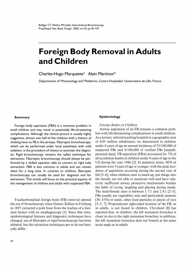
Bolliger CT, Mathur PN (eds): Interventional Bronchoscopy.Prog Respir Res. Basel, Karger, 2000, vol 30, pp 96–107
OOOOOOOOOOOOOOOOOOOOOOOOOOOOOOOOOOOOOOOOOOOOOOOOOOOOOOOOOOOOOOOOOOOOOOOOOOOOOOOOOOOOOOOOOOOOOOOOOOOOOOOOOOOOOOOOOOOOOOOOOOO
Foreign Body Removal in Adultsand ChildrenCharles-Hugo Marquettea Alain Martinotb
Departments of aPneumology and bPediatrics, Centre Hospitalier Universitaire de Lille, France
96
Summary
Foreign body aspiration (FBA) is a common problem insmall children and may result in potentially life-threateningcomplications. Although the clinical picture is usually highlysuggestive, almost one half of the children with a history ofchoking have no FB in the airways. Fiberoptic bronchoscopy,which can be performed under local anesthesia with mildsedation, is the procedure of choice to ascertain the diagno-sis. Rigid bronchoscopy remains the safest technique forextraction. Fiberoptic bronchoscopy should always be per-formed by a skilled operator able to convert to rigid tubeextraction. FBA is less common in adults and can remainsilent for a long time. In contrast to children, fiberopticbronchoscopy can usually be used for diagnosis and forextraction. This article will focus on the practical aspects ofthe management of children and adults with suspected FBA.
Tracheobronchial foreign body (FB) removal openedthe era of bronchoscopy when Gustav Killian in Freiburgin 1897 extracted a pork bone from the trachea of a Ger-man farmer with an esophagoscope [1]. Since that time,epidemiological features and diagnostic techniques havechanged, use of fiberoptic or rigid bronchoscopy has beendebated, but the extraction techniques per se do not basi-cally differ.
Epidemiology
Foreign Bodies in ChildrenAirway aspiration of an FB remains a common prob-
lem with life-threatening complications in small children.As a tertiary referral teaching hospital in a geographic areaof 4.05 million inhabitants, we determined in childrenunder 4 years of age an annual incidence of 25/100,000 ofsuspected FBs and 9/100,000 of verified FBs [unpubl.personal data]. FB aspiration (FBA) accounted for 7% ofall accidental deaths in children under 4 years of age in theUS during the year 1986 [2]. In pediatric series, 80% ofpatients were 3 years of age or younger, with the peak inci-dence of aspiration occurring during the second year oflife [3–6], when children start to stand up, put things intothe mouth, are not able to masticate well and have rela-tively inefficient airway protective mechanisms besidesthe habit of crying, laughing and playing during meals.The male/female ratio is between 1.7:1 and 2.4:1 [2–5].FBs usually are vegetables: nuts and particularly peanuts(36–55%) or seeds, other food particles or pieces of toys[3–5, 7]. Preponderant right-sided location of the FB, asin adults, is not found in children. Cleveland [8] hasreported that, in children, the left mainstem bronchus iscloser in size to the right mainstem bronchus; in addition,the left mainstem bronchus does not branch at the sameacute angle as in adults.

BOIS:L08:BPRR3002PD.96 FF: RESF E1: RESS
Foreign Body Removal in Adults andChildren
97
Fig. 1. Variety of organic FBs removed fromchildren and adults.Fig. 2. Variety of inorganic FBs removedfrom children and adults.Fig. 3. Polytrauma in a 28-year-old man.Fractured tooth impacted in the right lowerlobe.
Foreign Bodies in AdultsTracheobronchial FBA is uncommon in adults. In the
literature only 5 large series have been reported [7, 9, 10–12], compared to dozens of publications concerning chil-dren. The largest series, reported by the Mayo Clinic [10],identified 60 adults over a 33-year period. Two recentstudies in Taiwan reported, respectively, 47 and 43 con-
secutive cases over a 13- and a 15-year period [11, 12]. Inour institution, FBA in adults accounts for less than 2‰of final diagnoses out of 4,500 bronchoscopies/year. Incontrast, we see 15–25 cases in children (!15 years)annually. In children, nuts account for the large majorityof FBs whereas in adults, the nature of FBs is highly vari-able (fig. 1, 2). Aspiration of nails or pins mostly occurs in
1 2
3

BOIS:L08:BPRR3002PD.96 FF: RESF E1: RESS
98 Marquette/Martinot
young or middle-aged adults during do-it-yourself activi-ties. Aspiration of dental debris, appliances or prosthesesusually complicate facial trauma (fig. 3) or dental proce-dures. Neurological disorders, loss of consciousness com-plicating trauma, alcohol or sedative abuse classically pre-dispose to aspiration of food fragments. The type of theaspirated food fragments itself depends on local customs.For instance, in Western, Chinese and Middle east popu-lations, vegetable matters [10], bones [11, 12] and water-melon seeds [13] are, respectively, the most frequentlyaspirated food particles. One peculiar syndrome in adults,referred to as the cafe coronary [14, 15] consists in fatal ornear fatal food asphyxiation occurring during a mealwhile eating incompletely chewed meat. This generallyoccurs in the elderly with dentition problems, or swallow-ing disorders [16–19] or parkinsonism [20]. The esti-mated incidence of the cafe coronary syndrome is 0.66/100,000 [20].
Table 1. Signs and symptoms revealing a respiratory FB
History of chokingRecurrent wheezingHemoptysisChronic coughUnilaterally decreased breath soundsOverinflated lobe or lungAtelectasisPneumothoraxPneumomediastinum, subcutaneous emphysemaRecurrent pneumonia in the same areaBronchiectasisLung abcessPleuresis
Diagnostic Approach to Foreign Bodies
Clinical Features and Diagnostic Work-Up inChildrenA witnessed episode of choking (sudden onset of spell-
ing cough and/or dyspnea and/or cyanosis in a previouslyhealthy child) has a high sensitivity: 79–88% [2, 4, 5, 21].Whenever there is a history of choking, tracheobronchialexamination is necessary, even in the absence of physicalor radiographic change. Physical findings can be absent inup to 39% of patients with FB [7], and radiographs arenormal in 6–38% of patients [2, 5, 7, 21–24]. A history ofchoking is sometimes disregarded. In the series of Blazeret al. [25], as much as 18% of all children arrived at thehospital more than a month after aspiration; although ahistory of choking was present in 73% of these children.Among 125 patients with such a history in the series ofWiseman [22] only 52% were diagnosed early. In thosefew children who were not witnessed to choke, the diagno-sis is often delayed.
Signs and symptoms that should evoke FBA are listedin table 1. Once evoked, there is no question that tracho-bronchial examination is the key-stone to confirm thediagnosis. Three strategies can be proposed. The first andoldest one is to start systematically with rigid bronchosco-py because it allows both diagnosis and extraction withexcellent safety. The second is to promote fiberoptic bron-choscopy under local anesthesia as first diagnosis proce-dure when there is a low probability of an airway FB, andso to avoid numerous rigid bronchoscopies under generalanesthesia to reduce cost and length of hospitalization. Ifan FB is present, extraction is then immediately per-formed with the rigid bronchoscope. An alternative strat-egy, consisting in performing fiberoptic bronchoscopyafter tracheal intubation and under general anesthesia for
Table 2. Diagnostic value of patient’shistory of choking, physical and chest X-rayfindings in children with suspectedFBA [adapted from ref. 4, 5, 21]
PPV Se Sp NPV
History of choking 50/63/78 85/79/81 21/46/33 60/64/38Unilaterally decreased breath sounds 84/84/– 65/67/– 88/85/– 73/68/–Wheezing 65/47/74 33/10/60 84/87/40 57/45/26Radiopaque FB 100/100/– 20/7/– 100/100/– 57/47/–Unilateral obstructive emphysema 81/86/– 53/55/– 88/89/– 67/63/–Abnormal inspiratory X-ray 72/–/83 70/–/82 74/–/44 73/–/41
PPV = Positive predictive value; Se = sensitivity; Sp = specificity; NPV = negative predic-tive value; – = no data. Results are indicated in the following order of studies: Martinot et al.[5], François et al. [4] and Hoeve et al. [21].

BOIS:L08:BPRR3002PD.96 FF: RESF E1: RESS
Foreign Body Removal in Adults andChildren
99
Fig. 4. FB impacted in the left mainstembronchus of a 2-year-old child. Obstruc-tive emphysema during inspiration (a) andmarkedly increased during expiration (b).
Fig. 5. FB impacted in the right mainstembronchus of a 1-year-old child. Inspiratorychest X-ray normal (a). Obstructive emphy-sema present during expiration (b).
a b
a b
diagnosis and extraction [26] will be discussed below.Besides a chest X-ray, other explorations such as a V/Q ora CT scan seldom contribute to the diagnosis [27, 28]. Todetermine the best strategy, one needs to evaluate theprobability that an FB is present, i.e. to estimate the prob-ability that endoscopy will be therapeutic.
In the literature, all but three articles [4, 5, 21] studiedcases with definite FBA (excluded cases with suspectedFBs), and thus could only investigate the sensitivity ofclinical and radiological findings. In two studies [4, 21]the diagnostic value of clinical and radiological signs werecalculated on retrospective data. In the study that we con-ducted [5], physical and chest X-ray findings were notedbefore endoscopy. Diagnostic values of patient’s historyof choking, physical and chest X-ray findings are summa-rized in table 2. From these data, we can infer that a histo-ry of choking has a high sensitivity (79–85%), but a lowpositive predictive value (PPV). Obstructive emphysema(fig. 4), especially when associated with ipsilateral de-creased breath sounds, has a very high PPV. In childrenwith a normal inspiratory chest X-ray (fig. 5), an expira-
tory chest X-ray or fluoroscopy is worth being performed.We thus recommend that, for diagnosis, fiberoptic bron-choscopy should be performed first [5], except whenasphyxia, radiopaque FB (fig. 6) or unilaterally decreasedbreath sounds plus obstructive emphysema are present(fig. 7). If one of these three items is present, the diagnosisof FBA is highly probable (PPV 190%), and rigid bron-choscopy can be proposed as first-line examination [5].
Clinical Features and Diagnostic Work-Up in AdultsIn contrast to children, there is no prospective study
which evaluates the diagnostic values of clinical andradiological signs in adults with suspected FBA. There-fore, the available information derives from retrospectiveseries of definite cases of FBA. Acute presentation inadults is seldom, the reason being that in most cases theFB is wedged distally (lower lobe bronchi or truncus inter-medius) whereas in children the FB usually obstructsproximal airways (mainstem bronchi). Therefore dyspneais rarely reported by adults. In the series of Lan [11],shortness of breath was present in only 25% of the cases.

BOIS:L08:BPRR3002PD.96 FF: RESF E1: RESS
100 Marquette/Martinot
Fig. 6. Radiopaque FB in a 1-year-old child.
Fig. 7. Proposed management algorithm for children with suspectedFBA and simulated results when applied retrospectively in the studypopulation. [Adapted from ref. 5, with permission.]
Children with suspectedFBA
Asphyxia?(83)No
äUnilateral decreased breath sounds
and obstructive emphysema?(59)No
ä
Yes (0) Rigid bronchoscopyä
Radiopaque FB?(75)No
ä
Yes (8) Rigid bronchoscopy(8 FB+)
ä
Fiberoptic bronchoscopy(42 absence of FB)
ä
(17 FB+) Rigid bronchoscopyä
Yes (16) Rigid bronchoscopy(15 FB+)
ä
In addition, in contrast to children, adults do not alwaysvolunteer or recall a history of choking [11, 12, 19, 29]. Infact, except in the cases where patients or witnesses reporta typical choking episode and in the cases of radiopaqueFBs, the diagnosis may be disregarded for a while bypatients and doctors, due to the poor specificity of clinicaland radiological signs. Thus, in adults, silent FBA may bea fortuitous finding of fiberoptic bronchoscopy performedbecause of symptoms such as chronic cough, hemoptysis,recurrent or nonresolving pneumonia or asthma not re-sponding to therapy (fig. 8), which leads to suspicion ofendobronchial abnormalities.
As shown above, in children, we recently resolved thequestion of ‘how equivocal the diagnosis of FBA has to bebefore fiberoptic bronchoscopy is the initial investigation’[30] in a prospective study in 83 children referred for sus-pected FBA [5].
There is no question regarding the diagnostic utility offiberoptic bronchoscopy in adults. From a practical view-point, however, two points must be stressed here. First,because of the potential risk of wedging the FB into a dis-tal position, removal should not be attempted during adiagnostic bronchoscopy, unless the operator is skilled inthe extraction technique and has the appropriate equip-ment at his disposal. In our experience, soft beans andsoot bronchial plugs (fig. 9) are the only FBs that can beremoved by simple suction. Second, as in children, in

BOIS:L08:BPRR3002PD.96 FF: RESF E1: RESS
Foreign Body Removal in Adults andChildren
101
cases of asphyxiating FBs, the bronchoscopist performingthe diagnostic bronchoscopy must be able to convertimmediately to the extraction procedure.
Diagnostic Fiberoptic Bronchoscopy, TechnicalAspectsDiagnostic fiberoptic bronchoscopy for suspected FBA
has no particular technical aspects. Fiberoptic bronchos-copy allows precise identification of nature and localiza-tion of FBs, facilitates the choice of size of rigid bronchos-copy and type of forceps, and thus contributes to shorten-
Fig. 8. Sixty-eight-year-old lady with recent asthma unresponsive toantiasthma therapy. Endoscopic view showing an oyster shell frag-ment impacted in the right lower lobe bronchus.Fig. 9. Severe burns in a 53-year-old man on mechanical ventilationbecause of acute respiratory failure; high inspiratory pressures. ChestX-ray showing overinflated lungs (a). Endoscopic view showingproximally impacted soot plugs (b). Large soot plugs moulding theright upper lobe removed by simple suction with a flexible broncho-scope (c).Fig. 10. a From left to right: 6-mm outer diameter (OD) broncho-scope with a 2.8-mm diameter working channel (WC); 4.9-mm ODbronchoscope with a 2.2-mm diameter WC; 3.5-mm OD broncho-scope with a 1.2-mm diameter WC and 2.7-mm OD bronchoscopewith a 1.2-mm diameter WC (Olympus prototype). b A large varietyof ancillary equipment (forceps, grasping claws and baskets) can bepassed through these bronchoscopes to grip various FBs.
8 9a
9b
9c
10a
10b
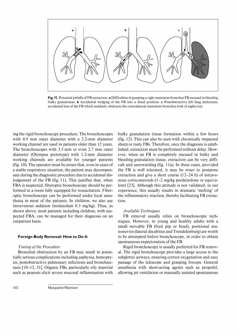
BOIS:L08:BPRR3002PD.96 FF: RESF E1: RESS
102 Marquette/Martinot
Fig. 11. Potential pitfalls of FB extraction. a Difficulties in grasping a right mainstem bronchus FB encased in bleedingbulky granulomas. b Accidental wedging of the FB into a distal position. c Postobstructive left lung atelectasis,accidental loss of the FB which suddenly obstructs the contralateral mainstem bronchus (risk of asphyxia).
ing the rigid bronchoscopy procedure. The bronchoscopeswith 4.9 mm outer diameter with a 2.2-mm diameterworking channel are used in patients older than 12 years.The bronchoscopes with 3.5 mm or even 2.7 mm outerdiameter (Olympus prototype) with 1.2-mm diameterworking channels are available for younger patients(fig. 10). The operator must be aware that, even in cases ofa stable respiratory situation, the patient may decompen-sate during the diagnostic procedure due to accidental dis-lodgement of the FB (fig. 11). This justifies that, whenFBA is suspected, fiberoptic bronchoscopy should be per-formed in a room fully equipped for resuscitation. Fiber-optic bronchoscopy can be performed under local anes-thesia in most of the patients. In children, we also useintravenous sedation (midazolam 0.3 mg/kg). Thus, asshown above, most patients including children, with sus-pected FBA, can be managed for their diagnosis on anoutpatient basis.
Foreign Body Removal: How to Do It
Timing of the ProcedureBronchial obstruction by an FB may result in poten-
tially serious complications including asphyxia, hemopty-sis, postobstructive pulmonary infections and bronchiec-tasis [10–12, 31]. Organic FBs, particularly oily materialsuch as peanuts elicit severe mucosal inflammation with
bulky granulation tissue formation within a few hours(fig. 12). This can also be seen with chronically impactedsharp or rusty FBs. Therefore, once the diagnosis is estab-lished, extraction must be performed without delay. How-ever, when an FB is completely encased in bulky andbleeding granulation tissue, extraction can be very diffi-cult and unrewarding (fig. 11a). In these cases, providedthe FB is well tolerated, it may be wiser to postponeextraction and give a short course (12–24 h) of intrave-nous corticosteroids (1–2 mg/kg prednisolone or equiva-lent) [23]. Although this attitude is not validated, in ourexperience, this usually results in dramatic ‘melting’ ofthe inflammatory reaction, thereby facilitating FB extrac-tion.
Available TechniquesFB removal usually relies on bronchoscopic tech-
niques. However, in young and healthy adults with asmall movable FB (fruit pip or bead), positional ma-noeuvres (lateral decubitus and Trendelenburg) are worthto be attempted before bronchoscopy, in order to obtainspontaneous expectoration of the FB.
Rigid bronchoscopy is usually preferred for FB remov-al. The rigid bronchoscope provides a large access to thesubglottic airways, ensuring correct oxygenation and easypassage of the telescope and grasping forceps. Generalanesthesia with short-acting agents such as propofol,allowing jet ventilation or manually assisted spontaneous

BOIS:L08:BPRR3002PD.96 FF: RESF E1: RESS
Foreign Body Removal in Adults andChildren
103
Fig. 12. a Half peanut recently (4 h) im-pacted in the right mainstem bronchus of a13-month-old child. b Same problem, butchoking occurred for 28 h. The oily peanuthas caused severe mucosal inflammationwith bulky granulation tissue.
Fig. 13. From top to bottom: rigid broncho-scope, rigid FB forceps, rigid telescope, opti-cal forceps.
a b
ventilation, is safe and seldom exceeds 10 min. An opticalforceps (fig. 13) allows a direct and clear vision of the FBand optically guided grasping. For lack, a rigid telescopeand a forceps can be used coaxially through the broncho-scope (fig. 13). During the extraction procedure, oneshould always pay attention not to push the FB distallywith the bronchoscope, the forceps or the suction catheter(fig. 11b). If present proximal to the FB, blood and secre-tions are carefully suctioned, and epinephrine (0.25 mg)can be instilled in order to retract swollen mucosa encas-ing the FB. The optical forceps is then advanced in the
bronchial axis, a few millimeters proximal to the FB. Forsmooth and rounded FBs, the principle is to grip the larg-est volume of the FB. To achieve this, the smooth forceps(FB forceps) are preferred to the sharp alligator forceps(fig. 14a–d). The cups of the forceps are opened as muchas possible (fig. 14a, b), and the forceps is advanced undervisual control without pushing the FB downwards. TheFB is then gently but securely gripped. Optical forcepsand FB are then pulled up, a few millimeters distal to thetip of the bronchoscope, and the whole is then withdrawnen masse from the trachea. In case of large and hard FBs

BOIS:L08:BPRR3002PD.96 FF: RESF E1: RESS
104 Marquette/Martinot
Fig. 14. Firm and atraumatic grasping of a smooth and rounded foreign body with an FB forceps (a, b). Traumatic andunsecured grasping with an alligator forceps (c, d). Risk of loosing the FB during extraction in cases of inappropriatecoaxial movement between the bronchoscope and the forceps, pushing the foreign body out of the cups or jaws of theforceps (e, f ).
such as pistachio shells, breaking the FB into 2 or 3 frag-ments may help extraction. However, too strong graspingof friable FBs such as peanuts should be avoided since itentails a risk of distal wedging of the small fragments(fig. 15). For heavy FBs, such as metallic FBs, which tendto move distally due to gravity, it may be helpful to placethe patient in the Trendelenburg position. Alligator for-ceps are preferably used for grasping sharp or irregularFB. During the last step of extraction, FB can be lost acci-dentally, either because it is blocked in the narrow glotticarea or because there was some inappropriate coaxialmovement between the bronchoscope and the forceps, thetip of the bronchoscope pushing the FB out of the cups orjaws of the forceps (fig. 14e, f). If the case arises, beforereintubating the trachea with the bronchoscope, oneshould first carefully inspect the oral cavity and the larynxwith the laryngoscope in order to possibly catch the FBwith a Magill forceps. Once the FB is removed, the tra-chea is reintubated with the rigid bronchoscope and care-ful examination is performed, as best with the fiberopticbronchoscope passed through the rigid tube to check for
the absence of another FB [32] or residual fragments.When managing food asphyxiation (generally with meat),one should always be aware that in about one third of thecases [20] the FB is indeed impacted at the glottic or sub-glottic level. Such proximally encased meat pieces can beeasily pulled out with a Magill forceps (fig. 16) or for lack,in the field, digitally by sticking the middle and index fin-gers down the throat to grip the piece of meat [15].
A large variety of ancillary equipment (forceps, grasp-ing claws, baskets and magnets) is available for FB extrac-tion through fiberoptic bronchoscopes (fig. 10). There-fore, from a technical point of view, fiberoptic bronchos-copy can be used as an alternative to rigid bronchoscopy.Several authors have demonstrated that FBs, even largeones, can be effectively removed with pediatric fiberopticbronchoscopes in children [26, 33–35].
Advantage and Drawbacks of Each TechniqueThe technique for removal of the FB depends on the
nature and the size of the FB and on the age and the respi-ratory status of patient. In adults and even in children

BOIS:L08:BPRR3002PD.96 FF: RESF E1: RESS
Foreign Body Removal in Adults andChildren
105
Fig. 15. a Half peanut grasped with an opti-cal forceps. b Accidental crushing of thepeanut.Fig. 16. Near fatal food asphyxiation (pieceof meat). Emergency extraction under laryn-goscopic examination with the Magill for-ceps.
15a
15b
16
over the age of 12, the available ancillary equipmentmakes it easy to remove most FBs with a 4.9-mm (ormore) outer diameter fiberoptic bronchoscope, under lo-cal anesthesia. We agree with Limper and Prakash [10]that fiberoptic bronchoscopy is superior to rigid bron-choscopy in cases of distally wedged FBs, in mechanicallyventilated patients or in cases of spine, jaw or skull frac-tures preventing rigid bronchoscope manipulation. Thesuccess rates of the fiberoptic bronchoscopy extractiontechnique in adults ranges from 60 to 90% [10–12]. Itshould however be stressed that the fiberoptic bronchos-copy extraction technique can be cumbersome and unre-warding and sometimes requires several attempts or aswitch to rigid bronchoscopy to achieve extraction [10–12]. In addition, as illustrated in figure 11, the fiberopticbronchoscopy extraction technique entails three risks.First, unsuccessful attempts to remove the FB may resultin pushing the FB into a wedged position. Second, inflam-matory lesions encasing an FB bleed as soon they aretouched. Thus, removal with the fiberoptic bronchoscopemay be cumbersome since suctioning blood and passingthe grasping instrument through the working channel atthe same time is impossible. Third, in cases of an im-pacted FB obstructing a mainstem bronchus with associ-

BOIS:L08:BPRR3002PD.96 FF: RESF E1: RESS
106 Marquette/Martinot
ated lung atelectasis, accidentally loosing the FB in thecontralateral lung, due to insufficient grasping with thefiberoptic forceps, is potentially lethal. As underlined byCastro et al. [26] the happenstance that a FB escapes fromthe rigid forceps (rigid bronchoscopy) is less likely tooccur than with the forceps, grasping claws and basketsused through fiberoptic bronchoscopes. For practice, inadults and children older than 12 years, we recommendthat fiberoptic bronchoscopy under local anesthesiashould be considered as first examination for diagnosisand extraction except in cases of asphyxiating FBs whererigid bronchoscopy should be considered first.
In young children, the extraction technique is still asubject of debate. Some doctors [26, 33–36] advocate theuse of the 3.5-mm outer diameter pediatric fiberopticbronchoscope (working channel diameter of 1.2 mm).Although FB extraction with the new pediatric ancillaryequipment is feasible with such fiberoptic bronchoscopes,it should be stressed that extraction is usually performedunder general anesthesia with the fiberoptic broncho-scope passed through an endotracheal tube. Thus, theadvantage, compared to FB removal with rigid instru-ments, appears to be rather limited. In addition, thepitfalls of FB extraction by fiberoptic bronchoscopy sum-marized in figure 11 and in figure 14e may have tragicconsequences in young children. Therefore, from an edu-cational point of view, we and others [2, 5, 30, 37, 38]recommended that rigid-tube extraction should be con-sidered as the technique of choice for removal of themajority of FBs in children.
Complications of FB ExtractionPre- and postinterventional complication rates of rigid
bronchoscopy in children with suspected FBA range from2 to 19% [3, 4, 24, 39], but include various complicationssuch as hypoxemia, bronchospasm, laryngeal edema, car-diac arrhythmias or even complications related to the FBand not to the bronchoscopy: pneumonia or persistentatelectasis [3, 4]. In our experience, the rate of postopera-tive complications for rigid bronchoscopy was 16% in-cluding laryngospasm (n = 1) and laryngeal edema (n = 6)requiring brief intubation in 2 children [5]. Perez andWood [30] suggested that if rigid bronchoscopy does notshow an FB, but there is still a high degree of suspicion,then fiberoptic bronchoscopy can be passed through therigid tube for inspection of more distal airways. The dis-lodgement of the FB as a result of fiberoptic bronchoscopywas not observed in the study of Wood and Gauderer [37]in 52 children. In our experience, this complication wasobserved once in 55 children undergoing diagnostic fiber-
optic bronchoscopy [5]. One should therefore always keepthis risk in mind and be able to convert to rigid-tubeextraction.
Perioperative ManagementThe current controversy regarding the need for cortico-
steroids or antibiotics in the perioperative management ofFBA results from the absence of comparative controlledstudies. We and others [23] recommend the use of a shortcourse of corticosteroids before FB removal when a well-tolerated FB is encased in bulky and bleeding granulationtissue. The preventive use of corticosteroids before bron-choscopy to decrease the incidence of postoperative sub-glottic edema has never been validated. When postopera-tive subglotting edema occurs, parenteral corticosteroidsand/or aerolized epinephrine is usually recommended. An-tibiotics are indicated only in cases of clinically, radiologi-cally or bronchoscopically documented respiratory tractinfection. The risk of laryngospasm within a few minutesafter bronchoscopy justifies special attention in small chil-dren and, if necessary, immediate reintubation. Once theFB is removed, the whole tracheobronchial tree should bechecked for the presence of another FB [32] or residualfragments. To do so, the trachea is reintubated with therigid bronchoscope and careful examination is performed,at best with the fiberoptic bronchoscope passed throughthe rigid tube. If doubt persists, fiberoptic bronchoscopicexamination may be recommended a few days later.
Conclusion
Clinical signs in children are usually highly suggestive.However, almost half of the children with a typical historyof choking have no FB in the airways. Therefore, fiberop-tic bronchoscopy is a safe and cost-saving diagnostic pro-cedure [5, 37]. This procedure avoids unnecessary generalanesthesia and reduces hospital stay. Even if fiberopticbronchoscopy detects an FB in a child which needs subse-quent rigid bronchoscopy for removal, this does notlengthen hospital stay as the second procedure imme-diately follows the first one [5]. For the majority of FBs inchildren, rigid-tube extraction remains the technique ofchoice for removal. In any case, diagnostic and therapeut-ic procedures should be performed by a skilled operator ina room equipped with full resuscitation equipment.
In adults, the diagnosis of FBA is often equivocal andsometimes even silent. In contrast to children, fiberopticbronchoscopy may usually be used both for diagnosis andtreatment.

BOIS:L08:BPRR3002PD.96 FF: RESF E1: RESS
Foreign Body Removal in Adults andChildren
107
References
1 Killian G: Meeting of the Society of Physiciansof Freiburg. Dec 17, 1897. Münch Med Wo-chenschr 1989;45:378.
2 Manto PC, Tuggle DW, Tunell WP: An appro-priate negative bronchoscopy rate in suspectedforeign body aspiration. Am J Surg 1989;158:622–624.
3 Inglis AF Jr, Wagner DV: Lower complicationrates associated with bronchial foreign bodiesover the last 20 years. Ann Otol Rhinol Laryn-gol 1992;101:61–66.
4 François M, Thach-Toan, Maisani D, PrevostC, Roulleau P: Endoscopie pour recherche decorps étrangers de voies aériennes inférieurschez l’enfant. A propos de 668 cas. Ann Otola-ryngol Chir Cervicofac 1985;102:433–441.
5 Martinot A, Closset M, Marquette CH, et al:Indications for flexible versus rigid bronchos-copy in children with suspected foreign bodyaspiration. Am J Respir Crit Care Med 1997;155:1676–1679.
6 Roethman BF, Boeckman CR: Foreign bodiesin the larynx and tracheobronchial tree in chil-dren: A review of 225 cases. Ann Otol RhinolLaryngol 1980;89:434–446.
7 McGuirt WF, Homes KD, Feehs R, BrowneJD: Tracheobronchial foreign bodies. Laryngo-scope 1988;98:615–618.
8 Cleveland RH: Symmetry of bronchial anglesin children. Radiology 1979;133:89–93.
9 Wolkove N, Kreisman H, Cohen C, Frank H:Occult foreign-body aspiration in adults.JAMA 1982;248:1350–1352.
10 Limper AH, Prakash UB: Tracheobronchialforeign bodies in adults. Ann Intern Med 1990;112:604–609.
11 Lan RS: Non-asphyxiating tracheobronchialforeign bodies in adults. Eur Respir J 1994;7:510–514.
12 Chen CH, Lai CL, Tsai TT, Lee YC, Perng RP:Foreign body aspiration into the lower airwayin Chinese adults. Chest 1997;112:129–133.
13 Elhassani NB: Tracheobronchial foreign bodiesin the Middle East. J Thorac Cardiovasc Surg1988;96:621–625.
14 Haugen RK: The cafe coronary: Sudden deathsin restaurants. JAMA 1963;186:142.
15 Eller WC, Haugen RK: Food asphyxiation –restaurant rescue. N Engl J Med 1973;289:81–82.
16 Holen S, Dickler EH: Missing teeth and den-tures – killers of 2,500 annually. The dentistsrole in the prevention and treatment of foodasphyxiation. J Dist Columbia Dent Soc 1973;48:26–27.
17 Feinberg MJ, Knebl J, Tully J, Segall L: Aspira-tion and the elderly. Dysphagia 1990;5:61–71.
18 Shaker R, Ren J, Zamir Z, Sarna A, Liu J, SuiZ: Effect of aging, position, and temperature onthe threshold volume triggering pharyngealswallows. Gastroenterology 1994;107:396–402.
19 Teramoto S, Matsuse T, Ouchi Y: Foreignbody aspiration into the lower airways may notbe unusual in older adults. Chest 1998;113:1733–1734.
20 Mittleman RE, Wetli CV: The fatal cafe coro-nary. Foreign-body airway obstruction. JAMA1982;247;1285–1288.
21 Hoeve LJ, Rombout J, Pot DJ: Foreign bodyaspiration in children. The diagnostic value ofsigns, symptoms and pre-operative examina-tion. Clin Otolaryngol 1993;18:55–57.
22 Wiseman NE: The diagnosis of foreign bodyaspiration in childhood. J Pediatr Surg 1984;19:531–535.
23 Banerjee A, Rao KS, Khanna SK, NarayananPS, Gupta BK, Sekar JC: Laryngo-tracheo-bronchial foreign bodies in children. J LaryngolOtol 1988;102:1029–1032.
24 Pasaoglu I, Dogan R, Demircin M, HatipogluA, Bozer AY: Bronchoscopic removal of for-eign bodies in children: Retrospective analysisof 822 cases. Thorac Cardiovasc Surg 1991;39:95–98.
25 Blazer S, Naveh Y, Friedman A: Foreign bodyin the airway. A review of 200 cases. Am J DisChild 1980;134:68–71.
26 Castro M, Midthun DE, Edell ES, Stelck MJ,Prakash UBS: Flexible bronchoscopic removalof foreign bodies from pediatric airways. JBronchol 1994;1:92–98.
27 Puhakka H, Svedström E, Kero P, Valli P, Iisa-lo E: Tracheobronchial foreign bodies. A per-sistent problem in pediatric patients. Am J DisChild 1989;143:543–565.
28 Mu L, Sun D, He P: Radiological diagnosis ofaspirated foreign bodies in children: Review of343 cases. J Laryngol Otol 1990;104:778–782.
29 Matsuse T, Oka T, Kida K, Fukuchi Y: Impor-tance of diffuse aspiration bronchiolitis causedby chronic occult aspiration in the elderly.Chest 1996;110:1289–1293.
30 Perez CR, Wood RE: Update on pediatricflexible bronchoscopy. Pediatr Clin North Am1994;41:385–400.
31 Nikolaizik WH, Warner JO: Aetiology ofchronic suppurative lung disease. Arch DisChild 1994;70:141–142.
32 Kim IG, Brummitt WM, Humphry A, SiomraSW, Wallace WB: Foreign body in the airway:A review of 202 cases. Laryngoscope 1973;83:347–354.
33 Monden Y, Morimoto T, Taniki T, Uyama T,Kimura S: Flexible bronchoscopy for foreignbody in airway. Tokushima J Exp Med 1989;36:35–39.
34 Akhter J, Gaspar M, Bassuk A, Roberts J: Inad-vertent removal of foreign body via a flexiblefiberoptic bronchoscope in a 4-year old boy.Pediatr Pulmonol 1994;18:51–52.
35 Wood RE: Flexible bronchoscopy to removeforeign bodies in children – Yes, may be – But.J Bronchol 1994;1:87.
36 Prakash UBS, Midthun DE, Edell ES: Indica-tions for flexible versus rigid bronchoscopy inchildren with suspected foreign body aspira-tion (letter to the editor). Am J Respir Crit CareMed 1997;156:1017–1018.
37 Wood RE, Gauderer MWL: Flexible fiberopticbronchoscopy in the management of tracheo-bronchial foreign bodies in children: The valueof a combined approach with open tube bron-choscopy. J Pediatr Surg 1984;19:693–694.
38 Green CG, Eisenberg J, Leong A, Nathanson I,Schnapf BM, Wood RE: Flexible endoscopy ofthe pediatric airway. Am Rev Respir Dis 1992;145:233–235.
39 Weissberg D, Schwartz I: Foreign bodies in thetracheobronchial tree. Chest 1987;91:730–733.
Ch. Marquette, Department of PneumologyHôpital Albert Calmette, Lille UniversitySchool of Medicine, Boulevard du Pr LeclercqF–59037 Lille cedex (France)Tel./Fax +33 320 44 51 77E-Mail [email protected]

OOOOOOOOOOOOOOOOOOOOOOOOOOOOOOOOOOOOOOOOOOOOOOOOOOOOOOOOOOOOOOOOOOOOOOOOOOOOOOOOOOOOOOOOOOOOOOOOOOOOOOOOOOOOOOOOOOOOOOOOOOO
Therapeutic Bronchoscopy

Bolliger CT, Mathur PN (eds): Interventional Bronchoscopy.Prog Respir Res. Basel, Karger, 2000, vol 30, pp 108–119
OOOOOOOOOOOOOOOOOOOOOOOOOOOOOOOOOOOOOOOOOOOOOOOOOOOOOOOOOOOOOOOOOOOOOOOOOOOOOOOOOOOOOOOOOOOOOOOOOOOOOOOOOOOOOOOOOOOOOOOOOOO
Laser BronchoscopySergio Cavalierea Jean-François Dumonb
aServizio di Endoscopia e Laserterapia, Spedali Civili, Brescia, Italy;bCentre Laser, Hôpital Sainte-Marguerite, Marseille, France
108
Summary
This chapter is based on our 17-year experience withlaser bronchoscopy. In addition to relevant backgroundinformation, it contains a description of our technique andresults. Suitability of a laser for therapeutic bronchoscopydepends on power density, absorption/scattering ratio insoft tissue and delivery system. Currently the Nd:YAG andNd:YAP are the most suitable laser systems. Knowledge oftracheobronchial anatomy is essential. Since the volume ofthe airways is only 150 cm3, accumulation of blood andsecretions can lead rapidly to hypoxemia. The tracheobron-chial tree contracts close relations with mediastinal vesselsand perforation can be fatal. Safety depends on the skill andcooperation of the laser team. Laser bronchoscopy shouldbe performed using a surgical bronchoscope under generalanesthesia. Mechanical resection helps to reduce laser expo-sure and risk of perforation. The main indications for laserbronchoscopy are malignant tumors and tracheal stenosis.Location and extrinsic compression are the most importantfactors for the outcome in patients with malignancies. Endo-scopic treatment must be associated with dilatation andstenting for effective treatment in patients with tracheal ste-nosis. Restoration of airway patency with immediate symp-tomatic improvement is achieved in 93% of cases overall.
With the exception of foreign body removal, applica-tions for conventional bronchoscopy are diagnostic in-cluding direct visualization of the tracheobronchial tree,biopsy of lesions and specimen collection for culture and
cytology. In this regard, the flexible fiberscope has greatlyextended the range of application in comparison withrigid endoscopy.
The modern era of interventional bronchoscopy wasborn with the advent of lasers and has been characterizedby a comeback of the rigid bronchoscope. The first endo-scopic laser resections were performed by Godard et al.[1], Toty et al. [2] and Dumon et al. [3] in the early 1980sand were soon followed by other therapeutic modalitiessuch as cryotherapy, electrocoagulation, brachytherapyand, most recently, stent placement.
Like any technique, endoscopic laser resection has lim-itations. However, it can be remarkably effective in well-selected indications and has provided a solution for pre-viously intractable emergency problems and for somecases of iatrogenic tracheal stenosis. The purpose of thischapter is to describe our 17-year experience with laserbronchoscopy.
Bronchoscopic Lasers
The word ‘laser’ is an acronym for light amplificationof stimulated emission of radiation. This name gives asuccinct description of the process by which the laserbeam is obtained. An external energy source, usuallyoptical or electrical, is used to stimulate an active sub-stance contained within the core of the laser so as toachieve a condition known as population inversion inwhich the population of atoms (or molecules) at somehigher energy level is greater than the population at somelower energy level. For sustained laser action, population

HABE:L08:BPRR3007XA.85 FF: RESF E1: RESS
Laser Bronchoscopy 109
Fig. 1. Electromagnetic radiation spectrum.KTP = Potassium titanyl phosphate.
inversion must be great enough so that the amplificationof photons by stimulated emission is greater than theattenuation of spontaneously emitted photons by absorp-tion into unexcited atoms or molecules. The core of thelaser, or pumping cavity, is designed with reflective wallswhich bounce emitted photons back into the active sub-stance thereby producing new photons. The laser beamescapes through a partially reflective mirror placed at oneend of the core.
Laser light has three unique characteristics, i.e. mono-chromaticity, coherence and collimation. Monochroma-ticity means that all light in the laser beam is of the samewavelength depending on the active element stimulatedin the laser core. In order of decreasing wavelength andincreasing frequency, electromagnetic radiation rangesfrom radiowaves to Á rays (fig. 1). Visible light is a narrowband from 400 to 700 nm between infrared and ultravio-let radiation. Coherence means that all waves in the beamare in step in time and space. Collimation means that thewaves are traveling along parallel ray directions.
Although many lasers are now available and others areunder development, devices differ with regard to theireffects on the human body and are thus not equal in termsof their usefulness for medical applications. Three maincharacteristics determine the suitability of a particularlaser for therapeutic bronchoscopy, namely: (1) powerdensity rating, (2) ratio of absorption and scattering coef-ficients in soft tissue and (3) delivery system. Power den-sity depends on laser technology and on factors such astarget distance and exposure time. By determining thevolume of tissue that is heated, absorption and scatteringmake the difference between cutting and hemostasis. La-sers with high absorption coefficients and low scatteringcoefficients are good scalpels, while those with low ab-sorption coefficients and high scattering coefficients aregood coagulators. The delivery system is important be-cause of the limited size of the endoscopic working chan-
nel. Only lasers with optical fiber delivery systems aresuitable for bronchoscopic use.
The CO2 laser was the first laser to be used in the air-ways. It generates an invisible infrared beam of 10,600nm. Because of its high absorption coefficient, the CO2 isan excellent cutting tool that has been used for drillingand resection of tissue everywhere in the body. However,its use for bronchoscopic applications has been limited byits poor coagulating properties and especially by the needfor a cumbersome articulated arm delivery system.
The argon laser produces a blue-green beam with awavelength of 488 or 514 nm. Since this wavelength isabsorbed by hemoglobin, the argon laser is a good coagu-lator. It has been used mainly for treatment of angioma.The main drawback for bronchoscopic applications ispoor cutting ability. The power density of the argon laseris low, and the fiber must be placed in contact with thetissue to achieve resection.
Dye lasers are devices in which the beam from a kryp-ton, argon, ruby or Nd:YAG laser is passed through a liq-uid made of fluorescent organic dyes such as rhodamines.The main application for dye laser technology has been toactivate hematoporphyrin for photochemotherapy of insitu carcinoma, early-stage carcinoma and submucosalcomponent of invasive carcinoma. Low-power dye lasersare not suitable for therapeutic bronchoscopy.
Excimer lasers are a special breed that produce ultra-violet wavelengths at 193, 222, 248, 308 and 351 nmusing various rare gases including argon fluoride, kryptonchloride, krypton fluoride, xenon chloride and xenonfluoride, respectively. Ultraviolet radiation is strongly ab-sorbed by living tissue, but unlike infrared and visible redlight, it is not converted into heat. Instead tissue destruc-tion results from mechanical effects. The lack of collateralthermal damage to adjacent tissue is an advantage forsome applications, but poor coagulation is a major limita-tion for bronchoscopic resection.

HABE:L08:BPRR3007XA.85 FF: RESF E1: RESS
110 Cavaliere/Dumon
Fig. 2. Anatomical relationships of the tra-cheobronchial tree. 1 = Trachea; 2 = rightmainstem bronchus; 3 = left main stembronchus; 4 = aorta; 5 = right pulmonaryartery; 6 = left pulmonary artery; 7 = rightupper lobe bronchus; 8 = troncus interme-dius; 9 = left upper lobe bronchus; 10 = leftlower lobe bronchus; 11 = vagus nerve; 12 =recurrent nerve; 13 = esophagus; 15 = superi-or vena cava; 16 = azygos vein; B1–3 = seg-mental bronchi of upper lobes.
Diode lasers represent a new technological concept.Laser radiation is obtained by passing an electrical cur-rent through a solid-state semiconductor diode. By sim-plifying the laser cavity, this technology has allowed thedesign of high-powered devices in compact, air-cooledcabinets. Aluminium gallium arsenide diode lasers emitbeams up to 60 W at a wavelength of 810 nm allowingfairly good cutting and hemostasis. This technology hasnot been widely used for therapeutic bronchoscopy.
The Nd:YAG (neodymium:yttrium aluminium garnet)laser with an invisible infrared beam (wavelength 1,064nm) exhibits a number of advantages for endoscopic use.It can deliver up to 100 W of output in the beam through aflexible quartz fiber. It is an inherently durable, reliablelaser with minimal servicing. It seems to have a virus-killing effect which may be a significant advantage for thetreatment of tumors like papillomas [4]. For these rea-sons, the Nd:YAG is probably the most widely used laserdevice for therapeutic bronchoscopy.
A remarkable feature of the Nd:YAG is its sensitivityto tissue pigmentation. Surface absorption is low in lightlycolored tissue and high in darkly colored tissue. In prac-tice, this means that a large volume of lightly colored tis-sue can be ‘cooked’ to achieve hemostasis by denaturationand agglomeration of tissue proteins, shrinkage of connec-tive tissue like collagen and extensive edema. To performresection, it is simply necessary to induce charring, so thatsurface absorption increases and vaporization proceeds
rapidly. Charring may be obtained by increasing the pow-er density or by inducing surface bleeding to darken thesurface. Some Nd:YAG lasers feature the superpuls-ing mode that can deliver short high-powered pulses(300 W) in rapid succession and frequency doubling usinga potassium titanyl phosphate crystal that provides a 532-nm green beam similar to that of an argon laser. Super-pulsing facilitates cutting and frequency doubling en-hances coagulation.
A recent addition to the biochemical laser family withpotential for therapeutic bronchoscopy is the Nd:YAP(neodymium: yttrium aluminum pevroskite) laser with awavelength of 1,340 nm. Absorption in water is 20 timesgreater for the Nd:YAP beam than the Nd:YAG beam.The Nd:YAP achieves particularly good coagulation.
Physiological and Anatomic Considerations forEndobronchial Laser Treatment
The volume of the tracheobronchial tree is only 150cm3 (anatomical dead space). Thus even a small amountof blood or secretion can cause major hypoxia leading toserious cardiovascular problems including arrhythmia,bradycardia, myocardial ischemia and even cardiac ar-rest. Maintaining a free airway and efficient ventilation atall times is essential for the prevention of these complica-tions. With regard to ventilation, it should be stressed that

HABE:L08:BPRR3007XA.85 FF: RESF E1: RESS
Laser Bronchoscopy 111
Fig. 3. Pediatric version of the EFER-DU-MON therapeutic bronchoscope with inter-changeable barrels.
proper precaution should be taken when using a laser inan enclosed oxygen-rich space containing inflammablematerials such as a flexible fiberscope.
The topography of the tracheobronchial tree whichconsists of a succession of subdividing bronchi is familiarto pulmonologists. However, it is useful to recall the ana-tomical relationships of the tracheobronchial tree (fig. 2).The back of the trachea is in permanent contact with thefront of the esophagus. The innominate artery crosses thefront of lower third of the trachea. The aortic arch and therecurrent nerve are in contact with the left side of the bot-tom of the trachea.
The main stem bronchi are also easily accessible withthe rigid bronchoscope. The right main stem bronchus isin contact with the pulmonary artery in the front. The leftmain stem bronchus is surrounded by the esophagus inthe back, the aortic arch above and the pulmonary arteryin the front. The upper lobe bronchi are in close contactwith the pulmonary artery especially on the left.
The main anatomic relations of the right lower andmiddle lobe bronchi and the left lower lobe bronchi arethe organs of the mediastinum, in particular the heart andpulmonary veins.
Basic Principles of Endoscopic Laser Bronchoscopy
Rigid Bronchoscopy Laser ResectionBecause endobronchial laser treatment involves surgi-
cal resection, it is most safely performed with a surgicalbronchoscope (fig. 3) under general anesthesia in a surgi-
Table 1. Anesthesia protocol for laser resection
Induction Maintenance
Fentanyl, 2.5 Ìg/kgDroperidrol, 125 Ìg/kgPropofol, 2.0 mg/kgSuccinylcoline, 1.5 mg/kg
propofol, 7–8 mg/kg/h(reduced to 5– 6 mg/kg/h after 15 min);if necessary,fentanyl, 2 Ìg/kg + propofol, 0.5 mg/h
cal setting with a nearby recovery room. The surgicalbronchoscope is the key to safety since it allows simulta-neous ventilation and treatment (aspiration, coagulationand debulking). Indeed, safe laser resection depends onpreserving ventilation, preventing hemorrhage and min-imizing laser exposure.
General anesthesia is usually more comfortable forboth the patient and endoscopist. For the bronchoscopist,the ideal anesthetic technique should allow spontaneousventilation while suppressing the cough reflex. Our anes-thetic protocol is described in table 1. Anesthetic agentswith short half-lives should be preferred so that thepatient recovers rapidly, and postoperative ventilation isnot needed. If muscle relaxants with Venturi jet ventila-tion or gas anesthesia are used, closed-circuit ventilationis required.
Regardless of the anesthetic technique, the endoscopistand anesthesiologist must work in close collaboration tosafeguard ventilation throughout the procedure. Bloodoxygen should be monitored using an oxymeter. A suction

HABE:L08:BPRR3007XA.85 FF: RESF E1: RESS
112 Cavaliere/Dumon
Fig. 4. Devascularization and vaporization using a laser.
catheter should always be in place at the site of treatmentin order to keep the operating field clear by continuoussuction of blood, secretions and smoke. The endoscopistshould not hesitate to perform aspiration whenever neces-sary, even if resection must be momentarily interrupted.An in-depth description of the anesthetic management forinterventional bronchoscopy is provided in the chapter byStuder and Biro.
To prevent hemorrhage, it is important to coagulatethe lesion before resection. In this regard, it is essential toremember the basic rules for adjusting power density toachieve coagulation or resection. For the Nd:YAG orNd:YAP, these rules can be summarized as follows:
(1) Dark areas having a higher absorption coefficientthan light aras; in addition to blood supply, the main fac-tor affecting tissue color during laser treatment is char-ring.
(2) Power density depends on the power setting of thelaser and the distance of the tip of the optical fiber fromthe tissue:
Power density = Power
Spot diameter.
(3) Duration of emission also determines energy densi-ty which is the product of power density and time:
Energy density = Power density ! time.
Practically speaking, these rules mean that power den-sity can be adjusted without changing power settingssimply by moving the tip of the laser closer to or fartherfrom the surface. Thus at a power setting of 30–45 W withan emission duration of 1 s, tissue shrinkage and thereforedevascularization is obtained by holding the fiber about1 cm from the target while charring and vaporization areobtained by holding the fiber about 3 mm from the target(fig. 4). It is important to note that fibers with metallic tipsshould always be used. Using bare fibers which have anirregular tip cross-section, laser light loses collimationand, as a result, aiming and control of power densitybecome problematic.
Once a tumor has been coagulated or in order to avoidheating beneath the surface, laser exposure can andshould be minimized using mechanical debulking withthe tip of the bronchoscope, biopsy forceps, resector sys-tem and suction tube. So great is the importance ofdebulking with the tip of the bronchoscope that a more

HABE:L08:BPRR3007XA.85 FF: RESF E1: RESS
Laser Bronchoscopy 113
Fig. 5. Mechanical debulking with the tip of the bronchoscope. RMSB = Right main stem bronchus.
suitable name for the technique might be ‘laser-assistedmechanical resection’ (fig. 5). The axis of the bronchuscan be located by tactile feedback from the bronchoscopein contact with bronchial cartilage. Mechanical debulkingalso shortens procedure times.
At the end of the procedure, the base of the lesionshould be thoroughly coagulated using long, low-powerpulses (20–30 W for 4–5 s). To avoid perforation, thebeam should be fired tangentially to the wall in a contin-ually scanning fashion.
Laser FiberscopyMost practitioners in the world, (Personne et al. [5],
Vergnon et al. [6] and Dumon et al. [7] in France; Caval-iere et al. [8] in Italy; Diaz et al. [9] in Spain; Becker et al.[10] and Freitag et al. [11] in Germany; Bolliger [12] inSwitzerland; Beamis and Shapshay [13], Edell and Cor-tese [14, 15], Gelb and Epstein [16] and Colt [17] in theUSA) perform endoscopic laser resection using a rigidbronchoscope. However, a few Americans (Unger [18]and Mehta et al. [19, 20]) have reported excellent resultsusing the flexible fiberscope. Tables 2 and 3 summarizethe advantages and disadvantages of rigid bronchoscopicand flexible fiberscopic laser treatment.
Laser fiberscopy requires strict cooperation on the partof the patient who must be able to cope with the discom-fort. Local anesthesia must be thorough and should berenewed if the procedure is prolonged. Care should beexercised when administering oxygen due to the risk ofignition of the fiberscope. The working channel must beperfectly clean (no trace of blood) when the laser fiber isinserted.
Small peripheral lesions are easy to treat under localanesthesia. After thorough coagulation, large pieces canbe removed with biopsy forceps. Fragments should bestripped off with a downward rather than an upwardmovement. Fragments can be removed using accessoriessuch as a Dormia basket or a Fogarty balloon. Mechanicalremoval greatly shortens the procedure.
Lesions in the main stem bronchi require great care.The patient should be turned on the obstructed side, sothat ventilation of the healthy side can continue in case ofhemorrhage.
Lesions in the trachea or in pneumonectomized pa-tients are very difficult to treat with a flexible fiberscope.The major risks are hemorrhage and hypoxia, which arevery difficult to control using the fiberoptic broncho-scope. Treatment of tracheal lesions confronts the endos-

Flexible fiberscopy is the most widely used
General anesthesia is more comfortable for the patient and the physician.
HABE:L08:BPRR3007XA.85 FF: RESF E1: RESS
114 Cavaliere/Dumon
Table 2. Advantages and disadvantages of flexible bronchoscopic laser technique.
Advantages Disadvantages
endoscopic technique.1Flexible fiberscopy is an ambulatory procedure
that can be performed under local anesthesia,thus avoiding the dangers of general anesthesiaparticularly in high-risk patients.
Fiberscopy is less cost intensive since it does notrequire an operating room and can be done bya smaller staff.
Treatment is uncomfortable for the patient and time-consuming for the physician.
Exposure to smoke is hazardous for the staff and thepatient.
The inability to perform mechanical resection anddilatation results in less complete disobstruction andmakes it necessary to use more laser energy to vaporizethe lesions.
Because the working channel of the fiberscope is small,instruments (laser fiber, suction tubes, forceps) mustbe inserted one at a time.
Bleeding is difficult to control.Fiberscopes and accessories are expensive and easily
damaged (ignition).Stent placement is difficult under local anesthesia without
previous dilatation.
1 This argument is based on the dubious assumption that lung physicians are reluctant to learn new techniques.
Table 3. Advantages and disadvantages of rigid bronchoscopic laser technqiue
Advantages Disadvantages
Laser fiber, suction catheters and forceps can be introduced and usedsimultaneously.
Dilatation and mechanical resection can be used to shorten procedure time,diminish laser exposure and enable more complete disobstruction.
Complications can be more easily controlled.Stent placement is simple.Equipment is inexpensive and indestructible.
Procedures require experienced staff, skillful endoscopist,courageous anesthetist and trained instrumentalists.
Procedures must be performed in an operating room with anearby recovery room.
Need for hospitalization increases the cost.
copist with the dilemma of choosing between vaporiza-tion to open the airway quickly and coagulation which isneeded to prevent excessive bleeding during removal. Thefiberscope should not be used in cases involving subtotalobstruction of the trachea.
Another important drawback of the flexible techniqueis the possibility of irreparable damage to the fiberscope.Indeed, sharp angulation can cause the optical fiber tobreak and, as a result, the working channel is burned whenthe laser is fired. In 300 laser procedures performed usingthe flexible system, Dumon destroyed 3 fiberscopes inthis way [unpubl. data]. Given the price of a fiberscope,the risk can greatly increase the cost of treatment.
Table 4. Complications occurring during 5,049 therapeutic bron-choscopy procedures carried out between April 1982 and March1997 (Cavaliere)
Complications n Deaths
Hemorrhage 1250 cm3 34Transient pneumothorax 25Transient mediastinal emphysema 16Respiratory failure 21 6Cardiac arrest 17 6Myocardial infarction 5 2Pulmonary embolism 1 1
Mortality rate 3/1,000.

Airway tumor
HABE:L08:BPRR3007XA.85 FF: RESF E1: RESS
Laser Bronchoscopy 115
Table 5. Indications for laser bronchoscopybetween April 1982 and March 1997(Cavaliere)
Pathology Patients Treatments Generalanesthesia
Localanesthesia
2,481 (74) 3,524 3,159 365Tracheal stenosis 499 (15) 950 800 150Miscellaneous 360 (11) 575 391 184Total 3,340 5,049 4,350 (86) 699 (14)
Rigid bronchoscope 91%, flexible fiberscope alone 9%, figures in parentheses are per-centages.
Table 6. Malignant tumors treated by therapeutic bronchoscopybetween April 1982 and March 1997 (Cavaliere)
Pathology Patients Treatments
Squamous cell 1,370 (66) 1,941Adenocarcinoma 174 (8) 229Small cell 117 (6) 135Large cell 62 (3) 83Unclassifiable 109 (5) 132Metastatic 157 (8) 286Rare tumors 80 (4) 109Total 2,069 2,915
Figures in parentheses are percentages of series.
Accident PreventionThe main risks of endoscopic laser resection are hypox-
ia, hemorrhage, perforation and fire. In accordance withthe old adage ‘an ounce of prevention is worth a pound ofcure’, the following rules should be respected: (1) avoidhypoxia by keeping peripheral airways free at all times;(2) avoid hemorrhage by coagulating lesions thoroughlybefore resection; (3) be mindful of the anatomical dangersand the biological effects of the laser; (4) prefer mechani-cal debulking to laser resection in order to limit iatrogeniccomplications; (5) use the laser in the discontinuous modeat a moderate power setting (40–45 W); (6) do not use thelaser when ventilating the patient with pure oxygen (weadvise a limit of 50%). Complications in our experi-ence are summarized in table 4. Overall mortality was3:1,000.
Indications for Laser Bronchoscopy
In theory, any obstructive lesion in the main airway,i.e. the trachea and/or the right and left stem bronchi, canbe treated by therapeutic bronchoscopy. The main indica-tions for laser resection are tumors and iatrogenic trachealstenosis. Table 5 summarizes the indications for laserbronchoscopy under general and local anesthesia in ourseries.
Airway TumorsAirway tumors may be subdivided into three histologi-
cal groups: malignant, uncertain prognosis and benign.For malignant lesions, laser therapy is purely palliativeand should be performed only in nonoperative cases. Thishaving been said, it should be underlined that surgery isfeasible in only 25% of lung cancer patients, and morethan 70% of these patients have recurrences. Thus, morethan 90% of lung cancer patients require palliative treat-ment. Laser resection is a therapeutic option for the 30%
of these patients in whom lung cancer causes obstructionin the trachea and main stem bronchi with subsequentrespiratory distress, bleeding and infection. Table 6 showsthe number of patients and treatments corresponding tomalignant airway tumors. Metastatic and rare tumors aredetailed in tables 7 and 8, respectively.
The category ‘tumors of uncertain prognosis’ regroupsvarious tumors characterized by slow growth and low ten-dency to metastasis such as carcinoids, adenoid cystic car-cinomas, paragangliomas etc. As for malignant tumors,laser therapy is mainly palliative. In some cases, it canalso be used as a bridge to surgery and even achieve localcure for small tumors of low-grade malignancy. For exam-ple, laser surgery can be curative for strictly endobron-chial carcinoids of typical histology. Conversely, typicalcarcinoid tumors with extraluminal extension are alwayssurgical indications. Table 9 shows the number of patientsand treatments corresponding to tumors with uncertainprognosis.
Benign tumors such as hamartoma, chondroma, leio-myoma and papilloma are excellent indications for laser

HABE:L08:BPRR3007XA.85 FF: RESF E1: RESS
116 Cavaliere/Dumon
Table 7. Metastatic tumors treated by therapeutic bronchoscopybetween April 1982 and March 1997 (Cavaliere)
Tumor location n Tumor location n
Thyroid 43 Thymus 1Kidney 32 Prostate 1Colon 30 Testis 1Esophagus 25 Bone 1Breast 9 Liver 1Melanoma 5 Larynx 1Uterus 4 Ureter 1Ovary 2
157 patients – 286 treatments.
Table 8. Rare malignant tumors treated by therapeutic bronchosco-py between April 1982 and March 1997 (Cavaliere)
Tumors n Tumors n
In situ carcinoma 27 Fibrosarcoma 2Non-Hodgkin lymphoma 15 Fibrohystiocytoma 1Hodgkin lymphoma 5 Teratoma 1Carcinosarcoma 16 Myeloma 1Sarcoma 4 Basaloid tumor 1Malignant plasmacytoma 3 Glomic tumor 1Leimyosarcoma 3
80 patients – 109 treatments.
Table 9. Tumors with uncertain prognosis treated by therapeuticbronchoscopy between April 1982 and March 1997 (Cavaliere)
Pathology Patients Treatments
Carcinoid 129 170Adenoid cystic 49 103Mucoepidermoid 14 19Spindle cell 5 10Mixed 2 3Paraganglioma 2 2Cystoadenoma 2 2Total 203 309
Table 10. Benign tumors treated by therapeutic bronchoscopy be-tween April 1982 and March 1997 (Cavaliere)
Tumors n Tumors n
Hamartoma 51 Leiomyoma 7Papilloma 44 Myoblastoma 5Amyloidosis 21 Plasmacytoma 4Polyp 21 Fibrolipoma 3Angioma 19 Fibroma 2Lipoma 14 Fibrohistiocytoma 2Chondroma 8 Chordoid chordoma 1Schwannoma 7
209 patients – 300 treatments.
therapy. Endoscopic laser resection should be the methodof choice for polypoid tumors insofar as recurrence is rareif the base is well coagulated. Surgery should be reservedfor cases with extraluminal growth and recurrences. Ta-ble 10 indicates the number of endoscopic laser proce-dures carried out for benign tumors in our series.
It should be noted that location, not cell type, is themost important factor for the outcome of laser therapy. Asshown in figure 6, best results are obtained in the tracheaand main stem bronchi because they are the most accessi-ble locations to rigid bronchoscopy. These are also thelocations that cause the severest impairment of ventila-tion. During treatment of tracheal lesions, the first priori-ty is to establish an airway. The most expeditious way todo this is simply to push the bronchoscope slowly but for-cibly through the stenosis. For bilateral lesions involvingboth main stem bronchi, e.g. astride the carina, it is advis-
able to begin on the less obstructed side in order to estab-lish a reliable airway as soon as possible.
Laser resection in third-order bronchi poses specialhazards due to poor accessibility and thinness of the walls(risk of perforation). Segmental lesions usually do notcause symptoms severe enough to warrant laser surgery,and the only indications for laser disobstruction are drain-age of distal purulent secretions and cure of benigntumors. Work in the upper lobes often requires use of thefiberscope which further increases the risks. A safer tech-nique is to use the open tube as a conduit for the fiber-scope.
Another important factor in the outcome of laser resec-tion for airway tumors is degree of extrinsic compression.Complete resection of polypoid tumors is simple. Ob-viously purely extrinsic compression is a contraindicationfor laser therapy. In cases involving tumors with both
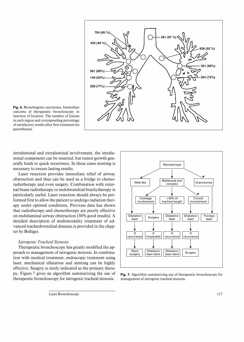
HABE:L08:BPRR3007XA.85 FF: RESF E1: RESS
Laser Bronchoscopy 117
Fig. 6. Bronchogenic carcinoma. Immediateoutcome of therapeutic bronchoscopy infunction of location. The number of lesionsin each region and corresponding percentageof satisfactory results after first treatment (inparentheses).
Fig. 7. Algorithm summarizing use of therapeutic bronchoscopy formanagement of iatrogenic tracheal stenosis.
Stenosis type
Bottleneck andcomplex
Cricoidinvolvement
Web-like Granulomas
ää
> 50% oftracheal length
Cartilageinvolvement
Dilatationlaser
ä
Surgery
ä
Dilatationlaser
Dilatationlaser
ä
Forcepslaser
Ifrecurrence
äää
Ifrecurrence
Ifinoperable
Ifrecurrence
ä
Dilatationlaser stent
äää
Stentsurgery
Dilatationlaser stent
ä
ää
ä ä
ä ä
Surgery
intraluminal and extraluminal involvement, the intralu-minal component can be resected, but tumor growth gen-erally leads to quick recurrence. In these cases stenting isnecessary to ensure lasting results.
Laser resection provides immediate relief of airwayobstruction and thus can be used as a bridge to chemo-radiotherapy and even surgery. Combination with exter-nal beam radiotherapy or endobronchial brachytherapy isparticularly useful. Laser resection should always be per-formed first to allow the patient to undergo radiation ther-apy under optimal conditions. Previous data has shownthat radiotherapy and chemotherapy are poorly effectiveon endoluminal airway obstruction (30% good results). Adetailed description of multimodality treatment of ad-vanced tracheobronchial diseases is provided in the chap-ter by Bolliger.
Iatrogenic Tracheal StenosisTherapeutic bronchoscopy has greatly modified the ap-
proach to management of iatrogenic stenosis. In combina-tion with medical treatment, endoscopic treatment usinglaser, mechanical dilatation and stenting can be highlyeffective. Surgery is rarely indicated as the primary thera-py. Figure 7 gives an algorithm summarizing the use oftherapeutic bronchoscopy for iatrogenic tracheal stenosis.

HABE:L08:BPRR3007XA.85 FF: RESF E1: RESS
118 Cavaliere/Dumon
Table 11. Iatrogenic stenoses treated by therapeutic bronchoscopybetween April 1982 and March 1997 (Cavaliere)
Type of narrowing Patients Treatments
Granuloma 136 203True stenosis 363 747Total 499 950
Table 12. Miscellaneous indications treated by therapeutic bron-choscopy between April 1982 and March 1997 (Cavaliere)
Miscellaneous indications n
Bronchial granuloma 77Hemorrhage 64Tuberculosis-related stenosis 46Stenosis after sleeve resection or radiation therapy 37Bronchial fistula 23Suture thread removal 18Vascular dilatation 13Wegener’s granulomatosis 13Inflammatory pseudotumors 11Tracheopathia osteoplastica 8Others 50
360 patients – 575 treatments.
An important factor in therapeutic decision making foriatrogenic tracheal stenosis is the type of lesion. The sim-plest form is granuloma due to formation of granulationtissue in the lumen. Granulomas are not true tracheal ste-noses since the wall of the trachea is not involved. Thesecond type of tracheal stenosis is the web. Webs corre-spond to formation of a fibrous membrane growing cir-cumferentially from the tracheal wall towards the centerof the lumen. These lesions are localized and the structureof the trachea is not altered. The third type of trachealstenosis is bottleneck stenosis due to collapse of the tra-cheal wall involving a short segment of the trachea (notmore than 5 cm). The fourth form of tracheal stenosis iscomplex stenosis which designates narrowing due to acombination of granuloma, web and/or bottleneck steno-sis or to extensive lesions involving a long segment of thetrachea. Table 11 shows the number of patients and treat-ments corresponding to iatrogenic tracheal stenosis in ourexperience.
The laser is useful in patients with simple lesions andin patients who are ineligible for surgery due to the lengthof the lesion or to associated risk factors (e.g. age andintercurrent disease). In these patients endoscopic treat-ment is the only alternative and should be used freely.Several other points deserve to be underlined. First, in thecontext of tracheal stenosis, laser resection is complemen-tary to tracheal dilatation using progressively larger tubes.Only fibrous lesions and granulomas projecting into thelumen of the trachea can be resected. For webs, the lasershould be used only at high power to make 3 or 4 radialincisions. Second, laser therapy should be used instead oftracheotomy as first-intention treatment in patients withacute dyspnea. Third, surgery and endoscopic therapy arenot mutually exclusive. Surgical patients may benefitfrom endoscopic laser therapy to allow stabilization of thelesion. Fourth, stenting is very useful as a means of main-taining airway patency after dilatation and laser resec-tion.
Miscellaneous IndicationsMiscellaneous indications include bronchial granulo-
ma, hemorrhage, tuberculosis-related stenosis, bronchialfistula, suture thread removal, vascular dilatation, We-gener’s granulomatosis, inflammatory pseudotumors, tra-cheopathia osteoplastica and foreign body removal. Thenumber of patients and treatment for these indicationsare summarized in table 12.
Laser bronchoscopy has greatly improved emergencytreatment of massive hemoptysis regardless of the cause.Using the rigid bronchoscope, which allows simultaneousventilation, aspiration and treatment, it is possible toestablish an airway, locate the source of bleeding andachieve hemostasis in almost all cases. An important pre-caution when treating hemoptysis is to turn the patient onthe bleeding side in order to prevent blood from floodingthe healthy contralateral lung. By continuous aspirationof blood or by injection and aspiration of adrenaline-con-taining saline, it is possible to locate the source of bleed-ing. The laser can then be used to coagulate the bleedingsite.
Long-Term Results
Long-term results of therapeutic endoscopy for malig-nant airway tumors are difficult to interpret since TNMrestaging was not performed and therapies used beforeand after laser treatment varied widely. In a series by Ca-valiere, cumulative survival in the first 333 patients was

HABE:L08:BPRR3007XA.85 FF: RESF E1: RESS
Laser Bronchoscopy 119
50% (B3%) at 6 months and 26% (B3%) at 1 year. Thisstudy was stopped because we consider that survival is nota suitable criterion for evaluating a modality used mainlyfor symptomatic/palliative relief. Other authors have re-ported that laser therapy extends survival, but we thinkthe technique should be judged on the basis of immediateimprovement in quality of life. In this regard it should beemphasized that good immediate results with significantimprovement in airway caliber and restoration of regionalventilation were obtained in 93% of cases overall. It isalso interesting to note that respiratory distress was rarelythe cause of death in our patients with malignant tumors.
As stated above, endobronchial laser therapy can becurative for typical carcinoid tumors and benign tumors.Cures have also been observed in other cases includingmetastatic lesion of tracheobronchial mucosa, early can-cers and in situ carcinomas. With follow-ups ranging from5 to 184 months, Cavaliere [unpubl. data] observed norecurrence in the 27 patients treated for in situ carcinomain his series.
Conclusion
Bronchoscopic laser technique has changed and im-proved with experience. The initial technique based pure-ly on laser vaporization has given way to a laser-assistedtechnique combining laser treatment, dilatation, mechan-ical debulking and stenting. The laser is increasingly usedprimarily for coagulation before resection and in case ofhemorrhage. Laser treatment of the residual tumor bed isalso important after treatment to prevent recurrence. Inaccordance with this evolution, technological research hasfocused mainly on finding the most suitable wavelengthfor coagulation. Other advances in biomedical laser tech-nology have involved mainly price and ergonomic fea-tures such as compactness and convenience in operation.Now as before, the goal of the therapeutic bronchoscopy isto reestablish the airway as quickly and thoroughly as pos-sible.
References
1 Godard P, Draussin M, et al: Utilisation durayonnement laser en bronchologie – Résectionde 2 tumeurs trachéobronchiques. PoumonCoeur 1979;35:147–150.
2 Toty L, Personne C, Colchen A, Vourch G:Bronchoscopic management of tracheal lesionsusing Nd:YAG laser. Thorax 1981;36:175–178.
3 Dumon JF, Rebound E, Aucomte F, Meric B:Treatment of tracheobronchial lesions by laserphotoresection. Chest 1982;81:278–284.
4 Buzalov S, Khristakieva E: The treatment ofneglected cases of condolymata acuminata inpregnant women with the Nd:YAG laser.Akush Ginekol 1985;34:38–39.
5 Personne C, Colchen A, Leroy M, Vourch G,Toty L: Indications and technique for endo-scopic laser resection in bronchology – A criti-cal analysis based upon 2,284 resections. Tho-rac Cardiovasc Surg 1986;91:710–715.
6 Vergnon JM, et al: Cryotherapy or Nd:YAGlaser in the treatment of tracheobronchial tu-mors. Chest 1987;91:473.
7 Dumon JF, Meric B, Bourcereau J, Garbi N,Cavaliere S, Shapshay S, Beamis J: Principlesfor safety in application of Nd:YAG laser inbronchology. Chest 1982;81:278–284.
8 Cavaliere S, Foccoli P, Farina PL: Nd:YAGlaser bronchoscopy – A five-year experiencewith 1936 applications in 1,000 patients. Chest1988;94:15–21.
9 Diaz Jimenez JP, Canela Cardona M, MaestreAlcacer J: Nd:YAG laser photoresections oflow-grade malignant tumors of the tracheo-bronchial tree. Chest 1990;97:920–922.
10 Becker HD, Wanjek M, van Bodegom PC,Drings P: Endoscopic laser therapy in the tra-cheobronchial system. Support Care Cancer1993;1:47–51.
11 Freitag L, Tekolf E, Eicker R, Greschuna D:Four years of palliation with airway stents.Results of 263 stent placements in 179 patients.Eur Respir J 1993;17:A1548.
12 Bolliger CT: Combined treatment modalitiesin lung cancer. Rigid Bronchoscopy Meeting,Marseille, October 1994, pp 177–187.
13 Beamis JF, Shapshay S: YAG laser therapy fortracheobronchial disorders. Postgrad Med1984;75:173–180.
14 Edell ES, Cortese DA: Bronchoscopic photo-therapy with hematoporphyrin derivative fortreatment of localized bronchogenic carcino-ma: A five-year experience. Mayo Clin Proc1987;62:8–14.
15 Cortese DA: Endobronchial management oflung cancer. Chest 1986;89:2345–2365.
16 Gelb AF, Epstein JD: Laser in treatment oflung cancer. Chest 1984;86:662–666.
17 Colt HG, Dumon JF: Lasers et endoprothèsesen bronchopneumologie. Rev Pneumol Clin1991;47:65–73.
18 Unger M: Bronchoscopic utilisation of theNd:YAG laser for obstructing lesions of the tra-chea and bronchi. Surg Clin North Am 1984;64:931–938.
19 Metha AC, Golish JA, Amad M, Padua NS,O’Donnell J: Palliative treatment of malignantairway obstruction by Nd:YAG laser. CleveClin Q 1985;52:513–524.
20 Mehta AC, Lee FY, Cordasco EM, Kirby T, deBoer G: Concentric tracheal and subglottic ste-nosis. Management using the Nd:YAG laser formucosal sparing followed by gentle dilatation.Chest 1993;104:673–677.
Sergio CavaliereServizio di Endoscopia e LaserterapiaSpedali CiviliI–25100 Brescia (Italy)Tel. +39 030 399 5593, Fax +39 030 399 5076

OOOOOOOOOOOOOOOOOOOOOOOOOOOOOOOOOOOOOOOOOOOOOOOOOOOOOOOOOOOOOOOOOOOOOOOOOOOOOOOOOOOOOOOOOOOOOOOOOOOOOOOOOOOOOOOOOOOOOOOOOOO
Therapeutic Bronchoscopy

Bolliger CT, Mathur PN (eds): Interventional Bronchoscopy.Prog Respir Res. Basel, Karger, 2000, vol 30, pp 120–132
OOOOOOOOOOOOOOOOOOOOOOOOOOOOOOOOOOOOOOOOOOOOOOOOOOOOOOOOOOOOOOOOOOOOOOOOOOOOOOOOOOOOOOOOOOOOOOOOOOOOOOOOOOOOOOOOOOOOOOOOOOO
Endobronchial Electrocautery andArgon Plasma CoagulationG. Sutedjaa C.T. Bolligerb
aDepartment of Pulmonology, Free University Hospital, Amsterdam, The Netherlands, andbRespiratory Division, Department of Internal Medicine, University Hospital Basel, Switzerland
120
Summary
Endobronchial electrocautery is the application of heat,produced by electrical current, via the bronchoscope totreat tumor tissue using a probe or a snare. Tumors locatedin the central airways can be treated with various broncho-scopic techniques such as lasers (Nd:YAG, Argon, KTP,CO2), photodynamic therapy (PDT), brachytherapy, cryo-therapy and electrocautery. Electrocautery is an alternativetreatment technique for immediate debulking of intraluminaltumor and has a curative potential in patients with radiologi-cally occult cancer. The various techniques of electrocauterymay suit the personal experience and preference of everybronchoscopist: the rigid technique, the fiberoptic methodor the noncontact mode of argon plasma coagulation. Simplelogistics, relatively cheap equipment and additional instru-ments improve its cost-effectiveness. With recognition ofthe population at risk and the awareness of the potentialbenefit of bronchoscopic intervention in general, broncho-scopic electrocautery will increasingly become a standardpart of lung cancer care.
In the majority of patients with locally advancedtumors, local control after conventional radiotherapy hasbeen shown to be poor, ultimately leading to disease pro-gression with local recurrence and the presence of distant
metastases [1–3]. There is also a relatively high rate ofsecond primary lung cancers after radical surgery [4, 5].Patients who received radical treatment for a head andneck tumor are 10–40% at risk of developing a subse-quent primary tumor of the lung [6].
Survival after surgery for early-stage squamous cellcancer detected by a sputum cytology screening programis close to 100% [7, 8]. In this category of patients withearly stage tumor, no nodal metastases are shown if tumorthickness was !3 mm and the longitudinal tumor axis was!2 cm [9, 10].
In the last decade, technical developments have facili-tated the use of endoscopic instruments for diagnostic andtherapeutic purposes [11, 12]. Improved imaging and theuse of smaller and more practical equipment enable bron-choscopic treatment of tumors located proximal to thesegmental bronchi. Palliation and curative treatment al-ternatives for patients with extra- and intraluminal tumorare now available.
The purpose of this chapter is to describe the back-ground, technique and clinical usefulness of endobron-chial electrocautery. Patients with major airway obstruc-tion require immediate relief of their symptoms by meansof a simple technique. Treatment can also be applied withcurative intent in treating occult, superficially spreading,intraluminal type, early stage cancer. Intraluminal bron-choscopic treatment such as photodynamic therapy(PDT) has been shown to have a curative potential, also in
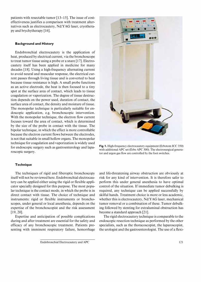
BOIS:L08:BPRR3009PD.95 FF: RESF E1: RESS
Endobronchial Electrocautery and APC 121
patients with resectable tumor [13–15]. The issue of cost-effectiveness justifies a comparison with treatment alter-natives such as electrocautery, Nd:YAG laser, cryothera-py and brychytherapy [16].
Background and History
Endobronchial electrocautery is the application ofheat, produced by electrical current, via the bronchoscopeto treat tumor tissue using a probe or a snare [17]. Electro-cautery itself has been applied in medicine for manydecades [18]. Using a high-frequency alternating currentto avoid neural and muscular response, the electrical cur-rent passes through living tissue and is converted to heatbecause tissue resistance is high. A small probe functionsas an active electrode, the heat is then focused to a tinyspot at the surface area of contact, which leads to tissuecoagulation or vaporization. The degree of tissue destruc-tion depends on the power used, duration of contact, thesurface area of contact, the density and moisture of tissue.The monopolar technique is particularly suitable for en-doscopic application, e.g. bronchoscopic intervention.With the monopolar technique, the electron flow currentfocuses toward the area of contact, which is determinedby the size of the probe in contact with the tissue. Thebipolar technique, in which the effect is more controllablebecause the electron current flows between the electrodes,is not that suitable in small hollow organs. The monopolartechnique for coagulation and vaporization is widely usedfor endoscopic surgery such as gastroenterology and lapa-roscopic surgery.
Technique
The techniques of rigid and fiberoptic bronchoscopyitself will not be reviewed here. Endobronchial electrocau-tery can be applied either using the rigid or flexible appli-cator specially designed for this purpose. The most popu-lar technique is the contact mode, in which the probe is indirect contact with tissue. The choice of technique andinstruments: rigid or flexible instruments or broncho-scopes, under general or local anesthesia, depends on theexpertise of the bronchoscopist and the risk assessment[19, 20].
Expertise and anticipation of possible complicationsduring and after treatment are essential for the safety andefficacy of any bronchoscopic treatment. Patients pre-senting with imminent respiratory failure, hemorrhage
Fig. 1. High-frequency electrocautery equipment (Erbotom ICC 350)with additional APC set (Erbe APC 300). The electrosurgical genera-tor and argon gas flow are controlled by the foot switches.
and life-threatening airway obstruction are obviously atrisk for any kind of intervention. It is therefore safer toperform this under general anesthesia to have optimalcontrol of the situation. If immediate tumor debulking isrequired, any technique can be applied successfully byskilful hands. Treatment choice is more or less academic,whether this is electrocautery, Nd:YAG laser, mechanicaltumor removal or a combination of these. Tumor debulk-ing followed by stenting for extraluminal obstruction hasbecome a standard approach [21].
The rigid electrocautery technique is comparable to theendoscopic resection technique as performed by the otherspecialists, such as the thoracoscopist, the laparoscopist,the urologist and the gastroenterologist. The use of a flexi-

BOIS:L08:BPRR3009PD.95 FF: RESF E1: RESS
122 Sutedja/Bolliger
Fig. 2. Schematic representation of the flexi-ble catheter tip for delivery of argon intothe tracheobronchial tree. The argon flowsaround a high-frequency electrode. The ar-gon gas (Ar) spreads out into the airwaylumen, and the portion that is activated andionized becomes the plasma jet. A sparkthrough the plasma jet desiccates tissues (de-picted by the brown area) to a specificdepth.
Fig. 3. Argon beamer effect illustrated on liver surface. Top:Although the beam is directed tangentially to the surface, coagulationtakes place at the tissue nearest to the catheter tip. Bottom: Withincreasing duration of coagulation (left to right) the effect still stayssuperficial, but enlarges the treated area as the argon beam movesfrom coagulated (= high impedance) to not yet coagulated (= lowimpedance) tissue.
ble endoscope and an electrosurgical loop to snare pedun-culated polyps is a common practice in gastroenterology.A similar technique can be applied in bronchology. Rigidbronchoscopic technique is more accepted in Europe andthe fiberoptic bronchoscope is the most popular instru-ment in the Anglo-Saxon countries [22]. The electrocaute-ry equipment is a standard and versatile instrument avail-able in almost every surgical ward: e.g. Valleylab® ForceGenerator with Gas Surgical Unit, Erbotom® ICC 350(fig. 1), PDS-20 Olympus.
In vivo studies showed that by using a monopolar flexi-ble electrocautery probe, predictable but superficial co-agulative necrosis by blanching the mucosa can be ob-tained with relative sparing of the cartilage. Under visualcontrol during the electrocautery session, one can easilyavoid charring of the bronchial mucosa to prevent toomuch damage and the destruction of the cartilage. Theinitial energy setting should be tested first on a normalpart of the mucosa, before starting to treat the tumoritself. After a relatively short learning period, the bron-choscopist can have visual control and obtain a shallownecrosis of 2–3 mm without causing too much unwanteddamage to the tracheobronchial wall [23]. Electrocauteryis easy to learn because it is not so different from the dailypractice of using the rigid or flexible biopsy forceps. Thus,electrocautery is an alternative technique for immediatetumor debulking, with the advantages of its ease, being astandard equipment and applying the familiar techniqueof diagnostic bronchoscopy. However, one always has tobe diligent to follow the safety guidelines of bronchoscop-ic intervention [19].
A relatively new development is the noncontact modeof argon beam coagulation or argon plasma coagulation(APC) in obtaining superficial homogeneous tissue coagu-lation (fig. 2). This may be attractive for the treatment ofhemorrhagic superficial spreading tumors [24, 25]. Ion-

BOIS:L08:BPRR3009PD.95 FF: RESF E1: RESS
Endobronchial Electrocautery and APC 123
Fig. 4. Profusely bleeding tumor of left upper lobe (a) and perfect hemostasis with superficial carbonization after10–15 s APC application through the flexible fiberscope (b).
ized argon gas functions as a conductance in the strongelectrical field between the active electrode and tissue.The nearest area of contact is not necessarily straight for-ward from the direction of the electrode, which makes thistechnique suitable for treating tumors ‘around the corner’at a sharp angle sideways from the tip of the electrode(fig. 3). APC has been shown to be effective in the treat-ment of small malignant gastrointestinal tumors [26]. His-tological studies have shown coagulative necrosis to besimilar either using standard electrocautery or the APCmethod [24–27], but APC causes more acute superficialtissue destruction and is most effective for the manage-ment of hemostasis. This superficial action makes APC avery safe tool. On the other hand, APC is less efficientthan standard monopolar electrocautery or laser resectionfor in-depth tissue destruction of bulky tumor. Althoughthere are reports on successful APC application in ob-structing lesions of the central airways due to recurrentrespiratory papillomatosis [28] or advanced bronchogeniccarcinoma [29], we use APC for quick hemostasis (fig. 4)and coagulation of superficial lesions during flexible bron-choscopy under local anesthesia only. For the destructionof bulky lesions either through the flexible or the rigidscope the Nd:YAG laser or standard electrocautery arethe preferred tools with immediate effect.
The Rigid TechniqueRigid bronchoscopy under general anesthesia must be
considered if the bronchoscopist feels that optimal controlis required during the session. It is safer and less time-consuming to use the rigid scope [20]. This allows ade-quate ventilation and rapid tumor debulking in patientswith imminent respiratory failure caused by hemorrhagicbulky tumor. Obviously, tissue coagulation can beachieved by using the rigid probe in combination with theflexible instruments to improve maneuverability. There-after, tumor debulking can be carried out [19].
From a mechanical and physical point of view, electro-cautery technique is quite similar to the contact mode ofNd:YAG laser. Tissue blanching and charring is imme-diately apparent, so the applied energy can be adjustedany time. Comparable to the Nd:YAG laser technique,coagulated tissue can be sheared off the wall mechanicallyusing the bevel of the rigid scope, the biopsy forceps or thefiberscope shaft [19]. The process of coagulation anddebulking can be repeated until sufficient airway passagehas been obtained. In contrast to the Nd:YAG laser, elec-trocautery achieves superficial necrosis, as electrons donot scatter in comparison to the laser photons of theNd:YAG laser. Also, current leak may occur during elec-trocautery treatment, and it is important to keep thetreated area dry, free of mucus and blood [17, 18]. Conse-
a b

BOIS:L08:BPRR3009PD.95 FF: RESF E1: RESS
124 Sutedja/Bolliger
Fig. 5. The rigid loop and the rigid suctionprobe for monopolar electrocautery. En-larged view of the distal tips of the instru-ments. From [72].
Fig. 6. The insulated fiberoptic broncho-scope and the flexible monopolar probe forcontact electrocautery; alongside the bron-choscope is an electrosurgical snare.
quently, a stent can be inserted if residual extraluminalstenosis becomes apparent.
The rigid technique of electrocautery is usually per-formed under general anesthesia. After induction of anes-thesia, the rigid scope is introduced and ventilationmaintained by jet ventilation. For rigid electrocauteryprobes, one can use the Storz® high-frequency loop elec-trode 10440 A or the rigid coagulation suction probe10390 N (fig. 5). Intraluminal tumor mass is gentlystripped or coagulated with the coagulation probe. Ener-
gy of about 50 W is applied by activating the electricalcurrent using the foot switch. Continuous suction of mu-cus, debris and blood while performing coagulation isnecessary to avoid current leak. The probe can be used topalpate the tracheobronchial wall to feel the anatomicalrigidity and to assess the compliance of the tracheobron-chial wall. If the choice had been local anesthesia, it issensible to anticipate possible complications by havingan intravenous line for giving sedatives [30]. Two milli-grams midazolam are injected as a bolus via the intrave-

BOIS:L08:BPRR3009PD.95 FF: RESF E1: RESS
Endobronchial Electrocautery and APC 125
Fig. 7. Granulosa cell tumor on the carina of the right upper lobe.Appearance before (a), immediately after argon plasma coagulation –tissue blanching (b) and 1 year after treatment (c). From [72].
nous line, immediately before starting the procedure. Ad-ditional midazolam can be titrated during the session bydripping infusion. The session is done under trend moni-toring by pulse oximetry and by giving supplemental oxy-gen. The bronchoscopist has to be able to immediately
intubate or insert a rigid scope in case of an emergency.We always keep the rigid scope ready for use at all times.The rigid scope can be mounted with two cuffs mimick-ing a Carlens tube to enable adequate ventilation whilecontinuing treatment.
a b
c

BOIS:L08:BPRR3009PD.95 FF: RESF E1: RESS
126 Sutedja/Bolliger
Fig. 8. Microinvasive early-stage carcinoma, before (a) and 6 weeks after (b) monopolar electrocautery treatment withcurative intent in roentgenologically occult lung cancer as an alternative to photodynamic therapy and surgical resec-tion. From [72].
The Flexible TechniqueThe flexible technique allows a better reach of smaller-
size tumors located in the bronchial segments. An insu-lated fiberscope (Olympus®; fig. 6) theoretically is safer inpreventing current leak [18], allows a better control of theprocess of coagulation and avoids burns or electricalshock to both the patient and the bronchoscopist. Thetumor area should be kept free of blood or mucus by con-tinuous suctioning. Coagulation can be achieved by gentlytouching the tumor base along the bronchial wall, ap-plying 20–40 W until sufficient blanching of the mucosabecomes apparent. One can easily watch the effect of elec-trocautery on the treated area. The noncontact method ofAPC is another alternative. It is comparable to the non-contact mode of CO2 laser that can be used to spray thetumor area to obtain superficial hemostasis, with theadvantage of treating areas from an angulated positionwhere the probe is located without any difficulty. Elec-trons will follow the path of the least conductance, wherethe flows then will diverge toward the plate placed on thepatient’s skin. Tissue blanching can be achieved in a mat-ter of seconds, both with standard monopolar electrocau-
tery (fig. 7) and APC (fig. 8), having the advantage ofbeing able to treat tumors obliquely if one cannot makethe tight bend using the fiberoptic bronchoscope. Treat-ment duration is very short if tumors are superficial andthe surface area is limited to a few square centimeters andcan certainly be performed under local anesthesia. It is ofthe utmost importance, however, to first consider the realindication and the oncological limitations of treatmentwith curative intent in patients with early-stage cancer [9,10, 12], especially in those with resectable malignancies.
Palliative Treatment
Results of electrocautery for palliation are shown intable 1 (the rigid technique) and in table 2 (the flexibletechnique) [31–40]. The results show that electrocauteryis equally effective in achieving tumor coagulation anddebulking compared to Nd:YAG laser and the complica-tion rate was not excessive. It is not surprising that com-parable results are seen in comparison to other debulkingtechniques, as the purpose of all these is mainly tumor
a b

Ledingham and Goldstraw [31]
Frizelly [34]
BOIS:L08:BPRR3009PD.95 FF: RESF E1: RESS
Endobronchial Electrocautery and APC 127
Table 1. Palliative results of endobronchial electrocautery using the rigid loop bronchoscope [from ref. 72]
Ref. n Technique Response Complications
15 general anesthesia, rigid loop +radioactive grains
successful palliation, 11/15 alive11 month; median 2.5 months
no
Pedersen et al. [32] 10 general anesthesia, jet ventilationand rigid loop
palliative 3–14 months,adenocystic 2–29 years
no
Petrou et al. [33] 29 general anesthesia and rigid loop(+ stents, + radioactive grains)
19/20 improved symptoms no
Table 2. Palliative results of endobronchial electrocautery using the fiberoptic bronchoscope
Ref. n Technique Response Complications
17 GA/LA, FFB average palliation 4–5 months(9 months)
1 hemorrhage
Hooper and Jackson [35] 4 GA, FFB + snare all successful 1 fire/explosion + respir. failureHooper and Jackson [36] 18 GA/LA, FFB + snare +
bipolar probe5 benign successful13 palliationfollow-up 3 years
1 bleeding and 1 fire
Gerasin and Shafirovsky [37] 14 GA, FFB + snare 10/14 complete and2/14 175% clearance
1 bleeding (100 ml)2 emergencies for bleeding/suffocation
Sutedja et al. [38] 17 LA and FFB + probe 15/17 successful2 extraluminal
1 minor bleeding1 pneumonia
Homasson et al. [39] 32 LA, rigid and flexible 27/32 150% tumor reduction,11/12 hemostasis
2 bleeding
Sutedja et al. [40] 56 GA/LA, mostly FFB+ probe
39/56 (70%)remaining extraluminal tumor
1 bleeding
GA/LA = General/local anesthesia; FFB = flexible fiberoptic bronchoscope. From [72].
coagulation to reduce the chance of hemorrhage beforethe tumor is mechanically removed. So, treatment resultsof all phase II studies strongly depend on the patient pop-ulation or selection and the expertise of the bronchosco-pist.
Treatment with Curative Intent
Early-stage lung cancer is a rarity and usually detectedby chance, either as a synchronous tumor or as a metach-ronous subsequent primary tumor. Particularly at risk arethose who have had a previous malignancy of the upperrespiratory tract [6]. Early-stage cancer lesions are easilymissed by conventional bronchoscopy [41]. The numberof patients with early-stage lung cancer treated broncho-scopically with curative intent is small, and experience inusing electrocautery is very limited. Many patients with
occult cancer who are regarded as inoperable because ofpoor pulmonary function, have already undergone pre-vious treatment with curative intent. The majority ofthem have been treated with PDT [13–15]. The prefer-ence for PDT in using hematoporphyrin derivatives assensitizers was based on the theory of ‘selective’ damagewith relative sparing of normal tissue. However, clinicaldata do not support this theoretical background. The factis that tumor area is selectively illuminated, and drugselectivity is a controversial issue [42, 43]. Cicatricialfibrosis due to treatment-induced hyperthermia may oc-cur [44], and normal tissue, if illuminated, may also bedamaged [45].
Any kind of bronchoscopic technique is potentiallycurative for occult, superficial and intraluminal lung can-cer [12]. Occult tumors only several millimeters thickwith !3 mm invasion of the bronchial mucosa and visibledistal margin can be effectively treated bronchoscopical-

Cortese and Edell [52]
Brachytherapy treatment with curative intent
BOIS:L08:BPRR3009PD.95 FF: RESF E1: RESS
128 Sutedja/Bolliger
Table 3. Results of bronchoscopic intervention for roentgenologically occult cancer
A Photodynamic therapy with curative intent
Ref. Lesions Response Survivalmonths
23 CR 9/12 (43%, resectable candidates!) 21–116Furuse et al. [53] 59 CR 45/59 (83%) 14–32Okunaka et al. [54] 10 synchronous
17 metachronous10/10 synchronous (100%)16/17 metachronous (94%)
14–87
Sutedja et al. [55] 39 CR 28/39 (72%) 2–95
B Other techniques
Ref. n Technique Survival
Macha and Wahlers [56] 9 15 Gy at 3 mm/4 ! HDR !27 monthsO’Driscoll et al. [57] 6 15–20 Gy/1 ! HDR 12–21 monthsHernandez and Donath [58] 4 7.5 Gy/? HDR 8–24 monthsSutedja et al. [59] 2 10 Gy/2–3 ! HDR 25, 54 monthsOno et al. [60] 12 50–60 Gy 2–8 months
Electrocautery for occult cancer and typical intraluminal carcinoidvan Boxem et al. [61] 12/15 (80%) LA with flexible probe 16–43 monthsvan Boxem et al. [50] 14/19 (73%) GA with flexible probe, resectable patients! 16–43 months
Nd:YAG laser for occult cancer and intraluminal carcinoidCavaliere et al. [62] 19 ca. in situ GA, rigid bronchoscope !12 yearsPersonne et al. [63] 3/30 carcinoids GA, rigid bronchoscope 2–4 years
CR = Complete response with negative histology/cytology; HDR = high-dose-rate brachytherapy; GA/LA = gener-al/local anesthesia. Modified from [72].
ly, as an alternative for surgical resection [9, 10, 14, 15].These oncological considerations are far more importantarguments than the issue of the proper treatment tech-nique. Hence, the results of bronchoscopic treatment withcurative intent strongly depend on the inclusion criteria ofthese early-stage lesions or the exact staging, showing astrong correlation between complete response rate andtumor size [13]. A small number of patients with superfi-cial spreading squamous cell cancer may also be treatedprior to surgery, to free the proximal tumor margin beforeresection, thus improving the chance for radical surgeryby using PDT, Nd:YAG laser and brachytherapy [46–48].This is an approach that can be applied in patients withsevere COPD in whom a less extensive surgical resectionmay still offer the best chance for cure. Also, patients withlow-grade malignant tumors such as intraluminal typicalcarcinoid may be treated bronchoscopically with curativeintent [49–51]. Again, the oncological considerations of
tumor histology and type are far more important than thetechnique of bronchoscopic intervention. Surgery is a rel-atively wasteful technique of lung parenchyma resectionin these early-stage cancers, justifying alternatives to beapplied with curative intent (table 3). Without doubt, incarefully selected cases, a bronchoscopic treatment is aworthy consideration instead of a radical surgical resec-tion.
A phase III randomized study versus surgical resectionin patients with occult cancer is difficult to perform due tothe low number of patients that can be included in such astudy [14, 15, 52]. Detection of early-stage lung cancer ismostly a chance occurrence. Any technique seems to bejustified in treating a tumor several millimeters thick, aslong as staging is oncologically correct. Inaccurate assess-ment of tumor dimension is the real hurdle in lung cancermanagement, and the true tumor stage can only be defi-nite in retrospect, after surgical resection with complete

Bronchoscopy
Mechanical removal
BOIS:L08:BPRR3009PD.95 FF: RESF E1: RESS
Endobronchial Electrocautery and APC 129
lymph node dissection. The improvement of our diagnos-tic accuracy in assessing peribronchial, nodal and distantmetastasis should also be the focus of our attention inbronchoscopic techniques. High-resolution CT, ultrason-ography, fluorescence bronchoscopy are worth to be in-vestigated to improve the staging in lung cancer screeningprogrammes.
Electrocautery and Other Treatment Modalities
Are treatment modalities such as mechanical removal[64], PDT, Nd:YAG laser, cryotherapy [65] and brachy-therapy comparable to electrocautery? Each techniquehas its own merits and limitations. Treatment choice isusually based upon various factors: clinical presentation,the expertise of the bronchoscopist, anesthetic care andthe technical facilities of each hospital. Practical aspects,such as logistics and cost, may further influence treatmentchoice. Many patients have imminent respiratory failurebecause of bulky tumor in the intra- and extraluminalcompartment. Immediate tumor debulking may be re-quired using Nd:YAG laser, electrocautery and mechani-cal removal, and a stent can be inserted for extraluminalstenosis [21]. In nonemergency cases, brachytherapy is agood alterantive to obtain durable response at the cost ofnormal tissue damage. About a 10% complication rate,e.g. radiation bronchitis, radiation stenosis, fistula andhemorrhages, is reported after high-dose-rate brachyther-apy [66]. Costs and comparison between the differenttechniques are shown in tables 4 and 5. An algorithm for
palliative endoscopic and nonendoscopic treatment op-tions is presented in the chapter on multimodality treat-ment of advanced pulmonary malignancies by Bolliger.
Electrocautery is a cheaper alternative than Nd:YAGlaser for tumor coagulation. The cost-effectiveness of anytechnique also depends on the price of the equipment,maintenance costs and its use by the different specialists.Electrocautery is a simple and straightforward techniquethat can be used by the different specialists in a hospital.In our daily practice, the majority of patients are treatedfor local palliation and treatment choice is also based onlogistics, thus based on pragmatism.
Table 4. Treatment costs of bronchoscopic intervention in TheNetherlands (calculated in USD)
Price ofequipment
Costrefund1
309Anesthesia 318Nd:YAG laser, diode laser 40,000 880Photodynamic therapy, diode laser 60,000 3,0772
Brachytherapy (high dose rate) 250,000 539Electrocautery 20,000 309
1 Based on tariffs of the ‘COTG’ (Government Health InsuranceTariff).2 Photofrin II drugs as sensitizers, 2 mg/kg, on average 2 vials areneeded.
Modified from [72].
Table 5. Advantages and disadvantages of various bronchoscopic intervention techniques
Treatment Equipment/technique Result Complications
standard, GA short-term, immediate bleedingCryotherapy special applicators and mostly GA not immediate, save for the
cartilagesecondary necrosis
Electrocautery standard, GA/LA, rigid and fiberscope immediate and superficial fibrosisNd:YAG laser laser, mostly GA, or fiber through
rigidimmediate and in-depth effect perforation
Brachytherapy afterloading, LA late and in-depth effect radiation bronchitis, stenosis,fistula
Photodynamic therapy dye and diode laser, LA save the cartilage, secondary butin-depth effect
skin photosensitivity, secondarynecrosis
GA/LA = General/local anesthesia. Modified from [72].

BOIS:L08:BPRR3009PD.95 FF: RESF E1: RESS
130 Sutedja/Bolliger
Conclusion
Tumors located in the central airways can be treatedwith various bronchoscopic techniques such as lasers(Nd:YAG, Argon, KTP, CO2), PDT, brachytherapy, cryo-therapy and electrocautery. Stenting is necessary for ex-traluminal airway stenosis. Thus, various techniques cancurrently be applied safely and effectively to treat patientswith malignancies in the major airways, both for imme-diate palliation and for treatment with curative intent.Usually, bronchoscopists have to deal with bulky tumor,in which the visible intraluminal tumor is only the tip ofan iceberg. Improvement of gas exchange is the aim ofairways reopening, but regions with low gas exchange, e.g.after external irradiation, may improve little if treated.Obstructive pneumonia and hemoptysis are good pallia-tive indications. Patients’ general, cardiac and pulmonarycondition, tumor dimension and stage, bronchoscopicfindings, (high resolution) CT findings and regional venti-lation-perfusion are factors to be taken into account priorto any intervention. Risk and potential benefit also de-pend on the expertise of the bronchoscopist and the avail-able facilities in each institution.
Results are immediate when using electrocautery orlasers (Nd:YAG, KTP, Argon, CO2) in combination withmechanical tumor removal. Techniques that induce lateresponse or cause secondary necrosis, e.g. brachytherapy,PDT and cryotherapy, are less practical for emergencycases. PDT and brachytherapy can obtain an in-deptheffect, the latter at the cost of normal tissue damage. Sec-ondary necrosis and fibrin plugs may require additional
bronchoscopy for nectrotomy (cryosurgery, PDT) andskin photosensitivity in PDT may increase treatmentmorbidity.
A small number of patients with intraluminal occultlung cancer may benefit from bronchoscopic treatmentwith curative intent. Treatment may also be used to im-prove resectability. However, occult tumors with full thick-ness invasion of the bronchial mucosa and invisible distalmargin are unlikely to be cured by bronchoscopic treat-ment alone. One should always keep in mind the oncologi-cal limitations in bronchoscopic intervention. In patientswith resectable tumors, staging inaccuracy is the mostimportant factor, not the treatment technique. High-reso-lution computed tomography [67], fluorescence bronchos-copy [68, 69] and bronchoscopic ultrasound [60, 70] mayimprove our ability to improve tumor staging procedures.
A simple and cheap bronchoscopic technique such aselectrocautery improves the cost-effectiveness of a bron-choscopic intervention, especially when more compli-cated and expensive procedures have not been shown tobe clinically superior. Electrocautery proves also to beeffective in treating benign and low-grade malignantlesions such as intraluminal typical bronchial carcinoid.The curative potential of various bronchoscopic therapiesprovide us with alternatives for surgical resection in treat-ing early-stage lung cancer lesions or relatively benigntumors which are amenable for surgical bronchoplasty.With the recognition of the population at risk and theawareness of the potential benefit of lung cancer screening[71], techniques such as electrocautery will become part ofstandard lung cancer care.
References
1 Chetty KG, Moran EM, Sasoon CS, Viravatha-na T, Light RW: Effect of radiation therapy onbronchial obstruction due to bronchogenic car-cinoma. Chest 1989;95:582–584.
2 Majid DA, Lee S, Kushalani S, Seydel H: Theresponse of atelectasis from lung cancer toradiation therapy. Int J Radiat Oncol Biol Phys1986;12:231–232.
3 LeChevalier M, Arriagada R, Cuoix E, RufifieP, Martin M, Tarayre M, Lacombe Terrier MJ,Douillard JY, Laplanche A: Radiotherapyalone versus combined chemotherapy and ra-diotherapy in nonresectable non-small cell lungcancer – First analysis of a randomized trial in353 patients. J Natl Cancer Inst 1991;83:417–423.
4 Woolner LB, Fontana RS, Cortese DA: Roent-genographically occult lung cancer: Pathologicfindings and frequency of multicentricity dur-ing a 10-year period. Mayo Clin Proc 1984;59:453–466.
5 Martini N, Bains MS, Burt ME, Zakowski MR,McCormack P, Rush V, Ginsberg R: Incidenceof local recurrence and second primary tumorsin resected stage I lung cancer. J Thorac Car-diovasc Surg 1995;109:120–129.
6 Lippman SM, Hong WK: Second malignanttumors in head and neck squamous cell carci-noma: The overshadowing threat for patientswith early-stage disease. Int J Radiat OncolBiol Phys 1989;17:691–694.
7 Watanabe Y, Shimizu J, Oda M, Takashi I,Takashima T, Kamimura R, Kitagawa M,Nonomura A, Nakamura S, Tanimoto K, Mat-subara F, Ikegaki S, Yamada K, et al: Earlyhilar lung cancer: Its clinical aspect. J SurgOncol 1991;48:75–80.
8 Flehinger BJ, Kimmel M, Melamed MR: Theeffect of surgical treatment on survival fromearly lung cancer. Implications for screening.Chest 1992;101:1013–1018.
9 Nagamoto N, Saito Y, Ohta S, Sato M, KanmaK, Sagawa M, Takahashi S, Usuda K, NakadaT, Hashimoto K: Relationship of lymph nodemetastasis to primary tumor size and micro-scopic appearance of roentgenographically oc-cult lung cancer. Am J Surg Pathol 1989;13:1009–1013.

BOIS:L08:BPRR3009PD.95 FF: RESF E1: RESS
Endobronchial Electrocautery and APC 131
10 Usuda K, Saito Y, Nagamoto N, Sato M, Saga-wa M, Kanma K, Takahasi S, Endo C, Fujimu-ra S: Relation between bronchoscopic findingsand tumor size of roentgenologically occultbronchogenic squamous cell carcinoma. J Tho-rac Cardiovasc Surg 1993;106:1098–1103.
11 Sutedja G, Postmus PE: Bronchoscopic treat-ment of lung tumors. Review article. LungCancer 1994;11:1–17.
12 Sutedja G, Schramel F, Postmus PE: Broncho-scopic treatment modalities in lung cancer, in-dications and limitations. Ann Oncol 1995;6:951–952.
13 Hayata Y, Kato H, Konaka C, Okunaka T:Photodynamic therapy (PDT) in early stagelung cancer. Lung Cancer 1993;9:287–294.
14 Edell ES, Cortese DA: Photodynamic therapyin the management of early superficial squa-mous cell carcinoma as an alternative to surgi-cal resection. Chest 1992;102:1319–1322.
15 Dougherty TJ: Photodynamic therapy for earlystage lung cancer. Preferable to resection?Chest 1992;102:1314.
16 Sutedja G, Postmus PE: The cost-effectivenessof intraluminal bronchoscopic treatment. AmRev Respir Dis 1995;151:A850.
17 Barlow DR: Endoscopic applications of elec-trosurgery: A review of basic principles. Gas-trointest Endosc 1982;28:73–76.
18 Jackson R: Basic principles of electrosurgery: Areview. Can J Surg 1970;13:354–361.
19 Dumon JF, Shapsay S, Bourceraou J, CavaliereS, Meirc B, Garbi N, Beamis J: Principles forsafety in application of neodymium-YAG laserin bronchology. Chest 1984;86:163–168.
20 George PJ, Barret CPO, Nixon C, Hetzel MR,Nanason EM, Miljard FJ: Laser treatment fortracheobronchial tumours: Local or generalanaesthesia? Thorax 1987;42:656–660.
21 Dumon JF: A dedicated tracheobronchialstent. Chest 1990;97:328–332.
22 Prakash UBS, Offord KP, Stubbs SE: Bron-choscopy in North America: The ACCP sur-vey. Chest 1991;100:1668–1675.
23 Van Boxem AJM, Westerga J, Venmans BJ,Postmus PE, Sutedja G: Tissue effects of bron-choscopic electrocautery: Macroscopy and mi-croscopy of bronchial wall after electrocautery.Program and Abstract 10th World Congress forBronchology, Budapest 1998, Abstract No 29,p 95.
24 Go PM, Goodman GR, Bruhn EW, Hunter JG:The Argon beam coagulator provides rapid he-mostasis of experimental hepatic and splenichemorrhage in anticoagulated dogs. J Trauma1991;31:1294–1300.
25 Rusch V, Schmidt R, Shoji Y, Fujimura Y: Useof the Argon beam electrocoagulator for per-forming pulmonary wedge resections. AnnThorac Surg 1990;49:287–291.
26 Sessler MJ, Becker HD, Flesch I: Therapeuticeffect of argon plasma coagulation on smallmalignant gastrointestinal tumors. J CancerRes Clin Oncol 1995;121:235–238.
27 Lanzafame RJ, Qiu K, Rogers DW, Naim JO,Caldwell FC, Perry F, Hall D, Hinshaw JR:Comparison of local tumor recurrence follow-ing excision with the CO2 laser, Nd:YAG laserand Argon Beam Coagulator. Lasers Surg Med1988;8:515–520.
28 Bergler W, Honig M, Gotte K, Petroianu G,Hormann K: Treatment of recurrent respirato-ry papillomatosis with argon plasma coagula-tion. J Laryngol Otol 1997;111:381–384.
29 Schmidt W: Erfahrungen mit dem Argon-Beamer in der interventionellen Bronchologie.Atemw Lungenkr 1996;22:168–173.
30 Sutedja G, Koppenol W, Stam J: Nd-YAGlaser under local anesthesia in obstructive en-dobronchial tumors. Respiration 1991;58:238–240.
31 Ledingham SJ, Goldstraw P: Diathermy resec-tion and radioactive gold grains for palliationof obstruction due to recurrence of bronchialcarcinoma after external irradiation. Thorax1989;44:48–51.
32 Pedersen U, Kristensen S, Illum P: Palliativeresection with high-frequency cutting loop inmalignant tracheobronchial diseases. J Bron-chol 1994;1:23–25.
33 Petrou M, Kaplan D, Goldstraw P: Broncho-scopic diathermy resection and stent insertion:A cost effective treatment for tracheobronchialobstruction. Thorax 1993;48:1156–1159.
34 Frizelly R: Le traitement par électrocoagula-tion en pathologie maligne trachéo-bronchi-que. Rev Pneumol Clin 1986;42:235–237.
35 Hooper RG, Jackson FN: Endobronchial elec-trocautery. Chest 1985;87:712–714.
36 Hooper RG, Jackson FN: Endobronchial elec-trocautery. Chest 1988;94:595–598.
37 Gerasin VA, Shafirovsky BB: Endobronchialelectrosurgery. Chest 1988;93:270–274.
38 Sutedja G, van Kralingen K, Schramel F, Post-mus PE: Fiberoptic bronchoscopy electrosurge-ry under local anesthesia for rapid palliation inpatients with central airway malignancies. Apreliminary report. Thorax 1994;49:1243–1246.
39 Homasson JP, Roden S, Angebault M: Traite-ment des tumeurs bronchiques par thermocoa-gulation haute fréquence. Etude préliminaire.Rev Pneumol Clin 1995;51:77–81.
40 Sutedja G, van Boxem TJ, Schramel FM,v Felius C, Postmus PE: Endobronchial elec-trocautery is an excellent alternative forNd:YAG laser to treat airway tumors. J Bron-chol 1997;4:101–105.
41 Sato M, Saito Y, Usuda K, Takahashi S, Saga-wa M, Fujimura S: Occult lung cancer beyondbronchoscopic visibility in sputum cytologypositive patients. Lung Cancer 1998;20:17–24.
42 Monnier Ph, Savary M, Fontollet Ch, Wag-nieres G, Chatelain A, Cornaz P, DepeursingeCh, van den Bergh H: Photodetection and pho-todynamic therapy of ‘early’ squamous cell car-cinomas of the pharynx, oesophagus and tra-cheobronchial tree. Lasers Med Sci 1990;5:149–169.
43 Braichotte D, Savary JF, Glanzmann T, Mon-nier P, Wagnieres G, van den Bergh H: Optim-izing light dosimetry in photodynamic therapyof the bronchi by fluorescence spectroscopy.Lasers Med Sci 1996;11:247–254.
44 Sutedja G, Risse E, van Mourik J, Postmus PE:Photodynamic therapy for treatment of bron-chial carcinomas. Thorax 1994;49:289–290.
45 Kawaguchi T, Furuse K, Kawahara M, Yama-moto S, Sutedja G: Histological examination ofbronchial mucosa after photodynamic therapyshowing no selectivity of effect between tumorand normal mucosa. Lasers Med Sci 1998;13:265–270.
46 Kato H, Konaka C, Ono J, Kawate N, Nishi-miya K, Shinohara H, Saito M, Sakai M, Nogu-chi M, Kito T, Aizawa K, Hayata Y: Preopera-tive laser photodynamic therapy in combina-tion with operation in lung cancer. J ThoracCardiovasc Surg 1985;90:420–429.
47 Shankar S, George PJM, Hetzel MR, Gold-straw P: Elective resection of tumours of thetrachea and main carina after endoscopic lasertherapy. Thorax 1990;45:493–495.
48 Sutedja G, Baris G, Zoetmulder F, van Zand-wijk N: High dose rate brachytherapy improvesresectability in squamous cell lung cancer.Chest 1992;102:308–309.
49 Sutedja G, Schreurs A, Vanderschueren R,Kwa B, v d Werf T, Postmus PE: Bronchoscop-ic therapy in patients with intraluminal typicalbronchial carcinoid. Chest 1995;107:556–558.
50 Van Boxem TJ, Venmans BJ, van Mourik JC,Postmus PE, Sutedja G: Bronchoscopic treat-ment of intraluminal typical carcinoid: A pilotstudy. J Thorac Cardiovasc Surg 1998;116:402–406.
51 Shah H, Garbe L, Nussbaum E, Dumon JF:Benign tumors of the tracheobronchial tree.Endoscopic characteristics and role of laser re-section. Chest 1995;107:1744–1751.
52 Cortese DA, Edell E: Photodynamic therapy inthe treatment of early stage squamous cell car-cinoma of the lung. Lung Cancer 1997;18(suppl 1):91(A355).
53 Furuse K, Fukuoka M, Kato H, Horai T, Kubo-ta Y, Kodama N, Kusunoki Y, Takifuji N,Okunaka T, Konaka C, Wada H, Hayata Y, forthe Japan Lung Cancer Photodynamic TherapyStudy Group: A prospective phase II study onphotodynamic therapy with photofrin II forcentrally located early-stage lung cancer. J ClinOncol 1993;11:1852–1857.
54 Okunaka T, Kato H, Konaka C, et al: Photody-namic therapy for multiple primary broncho-genic carcinoma. Cancer 1991;68:253–258.
55 Sutedja G, Lam S, LeRiche JC, Postmus PE:Response and pattern of failure after photody-namic therapy for intraluminal stage I lungcancer. J Bronchol 1994;1:295–298.
56 Macha HN, Wahlers B: Endobronchiale Bra-chytherapie: Die endoluminale Kleinraum-bestrahlung von Bronchialtumoren mit demAfterloading HDR-Verfahren. Pneumonologie1991;45:95–99.

BOIS:L08:BPRR3009PD.95 FF: RESF E1: RESS
132 Sutedja/Bolliger
57 O’Driscoll BR, Burt PA, Stout R, Barber P:Radical treatment of lung cancer by high doserate intraluminal radiotherapy. Am Rev RespirDis 1991;143:A60.
58 Hernandez P, Donath D: High dose rate endo-bronchial brachytherapy in the treatment ofbronchogenic carcinoma. Chest 1993;104:989–990.
59 Sutedja G, Baris G, van Zandwijk N, PostmusPE: High dose rate brachytherapy has a cura-tive potential in patients with intraluminalsquamous cell lung cancer. Respiration 1993;61:167–168.
60 Ono R, Hirano H, Egawa S, Suemasu K: Bron-choscopic ultrasonography and brachytherapyin roentgenologically occult bronchogenic car-cinoma. J Bronchol 1994;1:281–287.
61 Van Boxem TJ, Venmans BJ, Schramel FM,van Mourik JC, Golding RP, Postmus PE,Sutedja G: Radiographically occult lung cancertreated with fibreoptic bronchoscopic electro-cautery. A pilot study of a simple and inexpen-sive technique. Eur Respir J 1998;11:169–172.
62 Cavaliere S, Foccoli P, Toninelli C, Feijo S:Nd:YAG laser therapy in lung cancer: An 11-year experience with 2,253 applications in1,585 patients. J Bronchol 1994;1:105–111.
63 Personne C, Colchen A, Leroy M, Vourch G,Toty L: Indications and technique for endo-scopic laser resections in bronchology. J ThoracCardiovasc Surg 1986;91:710–715.
64 Mathissen DJ, Grillo HC: Endoscopic relief ofmalignant airway obstruction. Ann ThoracSurg 1989;48:469–475.
65 Homasson JP: Bronchoscopic cryotherapy: Areview. J Bronchol 1995;2:145–153.
66 Speiser BL, Spratling L: Radiation bronchitisand stenosis secondary to high dose rate endo-bronchial irradiation. Int J Radiat Oncol BiolPhys 1993;25:580–597.
67 Sutedja G, Golding RP, Postmus PE: Highresolution computed tomography in patientsreferred for intraluminal bronchoscopic thera-py with curative intent. Eur Respir J 1996;9:1020–1023.
68 Lam S, MacAulay C, Hung J, LeRiche J, ProfioAE, Palcic B: Detection of dysplasia and carci-noma in situ with a lung imaging fluorescenceendoscopy device. J Thorac Cardiovasc Surg1993;105:1035–1040.
69 Venmans BJ, v d Linden H, v Boxem TJ, Post-mus PE, Smit EF, Sutedja G: Early detection ofpreinvasive lesions in high risk patients. Acomparison of conventional flexible and fluo-rescence bronchoscopy. J Bronchol 1998;5:280–283.
70 Gress FG, Savidas TJ, Sandler A, Kesler K,Conces D, Cummings O, Mathur PN, Ikenber-ry S, Bilderback S, Hawes R: Endoscopic ultra-sonography, fine needle aspiration biopsyguided by endoscopic ultrasonography, andcomputed tomography in the preoperativestaging of non small cell lung cancer: A compar-ison study. Ann Intern Med 1997;127:604–612.
71 Strauss GM: Measuring effectiveness of lungcancer screening. From consensus to contro-versy and back. Chest 1997;112:216S–228S.
72 Sutedja TG, Postmus PE: Endobronchial elec-trocautery; in Beamis JF, Mathur PN (eds):Interventional Pulmonology. New York,McGraw-Hill, 1998, pp 83–95.
G. Sutedja, MD, PhD, FCCPDepartment of PulmonologyFree University Hospital, PO Box 7057NL–1007 MB Amsterdam (The Netherlands)Tel. +31 20 444 4782, Fax +31 20 444 4328E-Mail [email protected]

OOOOOOOOOOOOOOOOOOOOOOOOOOOOOOOOOOOOOOOOOOOOOOOOOOOOOOOOOOOOOOOOOOOOOOOOOOOOOOOOOOOOOOOOOOOOOOOOOOOOOOOOOOOOOOOOOOOOOOOOOOO
Therapeutic Bronchoscopy

Bolliger CT, Mathur PN (eds): Interventional Bronchoscopy.Prog Respir Res. Basel, Karger, 2000, vol 30, pp 133–145
OOOOOOOOOOOOOOOOOOOOOOOOOOOOOOOOOOOOOOOOOOOOOOOOOOOOOOOOOOOOOOOOOOOOOOOOOOOOOOOOOOOOOOOOOOOOOOOOOOOOOOOOOOOOOOOOOOOOOOOOOOO
Cryotherapy for EndobronchialDisordersJ.M. Vergnona Praveen N. Mathurb
aDepartment of Pulmonary Diseases and Thoracic Oncology, St. Etienne University Hospital,St. Etienne, France; bDivision of Pulmonary and Critical Care Medicine, Department of Medicine,Indiana University Medical Center, Indianapolis, Ind., USA
133
Summary
Cryotherapy has been used for destruction of endobron-chial tumors. Cryotherapy works by its cytotoxic effect, byfreezing the tissue, thus causing tissue death. Cryotherapycan be performed by rigid bronchoscopy under generalanesthesia. It can also be performed by fiberoptic bronchos-copy under sedation and local anesthesia. The indications forcryotherapy are similar to those for endobronchial laser orelectrocautery. The purpose of cryotherapy is essentially toablate the endobronchial tumor. Cryotherapy has also beenused for treatment of carcinoma in situ as well as benignendobronchial tumors. The advantages which cryotherapyprovides is safety not only for the operator, but also for thepatient. The major counterindication for cryotherapy is apatient with impending respiratory failure. In certain situa-tions cryotherapy has also shown to have a beneficial effectin conjunction with radiation therapy as well as chemothera-py. Cryotherapy offers an alternative for endobronchialtumor ablation with significant safety as well as being rela-tively inexpensive.
History of Cryosurgery
Documents from 3500 BC described the use of cold astreatment for swelling and war wounds [1]. Hippocratesdescribed the use of cold to treat orthopedic injuries [1].
The analgesic and anti-inflammatory properties of icehave been known for several centuries, but the use offreezing as a therapeutic agent for destruction of tissuebegan in 1845–1851. Arnott [2, 3] described the use of saltsolutions containing crushed ice at a temperature of about–8 to –12°C to freeze advanced cancers in accessiblesites, producing reduction in tumor size and improve-ment of pain. The Joule-Thomson effect, which is the sud-den expansion of a gas from a high- to a low-pressureregion, is the basis for the functioning of some cryoprobes,especially in pulmonary medicine. White [4, 5] showedthat liquid air could be used for the treatment of variousdermatological lesions. Hass and Taylor [6] describedfreezing local lesions in several organs; necrotic lesionswere sharply defined uniformly with a lack of suppurationor sequestration and were accompanied by a slow healingprocess.
During the next several years, many cryosurgical ma-chines were developed in most medical specialties [7–17].Due to the relative inaccessibility of endobronchial le-sions, cryosurgical techniques were late to develop. Thedestructive effect of cold has also been confirmed by thehealing of the tracheobronchial tree, with the restorationof a normal ciliated epithelium without stenosis in mon-grel dogs [18–24]. The first patient who had a carcinoma-tous obstruction of a bronchus was treated in 1968, andSanderson et al. [25] reported another case in 1975. Thisfirst study by the Mayo Clinic included 28 patients withendobronchial tumors, and the investigators concluded

BOIS:L08:BPRR3018PD.98 FF: RESF E1: RESS
134 Vergnon/Mathur
Table 1. Cryosensitive and cryoresistant tissues
Cryosensitive Cryoresistant
Skin FatMucous membrane CartilageNerve Nerve sheathEndothelium Connective tissueGranulation tissue Fibrosis
that cryotherapy did serve as a good alternative for pallia-tion. In Europe, Homasson et al. [26] rediscovered cryo-therapy in France, Maiwand [27] in England and Astesia-no et al. [28] in Italy. Since the early 1980s, most of thework with endobronchial cryotherapy has occurred out-side the US, where the introduction of laser diminishedinterest in cryotherapy. In 1994, a flexible cryoprobe(ERBE USA Inc., Marietta, Ga., USA) for use with theflexible bronchoscope was introduced in the US renewinginterest in endobronchial cryotherapy. In 1996, Mathur etal. [29] described the use of this probe with the flexiblebronchoscope in treating 22 patients with airway obstruc-tion. During the decade 1985–1995, cryosurgery has un-dergone a renaissance, and different specialties are usingthis technique, such as pulmonology, dermatology, urolo-gy and liver metastasis surgery. The literature has shownthe efficacy of cryosurgery [29–41] and its associationwith other treatment modalities such as irradiation [42] orchemotherapy [43]. It is a cytotoxic procedure and as suchshould be integrated into the plan of action of treatmentsfor bronchial carcinomas.
Scientific Basis of Cryosurgery
Cryotherapy deals with the destruction of biologicalmaterials through the cytotoxic effects of freezing. Therehas also been a parallel development in the subject ofcryopreservation, which deals with attempts to use freez-ing for the long-term preservation of biological materials.The fundamental processes that occur during cryosurgeryderived from research done on the use of freezing to pre-serve biological cells or tissues. Understanding the processof freezing and the mechanisms of damage is very impor-tant. One key to the success is having a clear understand-ing of the impacts that low and changing temperatureshave on cell and tissue structure.
The damage induced by freezing occurs at several lev-els, including the molecular level, the cellular level, struc-tural level and the whole tissue. The effect of a freezinginjury is influenced by many factors, and the survival ofcells is dependent on the cooling rate [44–46], the thawingrate [47], the lowest temperature achieved [48] and re-peated freezing-thawing cycles [49, 50]. Certain tissues(table 1) are cryosensitive (skin, mucous membrane, gran-ulation tissue), and others are cryoresistant such as fat,cartilage, fibrous or connective tissue. The cryosensitivitydepends on the water content of the cells. Tumor cellsmay be more sensitive than normal cells [50].
Mechanisms of Cellular Damage by FreezingThe physical events occurring in cells in suspension
during freezing, are as follows [50]: (1) At –5°C, the sys-tem remains in a liquid state, despite the cytoplasmicfreezing point of –2.2°C. (2) Between –5 and –15°C,extracellular crystals form while the intracellular mediumremains supercooled and unfrozen as the cell membraneis a barrier to the propagation of ice. It is at this stage thatthe rate of cooling determines the outcome. If the cell iscooled slowly enough, the dehydration of intracellularwater allows the buildup of an intracellular solute concen-tration that eliminates any intracellular freezing. How-ever, if the cell is cooled too rapidly, the intracellularwater will not have enough time to pass the cell mem-brane before crystals form within the cell. (3) Below–15°C, a transitional state occurs, after which any furtherincrease in solute concentration results in a decrease inthe freezing point. (4) Finally, the solid state or completecrystallization is attained.
During thawing, different physiochemical phenomenaoccur which again are determined by the cooling velocity:(1) after a suboptimal freezing, rapid thawing exposes thecell to a high electrolytic concentration and to an elevatedtemperature, which could induce an intra- or extracellularrecrystallization; (2) after supraoptimal freezing, slowthawing induces the recrystallization phenomenon; this isan extremely destructive process for intracellular organ-elles due to the grinding action of ice [51, 52]. During thisphysical period, factors involved in the determination ofcellular cryolesion and cell death are: extracellular crystal-lization that compresses and deforms the cells (‘pack-iceeffect’), intracellular crystallization, cellular dehydrationwith collapse, increases in intracellular electrolytic con-centration, denaturation of membranous lipoprotein andsimple thermic shock.
These factors are closely related and intervene to dif-ferent degrees in the determination of cellular cryolesions.

BOIS:L08:BPRR3018PD.98 FF: RESF E1: RESS
Cryotherapy for Endobronchial Disorders 135
Both mechanical effects due to the formation of ice crystaland more complex biochemical effects secondary to cellu-lar dehydration occur. Nevertheless, the essential deter-minant of the final degree of cellular lesion is the coolingvelocity. The study of these factors provides the basis ofthe following advice to cryosurgeons; the aim of usinglow-temperature instruments is to create the fastest possi-ble freezing of the target tissue to provoke intracellularfreezing.
Freezing of TissueThe process of freezing depends on the details of the
interaction between ice crystals, the solution surroundingthe ice crystals and the cells. Tissue sensitivity to cold isdependent upon the existence of microcirculation. A clearline of demarcation between tissue that was previouslyfrozen and tissue that was not frozen is observed aftercryosurgery [53]. Following cryosurgery, the cells in thefrozen region have a disrupted structure. Ischemic andinfarcted aspects of cryolesions appear from a few min-utes to several hours (fig. 1). Circulation stops rapidlyafter thawing, and this cryo-induced thrombosis seems toresult from the conglomeration of several factors: (1) va-soconstriction of arterioles and venules occurring dur-ing slight hypothermia, (2) modification of the vascularendothelium, (3) an increase in the permeability of thevascular walls, (4) an increase in the blood viscosity, (5) alowering of the intracapillary hydrostatic pressure, a de-crease in blood flow and finally formation of plateletplugs. These vascular effects explain the hemostatic effectof cryosurgery, which is not always immediate. At theedge of the frozen region, hypothermia causes a heteroge-neous effect and it is in these areas that chemotherapy (orradiotherapy) has a complementary destructive effect [41,54, 55].
Ischemic damage occurs in the whole region that wasfrozen. The result of these cellular changes in the days fol-lowing cryotherapy is cellular necrosis. An acidophilicslough forms and tumor tissue is no longer visible andtherefore destroyed. Nevertheless, freezing does not mod-ify histopathological findings when biopsies are madeimmediately after cryosurgery. The cryolesions are visibleonly under an electron microscope. The mechanism ofcell and tissue damage after cryosurgery explains thedelayed effect of this technique; the consequence is thatcryosurgery is not useful for acute respiratory distress.Besides these physical and vascular effects of cryosurgery,the literature contains many references to a repeatedcryoimmunologic action [56, 57]. Nevertheless, no defi-nite conclusion is yet possible but further studies of this
Fig. 1. Schematic view of cryonecrosis obtained after three cycles ofcryotherapy applied in a single point with a rigid probe of 3 mm indiameter.
phenomenon are needed. The goal of cryotherapy is todestroy pathologic but spare normal tissue. Mazur [58]demonstrated that rapid freezing and slow warming leadto maximum cell death. Sufficient time to reach and tosustain the critical temperature is required. A large con-tact area between the cryoprobe and the tissue willincrease the tissue mass exposed to freezing, and repeat-ing the cycle increases the amount of destruction. In anonhomogeneous tissue, reheated by vascularization,cold waves move radially around the point of application.At each point, the cytodestructive effect varies accordingto the speed of freezing and thawing.
Cytotoxicity decreases with distance from the center ofapplication as well as near the permeable vessels. Thisfirst physical and cellular phenomenon is coupled with avascular effect: at first, coldness induces vasoconstriction,useful to perform biopsies on well-vascularized tissues. Ina second phase, vasodilatation occurs followed by micro-thrombosis of vessels. A complete vascular thrombosisappears 6–12 h after cryotherapy, thus completing thephysical cytodestruction by local infarction. In fact, theinhomogeneous structure of the tumor determines severaltypes of cryolesion. In the central part of the cryolesion(about 3 mm of radius around the cryoprobe) all cells are

BOIS:L08:BPRR3018PD.98 FF: RESF E1: RESS
136 Vergnon/Mathur
Fig. 2. French equipment (DATE) with a‘rigid’ cryoprobe, the impedance meter closeto the rigid bronchoscope.
destroyed with vessel of thrombosis. Then, around thisarea, on 3–4 mm of thickness, the cryodestruction is inho-mogeneous, sparing vessels and perivascular cells (fig. 1).Nonhemorrhagic necrosis of the tissue occurs 8–15 daysfollowing the procedure. Collagen, cartilage or poorly vas-cularized tissues are very cryoresistant.
These data explain the essential characteristics of cryo-therapy: a sphere-like volumic action, a pure cytotoxicaction leading to late tissue necrosis and an important latehemostatic effect. The high resistance to cold of the sup-porting bronchial structure explains the safe applicationof this method. There is no risk of bronchial perforationnor scarring with residual fibrous stenosis.
Rubinsky showed that laryngeal epidermoid carcino-ma cells are more cryosensitive than human skin with invitro experiments using cell suspensions [22]. Severalinvestigators have used dog models to study the effects ofcryotherapy on tracheal and bronchial mucosa. In general,they have shown that the microscopic epithelial and carti-laginous changes resolve over 4–6 weeks, but the airwaymucosa appears normal macroscopically by 2 weeks [25–33]. No stenoses, malacia or airway perforations oc-curred, supporting the safety of cryotherapy in the air-way.
Finally, the effects of cryotherapy on the immunologicsystem – that is, any tumoricidal effects separate fromdirect injury – are not well elucidated. Some studiesinvolving prostate carcinoma or melanoma showed in-
creased activated peripheral lymphocytes, while othershave shown the resolution of metastasis when cryothera-py was applied to the primary tumor [56, 57, 59].
Equipment
Cooling AgentsSeveral cooling agents can be used as cryogen. These
are generally used in the liquid phase, so that on vaporiza-tion they remove heat at a constant temperature. Severalstudies have shown that the core temperature needed for alesion to be destroyed is between –20 and –40°C whichwill cause more than 90% cell death. The choice of aneffective cryogen for cryodestruction is important as theeffect of treatment is directly related to the temperatureachieved at the site. The two cryogens available to the pul-monologist are (1) liquid nitrogen (LN2) and (2) nitrousoxide (N2O).
LN2 is easily obtainable, and is stored near its satura-tion temperature (–196°C) in a vacuum insulated con-tainer to reduce the loss of liquid by evaporation. As LN2
passes through the cryoprobe at room temperature, itevaporates, and the gas in contact with the metal tipresults in slow cooling of the tip (with bronchial cryo-probes, the temperature of –196°C is reached in about1 min for the first freeze/thaw cycle and more rapidly, in20–30 s, for the other cycles).

BOIS:L08:BPRR3018PD.98 FF: RESF E1: RESS
Cryotherapy for Endobronchial Disorders 137
Fig. 3. Three probes manufactured by ERBE are available in theUS – flexible, semirigid, and rigid – left to right, respectively. Themetal contact tip of the flexible probe is about 7 mm long.Fig. 4. The console regulates cryogen flow, which is controlled by useof a foot pedal.
N2O is the commonest cooling agent used in tracheo-bronchial cryotherapy. It is stored at room temperature inhigh-pressure bottles where the gas is in the liquid state.N2O vaporizes at the metal tip of the cryoprobe, where itexpands from a high pressure to atmospheric pressure(Joule-Thomson effect). This expansion lowers the tem-perature of the fluid and produces droplets of liquid andreaches an equilibrium of –89°C at atmospheric pressure.These droplets strike the metal tip of the probe andremove heat from the wall of the probe as they evaporate.The heat exchanges only occur in the distal 1 or 2 cm fromthe expansion nozzle between high-pressure fluid comingin and low pressure gas exhausting. Thus, the coolingpower occurs just where it is needed, and in a very short
time, as the metallic mass to be cooled is very small. N2Oproduces an almost instant cooling effect, is readily avail-able and is a little more expensive than LN2. Carbon diox-ide (CO2) is attractive as it achieves a low temperature(–79°C), but when it expands at atmospheric pressure itproduces snow. Nevertheless, it cannot be used in finebore bronchial cryoprobes as the solid particles block theflow.
Cryotherapy EquipmentThere are three parts in the cryomachine: the console
(fig. 2, 3), the cryoprobe (fig. 4) and the transfer line thatconnects the console and gas cylinder to the probe. Thecryoprobes are rigid, semirigid or flexible (fig. 4). Rigid
3
4

BOIS:L08:BPRR3018PD.98 FF: RESF E1: RESS
138 Vergnon/Mathur
Fig. 5. The cryogen flows through the centralchannel of the probe, cooling the tip as itexits and vaporizes, then the vapors exitthrough the outer channel.
ss
s
s
T = –10 C°T = –80 C°
T = +20 C°s
and semirigid cryoprobes can only be used through a rigidbronchoscope, whereas flexible cryoprobes can be usedthrough the channel of the fiberoptic bronchoscope aswell as with the rigid bronchoscope. The diameter of theflexible probes [29, 38–40] requires the use of the largestworking channel fiberoptic bronchoscope available (2.6or 3.2 mm).
The monitoring of the freezing remains a problem [54]and there is no ideal solution. The empirical method relieson the experience of the operator, and the operator relieson the change in color/consistency of the frozen tissue,and the length of freezing. In clinical studies [60, 61],using rigid, semirigid or flexible probes, each freeze-thawcycle lasts about 30 s. The thaw phase is almost imme-diate with rigid probes that have a system of reheating;but with the flexible probes thawing takes place with bodywarmth, thus increasing the freeze/thaw cycle times.
The only method of monitoring freezing using anendoscope is the bioelectric method (fig. 2) [62]. The factthat complete extracellular crystallization is essential toproduce tissue necrosis, this change in the physical stateof the extracellular milieu during freezing (crystallization)leads to a change in the impedance of the tissue. Whencryotherapy is used, the cryoprobe represents one elec-trode, and the other is a metal plate placed in contact ofthe patient’s body. The ice ball that is formed breaks thecurrent between the two electrodes, and when a resistanceof between 200 and 500 kW is reached, this shows that thecryoprobe is working correctly.
Endoscope EquipmentThe selection of the cryoprobe will depend on whether
rigid or flexible bronchoscopy is used. The use of rigidbronchoscopy seems to be the preference in Europe(fig. 2). The flexible and a semirigid probe are used with
the flexible bronchoscope (fig. 4). N2O cools the probe tipto about –89°C (fig. 5). The temperature increases about10°C per mm from the tip (a warming effect); so the effec-tive ‘killing zone’ is about 5–8 mm. A flexible bronchos-cope with a working channel diameter greater than theprobe (flexible probe diameter is 2.2 mm, semirigid is2.6 mm) is needed. Furthermore, the probe may expandslightly with cooling. The probe is attached to a consolewhich regulates the coolant flow (fig. 3).
The minimum diameter for a rigid adult bronchoscopeis 8 mm for endobronchial cryosurgery to be carried outunder direct vision. Rigid probes have certain advan-tages: (1) there is a larger area of destruction due to itslarger diameter; (2) they have a reheating system by inter-rupting the return of the decompressed gas. This reheatingshortens the freeze/thaw time and allows the probe to bemoved to another area. The duration of the freeze/thawcycle is about 25 s with rigid probes to 2 min with flexibleprobes. In general, rigid probes are less fragile than flexi-ble ones. Yet, flexible probes are still important for treat-ing small lesions, accessing the upper lobes, and extractingforeign bodies.
Indication and Patient Selection
The key factor for successful tracheobronchial cryo-therapy is not only the technique but more so the indica-tions. Choosing endobronchial cryotherapy is based onseveral factors. The patient’s symptoms and signs shouldbe attributable to an endobronchial obstruction. A histo-logical diagnosis should be obtained; if curative treatmentis not an option then palliation is the objective. In anycase, the patient must be able and willing to undergo bron-choscopy.

BOIS:L08:BPRR3018PD.98 FF: RESF E1: RESS
Cryotherapy for Endobronchial Disorders 139
If the patient has impending respiratory failure due toan obstructing tumor or a stenosis, then surgery, laser,electrocautery, stent placement or combinations of thesemodalities are options, since cryotherapy is often ineffec-tive in removing tissue rapidly (see chapter by Bolliger onMultimodality Treatment of Advanced Pulmonary Ma-lignancies, pp. 187–196).
Additionally, the lesion must be accessible to the cryo-probe through the bronchoscope. Lesions that are poly-poid, of short length, have a large endobronchial compo-nent, allow some visibility beyond the lesion and havefunctioning lung distal to the lesion are prime candidates.Long tapering lesions or those with extensive submucosainvolvement are unfavorable. Similarly, if extrinsic air-way compression is the cause of the problem, then analternative treatment must be considered.
The role of cryotherapy in carcinoma in situ, dysplasiaor other premalignant states of bronchogenic carcinoma isunder investigation. A European multicenter study is cur-rently in progress [63]. Apart from malignancies, cryo-therapy can be used to remove foreign bodies, mucusplugs or blood clots, to treat granulation tissue or stenosesalone or with other adjunctive therapy (balloon dilation,stent placement) and to treat benign airway tumors.
Preparation of the Patient
An initial endoscopy is done along other routine exam-inations such as chest X-ray, CT scan, coagulation stud-ies, blood gases and respiratory function.
Rigid Bronchoscopy
Rigid bronchoscopy can be carried out under neurolep-tic analgesia or general anesthesia. An intravenous infu-sion is started followed by local anesthesia of the pharynx.Usual monitoring includes an electrocardiogram, bloodpressure recording, and arterial oxygen saturation. Forprocedures performed under general anesthesia, a periodof preoxygenation is followed by induction of anesthesiawith propofol, and complete relaxation is provided withfentanyl. The procedure lasts between 20 and 30 min.Rigid bronchoscopy allows satisfactory oxygenation andventilation. In our institution, the anesthetists prefermanual ventilation, rather than jet ventilation.
Fig. 6. Infiltrative aspect of the main carina due to an adenoid cysticcarcinoma: excellent indication for cryotherapy.
Technique
Rigid BronchoscopyThe patient is placed in the supine position, and the
endoscope is introduced. The lesion is inspected to see thelocation, degree of stenosis, extrinsic compression, infil-tration (fig. 6) and whether there is hemorrhagic tenden-cy. If there is a risk of bleeding, carrying out an initialfreeze/thaw cycle will be done. In this manner, the hemo-static properties of cold are used. The freeze/thaw proce-dures are carried out under direct vision via the telescope.The distal tip of the rigid bronchoscope should be placedabout 0.5–1 cm above the lesion.
The metallic tip of the cryoprobe is placed on thetumor or pushed into it, which produces circumferentialfreezing of maximal volume (fig. 7). Three freeze/thawcycles are carried out at each site. The probe is thenmoved 5–6 mm and another 3 cycles carried out in theadjoining area [64, 65]. The points of impact are stag-gered, with an overlap of the frozen zone with respect tothe previous site. The procedure is continued until theentire visible part of the tumor has been frozen. The mostsuitable types of lesions are polypoid ones, benign ormalignant. With infiltrating lesions, cryotherapy can beused with lateral tangential contact (fig. 8).

BOIS:L08:BPRR3018PD.98 FF: RESF E1: RESS
140 Vergnon/Mathur
Fig. 7. Tissue can be frozen applying theprobe perpendicular to the tissue. Overlap-ping the freezing fields will maximize tissuedestruction.
Fig. 8. Cryotherapy can be accomplished byapplying the probe to the lesion tangentially.
If the probe is equipped with a device for measuringthe impedance, reheating is achieved when a plateau isreached (between 250 and 500 kø according to the tissueand position of the cryoprobe). Subsequent cycles arecommenced when the impedance has fallen to 50 kø butbefore the probe has become unstuck. With the otherN2O-driven cryoprobes, the freezing time is around 30 sper cycle. There is no value in prolonging the freezingbeyond this time, and it has been shown that there is agreater cryodestructive effect when several freeze/thawcycles are used.
Eight to 10 days after the first session, a repeat bron-choscopy is usually done, most often with a flexible bron-choscope. This examination enables assessment of cryo-destruction, removal of any slough, and repeats cryothera-py if required. This is generally the case with voluminoustumors. The slough may be removed by forceps, aspira-tion or with the cryoprobe by using cryoadhesion.
Fiberoptic BronchoscopyThe patients undergo routine fiberoptic bronchoscopy
with standard monitoring in a fully equipped bronchosco-py suite. Before the bronchoscopy, routine clinical dataare obtained: Arterial blood gases, coagulation parame-ters, and chest X-ray. An intravenous access site is estab-lished and normal saline infusion is used during the pro-cedure. Monitoring includes an electrocardiogram, bloodpressure recording and arterial oxygen saturation. Supple-mental oxygen is given and the bronchoscopy is done in astandard fashion with premedication of atropine. Topicalanesthesia of the oro- and nasopharynx is achieved with
5% lidocaine. Conscious sedation is achieved with intra-venous midazolam and meperidine or morphine.
The fiberoptic bronchoscope is passed through thenose or orally in the usual fashion; however, oral intuba-tion through an endotracheal tube allows easy removal ofany tissue, debris or foreign bodies and allows for airwaycontrol should bleeding occur. A thorough inspection ofthe tracheobronchial tree is completed. (similar to therigid bronchoscopy). Endoscopic therapy is then per-formed using a flexible cryoprobe (ERBE USA Inc., Mar-ietta, Ga., USA) [28, 38–40] which is passed through theworking channel. The cryoprobe tip is visualized andapplied to the tumor area. N2O cools the probe tip toabout –89°C. The cryoprobe is activated with a footpedal. The cryoprobe should be kept about 2 mm awayfrom the tip of the bronchoscope. An ice ball appearswithin 30 s on the tip of the probe; three freeze/thawcycles each lasting for 1 min are applied to the same area.Similar to rigid bronchoscopy, the tip is moved to an adja-cent area until the whole lesion is completely frozen(fig. 7). The tip of the probe could be applied perpendicu-larly, tangentially or driven into the tumor mass (fig. 8).The tissue is frozen at –30 to –40°C. The cryodestructionbecomes visible. With release of the foot pedal, the iceballthaws. Now the probe can be retracted or removed. Whenattempting to remove material from the airway, retrac-tion of the probe after release of the foot pedal (but beforeiceball thawing) maintains probe-material adherence. Oc-casionally, suctioning of blood or secretions between ap-plications is necessary. Any devitalized or necrotic tissuecan be removed with a biopsy forceps during or before

BOIS:L08:BPRR3018PD.98 FF: RESF E1: RESS
Cryotherapy for Endobronchial Disorders 141
Fig. 9. Necrotic slough 2 weeks later, just before the mechanicalextraction.
Fig. 10. Treatment with cryotherapy of an ‘in situ’ cancer located on aspur in a right lower lobe.
completion. Bronchoscopic examination is repeated 1–2weeks later, when either more cryotherapy can be applied,and/or the slough is removed (fig. 9). Additionally, anyadjunctive treatment can be done at the same session ascryotherapy.
Postoperative Care
Cryotherapy does not require any particular imme-diate follow-up care. After a general anesthetic keepingthe patient under medical surveillance for 24 h is prudent.However, when local anesthesia and conscious sedationare chosen, the patients are sent home on the same day. Achest X-ray immediately after therapy is not essential, butis usually done. Occasionally, rapid relief of bronchialobstruction may be achieved by retraction of tumor tissueafter freezing, and reventilation of an area (lobe or lung)may be confirmed. Nevertheless due to the delayed effectof cryonecrosis, there is generally no immediate change ina chest X-ray.
Administration of corticosteroids is not routine and isonly suggested following treatment of laryngeal or tra-cheal lesions, where there is a risk of edematous reaction.The hemostatic effect of freezing is often sufficient to stop
hemoptysis, but it may be delayed and only occur somehours or the day after cryotherapy. Moreover, the traumacaused by the penetrating probe into an already hemor-rhagic lesion may temporarily aggravate the bleeding.
Clinical Basis for Cryotherapy
The results of cryotherapy are judged by several differ-ent means: endoscopic appearance, clinical criteria, radio-logical changes, changes in respiratory function, and his-tological appearance, Walsh et al. [33] reported the effectsof cryotherapy on dyspnea, cough and stridor. In theirstudy, symptoms, lung function, chest radiography andbronchoscopic findings were recorded serially before andafter 81 cryotherapy sessions in 33 consecutive patients.In most patients the overall symptoms, stridor and he-moptysis improved, and they had an overall improve-ment in dyspnea. Similarly, Maiwand and Homasson [36]reported on 600 patients treated with cryotherapy atHarefield Hospital in London. Following cryotherapy,78% of the patients noticed a subjective improvement intheir condition. These patients had less cough (64%), dys-pnea (66%), hemoptysis (65%) and stridor (70%) [32].Homasson et al. [26] described that hemoptysis stopped

Tracheal tumor with acute dyspnea
Hemoptysis control, %
BOIS:L08:BPRR3018PD.98 FF: RESF E1: RESS
142 Vergnon/Mathur
Table 2. Indications for cryotherapy and endoscopic therapeutic modalities in the treatment of lung cancer
Cryo-therapy
YAG laser HF electro-coagulation
Photo-dynamictherapy
Stents(silicone)
HDRbrachy-therapy
no ++++ ++++ no no noTracheal or mainstem tumor with acute dyspnea +++ ++++ ++++ + no +++Peripheral tumor +++ + +++ ++ no +++Well-limited infiltrative tumor ++++ no +++ +++ no +++Infiltrative tumor within precise limits +++ no ++ ++++ no ++++‘In situ’ or microinvasive tumor +++ + ND +++ no +++Coagulation ++++
delayed+++immediate
+++immediate
ND possible ++++delayed
Extrinsic compressions, dyskinesia no no no no ++++ ++
ND = Not done.
Table 3. Comparative results of cryotherapy and other endoscopic therapeutic modalities
Cryotherapy YAG laser HF electro-coagulation
Photo-dynamictherapy
Stents(silicone)
HDRbrachy-therapy
65–86 60 90 ND possible 80Cough/dyspnea improvement, % 66 80–90 50–60 70 90 85Functional test improvement, % 50 85 73 ND 71 80Airway clearance, % 75
delayed90immediate
84immediate
50–60delayed
90immediate
80delayed
Benefit duration, months 3–4 2–3 ND ND 4 6.5Ability to repeat treatment yes yes yes yes yes noCurative effects (early cancers) yes
(89%)in rare cases yes
(84%)yes(77–85%)
no yes(84%)
ND = Not done.
in 80%, and dyspnea decreased in 50% of cases [26]. Oth-er symptoms that showed changes were those of wheeze,cough and thoracic pain. Similar findings were reportedin a smaller number of patients when cryotherapy wasperformed using a fiberoptic bronchoscope [28, 38]. Ob-jective improvement of pulmonary function was seen in58% of patients, and these changes in lung function corre-lated with symptoms shown by Walsh et al. [33].
For malignant tumors, cryotherapy will destroy onlythe visible endobronchial portion, and therefore, the re-sults are difficult to assess, as they depend on various cri-teria, namely, endoscopic appearance and tumor histolo-gy. Cryotherapy is a palliative treatment in these cases,
and overall, the results are favorable in between 70 and80% of patients treated, according to the criteria used tomeasure performance. It invariably improves the qualityof life by reducing some unpleasant symptoms of bron-chial obstruction, but there has been no significant im-provement in survival.
There has been a renewed interest in treatment of earlystages of lung cancers (fig. 10). The French experience[63] reported by the GECC (study group on cryosurgery)is based on 36 patients with 44 lesions (in situ or microin-vasive tumors). Forty-two percent of these patients hadbeen treated for an invasive ENT or bronchial cancer. At1 year, complete clinical and histological control of the
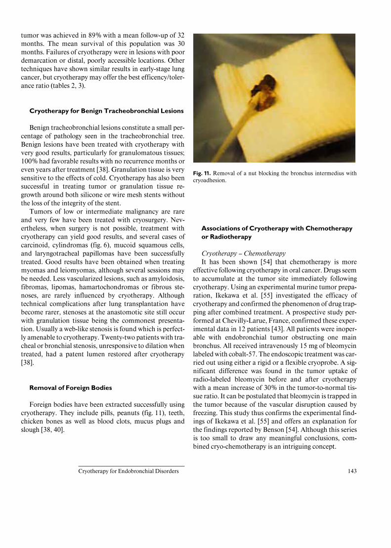
BOIS:L08:BPRR3018PD.98 FF: RESF E1: RESS
Cryotherapy for Endobronchial Disorders 143
tumor was achieved in 89% with a mean follow-up of 32months. The mean survival of this population was 30months. Failures of cryotherapy were in lesions with poordemarcation or distal, poorly accessible locations. Othertechniques have shown similar results in early-stage lungcancer, but cryotherapy may offer the best efficency/toler-ance ratio (tables 2, 3).
Cryotherapy for Benign Tracheobronchial Lesions
Benign tracheobronchial lesions constitute a small per-centage of pathology seen in the tracheobronchial tree.Benign lesions have been treated with cryotherapy withvery good results, particularly for granulomatous tissues;100% had favorable results with no recurrence months oreven years after treatment [38]. Granulation tissue is verysensitive to the effects of cold. Cryotherapy has also beensuccessful in treating tumor or granulation tissue re-growth around both silicone or wire mesh stents withoutthe loss of the integrity of the stent.
Tumors of low or intermediate malignancy are rareand very few have been treated with cryosurgery. Nev-ertheless, when surgery is not possible, treatment withcryotherapy can yield good results, and several cases ofcarcinoid, cylindromas (fig. 6), mucoid squamous cells,and laryngotracheal papillomas have been successfullytreated. Good results have been obtained when treatingmyomas and leiomyomas, although several sessions maybe needed. Less vascularized lesions, such as amyloidosis,fibromas, lipomas, hamartochondromas or fibrous ste-noses, are rarely influenced by cryotherapy. Althoughtechnical complications after lung transplantation havebecome rarer, stenoses at the anastomotic site still occurwith granulation tissue being the commonest presenta-tion. Usually a web-like stenosis is found which is perfect-ly amenable to cryotherapy. Twenty-two patients with tra-cheal or bronchial stenosis, unresponsive to dilation whentreated, had a patent lumen restored after cryotherapy[38].
Removal of Foreign Bodies
Foreign bodies have been extracted successfully usingcryotherapy. They include pills, peanuts (fig. 11), teeth,chicken bones as well as blood clots, mucus plugs andslough [38, 40].
Fig. 11. Removal of a nut blocking the bronchus intermedius withcryoadhesion.
Associations of Cryotherapy with Chemotherapyor Radiotherapy
Cryotherapy – ChemotherapyIt has been shown [54] that chemotherapy is more
effective following cryotherapy in oral cancer. Drugs seemto accumulate at the tumor site immediately followingcryotherapy. Using an experimental murine tumor prepa-ration, Ikekawa et al. [55] investigated the efficacy ofcryotherapy and confirmed the phenomenon of drug trap-ping after combined treatment. A prospective study per-formed at Chevilly-Larue, France, confirmed these exper-imental data in 12 patients [43]. All patients were inoper-able with endobronchial tumor obstructing one mainbronchus. All received intravenously 15 mg of bleomycinlabeled with cobalt-57. The endoscopic treatment was car-ried out using either a rigid or a flexible cryoprobe. A sig-nificant difference was found in the tumor uptake ofradio-labeled bleomycin before and after cryotherapywith a mean increase of 30% in the tumor-to-normal tis-sue ratio. It can be postulated that bleomycin is trapped inthe tumor because of the vascular disruption caused byfreezing. This study thus confirms the experimental find-ings of Ikekawa et al. [55] and offers an explanation forthe findings reported by Benson [54]. Although this seriesis too small to draw any meaningful conclusions, com-bined cryo-chemotherapy is an intriguing concept.

BOIS:L08:BPRR3018PD.98 FF: RESF E1: RESS
144 Vergnon/Mathur
Cryotherapy – RadiotherapyIn localized inoperable bronchial carcinomas, radio-
therapy is an accepted treatment, but the mean survival ofpatients is only 20 months, and local eradication oftumors is obtained in only 35% of cases. In obstructivetumors, the reinflation of the lung is achieved after irra-diation in only 21% of cases [42]. When these obstructivetumors have been removed with an Nd:YAG laser priorto irradiation, these has been a better survival and qualityof life [66, 67]. Cryotherapy will also remove endobron-chial obstruction, and studies have suggested a possiblesynergy between cryotherapy and external irradiation dueto increased blood flow after cryotherapy [36, 66]. Le Pi-vert [62] showed the appearance of marked neovasculari-zation by angiography 15 days after cryotherapy. Thishypervascularization may increase the radiosensitivity. Intwo similar prospective studies, Vergnon et al. [42] andHomasson et al. [26] have studied the association of cryo-therapy and radiotherapy. A satisfactory outcome wasdefined as 150% tumor destruction. Radiotherapy wasstarted 2 weeks after cryotherapy. Our study group con-sisted of 29 patients. All patients had symptomatic unre-sectable endobronchial obstruction. One or two sessionsof cryotherapy were done; cryotherapy was consideredsatisfactory in 16 cases and unsatisfactory in 13 cases withpersistent tumor lesions. Twenty-one patients received 65Gy, and 8 patients in poor general condition received 45Gy.
The results were evaluated with flexible bronchoscopyand biopsy 2 months after the end of irradiation. In theunsatisfactory cryotherapy group (tumor removal !50%),
the median survival was 5 months. In the satisfactorycryotherapy group (tumor removal 150%), the mediansurvival was 11 months. This study does not prove thepotentiating effect of cryotherapy on the effect of radio-therapy. However, it confirms several significant points:cryotherapy is a safe method and is successful in 70–80%of cases [42]. This study reinforces the interest alreadygenerated in eliminating an obstruction before irradiationto improve survival. The survival curves obtained corre-spond closely to those obtained using laser resection ofendobronchial lesions. This study also confirms the inad-equacy of radiotherapy alone in relieving a bronchialobstruction. This ‘curative’ local effect raises the possibili-ty of increased tumor radiosensitivity, induced by the vas-cular effect of the cryotherapy.
Conclusion
Cryotherapy for endobronchial lesions has been shownto be an effective and safe therapy in a number of patho-logies. In particular, the technique is extremely useful inpatients with carcinoma of the lung and airway obstruc-tion. A large majority of patients show improvement insymptoms and respiratory function. These benefits areachieved by using a technique which is safe, easy to per-form, inexpensive and has few complications.
References
1 Breasted JH: The Edwin Smith Surgical Papy-rus. Chicago, University of Chicago OrientalInstitute, vol 3, p 217.
2 Arnott J: On the Treatment of Cancer by Regu-lated Application of an Anaesthetic Tempera-ture. London, Churchill, 1851.
3 Arnott J: On the Treatment of Cancer by Con-gelation and an Improved Mode of Pressure.London, Churchill, 1855.
4 White AC: Liquid air; its application in medi-cine and surgery. Med Rec 1899;56:109.
5 White AC: Possibilities of liquid air to the phy-sician. JAMA 1901;36:426.
6 Hass G, Taylor C: A quantitative hypothermalmethod for the production of local injury of tis-sue. Arch Pathol 1948;45:563.
7 Cahan WG, Brockmier G: Cryosurgery of theuterine cavity. Am J Obstet Gynecol 1967;99:138.
8 Gonder MJ, Soanes WA, Smith V: Experimen-tal prostate cryosurgery. Invest Urol 1964;1:610.
9 Rand RW, Dashe AM, Paglia DE, ConwayLW, Solomon DH: Stereotactic cryohypophy-sectomy. JAMA 1964;189:255.
10 Gage AA, Koepf S, Wehrle D, Emmings F:Cryotherapy for cancer of the lip and oral cavi-ty. Cancer 1965;18:1646.
11 Zacarian S, Adham M: Cryotherapy of cuta-neous malignancy. Cryobiology 1966;2:212.
12 Barton R: Cryosurgical treatment of nasopha-ryngeal neoplasms. Am Surg 1966;32:744.
13 Lincoff H, McLean J, Long R: The cryosurgicaltreatment of intraocular tumors. Am J Ophtal-mol 1967;63:389.
14 Blackwood J, Moore FT, Pace WG: Cryothera-py for malignant tumors. Cryobiology 1967;4:33.
15 Ostergard D, Townsend DE: Malignant mela-noma of the female urethra treated by cryother-apy with radical vulvectomy and anterior exen-teration. Obstet Gynecol 1968;31:75.
16 Gage AA: Cryotherapy for inoperable cancer.Dis Colon Rectum 1968;2:36.
17 Marcove RC, Miller TR: Treatment of primaryand metastatic bone tumors by cryosurgery.JAMA 1969;207:1890.
18 Grana L, Kidd J, Swenson O: Cryogenic tech-niques within tracheobronchial tree. J Cryo-surg 1969;2:62.
19 Thomford NR, Wilson WH, Blackburn ED,Pace WG: Morphological changes in caninetrachea after freezing. Cryobiology 1970:7:19.
20 Skivolocki WP, Pace WG, Thomford NR: Ef-fect of cryotherapy on tracheal tumors in rats.Arch Surg 1971;103:341.

BOIS:L08:BPRR3018PD.98 FF: RESF E1: RESS
Cryotherapy for Endobronchial Disorders 145
21 Neel HB, Farrell KH, Payne WS, De SantoLW, Sanderson DR: Cryosurgery of respiratorystructures. I. Cryonecrosis of trachea and bron-chus. Laryngoscope 1973;83:1062.
22 Gorenstein A, Neel HB, Sanderson DR: Trans-bronchoscopic cryosurgery of respiratory stric-tures. Experimental and clinical studies. AnnOtol Rhinol Laryngol 1976;85:670.
23 Carpenter RJ, Neel HB, Sanderson DR: Cryo-surgery of bronchopulmonary structures. Anapproach to lesions inaccessible to the rigidbronchoscope. Chest 1977;72:279.
24 Gage AA: Cryotherapy for cancer; in Rand R,Rinfret R, Rinfret A, Von Leden H (eds): Cryo-surgery. Springfield, Thomas, 1968, pp 376–387.
25 Sanderson DR, Neel HB, Payne WS, WoolnerLB: Cryotherapy of bronchogenic carcinoma.Report of a case. Mayo Clin Proc 1975;435–437.
26 Homasson JP, Renault P, Angebault M, Bon-niot JP, Bell NJ: Bronchoscopic cryotherapyfor airway strictures caused by tumors. Chest1986;90:159–164.
27 Maiwand MO: Cryotherapy for advanced car-cinoma of the trachea and bronchi. Br Med J1986;293:181–182.
28 Astesiano A, Aversa S, Ciotta D, Galietti F,Gandolfi G, Giorgis GE, Doliaro A, Scappatic-ci E, Pepino E: Distruzione crioterapica deitumori invasivi tracheobronchiali. Casisticapersonale. Minerva Med 1986;77:2159.
29 Mathur PN, Wolf KM, Busk MF, Briete WM,Datzman M: Fiberoptic bronchoscopic cryo-therapy in the management of tracheobron-chial obstruction. Chest 1996;110:718–723.
30 Rodgers BM, Talbert JL: Clinical applicationof endotracheal cryotherapy. J Pediatr Surg1977;13:662–666.
31 Rodgers BM, Moazam PF, Talbert JL: Endo-tracheal cryotherapy in the treatment of refrac-tory airway strictures. Ann Thorac Surg 1983;35:52.
32 Homasson JP: Cryotherapy in pulmonology to-day and tomorrow. Eur Respir J 1989;2:799–801.
33 Walsh DA, Maiwand MO, Nath AR, Lock-wood P, Lloyd MH, Saab M: Bronchoscopiccryotherapy for advanced bronchial carcino-ma. Thorax 1990;45:509–513.
34 Pesek M, Simecek C, Bruha F: Kryokoagula-tion in der Behandlung von Bronchialtumoren.Z Erkr Atmungsorg 1990;175:126.
35 Homasson JP, Bell NJ: Cryotherapy in chestmedicine. Paris, Springer Verlag, 1992.
36 Maiwand MO, Homasson JP: Cryotherapy fortracheobronchial disorders; in Mathur PN,Beamis JF (eds): Clinics in Chest Medicine.Philadelphia, Saunders, 1995, vol 16, pp 427–443.
37 Homasson JP: Bronchoscopic cryotherapy. JBronchol 1995;2:145.
38 Sheski FD, Mathur PN: Endobronchial cryo-therapy for benign tracheobronchial lesions.Chest 1998;114:261s–262s.
39 Thommi G, McLeavy M: Cryotherapy in themanagement of tracheobronchial obstructionby lung tumors. Chest 1998;114:262s.
40 Thommi G, McLeay M: Cryobronchoscopy inthe management of foreign body in the tracheo-bronchial tree. Chest 1998;114:303s.
41 Morasso A, Gallo E, Massaglia GM, OnoscuriM, Bernardi V: Cryosurgery in bronchoscopictreatment of tracheobronchial stenosis. Chest1993;103:472.
42 Vergnon JM, Schmitt T, Alamartine E, Bar-thelemy JC, Fournel P, Emonot A: Initial com-bined cryotherapy and irradiation for unresect-able non-small cell lung cancer. Chest 1992;102:1436–1440.
43 Homasson JP, Pecking A, Roden S, AngebaultM, Bonniot JP: Tumor fixation of bleomycinlabeled with 57 cobalt before and after cryo-therapy of bronchial carcinoma. Cryobiology1992;29:543–548.
44 Fahy GM, Saur J, Williams RJ: Physical prob-lems with the vitrification of large biologicalsystems. Cryobiolgoy 1990;27:492.
45 Gage AA, Guest K, Montes M, Garuna JA,Whalen DA: Effect of varying freezing andthawing rates in experimental cryosurgery.Cryobiology 1985;22:175.
46 Smith JJ, Fraser J: An estimation of tissuedamage and thermal history in cryolesion.Cryobiology 1974;11:139.
47 Miller RH, Mazur P: Survival of frozen-thawedhuman red cells as a function of cooling andwarming velocities. Cryobiology 1976;13:404.
48 Gage AA: Critical temperature for skin necro-sis in experimental cryosurgery. Cryobiology1982;19:273.
49 Rand RW, Rand RP, Eggerding FA, Field M,Denbesten L, King W, Camici S: Cryolumpes-tomy for breast cancer: An experimental study.Cryobiology 1985;22:307.
50 Rubinsky B, Ikeda M: A cryomicroscope usingdirectional solidification for the controlledfreezing of biological tissue. Cryobiology 1985;22:55.
51 Forsyth M, Macfarlane DR: Recrystallisationrevisited. Cryo Lett 1986;7:367.
52 Martino MN, Zariteky NE: Ice recrystallisationin a model system and in frozen muscle tissue.Cryobiology 1989;26:138.
53 Gilbert JC, Onik GM, Hoddick W, RubinskyB: Real time ultrasonic monitoring of hepaticcryosurgery. Cryobiology 1985;22:319.
54 Benson JW: Combined chemotherapy andcryosurgery for oral cancer. Am J Surg 1975;13:596.
55 Ikekawa S, Ishihara K, Tanaka S, Ikeda S: Bas-ic studies of cryochemotherapy in a murinetumor system. Cryobiology 1985;22:477–483.
56 Ablin RJ, Soanes WA, Gonder M: Prospectsfor cryo-immunotherapy in case of metastasiz-ing carcinoma of the prostate. Cryobiology1971;8:217.
57 Tanaka S: Immunological aspects of cryosurge-ry in general surgery. Cryobiology 1982;19:247.
58 Mazur P: The role of intracellular freezing inthe death of cells cooled at supra optimal rates.Cryobiology 1977;14:251.
59 Albin RJ: Cryoimmunology: A critique of thephenomenon and its future, in cryosurgery.Mechanisms and applications. Intern InstitRefr 1995;14:133–138.
60 Gage AA, Caruana JA, Garamy G: A compari-son of instrument methods of monitoring freez-ing in cryosurgery. J Dermatol Surg Oncol1983;9:209.
61 Homasson JP, Thiery JP, Angebault M, Ov-tracht L, Maiwand MO: The operation andefficacy of cryosurgical, nitrous oxide-drivencryoprobe. Cryoprobe physical characteristics:Their effects on cell cryodestruction. Cryobio-logy 1994;31:290.
62 Le Pivert P, Binder P, Ougier T: Measurementof intratissue bioelectrical low frequency impe-dance: A new method to predict per-operative-ly the destructive effect of cryosurgery. Cryo-biology 1977;14:245.
63 Deygas N, Froudarakis ME, Ozenne G, JouveS, Fournel P, Vergnon JM: Cryotherapy in ear-ly superficial bronchogenic carcinoma. EurRespir J 1998;12(suppl 28):266S.
64 Homasson JP: Bronchoscopic cryotherapy.Bronchol 1995;2:145–153.
65 Vergnon J-M: How I do it: Bronchoscopic cryo-therapy. J Bronchol 1995;2:323–327.
66 Macha H, Becker KO, Kemmer HP: Pattern offailure and survival in endobronchial laser re-section. A matched pair study. Chest 145;967.
67 Eichenhorn MS, Kvale PA, Miks VM, SeydelHG, Horowitz B, Radke JR: Initial combina-tion therapy with Yag laser photoresection andirradiation for inoperable nonsmall cell carci-noma of the lung. A preliminary report. Chest1986;89:782.
J.M. VergnonC.H.U de St. Etienne, Hôpital NordF–42055 St. Etienne Cedex 2 (France)Tel. +33 477 82 83 14, Fax +33 477 82 80 90E-Mail [email protected]
Prof. Praveen N. MathurPulmonary, Critical Care andOccupational MedicineIndiana University School of Medicine1001 West 10th Street, OPW 529Indianapolis, IN 46202-2879 (USA)Tel. +1 317 630 7596, Fax +1 317 630 7487E-Mail [email protected]

OOOOOOOOOOOOOOOOOOOOOOOOOOOOOOOOOOOOOOOOOOOOOOOOOOOOOOOOOOOOOOOOOOOOOOOOOOOOOOOOOOOOOOOOOOOOOOOOOOOOOOOOOOOOOOOOOOOOOOOOOOO
Therapeutic Bronchoscopy

Bolliger CT, Mathur PN (eds): Interventional Bronchoscopy.Prog Respir Res. Basel, Karger, 2000, vol 30, pp 146–158
OOOOOOOOOOOOOOOOOOOOOOOOOOOOOOOOOOOOOOOOOOOOOOOOOOOOOOOOOOOOOOOOOOOOOOOOOOOOOOOOOOOOOOOOOOOOOOOOOOOOOOOOOOOOOOOOOOOOOOOOOOO
Endoluminal Brachytherapy inCentral Lung CancerR. Fischer R.M. Huber
Pneumology, Department of Medicine, Klinikum Innenstadt, University of Munich, Germany
146
Summary
Brachytherapy means the direct placement of a highlyradioactive source inside a tumor mass. This can be doneeither by implanting the source directly into the tumor, viathe natural route (endoluminal brachytherapy) or by placingthe source into the tumor bed during tumor resection.Endoluminal brachytherapy employing the afterloading tech-nique with iridium-192 high dose rate (HDR) is largelyapplied for the curative and palliative treatment of endo-bronchial tumors due to its tumor-specific and long-lastingeffect. Endoluminal brachytherapy using flexible bronchos-copy and an HDR regimen can be performed on an outpa-tient basis and is not more strenuous for the patient than adiagnostic bronchoscopy. Symptomatic improvement can beachieved in 70–80% of patients, and sometimes small tumorscan even be cured. The afterloading procedure can be com-bined with all other modalities of tumor therapy. It can beused as ‘boost’ to conventional external irradiation and aslocal treatment modality in patients on systemic chemother-apy or as the only local treatment. HDR treatment is usuallydelivered with 1–6 fractions at an interval of 1–3 weeks and adose of 3–20 Gy per fraction (at 1 cm from the source axis).In patients previously treated with external beam radiationtherapy and in the palliative setting, a regimen of 7–10 Gy(HDR) per fraction and a total of 2–3 fractions per treat-ment is recommended. However, the optimal dosage andfractionation schemes for the tumor therapy are still un-known and there is need for further studies. In about 10% ofthe patients, radiation bronchitis occurs, and there may befatal hemorrhage, possibly related to the therapy. Overall,
endobronchial brachytherapy is a well-tolerated, not veryaggressive treatment option, especially in patients withreduced performance status.
The majority of patients presenting with lung cancerhave locally advanced or metastatic disease not amenableto curative surgical resection. Attempted resection withquestionable or involved margins carries a high risk ofsubsequent local recurrence. Endobronchial metastases orlocal recurrence after surgery and/or radiotherapy oftencause symptoms like dyspnea or hemoptysis due to tumorstenosis in the central tracheobronchial system. Apartfrom dyspnea, cough and hemoptysis, infectious compli-cations are frequently encountered. Increased survivaland improved local control can be achieved by combinedchemotherapy and external beam irradiation (EBRT) forlocally advanced inoperable lung cancer. However, thereremains considerable need for improvement and for fur-ther local treatment options. Treatment should be quick,suitable for the patient and personnel, effective and carrya low risk for complications, because the treatment goal ismostly limited to palliation. For these patients, therapeut-ic tools suitable for the symptomatic management of thesesymptoms are available nowadays. The different localtherapeutic modalities have special advantages and disad-vantages. The best treatment option has to be chosenaccording to the individual situation, preferably using aninterdisciplinary approach.

BOIS:L08:BPRR3003XA.82 FF: RESF E1: RESS
Endoluminal Brachytherapy in CentralLung Cancer
147
Amongst the currently available interventional bron-choscopic procedures, endobronchial brachytherapy isone of the oldest techniques. As early as 1922, the firstsuccessful endobronchial implantation of radium cap-sules (derived from gynecological implants) was docu-mented, and many reports have followed since [1]. In the1960s, cobalt-60 seeds were most frequently used asradiation source. Through brachytherapy, it is possible todeliver a maximum dose to the tumor with a minimumdose in the surrounding normal tissue. One of the majordrawbacks of this method was the high level of radiationto which the medical personnel was exposed.
Therefore, the development of the afterloading tech-nique was essential for the widespread application of bra-chytherapy [2]. The introduction of the iridium-192 ra-dioisotope, and the refinement of the afterloading appara-tus by using automated, computer-controlled steering de-vices, has meant significant progress [3]. The small size ofthe iridium source with its high activity and high dose rate(HDR) allows its placement in a hollow guidance cathe-ter, which can be easily placed endobronchially by a flexi-ble bronchoscope. Nevertheless, it was not until the wide-spread use of the Nd:YAG laser recanalization of tumorstenoses that endoluminal brachytherapy employing theafterloading technique with iridium-192 HDR was largelyapplied for the palliative treatment of endobronchial andparabronchial tumors.
Performed by an experienced endoscopist, HDR bra-chytherapy has as few acute side effects as routine fiberop-tic bronchoscopy and can therefore be easily applied in anoutpatient setting. Irradiation lasts some minutes only.
Technique
Brachytherapy (brachys = greek expression for ‘short’)means the direct placement of a highly radioactive sourceinside a tumor mass. This can be done either by implant-ing the source directly into the tumor (interstitial brachy-therapy), via the natural route (endoluminal brachythera-py) or by placing the source into the tumor bed duringtumor resection (intraoperative brachytherapy). The tech-nical procedures to perform interstitial permanent vol-ume or planar implantation of radioactive sources orinterstitial temporary implantation are described else-where [4, 5]. Here we want to focus on the procedures ofendoluminal brachytherapy which are commonly used.
As compared to conventional external radiotherapy,brachytherapy offers the potential advantage of providinga higher dose of radiation to the desired tumor volume by
Fig. 1. Typical isodoses in HDR brachytherapy.
sparing normal tissues. The physical characteristics ofradioactive isotopes are characterized by the inversesquare law which means that the dose rate decreases as afunction of the inverse square of the distance to the sourcecentre. This makes it possible to achieve a high irradiationdose in the centre of the irradiation source with a fastdecrease towards the periphery. A typical distribution ofisodoses is shown in figure 1. For example, an irradiationdose of 30 Gy delivered at 1 cm from the source axisresults in a dose of only 7.5 Gy at 2 cm and as little as 3.3Gy at a distance of 3 cm. By employing the remote after-loading technique, the spinal cord receives not more than5–10% of the dose prescribed at 1 cm in normal endo-bronchial brachytherapy [6]. On the other hand, when aninhomogenous tumor mass is not well localized, normaltissue adjacent to it is likely to receive very high irradia-tion doses. In order to spare normal tissue, it is necessaryto place the irradiation probe carefully in the center of thetumor bulk and, whenever possible, to use devices whichguarantee a constant distance.
Endobronchial or interstitial brachytherapy can be ca-tegorized as permanent or temporary. Permanent im-plants are characterized by a very low-dose irradiationrate (e.g. iodine-125). This type of radioactive source isinfrequently used, most in tumor locations with no exist-ing lumen, e.g. in a perioperative setting or pancoasttumors [4].
Temporary brachytherapy is most frequently used.This form of brachytherapy can be divided into three doserate regimens: low dose rate (LDR, less than 2 Gy/h),

BOIS:L08:BPRR3003XA.82 FF: RESF E1: RESS
148 Fischer/Huber
Fig. 2. Chest X-ray after placement of brachytherapy catheter in theright upper lobe with dummy seeds.
intermediate dose rate (IDR, 1–12 Gy/h) and HDR (morethan 2 Gy/min). LDR brachytherapy usually requires 8–48 h of treatment time, while HDR is achieved within afew minutes. The common radioactive source is iridium-192 but cesium-137 or iodine-125 are also used.
Endobronchial treatment is achieved by placing one ortwo afterloading applicators within the trachea and thecentral bronchial system. In specialized centers, position-ing of the afterloading probe as far as the subsegmentalbronchi is possible (fig. 2).
The afterloading catheters have an external diameterof 2–3 mm. Flexible bronchoscopy is performed to local-ize the tumor region for irradiation. If there is subtotalstenosis of the bronchi due to submucosal or exophytictumor growth, it is sometimes necessary to perform bal-loon dilatation or use other recanalization methods forbetter applicator placement. If there was previous lasertreatment, it is recommended to wait at least 3 days
before brachytherapy treatment can be initiated, althoughthe debate about this issue is still ongoing [7, 8].
Endoluminal irradiation should be delivered with a‘safety’ margin of at least 1 cm at both ends of the visibleendobronchial tumor length. The active length refers tothe distance between the first and last dwelling point ofthe stepping iridium source in case of HDR brachythera-py. As the distal end of the tumor can not always beenseen by the bronchoscopist, the distal endpoint of the irra-diation length must often be estimated from previouschest X-rays or CT scans and controlled during bronchos-copy by fluoroscopy.
The irradiation length is marked by external tags, con-trolled by fluoroscopy. A guidewire is placed through theworking channel of the bronchoscope which is thenremoved (fig. 3a–c). Manipulation of the guidewire andthen of the applicator through a partially obstructedlumen requires skill, particularly within the upper lobebronchi.
For a better fit of the afterloading probe, we usuallyinsert a shortened gastric tube with an external diameterof 5 mm by the Seldinger technique over the guidewire(fig. 4a–c). The gastric tube should be placed inside thetumor bulk. The irradiation applicator is then placed intothe tube and taped to the tip of the nose to prevent it frombeing dislocated. This should be done under permanentvisual fluoroscopic control.
After placement of the afterloading probe, a dummyseed is inserted, and a set of orthogonal chest X-rays isobtained to confirm the correct placement of the catheterwithin the tumor bulk and to determine the necessaryirradiation length, as indicated by the external tags(fig. 5a–c). Treatment dose is prescribed by the radiationoncologist, usually specified at a depth taken 1 cm fromthe middle of source axis [9].
One of the technical challenges is the obvious differ-ence in luminal diameters of different segments of the tra-cheobronchial tree. It is uncommon to adjust for these dif-ferences, but Saito et al. [10] tried to answer this problemby setting distinct diameters at different segments of thetracheobronchial tree and adjusting the dose evaluationpoint to the lumen diameter at the lesion site.
Remote brachytherapy is performed in a shieldedroom with permanent oxygen delivery to the patient, whois monitored from outside by continuous assessment ofoxygen saturation, pulse rate or ECG and direct visualcontrol through a video camera. The treatment can beinterrupted and restarted whenever necessary.
After removal of the dummy seed, the applicator isconnected to the iridium-192 remote afterloading unit.

BOIS:L08:BPRR3003XA.82 FF: RESF E1: RESS
Endoluminal Brachytherapy in CentralLung Cancer
149
The irradiation source (diameter about 1 mm) is ad-vanced to the intended position under computer controland then drawn backwards at intervals of 5 mm distance.It remains in each position for the time needed to applythe computed dose. By varying the source position anddwelling time, individual computer-assisted dose distri-bution can be generated (fig. 6).
In general, endoluminal brachytherapy using flexiblebronchoscopy and an HDR regimen can be performed onan outpatient basis, as it is not more strenuous for thepatient than a diagnostic bronchoscopy. In the case ofLDR brachytherapy, hospitalization for several days isusually required. Due to the development of high activitysources, HDR brachytherapy has now largely replaced theLDR treatment at most medical centers. HDR treatmentis usually delivered as 1–6 fractions at an interval of 1–3weeks. Controlled comparative data are widely lacking.More recently, hyperfractionated HDR has been exploredin a pilot study with similar morbidity compared to con-ventional HDR or LDR [6], but the final data have notbeen published yet.
Acute side effects of the placement procedure includemore or less severe coughing and increased bronchialsecretion. Temporary pleuritic pain or even pneumotho-races have been described when the guidewire or theapplicator was placed too vigorously [5]. However, wenever observed such serious side effects during more than1,000 placements of afterloading probes.
Indications for Endoluminal Brachytherapy inCentral Lung Tumors
Carcinoma in situ and Early-Stage Non-Small CellLung CancerSurgical resection is widely accepted as the treatment
of choice in early-stage non-small cell lung cancer(NSCLC). But when occult carcinoma in situ or smallinvasive endobronchial lesions are discovered incidental-ly by bronchoscopy due to early irritative symptoms likecough or hemoptysis, HDR brachytherapy either alone oras a boost to EBRT offers an excellent treatment optionwith good results, low morbidity, low costs and littleinconvenience for the patient. Especially in carcinoma insitu or limited invasive tumors without nodal involve-ment, HDR brachytherapy could represent the definitetreatment.
Due to the development of other treatment modalities(e.g. photodynamic therapy), data published on intralu-minal brachytherapy in early-stage NSCLC are limited.
Studies comparing these two treatment modalities aremissing. Due to the deeper penetration, brachytherapycould be superior to the photodynamic treatment.
Stage II–IIIb (T3–T4, N0–N3), Curative TreatmentIntentionIn patients with stage II–IIIa, surgery is the treatment
of choice whenever possible. Several attempts have beenreported to improve survival by adding interstitial bra-chytherapy, but the data are sparse and conflicting [4, forreview]. When the option for interstitial brachytherapy isavailable, it should be considered to enhance local controlif complete surgical resection is not possible. Unfortu-nately, up to one third of newly diagnosed patients withNSCLC present with locally advanced, unresectable dis-ease [11].
External radiotherapy has a potential for cure in onlyfew patients, and local recurrences are even more frequent[12, 13]. Nevertheless, this treatment modality can im-prove symptoms for those patients who are inoperabledue to locally advanced disease or other medical reasonsas well as in a palliative situation.
In spite of high doses of 50–60 Gy, median survival isapproximately 1 year, and long-term survival rates areless than 10% [13].
To increase the cumulative dose of irradiation, addi-tional endobronchial brachytherapy has become morewidely used [14–21]. Brachytherapy has been applied as aboost either before, during or after EBRT. HDR brachy-therapy has been primarily used for previously untreatedpatients in conjunction with EBRT, often to relieve ob-struction quickly and to reduce the volume of irradiatednormal lung tissue. Particularly, when atelectasis due toobstruction of a main or global bronchus is obscuring thetrue tumor margins, brachytherapy can help to reduce thepermanent fibrosis of normal lung tissue due to largeexternal irradiation fields. It has been calculated that thisprocedure can reduce the irradiation of normal tissues byan average of 32% [22].
Apart from treatment for local stenosis, brachytherapyhas the potential to increase survival time and local con-trol when used in combination with external irradiation.Although none of the studies published so far could dem-onstrate a clear advantage in terms of survival in nonsel-ected patients treated with this combined modality, thereare indications that at least local control is better inpatients with additional endoluminal brachytherapy.Again, larger, multicenter, controlled, prospective andrandomized studies are needed to evaluate this promisingtreatment regimen.

BOIS:L08:BPRR3003XA.82 FF: RESF E1: RESS
150 Fischer/Huber
Although it is often difficult to differentiate betweentreatment complications and tumor progression, it is pos-sible that the combination of external and endoluminalirradiation increases the frequency of hemorrhages, per-haps due to the proximity of the afterloading catheter topulmonary vessels.
Stage IV, Tumor Recurrency, Palliative TreatmentIntentionEndobronchial brachytherapy is a relatively simple,
fast and minimally invasive method to relieve bronchialobstruction or to maintain airway patency. Therefore,HDR brachytherapy is now a frequently used treatmentoption in patients with relapsing endobronchial tumor,with thoracic metastases from other primaries, poor over-all performance status, or for patients unwilling to under-go other (i.e. chemotherapeutic) intensive treatments.
Although the term ‘palliative’ is difficult to define, pal-liative HDR brachytherapy should be considered in everypatient presenting with symptoms of endobronchial tu-mor growth (hemoptysis, cough, atelectasis, dyspnea,pain) if other definite treatment modalities (i.e. surgery,EBRT multimodal therapies) are not available for thepatient for any reason.
Compared to photodynamic therapy, endobronchialstenting or balloon dilatation, endobronchial brachythera-py is probably more effective to maintain long-term airwaypatency and symptom relief. In most of the published stud-ies on palliative brachytherapy, overall improvement ofsymptoms has been shown in 65–95% of all patients. Espe-cially hemoptysis can be treated with a high rate of success,this is also true for the reopening of obstructed bronchi.Improvement of cough, shortness of breath and pain wasobserved to a lesser degree. Palliation can be maintained ina high proportion of patients [6, 16, 18, 23–34].
Fig. 3–5. Endobronchial afterloading technique. 3 Insertion of theguidewire. a Schematic illustration. b Fluoroscopy. c Endoscopicview. 4 Insertion of a shortened gastric tube over the guidewire (Sel-dinger technique), then removal of the guidewire. a Schematic illus-tration. b Fluoroscopy. c Endoscopic view. 5 The afterloading appli-cator with the dummy seed is inserted through the stomach tube,then the necessary irradiation length is determined by using externaltags with fluoroscopy (here: 6 cm length), then the dummy seed isremoved. a Schematic illustration. b Fluoroscopy. c Endoscopicview.
3a
4a
5a
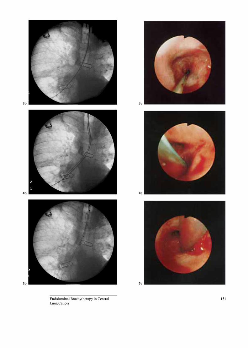
BOIS:L08:BPRR3003XA.82 FF: RESF E1: RESS
Endoluminal Brachytherapy in CentralLung Cancer
151
3b
4b
5b
3c
4c
5c

Aygun et al. [14]
BOIS:L08:BPRR3003XA.82 FF: RESF E1: RESS
152 Fischer/Huber
Fig. 6. Determination of the necessary irra-diation length by external tags with fluoros-copy and distribution of the typical HDRbrachytherapy isodoses.
Table 1. Results of some studies on HDR brachytherapy
Authors Patients Prior treatment(laser, stent,chemotherapy), %
TotalEBRTGy
Total HDRbrachytherapyGy range
Bronchoscopicresponse, %
Mediansurvivalweeks
Fatalhemorrhages%
62 0 61 12–30 56 56 15Cotter et al. [15] 48 0 66 6–35 86 32 5Huber et al. [17] 42 18.6 50 7.4 74 27 18.9Kohek et al. [20] 39 52 70 5.6–27.2 67 56 5Speiser and Spratling [33] 50 24 60 22.5–30 80 44 7

BOIS:L08:BPRR3003XA.82 FF: RESF E1: RESS
Endoluminal Brachytherapy in CentralLung Cancer
153
Review of Published Results in EndobronchialBrachytherapy
Carcinoma in situ and Early-Stage NSCLCThere are no prospective, randomized studies evaluat-
ing the effect of endobronchial brachytherapy alone in thetreatment of very early lung cancer, but some results arepromising and merit further study.
Tredaniel et al. [35] treated 29 patients with stage I andII disease with endoluminal brachytherapy alone. Theydelivered a dose of 7 Gy (at 1 cm from the source axis)every second week, with a maximum of 6 fractions. After2 months, histologic complete response was seen in 72%of the patients. After 23 months of follow-up, median sur-vival has not yet been reached. Fatal hemoptysis occurredin 17% of the patients, although it was not clear if recur-rent disease was the reason for this complication.
Sutedja et al. [36] treated 2 patients with LDR brachy-therapy, demonstrating feasibility and showing goodoverall response.
Saito et al. [10] studied prospectively the effect a com-bination of EBRT with 40 Gy in 20 fractions and addi-tional LDR brachytherapy (using a new technique with 4thin iridium-192 wires) with 25 Gy in 5 fractions. Out of39 evaluable patients, recurrent disease was observed inonly 2 cases, which could be treated by subsequent sur-gery. The median follow-up period was 24.5 months. Oth-er recurrences or severe complications from irradiationhad not occurred. Second primary cancers were observedin 19 (lung 10; other organs 10) of a total of 41 patients[10].
Stage II–IIIb (T3–T4, N0–N3), Curative TreatmentIntentionEndoluminal brachytherapy as sole treatment has not
been published for this disease stage. Although LDR andIDR brachytherapy in combination with EBRT havebeen described, most experience exists with HDR brachy-therapy as a boost to definite external radiation therapy.The results are summarized in table 1.
Higher combined doses of EBRT and brachytherapyhave been associated with increased overall response,although this was not found by all published studies.Overall response rates, often assessed several monthsafter completion of treatment, were good. However, theoverall median survival time (7–13 months) is not higherthan in patients treated with EBRT alone.
The only prospective, randomized study was per-formed by Huber et al. including a total of 98 patients.Two groups were compared: one group was treated with
Fig. 7. Survival for patients with squamous cell carcinoma. EI =External irradiation alone (group 1; n = 29), EI + HDRBT = externalirradiation and additional HDR brachytherapy (group 2; n = 39),censored (+) cases in group 1: 2; censored (+) cases in group 2: 1;Kaplan-Meier plot, p = 0.09 (log-rank test) [17].
0.0
0 100
Weeks
200
0.2 El El + HDRBT
0.4
Cu
mu
lati
ve
su
rviv
al
0.6
0.8
1.0
external radiotherapy alone (planned dose 60 Gy), the sec-ond group received an additional boost of HDR brachy-therapy (4.8 Gy scheduled, at 10 mm from the sourceaxis) before and after external irradiation. The mean totalexternal irradiation dose was 50 Gy in both groups. TheHDR brachytherapy group received an additional dose of7.44 B 2.6 Gy (at 10 mm depth). The median survivaltime in both groups was comparable (28 and 27 weeks,respectively). The low overall survival time in this study isprobably due to the very low performance status of thepatients (average Karnofsky performance status 65), com-pared to that of other, nonrandomized studies. In patientswith squamous cell carcinoma (68 patients), the HDRbrachytherapy group showed an advantage in median sur-vival with borderline significance (40 vs. 33 weeks, p =0.09). They also showed a better local tumor control(fig. 7). As could be expected, patients with squamous cellcarcinoma had a significantly longer period of local tumorcontrol. Fatal hemoptysis was the cause of death in 11(18.9%) compared to 6 (14.2%) patients in the grouptreated conventionally.
Although, to our knowledge, further randomized stud-ies to standardize the dose/fractionation regimen are not

Bedwinek et al. [25]
BOIS:L08:BPRR3003XA.82 FF: RESF E1: RESS
154 Fischer/Huber
Table 2. Comparison of some larger study series reporting experiences with different treatment regimens
Authors Patients Mean number offractions/meantotal dose, n/Gy
Overallresponse, %
Median survivaltime, weeks
Fatalhemorrhages, %
38 3/18 41–82 26 32Gauwitz et al. [40] 23 2/18 88 32 4Gollins et al. [27] 406 1/15–20 46–92 26 8Huber et al. [30] 93 2–4/15 41–49 19 21Mehta et al. [6] 31 4/16 85 na 7Speiser and Spratling [33] 342 3/22.5 85 22–26 7
na = Not available.
available, the following treatment regimen has been pro-posed: 60 Gy EBRT given over 6 weeks and a boost of 3fractions of brachytherapy with 7.5 Gy each [5]. As thisform of treatment has to be considered investigational,patients should be treated preferentially in controlledstudies.
Stage IV, Tumor Recurrence, Palliative TreatmentIntentionAs mentioned above, one of the main indications for
endobronchial brachytherapy is the palliation of symp-toms due to uncontrolled bronchial tumor growth. Ac-cordingly, many reports have been published reportingexperiences with different treatment regimens [6, 7, 9, 16,18, 25, 27, 30, 32–34, 37–40]. A comparison of some larg-er series is shown in table 2.
To our knowledge, there is only one controlled, ran-domized study to evaluate the effect of dose rate, overallradiation dose, fractionation and localization of the after-loading catheter to survival rate, local control and compli-cations. In this study, Huber et al. [30] compared twotreatment regimens with a comparable total irradiationdose of 15 Gy (at 1 cm from the source axis), but differentdoses per fraction (4 fractions of 3.8 Gy on a weekly basis,and 2 fractions of 7.2 Gy at a 3-week interval). Theyfound no disadvantages for the shorter fractionation regi-men with a similar survival time (19 weeks) and local con-trol time in both groups. The complication rate was alsosimilar with fatal hemorrhage occurring in about 21% ofall patients.
In a nonrandomized study, Speiser and Spratling [33]treated a large series of patients to find the optimum doseand fractionation for patients with central lung tumors.Different treatment regimens were tested subsequently
with three levels of brachytherapy doses. Regardless of thelimitations of their methods, their conclusion is that themost effective dose fractionation is 7.5 Gy in 10 mmdepth for 3 fractions once a week. There was no differencein survival time (median 25 weeks) in all three treatmentgroups. Compared to Huber et al. [30], they applied a 1/3
higher total radiation dose, but the overall survival timewas not very different.
The Manchester Group [27] reported another largeseries of more than 300 treated patients, again withoutrandomization between the different treatment protocols.They applied a dose range of 15–20 Gy with a single frac-tion. Although a large amount of the patients had onlystage II or III disease, they were judged inoperable or notsuitable for extensive EBRT. Therefore, median survivalwas better than in the other studies mentioned. However,it also reached only half a year for all patients. In accor-dance with Huber et al. [30], they observed longer mediansurvival in patients with fatal massive hemorrhage asopposed to patients dying from local recurrences.
There is no valid recommendation for a single ac-cepted dose/fractionation scheme. Considering the pub-lished data, irradiation doses of 15 Gy/fraction seem toapproach the upper limits of tolerance of the bronchialmucosa. There are not many experimental data to support
Fig. 8–11. Before and 6 weeks after the completion of brachytherapytreatment. Despite increase in whole tumor mass, there is substantialdecrease of the bronchial stenosis in the right mainstem bronchus.8 CT scan before treatment. 9 CT scan after treatment. 10 Endoscop-ic view before treatment. 11 Endoscopic view after treatment.

BOIS:L08:BPRR3003XA.82 FF: RESF E1: RESS
Endoluminal Brachytherapy in CentralLung Cancer
155
8
10
9
11

Vital capacity, liters
BOIS:L08:BPRR3003XA.82 FF: RESF E1: RESS
156 Fischer/Huber
Table 3. Example of a patient with adenoid-cystic carcinoma on the two mainstem bronchipretreated with EBRT, electrocautery and Nd:YAG laser: improvement of lung functionbefore and immediately after brachytherapy and 8 weeks later
Lung function parameter Before therapy Immediately afterbrachytherapy
8 weeks afterbrachytherapy
2.9 3.5 3.3Peak expiratory flow, l/s 4.2 6.1 6.6Total resistance, kPa/l/s 0.63 0.28 0.28pO2, mm Hg 75 82 88
this, however [41]. A higher total dose of more than 30–40Gy seems not to be necessary to obtain sufficient localtumor control and is possibly associated with higher fatalhemorrhages [28].
We therefore recommend a regimen of 7–10 Gy(HDR) per fraction and a total of 2–3 fractions per treat-ment (fig. 8–11). This applies also to patients previouslytreated with EBRT. In patients treated previously withendoluminal brachytherapy, we believe that an interval of6 months should be kept before initiating repeated bra-chytherapy.
ComplicationsAcute complications of the brachytherapy procedure
are not more frequent than those occurring during routinediagnostic flexible bronchoscopy. The placement of theafterloading catheter and the following remote irradiationprocedure are normally well tolerated. In LDR brachy-therapy, the catheter is less well tolerated, therefore, theuse of this treatment modality has significantly de-creased.
As mentioned above, the brachytherapy procedure isusually not associated with severe morbidity or increasedmortality, although patients with poor performance statusare at higher risk.
The most important potential side effect of brachyther-apy seems to be fatal hemoptysis. In some previous stud-ies [18, 25, 28, 42–44] causes of death are shown. Theoccurrence of fatal hemoptysis is partly considered as acomplication of treatment and not related to the diseaseitself. It is not clear if the incidence of fatal hemoptysis isrelated, for example, to tumor invasion into pulmonaryvessels or related to the administered irradiation dose andfractionation regimen. The data on the natural course oflung cancer are sparse. Following Cox et al. [45], hemop-tysis was the cause of death in lung cancer patients
depending on the cell type in about 2–8% of all caseswhen external irradiation was applied. In a large series,the frequency of fatal bleedings was 3.3%, irrespective oftreatment modality [46]. There was a correlation of thiscomplication with squamous cell histology and tumorlocalization in mainstem bronchi. This localization repre-sents a further negative selection towards more frequenthemorrhages. In the studies by Huber et al. [17, 30], thefrequency of fatal bleeding from the tracheobronchial treewas 15% and accounted for a maximum 22% of all causesof death. Compared to other studies, the incidence ofhemorrhage is higher, but this may be due to differentselection criteria concerning localization and histology.For the interpretation of results, it should be kept in mindthat most of the studies published are not randomized,not even prospective. In general, the incidence of fatalhemorrhage is high enough to strengthen all efforts tominimize potential side effects of endobronchial brachy-therapy.
Postradiation bronchitis and stenosis can occur in asubstantial number of patients undergoing endobronchialbrachytherapy. Histological changes consist of mild mu-cosal inflammation to severe bronchial fibrosis. Speiserand Spratling [33] have proposed a grading system fromgrade 1 (mild mucosal inflammation, thin, circumferen-tial membrane, no significant luminal obstruction) tograde 4 (greater degree of fibrosis with circumferential ste-nosis). Therapy of these postradiation effects consists ofconventional treatment, such as inhaled steroids and anti-biotics, or balloon dilatation, laser resection and stenting.
Overall, endobronchial brachytherapy must be consid-ered a well tolerated, not very aggressive treatment op-tion, especially in patients with reduced performance sta-tus.

BOIS:L08:BPRR3003XA.82 FF: RESF E1: RESS
Endoluminal Brachytherapy in CentralLung Cancer
157
Conclusion
Endoluminal HDR brachytherapy with Iridium-192has a tumor-specific and long-lasting effect. Symptomaticimprovement can be achieved in 70–80% of patients, andsometimes small tumors can be even cured by HDR bra-chytherapy. The afterloading procedure can be combinedwith all other modalities of tumor therapy. It can be usedfor example as ‘boost’ to conventional external irradiationand other local therapies, as local treatment modality inpatients on systemic chemotherapy or as the only localtreatment. Endoluminal brachytherapy is therefore a per-
manent interdisciplinary challenge with the need of aclose contact between radiation oncologists and chestphysicians.
However, the optimal dosage and fractionationschemes for the tumor therapy are still unknown, andthere is need for further studies. Furthermore, in about10% of the patients, radiation bronchitis occurs, and theremay be fatal hemorrhage, often occuring weeks to monthsafter the actual treatment. This complication could per-haps be avoided through better scheduling and dosing ofthe HDR brachytherapy.
References
1 Graham EA, Singer JJ: Successful removal ofan entire lung for carcinoma of the bronchus.JAMA 1933;101:1371–1374.
2 Henschke UK: Interstitial implantation in thetreatment of primary bronchogenic carcinoma.Am J Roentgenol 1958;79:81–89.
3 Hilaris BS, Nori D, Anderson LL: An Atlas ofBrachytherapy: Clinical Applications in Can-cer of the Lung. New York, McMillin, 1988, pp184–202.
4 Raben A, Mychalczak B: Brachytherapy fornon-small cell lung cancer and selected neo-plasms of the chest. Chest 1997;112:276S–286S.
5 Gaspar LE: Brachytherapy in lung cancer. JSurg Oncol 1998;67:60–70.
6 Mehta M, Petereit D, Chosy L, et al: Sequentialcomparison of low dose rate and hyperfraction-ated high dose rate endobronchial radiation formalignant airway occlusion. Int J Radiat OncolBiol Phys 1992;23:133–139.
7 Miller JI Jr, Phillips TW: Neodymium:YAGlaser and brachytherapy in the management ofinoperable bronchogenic carcinoma. Ann Tho-rac Surg 1990;50:190–195.
8 Spratling L, Speiser BL: Endoscopic brachy-therapy. Chest Surg Clin North Am 1996;6:293–304.
9 Nag S, Abitbol AA, Anderson LL, et al: Con-sensus guidelines for high dose rate remote bra-chytherapy in cervical, endometrial, and endo-bronchial tumors. Clinical Research Commit-tee, American Endocurietherapy Society. Int JRadiat Oncol Biol Phys 1993;27:1241–1244.
10 Saito M, Yokoyama A, Kurita Y, Uematsu T,Miyao H, Fujimori K: Treatment of roentgeno-graphically occult endobronchial carcinomawith external beam radiotherapy and intralu-minal low dose rate brachytherapy. Int J Ra-diat Oncol Biol Phys 1996;34:1029–1035.
11 Holmes EC: Combined modality therapy fornon-small cell lung cancer. Cancer 1988:12–21.
12 Coy P, Kennelly GM: The role of curativeradiotherapy in the treatment of lung cancer.Cancer 1980;45:698–702.
13 Perez CA, Pajak TF, Rubin P, et al: Long-termobservations of the patterns of failure in pa-tients with unresectable non-oat cell carcinomaof the lung treated with definitive radiothera-py. Report by the Radiation Therapy OncologyGroup. Cancer 1987;59:1874–1881.
14 Aygun C, Weiner S, Scariato A, Spearman D,Stark L: Treatment of non-small cell lung can-cer with external beam radiotherapy and highdose rate brachytherapy. Int J Radiat OncolBiol Phys 1992;23:127–132.
15 Cotter GW, Lariscy C, Ellingwood KE, HerbertD: Inoperable endobronchial obstructing lungcancer treated with combined endobronchialand external beam irradiation: A dosimetricanalysis. Int J Radiat Oncol Biol Phys 1993;27:531–535.
16 Hernandez P, Gursahaney A, Roman T, et al:High dose rate brachytherapy for the local con-trol of endobronchial carcinoma following ex-ternal irradiation. Thorax 1996;51:354–358.
17 Huber RM, Fischer R, Hautmann H, PollingerB, Haussinger K, Wendt T: Does additionalbrachytherapy improve the effect of externalirradiation? A prospective, randomized studyin central lung tumors. Int J Radiat Oncol BiolPhys 1997;38:533–540.
18 Macha HN, Wahlers B, Reichle C, von ZwehlD: Endobronchial radiation therapy for ob-structing malignancies: Ten years’ experienceiwth iridium-192 high-dose radiation brachy-therapy afterloading technique in 365 patients.Lung 1995;173:271–280.
19 Speiser B, Spratling L: Intermediate dose rateremote afterloading brachytherapy for intralu-minal control of bronchogenic carcinoma. Int JRadiat Oncol Biol Phys 1990;18:1443–1448.
20 Kohek PH, Pakisch B, Glanzer H: Intraluminalirradiation in the treatment of malignant air-way obstruction. Eur J Surg Oncol 1994;20:674–680.
21 Chang LF, Horvath J, Peyton W, Long SS:High dose rate afterloading intraluminal bra-chytherapy in malignant airway obstruction oflung cancer. Int J Radiat Oncol Biol Phys 1994;28:589–596.
22 Bastin KT, Mehta MP, Kinsella TJ: Thoracicvolume radiation sparing following endobron-chial brachytherapy: A quantitative analysis.Int J Radiat Oncol Biol Phys 1993;25:703–707.
23 Aygun C, Blum JE: Treatment of unresectablelung cancer with brachytherapy. World J Surg1995;19:823–827.
24 Allen MD, Baldwin JC, Fish VJ, Goffinet DR,Cannon WB, Mark JB: Combined laser therapyand endobronchial radiotherapy for unresecta-ble lung carcinoma with bronchial obstruction.Am J Surg 1985;150:71–77.
25 Bedwinek J, Petty A, Bruton C, Sofield J, LeeL: The use of high dose rate endobronchial bra-chytherapy to palliate symptomatic endobron-chial recurrence of previously irradiated bron-chogenic carcinoma. Int J Radiat Oncol BiolPhys 1992;22:23–30.
26 Goldman JM, Bulman AS, Rathmell AJ, CareyBM, Muers MF, Joslin CA: Physiological effectof endobronchial radiotherapy in patients withmajor airway occlusion by carcinoma. Thorax1993;48:110–114.
27 Gollins SW, Burt PA, Barber PV, Stout R:High dose rate intraluminal radiotherapy forcarcinoma of the bronchus: Outcome of treat-ment of 406 patients. Radiother Oncol 1994;33:31–40.
28 Gollins SW, Ryder WD, Burt PA, Barber PV,Stout R: Massive haemoptysis death and othermorbidity associated with high dose rate intra-luminal radiotherapy for carcinoma of thebronchus. Radiother Oncol 1996;39:105–116.
29 Gustafson G, Vicini F, Freedman L, et al: Highdose rate endobronchial brachytherapy in themanagement of primary and recurrent bron-chogenic malignancies. Cancer 1995;75:2345–2350.

BOIS:L08:BPRR3003XA.82 FF: RESF E1: RESS
158 Fischer/Huber
30 Huber RM, Fischer R, Hautmann H, et al: Pal-liative endobronchial brachytherapy for centrallung tumors. A prospective, randomized com-parison of two fractionation schedules. Chest1995;107:463–470.
31 Nori D, Allison R, Kaplan B, Samala E, OsianA, Karbowitz S: High dose-rate intraluminalirradiation in bronchogenic carcinoma. Tech-nique and results. Chest 1993;104:1006–1011.
32 Schraube P, Frith P, Becker HD, Wannen-macher M: The results of the endoluminalhigh-dose-rate irradiation of central non-smallcell bronchial carcinomas. Strahlenther Onkol1993;169:228–234.
33 Speiser BL, Spratling L: Remote afterloadingbrachytherapy for the local control of endo-bronchial carcinoma. Int J Radiat Oncol BiolPhys 1993;25:579–587.
34 Sutedja G, Baris G, Schaake Koning C, VanZandwijk N: High dose rate brachytherapy inpatients with local recurrences after radiothera-py of non-small cell lung cancer. Int J RadiatOncol Biol Phys 1992;24:551–553.
35 Tredaniel J, Hennequin C, Zalcman G, et al:Prolonged survival after high-dose rate endo-bronchial radiation for malignant airway ob-struction. Chest 1994;105:767–772.
36 Sutedja G, Baris G, van Zandwijk N, PostmusPE: High-dose rate brachytherapy has a cura-tive potential in patients with intraluminalsquamous cell lung cancer. Respiration 1994;61:167–168.
37 Lo TC, Girshovich L, Healey GA, et al: Lowdose rate versus high dose rate intraluminalbrachytherapy for malignant endobronchial tu-mors. Radiother Oncol 1995;35:193–197.
38 Nag S, Owen JB, Farnan N, et al: Survey of bra-chytherapy practice in the United States: Areport of the Clinical Research Committee ofthe American Endocurietherapy Society. Int JRadiat Oncol Biol Phys 1995;31:103–107.
39 Huber RM, Rohloff R, Duft S, et al: Treatmentof tracheobronchial tumors with endoluminalirradiation with iridium 192 (afterloading tech-nic). Prax Klin Pneumol 1988;42(suppl1):357–359.
40 Gauwitz M, Ellerbroek N, Komaki R, et al:High dose endobronchial irradiation in recur-rent bronchogenic carcinoma. Int J Radiat On-col Biol Phys 1992;23:397–400.
41 Huber RM, Waldschmidt A, Heun J, ArnoldM, Kummermehr J: Acute reactions of thebronchial system to high dose rate endoluminalirradiation with Ir 192 in the afterloading tech-nique. Am Rev Respir Dis 1992;145:A508.
42 Khanavkar B, Stern P, Alberti W, NakhosteenJA: Complications associated with brachyther-apy alone or with laser in lung cancer. Chest1991;99:1062–1065.
43 Roach MD, Leidholdt EM Jr, Tatera BS, Jo-seph J: Endobronchial radiation therapy(EBRT) in the management of lung cancer. IntJ Radiat Oncol Biol Phys 1990;18:1449–1454.
44 Schray MF, McDougall JC, Martinez A, Cor-tese DA, Brutinel WM: Management of malig-nant airway compromise with laser and lowdose rate brachytherapy. The Mayo Clinic ex-perience. Chest 1988;93:264–269.
45 Cox JD, Yesner R, Mietlowski W, Petrovich Z:Influence of cell type on failure pattern afterirradiation for locally advanced carcinoma ofthe lung. Cancer 1979;44:94–98.
46 Miller RR; McGregor DH: Hemorrhage fromcarcinoma of the lung. Cancer 1980;46:200–205.
R. Fischer, PneumologyDepartment of MedicineKlinikum InnenstadtUniversity of MünchenZiemssenstrasse 1D–80336 München (Germany)Tel. +49 89 5160 2111, Fax +49 89 5160 4905E-Mail [email protected]

OOOOOOOOOOOOOOOOOOOOOOOOOOOOOOOOOOOOOOOOOOOOOOOOOOOOOOOOOOOOOOOOOOOOOOOOOOOOOOOOOOOOOOOOOOOOOOOOOOOOOOOOOOOOOOOOOOOOOOOOOOO
Therapeutic Bronchoscopy

Bolliger CT, Mathur PN (eds): Interventional Bronchoscopy.Prog Respir Res. Basel, Karger, 2000, vol 30, pp 159–170
OOOOOOOOOOOOOOOOOOOOOOOOOOOOOOOOOOOOOOOOOOOOOOOOOOOOOOOOOOOOOOOOOOOOOOOOOOOOOOOOOOOOOOOOOOOOOOOOOOOOOOOOOOOOOOOOOOOOOOOOOOO
Photodynamic Therapy:Palliative and Curative AspectsK. Häussingera J.P. Diaz-Jiménezb A.N. Rodriguezb F. Stanzela
R.M. Huberc J. Pichlerd R. Baumgartnerd
aClinic for Pneumology and Thoracic Surgery, Asklepios Fachkliniken München-Gauting, Gauting,Germany; bEndoscopy and Laser Unit, Bellvitge University Hospital, Barcelona, Spain;cMedizinische Klinik, Klinikum Innenstadt der LMU München, and dLaser Research Institute,Department of Urology, University of Munich, Germany
159
Summary
Photodynamic therapy (PDT) is based on the interac-tion of tumor-selective photosensitizers and laser light.This interaction causes selective death of tumor cells. Forpalliative intention, PDT in comparison to Nd:YAG laser isapproximately equally effective in relieving endobronchialobstruction by tumor. The time to treatment failure isslightly longer after PDT than after Nd:YAG laser resec-tion, and the risk of reobstruction after PDT is lower thanafter Nd:YAG therapy. Additionally, PDT in most cases canbe performed with flexible bronchoscopes and local anes-thesia because there is no discomfort or significant riskduring intervention. Compared to Nd:YAG laser recanali-zation PDT, however, has no immediate effect and there-fore is not suitable for treatment of acute respiratory fail-ure. For centrally located early lung cancers, treatment ofincurative intention, the histologically confirmed completeremission rate 3 weeks after PDT is about 90%. However,over a long-term, only 50–70% of the patients remain sta-ble. Therefore, PDT with curative intention does notachieve the success rates of surgical treatment with morethan 80% and should be performed only in inoperable orhigh-risk patients. A main disadvantage of PDT is a pro-longed photosensitization of skin and eyes. Strict protec-tion from the sun is essential to avoid severe reactions.
For patients, treated with palliative intention, with a meansurvival of only 2–3 months, this means a severe reductionof quality of life. For patients with early-stage cancerstreated with curative intention, the photosensitization ofskin is less important in relation to the long mean survivaltime of these patients.
Photodynamic therapy (PDT) involves the administra-tion of a tumor-localizing photosensitizing agent, fol-lowed by activation of the agent by light of a specificwavelength [1]. In vivo, PDT causes vascular shut-offwith hypoxia and secondary tumor necrosis, while normaltissue is spared or suffers small damage that regeneratesafter PDT [2].
In its modern form, PDT has been studied for morethan two decades, but has only recently been acknowl-edged as useful for a variety of malignant conditions.
The phenomenon of photosensitization, though, wasalready known at the beginning of the century.
In January 1998, the US Food and Drug Administra-tion approved the use of Photofrin® (porfimer sodium) inPDT for patients with microinvasive lung cancer who arenot eligible for surgery or radiotherapy [3]. Its use for pal-liation of certain cancers was approved in 1997. So far,
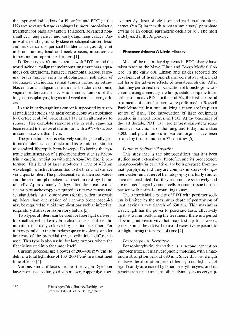
BOIS:L08:BPRR3022PD.98 FF: RESF E1: RESS
160 Häussinger/Diaz-Jiménez/Rodriguez/Stanzel/Huber/Pichler/Baumgartner
the approved indications for Photofrin and PDT (in theUS) are: advanced-stage esophageal tumors, prophylactictreatment for papillary tumors (bladder), advanced non-small cell lung cancer and early-stage lung cancer. Ap-proval is pending in: early-stage esophageal cancer, headand neck cancers, superficial bladder cancer, as adjuvantin brain tumors, head and neck cancers, intrathoracictumors and intraperitoneal tumors [1].
Different types of tumors treated with PDT around theworld include: malignant melanoma, angiosarcoma, squa-mous cell carcinoma, basal cell carcinoma, Kaposi sarco-ma; brain tumors such as glioblastoma; palliation ofesophageal carcinoma; retinal tumors including retino-blastoma and malignant melanoma; bladder carcinoma;vaginal, endometrial or cervical tumors; tumors of thetongue, nasopharynx, larynx and vocal cords, among oth-ers.
Its use in early-stage lung cancer is supported by sever-al published studies, the most conspicuous was publishedby Cortese et al. [4], presenting PDT as an alternative tosurgery. The complete response rate in early stage hasbeen related to the size of the tumor, with a 97.8% successin tumor size less than 1 cm.
The procedure itself is relatively simple, generally per-formed under local anesthesia, and its technique is similarto standard fiberoptic bronchoscopy. Following the sys-temic administration of a photosensitizer such as Photo-frin, a careful irradiation with the Argon-Dye laser is per-formed. This kind of laser produces a light of 630 nmwavelength, which is transmitted to the bronchial surfacevia a quartz fiber. The photosensitizer is then activated,and the resultant photochemical reaction destroys tumo-ral cells. Approximately 2 days after the treatment, aclean-up bronchoscopy is required to remove mucus andcellular debris usually too viscous for the patient to coughup. More than one session of clean-up bronchoscopiesmay be required to avoid complications such as infection,respiratory distress or respiratory failure [5].
Two types of fibers can be used for laser light delivery:for small superficial early bronchial cancers, surface illu-mination is usually achieved by a microlens fiber. Fortumors parallel to the bronchoscope or involving smallerbranches of the bronchial tree, a cylindrical diffuser isused. This type is also useful for large tumors, where thefiber is inserted into the tumor itself.
Current protocols use a power of 200–400 mW/cm2 todeliver a total light dose of 100–200 J/cm2 in a treatmenttime of 500 s [5].
Various kinds of lasers besides the Argon-Dye laserhave been used so far: gold vapor laser, copper dye laser,
excimer dye laser, diode laser and yttrium-aluminium-garnet (YAG) laser with a potassium titanyl phosphatecrystal or an optical parametric oscillator [6]. The mostwidely used is the Argon-Dye.
Photosensitizers: A Little History
Most of the major developments in PDT history havetaken place at the Mayo Clinic and Tokyo Medical Col-lege. In the early 60s, Lipson and Baldes reported thedevelopment of hematoporphyrin derivative, which didnot have the adverse effects of hematoporphyrin. Afterthat, they performed the localization of bronchogenic car-cinoma using a mercury arc lamp, establishing the foun-dations of today’s PDT. In the mid 70s, the first successfultreatments of animal tumors were performed at RoswellPark Memorial Institute, utilizing a xenon arc lamp as asource of light. The introduction of laser equipmentresulted in a rapid progress in PDT. At the beginning ofthe last decade, PDT was used to treat early-stage squa-mous cell carcinoma of the lung, and today more than3,000 malignant tumors in various organs have beentreated by this technique in 32 countries [6].
Porfimer Sodium (Photofrin)This substance is the photosensitizer that has been
studied most extensively. Photofrin and its predecessor,hematoporphyrin derivative, are both prepared from he-matoporphyrin, and they are complex mixtures of oligo-meric esters and ethers of hematoporphyrin. Early studieshave demonstrated that they accumulate selectively andare retained longer by tumor cells or tumor tissue in com-parison with normal surrounding tissues.
The tumoricidal capacity of PDT with porfimer sodi-um is limited by the maximum depth of penetration oflight having a wavelength of 630 nm. This maximumwavelength has the power to penetrate tissue effectivelyup to 3–5 mm. Following the treatment, there is a periodof skin photosensitivity that may last up to 6 weeks;patients must be advised to avoid excessive exposure tosunlight during this period of time [7].
Benzoporphyrin DerivativeBenzophorphyrin derivative is a second generation
photosensitizer. It is a hydrophobic molecule, with a max-imum absorption peak at 690 nm. Since this wavelengthis above the absorption peak of hemoglobin, light is notsignificantly attenuated by blood or erythrocytes, and itspenetration is maximal. Another advantage is its very rap-
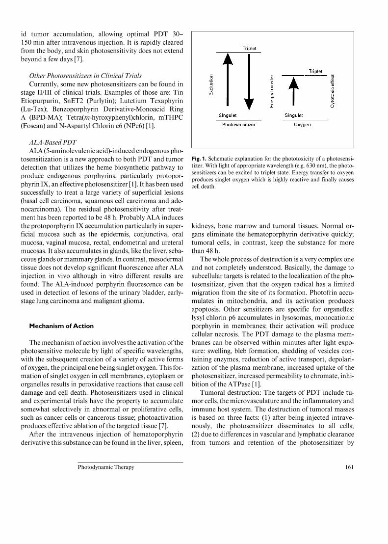
BOIS:L08:BPRR3022PD.98 FF: RESF E1: RESS
Photodynamic Therapy 161
id tumor accumulation, allowing optimal PDT 30–150 min after intravenous injection. It is rapidly clearedfrom the body, and skin photosensitivity does not extendbeyond a few days [7].
Other Photosensitizers in Clinical TrialsCurrently, some new photosensitizers can be found in
stage II/III of clinical trials. Examples of those are: TinEtiopurpurin, SnET2 (Purlytin); Lutetium Texaphyrin(Lu-Tex); Benzoporphyrin Derivative-Monoacid RingA (BPD-MA); Tetra(m-hyroxyphenyl)chlorin, mTHPC(Foscan) and N-Aspartyl Chlorin e6 (NPe6) [1].
ALA-Based PDTALA (5-aminolevulenic acid)-induced endogenous pho-
tosensitization is a new approach to both PDT and tumordetection that utilizes the heme biosynthetic pathway toproduce endogenous porphyrins, particularly protopor-phyrin IX, an effective photosensitizer [1]. It has been usedsuccessfully to treat a large variety of superficial lesions(basal cell carcinoma, squamous cell carcinoma and ade-nocarcinoma). The residual photosensitivity after treat-ment has been reported to be 48 h. Probably ALA inducesthe protoporphyrin IX accumulation particularly in super-ficial mucosa such as the epidermis, conjunctiva, oralmucosa, vaginal mucosa, rectal, endometrial and ureteralmucosas. It also accumulates in glands, like the liver, seba-ceous glands or mammary glands. In contrast, mesodermaltissue does not develop significant fluorescence after ALAinjection in vivo although in vitro different results arefound. The ALA-induced porphyrin fluorescence can beused in detection of lesions of the urinary bladder, early-stage lung carcinoma and malignant glioma.
Mechanism of Action
The mechanism of action involves the activation of thephotosensitive molecule by light of specific wavelengths,with the subsequent creation of a variety of active formsof oxygen, the principal one being singlet oxygen. This for-mation of singlet oxygen in cell membranes, cytoplasm ororganelles results in peroxidative reactions that cause celldamage and cell death. Photosensitizers used in clinicaland experimental trials have the property to accumulatesomewhat selectively in abnormal or proliferative cells,such as cancer cells or cancerous tissue; photoactivationproduces effective ablation of the targeted tissue [7].
After the intravenous injection of hematoporphyrinderivative this substance can be found in the liver, spleen,
Fig. 1. Schematic explanation for the phototoxicity of a photosensi-tizer. With light of appropriate wavelength (e.g. 630 nm), the photo-sensitizers can be excited to triplet state. Energy transfer to oxygenproduces singlet oxygen which is highly reactive and finally causescell death.
kidneys, bone marrow and tumoral tissues. Normal or-gans eliminate the hematoporphyrin derivative quickly;tumoral cells, in contrast, keep the substance for morethan 48 h.
The whole process of destruction is a very complex oneand not completely understood. Basically, the damage tosubcellular targets is related to the localization of the pho-tosensitizer, given that the oxygen radical has a limitedmigration from the site of its formation. Photofrin accu-mulates in mitochondria, and its activation producesapoptosis. Other sensitizers are specific for organelles:lysyl chlorin p6 accumulates in lysosomas, monocationicporphyrin in membranes; their activation will producecellular necrosis. The PDT damage to the plasma mem-branes can be observed within minutes after light expo-sure: swelling, bleb formation, shedding of vesicles con-taining enzymes, reduction of active transport, depolari-zation of the plasma membrane, increased uptake of thephotosensitizer, increased permeability to chromate, inhi-bition of the ATPase [1].
Tumoral destruction: The targets of PDT include tu-mor cells, the microvasculature and the inflammatory andimmune host system. The destruction of tumoral massesis based on three facts: (1) after being injected intrave-nously, the photosensitizer disseminates to all cells;(2) due to differences in vascular and lymphatic clearancefrom tumors and retention of the photosensitizer by

BOIS:L08:BPRR3022PD.98 FF: RESF E1: RESS
162 Häussinger/Diaz-Jiménez/Rodriguez/Stanzel/Huber/Pichler/Baumgartner
Table 1. Therapeutic response rates of Photofrin PDT and Nd:YAGtreatment: US study (P17), European study (P503)
US study (P17)
Photofrin%
Nd:YAG%
European study (P503)
Photofrin%
Nd:YAG%
n 33 37 69 72CR + PR week 1 45 51 65 61CR + PR month 1 42 19 61 36
CR = Complete response: absence of endoscopically visibletumor; PR = partial response: relative increase of 650% from base-line in the smallest luminal diameter or a decrease of 650% in theendobronchial obstruction.
tumoral cells, the photosensitizer is selectively retained inthe tumor cells and interstitial tissue of the tumor, so thatafter 2 days there is a greater concentration of the photo-sensitizer in the tumor than in the surrounding tissue;(3) the photosensitizer will absorb light energy, and pro-duce singlet oxygen, which destroys the tumor [7]. Thisreaction is known as photodynamic reaction, and is themain responsible of the tumor destruction (fig. 1).
Some other events are coming into focus, such as anti-tumor activity of inflammatory cells and tumor-sensitizedimmune reaction.
Both can be elicited by the phototoxic damage [7], andcontribute to a more complete tumoral cell destruction.
Some factors can limit the direct tumor cell kill, such asan inhomogeneous photosensitizer distribution withinthe tumor and the availability of oxygen.
The application of some drugs can also affect the over-all result of PDT treatment. Two drugs are known to mod-ify the PDT response: adriamycin [8] and glucocorticoids[9, 10]. Both improve the effects of the therapy; the gluco-corticoids increase the area of tumoral necrosis whenadministered 24 h after photoradiation.
PDT: Palliation of Lung Cancer
More than 50% of patients with nonsurgical lung can-cer die from local complications such as asphyxia, hemop-tysis, pneumonia, empyema. Publications state that 36%of patients died from similar causes, regardless of thepresence or absence of prior surgery. A same cause ofdeath in 58% of patients with surgery versus 83% ofpatients without was reported in another study [11]. Con-
sidering that only 30% of patients with bronchogenic car-cinoma are surgical candidates at the time of diagnosis, itcan be assumed that most inoperable patients will requirepalliative therapy at some point during the clinical courseof the disease.
However, the use of PDT as a palliative treatment forpatients with inoperable obstructive bronchial cancersmust be assessed in the context of what may be achievedwith conventional treatments. In the Nd:YAG laser thera-py, the laser is used to coagulate and remove tumoral tis-sue. The treatment is usually delivered under generalanesthesia and is highly effective in relieving airwayobstruction, especially in tumors centrally located. Fatalhemorrhage, respiratory failure or cardiac arrest are se-vere complications of this treatment, but the incidence isvery low (1.5%) and nonfatal complications occur in lessthan 0.5% of the cases [12]. Laser therapy is considered bymany experts the gold standard for the palliative treat-ment of partially or totally obstructing, centrally locatedtumors of the airways, primary or metastatic.
There is plenty of published clinical experience to sup-port this concept [12, 13]. Nonetheless, PDT is a usefulpalliative method that can have some advantages overlaser therapy, particularly in tumors which are moreperipherally located. Also, a more complete tumoral de-struction and a better survival have been reported inmany studies comparing laser photoresection versusPDT. Let us analyze some published reports.
Clinical DataTwo prospective, randomized trials of PDT versus
Nd:YAG laser ablation for partially obstructive lung can-cer have been reported [14]. These included data from 16centers in Europe and 20 centers in the US and Canada.In the European trial, 40% of the patients had prior treat-ment, while all patients in the US and Canada had priortherapy. Tumor response was similar for both therapies at1 week; but at 1 month 61 and 42% of the PDT patientsshowed sustained response in the European and US/Can-ada trials, respectively, whereas for the Nd:YAG group,the respective numbers were 36 and 19% only (table 1).
There were 12 and 6% of PDT patients versus 3 and5% of Nd:YAG patients who achieved complete, biopsy-proven response in the European and US/Canada trials,respectively. Improvement in dyspnea and cough weresuperior for PDT over Nd:YAG in the European group,but similar in the US/Canada group. It was concludedthat PDT is superior to Nd:YAG for relief of dyspnea,cough and hemoptysis [14]. Overall, adverse reactionswere similar in both groups and 20% of patients in the

BOIS:L08:BPRR3022PD.98 FF: RESF E1: RESS
Photodynamic Therapy 163
Table 2. Comparative survival rates for PDT, surgery and radiother-apy
Clinicalstage
Surgerymonths
Radiationmonths
PDT (KPS 150)months
PDT (KPS !50)months
I 42 19 not reached NAII 17.5 18 22.5 NAIIIA 12 12 8 2IIIB 7.5 10 7 4IV 5 – 7 3
KPS = Karnofsky Performance Status; NA = not applicable.
PDT group had photosensitivity reaction due to noncom-pliance with precautions [1].
A recently published prospective 14-year study [15]reported the evolution of 175 patients with primary endo-bronchial or tracheal squamous cell carcinoma or adeno-carcinoma. Patients had failed, refused or were ineligiblefor conventional treatment. The results showed that sur-vival was most significantly affected by the stage of thedisease. When compared with other treatment regimens(surgery and radiotherapy), survival results were as pre-sented in table 2 (modified from McCaughan and Wil-liams [15]).
Analysis of the time after treatment required for reob-struction of the bronchus after YAG laser versus PDTshowed that the immediate results were better after YAGlaser treatment, but after the clean-up bronchoscopy, thedegree of obstruction was the same, and the sites treatedreobstructed faster after Nd:YAG laser therapy (2 weeksfor YAG, 4 weeks for PDT).
A randomized study in the US comparing the efficacyand safety of PDT versus Nd:YAG laser showed that bothtreatments were equally effective in relieving endobron-chial obstruction by tumor. The time to treatment failurewas slightly longer after PDT than after Nd:YAG laserresection, and the risk of local recurrence after PDT wasonly 0.3 of that after Nd:YAG [16].
Another prospective trial of PDT plus radiotherapy,versus radiotherapy alone presented 41 patients. Resultsshowed that the obstructive airway was completely openin only 10% of the patients treated only with radiotherapyversus 70% of patients when the combination was used.Twenty percent of patients failed with either treatment[17].
A group of 10 patients with inoperable non-small cellcarcinoma presenting different degrees of tracheobron-chial obstruction (86 B 2%) showed a 150% response in 4and !50% response in 6 patients, respectively. However,in all patients symptoms improved, especially coughing.Side effects included burns in 2 patients and mild anasar-ca in 1 [18].
Our own experience with 31 patients suffering frominoperable non-small cell cancer with variable degree ofobstruction yielded similar results. We prospectively ran-domized patients to receive either PDT or Nd:YAG laser,finding that the immediate response was better in theNd:YAG laser group, but the duration of the response wasmore prolonged in the PDT group, with a better survivalrate. The number of complete biopsy-proven responseswas low and short in duration. Palliation of symptoms aswell as Karnofsky index was similar in both groups. The
PDT group had a higher incidence of severe side effects,photosensitivity being the most important one [Diaz-Jiménez et al., unpubl. data].
Indications and Contraindications for PDT
Indications for PDT(1) Relief of neoplastic endobronchial obstruction [1,
14–21]; (2) delay of tumor progression and improvementof symptoms [1, 14–16]; (3) making inoperable patientsoperable in selected cases [18, 20–22].
Complications of PDT(1) Dyspnea, worsening obstruction due to edema, mu-
cus plugs or atelectasis; respiratory insufficiency; (2) fever(reported incidence 20%); (3) infections: bronchitis, post-obstructive pneumonia, due to edema and impaired coughmechanisms; (4) fatal hemoptysis; (5) photosensitivity;(6) allergic or toxic reaction to the photosensitizer.
Contraindications for PDT(1) Tracheal lesions, carinal lesions, pneumonectom-
ized patients; the edema resulting from the treatment cantrigger respiratory insufficiency; (2) erosion or invasion ofvascular structures; (3) porphyry or porphyrin hypersensi-tivity.
PDT: Curative Treatment of SuperficialEndobronchial Tumors
Patients with central lung cancer have limited treat-ment options and poor prognosis. Many of these patientshave chronic obstructive pulmonary disease or coexisting

BOIS:L08:BPRR3022PD.98 FF: RESF E1: RESS
164 Häussinger/Diaz-Jiménez/Rodriguez/Stanzel/Huber/Pichler/Baumgartner
Fig. 2. Different forms of applicators used for curative PDT.
cardiovascular diseases. Surgery and high-dose radiother-apy are the standard therapeutic options for early-stagelung cancer either primary or recurrent. Even though ear-ly-stage endobronchial tumors are small by definition, alobectomy is required in approximately 70% of cases. Inthe remaining 30% bilobectomy or pneumonectomy isrequired [23]. Often the reasons for which a patient isinoperable also preclude the use of radiotherapy. In pa-tients with reduced pulmonary reserve, radiotherapy mayfurther compromise lung function due to pneumonitisand fibrosis [24]. Because of the limited pulmonary func-tion in many patients with lung cancer and the frequencyof second primary cancer [25, 26] a treatment that willminimize loss of lung tissue is clearly desirable. Patientswho cannot be treated with surgery or radiotherapy arelimited to experimental treatments such as chemotherapyor immunotherapy or endobronchial treatment modali-ties such as endobronchial brachytherapy, cryotherapy orphotodynamic therapy. PDT offers a potentially effective,minimally invasive treatment option and has been shownto be effective in selected patients with early-stage non-small cell lung cancer. The advantage of PDT over otherforms of curative treatment of malignant tumors, e.g. sur-gery or radiation therapy lies in its selectivity. Therefore,treatment does not cause any loss of function. Its radicali-
Table 3. Results of PDT in early lung cancer
Maximum dimension Lesions CR
Superficial type (123 lesions in 110 patients)!0.5 cm 64 61 (95)!1.0 cm 25 22 (88)!2.0 cm 20 9 (45)12.0 cm 14 6 (43)
Nodular type (45 nodular cancers in 43 patients)!0.5 cm 29 27 (93)!1.0 cm 9 6 (67)11.0 cm 7 1 (14)
Figures in parentheses are percentages. CR = Histologically con-firmed complete remission.
ty, however, will remain a controversal issue in spite ofthe great number of studies [26–35, 42–44] that havealready been published. Study designs varied greatly, acomparison of the results therefore is problematic.
For treatment a laser with 630 nm emission light, 2 Woutput power and specific light applicators according tothe irradiated tumor area are necessary (fig. 2).
The total dose should be 200 J/cm2. The irradiationtime has to be calculated from the irradiated area andtotal output light power of the applicator (typically fixedto 400 mW).
Clinical DataPhase I–II clinical trials in Japan by Hayata et al. [32,
33] showed that approximately 90% of superficial tumorsless than 1 cm in diameter can be completely eradicatedwith PDT. Similar results were achieved in patients withnodular tumors less than 0.5 cm in diameter. Of 81patients who had a complete response after PDT, only 2had died from primary disease at the time of follow-upevaluation. Fifteen patients were still alive and diseasefree 5 years after treatment. Three patients were alive anddisease free more than 10 years after treatment. The over-all complete response rate after PDT was 71%.
A phase III clinical trial completed recently in Japanshowed similar results. The complete remission (CR) rate,however, decreased significantly when treating larger le-sions. Hayata [34] summarized the results using PDT totreat 168 early-stage cancers of the lung and esophagus in150 patients, differentiating the lesions treated either intoa superficial type or a nodular type (table 3).

BOIS:L08:BPRR3022PD.98 FF: RESF E1: RESS
Photodynamic Therapy 165
Fig. 3. a Squamous cell carcinoma obstructing bronchus intermedius – before PDT. b Three days after PDT – necrot-ic tissue after interstitial irradiation. c Three days after PDT – necrotic tissue after removal by forceps.
a b c
Table 4. Tumor characteristics and reasons for nonoperability
n %
Tumor characteristics (102 patients)Squamous cell carcinoma 87 85Tis 23 23T1 63 62T2/T3 8 8Radiologically occult 90 88
Reasons for nonoperabilityPrior lung resection 48 47Poor pulmonary function 43 42Multilobar tumors 20 20Central location 11 11Old age 5 5Refused surgery 4 4Multiple reasons 61 60
Table 5. Response rate, survival and recurrence
Response rate, survival (100 patients)Total CR 79 (79%)95% CI, % 71–87Median survival, years 3.5Median disease-specific survival, years 5.7
Tumor recurrence after first CR (79 patients)Recurrence 35 (44%)Median TTR, years 2.8Range, years 0.1–10.1
CR = Histologically confirmed complete remission; TTR = timeto tumor recurrence.
Complete remission was achieved in 95% of the super-ficial type lesions less than 0.5 cm in diameter and in 88%of the lesions between 0.5 and 1.0 cm in diameter. Therate of CR decreased significantly in lesions larger than1.0 cm in diameter. Recurrence developed in only 4patients whose lesions had been less than 1.0 cm in diam-eter. Of the 83 CR cases treated with PDT alone, 3patients died of the original tumor and 47 patients sur-vived. Of these, 10 patients were disease free 5 years orlonger after treatment.
In the lesions of the nodular type, CR was achieved in93% of the lesions less than 0.5 cm in diameter and in67% of the lesions between 0.5 and 1.0 cm in diameter. Ofthe 32 CR cases, 8 died of unrelated diseases and 23
patients survived. Of these 23 patients, 12 have survived5 years or more.
Three open-labeled single arm studies which assessedthe safety and efficacy of PDT with Photofrin were real-ized in Germany, France, the Netherlands and Canada.Their results led to the approval of the use of Photofrin inPDT for early superficial lung cancer in patients who arenot eligible for surgery or radiotherapy [35]. A total of 102patients were treated in these three trials. The indicationswere carcinoma in situ, microinvasive tumors not invad-ing the cartilage by histological assessment and patientswho were not considered candidates for surgery.
Figure 3 shows a typical example of PDT treatment.Table 4 [35] presents the patient and tumor character-
istics of the study population and reasons why thesepatients were not candidates for surgery.
Table 5 summarizes the overall results [35]. Histologi-cally confirmed complete response was achieved in 79%

BOIS:L08:BPRR3022PD.98 FF: RESF E1: RESS
166 Häussinger/Diaz-Jiménez/Rodriguez/Stanzel/Huber/Pichler/Baumgartner
Fig. 4. Patient survival after PDT in superficial early lung cancer (n =100 patients).
Fig. 5. Disease-specific survival (DDS) after PDT in early lung can-cer (n = 79 patients).
Table 6. PDT in roentgenologically occult lung cancer: studies
Reference Lesions ResponseCR, %
Survivalmonths
Monnier et al., 1990 [42] 16 69 3–60Okunaka et al., 1991 [26] 27 98 mean 38Edell and Cortese, 1992 [43] 14 71 7–49Furuse et al., 1993 [30] 59 83 14–32Imamura et al., 1994 [31] 39 64 4–169Sutedja et al., 1994 [44] 39 72 2–95
CR = Histologically confirmed complete remission.
of patients with early-stage superficial nonsmall cell lungcancer (95% confidence interval 71–87%).
Median survival was 3.5 years (fig. 4) [35] and mediandisease specific survival was 5.7 years (fig. 5) [35].
These data were comparable to or better than thoserates achieved with radiotherapy for stage I disease withcomplete response rate of 52–70%, a median survival of72–48 months [36–40] and better than historic results ofuntreated patients [41]. The most frequent local adverseevents were related to photosensitivity reactions (23%)which were usually mild to moderate sunburn-like reac-tions. Other adverse events included pulmonary effectsrelated to the local pharmacological action of treatment
which resulted in mucus exudate (23%), local edema(18%), stricture due to scars (10%) and ulcerations (9%).Perforations did not occur.
Table 6 summarizes the results of PDT in superficialendobronchial lung cancers of several studies which havebeen reported in the last decade. The numbers of thepatients are low because these early lesions caused nosymptoms and were mostly choice discoveries during rou-tine bronchoscopies [26, 30, 31, 42–44].
In our center, PDT has been performed in 39 patientswith 52 carcinomas including 47 invasive tumors and 5CIS between April 1987 and July 1997. All lesions wereradiologically occult and all patients deemed functionallyor technically inoperable. In the immediate effect, wefound a histology-proven curative rate of 92%. In thelong-term follow-up (median time 25.7 months) 64% ofthese patients remained tumor free and 36% had a localrelapse proven by endoscopic routine controls which wereperformed every 6 months. Finally, the overall cure ratewas 59% [45]. The efficacy of preoperative PDT has beenevaluated by Kato et al. [46]. A typical treatment of one ofthese patients is shown in figure 6. Tumors arising froman upper lobe bronchus or from the bronchus intermediussometimes have superficial extension to the mainstembronchus or lower trachea and therefore are either unre-sectable or would require pneumonectomy to remove.Following preoperative PDT, 4 of 5 patients with initiallyunresectable tumors were rendered operable and from 10

BOIS:L08:BPRR3022PD.98 FF: RESF E1: RESS
Photodynamic Therapy 167
Fig. 6. a Squamous cell carcinoma in theright tracheobronchial wall, beginning of theright main stem bronchus – radiologicallyoccult. b During irradiation with 200 J/cm2
– direct contact of the irradiator with thetumor surface. c Three days after PDT:slight edema, thin fibrin plaque. d Long-term follow-up 5 years after PDT: Normalmucosa without any sign of local recur-rence.
a b
c d
patients who were originally candidates for pneumonecto-my, 7 became eligible for less extensive surgery.
Edell and Cortese [43] at the Mayo Clinic stated thatPDT may be an alternative to surgery. In a preliminarystudy of 30 patients, a total of 14 cancers were identifiedand treated with PDT as first-line intervention. Completeeradication after PDT was achieved in 13 of 14 tumors(93%). Only 3 of the 13 had local recurrences at 7–18months after PDT. Overall, 10 of the 13 cancers thatshowed a complete response have not recurred in up to 5years of follow-up. In all, 10 of 13 patients (77%) werespared a surgical resection. Three patients with persistentdisease after PDT underwent surgical resection and hadsurgical stage T1-N0-M0.
Conclusions
Palliative TreatmentPDT has proven to be effective in palliation. The first
treatment with PDT was performed at the beginning ofthe 80s, since then the number of treatments has beenincreasing every day.
The clinical data available at the moment usually com-pare the effectiveness of PDT versus Nd:YAG laser thera-py, which has been used for palliation since the 70s, and itis considered by some the gold standard in that matter.Both PDT and YAG laser resection are effective in reliev-ing airway obstruction caused by intraluminal tumors.Nd:YAG laser photoresection seems to be better in tu-mors centrally located, where they are easily reached bythe rigid bronchoscope, coagulated and then resected.PDT, on the other hand, is applied by flexible broncho-scope, which can reach tumors more peripherally locatedand does not require mechanical resection immediately

BOIS:L08:BPRR3022PD.98 FF: RESF E1: RESS
168 Häussinger/Diaz-Jiménez/Rodriguez/Stanzel/Huber/Pichler/Baumgartner
after the light irradiation. Repeated clean-up bronchosco-pies, however, are needed some days after PDT treatmentto remove detritus.
Since most patients with airway obstruction have acombination of intraluminal and submucosal or peribron-chial tumor, it seems reasonable to use a combination ofPDT and external radiation [17]. In very selected cases,PDT can also be used as palliative treatment for openingstenotic or obstructed bronchi due to tumor prior to thecombination therapy with surgery [19, 46].
As a single treatment modality in patients with ad-vanced disease presenting tracheobronchial obstruction,it seems that PDT still has a limited role given the avail-ability of other treatment modalities, similarly or equallyvalid, particularly laser photoresection and brachythera-py, which are simpler to perform.
Additionally, some disadvantages have to be pointedout. PDT is not suitable for patients with tracheal lesionsor lesions that compromise both main bronchus or pneu-monectomized patients. The sometimes severe edema fol-lowing the therapy can worsen the obstruction and makeit complete, endangering life.
When the tumor has infiltrated the walls or vascularstructures, the use of PDT can cause perforation and/orfatal hemorrhage.
Another disadvantage is that PDT does not imme-diately relieve the airway obstruction, and therefore, pa-tients presenting with acute respiratory distress due totumor obstruction are better treated with Nd:YAG laserresection.
Curative TreatmentFor patients with central early-stage lung cancers, who
are no candidates for external radiotherapy or surgerydue to functional reasons, PDT offers a new and effec-tive treatment modality with curative chance. In contrastto other unspecific local therapies, e.g. endobronchialbrachytherapy, cryotherapy, electrocoagulation and lasercoagulation by thermal effects, PDT is based on a se-lective death of tumor cells with subsequent necrosis ofthe irradiated tumor lesion. As all local methods, PDT islimited to centrally located lesions within the endoscopicview and to penetration depths of only some millimeters.The local tumoricidal effect of the above mentionedunspecific treatment modalities has been reported only insome few case reports. The curative effect of PDT in earlysuperficial cancers, however, has been studied systemati-cally and has been documented in several phase II and IIIstudies. Since 1980, more than 800 patients have beentreated.
The curative results of PDT range between 80 and100% for the immediate effect (i.e. histologically con-firmed complete remission within 3 weeks after PDT) and50 and 60% for the long-term follow-up.
Main factors of influence are tumor size and penetra-tion into depth. Successful application of PDT also de-pends on the ability to visualize the entire extent of thelesion bronchoscopically.
Informations about tumor geometry therefore are im-portant. Autofluorescence bronchoscopy and broncho-scopic ultrasound have been developed for practical userecently and will be useful for assessing local tumor sizeand depth in roentgenologically occult lung cancer [47,48]. In our experience, indication for PDT should be dif-ferentiated: CIS is an exclusive indication for a first-lineintervention. Microinvasive carcinoma is an optional in-dication, treatment should be performed only in inopera-ble or high-risk patients. Invasive carcinoma is an indica-tion for PDT only in highly selected inoperable patientswith roentgenologically occult lesions which cannot bedetected even by (high-resolution) CT scan. Severe dys-plasia is not an indication for endoscopic intervention upto now.
One of the reasons for long-term failure is the high inci-dence for development of second primary tumors in lungcancer patients. Therefore, all patients treated shouldundergo regular bronchoscopy follow-up both for localcontrol and exclusion of metachronous tumor lesionswhich could be treated by PDT too.
Photosensitivity is a main problem of PDT. The pho-tosensitizing agent used in most clinical protocols is Pho-tofrin, whose photosensitivity may last up to 3 months.Therefore, bronchoscopic intervention is limited to aperiod of 2 weeks after intravenous application of Photo-frin, which must not be repeated within at least 2–3months. The protection of eyes and skin is essential, andthe patients must avoid exposure to light, particularlysunlight for a period of 4–8 weeks [49]. It is expected thatwith the second-generation photosensitizers, this will de-crease considerably.
Developments, Future AspectsA new field of investigation is concerning new genera-
tion photosensitizers with an improved tumor accumula-tion and a decreased photosensitivity of the skin. Thismight improve efficacy and decrease both local and gener-al side effects of PDT.

BOIS:L08:BPRR3022PD.98 FF: RESF E1: RESS
Photodynamic Therapy 169
Additionally, there is a need for better and cheaperlight sources. Lasers might be replaced by conventionallight sources with integrated filters for the emission of dif-ferent wavelengths to obtain better penetration and a spe-cific absorption of light by the sensitizer used. Additional-ly there is a need for better light delivery systems and lightdistributors. Thin flexible fibers should be used only in
the smaller bronchi. Irradiation of more centrally locatedlesions should be performed by special light distributorsadapted to the complex geometry of the bronchial tree toimprove dosimetry. In summary, all these efforts couldhelp to improve practicability, efficacy and safety of thisnewly developed treatment modality and to establishPDT in further clinical use.
References
1 Dougherty TJ, Gomer CJ, Henderson BW, JoriG, Kessel D, Korbelik M, Moan J, Peng Q:Photodynamic therapy. J Natl Cancer Inst1998;90:889–905.
2 Sutedja G, Postmus PE: Bronchoscopic treat-ment of lung tumors. Lung Cancer 1994;11:1–17.
3 Reynolds T: Using lasers and light-activateddrugs, researchers home in on early lung can-cers. J Natl Cancer Inst 1998;90:411–418.
4 Cortese DA, Edell ES, Kinsey JH: Photody-namic therapy for early stage squamous cellcarcinoma of the lung. Mayo Clin Proc 1997;72:595–602.
5 Lam S: Photodynamic therapy in lung cancer.Semin Oncol 1994;21:15–19.
6 Kato H: Photodynamic therapy for early stagecentral type of lung cancer – Editorial. Chest1997;72:688–690.
7 Levy J: Photosensitizers in photodynamic ther-apy. Semin Oncol 1994;21:4–10.
8 Edell ES, Cortese DA: Potentiation of hemato-porphyrin derivative phototherapy with adria-mycin. Porphyrin photosensitization Work-shop. Abstract form IPA Los Angeles, July1986, p 15.
9 Edell ES, Cortese DA: Interaction between glu-cocorticosteroids and hematoporphyrin deriv-ative phototherapy. Porphyrin photosensitiza-tion Workshop (abstract). IPA Los Angeles,July 1986, p 16.
10 Cowled PA, Maekenzie L, Forbes M: Potentia-tion of photodynamic therapy with haemato-porphyrin derivatives by glucocorticoids. Can-cer Lett 1986;29:101–114.
11 Carroll M, Morgan SA, Yarnold JR, Hill JM,Wright NM: Prospective evaluation of a watchpolicy in patients with inoperable non-smallcell cancer. Eur J Cancer Clin Oncol 1986;22:1352–1356.
12 Personne C, Colchen A, Leroy M, Vourc’h G,Toty L: Indications and technique for endo-scopic laser resections in bronchology. A criti-cal analysis based upon 2,284 resections. JThorac Cardiovasc Surg 1986;91:710–715.
13 Diaz-Jiménez JP, Canela Cardona M, MaestreAlcacer J, Balust Vidal M, Fontanals Tortra J:Fotoreseccion con laser en 68 casos de patolo-gia traqueo-bronquial. Rev Clin Esp 1987;180:199–202.
14 Wieman TJ, Diaz-Jiménez JP, Moghissi K, Le-roy M, McCaughan J, Spinelli P, Lang N, Diaz-Agero P, York E, the Photodynamic TherapyLung Cancer Study Group Louisville, KY,USA/Barcelona, Spain: Photodynamic therapy(PDT) with Photofrin is effective in the pallia-tion of obstructive endobronchial lung cancer:Results of two clinical trials (abstract). ASCO1998.
15 McCaughan JS, Williams TE: Photodynamictherapy for endobronchial malignant disease: Aprospective fourteen-year study. J Thorac Car-diovasc Surg 1997;114:940–947.
16 McCaughan JS: Photodynamic Therapy versusNd-YAG laser treatment of endobronchial oresophageal malignancies; in PhotodynamicTherapy and Biomedical Lasers. Proceedingsof the International Conference on Photody-namic Therapy and Medical Laser Applica-tions. Milan 1992, pp 23–36.
17 Lam S, Cory P: Combined photodynamic ther-apy (PDT) using Photofrin and radiotherapy(XRT) versus radiotherapy alone in patientswith inoperable distribution non-small cellbronchogenic cancer. Proc SPIE 1991;5:20–28.
18 LoCicero J, Metzdorff M, Almgren C: Photo-dynamic therapy in the palliation of late stageobstructing non-small cell lung cancer. Chest1990;98:97–100.
19 Hayata Y, Kato H, Konaka C, Ono J, TakizawaN: Hematoporphyrin derivative and laser pho-toradiation in the treatment of lung cancer.Chest 1982;81:269–277.
20 McCaughan JS Jr, Williams TE Jr, Bethel BH:Photodynamic therapy of endobronchial tu-mors. Lasers Surg Med 1986;6:336–345.
21 Balchum OJ, Doiron DR: Photoradiation ther-apy of endobronchial lung cancer: Large ob-structing tumors, non-obstructing tumors, andearly stage bronchial cancer lesions. Clin ChestMed 1985;6:255–275.
22 Kato H, Konaka C, Kawate N, Shinohara H,Kinoshita K, Noguchi M, Ootomo S, HayataY: Five-year disease-free survival of a lung can-cer patient treated only by photodynamic ther-apy. Chest 1986;90:768–770.
23 Cortese DA, Pairolero PC, Bergstrahl EJ, et al:Roentgenographically occult lung cancer. Aten-year experience. J Thorac Cardiovasc Surg1983;86:373–380.
24 Gross NJ: Pulmonary effects of radiation ther-apy. Ann Intern Med 1977;86:81–92.
25 Pairolero PC, Williams DE, Bergstrahl EJ, etal: Postsurgical stage I bronchogenic carcino-ma: Morbid implications of recurrent disease.Ann Thorac Surg 1984;38:331–338.
26 Okunaka T, Kato H, Konaka C, et al: Photody-namic therapy for multiple primary broncho-genic carcinoma. Cancer 1991;68:253–258.
27 Edell ES, Cortese DA: Bronchoscopic photo-therapy with hematoporphyrin derivative fortreatment of localized bronchogenic carcino-ma: A 5-year experience. Mayo Clin Proc 1987;62:8–14.
28 Konaka C, Kato H, Hayata Y: Lung cancertreated by photodynamic therapy alone: Sur-vival for more than three years. Lasers Med Sci1987;2:17–19.
29 McCaughan JS, Hawley PhC, Bethel BH,Walker J: Photodynamic therapy of endobron-chial malignancies. Cancer 1988;62:691–701.
30 Furuse K, Fukoka M, Kato H, et al: A prospec-tive phase II study on photodynamic therapywith Photofrin for centrally located early stagelung cancer. J Clin Oncol 1993;11:1852–1857.
31 Imamura S, Kusunoki Y, Takifuji N, et al: Pho-todynamic therapy and/or external beam radia-tion therapy for roentgenologically occult lungcancer. Cancer 1994;73:1608–1614.
32 Hayata Y, Kato H, Konaka C, et al: Photody-namic therapy (PDT) in early stage lung can-cer. Lung Cancer 1993;9:287–294.
33 Hayata Y: The indications of photodynamictherapy in early stage lung cancer (abstract).Chest 1993;103:262.
34 Hayata Y, Kato H, Furuse K, Kusunoki Y,Suzuki S, Nimura S: Photodynamic therapy of168 early stage cancers of the lung and oesopha-gus: A japanese multicenter study. Lasers MedSci 1996;11:255–259.
35 Lam S, Häußinger K, Leroy M, Sutedja T,Huber RM: Photodynamic therapy (PDT) withPhotofrin, a treatment with curative potentialfor early stage superficial lung cancer (ab-stract). ASCO 1998.
36 Sandler HM, Curran WJ, Turrisi AT: The in-fluence of tumor size and treatment staging onoutcome following radiation therapy alone forstage I non small cell lung cancer. Int J RadiatOncol Bio Phys 1990;19:9–13.
BOIS:L08:BPRR3022PD.98 FF: RESF E1: RESS
170 Häussinger/Diaz-Jiménez/Rodriguez/Stanzel/Huber/Pichler/Baumgartner
37 Slotman BJ, Njo KH, Karim AB, et al: Cura-tive radiotherapy for technically operable stageI non-small cell lung cancer. Int J Radiat OncolBiol Phys 1994;29:33–37.
38 Haffty BG, Goldberg MB, Gerstley J, FischerDB, Peschel RE: Results of radical radiationtherapy in clinical stage I, technically operablenon-small cell lung cancer. Int J Radiat OncolBiol Phys 1988;15:69–73.
39 Ono R, Egawa S, Suemasu K, Sakusa M, Kito-gawa T: Radiotherapy for inoperable stage Ilung cancer. Jpn J Clin Oncol 1991;21:125–128.
40 Dosoretz DE, Katin MJ, Blitzer PH, Ruben-stein JH, Salenius S, Rashid M, et al: Radiationtherapy in the management of medically inop-erable carcinoma of the lung: Results and im-plications for future treatment strategies. Int JRadiat Oncol Biol Phys 1992;24:3–9.
41 Sobue T, Suzuki T, Matsuda M, et al: Survivalfor clinical stage I lung cancer not surgicallytreated. Cancer 1992;69:685–692.
42 Monnier PH, Savary M, Fontollet Ch, Wag-nieres G, Chatelain A, Cornaz P, DepeursingeCh, van den Bergh H: Photodetection and pho-todynamic therapy of ‘early’ squamous cell car-cinomas of the pharynx, oesophagus and tra-cheobronchial tree. Lasers Med Sci 1990;5:149–169.
43 Edell ES, Cortese DA: Photodynamic therapyin the management of early superficial squa-mous cell carcinoma as an alternative to surgi-cal resection. Chest 1992;102:1319–1322.
44 Sutedja G, Lam S, LeRiche C, Postmus PE:Response and pattern of failure after photody-namic therapy for intraluminal stage I lungcancer. J Bronchol 1994;1:295–298.
45 Häußinger K, Stanzel F, Sauer W: Photody-namic therapy of inoperable patients with earlystage lung cancer. Abstract World Congress forBronchology, June 1998, p 118.
46 Kato H, Konaka C, Ono J, et al: Preoperativelaser photodynamic therapy in combinationwith operation in lung cancer. J Thorac Car-diovasc Surg 1985;90:420–429.
47 Häußinger K, Stanzel F, Huber RM, Pichler J,Stepp H: Autofluorescence detection of bron-chial tumors with the D-Light/AF: Diagn TherEndosc 1999;5:105–112.
48 Becker HD: Endobronchialer Ultraschall –Eine neue Perspektive in der Bronchologie. En-dosk Heute 1995;1:55–56.
49 Dachowski LJ, DeLaney TF: Photodynamictherapy: The NCl experience and its nursingimplications. Oncol Nurs Forum 1992;19:63–67.
Prof. Dr. Karl HäussingerAsklepios Fachkliniken München-GautingZentrum für Pneumologie undThoraxchirurgie, Robert-Koch-Allee 2D–82131 Gauting (Germany)Tel. +49 89 85791 7301Fax +49 89 85791 7305
Jose Pablo Diaz-JiménezLaser Unit, Ciudad Sanitaria yUniversitaria de BellvitgeL’Hospitalet de Llobregat, Feixa Llarga s/nE–08907 Barcelona (Spain)Tel. +34 93 26 07 743, Fax +34 93 26 07 745E-Mail [email protected]

OOOOOOOOOOOOOOOOOOOOOOOOOOOOOOOOOOOOOOOOOOOOOOOOOOOOOOOOOOOOOOOOOOOOOOOOOOOOOOOOOOOOOOOOOOOOOOOOOOOOOOOOOOOOOOOOOOOOOOOOOOO
Therapeutic Bronchoscopy

Bolliger CT, Mathur PN (eds): Interventional Bronchoscopy.Prog Respir Res. Basel, Karger, 2000, vol 30, pp 171–186
OOOOOOOOOOOOOOOOOOOOOOOOOOOOOOOOOOOOOOOOOOOOOOOOOOOOOOOOOOOOOOOOOOOOOOOOOOOOOOOOOOOOOOOOOOOOOOOOOOOOOOOOOOOOOOOOOOOOOOOOOOO
Tracheobronchial StentsLutz Freitag
Lungenklinik Hemer, Germany
171
Summary
Airway stents have been in use for almost 90 years.Today they are indicated for: (1) reestablishing patency ofcompressed or strictured central airways; (2) supportingweakened cartilages in cases of tracheobronchial malacia,and (3) sealing of fistulas and dehiscences to the esophagusor the pleural cavity. For benign stenoses, surgery remainsthe gold standard for the time being. Stent placement is thelast option, and whenever possible a removable deviceshould be inserted. In cases of malignant tumors, only poly-mer stents or covered metal stent may be used in order toprevent ingrowth of tumor tissue. Debulking and dilatationshould always precede stent placement. Malacia is a relativeindication for stent therapy. The results are less favorable.Sufficient sealing of fistulas to the esophagus, the mediasti-num or the pleural cavity can be achieved with stents,though sometimes the commercially available models do notfit and they must be customized for a particular situation.Basically stents can be divided into four groups: (1) polymerstents such as the Dumon stent or the Polyflex stent;(2) metallic stents such as the Palmaz stent or the uncoveredWallstent; (3) covered metallic stents such as the coveredUltraflex stent and (4) hybrid stents such as the Dynamicstent. However, the response of the tissue and the outcomeof the procedure depend more on the biomechanical prop-erties of the prosthesis rather than the material used for itsconstruction. Type, size and length of the stent must be
carefully selected, considering the biomechanics of the air-way obstruction and of the endoprosthesis and always con-sidering the scenarios of possible complications. With allavailable stents granulation tissue formation may occur, thestents can get obstructed by incrusted secretions and allstents can migrate. Properly selected and used, airway stentsare already an invaluable addition to the armamentarium ofinterventional bronchoscopy and chances are high that evenbetter stents will be available in the foreseeable future.
About 100 years ago, the British dentist Charles R.Stent (1845–1901) invented a compound to cast dentalmodels and splints. In the years to come, his name wasused for various materials used to hold tissue in place orto provide support for a graft or anastomosis [1] . Today,surgeons and endoscopists insert stents into nearly all con-stricted tubular structures of the body, mainly to establisha sufficient lumen or to bridge a gap.
Historical Background
The implantation of an airway stent is by no means arecently developed procedure. Already in the last century,tubes had been surgically implanted by Trendelenburg [2]and Bond [3] for the treatment of airway strictures. In

BOIS:L08:BPRR3016PD.98 FF: RESF E1: RESS
172 Freitag
1915, Brünings and Albrecht [4], disciples of Gustav Kil-lian, the ‘Father of bronchoscopy’ implanted rubber pros-theses endoscopically into narrowed tracheas. In 1933,Canfield and Norton [5] used a ‘silver permanent dilatingtube’ to treat a 2-year-old child with bony stenosis of thelarynx. This is the first documented implantation of ametal stent. Various alloys such as stainless steel, tanta-lum, titanium and recently nitinol have been tried since.Many of these stents had originally been developed foruse in the vascular system, but were found to be adequatefor implantation into the central airways after only minormodifications, if any at all.
The polymer silicone has been established as the mostcommonly used stent material. In 1965, Montgomery [6]designed a silicone rubber T tube, which involved anexternal side limb protrusion through a tracheostomy.These stents have been widely applied to manage subglot-tic stenoses of various origins. They are still used todaywith minor modifications by ENT surgeons. The firststraight silicone stents without side limbs were inserted in1965 by Anderson and Egnud [7]. The stents wereinserted and fixed surgically after splitting the trachea.After insertion, the skin was closed without leaving a tra-cheostomy. The real breakthrough for silicone stent place-ment as an endoscopic measure came in 1990 whenDumon [8] presented a dedicated silicone stent for tra-chea and bronchi. These silastic stents became rapidlypopular, and they are currently the most frequently usedairway stents worldwide.
The first bifurcated airway prosthesis was developedby Neville et al. [9] in 1972. He used a bifurcated Dacroncuffed silastic device to replace intrathoracic trachea andstem bronchi. In 1980, Westaby et al. [10] reported thesuccessful endoscopic insertion of a bifurcated rubberstent. While his stents still had a side limb, the bifurcatedstents introduced by Clarke [11] in 1990 are held in placeby their geometric shape and no longer require a tracheos-tomy. The anatomically shaped bifurcated Dynamic stentdeveloped by Freitag et al. [12] in 1992 is a compositestent made from silicone with incorporated clasps of steelor nitinol.
At present, many companies and institutions work on‘better’ stents. The latest developments include stentsmade of shape memory alloys and composite stents madefrom metals, polymers and textiles with optimized histo-compatibility [13]. Further approaches are the develop-ments of bioactive stents that will be integrated into thetissue and bioabsorbable stents [14].
Fig. 1. Dumon stent reestablishing airway patency. Patient receivesradiation therapy.
Indications
There are three major indications for tracheobronchialstents: (1) reestablishing patency of compressed or stric-tured central airways; (2) supporting weakened cartilagesin cases of tracheobronchial malacia, and (3) sealing offistulas and dehiscences to the esophagus or the pleuralcavity.
Airway StenosesAirway stenoses can be classified according to their eti-
ology (malignant or benign) or according to their shapeand their biomechanical properties. The decision whethera patient is a better candidate for surgery or radiationtherapy, laser or stent therapy can be very complex.
Malignant stenoses are hardly ever operable. However,as surgery is the only reliable way to eradicate a tumor,one should give this option a thorough consideration,even if an operation does not seem feasible at first sight.No stent can cure cancer. In most cases, intraluminallygrowing tumors can be removed endoscopically, e.g. bylaser or cryotherapy. If a patient is short of breath and ifthe airway obstruction is dominantly due to extrinsiccompression (by the tumor itself or by metastatic lymph-nodes) a stent should be inserted. Malignant airway ob-struction is the leading indication for airway stents in all

BOIS:L08:BPRR3016PD.98 FF: RESF E1: RESS
Tracheobronchial Stents 173
Fig. 2. a Benign tracheal stenosis following short time intubation after myocardial infarction. b Polyflex stent placedtemporarily in the, at that time, functionally inoperable patient. Six weeks later the stent was removed and the patientreceived a tracheal sleeve resection.
larger studies [15–18]. However, stents should not only beregarded as palliative end-stage measures for terminallyill patients. Though a stent does not kill a single tumorcell, it can provide instant relief from dyspnea and thuscan buy time for other treatments to take effect. With astent in place, the patient can for example receive radia-tion therapy, brachytherapy or chemotherapy. If he re-sponds sufficiently to these causal treatments, the stentcan be removed.
Benign stenoses usually result from injuries to themucosa with impairment of tracheal wall blood flow.Postintubation tracheal stenosis caused by high cuff pres-sures is still the commonest type of benign stenosis [19]. Ifa patient is symptomatic (dyspnea, stridor), endoscopicmeasures will be taken to reestablish airway patency.Dilatation of the scarred tissue is rarely long-lasting, andstent placement has emerged as the preferred type oftreatment [20]. However, due to the high rate of complica-tions in high tracheal strictures, a stent should only beplaced if the patient is definitely inoperable [21]. A coun-cil with experienced surgeons should precede all stenttherapies for benign diseases. For the time being, surgery(tracheal sleeve resection) remains the gold standard [22].For high stenoses close to the vocal chords, a tracheosto-my and a Montgomery T tube may be a safer alternativeto surgery or to the insertion of indwelling stents. In cases
of tracheobronchial strictures induced by radiation thera-py, inflammation or impaired local blood supply (e.g. fol-lowing bronchoplastic operation or transplantation),there is hardly any alternative to stent placement [23].Shape and biomechanical behavior of the stenosis deter-mine the treatment selection more than the nature of theunderlying disease [24].
MalaciaThe collapse of a major airway is caused by a structural
abnormality of its wall. Basically, two forms of dyskinesiaare found. In the ‘scabbard trachea type’, the cartilages aredamaged resulting in lateral narrowing especially during aValsalva maneuver. In the ‘floppy membrane type’, anunusually broad membranous part of the trachea bulgesinward under forced expiration [24]. The later type is acommon finding associated with lung emphysema. Pa-tients suffer from dyspnea on exertion and retained secre-tions due to cough flow limitation. If breathing techniques(pursed lips) and standard therapies including CPAP orBIPAP fail, stent placement may be tried to stabilize theweakened airways [25]. Stents have also been used suc-cessfully for treating special forms of dyskinesia suchas relapsing polychondritis [26] or tracheomegaly syn-dromes [27]. However, there are no reliable prognosticparameters, neither in pulmonary function tests nor in
a b

BOIS:L08:BPRR3016PD.98 FF: RESF E1: RESS
174 Freitag
Fig. 3. Ultraflex stent stabilizing the weakened cartilages in tracheo-malacia. Within the first 2 weeks, the stent can be removed, if thepatient does not benefit from the procedure.
imaging techniques to predict whether a particular patientwill benefit from stabilizing a localized dyskinesia. On theother hand, stent related complications (mucostasis, mi-gration, granulation tissue formation) are more commonin these patients. Because stent placement for malacia,especially in chronic obstructive pulmonary disease pa-tients, remains a try and error maneuver, a removablestent should be selected.
Fistulas and DehiscencesFistulas between trachea or bronchi and esophagus can
be congenital, but the majority is acquired through malig-nancies. A third of all esophageal carcinomas infiltrate theairways. The clinical signs of an esophagotracheal fistulaare distressing cough associated with drinking and eatingand aspiration pneumonia. Insertion of an esophagealtube often improves the quality of life, but usually fails toseal the fistula sufficiently. Double stenting with anesophageal stent and an airway stent yields the best clini-cal results and has become a standard procedure [28, 29].We prefer to place both stents simultaneously under gen-eral anesthesia, often combined with the placement of aPEG catheter.
Stump fistulas or dehiscences are feared surgical com-plications of pneumonectomies in cases of central carci-nomas and often consequences of R1 resections. Patients
are lethally threatened by aspiration with exsudates fromthe pleural cavity. Common endoscopical sealing andbridging maneuvers include temporary intubation, theuse of fibrin sealant, spongiosa blocks and the implanta-tion of airway stents [30]. We have recently developed atechnique of placing a spongiosa block and fixing it byplacing customized stents. When the spongiosa gets orga-nized, the stent can be removed.
There are numerous other stent indications. Stentmight be used in cases of hemoptysis to control a bleedingsite. They have been used to counteract airway compres-sions from enlarged or atypical vessels (aneurysm, slingcomplex). We have inserted stents to bridge huge defectsand even complete dehiscences of the trachea in traumapatients. Stents are established devices in pulmonarymedicine, otolaryngology and thoracic surgery. It is im-portant to look upon them as aides and not as competitorsto others methods for solving an airway problem in a criti-cally ill patient.
Currently Used Stents
Specific StentsA plethora of stents has been used in the airways. They
can be divided into four major groups: (1) polymer stentssuch as the Dumon stents or the Polyflex stents, (2) metal-lic stents such as the Palmaz stent, the Gianturco stent oruncovered Wallstents, (3) covered metallic stents such asthe covered Wallstent or the covered Ultraflex stent and(4) hybrid stents such as the Orlowski stent or the Dy-namic stent.
Several other classifications or groupings might beused. Stents could be grouped by their major indication,their anatomical position, their insertion technique orwhether they can be removed or not.
Many stents were used as investigation devices onlyand many have not passed the test of time. Only a veryfew stents have been approved by the FDA or the equiva-lent authorities in other countries. Figure 5 shows somestents that are currently used in our department. The firstrow displays polymer stents, the second row metallicstents, the third row bifurcation stents.
Straight Polymer StentsT Tubes. The T tube, originally developed by Mont-
gomery [6] in 1965, has undergone slight modifications. Itis now distributed by several companies. Early modelswere made of acrylic, later replaced by a soft silicone rub-ber. T tubes are available in different diameters, fixed

BOIS:L08:BPRR3016PD.98 FF: RESF E1: RESS
Tracheobronchial Stents 175
Fig. 4. a Huge esophagotracheal fistula from esophageal cancer. b Fistula sealed with a cuffed esophageal stent and aDynamic tracheal stent.
and tapered and with various lengths of the three limbs(number 1 in fig. 5). They are used to treat tracheal ste-noses at all levels up to the vocal cords. A Montgomerytube requires a surgical tracheotomy. The stent can beplaced immediately in the same operation or after epithe-lialization of the tracheostoma. Later, it can be exchangedblindly or endoscopically. The limb coming out of thetracheostoma can be closed to enable speaking. It can beleft open in cases of cricoid or glottic stenoses, or it can beunplugged temporarily to facilitate bronchial suctioning.Migration is nearly impossible, as one limb is fixed in thetracheostomy opening. An acute obstruction from driedsecretions is the only dangerous complication. In contrastto all other stents, no high mucosal pressure is required tohold the stent in place. Consequently, blood and lymphat-ic flows of the sensitive upper part of the tracheal wall arenot impaired. Thus, for the treatment of very high tra-cheal stenoses Montgomery T tubes are still the safeststents for the time being.
Orlowski Stents. Technically, Orlowski stents (Rüsch,Kernen, Germany) are modified Woodbridge or Tracheo-flex tubes [31]. They have steel wire rings instead of a con-tinuos coil, embedded in silicone (number 3 in fig. 5).Using a sharp scalpel, they can be cut to any desiredlength. Rings on the outer surface of the stent are intendedto prevent migration. The tracheal stent is inexpensive,
but it has too many disadvantages compared to other sili-cone prostheses available today.
Dumon Stents. Dumon stents (Novatech, Abayone,France) are the most frequently used stents worldwide.Within 10 years, they have become the de facto gold stan-dard [8, 16]. Made from coated silicone with little studson the outside, they look surprisingly simple. They areavailable in various lengths and diameters for the tracheaand the bronchi (numbers 3 and 4 in fig. 5). The innersurface of a Dumon stent is very smooth. With the cur-rently available models, problems with incrusted secre-tions are seen less frequently than in former years. Du-mon stents are very versatile. They can be used for thetreatment of any structural stenosis of the trachea, thestem bronchi or the bronchus intermedius of adults andchildren. For malacic stenoses, these stents are less suit-able as their fixation depends on sufficient contact pres-sure between the airway wall and the studs. In flexibledyskinetic tracheas and in gradually opening benign ste-noses, a Dumon stent is prone to migration. Many inser-tion techniques have been tried. Ideally, this stent isinserted through the Dumon-Efer bronchoscope using adedicated pusher. If necessary, the stent can be reposi-tioned, removed and replaced at any time with ease.
Polyflex Stents. The Polyflex stent (Rüsch) is a recentlyapproved silicone stent with polyester mesh [32]. The
a b

BOIS:L08:BPRR3016PD.98 FF: RESF E1: RESS
176 Freitag
Fig. 5. Selection of currently used airway stents.1.2.3.4.5.6.7.8.
Montgomery T-tubesOrlowski tracheal stentDumon tracheal stentDumon bronchial stentPolyflex tracheal stentPolyflex bronchial stentPolyflex stump stentNoppen tracheal stent
9.10.11.12.13.14.15.16.
Hood bronchial stentGianturco stentPalmaz stentTantalum Strecker stentUncovered Ultraflex stentCovered Ultraflex stentUncovered WallstentCovered Wallstent
17–24. Prototypes of metal stentsand compound stents currentlytested preclinically and clinically
25. Westaby T-Y stent26. Bifurcated Orlowski stent27. Hood Y-stent28. Bifurcated Dumon stent29. Dynamic stent
hoop strength does not come from the silicone’s elasticmodulus, but from cross-woven polyester threads embed-ded in the silicone cover. The walls of Polyflex stents arethinner than the walls of Dumon stents or Noppen stents.Stent-in-stent placement (telescope technique) is possiblewith two Polyflex stents. In contrast to all other polymerstents, their circumferential length does not remain con-stant if they are locally compressed. Thus, they can adaptbetter to conical stenoses. The stent can be used to treattumor stenoses as well as benign strictures of the trachea
and the stem bronchi. It is also feasible to bridge esopha-gotracheal fistulas. Various lengths and diameters areavailable (numbers 5 and 6 in fig. 5). Special curved andtapered models are made on request for the sealing ofstump fistulas (number 7 in fig. 5). Little tungsten spots inthe stent wall make them visible on chest X-rays. As theouter surface of Polyflex stents is smoother than the sur-face of Noppen or Dumon stents, the risk of migration isslightly higher. A modification with silicone spikes on theouter surface for the prevention of migration has been

BOIS:L08:BPRR3016PD.98 FF: RESF E1: RESS
Tracheobronchial Stents 177
Fig. 6. Granulation tissue formation, causing a nearly complete obstruction of a stent in a high tracheal stenosis, isremoved with the argon plasma coagulator.
tested in an animal model. However, the amount of gran-ulation tissue formation was unacceptable [33]. Undercircumferential compression (stricture) these stents showsome foreshortening, under lateral compression (tumor),the lengths remain constant. The Polyflex stent should beplaced with a dedicated introducer through a rigid bron-choscope or a Kleinsasser laryngoscope. Insertion, reposi-tioning and removal procedures are relatively simple, pro-vided there is good competence in rigid bronchoscopywith standard instruments.
Noppen Stents. Considerably different from the otherpolymer stents is the Tygon model (Reynders MedicalSupplies, Lennik, Belgium) developed by Noppen [34].Instead of studs like the Dumon stent, this prosthesis isthermally molded to have a screw-type outer surfacewhich creates enough wall friction to hold the Tygoncylinder in place (number 8 in fig. 5). The stent is less elas-tic than other stents (fig. 6). A few years after its develop-ment, this screw-thread endoprosthesis, though less ex-pensive, has not become very popular. A recent studycomparing it with Dumon stents showed similar clinicalresults [35]. In this study, the Noppen stent had a lowermigration rate in benign diseases. The indications forNoppen stents are similar to the indications for Dumonand Polyflex stents.
Hood stents. The Hood Company (Pembroke, Mass.,USA), one of the market leaders in the USA, still offerslittle dumbbell-shaped silicone stents for bronchial anas-
tomosis (number 9 in fig. 5). In Europe, these stents arehardly ever used.
Metal StentsGianturco Stents. Gianturco-Rösch stents (Cook,
Bjaeverskov, Denmark), originally developed for bloodvessels and the biliary tract, were frequently implantedinto the tracheobronchial tree in the late 1980s [36, 37].At that time, the Z wire stents were the only stents thatcould be inserted with a flexible bronchoscope [38]. Thestent is made from stainless steel, expanding to 15 or 25mm diameter (number 10 in fig. 5). Due to the barbs thatembedded in the mucosa, migration did not occur. How-ever, a few years ago, it became evident that the spring-type steel stent is too dangerous, giving rise to a high rateof airway wall perforation and granulation tissue forma-tion [39–41]. The biomechanical behavior is unfavorablewith high pinpoint pressure on the mucosa inclining thestent to destroy the tissue [42]. Furthermore, Gianturcostents showed a high rate of failures due to fatigue frac-tures. Today, with other metal stents available, placingGianturco stents in the airways must be considered obso-lete [41, 43]. Modifications of the original design maysolve some of the problems [44].
Palmaz Stents. The Palmaz steel stent (Corning/John-son & Johnson, Warren, N.J., USA) is a balloon-expand-able mesh stent that has been developed for blood vesselsand the biliary duct [45]. It has been used successfully in

BOIS:L08:BPRR3016PD.98 FF: RESF E1: RESS
178 Freitag
Fig. 7. A tracheal Dumon stent and a diagonally cut bronchial stenthave been glued together to prevent tumor compression and invasionin the carinal region after pneumonectomy.
the airways [41, 46, 47]. Biliary stents with 18 or 30 mmnominal length and adjustable diameters ranging from 4to 10 mm are feasible for lobar and segmental bronchi(number 11 in fig. 5). The stent can be adjusted in vivo toa desired shape with different angioplasty balloons or di-latation forceps. Thus, a tapered stent or a stent withsteps, e.g. between lobar bronchus and bronchus interme-dius can be modeled by the endoscopist. The stent is notcovered, and it should therefore not be used in tumor-infiltrated areas. In benign stenoses, epithelialization isusually observed within a few weeks. Up to this phase, thestent can be repositioned or removed. The mechanical
behavior of a Palmaz stent is more plastic than elastic. Itis comparable to a copper cylinder. If the stent is com-pressed, it does not regain its shape and diameter afterdecompression. Pressure swings from coughing can irre-versibly crush such a stent [42]. Therefore, Palmaz stentsshould only be used with extreme caution in the trachea orstem bronchi. The thin wall and the possible tailoringmake it an ideal stent for smaller bonchi. However, oneshould seriously consider whether a stent in a smaller air-way is prudent at all.
Ultraflex Strecker Stents. The Ultraflex stent (Microva-sive-Boston Scientific, Natick, Mass., USA) made fromNitinol (number 13 in fig. 5) has replaced the formerStrecker stent, which was made from tantalum [48] (num-ber 12 in fig. 5). The knitted design permits axial andradial movements of the wire filaments. Thus, an Ultra-flex stent can adapt well and smoothly to irregular shapedor kinked airways (fig. 3b) [15]. Recently covered (poly-urethane) versions became available (number 14 in fig. 5).The bare stents are usually covered by epithelium within afew months. Beating cilia can be found inside the stent.The indications for Ultraflex stents are wide. They havebeen used for the treatment of tumor stenoses, benign ste-noses and strictures. As the recoil (hoop strength) of theknitted Nitinol stent is not very high (fig. 7), sufficientpredilatation, e.g. with angioplasty balloons or bougies, isrequired to accomplish good airway patency with thisstent. The Ultraflex stent has also been used to seal airwayfistulas to the esophagus or the pleural cavity. As the latestdevelopment, these stents are fixed on a semirigid cathe-ter with the Crochet knotting technique. Using a guide-wire, the stent can be placed with flexible endoscopes [49].Repositioning and removal are far easier with rigid instru-ments. Granulation tissue formation at the end of thestents and tissue growth through the wire meshes has to beconsidered as with all metallic stents.
Wallstents. The Wallstent (formerly Schneider, Swit-zerland, now distributed by Boston Scientific) is a wovenmetallic prosthesis (number 15 in fig. 5). The stent is flexi-ble and compressible and it can be bent without collaps-ing, unlike any other stent. The new model is surroundedby a layer of polyurethane, preventing tumoral regrowththrough the metallic meshes [18] (number 16 in fig. 5).The stent can be used in the trachea or bronchi for thetreatment of malignant and benign stenoses [50]. Due toits mechanical properties, the Wallstent is particularlyuseful for hourglass-shaped stenoses and for tapered re-gions of the bronchial tree such as the tracheal stem bron-chus transition [51]. Sealing of a stump fistula after pneu-monectomy can be achieved easily with this prosthesis. A

BOIS:L08:BPRR3016PD.98 FF: RESF E1: RESS
Tracheobronchial Stents 179
problem inherent to the design of the Wallstent is the fore-shortening [42]. If it is compressed, circumferentially orlaterally, it remains round shaped, but it becomes longer.Conversely, a slightly compressed Wallstent becomesshorter when it opens completely. This axial movementand spiking of the ends into the mucosa can stimulategranulation tissue formation [41]. The Wallstent can beplaced with a dedicated rigid instrument, the Rigidstepdevice which is basically a 12 rigid bronchoscope or with aflexible catheter, the Telestep device.
Other Metal Stents. Several companies are developingnew metallic stents. Nitinol is the most frequently usedalloy because of its favorable biomechanics [42, 52–56].Covered and uncovered versions are available as investi-gation devices (numbers 17–24 in fig. 5).
Bifurcated StentsWestaby T-Y Tubes. A special T tube extending into
the stem bronchi has been introduced by Westaby et al.[10] as the first bifurcated tracheobronchial prosthesis.Currently, the T-Y stent (number 25 in fig. 5) is availablein three diameters from Hood. This stent is difficult toinsert. The long, relatively rigid stent complicates cough-ing. Today, this prosthesis is only used for extremely longstenoses from the cricoid to the upper lobe bronchi.
Orlowski Y-Stent. Bifurcation models of the Orlowskitubes (number 25 in fig. 5) are available (Rüsch). Theyhave been used for the treatment of extreme strictures[57]. Orlowski stents are inexpensive, but problems withretained secretions require frequent bronchoscopies.These Y stents have not become very popular, and inmost countries they have been replaced by other bifur-cated stents.
Hood Y-Stent. One of the best established stents is thebifurcated Hood stent (number 27 in fig. 5) [59]. It is soft-er than other bifurcated stents. The tracheal diameter is14 mm, two lengths are available. Several techniques ofinsertion have been described [58]. Good skill in rigidbronchoscopy is required.
Dumon Y-Stent. The latest development in the Dumonstent series is a bifurcated model (number 28 in fig. 5). Itis pushed blindly through the introducer system of theEfer bronchoscope. In the trachea, it has to be twisted andpositioned with a grasping forceps until it sits on the cari-na. Compared to the straight cylindrical models it is farmore difficult to insert. The stent can be removed like allother Dumon stents.
Dynamic Stents. Dynamic stents (Rüsch) are anatomi-cally shaped, bifurcated silicone stents (number 29 infig. 5) [12] They have a flexible posterior membrane,
resembling the membranous part of the trachea (fig. 4).This membrane can bulge inwards during coughing,thereby increasing its efficiency. Due to this physiologicalfunctioning, problems with retained secretions are rela-tively rare. Dynamic stents are available in three sizes.They can be cut to the desired length. The stent has beenwidely used for the treatment of tumor compressions,strictures, malacias [17, 27], tracheobronchomegaly [59],and esophagotracheal fistulas [29]. Dynamic stents aresuperior for the treatment of long stenoses, involving twothirds of the trachea or the bifurcation. They can beinserted with a special forceps, an ordinary foreign bodygrasping forceps or, if necessary with flexible instruments[60–62]. Removal is possible without problems at anytime.
Stent Selection and Insertion
Prior to stent placement, some preparations are neces-sary. Considering all the possible immediate and long-term complications, extreme caution is strongly advised.In the interest of the patient and an optimal outcome, amental checklist should be worked up by the endoscopistbefore he goes to action.1 Does this patient really need a stent?2 Will he truly benefit from a stent?3 Will stent placement interfere with or even prohibit a
surgical procedure?4 Is it safe to place a stent in this anatomical condition?5 Is it safe that I place a stent with my expertise, my
team, my equipment?6 What is the type of stenosis or fistula? What kind of
stent is ideal for this condition?7 What length and diameter is needed?8 Do I have the optimal stent at hand or shall I order a
more appropriate one?The stent should be selected according to the underly-
ing disease, the anatomical situation and the foreseeabletime course. In cases of benign strictures, a short remova-ble stent is advisable in order to minimize the damage tothe unaffected mucosa. Otherwise, a later surgical ap-proach might be hindered. For a malacia, a stent shouldbe used that has a low migration rate. A wire mesh stentwithout covering is usually preferable as it preserves suffi-cient humidity of the mucosa and to some degree evenmucociliary clearance. On the other hand, tumor tissue orfast developing granulations would grow through themeshes of uncovered stents. Only polymer stents or cov-ered metal stents should be used for these diseases. In

BOIS:L08:BPRR3016PD.98 FF: RESF E1: RESS
180 Freitag
Fig. 8. Insertion and deployment devices for commonly used stents.1. HTR delivery system for Ultraflex stents2. Tube push system for Polyflex stent3. Efer bronchoscope with Dumon stent4. Dedicated forceps for insertion of Dynamic stent
malignant stenoses, a possible progression has to be con-sidered. Tumor overgrowth at the stent edges might occurif the stent is too short in first place.
Insertion Techniques
Stent insertion ranges from a simple outpatient proce-dure [63] to highly sophisticated surgery using an extra-corporal lung assist [64]. Most stents require rigid bron-choscopes for insertion and most authors agree that this isthe fastest and safest approach [65]. Several techniqueshave been described for the insertion of silicone stentsand metallic stents using flexible bronchoscopes underlocal anesthesia [49, 66–68]. Even if insertion is possible
without rigid equipment, handling of possible complica-tions is almost impossible without rigid bronchoscopes.Thus, for the time being, rigid bronchoscopy should be atleast available anytime when stent insertion is planned.
Special catheters and deployment systems have beendeveloped for the placement of metallic stents. The origi-nal Strecker stent (Boston Scientific) was mounted on aballoon catheter. Using the balloon, the stent was inflatedand deployed in the stenotic area. The Gianturco stent(Cook) was pushed out of a catheter. Both stents did notpass the test of time.
The Ultraflex nitinol stent (Boston Scientific) is self-expanding. It is mounted on an introduction catheter withCrochet knots. Pulling on the thread gradually opens theknots and releases the stent [49]. Up and down reposition-

BOIS:L08:BPRR3016PD.98 FF: RESF E1: RESS
Tracheobronchial Stents 181
ing is possible until the stent is completely set free. Twomodels are available. The distal release option is easier tohandle (number 1 in fig. 8).
Polymer stents are usually placed under general anes-thesia [32]. The Polyflex stent is squeezed into an 8-mmtube which is then slipped through a rigid bronchoscopeinto the stenosis. While a pusher holds the stent in posi-tion, tube and bronchoscope are withdrawn until the stentis deployed (number 2 in fig. 8).
Standard bronchoscopes can be used, but some dedi-cated instruments have been developed to facilitate intro-duction and deployment of particular prostheses. Dumonstents are ideally inserted with the Dumon-Efer bronchos-copy set (number 3 in fig. 8). Exchangeable tubes can beused for dilatation until a sufficient lumen is achieved forthe complete opening of the stent.
Dynamic and other bifurcated stents can be placedwith a dedicated forceps or a normal forceps [60, 62](number 4 in fig. 8). Using a guidewire and fluoroscopycan facilitate the implantation. If necessary, it is possibleto insert such a prosthesis with flexible instruments underlocal anesthesia [61]. In some cases, it is preferable to usea bronchoscope with a lens in order to place a Dumonstent or a Dynamic stent under direct vision [41].
DilatationSufficient dilatation is a prerequisite of stent insertion.
Rigid bronchoscope, bougies or balloons can be used todilate strictures. One should not underestimate the im-portance of this procedure. Tumor tissue should be re-moved first, e.g. with the Nd:YAG laser. The largest pos-sible prosthesis should be selected. Even if a stent is notcompletely unfolded, it can usually be opened with a bal-loon or a forceps. As tissue has complex visco-elasto-plas-tic properties, it might take some time until the desiredlumen is achieved [24]. In cases of benign strictures, agradual dilatation effect can be observed, even weeks afterstent placement. As a rule of thumb, the nominal stentdiameter should be about half way between the diameterof the normal unaffected airway and the stances. A tra-cheal stent for example treating an 8-mm stricture of anormally 18-mm trachea should have a nominal diameterof 14 mm, provided that dilatation of 12 mm could beachieved prior to stent placement.
After stent placement, a stent pass should be given tothe patient. The pass should provide all necessary infor-mation about the type and exact location of the stent. Itshould be stated clearly, whether an endotracheal intuba-tion is possible with the stent in place and what size ofendotracheal tube can be inserted in case of an emergen-
cy. A nebulizer should be prescribed. Adequate humidifi-cation is the most efficient countermeasure against re-tained secretions in the stent. A prescribed peak flowmeter can help to indicate the need for an endoscopicalintervention, e.g. to remove dried secretions or granula-tion tissue. Figure 8 shows insertion and deploymentdevices for commonly used stents.
Stent-Related Complications, Prevention andManagement
Despite the many good things that can be accom-plished with airway stents, one should not forget the manypossible problems associated with these ‘iatrogenic for-eign bodies’.
MigrationThe commonest complication with polymer stents is
migration. Indwelling silicone stents are held in place bythe pressure between their outer surface and the mucosa.Most stents have studs, rings or other protuberances toincrease the friction. If a tumor shrinks, e.g. in response toradiation therapy, the contact pressure declines and thestent can migrate. Migration rates are higher in the tra-chea and higher in benign stenoses [16, 24, 35]. A benignstricture usually responds to the counteracting dilatationpressure of a stent in the same way as it does to a dilata-tion balloon. The cross-sectional area gradually increases,and the stent starts to migrate. The time scale is unpre-dictable. It depends on the severity of the damage, theamount and the content of the scar tissue. Some stricturesopen within hours while others are still contracted aftermonths. Malacic stenoses should not be palliated withstraight indwelling polymer stents in the first place.Whenever a stent is placed, the worst case scenario has tobe considered. Type, size and position of the stent must beselected in such a manner that a possible migration couldnot block a larger airway.
MucostasisUnder normal conditions less than 5 ml of mucus are
produced per day. The thin mucus film is transportedcephalically by ciliary beating. Excessive secretions (spu-tum) resulting from inflammation or irritation must becleared by coughing. Obviously, there are no beating ciliainside polymer stents and covered metal stents. Eventual-ly, ciliated epithelium grows between the wire meshes ofcertain metal stents. Small volumes of secretions can beadequately transported by these cilia [69]. Most often,

BOIS:L08:BPRR3016PD.98 FF: RESF E1: RESS
182 Freitag
however, coughing is the only mechanism by which astent patient can expel his or her sputum. Rigid stents andvery long stents require a higher cough flow to create suffi-cient shear forces. Dynamic stents cause fewer problems.Cigarette smoking results in tar films on the stent surface.This tar acts as an adhesive to sputum, resulting in incrus-tations of secretions. Patients with stents should quitsmoking. In the first days after stent placement, bronchialsuctioning is often required. The best measure to preventmucostasis in the long run is a regular inhalation with asimple mist inhaler (large water droplets). Excessive secre-tions can be avoided by treating concomitant infectionsand inflammations with antibiotics and topical steroids.
Stent ObstructionTumor tissue can easily grow through the openings of
uncovered metal stents. Therefore, in cases of intralumi-nal malignant obstructions polymer stents or coveredmetal stents should be selected. Even if these stents areused, a tumor can grow over the edges and can sometimesprotrude into the stent. To avoid this, it is important toselect a stent which is long enough in the first place. Iftissue penetrates the meshes of a wire stent, a highly effi-cient approach is by removing it mechanically and ap-plying intraluminal radiation.
The constant friction of stents and high localized pres-sures on the mucosa promote the development of granula-tion tissue. A stent should fit tightly and should not moveto avoid friction. On the other hand, it should not applytoo much pressure, as this would impair the mucosalmicrocirculation. It is important to select the proper sizeof stent and to make sure that the stent ends are smooth.Especially at sharp edges of polymer stents and at the tipsof wire stents, scarring is frequently found within days.Cut ends of polymer stents must be smoothed, e.g. byusing a grinding tool. The region below the cricoid seemsto be most sensitive. Indwelling stents in the very upperpart of the trachea are critical. Usually a Montgomery Ttube or a silver cannula are safer. Steroids usually fail toprevent growth of granulomas.
If a stent becomes occluded by tumor or granulationtissue, caution is required. Laser resection is not advisa-ble, as most stents are inflammable. Ignition hazards andstent damage can be avoided by using other techniquessuch as cryotherapy or argon-beamer coagulation [70]. Arecently developed semirigid argon-beamer catheter hasproved to be feasible for cutting granulation tissue at theedge of metal and silicone stents (fig. 6). Internal or exter-nal radiation therapy is possible with any type of stent.Neither metallic stents nor polymer stents are affected by
X-rays or Á-rays. Brachytherapy with 5–10 Gy throughthe stent can prevent further development of granulationtissue.
Mechanical Stent FailureAirway stents are under permanent stress of varying
degree and orientation. Constant tumor compression,rapid compression from coughs, respiratory motion withelongation, tossing and torque of the trachea create a com-plex pattern of stresses that can yield fatigue fractures ofstents [42]. Newer alloys can resist more deformations,but for the time being a possible failure has to be consid-ered with all available stents.
Embedding and PerforationThe most dangerous complication is the perforation of
one side of the stent through weak tumor tissue. In thisinstance, the stent edge disappears in the destroyed air-way, leaving even less space for ventilation than thepatient would have without a stent [42]. The stent has tobe removed and must be replaced by a longer model, e.g.by a bifurcation stent. An alternative might be a stent instent placement (telescope technique).
Biomechanical Considerations and StentMaterials
Looking at the different indications, it becomes ob-vious that there are diverse biomechanical requirements.Stents should oppose the recoil of tracheobronchial ste-noses. They should stabilize floppy airways as in tracheo-malacia, but prevent airway collapse without being a trapfor mucus. Sealing of an airway-esophageal fistula re-quires that the stent is tight and adapts well to the surfaceof the airway, preferably without altering airway mechan-ics. The inner stent surface has to be smooth to preventincrustation of secretions. The outer surface, which is inpermanent contact with the mucosa, must be highly bio-compatible and nonirritating. The stent should be easy toinsert, and should adapt well to the irregularly shaped air-ways. The stent should not migrate but be easy to remove.Various materials and constructions have been used tomeet these requirements. The most often used materialsfor modern stents are silicone, polyurethane, Tygon,stainless steel and nitinol. Modern polymers are stableunder most physiological conditions and bacteriologicallyindifferent. Today, most commercial stents, such as theDumon stent, are made of silicone. The Polyflex stent ismade from polyethylene threads embedded in silicone,

BOIS:L08:BPRR3016PD.98 FF: RESF E1: RESS
Tracheobronchial Stents 183
the Dynamic stent has steel clasps encased in silicone.Some metal stents are covered by polyurethane, which issofter and more stretchable. Noppen stents are made fromTygon.
Most vascular stents are made of stainless steel. Theirrecoil and elasticity is determined by their shape and con-struction rather than by the properties of the materialitself. Balloon-expandable Palmaz steel stents can bedilated to a desired diameter. Their behavior is far moreplastic than elastic. Thus, if they are severely compressed,e.g. by a cough, their shape is altered, they collapse and donot regain the desired luminal profile. Gianturco stents onthe other hand, are made of the same stainless steel, butthey are spring loaded, self-expanding, and their mechani-cal behavior is purely elastic [42]. Newer stents such as theUltraflex stent are made from nitinol, an alloy withunique biomechanical properties. It is a shape-memorymetal with superelasticity. Nitinol can be squeezed morethan ‘normal’ metals without breaking or being perma-nently deformed. Thus, these stents can be mounted on orinto very small catheters. This facilitates insertion withflexible endoscopes.
RecoilConsidering that all commercially available stents
have basically the same indication (opposing the constrin-gent force of a tumor), one would expect that they shouldhave similar biomechanical properties. At least the tensilestrength (expansion force) should be in the same range.We have tested many airway stents that are recom-mended for the treatment of malignant stenoses with asimple stress-strain testing apparatus resembling a tumorcompression. The technique and the devices have beenused before and are described in detail elsewhere [42].Basically stents are compressed in 0.2-mm steps while thenecessary force is measured with an electronic force trans-ducer. Stress versus strain are plotted. Surprisingly thestress-strain curves of figure 9 show that the recoils of thepopular airway stents differ by several orders of magni-tude.
The results of these measurements indicate that thetrue constricting forces of airway tumors, benign stenosesor strictures are completely unknown. Selecting a stentwith optimal mechanical features is currently based onassumptions and not on scientific data. We have devel-oped an instrument to acquire in vivo stress-strain curvesin stenotic areas through a bronchoscope. Unfortunately,practical problems such as fragility of the force transduc-ers, disinfection or proper calibration have hindered theuse in daily hospital practice. However, it is certainly
Fig. 9. Stress-strain curves of currently available tracheal stents. Lat-teral load (in gram) simulating tumor compression is plotted versusthe decrease of diameter (in mm).
0
0 2 4 6
Compression (mm)
Stress-strain behavior oftracheal stents
8 10
500
1,000
Fo
rce
(g)
1,500
2,000
s
s
s
s
s
s
s
s
s
s
s
s
s
s
s
Noppen stentDumon stentPolyflex stentWallstentUltraflex stentNitinol tube
s
s
s
s
s
s
s
s
s
s
s
s
s
s
s
Forceä
important to know what biomechanical property is ac-tually required in order to select the most appropriatestent for a given situation.
The biomechanical requirements are further compli-cated by the fact that nearly all available stents are roundshaped, executing a circular uniformly distributed hoopstrength. Typically, neither tumor stenoses nor benignstrictures result in round-shaped constringent forces.While already the normal airways have a locally differingand nonlinear stress-strain behavior (e.g. the cartilagepart and the membranous part) a stenotic area is totallyirregular. A circular-shaped, concentrically expandingstent can hardly cope with a triangular-shaped scar steno-sis of the trachea. Thus, locally high mucosal pressurescannot be avoided, giving rise to future destruction ofmucosa and cartilage. Consequently, the stent itself,which was intended to treat an airway stenosis can cause asecondary stenosis due to unfavorable distribution of itstensile force.

BOIS:L08:BPRR3016PD.98 FF: RESF E1: RESS
184 Freitag
Fig. 10. Cut and unilaterally sealed Dynamic bifurcation stent for thetreatment of a stump fistula. Silicone rings around the tracheal andthe bronchial limb enhance the sealing effect and help to preventaspiration.
Fig. 11. A temperature-dependent nitinol stent can be squeezed downto less than 2 mm diameter if cooled below 20 °C. It opens up to 12mm diameter at body temperature and applies sufficient force tocounteract any tumor compression.
Special Stents, Customized StentsWhile most anatomical problems can be managed with
commercially available stents, there are conditions inwhich nothing off the shelf fits. Especially in patients aftersurgical procedures, the anatomy can be altered to such adegree that only a customized stent can solve the problem.Ignoring the warnings of the manufacturers and legalaspects we have occasionally combined silicone pros-theses with simple silicone glue. The edges of the stentscan be smoothed with a little electric grinding tool (Dre-mel). This works surprisingly well. We have not seen asingle failure of these tailored stents. Figure 7 shows twocut and combined Dumon stents in a patient with tumorrecurrence at the bifurcation site after left side pneumon-ectomy. Occasionally we have ordered angled and taperedPolyflex stents (Number 7 in fig. 5) for these indications.Wallstents have also been used for tumor treatment andfor sealing fistulas after pneumonectomy [50]. If the prob-lem of aspiration cannot be resolved satisfactorily, specialstents can be fabricated. Figure 10 shows a cut and sealedDynamic stent used in a patient with a huge stump fistulaafter pneumonectomy. Rings of silicone glue (WackerChemie, Munich, Germany) around the tracheal and the
bronchial limb act as sealing rings. There was no furtheraspiration following the implantation of this customizedstent.
It is possible to construct three-dimensional images ofairway stenoses from CT or NMR scans [71]. In the fore-seeable future, it should be possible to order customizedstents for critical situations. Especially for patients withbenign diseases and long lifetime expectations, thesestents would be desirable. Rapid prototype techniques areavailable for orthopedic prostheses [72], and the only rea-son why these techniques have not been applied to tra-cheobronchial problems are financial matters.
Future StentsThe latest developments which we are currently testing
include temperature-dependent nitinol stents that changetheir elastic properties if they warm up from room tem-perature to body temperature. A prototype of such a stentis shown in figure 11. This stent does not change its lengthunder compression and adapts easily to curved stenoses.With newer models, the stent recoil can even be adjustedin vivo by applying heat with a saline-filled balloon cathe-ter [56, 73, 74]. This fourth generation stent can be sof-
BOIS:L08:BPRR3016PD.98 FF: RESF E1: RESS
Tracheobronchial Stents 185
tened, e.g. if a patient responds to radiation therapy, or itcan be made stronger in cases of tumor progression.
Another unique mechanical property of nitinol is theso-called stress-induced martensinitic transformation [42,52, 53]. Prostheses taking advantage of this metallurgicalphenomenon could be truly self-adapting. Such a stentwould at least theoretically avoid pressure-related harm tothe mucosa.
Researchers all over the world try to improve the bio-compatibility of stents. Surface treatment with differentcoverings [13] or improved mechanical features [75] willlower the complication rates. Like coronary stents [76],airway stents could be loaded with radioactive materialsuch as 32P. This could prevent the development of granu-
lation tissue formation. Another approach is the devel-opment of temporary stents that are completely bioab-sorbable [77]. Stents made from self-reinforced Poly-L-lactides have revealed good biocompatibilty in animalexperiments.
Airway stents are true enrichments, but after morethan 10 years of frequent use, we are still in a kind ofexperimental stage. A lot of work is ahead to improve theiatrogenic foreign bodies. The industry partners tend toneglect the relatively small market of airway stents, andwe physicians must use all our influence on them toimprove their endoprostheses. Eventually, with combinedefforts, the synergy of different techniques will result inbetter stents.
References
1 Taber’s Cyclopedic Medical Dictionary. Phila-delphia, Davis Company, 1989.
2 Trendelenburg F: Beiträge zu den Operationenan den Luftwegen. Langenbecks Arch Chir1872;13:335.
3 Bond CJ: Note on the treatment of tracheal ste-nosis by a new T-shaped tracheostomy tube.Lancet 1891;i:539–540.
4 Brünings W, Albrecht W: Direkte Endoskopieder Luft- und Speisewege. Stuttgart, Enke,1915, pp 134–138.
5 Canfield N, Norton N: Bony stenosis of thelarynx. Ann Otol Rhinol Laryngol 1949;58:559–565.
6 Montgomery WW: T-tube tracheal stent. ArchOtolaryngol 1965;82:320–321.
7 Anderson HC, Egknud P: Intratracheal tubetreatment of stenosis of the trachea. Acta Oto-laryngol 1966;224(suppl):29–30.
8 Dumon JF: A dedicated tracheobronchialstent. Chest 1990;97:328–332.
9 Neville WE, Hamouda F, Anderson J, DwanFM, Newark NJ: Replacement of the intratho-racic trachea and both stem bronchi with amolded silastic prosthesis. J Thorac Cardio-vasc Surg 1972;63:569–576.
10 Westaby S, Jackson JW, Pearson FG: A bifur-cated silicone rubber stent for relief of tracheo-bronchial obstruction. J Thorac CardiovascSurg 1982;83:414–417.
11 Clarke DB: Palliative intubation of the tracheaand main bronchi. J Thorac Cardiovasc Surg1980;80:736–741.
12 Freitag L, Tekolf E, Eiker R, Stamatis G, LinzB, Greschuchna D: Theoretical and experimen-tal basis for the development of a dynamic air-way stent. Eur Respir J 1994;7:2038–2045.
13 Mitsuoka M, Hayashi A, Takamori S, TayamaK, Shirouzu K: Experimental study of the his-tocompatibility of covered stents in the tra-chea. Chest 1998;114:110–114.
14 Korpela A, Aarnio P, Sariola H, Törmälä P,Harjula A: Bioabsorbable bronchial stent in ananimal model. J Bronchol 1998;5:9–13.
15 Becker HD: Stenting the central airways. JBronchol 1995;2:98–106.
16 Dumon JF, Cavaliere S, Diaz-Jimenez JP,Vergnon JM, Venuta F, Dumon MC, KevitzKL: Seven-year experience with the Dumonprosthesis. J Bronchol 1996;3:6–10.
17 Freitag L, Tekolf E, Stamatis G, GreschuchnaD: Clinical evaluation of a new bifurcated dy-namic airway stent. A five year experience in135 patients. Thorac Cardiovasc Surg 1997;45:6–12.
18 Monnier P, Mudry A, Stanzel F, Häussinger K,Heitz M, Probst R, Bolliger CT: The use of cov-ered Wallstent for the palliative treatment ofinoperable tracheobronchial cancers. A pro-spective multicenter study. Chest 1996;110:1161–1168.
19 Fishman NH, Dedo HH, Hamilton WK,Hinchcliffe WA, Roe BB: Postintubation tra-cheal stenosis. Ann Thorac Surg 1969;8:47–56.
20 Strausz J: Management of postintubation tra-cheal stenosis with stent implantation. J Bron-chol 1997;4:294–296.
21 Hanna E, Eliachar I: Endoscopically intro-duced expandable stents in laryngotracheal ste-nosis: The jury is still out. Otolaryngol HeadNeck Surg 1997;116:97–103.
22 Grillo HC, Mathisen DJ, Wain JC: Laryngotra-cheal resection and reconstruction for subglot-tic stenosis. Ann Thorac Surg 1992;53:54–63.
23 Colt HG, Janssen JP, Dumon JF, NoirclercMJ: Endoscopic management of bronchial ste-nosis after double lung transplantation. Chest1992;102:10–16.
24 Freitag L: Biomechanik von Stents and Steno-sen. Atemw Lungenerkr 1997;23:194–200.
25 Mair EA, Parsons DS, Lally KP: Treatment ofsevere bronchomalacia with expanding endo-bronchial stents. Arch Otolaryngol Head NeckSurg 1990;116:1087–1090.
26 Dunne JA, Sabanathan S: Use of metallicstents in relapsing polychondritis. Chest 1994;105:864–867.
27 Collard Ph, Freitag L, Reynaert MS, Roden-stein DO, Francis Ch: Terminal respiratoryfailure from tracheobronchomalacia. Thorax1996;51:224–226.
28 Colt HG, Meric B, Dumon JF: Double stentsfor carcinoma of the esophagus invading thetracheo-bronchial tree. Gastrointest Endosc1992;38:485–489.
29 Freitag L, Tekolf E, Steveling H, Donovan TJ,Stamatis G: Management of malignant esopha-go-tracheal fistulas with airway stenting anddouble stenting. Chest 1996;110:1155–1160.
30 Pagliero KM, Shepherd MP: Use of stainless-steel wire coil prosthesis in treatment of anasto-motic dehiscence after cervical tracheal resec-tion. J Thorac Cardiovasc Surg 1974;67:932–935.
31 Orlowski TM: Palliative intubation of the tra-cheobronchial tree. J Thorac Cardiovasc Surg1987;94:343–348.
32 Wassermann K, Koch A, Müller-Ehmsen J,Reuter M, Michel O, Eckel HE: Clinical andlaboratory evaluation of a thin-walled self-expanding tracheobronchial silicone stent: Pro-gress and pitfalls. J Thorac Cardiovasc Surg1997;114:527–534.
33 Bolliger CT, Wyser C, Wu X, Hauser R, StuderW, Dalquen P, Perruchoud AP: Evaluation of anew self-expandable silicone stent in an experi-mental tracheal stenosis. Chest 1999;115:496–501.
34 Noppen M, Dhaese J, Meysman M, MonsieurI, Verhaeghe W, Vincken W: A new screw-thread tracheal endoprosthesis. J Bronchol1996;3:22–26.

BOIS:L08:BPRR3016PD.98 FF: RESF E1: RESS
186 Freitag
35 Noppen M, Meysman M, Claes I, D’Haese J,Vincken W: Screw-thread vs. Dumon endo-prosthesis in the management of tracheal ste-nosis. Chest 1999;115:532–535.
36 Simonds AK, Irving JD, Clarke SW, Dick R:Use of expandable metal stents in the treat-ment of bronchial obstruction. Thorax 1989;44:680–681.
37 Varela A, Maynar M, Irving D, Dick R, ReyesR, Rousseneau H, Lopez L, Pulido-Duque JM,Letourneau JG, Castañeda-Zuñiga WR: Use ofGianturco self-expandable stents in the tra-cheobronchial tree. Ann Thorac Surg 1990;49:806–809.
38 Coolen D, Slabbynck H, Galdermans D, VanSchaardenburg C, Mortelmans LI: Insertion ofa self-expandable metal stent using topicalanesthesia and a fiberoptic bronchoscope: Acomfortable way to offer palliation. Thorax1994;49:87–88.
39 Rousseau H, Dahan M, Lauque D: Self-ex-pandable prostheses in the tracheobronchialtree. Radiology 1993;188:199–203.
40 Hind CRK, Donelly RJ: Expandable metalstents for tracheal obstruction: Permanent ortemporary? A cautionary tale. Thorax 1992;47:757–758.
41 Freitag L: Tracheobronchial stents; in Strausz J(ed): European Respiratory Monograph. 1998,pp 79–105.
42 Freitag L, Eicker K, Donovan TJ, Dimov D:Mechanical properties of airway stents. J Bron-chol 1995;2:270–278.
43 Bolliger CT: The Gianturco stent in the tra-cheobronchial tree. Thorax 1997;52:400.
44 Kishi K, Fujimoto HK, Sonomura T, UetaniK, Nishida N, Ohata M, Sato M, Yamada R: Adedicated Z-stent for acquired saber-sheathtracheobronchomalacia. Cardiovasc InterventRadiol 1997;20:462–465.
45 Palmaz J: Balloon expandable intravascularstent. Am J Roentgenol 1988;150:1263–1269.
46 Fraga JC, Filler RM, Forte V, Bahoric A, SmithC: Experimental trial of balloon-expandable,metallic Palmaz stent in the trachea. Arch Oto-laryngol Head Neck Surg 1997;123:522–528.
47 Slonim SM, Razavi M, Kee S, Semba CP, DakeMD: Transbronchail Palmaz stent placementfor tracheo-bronchial stenosis. J Vasc InterventRadiol 1998;911:153–160.
48 Strecker EP, Liermann D, Barth KH, Wolf HR,Freudenberg N, Berg G, Westphal M, TsikurasP, Savin M, Schneider R: Expandable tubularstents for treatment of arterial occlusive dis-eases: Experimental and clinical results. Radi-ology 1990;175:87–102.
49 Becker HD: Flexible versus rigid bronchoscop-ic placement of tracheobronchial prostheses.Pro flexible bronchoscopy. J Bronchol 1995;2:251–254.
50 Bolliger CT, Heitz M, Hauser R, Probst R, Per-ruchoud AP: An airway Wallstent for the treat-ment of tracheobronchial malignancies. Tho-rax 1996;51:1127–1129.
51 Bolliger CT, Arnoux A, Oeggerli MV, Lukic G,Perruchoud AP: Covered Wallstent insertionin a patient with conical tracheobronchial ste-nosis. J Bronchol 1995;2:215–218.
52 Buehler WF, Gilfrich JV, Wiley RC: Effect oflow-temperature phase changes on the mechan-ical properties of alloys near composition TiNi.J Appl Physiol 1963;34:1475–1477.
53 Castelman LS, Motzkin SM, Alicandri FP,Bonawit UL, Johnson AA: Biocompatibility ofnitinol as an implant material. J Biomed MaterRes 1976;10:695–696.
54 Nakamura T, Shimitzu Y, Ito Y, Matsui T,Okumura N, Takimoto Y, Ariyasu T, KiyotaniT: A new thermal shape memory Ti-Ni alloystent covered with silicone. ASAIO J 1992;38:347–350.
55 Vinograd I, Klin B, Brosh T, Weinberg M,Flomenblit Y, Nevo Z: A new intratrachealstent made from nitinol, an alloy with ‘shapememory effect’. J Thorac Cardiovasc Surg1994;107:1255–1261.
56 Freitag L: Development and evaluation of a tri-phase airway stent. Proceedings, World Con-gress of Bronchology, München 1994.
57 Freitag L, Firusian N, Stamatis G, Greschuch-na D: The role of bronchoscopy in pulmonarycomplications due to mustard gas inhalation.Chest 1991;100:1436–1441.
58 Cooper JD, Pearson FG, Patterson AG, ToddTRJ, Ginsberg RJ, Goldberg M, Waters P: Useof silicone stents in the management of airwayproblems. Ann Thorac Surg 1978;47:371–378.
59 Lafaye-Robin ML, Muir JF, Kouziaeff N, Cu-veliier A, Lepic P: Treatment of tracheobron-chomegaly using the Freitag prosthesis. RevMail Respir 1998;15:291–294.
60 Freitag L, Tekolf E, Greschuchna D: Develop-ment of a new insertion technique and a newdevice for the placement of bifurcated airwaystents. Surg Endosc 1994;8:1409–1411.
61 Freitag L, Tekolf E, Bortel J, Greschuchna D:Bifurcated dynamic stent insertion using flexi-ble bronchoscopy. J Bronchol 1995;2:130–134.
62 Heitz M, Bolliger CT: Tracheobronchial inser-tion of the Dynamic (Rüsch) Stent. J Bronchol1997;4:250–254.
63 Witt C, Zietz G, Petri F, Ewert R, Baumann G:Flexible bronchoscopic stenting followed by ra-diation in malignant obstruction of central air-ways. J Bronchol 1995;2:118–122.
64 Amakawa K, Tsuno K, Adachi N, Abe S, Kii N,Arai T: Placement of self-expanding metallicstents in the stenotic trachea and bronchusunder the support of exchange by extracorpor-eal lung assist. Masui 1998;47:180–184.
65 Freitag L: Flexible versus rigid bronchoscopicplacement of tracheobronchial prostheses. Prorigid bronchoscopy. J Bronchol 1995;2:248–251.
66 Rodriguez de Castro F, Lopez L, Varela A,Freixenet J: Tracheobronchial stent and fiber-optic bronchoscopy. Chest 1991;99:592.
67 Strausz J, Kis R, Papai Z, Szima B, Juahsz J,Bölcskei P: Tracheobronchial silicone stent im-plantation with flexible bronchoscope. J Bron-chol 1994;1:123–125.
68 Noppen M, Meysman M, Dhont E, Monsieur I,Verhaeghe W, Vincken WG: Guide wire as-sisted simplified insertion of silicone endo-bronchial prostheses. J Bronchol 1995;2:135–138.
69 Tojo T, Imai T, Sasaki Y, Ohishi H, Uchida H,Nezu K, Taniguchi S, Kitamura S: Improve-ment of mucociliary transport system fouryears after metal stent insertion in trachea. JBronchol 1999;6:35–38.
71 Colt H: Bronchoscopic resection of wallstent-associated granulation tissue using argon plas-ma coagulation. J Bronchology 1999;5:209–212.
71 Zwischenberger JB, Wittich GR, van Sonnen-berg E, Johnson RF, Alpard SK, Anand SK,Morrison RJ: Airway simulation to guide stentplacement for tracheobronchial obstruction inlung cancer. Ann Thorac Surg 1997;64:1619–1625.
72 Lopponen H, Holma T, Sorri M, Jyrkinen L,Karhula V, Koivula A, Ilkko E, Laitinen J, Koi-vukangas J, Oikarinen J, Alamaki O: Comput-er tomography data based rapid prototypingmodel of the temporary bone before cochlearimplant surgery. Acta Otolaryngol Suppl(Stockh) 1997;529:47–49.
73 United States Patent Office: Stent for place-ment in a body tube. Patent Number5,674,277, October 1997.
74 United States Pantent Office: Stent for place-ment in a body tube. Patent Number5,814,063, September 1998.
75 Tsugawa C, Nishijima E, Muraji T, YoshimuraM, Tsubota N, Asano H: A shape memory air-way stent for tracheobronchomalacia in chil-dren: An experimental and clinical study. JPediatr Surg 1997;32:50–53.
76 Carter AJ, Fischell TA: Current status of ra-dioactive stents for the prevention of in-stentrestenosis. Int J Radiat Oncol Biol Phys 1998;41:127–133.
77 Korpela A, Aarnio P, Sariola H, Törmälä P,Harjula A: Bioabsorbable self-reinforced poly-L-lactide, metallic, and silicone stents in themanagement of experimental stenosis. Chest1999;115:490–495.
Priv.-Doz. Dr. med. Lutz FreitagLungenklinik Hemer, Theo-Funccius-Strasse 1D–58675 Hemer (Germany)Tel. +49 2372 9080, Fax +49 2372 908580E-Mail [email protected]

OOOOOOOOOOOOOOOOOOOOOOOOOOOOOOOOOOOOOOOOOOOOOOOOOOOOOOOOOOOOOOOOOOOOOOOOOOOOOOOOOOOOOOOOOOOOOOOOOOOOOOOOOOOOOOOOOOOOOOOOOOO
Therapeutic Bronchoscopy

Bolliger CT, Mathur PN (eds): Interventional Bronchoscopy.Prog Respir Res. Basel, Karger, 2000, vol 30, pp 187–196
OOOOOOOOOOOOOOOOOOOOOOOOOOOOOOOOOOOOOOOOOOOOOOOOOOOOOOOOOOOOOOOOOOOOOOOOOOOOOOOOOOOOOOOOOOOOOOOOOOOOOOOOOOOOOOOOOOOOOOOOOOO
Multimodality Treatment ofAdvanced Pulmonary MalignanciesC.T. Bolliger
Respiratory Division, Department of Internal Medicine, University Hospital, Basel, Switzerland
187
Summary
At the time of diagnosis, only 20–25% of lung cancers canbe cured, primarily by surgery. Thus, for the large majorityof patients presenting with inoperable, locally advanced ormetastatic disease, treatment remains palliative. Classically,this treatment consists of external beam irradiation, chemo-therapy or a combination of both. About 30% of all lung can-cers present with neoplastic bronchial obstruction, andabout 35% of all lung cancer patients will die from local intra-thoracic complications, such as hemoptysis, respiratory in-fections and asphyxia. Local tumor control should thereforenot only lead to palliation of the symptoms, but also toimproved survival. With the rapidly developing field of inter-ventional bronchoscopy with both the rigid as well as theflexible bronchoscope, a large variety of different endoscop-ic treatment modalities are now available which have showntheir utility in controlling local tumor progression. The mod-ern approach to inoperable pulmonary malignancies shouldtherefore always include an endoscopic assessment of thecentral airways, and the term multimodality treatmentshould include endoscopic modalities. In patients exhibitinglocally advanced tumors with central airway obstructionleading to moderate to severe symptoms and/or to anobstruction of 150% of the normal airway diameter, the ini-tial combination of endoscopic with nonendoscopic therapy
is recommended. Using endoscopic measures first leads torapid relief of endobronchial obstruction, and subsequentradio- or chemotherapy helps to consolidate the initial ther-apeutic result. An exception to this rule can be made inuntreated small cell carcinoma and lymphoma which canshow dramatic response to chemotherapy. An algorithm forthe management of locally advanced pulmonary malignanciesis presented. Currently there are no data showing unequivo-cally what endoscopic treatment to use and what multimo-dality approach might be best in a certain situation. There-fore, prospective outcome studies are clearly needed.
The term multimodality treatment is frequently usedin the oncological literature pertaining to the manage-ment of lung cancer. Only 20–25% of all lung cancers arecurable at the time of diagnosis [1], and untreated, morethan 95% of patients die within the first year [2]. In smallcell lung cancer (SCLC) cure is possible in only 10–15%,and in non-small cell lung cancer (NSCLC), it is usuallylimited to the early stages (I–II, sometimes IIIA), and thetreatment of choice is surgical resection. Current knowl-edge favors neoadjuvant therapy in stage IIIA followed bycurative resection. For patients with locally advanced(stage IIIB) or metastatic disease (stage IV), cure is the

BOIS:L08:BPRR3001PD.98 FF: RESF E1: RESS
188 Bolliger
exception and treatment therefore mainly palliative. Theoptions are chemotherapy, external beam irradiation or acombination of both. Combining various of these treat-ment options is called multimodality treatment. Interest-ingly enough, the addition of endoscopic treatment mo-dalities to any of the above-mentioned therapies or tocombinations of them is hardly ever mentioned in theoncological literature. On the other hand, the variousendoscopic treatments are mainly discussed in the endo-scopic literature without putting them into perspectivewith the nonendoscopic treatments. The main reason whythis is so, is that therapeutic endoscopy is a fairly newarea, and the majority of reports on evolving endoscopictreatments have been mainly descriptive, without properoutcome research data. The various emerging techniqueshave been described in many chapters of this book, andsometimes the term multimodality treatment is also usedfor a combination of purely endoscopic techniques. To-day, endoscopic therapy has become an accepted modali-ty in the therapeutic armamentarium against pulmonarymalignancies, and therefore, the modern use of the termmultimodality treatment of these tumors should encom-pass standard nonendoscopic and endoscopic treatmentcombinations.
It is the aim of this chapter to describe the rationalebehind this inclusion of therapeutic endoscopy in theoncological approach, to suggest certain treatment combi-nations and finally to present an algorithm for the treat-ment of locally advanced lung cancer, which has beenused successfully at our institution for many years. Twocase descriptions with pictorial illustrations of standardsituations of locally advanced pulmonary malignanciesare added. They show the reader how multi-modalitytreatment is used to obtain the best possible palliation.
Standard Treatment for Lung Cancer
The grim prognostic situation of lung cancer patients ispartly due to the late occurrence of symptoms which bringthe patient to the doctor. In other words, the best progno-sis is often encountered when lung cancer is diagnosedfortuitously in an asymptomatic patient (stage I and II). Inrare cases, a very small peripheral tumor can lead tohemoptysis as an early symptom of a still curable tumor.The majority of patients will present with symptomaticdisease, which is true for both NSCLC and SCLC. InNSCLC this often implies stage III–IV tumors. Stage IIIAtumors will still be considered operable for selected pa-tients, who currently undergo neoadjuvant chemothera-
py, resection and in some institutions adjuvant externalbeam irradiation [3]; stages IIIB and IV are inoperable. InSCLC, which is divided into limited and extensive dis-ease, the treatment generally consists of chemotherapyand consolidating radiotherapy [4, 5], and althoughstarted with curative intent remains largely palliative.Only rarely are small peripheral SCLC lesions amenableto surgery, provided that a careful search for metastases isnegative [6].
To this day, the majority of all patients (NSCLC,SCLC and pulmonary metastases) will therefore onlyreceive some form of palliative treatment. Many differenttherapeutic modalities have evolved during the last 10–15years. The following discussion will largely center on thetreatment of NSCLC.
Treatment Modalities for Inoperable Lung Cancer
Currently, the treatment of symptomatic inoperabledisease consists of some form of a combination of externalbeam irradiation and chemotherapy [7], which in the con-text of this chapter we call radio-/chemotherapy or nonen-doscopic treatment. Chemotherapeutic regimens shouldcontain cisplatin, if a maximal therapeutic effect is de-sired with acceptance of a higher toxicity. Various drugcombinations have been tried successfully, such as cispla-tin with vinorelbine [8]. Newer substances which haveshown promise are paclitaxel [9], gemcitabine [10] andtopotecan [11]. Nonendoscopic treatment combinationsare usually the sole approach in the absence of stenosis ofthe central airways. Modern staging of lung cancer alwaysincludes bronchoscopy, and therefore the state of the cen-tral airways is known. About 30% of all lung cancerspresent with neoplastic bronchial obstruction, and about35% of all lung cancer patients will die from local intra-thoracic complications, such as hemoptysis, respiratoryinfections and asphyxia [12]. Local tumor control shouldtherefore not only lead to palliation of symptoms, but alsoto increased survival. There are a large number of reportsin the literature showing good palliation with either non-endoscopic or endoscopic treatment modalities. Somestudies have also shown that relief of central airwayobstruction can improve survival [13–15].
Multimodality treatment can be a combination of dif-ferent endoscopic techniques or a combination of endo-scopic with nonendoscopic treatments. Both combina-tions will be discussed in the following paragraphs.

BOIS:L08:BPRR3001PD.98 FF: RESF E1: RESS
Multimodality Treatment of AdvancedPulmonary Malignancies
189
Fig. 1. Schematic illustration of the 3 maintypes of malignant central airway obstruc-tion shown at the tracheal level with identi-cal degrees of obstruction. Superimposed onthe schema is the value of the various endo-scopic modalities in treating these obstruc-tions. +++ = excellent; ++ = good; ø = of novalue.
Endobronchial Treatment Combinations
The various single treatment options are extensivelydiscussed in the corresponding chapters in this book andwill only be compared here. Figure 1 illustrates that someof these methods are largely competitive. Another com-parison of the different endoscopic treatments is given inthe chapter on cryotherapy by Vergnon and Mathur (ta-ble 3, page 142). For endobronchial or intrinsic obstruc-tions, the most widely used technique is the Nd:YAGlaser [16–18]. This laser is also excellent in the treatmentof hemoptysis and can occasionally be curative in carcino-ma in situ. One therapeutic session is enough to relieveeven total obstruction of an airway. Similar results can beobtained with electrocautery or diathermy [19]. Boththese methods can be used for elective as well as for emer-gent procedures, hence their popularity. As they are com-petitive, only one of the two is usually used in any giveninstitution, and therefore, randomized studies comparingthe two methods would be difficult to design and have sofar not been conducted. There is, however, a clear differ-ence in the cost of the equipment, electrocautery being atthe most half the price of laser equipment. It has to beemphasized that in experienced hands working with therigid bronchoscope for the relief of endoluminal obstruc-tions, both the laser and electrocautery are mainly usedfor hemostasis. The actual debulking of the tumor is doneby mechanical removal with a large forceps or, if possible,by shearing the tumor off the airway wall by rotating for-ward movements of the tip of the bronchoscope.
Whenever an airway lesion does not need immediaterestoration of the airway lumen, techniques with a de-layed effect can be used. This is the case when an airway isnot threatened by rapid occlusion, or when a totallyoccluded airway is not vital for immediate survival. Tech-niques with delayed onset of action are cryotherapy [20],brachytherapy [21], which can complement laser resec-tion [22], and photodynamic therapy [23]. All 3 tech-niques are largely competitive, and again no randomizedtrials exist which would compare them directly. The mostwidely used endoscopic treatment combination for endo-luminal and mixed stenoses is laser resection followedby brachytherapy, which prolongs the time from initialreopening of an airway to the reappearance of local tumorgrowth.
If an airway is obstructed through extraluminal orextrinsic compression, simple dilatation is the best imme-diate procedure. This is achieved by the rigid broncho-scope, choosing increasing diameters of tubes, or by bal-loon dilatation, which is often gentler than the metal tip ofthe scope. Quite often, the effect of a mechanical dilata-tion, especially in malignant disorders, is very short-lived,in the order of 1–2 days. Sometimes the additional irrita-tion during the dilatation causes additional swelling of themucosa, leading to a worse situation than before dilata-tion. Therefore, the best and only treatment option forextrinsic airway compression is the placement of a stentor endobronchial prosthesis. A detailed description of themost important devices and their mechanical propertiesis given in the chapter by Freitag. Thus, stents are not

BOIS:L08:BPRR3001PD.98 FF: RESF E1: RESS
190 Bolliger
competitive to any other endoscopic treatment modality,they rather represent an ideal complement to other endo-scopic techniques. This is especially true for the manage-ment of mixed stenoses, which are a combination of intra-luminal and extraluminal obstructions, usually with animportant intramural component.
In our institution, the best results for long-term paten-cy of occluded airways with a mixed extrinsic and intrin-sic component have been obtained by first removingendoluminal tumor by mechanical debulking with rigidbronchoscopy and laser coagulation of bleeding vessels,placement of a stent in the same setting, and 2 weeks lateradditional radiotherapy, either external beam irradiationor brachytherapy (usually 5–7.5 Gy at 1 cm from the cen-ter of the source – iridium-192 high-dose rate – applied 3times in weekly intervals) [24]. Applying radiotherapydoubled the time to reoccurence of local tumor growth.Additional brachytherapy is very useful in patients whohave already undergone full-dose external beam irradia-tion of 50–60 Gys, as brachytherapy can still be used safe-ly. For patients without previous conventional radio-/che-motherapy, the options to be followed are outlined be-low.
Combinations of Endoscopic Treatment Modalitieswith Nonendoscopic Anticancer Treatments
Conventional management of inoperable lung cancerpatients consists of radio-chemotherapy for symptomaticpatients and a ‘watch and wait’ approach for asymptomat-ic patients. If the cancer is only locally advanced (usuallystage IIIb), external beam irradiation to the primarytumor site and the mediastinum is the preferred treat-ment with a total dose of F60 Gy. Some centers use high-er doses, up to 70–80 Gy, in a clearly curative attempt,albeit at the price of clearly higher local toxicity. At ourinstitution, a combined ‘low-dose cisplatin’ regimen withconcurrent radiotherapy is used for stage IIIb NSCLC [7].In metastatic disease, the preferred treatment is chemo-therapy, or combined chemo-/radiotherapy [7]. The che-motherapy regimens vary constantly, but so far, the bestresults have been achieved with combinations containingcisplatin.
If a patient is asymptomatic with stage IIIb disease,treatment is sometimes withheld on the grounds that sur-vival and quality of life are not influenced by a later incep-tion of palliative therapy. On the other hand, it has beenshown that a delay of initiation of chemotherapy untilsymptoms occur, is associated with a decreased thera-
peutic index. Further, a wait and see approach does nottake the endobronchial situation into account. Whetherbronchi are open or not is only indirectly assessed with theTNM classification by the presence or absence of atelecta-sis. This is a very crude way of analyzing airway involve-ment if one keeps in mind that obstructions of major air-ways usually do not lead to symptoms until at least 50% ofthe normal diameter is reduced. Further, lung parenchy-ma may contain a normal amount of air on chest X-ray orCT scan, even if the contributing airway is almost totallyoccluded, and normal ventilation and thus function arealready abolished. In this situation, the small amount ofair entering the lung mainly on deep inspiration is enoughto avoid resorption atelectasis. This can be assessed clini-cally by the pathognomonic sign of late inspiratory local-ized wheezing or even better by spirometry with a loss ofdynamic lung volumes such as the FEV1 or the FVC. In anasymptomatic patient with a 150% obstruction of a mainbronchus a watch and wait approach often leads to thepatient coming back quite dyspneic, and the chest X-rayrevealing atelectasis of an entire lung. One could arguethat radio- or chemotherapy will then still be successful.Unfortunately, a good percentage of these patients pre-sent with additional postobstructive pneumonia, which isat least a relative contraindication for both external beamirradiation and chemotherapy. Therefore, the endoscopicsituation should always be included in the decision mak-ing process of when to start what treatment. This willoccasionally lead to endoscopic treatment of an asymp-tomatic patient with impending obstruction of a majorairway. The type of endoscopic treatment chosen dependson the type of lesion, the equipment available and the per-sonal preference of the endoscopist.
A special situation is the patient presenting with simul-taneous obstruction of the central airways and additionalupper vena cava syndrome. Efficient palliation can beachieved by a combination of endobronchial therapy andstenting of the vena cava. In life-threatening situations,the more urgent part is dealt with first. In our experience,initial stenting of the vena cava, sometimes including thesubclavian veins leads to dramatic relief of venous ob-struction within a few hours. Subsequent deblocking ofthe airways can then take place in a patient without car-diovascular compromise [25]. The first case history illus-trates a multimodality treatment approach, combiningvascular and bronchial stenting with external beam irra-diation.
Case 1, a 65-year-old woman, had been diagnosed with locallyadvanced central (mediastinal and supraclavicular lymph nodes)squamous cell carcinoma of the lung. Her symptoms were a grade IV

BOIS:L08:BPRR3001PD.98 FF: RESF E1: RESS
Multimodality Treatment of AdvancedPulmonary Malignancies
191
Fig. 2. Chest roentgenogram of a 65-year-old woman with advancedcentral squamous cell carcinoma of the lung. Several uncoveredWallstents in the upper vena cava reaching into the left subclavianvein (long arrows) have been inserted to relieve severe symptomsfrom an upper vena cava syndrome. Also visible is a covered (cover-
ing not radiopaque) Airway Wallstent in the left main bronchus(short arrows) which was inserted because of an important extralumi-nal compression of the left main bronchus.Fig. 3. Endoscopic view of the covered Airway Wallstent in the leftmain bronchus, seen radiographically in figure 2.
dyspnea, a left-sided pneumonia, and severe swelling of the neck andthe head. They were caused by a subtotal obstruction of the left mainbronchus and an upper vena cava syndrome. On an emergency basisseveral vascular stents were inserted into the left subclavian vein, theleft brachiocephalic vein, as well as into the upper vena cava (uncov-ered Wallstents; fig. 2). This resulted in rapid resolution of thevenous congestion, which also led to a slight decrease in dyspnea.Then, rigid bronchoscopy was performed. The left main bronchuswas subtotally occluded partly by endoluminal tumor, partly byextraluminal compression. The endoluminal part was removed bycoring it out with the tip of the rigid bronchoscope, hemostasis beingachieved with the Nd:YAG laser. After passage of the rigid broncho-scope beyond the obstruction, massive pus drained from the left lung.The residual stenosis after dilatation with the rigid bronchoscope was150% of the normal diameter, and the distal run-off showed normalleft upper and lower lobe anatomy. Therefore, a covered Wallstent(Airway Wallstent) was inserted into the left main bronchus (fig. 3).The patient’s dyspnea decreased from a grade IV to II, and mostimportantly, the Karnofsky scale increased from 20 to 50. She wasdischarged home and underwent subsequent external beam irradia-tion of 40 Gy to the tumor area and the mediastinum. Ten monthsafter the initial diagnosis, the patient died of general cachexia. Herquality of life was acceptable for about 8 months, whereas withoutvascular and bronchial stenting, she would have died within a fewdays since her initial general condition with severe postobstructivepneumonia would not have allowed radio- or chemotherapy.
Another important point is when to opt for what treat-ment, or should endoscopic treatment precede or followirradiation or chemotherapy. There are no data provingany approach of multimodality treatment superior to anyother. At our institution, we have therefore empiricallydeveloped an algorithm for the treatment of patients withlocally advanced lung cancer which incorporates both theclassical tumor stage and the endoscopic situation of thecentral airways. These are usually defined as reachingfrom the trachea to the lobar orifices, sometimes down tothe segmental level. The following section describes thisalgorithm and the rationale behind it.
Algorithm for the Combination of DifferentTreatment Modalities
In order to decide on the treatment options, it is pri-marily the clinical situation which will guide the clinician.In a stable situation, with the patient in a good generalstate and no obstruction of a central airway, treatmentwith radio- or chemotherapy will most often be sufficient.
2 3
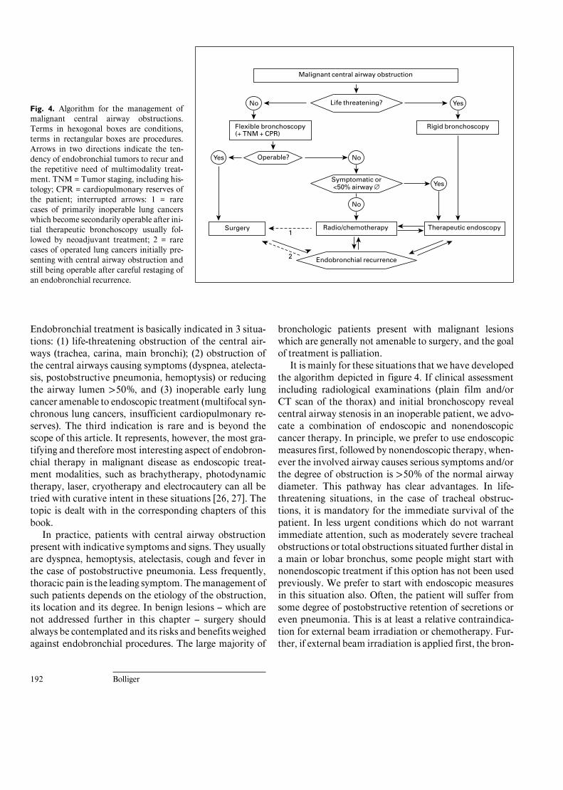
BOIS:L08:BPRR3001PD.98 FF: RESF E1: RESS
192 Bolliger
Fig. 4. Algorithm for the management ofmalignant central airway obstructions.Terms in hexogonal boxes are conditions,terms in rectangular boxes are procedures.Arrows in two directions indicate the ten-dency of endobronchial tumors to recur andthe repetitive need of multimodality treat-ment. TNM = Tumor staging, including his-tology; CPR = cardiopulmonary reserves ofthe patient; interrupted arrows: 1 = rarecases of primarily inoperable lung cancerswhich become secondarily operable after ini-tial therapeutic bronchoscopy usually fol-lowed by neoadjuvant treatment; 2 = rarecases of operated lung cancers initially pre-senting with central airway obstruction andstill being operable after careful restaging ofan endobronchial recurrence.
Life threatening?
Symptomatic or<50% airway Æ
Endobronchial recurrence
ä
ä
ä
No
Operable?
Flexible bronchoscopy(+ TNM + CPR)
ää
Yes
ä ä
ä
ä
ä
ä
ä
No
äNo
1
2
ä
ä
ä
ä
ä
ää
ää
ä
Yes
Yes
Rigid bronchoscopy
Therapeutic endoscopyRadio/chemotherapy
Malignant central airway obstruction
Surgery
Endobronchial treatment is basically indicated in 3 situa-tions: (1) life-threatening obstruction of the central air-ways (trachea, carina, main bronchi); (2) obstruction ofthe central airways causing symptoms (dyspnea, atelecta-sis, postobstructive pneumonia, hemoptysis) or reducingthe airway lumen 150%, and (3) inoperable early lungcancer amenable to endoscopic treatment (multifocal syn-chronous lung cancers, insufficient cardiopulmonary re-serves). The third indication is rare and is beyond thescope of this article. It represents, however, the most gra-tifying and therefore most interesting aspect of endobron-chial therapy in malignant disease as endoscopic treat-ment modalities, such as brachytherapy, photodynamictherapy, laser, cryotherapy and electrocautery can all betried with curative intent in these situations [26, 27]. Thetopic is dealt with in the corresponding chapters of thisbook.
In practice, patients with central airway obstructionpresent with indicative symptoms and signs. They usuallyare dyspnea, hemoptysis, atelectasis, cough and fever inthe case of postobstructive pneumonia. Less frequently,thoracic pain is the leading symptom. The management ofsuch patients depends on the etiology of the obstruction,its location and its degree. In benign lesions – which arenot addressed further in this chapter – surgery shouldalways be contemplated and its risks and benefits weighedagainst endobronchial procedures. The large majority of
bronchologic patients present with malignant lesionswhich are generally not amenable to surgery, and the goalof treatment is palliation.
It is mainly for these situations that we have developedthe algorithm depicted in figure 4. If clinical assessmentincluding radiological examinations (plain film and/orCT scan of the thorax) and initial bronchoscopy revealcentral airway stenosis in an inoperable patient, we advo-cate a combination of endoscopic and nonendoscopiccancer therapy. In principle, we prefer to use endoscopicmeasures first, followed by nonendoscopic therapy, when-ever the involved airway causes serious symptoms and/orthe degree of obstruction is 150% of the normal airwaydiameter. This pathway has clear advantages. In life-threatening situations, in the case of tracheal obstruc-tions, it is mandatory for the immediate survival of thepatient. In less urgent conditions which do not warrantimmediate attention, such as moderately severe trachealobstructions or total obstructions situated further distal ina main or lobar bronchus, some people might start withnonendoscopic treatment if this option has not been usedpreviously. We prefer to start with endoscopic measuresin this situation also. Often, the patient will suffer fromsome degree of postobstructive retention of secretions oreven pneumonia. This is at least a relative contraindica-tion for external beam irradiation or chemotherapy. Fur-ther, if external beam irradiation is applied first, the bron-

BOIS:L08:BPRR3001PD.98 FF: RESF E1: RESS
Multimodality Treatment of AdvancedPulmonary Malignancies
193
chi reopen, but if the endobronchial involvement hasbeen important – long stenoses, destruction of airwaywalls – the radiation effect often causes postactinic scar-ring, resulting in distortion of the airway anatomy withimpaired mucus clearance [24, 28]. On the other hand, anairway obstruction of !50% of the normal diameter oftendoes not cause symptoms and usually disappears com-pletely after conventional radio-/chemotherapy. In thesesituations, we refrain from starting with endoscopic treat-ment. On the other hand, if the fiberoptic diagnosticexamination reveals a ‘technically appealing’ lesion, gen-erally a short endoluminal tumor, which is judged to betreatable under local anesthesia, we immediately proceedwith therapeutic bronchoscopy, provided that the esti-mated procedure time does not exceed 30 min.
An exception to this general approach is sometimespossible in patients presenting with untreated SCLC orlymphoma. Even severe airway obstructions, clearly ex-ceeding 50% of the normal diameter, can react dramati-cally to initial chemotherapy within 1–2 days. In these sit-uations, we start with chemotherapy first, unless the situa-tion is immediately life-threatening.
The type of the initial endoscopic treatment will de-pend on the nature and the extent of the obstruction, aswell as on the equipment available (fig. 1). An endolumi-nal tumor should be removed by laser, electrocoagulationand mechanical coring out if immediate reopening of theairway is the goal. Cryotherapy or photodynamic therapycan be used for nonvital situations, as their therapeuticeffect is delayed for about 2–3 weeks. Another endoscopicmodality with delayed effect only is brachytherapy, whichhas the advantage of effectively treating lesions at a dis-tance of up to 1 cm of the center of the source, most oftenreaching beyond the airway wall. It is therefore an idealcomplement for the treatment of intramural componentsof airway obstructions.
Extrinsic stenoses are dilated with the rigid broncho-scope or with a balloon used with either the rigid or flexi-ble bronchoscope, and continued patency is guaranteedby insertion of a stent. In mixed stenoses, the endoluminaltumor is first removed, and if the remaining extrinsic and/or intramural component after resection and dilatation is150% of the normal lumen, a stent is also inserted. Wealso favor stent insertion if a patient has a posttreatmentlumen patency of 150%, but shows unstable airway wallsdue to destruction of the supporting cartilaginous struc-tures. Further, we insert stents in patients who have nofurther treatment options, irrespective of the airway di-ameter at the end of the endobronchial procedure. Thisusually means the patients have had full-dose external
beam irradiation, exhausted chemotherapy and have un-dergone full treatment of 15–20 Gy brachytherapy.
If nonendoscopic treatment is still available, externalbeam irradiation and/or chemotherapy should be ini-tiated within 2 weeks after the endobronchial treatment toconsolidate the local effect. A full radiation dose of up to50 Gy is usually applied to the mediastinum, includingthe endoscopically pretreated stenotic segment. The che-motherapy regimen will depend on tumor histology andlocal preferences. Patients who have had full-dose exter-nal beam irradiation should undergo brachytherapy. Inour institution they undergo 3 sessions with a HDR-irid-ium-192 afterloading source which will lead to a total doseof F15 Gy at a distance of 1 cm from the source. In ourexperience, it takes only about 2–3 months, depending ontumor biology and the site of the initial stenosis, untillocal tumor recurrence leads to reobstruction of an airwaywhich has been treated by laser resection only [24]. Inser-tion of a stent usually extends this period somewhat, buteventually the tumor will obstruct the airway proximallyand distally to the stent. When we compared patients afterstent insertion with and without subsequent radiotherapy,the group who underwent radiotherapy showed no reob-struction for at least 4–6 months in comparison to 2–3months in the group without radiotherapy [24]. A classi-cal approach used at our institution is illustrated by thesecond case history.
Case 2, a 54-year-old man, consulted his general practitionerbecause of increased cough, blood-tinged sputum and a weight loss of7 kg over 3 months. He was a tiler, current smoker with a 40 pack-year history and consumed about 1.5 liters of beer a day. His chestroentgenogram showed an ill-defined hilar opacity at the level of theright upper lobe, which on chest CT scan was highly suspicious of alocally advanced upper lobe tumor with invasion of the mediastinumand partial obstruction of the right main bronchus (fig. 5). This wasconfirmed by bronchoscopy, showing a mixed subtotal obstruction ofthe right main bronchus (fig. 6) which could easily be passed with aclosed forceps. Additionally, there was an inward bulging of the righttracheobronchial angle due to compression by the tumor. Histologyobtained by a forceps biopsy during bronchoscopy revealed squa-mous cell carcinoma. The patient had a grade III dyspnea and cor-responding severe restrictive airflow limitation with an FEV1 of1.3 liters (39%) and an FVC of 2.4 liters (57%). A search for metas-tases (bone and brain scans) remained negative, leading to a clinicaldiagnosis of a highly symptomatic locally advanced (stage IIIb) squa-mous cell carcinoma with impending atelectasis of the right lung. Thepatient therefore first underwent rigid bronchoscopy with dilatationof the right main bronchus, which revealed a practically occludedright upper lobe entrance but a normal intermediate bronchus withnormal middle and lower lobes. A spiked polyflex stent was insertedinto the right main bronchus (fig. 7), which resulted in immediateimprovement of the patient’s dyspnea to a grade II correlating witha marked improvement in FEV1 to 2.0 liters (62%) and FVC to

BOIS:L08:BPRR3001PD.98 FF: RESF E1: RESS
194 Bolliger
Fig. 5. CT scan at the level of the main cari-na of a 54-year-old male smoker with blood-tinged sputum. High suspicion of locally ad-vanced right upper lobe tumor with invasionof the mediastinum and partial obstruction(curved arrow) of the right main bronchus.
2.9 liters (71%); further, retained secretions were expectorated, andthe cough virtually disappeared. The patient then underwent exter-nal beam irradiation of the tumor and the mediastinum with a totaldose of 39.6 Gy. Three months after stent placement, the patient didwell, but a routine control bronchoscopy showed that the stent hadpartially migrated, its tip lying in the trachea. This migration was dueto the radiation effect leading to shrinkage of the endobronchialtumor mass. The stent was removed during flexible bronchoscopy,showing a stent bed which was stable and only slightly reduced indiameter (fig. 8). Another 3 months later, a routine control bronchos-copy revealed squamous cell carcinoma in a mucosal biopsy of theright main bronchus; the patient still did well. To prevent recurrentendobronchial tumor growth, it was therefore decided to continuewith brachytherapy (3 sessions at F5 Gy at 1 cm from source atweekly intervals), which the patient is still undergoing at the time ofwriting. In summary, this patient profited immediately from inter-ventional bronchoscopy with stent placement, patency of the rightmain bronchus helped save the function of middle and lower lobeand subsequent radiotherapy consolidated this initial effect.
Eventually endobronchial recurrence of previouslytreated central airway obstruction is the rule. Generallyrepetitive sessions of therapeutic endoscopy are necessaryto guarantee sustained airway patency (fig. 4). In patientswho have not exhausted conventional radio-/chemothera-py, this option should be tried. The result of carefullytitrated multimodality treatment will increase the num-ber of patients who will have patent central airways untiltheir death.
So, what do these patients die of? A large proportionwill succumb to metastatic disease, the ones dying of localintrathoracic complications often experience fatal throm-boembolic incidents, suffer from intractable malignantpleural or pericardial effusions, develop upper vena cavasyndrome, or in our experience very often develop termi-nal pneumonia in a state of general tumor cachexia. Onthe other hand, there is a higher proportion of fatalhemoptysis in patients having undergone interventionalbronchoscopic treatment, especially after brachytherapy(see chapter on brachytherapy by Fischer and Huber). It isnot clear to what extent this complication can be attribut-ed to the treatment per se or simply to the fact that thistreatment keeps the airways open, allowing fragile vesselsto erode into them rather than into a compact tumor bedcontaining the hemorrhage.
The important message of the addition of endoscopictreatment modalities to the armamentarium of anticancertherapy of pulmonary malignancies is that the availabledata unequivocally show very efficient palliation of symp-toms. What methods should be used for what situationand what multimodality approach is best remains to beelucidated, randomized trials are clearly needed. A firstexample in the right direction was set by Huber et al. [29],in a randomized study comparing external beam irradia-tion alone to the combination of external beam irradia-

BOIS:L08:BPRR3001PD.98 FF: RESF E1: RESS
Multimodality Treatment of AdvancedPulmonary Malignancies
195
Fig. 6. Endoscopic view of the patient of figure 5, confirming thelocally advanced tumor with subtotal mixed obstruction of the rightmain bronchus and inward bulging of the right tracheobronchialangle.Fig. 7. A polyflex stent (Rüsch AG, Waiblingen, Germany) has beeninserted after laser resection of the endobronchial tumor and balloondilatation of the extraluminal component of the stenosis of the rightmain bronchus.Fig. 8. Aspect of right main bronchus 3 months after completion ofexternal beam irradiation and removal of the partially migratedstent. The right main bronchus is patent with stable walls, the lumenis only moderately narrowed.
tion and brachytherapy in patients with locally advancedlung cancer. Additional brachytherapy resulted in signifi-cantly longer local tumor control, but not median surviv-al. For a subgroup of patients with squamous cell carcino-ma, however, survival after additional brachytherapy waslonger (40 vs. 33 weeks); but this difference did not reach
statistical significance (p = 0.09). The incidence of fatalhemoptysis was not different between the two groups.
In agreement with other authors [30–32], we suggestthat in locally advanced pulmonary malignancies withinvolvement of the central airways (causing moderate tosevere symptoms and/or a reduction of the involved air-
6 7
8

BOIS:L08:BPRR3001PD.98 FF: RESF E1: RESS
196 Bolliger
way lumen of 150% of the normal diameter), it is best tocombine endoscopic and nonendoscopic treatment mo-dalities from the start, which will prolong the time untilrecurrence of local tumor and may improve survival. Atour institution, we generally prefer to start with endoscop-
ic treatment, using techniques with immediate effect (la-ser, coring out, dilatation and stent placement). Thepatient will thus be able to experience rapid, sustainedsymptom relief, which allows him to enjoy life in a fairlypreserved state of health.
References
1 Belani CP: Multimodality management of re-gionally advanced non-small-cell lung cancer.Semin Oncol 1993;20:302–314.
2 Martini N: Our responsibility in the treatmentof lung cancer. Chest 1979;75:654–655.
3 Rosell R, Gomez-Codina J, Camps C, MaestreJ, Padille J, Canto A, Mate JL, Li S, Roig J,Olazabal A, Canela M, Ariza A, Skacel Z,Morera-Prat J, Abad A: A randomized trialcomparing preoperative chemotherapy plussurgery with surgery alone in patients with non-small-cell lung cancer. N Engl J Med 1994;330:153–158.
4 Thatcher N, Lind M, Stout R, Payne C, CarrollKB, Campbell C, Moussalli H: Carboplatin,ifosfamide and etoposide with mid-course vin-cristine and thoracic radiotherapy for ‘limited’stage small cell carcinoma of the bronchus. Br JCancer 1989;60:98–101.
5 Pignon JP, Arriagada R, Ihde DC, JohnsonDH, Perry MC, Souhami RL, Brodin O, JossRA, Kies MS, Lebeau B, Onoshi T, OsterlindK, Tattersall MHN, Wagner H: A meta-analy-sis of thoracic radiotherapy for small-cell lungcancer. N Engl J Med 1992;327:1618–1624.
6 Smit EF, Groen HJM, Timens W, de Boer WJ,Postmus PE: Surgical resection for small cellcarcinoma of the lung: A retrospective study.Thorax 1994;49:20–22.
7 Schaake-Koning C, Van den Bogaert W, Dale-sio O, Festen J, Hoogenhout J, Van Houtte P,Kirkpatrick A, Koolen M, Maat B, Nijs A, Ren-aud A, Rodrigus P, Schuster-Uitterhoeve L,Sculier JP, Van Zandwijk N, Bartelink H: Ef-fects of concomitant cisplatin and radiotherapyon inoperable non-small-cell lung cancer. NEngl J Med 1992;326:524–530.
8 Wozniak AJ, Crowley JJ, Balcerzak SP, WeissGR, Spiridonidis CH, Baker LH, Albain KS,Kelly K, Taylor SA, Gandara DR, LivingstonRB: Randomized trial comparing cisplatinwith cisplatin plus vinorelbine in the treatmentof advanced non-small-cell lung cancer: ASouthwest Oncology Group study. J Clin Oncol1998;16:2459–2465.
9 Langer CJ: The emerging role of paclitaxel andcarboplatin in non-small cell lung carcinoma;in Schiller JH (ed): Updates in Advances inLung Cancer. Prog Respir Res. Basel, Karger1997, vol 29, pp 73–90.
10 Gatzemeier U, Peters H-D: The use of gemcita-bine in non-small-cell lung cancer; in SchillerJH (ed): Updates in Advances in Lung Cancer.Prog Respir Res. Basel, Karger 1997, vol 29, pp91–105.
11 Schiller JH: New drug therapies for small cellcarcinoma of the lung; in Schiller JH (ed):Updates in Advances in Lung Cancer. ProgRespir Res. Basel, Karger 1997, vol 29, pp135–149.
12 Cox JD, Yesner R, Mietlowski W, Petrovich Z:Influence of cell type on failure pattern afterirradiation for locally advanced carcinoma ofthe lung. Cancer 1979;44:94–98.
13 Eichenhorn MS, Kvale PA, Miks VM, SeydelHG, Horowitz B, Radke JR: Initial combina-tion therapy with YAG laser photoresectionand irradiation for inoperable non small cellcarcinoma of the lung. Chest 1986;89:782–785.
14 Macha HN, Becker KO, Kemmer HP: Patternof failure and survival in endobronchial laserresection. A matched pair study. Chest 1994;105:1668–1672.
15 Vergnon JM: Traitement palliatif de l’obstruc-tion bronchique tumorale. Pour qui? Pour-quoi? Comment? Rev Mal Respir 1995;12:331–334.
16 Dumon JF, Reboud E, Garbe L, Aucomte F,Meric B: Treatment of tracheobronchial lesionsby laser photoresection. Chest 1982;81:278–284.
17 Cavaliere S, Foccoli P, Farina PL: Nd:YAGlaser bronchoscopy: A five-year experiencewith 1,396 applications in 1,000 patients.Chest 1988;94:15–21.
18 Cavaliere S, Foccoli P, Toninelli C, Feijo S:Nd:YAG laser therapy in lung cancer: An 11-year experience with 2,253 applications in1,585 patients. J Bronchology 1994;1:105–111.
19 Pedersen U, Kristensen S, Illum P: Palliativeresection with high-frequency cutting loop inmalignant tracheobronchial diseases. J Bron-chology 1994;1:23–25.
20 Homasson J, Renault P, Angebault M, BonniotJP, Bell NJ: Bronchoscopic cryotherapy for air-way strictures caused by tumors. Chest 1986;90:159–164.
21 Speiser BL, Spratling L: Remote afterloadingbrachytherapy for the local control of endo-bronchial carcinoma. Int J Radiat Oncol BiolPhys 1993;25:579.
22 Macha HN, Koch K, Stadler M, SchumacherW, Krumhaar D: New technique for treatingocclusive and stenosing tumours of the tracheaand main bronchi: Endobronchial irradiationby high dose iridium-192 combined with lasercanalisation. Thorax 1987;42:511–515.
23 Vincent RG, Dougherty TJ, Rao U, Boyle DG,Potter WR: Photoradiation therapy in ad-vanced carcinoma of the trachea and bronchi.Chest 1984;85:29–33.
24 Bolliger CT, Probst R, Tschopp K, Solèr M,Perruchoud AP: Silicone stents in the manage-ment of inoperable tracheobronchial stenoses.Indications and limitations. Chest 1993;104:1653–1659.
25 Stock KW, Jacob AL, Proske M, Bolliger CT,Rochlitz C, Steinbrich W: Treatment of malig-nant obstruction of the superior vena cava withthe self-expanding wallstent. Thorax 1995;50:1151–1156.
26 Edell ES, Cortese DA: Photodynamic therapyin the management of early superficial squa-mous cell carcinoma as an alternative to surgi-cal resection. Chest 1992;102:1319–1322.
27 Sutedja G, Schramel F, Postmus PE: Broncho-scopic treatment modalities in lung cancer, in-dications and limitations. Ann Oncol 1995;6:951–952.
28 Jain PR, Dadhia HV, Lapp NL, ThompsonAB, Frich JC: Nd:YAG laser followed by radia-tion fro treatment of malignant airway lesions.Lasers Surg Med 1985;5:47–53.
29 Huber RM, Fischer R, Hautmann H, PollingerB, Haussinger K, Wendt T: Does additionalbrachytherapy improve the effect of externalirradiation? A prospective, randomized studyin central lung tumors. Int J Radiat Oncol BiolPhys 1997;38:533–540.
30 Perez CA, Stanley K, Grundy G, Hanson W,Rubin P, Kramer S, et al: Impact of irradiationtechnique and tumor extent in tumor controland survival of patients with unresectable non-oat cell carcinoma of the lung: Report by theradiation therapy oncology group. Cancer1982;50:1091–1099.
31 Vergnon J-F, Schmitt T, Alamartine E, Bar-thelemy J-C, Fournel P, Emonot A: Initial com-bined cryotherapy and irradiation for unresect-able non-small cell lung cancer. Preliminaryresults. Chest 1992;102:1436–1440.
32 Cavaliere S, Dumon J-F: Laser bronchoscopy;in Bolliger CT, Mathur P (eds): InterventionalBronchoscopy. Prog Respir Res. Basel, Karger,2000, vol 30, pp 108–119.
C.T. BolligerDepartment of Internal MedicineFaculty of Medicine, University of Stellenbosch19063 Tygerberg 7505 (South Africa)Tel. +27 21 938 9247, Fax +27 21 932 3105E-Mail [email protected]

OOOOOOOOOOOOOOOOOOOOOOOOOOOOOOOOOOOOOOOOOOOOOOOOOOOOOOOOOOOOOOOOOOOOOOOOOOOOOOOOOOOOOOOOOOOOOOOOOOOOOOOOOOOOOOOOOOOOOOOOOOO
Other Procedures
HABE:L08:BPRR3005XA.79 FF: RESF E1: RESS 22.10.1999
Bolliger CT, Mathur PN (eds): Interventional Bronchoscopy.Prog Respir Res. Basel, Karger, 2000, vol 30, pp 198–214
OOOOOOOOOOOOOOOOOOOOOOOOOOOOOOOOOOOOOOOOOOOOOOOOOOOOOOOOOOOOOOOOOOOOOOOOOOOOOOOOOOOOOOOOOOOOOOOOOOOOOOOOOOOOOOOOOOOOOOOOOOO
Percutaneous Imaging-GuidedInterventional Procedures in theThoraxB. Ghaye R.F. Dondelinger
Department of Medical Imaging, University Hospital Sart-Tilman, Liège, Belgium
198
Summary
Imaging-guided nonvascular diagnostic and therapeuticprocedures in the chest are applicable in the lung, pleura andmediastinum. Guidance systems include uni- or biplanarfluoroscopy, ultrasound, CT and MR or a combination ofthese. Percutaneous tissue sampling of a thoracic lesion isindicated when histological diagnosis will modify the stage ofthe disease or influence therapeutic strategy, and when thediagnosis is not obtained by endobronchial techniques. Cy-tology and histology obtained by small-gauge needle biopsyconfirm malignancy in 80–95% of cases at low cost and carrya low incidence of major complications. Thoracic fluid orgazeous collections located in the pleural space, pericardi-um, lung and mediastinum can be aspirated or drained by aclosed percutaneous catheter insertion with imaging guid-ance techniques using catheters of variable size and design.The potential of percutaneous therapy supersedes blindtechnique and surgery. Clinical success of catheter drainageof pleural empyema varies from 80 to 100%, when drainageis performed in a first attempt or after failure of other tech-niques. Minimally invasive drainage of lung abscesses andmediastinal abscesses show similar success rates, but pa-tients referred often exhibit a bad prognosis. Further refine-ment is achieved by local instillation of fibrinolytic agents inselected cases of empyema and hemothorax. These proce-dures are cost effective and complementary to endobron-chial procedures and surgery.
Percutaneous Transthoracic Needle Biopsy
Percutaneous lung biopsy was first described by Leyd-en in 1883 to establish the infectious nature of lung dis-ease. Currently, percutaneous tissue sampling of a pulmo-nary, pleural or mediastinal lesion is indicated when his-tological diagnosis will further influence diagnostic strate-gy and therapeutical options, or modify tumor staging andthe sequence of cancer treatment. Cost efficiency byshortening hospital stay was demonstrated [1].
IndicationsMain indications for percutaneous transthoracic nee-
dle biopsy (PTNB) can be summarized as follows [2]: pul-monary nodule(s) without specific diagnostic criteria oncomputed tomography (CT) ascertaining benignity; pul-monary nodule(s) or mass suggestive of malignancy, whensurgery will be postponed, or replaced by chemotherapyand/or radiotherapy – a patient who refuses invasive diag-nostic procedures and therapy may change his mind,when irrefutable proof of malignancy is established bypercutaneous biopsy – pulmonary nodule(s) in a patientwith a history of extrapulmonary primary malignancy andin clinical remission or presenting several primary malig-nancies; a residual nonregressive lesion following radio-therapy or chemotherapy; tissue sampling for therapeuticsensitivity tests; measurements of tumor markers; hor-mone dependence; DNA analysis; chronic diffuse pulmo-

HABE:L08:BPRR3005XA.79 FF: RESF E1: RESS
Percutaneous Imaging-GuidedInterventional Procedures in the Thorax
199
Fig. 1. PTNB of a small pulmonary nodule in 2 differentpatients. a, b An 11-mm nodule (arrow) is evidenced inthe left upper lobe in a central location. Two previousfiberoptic bronchoscopies with transbronchial biopsieswere negative. PTNB was performed with a 21-gaugeneedle, which revealed small cell lung cancer. Note thesmall alveolar blood flooding along the needle track (ar-rowheads). No complication occurred. c, d PTNB of a9-mm solitary right pulmonary nodule (arrow) whichshowed large cell carcinoma on cytology and histology.Note a small anterior pneumothorax (long arrow) andlimited perinodular hemorrhage (arrowhead).
a b
c d
nary infiltrate in selected cases; pleural nodule or mass ofindeterminate origin; mediastinal mass or adenopathy.The need for preoperative diagnosis of a solitary pulmo-nary nodule varies between institutions and depends onthe pretest probability of diagnosing a lesion that wouldobviate an unnecessary thoracoscopy or thoracotomy,and the demand of the patient [3]. Concerning X-ray andCT characteristics of pulmonary nodules, the followingobservations should be remembered: 43% of nodules witha diameter smaller than 1 cm are benign; 97% of noduleswith a diameter larger than 3 cm are malignant; 33% ofprimary malignant nodules have regular contours; 46% ofbenign nodules are spiculated; 26% of benign nodules and5% of malignant nodules show microcalcifications on CT;21% of benign and 40% of malignant nodules contain anair-bronchogram. Overall, a correct diagnosis is estab-lished with CT in 66–98% of cases [4].
PTNB is a useful alternative to mediastinoscopy ormediastinotomy for tissue diagnosis of enlarged hilar ormediastinal lymph nodes. All areas of the mediastinumare accessible to PTNB. The technique is faster, better tol-erated and less expensive [5].
Percutaneous puncture is also indicated in other condi-tions: intracavitary injection for treatment of secondaryaspergillomas [6]; insertion of a harpoon or injection ofmethylene blue or black carbon as a localizer prior to pleu-roscopic or video-assisted thoracic surgery (VATS) of lungnodules or highest yield areas for infiltrative disease [7–10] and percutaneous brachytherapy of pulmonary malig-nancy [11]. Lesions with a diameter of less than 3 cm,seated in the outer third of the lung or near a fissure areusually considered.
ContraindicationsVascular structures such as aneurysm or pulmonary
arteriovenous malformation, hydatid cyst, meningoceleand mediastinal pheochromocytoma are absolute contra-indications of PTNB. The correct diagnosis should beestablished with cross section imaging, including angiog-raphy. The following are relative contraindications: punc-ture of both lungs during the same day, puncture of onlyone functional lung, chronic respiratory insufficiency,pulmonary arterial hypertension, cardiac insufficiency,recent myocardial infarct, angor, severe emphysema and
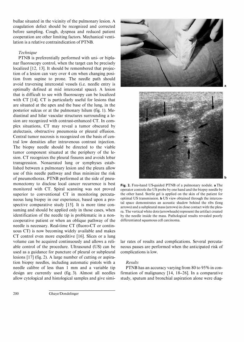
HABE:L08:BPRR3005XA.79 FF: RESF E1: RESS
200 Ghaye/Dondelinger
bullae situated in the vicinity of the pulmonary lesion. Acoagulation defect should be recognized and correctedbefore sampling. Cough, dyspnea and reduced patientcooperation are other limiting factors. Mechanical venti-lation is a relative contraindication of PTNB.
TechniquePTNB is preferentially performed with uni- or bipla-
nar fluoroscopy control, when the target can be preciselylocalized [12, 13]. It should be remembered that projec-tion of a lesion can vary over 4 cm when changing posi-tion from supine to prone. The needle path shouldavoid traversing intercostal vessels (i.e. needle entry isoptimally defined at mid intercostal space). A lesionthat is difficult to see with fluoroscopy can be localizedwith CT [14]. CT is particularly useful for lesions thatare situated at the apex and the base of the lung, in theposterior sulcus or at the pulmonary hilum (fig. 1). Me-diastinal and hilar vascular structures surrounding a le-sion are recognized with contrast-enhanced CT. In com-plex situations, CT may reveal a tumor obscured byatelectasis, obstructive pneumonia or pleural effusion.Central tumor necrosis is recognized on the basis of cen-tral low densities after intravenous contrast injection.The biopsy needle should be directed to the viabletumor component situated at the periphery of the le-sion. CT recognizes the pleural fissures and avoids lobartransgression. Nonaerated lung or symphyses estab-lished between a pulmonary lesion and the pleura allowuse of this needle pathway and thus minimize the riskof pneumothorax. PTNB performed at the side of pneu-monectomy to disclose local cancer recurrence is bestmonitored with CT. Spiral scanning was not provedsuperior to conventional CT in monitoring percuta-neous lung biopsy in our experience, based upon a pro-spective comparative study [15]. It is more time con-suming and should be applied only in those cases, whenidentification of the needle tip is problematic in a non-cooperative patient or when an oblique pathway of theneedle is necessary. Real-time CT (fluoro-CT or contin-uous CT) is now becoming widely available and makesCT control even more expeditive [16]. Slices or a lungvolume can be acquired continuously and allows a reli-able control of the procedure. Ultrasound (US) can beused as a guidance for puncture of pleural or subpleurallesions [17] (fig. 2). A large number of cutting or aspira-tion biopsy needles, including automatic pistols with aneedle calibre of less than 1 mm and a variable tipdesign are currently used (fig. 3). Almost all needlesallow cytological and histological samples and give simi-
Fig. 2. Free-hand US-guided PTNB of a pulmonary nodule. a Theoperator controls the US probe by one hand and the biopsy needle bythe other hand. Sterile gel is applied on the skin of the patient foroptimal US transmission. b US view obtained through the intercos-tal space demonstrates an acoustic shadow behind the ribs (longarrows) and a subpleural mass (arrows) in close contact with the pleu-ra. The vertical white dots (arrowheads) represent the artifact createdby the needle inside the mass. Pathological results revealed poorlydifferentiated squamous cell carcinoma.
lar rates of results and complications. Several percuta-neous passes are performed when the anticipated risk ofcomplications is low.
ResultsPTNB has an accuracy varying from 80 to 95% in con-
firmation of malignancy [14, 18–26]. In a comparativestudy, sputum and bronchial aspiration alone were diag-
a
b

HABE:L08:BPRR3005XA.79 FF: RESF E1: RESS
Percutaneous Imaging-GuidedInterventional Procedures in the Thorax
201
Fig. 3. Most frequently used needles to per-form PTNB. Aspiration needles: Otto nee-dles of different size and length (18 G, 10cm; 19.5 G, 15 cm; 21 G, 20 cm; A), suchneedles allow for cytological smears and his-tological specimens; spinal needle (20 G, 10cm; B); Chiba needle (22 G, 20 cm; C). Cut-ting needles: tru-cut (19 G, 15 cm; D); tru-cut (14 G, 15 cm; E); for parietal or mediasti-nal biopsy; biopsy gun with cutting needle(F); such a device can be difficult to handlewithout moving the tip of the needle in criti-cal situations.
nostic of cancer in 5.4%, PTNB alone in 72.6% and bothtechniques were in agreement in 22% [25]. Inadequatecytologic smears may be noted in up to 17% and allowedno interpretation in 9% in larger series [27]. Additionalfalse-negative diagnoses are obtained in about 5% and arecaused by inadequate sampling, tumor necrosis, crushedcells or errors in sampling reading. Negative predictivevalue of percutaneous pulmonary biopsy is 84–96%, andfalse-positive results are noted in 2–4% [12, 27]. Diagnos-tic accuracy for small nodules (!15 mm) is similar to larg-er lesions [28] (fig. 1). The following criteria should be ful-filled to ascertain that a lung nodule is benign: a technical-ly successful biopsy, adequate aspiration that is not nor-mal lung, no suspicion of cancer on the aspirated cells andnormal bronchoscopy. If a specific benign diagnosis canbe reached, no further exploration is required. Most fre-quently, a nonspecific and nonmalignant inflammatoryprocess is evidenced on the cytological smears. Theselesions should be followed up for 2 years [22]. If the sam-pled material is inadequate or insufficient, or in case ofcytologic or clinical suspicion of malignancy, the biopsyshould be repeated. The literature reported a mean spe-cific benign diagnosis in 25% of patients (table 1). Mostmediastinal lesions are of metastatic or lymphomatous
Table 1. Transthoracic needle biopsy of benign lung nodules
Authors Benign lesionsbiopsied
Specificbenign diagnosis
Flower and Verney [13] 75 18 (24)Johnston [25] – – (11.7)Horrigan et al. [23] 8 2 (25)Khouri et al. [22] 137 93 (67.8)Greene et al. [21] 27 12 (44)Winning et al. [24] 43 7 (16)Calhoun et al. [20] 132 16 (12)Perlmutt et al. [29] – – (12)Charig et al. [26] – – (5)
Figures in parentheses are percentages.
origin. Results of PTNB are similar to those of lung biop-sy, when metastatic adenopathies are considered. Diagno-sis of mediastinal non-Hodgkin lymphoma gives betterresults when comparison of smears or tissue fragmentswith a previous sampling or another extrathoracic site isavailable. Confirmation of Hodgkin’s disease is difficult,
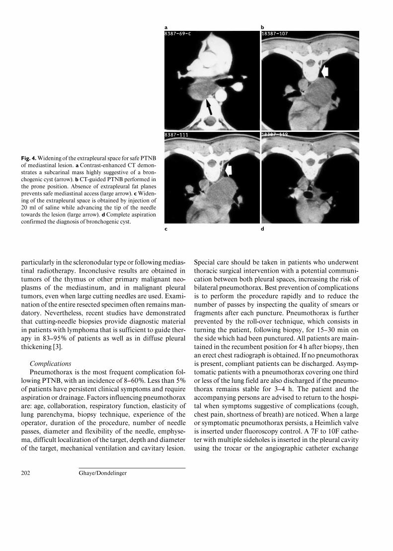
HABE:L08:BPRR3005XA.79 FF: RESF E1: RESS
202 Ghaye/Dondelinger
Fig. 4. Widening of the extrapleural space for safe PTNBof mediastinal lesion. a Contrast-enhanced CT demon-strates a subcarinal mass highly suggestive of a bron-chogenic cyst (arrow). b CT-guided PTNB performed inthe prone position. Absence of extrapleural fat planesprevents safe mediastinal access (large arrow). c Widen-ing of the extrapleural space is obtained by injection of20 ml of saline while advancing the tip of the needletowards the lesion (large arrow). d Complete aspirationconfirmed the diagnosis of bronchogenic cyst.
a b
c d
particularly in the scleronodular type or following medias-tinal radiotherapy. Inconclusive results are obtained intumors of the thymus or other primary malignant neo-plasms of the mediastinum, and in malignant pleuraltumors, even when large cutting needles are used. Exami-nation of the entire resected specimen often remains man-datory. Nevertheless, recent studies have demonstratedthat cutting-needle biopsies provide diagnostic materialin patients with lymphoma that is sufficient to guide ther-apy in 83–95% of patients as well as in diffuse pleuralthickening [3].
ComplicationsPneumothorax is the most frequent complication fol-
lowing PTNB, with an incidence of 8–60%. Less than 5%of patients have persistent clinical symptoms and requireaspiration or drainage. Factors influencing pneumothoraxare: age, collaboration, respiratory function, elasticity oflung parenchyma, biopsy technique, experience of theoperator, duration of the procedure, number of needlepasses, diameter and flexibility of the needle, emphyse-ma, difficult localization of the target, depth and diameterof the target, mechanical ventilation and cavitary lesion.
Special care should be taken in patients who underwentthoracic surgical intervention with a potential communi-cation between both pleural spaces, increasing the risk ofbilateral pneumothorax. Best prevention of complicationsis to perform the procedure rapidly and to reduce thenumber of passes by inspecting the quality of smears orfragments after each puncture. Pneumothorax is furtherprevented by the roll-over technique, which consists inturning the patient, following biopsy, for 15–30 min onthe side which had been punctured. All patients are main-tained in the recumbent position for 4 h after biopsy, thenan erect chest radiograph is obtained. If no pneumothoraxis present, compliant patients can be discharged. Asymp-tomatic patients with a pneumothorax covering one thirdor less of the lung field are also discharged if the pneumo-thorax remains stable for 3–4 h. The patient and theaccompanying persons are advised to return to the hospi-tal when symptoms suggestive of complications (cough,chest pain, shortness of breath) are noticed. When a largeor symptomatic pneumothorax persists, a Heimlich valveis inserted under fluoroscopy control. A 7F to 10F cathe-ter with multiple sideholes is inserted in the pleural cavityusing the trocar or the angiographic catheter exchange
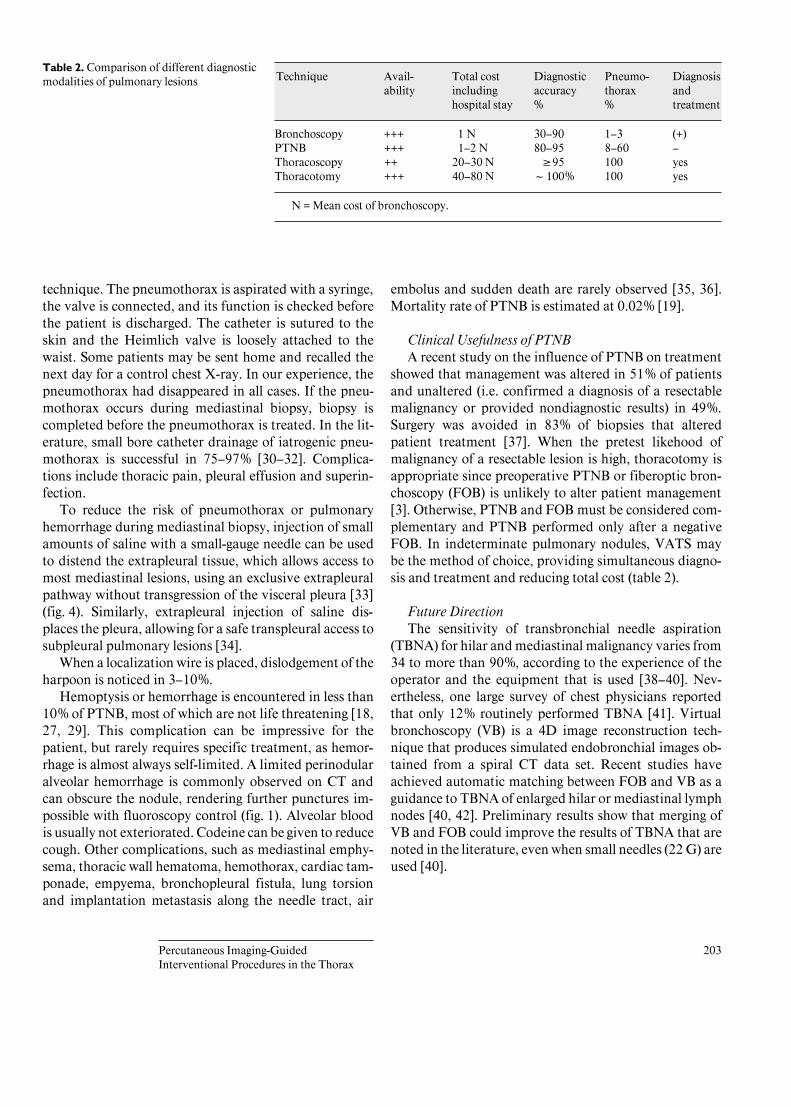
HABE:L08:BPRR3005XA.79 FF: RESF E1: RESS
Percutaneous Imaging-GuidedInterventional Procedures in the Thorax
203
Table 2. Comparison of different diagnosticmodalities of pulmonary lesions Technique Avail-
abilityTotal costincludinghospital stay
Diagnosticaccuracy%
Pneumo-thorax%
Diagnosisandtreatment
Bronchoscopy +++ 1 N 30–90 1–3 (+)PTNB +++ 1–2 N 80–95 8–60 –Thoracoscopy ++ 20–30 N 695 100 yesThoracotomy +++ 40–80 N F100% 100 yes
N = Mean cost of bronchoscopy.
technique. The pneumothorax is aspirated with a syringe,the valve is connected, and its function is checked beforethe patient is discharged. The catheter is sutured to theskin and the Heimlich valve is loosely attached to thewaist. Some patients may be sent home and recalled thenext day for a control chest X-ray. In our experience, thepneumothorax had disappeared in all cases. If the pneu-mothorax occurs during mediastinal biopsy, biopsy iscompleted before the pneumothorax is treated. In the lit-erature, small bore catheter drainage of iatrogenic pneu-mothorax is successful in 75–97% [30–32]. Complica-tions include thoracic pain, pleural effusion and superin-fection.
To reduce the risk of pneumothorax or pulmonaryhemorrhage during mediastinal biopsy, injection of smallamounts of saline with a small-gauge needle can be usedto distend the extrapleural tissue, which allows access tomost mediastinal lesions, using an exclusive extrapleuralpathway without transgression of the visceral pleura [33](fig. 4). Similarly, extrapleural injection of saline dis-places the pleura, allowing for a safe transpleural access tosubpleural pulmonary lesions [34].
When a localization wire is placed, dislodgement of theharpoon is noticed in 3–10%.
Hemoptysis or hemorrhage is encountered in less than10% of PTNB, most of which are not life threatening [18,27, 29]. This complication can be impressive for thepatient, but rarely requires specific treatment, as hemor-rhage is almost always self-limited. A limited perinodularalveolar hemorrhage is commonly observed on CT andcan obscure the nodule, rendering further punctures im-possible with fluoroscopy control (fig. 1). Alveolar bloodis usually not exteriorated. Codeine can be given to reducecough. Other complications, such as mediastinal emphy-sema, thoracic wall hematoma, hemothorax, cardiac tam-ponade, empyema, bronchopleural fistula, lung torsionand implantation metastasis along the needle tract, air
embolus and sudden death are rarely observed [35, 36].Mortality rate of PTNB is estimated at 0.02% [19].
Clinical Usefulness of PTNBA recent study on the influence of PTNB on treatment
showed that management was altered in 51% of patientsand unaltered (i.e. confirmed a diagnosis of a resectablemalignancy or provided nondiagnostic results) in 49%.Surgery was avoided in 83% of biopsies that alteredpatient treatment [37]. When the pretest likehood ofmalignancy of a resectable lesion is high, thoracotomy isappropriate since preoperative PTNB or fiberoptic bron-choscopy (FOB) is unlikely to alter patient management[3]. Otherwise, PTNB and FOB must be considered com-plementary and PTNB performed only after a negativeFOB. In indeterminate pulmonary nodules, VATS maybe the method of choice, providing simultaneous diagno-sis and treatment and reducing total cost (table 2).
Future DirectionThe sensitivity of transbronchial needle aspiration
(TBNA) for hilar and mediastinal malignancy varies from34 to more than 90%, according to the experience of theoperator and the equipment that is used [38–40]. Nev-ertheless, one large survey of chest physicians reportedthat only 12% routinely performed TBNA [41]. Virtualbronchoscopy (VB) is a 4D image reconstruction tech-nique that produces simulated endobronchial images ob-tained from a spiral CT data set. Recent studies haveachieved automatic matching between FOB and VB as aguidance to TBNA of enlarged hilar or mediastinal lymphnodes [40, 42]. Preliminary results show that merging ofVB and FOB could improve the results of TBNA that arenoted in the literature, even when small needles (22 G) areused [40].

HABE:L08:BPRR3005XA.79 FF: RESF E1: RESS
204 Ghaye/Dondelinger
Percutaneous Aspiration and Drainage ofThoracic Fluid Collections
Thoracic fluid collections of various origin, located inthe pleura, pericardium, lung or mediastinum can be aspi-rated or drained percutaneously with imaging guidance.Percutaneous therapy is an important refinement com-pared to blind bedside technique and compares favorablywith surgery. Diagnostic fluid aspiration includes cytolog-ical, bacteriological and chemical analyses. Confirmationof malignant cells, exsudative fluid, blood, lymph, bile,amylase, clear fluid or gross pus give etiological indica-tions. Fluoroscopy, US and CT can be used as guidancemodalities, either alone or in combination. The choice ofthe imaging technique depends on technical characteris-tics, availability, location of the collection and operatorexpertise. Loculated pleural effusions may be treated bythoracocentesis, thoracoscopy, surgically or CT-guidedthoracostomy tube drainage, open drainage or thoracoto-my and pleural decortication. Imaging guidance bestinsures proper catheter positioning.
Guidance ModalitiesFluoroscopy. Uni- or biplanar fluoroscopy is used for
percutaneous drainage of large pleural and pulmonarycollections [31, 43–50]. The patient is placed in a sittingor a posterior oblique position with the involved side up.A free pneumothorax is drained by a cephalad anteriorapproach, the patient being in the upright position. Pul-monary abscesses can be drained with fluoroscopy controlwhen a broad contact with the pleura is demonstrated.When a catheter is inserted in a pulmonary or in a pleuralcollection with US or CT control, definitive adjustment ofthe tip of the catheter is easily achieved with fluoroscopycontrol [51]. For this purpose, we use a combined CT andangiography suite equipped by a single table, which pivotson a vertical axis. The patient is brought under fluorosco-py without moving in a few seconds [52]. Opacification ofthoracic cavities is best documented with fluoroscopy.
Ultrasonography. US is particularly indicated to guidebedside percutaneous aspiration and catheter drainage ofpericardial and pleural fluid, even of reduced amount [31,44–46, 51, 53–56]. Pulmonary abscesses that are locatedin a subpleural location can also be included. Shifting offree pleural fluid during respiratory movements andchange of the patient’s position is easily observed withUS. Percutaneous approach is performed in the positionthat optimally displays access to the fluid collection.
Computed Tomography. CT offers exquisite anatomi-cal display of all thoracic structures and allows percuta-
neous access to all spaces with equal ease [31, 44–47, 51,57–60] (fig. 4–8). Most catheters can be inserted, thepatient remaining in a supine position. The optimal per-cutaneous entry point to a pulmonary abscess is definedaccording to its pleural contact, location of pleural fissuresand vascular structures. CT is particularly useful for thepercutaneous approach to the mediastinum, pleural fis-sures and for pulmonary abscesses with a reduced pleuralcontact.
CT is the optimal modality to confirm pneumothoraxor pneumomediastinum following a percutaneous proce-dure.
Drainage TechniquesNumber and size of percutaneous drainage catheters
should be adapted to the number and extent of collectionsand to viscosity of fluid to be drained. Small 7F to 9Fflexible catheters are well tolerated and sufficient for ashort-term drainage of water-like fluid. The trocar tech-nique is the most expeditive. It can be used in combina-tion with the tandem technique (a trocar is inserted paral-lel to an 18-gauge needle placed in the collection for aspi-ration). This technique is useful for large collections andpulmonary abscesses with a broad pleural contact. Theangiographic catheter exchange technique or Seldingertechnique is more rarely used in the thorax and most suit-able for mediastinal collections or small or encapsulatedpleural pockets that are located in the paramediastinalpleural space [58].
Percutaneous access through a thickened pleura can bedifficult, even when using regular chest tubes as stiff dila-tors and guide wires may be required. Chest drainagecatheters are connected to a continuous negative water-seal suction. Gentle saline irrigation is performed on theward to increase drainage and maintain catheter patency.
Pleural Empyema. Empyemas develop secondary topreexisting pulmonary, mediastinal or thoracic wall infec-tion, spread of cervical or abdominal infection, trauma,surgical or other invasive thoracic procedures, lung ab-scess or arise from hematogenous spread of infection. Ofthe patients with pleural empyema, 50% have coexistingpneumonia. Clinical signs may be torpid. Time intervalbetween onset of empyema and diagnosis may be severalweeks. Antibiotherapy is necessary, but without effectivefluid drainage, medical treatment will fail.
CT discloses satellite pleural pockets that form rapidlydue to pleural adhesions and can be aspirated or drainedwith small-caliber catheters (fig. 5). Pleural fluid, encap-sulated in a fissure, can also be adequately drained withCT control with or without transgression of collapsed lung

HABE:L08:BPRR3005XA.79 FF: RESF E1: RESS
Percutaneous Imaging-GuidedInterventional Procedures in the Thorax
205
Fig. 5. Percutaneous drainage of a loculated empyema by an extrapleural pathway. a CT shows a left basal loculatedempyema (arrow) following resection of the spleen, left adrenal and left kidney for invasive adrenal cancer. Note thatthe collection is surrounded by pulmonary parenchyma (arrowheads) preventing puncture in an axial plane. b A 9Fpigtail catheter was inserted under combined CT and fluoroscopic guidance with an oblique extrapleural approachalong the diaphragm. The curled pigtail tip of the catheter is documented inside the collapsed cavity (arrowheads).c Curved multiplanar reformatting along the catheter pathway (arrows) demonstrates its extrapleural position and theabsence of lung injury. d Fluoroscopic view shows the extremity of the catheter coiling in the empyema (arrow-heads).
parenchyma by the catheter. The diameter of the cathetervaries from 7F to 30F according to the viscosity of thefluid.
Indications depend on the evolutionary stage of thepleural effusion [61] (table 3). Classes 1 and 2 or exsuda-tive phase is an obligatory transitional phase before theempyema is formed and does not require drainage. Pleu-ral effusion of Class 3 can still be treated by complete aspi-ration of the pleural fluid, but the recurrence rate is 75%.A collection of Class 4 or at the fibrinopurulent stageshould be drained with mid-sized 8F to 14F catheters,whereas Class 6 effusions require regularly large bore 20Fto 30F catheters. If drainage is attempted during Class 4,pleural thickening may increase to 10 mm within a week.At Class 6, fibroblasts infiltrate the thickened pleura andlimit expansion of the underlying lung due to pleuralpeels.
Technical success is obtained in almost all cases. Mul-tiple catheters are required in 30% of cases. Clinical suc-cess is achieved in 70–89% of patients (table 4) anddepends on the stage of empyema. With a history of ill-ness in excess of 1 month before drainage, the proceduremay fail in spite of insertion of multiple catheters. Usual-ly, duration of percutaneous drainage is about 1 week,although a single session catheter treatment was described[55]. Radiologically guided drainage procedures exhibit asimilar efficiency in patients in whom previous surgicaltubes have failed, as in patients treated in a first intent(table 5). Failure of percutaneous drainage varies from 12to 28%: in chronic empyema, pleural thickening can pre-vent any closed catheter insertion. Failure of the pleuralcavity to collapse and obliterate occurs in 5–12% andfavors recurrence, which requires additional drainage.Opacification of the pleural catheter with propyliodine oil
a
c
b
d

Imaging aspect
van Sonnenberg et al. [51]
HABE:L08:BPRR3005XA.79 FF: RESF E1: RESS
206 Ghaye/Dondelinger
Table 3. Classification and treatment of pleural effusions
Type ofeffusion
Class 1nonsignificant
Class 2typical para-pneumonic
Classe 3borderlinecomplicated
Class 4simplecomplicated
Class 5complexcomplicated
Class 6simpleempyema
Class 7complexempyema
!10 mm ondecubituschest X-rayfree flowing
110 mm ondecubituschest X-rayfree flowing
usually notloculated
not loculated multiloculated single loculusor free flowing
multiloculated
Fluid aspect clear clear clear not frank pus not frank pus frank pus frank puspH 17.2 17.2 17.0 and !7.2 !7.0 !7.0Glucose, mg/dl 140 140 140 !40 !40LDH, U/l !1,000 !1,000 11,000Gram stain or
culture negative negative negative positive positiveTreatment not indicated
(antibiotics)antibioticsalone(thoraco-centesisif ineffective)
antibiotics(+ serialthoraco-centesis)
antibiotics +thoracostomycatheter
thoracostomycatheter +thrombolytics(thoracoscopyif ineffective)
thoracostomytube Bdecortication
thoracostomytube +thrombolytics Bdecortication
Adapted from Light and Rodriguez [61].
Table 4. Percutaneous drainage of pleural empyema in a first intent
Authors Patients Calibre ofdrainage catheterfrench
Guidancemodality
Durationof drainagedays
Clinicalsuccess%
17 8.3–12 CT: 59%US: 41%
– 88
Westcott [43] 12 8–10 fluoroscopy 1–7 83O’Moore et al. [54] 17 8.3–12 US 88Silverman et al. [46] 43 8.3–12 US: 70%
CT: 18%fluoroscopy: 12%
7–45 72
Merriam et al. [45] 18 8–14 fluoroscopyCT, US
1–20 (9) 80
Hunnam and Flower [44] 20 fluoroscopy: 5%CT: 25%, US: 40%or combination: 30%
2–42 (9) 80
Reinhold et al. [32] 42 9–14 US: 72%CT: 20%fluoroscopy: 8%
– 80
Neff et al. [59] 10 8.4–14 US: 80%CT: 20%
4–18 (10) 83
Cummin et al. [55] 13 20–28 US 1 85Dondelinger and Kurdziel [58] 14 8–24 CT (12) 89Lambiase et al. [50] 27 12–14 CT or US or
fluoroscopy3–33 (12) 70
Figures in parentheses are means.

Merriam et al. [45]
Bergh et al. [62]
HABE:L08:BPRR3005XA.79 FF: RESF E1: RESS
Percutaneous Imaging-GuidedInterventional Procedures in the Thorax
207
can reveal unexpected bronchopleural fistulae. Massivebronchopleural fistulae are an indication for surgery.
Thick pleural peels often resolve spontaneously afterweeks or months and regression can be followed by serialCT examinations. In selected patients, pleural decortica-tion should only be considered if pleural thickening per-sists [59].
Irrigation and lavage are performed when blood or vis-cous material is present. Intrapleural injection of fibrino-lytic agents was proven to be efficient in the prevention offibrin deposit and secondary loculation of the empyema(classes 5 and 7 of Light) [47, 49, 61–75] (table 6). Byreducing the number of pleural pockets, it also reduces thenumber of drainage catheters, length of drainage and hos-pitalization, and prevents progressive fibrosis of the pleu-ral surfaces. Intrapleurally injected fibrinolytic agents donot diffuse through the pleura, and blood coagulation testsare not affected. Urokinase should be preferred to strepto-kinase to avoid possible allergic reactions. Recent surgicalbronchial suture, macroscopic bronchopleural fistula andrecent trauma should be considered as a relative contrain-
dication of intrapleural injections of fibrinolytic agents[76]. Larger prospective studies are required to evaluatethe effect of daily administration of intrapleural fibrino-lytic agents on morbidity, mortality or the need for surgi-cal intervention.
Table 5. Percutaneous drainage of pleural empyema in a secondintent after failed chest tube drainage
Authors Failure ofthoracostomytube
Clinicalsuccess
7 4 (57)van Sonnenberg et al. [51] 13 12 (92)Westcott [43] 4 4 (100)Silverman et al. [46] 3 3 (100)Moulton et al. [47] 5 5 (100)Lambiase et al. [50] 8 4 (50)
Figures in parentheses are percentages.
Table 6. Percutaneous drainage of pleural empyema and intrapleural injection of fibrinolytic agents
Authors Patients Fibrinolyticagents
DoseIU/day
Results%
Complications
12 SK 250,000 83 low-grade feverBerglin et al. [63] 10 SK 100 chest discomfort (3)Fraedrich et al. [64] 27 SK 40 –Moulton et al. [47] 13 UK 80–150,000 100 –Lee et al. [49] 10 UK 100,000 90 –Aye et al. [65] 14 SK 250,000 93 fever (3)Henke and Leatherman [66] 12 SK 250,000 83 –Robinson et al. [67] 13 SK (3)
UK (10)150,000100,000
77 fever (1)
Taylor et al. [68] 20 SK 250,000 95 fever (1)Moulton et al. [69] 96 UK 250,000 94 –Park et al. [70] 31 UK 250,000 81 –Jerjes-Sanchez et al. [71] 30 SK 250,000 93 pain, fever, rash,
neuro signTemes et al. [72] 26 SK F275,000
F120,00062–7062–70
bleeding (1)fever, discomfort
Davies et al. [73] 1212
SKsaline
250,000(control group)
10075
––
Bouros et al. [74] 2525
SKUK
250,000100,000
9292
fever, 28%–
Chin and Lim [75] 2329
SK–
250,000(control group)
7066
––
Most studies contain subgroups of patients who did not respond to drainage without the use of fibrinolytic agents.SK = Streptokinase; UK = urokinase. Figures in parentheses are number of patients.

HABE:L08:BPRR3005XA.79 FF: RESF E1: RESS
208 Ghaye/Dondelinger
Fig. 6. Percutaneous drainage of a loculated pneumothorax. a A 70-year-old patient presented with severe acutedyspnea in the emergency department. CT performed 2 months before admission demonstrates severe COPD withlarge bullae on the left side (arrows) and pleural adhesions (arrowheads). b, c CT confirmed a loculated pneumothorax(arrows) in a subpulmonic location. Multiple pleural adhesions (arrowheads) discouraged from blind catheter drain-age. d A 9F pigtail catheter was inserted under CT guidance in the anterior costodiaphragmatic sulcus. The air leakresolved after 5 days of drainage, but relapsed 1 month later. The procedure was repeated and subsequent chemicalpleurodesis performed through the pleural catheter (arrows).
Malignant Pleural Effusion. Recurrent malignant pleu-risies are usually drained with soft 8F catheters. Scleros-ing drugs (doxycyclin, bleomycin) that limit mesothelialcell secretion are injected through the catheter when out-put volume has decreased to 100 ml or less per day.Obstruction of the pulmonary interstitium by metastaticlymphangitis can impede the lung to reexpand and is acontraindication to treatment. Complete regression of thepleural effusion is obtained in 61–81% and partial resolu-tion in up to 95%, which is equal to response with largerthoracostomy tubes [48, 56, 77]. In 20–40% of patients, apneumothorax ‘ex vacuo’ will appear, due to a stiff andnoncompliant lung after chronic compression. In 50% ofthose patients, the lung will reexpand in the next days, butin 50%, air will persist until fluid has reaccumulated.Nevertheless, even in the latter situation, dyspnea can bedramatically improved.
Posttraumatic Hemothorax. Massive hemothoraxshould be drained to reduce compression of the underly-ing lung parenchyma and prevent encapsulation and ad-herences. Large bore catheters are mandatory and pleurallavage can be useful. In selected cases, careful angiograph-ic workup is indicated, including catheterization of allpotentially involved intercostal arteries, internal mam-mary and inferior phrenic arteries. Active arterial bleed-ing is treated by hemostatic embolization with gelfoam,polyvinyl alcohol particles, or coils.
Gazeous Collections. Postbiopsy or spontaneous pneu-mothorax can be drained percutaneously with small cali-ber catheters [30–32] (fig. 6). Evacuation of the pneumo-thorax is performed for patient comfort and safety or tocontinue PTNB if accurate localization of the lesion turnsdifficult. Most pneumothoraces can be connected to aHeimlich valve, and when recurrent chemically pleuro-
a
c
b
d

HABE:L08:BPRR3005XA.79 FF: RESF E1: RESS
Percutaneous Imaging-GuidedInterventional Procedures in the Thorax
209
Fig. 7. Percutaneous drainage of a pneumatocele. a A 24-year-old intravenous drug addict presented with pulmonarystaphylococcus infection following endocarditis. Recurrent left-sided pneumothorax was treated by multiple thora-costomy tubes. Severe respiratory failure developed along with a rapidly growing lung lucency (arrows) compressingthe normal parenchyma on the right side. Note the multiple cavitated nodules (arrowheads) on both sides. b CTconfirmed a large right posterior pneumatocele and a small left pneumothorax with well-positioned chest tubes. c A9F pigtail catheter was inserted under CT guidance in the pneumatocele, which dramatically improved the patient’ssymptoms. d After 3 weeks of negative waterseal aspiration, a chest X-ray confirmed complete regression of thepneumatocele.
desed. Percutaneous drainage of pulmonary pneumato-cele has also been described [78] (fig. 7).
Pulmonary Abscess. Pyogenic pulmonary abscesses oc-cur rarely today, due to progress in antibiotic treatmentand more efficient eradication of the cause. They aremainly seen in adults, whose general condition is debili-tated by cancer, alcoholism, denutrition, diabetes andimmune deficiency. Pulmonary abscess results from he-matogenous spread, or from infection of a preexisting pul-monary cavity (emphysematous bulla, posttraumaticpneumatocele or pseudocyst, tuberculosis, fungus), or pul-monary infarction or tumor. A pulmonary abscess cancomplicate pneumonia with or without bronchial obstruc-tion. A pulmonary abscess which resists medical treat-ment, postural and bronchoscopic drainage requires per-cutaneous drainage.
A percutaneous diagnostic aspiration with an 18-gaugeneedle can preceed catheter insertion in doubtful cases.Bullae may frequently contain an air-fluid level, but arenot necessarily infected. An abscess with a diameter of1–3 cm is adequately treated by a single percutaneousaspiration and antibiotics.
Endoscopical transbronchial placement of a catheterinside a lung abscess can be planned with CT and moni-tored with fluoroscopy control. Transbronchial drainageis less comfortable for the patient than a percutaneousapproach. Contamination of the opposite lung is also arisk with the transbronchial technique. Mechanical venti-lation is not a contraindication for percutaneous abscessdrainage [79].
Pyogenic lung abscesses are often located in the periph-ery of the lung and usually they do not break through thelobar fissure. Pleural symphyses are rapidly established.
a
c
b
d

HABE:L08:BPRR3005XA.79 FF: RESF E1: RESS
210 Ghaye/Dondelinger
Fig. 8. Percutaneous drainage of pulmonary abscess. a A51-year-old patient presented with a 7-cm pulmonaryabscess (arrow) of the right upper lobe, following largebowel plasty for esophageal cancer. b CT confirmed abroad contact of the abscess with the pleura. The arrow-heads show the large bowel plasty. c A 9F pigtail catheterwas inserted under CT guidance and drained pus. d CTcontrol on the next day demonstrated complete regres-sion of the abscess (arrow).
a b
c d
The broad pleural contact allows planning of a percuta-neous access in such a way, that normal lung parenchymais not crossed by the drainage catheter (fig. 8). Pleuralempyema by direct contamination is unlikely to occurwhen the trocar technique is used, and in practice, it doesnot make a difference if an empyema or a large lungabscess is drained. The trocar technique is the preferreddrainage modality. Generally, 7F to 14F catheters are ade-quate. The drainage catheter is connected to a negativewaterseal aspiration. Pulmonary abscesses have a more orless thick wall and do not collapse rapidly after aspiration.Duration of drainage is variable, the cavity may close onlyafter 4 or 5 weeks. Decompression should be performedslowly in order to avoid rupture of a vessel or a so-calledRasmussen pseudoaneurysm that is incorporated or lo-cated close to the abscess wall. Lavage of a pulmonaryabscess is dangerous due to the risk of bronchogenicspread of pus. Injection of contrast medium through thedrainage catheter does not add significant information,but patency of the catheter should be maintained by dailyinjection of a minimal amount of saline. Daily chest radio-graphs are imperative for monitoring regression of theabscess cavity and early detection of complications.
Cure of the abscess is obtained in 73–100% of cases butoverall mortality is high, due to a bad general condition orunderlying disease [53, 57, 58, 60, 79–84] (table 7). Sur-gery remains indicated when extensive necrosis of lungparenchyma or life-threatening hemorrhage occurs. Tem-porizing percutaneous drainage cures the abscess, but sur-gery can be required for removal of necrotized tissue orpleural decortication.
Mediastinal Collections. Mediastinal abscesses resultfrom circumscribed mediastinitis. Mediastinal abscessesare among the most challenging infections to treat, asprognosis of diffuse mediastinitis is poor. CT distin-guishes diffuse infiltration of mediastinal planes from cir-cumscribed abscess, but separation between acute medi-astinitis and postsurgical changes can remain problematicin the early phase. Most purulent mediastinal collectionsresult from trauma, either penetrating transthoracic inju-ry or perforation of the esophagus. Thoracic and mediasti-nal surgery can be followed by mediastinal abscess forma-tion, and other intra- or extrathoracic infections can alsospread to the mediastinum.
CT is the most useful modality for diagnosis and plan-ning of percutaneous drainage of mediastinal collections.

Vainrub et al. [80]
Gobien et al. [87]
HABE:L08:BPRR3005XA.79 FF: RESF E1: RESS
Percutaneous Imaging-GuidedInterventional Procedures in the Thorax
211
Table 7. Percutaneous drainage of pulmonary abscess
Authors Patients Guidancemodality
Calibre ofdrainage catheterfrench
Duration ofdrainagedays
Clinicalsuccess%
3 fluoroscopy 16–18 100Lorenzo et al. [81] 3 fluoroscopy 18-gauge teflon
sheathed needleaspiration 100
Rami-Porta et al. [82] 13 – repeat aspiration5–30
15 100
Yellin et al. [83] 7 fluoroscopy – 15 100Parker et al. [53] 6 fluoroscopy 7–10 10–59 100Rice et al. [79] 11 fluoroscopy – – 73Ball et al. [57] 3 fluoroscopy,
US, CT8–12 19–24 100
Dondelinger and Kurdziel [58] 7 CT 8–14 12 431
van Sonnenberg et al. [60] 19 CT 9–20 4–38 84Ha et al. [84] 6 CT 8–10 7–18 83
1 Four of 7 patients died from underlying disease after successful drainage.
Table 8. Percutaneous drainage of mediastinal abscess
Authors Patients Guidancemodality
Calibre ofdrainage catheterfrench
Duration ofdrainagedays
Clinicalsuccess%
6 CTfluoroscopy
8.3–14 5–91 days(mean 35)
83
Meranze et al. [85] 8 fluoroscopy 8–12 2–4 weeks 88Carrol et al. [86] 5 CT – – 100Ball et al. [57] 4 fluoroscopy,
US, CT8–12 mean 28 days 100
Abscesses in the anterior mediastinum are drained by aparasternal approach avoiding the internal mammaryvessels. Abscesses that are located in the middle and pos-terior mediastinal compartment are treated by a paraver-tebral and extrapleural approach. In selected cases, a per-cutaneous cervical descending approach following thepathway of the infection can be used [2].
Multiple catheters are often necessary and are placedeither at both sides of the thoracic spine or the sternum orin the upper and in the lower part of the mediastinum.Conservative treatment is commonly applied to esopha-geal perforation [85]. An endoesophageal catheter can beinserted through the esophageal tear or in combination
with a percutaneous drainage catheter placed at the site ofleakage. When a fistulous tract is present between themediastinal abscess and an empyema or a subphrenicabscess, percutaneous drainage of these collections mayresolve the mediastinal abscess [86]. Only a limited num-ber of patients with percutaneous drainage are reported[57, 85–87] (table 8). Cure can be obtained in 83–100%,in the radiological literature, which does not consider the30-days mortality rate.
Other mediastinal fluid collections, such as pancreaticpseudocysts, pleuro-pericardial or bronchogenic cysts, in-fected tumor, lymphocele or hematoma can be aspiratedor drained percutaneously (fig. 4).

HABE:L08:BPRR3005XA.79 FF: RESF E1: RESS
212 Ghaye/Dondelinger
Pericardial Fluid Collection. Diagnosis is confirmedwith transthoracic or transesophageal US [88]. Causes ofpericardial effusion include cardiac insufficiency, malig-nant tumors, postoperative effusion, pericarditis, hypo-thyroidism, connective tissue disease, trauma and others.
Percutaneous aspiration of pericardial fluid is indi-cated for diagnosis; percutaneous drainage prevents car-diac tamponade. Clinical signs of tamponade include dys-pnea, tachycardia, compromised venous return and para-doxical pulse. Large amounts of pericardial fluid can bedrained with CT control, sometimes with an atypical leftanterolateral puncture, but most effusions are evacuatedwith US guidance, by a subcostal or a subxiphoid ap-proach. The angiographic catheter exchange technique ismainly used, the initial puncture being performed with an18-gauge or a 5F teflon-sheathed needle. A 5F to 7F pig-tail catheter is placed in the pericardial space [89]. Tech-nical success of the procedure and decompression of theheart is achieved in almost all cases, when the effusion isof sufficient amount. Duration of drainage is several daysonly. Chylopericardium can be difficult to drain and mayneed thoracic duct ligation or pericardectomy.
Percutaneous Drainage of Tension Pneumomediasti-num. Tension pneumomediastinum results from barotrau-ma in patients with increased pulmonary resistance whoare mechanically ventilated, with an elevated PAP andPEEP. Alveolar rupture results in interstitial emphysemathat migrates to the mediastinum along the bronchovascu-lar sheaths. When the mediastinal air fails to escape, due toadhesions or scarring, tension pneumomediastinum devel-ops. CT shows unequivocally the air collection in the medi-astinum. It mainly dissects the anterior mediastinal fatplanes, compressing the mediastinal vessels and the heart.Compromised venous return, cardiac and respiratory in-sufficiency and anuria urge for rapid decompression. Aleft-sided pneumothorax is usually associated.
Usual treatment of tension pneumomediastinum ismediastinostomy. Percutaneous catheter drainage is analternative emergency treatment. A soft 12F or 14F drain-age tube can be inserted into the mediastinum behind thesternum with CT guidance by a left anterolateral percuta-neous approach. Multiple sideholes are necessary to avoidcatheter occlusion during negative waterseal aspiration.When a left pneumothorax is also present, the mediastinalcatheter should be inserted before exsufflation of the pleu-ral air collection. [90].
Complications of Percutaneous Catheter DrainageComplications occur in approximately 5% and are
mainly due to inadequate catheter insertion technique
[43, 46, 51, 80, 81, 87]. Injury to an intercostal artery canproduce a hemothorax or a hematoma of the thoracicwall. Life-threatening hemorrhage is rare, but can beobserved following rupture of the wall of a pulmonaryabscess, erosion of a branch of the pulmonary artery ortransfixation of an internal mammary or mediastinal ves-sel by the catheter.
Pericardial drainage is prone to most of the clinicallysignificant complications, such as hemopericardium withtamponade, dysrhythmia, pneumopericardium or super-infection. The thoracic catheter can be introduced errone-ously in the subdiaphragmatic space, liver or spleen whencrosssectional imaging is not used properly. When thecatheter creates a communication between a pulmonaryabscess and the pleural space, a secondary empyema or abronchopleural fistula can be established.
When normal lung is crossed with a large bore catheterusing the trocar technique, a pulmonary infarction canresult. A pleural sinus tract through the chest wall can per-sist when the patient has undergone previous irradiation,but chest wall gangrene is rare. Transient bacteremia andhemoptysis can occur during catheter insertion. Pneumo-thorax is a potentially frequent complication, but can beavoided by proper crosssection imaging. Complicationsare best avoided by a proper planning of percutaneousapproach with CT control. When fluoroscopy is used pre-dominantly as a guidance modality for thoracic drainageprocedures, the overall complication rate is as high as20%, most being minor events.
Other side-effects include subcutaneous emphysema,local skin infection at the entry point of the catheter anddiscomfort during breathing, rib erosion, catheter leakage,bending, breakage and obstruction.

HABE:L08:BPRR3005XA.79 FF: RESF E1: RESS
Percutaneous Imaging-GuidedInterventional Procedures in the Thorax
213
References
1 Dicato M, Freilinger J, Dondelinger RF, Kurd-ziel JC: Economic considerations; in Dondelin-ger RF, Rossi P, Kurdziel JC, Wallace S (eds):Interventional Radiology. Stuttgart, Thieme,1990, pp 142–155.
2 Dondelinger RF: Interventional Radiology ofthe chest: Percutaneous procedures; in Imagingof the Chest (Syllabus) European Congress ofRadiology (ECR), Vienna 1991. Heidelberg,Springer, CC–1001, 1991, pp 165–175.
3 Klein JS, Zarka MA: Transthoracic needlebiopsy: An overview. J Thorac Imaging 1997;12:232–249.
4 Minami M, Kawauchi N, Oka H, Itai Y, SasakiY: High-resolution CT of small lung nodules:Malignancy versus benignancy. Radiology1992;185:357.
5 Protopapas Z, Westcott JL: Transthoracic nee-dle biopsy of mediastinal lymph nodes for stag-ing lung and other cancers. Radiology 1996;199:489–496.
6 Giron JM, Poey CG, Fajadet PP, Balagner GB,Assoun JA, Richardi GR, Haddad JH, CaceresJC, Senac JP, Railhac JJ: Inoperable pulmo-nary aspergilloma: Percutaneous CT-guided in-jection with glycerin and Amphotericin B pastein 15 cases. Radiology 1993;188:825–827.
7 Strautman PR, Dorfman GS, Haas RA: Pre-biopsy wire localization of a small peripherallung nodule. J Vasc Intervent Radiol 1992;3:391–393.
8 Shah RM, Spirn PW, Salazar AM, Steiner RM,Cohn HE, Solit RW, Wechsler RJ, Erdman S:Localization of peripheral pulmonary nodulesfor thoracoscopic excision: Value of CT-guidedwire placement. AJR 1993;161:279–283.
9 Templeton PA, Krasna M: Localization of pul-monary nodules for thoracoscopic resection:Use of needle/wire breast-biopsy system. AJR1993;160:761–762.
10 Spirn PW, Shah RM, Steiner RM, GreenfieldAL, Salazar AM, Liu JB: Image-guided local-ization for video-assisted thoracic surgery. JThorac Imaging 1997;12:285–292.
11 Brach B, Buhler C, Hayman MH, Joyner LR,Liprie SF: Percutaneous computed tomogra-phy-guided fine needle brachytherapy of pul-monary malignancies. Chest 1994;106:268–274.
12 Dahlgren S, Nordenstrom B (eds): Transtho-racic Needle Biopsy. Stockholm, Almquist andWiksell, 1966.
13 Flower CDR, Verney GI: Percutaneous needlebiopsy of the thoracic lesions: An evaluation of300 biopsies. Clin Radiol 1979;30:215–218.
14 van Sonnenberg E, Casola G, Ho M, Neff CC,Varney RR, Wittich GR, Christensen R, Fried-man PJ: Difficult thoracic lesions: CT-guidedbiopsy experience in 150 cases. Radiology1988;167:457–461.
15 Ghaye B, Dondelinger RF: Percutaneous CTguided lung biopsy: Sequential versus spiralscanning. A randomized prospective study. EurRadiol 1999;9:1317–1320.
16 Katada K, Kato R, Anno H, Ogura Y, Koga S,Ida Y, Sato M, Nonomura K: Guidance withreal-time CT fluoroscopy: Early clinical experi-ence. Radiology 1996;200:851–856.
17 Yang PC, Luh KT, Lee YC, Chang DB, Yu CJ,Wu HD, Lee LN, Kuo SH: Lung abscesses: USexamination and US-guided transthoracic as-piration. Radiology 1991;180:171–175.
18 Westcott JL: Direct percutaneous needle aspi-ration of localized pulmonary lesions: Resultsin 422 patients. Radiology 1980;137:31–35.
19 Greene R: Transthoracic needle aspirationbiopsy; in Athanasoulis CA, Pfister RC,Greene R, Roberson GH (eds): InterventionalRadiology. Philadelphia, Saunders, 1982, pp587–634.
20 Calhoun P, Feldman PS, Armstrong P, BlackWC, Pope TL, Minor GR, Daniel TM: Theclinical outcome of needle aspirations of thelung when cancer is not diagnosed. Ann ThoracSurg 1986;41:592–596.
21 Greene R, Szyfelbein WM, Isler RJ, Stark P,Jantsch H: Supplementary tissue-core histologyfrom fine-needle transthoracic aspiration biop-sy. AJR 1985;144:787–792.
22 Khouri NF, Stitik FP, Erozan YS, Gupta PK,Kim WS, Scott WW Jr, Hamper UM, MannRB, Eggleston JC, Baker RR: Transthoracicneedle aspiration biopsy of benign and malig-nant lung lesions. AJR 1985;144:281–288.
23 Horrigan TP, Bergin KT, Snow N: Correlationbetween needle biopsy of lung tumors and his-topathologic analysis of resected specimens.Chest 1986;90:638–640.
24 Winning AJ, McIvor J, Seed WA, HusainOAN, Metaxas N: Interpretation of negativeresults in fine needle aspiration of discrete pul-monary lesions. Thorax 1986;41:875–879.
25 Johnston WW: Fine needle aspiration biopsyversus sputum and bronchial material in thediagnosis of lung cancer. A comparative studyof 168 patients. Acta Cytol 1988;32:641–645.
26 Charig MJ, Stutley JE, Padley SPG, HansellDM: The value of negative needle biopsy insuspected operable lung cancer. Clin Radiol1991;44:147–149.
27 Sinner WN: Wert und Bedeutung der perkuta-nen transthorakalen Nadelbiopsie für dieDiagnose intrathorakaler Krankheitprozesse.ROFO 1975;123:197–202.
28 Westcott JL, Rao N, Colley DP: Transthoracicneedle biopsy of small pulmonary nodules. Ra-diology 1997;202:97–103.
29 Perlmutt LM, Johnston WW, Dunnick NR:Percutaneous transthoracic needle aspiration:A review. AJR 1989;152:451–455.
30 Perlmutt LM, Braun SD, Newman GE, CohanRH, Saeed M, Sussman SK, Dunnik NR:Transthoracic needle aspiration: Use of a smallchest tube to treat pneumothorax. AJR 1987;148:849–851.
31 Casola G, van Sonnenberg E, Keightley A, HoM, Withers C, Lee AS: Pneumothorax: Radio-logic treatment with small catheters. Radiology1988;166:89–91.
32 Reinhold C, Illescas FF, Atri M, Bret PM:Treatment of pleural effusions and pneumo-thorax with catheters placed percutaneouslyunder imaging guidance. AJR 1989;152:1189–1191.
33 Langen HJ, Klose KC, Keulers P, Adam G,Jochims M, Günther RW: Artificial wideningof the mediastinum to gain access for extra-pleural biopsy: Clinical results. Radiology1995;196:703–706.
34 Klose KC: CT-guided large-bore biopsy: Extra-pleural injection of saline for safe transpleuralaccess to pulmonary lesions. Cardiovasc Inter-vent Radiol 1993;16:259–261.
35 Ayar D, Golla B, Lee JY, Nath H: Needle-trackmetastasis after transthoracic needle biopsy. JThorac Imaging 1998;13:2–6.
36 Wong RS, Ketai L, Temes RT, Follis FM, Ash-by R: Air embolus complicating transthoracicpercutaneous needle biopsy. Ann Thorac Surg1995;59:1010–1011.
37 Lee SI, Shepard JO, Boiselle PM, Trotman-Dickenson B, McLoud TC: Role of transtho-racic needle biopsy in patient treatment deci-sions. Radiology 1996;201:269.
38 Schenk DA: The role of transbronchial needleaspiration in staging of bronchogenic carcino-ma, in Wang KP, Mehta AC (eds): FlexibleBronchoscopy. Cambridge, Blackwell Science,1995, pp 215–221.
39 Wang KP: Transbronchial needle aspirationand percutaneous needle aspiration for stagingand diagnosis of lung cancer. Clin Chest Med1995;16:535–552.
40 McAdams HP, Goodman PC, Kussin P: Vir-tual bronchoscopy for directing transbronchialneedle aspiration of hilar and mediastinallymph nodes: A pilot study. AJR 1998;170:1361–1364.
41 Prakash UBS, Offord KP, Stubbs SE: Bron-choscopy in North America: The ACCP sur-vey. Chest 1991;100:1668–1675.
42 Bricault I, Ferretti GR, Arbib F, Cinquin P:Automatic matching between real and virtualbronchoscopy: A guidance for trans-bronchialneedle aspiration. Radiology 1996;201:359.
43 Westcott JL: Percutaneous catheter drainage ofpleural effusion and empyema. AJR 1985;144:1189–1193.
44 Hunnam GR, Flower CDR: Radiologically-guided percutaneous catheter drainage of em-pyemas. Clin Radiol 1988;39:121–126.
45 Merriam MA, Cronan JJ, Dorfman GS, Lam-biase RE, Haas RA: Radiographically guidedpercutaneous catheter drainage of pleural fluidcollections. AJR 1988;151: 1113–1116.
46 Silverman SG, Mueller PR, Saini S, Hahn PF,Simeone JF, Forman BH, Steiner E, FerrucciJT: Thoracic empyema: Management withimage-guided catheter drainage. Radiology1988;169:5–9.
47 Moulton JS, Moore PT, Mencini RA: Treat-ment of loculated pleural effusions with trans-catheter intracavitary Urokinase. AJR 1989;153:941–945.

HABE:L08:BPRR3005XA.79 FF: RESF E1: RESS
214 Ghaye/Dondelinger
48 Parker LA, Charnock GC, Delany DJ: Smallbore catheter drainage and sclerotherpay formalignant pleural effusions. Cancer 1989;64:1218–1221.
49 Lee KS, Im JG, Kim YH, Hwang SH, Bae WK,Lee BH: Treatment of thoracic multiloculatedempyemas with intracavitary Urokinase: Aprospective study. Radiology 1991;179:771–775.
50 Lambiase RE, Deyoe L, Cronan JJ, DorfmanGS: Percutaneous drainage of 335 consecutiveabscesses: Result of primary drainage with 1-year follow-up. Radiology 1992;184:167–179.
51 van Sonnenberg E, Nakamoto SK, Mueller PR,Casola G, Neff CC, Friedman PJ, Ferrucci JT,Simeone JF: CT and ultrasound-guided cathe-ter drainage of empyemas after chest-tube fail-ure. Radiology 1984;151:349–353.
52 Capasso P, Trotteur G, Flandroy P, Dondelin-ger RF: A combined CT and angiography suitewith a pivoting table. Radiology 1996;199:561–563.
53 Parker LA, Melton JW, Delany DJ, YakaskasBC: Percutaneous small bore catheter drainagein the management of lung abscesses. Chest1987;92:213–218.
54 O’Moore PV, Mueller PR, Simeone JF, Saini S,Butch RJ, Hahn PF, Steiner E, Stark DD, Fer-rucci JT Jr: Sonographic guidance in diagnosticand therapeutic interventions in the pleuralspace. AJR 1987;149:1–5.
55 Cummin ARC, Wright NL, Joseph AE: Suctiondrainage: A new approach to the treatment ofempyema. Thorax 1991;46:259–260.
56 Morrison MC, Mueller PR, Lee MJ, Saini S,Brink JA, Dawson SL, Cortell ED, Hahn PF:Sclerotherapy of malignant pleural effusionthrough sonographically placed small-borecatheters. AJR 1992;158:41–43.
57 Ball WS Jr, Bisset GS, Towbin RB: Percuta-neous drainage of chest abscesses in children.Radiology 1989;171:431–434.
58 Dondelinger RF, Kurdziel JC: Percutaneousdrainage of thoracic fluid collections, in Don-delinger RF, Rossi P, Kurdziel JC, Wallace S(eds): Interventional Radiology. Stuttgart,Thieme, 1990, pp 142–155.
59 Neff CC, van Sonnenberg E, Lawson DW, Pat-ton AS: CT follow-up of empyemas: Pleuralpeels resolve after percutaneous catheter drain-age. Radiology 1990;176:195–197.
60 van Sonnenberg E, D’Agostino HB, Casola G,Wittich GR, Varney RR, Harker C: Lung ab-scess: CT-guided drainage. Radiology 1991;178:347–351.
61 Light RW, Rodriguez RM: Management of pa-rapneumonic effusions. Clin Chest Med 1998;15:373–382.
62 Bergh NP, Ekroth R, Larsson S, Nagy P: Intra-pleural streptokinase in the treatment of hae-mothorax and empyema. Scan J Thorac Car-diovasc Surg 1977;11:265–268.
63 Berglin E, Ekroth R, Teger-Nilsson AC, Wil-liam-Olsson G: Intrapleural instillation ofStreptokinase: Effects on systemic fibrinolysis.Thorac Cardiovasc Surg 1981;29:124–126.
64 Fraedrich G, Hoffman D, Effenhauser P, Jan-der R: Instillation of fibrinolytic enzymes intreatment of pleural empyema. Thorac Car-diovasc Surg 1982;30:36–38.
65 Aye RW, Froese DP, Hill LD: Use of purifiedStreptokinase in empyema and hemothorax.Am J Surg 1991;161:560–562.
66 Henke CA, Leatherman JW: Intrapleurally ad-ministered streptokinase in the treatment ofacute loculated nonpurulent parapneumoniceffusions. Am Rev Respir Dis 1992;145:680–684.
67 Robinson LA, Moulton AL, Fleming WH,Alonso A, Galbraith TA: Intrapleural fibrino-lytic treatment of multiloculated thoracic em-pyemas. Ann Thorac Surg 1994;57:803–814.
68 Taylor RFH, Rubens MB, Pearson MC, BarnesNC: Intrapleural streptokinase in the manage-ment of empyema. Thorax 1994;49:856–859.
69 Moulton SM, Benkert RE, Weisiger KH,Chambers JA: Treatment of complicated pleu-ral fluid collections with image-guided drain-age and intracavitary urokinase. Chest 1995;108:1252–1259.
70 Park CS, Chung WM, Lim MK, Cho CH, SuhCH, Chung WK: Transcatheter instillation ofurokinase into loculated pleural effusion: Anal-ysis of treatment effect. AJR 1996;167:649–652.
71 Jerjes-Sanchez C, Ramirez-Rivera A, ElizaldeJJ, Delgado R, Cicero R, Ibarra-Perez C, Arrol-iga AC, Padua A, Portales A, Villareal A, Perez-Romo A: Intrapleural fibrinolysis with strepto-kinase as an adjunctive treatment in hemotho-rax and empyema. Chest 1996;109:1514–1519.
72 Temes RT, Follis F, Kessler RM, Pett SB,Wernly JA: Intrapleural fibrinolytics in man-agement of empyema thoracis. Chest 1996;110:102–106.
73 Davies RJO, Traill ZC, Gleeson FV: Random-ised controlled trial of intrapleural streptoki-nase in community acquired pleural infection.Thorax 1997;52:416–421.
74 Bouros D, Schiza S, Patsourakis G, Chalkiada-kis G, Panagou P, Siafakas NM: Intrapleuralstreptokinase versus urokinase in the treatmentof complicated parapneumonic effusions. Aprospective, double-blind study. Am J RespirCrit Care Med 1997;155:291–295.
75 Chin NK, Lim TK: Controlled trial of intra-pleural streptokinase in the treatment of pleu-ral empyema and complicated parapneumoniceffusions. Chest 1997;111:275–279.
76 Godley PJ, Bell RC: Major hemorrhage follow-ing administration of intrapleural Streptoki-nase. Chest 1984;86:486–487.
77 Seaton KG, Patz EF Jr, Goodman PC: Pallia-tive treatment of malignant pleural effusions:Value of small-bore catheter thoracostomy anddoxycycline sclerotherapy. AJR 1995;164:589–591.
78 Sewall LE, Franco AI, Wojtowycz MM,McDermott JC: Pneumatoceles causing respi-ratory compromise. Treatment by percuta-neous decompression. Chest 1993;103:1266–1267.
79 Rice TW, Ginsberg RJ, Todd TRJ: Tube drain-age of lung abscesses. Ann Thorac Surg 1987;44:356–359.
80 Vainrub B, Musher DM, Guinn GA, Young EJ,Septimus EJ, Travis LL: Percutaneous drain-age of lung abscess. Am Rev Respir Dis 1978;117:153–160.
81 Lorenzo RL, Bradford BF, Black J, Smith CD:Lung abscesses in children: Diagnostic andtherapeutic needle aspiration. Radiology 1985;157:79–80.
82 Rami-Porta R, Bravo-Bravo JL, Alix-TruebaA, Serrano-Munoz F: Percutaneous drainage oflung abscess. J Thorac Cardiovasc Surg 1985;89:314–317.
83 Yellin A, Yellin EO, Lierman Y: Percutaneoustube drainage: The treatment of choice forrefractory lung abscess. Ann Thorac Surg 1985;39:266–270.
84 Ha HK, Kang MW, Park JM, Yang WJ, ShinnKS, Bahk YW: Lung abscess. Percutaneouscatheter therapy. Acta Radiol 1993;34:362–365.
85 Meranze SG, LeVeen RF, Burke DR, Cope C,McLean GK: Transesophageal drainage of me-diastinal abscesses. Radiology 1987;165:395–398.
86 Carrol CL, Jeffrez RB Jr, Federle MP, Vernac-chia FS: CT evaluation of mediastinal infec-tions. J Comput Assist Tomogr 1987;11:449–453.
87 Gobien RP, Stanley JH, Gobien BS, Vujic I,Pass HI: Percutaneous catheter aspiration anddrainage of suspected mediastinal abscesses.Radiology 1984;151:69–71.
88 Callahan JA, Seward JB, Nishimura RA, MillerFA Jr, Reeder GS, Shub C, Callahan MJ,Schattenberg TT, Tadik AJ: Two-dimensionalechocardiographically guided pericardiocente-sis: Experience in 117 consecutive patients. AmJ Cardiol 1985;55:476–479.
89 Szapiro D, Ghaye B, Dondelinger RF: Pericar-dial tamponade: CT-guided percutaneousdrainage. J Intervent Radiol 1996;11:129–131.
90 Dondelinger RF, Coulon M, Kurdziel JC,Hemmer M: Tension mediastinal emphysema:Emergency percutaneous drainage with CTguidance. Eur J Radiol 1992;15:7–10.
B. Ghaye, Department of Medical ImagingUniversity Hospital Sart-TilmanDomaine Universitaire du Sart-Tilman, B35B–4000 Liège (Belgium)Tel. +32 4366 7259, Fax +32 4366 7224

OOOOOOOOOOOOOOOOOOOOOOOOOOOOOOOOOOOOOOOOOOOOOOOOOOOOOOOOOOOOOOOOOOOOOOOOOOOOOOOOOOOOOOOOOOOOOOOOOOOOOOOOOOOOOOOOOOOOOOOOOOO
Other Procedures
Bolliger CT, Mathur PN (eds): Interventional Bronchoscopy.Prog Respir Res. Basel, Karger, 2000, vol 30, pp 215–225
OOOOOOOOOOOOOOOOOOOOOOOOOOOOOOOOOOOOOOOOOOOOOOOOOOOOOOOOOOOOOOOOOOOOOOOOOOOOOOOOOOOOOOOOOOOOOOOOOOOOOOOOOOOOOOOOOOOOOOOOOOO
Percutaneous DilatationalTracheostomyM. Noppen
Respiratory Division, Academic Hospital AZ-VUB, Brussels, Belgium
215
Summary
Tracheostomies are elective procedures performed inalready intubated patients in whom prolonged mechanicalventilation is expected. Surgical open tracheostomy – whichis usually performed by a surgeon in the operating theatre –has been the procedure of choice for decades. More recent-ly, percutaneous dilatational tracheostomy (PT) techniques– which are bedside procedures which can be performed byany trained specialist – are becoming increasingly popular.PT consists of percutaneous needle puncture of the trachea,followed by stepwise (Ciaglia technique, using dilators ofincreasing size) or one-time (Griggs technique, using dedi-cated dilator forceps) dilatation and placement of a tra-cheostomy tube. The indications of PT being similar to thoseof surgical open tracheostomy, the former seems to offervarious advantages over ST, including logistic benefits (noneed for an operating theatre and time, no need for patienttransport, hence less time between the decision and perfor-mance of tracheostomy, and cost savings), and an equal oreven better safety profile (less immediate and long-termcomplications). Necessary requirements for PT are a trainedoperator (which may be a surgeon or an ICU specialist) andassistants (probably including an experienced endoscopistfor peroperative endoscopic control). PT is a safe bedsideICU procedure in experienced hands, and may – due to itsrelative simplicity, safety and bedside performance – in-fluence optimal airway management strategies in the future.
Definition and History
Percutaneous dilatational tracheostomy (PT) is a bed-side tracheostomy technique that consists of percuta-neous needle puncture of the trachea, followed by step-wise or one-time dilatation and placement of a tracheosto-my tube.
Tracheostomy is one of the oldest procedures in medi-cal history. The earliest known references to tracheostomyare found in the ancient Hindu writings (circa 2000 BC)and in ancient Egypt, some 3,500 years ago. In the laterhalf of the 19th century, during the diphtheria epidemicin western Europe, tracheostomy became widely used astherapy. The German Trendelenburg [1] was the first todescribe the use of a tracheostomy tube with a cuff in1870. Modern surgical tracheostomy was introduced byJackson [2] in the early 1900s.
Standardization of the procedure, refinements in anes-thesiologic techniques and recommendations for after-care reduced mortality of surgical tracheostomy signifi-cantly. In 1943, during the polio epidemic, tracheostomywas proposed by Galloway [3] as a permanent accessroute to the lower respiratory tract to facilitate removal ofsecretions in polio patients with respiratory failure [3].
The introduction of polio vaccines decreased the num-ber of surgical tracheostomies, but the application of tra-cheostomy for positive pressure ventilation immediatelyfollowed and was facilitated by the development and

HABE:L08:BPRR3010XA.81 FF: RESF E1: RESS
216 Noppen
growth of intensive care units (ICU). Although some tech-nical aspects in surgical tracheostomy had evolved, thebasic technique for performing tracheostomy in a patientrequiring an artificial airway for mechanical ventilationhad remained largely unchanged since the original de-scription by Jackson. This, and the historically rather highcomplication rate, has led to the widespread idea that tra-cheostomy necessarily is a surgical procedure whichshould only be performed in an operating theatre.
Since 1957, however, various percutaneous tracheosto-my techniques have been described which allow a tra-cheostomy to be performed rapidly, easily and safely atthe ICU bedside. Sheldon [4] used a three-bladed cuttingstylette. The use of the Seldinger technique was firstreported in 1969 by Toy and Weinstein [5]. In 1976,Brantigan and Grow [6] reported a large series of electivecricothyroidotomy, and in 1985, Ciaglia et al. [7] pub-lished their experience with percutaneous dilatationalsubcricoid tracheostomy. Major modifications of thistechnique include the advocation of the use of flexiblebronchoscopy through the endotracheal tube to avoidparatracheal insertion of the tracheostomy tube, and toverify tracheostomy tube placement, and the advocation(also by Ciaglia) to place the tracheostomy tube betweenfirst and second or between the second and third trachealrings, rather than in the subcricoid space.
A different approach was described by Schachner et al.[8] in 1989: instead of the stepped dilator technique ofCiaglia, he used a dilating forceps over a guidewire forrapid stomal dilatation. Because of the observed high riskof complications, Griggs et al. [9] proposed the use of amodified pair of Kelley forceps fitted over a guidewire toperform rapid stomal dilatation.
Of these procedures, the progressive dilatation methoddescribed by Ciaglia currently is the most popular tech-nique.
Indications
The indications for PT are the same as for classical sur-gical tracheostomy. Tracheostomies (whether percuta-neous or surgical) are elective procedures performed inpatients who already have been intubated with an endo-tracheal tube; advantages of tracheostomy in these pa-tients, as compared with long-term endotracheal intuba-tion, are increased patient comfort, decreased laryngealinjury, facilitation of nursing care, improvement in com-munication and oral nourishment, and provision of amore secure airway. Percutaneous tracheostomy therefore
should not be used as an emergency airway managementprocedure [10].
The timing of tracheostomy is the subject of continu-ing debate in the literature, and will not be discussed here.Ultimately, the timing of tracheostomy will depend on theanalysis of risks and benefits of maintaining the endotra-cheal tube versus performing the tracheostomy in theindividual patient with his or her individual underlyingdisease. Recent data suggest, however, a trend in favor ofearly tracheostomy, which may even be enhanced by theavailability of percutaneous tracheostomy techniques,which present logistic benefits and an equal or better safe-ty profile than surgical tracheostomy [11]. Large-scale,randomized, prospective studies in a variety of ICUpatient categories will be necessary to identify the optimalairway management strategy.
Personnel and Equipment
Minimal personnel requirements include a trained, ex-perienced operator (surgeon, medical intensivist/pulmon-ologist (see Discussion on who should perform PT) and anassistant for endotracheal tube handling, patient sedationand monitoring, and ventilatory and oxygenation require-ments. This assistant, who stays at the head of the patient,can be a critical care fellow or anesthesiologist. This assis-tant, when qualified, may also perform fiberbronchoscopiccontrol, although it is probably safer to leave endoscopiccontrol to a third assistant (pulmonologist or qualifiedanesthesiologist). All medical personnel performing PTsshould be proficient in airway management and be capableof managing acute airway complications.
Sufficient sedation and analgesia usually can beachieved by temporarily increasing usual intravenousmaintenance drugs used in ventilation patients (e.g. fenta-nyl, midazolam, propofol). If needed, short-acting neuro-muscular blockade (e.g. with atracurium) may be neces-sary.
A laryngeal mask airway may be used for ventilation,avoiding contact with or damage to the endotracheal tubeduring PT. This technique, however, is not recommendedin obese patients, in those with high airway pressures or ahigh positive endexpiratory pressure, or in those at risk ofvomiting.
In most adult patients, a size 8 or larger tracheostomytube can be placed. Adjustments in tube length or configu-ration may be necessary in individual cases to ensureproper fit. Commercially available PT kits offer curveddilators (Cook Inc, Bloomington, Ill. USA) or straight

HABE:L08:BPRR3010XA.81 FF: RESF E1: RESS
Percutaneous Dilatational Tracheostomy 217
dilators (SIMS Inc., Keene, N.H., USA). The latter kitalready includes a dedicated tracheostomy cannula whichis sufficiently flexible and features a tapered distal tip.When using the curved dilator set, a tracheostomy cannu-la, flexible enough to be placed over the dilator, and pref-erably with a conical tip, is to be preferred (e.g. ShileyPERC; Mallinckrodt Inc, St Louis, Mo., USA).
When using the rapid dilation technique (SIMS PortexLtd, Hythe, UK) a tracheostomy cannula is included inthe kit.
Technique
PT is performed in a supine, adequately anesthetized,ventilated and oxygenated (FiO2 1.0) patient in the ICU,under continuous heart rate and oxygen saturation moni-toring. The patient is positioned with the head extendedand with a cushion under the shoulders. After carefulidentification of the thyroid and cricoid cartilage, theoperative area is prepared (fig. 1).
The assistant (respiratory therapist, anesthesiologist)loosens the fixation system of the in-place endotrachealtube, deflates the cuff and adapts ventilator conditions asnecessary to compensate for air leak, or uses an ambubagto insure ventilation and oxygenation. The endotrachealtube is then gently withdrawn (under direct visualizationusing a laryngoscope, or via flexible bronchoscopy) withdeflated cuff until the tip of the latter is repositioned justbelow the vocal cords. The assistant should hold the loo-sened endotracheal tube firmly throughout the procedureto prevent inadvertent premature extubation.
Meanwhile, the operator makes a 1.5–2-cm horizontalskin incision above the first or second tracheal cartilageinterspace, most often after additional local skin and sub-cutis anesthesia with 2% lidocaine.
While the left hand of the operator stabilizes the tra-chea, a 12- or 15-gauge needle is inserted at the anteriormidline (fig. 2a), preferably under flexible bronchoscopicobservation by a third assistant (pulmonologist or ICUspecialist), thus avoiding paratracheal needle insertion orendotracheal tube and cuff damage. It is recommended,however, that the bronchoscope should only be insertedtemporarily during the procedure, in order to avoiddecreased minute volumes or hypercarbia. Free air bub-bles are aspirated into the syringe when the tracheallumen is reached; lidocaine is then injected into the tra-cheal lumen to decrease irritative cough reflexes in thecourse of the procedure. A flexible, curved guidewire(0.052 J) is gently introduced through the needle (fig. 2b
Fig. 1. Prepared operative area (cranial view) with incision lineabove the second tracheal cartilage interspace (superior line), sternalnotch (shorter curved line) and sternal midline (vertical line)marked.
and 3a). The correct intrabronchial distal orientation ofthe guidewire is confirmed by the bronchoscopist. Theneedle is then removed, and one of the two following tech-niques can be used.
Progressive Dilatation (Ciaglia) Technique
A standard PDT kit (Cook), using curved dilators, orstraight dilators (SIMS Inc.) (fig. 2) [4] are used.
An 8-french guiding catheter is introduced into the tra-chea over the guidewire. When using the curved dilators,dilatations are done over this ‘double guide’ to preventany kinking which may cause tissue trauma. The straightdilators are introduced over a 90°C angle to the tracheaand advanced with a rotating movement until a bandmark is reached. Dilatation usually starts with a 12-frenchdilator, positioned accurately according to the guidingmarks. The whole assembly of guidewire, guiding cathe-ter, and dilator are advanced as a unit to the skin positionmark on the dilator (fig. 2c).
Dilators of increasing size are then inserted and re-moved going up to 36 fr for insertion of an 8-mm internaldiameter single cannula tracheostomy tube. Slight over-dilatation ensures easier passage of a 6-, 7- or 8-mm tube.
The tracheostomy tube is preloaded and positioned onthe proper-sized lubricated dilator (18 fr for 6-mm IDtube, 21 fr for 7-mm ID tube, 24 fr for 8-mm ID tube,28 fr for 9-mm ID tube). Its cuff should be activelydeflated and well lubricated. The appropriate dilator

HABE:L08:BPRR3010XA.81 FF: RESF E1: RESS
218 Noppen
Fig. 2. Schematic and photographic illustration of the progressive dilatation-technique (Standard PDT kit; Cook).a The needle and cannula (with syringe attached) are inserted in the tracheal midline, into the insertion site. Airbubble aspiration confirms intratracheal position. Fiberbronchoscopic control confirms midline position. b Aftersyringe removal, the guidewire introducer is inserted into the cannula; the guidewire is fed out of its sheath until atleast 10 cm lie in the trachea. The cannula is then removed, leaving the guidewire in place. Distal orientation andintraluminal position of the guidewire may be confirmed endoscopically. c After advancing a guiding catheter overthe guidewire, dilators of increasing diameter are advanced over the guiding catheter, and into the trachea. Theguidewire, guiding catheter and dilator are advanced as a unit up to the skin position mark on the dilator. d Thetracheostomy tube with the obturator/dilator is inserted into the trachea.
a b

HABE:L08:BPRR3010XA.81 FF: RESF E1: RESS
Percutaneous Dilatational Tracheostomy 219
carrying the tracheostomy tube is threaded over the dou-ble guide to the proper position (guiding marks), and theassembly is inserted as a unit into the trachea up to theflange of the tracheostomy tube (fig. 2d). During eachdilation, bronchoscopic confirmation may reassure theoperator.
After insertion of the tracheostomy cannula and afterfinal bronchoscopic control, the guides and dilator areremoved. Finally, the endotracheal tube is removed.Fiberbronchoscopic examination via the tracheostomythen controls the position of the tip of the tracheostomycannula and allows for a bronchial toilette with aspira-
c d

HABE:L08:BPRR3010XA.81 FF: RESF E1: RESS
220 Noppen
Fig. 3. Schematic and photographic illustration of the rapid dilatation technique (SIMS portex Ltd.). a After syringeremoval, the guidewire introducer is inserted into the cannula; the guidewire is fed out of its sheath until at least 10 cmlie in the trachea. The cannula is then removed, leaving the guidewire in place. Distal orientation and intraluminalposition of the guidewire may be confirmed endoscopically. b After endoscopic confirmation of correct introtrachealposition, the forceps is gradually opened, dilating tissues sufficiently to accept the tracheostomy tube. The forceps isthen withdrawn in the opened position. c The obturator and tracheostomy tube are passed over the guidewire into thetrachea.
a b

HABE:L08:BPRR3010XA.81 FF: RESF E1: RESS
Percutaneous Dilatational Tracheostomy 221
tion of blood clots. The tracheostomy cannula is se-cured, and local antibacterial ointment dressing may beapplied to the stoma. Immediately after insertion of thetracheostomy cannula, manual bag ventilation with100% oxygen via the cannula may alleviate occasionalhypoxia.
In experienced hands, the whole procedure takes about5 min (from the first needle puncture or incision, until thetracheostomy tube is in place). A chest radiograph afterthe procedure is recommended to detect the presence ofany pneumothorax, pneumomediastinum, aspiration oratelectasis (e.g. by blood clots) or any other pulmonaryabnormality resulting from the intervention.
Rapid Dilatation (Griggs) Technique
The dilating forceps originally described by Schachner(Rapitrach) no longer being marketed, the use of the com-mercially available Portex kit (SIMS Portex, SIMS PortexLtd.), using Griggs technique is described (fig. 3) [9].
The dedicated percutaneous tracheostomy tool (a gui-dewire dilating forceps) is guided over the metal wire. It isintroduced through the anterior tracheal interspace (itscorrect introtracheal position can be confirmed by flexiblebronchoscopy through the endotracheal tube) and openedgently to dilate the tracheostomy site, and sufficiently toaccept the tracheostomy tube (fig. 3b). The dilating for-ceps is removed, and the selected tracheostomy cannula,fitted on a snug plastic trocar, is placed over the guidewireinto the trachea with deflated cuff, again under broncho-scopic control (fig. 3c).
The trocar and guidewire are removed, the cuff in-flated, the endotracheal tube withdrawn and the tracheos-tomy cannula secured.
Although less widely used, this technique is equallysafe (and may even be performed more rapidly) as com-pared to the Ciaglia technique.
Contraindications
Skin infections, unstable cervical vertebrae and in-creased intracranial pressure are absolute contraindica-tions for PT (table 1). Relative contraindications (i.e. PTmay be technically feasible, but is not accepted as first-line treatment of choice) include emergencies, high venti-latory requirements, tracheostomy in children, uncorrect-able coagulopathies or the presence of anatomic abnor-malities (e.g. goitre) or marked obesity. Anticoagulanttherapy should be discontinued 3 h before the procedure.Thrombocytopenia or other coagulation disorders (e.g.disseminated intravascular coagulation) should be cor-rected with platelet or plasmatic substitution, respec-tively.
c
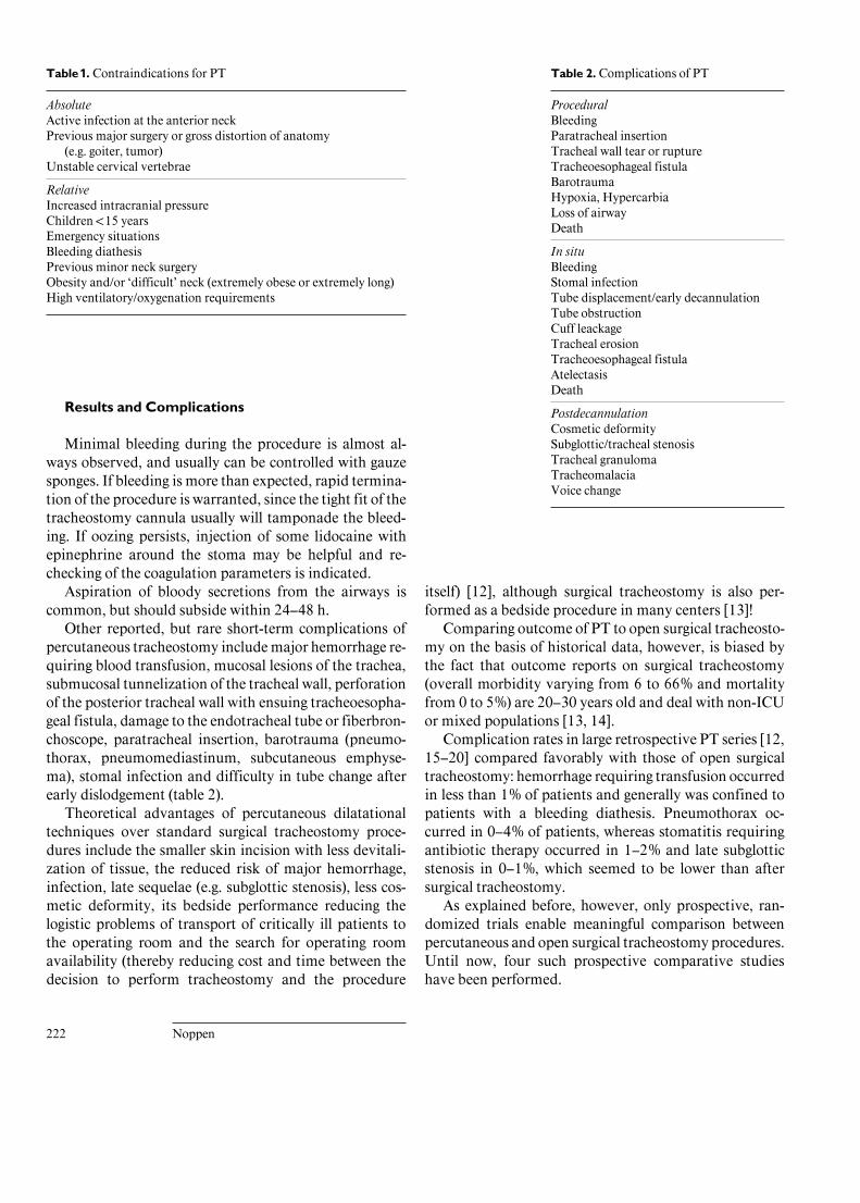
HABE:L08:BPRR3010XA.81 FF: RESF E1: RESS
222 Noppen
Table 1. Contraindications for PT
AbsoluteActive infection at the anterior neckPrevious major surgery or gross distortion of anatomy
(e.g. goiter, tumor)Unstable cervical vertebrae
RelativeIncreased intracranial pressureChildren !15 yearsEmergency situationsBleeding diathesisPrevious minor neck surgeryObesity and/or ‘difficult’ neck (extremely obese or extremely long)High ventilatory/oxygenation requirements
Table 2. Complications of PT
ProceduralBleedingParatracheal insertionTracheal wall tear or ruptureTracheoesophageal fistulaBarotraumaHypoxia, HypercarbiaLoss of airwayDeath
In situBleedingStomal infectionTube displacement/early decannulationTube obstructionCuff leackageTracheal erosionTracheoesophageal fistulaAtelectasisDeath
PostdecannulationCosmetic deformitySubglottic/tracheal stenosisTracheal granulomaTracheomalaciaVoice change
Results and Complications
Minimal bleeding during the procedure is almost al-ways observed, and usually can be controlled with gauzesponges. If bleeding is more than expected, rapid termina-tion of the procedure is warranted, since the tight fit of thetracheostomy cannula usually will tamponade the bleed-ing. If oozing persists, injection of some lidocaine withepinephrine around the stoma may be helpful and re-checking of the coagulation parameters is indicated.
Aspiration of bloody secretions from the airways iscommon, but should subside within 24–48 h.
Other reported, but rare short-term complications ofpercutaneous tracheostomy include major hemorrhage re-quiring blood transfusion, mucosal lesions of the trachea,submucosal tunnelization of the tracheal wall, perforationof the posterior tracheal wall with ensuing tracheoesopha-geal fistula, damage to the endotracheal tube or fiberbron-choscope, paratracheal insertion, barotrauma (pneumo-thorax, pneumomediastinum, subcutaneous emphyse-ma), stomal infection and difficulty in tube change afterearly dislodgement (table 2).
Theoretical advantages of percutaneous dilatationaltechniques over standard surgical tracheostomy proce-dures include the smaller skin incision with less devitali-zation of tissue, the reduced risk of major hemorrhage,infection, late sequelae (e.g. subglottic stenosis), less cos-metic deformity, its bedside performance reducing thelogistic problems of transport of critically ill patients tothe operating room and the search for operating roomavailability (thereby reducing cost and time between thedecision to perform tracheostomy and the procedure
itself) [12], although surgical tracheostomy is also per-formed as a bedside procedure in many centers [13]!
Comparing outcome of PT to open surgical tracheosto-my on the basis of historical data, however, is biased bythe fact that outcome reports on surgical tracheostomy(overall morbidity varying from 6 to 66% and mortalityfrom 0 to 5%) are 20–30 years old and deal with non-ICUor mixed populations [13, 14].
Complication rates in large retrospective PT series [12,15–20] compared favorably with those of open surgicaltracheostomy: hemorrhage requiring transfusion occurredin less than 1% of patients and generally was confined topatients with a bleeding diathesis. Pneumothorax oc-curred in 0–4% of patients, whereas stomatitis requiringantibiotic therapy occurred in 1–2% and late subglotticstenosis in 0–1%, which seemed to be lower than aftersurgical tracheostomy.
As explained before, however, only prospective, ran-domized trials enable meaningful comparison betweenpercutaneous and open surgical tracheostomy procedures.Until now, four such prospective comparative studieshave been performed.

HABE:L08:BPRR3010XA.81 FF: RESF E1: RESS
Percutaneous Dilatational Tracheostomy 223
In 1991, Hazard et al. [21] reported the results of astudy that randomized 46 mechanically ventilated pa-tients with respiratory failure to either conventional oper-ative tracheostomy or PT. Operative tracheostomies wereperformed in either the ICU or the operating room, undereither local or general anesthesia by experienced board-certified surgeons. All of the percutaneous tracheostomieswere performed under local anesthesia and intravenoussedation at the bedside in the ICU by or under the super-vision of one of the investigators. Hemorrhage, infectionand pneumothorax were the commonest early adverseevents in both groups. These complications occurred sig-nificantly more commonly in patients who had operativetracheostomies (45.8%) than in patients who had percuta-neous tracheostomies (12.5%). Delayed stomal healing,significant subglottic stenosis and cosmetic deformitieswere significantly commoner after operative tracheosto-my (88%) than after percutaneous tracheostomy (27%).In summary, percutaneous tracheostomy was associatedwith a lower rate of both early and late adverse eventscompared with operative tracheostomy.
In 1995, Crofts et al. [22] published the results of a pro-spective clinical trial in which they randomized 53 criti-cally ill mechanically ventilated patients to either open orpercutaneous tracheostomy. Open tracheostomies wereperformed in the operating room by consulting surgeons,whereas percutaneous tracheostomies were performed inthe ICU by supervised otolaryngology housestaff. The fre-quency of tracheostomy complications occurring during a2-week follow-up period was low in both groups. Therewere no significant differences in the incidence of hemor-rhage or other complications between the groups. Theonly stomal infection occurred in the open tracheostomygroup.
In 1996, Friedman et al. [12] randomized 53 patientsto either PT performed in the ICU by 1 of 2 intensivists orto open tracheostomy performed in the operating room by1 of 4 surgeons. All PTs were performed under local anes-thesia and intravenous sedation, whereas the open proce-dures were performed under general anesthesia. Therewere no significant differences in the rates of intraproce-dural complications (35 vs. 41%; PT vs. open tracheosto-my, respectively). However, postprocedural complica-tions (accidental decannulation, bleeding, or stomal infec-tion) occurred significantly more frequently in patientsrandomised to open tracheostomy (12 vs. 41%; PT vs.open tracheostomy, respectively). It was noted that the PTtracheostomy tubes appeared securer. Only 1 patient inthe PT group experienced premature decannulation ver-sus 4 patients in the open tracheostomy group. Despite
the presence of retention sutures in the patients with sur-gical tracheostomies, the tracheostomy tube could not bereinserted in 2 of the 4 patients who experienced prema-ture decannulation, resulting in death of both.
In a recent preliminary report, Heikkinen et al. [23]randomized 40 patients to PT or open tracheostomy. Alltracheostomies were performed at the bedside under localanesthesia and intravenous sedation. There were no sig-nificant differences in complication rates between the twogroups, except that the PT group required more intrave-nous sedation because ‘the guidewire irritated the trachea’(probably because no lidocaine was injected into the tra-cheal lumen).
Discussion
It should be clear now, that PT techniques are swiftand safe procedures in experienced hands, with a risk ofearly and late complications equal to or lower than that ofopen surgical tracheostomy.
Furthermore, since PT is a bedside technique, obviat-ing the need for transportation of critically ill patients,and reducing the treatment delay and costs [24, 25], itmay well represent the preferred technique for electivetracheostomy in critically ill patients, and may evenchange our general approach to tracheostomy in the ICUpatient.
However, as for all new ‘simplified’ techniques, espe-cially when available for surgeons as well as for non-surgi-cal specialists and thus situated in the ‘grey zone’ betweensurgery and medicine, controversies and discussions –sometimes emotional or ill-based – arise. Similar contro-versies have taken place when fiberoptic or rigid interven-tional endoscopic techniques in bronchology and gastro-enterology were developed, and are still taking place inthoracoscopy and, to a lesser extent, in the field of inter-ventional bronchoscopic procedures.
Surgeons, obviously, state that tracheostomy – whetherpercutaneous or open – is a surgical procedure, and thatwhoever performs a procedure should be able to handlethe emergent complications associated with it.
Although intrinsically logic, this statement would im-ply that, e.g. no cardiologist would be allowed to performcoronary balloon angioplasty, no pulmonologist would beallowed to perform laser therapy in the airways, no neph-rologist would be allowed to perform a renal biopsy, andno gastroenterologist would be allowed to perform a poly-pectomy in the colon.

HABE:L08:BPRR3010XA.81 FF: RESF E1: RESS
224 Noppen
Inversely, however, no cardiologist would be allowedto treat cardiac arrhythmia after coronary surgery, no pul-monologist would be allowed to diagnose and treat post-operative (ventilator-associated) pneumonia, no gastroen-terologist would be allowed to dilate postoperative anasto-motic digestive stenosis, and no nephrologist would beallowed to take care of postoperative renal insufficiency.Protectionist reflexes should never interfere with a keyelement in the success of any medical or surgical act:teamwork. As to the tracheostomy discussions, surgeonsare correct when they state that a lost airway can result indeath within minutes, that – as a result – there is no timeto call for back up, and that the ‘procedurist’ thereforeshould be able to perform emergency airway procedures.As proven by the various large patient series published bynonsurgical ICU specialists, they are not correct whensaying that only surgeons can possess these qualifica-tions.
It should be obvious, however, that any physician (sur-geon, anaesthesiologist, medical intensivist) performingPT should have expertise in all aspects of airway manage-ment, including intubation, bronchoscopy and cricothy-roidotomy. Credentials for PT should have the samerequirements as for any (surgical) procedure: formal train-ing, an apprenticeship and ongoing peer review. Experi-ence in open surgical tracheostomy per se should not be asubstitute for formal training and apprenticeship in PTbecause the procedures are similar.
Far better still than these ‘pro-con’ discussions on whoshould perform PT, would be to stimulate local activecooperation between the various specialists at the ICUbedside, the specification of which may differ from insti-tution to institution. In our hospital for instance, PT isperformed by a surgeon, endotracheal tube handling, ven-tilation and oxygenation are controlled by an anesthesiol-ogist, and per- and postoperative bronchoscopic control isperformed by a pulmonologist. In our opinion, this coop-eration – when feasible – represents the best guarantee foroptimal therapeutic outcome.
Although four [12, 21–23] prospective comparativestudies have proven PT to be equal and in some aspectseven superior to classical open surgical tracheostomy,some issues of concern or criticism remain.
PT might be associated with an increased risk of latetracheal stenosis because it is generally performed at ahigher tracheal interspace than is open tracheostomy.However, when symptomatic tracheal stenosis has beenobserved after percutaneous tracheostomy, preexistingpathology of the airway – a recognized risk factor for thedevelopment of tracheal stenosis after open surgical tra-
cheostomy – was invariably present. Retrospective andprospective studies, however, showed that late stenosisactually is less often observed after percutaneous thanafter operative tracheostomy [18–20]. Risk factors for thislate complication include preexisting laryngotrachealdamage (e.g. due to long-standing endotracheal intuba-tion) and lack of experience with the PT procedure.
Fiberbronchoscopic control during the PT procedurecan reduce minute ventilation and cause hypercapnia.This complication can easily be avoided by introducingthe bronchoscope intermittently only during certain criti-cal stages of the procedure (e.g. during endotracheal tuberetraction, needle introduction in the trachea, guidewirepassage, dilator introduction and cannula position con-trol), by using a small-diameter bronchoscope (e.g. pediat-ric size), and by avoiding continuous suction through thebronchoscope. When these precautions are taken, we (andothers) have never been confronted with hypoxemia, hy-percarbia or dynamic hyperinflation.
The fear for paratracheal insertion, which has beensupported by cadaveric studies, is inherent to a blind pro-cedure such as PT. However, although not universallyadvocated by experienced surgeons or intensivists, fiber-bronchoscopic control essentially eliminates this poten-tially life-threatening complication [26].
In our opinion, therefore, although it may increase thecost and logistic complexity of the procedure, fiberbron-choscopic control during the critical stages of percuta-neous tracheostomy should be highly advocated.
The reduced logistic complexity of a bedside procedureis another major advantage of percutaneous tracheostomy(although surgical tracheostomy may also be performed atthe bedside, many surgeons prefer the facilities of theoperating theatre). Bedside PT thus avoids the potentiallydangerous time- and personnel-consuming patient trans-port to the operating room. Furthermore, the treatmentdelay (i.e. the time between the decision to perform tra-cheostomy and the actual performance) has been shownto be substantially reduced (from 100.4 to 28.5 h) inpatients randomized for PT as compared to those ran-domized to open surgical tracheostomy [12]. The simplic-ity of PT also leads to significantly reduced operatingtime: both set-up and actual procedural times with PTaverage one third of those with open tracheostomy.
Although increased speed in itself probably is not ofprimal concern [13], it is obvious that, if two proceduresare equally safe or if the faster procedure is associatedwith a lower risk of complications, speed certainly is notdisadvantageous [11].

HABE:L08:BPRR3010XA.81 FF: RESF E1: RESS
Percutaneous Dilatational Tracheostomy 225
Nevertheless, considerable variations in PT perfor-mance times have been observed, probably relating to theskill of the operator or individual patient characteristics(e.g. obese patients, short neck).
Again, mainly due to its relative simplicity and bedsideperformance, PT has been claimed to be less costly thanopen surgical tracheostomy. Several studies, North Amer-ican as well as European, have estimated true costs andcharges for PT to be approximately 50% of those of opentracheostomy. Cost savings are largely due to eliminationof operating room charges and largely overcome the costof the disposable kit, bronchoscopy and second/thirdassisting physician inherent to the PT procedure.
Although an open surgical tracheostomy procedureperformed at the bedside probably would be the leastexpensive method of performing tracheostomy, currentpractice in the vast majority of hospitals where open sur-gical tracheostomy is performed in the operating room,undoubtedly makes PT the most cost-effective method.
Acknowledgment
The author thanks Dr. Dirk Smets (ICU and thoracic surgeon) forhis co-operation.
References
1 Trendelenburg F: Beiträge zu den Operationenan den Luftwegen. Arch Klin Chir 1870;12:112–33.
2 Jackson C: Tracheostomy. Laryngoscope 1909;19:285–290.
3 Galloway TC: Tracheostomy in bulbar polio-myelitis. JAMA 1943;123:1096–1097.
4 Sheldon CH, Pudenz RH, Tichy FY. Percuta-neous tracheostomy. JAMA 1957;165:2068–2070.
5 Toy FJ, Weinstein JD: A percutaneous tra-cheostomy device. Surgery 1969;65:384–389.
6 Brantigan CO, Grow JB: Cricothyroidotomy:Elective use in respiratory problems requiringtracheostomy. Surgery 1976;71:72–81.
7 Ciaglia P, Firsching R, Syniec C: Elective per-cutaneous dilatational tracheostomy: A newsimple bedside procedure. Preliminary Report.Chest 1985;87:715–719.
8 Schachner A, Ovil Y, Sidi J, Rogev M, Heil-bronn Y, Levy MJ: Percutaneous tracheostomy– A new method. Crit Care Med 1989;17;1052–1056.
9 Griggs WM, Worthley LI, Gilligan JE, et al: Asimple percutaneous tracheostomy technique.Surg Gynecol Obstet 1990;170:543–545.
10 Ernst A, Garland R, Zibrak J: Percutaneoustracheostomy. J Bronchol 1998;5:247–250.
11 De Boisblanc BP: Percutaneous dilatationaltracheostomy versus standard tracheostomy.Pro percutaneous dilatational tracheostomy. JBronchol 1998;5:251–256.
12 Friedman Y, Fildes J, Mizock B, et al: Compar-ison of percutaneous and surgical tracheostom-ies. Chest 1996;110:480–485.
13 Schweitzberg SD: Percutaneous dilatationaltracheostomy versus standard tracheostomy.Pro standard tracheostomy. J Bronchol 1998;5:257–260.
14 Stauffer JL, Olson DE, Petty TL: Complica-tions and consequences of endotracheal intuba-tion and tracheostomy: Prospective study of150 critically ill adult patients. Am J Med1981;70:65–76.
15 Leinhardt DJ, Mughal M, Bowles B, et al:Appraisal of percutaneous tracheostomy. Br JSurg 1992;79:255–258.
16 Fernandez L, Norwood S, Roettger R, et al:Bedside percutaneous tracheostomy with bron-choscopic guidance in critically ill patients.Arch Surg 1996;131:129–132.
17 Graham JS, Mulloy RH, Sutherland FR, et al:Percutaneous versus open tracheostomy: Ret-rospective cohort outcome study. J Trauma1996;41:245–248.
18 Law RC, Carney AS, Manara AR: Long-termoutcome after percutaneous dilational tra-cheostomy: Endoscopic and spirometry find-ings. Anaesthesia 1997;52:51–56.
19 Fischler MP, Kuhn M, Cantieni R, et al: Lateoutcome of percutaneous dilatational tracheos-tomy in intensive care patients. Intensive CareMed 1995;21:475–481.
20 Holdgaard HO, Pedersen J, Paaske P, et al:Percutaneous dilatational tracheostomy. ActaAnaesthesiol Scand 1996;40:838–841.
21 Hazard P, Jones C, Benitone J: Comparativeclinical trial of standard operative tracheosto-my with percutaneous tracheostomy. Crit CareMed 1991;19:1018–1024.
22 Crofts SL, Alzeer A, McGuire GP, et al: A com-parison of percutaneous and operative tra-cheostomies in intensive care patients. Can JAnaesth 1995;42:775–779.
23 Heikkinen M, Aarnio P, Hannukainen J: Per-cutaneous dilatational tracheostomy or con-ventional tracheostomy (abstract). Chest 1997;112(suppl 3):1S.
24 Toursarkissian B, Zweng TN, Kearney PA, Po-fahl WE, Johnson SB, Barber DE: Percuta-neous dilatational tracheostomy: Report of 141cases. Ann Thorac Surg 1994;57:862–867.
25 Imami ER, Komer K, Martin M: Conventionalopen versus percutaneous tracheostomy: Expe-rience in 110 patients with cost analysis (ab-stract). Presented at the Meeting of the RoyalCollege of Physicians and Surgeons of Canada,1994.
26 Barba CA, Angood PB, Kauder DR, et al.Bronchoscopic guidance makes percutaneoustracheostomy a procedure easy to teach, safe,and cost effective. Surgery 1995;118:879–883.
M. Noppen, MD, PhD, Respiratory DivisionAcademic Hospital AZ-VUB Laarbeeklaan 101B–1090 Brussels, BelgiumTel. +32 2 477 63 44, Fax +32 2 477 60 19E-Mail [email protected]

OOOOOOOOOOOOOOOOOOOOOOOOOOOOOOOOOOOOOOOOOOOOOOOOOOOOOOOOOOOOOOOOOOOOOOOOOOOOOOOOOOOOOOOOOOOOOOOOOOOOOOOOOOOOOOOOOOOOOOOOOOO
Other Procedures

Bolliger CT, Mathur PN (eds): Interventional Bronchoscopy.Prog Respir Res. Basel, Karger, 2000, vol 30, pp 226–234
OOOOOOOOOOOOOOOOOOOOOOOOOOOOOOOOOOOOOOOOOOOOOOOOOOOOOOOOOOOOOOOOOOOOOOOOOOOOOOOOOOOOOOOOOOOOOOOOOOOOOOOOOOOOOOOOOOOOOOOOOOO
Emergency TranstrachealOxygenation Techniques andLong-Term TranstrachealOxygen TherapyPeter Biroa Erich W. Russib
aDepartment of Anesthesiology and bPulmonary Division, University Hospital of Zurich, Switzerland
226
Summary
Maintenance of adequate gas exchange and protection ofthe airway against aspiration of gastric content are the maingoals of the anesthesiological and emergency management ofthe difficult airway. Meanwhile, there are accepted guidelinesand algorithms for a rational approach to this problem and avariety of techniques and equipment has been evaluated andapproved for this purpose. Among specially developedequipment that may help to visualize the larynx or to intro-duce endotracheal or pharyngeal tubes and analogous de-vices, transtracheal puncture remains an important routefor oxygen application in the ‘cannot intubate – cannot venti-late’ scenario. This method has to be considered if any of thetransoral accesses turns out to be not feasible and less inva-sive techniques have failed, or are not indicated. The optimalplace for transcutaneous puncture is located right above theupper margin of the cricoid cartilage and midline position,since the cricothyroid membrane is nearly completely freeof vessels and can be punctured without a high risk forbleeding, unless a clinically relevant coagulopathy is present.There are two different categories of nonsurgical transtra-cheal equipment: (1) transtracheal puncture with narrowcannulas and catheters that do not require initial skin inci-sion, and (2) large bore cricothyrotomy cannulas and cathe-ters. This differentiation is essential since narrow cannulas
require high-pressure oxygen delivery for oxygenation andventilation, while large bore cricothyrotomy cannulas andcatheters enable regular (low-pressure) respiration and ven-tilation. Therefore, the availability of the ventilation systemdecides primarily the choice of the transtracheal equipment,but certain medical conditions also have an influence on thefeasibility of the chosen material. If previously attemptedoxygenation, ventilation and intubation techniques havefailed, a nonsurgical oxygenation technique seems to beinappropriate, and there is no possibility to return soon tospontaneous ventilation, a surgical airway is urgently re-quired. A surgical cricothyrotomy can be completed in lessthan 1 min of operating time when the anatomy is favorable.When no surgeon is available and as a last resort, this life-saving intervention should be carried out without hesitationby any involved physician. Long-term oxygen therapy(LTOT) is an established treatment for endstage lung diseaseaccompanied by hypoxemia. Oxygen delivery by nasal can-nula is an uncomfortable mode of application for the patientand goes along with a decreased compliance. In contrast,transtracheal application of oxygen via a small catheter,which can be removed and cleaned by the patient over aminitracheostoma performed with local anesthesia on anoutpatient basis circumvents these problems and enhancesconsiderably the efficiency of LTOT.

HABE:L08:BPRR3006XA.81 FF: RESF E1: RESS
Transtracheal Oxygenation Techniques 227
One of the most concerning and potentially life-threat-ening challenges is unexpected difficulty in securing theairway and maintaining an adequate gas exchange. Al-though the term ‘difficult intubation’ is predominantlyused in this context, the problem can encompass all facetsof airway control, oxygenation and carbon dioxide elimi-nation. The first and foremost concern has to be mainte-nance of adequate oxygenation. It should always havepriority in comparison to intubation and even to ventila-tion. Next in importance is ventilation; this can be pro-vided by expedient means, including face mask or laryn-geal mask ventilation, and does not generally require tra-cheal intubation. The final consideration is to provide asecure, stable airway that can be used for an extendedperiod. Alternative techniques for oxygenation and venti-lation in the difficult airway have different degrees ofavailability, complexity and invasiveness. Simple tech-niques should be favored, whereas more invasive meth-ods should be regarded as a backup to rely on when theinitial technique has failed. However, when a ‘cannotintubate – cannot ventilate’ situation occurs, direct trans-cutaneous airway access is a rapid way to provide oxygen-ation. The cascade of options promoted by the AmericanSociety of Anesthesiologists includes standard and modi-fied intubation attempts, mask ventilation, the use ofnumerous auxiliary devices such as laryngeal masks, theesophageal-tracheal combitube (a double-lumen devicethat is blindly introduced into the hypopharynx and isintended to allow tracheal ventilation via the lumen thatmatches the airway) and transtracheal oxygenation tech-niques [1]. The latter can be used, either to administeroxygen alone in order to prevent hypoxia or at least toachieve rapid reversal of hypoxemia, and under favorablecircumstances to establish additional CO2 elimination. Inany case, oxygenation is the primary concern of theseefforts. Usually, the establishment of a secure and patentairway has to be performed additionally after life threathas been overcome due to successful application of trans-tracheal oxygen.
Emergency Transtracheal OxygenationTechniques
Anterior Cervical AnatomyKnowledge of the cervical anatomy and especially of
the predominant vascular pattern is of utmost impor-tance. The same is true for the careful recognition of thetranstracheal puncture site [2]. The distance from the skinsurface to the airway lumen is mostly overestimated, but
usually it is only about 5 mm. The anterior cervical regionis well vascularized, but the cricothyroid membrane isnearly completely free of vessels and can be puncturedwithout a high risk for bleeding, unless a clinically rele-vant coagulopathy is present. The nearest vessel is a smallartery which follows horizontally the caudal edge of thethyroid cartilage. Therefore, the ideal place for the trans-cutaneous access to the airway is located right above theupper margin of the cricoid cartilage in midline position.This entrance to the airway also has the advantage ofbeing the single place where a rigid dorsal wall which con-sists of the posterior part of the ring-shaped cricoid carti-lage is present, and therefore protects the esophagusagainst inadvertent perforation. Nevertheless, in slim in-dividuals, a more caudal puncture site is also possible, butone must be aware of the risks related to injury of theabundantly vascularized thyroid gland and the possibilityof penetration through the posterior tracheal wall.
Performance of Transtracheal PuncturePercutaneous puncture and catheter placement should
be performed in a standardized fashion [3]. The patientmust be positioned with the neck in maximal extension.The head retroflection can be facilitated by means of afolded cover placed under the shoulders of the patient.The benefit of this maneuver is an anterior displacementof the larynx effectuated by the cervical spine. Additional-ly, the skin will be more tightened and the puncture sitecan be recognized much easier. The larynx should be sta-bilized laterally by one hand, while the other performs thepuncture. During tracheal penetration, the cannula mustbe kept perpendicular to the skin, and a characteristic lossof resistance will be noted when the airway is entered.Before advancing the catheter, it is essential to confirmintratracheal positioning by free aspiration of air. For thispurpose, a 10-ml syringe should be used that can contain2 ml of water in order to make air bubbles visible (fig. 1).At this point, the cannula tip is tilted caudally at an angleof 30° to the skin, and the catheter is advanced into thetrachea while holding the introducer stationary. If a sys-tem based on the Seldinger® technique is used, the guide-wire must be advanced before the catheter is moved for-ward. Following a second-position control by free air aspi-ration, the catheter is fixed to the skin and ventilation isbegun. As with any delicate invasive procedure, there is adefinite learning curve, and transtracheal puncture andcatheter placement should not be attempted for the firsttime in an emergent situation. Practice can be obtainedeither in elective cases (maxillofacial or laryngeal surgery)or on the cadaver.

HABE:L08:BPRR3006XA.81 FF: RESF E1: RESS
228 Biro/Russi
Fig. 1. Confirmation of intratracheal loca-tion of the cannula tip by air aspiration priorto advancement of the flexible catheter.
Transtracheal EquipmentFrom the technical point of view, two different catego-
ries of nonsurgical transtracheal equipment must be dis-tinguished: (1) transtracheal puncture with narrow cannu-las and catheters (ID usually !3 mm) and (2) cricothy-rotomy performed with large bore cricothyrotomy cannu-las and catheters. This differentiation is essential, sinceboth techniques enable only the application of specificoxygenation/ventilation modes. Narrow cannulas requirehigh-pressure oxygen delivery for oxygenation and venti-lation. Low-pressure insufflation from the flush valve ofan anesthesia machine can only prevent hypoxia, but eventhis limited achievement may be a valuable benefit, sinceit results in additional time for further airway manage-ment measures [4]. Squeezing a hand bag or ventilationvia a circuit system usually fails to bring sufficient oxygenthrough narrow cannulas into the trachea. In contrast,large bore cannulas are designed for regular ventilationand should not be fitted to high-pressure oxygen sourcesor a jet ventilator. In order to prevent misunderstandings,commercially available equipment is designed accordingto the underlying methodological options: narrow cathe-ters have a luer-lock connection that can be fitted with jetventilation or insufflation devices, whereas large bore cri-cothyrotomy cannulas have a regular 15-mm connectorfor linkage with conventional ventilation systems. Unfor-tunately, some narrow cannulas are equipped with bothconnection types, and the 15-mm connector may misleadthe user to try conventional ventilation, which in most
cases may not be sufficient. It is essential to clarify inadvance which kind of transtracheal equipment should beadopted in a specific location, whether it should be basedon narrow or large bore cannulas and their respective oxy-gen delivery systems or whether a surgical cricothyrotomyshould be preferred. Before the decision is made as towhich system should be adopted, the specific advantagesand disadvantages of all types of equipment should beconsidered (tables 1–3). However, the availability ofequipment is not the only consideration in this context.Certain medical conditions of the involved patient alsohave an influence on the feasibility of these differenttranstracheal approaches. On an individual scale, theremay also be arguments in favor of one or the other of thepresented methods, and the appropriate choice can onlybe made accordingly if all kinds of techniques are avail-able (table 4).
Oxygen Delivery SystemsSufficient oxygenation can be provided by percuta-
neous transtracheal oxygen insufflation using narrow can-nulas and low-pressure sources like the flush valve of ananesthesia machine. Though not allowing ventilation, thisextremely simple and minimally invasive method canprovide sufficient time to attempt alternative intubationtechniques or create a more comfortable surgical airway[5, 6]. Since a sufficient carbon dioxide elimination can-not be achieved this way, this technique can only beadopted for a limited period of at least 15–20 min. Nei-

Less invasive and less traumatic
Suitable for regular (low-pressure) ventilation
Offers best ventilation conditions
Difficult anatomical approach
HABE:L08:BPRR3006XA.81 FF: RESF E1: RESS
Transtracheal Oxygenation Techniques 229
Table 1. Advantages and disadvantages of narrow transtracheal cannulas and catheters
Advantages Disadvantages
Unsuitable for regular (low-pressure) ventilationSimpler positioning and faster installation Require additional exhalation outletSuitable for retrograde intubation Airway remains unprotected against aspiration
(with rigid cannulas only) Higher risk of kinking, bending, distortion and obstructionSuitable for insertion under local anesthesia of the catheter
Table 2. Advantages and disadvantages of large bore transtracheal cannulas and catheters
Advantages Disadvantages
More invasiveDo not require additional exhalation outlet Cause more tissue damageOffer a fair airway patency and protection Insertion is more difficult, the installation is more complex
Table 3. Advantages and disadvantages of surgical cricothyrotomy
Advantages Disadvantages
Is the most invasive methodDoes not require additional exhalation outlet Requires surgical instruments and certain surgical skillOffers best airway patency and protection Requires longest time period to be completed
Table 4. Arguments for decision makingand choice of appropriate emergencytranstracheal technique
Narrowcannula
Large borecannula
Surgicalcricothy-rotomy
++ – +++Only regular (low-pressure) oxygen source
available – ++ +++Coagulopathy ++ – +Indication of awake application (under local
anesthesia) +++ – +Upper airway stenosis – ++ +++Shortage of time, imminent asphyxia +++ ++ –

HABE:L08:BPRR3006XA.81 FF: RESF E1: RESS
230 Biro/Russi
Fig. 2. A narrow Ravussin-type transtrachealcannula with catheter (left) for jet insuffla-tion and a large bore Quicktrach cannulawith catheter (right) for positive pressureventilation.
ther the demand-valve device of an anesthesia machine,nor the bag-valve device can deliver the 1.5–4.0 atmrequired for adequate flow through such a narrow cannu-la. Only gas cylinders or a central source will be capable ofgenerating sufficient oxygen pressure. A simple, low-costhand-triggered jet injector is adequate. When the catheteris in place and connected to the injector, ventilation isbegun at a rate of 15–20 cycles per minute. Oxygenation ismonitored by pulse oxymetry. Effective ventilation oc-curs if thoracic excursion can be seen, and insufflationpressure should be adjusted according to the movement ofthe subclavicular region. Electronically operated jet venti-lators have the advantage of on-line measurement of air-way pressure with inbuilt automatic shut-down mecha-nism if a preset airway pressure limit is exceeded. To pro-vide an egress for insufflated oxygen, it is important toensure that the upper airway is at least partially open.This can be facilitated with an oro- or nasopharyngeal air-way if necessary. For both transtracheal oxygen insuffla-tion and jet ventilation, specially designed cannulas andcatheters such as the Patil-setTM, the Arndt-setTM, and theRavussin-type transtracheal catheter are available (fig. 2).To avoid potentially fatal complications, it is essentialthat the equipment is not assembled from random parts(intravenous catheters such as angiocath, syringe barrels,3-way stopcocks, capnography tubing, standard 15-mm
connectors). The contraindications include intrathoracicairway obstruction, inability to locate the cricothyroidmembrane, severe pretracheal tissue distortion throughwhich the cannula would be too short to penetrate andcomplete airway obstruction. The latter is essential, as anyventilation technique that depends on passive exhalationthrough an open outlet can increase intrathoracic pressureand may subsequently cause barotrauma [5].
Nonsurgical CricothyrotomyPercutaneous insertion of a large bore catheter is faster
and less invasive than a formal surgical cricothyrotomyand does not require surgical instruments. On the otherhand, this technique is somewhat more difficult and moreinvasive than transtracheal puncture with narrow cannu-las. Several nonsurgical cricothyrotomy kits are commer-cially available, such as QuicktrachTM, Nu-TrakeTM,Melker-setTM and TracheoquickTM, differing in shape,components and some details concerning the method ofplacement (fig. 2). They typically contain a sharp bladefor initial skin incision, an introducer, guidewires, dila-tors and a large bore catheter with a standard 15-mm con-nector. These devices are favored when conventional ven-tilation with a bag-valve device is the only possibilityavailable. The insertion technique is quite similar to theone adopted with narrow cannulas: positioning of the
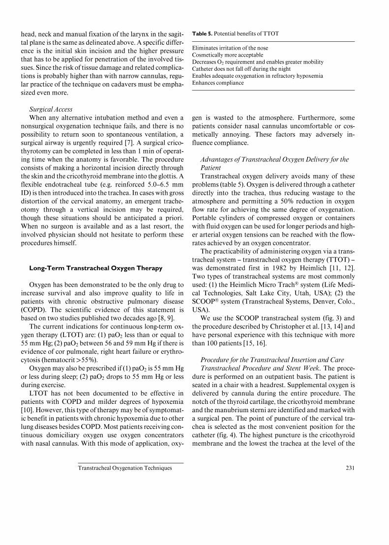
HABE:L08:BPRR3006XA.81 FF: RESF E1: RESS
Transtracheal Oxygenation Techniques 231
head, neck and manual fixation of the larynx in the sagit-tal plane is the same as delineated above. A specific differ-ence is the initial skin incision and the higher pressurethat has to be applied for penetration of the involved tis-sues. Since the risk of tissue damage and related complica-tions is probably higher than with narrow cannulas, regu-lar practice of the technique on cadavers must be empha-sized even more.
Surgical AccessWhen any alternative intubation method and even a
nonsurgical oxygenation technique fails, and there is nopossibility to return soon to spontaneous ventilation, asurgical airway is urgently required [7]. A surgical crico-thyrotomy can be completed in less than 1 min of operat-ing time when the anatomy is favorable. The procedureconsists of making a horizontal incision directly throughthe skin and the cricothyroid membrane into the glottis. Aflexible endotracheal tube (e.g. reinforced 5.0–6.5 mmID) is then introduced into the trachea. In cases with grossdistortion of the cervical anatomy, an emergent trache-otomy through a vertical incision may be required,though these situations should be anticipated a priori.When no surgeon is available and as a last resort, theinvolved physician should not hesitate to perform theseprocedures himself.
Long-Term Transtracheal Oxygen Therapy
Oxygen has been demonstrated to be the only drug toincrease survival and also improve quality to life inpatients with chronic obstructive pulmonary disease(COPD). The scientific evidence of this statement isbased on two studies published two decades ago [8, 9].
The current indications for continuous long-term ox-ygen therapy (LTOT) are: (1) paO2 less than or equal to55 mm Hg; (2) paO2 between 56 and 59 mm Hg if there isevidence of cor pulmonale, right heart failure or erythro-cytosis (hematocrit 155%).
Oxygen may also be prescribed if (1) paO2 is 55 mm Hgor less during sleep; (2) paO2 drops to 55 mm Hg or lessduring exercise.
LTOT has not been documented to be effective inpatients with COPD and milder degrees of hypoxemia[10]. However, this type of therapy may be of symptomat-ic benefit in patients with chronic hypoxemia due to otherlung diseases besides COPD. Most patients receiving con-tinuous domiciliary oxygen use oxygen concentratorswith nasal cannulas. With this mode of application, oxy-
Table 5. Potential benefits of TTOT
Eliminates irritation of the noseCosmetically more acceptableDecreases O2 requirement and enables greater mobilityCatheter does not fall off during the nightEnables adequate oxygenation in refractory hypoxemiaEnhances compliance
gen is wasted to the atmosphere. Furthermore, somepatients consider nasal cannulas uncomfortable or cos-metically annoying. These factors may adversely in-fluence compliance.
Advantages of Transtracheal Oxygen Delivery for thePatientTranstracheal oxygen delivery avoids many of these
problems (table 5). Oxygen is delivered through a catheterdirectly into the trachea, thus reducing wastage to theatmosphere and permitting a 50% reduction in oxygenflow rate for achieving the same degree of oxygenation.Portable cylinders of compressed oxygen or containerswith fluid oxygen can be used for longer periods and high-er arterial oxygen tensions can be reached with the flow-rates achieved by an oxygen concentrator.
The practicability of administering oxygen via a trans-tracheal system – transtracheal oxygen therapy (TTOT) –was demonstrated first in 1982 by Heimlich [11, 12].Two types of transtracheal systems are most commonlyused: (1) the Heimlich Micro Trach® system (Life Medi-cal Technologies, Salt Lake City, Utah, USA); (2) theSCOOP® system (Transtracheal Systems, Denver, Colo.,USA).
We use the SCOOP transtracheal system (fig. 3) andthe procedure described by Christopher et al. [13, 14] andhave personal experience with this technique with morethan 100 patients [15, 16].
Procedure for the Transtracheal Insertion and CareTranstracheal Procedure and Stent Week. The proce-
dure is performed on an outpatient basis. The patient isseated in a chair with a headrest. Supplemental oxygen isdelivered by cannula during the entire procedure. Thenotch of the thyroid cartilage, the cricothyroid membraneand the manubrium sterni are identified and marked witha surgical pen. The point of puncture of the cervical tra-chea is selected as the most convenient position for thecatheter (fig. 4). The highest puncture is the cricothyroidmembrane and the lowest the trachea at the level of the

HABE:L08:BPRR3006XA.81 FF: RESF E1: RESS
232 Biro/Russi
Fig. 3. SCOOP-II transtracheal catheter inplace and secured with a necklace.
Fig. 4. Needle with guidewire between cri-coid and first tracheal cartilage.
manubrium. Two percent lidocaine is injected into thedermis and the pretracheal soft tissue. Lidocaine is theninjected transtracheally for endotracheal anesthesia. Ascalpel is used to make a 1-cm vertical incision throughthe dermis at the puncture site. An 18-gauge thin-wallneedle attached to a syringe containing saline is thendirected through the incision into the trachea and air isfreely aspirated. The syringe is then removed, the needleangled downward toward the carina, an atraumatic end ofa wire guide is inserted through the needle into the tracheaup to 11 cm. The needle is withdrawn, and a 10-french
(1 french = 1/3 mm) dilator is advanced into the tracheaover the wire guide. The dilator is exchanged for a9-french tubular stent (Pre-SCOOP®; Transtracheal Sys-tems), which is passed over the wire guide. The stent isthen secured to the skin with two sutures. Codeine and anantibiotic are prescribed for 7 days.
TTOT over the Immature Tract. One week after theprocedure, a guidewire is used to exchange the stent for afunctioning transtracheal catheter. The catheter is a 9-french 20-cm-long reinforced tube with an internal lengthof 11 cm and one distal port (SCOOP-I®; Transtracheal

HABE:L08:BPRR3006XA.81 FF: RESF E1: RESS
Transtracheal Oxygenation Techniques 233
Table 6. Contraindications for a transtracheal catheter
AbsoluteAnatomic abnormality which makes insertion more difficult (severe
obesity, goiter, deviated trachea, previous tracheostomy)Severe coagulopathyProduction of copious amount of tenacious secretion (e.g. bronchi-
ectases)Risk of delayed healing (connective tissue disease, diabetes)Lack of compliance
RelativeInability for self-care
Table 7. Potential complications of a transtracheal catheter
Procedure relatedCephalad-displaced catheterSubcutaneous emphysema
Early and usually transient complicationsIncrease in coughIncrease in sputum productionExpectoration of blood-tinged sputumMinimal discomfort at the puncture siteLost tract
Complications making removal of the catheter mandatoryUpper airway obstruction by mucous ball formationPoor tolerance due to continuous irritationChondritis of a tracheal cartilage
Keloid formation
Systems). The catheter is secured around the neck withthe bead chain necklace and then connected to the oxygensource by an oxygen hose with a security clip attached tothe belt or other clothing to prevent inadvertent pull onthe catheter. Transtracheal flow is titrated to obtain anarterial oxygen saturation higher than 90%. Patients areinstructed to clean the catheter in place twice daily using3 ml of normal saline or possibly 2% lidocaine and acleaning rod.
During the cleaning procedure, patients often expecto-rate an accumulation of inspissated mucus which adheresto the catheter near the tip. If the mucus is not cleared, itcan increase in amount until it partially or totally ob-structs the trachea [17]. To minimize the risk of mucousball formation, emphasis is placed on adequate humidifi-cation, frequent cleaning and early perception. The pa-tient is instructed about symptoms which suggest mucousball accumulation and he is told to call if he perceives anyproblem. Complications with mucous ball formation arecommonest in this phase of the procedure, but may occuryears after an uneventful transtracheal oxygen use. Pa-tients with a very low peak flow and weak cough may beunable to dislodge the mucous ball by coughing. In thissituation, removal of the mucous ball by fiberoptic bron-choscopy may be necessary.
TTOT over a Mature Tract. The patient returns 6–8weeks after the initial procedure. The 9-french catheter isremoved and an 8-french catheter with multiple side ports(SCOOP-II®; Transtracheal Systems) is inserted withoutthe use of a guidewire. If any difficulty is encounteredwhen the catheter is inserted, the tract is assumed not tobe fully epithelialized, and the initial 9-french catheter isreinserted with the guidewire. The patient is told to usethis catheter for an additional 2-week period before tractmaturity is reevaluated. Once the tract is determined tobe mature, patients are taught to remove, clean and re-insert the second catheter on a twice-daily basis. Patientswho have problems to reinsert the catheter through themicrostoma may be instructed to use a guidewire.
TTOT is an ideal method to deliver continuous oxygenin a highly motivated and compliant patient who has thestrong desire to be active and mobile [18]. In other pa-tients on LTOT, the insertion of a transtracheal cathetermay be impossible or is contraindicated (table 6). TTOTis a method to be used by a team experienced with thistype of procedure and well aware of and trained to handlepotential complications (table 7).
Oxygen ConservationTo obtain the same degree of oxygenation in a given
patient only 50% of oxygen flow is required by transtra-cheal application as compared to the delivery by nasalcannula [14, 15]. This is advantageous for patients whocan only be adequately oxygenated with high flows of oxy-gen (e.g. pulmonary fibrosis). Furthermore, it enables thepatient to remain mobile and independent from his homebase oxygen source for an extended period of time.
Potential Physiological Benefit of TranstrachealOxygen DeliveryBesides the advantage of oxygen conservation, possible
explanations for the added benefit of TTOT relate to themode of administration more than the oxygen itself (ta-ble 8). Couser and Make [19] studied stable patients withdifferent lung diseases while they breathed room air ortranstracheally insufflated oxygen and found a decrease in

HABE:L08:BPRR3006XA.81 FF: RESF E1: RESS
234 Biro/Russi
Table 8. Physiological benefit oftranstracheal oxygen therapy Decrease in dead space O2 is delivered by the catheter directly into the
bypassing upper airways and decreasing deadspace
Increase in CO2 elimination efficiency CO2 flushes outward from the upper airwaysduring exhalation of the oxygen flow
Decrease in work of breathing
inspired minute ventilation measured at the mouth with-out changes in blood gas tension. Another group found adrop in patient-generated minute ventilation and nochange in paCO2, which is best explained by a decrease indead space [20, 21]. Benditt et al. [22] found that transtra-cheal delivery of oxygen or air reduces the oxygen cost ofbreathing as estimated by calculation of the pleural pres-sure-time index. TTOT was associated with a less demand-ing respiratory pattern of breathing for the diaphragm.
These findings help to explain the improvements inexercise tolerance and decrease of dyspnea that are notedclinically. Another elegant study showed in a randomized,double-blinded manner that patients with COPD have agreater exercise tolerance when using TTOT than whenusing nasal cannula delivery [23]. It was found that theimprovement is not related to improved arterial oxygena-tion.
References
1 Benumof JL: Management of the difficult air-way. Anesthesiology 1991;75:1087–1110.
2 Biro P: Transtracheal access and oxygenationtechniques. Acta Anaesthesiol Scand 1998;42(suppl 112):168–170.
3 Biro P, Moe KS: Emergency transtracheal jetventilation in high grade airway obstruction. JClin Anesth 1997;9:604–607.
4 Gaughan SD, Benumof JL, Ozaki AS: Can ananesthesia machine flush valve provide for ef-fective jet ventilation? (abstract). Anesth Analg1991;76:800.
5 Ryder IG, Paoloni CC, Harle CC: Emergen-cy transtracheal ventilation: Assessment ofbreathing systems chosen by anaesthetists.Anaesthesia 1996;51:764–768.
6 Yealy DM, Stewart RD, Kaplan RM: Mythsand pitfalls in emergency translaryngeal venti-lation: Correcting misimpressions. Ann EmergMed 1988;17:690–692.
7 DeLaurier GA, Hawkins ML, Treat RC, Mans-berger AR: Acute airway management. Role ofcricothyroidotomy. Am Surg 1990;56:12–15.
8 Medical Research Council Working Party:Long term domiciliary oxygen therapy inchronic hypoxic cor pulmonale complicatingchronic bronchitis and emphysema. Report ofthe Medical Research Council Working Party.Lancet 1981;i:681–686.
9 Nocturnal Oxygen Therapy Trial Group: Con-tinuous or nocturnal oxygen therapy in hypox-emic chronic obstructive lung disease. A clini-cal trial. Ann Intern Med 1980;93:391–398.
10 Gorecka D, Gorzelak K, Sliwinski P, TobiaszM, Zielinksi J: Effect of long-term oxygen ther-apy on survival in patients with chronic ob-structive pulmonary disease with moderatehypoxemia. Thorax 1997;52:674–679.
11 Heimlich HJ: Respiratory rehabilitation withtranstracheal oxygen system. Ann Otol RhinolLaryngol 1982;91:643–647.
12 Heimlich HJ: Transtracheal catheter techniquefor pulmonary rehabilitation. Ann Otol RhinolLaryngol 1985;94:502–504.
13 Christopher KL, Spofford BT, Brannin PK,Petty TL: Transtracheal oxygen therapy forrefractory hypoxemia. JAMA 1986;256:494–497.
14 Christopher KL, Spofford BT, Petrun MD,McCarty DC, Goodman JR, Petty TL: A pro-gram for transtracheal oxygen delivery. Assess-ment of safety and efficacy. Ann Intern Med1987;107:802–808.
15 Häggi J, Anderhub HP, Kronauer C, RussiEW: Transtracheale O2-Applikation zur Sauer-stoff-Langzeittherapie. Schweiz Med Wo-chenschr 1988;118:1321–1324.
16 Russi EW, Anderhub HP, Bloch K, Speich R:Erfahrungen mit der transtrachealen Langzeit-Sauerstofftherapie. Schweiz Rundsch MedPrax 1990;79:850–853.
17 Fletcher EC, Nickeson D, Costarangos-GalarzaC: Endotracheal mass resulting from a transtra-cheal oxygen catheter. Chest 1988;93:438–439.
18 Kampelmacher MJ, Deenstra M, van KesterenRG, Melissant CF, Douze JM, Lammers JW:Transtracheal oxygen therapy: An effective andsafe alternative to nasal oxygen administration.Eur Respir J 1997;10:828–833.
19 Couser JI, Make BJ: Transtracheal oxygen de-creases inspired minute ventilation. Am RevRespir Dis 1989;139:627–631.
20 Bergofsky EH, Hurewitz AN: Airway insuffla-tion: Physiological effects on acute and chronicgas exchange in humans. Am Rev Respir Dis1989;140:885–890.
21 Hurewitz AN, Bergofsky EH, Vomero E: Air-way insufflation. Increasing flow rates progres-sively reduce dead space in respiratory failure.Am Rev Respir Dis 1991;144:1229–1233.
22 Benditt J, Pollock M, Roa J, Celli BR: Trans-tracheal delivery of gas decreases the oxygencost of breathing. Am Rev Respir Dis 1993;147:1207–1210.
23 Wesmiller SW, Hoffman LA, Sciurba FC, Fer-son PF, Johnson JT, Dauber JH: Exercise toler-ance during nasal cannula and transtrachealoxygen delivery. Am Rev Respir Dis 1990;141:789–791.
P. Biro, MD, DEAADepartment of AnesthesiologyUniversity Hospital, Rämistrasse 100CH–8091 Zürich (Switzerland)Tel. +41 1 255 26 96, Fax +41 1 255 44 09E-Mail [email protected]
E.W. Russi, MD, FCCPPulmonary DivisionUniversity Hospital, Rämistrasse 100CH–8091 Zürich (Switzerland)Tel. +41 1 255 3828, Fax +41 1 255 4451E-Mail [email protected]

OOOOOOOOOOOOOOOOOOOOOOOOOOOOOOOOOOOOOOOOOOOOOOOOOOOOOOOOOOOOOOOOOOOOOOOOOOOOOOOOOOOOOOOOOOOOOOOOOOOOOOOOOOOOOOOOOOOOOOOOOOO
Interventional Bronchoscopybeyond the Year 2000

Bolliger CT, Mathur PN (eds): Interventional Bronchoscopy.Prog Respir Res. Basel, Karger, 2000, vol 30, pp 236–242
OOOOOOOOOOOOOOOOOOOOOOOOOOOOOOOOOOOOOOOOOOOOOOOOOOOOOOOOOOOOOOOOOOOOOOOOOOOOOOOOOOOOOOOOOOOOOOOOOOOOOOOOOOOOOOOOOOOOOOOOOOO
Autofluorescence Bronchoscopy:The Laser Imaging FluorescenceEndoscopeJohn A. Nakhosteen Barbara Khanavkar
RIDTELCTM, Research Institute for Diagnosis and Treatment of Early Lung Cancer,Augusta Teaching Hospital, Bochum, Germany
236
Summary
In conventional bronchoscopy, detection and localizationof dysplasia and carcinoma in situ (CIS) has been limited toroughly 30% of the total number thought to be present.Using a special light source which generates monochromallight of 442 Ìm and a digitalized integrated two-camera sys-tem 30,000 times more powerful than usual endoscopiccameras, the Laser Imaging Fluorescence Endoscope (LIFE®)system is said to be able to detect these lesions more readi-ly. In our initial results from 144 bronchoscopies with 253biopsies, in which a total of 50 severe dysplasias and/or CISwere cyto-histologically confirmed, 39 (78%) were correctlyidentified by the combined white light + LIFE modes, whileonly 15 (30%) were noted on white light alone. These resultsare largely in agreement with other international studies alsoreviewed in this chapter. The complication rate of the com-bined procedure is not higher than for conventional bron-choscopy alone, the intervention is performed under topicalanesthesia as an outpatient procedure, and the examinationtime is increased by approximately 10 min only, comparedto conventional flexible bronchoscopy. Improved ability todiagnose pre-neoplasias and early lung cancer lesions willnot only result in more lives being saved, but will also pro-vide whole new insights into carcinogenesis, with promisingramifications for yet further improvement in diagnosis andtherapy of lung cancer.
Assessment of earlier chest X-ray screening studies [1]and recent technological innovations have reawakenedinterest in early – or asymptomatic – lung cancer (ELC)diagnosis. Whereas X-ray may be helpful for peripherallesions, for centrally located, radio-occult and exfoliativetumors – usually squamous cell – sputum studies can beuseful [2]. Once suspicious cells have been detected, sensi-tive endoscopic procedures can be used to localize dis-crete, early lesions, most of which are curable [3]. Morethan 80% of X-ray-occult cancers are located in the visibleportion of the tracheo-bronchial tree [4], and, once local-ized, at least 80% can be cured, either through surgicalresection or, in cases of functional inoperability, throughinterventional procedures [5, 6]. In spite of most ELCsbeing situated within the visible range of endoscopes,their precise localization has been disappointing. Thisparadox is due primarily to the predominant intraepithe-lial growth pattern of most ELCs, which is difficult tovisualize, as compared to the easier-to-spot, yet more sel-dom, polypoid tumor. The intraepithelial pattern showsonly very discrete spur thickening and mucosal irregulari-ty, minor vessel dilatation, localized edema and distortedlight reflex, all of which are particularly difficult to recog-nize in the presence of chronic bronchitis. Even notableimprovement in fiber- and video-optic technology ofmodern, conventional endoscopes has not significantlyimproved diagnosis here [7]. Systematic segmental wash-

BOIS:L08:BPRR3015PD.98 FF: RESF E1: RESS
Autofluorescence Bronchoscopy 237
Fig. 1. Fluorescence light-emitting characteristics of normal bron-chial mucosa and bronchial mucosa altered by dysplasia and CIS.
Mucosa
Submucosa
Normal
Diagram of tissue autofluorescence
ä
ä
ä
Dysplastic
Blue (442 nm)
Green (520 nm)
Red (630 nm)
ä
ä
ä
ings for ELC localization are time-consuming and cum-bersome for the bronchology unit, and patient compliancefor repeated follow-up is low; Saito et al. [8], for example,reported a 25% dropout rate. The localization of ELC can,however, be significantly improved with the autofluores-cence bronchoscope, or the Laser Imaging FluorescenceEndoscope LIFE® system, described below.
Materials and Methods
‘Fluorescence’ is a physical phenomenon, a form of luminescenceor ‘cold’ light emission. It occurs when an object on which light of acertain wave length is directed remits light of a longer wave length. Itseffect on body tissue has been recognized since the early 1900s(‘Wood’s light’, [9]. In 1943, Herley [10] described fluorescence tis-sue reaction to ultraviolet light, noting even then that a tumorbehaved differently than healthy tissue. Substances responsible forfluorescence, fluorophores, yield organ-typical patterns, are variouslyconcentrated within organs and may change according to prevailingconditions. The many fluorophore subtypes include tryptophane,collagen, elastine and porphyrine; participating in the oxidativemetabolism NAD/NADH pyridoxal phosphate and flavines [11].Wave length and intensity of fluorophore-emitted light depends ontheir concentration, their maximal absorption and remission, and, ofcourse, on the characteristics of the exogenous light source.
Compared to light visible to the unaided eye, autofluorescence ofbronchial mucosa is extremely weak and cannot be visualized unlessstrongly amplified, an effect also caused by porphyrines, which selec-tively concentrate in malignant tissue. Efforts at using hematopor-phyrine derivatives (HpD) [12] as a diagnostic aid have, however,proven unsuccessful for a variety of reasons, including costs, compli-cations, and poor specificity. While attempting to reduce the amountof HpD to an absolute minimum, Staël v. Holstein et al. [13] andLam et al. [14] discovered that even in complete absence of HpD,tumorous tissue could be distinguished from normal by a noticeablereduction of autofluorescence – the key step to LIFE bronchoscopy!
When monochromal light of 442 Ìm is directed at mucosa, sub-epithelial fluorophores are stimulated to emit light of a longer wavelength. This emission is a mixed signal, consisting of a stronger greenpeak of 520 Ìm and a weaker red one of 630 Ìm wave length. Thedigitalized image on the video screen is green. In carcinoma in situ(CIS) or dysplasia, both the intensity of emitted light is weaker (ap-proximately ten times in CIS) and the light composition is altered infavor of the red spectrum (fig. 1–4). Reduced fluorescence is due toepithelial thickening, tumor hyperemia (hemoglobin absorbs virtual-ly all the green light), redox changes in the tumor matrix and reducedfluorophore concentration [15]. It has been possible to integrate themillions of emitted fluorescence signals through digital technology,thus producing a real-time video image of bronchial mucosa, throughthe autofluorescence bronchoscope, the LIFE system (Olympus/Xil-lix) [16]. The light source, a low-intensity helium-cadmium laser,generates the 442-Ìm monochromal beam, which is conducted to themucosa via a conventional bronchoscope (Olympus BF-20 or BF-40].The red and green emissions are registered, respectively, by two inte-grated, high-resolution charge-coupled device cameras and the digi-talized impulses conducted to a mother board, which processes theminto the real-time video image for the endoscopist (fig. 2). The moni-
tor image of normal mucosa appears light green; dysplasia and CIS ortumor reddish-brown. Normally, a conventional white light bron-choscopy (WLB) with usual inspection preceeds the LIFE examina-tion. Photosensitizers are not required, and the bronchoscope re-mains intubated when switching from one system to another.
The LIFE examination is done under topical anesthesia; optimalpremedication is essential to minimize cough and restlessness. Thecombined WLB and LIFE endoscopy result in increased examina-tion time from 5–15 min compared to conventional proceduresalone. Suspicious findings are documented and classified in bothLIFE and WLB modes, and biopsies may be taken in either.
Certain points should be considered in assessing diagnosis of ear-ly lung cancer with the LIFE system. The limited optical spectrum onwhich the video image is based narrows, of necessity, the breadth ofvisual information. Secretions, hyperemia, inflammation or spacialorientation may well be better assessable by WLB. Hence, optimalresults can be expected only by combining the procedures. It followsthat even in assessing the diagnostic rate for early lung cancer, themost logical comparison is WLB alone, compared to WLB + LIFEcombined. The concept of ‘sensitivity’ needs also to be redefined inthis context, since only individual biopsies of a lesion are taken witha few control biopsies, and to biopsy all normal sites is impossible. So‘relative sensitivity’ is the term used, meaning the number of correctpositive biopsies in WLB compared to those with the combined –WLB + LIFE – procedure.

BOIS:L08:BPRR3015PD.98 FF: RESF E1: RESS
238 Nakhosteen/Khanavkar
Fig. 2. Diagram of the LIFE (Olympus/Xil-lix) system. The key elements are the digital-ized, powerful, integrated two-camera sys-tem and the monochromal-light-generatinghelium-cadmiun laser.
Diagram of the LIFE system
Data input
High-resolutionmonitor
Image board
System control
Image processing
ä
ä
ä
Image acquisition
ä
äRed
Green
CCD camera
Laser lightä
ä
Results in the Diagnosis of Dysplasia and CIS withthe LIFE System
International PublicationsIn 1992, Lam et al. [17] performed autofluorescence
bronchoscopy in 82 asbestos- and/or diesel-exposed vol-unteers. Their results showed a sensitivity for moderate tosevere dysplasia and CIS of 52% for WLB and 86% forLIFE, with corresponding specificities of 81 and 79%. Atotal of 238 biopsies were taken, resulting in 12 moderateand 6 severe dysplasias, and 3 CIS.
In 1994, in a prospective study of patients with sus-pected lung cancer or in postresection follow-ups, whichalso included volunteer smokers (125 pack years) as con-trols, Lam et al. [18] reported an increase in CIS diagnosiswith LIFE to 91% from 40% with WLB (35 lesions). Thediagnostic rate in 78 dysplasias increased from 38.5% forWLB to 78.1% for LIFE. The authors reported only aminor loss in specificity for LIFE (86.7%) compared toWLB (91%).
In 1998, the results of a North American multicenterstudy in 173 patients with suspected lung cancer, in whomWLB was followed by the fluorescence mode [19], were asfollows: from a total of 700 biopsies, 142 were moderateto severe dysplasia, CIS or invasive cancer. Whereas 35 ofthese lesions were recognized by WLB, 91 were correctlyidentified in the LIFE mode, corresponding to a relativesensitivity of 2.71 [19].If only intraepithelial lesions were
Table 1. Indications for LIFE bronchoscopy
Indications Examinations
Radiological changes suggestive of lung cancer 67Follow-up after resection for cure 61New respiratory symptoms in patients at risk 41Positive sputum cytology or previous dysplasia 28Upper aerodigestive tumors, staging 14Others (smokers, industrially exposed) 32
Total 243
considered, the relative sensitivity increased to 6.1 for theLIFE mode. There was, however, a fourfold increase inthe rate of false positives for the LIFE system. Results ofthe European Multi-Center Study on Diagnosis of EarlyLung Cancer with the LIFE System, still to be published,are in broad agreement with those reported above.
Reporting on both precancerous and obviously mani-fest tumors in 1997, Yokomize et al. [20] found a WLBdiagnostic rate of 63%, and of 89.5% with WLB + LIFE.The total number of lesions was, however, only 14, andincluded only a few preneoplasias [20].
Authors’ ResultsAutofluorescence bronchoscopy with the LIFE system
has been in routine use in the Bronchology Unit of the

BOIS:L08:BPRR3015PD.98 FF: RESF E1: RESS
Autofluorescence Bronchoscopy 239
Pulmonary Department, Augusta Teaching Hospital,since May, 1995. Data, analyzed from the first 2 years,show 244 combined WLB + LIFE examinations on 201patients (39 females), with more than one examinationusually as postresection follow-up (table 1). All suspicious
findings were video- and photo-documented, anatomical-ly designated and classified as normal or inflammation(Class I), suspected dysplasia or CIS (Class II) or manifesttumor (Class III). In 144 bronchoscopies, a total of 253biopsies were taken; these included random biopsies of
Fig. 3. a A slightly thickened subsegmental spur looking otherwise normal in the conventional, white light mode.b The same spur seen in the LIFE mode: no reduction in fluorescence characteristics, normal finding.
Fig. 4. a Segmental RB 9/10 spur in white light mode: slightly thickened, but no suspicious mucosal changes. b Thesame subcarina in LIFE mode: sharply delineated, oval region at medial orifice of RB 10 with significantly reducedfluorescence: macroscopic DD: severe dysplasia, CIS.
a b
a b

Suspicious
BOIS:L08:BPRR3015PD.98 FF: RESF E1: RESS
240 Nakhosteen/Khanavkar
Table 2. Diagnostic Rate of LIFE comparedto WLB Endoscopic diagnosis
WLB LIFE
Tissue diagnosis
(pre-)neoplasia no (pre-)neoplasia
Suspicious 15 24Nonsuspicious Suspicious 24 59Suspicious Nonsuspicious 0 8Nonsuspicious Nonsuspicious 11 86
Total: endoscopic diagnosis n = 227; (pre-)neoplasia n = 50; no (pre-)neoplasia n = 177.
normal-appearing mucosa for a multi-center study. Only5 biopsies were considered inadequate for histology, and22 were excluded due to previous biopsy at the same site.Metaplasia and mild dysplasia were classified as normal(Class I), moderate to severe dysplasia and CIS as abnor-mal (Class II). Excluding manifest tumor (Class III), acomparison was made between the suspected endoscopicdiagnosis and cyto- and/or histological final diagnosis in227 biopsies (table 2). From a total of 50 moderate tosevere dysplasia or CIS, 39 (78%) had been identified byWLB + LIFE, whereas only 15 (30%) were noted by WLB,corresponding to a relative sensitivity of 2.6 for the com-bined procedure. From 97 random (control) biopsies, his-tology confirmed normal findings in 86. In 11 controls,however, varying degrees of dysplasia, and even one CIS(cytology), were found. Eight examinations (3.7%) wereaborted due to excessive cough, a figure corresponding tothat in conventional bronchoscopy. Side-effects due tostronger sedation, increased examination time, or morebiopsies were not observed.
In our unit, the addition of autofluorescence bronchos-copy with the LIFE system to conventional bronchoscopy,has resulted in an improved relative sensitivity of 2.6 forthe diagnosis of dysplasia and CIS.
Discussion
Recent experience shows that the addition of auto-fluorescence bronchoscopy to conventional endoscopycan significantly improve the endoscopic diagnosis ofpreneoplastic lesions and early lung cancer. Improveddiagnosis is thus obtained without need of tumor sensi-tizers and with complication rates comparable to con-ventional bronchoscopy. Relatively low specificity (i.e.too many false positives) remains a problem; however,in both LIFE and WLB, inflammatory or granuloma-
tous pathologies, or any number of other localizedchanges, may mistakenly be taken for early or preneo-plastic lesions. The problem is not unique to fluores-cence endoscopy, but it deserves further attention inboth modes.
The degree of fluorescence loss correlates with thegradual change from metaplasia to mild, then to moderateand severe dysplasia, and, ultimately, to CIS [21]. Withincreasing experience, the endoscopist can learn to differ-entiate these gradations macroscopically, to some extent.Experience also helps recognition of artifactual (traumat-ic), inflammatory and granulomatous alterations. Intraining for LIFE bronchoscopy, 25 examinations in ateaching setting are generally recommended before theendoscopist goes ‘on his own’. And for the teaching unit,the Broncho-Boy Universal Teaching Model (CLA, Co-burg, Germany) has been further modified to include thefluorescence tracheobronchial tree, which looks normal inwhite light, and light green on LIFE bronchoscopy, withareas of reduced fluorescence not visible in the white lightmode.
One unanswered question is whether metaplasia andmild dysplasia are, of necessity, a part of progressivedevelopment from preneoplasia to neoplasia, or whetherthese are simply spontaneously reversible, reactivechanges [25]. CIS is, by definition, irreversible, but whatabout severe dysplasia? Besides, genetic instability (whichmay lead to cancer) has been demonstrated in morpholog-ically quite normal-looking mucosa [22–24]. The problemis further confounded by the fact that about 10% of dys-plasias and CIS do not even cause thickening of the epi-thelium [le Riche, pers. commun, January, 1999]. Sinceepithelial thickening is considered one of the main causesof reduced fluorescence, will individual lesions in this10% group even show up on fluorescence? These are openquestions in the spring of 1999, but for the LIFE bron-choscopist, they do emphasize the importance of taking as

BOIS:L08:BPRR3015PD.98 FF: RESF E1: RESS
Autofluorescence Bronchoscopy 241
many random biopsies as possible (6 is a good number toaim for).
Several benign mucosal changes can mislead the exam-iner: granulation tissue, rich in capillaries with a high con-tent of hemoglobin, absorb fluorescence light, as do muco-sal traumata. Some patients tend to respond to biopsy bya reactive hyperplasia (i.e. reduced fluorescence), a phe-nomenon observed on follow-up LIFE examinations.Tangential light may cause shadowing which can only beresolved by increasing the angle of the tip of the scope tothe mucosal plane to as close as possible to 90°.
Growth patterns of early cancers amenable to LIFEdiagnosis are superficial. Maximum penetration of thelaser light is to the submucosal level [15], hence earlytumors developing from deeper layers, e.g. small cell can-cer, are undetectable by the LIFE mode.
Both in our series and in other published studies, anumber of cytohistological positive precancerous and CISlesions have been found in endoscopically ‘normal’ ran-dom biopsies. A partial explanation of these false nega-tives may be the 10% of dysplasias and CIS which do notcause thickening of the epithelium (see above).
Current interest in diagnosis of ELC has spurred anumber of alternative fluorescence techniques whichhave yet to be validated on a larger scale. These includethe ‘optical multichannel analyzer’, which, combinedwith photosensitizer, has been reported to diagnose 12relapses in ‘for cure’ resected patients [26]. Consisting of asingle beam which is swept over the area of interest, thismethod requires prior identification and delineation ofthe area of interest through conventional endoscopy. Thesystem is being further expanded and developed [27].Other systems (Pentax, Storz) are being clinically tested instudies which include fluorescence detection of manifesttumors – i.e. those readily seen in white light – in theiranalyses, as has been done previously [20]. Since fluores-cence systems are designed to detect changes occult towhite light, it would seem irrelevant to use these for mani-fest tumors – unless these studies are designed to broadenthe statistical base for analyzing results, which some mayconsider a reasonable point (the authors do not).
In the patients reported here, precancerous lesionsoccurred often as a second finding in known lung cancer,an observation, also made by others, which indicates thatup to 15% simultaneous lesions may exist [28]. Hence, itis reasonable to integrate LIFE bronchoscopy in preopera-tive staging. Follow-up bronchoscopy in ‘for cure’ re-sected lung cancers should also include fluorescence bron-choscopy for the same reason [29]. Staging for upper aero-digestive tumors is a further indication [30].
Industrially exposed workers may be another group toprofit from autofluorescence bronchoscopy, as the studyreferred to above by Lam et al. [17] indicated. But forobvious reasons, as a screening procedure, sputum studiesshould preceed any endoscopic measures. By the sametoken, heavy smokers should also submit themselves tosputum screening studies, since modern automated cy-tometry [31–33] shows promise in narrowing down thenumber of patients requiring endoscopy. Many of thepatients referred to us for LIFE bronchoscopy have pre-viously had suspicious cytometry findings.
Improved localization of dysplasias with the LIFE sys-tem can be used together with studies on chemopreven-tion [34] and tumor genesis. Also, fluorescence changes oftumor-adjacent tissue may yield important informationregarding the body’s tumor defenses. The next phase ofdevelopment of fluorescence bronchoscopy will includecheaper and less cumbersome systems becoming afforda-ble for an increasingly growing group of endoscopists andresearchers.
Conclusion
In multicenter trials and in the authors’ experience,autofluorescence bronchoscopy, or the LIFE system, hasbeen shown to increase diagnosis of dysplasia and CISwhen used in combination with conventional bronchosco-py; it is, as such, a significant advance in ELC diagnosis.Thanks to the wide spectrum of interventional proceduresfor endoluminal treatment of lung cancer in functionallyinoperable patients, even this group can profit from LIFEbronchoscopy. Submucosal tumors or those with a veryhigh replication rate are less amenable to LIFE diagnosis.Notwithstanding this limitation, the doubling to triplingof the rate of early centrally located lung cancer diagnosisdoes provide a significant window of hope for patientsafflicted with lung cancer. The LIFE system provides thiswindow without the need for exogenous sensitizers, noincrease in complications compared to conventionalbronchoscopy and little increase in examination time.
Note Added in Proof
Since this chapter was written, the research and development col-laboration between Olympus Optical Co. and Xillix Technologieshas been terminated. Olympus remains, however, fully committed tofurther development of diagnosis and treatment modalities for earlycancers. -JAN

BOIS:L08:BPRR3015PD.98 FF: RESF E1: RESS
242 Nakhosteen/Khanavkar
References
1 Strauss GM, Gleason RE, Sugarbaker DJ:Chest X-ray screening improves outcome inlung cancer: A reappraisal of randomized trialson lung cancer screening. Chest 1995;107:270S–279S.
2 Early Lung Cancer Cooperative Study: Earlylung cancer detection: Summary and conclu-sions. Am Rev Respir Dis 1984;130:565–570.
3 Nakhosteen JA, Khanavkar B, Muti A, MarekW: Früherkennung des Bronchialkarzinomsdurch Autofluoreszenz (LIFE)-Bronchoskopieund automatisierte Sputumzytometrie. AtemwLungenkr 1997;23:211–217.
4 Woolner LB, Fontana RS, Cortese DA, et al:Roentgenographically occult lung cancer:Pathologic findings and frequency of multicen-tricity during a 10-year period. Mayo Clin Proc1984;59:453–466.
5 Kato H, Okunaka T: Photodynamic therapy inearly tumors: Combined use of photodynamictherapy and surgery; in Hetzel HR (ed): Mini-mally Invasive Techniques in Thoracic Medi-cine and Surgery. London, Chapman & HallMedical, 1995, pp 149–171.
6 Sutedja G, Schramel FMNH, Smit HJF, Post-mus PE: Bronchoscopic electrocautery has acurative potential for small intraluminal lungtumors. Eur Resp J 1996;9:374S.
7 Woolner LB: Pathology of cancer detected cy-tologically; in Atlas of Early Lung Cancer: Na-tional Cancer Institute, National Institutes ofHealth, US Departement Of Health and Hu-man Services. Tokyo, Igaku-Shoin 1983, pp107–213.
8 Saito M, Kato H, Tsuchida T, Konaka C: Che-moprevention effects on bronchial squamousmetaplasia by folate and vitamin B12 in heavysmokers. Chest 1994;106:496–499.
9 Stübel H: Die Fluoreszenz tierischer Gewebeim ultravioletten Licht. Pflügers Arch Physiol1911;142:1.
10 Herley L: Studies in selective differentiation oftissue by means of filtered ultraviolet light.Cancer Res 1944;4:227–231.
11 Andersson-Engels S, af Klinteberg C, SvanbergK, Svanberg S: In vivo fluorescence imaging fortissue diagnostics. Phys Med Biol 1997;42:815–824.
12 Cortese DA, Kinsey JH: Endoscopic manage-ment of lung cancer with hematoporphyrin de-rivative phototherapy. Mayo Clin Proc 1982;57:543–547.
13 Staël v Holstein C, Nilsson AMK, Andersson-Engels S, et al: Detection of adenocarcinoma inBarrett’s oesophagus by means of laser inducedfluorescence. Gut 1996;39:711- 716.
14 Lam S Hung J, Palcic B: Detection of lung can-cer by ratio fluorometry with and without Pho-tofrin II. SPIE Opt Fibers Med V 1990;1201:561–568.
15 Qu J, MacAulay C, Lam S, et al: Laser inducedfluorescence spectroscopy at endoscopy: Tissueoptics, Monte Carlo modeling, and in vivomeasurements. Optical Engineering 1995;34:3334–3343.
16 Hung J, Lam S, Le Riche JC, Palcic B: Auto-fluorescence of normal and malignant bron-chial tissue. Lasers Surg Med 1991;11:99–105.
17 Lam S, Hung J, Kennedy SM, et al: Detectionof dysplasia and carcinoma in situ by ratiofluorometry. Am Rev Respir Dis 1992;146:1458–1461.
18 Lam S, MacAuley C, LeRiche JC, et al: Earlylocalization of bronchogenic carcinoma. DiagnTher Endosc 1994;1:75–78.
19 Lam S, Kennedy T, Unger M, et al: Localiza-tion of bronchial intraepithelial neoplastic le-sions by fluorescence bronchoscopy. Chest1998;113:696–702.
20 Yokomise H, Yanagihara K, Fukuse T, et al:Clinical experience with Lung-Imaging Fluore-cence Endoscope (LIFE) in patients with lungcancer. J Bronchol 1997;4:205–208.
21 Lam S, MacAulay C, Hung J, et al: Detection ofdysplasia and carcinoma in situ with a lungimaging fluorescence endoscope device. J Tho-rac Cardiovasc Surg 1993;105:1035–1040.
22 Tockman MS, Erozan YS, Gupta P, et al: Theearly detection of second primary lung cancersby sputum immunostaining. Chest 1994;106:385S–390S.
23 Wistuba I, Lam S, Behrens C, et al: Moleculardamage in the bronchial epithelium of currentand former smokers. J Natl Cancer Inst 1997;89:1366–1373.
24 Hung J, Kishimoto Y, Sugio K, et al: Allele-spe-cific chromosome 3p deletions occur at an earlystage in the pathogenesis of lung carcinoma.JAMA 1995;273:558–563.
25 Trump BF, McDowell EM, Glavin F, et al: Therespiratory epithelium. III. Histogenesis of epi-dermoid metaplasia and carcinoma in situ inthe human. J Natl Cancer Inst 1978;61:563–575.
26 Freitag L, Korupp A, Itzigehl I, et al: Erfahrun-gen mit Fluoreszenzdiagnostik und photodyna-mischer Therapie im multimodalen Therapie-konzept des operierten, rezidivierenden Bron-chialkarzinoms. Pneumologie 1996;50:693–699.
27 Freitag L, Dankwart F, Reichle G, et al: Ent-wicklung eines Lichtleitkathetersystems fürRoutinebronchoskope zur Messung von Ge-websstreuung, Autofluoreszenz und medika-mentös induzierter Fluoreszenz während derWeisslichtbronchoskopie. Pneumologie 1997;51:250.
28 Cortese DA, Pairolero PD, Bergstralh EJB, etal: Roentgenographically occult lung cancer: Aten year experience. J Thorac Cardiovasc Surg1983;86:373–380.
29 Thomas P, Rubinstein L, the Lung CancerStudy Group: Cancer recurrence after resection(T1N0 non-small cell lung cancer). Ann ThoracSurg 1990;48:242–247.
30 Liccairdello JT, Spitz MR, Hong WK: Multipleprimary cancer in patients with cancer of headand neck: Second cancer of the head and neck,esophagus and lung. Int. J Radiat Oncol BiolPhys 1989;17:467–476.
31 Marek W, Köhler C, Nielsen L, et al: Ermitt-lung eines Normalkollektivs für die automati-sierte Sputumzytometrie (ASC). Atemw Lun-genkr 1998;24:320–323.
32 Marek W, Krampe S, Dickgreber N, et al:Automatisierte quantitative Image-Zytometriebronchialer Spülungen bei Verdacht auf einBronchialkarzinom: Vergleich mit Zytologie,Histologie und Enddiagnose. Atemw Lungenkr1998;24:316–319.
33 Nielsen L, Marek W, Khanavkar B, et al: Opti-mierung der Probenaufbereitung für die auto-matisierte Sputumzytometrie mittels Dithio-treitol (DTT). Atemw Lungenkr 1998;24:324–326.
34 Thiberville L, Payne P, Vielkinds J: Evidenceof cumulative gene losses with progression ofpremalignant epithelial lesions to carcinoma ofthe bronchus. Cancer Res 1995;55:5133–5139.
Prof. John A. NakhosteenAugusta Krankenanstalt, Bergstrasse 26D–44791 Bochum (Germany)Tel. +49 234 51 70, Fax +49 234 51 74 60E-Mail [email protected]
OOOOOOOOOOOOOOOOOOOOOOOOOOOOOOOOOOOOOOOOOOOOOOOOOOOOOOOOOOOOOOOOOOOOOOOOOOOOOOOOOOOOOOOOOOOOOOOOOOOOOOOOOOOOOOOOOOOOOOOOOOO
Interventional Bronchoscopybeyond the Year 2000

Bolliger CT, Mathur PN (eds): Interventional Bronchoscopy.Prog Respir Res. Basel, Karger, 2000, vol 30, pp 243–252
OOOOOOOOOOOOOOOOOOOOOOOOOOOOOOOOOOOOOOOOOOOOOOOOOOOOOOOOOOOOOOOOOOOOOOOOOOOOOOOOOOOOOOOOOOOOOOOOOOOOOOOOOOOOOOOOOOOOOOOOOOO
Autofluorescence Bronchoscopy:The D-Light SystemK. Häussingera J. Pichlerb F. Stanzela A. Markusa H. Steppb
A. Morresi-Hauffa R. Baumgartnerb
aClinic for Pneumology and Thoracic Surgery, Asklepios Fachkliniken München-Gauting,bLaser Research Institute, Department of Urology, University of Munich, Germany
243
Summary
Early malignant changes are difficult to detect by conven-tional white light bronchoscopy (WLB). These lesions areonly a few cell layers thick and some millimeters in surfacediameter. Therefore, the endoscopic changes are very sub-tle and can be missed even by experienced bronchoscopists.On the other hand, dysplasia and carcinoma in situ occur inup to 10% of high risk patients. New methods can help toimprove the detection rate of these findings. The mostpromising technique for this purpose is based on the detec-tion of autofluorescence phenomena. Up to now, several dif-ferent systems have been developed, some of them being inthe state of clinical evaluation. All fluorescence bronchosco-py systems depend on the individual, subjective experienceof the investigator. The technical and scientific effort aimsfirstly to simplify the technical procedure, secondly to objec-tify the endoscopic findings, and thirdly to increase sensitivi-ty and specificity further on. Some possibilities to obtainthese targets are: (1) Replacement of lasers by conventionalwhite light sources with the advantage of using WLB andautofluorescence bronchoscopy (AFB) in one diagnosticprocedure. (2) Development of appropriate image process-ing routines and implementation of wavelength-resolvingspectral analysis. (3) Integration of various filters for emis-sion of specific wavelength is necessary to stimulate exog-enous fluorescence sensitizers (e.g. aminolevulinic acid).(4) Combination of various detection methods in one tech-
nical system. Some of the above-mentioned targets havebeen implemented in a newly developed diagnostic systemdescribed in detail in this paper. Preliminary results of thissystem show a more than twofold increase of the sensitivityfor early stages by the use of additional AFB in comparisonto WLB. Future research should be focused on the use ofautofluorescence phenomena on a cellular level too. Thediagnosis of early malignancy should include newly devel-oped cytological methods to preselect patients with clinical-ly occult malignancies within a screening program. Finally,these highly selected patients should be investigated by high-ly efficient interventional fluorescence bronchoscopy meth-ods to localize and to characterize the malignancy. This willoffer the chance for endoscopic therapeutic procedures andmight help to significantly reduce mortality for patients withlung cancer.
Early diagnosis and localization of lung cancer is anessential precondition for its curative treatment [1, 2].Detection of early lung cancer may be helpful for two rea-sons: Firstly, carcinogenesis is a slow process evolvingover years and is estimated to take 3–4 years for dysplasiaand approximately a further 6 months for carcinoma insitu (CIS) [3, 4]. Secondly, about 50–60% of all lung can-cers, especially squamous cell carcinomas, develop in thecentral airways [5]. This means that we have a window of

BOIS:L08:BPRR3012PD.96 FF: RESF E1: RESS
244 Häussinger/Pichler/Stanzel/Markus/Stepp/Morresi-Hauff/Baumgartner
time for early detection, and most of the lesions in thecentral airways are accessible with the flexible broncho-scope [5]. On the other hand, there is a high prevalence ofsynchronous early carcinomas and metachronous carci-nomas in patients at risk. Based on an investigation ofsection specimens of complete lungs, Auerbach et al. [6]found a prevalence of carcinoma in situ in 4.3 and 11.4%of slight and heavy smokers, respectively. Fifteen percentof all patients dying from bronchial carcinoma had syn-chronous carcinomas in situ. In patients with radiologi-cally occult lung cancer, carcinoma in situ was found in20% and microinvasive carcinomas were found in 41%[7]. These factors offer a chance for screening to detectearly malignancies in high-risk groups for curative endo-scopic therapy. The lesions, however, are usually smalland only a few cell layers thick [8]. Sensitivity and speci-ficity of conventional bronchoscopy for detection of pre-and early malignant changes therefore are low [9]. Subtlechanges indicating these findings can be missed easilyeven by experienced endoscopists [10]. On the otherhand, these subtle changes can be mimicked by inflamma-tion. This was shown in a study by Sato et al. [11], whereonly 14% of the bronchial biopsy specimens taken fromabnormal areas of the bronchial mucosa were confirmedas moderate to severe dysplasia or cancer.
To improve sensitivity and specificity, diagnostic fluo-rescence procedures have been developed since the early1980s [12]. The principles of these procedures are basedon a difference in fluorescence between normal tissue andmalignant tissue [13] and have been discussed in thechapter on autofluorescence bronchoscopy by Nakhos-teen.
Two basic methods are available to make this differ-ence visible: (1) the detection of the specific autofluores-cence of tumor tissue and normal tissue [14]; (2) the appli-cation of photosensitive drugs, which selectively accumu-late in tumor tissue and cause a specific drug-inducedfluorescence [15]. Up to now, different technical featuresbased on these methods have been developed in Canada[16], Japan [17] and Germany [18–20], some of them arein clinical evaluation.
History of Autofluorescence- and Drug-InducedFluorescence Bronchoscopy
Fluorescence bronchoscopy has been developed overthe last two decades, yet the process of technical improve-ment and clinical evaluation is still going on. Pioneeringobservations focusing onto the phenomenon of distinct
autofluorescent characteristics of normal tissue and tu-mor tissue were reported by Sutro and Burmann [13] in1933. In the 1960s, Lipson et al. [21] described the prefer-ential retention of hematoporphyrin derivatives by tu-mor and the tumor-localizing properties of this substance.Fluorescence bronchoscopy was developed in the early80s mainly using fluorescing drugs [22] and later on basedon autofluorescence [23]. Drug-induced fluorescence ischaracterized by emission of red fluorescent light whenexcited by violet light. The main limitation of porphyrin-based fluorescence, however, is caused by its cutaneousphotosensitivity [24]. The low-dose photofrin 2-wave-length method which was tested to avoid this side effect inthe early 90s could not be established clinically [15].Hematoporphyrin derivatives have recently been re-placed by 5-aminolevulinic acid (ALA; Medac Company,Hamburg, Germany) [25]. Presently ALA-induced fluo-rescence is a field of hopeful investigation and may be anoption for clinical use in the future [26, 27].
Another approach has been made by Hung et al. [14]based on the observation of reduced autofluorescenceintensity in tumor lesions compared with normal tissue.After several steps of technical developments, finally theLung Imaging Fluorescence Endoscopy (LIFE) was devel-oped by the British Columbia Cancer Agency in collabo-ration with Xillix Technologies Corporation in Vancou-ver. It is comprised of a helium-cadmium laser lightsource, two image-intensifier CCD cameras and a colorvideo monitor [16]. The system has been evaluated clini-cally by two large multicenter studies [28, 29]. It has beendemonstrated to have significant clinical benefit in earlydetection of dysplasia and CIS. However, the system istechnically complex, its handling is uncomfortable anddoes not allow direct and immediate comparison of whitelight and autofluorescence imaging, and last but not leastits costs are high.
To avoid these disadvantages, currently some newtechnical features have been developed. In the following,we will describe the detection of the specific autofluores-cence by stimulating the tissue with an incoherent lightsource, developed by Storz company (Tuttlingen, Germa-ny) in collaboration with the Laser Forschungs Labor ofthe University of Munich, Germany [19]. This system is amodification of the ALA-based method primarily used inurology [30].
Before discussing the components of this system, someprinciples concerning the underlying biophysical princi-ples have to be outlined.

BOIS:L08:BPRR3012PD.96 FF: RESF E1: RESS
Autofluorescence Bronchoscopy:The D-Light System
245
Fig. 1. Emission spectra of different chromo-phores of the tissue after excitation with lightfrom a XeCl laser (308 nm). NADH = Re-duced nicotinamide adenine dinucleotide;FADH2 = reduced flavin adenine dinucleo-tide.
Fig. 2. Effect of the thickened epithelium ofpre- and early malignant tumors (for exam-ple dysplasia, CIS) on excitation light (blue)and the fluorescent light (red, green) emittedfrom the submucosa. A low part of the blueremission light (blue reflected arrow) is usedfor enhancement of the contrast.
Biophysical Principles of Autofluorescence Light –Basis for a Newly Developed System
To evoke autofluorescence, an excitation light sourcein the near ultraviolet range up to blue light (250–500 nm)is required. Various chromophores embedded in cells andtissue structures are known to emit fluorescence [31]. Theexcitation and fluorescence spectra of the dyes involvedtypically show broad band widths. Figure 1 displays sev-eral molecules which are expected to play an importantrole in autofluorescence when excited with a XeCl laser at308 nm [32].
Incoherent light (380–460 nm) has an adequate effectfor the emission of the above-mentioned fluorophores,but since blood absorption is lower in the range of excita-tion and emission, the light penetrates deeper into the tis-sue and excites green to red fluorescence originatingmainly from the submucosa. This fluorescence, however,is very weak; not more than 1% of the excitation light istransformed to fluorescence light [33]. On the other hand,about 40% of the blue excitation light is remitted from thesurface of the tissue and covers the weak fluorescencelight originating from the submucosa [34]. To make auto-fluorescence light visible for clinical use, these problems

BOIS:L08:BPRR3012PD.96 FF: RESF E1: RESS
246 Häussinger/Pichler/Stanzel/Markus/Stepp/Morresi-Hauff/Baumgartner
Fig. 3. Technical setup. AF = Autofluorescence.
are solved by two basic technical developments: Firstly,excitation is caused by a high-power light source, andtransmission is realized with minimal loss of light intensi-ty by a liquid light guidance. Secondly, most of thereflected blue excitation light is eliminated to achievevisibility of the fluorescence light [35].
In contrast to other autofluorescence systems, the new-ly developed system does not block off the blue excitationlight entirely. So the blue component originating from thescattering of the upper layer of the tissue increases theoverall light intensity [36]. Additionally, the blue compo-nent is used as a reference to generate a color contrast fordiscriminating tumor tissue from normal tissue [37].
An explanation for the contrast between tumor andnormal tissue may be a higher absorption of light by thethickened epithelium and/or a reduced fluorescence exci-tation of chromophores in the submucosa due to the lim-ited penetration depth of the blue excitation light (fig. 2)[38]. Finally, the concentration of the fluorophores in
tumor tissue may be reduced in comparison to normal tis-sue. The blue remission and the red fluorescence light arenot reduced by the same amount as the green fluorescencelight [39]. As a result, the color of malignant areas appearsbluish-reddish and darkened. In contrast, the color of thenormal mucosa is dominated by the green fluorescencecomponent [40]. Endoscopically, malignant tissues areindicated by specific changes in color and disturbance ofrelief and fine structure of the mucosal surface.
Autofluorescence Detection with the NewlyDeveloped System D-Light/AF
Technical ComponentsThe D-Light/AF system offers three different illumina-
tion modes: (1) the conventional white light mode, (2) theautofluorescence mode and (3) the ALA-induced fluores-cence mode which is described elsewhere [20, 41]. The

BOIS:L08:BPRR3012PD.96 FF: RESF E1: RESS
Autofluorescence Bronchoscopy:The D-Light System
247
system is based on a 300-watt Xenon lamp with specialcondensor optics to couple high intensities of light into aliquid light guide, which is optimized for blue light trans-mission. The modes can easily be switched by a pedal dur-ing bronchoscopy. In the autofluorescence mode, thewavelengths from 380 to 460 nm are used for excitation.The blue light output power at the distal end of a broncho-fiberscope is typically 80 mW. For detection, most of theexcitation light is blocked by a long pass filter integratedin the eyepiece of the bronchoscope. Endoscopy can beperformed with flexible as well as with rigid bronchoscopy[18].
Light intensity and quality of the image of the systemare excellent and allow an endoscopic orientation andinvestigation under direct autofluorescence guidance.The findings can be documented with a highly sensitiveintegrating camera which is connected directly to the ocu-lar of the bronchoscope. Optionally and experimentally, aspectral detection of the specific autofluorescence by abeam splitter can be added [42]. An experimental onlineimage analysis system first described by Profio et al. [43],calculates the color ratio red + blue versus green for eachpixel of the image and produces a false color image.Figure 3 shows the complete system D-Light/AF withbronchoscope and camera as well as the optional spec-trometer.
Finally, the system has an option for drug-inducedfluorescence too: the integration of a second filter with theemission characteristic of 390–440 nm allows the detec-tion of ALA-induced fluorescence which may be used inaddition to white light and autofluorescence mode in thefuture [18, 20, 44].
In summary, the main advantages of the new systemare: (1) the high intensity of autofluorescence light allowsdirect and brilliant imaging; (2) the white light and auto-fluorescence mode can be changed easily during bron-choscopy; (3) the costs are low, and (4) investigation ofALA-induced fluorescence is possible additionally.
Clinical StudyThe system D-Light/AF was tested in a pilot study
including 60 patients (44 males, 16 females) with in-creased risk for developing bronchial carcinoma [19].Overall, 264 biopsies were taken. According to the criteriaof the International Association of Study of Lung Cancer(IASLC) [45], patients were included as shown in table 1.Bronchoscopy was performed under local anesthesia withflexible instruments or under general anesthesia with rigidtubes in combination with flexible bronchoscopes. Insome individual cases, autofluorescence detection in the
Table 1. Definition of risk patients according to IASLC
Radiological or clinical suspicion of carcinomaPostoperative care (resected bronchial carcinoma)Positive cytological findingsPrevious positive findings of dysplasia/CISKnown bronchial carcinoma (e.g. staging)Smoker older than 40 years and evidence of COPD and/or
occupational exposure
COPD = Chronic obstructive pulmonary disease.
trachea and the main stem bronchi was performed byrigid optical devices to obtain a better documentation.The investigation first started with white light modeswitching over to blue light mode for the detection of thespecific autofluorescence. Biopsies were taken from allsuspicious areas detected either by white light and/orautofluorescence bronchoscopy (AFB). Additionally, inevery patient, two random biopsies were taken from nor-mal bronchial tissue. The biopsies were described in whitelight bronchoscopy (WLB) as well as in AFB by the follow-ing classification: (1) normal appearance, not suspicious;(2) nonspecific changes: (a) e.g. scars, granulomas, swell-ing, anatomical abnormalities; (b) inflammation (acute,chronic bronchitis), (c) confirmed location of precedingbiopsies; (3) suspicion of malignant change, and (4) visi-ble tumor (e.g. exophyt).
The mean age of the patients investigated was 62.2years (37–80 years). Forty-nine patients underwent bron-choscopy because of radiological or clinical suspicion onbronchial carcinoma, 4 patients had positive findings incytology, 11 patients had undergone surgical treatmentbecause of bronchial carcinoma.
Mean time for overall bronchoscopy was 25 min, addi-tional autofluorescence bronchoscopy lasted 7 min on av-erage. We found 5 cases of mild dysplasia, 6 cases of mod-erate to severe dysplasia, 1 case of CIS and 36 tumors. Forthe detection rate of dysplasia and CIS, we found a clearincrease in sensitivity and for normal tissue a slightdecrease in specificity. The prevalence of moderate dys-plasia, severe dysplasia and CIS was 7% for WLB aloneand 12% for WLB + AFB. The number of false-positivefindings of 216 biopsies taken from normal tissue wassmall (table 2).
The positive predictive value indicates that most of thebiopsies taken from suspicious areas were confirmed his-tologically, i.e. autofluorescence investigation caused an

White light, %
BOIS:L08:BPRR3012PD.96 FF: RESF E1: RESS
248 Häussinger/Pichler/Stanzel/Markus/Stepp/Morresi-Hauff/Baumgartner
Table 2. Pilot study with 60 patients and264 biopsies Sensitivity
dysplasia/CIS
tumors
PPVdysplasiaCIS/tumors
NPVdysplasiaCIS/tumors
Specificity
33 81 72 93 94White light +
autofluorescence, % 83 83 63 96 89Relative factor 2.80 1.03
additional expense which was adequate to the final posi-tive results. The high negative predictive value shows thatonly a few biopsies which had been judged to be nonspe-cific were false-negative.
The specificity was very high even if autofluorescenceand white light endoscopy were combined. In autofluores-cence mode, we found the same amount of false-positivebiopsies characterizing inflammations or metaplasias,scars or necrotic tissue as in white light mode. In the com-bination WLB + AFB, the amount of false-positive resultsincreased only slightly.
Figure 4 shows the right B2/3 carina of a heavy smokerwith more than 40 pack-years. Under WLB, there are nosuspicious areas while the fluorescence image shows adelimited reddish area, breaking the normal green struc-tures. Histologically, the finding was classified as moder-ate dysplasia.
Figure 5 shows the middle lobe carina of a heavy smok-er, which is largely inconspicuous under WLB, while thefluorescence image shows a delimited reddish-bluish area,corresponding histologically to a CIS.
Online image analysis calculates the relation betweengreen fluorescence and red + blue reference for each pixelto quantify autofluorescence by false colors and to classifythe findings [46].
Figure 6 shows an example of an autofluorescencespectrum taken from an area of severe dysplasia at theupper lobe carina and from normal tissue after excitationwith blue light (380–460 nm). Both spectrums are cor-rected to eliminate distance dependency [47] and to quan-tify the autofluorescence. Excitation of tumor tissue re-sults in a strong reduced intensity of the spectrum in therange between 500 and 600 nm. There is no differencebetween tumor and normal tissue for wavelengths upon650 nm [48].
Summary of the Present Clinical and TechnicalStatus – Future Aspects
The Drug-Induced Fluorescence BronchoscopyDrug-induced fluorescence is the historically older
method and was developed experimentally [23]. It has,however, not been used in a considerable clinical extentup to now. The main disadvantage for the systemic appli-cation of most of the photosensitizers is a long persistentphoto-sensibilization of the skin [24], especially in thecase of the application of porphyrin derivatives. Pharma-cological research therefore focuses on the developmentof new substances with higher tumor selectivity andminor retention in the skin. Very promising substancesare tetra(m-hydroxyphenyl)chlorin [49], meso-tetra(4-carboxyphenyl)porphine [50], ‰-ALA [51] and aminole-vulinic esters [52]. Even in therapeutic doses, these sub-stances are burdened with a photo-sensibilization of 2–4days only.
‰-ALA was applied topically in a clinical pilot study[53]. First results indicate a high sensitivity for detectionof dysplasia and CIS. However, a low specificity is a maindisadvantage in clinical use. Low specificity is caused byan interindividually highly variable deposition and differ-ent nonspecific uptake of ‰-ALA in normal and especiallyinflammated mucosa [41]. Trials to achieve a reproduc-ible and homogeneous deposition in the central airwaysby specially developed inhalers could not solve the prob-lem of low specificity sufficiently [53]. Although the em-ployment of diagnostic photosensitizers is establishedclinically in urology [44] and neurology [54], these meth-ods therefore cannot be transferred into clinical use inpneumology without further investigation. Finally, amain disadvantage for the drug-induced fluorescencediagnosis will remain that bronchoscopy cannot be per-formed spontaneously.

BOIS:L08:BPRR3012PD.96 FF: RESF E1: RESS
Autofluorescence Bronchoscopy:The D-Light System
249
Fig. 4. Severe dyplasia. a White lightmode. b Autofluorescence mode.
Fig. 5. Carcinoma in situ. a White light mode. b Autofluorescence mode. c Autofluorescence image analysis: bluecolor indicates low, red color indicates high reduction of autofluorescence.
Fig. 6. Spectrum of normal (N) and neoplas-tic (T) epithelium.
a b
a b c

BOIS:L08:BPRR3012PD.96 FF: RESF E1: RESS
250 Häussinger/Pichler/Stanzel/Markus/Stepp/Morresi-Hauff/Baumgartner
Fig. 7. Concept for a two-arm multicenter study to evaluate the diagnostic effort of the additional fluorescence detec-tion in contrast to a normal WLB.
Patients withsuspicion of abronchial tumor,bronchoscopicindication
White lightbronchoscopy
Lesions additionally found
White lightbronchoscopywith biopsy
Fluorescencebronchoscopywith biopsy
Histologicalclassificationof biopsies,referencehistologyof pre- andearly malignantfindings
RANDOM
A
B
ä
Stratified byrisk groups
Autofluorescence BronchoscopyAFB is based on a reduction of fluorescence light with-
in (pre-)malignant areas. This method has been intro-duced for clinical use by the so-called LIFE System (XillixCorporation, Canada) [16, 28]. Disadvantages of this sys-tem are high costs, an inconvenient and weighty imageintensifier camera and imaging being based only on apseudo color on the video monitor.
At present, some developments are going on concern-ing technical improvements and clinical evaluation of anewly developed system. The components of this systemare more convenient and are based on a modified xenonlight source, on a filter on the ocular of the bronchoscopeand an optional integrating camera which can be attachedeasily. The system allows a direct investigation by thenaked eye and a quick and simple change between whitelight and autofluorescence mode by foot switch. Lightintensity and quality of the image of this system are excel-lent and allow an exclusive bronchoscopical orientationand investigation under direct autofluorescence illumina-tion.
A pilot study performed up to now yielded a similarfinding rate as published in the Canadian study due tosensitivity and a slightly improved specificity. To evalu-ate this system, a two-armed randomized European mul-ticenter study has been designed (fig. 7). In one arm (B),patients are investigated exclusively by white light, in thesecond arm WLB and AFB are combined (A). This designwill help to avoid a mutual influence of the results of thespecific investigation modes and to receive a realisticevaluation of the newly developed fluorescence devicesfor clinical use in early cancer detection.
All fluorescence bronchoscopy systems are based onindividual experiences of the investigators concerning theassessment of the findings, therefore the outcome finallydepends on specific and subjective clinical criteria. Toeliminate this disadvantage, some technical improve-ments for obtaining more objective results are underway.
One possibility to obtain this target is the registrationof spectra above normal, and suspicious areas to quantifytheir differences of light intensity objectively. A secondapproach is to objectify the endoscopic findings by meansof electronic image analysis [55].
The most outstanding improvement of all newly devel-oped systems, however, is most likely the dispensability oflasers and their replacement with conventional lightsources. The use of filters provides an easy way of emit-ting the specific wavelengths, necessary to stimulate exog-enous sensitizers or endogenous chromophores. Futureresearch will increasingly focus on the use of fluorescencephenomena on a cellular level and will try to improvecytological screenings by automated sputum cytometry orby fluorescence-based methods [56]. Clinical progress andthe ‘vision for the period beyond the year 2000’ give riseto hope that fluorescence bronchoscopy will be used as theprimary method in future routine bronchoscopy.

BOIS:L08:BPRR3012PD.96 FF: RESF E1: RESS
Autofluorescence Bronchoscopy:The D-Light System
251
References
1 Williams DE, Pairolero PC, Davis CS, BernatzPE, Payne WS, Taylor WF, et al: Survival ofpatients surgically treated for stage I lung can-cer. J Thorac Cardiovasc Surg 1981;82:70–76.
2 Bolliger CT: Introduction to different ap-proaches to intrabronchial treatment. MonaldiArch Chest Dis 1996;51:316–324.
3 Saccomano G, Archer VE, Auerbach O: Devel-opment of carcinoma of the lung as reflected inexfoliative cells. Cancer 1974;33:256–270.
4 Mueller KM, Gonzales S: Präneoplasien undFrühkarzinom der Lunge – HistogenetischeAspekte des Bronchialkarzinoms. Pneumologie1991;45:971–976.
5 Saccomano G, Auerbach O, Kuschner M, Har-ley NH, Michels RY, Anderson MW, BechtelJJ: A comparison between the localization oflung tumors in uranium miners and in nonmin-ers from 1947 to 1991. Cancer 1996;77:1278–1283.
6 Auerbach O, Petrick TG, Stout AP, et al: Theanatomical approach to the study of smokingand bronchogenic carcinoma. Cancer 1956;9:76–83.
7 Cortese DA, Pairolero PC, Bergstralh EJ, et al:Roentgenographically occult lung cancer: Aten-year experience. J Thorac Cardiovasc Surg1983;86:373–380.
8 Hayata Y, Kato H, Onaka C, Okunaka T: Pho-todynamic therapy in early stage lung cancer.Lung Cancer 1993;9:287–294.
9 Saccomano G: Carcinoma in situ of the lung:Its development, detection, and treatment.Semin Respir Med 1982;4:156–160.
10 The Japan Lung Cancer Society: Proposed newclassification of bronchoscopic findings in lungcancer. Lung Cancer 1986;26:1–10.
11 Sato M, Saito Y, Nagamoto N, et al: Diagnosticvalue of differential brushing of all branches ofthe bronchi in patients with sputum positive orsuspected positive for lung cancer. Acta Cytol1993;37:879–883.
12 Hayata Y, Kato H, Ono J, Matsushima Y,Hayashi N, Saito T, Kawate N: Fluorescencefiberoptic bronchoscopy in the diagnosis of ear-ly stage lung cancer. Recent Results Cancer Res1982;82:121–130.
13 Sutro CJ, Burman MS: Examination of patho-logic tissue by filtered ultraviolet radiation.Arch Pathol 1933;16:346–349.
14 Hung J, Lam S, LeRiche JC, Palcic B: Auto-fluorescence of normal and malignant bron-chial tissue. Lasers Surg Med 1991;11:99.
15 Baumgartner R, Fisslinger H, Jocham D, LenzH, Ruprecht L, Stepp H, Unsöld E: A fluores-cence imaging device for endoscopic detectionof early stage cancer – Instrumental and experi-mental studies. Photochem Photobiol 1987;46:759–763.
16 Lam S, MacAulay C, Hung J, LeRiche J: Detec-tion of dysplasia and carcinoma in situ with alung imaging fluorescence endoscope device. JThorac Cardiovasc Surg 1993;106:1035–1040.
17 Kakihana M, Il KK, Okunaka T, Furukawa K,Hirano T, Konaka C, Kato H, Ebihara Y: Earlydetection of bronchial lesions using system ofautofluorescence endoscopy (SAFE) 1000.Diagn Ther Endosc 1999;5:99–104.
18 Leonhard M: New incoherent autofluores-cence/fluorescence system for early detectionof lung cancer. Diagn Ther Endosc 1999;5:71–75.
19 Häussinger K, Stanzel F, Huber RM, Pichler J,Stepp H: Autofluorescence detection of bron-chial tumors with the D-Light/AF. Diagn TherEndosc 1999;5:105–115.
20 Huber RM, Gamarra F, Hautmann H, Häus-singer K, Wagner S, Castro M, Baumgartner R:5-Aminolevulinic acid (ALA) for the fluores-cence detection of bronchial tumors. DiagnTher Endosc 1999;5:113–118.
21 Lam S, Becker HD: Future diagnostic proce-dures. Thorac Endosc 1996;6:363–379.
22 Lipson RL, Blades EJ, Olsen AM: Hematopor-phyrin derivative: A new aid for endoscopicdetection of malignant disease. J Thorac Car-diovasc Surg 1961;42:623–629.
23 Profio AE, Balchum OJ: Fluorescence diagno-sis of cancer. Adv Exp Med Biol 1985;193:43–50.
24 Huber RM, Gamarra F, Leberig A, Stepp H,Rick K, Häussinger K, Baumgartner R: Stellen-wert der Fluoreszenzmethoden in der broncho-logischen Diagnostik: Früherkennung desBronchialkarzinoms möglich? Atemw Lun-genkr 1995;11:558–561.
25 Kennedy J, Pottier R: Endogenous protopor-phyrin IX, a clinically useful photosensitizerfor photodynamic therapy. Photochem Photo-biol 1992;14:275–292.
26 Kriegmair M, Baumgartner R, Knuechel R,Steinbach P, Ehsan A: Fluorescence photode-tection of neoplastic urothelial lesions follow-ing intravesical instillation of 5-aminolevulinicacid. Urology 1994;44:836–841.
27 Pichler J, Stepp H, Baumgartner R, HäussingerK, Brand P, Stanzel F: Fluoreszenztumorloka-lisation nach Applikation von 5-Aminolävulin-säure. Anwendung in der Pulmologie. Laser-medizin 1998;13:151.
28 Lam S, Kennedy T, Unger M, et al: Localiza-tion of bronchial intraepithelial neoplastic le-sions by fluorescence bronchoscopy. Chest1998;3:696–702.
29 Nakhosteen JA, Khanavkar B, Muti A, MarekW: Early diagnosis of lung cancer with auto-fluorescent (LIFE) bronchoscopy and auto-mated sputum cytometry: The next generation.Atemwegs Lungenkr 1997;23:211–217.
30 Kriegmair M, Baumgartner R, Knüchel R,Stepp H, Hofstadter F, Hofstetter A: Detectionof early bladder cancer by 5-aminolevulinicacid induced porphyrin fluorescence. J Urol1996;155:122.
31 Docchio F, Ramponi R: Time-resolved laserfluorescence spectroscopy and its applicationsto the ‘in vivo’ analysis of ocular fluorophores.Lasers Light Ophthalmol 1988;2:3–15.
32 Avrillier S, Tinet E, Ettori D, Anidjar M: Laser-induced autofluorescence diagnosis of tumors.Phys Scripta 1997;72:87–92.
33 Nagy P, Vamosi G, Bodnar A, Lockett SJ, Szol-losi J: Intensity-based energy transfer measure-ments in digital imaging microscopy. Eur Bio-phys J Biophys Lett 1998;27:377–389.
34 Chida K, Sato A, Nakamura SI, Iwata M, Yasu-da K, Shichi I, Nakano Y: A bronchofibroscop-ic analysis of the bronchial mucosa using a dye-scattering method for the detection of bron-chial lesions. Am Rev Respir Dis 1993;147:208–210.
35 Herly L: Studies in selective differentiation oftissue by filtered ultraviolet light. Cancer Res1943:227–231.
36 Stepp H, Baumgartner R, Beyer W, Knüchel R,Kriegmair M, Rick K, Steinbach P, Stepp HG,Hofstetter A: Fluorescence imaging and spec-troscopy of ALA-induced protoporphyrin IXpreferentially accumulated in tumor tissue.SPIE vol 2627:13–24.
37 Brodbeck KJ, Profio AE, Frewin T, BalchumOJ: A system for real time fluorescence imagingin color for tumor diagnosis. Med Phys 1987;14:637–639.
38 Benson DM, Knopp JA: Effect of tissue absorp-tion and microscope optical parameters on thedepth of penetration for fluorescence and re-flectance measurements of tissue samples. Pho-tochem Photobiol 1984;39:495–502.
39 Lam S, Hung J, Palcic B: Mechanism of detec-tion of early lung cancer by ratio fluorometry.Lasers Life Sci 1991;4:67–73.
40 Schneckenburger H, Lang M, Kollner T, RuckA, Herzog M, Horauf H, Steiner R: Fluores-cence spectra and microscopic imaging of por-phyrins in single cells and tissues. Lasers MedSci 1989;4:159–166.
41 Baumgartner R, Huber RM, Schulz H, SteppH, Rick K, Gamarra F, Leberig A, Roth C:Inhalation of 5-aminolevulinic acid: A newtechnique for fluorescence detection of earlystage lung cancer. J Photochem Photobiol B1996;36:169–174.
42 Leunig A, Rick K, Stepp H, Gutmann R, AlwinG, Baumgartner R, Feyh J: Fluorescence imag-ing and spectroscopy of 5-aminolevulinic acidinduced protoporphyrin IX for the detection ofneoplastic lesions in the oral cavity. Laryngo-rhinootologie 1996;75:459–464.
43 Profio AE, Doiron DR, Balchum OJ, Huth GC:Fluorescence bronchoscopy for localization ofcarcinoma in situ. Med Phys 1983;10:35–39.
44 Kriegmair M, Stepp H, Steinbach P, LumperW, Ehsan A, Stepp HG, Rick K, Knüchel R,Baumgartner R, Hofstetter A: Fluorescencecystoscopy following intravesical instillation of5-aminolevulinic acid: A new procedure withhigh sensitivity for detection of hardly visibleurothelial neoplasias. Urol Int 1995;55:190–196.

BOIS:L08:BPRR3012PD.96 FF: RESF E1: RESS
252 Häussinger/Pichler/Stanzel/Markus/Stepp/Morresi-Hauff/Baumgartner
45 Battey JF, Brown PH, Gritz ER, Hong WK,Johnson BE, Karp DD, Mulshine JL, Shaw GL,Shopland DR, Sunday ME, Szabo E: Primaryand secondary prevention of lung cancer: Aninternational association for the study of lungcancer workshop. Lung Cancer 1995;12:91.
46 Wagnieres GA, Studzinski AP, van den BerghHE: Endoscopic fluorescence imaging systemfor simultaneous visual examination and pho-todetection of cancers. Rev Sci Instrum 1997;68:203–212.
47 Santos MC, Strande L, Doolin EJ: Airway mea-surement using morphometric analysis. AnnOtol Rhinol Laryngol 1995;104:835–838.
48 Ischinger T, Pesarini AC, Baumgartner R, Cop-penrath K, Stepp H, Unsöld E: Spatial fluores-cence imaging of atherosclerotic plaque: Con-trast enhancement by 2 wavelength laser stimu-lation, digital image processing and dye mark-ing. Z Kardiol 1991;80:207–214.
49 Grosjean P, Savary JF, Wagnieres G, MizeretJ, Woodtl A, Theumann JF, Fontolliet C, Vanden Bergh H, Monnier P: Tetra(m-hydroxy-phenyl)chlorin clinical photodynamic therapyof early bronchial and oesophageal cancers. La-sers Med Sci 1996;11:227–235.
50 Bhalgat MK, Roberts JC, Mercer-Smith JA,Knotts BD, Vessella RL, Lavallee DK: Prepa-ration and biodistribution of copper-67-labeledporphyrins and porphyrin-A6H immunoconju-gates. Nucl Med Biol 1997;24:179–185.
51 Berg K, Anholt H, Bech O, Moan J: Theinfluence of iron chelators on the accumulationof protoporphyrin IX in 5-aminolaevulinicacid-treated cells. Br J Cancer 1996;74:688–697.
52 Kloek J, Akkermans W, Beijersbergen-van-Henegouwen GM: Derivatives of 5-aminolevu-linic acid for photodynamic therapy: Enzymat-ic conversion into protoporphyrin. PhotochemPhotobiol 1998;67:150–154.
53 Stanzel F, Häussinger K: BronchoskopischeFluoreszenzdiagnostik mit 5-Aminolävulin-säure (ALA). Pneumologie 1997;5:250.
54 Stummer W, Stocker S, Wagner S, Stepp H,Fritsch C, Goetz C, Goetz AE, Kiefmann R,Reulen HJ: Intraoperative detection of malig-nant gliomas by 5-aminolevulinic acid-inducedporphyrin fluorescence. Neurosurgery 1998;42:518–526.
55 Roth K, Wolf G, Dietel M, Petersen L: Imageanalysis for the quantitative detection of chro-mosomal aberrations by comparative genomichybridization. Pathologe 1996;17:342–348.
56 Payne PW, Sebo TJ, Doudkine A, Garner D,MacAulay C, Lam S, Le Richie JC, Palcic B:Sputum screening by quantification microsco-py: a reexamination of a portion of the Nation-al Cancer Institute Cooperative Early LungCancer Study. Mayo Clin Proc 1997;72:697–704.
Prof. Dr. Karl HäussingerAsklepios Fachkliniken München-GautingZentrum für Pneumologie und ThoraxchirurgieRobert-Koch-Allee 2D–82131 Gauting (Germany)Tel. +49 89 85791 7301Fax +49 89 85791 7305

OOOOOOOOOOOOOOOOOOOOOOOOOOOOOOOOOOOOOOOOOOOOOOOOOOOOOOOOOOOOOOOOOOOOOOOOOOOOOOOOOOOOOOOOOOOOOOOOOOOOOOOOOOOOOOOOOOOOOOOOOOO
Interventional Bronchoscopybeyond the Year 2000

Bolliger CT, Mathur PN (eds): Interventional Bronchoscopy.Prog Respir Res. Basel, Karger, 2000, vol 30, pp 253–266
OOOOOOOOOOOOOOOOOOOOOOOOOOOOOOOOOOOOOOOOOOOOOOOOOOOOOOOOOOOOOOOOOOOOOOOOOOOOOOOOOOOOOOOOOOOOOOOOOOOOOOOOOOOOOOOOOOOOOOOOOOO
Virtual BronchoscopyGregory W. Gladisha Edward F. Haponikb
Departments of aRadiology and bInternal Medicine, Wake Forest University School of Medicine,Winston-Salem, N.C., USA
253
Summary
Virtual bronchoscopy is still in its infancy, having beeninitially described in 1993. The technical aspects of dataacquisition are still being refined and will continue to devel-op with advances in computer and scanner technology.Common current scanning protocols include spiral scanningwith 3-mm collimation and a pitch of 1, with the scan volumestarting at the top of the aortic arch and extending inferiorlyfor a single breath hold. Images are typically displayed on anindependent workstation using custom or commercial soft-ware. Both surface and volume rendering are being utilizedto produce virtual bronchoscopy. Virtual endoscopy in gen-eral is finding applications in screening and surgical planning.The clearest potential applications of virtual bronchoscopyare in fiberoptic bronchoscopy planning and guidance, andthese are likely to drive its development. The three-dimen-sional images provided may allow better communication ofthe extent and distribution of mediastinal disease. The utilityof virtual bronchoscopy as a diagnostic tool is less certain.Any advantages over axial CT must be proven and mayrelate to specific clinical circumstances. Teaching and train-ing applications will continue to develop and must be tested.Limitations of virtual bronchoscopy are being identified andpotential solutions developed. Particularly, the process mustbecome more automated and accurate and less time con-suming. As hardware ability to provide data overwhelmshuman ability to thoroughly evaluate it, virtual bronchosco-
py and other computer assistance techniques may becomemore important. It certainly does not represent a replace-ment for fiberoptic bronchoscopy, but rather an adjunct.Though many limitations remain, virtual bronchoscopy islikely to prove a valuable tool in directing and improvingpatient care.
Virtual bronchoscopy is a rapidly developing form ofvirtual reality imaging, which promises to improve plan-ning and offer guidance for interventional bronchoscopyand other diagnostic and therapeutic procedures. Basedon actual patient data, usually obtained at computedtomography (CT) of the chest, virtual bronchoscopy al-lows the user to manipulate a computer model of apatient’s airway, remarkably simulating the perspectiveachieved during real endoscopic procedures. This novelapproach can provide information on the position ofendobronchial lesions, length of stenoses, and site fortransbronchial needle aspiration (TBNA) biopsy [1, 2]. Itis anticipated that this information may contribute toshortened procedure times, improved biopsy yields, andbetter patient selection, but the ideal applications of thistechnologic advance have yet to be defined. Familiaritywith aspects of virtual bronchoscopy technique and itsearly applications is important for chest clinicians as theyappraise this new modality.

BOIS:L08:BPRR3013PD.98 FF: RESF E1: RESS
254 Gladish/Haponik
Technical Issues: Generating Virtual Bronchoscopy
Virtual bronchoscopy has developed as a convergenceof advancing CT and computer graphics technologies.Generating accurate clinically useful airway simulationsin a timely manner requires meticulous attention toaspects of data acquisition and image display.
Data AcquisitionImprovements in CT were required to generate a vol-
ume of data, rather than independent slices. Although thiswas possible with older scanners in stable portions of thebody such as the head and extremities, motion artifactsprevented slice-to-slice registration of data for the chestand abdomen. The sources of these artifacts includebreathing, swallowing, and large-scale patient motion dur-ing the relatively long (e.g. 3–5 min) scanning time re-quired. In addition, long scan times make thinner slicesless desirable. Thinner slices, created by collimating ornarrowing the X-ray beam, require a larger total numberof slices for complete coverage. The resulting increase inscan duration contributes to patient discomfort and re-duces patient throughput. The recent development of slip-ring technology, culminating in helical (‘spiral’) CT scan-ners, allows continuous acquisition of data while thepatient is moved through the scanner. A large portion ofthe patient can be scanned during a single breath hold,yielding a volume of data without confounding motionartifacts [3–6].
The length of scan volume is affected by several fac-tors. These include table speed, slice collimation, X-raytube heat capacity, and duration of breath holding. Thesefactors also influence the quality of the data acquired.Inadequate breath holding reintroduces respiratory mo-tion artifacts. Increased table speed or wider collimationincreases the scanned volume, but also increases partialvolume averaging effects. Partial volume averaging re-sults from increasing the volume of tissue contributing toeach image element and can blur edges and obscure smallstructures. Tube heat capacity can limit scan volume, butis usually less important than breath hold duration forchest CT. A recent development is that of multidetectorspiral scanners, which promises to double to quadruplevolume length without affecting other parameters.
Table speed is frequently expressed as pitch, the ratioof distance of table movement per scan revolution to colli-mation width [3, 4]. For example, a table speed of 6 mmper scan revolution with a slice collimation of 3 mm givesa pitch of 6/3 or 2. There has been considerable investiga-tion of the selection of appropriate combinations of pitch
and collimation for generating virtual bronchoscopyimages. Exact recommendations have varied with institu-tional preferences, but 3-mm collimation and a pitch of 1or 1.5 are most frequently cited. Upper limits of less than5-mm collimation and pitch of 2 or less have been suggest-ed [7–11]. A further step to improve craniocaudal resolu-tion is the reconstruction of overlapping images at 1- to5-mm intervals from the raw spiral data [11, 12]. Ordi-narily, image reconstruction is performed at the scannerafter the patient is off the table, but it can affect patientthroughput somewhat. This effect is likely to be minorcompared to normal patient logistics.
Image DisplayCT scan data presentation has developed with impetus
from the field of computer graphics. Initially, scans werepresented in the same axial format as the acquisition.Coronal, sagittal and oblique planar reformatting of theimage data allowed additional views with only mild com-putational expense [13, 14]. The development of three-dimensional display was initially used in complex bonystructures, such as the skull, hip and ankle [15–20]. Pro-cessing of CT data was performed at the scanning consoleand required considerable time and effort by the techno-logist, as well as more complex scanner software.
Much of the computing task has now been transferredto independent workstations [21]. Initially, the communi-cation between scanner and workstation was accom-plished through proprietary communication schemes,possibly including magnetic tape or other media. Therecent development of the Digital Imaging and Commu-nication in Medicine (DICOM) format and use of Inter-net connections allow rapid transfer of data between scan-ners and workstations of different manufacturers. Pro-gress in computer manufacturing has allowed workstationprices to decrease, while computer memory and disk stor-age have increased. These developments have allowed thewider access and faster processing necessary to make vir-tual bronchoscopy practical. Workstations currently inuse for virtual bronchoscopy range from small, single pro-cessor units with 64 megabytes of memory in clinicalpractice to multi-processor machines with one or moregigabytes of memory in research settings.
Fig. 1. Virtual bronchoscopy airway simulation. Anteroposterior (a,b) and posteroanterior (c, d) views of an airway model. Superim-posed multiplanar reformatted images with soft tissue (b) and lung(d) windows show relationships to adjacent mediastinal and pulmo-nary structures. Endoluminal view (e) and fiberoptic bronchoscopyview (f) demonstrate correlation between these modalities.

BOIS:L08:BPRR3013PD.98 FF: RESF E1: RESS
Virtual Bronchoscopy 255
a
c
e
b
d
f

BOIS:L08:BPRR3013PD.98 FF: RESF E1: RESS
256 Gladish/Haponik
Fig. 2. Endoluminal view display. Comparison of surface rendering(a) with volume rendering with opacity maps emphasizing airwaywalls (b) and surrounding soft tissues (c).
Software progress has been essential to the develop-ment of virtual bronchoscopy. The earliest attempts at theapplication of three-dimensional imaging in the chestused simple thresholding to produce an external view ofthe lungs and tracheobronchial tree [7, 22–26]. In thresh-olding, volume elements (voxels) are included if the atten-uation value is below a selected Hounsfield level andexcluded if above this level. More advanced image editingsoftware allowed the removal of lung structures for anunobstructed view of the airway [27]. Currently, the com-monest method of identifying, or segmenting, the airwayconsists of selecting volume elements below a thresholdwhich are connected to a user-selected point within theairway [28–30]. More sophisticated segmentation meth-
ods are being developed which use advanced image pro-cessing techniques [31] or incorporate some form of artifi-cial intelligence [32, 33].
Virtual bronchoscopy allows the user to move withinthe airway model created from this segmentation. Imagesrendered from each position simulate the view through acamera at that position [30, 34, 35] (fig. 1). Other struc-tures can also be modeled, and the airway model madepartially transparent in order to demonstrate relation-ships to extrabronchial anatomy. Though the majority ofcurrently available virtual bronchoscopy systems use thisform of imaging, called surface rendering, yet more ad-vanced hardware and software can produce images byvolume rendering [36–38] (fig. 2). With volume render-
a b
c

BOIS:L08:BPRR3013PD.98 FF: RESF E1: RESS
Virtual Bronchoscopy 257
Fig. 3. Volume rendering and hybrid rendering. Volume rendering with opacity maps emphasizing airway walls (a, b)and surrounding soft tissues (c, d). Soft tissue map requires cut planes to eliminate obscuring portions of the volume.Hybrid rendering (b, d) superimposes surface rendering on volume rendering, better visualizing distal airways.
ing, opacity is calculated from CT Hounsfield units ateach voxel. Airway structures are rendered as lucent whilesoft tissues are more opaque, allowing the elimination ofthe segmentation step [37]. Because volume renderingrequires considerably greater computer memory and pro-cessing power to perform acceptably, this method hasbeen used predominately in the research setting, althoughits use is becoming more widespread. Hybrid rendering
techniques combining these approaches have also beenused [29] (fig. 3).
Virtual bronchoscopy remains a labor-intensive pro-cess. Although the data transfer and rendering processmay take only a few minutes, completely evaluating theairway can take much longer. In addition, without thephysical cues obtained from holding the bronchoscope,the user may easily become disoriented during the view-
a
c
b
d

BOIS:L08:BPRR3013PD.98 FF: RESF E1: RESS
258 Gladish/Haponik
Fig. 4. Navigation aids. The map viewer,shown with surface rendering (a), and multi-planar reformatted images, shown with vol-ume rendering (b), are used to aid orienta-tion during bronchoscopy simulation.
ing. Tools available in different software packages includereverse views, coordinate axis display, and position andpath display on separate three-dimensional images or onmultiplanar reformatted images [39] (fig. 4). Virtual real-ity software is available from several manufacturers in-cluding CT scanner manufacturers and independent
sources. Some of the commoner products and manufac-turers are listed in table 1. Whether particular optionsassure a better overall impact upon patient care has notbeen established. It is likely that the approaches to virtualbronchoscopy will vary considerably with personal prefer-ences.
a
b

Navigator
BOIS:L08:BPRR3013PD.98 FF: RESF E1: RESS
Virtual Bronchoscopy 259
Table 1. Common virtual bronchoscopy packages
Product name Manufacturer Platform
General Electric Medical Systems(Milwaukee, Wisc., USA)
Advantage Workstation(Sun UltraSPARC 170E)
Voyager Picker International(Cleveland, Ohio, USA)
Voxel Q Workstation(Sun SPARC based)
Vitrea Vital Images(Minneapolis, Minn., USA)
Vitrea Workstation(Silicon Graphics O2)
Free flight Wake Forest University Baptist Medical Center(Winston-Salem, N.C., USA)
Silicon Graphics Maximum Impact,Octane, or Onyx
Analyze CNSoftware(Rochester, Minn., USA)
Various UNIX Operating-System-basedWorkstations
Applications of Virtual Bronchoscopy
This virtual reality process has been applied to apprais-ing several organ systems [35, 40, 41]. The most attentionhas been to its use in evaluating the colon and the upperairway. Virtual colonoscopy is developing as a screeningmodality, potentially replacing barium enema or colonos-copy for surveillance [42–45]. To this end, more automat-ic methods of viewing the anatomy are being developed,as well as computer-aided lesion detection. Both of thesefeatures should be readily applicable to the airway. Vir-tual endoscopy in the nose and sinuses is being used forplanning of functional endoscopic sinus surgery [46]. Vir-tual laryngoscopy has been used to evaluate for subglottictumor extent, potentially guiding selection of patients forhemilaryngectomy rather than total laryngectomy [47,48]. Other virtual reality endeavors, such as cystoscopy,angioscopy and spinoscopy are helping demonstrate thepotential of this technology for preprocedure planning[49–52].
Procedure PlanningProcedure planning for fiberoptic bronchoscopy has
been provided by several imaging modalities. Chest ra-diographs are usually the initial method for visualizingintrathoracic structures. Unfortunately, only limited in-formation is available from the radiograph to assist inbronchoscopy planning. Laterality, lobe, and segmentaldistribution can be obtained in some cases, usually thosewith fairly significant disease. Radiographs are also poorat detecting endobronchial disease, and at defining theextent and location of adenopathy. Standard CT provides
much more information. Subtle parenchymal disease canbe easily detected. Bronchial abnormalities, such as endo-bronchial lesions and bronchiectasis, can also be evaluat-ed. The size and distribution of mediastinal and hilar ade-nopathy can be quantified. Suspicious areas can often belocalized to the segmental level and beyond, and sites forTBNA biopsy selected with some accuracy [1, 2]. How-ever, limitations to the utility of CT in guiding bronchos-copy remain. Oblique structures are more difficult to ade-quately assess, especially if measurements in an obliqueplane are needed. Thinner sections or reconstructionsmay be of some help, but this approach can lead to anoverwhelming number of images. In addition, it cansometimes be difficult to appreciate three-dimensionalrelationships from axial images.
The development of three-dimensional rendering tech-niques, including virtual bronchoscopy, offers a methodfor overcoming some of these difficulties [12]. Externalsurface rendering of the airway has shown improvementin detecting airway stenosis compared to axial CT in somecases [27]. Virtual bronchoscopy has similarly beenshown to be effective in demonstrating airway pathology[13, 53–55], with a high concordance between majorendobronchial disease demonstrated at airway simula-tions with findings visualized at fiberoptic bronchoscopy[56]. Large-scale trials have yet to be performed to quanti-fy the accuracy of virtual bronchoscopy and its relativecontribution compared to CT. Part of the reason for this isthat the technology, both for scanning and display, contin-ue to advance, and consensus regarding the optimum pro-cedure has not yet been reached.

BOIS:L08:BPRR3013PD.98 FF: RESF E1: RESS
260 Gladish/Haponik
Fig. 5. Lymph node modeling. Spherical targets used to model lymph nodes are placed and sized with the aid ofmultiplanar reformatted image (a). Target colors (b) are used to indicated lymph nodes smaller (blue) and larger (red)than 1.5 cm.Fig. 6. Bronchoscopy simulation with lymph node models. Surface rendering with transparency (a) or volume render-ing (b) can be used to show an endoluminal perspective for transbronchial needle biopsy planning.
Procedure planning uses for virtual bronchoscopy in-clude evaluating endobronchial lesions and selecting sitesfor airway sampling [2]. Endobronchial lesions can be dif-ficult to fully assess on axial images. Virtual bronchoscopycan add measurement of cross-sectional area and length ofstenosis that cannot be calculated directly in the axialplane and may be difficult to estimate during bronchosco-py [1]. This quantitative information can be especiallyworthwhile in planning endobronchial stent placement,laser photocoagulation, cryotherapy and brachytherapyprocedures [57]. Secondary areas of airway obstruction
distal to the primary lesion can be seen at virtual bron-choscopy even if the more proximal lesion cannot becrossed endoscopically [58]. This information may affecttherapy selections by altering the expected benefit of anendobronchial intervention. Planning of endobronchialsampling methods, such as brushing, washings or forcepsbiopsy might also be improved, particularly as the render-ing of more distal bronchi is improved. Selection of thesite of highest likelihood of positive yield could shortenprocedure times, potentially reducing complications. Fol-low-up after endobronchial intervention may be another
5a
6a
5b
6b

BOIS:L08:BPRR3013PD.98 FF: RESF E1: RESS
Virtual Bronchoscopy 261
role for virtual bronchoscopy. The assessment of responseor recurrence of major abnormalities after therapy may beadequate with virtual imaging along, with fiberoptic bron-choscopy reserved for repeat interventions as needed[59].
An additional application of virtual bronchoscopy is inlung cancer staging [60]. Adenopathy may be segmentedor modeled to demonstrate its distribution in the hila andmediastinum (fig. 5). This information could influencethe selection among percutaneous, mediastinoscopic orbronchoscopic approaches, as well as aiding in planningthese procedures. The improved perspective concordanceafforded by virtual bronchoscopy can particularly aid inthe selection of TBNA biopsy sites [2]. Accurate measure-ments from anatomic landmarks such as the carina orlobar bronchi origins can be combined with directionalindication given as clock positions [1]. Axial images aresimply unable to provide this type of information and arelimited to anatomic directions (anterior, posterior, inferi-or, etc.) which may be difficult to apply from an endo-bronchial perspective. Rendering extrabronchial struc-tures through translucent airway walls can provide a refer-ence image of the proposed biopsy site [1, 56] (fig. 6). Thistype of information might be most helpful at institutionswhere TBNA yield is low, but might also provide somebenefit at other centers. Smaller and more difficult targetsmight be sampled with this form of procedure planning.
Procedure GuidanceReal-time procedure guidance of TBNA biopsy by vir-
tual bronchoscopy is an application only beginning to beexplored. Currently, the majority of image guidance ofbronchoscopy is achieved with fluoroscopy. Fluoroscopicguidance is most helpful in the distal airways, especiallyfor brushings or forceps biopsies performed beyond thelimits of direct fiberoptic visualization. Under fluorosco-py, accurate selection of bronchial paths to peripheral pul-monary nodules can be difficult due to the overlapping ofstructures. CT guidance of bronchoscopy has been de-scribed, enhancing the ability to direct forceps or brushesto far distal lesions [61] or confirming the needle positionat TBNA [62, 63]. In this case, bronchoscopy is performedin the CT scanner. While this approach still has the limita-tions of axial images, for very small peripheral lesions itmay improve biopsy success. The visual confirmation ofcontact between the biopsy instrument and the lesion maybe most helpful in nonmalignant lesions by increasingconfidence in the sampling process [63].
One proposed solution to biopsy guidance is throughthe use of previously acquired virtual bronchoscopy data
registered to the patient during fiberoptic bronchoscopy.The position of the fiberoptic instrument is then superim-posed on the virtual bronchoscopy data. When the endo-scope tip is positioned correctly with respect to the extra-bronchial lesion, biopsy is performed. Hardware and soft-ware solutions to this registration problem are underdevelopment. The former involves a positioning sensor(e.g. Biosense, Cordis Webster Inc., Baldwin Park, Calif.,USA) placed at the tip of the endoscope [64]. The posi-tion, angle and rotation of the sensor can be determinedand this information used to update the virtual bronchos-cope position. The software approach involves an image-matching process that determines the fiberoptic endo-scope position from its video output [65].
Once the fiberoptic endoscope position is obtained, thevirtual bronchoscopy view from that point is calculated.These images could be displayed in the bronchoscopysuite alongside the endoscopic image, complimenting thewidespread use of television monitors displaying imagesacquired with video computer chip bronchoscopes. Byrendering the simulated airway walls translucent, thebronchoscopist could see when the target mass or lymphnode was directly ahead and proceed with the biopsy. Analternative is to have audible feedback in the form of analarm, which would increase in pitch or volume as thelesion was approached. A distinct shift in the alarm pitchor volume when the projected needle path enters the tar-get lesion would be the signal for sampling. An advantageof this method is that the bronchoscopist would not haveto shift attention between two monitors [66].
Real-time CT scanning, also known as fluoro-CT,involves subsequent acquisition and reconstruction of CTimages. This technique allows real-time imaging at up to 8frames per second during the procedure, for improvedguidance [62]. Multidetector scanners promise to deliverreal-time volumetric acquisition and allow virtual bron-choscopy data to be updated during the procedure, ob-viating the need for a registration step. This capabilitywould represent the highest level of accuracy in imagingguidance.
Applications to Education and TrainingIn addition to its direct roles in patient care, virtual
bronchoscopy has great potential in education, endoscopytraining and procedure rehearsal [67]. In anatomy teach-ing, virtual bronchoscopy and three-dimensional render-ing can present a more understandable and interactivemethod for presenting anatomic relationships. This ap-proach is being realized (e.g. The Dissectable Human;Engineering Animations Inc., Ames, Iowa, USA) using

BOIS:L08:BPRR3013PD.98 FF: RESF E1: RESS
262 Gladish/Haponik
Fig. 7. Artifacts. Airway models (a, c, e, g) and superimposed multiplanar reformatted images (b, d, f, h) demonstratecauses of artifacts. Respiratory motion (a, b) causes moderate distortion of the airway model. Cardiac pulsation (c, d)disrupts the airway model at the level of the left atrium. Large-scale motion due to coughing (e, f) causes severedistortion of the airway model. Streak artifact due to contrast bolus (g, h) primarily affects low segmentation thresh-olds.
data from the Visible Human project (United StatesNational Library of Medicine, Bethesda, Md., USA). Fea-tures include structure labels and the ability to make var-ious structures visible as needed. A bronchoscopy trainingdevice (PreOp Endoscopy Simulator; HT Medical, Rock-ville, Md., USA) has been developed, again based on theVisible Human dataset, and includes a realistic broncho-scope and simulated patient. As the bronchoscope ispassed into the patient, the endoscopic view is rendered.This complex simulator includes respiratory and cardiacmotion, coughing, secretions, hemorrhage and a variety of
airway lesions. Both diagnostic and interventional proce-dures can be simulated.
Several methods of training, combined with experi-ence, have been shown to improve TBNA yields [68, 69].The fluency in use of a simulator has been shown to dis-criminate experience levels among users [70], and sug-gests that practice with such a simulator may be an effec-tive training method. Because some research suggests thatthe majority of endoscopic complications occur early inan operator’s experience [71], virtual bronchoscopy train-ing may offer a reduction in overall complications. With
a
c
b
d

BOIS:L08:BPRR3013PD.98 FF: RESF E1: RESS
Virtual Bronchoscopy 263
the ability to import real patient data into a simulator,patient-specific procedure rehearsal could be performed.Though the benefit of this strategy remains to be defined,it may affect both procedure time and patient selection.
Limitations and Future Development
Much work remains to be done in the field of virtualbronchoscopy and virtual endoscopy in general. Study isrequired to establish the sensitivity and specificity for the
procedure in its diagnostic applications [13, 53–56] and islikely to be an ongoing process as further refinements aredeveloped. The benefit of virtual bronchoscopy comparedto standard helical CT must be evaluated critically in var-ious clinical situations [14], and the optimal techniquesmust be determined and updated as technology advances.Several artifacts must be addressed and minimized [11,12, 58]. The most important of these is patient motion,especially respiratory and cardiac. The contrast bolus cancause streak artifact, and its appropriate timing must bedefined (fig. 7).
e
g
f
h
BOIS:L08:BPRR3013PD.98 FF: RESF E1: RESS
264 Gladish/Haponik
Another important limitation of virtual bronchoscopyis its inability to provide mucosal detail [72]. At thispoint, there is no foreseeable method to address this. Theclinical implications of this lack of mucosal informationmust be considered as the applications of virtual bron-choscopy are evaluated further.
Time and equipment requirements have a large impacton the utility and availability of virtual bronchoscopy.Equipment costs have fallen, but are still not inconse-quential. This problem should continue to improve asprices fall. Independent consoles are becoming more com-moner for monitoring and rapid review of examinations.Many of these are capable of rendering virtual bronchos-copy, though often with relatively long processing times.The segmentation step frequently requires some humaninteraction, which also increases time requirements. Fur-thermore, the viewing process is time consuming for thephysician, and whether this commitment is justified byclear-cut benefits for the patient requires evaluation. Fi-nally, it is difficult to insure that all of the anatomy isactually seen during the viewing session.
Many of these limitations are being addressed. Auto-matic segmentation methods, which accurately identifymore distal airways, are being developed. Viewing path
algorithms can be used to produce a movie that visualizesall mucosal surfaces. The movie is created offline, reduc-ing required viewing time while assuring completeness.Computer-assisted lesion detection, successfully appliedin virtual colonoscopy [73, 74], promises to improve thereliability of virtual bronchoscopy findings. Analyses ofwall thickness and surface curvature have been proposedas methods of identifying tracheobronchial abnormalities[75]. Motion compensation and cardiac gating of CT datamay reduce artifacts. Registration of serial examinationscould be used to identify subtle interval change. Signifi-cant hurdles remain in each of these developing areas.
Hardware improvements will also aid in the accuracyand utility of virtual bronchoscopy. Multidetector scan-ners will reduce examination times to levels tolerable byall but the most dyspneic patients. Falling computer costs,accompanied by increasing processing power, can makevirtual bronchoscopy more widely available. Develop-ment of simulators that accept patient data may make vir-tual bronchoscopy review quicker and more meaningfulto bronchoscopists. Availability of fluoro-CT and real-time virtual rendering could make imaging guidance ofprocedures more accurate and useful.
References
1 Vining DJ, Ferretti G, Stelts DR, Ahn D, Ge Y,Haponik EF: Mediastinal lymph node map-ping using spiral CT and three-dimensionalreconstructions in patients with lung cancer:preliminary observations. J Bronchol 1997;4:18–25.
2 McAdams HP, Goodman PC, Kussin P: Vir-tual bronchoscopy for directing transbronchialneedle aspiration of hilar and mediastinallymph nodes: A pilot study. AJR 1998;170:1361–1364.
3 Kalender WA: Technical foundations of spiralCT. Semin Ultrasound CT MR 1994;15:81–89.
4 Kalender WA: Principles and applications ofspiral CT. Nucl Med Biol 1994;21:693–699.
5 Costello P: Spiral CT of the thorax. SeminUltrasound CT MR 1994;15:90–106.
6 Tello R, Kruskal J, Dupuy D, Costello P: Invivo three-dimensional evaluation of the tra-cheobronchial tree. J Thorac Imaging 1995;10:291–293.
7 Newmark GM, Conces DJ Jr, Kopecky KK:Spiral CT evaluation of the trachea and bron-chi. J Comput Assist Tomogr 1994;18:552–554.
8 Nicotra JJ, Mahboubi S, Kramer SS: Three-dimensional imaging of the pediatric airway.Int J Pediatr Otorhinolaryngol 1997;41:299–305.
9 Rodenwaldt J, Kopka L, Roedel R, Margas A,Grabbe E: 3D virtual endoscopy of the upperairway: Optimization of the scan parameters ina cadaver phantom and clinical assessment. JComput Assist Tomogr 1997;21:405–411.
10 Chinn RJ, Yang GZ, Congleton J, Mellor J,Geddes DM, Hansell DM: Three-dimensionalcomputed tomography bronchoscopy usingclinical datasets: A comparison with fibreopticbronchoscopy. Clin Radiol 1997;52:830–836.
11 Summers RM, Shaw DJ, Shelhamer JH: CTvirtual bronchoscopy of simulated endobron-chial lesions: Effect of scanning, reconstruc-tion, and display settings and potential pitfalls.AJR 1998;170:947–950.
12 Kauczor HU, Wolcke B, Fischer B, Milden-berger P, Lorenz J, Thelen M: Three-dimen-sional helical CT of the tracheobronchial tree:Evaluation of imaging protocols and assess-ment of suspected stenoses with bronchoscopiccorrelation. AJR 1996;167:419–424.
13 Remy-Jardin M, Remy J, Petyt L, Wanne-broucq J: Spiral CT tracheobronchographywith multiplanar and 3D reformations. RadiolSoc NA, 80th Scientific Assembly, Chicago1994.
14 Naidich DP, Gruden JF, McGuinness G,McCauley DI, Bhalla M: Volumetric (helical/spiral) CT (VCT) of the airways. J ThoracImaging 1997;12:11–28.
15 Guy RL, Butler-Manuel PA, Holder P, BruetonRN: The role of 3D CT in the assessment ofacetabular fractures. Br J Radiol 1992;65:384–389.
16 Ray CE Jr, Mafee MF, Friedman M, Tahmor-essi CN: Applications of three-dimensional CTimaging in head and neck pathology. RadiolClin North Am 1993;31:181–194.
17 Fishman EK, Ney DR: Advanced computerapplications in radiology: Clinical applications.Radiographics 1993;13:463–475.
18 Gautsch TL, Johnson EE, Seeger LL: Truethree-dimensional stereographic display of 3Dreconstructed CT scans of the pelvis and aceta-bulum. Clin Orthop 1994;305:138–151.
19 Hauman MT, Carlson GH: Three-dimensionalcomputed tomography reconstruction. A pre-surgical adjunct in the severely traumatizedrearfoot. J Foot Ankle Surg 1994;33:239–243.

BOIS:L08:BPRR3013PD.98 FF: RESF E1: RESS
Virtual Bronchoscopy 265
20 Vannier MW, Pilgram TK, Marsh JL, KraemerBB, Rayne SC, Gado MH, Moran CJ, McAlis-ter WH, Shackelford GD, Hardesty RA: Cran-iosynostosis: Diagnostic imaging with three-dimensional CT. AJNR 1994;15:1861–1869.
21 Ferretti GR, Vining DJ, Knoplioch J, CoulombM: Tracheobronchial tree: Three-dimensionalspiral CT with bronchoscopic perspective. JComput Assist Tomogr 1996;20:777–781.
22 Kitaoka H, Yumoto T: Three-dimensional CTof the bronchial tree. A trial using an inflatedfixed lung specimen. Invest Radiol 1990;25:813–817.
23 Shimizu T, Narabayashi I, Ogura Y, Uesugi Y,Tabuchi K, Namba R, Nakata Y, Matsui R,Sueyoshi K, Kawai T: Three-dimensional dis-play of the bronchi using helical CT. RadiatMed 1995;13:109–113.
24 Shimizu T, Narabayashi I, Uesugi Y, TabuchiK, Namba R, Makata Y, Saika Y, Matsui R,Sueyoshi K, Kawai T: Three-dimensional dis-play of pulmonary nodules using helical CT.Radiat Med 1995;13:209–216.
25 LoCicero J III, Costello P, Campos CT, Fran-calancia N, Dushay KM, Silverstri RC, ZibrakJD: Spiral CT with multiplanar and three-dimensional reconstructions accurately pre-dicts tracheobronchial pathology. Ann ThoracSurg 1996;62:811–817.
26 Sagy M, Poustchi-Amin M, Nimkoff L, SliverP, Shikowitz M, Leonidas JC: Spiral computedtomographic scanning of the chest with threedimensional imaging in the diagnosis and man-agement of paediatric intrathoracic airway ob-struction. Thorax 1996;51:1005–1009.
27 Lacrosse M, Trigauz JP, Van Beers BE, Wey-nants P: 3D spiral CT of the tracheobronchialtree. J Comput Assist Tomogr 1995;19:341–347.
28 Blezek DJ, Robb RA: Evaluating virtual endos-copy for clinical use. J Digit Imaging 1997;10:51–55.
29 Englemeier KH, Haubner M, Losch A, Eck-stein F, Seemann MD, van Eimeren W, ReiserM: Hybrid rendering of multidimensionalimage data. Methods Inf Med 1997;36:1–10.
30 Jolesz FA, Lorensen WE, Shinmoto H, AtsumiH, Nakajima S, Kavanaugh P, SaiviroonpornP, Seltzer SE, Silverman SG, Phillips M, Kikin-is R: Interactive virtual endoscopy. AJR 1997;169:1229–1235.
31 Summers RM, Feng DH, Holland SM, SnellerMC, Shelhamer JH: Virtual bronchoscopy:Segmentation method for real-time display.Radiology 1996;200:857–862.
32 Sonka M, Sundaramoorthy G, Hoffman EA:Knowledge-based segmentation of intrathorac-ic airways from multidimensional high-resolu-tion CT images; in Hoffman EA, Acharya RS(eds): Medical Imaging 1994: Physiology andFunction from Multidimensional Images. ProcSPIE. Bellingham, SPIE Press, 1994, vol 2168,pp 73–85.
33 Brown MS, McNitt-Gray MF, Mankovich NJ,Goldin JG, Hiller J, Wilson LS, Aberle DR:Method for segmenting chest CT image datausing an anatomical model: Preliminary re-sults. IEEE Trans Med Imaging 1997;16:828–839.
34 Satava RM: Virtual endoscopy: Diagnosis us-ing 3-D visualization and virtual representa-tion. Surg Endosc 1996;10:173–174.
35 Kay CL, Evangelou HA: A review of the techni-cal and clinical aspects of virtual endoscopy.Endoscopy 1996;28:768–775.
36 Mankovich NJ, Robertson DR, CheesemanAM: Three-dimensional image display in med-icine. J Digit Imaging 1990;3:69–80.
37 Ney DR, Kuhlman JE, Hruban RH, Ren H,Hutchins GM, Fishman EK: Three-dimension-al CT-volumetric reconstruction and display ofthe bronchial tree. Invest Radiol 1990;25:736–742.
38 Udupa JK, Hung HM, Chuang KS: Surfaceand volume rendering in three-dimensionalimaging: A comparison. J Digit Imaging 1991;4:159–168.
39 Summers RM: Navigational aids for real-timevirtual bronchoscopy. AJR 1997;168:1165–1170.
40 Rubin GD, Beaulieu CF, Argiro V, Ringl H,Norbash AM, Feller JF, Dake MD, Jeffrey RB,Napel S: Perspective volume rendering of CTand MR images: Applications for endoscopicimaging. Radiology 1996;199:321–330.
41 Robb RA, Aharon S, Cameron BM: Patient-specific anatomic models from three dimen-sional medical image data for clinical applica-tions in surgery and endoscopy. J Digit Imaging1997;10:31–35.
42 Vining DJ: Virtual colonoscopy. GastrointestEndosc Clin North Am 1997;7:285–291.
43 Royster AP, Fenlon HM, Clarke PD, NunesDP, Ferrucci JT: CT colonoscopy of colorectalneoplasms: Two-dimensional and three-di-mensional virtual-reality techniques with co-lonoscopic correlation. AJR 1997;169:1237–1242.
44 Royster AP, Gupta AK, Fenlon HM, FerrucciJT: Virtual colonoscopy: Current status andfuture implications. Acad Radiol 1998;5:282–288.
45 Fenlon HM, Clarke PD, Ferrucci JT: Virtualcolonoscopy: Imaging features with colono-scopic correlation. AJR 1998;170:1303–1309.
46 Ecke U, Klimek L, Muller W, Ziegler R, MannW: Virtual reality: Preparation and executionof sinus surgery. Comput Aided Surg 1998;3:45–50.
47 Gilani S, Norbash AM, Ringl H, Rubin GDNapel S, Terris DJ: Virtual endoscopy of theparanasal sinuses using perspective volumerendered helical sinus computed tomography.Laryngoscope 1997;107:25–29.
48 Silverman PM, Zeiberg AS, Sessions RB,Troost TR, Davros WJ, Zeman RK: Helical CTof the upper airway: Normal and abnormalfindings on three-dimensional reconstructedimages. AJR 1997;165:541–546.
49 Ossoff RH, Reinisch L: Computer-assistedtechniques: A vision for the future of otolaryn-gology – head and neck surgery. J Otolaryngol1994;23:354–359.
50 Katz M, Konen E, Rozenman J, Szeinberg A,Itzchak Y: Spiral CT and 3D image reconstruc-tion of vascular rings and associated tracheo-bronchial anomalies. J Comput Assist Tomogr1995;19:564–568.
51 Marro B, Galanaud D, Valery CA, Zouaoui A,Biondi A, Casasco A, Sahel M, Marsault C:Intracranial aneurysm: Inner view and neckidentification with CT angiography virtual en-doscopy. J Comput Assist Tomogr 1997;21:587–589.
52 Stenzl A, Frank R, Eder R, Techeis W, KnappR, zur Nedden D, Bartsch G: 3-Dimensionalcomputerized tomography and virtual realityendoscopy of the reconstructed lower urinarytract. J Urol 1998;159:741–746.
53 Vining DJ, Shifrin RY, Haponik EF, Liu K,Choplin RH: Virtual bronchoscopy. Radiol SocNA, 80th Scientific Assembly, Chicago 1994.
54 Mergo PJ, Fenton J, Ros PR, Staab EV: Three-dimensional CT of the tracheobronchial tree:Correlative study with bronchoscopy in 30cases. Radiol Soc NA, 80th Scientific Assem-bly, Chicago 1994.
55 Im J, Song JG, Chung MJ, Yeon KM, Han MC:Clinical utility of 2D and 3D spiral CT in theevaluation of tracheobronchial diseases. RadiolSoc NA, 80th Scientific Assembly, Chicago1994.
56 Vining DJ, Liu K, Choplin RH, Haponik EF:Virtual bronchoscopy. Relationships of virtualreality endobronchial simulations to actualbronchoscopic findings. Chest 1996;109:549–553.
57 Zwischenberger JB, Wittich GR, van Sonnen-berg E, Johnson RF, Alpard SK, Anand SK,Morrison RJ: Airway simulation to guide stentplacement for tracheobronchial obstruction inlung cancer. Ann Thorac Surg 1997;64:1619–1625.
58 Fleiter T, Merkle EM, Aschoff AJ, Lang G,Stein M, Gorich J, Liewald F, Filinger N, Soki-ranski R: Comparison of real-time virtual andfiberoptic bronchoscopy in patients with bron-chial carcinoma: Opportunities and limita-tions. AJR 1997;169:1591–1595.
59 Lee KS, Yoon JH, Kim TK, Kim JS, ChungMP, Kwon OJ: Evaluation of tracheobronchialdisease with helical CT with multiplanar andthree-dimensional reconstruction correlationwith bronchoscopy. Radiographics 1997;17:555–570.
60 Johnson PT, Fishman EK, Duckwall JR, Cal-houn PS, Heath DG: Interactive three-dimen-sional volume rendering of spiral CT data: Cur-rent applications in the thorax. Radiographics1998;18:165–187.

BOIS:L08:BPRR3013PD.98 FF: RESF E1: RESS
266 Gladish/Haponik
61 Wagner U, Walthers EM, Gelmetti W, KloseKJ, von Wichert P: Computer-tomographicallyguided fiberbronchoscopic transbronchialbiopsy of small pulmonary lesions: A feasibilitystudy. Respiration 1996;63:181–186.
62 White CS, Templeton PA, Hasday JD: CT-assisted transbronchial needle aspiration: use-fulness of CT fluoroscopy. AJR 1997;169:393–394.
63 Rong F, Cui B: CT scan directed transbron-chial needle aspiration biopsy for mediastinalnodes. Chest 1998;114:36–39.
64 Solomon SB, White P, Acker DE: Real-timebronchoscope tip position displayed on pre-viously acquired 3-D CT data using a minia-ture electromagnetic position sensor. RadiolSoc NA, 84th Scientific Assembly, Chicago1998.
65 Bricault I, Ferretti G, Cinquin P: Registrationof real and CT-derived virtual bronchoscopicimages to assist transbronchial biopsy. IEEETrans Med Imaging 1998;17:703–714.
66 Vining DJ, Stelts DR, Ahn DK, Ge Y, GladishGW: Targeting system for transbronchial nee-dle aspiration. Radiol Soc NA, 84th ScientificAssembly, Chicago 1998.
67 Gessner CE, Jowell PS, Baillie J: Novel meth-ods for endoscopy training. Gastrointest En-dosc Clin North Am 1995;5:323–336.
68 Haponik EF, Cappellari JO, Chin R, Adair NE,Lykens M, Alford PT, Bowton DL: Educationand experience improve transbronchial needleaspiration performance. Am J Respir Crit CareMed 1995;151:1998–2002.
69 De Castro FR, Lopez FD, Serda GJ, Lopez AR,Gilart JF, Navarro PC: Relevance of trainingin transbronchial fine-needle aspiration tech-nique. Chest 1997;111:103–105.
70 Britt E, Tassto JL, Merril CL: Assessing compe-tence in bronchoscopy by use of a virtual realitysimulator. World Congress of Bronchology, Bu-dapest 1998.
71 Cagir B, Rangraj M, Maffuci L, Herz BL: Thelearning curve for laparoscopic cholecystecto-my. J Laparoendosc Surg 1994;4:419–427.
72 Vining DJ: Virtual endoscopy: Is it reality?Radiology 1996;200:30–31.
73 Vining DJ, Hunt GW, Ahn DK, Stelts DR,Hemler PF: Computer-assisted detection of co-lon polyps and masses. Radiol Soc NA, 83rdScientific Assembly, Chicago 1997.
74 Vining DJ, Ahn DK, Ge Y, Stelts DR: Im-proved computer-assisted colon polyp detec-tion. Radiol Soc NA, 84th Scientific Assembly,Chicago 1998.
75 Summers RM, Selbie WS, Malley JD, PusanikLM, Dwyer AJ, Courcoutsakis NA, Shaw DJ,Kleiner DE, Sneller MC, Langford CA, Hol-land SM, Shelhamer JH: Polypoid lesions ofairways: Early experience with computer-as-sisted detection by using virtual bronchoscopyand surface curvature. Radiology 1998;208:331–337.
Gregory W. Gladish, MDDepartment of Radiology, The University ofTexas M.D. Anderson Cancer Center1515 Holcombe Blvd.Houston, TX 77030 (USA)Tel. +1 713 792 3140, Fax +1 713 745 1399E-Mail [email protected]

OOOOOOOOOOOOOOOOOOOOOOOOOOOOOOOOOOOOOOOOOOOOOOOOOOOOOOOOOOOOOOOOOOOOOOOOOOOOOOOOOOOOOOOOOOOOOOOOOOOOOOOOOOOOOOOOOOOOOOOOOOO
Interventional Bronchoscopybeyond the Year 2000

Bolliger CT, Mathur PN (eds): Interventional Bronchoscopy.Prog Respir Res. Basel, Karger, 2000, vol 30, pp 267–279
OOOOOOOOOOOOOOOOOOOOOOOOOOOOOOOOOOOOOOOOOOOOOOOOOOOOOOOOOOOOOOOOOOOOOOOOOOOOOOOOOOOOOOOOOOOOOOOOOOOOOOOOOOOOOOOOOOOOOOOOOOO
Optical Diagnostic andTherapeutic Technologies inPulmonary MedicineTuan Vo-Dinha Praveen N. Mathurb
aOak Ridge National Laboratory, Oak Ridge, Tenn., andbDivision of Pulmonary and Critical Care Medicine, Department of Medicine, Indiana UniversityMedical Center, Indianapolis, Ind., USA
267
The submitted manuscript has been authored by a contractor of the USGovernment under contract No. DE-AC05-96OR22464. Accordingly, theUS Government retains a nonexclusive, royalty-free license to publish orreproduce the published form of this contribution, or allow others to doso, for US Government purposes.
Summary
This chapter describes optical diagnostic and therapeutictechnologies. Some are currently available, while others arestill in the research and development phase. Diagnostic tech-nologies involve spectroscopic techniques such as fluores-cence, elastic scattering, Raman and infrared spectroscopies.Fluorescence spectroscopy includes methods of detectingendogeneous fluorophores in tissue (autofluorescence) aswell as exogenous fluorophores. Currently available systemsusing laser-induced fluorescence have been clinically used.Other technologies are currently being investigated: elasticscattering technique and Raman and infrared spectroscopictechniques. These technologies are complementary to auto-fluorescence and can be used for diagnosing dysplasia andearly lung cancer. Optical imaging and tomography includeoptical endoscopic imaging and optical coherence tomogra-phy as well as confocal and multiphoton microscopy. Opticaltherapeutic technologies include interstitial laser thermo-
therapy as well as photodynamic therapy. Most of thesetechnologies are currently investigated in laboratory settingsand may be clinically used in the near future.
Background
Lung cancer, which accounts for 25% of all cancerdeaths, is currently the most common cause of cancerdeath among men and women in the US. It is projectedthat by 2004, there will be twice as many women who willdie of lung cancer than of breast cancer. Long-term sur-vival for patients with lung cancer has not changed signifi-cantly in the last 20 years. The majority of patients arediagnosed at an advanced stage, and most of the researchhas been aiming at treating the advanced disease. How-ever, treatments at this advanced stage have not beenshown to make a significant difference in improving sur-vival over the last two decades.
Current efforts aimed at preventing or stopping smok-ing activities in teenagers and young adults are a highpriority and have been receiving increasing interest. How-ever, a different strategy based on early diagnostics should

BOIS:L08:BPRR3023XA.79 FF: RESF E1: RESS
268 Vo-Dinh/Mathur
be applied to the older age groups in order to achieve asignificant reduction in lung cancer mortality. One reasonfor this latter strategy is based on the fact that long-termheavy smokers carry a substantial lung cancer risk evenyears after smoking cessation [1, 2]. Nowadays, over 50%of patients with lung cancer are former smokers, many ofwhom had stopped smoking for 5 years or more [1, 2]. Thesuccess of cervical cytology and screening mammographyprograms in reducing the mortality associated with cervi-cal and breast cancers promises some hope that a similarstrategy of early detection and intervention will also bringabout a significant reduction in lung cancer mortality [3].
Currently, there is no rapid, practical and effectivescreening test available for lung cancer. Advances in com-puter-assisted image analysis and optical imaging techno-logies make it possible to reexamine the issue of earlydiagnosis and treatment in order to improve the outcomeof patients with lung cancer. The stage of premalignantepithelium places the patient at risk for carcinoma, usual-ly by the development of dysplasia, starting from low-grade to high-grade dysplasia [4, 5]. This stepwise progres-sion has provided the basis to survey premalignant epithe-lium by random biopsies, which are then submitted to his-tological interpretation by a pathologist [6]. Unfortunate-ly, panels of experts have poor agreement on the interpre-tation of such biopsies, particularly for the severest andhighest risk samples such as high-grade dysphasia or carci-noma in situ (CIS) [7, 8]. In addition, biopsies carry someinherent risk since they are invasive. In an effort to mea-sure the premalignant progression from normal epithe-lium to high-grade dysphasia in an objective manner, epi-thelial biopsies have been analyzed using flow cytometryand assayed for biomarkers of such pre-malignantchanges, including measuring for the p53 mutant protein[9]. Recent development of DNA biochip technologiescould provide the much-needed tools that can be used inthe physician’s office to detect genetic markers such as thep53 gene mutation [10, 11].
Patients at high risk for lung cancer are generally sur-veyed with multiple random biopsies at specified inter-vals and with more intensive sampling of any suspiciousareas that are visually evident. A major drawback of thisapproach is the known difficulty in recognizing objectivevisual clues that unambiguously indicate the presence of asignificant histologic abnormality. If the epithelium atrisk is rigorously excised via physical biopsy, the histo-logic and molecular biologic properties can be mappedout. However, dysplastic and cancerous changes are po-tentially present at many hidden locations in the epithe-lium at risk such that there is often no visual indication
available to the endoscopist. It is often difficult to detectand localize early cancers, e.g. CIS and pre-neoplasticlesions using white light broncoscopy. In a previous study,it was reported that the site of CIS lesions could be local-ized only in approximately 40% of the cases investigated[12]. There is an urgent need for an objective and accuratemethod capable of detecting these hidden abnormalitiesreliably so that precancer and cancer occurrence can bedirectly treated at the earliest, and presumably, most cur-able stage. The non-invasive or minimally invasive natureof optical technologies makes them most attractive forboth early diagnostic and therapeutic applications.
Optical Diagnostic Technologies
Optical technologies can provide a powerful means fordetecting early neoplastic changes. The detection of earlyneoplastic changes is important from an outcome view-point, since once invasive carcinoma and metastases haveoccurred, treatment is difficult. At present, excisionalbiopsy followed by histology is considered to be the ‘goldstandard’ for the diagnosis of early neoplastic changes andcarcinoma. In some cases, cytology rather than excisionalbiopsy is performed. These techniques are powerful diag-nostic tools because they provide high-resolution spatialand morphological information of the cellular and subcel-lular structures of tissues. The use of staining and process-ing can enhance contrast and specificity of histopatholo-gy. However, both of these diagnostic procedures requirephysical removal of specimens followed by tissue process-ing in the laboratory. As such, these procedures incur arelatively high cost because specimen handling is requiredand, more importantly, diagnostic information is notavailable in real time. Moreover, in the context of detect-ing early neoplastic changes, both excisional biopsy andcytology can have unacceptable false-negative rates oftenarising from sampling errors.
Optical technologies have the potential capability ofperforming in situ diagnosis on tissue without the need forsample excision and processing. This diagnostic informa-tion can be available in real time. In addition, sinceremoval of tissue is not required for optical diagnostics, amore complete examination of the organ of interest can beachieved than with excisional biopsy or cytology. Cur-rently, one of the major challenges in the development ofoptical technologies in neoplastic diagnosis is to developprotocols and algorithms which permit raw data producedwith an optical diagnostic tool to identify dysplasia andneoplasia.

BOIS:L08:BPRR3023XA.79 FF: RESF E1: RESS
Optical Diagnostic and TherapeuticTechnologies in Pulmonary Medicine
269
Fig. 1. Schematic diagram of a LIF systemfor use in lung cancer diagnostics.
Another important application for optical diagnostictechnologies is the possible use of these techniques forguidance of surgical intervention and treatment. In suchassistance applications, the ability of optical diagnostictechnologies to provide real-time information is criticallyuseful. The optical diagnostic approaches may either bean imaging modality or a spectroscopic modality. In eith-er case, but especially with the imaging modality, one canprovide more precise guidance of surgical intervention.The spectroscopic diagnostics may also be used to providereal-time assessment of tissue response to therapy. Anexample would be the assessment of tissue viability andnecrosis in thermal, laser interstitial therapy or photody-namic therapy (PDT).
Currently used optical diagnostic technologies can bebroadly classified into two categories: (1) spectroscopicdiagnostics and (2) optical imaging, often referred to asoptical tomography. These two categories will be dis-cussed later. Spectroscopic diagnostic techniques are gen-erally used to obtain an entire spectrum of a single tissuesite within a wavelength region of interest. These tech-niques are often referred to as point-measurement meth-ods. On the other hand, optical imaging methods areaimed at recording a two-dimensional image of an area ofthe sample of interest at one specific wavelength. A thirdcategory, which combines the two modalities, is currentlyin an early development phase. This category, oftenreferred to as multi-spectral imaging or hyperspectralimaging, will also be discussed.
Spectroscopic Diagnostic TechnologiesSpectroscopic diagnostics all share the common fea-
ture that they measure some spectroscopic properties thatare related to the molecular composition and structure ofbiochemical species in the tissue of interest. There are sev-eral spectrocopic methods that are used for optical diag-nostics: fluorescence, elastic scattering, Raman (inelasticscattering) and infrared absorption.
Fluorescence Spectroscopy. Fluorescence methods canbe grouped into two main categories: (1) methods thatdetect endogenous fluorophores of tissues, often referredto as autofluorescence; and (2) methods that detect oruse exogenous fluorophores or fluorophore precursors(such as 5-aminolevulinic acid, ALA). In other words,the fluorescence may originate from native fluorescentchromophores already present in the tissue or from anadministered exogenous chromophore that has been spe-cifically synthesized so that it targets specific tissue (e.g.dysplasia versus normal) or is activated by functionalchanges in the tissue. The approach using exogenouschromophores can be applied not only to fluorescencebut also to other spectroscopic techniques and will bediscussed later. This section mainly discusses the auto-fluorescence method that involves detection of the fluo-rescence emission signal from the tissue itself, reflectingvariations in the biochemistry of the tissue, as reflectedin changes in fluorescent chromophores. The tissue isexposed to excitation light at some specific wavelength(typically near ultraviolet or visible), which excites tis-

BOIS:L08:BPRR3023XA.79 FF: RESF E1: RESS
270 Vo-Dinh/Mathur
sue molecules and induces fluorescence emission fromthese molecules; the emission spectrum (emission inten-sity versus wavelength) is then measured by varying theemission wavelength.
A number of investigators have investigated laser-induced fluorescence (LIF) as a method to discriminatetumors from normal tissues, and diagnostic methods forthe lung can benefit from the extensive research con-ducted for detecting cancer in other organs. LIF spectros-copy is a powerful technique that can potentially be usedto noninvasively examine tissue fluorescence spectral sig-natures. Figure 1 shows a schematic diagram of an LIFsystem for lung diagnostics. The LIF technique has beenused to characterize pre-malignant colorectal lesions andto distinguish adenomatous polyps from normal colon tis-sue and hyperplastic polyps [13]. LIF was also used to dis-tinguish adenomatous tissue from normal colon tissue invivo [14] and to investigate the origin of the spectral dif-ferences between normal and cancerous lung and breasttissues [15]. A method using excitation-emission matriceshas been investigated to find the optimum excitationwavelength for fluorescence diagnosis of colon dysplasia[16]. An analysis method using fluorescence data was usedto distinguish neoplastic tissue from nonneoplastic tissue[17]. Clinical studies using LIF have shown high accuracyin differentiating normal and malignant esophageal tis-sues using 410-nm excitation [18–21]. Various aspects ofLIF techniques in imaging and point measurement sys-tems for cancer diagnosis have been described recently[22–30].
Elastic Scattering Technique. The elastic scattering(ES) technique involves detection of the backscattering ofa broad-band light source irradiating the tissue. A spec-trometer records the back-scattered light at various wave-lengths and produces a spectrum that is dependent on tis-sue structure as well as chromophore constituents. Tissuestructure is the parameter a pathologist assesses in per-forming diagnosis using histological examination. In gen-eral, the tissue is illuminated with the excitation lightlaunched into a specific point location via an optical fiberand the scattered light is measured from a nearby loca-tion. Since light is strongly scattered in tissue, which is ahighly diffusing medium, the mean free path for aniso-tropic scattering is in the order of several microns. There-fore, the scattering path of photons is generally describedby a stochastic process of many random steps. Calcula-tions based on Monte Carlo methods have been used toinvestigate the photon scattering process. In some varia-tions of this type of methodology, the optical transportproperties of the tissue can be measured directly. The
physical quantities that are measured in this diagnostictechnique are the absorption and scattering properties ofthe tissue and/or the wavelength dependence of theseproperties. The ES technique has been developed for invivo cancer diagnostics [31, 32].
Raman and Infrared Spectroscopic Techniques. Ra-man scattering (RS) and infrared (IR) absorption spec-troscopies are vibrational techniques, which typicallyhave high specificity because they provide vibrationalinformation intimately related to molecular structures.Thus, the Raman technique can also be used for qualita-tive identification of organic compounds using chemicalgroup frequencies and scatter intensities. The selectionrules and relative intensities of IR and Raman peaks arenot similar, so that RS and IR spectroscopies are oftenviewed as complementary. With IR spectroscopy, theever-present intense absorption bands of water (presentin all biological samples), which overlap with most of theother tissue component spectra, severely hamper IR spec-troscopy.
In the case of Raman spectroscopy, due to the intrinsi-cally weak Raman cross-section, the magnitude of theRaman scattering is typically small, making either high-illumination intensities or relatively long measurementtimes necessary [33–35]. Although Raman spectroscopy isincreasingly being used for quantitative analysis, there area number of obstacles to overcome before Raman tech-niques can routinely match the accuracy, precision, andease of use of more mature analytical methods. Twoimportant components of any reliable analytical methodare reproducibility and sensitivity. Although any lightsource can be used, the intrinsically low intensity ofRaman scattered radiation generally requires the use oflaser radiation as the excitation source and, in fact, the useof Raman spectroscopy has increased significantly sincethe development of high-powered, continuous-wave gasion lasers (e.g. argon and krypton ion lasers) in the late1960s. Raman spectroscopy has been investigated fordiagnostics of other organs, but this technique has notbeen implemented clinically in lung diagnostics.
Optical Imaging and TomographyOptical Endoscopic Imaging. The second category of
optical medical diagnostics is optical imaging and tomog-raphy. A method using ratio fluorescence imaging [22] hasbeen developed to obtain images of tissues at differentwavelengths. Fluorescence bronchoscopy is an imagingtechnology that can provide an improved tool to deter-mine the location of cancer using autofluorescence [26–30]. The Light Imaging Fluorescence Endoscopic device

BOIS:L08:BPRR3023XA.79 FF: RESF E1: RESS
Optical Diagnostic and TherapeuticTechnologies in Pulmonary Medicine
271
(LIFE-Lung) is the first optical imaging endoscopic devicesince the development of white-light reflectance bron-choscopy that has been approved by the Food and DrugsAdministration in the US and elsewhere (Canada, Europeand Japan) to enhance the detection and localization ofpreinvasive lung cancer [30]. Clinical trials worldwide inover 1,000 patients showed that fluorescence bronchosco-py can improve the detection rate of preinvasive lunglesions by several times compared with conventionalwhite light examination. Lesions as small as 1–2 mm canbe detected by this method. Potential application of thistechnology in other sites such as the esophagus, colon, uri-nary bladder, cervix and oral cavity requires furtherresearch and development.
Confocal and Multiphoton Confocal Microscopy. Con-focal and multiphoton confocal microscopy use the spa-tial focusing properties of light in order to obtain depthinformation or to enhance imaging in optically scatteringtissues. These imaging technologies produce very highresolution images. Recent advances in multiphoton con-focal microscopy use short-pulse illumination and multi-photon excitation of chromophores [36]. These multipho-ton techniques yield enhanced imaging depth and enableimaging of living cells with reduced photobleaching ef-fects. These technologies represent one of the majorrecent advances in optical microscopy and have beenused extensively in research. Clinical application requiresfurther development of endoscopic light delivery and col-lection systems.
Optical Coherence Tomography. Optical coherence to-mography (OCT) is a technique, which provides cross-sectional images of tissue in situ. OCT is analogous toultrasound B mode imaging that allows high-resolutioncross-section imaging of the tissue microstructure. Insteadof acoustic waves, OCT uses light and performs imagingby measuring the backscattered intensity of light fromstructures in tissues. In contrast to ultrasound, becausethe velocity of light is extremely high, the echo time delayof reflected light cannot be measured directly. Interfero-metric detection techniques must therefore be used [37].An optical beam is focused into the tissue; the echo timedelay of light reflected from the microstructure at differ-ent depths is measured by interferometry. Image informa-tion is obtained by performing repeated axial measure-ments at different transverse positions as the optical beamis scanned across the tissue. The resulting data yield atwo-dimensional map of backscattering or reflectancefrom internal architectural morphology and cellular struc-ture in the tissue. The axial resolution is 10 Ìm, i.e. up toten times higher than any clinically available diagnostic
modality. A device could be constructed with a fiberopticprobe, which can be incorporated into catheter-based orendoscopic systems.
The ‘histology-like’ images are obtained in real timewith no need for excision, and thus this technique haspotential for diagnosis of early neoplasia and surgicalguidance. It is largely still a research technique, althoughit has been introduced into clinical applications in oph-thalmology. OCT was originally developed and applied totomographic diagnostics in ophthalmology. OCT can pro-vide images of the retina with resolutions of 10 Ìm. Imag-ing depth is limited by optical attenuation due to scatter-ing and absorption. OCT has been applied in vitro toimage arterial pathology where it can differentiate plaquemorphology with a resolution superior to that obtainedwith ultrasound. OCT is a promising imaging technologybecause it can permit real-time and in situ visualization oftissue microstructure without the need to excisionallyremove and process a specimen as in conventional biopsyand histopathology. However, the current resolution isonly 2–3 mm deep, thus the bronchial thickness of 8 mmmay pose a problem.
OCT can provide a diagnostic tool complementary tospectroscopic techniques and has great potential for insitu microscopic imaging of cellular attributes of malig-nancies and precancers, which rivals that of a histopatho-logist examining a tissue biopsy specimen under a micro-scope. While cell-staining techniques have not yet beendesigned to be visualized with this technique for use bythe histopathologist, the orientation of the tissue within athree-dimensional matrix and the measurement of thesizes of cellular and subcellular elements in vivo can pro-vide unique information and insights into the dysplasticand malignant processes and can be linked, just like tissuespectroscopy probes, to therapeutic procedures once asuspicious area is identified. Development is ongoing tomake these probes small and flexible enough to be em-ployed endoscopically.
Photon Migration Techniques. Photon migration tech-niques can be used to perform point measurements orimaging measurements in deep tissues (several centimet-ers) by using multiply scattered light. Great interest hasbeen directed to time-resolved and phase-resolved in vivospectroscopy, particularly the measurement of the proba-bility distribution of times for photons to travel from onepoint on a tissue surface to another [38]. The time delay ofthe photons coming out from another point on the surfacea few centimeters away can be measured by using picose-cond laser pulses launched into the tissue via opticalfibers. The transit time is the optical path length divided

BOIS:L08:BPRR3023XA.79 FF: RESF E1: RESS
272 Vo-Dinh/Mathur
by the speed of light in the tissue medium, and the attenu-ation of the signal intensity is due to the absorption withinthe tissue through which the light has migrated. The atten-uation of the signal due to absorption depends on themolar extinction coefficient, the path length, and the con-centration of the absorption species. Since the first twofactors can be determined, these measurements mightprovide useful values of the concentration of the ab-sorbing molecules. These techniques take advantage ofthe fact that light at near IR wavelengths is not highlyabsorbed by tissue and thus can penetrate several centi-meters. Multiple scattering of the light degrades imageinformation, so most of these techniques either focus onlow-resolution imaging or functional assessment of tissueat low-resolution. Because tissues are very strong diffusersof light, the mean free path between anisotropic scatteringis in the order of 50 Ìm, and the light is almost completelyrandomized within 1 mm. Thus, for distances betweentwo points in tissue greater than 1 mm, the migration ofphotons may be adequately described by a random walkof many steps. Various methods and instrumentationbased on time domain techniques [39, 40] and frequencydomain techniques [41–43] have been investigated to elu-cidate various aspects related to photon migration in tis-sues.
There has recently been great interest in theoreticalstudies to develop mathematical methods to reconstructthe image of tissues using photon migration techniques[44]. In the frequency domain techniques, the incidentlight is intensity modulated, thus producing a photonnumber density which oscillates in space and time. Theresultant photon density waves scatter from tissue inho-mogeneities (e.g. tumors in a tissue volume), and thesescattered waves, if properly recorded, can be used toreconstruct the unknown inhomogeneity distribution intoa two-dimensional image. The intensity-modulated pho-ton density waves obey a Helmholtz equation, and severalinvestigators have attempted to exploit this fact to deriveimage reconstruction equations, using mathematicalmodels analogous to that of ultrasonic diffraction tomog-raphy. Recent theoretical studies have been performed toderive a complete and explicit solution to this inverseproblem of image reconstruction for diffraction tomogra-phy [45].
Other TechniquesOptical Diagnostics Using Exogenous Chromophores
or Precursors. Although the method based on the detec-tion of exogenous chromophores can be applied to all ofthe spectroscopic techniques described previously, LIF
spectroscopy has been used for diagnosis of cancer inconjunction with photosensitizers such as hematopor-phyrin derivatives (HPD), fluorescein and indocyaningreen [46]. A spectroscope system has been developed toanalyze HPD fluorescence in early stages of cancer of thelung, stomach, skin, urinary bladder and cervix in hu-mans [47]. A krypton laser was used to excite fluores-cence and an intensified optical multichannel analyzerwas used for analysis of the fluorescence spectra. HPD-specific fluorescence of the cancer tissues was clearlydemonstrated. An endoscopic technique was describedfor photographic and visual detection of the laser-in-duced fluorescence of photofrinTM using helium-cad-mium laser excitation at 442 nm [48]. The detection sys-tem was a flexible fiber bronchoscope equipped with acamera for long exposure photography. A review of thisdiagnostic technique has discussed different excitationlight sources, detection systems and the minimum doseof photosensitizer required to exceed background auto-fluorescence [49]. There is also great interest in usingfluorophores synthesized in the tissue after external ad-ministration of a precursor molecule, specifically proto-porphyrin IX induced by ALA [50]. The chromophoresdiscussed previously have all been used in clinical studiesof fluorescence diagnostics [51–56]. Fluorescence spectrain lung tissue resulting from photosensitizer injectionshave been investigated [55, 56].
It is noteworthy that, with the use of certain chromo-phores, a possible disadvantage of using photosensitizersfor diagnosis of malignancies is skin photosensitivity forseveral weeks, requiring the patients to avoid sunlight.However, this problem is not relevant to lung diagnosticssince the lung is not exposed to sunlight.
Synchronous Luminescence for Cancer Diagnostics.Synchronous luminescence is a unique approach thatcould improve the selectivity of fluorescence techniquesfor cancer diagnostics. As discussed previously, autofluor-escence of neoplastic and normal tissues has been ob-served using fixed-wavelength laser excitation. However,the use of fixed-excitation might not be sufficiently se-lective in some diagnostic applications due to strong over-lap of the emission spectra from different fluorophores.An alternative approach is the synchronous luminescencemethod, which involves scanning both excitation andemission wavelength simultaneously while keeping a con-stant wavelength interval between them [57, 58]. Thismethod, also referred to as synchronous fluorescence(SF), has been developed for multicomponent analysisand has been used to obtain fingerprints of real-life sam-ples and for enhancing selectivity in the assay of complex

BOIS:L08:BPRR3023XA.79 FF: RESF E1: RESS
Optical Diagnostic and TherapeuticTechnologies in Pulmonary Medicine
273
Fig. 2. Differences in spectral signatures ofnormal rat liver epithelial (A) and neoplasticMcA cells (B) using synchronous fluores-cence (wavelength difference: 20 nm).
systems. This SF procedure has been shown to simplifythe emission spectrum and provides for greater selectivitywhen measuring the fluorescence or phosphorescencefrom mixtures of compounds [57]. Spectral differences inSF emission profiles are related to the specific macromol-ecule(s) that differed between neoplastic and normal cells.The SF method has been investigated as a rapid screeningtool for monitoring DNA damage (DNA adduct metabo-lites) in animal studies, thus providing a technique forearly cancer prescreening at the DNA level [59]. The SFtechnique has been shown to improve spectral selectivityin detecting normal and cancer cells for potential use inbiomedical diagnostics [60]. Whereas conventional fixed-excitation fluorescence could not show any differentiationbetween normal and cancerous cell lines, spectral differ-ence between the fluorescence spectra of the normal ratliver epithelial and neoplastic rat hepatoma McA cell lineswas detected using SF (fig. 2) [60]. The results demon-strated the great potential of SF as an improved screeningtool for cancer diagnosis in specific cases where conven-tional fixed-excitation methods are not sufficiently effec-tive.
Multispectral Imaging. Spectral imaging represents ahybrid modality for optical diagnostics, which obtainsspectroscopic information and renders it in image form.In principle, almost any spectroscopic method can also becombined with imaging. Some of these techniques usecomputer-based image processing in combination withmicroscopy. These techniques have had a major impacton research and are being implemented in cytologicaldiagnostics. There is also considerable future potential fordirect clinical applications. The concept of multispectralimaging (MSI) is illustrated in figure 3. With convention-al imaging, the optical emission from every pixel of animage can be recorded, but only at a specific wavelengthor spectral bandpass. With conventional spectroscopy,the signal at every wavelength within a spectral range canbe recorded, but for only a single analyte spot. On the oth-er hand, the MSI concept combines these two recordingmodalities and allows recording the entire emission forevery pixel on the entire image in the field of view withthe use of a rapid-scanning solid-state device, such as theacousto-optic tunable filter (AOTF). An MSI device usingan AOTF has been developed [61, 62] and was used as a

BOIS:L08:BPRR3023XA.79 FF: RESF E1: RESS
274 Vo-Dinh/Mathur
Fig. 3. Concept of multi-spectral imaging.
rapid-scanning wavelength selector. Figure 4 shows a sche-matic diagram of an MSI system developed to record theentire two-dimensional image of rat brain tissue [61]. Atwo-dimensional charge-coupled device (CCD) was used asa detector. The AOTF was used as a wavelength selector.Unlike a tunable grating or prism-based monochromator,the tunable filter has no moving parts, and it can be rapidlytuned to any wavelength in its operating range. The largeaperture of the AOTF and its high spatial resolution allowsthe optical image from an imaging fiberoptic probe to berecorded by the CCD. These characteristics, combined
with their small size, make AOTFs important new alterna-tives to conventional monochromators, especially for spec-tral imaging in biomedical applications.
Functional Assessment. Functional assessment tech-niques also combine spectroscopic measurements withimaging. In some cases, these techniques combine spec-troscopy with photon migration in order to perform func-tional assessment of deep tissue structures. One exampleis the spectroscopic detection of oxy- and deoxy-hemoglo-bin for noninvasive assessment of tissue oxygenation.This application is being transferred to clinical practice.

BOIS:L08:BPRR3023XA.79 FF: RESF E1: RESS
Optical Diagnostic and TherapeuticTechnologies in Pulmonary Medicine
275
Point Probe Systems versus Two-Dimensional ImagingSystemsCurrent systems having point monitor probes can pro-
vide quantitative data on certain tissue properties such asfluorescence emission, and elastic/nonelastic scattering.These point probe devices are generally less costly thanimaging devices. They hold the promise to characterizeand quantify important biochemical and/or structuralproperties of tissues for clinical diagnosis or monitoring.For rapid screening, the complex topology of the bron-chial passages renders a point monitoring strategy lesspractical than an imaging modality that can obtain infor-mation for a larger area. However, vast areas of the lungthat consist of bronchial microchannels, are not accessibleto the relatively large imaging endoscope probe; therefore,the use of a point probe when passed to a distal area underimage guidance could be most appropriate to provide theneeded information in bronchial diagnostics. It is obviousthat a device that combines an imaging probe and pointprobe system may be an ideal system. To confirm thediagnosis, fine-needle aspiration biopsy or transbroncho-scopic biopsy may be also required. This procedure, evenunder CT guidance, is difficult for small lung nodulesunder 1 cm in size. Needle biopsy or thoracoscopic lungbiopsy is uncomfortable for the patient and carries a sig-nificant risk of morbidity. A potential solution for localiz-ing small peripheral lung nodules, is to develop smallerbronchoscopes with resolution as good as the currentlyavailable finger-size bronchoscopes. This would allow ac-cess to regions of interest. One should also develop a‘smart needle’ with a point probe that can interrogate thetissue deep into bronchial passages.
Optical Therapeutic Technologies
In addition to optical diagnostics, optical techniquesare used for therapy in specific applications. The uniquefeatures, advantages and limitations of these optical diag-nostic techniques are discussed in the following sections.
Reduction of Invasiveness of Surgical ProceduresBy virtue of the way that they are delivered, optical
therapeutic techniques can often reduce the invasivenessof conventional surgical procedures, or enable new proce-dures that are not possible with conventional surgicaltools. For example, interstitial laser thermotherapy (ILT)has the potential to replace surgical excision in severalapplications. The ILT procedure typically involves alight-diffusing fiber tip to deliver laser light (typically
Fig. 4. Schematic diagram of a multispectral imaging system using onAOTF for medical diagnostics.
near-infrared) to a volume of tissue in order to gently heatthe tissue to the point of denaturing proteins and DNA,generally at temperatures F60–90°C. This ‘gentle’ heat-ing is designed to prevent tissue carbonization and vapo-rization, thus permitting the body’s immune system togradually remove the dead cells over a period of time. ILThas recently been approved for treatment of benign pros-tatic hyperplasia, as an outpatient procedure. In general,due to the availability of fiberoptic light delivery systems,laser therapeutics have the advantage that they can be per-formed in conjunction with many existing surgical ordiagnostic modalities and tools such as endoscopes, tro-cars or catheters. With laser surgery, precision of thera-peutic protocols can be enhanced because control of laserexposure parameters can be used to control the therapeu-tic process. For surgical applications, effects ranging fromthermal cautery with hemostasis to precision ablation canbe achieved.
Treatment where the Locus of Treatment RequiresSelectivityThe precise control of the wavelength as well as tempo-
ral and power parameters of laser therapeutic techniquescan restrict the interaction to specific target areas of tis-sue. This important feature is beginning to be used in der-matology, where careful control of laser parameters per-mits selective destruction of specific loci in the skin, forexample in tattoo removal, treatment of port-wine stains,and various cosmetic applications.

BOIS:L08:BPRR3023XA.79 FF: RESF E1: RESS
276 Vo-Dinh/Mathur
An important application of optical technologies forsite-selective therapy is the use of PDT [46, 63]. WithPDT, a photoactive compound with some degree ofselective affinity for cancerous tissue is administered topi-cally, orally or intravenously. After a period of time (typi-cally 6–48 h), the compound has concentrated selectivelyin areas of malignancy. The molecule is then photoacti-vated with light at a specific wavelength, producing sin-glet oxygen preferentially in malignant tissues. Althoughthe exact mechanism of cell death is still under investiga-tion, it has been shown that the presence of singlet oxygenin cells has a cytotoxic effect on the target cells. PDT isbeing investigated for applications in several clinical areasincluding skin cancer, bladder cancer, carcinoma of theGI tract and lung cancer.
An essential parameter in photodynamic therapy is thepreferential uptake of the photosensitizer by neoplastictissue. Photodynamic therapy is most beneficial if thelight is delivered when the concentration of photosensitiz-er in tumor tissue is greater than that of adjacent normaltissue. Photosensitizer concentration in tissue may bedetermined by chemically extracting the drug from thetissue. These techniques involve collection of a tissuesample, homogenization in an alkaline solution, and cen-trifugation followed by spectrofluorometric examinationof the supernatant [64, 65]. The amount of tissue requiredfor such a procedure is often prohibitive in a clinical envi-ronment, and the limited sensitivity of the assay pre-cludes accurate analysis of biopsy-sized samples. Whilesuch techniques are currently available for photosensitiz-er quantitation in tissue for basic studies, they are consid-ered invasive, time consuming and often clinically un-practical.
Development of a noninvasive technique to estimatephotosensitizer concentration in tissue, preferably in realtime, is desirable. While fluorescence of photosensitizersmay be used to localize tumors, it does not provide aquantitative measure of drug concentration in normaland tumor tissue. It is desirable to develop a noninvasivetechnique to quantitate photosensitizer concentration intissue. A prototype instrument was designed to measurein vivo photosensitizer concentration [66]. This tech-nique is based on measuring the spectrum of diffuselyreflected light from tissue surfaces and identifying thecharacteristic absorption spectrum of the photosensitizer.The feasibility of this technique was demonstrated in thedetection of phthalocyanine in tissue. The relationshipbetween the in vivo fluorescence signal and the concentra-tion of phthalocyanine in rat tissue has been studied forPDT treatment [67]. This allowed an estimation of drug
concentration from fluorescence intensity measurements.It should be emphasized that an estimation of drug con-centration based on fluorescence intensity is under theassumption that the optical properties of each tissue typeare identical among all samples. Further studies areneeded to develop expressions between LIF intensity andphotosensitizer concentration for each normal and malig-nant tissue.
Discussion
With all these spectroscopic techniques, the opticalmeasurements are used to characterize a state of the tis-sue. This characterization can relate to pathology, such asthe detection and grading of early neoplastic changes, orcan relate to functionality, such as the determination oftissue viability and treatment margins in laser interstitialtherapy. Many of these spectroscopic techniques can alsobe combined with imaging so that images of diseased tis-sue sites versus normal sites can be constructed. Thus,these diagnostic techniques inherently provide superiorcoverage and thus minimize sampling errors, which oftenoccur with biopsy or cytology. Real-time diagnostic infor-mation is a significant added benefit. At this early stage ofdevelopment, spectroscopic diagnostic techniques have tobe verified by comparison with pathology, the current‘gold standard’, which itself varies in reliability. Thus,sensitivity and specificity of spectroscopic diagnostics de-pends upon the reliability of pathology in each specificdesired application.
Evidence is mounting that molecular events comebefore morphological changes. Thus a standardized classi-fication of preclinical morphological change, while benefi-cial, may not actually monitor the earliest stages of carci-nogenesis. If ‘optical biopsy’ is going to have a role in clin-ical diagnosis, the problem becomes what gold standardwe are going to use for calibrating the optical measure-ments. Using pathology as the gold standard for intraepi-thelial neoplasia (preinvasive cancer) has its own difficul-ties. Substantial variation exists among pathologists in thediagnosis of preinvasive cancer. Pathologists often dis-agree on their interpretation of these lesions, with the lev-el of consistency varying for different organs. Therefore,building decision-making boundaries using optical mea-surements that are based on conventional pathology willencounter limitations due to the variability of that goldstandard. Since both under- and over-diagnosis are unde-sirable, it is imperative that objective and standardizedpathological classification systems be established for grad-

BOIS:L08:BPRR3023XA.79 FF: RESF E1: RESS
Optical Diagnostic and TherapeuticTechnologies in Pulmonary Medicine
277
ing preinvasive lesions for each separate organ area wherethese new technologies may be applied. In addition, long-term outcome studies will be valuable in assessing the pre-dictive capability of these new technologies, which mayexceed that of the current standard. It is anticipated thatoptical diagnostic techniques have a great potential toprovide noninvasive or minimally invasive screeningtools that can rapidly identify ‘hot spots’ that would befurther analyzed for pathology.
Another potentially important opportunity for opticaltechnologies lies in the noninvasive measurement of theconcentrations of various drugs and biological species intissues. This capability would provide a variety of benefitsin medical research. A specific example of such a benefitis the case of chemotherapy drugs used for the treatmentof various cancers. While the therapeutic benefit is deter-mined by the tissue concentration of the drug in the tar-geted organ or site, the only minimally invasive checkavailable to the oncologist is to track the blood serum con-centration and to assume a relationship between theamount of drugs at the target organs and the tissue con-centration. More generally, the ability to track compoundconcentrations in tissue noninvasively would be a tremen-dous advantage. Optical methods could bypass much ofthe tedious and time-consuming trials that attempt torelate dosage to metabolic rates and target organ concen-trations.
In conclusion, from a clinical standpoint, optical tech-nologies such as LIF, ES, RS and OCT have the ability toprovide rapid in vivo interpretation. Sensitivity and spec-ificity in various organ systems will be the deciding factoron which technology is best for each organ or epithelium.Combination of these technologies may be important forimproving sensitivity and specificity. For instance, LIFcan be used to generate a real-time pseudo image on aviewing monitor by using radio fluorescence imaging,thereby expressing malignant or dysplastic regions in adistinctly different color than normal surrounding tissue.Large areas of epithelium could be rapidly prescreenedwith this technique so that when an island of hot spot epi-thelium is identified, then the prescreening can be con-firmed by spectroscopic techniques (LIF, ES, RS or OCT)using point probes that are placed in contact with theabnormal region identified in the prescreening phase. Thecombination of these new optical technologies will beextremely useful for elucidating the underlying processesof various diseases and for increasing the accuracy, sensi-tivity and specificity of diagnostic and therapeutic fea-tures. One single technique may not be able to achievethese levels of accuracy, but a combination of techniques,
Fig. 5. Photograph of the integrated circuit multiarray system of theDNA biochip.
such as radio fluorescence imaging endoscopy to screenlarge areas of epithelium followed by confirmation ofabnormal areas by spectroscopic probe-based techniquessuch as LIF, ES, RS and/or OCT could provide an effec-tive diagnostic modality. With these combined detectionmodalities, new information can be gathered, thus im-proving our understanding of the basic pathophysiologyof not only cancers, but also other nonmalignant diseases,such as benign structures and inflammatory diseaseswhose mechanisms are currently poorly understood. Fi-nally, recent advances in biochip technologies, whichcombine biotechnology concepts and silicon fabricationtechnology, could lead to the development of inexpensive,portable or hand-held devices that can be used in the phy-sician’s office for rapid cancer screening by detectinggenetic markers. Figure 5 shows such an integrated circuitsensing system of a DNA biochip for detecting a variety ofpathogens and diseases [10, 11]. The cost saving withimproved quality healthcare could be achieved even ifonly one promise of optical diagnostic technologies is real-ized, that of reducing the number of required (often ran-dom, costly and unnecessary) surgical biopsies.
Acknowledgments
This research is jointly sponsored by the Office of Biological andEnvironmental Research, US Department of Energy under contractDE-AC05-960R22464 with Lockheed Martin Energy Research Cor-poration, and by the ORNL LDRD (Advanced Nanosystems andMouse Phenotype Projects).

BOIS:L08:BPRR3023XA.79 FF: RESF E1: RESS
278 Vo-Dinh/Mathur
References
1 Halpern M, Gillespie B, Warner K: Patterns ofabsolute risk of lung cancer mortality in formersmokers. J Natl Cancer Inst 1993;85:457–464.
2 Strauss G, De Camp M, Dibbicaro E, RichardsW, et al: Lung cancer diagnosis is being madewith increasing frequency in former cigarettesmokers. Proc ASCO 1995;14:362.
3 Anderson GH, Boyes DA, Beredet JL, et al:Organization and results of the cervical cytolo-gy screening program in British Columbia,1955–1985. Br Med J 1988;296:975–978.
4 Lashner BA: Colorectal cancer in ulcerativecolitis patients: Survival curves and surveil-lance. Cleveland Clinic J Med 1994;6:272–275.
5 Boland CR: The biology of colorectal cancer.Cancer 1993;71(suppl 12):4180–4186.
6 Spechler SJ: Endoscopic assessment of premal-ignant esophageal disorders. Gastrointest En-dosc Clin of North Am 1992;2:383–394.
7 Jensen P, Krogsgaard MR, Christiansen J, et al:Observer variability in the assessment of typeand dysplasia of colorectal adenomas, analyzedusing kappa statistics. Dis Colon Rectum 1995;38:195–198.
8 Melville DM, Jass JR, Morson BC, et al: Ob-server study of the grading of dysplasia in ulcer-ative colitis: Comparison with clinical out-come. Hum Pathol 1989;20:1008–1014.
9 Naber SP: Molecular pathology: Detection ofneoplasia. N Engl J Med 1994;331:1508–1510.
10 Vo-Dinh T: Development of a DNA biochip:Principle and applications. Sensors Actuators1998;B51:52–59.
11 Vo-Dinh T, Alarie JP, Isola N, Landis D, Win-tenberg AL, EricsonMN: A DNA biochip usinga phototransitor integrated circuit. Anal Chem1999;71:358–363.
12 Woolner LB, Fontana RS, Cortese DA, Sander-son DR, Bernatz PE, Payne WS, Pairolero PC,Piehler JM, Taylor WF: Roentgenographicallyoccult lung cancer: Pathological findings andfrequency of multicentricity during a ten-yearperiod. Mayo Clin Proc 1984;59:453–466.
13 Kapadia CR, Cutruzzola FW, O’Brien KM,Stetz ML, Enriquez R, Deckelbaum LI: Laser-induced fluorescence spectroscopy of humancolonic mucosa – Detection of adenomatoustransformation. Gastroenterology 1990;99:150–157.
14 Cothren RM, Richards-Kortum R, Sivak MV,Fitzmaurice M, Rava RP, Boyce GA, DoxtaderM, Blackman R, Ivanc TB, Hayes GB, FeldMS, Petras RE: Gastrointestinal tissue diagno-sis by laser-induced fluorescence spectroscopyat endoscopy. Gastrointest Endosc 1990;36:105–111.
15 Alfano RR, Tang GC, Pradhan A, Lam W,Choy DSJ, Opher E: Fluorescence spectra fromcancerous and normal human breast and lungtissues. IEEE J Quantum Electronics 1987;23:1806–1811.
16 Richards-Kortum R, Rava RP, Petras RE,Fitzmaurice M, Sivak M, Feld MS: Spectro-scopic diagnosis of colonic dysplasia. Photo-chem Photobiol 1991;53:777–786.
17 Schomacker KT, Frisoli JK, Compton CC,Flotte TJ, Richter JM, Nishioka NS, DeutschTF: Ultraviolet laser-induced fluorescence ofcolonic tissue: Basic biology and diagnostic po-tential. Lasers Surg Med 1992;12:63–78.
18 Vo-Dinh T, Panjehpour M, Overholt BF, Far-ris C, Buckley FP, Sneed R: In-vivo cancerdiagnosis of the esophagus using differentialnormalized fluorescence (DNF) indices. LasersSurg Med 1995;16:41.
19 Panjehpour M, Overholt BF, Vo-Dinh T, Hag-gitt RC, Edwards DH, Buckley FP: Endoscopicfluorescence detection of high-grade dysplasiain Barrett’s esophagus. Gatrorenterology 1996;111:93–98.
20 Vo-Dinh T, Panjehpour M, Overholt BF,Buckley P III: Laser-induced differential fluo-rescence for cancer diagnosis without biopsy.Appl Spectr 1997;51:58–63.
21 Vo-Dinh T, Panjehpour M, Overholt BF: La-ser-induced fluorescence for esophageal cancerand dysplasia diagnosis. Ann NY Acad Sci1998;838:116–122.
22 Andersson-Engels S, Klintenberg C, SvanbergK, Svandberg S: In vivo fluorescence imagingfor tissue diagnostics Phys Med Biol 1997;42:815.
23 Richards-Kortum R, Sevick-Muraca E: Quan-titative optical spectroscopy for tissue diagnos-tics. Annu Rev Phys Chem 1996;47:555.
24 Wagnieres GA, Star WM, Wilson BC: In vivofluorescence spectroscopy and imaging for on-cological applications. Photochem Photobiol1998;68:603–632.
25 Alfano RR (ed): Advances in optical biopsyand optical mammography. Ann NY Acad Sci1998;838:14–28.
26 Hung J, Lam S, LeRiche JC, Pattie B: Auto-fluorescence of normal and malignant bron-chial tissue. Lasers Surg Med 1991;11:99–105.
27 Qu J, MacAulay C, Lam S, Palcic B: Opticalproperties of normal am carcinoma bronchialtissue. Appl Opt 1991;11:99–105.
28 Qu I, MacAulay C, Lam S, Palcic B: Laserinduced fluorescence spectroscopy at endosco-py: Tissue optics; Monte Carlo modeling and invivo measurements. Opt Eng 1995;34:3334–3341.
29 Lam S, MacAulay C, Hung J, et al: Detection ofdysplasia and carcinoma in situ with a lungimaging fluorescence endoscope device. J Tho-rac Cardiac Surg 1993;105:1035–1040.
30 Lam S, Kennedy T, Unger M, Miller Y, Gel-mont D, Rusch V, Gipe B, Howard D, LeRicheJC, Coldnem A, Gazdar AF: Localization ofbronchial intraepithelial neoplastic lesions byfluorescence bronchoscopy. Chest 1998;113:696–702.
31 Mourant JR, Boyer JD, Johnson TM, et al:Detection of gastrointestinal cancer by elasticscattering and absorption spectroscopies withthe Los Alamos Optical Biopsy System. ProcSPIE 1995:2387:210–217.
32 Bohorfoush AG, Bigio IJ, Ross AB, et al: Whitelight optical biopsy for detection of dysplasia ormalignancy. Gastroenterology 1995;108:A449.
33 Mahadevan-Jensen A, Richards-Korturn R:Raman spectroscopy for the detection of can-cers and precancers. J Biomed Opt 1996;31:31–70.
34 Feld MS, Marroharrair R, Salenius L, et al:Detection and characterization of human tis-sue lesions with near infrared Raman spectros-copy. Proc SPIE 1995;2388:99–104.
35 Puppels GJ, Olminkhof JHF, Segers-NoltonGMJ, Otto C, DeMul FFM, Greeve J: Laserirradiation and Raman spectroscopy of singleliving cells and chromosomes. Exp Cell Res1991;195:361–367.
36 Denk W, Strickler JH, Webb WW: Two-pho-ton laser scanning fluorescence microscopy.Science 1990;248:73–75.
37 Fugimoto JG, Bouma B, Terney GJ, BoppartSA, Pitris C, Southern JF, Brezinski E: Newtechnology for high-speed and high-resolutionoptical coherence tomography. Ann NY AcadSci 1998;838:95–107.
38 Chance B (ed): Photon Migration in Tissue.New York, Plenum 1989.
39 Alfano RR, Demos SG, Galland P, Gayen SK,Guo Y, Ho PP, Liang X, Liu F, Wang L, WangQZ, Wang WB: Time-resolved and nonlinearoptical imaging for medical applications. AnnNY Acad Sci 1998;838:14–28.
40 Beuthan J, Minet O, Muller G: Optical biopsyof cytokeratin and NADH in the tumor borderzone. Ann NY Acad Sci 1998;838:150–170.
41 Lakowicz JR: Gigahertz frequency-domainfluorimetry: Resolution of complex intensitydecays, picosecond processes and future devel-opments; in Chance B (ed): Photon Migrationin Tissue. New York, Plenum, 1989, pp 169–188.
42 Sevick-Muraka EM, Reynolds JS, Troy TL, Lo-pez G, Paithsankar DY: Fluorescence lifetimespectroscopic imaging with measurements ofphoton migration. Ann NY Acad Sci 1998;838:46–57.
43 Yodh A, Tromberg BJ, Sevick-Muraca E, PineD (eds): Special Issue, Diffusing photons in tur-bid media. J Opt Soc Am 1997;A14:136–239.
44 Jacques SL: Path integral description of lighttransport in tissue, in advances in Optical biop-sy and optical mammography. Ann NY AcadSci 1998;888:Anal Letters (1999), in press.
45 Norton SJ, Vo-Dinh T: Diffraction tomograph-ic imaging with photon density waves: An ex-plicit solution. J Opt Soc Am 1998;15:2670–2677.

BOIS:L08:BPRR3023XA.79 FF: RESF E1: RESS
Optical Diagnostic and TherapeuticTechnologies in Pulmonary Medicine
279
46 Dougherty TJ (ed): Optical methods for tumortreatment and detection: Mechanisms andtechniques in photodynamic therapy. SPIE1992:4.
47 Kato H, Aizawa K, Ono J, Konaka C, KawateN, Yoneyama K, Kinoshita K, Nishimiya K,Sakai H, Noguchi M, Tomono T, Kawasaki S,Tokuda Y, Hayata Y: Clinical measurement oftumor fluorescence using a new diagnostic sys-tem with hematoporphyrin derivative, laserphotoradiation, and a spectroscope. LasersSurg Med 1984;4:49–58.
48 Marcus SL, Sobel RS, Golub AL, Carroll RL,Lundahl S, Schulman DG: Photodynamic ther-apy and photodiagnosis using endogenous pho-tosensitization induced by ALA: Current clini-cal and developmental status. J Clin Laser MedSurg 1996;14:59–66.
49 Profio AE, Balchum OJ: Fluorescence diagno-sis of cancer. Adv Exp Med Biol 1985;193:43–50.
50 Kriegmair M, Baumgarter R, Knuchel R, SteppH, Fofstadler F, Hostetter A: Detection of earlybladder cancer by 5-aminolevulinic acid-in-duced porphyrin fluorescence. J Urol 1996;155:105–109.
51 Svanberg K, Kjellen E, Ankerst J, Montan S,Sjoholm E, Svandberg S: Fluorescence studiesof hematoporphyrin derivatives in normal andmalignant rat tissue. Cancer Res 1986;46:3803–3808.
52 Wagnieres GC, Depeursinge C, Monnier P,Savary M, Caornaz P, Chatelain A, Van denBergh H: Photodetection of early cancer bylaser-induced fluorescence of a tumor selectivedye: Apparatus and design and realization.Proc SPIE 1990;1203:43–52.
53 Kennedy JC, Poitier RH: Endogenous proto-porphyrin IX, a clinically useful photosensitiz-er for photodynamic therapy. J PhotochemPhotobiol 1992;B14:275–292.
54 Gillies R, Kollias N, Hasan T, Diddens H:Spectral characterization of the benzoporphy-rin derivative monoacid ring. J Photchem Pho-tobiol 1996;B33:87–90.
55 Anthony DJ, Profio AE, Balchum OJ: Fluores-cence spectra in lung with porphyrin injection.Photochem Photobiol 1989;49:583–586.
56 Forrer M, Mizeret J, Braichotte D, WagnieresG, Savary JF, Monnier P, Jichlinski P, Leisin-ger HJ, Van den Bergh HE: Fluorescecne imag-ing photodetection of early cancer in the bron-chi with mTHPC and in the bladder with ALA-induced protoprophyrin IX: Preliminary clini-cal results. Proc SPIE 1995;2371:109–114.
57 Vo-Dinh T: Multicomponent analysis by syn-chronous luminescence spectrometry. AnalChem 1978;50:396–401.
58 Vo-Dinh T: Synchronous luminescence spec-troscopy: Methodology and applicability. ApplSpectrosc 1982;36:576–582.
59 Uziel M, Ward RJ, Vo-Dinh T: Synchronousfluorescence of B(a)P metabolites in humanand animal urine. Anal Lett 1987;20:761–776.
60 Watts WE, Isola NR, Frazier D, Vo-Dinh T:Differentiation of normal and neoplastic cellsby synchronous fluorescence: Rat liver epithe-lial and rat hepatoma cell models. Proc SPIE,in press.
61 Vo-Dinh T, Li Y, Stokes DL, Moreau F, Hue-ber DM, Sega G, Johnson D: Multispectralimaging system using acousto-optic tunable fil-ter (AOTF) for biomedical diagnostics. ProcSPIE, in press.
62 Moreau F, Hueber DM, Vo-Dinh T: Remotespectral imaging system (RSIS) based on acous-to-optic tunable filter. Instrum Sci Technol1996;24:179–184.
63 Dougherty TJ, Lawrence G, Kaufman JH,Boyle D, Weishaupt KR, Goldfarb A: Photora-diation therapy in the treatment of malignanttumors. J Natl Cancer Inst 1979;62:231–237.
64 Bown SG, Tralau CJ, Coleridge-Smith PD, Ak-demir D, Wieman TJ: Photodynamic therapywith porphyrin and phthalocyanine sensitiza-tion: Quantitative studies in normal rat liver.Br J Cancer 1986;54:43–52.
65 Tralua CJ, MacRobert AJ, Coleridge-SmithPD, Barr H, Bown SG: Photodynamic therapywith phthalocyanine sensitization: Quantita-tive studies in a transplantable rat fibrosarco-ma. Br J Cancer 1987;55:389–395.
66 Rogers DW, Lanzafame RJ, Blackman J, NaimJO, Herrera HR, Hinshaw JR: Methods for theendoscopic photographic and visual detectionof helium cadmium laser-induced fluorescenceof Photofrin II. Lasers Surg Med 1990;10:45–51.
67 Panjehpour M, Sneed RE, Frazier DL, BarnhillMA, O’Brien S, Harb W, Overholt BF: Laser-induced fluorescence quantitation of chloro-aluminum phthalocyanine tetrasulfonate con-centration in rat tissue. Optical Methods forTumor Treatment and Detection: Mechanismsand Techniques in Photodynamic Therapy.Proc SPIE 1992;1645:171–179.
Tuan Vo-Dinh, PhDOak Ridge National LaboratoryOak Ridge, TN 37831-6101 (USA)Tel. +1 423 574 6249, Fax +1 423 576 7651E-Mail [email protected]

OOOOOOOOOOOOOOOOOOOOOOOOOOOOOOOOOOOOOOOOOOOOOOOOOOOOOOOOOOOOOOOOOOOOOOOOOOOOOOOOOOOOOOOOOOOOOOOOOOOOOOOOOOOOOOOOOOOOOOOOOOO
Interventional Bronchoscopybeyond the Year 2000

Bolliger CT, Mathur PN (eds): Interventional Bronchoscopy.Prog Respir Res. Basel, Karger, 2000, vol 30, pp 280–289
OOOOOOOOOOOOOOOOOOOOOOOOOOOOOOOOOOOOOOOOOOOOOOOOOOOOOOOOOOOOOOOOOOOOOOOOOOOOOOOOOOOOOOOOOOOOOOOOOOOOOOOOOOOOOOOOOOOOOOOOOOO
Gene Therapy for Lung CancerChristoph F. Rochlitz
Division of Oncology, Department of Internal Medicine, Kantonsspital, Basel, Switzerland
280
Summary
The lung is a readily accessible target organ for gene ther-apy, and lung cancer is the second most important pulmo-nary disease currently treated in phase I and II studies ofgene therapy. In the foreseeable future, the best way toachieve reasonable intratumoral concentrations of a trans-gene with available vectors is injection by bronchoscopy.The focus of this review will therefore be the treatment oflung cancer by bronchoscopic application of gene therapeu-tic vectors. At present, viral and nonviral methods of genetransfer are used either in vivo or ex vivo/in vitro. The mostimportant viral vectors currently in use in clinical trials com-prise retroviruses, adenoviruses, adeno-associated virusesand herpes viruses. None of the available vectors satisfies allthe criteria of an ideal gene therapeutic system, and vectorswith only minimal residues of their parent viruses (‘gutlessvectors’) as well as completely ‘synthetic viral vectors’ willgain more and more importance in the future. Nonviralmethods of gene therapy include liposomes, injection of vec-tor-free DNA (‘naked DNA’), protein-DNA complexes,delivery by ‘gene gun’, calcium-phosphate precipitation, elec-troporation and intracellular microinjection of DNA. Thefirst clinical trial of gene therapy for cancer was performedin 1991 in patients with melanoma, and since then, more than3,500 patients have been treated worldwide in more than300 clinical protocols. In all these studies, side effects wererare and mostly mild, and expression of the transgene couldbe demonstrated in patients in vivo. However, despite anec-dotal reports of therapeutic responses in some patients,there is still no unequivocal proof of clinical efficacy of
any of the different approaches to gene therapy in humans.In addition to our only fragmentary understanding of themolecular pathophysiology of many diseases, the principalreason for the present lack of clinical success of gene thera-py is the very low transduction and expression efficiency invivo of available vectors. Despite the complexities of lungcancer gene therapy, the numerous different approaches canbe subdivided into four basic concepts: strengthening of theimmune response against a tumor, repair of cell cycledefects caused by losses of tumor suppressor genes or inap-propriate activation of oncogenes, suicide gene strategiesand inhibition of tumor angiogenesis. In addition, the impor-tance of gene marker studies and the gene therapeutic pro-tection of normal tissue will be covered shortly in thisreview. Finally, the results of a phase I gene therapy studyusing an adenovirus carrying wild-type p53 in patients withnon-small cell lung cancer carried out at our institution willbe discussed in some detail.
The first clinical trial of gene therapy was performedin 1990 in children with adenosine deaminase deficiency,a severe combined immunodeficiency syndrome [1].Since then, more than 3,500 patients have been treatedworldwide in more then 300 clinical protocols. A recur-ring theme in all these studies was that side effects wererare and mostly mild, and that expression of the trans-gene could be demonstrated in patients in vivo. How-ever, despite anecdotal reports of therapeutic responsesin some patients, there is still no unequivocal proof of

BOIS:L08:BPRR3004PD.97 FF: RESF E1: RESS
Gene Therapy for Lung Cancer 281
Fig. 1. Transfection of a tumor cell by anadenovirus (Ad) carrying human wild-typep53; expression of p53 is driven by a cytome-galo-virus (CMV) promotor. Wild-type p53(rectangular symbols) replaces the functionsof mutant cellular p53 (cleaved symbols),thus restoring the control of the defect cellcycle (G1-S-G2-M).
clinical efficacy of any of the different approaches to genetherapy in humans [2–7]. In addition to our only fragmen-tary understanding of the molecular pathophysiology ofmany diseases, the principal reason for the present lack ofclinical success of gene therapy is the very low transduc-tion and expression efficiency in vivo of available vec-tors.
The lung is a readily accessible target organ for genetherapy. Initially, therapeutic gene delivery had largelyfocused on introducing functional, corrective genes inlung diseases arising from single gene defects such as cys-tic fibrosis (CF), ·1-antitrypsin deficiency, or surfactantprotein B deficiency [8]. More recently, interest has cen-tered on gene therapy as a potential therapeutic tool inmodulating complex pathological processes such as pul-monary inflammation, pulmonary hypertension, compli-cations of lung transplantation such as reperfusion injuryand graft rejection, and most importantly, lung cancerand malignant mesothelioma [8, 9]. Despite the highexpectations associated with the idea of a gene therapy forCF, severe immune reactions against the adenovirus usedfor gene transfer in the early trials and a disappointinglylow transduction rate of pulmonary airway epithelial cellshave hampered the further development of this strategy.Nevertheless, encouraging results in animal models and
the rapid progress in vector technology justify the hopethat within the next decade, a successful gene therapeuticstrategy against CF might be available [10].
After CF, lung cancer is the second most importantpulmonary disease currently treated in phase I and IIstudies of gene therapy. In CF, ·1-antitrypsin deficiencyand most other diseases mentioned above, it should bepracticable to apply a therapeutic vector by the inhalationof an aerosol. This is fundamentally different in lung can-cer and mesothelioma. Currently, and in the foreseeablefuture, the only way to achieve reasonable intratumoralconcentrations of a transgene with available vectors isinjection by bronchoscopy. Therefore, one focus of thisreview will be the treatment of lung cancer by broncho-scopic application of gene therapeutic vectors.
Methods of Gene Transfer
To eliminate potential risks of gene transfer in vivo,such as induction of malignant transformation or evolu-tion of new viral diseases in humans, the development ofvectors with the highest possible safety profile is manda-tory. At the same time, the key for eventual future suc-cesses of gene therapy is the availability of gene vehicles
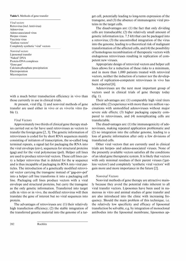
BOIS:L08:BPRR3004PD.97 FF: RESF E1: RESS
282 Rochlitz
Table 1. Methods of gene transfer
Viral vectorsRetrovirus (incl. lentivirus)AdenovirusAdeno-associated virusHerpes virusesVaccinia virus‘Gutless vectors’Completely synthetic ‘viral’ vectors
Nonviral vectorsLiposomal transfer‘Naked’ DNAProtein-DNA complexes‘Gene gun’Calcium-phosphate precipitationElectroporationMicroinjection
with a much better transduction efficiency in vivo thanthose currently in use in clinical trials.
At present, viral (fig. 1) and nonviral methods of genetransfer are used either in vivo or ex vivo/in vitro (ta-ble 1).
Viral VectorsApproximately two thirds of clinical gene therapy stud-
ies carried out so far have used retroviruses as vectors totransfer the foreign gene [2, 3]. The genetic information ofretroviruses is coded for by short RNA sequences mainlyconsisting of initiators of transcription, the so-called longterminal repeats, a signal („) for packaging the RNA intothe viral envelope (env), sequences for structural proteins(gag) and for the viral polymerase (pol). Helper cell linesare used to produce retroviral vectors. These cell lines car-ry a helper retrovirus that is deleted for the „ sequenceand is thus incapable of packaging its RNA into viral par-ticles. The introduction of a genetically modified retrovi-ral vector carrying the transgene instead of ‘gag-env-pol’into a helper cell line transforms it into a packaging cellline. Packaging cell lines produce vectors with a viralenvelope and structural proteins, but carry the transgeneas the only genetic information. Transferred into targetcells in vitro or in vivo, the modified retrovirus will thentranslate the gene of interest but no viral sequences intoprotein.
The advantages of retroviruses are: (1) their relativelyhigh transfection efficiency; (2) the stable integration ofthe transferred genetic material into the genome of a tar-
get cell, potentially leading to long-term expression of thetransgene, and (3) the absence of immunogenic viral pro-teins in the target cells.
The disadvantages are: (1) the fact that only dividingcells are transducable; (2) the relatively small amount ofgenetic information (ca. 7.5 kb) that can be packaged intoa retrovirus; (3) the uncontrolled integration of the virusinto the genome, leading to a theoretical risk of malignanttransformation of the affected cells, and (4) the possibilityof homologous recombination of therapeutic vectors withendogenous retroviruses resulting in replication of com-petent new viruses.
Appropriate design of retroviral vectors and helper celllines allows for a reduction of these risks to a minimum,and in more than 1,000 patients treated with retroviralvectors, neither the induction of a tumor nor the develop-ment of replication-competent retroviruses in vivo hasbeen reported [6].
Adenoviruses are the next most important group ofvectors used in clinical trials of gene therapy today(fig. 1).
Their advantages are: (1) comparably high viral titersachievable; (2) experience with more than ten million vac-cinations with unmodified adenoviruses without majorsevere side effects; (3) higher packaging capacity com-pared to retroviruses, and (4) nonreplicating cells aretransfectable.
The disadvantages are: (1) the immunogenicity of ade-noviruses, making repeated application problematic and(2) no integration into the cellular genome, leading to aloss of genetic information after only a few divisions oftransfected cells.
Other viral vectors that are currently used in clinicaltrials are herpes- and adeno-associated viruses. None ofthe presently available vectors satisfies all the conditionsof an ideal gene therapeutic system. It is likely that vectorswith only minimal residues of their parent viruses (‘gut-less vectors’) and completely ‘synthetic viral vectors’ willgain more and more importance in the future [2].
Nonviral VectorsNonviral methods of gene therapy are attractive main-
ly because they avoid the potential risks inherent to allviral transfer vectors. Liposomes have been used in nu-merous in vitro and animal studies of gene transfer andare also introduced into the clinic with increasing fre-quency. Should the main problem of this technique, i.e.the relatively low specificity and efficacy of liposomaltransfection be solvable, e.g. by integration of monoclonalantibodies into the liposomal membrane, liposomes ap-
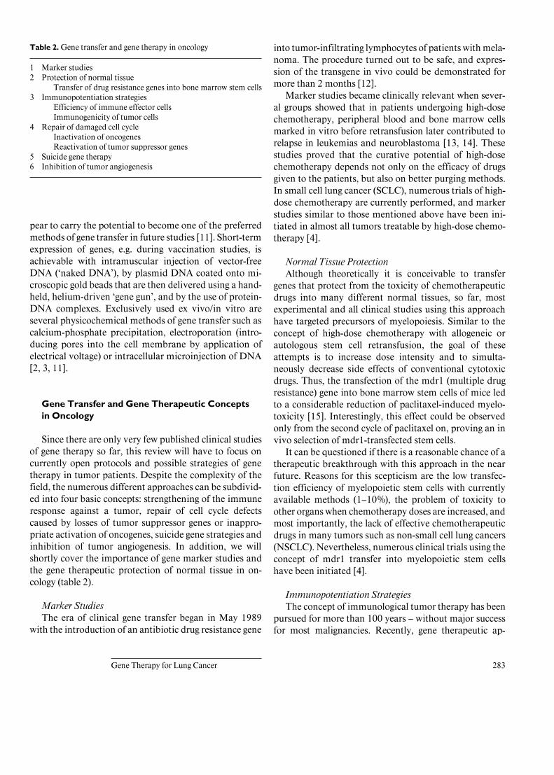
BOIS:L08:BPRR3004PD.97 FF: RESF E1: RESS
Gene Therapy for Lung Cancer 283
Table 2. Gene transfer and gene therapy in oncology
1 Marker studies2 Protection of normal tissue
Transfer of drug resistance genes into bone marrow stem cells3 Immunopotentiation strategies
Efficiency of immune effector cellsImmunogenicity of tumor cells
4 Repair of damaged cell cycleInactivation of oncogenesReactivation of tumor suppressor genes
5 Suicide gene therapy6 Inhibition of tumor angiogenesis
pear to carry the potential to become one of the preferredmethods of gene transfer in future studies [11]. Short-termexpression of genes, e.g. during vaccination studies, isachievable with intramuscular injection of vector-freeDNA (‘naked DNA’), by plasmid DNA coated onto mi-croscopic gold beads that are then delivered using a hand-held, helium-driven ‘gene gun’, and by the use of protein-DNA complexes. Exclusively used ex vivo/in vitro areseveral physicochemical methods of gene transfer such ascalcium-phosphate precipitation, electroporation (intro-ducing pores into the cell membrane by application ofelectrical voltage) or intracellular microinjection of DNA[2, 3, 11].
Gene Transfer and Gene Therapeutic Conceptsin Oncology
Since there are only very few published clinical studiesof gene therapy so far, this review will have to focus oncurrently open protocols and possible strategies of genetherapy in tumor patients. Despite the complexity of thefield, the numerous different approaches can be subdivid-ed into four basic concepts: strengthening of the immuneresponse against a tumor, repair of cell cycle defectscaused by losses of tumor suppressor genes or inappro-priate activation of oncogenes, suicide gene strategies andinhibition of tumor angiogenesis. In addition, we willshortly cover the importance of gene marker studies andthe gene therapeutic protection of normal tissue in on-cology (table 2).
Marker StudiesThe era of clinical gene transfer began in May 1989
with the introduction of an antibiotic drug resistance gene
into tumor-infiltrating lymphocytes of patients with mela-noma. The procedure turned out to be safe, and expres-sion of the transgene in vivo could be demonstrated formore than 2 months [12].
Marker studies became clinically relevant when sever-al groups showed that in patients undergoing high-dosechemotherapy, peripheral blood and bone marrow cellsmarked in vitro before retransfusion later contributed torelapse in leukemias and neuroblastoma [13, 14]. Thesestudies proved that the curative potential of high-dosechemotherapy depends not only on the efficacy of drugsgiven to the patients, but also on better purging methods.In small cell lung cancer (SCLC), numerous trials of high-dose chemotherapy are currently performed, and markerstudies similar to those mentioned above have been ini-tiated in almost all tumors treatable by high-dose chemo-therapy [4].
Normal Tissue ProtectionAlthough theoretically it is conceivable to transfer
genes that protect from the toxicity of chemotherapeuticdrugs into many different normal tissues, so far, mostexperimental and all clinical studies using this approachhave targeted precursors of myelopoiesis. Similar to theconcept of high-dose chemotherapy with allogeneic orautologous stem cell retransfusion, the goal of theseattempts is to increase dose intensity and to simulta-neously decrease side effects of conventional cytotoxicdrugs. Thus, the transfection of the mdr1 (multiple drugresistance) gene into bone marrow stem cells of mice ledto a considerable reduction of paclitaxel-induced myelo-toxicity [15]. Interestingly, this effect could be observedonly from the second cycle of paclitaxel on, proving an invivo selection of mdr1-transfected stem cells.
It can be questioned if there is a reasonable chance of atherapeutic breakthrough with this approach in the nearfuture. Reasons for this scepticism are the low transfec-tion efficiency of myelopoietic stem cells with currentlyavailable methods (1–10%), the problem of toxicity toother organs when chemotherapy doses are increased, andmost importantly, the lack of effective chemotherapeuticdrugs in many tumors such as non-small cell lung cancers(NSCLC). Nevertheless, numerous clinical trials using theconcept of mdr1 transfer into myelopoietic stem cellshave been initiated [4].
Immunopotentiation StrategiesThe concept of immunological tumor therapy has been
pursued for more than 100 years – without major successfor most malignancies. Recently, gene therapeutic ap-

BOIS:L08:BPRR3004PD.97 FF: RESF E1: RESS
284 Rochlitz
proaches have brought new optimism to the field of tumorimmunology, and both immune effector cells and tu-mor cells have been identified as possible targets of genetransfer.
Improvement in the Efficiency of Immune EffectorCells. The first gene therapy study in oncology transferredthe gene coding for tumor necrosis factor (TNF) intotumor-infiltrating lymphocytes to increase their antitu-moral activity. With that same goal, a multitude of differ-ent cytokines, cytokine receptors, adhesion molecules and‘chimeric antibody/T cell receptor molecules’ have beenintroduced into different immune effector cells in animalmodels and clinical trials in humans [reviews in 4, 16]. Itis currently debatable if, in consideration of the com-plexity of the antitumoral immune response, the restric-tion to one transferred cytokine gene and to one type ofeffector cell can be an effective means to destroy a malig-nant tumor in vivo [17]. Since several cytokines and cyto-kine receptors consist of more than one chain, coded forby different genes, an additional problem of this approachis the necessity to express more than one transgene in thesame cell, a technique that works especially poorly inhuman T cells.
Increase in the Immunogenicity of Tumor Cells. Con-ceptually more convincing are attempts to increase theimmunogenicity of tumor cells by the introduction of for-eign genes. This approach can potentially work even ifneither the immune effector cells involved in tumor cellkilling nor tumor-specific antigens potentially recognizedby these effector cells are clearly defined – a conditionthat accurately describes the situation in many immunetherapy trials in humans.
The most frequently used approach in animal modelsconsists of the introduction of cytokine genes such as IL-2,IL-4, IL-7, IL-12, INF-Á, TNF-·, or granulocyte/macro-phage colony-stimulating factor (GM-CSF) into tumorcells. When injected intratumorally, subcutaneously or byother routes, these manipulated tumor cells are capable ofproducing high local amounts of the respective proteinwithout causing the usual systemic side effects of cyto-kines [review in 16]. Since transfection of autologoustumor cells of individual patients is cumbersome andprone to technical problems in clinical trials, and sinceefficient vectors for in situ transfection are currently notavailable, many groups have chosen other ways to over-come these restrictions. Allogeneic tumor cells potentiallycarrying the same specific antigens as the patients’ tumorscan be transfected with a cytokine gene and then beinjected subcutaneously or into the lymphatic system[18]. Alternatively, other autologous, allogeneic or xeno-
geneic cells such as fibroblasts can be transfected withcytokines, mixed with irradiated tumor cells of the patientand then reinjected into the patient. The common princi-ple of all these studies is the creation of an immunostimu-latory paracrine milieu in the close proximity of tumor-specific antigens [19]. In addition to cytokine genes, for-eign MHC molecules, viral antigens, known tumor-spe-cific antigens such as the MAGE (melanoma antigen)genes in melanoma, or costimulatory molecules such asB7-1 or B7-2 have been used to increase the immunoge-nicity of tumor cells in animal models and clinical studies.The different approaches to genetically modify tumorimmunogenicity account for approximately two thirds ofall gene therapy studies performed in patients with malig-nancies, and anecdotal evidence of therapeutic success inindividual patients has been reported [5]. On the basis ofpreclinical data and immunological properties of humanbronchial carcinomas, cytokines of particular interest forgene therapy in lung cancer include IL-7, IL-12 and GM-CSF [20].
Suicide Gene TherapyThe common principle of sucide gene strategies is the
selective intratumoral activation of otherwise nontoxicdrugs by specific transfer of the activating transgene intotumor cells. The best known example of such a prodrugactivator is the gene coding for the herpes simplex virusthymidine kinase (HSV-TK). As opposed to the humanthymidine kinase, HSV-TK phosphorylates the anti-herpes drug ganciclovir to toxic triphosphates with veryhigh affinity. In a rat model, stereotactic injection ofmouse fibroblasts carrying a retrovirus with an insertionof the HSV-TK gene into gliomas led to a sensibilizationof the tumor cells to systemic treatment with ganciclovirand to cure of the gliomas in a majority of treated rats[21]. Clinically interesting in different models of suicidegene therapy was the observation that transfection of onlya minority of tumor cells with the prodrug activator genecould be sufficient for a clear benefit of treated animals.This ‘bystander effect’ was probably due to differentmechanisms such as transfer of activated drug via gapjunctions between tumor cells, immunological mecha-nisms after destruction of transfected tumor cells and pos-sibly transfection of endothelial cells of tumor vasculatureleading to sensitivity of endothelial cells to ganciclovirand consequently tumor ischemia [21]. Although it isanticipated that similar bystander effects might help tosolve the problem of inefficient gene transfer in humans, arecently published study of HSV-TK suicide gene therapyin 15 patients with glioma showed only limited success in

BOIS:L08:BPRR3004PD.97 FF: RESF E1: RESS
Gene Therapy for Lung Cancer 285
smaller tumors and unconvincing evidence of a bystandereffect [22]. Improvements of suicide gene therapy mightcome with other potentially more effective enzyme-pro-drug systems such as cytosine-deaminase/5-fluorocyto-sine or with an increase in tumor specificity by the use oftumor-specific promotors such as the prostate-specificantigen promotor in prostate cancer or a carcinoembryo-nal antigen promotor in tumors expressing carcinoem-bryonal antigen, including many non-small cell and somesmall cell bronchial carcinomas [7]. Different groups arealso currently testing combinations of suicide gene thera-py with other gene therapeutic or conventional chemo-therapeutic strategies. In mesothelioma, a phase I studyhas recently been completed using intrapleural injectionof an adenovirus expressing HSV-TK in 8 patients. A pre-liminary report cites low toxicity, evidence for in situ genetransfer and no tumor progression or death during a rela-tively short observational period [9].
Repair of Damaged Cell CycleThe development of malignant tumors of the airways is
caused by and closely linked to alterations in two groupsof genes that are involved in the regulation of the normalcell cyle, oncogenes and tumor suppressor genes. Onco-genes are generally activated by amplification, overex-pression, mutation or translocation, and alteration in oneallele of an oncogene is usually sufficient to cause cellcycle dysregulation. Deactivation of tumor suppressorgenes, on the other hand, occurs by deletion or mutationalinactivation, and these molecular alterations are frequent-ly recessive, i.e. both copies of the gene have to be func-tionally lost for the protein to become inactivated.
Inactivation of Oncogenes. The most important onco-gene alterations found in lung cancer concern four genes,RAS, MYC, c-erbB-2, and BCL-2 [23]. RAS is a 21-kd Gprotein, and up to 30% of bronchial adenocarcinomasshow mutations in the K-RAS oncogene that have beenassociated with poor prognosis. MYC encodes a transcrip-tional activator and amplification may also adverselyaffect survival in small cell lung cancer. The growth factorreceptor c-erbB-2 is overexpressed in up to 25% ofNSCLC cases, and finally, BCL-2, a negative regulator ofapoptosis, is expressed differently in some NSCLC [23].Attempts to reverse the activation of oncogenes in lungcancer have included a preclinical strategy in which a vec-tor encoding for an intracellular single-chain antibodyfragment directed against c-erbB-2 has been transfectedinto tumor cells [9], and a phase I study with antisenseK-RAS using a retroviral vector in patients with NSCLC[24].
Fig. 2. Wild-type p53 plays a central role in the cell physiology.
p53
Cell growth Apoptosis
ä
ä
ä
ää
ä
ä
p21
Angiogenesis Drug resistanceMutation repair
DNA
3 exoNase´
Growthfactors
ää
ää
–
–
+ +
+ +
ä
ä
ä ä
––
––
C-mycbcl-2bax
ä
tsp-1
–
+
ää
mdr-1
–
+ +
Reactivation of Tumor Suppressor Genes. Tumor sup-pressor genes lost or mutated in lung cancer include theretinoblastoma gene (RB), the gene for the cyclin-depen-dent kinase inhibitor p16INK4A and the p53 gene [23].Abnormalities of RB, a key regulator of cell cycle, aredetected in more than 90% of SCLCs, and there is aninverse relationship in lung cancer cells between expres-sion of RB and p16INK4A, an upstream regulator of RB.Mutations of p53, with frequencies up to 60% in NSCLCand 80% in SCLC, can lead to loss of tumor suppressorfunction, increased drug resistance, loss of mutationalrepair, increased tumor angiogenesis, cellular prolifera-tion and inhibition of apoptosis (fig. 2) [review in 25].Gene transfer of wild-type p53 was shown to reverse thesedeficiencies and to induce apoptosis in vitro and in pre-clinical in vivo tumor models. In light of this preclinicalevidence, restoration of functional p53 represents anattractive target for somatic gene therapy in cancer. Apilot study in 9 patients with NSCLC supports this view[26].
Technique and Effects of Endobronchial p53 Applica-tion. Based on these results, we initiated a phase I dose

BOIS:L08:BPRR3004PD.97 FF: RESF E1: RESS
286 Rochlitz
Fig. 3. CT scans of a patient treated with 109 PFU of adenovirus-p53 before (a) and 2 months after (b) therapy. Thereduction of the size of the injected lesion fulfills the criteria for a partial remission. The decrease of the volume ofpleural effusion is due to a pleurodesis performed directly before gene therapy (courtesy of G. Bongartz, Division ofRadiology, Kantonsspital, Basel).
escalation study of a single intratumoral injection of areplication-defective adenoviral expression vector encod-ing wild-type p53 [27]. Fifteen patients with incurableNSCLC were treated in two centers (University HospitalMainz, Germany, and Kantonsspital of the University ofBasel, Switzerland). All patients enrolled had p53 proteinoverexpression as a marker of mutant p53 status in pre-treatment tumor biopsies. Treatment was performed ei-ther by bronchoscopic intratumoral injection or by CT-guided percutaneous intratumoral injection of the vectorsolution.
Practically, patients were enrolled who had undergoneprevious bronchoscopies and/or CT scans which hadshown that the injection of a volume between 1 and 10 mlof a watery solution was feasible. After informed consentwas obtained, the patient then underwent a second fiber-optic bronchoscopy, and 1–10 ml of wild-type p53 con-taining aquous adenoviral solution was instilled via a22-G Millrose catheter. Twenty-four hours later, the in-jected site was biopsied and the tissue examined for wild-type p53 gene expression. Fifteen patients were enrolled,and were treated at four different dose levels ranging from107 to 1010 PFU (7.5 ! 109 to 7.5 ! 1012 particles).
No clinically significant toxicity was observed. Suc-cessful transfer of wild-type p53 was achieved with highervector doses (6108 PFU). Vector-specific wild-type p53RNA sequences could be demonstrated in posttreatmentbiopsies of 6 patients. Transient local disease control by asingle intratumoral injection of the vector solution wasobserved in 4 of those 6 successfully transduced patients.In 1 patient, a clinical response (partial remission 650%reduction) of the injected lesion was observed 2 monthsafter treatment (fig. 3), but the patient died of progressionof noninjected distant metastases. There was no evidenceof clinical responses at untreated tumor sites in anypatient. In conclusion, wild-type p53 gene therapy byintratumoral injection of a replication-defective adenovi-ral expression vector was safe, feasible and biologicallyeffective in patients with advanced NSCLC [27].
In a multicenter international study, we are currentlytesting the combination of repeated injections of 7.5 !1012 particles of adenovirus-p53 with concurrent plati-num-containing standard chemotherapy in patients withNSCLC [unpubl. data]. Figure 4a shows the injection ofadenoviral solution using a 22-G Millrose catheter into aright upper lobe lesion in a patient suffering from adeno-
a b

BOIS:L08:BPRR3004PD.97 FF: RESF E1: RESS
Gene Therapy for Lung Cancer 287
Fig. 4. Bronchoscopy of a patient with an adenocarcinoma of the right upper lobe. a Injection of adenoviral solutioninto a tumorous right upper lobe spur using a 22-G Millrose catheter. b The same lesion after three cycles of combinedchemo-gene therapy, demonstrating a very good remission of the injected tumor with only scarred tissue remaining insitu (courtesy of A.P. Perruchoud, C.T. Bolliger, Division of Pneumology, Kantonsspital, Basel).
carcinoma; figure 4b shows the same lesion after threecycles of combined chemo-gene therapy, demonstratinga very good remission of the injected tumor with onlyscarred tissue remaining in situ. Obviously, it is unclear ifadenovirus-p53 gene therapy contributed to this response,since chemotherapy alone could have induced tumorshrinkage. On the other hand, adenovirus gene therapyalone has been shown to induce tumor responses byimmune-mediated mechanisms in a small phase I studyin lung cancer, and randomized trials comparing chemo-therapy alone with the combination of chemotherapy andp53 gene therapy are necessary to solve the question ifwild-type p53 gene therapy indeed leads to chemosensiti-zation in humans in vivo [28].
An interesting new approach to p53-mediated genetherapy consists of the use of partially replication-compe-tent adenoviruses carrying a loss of the E1B gene region.This loss leads to a selective capability of the virus toreplicate in p53-deficient cells, since wild-type adenovirususes E1B to eliminate the replication-blocking function ofcellular p53. Tumor cells with p53 mutations and/or dele-tions will thus be selectively destroyed by this first exam-ple of a new type of tumor-specific vectors [29].
Inhibition of Tumor AngiogenesisTumor angiogenesis appears to be crucial for both
tumor growth and metastasis in lung cancer as well as inall other solid tumors and, interestingly, also leukemias[30]. Although more than 40 inhibitors of tumor angio-genesis have been tested in clinical studies over the lastyears without any published major breakthroughs, thefield has recently received new attention with the detec-tion of the natural angiogenesis inhibitors endostatin andangiostatin by O’Reilly et al. [31, 32]. The inhibition ofangiogenesis seems to be much less hampered by thedevelopment of drug resistance than other therapeuticapproaches in oncology, since benign endothelial cellswhich are the target of therapy are genetically stable,homogeneous and have a low mutational rate comparedto tumor cells [33]. In bronchial carcinoma, vascularendothelial growth factor, IL-8, tumor growth factor-ß,platelet-derived growth factor and basic fibroblast growthfactor are important natural angiogenic factors associatedwith decreased survival in some studies and are targets ofanti-angiogenic gene therapy in preclinical cancer models[20].
a b

BOIS:L08:BPRR3004PD.97 FF: RESF E1: RESS
288 Rochlitz
Conclusion
An evaluation of the first 7 years of gene therapy in1996 concluded that gene therapy was safe and feasible,but that none of the more than 100 clinical studies per-formed so far had formally proven the efficacy of theapproach in any human disease. The consequence was therequest to go ‘back to the bench’ and develop better viraland non-viral vectors and a better molecular understand-ing of the pathology of treated diseases [34]. Althoughanecdotal reports of tumor responses are becoming morefrequent in lung cancer and other human malignancies,the situation has not changed dramatically. Main prob-lems are still the lack of vectors with high transductionefficiency in vivo, the low tumor specificity of availablesystems, the progressive down-regulation of transcriptionelements in vivo even after successful transfer of a trans-gene, and our incomplete knowledge of molecular tumorpathology.
However, the results of the few published phase I stud-ies of gene therapy in lung cancer [8, 9, 20, 26–28] areencouraging, and important progress in vector technologyis expected in the near future. It is likely that tumor-spe-cific vectors and tissue-specific promotors will be in rou-tine use within the next 5–10 years, and that the comple-tion of the human genome project will lead to majoradvances in our understanding of the molecular aspects ofcarcinogenesis, tumor progression and metastasis [2]. Thefirst intravenously applicable vectors should be availableat the end of this period and expression of a transgene in asufficient percentage of targeted tumor cells achievable.The most likely scenario of the role of genetic approachesto the treatment of lung cancer during the next twodecades will be a modest contribution within a multi-modality treatment concept consisting of surgery, radio-therapy, chemotherapy, endoscopic treatment modalitiesand – hopefully – immunotherapy and gene therapy.
References
1 Blaese RM, Culver KW, Miller AD, Carter CS,Fleisher T, Clerici M, Shearer G, Chang L,Chiang Y, Tolstoshev P, et al: T lymphocyte-directed gene therapy for ADA-SCID: Initialtrial results after 4 years. Science 1995;270:475–480.
2 Anderson WF: Human gene therapy. Nature1998;392(suppl):25–30.
3 Paillard F: Cancer gene therapy annual confer-ence 1997: Trends and news. Hum Gene Ther1998;9:283–286.
4 Hwu P, Rosenberg S: Gene therapy of cancer;in De Kita VT Jr, Hellman S, Rosenberg SA(eds): Cancer: Principles and Practice of On-cology, ed 5. Philadelphia, Lippincott-Raven,1997, pp 3005–3022.
5 Roth JA, Cristiano RJ: Gene therapy for can-cer: What have we done and where are wegoing? J Natl Cancer Inst 1997;89:21–39.
6 Verma IM, Somia N: Gene therapy – Promises,problems and prospects. Nature 1997;389:239–242.
7 Weichselbaum RR, Kufe D: Gene therapy ofcancer. Lancet 1997;349(suppl 2):10–12.
8 Curiel DT: Gene-based therapies for inheritedand acquired disorders of the lung. Chest 1997;111:149S–152S.
9 Albelda SM: Gene therapy for lung cancer andmesothelioma. Chest 1997;111:144S–149S.
10 Alton EW, Geddes DM, Gill DR, Higgins CF,Hyde SC, Innes JA, Porteous DJ: Towards genetherapy for cystic fibrosis: A clinical progressreport. Gene Ther 1998;5:291–292.
11 Ledley FD: Nonviral gene therapy: The prom-ise of genes as pharmaceutical products. HumGene Ther 1995;6:1129–1144.
12 Rosenberg SA, Aebersold P, Cornetta K, KasidA, Morgan RA, Moen R, Karson EM, LotzeMT, Yang JC, Topalian SL, Merino MJ, Cul-ver K, Miller AD, Blaese RM, Anderson WF:Gene transfer into humans – Immunotherapyof patients with advanced melanoma, usingtumor-infiltrating lymphocytes modified byretroviral gene transduction. N Engl J Med1990;323:570–578.
13 Brenner MK, Rill DR, Moen RC, Krance RA,Mirro J Jr, Anderson WF, Ihle JN: Gene-mark-ing to trace origin of relapse after autologousbone-marrow transplantation. Lancet 1993;341:85–86.
14 Deisseroth AB, Zu Z, Claxton D: Geneticmarking shows that Ph(+) cells present in autol-ogous transplants of chronic myelogenous leu-kemia (CML) contribute to relapse after autol-ogous bone marrow in CML. Blood 1994;83:3068–3076.
15 Sorrentino BP, Brandt SJ, Bodine D, Gottes-man M, Pastan I, Cline A, Nienhuis AW: Selec-tion of drug-resistant bone marrow cells in vivoafter retroviral transfer of human MDR1.Science 1992;257:99–103.
16 Roth C, Rochlitz CF, Kourilsky P: Immuneresponse against tumors. Adv Immunol 1994;57:281–351.
17 Scott AM, Cebon J: Clinical promise of tumourimmunology. Lancet 1997;349(suppl 2):SII19–22.
18 Arienti F, Sule-Suso J, Belli F, Mascheroni L,Rivoltini L, Melani C, Maio M, Cascinelli N,Colombo MP, Parmiani G: Limited antitumorT cell response in melanoma patients vacci-nated with interleukin-2 gene-transduced allo-geneic melanoma cells. Hum Gene Ther 1996;7:1955–1963.
19 Rochlitz CF, Jantscheff P, Bongartz G, Diet-rich PY, Schatz C, Mehtali M, Courtney M,Tartour E, Dorval T, Fridman WH, HerrmannR: Gene therapy study of cytokine-transfectedxenogeneic cells (Vero-IL-2) in patients withmetastatic solid tumors. Cancer Gene Ther1999;6:271–281.
20 Dubinett SM, Miller PW, Sharma S, Batra RK:Gene therapy for lung cancer. Hematol OncolClin North Am 1998;12:569–594.
21 Culver KW, Ram Z, Wallbridge S, Ishii H, Old-field EH, Blaese RM: In vivo gene transfer withretroviral vector-producer cells for treatment ofexperimental brain tumors. Science 1992;256:1550–1552.
22 Ram Z, Culver KW, Oshiro EM, Viola JJ, Dev-room HL, Otto E, Long Z, Chiang Y, McGarri-ty GJ, Muul LM, Katz D, Blaese RM, OldfieldEH: Therapy of malignant brain tumors byintratumoral implantation of retroviral vector-producing cells. Nat Med 1997;3:1354–1361.
23 Salgia R, Skarin AT: Molecular abnormalitiesin lung cancer. J Clin Oncol 1998;16:1207–1217.
24 Roth JA: Modification of mutant K-ras geneexpression in non-small cell lung cancer(NSCLC). Hum Gene Ther 1996;7:875–889.

BOIS:L08:BPRR3004PD.97 FF: RESF E1: RESS
Gene Therapy for Lung Cancer 289
25 Kirsch DG, Kastan MB: Tumor-suppressorp53: Implications for tumor development andprognosis. J Clin Oncol 1998;16:3158–3168.
26 Roth JA, Nguyen D, Lawrence DD, Kemp BL,Carrasco CH, Ferson DZ, Hong WK, KomakiR, Lee JJ, Nesbitt JC, Pisters KMW, PutnamJB, Schea R, Shin DM, Walsh GL, DolormenteMM, Han CI, Martin FD, et al: Retrovirus-mediated wild-type p53 gene transfer to tumorsof patients with lung cancer. Nat Med 1996;2:985–991.
27 Schuler M, Rochlitz CF, Horowitz JA, SchlegelJ, Perruchoud A, Kommoss F, Bolliger C,Kauczor U, Dalquen P, Herrmann R, Huber C:A phase I study of adenovirus mediated wildtype p53 gene transfer in patients with ad-vanced non-small cell lung cancer. Hum GeneTher 1998;9:2075–2082.
28 Gahery-Segard H, Molinier-Frenkel V, Le Bou-laire C, Saulnier P, Opolon P, Lengagne R,Gautier E, Le Cesne A, Zitvogel L, Venet A,Schatz C, Courtney M, Le Chevalier T, TurszT, Guillet JG, Farace F: Phase I trial of recom-binant adenovirus gene transfer in lung cancer.Longitudinal study of the immune responses totransgene and viral products. J Clin Invest1997;100:2218–2226.
29 Bischoff JR, Kirn DH, Williams A, Heise C,Horn S, Muna M, Ng L, Nye JA, Sampson-Johannes A, Fattaey A, McCormick F: An ade-novirus mutant that replicates selectively inp53-deficient human tumor cells. Science1996;274:373–376.
30 Folkman J: New perspectives in clinical oncol-ogy from angiogenesis research. Eur J Cancer1996;32A:2534–2539.
31 O’Reilly MS, Holmgren L, Shing Y, Chen C,Rosenthal RA, Moses M, Lane WS, Cao Y,Sage EH, Folkman J: Angiostatin: A novel an-giogenesis inhibitor that mediates the suppres-sion of metastases by a Lewis lung carcinoma.Cell 1994;79:315–328.
32 O’Reilly MS, Boehm T, Shing Y, Fukai N,Vasios G, Lane WS, Flynn E, Birkhead JR, Ol-sen BR, Folkman J: Endostatin: An endoge-nous inhibitor of angiogenesis and tumorgrowth. Cell 1997;88:277–285.
33 Boehm T, Folkman J, Browder T, O’Reilly MS:Antiangiogenic therapy of experimental cancerdoes not induce acquired drug resistance. Na-ture 1997;390:404–407.
34 Orkin SH, Motulsky AG: Report and recom-mendations of the panel to assess the NIHinvestment in research on gene therapy. NIHPanel Report, 1996.
PD Dr. Christoph F. RochlitzDepartment of Oncology, KantosspitalCH–4031 Basel (Switzerland)Tel. +41 61 265 50 74, Fax +41 61 265 53 16E-Mail [email protected]

OOOOOOOOOOOOOOOOOOOOOOOOOOOOOOOOOOOOOOOOOOOOOOOOOOOOOOOOOOOOOOOOOOOOOOOOOOOOOOOOOOOOOOOOOOOOOOOOOOOOOOOOOOOOOOOOOOOOOOOOOOO
Author Index
291
Baumgartner, R. 159, 243Beamis, J.F., Jr. 22Becker, H.D. 2, 80Biro, P. 44, 226Bolliger, C.T. 44, 120, 187
Cavaliere, S. 108Colt, H.G. 55
Dasgupta, A. 66Diaz-Jiménez, J.P. 159Dondelinger, R.F. 198Dumon J.-F. 108
Fischer, R. 146Freitag, L. 171
Ghaye, B. 198Gladish, G.W. 253
Haponik, E.F. 253Häussinger, K. 159, 243Herth, F. 80Huber, R.M. 146, 159
Khanavkar, B. 236
Markus, A. 243Marquette, C.-H. 96Marsh, B.R. 2Martinot, A. 96Mathur, P.N. 133, 267Mehta, A.C. 66Minai, O.A. 66Miyazawa, T. 16Morresi-Hauff, A. 243
Nakhosteen, J.A. 236Noppen, M. 215
Pichler, J. 159, 243Prakash, U.B.S. 31
Rochlitz, C.F. 280Rodriguez, A.N. 159Russi, E.W. 226
Stanzel, F. 159, 243Stepp, H. 243Studer, W. 44Sutedja, G. 120
Vergnon, J.M. 133Vo-Dinh, T. 267

OOOOOOOOOOOOOOOOOOOOOOOOOOOOOOOOOOOOOOOOOOOOOOOOOOOOOOOOOOOOOOOOOOOOOOOOOOOOOOOOOOOOOOOOOOOOOOOOOOOOOOOOOOOOOOOOOOOOOOOOOOO
Subject Index
293
Abscess, see Mediastinal collections, Pulmo-nary abscess
Acousto-optic tunable filter, multispectralimaging 273, 274
Adenosine deaminase deficiency, genetherapy 280
Airway fire, prevention 50, 115Airway obstruction
central obstruction, serendipitousfinding 57
clinical presentation, acute obstructionsbenign disease 56, 57malignant disease 56
Airway stenosisiatrogenic tracheal stenosis, laser bron-
choscopy 117, 118stenting indications 172, 173
Airway tumor, laser bronchoscopy 115–119Alligator forceps, foreign body removal 1045-Aminolevulinic acid
fluorescence bronchoscopy 244, 247,248, 270
photodynamic therapy 161Anesthesia
complications 52local anesthesia
application 48development 4, 5toxicity 48
personnel requirements 41postoperative care 52, 60–62premedication, interventional bronchos-
copy 47preoperative evaluation 45, 46preoperative fasting 46rigid bronchoscopy, general anesthesia
27, 48
rigid bronchoscopy, laser resection 111,112
surgical factors 44Angiogenesis, inhibition in tumors, gene
therapy 287Argon plasma coagulation 122, 123, 126Atropine, premedication, interventional
bronchoscopy 47Autofluorescence bronchoscopy
D-Light/AF systemcarcinoma in situ detection 247, 248dysplasia detection 247, 248illumination modes 246, 247improvements 250
history 244Laser Imaging Fluorescence Endoscope
carcinoma in situ detection 238–241disadvantages 244dysplasia detection 238–241examination technique 237indications 238, 239, 241, 270, 271light source 237, 244
optical multichannel analyzer 241principles 237, 244–246
Baskets, flexible bronchoscopy 38, 104Benign tracheobronchial lesions, cryo-
therapy 143Benzoporphyrin derivative, photodynamic
therapy 160, 161Biochip, optical diagnostics 277Biopsy forceps
flexible bronchoscopy 37rigid bronchoscopy 37, 38
Brachytherapy, endobronchialafterloading 147, 148complications 149, 156
definition 147dose rates 147, 148, 157endoluminal brachytherapy
early-stage non-small-cell lung cancerindications 149outcomes 153
stage II–IIIb lung cancer, curativetreatment intentionindications 149, 150outcomes 153, 154
stage IV lung cancer, palliative treat-ment intentionindications 150outcomes 154, 156
fractionation 149, 157history 147isodoses 147multimodality treatment, lung cancer
190, 193, 195patient monitoring 148permanent versus temporary 147planning 148rationale 146, 147technique 147–149
Bronchoscopy, history 5–10Bronchoscopy needles, see Needles, bron-
choscopyBronchoscopy unit
equipment 35–39example of layout 33, 35mobile unit, see Mobile bronchoscopy
unitpatient preparation area 35physical requirements 32, 33postbronchoscopy area 35procedure area 35rationale 31, 32

294 Subject Index
Bronchoscopy unit (continued)safety 42, 43storage area 39
Cancer, see Airway tumor, Lung cancer,Malignant airway obstruction
Carcinoma in situ, see Lung cancerChest imaging
foreign bodies 99interventional bronchoscopy
after procedure 61, 62before procedure 58, 59
Chest rest, invention 10Chronic obstructive pulmonary disease
flow volume loop abnormalities 60long-term oxygen therapy 231, 234
Ciaglia technique, percutaneous dilatationaltracheostomy 217, 219, 221
Claws, flexible bronchoscopy 38, 104Coagulation, see Electrocautery, Laser bron-
choscopyCocaine, local anesthesia 4, 5Computed tomography, see also Virtual
bronchoscopyimaging prior to interventional bron-
choscopy 58percutaneous catheter drainage guid-
ance 204, 212percutaneous transthoracic needle
biopsy guidance 200real-time scanning 261
Confocal microscopy, optical diagnostics271
Corticosteroids, postprocedure care 60, 61Cricothyrotomy
nonsurgical 230, 231surgical 231
Cryotherapybenign tracheobronchial lesions 143cryogens 136, 137equipment
bronchoscopes 138console 137cryoprobes 137, 138monitors 138
flexible bronchoscopy 140, 141foreign body removal 143history 133, 134indications, patient selection 138, 139lung cancer
chemotherapy enhancement 143early lesions 139, 142, 143radiotherapy enhancement 144
mechanismscellular damage, freezing 134, 135distribution, necrosis 135, 136immune activation 136ischemia 135thrombosis induction 135
multimodality treatment 192, 193
outcome evaluation 141–143postoperative care 141preparation, patient 139rigid bronchoscopy 139, 140tissue sensitivity 135, 136
Cystic fibrosis, gene therapy 281Cytology brush, flexible bronchoscopy 36,
37
Dehiscence, stenting indications 174Diazepam, premedication, interventional
bronchoscopy 47Difficult airway, see Emergency transtra-
cheal oxygenation, VentilationD-Light/AF
carcinoma in situ detection 247, 248drug-induced fluorescence bronchos-
copy 244, 247, 248dysplasia detection 247, 248illumination modes 246, 247improvements 250
Documentation, historic developments 14Drainage, see Percutaneous catheter
drainageDumon stents 175, 179Dynamic stents 179Dysplasia, see Lung cancer
Elastic scattering, optical diagnostics 270Electrocardiogram
intraoperative monitoring 47, 48preoperative anesthesia evaluation 46
Electrocautery, endobronchialbackground, history 121comparison with other treatment modal-
ities 129, 130cost analysis 129, 130early-stage lung cancer 127–129equipment 122palliative treatment 126, 127technique
argon plasma coagulation 122, 123,126
flexible bronchoscopy 121, 122, 126rigid bronchoscopy 121–125
Emergency transtracheal oxygenationanatomy 227cricothyrotomy
nonsurgical 230, 231surgical 231
equipment 228oxygen delivery systems 228, 230transtracheal puncture 227
Endobrachial ultrasound, see Ultrasound,endobrachial
Endobronchial electrocautery, see Electro-cautery, endobronchial
Endoscopy, history 3–5Endotracheal tube, bronchoscope insertion
38
Fiberoptic bronchoscopy, see Flexible bron-choscopy
Fistula, stenting indications 174Flexible bronchoscopy
complications 52cryotherapy 140, 141endobronchial electrocautery 121, 122,
126equipment
requirements 35–37storage 39
foreign body diagnosis, removal100–102, 104–106
history 13, 16–21instrumentation 36laser fiberscopy 113, 114transbronchial needle aspiration, see
Transbronchial needle aspirationventilation 48
Fluoroscopypercutaneous catheter drainage guid-
ance 204, 212uses with bronchoscopy 38
Foreign bodyclinical features, diagnostic workup for
aspirationadults 99–101, 106children 98, 99, 106
diagnostic fiberoptic bronchoscopy100–102
epidemiology, aspirationadults 97, 98children 96
removalcomplications 106cryotherapy 143equipment 38, 104flexible bronchoscopy 104–106forceps operation 103, 104perioperative management 106rigid bronchoscopy 25, 96, 102–104timing 102
Gene therapyadenosine deaminase deficiency 280cystic fibrosis 281lung cancer
angiogenesis inhibition 287immunopotentiation 283, 284marker studies 283oncogene inactivation 285rationale 280, 281stem cell protection, cytotoxic
agents 283suicide gene therapy 284, 285tumor suppressor gene reactivation
285–287vectors
adenovirus 282liposomes 282, 283

Subject Index 295
naked DNA transfer 283retrovirus 282
Gianturco stent 177Glycopyrrolate, premedication, interven-
tional bronchoscopy 47Griggs technique, percutaneous dilatational
tracheostomy 221
Hemoptysislaser bronchoscopy 118percutaneous transthoracic needle
biopsy complication 203rigid bronchoscopy 26stenting indications 174
Hemothorax, percutaneous catheterdrainage 208
Hood stents 177, 179
Ikeda, S., bronchoscopy contributions 13,16–21
Illuminator, history 4, 11, 13, 14Infection, transbronchial needle aspiration
74Infrared spectroscopy, optical diagnostics
270Interstitial laser therapy, applications 275Interventional bronchoscopy
airway obstruction, clinical presenta-tions 56, 57
anesthesia, see Anesthesiadischarge planning 62endobrachial ultrasound 92hospitalization versus same-day surgery
62outcomes
pulmonary function 63study design 62, 64survival 63symptom relief 62, 63
postprocedure care 52, 60–62preprocedure evaluation
blood tests 45, 46, 60chest imaging 58, 59flexible bronchoscopy 59patient history 45, 57physical examination 58pulmonary function test 59upper endoscopy 59
Jackson, C., bronchoscopy contributions10–12, 24, 30
Jet ventilationadvantages and disadvantages 51equipment 48, 49, 51history 12, 13, 50monitoring 51principles 51
Killian, G., bronchoscopy contributions5–10, 24
Laryngeal mirror, history 3Laser bronchoscopy
accident prevention 115anatomy 110, 111complications 114history 108, 119indications
airway tumors 115–117hemoptysis 118iatrogenic tracheal stenosis 117, 118miscellaneous indications 118
laser fiberscopy 113, 114lasers
principles 108, 109types 109, 110
multimodality treatment 189, 190outcomes, airway tumors 118, 119rigid bronchoscopy laser resection
advantages and disadvantages 114anesthesia 111, 112coagulation 112debulking 112, 113
ventilation 110, 111Laser Imaging Fluorescence Endoscope
carcinoma in situ detection 238–241disadvantages 244dysplasia detection 238–241examination technique 237indications 238, 239, 241, 270, 271light source 237, 244
Location, physical locations, bronchoscopyprocedures 32
Long-term oxygen therapyindications 231transtracheal delivery, see Transtracheal
oxygen therapyLorazepam, premedication, interventional
bronchoscopy 47Lung cancer
accuracy, mediastinal staging 80, 81autofluorescence bronchoscopy, see
Autofluorescence bronchoscopycarcinoma in situ progression 243, 244,
268cryotherapy
chemotherapy enhancement 143early lesions 139, 142, 143radiotherapy enhancement 144
early detection 236, 237, 243endobrachial ultrasound staging
early carcinoma 86, 87goals 86intraluminal extension, tracheobron-
chial wall involvement 87lymph node involvement 87, 89
endobronchial electrocautery 127–129endoluminal brachytherapy
early-stage, non-small-cell lung cancerindications 149outcomes 153
stage II–IIIb lung cancer, curativetreatment intentionindications 149, 150outcomes 153, 154
stage IV lung cancer, palliative treat-ment intentionindications 150outcomes 154, 156
gene therapyangiogenesis inhibition 287immunopotentiation 283, 284marker studies 283oncogene inactivation 285p53 285–287rationale 280, 281stem cell protection, cytotoxic
agents 283suicide gene therapy 284, 285tumor suppressor gene reactivation
285–287mortality 267multimodality treatment
algorithm 191–197definition 187, 188endobrachial treatment combina-
tions 189, 190endoscopic-nonendoscopic treat-
ment combinations 190, 191inoperable lung cancer 188
needle biopsy, see Percutaneous trans-thoracic needle biopsy
oncogenesis 243, 244, 268optical diagnostics, see also Autofluores-
cence bronchoscopyadvantages 268applications 268, 269, 276, 277biochips 277confocal microscopy 271elastic scattering 270exogenous chromophores 272fluorescence 269, 270functional assessment 274infrared spectroscopy 270multispectral imaging 273, 274optical coherence tomography 271photon migration 271, 272point probe systems versus two-
dimensional imaging 275Raman spectroscopy 270synchronous luminescence 272, 273
outcomes 120, 187, 188, 194photodynamic therapy
advantages, technique 163, 164applicators, dose 164outcomes, curative treatment
163–166, 168palliative therapy 162, 163, 167, 168tumor size effects 164, 165
prevention, screening 267, 268standard treatment 188

296 Subject Index
Lung cancer (continued)transbronchial needle aspiration,
staging 72Lymph node
endobrachial ultrasound, cancerinvolvement 87, 89
stations, transbronchial needle aspira-tion 66–68
Machida bronchofiberscope, development,features 16–18
Magill forceps, foreign body removal 104Magnetic resonance imaging, imaging prior
to interventional bronchoscopy 58Magnets, foreign body removal 104Malacia, stenting indications 173, 174Malignant airway obstruction
clinical presentation 56rigid bronchoscopy 25
Mediastinal collections, percutaneous cath-eter drainage 210, 211
Mediastinal massendobrachial ultrasound 89, 90transbronchial needle aspiration 74
Midazolam, premedication, interventionalbronchoscopy 47
Mobile bronchoscopy unit, requirements41, 42
Multidrug resistance, gene therapy, stemcell protection during chemotherapy283
Multispectral imaging, optical diagnostics273, 274
Needles, bronchoscopyaspiration, see Transbronchial needle
aspirationbiopsy, see Percutaneous transthoracic
needle biopsytypes 38, 68uses 38
Olympus bronchofiberscope, development,features 18, 19
Optical coherence tomography, optical diag-nostics 271
Optical forceps, foreign body removal 103Orlowski stents 175, 179Oxygen delivery, see Emergency transtra-
cheal oxygenation, Transtracheal oxygentherapy, Ventilation
Palmaz stent 177, 178Percutaneous catheter drainage
applications 204catheters 204complications 212gazeous collections 308, 309imaging guidance
computed tomography 204, 212
fluoroscopy 204, 212ultrasonography 204
malignant pleural effusion 208mediastinal collections 210, 211pericardial fluid 211pleural empyema 204, 205, 207posttraumatic hemothorax 208pulmonary abscess 309, 210tension pneumomediastinum 212
Percutaneous dilatational tracheostomyadvantages over surgical technique
223–225complications 222contraindications 221costs 225definition 215duration 224, 225equipment 216, 217history 215, 216indications 216outcomes 223personnel 216technique
Ciaglia technique 217–219, 221Griggs technique 221guidewire placement 217preparation, operative area 217
training 223, 224Percutaneous transthoracic needle biopsy
accuracy 200–202comparison with other techniques 203complications
hemoptysis 203pneumothorax 202, 203
computed tomography guidance 200contraindications 199, 200improvements 203indications 198, 199technique 200
Pericardial fluid, percutaneous catheterdrainage 211
Photodynamic therapycomplications 163contraindications 163history 159, 160indications 120, 121, 127, 159, 160, 163lasers 160, 164, 169lung cancer
advantages 163, 164applicators, dose 164outcomes, curative treatment
163–166, 168palliative therapy 162, 163, 167, 168tumor size effects 164, 165
mechanism of action 161, 162multimodality treatment 192, 193photosensitizers
5-aminolevulinic acid 161benzoporphyrin derivative 160, 161cell uptake determinations 276
development 161, 169porfimer sodium 159, 160
site-selective therapy 276technique 160
Photon migration, optical diagnostics 271,272
Physical status classification, AmericanSociety of Anesthesiologists 45
Pleural effusionclassification and treatment 205, 206malignant pleural effusion 208
Pleural empyema, percutaneous catheterdrainage 204, 205, 207
Pleural space, endobrachial ultrasound 91Pneumothorax, percutaneous transthoracic
needle biopsy complication 202, 203Polyflex stents 175–177Porfimer sodium, photodynamic therapy
159, 160Pulmonary abscess, percutaneous catheter
drainage 309, 210Pulmonary function test
chronic obstructive pulmonary disease60
interventional bronchoscopy impact 63preprocedure evaluation
anesthesia 46, 55interventional bronchoscopy 59, 60
Pulse oximetry, monitoring, bronchoscopy38, 39, 47
Raman spectroscopy, optical diagnostics270
Rigid bronchoscopyanesthesia 27, 48applications
foreign body removal 25hemoptysis 26malignant airway obstruction 25overview 14, 22, 25.44
complications 26, 52contraindications 26cryotherapy 139, 140endobronchial electrocautery 121–125equipment requirements 37–39foreign body removal 25, 96, 102–104history 4–13, 22, 24insertion techniques
bronchoscopic insertion, laryngec-tomy stoma 29
classic technique 28intubation insertion, endotracheal
tube 28, 29laryngoscope guidance 28
instrumentation 24, 36laser resection
advantages and disadvantages 114anesthesia 111, 112coagulation 112debulking 112, 113

Subject Index 297
patient preparation 26, 27training 30, 39ventilation 27, 28, 48–50
Sarcoidosis, transbronchial needle aspira-tion 72, 74
Scopolamine, premedication, interventionalbronchoscopy 47
Specimen traps 38Stenosis, see Airway stenosisStents, see Tracheobronchial stentsSynchronous luminescence, optical diagnos-
tics 272, 273
Temazepam, premedication, interventionalbronchoscopy 47
Tension pneumomediastinum, percuta-neous catheter drainage 212
Tracheobronchial stentsbifurcated stents
Dumon Y-stent 179Dynamic stents 179Hood Y-stent 179Orlowski Y-stent 179Westaby T-Y stent 179
classification 174complications, prevention and manage-
mentembedding, perforation 182failure 182migration 181mucostasis 181, 182obstruction 182
customized stents 184dilatation prior to insertion 181history, use 171, 172indications
airway stenosis 172, 173dehiscence 174fistula 174hemoptysis 174malacia 173, 174
insertion techniques 180, 181materials
biocompatibility 185nitinol modifications 184, 185overview 172, 182, 183recoils 183
metal stentsGianturco stent 177miscellaneous stents 179Palmaz stent 177, 178Ultraflex stents 178Wallstents 178, 179
multimodality treatment 190, 191, 193
selection 179, 180straight polymer stents
Dumon stents 175Hood stent 177Orlowski stent 175Polyflex stents 175–177T tube 174, 175
Tracheostomy, see Percutaneous dilata-tional tracheostomy
Tracheotomy, history 3Training
guideline recommendations 40number of bronchoscopy procedures
required 39, 40paramedical personnel 41percutaneous dilatational tracheostomy
223, 224pulmonologist survey 40rigid bronchoscopy 30, 39sources 40
Transbronchial needle aspirationapplications
airway lesions 75, 76bronchogenic carcinoma staging 72infections 74mediastinal masses 74overview 66, 72, 77peripheral nodules or masses 74, 75sarcoidosis 72, 74
complications 76development 66equipment 68histology specimen collection 71nodal stations 66–68popularity 76, 77preprocedure evaluation 68, 69specimen handling 71technique
cough method 70hub-against-wall method 70jabbing method 69, 70needle advancement 69piggyback method 70suction and removal 70, 71
virtual bronchoscopy guidance 261Transcutaneous gas monitoring 47Transtracheal oxygen therapy
benefits 231, 233, 234complications 233contraindications 233emergency delivery, see Emergency
transtracheal oxygenationindications 231oxygen conservation compared to nasal
delivery 233
SCOOP-II catheter insertion 233stent insertion, 1st week care 231, 232stent removal, catheter insertion 232,
233T tube 174, 175
Ultraflex stents 178Ultrasonography
endobrachial, see Ultrasound, endobra-chial
percutaneous catheter drainage guid-ance 204
transesophageal exploration 91Ultrasound, endobrachial
anatomy 83–85artifacts 83cost-effectiveness 92indications, overview 85, 92instrumentation 82, 83interventional bronchoscopy 92intrapulmonary lesions 91intrathoracic vessels 91mediastinal masses 89, 90pleural space 91principles 81quality factors 81staging, lung cancer
early carcinoma 86, 87goals 86intraluminal extension, tracheobron-
chial wall involvement 87lymph node involvement 87, 89
technique 82, 83
Ventilation, see also Jet ventilationdifficult airways 227flexible bronchoscopy 48rigid bronchoscopy 27, 28, 48–50
Ventilation bronchoscope, invention 10Videobronchoscope, development 14, 20,
21Virtual bronchoscopy
applicationseducation and training 261–263overview 253, 259procedure guidance 261procedure planning 259–261
data acquisition 254image display 254, 256–258limitations and prospects 263, 264
Wallstents 178, 179Westaby T-Y stent 179
X-ray, see Chest imaging