University of New Hampshire Scholars' Repository
72
University of New Hampshire University of New Hampshire University of New Hampshire Scholars' Repository University of New Hampshire Scholars' Repository Doctoral Dissertations Student Scholarship Winter 1975 STUDIES ON THE COMPOSITION AND AMYLOLYTIC ACTIVITY OF STUDIES ON THE COMPOSITION AND AMYLOLYTIC ACTIVITY OF THE MOLLUSCAN CRYSTALLINE STYLE THE MOLLUSCAN CRYSTALLINE STYLE DAVID GIBSON TRAINER Follow this and additional works at: https://scholars.unh.edu/dissertation Recommended Citation Recommended Citation TRAINER, DAVID GIBSON, "STUDIES ON THE COMPOSITION AND AMYLOLYTIC ACTIVITY OF THE MOLLUSCAN CRYSTALLINE STYLE" (1975). Doctoral Dissertations. 1110. https://scholars.unh.edu/dissertation/1110 This Dissertation is brought to you for free and open access by the Student Scholarship at University of New Hampshire Scholars' Repository. It has been accepted for inclusion in Doctoral Dissertations by an authorized administrator of University of New Hampshire Scholars' Repository. For more information, please contact [email protected].
-
Upload
khangminh22 -
Category
Documents
-
view
0 -
download
0
Transcript of University of New Hampshire Scholars' Repository

University of New Hampshire University of New Hampshire
University of New Hampshire Scholars' Repository University of New Hampshire Scholars' Repository
Doctoral Dissertations Student Scholarship
Winter 1975
STUDIES ON THE COMPOSITION AND AMYLOLYTIC ACTIVITY OF STUDIES ON THE COMPOSITION AND AMYLOLYTIC ACTIVITY OF
THE MOLLUSCAN CRYSTALLINE STYLE THE MOLLUSCAN CRYSTALLINE STYLE
DAVID GIBSON TRAINER
Follow this and additional works at: https://scholars.unh.edu/dissertation
Recommended Citation Recommended Citation TRAINER, DAVID GIBSON, "STUDIES ON THE COMPOSITION AND AMYLOLYTIC ACTIVITY OF THE MOLLUSCAN CRYSTALLINE STYLE" (1975). Doctoral Dissertations. 1110. https://scholars.unh.edu/dissertation/1110
This Dissertation is brought to you for free and open access by the Student Scholarship at University of New Hampshire Scholars' Repository. It has been accepted for inclusion in Doctoral Dissertations by an authorized administrator of University of New Hampshire Scholars' Repository. For more information, please contact [email protected].

INFORMATION TO USERS
This material was produced from a microfilm copy of the original document. While the most advanced technological means to photograph and reproduce this document have been used, the quality is heavily dependent upon the quality of the original submitted.
The following explanation of techniques is provided to help you understand markings or patterns which may appear on this reproduction.
1 .The sign or "target" for pages apparently lacking from the document photographed is "Missing Page(s)". If it was possible to obtain the missing page(s) or section, they are spliced into the film along with adjacent pages. This may have necessitated cutting thru an image and duplicating adjacent pages to insure you complete continuity.
2. When an image on the film is obliterated with a large round black mark, it is an indication that the photographer suspected that the copy may have moved during exposure and thus cause a blurred image. You will find a good image of the page in the adjacent frame.
3. When a map, drawing or chart, etc., was part of the material being photographed the photographer followed a definite method in "sectioning" the material. It is customary to begin photoing at the upper left hand corner of a large sheet and to continue photoing from left to right in equal sections with a small overlap. If necessary, sectioning is continued again — beginning below the first row and continuing on until complete.
4. The majority of users indicate that the textual content is of greatest value, however, a somewhat higher quality reproduction could be made from "photographs" if essential to the understanding of the dissertation. Silver prints of "photographs" may be ordered at additional charge by writing the Order Department, giving the catalog number, title, author and specific pages you wish reproduced.
5. PLEASE NOTE: Some pages may have indistinct print. Filmed as received.
Xerox University Microfilms300 North Zeeb RoadAnn Arbor, M ichigan 48106

76-11,686TRAINER, David Gibson, 1945- STUDIES ON THE COMPOSITION AND AMYLOLYTIC ACTIVITY OF THE MOLLUSCAN CRYSTALLINE STYLE.University o£ New Hampshire, Ph.D., 1975 Zoology
Xerox University Microfilms, Ann Arbor, Michigan 48106
THIS DISSERTATION HAS BEEN MICROFILMED EXACTLY AS RECEIVED.

STUDIES ON THE COMPOSITION AND AMYLOLYTIC
ACTIVITY OF THE MOLLUSCAN CRYSTALLINE STYLE
by
DAVID GIBSON TRAINERA.B., Washington and Jefferson College, 1967 M.Sc., University of Maine, 1969
A THESIS
Submitted to the University of New Hampshire In Partial Fulfillment of
The Requirements for the Degree of
Doctor of Philosophy Graduate School
Department of Zoology December, 1975

This thesis has been examined and approved.
i b ' . I : i ^ . I L_________________________
Thesis director, Edward 'K. Tillinghast, Assoc. Prof. of Zoology
Hi- < - / WJqpn E. Foret, Assoc. Prof. of Zoology
r/'' — .>■ - VV\LVN . ,,.W
•\
D. MacDonald Green, Professor of Biochemistry
Larry G.'Harris, Assoc. Prof. of Zoology
4-C L l vv f~''- q fl J ) u1 L■ V._________/ /ohn J. Sasner, Jr. , Asfe'oc. Prof. of" Zoology
/ / /') ki| U'ii I C) / jDate

ACKNOWLEDGEMENTS
I am indebted to Dr. Edward K. Tillinghast for his advice, encouragement, and assistance during the course of this study. His willingness to share his time and experience made the completion of this work possible. It is a pleasure to acknowledge the assistance of the other members of my doctoral committee, John E. Foret, D. MacDonald Green, Larry G. Harris, and John J. Sasner, Jr.
I wish to thank the Biology Department of Bowdoin College, Brunswick, Maine, for permitting me to use their scanning electron microscope facility and particularly Dr. John Howland for his assistance and instruction in the use of the scanning electron microscope.
The contributions of my wife, Robin, were innumerable. Her kindness and understanding insured both the completion of this dissertation and the sanity of its author.

TABLE OF CONTENTS
LIST OF TABLES ......................................... vLIST OF FIGURES ........................................ viABSTRACT ................. vii
I. INTRODUCTION ........................................... 1II. STUDIES ON THE COMPOSITION OF THE CRYSTALLINE STYLE. 13
1. Introduction ....................................... 132. Materials and Methods .............................. 143 . Results ............................................. 154. Discussion ......................................... 23
III. AMYLOLYTIC ACTIVITY OF THE CRYSTALLINE STYLE OF MYAARENARIA (BIVALVIA, MOLLUSCA) 321. Introduction ....................................... 322. Materials and Methods .............................. 333 . Results ............................................. 344. Discussion ......................................... 49REFERENCES ............................................. 57BIOGRAPHICAL DATA ..................................... 6l
iv

LIST OF TABLES
1. Dissolution of crystalline styles by variousenzymes ................................................ 19
2. Substitution of other halides for chloride ........... 38
v
LIST OF FIGURES
1 . Diagram showing position of the crystalline styleof Mya arenaria ......................................... 3
2. Diagram of a transverse section through the stylesac and intestine in a bivalve ........................ 7
3 . Scanning electron micrograph of the extruded anterior end of the crystalline style of Myaarenaria ................................................. 9
k. Scanning electron micrograph of the posterior endof the crystalline style of Mya arenaria ............ 11
5 . Dissolution of amylase and protein from crystallinestyles ................................................... IB
6. Electrophoretic patterns of Mya arenaria and Placo-pecten magellanicus style proteins ................... 22
7 . Immunoelectrophoretic patterns of Mya arenaria andPlacopecten magellanicus style proteins ............. 25
8 . Electrophoretic patterns of Mya arenaria and Modiolus modiolus style proteins ........................... 27
9 . Immunoelectrophoretic patterns of Mya arenaria andModiolus modiolus style proteins ...................... 29
10. Amylase activity in the presence of varying concentrations of sodium chloride ........................ 36
1 1 . pH optima of style amylase at three concentrationsof NaCl .................................................. 40
12. Heat stability of amylase ............................. 431 3 . Gel filtration of amylolytic activity ................ 4514. Molecular weight determination of amylase ........... 481 5 . Electrophoretic pattern of whole style proteins .... 5116 . Electrophoretic pattern of amylase from whole style
homogenate ............................................... 53
vi

ABSTRACT
STUDIES ON THE COMPOSITION AND AMYLOLYTIC ACTIVITY OF THE MOLLUSCAN CRYSTALLINE STYLE
"by
DAVID GIBSON TRAINER
The crystalline style is an acellular mucoprotein rod found only in Bivalve and herbivorous gastropod molluscs. The enzymes contained within the style and the anatomical association of the style with the digestive system indicate that the style serves a digestive function. The present study investigates several aspects of the composition and amylolytic activity of the crystalline style of Mya arenaria.
1. Histological techniques indicated that the isoelectric point of the style is in the range of pH ^ . 0 to 4.5*
2. The rapid dissolution of the style in alkaline solutions and by proteolytic activity was demonstrated in vitro. The possibility that the dissolution of the style occurs through similar processis in vivo is discussed.
3. Although citrate and EDTA are both strong calcium chelators, style dissolution is increased by citrate and decreased by EDTA.
Antibodies formed against Mya arenaria whole style protein precipitate some protein from style homogenates of Placopectin magellanicus but none from Modiolus modiolus

style homogenates.5- Amylolytic activity rises with increasing
chloride concentrations to a maximum activity at 0.5 M NaCl. The ability of other halides to activate style amylase is similar to their ability to activate mammalian amylase (Br > I > F ) .
6 . pH optima of style amylase are dependent upon the concentrations of chloride present and may occur at pH 6 .3> 7 .0 , and 7 • 3 •
7. Amylase is partially inactivated after one hour at 25-35° C and totally inactivated after one hour at 45° C.
8 . Gel filtration of style extracts indicated that the major portion of style amylolytic activity is associated with low molecular weight species. Calculations based on gel filtration on Bio-Gel P-100 yielded approximate molecular weight values of 15>700 and 1 2 , 9 0 0 daltons.
9 . Electrophoresis of style extracts on agarose gels demonstrated three bands of amylolytic activity. Polyacrylamide disc electrophoresis demonstrated many more bands of amylolytic activity, but was not reproducible.

INTRODUCTION
Mya arenaria, an intertidal estuarine species, utilizes a ciliary-mucous mode of feeding typical of other lamellihranch bivalves. Particulate matter, drawn into the mantle cavity through an incurrent siphon, is caught and sorted primarily by the ciliary-mucous mechanisms of the gills or ctenidia. Rejected particles are carried posteriorly by ciliary currents to be voided eventually through the incurrent siphon. Acceptable particles are carried anteriorly by separate ciliary tracts to the labial palps which conduct further sorting before the accepted particles are passed through the short esophagus to the stomach. The stomach of M. arenaria, as other lamellibranchs, contains a complex system of ciliary sorting areas and ciliary tracts which may carry particles to sorting areas, to ducts leading to the digestive diverticula, or to a midventral groove leading to the midgut for elimination of specific particles (Purchon, i9 6 0 ). Digestion is carried out both extra- cellularly in the stomach and by intracellular action in the digestive glands.
The crystalline style is a non-cellular protein rod found in the digestive systems of bivalve and herbivorous gastropod molluscs. In M. arenaria one end of the crystalline style projects across the stomach and abuts against the gastric shield, a chitinous structure on the left wall of the stomach (Fig. l). Halton and Owen (1 9 6 8) and McQuiston (1 9 7 0) have shown that microvilli from underlying cells
1

Figure 1 . Diagram showing position of the crystalline style of Mya arenaria. G, gastric shield; M, mouth; S, style; SS, style sac; ST, stomach; VM, visceral mass. The alimentary canal with the exception of stomach and style sac is shown solid black. (From Yonge, 1932.)

ST
VM
SS

4
penetrate the shield and probably carry enzymes to the inner surface of the shield.
The style is both formed by and contained within the style sac, a pocket which may only open into the stomach at one end, as in M. arenaria, or which may also have a longitudinal opening connecting it to the midgut. Rotation of the style by cilia lining the style sac has been observed and reported several times (Nelson, 1918; Yonge, 1 9 2 6 ; Allen,1958; Purchon, 1958). However, the failure of recent investigators (Kristensen, 1972a; Bernard, 1973) "to observe rotation of the style suggests that intermittent rotation occurs. The fact that styles are composed of concentric layers and the presence of a mucous thread wound around the tips of dissected styles indicate that the styles do rotate at some time. Rotation of the style may facilitate the digestive process through several mechanisms: 1 . mucous material containing food particles may be "reeled" into the stomach from the esophagus, 2 . mixing of the stomach contents by the style exposes more food particles to the stomach enzymes,3 . trituration of food particles between the style and gastric shield may occur. The most important feature of the style is probably the variety of enzymes contained within it (see review by Owen, 1 9 6 6). However, the ability of the style to emulsify and decrease the viscosity of the stomach contents (Kristensen, 1972a) maj also be important. Bernard (1973) states that the ability of the style to carry a thin layer of particulate matter into the style sac and press it against

5
the absorptive epithelial lining of the style sac represents an important digestive mechanism.
The style is not homogeneous, hut is composed of an outer portion of concentric layers around an inner core of longitudinal fibers (Fig. 2). Kristensen (1972a) has shown that new style material is added to the outside of the style, gradually moves into the central core, and is extruded from the core into the stomach during the dissolution of the style in vivo. The extruded anterior (stomach) end of the style and the rounded posterior end are shown in Figures 3 and 5-, respectively.
Bailey and Worboys (i960) have shown that the organic content of the style of Pinna nobilis consists of about Jdfo
carbohydrate and 70% protein material. Their amino acid analyses revealed high proportions of proline and of hydroxy and acidic amino acids. Carbohydrates found in styles of several species by Bailey and Worboys (i960) or Doyle (1966) include glucose, galactose, N-acetylated glucosamine and galactosamine, fucose, xylose and deoxyribose.
Although styles of a few species may contain lipolytic (George, 1952) or proteolytic enzymes (Reid, 1966) styles of many species will hydrolyze a number of carbohydrate substrates (Wojtowicz, 1972; Kristensen, 1972b; for review see Owen, 1966). Amylase is apparently present in all styles investigated and is probably of the alpha-amylase type (Chaet, 1956; Owen, 1966).
Both mammalian (Frati and Caputo, 1970) and inverte-

Figure 2. Diagram of a transverse section through the style sac and intestine in a bivalve in which the style sac and intestine are in direct communication with each other.I, intestine; S, style; SS, style sac.(From Yonge, 1932.)


Figure 3 . Scanning electron micrograph of the extruded anterior end of the crystalline style of Mya arenaria x 100.

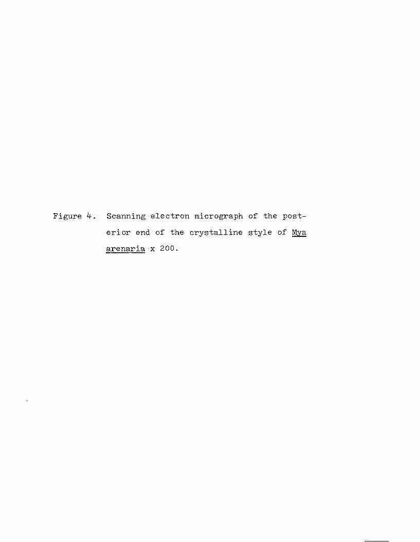
Figure 4 Scanning electron micrograph of the posterior end of the crystalline style of Mya arenaria x 200.


12
brate (Wojtowicz and Brockerhoff, 1972; Wojtowicz, 1972) amylases have been isolated to an electrophoretically pure state by successive Sephadex and Bio-Gel gel filtration chromatography. Under certain conditions, the flow of amylase through a Sephadex column is slowed by an apparent affinity of amylase for the starch-like dextran matrix of Sephadex. Because of its retarded flow, amylase is eluted as though it were a smaller molecule than it actually is, i.e. molecular weight determinations yield values lower than the true molecular weight. Impurities eluted with amylase thus have a lower molecular weight than amylase and can be separated by gel filtration on a matrix with which amylase will not interact, e.g. Bio-Gel.
The first part of the present study investigates several aspects of the crystalline style relative to its function in vivo. In the second part the characteristics of the amylolytic activity of the style are studied and compared to the characteristics of other invertebrate and mammalian amylases.
13
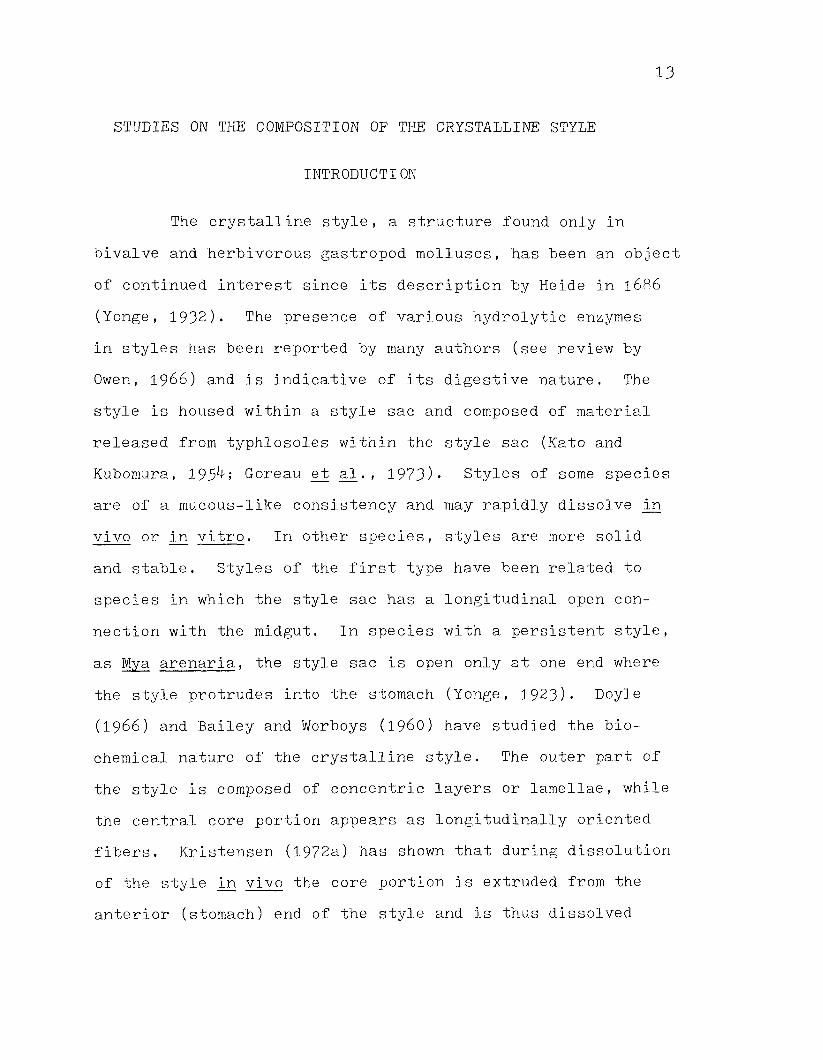
STUDIES ON THE COMPOSITION OF THE CRYSTALLINE STYLE
INTRODUCTION
The crystalline style, a structure found only in bivalve and herbivorous gastropod molluscs, has been an object of continued interest since its description by Heide in 1686
(Yonge, 1932). The presence of various hydrolytic enzymes in styles has been reported by many authors (see review by Owen, 1 9 6 6 ) and is indicative of its digestive nature. The style is housed within a style sac and composed of material released from typhlosoles within the style sac (Kato and Kubomura, 195^; Goreau et al., 1973)* Styles of some species are of a mucous-like consistency and may rapidly dissolve in vivo or in vitro■ In other species, styles are more solid and stable. Styles of the first type have been related to species in which the style sac has a longitudinal open connection with the midgut. In species with a persistent style, as Mya arenaria, the style sac is open only at one end where the style protrudes into the stomach (Yonge, 1 9 2 3). Doyle (196 6 ) and Bailey and Worboys (i9 6 0 ) have studied the biochemical nature of the crystalline style. The outer part of the style is composed of concentric layers or lamellae, while the central core portion appears as longitudinally oriented fibers. Kristensen (1972a) has shown that during dissolution of the style in vivo the core portion is extruded from the anterior (stomach) end of the style and is thus dissolved
14
independently from the outer lamellar portions. In this communication we report additional observations on the physical nature of the style of Mya arenaria and discuss the possible relationships of these features to the style's function in vivo.
MATERIALS AND METHODS
Amylase activity was determined by the method of Bernfeld (1955). employing a substrate of ifo soluble starch (Fisher) and 0.5 M NaCl in 0.1 M sodium phosphate buffer pH 7-0. The procedure of Lowry et al. (1951) was used for protein determination.
For the production of antibodies, homogenates of whole styles from M. arenaria were injected subcutaneously into a rabbit six times at intervals of one week. Each injection contained approximately 450 pg of style protein and was administered as a mixture of equal volumes of style homogenate and Freund's adjuvant.
Immunoelectrophoresis was performed according to techniques described by Ouchterlony (1 9 6 8 ). Gels, layered on 3 x 11 cm glass plates, contained 1 fo Difco Bacto Agar in 0.025 M barbital buffer, pH 8 .6 . Electrode vessels contained 0.05 M barbital buffer pH 8 . 6 and were electrically connected to the gels with filter paper strips. After 20 pi samples of whole style homogenates were added to the surface of the gel, electrophoresis was carried out for approximately 2 hours at

15
3 mamp per plate. Proteins in the gels were stained with 0.05% Coomassie Blue after precipitation with 12% trichloroacetic acid, or were exposed to the antiserum prepared above by filling a center well with the antiserum.
RESULTS
Isoelectric PointThe approximate isoelectric point of the style was
determined by a histochemical technique (Ruthmann, 1970). Separate solutions of orange G and methylene blue stains were prepared in individual 0 . 0 2 M acetate buffer solutions ranging from pH 3-5 to 8.0 in increments of 0.5 pH units. Single styles were incubated in each of the buffer-stain solutions for 10 minutes. The failure of the styles to stain with orange G or with methylene blue at pH 4.0 and 4.5 indicates an isoelectric point in this pH range.
Solubility of the style as a function of pHIndividual styles were placed in 1.5 ml of 0.01 M
acetate or phosphate buffer at various pH values and incubated at room temperature for 16 hours. After removing the whole styles or centrifuging to remove small undissolved style particles for later assays of residual material, 0 . 5 ml aliquots of the buffer solutions were assayed for amylase and soluble protein. Aliquots for amylase determinations were first brought to pH 7*0 Uy addition of 0.5 ml of 0.1 M sodium phosphate at pH 7-0-

16
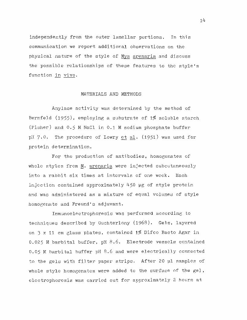
The residual style material, not dissolved during the 16 hour incubation above, was solubilized by incubation at pH 8.0 for 24 hours and assayed for protein and amylase as above. The results shown in Figure 5 demonstrate the greater amounts of protein and amylase dissolved and lower amounts of residual protein and amylase remaining after incubation at higher pH values.
Enzymatic degradation of the styleIndividual styles were placed in separate enzyme
solutions, each containing 5 mg of commercial enzyme (Sigma Chemical Co.) dissolved in 5 ml of either 0.1 M phosphate buffer, pH 7.0, or 0.02 M acetate buffer, pH 5-0. The degree of dissolution of each style was visually estimated and recorded at various times during incubation at room temperature. The results recorded in Table 1 demonstrate a rapid dissolution of the style by proteases and little or no degradation of styles incubated with carbohydrases.
Calcium and its role in style dissolutionBecause of the role of calcium in structural protein,
the presence of calcium and the effect of its removal were investigated. Flame emission spectrophotometry of two acid hydrolyzed styles yielded values of 0.408 and 0.403 pg Ca/mg whole style. Using the data of Doyle (1 9 6 6) for M. arenaria styles (0 . 1 6 mg dry style/mg whole style, 0.5 mg protein/mg dry style), the above values were converted to 5-1 and 5-° pg Ca/mg protein.
Figure 5* Amylase and protein dissolved and not dissolved from crystalline styles incubated at room temperature for 16 hours in buffers at the indicated pH values.
•--------- • amylase dissolved
•--------- • amylase not dissolved
o---------- o protein dissolved
o---------- o protein not dissolved

Amylase p e r c e n t ac t iv i ty
ro .fc cr> oo oo o o o o
• )
r\) <j> oo o ro
j j q P r o t e i n x 1 0 0

Table 1. Dissolution of crystalline styles by various enzymes.
Enzyme pH Time
0.5 1 7 Hr.Buffer only 7.0 +++/+++ +++/+++ +++/++Hyaluronidase 7.0 -f— 1— b / H— 1— b H—1—b /H—b+ + + / + +Lysozyme 7.0 + + + / + + + +++/+++ + + / + +Pancreatin 7.0 +/+ o / o 0/0Pronase 7.0 +/+ o / o 0/0Subtilisin 7.0 +/+ ' 0/0 o / oTrypsin 7.0 0/0 o / o 0/0Alpha glucosidase 7.0 +++ ++Beta glucosidase 5-0 H—1—b/H—1—b +++/+++ +H—b/H—bBeta galactosidase 7.0 +++/+++ H—1—b/H—1—b +++/++Buffer only 5.0 +++/+++ +++/+++ +++/+++Style l/Style 2; +++ = intact style: 0 = completely dissolved

20
Dissolution of styles ir. the presence of calcium chelators, citrate and ethylenediamine tetraacetate (EDTA) was measured and compared to dissolution in phosphate buffer. Styles were placed in separate solutions of: a. 0.01 M phosphate buffer, pH 6.0; b. 0.01 M phosphate buffer, pH 6.0, containing 10 ^ M EDTA; and c. 0.01 M citrate buffer, pH 6.0. After incubation for 18 hours at room temperature, the amount of protein dissolved in citrate buffer was 2k2% of that dissolved in phosphate buffer, indicating a more rapid dissolution of style material in citrate. However, styles incubated with EDTA had a stiffer, more solid consistency than any of the others. And, the style protein dissolved in the presence of EDTA was only of that dissolved in phosphate buffer without EDTA.
ImmunoelectrophoresisThe style proteins of Mya arenaria were compared to
those of the horse mussel, Modiolus modiolus, and the scallop, Placopecten magellanicus, by immunoelectrophoresis as described in the materials and methods section using antisera against M. arenaria whole style homogenates. Staining for protein with Coomassie Blue after electrophoresis in a Difco Bacto Agar medium demonstrated that some M. arenaria style protein migrated toward the anode electrophoretically, while others were carried toward the cathode by endosmosis (Fig. 6). Nearly all of the P. magellanicus style proteins migrated toward the cathode (Fig. 6). Antiserum prepared against whole style protein of M. arenaria contained antibodies

Figure 6. Proteins of styles of Mya arenaria (left)and Placopecten magellanicus (right) stained with Coomassie Blue after electrophoresis on a Bacto-Agar immunoelectrophoresis plate. Anode is toward top of photograph in Figures 6 through 9.

23
which precipitated several specific proteins of the M. arenaria style but which precipitated only one protein of the P. magellanicus style (Fig. 7). Proteins of M. modiolus migrated toward the cathode (Fig. 8 ), but showed no cross reaction with the antibodies formed against M. arenaria style proteins (Fig. 9)•
DISCUSSION
The approximate isoelectric point of pH 4.0-4.5 determined here is in agreement with the observations of Bailey and Worboys (i9 6 0 ) that the minimum solubility of the crystalline style of Pinna nobilis occurs at about pH 5*
The rapid dissolution in vitro of M. arenaria styles with increasing pH (Fig. 5) supports observations of the dissolution of styles from other species (Morton, 1 9 6 9 ; Kristensen, 1972a). Although several reports (Yonge, 1925. 1926; Morton, 1971) indicate that the stomach pH of various bivalves is acidic (pH 5-^-5.8 ) and invariant, Morton (1 9 6 9) found that the stomach pH of Dreissena polymorpha varies from 6.6 to 8.2. Thus a rise in pH of the stomach contents, presumably resulting from an influx of sea water, may serve as a mechanism for dissolution of the style in vivo.
The rapid degradation of styles by proteases (Table l) suggests another mechanism of style degradation _in vivo. Proteases have been identified in the stomach contents of several bivalves including Mya arenaria (Reid, 1966, 1 9 6 8). Kristensen (1972a) has shown that extracts of the bivalve

Figure 7* Immunoelectrophoretic pattern of style proteins of Mya arenaria (left) and Placopecten magellanicus (right) after precipitation with antiserum against M. arenaria whole style protein.


Figure 8. Proteins of the styles of Mya arenaria (left) and Modiolus modiolus (right) stained with Coomassie Blue after electrophoresis on a Bacto-Agar immunoelectro- phoresis plate.


Figure 9- Immunoelectrophoretic pattern of style proteins of Mya arenaria (left) and Modiolus modiolus (right) after precipitation with antiserum against M. arenaria whole style protein.


30
digestive diverticula increase the rate of style dissolution in vitro■ The digestive diverticula contain proteases as well as other enzymes (Reid, 1 9 6 6 , 1 9 6 8 ) which may be active in the stomach after their release from excretory spherules derived from the digestive diverticula (Morton, 1971).
Halton and Owen (1 9 6 8 ) demonstrated esterase, acid phosphatase, and amino peptidase activity in the gastric cuticle, a homologue of the gastric shield, in the proto- branchiate bivalve Nucula sulcata. Although McQuiston (1970) did not demonstrate enzymatic activity he did determine that the fine structure of the gastric shield of Lasea rubra is similar to that of the protobranch's gastric cuticle.Because the style is dissolved in vivo in the general area contacting the shield (Kristensen, 1972a) the possibility of style degradation by gastric shield enzymes should be studied further.
Citrate and EDTA are known to chelate calcium and, to some degree, other divalent cations. Thus the opposite effects of citrate and EDTA on the dissolution rate of styles in vitro are difficult to understand.
The styles of different molluscan species are known to vary with respect to their consistency and their stability, or solubility, in sea water. While the style of Mya arenaria is quite stiff and relatively stable, the style of the scallop Placopecten magellanicus is softer and more soluble. The style of the horse mussel Modiolus modiolus is softer and less persistent than that of P. magellanicus. Because these

31
characteristics of styles probably result in part from their component proteins, the proteins of M. arenaria styles were compared to those of the other two species. The results demonstrate that at least one component of the style of M. arenaria has a very similar counterpart in the style of P. magellanicus■ Whether the immunological similarity of styles is generally correlated with similarities of physical characteristics remains to be seen.

32
AMYLOLYTIC ACTIVITY OF THE CRYSTALLINE STYLE OF MYA ARENARIA (BIVALVIA, MOLLUSCA)
INTRODUCTION
The molluscan crystalline style contains enzymes which are able to hydrolyze a number of different carbohydrate substrates (Wojtowicz, 1972; Kristensen, 1972b; for review see Owen, 1966). Lipolytic activity has been demonstrated in the style of Crassostrea virginica (George, 1952). Endopeptidase isozymes have been reported in the style of Cardium edule (Reid, 1966). An amylolytic activity in style material was demonstrated as early as 1900 (Coupin) and has since then been shown to be one of the most active enzymes found in the style. Animal amylases are generally believed to be ot-amylases (Owen, 1966), and those of the crystalline style are more similar to ot-amylases than /3-amylases in terms of molecular weight (Wojtowicz, 1972), pH optima (Black and Pengelley, 196^; Horiuchi and Lane, 1966; Kristensen, 1972b; Wojtowicz, 1972), and migration in paper chromatography (Chaet, 1956). Wojtowicz (1972) has purified and characterized amylase from the style of the scallop Placopecten magellanicus. The purpose of the present study was to investigate certain characteristics of the amylolytic activity in the style of Mya arenaria and to compare these characteristics to those of amylases found in other invertebrate or mammalian systems.

33
MATERIALS AND METHODS
Clams were purchased from a local distributor and maintained without feeding in recirculating water tanks at 12° C. After removal, styles were homogenized in phosphate buffer or 0.65% saline or were prepared as an acetone powder. Acetone powder was prepared according to Bundy and Gustafson (1 9 7 3 ) but using larger volumes of organic solvents than was prescribed. Whole homogenates and resuspended acetone powders were centrifuged at 5,000 g for 5 minutes to remove undissolved style material. Dialysis, when employed, was carried out for a minimum of 24 hours against four changes of distilled water. Lyophilized powder was prepared from resuspended acetone powder after centrifugation and dialysis (Wojtowicz, 1972).
Amylase activity was determined by the method of Bernfeld (1955)» employing a substrate of 1% soluble starch (Fischer) and 0.5 M NaCl in 0.1 M sodium phosphate buffer pH 7.0 unless otherwise noted. The procedure of Lowry et al. (1 9 5 1 ) was used for protein determination.
Descending gel filtration was performed in 1.5 x 90 cm columns. Eluents of 0.1 M sodium phosphate buffer, pH 7-0, or 0.85% NaCl were used with Sephadex G-100 at a flow rate of 6-8 ml/hr; with Bio-Gel P-100, 0.1 M sodium phosphate buffer, pH 7.0 was eluted at 2-4 ml/hr. Columns and fractions were maintained at 2-4° C. Molecular weights were determined as described by Andrews (1964).
Disc electrophoresis on Canalco (Rockville, Md.)

34
apparatus with standard RDS reagents was performed using 7$ acrylamide gels run at pH 9-5 for approximately one hour at 5 mamp per gel. In a second electrophoretic method, 1$ agarose in 0.05 M Barbitol buffer pH 8.6 was layered on 3 x 11 cm glass plates and allowed to solidify. Samples of 20 pi were applied to the surface of the gel. Electrophoresis was run for 3 hours at A mamp per gel using barbitol buffer as above. Proteins were stained with Coomassie Blue (0 .05$) after precipitation with 12$ trichloroacetic acid.In order to locate amylolytic activity in disc electrophoresis gels, an acrylamide separating gel containing 0 .25$ starch (Bedford and Reid, 1 9 6 9 ) was employed. After electrophoresis as above, the gels were incubated at room temperature for one hour in 0.1 M sodium phosphate buffer pH 7•0 with 0,5 i NaCl. Gels were then placed in a dilute iodine-potassium iodide solution in which the unhydrolyzed starch stained blue and the area hydrolyzed by amylase remained clear. Amylase activity was identified in agarose electrophoresis gels by infiltrating the gel with starch after electrophoresis and then incubating and staining as above.
RESULTS
Halide ActivationAmylase activity of a centrifuged dialyzed 1$ style
homogenate was increased by addition of NaCl to a maximum activity at 0.5 M NaCl in sodium phosphate buffer, pH 7•0 (Fig. 10). Amylase activity decreased with higher concen-

Figure 10. Amylase activity in the presence ofvarying concentrations of sodium chloride.Maximum activity (at 0.5 M NaCl) is considered 100$ activity. Amylase activity of dialyzed homogenate (16.5$) is indicated in the lower left of the figure.
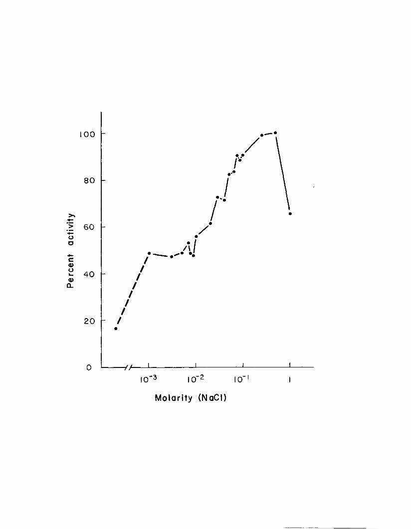
Molar i ty (NaCl)

37
trations of NaCl. The plateau seen in Figure 10 between - 3 -210 and 10 molar NaCl was reproduced in several experi
ments .In assays using substrate buffered with Tris-HCl
instead of sodium phosphate the amylase activity of dialysate incubated with 0.1 M NaCl was 93^ of that incubated with 0.1 M NH^Cl indicating that the increased amylase activity seen in Figure 10 results from chloride and not sodium ions.
Experiments similar to those above demonstrated the effect of other halides on amylase activity in the absence of chloride (Table 2). Bromide increases the amylase activity of a dialized homogenate almost as much as chloride, iodide less, while fluoride may have a slight inhibitory effect.
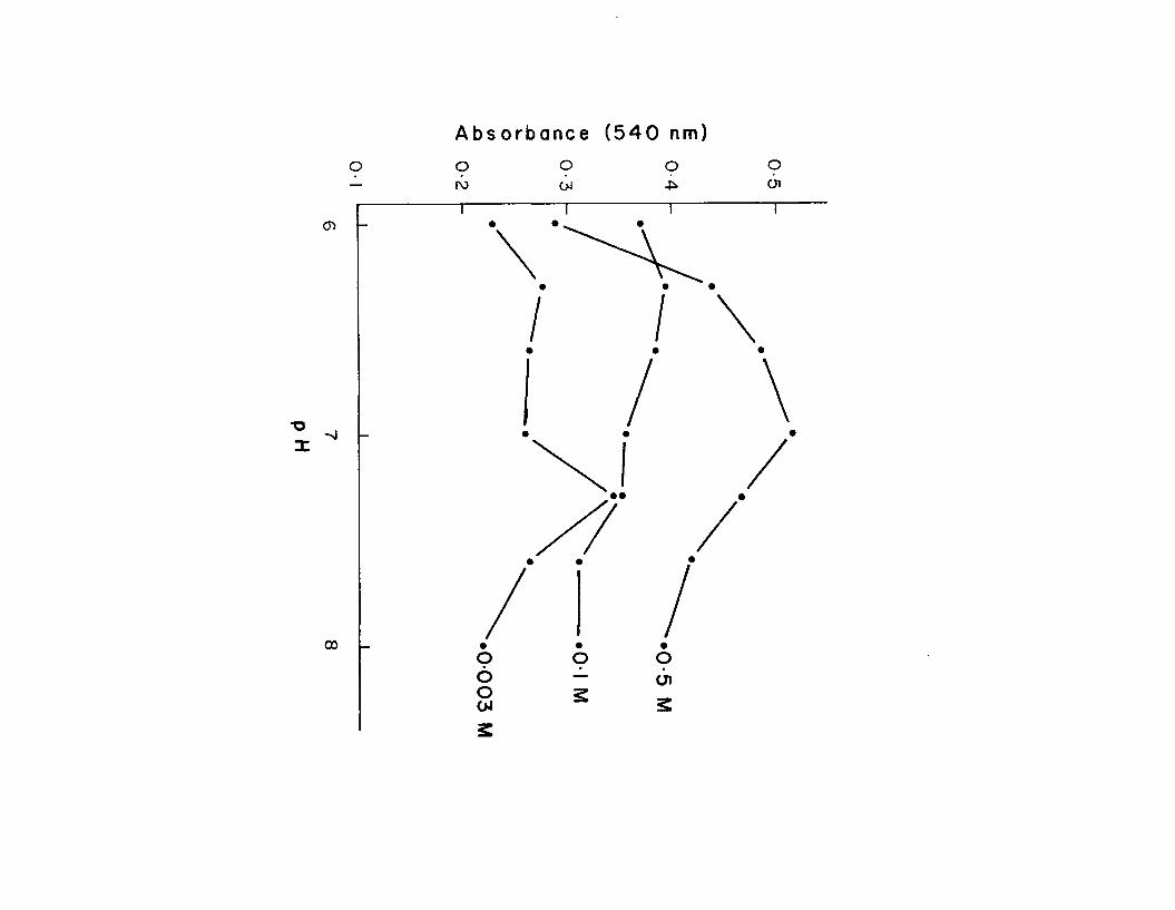
pH OptimaFrozen styles were homogenized, centrifuged and
dialyzed. Figure 11 illustrates the amylase activity of the homogenate at varying pH and in three concentrations of NaCl (0.003 M, 0.1 M, 0.5 I). In the presence of 0.5 M NaCl, the highest activity was found at pH 7 .0 . Peaks of activity occurred at pH 6 . 3 and 7-3 in 0.003 M and 0.1 M NaCl. Thepeaks at pH 7 . 3 (0.003 M and 0.1 M NaCl) decreased markedlyin each of three successive determinations.
Heat StabilityAliquots of style homogenate were placed in ice or
water baths at various temperatures from 0° C to 100° C forone hour, then assayed at 25° C for amylase activity.

Table 2. Substitution of other halides for chloride.
Salt Concentration Amylase Activity ( f o of activity with NaCl)
NaBr 0.1 M 95NaBr 0.05 M 93Nal 0.1 M 65Nal 0.05 M 52NaF 0.1 M 16NaF 0.05 M 20none(dialysate)
- - 19

Figure 11. pH optima of style amylase at three concentrations of NaCl.
A b s o r b a n c e ( 5 4 0 nm)
o o o o o01
• •
00

41
Samples held at 0° C and 15° C had equally high activity while those held at -̂5° C or higher showed no amylase activity (Fig. 12).
Gel FiltrationGel filtration of various style extracts on Sephadex
G-100 and Bio-Gel P-100 produced elution patterns which were similar with respect to the elution of protein or of amylo- lytic activity (Fig. 13)■ A broad peak of protein was always eluted at the void volume. Two peaks of amylolytic activity were found. Peak A, a broad peak containing a very low activity, was eluted at or near the void volume. The second peak, B, had a much higher amylolytic activity and had a high elution ratio (Ve/Vo = 2.0 - 2.9).
When lyophilized style extract (100 mg in 1.0 ml) was filtered on Sephadex G-100 utilizing a 0.1 M sodium phosphate pH 8.0 buffer, the elution ratio (Ve/Vo) of peak B was 2.0. Under similar conditions, the use of a 0,85$ NaCl eluant, rather than the sodium phosphate buffer above, apparently increased the affinity of amylase for the dextran matrix of Sephadex and thus retarded the flow of amylase through the column. The B peak eluted as two close peaks,B1 and B2, with elution ratios of 2.60 and 2.93. Molecular weights corresponding to these elution ratios were below the range calibrated with known proteins but extrapolated values were below 1 0 ,0 0 0 .
Because amylase does not interact with the acrylamide matrix of Bio-Gel, molecular weight determination using this
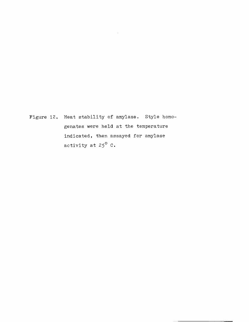
Figure 12. Heat stability of amylase. Style homo- genates were held at the temperature indicated, then assayed for amylase activity at 25° C.

Percent activity
oO
roUl cno
~n|a i
oo
o
noO
o
a>o
ooo

Figure 13* Gel filtration of amylolytic activity.The results shown are from the gel fil tration of whole style homogenate on a Bio-Gel P-100 column, employing a pH 7 sodium phosphate buffer, 0.1 M.

A b s o r b a n c e ( 5 4 0 nm)
9 9 9rb -P> (j) 80

4 6
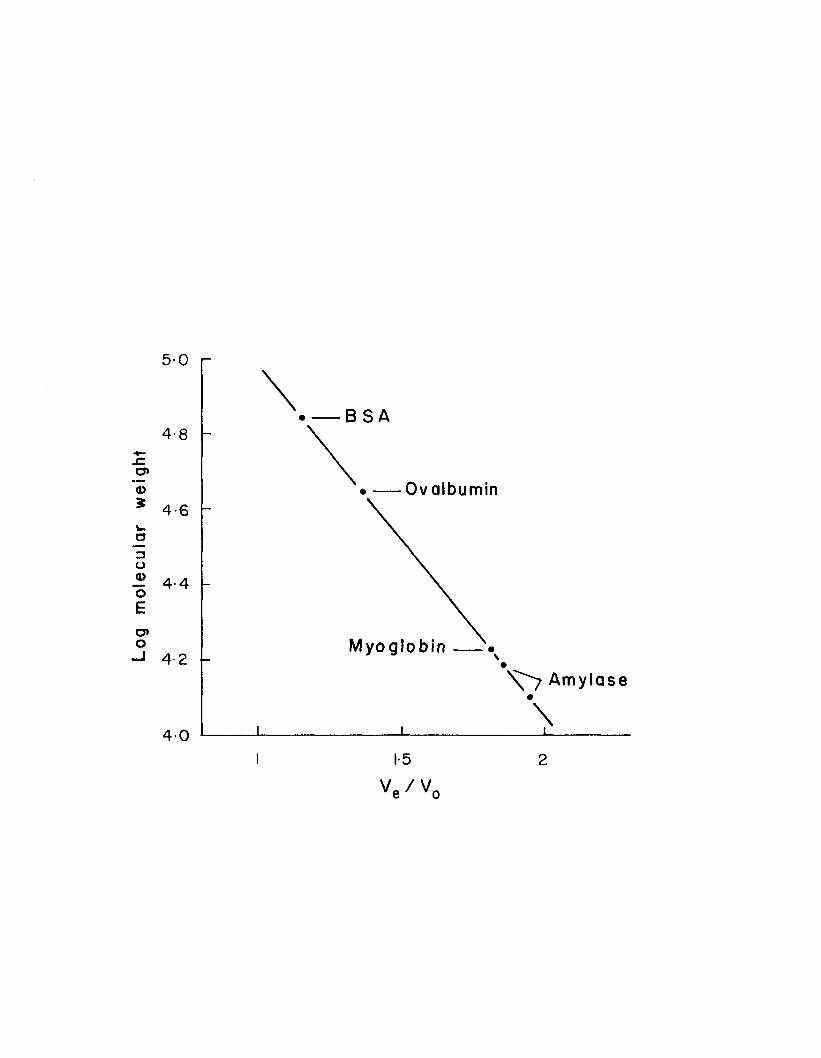
matrix should yield more accurate values. Approximate molecular weight values for peaks B1 and B 2 , calculated on Bio-Gel P-100 by comparing elution ratios of the amylolytic peaks to those of known proteins, are 1 5 , 7 0 0 and 1 2 , 9 0 0 respectively (Fig. 14).
ElectrophoresisPolyacrylamide disc electrophoresis of whole style
extract demonstrated a minimum of ten protein bands. Electrophoresis of peak B eluted from a Sephadex column with sodium phosphate buffer yielded six protein bands. When the B peaks eluted from two Sephadex column runs were concentrated and passed through a Bio-Gel column, disc electrophoresis of the resulting Bio-Gel B peak produced two protein bands. Two protein bands were also seen after electrophoresis of the B peak from gel filtration of whole style extract on Bio-Gel. However, the patterns of two protein bands from the two samples above were similar with respect to only one band.
Disc electrophoresis on starch-impregnated gels produced as many as six bands of amylolytic activity from samples of whole style extract or concentrates of those gel filtration fractions eluted at the void volume (peak A). Similar electrophoresis of peak B concentrates produced two or three bands of amylolytic activity. Although the results obtained were not adequate to indicate the number of amylase species present nor the ways in which they might associate, the results do serve to demonstrate the complex nature of the amylolytic activity in the style.

Figure 14. Molecular weight determination of amylase by comparison of the elution ratios of amylase shown in Figure 13 to those of proteins of known molecular weight. BSA represents bovine serum albumin.
Log
mol
ecul
ar
wei
gh
t
5-0
B S A4-8
Ovalbumin4-6
4-4
M y og lo b i n4-2A m y la s e
401-5 2
V / V ve ' vo

49
Electrophoresis of whole style protein on agarose plates produced one large rapidly moving protein band, a second narrower "band midway between the first band and the origin, and a continuous smudge between the origin and the rapidly migrating band (Fig. 15)- Electrophoresis of whole style extracts or gel filtration peak A concentrates on agarose consistently demonstrated three bands of amylolytic activity (Fig. l6). The migration of one of these bands corresponded to that of the large rapidly moving protein band. Only one band, that closest to the origin, was visible after electrophoresis of peak B.
DISCUSSION
The style amylase of M. arenaria appears particularly well suited to the NaCl concentrations at which it must function. The optimum NaCl concentration shown in Figure 10 (0.5 M) is nearly equal to that contained in "normal" 35°/oo
salinity sea water. An estuarine species such as M. arenaria can be exposed to much lower salinities, and Figure 10 indicates that some amylase activity is retained at very low NaCl concentrations. Because M. arenaria is an osmocon- former, as are most marine molluscs (see review by Robertson, 1964), the NaCl concentration of the medium in which the amylase acts will equal that of the surrounding water.
The activity plateau between 10~^ M and 10 2 M NaCl (Fig. 10) is indicative of more than one chloride binding site, i. e. one site is able to bind chloride at low NaCl
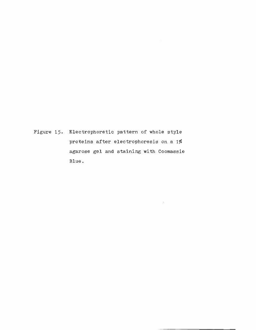
Figure 15. Electrophoretic pattern of whole style proteins after electrophoresis on a ifo agarose gel and staining with Coomassie Blue.


Figure 16. Electrophoretic pattern of amylase from whole style homogenate after electrophoresis on a 1% agarose gel. Light hands indicate the areas of amylolytic activity.


5^
_ Q _ 2concentrations (10 J - 10 M ) , while the binding at asecond site increases as the NaCl concentration increases
_2from 10 M to 0.5 M. Because of the electrophoretic demonstration of different amylase molecules (Fig. 16), I believe that the different chloride binding sites occupy different amylase molecules rather than multiple sites on a single amylase molecule.
The activation of mammalian amylase by other halides in the absence of chloride (Br> I >F) (Schwartz and Fleischer, 1970) is very similar to that found in the present study (Table 1) .
The pH optima of style amylase shown in Figure 11 (6 .3 , 7-0, 7.3) is in the range found in other molluscs (6.2 and 7.0, Black and Pengelley, 1964-; 5 >8-5 -9 and 6.5-6.7. Horiuchi and Lane, 1 9 6 6 ; 6 .5 * Wojtowicz, 1972) and in mammals (6 .5-7-5. Schwartz and Fleischer, 1970). Although different pH optima at different chloride concentrations may be interpreted as resulting from the effect of the chloride on the tertiary structure of one amylase enzyme, the activation of different amylase molecules of different pH optima seems more likely. Indeed, three amylases, with pH optima of 6.2, 6 .6 , and 6 .9 , were separated by Berk et al. (1963) "by i°n exchange chromatography of rabbit serum. The presence of a highly labile amylase with a pH optimum of 7 . 3 may have resulted in the decreased activity observed at pH 7-3 in successive experiments.
Both mammalian (Frati and Caputo, 1970) and inverte-
55
brate (Wojtowicz and Brockerhoff, 1972; Wojtowicz, 1972) amylases have been purified by successive Sep'hadex and Bio- Gel chromatography. This technique relies upon the affinity of amylase for the dextran matrix of Sephadex and the resulting slower elution of amylase. The failure of this technique to produce electrophoretically pure amylase in the present study may have resulted from the affinity of several amylases or other carbohydrases for the dextran matrix.Robyt et al. (1971 ) obtained identical molecular weight values for porcine amylase fractionated on Sephadex and Bio- Gel, indicating that the elution of amylase was not slowed by an affinity for the Sephadex. The different Ve/Vo ratios given in the present study for Sephadex gel filtration with sodium phosphate and NaCl eluants demonstrates the effect of different eluants on the gel filtration of amylase. Thus Robyt et al, (1971) » by using a pH 8 . 5 eluant, may havereduced the amylase activity and consequently the Sephadex- amylase interaction.
Reported molecular weights of invertebrate and mammalian amylases have ranged from ^ 0 , 0 0 0 to 60,000 (Fischer and Stein, I960; Frati and Caputo, 1 9 70; Cozzone et a l . , 1970; Robyt et al., 1971; Wojtowicz and Brockerhoff, 1972; Wojtowicz, 1972). Robyt et al. (1971) separated porcine pancreatic amylase (5 0 , 0 0 0 daltons) into two subunits of 2 5 , 0 0 0 dal-tons by strong reducing conditions which presumably broke disulfide bonds. They also gave some evidence of 12,500- dalton subunits. Thus the low molecular weight amylases of

56
peak B in the present study probably correspond to subunits of the porcine amylase. Although the low molecular weight amylases probably associate with each other or with style- matrix proteins eluted at peak A, they are not bound together by strong disulfide bridges as are the porcine amylase subunits .
Agarose and polyacrylamide electrophoresis demonstrate the high degree of protein heterogeneity in the style. Three different amylases were demonstrated by agarose electrophoresis. The large number of amylase bands seen by polyacrylamide electrophoresis may result from partial dissociation of the amylases.
Although style amylase is similar to mammalian amylase in several respects as discussed above, differences demonstrated by gel filtration chromatography are additionally indicated by our findings that antibodies against porcine pancreatic amylase would not precipitate any proteins from whole style homogenate.
57
REFERENCES
Allen J. A. (1958) On the basic form and adaptations to habitat in the Lucinacea (Eulamellibranchia). Phil. Trans. R. Soc. , Ser. B 24l, 421-484.
Andrews P. (1964) Estimation of the molecular weights of proteins by Sephadex gel-filtration. Biochem. J. 91, 2 2 2-2 3 3 .
Bailey K. & Worboys B. (i9 6 0 ) The lamellibranch crystalline style. Biochem. J. 7 6 , 487-491.
Bedford J. J. & Reid M. S. (1 9 6 9 ) Gel electrophoresis of proteins in the crystalline style of certain mollusca.Comp. Biochem. Physiol■ 2 9 , 659-664.
Berk J. E., Kawaguchi K . , Zeineh R., Ujihira I. & Searcy R. (1 9 6 3 ) Chromatographic heterogeneity of rabbit serum amylase. Science l4l, 1182-1183.
Bernard F. R. (1973) Crystalline style formation and function in the oyster Crassostrea gigas (Thunberg, 1795)* Ophelia 12, 159-170.
Bernfeld P. (1955) Amylases. In Methods of Enzymology (Edited by Colowick S. P. & Kaplan N. OTT, Vol. 1 , pp. 149-150. Academic Press, New York.
Black R. E. & Pengelley E. T. (1964) Alpha amylase development in embryos of Crassostrea virgin:! ca. Biol. Bull. mar, biol. Lab., Woods Hole 126, 199-204•
Chaet A. B. (1956) Chromatographic study of crystalline style amylase. Biol. Bull. mar. biol. Lab., Woods Hole 111, 2 9 8 .
Coupin H. (1900) Sur les fonctions de la tige cristalline des Acephales. C. r. Sea.nc. Acad. Sci. 130, 1214-1216.
Cozzone P., Pasero L. & Marchis-Mouren G. (1970) Characterization of porcine pancreatic isoamylases: separation and amino acid composition. Biochem. biophys. Acta 200,590-593-
Doyle J. (1 9 6 6 ) Studies on the chemical nature of the crystalline style. In Some Contemporary Studies in Marine Science (Edited by Barnes H. )~i pp. 253-263- George Allen and Unwin Ltd., London.

58
Fischer E. H. & Stein E. A. (i9 6 0 ) Alpha-amylases. In The Enzymes (Edited "by Boyer P. D. , Lardy H. & Myrback K . ) ,2nd edn. Vol. 4, pp. 313-343- Academic Press, New York.
Frati K. L. & Caputo A. (1970) Purification of alpha- amylase from C3H mouse submaxillary gland by gel filtration. J. Chromat. 49, 547-550.
George V. C. (1952) The digestion and absorption of fat in lamellibranchs. Biol. Bull. mar. biol. Lab., Woods Hole 102, 118-128.
Goreau T. F., Goreau N. I. & Yonge C. M. (1973) On theutilization of photosynthetic products from zooxanthellae and of a dissolved amino acid in Tridacna maxima f. elongata (Mollusca: Bivalvia). J. Zool., Land. 1 6 9 , 417-454.
Halton D. W. & Owen G. (1 9 6 8 ) The fine structure and histochemistry of the gastric cuticle of the protobranchiate bivalve Nucula sulcata Brown. Proc. malac. Soc. Lond.38, 71-81.
Horiuchi S. & Lane C. E. (1 9 6 6 ) Carbohydrases of the crystalline style and hepatopancreas of Strombus gigus Linne. Comp. Biochem. Physiol. 17. 1189-1197-
Kato K. & Kubomura K. (1954) On the origin of the crystalline style of lamellibranchs. Sci■ Repts Saitama Univ.91, 135-152.
Kristensen J. H. (1972a) Structure and function of crystalline styles of bivalves. Ophelia 10, 91-108.
Kristensen J. H. (1972b) Carbohydrases of some marineinvertebrates with notes on their food and on the natural occurrence of the carbohydrates studied. Mar. Biol. 14, 130-142.
Lowry 0. H . , Rosebrough N. J., Farr A. L. & Randall R. J. (1951) Protein measurement with the Folin phenol reagent. J. Biol. Chem. 1 9 3 , 265-275-
McQuiston R. W. (1970) Fine structure of the gastric shield in the lamellibranch bivalve Lasea rubra (Montagu). Proc. malac. Soc. Lond. 3 6 , 69-75-
Morton B. S. (1 9 6 9 ) Studies on the biology of Dreissenapolymorpha Pall II. correlation of the rhythms of adductor activity, feeding, digestion and excretion. Proc. malac. Soc. Lond. 38, 401-414.

59
Morton B. S. (1971) The diurnal rhythm and tidal rhythm of feeding and digestion in Ostrea edulis. Biol. J. Linn.Soc. 3 , 329-342.
Nelson T. C. (1 9 1 8 ) On the origin, nature and function of the crystalline style of the lamellibranchs. J. morph.31, 53-in.
Ouchterlony 0. (1 9 6 8 ) Handbook of Immunodiffusion and Immunoelectrophoresis. Ann Arbor Science Publishers,Inc., Ann Arbor.
Owen G. (1 9 6 6 ) Digestion. In Physiology of Mollusca (Edited by Wilbur K. M. & Yonge C. M . ), Vol. II, pp. 53-96.Aca.demic Press, New York.
Purchon R. D. (1958) The stomach in the Eulamellibranchia; stomach type IV. Proc. Zool■ Soc. Lond. 131. 487-525.
Purchon R. D. (i9 6 0 ) The stomach in the Eulamellibranchia; stomach types IV and V. Proc. Zool. Soc. Lond. 135)431-489.
Reid R. G. B. (1 9 6 6 ) Digestive tract enzymes in the bivalves Lima hians Gmelin and Mya arenaria L. Comp■ Biochem.Physiol. 17, 417-433-
Reid R. G. B. (1 9 6 8 ) Distribution of digestive tract enzymes in lamellibranchiate bivalves. Comp. Biochem. Physiol.24, 727-744.
Robertson J. D. (1964) Osmotic and ionic regulation. In Physiology of Mollusca (Edited by Wilbur K. M. & Yonge C . M. ) , pp. 283-3H. Academic Press, New York.
Robyt J. F., Chittenden C. G. & Lee C. T. (1971) Structure and function of amylases 1 . the subunit structure of porcine pancreatic alpha-amylase. Archs Biochem. Biophys. 144, 1 6 0-1 6 7 .
Ruthmann A. (1970) Methods in Cell Research. Cornell University Press, Ithaca.
Schwartz M. K. & Fleischer M. (1970) Diagnostic biochemical methods in pancreatic disease. In Advances in Clinical Chemistry (Edited by Bodansky 0. & Stewart C. P.), Vol. 13. pp. 130-133. Academic Press, New York.
Wojtowicz M. B. (1972) Carbohydrases of the digestive gland and the crystalline style of the Atlantic deep-sea scallop (Placopecten magellanicus, Gmelin). Comp. Biochem. Physiol. 43A, 131-141.

60
Wojtowicz M. B. & Brockerhoff H. (1972) Isolation and some properties of the digestive amylase of the American lobster (Homarus americanus). Comp. Biochem. Physiol. 42B, 295-302.
Yonge C. M. (1923) Studies on the comparative physiology of digestion. The mechanism of feeding, digestion, and assimilation in the lamellibranch Mya. Brit. J. exp. Biol.1, 15-63.
Yonge C. M. (1925) The hydrogen ion concentration in the gut of certain lamellibranchs and gastropods. J. mar. Biol.Ass. U. K. 13, 938-952.
Yonge C. M. (1926) Structure and physiology of the organs of feeding and digestion in Ostrea edulis. J. mar. Biol. Ass.u. k . i4, 295-386.
Yonge C. M. (1932) The crystalline style of the mollusca.Sci. Progress 26, 643-653*

BIOGRAPHICAL DATA
Name: David Gibson TrainerDate of Birth: March 11, 19^5Place of Birth: Allentown, PennsylvaniaSecondary education: William Allen High School
Allentown, PennsylvaniaCollegiate Institutions attended: Dates Degrees
Washington and Jefferson College 9/63-6/67 A.B.University of Maine 9/67-6/70 M.Sc.University of New Hampshire 9/70-8/75 Ph.D.
Honors or Awards: Dean's Honor List; UNH Summer Fellowship,1971. 197 ;̂ Phi Sigma; Sigma X i .
Positions held: DatesGraduate Research Assistant, U. of Maine 7/67-9/68Graduate Teaching Assistant, U. of Maine 9/68-6/70Graduate Teaching Assistant, U. of N. H. 9/70-1/75Instructor, Merrimack Valley Branch, U. of N. H. 6/75-8/75