University of Groningen Therapeutic and mechanistic explorations of ...
101
University of Groningen Therapeutic and mechanistic explorations of in-stent restenosis in the rat aortic stenting model Groenewegen, Hendrik Cornelis IMPORTANT NOTE: You are advised to consult the publisher's version (publisher's PDF) if you wish to cite from it. Please check the document version below. Document Version Publisher's PDF, also known as Version of record Publication date: 2008 Link to publication in University of Groningen/UMCG research database Citation for published version (APA): Groenewegen, H. C. (2008). Therapeutic and mechanistic explorations of in-stent restenosis in the rat aortic stenting model. s.n. Copyright Other than for strictly personal use, it is not permitted to download or to forward/distribute the text or part of it without the consent of the author(s) and/or copyright holder(s), unless the work is under an open content license (like Creative Commons). The publication may also be distributed here under the terms of Article 25fa of the Dutch Copyright Act, indicated by the “Taverne” license. More information can be found on the University of Groningen website: https://www.rug.nl/library/open-access/self-archiving-pure/taverne- amendment. Take-down policy If you believe that this document breaches copyright please contact us providing details, and we will remove access to the work immediately and investigate your claim. Downloaded from the University of Groningen/UMCG research database (Pure): http://www.rug.nl/research/portal. For technical reasons the number of authors shown on this cover page is limited to 10 maximum. Download date: 15-02-2022
-
Upload
khangminh22 -
Category
Documents
-
view
4 -
download
0
Transcript of University of Groningen Therapeutic and mechanistic explorations of ...
University of Groningen
Therapeutic and mechanistic explorations of in-stent restenosis in the rat aortic stentingmodelGroenewegen, Hendrik Cornelis
IMPORTANT NOTE: You are advised to consult the publisher's version (publisher's PDF) if you wish to cite fromit. Please check the document version below.
Document VersionPublisher's PDF, also known as Version of record
Publication date:2008
Link to publication in University of Groningen/UMCG research database
Citation for published version (APA):Groenewegen, H. C. (2008). Therapeutic and mechanistic explorations of in-stent restenosis in the rataortic stenting model. s.n.
CopyrightOther than for strictly personal use, it is not permitted to download or to forward/distribute the text or part of it without the consent of theauthor(s) and/or copyright holder(s), unless the work is under an open content license (like Creative Commons).
The publication may also be distributed here under the terms of Article 25fa of the Dutch Copyright Act, indicated by the “Taverne” license.More information can be found on the University of Groningen website: https://www.rug.nl/library/open-access/self-archiving-pure/taverne-amendment.
Take-down policyIf you believe that this document breaches copyright please contact us providing details, and we will remove access to the work immediatelyand investigate your claim.
Downloaded from the University of Groningen/UMCG research database (Pure): http://www.rug.nl/research/portal. For technical reasons thenumber of authors shown on this cover page is limited to 10 maximum.
Download date: 15-02-2022

Therapeutic and mechanistic explorations of in-stent restenosis in the rat aortic stenting model.
Hendrik C. Groenewegen

© Copyright 2008 H.C. Groenewegen All rights are reserved. No part of this publication may be reproduced, stored in a retrieval system , or transmitted in any form or by any means, mechanically, by photocopying, recording, or otherwise, without the written permission of the author. Cover: interpretation of Scylla and Charybis by Lynck+Zoon B.V. Lay-out: Drukkerij Van Denderen Printed by: Drukkerij Van Denderen

RIJKSUNIVERSITEIT GRONINGEN
Therapeutic and mechanistic explorations of in-stent restenosis in the rat aortic stenting model
Proefschrift
ter verkrijging van het doctoraat in de
Medische Wetenschappen aan de Rijksuniversiteit Groningen
op gezag van de Rector Magnificus, dr. F. Zwarts, in het openbaar te verdedigen op
woensdag 18 juni 2008 om 13.15 uur
door
Hendrik Cornelis Groenewegen
geboren op 27 juli 1979 te Heerenveen

Promotores: Prof. Dr. F. Zijlstra Prof. Dr. W.H. van Gilst Copromotores: Dr. B.J.G.L. de Smet Dr. A.J.M. Roks Beoordelingscommisie: Prof. dr. W.J. van der Giessen Prof. dr. D.J. van Veldhuisen Prof. dr. R.H. Henning
ISBN: 978-90-367-3419-6 ISBN: 978-90-367-3418-9 (digitaal)
Paranimfen: Robert de Kwant Hans Hofer The publication of this thesis was financially supported by Medtronic Trading NL B.V., Merck Sharp & Dohme B.V., Boehringer Ingelheim B.V.,Pfizer B.V., St Jude Medical Nederland B.V., Schering-Plough B.V., Bristol-Myers Squibb B.V., Meda Pharma B.V. ,Servier Nederland Farma B.V. and Interuniversitair Cardiologisch Instituut Nederland.
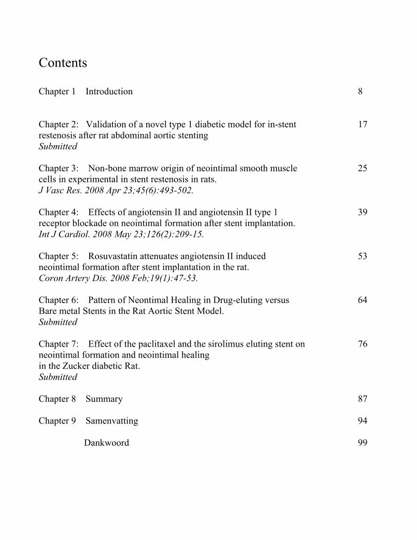
Contents Chapter 1 Introduction 8 Chapter 2: Validation of a novel type 1 diabetic model for in-stent 17 restenosis after rat abdominal aortic stenting Submitted Chapter 3: Non-bone marrow origin of neointimal smooth muscle 25 cells in experimental in stent restenosis in rats. J Vasc Res. 2008 Apr 23;45(6):493-502. Chapter 4: Effects of angiotensin II and angiotensin II type 1 39 receptor blockade on neointimal formation after stent implantation. Int J Cardiol. 2008 May 23;126(2):209-15. Chapter 5: Rosuvastatin attenuates angiotensin II induced 53 neointimal formation after stent implantation in the rat. Coron Artery Dis. 2008 Feb;19(1):47-53. Chapter 6: Pattern of Neontimal Healing in Drug-eluting versus 64 Bare metal Stents in the Rat Aortic Stent Model. Submitted Chapter 7: Effect of the paclitaxel and the sirolimus eluting stent on 76 neointimal formation and neointimal healing in the Zucker diabetic Rat. Submitted Chapter 8 Summary 87 Chapter 9 Samenvatting 94 Dankwoord 99

CHAPTER 1 INTRODUCTION AND AIMS

Chapter 1
8
INTRODUCTION Cardiovascular diseases cause millions of deaths worldwide1. In large part related to coronary heart disease (CHD)2. During the past decades the treatment and prevention of CHD has been significantly improved with the introduction of drugs which slow the development or progression of CHD or prevent the clinical manifestations of CHD (B-blockers, aspirin, statins, ACE inhibitors and AT1 receptor antagonists) and by revascularization of obstructed coronary arteries using coronary artery bypass grafting (CABG) and percutanous coronary intervention (PCI)3-7. In recent years therapeutic advances of PCI such as antiplatelet drugs given before and after the procedure, the stent and later the drug eluting-stent have improved mortality and morbidity for patients with CHD and this has led to a marked rise in the number of PCI procedures 8-10. Nowadays In the Netherlands every year around thirty thousand PCI procedures are performed 11. However PCI is still troubled by two major limitations: restenosis and stent thrombosis12,13. Restenosis is the narrowing of the vessel wall after PCI and is the result of the arterial healing response after the induction of vascular injury. Restenosis usually does not lead to acute occlusion of the vessel with myocardial infarction and death. However recurrence of angina pectoris is often seen requiring revascularization of the stented vessel (target lesion revascularization). The rate of target lesion revascularization after 5 years is markedly lower with drug eluting stents 10,3%, compared to 26% in bare metal stents14. Riskfactors for restenosis are small vessels, longer lesions, restenotic lesions and importantly diabetes15,16. Diabetic coronary artery disease is characterized by multivessel involvement, diffuse disease, more significant stenoses, increased calcified disease and decreased collateral vessel formation17. These factors might influence the technical success of PCI leading to a lower final lumen diameter. Lower final lumen diameter is strongly correlated with increased restenosis18. At the molecular level these disease factors may be modulated by insulin resistance, hyperglycaemia and inflammation19,20. Therefore pharmacological intervention to improve glycaemic control and reduce insulin resistance and inflammation may be an interesting target to decrease the risk of restenosis in diabetic patients undergoing PCI17. Restenosis is more common after balloon angioplasty and is caused by both elastic recoil, negative remodeling and to a lesser extent neointimal formation21. Stenting has reduced restenosis by providing a rigid structure in the vessel wall which eliminates elastic recoil and negative remodelling. However restenosis is not eliminated because stenting induces more injury to the vessel wall. This leads to a more proliferative healing response with more neointimal formation22,23. To tackle the problem of restenosis after stenting the mechanism and pathways of the different phases of neointimal formation have been studied extensively: thrombus formation, inflammation, smooth muscle cell migration, smooth muscle cell proliferation and extracellular matrix formation. The arterial healing response leading to neointimal formation begins with platelet aggregation and platelet activation followed by infiltration of the injured vessel wall and thrombus by predominantly mononuclear and polymorphonuclear leukocytes24-26. The leukocytes and platelets produce cytokines like monocyte chemoattractant protein (MCP-1), platelet-derived growth factor (PDGF) and angiotensin II. These cytokines enhance inflammation, stimulate smooth muscle cell migration from the media of the vessel towards the lumen and stimulate smooth muscle cell proliferation 27-29. After a few weeks the proliferation of smooth muscle cells in the neointima peaks and apoptosis of neointimal cells becomes more prominent 30,31. The remaining neointimal cells (most of smooth muscle cell origin and some macrophages) begin production of proteoglycans and collagen creating an extracellular matrix32,33. After insight had been gained in the pathofysiology of neointimal formation, a myriad of known and newly developed anti-thrombotic, anti-inflammatory, anti-migratory, anti-proliferative and anti-

INTRODUCTION
9
extracellular matrix producing drugs have been tested in animal models and clinical trials in an attempt to reduce neointimal formation 34-39. Of these drugs the two anti-proliferative drugs sirolimus and paclitaxel have shown exceptional promise in reducing in-stent restenosis 40,41. Brachytherapy by using the anti-proliferative properties of beta radiation was initially successful in reducing in-stent restenosis, but long term follow-up studies showed restenosis and late thrombosis42. The strong anti-proliferative effects of sirolimus and paclitaxel make these drugs less suitable for systemic administration in patients43. Local application of these drugs on a stent circumvents high systemic doses: the drug eluting stent was born44. These two new drug eluting stents (paclitaxel eluting stent and sirolimus eluting stent) have been proven to be very successful in reducing in-stent restenosis in several clinical trials45,46. However, recently drug eluting stents have been associated with an increased risk of late stent thrombosis.47. Stent thrombosis is the occurrence of thrombus formation in or near the stent resulting in an acute occlusion of the vessel. The incidence of stent thrombosis after stenting with the use of dual antiplatelet therapy is around 1% after 1 year and 2% after 10 year48. Although its incidence is much lower than in-stent restenosis nevertheless it is a serious complication because in contrast with in-stent restenosis it leads more often to acute myocardial infarction and has a high mortality rate48. It is commonly divided in acute ( ≤ 24 hours post-PCI), subacute (24 hours to 30 days post-PCI) and late (> 30 days post-PCI) stent thrombosis. The problem of acute and subacute thrombosis was greatly reduced by preventing early thrombus formation using dual anti-platelet (acetylsalicilzuur and clopidogrel) therapy49. However late stent thrombosis is still a problem especially in drug eluting stents. Therefore dual anti-platelet therapy post-PCI is recommended for one month for patients with bare metal stents (BMS) and six months to a year for patients with drug eluting stents(DES) according to the guidelines of the European Society of Cardiology 50. Some authors have suggested that triple anti-platelet (addition of cilostazol) may benefit patients with a DES51. However there are disadvantages of more intensive and longer anti-platelet therapy. An increased chance of serious bleeding exists with longer and more potent antiplatelet therapy52. More importantly patients who need surgery for other medical conditions often have to stop antiplatelet therapy and are at a high risk for developing stent thrombosis during surgery or risk serious bleeding with the continuation of antiplatelet therapy53. Several large clinical trials with long follow-up are addressing the issue of stent thrombosis with DES and its clinical relevance. So the impact of stent thrombosis on the future use and development of present and new generation drug eluting stents is still unclear. Pending these results research has focused on finding alternative drugs to treat in-stent restenosis and a renewed interest in the pathofysiology, mechanisms and riskfactors (diabetes) of in-stent restenosis and stent thrombosis. In this thesis the rat aortic stenting model was used to study several new treatments to reduce in-stent restenosis and some aspects of the pathofysiology of in stent restenosis. We also adapted our rat aortic model and developed and validated two different diabetic models for studying the effects of in-stent restenosis in diabetes 54. Furthermore we tried to identify possible mechanisms of late stent trombosis by comparing the neointima of bare metal and drug eluting stents.

Chapter 1
10
AIMS In Chapter 2 we introduce a novel type 1 diabetic model for in-stent restenosis after rat abdominal aortic stenting study. Diabetes is an important clinical riskfactor for restenosis and with the future epidemic in diabetes related to overweigth it will grow even further in importance 55,56. Research of the mechanisms of increased restenosis in diabetes may also lead to general anti-restenotic drugs. In Chapter 3 we study the contribution of circulating bone marrow cells to neointimal formation. The role of bone marrow cells in neointimal formation after stenting is not yet clear but it is an important issue, because it could lead to new cell based therapies for in-stent restenosis57-59. The neointima of stented vessels is mainly composed of α-smooth muscle actin (SMA) positive cells60 and the paradigm states that the origin of α-smooth muscle positive cells in the neointima is from smooth muscle cells residing in the media and migrating to the lumen after stenting61. However circulating bone marrow cells can differentiate in a wide variety of cell types62 (including smooth mucle actin positive cells) and could be involved in neointimal formation after stenting. In Chapter 4 we describe the effect of the AT1-receptor candesartan on neointimal formation after stenting. Abundant pre-clinical evidence exists that Ang II is involved in in-stent restenosis, but in clinical trials ACE-inhibitors and AT1-receptor blockers have not been successful in reducing in-stent restenosis. We study if the conflicting results of animal research and clinical trials is related to the need of supraphysiological Ang II levels for Ang II mediated neointima formation. In Chapter 5 we test the effect of the statin rosuvastatin on neointimal formation and endothelial function after stenting. Statins have several pleiotropic effects which may reduce in- stent restenosis: reduction of platelet activation, amelioration of endothelial function lowering of inflammatory responses, reduction of oxidative stress and inhibition of smooth muscle proliferation and migration. Ang II increases oxidative stress, stimulates smooth muscle cell proliferation and detoriates endothelial function. We study if statins reduce neointimal formation in a normal and a stimulative setting (Ang II induced). In Chapter 6 and 7 we describe quantative and qualitative differences in neointimal formation between bare metal stents and drug eluting stents in normoglycemic (Chapter 6) and diabetic setting (Chapter 7). Incomplete endothelial healing has been associated with stent thrombosis. Differences in neointimal healing between drug eluting stents and bare metal stents may be associated with increased rates of late stent thrombosis.

INTRODUCTION
11
References
1. Daar AS, Singer PA, Persad DL, Pramming SK, Matthews DR, Beaglehole R, Bernstein A, Borysiewicz LK, Colagiuri S, Ganguly N, Glass RI, Finegood DT, Koplan J, Nabel EG, Sarna G, Sarrafzadegan N, Smith R, Yach D, Bell J. Grand challenges in chronic non-communicable diseases. Nature 2007;450:494-496.
2. Beaglehole R, Saracci R, Panico S. Cardiovascular diseases: causes, surveillance and prevention. Int J Epidemiol 2001;30 Suppl 1:S1-S4.
3. Rosendorff C. Hypertension and coronary artery disease: a summary of the american heart association scientific statement. J Clin Hypertens (Greenwich ) 2007;9:790-795.
4. Reilly IA, Fitzgerald GA. Aspirin in cardiovascular disease. Drugs 1988;35:154-176.
5. Paraskevas KI, Hamilton G, Mikhailidis DP. Statins: an essential component in the management of carotid artery disease. J Vasc Surg 2007;46:373-386.
6. Christian CB, Jr., Mack JW, Wetstein L. Current status of coronary artery bypass grafting for coronary artery atherosclerosis. Surg Clin North Am 1985;65:509-526.
7. Gruntzig AR, Senning A, Siegenthaler WE. Nonoperative dilatation of coronary-artery stenosis: percutaneous transluminal coronary angioplasty. N Engl J Med 1979;301:61-68.
8. Wiviott SD, Braunwald E, McCabe CH, Montalescot G, Ruzyllo W, Gottlieb S, Neumann FJ, Ardissino D, De Servi S, Murphy SA, Riesmeyer J, Weerakkody G, Gibson CM, Antman EM. Prasugrel versus clopidogrel in patients with acute coronary syndromes. N Engl J Med 2007;357:2001-2015.
9. Sigwart U, Puel J, Mirkovitch V, Joffre F, Kappenberger L. Intravascular stents to prevent occlusion and restenosis after transluminal angioplasty. N Engl J Med 1987;316:701-706.
10. Serruys PW, Degertekin M, Tanabe K, Abizaid A, Sousa JE, Colombo A, Guagliumi G, Wijns W, Lindeboom WK, Ligthart J, de Feyter PJ, Morice MC. Intravascular ultrasound findings in the multicenter, randomized, double-blind RAVEL (RAndomized study with the sirolimus-eluting VElocity balloon-expandable stent in the treatment of patients with de novo native coronary artery Lesions) trial. Circulation 2002;106:798-803.
11. Huijskes RV, Koch KT, van Herwerden LA, Berreklouw E, Limburg M, Tijssen JG. [The waiting time for heart interventions: trends for percutaneous coronary interventions and cardiothoracic interventions]. Ned Tijdschr Geneeskd 2003;147:1860-1865.
12. Ako J, Bonneau HN, Honda Y, Fitzgerald PJ. Design criteria for the ideal drug-eluting stent. Am J Cardiol 2007;100:3M-9M.
13. Slottow TL, Waksman R. Drug-eluting stent safety. Am J Cardiol 2007;100:10M-17M.
14. Morice MC, Serruys PW, Barragan P, Bode C, Van Es GA, Stoll HP, Snead D, Mauri L, Cutlip DE, Sousa E. Long-term clinical outcomes with sirolimus-eluting coronary stents: five-year results of the RAVEL trial. J Am Coll Cardiol 2007;50:1299-1304.
Chapter 1
12
15. Moses JW, Leon MB, Popma JJ, Fitzgerald PJ, Holmes DR, O'Shaughnessy C, Caputo RP, Kereiakes DJ, Williams DO, Teirstein PS, Jaeger JL, Kuntz RE. Sirolimus-eluting stents versus standard stents in patients with stenosis in a native coronary artery. N Engl J Med 2003;349:1315-1323.
16. Lee CW, Suh J, Lee SW, Park DW, Lee SH, Kim YH, Hong MK, Kim JJ, Park SW, Park SJ. Factors predictive of cardiac events and restenosis after sirolimus-eluting stent implantation in small coronary arteries. Catheter Cardiovasc Interv 2007;69:821-825.
17. Morgan KP, Kapur A, Beatt KJ. Anatomy of coronary disease in diabetic patients: an explanation for poorer outcomes after percutaneous coronary intervention and potential target for intervention. Heart 2004;90:732-738.
18. Kuntz RE, Safian RD, Carrozza JP, Fishman RF, Mansour M, Baim DS. The importance of acute luminal diameter in determining restenosis after coronary atherectomy or stenting. Circulation 1992;86:1827-1835.
19. Takezako T, Saku K, Zhang B, Shirai K, Arakawa K. Insulin resistance and angiographical characteristics of coronary atherosclerosis. Jpn Circ J 1999;63:666-673.
20. Hoffmeister A, Rothenbacher D, Bazner U, Frohlich M, Brenner H, Hombach V, Koenig W. Role of novel markers of inflammation in patients with stable coronary heart disease. Am J Cardiol 2001;87:262-266.
21. Mintz GS, Popma JJ, Pichard AD, Kent KM, Satler LF, Wong C, Hong MK, Kovach JA, Leon MB. Arterial remodeling after coronary angioplasty: a serial intravascular ultrasound study. Circulation 1996;94:35-43.
22. Grewe PH, Deneke T, Machraoui A, Barmeyer J, Muller KM. Acute and chronic tissue response to coronary stent implantation: pathologic findings in human specimen. J Am Coll Cardiol 2000;35:157-163.
23. Moreno PR, Palacios IF, Leon MN, Rhodes J, Fuster V, Fallon JT. Histopathologic comparison of human coronary in-stent and post-balloon angioplasty restenotic tissue. Am J Cardiol 1999;84:462-6, A9.
24. Chandrasekar B, Tanguay JF. Platelets and restenosis. J Am Coll Cardiol 2000;35:555-562.
25. Yeo EL, Sheppard JA, Feuerstein IA. Role of P-selectin and leukocyte activation in polymorphonuclear cell adhesion to surface adherent activated platelets under physiologic shear conditions (an injury vessel wall model). Blood 1994;83:2498-2507.
26. Rogers C, Welt FG, Karnovsky MJ, Edelman ER. Monocyte recruitment and neointimal hyperplasia in rabbits. Coupled inhibitory effects of heparin. Arterioscler Thromb Vasc Biol 1996;16:1312-1318.
27. Chandrasekar B, Tanguay JF. Platelets and restenosis. J Am Coll Cardiol 2000;35:555-562.
28. Welt FG, Rogers C. Inflammation and restenosis in the stent era. Arterioscler Thromb Vasc Biol 2002;22:1769-1776.

INTRODUCTION
13
29. Schwartz SM. Smooth muscle migration in atherosclerosis and restenosis. J Clin Invest 1997;100:S87-S89.
30. Carter AJ, Laird JR, Farb A, Kufs W, Wortham DC, Virmani R. Morphologic characteristics of lesion formation and time course of smooth muscle cell proliferation in a porcine proliferative restenosis model. J Am Coll Cardiol 1994;24:1398-1405.
31. Kollum M, Kaiser S, Kinscherf R, Metz J, Kubler W, Hehrlein C. Apoptosis after stent implantation compared with balloon angioplasty in rabbits. Role of macrophages. Arterioscler Thromb Vasc Biol 1997;17:2383-2388.
32. Glover C, Ma X, Chen YX, Miller H, Veinot J, Labinaz M, O'Brien E. Human in-stent restenosis tissue obtained by means of coronary atherectomy consists of an abundant proteoglycan matrix with a paucity of cell proliferation. Am Heart J 2002;144:702-709.
33. Chung IM, Gold HK, Schwartz SM, Ikari Y, Reidy MA, Wight TN. Enhanced extracellular matrix accumulation in restenosis of coronary arteries after stent deployment. J Am Coll Cardiol 2002;40:2072-2081.
34. Holmes DR, Jr., Savage M, LaBlanche JM, Grip L, Serruys PW, Fitzgerald P, Fischman D, Goldberg S, Brinker JA, Zeiher AM, Shapiro LM, Willerson J, Davis BR, Ferguson JJ, Popma J, King SB, III, Lincoff AM, Tcheng JE, Chan R, Granett JR, Poland M. Results of Prevention of REStenosis with Tranilast and its Outcomes (PRESTO) trial. Circulation 2002;106:1243-1250.
35. Does the new angiotensin converting enzyme inhibitor cilazapril prevent restenosis after percutaneous transluminal coronary angioplasty? Results of the MERCATOR study: a multicenter, randomized, double-blind placebo-controlled trial. Multicenter European Research Trial with Cilazapril after Angioplasty to Prevent Transluminal Coronary Obstruction and Restenosis (MERCATOR) Study Group. Circulation 1992;86:100-110.
36. Miller AM, McPhaden AR, Preston A, Wadsworth RM, Wainwright CL. TNFalpha increases the inflammatory response to vascular balloon injury without accelerating neointimal formation. Atherosclerosis 2005;179:51-59.
37. Zohlnhofer D, Hausleiter J, Kastrati A, Mehilli J, Goos C, Schuhlen H, Pache J, Pogatsa-Murray G, Heemann U, Dirschinger J, Schomig A. A randomized, double-blind, placebo-controlled trial on restenosis prevention by the receptor tyrosine kinase inhibitor imatinib. J Am Coll Cardiol 2005;46:1999-2003.
38. Savage MP, Goldberg S, Bove AA, Deutsch E, Vetrovec G, Macdonald RG, Bass T, Margolis JR, Whitworth HB, Taussig A. Effect of thromboxane A2 blockade on clinical outcome and restenosis after successful coronary angioplasty. Multi-Hospital Eastern Atlantic Restenosis Trial (M-HEART II). Circulation 1995;92:3194-3200.
39. Serruys PW, Herrman JP, Simon R, Rutsch W, Bode C, Laarman GJ, van Dijk R, van den Bos AA, Umans VA, Fox KA, . A comparison of hirudin with heparin in the prevention of restenosis after coronary angioplasty. Helvetica Investigators. N Engl J Med 1995;333:757-763.

Chapter 1
14
40. Burke SE, Lubbers NL, Chen YW, Hsieh GC, Mollison KW, Luly JR, Wegner CD. Neointimal formation after balloon-induced vascular injury in Yucatan minipigs is reduced by oral rapamycin. J Cardiovasc Pharmacol 1999;33:829-835.
41. Sollott SJ, Cheng L, Pauly RR, Jenkins GM, Monticone RE, Kuzuya M, Froehlich JP, Crow MT, Lakatta EG, Rowinsky EK, . Taxol inhibits neointimal smooth muscle cell accumulation after angioplasty in the rat. J Clin Invest 1995;95:1869-1876.
42. Ferrero V, Ribichini F, Piessens M, Heyndrickx GR, Verbeke L, de Bruyne B, Feola M, Vassanelli C, Wijns W. Intracoronary beta-irradiation for the treatment of de novo lesions: 5-year clinical follow-up of the BetAce randomized trial. Am Heart J 2007;153:398-402.
43. Brara PS, Moussavian M, Grise MA, Reilly JP, Fernandez M, Schatz RA, Teirstein PS. Pilot trial of oral rapamycin for recalcitrant restenosis. Circulation 2003;107:1722-1724.
44. Serruys PW, Degertekin M, Tanabe K, Abizaid A, Sousa JE, Colombo A, Guagliumi G, Wijns W, Lindeboom WK, Ligthart J, de Feyter PJ, Morice MC. Intravascular ultrasound findings in the multicenter, randomized, double-blind RAVEL (RAndomized study with the sirolimus-eluting VElocity balloon-expandable stent in the treatment of patients with de novo native coronary artery Lesions) trial. Circulation 2002;106:798-803.
45. Morice MC, Serruys PW, Barragan P, Bode C, Van Es GA, Stoll HP, Snead D, Mauri L, Cutlip DE, Sousa E. Long-term clinical outcomes with sirolimus-eluting coronary stents: five-year results of the RAVEL trial. J Am Coll Cardiol 2007;50:1299-1304.
46. Stone GW, Ellis SG, Cox DA, Hermiller J, O'Shaughnessy C, Mann JT, Turco M, Caputo R, Bergin P, Greenberg J, Popma JJ, Russell ME. One-year clinical results with the slow-release, polymer-based, paclitaxel-eluting TAXUS stent: the TAXUS-IV trial. Circulation 2004;109:1942-1947.
47. Pfisterer M, Brunner-La Rocca HP, Buser PT, Rickenbacher P, Hunziker P, Mueller C, Jeger R, Bader F, Osswald S, Kaiser C. Late clinical events after clopidogrel discontinuation may limit the benefit of drug-eluting stents: an observational study of drug-eluting versus bare-metal stents. J Am Coll Cardiol 2006;48:2584-2591.
48. Doyle B, Rihal CS, O'Sullivan CJ, Lennon RJ, Wiste HJ, Bell M, Bresnahan J, Holmes DR, Jr. Outcomes of stent thrombosis and restenosis during extended follow-up of patients treated with bare-metal coronary stents. Circulation 2007;116:2391-2398.
49. Bhatt DL, Kapadia SR, Bajzer CT, Chew DP, Ziada KM, Mukherjee D, Roffi M, Topol EJ, Yadav JS. Dual antiplatelet therapy with clopidogrel and aspirin after carotid artery stenting. J Invasive Cardiol 2001;13:767-771.
50. Silber S, Richartz BM, Brilmayer M. [The European Society of Cardiology (ESC) guidelines for percutaneous coronary interventions (PCI). Three case reports]. Herz 2006;31:836-46, 848.
51. Lee SW, Park SW, Kim YH, Yun SC, Park DW, Lee CW, Hong MK, Kim HS, Ko JK, Park JH, Lee JH, Choi SW, Seong IW, Cho YH, Lee NH, Kim JH, Chun KJ, Park SJ. Comparison of triple versus dual antiplatelet therapy after drug-eluting stent implantation (from the DECLARE-Long trial). Am J Cardiol 2007;100:1103-1108.

INTRODUCTION
15
52. Wiviott SD, Braunwald E, McCabe CH, Montalescot G, Ruzyllo W, Gottlieb S, Neumann FJ, Ardissino D, De Servi S, Murphy SA, Riesmeyer J, Weerakkody G, Gibson CM, Antman EM. Prasugrel versus clopidogrel in patients with acute coronary syndromes. N Engl J Med 2007;357:2001-2015.
53. Head DE, Sebranek JJ, Zahed C, Coursin DB, Prielipp RC. A tale of two stents: perioperative management of patients with drug-eluting coronary stents. J Clin Anesth 2007;19:386-396.
54. Langeveld B, Roks AJ, Tio RA, van Boven AJ, van der Want JJ, Henning RH, van Beusekom HM, van der Giessen WJ, Zijlstra F, van Gilst WH. Rat abdominal aorta stenting: a new and reliable small animal model for in-stent restenosis. J Vasc Res 2004;41:377-386.
55. Kastrati A, Schomig A, Elezi S, Schuhlen H, Dirschinger J, Hadamitzky M, Wehinger A, Hausleiter J, Walter H, Neumann FJ. Predictive factors of restenosis after coronary stent placement. J Am Coll Cardiol 1997;30:1428-1436.
56. Balkau B, Deanfield JE, Despres JP, Bassand JP, Fox KA, Smith SC, Jr., Barter P, Tan CE, Van Gaal L, Wittchen HU, Massien C, Haffner SM. International Day for the Evaluation of Abdominal Obesity (IDEA): a study of waist circumference, cardiovascular disease, and diabetes mellitus in 168,000 primary care patients in 63 countries. Circulation 2007;116:1942-1951.
57. Bentzon JF, Weile C, Sondergaard CS, Hindkjaer J, Kassem M, Falk E. Smooth muscle cells in atherosclerosis originate from the local vessel wall and not circulating progenitor cells in ApoE knockout mice. Arterioscler Thromb Vasc Biol 2006;26:2696-2702.
58. Hillebrands JL, Klatter FA, van Dijk WD, Rozing J. Bone marrow does not contribute substantially to endothelial-cell replacement in transplant arteriosclerosis. Nat Med 2002;8:194-195.
59. Inoue T, Sata M, Hikichi Y, Sohma R, Fukuda D, Uchida T, Shimizu M, Komoda H, Node K. Mobilization of CD34-positive bone marrow-derived cells after coronary stent implantation: impact on restenosis. Circulation 2007;115:553-561.
60. Skowasch D, Jabs A, Andrie R, Dinkelbach S, Luderitz B, Bauriedel G. Presence of bone-marrow- and neural-crest-derived cells in intimal hyperplasia at the time of clinical in-stent restenosis. Cardiovasc Res 2003;60:684-691.
61. Langeveld B, Roks AJ, Tio RA, Voors AA, Zijlstra F, van Gilst WH. Renin-angiotensin system intervention to prevent in-stent restenosis: an unclosed chapter. J Cardiovasc Pharmacol 2005;45:88-98.
62. Pittenger MF, Mackay AM, Beck SC, Jaiswal RK, Douglas R, Mosca JD, Moorman MA, Simonetti DW, Craig S, Marshak DR. Multilineage potential of adult human mesenchymal stem cells. Science 1999;284:143-147.

CHAPTER 2
Validation of a novel type 1 diabetic model for in-stent restenosis after rat abdominal aortic stenting.
Geanina Onuta, Hendrik C Groenewegen, Flip A. Klatter, M. Walther Boer, Manon van Reizen, Jan Rozing, Anton J.M. Roks, Bart.J.G.L. de Smet, Jan-Luuk Hillebrands.
submitted

VALIDATION OF A NOVEL TYPE 1 DIABETIC MODEL
17
ABSTRACT Aim: Diabetic animal models are useful for studying the mechanism of increased in-stent restenosis in diabetic populations. We aimed to establish a novel type 1 diabetic model for in-stent restenosis. Methods: Thymectomy was performed on 6 young BB-DP(Bio-Breeding Diabetes-Prone)to prevent the development of diabetes and create a non-diabetic group, 6 other age-macthed BB-DP from the same breeding population were allowed to develop diabetes. At the age of nine months all 12 animals were implanted with a stent in the abdominal aortic and after 28 days stented abdominal aortas were harvested, embedded in plastic, cut, stained and analyzed. Results: Neointimal area was increased in the non-thymectomized BB-DP rats compared with the thymectomized BB-DP rats. Furthermore there was significant proteinuria, and polyuria in diabetic non-thymectomized BB-DP . Conclusions: These results validate this novel type 1 diabetic rat abdominal aortic stenting model for studying the mechanism of increased in-stent restenosis in diabetic populations and more specific in the type 1 diabetes population.

Chapter 2
18
INTRODUCTION Diabetes is a risk factor for in-stent restenosis even with the use of drug-eluting stents1. Increased neointimal formation in diabetic patients is the cause of increased clinical in-stent restenosis in diabetic patients2. Diabetic animal models are useful for studying the mechanism of increased in-stent restenosis in diabetic populations 3. Although a type 2 diabetic restenosis model has been established4, a reliable type 1 diabetic restenosis model has not been described yet. Diabetes type 1 patients represent only 5-10% of all diabetes patients, however these patients often have severe coronary artery disease at a young age5. A type 1 diabetic restenosis swine model using streptozotocin to induce diabetes was developed by Carter et al6. However the high reported mortality (45%) of diabetic animals in this study may limit the practicality of this model6. We present in this study a novel and reliable type 1 diabetic model for in-stent restenosis after rat abdominal aortic stenting. Hyperglycemia, proteinuria, polyuria, weight and neointimal area were measured to validate this type 1 diabetic model for in-stent restenosis.
METHODS
Animals All procedures conformed to the Guide for the Care and Use of Laboratory Animals published by the US National Institutes of Health (NIH Publication No. 85-23, revised 1996). Specific pathogen-free, Bio-Breeding Diabetes-Prone were bred at the Central Animal Facility of the University Medical Center Groningen. Original breeding stocks were obtained from BRM Inc.( Worchester, MA,USA). If thymectomized BB-DP do not develop diabetes7. Rats were kept under clean conventional conditions and were fed standard rat chow and acidified water ad libitum. Animal Protocol Diabetes measurements Thymectomy was performed on 6 BB-DP to prevent the development of diabetes and create a non-diabetic control group. Thymectomy was performed on young rats (age 21 days) as described in detail by Visser et al7. In our breeding colony of BB-DP rats 90% of rats have diabetes at an age of 70 days. 6 age-matched non-thymectomized BB-DP were selected from our breeding population to form the diabetic population. Blood-glucose levels were measured in blood from the tail vein of non-diabetic thymectomized BB-DP(n=6) and diabetic non-thymectomized BB-DP(n=6) with an Accu-Chek Sensor Comfort glucose strips (Roche Diagnostics Nederland B.V., Almere The Netherlands). Diabetic non-thymectomized BB-DP animals were also checked for weight loss three times a week. If significant weight loss occurred combined with glucose exceeding 20 mM, the diabetic animals were treated with an insulin pellet (Lin-Plant; LinShin Canda Inc, Toronto, ON, Canada) to prevent severe diabetic dysregulation which is usually followed by death. Half an insulin pellet was inserted through a small incision in the scruff of the neck with a heavy-gauge needle. The insulin pellet released insulin at a steady state, however the insulin released by half the pellet was deliberately dosed too low too ensure hyperglycemic episodes in the diabetic animals. Urine production was determined before stent-implantation and at the end of the study by placing the rats in individual, urine-collecting metabolic cages for 24 hours. Total urine protein excretion was determined with the U/CSF protein assay (Roche, Woerden, The Netherlands).
VALIDATION OF A NOVEL TYPE 1 DIABETIC MODEL
19
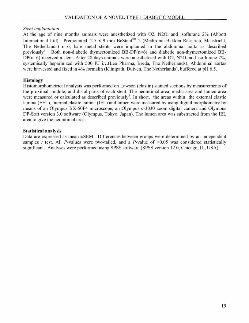
Stent implantation At the age of nine months animals were anesthetized with O2, N2O, and isoflurane 2% (Abbott International Ltd). Premounted, 2.5 x 9 mm BeStenttm 2 (Medtronic-Bakken Research, Maastricht, The Netherlands) n=6, bare metal stents were implanted in the abdominal aorta as described previously8. Both non-diabetic thymectomized BB-DP(n=6) and diabetic non-thymectomized BB-DP(n=6) received a stent. After 28 days animals were anesthetized with O2, N2O, and isoflurane 2%, systemically heparinized with 500 IU i.v.(Leo Pharma, Breda, The Netherlands). Abdominal aortas were harvested and fixed in 4% formalin (Klinipath, Duiven, The Netherlands), buffered at pH 6.5. Histology Histomorphometrical analysis was performed on Lawson (elastin) stained sections by measurements of the proximal, middle, and distal parts of each stent. The neointimal area, media area and lumen area were measured or calculated as described previously8. In short, the areas within the external elastic lamina (EEL), internal elastic lamina (IEL) and lumen were measured by using digital morphometry by means of an Olympus BX-50F4 microscope, an Olympus c-3030 zoom digital camera and Olympus DP-Soft version 3.0 software (Olympus, Tokyo, Japan). The lumen area was substracted from the IEL area to give the neointimal area. Statistical analysis Data are expressed as mean ±SEM. Differences between groups were determined by an independent samples t test. All P-values were two-tailed, and a P-value of <0.05 was considered statistically
significant. Analyses were performed using SPSS software (SPSS version 12.0, Chicago, IL, USA).

Chapter 2
20
Figure 1: Hyperglycemia, proteinuria and polyuria is present in non-thymectomized BB-DP rats but not in thymectomized BB-DP rats.
RESULTS Diabetes parameters The non-thymectomized BB-DP rats developed diabetes at an median age of 82 days. Thymectomized BB-DP control rats did not develop diabetes. Mean blood glucose level after diabetes onset in diabetic non-thymectomized BB-DP rats was 15.1±0.3mmol/L versus 5.3±0.1mmol/L in non-diabetic thymectomized BB-DP control rats (Figure 1). Furthermore there was significant proteinuria, and polyuria in diabetic non-thymectomized BB-DP (Figure 1).

VALIDATION OF A NOVEL TYPE 1 DIABETIC MODEL
21
Figure 2
Figure 2: Photomicrographs of stented abdominal aortas showing the neointima, internal elastic lamina , external elastic lamina, stent struts and A and B(diabetes),C and D (normoglycemic, x40 and x400). Neointimal formation In diabetic non-thymectomized BB-DP neointimal area was significantly increased 0.69±0.02 mm2 compared to 0.53±0.06 mm2 in non-diabetic thymectomized BB-DP control rats (Figure 2 and 3).

Chapter 2
22
Figure 3: Neointimal area is increased in diabetic animals.
DISCUSSION
In this study we demonstrated increased neointimal formation in diabetic BB-DP rats compared with BB-DP normoglycemic rats who are genetically similar but do not develop diabetes due to a thymectomy at young age. The diabetic BB-DP had proteinuria, polyuria and raised glucose values of diabetes whereas the thymectomized BB-DP had not. The increase in neointimal formation in the diabetic animals corresponds with results in humans. Because of the genetic similarity of the diabetic and normoglycemic rats in this model the mechanisms responsible for increased in-stent restenosis in diabetic populations can more easily be identified in future studies. Furthermore the feasibility of treating these rats with insulin pellets as shown in this study will enable studies on the relation between glucose levels and in-stent restenosis. Limitation of this study is that the effect of thymectomy on in-stent restenosis was not studied. Before the adult age of ten weeks in rats the thymus is needed for T-cell development. Therefore the juvenile rats of three weeks in our study have an impairment in T-cell mediated immune response. This impairment in T-cell mediated immune response prevents the development of diabetes in this model. However it could also affect the development of in-stent restenosis. One study found activation of T-cells after percutaneous transluminal coronary angioplasty9. However treatment with cyclosporine a powerful specific inhibitor of the T-cell mediated immune response did not reduce restenosis in a cholesterol-clamped rabbit animal model10. So the T-cell mediated immune response does not attribute significantly to in-stent restenosis. Therefore it is not likely that thymectomy affected in-stent restenosis in this animal model. These results validate this novel type 1 diabetic rat abdominal aortic stenting model for studying the mechanism of increased in-stent restenosis in diabetic populations and more specific in the type 1 diabetes population.
Acknowledgments We thank Annemiek Smit-van Oosten and Anthony van Dijk for their excellent biotechnical assistance.

VALIDATION OF A NOVEL TYPE 1 DIABETIC MODEL
23
References
1. Scheen AJ, Warzee F. Diabetes is still a risk factor for restenosis after drug-eluting stent in coronary arteries. Diabetes Care 2004;27:1840-1841.
2. Kornowski R, Mintz GS, Kent KM, Pichard AD, Satler LF, Bucher TA, Hong MK, Popma JJ, Leon MB. Increased restenosis in diabetes mellitus after coronary interventions is due to exaggerated intimal hyperplasia. A serial intravascular ultrasound study. Circulation 1997;95:1366-1369.
3. Takeda R, Suzuki E, Satonaka H, Oba S, Nishimatsu H, Omata M, Fujita T, Nagai R, Hirata Y. Blockade of endogenous cytokines mitigates neointimal formation in obese Zucker rats. Circulation 2005;111:1398-1406.
4. Shelton J, Wang D, Gupta H, Wyss JM, Oparil S, White CR. The neointimal response to endovascular injury is increased in obese Zucker rats. Diabetes Obes Metab 2003;5:415-423.
5. Dickinson S, Rogers T, Kasiske B, Bertog S, Tadros G, Malik J, Wilson R, Panetta C. Coronary artery disease in young women and men with long-standing insulin-dependent diabetes. Angiology 2008;59:9-15.
6. Carter AJ, Bailey L, Devries J, Hubbard B. The effects of uncontrolled hyperglycemia on thrombosis and formation of neointima after coronary stent placement in a novel diabetic porcine model of restenosis. Coron Artery Dis 2000;11:473-479.
7. Visser J, Klatter F, Hillebrands JL, Jansen A, Vijfschaft L, Rozing J. Thymectomy should be the first choice in the protection of diabetes-prone BB rats for breeding purposes. Lab Anim 2004;38:371-375.
8. Langeveld B, Roks AJ, Tio RA, van Boven AJ, van der Want JJ, Henning RH, van Beusekom HM, van der Giessen WJ, Zijlstra F, van Gilst WH. Rat abdominal aorta stenting: a new and reliable small animal model for in-stent restenosis. J Vasc Res 2004;41:377-386.
9. Osada M, Takeda S, Ogawa R, Komori S, Tamura K. T lymphocyte activation and restenosis after percutaneous transluminal coronary angioplasty. J Interferon Cytokine Res 2001;21:219-221.
10. Andersen HO, Hansen BF, Holm P, Stender S, Nordestgaard BG. Effect of cyclosporine on arterial balloon injury lesions in cholesterol-clamped rabbits: T lymphocyte-mediated immune responses not involved in balloon injury-induced neointimal proliferation. Arterioscler Thromb Vasc Biol 1999;19:1687-1694.

CHAPTER 3
NON-BONE MARROW ORIGIN OF NEOINTIMAL SMOOTH MUSCLE CELLS IN EXPERIMENTAL IN-STENT RESTENOSIS IN RATS
Hendrik C. Groenewegen*, Geanina Onuta*, Maaike Goris, André Zandvoort, Felix Zijlstra,
Wiek H. van Gilst, Jan Rozing, Bart J.G.L. de Smet, Anton J.M. Roks, Jan-Luuk Hillebrands
* H.C. Groenewegen and G. Onuta contributed equally J Vasc Res. 2008 Apr 23;45(6):493-502.

NON-BONE MARROW ORIGIN OF NEOINTIMAL SMOOTH MUSCLE CELLS
25
ABSTRACT Aim: To determine the contribution of bone marrow-derived cells in in-stent restenosis and transplant arteriosclerosis. Methods: Nontransgenic rats WT F344TG (n=3) received stent implantation 6 weeks after lethal total body irradiation and suppletion with bone marrow from a R26-hPAP transgenic rat. After 4 weeks the abdominal aortas were harvested, the stent was quickly removed, the abdominal aorta was snap-frozen in liquid nitrogen and 5 µm cryosections for stainings were cut. Additionaly DA aortic allografts were transplanted into WT F344TG(n=3) and R26-hPAPWT(n=3) BM-chimeric recipients. Immunohistochemistry (hPAP-staining) and immunofluorescence (hPAP, α-SMA and OX-1) was performed on all sections. Results: Few hPAP positive cells were observed in the neointima Double stainings of hPAP positive areas showed no α-SMA colokalization, OX-1 did show colokalization. Conclusions: Non-BM-derived cells are the predominant source of neointimal cells in ISR and TA. Vascular wall-derived progenitor cells may rather be the source of SMCs that contribute to ISR and TA which may have implications for our quest for new therapeutic targets to treat these vasculopathies.

Chapter 3
26
INTRODUCTION Development of in-stent restenosis (ISR) is the most common complication associated with coronary stenting, especially in patients treated with bare-metal stents. No adequate treatment modalities are available to treat or prevent development of ISR[1;2]. Recruitment and proliferation of smooth muscle cells (SMCs) in response to vascular injury after stenting are key phenomena that lead to the development of an occlusive neointima culminating in ISR[3;4]. Although it is well established that in humans the neointima of stented vessels is mainly composed of α-smooth muscle actin (SMA) positive cells[5], the origin of neointimal cells in ISR is still a matter of debate. Identifying the anatomical origin and molecular characteristics of the progenitor cells that ultimately form the neointima in ISR is of importance since these cells form a putative target for therapeutic intervention to prevent ISR and related occlusive vascular diseases like transplant arteriosclerosis (TA). Along with the classical theory of inward migration and proliferation of medial SMCs[6], a more recent proposed hypothesis attributes an important role to bone marrow (BM)-derived vascular progenitor cells in the process of neointimal formation[7]. BM contains both hematopoietic and mesenchymal stem cells which have the capacity to self-renew and to differentiate into a variety of cell types including SMCs[8;9]. Given the potential of BM stem cells to give rise to SMCs, SMC progenitors might be recruited from the BM into the circulation in response to vascular injury and home to the site of injury resulting in neointimal formation eventually. In line with this, various animal models of vascular injury (atherosclerosis, wire injury and TA) indeed demonstrate contribution of BM-derived cells in neointimal formation to some extent[10-13]. However, results described so far are not conclusive since we and others demonstrated that in experimental TA and (vein graft) atherosclerosis neointimal SMCs are primarily non-BM-derived[14-16]. The contribution of BM-derived cells in the development of ISR is largely unknown although some recent studies suggest involvement of BM-derived cells based on increased numbers of circulating CD34+ cells shortly after stenting[17] and the presence of neointimal cells expressing stem cell antigens like c-kit[18;19], CD34 and AC133[20]. However, although these markers are indeed expressed on primitive cells residing in the bone marrow, expression is not strictly confined to BM-derived cells. As a result, neointimal cells expressing these markers in ISR are not derived from the BM by definition. Direct evidence of involvement of BM-derived cells in the development of ISR has thus not been reported so far. Since the contribution of BM-derived cells in neointimal formation is most likely dependent on the severity of endovascular injury [11;21], it is of importance that studies on the origin of neointimal cells in specifically ISR are performed in a relevant model of true ISR and not a model of otherwise induced endovascular injury. In this study we therefore determined the contribution of BM-derived cells in ISR in a direct way using our recently developed model of ISR [22] using genetically marked BM chimeric rats.
METHODS Rats Male wild-type (WT) Fischer344 (F344) and Dark Agouti (DA) rats were obtained from Harlan Nederland (Horst, The Netherlands). Human Placental Alkaline Phosphatase (hPAP) transgenic F344 rats (R26-hPAP rats) were derived from a breeding nucleus provided by Dr. E.P. Sandgren (University of Wisconsin-Madison, USA)[23]. Rats were kept under clean conventional conditions and were fed standard rat chow and acidified water ad libitum. The investigation conforms with the Guide for the Care and Use of Laboratory Animals published by the US National Institutes of Health (NIH Publication No.85-23, revised 1996) and the Dutch Law on Experimental Animal Care.

NON-BONE MARROW ORIGIN OF NEOINTIMAL SMOOTH MUSCLE CELLS
27
Bone marrow transplantation Both femora and tibiae of BM donor rats were excised and surrounding muscle and connective tissue were removed and the BM was flushed with sterile PBS. Erythrocytes were lysed in lysing buffer (155 mM NH4Cl, 10 mM KHCO3, 0.1 mM sodium ethylenediaminetetraacetic acid [EDTA]), and the cell suspension was then filtered through a 20 µm cell strainer (Becton Dickinson, Alphen aan den Rijn, The Netherlands). BM recipient rats were lethally γ-iradiated (9 Gy) using a 137Cesium source (IBL 637, CIS Bio International). One hour after irradiation rats were reconstituted with 1-5x10⁷ BM cells by tail vein injection. Three experimental groups were included: 1) hPAP Tg F344 BM -> WT F344 [WT F344TG], stented 6 wks after reconstitution; 2) hPAP Tg F344 BM -> WT F344 [WT F344TG], aorta allografted 6 wks after reconstitution; 3) WT F344 BM -> hPAP Tg F344 [R26-hPAPWT], aorta allografted 6 wks after reconstitution. BM chimeric rats were housed in filtertop cages throughout the duration of the experiment. Rats received drinking water containing neomycin (0.35% wt/vol) starting 1 week before irradiation until 2 weeks after BM reconstitution. Six weeks after BM reconstitution and prior to stenting or allografting the level of chimerism was determined by flowcytometric analysis on PBMNCs. The level of chimerism was typically between 80% and 90% (data not shown). Stent implantation Chimeric rats (6 wks after BM reconstitution) received a stent in the abdominal aorta as described in detail elsewhere[22]. Briefly, under anesthesia (2% isoflurane [Abbot, Hoofddorp, The Netherlands], 0.4 L/min O2 and 0.4 L/min N2O) the abdominal cavity was opened. The aorta was dissected and surrounding connective tissue was removed. Next, two vascular clips were placed onto the aorta distal to the renal arteries and proximal to the aortic bifurcation. A small incision was then made in the distal abdominal aorta and the balloon catheter was inserted and inflated to 9 atm pressure to deploy a pre-mounted 2.5 x 8 mm Micro-Driver stent (Medtronic, Minneapolis, United States, n=3). After deflation and removal of the balloon, the aortic incision was closed with a 9-0 suture. Reperfusion was established by removing the clips and the abdomen was closed with 4-0 sutures. Four weeks after surgery the stented aorta’s were harvested and the stents were carefully removed from the lumen of the aorta. Aortic tissue was snap-frozen in liquid nitrogen and stored at -80°C for cryostat sections.
Aorta transplantation Since we previously showed non-BM origin of neointimal VSMCs and ECs in transplant arteriosclerosis (TA) using allogeneic BM-chimeric rats [15;21], we also performed aortic allografting in WT F344TG and R26-hPAPWT BM-chimeric rats to test the feasibility of detecting (non)-BM-derived VSMCs and ECs in established neointimal lesions in the hPAP-transgenic F344 rat model. So far, this rat model has not been used to track (non)-BM-derived VSMCs and ECs in neointimal lesions. Under anesthesia (as described above) DA aortic allografts were transplanted into WT F344TG(n=3) and R26-hPAPWT(n=3) BM-chimeric recipients as described previously[24]. Briefly, the abdominal aorta between the left renal artery and the bifurcation was removed from the donor, perfused with saline and subsequently orthotopically transplanted into the recipient via end-to-end anastomosis with total cold and warm ischemic time consistently less than 25 minutes. Immunohistochemistry To localize BM-derived hPAP-positive cells in ISR, an indirect immunoperoxidase staining for hPAP was performed on cryosections cut from the stented area. Sections (5 µm) were acetone-fixed (10 min., 4°C). Blockade of endogenous peroxidase (incubation 30 min. with PBS containing 0.03% H2O2) was followed by incubation for 60 min at room temperature with the primary polyclonal antibody against hPAP (AHP537HT, AbD Serotec, BioConnect, Huissen, The Netherlands) diluted in 1% BSA/PBS.

Chapter 3
28
Subsequently, the sections were incubated with a second-step horseradish peroxidase-conjugated goat-anti-rabbit antibody (DAKO A/S, Glostrup, Denmark) for 30 min diluted in 1% BSA/PBS supplemented with 1% normal rat serum. Peroxidase activity was developed using chromogen 3-amino-9-ethyl carbazole (AEC, DAKO A/S, Glostrup, Denmark). Sections were counterstained with hematoxylin and mounted in Faramount (DAKO A/S, Glostrup, Denmark). Control slides, in which the primary antibody was replaced with PBS were consistently negative (not shown). Immunofluorescence To further phenotype hPAP+ cells in ISR triple-immunofluorescent staining was performed using anti-hPAP, α-SMA (SMCs; clone 1A4, mIgG2a, Dako A/S, Glostrup, Denmark) and anti-CD45 (clone OX-1, mIgG1 tissue culture supernatant). Sections were incubated for 1 hr with a mixture of the primary antibodies (diluted in 1% BSA/PBS) followed by incubation with Alexa488-conjugated goat anti-mouse IgG2a (Molecular Probes, Leiden, The Netherlands), Cy5-conjugated goat anti-mouse IgG1 (Molecular Probes, Leiden, The Netherlands) and horseradish peroxidase-conjugated swine-anti-rabbit Ig (Dako A/S, Glostrup, Denmark) in 1% BSA/PBS supplemented with 1% normal rat serum for 30 min. Horseradish peroxidase-conjugated swine-anti-rabbit Ig was detected using the TSATM Tetramethylrhodamine System (PerkinElmer LAS, Inc., Boston, MA, USA). Nuclei were stained with DAPI and sections were embedded in Citifluor (AF1, Agar Scientific Ltd., Stansted, UK). To validate this four-color immunofluorescent staining protocol and check for potential crossreactivity of the isotype-specific second-step antibodies, first single stainings for hPAP, SMA and CD45 were performed on hPAP-transgenic F344 spleen sections. Following a similar immunofluorescence protocol double staining for hPAP (polyclonal rabbit Ig) and α-SMA (mouse IgG2a), and hPAP and RECA-1 (mouse IgG1, endothelium)[25] were performed on 5 µm aortic graft cryosections. Binding of anti-hPAP antibodies was detected using FITC-conjugated goat anti-rabbit Ig (Dako A/S, Glostrup, Denmark) whereas binding of α-SMA and RECA-1 antibodies was detected using horseradish peroxidase-conjugated rabbit-anti-mouse Ig (Dako A/S, Glostrup, Denmark) which was visualized using the TSATM Tetramethylrhodamine System. All fluorescently labeled sections were analyzed on a Confocal Laserscanning Microscope (TCS SP2, Leica, Microsystems Nederland B.V., Rijswijk, The Netherlands).
RESULTS
Specificity α-hPAP staining Since the hPAP-transgenic F344 rat model has not been used before to track (non)-BM-derived VSMCs and ECs in neointimal lesions we first analyzed the specificity and sensitivity of our staining method to detect hPAP-transgenic BM and vascular wall cells. As shown in Figure 1, both BM cells (C) and medial VSMCs and ECs in non-injured aorta (D) from hPAP-transgenic F344 rats stained positive for hPAP using an hPAP-specific polyclonal antibody. For comparison, BM cells (A) and aortic tissue (B) from wildtype F344 rats did not react with the α-hPAP antibody. These results indicate that this staining method is specific and sufficiently sensitive to detect hPAP-expressing BM and vascular cells. Development 4-parameter (CD45, SMA, hPAP, DNA) immunofluorescent staining protocol To validate a four-parameter immunofluorescent staining protocol and check for potential cross-reactivity of the isotype-specific second-step antibodies, single and triple stainings for hPAP, SMA and CD45 were performed on hPAP-transgenic F344 spleen sections. Sections were incubated with one primary antibody and then detected with a cocktail of three fluorochrome-labeled isotype-specific second-step antibodies. As shown in Figure 2, mIgG1 α-CD45 was only detected with α-mIgG1-Cy5 (A-E), mIgG2a α-SMA was only detected with α-mIgG2a-Alexa488 (F-J), and rIgG α-hPAP was only

NON-BONE MARROW ORIGIN OF NEOINTIMAL SMOOTH MUSCLE CELLS
29
detected with α-rIgG TRITC (K-O). When incubating sections with a mixture of CD45, hPAP and SMA primary antibodies, expression of all antigens could be demonstrated simultaneously (P-T). This four-parameter immunofluorescent staining protocol was then used to determine the origin of neointimal VSMCs and ECs in TA and ISR.
Figure 1. The α-hPAP staining is sufficiently sensitive and specific to detect hPAP-transgenic BM and vascular wall cells. (A) BM cells (magnification x1890) and (B) aortic tissue (magnification x200, inset x630) from wildtype F344 rats did not react with the α-hPAP antibody, whereas (C) BM cells (magnification x1890) and (D) medial VSMCs and ECs (arrowheads inset) in non-injured aorta (magnification x200, inset x630) from hPAP-transgenic F344 rats clearly reacted with the α-hPAP antibody. Abreviations: A: adventitia, M: media. Neointimal VSMCs and ECs in TA are non-BM-derived Since we previously showed non-BM origin of neointimal VSMCs and ECs in transplant arteriosclerosis (TA) using allogeneic BM-chimeric rats [15;21], we first performed aortic allografting in WT F344TG and R26-hPAPWT BM-chimeric rats to test our model system for specificity and sensitivity of detecting (non)-BM-derived VSMCs and ECs in established neointimal lesions. Two months after allografting both the WT-F344TG (Figure 3A and B) and R26-hPAPWT (Figure 3C and D) had developed marked TA characterized by a neointima consisting of a packed layer of SMA+ cells covered by ECs at the luminal side. The neointimal cells in allografts transplanted in wild-type recipients reconstituted with hPAP-transgenic BM (WT-F344TG) expressed SMA but colocalization with hPAP-expression was not observed (0% BM-derived α-SMA+ VSMCs, Figure 3A). Also neointimal ECs did not express hPAP (0% BM-derived RECA-1+ VSMCs, Figure 3B). The BM-derived hPAP+ cells that were detected in the neointima, media and adventitia expressed CD45

Chapter 3
30
indicating that these cells were infiltrating leukocytes (data not shown). These results suggest a non-BM origin of the neointimal ECs and VSMCs in established TA. Analyses performed on allografts transplanted in hPAP-transgenic recipients reconstituted with WT BM (R26-F344WT) confirmed this premise as shown in Figure 3C and D. Virtually all neointimal SMA+ (Figure 3C) and ECs (Figure 3D) coexpressed the hPAP transgene indicating ~100% non-BM origin of these cells in TA. These data confirm our previous observations and indicate that the hPAP-transgenic F344 rat model is sufficiently specific and sensitive to detect (non)-BM-derived VSMCs and ECs in established neointimal lesions.
Figure 2. Simultaneous detection of CD45, SMA, hPAP and DNA by immunofluorescent staining on hPAP-transgenic F344 rat spleen. (A-E) Primary incubation with OX1 (α-CD45, mIgG1) and secondary incubation with α-mIgG1-Cy5, α-mIgG2a-Alexa488, and α-rIgG TRITC. OX1 reacted with the lymphocytes present in the white pulp (WP) and red pulp (RP) around the central arteriole (arrowhead) and was only detected with α-mIgG1-Cy5. (F-J) Primary incubation with 1A4 (α-SMA, mIgG2a) and secondary incubation with α-mIgG1-Cy5, α-mIgG2a-Alexa488, and α-rIgG TRITC. 1A4 reacted with the stromal cells present in the white pulp and medial VSMCs in the central arteriole (arrowhead) and was only detected with α-mIgG2a-Alexa488. (K-O) Primary incubation with α-hPAP (rIgG) and secondary incubation with α-mIgG1-Cy5, α-mIgG2a-Alexa488, and α-rIgG TRITC. α-hPAP reacted with all cells present in the white and red pulp and was only detected with α-rIgG-TRITC. (P-T) Primary incubation with OX1, 1A4 and α-hPAP and secondary incubation with α-mIgG1-Cy5, α-mIgG2a-Alexa488, and α-rIgG TRITC. Expression of all antigens could be demonstrated simultaneously. (magnification x200). Abbreviations: RP: red pulp, WP: white pulp, arrowhead: central arteriole.

NON-BONE MARROW ORIGIN OF NEOINTIMAL SMOOTH MUSCLE CELLS
31
Presence of BM-derived hPAP+ cells in ISR Stenting of the BM-chimeric rats resulted in the development of extensive and maximal ISR after 4 weeks. In this model of ISR at earlier time-points only mild lesions (without SMA-positive VSMCs) are present which are characterized by local thrombus formation around the stent struts with surface adhering leucocytes (1 day) or thrombus-infiltrating leucocytes (3 days and 1 week). Figure 4 shows representative photomicrographs of the histological appearance of the composition of the lesions in developing ISR at 1 (A) and 3 days (B) and 1 (C) and 4 (D) weeks after stenting. Since the aim of this study is to determine the (non)-BM origin of neointimal VSMCs in established ISR, stented aorta’s were analyzed 4 weeks after stenting. After removal of the stents the neointima was still attached to the luminal side of the aortic wall (Figure 5). Immunohistochemistry for hPAP-transgene expression revealed the abundant presence of hPAP+ cells in the adventitia (Figure 5A) and media (Figure 5B and C) whereas the neointima contained a relatively low number of hPAP+ cells (Figure 5B and 5C). Neointimal VSMCs in ISR are non-BM-derived To determine the smooth-muscle-like phenotype of the BM-derived neointimal hPAP+ cells in ISR triple staining for hPAP, SMA and CD45 was performed. The neointima consisted primarily of SMA+ VSMCs whereas the media was devoid of SMA+ VSMCs after stenting (Figure 6B). Although the neointima contained considerable numbers of BM-derived hPAP+ cells (Figure 6C) the absence of colocalization of hPAP and SMA expression was consistently observed in all animals analyzed (Figure 6E) indicating a non-BM origin of neointimal VSMCs in ISR (0% BM-derived SMA+ VSMCs). Similar results were obtained for the neointimal ECs which were however only sparsely present due to the mechanical removal of the stents (data not shown). Colocalization of hPAP and CD45 expression in the neointima (Figure 6D and E), media (Figure 6D and E) and adventitia (Figure 7) indicate that the BM-derived hPAP+ cells in ISR were infiltrating leukocytes.
DISCUSSION
In the present study we determined the contribution of BM-derived cells in the development of ISR and TA after respectively experimental stenting and aortic transplantation in rats. In both models no BM-derived neointimal SMCs and ECs were detected and the few neointimal hPAP+ BM-derived cells turned out to be CD45+ infiltrating leukocytes. We conclude that vascular cells originating from the BM are not part of established neointimal lesions in both TA and ISR. Although the origin of neointimal cells has gained considerable interest in the last decade, only a few studies have been reported on the origin of neointimal SMCs after stenting[17-19] [20]. Identification of the anatomical origin of the cells involved in development of ISR is of clinical importance since this may elucidate new targets that can be used for therapeutic intervention in order to prevent or reduce development of ISR. A putative source is the bone marrow. It is generally accepted that the BM contains hematopoietic and mesenchymal stem cells which have the ability of self-renewal and which can differentiate into a variety of cell types including SMCs [8;9]. Furthermore, the human peripheral blood contains CD34+ SMC progenitors[26] and therefore the BM is a putative source of SMCs involved in the development of ISR. In line with this, increased frequencies of circulating CD34+ cells were detected after coronary stenting [17] and which was found to correlate with the late lumen loss i.e. ISR in stented patients[27] . Not only numerical differences but also the differentiation fate of progenitor cells appear to correlate with the development of ISR[17]. Mononuclear cells isolated from patients with and without ISR preferentially differentiated into α-SMA+ and endothelial-like cells in vitro respectively, indicating that differentiation in favor of SMCs may predispose for ISR[17]. Despite these correlative studies direct evidence of involvement of BM-derived cells in the development of ISR has not been reported. In

Chapter 3
32
clinical ISR it is furthermore hard to discriminate between potential BM-derived cells that appeared after stenting or that were already present in the vicinity of the stenotic area before stenting[17;20].
Figure 4. Kinetics of the development of ISR after experimental stenting in rats. (A) 1 day after stenting: local thrombus formation around the stent struts (asterisk) with surface adhering leucocytes (arrows). Toluidine blue staining, magnification x200. (B) 3 days after stenting: local thrombus formation around the stent struts (asterisks) with an increased number of infiltrating leucocytes (arrowheads). Toluidine blue-basic fuchsin staining, magnification x200. (C) 1 week after stenting: organized thrombus with surface-adherent leucocytes (arrows) and increased leucocyte infiltration (arrowhead). Toluidine blue-basic fuchsin staining, magnification x200. (D) 4 weeks after stenting: stent struts (asterisk) are completely covered by neointima which mainly consist of VSMCs and extracellular matrix with the absence of large numbers of infiltrating leucocytes. Elastica van Gieson staining, magnification x200. Abbreviations: A: adventitia, M: media, NI: neointima. In our model[22] no atherosclerosis is present at the time of stenting and therefore allows analysis of the direct effect of stenting on the recruitment of BM-derived cells and the development of ISR as reported in this article. However, in human atherosclerosis it has been shown that in atherosclerotic plaques about 10% of the intimal cells is derived from the BM[13]. In case these BM-derived cells are a main source for the SMA+ VSMCs in ISR after stenting of the atherosclerotic lesion, pre-existing atherosclerosis might result in a higher percentage of BM-derived VSMCs in ISR than observed in our study without the presence of pre-existing atherosclerosis. Recently, cells expressing stem cell antigens like CD34, c-kit[18-20]and AC133 have been shown to be present in ISR albeit at low levels (maximal ~11%). Taking into account the indirect way of detecting putative BM-derived cells in these studies, our data are in fact quite similar and support the previously published data that the BM compartment is only marginally involved in the development of established ISR if involved at all. Although the BM has been shown to harbor potential to provide cells that contribute to neointimal formation in various models for vascular injury other than ISR the actual contribution of these cells is relatively low[11;12;28]. We and others indeed showed that a non-BM source predominantly provides the cells involved in neointimal formation in restenosis and TA[11;14;15]. A potential explanation for the differences in the contribution of BM-derived cells between previous reports[11;12;28] and the current

NON-BONE MARROW ORIGIN OF NEOINTIMAL SMOOTH MUSCLE CELLS
33
study is the severity of vascular injury since BM contribution in neointimal formation appears to be dependent on the severity of endovascular injury[11;21].
Figure 5. BM-derived hPAP+ cells are present in ISR. Stenting was performed in hPAP-transgenic BM chimeric rats and analyzed 4 wks after stenting. Photomicrographs of neointima formed in stented aorta immunostained for hPAP (red-brown) and counterstained with hematoxylin. Few BM-derived hPAP+ cells are present in the neointima (arrows, B and C), whereas hPAP+ cells are abundantly present in the adventitia (asterisks, B and C). Arrowheads indicate the internal elastic lamina. Magnification: A: x20; B & C: x200 Abreviations: Adv: adventitia; M: media; NI: neointima.
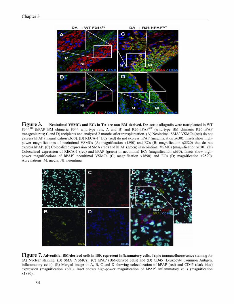
Figure 6. Neointimal BM-derived cells in ISR represent inflammatory cells but not VSMCs. Triple immunofluorescence staining for (A) Nuclear staining, (B) SMA (VSMCs), (C) hPAP (BM-derived cells) and (D) CD45 (Leukocyte Common Antigen, inflammatory cells). (E) Merged image of A, B C and D showing no colocalization of hPAP (red) and SMA (green) expression and colocalization of hPAP (red) and CD45 (dark blue) expression (magnification x630). Inset shows high-power magnification of hPAP─ neointimal VSMCs (magnification x1890). Abreviations: M: media; NI: neointima.
Chapter 3
34
Figure 3. Neointimal VSMCs and ECs in TA are non-BM-derived. DA aortic allografts were transplanted in WT F344TG (hPAP BM chimeric F344 wild-type rats; A and B) and R26-hPAPWT (wild-type BM chimeric R26-hPAP transgenic rats; C and D) recipients and analyzed 2 months after transplantation. (A) Neointimal SMA+ VSMCs (red) do not express hPAP (magnification x630). (B) RECA-1+ ECs (red) do not express hPAP (magnification x630). Insets show high-power magnifications of neointimal VSMCs (A; magnification x1890) and ECs (B; magnification x2520) that do not express hPAP. (C) Colocalized expression of SMA (red) and hPAP (green) in neointimal VSMCs (magnification x630). (D) Colocalized expression of RECA-1 (red) and hPAP (green) in neointimal ECs (magnification x630). Insets show high-power magnifications of hPAP+ neonitimal VSMCs (C; magnification x1890) and ECs (D; magnification x2520). Abreviations: M: media; NI: neointima.
Figure 7. Adventitial BM-derived cells in ISR represent inflammatory cells. Triple immunofluorescence staining for (A) Nuclear staining, (B) SMA (VSMCs), (C) hPAP (BM-derived cells) and (D) CD45 (Leukocyte Common Antigen, inflammatory cells). (E) Merged image of A, B, C and D showing colocalization of hPAP (red) and CD45 (dark blue) expression (magnification x630). Inset shows high-power magnification of hPAP+ inflammatory cells (magnification x1890).

NON-BONE MARROW ORIGIN OF NEOINTIMAL SMOOTH MUSCLE CELLS
35
However, we believe that our experimental model of ISR in rats also produces solid mechanical endovascular injury as measured by the injury scores as reported previously [22] which makes differences in severity of endovascular injury a less likely explanation for the observed differences in BM contribution. In this study we for the first time clearly demonstrate that in experimental ISR in rats the neointimal VSMCs are derived from a non-BM source. The BM thus plays a minor role in the development of established ISR. However, our results do not exclude the possibility that early after stenting BM-derived cells are recruited to the injured vascular wall and create a microenvironment in which local progenitor cell niches are activated and mobilized by BM-derived cells in a paracrine fashion. Localized progenitor cell niches in the media[29] and the adventitia[30;31]of the vascular wall have been recently identified. Furthermore, isolated adventitial Sca-1+ progenitor cells were shown to differentiate into VSMCs in vitro, but also to contribute to the development of atherosclerotic lesions in vivo[30]. The contribution of vascular wall-derived progenitor cells in the development of ISR and TA is as yet unknown but is currently under investigation. In conclusion, non-BM-derived cells are the predominant source of neointimal cells in ISR and TA. Vascular wall-derived progenitor cells may rather be the source of SMCs that contribute to ISR and TA which may have implications for our quest for new therapeutic targets to treat these vasculopathies. Acknowledgments The reported work was supported by the Netherlands Organization for Scientific Research (NWO VENI-grant 916.46.104 to JLH) and the Ubbo Emmius Foundation (to GO).

Chapter 3
36
REFERENCES (1) Serruys PW, de Jaegere P, Kiemeneij F, Macaya C, Rutsch W, Heyndrickx G, Emanuelsson H, Marco J, Legrand
V, Materne P, .: A comparison of balloon-expandable-stent implantation with balloon angioplasty in patients with coronary artery disease. Benestent Study Group. N Engl J Med 1994;331:489-495.
(2) Fischman DL, Leon MB, Baim DS, Schatz RA, Savage MP, Penn I, Detre K, Veltri L, Ricci D, Nobuyoshi M, .: A randomized comparison of coronary-stent placement and balloon angioplasty in the treatment of coronary artery disease. Stent Restenosis Study Investigators. N Engl J Med 1994;331:496-501.
(3) Hoffmann R, Mintz GS, Dussaillant GR, Popma JJ, Pichard AD, Satler LF, Kent KM, Griffin J, Leon MB: Patterns and mechanisms of in-stent restenosis. A serial intravascular ultrasound study. Circulation 1996;94:1247-1254.
(4) Moreno PR, Palacios IF, Leon MN, Rhodes J, Fuster V, Fallon JT: Histopathologic comparison of human coronary in-stent and post-balloon angioplasty restenotic tissue. Am J Cardiol 1999;84:462-6, A9.
(5) Komatsu R, Ueda M, Naruko T, Kojima A, Becker AE: Neointimal tissue response at sites of coronary stenting in humans: macroscopic, histological, and immunohistochemical analyses. Circulation 1998;98:224-233.
(6) Ross R: Atherosclerosis--an inflammatory disease. N Engl J Med 1999;340:115-126.
(7) Hillebrands JL, Onuta G, Rozing J: Role of progenitor cells in transplant arteriosclerosis. Trends Cardiovasc Med 2005;15:1-8.
(8) Pittenger MF, Mackay AM, Beck SC, Jaiswal RK, Douglas R, Mosca JD, Moorman MA, Simonetti DW, Craig S, Marshak DR: Multilineage potential of adult human mesenchymal stem cells. Science 1999;284:143-147.
(9) Krause DS, Theise ND, Collector MI, Henegariu O, Hwang S, Gardner R, Neutzel S, Sharkis SJ: Multi-organ, multi-lineage engraftment by a single bone marrow-derived stem cell. Cell 2001;105:369-377.
(10) Campbell JH, Han CL, Campbell GR: Neointimal formation by circulating bone marrow cells. Ann N Y Acad Sci 2001;947:18-24.
(11) Tanaka K, Sata M, Hirata Y, Nagai R: Diverse contribution of bone marrow cells to neointimal hyperplasia after mechanical vascular injuries. Circ Res 2003;93:783-790.
(12) Shimizu K, Sugiyama S, Aikawa M, Fukumoto Y, Rabkin E, Libby P, Mitchell RN: Host bone-marrow cells are a source of donor intimal smooth- muscle-like cells in murine aortic transplant arteriopathy. Nat Med 2001;7:738-741.
(13) Caplice NM, Bunch TJ, Stalboerger PG, Wang S, Simper D, Miller DV, Russell SJ, Litzow MR, Edwards WD: Smooth muscle cells in human coronary atherosclerosis can originate from cells administered at marrow transplantation. Proc Natl Acad Sci U S A 2003;100:4754-4759.
(14) Hu Y, Davison F, Ludewig B, Erdel M, Mayr M, Url M, Dietrich H, Xu Q: Smooth muscle cells in transplant atherosclerotic lesions are originated from recipients, but not bone marrow progenitor cells. Circulation 2002;106:1834-1839.
(15) Hillebrands JL, Klatter FA, van Dijk WD, Rozing J: Bone marrow does not contribute substantially to endothelial-cell replacement in transplant arteriosclerosis. Nat Med 2002;8:194-195.
(16) Bentzon JF, Weile C, Sondergaard CS, Hindkjaer J, Kassem M, Falk E: Smooth muscle cells in atherosclerosis originate from the local vessel wall and not circulating progenitor cells in ApoE knockout mice. Arterioscler Thromb Vasc Biol 2006;26:2696-2702.

NON-BONE MARROW ORIGIN OF NEOINTIMAL SMOOTH MUSCLE CELLS
37
(17) Inoue T, Sata M, Hikichi Y, Sohma R, Fukuda D, Uchida T, Shimizu M, Komoda H, Node K: Mobilization of CD34-positive bone marrow-derived cells after coronary stent implantation: impact on restenosis. Circulation 2007;115:553-561.
(18) Hibbert B, Chen YX, O'Brien ER: c-kit-immunopositive vascular progenitor cells populate human coronary in-stent restenosis but not primary atherosclerotic lesions. Am J Physiol Heart Circ Physiol 2004;287:H518-H524.
(19) Soda T, Suzuki H, Iso Y, Kusuyama T, Omori Y, Sato T, Shoji M, Koba S, Geshi E, Katagiri T: Bone marrow cells contribute to neointimal formation after stent implantation in swine. Int J Cardiol 2007;121:44-52.
(20) Skowasch D, Jabs A, Andrie R, Dinkelbach S, Luderitz B, Bauriedel G: Presence of bone-marrow- and neural-crest-derived cells in intimal hyperplasia at the time of clinical in-stent restenosis. Cardiovasc Res 2003;60:684-691.
(21) Hillebrands JL, Klatter FA, Rozing J: Origin of vascular smooth muscle cells and the role of circulating stem cells in transplant arteriosclerosis. Arterioscler Thromb Vasc Biol 2003;23:380-387.
(22) Langeveld B, Roks AJ, Tio RA, van Boven AJ, van der Want JJ, Henning RH, van Beusekom HM, van der Giessen WJ, Zijlstra F, van Gilst WH: Rat abdominal aorta stenting: a new and reliable small animal model for in-stent restenosis. J Vasc Res 2004;41:377-386.
(23) Kisseberth WC, Brettingen NT, Lohse JK, Sandgren EP: Ubiquitous expression of marker transgenes in mice and rats. Dev Biol 1999;214:128-138.
(24) Hillebrands JL, Klatter FA, van den Hurk BM, Popa ER, Nieuwenhuis P, Rozing J: Origin of neointimal endothelium and alpha-actin-positive smooth muscle cells in transplant arteriosclerosis. J Clin Invest 2001;107:1411-1422.
(25) Duijvestijn AM, van Goor H, Klatter F, Majoor GD, van Bussel E, Breda Vriesman PJ: Antibodies defining rat endothelial cells: RECA-1, a pan-endothelial cell-specific monoclonal antibody. Lab Invest 1992;66:459-466.
(26) Simper D, Stalboerger PG, Panetta CJ, Wang S, Caplice NM: Smooth muscle progenitor cells in human blood. Circulation 2002;106:1199-1204.
(27) Schober A, Hoffmann R, Opree N, Knarren S, Iofina E, Hutschenreuter G, Hanrath P, Weber C: Peripheral CD34+ cells and the risk of in-stent restenosis in patients with coronary heart disease. Am J Cardiol 2005;96:1116-1122.
(28) Sata M, Saiura A, Kunisato A, Tojo A, Okada S, Tokuhisa T, Hirai H, Makuuchi M, Hirata Y, Nagai R: Hematopoietic stem cells differentiate into vascular cells that participate in the pathogenesis of atherosclerosis. Nat Med 2002;8:403-409.
(29) Sainz J, Al Haj ZA, Caligiuri G, Demerens C, Urbain D, Lemitre M, Lafont A: Isolation of "side population" progenitor cells from healthy arteries of adult mice. Arterioscler Thromb Vasc Biol 2006;26:281-286.
(30) Hu Y, Zhang Z, Torsney E, Afzal AR, Davison F, Metzler B, Xu Q: Abundant progenitor cells in the adventitia contribute to atherosclerosis of vein grafts in ApoE-deficient mice. J Clin Invest 2004;113:1258-1265.
(31) Zengin E, Chalajour F, Gehling UM, Ito WD, Treede H, Lauke H, Weil J, Reichenspurner H, Kilic N, Ergun S: Vascular wall resident progenitor cells: a source for postnatal vasculogenesis. Development 2006;133:1543-1551.

CHAPTER 4 Effects of angiotensin II and angiotensin II type 1 receptor blockade on neointimal formation after stent implantation.
Hendrik C. Groenewegen, Pim van der Harst, Anton J.M. Roks, Hendrik Buikema, Felix Zijlstra, Wiek
H. van Gilst, Bart J.G.L. de Smet
Int J Cardiol. 2008 May 23;126(2):209-15.
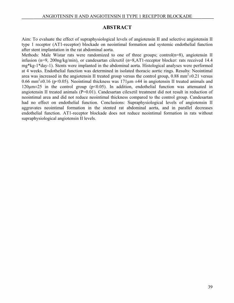
ANGIOTENSIN II AND ANGIOTENSIN II TYPE 1 RECEPTOR BLOCKADE
39
ABSTRACT
Aim: To evaluate the effect of supraphysiological levels of angiotensin II and selective angiotensin II type 1 receptor (AT1-receptor) blockade on neointimal formation and systemic endothelial function after stent implantation in the rat abdominal aorta. Methods: Male Wistar rats were randomized to one of three groups; control(n=8), angiotensin II infusion (n=9, 200ng/kg/min), or candesartan cilexetil (n=8,AT1-receptor blocker: rats received 14.4 mg*kg-1*day-1). Stents were implanted in the abdominal aorta. Histological analyses were performed at 4 weeks. Endothelial function was determined in isolated thoracic aortic rings. Results: Neointimal area was increased in the angiotensin II treated group versus the control group, 0.88 mm2±0.21 versus 0.66 mm2±0.16 (p<0.05). Neointimal thickness was 171µm ±44 in angiotensin II treated animals and 120µm±25 in the control group (p<0.05). In addition, endothelial function was attenuated in angiotensin II treated animals (P=0.01). Candesartan cilexetil treatment did not result in reduction of neointimal area and did not reduce neointimal thickness compared to the control group. Candesartan had no effect on endothelial function. Conclusions: Supraphysiological levels of angiotensin II aggravates neointimal formation in the stented rat abdominal aorta, and in parallel decreases endothelial function. AT1-receptor blockade does not reduce neointimal formation in rats without supraphysiological angiotensin II levels.

Chapter 4
40
INTRODUCTION
The renin-angiotensin system. has been implicated in the pathophysiology of in-stent restenosis (1). Angiotensin II (Ang II) Type 1 (AT1)-receptors are found abundantly on smooth muscle cells derived from human in-stent restenotic lesions and Ang II induces vascular smooth muscle proliferation through the activation of mitogen-activated protein kinase (2;3), and elevated Ang II levels aggravate neointimal formation after vascular injury (4). Several large trials investigated the effect of Angiotensin Converting Enzyme (ACE)-inhibitors on restenosis after angioplasty and stenting, but found no reduction in restenosis (5;6). This inefficacy of ACE-inhibitors to reduce restenosis has been attributed to inadequate tissue ACE inhibition and the existence of alternative pathways of Ang II formation, such as chymase (7-9). Alternative approaches, using direct inhibition of Ang II using AT1-receptor blockers reduced restenosis after vascular injury in the rat carotid model(10;11). However, clinical trials with AT1-receptors blockers this far have shown no effect (12;13). Considering the discrepancy between animal research and clinical trials we hypothesized that supraphysiological Ang II levels are needed for Ang II mediated neointimal formation. Accordingly, the effect of candesartan cilexetil (a selective AT1-receptor blocker) in the setting of in-stent restenosis in rats with physiological levels of Ang II was determined. Restenosis after balloon injury is explained by negative arterial remodeling (shrinkage) and also by neointimal formation, while restenosis after stenting depends only on neointimal formation(14-16). To confirm the role of the renin-angiotensin system in the setting of in-stent restenosis, we also determined the effect of Ang II infusion. We assessed the role of Ang II infusion and candesartan treatment on endothelial function in relation to neointimal formation.
METHODS
All procedures conformed to the Guide for the Care and Use of Laboratory Animals published by the US National Institutes of Health (NIH Publication No. 85-23, revised 1996). Stent implantation was performed in thirty rats. Overall mortality was approximately 17%. Most animals died either of perforation or thrombosis of the aorta immediately following stenting. Animal Protocol This study was approved by the animal care and use committee of the University of Groningen. Male Wistar WU rats (Charles Rivers) weighing 450 to 520 g were anesthetized with O2, N2O, and isoflurane 2% (Abbot B.V.). Premounted, 2.5 x 8 mm, bare metal stents (Lekton Motion Petite, Biotronik) were implanted in the abdominal aorta as described previously (17). There were 3 groups: control group, Ang II infusion group and candesartan-treated group. In candesartan-treated animals normal rat chow was mixed with candesartan(10 mg·kg–1·day–1) and administrated ad libitum starting one week before stent implantation. The concentration of candesartan in the diet was 0.25mg·g–1. Baseline weight, weight at 4 weeks and daily oral intake were measured in the candesartan-treated group. Rats in the Ang II infusion group received an osmotic minipump (Model 2004; Alzet) subcutaneously for Ang II delivery (200 ng/kg per minute) after the stent implantation. Blood pressure was measured serially (0,1,7,14 and 28 days after stent implantation) under general anesthesia using an electrosphygmomanometer in the tail of the rat. After 28 days, animals were anesthetized and heparinized with 500 IU intravenously (Leo Pharma B.V.). Abdominal aortas were harvested, fixed, embedded in methylmetacrylate, sectioned, and stained for histological analysis. The thoracic aortas were quickly excised and mounted in organ baths to assess endothelial function.
ANGIOTENSIN II AND ANGIOTENSIN II TYPE 1 RECEPTOR BLOCKADE
41
Histology Histomorphometrical analysis was performed on Lawson-stained sections by measurements of the proximal, middle, and distal parts of each stent. To assess neointimal formation, areas within the external elastic lamina, internal elastic lamina, and lumen were measured using digital morphometry by means of an Olympus BX-50F4 microscope, an Olympus c-3030 zoom digital camera and Olympus DP-Soft version 3.0 software (Olympus, Tokyo, Japan). The neointimal area, media area, lumen area, and the percentage of stenosis were calculated as described previously (18).The injury scores were assessed as described by Schwartz et al and Kornowski et al (19;20) . N-terminal Atrial Natriuretic Peptide Concentrations of N-terminal atrial natriuretic peptide (N-ANP) in plasma were measured with an commercially available radioimmunoassay from Biotop (Oulu, Finland) as described previously (21). Organ Bath Studies With Isolated Aortic Rings Organ bath studies were performed as described previously(22;23). In brief, periaortic tissue was removed and rings of 2 mm in length were cut and rings were connected to an isotonic displacement transducer. After stabilization, during which regular washing was performed, rings were checked for viability by stimulation with phenylephrine (10 µmol/L). Rings were washed and restabilized. Rings were precontracted with phenylephrine (10 µmol/L). The endothelium-dependent vasodilation was assessed by a cumulative dose of metacholine (10nmol/L to 10 µmol/L). Endothelium independent vasodilation was assessed by a cumulative dose of nitroglycerine in parallel rings. Vascular responsiveness to Ang II (0.1 nmol/L to 1 µmol/L) was assessed in parallel rings as described previously(24). Statistical methods Data are expressed as mean ±SD unless specifically stated otherwise. Comparison of means was determined by ANOVA with Bonferonni correction for multiple comparisons. All P-values were two-tailed, and a P-value of <0.05 was considered statistically significant. Analyses were performed using SPSS software (SPSS version 12.0, Chicago, IL, USA).
RESULTS
Candesartan dose Baseline weight (mean±SD) was 474 gram ±30,weight at 4 weeks was 510±34 and daily oral intake was 28±0.7. The mean candesartan dose received over the 4 week period was calculated to be 14.4 mg·kg–1·day–1. Histological analysis Histomorphometric measurements are presented in Table 1. Stent expansion, expressed as the internal elastic lamina area and injury score was similar among groups. In Ang II infused animals, neointimal area (Figure 1) and neointimal thickness were significantly increased. Candesartan treatment did not change neointimal area or neointimal thickness compared to the control group. Representative photomicrographs of stented abdominal aortas of the three groups are shown in Figure 2.

Chapter 4
42
Table 1. Neointimal formation.
Variable A (N=8) B (N=9) C (N=8) P-value
Mean injury score 0.06±0.08 0.11±0.13 0.08±0.19 P=1.00/P=1.00
Neointimal area 0.66±0.16 0.88±0.21 0.71±0.14 P=0.048/P=1.00
Neointimal thickness(µm) 120±25 171±44 132±32 P=0.019/P=1.00
Media area(mm2) 0.29±0.06 0.30±0.05 0.30±0.04 P=0.86/P=1.00
3.79±0.39 3.59±0.22 3.68±0.38 P=1.00/P=1.00 Internal elastic lamina area(mm2)
A=Control B=Angiotensin II C=Candesartan (A:B/A:C)
Endothelial Function The effects of Ang II infusion and candesartan treatment were examined in thoracic aorta rings. Endothelium-dependent relaxation was decreased in the Ang II group compared tot the control group (Figure 3). Candesartan did not change endothelial-dependent vasodilatory response to metacholine. Endothelial-independent vasodilatory response to nitroglycerine did not differ among groups and are shown in Figure 4. Effectiveness of Ang II delivery and Ang II type 1 receptor blockade. Blood pressure was increased in the Ang II group and decreased in the candesartan-treated group (Table 2). N-ANP levels were raised (p=0.05) in the Ang II group as compared to the control group (1.71±0.22, versus 1.21±0.14). Candesartan did not change N-ANP levels (1.10±0.21, p=1.00) compared to the control group. Ang II was notably blocked in the candesartan group (Table 2). Table 2. Effect of Angiotensin II (Ang II) and candesartan on vascular responsiveness to Ang II and blood pressure.
Variable #=candesartan started 7 days
before stent-implantation
A (N=8)
B (N=9) C (N=8) P-value
Baseline blood pressure (mmHg) 97±8.0 93±14 74±11 # P=1.00/P=0.002
Mean blood pressure (mmHg) 99±11 148±20 76±11 P=0.001/P=0.001
Maximum Ang II response (%PE) 37±10% 38±8% 1.0±2% P=1.00/P=0.001
A=Control B=Ang II C=Candesartan (A:B/A:C)

ANGIOTENSIN II AND ANGIOTENSIN II TYPE 1 RECEPTOR BLOCKADE
43
Figure 1: Neointimal area is significantly higher in the Figure 2: Photomicrographs of stented abdominal Ang II group (grey bar) compared to the control group aortas showing the neointima, internal elastic (black bar), but neointimal area in the candesartan treated lamina , external elastic lamina, stent struts group (dark grey bar) is not significantly lower compared (blank spaces in vessel wall) and media: A to the control group. and B(control),C and D(Ang II infused) E and F(candesartan-treated, x40 and x400).
DISCUSSION In the present study, we examined the effect of high serum Ang II levels and Ang II type 1 receptor blockade on neointimal formation in a rat abdominal aorta stenting model. Increased Ang II levels increased neointimal area and thickness. Ang II also increased blood pressure. In contrast, Ang II type 1 receptor blockade did not reduce the neointimal area or thickness. Endothelial function decreased in the Ang II group, but did not change in the candesartan group. Effect of candesartan on in-stent restenosis The effect of candesartan on neointimal formation in this study seemingly conflicts with earlier studies performed in the rat carotid balloon injury model where a reduction in neointimal formation after treatment with AT1-receptor blockers was observed (25;26). However, balloon injury and stenting have different mechanism of restenosis as demonstrated in different animal models and in humans. Restenosis after balloon injury is explained by negative arterial remodeling (shrinkage) and also by neointimal formation, while restenosis after stenting depends only on neointimal formation(27-29). Sustained chemokine expression and leukocyte recruitment indicate that inflammation is more prominent after stenting (30). Anti-inflammatory properties of AT1-receptor blockers after balloon injury in the femoral artery have been reported, but are still unknown after stenting(31).

Chapter 4
44
Figure 3: Effect of stenting (mean, SEM) on Figure 4: Effects of stenting (mean, SEM) on endothelial-dependent vasodilatation of the thoracic endothelial-independent vasodilatation of the aorta. Ang II infusion decreased metacholine induced thoracic aorta. No differences existed in vasodilation of phenylephrine-precontracted aortic nitroglycerine induced vasodilatation of rings compared to control (p=0.01). Candesartan phenylephrine-precontracted aortic rings in the treatment did not change metacholine induced three groups (control versus Ang II p=1.00, vasodilation of phenylephrine-precontracted aortic control versus candesartan p=0.20). rings compared to control (p=0.25). PE indicates PE indicates phenylephrine; NTG indicates phenylephrine; ME indicates metacholine. nitroglycerine. Only two previous studies examined the effect of AT1-receptor blockade on neointimal formation after stent implantation; their results were conflicting. Huckle et al. found no reduction of neointimal formation in the coronary artery of pigs, while Ohtani et al. found a reduction of neointimal formation in the iliac arteries of cynomolgus monkeys, and in the common carotid arteries of rabbits(32;33). An important limitation of the study of Othani et al. was the omission of injury scores, an important factor influencing neointimal formation. The groups in the study of Huckle et al. had equal injury scores, making the groups well comparable(34). So the question should be raised whether the effect of systemic AT1-receptor blockage on restenosis in the balloon injury model can be extrapolated to the different process of in-stent restenosis, because most preclinical studies done in an angioplasty setting are successful in reducing neointimal formation while results in studies evaluating the effect on in-stent restenosis are not so clear. This question is justifiable by the fact that most trials with AT1-receptor blockers have shown no effect of AT1-receptor blockade on in-stent restenosis(35;36). Only Yoshida et al. found a reduction of neointimal formation (using intravascular ultrasound) after candesartan compared to placebo treatment in clinically stable patients(37). However, this study suffered some methodological difficulties. In the candesartan group up to 47% of the patients received a Multi-Link stent, whereas only 28% of the control group did. This bias is important because Multi-Link stent designs are known to be associated with decreased neointimal formation(38). In addition, in the study of Yoshida et al. the clinical variables of restenosis rate and target lesion revascularization did not differ between the two groups. There could be several other explanations for the lack of effect of the AT1-receptor blocker on in-stent restenosis in this study. Inadequate dosing would be the most

ANGIOTENSIN II AND ANGIOTENSIN II TYPE 1 RECEPTOR BLOCKADE
45
important one. The rats in this study however received a higher dosis of candesartan than in the balloon injury models(39;40). A second explanation could be inadequate tissue levels of candesartan. However the aortic rings in the candesartan treated rat showed no response on Ang II indicating high tissue levels of candesartan (Table 2). Furthermore the rats were pretreated before stent implantation to increase local tissue levels at an early stage after the stenting injury. Thirdly mechanical stretch can stimulate AT1-receptors without the involvement of Ang II. However, candesartan is an inverse agonist which has been shown to inhibit AT1-receptor stimulation by mechanical stretch (41). Therefore, AT1-receptor stimulation by mechanical stress is likely to have been blocked by candesartan in this study. Fourthly, we only used a limited number of rats per group, leaving the possibility that we missed small sized effects on our primary endpoint. Considering the standard deviation, post-hoc calculation of the detectable alternative is approximately 0.22 mm2 (based on a power of 0.8 and an alpha of 0.05). Considering the observed non-significant difference of 0.05 mm2 between candesartan treated animals versus controls, we will need to study at least 142 animals per group to detect such a small difference(42). Effect of high levels of Ang II on in-stent restenosis We showed that high levels of Ang II can induce neointimal formation. We found also a diminished endothelial function in rats treated with Ang II. The positive correlation between endothelial dysfunction and increased neointimal formation found in this study after Ang II infusion is known (43). One link which could explain the parallel effects of Ang II on endothelial function and neointimal formation is oxidative stress. Ang II can induce oxidative stress by the NAD(P)H oxidase system and this Ang II induced oxidative stress has been linked to endothelial dysfunction (44;45). The endothelial dysfunction however may also be indirectly caused by hypertension which was observed in the Ang II treated rats. We do not think that blood pressure affected neointimal formation since hypertension is not a risk factor for in-stent restenosis(46). In conclusion, candesartan does not decrease neointimal formation after stent implantation, however high levels of Ang II can increase neointimal formation after stenting and lead to an impairment of endothelial function. It could be that AT1-receptor stimulation by Ang II is not a major contributor to in-stent restenosis and only high doses lead to neointimal formation. This is supported by our finding that in normal rats without any prior activation of the renin-angiotensin system high dose AT1-receptor blockade does not lead to a reduction in neointimal formation. The current study only examined systemic AT1-receptor blockage, limited by its systemic side-effects like hypotension. However, local delivery could allow even higher dosing, and should be a focus of future studies. Data of Wilson et al. and Taguchi et al. has suggested that high local dosing of AT1-receptor blockers might be a more successful approach for reducing neointimal formation compared to systemic delivery(47;48). Wang et al. used drug-eluting stents for high local delivery of valsartan and found reduced neointimal formation. Interestingly they found an increase in AT2-receptor expression(49). It has been suggested that AT2-receptor stimulation has anti-inflammatory and anti-proliferative effects beneficial beneficial for the reduction of neointimal formation and that AT1-receptor blockers can stimulate AT2-receptors (50;51). Besides AT2 receptor stimulation, future studies could also focus on alternative ways of renin-angiotensin modulation such as stimulation of the production of Ang 1-7 or alternatively stimulation of the kinin system (52-54).
Chapter 4
46
Treatments of patients with combination therapy (ACE-inhibition and AT1-receptor blockade) might employ these mechanisms. ACE-inhibitors would stimulate kinin production and ACE-inhibitors and AT1-receptors would work synergistly to increase Ang 1,7(55;56). One study by Kim et al. showed in rabbits that combination therapy of ACE-inhibitors and AT1-receptor blockers was more effective in reducing neointimal formation compared to ACE-inhibitors and AT1-receptor blockers alone (57). Acknowledgments We thank Hans Bartels, Azuwerus van Buiten and Maaike Goris for their excellent biotechnical assistance. We thank Dr. Frans Boomsma, Department of Internal Medicine, Erasmus MC, Rotterdam, the Netherlands, for determination of N-ANP. Funding: We thank AstraZeneca CVGI Research (Cheshire, UK) for supporting this study.

ANGIOTENSIN II AND ANGIOTENSIN II TYPE 1 RECEPTOR BLOCKADE
47
Reference List
(1) Ribichini F, Steffenino G, Dellavalle A, Matullo G, Colajanni E, Camilla T et al. Plasma activity and insertion/deletion polymorphism of angiotensin I-converting enzyme: a major risk factor and a marker of risk for coronary stent restenosis. Circulation 1998; 97(2):147-154.
(2) Wagenaar LJ, van Boven AJ, van der Wal AC, Amoroso G, Tio RA, van der Loos CM et al. Differential localisation of the renin-angiotensin system in de-novo lesions and in-stent restenotic lesions in in-vivo human coronary arteries. Cardiovasc Res 2003; 59(4):980-987.
(3) Xi XP, Graf K, Goetze S, Fleck E, Hsueh WA, Law RE. Central role of the MAPK pathway in ang II-mediated DNA synthesis and migration in rat vascular smooth muscle cells. Arterioscler Thromb Vasc Biol 1999; 19(1):73-82.
(4) Daemen MJ, Lombardi DM, Bosman FT, Schwartz SM. Angiotensin II induces smooth muscle cell proliferation in the normal and injured rat arterial wall. Circ Res 1991; 68(2):450-456.
(5) Faxon DP. Effect of high dose angiotensin-converting enzyme inhibition on restenosis: final results of the MARCATOR Study, a multicenter, double-blind, placebo-controlled trial of cilazapril. The Multicenter American Research Trial With Cilazapril After Angioplasty to Prevent Transluminal Coronary Obstruction and Restenosis (MARCATOR) Study Group. J Am Coll Cardiol 1995; 25(2):362-369.
(6) Meurice T, Bauters C, Hermant X, Codron V, VanBelle E, Mc Fadden EP et al. Effect of ACE inhibitors on angiographic restenosis after coronary stenting (PARIS): a randomised, double-blind, placebo-controlled trial. Lancet 2001; 357(9265):1321-1324.
(7) Hojo Y, Ikeda U, Katsuki T, Mizuno O, Fujikawa H, Shimada K. Inhibition of angiotensin converting enzyme cannot prevent increases in angiotensin II production in coronary circulation. Heart 2000; 83(5):574-576.
(8) Rakugi H, Wang DS, Dzau VJ, Pratt RE. Potential importance of tissue angiotensin-converting enzyme inhibition in preventing neointima formation. Circulation 1994; 90(1):449-455.
(9) Nishimoto M, Takai S, Kim S, Jin D, Yuda A, Sakaguchi M et al. Significance of chymase-dependent angiotensin II-forming pathway in the development of vascular proliferation. Circulation 2001; 104(11):1274-1279.
(10) Igarashi M, Hirata A, Yamaguchi H, Tsuchiya H, Ohnuma H, Tominaga M et al. Candesartan inhibits carotid intimal thickening and ameliorates insulin resistance in balloon-injured diabetic rats. Hypertension 2001; 38(6):1255-1259.
(11) Kawamura M, Terashita Z, Okuda H, Imura Y, Shino A, Nakao M et al. TCV-116, a novel angiotensin II receptor antagonist, prevents intimal thickening and impairment of vascular function after carotid injury in rats. J Pharmacol Exp Ther 1993; 266(3):1664-1669.
(12) Wakeyama T, Ogawa H, Iida H, Takaki A, Iwami T, Mochizuki M et al. Effects of candesartan and probucol on restenosis after coronary stenting: results of insight of stent intimal hyperplasia inhibition by new angiotensin II receptor antagonist (ISHIN) trial. Circ J 2003; 67(6):519-524.

Chapter 4
48
(13) Peters S, Trummel M, Meyners W, Koehler B, Westermann K. Valsartan versus ACE inhibition after bare metal stent implantation--results of the VALVACE trial. Int J Cardiol 2005; 98(2):331-335.
(14) Post MJ, de Smet BJ, van der HY, Borst C, Kuntz RE. Arterial remodeling after balloon angioplasty or stenting in an atherosclerotic experimental model. Circulation 1997; 96(3):996-1003.
(15) Haude M, Erbel R, Issa H, Meyer J. Quantitative analysis of elastic recoil after balloon angioplasty and after intracoronary implantation of balloon-expandable Palmaz-Schatz stents. J Am Coll Cardiol 1993; 21(1):26-34.
(16) Kakuta T, Currier JW, Haudenschild CC, Ryan TJ, Faxon DP. Differences in compensatory vessel enlargement, not intimal formation, account for restenosis after angioplasty in the hypercholesterolemic rabbit model. Circulation 1994; 89(6):2809-2815.
(17) Langeveld B, Roks AJ, Tio RA, van Boven AJ, van der Want JJ, Henning RH et al. Rat abdominal aorta stenting: a new and reliable small animal model for in-stent restenosis. J Vasc Res 2004; 41(5):377-386.
(18) Langeveld B, Roks AJ, Tio RA, van Boven AJ, van der Want JJ, Henning RH et al. Rat abdominal aorta stenting: a new and reliable small animal model for in-stent restenosis. J Vasc Res 2004; 41(5):377-386.
(19) Schwartz RS, Huber KC, Murphy JG, Edwards WD, Camrud AR, Vlietstra RE et al. Restenosis and the proportional neointimal response to coronary artery injury: results in a porcine model. J Am Coll Cardiol 1992; 19(2):267-274.
(20) Kornowski R, Hong MK, Tio FO, Bramwell O, Wu H, Leon MB. In-stent restenosis: contributions of inflammatory responses and arterial injury to neointimal hyperplasia. J Am Coll Cardiol 1998; 31(1):224-230.
(21) van Veldhuisen DJ, Boomsma F, de Kam PJ, Man i, V, Crijns HJ, Hampton JR et al. Influence of age on neurohormonal activation and prognosis in patients with chronic heart failure. Eur Heart J 1998; 19(5):753-760.
(22) Buikema H, Monnink SH, Tio RA, Crijns HJ, de Zeeuw D, van Gilst WH. Comparison of zofenopril and lisinopril to study the role of the sulfhydryl-group in improvement of endothelial dysfunction with ACE-inhibitors in experimental heart failure. Br J Pharmacol 2000; 130(8):1999-2007.
(23) van der HP, Volbeda M, Voors AA, Buikema H, Wassmann S, Bohm M et al. Vascular response to angiotensin II predicts long-term prognosis in patients undergoing coronary artery bypass grafting. Hypertension 2004; 44(6):930-934.
(24) van der HP, Volbeda M, Voors AA, Buikema H, Wassmann S, Bohm M et al. Vascular response to angiotensin II predicts long-term prognosis in patients undergoing coronary artery bypass grafting. Hypertension 2004; 44(6):930-934.

ANGIOTENSIN II AND ANGIOTENSIN II TYPE 1 RECEPTOR BLOCKADE
49
(25) Igarashi M, Hirata A, Yamaguchi H, Tsuchiya H, Ohnuma H, Tominaga M et al. Candesartan inhibits carotid intimal thickening and ameliorates insulin resistance in balloon-injured diabetic rats. Hypertension 2001; 38(6):1255-1259.
(26) Kawamura M, Terashita Z, Okuda H, Imura Y, Shino A, Nakao M et al. TCV-116, a novel angiotensin II receptor antagonist, prevents intimal thickening and impairment of vascular function after carotid injury in rats. J Pharmacol Exp Ther 1993; 266(3):1664-1669.
(27) Post MJ, de Smet BJ, van der HY, Borst C, Kuntz RE. Arterial remodeling after balloon angioplasty or stenting in an atherosclerotic experimental model. Circulation 1997; 96(3):996-1003.
(28) Haude M, Erbel R, Issa H, Meyer J. Quantitative analysis of elastic recoil after balloon angioplasty and after intracoronary implantation of balloon-expandable Palmaz-Schatz stents. J Am Coll Cardiol 1993; 21(1):26-34.
(29) Kakuta T, Currier JW, Haudenschild CC, Ryan TJ, Faxon DP. Differences in compensatory vessel enlargement, not intimal formation, account for restenosis after angioplasty in the hypercholesterolemic rabbit model. Circulation 1994; 89(6):2809-2815.
(30) Welt FG, Tso C, Edelman ER, Kjelsberg MA, Paolini JF, Seifert P et al. Leukocyte recruitment and expression of chemokines following different forms of vascular injury. Vasc Med 2003; 8(1):1-7.
(31) Wu L, Iwai M, Nakagami H, Li Z, Chen R, Suzuki J et al. Roles of angiotensin II type 2 receptor stimulation associated with selective angiotensin II type 1 receptor blockade with valsartan in the improvement of inflammation-induced vascular injury. Circulation 2001; 104(22):2716-2721.
(32) Huckle WR, Drag MD, Acker WR, Powers M, McFall RC, Holder DJ et al. Effects of L-749,329, an ET(A)/ET(B) endothelin receptor antagonist, in a porcine coronary artery injury model of vascular restenosis. Circulation 2001; 103(14):1899-1905.
(33) Ohtani K, Egashira K, Ihara Y, Nakano K, Funakoshi K, Zhao G et al. Angiotensin II type 1 receptor blockade attenuates in-stent restenosis by inhibiting inflammation and progenitor cells. Hypertension 2006; 48(4):664-670.
(34) Huckle WR, Drag MD, Acker WR, Powers M, McFall RC, Holder DJ et al. Effects of L-749,329, an ET(A)/ET(B) endothelin receptor antagonist, in a porcine coronary artery injury model of vascular restenosis. Circulation 2001; 103(14):1899-1905.
(35) Wakeyama T, Ogawa H, Iida H, Takaki A, Iwami T, Mochizuki M et al. Effects of candesartan and probucol on restenosis after coronary stenting: results of insight of stent intimal hyperplasia inhibition by new angiotensin II receptor antagonist (ISHIN) trial. Circ J 2003; 67(6):519-524.
(36) Radke PW, Figulla HR, Drexler H, Klues HG, Mugge A, Silber S et al. A double-blind, randomized, placebo-controlled multicenter clinical trial to evaluate the effects of the angiotensin II receptor blocker candesartan cilexetil on intimal hyperplasia after coronary stent implantation. Am Heart J 2006; 152(4):761-766.
Chapter 4
50
(37) Yoshida O, Hirayama H, Nanasato M, Watanabe T, Murohara T. The angiotensin II receptor blocker candesartan cilexetil reduces neointima proliferation after coronary stent implantation: a prospective randomized study under intravascular ultrasound guidance. Am Heart J 2005; 149(1):e2.
(38) Hoffmann R, Mintz GS, Haager PK, Bozoglu T, Grube E, Gross M et al. Relation of stent design and stent surface material to subsequent in-stent intimal hyperplasia in coronary arteries determined by intravascular ultrasound. Am J Cardiol 2002; 89(12):1360-1364.
(39) Igarashi M, Hirata A, Yamaguchi H, Tsuchiya H, Ohnuma H, Tominaga M et al. Candesartan inhibits carotid intimal thickening and ameliorates insulin resistance in balloon-injured diabetic rats. Hypertension 2001; 38(6):1255-1259.
(40) Kawamura M, Terashita Z, Okuda H, Imura Y, Shino A, Nakao M et al. TCV-116, a novel angiotensin II receptor antagonist, prevents intimal thickening and impairment of vascular function after carotid injury in rats. J Pharmacol Exp Ther 1993; 266(3):1664-1669.
(41) Zou Y, Akazawa H, Qin Y, Sano M, Takano H, Minamino T et al. Mechanical stress activates angiotensin II type 1 receptor without the involvement of angiotensin II. Nat Cell Biol 2004; 6(6):499-506.
(42) Dupont WD, Plummer WD, Jr. Power and sample size calculations. A review and computer program. Control Clin Trials 1990; 11(2):116-128.
(43) Patti G, Pasceri V, Melfi R, Goffredo C, Chello M, D'Ambrosio A et al. Impaired flow-mediated dilation and risk of restenosis in patients undergoing coronary stent implantation. Circulation 2005; 111(1):70-75.
(44) Schieffer B, Luchtefeld M, Braun S, Hilfiker A, Hilfiker-Kleiner D, Drexler H. Role of NAD(P)H oxidase in angiotensin II-induced JAK/STAT signaling and cytokine induction. Circ Res 2000; 87(12):1195-1201.
(45) Szabo C, Pacher P, Zsengeller Z, Vaslin A, Komjati K, Benko R et al. Angiotensin II-mediated endothelial dysfunction: role of poly(ADP-ribose) polymerase activation. Mol Med 2004; 10(1-6):28-35.
(46) Hoffmann R, Mintz GS. Coronary in-stent restenosis - predictors, treatment and prevention. Eur Heart J 2000; 21(21):1739-1749.
(47) Wilson DP, Saward L, Zahradka P, Cheung PK. Angiotensin II receptor antagonists prevent neointimal proliferation in a porcine coronary artery organ culture model. Cardiovasc Res 1999; 42(3):761-772.
(48) Taguchi J, Abe J, Ohno M, Schwartz SM, Kurokawa K. Topical application of AT1 receptor antagonists prevents medial and neointimal proliferation after balloon injury. Blood Press Suppl 1994; 5:38-42.
(49) Wang L, Li GH, Chen H, Li HW, Zhao L, Yao DK et al. Effect of valsartan-eluting stents on the expression of angiotensin II type 2 receptor. Chin Med J (Engl ) 2006; 119(7):601-604.

ANGIOTENSIN II AND ANGIOTENSIN II TYPE 1 RECEPTOR BLOCKADE
51
(50) Wu L, Iwai M, Nakagami H, Li Z, Chen R, Suzuki J et al. Roles of angiotensin II type 2 receptor stimulation associated with selective angiotensin II type 1 receptor blockade with valsartan in the improvement of inflammation-induced vascular injury. Circulation 2001; 104(22):2716-2721.
(51) Suzuki J, Iwai M, Nakagami H, Wu L, Chen R, Sugaya T et al. Role of angiotensin II-regulated apoptosis through distinct AT1 and AT2 receptors in neointimal formation. Circulation 2002; 106(7):847-853.
(52) Farhy RD, Carretero OA, Ho KL, Scicli AG. Role of kinins and nitric oxide in the effects of angiotensin converting enzyme inhibitors on neointima formation. Circ Res 1993; 72(6):1202-1210.
(53) Strawn WB, Ferrario CM, Tallant EA. Angiotensin-(1-7) reduces smooth muscle growth after vascular injury. Hypertension 1999; 33(1 Pt 2):207-211.
(54) Langeveld B, van Gilst WH, Tio RA, Zijlstra F, Roks AJ. Angiotensin-(1-7) attenuates neointimal formation after stent implantation in the rat. Hypertension 2005; 45(1):138-141.
(55) Iyer SN, Chappell MC, Averill DB, Diz DI, Ferrario CM. Vasodepressor actions of angiotensin-(1-7) unmasked during combined treatment with lisinopril and losartan. Hypertension 1998; 31(2):699-705.
(56) Emanueli C, Grady EF, Madeddu P, Figini M, Bunnett NW, Parisi D et al. Acute ACE inhibition causes plasma extravasation in mice that is mediated by bradykinin and substance P. Hypertension 1998; 31(6):1299-1304.
(57) Kim S, Izumi Y, Izumiya Y, Zhan Y, Taniguchi M, Iwao H. Beneficial effects of combined blockade of ACE and AT1 receptor on intimal hyperplasia in balloon-injured rat artery. Arterioscler Thromb Vasc Biol 2002; 22(8):1299-1304.

CHAPTER 5
Rosuvastatin attenuates angiotensin II induced neointimal formation after stent implantation in the rat
Pim van der Harst, Hendrik C. Groenewegen, Anton J.M. Roks, Hendrik Buikema, Felix Zijlstra, Wiek H. van Gilst, Bart J.G.L. de Smet
Coron Artery Dis. 2008 Feb;19(1):47-53.

ROSUVASTATIN ATTENUATES NEOINTIMAL FORMATION
53
ABSTRACT Aim: Drawbacks of current drug-eluting stents include inhibition of reendothelialization, induction of abnormal coronary endothelial function, and, most important, late in-stent thrombosis. Statin treatment might be a more subtle approach, with known beneficial vascular effects. We investigated the efficacy of oral rosuvastatin treatment to reduce in-stent neointimal formation, both in the absence and presence of high levels of the pro-proliferative substance Angiotensin II (Ang II). Methods: Wistar rats were allocated to 4 treatment groups by two consecutive randomization steps: one to allocate rosuvastatin 0.047% (wt/wt) supplemented rat chow, and one to implant an osmotic minipump releasing Ang II (200ng/kg). Stents were implanted in the abdominal aorta in all groups. After 4 weeks, in-stent neointimal formation and vascular function in the thoracic aorta was determined. Results: In the absence of Ang II, rosuvastatin reduced neointimal formation by 23% as compared to control (0.66±0.06 versus 0.51±0.02 mm2; P<0.05). The presence of Ang II enhanced neointimal area by 30%. This was inhibited to the same extent by rosuvastatin (0.88±0.06 mm2 versus 0.67±0.03; P<0.05). In parallel, rosuvastatin improved endothelial dependent vasodilatation, both in the presence and absence of high levels of Ang II. Conclusions: Ang II infusion increases in-stent neointimal formation and decreases endothelial function. We now provide evidence that rosuvastatin effectively inhibits in-stent neointimal formation and in parallel improves endothelial dilator function, both in the presence and absence of high Ang II levels.

Chapter 5
54
INTRODUCTION Restenosis after stent implantation is mainly caused by neointimal formation: a pronounced hyperplasia of vascular smooth muscle cells that renarrows the vessel. This process is prompted and propagated by endothelial denudation, thrombus formation, and inflammation.(1) Stents coated with the cytostatic agents such as sirolimus and paclitaxel inhibit neointimal formation and reduce clinical restenosis.(2;3) Unfortunately, drawbacks with such stents include inhibition of reendothelialization(4;5), induction of abnormal coronary endothelial function distal to the site of sirolimus-eluting stents(6;7) and, most important, late in-stent thrombosis.(8;9) Now that the initial studies prove that drug-coated stents are promising, the search for more subtle pharmacotherapy has begun. To this end, the ubiquitously used group of 3-hydroxy-3-methylglutaryl coenzyme A (HMG-CoA)-reductase inhibitors, or statins, might be an interesting drug class to study more specifically in a stent restenosis model. Although originally developed to reduce plasma cholesterol levels, the benefit of statins is additionally ascribed to pleiotropic effects, including improvement of endothelial function and reduction of tissue activation. The positive effect of statins on endothelial function in patients with coronary artery disease is well established.(10) (11) With respect to tissue activation, statins might intervene in the pathophysiological processes of in-stent restenosis by several other mechanisms: statins reduce platelet activation, accelerate reendothelialization, and consequently reduce thrombus formation. Furthermore, statins attenuate inflammatory responses, reduce oxidative stress, and inhibit vascular smooth muscle cell migration and proliferation.(12) Within the plethora of activating processes, the inhibitory effects of statins on angiotensin II (Ang II) signalling, e.g. by downregulation of the Ang II type 1 receptor(13), represents a potentially beneficial pharmacological platform to intervene in-stent restenosis and endothelial dilator function. However, they have not been investigated in this context. We hypothesize that statin therapy inhibits in-stent restenosis and improves endothelial dilator function. Additionally, we hypothesize that statin therapy is effective in the presence of high levels of Ang II.
METHODS Animal Protocol 40 male Wistar rats (Charles-River) each weighing 450 to 520 g received regular rat chow during a run-in period of 2-3 weeks. Thereafter, animals were randomly divided into 2 groups, one group continued to receive regular rat chow, the other group received the same chow, but supplemented with 0.047% (wt/wt) rosuvastatin, selected to deliver 20 mg/kg per day based on food intake, as described previously.(14) Rosuvastatin was provided by AstraZeneca. After one week of pre-treatment all rats were anesthetized with O2, N2O, and isoflurane (Abbot B.V.) and a premounted 2.5 x 8 mm bare metal stent (lekton motion petite, Biotronik) was implanted in the abdominal aorta as described previously.(15;16) In addition, both groups were divided into 2 subgroups of which one received an osmotic minipump subcutaneously with a pumping rate of 0.25 µL per hour lasting for 4 weeks (Model 2004; Alzet) to receive Angiotensin II (500 ng/kg per minute). Systolic blood pressures and heart rates were measured under anesthesia with an electrosphygmomanometer after rats were prewarmed for 20 minutes. Animals had free access to water and food. Food intake and body weight were monitored throughout the study. After 4 weeks, animals were anesthetized and heparinized with 500 IU intravenously (Leo Pharma B.V.), euthanased without recovery, and abdominal aortas harvested, fixed, embedded in methylmetacrylate, sectioned, and stained for histological analysis. The endothelial function was tested in isolated thoracic aortic rings.(17) This study was approved by the animal care and use committee of

ROSUVASTATIN ATTENUATES NEOINTIMAL FORMATION
55
the University of Groningen and performed in accordance with the Guide for the Care and Use of Laboratory Animals. Histology Histomorphometrical analysis was performed on Lawson stained (modified Elastica von Giesson) sections by measurements of the proximal, middle, and distal part of each stent. To assess neointimal formation, areas within the external elastic lamina, internal elastic lamina, and lumen were measured using digital morphometry. The neointimal area, media area, lumen area, and the percentage of stenosis were calculated.(18) A semiquantitative injury score was determined according to the method described
by Schwartz et al.(18) Surface adherent leucocytes were counted at x400 magnification and expressed as cells/field. To assess a single measurement for each stent, the mean values of the proximal, middle, and distal sections were calculated. Organ Bath Studies With Isolated Aortic Rings Vascular measurements were performed as described previously.(17) In brief, periaortic tissue was removed from the aorta, and rings of 2 mm were cut. Rings were connected to an isotonic displacement
transducer at a preload of 14 nmol/L in an organ bath containing Krebs solution, pH 7.5, containing (in mmol/L): 120.4 NaCl, 5.9 KCl, 2.5 CaCl2, 1.2 MgCl2, 1.2 NaH2PO4, 11.5 glucose, and 25.0 NaHCO3 at 37°C and continuously gassed with 95% O2
and 5% CO2. After stabilization, during which regular washing was performed, rings were checked for viability by stimulation with phenylephrine (1 mmol/L). Rings were washed and restabilized. Sets of rings were precontracted with phenylephrine (1 mmol/L). The endothelium-dependent vasodilation was assessed by a cumulative dose of methacholine (10-9 to 10-5 mmol/L). In parallel rings, endothelium-independent vasodilatation was assessed by a cumulative dose of nitroglycerine (10-9 to 10-5 mmol/L). Drugs were purchased from Sigma-Aldrich. N-terminal Atrial Natriuretic Peptide Concentrations of N-terminal atrial natriuretic peptide (N-ANP) in plasma were measured with commercially available radioimmunoassays from Biotop (Oulu, Finland) as described previously.(19) Statistical Methods Data are expressed as means ± SEM. Statistical analysis between groups was performed by a Student’s t test. Differences in dose-response curves between groups were tested by ANOVA for repeated measurements using Greenhouse–Geisser correction for asphericity. All P-values were two-tailed, and a P-value of <0.05 was considered statistically significant. All analyses were performed using SPSS version 12.0 software (SPSS, Chicago, IL, USA).
RESULTS Determined from food consumption the average rosuvastatin intake was 25.9±2.24 mg/kg/day per day. Consistent with previous studies(20) chronic Ang II infusion significantly increased systolic blood pressure at 1, 2 and 4 weeks (1 week 133±4, 2 weeks 140±6, 4 weeks 165±9 mm Hg) compared to animals not receiving Ang II (1 week 104±3, 101±3, 98±4, all P<0.001 versus Ang II infusion). In addition, 4 weeks of Ang II infusion resulted in a significantly higher plasma level of the cardiac load marker N-ANP (1.69±0.18 in Ang II versus 1.19±0.08 in controls; P<0.05). We did not observe an influence of rosuvastatin treatment on either blood pressure or N-ANP levels.

Chapter 5
56
Effect of rosuvastatin and Ang II on neointimal formation A neointima was present in all stented animals after 28 days. Representative photomicrographs of stented abdominal aortas of the four groups are shown in figure 1. Injury score and stent expansion, expressed as the area within the internal elastic lamina (IEL), were equal among the four groups. Histomorphometrical data are presented in table 1.
Figure 1. Photomicrographs of Lawson– stained sections of rat abdominal aortas. A and B, aorta from control rat. C and D, from Ang II infused rat, G and H, aorta from an Ang II infused rat treated with rosuvastatin. (40 and 400×, respectively). Furthermore, we did not observe differences in vascular media areas. Neointimal area was significantly decreased by 23% in rosuvastatin treated animals compared to controls (figure 2). In addition, neointimal thickness was significantly reduced by 19% in rosuvastatin treated animals (table 1). In the presence of Ang II, rosuvastatin treatment significantly decreased neointimal area by 24% (figure 2)

ROSUVASTATIN ATTENUATES NEOINTIMAL FORMATION
57
and neointimal thickness by 24% (table 1), both to a relatively similar degree as in the absence of Ang II. There was a non-significant reduction in the number of surface adherent leukocytes in rosuvastatin treated animals compared to controls and the number of surface adherent leukocytes was significantly decreased in animals treated with rosuvastatin-treated animals which were exposed to Ang II treatment compared to Ang II controls (figure 3). The effects of Ang II infusion on vascular function with or without rosuvastatin treatment were examined in thoracic aortic rings. Precontractions to phenylephrine did not differ among groups. Rosuvastatin treatment resulted in significant improvement of endothelium-dependent vasodilatation both in the presence and in the absence of Ang II infusion (P<0.05; figure 4a). The relaxation after administration of endothelium-independent vasodilator nitroglycerine was equal in all groups (figure 4b). Table 1. Histomorphometrical data
Variable Stent
(N=8)
Stent+
Rosuvastatin
(N=8)
P Stent+AngII
(N=9)
Stent+AngII+
Rosuvastatin
(N=10)
P
IEL Area (mm2) 3.79±0.14 3.51±0.07 NS 3.59±0.07 3.52±0.09 NS
Media Area (mm2) 0.29±0.02 0.27±0.01 NS 0.30±0.02 0.32±0.01 NS
Neointimal
Thickness (um)
119.9±9.0 97.3±4.2 <0.05 171.3±14.5 129.9±4.5 <0.05
DISCUSSION
The present study demonstrates that rosuvastatin reduces the development of neointimal formation after stent implantation in rats. Furthermore, Ang II infusion enhanced neointimal formation, which was also reduced by rosuvastatin treatment to a similar extent as in the absence of Ang II infusion. In parallel, Ang II increased surface adherent leucocytes and decreased endothelium dependent vasodilatation. Rosuvastatin decreased these surface adherent leukocytes and improved endothelium dependent vasodilatation. These results show that reduction of neointimal formation by rosuvastatin treatment is related to improvement of endothelial function. Although numerous studies on neointimal formation after balloon injury have been published, only limited information is available on the efficacy of statin treatment in reducing neointimal formation after stent implantation. Two previous reports have suggested that statin treatment reduces neointimal formation after stent deployment.(21;22) We used a different statin in a different animal model, but have obtained similar results. In addition, we studied statin treatment in the presence of high levels of Ang II, and assessed whether improvement of neointimal formation is paralleled by improvement of systemic endothelial functioning. The exact molecular mechanisms that underlie these associations cannot be deduced from the current or previous studies. It is conceivable that an improvement in endothelial function may have mediated the decreased neointimal formation.

Chapter 5
58
Figure 2. Neointimal area (mm2) in the four Figure 3. Surface adherent leukocytes in the groups. Rosuvastatin reduces neointimal for- four groups. mation and also reduces Ang II induced neointimal formation. Endothelial dysfunction is considered a systemic process and has been described as the next target in restenosis prevention.(23) Both endothelial dysfunction and in-stent restenosis are pathophysiological processes involving an abnormal vascular response to injury. The integrity of the vascular wall, and especially the functioning of the endothelium, plays a key role in the response to injury and might provide a common pathway for endothelial dysfunction and in-stent restenosis.(24) A recent study demonstrated that impaired systemic endothelial function of the brachial artery 30 days after PCI, independently predicted the occurrence of in-stent restenosis in patients.(25) Restenosis after balloon injury has been associated with endothelial dysfunction and improvement of endothelial function by local administration of L-arginine, resulted in reduced neointimal thickening.(26) Indeed, statins are also well known to improve endothelial functioning.(10) This effect on endothelial function might be largely independent of LDL cholesterol lowering.(11) Although we did not determine plasma lipid levels in the current study, 20 mg/kg rosuvastatin treatment (and other statins) do not affect plasma levels of total cholesterol, high-density lipoprotein, low-density lipoprotein, or triglycerides in rats. The link between endothelial function and restenosis might also be found at a systemic level. The concept of bone marrow-derived endothelial progenitor cells as a continuous source of endothelial cells to repair vascular injury is emerging.(27) In mice, systemically applied endothelial progenitor cells home in to the site of vascular injury, resulting in the enhanced reendothelialization associated with decreased neointimal formation after balloon injury.(28) Ang II is critically involved in the pathophysiology of multiple cardiovascular diseases, including hypertension and left ventricular hypertrophy. We found

ROSUVASTATIN ATTENUATES NEOINTIMAL FORMATION
59
increased blood pressure and N-ANP levels in rats receiving Ang II thereby confirming the biological efficacy of Ang II in our model.
Figure 4. Vascular function of the four groups. Effects of Angiotensin II infusion and treatment with rosuvastatin on endothelium-dependent (figure above) methacholine (ME)and endothelium-independent vasodilation to nitroglycerine (NTG). The Ang II type 1 receptor is the principal mediator of the detrimental effects of Ang II and is involved in the release of reactive oxygen species elicited by Ang II induced activation of NAD(P)H oxidases of the vasculature and inflammatory cells. Ang II and oxidative stress stimulate smooth muscle cell proliferation and vascular hypertrophy.(20) Ang II aggravated neointimal formation in our model. However, we did observe a similar reduction of neoinimal formation by rosuvastatin treatment in the presence or absence of Ang II infusion (24 and 23% reduction, respectively). Again, the reduction of neointimal formation by statin treatment was paralleled by improvement of endothelial function. Downregulation of the Ang II type 1 receptors in vascular smooth muscle and attenuation of Ang II induced vascular responses(29) by statin treatment has been reported. However, when Ang II receptor downregulation is of importance, we would have expected to see more effects of rosuvastatin in the Ang II infused rats. Even when Ang II type 1 receptor was indeed downregulated, the abundance of

Chapter 5
60
infused Ang II might still have resulted in maximal intracellular signaling. A limitation of our study is that we did not measure Ang II type 1 receptor expression. On the other hand, as statin treatment has been reported to influence an array of potentially relevant mechanisms, including reduction of platelet activation, thrombus formation, inflammation, oxidative stress and inhibition of vascular smooth muscle cell migration and proliferation proliferation it is as likely that statins interact with signaling peptides other than Ang II in the process of in-stent restenosis.(12) In addition,AT1-receptor blockade does not reduce neointimal formation in rats without supraphysiological angiotensin II levels.(30) Our study has limitations, which need consideration. We used a relatively high dose of statin. However, local vascular drug delivery on coated stents will make it feasible to deliver a safe statin dose in an anti-restenotic concentration range and might provide a new area of research. Preliminary results with statin-coated stents in animals are promising. However, the release profile of this coated stent was only 3 hours, and re-endothelialization of the vasculature was not affected as assessed semiquantitatively.(31) In conclusion, this study demonstrates that systemic treatment with rosuvastatin after stent implantation in the rat abdominal aorta results in attenuation of neointimal formation, and improvement in endothelial dilator function. Excessive tissue activation and endothelial dysfunction brought about by high Ang II levels is effectively countered by rosuvastatin. It seems likely that this beneficial effect was mediated by the improvement of endothelial function, although other relevant mechanisms are possible. Therefore, statin coated stents without the potential side-effects of late stent thrombosis and endothelial dysfunction seen with paclitaxel and sirolimus eluting stents may be a possible alternative. ACKNOWLEDGMENTS We thank Dr. Frans Boomsma, Department of Internal Medicine, Erasmus MC, Rotterdam, the Netherlands, for determination of N-ANP. We thank Hans Bartels, Azuwerus van Buiten and Maaike Goris for their excellent biotechnical assistance. FUNDING Pim van der Harst is supported by Zon-MW 920-03-236 of The Netherlands Organization for Health Research and Development. We thank AstraZeneca CVGI Research (Cheshire, UK) and the Jan Kornelis de Cock Foundation for supporting this study.

ROSUVASTATIN ATTENUATES NEOINTIMAL FORMATION
61
REFERENCES
(1) Costa MA, Simon DI. Molecular basis of restenosis and drug-eluting stents. Circulation 2005; 111(17):2257-2273.
(2) Morice MC, Serruys PW, Sousa JE, Fajadet J, Ban HE, Perin M et al. A randomized comparison of a sirolimus-eluting stent with a standard stent for coronary revascularization. N Engl J Med 2002; 346(23):1773-1780.
(3) Moses JW, Leon MB, Popma JJ, Fitzgerald PJ, Holmes DR, O'Shaughnessy C et al. Sirolimus-eluting stents versus standard stents in patients with stenosis in a native coronary artery. N Engl J Med 2003; 349(14):1315-1323.
(4) Drachman DE, Edelman ER, Seifert P, Groothuis AR, Bornstein DA, Kamath KR et al. Neointimal thickening after stent delivery of paclitaxel: change in composition and arrest of growth over six months. J Am Coll Cardiol 2000; 36(7):2325-2332.
(5) Fukuda D, Sata M, Tanaka K, Nagai R. Potent inhibitory effect of sirolimus on circulating vascular progenitor cells. Circulation 2005; 111(7):926-931.
(6) Hofma SH, van der Giessen WJ, van Dalen BM, Lemos PA, McFadden EP, Sianos G et al. Indication of long-term endothelial dysfunction after sirolimus-eluting stent implantation. Eur Heart J 2005.
(7) Togni M, Windecker S, Cocchia R, Wenaweser P, Cook S, Billinger M et al. Sirolimus-eluting stents associated with paradoxic coronary vasoconstriction. J Am Coll Cardiol 2005; 46(2):231-236.
(8) McFadden EP, Stabile E, Regar E, Cheneau E, Ong AT, Kinnaird T et al. Late thrombosis in drug-eluting coronary stents after discontinuation of antiplatelet therapy. Lancet 2004; 364(9444):1519-1521.
(9) Virmani R, Guagliumi G, Farb A, Musumeci G, Grieco N, Motta T et al. Localized hypersensitivity and late coronary thrombosis secondary to a sirolimus-eluting stent: should we be cautious? Circulation 2004; 109(6):701-705.
(10) Asselbergs FW, van der Harst P, Jessurun GA, Tio RA, van Gilst WH. Clinical impact of vasomotor function assessment and the role of ACE-inhibitors and statins. Vascul Pharmacol 2005; 42(3):125-140.
(11) Landmesser U, Bahlmann F, Mueller M, Spiekermann S, Kirchhoff N, Schulz S et al. Simvastatin versus ezetimibe: pleiotropic and lipid-lowering effects on endothelial function in humans. Circulation 2005; 111(18):2356-2363.
(12) van der Harst P, Voors AA, van Gilst WH, Bohm M, van Veldhuisen DJ. Statins in the treatment of chronic heart failure: biological and clinical considerations. Cardiovasc Res 2006; 71(3):443-454.
(13) Nickenig G, Baumer AT, Temur Y, Kebben D, Jockenhovel F, Bohm M. Statin-sensitive dysregulated AT1 receptor function and density in hypercholesterolemic men. Circulation 1999; 100(21):2131-2134.
(14) Kleemann R, Princen HM, Emeis JJ, Jukema JW, Fontijn RD, Horrevoets AJ et al. Rosuvastatin reduces atherosclerosis development beyond and independent of its plasma cholesterol-lowering effect in APOE*3-Leiden transgenic mice: evidence for antiinflammatory effects of rosuvastatin. Circulation 2003; 108(11):1368-1374.
(15) Langeveld B, Roks AJ, Tio RA, van Boven AJ, van der Want JJ, Henning RH et al. Rat abdominal aorta stenting: a new and reliable small animal model for in-stent restenosis. J Vasc Res 2004; 41(5):377-386.
(16) Lowe HC, James B, Khachigian LM. A novel model of in-stent restenosis: rat aortic stenting. Heart 2005; 91(3):393-395.
(17) Buikema H, Grandjean JG, van den Broek S, van Gilst WH, Lie KI, Wesseling H. Differences in vasomotor control between human gastroepiploic and left internal mammary artery. Circulation 1992; 86(5 Suppl):II205-II209.

Chapter 5
62
(18) Schwartz RS, Huber KC, Murphy JG, Edwards WD, Camrud AR, Vlietstra RE et al. Restenosis and the proportional neointimal response to coronary artery injury: results in a porcine model. J Am Coll Cardiol 1992; 19(2):267-274.
(19) van Veldhuisen DJ, Boomsma F, de Kam PJ, Man in t' Veldt AJ, Crijns HJ, Hampton JR et al. Influence of age on neurohormonal activation and prognosis in patients with chronic heart failure. Eur Heart J 1998; 19(5):753-760.
(20) Daemen MJ, Lombardi DM, Bosman FT, Schwartz SM. Angiotensin II induces smooth muscle cell proliferation in the normal and injured rat arterial wall. Circ Res 1991; 68(2):450-456.
(21) Indolfi C, Cioppa A, Stabile E, Di Lorenzo E, Esposito G, Pisani A et al. Effects of hydroxymethylglutaryl coenzyme A reductase inhibitor simvastatin on smooth muscle cell proliferation in vitro and neointimal formation in vivo after vascular injury. J Am Coll Cardiol 2000; 35(1):214-221.
(22) Yokoyama T, Miyauchi K, Kurata T, Satoh H, Daida H. Inhibitory efficacy of pitavastatin on the early inflammatory response and neointimal thickening in a porcine coronary after stenting. Atherosclerosis 2004; 174(2):253-259.
(23) Losordo DW, Isner JM, Diaz-Sandoval LJ. Endothelial recovery: the next target in restenosis prevention. Circulation 2003; 107(21):2635-2637.
(24) Lerman A. Restenosis: another "dysfunction" of the endothelium. Circulation 2005; 111(1):8-10.
(25) Patti G, Pasceri V, Melfi R, Goffredo C, Chello M, D'Ambrosio A et al. Impaired flow-mediated dilation and risk of restenosis in patients undergoing coronary stent implantation. Circulation 2005; 111(1):70-75.
(26) Schwarzacher SP, Lim TT, Wang B, Kernoff RS, Niebauer J, Cooke JP et al. Local intramural delivery of L-arginine enhances nitric oxide generation and inhibits lesion formation after balloon angioplasty. Circulation 1997; 95(7):1863-1869.
(27) Werner N, Nickenig G. Influence of Cardiovascular Risk Factors on Endothelial Progenitor Cells. Limitations for Therapy? Arterioscler Thromb Vasc Biol 2005.
(28) Werner N, Priller J, Laufs U, Endres M, Bohm M, Dirnagl U et al. Bone marrow-derived progenitor cells modulate vascular reendothelialization and neointimal formation: effect of 3-hydroxy-3-methylglutaryl coenzyme a reductase inhibition. Arterioscler Thromb Vasc Biol 2002; 22(10):1567-1572.
(29) van der Harst P, Wagenaar LJ, Buikema H, Voors AA, Plokker HW, Morshuis WJ et al. Effect of Intensive Versus Moderate Lipid Lowering on Endothelial Function and Vascular Responsiveness to Angiotensin II in Stable Coronary Artery Disease. Am J Cardiol 2005; 96(10):1361-1364.
(30) Groenewegen HC, van der Harst P, Roks AJ, Buikema H, Zijlstra F, van Gilst WH et al. Effects of angiotensin II and angiotensin II type 1 receptor blockade on neointimal formation after stent implantation. Int J Cardiol 2007.
(31) Jaschke B, Michaelis C, Milz S, Vogeser M, Mund T, Hengst L et al. Local statin therapy differentially interferes with smooth muscle and endothelial cell proliferation and reduces neointima on a drug-eluting stent platform. Cardiovasc Res 2005; 68(3):483-492.

CHAPTER 6 Pattern of neointimal healing in drug-eluting versus bare metal
stents in the rat aortic stenting model.
Hendrik C. Groenewegen, Anton J.M. Roks, Maaike Goris, Felix Zijlstra, Wiek H. van Gilst, Bart J.G.L. de Smet
submitted

Chapter 6
64
ABSTRACT
Objectives: Histological changes in the neointima might be associated with late stent thrombosis. We wanted to determine if the neointima of drug-eluting stents is different from their bare metal controls. Methods: Male Wistar rats were randomized to one of four groups: Express® Stent, Taxus® Express2™ Stent, Bx Velocity® Stent and Cypher® Stent. Stents were implanted in the abdominal aorta. Histological analyses were performed after 1 and 4 weeks. After 1 week the inflammation score, neointimal cell density and signs of incomplete healing were measured. After 4 weeks the same measurements were performed plus the neointimal area, neointimal thickness, media area and lumen area. Results: no differences were observed after 1 week between bare metal stents and drug-eluting stents. At 4 weeks neointimal cell density was lower, inflammation-score was higher in the drug-eluting stents. The healing at 4 weeks was complete in the bare metal stent groups and incomplete in more than 90% of the drug-eluting stents. Neointima area was reduced in the drug-eluting stents. No differences in the neointima were found between the paclitaxel and the sirolimus eluting stent. Conclusions: These results show that differences in inflammation, cell density and signs of incomplete healing exist between drug-eluting stents and bare metal stents, especially adjacent to the stent struts. Over time incomplete healing persists in drug-eluting stents but resolves in bare metal stents.

PATTERN OF NEOINTIMAL HEALING
65
INTRODUCTION Drug-eluting stents are successful in reducing in-stent restenosis(1;2). However there have been some concerns over the safety of drug-eluting stents especially since there have been reports of increased late stent thrombosis(3). Joner et al found persistent fibrin deposition and delayed re-endothelialization in patients who received a drug-eluting stent and had late stent thrombosis(4). This suggests that late stent thrombosis might arise from incomplete endothelial healing. Alternatively, one could speculate that not only incomplete healing of the endothelium but also incomplete healing and structural changes in the neointima could lead to increased risk of late stent thrombosis. Although fibrin content of the neointima was studied in drug-eluting stents, changes in the structure of the neointima (e.g. cell density) have not been well characterized .(5) The goal of this study was to determine if the neointima of drug-eluting stents is structurally different from the neointima of bare metal stents. Therefore we measured neointimal cell density, inflammation-score and looked for signs of incomplete healing (hemorrhage and acellularity) in the Taxus® Express2™ and the Cypher® Stent and their bare metal controls (Express ® and Bx Velocity ® Stent). Furthermore to assess extracellulair matrix formation and tissue strength we measured collagen content in both drug-eluting and bare metal stents. To assess incomplete healing of the neointima these measurements were performed after 1 week (inflammation phase) and 4 weeks (maximum neointimal area) . To this end, we employed the rat abdominal stent model , previously shown to display reduced neointimal formation in the Cypher® Stent(6).
METHODS Animals All procedures conformed to the Guide for the Care and Use of Laboratory Animals published by the US National Institutes of Health (NIH Publication No. 85-23, revised 1996). Specific pathogen-free, Male Wistar WU rats (Charles Rivers) weighing 450-520 g were fed standard rat chow and water ad libitum. Stent implantation was performed on fifty-one rats. Overall mortality was approximately 8%. Most animals died of perforation of the aorta, vena cava puncture or aortic thrombosis during or shortly after stent implantation. Stent Implantation Animals were anesthetized with O2, N2O, and isoflurane 2% (Abbott International Ltd). Premounted stents were implanted in the abdominal aorta as described previously all 2.5 x 8 mm (6). There were 4 groups: one group received the Express® Stent manufactured by Boston Scientific (the same stent as the Taxus® Express2™ Stent only without the paclitaxel-eluting polymer), one group received the Taxus® Express2™ Stent , one group received the Bx Velocity® Stent manufactured by Cordis (the same stent as the Cypher® Stent only without the sirolimus-eluting polymer) and the last group received the Cypher® Stent . We gave clopidogrel to all rats to prevent early thrombus and inflammation around stent-struts and to mimick the situation in humans. All animals received rat chow mixed with clopidogrel (Plavix, Sanophi) 0,33 mg· gram rat chow starting 5 days before stent-implantation until termination of the animals. After 7 and 28 days, animals were anesthetized and heparinized with 500 IU intravenously (Leo Pharma B.V.). Abdominal aortas were harvested, fixed, embedded in methylmetacrylate, sectioned, and stained for histological analysis. Histology Histomorphometrical analysis was performed on Lawson (elastin staining), hematoxyline-eosine and Sirius red stained sections by measurements of the proximal, middle, and distal parts of each stent. Lawson-stained sections were used to measure neointimal formation and injury score. Hematoxyline-eosine stained sections were used to measure neointimal cell density, inflammation score, and to look

Chapter 6
66
for signs of incomplete healing. The neointimal area, neointimal thickness, media area and lumen area were measured or calculated as described previously(6). In short, the areas within the external elastic lamina (EEL), IEL and lumen were measured by using digital morphometry by means of an Olympus BX-50F4 microscope, an Olympus c-3030 zoom digital camera and Olympus DP-Soft version 3.0 software (Olympus, Tokyo, Japan). The lumen area was substracted from the IEL area to give the neointimal area. The IEL area was substracted from the EEL area to give the media area. The neointimal thickness (length perpendicular from the bottom of the stent strut to the roof of the stent strut) was measured at each stent strut and averaged for all struts. For each stent part the mean of six sections was calculated. The injury and inflammation scores were assessed as described by Schwartz et al and Kornowski et al (7;8). Neointimal cell density was determined in hematoxylin-eosin–stained sections at x400 magnification and expressed as x100/mm2 as described previously (9). Signs of incomplete (neointimal) healing were defined as acellularity of neointima or the presence of a hemorrhage in the neointima. Hemorrhage was defined as the presence of more than one hundred grouped biconcave discs in the neointima. Acellularity of neointima was defined as absence of cells in an area with an estimated width of half the nearest stent strut and one-third the neointimal thickness. Signs of incomplete healing were scored in the proximal, middle, and distal parts of each stent. Each parts was scored four times at 90 degree rotation apart. If none of the parts of the stent showed either a hemorrhage or an acellular neointimal area a score 0 was assigned. If only one of the parts showed either a hemorrhage or an acellular neointimal a score of 1 was assigned. The total score of incomplete healing in each group was averaged, multiplied by 100 and expressed as a percentage. After 1 week the inflammation score, signs of incomplete healing and neointimal cell density were measured. After 4 weeks the same measurements were performed plus the neointimal area, neointimal thickness, media area, lumen area and injury score. Collagen staining and analysis 0.1% Sirius Red F3B stained sections were used to measure collagen content using computerized image analysis. The expression of collagen was measured using computer- assisted morphometry. A total of 5 fields per slide were evaluated at a magnification of 200 x. Image analysis was performed by a technician blinded to the source of the sample. Image analysis was performed using an automated macro written with the software package Leica Qwin. A background image of a blank area of the slide was obtained and background correction was performed to adjust for subtle irregularities in the illumination of the microscope field. The software was set to substract background staining from the stained sections, subsequently staining intensity was measured and used as an indirect measure for collagen content. Statistical methods Data are expressed as mean ±SEM. Differences between groups were determined by Student’s t-test for unpaired samples with the bonferroni correction for multiple comparisons (dichotomous variables were tested with Chi-square). All P-values were two-tailed, and a P-value of <0.05 was considered statistically significant. Analyses were performed using SPSS software (SPSS version 12.0, Chicago, IL, USA).
RESULTS Weight Baseline weight (mean±SD) was 508±44, weight at 4 weeks was 519±64.

PATTERN OF NEOINTIMAL HEALING
67
Histological analysis Incomplete healing After 1 week no differences exist between bare metal stents and drug-eluting stents in measurements representing incomplete healing of the neointima as shown in Table 1 and Figures 2 and 3. However after 4 weeks drug-eluting stents had a higher inflammation score, lower cell density and more signs of incomplete healing (hemorrhage or acellularity of neointima) compared to bare metal stents as shown in Table 2 and Figures 1, 2 and 3. These differences were mainly observed in the neointima adjacent to the stent struts, but they were also found further from the stent struts. In Figure 4 the irregularity of the pattern of incomplete healing in drug-eluting stents is shown. Collagen content was decreased in drug-eluting stents compared to bare metal stents as shown in Table 4. In bare metal stents collagen was evenly distributed over the neointima. On the contrary in the drug-eluting stents collagen was not evenly distributed: almost absent in acellular areas but present in almost normal quantities (compared to bare metal stents) in more cellular areas as shown in Figure 5. Neontimal formation Neointimal area and control parameters are presented in Table 3. Neointimal area and thickness were decreased in drug-eluting stents.
Figure 1: Hematoxyline-eosine stained photomicrographs (x400) of stented abdominal aortas after 4 weeks showing the lumen, the neointima and the media. A striking difference in neointimal cell density is present between the bare metal stents (A= Express 2 and C= Bx Velocity) and the drug-eluting stents (B=Taxus Express 2 and D=Cypher) Furthermore hemorrhage and acellular areas are seen in the neointima of the drug-eluting stents (#=hemorrhage, *=acellular areas).

Chapter 6
68
Table 1. Incomplete healing after 1 week: Variable (A=Express 2)
(N=5) (B= Taxus) (N=6)
(C= Bx Velocity) (N=6)
(D=Cypher) (N=6)
P-value A:B/C:D/A:C/B:D
Neointimal cell density (× 100/mm2)
24.0±1.5
28.9±4.9
26.1±1.60
27.0±3.7
NS/ NS/ NS/ NS
Inflammation score 0.69±0.05
0.80±0.15
0.56±0.02
0.56±0.08
NS/ NS/ NS/ NS
Signs of incomplete healing (% of rats)
100 100 100 100 NS/ NS/ NS/ NS
DES versus DES (B:D): There was no significant difference in inflammation score, cell density and signs of incomplete healing between the Taxus® Express2™ and the Cypher® Stent. BMS versus BMS (A:C): There was no significant difference in inflammation score, cell density and signs of incomplete healing between Express® and the Bx Velocity® Stent. BMS’s versus DES’s (A:B and C:D ): the Express® did not differ from the Taxus® Express2™ Stent in inflammation score, cell density and signs of incomplete healing. The Bx Velocity® did not differ from the Cypher® Stent in inflammation score, cell density and signs of incomplete healing. Table 2. Incomplete healing after 4 weeks: Variable (A=Express 2)
(N=6) (B= Taxus) (N=7)
(C= Bx Velocity) (N=7)
(D=Cypher) (N=8)
P-value A:B/C:D/A:C/B:D
Neointimal cell density (× 100/mm2)
37.2±1.1
22.2±2.5
42.4±2.5
28.7±4.9
P<0.05/P<0.05/NS/NS
Inflammation score 0.06±0.02
0.26±0.04
0.11±0.03
0.30±0.06
P<0.05/P<0.05/NS/NS
Signs of incomplete healing (% of rats)
0±0 82±11 4±4 96±4 P<0.05/P<0.05/NS/NS
DES versus DES (B:D): There was no significant difference in inflammation score, cell density and signs of incomplete healing between the Taxus® Express2™ and the Cypher® Stent. BMS versus BMS (A:C): There was no significant difference in inflammation score, cell density and signs of incomplete healing between Express® and the Bx Velocity® Stent. BMS’s versus DES’s (A:B and C:D ) the Taxus® Express2™ had a higher inflammation score, lower cell density and more signs of incomplete healing compared to the Express® Stent. The Cypher® Stent had a higher inflammation score, lower cell density and more signs of incomplete healing compared to the Bx Velocity® Stent. Table 3 Neointimal area and control parameters. Variable (A=Express 2)
(N=6) (B= Taxus) (N=7)
(C= Bx Velocity) (N=7)
(D=Cypher) (N=8)
P-value A:B/C:D/A:C/B:D
Injury score 0 0.05±0.05
0.05±0.05
0 NS/NS/NS/NS
Internal elastic lamina area (mm2)
3.87±0.06
3.77±0.25
3.56±0.09
3.68±0.11
NS/NS/NS/NS
Neointima area (mm2)
0.77±0.04
0.56±0.02
0.73±0.04
0.52±0.03
P<0.05/P<0.05/NS/NS
Neointimal thickness (µm)
165±6.3
122±7.8
169±9.4
144±5.5
P<0.05/P<0.05/NS /NS
Media area(mm2) 0.22±0.02 0.20±0.02 0.23±0.01 0.21±0.02 NS/NS/NS/NS In the drug-eluting stents the neointimal area and neointimal thickness were significantly decreased compared to the bare metal stents. There are no differences in the control parameters (injury score, internal elastic lamina area, media area) of all groups.

PATTERN OF NEOINTIMAL HEALING
69
Table 4. Collagen content and distribution Variable (A=Express
2) (N=6)
(B= Taxus)
(N=6)
(C= Bx
Velocity)
(N=7)
(D=Cyphe
r) (N=8)
(E= A+C)
(N=13)
(F=B+D)
N=14)
P-value
A:B/C:D/A:C/B:D/E:F
Collagen
content
8.6±2.3 3.3±1.1 6.5±1.5 3.3±2.1 7.5±1.3 3.3±1.3 NS /NS /NS/ NS /P<0.05
No significant differences in total collagen content were found when all 4 groups were separately compared however there was a trend towards less collagen content in the drug-eluting stents. Furthermore a significantly lower collagen content was found in the drug-eluting stents after we combined both drug-eluting and bare metal stent groups and compared the two groups.
Figure 2: Neointimal cell density after 1 week and. Figure 3: Signs of incomplete healing after 1 week and 4 weeks .After 1 week no difference is seen between the bare and 4 weeks. After 1 week no difference is seen between metal stent groups and the drug-eluting stent groups. However the bare metal stent groups and the drug- eluting stent after 4 weeks cell density is lower in the drug-eluting groups. However after 4 weeks still signs of incomplete healing stent groups are seen in the drug-eluting stent groups

Chapter 6
70
Figure 4 Hematoxyline-eosine stained photomicrographs (x400) of stented abdominal aortas after 4 weeks showing the lumen, the neointima and the media. We observed an irregular pattern of incomplete healing in drug-eluting stents: in the same cross section of neointima normal cell density areas existed next to completely acellular areas (A=Taxus, B=Cypher,#=normal cel density, *=acellular areas).
Figure 5 Corresponding sirius red stained photomicrographs(B,D,F and H) and hematoxyline-eosine stained photomicrographs (A,C,E and Gx400) of stented abdominal aortas after 4 weeks showing the lumen, the neointima,media and adventitia. Absent collagen(less red) staining(B,F) in acellular neointima (A,E) and more collagen staining(D,H) in neointima areas with higher cell density(C,G). (A,B,C,D=cypher E,F,G,H=taxus).

PATTERN OF NEOINTIMAL HEALING
71
DISCUSSION In this study we explored the differences in neointimal healing between bare metal and drug-eluting stents, 1 and 4 weeks after stenting. After 1 week neointimal cell density, inflammation score and signs of incomplete healing were not different between bare metal and drug-eluting stents. However after 4 weeks neointimal cell density was lower, signs of incomplete healing were higher and the inflammation score was higher in the drug-eluting groups, especially in the neointimal area adjacent to the stent struts. To interpret these results understanding of the development of in-stent restenosis in our model is needed. At 1 week smooth muscle proliferation and neointimal formation is incomplete and inflammation is at its peak(6). At 4 weeks neointimal formation has reached its peak and inflammation is low. The normal complete healing pattern of bare metal stents in our model is a high neointimal cell density, low inflammation and absence of acellular areas and hemorrhages after 4 weeks(6). This pattern was found only in the bare metal stents and not in the drug-eluting stents. These results suggests that not only endothelial healing (as observed in other studies) but also neointimal healing is delayed in drug-eluting stents. In this study incomplete healing was characterized by low cellular density, acellular areas, hemorrhage and higher inflammation in the neointima of drug-eluting stents. Low neointimal cell density was also found by Finn et al in drug-eluting stents, but this was found in overlapping stent segments (drug and/or polymer concentrations are likely to be significantly higher at sites of stent overlap). Also they did not compare their measurements of neointimal cell density in drug-eluting stents with control bare metal stents.(10) Farb et al showed that systemic everolimus treatment in rabbits reduced neointimal formation but they also noticed hypocellularity. This suggests that the inhibiting effect of anti-restenotic drug (everolimus, sirolimus, paclitaxel) on proliferating cells is responsible for the low neointimal cell density rather than the polymer or the stent itself (11). However the exact mechanisms underlying incomplete neointimal healing in drug-eluting stents are still unknown. A link between late stent thrombosis and incomplete endothelial healing was suggested by Joner et al: incomplete reendothelization and high fibrin content in drug-eluting stents could be a potent thrombogenic stimulus. Our results showing low collagen content in neointimal areas with incomplete healing suggest a second hypothesis: the neointimal tissue of drug-eluting stents is weaker and could rupture more easily thereby exposing tissue factor and inducing thrombus. The presence of hemorrhages (Figure 1) within the neointima of drug-eluting stents seems to support this hypothesis. The low neointimal cell density could be correlated to tissue strength: there are not enough cells to produce extracellular matrix and ensure a strong neointima. Both sirolimus and paclitaxel have a direct effect on the extracellular matrix production in cells. Sirolimus inhibits collagen synthesis in rat vascular smooth muscle cells and paclitaxel also reduces tenascin (a extracellular matrix glycoprotein) in human arterial smooth muscle cells (12). Collagen and tenascin are both positively correlated with tissue strength(13;14). Interestingly we found a lower collagen content in drug-eluting stents especially in acellular areas. We did not find significant differences in the neointima between the paclitaxel and sirolimus-eluting stents at both 1 and 4 weeks. According to our findings both sirolimus and paclitaxel stents have incomplete healing and prolonged inflammation after 4 weeks. After 4 weeks both the Taxus® Express2™ and the Cypher® Stent still elute some of their drug(4;15). So it could be that differences in the neointima between the Taxus® Express2™ and the Cypher® Stent would occur later after stenting due to different release profiles of both stents. There are some limitations to this study. Firstly, this study was conducted with juvenile rats without preformed atherosclerotic plaque in arteries. The hydrophobic nature of sirolimus and paclitaxel could

Chapter 6
72
increase drug concentrations in atherosclerotic plaques with high lipid content. So it is possible that we underestimated the effects of drug-eluting stents on the neointima due to lower tissue concentrations in our non- atherosclerotic model. Secondly, due to the low incidence of late stent thrombosis it is difficult to measure differences in the incidence of late stent thrombosis between drug-eluting stents and bare metal stents in a small animal study (16). This study therefore can not directly link the differences in the neointima with late stent thrombosis. It would be interesting to study to what extent the differences in the neointima between drug-eluting stents and bare metal stents found in this study are related to the incidence of late stent thrombosis. Acknowledgments We thank Annemiek Smit-van Oosten, Anthony van Dijk and Dirk Snippe for their excellent biotechnical assistance. Funding: We thank Boston-Scientific for supporting this study.

PATTERN OF NEOINTIMAL HEALING
73
Reference List
(1) Morice MC, Serruys PW, Sousa JE, Fajadet J, Ban HE, Perin M et al. A randomized comparison of a sirolimus-eluting stent with a standard stent for coronary revascularization. N Engl J Med 2002; 346(23):1773-1780.
(2) Grube E, Silber S, Hauptmann KE, Mueller R, Buellesfeld L, Gerckens U et al. TAXUS I: six- and twelve-month results from a randomized, double-blind trial on a slow-release paclitaxel-eluting stent for de novo coronary lesions. Circulation 2003; 107(1):38-42.
(3) McFadden EP, Stabile E, Regar E, Cheneau E, Ong AT, Kinnaird T et al. Late thrombosis in drug-eluting coronary stents after discontinuation of antiplatelet therapy. Lancet 2004; 364(9444):1519-1521.
(4) Joner M, Finn AV, Farb A, Mont EK, Kolodgie FD, Ladich E et al. Pathology of drug-eluting stents in humans: delayed healing and late thrombotic risk. J Am Coll Cardiol 2006; 48(1):193-202.
(5) Kotani J, Awata M, Nanto S, Uematsu M, Oshima F, Minamiguchi H et al. Incomplete neointimal coverage of sirolimus-eluting stents: angioscopic findings. J Am Coll Cardiol 2006; 47(10):2108-2111.
(6) Langeveld B, Roks AJ, Tio RA, van Boven AJ, van der Want JJ, Henning RH et al. Rat abdominal aorta stenting: a new and reliable small animal model for in-stent restenosis. J Vasc Res 2004; 41(5):377-386.
(7) Schwartz RS, Huber KC, Murphy JG, Edwards WD, Camrud AR, Vlietstra RE et al. Restenosis and the proportional neointimal response to coronary artery injury: results in a porcine model. J Am Coll Cardiol 1992; 19(2):267-274.
(8) Kornowski R, Hong MK, Tio FO, Bramwell O, Wu H, Leon MB. In-stent restenosis: contributions of inflammatory responses and arterial injury to neointimal hyperplasia. J Am Coll Cardiol 1998; 31(1):224-230.
(9) Langeveld B, van Gilst WH, Tio RA, Zijlstra F, Roks AJ. Angiotensin-(1-7) attenuates neointimal formation after stent implantation in the rat. Hypertension 2005; 45(1):138-141.
(10) Finn AV, Kolodgie FD, Harnek J, Guerrero LJ, Acampado E, Tefera K et al. Differential response of delayed healing and persistent inflammation at sites of overlapping sirolimus- or paclitaxel-eluting stents. Circulation 2005; 112(2):270-278.
(11) Farb A, John M, Acampado E, Kolodgie FD, Prescott MF, Virmani R. Oral everolimus inhibits in-stent neointimal growth. Circulation 2002; 106(18):2379-2384.
(12) Park J, Ha H, Ahn HJ, Kang SW, Kim YS, Seo JY et al. Sirolimus inhibits platelet-derived growth factor-induced collagen synthesis in rat vascular smooth muscle cells. Transplant Proc 2005; 37(8):3459-3462.

Chapter 6
74
(13) Doillon CJ, Dunn MG, Bender E, Silver FH. Collagen fiber formation in repair tissue: development of strength and toughness. Coll Relat Res 1985; 5(6):481-492.
(14) Mao JR, Taylor G, Dean WB, Wagner DR, Afzal V, Lotz JC et al. Tenascin-X deficiency mimics Ehlers-Danlos syndrome in mice through alteration of collagen deposition. Nat Genet 2002; 30(4):421-425.
(15) Klugherz BD, Llanos G, Lieuallen W, Kopia GA, Papandreou G, Narayan P et al. Twenty-eight-day efficacy and phamacokinetics of the sirolimus-eluting stent. Coron Artery Dis 2002; 13(3):183-188.
(16) Park DW, Park SW, Park KH, Lee BK, Kim YH, Lee CW et al. Frequency of and risk factors for stent thrombosis after drug-eluting stent implantation during long-term follow-up. Am J Cardiol 2006; 98(3):352-356.

CHAPTER 7 Effect of the paclitaxel and the sirolimus-eluting stent on
neointimal formation and neointimal healing in the Zucker diabetic Rat.
Hendrik C. Groenewegen, Anton J.M. Roks, Maaike Goris, Felix Zijlstra, Wiek H. van Gilst, Bart J.G.L. de
Smet submitted

Chapter 7
76
ABSTRACT Aims: Some studies suggest that the paclitaxel eluting stent and the sirolimus eluting stent might differ in their ability to reduce in-stent restenosis in diabetic patients. We wanted to determine in diabetic rats which drug-eluting stent (paclitaxel or sirolimus eluting stent) is better in reducing in-stent restenosis and secondly to assess if the neointima of drug-eluting stents is different compared to bare metal control stents in a diabetic population. Methods: Male Zucker rats were randomized to one of four groups: Express 2 stent, Taxus Express 2 stent, Bx Velocity stent and Cypher stent. Stents were implanted in the abdominal aorta. Histological analyses were performed after 1 and 4 weeks. After 1 week the inflammation score, neointimal cell density and signs of incomplete healing were measured. After 4 weeks the same measurements were performed plus the neointimal area, media area and lumen area. Results: After 4 weeks neointimal area was reduced in the sirolimus eluting compared to the paclitaxel eluting stent. No differences were observed after 1 week between bare metal stents and drug-eluting stents. After 4 weeks neointimal cell density was lower, inflammation-score was higher, signs of incomplete healing were increased and collagen content was lower in the drug-eluting stents. No differences in neointima healing were found between the paclitaxel and the sirolimus eluting stent. Conclusions: The sirolimus eluting stent reduced neointimal formation compared to the paclitaxel eluting stent. Secondly incomplete healing is also found in diabetic animals with drug-eluting stents but with no clear differences between the paclitaxel and the sirolimus eluting stent.

THE PACLITAXEL AND THE SIROLIMUS-ELUTING STENT IN DIABETES
77
INTRODUCTION Drug-eluting stents are successful in reducing in-stent restenosis1,2. Diabetes is a risk factor for in-stent restenosis and drug-eluting stents are used more frequently in these patients3. A recent study suggest that the paclitaxel eluting stent and the sirolimus eluting stent might differ in their ability to reduce in-stent restenosis in a diabetic population4. Our first goal was to study if one of those two stents is better in reducing neointimal formation in a diabetic population. Therefore neointimal area was measured in the paclitaxel eluting stent (Taxus) and the sirolimus eluting stent (Cypher) after stent-implantation in diabetic rats. We also measured neointimal area in bare metal control stents (Express 2 and Bx Velocity) to see if stent design affected in-stent restenosis in a diabetic population. Recently drug-eluting stents have been associated with increased late stent thrombosis5. Joner et al suggested that histologic differences in drug-eluting stent such as increased fibrin deposition and inflammation may be linked with late stent thrombosis6. So our second goal was to determine if the neointima of drug-eluting stents is different from their bare metal controls in a diabetic population. We also studied if differences existed between the neointima of the paclitaxel eluting stent and the sirolimus eluting stent. We measured neointimal cell density, inflammation-score and signs of abnormal healing (hemorrhage and acellularity) at two different time points in the two most used drug-eluting stents (Taxus Express 2 and Cypher) and their bare metal controls (Express 2 and Bx Velocity). Furthermore to assess extracellulair matrix formation and tissue strength we measured collagen content in both drug-eluting and bare metal stents.
METHODS Animals All procedures conformed to the Guide for the Care and Use of Laboratory Animals published by the US National Institutes of Health (NIH Publication No. 85-23, revised 1996). Specific pathogen-free, Male Zucker (Charles Rivers) diabetic fatty/ rats (ZDF/GmiCrl-fa/fa), characterized by obesity, insulin resistance, and hyperlipidemia, as well as overt hyperglycemia, weighing 320 to 400 g were fed standard rat chow and water ad libitum. Stent implantation was succesfully performed in fifty-six rats. Overall mortality was approximately 30%. Most animals died of perforation of the aorta, vena cava puncture or aortic thrombosis during or shortly after stent-implantation. Animal Protocol Animals were anesthetized with O2, N2O, and isoflurane 2% (Abbot B.V.). Premounted stents all 2.5 x 8 mm were implanted in the abdominal aorta as described previously7. There were 4 groups: one group received the Express 2 stent manufactured by Boston Scientific ( the same stent as the Taxus Express 2 stent only without the paclitaxel-eluting polymer) , one group received the Taxus Express 2 paclitaxel eluting stent , one group received the Bx Velocity stent manufactured by Cordis Johnson & Johnson ( the same stent as the Cypher stent only without the sirolimus-eluting polymer) and one group received the Cypher stent sirolimus eluting stent. All animals received rat chow mixed with clopidogrel (Plavix, Sanophi) 0,33 mg·gram rat chow starting 5 days before stent-implantation until termination of the animals. Clopidogrel intake was estimated by monitoring bodyweight over time (daily oral intake was estimated at 25 gram/day/rat). After 7 or 28 days, animals were anesthetized and heparinized with 500 IU intravenously (Leo Pharma B.V.). Abdominal aortas were harvested, fixed, embedded in methylmetacrylate, sectioned, and stained for histological analysis. Histology Histomorphometrical analysis was performed on Lawson (elastin staining) and hematoxyline-eosine stained sections by measurements of the proximal, middle, and distal parts of each stent. Lawson-stained sections were used to measure neointimal formation and injury score. Hematoxyline-eosine

Chapter 7
78
stained sections were used to measure neointimal cell density, inflammation score and look for signs of abnormal healing. The neointimal area, media area and lumen area were measured or calculated as described previously7. In short, the areas within the external elastic lamina (EEL), IEL and lumen were measured by using digital morphometry by means of an Olympus BX-50F4 microscope, an Olympus c-3030 zoom digital camera and Olympus DP-Soft version 3.0 software (Olympus, Tokyo, Japan). The lumen area was substracted from the IEL area to give the neointimal area. The IEL area was substracted from the EEL area to give the media area. For each stent part the mean of six sections was calculated. The injury and inflammation scores were assessed as described by Schwartz et al and Kornowski et al 8,9. Neointimal cell density was determined in hematoxylin-eosin–stained sections at x400 magnification and expressed as x100/mm2 as described previously 10. Signs of incomplete (neointimal) healing were defined as acellularity of neointima or the presence of a hemorrhage in the neointima. Hemorrhage was defined as the presence of more than hundred grouped biconcave discs in the neointima. Acellularity of neointima was defined as absence of cells in an area with an estimated width of half the nearest stent strut and one-third the neointimal thickness. Signs of incomplete healing were scored in the proximal, middle, and distal parts of each stent. Each parts was scored four times at 90 degree rotation apart. If none of the parts of the stent showed either a hemorrhage or an acellular neointimal area a score 0 was assigned. If only one of the parts showed either a hemorrhage or an acellular neointimal a score of 1 was assigned. The total score of incomplete healing in each group was averaged, multiplied by 100 and expressed as a percentage. After 1 week the inflammation score, signs of incomplete healing and neointimal cell density were measured. After 4 weeks the same measurements were performed plus the neointimal area, neointimal thickness, media area, lumen area and injury score. Collagen staining and analysis 0.1% Sirius Red F3B stained sections were used to measure collagen content using computerized image analysis. The expression of collagen was measured using computer- assisted morphometry. A total of 5 fields per slide were evaluated at a magnification of 200 x. Image analysis was performed by a technician blinded to the source of the sample. Image analysis was performed using an automated macro written with the software package Leica Qwin. A background image of a blank area of the slide was obtained and background correction was performed to adjust for subtle irregularities in the illumination of the microscope field. The software was set to substract background staining from the stained sections, subsequently staining intensity was measured and used as an indirect measure for collagen content. Statistical methods Data are expressed as mean ±SEM. Differences between groups were determined by an unpaired samples t test with Bonferonni correction for multiple comparisons (dichotomous variables were tested with Chi-square). All P-values were two-tailed, and a P-value of <0.05 was considered statistically
significant. Analyses were performed using SPSS software (SPSS version 12.0, Chicago, IL, USA).
RESULTS Weight and blood glucose measurements: Weight and blood glucose measurements of the 1 and 4 week groups are displayed in Table 1. There were no significant differences in weight and blood glucose.

THE PACLITAXEL AND THE SIROLIMUS-ELUTING STENT IN DIABETES
79
Histological analysis Neointimal formation Neointimal area and control parameters are presented in Table 2. DES versus DES: There was a significant difference in mean neointimal area between the Taxus and the Cypher stent. Neointimal formation was especially higher at the stent edges in the Taxus stent. BMS versus BMS: Mean neointimal area was higher in the Express 2 compared to the Bx Velocity, however this difference was not significant. Neointimal area was especially higher at the stent edges in the Express 2 stent. BMS’s versus DES’s: There was no significant difference in mean neointimal area between the Express 2 and the Taxus stent. There was a significant difference in mean neointimal area between the Bx Velocity and the Cypher stent. Incomplete healing After 1 week no differences exist between bare metal stents and drug-eluting stents in measurements representing incomplete healing of the neointima as shown in Table 3 and Figures 2 and 3. However after 4 weeks drug-eluting stents had a higher inflammation score, lower cell density and more signs of incomplete healing (hemorrhage or acellularity of neointima) compared to bare metal stents as shown in Table 3 and Figures 2, 3 and 4. These differences were mainly observed in the neointima adjacent to the stent struts, but they were also found further from the stent struts. Collagen content was decreased in drug-eluting stents compared to bare metal stents as shown in Table 4. Table 1: Weight and blood glucose measurements of the 1 and 4 week groups. 1 week
A=Express 2 N=7
B=Taxus N=5
C=BxVelocity N=6
D=Cypher N=6
P-value A:B/C:D/B:D/A:C
Beginweight (g) 360±6 376±1 354±9 372±5 NS /NS /NS /NS Endweight (g) 342±3 340±6 334±7 357±6 NS /NS /NS /NS Glucose(mmol/l)B 29.8±1.7 30.4±2.15 25.6±2.5 30.0±1.59 NS /NS /NS /NS Glucose(mmol/l) 1 30.2±1.39 26.1±2.15 26.9±2.0 30.6±1.67 NS /NS /NS /NS 4 weeks N=8 N=8 N=8 N=8 Beginweight (g) 354±12 388±12 368±6 372±5 NS /NS /NS /NS Endweight (g) 354±12 388±14 368±6 357±6 NS /NS /NS /NS Glucose(mmol/l)B 28.5±1.66 27.2±2.23 28.0±1.66 23.0±0.95 NS /NS /NS /NS Glucose(mmol/l) 4 28.9±1.80 30.5±2.15 28.6±1.78 28.6±1.41 NS /NS /NS /NS B=baseline 1=1week 4=4weeks Table 2. Neointimal area, injury score, media and internal elastic lamina area after 4 weeks. Variable A=Express2
N=8 B=Taxus N=8
C=BxVelocity N=8
D=Cypher N=8
P-value A:B /C:D / B:D/A:C
IS (mean) 0.16±0.10 0.34±0.19 0.13±0.12 0±0 NS /NS /NS/NS IS (mid) 0±0 0±0 0.17±0.17 0±0 NS /NS /NS/NS IS (edges) 0.31±0.21 0.69±0.38 0.09±0.09 0±0 NS /NS /NS/NS NA mean (mm2) 0.92±0.06 0.89±0.13 0.77±0.07 0.56±0.05 NS/P<0.05/P<0.05/NS NA mid (mm2) 0.77±0.10 0.60±0.07 0.66±0.07 0.52±0.06 NS /NS /NS/NS NA edges (mm2) 1.07±0.09 1.18±0.25 0.87±0.09 0.61±0.05 NS/P<0.05/P<0.05/NS IEL area (mm2) 3.06±0.16 3.60±0.25 3.12±0.05 3.05±0.11 NS /NS /NS/NS Media area(mm2) 0.15±0.01 0.19±0.02 0.17±0.01 0.16±0.01 NS /NS /NS/NS Abbreviations: IS = Injury Score, NA = Neointimal Area, IEL = Internal Elastic Lamina

Chapter 7
80
Table 3 . Incomplete healing: results in the 1 and 4 week groups. 1 week A=Express 2
N=7 B=Taxus N=5
C=BxVelocity N=6
D=Cypher N=6
P-value A:B/C:D/B:D/AC
Inflammation score 0.44±0.05 0.44±0.09 0.41±0.03 0.53±0.09 NS /NS /NS /NS Neointimal cell density (× 100/mm2)
20.31±1.9
20.39±4.9
21.14±2.8
18.17±2.6
NS /NS /NS /NS
Signs of incomplete healing (% of rats)
100±0
100±0
100±0 100±0
NS /NS /NS /NS
4 weeks N=8 N=8 N=8 N=8 A:B /C:D /B:D/A:C Inflammation score 0.03±0.02 0.19±0.06 0.04±0.02 0.16±0.04 P<0.05/P<0.05/NS/NS Neointimal cell density (× 100/mm2)
44.62±2.59
14.30±1.87
43.37±2.84
17.91±3.39
P<0.05/P<0.05/NS/NS
Signs of incomplete healing (% of rats)
25±16
100±0
25±16
100±0
P<0.05/P<0.05/NS/NS
1 week: DES versus DES (B:D): There was no significant difference in inflammation score, cell density and signs of incomplete healing between the Taxus® Express2™ and the Cypher® Stent. BMS versus BMS (A:C): There was no significant difference in inflammation score, cell density and signs of incomplete healing between Express® and the Bx Velocity® Stent. BMS’s versus DES’s (A:B and C:D ): the Express® did not differ from the Taxus® Express2™ Stent in inflammation score, cell density and signs of incomplete healing. The Bx Velocity® did not differ from the Cypher® Stent in inflammation score, cell density and signs of incomplete healing. 4 weeks:DES versus DES (B:D): There was no significant difference in inflammation score, cell density and signs of incomplete healing between the Taxus® Express2™ and the Cypher® Stent. BMS versus BMS (A:C): There was no significant difference in inflammation score, cell density and signs of incomplete healing between Express® and the Bx Velocity® Stent. BMS’s versus DES’s (A:B and C:D ) the Taxus® Express2™ had a higher inflammation score, lower cell density and more signs of incomplete healing compared to the Express® Stent. The Cypher® Stent had a higher inflammation score, lower cell density and more signs of incomplete healing compared to the Bx Velocity® Stent.
Figure 1: Neointimal area (4 weeks mean, mid, end) in the four different groups.

THE PACLITAXEL AND THE SIROLIMUS-ELUTING STENT IN DIABETES
81
Figure 2: Inflammation score and neointimal cell density in the four different groups (1 and 4 weeks). After 1 week there are no differences in inflammation score and neointimal cell density between bare metal and drug-eluting stents. However after 4 weeks there is more inflammation and lower cell density in the neointima of drug-eluting stents. Table 4. Collagen content: results in the 4 week groups. In Group E (bare metal stents) collagen content was significantly greater compared with group F(drug-eluting stents).
No significant differences in total collagen content were found when all 4 groups were seperately compared however there was a trend towards less collagen content in the drug-eluting stents especially in the sirolimus-eluting stent. However after we combined both drug-eluting (E) and bare metal stent (F) groups and compared the two groups (E:F) a significantly lower collagen content was found in the drug-eluting stents (Table 4).
Variable A=Express 2 N=8
B= Taxus N=8
C=BxVelocity N=8
D=Cypher N=8
E=A+C N=16
F=B+D N=16
Collagen content
4.4±0.9
1.5±0.7 2.1±0.6 0.4±0.4 3.25±0.6 1.0±0.4

Chapter 7
82
Figure 3: Signs of delayed healing in the four different groups (1 and 4 weeks). After 1 week there are no differences in signs of delayed healing between bare metal and drug-eluting stents. However after 4 weeks there are more signs of delayed healing present in the neointima of drug-eluting stents.
DISCUSSION
Neointimal formation In the diabetic Zucker rat, no reduction in neointimal area was seen with the Taxus stent. The Cypher stent had decreased neointimal formation compared to its bare metal control. It is not likely that this difference in neointimal area can be explained by differences in cytostatic effects of both drugs, because the effect on cell density was similar with both drugs. A more pronounced edge effect with the Express 2 stent is the most likely explanation which is not counteracted by paclitaxel. The trend towards an increase in neointimal formation seen in the Taxus stent design (Express 2 and Taxus) was largely attributed to an increase in neointimal formation at the edges of the stent (Table 2, Figure 1). Increased injury to the artery is correlated with more neointimal formation11. Interestingly in the Taxus and Express (same stent) there was also a trend towards more injury at the edges of the stent (Table 2). Diabetic arteries differ from non diabetic arteries: they contain more hyaluronan, type IV collagen, and fibronectin in the media12. So the Taxus and Express 2 stent design may induce more neointimal formation at the edges and a diabetic population may be especially at risk due to differences in vessel characteristics with vulnerability to injury. There is some evidence for this Iakouva et al reported in a retrospective analysis of 977 patients (26% diabetics) who received a Taxus stent a high incidence of in-stent restenosis at the stent edges13. This might indicate that stent design is still important for in-stent restenosis even in drug-eluting stents possibly because of its effect on local drug concentrations14.
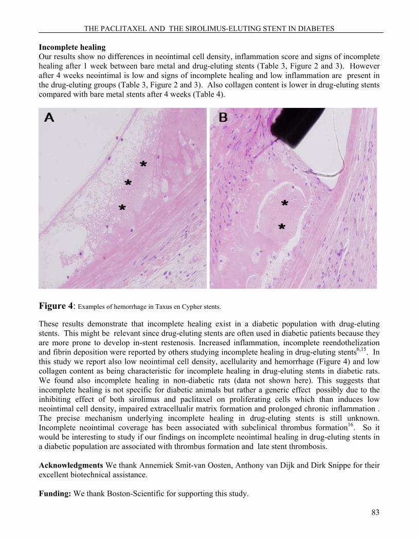
THE PACLITAXEL AND THE SIROLIMUS-ELUTING STENT IN DIABETES
83
Incomplete healing Our results show no differences in neointimal cell density, inflammation score and signs of incomplete healing after 1 week between bare metal and drug-eluting stents (Table 3, Figure 2 and 3). However after 4 weeks neointimal is low and signs of incomplete healing and low inflammation are present in the drug-eluting groups (Table 3, Figure 2 and 3). Also collagen content is lower in drug-eluting stents compared with bare metal stents after 4 weeks (Table 4).
Figure 4: Examples of hemorrhage in Taxus en Cypher stents. These results demonstrate that incomplete healing exist in a diabetic population with drug-eluting stents. This might be relevant since drug-eluting stents are often used in diabetic patients because they are more prone to develop in-stent restenosis. Increased inflammation, incomplete reendothelization and fibrin deposition were reported by others studying incomplete healing in drug-eluting stents6,15. In this study we report also low neointimal cell density, acellularity and hemorrhage (Figure 4) and low collagen content as being characteristic for incomplete healing in drug-eluting stents in diabetic rats. We found also incomplete healing in non-diabetic rats (data not shown here). This suggests that incomplete healing is not specific for diabetic animals but rather a generic effect possibly due to the inhibiting effect of both sirolimus and paclitaxel on proliferating cells which than induces low neointimal cell density, impaired extracellualir matrix formation and prolonged chronic inflammation . The precise mechanism underlying incomplete healing in drug-eluting stents is still unknown. Incomplete neointimal coverage has been associated with subclinical thrombus formation16. So it would be interesting to study if our findings on incomplete neointimal healing in drug-eluting stents in a diabetic population are associated with thrombus formation and late stent thrombosis. Acknowledgments We thank Annemiek Smit-van Oosten, Anthony van Dijk and Dirk Snippe for their excellent biotechnical assistance. Funding: We thank Boston-Scientific for supporting this study.

Chapter 7
84
References
1. Morice MC, Serruys PW, Sousa JE, Fajadet J, Ban HE, Perin M, Colombo A, Schuler G, Barragan P, Guagliumi G, Molnar F, Falotico R. A randomized comparison of a sirolimus-eluting stent with a standard stent for coronary revascularization. N Engl J Med 2002;346:1773-1780.
2. Grube E, Silber S, Hauptmann KE, Mueller R, Buellesfeld L, Gerckens U, Russell ME. TAXUS I: six- and twelve-month results from a randomized, double-blind trial on a slow-release paclitaxel-eluting stent for de novo coronary lesions. Circulation 2003;107:38-42.
3. Scheen AJ, Warzee F. Diabetes is still a risk factor for restenosis after drug-eluting stent in coronary arteries. Diabetes Care 2004;27:1840-1841.
4. Dibra A, Kastrati A, Mehilli J, Pache J, Schuhlen H, von Beckerath N, Ulm K, Wessely R, Dirschinger J, Schomig A. Paclitaxel-eluting or sirolimus-eluting stents to prevent restenosis in diabetic patients. N Engl J Med 2005;353:663-670.
5. McFadden EP, Stabile E, Regar E, Cheneau E, Ong AT, Kinnaird T, Suddath WO, Weissman NJ, Torguson R, Kent KM, Pichard AD, Satler LF, Waksman R, Serruys PW. Late thrombosis in drug-eluting coronary stents after discontinuation of antiplatelet therapy. Lancet 2004;364:1519-1521.
6. Joner M, Finn AV, Farb A, Mont EK, Kolodgie FD, Ladich E, Kutys R, Skorija K, Gold HK, Virmani R. Pathology of drug-eluting stents in humans: delayed healing and late thrombotic risk. J Am Coll Cardiol 2006;48:193-202.
7. Langeveld B, Roks AJ, Tio RA, van Boven AJ, van der Want JJ, Henning RH, van Beusekom HM, van der Giessen WJ, Zijlstra F, van Gilst WH. Rat abdominal aorta stenting: a new and reliable small animal model for in-stent restenosis. J Vasc Res 2004;41:377-386.
8. Schwartz RS, Huber KC, Murphy JG, Edwards WD, Camrud AR, Vlietstra RE, Holmes DR. Restenosis and the proportional neointimal response to coronary artery injury: results in a porcine model. J Am Coll Cardiol 1992;19:267-274.
9. Kornowski R, Hong MK, Tio FO, Bramwell O, Wu H, Leon MB. In-stent restenosis: contributions of inflammatory responses and arterial injury to neointimal hyperplasia. J Am Coll Cardiol 1998;31:224-230.
10. Langeveld B, van Gilst WH, Tio RA, Zijlstra F, Roks AJ. Angiotensin-(1-7) attenuates neointimal formation after stent implantation in the rat. Hypertension 2005;45:138-141.
11. Schwartz RS, Huber KC, Murphy JG, Edwards WD, Camrud AR, Vlietstra RE, Holmes DR. Restenosis and the proportional neointimal response to coronary artery injury: results in a porcine model. J Am Coll Cardiol 1992;19:267-274.
12. Chai S, Chai Q, Danielsen CC, Hjorth P, Nyengaard JR, Ledet T, Yamaguchi Y, Rasmussen LM, Wogensen L. Overexpression of hyaluronan in the tunica media promotes the development of atherosclerosis. Circ Res 2005;96:583-591.
THE PACLITAXEL AND THE SIROLIMUS-ELUTING STENT IN DIABETES
85
13. Iakovou I, Schmidt T, Ge L, Sangiorgi GM, Stankovic G, Airoldi F, Chieffo A, Montorfano M, Carlino M, Michev I, Corvaja N, Cosgrave J, Gerckens U, Grube E, Colombo A. Angiographic patterns of restenosis after paclitaxel-eluting stent implantation. J Am Coll Cardiol 2005;45:805-806.
14. Hara H, Nakamura M, Palmaz JC, Schwartz RS. Role of stent design and coatings on restenosis and thrombosis. Adv Drug Deliv Rev 2006;58:377-386.
15. Finn AV, Kolodgie FD, Harnek J, Guerrero LJ, Acampado E, Tefera K, Skorija K, Weber DK, Gold HK, Virmani R. Differential response of delayed healing and persistent inflammation at sites of overlapping sirolimus- or paclitaxel-eluting stents. Circulation 2005;112:270-278.
16. Kotani J, Awata M, Nanto S, Uematsu M, Oshima F, Minamiguchi H, Mintz GS, Nagata S. Incomplete neointimal coverage of sirolimus-eluting stents: angioscopic findings. J Am Coll Cardiol 2006;47:2108-2111.

CHAPTER 8 SUMMARY

SUMMARY
87
SUMMARY In recent years therapeutic advances of PCI such as antiplatelet drugs given before and after the procedure, the stent and later the drug eluting stent have improved mortality and morbidity for patients with CHD and this has led to a marked rise in the number of PCI procedures.1-3 Nowadays in the Netherlands every year around thirty thousand PCI procedures are performed.4 However PCI is still troubled by two major limitations: in- stent restenosis and stent thrombosis. In-stent restenosis is the narrowing of the vessel wall after stenting. In-stent restenosis usally does not lead to acute occlusion of the vessel with myocardial infarction and death, however recurrence of angina pectoris is often seen which in practice requires revascularization of the stented vessel. Stent thrombosis is the occurrence of thrombus formation in or near the stent resulting in an acute occlusion of the vessel. Although its incidence is much lower than in-stent restenosis nevertheless it is a serious complication because in contrast with in-stent restenosis it leads more often to acute myocardial infarction and has a much higher mortality rate. In this thesis the rat aortic stenting model was used to study several new treatments to reduce in-stent restenosis and some aspects of the pathofysiology of in stent restenosis. We also adapted our rat aortic model for studying in-stent restenosis in diabetics by developing two different (type 1 and type 2) diabetic models for in-stent restenosis. Furthermore we tried to identify a possible mechanism of late stent trombosis by comparing the neointima of bare metal and drug eluting stents. Chapter 2 describes a a novel type 1 diabetic model for in-stent restenosis after rat abdominal aortic stenting. Diabetic animal models are useful for studying the mechanisms of increased in-stent restenosis in diabetic populations5. We demonstrated increased neointimal formation in diabetic BBDP (Bio-Breeding Diabetes-Prone) and significant proteinuria, polyuria and glycemia compared to non-diabetic thymectomized BB-DP rats. These results validate this novel type 1 diabetic rat abdominal aortic stenting model for studying the mechanism of increased in-stent restenosis in diabetic populations and more specific in the type 1 diabetes population. Furthermore because of the genetic similarity of the diabetic and normoglycemic rats in this model the mechanisms responsible for increased in-stent restenosis in diabetic populations can more easily be identified in future studies. In Chapter 3 we describe the contribution of circulating bone marrow cells to neointimal formation after in-stent restenosis and transplant arteriosclerosis. In the specimens of both stented and transplanted vessels no bone marrow-derived neointimal smooth muscle and endothelial cells were detected. A few bone marrow-derived cells were found in the neointima but they were infiltrating leukocytes. In conclusion, non-bone marrow-derived cells are the predominant source of neointimal cells in stented and transplanted vessels. Vascular wall-derived progenitor cells may rather be the source of smooth muscle cells that contribute to in-stent restenosis and transplant arteriosclerosis which may have implications for our quest for new therapeutic targets to treat these vasculopathies6-8. In Chapter 4 we discuss the effect of the AT1-receptor candesartan on neointimal formation after stenting. Systemic candesartan cilexetil treatment did not result in reduction of neointimal formation however angiotensin II did result in an increase in neointimal formation. It is likely that AT1-receptor stimulation by Ang II is not a major contributor to in-stent restenosis and only high doses lead to neointimal formation explaining the inability of the AT1-receptor blocker candesartan to reduce neointimal formation after systemic treatment. The current study examined systemic AT1-receptor blockade, limited by its systemic side-effects like hypotension. However, local delivery could allow higher dosing, and could be more successful than systemic AT1-receptor blockade 9 .

Chapter 8
88
The effect of treatment with rosuvastin on in-stent restenosis was analyzed in Chapter 5. Rosuvastatin treatment reduced neointimal formation both after Ang II infusion stimulated neointimal formation and in the absence of Ang II infusion stimulated neointimal formation. Furthermore we found rosuvastatin improved systemic endothelial function in the presence and absence of high levels of Ang II and reduced inflammation. Retrospective analysis in patients suggest statins also slightly reduced in-stent restenosis in humans and that the pleiotropic effects (involve improving endothelial function, decreasing oxidative stress and inflammation, and inhibiting the thrombogenic response of statins) are responsible for the reduction of neointima formation rather than the lipid-lowering effects10. The improvement of endothelial function by rosuvastatin as described in chapter 5 also suggest involvement of the pleiotropic effects. Although the high dose of rosuvastatin greatly reduced neointimal formation in our study, systemic treatment in patients is not feasible in these doses due to hepatotoxicity. A rosuvastatin-eluting stent could circumvent this problem. In a recent study in the porcine coronary stenting model a cerivastatin statin-eluting stent proved successful in reducing in-stent restenosis11. Lipophilic statins like cerivastatin penetrate muscle cells at a higher degree than hydrophilic statins like rosuvastatin. Therefore in theory cerivastatin will inhibit smooth muscle proliferation and in-stent restenosis more strongly than rosuvastatin. However the potential for local tissue toxicity is also greater in lipophilic statins. Indeed cerivastatin was withdrawn from the market for systemic treatment in patients because of myopathy and rhabdomyolysis. Therefore both lipophilic and hydrophilic statin-eluting stents should be tested in animal models for both efficacy and safety. In Chapter 6 and 7 we describe quantative and qualitative differences in neointimal formation between bare metal stents and drug eluting stents in normoglycemic (chapter 6) and diabetic setting (chapter 7). The drug eluting stents reduced neointimal formation in the normoglycemic setting compared with the bare metals stent. However in the diabetic setting only the sirolimus eluting stent succesfully reduced in-stent restenosis in comparison with the bare metal stent. The qualitative differences we found were low cell neointimal density, low neointimal collagen content and increased low-grade inflammation. These differences developed in time: after 1 week cell density and inflammation did not differ between bare metals stents and drug eluting stents but after 4 weeks they differed. We conclude that drug eluting stents were superior in reducing neointimal formation, although the paclitaxel-eluting stent did not reduce neointimal formation as well as the sirolimus-eluting stent in diabetic setting. In clinical trials the sirolimus-eluting stent seems also to decrease restenosis better than the paclitaxel stent12. Our results suggests that the decreased efficiency of the paclitaxel-eluting stent in reducing stent thrombosis may be related to the stent design of the paclitaxel-eluting stent rather than the drug itself. Second-generation paclitaxel-eluting stents with different stent design may thus be better in reducing in-stent restenosis. We found notable incomplete healing in both non-diabetic rats and diabetic rats. This suggests that incomplete healing is not specific for diabetic animals but rather a generic effect possibly due to the inhibiting effect of both sirolimus and paclitaxel on proliferating cells which may induce low neointimal cell density, impaired extracellulair matrix formation and prolonged chronic inflammation. A link between late stent thrombosis and incomplete endothelial healing was suggested by Joner et al: incomplete reendothelization and high fibrin content in drug-eluting stents could be a potent thrombogenic stimulus13. Our results which demonstrate low collagen content in neointimal areas with incomplete healing suggest a second hypothesis: the subendothelial neointimal tissue of drug-eluting stents is weaker and could rupture more easily thereby exposing tissue factor and inducing thrombus. The presence of hemorrhages found within the neointima of drug-eluting stents seems to support this hypothesis. The low neointimal cell density could be correlated to tissue strength: there are not enough cells to produce extracellular matrix and ensure a strong neointima. Both sirolimus and

SUMMARY
89
paclitaxel have a direct effect on the extracellular matrix production in cells. Sirolimus inhibits collagen synthesis in rat vascular smooth muscle cells and paclitaxel also reduces tenascin (an extracellular matrix glycoprotein) in human arterial smooth muscle cells14. Future perspectives: Further reduction of in-stent restenosis will remain the most important goal in stent development. However this reduction of in-stent restenosis should be accomplished while minimizing the risk of stent trombosis. Several concepts need to be developed to reach these goals. The prevailing concept to achieve those goals at the moment is that of the drug eluting stent. The concept of the drug eluting stent is to use high local drug doses to inhibit the arterial healing response after stenting and thus reduce in-stent restenosis. Indeed current first and second generation drug eluting stents have been successful in reducing in-stent restenosis, however at the moment it is not clear if they present a long-term risk of stent trombosis and this is currently being addressed in clinical trials. Furthermore pre-clinical research will be needed to find the pathological substrate of stent trombosis. It is possible that the aggressive inhibition of the arterial healing response predisposes to stent trombosis. If the pathological substrate of stent trombosis is related to the strong anti-proliferative drugs used on current drug eluting stents, a critical reevaluation of the advantages and disadvantages of the use of these drugs on stents will be needed. In addition the search for new more ‘physiological’ acting drugs will start. Statins as discussed in chapter 5 may be a candidate for such drug eluting stents. However it remains to be seen if these more ‘physiological’ acting drug eluting stents are as efficient in reducing in-stent restenosis as the older ones. Another concept is to reduce the vascular injury induced by the stent, resulting in a smaller arterial healing response and less in-stent restenosis. An optimal stent design should combine maximum final stent diameter with little arterial trauma15 ref. Significant improvements in stent design have already been made by abandoning long, self-expandable, thick strutted, coiled stents for shorter, balloon expandable, thin strutted stents of tubular design15. Especially the thickness of the struts seems important for reducing vascular injury and in-stent restenosis16. At the moment high grade hard steel and kobalt alloys are the preferred material for stents enabling thin struts which minimize arterial trauma. However improvements in material sciences and nanotechnology may lead to stronger and more elastic metal alloys. Stents made of these new alloys would enabling even thinner struts while also being more flexible. Dense and radio-opaque elements such as hafnium and tantalum might also be used in these alloys to maintain radiopacity of the thin stent struts. Biocompatibility may also be important for stent design because increased thromboresistance may reduce in-stent restenosis and are likely to reduce stent thrombosis. The thromboresistant substances pyrolytic carbon and phosphorylcholine have been tested as coatings for hard steel and kobalt alloys stents. These studies did not show a reduction in restenosis17,18. The effect on stent thrombosis was not evaluated and should be evaluated in new studies. A controlled registry of the biocompatible iridium oxide-coated stent showed in-stent restenosis rates somewhat in between current drug-eluting stents and bare metal stents. However if this difference is due to the better biocompatibility of the iridium oxide coating or due to the thinner struts of the iridium oxide coated stents in comparison with first-generation bare metal stents is not clear. More promising is the polymer polybistrifluroexthoxyphosphazene (Polyzene-F). The haemocompatibility of a Polyzene-F-coated stainless steel was much better than that of uncoated bare metal stents19. More interestingly the Polyzene-F nanocoated cobalt-chromium stent demonstrated similar rates of in-stent restenosis as the paclitaxel and sirolimus-eluting stents in a porcine coronary model20.

Chapter 8
90
Still randomized controlled trials comparing biocompatible coated stents with drug-eluting and bare metal stents are needed to demonstrate the relevance of biocompatible stents for reducing stent thrombosis and in-stent restenosis in humans. A relatively new concept is that of the drug eluting balloon21. In this concept drug-eluting balloon dilatation is followed by bare metal stent implantation. It is similar to the drug-eluting concept in that it aims to inhibit the arterial healing response after stenting. However this concept seems more attractive: the drug itself is not present for long periods because no physical connection between drug and stent exists. Another theoretical advantage of the drug eluting balloon over the drug eluting stent is in the treatment of in-stent restenosis. Treatment of in-stent restenosis with a drug eluting stent creates an undesirable amount of four metal layers whereas the drug eluting balloon does not. Another promising concept is that of the resorbable stent. Directly after PCI a stent acts as a stabilizing scaffolding device:it prevents collapsing of intimal flaps and plaque dissections leading to a smoother surface and a larger lumen with more blood flow22. Furthermore a stent prevents abrupt vessel closure and reduces restenosis rates by eliminating elastic vessel recoil and negative vessel remodeling22. However on the long term the permanent nature of a stent has also negative effects: it prevents positive remodeling and the stent may continue to evoke inflammatory responses and thus stimulate neointimal formation. The resorbable stent acts in the early phase successfully as a scaffolding device like any other stent. However after several months the stent dissolves into the body theoretically allowing positive remodeling to occur and preventing neointimal formation due to the continued presence of the stent. Preclinical and human studies with a magnesium resorbable stent have shown the safety of the resorbable stent, although they had high restenosis rates due to both negative remodeling and chronic wall recoil23,24. Still the magnesium stent was almost completely resorbed and it successfully functioned as a scaffolding device in the early phase after stenting. The rate of resorption of a resorbable stent may be critical in preventing increased in-stent restenosis due to negative remodeling and premature recoil. Recent preclinical research suggests that biocorrodible iron stents may be better in preventing premature recoil than magnesium resorbable stents25. Future research on resorbable stents will learn us more about the ideal resorption interval and material. It is likely that in the near future less in-stent restenosis and stent thrombosis will be achieved by developing new drug eluting stents with superior (less injury) stent design and possibly also with new less aggressive drugs (e.g. statins) or with a polymer biocoating ( e.g.Polyzene-F). The drug eluting balloon concept although interesting has yet to prove itself in large clinicial trials. The resorbable stent is very promising but still in an experimental phase. In the long run combinations of these concepts( e.g. a resorbable drug eluting or biocoated stent) may reduce in-stent restenosis and stent thrombosis even better.
SUMMARY
91
References
1. Wiviott SD, Braunwald E, McCabe CH, Montalescot G, Ruzyllo W, Gottlieb S, Neumann FJ, Ardissino D, De Servi S, Murphy SA, Riesmeyer J, Weerakkody G, Gibson CM, Antman EM. Prasugrel versus clopidogrel in patients with acute coronary syndromes. N Engl J Med 2007;357:2001-2015.
2. Sigwart U, Puel J, Mirkovitch V, Joffre F, Kappenberger L. Intravascular stents to prevent occlusion and restenosis after transluminal angioplasty. N Engl J Med 1987;316:701-706.
3. Serruys PW, Degertekin M, Tanabe K, Abizaid A, Sousa JE, Colombo A, Guagliumi G, Wijns W, Lindeboom WK, Ligthart J, de Feyter PJ, Morice MC. Intravascular ultrasound findings in the multicenter, randomized, double-blind RAVEL (RAndomized study with the sirolimus-eluting VElocity balloon-expandable stent in the treatment of patients with de novo native coronary artery Lesions) trial. Circulation 2002;106:798-803.
4. Huijskes RV, Koch KT, van Herwerden LA, Berreklouw E, Limburg M, Tijssen JG. [The waiting time for heart interventions: trends for percutaneous coronary interventions and cardiothoracic interventions]. Ned Tijdschr Geneeskd 2003;147:1860-1865.
5. Takeda R, Suzuki E, Satonaka H, Oba S, Nishimatsu H, Omata M, Fujita T, Nagai R, Hirata Y. Blockade of endogenous cytokines mitigates neointimal formation in obese Zucker rats. Circulation 2005;111:1398-1406.
6. Sainz J, Al Haj ZA, Caligiuri G, Demerens C, Urbain D, Lemitre M, Lafont A. Isolation of "side population" progenitor cells from healthy arteries of adult mice. Arterioscler Thromb Vasc Biol 2006;26:281-286.
7. Hu Y, Zhang Z, Torsney E, Afzal AR, Davison F, Metzler B, Xu Q. Abundant progenitor cells in the adventitia contribute to atherosclerosis of vein grafts in ApoE-deficient mice. J Clin Invest 2004;113:1258-1265.
8. Zengin E, Chalajour F, Gehling UM, Ito WD, Treede H, Lauke H, Weil J, Reichenspurner H, Kilic N, Ergun S. Vascular wall resident progenitor cells: a source for postnatal vasculogenesis. Development 2006;133:1543-1551.
9. Wang L, Li GH, Chen H, Li HW, Zhao L, Yao DK, Ding RJ, Jia SQ. Effect of valsartan-eluting stents on the expression of angiotensin II type 2 receptor. Chin Med J (Engl ) 2006;119:601-604.
10. Kamishirado H, Inoue T, Sakuma M, Tsuda T, Hayashi T, Takayanagi K, Node K. Effects of statins on restenosis after coronary stent implantation. Angiology 2007;58:55-60.
11. Miyauchi K, Kasai T, Yokayama T, Aihara K, Kurata T, Kajimoto K, Okazaki S, Ishiyama H, Daida H. Effectiveness of statin-eluting stent on early inflammatory response and neointimal thickness in a porcine coronary model. Circ J 2008;72:832-838.
12. Billinger M, Beutler J, Taghetchian KR, Remondino A, Wenaweser P, Cook S, Togni M, Seiler C, Stettler C, Eberli FR, Luscher TF, Wandel S, Juni P, Meier B, Windecker S. Two-year clinical outcome after implantation of sirolimus-eluting and paclitaxel-eluting stents in diabetic patients. Eur Heart J 2008;29:718-725.
Chapter 8
92
13. Joner M, Finn AV, Farb A, Mont EK, Kolodgie FD, Ladich E, Kutys R, Skorija K, Gold HK, Virmani R. Pathology of drug-eluting stents in humans: delayed healing and late thrombotic risk. J Am Coll Cardiol 2006;48:193-202.
14. Park J, Ha H, Ahn HJ, Kang SW, Kim YS, Seo JY, Kim MS. Sirolimus inhibits platelet-derived growth factor-induced collagen synthesis in rat vascular smooth muscle cells. Transplant Proc 2005;37:3459-3462.
15. Morton AC, Crossman D, Gunn J. The influence of physical stent parameters upon restenosis. Pathol Biol (Paris) 2004;52:196-205.
16. Kastrati A, Mehilli J, Dirschinger J, Dotzer F, Schuhlen H, Neumann FJ, Fleckenstein M, Pfafferott C, Seyfarth M, Schomig A. Intracoronary stenting and angiographic results: strut thickness effect on restenosis outcome (ISAR-STEREO) trial. Circulation 2001;103:2816-2821.
17. Galli M, Bartorelli A, Bedogni F, DeCesare N, Klugmann S, Maiello L, Miccoli F, Moccetti T, Onofri M, Paolillo V, Pirisi R, Presbitero P, Sganzerla P, Viecca M, Zerboni S, Lanteri G. Italian BiodivYsio open registry (BiodivYsio PC-coated stent): study of clinical outcomes of the implant of a PC-coated coronary stent. J Invasive Cardiol 2000;12:452-458.
18. Kim YH, Lee CW, Hong MK, Park SW, Tahk SJ, Yang JY, Saito S, Santoso T, Quan L, Ge J, Weissman NJ, Lansky AJ, Mintz GS, Park SJ. Randomized comparison of carbon ion-implanted stent versus bare metal stent in coronary artery disease: the Asian Pacific Multicenter Arthos Stent Study (PASS) trial. Am Heart J 2005;149:336-341.
19. Mrowietz C, Franke RP, Seyfert UT, Park JW, Jung F. Haemocompatibility of polymer-coated stainless steel stents as compared to uncoated stents. Clin Hemorheol Microcirc 2005;32:89-103.
20. Radeleff B, Thierjung H, Stampfl U, Stampfl S, Lopez-Benitez R, Sommer C, Berger I, Richter GM. Restenosis of the CYPHER-Select, TAXUS-Express, and Polyzene-F Nanocoated Cobalt-Chromium Stents in the Minipig Coronary Artery Model. Cardiovasc Intervent Radiol 2007.
21. Scheller B, Hehrlein C, Bocksch W, Rutsch W, Haghi D, Dietz U, Bohm M, Speck U. Treatment of coronary in-stent restenosis with a paclitaxel-coated balloon catheter. N Engl J Med 2006;355:2113-2124.
22. De Feyter PJ. The quest for the ideal stent. Eur Heart J 2001;22:1766-1768.
23. Heublein B, Rohde R, Kaese V, Niemeyer M, Hartung W, Haverich A. Biocorrosion of magnesium alloys: a new principle in cardiovascular implant technology? Heart 2003;89:651-656.
24. Erbel R, Di Mario C, Bartunek J, Bonnier J, de Bruyne B, Eberli FR, Erne P, Haude M, Heublein B, Horrigan M, Ilsley C, Bose D, Koolen J, Luscher TF, Weissman N, Waksman R. Temporary scaffolding of coronary arteries with bioabsorbable magnesium stents: a prospective, non-randomised multicentre trial. Lancet 2007;369:1869-1875.
25. Waksman R, Pakala R, Baffour R, Seabron R, Hellinga D, Tio FO. Short-term effects of biocorrodible iron stents in porcine coronary arteries. J Interv Cardiol 2008;21:15-20.

CHAPTER 9 SAMENVATTING

Chapter 9
94
SAMENVATTING De afgelopen jaren hebben verbeteringen in de behandeling met PCI (percutane coronaire interventie) bestaande uit het geven van plaatjesremmers voor en na de procedure, de stent en de drug-eluting stent geleid tot een verbetering van de sterfte en morbiditeit van patiënten met coronairlijden1-3. Daardoor is het aantal PCI-procedures fors gestegen en inmiddels worden in Nederland jaarlijks ongeveer dertigduizend PCI-procedures verricht4. Echter PCI kent nog steeds twee belangrijke beperkingen: in-stent restenose en stent trombose. In-stent restenose is het vernauwen van het vaatlumen na stenten. In-stent restenose leidt meestal niet tot een acute afsluiting van het vat met een myocardinfarct of de dood als gevolg, maar geeft vaak angina pectoris waardoor patiënten opnieuw een PCI-procedure moeten ondergaan. Stent trombose is het ontstaan van een trombus in of nabij de stent leidend tot een acute afsluiting van het vat. De incidentie van stent trombose is veel lager dan de incidentie van in-stent restenosis. Daar staat tegenover dat stent trombose in tegenstelling tot in-stent restenose vaak leidt tot een acuut myocardinfarct en de dood. In dit proefschrift is het buikaorta stent model in de rat gebruikt om een aantal nieuwe behandelingen voor de vermindering van in-stent restenose te testen en om meer inzicht in een aantal aspecten van de pathofysiologie van in-stent restenose te krijgen. Tevens werden twee nieuwe (type 1 en type 2) modellen voor het bestuderen van in-stent restenosis bij diabetes gevalideerd. Daarnaast werd de weefselopbouw van vaten gestent met bare metal stents vergeleken met die van drug-eluting stents. Verschillen in de weefselopbouw tussen bare metal stents en drug-eluting stents kunnen mogelijk aanknopingspunten geven voor de pathophysiologie van stent trombose. In Hoofstuk 2 wordt een nieuw type 1 diabetes model voor in-stent restenose in de rat beschreven. Diabetische diermodellen zijn nuttig voor het onderzoeken van de mechanismen van in-stent restenose in diabetische populaties5.We laten zien dat er meer neointima formatie, proteinurie, polyurie en glycemie is in diabetische BBDP (Bio-Breeding Diabetes-Prone) ratten vergeleken met niet-diabetische BBDP ratten waar de thymus van verwijderd is. Deze resultaten valideert dit nieuw type 1 diabetes model voor het bestuderen van de mechanismen van in-stent restenose in diabetische populaties en meer specifiek type 1 diabetische populaties. De grote genetische overeenkomst tussen diabetische en niet-diabetische ratten in dit model maakt het makkelijker om de mechanismen te vinden die verantwoordelijk zijn voor de toegenomen in-stent restenose in diabetische populaties. In Hoofdstuk 3 beschrijven we de bijdrage van circulerende beenmergcellen aan in-stent restenose en transplantatie arteriosclerose. In de preparaten van zowel gestente als getransplanteerde vaten werden geen gladde spiercellen of endotheelcellen afkomstig uit het beenmerg aangetroffen. Enkele beenmergcellen werden gevonden in de neointima maar dit bleken onstekingscellen te zijn. Concluderend zijn beenmergcellen niet de directe bron van neointima cellen in gestente en getransplanteerde vaten. Het is waarschijnlijker dat voorlopercellen aanwezig in of nabij de gestente vaatwand de bron vormen van gladde spiercellen welke bijdragen aan neointima formatie in gestente en getransplanteerde6-8. Deze bevindingen zijn belangrijk voor het vinden van nieuwe therapeutische aangrijpingspunten voor het behandelen van in-stent restenose en transplantatie arteriosclerose. In Hoofdstuk 4 wordt het effect van behandeling met de angiotensin II receptor blokker candesartan cilexetil op in-stent restenose getest. Systemische behandeling met candesartan cilexetil leidde niet tot minder neointima formatie. Echter infusie met angiotensin II gaf wel meer neointima formatie. Het is waarschijnlijk dat angiotensin II receptor stimulatie een minder belangrijke rol speelt in in-stent restenose en alleen sterke angiotensin II receptor stimulatie leidt tot meer neointima formatie. Dit zou ook verklaren waarom de angiotensin II receptor blokker candesartan cilexetil niet in staat is om in-stent restenose te verminderen. Aan de andere kant werd de dosis van de angiotensin II receptor blokker candesartan cilexetil beperkt door systemische bijwerkingen zoals hypotensie. Locale dosering

SAMENVATTING
95
bijvoorbeeld met een drug-eluting stent heeft deze beperking in veel mindere mate. Het is mogelijk dat hogere doseringen die gehaald kunnen worden door locale dosering wel effectief zijn in het verminderen van in-stent restenosis9. Het effect van de behandeling met rosuvastatine op in-stent restenose wordt beschreven in Hoofdstuk 5. Behandeling met rosuvastatine verminderde neointima formatie zowel met angiotensin II gestimuleerde neointima formatie als zonder angiotensin II gestimuleerde neointima formatie. Bovendien verbeterde rosuvastatine de systemische endotheelfunctie en verminderde ontsteking in zowel de groep met angiotensin II infusie als in de groep zonder angiotensin II infusie. Een retrospectieve studie bij patiënten suggereerde dat de pleiotrope effecten van statines (verbetering van endotheelfunctie, vermindering van oxidatieve stress en ontsteking) leiden tot minder in-stent restenose10. De verbetering van de endotheelfunctie gevonden in onze studie met rosuvastatine lijkt dit te bevestigen. Alhoewel systemische behandeling met hoge doseringen rosuvastatine sterk in-stent restenose verminderde, is systemische behandeling bij mensen niet mogelijk door met name levertoxiciteit. Een rosuvastatine-eluting stent heeft dit probleem niet. In een recente studie bleek een cerivastatine-eluting stent succesvol in het verminderen van in-stent restenose11. Lipofiele statines zoals cerivastatine penetreren gladde spiercellen beter dan hydrofiele statines als rosuvastatine. Daarom zouden lipofiele statines zoals cerivastatine beter in staat moeten zijn om gladde spiercel proliferatie en in-stent restenose te remmen dan hydrofiele statines als rosuvastatine. Echter de kans op locale weefseltoxiciteit is daardoor ook groter bij lipofiele statines. Cerivastatine bijvoorbeeld is niet geregistreerd voor systemische behandeling bij patiënten door bijwerkingen als myopathie en rhabdomyolyse. Daarom zouden zowel de lipofiele als de hydrofiele statine-eluting stents getest moeten worden op het veilig verminderen van in-stent restenose. Hoofdstuk 6 en 7 beschrijven kwantitatieve en kwalitatieve verschillen van neointima formatie tussen bare metal stents en drug-eluting stents onder normoglycemische en diabetische omstandigheden. Onder normoglycemische omstandigheden waren zowel de paclitaxel eluting stent als de sirolimus eluting stent beter in het verminderen van neointima formatie. Echter onder diabetische omstandigheden was alleen de sirolimus eluting stent beter in het verminderen van neointima formatie. De kwalitatieve verschillen tussen drug-eluting stents en bare metal stents bestonden uit lage celdichtheid, minder collageen en meer chronische ontsteking in de preparaten van de drug-eluting stents. Deze verschillen ontwikkelden zich in de tijd: na 1 week waren deze verschillen nog niet aanwezig maar na 4 weken wel. We concluderen dat drug-eluting stents beter zijn in het verminderen van neointima formatie, alhoewel de paclitaxel-eluting stent onder diabetische omstandigheden meer in-stent restenose had als de sirolimus-eluting stent. In klinische trials bij diabetische patiënten bleek de sirolimus-eluting stent ook minder in-stent restenose te hebben12. Onze resultaten bevestigen dat maar suggereren ook dat dit meer te maken heeft met de stent dan met het medicijn paclitaxel zelf. Nieuwe paclitaxel-eluting stents met een beter ontwerp hebben mogelijk minder in-stent restenose. We hebben een vertraagde genezing van de neointima van drug-eluting stents gevonden in zowel diabetische als niet-diabetische ratten. Dit wijst erop dat de vertraagde genezing van de neointima dus niet specifiek is voor diabetes maar een algemeen cytostatisch effect van sirolimus en paclitaxel is wat leidt tot een lage celdichtheid, meer inflammatie en minder collageen in de neointima. Een verband tussen late stent thrombose en vertraagde endotheel genezing is gesuggereerd door Joner et al13. Onze resultaten waarin we weinig collageen vinden in gebieden met weinig cellen suggereren een tweede hypothese: het subendotheliale neointima weefsel van drug-eluting stents is mogelijk zwakker, scheurt makkelijker waardoor tissue factor vrij kan komen leidend tot stent trombose. De aanwezigheid van bloedingen in de neointima van drug-eluting stents zoals beschreven door ons ondersteunt deze hypothese. Dus op plekken met een lage celdichtheid (geïnduceerd door lokale toxische doseringen sirolimus en paclitaxel in de neointima) is er mogelijk minder collageenvorming en daardoor

Chapter 9
96
weefselzwakte. Het is bekend uit studies dat sirolimus en paclitaxel extracellulaire matrix vorming (o.a. collageen, tenascine) sterk verminderen14. Toekomstperspectieven: Vermindering van in-stent restenose zal waarschijnlijk het belangrijkste doel blijven in de ontwikkeling van stents. Echter deze vermindering van in-stent restenose zou bereikt moeten worden zonder een hoog risico op stent trombose. Verschillende concepten zullen nodig zijn om deze doelen te bereiken. Het belangrijkste concept om deze doelen te bereiken is op dit moment de drug-eluting stent. Het concept van de drug-eluting stent is het gebruik van hoge locale doseringen van medicijnen om het herstelproces van de vaatwand na implantatie van een stent te remmen en daarmee in-stent restenose te verminderen. De huidige drug-eluting stents zijn inderdaad goed in staat om in-stent restenose te verminderen maar het is op het moment nog niet duidelijk of deze stents op de lange termijn een verhoogd risico op stent trombose hebben. Dit wordt momenteel onderzocht in een aantal grote klinische trials. Preklinisch onderzoek zal nodig zijn om het pathologische substraat van stent trombose te vinden. Mogelijk zal blijken dat sterke remming van het herstelproces van de vaatwand na implantatie van een stent predisponeert voor stent trombose. Als het pathologische substraat van stent trombose gerelateerd is aan het gebruik van sterk anti-proliferatieve medicijnen op de huidige drug-eluting stents, dan zal een kritische evaluatie van de voordelen en nadelen van het gebruik van deze medicijnen op stents nodig zijn. Bovendien zal dan een zoektocht beginnen naar nieuwe kandidaatmedicijnen voor gebruik op drug-eluting stents welke minder ingrijpen in het normale herstelproces van de vaatwand. Een statine zoals rosuvastatine beschreven in Hoofdstuk 5 is mogelijk een kandiaatmedicijn voor een drug-eluting stent. Belangrijk voor het succes van deze kandidaatmedicijnen is dan wel dat zij net zo goed zijn in het verminderen van in-stent restenose als de oude sterk anti-proliferatieve medicijnen. Een ander concept is het verminderen van de schade die de stent in de vaatwand aanricht. Daardoor onstaat een kleinschaliger herstelproces met minder in-stent restenose. Een optimaal stent-ontwerp moet dus streven naar een maximale diameter van het vat na het plaatsen van de stent maar met minimale schade aan de vaatwand15. Belangrijke veranderingen in stentontwerp zoals verkorting van de stent, balloninflatie, dunnere stentstruts en een tubulair ontwerp hebben al hun intree gevonden 15. Met name de dikte van de stentstruts lijkt belangrijk te zijn voor het verminderen van schade aan de vaatwand en vermindering van in-stent restenosis16. Op het moment bestaat de trend om steeds hardere metalen, bijvoorbeeld kobaltlegeringen, te gebruiken om stents van te vervaardigen zodat de stentstruts steeds dunner kunnen worden. Toekomstige ontwikkelingen in de materiaal- en nanowetenschappen maken het misschien mogelijk nog sterkere en flexibeler materialen te ontwikkelen en gebruiken voor het produceren van stents. Stents gemaakt van deze materialen zouden dunner zijn en met betere flexibiliteit. Toevoeging van kleine hoeveelheden van dichte metalen zoals hafnium of tantalum zou ervoor zorgen dat de radiopaciteit van zulke dunne stents behouden blijft. Het gebruik van lichaamsvriendelijke materialen op de stent kan mogelijk ook bijdragen aan minder in-stent restenose en stent trombose doordat het oppervlak van de stent minder trombosegevoelig is. Coatings van stents met de trombose resistente stoffen pyrolytic carbon en phosphorylcholine verminderden in-stent restenose niet, maar zijn nog niet getest op het verminderen van stent trombose17,18. Een studie met een lichaamsvriendelijke iridiumoxide gecoate stent verminderde in-stent restenose vergeleken met bare metal stents, maar niet zoveel als drug-eluting stents. Echter in deze studie was niet geheel duidelijk of de vermindering van in-stent restenose kwam door de lichaamsvriendelijke iridiumoxide coating of door de dunne struts van deze stent. Veelbelovend is de

SAMENVATTING
97
polymeer polybistrifluroexthoxyphosphazene (Polyzene-F). De heamocompatibiliteit van Polyzene-F-coated stents was beter dan van gewone bare metal stents19. In een recente studie in varkens had de Polyzene-F nanocoated cobalt-chromium stent in vergelijking met een sirolimus-eluting en de paclitaxel-eluting stent evenveel in-stent restenose20. Klinische trials moeten uitwijzen of lichaamsvriendelijke gecoate stents een meerwaarde hebben boven drug-eluting en bare metal stents. Een nieuw concept is dat van de drug eluting ballon21. In dit concept wordt inflatie met een drug-eluting ballon gevolgd door implantatie met een bare metal stent. Zowel de drug-eluting ballon als de drug-eluting stent proberen het herstelproces na stenten te remmen. Theoretisch heeft dit concept enkele voordelen boven de drug-eluting stent: er hoeft geen drager van het medicijn aanwezig te zijn op de stent en de kans is kleiner dat medicijnen voor langere tijd aanwezig blijven op de stent. Bovendien als eenmaal in-stent restenose is opgetreden kan deze beter behandeld worden met een drug eluting ballon dan met een drug eluting stent omdat bij behandeling met een drug eluting stent een onwenselijke situatie ontstaat in de gestente vaatwand met dubbele lagen van metaal op elkaar. Een veelbelovend concept is dat van de resorberende stent. Direct na PCI fungeert een stent als een ondersteunend apparaat: het voorkomt het loshangen van delen van de intima in het lumen en van dissecties in de plaques. Daarmee zorgt de stent voor een gladder oppervlak van de gestente vaatwand met een groter lumen en een betere bloedstroom door het vat22. Bovendien voorkomt een stent plotselinge afsluiting van het vat en vermindert restenose door elastische recoil en negatieve vaatremodellering22. Echter op de lange termijn heeft het permanente karakter van een stent nadelen: het voorkomt positieve remodellering en de stent kan aanleiding geven tot chronische ontsteking en daarmee neointima formatie stimuleren. De resorberende stent gedraagt zich als een normale stent in de vroege fase na stenten. Maar na enkele maanden lost de resorberende stent op zodat positieve remodellering kan plaatsvinden en de afwezigheid van de stent voorkomt meer neointima formatie. Preklinische en klinische studies met een resorberende magnesium stent hebben laten zien dat deze stent veilig is, maar deze stent had ook hoge restenose percentages door negatieve remodellering en late elastische recoil23,24. Gunstig was dat de magnesium stent vrijwel geheel was geresorbeerd aan het einde van de studie en in de vroege fase succesvol functioneerde als een ondersteunend apparaat. De snelheid waarmee de stent resorbeert is waarschijnlijk van belang bij het voorkomen van in stent restenose door negatieve remodellering en elastische recoil. Recent preklinisch onderzoek suggereert dat een biocorrosieve ijzeren stent beter is bij het voorkomen van elastische recoil dan een stent van magnesium 25. Toekomstig onderzoek naar resorberende stents zal ons meer leren over het ideale resorptie-interval en materiaal. Waarschijnlijk kan in de nabije toekomst in-stent restenose en stent trombose verminderd worden door de ontwikkeling van nieuwe drug-eluting stents met superieure stent-ontwerpen (minder vaatschade) en mogelijk ook door het gebruik van nieuwe medicijnen (b.v. statines) of lichaamsvriendelijke coatings (b.v. Polyzene-F). De drug-eluting ballon moet zich nog bewijzen in klinische trials en de resorberende stent is nog in een experimenteel stadium. Op de lange termijn kunnen combinaties van bovenstaande concepten (b.v. een resorberende drug-eluting of lichaamsvriendelijke gecoate stent) in-stent restenose en stent trombose verder verminderen.

DANKWOORD

99
Met dit dankwoord wil ik alle mensen bedanken welke bijgedragen hebben aan dit proefschrift. Ten eerste wil ik mijn eerste promotor Prof. dr. F. Zijlstra bedanken. Beste Felix, jouw uitgebreide kennis op het gebied van de interventiecardiologie heeft bijgedragen aan de kwaliteit en richting van dit proefschrift. Jouw snelle adequate manier van becommentariëren waardeer ik zeer. Daarnaast gaat mijn dank uit aan Prof. Dr. W.H. van Gilst. Beste Wiek, jouw grote pharmacologische kennis en ervaring met preklinisch onderzoek is van grote waarde geweest voor de kwaliteit en richting van dit proefschrift. Daarnaast waardeer ik het vertrouwen en de vrijheid die je aan mij gegeven hebt gedurende het werk aan dit proefschrift. Veel dank gaat uit aan mijn eerste copromotor dr B.J.G.L. de Smet. Beste Bart, afgelopen drie en half jaar heb ik met veel plezier met jou samengewerkt, jouw feedback was direct en integer. Jouw ervaring met preklinisch onderzoek en kennis op het gebied van in-stent restenose heeft bijgedragen aan de kwaliteit en richting van dit proefschrift. Verder wil ik mijn tweede copromotor dr Anton J.M. Roks bedanken. Beste Anton, Alleen de chaos op jouw bureau kon wedijveren met die van mijn eigen bureau, ik waardeer jouw vindingrijkheid. Jouw ervaring met vasculair onderzoek en brede kennis op het gebied van labaratoriumtechnieken heeft bijgedragen aan de kwaliteit en richting van dit proefschrift. Dr J.L Hillebrands, Dr Jan Rozing en G. Onuta van de sectie Immunologie wil ik bedanken voor de prettige samenwerking welke reeds één publicatie opgeleverd heeft. Prof. dr. W. van der Giessen, Prof.dr. D.J. van Veldhuisen en Prof.dr. R.H. Henning wil ik bedanken voor het plaatsnemen in de beoordelingscommisie en kritisch doorlezen van dit proefschrift. Prof. Dr. D.J. van Veldhuisen en Dr M. van den Berg wil ik bedanken voor de mogelijkheid de opleiding tot cardioloog te volgen. Dit proefschrift was niet mogelijk geweest zonder de excellente bijdrage in kennis en arbeid van studenten, biotechnici, analisten en postdocs van de afdeling klinische farmacologie, celbiologie en het centraal dierlaboratorium. Jacko, Marry, Bianca, Alex, Leo, Maria, Hendrik, Wessel, Adriaan, Dirk, Ellen, Ardy, Han, Flip, Ietse Alexandra e.a. bedankt. In het bijzonder wil ik bedanken: Geert Kors voor het uitzagen van de plasticpreparaten, Azuwerus van Buiten voor zijn hulp bij de vaatexperimenten en Hans Bartels , Annemieke Smit-van Oosten en Anthony van Dijk voor het implanteren van de stents. Verder wil ik Maaike Goris bedanken voor haar grote hulp bij het snijden en

100
kleuren van de preparaten en haar biotechnische ondersteuning in het centraal dierlaboratorium. Bernadette, Mirjam,Willemijn, Cheng, Hisko, Irma en Hiddo bedankt voor de gezelligheid tijdens de uitjes naar Berlijn, Terschelling en Gent. Verder wil ik alle (ex)-collega’s en arts-onderzoekers van de greenhouse: Daan, Wim, Patrick, Sandra, Martin, Michiel, Anne, Peter, Jessica, Tone, Christiane, Sheba, Kevin, Tom, Erik, Jasper, Meint, Willem-Peter, Matthijs, Marcelle, Lieuwe, Anne-Chris en Pieter bedanken voor de gezellige uitjes naar de golfbaan, wijnproeverij en vrijdagavondborrels in ’t Feithhuis. Mijn voorganger Bas Langeveld wil ik bedanken voor mijn introductie op de klinische farmacologie en in het centraal dierlaboratorium. Pim wil ik in het bijzonder bedanken voor de prettige samenwerking en flammende start van mijn promotietraject welke ons beiden reeds twee publicaties opgeleverd heeft. Van de afdeling en het secretariaat cardiologie wil ik ook een aantal mensen bedanken. Alma, Audrey, Marianne, Danielle, Morela en in het bijzonder Magda bedankt voor jullie hulp bij het administratieve deel van mijn promotie. Johan, Lineke, Moniek en de andere medewerkers op de VO2max en ergometrie bedankt voor de prettige samenwerking afgelopen jaren. Mijn paranimfen Robert de Kwant en Hans Hofer wil ik bedanken voor hun hulp en steun. Beste Robert, jouw relativeringsvermogen en gemoedelijkheid waardeer ik zeer. Ik voorzie in de toekomst nog vele gezellige momenten in de kroeg. Beste Hans, jouw optimisme, behulpzaamheid en humor waardeer ik zeer. Ik kijk er naar uit deze zomer weer eens op de racefiets te stappen. Mijn ouders wil ik bedanken voor hun steun en liefde vanjongsafaan. Lieve Leonie, bedankt voor al je steun en liefde, jij bent mijn Venus én Athene. Tot slot familie en vrienden bedankt voor jullie interesse en luisterend oor.