Feelings as agents of selection: Putting Charles Darwin back into (extended neo-) Darwinism
Upload
khangminh22Category
view
0download
0
University of Groningen
Pins and Needles, Facts and Feelings.Timmerman, Hans
IMPORTANT NOTE: You are advised to consult the publisher's version (publisher's PDF) if you wish to cite fromit. Please check the document version below.
Publication date:2019
Link to publication in University of Groningen/UMCG research database
Citation for published version (APA):Timmerman, H. (2019). Pins and Needles, Facts and Feelings. Efficacy of Screening Tools to AssessNeuropathic Pain in Daily Clinical Practice.
CopyrightOther than for strictly personal use, it is not permitted to download or to forward/distribute the text or part of it without the consent of theauthor(s) and/or copyright holder(s), unless the work is under an open content license (like Creative Commons).
The publication may also be distributed here under the terms of Article 25fa of the Dutch Copyright Act, indicated by the “Taverne” license.More information can be found on the University of Groningen website: https://www.rug.nl/library/open-access/self-archiving-pure/taverne-amendment.
Take-down policyIf you believe that this document breaches copyright please contact us providing details, and we will remove access to the work immediatelyand investigate your claim.
Downloaded from the University of Groningen/UMCG research database (Pure): http://www.rug.nl/research/portal. For technical reasons thenumber of authors shown on this cover page is limited to 10 maximum.
Download date: 01-02-2022
PINS AND NEEDLES, FACTS AND FEELINGSEFFICACY OF SCREENING TOOLS FOR NEUROPATHIC PAIN IN DAILY CLINICAL PRACTICE
HANS TIMMERMAN
PINS A
ND
NEED
LES, FAC
TS AN
D FEELIN
GS
HA
NS TIM
MERM
AN

PINS AND NEEDLES, FACTS AND FEELINGS
Efficacy of Screening Tools for Neuropathic Pain in Daily Clinical Practice
Hans Timmerman
Colophon
The work presented in this thesis was carried out within the Radboud Institute for Health Sciences.
Lay out: ProefschriftOntwerp.nl, Nijmegen
Printed by: Ipskamp Printing, Enschede
ISBN: 978-94-93118-17-1
© Copyright: Hans Timmerman 2019
All rights are reserved. No part of this thesis may be reproduced, distributed, stored in a retrieval
system or transmitted in any form or by any means, without prior written permission of the author.
PINS AND NEEDLES, FACTS AND FEELINGS
Efficacy of Screening Tools for Neuropathic Pain in Daily Clinical Practice
Proefschrift
ter verkrijging van de graad van doctor
aan de Radboud Universiteit Nijmegen
op gezag van de rector magnificus prof. dr. J.H.J.M. van Krieken,
volgens besluit van het college van decanen
in het openbaar te verdedigen op vrijdag 3 mei 2019
om 14.30 uur precies
door
Hans Timmerman
geboren op 25 september 1969
te Ede
Promotoren
Prof. dr. K.C.P. Vissers
Prof. dr. A.P. Wolff (UMCG)
Copromotoren
Prof. dr. M.A.H. Steegers
Dr. O.H.G. Wilder-Smith
Manuscriptcommissie
Prof. dr. L.A.L.M. Kiemeney
Prof. dr. R.H.M.A. Bartels
Prof. dr. M. van Kleef (MUMC)

PINS AND NEEDLES, FACTS AND FEELINGS
Efficacy of Screening Tools for Neuropathic Pain in Daily Clinical Practice
Doctoral Thesis
to obtain the degree of doctor
from Radboud University Nijmegen
on the authority of the Rector Magnificus prof. dr. J.H.J.M. van Krieken,
according to the decision of the Council of Deans
to be defended in public on Friday, May 3, 2019
at 14.30 hours
by
Hans Timmerman
Born on September 25, 1969
in Ede, The Netherlands
Supervisors
Prof. dr. K.C.P. Vissers
Prof. dr. A.P. Wolff (UMCG)
Co-supervisors
Prof. dr. M.A.H. Steegers
Dr. O.H.G. Wilder-Smith
Doctoral Thesis Committee
Prof. dr. L.A.L.M. Kiemeney
Prof. dr. R.H.M.A. Bartels
Prof. dr. M. van Kleef (MUMC)


TABLE OF CONTENTS
Chapter 1 General introduction
Chapter 2 Cross-cultural adaptation to the Dutch language of the Pain DETECT-
Questionnaire
Chapter 3 Assessment of neuropathic pain in patients with cancer: The
interobserver reliability. An observational study in daily practice
Chapter 4 Detecting the neuropathic pain component in the clinical setting:
a study protocol for validation of screening instruments for the
presence of a neuropathic pain component
Chapter 5 Avoiding Catch-22: validating the PainDETECT in a population of
patients with chronic pain
Chapter 6 Investigating the validity of the DN4 in a consecutive population of
patients with chronic pain
Chapter 7 The added value of bedside examination and screening QST to
improve neuropathic pain identification in patients with chronic pain
Chapter 8 General conclusions and discussion. Recommendations for clinical
practice, education, future research and societal impact
Chapter 9 Summary
Nederlandse Samenvatting
Data management
Curriculum Vitae
Publication list
PhD Portfolio
11
47
63
85
103
131
163
185
219
223
229
233
235
241

CHAPTER 1Introduction

Chapter 1
12
General Introduction
13
1
GENERAL INTRODUCTION
“Pain is a major healthcare problem worldwide. Although acute pain may reasonably be considered a
symptom of disease or injury, chronic and recurrent pain is a specific healthcare problem, a disease in its
own right” (IASP 2001).
Pain is a biopsychosocial phenomenon defined as “an unpleasant sensory and emotional experience
associated with actual or potential tissue damage, or described in terms of such damage” [1]. Pain is
regarded as a subjective experience and thus implies consciousness, as described in 1968 by
McCaffery: “Pain is whatever the experiencing person says it is, existing whenever he says it does” [2].
Moreover, the patient has to be considered as the expert of his/her own pain, which makes it difficult
to document pain objectively, but it also makes pain a highly individual disease which requires a
personalized approach and treatment.
Nociception is defined by the IASP as “the neural process of encoding noxious stimuli” [3], whereby
information about a harmful stimulus is passed on via the activation of nociceptors to the brain.
However, nociception alone is not enough to rate a stimulus as pain. To experience a nociceptive
stimulus as ‘pain’, a person is influenced by personal memory, emotions, pathology and cognitive
factors [4].
Section1 of the introduction discusses the classification of patients’ pain based on the type and
duration of the pain. Section 2 describes the neuroanatomy and physiology of pain, neuropathic pain
in particular, and provides additional information about pain processing. The epidemiology, burden,
costs and consequences of (neuropathic) pain are described in Section 3. Section 4 introduces
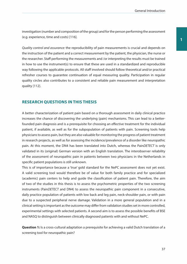
the assessment of neuropathic pain in daily clinical practice. In Section 5, the requirements for a
screening tool for the assessment of (neuropathic) pain are specified. The research questions to be
answered in this thesis are then introduced in the final section of the Introduction.
Chapter 1
14
CLASSIFICATION OF PAIN
Pain has multiple causes, and people’s response to pain is various and individually fixed, depending
on, for example, the circumstances. This section discusses how patients’ pain can be classified based
on the type and/or duration of pain.
Classification based on type of painNociceptive pain: the IASP define nociceptive pain as “pain that arises from actual or threatened
damage to non-neural tissue and is due to the activation of nociceptors” [5]. This type of pain mostly
results from a clear, identifiable mechanical, thermal or chemical damage to various parts of the
body (somatic: such as skin, bones, muscles; or visceral: abdominal or thoracic internal organs).
The pain is felt at the site of the injury or by stimulation of local nociceptors without injury, and is
relatively easy to treat [1]. In recent years, pain has also been described as inflammatory pain and
visceral pain, both with a more causal than mechanistic orientation. Inflammatory pain is defined as
‘a result of activation and sensitization of the nociceptive pain pathway by a variety of mediators released
at a site of tissue inflammation’ [6]. Inflammatory pain can be found in patients with, amongst others,
rheumatoid arthritis, pancreatitis, or a herpes zoster infection. Visceral pain arises from the internal
organs; it often has a diffuse localization due to major perceptive fields, overlap of innervations and
‘cross-talking’ of innervating nerves. The pain refers to other areas of the body and is associated with
motor- and autonomic reflexes [7]. An example of visceral pain is deep pain from the bladder which
is referred to the perianal region [8].
Neuropathic pain: in 1994, neuropathic pain was defined as “Pain initiated or caused by a primary
lesion or dysfunction in the nervous system” [1]. However in 2008, neuropathic pain was redefined as
“Pain arising as a direct consequence of a lesion or disease affecting the somatosensory system” [9, 10].
According to the IASP taxonomy, neuropathic pain is not a diagnosis but a ‘clinical description which
requires a demonstrable lesion or a disease that satisfies established neurological diagnostic criteria’ [9].
It is divided into central and peripheral neuropathic pain. Central neuropathic pain is caused by a
lesion or disease of the central somatosensory nervous system, for example in patients with a spinal
cord injury or multiple sclerosis. In peripheral neuropathic pain, the lesion or disease is localized in
the peripheral somatosensory nervous system, for example in patients with diabetic neuropathy, or
as a side effect after treatment for cancer with chemotherapy [9].
Nociplastic pain: in November 2017 (after our study was completed), the International Association
for the Study of Pain (IASP) acknowledged nociplastic pain as the third mechanistic descriptor for
chronic pain states in addition to nociceptive and neuropathic pain, because in 2008, the term
‘dysfunction’ was removed from the definition of neuropathic pain [11-14]. It is now defined as “Pain
that arises from altered nociception despite no clear evidence of actual or threatened tissue damage
causing the activation of peripheral nociceptors or evidence for disease or lesion of the somatosensory

General Introduction
15
1
system causing pain” [11]. Patients in this group are in pain but neither from an obvious activation of
their nociceptors nor from neuropathy. Nociplastic pain is suggested to be an altered nociceptive
function based on clinical, physical and psychological observations and it can occur in combination
with nociceptive pain and/ or neuropathic pain. The advantage of this third descriptor is that it
gives more recognition to pain as experienced by the patient, and it is intended to improve the
diagnosis and treatment of patients with (chronic) pain by creating an extra subdivision. As
debate is a fundamental part of an academic environment, there is an ongoing discussion about
the use of the term nociplastic pain and its meaning[11-19]. The question then arises, ‘What does
‘altered nociception’ mean?” Does this refer to a change in the nociceptors or is there a change in
the signal processing of the nociceptive input, or perhaps both? Nociceptor activity or activity in
the pathways/cortical networks is not necessarily pain [12]. Describing a persistent pain condition
without a clear medical explanation and without objective criteria for assessment and diagnosis
will lead to a continuation of the debate about this new mechanistic descriptor until research
provides more insights into this phenomenon [12]. Pain conditions fitting this description are,
amongst others, fibromyalgia, CRPS and irritable bowel syndrome [11]. One of the extensively
described phenomena which fits the term nociplastic pain is ‘central sensitization’ [20]. Central
sensitization is defined as “an amplification of neural signaling within the central nervous system that
elicits hypersensitivity” [21, 22]. Moreover, the IASP defines central sensitization as “an increased
response and reduced threshold of nociceptive neurons in the central nervous system to their normal
or subthreshold afferent input” [23]. Correctly determining and recognizing central sensitization is
important when diagnosing the patient, classifying the patient’s pain, and in treatment [20, 24-27].
However, as Kosek et al [16] suggested, the underlying mechanism of nociplastic pain may also be
the central sensitization of nociception or nociceptive pathways. Research in the field of nociplastic
pain should target identifying the suggested altered nociceptive function in patients with (chronic)
pain and, consequently, developing treatment opportunities.
Mixed pain: pain can be classified as an independent condition, but also as part of a ‘mixed pain
condition’ [28, 29] in which, for example, nociceptive pain and neuropathic pain are present in one
patient. Because of the coexistence of pain classifications in daily clinical practice, it is better to
speak of an absent or present neuropathic pain component in patients’ pain (NePC) with respect to
mixed pain conditions.
Pain of unknown origin: pain can also be classified as ‘pain from an unidentified source’; this used to
be termed ‘idiopathic pain’. It is now defined as “pain of unknown cause and origin” [11].
Classification based on duration of painBesides the differentiation in type of pain, patients can be classified based on the duration of their
pain. Acute pain is defined as “pain of recent onset and probable limited duration. It usually has an
identifiable temporal and causal relationship to injury or disease” [30]. Acute pain may induce chronic
Chapter 1
16
pain states [31, 32]; for example after undergoing surgery, it is known that acute pain is followed by
chronic pain in 10-50% of patients[33] . The definition of chronic pain is “pain that extends beyond
the expected period of healing” [1]. Chronic pain is recognized as pain that persists or recurs after
normal healing time, and that lacks the acute warning function of physiological nociception [34,
35]. Chronic pain can be present without an identifiable temporal or causal relationship with the
injury or disease according to currently available diagnostic methods. In daily clinical practice, pain
is regarded as ‘chronic’ if it lasts for more than (or recurs within) 3-6 months [1].
NEUROANATOMY AND PHYSIOLOGY OF PAIN AND NEUROPATHIC PAIN IN PARTICULAR
In this section, I provide an overview of the physiological mechanisms of pain and the important
pain pathways between receptors and the brain.
In normal conditions, pain is a protective natural response to a disease or injury after a body is
threatened. The protective function of the nociceptive sensory system is divided into a somatosensory
and a homeostatic part. The somatosensory part localizes the disease or injury and causes painful
stimuli, followed by corresponding fast motor reflexes. The homeostatic function results in
hyperalgesia and autonomic adaptation during the healing phase in pathological conditions [36,
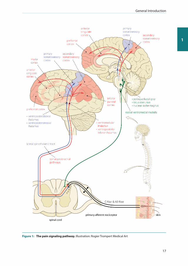
37]. Pain is the result of a complex interaction between signaling systems, modulation that may
originate from higher centers, and the unique perception of the individual [8] (figure 1). In addition
to the experience of pain, an increase in heart rate and blood pressure, sweating and changes in
respiratory behavior can occur after activation of the nociceptors due to sympathetic activation.
The pain signaling pathwayPrimary afferents: Nociceptors are receptors (free nerve endings) found in a range of tissues activated
by specific painful stimuli such as the free nerve endings of cutaneous nociceptors localized in the
epidermal layer of the skin. Other nociceptors, such as the high-threshold mechanoreceptors,
respond to mechanical deformation (pressure, stretch, etc.). Another example, polymodal receptors,
respond to a variety of tissue damaging inputs (mechanical, temperature and chemical stimuli).
Inflammatory mediators such as hydrogen ions (protons), 5-hydoxytryptamine (5-HT), cytokines,
bradykinin, histamine, prostaglandins, and leucotrienes, activate and sensitize the free nerve
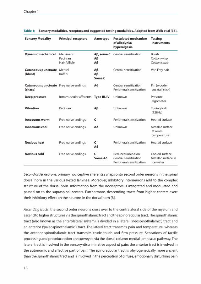
endings of different types of nerve fibers [8]. Aβ fibers generate touch, pressure, proprioception and
vibration signals; Aδ may produce acute, well localized sharp pain, and C fibers result in warmth,
delayed, and more diffuse pain, and a long-lasting burning sensation. Type III & IV fibers are sensitive
to deep (muscular) pressure (table 1) [38, 39]. These primary afferent nerve fibers have cell bodies in
the dorsal root ganglia or in the trigeminal ganglion, and terminate in the dorsal horn of the spinal
cord [8].
General Introduction
17
1
Figure 1: The pain signaling pathway. Illustration: Rogier Trompert Medical Art
Chapter 1
18
Table 1: Sensory modalities, receptors and suggested testing modalities. Adapted from Walk et al [38].
Sensory Modality Principal receptors Axon type Postulated mechanism of allodynia/ hyperalgesia
Testing instruments
Dynamic mechanical Meissner’sPacinianHair follicle
Aβ, some CAβAβ
Central sensitization BrushCotton wispCotton swab
Cutaneous punctuate (blunt)
MerkelRuffini
AβAβSome C
Central sensitization Von Frey hair
Cutaneous punctuate (sharp)
Free nerve endings Aδ Central sensitizationPeripheral sensitization
Pin (wooden cocktail stick)
Deep pressure Intramuscular afferents Type III, IV Unknown Pressure algometer
Vibration Pacinian Aβ Unknown Tuning fork (128Hz)
Innocuous warm Free nerve endings C Peripheral sensitization Heated surface
Innocuous cool Free nerve endings Aδ Unknown Metallic surface at room temperature
Noxious heat Free nerve endings CAδ
Peripheral sensitization Heated surface
Noxious cold Free nerve endings CSome Aδ
Reduced inhibitionCentral sensitizationPeripheral sensitization
Cooled surfaceMetallic surface in ice water
Second order neurons: primary nociceptive afferents synaps onto second order neurons in the spinal
dorsal horn in the various Rexed laminae. Moreover, inhibitory interneurons add to the complex
structure of the dorsal horn. Information from the nociceptors is integrated and modulated and
passed on to the supraspinal centers. Furthermore, descending tracts from higher centers exert
their inhibitory effect on the neurons in the dorsal horn [8].
Ascending tracts: the second order neurons cross over to the contralateral side of the myelum and
ascend to higher structures via the spinothalamic tract and the spinoreticular tract. The spinothalamic
tract (also known as the anterolateral system) is divided in a lateral (‘neospinothalamic’) tract and
an anterior (‘paleospinothalamic’) tract. The lateral tract transmits pain and temperature, whereas
the anterior spinothalamic tract transmits crude touch and firm pressure. Sensations of tactile
processing and proprioception are conveyed via the dorsal column-medial lemniscus pathway. The
lateral tract is involved in the sensory-discriminative aspect of pain; the anterior tract is involved in
the autonomic and affective part of pain. The spinoreticular tract is phylogenetically more ancient
than the spinothalamic tract and is involved in the perception of diffuse, emotionally disturbing pain
General Introduction
19
1
[8, 40]. It also plays an important role in autonomous functions like breathing, heart and circulation,
and the regulation of posture and muscle tone.
The brain: a very important area for pain processing is the thalamus; from there the sensory
information is distributed to the cerebral cortex [41]. Via the spinothalamic tracts, the axons
terminate in the thalamic nuclei and connect further to the primary and secondary somatosensory
cortex, the insula, the anterior cingulated cortex, and the prefrontal cortex [42]. These areas are
known for the perception of pain and their interaction with, for example, areas associated with
motor function [8]. The cortico-limbic structures integrate the sensation of pain and the pain effect.
Descending tracts: the descending tracts play an important role in pain modulation. Descending
pain inhibition is, among others, controlled via neurotransmitters (Noradrenaline and 5-HT). Via the
peri-aquaductal grey and the nucleus raphe magnus, the brainstem is involved in reducing pain
transmission in the dorsal horn of the spinal cord where incoming stimuli are toned or blocked [8].
Neuropathic pain Neuropathic pain is a direct result of damage to the nervous system [9]. It can develop after an injury
to or a disease affecting the peripheral nerve (peripheral neuropathic pain), or parts of the central
nervous system (central neuropathic pain). It is often accompanied by maladaptive changes in the
nervous system (changes in the injured neurons and along the ascending and descending modulatory
pathways) [43, 44]. Peripheral neuropathic pain can be a result of surgery, as well as, amongst others,
from herpes zoster, radiculopathy, diabetes mellitus, chemotherapy, or a peripheral nerve injury [45].
Central neuropathic pain can be a result of stroke (‘central post-stroke pain’) or, for example, be caused
by a neurodegenerative disease like morbus Parkinson [46]. However, not all patients with a lesion or
disease in the peripheral or central somatosensory system develop neuropathic pain [39].
The sensory abnormalities which the patient experiences are crucial to the clinical diagnosis of
neuropathic pain, and to distinguish this type of pain from nociceptive and nociplastic pain [47]. Nerve
damage can result in structural changes in the nerve itself but also to functional changes in the nervous
system. These changes may cause a variety of continuous or intermittent symptoms [48]. Patients
with neuropathic pain may experience symptoms like burning, painful cold, electric shocks, shooting,
stabbing, tingling, pins and needles, numbness and/or itch [47]. Moreover, the pain can be evoked by a
stimulus or it can be spontaneous, i.e. pain not evoked by a stimulus [8, 39], and may present as allodynia,
hyperalgesia, hyperpathia, hyperesthesia and/or dysesthesia. It can also result in an decreased response
to a stimulus, which can be described as analgesia, hypoalgesia and hypoesthesia (Table 2). Provocation
of pain can occur via dynamic (e.g. stroking with a brush), and or static (e.g. touching with a finger) stimuli.
The symptoms and signs may be similar for both central and peripheral neuropathic pain therefore it is
not always easy to judge where the injury or disease affects the nervous system[8].

Chapter 1
20
Table 2: Clinical manifestation of neuropathic pain. Adapted from Merskey and Bogduk, Classification of Chronic Pain [1, 120].
Term Description
Allodynia Pain due to a stimulus that does not normally provoke pain
Hyperalgesia An increased response to a stimulus that is normally painful
Hyperesthesia Increased sensitivity to stimulation
Hyperpathia A painful syndrome characterized by an abnormally painful reaction to a stimulus, especially a repetitive stimulus, as well as an increased threshold
Paresthesia An abnormal sensation, whether spontaneous or evoked
Dysesthesia An unpleasant abnormal sensation whether spontaneous or evoked
Analgesia Absence of pain in response to stimulation that would normally be painful
Hypoalgesia Diminished pain in response to a normally painful stimulus
Hypoesthesia Decreased sensitivity to stimulation

General Introduction
21
1
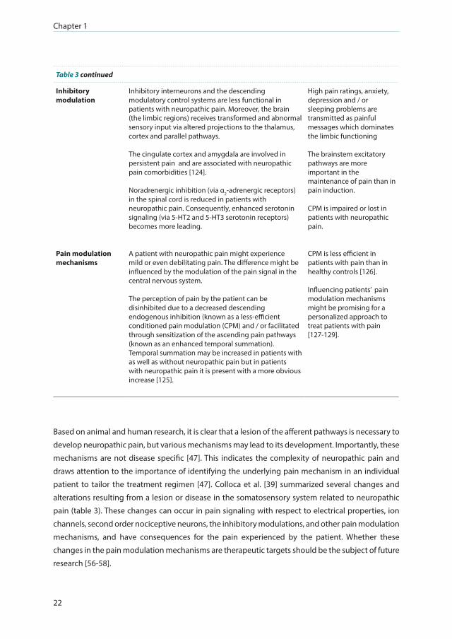
Table 3: Changes in the nervous system due to or caused by neuropathic pain and consequences for the patient. Adapted from Colloca et al [39].
Changes in Description Consequences
Pain signaling In patients with neuropathic pain, the changes in the (electrical) properties of the sensory nerves might result in an imbalance between the central excitatory and inhibitory signaling. This leads to an impairment of the inhibitory interneurons and the descending control systems.
In the spinal cord, at the level of the dorsal horn neurons, there is a change in the transmission of sensory signals and disinhibition or facilitation mechanisms.
An increase in excitation and facilitation and a decrease in inhibition is existing in the peripheral nervous system, the spinal cord and the brain.
Change to a state ofhyperexcitability
Ongoing changes in the sensory pathway might contribute to the fact that neuropathic pain becomes chronic neuropathic pain
Ion channels Neuropathy causes changes in the ion channels in the affected nerves which influences the sensory signaling at the spinal level and in the brain.
Experiences by the patient of ongoing pain; numbness and/or evoked pains
Second order nociceptive neurons
An increased excitability of spinal neurons leads to an enhanced response to several sensory modalities. It allows low-threshold mechanosensitive (Aβ & Aδ) afferent nerve fibers to activate the second order nociceptive neurons. These are transmitting sensory information to the brain, and increases the receptive fields of the neurons in a way that a given stimulus is excitating more secondary order nociceptive neurons [20, 121].
Hyperexcitability can be caused by a loss of γ-aminobutyric acid (GABA)- releasing inhibitory interneurons. These inhibitory neurons can switch to utilize excitatory actions in the spinal cord [122]. Moreover, functional changes in non-neuronal cells in the spinal cord (by example microglia and astrocytes) might play a role in the development of hypersensitivity [123].
Generates central sensitization
The changes in second order neurons might explain the existence of allodynia
Development of hypersensitivity
Chapter 1
22
Table 3 continued
Inhibitory modulation
Inhibitory interneurons and the descending modulatory control systems are less functional in patients with neuropathic pain. Moreover, the brain (the limbic regions) receives transformed and abnormal sensory input via altered projections to the thalamus, cortex and parallel pathways.
The cingulate cortex and amygdala are involved in persistent pain and are associated with neuropathic pain comorbidities [124].
Noradrenergic inhibition (via α2-adrenergic receptors) in the spinal cord is reduced in patients with neuropathic pain. Consequently, enhanced serotonin signaling (via 5-HT2 and 5-HT3 serotonin receptors) becomes more leading.
High pain ratings, anxiety, depression and / or sleeping problems are transmitted as painful messages which dominates the limbic functioning
The brainstem excitatory pathways are more important in the maintenance of pain than in pain induction.
CPM is impaired or lost in patients with neuropathic pain.
Pain modulation mechanisms
A patient with neuropathic pain might experience mild or even debilitating pain. The difference might be influenced by the modulation of the pain signal in the central nervous system.
The perception of pain by the patient can be disinhibited due to a decreased descending endogenous inhibition (known as a less-efficient conditioned pain modulation (CPM) and / or facilitated through sensitization of the ascending pain pathways (known as an enhanced temporal summation). Temporal summation may be increased in patients with as well as without neuropathic pain but in patients with neuropathic pain it is present with a more obvious increase [125].
CPM is less efficient in patients with pain than in healthy controls [126].
Influencing patients’ pain modulation mechanisms might be promising for a personalized approach to treat patients with pain [127-129].
Based on animal and human research, it is clear that a lesion of the afferent pathways is necessary to
develop neuropathic pain, but various mechanisms may lead to its development. Importantly, these
mechanisms are not disease specific [47]. This indicates the complexity of neuropathic pain and
draws attention to the importance of identifying the underlying pain mechanism in an individual
patient to tailor the treatment regimen [47]. Colloca et al. [39] summarized several changes and
alterations resulting from a lesion or disease in the somatosensory system related to neuropathic
pain (table 3). These changes can occur in pain signaling with respect to electrical properties, ion
channels, second order nociceptive neurons, the inhibitory modulations, and other pain modulation
mechanisms, and have consequences for the pain experienced by the patient. Whether these
changes in the pain modulation mechanisms are therapeutic targets should be the subject of future
research [56-58].
General Introduction
23
1
EPIDEMIOLOGY, BURDEN, COSTS AND CONSEQUENCES OF CHRONIC PAIN, AND OF NEUROPATHIC PAIN IN PARTICULAR
Pain is a major clinical, social and economic problem. It has challenged generations of, amongst
others, (para-)medical professionals, psychologists and researchers. However, for many patients,
pain remains a threat to the quality of their daily lives.
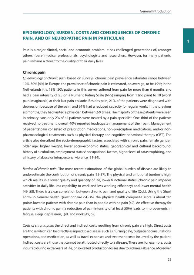
Chronic painEpidemiology of chronic pain: based on surveys, chronic pain prevalence estimates range between
10%-30% [49]. In Europe, the prevalence of chronic pain is estimated, on average, to be 19%; in the
Netherlands it is 18% [50]: patients in this survey suffered from pain for more than 6 months and
had a pain intensity of ≥5 on a Numeric Rating Scale (NRS) ranging from 1 (no pain) to 10 (worst
pain imaginable) at their last pain episode. Besides pain, 21% of the patients were diagnosed with
depression because of the pain, and 61% had a reduced capacity for regular work. In the previous
six months, they had visited a physician between 2-9 times. The majority of these patients were seen
in primary care, only 2% of all patients were treated by a pain specialist. One-third of the patients
received no treatment, overall 40% reported inadequate management of their pain. Management
of patients’ pain consisted of prescription medications, non-prescription medications, and/or non-
pharmacological treatments such as physical therapy and cognitive behavioral therapy (CBT). The
article also described the socio-demographic factors associated with chronic pain: female gender;
older age; higher weight, lower socio-economic status; geographical and cultural background;
history of alcoholism, employment status/ occupational factors, higher level of catastrophizing, and
a history of abuse or interpersonal violence [51-54].
Burden of chronic pain: The most recent estimations of the global burden of disease are likely to
underestimate the contribution of chronic pain [55-57]. The physical and emotional burden is high,
which results in a lower quality and quantity of life, lower functional status (chronic pain impedes
activities in daily life, less capability to work and less working efficiency) and lower mental health
[49, 58]. There is a clear correlation between chronic pain and quality of life (QoL). Using the Short
Form-36 General health Questionnaire (SF-36), the physical health composite score is about ten
points lower in patients with chronic pain than in people with no pain [49]. An effective therapy for
patients with chronic pain (a reduction of pain intensity of at least 50%) leads to improvements in
fatigue, sleep, depression, QoL and work [49, 59].
Costs of chronic pain: the direct and indirect costs resulting from chronic pain are high. Direct costs
are those which can be directly assigned to a disease, such as nursing days, outpatient consultations,
operations, and medication, as well as travel expenses and treatment costs incurred by the patient.
Indirect costs are those that cannot be attributed directly to a disease. These are, for example, costs
incurred during extra years of life, or so-called production losses due to sickness absence. Moreover,

Chapter 1
24
the costs and effects of informal care are also increasingly reflected in these indirect costs. In the
USA, [60] chronic pain impacts 100 million adults and the annual costs are estimated at $560 to $635
billion; this is much higher than the economic costs of the six most expensive major diagnoses in
the USA: cardiovascular diseases ($309 billion); neoplasms ($243 billion); injury and poisoning ($205
billion); endocrine, nutritional and metabolic diseases ($127 billion), digestive system diseases
($112 billion), and respiratory system diseases ($112 billion). The total costs due to chronic pain in
the Netherlands are estimated at over €20 billion, annually [61].
Neuropathic painEpidemiology of neuropathic pain: the incidence of neuropathic pain in the Dutch general population
[62] is 8.2 cases per 1000 person-years. Neuropathic pain is 63% more common in women than in
men and has the highest prevalence in those aged between 70 and 79 [62]. In a systematic review
by Van Hecke et al., [63] the population prevalence of pain with neuropathic characteristics was
estimated to be between 6.9% and 10%. Moore et al stated that 7% of the patients with chronic
pain suffered from pain due to an NePC [49]. Recently, the prevalence of probable neuropathic pain
in the USA was estimated to be 10% [64]. In patients with cancer, the prevalence of pain with a
neuropathic mechanism was estimated to be 18.7% -21.4% [65]. Due to aging, higher prevalence
of diabetes mellitus, surgery, and the increasing incidence of cancer (with and without treatment
with surgery and/or chemotherapy), peripheral neuropathic pain will probably be more common in
the future because these diseases and their treatments can affect the sensory nervous system [39].
Burden of neuropathic pain: neuropathic pain is associated with a poor general health status; this is
comparable to other severe chronic disease. All three dimensions, the physical, psychological, and
social dimension are affected [66]. Patients with neuropathic pain have a lower health-related quality
of life compared to the general population [67]. A survey using the SF-36 reported that Health-
related QoL was as severely affected in patients with neuropathic pain as in patients affected with
a coronary artery disease, clinical depression, recent myocardial infarct or inadequately controlled
diabetes mellitus [68]. The physical component score of the SF-12 can be qualified as severe
impairment: 94% of the included patients with neuropathic pain combined with breakthrough pain
scored below the population mean score [69]. As suggested by Attal et al. [70] the specific signs and
symptoms of neuropathic pain and the painful and/or unpleasant nature of these symptoms also
have an impact on Health-related QoL.
Costs of neuropathic pain: neuropathic pain results in a substantial use of health resources, in
particular by patients who have been referred to specialized pain clinics for pain control via
primary care or other specialists [71]. The additional health care costs incurred in patients whose
pain is mainly treated in pain clinics are compensated by lower costs of other pain management
components, resulting in comparable average monthly total costs [71]. In a recent European study,
Liedgens et al. concluded that there is an economic and socioeconomic burden due to neuropathic
General Introduction
25
1
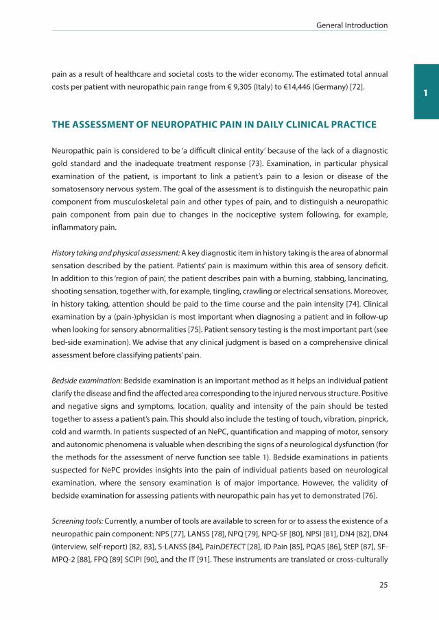
pain as a result of healthcare and societal costs to the wider economy. The estimated total annual
costs per patient with neuropathic pain range from € 9,305 (Italy) to €14,446 (Germany) [72].
THE ASSESSMENT OF NEUROPATHIC PAIN IN DAILY CLINICAL PRACTICE
Neuropathic pain is considered to be ‘a difficult clinical entity’ because of the lack of a diagnostic
gold standard and the inadequate treatment response [73]. Examination, in particular physical
examination of the patient, is important to link a patient’s pain to a lesion or disease of the
somatosensory nervous system. The goal of the assessment is to distinguish the neuropathic pain
component from musculoskeletal pain and other types of pain, and to distinguish a neuropathic
pain component from pain due to changes in the nociceptive system following, for example,
inflammatory pain.
History taking and physical assessment: A key diagnostic item in history taking is the area of abnormal
sensation described by the patient. Patients’ pain is maximum within this area of sensory deficit.
In addition to this ‘region of pain’, the patient describes pain with a burning, stabbing, lancinating,
shooting sensation, together with, for example, tingling, crawling or electrical sensations. Moreover,
in history taking, attention should be paid to the time course and the pain intensity [74]. Clinical
examination by a (pain-)physician is most important when diagnosing a patient and in follow-up
when looking for sensory abnormalities [75]. Patient sensory testing is the most important part (see
bed-side examination). We advise that any clinical judgment is based on a comprehensive clinical
assessment before classifying patients’ pain.
Bedside examination: Bedside examination is an important method as it helps an individual patient
clarify the disease and find the affected area corresponding to the injured nervous structure. Positive
and negative signs and symptoms, location, quality and intensity of the pain should be tested
together to assess a patient’s pain. This should also include the testing of touch, vibration, pinprick,
cold and warmth. In patients suspected of an NePC, quantification and mapping of motor, sensory
and autonomic phenomena is valuable when describing the signs of a neurological dysfunction (for
the methods for the assessment of nerve function see table 1). Bedside examinations in patients
suspected for NePC provides insights into the pain of individual patients based on neurological
examination, where the sensory examination is of major importance. However, the validity of
bedside examination for assessing patients with neuropathic pain has yet to demonstrated [76].
Screening tools: Currently, a number of tools are available to screen for or to assess the existence of a
neuropathic pain component: NPS [77], LANSS [78], NPQ [79], NPQ-SF [80], NPSI [81], DN4 [82], DN4
(interview, self-report) [82, 83], S-LANSS [84], PainDETECT [28], ID Pain [85], PQAS [86], StEP [87], SF-
MPQ-2 [88], FPQ [89] SCIPI [90], and the IT [91]. These instruments are translated or cross-culturally
Chapter 1
26
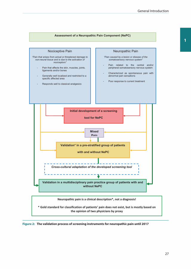
adapted to different languages and are validated in different patient populations (partly) following
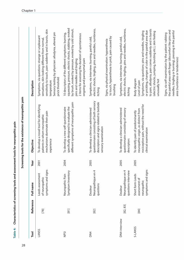
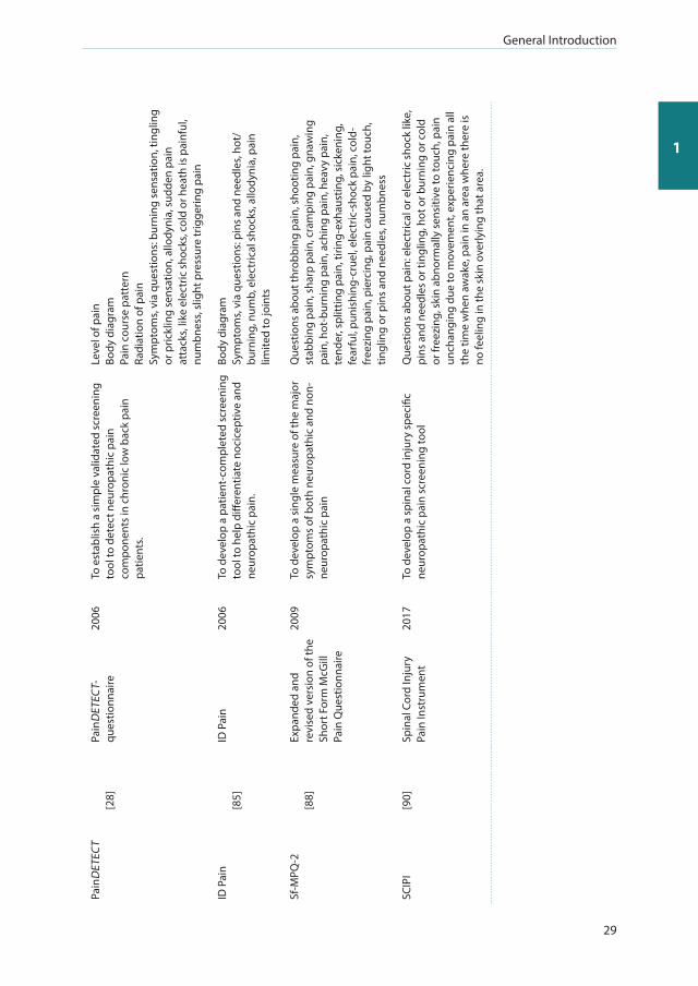
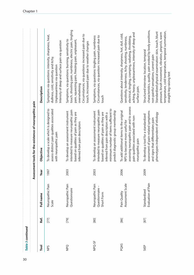
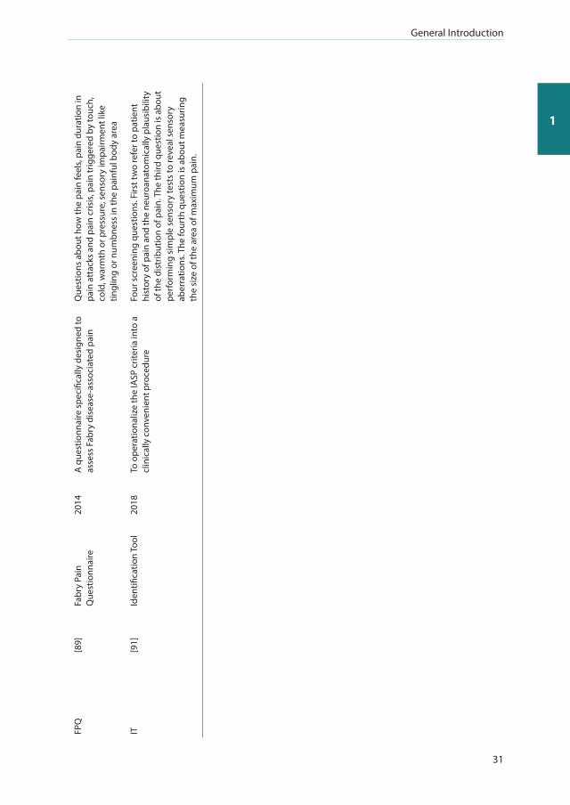
the flow diagram in figure 2. For an overview of the objective and description of each instrument, see
table 4. In a recently published systematic review regarding the measurement properties of these
questionnaires, it was concluded that the Neuropathique Pain Questionnaire (NPQ) [79] and the DN4
[82] were the most suitable for use in daily clinical practice [92]. Screening tools are considered to
be useful in identifying patients with a possible neuropathic pain component, especially when used
by a non-specialist, and to provide added-value for further diagnostic assessment of the patient [74,
75]. This is their most important advantage; however, these screening instruments should never
replace a thorough clinical assessment by a (pain-) physician.
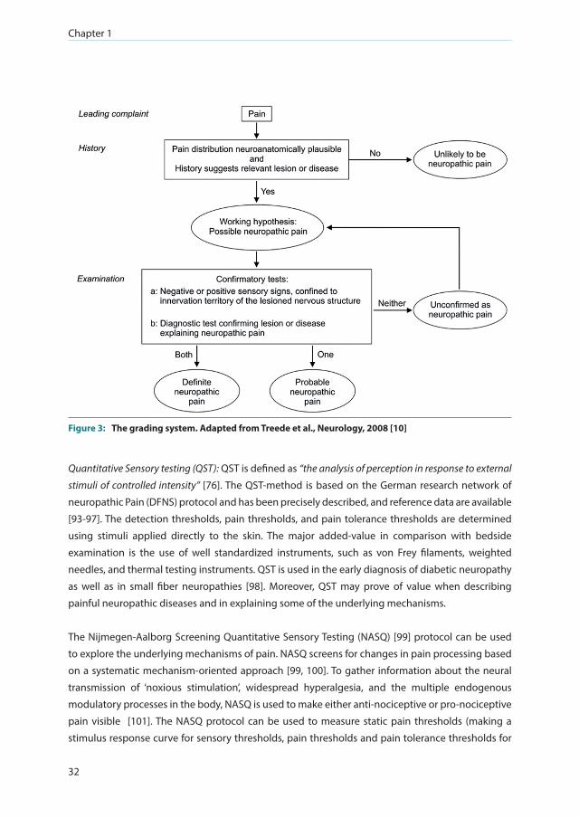
NeuPSIG Grading system: In 2008, Treede et al. [10] presented a grading system for neuropathic
pain suitable for both clinical and research purposes. This stepwise approach provides a working
hypothesis for the origin of patient pain based on four evaluation criteria: 1) pain with a distinct
neuroanatomically plausible distribution; 2) a history suggestive of a relevant lesion or disease
affecting the peripheral or central somatosensory system; 3) demonstration of the distinct
neuroanatomically plausible distribution by at least one confirmatory test; 4) demonstration of
the relevant lesion or disease by at least one confirmatory test. A working hypothesis of ‘possible
neuropathic pain is provided when both criteria 1 & 2 are answered with ‘yes’: when one of criteria 3
& 4 is fulfilled, then the outcome is ‘probable neuropathic pain’. When both criteria 3 & 4 are fulfilled,
the outcome is ‘definite neuropathic pain’ (see figure 3).
General Introduction
27
1
Neuropathic pain is a clinical description*, not a diagnosis!
* Gold standard for classification of patients’ pain does not exist, but is mostly based on the opinion of two physicians by proxy
Cross-cultural adaptation of the developed screening tool
Assessment of a Neuropathic Pain Component (NePC)
Nociceptive Pain
“Pain that arises from actual or threatened damage to non-neural tissue and is due to the activation of
nociceptors”
- Pain that affects the skin, muscles, joints, ligaments and/or bones
- Generally well localized and restricted to a specific affected area
- Responds well to classical analgesics
Neuropathic Pain
“Pain caused by a lesion or disease of the somatosensory nervous system”
- Pain related to the central and/or
peripheral somatosensory nervous system
- Characterized as spontaneous pain with abnormal pain sensations
- Poor response to current treatment
Initial development of a screening
tool for NePC
Validation in a multidisciplinary pain practice group of patients with and without NePC
Validation* in a pre-stratified group of patients
with and without NePC
Mixed Pain
Figure 2: The validation process of screening instruments for neuropathic pain until 2017
Chapter 1
28
Tabl
e 4:
Ch
arac
teri
stic
s of
scr
eeni
ng to
ols
and
asse
ssm
ent t
ools
for n
euro
path
ic p
ain
Scre
enin
g to
ols
for t
he e
xist
ence
of n
euro
path
ic p
ain
Tool
Refe
renc
eFu
ll na
me
Year
Obj
ecti
veD
escr
ipti
on
LAN
SS[7
8]
Leed
s as
sess
men
t of
neu
ropa
thic
sy
mpt
oms
and
sign
s
2001
To d
evel
op a
nov
el to
ol fo
r ide
ntify
ing
patie
nts
in w
hom
neu
ropa
thic
m
echa
nism
s do
min
ate
thei
r pai
n ex
perie
nce
Sym
ptom
s, vi
a qu
estio
ns: s
tran
ge o
r unp
leas
ant
sens
atio
ns, s
kin
look
ing
diffe
rent
from
nor
mal
, se
nsiti
vity
to to
uch,
pai
n su
dden
ly a
nd in
bur
sts,
skin
te
mpe
ratu
reSe
nsor
y te
stin
g by
phy
sici
an: a
llody
nia,
alte
red
pin
pric
k th
resh
old
NPS
I[8
1]N
euro
path
ic P
ain
Sym
ptom
Inve
ntor
y20
04To
dev
elop
a n
ew s
elf-
ques
tionn
aire
sp
ecifi
cally
des
igne
d to
eva
luat
e th
e di
ffere
nt s
ympt
oms
of n
euro
path
ic p
ain
10 d
escr
ipto
rs o
f the
diff
eren
t sym
ptom
s: b
urni
ng,
sque
ezin
g, p
ress
ure,
ele
ctric
sho
cks,
stab
bing
, evo
ked
by b
rush
, evo
ked
by p
ress
ure,
evo
ked
by c
old
stim
uli,
pins
and
nee
dles
and
ting
ling
2 ite
ms
for a
sses
sing
the
dura
tion
of s
pont
aneo
us
ongo
ing
and
paro
xysm
al p
ain
DN
4[8
2]D
oule
ur
Neu
ropa
thiq
ue e
n 4
ques
tions
2005
To d
evel
op a
clin
icia
n-ad
min
iste
red
ques
tionn
aire
con
sist
ing
of b
oth
sens
ory
desc
ripto
rs a
nd s
igns
rela
ted
to b
edsi
de
sens
ory
exam
inat
ion
Sym
ptom
s, vi
a in
terv
iew
: bur
ning
, pai
nful
col
d,
elec
tric
sho
cks,
tingl
ing,
pin
s an
d ne
edle
s, nu
mbn
ess,
itchi
ng
Sign
s, vi
a ph
ysic
al e
xam
inat
ion:
hyp
oest
hesi
a to
touc
h, h
ypoe
sthe
sia
to p
rick,
pai
n ca
used
by
brus
hing
DN
4-in
terv
iew
[82,
83]
Dou
leur
N
euro
path
ique
en
4 qu
estio
ns-in
terv
iew
2005
To d
evel
op a
clin
icia
n-ad
min
iste
red
ques
tionn
aire
con
sist
ing
of s
enso
ry
desc
ripto
rs
Sym
ptom
s, vi
a in
terv
iew
: bur
ning
, pai
nful
col
d,
elec
tric
sho
cks,
tingl
ing,
pin
s an
d ne
edle
s, nu
mbn
ess,
itchi
ng
S-LA
NSS
[84]
Shor
t for
m-L
eeds
as
sess
men
t of
neur
opat
hic
sym
ptom
s an
d si
gns
2005
To id
entif
y pa
in o
f pre
dom
inan
tly
neur
opat
hic
orig
in, a
s di
stin
ct fr
om
noci
cept
ive
pain
, with
out t
he n
eed
for
clin
ical
exa
min
atio
n
Body
dia
gram
Leve
l of p
ain
Sym
ptom
s, vi
a qu
estio
ns: p
ins
and
need
les,
tingl
ing
or p
rickl
ing,
cha
nges
of c
olor
in th
e pa
infu
l are
a du
e to
pai
n, a
llody
nia,
pai
n co
mes
sud
denl
y an
d in
bur
sts
(ele
ctric
sho
cks,
jum
ping
, bur
stin
g et
c.),
skin
feel
ing
unus
ually
hot
Sign
s, vi
a se
lf-ex
amin
atio
n by
the
patie
nt: r
ubbi
ng
the
pain
ful a
rea
with
fing
er (d
isco
mfo
rt li
ke p
ins
and
need
les,
tingl
ing
or b
urni
ng),
pres
sing
in th
e pa
infu
l ar
ea (n
umbn
ess
or te
nder
ness
)
General Introduction
29
1
Pain
DET
ECT
[28]
Pain
DET
ECT-
ques
tionn
aire
2006
To e
stab
lish
a si
mpl
e va
lidat
ed s
cree
ning
to
ol to
det
ect n
euro
path
ic p
ain
com
pone
nts
in c
hron
ic lo
w b
ack
pain
pa
tient
s.
Leve
l of p
ain
Body
dia
gram
Pain
cou
rse
patt
ern
Radi
atio
n of
pai
nSy
mpt
oms,
via
ques
tions
: bur
ning
sen
satio
n, ti
nglin
g or
pric
klin
g se
nsat
ion,
allo
dyni
a, s
udde
n pa
in
atta
cks,
like
elec
tric
sho
cks,
cold
or h
eath
is p
ainf
ul,
num
bnes
s, sl
ight
pre
ssur
e tr
igge
ring
pain
ID P
ain
[85]
ID P
ain
2006
To d
evel
op a
pat
ient
-com
plet
ed s
cree
ning
to
ol to
hel
p di
ffere
ntia
te n
ocic
eptiv
e an
d ne
urop
athi
c pa
in.
Body
dia
gram
Sym
ptom
s, vi
a qu
estio
ns: p
ins
and
need
les,
hot/
burn
ing,
num
b, e
lect
rical
sho
cks,
allo
dyni
a, p
ain
limite
d to
join
ts
Sf-M
PQ-2
[88]
Expa
nded
and
re
vise
d ve
rsio
n of
the
Shor
t For
m M
cGill
Pa
in Q
uest
ionn
aire
2009
To d
evel
op a
sin
gle
mea
sure
of t
he m
ajor
sy
mpt
oms
of b
oth
neur
opat
hic
and
non-
neur
opat
hic
pain
Que
stio
ns a
bout
thro
bbin
g pa
in, s
hoot
ing
pain
, st
abbi
ng p
ain,
sha
rp p
ain,
cra
mpi
ng p
ain,
gna
win
g pa
in, h
ot-b
urni
ng p
ain,
ach
ing
pain
, hea
vy p
ain,
te
nder
, spl
ittin
g pa
in, t
iring
-exh
aust
ing,
sic
keni
ng,
fear
ful,
puni
shin
g-cr
uel,
elec
tric
-sho
ck p
ain,
col
d-fr
eezi
ng p
ain,
pie
rcin
g, p
ain
caus
ed b
y lig
ht to
uch,
tin
glin
g or
pin
s an
d ne
edle
s, nu
mbn
ess
SCIP
I[9
0]Sp
inal
Cor
d In
jury
Pa
in In
stru
men
t20
17To
dev
elop
a s
pina
l cor
d in
jury
spe
cific
ne
urop
athi
c pa
in s
cree
ning
tool
Que
stio
ns a
bout
pai
n: e
lect
rical
or e
lect
ric s
hock
like
, pi
ns a
nd n
eedl
es o
r tin
glin
g, h
ot o
r bur
ning
or c
old
or fr
eezi
ng, s
kin
abno
rmal
ly s
ensi
tive
to to
uch,
pai
n un
chan
ging
due
to m
ovem
ent,
expe
rienc
ing
pain
all
the
time
whe
n aw
ake,
pai
n in
an
area
whe
re th
ere
is
no fe
elin
g in
the
skin
ove
rlyin
g th
at a
rea.
Chapter 1
30
Tabl
e 3
cont
inue
d
Ass
essm
ent t
ools
for t
he e
xist
ence
of n
euro
path
ic p
ain
Tool
Ref.
Full
nam
eYe
arO
bjec
tive
Des
crip
tion
NPS
[77]
Neu
ropa
thic
Pai
n Sc
ale
1997
To d
evel
op a
sca
le w
hich
is d
esig
ned
to
asse
ss d
istin
ct p
ain
qual
ities
ass
ocia
ted
with
neu
ropa
thic
pai
n
Sym
ptom
s vi
a qu
estio
ns: i
nten
sity
, sha
rpne
ss, h
eat,
dulln
ess,
cold
, sen
sitiv
ity a
nd it
chy.
Unp
leas
antn
ess
via
ques
tion
Inte
nsity
of d
eep
and
surf
ace
pain
via
que
stio
n
NPQ
[79]
Neu
ropa
thic
Pai
n Q
uest
ionn
aire
2003
To d
evel
op a
n as
sess
men
t ins
trum
ent
inte
nded
to m
easu
re n
euro
path
ic p
ain
base
d on
qua
litie
s of
pai
n as
they
are
in
ferr
ed fr
om p
ain
desc
ripto
rs
Sym
ptom
s, vi
a qu
estio
ns: b
urni
ng, s
ensi
tivity
to
touc
h, s
hoot
ing
pain
, num
bnes
s, el
ectr
ic p
ain,
ting
ling
pain
, squ
eezi
ng p
ain,
free
zing
pai
n, u
nple
asan
t, ov
erw
helm
ing
Circ
umst
ance
s, vi
a qu
estio
ns: i
ncre
ased
pai
n du
e to
to
uch,
incr
ease
d pa
in d
ue to
wea
ther
cha
nges
NPQ
-SF
[80]
Neu
ropa
thic
Pai
n Q
uest
ionn
aire
–
Shor
t For
m
2003
To d
evel
op a
n as
sess
men
t ins
trum
ent
inte
nded
to m
easu
re n
euro
path
ic p
ain
base
d on
qua
litie
s of
pai
n as
they
are
in
ferr
ed fr
om p
ain
desc
ripto
rs w
ith a
m
inim
um n
umbe
r of i
tem
s su
ffici
ent t
o pr
edic
t dia
gnos
tic g
roup
mem
bers
hip
Sym
ptom
s, vi
a qu
estio
ns: t
ingl
ing
pain
, num
bnes
sCi
rcum
stan
ces,
via
ques
tion:
incr
ease
d pa
in d
ue to
to
uch
PQA
S[8
6]Pa
in Q
ualit
y A
sses
smen
t Sca
le20
06To
add
add
ition
al it
ems
to th
e or
igin
al
NPS
to b
ecom
e m
ore
usef
ul fo
r as
sess
ing
neur
opat
hic
pain
and
to a
sses
s pa
in q
ualit
ies
asso
ciat
ed w
ith n
on-
neur
opat
hic
pain
Que
stio
ns a
bout
inte
nsity
, sha
rpne
ss, h
ot, d
ull,
cold
, se
nsiti
vity
, ten
dern
ess,
itchy
, sho
otin
g, n
umbn
ess,
elec
tric
al, t
ingl
ing,
cra
mpi
ng, r
adia
ting,
thro
bbin
g,
achi
ng, h
eavy
, unp
leas
antn
ess,
inte
nsity
of d
eep
and
surf
ace
pain
.
StEP
[87]
Stan
dard
ized
Ev
alua
tion
of P
ain
2009
To d
evel
op a
tool
for a
sta
ndar
dize
d as
sess
men
t of p
ain-
rela
ted
sym
ptom
s an
d si
gns
that
diff
eren
tiate
s pa
in
phen
otyp
es in
depe
nden
t of e
tiolo
gy
Stru
ctur
ed in
terv
iew
: loc
atio
n, te
mpo
ral
char
acte
ristic
s, qu
ality
, pai
n ev
oked
by
body
pos
ition
s, no
n-pa
infu
l sen
satio
ns, c
urre
nt p
ain
Stan
dard
ized
phy
sica
l exa
min
atio
n: s
kin,
touc
h, b
lunt
pr
essu
re, b
rush
mov
emen
t, vi
brat
ion,
pin
pric
k, w
arm
te
mpe
ratu
re, c
old
tem
pera
ture
, tem
pora
l sum
mat
ion,
st
raig
ht-le
g-ra
isin
g te
st
General Introduction
31
1
FPQ
[89]
Fabr
y Pa
in
Que
stio
nnai
re20
14A
que
stio
nnai
re s
peci
fical
ly d
esig
ned
to
asse
ss F
abry
dis
ease
-ass
ocia
ted
pain
Que
stio
ns a
bout
how
the
pain
feel
s, pa
in d
urat
ion
in
pain
att
acks
and
pai
n cr
isis
, pai
n tr
igge
red
by to
uch,
co
ld, w
arm
th o
r pre
ssur
e, s
enso
ry im
pairm
ent l
ike
tingl
ing
or n
umbn
ess
in th
e pa
infu
l bod
y ar
ea
IT[9
1]Id
entifi
catio
n To
ol20
18To
ope
ratio
naliz
e th
e IA
SP c
riter
ia in
to a
cl
inic
ally
con
veni
ent p
roce
dure
Four
scr
eeni
ng q
uest
ions
. Firs
t tw
o re
fer t
o pa
tient
hi
stor
y of
pai
n an
d th
e ne
uroa
nato
mic
ally
pla
usib
ility
of
the
dist
ribut
ion
of p
ain.
The
third
que
stio
n is
abo
ut
perf
orm
ing
sim
ple
sens
ory
test
s to
reve
al s
enso
ry
aber
ratio
ns. T
he fo
urth
que
stio
n is
abo
ut m
easu
ring
the
size
of t
he a
rea
of m
axim
um p
ain.
Chapter 1
32
Figure 3: The grading system. Adapted from Treede et al., Neurology, 2008 [10]
Quantitative Sensory testing (QST): QST is defined as “the analysis of perception in response to external
stimuli of controlled intensity” [76]. The QST-method is based on the German research network of
neuropathic Pain (DFNS) protocol and has been precisely described, and reference data are available
[93-97]. The detection thresholds, pain thresholds, and pain tolerance thresholds are determined
using stimuli applied directly to the skin. The major added-value in comparison with bedside
examination is the use of well standardized instruments, such as von Frey filaments, weighted
needles, and thermal testing instruments. QST is used in the early diagnosis of diabetic neuropathy
as well as in small fiber neuropathies [98]. Moreover, QST may prove of value when describing
painful neuropathic diseases and in explaining some of the underlying mechanisms.
The Nijmegen-Aalborg Screening Quantitative Sensory Testing (NASQ) [99] protocol can be used
to explore the underlying mechanisms of pain. NASQ screens for changes in pain processing based
on a systematic mechanism-oriented approach [99, 100]. To gather information about the neural
transmission of ‘noxious stimulation’, widespread hyperalgesia, and the multiple endogenous
modulatory processes in the body, NASQ is used to make either anti-nociceptive or pro-nociceptive
pain visible [101]. The NASQ protocol can be used to measure static pain thresholds (making a
stimulus response curve for sensory thresholds, pain thresholds and pain tolerance thresholds for
General Introduction
33
1
pressure algometry as well as electrical stimuli). Dynamic pain tests such as the Conditioned Pain
Modulation (CPM) paradigm [102-104], also known as the “pain inhibits pain” phenomenon, measure
an inhibitory mechanism like diffuse noxious inhibitory controls. This indicates that peripheral and
central mechanisms play a role in the way the body handles nociception.
Currently, there is cumulative evidence that pain and sensitization play an essential role in the
development of chronic pain [105]. An understanding of the pathophysiology of acute pain and
of the development of chronic pain are essential to improving patient outcomes and in making a
mechanism-based treatment. Both QST and NASQ are difficult to implement in daily clinical practice
as they are time consuming and require expensive instrumentation. However, test-retest reliability
and the interrater reliability are both classified as good if tests are performed by trained examiners
[96].
Neurophysiological techniques: Following the definition of neuropathic pain and to fulfill the NeuPSIG
grading criteria, confirmation of a lesion or disease affecting the central or peripheral nervous system
is a prerequisite for the outcome ‘definite neuropathic pain’. Several techniques are described in the
literature, such as nerve conduction studies via electromyography (EMG) testing large-fiber affection
in, for example, patients with HIV. Skin wrinkle tests and quantitative sudomotor axon reflex testing
(QSART) are used for testing small fibers, and somatosensory evoked potential testing (SSEP) is used
to detect sensory abnormalities in, for example, the trunk or proximal limb regions. Nerve ultrasound
has proved to provide reliable information for by example nerve entrapments during the diagnostic
work-up of neuropathic pain. Positron emission topography (PET) is used to target specific ligands
and to access detailed information about the neurotransmitters. Functional magnetic resonance
imaging (fMRI) makes it possible to gather information about blood deoxygeneration and changes
in metabolites via spectroscopy. Electrophysiological methods, such as the nociceptive withdrawal
reflex, and electroencephalography (EEG) including (laser)-evoked potentials and resting-state EEG
provide complementary information and reflect real-time activity in the neural system [29, 75, 106-
109].
REQUIREMENTS FOR A SCREENING TOOL TO ASSESS (NEUROPATHIC) PAIN
Screening tools for the assessment of NePC such as the PainDETECT and the DN4 are biopsychological
measurements. These instruments screen for the presence of NePC via a set of items related to
various pain descriptors. The individual items and the outcomes of the questionnaires reflect the
patient’s perception of the pain. Instruments like the PainDETECT and the DN4 are in current use
in daily clinical practice, research and education. Their popularity in daily clinical practice and in
research is partly due to their simplicity and ease-of-use when identifying potential patients with
Chapter 1
34
NePC and their immediate provision of information, in particular by non-specialists [47, 110]. BSE
and NASQ are other biopsychological measures that examine the negative (loss of function) and
positive (augmented excitation, for example hyperalgesia and allodynia) signs, and to gain insights
into the underlying lesion or disease [29]. These observations rely, at least partly, on the patients’
evaluation of pain and on the physician’s experience with performing the tests [111].
It is hard to understand the manifestation, the time course and the impact of patients’ pain and
therefore difficult to find the right solution or management for patients’ pain when symptoms
of pain are not systematically documented. An effective diagnosis, prognosis and treatment of
patient’s pain must be based on the underlying (pain) mechanisms. To achieve this, a number of
valid, and reliable tools have been developed to assess chronic pain. The measurement of pain and
the underlying pain systems is important to understand its origin, intensity, quality and the progress
suffered by the patient during the treatment process, but it has to be accepted that the symptoms
as provided by the patient and arising from the clinical examination by the physician only gives a
few insights into the underlying pain mechanisms and the pain diseases resulting from a changing
somatosensory system [112].
Assessing patients suffering from pain in daily clinical practice serves several goals: screening,
diagnosis, therapy and monitoring. The goal of screening is the initial triage of patients, for example
by referring them for more diagnostic research or by placing them in a specific clinical treatment
trajectory. Individual patients can be classified in specific sub-groups with common underlying pain
mechanisms to undergo, for example, a similar pain treatment. Differential diagnosis, prognosis,
the prediction of the process and/or outcome of the disease; all these form an important part of
the assessment of patients’ pain [112]. The choice of therapy is based on a patient’s diagnosis and
the impact and course of the disease. Moreover, a patient’s diagnosis is also related to the disease
mechanisms [29, 74, 75]. To increase the chance of a positive treatment outcome, it is necessary to be
able to identify responders versus non-responders. However, this is not always possible for patients
with pain: the mechanism(s) that underlie the presentation of pain are not always known, which
therefore reduces the probability of a correct diagnostic profile and consequently an adequate
management of treatment. Finally, the goal of monitoring is to follow the evolution, the treatment
response and duration of the disease in patients [112].
Measuring pain A regular, structured and standardized documentation of the pain suffered by a patient is a
prerequisite for an effective and timely treatment and follow-up. An important difference between
the avaliable pain measurement instruments is whether the measurements are made in terms of
quantity or quality and dimensionality. At a quantitative level, it is necessary to measure how much
pain the patient is suffering from (pain intensity), how long the patient has been suffering from the
pain (time), and where the pain is located. At a qualitative level the patient will be asked how much
General Introduction
35
1
the pain functionally limits the patient (disability), how much it affects the patient’s daily life (quality
of life), and how the patient deals with the pain (coping). Patients’ pain quality can be characterized
at a more qualitative level by describing feelings like the feeling of pins and needles, burning,
stabbing or itching. Dimensionality reflects either uni-dimensionality, for example the amount of
pain, and/or multi-dimensionality, where data comes from multiple perspectives, such as level of
pain, experience of pain and behavior [112].
Requirements to measuring instrumentsThe value of a measurement instrument for pain is determined by its (clinimetrical) quality. This
includes the quality of the measurement instruments as well as the performance of the actual
measurement. Important indicators are the performance of the translation process, the reliability,
validity, responsiveness, and sensitivity for change, as well as quality assurance [112].
Translation and cross-cultural adaptation: translation is the process of translating an instrument from
one language into another. The term ‘cross-cultural adaptation’ is used when both language and
culture are considered in the process of the preparation of an instrument that is to be used in another
language and/or another country to provide equivalency, based on content, between source and
target language [113-115]. After translation or cross-cultural adaptation, the instrument’s face
validity can be assessed, the extent to which a test is subjectively viewed as covering the concept it
purports to measure.
Reliability: the reliability of an instrument expresses the measure in which the instrument shows the
same result if used again on the same person (test-retest reliability or inter-assessor reliability). The
reliability can also be expressed as intra-assessor reliability: will two different assessors reach the
same conclusion? The inter- and intra-assessor reliability are both only valid if no changes in the
disease, conditions or the circumstances have occurred between the assessments [116].
Validity: the validity of an instrument is the way in which an instrument measures what it intends
to measure. This is determined on the basis of a ‘gold standard’; an instrument or method for which
it has been proven that it documents the presence or absence or the stage of the same condition,
and for which people know beforehand that it is ‘true’, or that it is, at least, the best available test
[117]. An instrument can be reliable without being valid, but a valid instrument must be reliable
[116]. Important features for screening tools that assess NePC are the construct validity, content
validity, criterion validity, and external validity. The theoretical embedding of the neuropathic pain
concept is captured in the construct validity: how well does the test measure what it intends to
measure. Construct validity consists of convergent and discriminant validity. Convergent validity
is achieved when different tools that measure the same concept yield the same results (converge).
On the other hand, an instrument must distinguish the concept which it intends to measure from
other concepts (discriminate). Content validity refers to the question whether the content of the
Chapter 1
36
instrument (asked questions, used measurement scales) represents all elements of the construct.
The term criterion validity refers to the extent to which the outcome of the instrument is related to
one or more criterion variables. Criterion validity is accessed via sensitivity, specificity and predictive
value. External validity is important to assess and to see to which extent the outcomes obtained with
the instrument are generalizable to other situations, other groups of patients, or to other concepts.
Diagnostic procedures are used for clinical decisions, and therefore imply a certain risk for a patient
as an incorrect diagnosis might harm the patient. From this perspective, it is important to assess the
validity of a measuring instrument for each condition and per (sub-)population, as the fact that an
instrument is valid for a specific group of patients with a certain diagnosis does not automatically
mean that it is also valid for patients who suffer from another condition [112].
The sensitivity of a measuring instrument indicates which percentage of those suffering from
certain diseases are (accurately) classified as ill by the measuring instrument [118]. The specificity
of the measuring instrument indicates which percentage of a group of people not affected by the
disease are (accurately) classified as not being ill [118]. The predictive value (also known as the
diagnostic value) gives an indication for the chance that the person with the relevant test result will
have the disease or condition now or in the near future [119]. A positive and/or negative predictive
value refers to the chance that a disease or condition is present or absent in people with a certain
test result. If an instrument has a high sensitivity, only a few patients suffering from the disease
or condition are missed, it leads to a higher positive predictive value. If an instrument has a high
specificity, only a few patients suffering from the disease or condition are incorrectly classified as
suffering from this disease or condition, it results in a higher negative predictive value [119]. The
number of people suffering from the disease or condition in the population on whom the measuring
instrument is used at any given moment is called the prevalence. The prevalence influences the
sensitivity, specificity and the predictive value. When the condition frequently occurs within a
population, this will lead to a higher positive predictive value. At a lower prevalence, the number
of false-positive test results will increase on the basis of coincidence [119]. For this, the (positive)
likelihood ratio can be used which gives an indication of the value of an instrument for increasing
certainty about a positive diagnosis [119]. However, as indicated by Altman and Bland [119], a high
positive likelihood ratio might show that an instrument is useful, but that it cannot ensure that a
positive test is a certainty for the presence of a disease [119].
Responsiveness: in (pain) measurement instruments that are used frequently over a longer period
(for example for follow-up research), it is important to know whether the instrument shows any
changes that have taken place in that time [116].
Quality of performance: The measuring instrument must be suitable for the situation for which
it is to be used (practical applicability), for the purpose of the research (e.g. screening for an
epidemiological study or serve as a diagnostic assessment by the physician), the population under
General Introduction
37
1
investigation (number and composition of the group) and for the person performing the assessment
(e.g. experience, time and costs) [116].
Quality control and assurance: the reproducibility of pain measurements is crucial and depends on
the instruction of the patient and a correct measurement by the patient, the physician, the nurse or
the researcher. Staff performing the measurements and /or interpreting the results must be trained
in how to use the instrument(s) to ensure that these are used in a standardized and reproducible
way following the applicable protocols. All staff involved should follow theoretical and/or practical
refresher courses to guarantee continuation of equal measuring quality. Participation in regular
quality circles also contributes to a consistent and reliable pain measurement and interpretation
quality [112].
RESEARCH QUESTIONS IN THIS THESIS
A better characterization of patient pain based on a thorough assessment in daily clinical practice
increases the chance of discovering the underlying (pain) mechanisms. This can lead to a better-
founded pain diagnosis and is a prerequisite for choosing an effective treatment for the individual
patient, if available, as well as for the subpopulation of patients with pain. Screening tools help
physicians to assess pain, but they are also valuable for monitoring the progress of patient treatment
in research projects, as well as for assessing the incidence/prevalence of a disorder like neuropathic
pain. At this moment, the DN4 has been translated into Dutch, whereas the PainDETECT is only
validated in its (original) German version with an English translation. The interobserver reliability
of the assessment of neuropathic pain in patients between two physicians in the Netherlands in
specific patient populations is still unknown.
This is of importance because a ‘true’ gold standard for the NePC assessment does not yet exist.
A valid screening tool would therefore be of value for both family practice and for specialized
(academic) pain centers to help and guide the classification of patient pain. Therefore, the aim
of two of the studies in this thesis is to assess the psychometric properties of the two screening
instruments (PainDETECT and DN4) to assess the neuropathic pain component in a consecutive,
daily practice population of patients with low back and leg pain, neck-shoulder pain, or with pain
due to a suspected peripheral nerve damage. Validation in a more general population and in a
clinical setting is important as the outcome may differ from validation studies set in more controlled,
experimental settings with selected patients. A second aim is to assess the possible benefits of BSE
and NASQ to distinguish between clinically diagnosed patients with and without NePC.
Question 1: Is a cross-cultural adaptation a prerequisite for achieving a valid Dutch translation of a
screening tool for neuropathic pain?
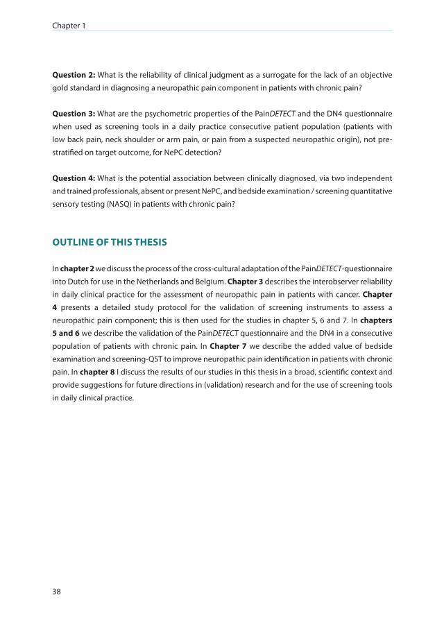
Chapter 1
38
Question 2: What is the reliability of clinical judgment as a surrogate for the lack of an objective
gold standard in diagnosing a neuropathic pain component in patients with chronic pain?
Question 3: What are the psychometric properties of the PainDETECT and the DN4 questionnaire
when used as screening tools in a daily practice consecutive patient population (patients with
low back pain, neck shoulder or arm pain, or pain from a suspected neuropathic origin), not pre-
stratified on target outcome, for NePC detection?
Question 4: What is the potential association between clinically diagnosed, via two independent
and trained professionals, absent or present NePC, and bedside examination / screening quantitative
sensory testing (NASQ) in patients with chronic pain?
OUTLINE OF THIS THESIS
In chapter 2 we discuss the process of the cross-cultural adaptation of the PainDETECT-questionnaire
into Dutch for use in the Netherlands and Belgium. Chapter 3 describes the interobserver reliability
in daily clinical practice for the assessment of neuropathic pain in patients with cancer. Chapter
4 presents a detailed study protocol for the validation of screening instruments to assess a
neuropathic pain component; this is then used for the studies in chapter 5, 6 and 7. In chapters
5 and 6 we describe the validation of the PainDETECT questionnaire and the DN4 in a consecutive
population of patients with chronic pain. In Chapter 7 we describe the added value of bedside
examination and screening-QST to improve neuropathic pain identification in patients with chronic
pain. In chapter 8 I discuss the results of our studies in this thesis in a broad, scientific context and
provide suggestions for future directions in (validation) research and for the use of screening tools
in daily clinical practice.
General Introduction
39
1
REFERENCES
1. Merskey HB, N. Classification of chronic pain: Descriptions of chronic pain syndromes and defination of pain terms.: IASP Press; 1994.
2. McCaffery M. Nursing practice theories related to cognition, bodily pain and man-environmental interactions. Los Angeles, USA: UCLA students Store.; 1968.
3. IASP. International Association for the Study of Pain; Taxonomy Nociception 2018 [cited 2018 july 4, 2018]. Available from: http://www.iasp-pain.org/Education/Content.aspx?ItemNumber=1698#Nociception.
4. Tracey I. Nociceptive processing in the human brain. Curr Opin Neurobiol. 2005;15(4):478-87. doi: 10.1016/j.conb.2005.06.010.
5. IASP. IASP Taxonomy Nociceptive Pain: International Association for the Study of Pain; 2015 [cited 2015 May 19, 2015]. http://www.iasp-pain.org/Taxonomy#Nociceptivepain]. Available from: http://www.iasp-pain.org/Taxonomy#Nociceptivepain.
6. Wisconsin Uo. Inflammatory pain definition 2010. Available from: http://projects.hsl.wisc.edu/GME/PainManagement/session2.4.html.
7. Cervero F, Laird JM. Visceral pain. Lancet. 1999;353(9170):2145-8. doi: 10.1016/S0140-6736(99)01306-9. 8. Steeds CE. The anatomy and physiology of pain. Surgery. 2016;34(2):5.9. IASP. IASP Taxonomy Neuropathic Pain: International Association for the Study of Pain; 2015 [cited 2015
May 19, 2015]. http://www.iasp-pain.org/Taxonomy#Neuropathicpain]. Available from: http://www.iasp-pain.org/Taxonomy#Neuropathicpain.
10. Treede RD, Jensen TS, Campbell JN, Cruccu G, Dostrovsky JO, Griffin JW, et al. Neuropathic pain: redefinition and a grading system for clinical and research purposes. Neurology. 2008;70(18):1630-5.
11. Kosek E, Cohen M, Baron R, Gebhart GF, Mico JA, Rice AS, et al. Do we need a third mechanistic descriptor for chronic pain states? Pain. 2016;157(7):1382-6.
12. Granan LP. We do not need a third mechanistic descriptor for chronic pain states! Not yet. Pain. 2017;158(1):179.
13. Brummett C, Clauw D, Harris R, Harte S, Hassett A, Williams D. We agree with the need for a new term but disagree with the proposed terms. Pain. 2016;157(12):2876.
14. Moloney N, Rabey M, Nijs J, Hush J, Slater H. Support for extended classification of pain states. Pain. 2017;158(7):1395.
15. Aydede M, Shriver A. Recently introduced definition of “nociplastic pain” by the International Association for the Study of Pain needs better formulation. Pain. 2018;159(6):1176-7.
16. Cohen M, Kosek E, Baron R, Mico JA, Rice AS. Reply. Pain. 2016;157(12):2876-7. 17. Kosek E, Cohen M, Baron R, Mico JA, Rice AS. Reply. Pain. 2017;158(1):180. 18. Kosek E, Cohen M, Baron R, Mico JA, Rice ASC. Reply. Pain. 2018;159(6):1177-8. 19. Kosek E, Cohen M, Baron R, Mico JA, Rice ASC. Reply. Pain. 2017;158(7):1396. 20. Woolf CJ. Central sensitization: implications for the diagnosis and treatment of pain. Pain. 2011;152(3
Suppl):S2-15. 21. Costigan M, Scholz J, Woolf CJ. Neuropathic pain: a maladaptive response of the nervous system to damage.
Annu Rev Neurosci. 2009;32:1-32. 22. Woolf CJ, American College of P, American Physiological S. Pain: moving from symptom control toward
mechanism-specific pharmacologic management. Ann Intern Med. 2004;140(6):441-51. 23. IASP. IASP Taxonomy central sensitization: International Association for the Study of Pain
2018 [cited 2018 July 4, 2018]. Available from: http://www.iasp-pain.org/Education/Content.aspx?ItemNumber=1698#Centralsensitization.
24. Nijs J, Apeldoorn A, Hallegraeff H, Clark J, Smeets R, Malfliet A, et al. Low back pain: guidelines for the clinical classification of predominant neuropathic, nociceptive, or central sensitization pain. Pain Physician. 2015;18(3):E333-46.
25. Nijs J, Leysen L, Adriaenssens N, Aguilar Ferrandiz ME, Devoogdt N, Tassenoy A, et al. Pain following cancer treatment: Guidelines for the clinical classification of predominant neuropathic, nociceptive and central sensitization pain. Acta Oncol. 2016;55(6):659-63.
26. Sanzarello I, Merlini L, Rosa MA, Perrone M, Frugiuele J, Borghi R, et al. Central sensitization in chronic low back pain: A narrative review. J Back Musculoskelet Rehabil. 2016;29(4):625-33.
Chapter 1
40
27. Arendt-Nielsen L, Morlion B, Perrot S, Dahan A, Dickenson A, Kress HG, et al. Assessment and manifestation of central sensitisation across different chronic pain conditions. Eur J Pain. 2018;22(2):216-41.
28. Freynhagen R, Baron R, Gockel U, Tolle TR. painDETECT: a new screening questionnaire to identify neuropathic components in patients with back pain. Curr Med Res Opin. 2006;22(10):1911-20.
29. La Cesa S, Tamburin S, Tugnoli V, Sandrini G, Paolucci S, Lacerenza M, et al. How to diagnose neuropathic pain? The contribution from clinical examination, pain questionnaires and diagnostic tests. Neurological sciences : official journal of the Italian Neurological Society and of the Italian Society of Clinical Neurophysiology. 2015;36(12):2169-75.
30. Management. ASoATFoAP. Practice guidelines for acute pain management in the perioperative setting: an updated report by the American Society of Anesthesiologists Task Force on Acute Pain Management. Anesthesiology. 2012;116(2):248-73.
31. Chapman CR, Vierck CJ. The Transition of Acute Postoperative Pain to Chronic Pain: An Integrative Overview of Research on Mechanisms. The journal of pain : official journal of the American Pain Society. 2017;18(4):359 e1- e38.
32. McGreevy K, Bottros MM, Raja SN. Preventing Chronic Pain following Acute Pain: Risk Factors, Preventive Strategies, and their Efficacy. Eur J Pain Suppl. 2011;5(2):365-72.
33. Kehlet H, Jensen TS, Woolf CJ. Persistent postsurgical pain: risk factors and prevention. Lancet. 2006;367(9522):1618-25.
34. Treede RD. Entstehung der Schmerzchronifizierung. Baron R KW, Strumpf M, Willweber-Strumpf A., editor. Heidelberg: Springer; 2011.
35. Treede RD, Rief W, Barke A, Aziz Q, Bennett MI, Benoliel R, et al. A classification of chronic pain for ICD-11. Pain. 2015;156(6):1003-7.
36. Quintner JL, Cohen ML, Buchanan D, Katz JD, Williamson OD. Pain medicine and its models: helping or hindering? Pain Med. 2008;9(7):824-34.
37. Woolf CJ, Salter MW. Neuronal plasticity: increasing the gain in pain. Science. 2000;288(5472):1765-9. 38. Walk D, Sehgal N, Moeller-Bertram T, Edwards RR, Wasan A, Wallace M, et al. Quantitative sensory testing and
mapping: a review of nonautomated quantitative methods for examination of the patient with neuropathic pain. The Clinical journal of pain. 2009;25(7):632-40.
39. Colloca L, Ludman T, Bouhassira D, Baron R, Dickenson AH, Yarnitsky D, et al. Neuropathic pain. Nat Rev Dis Primers. 2017;3:17002.
40. Apkarian AV, Hashmi JA, Baliki MN. Pain and the brain: specificity and plasticity of the brain in clinical chronic pain. Pain. 2011;152(3 Suppl):S49-64.
41. Garland EL. Pain processing in the human nervous system: a selective review of nociceptive and biobehavioral pathways. Prim Care. 2012;39(3):561-71.
42. Price DD. Central neural mechanisms that interrelate sensory and affective dimensions of pain. Molecular interventions. 2002;2(6):10.
43. Cohen SP, Mao J. Neuropathic pain: mechanisms and their clinical implications. BMJ. 2014;348:f7656. 44. Baron R. Mechanisms of disease: neuropathic pain--a clinical perspective. Nat Clin Pract Neurol. 2006;2(2):95-
106. 45. Freeman R. Not all neuropathy in diabetes is of diabetic etiology: differential diagnosis of diabetic
neuropathy. Curr Diab Rep. 2009;9(6):423-31. 46. Borsook D. Neurological diseases and pain. Brain. 2012;135(Pt 2):320-44. 47. Baron R, Binder A, Wasner G. Neuropathic pain: diagnosis, pathophysiological mechanisms, and treatment.
Lancet Neurol. 2010;9(8):807-19. 48. McCarberg B, D’Arcy Y, Parsons B, Sadosky A, Thorpe A, Behar R. Neuropathic pain: a narrative review
of etiology, assessment, diagnosis, and treatment for primary care providers. Curr Med Res Opin. 2017;33(8):1361-9.
49. Andrew R, Derry S, Taylor RS, Straube S, Phillips CJ. The costs and consequences of adequately managed chronic non-cancer pain and chronic neuropathic pain. Pain Pract. 2014;14(1):79-94.
50. Breivik H, Collett B, Ventafridda V, Cohen R, Gallacher D. Survey of chronic pain in Europe: prevalence, impact on daily life, and treatment. European journal of pain. 2006;10(4):287-333.
51. Smith BH, Macfarlane GJ, Torrance N. Epidemiology of chronic pain, from the laboratory to the bus stop: time to add understanding of biological mechanisms to the study of risk factors in population-based research? Pain. 2007;127(1-2):5-10.

General Introduction
41
1
52. Daoust R, Paquet J, Moore L, Emond M, Gosselin S, Lavigne G, et al. Early Factors Associated with the Development of Chronic Pain in Trauma Patients. Pain research & management. 2018;2018:7203218.
53. van Helmond N, Timmerman H, van Dasselaar NT, van de Pol CC, Olesen SS, Drewes AM, et al. High Body Mass Index Is a Potential Risk Factor for Persistent Postoperative Pain after Breast Cancer Treatment. Pain physician. 2017;20(5):E661-E71.
54. Wertli MM, Burgstaller JM, Weiser S, Steurer J, Kofmehl R, Held U. Influence of catastrophizing on treatment outcome in patients with nonspecific low back pain: a systematic review. Spine. 2014;39(3):263-73.
55. Croft PB, F.M.; Van der Windt, D. The global occurrence of chronic pain: an introduction. In: Croft PB, F.M.; Van der Windt, D., editor. Chronic Pain Epidemiology ‘From Aetiology to Public Health’. New York: Oxford University Press; 2010. p. 9.
56. Blyth FM, Van Der Windt DA, Croft PR. Chronic Disabling Pain: A Significant Public Health Problem. Am J Prev Med. 2015;49(1):98-101.
57. Vos T, Flaxman AD, Naghavi M, Lozano R, Michaud C, Ezzati M, et al. Years lived with disability (YLDs) for 1160 sequelae of 289 diseases and injuries 1990-2010: a systematic analysis for the Global Burden of Disease Study 2010. Lancet. 2012;380(9859):2163-96.
58. Bekkering GE, Bala MM, Reid K, Kellen E, Harker J, Riemsma R, et al. Epidemiology of chronic pain and its treatment in The Netherlands. Neth J Med. 2011;69(3):141-53.
59. Leadley RM, Armstrong N, Reid KJ, Allen A, Misso KV, Kleijnen J. Healthy aging in relation to chronic pain and quality of life in Europe. Pain Pract. 2014;14(6):547-58.
60. Gaskin DJ, Richard P. The economic costs of pain in the United States. The journal of pain : official journal of the American Pain Society. 2012;13(8):715-24.
61. Boonen A, van den Heuvel R, van Tubergen A, Goossens M, Severens JL, van der Heijde D, et al. Large differences in cost of illness and wellbeing between patients with fibromyalgia, chronic low back pain, or ankylosing spondylitis. Ann Rheum Dis. 2005;64(3):396-402.
62. Dieleman JP, Kerklaan J, Huygen FJ, Bouma PA, Sturkenboom MC. Incidence rates and treatment of neuropathic pain conditions in the general population. Pain. 2008;137(3):681-8.
63. van Hecke O, Austin SK, Khan RA, Smith BH, Torrance N. Neuropathic pain in the general population: a systematic review of epidemiological studies. Pain. 2014;155(4):654-62. doi: 10.1016/j.pain.2013.11.013.
64. DiBonaventura MD, Sadosky A, Concialdi K, Hopps M, Kudel I, Parsons B, et al. The prevalence of probable neuropathic pain in the US: results from a multimodal general-population health survey. Journal of pain research. 2017;10:2525-38.
65. Bennett MI, Rayment C, Hjermstad M, Aass N, Caraceni A, Kaasa S. Prevalence and aetiology of neuropathic pain in cancer patients: a systematic review. Pain. 2012;153(2):359-65.
66. Smith BH, Torrance N. Epidemiology of neuropathic pain and its impact on quality of life. Current pain and headache reports. 2012;16(3):191-8.
67. Doth AH, Hansson PT, Jensen MP, Taylor RS. The burden of neuropathic pain: a systematic review and meta-analysis of health utilities. Pain. 2010;149(2):338-44.
68. Smith BH, Torrance N, Bennett MI, Lee AJ. Health and quality of life associated with chronic pain of predominantly neuropathic origin in the community. Clin J Pain. 2007;23(2):143-9.
69. Munoz MCM, Estevez FV, Lopez AJJ, Alvarez AC, Lopez BS. Evaluation of quality of life and satisfaction of patients with neuropathic pain and breakthrough pain: economic impact based on quality of life. Pain research and treatment. 2018;2018:8.
70. Attal N, Lanteri-Minet M, Laurent B, Fermanian J, Bouhassira D. The specific disease burden of neuropathic pain: results of a French nationwide survey. Pain. 2011;152(12):2836-43.
71. Rodriguez MJ, Garcia AJ, Investigators of Collaborative Study REC. A registry of the aetiology and costs of neuropathic pain in pain clinics : results of the registry of aetiologies and costs (REC) in neuropathic pain disorders study. Clin Drug Investig. 2007;27(11):771-82.
72. Liedgens H, Obradovic M, De Courcy J, Holbrook T, Jakubanis R. A burden of illness study for neuropathic pain in Europe. Clinicoecon Outcomes Res. 2016;8:113-26.
73. Magrinelli F, Zanette G, Tamburin S. Neuropathic pain: diagnosis and treatment. Pract Neurol. 2013;13(5):292-307.
74. Haanpaa ML, Backonja MM, Bennett MI, Bouhassira D, Cruccu G, Hansson PT, et al. Assessment of neuropathic pain in primary care. Am J Med. 2009;122(10 Suppl):S13-21.
75. Haanpaa M, Attal N, Backonja M, Baron R, Bennett M, Bouhassira D, et al. NeuPSIG guidelines on neuropathic pain assessment. Pain. 2011;152(1):14-27. Epub 2010/09/21.

Chapter 1
42
76. Cruccu G, Anand P, Attal N, Garcia-Larrea L, Haanpaa M, Jorum E, et al. EFNS guidelines on neuropathic pain assessment. European journal of neurology. 2004;11(3):153-62.
77. Galer BS, Jensen MP. Development and preliminary validation of a pain measure specific to neuropathic pain: the Neuropathic Pain Scale. Neurology. 1997;48(2):332-8.
78. Bennett M. The LANSS Pain Scale: the Leeds assessment of neuropathic symptoms and signs. Pain. 2001;92(1-2):147-57.
79. Krause SJ, Backonja MM. Development of a neuropathic pain questionnaire. Clin J Pain. 2003;19(5):306-14. 80. Backonja MM, Krause SJ. Neuropathic pain questionnaire--short form. Clin J Pain. 2003;19(5):315-6. 81. Bouhassira D, Attal N, Fermanian J, Alchaar H, Gautron M, Masquelier E, et al. Development and validation
of the Neuropathic Pain Symptom Inventory. Pain. 2004;108(3):248-57. 82. Bouhassira D, Attal N, Alchaar H, Boureau F, Brochet B, Bruxelle J, et al. Comparison of pain syndromes
associated with nervous or somatic lesions and development of a new neuropathic pain diagnostic questionnaire (DN4). Pain. 2005;114(1-2):29-36.
83. van Seventer R, Vos C, Giezeman M, Meerding WJ, Arnould B, Regnault A, et al. Validation of the Dutch version of the DN4 diagnostic questionnaire for neuropathic pain. Pain Pract. 2013;13(5):390-8.
84. Bennett MI, Smith BH, Torrance N, Potter J. The S-LANSS score for identifying pain of predominantly neuropathic origin: validation for use in clinical and postal research. J Pain. 2005;6(3):149-58.
85. Portenoy R. Development and testing of a neuropathic pain screening questionnaire: ID Pain. Curr Med Res Opin. 2006;22(8):1555-65.
86. Jensen MP, Gammaitoni AR, Olaleye DO, Oleka N, Nalamachu SR, Galer BS. The pain quality assessment scale: assessment of pain quality in carpal tunnel syndrome. The journal of pain : official journal of the American Pain Society. 2006;7(11):823-32.
87. Scholz J, Mannion RJ, Hord DE, Griffin RS, Rawal B, Zheng H, et al. A novel tool for the assessment of pain: validation in low back pain. PLoS Med. 2009;6(4):e1000047.
88. Dworkin RH, Turk DC, Revicki DA, Harding G, Coyne KS, Peirce-Sandner S, et al. Development and initial validation of an expanded and revised version of the Short-form McGill Pain Questionnaire (SF-MPQ-2). Pain. 2009;144(1-2):35-42.
89. Uceyler N, Magg B, Thomas P, Wiedmann S, Heuschmann P, Sommer C. A comprehensive Fabry-related pain questionnaire for adult patients. Pain. 2014;155(11):2301-5.
90. Franz S, Schuld C, Wilder-Smith EP, Heutehaus L, Lang S, Gantz S, et al. Spinal Cord Injury Pain Instrument and painDETECT questionnaire: Convergent construct validity in individuals with Spinal Cord Injury. European journal of pain. 2017;21(10):1642-56.
91. Mayoral V, Perez-Hernandez C, Muro I, Leal A, Villoria J, Esquivias A. Diagnostic accuracy of an identification tool for localized neuropathic pain based on the IASP criteria. Curr Med Res Opin. 2018;34(8):1465-73.
92. Mathieson S, Maher CG, Terwee CB, Folly de Campos T, Lin CW. Neuropathic pain screening questionnaires have limited measurement properties. A systematic review. Journal of clinical epidemiology. 2015;68(8):957-66.
93. Rolke R, Baron R, Maier C, Tolle TR, Treede RD, Beyer A, et al. Quantitative sensory testing in the German Research Network on Neuropathic Pain (DFNS): standardized protocol and reference values. Pain. 2006;123(3):231-43.
94. Rolke R, Magerl W, Campbell KA, Schalber C, Caspari S, Birklein F, et al. Quantitative sensory testing: a comprehensive protocol for clinical trials. Eur J Pain. 2006;10(1):77-88.
95. Maier C, Baron R, Tolle TR, Binder A, Birbaumer N, Birklein F, et al. Quantitative sensory testing in the German Research Network on Neuropathic Pain (DFNS): somatosensory abnormalities in 1236 patients with different neuropathic pain syndromes. Pain. 2010;150(3):439-50.
96. Geber C, Klein T, Azad S, Birklein F, Gierthmuhlen J, Huge V, et al. Test-retest and interobserver reliability of quantitative sensory testing according to the protocol of the German Research Network on Neuropathic Pain (DFNS): a multi-centre study. Pain. 2011;152(3):548-56.
97. Pfau DB, Krumova EK, Treede RD, Baron R, Toelle T, Birklein F, et al. Quantitative sensory testing in the German Research Network on Neuropathic Pain (DFNS): reference data for the trunk and application in patients with chronic postherpetic neuralgia. Pain. 2014;155(5):1002-15.
98. Cruccu G, Sommer C, Anand P, Attal N, Baron R, Garcia-Larrea L, et al. EFNS guidelines on neuropathic pain assessment: revised 2009. Eur J Neurol. 2010;17(8):1010-8.
99. Wilder-Smith OH. A paradigm-shift in pain medicine : implementing a systematic approach to altered pain processing in everyday clinical practice based on quantitative sensory testing. Aalborg, Denmark: Center for

General Introduction
43
1
Sensory-Motor Interaction (SMI), Department of Health Science and Technology, Aalborg University; 2013. 76 p.
100. Bouwense SA, de Vries M, Schreuder LT, Olesen SS, Frokjaer JB, Drewes AM, et al. Systematic mechanism-orientated approach to chronic pancreatitis pain. World J Gastroenterol. 2015;21(1):47-59.
101. Arendt-Nielsen L, Yarnitsky D. Experimental and clinical applications of quantitative sensory testing applied to skin, muscles and viscera. The journal of pain : official journal of the American Pain Society. 2009;10(6):556-72.
102. Yarnitsky D. Conditioned pain modulation (the diffuse noxious inhibitory control-like effect): its relevance for acute and chronic pain states. Curr Opin Anaesthesiol. 2010;23(5):611-5.
103. Pud D, Granovsky Y, Yarnitsky D. The methodology of experimentally induced diffuse noxious inhibitory control (DNIC)-like effect in humans. Pain. 2009;144(1-2):16-9.
104. Yarnitsky D, Arendt-Nielsen L, Bouhassira D, Edwards RR, Fillingim RB, Granot M, et al. Recommendations on terminology and practice of psychophysical DNIC testing. Eur J Pain. 2010;14(4):339.
105. Yarnitsky D, Crispel Y, Eisenberg E, Granovsky Y, Ben-Nun A, Sprecher E, et al. Prediction of chronic post-operative pain: pre-operative DNIC testing identifies patients at risk. Pain. 2008;138(1):22-8.
106. Garcia-Larrea L. Objective pain diagnostics: clinical neurophysiology. Neurophysiol Clin. 2012;42(4):187-97. 107. Barraza-Sandoval G, Casanova-Molla J, Valls-Sole J. Neurophysiological assessment of painful neuropathies.
Expert Rev Neurother. 2012;12(11):1297-309; quiz 310. 108. Gasparotti R, Padua L, Briani C, Lauria G. New technologies for the assessment of neuropathies. Nat Rev
Neurol. 2017;13(4):203-16. 109. Bentley LD, Duarte RV, Furlong PL, Ashford RL, Raphael JH. Brain activity modifications following spinal cord
stimulation for chronic neuropathic pain: A systematic review. Eur J Pain. 2016;20(4):499-511. 110. Bouhassira D, Attal N. Diagnosis and assessment of neuropathic pain: the saga of clinical tools. Pain.
2011;152(3 Suppl):S74-83. 111. Cruccu G, Truini A. Tools for assessing neuropathic pain. PLoS Med. 2009;6(4):e1000045. 112. Timmerman H, van den Broeke EN, Wilder-Smith OH. Meetinstrumenten voor Pijn. In: Huygen FJP, van Kleef
M, Vissers KCP, Zuurmond WWA, editor. Handboek Pijngeneeskunde. utecht: de Tijdstroom; 2014. p. 89-106.113. Beaton DE, Bombardier C, Guillemin F, Ferraz MB. Guidelines for the process of cross-cultural adaptation of
self-report measures. Spine. 2000;25(24):3186-91. 114. Wild D, Grove A, Martin M, Eremenco S, McElroy S, Verjee-Lorenz A, et al. Principles of Good Practice for the
Translation and Cultural Adaptation Process for Patient-Reported Outcomes (PRO) Measures: report of the ISPOR Task Force for Translation and Cultural Adaptation. Value in health : the journal of the International Society for Pharmacoeconomics and Outcomes Research. 2005;8(2):94-104.
115. Guillemin F, Bombardier C, Beaton D. Cross-cultural adaptation of health-related quality of life measures: literature review and proposed guidelines. Journal of clinical epidemiology. 1993;46(12):1417-32.
116. Kimberlin CL, Winterstein AG. Validity and reliability of measurement instruments used in research. American journal of health-system pharmacy : AJHP : official journal of the American Society of Health-System Pharmacists. 2008;65(23):2276-84.
117. Versi E. “Gold standard” is an appropriate term. British Medical Journal. 1992;305:187.118. Altman DG, Bland JM. Diagnostic tests. 1: Sensitivity and specificity. Bmj. 1994;308(6943):1552. 119. Altman DG, Bland JM. Diagnostic tests 2: Predictive values. Bmj. 1994;309(6947):102. 120. Vissers KC. The clinical challenge of chronic neuropathic pain. Disabil Rehabil. 2006;28(6):343-9. 121. Baron R, Hans G, Dickenson AH. Peripheral input and its importance for central sensitization. Annals of
neurology. 2013;74(5):630-6. 122. Gagnon M, Bergeron MJ, Lavertu G, Castonguay A, Tripathy S, Bonin RP, et al. Chloride extrusion enhancers
as novel therapeutics for neurological diseases. Nat Med. 2013;19(11):1524-8. 123. Tsuda M, Beggs S, Salter MW, Inoue K. Microglia and intractable chronic pain. Glia. 2013;61(1):55-61. 124. Navratilova E, Atcherley CW, Porreca F. Brain Circuits Encoding Reward from Pain Relief. Trends Neurosci.
2015;38(11):741-50. 125. Yarnitsky D. Role of endogenous pain modulation in chronic pain mechanisms and treatment. Pain.
2015;156 Suppl 1:S24-31. 126. Lewis GN, Rice DA, McNair PJ. Conditioned pain modulation in populations with chronic pain: a systematic
review and meta-analysis. The journal of pain : official journal of the American Pain Society. 2012;13(10):936-44.
Chapter 1
44
127. Yarnitsky D, Granot M, Nahman-Averbuch H, Khamaisi M, Granovsky Y. Conditioned pain modulation predicts duloxetine efficacy in painful diabetic neuropathy. Pain. 2012;153(6):1193-8.
128. Wilder-Smith OH, Schreyer T, Scheffer GJ, Arendt-Nielsen L. Patients with chronic pain after abdominal surgery show less preoperative endogenous pain inhibition and more postoperative hyperalgesia: a pilot study. Journal of pain & palliative care pharmacotherapy. 2010;24(2):119-28.
129. Petersen KK, Graven-Nielsen T, Simonsen O, Laursen MB, Arendt-Nielsen L. Preoperative pain mechanisms assessed by cuff algometry are associated with chronic postoperative pain relief after total knee replacement. Pain. 2016;157(7):1400-6.

General Introduction
45
1


CHAPTER 2Cross-Cultural Adaptation to the Dutch Language of the PainDETECT-Questionnaire
Hans Timmerman
André P. Wolff
Tobias Schreyer
Jacqueline Outermans
Andrea W.M. Evers
Rainer Freynhagen
Oliver H.G. Wilder-Smith
Jan van Zundert
Kris C.P. Vissers
Published in:
Pain Practice, Volume 13, Issue 3, 2013: 206-214
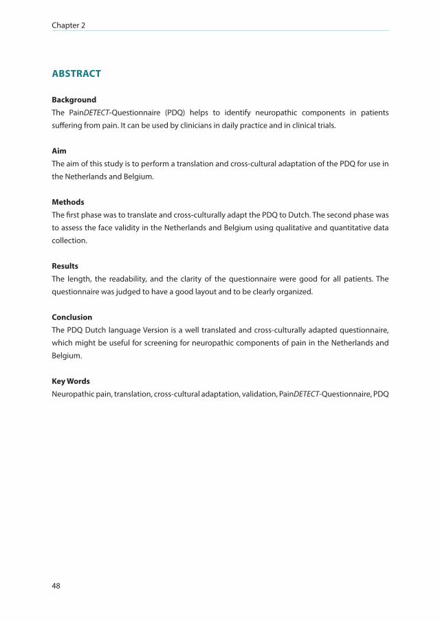
Chapter 2
48
ABSTRACT
Background
The PainDETECT-Questionnaire (PDQ) helps to identify neuropathic components in patients
suffering from pain. It can be used by clinicians in daily practice and in clinical trials.
Aim
The aim of this study is to perform a translation and cross-cultural adaptation of the PDQ for use in
the Netherlands and Belgium.
Methods
The first phase was to translate and cross-culturally adapt the PDQ to Dutch. The second phase was
to assess the face validity in the Netherlands and Belgium using qualitative and quantitative data
collection.
Results
The length, the readability, and the clarity of the questionnaire were good for all patients. The
questionnaire was judged to have a good layout and to be clearly organized.
Conclusion
The PDQ Dutch language Version is a well translated and cross-culturally adapted questionnaire,
which might be useful for screening for neuropathic components of pain in the Netherlands and
Belgium.
Key Words
Neuropathic pain, translation, cross-cultural adaptation, validation, PainDETECT-Questionnaire, PDQ
Cross-cultural adaptation of the PDQ
49
2
INTRODUCTION
Neuropathic Pain (NeP) is defined as ‘‘pain arising as a direct consequence of a lesion or disease
affecting the somatosensory system’’ [1]. NeP is characterized by spontaneous pain and abnormal
pain sensations [2]. Clinically, NeP is typically described as a constant burning pain with spontaneous
sharp exacerbations and somatosensory abnormalities [3]. As a rule, NeP has a considerable impact
on the quality of daily life [4]. The incidence of NeP in the Dutch general population is 0.81% or
130.000 new patients in the Netherlands per year. NeP is 63% more common in women than in men
and peaks between 70 and 79 years of age [4].
Medical history and (highly valid) screening tools may help to identify or differentiate between NeP,
nociceptive pain (NoP), and mixed pain syndromes [5-7]. Since 1996, various questionnaires have
been developed in different countries for the screening of neuropathic pain components based
on verbal pain description, with or without physical examination or attention to quality of life [8-
17]. Such questionnaires have been translated or cross-culturally adapted into different language
settings [18-25]. The term ‘‘cross-cultural adaptation’’ is used to define an important process that
considers both language and culture in the process of preparing a questionnaire in another
language and/or another country to provide equivalency between source and target language
based on content [26]. For example, the Netherlands are Dutch speaking, and parts of Belgium are
Flemish speaking (almost the same as Dutch, but interpretation and use of some words may vary).
Because of slight differences in language and sociocultural characteristics between the Netherlands
and Belgium, it is necessary to perform an adaptation in both countries. Moreover, it is useful to
have this instruments in a well-adapted Dutch language version not only for national studies but
also to permit participation in multinational studies [26-28].
The PainDETECT © Questionnaire (PDQ) (Pfizer GmbH, Berlin, Germany; 2005) was developed in
Germany [13] and tested as a reliable screening tool with high sensitivity (85%), specificity (80%),
and positive predictive accuracy (83%) for NeP. In the Spanish version of the PDQ sensitivity is 75%,
specificity 84%, and the positive predictive value is 92% [25]. The PDQ is a questionnaire that can be
filled in by the patients themselves and was devised to screen for neuropathic signs and symptoms
without physical examination.
The aim of this study is to achieve a cross-cultural adaptation of the PDQ for use in the Netherlands
and Belgium. The PainDETECT © Questionnaire Dutch language version: PDQ-Dlv (Pfizer, GmbH,
Berlin, Germany; 2008). Subsequently, this PDQ-Dlv will be used to assess face validity, as a first step
in a validation trajectory for this questionnaire in the Netherlands and Belgium.
Chapter 2
50
METHODS
The medical and ethical review board (CCMO Arnhem/Nijmegen, Nijmegen, the Netherlands) gave
approval to conduct this study. All patients signed an informed consent form prior to participation
in the study.
Translation of the PDQ The process of cross-cultural adaptation of the PDQ to Dutch was based on the 10 steps as described
in the ISPOR Patient-Reported Outcomes Translation and Linguistic Validation Task Force guidelines
[28] (Figure 1). The source questionnaire and the translations by translator 1 (T1), translator 2 (T2),
back translator 1 (BT1), and back translator 2 (BT2) together with the preliminary Dutch version
(Translation T1 to 2) were discussed item by item in a multidisciplinary expert committee review
during the harmonization phase: (AW) a physician experienced in the treatment of (chronic) pain,
(AE) a medical psychologist experienced in the translation, development and use of measurement
instruments (German native speaker and fluent in Dutch), (RF) the developer of the PainDETECT-
questionnaire, physician and experienced in the treatment of (chronic) pain (German native speaker
participating by telephone), and (HT) the project manager, in close contact with both the forward-
and backward translators, to prepare the pre-PDQ-Dlv. Special attention was given to three kinds
of equivalence: (1) Semantic equivalence / Idiomatic equivalence: The similarity of meaning of
each word or colloquialism in each culture after translation, (2) Experiential equivalence: Assessing
the experiences of daily life, and (3) Conceptual equivalence: In different cultures, words can have
different conceptual meaning. The committee searched both the source and the (back-) translations
for all such equivalences and adapted them to the most fitting meaning in Dutch [26-27]. In the
cognitive debriefing phase, the authors used qualitative semi-structured interviews to ask the
patients about the questionnaire and their understanding of the questions. It was performed by
one researcher from the review committee (HT) in a group of randomly selected patients in an
outpatient pain clinic (n = 10, seven women; mean age 58 years [± 16.2 years]) and in a group of
researchers (n = 4, two women; mean age 32 [± 4 years]).
Testing the PDQ After finalizing the translation, the quality of all the different versions in the cross-cultural adaptation
process were assessed using a 3-point scale [29]. The items were categorized (Cat.) as (Cat. 1) ‘‘different
meaning in each version’’, (Cat. 2) ‘‘almost the same meaning in both versions’’, or (Cat. 3) ‘‘exactly the
same meaning in both versions’’. Comparison of items was performed between translation T1 and
translation T2 by both forward translators, between back translation BT1 and back translation BT2 by
both backward translators. Comparison between the backward translation BT1-2 and the source PDQ
was performed by one of the developers of the questionnaire (RF). The final comparison was between
the source PDQ and the PDQ-Dlv during the harmonization phase and the review of the cognitive
debriefing results. After completion of the whole process, a quality control process was implemented.
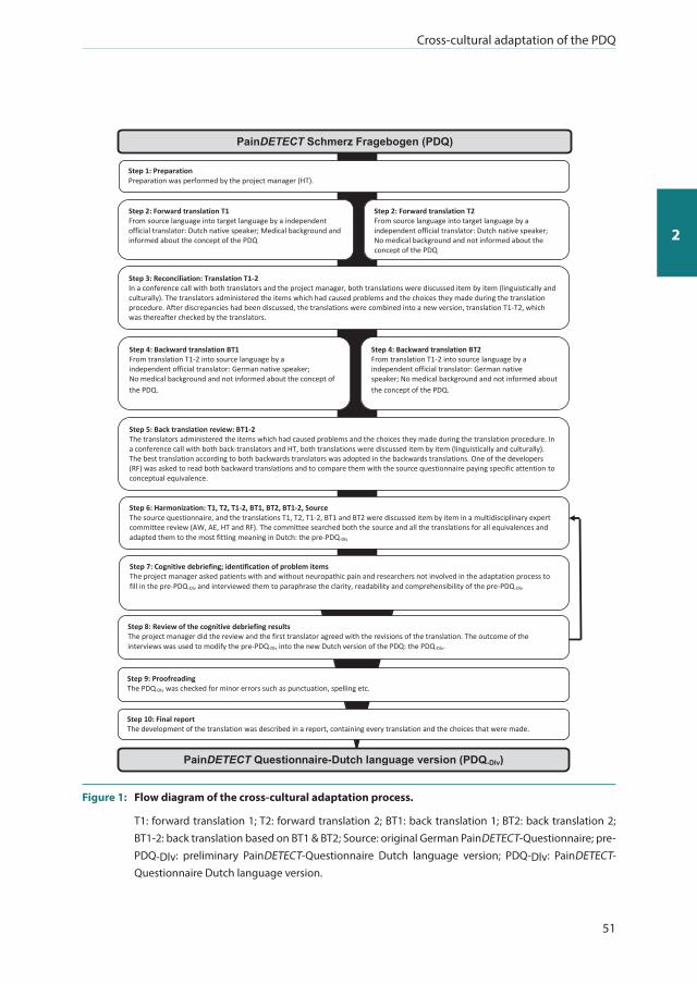
Cross-cultural adaptation of the PDQ
51
2
Step 1: Preparation Preparation was performed by the project manager (HT).
Step 2: Forward translation T1 From source language into target language by a independent official translator: Dutch native speaker; Medical background and informed about the concept of the PDQ
Step 4: Backward translation BT1 From translation T1-2 into source language by a independent official translator: German native speaker; No medical background and not informed about the concept of the PDQ.
concept of the PDQ
Step 3: Reconciliation: Translation T1-2 In a conference call with both translators and the project manager, both translations were discussed item by item (linguistically and culturally). The translators administered the items which had caused problems and the choices they made during the translation procedure. After discrepancies had been discussed, the translations were combined into a new version, translation T1-T2, which was thereafter checked by the translators.
Step 5: Back translation review: BT1-2 The translators administered the items which had caused problems and the choices they made during the translation procedure. In a conference call with both back-translators and HT, both translations were discussed item by item (linguistically and culturally). The best translation according to both backwards translators was adopted in the backwards translations. One of the developers (RF) was asked to read both backward translations and to compare them with the source questionnaire paying specific attention to conceptual equivalence.
Step 8: Review of the cognitive debriefing results The project manager did the review and the first translator agreed with the revisions of the translation. The outcome of the interviews was used to modify the pre-PDQ-Dlv into the new Dutch version of the PDQ: the PDQ-Dlv.
Step 2: Forward translation T2 From source language into target language by a independent official translator: Dutch native speaker; No medical background and not informed about the concept of the PDQ
Step 4: Backward translation BT2 From translation T1-2 into source language by a independent official translator: German native speaker; No medical background and not informed about the concept of the PDQ.
about the concept of the PDQ
Step 7: Cognitive debriefing; identification of problem items The project manager asked patients with and without neuropathic pain and researchers not involved in the adaptation process to fill in the pre-PDQ-Dlv and interviewed them to paraphrase the clarity, readability and comprehensibility of the pre-PDQ-Dlv
Step 6: Harmonization: T1, T2, T1-2, BT1, BT2, BT1-2, Source The source questionnaire, and the translations T1, T2, T1-2, BT1 and BT2 were discussed item by item in a multidisciplinary expert committee review (AW, AE, HT and RF). The committee searched both the source and all the translations for all equivalences and adapted them to the most fitting meaning in Dutch: the pre-PDQ-Dlv
PainDETECT Questionnaire-Dutch language version (PDQ-Dlv)
PainDETECT Schmerz Fragebogen (PDQ)
Step 10: Final report The development of the translation was described in a report, containing every translation and the choices that were made.
Step 9: Proofreading The PDQ-Dlv was checked for minor errors such as punctuation, spelling etc.
Figure 1: Flow diagram of the cross-cultural adaptation process.
T1: forward translation 1; T2: forward translation 2; BT1: back translation 1; BT2: back translation 2; BT1-2: back translation based on BT1 & BT2; Source: original German PainDETECT-Questionnaire; pre-PDQ-Dlv: preliminary PainDETECT-Questionnaire Dutch language version; PDQ-Dlv: PainDETECT-Questionnaire Dutch language version.
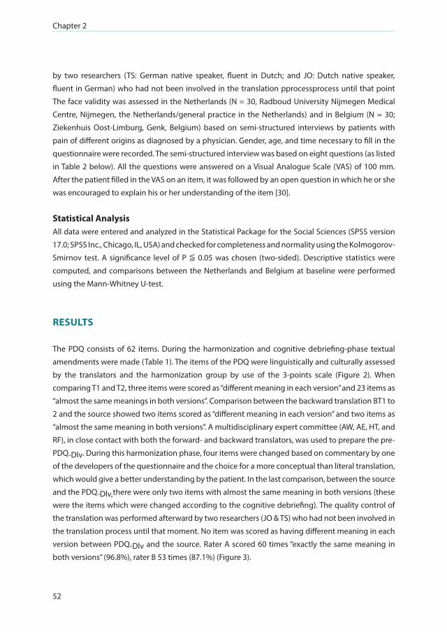
Chapter 2
52
by two researchers (TS: German native speaker, fluent in Dutch; and JO: Dutch native speaker,
fluent in German) who had not been involved in the translation pprocessprocess until that point
The face validity was assessed in the Netherlands (N = 30, Radboud University Nijmegen Medical
Centre, Nijmegen, the Netherlands/general practice in the Netherlands) and in Belgium (N = 30;
Ziekenhuis Oost-Limburg, Genk, Belgium) based on semi-structured interviews by patients with
pain of different origins as diagnosed by a physician. Gender, age, and time necessary to fill in the
questionnaire were recorded. The semi-structured interview was based on eight questions (as listed
in Table 2 below). All the questions were answered on a Visual Analogue Scale (VAS) of 100 mm.
After the patient filled in the VAS on an item, it was followed by an open question in which he or she
was encouraged to explain his or her understanding of the item [30].
Statistical Analysis All data were entered and analyzed in the Statistical Package for the Social Sciences (SPSS version
17.0; SPSS Inc., Chicago, IL, USA) and checked for completeness and normality using the Kolmogorov-
Smirnov test. A significance level of P ≦ 0.05 was chosen (two-sided). Descriptive statistics were
computed, and comparisons between the Netherlands and Belgium at baseline were performed
using the Mann-Whitney U-test.
RESULTS
The PDQ consists of 62 items. During the harmonization and cognitive debriefing-phase textual
amendments were made (Table 1). The items of the PDQ were linguistically and culturally assessed
by the translators and the harmonization group by use of the 3-points scale (Figure 2). When
comparing T1 and T2, three items were scored as ‘‘different meaning in each version’’ and 23 items as
‘‘almost the same meanings in both versions’’. Comparison between the backward translation BT1 to
2 and the source showed two items scored as ‘‘different meaning in each version’’ and two items as
‘‘almost the same meaning in both versions’’. A multidisciplinary expert committee (AW, AE, HT, and
RF), in close contact with both the forward- and backward translators, was used to prepare the pre-
PDQ-Dlv. During this harmonization phase, four items were changed based on commentary by one
of the developers of the questionnaire and the choice for a more conceptual than literal translation,
which would give a better understanding by the patient. In the last comparison, between the source
and the PDQ-Dlv,there were only two items with almost the same meaning in both versions (these
were the items which were changed according to the cognitive debriefing). The quality control of
the translation was performed afterward by two researchers (JO & TS) who had not been involved in
the translation process until that moment. No item was scored as having different meaning in each
version between PDQ-Dlv and the source. Rater A scored 60 times ‘‘exactly the same meaning in
both versions’’ (96.8%), rater B 53 times (87.1%) (Figure 3).
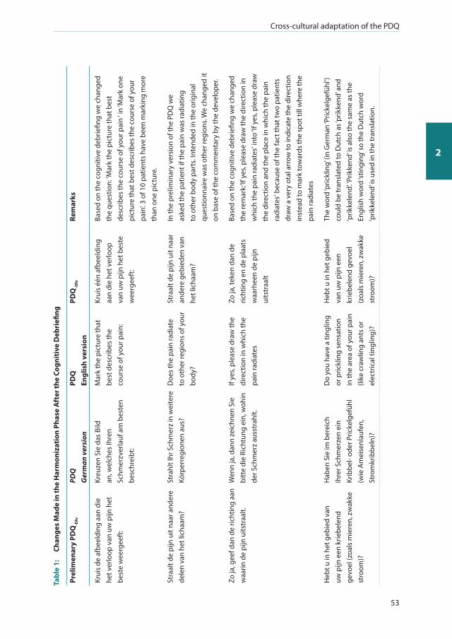
Cross-cultural adaptation of the PDQ
53
2
Tabl
e 1:
Ch
ange
s M
ade
in th
e H
arm
oniz
atio
n Ph
ase
Aft
er th
e Co
gnit
ive
Deb
riefi
ng
Prel
imen
ary
PDQ
-Dlv
PDQ
G
erm
an v
ersi
onPD
Q
Engl
ish
vers
ion
PDQ
-Dlv
Rem
arks
Krui
s de
afb
eeld
ing
aan
die
het v
erlo
op v
an u
w p
ijn h
et
best
e w
eerg
eeft
:
Kreu
zen
Sie
das
Bild
an
, wel
ches
Ihre
n Sc
hmer
zver
lauf
am
bes
ten
besc
hrei
bt:
Mar
k th
e pi
ctur
e th
at
best
des
crib
es th
e co
urse
of y
our p
ain:
Krui
s éé
n af
beel
ding
aa
n di
e he
t ver
loop
va
n uw
pijn
het
bes
te
wee
rgee
ft:
Base
d on
the
cogn
itive
deb
riefin
g w
e ch
ange
d th
e qu
estio
n: ’M
ark
the
pict
ure
that
bes
t de
scrib
es th
e co
urse
of y
our p
ain
‘ in
‘Mar
k on
e pi
ctur
e th
at b
est d
escr
ibes
the
cour
se o
f you
r pa
in’. 3
of 1
0 pa
tient
s ha
ve b
een
mar
king
mor
e th
an o
ne p
ictu
re.
Stra
alt d
e pi
jn u
it na
ar a
nder
e de
len
van
het l
icha
am?
Stra
hlt I
hr S
chm
erz
in w
eite
re
Körp
erre
gion
en a
us?
Doe
s th
e pa
in ra
diat
e to
oth
er re
gion
s of
you
r bo
dy?
Stra
alt d
e pi
jn u
it na
ar
ande
re g
ebie
den
van
het l
icha
am?
In th
e pr
elim
inar
y ve
rsio
n of
the
PDQ
we
aske
d th
e pa
tient
if th
e pa
in w
as ra
diat
ing
to o
ther
bod
y pa
rts.
Inte
nded
in th
e or
igin
al
ques
tionn
aire
was
oth
er re
gion
s. W
e ch
ange
d it
on b
ase
of th
e co
mm
enta
ry b
y th
e de
velo
per.
Zo ja
, gee
f dan
de
richt
ing
aan
waa
rin d
e pi
jn u
itstr
aalt.
Wen
n ja
, dan
n ze
ichn
en S
ie
bitt
e di
e Ri
chtu
ng e
in, w
ohin
de
r Sch
mer
z au
sstr
ahlt.
If ye
s, pl
ease
dra
w th
e di
rect
ion
in w
hich
the
pain
radi
ates
Zo ja
, tek
en d
an d
e ric
htin
g en
de
plaa
ts
waa
rhee
n de
pijn
ui
tstr
aalt
Base
d on
the
cogn
itive
deb
riefin
g w
e ch
ange
d th
e re
mar
k:‘If
yes
, ple
ase
draw
the
dire
ctio
n in
w
hich
the
pain
radi
ates
‘ int
o ‘If
yes
, ple
ase
draw
th
e di
rect
ion
and
the
plac
e in
whi
ch th
e pa
in
radi
ates
‘ bec
ause
of t
he fa
ct th
at tw
o pa
tient
s dr
aw a
ver
y ot
al a
rrow
to in
dica
te th
e di
rect
ion
inst
ead
to m
ark
tow
ards
the
spot
till
whe
re th
e pa
in ra
diat
es
Heb
t u in
het
geb
ied
van
uw p
ijn e
en k
riebe
lend
ge
voel
(zoa
ls m
iere
n, z
wak
ke
stro
om)?
Hab
en S
ie im
ber
eich
Ih
rer S
chm
erze
n ei
n Kr
ibbe
l- od
er P
ricke
lgef
ühl
(wie
Am
eise
nlau
fen,
St
rom
krib
beln
)?
Do
you
have
a ti
nglin
g or
pric
klin
g se
nsat
ion
in th
e ar
ea o
f you
r pai
n (li
ke c
raw
ling
ants
or
elec
tric
al ti
nglin
g)?
Heb
t u in
het
geb
ied
van
uw p
ijn e
en
krie
bele
nd g
evoe
l (z
oals
mie
ren,
zw
akke
st
room
)?
The
wor
d ‘p
rickl
ing’
(in
Ger
man
‘Pric
kelg
efüh
l’)
coul
d be
tran
slat
ed to
Dut
ch a
s ‘pr
ikke
nd’ a
nd
‘prik
kele
nd’. ‘P
rikke
nd’ is
als
o th
e sa
me
as th
e En
glis
h w
ord
‘stin
ging
’ so
the
Dut
ch w
ord
‘prik
kele
nd’ is
use
d in
the
tran
slat
ion.
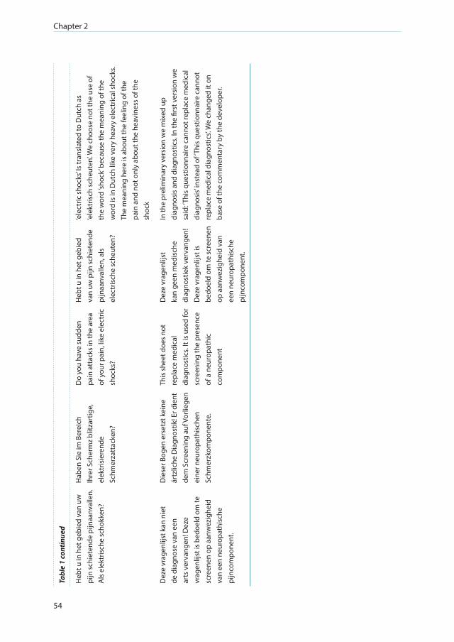
Chapter 2
54
Tabl
e 1
cont
inue
d
Heb
t u in
het
geb
ied
van
uw
pijn
sch
iete
nde
pijn
aanv
alle
n.
Als
ele
ktris
che
scho
kken
?
Hab
en S
ie im
Ber
eich
Ih
rer S
cher
mz
blitz
artig
e,
elek
tris
iere
nde
Schm
erza
ttac
ken?
Do
you
have
sud
den
pain
att
acks
in th
e ar
ea
of y
our p
ain,
like
ele
ctric
sh
ocks
?
Heb
t u in
het
geb
ied
van
uw p
ijn s
chie
tend
e pi
jnaa
nval
len,
als
el
ectr
isch
e sc
heut
en?
‘ele
ctric
sho
cks’
Is tr
ansl
ated
to D
utch
as
‘ele
ktris
ch s
cheu
ten’.
We
choo
se n
ot th
e us
e of
th
e w
ord
’shoc
k’ b
ecau
se th
e m
eani
ng o
f the
w
ord
is in
Dut
ch li
ke v
ery
heav
y el
ectr
ical
sho
cks.
The
mea
ning
her
e is
abo
ut th
e fe
elin
g of
the
pain
and
not
onl
y ab
out t
he h
eavi
ness
of t
he
shoc
k
Dez
e vr
agen
lijst
kan
nie
t de
dia
gnos
e va
n ee
n ar
ts v
erva
ngen
! Dez
e vr
agen
lijst
is b
edoe
ld o
m te
sc
reen
en o
p aa
nwez
ighe
id
van
een
neur
opat
hisc
he
pijn
com
pone
nt.
Die
ser B
ogen
ers
etzt
kei
ne
ärtz
liche
Dia
gnos
tik! E
r die
nt
dem
Scr
eeni
ng a
uf V
orlie
gen
eine
r neu
ropa
this
chen
Sc
hmer
zkom
pone
nte.
This
she
et d
oes
not
repl
ace
med
ical
di
agno
stic
s. It
is u
sed
for
scre
enin
g th
e pr
esen
ce
of a
neu
ropa
thic
co
mpo
nent
Dez
e vr
agen
lijst
ka
n ge
en m
edis
che
diag
nost
iek
verv
ange
n!
Dez
e vr
agen
lijst
is
bedo
eld
om te
scr
eene
n op
aan
wez
ighe
id v
an
een
neur
opat
hisc
he
pijn
com
pone
nt.
In th
e pr
elim
inar
y ve
rsio
n w
e m
ixed
up
diag
nosi
s an
d di
agno
stic
s. In
the
first
ver
sion
we
said
: ‘Thi
s qu
estio
nnai
re c
anno
t rep
lace
med
ical
di
agno
sis’
inst
ead
of ‘T
his
ques
tionn
aire
can
not
repl
ace
med
ical
dia
gnos
tics’.
We
chan
ged
it on
ba
se o
f the
com
men
tary
by
the
deve
lope
r.

Cross-cultural adaptation of the PDQ
55
2
0%
10%
20%
30%
40%
50%
60%
70%
80%
90%
100%
T1 & T2 BT1 & BT2 BT1-2 & Source Source & PDQ-dlv
Cat.3
Cat.2
Cat.1
Figure 2: Translation process: degree of agreement.
T1: forward translation 1; T2: forward translation 2; BT1: back translation 1; BT2: back translation 2; BT1-2: back translation based on BT1 and BT2; Source: original German PainDETECT-Questionnaire; PDQ-
Dlv: PainDETECT-Questionnaire Dutch language version; Cat.1: different meaning in each version; Cat. 2: almost the same meaning in both versions; Cat. 3: exactly the same meaning in both versions.
0%
10%
20%
30%
40%
50%
60%
70%
80%
90%
100%
Cat. 3
Cat. 2
Cat. 1
Figure 3: Quality control after the translation process.
A: rater A; B: rater B; T1: translation 1; T2: translation 2; BT1: back translation 1; BT2: back translation 2; BT1-2: back translation based on BT1 & BT2; Source: original German PainDETECT-Questionnaire; PDQ-
Dlv: PainDETECT-Questionnaire Dutch language version; Cat.1: different meaning in each version; Cat.2: almost the same meaning in both versions; Cat.3: Exactly the same meaning in both versions.
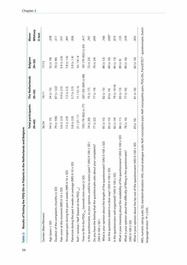
Chapter 2
56
Tabl
e 2:
Re
sult
s of
Tes
ting
the
PDQ
-Dlv
in P
atie
nts
in th
e N
ethe
rlan
ds a
nd B
elgi
um To
tal p
arti
cipa
nts
(N=6
0)Th
e N
ethe
rlan
ds
(N=3
0)Be
lgiu
m
(N=3
0)M
ann-
Whi
tney
U
-tes
tG
ende
r (M
an/W
omen
)36
/24
19/1
117
/13
Age
(yea
rs ±
SD
)54
(± 1
5)54
(± 1
5)52
(± 1
8),3
38
Dur
atio
n of
the
com
plai
nts
(mon
ths
(± S
D)
79 (±
106
)87
(± 1
22)
70 (±
90)
,511
Pain
sco
re a
t thi
s m
omen
t (N
RS 0
-10
± SD
)5.
6 (±
2.5
)5.
9 (±
2.5
)5.
4 (±
2.6
),5
31
Stro
nges
t pai
n du
ring
the
past
4 w
eeks
(NRS
0-1
0 ±
SD)
7.5
(± 2
.0)
7.2
(± 2
.3)
7.8
(± 1
.6)
,401
Pain
scor
e du
ring
the
past
4 w
eeks
on
aver
age
(NRS
0-1
0 ±
SD)
5.8
(± 2
.0)
5.7
(± 2
.5)
5.9
(± 1
.4)
,926
NoP
/ un
clea
r / N
eP (b
ased
on
the
PDQ
- Dlv)
21 /
27 /
1111
/ 13
/ 6
10 /
14 /
6
Tim
e to
fill
in th
e PD
Q- D
lv (s
econ
ds (m
in-m
ax) (
± SD
)19
3 ( 7
9-36
2) (±
77)
207
( 83-
360)
(± 8
8)18
1 (7
9-36
2) (±
65)
,417
Is th
e qu
estio
nnai
re, t
o yo
ur o
pini
on, u
sefu
l to
asse
ss ‘p
ain’
? (V
AS
0-10
0 ±
SD )
74 (±
20)
75 (±
17)
72 (±
23)
,767
Do
you
have
the
feel
ing
that
the
ques
tionn
aire
ask
s ab
out y
our c
ompl
aint
s?
(VA
S 0-
100
± SD
)77
(± 2
2)77
(± 1
9)76
(± 2
4),6
95
Wha
t is
your
opi
nion
abo
ut th
e le
ngth
of t
he q
uest
ionn
aire
? (V
AS
0-10
0 ±
SD)
85 (±
15)
82 (±
15)
88 (±
12)
,166
Are
the
ques
tions
sta
ted
in a
cle
ar w
ay?
(VA
S 0-
100
± SD
)85
(± 1
3)81
(± 1
4)89
(± 1
0),0
30*
Is th
e qu
estio
nnai
re w
ell o
rgan
ized
? (V
AS
0-10
0 ±
SD)
82 (±
14)
79 (±
101
4)85
(± 1
3),0
73
Wha
t is
your
mea
ning
abo
ut th
e re
adab
ility
of t
he q
uest
ionn
aire
? (V
AS
0-10
0 ±
SD)
86 (±
11)
83 (±
13)
89 (±
9)
,125
Wha
t is
your
opi
nion
abo
ut th
e di
fficu
lty o
f filli
ng in
the
ques
tionn
aire
?
(VA
S 0-
100
± SD
)78
(± 1
6)77
(± 1
6)80
(± 1
6),5
39
Wha
t is
your
opi
nion
abo
ut th
e la
y-ou
t of t
he q
uest
ionn
aire
(VA
S 0-
100
± SD
)81
(± 1
6)81
(± 1
6)82
(± 1
6),9
52
NRS
, num
eric
ratin
g sc
ale;
SD
, sta
ndar
d de
viat
ion;
VA
S, v
isua
l ana
logu
e sc
ale;
NoP
, noc
icep
tive
pain
; NeP
, neu
ropa
thic
pai
n; P
DQ
-Dlv
, Pai
nDET
ECT
- que
stio
nnai
re, D
utch
la
ngua
ge v
ersi
on. *
P ≤
0.05
.
Cross-cultural adaptation of the PDQ
57
2
In total, 60 patients were asked to fill in the PDQ-Dlv to assess the face validity. Twenty-one of 60
patients (35%) forgot to mark the main area of pain (‘‘drawing’’). Five patients (12%) forgot to tick
the box whether the pain was radiating to other regions of the body or not. One patient did not
fill in all the questions because there was no appropriate answer according to the patient. Patients
gave their opinion about the usability of the questionnaire on a VAS-scale (0 to 100), in which ‘‘0’’
means totally not useable and ‘‘100’’ means very useable (Table2). Most Belgian and Dutch patients
found the PDQ-Dlv a clear, readable, well organized, and useful instrument to assess their pain. The
question ‘‘Are the questions stated in a clear way?’’ showed a significant difference in favor of the
patients from Belgium (P = 0.03, Mann-Whitney U-test). The most common question in the group of
sixty patients (n = 14; 23.3%) during the fillingin period was ‘‘Should I circle the number or tick the
box?’’ in the questions about the pain at this moment, the strongest pain during the last 4 weeks
and on average in the last 4 weeks. After answering the questions in the interview, the patients were
challenged to give their thoughts and comments about the PDQ-Dlv in words. Most of the patients
had no comment. The most frequent comment was that the kind of pain of the patient ‘‘did not fit in
the questionnaire’’ (n = 4, 7%). Suggestions made by patients for a next version were the use of less
color (n = 1) and increasing font size (n = 1).
DISCUSSION
This study presents the results of the cross-cultural adaptation and face validation of the PDQ into
the Dutch language for use in the Netherlands and Belgium. The PDQ was until this moment only
published in a German, English, and Spanish language version [13,25]. The quality of a questionnaire
used in research is expected to be dependent on the quality of the chosen method for translation
[31]. There are many strategies to perform a cross-cultural adaptation [26-28,32,33]. The use of
these guidelines improves the linguistic, structure, and cultural equivalence [34]. However, there
is a lack of consensus and consistency in quality, methodology and application of these guidelines
in healthcare literature [28,35]. The ISPOR guidelines [28] represent a consensus regarding the
principles of good practice in translation and cultural adaptation. Their goal is to provide a more
conceptual equivalent approach instead of a more literal translation. Because it provides clear
recommendations and a detailed multistep approach, the ISPOR guidelines were chosen as method
for the translation and cultural adaptation of the PDQ. This study consequently followed these
guidelines in the choice of translators, the members of the multidisciplinary expert committee as
well as the process of the adaptation. Decisions were discussed with the persons involved in the
process either by conference call or by live meetings.
Because differences in instrument formatting and administration may produce variations in
response even if meticulous care is taken [36], we opted for the same layout as the original version.
In the cognitive debriefing phase and during data collection before the face validity testing, some
Chapter 2
58
patients indicated that in some questions they did not know where in the questionnaire they had to
put their answer to that question. They found the questionnaire too colorful and that the colors were
distracting. Another difficulty when using the original format was that Dutch translated questions
were limited to the space the original developers used in the original version.
Patient interviews were very useful to find out what the patient is thinking and how he interprets
the questions in the questionnaire [37]. On the basis of these interviews, we changed two items to
make them clearer to the patient and to reduce missing data: ‘‘Mark the picture that best describes
the course of your pain’’ was changed into ‘‘Mark one picture that best describes the course of your
pain’’. We also changed ‘‘If yes, please draw the direction in which the pain radiates’’ to ‘‘If yes, please
draw the direction and the place into which the pain radiates’’. Thirty-five percent of the patients
forgot to fill in this drawing but this is not of influence on the outcome score of the questionnaire.
The question ‘‘Does the pain radiate to other regions of the body?’’ was not filled in by 7% of the
patients. This has an influence on the outcome score. However, owing to the choice to use the same
format as the original questionnaire, we were not able to change this item in a manner that there
was more attention to the drawing and corresponding questions.
A quality check based on a 3-step scale [29] during and after completion of all the steps recommended
by ISPOR was added because during the translation process the comparison was performed only by
the translators. Thus, after the translation was completed, two independent researchers were asked
to check each step in the translation trajectory. Rater A scored almost the same meaning in both
versions two times when comparing the source and the PDQ-Dlv, and Rater B, who took a more
literal rather than a conceptual approach, scored nine items as almost the same meaning in both
versions. On the basis of the outcome of all scores, it is clear that a good translation process consists
of more than a forward translation and that the steps the authors took in this study are necessary to
come to a good cross-cultural adaptation.
The face validity of the PDQ-Dlv was assessed in Dutch and Flemish because of the slight differences
in the interpretation and use of some words. Therefore, the understanding of questions in the
questionnaire may not be equal. In the end, only one significant difference in favor of the patients
from Belgium occurred (‘‘are the questions stated in a clear way?’’). No further differences were
found.
A valid adaptation (‘‘Are the questions easily understood by patients as well as by clinicians/
researchers and do they measure the same concept in different languages and countries?’’) of a
questionnaire is necessary for its use in every day clinical practice and also in (inter-)national research
to make the outcome comparable in different cultures. Furthermore, cross-cultural adaptation is
cheaper, less labor-intensive and less time-consuming than to develop a whole new questionnaire.
[27,38]
Cross-cultural adaptation of the PDQ
59
2
Publishing a cross-cultural adaptation has a value itself. First, it will help to prevent the existence of
multiple versions of an instrument in a given language. Second, it ensures that the large amount of
work involved will not be repeated unnecessarily [26]. Third, it gives a clear insight in which choices
have been made during the translation process and thus provides important information about the
strength of the translation [27]. On the basis of the method used, the reported patient outcome
in the cognitive debriefing phase as well as in the face validity assessment and in the quality
check, the PDQ-Dlv is a well translated and cross-culturally adapted questionnaire for screening on
neuropathic pain components in patients in the Netherlands and Belgium. A validation study of the
PDQ is now in progress to assess the psychometric properties (ie, sensitivity, specificity, predictive
value, and reliability) for different groups of patients in the Netherlands.
ACKNOWLEDGEMENTS
The authors would like to thank Emanuel van den Broeke and Jelle van Gurp for the fruitful
discussions about the cross-cultural adaptation of questionnaires. This study was performed within
DALI for PAIN, a national program that focuses on neuropathic pain care optimization. DALI for PAIN
is an initiative of Pfizer. This project is supported by an unrestricted grant from Pfizer.
Chapter 2
60
REFERENCES
1. Treede RD, Jensen TS, Campbell JN, et al. Neuropathic pain: redefinition and a grading system for clinical and research purposes. Neurology. 2008;70:1630-1635.
2. Vissers KC. The clinical challenge of chronic neuropathic pain. Disabil Rehabil. 2006;28:343-349. 3. Baron R, Binder A, Wasner G. Neuropathic pain: diagnosis, pathophysiological mechanisms, and treatment.
Lancet Neurol. 2010;9:807-819. 4. Dieleman JP, Kerklaan J, Huygen FJ, Bouma PA, Sturkenboom MC. Incidence rates and treatment of
neuropathic pain conditions in the general population. Pain. 2008;137:681-688. 5. Bennett MI, Attal N, Backonja MM, et al. Using screening tools to identify neuropathic pain. Pain. 2007;127:
199-203. 6. Freynhagen R, Bennett MI. Diagnosis and management of neuropathic pain. BMJ. 2009;339:b3002. 7. Baron R, Tolle TR. Assessment and diagnosis of neuropathic pain. Curr Opin Support Palliat Care. 2008;2:1-8. 8. Backonja MM, Krause SJ. Neuropathic pain questionnaire—short form. Clin J Pain. 2003;19:315-316. 9. Bennett M. The LANSS Pain Scale: the Leeds assessment of neuropathic symptoms and signs. Pain.
2001;92:147-157. 10. Bennett MI, Smith BH, Torrance N, Potter J. The S-LANSS score for identifying pain of predominantly
neuropathic origin: validation for use in clinical and postal research. J Pain. 2005;6:149-158. 11. Bouhassira D, Attal N, Fermanian J, et al. Development and validation of the neuropathic pain symptom
inventory. Pain. 2004;108:248-257. 12. Bouhassira D, Attal N, Alchaar H, et al. Comparison of pain syndromes associated with nervous or somatic
lesions and development of a new neuropathic pain diagnostic questionnaire (DN4). Pain. 2005;114:29-36. 13. Freynhagen R, Baron R, Gockel U, Tolle TR. PainDETECT: a new screening questionnaire to identify
neuropathic components in patients with back pain. Curr Med Res Opin. 2006;22:1911-1920. 14. Galer BS, Jensen MP. Development and preliminary validation of a pain measure specific to neuropathic
pain: the neuropathic pain scale. Neurology. 1996;48:332-338. 15. Krause SJ, Backonja MM. Development of a neuropathic pain questionnaire. Clin J Pain. 2003;19:306-314. 16. Poole HM, Murphy P, Nurmikko TJ. Development and preliminary validation of the NePIQoL: a quality-of-life
measure for neuropathic pain. J Pain Symptom Manage. 2009;37:233-245. 17. Portenoy R. Development and testing of a neuropathic pain screening questionnaire: ID pain. Curr Med Res
Opin. 2006;22:1555-1565. 18. Chaudakshetrin P, Prateepavanich P, Chira-Adisai W, Tassanawipas W, Leechavengvongs S, Kitisomprayoonkul
W. Cross-cultural adaptation to the Thai language of the neuropathic pain diagnostic questionnaire (DN4). J Med Assoc Thai. 2007;90:1860-1865.
19. Hans G, Masquelier E, De Cock P. The diagnosis and management of neuropathic pain in daily practice in Belgium: an observational study. BMC Public Health. 2007;24:7.
20. Negri E, Bettaglio R, Demartini M, et al. Validazione della Scala del Dolore Neuropatico (SDN) e sue applicazioni terapeutiche. Minerva Anestesiol. 2002;68:95-104.
21. Perez C, Galvez R, Insausti J, Bennett M, Ruiz M, Rejas J. Linguistic adaptation and Spanish validation of the LANSS (Leeds Assessment of Neuropathic Symptoms and Signs) scale for the diagnosis of neuropathic pain. Med Clin. 2006;127:485-491.
22. Perez C, Galvez R, Huelbes S, et al. Validity and reliability of the Spanish version of the DN4 (Douleur Neuropathique 4 questions) questionnaire for differential diagnosis of pain syndromes associated to a neuropathic or somatic component. Health Qual Life Outcomes. 2007;5:66.
23. Yucel A, Senocak M, Orhan EK, Cimen A, Ertas M. Results of the leeds assessment of neuropathic symptoms and signs pain scale in Turkey: a validation study. Journal of Pain. 2004;5:427-432.
24. Seventer VR, Vos C, Meerding W, et al. Linguistic validation of the DN4 for use in international studies. Eur J Pain. 2010;14:58-63.
25. De Andres J, Perez-Cajaraville J, Lopez-Alarcon MD, et al. Cultural adaptation and validation of the PainDETECT scale into Spanish. Clin J Pain. 2012;28:243-253.
26. Beaton DE, Bombardier C, Guillemin F, Ferraz MB. Guidelines for the process of cross-cultural adaptation of self-report measures. Spine. 2000;25:3186-3191.
27. Guillemin F, Bombardier C, Beaton D. Cross-cultural adaptation of health-related quality of life measures: literature review and proposed guidelines. J Clin Epidemiol. 1993; 46:1417-1432.
Cross-cultural adaptation of the PDQ
61
2
28. Wild D, Grove A, Martin M, et al. Principles of Good Practice for the Translation and Cultural Adaptation Process for Patient-Reported Outcomes (PRO) Measures: report of the ISPOR Task Force for Translation and Cultural Adaptation. Value Health. 2005;8:94-104.
29. Flaherty JA, Gaviria FM, Pathak D, et al. Developing instruments for cross-cultural psychiatric research. J Nerv Ment Dis. 1988;176:257-263.
30. Schuman H. The random probe: a technique for evaluating the validity of closed questions. Am Sociol Rev. 1966; 31:218-222.
31. Maneesriwongul W, Dixon JK. Instrument translation process: a methods review. J Adv Nurs. 2004;48:175- 186.
32. Peters M, Passchier J. Translating instruments for cross-cultural studies in headache research. Headache. 2006; 46:82-91.
33. Sperber AD. Translation and validation of study instruments for cross-cultural research. Gastroenterology. 2004;126(suppl 1):S124-S128.
34. Menezes Costa LC, Maher CG, McAuley JH, Costa LO. Systematic review of cross-cultural adaptations of McGill Pain Questionnaire reveals a paucity of clinimetric testing. J Clin Epidemiol. 2009;62:934-943.
35. Sousa VD, Rojjanasrirat W. Translation, adaptation and validation of instruments or scales for use in cross-cultural health care research: a clear and user-friendly guideline. J Eval Clin Pract. 2011;17:268-274.
36. Hilton A, Skrutkowski M. Translating instruments into other languages: development and testing processes. Cancer Nurs. 2002;25:1-7.
37. Charmaz K Constructing Grounded Theory; A Practical Guide Through Qualitative Analysis. London: SAGE publications Ltd; 2009.
38. Acquadro C, Conway K, Hareendran A, Aaronson N. Literature review of methods to translate health-related quality of life questionnaires for use in multinational clinical trials. Value in Health. 2008;11:509-521.


CHAPTER 3Assessment of Neuropathic Pain in Patients with Cancer: The Interobserver Reliability. An Observational Study in Daily Practice
Hans Timmerman
Irene Heemstra
Annelies Schalkwijk
Constans Verhagen
Kris Vissers
Yvonne Engels
Published in:
Pain Physician 2013; 16: 569-580
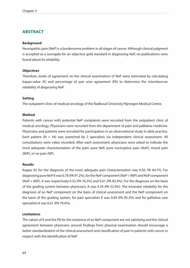
Chapter 3
64
ABSTRACT
Background
Neuropathic pain (NeP) is a burdensome problem in all stages of cancer. Although clinical judgment
is accepted as a surrogate for an objective gold standard in diagnosing NeP, no publications were
found about its reliability.
Objectives
Therefore, levels of agreement on the clinical examination of NeP were estimated by calculating
kappa-value (Κ) and percentage of pair wise agreement (PA) to determine the interobserver
reliability of diagnosing NeP.
Setting
The outpatient clinic of medical oncology of the Radboud University Nijmegen Medical Centre.
Method
Patients with cancer with potential NeP complaints were recruited from the outpatient clinic of
medical oncology. Physicians were recruited from the department of pain and palliative medicine.
Physicians and patients were recruited for participation in an observational study in daily practice.
Each patient (N = 34) was examined by 2 specialists via independent clinical assessment. All
consultations were video recorded. After each assessment, physicians were asked to indicate the
most adequate characterization of the pain: pure NeP, pure nociceptive pain (NoP), mixed pain
(MiP), or no pain (NP).
Results
Kappa (Κ) for the diagnosis of the most adequate pain characterization was 0.50, PA 64.7%. For
diagnosing pure NeP K was 0.78 (PA 91.2%), for the NeP component (NeP + MiP) and NoP component
(NoP + MiP), it was respectively 0.52 (PA 76.5%) and 0.61 (PA 82.4%). For the diagnosis on the basis
of the grading system between physicians, Κ was 0.34 (PA 52.9%). The intrarater reliability for the
diagnosis of an NeP component on the basis of clinical assessment and the NeP component on
the basis of the grading system, for pain specialists Κ was 0.69 (PA 85.3%) and for palliative care
specialists Κ was 0.61 (PA 79.4%).
Limitations
The values of Κ and the PA for the existence of an NeP component are not satisfying and the clinical
agreement between physicians around findings from physical examination should encourage a
better standardization of the clinical assessment and classification of pain in patients with cancer in
respect with the identification of NeP.
The interobeserver reliability of neuropathic pain
65
3
Conclusions
A substantial level of agreement was found for the diagnosis of pure NeP and a moderate level of
agreement for the diagnosis of the NeP component was found, both with a PA ≥ 70%. There was
only a fair agreement between the physicians regarding the grading system. However, there was a
substantial level of (interrater) agreement for the diagnosis of an NeP component and the outcome
of the grading system. The findings in this study also suggest that a better standardization of the
clinical assessment and classification of pain in patients with cancer with respect to the identification
of neuropathic pain is necessary.
Key words
Neuropathic pain, diagnosis, interobserver reliability, agreement, cancer observational study, pain,
clinical assessment, diagnostic test

Chapter 3
66

The interobeserver reliability of neuropathic pain
67
3
INTRODUCTION
Pain is a burdensome symptom in all stages of cancer. Van den Beuken et al [1] found a prevalence
of 55% in patients with cancer in the Netherlands. Of those, 44% suffered from moderate to severe
pain [1]. As described in a review, 64% of the patients with metastatic, advanced, or terminal stages
of cancer had pain, 59% of patients who were on anticancer treatment and 33% of patients who had
been cured from cancer still suffered from pain [1,2]. In patients with cancer who were on opioid
treatment by a pain specialist for their pain, almost 40% had neuropathic pain (NeP) alone or in
combination with nociceptive or visceral pain [3]. In several other studies, the prevalence of NeP in
patients with cancer varied between 17% and 36% [4-7]. This large variability in prevalence between
studies can be explained by differences in populations, differences in diagnostic methodologies,
and differences in definitions [8].
The International Association for the Study of Pain (IASP) defines NeP as “pain caused by a lesion
or disease of the somatosensory nervous system” [9].The question arises when (part of ) the pain in
patients with cancer can be diagnosed as NeP. Despite the attempts to specify the entity of NeP, still
no gold standard for the diagnosis of NeP exists [10]. NeP is experienced by the patient and despite
the characteristic signs and symptom complex that may be recognized by experienced doctors,
it is still difficult to measure objectively. Several screening tools, like the DN4, LANSS, NPQ, and
PainDETECT have been developed to indicate the possible existence of NeP [11-15]. Yet, screening
tools are no substitute for history taking and physical examination, and they are not intended to
be a diagnostic method [12]. Therefore, clinical judgment is the only recommended method to
diagnose NeP [10,16]. When standardized diagnostic criteria are lacking, the reliability of diagnostic
procedures is usually demonstrated by acceptable levels of agreement among physicians [17-19].
Interobserver reliability is an important measure to assess the agreement of categorical variables
such as diagnosis or the interpretation of findings in physical examination [20]. Cohen’s kappa is a
for chance corrected statistical outcome for interobserver reliability [21]. We used Cohen’s kappa
and percentage of pair wise agreement to investigate the interobserver reliability and agreement of
the diagnosis of NeP in patients with cancer.
METHODS
Patients Patients were recruited from the outpatient clinic of the department of medical oncology of the
Radboud University Nijmegen Medical Centre (RUNMC). Between September and November 2010,
all patients who visited the outpatient clinic were screened for pain for another larger study. As part
of a larger set of questionnaires, they were also asked to complete the 7-item DN4 questionnaire
[13] about the quality of their pain. Inclusion criteria for enrollment in the kappa-study were (1) age
Chapter 3
68
≥ 18 years; (2) diagnosed with cancer (regardless of the type and stage of cancer) or being cured
from cancer; (3) at least 2 positive answers on the 7-item DN4 questionnaire in order to enrich the
chance of including patients suffering from NeP in the research population.
Exclusion criteria were (1) no consent to be contacted for further research; (2) no permission
for video recording of the consultations. Eligible patients were phoned by the researcher (IH).
Subsequently, the patients received information by mail. After verbal and written informed consent
patient-volunteers were included in the study. They did not receive any benefit from the study; only
costs for transportation were reimbursed. This study was approved by the local ethics committee:
the Committee on Research Involving Human Subjects region Arnhem-Nijmegen, Nijmegen, the
Netherlands.
Patients were examined by experienced pain specialists (N = 4) and palliative care specialists (N
= 2), recruited from the department of anesthesiology, pain, and palliative medicine of RUNMC.
All 4 participating pain specialists, 2 men and 2 women, median age of 40 (range 32 – 47), had a
background as an anesthesiologist. The 2 palliative care specialists, both male, were 58 and 63. One
was a medical oncologist and the other an elderly care physician. Years of experience in their actual
specialization (pain or palliative care) was 10 years for the pain specialists (range 1 – 18 years) and
13 years for the palliative care specialists (7 and 18 years). All physicians worked full time, but, as a
mean, they worked 19 hours per week (10 – 26 hours) in this specific field.
Test Methods All physicians completed a questionnaire recording their age, gender, professional background,
specialty, and number of weekly hours working as a pain specialist or as a palliative care specialist.
They were also asked to provide a working definition of NeP, including symptoms and findings at
physical examination they considered decisive for NeP. As a part of the preparation of the study, an
inquiry was made among the physicians regarding the tools they wanted to use for the physical
examination. There was no prearranged set of tools available in the examination rooms, only those
recommended by one or more of the participating physicians: pieces of cotton wool, cotton buds,
a tuning fork, and a reflex hammer. All physicians had access to the same set of tools. They were
allowed to use the Electronic Patient Record (EPR), and instructed to diagnose NeP in the way they
were used to in their daily practice.
Before the consultation, each patient completed a set of questionnaires, consisting of repetition of
the 7-item DN4 questionnaire [13], the Brief Pain Inventory-Short Form (BPI-SF) [22], and a question
about duration and course of their pain over time. Subsequently the patients were randomly
assigned to be seen first by the pain specialist or the palliative care specialist and underwent a
second assessment by the other specialist after 30 minutes. The physicians were not informed about
the selection procedure of the participating patient-volunteers, or about the outcome of the DN4

The interobeserver reliability of neuropathic pain
69
3
and BPI-SF. Each physician had 20 minutes for clinical assessment of the patient (history taking and
physical examination). However, the physician was allowed to take more time when necessary.
After the consultation, the physician had 10 minutes to complete a research form with a tick box
for the diagnosis: “NeP,” “nociceptive pain (NoP),” or “mixed pain (MiP)” which was categorized as
NeP together with NoP or no pain (NP). If there was more than one pain location, physicians were
instructed to focus on the location of the worst pain. During the assessments, physicians were
blinded to the results of their colleague and patients were instructed not to mention the findings of
the other physician. In each session, 4 patients were seen in a row by each physician.
Each assessment was videotaped and evaluated by 2 researchers (IH and AS). Regarding history
taking, items of evaluation were words mentioned to characterize the pain, including items of
the 7-item DN4 questionnaire, and whether a Numeric Rating Scale (NRS) was mentioned (yes/
no) for scoring intensity of pain. Regarding the physical examination, items of evaluation where
performing a physical examination (yes/ no), comparison of affected and healthy body parts (yes/
no), and which tools were used.
Statistical Methods Because there are no previous data regarding this research topic, it was not possible to perform
a reliable power calculation. However, NeP prevalence in patients with cancer is 31%-36% [4-7].
To artificially create a higher probability of patients suffering from NeP, we included only patients
who scored 2 or more items on the 7-item DN4 questionnaire during the previous screening study.
We assumed NeP prevalence in this specific study group to be 0.5 during the actual study. With an
assumed kappa of 0.7, a study power of 80%, and an alpha of 0.05, we estimated that 30 patients
were needed. To be able to focus on agreement whether or not an NeP component exists in a
patient, kappa’s aimed at this specific part were determined. Patients with NeP or with MiP were
rated together as having an NeP component. Patients with NoP or with MiP were also rated together
as NoP component present. The physicians were, afterwards, asked to rate Treede’s Grading System
[23] for each patient they had seen. The outcomes “probable” and “definite” were regarded as an
NeP component was present. Unlikely and possible were rated as no NeP component was present.
To assess interobserver reliability and agreement of the diagnosis of NeP in patients with cancer, we
calculated pair-wise Cohen kappa-values (Κ), the prevalence index (Pi), and pair-wise percentages
of agreement (PA). Κ gives the proportion of agreement after chance agreement is removed [21].
The Κ-value can vary between -1.0 and 1.0 though it usually falls between 0 and 1 [20]. Landis and
Koch [24] categorized values of kappa as: none beyond chance (Κ = 0.00), slight (Κ = 0.01 – 0.20), fair
(Κ = 0.21 – 0.40), moderate (Κ = 0.41 – 0.60), substantial (Κ = 0.61 – 0.80), almost perfect agreement
(Κ = 0.81 – 1.00). Pi is calculated to quantify the effect of prevalence to Κ. It is the absolute value of
the difference between the number of agreements on positive and negative findings divided by
the total number of observations [20,25]. PA represents the number of exact agreements divided
Chapter 3
70
by the number of possible agreements [26]. A Κ ≥ 0.40 and a PA ≥ 70% is considered indicative of
interobserver reliability acceptable for use in clinical practice [24]. Statistics were applied regarding
diagnosis, outcome of the grading system [23], and the outcome of the DN4. All data were entered
and analyzed in Statistical Package for the Social Sciences (SPSS version 18.0, SPSS Inc., Chicago,
Illinois, USA).
RESULTS
Patients Between September and November 2010, 340 patients visiting the outpatient clinic of the
department of medical oncology of the RUNMC completed the pain questionnaire. Of them, 94
scored 2 or more on the 7-item DN4 and gave their consent to be approached for a subsequent pain
study (Figure 1). After 56 patients were approached we stopped the inclusion in this study. Eighteen
patients refused to join the study due to personal reasons (mainly because of active ongoing
chemotherapy schedules). Finally, 38 patients gave their written informed consent. Due to an acute
intercurrent illness at the day of the assessments, 3 patients dropped out of the study. Therefore,
35 patients participated in the kappa study. One patient was excluded afterwards, because the 2
physicians had examined different pain locations.
These 34 patients had a median age of 56 (range 36 – 76). There were 8 men (24%), of whom 2
had testis carcinoma, 4 had tumors arising from the digestive system, one had a GIST tumor, and
one had a carcinoid. Of the 26 women (76%), 92% had breast cancer (N = 24), one a GIST tumor,
and one an angiosarcoma. The duration of the pain in months was at mean 64 months (± SD 100;
range 1 – 568 months). Worst pain during the 24 hours before the consultations was experienced
as severe in 5 cases (15%), moderate in 21 cases (63%), and mild in 7 cases (7%): mean 5.24 ± SD
2.28; range 0 – 9 (NRS 0 – 10). The average pain in the last 24 hours was at mean 4.19 ± SD 2.15;
range 0 – 9 (NRS 0 – 10). The outcome of the BPI-SF for the pain severity score at mean was 4.08 ±
SD 2.23; range 0 – 8 (NRS 0 – 10) and for the pain interference score 3.67 ± SD 2.37; range 0 – 9 (NRS
0 – 10). On the repeated 7-item DN4 questionnaire on the day of examination, one patient didn’t fill
in the questionnaire, one patient scored 0 points, 8 patients scored 2 points, 11 scored 3 points, 10
scored 4 points, 2 patients scored 6 points, and one 7 points. See Table 1 for more detailed patient
characteristics.
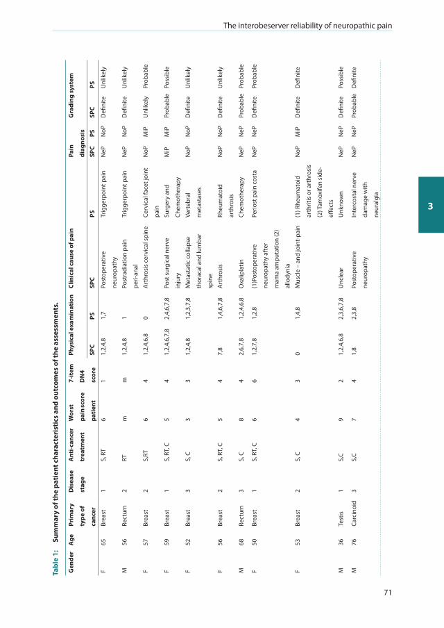
The interobeserver reliability of neuropathic pain
71
3
Tabl
e 1:
Su
mm
ary
of th
e pa
tien
t cha
ract
eris
tics
and
out
com
es o
f the
ass
essm
ents
.
Gen
der
Age
Prim
ary
type
of
canc
er
Dis
ease
stag
e
Ant
i-can
cer
trea
tmen
t
Wor
st
pain
scor
e
pati
ent
7-it
em
DN
4
scor
e
Phys
ical
exa
min
atio
nCl
inic
al c
ause
of p
ain
Pain
diag
nosi
s
Gra
ding
sys
tem
SPC
PSSP
CPS
SPC
PSSP
CPS
F65
Brea
st1
S, R
T6
11,
2,4,
81,
7Po
stop
erat
ive
neur
opat
hy
Trig
gerp
oint
pai
nN
ePN
oPD
efini
teU
nlik
ely
M56
Rect
um2
RTm
m1,
2,4,
81
Post
radi
atio
n pa
in
peri-
anal
Trig
gerp
oint
pai
nN
ePN
oPD
efini
teU
nlik
ely
F57
Brea
st2
S,RT
64
1,2,
4,6,
80
Art
hros
is c
ervi
cal s
pine
Cerv
ical
face
t joi
nt
pain
NoP
MiP
Unl
ikel
yPr
obab
le
F59
Brea
st1
S, R
T, C
54
1,2,
4,6,
7,8
2,4,
6,7,
8Po
st s
urgi
cal n
erve
inju
ry
Surg
ery
and
Chem
othe
rapy
MiP
MiP
Prob
able
Poss
ible
F52
Brea
st3
S, C
33
1,2,
4,8
1,2,
3,7,
8M
etas
tatic
col
laps
e
thor
acal
and
lum
bar
spin
e
Vert
ebra
l
met
asta
ses
NoP
NoP
Defi
nite
Unl
ikel
y
F56
Brea
st2
S, R
T, C
54
7,8
1,4,
6,7,
8A
rthr
osis
Rh
eum
atoi
d
arth
rosi
s
NoP
NoP
Defi
nite
Unl
ikel
y
M68
Rect
um3
S, C
84
2,6,
7,8
1,2,
4,6,
8O
xalip
latin
Chem
othe
rapy
NeP
NeP
Prob
able
Prob
able
F50
Brea
st1
S, R
T, C
66
1,2,
7,8
1,2,
8(1
)Pos
tope
rativ
e
neur
opat
hy a
fter
mam
a am
puta
tion
(2)
allo
dyni
a
Perio
st p
ain
cost
aN
ePN
ePD
efini
tePr
obab
le
F53
Brea
st2
S, C
43
01,
4,8
Mus
cle
– an
d jo
int-
pain
(1) R
heum
atoi
d
arth
ritis
or a
rthr
osis
(2) T
amox
ifen
side
-
effec
ts
NoP
MiP
Defi
nite
Defi
nite
M36
Test
is1
S,C
92
1,2,
4,6,
82,
3,6,
7,8
Unc
lear
Unk
now
nN
ePN
ePD
efini
tePo
ssib
le
M76
Carc
inoi
d3
S,C
74
1,8
2,3,
8Po
stop
erat
ive
neur
opat
hy
Inte
rcos
tal n
erve
dam
age
with
neur
algi
a
NeP
NeP
Prob
able
Defi
nite

Chapter 3
72
Tabl
e 1
cont
inue
d
F59
Brea
st3
S, R
T, C
94
4,7,
82,
3,7,
8(1
) Deg
ener
ativ
e
diso
rder
s (2
) mul
tiple
scle
rosi
s
Vert
ebra
l met
asta
sis
MiP
MiP
Prob
able
Unl
ikel
y
F52
Brea
st2
S, R
T, C
53
1,2,
4,8
2,6,
8(1
) Art
hros
is (2
)use
of
Tam
oxife
n
Deg
ener
ativ
e
diso
rder
bac
k
NoP
MiP
Unl
ikel
yU
nlik
ely
F58
Brea
st1
S,C
02
1,2,
4,6,
72,
3,6,
8N
o pa
inPr
othe
sis
NP
NoP
Unl
ikel
yU
nlik
ely
F50
Brea
st3
S, R
T, C
72
1,2,
87
Bone
met
asta
sis
cost
aeVi
scer
al b
one
pain
NoP
NoP
Unl
ikel
yU
nlik
ely
F65
Brea
st3
S, C
46
1,2,
4,,6
2,3,
6,8
Myo
gene
dis
orde
rs
+ Fe
ar
Chem
othe
rapy
,
surg
ery,
bac
kpai
n
and
med
icat
ion
NoP
MiP
Prob
able
Defi
nite
F60
Brea
st3
S, C
94
1, 8
1,2,
6,7,
Vert
ebra
l met
asta
sis
T10/
T11
Met
asta
tic d
isor
ders
with
inte
rcos
tal
neur
algi
a T1
1-T1
2
MiP
MiP
Defi
nite
Defi
nite
F62
Brea
st3
S, R
T, C
53
1,2,
4,6,
82,
3,6,
7,8
(1)A
rm p
ain:
pos
t
surg
ery
(2)H
and+
Feet
pain
pos
t tax
ol
Scar
tiss
ueN
PN
oPPr
obab
leU
nlik
ely
F49
Brea
st2
S, C
44
1,2,
4,,6
,82,
3,6,
8Ch
emot
hera
pyA
llody
nia,
sens
ibili
ty
diso
rder
NeP
NeP
Prob
able
Prob
able
F46
Brea
st2
S, R
T, C
73
1,2,
4,8
1,4,
6,8
Post
ope
rativ
e, p
ost
chem
othe
rapy
,
noci
cept
ive
pain
by
lym
phed
ema
(1)A
xilla
ir ly
mph
e
diss
ectio
n, (2
)
Radi
othe
rapy
, (3)
Chem
othe
rapy
, (4)
Mig
rain
e
MiP
MiP
Defi
nite
Defi
nite
F54
Brea
st2
S, C
53
1,2,
4,6,
7,8
2,7,
8Br
east
sur
gery
Post
sur
gica
l sca
r
pain
NeP
NeP
Prob
able
Prob
able
F52
Brea
st2
S, R
T, C
62
82,
7,8
Ant
i-oes
trog
en
trea
tmen
t
Reac
tive
arth
ritis
com
plai
nts
follo
win
g ho
rmon
al
trea
tmen
t
NoP
MiP
Unl
ikel
yU
nlik
ely
F61
GIS
T3
S, C
83
02,
3,7,
8M
issi
ngM
yote
ndin
ogen
lum
bar p
ain/
sac
ro
iliac
join
t pai
n
NoP
NoP
Unl
ikel
yU
nlik
ely
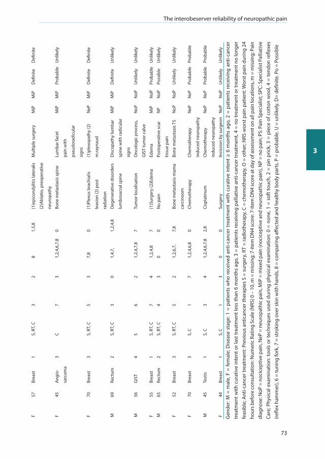
The interobeserver reliability of neuropathic pain
73
3
F57
Brea
st1
S, R
T, C
32
81,
5,8
(1)e
pico
ndyl
itis
late
ralis
(2)A
blat
io, p
reop
erat
ive
neur
opat
hy
Mul
tiple
sur
gery
MiP
MiP
Defi
nite
Defi
nite
F45
Ang
io-
sarc
oma
3C
73
1,2,
4,6,
7,8
0Bo
ne m
etas
tasi
s sp
ine
Lum
bar f
acet
pain
with
pseu
dora
dicu
lar
sign
s
MiP
MiP
Prob
able
Unl
ikel
y
F70
Brea
st3
S, R
T, C
53
7,8
0(1
)Ple
xus
brac
hial
is
leas
ion
(2) p
ost
radi
atio
n
(1)p
lexo
path
y (2
)
m.ra
ynau
d
NeP
MiP
Defi
nite
Defi
nite
M69
Rect
um2
S, R
T, C
30
1,4,
7,1,
2,4,
8D
egen
erat
ive
diso
rder
s
lum
bosa
cral
spi
ne
Dis
copa
thy
lum
bar
spin
e w
ith ra
dicu
lar
sign
s
MiP
MiP
Defi
nite
Unl
ikel
y
M56
GIS
T4
S6
21,
2,4,
7,8
7Tu
mor
loca
lisat
ion
Onc
olog
ic p
roce
ss,
GIS
T tu
mor
cal
ve
NoP
NoP
Unl
ikel
yU
nlik
ely
F55
Brea
st1
S, R
T, C
74
1,2,
4,8
7(1
)Sur
gery
(2)E
dem
aEd
ema
MiP
NoP
Prob
able
Unl
ikel
y
M65
Rect
um2
S, R
T, C
43
00
No
pain
Post
oper
ativ
e sc
ar
tissu
e pa
in
NP
NoP
Poss
ible
Unl
ikel
y
F52
Brea
st3
S, R
T, C
52
1,2,
6,7,
7,8
Bone
met
asta
sis
mam
a
carc
inoo
m
Bone
met
asta
sis T
5N
oPN
oPU
nlik
ely
Unl
ikel
y
F70
Brea
st3
S, C
17
1,2,
4,6,
80
Chem
othe
rapy
Chem
othe
rapy
indu
ced
neur
opat
hy
NeP
NeP
Prob
able
Prob
able
M45
Test
is1
S, C
34
1,2,
4,6,
7,8
2,8
Cisp
latin
umCh
emot
hera
py
indu
ced
neur
opat
hy
NeP
NeP
Prob
able
Prob
able
F44
Brea
st1
S, C
13
00
Surg
ery
Inci
ssio
n by
sur
geon
NoP
NoP
Unl
ikel
yU
nlik
ely
Gen
der:
M =
mal
e, F
= fe
mal
e; D
isea
se s
tage
: 1 =
pat
ient
s w
ho re
ceiv
ed a
nti-c
ance
r tr
eatm
ent
with
cur
ativ
e in
tent
≥ 6
mon
ths
ago,
2 =
pat
ient
s re
ceiv
ing
anti-
canc
er
trea
tmen
t with
cur
ativ
e in
tent
or l
ast t
reat
men
t les
s th
an 6
mon
ths
ago,
3 =
pat
ient
s re
ceiv
ing
palli
ativ
e an
ti-ca
ncer
trea
tmen
t, 4
= no
trea
tmen
t or t
reat
men
t no
long
er
feas
ible
; Ant
i-can
cer t
reat
men
t: Pr
evio
us a
ntic
ance
r the
rapi
es S
= s
urge
ry, R
T =
radi
othe
rapy
, C =
che
mot
hera
py, O
= o
ther
; NRS
wor
st p
ain
patie
nt: W
orst
pai
n du
ring
24
hour
s bef
ore
cons
ulta
tion:
Num
eric
Rat
ing
Scal
e (N
RS) 0
– 1
0, m
= m
issi
ng; 7
-item
DN
4 sc
ore:
7-it
em D
N4
scor
e at
day
of a
sses
smen
t on
all p
ain
loca
tions
, m =
mis
sing
; Pai
n di
agno
se: N
oP =
noc
icep
tive
pain
, NeP
= n
euro
path
ic p
ain,
MiP
= m
ixed
pai
n (n
ocic
eptiv
e an
d ne
urop
athi
c pa
in),
NP
= no
pai
n; P
S: P
ain
Spec
ialis
t; SP
C: S
peci
alis
t Pal
liativ
e Ca
re; P
hysi
cal e
xam
inat
ion:
tool
s or
tech
niqu
es u
sed
durin
g ph
ysic
al e
xam
inat
ion:
0 =
non
e, 1
= s
oft t
ouch
, 2 =
pin
pric
k, 3
= p
iece
of c
otto
n w
ool,
4 =
tend
on re
flexe
s (re
flex
ham
mer
), 6
= tu
ning
fork
, 7 =
str
okin
g ov
er s
kin
with
han
ds, 8
= c
ompa
ring
affec
ted
and
heal
thy
body
par
ts. P
= p
roba
ble.
U =
unl
ikel
y. D
= de
finite
. Po
= Po
ssib
le
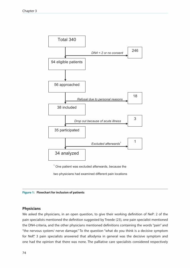
Chapter 3
74
94 eligible patients
34 analyzed
246
18
3
1
DN4 < 2 or no consent
Excluded afterwards1
56 approached
Total 340
Drop out because of acute illness
Refusal due to personal reasons
1 One patient was excluded afterwards, because the
two physicians had examined different pain locations
35 participated
38 included
Figure 1: Flowchart for inclusion of patients
Physicians We asked the physicians, in an open question, to give their working definition of NeP: 2 of the
pain specialists mentioned the definition suggested by Treede (23), one pain specialist mentioned
the DN4-criteria, and the other physicians mentioned definitions containing the words “pain” and
“the nervous system/ nerve damage.” To the question “what do you think is a decisive symptom
for NeP,” 3 pain specialists answered that allodynia in general was the decisive symptom and
one had the opinion that there was none. The palliative care specialists considered respectively
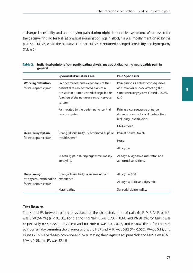
The interobeserver reliability of neuropathic pain
75
3
a changed sensibility and an annoying pain during night the decisive symptom. When asked for
the decisive finding for NeP at physical examination, again allodynia was mostly mentioned by the
pain specialists, while the palliative care specialists mentioned changed sensibility and hyperpathy
(Table 2).
Table 2: Individual opinions from participating physicians about diagnosing neuropathic pain in general.
Specialists Palliative Care Pain Specialists
Working definition for neuropathic pain
Pain or troublesome experience of the patient that can be traced back to a possible or demonstrated change in the function of the nerve or central nervous system.
Pain arising as a direct consequence of a lesion or disease affecting the somatosensory system (Treede, 2008). (2x)
Pain related to the peripheral or central nervous system.
Pain as a consequence of nerve damage or neurological dysfunction including sensitization.
DN4-criteria.
Decisive symptomfor neuropathic pain
Changed sensibility (experienced as pain/ troublesome).
Pain at normal touch.
None.
Allodynia.
Especially pain during nighttime, mostly annoying.
Allodynia (dynamic and static) and abnormal sensations.
Decisive sign at physical examination for neuropathic pain
Changed sensibility in an area of pain experience.
Allodynia. (2x)
Allodynia static and dynamic.
Hyperpathy. Sensorial abnormality.
Test Results The Κ and PA between paired physicians for the characterization of pain (NeP, MiP, NoP, or NP)
was 0.50 (64.7%) (P < 0.000). For diagnosing NeP Κ was 0.78, Pi 0.44, and PA 91.2%; for MiP it was
respectively 0.53, 0.38, and 79.4%; and for NoP it was 0.31, 0.26, and 67.6%. The Κ for the NeP
component (by summing the diagnoses of pure NeP and MiP) was 0.52 (P = 0.002), Pi was 0.18, and
PA was 76.5%. For the NoP component (by summing the diagnoses of pure NoP and MiP) Κ was 0.61,
Pi was 0.35, and PA was 82.4%.

Chapter 3
76
The interobserver reliability and the pair-wise agreement between the pain specialist and the
palliative care specialists regarding the grading system (unlikely, possible, probable, and definite
neuropathic pain) showed a Κ of 0.34 and a PA of 52.9%. The comparison between the NeP
component, following from the diagnosis of the physician and the outcome of the grading system
(the outcomes probable and definite were regarded as an NeP component was present) gave for
the pain specialists a Κ of 0.69, Pi 0.26, and PA of 85.3%. For the palliative care specialists it was
respectively 0.61, 0.03, and 79.4%. The comparison between the NeP component, following from the
diagnosis of the physician and the outcome of the DN4 (7-items, a “yes” on ≥ 3 items is considered
as having NeP) gave for the pain specialists a Κ of 0.24, Pi 0.36, and PA of 66.7%. For the palliative
care specialists it was respectively 0.16, 0.27, and 57.6%. The comparison between the outcome of
the grading system (the outcomes probable and definite were regarded as an NeP component was
present) and the outcome of the DN4 (7-items, a “yes” on ≥ 3 items is considered as having NeP) gave
for the pain specialists a Κ of 0.34, Pi 0.42, and PA of 72.7%. For the palliative care specialists it was
respectively 0.32, 0.15, and 63.6%.
Secondly, items from history taking and physical examination were assessed by video recording
and analyzed afterwards. In 27 out of 34 cases the palliative care specialists asked for a pain score
and the pain specialists asked in 21 cases. Most frequently asked items of the DN4 during history
taking were tingling (23 times by the palliative care specialists and 15 times by the pain specialists),
numbness (18 times by both), and burning (12 versus 19 times). During physical examination, the
cotton bud was most often used. The palliative care specialists used the sharp side of a cotton tip
22 times and the pain specialists 18 times. The soft side of it was used 25 times by the palliative care
specialists and 10 times by the pain specialists. Of the available tools, the cotton wool was used the
least: 9 times by the pain specialists while the palliative care specialists did not use it at all.
Table 3: The kappa coefficient (Κ) and the percentage of pair-wise (PA) agreement between physicians calculated for the patients’ diagnosis.
k-value Approx. Sig.
Categorized value of kappa
Pi PA-value (%)
Diagnosis (NeP, MiP, NoP, NP) 0.50 0.000* Moderate --- 64.7
NeP (NeP versus MiP + NoP + NP) 0.78 0.000 * Substantial 0.44 91.2
MiP (MiP versus NeP + NoP + NP) 0.53 0.001 * Moderate 0.38 79.4
NoP (NoP versus NeP + MiP + NP) 0.31 0.08 Fair 0.26 67.6
NePcomponent (NeP + MiP versus NoP + NP) 0.52 0.002* Moderate 0.18 76.5
NoPcomponent (NoP + MiP versus NeP + NP) 0.61 0.000* Substantial 0.35 82.4
k-value: Kappa value; 95% CI: 95% confidence interval; Approx. Sig.: Approximate significance; *: significant, P ≤ 0,05; Pi: Prevalence index; Pavalue: Pair-wise Agreement-value; NeP: neuropathic pain; MiP: mixed pain; NoP: nociceptive pain; NP: no pain; Fair: ĸ = 0.21 – 0.40; Moderate: ĸ = 0.41 – 0.60; Substantial: ĸ = 0.61 – 0.80.
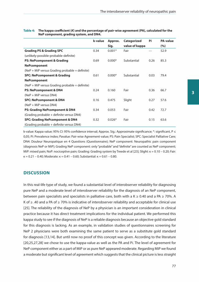
The interobeserver reliability of neuropathic pain
77
3
Table 4: The kappa coefficient (Κ) and the percentage of pair-wise agreement (PA), calculated for the NeP component, grading system, and DN4.
k-value Approx. Sig.
Categorized value of kappa
Pi PA-value (%)
Grading PS & Grading SPC (unlikely-possible-probable-definite)
0.34 0.001* Fair --- 52.9
PS: NePcomponent & Grading NePcomponent (NeP + MiP versus Grading probable + definite)
0.69 0.000* Substantial 0.26 85.3
SPC: NePcomponent & Grading NePcomponent (NeP + MiP versus Grading probable + definite)
0.61 0.000* Substantial 0.03 79.4
PS: NePcomponent & DN4 (NeP + MiP versus DN4)
0.24 0.160 Fair 0.36 66.7
SPC: NePcomponent & DN4 (NeP + MiP versus DN4)
0.16 0.475 Slight 0.27 57.6
PS: Grading NePcomponent & DN4 (Grading probable + definite versus DN4)
0.34 0.053 Fair 0.42 72.7
SPC: Grading NePcomponent & DN4 (Grading probable + definite versus DN4)
0.32 0.026* Fair 0.15 63.6
k-value: Kappa value; 95% CI: 95% confidence interval; Approx. Sig.: Approximate significance; *: significant, P ≤ 0,05; Pi: Prevalence index; Pavalue: Pair-wise Agreement-value; PS: Pain Specialist; SPC: Specialist Palliative Care; DN4: Douleur Neuropatique en 4 Questions (Questionnaire); NeP component: Neuropathic pain component (diagnosis NeP or MiP); Grading NeP component: only “probable” and “definite” are counted as NeP component; MiP: mixed pain; NoP: nociceptive pain; Grading: Grading system by Treede et al [23]; Slight: ĸ = 0.10 – 0.20; Fair: ĸ = 0.21 – 0.40; Moderate: ĸ = 0.41 – 0.60; Substantial: ĸ = 0.61 – 0.80.
DISCUSSION
In this real-life type of study, we found a substantial level of interobserver reliability for diagnosing
pure NeP and a moderate level of interobserver reliability for the diagnosis of an NeP component,
between pain specialists and specialists in palliative care, both with a Κ ≥ 0.40 and a PA ≥ 70%. A
Κ of ≥ .40 and a PA of ≥ 70% is indicative of interobserver reliability and acceptable for clinical use
[25]. The reliability of the diagnosis of NeP by a physician is an important consideration in clinical
practice because it has direct treatment implications for the individual patient. We performed this
kappa study to see if the diagnosis of NeP is a reliable diagnosis because an objective gold standard
for this diagnosis is lacking. As an example, in validation studies of questionnaires screening for
NeP 2 physicians were both examining the same patient to serve as a substitute gold standard
for diagnosis [13,14]. But until now no proof of this concept was given. According to the literature
[20,25,27,28] we chose to use the kappa-value as well as the PA and Pi. The level of agreement for
NeP component either as a part of MiP or as pure NeP appeared moderate. Regarding MiP we found
a moderate but significant level of agreement which suggests that the clinical picture is less straight
Chapter 3
78
forward. Probably, a combined pain syndrome is a less clear outcome, explaining the lower kappa.
For pure NoP the physician pairs only had a fair, non significant level of agreement. The PA-value for
NoP was below 70% and thus considered as not acceptable for clinical use. This might be due to the
focus of the physicians: the instruction of the physician was to diagnose NeP in the way they were
used to in their daily practice. Probably there was less attention to NoP. For NoP component the level
of agreement was substantial.
Although the participating physicians used different descriptions for NeP, a high consensus existed
for the decisive symptom and sign for NeP, namely allodynia or a description of allodynia. However,
allodynia is not a decisive symptom for NeP, because it might also be present in patients with
nociceptive pain, especially in inflammatory conditions.
The presented results indicate that the specialists used very different diagnostic criteria for
neuropathic pain. This was confirmed most notably by the working definition used by the
investigators, which corresponded to the IASP definition of neuropathic pain in only one third of
the investigators (Table 2). In conclusion, the majority of the participating physicians didn’t know
the current definition of neuropathic pain and use “personalized” inappropriate diagnostic criteria
in their daily practice.
In this study, we also have used the grading system by Treede et al [23], filled in by both physicians
after the clinical examination of the patient. Comparing the diagnosis of the existence of an
NeP component with the outcome of the grading system per physician, we found a substantial
intraobserver reliability with a PA ≥ 70%, indicating a good reliability and useful in clinical practice.
However, the comparison on the outcome of the grading system (unlikely, possible, probable, or
definite) between both physicians gave a fair reliability and a low PA (< 70%), indicating a poor
reliability between both physicians and therefore it might be less useful in clinical practice.
Moreover, the grading system will not necessarily provide the right diagnosis. In a patient suffering
from MiP, the NoP part may be paramount. The physicians’ diagnosis (NeP, MiP, NoP, or NP) had a
moderate reliability, but also a low PA < 70%. All this indicates that it is difficult to categorize the
kind of pain the patient is suffering from, as well with the physicians’ diagnosis as with the grading
system. It can be questioned whether the clinical judgment should be regarded as a gold standard
for the diagnosis of NeP because both clinicians might be wrong in their diagnosis even with values
of Κ > 0.5 and a PA of 70%.
Our study measured the interobserver reliability of 2 physicians diagnosing NeP in patients with
pain from cancer and taking the grading system and the DN4 into account. The focus of the study
was to diagnose the kind of pain and not on which specific diagnostic tests were used in the
diagnostic process. Mostly, kappa studies are used to report the reliability of specific diagnostic
tests in patients or from clinical data [18,29-31]. Comparing the outcome of the physicians diagnosis

The interobeserver reliability of neuropathic pain
79
3
on the existence of an NeP component with the outcome of the 7-item DN4 we found only a fair (Κ
< 0.40) reliability for the pain specialists and a slight interobserver reliability for the palliative care
specialists (both with a PA ≤ 70%). In the paper of Garcia de Paredes et al [7] it was described that
only half of the patients with cancer suffering from NeP had a positive score on the DN4 compared
with the clinicians diagnosis. They suggested investigating if a specific cut-off score for the DN4 for
patients with NeP from cancer would fit better. The same was suggested in the study by Mercadente
et al [32] for the LANSS, NPQ, and NPQ-SF. This study also indicates that the DN4, at this moment, is
less valid and thus less useful in clinical practice for screening for NeP in patients with cancer pain.
During the pain history taking, the pain specialists asked for a pain intensity score only 21 out of
34 times and the palliative care specialists 27 times. A marginal comment should be made on this
statement, as the physicians were only instructed to diagnose the type of pain. However, one expects
a pain intensity score to be a standard item during a pain history taking. During the observation of
the clinical examination of the patients, in 10 of 68 cases (Table 1), no clinical examination was
performed, and in many cases only one sensory modality was tested. It has been recommended
[16] that (a) clinical bedside (sensory) examination of a patient with suspected NeP includes testing
of touch/vibration, cold, warmth, and pain sensibility (pinprick) and (b) the outcomes should be
compared with the findings in the contra lateral region or in a region without pain (not performed
in 21 of 68 cases).
To our knowledge, this is the first study to determine the interobserver reliability of the diagnosis of
NeP in patients with cancer. Participation of patients, examination rooms that were equipped as real
consultation rooms, and instructing the physicians to perform the diagnosing procedure as they
usually do, all contributed to collect reliable information about the current state of daily practice in
this hospital. Besides, by using a video camera that was almost invisible to the physician and patient,
the consultation was not disturbed by the researchers.
While the patient number (N = 34) is sufficient for a reliable kappa study, the number of participating
physicians was low and unequal: 4 participating pain specialists and 2 palliative care specialists.
Another weak aspect was the fact that one of the palliative care specialists was a medical oncologist
and the other an elderly care physician. Yet, both of them had palliative care as their main task for
at least 7 years. However, both the pain specialists and the palliative care specialists will be more
experienced than usual physicians in pain and NeP and our findings cannot be interpreted for a
broader group of physicians. Furthermore, in the questionnaire, patients were asked about all sites
of pain; whereas in the clinical examination, physicians were instructed to focus on the site of worst
pain. The majority of patients had breast cancer. This high number is an adequate representation
as breast cancer is the most frequent type of cancer among women in the Netherlands (www.
cijfersoverkanker.nl) and many of them suffer from chronic pain [33,34]. The incidence of NeP in
this study is artificially high in comparison with the normal population in the oncology outpatient
Chapter 3
80
clinic. Because we used a score of at least 2-points on the 7-item DN4 as an inclusion criterion for
this study, the presence of NeP was more likely and thus enlarged the possibility of diagnosing
NeP. For now it is not sure that in a situation of a lower incidence of NeP the kappa values will
be the same. The physicians were also more triggered and focused on NeP than on NoP because
we asked their working definition of NeP, the symptoms and findings at physical examination they
considered decisive for NeP, and their self-efficacy in diagnosing NeP. This is probably the cause of
a lower kappa-value in patients with NoP. Finally, the worst pain did not necessarily originate from
the cancer or anti-cancer treatment. Patients sometimes had comorbidity causing the (worst) pain,
for example rheumatoid arthritis.
CONCLUSIONS
We found a substantial level of agreement for the diagnosis of NeP and a moderate level of agreement
for diagnosing an NeP component, both with a PA ≥ 70%. This study shows preliminary evidence
that the clinical judgment of NeP in patients with cancer is reliable. Implementation of the proposed
criteria for categorizing NeP as definite, probable, possible, or unlikely might be a step forward [23]
to come to more diagnostic clarity for NeP. As stated by Bennett et al [8] a standardized approach
is essential for clinical assessment, for appropriate treatment, and for clinical research. Despite the
lack of a gold standard for diagnosing NeP, our study shows that physicians have a good agreement
in the diagnosis of pure NeP. For MiP however, the level of agreement is moderate but with a high
PA. Based on these findings, in MiP we suggest the opinion of a second physician to enlarge the
chance of a correct diagnosis and thereby of adequate pain treatment. Especially in more complex
pain syndromes, the recognition of NeP component needs attention. Since the treatment of NeP
and MiP or NoP is quite different according to the international guidelines, a strict delineation
and certitude about the correct diagnosis is of upmost importance and will influence the result
of consequent pharmacological treatment schemes [35]. Taking into account the different pain
mechanisms of NeP and NoP and working mechanisms of the medications, it is important to have
an adequate pain diagnosis for optimal pain treatment with the least side effects. The general value
of the findings for validating physician assessment of neuropathic cancer pain in this study is limited
to our centre and participating physicians in order to confirm their relevance and general interest.
However, the findings in this study suggest that a better standardization of the clinical assessment
and classification of pain in patients with cancer in respect to the identification of neuropathic pain
is necessary. Moreover, we recommend a further study on how to improve the level of agreement
in, and the validity of, the clinical diagnosis of NeP by systematically analyzing the history taking
and the different (diagnostic) tools used in pain assessment and how standardizing the diagnostic
process can improve the level of agreement and validity in clinical circumstances [16,23].

The interobeserver reliability of neuropathic pain
81
3
ACKNOWLEDGEMENTS
We thank the participating patients and physicians for their contribution to this study.
FUNDING
This study was co-funded by the Netherlands Organisation for Health Research and Development.
No specific conflicts of interest are present for the authors.
Chapter 3
82
REFERENCES
1. van den Beuken-van Everdingen MH, de Rijke JM, Kessels AG, Schouten HC, van Kleef M, Patijn J. High prevalence of pain in patients with cancer in a large population-based study in The Netherlands. Pain 2007; 132:312-320.
2. van den Beuken-van Everdingen MH, de Rijke JM, Kessels AG, Schouten HC, van Kleef M, Patijn J. Prevalence of pain in patients with cancer: a systematic review of the past 40 years. Ann Oncol 2007; 18:1437-1449.
3. Caraceni A, Portenoy RK. An international survey of cancer pain characteristics and syndromes. IASP Task Force on Cancer Pain. International Association for the Study of Pain. Pain 1999; 82:263-274.
4. Grond S, Radbruch L, Meuser T, Sabatowski R, Loick G, Lehmann KA. Assessment and treatment of neuropathic cancer pain following WHO guidelines. Pain 1999; 79:15-20.
5. Grond S, Zech D, Diefenbach C, Radbruch L, Lehmann KA. Assessment of cancer pain: a prospective evaluation in 2266 cancer patients referred to a pain service. Pain 1996; 64:107-114.
6. Manas A, Monroy JL, Ramos AA, Cano C, Lopez-Gomez V, Masramon X, Perez M. Prevalence of neuropathic pain in radiotherapy oncology units. Int J Radiat Oncol Biol Phys 2011; 81:511-520.
7. Garcia de Paredes ML, del Moral Gonzalez F, Martinez del Prado P, Marti Ciriquian JL, Enrech Frances S, Cobo Dols M, Esteban Gonzalez E, Ortega Granados AL, Majem Tarruella M, Cumplido Buron JD, Gasco Hernandez A, Lopez Miranda E, Ciria Santos JP, Castro Carpeno FJ. First evidence of oncologic neuropathic pain prevalence after screening 8615 cancer patients. Results of the On study. Ann Oncol 2011; 22:924-930.
8. Bennett MI, Rayment C, Hjermstad M, Aass N, Caraceni A, Kaasa S. Prevalence and aetiology of neuropathic pain in cancer patients: a systematic review. Pain 2012; 153:359-365.
9. The International Association for the Study of Pain (IASP): http://www.iasppain.org/Content/NavigationMenu/ GeneralResourceLinks/PainDefinitions/ default.htm
10. Freynhagen R, Bennett MI. Diagnosis and management of neuropathic pain. BMJ 2009; 339:b3002.11. Krause SJ, Backonja MM. Development of a neuropathic pain questionnaire. Clin J Pain 2003; 19:306-314. 12. Bennett MI, Attal N, Backonja MM, Baron R, Bouhassira D, Freynhagen R, Scholz J, Tölle TR, Wittchen HU,
Jensen ST. Using screening tools to identify neuropathic pain. Pain 2007; 127:199-203. 13. Bouhassira D, Attal N, Alchaar H, Boureau F, Brochet B, Bruxelle J, Cunin G, Fermanian J, Ginies P, Grun-
Overdyking A, Jafari-Schluep H, Lanteri-Minet M, Laurent B, Mick G, Serrie A, Valade D, Vicaut E. Comparison of pain syndromes associated with nervous or somatic lesions and development of a new neuropathic pain diagnostic questionnaire (DN4). Pain 2005; 114: 29-36.
14. Freynhagen R, Baron R, Gockel U, Tolle TR. PainDETECT: a new screening questionnaire to identify neuropathic components in patients with back pain. Curr Med Res Opin 2006; 22:1911-1920.
15. Bennett M. The LANSS Pain Scale: the Leeds assessment of neuropathic symptoms and signs. Pain 2001; 92:147-157.
16. Haanpaa M, Attal N, Backonja M, Baron R, Bennett M, Bouhassira D, Cruccu G, Hansson P, Haythornthwaite JA, Ianetti GD, Jensen TS, Kauppila T, Nurmikko TJ, Rice ASC, Rowbotham M, Serra J, Sommer C, Smith BH, Treede RD. NeuPSIG guidelines on neuropathic pain assessment. Pain 2011; 152:14-27.
17. Duong L, Cheriet F, Labelle H, Cheung KM, Abel MF, Newton PO, et al. Interobserver and intraobserver variability in the identification of the Lenke classification lumbar modifier in adolescent idiopathic scoliosis. J Spinal Disord Tech 2009; 22:448-455.
18. Lovelock CE, Anslow P, Molyneux AJ, Byrne JV, Kuker W, Pretorius PM, Coull A, Rothwell PM. Substantial observer variability in the differentiation between primary intracerebral hemorrhage and hemorrhagic transformation of infarction on CT brain imaging. Stroke 2009; 40:3763-3767.
19. Van Suijlekom HA, De Vet HC, Van Den Berg SG, Weber WE. Interobserver reliability in physical examination of the cervical spine in patients with headache. Headache. 2000; 40:581-586.
20. Sim J, Wright CC. The kappa statistic in reliability studies: use, interpretation, and sample size requirements. Phys Ther 2005; 85:257-268.
21. Cohen J. A coefficient of agreement for nominal scales. Educational and psychological measurement 1960; XX:37-46.
22. Cleeland CS, Ryan KM. Pain assessment: global use of the Brief Pain Inventory. Ann Acad Med Singapore 1994; 23:129-138.

The interobeserver reliability of neuropathic pain
83
3
23. Treede RD, Jensen TS, Campbell JN, Cruccu G, Dostrovsky JO, Griffin JW, Hansson P, Hughes R. Nurmikko T, Serra J. Neuropathic pain: redefinition and a grading system for clinical and research purposes. Neurology 2008; 70:1630-1635.
24. Landis JR, Koch GG. The measurement of observer agreement for categorical data. Biometrics 1977; 33:159-174.
25. Bron C, Franssen J, Wensing M, Oostendorp RA. Interrater reliability of palpation of myofascial trigger points in three shoulder muscles. J Man Manip Ther. 2007; 15:203-215.
26. Portney LGW, Watkins M.P. Foundations of Clinical Research: Applications to Practice. 2nd edition edn: Prentice Hall Health, Upper Saddle River, New jersey, USA, 2000.
27. Cicchetti DV, Feinstein AR. High agreement but low kappa: II. Resolving the paradoxes. J Clin Epidemiol 1990; 43:551-558.
28. Feinstein AR, Cicchetti DV. High agreement but low kappa: I. The problems of two paradoxes. J Clin Epidemiol 1990; 43:543-549.
29. Bhatia KS, Cho CC, Yuen YH, Rasalkar DD, King AD, Ahuja AT. Real-time qualitative ultrasound elastography of cervical lymph nodes in routine clinical practice: interobserver agreement and correlation with malignancy. Ultrasound Med Biol 2010; 36:1990-1997.
30. Richardson JK. The clinical identification of peripheral neuropathy among older persons. Arch Phys Med Rehabil 2002; 83:1553-1558.
31. Wald R, Bell CM, Nisenbaum R, Perrone S, Liangos O, Laupacis A, Jaber BL.Interobserver reliability of urine sediment interpretation. Clin J Am Soc Nephrol 2009; 4:567-571.
32. Mercadente S, Gebbia V, David F, Aielli F, Verna L, Casuccio A, Porzio G, Mangione S. Tools for identifying cancer pain of predominantly neuropathic origin and opioid responsiveness in cancer patients. J Pain 2009; 6:594-600
33. Steegers MA, Snik DM, Verhagen AF, van der Drift MA, Wilder-Smith OH. Only half of the chronic pain after thoracic surgery shows a neuropathic component. J Pain 2008; 9:955-961.
34. Steegers MA, Wolters B, Evers AW, Strobbe L, Wilder-Smith OH. Effect of axillary lymph node dissection on prevalence and intensity of chronic and phantom pain after breast cancer surgery. J Pain 2008; 9:813-822.
35. Vadalouca A, Raptis E, Moka E, Zis P, Sykioti P, Siafaka I. Pharmacological treatment of neuropathic cancer pain: a comprehensive review of the current literature. Pain Pract 2012; 12:219-251.

CHAPTER 4Detecting the neuropathic pain component in the clinical setting: a study protocol for validation of screening instruments for the presence of a neuropathic pain component
Hans Timmerman
Oliver Wilder-Smith
Chris van Weel
André Wolff
Kris Vissers
Published in:
BMC Neurology 2014 14:94.
Chapter 4
86
ABSTRACT
Background
The presence of nerve damage plays a key role in the development and prognosis of chronic
pain states. Assessment of the presence and severity of a neuropathic pain component (NePC) is
key in diagnosing chronic pain patients. Low back pain (LBP) and neck and shoulder pain (NSP)
are highly prevalent and clinically important medical and societal problems in which a NePC
is frequently present. The more severe the NePC, the worse the course of the pain, its prognosis
and the results of treatment. Reliable and standardised diagnosis of the NePC remains difficult
to achieve. Standardized and validated screening tools may help to reliably identify the NePC in
individual chronic pain patients. The aim of this study is to validate the Dutch language versions of
the PainDETECT Questionnaire (PDQ-Dlv) and the ‘Douleur Neuropathique 4 Questions’ (DN4-Dlv)
for use in primary and specialist medical care settings to screen for a NePC in patients with chronic
pain due to (1) LBP, (2) NSP or (3) known peripheral nerve damage (PND).
Methods / design
The study design is cross-sectional to assess the validity of the PDQ-Dlv and the DN4-Dlv with 2
weeks follow-up for test-retest reliability and 3 months follow-up for monitoring and prognosis.
438 patients with chronic pain due to (1) LBP, (2) NSP or (3) PND will be included in this study. Based
on the IASP definition of neuropathic pain, two physicians will independently assess whether the
patient has a NEPC or not. This result will be compared with the outcome of the PDQ-Dlv & DN4-Dlv,
the grading system for neuropathic pain, bed side examination and quantitative sensory testing.
This study will further collect data regarding prevalence of NePC, general health status, mental
health status, functioning, pain attribution and quality of life.
Discussion
The rationale for this study is to provide detailed information on the clinimetric quality of the PDQ-
Dlv and DN4-Dlv in Dutch speaking countries. Our innovative multi-factorial approach should help
achieve more reliable diagnosis and quantification of a NePC in patients with chronic pain.
Trial registration
The Netherlands National Trial Register (NTR3030).
Keywords
PainDETECT questionnaire, PDQ, DN4, Validation, Low back pain, Neck-shoulder pain, Peripheral
nerve damage

Study protocol
87
4
BACKGROUND
The International Association for the Study of Pain (IASP, 2011) defines Neuropathic Pain (NeP) as
‘pain caused by a lesion or disease of the somatosensory nervous system’ (http://www.iasp-pain.
org/Education/Content.aspx?ItemNumber=1698 #Neuropathicpain). This definition will be used in
this study because of its diagnostic specificity, anatomic precision and the usefulness in clinical as
well as research conditions [1]. NeP plays an important role in the development and prognosis of
chronic pain states. A relevant example is patients with low back pain (LBP) and neck-shoulder pain
(NSP), which are both highly prevalent and clinically important medical and societal problems: In
this context, the more severe the NeP, the worse the pain course, the prognosis and the results of
treatment [2-5].
The incidence of NeP in the Dutch general population is 0.81% or 130.000 new patients in the
Netherlands per year. NeP is 63% more common in women than in men and peaks between 70
and 79 years of age [6]. LBP as well as NSP are among the top 10 health problems encountered
in general practice. For men and women, respectively, the prevalence of LBP and/or NSP in the
general practice is in the range of 55 – 86 and 24 – 113 per 1000 patients a year. In general practice,
radiating pain from the low back or neck occurs in men and women in respectively 4 – 8 and 10
patients per 1000 patients [7] (http://www.nationaalkompas.nl/gezondheid-en-ziekte/ ziekten-en-
aandoeningen/bewegingsstelsel-en-bindweefsel/ nek-en-rugklachten/ omvang/). The prevalence
of chronic pain syndromes due to peripheral nerve damage (PND) is 3,3 per 1000 per year [8].
Strictly speaking, the diagnosis of neuropathic pain is a patho-anatomical diagnosis presuming
knowledge regarding nerve injury which is difficult to obtain in the clinical situation. Thus in the
clinical context it is better to speak of a neuropathic pain component (NePC), which is a clinical
syndrome based on a typical set of clinical symptoms and signs. Clinically, a NePC is characterized
by spontaneous pain and abnormal pain sensations [9]. NeP is typically described as a spontaneous
ongoing burning or shooting pain with spontaneous sharp exacerbations and somatosensory
abnormalities after a (non-) noxious stimulus [10].
As a rule, a NePC has a considerable impact on the quality of daily life [6]. Hence it is important for
physicians in daily practice (specialist care as well as primay care) to diagnose the presence and
severity of a NePC in individual patients. In clinical practice it is, however, often difficult to reliably
diagnose a NePC in (sub)acute and chronic pain of the low back and neck shoulder region. The
diagnosis of a NePC is at present primarily based on clinical examination by a physician including
sensory examination. Quantitative sensory testing (QST) may provide extra information for selected
clinical cases and in the research context [11,12].
Chapter 4
88
Because a reliable diagnosis of the neuropathic pain component is often difficult to accomplish in
routine practice [2], it would be helpful to have a screening tool to detect such a component for
clinical triage and epidemiological purposes [12,13]. Apart from optimal sensitivity and specificity,
such a screening tool should be easy to use in clinical practice, not only for the first visit but also
during follow up. The availability of such a simple, validated, Dutch language screening tool should
improve diagnosis and quantification of a NePC and hence lead to better therapy. At present, no
specific (validated) instrument to determine the neuropathic component in LBP, NSP and PND
is available in the Dutch language. The PainDETECT-Questionnaire (PDQ) [2] and the Douleur
Neuropatique 4 Questions (DN4) [14] were originally developed and validated in Germany and
France, respectively. Both are considered to be reliable screening tools with a high sensitivity,
specificity and positive predictive value. Recently, the DN4 and the PDQ have been translated into
a Dutch language version (Dlv) by Van Seventer et al [15] and Timmerman et al [16], respectively.
Validation of the Dutch versions of DN4-Dlv and PDQ-Dlv will improve the identification of a NePC
in Dutch primary and specialist medical care, also facilitating remote follow up evaluation by
telephone, internet or post for clinical and scientific purposes. We chose an innovative approach
which should lead to a more reliable identification and quantification of a NePC in patients with
chronic pain. This study will help define patient groups at risk for a NePC and will help to understand
and assess the variability and burden of a NePC in individual patients.
The aim of this study is to establish the clinimetric quality, including 2-weeks test-retest reliability, of
the PDQ-Dlv and the DN4-Dlv for use in primary care and specialist medical care settings in Dutch
speaking countries for patients with chronic pain due to LBP, NSP or known peripheral nerve damage
(PND). Follow-up for monitoring and prognosis properties of DN4-Dlv and PDQ-Dlv for a NePC will
be done over a period of 3 months. Additional data will be collected regarding NEPC prevalence,
general health status, mental health status, functioning, pain attribution and health related quality
of life in patients with chronic pain.
METHODS
The medical and ethical review board Committee on Research Involving Human Subjects region
Arnhem-Nijmegen, Nijmegen, the Netherlands, has given approval to conduct this study, Dossier
number: 2008/348; NL 25343.091.08; The Netherlands National Trial Register NTR3030.
Design In this study a cross-sectional, observational, research design with 3-months follow up will be used
to study the clinimetric quality of the DN4-Dlv and PDQ-Dlv (Figure 1).
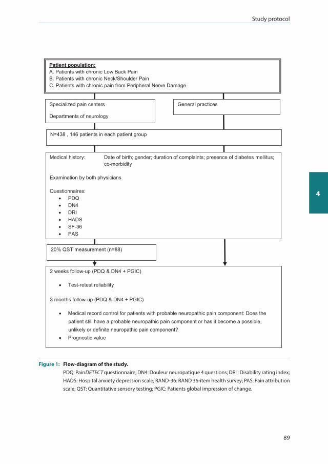
Study protocol
89
4
Specialized pain centers
Departments of neurology
General practices
N=438 , 146 patients in each patient group
Medical history: Date of birth; gender; duration of complaints; presence of diabetes mellitus; co-morbidity
Examination by both physicians Questionnaires:
PDQ DN4 DRI HADS SF-36 PAS
Patient population: A. Patients with chronic Low Back Pain B. Patients with chronic Neck/Shoulder Pain C. Patients with chronic pain from Peripheral Nerve Damage
20% QST measurement (n=88)
2 weeks follow-up (PDQ & DN4 + PGIC)
Test-retest reliability
3 months follow-up (PDQ & DN4 + PGIC)
Medical record control for patients with probable neuropathic pain component: Does the patient still have a probable neuropathic pain component or has it become a possible, unlikely or definite neuropathic pain component?
Prognostic value
Figure 1: Flow-diagram of the study. PDQ: PainDETECT questionnaire; DN4: Douleur neuropatique 4 questions; DRI : Disability rating index;
HADS: Hospital anxiety depression scale; RAND-36: RAND 36-item health survey; PAS: Pain attribution scale; QST: Quantitative sensory testing; PGIC: Patients global impression of change.
Chapter 4
90
Setting Multicenter recruitment will be take place in academic pain centres, non-academic pain centres and
non-academic departments of neurology. Patients will be seen by the two physicians during normal
office hours, or when that is not possible during a special office hour for this study. Furthermore,
patients willing to participate in this study from general practices will also be included in this
study via a special office hour in the clinical trainings centre of Radboud university medical center
(Radboudumc).
Each patient will be seen by two physicians, independently of each other, working in the same
institute. The medical background of the participating physicians is diverse (experienced pain
specialists, pain specialist trainees, experienced neurologists and experienced general practitioners).
Participants The patients will be recruited non-selectively and consecutively in the period from September 2009
till July 2013. Inclusion criteria: Male and female adult patients (>18 years of age) with chronic (>3
months) LBP or NSP radiating into respectively leg(s) or arm(s) or patients with chronic pain due
to PND. Exclusion criteria: Patients diagnosed with malignancy, compression fractures, patients
with painful syndromes of unknown origin or associated with diffuse pains (such as ankylosing
spondylitis or fibromyalgia), severe mental illness, chronic alcoholism or substance abuse, inability
to fill in the questionnaire adequately, or incapable of understanding Dutch. Subjects can leave
the study at any time for any reason without any consequences. The investigator can decide to
withdraw a subject from the study for urgent medical reasons. When an individual subject leaves
the study all the information from the patient will be kept in the database, and possibly be used for
data analysis or withdrawal analysis. Such patients will be replaced.
Measurements At the first visit, each patient will be seen by two physicians who will question and examine the
patients. They will then independently classify the patients’ pain as pain with or without a NePC,
based in the IASP NeP criteria and supported by a standardized assessment form. Next, the patient
will complete seven questionnaires (including the PDQ and the DN4). Twenty percent of the patients
will additionally undergo QST measurement following the first visit. Two weeks and three months
after the initial visit three follow-up questionnaires will be sent to the patient by mail.
Demographics Date of birth, gender, weight (Kg), length (m), nationality, nationality of origin, pain medication,
smoking (cigarettes a day), alcohol consumption (units per day) and education level will be assessed
by use of a self completed questionnaire. Pain at this moment will be assessed by use of a numeric
rating scale (0-10, NRS). Medical co-morbidity, duration of complaints (years/months), presence of
diabetes mellitus (yes/no), presence of HIV (yes/no), presence of herpes (yes/no) and undergoing of
Study protocol
91
4
chemotherapy (yes/no) are based on interview by the physician and noted by the physician on the
standardized assessment form.
Pain classification At each centre participating patients will be examined by two (rater A and B) independent and
trained pain physicians, two experienced neurologists or two experienced general practitioners,
working independently of each other and blinded to the diagnosis of the other physician. To achieve
standardization of the history and clinical examination all participating physicians will be trained at
the sites. Both physicians will classify the pain regarding the presence or absence of a NePC based on
history and clinical examination. Level of certainty of the physicians regarding the pain component
classification will be assessed by use of a Visual Analogue Scale (VAS, range 0-100). The findings are
noted on the standardized assessment form by the physician. To monitor the quality of the clinical
examination random quality checks, by expected/ unexpected visits, will be used.
Grading system The grading system for neuropathic pain as proposed by Treede et al. [1] will be used as a secondary
comparison with the outcome of both the PDQ-Dlv and DN4-Dlv and with the outcome of the
original pain classification by the two physicians. This system provides a working hypothesis for the
origin of patients’ pain. The criteria are graded on basis of history and testing in medical examination
[1]: (1) Pain with a distinct neuroanatomically plausible distribution; (2) A history suggestive
of a relevant lesion or disease affecting the peripheral or central somatosensory system; (3)
Demonstration of the distinct neuroanatomically plausible distribution by at least one confirmatory
test; (4) Demonstration of the relevant lesion or disease by at least one confirmatory test lesion or
disease explaining NeP. When the criteria 1-4 are all fulfilled the pain will be graded as possessing
a ‘definite NePC’. Criteria 1 and 2 and either 3 or 4 will be graded as ‘probable NePC’. Criteria 1 & 2
without criteria 3 or 4 will be graded as ‘possible NePC’. The pain is ‘unlikely to have a NePC’ when
no criteria, or only criteria 1 or 2, are graded as present (Table 1). The answers (yes or no) to the four
criteria are noted by the physician on the standardized assessment form. Three months after the first
consultation by the two physicians the medical record of patients with ‘probable NePC’ according
to the grading system will be controlled: i.e. does the patient still have probable NePC, or does he
now have definite NePC?.
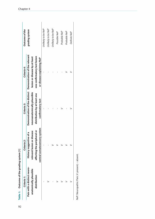
Chapter 4
92
Tabl
e 1:
O
utco
me
of th
e gr
adin
g sy
stem
[1]
Crite
ria
1:Cr
iteri
a 2:
Crite
ria
3:Cr
iteri
a 4:
Out
com
e of
the
gr
adin
g sy
stem
Pain
wit
h a
dist
inct
neu
ro-
anat
omic
ally
pla
usib
le
dist
ribu
tion
His
tory
sug
gest
ive
of a
re
leva
nt le
sion
or d
isea
se
affec
ting
the
peri
pher
al o
r ce
ntra
l som
atos
enso
ry s
yste
m
Dem
onst
rati
on o
f the
dis
tinc
t ne
uroa
nato
mic
ally
pla
usib
le
dist
ribu
tion
by
at le
ast o
ne
confi
rmat
ory
test
Dem
onst
rati
on o
f the
rele
vant
le
sion
or d
isea
se b
y at
leas
t on
e co
nfirm
ator
y te
st le
sion
or
dis
ease
exp
lain
ing
NeP
--
--
Unl
ikel
y to
be
NeP
V-
--
Unl
ikel
y to
be
NeP
-V
--
Unl
ikel
y to
be
NeP
VV
--
Poss
ible
NeP
VV
V-
Prob
able
NeP
VV
-V
Prob
able
NeP
VV
VV
Defi
nite
NeP
NeP
: Neu
ropa
thic
Pai
n; V
: pre
sent
; -: a
bsen
t.
Study protocol
93
4
Bedside examination Bed-side examination of the patient is performed by both physicians. The aim of this examination is
to find possible abnormalities suggestive for a relevant lesion or disease which affects the peripheral
or central somatosensory system [17]. The value of bed-side examination within the clinical
examination is that it will give insight in the pathology and the localization of the lesion or disease
which is causing the pain. Touch, pin prick, pressure, cold, heath, vibration and temporal summation
were included in the examination to provide proof of a NePC [10,12,18]. This evaluation will be
assessed in a standardized way. The location indicated by the patient as the one with maximum
pain will be compared with the mirrored location on the contra lateral side. If the pain has a double
sided character a location without pain as close as possible to the original mirror site will be tested
for comparison. The outcome is noted by the physician on the standardized assessment form: a) Is
there a sensation b) is the sensation unpleasant or c) is the sensation painful (all scored as Yes, No or
Unclear). The response of the patient will be noted on the assessment form. The following tests will
be performed in all patients: Mechanical static allodynia by blunt pressure with a finger at a force
which normally doesn’t evoke pain; Dynamic mechanical allodynia by stroking the skin with a Soft
Brush (SENSElabTM, Brush-05, Somedic AB, Hörby, Sweden), one movement of 1-2 centimeter and
three movements of 1-2 centimeters (wind-up response); Mechanical pinprick allodynia by touch
of the skin with a plastic safety pin and a Von Frey hair (TOUCH TESTR, 5.07, 10.0 g, North Coast
Medical Inc., Gilroy, USA). Heat allodynia by using TipThermR (TipTherm, Brüggen, Germany) in a
baby-bottle warmer (ISI mini Baby Bottle Warmer, Assen, the Netherlands) set at 45 degrees Celsius;
Cold allodynia with an ice cube placed on the skin for 2 seconds and Vibration with a Tuning fork
(128 Hz; Medipharchem, Wormerveer, the Netherlands) applied to joint, bone or soft tissue in the
region of the pain.
Quantitative sensory testing Over the last two decades, QST has been developed to complement traditional neurological
bedside examination in the analysis of somatosensory aberrations [19,20]. In theory, greater
precision in assessing the functionality of the somatosensory systems is the main advantage of
QST over standard bedside examination. QST improves diagnostic procedures and can be helpful
for treatment monitoring [11,21]. The protocol we chose is the Nijmegen Aalborg Screening QST
Paradigm (NASQ Paradigm) [22]. This maps pain sensitivity at multiple sites by measuring the
responses (i.e. painful sensations) evoked by mechanical and electrical non invasive stimuli and
measures the patient’s capacity to modulate pain using the Conditioned Pain Modulation [23],
previously termed Diffuse Noxious Inhibitory controls (DNIC) or Heterotopic Noxious Conditioning
Stimulation (HNCS) [24]. In this study the QST is used to quantify alterations in sensory processing
due to the NePC (sensory profiling) in a sub-sample of patients with LBP, NSP and PND (20% of the
total population under study, n = 88, equally but randomly divided over all three pain syndromes).
Instructions are standardized and will be read to the patient from an instruction sheet. Pressure
Pain Thresholds (PPT) will be tested by use of an pressure algometer (Somedic sales AB, Hörby,
Chapter 4
94
Sweden). PPT will be measured on the left and right bodyside once at each location: Thenar (middle
part), musculus trapezius pars median (middle part), musculus rectus femoral (15 cm above patella)
and m. abductor hallucis (middle part). Electrical pain thresholds (EPT) will be tested by use of the
QST-3 device (JNI Biomedical ApS, Klarup, Denmark) on the left and right body side. Measurement
locations are the musculus trapezius pars median (middle part) and the musculus rectus femoris
(20 cm above patella). Electrical pain thresholds (EPT) are assessed and expressed in milli-Ampere.
Single pulse evoked pain measurement is performed by one pulse at 150% of the EPT and assessed
on a VAS. Summation (i.e. Electric Wind-Up response (E-WUR)) is measured by a train of five pulses
at 150% of the EPT and assessed on as VAS. Conditioned Pain Modulation (CPM) [23,25] will be
assessed. Electrical Pain Tolerance Thresholds (EPTT) (test stimulation) are assessed and expressed
in milli-Ampere on the m. Rectus femoris contralateral to the dominant hand. The noxious stimulus
(conditioning stimulation) is to immerse the dominant hand until the wrist in a bucket filled with
water and icecubes (‘Ice water bucket test’) [24] for ‘as long as possible, until the moment that the
sensation becomes unbearable and you want to stop directly”. The pain will be recorded every
10 seconds on a NRS. The duration of the immersion (with a maximum of three minutes) will be
recorded and the pain at the end of the immersion will be asked. Afterwards, again the EPTT and the
PPT on the contra lateral m. rectus femoris are assessed.
Douleur neuropathique 4 questions (DN4) The DN4 [14,15] (© Pfizer bv. Capelle a/d Ijssel, the Netherlands) consists of 10 items in total, divided
in two questions and two physical examination tests, and is developed to screen components of
NeP resulting in a yes/no answer for the presence of NeP. Questions 1 & 2 are sensory descriptors
and have to be filled in by the patient or assessed by the physician by interview; questions 3 & 4
are based on a sensory examination by the physician. Question 3 includes two items related to
sensory deficits: ‘Is the pain located in an area where the physical examination may reveal one or
more of the following characteristics? Touch hypoesthesia and/or pricking hypoesthesia. Question
4 includes 1 item related to evoked pain: ‘In the painful area, can the pain be caused or increased by
brushing? Examination of sensitivity to touch (one movement) will be performed with the use of a
soft brush (SENSElabTM, Brush-05, Somedic AB, Hörby, Sweden). The soft brush will also be used to
evaluate tactile (i.e. dynamic mechanical) allodynia (wind-up, with three movements). Examination
of sensitivity to touch and pricking will be performed with the use of a Von Frey hair (TOUCH
TESTR, 5.07, 10.0 g, North Coast Medical Inc., Gilroy, USA). Pressure allodynia (i.e. static mechanical
allodynia) is tested by blunt pressure with a finger at a pressure that does not provoke pain in a
normally sensitive area [14]. The findings in the physical tests are noted by the physician on the
standardized assessment form. The cut-off score for the diagnosis of NeP for the 10-item’ DN4 was
determined on 4 times ‘yes’ out of 10 (score range 0-10). This score gave the highest percentage
of correctly identified patients (86%), sensitivity (82,9%) and specificity (89,9%). The 7-item’ DN4-
interview (score range 0-7) has a cut-off score of 3 times ’yes’ out of 7 which resulted in a percentage
of correctly identified patients of 79, 5%, 78% sensitivity and 81,2% specificity [14].

Study protocol
95
4
PainDETECT-Questionnaire (PDQ) The PDQ (© Pfizer Pharma GmbH 2005, Pfizer bv 2009. Cappelle a/d IJssel, the Netherlands) was
developed in Germany [2,16]. The questionnaire can be filled in by the patients themselves and was
devised to screen for the presence of a NePC without physical examination. Scoring is performed
using a scoring manual and results in a final screening score for the presence of a NePC: ‘negative,’
a NePC is unlikely (<15%, score range 0-12); ‘unclear’, result is ambiguous, however a NePC can be
present (score range 13-18); or ‘positive’, a NePC is likely (>90%, score range 19-38). The PDQ was
tested as a reliable screening tool with a percentage of correctly identified patients of 83% for NeP,
sensitivity of 85% and a specificity of 80% [2].
Additional questionnaires Functioning: Disability Rating Index (DRI) [26]. The self- administered DRI inquires, in a clinical
setting, in 12 items about specified activities (Dressing, Out-door walks, Climbing stairs, Sitting
longer time, Standing bent over a sink, Carrying a bag, Making a bed, Running, Light work, Heavy
work, Lifting heavy objects, Participating in exercise/sports). Score range is from 0 to 100 for each
item on a Visual Analogue Scale (VAS). A higher score indicates more disability. The DRI has a good
responsiveness (p = 0,0001) and a good test-retest correlation of 0,95. The inter- and Intra-rater
reproducibility were respectively 0.99 and 0.98 [26]. Mental health status: The Hospital Anxiety
Depression Scale (HADS) [27] will be used to assess the presence of anxiety and depressive states
of patients. This self-administered questionnaire is divided into an anxiety subscale (HADS-A) and
a depression subscale (HADS-D), both containing 7-items with a score range of 0-21. The HADS-Dlv [28] has a good test-retest reliability for HADS-A, HADS-D and the total HADS (respectively 0.89;
0.86 and 0.91 p =0.001). The correlation between the anxiety and the depression subscale was
high (0.43 to 0.73) [28]. Based on the review by Bjelland [29] a cut-off score for both the HADS-A
and the HADS-D of 8+ gives the best balance in sensitivity and specificity (approximately 0.80 for
sensitivity and specificity). Pain Attribution: Pain Attribution Scale (PAS) Additional questions to
study the attribution of the pain in patients. On a 5-point Likert-scale the patient is asked to rate
the influence of several items on the pain complaints. Rating is from ‘totally not of influence on
the pain complaints’ to ‘very much of influence on the pain complaints’. Quality of life: The RAND
36-item Health Survey (RAND-36) [30] is developed to investigate the health related quality of life.
The short, self-administered questionnaire consists of 8 different scales: Physical functioning, social
functioning, role limitations (physical problem), role limitations (emotional problem), mental health,
pain, general health perception and health change. The psychometric quality of the RAND-36-Dlv was studied by van der Zee [31,32]. Change (Follow-up, 2 weeks and 3 months): The Patients Global
Impression of Change (PGIC) is a patient rated instrument which measures changes over time on
a seven-points scale. Score range is from 1 (very much worse) to 7 (very much improved) [33-35].
Chapter 4
96
Power calculation In an unselected cohort of chronic LBP patients, 37% had a high probability of a NePC [2]. Sensitivity
and specificity of the PDQ is respectively 85% and 80% [2] and the sensitivity and specificity of
the DN4 are respectively 83% and 90% [14]. The expected sensitivity and specificity of the Dutch
versions of both questionnaires is set at 80% with an prevalence of 37% and the required lower 95%
confidence limit > 0.55. According to Flahault et al the N cases is 40. From the equation in the first
formula by Flahault, the Ncontrols = 68 [36]. Without prior knowledge of the individual case-control
status, the sample size must be determined such that, with high probability (e.g. 95%), the sample
contains sufficient numbers of cases and controls. According to the second formula by Flahault et
al: Ntotal = 132, in each group. Thus in each group 146 patients will be included (10% drop out). It
is expected that this recruitment will be achievable in the 10 general practices, 4 pain treatment
centres and 2 departments of neurology chosen.
Data All data will be collected from the patients and the physicians on paper and stored by Radboudumc.
Data management and monitoring will be performed within MACRO (MACRO, version 4.1.1.3720,
Infermed, London, United Kingdom).
Statistical analysis To establish the clinimetric quality of the PDQ-Dlv and the DN4-Dlv a comparison will be made
between the outcome of both the screening questionnaires and the original pain classification,
the Grading system by Treede at al. [1], the bedside examination and the QST measurements.
The prevalence of a NePC in patients with LBP and NSP in the Netherlands will be assessed by
extrapolating the outcome of this study to the Dutch population. The monitoring and prognosis of
the patient over a period of three months by use of the PDQ-Dlv and the DN4-Dlv will be recorded.
Data analysis and statistics will be performed by use of Statistical Package for the Social Sciences
(SPSS version 18.0, SPSS Inc., Chicago, Illinois, USA). All statistical tests will be two-tailed, for all
statistical analysis the type 1 error will be set on 5%.
Descriptive statistics: The quantitative variables will be described using mean, standard deviation
(SD) and range; Qualitative variables will be described using frequency and percentages. To assess
central position, dispersion and distribution of variables, the Kolmogorov-Smirnov test will be used.
Univariate analysis: Both the physicians assessments (by rater A and B) will serve as the ‘gold standard’
to assess the presence of a NePC. The internal consistency of both the physicians assessments and the
physical examination tests of the DN4 will be separately established for rater A and B by calculating
Cronbach’s α that assesses the contribution of each item to the precision of the measurement by
both the physicians assessments and the examination items of the DN4 questionnaire.
Study protocol
97
4
Inter-rater reliability: will be assessed by the agreement of the results obtained by raters A and B for
both the physicians assessments and the examination items of the DN4. Agreement was determined
by calculating the Cohen’s kappa coefficient.
Test-retest reliability: will be assessed for the PDQ and DN4, after two weeks of completion of the
questionnaires during the first visit. Stability of the questionnaire will be analyzed by measuring the
intra-class correlation coefficient and by use of Cohen’s kappa coefficient of agreement.
Prognosis and monitoring: will be assessed for the PDQ and DN4, after three months of completion of
the questionnaires during the first visit. Stability of the questionnaire will be analyzed by measuring
the intra- class correlation coefficient and by use of Cohen’s kappa coefficient of agreement.
Correlations: will be calculated between scores and continuous variables using Pearson correlation
coefficient (i.e. correlation between DN4, PDQ and both the physicians assessments). A students-t
test for independent groups or a Mann-Whitney’s U test (non-normal distribution) will be used
to compare respectively continuous or ordinal variables between patients with and without a
neuropatic pain component
Multivariate analysis: Sensitivity and specificity percentage of well classified observations and
Youden index (i.e. sensitivity + specificity-1) will be calculated for different values of the score of
the questionnaire by logistical regression analysis. Positive and negative predictive value for both
instruments will also be calculated. The corresponding ROC (receiver operating characteristics)
curves will be plotted and AUC calculated using the trapezoid method. Discriminant analysis will be
used to analyze complementarily of PDQ and DN4 to each other.
DISCUSSION
The rationale for this study is to provide detailed information on the clinimetric quality, including
test-retest reliability, of the PDQ-Dlv and DN4-Dlv in patients with LBP, NSP or PND regarding
of diagnosing a NePC. A validation of these questionnaires is necessary for its use in everyday
clinical practice and also in (inter-)national research to make the outcome comparable in different
countries. The key question of this study is whether a NePC as assessed by the physician is reflected
in the outcome of the PDQ-Dlv and DN4-Dlv. In already published articles both questionnaires
have proven to be useful in daily clinical practice and for research purposes with good clinimetric
qualities [2,14].
This study chose an innovative and wide ranging approach to diagnose a NePC in patients in based
on a more reliable identification and qualification of a NePC. In the absence of an internationally
Chapter 4
98
accepted ‘gold standard’ [12] the challenge was to find a method to examine the patients in a
standardized manner to assess a NePC. The opinion of two physicians about a NePC, the most
frequently used standard, will be used in this study and is also used in the original validation studies
by Freynhagen et al [2] and Bouhassira et al. [14]. Together with the grading system [1], sensory
bed-side examination and QST we will aim to confirm the diagnosis of a NePC, also following the
NeuPSIG guidelines for the assessment of neuropathic pain [12]. Screening for nerve damage on
basis of sensory bed side examination will be performed by both the physicians. The aim of this
examination is to find possible abnormalities suggestive for a relevant lesion or disease which
affects the peripheral or central somatosensory system [17]. The value of bed-side examination
within the clinical examination is that it will give insight into the pathology and the localization of
the lesion or disease which is causing the pain. Touch, pin prick, pressure, cold, heath, vibration and
temporal summation were included in the examination to assess the NePC of pain [10,12,18]. For
heat allodynia we use a Tip-ThermR in a baby-bottle warmer at 45 degrees Celsius. To our knowledge
we are the first to use this method. Because a bottle warmer has a reasonably good thermostat, the
temperature of the water inside, and thus the TipThermR, will be kept at the set temperature. In this
study we did not use the DFNS sensory testing protocol [19,20] but our own NASQ-protocol. This
because we were interested in using QST to assess the altered pain processing, including changes
in function of endogenous pain modulation, that may underlie chronic pain conditions, instead of
testing small and large nerve-fibre function and the nerve damage related sensory changes [21].
This study will aim to try to define patient groups at risk and to understand and assess the variability
and burden of a NePC in individual patients. The PDQ [2] outcome is an ordinal scale, ranging from
zero to thirty-eight (a neuropathic pain component is unlikely-neuropathic pain component is
likely) and thus the question logically arises whether the PDQ is suitable for the assessment of the
amount of nerve damage.
By the choice for a non-selective consecutive patient recruitment in specialized pain clinics,
neurology clinics as well as general practices this study aims to validate the PDQ-Dlv and DN4-Dlv in a general, unselected chronic pain population. To date, almost all screening questionnaires are
validated in a defined, restricted, population, recruited in specialized pain clinics and pre-selected by
precise medical diagnosis (lumbar radicular pain, diabetic polyneuropathy, postherpetic neuralgia
etc.). Our choice of a non-selcted population might lead to a lower sensitivity and specificity of the
PDQ-Dlv and DN4-Dlv in this study than published in the original validation studies [2,14]. However,
the choice for a non-consecutive population has the advantage of providing more information
relevant to ordinary clinical practice, in that it is relevant to the unselected ‘general population’.
In conclusion, this study seeks to identify the association between patient’ symptoms, the signs as
found in the bedside examination and outcome of the QST measurements, the general and mental
Study protocol
99
4
health status, functioning, pain attribution and quality of life with regard to the outcome of the
PDQ-Dlv and DN4-Dlv in patients with chronic pain due to LBP, NSP or PND.
TRIAL STATUS
This study is ongoing. The expected end date of patient recruitment in this study is July 1, 2013.
ABBREVIATIONS
CPM: Conditioned pain modulation; DN4: Douleur neuropathique 4 questions; DN4-Dlv: Douleur
neuropathique 4 questions dutch language version; DNIC: Diffuse noxious inhibitory control;
DRI: Disability rating index; HADS: Hospital anxiety depression scale; LBP: Low back pain; NASQ
paradigm: Nijmegen otali screening QST paradigm; NeP: Neuropathic pain; NePC: Neuropathic
pain component; NRS: Numeric rating scale; NSP: Neck shoulder pain; PAS: Pain attribution scale;
PDQ: PainDETECT questionnaire; PDQ-Dlv: PainDETECT questionnaire dutch language version;
PGIC: Patients global impression of change; RAND-36: RAND 36-item health survey; Radboudumc:
Radboud University medical center; QST: Quantitative sensory testing; VAS: Visual analogue scale.
ACKNOWLEDGEMENTS
Thanks to Radboud University medical center, Nijmegen; University Medical Center Utrecht,
Utrecht, Erasmus University Medical Centre, Rotterdam, Ziekenhuis Bernhoven, Oss/Veghel; Reinier
de Graaf Gasthuis, Delft, St. Anna ziekenhuis, Geldrop, Rijnstate Hospital, Arnhem, and the general
practices, all in the Netherlands, for participating in this study. This study was performed within DALI
for PAIN, a national program that focuses on neuropathic paincare optimalisation. DALI for PAIN is
an initiative of Pfizer. This project is supported by an unrestricted grant from Pfizer.
Chapter 4
100
REFERENCES
1. Treede RD, Jensen TS, Campbell JN, Cruccu G, Dostrovsky JO, Griffin JW, Hansson P, Hughes R, Nurmikko T, Serra J: Neuropathic pain: redefinition and a grading system for clinical and research purposes. Neurology 2008, 70(18):1630-1635.
2. Freynhagen R, Baron R, Gockel U, Tolle TR: PainDETECT: a new screening questionnaire to identify neuropathic components in patients with back pain. Curr Med Res Opin 2006, 22(10):1911-1920.
3. Freynhagen R, Baron R: The evaluation of neuropathic components in low back pain. Curr Pain Headache Rep 2009, 13(3):185-190.
4. Pinto RZ, Maher CG, Ferreira ML, Ferreira PH, Hancock M, Oliveira VC, McLachlan AJ, Koes B: Drugs for relief of pain in patients with sciatica: systematic review and meta-analysis. BMJ 2012, 344:e497.
5. Attal N, Cruccu G, Baron R, Haanpaa M, Hansson P, Jensen TS, Nurmikko T: European Federation of Neurological S: EFNS guidelines on the pharmacological treatment of neuropathic pain: 2010 revision. Eur J Neurol 2010, 17(9):1113-e1188.
6. Dieleman JP, Kerklaan J, Huygen FJ, Bouma PA, Sturkenboom MC: Incidence rates and treatment of neuropathic pain conditions in the general population. Pain 2008, 137(3):681-688.
7. Schers H, Bor H, van den Hoogen H, van Weel C: What went and what came? Morbidity trends in general practice from the Netherlands. Eur J Gen Pract 2008, 14(Suppl 1):13-24.
8. Van Den Linden MWWGP, De Bakker DH, Schellevis FG: Tweede nationale studie naar ziekten en verrichtingen in de huisartsenpraktijk: klachten een aandoeningen in de bevolking en in de huisartspraktijk. Utrecht/Bilthoven: NIVEL/RIVM; 2004.
9. Vissers KC: The clinical challenge of chronic neuropathic pain. Disabil Rehabil 2006, 28(6):343-349. 10. Baron R, Binder A, Wasner G: Neuropathic pain: diagnosis, pathophysiological mechanisms, and treatment.
Lancet Neurol 2010, 9(8):807-819. 11. Cruccu G, Sommer C, Anand P, Attal N, Baron R, Garcia-Larrea L, Haanpaa M, Jensen TS, Serra J, Treede RD:
EFNS guidelines on neuropathic pain assessment: revised 2009. Eur J Neurol 2010, 17(8):1010-1018. 12. Haanpaa M, Attal N, Backonja M, Baron R, Bennett M, Bouhassira D, Cruccu G, Hansson P, Haythornthwaite
JA, Iannetti GD, Jensen TS, Kauppila T, Nurmikko TJ, Rice AS, Rowbotham M, Serra J, Sommer C, Smith BH, Treede RD: NeuPSIG guidelines on neuropathic pain assessment. Pain 2011, 152(1):14-27.
13. Bennett MI, Attal N, Backonja MM, Baron R, Bouhassira D, Freynhagen R, Scholz J, Tolle TR, Wittchen HU, Jensen TS: Using screening tools to identify neuropathic pain. Pain 2007, 127(3):199-203.
14. Bouhassira D, Attal N, Alchaar H, Boureau F, Brochet B, Bruxelle J, Cunin G, Fermanian J, Ginies P, Grun-Overdyking A, Jafari-Schluep H, Lantéri-Minet M, Laurent B, Mick G, Serrie A, Valade D, Vicaut E: Comparison of pain syndromes associated with nervous or somatic lesions and development of a new neuropathic pain diagnostic questionnaire (DN4). Pain 2005, 114(1-2):29-36.
15. Van Seventer R, Vos C, Meerding W, Mear I, Le Gal M, Bouhassira D, Huygen FJ: Linguistic validation of the DN4 for use in international studies. Eur J Pain 2010, 14(1):58-63.
16. Timmerman H, Wolff AP, Schreyer T, Outermans J, Evers AW, Freynhagen R, Wilder Smith OH, Van Zundert J, Vissers KC: Cross-Cultural Adaptation to the Dutch Language of the PainDETECT-Questionnaire. Pain Pract 2013, 13(3):206-214.
17. Haanpaa ML, Backonja MM, Bennett MI, Bouhassira D, Cruccu G, Hansson PT, Jensen TS, Kauppila T, Rice AS, Smith BH, Treede RD, Baron R: Assessment of neuropathic pain in primary care. Am J Med 2009, 122(10 Suppl):S13-S21.
18. Cruccu G, Truini A: Tools for assessing neuropathic pain. PloS Med 2009, 6(4):e1000045. 19. Rolke R, Baron R, Maier C, Tolle TR, Treede RD, Beyer A, Binder A, Birbaumer N, Birklein F, Bötefür IC, Braune S,
Flor H, Huge V, Klug R, Landwehrmeyer GB, Magerl W, Maihöfner C, Rolko C, Schaub C, Scherens A, Sprenger T, Valet M, Wasserka B: Quantitative sensory testing in the German Research Network on Neuropathic Pain (DFNS): standardized protocol and reference values. Pain 2006, 123(3):231-243.
20. Rolke R, Magerl W, Campbell KA, Schalber C, Caspari S, Birklein F, Treede RD: Quantitative sensory testing: a comprehensive protocol for clinical trials. Eur J Pain 2006, 10(1):77-88.
21. Krumova EK, Geber C, Westermann A, Maier C: Neuropathic pain: is quantitative sensory testing helpful? Curr Diabetes Reports 2012, 12(4):393-402.
22. Wilder Smith OH: A Paradigm-Shift in Pain Medicine: Implementing a Systematic Approach to Altered Pain Processing in Everyday Clinical Practice Based on Quantitative Sensory Testing. Aalborg, Denmark: Center
Study protocol
101
4
for Sensory-Motor Interaction (SMI), Department of Health Science and Technology, Aalborg University; 2013.
23. Yarnitsky D: Conditioned pain modulation (the diffuse noxious inhibitory control-like effect): its relevance for acute and chronic pain states. Curr Opin Anaesthesiol 2010, 23(5):611-615.
24. Pud D, Granovsky Y, Yarnitsky D: The methodology of experimentally induced diffuse noxious inhibitory control (DNIC)-like effect in humans. Pain 2009, 144(1-2):16-19.
25. Yarnitsky D, Arendt-Nielsen L, Bouhassira D, Edwards RR, Fillingim RB, Granot M, Hansson P, Lautenbacher S, Marchand S, Wilder-Smith O: Recommendations on terminology and practice of psychophysical DNIC testing. Eur J Pain 2010, 14(4):339.
26. Salen BA, Spangfort EV, Nygren AL, Nordemar R: The disability rating index: an instrument for the assessment of disability in clinical settings. J Clin Epidemiol 1994, 47(12):1423-1435.
27. Zigmond AS, Snaith RP: The hospital anxiety and depression scale. Acta Psychiatr Scand 1983, 67(6):361-370.
28. Spinhoven P, Ormel J, Sloekers PP, Kempen GI, Speckens AE, Van Hemert AM: A validation study of the Hospital Anxiety and Depression Scale (HADS) in different groups of Dutch subjects. Psychol Med 1997, 27(2):363-370.
29. Bjelland I, Dahl AA, Haug TT, Neckelmann D: The validity of the Hospital Anxiety and Depression Scale. An updated literature review. J Psychosom Res 2002, 52(2):69-77.
30. Hays RD, Sherbourne CD, Mazel RM: The RAND 36-Item Health Survey 1.0. Health Econ 1993, 2(3):217-227.31. VanderZee KI, Sanderman R, Heyink J: A comparison of two multidimensional measures of health status:
the Nottingham Health Profile and the RAND 36-Item Health Survey 1.0. Qual Life Res 1996, 5(1):165-174. 32. VanderZee KI, Sanderman R, Heyink JW, De Haes H: Psychometric qualities of the RAND 36-Item Health
Survey 1.0: a multidimensional measure of general health status. Int J Behav Med 1996, 3(2):104-122. 33. Collins SL, Edwards J, Moore RA, Smith LA, McQuay HJ: Seeking a simple measure of analgesia for mega-
trials: is a single global assessment good enough? Pain 2001, 91(1-2):189-194. 34. Farrar JT, Young JP Jr, LaMoreaux L, Werth JL, Poole RM: Clinical importance of changes in chronic pain
intensity measured on an 11-point numerical pain rating scale. Pain 2001, 94(2):149-158. 35. Fischer D, Stewart AL, Bloch DA, Lorig K, Laurent D, Holman H: Capturing the patient’s view of change as a
clinical outcome measure. JAMA 1999, 282(12):1157-1162. 36. Flahault A, Cadilhac M, Thomas G: Sample size calculation should be performed for design accuracy in
diagnostic test studies. J Clin Epidemiol 2005, 58(8):859-862.


CHAPTER 5Avoiding Catch-22: validating the PainDETECT in a population of patients with chronic pain
Hans Timmerman
André P. Wolff
Ewald M. Bronkhorst
Oliver H.G. Wilder-Smith
Marcel J. Schenkels
Nick T. van Dasselaar
Frank J.P.M. Huygen
Monique A.H. Steegers
Kris C.P. Vissers
Published in:
BMC Neurology (2018) 18:91
Chapter 5
104
ABSTRACT
Background
Neuropathic pain is defined as pain caused by a lesion or disease of the somatosensory nervous
system and is a major therapeutic challenge. Several screening tools have been developed to help
physicians detect patients with neuropathic pain. These have typically been validated in populations
pre-stratified for neuropathic pain, leading to a so called “Catch-22 situation:” “a problematic situation
for which the only solution is denied by a circumstance inherent in the problem or by a rule”. The validity of
screening tools needs to be proven in patients with pain who were not pre-stratified on basis of the
target outcome: neuropathic pain or non- neuropathic pain. This study aims to assess the validity of
the Dutch PainDETECT (PainDETECT-Dlv) in a large population of patients with chronic pain.
Methods
A cross-sectional multicentre design was used to assess PainDETECT-Dlv validity. Included where
patients with low back pain radiating into the leg(s), patients with neck-shoulder-arm pain and
patients with pain due to a suspected peripheral nerve damage. Patients’ pain was classified as
having a neuropathic pain component (yes/no) by two experienced physicians (“gold standard”).
Physician opinion based on the Grading System was a secondary comparison.
Results
In total, 291 patients were included. Primary analysis was done on patients where both physicians
agreed upon the pain classification (n = 228). Compared to the physician’s classification, PainDETECT-
Dlv had a sensitivity of 80% and specificity of 55%, versus the Grading System it achieved 74 and 46%.
Conclusion
Despite its internal consistency and test-retest reliability the PainDETECT-Dlv is not an effective
screening tool for a neuropathic pain component in a population of patients with chronic pain
because of its moderate sensitivity and low specificity. Moreover, the indiscriminate use of the
PainDETECT-Dlv as a surrogate for clinical assessment should be avoided in daily clinical practice
as well as in (clinical-) research. Catch-22 situations in the validation of screening tools can be
prevented by not pre-stratifying the patients on basis of the target outcome before inclusion in a
validation study for screening instruments.
Trial registration
The protocol was registered prospectively in the Dutch National Trial Register: NTR 3030.
Keywords
PainDETECT questionnaire, Reliability, Validity, Sensitivity, Specificity, Screening tool, Neuropathic
pain, Pain, Clinical assessment, Low back pain, Neck shoulder arm pain, Peripheral nerve damage
The validity of the PainDETECT in patients with chronic pain
105
5
BACKGROUND
The International Association for the Study of Pain defines neuropathic pain as “pain caused by
a lesion or disease of the somatosensory nervous system” and states that “neuropathic pain is not a
medical diagnosis but a clinical description which requires a demonstrable lesion or a disease that
satisfies established neurological diagnostic criteria” [1]. In the clinical context it is better to speak
of a present or an absent neuropathic pain component (present- or absent NePC) with respect to
so called mixed-pain conditions [2, 3] in which neuropathic pain and nociceptive pain both exist.
Clinically, NePC is considered to manifest specific symptoms and signs [4, 5]. The classification of
NePC is usually based on history and physical examination including (bedside-) sensory testing [6,
7]. The correct classification of NePC is important for patients because NePC has a considerable
impact on the quality of daily life [8] and for physicians since the treatment differs strongly from that
of patients without NePC [6, 9, 10].
An easy to use and validated screening tool for clinical triage and epidemiological purposes could
aid uniform classification and quantification of NePC and hence lead to better therapy, particularly
when used by non-specialists [6-8, 11-15].
The PainDETECT is such a patient friendly screening tool for the screening for neuropathic pain. It
was originally developed and validated in Germany [2] based on two groups of patients (patients
with pain of predominantly neuropathic origin or of predominantly nociceptive origin) with at
least a 40% score on a visual analogue scale for pain (VAS; 0-100). The gold standard used in this
study was the assessment of the pain type based on the examination by two experienced pain
specialists. This resulted in a percentage of correctly identified patients of 83% for neuropathic
pain, a sensitivity of 85 and 80% specificity [2]. Subsequently, validation studies were performed
in Spain [16], Turkey [17], Japan [18], India (Hindi) [19] and Korea [20]. Since the introduction of the
PainDETECT this instrument has been used in many clinical and epidemiological studies [21]. In a
Danish study, based on PainDETECT outcome, NePC was present [22] in about 40% of the patients
with musculoskeletal pain.
In the above-mentioned validation studies [2, 16-20], the validity of the PainDETECT as a screening
tool was performed in pre-stratified groups of patients based on the target outcome (pain of
predominantly neuropathic origin or of predominantly nociceptive origin and limitation to pain
scores). The inclusion of only patients with a known pain classification on forehand might lead
to a prerequisite for the determination of validity of the PainDETECT. For this situation, the term
“Catch-22” is used in the English language for “a problematic situation for which the only solution is
denied by a circumstance inherent in the problem or by a rule” [23]. It was firstly described in Joseph
Heller’s novel Catch-22 which describes a general situation in which an individual has to accomplish
two actions that are mutually dependent on the other action that must be completed first.
Chapter 5
106
The objective of this study is to further validate the PainDETECT as a screening tool for use in daily
outpatient practices for detecting a NePC. The current validation study is being conducted in
a general patient population having common chronic pain syndromes, not pre-stratified on the
target outcome: low back with leg pain (LBLP), neck-shoulder-arm pain (NSA pain) or a suspected
peripheral nerve damage pain (suspected PND pain).
METHODS
The study was conducted in a cross-sectional, observational, research design with two weeks and
three months follow up to study the clinimetric quality (i.e. reliability and validity) of the PainDETECT.
This study, to detect a NePC in patients suffering from chronic pain, was approved by the medical
and ethical review board Committee on Research Involving Human Subjects region Arnhem-
Nijmegen, Nijmegen, the Netherlands, Dossier number: 2008/348; NL 25343.091.08 and conducted
in accordance with the declaration of Helsinki and the declaration of the World Medical Association.
As required, written informed consent was obtained from patients prior to study participation. The
protocol is registered in the Dutch National Trial Register: NTR 3030. The PainDETECT was translated
and cross-culturally adapted into the Dutch language (PainDETECT –Dlv) (© Pfizer Pharma GmbH
2005, Pfizer bv 2008. Cappelle a/d Ijssel, the Netherlands) in a separate study [24] before the
commencement of the present validation study. In this study, the same methodology was used as
in the previously published protocol [25] and as employed in a simultaneous study regarding the
validity of the DN4 [26] .
Patients The patients were recruited from October 2009 until July 2013. Multicenter recruitment took place
in the Netherlands in three academic centers specialized in pain medicine, three non-academic
centers specialized in pain medicine and one non-academic department of neurology. The question
to participate in the study was asked by the patients’ own physician. At that moment they only
had a provisional diagnosis: LBLP, NSA pain or pain due to a suspected PND (Conditions associated
with a lesion of the peripheral somatosensory system). These three groups of patients include a
majority of the patients referred towards an academic or peripheral pain clinic from the general
practitioner. Patients had to be diagnosed for the initial cause of the pain as classified according
to the International Statistical Classification of Diseases and Related Health Problems 10th Revision
(ICD-10)-2015-WHO Version 2015 [27]. Importantly, patients were not pre-stratified on the target
outcome: the existence of NePC yes or no [28]. Patients, when willing to participate, were included
when they met the following inclusion criteria: Male or female adult patients (> 18 years of age)
with chronic (≥3 months) LBLP or NSA pain radiating into leg (s) or arm (s) respectively or patients
with chronic pain due to a suspected PND. Exclusion criteria were: Patients diagnosed with an
active malignant disorder, compression fractures, patients with diffuse pains (pains with an origin

The validity of the PainDETECT in patients with chronic pain
107
5
in muscles, bones or joints: such as fibromyalgia or ankylosing spondylitis), severe mental illness,
chronic alcoholism or substance abuse, inability to fill in the questionnaire adequately or incapable
of understanding the Dutch language.
Physicians’ assessmentPatients were examined for the presence of NePC by two physicians which was considered to
be the “gold standard” in this study. The physicians (pain specialists, pain specialist in training or
neurologists always operated in differently composed pairs) worked independently from each other
and were blinded to the classification made by the other physician. The physicians were not selected
on basis of age, years of experience as a physician or other criteria. A full medical history was taken
followed by a thorough clinical examination. A bedside examination (touch, pin prick, pressure,
cold, heat and temporal summation) to assess patients’ pain [25] was based on the European
Federation of Neurological Societies (EFNS) guidelines [29, 30], the IASP Neuropathic Pain Special
Interest Group (NeuPSIG) guidelines on neuropathic pain assessment [6] and the guidelines for
assessment of neuropathic pain in primary care [7]. Patients’ pain was classified by the physician as
pain with present- or absent-NePC. The NeuPSIG Grading System for neuropathic pain as proposed
by Treede et al. [31] was used as a secondary comparison with the outcome of the PainDETECT-Dlv.
The assessment of the Grading System was implemented in the standardized assessment protocol
and thus included in the diagnostic work-up of the patients [25]. The outcomes “probable” and
“definite” were regarded as “present-NePC”. “Unlikely” and “possible” were rated as “absent-NePC”
[32-34]. All participating physicians underwent standard medical training, belonging to the classic
medical curriculum, and examination of the (central) nervous system in particular. To achieve
standardization of history and assessment of NePC presence in patients included in this study all
participating physicians underwent a training in the performance of the clinical examination of the
patients (including sensory (bedside) examination and use of the NeupSIG Grading System) [25].
Training of the physicians took place at the participating center. During the execution of the study,
the study coordinator (HT) visited the participating centers on a regularly basis to answer questions,
to see if the necessary equipment was always available and to keep an eye on the inclusion of
patients. Based on the order of assessment, the physician who performed the first assessment was
called physician A and the physician who performed the assessment as a second physician was
named physician B. However, the order of the physicians was based on availability during the study.
PainDETECT-Dlv and other questionnaires The PainDETECT-Dlv (© Pfizer Pharma GmbH 2005, Pfizer bv 2008. Cappelle a/d Ijssel, the
Netherlands) [2, 24] was designed as a simple, patient self-administered screening tool to screen
for the presence of neuropathic pain without physical examination. This instrument consists of one
item about the pain course pattern, one about radiating pain and seven items about the gradation
of pain. An overall score is generated and ranges between -1 and 38. Additionally, there are three
items about pain severity (current, worst and average pain) included in the PainDETECT. For the
Chapter 5
108
original German version [2] the outcome was as follows: ‘-1 – 12: negative’, neuropathic pain is
unlikely; 13-18: ‘unclear’; result is ambiguous, however neuropathic pain can be present; 19-38
‘positive,’ neuropathic pain is likely.
The patient completed five questionnaires (including the PainDETECT-Dlv directly after the clinical
assessment by the participating physicians but without any interference by the physicians. The
researcher (HT) was available for help via telephone or in person when it was not clear how to fill
in the questionnaires. Besides screening for NePC via the PainDETECT-Dlv [24], the disability of the
patient was assessed via the Disability Rating Index (DRI) [35]. The existence of an anxiety disorder
and/or depression were assessed via the Hospital Anxiety Depression Scale (HADS) [36-38] and the
Pain Attribution Scale (PAS) was used to assess patients attribution of his or hers pain. Quality of
life was determined via the RAND 36-item Health Survey (RAND-36) [39-41]. Two weeks and three
months after the initial visit the follow-up questionnaires (the Patients Global Impression of Change
(PGIC) [42-44] and the PainDETECT-Dlv) were sent to the patient by mail.
Data All data gathered from patients and physicians was collected on paper and stored at the
Radboudumc, Nijmegen, The Netherlands. Data management and monitoring were performed
within MACRO (MACRO, version 4.1.1.3720, Infermed, London, United Kingdom).
Statistical methods Power calculation for this study was based on an expected NePC prevalence of 37% in an unselected
cohort of patients with chronic low back pain [2]. Sensitivity and specificity of the PainDETECT were
assessed in the original validation study as respectively 85 and 80% [2]. The sensitivity and specificity
of the PainDETECT-Dlv was, on forehand, expected to be 80% with a prevalence of 37%. The lower
95% confidence limit was required to be >0.55. According to the calculations following the formulas
by Flahault et al. [45] 132 patients with LBLP, NSA pain or suspected PND pain were needed so that
the sample size contained a sufficient numbers of cases and controls [25].
Qualitative variables were presented as frequencies and percentages. The quantitative variables
were presented as mean and standard deviation (SD) or as median and inter quartile range (IQR).
Based on the classifications of the two physicians, all patients were categorized as absent-NePC,
NePC or ‘undetermined’ (i.e. the classification by both physicians jointly was not equal).
One-way ANOVA (with additional Tukey’s studentized range post-hoc test) or Kruskal-Wallis test
were used to study differences between the three groups (NePC, absent-NePC, Undetermined).
Intraclass correlation (ICC) was used to assess reproducibility (‘test-retest reliability’) of the
PainDETECT-Dlv between the fixed time points (baseline versus two weeks & baseline versus three
The validity of the PainDETECT in patients with chronic pain
109
5
months). The ICC and responsiveness of the PainDETECT-Dlv were assessed between each point of
measurement.
A receiver operating characteristic (ROC) curve was calculated and the area under the curve (AUC)
with 95% confidence interval is presented to indicate the discriminatory power of the PainDETECT-
Dlv to discriminate patients classified as with or without a NePC. The classification was based on
the physicians’ assessment outcome or based on the Grading System outcome, respectively. The
theoretical maximum of the AUC is 100%, indicating a perfect discrimination and 50% is equal to
tossing a coin. The optimal cut-off point of the PainDETECT-Dlv – sum score was calculated under the
condition of equal-costs of misclassification, using the Youden-index. Sensitivity, specificity, positive
and negative predictive values and the likelihood ratio in the population in this study was calculated
at this cut-off point. Also, the ‘number needed to diagnose (NND)’ was assessed [46] by use of the
formula: NND = 1/ [Sensitivity – (1-specificity)]. A clinical screening tool for the demonstration of a
neuropathic pain component is considered valid if it has a high sensitivity, specificity and a high
positive predictive value. For the measurement of the usefulness of the screening tool the likelihood
ratio will be used [47].
The agreement between the pain classification by the physicians, the NeuPSIG Grading Systems
and the PainDETECT-Dlv (yes: ≥11, no:< 11) outcome was evaluated by using Cohen’s kappa (K),
prevalence index (Pi) and percentage of pair wise agreement (PA) [25]. A Κ ≥ 0.40 and a PA ≥ 70% is
considered indicative of interobserver reliability which is acceptable for use in clinical practice [48].
Data analysis and statistics were performed by use of Statistical Package for the Social Sciences
(SPSS version 20.0, SPSS Inc., Chicago, Illinois, USA). Two-tailed p-value below 0.05 was considered
statistically significant.
RESULTS
Patient population In this study 330 patients, not pre-stratified on the target outcome, with chronic LBLP, NSA pain or
suspected PND pain were assessed for eligibility. Two patients did not give their informed consent.
Exclusion (n = 37) was due to not fulfilling the in- and exclusion criteria (n = 13); not returning the
baseline questionnaires by the patient (n = 16); missing pain classification by one physician (n =
5) or both physicians (n = 3). In eight patients the assessment of the grading system (secondary
comparison) was missing by one or both physicians. Finally, 291 patients participated in the study
between October 2009 and July 2013. According to the international classification of diseases
(ICD-10, version 2015) [27] these patients were classified as follows: 8 patients suffered from pain
related to endocrine, nutritional and metabolic diseases (chapter IV); 75 patients from diseases of
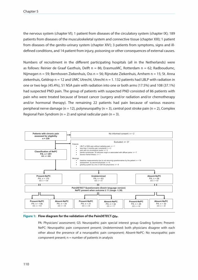
Chapter 5
110
the nervous system (chapter VI); 1 patient from diseases of the circulatory system (chapter IX); 189
patients from diseases of the musculoskeletal system and connective tissue (chapter XIII); 1 patient
from diseases of the genito-urinary system (chapter XIV); 3 patients from symptoms, signs and ill-
defined conditions, and 14 patient from injury, poisoning or other consequences of external causes.
Numbers of recruitment in the different participating hospitals (all in the Netherlands) were
as follows: Reinier de Graaf Gasthuis, Delft n = 86; ErasmusMC, Rotterdam n = 62; Radboudumc,
Nijmegen n = 59; Bernhoven Ziekenhuis, Oss n = 56; Rijnstate Ziekenhuis, Arnhem n = 15; St. Anna
ziekenhuis, Geldrop n = 12 and UMC Utrecht, Utrecht n = 1. 132 patients had LBLP with radiation in
one or two legs (45.4%), 51 NSA pain with radiation into one or both arms (17.5%) and 108 (37.1%)
had suspected PND pain. The group of patients with suspected PND consisted of 86 patients with
pain who were treated because of breast cancer (surgery and/or radiation and/or chemotherapy
and/or hormonal therapy). The remaining 22 patients had pain because of various reasons:
peripheral nerve damage (n = 12), polyneuropathy (n = 3), central post stroke pain (n = 2), Complex
Regional Pain Syndrom (n = 2) and spinal radicular pain (n = 3).
No informed consent: n = 2
Excluded: n= 37 Patients
LBLP or NSA pain without radiating pain: n = 1 less than 3 months pain complaints: n = 2 pain with an oncological cause: n = 2 painful syndromes of unknown origin or associated with diffuse pains: n = 7 severe mental illness: n = 1
Missings
baseline measurements due to not returning questionnaires by the patient: n = 16 assessment by second physician: n = 8 grading system by one or both the physicians: n = 8
PainDETECT Questionnaire (Dutch language version) NePC present when outcome ≥ 11 (range -1;38)
Absent-NePC PA: n = 32 GS: n = 43
Present-NePC PA: n = 26 GS: n = 50
Absent-NePC PA: n = 32 GS: n = 17
Present-NePC PA: n = 31 GS: n = 34
Absent-NePC PA: n = 34 GS: n = 36
Present-NePC PA: n = 136
GS: n = 103
Present-NePC PA: n = 170
GS: n = 139
Absent-NePC PA: n = 58 GS: n = 93
Undetermined PA: n = 63 GS: n = 51
Patients with chronic pain assessed for eligibility
n = 330
Classification of NePC PA: n = 291
GS: n = 283
Figure 1: Flow diagram for the validation of the PainDETECT-Dlv.
PA: Physicians’ assessment; GS: Neuropathic pain special interest group Grading System; Present-NePC: Neuropathic pain component present; Undetermined: both physicians disagree with each other about the presence of a neuropathic pain component; Absent-NePC: No neuropathic pain component present; n = number of patients in analysis
The validity of the PainDETECT in patients with chronic pain
111
5
After assessment by physicians A and physicians B, 170 patients were classified as having
present-NePC, 58 as absent-NePC. In 63 patients the two physicians made a non-concordant pain
classification, so the outcome based on the physicians assessment was classified as ‘undetermined’.
Based on the NeuPSIG Grading System in 139 patients NePC was classified as present, in 93 patients
NePC was absent and in 51 patients the two physicians made a non-concordant pain classification
in which the outcome was classified as ‘undetermined’ (see Figure 1: Flow Diagram).
Social-demographic and clinical details of the 291 patients were analyzed and divided from each
other based on the pain classification (see Table 1). No statistically significant differences were found
between absent-NePC, present-NePC and undetermined for gender, age, height, weight, body
mass index (BMI), education, medication, duration of pain, quality of life, disability, pain attribution,
anxiety disorder and depression. Moreover, no statistically significant differences were observed
between absent-NePC and present-NePC for pain (current, worst and average pain).
Physicians During this study 62 various physicians (pain specialist, pain specialist-fellow or neurologist), from
seven different hospitals, assessed all included patients. All patients were assessed two times by
two different physicians. Of all participating physicians, 21 physicians assessed ≤2 patients during
the execution of the study, 23 physicians saw ≤9 patients, 10 physicians saw ≥10 patients and 8
physicians saw ≥20 patients.
Evaluation of the PainDETECT-Dlv The mean score of the PainDETECT-Dlv (Range – 1;38) for patients classified as absent-NePC was
10.7 (SD± 5.7); for patients classified as present-NePC it was 15.7 (SD ± 6.3) and for patients with an
undetermined outcome it was 11.8 (SD ± 5). As calculated based on a one-way ANOVA with Tukey’s
studentized range post-hoc test, there was a statistical significant difference between absent-
NePC and present-NePC (P < 0.001) and between present-NePC and undetermined (P < 0.001). No
significant difference was seen between absent-NePC and undetermined (P = 0.57). Patients pain
course pattern and if the pain was radiating to other regions of the body were not significantly
different between the three groups. Pain descriptors (burning, tingling or prickling, painful light
touching, sudden pain attacks, temperature evoked pain, numbness sensation and pressure evoked
pain) were all statistically significant discriminators for the presence of NePC (P ≤ 0.005) except for
pressure evoked pain (P = 0.07). See Table 2 for the PainDETECT-Dlv outcomes divided according to
the pain classification by the physicians (presentNePC, absent-NePC or undetermined) (See Table 2).
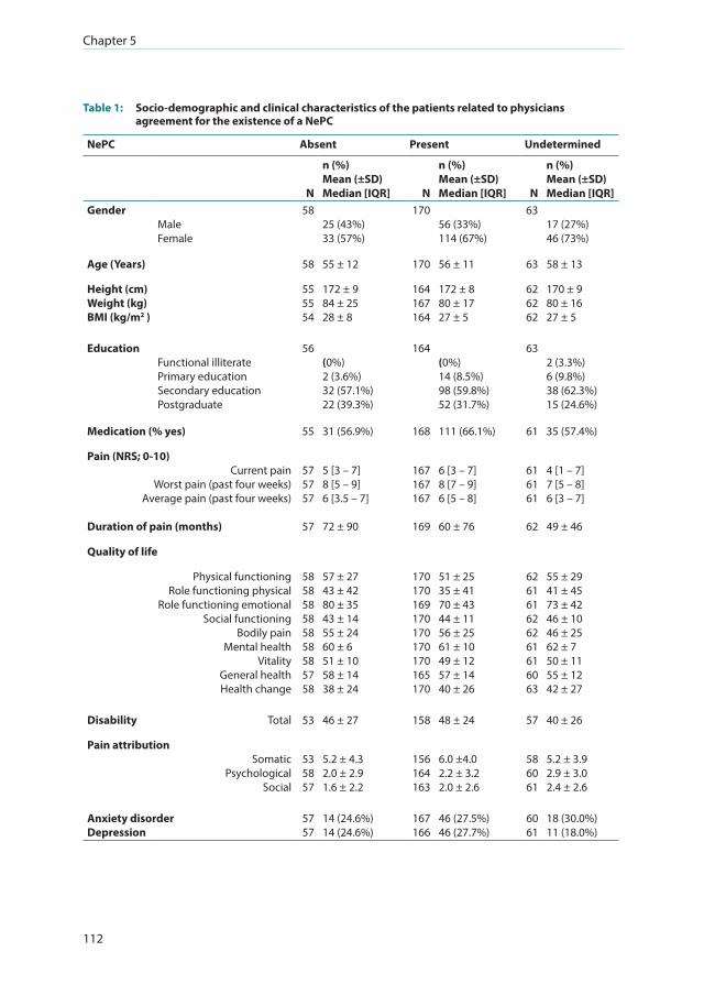
Chapter 5
112
Table 1: Socio-demographic and clinical characteristics of the patients related to physicians agreement for the existence of a NePC
NePC Absent Present Undetermined
N
n (%) Mean (±SD) Median [IQR] N
n (%) Mean (±SD)Median [IQR] N
n (%) Mean (±SD)Median [IQR]
GenderMaleFemale
5825 (43%)33 (57%)
17056 (33%)114 (67%)
6317 (27%)46 (73%)
Age (Years) 58 55 ± 12 170 56 ± 11 63 58 ± 13
Height (cm)Weight (kg)BMI (kg/m2 )
55 55 54
172 ± 984 ± 2528 ± 8
164 167 164
172 ± 880 ± 1727 ± 5
6262 62
170 ± 980 ± 1627 ± 5
Education Functional illiteratePrimary educationSecondary educationPostgraduate
56(0%)2 (3.6%)32 (57.1%)22 (39.3%)
164(0%)14 (8.5%)98 (59.8%)52 (31.7%)
632 (3.3%)6 (9.8%)38 (62.3%)15 (24.6%)
Medication (% yes) 55 31 (56.9%) 168 111 (66.1%) 61 35 (57.4%)
Pain (NRS; 0-10)Current pain
Worst pain (past four weeks)Average pain (past four weeks)
Duration of pain (months)
575757
57
5 [3 – 7]8 [5 – 9]6 [3.5 – 7]
72 ± 90
167167167
169
6 [3 – 7]8 [7 – 9]6 [5 – 8]
60 ± 76
616161
62
4 [1 – 7]7 [5 – 8]6 [3 – 7]
49 ± 46
Quality of life
Physical functioning Role functioning physical
Role functioning emotional Social functioning
Bodily painMental health
VitalityGeneral healthHealth change
5858 58 5858 58 5857 58
57 ± 2743 ± 4280 ± 3543 ± 1455 ± 2460 ± 651 ± 1058 ± 1438 ± 24
170170169170170170170165170
51 ± 25 35 ± 4170 ± 4344 ± 1156 ± 2561 ± 1049 ± 1257 ± 1440 ± 26
626161626261616063
55 ± 2941 ± 4573 ± 4246 ± 1046 ± 2562 ± 750 ± 1155 ± 1242 ± 27
Disability Total 53 46 ± 27 158 48 ± 24 57 40 ± 26
Pain attributionSomatic
Psychological Social
5358 57
5.2 ± 4.32.0 ± 2.91.6 ± 2.2
156164163
6.0 ±4.02.2 ± 3.22.0 ± 2.6
586061
5.2 ± 3.92.9 ± 3.02.4 ± 2.6
Anxiety disorder Depression
5757
14 (24.6%)14 (24.6%)
167166
46 (27.5%)46 (27.7%)
6061
18 (30.0%)11 (18.0%)

The validity of the PainDETECT in patients with chronic pain
113
5
Classification for the existence of NePC is based on physicians assessment of the patients. NePC: neuropathic pain component; Absent: NePC is absent; Present: NePC is present; Undetermined: both physicians disagree with each other about the existence of a neuropathic pain component; N: total number of patients in analysis; n: number of patients; %: percentage; SD: Standard deviation; IQR: Inter quartile range.
Table 2: The median (IQR) of the items of the PainDETECT by physicians agreement for the existence of a NePC
NePC Absent Present Undetermined
PainDETECT itemN
n (%) Median [IQR]Mean (±SD) N
n (%) Median [IQR]Mean (±SD) N
n (%) Median [IQR]Mean (±SD)
Pain course pattern 5819 (33%)14 (24%)16 (28%)9 (16%)
16253 (33%)58 (36%)32 (20%)18 (11%)
5917 (29%)17 (29%)20 (34%)5 (9%)
Persistent pain with slight fluctuations Persistent pain with pain attacks Pain attacks without pain between them Pain attacks with pain between them
Radiating pain (% yes) 51 41 (78%) 154 112 (73%) 57 38 (67%)
Gradation of pain55555555545655
0 [0 – 2] 1 [0 – 3] 0 [0 – 1] 2 [0 – 3] 0 [0 – 1] 2 [0 – 3] 2 [1 – 3]
170170169167170170170
1 [0 – 3] 2 [0 – 3] 1 [0 – 2] 3 [1 – 4] 1 [0 – 2]3 [2 – 4]3 [1 – 4]
62636362636363
0 [0 – 2.25] 1 [0 – 3] 0 [0 – 1] 2 [0 – 3] 1 [0 – 1] 3 [1 – 4] 2 [1 – 3]
Burning Tingling or prickling Painful light touching Sudden pain attacks Temperature evoked pain Numbness sensation Pressure evoked pain
Total sum score PainDETECT 58 10 [6.75 – 15.25]10.7 (± 5.75)
170 16 [11 – 20]15.7 (± 6.3)
63 10 [8 – 15]11.8 (± 5)
Classification of NePC is based on physicians assessment of the patients. NePC: neuropathic pain component; Absent: NePC is absent; Present: NePC is present; Undetermined: both physicians disagree with each other about the existence of a neuropathic pain component; N= total number of patients in analysis; n= number of patients; IQR: inter quartile range; SD: standard deviation; Range: 0 = never; 1 = hardly noticed; 2 = slightly; 3 = moderately; 4 = strongly; 5 = very strongly; Total sum score PainDETECT: Sum score calculation of the PainDETECT.
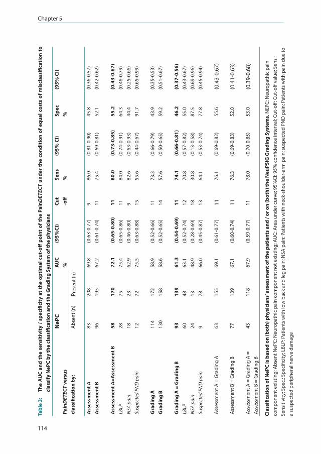
Chapter 5
114
Tabl
e 3:
Th
e A
UC
and
the
sens
itiv
ity
/ spe
cific
ity
at t
he o
ptim
al c
ut-o
ff p
oint
of t
he P
ainD
ETEC
T un
der
the
cond
itio
n of
equ
al c
osts
of m
iscl
assi
ficat
ion
to
clas
sify
NeP
C by
the
clas
sific
atio
n an
d th
e G
radi
ng S
yste
m o
f the
phy
sici
ans
Pain
DET
ECT
vers
us
clas
sific
atio
n by
:
NeP
CA
UC
%(9
5%CI
)Cu
t-o
ffSe
ns
%(9
5% C
I)Sp
ec
%(9
5% C
I)
Abs
ent (
n)Pr
esen
t (n)
Ass
essm
ent A
Ass
essm
ent B
Ass
essm
ent A
=Ass
essm
ent B
LBLP
NSA
pai
nSu
spec
ted
PND
pai
n
83 96 58 28 18 12
208
195
170
75 23 72
69.8
67.2
72.1
75.4
62.9
75.5
(0.6
3-0.
77)
(0.6
1-0.
74)
(0.6
5-0.
80)
(0.6
5-0.
86)
(0.4
6-0.
80)
(0.6
3-0.
88)
9 11 11 11 9 15
86.0
75.4
80.0
84.0
82.6
55.6
(0.8
1-0.
90)
(0.6
9-0.
81)
(0.7
3-0.
85)
(0.7
4-0.
91)
(0.6
3-0.
93)
(0.4
4-0.
67)
45.8
52.1
55.2
64.3
44.4
91.7
(0.3
6-0.
57)
(0.4
2-0.
62)
(0.4
3-0.
67)
(0.4
6-0.
79)
(0.2
5-0.
66)
(0.6
5-0.
99)
Gra
ding
AG
radi
ng B
Gra
ding
A =
Gra
ding
BLB
LPN
SA p
ain
Susp
ecte
d PN
D p
ain
114
130
93 60 24 9
172
158
139
48 13 78
58.9
58.6
61.3
63.1
48.9
66.0
(0.5
2-0.
66)
(0.5
2-0.
65)
(0.5
4-0.
69)
(0.5
2-0.
74)
(0.2
8-0.
69)
(0.4
5-0.
87)
11 14 11 12 18 13
73.3
57.6
74.1
70.8
30.8
64.1
(0.6
6-0.
79)
(0.5
0-0.
65)
(0.6
6-0.
81)
(0.5
7-0.
82)
(0.1
3-0.
58)
(0.5
3-0.
74)
43.9
59.2
46.2
55.0
87.5
77.8
(0.3
5-0.
53)
(0.5
1-0.
67)
(0.3
7-0.
56)
(0.4
3-0.
67)
(0.6
9-0.
96)
(0.4
5-0.
94)
Ass
essm
ent A
= G
radi
ng A
6315
569
.1(0
.61-
0.77
)11
76.1
(0.6
9-0.
82)
55.6
(0.4
3-0.
67)
Ass
essm
ent B
= G
radi
ng B
7713
967
.1(0
.60-
0.74
)11
76.3
(0.6
9-0.
83)
52.0
(0.4
1-0.
63)
Ass
essm
ent A
= G
radi
ng A
=
Ass
essm
ent B
= G
radi
ng B
4311
867
.9(0
.59-
0.77
)11
78.0
(0.7
0-0.
85)
53.0
(0.3
9-0.
68)
Clas
sific
atio
n of
NeP
C is
bas
ed o
n (b
oth)
phy
sici
ans’
asse
ssm
ent o
f the
pat
ient
s an
d / o
r on
(bot
h) th
e N
euPS
IG G
radi
ng S
yste
ms.
NEP
C: N
euro
path
ic p
ain
com
pone
nt e
xist
ing;
Abs
ent N
ePC:
Neu
ropa
thic
pai
n co
mpo
nent
not
exi
stin
g; A
UC:
Are
a un
der c
urve
; 95%
CI: 9
5% c
onfid
ence
inte
rval
; Cut
-off
: Cut
-off
valu
e; S
ens.:
Se
nsiti
vity
; Spe
c.: S
peci
ficity
; LBL
P: P
atie
nts
with
low
bac
k an
d le
g pa
in; N
SA p
ain:
Pat
ient
s w
ith n
eck-
shou
lder
-arm
pai
n; s
uspe
cted
PN
D p
ain:
Pat
ient
s w
ith p
ain
due
to
a su
spec
ted
perip
hera
l ner
ve d
amag
e

The validity of the PainDETECT in patients with chronic pain
115
5
Validity The gold standard for presence of the NePC in this study was the concordant opinion of both
physicians. On basis of this gold standard, patients with an identical pain classification were
included in the initial analysis (n = 228): 58 patients were classified as absent-NePC (25.4%) and 170
were classified as present-NePC (74.6%)(see Table 3 and Additional file 1: Table S1). A ROC-curve
was constructed for PainDETECT-Dlv (see Figure 2). Based on the gold standard, PainDETECT-Dlv sensitivity was (at maximal Youden-index) 80%, specificity 55.2%, positive predictive value 84% and
the positive likelihood ratio was 1.78. Based on the neuropathic pain Grading System, the sensitivity
was 74.1%, specificity 46.2%, positive predictive value 67.3%, and positive likelihood ratio of 1.38.
1-Specificity
Sens
itivi
ty
Figure 2: Receiver operating characteristics and area under the curve (AUC) for the total score of the PainDETECT-Dlv versus the presence of a neuropathic pain component as classified by two physicians (n = 228; undetermined patients are not included).
X-axis: 1-Specifity; Y-axis: Sensitivity
We also constructed ROC-curves for the classification by solely physicians A or B and according
to the neuropathic pain Grading System by physicians A or B and all the combinations. Except
for classification of patients’ pain based on the description of physicians A and the outcome of
the Grading System by physicians B all cut-off scores were calculated at 11-points out of 38: The
sensitivity ranges from 57.6-86.1% and specificity from 43.9-59.2%. The classification of patients’ pain
based on the classification of physicians A resulted in a cut-off score of 9-points: Sensitivity 86.1%
and specificity 45.8%. The classification of patients’ pain based on the Grading System according to
physicians B resulted in a cut-off score of 14-points: Sensitivity of 57.6% and specificity of 59.2%.
In Table 3 and Additional file 1: Table S1 we present the number of patients with LBLP, NSA pain or
suspected PND pain, in total and per group based on physicians’ assessment and/or the Grading
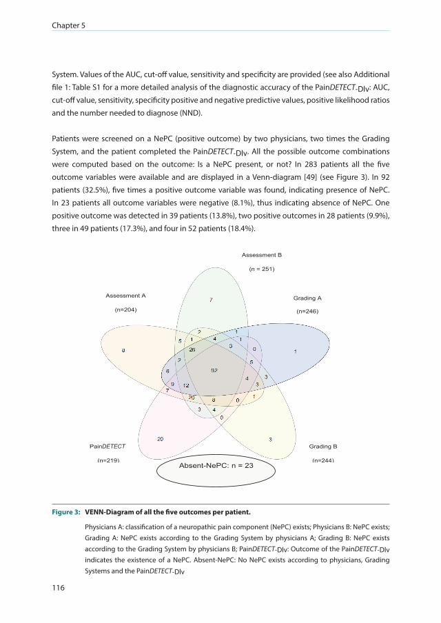
Chapter 5
116
System. Values of the AUC, cut-off value, sensitivity and specificity are provided (see also Additional
file 1: Table S1 for a more detailed analysis of the diagnostic accuracy of the PainDETECT-Dlv: AUC,
cut-off value, sensitivity, specificity positive and negative predictive values, positive likelihood ratios
and the number needed to diagnose (NND).
Patients were screened on a NePC (positive outcome) by two physicians, two times the Grading
System, and the patient completed the PainDETECT-Dlv. All the possible outcome combinations
were computed based on the outcome: Is a NePC present, or not? In 283 patients all the five
outcome variables were available and are displayed in a Venn-diagram [49] (see Figure 3). In 92
patients (32.5%), five times a positive outcome variable was found, indicating presence of NePC.
In 23 patients all outcome variables were negative (8.1%), thus indicating absence of NePC. One
positive outcome was detected in 39 patients (13.8%), two positive outcomes in 28 patients (9.9%),
three in 49 patients (17.3%), and four in 52 patients (18.4%).
Absent-NePC: n = 23
Assessment B
(n = 251)
(n=251)
Grading A
(n=246)
Grading B
(n=244)
PainDETECT
(n=219)
Assessment A
(n=204)
Figure 3: VENN-Diagram of all the five outcomes per patient.
Physicians A: classification of a neuropathic pain component (NePC) exists; Physicians B: NePC exists; Grading A: NePC exists according to the Grading System by physicians A; Grading B: NePC exists according to the Grading System by physicians B; PainDETECT-Dlv: Outcome of the PainDETECT-Dlv indicates the existence of a NePC. Absent-NePC: No NePC exists according to physicians, Grading Systems and the PainDETECT-Dlv
DETECT
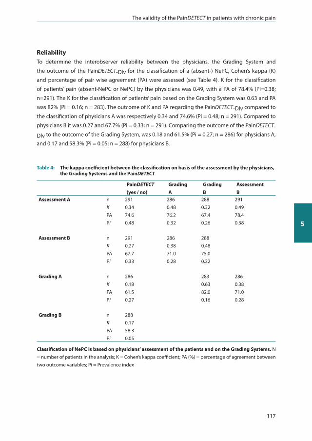
The validity of the PainDETECT in patients with chronic pain
117
5
Reliability To determine the interobserver reliability between the physicians, the Grading System and
the outcome of the PainDETECT-Dlv for the classification of a (absent-) NePC, Cohen’s kappa (K)
and percentage of pair wise agreement (PA) were assessed (see Table 4). K for the classification
of patients’ pain (absent-NePC or NePC) by the physicians was 0.49, with a PA of 78.4% (Pi=0.38;
n=291). The K for the classification of patients’ pain based on the Grading System was 0.63 and PA
was 82% (Pi = 0.16; n = 283). The outcome of K and PA regarding the PainDETECT-Dlv compared to
the classification of physicians A was respectively 0.34 and 74.6% (Pi = 0.48; n = 291). Compared to
physicians B it was 0.27 and 67.7% (Pi = 0.33; n = 291). Comparing the outcome of the PainDETECT-
Dlv to the outcome of the Grading System, was 0.18 and 61.5% (Pi = 0.27; n = 286) for physicians A,
and 0.17 and 58.3% (Pi = 0.05; n = 288) for physicians B.
Table 4: The kappa coefficient between the classification on basis of the assessment by the physicians, the Grading Systems and the PainDETECT
PainDETECT(yes / no)
Grading A
Grading B
Assessment B
Assessment A nK PA Pi
2910.3474.60.48
2860.4876.20.32
2880.3267.40.26
2910.4978.40.38
Assessment B nK PA Pi
2910.2767.70.33
2860.3871.00.28
2880.4875.00.22
Grading A nK PA Pi
2860.1861.50.27
2830.6382.00.16
2860.3871.00.28
Grading B nK PA Pi
2880.1758.30.05
Classification of NePC is based on physicians’ assessment of the patients and on the Grading Systems. N = number of patients in the analysis; K = Cohen’s kappa coefficient; PA (%) = percentage of agreement between two outcome variables; Pi = Prevalence index

Chapter 5
118
Stability and responsiveness of the PainDETECT-Dlv over time was assessed over a period of two
weeks. The mean sum score of the PainDETECT-Dlv at baseline for the total group was 13.8 ± 6.3.
The mean sum score after two weeks was 14.1 ± 6.1. Test-retest reliability via ICC was 0.83 (95%CI
0.79-0.87; n = 268). Taking into consideration the fact that patients’ pain should not have changed
(outcome based on the PGIC), because otherwise the ICC would not reflect the consistency of the
PainDETECT-Dlv, and a time gap of 7-21 days was allowed (to rule out the early or delayed return of
questionnaires) between the first and second PainDETECT-Dlv, the ICC was 0.87 (95% CI 0.81-0.91; n
= 123). After three months, with no change in the degree of patients’ pain and a time gap of 60-120
days between the first and third PainDETECT-Dlv, ICC was 0.86 (95% CI 0.79-0.91; n = 102).
DISCUSSION
This study demonstrates the clinimetric quality of the PainDETECT-Dlv, a screening instrument for
the presence of a NePC, on a large population of patients, with chronic pain due to low back with
leg pain, neck-shoulder-arm pain or pain due to a suspected peripheral nerve damage as normally
present in a physician’s daily practice. Because the patients were included without pre-stratification
on the target outcome, previous Catch-22 situations in the assessment of the validity of screening
instruments were avoided. Under these conditions, the PainDETECT-Dlv failed to be predictive for
the existence of a NePC due to a moderate sensitivity and low specificity, irrespective of comparison
with the expert opinion via the classification by two physicians (gold standard) as well as with the
outcome of the NeuPSIG Grading System. Moreover, the predictive values were also not indicating
that the PainDETECT-Dlv is a valid screening tool for the assessment of a NePC. The likelihood ratios
were also not suggestive for the usefulness of this instrument.
Validation studies with patients pre-stratified for NePC We found an optimal cut-off score for the PainDETECT-Dlv of ≥11 points corresponding to a
sensitivity of 80% and a specificity of 55%. In the original development and validation study of the
PainDETECT by Freynhagen et al. [2] a sensitivity and specificity of 84% was found. The gold standard
in their study was the examination by two experienced pain specialists. The study was performed at
ten different specialized pain centers. Only patients with ‘typical’ neuropathic or nociceptive entities
(i.e. no ‘unclear’ outcome) and only patients with a VAS of >40 mm (0 – 100 mm) were included. In
the Spanish validation study by De Andrés et al. [16] only patients with a VAS ≥40 mm and a known
classification (by one experienced specialist) of neuropathic pain, mixed pain or nociceptive pain
were included. It revealed a sensitivity and specificity of 81% when patients with the classification
of neuropathic pain or nociceptive pain were included. The inclusion of patients with mixed pain
in the neuropathic pain group resulted in a sensitivity of 84% and specificity of 78%. The Korean
version of the PainDETECT [20] was validated based on the study by De Andrés [16] in patients with
chronic pain and with a NRS ≥ 3 (NRS 0-10). The gold standard was the independent diagnosis of
The validity of the PainDETECT in patients with chronic pain
119
5
the patient by two experienced pain physicians. It revealed a sensitivity of 82% and a specificity of
92% based on a cut-off score of ≥19 (range – 1; 38). In the validation of the Turkish version of the
PainDETECT [17] patients were included with the classification of pain type (i.e. NePC) being assessed
beforehand (based on the opinion of two expert pain physicians) and patients suffering from pain
of three centimeters or more (VAS 0-10 cm). Sensitivity and specificity were respectively 78 and
83%. The Hindi version of the PainDETECT [19] was validated in patients with neuropathic and in
patients with non-neuropathic pain based on a conventional single assessment by one physician.
At a optimal cut off point of ≥18 sensitivity was 83% and the specificity was 84%.
In a cohort of patients with a spinal cord injury for more than one year, pain lasting more than six
months and a pain intensity of more than three on a NRS (0-10) a sensitivity was found of 68% and
specificity of 83% [50].
The present study included patients with chronic pain without limits to the minimal pain intensity
or other limitations. At the moment of inclusion in the study our patients had only a provisional
diagnosis (LBLP, NSA pain, suspected PND pain) established in primary or secondary care without
further refinement or confirmation. Then, after referral to a (non-) academic pain clinic, they were
assessed as to their complaints for the first time at study inclusion. Thus this was a ‘real-life’ clinical
out-patient population. Avoiding patient selection due to pre-stratification to the outcome target
makes our study unique and clinically more relevant as compared to other studies on the same topic
and is crucial for the validation of a screening instrument.
Validation studies with patients not pre-stratified for NePC In a study by Gauffin et al. [51] in patients diagnosed with fibromyalgia (n = 158) a cut-off score
of 17 was found with a sensitivity of 79% and specificity of 53% (Gold standard: the classification
by one experienced physician). This study, like ours, did not pre-stratify patients according to the
pain classification either, and patients were not excluded because of a low pain level. The outcome
of Gauffin’s sensitivity analyses in this fibromyalgia study was comparable to our study. Tampin et
al. [34] found, based on the examination by a physical therapist, a sensitivity and specificity of the
PainDETECT of respectively 64 and 62% (cut-off score 18.5) in a population of patients with neck/
upper limb pain (n = 122). In our study, the outcome for patients with neck-shoulder-arm pain was
83 and 44% respectively (cut-off score of ≥9).
Grading system In this study the physicians assessed patients for the presence of a NePC according to the Grading
System [31]. Probable neuropathic pain and definite neuropathic pain were combined as present-
NePC, and non neuropathic pain and possible neuropathic pain were combined in absent-NePC.
Sensitivity and specificity of the PainDETECT-Dlv resulted to be 74 and 46% respectively (Cut-off score
11 out of 38, n = 232). Using the classification of patients’ pain based solely on the Grading System
Chapter 5
120
by one physician results in a lower validity than based on the physicians assessment. This might
suggest that the classification of patients’ pain based on the Grading System is less accurate than
the classification based on the physicians’ assessment in respect to the outcome of the PainDETECT-
Dlv. However, the grading system was assessed by the same physician who also performed the
physician’s assessment so it is also possible that the physician had difficulties to classify patients
pain based on the Grading System or vice versa. When using the physicians’ assessments as well as
the Grading Systems of both physicians (n = 161), sensitivity was 78%, specificity was 53% and the
cut-off score was 11: The same poor result as for the gold standard. In the papers by Vaegter et al.
[33] and Tampin et al. [34] the PainDETECT was also compared with the NeuPSIG Grading System. In
both papers, like in ours, the outcome of the Grading System was not comparable to the outcome
of the PainDETECT. As stated by Finnerup et al. [52] the PainDETECT (and other screening tools for
the assessment of neuropathic pain) is only to alert the physician to further assess the patient who
may have a NePC.
NePC classification The initial classification of patients’ pain in our study was based on an interview and (clinical/
physical) examination by trained (pain-) physicians. There is a lack of consensus with respect
to the classification of a NePC in patients with pain of different origins [53]. Moreover, a lack of
standardization of assessment methods increases the number of undetected or poorly classified
patients which leads to a variation in the classification accuracy (i.e. sensitivity and specificity) of
screening tools caused by differences in strategy and patient population [15, 54]. Bouhassira and
Attal recently stated that neuropathic pain is “a consistent clinical entity, but it is multidimensional
in terms of its clinical expression, with different sensory profiles, potentially reflecting specific
pathophysiological mechanisms” [55]. As stated by Scholz et al. [53] physical tests are more useful
to identify patients with neuropathic back pain than interview questions. To reach a more unified
classification system to differentiate between present-NePC and absent-NePC a standardized
assessment of symptoms and signs is necessary [53]. However, these tests are not able to confirm the
relation between the potential lesion or disease of the nerve and the pain directly: The classification
of neuropathic pain should be based on clinical examination and the interpretation should be
placed in the clinical context of patients’ pain [55].
In this study we used a mandatory standardized assessment [25] in addition to the medical history
and physical examination which were performed according to the physicians’ standards. The clinical
assessment and the use of the Grading System showed that in 18-22% of the patients a non-
consistent assessments was present resulting in an ‘undetermined’ status. In Freynhagens paper
[2] it was 5%. This difference might be due to the inclusion of patients with a less clear absent or
present NePC in our study which might reflect what happens in the assessment of a NePC in usual
clinical care. Moreover, this also might occur in the treatment of patients with chronic pain. Based
on both the physician’s assessments, almost 75% of the patients in this study had a neuropathic pain
The validity of the PainDETECT in patients with chronic pain
121
5
component. This might be due to several facts. (1) Patients with LBLP or NSA pain were only included
when the pain was radiating into the leg(-s) respectively the arm(-s) and were not removed from this
study when they had mixed pain. Moreover, patients with radiating pain are more suspected to have
a NePC. (2) There is a possibility that neuroplastic changes are interpreted as neuropathy in patients
with chronic LBLP. (3) Patients were recruited in secondary and tertiary pain clinics. This might have
led to a inclusion of patients who were more difficult to treat in primary care and (4) we included 108
patients with suspected peripheral nerve damage. Almost 60% of the patients after treatment for
breast cancer has pain [56]. Based on the recent review by Ilhan et al. [57], in patients who reported
pain following breast cancer treatment the pooled prevalence of neuropathic pain from screening
questionnaires ranged from 32.6 to 58.2%. Following the NeuPSIG Grading System the prevalence
ranged from 29.5 to 57.1%. Based on these numbers, patients after breast cancer can be regarded as
patients suspected of neuropathic pain due to peripheral nerve damage. However, the PainDETECT-
Dlv (compared to the gold standard and the NeuPSIG Grading System) as used in our study seems
not valid for the assessment of patients with neuropathic pain based on a suspected PND in which
the majority of patients was suffering of pain after treatment for breast cancer.
Strengths and weaknesses There are several strengths in this study. Firstly, we included a large population of patients with
diagnoses who are regularly seen in daily clinical practice. Secondly, there was no pre-stratification
on the target outcome, clear inclusion criteria and almost no exclusion criteria. Thirdly, we used
the NeuPSIG Grading System [31, 52] as a secondary comparison. The main purpose of the Grading
System is to help in the classification of the pain as neuropathic [52]. In our study, the Grading
System was added to the standardized assessment form which had to be filled in by the physician.
There are also some weaknesses in this study. The use of the Grading System within the clinical
assessment (including bed-side examination) is a strong aspect of our study, but the outcome of
the clinical examination as well as the outcome of the Grading System might be influenced by
each other. However, combining the physicians’ assessment with the Grading System might have
made the ‘gold standard’ even stronger but also might have led to a cross-contamination. Secondly,
diagnosing NePC by assessing patients’ pain by two separate physicians in our and in other
studies is considered as the ‘Gold Standard’. However, classifying patients’ pain may be done more
objectively by establishing a damaged nerve and by diagnosticating in a more detailed clinical way.
Moreover, the breakdown of clinical grounds for in- and exclusion could also have been assessed
and captured in more detail. Thirdly, 62 physicians participated. This might have led to the inclusion
of younger, less clinical experienced physicians. However, it reflects ‘real life’ practice and limits the
risk of systematic bias in the classification of patients’ pain and bias based on assumptions about
the existence of a NePC. Moreover, all physicians followed the standardized training as described.
Fourthly, almost only patients with peripheral causes of pain were selected. This can be considered
as a methodological drawback. Moreover, because we did not include patients with, by example,
low back pain without irradiation to the leg who would probably be diagnosed as absent-NePC the
Chapter 5
122
specificity might decrease. Fifthly, there is an apparent lack of objective tests to determine whether
the somatosensory fibers were affected, in particular the small fibers. This can be seen as crucial
since objective data are mandatory to reach a definite neuropathic pain classification in the grading
system. Lastly, in a following study we would collect data from the patients who were not able to
participate in the study to prevent inclusion bias. In this study this was not possible because of
ethical regulations.
CONCLUSIONS
The PainDETECT-Dlv has a good internal consistency and test-retest reliability but is not an effective
screening tool for the assessment of a neuropathic pain component in a population of patients with
chronic pain, irrespective of the chosen comparison because of its moderate sensitivity and low
specificity. However, the agreement by both the physicians and the agreement with the grading
systems (performed by the physicians) were also not impressive. Moreover, the differences in the
cut-off scores for the different comparisons reflects the fact that agreement in a not pre-stratified
to the target outcome patient population is not easy to accomplish. Using the PainDETECT-Dlv (for
screening purposes or as a surrogate for clinical assessment) may result in unreliably separating
NePC presence from non-presence in patients with chronic pain in clinical outpatient practices and
in research settings. Catch-22 situations in the validation of screening tools can be prevented by not
pre-stratifying the patients on basis of the target outcome before inclusion in a validation study for
screening instruments. For now, classifying patients pain still needs the clinical assessment based
on history and physical examination including bed-side sensory testing by the physician and cannot
be replaced by the use of the PainDETECT.
ABBREVIATIONS
AUC: Area Under the Curve; BMI: Body Mass Index; CI: Confidence Interval; DRI: Disability Rating
Index; EFNS: European Federation of Neurological Societies; HADS: Hospital Anxiety and Depression
Scale; IASP: International Association for the Study of Pain; ICC: Intraclass Correlation; ICD-10:
International Statistical Classification of Diseases and Related Health Problems 10th revision; IQR:
Inter Quartile Range; K: Cohen’s kappa; LBLP: Low Back with Leg Pain; NePC: Neuropathic Pain
Component; NeuPSIG: Neuropathic Pain Special Interest Group; NND: Number Needed to Diagnose;
NRS: Numeric Rating Scale; NSA pain: Neck-Shoulder-Arm pain; NTR: National Trial register;
PA: Percentage of Agreement; PainDETECT-Dlv: PainDETECT Dutch language version; PAS: Pain
Attribution Scale; PGIC: Patient Global Impression of Change; Pi: Prevalence index; PND: Peripheral
nerve damage pain; RAND-36: RAND 36-item health Survey; ROC: Receiver Operating Characteristic;
The validity of the PainDETECT in patients with chronic pain
123
5
SD: Standard Deviation; SPSS: Statistical package for the Social Sciences; VAS: Visual Analogue Scale;
WHO: World Health Organization
ACKNOWLEDGEMENTS
Thanks to all the participating patients for their invaluable work to this study. Also thanks to the
participating physicians and assistants of Rijnstate Ziekenhuis, Arnhem; Bernhoven Ziekenhuis, Oss;
St.Anna Ziekenhuis, Geldrop; Reinier de Graaf Gasthuis, Delft; Utrecht University Medical Center,
Utrecht; Erasmus Medical Center, Rotterdam and Radboud university medical center, Nijmegen. We
would like to thank Jan Hendriks for his help with the statistics until his retirement.
FUNDING
This study was performed within DALI for PAIN, a national program that focuses on neuropathic pain
care optimalisation. DALI for PAIN is an initiative of Pfizer. This project is supported by an unrestricted
grant from Pfizer. The funders had no role in study design, data collection and analysis, decision to
publish, or preparation of the manuscript.
AVAILABILITY OF DATA AND MATERIALS
The datasets used and/or analyzed during the current study are available from the corresponding
author on reasonable request.
ETHICS APPROVAL AND CONSENT TO PARTICIPATE
This study was approved by the medical and ethical review board Committee on Research Involving
Human Subjects region Arnhem- Nijmegen, Nijmegen, the Netherlands, Dossier number: 2008/348;
NL 25343.091.08 and conducted in accordance with the declaration of Helsinki and the declaration
of the World Medical Association. As required, written informed consent was obtained from patients
prior to study participation. The protocol is registered in the Dutch National Trial Register: NTR 3030.
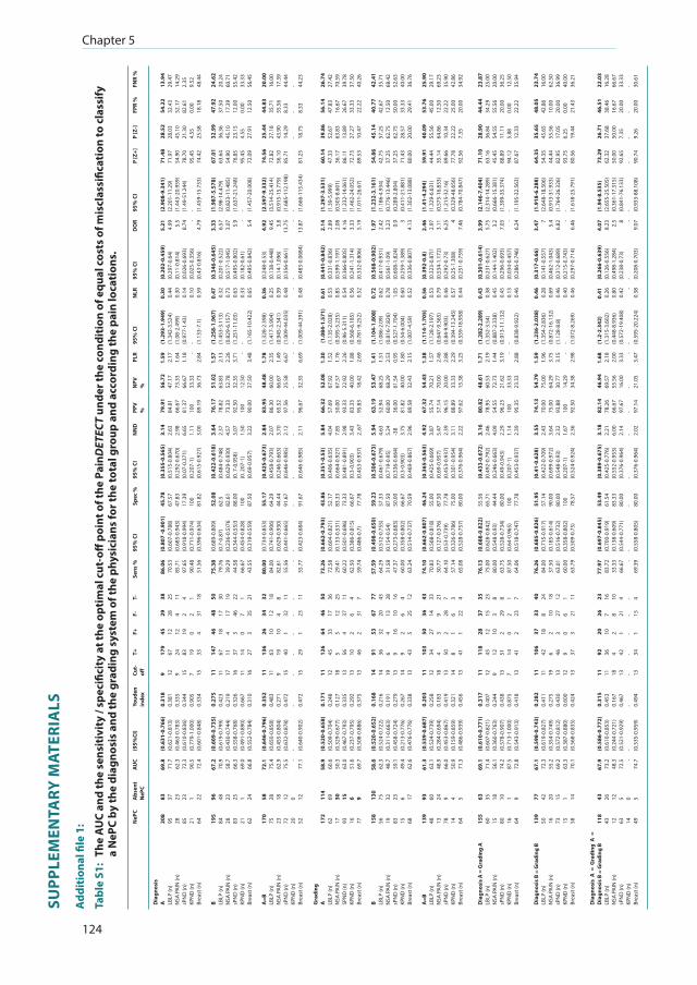
Chapter 5
124
SUPP
LEM
ENTA
RY M
ATER
IALS
Add
itio
nal fi
le 1
:
Tabl
e S1
: Th
e A
UC
and
the
sens
itiv
ity
/ spe
cific
ity
at th
e op
tim
al c
ut-o
ff p
oint
of t
he P
ainD
ETEC
T un
der t
he c
ondi
tion
of e
qual
cos
ts o
f mis
clas
sific
atio
n to
cla
ssify
a
NeP
C by
the
diag
nosi
s an
d th
e gr
adin
g sy
stem
of t
he p
hysi
cian
s fo
r the
tota
l gro
up a
nd a
ccor
ding
the
pain
loca
tion
s.
NeP
CA
bsen
t N
ePC
AU
C(9
5%CI
)Yo
uden
inde
xCu
t-off
T+F+
F-T-
Sens
%95
% C
ISp
ec %
95%
CI
NN
DPP
V
%N
PV
%PL
R95
% C
IN
LR95
% C
ID
OR
95%
CI
P [Z
+]P
[Z-]
FPR
%FN
R %
Dia
gnos
isA LB
LP (n
)N
SA P
AIN
(n)
sPN
D (n
)KP
ND
(n)
Brea
st (n
)
B LBLP
(n)
NSA
PA
IN (n
)sP
ND
(n)
KPN
D (n
)Br
east
(n)
A=B
LBLP
(n)
NSA
PA
IN (n
)sP
ND
(n)
KPN
D (n
)Br
east
(n)
208
95 28 85 21 64 195
84 28 83 21 62 170
75 23 72 20 52
83 37 23 23 1 22 96 48 23 25 1 24 58 28 18 12 0 12
69.8
71.7
62.3
72.3
90.5
72.4
67.2
70.9
58.7
66.3
69.0
66.8
72.1
75.4
62.9
75.5 -
77.1
(0.6
31-0
.766
)(0
.621
-0.8
13)
(0.4
63-0
.783
)(0
.610
-0.8
36)
(0.7
79-1
.000
)(0
.601
-0.8
48)
(0.6
09-0
.735
)(0
.619
-0.7
99)
(0.4
30-0
.744
)(0
.558
-0.7
68)
(0.4
91-0
.890
)(0
.552
-0.7
84)
(0.6
46-0
.796
)(0
.650
-0.8
58)
(0.4
55-0
.804
)(0
.632
-0.8
78)
- (0.6
40-0
.902
)
0.31
80.
381
0.33
50.
344
0.90
50.
334
0.27
50.
423
0.21
90.
326
0.66
70.
310
0.35
20.
483
0.27
10.
472
- 0.47
2
9 12 9 15 7 15 11 11 17 16 11 16 11 11 9 15 - 15
179
67 24 83 19 33 147
67 11 37 14 27 136
63 19 40 - 29
45 12 12 19 0 4 46 18 4 3 0 3 26 10 10 1 - 1
29 28 4 2 2 31 48 17 17 46 7 35 34 12 4 32 - 23
38 25 11 4 1 18 50 30 19 22 1 21 32 18 8 11 - 11
86.0
670
.53
85.7
197
.65
90.4
851
.56
75.3
879
.76
39.2
944
.58
66.6
743
.55
80.0
084
.00
82.6
155
.56 -
55.7
7
(0.8
07 -0
.901
)(0
.607
-0.7
88)
(0.6
85-0
.943
)(0
.918
-0.9
94)
(0.7
11-0
.974
)(0
.396
-0.6
34)
(0.6
89-0
.809
)(0
.7-0
.87)
(0.2
36-0
.5)7
6(0
.344
-0.5
5)3
(0.4
54-0
.828
)(0
.319
-0.5
59)
(0.7
34-0
.853
)(0
.741
-0.9
06)
(0.6
29-0
.930
)(0
.441
-0.6
65)
- (0.4
23-0
.684
)
45.7
867
.57
47.8
317
.39
100
81.8
2
52.0
862
.582
.61
88.0
010
087
.50
55.1
764
.29
44.4
491
.67
- 91.6
7
(0.3
55-0
.565
)(0
.515
-0.8
04)
(0.2
92-0
.670
)(0
.07-
0.37
1)(0
.207
-1)
(0.6
15-0
.927
)
(0.4
22-0
.618
)(0
.484
-0.7
48)
(0.6
29-0
.930
)(0
. 7-0
.958
)(0
. 207
-1)
(0.6
9-0.
957)
(0.4
25-0
.673
)(0
.458
-0.7
93)
(0.2
46-0
.663
)(0
.646
-0.9
85)
- (0.6
46-0
.985
)
3.14
2.63
2.98
6.65
1.11
3.00
3.64
2.37
4.57
3.07
1.50
3.22
2.84
2.07
3.70
2.12
- 2.11
79.9
184
.81
66.6
781
.37
100
89.1
9
76.1
778
.82
73.3
392
.50
100
90.0
0
83.9
586
.30
65.5
297
.56
- 96.6
7
56.7
247
.17
73.3
366
.67
33.3
336
.73
51.0
263
.83
52.7
832
.35
12.5
037
.50
48.4
860
.00
66.6
725
.58
- 32.3
5
1.59
2.17
1.64
1.18
-2.
84
1.57
2.13
2.26
3.71
-3.
48
1.78
2.35
1.49
6.67
-6.
69
(1.2
93-1
.949
)(1
.342
-3.5
24)
(1.0
8-2.
499)
(0.9
77-1
.43)
- (1.1
33-7
.1)
(1.2
58-1
.967
)(1
.453
-3.1
13)
(0.8
29-6
.157
)(1
.251
-11.
03)
- (1.1
65-1
0.42
2)
(1.3
28-2
.398
)(1
.417
-3.9
04)
(0.9
45-2
.341
)(1
.009
-44.
035)
- (1.0
09-4
4.39
1)
0.30
0.44
0.30
0.14
0.10
0.59
0.47
0.32
0.73
0.63
0.33
0.65
0.36
0.25
0.39
0.48
-0.
48
(0.2
02-0
.459
)(0
.297
-0.6
4)(0
.11-
0.81
4)(0
.026
-0.6
93)
(0.0
25-0
.356
)(0
.43-
0.81
6)
(0.3
46-0
.645
)(0
.201
-0.5
22)
(0.5
17-1
.045
)(0
.495
-0.8
02)
(0.1
82-0
.61)
(0.4
95-0
.842
)
(0.2
48-0
.53)
(0.1
38-0
.448
)(0
.14-
1.09
6)(0
.356
-0.6
61)
- (0.4
83-0
.068
4)
5.21
4.99 5.
58.
74-
4.79
3.33
6.57
3.07 5.
9 -5.
4
4.92
9.45 3.
813
.75 -
13.8
7
(2.9
08-9
.341
)(2
.201
-11.
29)
(1.4
43-2
0.95
9)(1
.49-
51.2
44)
- (1.4
59-1
5.73
3)
(1.9
87-5
.578
)(2
.98-
14.4
79)
(0.8
23-1
1.48
5)(1
.637
-21.
248)
- (1.4
57-2
0.00
8)
(2.5
97-9
.332
)(3
.514
-25.
414)
(0.9
15-1
5.77
9)(1
.685
-112
.198
)- (1
.666
-115
.434
)
71.4
871
.97
54.9
078
.70
95.4
574
.42
67.0
163
.64
54.9
076
.85
95.4
572
.09
74.5
672
.82
56.1
085
.71
81.2
5
28.5
228
.03
45.1
021
.30
4.55
25.5
8
32.9
936
.36
45.1
023
.15
4.55
27.9
1
25.4
427
.18
43.9
014
.29
- 18.7
5
54.2
232
.43
52.1
782
.61
0.00
18.1
8
47.9
237
.50
17.3
912
.00
0.00
12.5
0
44.8
335
.71
55.5
68.
33- 8.
33
13.9
429
.47
14.2
92.
359.
5248
.44
24.6
220
.24
60.7
155
.42
33.3
356
.45
20.0
016
.00
17.3
944
.44
- 44.2
3
Gra
ding
A LBLP
(n)
NSA
PA
IN (n
)SP
ND
(n)
KPN
D (n
)Br
east
(n)
B LBLP
(n)
NSA
PA
IN (n
)sP
ND
(n)
KPN
D (n
)Br
east
(n)
A=B
LBLP
(n)
NSA
PA
IN (n
)sP
ND
(n)
KPN
D (n
)Br
east
(n)
172
62 17 93 16 77 158
56 19 83 15 68 139
48 13 78 14 64
114
69 30 15 6 9 130
75 32 23 6 17 93 60 24 9 4 5
58.9
60.6
50.3
63.0
51.6
69.7
58.6
62.3
48.7
59.1
49.4
62.6
61.3
63.1
48.9
66.0
50.9
71.3
(0.5
20-0
.658
)(0
.509
-0.7
04)
(0.3
29-0
.677
)(0
.467
-0.7
93)
(0.2
37-0
.795
)(0
.508
-0.8
86)
(0.5
20-0
.652
)(0
.524
-0.7
22)
(0.3
11-0
.663
)(0
.458
-0.7
24)
(0.2
13-0
.776
)(0
.476
-0.7
76)
(0.5
39-0
.687
)(0
.524
-0.7
39)
(0.2
84-0
.694
)(0
.453
-0.8
67)
(0.1
59-0
.859
)(0
.486
-0.9
39)
0.17
10.
248
0.12
70.
335
0.29
20.
375
0.16
80.
216
0.19
10.
279
0.26
70.
338
0.20
30.
258
0.18
30.
419
0.32
10.
456
11 12 18 13 13 13 14 14 19 13 14 13 11 12 18 13 14 13
126
45 5 56 10 46 91 36 6 9 9 43 103
34 4 50 8 41
64 33 5 4 2 2 53 32 4 16 2 5 50 27 3 2 1 1
46 17 12 37 6 31 67 20 13 10 6 25 36 14 9 28 6 22
50 36 25 11 4 7 77 43 28 16 4 12 43 33 21 7 3 4
73.2
672
.58
29.4
160
.22
62.5
059
.74
57.5
964
.29
31.5
847
.37
60.0
063
.24
74.1
070
.83
30.7
764
.10
57.1
465
.08
(0.6
62-0
.793
)(0
.604
-0.8
21)
(0.1
33-0
.531
)(0
.501
-0.6
96)
(0.3
86-0
.815
)(0
.486
-0.7
)
(0.4
98-0
.650
)(0
.512
-0.7
55)
(0.1
54-0
.54)
(0.2
73-0
.683
)(0
.358
-0.8
02)
(0.5
14-0
.737
)
(0.6
62-0
.807
)(0
.568
-0.8
18)
(0.1
27-0
.576
)(0
.53-
0.73
9)(0
.326
-0.7
86)
(0.5
28-0
.757
)
43.8
652
.17
83.3
373
.33
66.6
777
.78
59.2
357
.33
87.5
050
.00
66.6
770
.59
46.2
455
.00
87.5
077
.78
75.0
080
.00
(0.3
51-0
.53)
(0.4
06-0
.635
)(0
.664
-0.9
27)
(0.4
81-0
.891
)(0
.3-0
.903
)(0
.453
-0.9
37)
(0.5
06-0
.673
)(0
.461
-0.6
79)
(0.7
19-0
.95)
(0.3
36-0
.664
)(0
.3-0
.903
)(0
.469
-0.8
67)
(0.3
65-0
.563
)(0
.425
-0.6
69)
(0.6
9-0.
957)
(0.4
53-0
.937
)(0
.301
-0.9
54)
(0.3
76-0
.964
)
5.84
4.04
7.85
2.98
3.43
2..6
7
5.94
4.63
5.24
-38
3.75
2.96
4.92
3.87
5.47
2.39
3.11
2.22
66.3
257
.69
50.0
093
.33
83.3
395
.83
63.1
952
.94
60.0
036
.00
81.8
289
.58
67.3
255
.74
57.1
496
.15
88.8
997
.62
52.0
867
.92
67.5
722
.92
40.0
018
.42
53.4
768
.25
68.2
961
.54
40.0
032
.43
54.4
370
.21
70.0
020
.00
33.3
315
.38
1.30
1.52
1.76
2.26
1.88
2.69
1.41
1.51
2.53
0.95
1.80
2.15
1.38
1.57
2.46
2.88
2.29
3.25
(1.0
84-1
.571
)(1
.135
-2.0
28)
(0.5
95-5
.235
)(0
.96-
5.31
1)(0
.568
-6.1
85)
(0.7
81-9
.252
)
(1.1
04-1
.808
)(1
.086
-2.0
9)(0
.816
-7.8
26)
(0.5
27-1
.704
)(0
.54-
6.00
4)(1
.007
-4.5
9)
(1.1
14-1
.705
)(1
.128
-2.1
97)
(0.6
47-9
.365
)(0
.84-
9.90
3)(0
.394
-13.
245)
(0.5
59-1
8.95
8)
0.61
0.53
0.85
0.54
0.56
0.52
0.72
0.62
0.78
1.05
0.60
0.52
0.56
0.53
0.79
0.46
0.57
0.44
(0.4
41-0
.842
)(0
.331
-0.8
36)
(0.5
99-1
.197
)(0
.366
-0.8
05)
(0.2
41-1
.314
)(0
.332
-0.8
06)
(0.5
68-0
.902
)(0
.417
-0.9
31)
(0.5
61-1
.09)
(0.6
08-1
.824
)(0
.259
-1.3
89)
(0.3
36-0
.807
)
(0.3
92-0
.8)
(0.3
23-0
.87)
1(0
.534
-1.1
72)
(0.2
92-0
.73)
(0.2
5-1.
308)
(0.2
51-0
.759
)
2.14
2.89
2.08
4.16
3.33
5.19
1.97
2.42
3.23 0.
9 34.
13
2.46
2.97
3.11
6.25 4
7.46
(1.2
97-3
.531
)(1
.39-
5.99
9)(0
.505
-8.6
01)
(1.2
32-1
4.06
1)(1
.462
-24.
052)
(1.0
11-2
6.67
)
(1.2
32-3
.161
)(1
.186
-4.9
34)
(0.7
76-1
3.44
6)(0
.289
-2.8
04)
(0.4
11-2
1.88
1)(1
.302
-13.
088)
(1.4
1-4.
294)
(1.3
29-6
.631
)(0
.575
-16.
833)
(1.2
15-3
2.16
)(0
.329
-48.
656)
(0.7
84-7
0.84
7)
60.1
447
.33
36.1
786
.11
72.7
389
.53
54.8
642
.75
37.2
537
.25
71.4
380
.00
59.9
144
.44
35.1
489
.66
77.7
892
.56
39.8
652
.67
63.8
313
.89
27.2
710
.47
45.1
457
.25
62.7
562
.75
28.5
720
.00
40.0
955
.56
64.8
610
.34
22.2
27.
35
56.1
447
.83
16.6
726
.67
33.3
322
.22
40.7
742
.67
12.5
050
.00
33.3
329
.41
53.7
645
.00
12.5
022
.22
25.0
020
.00
26.7
427
.42
70.5
939
.78
37.5
040
.26
42.4
135
.71
68.4
252
.63
40.0
036
.76
25.9
029
.17
69.2
335
.90
42.8
634
.92
Dia
gnos
is A
= G
radi
ng A
LBLP
(n)
NSA
PA
IN (n
)sP
ND
(n)
KPN
D (n
)Br
east
(n)
155
60 15 80 16 64
63 35 18 10 1 9
69.1
71.4
56.1
74.2
87.5
72.8
(0.6
10-0
.771
)(0
.607
-0.8
21)
(0.3
60-0
.763
)(0
.578
-0.9
07)
(0.7
13-1
.000
)(0
.543
-0.9
13)
0.31
70.
407
0.24
40.
438
0.87
50.
418
11 12 9 13 7 13
118
45 12 51 14 41
28 12 10 2 0 2
37 15 3 29 2 23
35 23 8 8 1 7
76.1
375
.00
80.0
063
.75
87.5
064
.06
(0.6
88-0
.822
)(0
.628
-0.8
42)
(0.5
48-0
.93)
(0.5
28-0
.734
)(0
.64-
0.96
5)(0
.518
-0.7
47)
55.5
665
.71
44.4
480
.00
100
77.7
8
(0.4
33-0
.672
)(0
.492
-0.7
92)
(0.2
46-0
.663
)(0
.49-
0.94
3)(0
.207
-1)
(0.4
53-0
.937
)
3.16
2.46
4.09
2.29
1.14
2.39
80.8
278
.95
54.5
596
.23
100
95.3
5
48.6
160
.53
72.7
321
.62
33.3
323
.33
1.71
2.19
1.44
3.19
-2.
88
(1.2
82-2
.289
)(1
.352
-3.5
4)(0
.887
-2.3
38)
(0.9
13-1
1.13
2)- (0
.838
-9.9
22)
0.43
0.38
0.45
0.45
0.13
0.46
(0.3
01-0
.614
)(0
.231
-0.6
27)
(0.1
44-1
.402
)(0
.296
-0.6
93)
(0.0
34-0
.457
)(0
.286
-0.7
46)
3.99
5.75 3.
27.
03-
6.24
(2.1
46-7
.404
)(2
.314
-14.
289)
(0.6
66-1
5.38
1)(1
.399
-35.
374)
- (1.1
95-3
2.56
3)
71.1
063
.16
45.4
588
.89
94.1
287
.67
28.9
036
.84
54.5
511
.11
5.88
12.3
3
44.4
434
.29
55.5
620
.00
0.00
22.2
2
23.8
725
.00
20.0
036
.25
12.5
035
.94
Dia
gnos
is B
= G
radi
ng B
LBLP
(n)
NSA
PA
IN (n
)sP
ND
(n)
KPN
D (n
)Br
east
(n)
139
50 16 73 15 58
77 42 20 15 1 14
67.1
72.3
55.2
69.2
63.3
70.1
(0.5
98-0
.743
)(0
.619
-0.8
27)
(0.3
54-0
.749
)(0
.572
-0.8
12)
(0.3
87-0
.880
)(0
.568
-0.8
35)
0.28
20.
411
0.27
50.
430
0.60
00.
424
11 11 19 13 12 13
106
42 6 46 9 37
37 18 2 3 0 3
33 8 10 27 6 21
40 24 18 12 1 11
76.2
684
.00
37.5
063
.01
60.0
063
.79
(0.6
85-0
.826
)(0
.715
-0.9
17)
(0.1
85-0
.614
)(0
.516
-0.7
32)
(0.3
58-0
.802
)(0
.509
-0.7
5)
51.9
557
.14
90.0
080
.00
100
78.5
7
(0.4
1-0.
628)
(0.4
22-0
.709
)(0
.699
-0.9
72)
(0.5
48-0
.93)
(0.2
07-1
)(0
.524
-0.9
24)
3.55
2.43
3.64
2.32
1.67
2.36
74.1
370
.00
75.0
093
.88
100
92.5
0
54.7
975
.00
64.2
930
.77
14.2
934
.38
1.59
1.96
3.75
3.15
-2.
98
(1.2
36-2
.038
)(1
.354
-2.8
36)
(0.8
72-1
6.13
2)(1
.128
-8.8
)- (1
.072
-8.2
69)
0.46
0.28
0.69
0.46
0.40
0.46
(0.3
17-0
.66)
(0.1
41-0
.557
)(0
.462
-1.0
43)
(0.3
12-0
.689
)(0
.215
-0.7
43)
(0.2
97-0
.714
)
3.47 75.
46.
82-
6.46
(1.9
18-6
.288
)(2
.648
-18.
506)
(0.9
13-3
1.93
3)(1
.764
-26.
326)
- (1.6
18-2
5.79
1)
64.3
554
.35
44.4
482
.95
93.7
580
.56
35.6
545
.65
55.5
617
.05
6.25
19.4
4
48.0
542
.86
10.0
020
.00
0.00
21.4
3
23.7
416
.00
62.5
036
.99
40.0
036
.21
Dia
gnos
is A
= G
radi
ng A
=
Dia
gnos
is B
= G
radi
ng B
LBLP
(n)
NSA
PA
IN (n
)sP
ND
(n)
KPN
D (n
)Br
east
(n)
118
43 12 63 14 49
43 26 12 5 0 5
67.9
73.2
48.3
72.5 -
74.7
(0.5
86-0
.772
)(0
.610
-0.8
53)
(0.2
44-0
.721
)(0
.521
-0.9
29)
- (0.5
35-0
.959
)
0.31
50.
453
0.16
70.
467
- 0.49
4
11 11 18 13 - 13
92 36 4 42 - 34
20 10 2 1 - 1
26 7 8 21 - 15
23 16 10 4 - 4
77.9
783
.72
33.3
366
.67 -
69.3
9
(0.6
97-0
.845
)(0
.700
-0.9
19)
(0.1
38-0
.609
)(0
.544
-0.7
71)
- (0.5
58-0
.805
)
53.4
961
.54
83.3
380
.00
- 80.0
0
(0.3
89-0
.675
)(0
.425
-0.7
76)
(0.5
52-0
.953
)(0
.376
-0.9
64)
- (0.3
76-0
.964
)
3.18
2.21
6.00
2.14
- 2.02
82.1
478
.26
66.6
797
.67
- 97.1
4
46.9
469
.57
55.5
616
.00
- 21.0
5
1.68
2.18
2.00
3.33
-3.
47
(1.2
-2.3
42)
(1.3
15-3
.602
)(0
.448
-8.9
36)
(0.5
72-1
9.40
8)- (0
.595
-20.
224)
0.41
0.26
0.80
0.42
-0.
38
(0.2
66-0
.639
)(0
.126
-0.5
56)
(0.4
98-1
.284
)(0
.238
-0.7
3)- (0
.208
-0.7
03)
4.07
8.23 2.
5 8 -9.
07
(1.9
4-8.
535)
(2.6
55-2
5.50
5)(0
.361
-17.
315)
(0.8
41-7
6.13
3)- (0
.933
-88.
108)
73.2
962
.32
50.0
092
.65
- 90.7
4
26.7
137
.68
50.0
07.
35- 9.
26
46.5
138
.46
16.6
720
.00
- 20.0
0
22.0
316
.28
66.6
733
.33
- 30.6
1
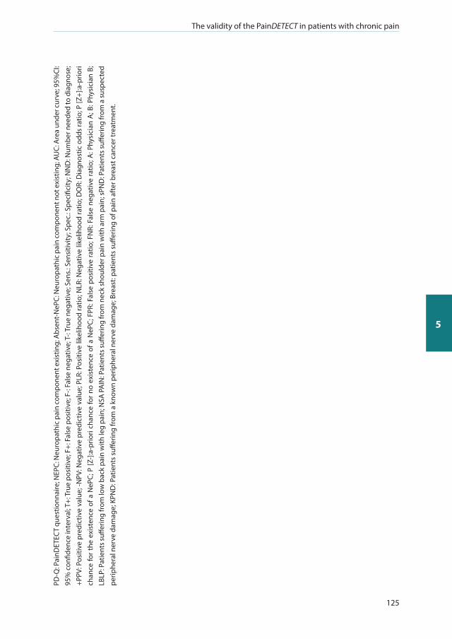
The validity of the PainDETECT in patients with chronic pain
125
5
PD-Q
: Pai
nDET
ECT
ques
tionn
aire
; NEP
C: N
euro
path
ic p
ain
com
pone
nt e
xist
ing;
Abs
ent-
NeP
C: N
euro
path
ic p
ain
com
pone
nt n
ot e
xist
ing;
AU
C: A
rea
unde
r cur
ve; 9
5%CI
: 95
% c
onfid
ence
inte
rval
; T+:
Tru
e po
sitiv
e; F
+: F
alse
pos
itive
; F-:
Fals
e ne
gativ
e; T
-: Tr
ue n
egat
ive;
Sen
s.: S
ensi
tivity
; Spe
c.: S
peci
ficity
; NN
D: N
umbe
r nee
ded
to d
iagn
ose;
+P
PV: P
ositi
ve p
redi
ctiv
e va
lue;
-NPV
: Neg
ativ
e pr
edic
tive
valu
e; P
LR: P
ositi
ve li
kelih
ood
ratio
; NLR
: Neg
ativ
e lik
elih
ood
ratio
; DO
R: D
iagn
ostic
odd
s ra
tio; P
[Z+]
:a-p
riori
chan
ce fo
r the
exi
sten
ce o
f a N
ePC;
P [Z
-]:a-
prio
ri ch
ance
for n
o ex
iste
nce
of a
NeP
C; F
PR: F
alse
pos
itive
ratio
; FN
R: F
alse
neg
ativ
e ra
tio; A
: Phy
sici
an A
; B: P
hysi
cian
B;
LBLP
: Pat
ient
s suff
erin
g fr
om lo
w b
ack
pain
with
leg
pain
; NSA
PA
IN: P
atie
nts s
uffer
ing
from
nec
k sh
ould
er p
ain
with
arm
pai
n; sP
ND
: Pat
ient
s suff
erin
g fr
om a
susp
ecte
d pe
riphe
ral n
erve
dam
age;
KPN
D: P
atie
nts
suffe
ring
from
a k
now
n pe
riphe
ral n
erve
dam
age;
Bre
ast:
patie
nts
suffe
ring
of p
ain
afte
r bre
ast c
ance
r tre
atm
ent.
Chapter 5
126
REFERENCES
1. IASP Taxonomy Neuropathic Pain. http://www.iasp-pain.org/Education/ Content.aspx?ItemNumber=1698#Neuropathicpain. Accessed 26 June 2018.
2. Freynhagen R, Baron R, Gockel U, Tolle TR. PainDETECT: a new screening questionnaire to identify neuropathic components in patients with back pain. Curr Med Res Opin. 2006;22(10):1911-20.
3. La Cesa S, Tamburin S, Tugnoli V, Sandrini G, Paolucci S, Lacerenza M, Marchettini P, Cruccu G, Truini A. How to diagnose neuropathic pain? The contribution from clinical examination, pain questionnaires and diagnostic tests. Neurol Sci : official J of the Italian Neurol Soc and of the Ital Soc of Clin Neurophysiol. 2015;36(12):2169-75.
4. Vissers KC. The clinical challenge of chronic neuropathic pain. Disabil Rehabil. 2006;28(6):343-9. 5. Baron R, Binder A, Wasner G. Neuropathic pain: diagnosis, pathophysiological mechanisms, and treatment.
The Lancet Neurol. 2010; 9(8):807-19. 6. Haanpaa M, Attal N, Backonja M, Baron R, Bennett M, Bouhassira D, Cruccu G, Hansson P, Haythornthwaite
JA, Iannetti GD, et al. NeuPSIG guidelines on neuropathic pain assessment. Pain. 2011;152(1):14-27. 7. Haanpaa ML, Backonja MM, Bennett MI, Bouhassira D, Cruccu G, Hansson PT, Jensen TS, Kauppila T, Rice
AS, Smith BH, et al. Assessment of neuropathic pain in primary care. Am J Med. 2009;122(10 Suppl):S13-21. 8. Dieleman JP, Kerklaan J, Huygen FJ, Bouma PA, Sturkenboom MC. Incidence rates and treatment of
neuropathic pain conditions in the general population. Pain. 2008;137(3):681-8. 9. O’Connor AB, Dworkin RH. Treatment of neuropathic pain: an overview of recent guidelines. Am J Med.
2009;122(10 Suppl):S22-32. 10. Dworkin RH, O’Connor AB, Kent J, Mackey SC, Raja SN, Stacey BR, Levy RM, Backonja M, Baron R, Harke H, et
al. Interventional management of neuropathic pain: NeuPSIG recommendations. Pain. 2013;154(11):2249-61.
11. Bennett MI, Attal N, Backonja MM, Baron R, Bouhassira D, Freynhagen R, Scholz J, Tolle TR, Wittchen HU, Jensen TS. Using screening tools to identify neuropathic pain. Pain. 2007;127(3):199-203.
12. Bouhassira D, Attal N. Diagnosis and assessment of neuropathic pain: the saga of clinical tools. Pain. 2011;152(3 Suppl):S74-83.
13. Andermann A, Blancquaert I, Beauchamp S, Dery V. Revisiting Wilson and Jungner in the genomic age: a review of screening criteria over the past 40 years. Bull World Health Organ. 2008;86(4):317-9.
14. Wilson JM, Jungner YG. Principles and practice of mass screening for disease. Bol Oficina Sanit Panam. 1968;65(4):281-393.
15. Mathieson S, Maher CG, Terwee CB, Folly de Campos T, Lin CW. Neuropathic pain screening questionnaires have limited measurement properties. A systematic review. J Clin Epidemiol. 2015;68(8):957-66.
16. De Andres J, Perez-Cajaraville J, Lopez-Alarcon MD, Lopez-Millan JM, Margarit C, Rodrigo-Royo MD, Franco-Gay ML, Abejon D, Ruiz MA, Lopez- Gomez V, et al. Cultural adaptation and validation of the PainDETECT scale into Spanish. Clin J Pain. 2012;28(3):243-53.
17. Alkan H, Ardic F, Erdogan C, Sahin F, Sarsan A, Findikoglu G. Turkish version of the PainDETECT questionnaire in the assessment of neuropathic pain: a validity and reliability study. Pain Med. 2013;14(12):1933-43.
18. Matsubayashi Y, Takeshita K, Sumitani M, Oshima Y, Tonosu J, Kato S, Ohya J, Oichi T, Okamoto N, Tanaka S. Validity and reliability of the Japanese version of the PainDETECT questionnaire: a multicenter observational study. PloS One. 2013;8(9):e68013.
19. Gudala K, Ghai B, Bansal D. Neuropathic pain assessment with the PainDETECT questionnaire: cross-cultural adaptation and psychometric evaluation to Hindi. Pain practice : the official journal of World Institute of Pain. 2017;17(8):1042-49.
20. Sung JK, Choi JH, Jeong J, Kim WJ, Lee DJ, Lee SC, Kim YC, Moon JY. Korean version of the PainDETECT questionnaire: a study for cultural adaptation and validation. Pain practice : the official journal of World Institute of Pain. 2017;17(4):494-504.
21. Freynhagen R, Tolle TR, Gockel U, Baron R. The PainDETECT project – far more than a screening tool on neuropathic pain. Curr Med Res Opin. 2016:1-25.
22. Jespersen A, Amris K, Bliddal H, Andersen S, Lavik B, Janssen H, Poulsen PB. Is neuropathic pain underdiagnosed in musculoskeletal pain conditions? The Danish PainDETECTive study. Curr Med Res Opin. 2010;26(8):2041-5.
23. Catch-22. https://www.merriam-webster.com/dictionary/catch-22. Accessed 26 June 2018.
The validity of the PainDETECT in patients with chronic pain
127
5
24. Timmerman H, Wolff AP, Schreyer T, Outermans J, Evers AW, Freynhagen R, Wilder-Smith OH, van Zundert J, Vissers KC. Cross-cultural adaptation to the Dutch language of the PainDETECT-questionnaire. Pain practice : the official journal of World Institute of Pain. 2013;13(3):206-14.
25. Timmerman H, Wilder-Smith O, van Weel C, Wolff A, Vissers K. DETECTing the neuropathic pain component in the clinical setting: a study protocol for validation of screening instruments for the presence of a neuropathic pain component. BMC Neurol. 2014;14(1):94.
26. Timmerman H, Steegers MAH, Huygen F, Goeman JJ, van Dasselaar NT, Schenkels MJ, Wilder-Smith OHG, Wolff AP, Vissers KCP. Investigating the validity of the DN4 in a consecutive population of patients with chronic pain. PloS One. 2017;12(11):e0187961.
27. ICD-10: version. 2015 International Statistical Classification of Diseases and Related Health Problems 10th Revision. http://apps.who.int/classifications/ icd10/browse/2015/en. Accessed 26 June 2018.
28. Lavand’homme P, Thienpont E. Pain after total knee arthroplasty: a narrative review focusing on the stratification of patients at risk for persistent pain. Bone Joint J. 2015;97-B(10 Suppl A):45-8.
29. Cruccu G, Sommer C, Anand P, Attal N, Baron R, Garcia-Larrea L, Haanpaa M, Jensen TS, Serra J, Treede RD. EFNS guidelines on neuropathic pain assessment: revised 2009. Eur J Neurol. 2010;17(8):1010-8.
30. Cruccu G, Anand P, Attal N, Garcia-Larrea L, Haanpaa M, Jorum E, Serra J, Jensen TS. EFNS guidelines on neuropathic pain assessment. Eur J Neurol. 2004;11(3):153-62.
31. Treede RD, Jensen TS, Campbell JN, Cruccu G, Dostrovsky JO, Griffin JW, Hansson P, Hughes R, Nurmikko T, Serra J. Neuropathic pain: redefinition and a grading system for clinical and research purposes. Neurol. 2008;70(18):1630-5.
32. Timmerman H, Heemstra I, Schalkwijk A, Verhagen C, Vissers K, Engels Y. Assessment of neuropathic pain in patients with Cancer: the Interobserver reliability. An Observational Study in Daily Practice. Pain physician. 2013;16:11.
33. Vaegter HB, Andersen PG, Madsen MF, Handberg G, Enggaard TP. Prevalence of neuropathic pain according to the IASP grading system in patients with chronic non-malignant pain. Pain Med. 2014;15(1):120-7.
34. Tampin B, Briffa NK, Goucke R, Slater H. Identification of neuropathic pain in patients with neck/upper limb pain: application of a grading system and screening tools. Pain. 2013;154(12):2813-22.
35. Salen BA, Spangfort EV, Nygren AL, Nordemar R. The disability rating index: an instrument for the assessment of disability in clinical settings. J Clin Epidemiol. 1994;47(12):1423-35.
36. Zigmond AS, Snaith RP. The hospital anxiety and depression scale. Acta Psychiatr Scand. 1983;67(6):361-70. 37. Spinhoven P, Ormel J, Sloekers PP, Kempen GI, Speckens AE, Van Hemert AM. A validation study of the
hospital anxiety and depression scale (HADS) in different groups of Dutch subjects. Psychol Med. 1997;27(2):363-70.
38. Bjelland I, Dahl AA, Haug TT, Neckelmann D. The validity of the hospital anxiety and depression scale. An updated literature review. J Psychosom Res. 2002;52(2):69-77.
39. Hays RD, Sherbourne CD, Mazel RM. The RAND 36-item health survey 1.0. Health Econ. 1993;2(3):217-27. 40. VanderZee KI, Sanderman R, Heyink J. A comparison of two multidimensional measures of health status:
the Nottingham health profile and the RAND 36-item health survey 1.0. Qual Life Res. 1996;5(1):165-74. 41. VanderZee KI, Sanderman R, Heyink JW, de Haes H. Psychometric qualities of the RAND 36-item health
survey 1.0: a multidimensional measure of general health status. Int J Behav Med. 1996;3(2):104-22. 42. Collins SL, Edwards J, Moore RA, Smith LA, McQuay HJ. Seeking a simple measure of analgesia for mega-
trials: is a single global assessment good enough? Pain. 2001;91(1-2):189-94. 43. Farrar JT, Young JP Jr, LaMoreaux L, Werth JL, Poole RM. Clinical importance of changes in chronic pain
intensity measured on an 11-point numerical pain rating scale. Pain. 2001;94(2):149-58. 44. Fischer D, Stewart AL, Bloch DA, Lorig K, Laurent D, Holman H. Capturing the patient’s view of change as a
clinical outcome measure. Jama. 1999; 282(12):1157-62. 45. Flahault A, Cadilhac M, Thomas G. Sample size calculation should be performed for design accuracy in
diagnostic test studies. J Clin Epidemiol. 2005;58(8):859-62. 46. How good is that test? II. http://www.bandolier.org.uk/band27/b27-2.html. Accessed 26 June 2018. 47. Greenhalgh T. How to read a paper: papers that report diagnostic or screening tests (vol 315, pg 540, 1997).
Br Med J. 1997;315(7113):942. 48. Landis JR, Koch GG. The measurement of observer agreement for categorical data. Biometrics.
1977;33(1):159-74. 49. Heberle H, Meirelles GV, da Silva FR, Telles GP, Minghim R. InteractiVenn: a web-based tool for the analysis
of sets through Venn diagrams. BMC Bioinformatics. 2015;16:169.
Chapter 5
128
50. Hallstrom H, Norrbrink C. Screening tools for neuropathic pain: can they be of use in individuals with spinal cord injury? Pain. 2011;152(4):772-9.
51. Gauffin J, Hankama T, Kautiainen H, Hannonen P, Haanpaa M. Neuropathic pain and use of PainDETECT in patients with fibromyalgia: a cohort study. BMC Neurol. 2013;13:21.
52. Finnerup NB, Haroutounian S, Kamerman P, Baron R, Bennett DL, Bouhassira D, Cruccu G, Freeman R, Hansson P, Nurmikko T, et al. Neuropathic pain: an updated grading system for research and clinical practice. Pain. 2016;157(8): 1599-606.
53. Scholz J, Mannion RJ, Hord DE, Griffin RS, Rawal B, Zheng H, Scoffings D, Phillips A, Guo J, Laing RJ, et al. A novel tool for the assessment of pain: validation in low back pain. PloS Med. 2009;6(4):e1000047.
54. CADTH. Diagnostic methods for neuropathic pain: A review of diagnostic accuracy. In: Canadian Agency for Drugs and Technologies in Health. Canada: Ottawa (ON); 2015. https://www.ncbi.nlm.nih.gov/pubmedhealth/ PMH0078647/. Accessed 26 June 2018.
55. Bouhassira D, Attal N. Translational neuropathic pain research: a clinical perspective. Neuroscience. 2016;3:27-35.
56. Cooney MA, Culleton-Quinn E, Stokes E. Current knowledge of pain after breast cancer treatment: a systematic review. Pain Manag Nurs. 2013;14(2): 110-23.
57. Ilhan E, Chee E, Hush J, Moloney N. The prevalence of neuropathic pain is high following treatment for breast cancer: a systematic review. Pain. 2017; 15(11):2082-91.

The validity of the PainDETECT in patients with chronic pain
129
5


CHAPTER 6Investigating the validity of the DN4 in a consecutive population of patients with chronic pain
Hans Timmerman
Monique A. H. Steegers
Frank J. P. M. Huygen
Jelle J. Goeman
Nick T. van Dasselaar
Marcel J. Schenkels
Oliver H. G. Wilder-Smith
André P. Wolff
Kris C. P. Vissers
Published in:
PloS ONE 12(11): e0187961.
Chapter 6
132
ABSTRACT
Neuropathic pain is clinically described as pain caused by a lesion or disease of the somatosensory
nervous system. The aim of this study was to assess the validity of the Dutch version of the DN4, in a
cross-sectional multicentre design, as a screening tool for detecting a neuropathic pain component
in a large consecutive, not pre-stratified on basis of the target outcome, population of patients
with chronic pain. Patients’ pain was classified by two independent (pain-)physicians as the gold
standard. The analysis was initially performed on the outcomes of those patients (n = 228 out of
291) in whom both physicians agreed in their pain classification. Compared to the gold standard
the DN4 had a sensitivity of 75% and specificity of 76%. The DN4-symptoms (seven interview items)
solely resulted in a sensitivity of 70% and a specificity of 67%. For the DN4-signs (three examination
items) it was respectively 75% and 75%. In conclusion, because it seems that the DN4 helps to
identify a neuropathic pain component in a consecutive population of patients with chronic pain in
a moderate way, a comprehensive (physical-) examination by the physician is still obligate.
Data Availability Statement
All relevant data are within the paper and its Supporting Information files.
Funding
This study was performed within DALI for PAIN, a national program that focuses on neuropathic pain
care optimalisation to APW. DALI for PAIN is an initiative of Pfizer. This project is supported by an
unrestricted grant from Pfizer. The funders had no role in study design, data collection and analysis,
decision to publish, or preparation of the manuscript.
Competing interests
This project is supported by an unrestricted grant from Pfizer. This unrestricted grant does not alter
our adherence to PLOS ONE policies on sharing data and materials. The authors declare that they
have no other competing interests.
The validity of the DN4 in patients with chronic pain
133
6
INTRODUCTION
Neuropathic pain is described as pain caused by a lesion or disease of the somatosensory nervous
system and requires a demonstrable lesion or a disease that satisfies established neurological
diagnostic criteria [1]. Moreover, neuropathic pain is a clinical description and not a diagnosis [1].
In daily clinical practice it is to our opinion more appropriate to speak of a present neuropathic
pain component (present NePC) or absent neuropathic pain component (absent NePC) [2, 3].
This because the pain experienced by the patient in the clinical context may be caused by both
neuropathic- as well as nociceptive mechanisms (also known as ‘mixed pain’) [2, 4-6]. The main
features of neuropathic pain components are, in clinical practice, the painful signs and symptoms in
a region of altered sensations (numbness or increased sensitivity) [6]. The assessment of neuropathic
pain is nowadays primarily based on history and physical examination including (bedside-)sensory
testing [7-9] to assess patients’ pain.
Since current pharmacological treatment of patients with and without a NePC differs strongly, a
correct pain classification is imperative [7, 10]. The availability of a simple and validated screening
tool to determine the presence of NePC for clinical triage and epidemiological purposes can assist
in detection of NePC [7, 8, 11-16]. This is especially true when this tool can be used by non pain
specialists.
The original French validation study of the ‘Douleur Neuropathique en 4 Questions’ (DN4) [17] was
performed in patients with neuropathic pain resulting from, for example, nerve trauma or post
herpetic neuralgia. Patients with non-neuropathic pain were, amongst other diagnoses, suffering
from osteoarthritis. All included patients had pain of at least a moderate severity (≥ 40 on a 100mm
visual analogue scale). Pain classification in this study was based on medical history, physical
examination, electromyography and/or imaging by two independently working physicians. DN4
application resulted in a sensitivity of 83% and a specificity of 90% [17]. As indicated in a systematic
review by Mathieson et al [16] the classification of a NePC may differ between clinicians and may
be more difficult when there are patients included with mixed pain and with all levels of pain. This
reflects the patient population in a daily clinical practice, but might have an influence on the validity.
Moreover, the accuracy of screening tools is dependent on the standardization of the assessment
strategy [18]. Translation/ cross-cultural adaptation and/or validation of the DN4 was performed in
more than 75 languages [19-31].
The neuropathic pain special interest group (NeuPSIG) grading system [32] is developed by Treede
et al in 2008 and updated in 2016 [33]. It is a system to help the clinician to determine the certainty
of the pain classification for the existence of a NePC in an individual patient: non-neuropathic pain;
possible, probable or definite neuropathic pain. The grading system is suggested to be helpful in the
assessment of the pain classification in clinical practice [34-38].
Chapter 6
134
The aim of this study was to assess the validity and reliability of the DN4 as a screening tool for
use in daily outpatient practices to detect a NePC in a, not pre-stratified on the target outcome,
consecutive patient population having chronic pain syndromes due to low back and leg pain (LBLP),
neck-shoulder-arm-pain (NSAP) or pain of suspected neuropathic origin (PSNO).
METHODSThis validity and reliability study had a cross-sectional, longitudinal, research design with a 2-weeks
and 3-months follow-up period. Comparisons were made between the DN4 (as a whole and for
the symptom questions and signs tests separately) and the classification of patients’ pain by two,
independently working, physicians (the gold standard) as well as with the grading system.
The study was approved by the Committee on Research Involving Human Subjects region Arnhem-
Nijmegen, Nijmegen, the Netherlands, (dossier number: 2008/348; NL 25343.091.08) which
counts for participation of the Dutch academic pain centers (Radboud University Medical Center,
Nijmegen; Utrecht University Medical Center, Utrecht; Erasmus Medical Center, Rotterdam), Dutch
non-academic pain centers (Bernhoven Ziekenhuis, Oss; St.Anna Ziekenhuis, Geldrop) and a Dutch
non-academic department of neurology (Rijnstate Ziekenhuis, Arnhem). Participation of Dutch non-
academic pain center in Delft, the Netherlands (Reinier de Graaf gasthuis) was approved by Medisch
Ethische Toestings Commissie Zuidwest Holland (dossier number: 10-145). The study protocol was
registered in the Dutch National Trial Register (NTR3030).
We used the same methodology as in the published protocol [39] and as employed in a simultaneous
study regarding the validity of the PainDETECT (Timmerman et.al / Under review by BMC Neurology).
Participants Consecutive patients (first time visitors of the participating centers) without pre-stratification based
on the target outcome [40] were included in the study between October 2009 until July 2013.
Patients were asked to participate by their doctor. Each patient signed informed consent before
participation in the study.
At that time, there was only a rough diagnosis: LBLP, NSAP or PSNO. Inclusion criteria: Male and female
adult patients (≥18 years of age) with chronic (≥3 months) LBLP or NSAP radiating into respectively
leg(s) or arm(s) or patients with chronic pain due to a PSNO (pain associated with a lesion or disease
of the peripheral somatosensory system). Exclusion criteria: Patients diagnosed with malignancy;
compression fractures; patients with diffuse pains (such as fibromyalgia or ankylosing spondylitis);
severe mental illness; chronic alcoholism or substance abuse; inability to fill in the questionnaire
adequately or incapable of understanding the Dutch language.

The validity of the DN4 in patients with chronic pain
135
6
Physicians The physicians (pain specialists, pain specialist fellows or neurologists always operating in
differently composed pairs) participating in this study were not selected on basis of age, experience
as a physician or any other criteria. Classification of patients’ pain was based on the NeuPSIG
guidelines on neuropathic pain assessment [7] and recorded as absent NePC or present NePC. Pain
classification was performed consecutively on the same patient by two physicians and categorized
afterwards in three groups: absent NePC, present NePC or ‘undetermined’ (i.e. the pain classification
of the two physicians was not the same). A full medical history and clinical examination including
sensory bedside examination (touch, pinprick, pressure, cold, heath and temporal summation) was
taken [7, 8, 39, 41, 42] and was considered to be the gold standard when assessed by two physicians.
The NeuPSIG grading system [32, 33] was used as a secondary comparison with the outcome of the
DN4 and was assessed by both the physicians separately. The outcomes “probable” and “definite”
were regarded as present NePC. “unlikely” and “possible” as absent NePC [38, 43, 44]. The physicians
worked independently of each other and were blinded to the pain classification of the other
physician. Each physician was allowed to perform the clinical examination in the way he or she
is used to do but were supported by a standardized assessment form [39]. In this form, the pain
score, a body map to indicate the localization of patients pain, the sensory examination and the four
questions of the grading system had to be filled in by the physician. The participating physicians
were trained in a standardized way (presentation about the study and the outcome parameters
and a practical training on how to use the (measurement) instruments), by the investigator (HT) or
by a designated person on location before participation in the study. Practical training was focused
on the classification of NePC, the assessment of the grading system, the performance of bed-side
examination tests and the performance and assessment of the examination items of the DN4.
In this study, 62 physicians (pain specialist, pain specialist-fellow or neurologist) participated. The
physicians who were classifying patients’ pain at the first session were called ‘Physicians A’. The
physicians who performed the classification at the second session, were called ‘Physicians B’.
Measurements Douleur Neuropathique en 4 questions (DN4). The DN4 [17, 20, 25] (Pfizer bv. Capelle a/d Ijssel, the
Netherlands) consists of 10 items in total and is developed to screen for symptoms and signs of
neuropathic pain resulting in a yes/no answer for the presence of neuropathic pain. This instrument
is divided into two questions (seven answers, DN4-symptoms: score range 0-7) and two physical
examination tests (three answers, DN4-signs: score range 0-3). The examination items of the DN4
regarding the signs (hypoesthesia to touch, hypoesthesia to prick and brushing) were incorporated
in the sensory examination part of the standardized assessment form and were carried out
according the original publication by Bouhassira et al [17]. This assessment form was filled in by
both physicians separately. The seven symptom items are consisting of characteristics (Burning,
painful cold, electric shocks) and symptoms (Tingling, pins and needles, numbness, and itching).
Chapter 6
136
The patient completed the DN4-symptoms directly after the clinical assessments by the physicians
but without interference. The researcher (HT) or a nurse was available for help in person or via
telephone when it was not clear fort the patient how to fill in the questionnaires.
The items of the DN4 are scored based on a yes (1 point) /no (0 points) answer. This leads to a score
range of 0-10 when the symptoms (range 0-7 points) as well as the signs (range 0-3 points) items are
included. Values in the DN4 who were not filled in were considered as ‘no’ (0 points). However, in the
reliability analysis these data were not incorporated.
Patient global impression of change (PGIC). The Patients Global Impression of Change (PGIC) [11,
45-47] was used to assess the change of pain complaints, based on the patients’ own impression
of change over time, during the follow-up period (7-points scale: Very much improved-very much
worse). Follow-up took place two weeks and three months after the initial visit. To compare the
outcome of the DN4 in the follow-up period the pain complaints as addressed by the patient had
to be unchanged.
Time-line All baseline measurements (the assessment by the physicians, the grading system by both physicians
as well as filling in the questionnaires by the patient) took place on preferably the same day. The
PGIC [45-47] and the DN4-symptoms (sensory testing for the DN4-signs was not performed) were
sent to the patient after two weeks and three months with instructions how to fill them in by mail.
Also for the follow-up measurements help was available in person or via telephone when it was not
clear how to fill in the questionnaires.
Data All data was collected on paper and stored by Radboudumc, Nijmegen, The Netherlands. Data
management and monitoring were performed within MACRO (MACRO, version 4.1.1.3720, Infermed,
London, United Kingdom). Data analysis and statistics was performed by use of Statistical Package
for the Social Sciences (IBM SPSS statistics 22, SPSS Inc., Chicago, Illinois, USA).
Statistical analysis According to the power-calculation in the protocol 132 patients with LBLP, NSAP or PSNO were
needed such that the sample size contains adequate numbers of cases and controls [39]. Qualitative
variables are presented as frequencies and percentages. The quantitative variables are presented as
mean and standard deviation (SD) or as median and inter quartile range (IQR).
The agreement between any of the two combinations of the two observers (pain classification
by the physician and the outcome of the grading system) to establish a present NePC or
absent NePC, and of the DN4 (DN4 / DN4-symptoms / DN4-signs outcome) was evaluated
The validity of the DN4 in patients with chronic pain
137
6
by use of Cohen’s kappa (K), prevalence index (Pi) and percentage of pair wise agreement
(PA). The categorization of the kappa values are, according to the categorization of observer
agreement by Landis and Koch [48], none beyond chance (K ≤ 0.00); slight (K = 0.01-0.20); fair
(K = 0.21-0.40); moderate (K = 0.41-0.60); substantial (K = 0.61-0.80) and (almost) perfect agreement (K
= 0.81-1.00). A K ≥ 0.40 and a PA ≥ 70% is considered indicative of interobserver reliability acceptable
for use in clinical practice [48]. Moreover, also the interobserver reliability of the examination items
in the DN4-signs were tested.
Based on the classifications of the two phyicians, all patients were categorized as absent NePC,
present NePC or ‘undetermined’ (i.e. the pain classification of the two physicians was not the same).
Statistical significant differences between absent NePC and present NePC were determined by
use of students t-test (Interval scales), Mann-Whitney U-test groups (ordinal scales) or via Chi2-test
(nominal scale). The statistical significant differences between present NePC, absent NePC and the
Undetermined group was assessed by use of One-way ANOVA (with additional Tukey’s studentized
range post-hoc test) or Kruskal-Wallis test. Chi2 test was also used to analyze the nominal outcome
scale of the DN4 regarding the three groups.
A factor analysis was used to study the structure of the DN4 in such a way that variables that were
thought to reflect a smaller number of underlying variables were observed. This method was
performed for all three versions of the DN4 (DN4; DN4-symptoms and DN4-signs). Principal axis
factoring was used as the extraction method. The varimax rotation with Kaiser normalization was
used. Extraction of the factors was based on Eigenvalues being greater than 1.0. Cronbach’s alpha
was used to calculate the internal consistency of the factors constructed. The results are only shown
for the Physicians A (the assessment of the patient by the first physician). The outcomes by the
Physicians B (the assessment of the patient by the second physician) are shown in S1 Table. However,
the conclusions, which are drawn, are identical for physicians A and for physicians B.
A receiver operating characteristic (ROC) curve was calculated for the DN4 and the DN4 signs by
both the physicians A and B and for the DN4-symptoms as filled in by the patient. The area under
the curve (AUC) with 95% confidence interval was presented to indicate the discriminatory power
of the DN4 to discriminate patients by present NePC or absent NePC. This dichotomy was based
on the physicians’ assessment outcome or based on the grading system outcome, respectively.
The theoretical maximum of the AUC is 100%, indicating a perfect discrimination and 50% is equal
to tossing a coin. An AUC between 0.9 and 1 is considered to be excellent, an AUC between 0.8
and 0.9 is good and between 0.7 and 0.8 is fair. An AUC between 0.6 and 0.7 is considered to be
poor. Between 0.5 and 0.6 the AUC is considered to be failed [49-52]. The optimal cut-off point of
the DN4 was calculated under the condition of equal-costs of misclassification using the Youden-
index. Sensitivity, specificity, positive and negative predictive values and the likelihood ratio in the
Chapter 6
138
population in this study was calculated at this cut-off point. The outcome results were averaged
between both physicians and the 95% confidence intervals were noted with respect to the lowest
and highest level.
Clinimetrics of the DN4 based on both the physicians assessment and/or both the grading system
outcome were assessed for the DN4, the DN4-symptoms and for the DN4-signs items. A screening
tool for the presence of a NePC is considered valid if it has a high sensitivity, specificity, high positive
predictive value and a high positive likelihood ratio [53]. Intraclass correlation (ICC) was used to
assess reproducibility (‘test-retest reliability’) of the DN4-symptoms between the predetermined
time points (baseline versus two weeks & baseline versus three months). Based on the guidelines
by Cicchetti et al. [54, 55] an ICC <0.40 indicates poor level of clinical significance. The level is fair
when the ICC is between 0.40 and 0.59, good between 060 and 0.74 and excellent when the ICC is
between 0.75 and 1.00. To assess the test-retest reliability patients’ pain should not have changed
(outcome based on the PGIC) because otherwise the ICC would not reflect the consistency of the
DN4. Test-retest reliability was assessed for those questionnaires returned within 7-21 days for the
two weeks test-retest reliability and 60-120 days for the three months test—retest reliability. The ICC
and responsiveness of the DN4-symptoms was assessed at each point of measurement.
Two-tailed p-value below 0.05 was considered statistically significant.
RESULTS
Patients In this study 330 consecutive patients were assessed for eligibility (Figure 1). Of these, 291
participated in the study between October 2009 and July 2013. Two patients did not give their
informed consent. Exclusion (n = 37) was because of not fulfilling the in- and exclusion criteria (n
= 13): patients with LBLP or NSAP without radiating pain: n = 1; patients with less than 3 months
pain complaints: n = 2; patients with pain with an oncological cause: n = 2; patients with painful
syndromes of unknown origin or associated with diffuse pains: n = 7; patients with severe mental
illness: n = 1; missing baseline measurements due to not returning questionnaires by the patient:
n = 16; missing pain classification based on the grading system by one physician (n = 5) or both
the physicians (n = 3). 132 patients had LBLP with radiation in one or two legs (45.4%), 51 NSAP
with radiation in one or both arms (17.5%) and 108 patients (37.1%) had PSNO: 86 patients with
pain after treatment for breast cancer (surgery and chemotherapy and/or radiation therapy and/
or hormonal therapy). Twenty-two patients had pain for various reasons: peripheral nerve damage
(n = 12), radicular pain (n = 3), polyneuropathy (n = 3), CRPS (n = 2) and post stroke pain (n = 2).
The gold standard for presence of the NePC in this study was the concordant clinical opinion of
both physicians. After pain classification by two physicians, 170 patients were classified as present
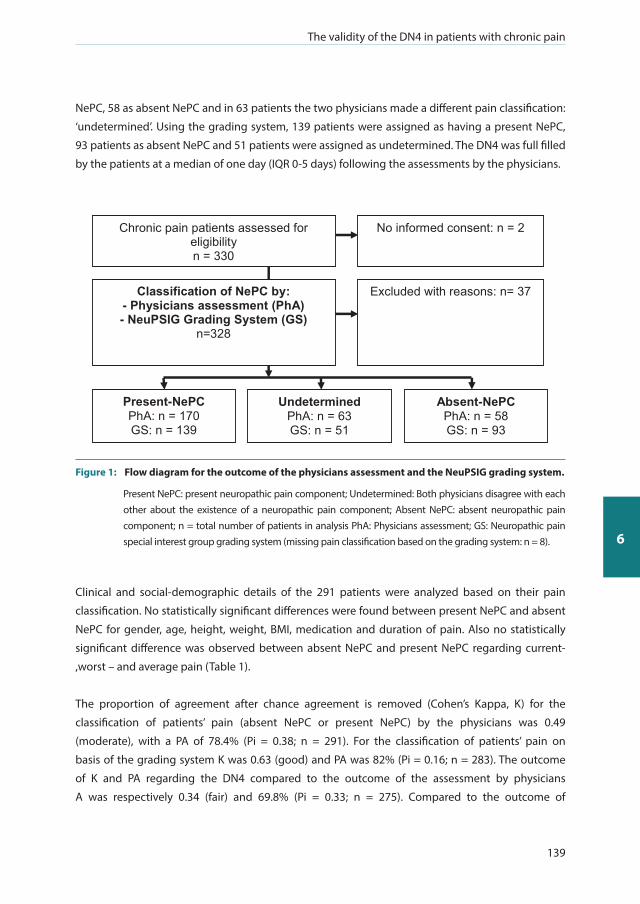
The validity of the DN4 in patients with chronic pain
139
6
NePC, 58 as absent NePC and in 63 patients the two physicians made a different pain classification:
‘undetermined’. Using the grading system, 139 patients were assigned as having a present NePC,
93 patients as absent NePC and 51 patients were assigned as undetermined. The DN4 was full filled
by the patients at a median of one day (IQR 0-5 days) following the assessments by the physicians.
Chronic pain patients assessed for eligibility n = 330
Classification of NePC by: - Physicians assessment (PhA) - NeuPSIG Grading System (GS)
n=328
Excluded with reasons: n= 37
No informed consent: n = 2
Present-NePC PhA: n = 170 GS: n = 139
Undetermined PhA: n = 63 GS: n = 51
Absent-NePC PhA: n = 58 GS: n = 93
Figure 1: Flow diagram for the outcome of the physicians assessment and the NeuPSIG grading system.
Present NePC: present neuropathic pain component; Undetermined: Both physicians disagree with each other about the existence of a neuropathic pain component; Absent NePC: absent neuropathic pain component; n = total number of patients in analysis PhA: Physicians assessment; GS: Neuropathic pain special interest group grading system (missing pain classification based on the grading system: n = 8).
Clinical and social-demographic details of the 291 patients were analyzed based on their pain
classification. No statistically significant differences were found between present NePC and absent
NePC for gender, age, height, weight, BMI, medication and duration of pain. Also no statistically
significant difference was observed between absent NePC and present NePC regarding current-
,worst – and average pain (Table 1).
The proportion of agreement after chance agreement is removed (Cohen’s Kappa, K) for the
classification of patients’ pain (absent NePC or present NePC) by the physicians was 0.49
(moderate), with a PA of 78.4% (Pi = 0.38; n = 291). For the classification of patients’ pain on
basis of the grading system K was 0.63 (good) and PA was 82% (Pi = 0.16; n = 283). The outcome
of K and PA regarding the DN4 compared to the outcome of the assessment by physicians
A was respectively 0.34 (fair) and 69.8% (Pi = 0.33; n = 275). Compared to the outcome of

Chapter 6
140
Table 1: Clinical and socio-demographic characteristics of the patients related to physicians agreement for the existence of a neuropathic pain component.
NePC Absent Present Undetermined
N N P N P
Total number of patients 58 170 63
GenderMaleFemale
25 (43%)33 (57%)
56 (33%)114 (67%)
0.163c
17 (27%)46 (73%)
0.164c
Age (Years) # 58 55 ± 12 170 56 ± 11 0.594a 63 58 ± 13 0.522d
Height (cm) #
Weight (kg) #
BMI (kg/m2 ) #
55 55 54
172 ± 984 ± 228 ± 8
164 167 164
172 ± 880 ±1727 ±5
0.845a
0.382a
0.436a
6262 62
170 ± 980 ± 1627 ± 5
0.250d
0.461d
0.593d
Medication use^ 55 56.9% 168 66.1% 0.414c 61 57.4% 0.423c
Duration of pain (months)# 57 72 ± 90 169 60 ± 76 0.327a 62 49 ± 46 0.247d
Pain* (NRS; 0-10)
Current pain Worst pain during the past four weeks Average pain during the past four weeks
5757
57
5 (3-7)8 (5-9)
6 (3.5-7)
167167
167
6 (3-7)8 (7-9)
6 (5-8)
0.577b
0.371b
0.233b
6161
61
4 (1-7)7 (5-8)
6 (3-7)
0.084e
0.053e
0.018e
Classification for the existence of NePC is based on physicians assessment of the patients. NePC: neuropathic pain component; Absent: NePC is absent; Present: NePC is present; Undetermined: both physicians disagree with each other about the existence of a neuropathic pain component; N: total number of patients in analysis; n: number of patients; ^ percentage; # Standard deviation; * Inter quartile range. A: physicians A; B: Physicians B; P value for significant difference between groups (P ≤ 0.05) by use of different analyse methods: a: Students t-test; b: Mann-Whitney U test; c: Chi-square; d: One-Way ANOVA; e: Kruskal-Wallis test.
the assessment by physicians B it was 0.33 (fair) and 69.2% (Pi = 0.30; n = 263). Comparing
the outcome of the DN4 to the outcome of the grading system, it was 0.35 (fair) and 69.1%
(Pi = 0.22; n = 272) for physicians A, and 0.32 (fair) and 67.3%(Pi = 0.19; n = 260) for physicians B (Table
2). The interobserver reliability for ‘hypoesthesia to touch’ as well as for ‘brushing’ was respectively K
= 0.59 (moderate) (PA = 79.7%) and K = 0.53 (moderate)(PA = 76.6%). The interobserver reliability for
‘hypoesthesia to prick’ was K = 0.21 (fair); PA = 87% (Table 3).
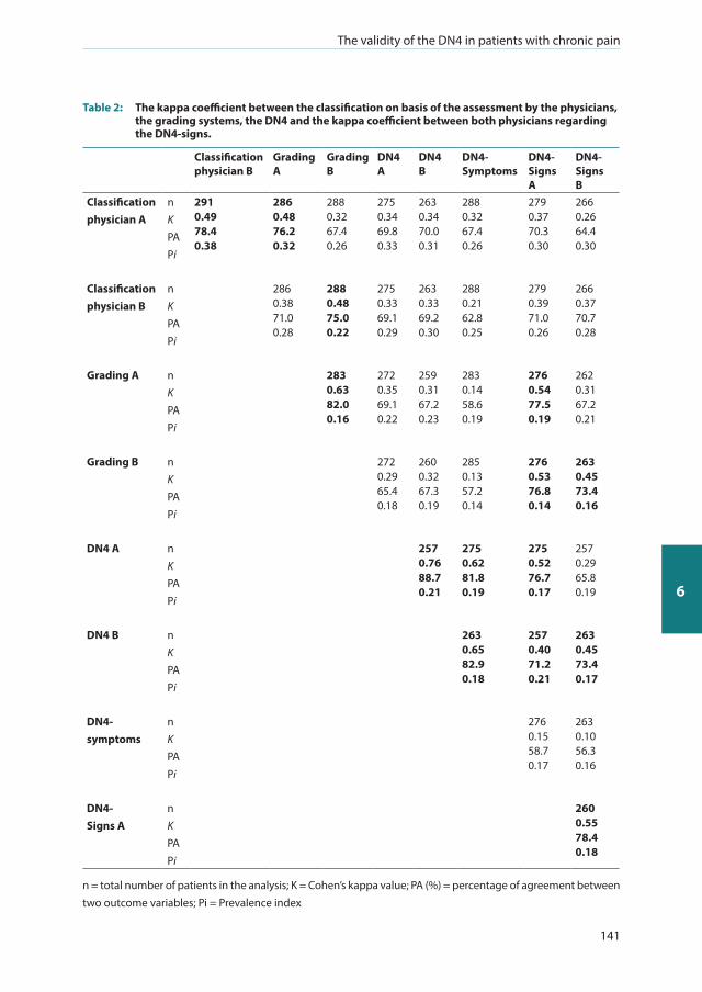
The validity of the DN4 in patients with chronic pain
141
6
Table 2: The kappa coefficient between the classification on basis of the assessment by the physicians, the grading systems, the DN4 and the kappa coefficient between both physicians regarding the DN4-signs.
Classification physician B
Grading A
GradingB
DN4 A
DN4 B
DN4-Symptoms
DN4-Signs A
DN4-Signs B
Classification physician A
nK PA Pi
2910.4978.40.38
2860.4876.20.32
2880.3267.40.26
2750.3469.80.33
2630.3470.00.31
2880.3267.40.26
2790.3770.30.30
2660.2664.40.30
Classificationphysician B
nK PA Pi
2860.3871.00.28
2880.4875.00.22
2750.3369.10.29
2630.3369.20.30
2880.2162.80.25
2790.3971.00.26
2660.3770.70.28
Grading A nK PA Pi
2830.6382.00.16
2720.3569.10.22
2590.3167.20.23
2830.1458.60.19
2760.5477.50.19
2620.3167.20.21
Grading B nK PA Pi
2720.2965.40.18
2600.3267.30.19
2850.1357.20.14
2760.5376.80.14
2630.4573.40.16
DN4 A nK PA Pi
2570.7688.70.21
2750.6281.80.19
2750.5276.70.17
2570.2965.80.19
DN4 B nK PA Pi
2630.6582.90.18
2570.4071.20.21
2630.4573.40.17
DN4-symptoms
nK PA Pi
2760.1558.70.17
2630.1056.30.16
DN4-Signs A
nK PA Pi
2600.5578.40.18
n = total number of patients in the analysis; K = Cohen’s kappa value; PA (%) = percentage of agreement between two outcome variables; Pi = Prevalence index
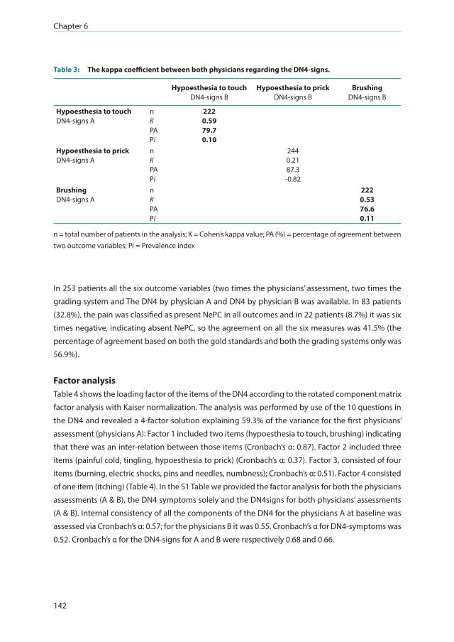
Chapter 6
142
Table 3: The kappa coefficient between both physicians regarding the DN4-signs.
Hypoesthesia to touchDN4-signs B
Hypoesthesia to prickDN4-signs B
BrushingDN4-signs B
Hypoesthesia to touch DN4-signs A
nK PA Pi
2220.5979.70.10
Hypoesthesia to prick DN4-signs A
nK PA Pi
2440.2187.3-0.82
Brushing DN4-signs A
nK PA Pi
2220.5376.60.11
n = total number of patients in the analysis; K = Cohen’s kappa value; PA (%) = percentage of agreement between two outcome variables; Pi = Prevalence index
In 253 patients all the six outcome variables (two times the physicians’ assessment, two times the
grading system and The DN4 by physician A and DN4 by physician B was available. In 83 patients
(32.8%), the pain was classified as present NePC in all outcomes and in 22 patients (8.7%) it was six
times negative, indicating absent NePC, so the agreement on all the six measures was 41.5% (the
percentage of agreement based on both the gold standards and both the grading systems only was
56.9%).
Factor analysis Table 4 shows the loading factor of the items of the DN4 according to the rotated component matrix
factor analysis with Kaiser normalization. The analysis was performed by use of the 10 questions in
the DN4 and revealed a 4-factor solution explaining 59.3% of the variance for the first physicians’
assessment (physicians A): Factor 1 included two items (hypoesthesia to touch, brushing) indicating
that there was an inter-relation between those items (Cronbach’s α: 0.87). Factor 2 included three
items (painful cold, tingling, hypoesthesia to prick) (Cronbach’s α: 0.37). Factor 3, consisted of four
items (burning, electric shocks, pins and needles, numbness); Cronbach’s α: 0.51). Factor 4 consisted
of one item (itching) (Table 4). In the S1 Table we provided the factor analysis for both the physicians
assessments (A & B), the DN4 symptoms solely and the DN4signs for both physicians’ assessments
(A & B). Internal consistency of all the components of the DN4 for the physicians A at baseline was
assessed via Cronbach’s α: 0.57; for the physicians B it was 0.55. Cronbach’s α for DN4-symptoms was
0.52. Cronbach’s α for the DN4-signs for A and B were respectively 0.68 and 0.66.
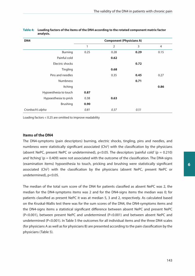
The validity of the DN4 in patients with chronic pain
143
6
Table 4: Loading factors of the items of the DN4 according to the rotated component matrix factor analysis.
DN4 Component (Physicians A)
1 2 3 4
Burning 0.25 0.28 0.29 0.15
Painful cold 0.62
Electric shocks 0.72
Tingling 0.68
Pins and needles 0.35 0.45 0.27
Numbness 0.71
Itching 0.86
Hypoesthesia to touch 0.87
Hypoesthesia to prick 0.38 0.63
Brushing 0.90
Cronbach’s alpha 0.81 0.37 0.51
Loading factors < 0.25 are omitted to improve readability
Items of the DN4 The DN4-symptoms (pain descriptors) burning, electric shocks, tingling, pins and needles, and
numbness were statistically significant associated (Chi2) with the classification by the physicians
(absent NePC, present NePC or undetermined), p<0.05. The descriptors ‘painful cold’ (p = 0.210)
and ‘itching’ (p = 0.409) were not associated with the outcome of the classification. The DN4-signs
(examination items) hypoesthesia to touch, pricking and brushing were statistically significant
associated (Chi2) with the classification by the physicians (absent NePC, present NePC or
undetermined), p<0.05.
The median of the total sum score of the DN4 for patients classified as absent NePC was 2, the
median for the DN4-symptoms items was 2 and for the DN4-signs items the median was 0; for
patients classified as present NePC it was at median 5, 3 and 2, respectively. As calculated based
on the Kruskal-Wallis test there was for the sum scores of the DN4, the DN4-symptoms items and
the DN4-signs items a statistical significant difference between absent NePC and present NePC
(P<0.001), between present NePC and undetermined (P<0.001) and between absent NePC and
undetermined (P<0.001). In Table 5 the outcomes for all individual items and the three DN4 scales
(for physicians A as well as for physicians B) are presented according to the pain classification by the
physicians (Table 5).

Chapter 6
144
Table 5: The median (IQR) and percentages of the items of the DN4 by physicians agreement of a NePC.
NePC Absent Present Undetermined
N N P N P
Total number of patient 58 170 63DN4-Symptoms^
Burning 56 12 (21%) 161 77 (48%) 0.001a 57 22 (39%) 0.002a
Painful Cold 54 6 (11%) 154 34 (22%) 0.078a 53 11 (21%) 0.210a
Electric Shocks 55 18 (33%) 162 87 (54%) 0.007a 55 19 (35%) 0.005a
Tingling 55 29 (53%) 160 110 (69%) 0.032a 57 28 (49%) 0.011a
Pins and Needles 52 19 (37%) 157 101 (60%) 0.000a 58 27 (47%) 0.001a
Numbness 54 29 (54%) 165 131 (79%) 0.000a 59 42 (71%) 0.001a
Itching 51 10 (20%) 149 25 (17%) 0.646a 56 14 (25%) 0.409a
DN4-signs^ Hypoesthesia to touch A 42 9 (21%) 153 102 (67%) 0.000a 60 16 (27%) 0.000a
B 41 11 (27%) 151 101 (67%) 0.000a 49 18 (37%) 0.000a
Hypoesthesia to prick A 47 0 (0%) 162 20 (12%) 0.011a 58 3 (5%) 0.017a
B 48 0 (0%) 159 21 (12%) 0.008a 53 1 (2%) 0.002a
Brushing A 43 9 (21%) 157 110 (70%) 0.000a 55 14 (25%) 0.000a
B 43 13 (30%) 151 99 (66%) 0.000a 52 19 (37%) 0.000a
Total sum score DN4 A* (0-10) 47 2 (1-3) 166 5 (3-6) 0.000b 62 3 (2-4) 0.000c
Total sum score DN4 B* (0-10) 48 2 (2-3,75) 159 5 (3-6) 0.000b 56 3 (2-4.75) 0.000c
Total sum score DN4 symptoms* (0-7) 57 2 (1-3) 168 3 (2-5) 0.000b 63 2 (2-4) 0.000c
Total sum score DN4 signs A* (0-3) 49 0 (0-0) 168 2 (1-2) 0.000b 62 0 (0-1) 0.000c
Total sum score DN4 signs B* (0-3) 49 0 (0-1) 161 2 (0-2) 0.000b 56 0 (0-2) 0.000c
Classification for the existence of NePC is based on physicians assessment of the patients. NePC: neuropathic pain component; Absent: NePC is absent; Present: NePC is present; Undetermined: both physicians disagree with each other about the existence of a neuropathic pain component; N: total number of patients in analysis; n: number of patients; ^ percentage; * Inter quartile range. A: physicians A; B: Physicians B; P value for significant difference between groups (P _ 0.05) by use of different analyse methods: a: Chi-Squared; b: Mann-Whitney U test; c: Kruskal-Wallis test.
Validity We constructed ROC-curves for the DN4, the DN4-symptoms and the DN4-signs with respect to the
classification by physician A or B and according to the neuropathic pain grading system by physician
A or B and all the combinations (Concordant assessment by physicians A and B together, concordant
grading system by Physicians A and B together and concordant grading system for Physicians A
and B together with the concordant grading system by physicians A and B). This because of the
chosen gold standard and the grading system in which patients were classified by two different
physicians. This might have lead to differences in the outcomes relative to the individual outcome

The validity of the DN4 in patients with chronic pain
145
6
by the physician. In Figure 2 the ROC-curve is displayed for the DN4 (physicians A and physicians B),
DN4-symptoms and the DN4-signs (physicians A and physicians B) (Figure 2).
DN4 A: AUC = 0.829
DN4 B: AUC = 0.807
DN4 symptoms: AUC = 0.713
DN4 signs A: AUC = 0.781
DN4 signs B: AUC = 0.738
Figure 2: The ROC curve of the DN4, DN4 symptoms and the DN4 signs to the probability of the presence of NePC as classified based on the assessment by the physicians (A and B).
DN4: Doleur Neuropathique en 4 questions; DN4-symptoms: the items filled in by the patient; DN4 A: DN4-symptoms filled in by the patient and DN4-signs as assessed by physicians A; DN4 B: DN4-symptoms filled in by the patient and DN4-signs as assessed by physicians B; DN4 signs A: DN4-signs as assessed by physicians A; DN4 signs B: DN4-signs as assessed by physicians B.
Based on the gold standard the sensitivity of the DN4 was on average (at maximal Youden- index, cut
off point: 4/10) 75% (95% CI 0.68-0.81), specificity 76% (95% CI 0.61-0.86), positive predictive value
92% and the positive likelihood ratio was 3.09 (95% CI 1.82-5.39) (Table 5; S2 Table). For patients with
LBLP the sensitivity was on average 75% and specificity was on average 81%. For patients with NSAP
the averaged sensitivity was 73% and the specificity was on average 72%. For patients with pain
due to a PSNO it was respectively, on average, 70% and 78%. The sensitivity of the DN4-symptoms
was, in respect to the gold standard, 70% (95% CI 0.63-0.77) and the specificity was 67% (95% CI
0.54-0.78) (at maximal Youden-index, cut off point 3/7). Analysis of the DN4-signs solely resulted in
an average sensitivity of 75% (95% CI 0.66-0.82) and an average specificity of 75% (95% CI 0.58-0.87)
(at maximal Youden-index, cut off point 1/3). With the outcome based on the grading system the
sensitivity was on average 76% (95% CI 0.68-0.82) and the specificity was 64% (95% CI 0.51-0.74) (at
maximal Youden-index, cut off point 4/10). (Table 6; S2 Table).

Chapter 6
146
Tabl
e 6:
Th
e ar
ea u
nder
the
curv
e an
d th
e se
nsit
ivit
y / s
peci
ficit
y at
the
opti
mal
cut
-off
poi
nt o
f the
DN
4 un
der t
he c
ondi
tion
of e
qual
cos
ts o
f m
iscl
assi
ficat
ion
to c
lass
ify a
neu
ropa
thic
pai
n co
mpo
nent
by
the
clas
sific
atio
n an
d th
e gr
adin
g sy
stem
of t
he p
hysi
cian
s.
Pres
ent
NeP
CA
bsen
tN
ePC
AU
C(9
5%CI
)Yo
uden
inde
xCu
t-off
Sens
%
95%
CI
Spec
%
95%
CI
PPV
%N
PV
%PL
R95
% C
I
Clas
sific
atio
n A
=BD
N4
A16
647
0.82
90.
767-
0.89
00.
513
475
0.67
6-0.
807
770.
628-
0.86
492
463.
191.
889-
5.39
4
LBLP
7226
0.82
30.
738-
0.90
0.54
44
740.
624-
0.82
481
0.62
1-0.
915
9153
3.83
1.72
-8.5
17
NSA
P23
100.
763
0.57
6-0.
950
0.43
93
740.
535-
0.87
570
0.39
7-0.
892
8554
2.46
0.92
7-6.
547
PSN
O71
110.
836
0.71
3-0.
959
0.54
35
630.
518-
0.73
691
0.62
3-0.
984
9828
6.97
1.06
7-45
.558
DN
4 B
159
480.
807
0.74
2-0.
872
0.49
84
750.
678-
0.81
750.
612-
0.85
191
472.
991.
819-
4.92
7
LBLP
6726
0.82
10.
736-
0.90
60.
554
475
0.63
1-0.
835
810.
621-
0.91
591
553.
881.
744-
8.63
7
NSP
2111
0.72
50.
529-
0.92
10.
442
471
0.5-
0.86
273
0.43
4-0.
903
8357
2.62
0.96
1-7.
135
PSN
O71
110.
777
0.64
4-0.
910
0.39
74
760.
65-0
.845
640.
354-
0.84
893
292.
090.
947-
4.62
DN
4-sy
mpt
oms
168
570.
713
0.63
4-0.
791
0.36
93
700.
629-
0.76
667
0.53
7-0.
775
8643
2.11
1.44
1-3.
082
LBLP
7428
0.71
60.
606-
0.82
60.
348
372
0.60
5-0.
806
610.
424-
0.76
483
451.
821.
126-
2.95
3
0.34
84
530.
415-
0.63
782
0.64
4-0.
921
8940
2.95
1.29
6-6.
723
NSA
P23
180.
661
0.48
4-0.
837
0.37
43
650.
449-
0.81
272
0.49
1-0.
875
7562
2.35
1.05
2-5.
238
PSN
O71
110.
764
0.61
1-0.
918
0.43
13
700.
59-0
.798
730.
434-
0.90
394
282.
580.
972-
6.85
8
DN
4-si
gns
A16
849
0.78
10.
709-
0.85
20.
537
176
0.69
2-0.
8278
0.64
1-0.
8792
493.
392.
003-
5.75
LBLP
7326
0.74
40.
637-
0.85
00.
479
167
0.55
7-0.
768
810.
621-
0.94
291
473.
491.
562-
7.79
9
NSA
P23
110.
783
0.63
2-0.
933
0.56
51
570.
368-
0.74
410
00.
741-
1.00
010
052
---
---
PSN
O72
120.
763
0.60
8-0.
917
0.41
71
920.
83-0
.961
500.
254-
0.74
692
501.
831.
037-
3.24
2
DN
4-si
gns
B16
149
0.73
80.
660-
0.81
60.
447
173
0.66
-0.7
9571
0.57
6-0.
822
8945
2.57
1.63
2-4.
033
LBLP
6826
0.74
20.
636-
0.84
70.
455
165
0.52
8-0.
7581
0.62
1-0.
915
9047
3.36
1.50
1-7.
541
NSA
P21
110.
777
0.60
6-0.
948
0.57
61
670.
454-
0.82
891
0.62
3-0.
984
9359
7.33
1.10
4-48
.691
PSN
O72
120.
628
0.46
3-0.
794
0.16
71
830.
731-
0.90
233
0.13
8-0.
609
8825
1.25
0.82
7-1.
89
0.16
72
670.
552-
0.76
550
0.25
4-0.
746
8920
1.33
0.74
-2.4
03
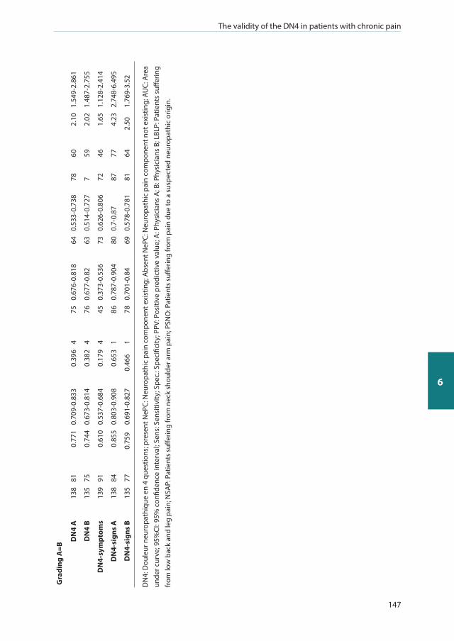
The validity of the DN4 in patients with chronic pain
147
6
Gra
ding
A=B
DN
4 A
138
810.
771
0.70
9-0.
833
0.39
64
750.
676-
0.81
864
0.53
3-0.
738
7860
2.10
1.54
9-2.
861
DN
4 B
135
750.
744
0.67
3-0.
814
0.38
24
760.
677-
0.82
630.
514-
0.72
77
592.
021.
487-
2.75
5
DN
4-sy
mpt
oms
139
910.
610
0.53
7-0.
684
0.17
94
450.
373-
0.53
673
0.62
6-0.
806
7246
1.65
1.12
8-2.
414
DN
4-si
gns
A13
884
0.85
50.
803-
0.90
80.
653
186
0.78
7-0.
904
800.
7-0.
8787
774.
232.
748-
6.49
5
DN
4-si
gns
B13
577
0.75
90.
691-
0.82
70.
466
178
0.70
1-0.
8469
0.57
8-0.
781
8164
2.50
1.76
9-3.
52
DN
4: D
oule
ur n
euro
path
ique
en
4 qu
estio
ns; p
rese
nt N
ePC:
Neu
ropa
thic
pai
n co
mpo
nent
exi
stin
g; A
bsen
t NeP
C: N
euro
path
ic p
ain
com
pone
nt n
ot e
xist
ing;
AU
C: A
rea
unde
r cur
ve; 9
5%CI
: 95%
con
fiden
ce in
terv
al; S
ens:
Sen
sitiv
ity; S
pec.
: Spe
cific
ity; P
PV: P
ositi
ve p
redi
ctiv
e va
lue;
A: P
hysi
cian
s A
; B: P
hysi
cian
s B;
LBL
P: P
atie
nts
suffe
ring
from
low
bac
k an
d le
g pa
in; N
SAP:
Pat
ient
s su
fferin
g fr
om n
eck
shou
lder
arm
pai
n; P
SNO
: Pat
ient
s su
fferin
g fr
om p
ain
due
to a
sus
pect
ed n
euro
path
ic o
rigin
.
Chapter 6
148
In Table 6 and S2 Table we present the number of patients per group, values of the AUC, Youden index,
cut-off score, true positives, false positives, false negatives, true negatives, sensitivity, specificity,
positive and negative predictive values, positive and negative likelihood ratios, the diagnostic odds
ratio, the a-priori chance for the existence (or not) of a NePC and false positive and negative ratios
for all validity outcomes (DN4 A & B, DN4-symptoms, DN4-signs A & B) divided according to the pain
classification and divided into LBLP, NSAP and PSNO (Table 6 and S2 Table).
Test-retest reliability Stability and responsiveness of the DN4-symptoms over time was assessed over a period of two
weeks. The median sum score (IQR) of the DN4 at baseline for the total group was 3 (2-4), after two
weeks it was 3 (2-4). Taking into consideration the fact that patients’ pain should not have changed
(outcome based on the PGIC) because otherwise the ICC would not reflect the consistency of the
DN4, test-retest reliability via ICC was 0.84 (excellent) (95%CI 0.80-0.87; n = 265). For the time gap
of 7-21 days (to rule out the early or delayed return of questionnaires) between the first and second
DN4-symptoms the ICC was 0.85 (excellent) (95% CI 0.79-0.90; n = 122). After three months, with no
change in patients pain and a time gap of 60-120 days between the first and third DN4-symptoms,
ICC was 0.79 (excellent) (95% CI 0.70-0.86; n = 102).
DISCUSSION
The DN4 seems, in this study, to help to identify a neuropathic pain component in a consecutive
population of patients with chronic pain in a moderate way.
Reliability We used the concordant opinion about the classification of patients’ pain by two physicians as the
gold standard. It is disputable if the term gold standard is practicable. However, as written by Versi
[56, 57] “the gold standard is not the perfect test but merely the best available test. . .. Against which newer
tests can be compared”. There are studies regarding the validity of the DN4 using only one physician’s
opinion [21, 30]. To our opinion it is preferable to use two physicians as the gold standard, which is
also performed in the original validation study of the DN4 [17]. This might lead to less false positive
or false negative outcomes which, of course, will lead to a more accurate validity outcome. The
physicians in this study agreed on pain classification in 78% of the patients. In other studies without
pre-stratification of patients on the target outcome the results for the physicians agreement were
53% [25] and 89% [27]. The kappa coefficient between the DN4 as filled in by physician A compared
to the DN4 by physician B was ‘good’ with a high percentage of agreement. Test-retest reliability of
the DN4-symptoms in this study was excellent. Based on these results DN4 seems to be reliable.
However, it is possible that an instrument is reliable without being valid [58].
The validity of the DN4 in patients with chronic pain
149
6
Validity To quantify the screening ability of the DN4, for the existence of a NePC, sensitivity and specificity
can be used [59]. However, in clinical practice we want to know how many patients with a positive
score on the DN4 really does have a NePC. To report this, the positive and negative predictive values
are important because they give the proportion of patients with positive or negative test results
which are correctly diagnosed [60]. The predictive value depends on the prevalence of NePC in the
group of patients under study [60]. In our study the prevalence of NePC was high, 75%. The higher
the prevalence of NePC in the group under study the more sure it is that a positive outcome of the
DN4 indicates the presence of a NePC, but the less sure it is that a negative DN4 outcome indicates
absent NePC [60]. The likelihood ratio gives an indication of the value of the DN4 for increasing
certainty about a positive diagnosis [60]. A higher likelihood ratio might indicate that the DN4 is
useful, but is still not sure that a positive outcome of the DN4 is a good indicator for the presence of
a NePC [60]. In the literature there are, as far as we know, no ‘cut-off’ scores for the validity indices. In
our study we found a sensitivity of 75% (DN4-symptoms 70%), a specificity of 76% (DN4-symptoms
67%), positive predictive value of 92%, negative predictive value of 46% and the positive and
negative likelihood ratios were respectively 3.09 and 0.34. In the original study by Bouhassira et al.
[17] patients with only ‘typical’ neuropathic or nociceptive entities and a VAS of ≥40 mm (0-100mm)
were included. They found a sensitivity of 83% and a specificity of 90%. For the DN4-symptoms
the sensitivity was 78% and the specificity 81%. The Dutch version of the DN4 [20] was validated
before in a consecutive group of patients suffering from chronic pain for more than three months
with a pain score of 5 or higher on a 0-10 numeric rating scale (NRS) [25]. For the DN4 a sensitivity
of 75% and a specificity of 79% was found. For the DN4-symptoms version sensitivity was 74% and
the specificity 79%. Van Seventer et al. concluded that the DN4 was a diagnostic tool with a good
ability to discriminate between neuropathic pain and nociceptive pain [25]. However, the paper
by Bouhassira et al. [17] and the paper by Van seventer et al. [25] both didn’t report the predictive
values and likelihood ratios. Inappropriate screening might result in higher health care costs due to
more diagnostic testing or even lead to a harmful treatment for the patient [61]. It seems that the
validity indices in our study are resulting in a lower score for the DN4 as in the original publication
[17] and than in other studies [4, 21, 23-28, 30, 31, 62-67]. This might have several reasons. At first, we
did not pre-stratify on the target outcome. In studies, besides the original validation study [17] with
pre-stratification on the target outcome [23, 24, 26, 28, 31] (neuropathic or non-neuropathic pain),
the sensitivity of the DN4 was ranging from 90% [26] till 100% [24], the specificity from 93% [24]-
97% [23, 28]. In studies where there was no pre-stratification on the target outcome (neuropathic
or non-neuropathic pain), the sensitivity of the DN4 was ranging from 80% [21] till 100% [30], the
specificity ranges from 78% [21, 27] till 87% [30]. These results are showing that the validity of the
DN4 is lower in studies without pre-stratification than in studies were patients were stratified based
on their pain classification before entering the study. In studies with specified diseases as spinal cord
injury [64]; diabetes [63, 64]; leprosy [65, 66]; FBSS [67], chronic low back pain [4] and in patients with
cancer before starting with chemotherapy [68], the sensitivity (62%-100%) and specificity (44%-
Chapter 6
150
93%) ranges were much wider. Our results, also when separated into results for LBLP, NSAP and
PSNO, falls within these ranges. This indicates that the neuropathic pain component is not always
clear and/or easy to classify by use of the DN4 in the different medical conditions. Secondly, in our
study we did not have a minimum level of pain as an inclusion criteria. In seven studies a minimal
level of pain (on a rating scale of 0-10) was not an inclusion criteria [21, 23, 31, 62, 63, 65, 66]. In
other studies a level ≥ three [64, 67], ≥ four [4, 17, 24, 26, 28, 30] or ≥ five [25, 27] is set as an inclusion
criterium. As shown by Perez et al [21], pain severity has a major influence on the sensitivity and
specificity of the DN4. A severity of < 40 mm on a 0-100mm VAS resulted in a sensitivity of 56% and
a specificity of 67%. For moderate pain (between 40mm en 70mm on a 0-100mm VAS) it was 85%
and 84% respectively, and >70 mm sensitivity was 80% and specificity was 74% [21]. In a study by
Marksman [67] in patients after FBSS it was showed that the presence of neuropathic characteristics,
as determined by the DN4, was associated with a higher pain intensity. These facts are crucial for
the validation of a screening instrument because such a tool must be valid for use in daily clinical
out-patient practice and/or for epidemiological purposes.
As a second comparison, we validated the DN4 in comparison with the grading system [21, 32].
In this study, we combined ‘unlikely’ and possible neuropathic pain as absent NePC and probable
and definite as present NePC, which resulted in an average sensitivity for the DN4 of 76% and an
average specificity of 64%. In patients with a failed back surgery syndrome [67], the validation of
the DN4 resulted in a sensitivity of 62% and a specificity of 44%. In a study by Sadler et al [69] where
patients with neuropathic pain were compared to musculoskeletal pain the sensitivity was 76% and
the specificity was 70%. However, in patients with a more mixed pain the sensitivity and specificity
descended to 59% and 70% respectively. Abdallah et al [36] compared the DN4 with the grading
system in patients after breast tumor resection with and without paravertebral blocks. This resulted
in a sensitivity of 90% and a specificity of 60% to identify patients with chronic neuropathic pain
based on the outcome of the grading system. However, this outcome was not validated by (expert)
physicians. The distinction between possible neuropathic pain and probable or definite neuropathic
pain is of high importance because the outcome forms the basis for selecting a different treatment
strategy [34]. The combination of outcomes in our study might have resulted in a lower sensitivity
and a bit higher specificity in comparison with the classification in the study of Abdallah et al [36].
Bouhassira [17] presented the DN4 as a clinician-administered questionnaire. In different studies not
a physician but a research coordinator [30], a nurse [25] or the patient self [25, 70] filled in the DN4.
In our study we gave the patient the questionnaire with the 7-items (DN4-symptoms) to fill them
in after the physical examinations. The three examination-items (DN4-signs) were incorporated in
the standardized assessment form which should be filled in by the physician. We presented the
DN4 total sum score as well as the DN4-signs score separately for physicians A and B. This is due to
the fact that it is only possible to have one outcome when the sign-items were performed by one
physician.
The validity of the DN4 in patients with chronic pain
151
6
Strength and weaknesses There are several strengths in this study. At first, this study reflects daily clinical practice. In this
study, we included a large cohort of patients irrespective of the predominant origin of the pain
and level of pain which corresponds to a typical daily clinical patient population. These patients
were associated with the most common specified medical conditions for pain (i.e. LBLP or NSAP or
PSNO) and classified by two, independently working, physicians. Moreover, patients were referred
from primary care to secondary and tertiary pain clinics and were assessed for their complaints
for the first time at the time of inclusion in this study. This limits the risk of systematic bias and
also reflects daily clinical practice. Secondly, we used a standardized assessment form in which the
bedside examination and the grading system [32, 33] and the DN4-signs were incorporated. This
might, however, have led to an influence on each other which made the physician more sure about
the final classification of patients pain and thus made the gold standard stronger. There are also
some weaknesses in this study. As said before, we have not used the DN4-symptoms as a interview
by a physician but as a questionnaire which has to be filled in by the patient. This might have had
an influence on the reliability and validity. In the revised EFNS guidelines on neuropathic pain
assessment [42] it is suggested that “The seven sensory descriptors can be used as a self-report
questionnaire with similar results”. Moreover, above the official Dutch version [20, 25] of the DN4 is
written in Dutch: “To be completed by the patient”. In the paper by van Seventer et al the agreement
between the patient administered and a nurse administered was good till very good for the first
seven items [25]. It would be of interest to see if there are differences in the outcome when the DN4
is filled in by the patient himself or as an interview by the pain physician. Questions by the patient
to the nurse of via telephone to the researcher regarding the DN4 were very rare. However, we
didn’t keep track of the questions. Another limitation is the fact that we only tested the test-retest
reliability regarding the DN4-symptoms and not the DN4-signs to prevent the patient to come back
to the hospital only for these test-items. Another weakness is the gold standard which is, for now,
the best measure for the existence of a neuropathic pain component but the result is still open for
discussion.
Suggestions for the validation of neuropathic pain screening tools Validation of screening tools should be performed in a standardized manner and described in detail,
but performed in a setting which is comparable to a daily clinical practice. A research setting might
be different from a clinical setting and thus might have influence on the patient and on the study
results. The group of patients as well as the physicians under study should be comparable to the
patients/physicians for who the tool is intended. Pre-stratification on the target outcome must be
avoided (especially the exclusion of the so called mixed pain), because this will lead to a non-clinical
situation and thus decreases the validity and generalizability of the instrument [16, 71].
Chapter 6
152
CONCLUSION
The validity of DN4-signs is equal to the DN4 outcome and, importantly, both are more valid than
the DN4-symptoms alone. It seems that the patients’ symptoms and signs doesn’t reliably reflect the
underlying mechanisms, indicating there is a need for a more objective way to assess patients’ pain
to facilitate improvement in the treatment of patients with pain. The physicians’ assessment cannot
be replaced by a screening tool as the DN4, but gives the physician a little hint towards the (non-)
existence of neuropathic pain component.
ACKNOWLEDGEMENTS
Thanks to all the participating patients, physicians and assistants for their invaluable work to this
study.
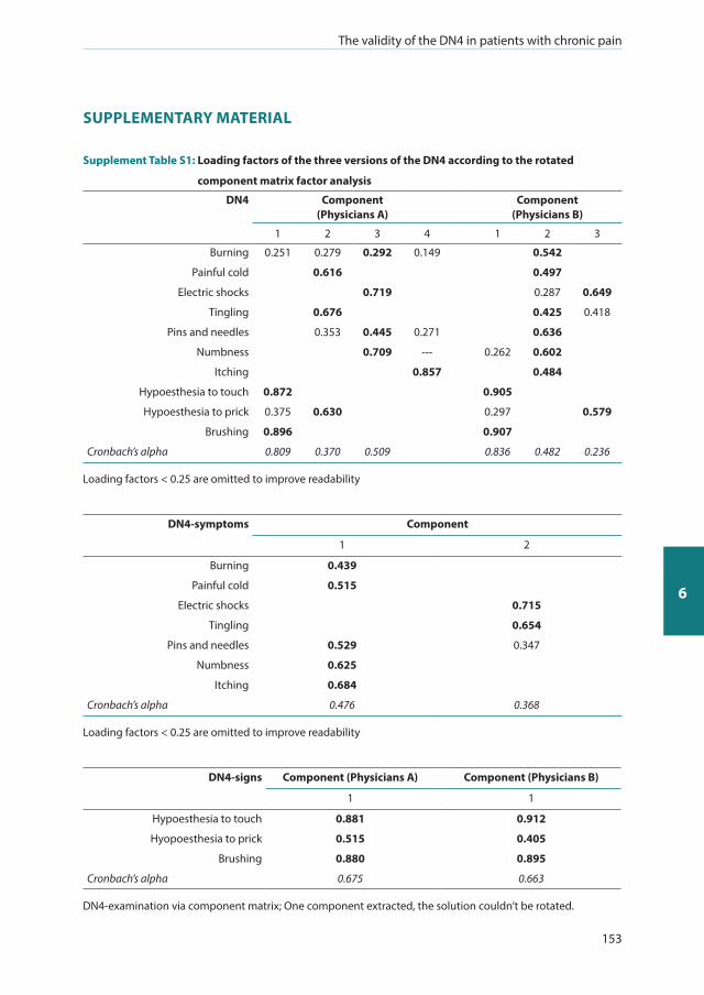
The validity of the DN4 in patients with chronic pain
153
6
SUPPLEMENTARY MATERIAL
Supplement Table S1: Loading factors of the three versions of the DN4 according to the rotated
component matrix factor analysis
DN4 Component (Physicians A)
Component (Physicians B)
1 2 3 4 1 2 3
Burning 0.251 0.279 0.292 0.149 0.542
Painful cold 0.616 0.497
Electric shocks 0.719 0.287 0.649
Tingling 0.676 0.425 0.418
Pins and needles 0.353 0.445 0.271 0.636
Numbness 0.709 --- 0.262 0.602
Itching 0.857 0.484
Hypoesthesia to touch 0.872 0.905
Hypoesthesia to prick 0.375 0.630 0.297 0.579
Brushing 0.896 0.907
Cronbach’s alpha 0.809 0.370 0.509 0.836 0.482 0.236
Loading factors < 0.25 are omitted to improve readability
DN4-symptoms Component
1 2
Burning 0.439
Painful cold 0.515
Electric shocks 0.715
Tingling 0.654
Pins and needles 0.529 0.347
Numbness 0.625
Itching 0.684
Cronbach’s alpha 0.476 0.368
Loading factors < 0.25 are omitted to improve readability
DN4-signs Component (Physicians A) Component (Physicians B)
1 1
Hypoesthesia to touch 0.881 0.912
Hyopoesthesia to prick 0.515 0.405
Brushing 0.880 0.895
Cronbach’s alpha 0.675 0.663
DN4-examination via component matrix; One component extracted, the solution couldn’t be rotated.
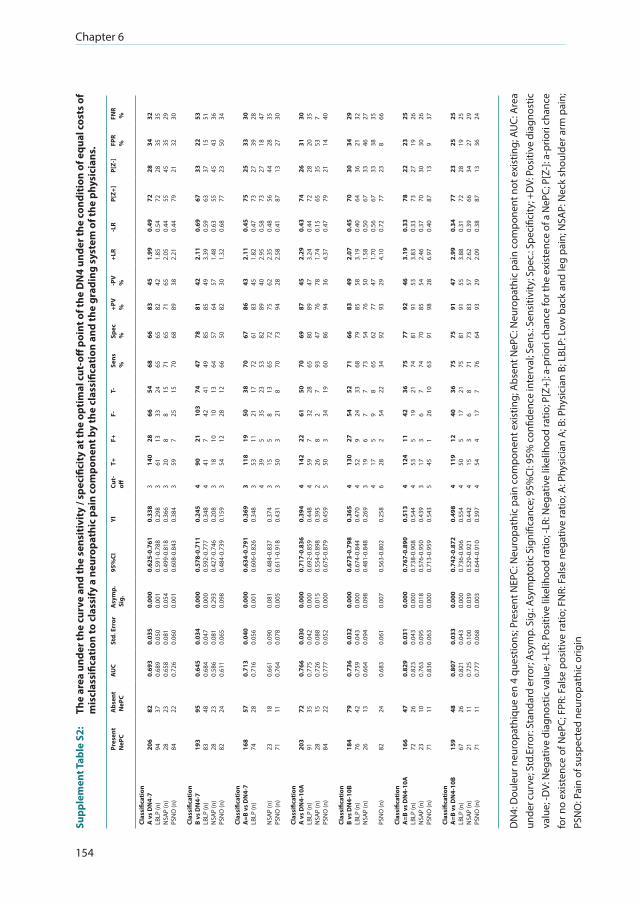
Chapter 6
154
Supp
lem
ent T
able
S2:
T
he a
rea
unde
r the
cur
ve a
nd th
e se
nsit
ivit
y / s
peci
ficit
y at
the
opti
mal
cut
-off
poi
nt o
f the
DN
4 un
der t
he c
ondi
tion
of e
qual
cos
ts o
f m
iscl
assi
ficat
ion
to c
lass
ify a
neu
ropa
thic
pai
n co
mpo
nent
by
the
clas
sific
atio
n an
d th
e gr
adin
g sy
stem
of t
he p
hysi
cian
s.
Pres
ent
NeP
CA
bsen
t N
ePC
AU
CSt
d. E
rror
Asy
mp.
Sig
.95
%CI
YICu
t-off
T+F+
F-T-
Sens
%
Spec
%
+PV
%
-PV
%
+LR
-LR
P[Z+
]P[
Z-]
FPR
%FN
R %
Clas
sific
atio
nA
vs
DN
4-7
LBLP
(n)
NSA
P (n
)PS
NO
(n)
Clas
sific
atio
nB
vs D
N4-
7LB
LP (n
)N
SAP
(n)
PSN
O (n
)
Clas
sific
atio
nA
=B v
s D
N4-
7LB
LP (n
)
NSA
P (n
)PS
NO
(n)
Clas
sific
atio
nA
vs
DN
4-10
ALB
LP (n
)N
SAP
(n)
PSN
O (n
)
Clas
sific
atio
nB
vs D
N4-
10B
LBLP
(n)
NSA
P (n
)
PSN
O (n
)
Clas
sific
atio
nA
=B v
s D
N4-
10A
LBLP
(n)
NSA
P (n
)PS
NO
(n)
Clas
sific
atio
nA
=B v
s D
N4-
10B
LBLP
(n)
NSA
P (n
)PS
NO
(n)
206
94 28 84 193
83 28 82 168
74 23 71 203
91 28 84 184
76 26 82 166
72 23 71 159
67 21 71
82 37 23 22 95 48 23 24 57 28 18 11 72 35 15 22 79 42 13 24 47 26 10 11 48 26 11 11
0.69
30.
689
0.65
80.
726
0.64
50.
684
0.58
60.
611
0.71
30.
716
0.66
10.
764
0.76
60.
775
0.72
60.
777
0.73
60.
759
0.66
4
0.68
3
0.82
90.
823
0.76
30.
836
0.80
70.
821
0.72
50.
777
0.03
50.
050
0.08
10.
060
0.03
40.
047
0.08
10.
065
0.04
00.
056
0.09
00.
078
0.03
00.
042
0.08
80.
052
0.03
20.
043
0.09
4
0.06
1
0.03
10.
043
0.09
50.
063
0.03
30.
043
0.10
00.
068
0.00
00.
001
0.05
40.
001
0.00
00.
000
0.29
30.
098
0.00
00.
001
0.08
10.
005
0.00
00.
000
0.01
50.
000
0.00
00.
000
0.09
8
0.00
7
0.00
00.
000
0.01
80.
000
0.00
00.
000
0.03
90.
003
0.62
5-0.
761
0.59
1-0.
788
0.49
9-0.
818
0.60
8-0.
843
0.57
8-0.
711
0.59
2-0.
777
0.42
7-0.
746
0.48
4-0.
739
0.63
4-0.
791
0.60
6-0.
826
0.48
4-0.
837
0.61
1-0.
918
0.71
7-0.
836
0.69
2-0.
859
0.55
4-0.
898
0.67
5-0.
879
0.67
3-0.
798
0.67
4-0.
844
0.48
1-0.
848
0.56
3-0.
802
0.76
7-0.
890
0.73
8-0.
908
0.57
6-0.
950
0.71
3-0.
959
0.74
2-0.
872
0.73
6-0.
906
0.52
9-0.
921
0.64
4-0.
910
0.33
80.
298
0.36
60.
384
0.24
50.
348
0.20
80.
159
0.36
90.
348
0.37
40.
431
0.39
40.
448
0.39
50.
459
0.36
50.
470
0.26
9
0.25
8
0.51
30.
544
0.43
90.
543
0.49
80.
554
0.44
20.
397
3 3 3 3 4 4 3 3 3 3 4 3 3 4 4 2 5 4 4 3 4 6 4 4 3 5 4 4 4 4
140
61 20 59 90 41 18 54 118
53 39 15 50 142
59 26 50 130
52 19 17 28 124
53 17 45 119
50 15 54
28 13 8 7 21 7 10 12 19 11 5 5 3 22 7 8 3 27 9 6 5 2 11 5 3 1 12 5 3 4
66 33 8 25 103
42 10 28 50 21 35 8 21 61 32 2 34 54 24 7 9 54 42 19 6 26 40 17 6 17
54 24 15 15 74 41 13 12 38 17 23 13 8 50 28 7 19 52 33 7 8 22 36 21 7 10 36 21 8 7
68 65 71 70 47 49 64 66 70 72 53 65 70 70 65 93 60 71 68 73 65 34 75 74 74 63 75 75 71 76
66 65 65 68 78 85 57 50 67 61 82 72 73 69 80 47 86 66 79 54 62 92 77 81 70 91 75 81 73 64
83 82 71 89 81 85 64 82 86 83 89 75 94 87 89 76 94 83 85 76 77 93 92 91 85 98 91 91 83 93
45 42 65 38 42 49 57 30 43 45 40 62 28 45 47 78 36 49 58 50 47 29 46 53 54 28 47 55 57 29
1.99
1.85
2.05
2.21
2.11
3.39
1.48
1.32
2.11
1.82
2.95
2.35
2.58
2.29
3.24
1.74
4.37
2.07
3.19
1.58
1.70
4.10
3.19
3.83
2.46
6.97
2.99
3.88
2.62
2.09
0.49
0.54
0.44
0.44
0.69
0.59
0.63
0.68
0.45
0.47
0.58
0.48
0.41
0.43
0.44
0.15
0.47
0.45
0.40
0.50
0.56
0.72
0.33
0.33
0.37
0.40
0.34
0.31
0.39
0.38
72 72 55 79 67 63 55 77 75 73 73 56 87 74 72 65 79 70 64 67 67 77 78 73 70 87 77 72 66 87
28 28 45 21 33 37 45 23 25 27 27 44 13 26 28 35 21 30 36 33 33 23 22 27 30 13 23 28 34 13
34 35 35 32 22 15 43 50 33 39 18 28 27 31 20 53 14 34 21 46 38 8 23 19 30 9 25 19 27 36
32 35 29 30 53 51 36 34 30 28 47 35 30 30 35 7 40 29 32 27 35 66 25 26 26 37 25 25 29 24
DN
4: D
oule
ur n
euro
path
ique
en
4 qu
estio
ns; P
rese
nt N
EPC:
Neu
ropa
thic
pai
n co
mpo
nent
exi
stin
g; A
bsen
t NeP
C: N
euro
path
ic p
ain
com
pone
nt n
ot e
xist
ing;
AU
C: A
rea
unde
r cur
ve; S
td.E
rror
: Sta
ndar
d er
ror;
Asy
mp.
Sig
.: Asy
mpt
otic
Sig
nific
ance
; 95%
CI: 9
5% c
onfid
ence
inte
rval
; Sen
s.: S
ensi
tivity
; Spe
c.: S
peci
ficity
; +D
V: P
ositi
ve d
iagn
ostic
va
lue;
-DV:
Neg
ativ
e di
agno
stic
val
ue; +
LR: P
ositi
ve li
kelih
ood
ratio
; -LR
: Neg
ativ
e lik
elih
ood
ratio
; P[Z
+]: a
-prio
ri ch
ance
for t
he e
xist
ence
of a
NeP
C; P
[Z-]:
a-p
riori
chan
ce
for n
o ex
iste
nce
of N
ePC;
FPR
: Fal
se p
ositi
ve ra
tio; F
NR:
Fal
se n
egat
ive
ratio
; A: P
hysi
cian
A; B
: Phy
sici
an B
; LBL
P: L
ow b
ack
and
leg
pain
; NSA
P: N
eck
shou
lder
arm
pai
n;
PSN
O: P
ain
of s
uspe
cted
neu
ropa
thic
orig
in
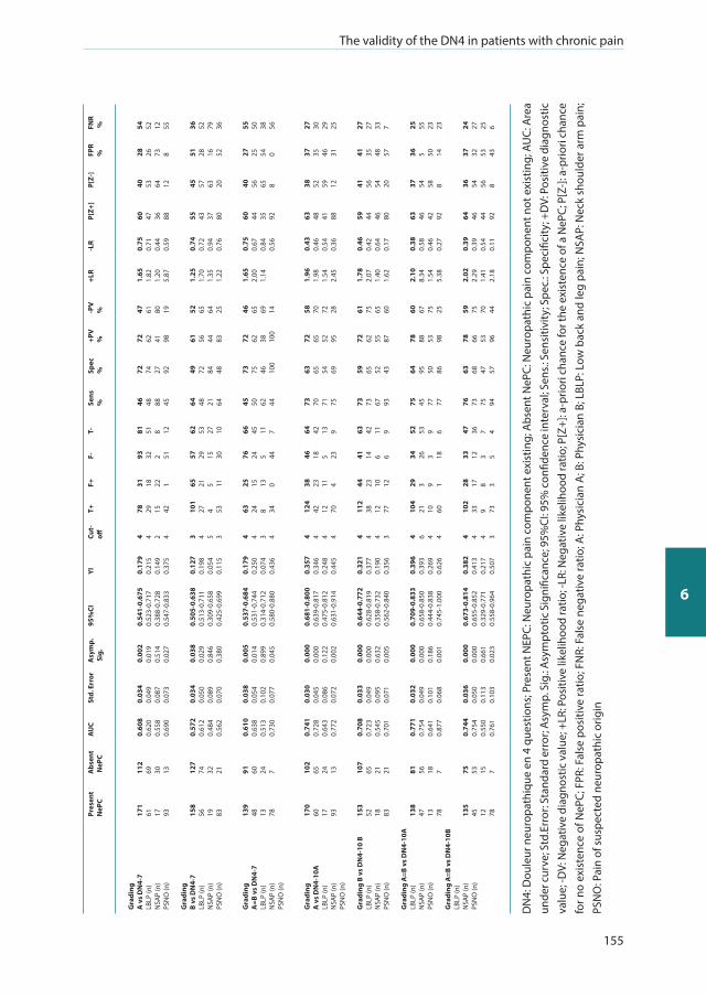
The validity of the DN4 in patients with chronic pain
155
6
Pres
ent
NeP
CA
bsen
t N
ePC
AU
CSt
d. E
rror
Asy
mp.
Sig
.95
%CI
YICu
t-off
T+F+
F-T-
Sens
%
Spec
%
+PV
%
-PV
%
+LR
-LR
P[Z+
]P[
Z-]
FPR
%FN
R %
Gra
ding
A v
s D
N4-
7LB
LP (n
)N
SAP
(n)
PSN
O (n
)
Gra
ding
B vs
DN
4-7
LBLP
(n)
NSA
P (n
)PS
NO
(n)
Gra
ding
A=B
vs
DN
4-7
LBLP
(n)
NSA
P (n
)PS
NO
(n)
Gra
ding
A v
s D
N4-
10A
LBLP
(n)
NSA
P (n
)PS
NO
(n)
Gra
ding
B v
s D
N4-
10 B
LBLP
(n)
NSA
P (n
)PS
NO
(n)
Gra
ding
A=B
vs
DN
4-10
ALB
LP (n
)N
SAP
(n)
PSN
O (n
)
Gra
ding
A=B
vs
DN
4-10
BLB
LP (n
)N
SAP
(n)
PSN
O (n
)
171
61 17 93 158
56 19 83 139
48 13 78 170
60 17 93 153
52 18 83 138
47 13 78 135
45 12 78
112
69 30 13 127
74 32 21 91 60 24 7 102
65 24 13 107
65 21 21 81 56 18 7 75 53 15 7
0.60
80.
620
0.55
80.
690
0.57
20.
612
0.48
40.
562
0.61
00.
638
0.51
30.
730
0.74
10.
728
0.64
30.
772
0.70
80.
723
0.54
50.
701
0.77
10.
754
0.64
10.
877
0.74
40.
754
0.55
00.
761
0.03
40.
049
0.08
70.
073
0.03
40.
050
0.08
90.
070
0.03
80.
054
0.10
20.
077
0.03
00.
045
0.08
60.
072
0.03
30.
049
0.09
50.
071
0.03
20.
049
0.10
10.
068
0.03
60.
050
0.11
30.
103
0.00
20.
019
0.51
40.
027
0.03
80.
029
0.84
60.
380
0.00
50.
014
0.89
90.
045
0.00
00.
000
0.12
20.
002
0.00
00.
000
0.63
20.
005
0.00
00.
000
0.18
60.
001
0.00
00.
000
0.66
10.
023
0.54
1-0.
675
0.52
3-0.
717
0.38
8-0.
728
0.54
7-0.
833
0.50
5-0.
638
0.51
3-0.
711
0.30
9-0.
658
0.42
5-0.
699
0.53
7-0.
684
0.53
1-0.
744
0.31
4-0.
712
0.58
0-0.
880
0.68
1-0.
800
0.63
9-0.
817
0.47
5-0.
812
0.63
1-0.
914
0.64
4-0.
772
0.62
8-0.
819
0.35
8-0.
732
0.56
2-0.
840
0.70
9-0.
833
0.65
8-0.
850
0.44
4-0.
838
0.74
5-1.
000
0.67
3-0.
814
0.65
5-0.
852
0.32
9-0.
771
0.55
8-0.
964
0.17
90.
215
0.14
90.
375
0.12
70.
198
0.05
40.
115
0.17
90.
250
0.07
40.
436
0.35
70.
346
0.24
80.
445
0.32
10.
377
0.19
00.
356
0.39
60.
393
0.26
90.
626
0.38
20.
413
0.21
70.
507
4 4 2 4 3 4 5 3 4 4 3 4 4 4 4 4 4 4 4 3 4 6 4 4 4 4 4 3
78 29 15 42 101
27 4 53 63 24 8 34 124
42 12 70 112
38 12 77 104
21 10 60 102
33 9 73
31 18 22 1 65 21 5 11 25 15 13 0 38 23 11 4 44 23 10 12 29 3 9 1 28 17 8 3
93 32 2 51 57 29 15 30 76 24 5 44 46 18 5 23 41 14 6 6 34 26 3 18 33 12 3 5
81 51 8 12 62 53 27 10 66 45 11 7 64 42 13 9 63 42 11 9 52 53 9 6 47 36 7 4
46 48 88 45 64 48 21 64 45 50 62 44 73 70 71 75 73 73 67 93 75 45 77 77 76 73 75 94
72 74 27 92 49 72 84 48 73 75 46 100
63 65 54 69 59 65 52 43 64 95 50 86 63 68 47 57
72 62 41 98 61 56 44 83 72 62 38 100
72 65 52 95 72 62 55 87 78 88 53 98 78 66 53 96
47 61 80 19 52 65 64 25 46 65 69 14 58 70 72 28 61 75 65 60 60 67 75 25 59 75 70 44
1.65
1.82
1.20
5.87
1.25
1.70
1.35
1.22
1.65
2.00
1.14
1.96
1.98
1.54
2.45
1.78
2.07
1.40
1.62
2.10
8.34
1.54
5.38
2.02
2.29
1.41
2.18
0.75
0.71
0.44
0.59
0.74
0.72
0.94
0.76
0.75
0.67
0.84
0.56
0.43
0.46
0.54
0.36
0.46
0.42
0.64
0.17
0.38
0.58
0.46
0.27
0.39
0.39
0.54
0.11
60 47 36 88 55 43 37 80 60 44 35 92 63 48 41 88 59 44 46 80 63 46 42 92 64 46 44 92
40 53 64 12 45 57 63 20 40 56 65 8 38 52 59 12 41 56 54 20 37 54 58 8 36 54 56 8
28 26 73 8 51 28 16 52 27 25 54 0 37 35 46 31 41 35 48 57 36 5 50 14 37 32 53 43
54 52 12 55 36 52 79 36 55 50 38 56 27 30 29 25 27 27 33 7 25 55 23 23 24 27 25 6
DN
4: D
oule
ur n
euro
path
ique
en
4 qu
estio
ns; P
rese
nt N
EPC:
Neu
ropa
thic
pai
n co
mpo
nent
exi
stin
g; A
bsen
t NeP
C: N
euro
path
ic p
ain
com
pone
nt n
ot e
xist
ing;
AU
C: A
rea
unde
r cur
ve; S
td.E
rror
: Sta
ndar
d er
ror;
Asy
mp.
Sig
.: Asy
mpt
otic
Sig
nific
ance
; 95%
CI: 9
5% c
onfid
ence
inte
rval
; Sen
s.: S
ensi
tivity
; Spe
c.: S
peci
ficity
; +D
V: P
ositi
ve d
iagn
ostic
va
lue;
-DV:
Neg
ativ
e di
agno
stic
val
ue; +
LR: P
ositi
ve li
kelih
ood
ratio
; -LR
: Neg
ativ
e lik
elih
ood
ratio
; P[Z
+]: a
-prio
ri ch
ance
for t
he e
xist
ence
of a
NeP
C; P
[Z-]:
a-p
riori
chan
ce
for n
o ex
iste
nce
of N
ePC;
FPR
: Fal
se p
ositi
ve ra
tio; F
NR:
Fal
se n
egat
ive
ratio
; A: P
hysi
cian
A; B
: Phy
sici
an B
; LBL
P: L
ow b
ack
and
leg
pain
; NSA
P: N
eck
shou
lder
arm
pai
n;
PSN
O: P
ain
of s
uspe
cted
neu
ropa
thic
orig
in
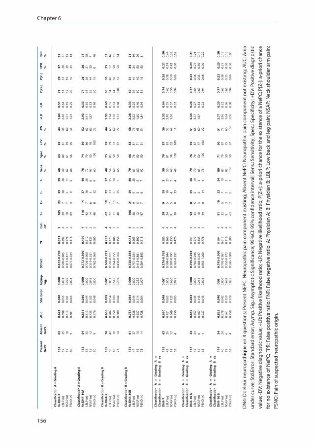
Chapter 6
156
Pres
ent
NeP
CA
bsen
t N
ePC
AU
CSt
d. E
rror
Asy
mp.
Sig
.95
%CI
YICu
t-off
T+F+
F-T-
Sens
%
Spec
%
+PV
%
-PV
%
+LR
-LR
P[Z+
]P[
Z-]
FPR
%FN
R %
Clas
sific
atio
n A
= G
radi
ng A
Vs D
N4-
7LB
LP (n
)N
SAP
(n)
PSN
O (n
)
Clas
sific
atio
n A
= G
radi
ng A
Vs D
N4-
10A
LBLP
(n)
NSA
P (n
)PS
NO
(n)
Clas
sific
atio
n B
= G
radi
ng B
Vs D
N4-
7LB
LP (n
)N
SAP
(n)
PSN
O (n
)
Clas
sific
atio
n B
= G
radi
ng B
Vs D
N4-
10B
LBLP
(n)
NSA
P (n
)PS
NO
(n)
154
59 15 80 153
58 15 80 139
50 16 73 135
47 15 73
62 35 18 9 54 33 12 9 76 42 20 14 61 37 10 14
0.69
10.
698
0.61
30.
801
0.83
10.
813
0.72
50.
876
0.63
60.
704
0.55
90.
600
0.78
70.
838
0.63
70.
728
0.04
00.
055
0.10
10.
063
0.03
00.
044
0.10
50.
048
0.03
90.
054
0.10
00.
084
0.03
40.
044
0.11
40.
084
0.00
00.
001
0.27
00.
003
0.00
00.
000
0.04
80.
000
0.00
10.
001
0.54
50.
239
0.00
00.
000
0.25
50.
007
0.61
2-0.
770
0.59
0-0.
805
0.41
5-0.
811
0.67
7-0.
924
0.77
2-0.
890
0.72
6-0.
900
0.51
9-0.
931
0.78
2-0.
969
0.56
0-0.
713
0.59
7-0.
811
0.36
4-0.
755
0.43
6-0.
764
0.72
0-0.
854
0.75
1-0.
924
0.41
3-0.
861
0.56
4-0.
893
0.31
10.
292
0.27
80.
418
0.49
90.
512
0.43
30.
600
0.23
20.
373
0.18
80.
158
0.44
10.
565
0.30
00.
418
3 4 3 2 4 4 2 5 4 4 3 3 4 4 4 3
105
29 10 69 116
42 14 48 67 27 11 48 106
38 12 67
23 7 7 4 14 7 6 0 19 7 10 7 21 9 5 7
49 30 5 11 37 16 1 32 72 23 5 25 29 9 3 6
39 28 11 5 40 26 6 9 57 35 10 7 40 28 5 7
68 49 67 86 76 72 93 60 48 54 69 66 79 81 80 92
63 80 61 56 74 79 50 100
75 83 50 50 66 76 50 50
82 81 59 95 89 86 70 100
78 79 52 87 83 81 71 91
44 48 69 31 52 62 86 22 44 60 67 22 58 76 63 54
1.84
2.46
1.71
1.94
2.92
3.41
1.87
1.93
3.24
1.38
1.32
2.28
3.32
1.60
1.84
0.51
0.64
0.55
0.25
0.33
0.35
0.13
0.40
0.69
0.55
0.63
0.68
0.33
0.25
0.40
0.16
71 63 45 90 74 64 56 90 65 54 44 0.84
69 56 60 84
29 37 55 10 26 36 44 10 35 46 56 16 31 44 40 16
37 20 39 44 26 21 50 0 25 17 50 50 34 24 50 50
32 51 33 14 24 28 7 40 52 46 31 34 21 19 20 8
Clas
sific
atio
n A
=
Gra
ding
A
=
Clas
sific
atio
n B
= G
radi
ng
B vs
D
N4-
7LB
LP (n
)N
SAP
(n)
PSN
O (n
)
Clas
sific
atio
n A
=
Gra
ding
A
=
Clas
sific
atio
n B
= G
radi
ng
B vs
D
N4-
10 A
LBLP
(n)
NSA
P (n
)PS
NO
(n)
Clas
sific
atio
n A
=
Gra
ding
A
=
Clas
sific
atio
n B
= G
radi
ng
B vs
D
N4-
10 B
LBLP
(n)
NSA
P (n
)PS
NO
(n)
118
43 12 63 117
42 12 63 114
40 11 63
42 26 12 4 34 24 6 4 34 24 6 4
0.67
00.
721
0.54
90.
750
0.85
90.
857
0.66
70.
937
0.82
20.
878
0.56
10.
758
0.04
80.
063
0.12
30.
095
0.03
30.
045
0.14
30.
043
0.04
00.
042
0.15
90.
128
0.00
10.
002
0.68
60.
095
0.00
00.
000
0.26
10.
004
.000
0.00
00.
688
0.08
5
0.57
6-0.
765
0.59
8-0.
844
0.30
7-0.
791
0.56
3-0.
937
0.79
5-0.
923
0.76
9-0.
946
0.38
6-0.
947
0.85
3-1.
000
0.74
5-0.
900
0.79
5-0.
961
0.25
0-0.
872
0.50
6-1.
000
0.28
60.
366
0.25
00.
476
0.55
10.
577
0.33
30.
778
0.50
40.
617
0.31
80.
500
4 4 3 4 4 4 3 4 4 4 4 2
59 24 8 30 92 33 10 49 91 33 9 63
9 5 5 0 8 5 3 0 10 5 3 2
59 19 4 33 25 9 2 14 23 7 2 0
3 21 7 4 26 19 3 4 24 19 3 2
50 56 67 48 79 79 83 78 80 83 82 100
79 81 58 100
76 79 50 100
71 79 50 50
87 83 62 100
92 87 77 100
90 87 75 97
36 53 64 11 51 68 60 22 51 73 60 100
2.33
2.90
1.60
3.34
3.77
1.67
2.71
3.96
1.64
2.00
0.64
0.55
0.57
0.52
0.28
0.27
0.33
0.22
0.29
0.22
0.36
0.00
0.74
0.62
0.50
0.94
0.77
0.64
0.67
0.94
0.77
0.63
0.65
0.94
0.26
0.38
0.50
0.06
0.23
0.36
0.33
0.06
0.23
0.38
0.35
0.06
0.21
0.19
0.42
0.00
0.24
0.21
0.50
0.00
0.29
0.21
0.50
0.50
0.50
0.44
0.33
0.52
0.21
0.21
0.17
0.22
0.20
0.18
0.18
0.00
DN
4: D
oele
ur n
euro
path
ique
en
4 qu
estio
ns; P
rese
nt N
EPC:
Neu
ropa
thic
pai
n co
mpo
nent
exi
stin
g; A
bsen
t NeP
C: N
euro
path
ic p
ain
com
pone
nt n
ot e
xist
ing;
AU
C: A
rea
unde
r cur
ve; S
td.E
rror
: Sta
ndar
d er
ror;
Asy
mp.
Sig
.: Asy
mpt
otic
Sig
nific
ance
; 95%
CI: 9
5% c
onfid
ence
inte
rval
; Sen
s.: S
ensi
tivity
; Spe
c.: S
peci
ficity
; +D
V: P
ositi
ve d
iagn
ostic
va
lue;
-DV:
Neg
ativ
e di
agno
stic
val
ue; +
LR: P
ositi
ve li
kelih
ood
ratio
; -LR
: Neg
ativ
e lik
elih
ood
ratio
; P[Z
+]: a
-prio
ri ch
ance
for t
he e
xist
ence
of a
NeP
C; P
[Z-]:
a-p
riori
chan
ce
for n
o ex
iste
nce
of N
ePC;
FPR
: Fal
se p
ositi
ve ra
tio; F
NR:
Fal
se n
egat
ive
ratio
; A: P
hysi
cian
A; B
: Phy
sici
an B
; LBL
P: L
ow b
ack
and
leg
pain
; NSA
P: N
eck
shou
lder
arm
pai
n;
PSN
O: P
ain
of s
uspe
cted
neu
ropa
thic
orig
in.
The validity of the DN4 in patients with chronic pain
157
6
REFERENCES
1. IASP. IASP Taxonomy Neuropathic Pain: International Association for the Study of Pain; 2015 [cited 2015 May 19, 2015]. http://www.iasp-pain.org/Taxonomy#Neuropathicpain].
2. Freynhagen R, Baron R, Gockel U, Tolle TR. PainDETECT: a new screening questionnaire to identify neuropathic components in patients with back pain. Curr Med Res Opin. 2006; 22(10):1911-20.
3. La Cesa S, Tamburin S, Tugnoli V, Sandrini G, Paolucci S, Lacerenza M, et al. How to diagnose neuropathic pain? The contribution from clinical examination, pain questionnaires and diagnostic tests. Neurological sciences: official journal of the Italian Neurological Society and of the Italian Society of Clinical Neurophysiology. 2015; 36(12):2169-75.
4. Attal N, Perrot S, Fermanian J, Bouhassira D. The neuropathic components of chronic low back pain: a prospective multicenter study using the DN4 Questionnaire. The journal of pain: official journal of the American Pain Society. 2011; 12(10):1080-7.
5. Baron R, Binder A, Attal N, Casale R, Dickenson AH, Treede RD. Neuropathic low back pain in clinical practice. European journal of pain. 2016; 20(6):861-73.
6. Freynhagen R, Baron R. The evaluation of neuropathic components in low back pain. Curr Pain Headache Rep. 2009; 13(3):185-90.
7. -Haanpaa M, Attal N, Backonja M, Baron R, Bennett M, Bouhassira D, et al. NeuPSIG guidelines on neuropathic pain assessment. Pain. 2011; 152(1):14-27.
8. Haanpaa ML, Backonja MM, Bennett MI, Bouhassira D, Cruccu G, Hansson PT, et al. Assessment of neuropathic pain in primary care. Am J Med. 2009; 122(10 Suppl):S13-21.
9. Truini A, Cruccu G. How diagnostic tests help to disentangle the mechanisms underlying neuropathic pain symptoms in painful neuropathies. Pain. 2016; 157 Suppl 1:S53-9.
10. Dworkin RH, O0Connor AB, Kent J, Mackey SC, Raja SN, Stacey BR, et al. Interventional management of neuropathic pain: NeuPSIG recommendations. Pain. 2013; 154(11):2249-61.
11. Bennett MI, Attal N, Backonja MM, Baron R, Bouhassira D, Freynhagen R, et al. Using screening tools to identify neuropathic pain. Pain. 2007; 127(3):199-203.
12. Bouhassira D, Attal N. Diagnosis and assessment of neuropathic pain: the saga of clinical tools. Pain. 2011; 152(3 Suppl):S74-83.
13. Andermann A, Blancquaert I, Beauchamp S, Dery V. Revisiting Wilson and Jungner in the genomic age: a review of screening criteria over the past 40 years. Bull World Health Organ. 2008; 86(4):317-9.
14. Wilson JM, Jungner YG. [Principles and practice of mass screening for disease]. Bol Oficina Sanit Panam. 1968; 65(4):281-393.
15. Dieleman JP, Kerklaan J, Huygen FJ, Bouma PA, Sturkenboom MC. Incidence rates and treatment of neuropathic pain conditions in the general population. Pain. 2008; 137(3):681-8.
16. Mathieson S, Maher CG, Terwee CB, Folly de Campos T, Lin CW. Neuropathic pain screening questionnaires have limited measurement properties. A systematic review. Journal of clinical epidemiology. 2015; 68(8):957-66.
17. Bouhassira D, Attal N, Alchaar H, Boureau F, Brochet B, Bruxelle J, et al. Comparison of pain syndromes associated with nervous or somatic lesions and development of a new neuropathic pain diagnostic questionnaire (DN4). Pain. 2005; 114(1-2):29-36.
18. CADTH. Diagnostic methods for neuropathic pain: A review of diagnostic accuracy. Canadian Agency for Drugs and Technologies in Health [Internet]. 2015 10-28-2015 [cited 2015. http://www.ncbi.nlm.nih. gov/pubmedhealth/PMH0078647/pdf/PubMedHealth_PMH0078647.pdf.
19. Proqolid. Neuropathic pain 4 questions: MAPI Research trust; 2015 [cited 2015].http://www.proqolid. org/instruments/neuropathic_pain_4_questions_dn].
20. Van Seventer R, Vos C, Meerding W, Mear I, Le Gal M, Bouhassira D, et al. Linguistic validation of the DN4 for use in international studies. European journal of pain. 2010; 14(1):58-63.
21. Perez C, Galvez R, Huelbes S, Insausti J, Bouhassira D, Diaz S, et al. Validity and reliability of the Spanish version of the DN4 (Douleur Neuropathique 4 questions) questionnaire for differential diagnosis of pain syndromes associated to a neuropathic or somatic component. Health Qual Life Outcomes. 2007; 5:66.
22. Harifi G, Ouilki I, El Bouchti I, Ouazar MA, Belkhou A, Younsi R, et al. Validity and reliability of the Arabic adapted version of the DN4 questionnaire (Douleur Neuropathique 4 Questions) for differential diagnosis
Chapter 6
158
of pain syndromes with a neuropathic or somatic component. Pain practice: the official journal of World Institute of Pain. 2011; 11(2):139-47.
23. Unal-Cevik I, Sarioglu-Ay S, Evcik D. A comparison of the DN4 and LANSS questionnaires in the assessment of neuropathic pain: validity and reliability of the Turkish version of DN4. The journal of pain: official journal of the American Pain Society. 2010; 11(11):1129-35.
24. Santos JG, Brito JO, de Andrade DC, Kaziyama VM, Ferreira KA, Souza I, et al. Translation to Portuguese and validation of the Douleur Neuropathique 4 questionnaire. The journal of pain: official journal of the American Pain Society. 2010; 11(5):484-90.
25. van Seventer R, Vos C, Giezeman M, Meerding WJ, Arnould B, Regnault A, et al. Validation of the Dutch version of the DN4 diagnostic questionnaire for neuropathic pain. Pain practice: the official journal of World Institute of Pain. 2013; 13(5):390-8.
26. Madani SP, Fateh HR, Forogh B, Fereshtehnejad SM, Ahadi T, Ghaboussi P, et al. Validity and reliability of the otali (Farsi) version of the DN4 (Douleur Neuropathique 4 Questions) questionnaire for differential diagnosis of neuropathic from non-neuropathic pains. Pain practice: the official journal of World Institute of Pain. 2014; 14(5):427-36..
27. Sykioti P, Zis P, Vadalouca A, Siafaka I, Argyra E, Bouhassira D, et al. Validation of the Greek Version of the DN4 Diagnostic Questionnaire for Neuropathic Pain. Pain practice: the official journal of World Institute of Pain. 2014.
28. Hamdan A, Luna JD, Del Pozo E, Galvez R. Diagnostic accuracy of two questionnaires for the detection of neuropathic pain in the Spanish population. European journal of pain. 2014; 18(1):101-9.
29. Chaudakshetrin P, Prateepavanich P, Chira-Adisai W, Tassanawipas W, Leechavengvongs S, Kitisomprayoonkul W. Cross-cultural adaptation to the Thai language of the neuropathic pain diagnostic questionnaire (DN4). J Med Assoc Thai. 2007; 90(9):1860-5.
30. Kim HJ, Park JH, Bouhassira D, Shin JH, Chang BS, Lee CK, et al. Validation of the Korean Version of the DN4 Diagnostic Questionnaire for Neuropathic Pain in Patients with Lumbar or Lumbar-Radicular Pain. Yonsei Med J. 2016; 57(2):449-54.
31. Chatila N, Pereira B, Maarrawi J, Dallel R. Validation of a New Arabic Version of the Neuropathic Pain Diagnostic Questionnaire (DN4). Pain practice: the official journal of World Institute of Pain. 2016.
32. Treede RD, Jensen TS, Campbell JN, Cruccu G, Dostrovsky JO, Griffin JW, et al. Neuropathic pain: redefinition and a grading system for clinical and research purposes. Neurology. 2008; 70(18):1630-5.
33. Finnerup NB, Haroutounian S, Kamerman P, Baron R, Bennett DL, Bouhassira D, et al. Neuropathic pain: an updated grading system for research and clinical practice. Pain. 2016.
34. Geber C, Baumgartner U, Schwab R, Muller H, Stoeter P, Dieterich M, et al. Revised definition of neuropathic pain and its grading system: an open case series illustrating its use in clinical practice. Am J Med. 2009; 122(10 Suppl):S3-12.
35. Mulvey MR, Rolke R, Klepstad P, Caraceni A, Fallon M, Colvin L, et al. Confirming neuropathic pain in cancer patients: applying the NeuPSIG grading system in clinical practice and clinical research. Pain. 2014; 155(5):859-63.
36. Abdallah FW, Morgan PJ, Cil T, Escallon JM, Semple JL, Chan VW. Comparing the DN4 tool with the IASP grading system for chronic neuropathic pain screening after breast tumor resection with and with- out paravertebral blocks: a prospective 6-month validation study. Pain. 2015; 156(4):740-9.
37. Guastella V, Mick G, Soriano C, Vallet L, Escande G, Dubray C, et al. A prospective study of neuropathic pain induced by thoracotomy: incidence, clinical description, and diagnosis. Pain. 2011; 152 (1):74-81.
38. Timmerman H, Heemstra I, Schalkwijk A, Verhagen C, Vissers K, Engels Y. Assessment of Neuropathic Pain in Patients with Cancer: The Interobserver Reliability. An Observational Study in Daily Practice. Pain physician. 2013;( 16):11.
39. Timmerman H, Wilder-Smith O, van Weel C, Wolff A, Vissers K. Detecting the neuropathic pain component in the clinical setting: a study protocol for validation of screening instruments for the presence of a neuropathic pain component. BMC Neurol. 2014; 14(1):94.
40. Lavand’ ota P, Thienpont E. Pain after total knee arthroplasty: a narrative review focusing on the stratification of patients at risk for persistent pain. Bone Joint J. 2015; 97-B(10 Suppl A):45-8.
41. Cruccu G, Anand P, Attal N, Garcia-Larrea L, Haanpaa M, Jorum E, et al. EFNS guidelines on neuropathic pain assessment. European journal of neurology. 2004; 11(3):153-62.
42. Cruccu G, Sommer C, Anand P, Attal N, Baron R, Garcia-Larrea L, et al. EFNS guidelines on neuropathic pain assessment: revised 2009. European journal of neurology. 2010; 17(8):1010-8.
The validity of the DN4 in patients with chronic pain
159
6
43. Tampin B, Briffa NK, Goucke R, Slater H. Identification of neuropathic pain in patients with neck/upper limb pain: application of a grading system and screening tools. Pain. 2013; 154(12):2813-22.
44. Vaegter HB, Andersen PG, Madsen MF, Handberg G, Enggaard TP. Prevalence of neuropathic pain according to the IASP grading system in patients with chronic non-malignant pain. Pain Med. 2014; 15(1):120-7.
45. Collins SL, Edwards J, Moore RA, Smith LA, McQuay HJ. Seeking a simple measure of analgesia for mega-trials: is a single global assessment good enough? Pain. 2001; 91(1-2):189-94.
46. Farrar JT, Young JP Jr., LaMoreaux L, Werth JL, Poole RM. Clinical importance of changes in chronic pain intensity measured on an 11-point numerical pain rating scale. Pain. 2001; 94(2):149-58.
47. Fischer D, Stewart AL, Bloch DA, Lorig K, Laurent D, Holman H. Capturing the patient0s view of change as a clinical outcome measure. Jama. 1999; 282(12):1157-62.
48. Landis JR, Koch GG. The measurement of observer agreement for categorical data. Biometrics. 1977; 33(1):159-74. Epub 1977/03/01.
49. El Khouli RH, Macura KJ, Barker PB, Habba MR, Jacobs MA, Bluemke DA. Relationship of temporal resolution to diagnostic performance for dynamic contrast enhanced MRI of the breast. J Magn Reson Imaging. 2009; 30(5):999-1004.
50. Obuchowski NA. Receiver operating characteristic curves and their use in radiology. Radiology. 2003; 229(1):3-8.
51. Ludemann L, Grieger W, Wurm R, Wust P, Zimmer C. Glioma assessment using quantitative blood volume maps generated by T1-weighted dynamic contrast-enhanced magnetic resonance imaging: a receiver operating characteristic study. Acta Radiol. 2006; 47(3):303-10.
52. Metz CE. Basic principles of ROC analysis. Semin Nucl Med. 1978; 8(4):283-98. 53. Greenhalgh T. How to read a paper: Papers that report diagnostic or screening tests (vol 315, pg 540, 1997).
British Medical Journal. 1997; 315(7113):942-. 54. Cicchetti DV. Guidelines, criteria, and rules of thumb for evaluating normed and standardized assessment
instruments in psychology. Psychological Assessment. 1994; 6(4):6. 55. Cicchetti DV, Sparrow SA. Developing criteria for establishing interrater reliability of specific items:
applications to assessment of adaptive behavior. American journal of mental deficiency. 1981; 86 (2):127-37.
56. Versi E. “Gold standard” is an appropriate term. British Medical Journal. 1992; 305:187. 57. Versi E. Discriminant analysis of urethral pressure profilometry data for the diagnosis of genuine stress
incontinence. British journal of obstetrics and gynaecology. 1990; 97(3):251-9. 58. Kimberlin CL, Winterstein AG. Validity and reliability of measurement instruments used in research.
American journal of health-system pharmacy: AJHP: official journal of the American Society of Health- System Pharmacists. 2008; 65(23):2276-84.
59. Altman DG, Bland JM. Diagnostic tests. 1: Sensitivity and specificity. Bmj. 1994; 308(6943):1552. 60. Altman DG, Bland JM. Diagnostic tests 2: Predictive values. Bmj. 1994; 309(6947):102. 61. Grimes DA, Schulz KF. Uses and abuses of screening tests. Lancet. 2002; 359(9309):881-4. 62. Themistocleous AC, Ramirez JD, Shillo PR, Lees JG, Selvarajah D, Orengo C, et al. The Pain in Neuropathy
Study (PiNS): a cross-sectional observational study determining the somatosensory phenotype of painful and painless diabetic neuropathy. Pain. 2016; 157(5):1132-45.
63. Spallone V, Morganti R, D0 Amato C, Greco C, Cacciotti L, Marfia GA. Validation of DN4 as a screening tool for neuropathic pain in painful diabetic polyneuropathy. Diabet Med. 2012; 29(5):578-85.
64. Hallstrom H, Norrbrink C. Screening tools for neuropathic pain: can they be of use in individuals with spinal cord injury? Pain. 2011; 152(4):772-9. Epub 2011/01/29.
65. Lasry-Levy E, Hietaharju A, Pai V, Ganapati R, Rice AS, Haanpaa M, et al. Neuropathic pain and psychological morbidity in patients with treated leprosy: a cross-sectional prevalence study in Mumbai. PloS Negl Trop Dis. 2011; 5(3):e981.
66. Haroun OM, Hietaharju A, Bizuneh E, Tesfaye F, Brandsma JW, Haanpaa M, et al. Investigation of neuropathic pain in treated leprosy patients in Ethiopia: a cross-sectional study. Pain. 2012; 153(8):1620-4.
67. Markman JD, Kress BT, Frazer M, Hanson R, Kogan V, Huang JH. Screening for neuropathic characteristics in failed back surgery syndromes: challenges for guiding treatment. Pain Med. 2015; 16 (3):520-30.
68. Geber C, Breimhorst M, Burbach B, Egenolf C, Baier B, Fechir M, et al. Pain in chemotherapy-induced neuropathy—more than neuropathic? Pain. 2013; 154(12):2877-87.
69. Sadler A, Wilson J, Colvin L. Acute and chronic neuropathic pain in the hospital setting: use of screening tools. The Clinical journal of pain. 2013; 29(6):507-11.
Chapter 6
160
70. VanDenKerkhof EG, Mann EG, Torrance N, Smith BH, Johnson A, Gilron I. An Epidemiological Study of Neuropathic Pain Symptoms in Canadian Adults. Pain research & management. 2016; 2016:9815750.
71. Deeks JJ. Systematic reviews in health care: Systematic reviews of evaluations of diagnostic and screening tests. Bmj. 2001; 323(7305):157-62.

The validity of the DN4 in patients with chronic pain
161
6


CHAPTER 7The added value of bedside examination and screening QST to improve neuropathic pain identifi cation in patients with chronic pain
Hans Timmerman
Oliver H.G. Wilder-Smith
Monique A.H. Steegers
Kris C.P. Vissers
André P Wolff
Published in:
Journal of Pain Research 2018: 11; 1-12.
Chapter 7
164
ABSTRACT
Background
The assessment of a neuropathic pain component (NePC) to establish the neurological criteria
required to comply with the clinical description is based on history taking, clinical examination,
and quantitative sensory testing (QST) and includes bedside examination (BSE). The objective of
this study was to assess the potential association between the clinically diagnosed presence or
absence of an NePC, BSE, and the Nijmegen-Aalborg screening QST (NASQ) paradigm in patients
with chronic (≥3 months) low back and leg pain or with neck shoulder arm pain or in patients with
chronic pain due to suspected peripheral nerve damage.
Methods
A total of 291 patients participated in the study. Pain (absence or presence of neuropathic pain) was
assessed independently by two physicians and compared with BSE (measurements of touch [finger,
brush], heat, cold, pricking [safety pin, von Frey hair], and vibration). The NASQ paradigm (pressure
algometry, electrical pain thresholds, and conditioned pain modulation) was assessed in 58 patients
to generate new insights.
Results
BSE revealed a low association of differences between patients with either absent or present NePC:
heat, cold, and pricking sensations with a von Frey hair were statistically significantly less common
in patients with present NePC. NASQ did not reveal any differences between patients with and
without an NePC.
Conclusion
Currently, a standardized BSE appears to be more useful than the NASQ paradigm when
distinguishing between patients with and without an NePC.
Keywords
quantitative sensory testing, NASQ, Nijmegen-Aalborg screening QST, clinical assessment,
diagnostic accuracy
BSE and NASQ in patients with and without NePC
165
7
INTRODUCTION
Neuropathic pain is defined by the International Association for the Study of Pain (IASP) as “pain
caused by a lesion or disease of the somatosensory nervous system”. It is a clinical description rather than
a clinical diagnosis which would require “a demonstrable lesion or disease that satisfies the established
neurological diagnostic criteria” [1]. In the general population, 6%-8% suffer from neuropathic pain
[2-4]. Nociceptive pain is defined as “pain that arises from actual or threatened damage to non-neural
tissue and is due to activation of nociceptors”. This allows us to distinguish between patients with
neuropathic pain (classification based on an abnormally functioning somatosensory nervous
system) and nociceptive pain (classification based on a normally functioning somatosensory nervous
system). Because co-existence of both conditions (mixed pain condition) is frequently observed in
daily clinical practice, La Cesa et al. suggest using the presence or absence of a neuropathic pain
component (absent or present NePC) [5]. NePC assessment is based on history taking, clinical
examination, and (quantitative) sensory testing and includes bedside examination (BSE) [6-8].
Clinical examination alone can never offer proof that a specific pain is of neuropathic origin, but it
provides supporting evidence for alterations in the functioning of the nervous system [6]. According
to the IASP neuropathic pain special interest group (NeuPSIG), abnormal sensory findings should be
neuroanatomically plausible when an NePC is present, and the sensory signs should be associated
with the neuroanatomically plausible distribution compatible with an underlying relevant lesion
or disease of the somatosensory nervous system [9,10]. As part of a bedside clinical neurological
examination, sensory testing can identify negative sensory symptoms such as hypoalgesia or
hypoesthesia and/or positive sensory symptoms such as allodynia and hyperalgesia [5]. According
to Haanpää et al. [6] BSE can possibly identify where the pathology causing the pain can be found
in the central nervous system.
In the last decades, quantitative sensory testing (QST) has complemented traditional neurological
BSE tests. QST uses psychophysical tests defined as stimuli with predetermined physical properties
based on specific measurement protocols for the analysis of somatosensory aberrations [11-13].
QST measures responses to sensory stimuli and can be used to assess somatosensory system
function [11-12], the measurement of the altered peripheral and/or central pain sensitivity [14-16],
and descending pain modulation [17,18]. QST is thought to offer greater precision and reliability
when assessing somatosensory system functionality than a standard BSE [19,20] because of the
use of controlled automated devices. There is evidence that QST improves the diagnostic process
of patients with pain, and that it may be valuable when monitoring for a specific anti-neuropathic
treatment [21,22]. Moreover, an altered pain modulation can be assessed on the basis of signs and
symptoms of peripheral and central sensitization [17,23-25] and by the use of conditioned pain
modulation (CPM) [19,26]. CPM is a physiological phenomenon that can be used to assess the quality
of the endogenous pain inhibitory pathway, also known as the “pain inhibits pain” phenomena
[27]. The Nijmegen-Aalborg screening QST (NASQ) [15,16,24,28] measures pain and central pain
Chapter 7
166
processing under standardized conditions using defined stimuli and experienced intensity ratings.
There is no “gold standard” for the diagnosis of NePC, and the association between NePC and BSE/
NASQ has not yet been fully evaluated. There is a need for studies to more objectively identify the
presence of an NePC and to assess the diagnostic accuracy of BSE and NASQ for NePC [5].
The objective of this study was to assess the potential association between clinically diagnosed
absent or present NePC and BSE and NASQ in patients with chronic (≥3 months) low back and leg
pain (LBLP) or with neck shoulder arm pain (NSAP) radiating into the leg(s) or arm(s), or in patients
with chronic pain due to suspected peripheral nerve damage (sPND).
METHODS
This study is based on a cross-sectional, observational research design to generate new insights
into the clinical assessment of NePC. It is a sub-analysis of a study approved by the medical and
ethical review board Committee on Research Involving Human Subjects, region Arnhem-Nijmegen,
Nijmegen, the Netherlands, dossier number: 2008/348; NL 25343.091.08.
In the original study conducted between October 2009 and June 2013, we validated the Dutch
PainDETECT [29] and the DN4 [30,31]. The PainDETECT [32] and the DN4 [33] were both developed
to screen for the presence of neuropathic pain. The patient self-administered PainDETECT is a simple
screening tool with no need for physical examination. The instrument consists of one item about
the pain course pattern, one about radiating pain, and seven questions about the gradation of pain.
The clinician-administered DN4 consists of a total of 10 items with yes/no answers. It is divided
into two questions (symptoms) and two physical examination tests (signs). The two sign items
were incorporated in the sensory examination part of the standardized assessment form [28]. The
protocol was registered in the Dutch National Trial Register: NTR 3030 and published by Timmerman
et al. [28] Patients provided written informed consent after screening, but before participation in
the study.
Participants We recruited patients as part of the Dutch validation studies concerning the PainDETECT and the DN4.
Inclusion criteria were male and female adult patients aged over 18 years with chronic(≥3months)
LBLP or NSAP, or patients with chronic pain due to sPND. We excluded patients suspected for or
diagnosed with malignancy; compression fractures; patients with diffuse pains such as fibromyalgia
or ankylosing spondylitis; severe mental illness; chronic alcoholism or substance abuse; inability to
fill in the questionnaire adequately; or incapable of understanding the Dutch language.

BSE and NASQ in patients with and without NePC
167
7
Pain classification Classification of patients’ pain was based on the NeuPSIG guidelines on neuropathic pain assessment
[6]. Pain classification was performed consecutively but blinded for the outcome on the same patient
independently by two physicians working in different compositions, and then categorized into three
groups: “absent NePC”, “present NePC” where both physicians were in agreement, or “undetermined
NePC” in cases where they did not agree. A full medical history and clinical examination including
sensory BSE was taken [6,7,21,28,34] and considered as the gold standard for NePC diagnosis.
Bedside examination Multicenter recruitment took place in the Netherlands in three academic pain centers and in
four non-academic pain centers. A standardized BSE [28] was independently performed by two
physicians during the validation study for the two neuropathic pain screening tools. Prior to the
study, the physicians were trained in the standardized evaluation of patients with chronic pain
using specific modalities such as touch, pin prick, pressure, cold, heat, vibration, and temporal
summation. The location indicated by the patient as having maximum pain was compared with
the mirrored location on the contralateral side. When the pain had a double-sided character, a
location without pain but as close as possible to the original mirror site was tested for comparison.
Patients were asked the following: 1) is a sensation present? 2) is the sensation unpleasant? Or 3) is
the sensation painful? (all scored as yes, no, or unclear) The outcome was noted by the physician on
the standardized assessment form [28]. The following tests were performed consecutively on each
patient independently by two physicians: 1) mechanical static allodynia via blunt pressure with a
finger at a force that normally does not evoke pain; 2) dynamic mechanical allodynia via stroking
the skin with a Soft Brush (SENSElabTM, Brush-05, Somedic AB, Hörby, Sweden), 2a) one movement
of 1-2 centimeter and 2b) three movements of 1-2 centimeters (wind-up response); 3) mechanical
pinprick allodynia via touch of the skin with 3a) a plastic safety pin and 3b) a Von Frey hair (TOUCH
TEST®, 5.07, 10.0 g, North Coast Medical Inc., Gilroy, CA, USA); 4) heat allodynia by use of TipTherm®
(TipTherm, Brüggen, Germany) in a baby-bottle warmer (ISI mini Baby Bottle Warmer, Assen, the
Netherlands) set at 45 degrees Celsius; 5) cold allodynia with an ice cube placed on the skin for 2
seconds; and 6) vibration with a tuning fork (128 Hz; Medipharchem, Wormerveer, the Netherlands)
applied to joint, bone, or soft tissue in the region of the pain.
Nijmegen-Aalborg screening QST Patients for the additional NASQ part of the study were recruited in one academic pain center and
two non-academic pain centers. After screening in the clinical department, patients were asked to
participate. The NASQ was performed in a random sub-sample of 20% of the patient population
(LBLP, NSAP, and sPND) by a trained and experienced researcher (HT) [28]. The NASQ paradigm
[15,16,24,28] was used as screening protocol. The NASQ screens for changes in pain processing
based on a systematic mechanism-oriented approach [16]. It maps pain sensitivity at multiple
sites by measuring the responses (ie, painful sensations) evoked by mechanical and electrical non-
Chapter 7
168
invasive stimuli, and measures the patient’s capacity to modulate pain using the CPM. Instructions
were standardized and read to each patient from an instruction sheet.
Pressure pain threshold (PPT) test A pressure algometer (Somedic AB) was used to measure PPTs bilaterally at each location, expressed
in kilo Pascal: thenar (middle part), musculus trapezius pars median (middle part), musculus rectus
femoris (15 cm above patella), and musculus abductor hallucis (middle part). In addition to the
analysis with an average value over these eight measurement points, we performed additional
analyses in the four central measurement points: musculus trapezius pars median (both sides) and
musculus rectus femoral (both sides), and the four peripheral measurement points: thenar (both
sides) and m. abductor hallucis (both sides).
Electrical pain thresholds The QST-3 device (JNI Biomedical ApS, Klarup, Denmark) was used to measure electrical pain
thresholds (EPTs) on the left and right body side. Measurement locations were the musculus trapezius
pars median (middle part) and the musculus rectus femoris (20 cm above patella). Thresholds were
assessed and expressed in milli-Ampere. EPTs were measured as electrical pain detection threshold
(EPDT) when the current started to feel pain, and as electrical pain tolerance threshold (EPTT) when
the current was as high as the patient could tolerate.
CPM response We assessed CPM [17,27] via the PPT (CPMp) and the EPT (CPMe) on the m. rectus femoris
contralateral to the dominant hand. The noxious stimulus (conditioning stimulation) was to immerse
the dominant hand to the wrist in a bucket filled with water and ice cubes (ice water bucket [IWB]
test) [25]. The patient was instructed to “keep the hand in the water for as long as possible, until the
moment that the sensation becomes unbearable and you want to stop directly”. Pain was recorded
every 10 seconds on the numeric rating scale. The duration of the immersion (with a maximum of
180 seconds) was recorded and the pain intensity at the end of the immersion was also registered.
The PPT and the EPT were then assessed again on the contralateral m. rectus femoris. The response
was calculated by subtracting the outcome of the pre-measurement from the outcome of the post-
measurement. The CPM values were calculated using the following formulas:
CPMp=( [PPTpost – PPTpre]/PPTpre) * 100
CPMe=( [EPTpost – EPTpre]/EPTpre) * 100
CPM was regarded as “positive” when the outcome of the calculation was equal or higher than zero
and negative when it was below zero.
BSE and NASQ in patients with and without NePC
169
7
Data All data were collected on paper from the patients and the physicians and stored at Radboudumc,
Nijmegen, the Netherlands. Data management and monitoring were performed using MACRO
(MACRO, version 4.1.1.3720, InferMed, London, UK). Data analysis and statistics were performed
using Statistical Package for the Social Sciences (SPSS version 20.0, SPSS Inc., Chicago, IL, USA).
Statistical methods Qualitative variables are presented as frequencies and percentages. Quantitative variables are
presented as mean and SD or as median and interquartile range. The chi-square test was used to
test for significant differences between nominal outcome data. Cramér’s V was used as a measure of
association between two nominal variables, giving a value between 0 and 1. Mann-Whitney U-test
was used to test the differences between present and absent NePC. Kruskal-Wallis test was used
to study differences between the three (absent NePC, present NePC, and undetermined) groups.
We used Cohen’s Kappa and the percentage of pair wise agreement to determine the agreement
between the BSE between the patient’s first and second assessment. A two-tailed p-value below
0.05 was considered statistically significant.
RESULTS
In total, 330 patients with chronic LBLP, NSAP, or sPND were assessed for eligibility. Two patients did
not provide informed consent prior to inclusion in the study. Thirty-seven patients were excluded
because of not meeting the inclusion and exclusion criteria (n=13); not returning the baseline
questionnaires (n=16), and missing pain classification by one physician (n=5) or both physicians
(n=3).
BSE was performed in this study in 291 patients by 62 different physicians from seven hospitals.
The present NePC group (n=170) consisted of 75 patients with LBLP, 23 patients with NSAP, and 72
patients with sPND. The absent NePC group (n=58) consisted of 28 patients with LBLP,18 patients
with NSAP, and 12 patients with sPND. For the undetermined group (n=63), the numbers were 29,
10, and 24, respectively (see Figure 1 and Table 1).
The NASQ was performed in a total of 69 patients. Patients were excluded after the NASQ measurements
were made: not fulfilling the inclusion and exclusion criteria (n=9) or a missing assessment by a second
physician (n=2). Finally, a total of 58 patients (56 Dutch natives, 1 German native, and 1 of Chinese/
Indonesian origin) were included in the analysis: 25 with LBLP, 25 with NSAP, and 8 with sPND. After
NePC assessment by the physicians, 16 patients were classified as absent NePC, 29 with present NePC,
and 13 patients with an undetermined outcome. The absent NePC group, present NePC group, and
undetermined group had 4, 14, and 7 patients with LBLP; 12, 7, and 5 patients with NSAP; and 0, 7, and
1 patient(s) with sPND, respectively (see Figure 1 and Table 1).
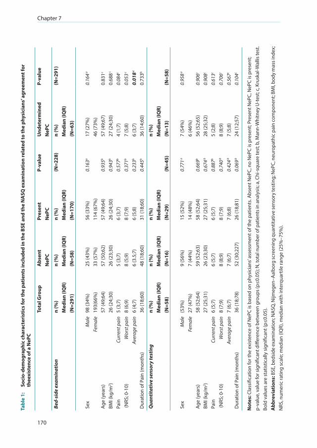
Chapter 7
170
Tabl
e 1:
So
cio-
dem
ogra
phic
cha
ract
eris
tics
for t
he p
atie
nts
incl
uded
in th
e BS
E an
d th
e N
ASQ
exa
min
atio
n re
late
d to
the
phys
icia
ns’ a
gree
men
t for
th
eexi
sten
ce o
f a N
ePC
Tota
l Gro
upA
bsen
t N
ePC
Pres
ent
NeP
C P-
valu
eU
ndet
erm
ined
NeP
C P-
valu
e
Bed-
side
exa
min
atio
nn
(%)
Med
ian
(IQR)
(N
=291
)
n (%
)M
edia
n (IQ
R)(N
=58)
n (%
)M
edia
n (IQ
R)(N
=170
)
(N=2
28)
n (%
)M
edia
n (IQ
R)(N
=63)
(N=2
91)
Sex
Mal
eFe
mal
e98
(34%
)19
3(66
%)
25 (4
3%)
33 (5
7%)
56 (3
3%)
114
(67%
)0.
163a
17 (2
7%)
46 (7
3%)
0.16
4 a
Age
(yea
rs)
57 (4
9;64
)57
(50;
62)
57 (4
9;64
)0.
935b
57 (4
9;67
)0.
831c
BMI (
kg/m
2 )26
(24;
30)
26 (2
3;30
)26
(24;
30)
0.94
3b27
(24;
30)
0.68
8 c
Pain
(NRS
; 0-1
0)Cu
rren
t pai
nW
orst
pai
nAv
erag
e pa
in
5 (3
;7)
8 (6
;9)
6 (4
;7)
5 (3
;7)
8 (5
;9)
6 (3
.5;7
)
6 (3
;7)
8 (7
;9)
6 (5
;8)
0.57
7b
0.37
1b
0.23
3b
4 (1
;7)
7 (5
;8)
6 (3
;7)
0.08
4c
0.05
3 c
0.01
8 c
Dur
atio
n of
Pai
n (m
onth
s)36
(18;
60)
48 (1
8;60
)31
(18;
60)
0.44
5b36
(14;
60)
0.73
3b
Qua
ntita
tive
sens
ory
test
ing
n (%
)M
edia
n (IQ
R)(N
=58)
n (%
)M
edia
n (IQ
R)
(N=1
6)
n (%
)M
edia
n (IQ
R)(N
=29)
(N=4
5)
n (%
)M
edia
n (IQ
R)(N
=13)
(N=5
8)
Sex
Mal
eFe
mal
e(5
3%)
27 (4
7%)
9 (5
6%)
7 (4
4%)
15 (5
2%)
14 (4
8%)
0.77
1 a7
(54%
)6
(46%
)0.
958 a
Age
(yea
rs)
58 (5
2;64
)59
(52;
63)
58 (5
2;64
)0.
669b
56 (5
2;65
)0.
906c
BMI (
kg/m
2 )27
(25;
31)
26 (2
3;30
)27
(25;
31)
0.67
4 b28
(25;
32)
0.90
8c
Pain
(NRS
; 0-1
0)Cu
rren
t pai
nW
orst
pai
nAv
erag
e pa
in
6 (5
;7)
8 (7
;9)
7 (6
;7)
6 (5
;7)
8 (8
;9)
7 (6
;7)
6 (5
;7)
8 (7
;9)
7 (6
;8)
0.88
7b
0.74
0 b
0.42
4 b
5 (2
;8)
8 (8
;9)
7 (5
;8)
0.61
3c
0.70
6c
0.56
7c
Dur
atio
n of
Pai
n (m
onth
s)36
(18;
78)
52 (3
0;22
7)26
(18;
81)
0.06
9 b24
(12;
57)
0.10
4c
Not
es: C
lass
ifica
tion
for t
he e
xist
ence
of N
ePC
is b
ased
on
phys
icia
ns’ a
sses
smen
t of t
he p
atie
nts.
Abs
ent N
ePC,
no
NeP
C is
pre
sent
; Pre
sent
NeP
C, N
ePC
is p
rese
nt;
p-va
lue,
val
ue fo
r sig
nific
ant d
iffer
ence
bet
wee
n gr
oups
(p≤0
.05)
; N, t
otal
num
ber o
f pat
ient
s in
anal
ysis
; a, C
hi-s
quar
e te
st; b
, Man
n-W
hitn
ey U
-tes
t; c,
Kru
skal
-Wal
lis te
st.
Bold
val
ues
are
stat
istic
ally
sig
nific
ant (
p≤0.
05).
Abb
revi
atio
ns: B
SE, b
edsi
de e
xam
inat
ion;
NA
SQ, N
ijmeg
en–A
albo
rg sc
reen
ing
quan
titat
ive
sens
ory
test
ing;
NeP
C, n
euro
path
ic p
ain
com
pone
nt; B
MI,
body
mas
s ind
ex;
NRS
, num
eric
ratin
g sc
ale;
med
ian
(IQR)
, med
ian
with
inte
rqua
rtile
rang
e (2
5%–7
5%).

BSE and NASQ in patients with and without NePC
171
7
Patients with chronic pain due to LBLP, NSAP or sPND assessed for eligibility
n = 330
No informed consent
n = 2
Present NePC
n = 170
Undetermined
n = 63
Absent NePC
n = 58
Performance of BSE (Excluded with reasons n = 37)
n = 291
Present NePC
n = 29
Undetermined
n = 13
Absent NePC
n = 16
Performance of NASQ (Excluded with reasons n = 11)
n = 58
Classification of NePC by Physicians Assessment
n = 328
Figure 1: Flow diagram for the performance of the BSE and NASQ in patients with chronic pain with respect to the physicians’ assessment.
Notes: n, number of patients in analysis; Present NePC, NePC is present; Undetermined, both physicians disagree with each other about the presence of a NePC; absent NePC, no NePC is present.
Abbreviations: LBLP, low back and leg pain; NSAP, neck shoulder arm pain; sPND, suspected peripheral nerve damage; BSE, bedside examination; NePC, neuropathic pain component; NASQ, Nijmegen- Aalborg screening quantitative sensory testing.
In Tables 2 and S1, we have shown the outcome of the BSE based on the inter-physician agreement
on the existence of an NePC. In the first assessment by the physician, the answers on the question
“is there a sensation (yes, no, unclear) during testing for heat, cold, touch (brush 3 times), and
pricking (both safety pin and von Frey hair)” were significantly lower (p≤0.05) for yes in the group
with present NePC compared to the absent NePC group. In the second assessment, the scores for
the question “is there a sensation (yes, no, unclear) of heat, cold, touch, (only brush 1 time), and
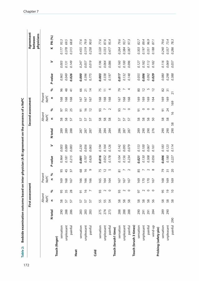
Chapter 7
172
Tabl
e 2:
Be
dsid
e ex
amin
atio
n ou
tcom
e ba
sed
on in
ter-
phys
icia
n (A
-B) a
gree
men
t on
the
pres
ence
of a
NeP
C
Firs
t ass
essm
ent
Seco
nd a
sses
smen
tA
gree
men
t be
twee
n ph
ysic
ians
Abse
ntN
ePC
Pres
ent
NeP
CAb
sent
NeP
CPr
esen
tN
ePC
N to
tal
n%
n%
P-va
lue
VN
tota
ln
%n
%P-
valu
eV
KPA
(%)
Touc
h (fi
nger
) sens
atio
n29
058
9516
995
0.96
40.
003
289
5897
168
960.
965
0.00
30.
177
93.3
unpl
easa
nt28
857
3516
845
0.18
10.
089
289
5833
168
480.
049
0.13
10.
378
69.5
pain
ful
286
5728
167
370.
215
0.08
328
857
2516
841
0.02
60.
149
0.31
568
.8H
eat
sen
satio
n28
357
9116
668
0.00
10.
230
287
5791
167
660.
000
0.24
70.
435
77.6
unpl
easa
nt28
357
1616
616
0.70
70.
056
287
5716
167
210.
396
0.05
70.
319
79.9
pain
ful
283
577
166
90.
626
0.06
528
757
1216
714
0.77
50.
019
0.25
890
.0Co
ldse
nsat
ion
275
5593
165
750.
016
0.19
428
458
9316
875
0.00
30.
196
0.32
077
.6un
plea
sant
274
552
164
120.
052
0.16
428
458
716
811
0.33
80.
064
0.33
387
.6pa
infu
l27
354
016
45
0.17
80.
126
284
582
168
60.
197
0.08
60.
477
95.4
Touc
h (b
rush
1 ti
me)
sens
atio
n28
858
9316
781
0.10
40.
142
286
5793
167
790.
017
0.16
00.
264
79.6
unpl
easa
nt28
858
216
77
0.15
60.
095
287
572
168
70.
132
0.10
00.
384
93.7
pain
ful
288
580
167
20.
234
0.07
928
757
016
84
0.14
80.
096
0.38
797
.3To
uch
(bru
sh 3
tim
es)
sens
atio
n29
058
9716
985
0.02
10.
153
289
5891
169
800.
055
0.12
70.
303
82.7
unpl
easa
nt29
158
217
07
0.13
00.
100
290
580
169
120.
006
0.18
20.
197
89.4
pain
ful
291
580
170
20.
308
0.06
729
058
016
95
0.09
20.
112
0.35
196
.9w
ind-
up28
456
016
78
0.05
60.
161
276
500
164
120.
029
0.18
20.
188
87.1
Pric
king
(saf
ety
pin)
sens
atio
n28
958
9516
879
0.00
60.
183
290
5891
169
820.
080
0.11
60.
240
79.6
unpl
easa
nt29
058
1916
931
0.18
00.
123
290
5824
169
310.
298
0.06
90.
357
73.4
pain
ful
290
5810
169
200.
227
0.11
429
058
1616
921
0.38
80.
057
0.28
678
.3
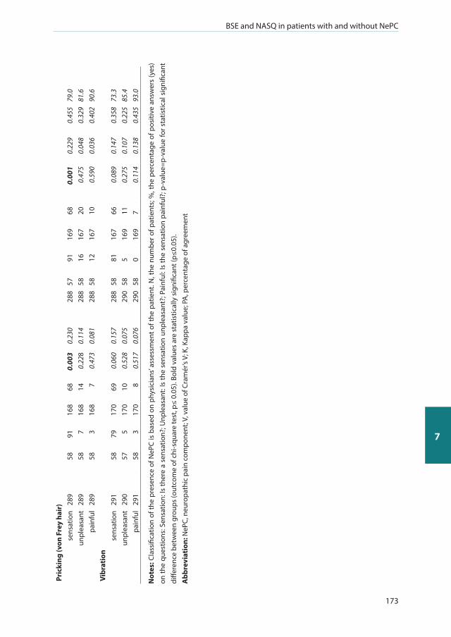
BSE and NASQ in patients with and without NePC
173
7
Pric
king
(von
Fre
y ha
ir)
sens
atio
n28
958
9116
868
0.00
30.
230
288
5791
169
680.
001
0.22
90.
455
79.0
unpl
easa
nt28
958
716
814
0.22
80.
114
288
5816
167
200.
475
0.04
80.
329
81.6
pain
ful
289
583
168
70.
473
0.08
128
858
1216
710
0.59
00.
036
0.40
290
.6Vi
brat
ion
sens
atio
n29
158
7917
069
0.06
00.
157
288
5881
167
660.
089
0.14
70.
358
73.3
unpl
easa
nt29
057
517
010
0.52
80.
075
290
585
169
110.
275
0.10
70.
225
85.4
pain
ful
291
583
170
80.
517
0.07
629
058
016
97
0.11
40.
138
0.43
593
.0
Not
es: C
lass
ifica
tion
of th
e pr
esen
ce o
f NeP
C is
bas
ed o
n ph
ysic
ians
’ ass
essm
ent o
f the
pat
ient
. N, t
he n
umbe
r of p
atie
nts;
%, t
he p
erce
ntag
e of
pos
itive
ans
wer
s (y
es)
on th
e qu
estio
ns: S
ensa
tion:
Is th
ere
a se
nsat
ion?
; Unp
leas
ant:
Is th
e se
nsat
ion
unpl
easa
nt?;
Pai
nful
: Is
the
sens
atio
n pa
infu
l?; p
-val
ue=p
-val
ue fo
r sta
tistic
al s
igni
fican
t di
ffere
nce
betw
een
grou
ps (o
utco
me
of c
hi-s
quar
e te
st, p
≤ 0.
05).
Bold
val
ues
are
stat
istic
ally
sig
nific
ant (
p≤0.
05).
Abb
revi
atio
n: N
ePC,
neu
ropa
thic
pai
n co
mpo
nent
; V, v
alue
of C
ram
ér’s
V; K
, Kap
pa v
alue
; PA
, per
cent
age
of a
gree
men
t
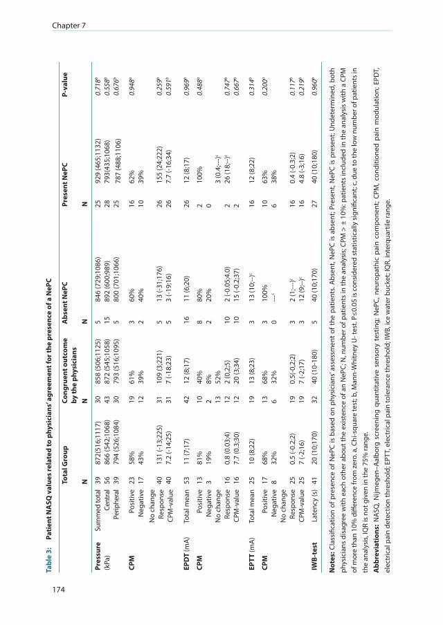
Chapter 7
174
Tabl
e 3:
Pa
tien
t NA
SQ v
alue
s re
late
d to
phy
sici
ans’
agre
emen
t for
the
pres
ence
of a
NeP
C
Tota
l Gro
upCo
ngru
ent o
utco
me
by th
e ph
ysic
ians
Abs
ent N
ePC
Pres
ent N
ePC
P-va
lue
NN
NN
Pres
sure
(kPa
)Su
mm
ed to
tal
Cent
ral
Perip
hera
l
39 56 39
872(
516;
1117
)86
6 (5
42;1
068)
794
(526
;108
4)
30 43 30
858
(506
;112
5)87
2 (5
45;1
058)
793
(516
;109
5)
5 15 5
846
(729
;108
6)89
2 (6
00;9
89)
800
(701
;106
6)
25 28 25
929
(465
;113
2)79
3(43
5;10
68)
787
(488
;110
6)
0.71
8b
0.55
8b
0.67
6b
CPM
Posi
tive
Neg
ativ
eN
o ch
ange
Re
spon
seCP
M-v
alue
23 17 40 40
58%
43%
131
(-13;
225)
7.2
(-14;
25)
19 12 31 31
61%
39%
109
(3;2
21)
7 (-1
8;23
)
3 2 5 5
60%
40%
13 (-
31;1
76)
3 (-1
9;16
)
16 10 26 26
62%
39%
155
(24;
222)
7.7
(-16;
34)
0.94
8a
0.25
9b
0.59
1b
EPD
T (m
A)
Tota
l mea
n53
11 (7
;17)
4212
(8;1
7)16
11 (6
;20)
2612
(8;1
7)0.
969b
CPM
Posi
tive
Neg
ativ
eN
o ch
ange
Re
spon
seCP
M-v
alue
13 3 16 16
81%
19%
0.8
(0.0
3;4)
7.7
(0.3
;30)
10 2 13 12 12
40%
8% 52%
2 (0
,2;5
)20
(3;3
4)
8 2 10 10
80%
20%
2 (-0
.05;
4.0)
15 (-
0.2;
37)
2 0 2 2
100%
3 (0
.4;--
-)c
26 (1
8;--
)c
0.48
8a
0.74
7b
0.66
7b
EPTT
(mA
)To
tal m
ean
2510
(8;2
2)19
13 (8
;23)
313
(10;
--)c
1612
(8;2
2)0.
314b
CPM
Po
sitiv
eN
egat
ive
No
chan
ge
Resp
onse
CPM
-val
ue
17 8 25 25
68%
32%
0.5
(-0.2
;2)
7 (-2
;16)
13 6 19 19
68%
32%
0.5(
-0,2
;2)
7 (-2
;17)
3 0 3 3
100%
---c
2 (1
;---)c
12 (9
;--)c
10 6 16 16
63%
38%
0.4
(-0.3
;2)
4.8
(-3;1
6)
0.20
0a
0.11
7b
0.21
9b
IWB-
test
Late
ncy
(s)
4120
(10;
170)
3240
(10-
180)
540
(10;
170)
2740
(10;
180)
0.96
0b
Not
es: C
lass
ifica
tion
of p
rese
nce
of N
ePC
is b
ased
on
phys
icia
ns’ a
sses
smen
t of
the
pat
ient
s. A
bsen
t, N
ePC
is a
bsen
t; Pr
esen
t, N
ePC
is p
rese
nt; U
ndet
erm
ined
, bot
h ph
ysic
ians
dis
agre
e w
ith e
ach
othe
r abo
ut th
e ex
iste
nce
of a
n N
ePC;
N, n
umbe
r of p
atie
nts
in th
e an
alys
is; C
PM >
± 1
0%: p
atie
nts
incl
uded
in th
e an
alys
is w
ith a
CPM
of
mor
e th
an 1
0% d
iffer
ence
from
zer
o. a
, Chi
-squ
are
test
; b, M
ann-
Whi
tney
U- t
est.
P≤0.
05 is
con
side
red
stat
istic
ally
sign
ifica
nt; c
, due
to th
e lo
w n
umbe
r of p
atie
nts i
n th
e an
alys
is, I
QR
is n
ot g
iven
in th
e 75
% ra
nge.
Abb
revi
atio
ns: N
ASQ
, Nijm
egen
–Aal
borg
scr
eeni
ng q
uant
itativ
e se
nsor
y te
stin
g; N
ePC,
neu
ropa
thic
pai
n co
mpo
nent
; CPM
, con
ditio
ned
pain
mod
ulat
ion;
EPD
T,
elec
tric
al p
ain
dete
ctio
n th
resh
old;
EPT
T, e
lect
rical
pai
n to
lera
nce
thre
shol
d; IW
B, ic
e w
ater
buc
ket;
IQR,
inte
rqua
rtile
rang
e.
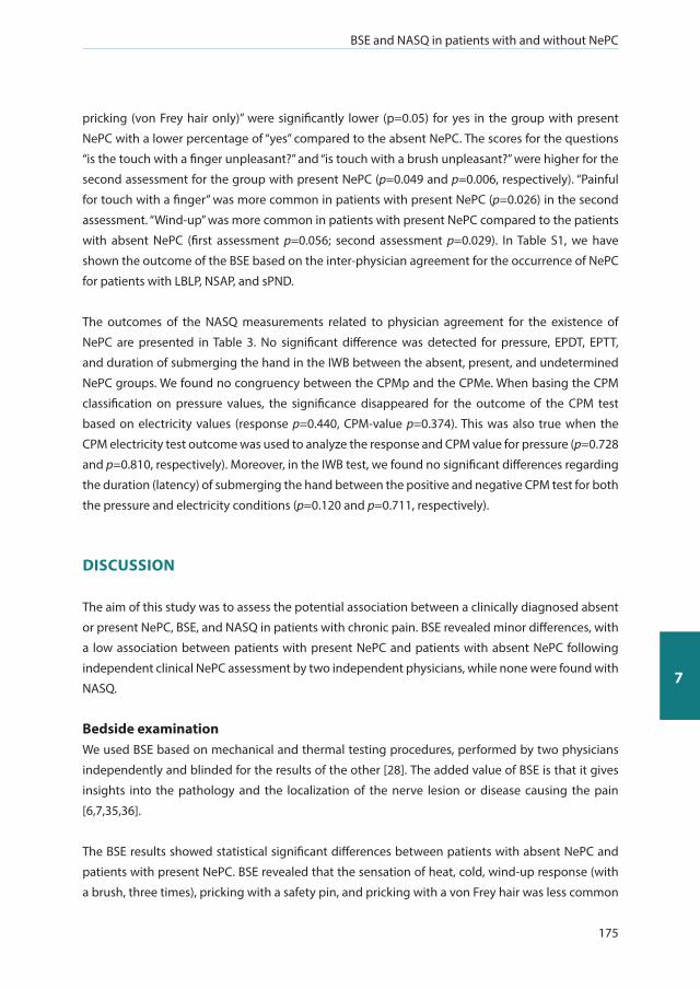
BSE and NASQ in patients with and without NePC
175
7
pricking (von Frey hair only)” were significantly lower (p=0.05) for yes in the group with present
NePC with a lower percentage of “yes” compared to the absent NePC. The scores for the questions
“is the touch with a finger unpleasant?” and “is touch with a brush unpleasant?” were higher for the
second assessment for the group with present NePC (p=0.049 and p=0.006, respectively). “Painful
for touch with a finger” was more common in patients with present NePC (p=0.026) in the second
assessment. “Wind-up” was more common in patients with present NePC compared to the patients
with absent NePC (first assessment p=0.056; second assessment p=0.029). In Table S1, we have
shown the outcome of the BSE based on the inter-physician agreement for the occurrence of NePC
for patients with LBLP, NSAP, and sPND.
The outcomes of the NASQ measurements related to physician agreement for the existence of
NePC are presented in Table 3. No significant difference was detected for pressure, EPDT, EPTT,
and duration of submerging the hand in the IWB between the absent, present, and undetermined
NePC groups. We found no congruency between the CPMp and the CPMe. When basing the CPM
classification on pressure values, the significance disappeared for the outcome of the CPM test
based on electricity values (response p=0.440, CPM-value p=0.374). This was also true when the
CPM electricity test outcome was used to analyze the response and CPM value for pressure (p=0.728
and p=0.810, respectively). Moreover, in the IWB test, we found no significant differences regarding
the duration (latency) of submerging the hand between the positive and negative CPM test for both
the pressure and electricity conditions (p=0.120 and p=0.711, respectively).
DISCUSSION
The aim of this study was to assess the potential association between a clinically diagnosed absent
or present NePC, BSE, and NASQ in patients with chronic pain. BSE revealed minor differences, with
a low association between patients with present NePC and patients with absent NePC following
independent clinical NePC assessment by two independent physicians, while none were found with
NASQ.
Bedside examination We used BSE based on mechanical and thermal testing procedures, performed by two physicians
independently and blinded for the results of the other [28]. The added value of BSE is that it gives
insights into the pathology and the localization of the nerve lesion or disease causing the pain
[6,7,35,36].
The BSE results showed statistical significant differences between patients with absent NePC and
patients with present NePC. BSE revealed that the sensation of heat, cold, wind-up response (with
a brush, three times), pricking with a safety pin, and pricking with a von Frey hair was less common
Chapter 7
176
in patients with a present NePC than in those with an absent NePC. In addition, wind-up response
occurred more often in patients with present NePC than in those with absent NePC.
Screening QST We used the NASQ to assess the altered pain processing, including changes in function of
endogenous pain modulation as a secondary test battery [15,28]. The NASQ test protocol has
standardized instructions, an important prerequisite to ensure reliability of the measurements
[20,37]. We found no differences between patients with absent and present NePC regarding
PPTs, electrical pain (tolerance) thresholds, and CPM outcomes (number of positive and negative
CPM outcomes, the response, the CPM value, and the latency times when submerging the hand
in ice water). Granovsky [38] reported that patients with chronic neuropathic pain express a less
efficient (negative) CPM. In our study, we could not confirm this when comparing patients with
LBLP, NSAP, or sPND with and without NePC. As suggested by Graven-Nielsen and Arendt-Nielsen
[39], lower PPTs may be indicative for central sensitization. We also could not find any differences
in the pain thresholds of patients with and without NePC. Moreover, a difference in CPM may also
suggest a central dysfunction. However, based on our results, we cannot state that there are signs
of central sensitization or altered central pain processing as might be suspected because of lower
pain thresholds for pressure pain or an impaired CPM, because we did not include age, sex, and
education matched controls, which would be necessary to draw these higher level conclusions.
Limitations We would have preferred to use the German Research Network on Neuropathic Pain (DFNS) [11,12]
to BSE because of the standardization of the complete test procedure (written test instructions,
application of the test stimuli, and data analyses) [12,40]. However, due to time constraints in a
patient care setting, it was not possible and preferable to use such a research test battery. Moreover,
in simulating daily clinical practice, fulfilling the DFNS protocol is not applicable due to instrument
availability and the associated costs in all participating sites. BSE as used in our study is easy to
learn (one training session before execution of the study) and to carry out in daily clinical practice.
Another strength of the study is that we included a range of locations and a large group of patients
with chronic pain arising from different origins, which is comparable to patients in a daily clinical
(pain) practice. A limitation of the BSE examination is that we only used the question “Is there a
sensation?” This may have led to a lower estimation of the outcomes because the patients and/
or physicians may have interpreted the question was only being related to the presence of
hypoesthesia, hypoalgesia, or analgesia (answer “no”: negative signs) rather than assessing the
presence of hyperalgesia or allodynia positively (answer “yes”). In a following study, we will change
this to a more open question that can be interpreted both ways. We did not use verbal standardized
instructions, although all participating professionals were trained in a standardized way and so this
is another possible limitation of our BSE method. This may have led to differences in the questioning
by the physicians, thereby influencing the patients’ answers and the test outcome. The order of the

BSE and NASQ in patients with and without NePC
177
7
BSE tests was not randomized and so there may be an order effect resulting from the previously
performed test. Moreover, both physicians tested the same patient directly following each other.
Although the second physician was not aware of the first results, this may have also influenced our
results. Furthermore, there was no correction for multiple testing while several statistical analyses
were performed. Because of this, the results must be interpreted with caution.
Another possible limitation is the fact that we only included a small group of patients with chronic
pain measured via NASQ; 8 patients with sPND. This may have affected our outcome because they
have a different disease origin compared to patients with LBLP or NSAP. For future NASQ research,
we would suggest collecting normative data preferably matched for age, sex, and education level.
With these data, the value of NASQ for clinical monitoring disease progression and the response of
individual patients on treatment can be evaluated.
CONCLUSION
Using a standardized BSE to assess sensory dysfunction indicating the presence or absence of an
NePC appears to be preferable compared to the NASQ paradigm in patients with chronic pain.
However, further development of both assessments is desirable. The BSE should be adapted to
detect sensory differences between absent and present NePC; the NASQ paradigm should be able
to measure altered pain processing and endogenous pain modulation in patients with chronic pain
due to present or absent NePC. We postulate that this will lead to a greater contribution to the
assessment of neuropathic components of patients’ pain.
ACKNOWLEDGEMENTS
We would like to thank all the participating patients, physicians and assistants for their invaluable
work in this study.
DISCLOSURE
This study was performed within DALI for PAIN, a national program that focuses on neuropathic
pain care optimalisation. DALI for PAIN is an initiative of Pfizer. This project was supported by an
unrestricted grant from Pfizer. The funders had no role in study design, data collection and analysis,
decision to publish, or preparation of the manuscript. The authors report no other conflicts of
interest in this work.
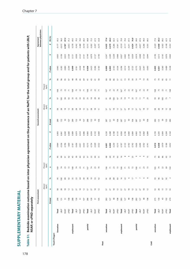
Chapter 7
178
SUPP
LEM
ENTA
RY M
ATER
IAL
Tabl
e S1
: Be
dsid
e ex
amin
atio
n ou
tcom
e ba
sed
on in
ter p
hysi
cian
agr
eem
ent o
n th
e pr
esen
ce o
f an
NeP
C fo
r the
tota
l gro
up a
nd fo
r pat
ient
s w
ith
LBLP
, N
SAP,
or s
PND
sep
arat
ely
Firs
t ass
essm
ent
Seco
nd a
sses
smen
tA
gree
men
tbe
twee
n ph
ysic
ians
Abse
ntN
ePC
Pres
ent
NeP
CAb
sent
NeP
CPr
esen
tN
ePC
N to
tal
n%
n%
P-va
lue
VN
tota
ln
%n
%P-
valu
eV
KPA
(%)
Touc
h (F
inge
r)
Sens
atio
nTo
tal
290
5895
169
950.
964
0.00
328
958
9716
896
0.96
50.
003
0.17
793
.3
LBLP
131
2810
074
990.
536
0.06
113
228
100
7596
0.28
30.
106
-0.0
1596
.0
NSA
P51
1889
2391
0.79
60.
040
5018
9422
950.
884
0.02
30.
787
97.5
sPN
D10
812
9272
921.
000
0.00
010
712
9271
970.
344
0.10
4-0
.053
91.3
unpl
easa
ntTo
tal
288
5735
168
450.
181
0.08
928
958
3316
848
0.04
90.
131
0.37
869
.5
LBLP
130
2722
7430
0.45
60.
074
132
2825
7528
0.76
10.
030
0.24
270
.3
NSA
P51
1844
2344
0.95
10.
010
5018
3322
450.
436
0.12
30.
388
70.0
sPN
D10
712
5071
620.
433
0.08
610
712
5071
690.
198
0.14
10.
318
68.3
pain
ful
Tota
l28
657
2816
737
0.21
50.
083
288
5725
168
410.
026
0.14
90.
315
68.8
LBLP
130
2719
7422
0.73
40.
034
132
2818
7543
0.42
50.
079
0.20
273
.3
NSA
P51
1833
2344
0.50
90.
103
5018
2822
410.
386
0.13
70.
297
67.5
sPN
D10
512
4270
510.
532
0.06
910
611
3671
580.
185
0.14
60.
275
63.8
Hea
t
sen
sati
onTo
tal
283
5791
166
680.
001
0.23
028
757
9116
766
0.00
00.
247
0.43
577
.6
LBLP
127
2796
7169
0.00
40.
288
131
2796
7568
0.00
30.
290
0.57
984
.5
NSA
P48
1889
2374
0.23
00.
188
5018
100
2286
0.10
30.
258
0.28
682
.5
sPN
D10
812
8372
650.
215
0.13
510
612
6770
570.
536
0.06
80.
301
67.1
unpl
easa
ntTo
tal
283
5716
166
160.
707
0.05
628
757
1616
721
0.39
60.
057
0.31
979
.9
LBLP
127
2722
718
0.15
30.
196
131
2719
7513
0.51
40.
065
0.11
779
.4
NSA
P48
186
239
0.70
20.
060
5018
622
180.
230
0.19
00.
231
87.5
sPN
D10
812
1772
250.
742
0.08
410
612
2570
300.
725
0.03
90.
424
76.8
pain
ful
Tota
l28
357
716
69
0.62
60.
065
287
5712
167
140.
775
0.01
90.
258
90.0
LBLP
127
277
713
0.49
40.
120
131
2711
758
0.62
50.
048
-0.0
5787
.6
NSA
P48
186
239
0.70
20.
060
5018
622
140.
397
0.13
40.
286
90.0
sPN
D10
812
872
150.
741
0.08
410
612
2570
200.
693
0.04
40.
355
80.5
Cold
sens
atio
nTo
tal
275
5593
165
750.
016
0.19
428
458
9316
875
0.00
30.
196
0.32
077
.6
LBLP
130
2896
7480
0.03
80.
205
131
2896
7579
0.03
10.
213
0.45
885
.3
NSA
P43
1587
2286
0.97
90.
004
5018
100
2295
0.36
00.
145
0.30
289
.2
sPN
D10
212
9269
650.
186
0.20
410
312
7571
650.
489
0.07
60.
148
62.5
unpl
easa
ntTo
tal
274
552
164
120.
052
0.16
428
458
716
811
0.33
80.
064
0.33
387
.6
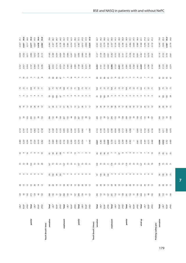
BSE and NASQ in patients with and without NePC
179
7
LBLP
129
284
7310
0.48
90.
119
131
287
755
0.72
70.
034
0.35
791
.1
NSA
P43
150
229
0.23
00.
197
5018
022
180.
057
0.30
20.
641
94.6
sPN
D10
212
069
160.
294
0.17
410
312
1771
150.
918
0.01
10.
217
80.0
pain
ful
Tota
l27
354
016
45
0.17
80.
126
284
582
168
60.
197
0.08
60.
477
95.4
LBLP
129
280
730
0.53
40.
062
131
284
751
0.46
40.
072
-0.0
0797
.0
NSA
P42
140
225
0.41
80.
135
5018
022
140.
103
0.25
80.
478
94.4
sPN
D10
212
069
100.
462
0.13
810
312
071
80.
296
0.11
50.
582
93.8
Touc
h (b
rush
1 ti
me)
sens
atio
nTo
tal
288
5893
167
810.
104
0.14
228
657
9316
779
0.01
70.
160
0.26
479
.6
LBLP
130
2810
073
900.
089
0.16
913
228
100
7588
0.05
50.
189
0.18
788
.1
NSA
P51
1889
2396
0.40
90.
129
4817
100
2286
0.11
30.
254
0.27
889
.7
sPN
D10
712
8371
680.
533
0.12
310
612
6770
670.
974
0.00
40.
178
64.2
unpl
easa
ntTo
tal
288
582
167
70.
156
0.09
528
757
216
87
0.13
20.
100
0.38
493
.7
LBLP
130
284
736
0.82
60.
022
132
280
753
0.38
30.
086
0.38
597
.0
NSA
P51
180
239
0.20
00.
200
4817
022
180.
063
0.29
70.
374
62.3
sPN
D10
712
071
100.
256
0.12
510
612
871
80.
989
0.00
10.
375
90.2
pain
ful
Tota
l28
858
016
72
0.23
40.
079
287
570
168
40.
148
0.09
60.
387
97.3
LBLP
130
280
731
0.53
40.
062
132
280
753
0.38
30.
086
-0.0
1397
.0
NSA
P51
180
230
---
---
4817
022
50.
373
0.14
30.
000
97.4
sPN
D10
712
071
40.
468
0.08
010
712
071
40.
468
0.08
00.
654
97.6
Touc
h (b
rush
3 ti
mes
)
sens
atio
nTo
tal
290
5897
169
850.
021
0.15
328
958
9116
980
0.05
50.
127
0.30
382
.7
LBLP
132
2810
075
960.
283
0.10
613
228
100
7593
0.16
10.
138
0.22
294
.2
NSA
P51
1889
2396
0.40
90.
129
5018
100
2286
0.10
30.
258
0.27
990
.0
sPN
D10
712
100
7170
0.02
90.
239
107
1258
7265
0.64
20.
051
0.19
065
.1
unpl
easa
ntTo
tal
291
582
170
70.
130
0.10
029
058
016
912
0.00
60.
182
0.19
789
.4
LBLP
132
284
755
0.71
10.
036
132
280
758
0.12
30.
152
0.13
691
.3
NSA
P51
180
2387
0.20
00.
200
5018
022
230.
031
0.34
20.
304
90.0
sPN
D10
812
072
80.
299
0.11
310
812
072
130.
195
0.14
10.
198
86.9
pain
ful
Tota
l29
158
017
02
0.30
80.
067
290
580
169
50.
092
0.11
20.
351
96.9
LBLP
132
280
753
0.38
30.
086
132
200
754
0.28
30.
106
0.38
697
.1
NSA
P51
180
230
---
---
5018
022
50.
360
0.14
50.
000
97.5
sPN
D10
812
072
10.
681
0.04
510
812
072
60.
403
0.09
10.
388
96.4
win
d-up
Tota
l28
456
016
78
0.05
60.
161
276
500
164
120.
029
0.18
20.
188
87.1
LBLP
131
280
757
0.30
40.
152
125
260
717
0.31
00.
155
0.29
691
.8
NSA
P48
160
229
0.21
50.
201
4412
022
50.
290
0.27
00.
145
87.1
sPN
D10
512
070
100.
468
0.13
610
712
071
150.
143
0.16
10.
116
81.5
Pric
king
(saf
ety
pin)
sens
atio
nTo
tal
289
5895
168
790.
006
0.18
329
058
9116
982
0.08
00.
116
0.24
079
.6
LBLP
132
2810
075
850.
032
0.21
113
228
100
7592
0.12
30.
152
0.04
685
.4
NSA
P51
1894
2391
0.70
20.
060
5018
100
2295
0.36
00.
145
0.48
195
.0
sPN
D10
612
2571
350.
489
0.07
910
812
5872
670.
574
0.06
10.
176
64.6
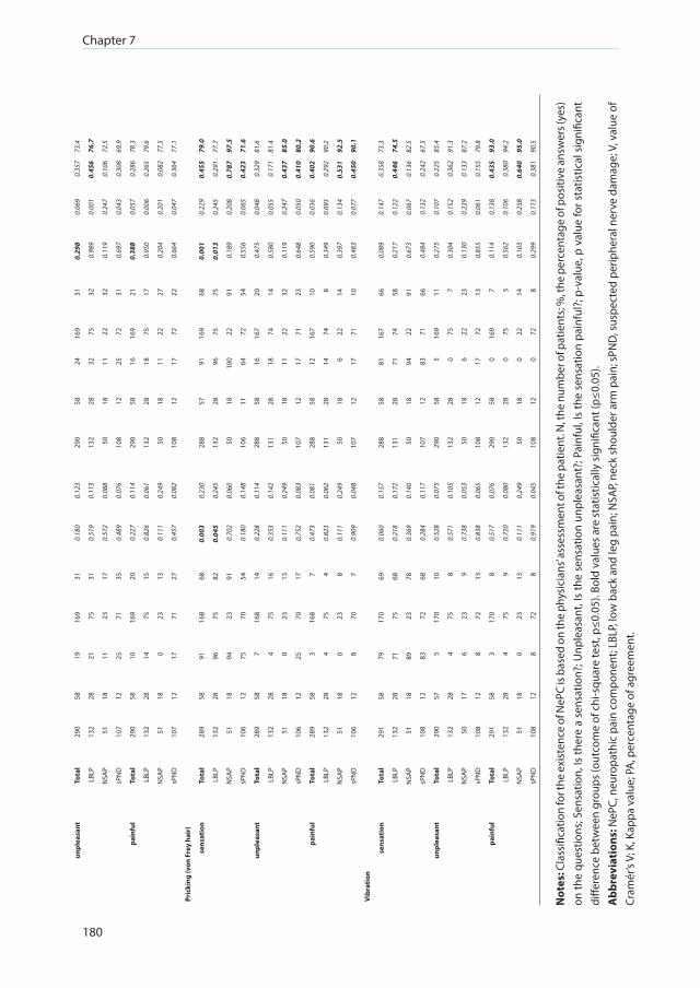
Chapter 7
180
unpl
easa
ntTo
tal
290
5819
169
310.
180
0.12
329
058
2416
931
0.29
80.
069
0.35
773
.4
LBLP
132
2821
7531
0.51
90.
113
132
2832
7532
0.98
90.
001
0.45
676
.7
NSA
P51
1811
2317
0.57
20.
088
5018
1122
320.
119
0.24
70.
106
72.5
sPN
D10
712
2571
350.
489
0.07
610
812
2572
310.
697
0.04
30.
308
69.9
pain
ful
Tota
l29
058
1016
920
0.22
70.
114
290
5816
169
210.
388
0.05
70.
286
78.3
LBLP
132
2814
7515
0.82
60.
061
132
2818
7517
0.95
00.
006
0.26
579
.6
NSA
P51
180
2313
0.11
10.
249
5018
1122
270.
204
0.20
10.
082
77.5
sPN
D10
712
1771
270.
457
0.08
210
812
1772
220.
664
0.04
70.
364
77.1
Pric
king
(von
Fre
y ha
ir)
sens
atio
nTo
tal
289
5891
168
680.
003
0.23
028
857
9116
968
0.00
10.
229
0.45
579
.0
LBLP
132
2896
7582
0.04
50.
245
132
2896
7575
0.01
30.
245
0.29
177
.7
NSA
P51
1894
2391
0.70
20.
060
5018
100
2291
0.18
90.
208
0.78
797
.5
sPN
D10
612
7570
540.
180
0.14
810
611
6472
540.
556
0.06
50.
423
71.6
unpl
easa
ntTo
tal
289
587
168
140.
228
0.11
428
858
1616
720
0.47
50.
048
0.32
981
.6
LBLP
132
284
7516
0.35
30.
142
131
2818
7414
0.58
00.
055
0.17
181
.4
NSA
P51
180
2313
0.11
10.
249
5018
1122
320.
119
0.24
70.
437
85.0
sPN
D10
612
2570
170.
752
0.08
310
712
1771
230.
648
0.05
00.
410
80.2
pain
ful
Tota
l28
958
316
87
0.47
30.
081
288
5812
167
100.
590
0.03
60.
402
90.6
LBLP
132
284
754
0.82
30.
062
131
2814
748
0.34
90.
093
0.29
290
.2
NSA
P51
180
238
0.11
10.
249
5018
622
140.
397
0.13
40.
531
92.5
sPN
D10
612
870
70.
909
0.04
810
712
1771
100.
483
0.07
70.
450
90.1
Vibr
atio
n
sens
atio
nTo
tal
291
5879
170
690.
060
0.15
728
858
8116
766
0.08
90.
147
0.35
873
.3
LBLP
132
2871
7568
0.21
80.
172
131
2871
7458
0.21
70.
122
0.44
674
.5
NSA
P51
1889
2378
0.36
90.
140
5018
9422
910.
673
0.06
70.
136
82.5
sPN
D10
812
8372
680.
284
0.11
710
712
8371
660.
484
0.13
20.
242
67.5
unpl
easa
ntTo
tal
290
575
170
100.
528
0.07
529
058
516
911
0.27
50.
107
0.22
585
.4
LBLP
132
284
758
0.57
10.
105
132
280
757
0.30
40.
152
0.36
291
.3
NSA
P50
176
239
0.73
80.
053
5018
622
230.
130
0.23
90.
133
87.2
sPN
D10
812
872
130.
838
0.06
510
812
1772
130.
855
0.06
10.
155
79.8
pain
ful
Tota
l29
158
317
08
0.51
70.
076
290
580
169
70.
114
0.13
80.
435
93.0
LBLP
132
284
759
0.72
00.
080
132
280
753
0.56
20.
106
0.38
094
.2
NSA
P51
180
2313
0.11
10.
249
5018
022
140.
103
0.25
80.
640
95.0
sPN
D10
812
872
80.
919
0.04
510
812
072
80.
299
0.11
30.
381
90.5
Not
es: C
lass
ifica
tion
for t
he e
xist
ence
of N
ePC
is b
ased
on
the
phys
icia
ns’ a
sses
smen
t of t
he p
atie
nt. N
, the
num
ber o
f pat
ient
s; %
, the
per
cent
age
of p
ositi
ve a
nsw
ers (
yes)
on
the
ques
tions
; Sen
satio
n, Is
ther
e a
sens
atio
n?; U
nple
asan
t, Is
the
sens
atio
n un
plea
sant
?; P
ainf
ul, I
s th
e se
nsat
ion
pain
ful?
; p-v
alue
, p v
alue
for s
tatis
tical
sig
nific
ant
diffe
renc
e be
twee
n gr
oups
(out
com
e of
chi
-squ
are
test
, p≤0
.05)
. Bol
d va
lues
are
sta
tistic
ally
sig
nific
ant (
p≤0.
05).
Abb
revi
atio
ns: N
ePC,
neu
ropa
thic
pai
n co
mpo
nent
; LBL
P, lo
w b
ack
and
leg
pain
; NSA
P, ne
ck s
houl
der a
rm p
ain;
sPN
D, s
uspe
cted
per
iphe
ral n
erve
dam
age;
V, v
alue
of
Cram
ér’s
V; K
, Kap
pa v
alue
; PA
, per
cent
age
of a
gree
men
t.
BSE and NASQ in patients with and without NePC
181
7
REFERENCES
1. IASP. IASP taxonomy neuropathic pain. 2015. Available from: http:// www.iasp-pain.org/Taxonomy#Neuropathicpain. Accessed May 19, 2015.
2. Dieleman JP, Kerklaan J, Huygen FJ, Bouma PA, Sturkenboom MC. Incidence rates and treatment of neuropathic pain conditions in the general population. Pain. 2008;137(3):681-688.
3. Freynhagen R, Bennett MI. Diagnosis and management of neuropathic pain. BMJ. 2009;339:b3002. 4. Bouhassira D, Lantéri-Minet M, Attal N, Laurent B, Touboul C. Prevalence of chronic pain with neuropathic
characteristics in the general population. Pain. 2008;136(3):380-387. 5. La Cesa S, Tamburin S, Tugnoli V, et al. How to diagnose neuropathic pain? The contribution from clinical
examination, pain questionnaires and diagnostic tests. Neurol Sci. 2015;36(12):2169-2175. 6. Haanpää M, Attal N, Backonja M, et al. NeuPSIG guidelines on neuropathic pain assessment. Pain.
2011;152(1):14-27. 7. Haanpää ML, Backonja MM, Bennett MI, et al. Assessment of neuropathic pain in primary care. Am J Med.
2009;122(10 Suppl):S13-S21. 8. Truini A, Cruccu G. How diagnostic tests help to disentangle the mechanisms underlying neuropathic pain
symptoms in painful neuropathies. Pain. 2016;157(Suppl 1):S53-S59. 9. Treede RD, Jensen TS, Campbell JN, et al. Neuropathic pain: redefinition and a grading system for clinical
and research purposes. Neurology. 2008;70(18):1630-1635. 10. Finnerup NB, Haroutounian S, Kamerman P, et al. Neuropathic pain: an updated grading system for research
and clinical practice. Pain. 2016;157(8):1599-1606. 11. Rolke R, Baron R, Maier C, et al. Quantitative sensory testing in the German Research Network on Neuropathic
Pain (DFNS): standardized protocol and reference values. Pain. 2006;123(3):231-243. 12. RolkeR,MagerlW,CampbellKA,etal.Quantitativesensorytesting:a comprehensive protocol for clinical trials.
Eur J Pain. 2006;10(1):77-88. 13. Backonja MM, Walk D, Edwards RR, et al. Quantitative sensory testing in measurement of neuropathic pain
phenomena and other sensory abnormalities. Clin J Pain. 2009;25(7):641-647. 14. Arendt-Nielsen L, Yarnitsky D. Experimental and clinical applications of quantitative sensory testing applied
to skin, muscles and viscera. J Pain. 2009;10(6):556-572. 15. Wilder-Smith OH. A Paradigm-Shift in Pain Medicine: Implementing a Systematic Approach to Altered Pain
Processing in Everyday Clinical Practice Based on Quantitative Sensory Testing. Aalborg: Center for Sensory-Motor Interaction (SMI), Department of Health Science and Technology, Aalborg University; 2013.
16. Bouwense SA, de Vries M, Schreuder LT, et al. Systematic mechanism- orientated approach to chronic pancreatitis pain. World J Gastroenterol. 2015;21(1):47-59.
17. Yarnitsky D. Conditioned pain modulation (the diffuse noxious inhibitory control-like effect): its relevance for acute and chronic pain states. Curr Opin Anaesthesiol. 2010;23(5):611-615.
18. Yarnitsky D. Role of endogenous pain modulation in chronic pain mechanisms and treatment. Pain. 2015;156(Suppl 1):S24-S31.
19. Hansson P, Backonja M, Bouhassira D. Usefulness and limitations of quantitative sensory testing: clinical and research application in neuropathic pain states. Pain. 2007;129(3):256-259.
20. Geber C, Klein T, Azad S, et al. Test-retest and interobserver reliability of quantitative sensory testing according to the protocol of the German Research Network on Neuropathic Pain (DFNS): a multi-centre study. Pain. 2011;152(3):548-556.
21. Cruccu G, Sommer C, Anand P, et al. EFNS guidelines on neuropathic pain assessment: revised 2009. Eur J Neurol. 2010;17(8):1010-1018.
22. Krumova EK, Geber C, Westermann A, Maier C. Neuropathic pain: is quantitative sensory testing helpful? Curr Diab Rep. 2012;12(4): 393-402.
23. Woolf CJ. Central sensitization: implications for the diagnosis and treatment of pain. Pain. 2011;152(3 Suppl):S2-S15.
24. Chua NHL, Timmerman H, Vissers KC, OH W-S. Multi-modal quantitative sensory testing in patients with unilateral chronic neck pain: an exploratory study. J Musculoskelet Pain. 2012;20(4):292-299.
25. Pud D, Granovsky Y, Yarnitsky D. The methodology of experimentally induced diffuse noxious inhibitory control (DNIC)-like effect in humans. Pain. 2009;144(1-2):16-19.
Chapter 7
182
26. Correa JB, Costa LO, de Oliveira NT, Sluka KA, Liebano RE. Central sensitization and changes in conditioned pain modulation in people with chronic nonspecific low back pain: a case-control study. Exp Brain Res. 2015;233(8):2391-2399.
27. Yarnitsky D, Arendt-Nielsen L, Bouhassira D, et al. Recommendations on terminology and practice of psychophysical DNIC testing. Eur J Pain. 2010;14(4):339.
28. Timmerman H, Wilder-Smith O, van Weel C, Wolff A, Vissers K. Detecting the neuropathic pain component in the clinical setting: a study protocol for validation of screening instruments for the presence of a neuropathic pain component. BMC Neurol. 2014;14(1):94.
29. Timmerman H, Wolff AP, Schreyer T, et al. Cross-cultural adaptation to the Dutch language of the PainDETECT-Questionnaire. Pain Pract. 2013;13(3):206-214.
30. Van Seventer R, Vos C, Meerding W, et al. Linguistic validation of the DN4 for use in international studies. Eur J Pain. 2010;14(1):58-63.
31. Timmerman H, Steegers MAH, Huygen F, et al. Investigating the validity of the DN4 in a consecutive population of patients with chronic pain. PloS One. 2017;12(11):e0187961.
32. Freynhagen R, Baron R, Gockel U, Tölle TR. PainDETECT: a new screening questionnaire to identify neuropathic components in patients with back pain. Curr Med Res Opin. 2006;22(10):1911-1920.
33. Bouhassira D, Attal N, Alchaar H, et al. Comparison of pain syndromes associated with nervous or somatic lesions and development of a new neuropathic pain diagnostic questionnaire (DN4). Pain. 2005;114(1-2):29-36.
34. Cruccu G, Anand P, Attal N, et al. EFNS guidelines on neuropathic pain assessment. Eur J Neurol. 2004;11(3):153-162.
35. Baron R, Binder A, Wasner G. Neuropathic pain: diagnosis, pathophysiological mechanisms, and treatment. Lancet Neurol. 2010;9(8):807-819.
36. Cruccu G, Truini A. Tools for assessing neuropathic pain. PloS Med. 2009;6(4):e1000045.37. Backonja MM, Attal N, Baron R, et al. Value of quantitative sensory testing in neurological and pain disorders:
NeuPSIG consensus. Pain. 2013;154(9):1807-1819.38. Granovsky Y. Conditioned pain modulation: a predictor for development and treatment of neuropathic
pain. Curr Pain Headache Rep. 2013;17(9):361.39. Graven-Nielsen T, Arendt-Nielsen L. Assessment of mechanisms in localized and widespread musculoskeletal
pain. Nat Rev Rheumatol. 2010;6(10):599-606.40. Pfau DB, Geber C, Birklein F, Treede RD. Quantitative sensory testing of neuropathic pain patients: potential
mechanistic and therapeutic implications. Curr Pain Headache Rep. 2012;16(3): 199-206.
BSE and NASQ in patients with and without NePC
183
7


CHAPTER 8General conclusions and discussion. Recommendations for clinical practice, education, future research, and societal impact

Chapter 8
186
Conclusions, discussion and recommendations
187
8
GENERAL CONCLUSIONS AND RECOMMENDATIONS
In this thesis we investigated the psychometric properties and efficacy of screening tools for
neuropathic pain for use in a consecutive population of patients with low back and leg pain, neck-
shoulder-arm pain, or with pain due to suspected peripheral nerve damage to assist the physician
in the assessment of a neuropathic pain component as seen in daily clinical (pain-) practice. We
selected this group of patients for our study because they are different from the other groups
assessed in previous validation studies. The group consists of a consecutive patient population
as seen in daily clinical outpatient practice where there is no pre-stratification on the target
outcome, and no minimum level of pain. In our articles, we prefer to use the term ‘neuropathic
pain component’ instead of neuropathic pain because the pain experienced by the patient in daily
clinical practice may be caused by neuropathic, nociceptive, and/or nociplastic mechanisms (also
known as ’mixed pain’). Neuropathic pain is distinguishable from nociceptive pain in two ways [1]:
1) nociceptive pain requires transduction to transfer a non-electrical signal to an electrochemical
signal, whereas neuropathic pain is based on a direct stimulation of the (injured) nerve; 2) most
people with nociceptive pain recover in a certain time, whereas patients with neuropathic pain
based on an injury of a nerve often retain persistent pain. As Cohen and Mao stated [1], there is
a considerable overlap between neuropathic and nociceptive pain regarding patho-physiological
mechanisms and response to treatment, so they can possibly be seen as ‘different points on the
same continuum’ [1]. Currently, multiple tests are available to screen for or to assess neuropathic
pain, but their verified accuracy is missing and there is no ‘gold standard’ questionnaire [2] . This can
lead to errors in prevalence studies, incorrect and expensive clinical treatment, and, most relevant
clinically, increasing disabilities for the patient.
Our studies show that current screening methods and tools for identifying the type of patients’ pain
are simply not accurate enough for a definite pain classification, and that this presents a number of
challenges: (1) the risk of starting inadequate, inappropriate or non-efficacious therapies solely based
on these classifications is high; (2) research in the field of treatment for pain is at risk because of the
lack of distinction between classification outcomes; (3) exploring nociception or pain as a disease
in its own right or as a symptom of a disease, trauma or disorder remains a problem; (4) identifying
the type of pain, in particular mixed pain with components of nociceptive pain, neuropathic pain
and/or nociplastic pain is difficult. These show that we need valid and reliable assessment tools
more directly related to the underlying pain mechanisms. Figure 1 presents a suggested path for
developing and validating screening tools in a way more related to daily clinical practice.
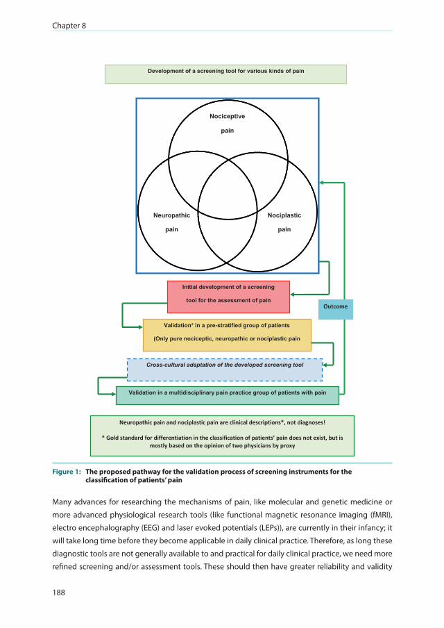
Chapter 8
188
Nociceptive
pain
Neuropathic
pain
Nociplastic
pain
Development of a screening tool for various kinds of pain
Validation* in a pre-stratified group of patients
(Only pure nociceptic, neuropathic or nociplastic pain
Cross-cultural adaptation of the developed screening tool
Validation in a multidisciplinary pain practice group of patients with pain
Initial development of a screening
tool for the assessment of pain
Neuropathic pain and nociplastic pain are clinical descriptions*, not diagnoses!
* Gold standard for differentiation in the classification of patients’ pain does not exist, but is mostly based on the opinion of two physicians by proxy
Outcome
Figure 1: The proposed pathway for the validation process of screening instruments for the classification of patients’ pain
Many advances for researching the mechanisms of pain, like molecular and genetic medicine or
more advanced physiological research tools (like functional magnetic resonance imaging (fMRI),
electro encephalography (EEG) and laser evoked potentials (LEPs)), are currently in their infancy; it
will take long time before they become applicable in daily clinical practice. Therefore, as long these
diagnostic tools are not generally available to and practical for daily clinical practice, we need more
refined screening and/or assessment tools. These should then have greater reliability and validity
Conclusions, discussion and recommendations
189
8
than currently available methods, and should be applicable to both primary care and specialist
academic pain centers. This will remain challenging as long as there is no true gold standard to
diagnose neuropathic pain. Until then, in current medical pain practice, diagnosis is formed on
history-taking, and physical assessment. The latter includes bedside examination (as suggested in
the NeuPSIG Grading System, [3]) and remains foremost as this is, however slightly, associated with
the pain classification ‘absent or present NePC’.
The research questions we addressed were:Question 1: Is a cross-cultural adaptation a prerequisite for achieving a valid Dutch translation of a
screening tool for neuropathic pain?
Question 2: What is the reliability of clinical judgment as a surrogate for the lack of an objective
gold standard in diagnosing a neuropathic pain component in patients with chronic pain?
Question 3: What are the psychometric properties of the PainDETECT and the DN4 questionnaire
when used as screening tools in a daily practice consecutive patient population (patients with
low back pain, neck shoulder or arm pain, or pain from a suspected neuropathic origin), not pre-
stratified on target outcome, for NePC detection?
Question 4: What is the potential association between clinically diagnosed, via two independent
and trained professionals, absent or present NePC, and bedside examination / screening quantitative
sensory testing (NASQ) in patients with chronic pain?
Our research program was designed to answer these questions, as discussed in the thesis
Introduction, and to provide recommendations for daily clinical practice and future research.
Chapter 8
190
Answer to question 1: A cross-cultural adaptation is a prerequisite for the correct translation and validation of measurement instruments such as screening tools for the assessment of NePC.Recently, the PainDETECT was used in an American prevalence study [4]. Freynhagen et al [5]
translated the original German version into English, however did not provide any information about
the translation process. Tampin et al. noted that the English language version of the PainDETECT has
not yet been validated[6]. Although the test-retest reliability turned out to be good, the validity of
the English language version of the PainDETECT is thus still questionable [6]. The outcome of the
American prevalence study was that, in addition to the estimation of the prevalence of neuropathic
pain in the USA, the prevalence of probable neuropathic pain among Blacks and Hispanics was
consistently higher than among Whites (per age and sex group). This interpretation is, of course,
possible, however, the PainDETECT was not cross-culturally adapted and validated for use in the USA
and thus the variance may be a result of, for example, a difference in interpretation of the questions
in the instrument. It may also be questioned whether the cut-off points of the PainDETECT are valid
in patients with various chronic pain conditions and different cultural backgrounds. Moreover, the
authors only used the English language version, which may also have influenced the outcome[4].
In our study (Chapter 2) which included Dutch-speaking patients from both Belgium and the
Netherlands, we found a number of differences based on culture and/or language perception. The
Belgian participants rated the clearness of the questions and the organization of the questionnaire
almost 10% higher than Dutch participants. This may be attributed to the slight cultural differences
in interpretation and use of some words between the Dutch and the Belgians.
Based on these issues, we state that a well-performed, cross-culturally adapted version should be
used instead of a ‘traditional’ forward / forward-backward translation before using an instrument in
another culture and/or language. We therefore followed and recommend that other groups follow
the published guidelines [7-9] to achieve the highest equivalence with the original tool in language,
structure and meaning.
Answer to question 2: The clinical judgment by two physicians is reliable as a replacement for the lack of a gold standard when diagnosing a neuropathic pain component in patients with chronic pain in clinical practiceAlthough clinical judgment is accepted as a surrogate for an objective gold standard in the
assessment of NePC, the reliability of this surrogate outcome is unknown. We therefore conducted
a study to find out if the agreement of two observers, based on pain classification by the physicians
and by the Grading System as entered by the physician, is acceptable to differentiate between
neuropathic and non-neuropathic pain in daily clinical practice. If the kappa-value is ≥ 0.4 and the
percentage of agreement is ≥ 70%, the outcome is considered acceptable for use in daily clinical
practice [10].
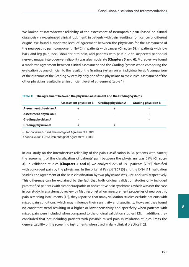
Conclusions, discussion and recommendations
191
8
We looked at interobserver reliability of the assessment of neuropathic pain (based on clinical
diagnosis via experienced clinical judgment) in patients with pain resulting from cancer of different
origins. We found a moderate level of agreement between the physicians for the assessment of
the neuropathic pain component (NePC) in patients with cancer (Chapter 3). In patients with low
back and leg pain, neck shoulder arm pain, and patients with pain due to suspected peripheral
nerve damage, interobserver reliability was also moderate (Chapters 5 and 6). Moreover, we found
a moderate agreement between clinical assessment and the Grading System when comparing the
evaluation by one clinician to the result of the Grading System on an individual level. A comparison
of the outcome of the Grading System by only one of the physicians to the clinical assessment of the
other physician resulted in an insufficient level of agreement (table 1).
Table 1: The agreement between the physician assessment and the Grading Systems.
Assessment physician B Grading physician A Grading physician B
Assessment physician A + + -
Assessment physician B - +
Grading physician A - +
Grading physician B + +
+: Kappa value ≥ 0.4 & Percentage of Agreement ≥ 70%-: Kappa value < 0.4 & Percentage of Agreement < 70%
In our study on the interobserver reliability of the pain classification in 34 patients with cancer,
the agreement of the classification of patients’ pain between the physicians was 59% (Chapter
3). In validation studies (Chapters 5 and 6) we analyzed 228 of 291 patients (78%) classified
with congruent pain by the physicians. In the original PainDETECT [5] and the DN4 [11] validation
studies, the agreement of the pain classification by two physicians was 95% and 96% respectively.
This difference can be explained by the fact that both original validation studies only included
prestratified patients with clear neuropathic or nociceptive pain syndromes, which was not the case
in our study. In a systematic review by Mathieson et al. on measurement properties of neuropathic
pain screening instruments [12], they reported that many validation studies exclude patients with
mixed pain conditions, which may influence their sensitivity and specificity. However, they found
no consistent trend resulting in a higher or lower sensitivity and specificity when patients with
mixed pain were included when compared to the original validation studies [12]. In addition, they
concluded that not including patients with possible mixed pain in validation studies limits the
generalizability of the screening instruments when used in daily clinical practice [12].
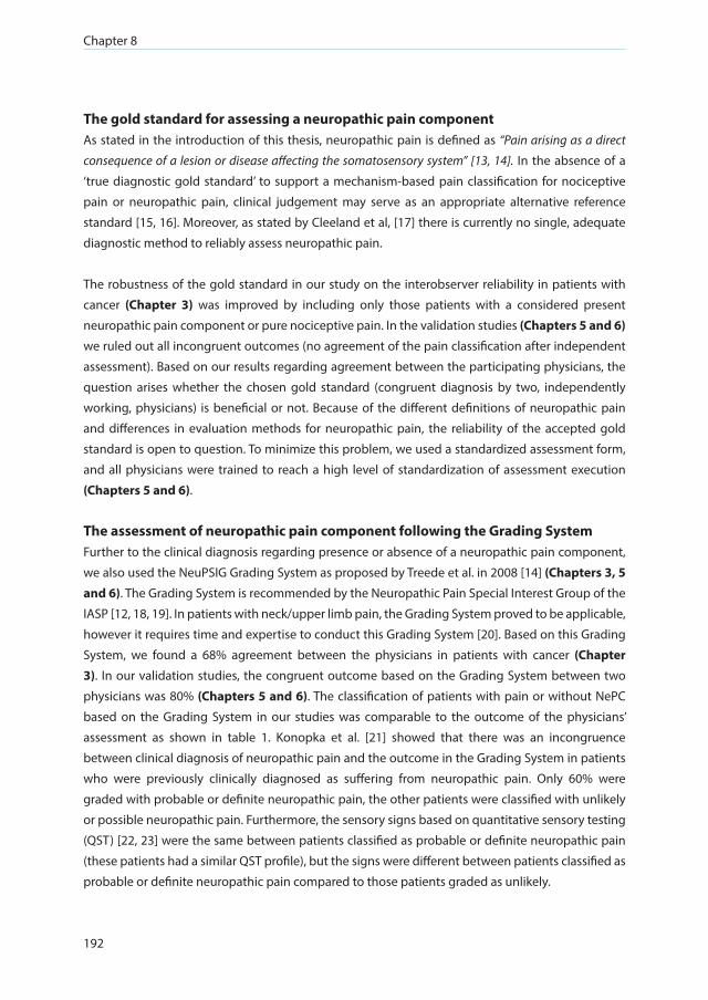
Chapter 8
192
The gold standard for assessing a neuropathic pain componentAs stated in the introduction of this thesis, neuropathic pain is defined as “Pain arising as a direct
consequence of a lesion or disease affecting the somatosensory system” [13, 14]. In the absence of a
‘true diagnostic gold standard’ to support a mechanism-based pain classification for nociceptive
pain or neuropathic pain, clinical judgement may serve as an appropriate alternative reference
standard [15, 16]. Moreover, as stated by Cleeland et al, [17] there is currently no single, adequate
diagnostic method to reliably assess neuropathic pain.
The robustness of the gold standard in our study on the interobserver reliability in patients with
cancer (Chapter 3) was improved by including only those patients with a considered present
neuropathic pain component or pure nociceptive pain. In the validation studies (Chapters 5 and 6)
we ruled out all incongruent outcomes (no agreement of the pain classification after independent
assessment). Based on our results regarding agreement between the participating physicians, the
question arises whether the chosen gold standard (congruent diagnosis by two, independently
working, physicians) is beneficial or not. Because of the different definitions of neuropathic pain
and differences in evaluation methods for neuropathic pain, the reliability of the accepted gold
standard is open to question. To minimize this problem, we used a standardized assessment form,
and all physicians were trained to reach a high level of standardization of assessment execution
(Chapters 5 and 6).
The assessment of neuropathic pain component following the Grading System Further to the clinical diagnosis regarding presence or absence of a neuropathic pain component,
we also used the NeuPSIG Grading System as proposed by Treede et al. in 2008 [14] (Chapters 3, 5
and 6). The Grading System is recommended by the Neuropathic Pain Special Interest Group of the
IASP [12, 18, 19]. In patients with neck/upper limb pain, the Grading System proved to be applicable,
however it requires time and expertise to conduct this Grading System [20]. Based on this Grading
System, we found a 68% agreement between the physicians in patients with cancer (Chapter
3). In our validation studies, the congruent outcome based on the Grading System between two
physicians was 80% (Chapters 5 and 6). The classification of patients with pain or without NePC
based on the Grading System in our studies was comparable to the outcome of the physicians’
assessment as shown in table 1. Konopka et al. [21] showed that there was an incongruence
between clinical diagnosis of neuropathic pain and the outcome in the Grading System in patients
who were previously clinically diagnosed as suffering from neuropathic pain. Only 60% were
graded with probable or definite neuropathic pain, the other patients were classified with unlikely
or possible neuropathic pain. Furthermore, the sensory signs based on quantitative sensory testing
(QST) [22, 23] were the same between patients classified as probable or definite neuropathic pain
(these patients had a similar QST profile), but the signs were different between patients classified as
probable or definite neuropathic pain compared to those patients graded as unlikely.
Conclusions, discussion and recommendations
193
8
The NeuPSIG Grading System was updated in 2016 by Finnerup et al. [3] (see figure 2). Several
adjustments were made to better reflect daily clinical practice. The most important adjustment to
the updated Grading System is the addition that even ‘definite neuropathic pain’ does not mean
there is a causality [3]. ‘Definite neuropathic pain’ only refers to the fact that a physician via history
taking, physical examination, and supplementary testing is able to clinically confirm that the patient
has a neurological lesion that might explain a patient’s pain [3]. In line with this, it is always important
to consider whether other causes of patients pain may be present. This confirms the observation
that currently, there is no true gold standard to diagnose neuropathic pain.
Figure 2. Flowchart of the updated Grading System as proposed in 2016. Adapted from Finnerup et al., Pain, 2016 [3].
a, pain suspected to be related to a lesion or disease of the somatosensory system which is associated with neuropathic pain. b, the pain distribution as described by the patient is in line with the assumed lesion or disease. c, the area of sensory signs is in the same neuroanatomically plausible distribution. d, when the location and nature of the lesion or disease are able to explain patients’ pain, probable neuropathic pain confirmed by confirmatory tests is called definite neuropathic pain.
Chapter 8
194
These issues raise questions about the different causes of patients‘ neuropathic pain component
and the manifestation of the signs and symptoms: is the clinical picture of the neuropathic pain
component following a nerve injury due to surgery (for example pain due to amputation or (major)
nerve lesion) the same as a radicular pain syndrome due to a herniated disc? Are the signs and
symptoms of a patient with trigeminal neuralgia the same as in the glove and sock distribution in
patients with chemotherapy-induced neuropathy? It seems clear that this is not the case. Moreover,
the classification based on the Grading System, screening tools like the DN4 or the PainDETECT,
and quantitative sensory testing via bedside examination or the Nijmegen-Aalborg Screening QST,
should be viewed individually. For example, in the original articles on PainDETECT (2006) [5] and
DN4 (2005) [11], patients with osteoarthritis were viewed as patients with pain of predominantly
nociceptive origin, thus they were included in the validation studies as no NePC. Recent studies
show that patients with pain resulting from osteoarthritis in which neuropathic pain features (based
on neuropathic pain screening tool outcome or pathophysiologic mechanisms) are present, may
thus require a different treatment paradigm than patients with osteoarthritis without NePC [24-
27]. However, they also confuse the use of screening tools because the validity in patients with
a different diagnosis may be challenged. This shows that, in neuropathic pain classification, the
patient, the disease as such, and the manifestation of the disease, must all be taken into account
when patients are assessed as an individual, unique, patient, and that a tailor-made treatment
regimen should be proposed.
Based on current insights, a surrogate gold standard, two independently working, well-trained,
experienced and blinded professionals as used in our studies can be improved by narrowing the
clinical consensus on signs and symptom. Moreover, meticulously following the guidelines leads
to an improved use of the Grading System. Bedside examination and quantitative sensory testing,
testing the descending pathways (pain modulation systems) and, for example, neuro-imaging may
also be of value when looking for ways to improve the diagnostic gold standard for the assessment
of a neuropathic pain component in patients with pain [28, 29].
Answer to question 3: The DN4, but not the PainDETECT, is of value in the screening for a neuropathic pain componentIn their original study, Freynhagen et al. revealed a sensitivity of 85% and a specificity of 80% for
the PainDETECT [5]. The DN4 developed by Bouhassira et al. [11] showed a sensitivity of 83% and a
specificity of 90%. For the DN4-symptoms, interview only, sensitivity was 78% and specificity 81%.
In our studies of comparisons with the physicians’ assessment as the “gold standard”, we conclude
that the cross-culturally adapted PainDETECT (sensitivity of 80% and specificity of 55%, figure
3a) is not an effective screening tool because of its moderate sensitivity and low specificity in a
consecutive population of patients with low back and leg pain, neck-shoulder pain, or with pain
due to a suspected peripheral nerve damage (Chapter 5). We tested the DN4 in the same group
of patients as the PainDETECT; this appeared to be moderately helpful in identifying a neuropathic
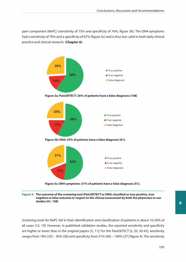
Conclusions, discussion and recommendations
195
8
pain component (NePC) (sensitivity of 75% and specificity of 76%, figure 3b). The DN4-symptoms
had a sensitivity of 70% and a specificity of 67% (figure 3c) and is thus less valid in both daily clinical
practice and clinical research. (Chapter 6).
True positive
True negative
False diagnosis
26%
60%
14%
True positive
True negative
False diagnosis
25%
58% 17%
True positive
True negative
False diagnosis
31%
52%
17%
Figure 3a: PainDETECT: 26% of patients have a false diagnosis [108]
Figure 3b: DN4: 25% of patients have a false diagnosis [61].
Figure 3c: DN4 symptoms: 31% of patients have a false diagnosis [61].
Figure 3: The outcome of the screening tool (PainDETECT or DN4) classified as true positive, true negative or false outcome in respect to the clinical assessment by both the physicians in our studies [61, 108]
Screening tools for NePC fail in their identification and classification of patients in about 10-20% of
all cases [12, 19]. However, in published validation studies, the reported sensitivity and specificity
are higher or lower than in the original papers [5, 11]: For the PainDETECT [5, 20, 30-42], sensitivity
ranges from 18% [35] – 95% [36] and specificity from 51% [40] – 100% [37] (figure 4). The sensitivity

Chapter 8
196
0
10
20
30
40
50
60
70
80
90
100
0 10 20 30 40 50 60 70 80 90 100
Spe
cific
ity (%
)
Sensitivity (%)
Figure 4: Comparing the sensitivity and specificity of the different validation studies as performed for the PainDETECT
The red dot represents the validation study presented in this thesis [108]; Blue squares represent the individual papers published by different research groups [5, 20, 30-38, 40, 42]
0
10
20
30
40
50
60
70
80
90
100
0 10 20 30 40 50 60 70 80 90 100
Spe
cific
ity (%
)
Sensitivity (%)
Figure 5: Comparing the sensitivity and specificity of the different validation studies as performed for the DN4
The red dot represents the validation study presented in this thesis [61]. Blue squares represent the individual papers published by different research groups [11, 30, 35, 37, 40, 43-60, 62, 64, 109]
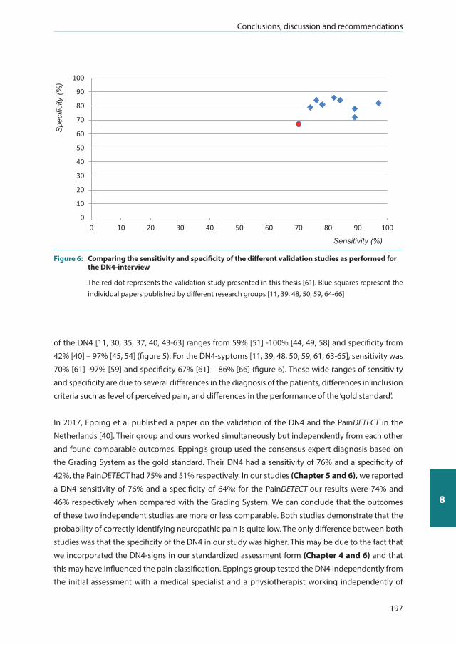
Conclusions, discussion and recommendations
197
8
0
10
20
30
40
50
60
70
80
90
100
0 10 20 30 40 50 60 70 80 90 100
Sensitivity (%)
Spe
cific
ity (%
)
Figure 6: Comparing the sensitivity and specificity of the different validation studies as performed for the DN4-interview
The red dot represents the validation study presented in this thesis [61]. Blue squares represent the individual papers published by different research groups [11, 39, 48, 50, 59, 64-66]
of the DN4 [11, 30, 35, 37, 40, 43-63] ranges from 59% [51] -100% [44, 49, 58] and specificity from
42% [40] – 97% [45, 54] (figure 5). For the DN4-syptoms [11, 39, 48, 50, 59, 61, 63-65], sensitivity was
70% [61] -97% [59] and specificity 67% [61] – 86% [66] (figure 6). These wide ranges of sensitivity
and specificity are due to several differences in the diagnosis of the patients, differences in inclusion
criteria such as level of perceived pain, and differences in the performance of the ‘gold standard’.
In 2017, Epping et al published a paper on the validation of the DN4 and the PainDETECT in the
Netherlands [40]. Their group and ours worked simultaneously but independently from each other
and found comparable outcomes. Epping’s group used the consensus expert diagnosis based on
the Grading System as the gold standard. Their DN4 had a sensitivity of 76% and a specificity of
42%, the PainDETECT had 75% and 51% respectively. In our studies (Chapter 5 and 6), we reported
a DN4 sensitivity of 76% and a specificity of 64%; for the PainDETECT our results were 74% and
46% respectively when compared with the Grading System. We can conclude that the outcomes
of these two independent studies are more or less comparable. Both studies demonstrate that the
probability of correctly identifying neuropathic pain is quite low. The only difference between both
studies was that the specificity of the DN4 in our study was higher. This may be due to the fact that
we incorporated the DN4-signs in our standardized assessment form (Chapter 4 and 6) and that
this may have influenced the pain classification. Epping’s group tested the DN4 independently from
the initial assessment with a medical specialist and a physiotherapist working independently of
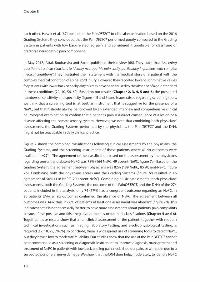
Chapter 8
198
each other. Hasvik et al. [67] compared the PainDETECT to clinical examination based on the 2016
Grading System; they concluded that the PainDETECT performed poorly compared to the Grading
System in patients with low back-related leg pain, and considered it unreliable for classifying or
grading a neuropathic pain component.
In May 2018, Attal, Bouhassira and Baron published their review [68]. They state that “screening
questionnaires help clinicians to identify neuropathic pain easily, particularly in patients with complex
medical conditions”. They illustrated their statement with the medical story of a patient with the
complex medical condition of spinal cord injury. However, they reported lower discriminative values
for patients with lower back or neck pain; this may have been caused by the absence of a gold standard
in these conditions [20, 40, 56, 69]. Based on our results (Chapter 2, 3, 4, 5 and 6) the presented
numbers of sensitivity and specificity (figure 4, 5 and 6) and issues raised regarding screening tools,
we think that a screening tool is, at best, an instrument that is suggestive for the presence of a
NePC, but that it should always be followed by an extended interview and comprehensive clinical
neurological examination to confirm that a patient’s pain is a direct consequence of a lesion or a
disease affecting the somatosensory system. However, we note that combining both physicians’
assessments, the Grading Systems performed by the physicians, the PainDETECT and the DN4,
might not be practicable in daily clinical practice.
Figure 7 shows the combined classifications following clinical assessments by the physicians, the
Grading Systems, and the screening instruments of those patients where all six outcomes were
available (n=274). The agreement of the classification based on the assessment by the physicians
regarding present and absent-NePC was 78% (164 NePC, 49 absent-NePC, figure 7a). Based on the
Grading System, the agreement between physicians was 82% (139 NePC, 85 Absent-NePC, figure
7b). Combining both the physicians scores and the Grading Systems (figure 7c) resulted in an
agreement of 50% (118 NePC, 20 absent-NePC). Combining all six assessments (both physicians’
assessments, both the Grading Systems, the outcome of the PainDETECT, and the DN4) of the 274
patients included in the analysis, only 74 (27%) had a congruent outcome regarding an NePC. In
20 patients (7%), all six outcomes confirmed the absence of NEPC. The agreement between all
outcomes was 34%; thus in 66% of patients at least one assessment was aberrant (figure 7d). This
indicates that it is not necessarily ‘better’ to have more assessments about patients’ pain complaints
because false-positive and false-negative outcomes occur in all classifications (Chapter 5 and 6).
Together, these results show that a full clinical assessment of the patient, together with modern
technical investigations such as imaging, laboratory testing, and electrophysiological testing, is
required [17, 18, 29, 70-76]. To conclude, there is widespread use of screening tools to detect NePC,
but they have a low to moderate reliability. Our studies show that the use of the PainDETECT cannot
be recommended as a screening or diagnostic instrument to improve diagnosis, management and
treatment of NePC in patients with low back and leg pain, neck-shoulder pain, or with pain due to a
suspected peripheral nerve damage. We show that the DN4 does help, moderately, to identify NePC
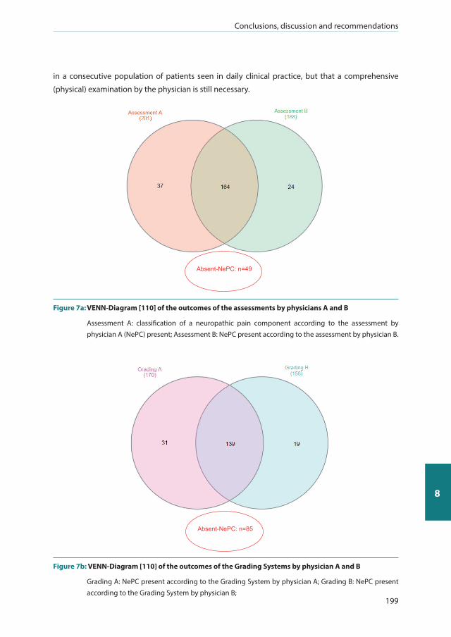
Conclusions, discussion and recommendations
199
8
in a consecutive population of patients seen in daily clinical practice, but that a comprehensive
(physical) examination by the physician is still necessary.
Absent-NePC: n=49
Figure 7a: VENN-Diagram [110] of the outcomes of the assessments by physicians A and B
Assessment A: classification of a neuropathic pain component according to the assessment by physician A (NePC) present; Assessment B: NePC present according to the assessment by physician B.
Absent-NePC: n=85
Figure 7b: VENN-Diagram [110] of the outcomes of the Grading Systems by physician A and B
Grading A: NePC present according to the Grading System by physician A; Grading B: NePC present according to the Grading System by physician B;
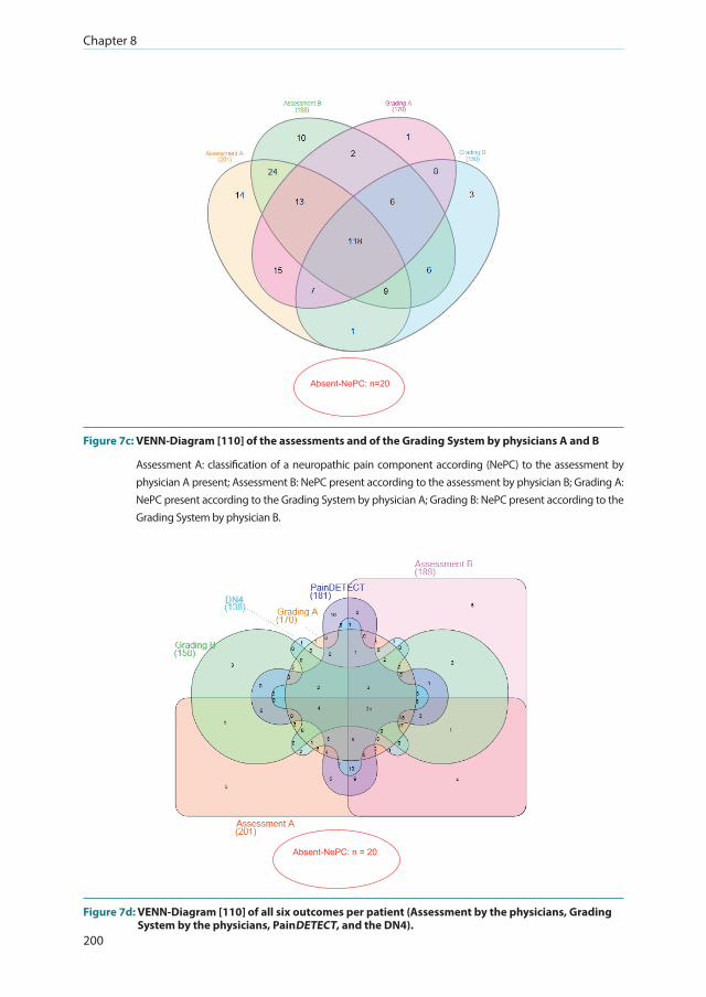
Chapter 8
200
Absent-NePC: n=20
Figure 7c: VENN-Diagram [110] of the assessments and of the Grading System by physicians A and B
Assessment A: classification of a neuropathic pain component according (NePC) to the assessment by physician A present; Assessment B: NePC present according to the assessment by physician B; Grading A: NePC present according to the Grading System by physician A; Grading B: NePC present according to the Grading System by physician B.
Absent-NePC: n = 20
Figure 7d: VENN-Diagram [110] of all six outcomes per patient (Assessment by the physicians, Grading System by the physicians, PainDETECT, and the DN4).

Conclusions, discussion and recommendations
201
8
Assessment A: classification of a neuropathic pain component (NePC) according to the assessment by physician A present; Assessment B: NePC present according to assessment by physician B; Grading A: NePC present according to the Grading System by physician A; Grading B: NePC present according to the Grading System by physician B; PainDETECT: outcome of the PainDETECT indicates the presence of NePC. Absent-NePC: No NePC present according to the physicians, the Grading Systems, the PainDETECT and the DN4.
Answer to question 4: Bed-side examination, but not NASQ, has a limited value in the classification of NePCIn Chapter 7, we discuss the value of bedside examination (BSE) and screening quantitative sensory
testing following the Nijmegen-Aalborg Screening Quantitative Sensory Testing protocol (NASQ).
We show that BSE revealed small differences between patients with either absent or present NePC,
whereas NASQ did not reveal any differences (chapter 7).
Bedside examination (BSE) is recommended as a method to identify positive and negative sensory
signs in several guidelines for assessing neuropathic pain components [16, 18, 29, 70-74, 76-79].
Clinical examination can never prove that pain is of a neuropathic origin, but it can indicate an
altered function of patients’ nervous system [18]. BSE can also indicate the presence of other
pathological processes which may cause pain. As a supplement to neurophysiological testing, BSE
can answer the question to where in the somatosensory system the lesion or disease that generates
neuropathic pain can be found [80]. Moreover, the classification of clinical symptoms in patients
with NePC based on a mechanism-based approach may be useful to quantify sensory signs [81]. As
stated by Garcia-Larrea [79], neuropathic pain is especially associated with lesions of temperature
and pain pathways (A-delta and C-fibers, spino-thalamo-cortical tracts). We demonstrated (chapter
7) that heat sensation, cold sensation, wind-up response, and pricking was less present in patients
with NePC.
Unfortunately, BSE data from other studies are scarce. Most published studies used the quantitative
sensory testing protocol (QST) following the German Research Network on Neuropathic Pain (DFNS)
[22, 23, 82]. Comparison of BSE results with QST following the DFNS protocol is questionable,
because the DFNS uses a very strict protocol which is only appropriate for trained persons using a
standardized protocol in certified clinical neurological/ neurophysiological laboratories [83]. As far
as we are aware, this is not the case when performing BSE tests to find support for the presence of
neuropathic pain. However, there is a great emphasis on BSE in the diagnostic work-up of patients
with pain in daily clinical practice. Unfortunately, very little has been published on BSE data for
groups of patients and for individual patient outcomes alongside the clinical classification of
patients’ pain.
Chapter 8
202
Spahr et al [84] stated that the methods used in clinical examination and the paradigms used to
detect NePC may fail in patients with low back pain (LBP). Based on tactile threshold discrimination
and 2-point discrimination (both methods were not used in our study, Chapter 7), they found that
patients with chronic low back pain had an increased tactile threshold and 2-point discrimination
when compared to healthy controls. In the comparison between patients with chronic low back pain
with and without NePC, patients with NePC had an increased tactile threshold in comparison with
those without. This suggests that there are differences in the clinical profiles of patients with LBP with
and without NePC (assessment of symptom profiles instead of evidence for a direct consequence of
a lesion or a disease affecting the somatosensory system), and that the symptoms might origin from
an underlying maladaptive plasticity of the nervous system [84]. Leffler and Hansson [85] compared
the outcome of clinical BSE to the outcome of standardized QST in a research setting in patients
with a painful traumatic peripheral nerve injury. They concluded that the individual outcome of
BSE compared to standardized QST outcome frequently differed in patients. This may be due to
differences in, for example, performance of the applied stimuli. This was most frequently observed
in the assessment for sensibility for touch. The differences may be explained by looking at what can
be considered as a ‘normal’ outcome and what a ‘pathological’ outcome. Moreover, the variety in
methodology (standardization, equipment) and conduction (force, position etc.) of the given test
stimuli may have contributed to differences between BSE and QST. In conclusion, sensory profiling
of patients with NePC based on QST may result in a more stratified and personalized treatment
regimen [86, 87], but QST is not directly interchangeable with BSE, as explained above [85].
NASQ is intended to assess the processing of pain in patients [88-90]. Our intention before starting
this study was to identify whether the assessment of NASQ benefited the classification of pain
by, for example, identifying peripheral or central sensitization. However, the Nijmegen-Aalborg
Screening QST (NASQ) protocol showed no association with neuropathic pain. We found no signs
of central sensitization (widespread hyperalgesia, descending inhibition [91]) in patients with and
without NePC included in our study. This contrasts with findings by Freynhagen et al [92] and Woolf
et al [93] who suggest that central sensitization is most manifest in patients with NePC. It is still
unclear whether this is due to the fact that our patients had no central sensitization, because the
test methodology was not refined enough to detect central sensitization, or otherwise. Granovsky
[94] stated that patients with NePC expressed less efficient CPM compared those without. However,
we found no differences in the CPM (Chapter 7). The differences between our results and the
literature may be due to several facts such as the low number of patients included and no inclusion
of matched, healthy controls. However, another reason may have been the forced separation of
patients with pain into either nociceptive or neuropathic pain. With today’s knowledge, our studies
would possibly have had the benefit of the recently proposed threefold separation [95], however
the term nociplastic pain did not exist when we started our studies. In patients with nociplastic pain,
altered nociceptive functions (hypersensitivity), with a regional or widespread pain distribution do
exist [95]. According to the IASP, central sensitization is “an increased response and reduced threshold
Conclusions, discussion and recommendations
203
8
of nociceptive neurons in the central nervous system to their normal or subthreshold afferent input
“ [96]. The underlying mechanism of nociplastic pain is probably the central sensitization of the
nociceptive pathways [95].
RECOMMENDATIONS FOR CLINICAL PRACTICE, EDUCATION, FUTURE RESEARCH AND SOCIETAL IMPACT
From the results of our research and the discussion on each of the four research questions, we have
formulated a number of specific recommendations that we trust will lead to improvements in the
diagnosis and treatment of pain for the patients and for the specialists involved in their care,
RECOMMENDATIONS FOR THE CLASSIFICATION OF PAIN IN DAILY CLINICAL PRACTICE
We recommend that the classification of a neuropathic pain component should be based on history taking, physical (neurological) examination, bedside examination, and up-to-date diagnostic testing.Our studies show that it is extremely important to pay attention to a clearer identification of
patients with pain, and to correctly classify them in terms of suffering from nociceptive, neuropathic
or nociplastic pain components or a combination of them; we have shown that their treatment
response may be dependent on it.
The classification of neuropathic pain in daily clinical practice should be based on accurate
(neurological) history taking and performing a neurological examination. History taking reveals the
characteristics and distribution of a patient’s pain. The clinical examination also reveals possible
negative and positive sensory signs and their association with the underlying lesion or disease. This
can be performed via a standard neurological examination combined with a bedside examination.
Finally, a diagnostic test such as R3 reflex testing, cutaneous response reduction (skin wrinkle test)
test, nerve ultrasound, quantitative sudomotor axon reflex testing, quantitative sensory testing,
laser-evoked potentials, pin-prick evoked potentials, electrophysiological testing, fMRI and skin
biopsy may all help to confirm the lesion or disease in the somatosensory system which explains the
pain [3, 71, 79]. However, as discussed, a factual relationship based on diagnostic testing between
nerve damage and a patient’s pain has not yet been verified but the search for this connection
remains of great importance for improving patient treatment [71] (Chapters 3,4,5,6 and 7).

Chapter 8
204
We recommend that pain classification should be standardized in daily clinical practice as this will lead to new insights and improve patient outcomes. By examining the patient in a standardized but individual way (history taking, physical examination,
bedside examination and, when possible, confirmatory test procedure(s)), the clinician will be better
able to classify a patient’s pain based on the clinical criteria and thus to design a personalized pain
treatment program.
The Grading System is intended to be used in individual patients in daily clinical practice as well
as in clinical research [3]. One advantage of this system [3, 14] is to help in the standardized
classification of neuropathic pain. By using the Grading System methodology when examining a
patient, it is possible to grade the certainty of NePC existence on an ordinal scale: no neuropathic
pain, possible neuropathic pain, probable neuropathic pain, and definite neuropathic pain [3].
However, currently the Grading System cannot be seen as a ‘gold standard’ for neuropathic pain; it is
a clinical assessment guideline which assists in classifying a patient’s pain. An outcome of probable
neuropathic pain in a patient should be reason for the physician to consider initiating treatment
according to the guidelines about treating patients with neuropathic pain [97]. To use and to rely
on the Grading System, it is important for the physicians to be experienced, and that they have the
skills and resources to assess their patient in the best possible way [3]. (Chapters 3, 4, 5 and 6).
We recommend that screening questionnaires should be used with caution, when assessing a neuropathic pain component in a daily clinical consecutive population of patients with chronic pain.Screening tools like the DN4 or PainDETECT only provide a slight indication of the presence of NePC;
they may therefore lead to misclassifying a patient’s pain.
Our studies show that false identification with respect to the presence or absence of NePC may
be the case in a substantial number of patients. It can be concluded that a clinical examination of
the patient is a prerequisite; a screening tool is not a diagnostic test and can thus never replace
a physician’s examination[12]. Moreover, it is always true that “a patient” is much more than the
outcome of the questionnaire(s), and that a patient’s pain is whatever the patient says it is (Chapters
5 and 6).
We recommend that a bedside examination forms a part of the diagnostic work-up of patients’ pain.After history taking, bedside examination is of second-most importance when examining the
(cause or mechanism) of the patient’s pain. It takes time to examine the patient and the sensory
disturbances should be noted on a body map.
Conclusions, discussion and recommendations
205
8
Bedside examination will provide the physician with an overview of a patient’s pain and may better
support a pain diagnosis and thus be valuable when monitoring a patient’s pain and treatment
effects. BSE should be performed in a standardized way, guided by clear instructions to the patient.
Our results suggest a low association between BSE and classification of a patient’s pain, however
it may assist the so-called ‘fingerspitzengefühl’ with which physicians classify patients’ pain in daily
clinical (primary) care as pain with or without a neuropathic component (Chapter 5,6 and 7).
We recommend that combining pain classification strategies to become ‘more sure’ of a patient’s pain classification is not standard daily clinical practice.Our studies show that there is no proof that it is ‘better’ to have more assessments about a patient’s
pain complaints, as false-positive and false-negative outcomes occur in all classifications (Chapters
5 and 6).
Running more and different clinical assessments about the type of a patient’s pain may not be
beneficial for the patient; it may even harm the patient due to, for example, delay of treatment. As
Goscinny and Uderzo stated in their inimitable way: “…appelle plutôt des medicines!…Je fais venir
tous les medicines de la garnison!.... Tu ne crains pas l’intervention des medicines…? En groupe ils sont
plus meurtriers qu’une legion armée jusqu-aux dents!” [98].
RECOMMENDATIONS FOR IMPROVING PHYSICIAN EDUCATION AND TRAINING FOR CLASSIFYING PATIENTS’ PAIN
We recommend that more attention should be paid to patients with pain, and to pain as a disease in its own rightBased on the epidemiology of pain, the reduced health related quality of life, lower functional
status, and lower mental health, in combination with the high direct and indirect costs associated
with patients with chronic pain, it is of utmost importance to create awareness about this major
personal and socioeconomic problem.
Each patient has his or her own ‘pain’, and that makes patients experts in their own pain based
on personal memory, emotions, cognitive factors, and pathology (Chapter 1). Currently, pain is
undervalued in daily clinical practice [99]. It is therefore important to pay greater attention to a
patient’s pain and to make greater financial resources available to fund scientific research in the
field of pain to identify potential mechanisms and to examine treatment effects. The first step in
this might be by raising the general public’s awareness of the epidemiology, burden, costs and
consequences of pain via newspapers, radio, television, and participation in research as a (healthy)
volunteer etc. (Chapter 1).
Chapter 8
206
We recommend that more education in the use of screening- and diagnostic instruments should be provided at an undergraduate, graduate and postgraduate level for everyone working with people with pain To assess pain, physicians should use a biopsychological model to address the somatic, cognitive,
emotional, behavioral, spiritual and social dimensions. Munrinson et al. [100] studied students in the
preclinical phase and concluded that the integration of the cognitive and the affective dimension is
related to the basic knowledge about emotional development and is associated with a high degree
of student satisfaction. Moreover, physicians should address pain with an integrated view of patho-
physiology and clinical care, but this is only possible if this is based on a long term vision for pain
education, wherein the patient plays a central role [100]. Another aspect of education is improving
learning about and using guidelines. Clinical practice guidelines are not always exactly followed by
the physician due to a number of factors like a lack of awareness, a lack of agreement, and a lack of
outcome expectancy [101].
Education in the field of pain should be, amongst others, targeted towards identifying symptoms
and signs, improving knowledge about screening, and assessing patients with pain (via, amongst
others, screening tools as the PainDETECT and the DN4 and the use of the Grading System). A
proposed pain medicine model developed by Wolff and Groen (APC, UMCG; 2016) includes different
steps in the diagnostic work-up of the patient to come to personalized pain treatment:1) The pain
complaint (patient history, body examination, additional examination); 2) supposed (anatomical)
substrate; 3) pathophysiology; 4) pain mechanisms / pain diagnosis; 5) bio-psycho-social-spiritual; 6)
conclusion; 7) management; and 8) repetition. Based on the statement “pain is whatever the patient
says it is, existing whenever he/she says it does” [102] I would suggest moving the Bio-Psycho-Social-
Spiritual part of the model forward to second position, as this element has important consequences
for patients with pain and leads to their daily-living limitations. Improving this knowledge may have
consequences for therapy regimes, amongst other factors. Additionally, it is important to implement
evaluation methods to measure and optimize treatment efficacy. Moreover, better training in
how to educate patients about their disease, master coping skills, and improving knowledge of
available self-care options may be beneficial [103]. Under ideal conditions this should be done in
a continuous learning environment combined with the possibility to quickly react to new insights
and for example, changes in treatment opportunities.
We recommend improving the knowledge and expectations about the psychometric values of evaluation / screening tools of the person interpreting it.Based on our results (Chapters 5, 6 and 7) and the other studies about the validity of screening
tools for assessing neuropathic pain, it is important for those interpreting the results to have a better
knowledge of the tool’s correct use and an understanding of its psychometric values. Each tool,
instrument or test results in false positive and/or false negative results. If treating physicians are
Conclusions, discussion and recommendations
207
8
aware of these, they can be taken into account when evaluating the outcome of the instrument in
question.
RECOMMENDATIONS FOR THE IMPROVEMENT OF FUTURE RESEARCH
We recommend that patients with neuropathic pain should be treated more optimally; we have to be able to better identify crucial pain markers.Neuropathic pain, as far as we can identify it, is characterized by a poor response to treatment
with medication. The conversion rate of medication needed to treat for 50% pain relief differs
greatly: 3.6 for tricyclic antidepressants, 6.4 for serotonin-noradrenaline reuptake inhibitor, 7.7 for
pregabalin, and 6.3 for gabapentin [97]. We should aim for a therapy that has a higher conversion
rate by gaining a better understanding of pain mechanisms and being better able to assess these
mechanisms. Unfortunately, a ‘one fits all’ treatment regime does not fit the needs of an individual
patient with pain. To further develop precise and personalized treatment, we need to develop a
more detailed assessment strategy and matching treatment regimens. To find a real ‘gold standard’
for the diagnosis of NePC would be extremely valuable, however this is not expected in the short
term. We recommend that more research should be conducted on the recently updated Grading
System and the implementation of nociplastic pain as a third mechanistic descriptor, both in the
laboratory and, in particular in daily clinical practice.
Collecting data (signs, symptoms, outcome measures) may influence the individual physician’s
clinical practice based on experience (pattern recognition), as well as research. ‘Big data’ may enable
us to recognize patterns in individual patients or in groups of patients, even where there is a great
deal of variability in individual patient outcomes (Chapters 3, 4, 5, 6 and 7).
We do not recommend NASQ when differentiating between nociceptive pain and the existence of a neuropathic pain component; this needs further evaluation to determine the mechanisms of nociplastic painThe acknowledgement of the third mechanism-based descriptor of pain, nociplastic pain, by the
International Association for the Study of Pain (IASP) has led to new opportunities for the use of
NASQ. To better differentiate between pain mechanisms, a phenomenon as central sensitization
might be accessed via the measurement of pain thresholds and/or changes in the endogenous pain
system. NASQ should be further developed and more research should be conducted to see whether
NASQ offers benefits for the assessment of patients with pain (Chapter 7).
Chapter 8
208
We recommend that the screening tools used when assessing a neuropathic pain component should have a very high sensitivity, specificity and positive likelihood ratio.A screening tool to detect neuropathic pain should have a high sensitivity to identify the patients
with NePC; they should also have a high specificity to recognize patients without NePC. Moreover,
the predictive value must be high to ensure that a positive test indicates the presence of NePC.
However, the likelihood ratio should also be given in the result sections of research papers as this
indicates how much more likely a positive test will be present in a patient with NePC compared
to a patient without NePC. Finally, when reporting on the validity of a screening tool, test-retest
reliability should also be stated in the paper [104-106] (Chapters 5 and 6).
RECOMMENDATIONS FOR IMPROVING THE ORGANIZATION OF PAIN CARE AND SOCIETAL IMPACT
We recommend that patients should be better identified and stratified; this will lead to a better organization and the health-economic optimization of and inclusion of patients in specialized treatment programs.In the future, improved identification and stratification of patients will lead to, in addition to greater
satisfaction about treatment, a positive effect on the health economy: reducing the costs of medical
care. Of even greater importance is the improvement in health care and a decrease in the burden of
chronic pain disease. Moreover, improvements in daily clinical practice will facilitate a faster return
to work, less direct costs, less indirect costs, better education in (para-) medical schools, and a better
stepped wedge care.
Health and healthy behavior are important issues for the near future; the World Health Organization
emphasize the need, “to promote health and development, and prevent or reduce risk factors for health
conditions associated with use of tobacco, alcohol, drugs and other psychoactive substances, unhealthy
diets, physical inactivity and unsafe sex” [107]. Education, self-management, and physical activity are
issues that should be further developed by, amongst others, pain physicians and researchers in the
field of pain, but always together with patients with pain.
Conclusions, discussion and recommendations
209
8
REFERENCES
1. Cohen SP, Mao J: Neuropathic pain: mechanisms and their clinical implications. BMJ 2014, 348:f7656.2. Magrinelli F, Zanette G, Tamburin S: Neuropathic pain: diagnosis and treatment. Pract Neurol 2013,
13(5):292-307.3. Finnerup NB, Haroutounian S, Kamerman P, Baron R, Bennett DL, Bouhassira D, Cruccu G, Freeman R,
Hansson P, Nurmikko T et al: Neuropathic pain: an updated grading system for research and clinical practice. Pain 2016.
4. DiBonaventura MD, Sadosky A, Concialdi K, Hopps M, Kudel I, Parsons B, Cappelleri JC, Hlavacek P, Alexander AH, Stacey BR et al: The prevalence of probable neuropathic pain in the US: results from a multimodal general-population health survey. Journal of pain research 2017, 10:2525-2538.
5. Freynhagen R, Baron R, Gockel U, Tolle TR: painDETECT: a new screening questionnaire to identify neuropathic components in patients with back pain. Curr Med Res Opin 2006, 22(10):1911-1920.
6. Tampin B, Bohne T, Callan M, Kvia M, Melsom Myhre A, Neoh EC, Bharat C, Slater H: Reliability of the English version of the painDETECT questionnaire. Curr Med Res Opin 2017, 33(4):741-748.
7. Wild D, Grove A, Martin M, Eremenco S, McElroy S, Verjee-Lorenz A, Erikson P, Translation ITFf, Cultural A: Principles of Good Practice for the Translation and Cultural Adaptation Process for Patient-Reported Outcomes (PRO) Measures: report of the ISPOR Task Force for Translation and Cultural Adaptation. Value in health : the journal of the International Society for Pharmacoeconomics and Outcomes Research 2005, 8(2):94-104.
8. Beaton DE, Bombardier C, Guillemin F, Ferraz MB: Guidelines for the process of cross-cultural adaptation of self-report measures. Spine 2000, 25(24):3186-3191.
9. Guillemin F, Bombardier C, Beaton D: Cross-cultural adaptation of health-related quality of life measures: literature review and proposed guidelines. Journal of clinical epidemiology 1993, 46(12):1417-1432.
10. Landis JR, Koch GG: The measurement of observer agreement for categorical data. Biometrics 1977, 33(1):159-174.
11. Bouhassira D, Attal N, Alchaar H, Boureau F, Brochet B, Bruxelle J, Cunin G, Fermanian J, Ginies P, Grun-Overdyking A et al: Comparison of pain syndromes associated with nervous or somatic lesions and development of a new neuropathic pain diagnostic questionnaire (DN4). Pain 2005, 114(1-2):29-36.
12. Mathieson S, Maher CG, Terwee CB, Folly de Campos T, Lin CW: Neuropathic pain screening questionnaires have limited measurement properties. A systematic review. Journal of clinical epidemiology 2015, 68(8):957-966.
13. IASP Taxonomy Neuropathic Pain [http://www.iasp-pain.org/Taxonomy#Neuropathicpain]14. Treede RD, Jensen TS, Campbell JN, Cruccu G, Dostrovsky JO, Griffin JW, Hansson P, Hughes R, Nurmikko T,
Serra J: Neuropathic pain: redefinition and a grading system for clinical and research purposes. Neurology 2008, 70(18):1630-1635.
15. Katz JN, Stock SR, Evanoff BA, Rempel D, Moore JS, Franzblau A, Gray RH: Classification criteria and severity assessment in work-associated upper extremity disorders: methods matter. Am J Ind Med 2000, 38(4):369-372.
16. Smart KM, Blake C, Staines A, Doody C: The Discriminative validity of “nociceptive,” “peripheral neuropathic,” and “central sensitization” as mechanisms-based classifications of musculoskeletal pain. The Clinical journal of pain 2011, 27(8):655-663.
17. Cleeland CS, Farrar JT, Hausheer FH: Assessment of cancer-related neuropathy and neuropathic pain. Oncologist 2010, 15 Suppl 2:13-18.
18. Haanpaa M, Attal N, Backonja M, Baron R, Bennett M, Bouhassira D, Cruccu G, Hansson P, Haythornthwaite JA, Iannetti GD et al: NeuPSIG guidelines on neuropathic pain assessment. Pain 2011, 152(1):14-27.
19. CADTH: Diagnostic methods for neuropathic pain: A review of diagnostic accuracy. In: Canadian Agency for Drugs and Technologies in Health. Ottawa (ON), Canada; 2015.
20. Tampin B, Briffa NK, Goucke R, Slater H: Identification of neuropathic pain in patients with neck/upper limb pain: application of a grading system and screening tools. Pain 2013, 154(12):2813-2822.
21. Konopka KH, Harbers M, Houghton A, Kortekaas R, van Vliet A, Timmerman W, den Boer JA, Struys MM, van Wijhe M: Somatosensory profiles but not numbers of somatosensory abnormalities of neuropathic pain patients correspond with neuropathic pain grading. PloS one 2012, 7(8):e43526.
Chapter 8
210
22. Rolke R, Baron R, Maier C, Tolle TR, Treede RD, Beyer A, Binder A, Birbaumer N, Birklein F, Botefur IC et al: Quantitative sensory testing in the German Research Network on Neuropathic Pain (DFNS): standardized protocol and reference values. Pain 2006, 123(3):231-243.
23. Rolke R, Magerl W, Campbell KA, Schalber C, Caspari S, Birklein F, Treede RD: Quantitative sensory testing: a comprehensive protocol for clinical trials. Eur J Pain 2006, 10(1):77-88.
24. Oteo-Alvaro A, Ruiz-Iban MA, Miguens X, Stern A, Villoria J, Sanchez-Magro I: High Prevalence of Neuropathic Pain Features in Patients with Knee Osteoarthritis: A Cross-Sectional Study. Pain Pract 2015, 15(7):618-626.
25. Fernandes GS, Valdes AM, Walsh DA, Zhang W, Doherty M: Neuropathic-like knee pain and associated risk factors: a cross-sectional study in a UK community sample. Arthritis Res Ther 2018, 20(1):215.
26. Hochman JR, Davis AM, Elkayam J, Gagliese L, Hawker GA: Neuropathic pain symptoms on the modified painDETECT correlate with signs of central sensitization in knee osteoarthritis. Osteoarthritis Cartilage 2013, 21(9):1236-1242.
27. Dimitroulas T, Duarte RV, Behura A, Kitas GD, Raphael JH: Neuropathic pain in osteoarthritis: a review of pathophysiological mechanisms and implications for treatment. Semin Arthritis Rheum 2014, 44(2):145-154.
28. Rifbjerg-Madsen S, Waehrens EE, Danneskiold-Samsoe B, Amris K: Psychometric properties of the painDETECT questionnaire in rheumatoid arthritis, psoriatic arthritis and spondyloarthritis: Rasch analysis and test-retest reliability. Health Qual Life Outcomes 2017, 15(1):110.
29. La Cesa S, Tamburin S, Tugnoli V, Sandrini G, Paolucci S, Lacerenza M, Marchettini P, Cruccu G, Truini A: How to diagnose neuropathic pain? The contribution from clinical examination, pain questionnaires and diagnostic tests. Neurological sciences : official journal of the Italian Neurological Society and of the Italian Society of Clinical Neurophysiology 2015, 36(12):2169-2175.
30. Hallstrom H, Norrbrink C: Screening tools for neuropathic pain: can they be of use in individuals with spinal cord injury? Pain 2011, 152(4):772-779.
31. De Andres J, Perez-Cajaraville J, Lopez-Alarcon MD, Lopez-Millan JM, Margarit C, Rodrigo-Royo MD, Franco-Gay ML, Abejon D, Ruiz MA, Lopez-Gomez V et al: Cultural adaptation and validation of the painDETECT scale into Spanish. Clin J Pain 2012, 28(3):243-253.
32. Rayment C, Hjermstad MJ, Aass N, Kaasa S, Caraceni A, Strasser F, Heitzer E, Fainsinger R, Bennett MI, European Palliative Care Research C: Neuropathic cancer pain: prevalence, severity, analgesics and impact from the European Palliative Care Research Collaborative-Computerised Symptom Assessment study. Palliat Med 2013, 27(8):714-721.
33. Alkan H, Ardic F, Erdogan C, Sahin F, Sarsan A, Findikoglu G: Turkish version of the painDETECT questionnaire in the assessment of neuropathic pain: a validity and reliability study. Pain Med 2013, 14(12):1933-1943.
34. Gauffin J, Hankama T, Kautiainen H, Hannonen P, Haanpaa M: Neuropathic pain and use of PainDETECT in patients with fibromyalgia: a cohort study. BMC neurology 2013, 13:21.
35. Perez C, Sanchez-Martinez N, Ballesteros A, Blanco T, Collazo A, Gonzalez F, Villoria J: Prevalence of pain and relative diagnostic performance of screening tools for neuropathic pain in cancer patients: A cross-sectional study. Eur J Pain 2015, 19(6):752-761.
36. Sung JK, Choi JH, Jeong J, Kim WJ, Lee DJ, Lee SC, Kim YC, Moon JY: Korean Version of the painDETECT Questionnaire: A Study for Cultural Adaptation and Validation. Pain practice : the official journal of World Institute of Pain 2017, 17(4):494-504.
37. Tzamakou E, Petrou A, Tefa L, Siafaka V, Laou E, Tzimas P, Pentheroudakis G, Papadopoulos G: Detection of Neuropathic Pain in End-Stage Cancer Patients: Diagnostic Accuracy of Two Questionnaires. Pain Pract 2017.
38. Gudala K, Ghai B, Bansal D: Neuropathic Pain Assessment with the PainDETECT Questionnaire: Cross-Cultural Adaptation and Psychometric Evaluation to Hindi. Pain Pract 2017.
39. Gudala K, Ghai B, Bansal D: Usefulness of four commonly used neuropathic pain screening questionnaires in patients with chronic low back pain: a cross-sectional study. Korean J Pain 2017, 30(1):51-58.
40. Epping R, Verhagen AP, Hoebink EA, Rooker S, Scholten-Peeters GGM: The diagnostic accuracy and test-retest reliability of the Dutch PainDETECT and the DN4 screening tools for neuropathic pain in patients with suspected cervical or lumbar radiculopathy. Musculoskelet Sci Pract 2017, 30:72-79.
41. Franz S, Schuld C, Wilder-Smith EP, Heutehaus L, Lang S, Gantz S, Schuh-Hofer S, Treede RD, Bryce TN, Wang H et al: Spinal Cord Injury Pain Instrument and painDETECT questionnaire: Convergent construct validity in individuals with Spinal Cord Injury. European journal of pain 2017, 21(10):1642-1656.
42. Abu-Shaheen A, Yousef S, Riaz M, Nofal A, AlFayyad I, Khan S, Heena H: Testing the validity and reliability of the Arabic version of the painDETECT questionnaire in the assessment of neuropathic pain. PloS one 2018, 13(4):e0194358.

Conclusions, discussion and recommendations
211
8
43. Perez C, Galvez R, Huelbes S, Insausti J, Bouhassira D, Diaz S, Rejas J: Validity and reliability of the Spanish version of the DN4 (Douleur Neuropathique 4 questions) questionnaire for differential diagnosis of pain syndromes associated to a neuropathic or somatic component. Health Qual Life Outcomes 2007, 5:66.
44. Santos JG, Brito JO, de Andrade DC, Kaziyama VM, Ferreira KA, Souza I, Teixeira MJ, Bouhassira D, Baptista AF: Translation to Portuguese and validation of the Douleur Neuropathique 4 questionnaire. The journal of pain : official journal of the American Pain Society 2010, 11(5):484-490.
45. Unal-Cevik I, Sarioglu-Ay S, Evcik D: A comparison of the DN4 and LANSS questionnaires in the assessment of neuropathic pain: validity and reliability of the Turkish version of DN4. J Pain 2010, 11(11):1129-1135.
46. Lasry-Levy E, Hietaharju A, Pai V, Ganapati R, Rice AS, Haanpaa M, Lockwood DN: Neuropathic pain and psychological morbidity in patients with treated leprosy: a cross-sectional prevalence study in Mumbai. PLoS Negl Trop Dis 2011, 5(3):e981.
47. Attal N, Perrot S, Fermanian J, Bouhassira D: The neuropathic components of chronic low back pain: a prospective multicenter study using the DN4 Questionnaire. J Pain 2011, 12(10):1080-1087.
48. Spallone V, Morganti R, D’Amato C, Greco C, Cacciotti L, Marfia GA: Validation of DN4 as a screening tool for neuropathic pain in painful diabetic polyneuropathy. Diabet Med 2012, 29(5):578-585.
49. Haroun OM, Hietaharju A, Bizuneh E, Tesfaye F, Brandsma JW, Haanpaa M, Rice AS, Lockwood DN: Investigation of neuropathic pain in treated leprosy patients in Ethiopia: a cross-sectional study. Pain 2012, 153(8):1620-1624.
50. van Seventer R, Vos C, Giezeman M, Meerding WJ, Arnould B, Regnault A, van Eerd M, Martin C, Huygen F: Validation of the Dutch version of the DN4 diagnostic questionnaire for neuropathic pain. Pain Pract 2013, 13(5):390-398.
51. Sadler A, Wilson J, Colvin L: Acute and chronic neuropathic pain in the hospital setting: use of screening tools. The Clinical journal of pain 2013, 29(6):507-511.
52. Padua L, Briani C, Truini A, Aprile I, Bouhassira D, Cruccu G, Jann S, Nobile-Orazio E, Pazzaglia C, Morini A et al: Consistence and discrepancy of neuropathic pain screening tools DN4 and ID-Pain. Neurol Sci 2013, 34(3):373-377.
53. Madani SP, Fateh HR, Forogh B, Fereshtehnejad SM, Ahadi T, Ghaboussi P, Bouhassira D, Raissi GR: Validity and reliability of the persian (Farsi) version of the DN4 (Douleur Neuropathique 4 Questions) questionnaire for differential diagnosis of neuropathic from non-neuropathic pains. Pain practice : the official journal of World Institute of Pain 2014, 14(5):427-436.
54. Hamdan A, Luna JD, Del Pozo E, Galvez R: Diagnostic accuracy of two questionnaires for the detection of neuropathic pain in the Spanish population. European journal of pain 2014, 18(1):101-109.
55. Sykioti P, Zis P, Vadalouca A, Siafaka I, Argyra E, Bouhassira D, Stavropoulou E, Karandreas N: Validation of the Greek Version of the DN4 Diagnostic Questionnaire for Neuropathic Pain. Pain practice : the official journal of World Institute of Pain 2014.
56. Markman JD, Kress BT, Frazer M, Hanson R, Kogan V, Huang JH: Screening for neuropathic characteristics in failed back surgery syndromes: challenges for guiding treatment. Pain Med 2015, 16(3):520-530.
57. Abdallah FW, Morgan PJ, Cil T, Escallon JM, Semple JL, Chan VW: Comparing the DN4 tool with the IASP grading system for chronic neuropathic pain screening after breast tumor resection with and without paravertebral blocks: a prospective 6-month validation study. Pain 2015, 156(4):740-749.
58. Kim HJ, Park JH, Bouhassira D, Shin JH, Chang BS, Lee CK, Baek CH, Yeom JS: Validation of the Korean Version of the DN4 Diagnostic Questionnaire for Neuropathic Pain in Patients with Lumbar or Lumbar-Radicular Pain. Yonsei Med J 2016, 57(2):449-454.
59. Chatila N, Pereira B, Maarrawi J, Dallel R: Validation of a New Arabic Version of the Neuropathic Pain Diagnostic Questionnaire (DN4). Pain practice : the official journal of World Institute of Pain 2016.
60. Terkawi AS, Abolkhair A, Didier B, Alzhahrani T, Alsohaibani M, Terkawi YS, Almoqbali Y, Tolba YY, Pangililan E, Foula F et al: Development and validation of Arabic version of the douleur neuropathique 4 questionnaire. Saudi J Anaesth 2017, 11(Suppl 1):S31-S39.
61. Timmerman H, Steegers MAH, Huygen F, Goeman JJ, van Dasselaar NT, Schenkels MJ, Wilder-Smith OHG, Wolff AP, Vissers KCP: Investigating the validity of the DN4 in a consecutive population of patients with chronic pain. PLoS One 2017, 12(11):e0187961.
62. Matsuki Y, Sukenaga N, Miyagi K, Tsunetoh T, Mizogami M, Shigemi K, Maeda L, Hirose M: Reliability and validity of the Japanese translation of the DN4 Diagnostic Questionnaire in patients with neuropathic pain. J Anesth 2018.
Chapter 8
212
63. VanDenKerkhof EG, Stitt L, Clark AJ, Gordon A, Lynch M, Morley-Forster PK, Nathan HJ, Smyth C, Toth C, Ware MA et al: Sensitivity of the DN4 in Screening for Neuropathic Pain Syndromes. The Clinical journal of pain 2018, 34(1):30-36.
64. Harifi G, Ouilki I, El Bouchti I, Ouazar MA, Belkhou A, Younsi R, Amine M, Tazi I, Abouqal R, Niamane R et al: Validity and reliability of the Arabic adapted version of the DN4 questionnaire (Douleur Neuropathique 4 Questions) for differential diagnosis of pain syndromes with a neuropathic or somatic component. Pain practice : the official journal of World Institute of Pain 2011, 11(2):139-147.
65. Gudala K, Ghai B, Bansal D: Hindi version of short form of douleur neuropathique 4 (S-DN4) questionnaire for assessment of neuropathic pain component: a cross-cultural validation study. Korean J Pain 2017, 30(3):197-206.
66. Bouhassira D, Lanteri-Minet M, Attal N, Laurent B, Touboul C: Prevalence of chronic pain with neuropathic characteristics in the general population. Pain 2008, 136(3):380-387.
67. Hasvik E, Haugen AJ, Gjerstad J, Grovle L: Assessing neuropathic pain in patients with low back-related leg pain: Comparing the painDETECT Questionnaire with the 2016 NeuPSIG grading system. European journal of pain 2018, 22(6):1160-1169.
68. Attal N, Bouhassira D, Baron R: Diagnosis and assessment of neuropathic pain through questionnaires. The Lancet Neurology 2018, 17(5):456-466.
69. Freynhagen R, Tolle TR, Gockel U, Baron R: The painDETECT project - far more than a screening tool on neuropathic pain. Curr Med Res Opin 2016:1-25.
70. Bouhassira D, Attal N: Diagnosis and assessment of neuropathic pain: the saga of clinical tools. Pain 2011, 152(3 Suppl):S74-83.
71. Mainka T, Maier C, Enax-Krumova EK: Neuropathic pain assessment: update on laboratory diagnostic tools. Curr Opin Anaesthesiol 2015, 28(5):537-545.
72. Baron R, Binder A, Attal N, Casale R, Dickenson AH, Treede RD: Neuropathic low back pain in clinical practice. European journal of pain 2016, 20(6):861-873.
73. Baron R, Binder A, Wasner G: Neuropathic pain: diagnosis, pathophysiological mechanisms, and treatment. Lancet Neurol 2010, 9(8):807-819.
74. Cruccu G, Anand P, Attal N, Garcia-Larrea L, Haanpaa M, Jorum E, Serra J, Jensen TS: EFNS guidelines on neuropathic pain assessment. European journal of neurology 2004, 11(3):153-162.
75. Nijs J, Apeldoorn A, Hallegraeff H, Clark J, Smeets R, Malfliet A, Girbes EL, De Kooning M, Ickmans K: Low back pain: guidelines for the clinical classification of predominant neuropathic, nociceptive, or central sensitization pain. Pain Physician 2015, 18(3):E333-346.
76. Haanpaa ML, Backonja MM, Bennett MI, Bouhassira D, Cruccu G, Hansson PT, Jensen TS, Kauppila T, Rice AS, Smith BH et al: Assessment of neuropathic pain in primary care. Am J Med 2009, 122(10 Suppl):S13-21.
77. Vissers KC: The clinical challenge of chronic neuropathic pain. Disabil Rehabil 2006, 28(6):343-349.78. Cruccu G, Truini A: Tools for assessing neuropathic pain. PLoS Med 2009, 6(4):e1000045.79. Garcia-Larrea L: Objective pain diagnostics: clinical neurophysiology. Neurophysiologie clinique = Clinical
neurophysiology 2012, 42(4):187-197.80. Attal N, Bouhassira D, Baron R, Dostrovsky J, Dworkin RH, Finnerup N, Gourlay G, Haanpaa M, Raja S, Rice
AS et al: Assessing symptom profiles in neuropathic pain clinical trials: can it improve outcome? Eur J Pain 2011, 15(5):441-443.
81. Truini A, Garcia-Larrea L, Cruccu G: Reappraising neuropathic pain in humans--how symptoms help disclose mechanisms. Nat Rev Neurol 2013, 9(10):572-582.
82. Maier C, Baron R, Tolle TR, Binder A, Birbaumer N, Birklein F, Gierthmuhlen J, Flor H, Geber C, Huge V et al: Quantitative sensory testing in the German Research Network on Neuropathic Pain (DFNS): somatosensory abnormalities in 1236 patients with different neuropathic pain syndromes. Pain 2010, 150(3):439-450.
83. Geber C, Scherens A, Pfau D, Nestler N, Zenz M, Tolle T, Baron R, Treede RD, Maier C: [Procedure for certification of QST laboratories]. Schmerz 2009, 23(1):65-69.
84. Spahr N, Hodkinson D, Jolly K, Williams S, Howard M, Thacker M: Distinguishing between nociceptive and neuropathic components in chronic low back pain using behavioural evaluation and sensory examination. Musculoskelet Sci Pract 2017, 27:40-48.
85. Leffler AS, Hansson P: Painful traumatic peripheral partial nerve injury-sensory dysfunction profiles comparing outcomes of bedside examination and quantitative sensory testing. European journal of pain 2008, 12(4):397-402.
Conclusions, discussion and recommendations
213
8
86. Baron R, Forster M, Binder A: Subgrouping of patients with neuropathic pain according to pain-related sensory abnormalities: a first step to a stratified treatment approach. The Lancet Neurology 2012, 11(11):999-1005.
87. Pfau DB, Geber C, Birklein F, Treede RD: Quantitative sensory testing of neuropathic pain patients: potential mechanistic and therapeutic implications. Curr Pain Headache Rep 2012, 16(3):199-206.
88. Wilder-Smith OH: A paradigm-shift in pain medicine : implementing a systematic approach to altered pain processing in everyday clinical practice based on quantitative sensory testing. Aalborg, Denmark: Center for Sensory-Motor Interaction (SMI), Department of Health Science and Technology, Aalborg University; 2013.
89. van Helmond N, Timmerman H, Olesen SS, Drewes AM, Kleinhans J, Wilder-Smith OH, Vissers KC, Steegers MA: A Quantitative Sensory Testing Paradigm to Obtain Measures of Pain Processing in Patients Undergoing Breast Cancer Surgery. J Vis Exp 2018(131).
90. van Helmond N, Steegers MA, Filippini-de Moor GP, Vissers KC, Wilder-Smith OH: Hyperalgesia and Persistent Pain after Breast Cancer Surgery: A Prospective Randomized Controlled Trial with Perioperative COX-2 Inhibition. PloS one 2016, 11(12):e0166601.
91. Arendt-Nielsen L, Morlion B, Perrot S, Dahan A, Dickenson A, Kress HG, Wells C, Bouhassira D, Mohr Drewes A: Assessment and manifestation of central sensitisation across different chronic pain conditions. Eur J Pain 2018, 22(2):216-241.
92. Freynhagen R, Baron R: The evaluation of neuropathic components in low back pain. Current pain and headache reports 2009, 13(3):185-190.
93. Woolf CJ: Central sensitization: implications for the diagnosis and treatment of pain. Pain 2011, 152(3 Suppl):S2-15.
94. Granovsky Y: Conditioned pain modulation: a predictor for development and treatment of neuropathic pain. Curr Pain Headache Rep 2013, 17(9):361.
95. Kosek E, Cohen M, Baron R, Gebhart GF, Mico JA, Rice AS, Rief W, Sluka AK: Do we need a third mechanistic descriptor for chronic pain states? Pain 2016, 157(7):1382-1386.
96. IASP Taxonomy central sensitization: International Association for the Study of Pain [http://www.iasp-pain.org/Education/Content.aspx?ItemNumber=1698#Centralsensitization]
97. Finnerup NB, Attal N, Haroutounian S, McNicol E, Baron R, Dworkin RH, Gilron I, Haanpaa M, Hansson P, Jensen TS et al: Pharmacotherapy for neuropathic pain in adults: a systematic review and meta-analysis. Lancet Neurol 2015, 14(2):162-173.
98. Goscinny R, Uderzo A: Asterix chez les Helvetes. Paris: Hachette Livre; 1970.99. Hoogervorst-Schilp J, van Boekel RL, de Blok C, Steegers MA, Spreeuwenberg P, Wagner C: Postoperative
pain assessment in hospitalised patients: National survey and secondary data analysis. Int J Nurs Stud 2016, 63:124-131.
100. Murinson BB, Nenortas E, Mayer RS, Mezei L, Kozachik S, Nesbit S, Haythornthwaite JA, Campbell JN: A new program in pain medicine for medical students: integrating core curriculum knowledge with emotional and reflective development. Pain Med 2011, 12(2):186-195.
101. Cabana MD, Rand CS, Powe NR, Wu AW, Wilson MH, Abboud PA, Rubin HR: Why don’t physicians follow clinical practice guidelines? A framework for improvement. JAMA 1999, 282(15):1458-1465.
102. McCaffery M: Nursing practice theories related to cognition, bodily pain and man-evironmental interactions. Los Angeles, USA: UCLA Students Store; 1968.
103. Arnstein P: Chronic neuropathic pain: issues in patient education. Pain Manag Nurs 2004, 5(4 Suppl 1):34-41.104. Altman DG, Bland JM: Diagnostic tests 2: Predictive values. Bmj 1994, 309(6947):102.105. Altman DG, Bland JM: Diagnostic tests. 1: Sensitivity and specificity. Bmj 1994, 308(6943):1552.106. Greenhalgh T: How to read a paper: Papers that report diagnostic or screening tests (vol 315, pg 540, 1997).
British Medical Journal 1997, 315(7113):942-942.107. Good health starts with healthy behaviour [http://www.euro.who.int/__data/assets/pdf_file/0005/140666/
CorpBrochure_Good_health.pdf ]108. Timmerman H, Wolff AP, Bronkhorst EM, Wilder-Smith OHG, Schenkels MJ, van Dasselaar NT, Huygen F,
Steegers MAH, Vissers KCP: Avoiding Catch-22: validating the PainDETECT in a in a population of patients with chronic pain. BMC neurology 2018, 18(1):91.
109. Mick G, Baron R, Correa-Illanes G, Hans G, Mayoral V, Frias X, Sintes D, Keller T: Is an easy and reliable diagnosis of localized neuropathic pain (LNP) possible in general practice? Development of a screening tool based on IASP criteria. Curr Med Res Opin 2014, 30(7):1357-1366.
Chapter 8
214
110. Heberle H, Meirelles GV, da Silva FR, Telles GP, Minghim R: InteractiVenn: a web-based tool for the analysis of sets through Venn diagrams. BMC Bioinformatics 2015, 16:169.
Conclusions, discussion and recommendations
215
8


CHAPTER 9SummaryNederlandse samenvattingData managementDankwoordPublication listPhD Portfolio

Chapter 9
218
Summary
219
9
SUMMARY
Pain is described as (1): “an unpleasant sensory and emotional experience associated with actual or
potential tissue damage, or described in terms of such damage” and as (2): “whatever the experiencing
person says it is, existing whenever he says it does”. The first is a definition of pain as a psychosocial
phenomenon, the second is a more patient orientated description of pain as a subjective experience.
The experience of pain by the patient is thus influenced by personal memory, emotions, pathology
and cognitive factors.
The introduction of this thesis, chapter 1, is divided into five sections. In the first section we describe
the classification of patients pain based on the type of pain and on the duration of pain. The type of
pain can be classified as: (1) nociceptive pain: “pain that arises from actual or threatened damage to
non-neural tissue and is due to activation of nociceptors’; (2) neuropathic pain: “pain arising as a direct
consequence of a lesion or disease affecting the somatosensory system”; (3) nociplastic pain: “pain
that arises from altered nociception despite no clear evidence of actual or threatened tissue damage
causing the activation of peripheral nociceptors or evidence for disease or lesion of the somatosensory
system causing pain”; (4) mixed pain or as (5) pain of unknown origin. Based on the duration, pain
can be classified as acute or chronic pain (pain within or beyond the expected period of healing).
The second section gives an overview of physiological mechanisms of pain and the important
pathways for pain between receptors and the brain. In section three we describe the epidemiology,
burden, costs and consequences of chronic pain and of neuropathic pain in particular. Section
four provides an overview of the assessment of neuropathic pain in daily clinical practice: history
taking and physical assessment, bedside examination, screening tools, the NeuPSIG grading system,
quantitative sensory testing and neurophysiologic techniques. The requirements for a screening
tool to assess (neuropathic) pain are described in section five.
The objectives of this thesis were to assess the psychometric properties of the PainDETECT and
the DN4 in a consecutive daily practice population of patients with low back and leg pain, neck-
shoulder arm pain or with pain due to a suspected peripheral nerve damage. A second aim was to
assess the possible benefits of bed side examination and the Nijmegen Aalborg Screening QST to
distinguish between clinically diagnosed patients with and without a neuropathic pain component.
In chapter 2 we described the process of the cross-cultural adaptation of the PainDETECT into the
Dutch language for use in the Netherlands and Belgium. According to the literature the PainDETECT
helps to identify a neuropathic pain component in patients suffering from pain in daily practice as
well as in clinical trials. A prerequisite for a valid screening instrument in the Dutch language was
to go through an extensive translation and cross-cultural adaptation process. The first phase in this
study was to translate and cross-culturally adapt the PainDETECT into Dutch via the internationally
accepted ISPOR Patient-Reported Outcomes Translation and Linguistic Validation Task Force

Chapter 9
220
guideline. The second phase was to assess the face validity in the Netherlands and Belgium using
qualitative and quantitative data collection. Patients from Belgium and the Netherlands participated
in this study. The length, the readability and the clarity of the questionnaire were good for all patients.
The questionnaire was judged to have a good lay-out (formatting and administration were the same
as in the original German version to prevent variations in response) and to be clearly organized. In
conclusion, the PainDETECT Dutch language version might be useful, based on only the face validity,
for screening for neuropathic pain components in the Netherlands and Belgium based.
Chapter 3 is about the reliability of the clinical judgment as an accepted surrogate for an objective
gold standard in diagnosing neuropathic pain. However, until this moment no publications were
available about the reliability of this diagnosis. This reliability is of importance because the diagnosis
of neuropathic pain has important treatment implications. In this study the levels of agreement on
the clinical examination of neuropathic pain were estimated by calculating the kappa-value and
percentage of pair wise agreement to determine the interobserver reliability of the assessment
of neuropathic pain in patients with cancer. Each patient was examined by two specialists via an
independent clinical assessment as performed in daily clinical practice. After each assessment
physicians were asked to indicate the most adequate characterization of the pain: pure neuropathic
pain, pure nociceptive pain, mixed pain, or no pain. A substantial level of agreement was found for
the diagnosis of pure neuropathic pain but the values of agreement for the existence of a neuropathic
pain component were not satisfying. There was only a fair agreement between the physicians
regarding the NeuPSIG grading system. In patients with cancer in respect to the identification of
neuropathic pain the agreement between physicians, as an outcome of reliability in the assessment
of neuropathic pain, based on physical examination is in need for a better standardization of the
clinical assessment and classification of pain.
In chapter 4 we described in detail the study protocol for our studies as stated in chapter 5, 6 and 7.
The aim of the studies in chapter 5 and chapter 6 was to assess the validity of the Dutch versions
of the PainDETECT and the DN4 in a large population of patients with chronic pain as seen in daily
clinical practice: patients with chronic low back and leg pain, with neck shoulder arm pain and in
patients with chronic pain due to suspected peripheral nerve damage. Screening tools have been
developed to assist the physician to assess patients with neuropathic pain. These tools have typically
been validated in patients who were pre-stratified for the outcome of neuropathic pain. The validity
of these screening tools needs to be proven in patients with pain who were not pre-stratified on basis
of the target outcome: neuropathic pain or non- neuropathic pain. A cross-sectional multicentre
design was used to assess the validity of both instruments. Patients with low back pain radiating
into the leg(s), patients with neck-shoulder-arm pain and patients with pain due to a suspected
peripheral nerve damage were included. Patients’ pain was classified as having a neuropathic pain
component (yes/no) by two experienced physicians (“gold standard”). Physicians opinion based
on the NeuPSIG Grading System was used a secondary comparison. Based on the results of the

Summary
221
9
study in chapter 5 the Dutch version of the PainDETECT was not an effective screening tool for a
neuropathic pain component in a population of patients with chronic pain because of its moderate
sensitivity and low specificity. Moreover, the indiscriminate use of the PainDETECT as a surrogate for
clinical assessment should be avoided in daily clinical practice as well as in (clinical-) research. The
study in chapter 6 showed that the DN4 seems to be helpful in the identification of a neuropathic
pain component because of a moderate sensitivity and specificity, but a comprehensive (physical-)
examination by the physician is still obligate.
The objective of the study in chapter 7 was to assess the potential association between the clinically
diagnosed presence or absence of a neuropathic pain component, bed side examination, and the
Nijmegen-Aalborg screening QST paradigm. Bed side examination consisted of measurements of
touch [finger, brush], heat, cold, pricking [safety pin, von Frey hair], and vibration). The Nijmegen
Aalborg paradigm (pressure algometry, electrical pain thresholds, and conditioned pain modulation)
was assessed to generate new insights. Bed side examination revealed statistical significant
differences between patients with either an absent or present neuropathic pain component. The
Nijmegen Aalborg Screening QST did not reveal any differences between patients with and without
a neuropathic pain component. Based on our study it can be concluded that a standardized bed
side examination is more useful than the Nijmegen Aalborg screening QST to distinguish between
patients with and without a neuropathic pain component.
In chapter 8 we discussed our main findings as included in this thesis with respect to the recent
literature. Recommendations were addressed for clinical practice, education, future research and
societal impact. These recommendations will lead to improvements in the assessment and treatment
of pain for the patients as well as for the (pain-) physicians involved in the care for patients.

Chapter 9
222

Nederlandse samenvatting
223
9
SAMENVATTING
Pijn wordt beschreven als (1): “een onplezierige, sensorische en emotionele ervaring die is geassocieerd
met actuele of potentiële weefselschade of beschreven wordt in termen van zulke schade” en als (2):
“dat wat de patiënt die pijn heeft zegt dat het is en deze treedt op als de patiënt zegt dat deze optreedt”.
De eerste is een definitie van pijn als een psychosociaal fenomeen, de tweede is een meer
patiëntgerichte beschrijving van pijn als een subjectieve ervaring. De ervaring van pijn door de
patiënt wordt dus mede beïnvloed door persoonlijk geheugen, emoties, pathologie en cognitieve
factoren.
De introductie van dit proefschrift, hoofdstuk 1, is verdeeld in vijf secties. In het eerste deel
beschrijven we de classificatie van pijn van de patiënt op basis van het soort pijn en de duur van
pijn. De soort pijn kan worden geclassificeerd als: (1) nociceptieve pijn: “pijn die voortkomt uit actuele
of dreigende schade aan niet-neurogeen weefsel en die het gevolg is van activatie van nociceptoren”;
(2) neuropathische pijn: “de pijn is een direct gevolg van een beschadiging of ziekte van het perifere of
het centrale zenuwstelsel”; (3) nociplastische pijn: “pijn die voortkomt uit een veranderde nociceptie
ondanks dat er geen duidelijk bewijs van daadwerkelijke of bedreigde weefselbeschadiging is die de
activering veroorzaakt van perifere nociceptoren en er ook geen bewijs voor ziekte of laesie van het
somatosensorische systeem is die pijn veroorzaakt”; (4) gemengde pijn of als (5) pijn van onbekende
oorsprong. Gebaseerd op de duur, kan pijn worden geclassificeerd als acute of chronische pijn
(pijn binnen of na de verwachte periode van genezing). Het tweede deel geeft een overzicht van
fysiologische mechanismen van pijn en de belangrijke banen voor pijn tussen receptoren en de
hersenen. In sectie drie beschrijven we de epidemiologie, de belasting, de kosten en de gevolgen
van chronische pijn en van neuropathische pijn in het bijzonder. Deel vier geeft een overzicht
van de beoordeling van neuropathische pijn in de dagelijkse klinische praktijk: het uitvoeren van
anamnese en lichamelijk onderzoek, oriënterend neurologisch onderzoek, screeningsinstrumenten,
het NeuPSIG beoordelingssysteem, kwantitatieve sensorische testen (QST) en neurofysiologische
technieken. De eisen die gesteld worden aan een screeningsinstrument om (neuropathische) pijn
te beoordelen, worden beschreven in deel vijf.
De doelstellingen van dit proefschrift waren het beoordelen van de psychometrische eigenschappen
van de PainDETECT en de DN4 (screeningsinstrumenten voor neuropathische pijn) in een populatie
van opeenvolgende patiënten uit de dagelijkse klinische praktijk met lage rug- en beenpijn,
nek-schouder-armpijn of pijn ten gevolge van een vermoedelijke perifere zenuwbeschadiging.
Een tweede doel was om de mogelijke voordelen van oriënterend neurologisch onderzoek en
de Nijmegen Aalborg Screening QST te beoordelen om onderscheid te maken tussen klinisch
gediagnosticeerde patiënten met en zonder een neuropathische pijncomponent.
Chapter 9
224
In hoofdstuk 2 hebben we het proces van de cross-culturele aanpassing van de PainDETECT
vanuit het Duits naar de Nederlands taal voor gebruik in Nederland en België beschreven. Volgens
de literatuur helpt de PainDETECT bij het identificeren van een neuropathische pijncomponent
bij patiënten met pijn in de klinische praktijk en in klinisch onderzoek. Een voorwaarde om een
valide screeningsinstrument in de Nederlandse taal te verkrijgen was om eerst een uitgebreide
vertaling en een cross-cultureel aanpassingsproces te doorlopen. De eerste stap in deze studie
was om de PainDETECT in het Nederlands te vertalen, cross-cultureel aan te passen op basis van
de internationaal aanvaarde ISPOR-richtlijn voor het vertalen, en linguïstisch valideren van door
de patiënt gerapporteerde uitkomsten. De tweede fase was om de indruksvaliditeit in Nederland
en België te beoordelen met behulp van kwalitatieve en kwantitatieve gegevensverzameling.
Hiervoor namen patiënten uit België en Nederland deel aan de studie. De lengte, de leesbaarheid
en de duidelijkheid van de vragenlijst was goed volgens de patiënten. De vragenlijst had een
goede lay-out (opmaak en wijze van invullen was hetzelfde als in de originele Duitse versie om
variaties in respons te voorkomen) en er was een duidelijke organisatie binnen de vragenlijst. De
conclusie uit deze studie was dat de Nederlandse versie van PainDETECT mogelijk nuttig kan zijn,
gebaseerd op alleen de indruksvaliditeit, voor screening op de aanwezigheid van neuropathische
pijncomponenten bij patiënten in Nederland en België.
Hoofdstuk 3 gaat over de betrouwbaarheid van het klinische oordeel als geaccepteerd surrogaat
voor een objectieve goud standaard bij het diagnosticeren van neuropathische pijn. Tot op dit
moment waren er geen publicaties beschikbaar over de betrouwbaarheid van deze diagnose.
Deze betrouwbaarheid is belangrijk omdat de diagnose neuropathische pijn implicaties voor de
behandeling heeft. In deze studie werden de niveaus van overeenstemming over het klinisch
onderzoek van neuropathische pijn vastgesteld door de kappa-waarde en het percentage
paarsgewijze overeenstemming te berekenen, om zo de interbeoordelaarsbetrouwbaarheid van
de diagnose neuropathische pijn bij patiënten met kanker te bepalen. Elke patiënt werd door
twee specialisten onderzocht via een onafhankelijke klinische beoordeling zoals uitgevoerd in de
dagelijkse klinische praktijk. Na elke beoordeling werd de artsen gevraagd om de meest adequate
karakterisering van de pijn aan te geven: pure neuropathische pijn, pure nociceptieve pijn,
gemengde pijn of geen pijn. Er werd een substantieel niveau van overeenstemming gevonden voor
de diagnose van pure neuropathische pijn, maar de waarden van overeenkomst voor het bestaan
van een neuropathische pijncomponent voldeden niet. Er was wel een redelijke overeenkomst
tussen de artsen met betrekking tot het NeuPSIG beoordelingssysteem. Bij patiënten met kanker
is er in de overeenstemming tussen artsen voor de identificatie van neuropathische pijn, als een
uitkomst van betrouwbaarheid, echter nog duidelijk behoefte aan een betere standaardisatie van
de klinische beoordeling en classificatie van pijn.
In hoofdstuk 4 hebben we het studieprotocol voor onze studies in detail beschreven, zoals gebruikt
in hoofdstuk 5, 6 en 7.
Nederlandse samenvatting
225
9
Het doel van de studies in hoofdstuk 5 en hoofdstuk 6 was om de validiteit te beoordelen van
de Nederlandse versies van de PainDETECT en de DN4 in een grote populatie van patiënten met
chronische pijn zoals gezien in de dagelijkse klinische praktijk: patiënten met chronische lage rugpijn
met ook pijn in de benen, met pijn in de nek en de schouder of arm en bij patiënten met chronische
pijn als gevolg van vermoedelijk een perifere zenuwbeschadiging. Screeningsinstrumenten zijn
ontwikkeld om de arts te helpen bij het beoordelen van patiënten met neuropathische pijn. Deze
hulpmiddelen zijn doorgaans gevalideerd bij patiënten die vooraf gestratificeerd waren voor de
uitkomst van neuropathische pijn. De validiteit van deze screeningsinstrumenten moet echter
ook worden bewezen bij patiënten met pijn die niet vooraf zijn gestratificeerd op basis van het
doelresultaat: neuropathische pijn of niet-neuropathische pijn. Een cross-sectioneel multicenter
studie design werd gebruikt om de validiteit van beide instrumenten te beoordelen. Patiënten met
lage rugpijn uitstralend in het been (of benen), patiënten met nek-schouder-arm pijn en patiënten
met pijn als gevolg van een vermoedelijke perifere zenuwbeschadiging werden geïncludeerd. De
pijn van de patiënt werd door twee ervaren artsen geclassificeerd op de aanwezigheid van een
neuropathische pijncomponent (ja / nee) (“goud standaard”). De mening van artsen op basis van
het NeuPSIG-beoordelingssysteem werd gebruikt als secundaire vergelijking. Op basis van de
resultaten van het onderzoek in hoofdstuk 5 bleek de Nederlandse versie van de PainDETECT geen
effectief screeningsinstrument voor de aanwezigheid van een neuropathische pijncomponent
in een populatie van patiënten met chronische pijn vanwege de matige sensitiviteit en lage
specificiteit. Bovendien moet het kritiekloos gebruik van de PainDETECT als surrogaat voor klinische
beoordeling worden vermeden in de dagelijkse klinische praktijk evenals in (klinisch-) onderzoek.
De studie in hoofdstuk 6 toonde aan dat de DN4 nuttig lijkt te zijn bij de identificatie van een
neuropathische pijncomponent die is gebaseerd op een matige sensitiviteit en specificiteit. Echter,
een uitgebreid (lichamelijk) onderzoek door de arts is nog steeds nodig.
Het doel van de studie in hoofdstuk 7 was om de mogelijke associatie te bepalen tussen de
klinisch gediagnosticeerde aanwezigheid of afwezigheid van een neuropathische pijncomponent,
oriënterend neurologisch onderzoek en het Nijmegen-Aalborg Screening QST paradigma.
Oriënterend neurologisch onderzoek bestond uit metingen van aanraking (vinger, kwast), hitte,
kou, prikken (Pinprick, von Frey haar) en trilling. Het Nijmegen-Aalborg Screening QST paradigma
(drukalgometrie, elektrische pijndrempels en geconditioneerde pijnmodulatie) werd onderzocht
om mogelijke nieuwe inzichten te genereren. Oriënterend neurologisch onderzoek toonde
statistische significante verschillen tussen patiënten met een aan- of afwezige neuropathische
pijncomponent. De Nijmegen-Aalborg Screening QST bracht geen verschillen aan het licht tussen
patiënten met en zonder een neuropathische pijncomponent. Op basis van ons onderzoek kan
worden geconcludeerd dat een gestandaardiseerd oriënterend neurologisch onderzoek zinvoller is
dan het Nijmeegse-Aalborg screening QST paradigma om onderscheid te maken tussen patiënten
met en zonder een neuropathische pijncomponent.
Chapter 9
226
In hoofdstuk 8 bespraken we onze belangrijkste bevindingen zoals opgenomen in dit proefschrift
met betrekking tot de recente literatuur. Er werden aanbevelingen gedaan voor de klinische praktijk,
het onderwijs, toekomstig onderzoek en de maatschappelijke impact. Deze aanbevelingen zullen
leiden tot verbeteringen in de beoordeling en behandeling van pijn voor de patiënten evenals voor
de (pijn-)artsen die betrokken zijn bij de zorg voor patiënten.

Nederlandse samenvatting
227
9

Chapter 9
228
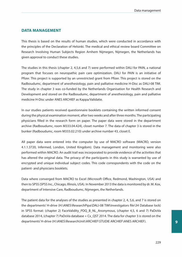
Data management
229
9
DATA MANAGEMENT
This thesis is based on the results of human studies, which were conducted in accordance with
the principles of the Declaration of Helsinki. The medical and ethical review board Committee on
Research Involving Human Subjects Region Arnhem Nijmegen, Nijmegen, the Netherlands has
given approval to conduct these studies.
The studies in this thesis (chapter 2, 4,5,6 and 7) were performed within DALI for PAIN, a national
program that focuses on neuropathic pain care optimization. DALI for PAIN is an initiative of
Pfizer. This project is supported by an unrestricted grant from Pfizer. This project is stored on the
Radboudumc, department of anesthesiology, pain and palliative medicine H-Disc as DALI-08 TIM.
The study in chapter 3 was co-funded by the Netherlands Organisation for Health Research and
Development and stored on the Radboudumc, department of anesthesiology, pain and palliative
medicine H-Disc under ANES ARCHIEF as Kappa/Validatie.
In our studies patients received questionnaire booklets containing the written informed consent
during the physical examination moment, after two weeks and after three months. The participating
physicians filled in the research form on paper. The paper data were stored in the department
archive (Radboudumc, room M333.04.424), closet number 7. The data of chapter 3 is stored in the
bunker (Radboudumc, room M333.02.210) under archive number 43, closet E.
All paper data were entered into the computer by use of MACRO software (MACRO, version
4.1.1.3720, Infermed, London, United Kingdom). Data management and monitoring were also
performed within MACRO. An audit trail was incorporated to provide evidence of the activities that
has altered the original data. The privacy of the participants in this study is warranted by use of
encrypted and unique individual subject codes. This code correspondents with the code on the
patient- and physicians booklets.
Data where converged from MACRO to Excel (Microsoft Office, Redmond, Washington, USA) and
then to SPSS (SPSS Inc., Chicago, Illinois, USA). In November 2013 the data is monitored by dr. M. Kox,
department of Intensive Care, Radboudumc, Nijmegen, the Netherlands.
The patient data for the analyses of the studies as presented in chapter 2, 4, 5,6, and 7 is stored on
the departments’ H-drive (H:\ANES\ResearchPipa\DALI-08 TIM\Investigators file\34 Database lock)
in SPSS format: (chapter 2) FaceValidity_PDQ_B_NL_Anonymous, (chapter 4,5, 6 and 7) PaDoVa
database 2014, (chapter 7) PaDoVa database + Cx_QST 2014. The data for chapter 3 is stored on the
departments’ H-drive (H:\ANES\ResearchUnit\ARCHIEF\STUDIE ARCHIEF\ANES ARCHIEF).
Chapter 9
230
The data will be saved for 15 years after termination of the study (July 1, 2013). Using these patient
data in future research is only possible after a renewed permission by the patient as recorded in the
informed consent. The datasets analyzed during these studies are available from the corresponding
author on reasonable request.
Data management
231
9

Chapter 9
232
Curriculum Vitae
233
9
CURRICULUM VITAE
Hans Timmerman is born on September 25, 1969 in Lunteren (municipal of Ede, the Netherlands),
and grew up in Bemmel, a small village between Arnhem and Nijmegen.
After primary and secondary school in Bemmel he went to Nijmegen to study Physical Therapy at
the HAN University of Applied Sciences. At the age of twenty Hans got his license as a physical
therapist. After his graduation Hans worked in several hospitals, institutions and private practices. In
1995 he started his own private practice in Arnhem. The revalidation of patients with low back pain,
neck shoulder pain and sports injuries were his specialisms. During this period he followed several
post-graduation courses in the field of physical therapy. Besides his own practice he worked as the
staff sports physical therapist for the Royal Dutch Fencing Association during training and national
competitions but also during European and World Championships.
In 2003 he had to give up his private practice because of the limitations due to a serious illness he
suffered a few years before. At the same moment, he started his study Physical Therapy Sciences
at the Utrecht University, Academy of Health Sciences. He graduated in 2007 with a thesis called
“Can preoperative exercise assist the patient with cancer?”. Until today Hans is still involved in research
regarding the prehabilitation of patients before surgery.
Hans started in 2008 as a scientific researcher/PhD student in the Department of Anesthesiology,
Pain and Palliative Medicine at the Radboud university medical center in Nijmegen. His research
was about the validation of screening instruments for use in a consecutive, daily clinical practice
population of patients with low back and leg pain, neck-shoulder pain or with pain due to a
suspected peripheral nerve damage to assist the physician in the assessment of a neuropathic pain
component. During his PhD trajectory Hans followed several additional courses in methodology
and statistics. Nowadays he is frequently asked as a reviewer for several peer-reviewed journals.
Besides this research topic he participated and still participates in several other projects and
cooperations in the field of pain-research and education about (the measurement of ) pain in and
outside the Radboudumc. In 2016 he and his team won the Lowlands Science Grant to investigate
the influence of preferred and disliked music on pain experience (Title: Meer of Minder Pijn met Muse).
In 2017 he was part of the team who obtained the Great National Research Grant to investigate
the pain sensitivity of the Dutch public (Title: Pijngevoeligheid bij het zien van pijn bij mannen en
vrouwen). An important issue in both projects was to raise awareness by the general public about
the necessity of research in the field of pain and to let the people participate in pain research.
Hans has one daughter named Anna (born in 2000) and is living together with Nathalie van Schayk
in Nijmegen.

Chapter 9
234

Publication list
235
9
PUBLICATION LIST
Publications, PubMed indexed:1. Timmerman H, Wolff AP, Bronckhorst E, Wilder-Smith OHG, Schenkels M, van Dasselaar NT,
Huygen FJPM, Steegers MAH, Vissers KCP. Avoiding Catch-22: validating the PainDETECT in a
population of patients with chronic pain. BMC Neurology. 2018, Jun 29;18(1):91.
2. Timmerman H, Vissers KCP, Wolff AP, Wilder-Smith OHG. The added value of bed-side
examination and screening-QST to improve neuropathic pain identification in patients with
chronic pain. Journal of Pain Research. 2018, Jul 10;11:1307-1318.
3. Van Vliet J, Tieleman AA, Verrips A, Timmerman H, van Dongen RTM, van Engelen BGM,
Wilder-Smith OHG. Qualitative and Quantitative Aspects of Pain in Patients with Myotonic
Dystrophy Type 2. Journal of pain. 2018, Aug;19(8):920-930.
4. van Helmond N, Timmerman H, Olesen SS, Drewes AM, Kleinhans J, Wilder-Smith OH,
Vissers KCP, Steegers MAH. A Quantitative Sensory Testing Paradigm to Obtain Measures of Pain
Processing in Patients Undergoing Breast Cancer Surgery. Journal Of Visualized Experiments
(JOVE). 2018, Jan 18;(131).
5. Timmerman H, Steegers MAH, Goeman J, Wilder-Smith OHG, Schenkels M, van Dasselaar NT,
Huygen FJPM, Wolff A, Vissers K. Investigating the validity of the DN4 in a consecutive population
of patients with chronic pain. PLoS One. 2017, Nov 30;12(11):e0187961.
6. Van Helmond N, Timmerman H, van Dasselaar N, van der Pol C, Vissers K, Wilder-Smith OH,
Steegers M. High body mass index is a risk factor for moderate to severe chronic postoperative
pain after breast cancer treatment. Pain Physician. 2017; 20:E661-E671
7. Timmerman H, Wilder-Smith OH, van Weel C, Wolff AP, Vissers KCP. 2014. Detecting the
neuropathic pain component in the clinical setting: A study protocol for validation of screening
instruments for the presence of a neuropathic pain component. BMC Neurology. 2014; 14:94
(May 2, 2014)
8. Van Haren IEPM, Timmerman H, Potting CM, Blijlevens NMA, Staal JB, Nijhuis-van der Sanden
G. Physical exercise for patients undergoing hematopoietic stem cell transplantation: systematic
review and meta-analyses of randomized controlled trials. Physical Therapy. 2013. April;
93(4):514-28.
Chapter 9
236
9. Timmerman H, Wolff AP, Schreyer T, Outermans J, Evers AWM, Freynhagen R, Wilder-Smith OH,
van Zundert J, Vissers KCP. Cross-cultural adaptation to the Dutch language of the PainDETECT
Questionnaire. Pain Practice. 2013; 3: 206-214.
10. Timmerman H, Heemstra I, Schalkwijk A, Verhagen C, Vissers K, Engels Y. Assessment of
neuropathic pain in patients with cancer: The Interobserver reliability. An observational study in
daily practice. Pain Physician. 2013;16: 569-580
11. Chua NHL, Timmerman H, Vissers KCP, Wilder-Smith OH. Multi-modal Quantitative sensory
testing in patients with unilateral chronic neck pain: an exploratory study. MYOPAIN (f.k.a.
Journal of Musculoskeletal Pain). December 2012, Vol. 20, No. 4 , Pages 292-299.
12. Timmerman H, de Groot J, Hulzebos E, de Knikker R, Kerkkamp H, van Meeteren N. Feasibility
and preliminary effectiveness of preoperative therapeutic exercise in patients with cancer. A
pragmatic study. Physiotherapy Theory and Practice. 2011. Feb; 27 (2) 117-24.
Publications, not PubMed indexed:1. Timmerman H, Wilder-Smith O. Richtlijn voor de Onderzoeker. Een gestandaardiseerde testbatterij
voor Kwantitatief Sensorisch Testen volgens de regels van de Deutsche Forschungsverbundes
Neuropathischer Schmerz (DFNS). Officiële Nederlandse vertaling van het DFNS protocol (Versie
2.1-Dlv). German Research Network on Neuropathic Pain (DFNS). München, Germany. 2013
2. Timmerman H, Wolff AP, van Rijswijk ECAM, Vissers KCP. Comment on StEP (Scholz J, Mannion
RJ, Hord DE, Griffin RS, Rawal B, et al.). A Novel Tool for the Assessment of Pain: Validation in Low
Back Pain. PLoS Medicine 2009. 6 (4): e1000047. doi: 10.1371/journal.pmed. 1000047).
3. De Groot J, de Knikker H, Timmerman H, Hulzebos E, Kerkkamp H, van Meeteren N.
Preoperatief fysiotherapeutisch trainingsprogramma voor oncologiepatienten: in Utrecht een
MUST. Fysiopraxis. 2007 (2): 28-31.
Publications, submitted:1. Van Heusden-Scholtalbers LAG, Timmerman H, Bonenkamp H, van Goor H, Nijhuis-van
der Sanden MWG, Staal BJ. Preoperative physical exercise training for patients scheduled for
major abdominal surgery. A systematic review and meta-analysis. 2019. (Cochrane Review,
Submitted)
Publication list
237
9
2. Timmerman H, van de Linde L, van Boekel R, Bronkhorst E, Vissers K, van der Wal S, Steegers,
M. The effect of preferred music versus disliked music on pain thresholds in healthy volunteers.
2019. (Submitted)
3. van den Elzen NGA, Daman V, Duijkers M, Wijnhoven E, Otte K, Timmerman H, Olde Rikkert
MGM. The Bach-Sinatra effect of enhancing muscle power in older people. A randomized crossover
study. 2019. (Submitted)
4 . van Boekel R, Timmerman H, Bronkhorst E, Vissers KCP, Steegers M. The validity of the Dutch
version of the Pain Sensitivity Questionnaire. 2019. (Submitted)
Books and Bookchapters:1. Timmerman H, van den Broeke EN, Wilder-Smith OH. 2014. Hoofdstuk 7: Meetinstrumenten
voor pijn in: Handboek Pijnbestrijding (Translated: Chapter 7: Measurement instruments for Pain
in: Handbook Pain Treatment). Uitgeverij de Tijdstroom, Utrecht, the Netherlands.
Posters and poster presentations:1. Schuttert I, Timmerman H, Groen GW, Wolff AP. Effects of Tapentadol on chronic pain and
parameters of central sensitisation. A prospective, open label, randomized cross-over study with
pregabaline as comparator (PRINCE study). SPBR 2018, Groningen, the Netherlands.
2. Schuttert I, Timmerman H, Groen GW, Wolff AP. Central sensitisation in patients with chronic low
back pain radiating to the leg (CLaSSICO STUDY). SPBR 2018, Groningen, the Netherlands.
3. Timmerman H, Wolff AP, Wilder-Smith OH, Vissers KCP. Screening for a neuropathic pain
component: a mission impossible? e-Poster presentation. 8th World Congress of the World
Institute of Pain 2016, New York, USA.
4. Timmerman H, van Dasselaar NT, Wolff AP, Wilder-Smith OH, Steegers MAH, Vissers KCP. The
validity of the PDQ in patients after treatment for breast cancer. 5th international congress of
neuropathic pain special interest group: NeuPSIG 2015, Nice, France.
5. Van Vliet J, Verrips A, Timmerman H, van Dongen R, Wilder-Smith H, van Engelen E.
Karakterisering van pijn in patiënten met myotone dystrofie type 2. Wetenschappelijke
vergadering van de Neurologie 2015, Nunspeet, the Netherlands.
Chapter 9
238
6. Timmerman H, Wolff A, van Dasselaar N, Steegers M, Wilder-Smith O, Vissers K. The validity of
the DN4 in patients after breast cancer. 7th World Congress of the World Institute of Pain 2014,
Maastricht, the Netherlands.
7. Van der Wal S, Steegers M, Radema S, De Graaf W, Timmerman H, Vaneker M, Vissers K. A case
report: lowdose intravenous lidocaine for chronic chemotherapy induced peripheral neuropathy.
7th World Congress of the World Institute of Pain 2014, Maastricht, the Netherlands.
8. Timmerman H, Heemstra I, Schalkwijk A, Verhagen C, Engels Y, Vissers K.The interobserver
reliability of the diagnosis of neuropathic pain in patients with cancer: a kappa study. World
Congress International Association for the Study of Pain 2012, Milan, Italy
9. Timmerman H, Wolff AP, Schreyer T, Outermans J, Evers AWM, Freynhagen R, Wilder-Smith
OH, van Zundert J, van Rijswijk E, Vissers KCP. Crossculturele adaptatie van de Nederlandstalige
PainDETECT vragenlijst. NHG-Wetenschapsdag 2011, Nijmegen, The Netherlands
10. Timmerman H, Wolff AP, Schreyer T, Outermans J, Evers AWM, Freynhagen R, Wilder-Smith
OH, van Zundert J, van Rijswijk E, Vissers KCP. Cross-cultural adaptation of the PainDETECT
questionnaire to the Dutch language. World Congress on Physical Therapy 2011, Amsterdam,
The Netherlands
(Congress) Presentations and lectures:1. Timmerman, H. AUWCH! PAIN is no FUN!! Lectures for children held in the framework of
Radboud Invites (Science Weekend). 2018, Radboud University Nijmegen, the Netherlands
2. Timmerman H. Use of evaluation scales: Measuring the pain experience. WIP Benelux-WAPMU:
International evidence-based medicine symposium, 2017, Nijmegen, the Netherlands.
3. Timmerman H. How do you communicate your science? About Lowlands Science and the Media.
Radboud Post Doc Initiative, 2017, Nijmegen, the Netherlands
4. Timmerman H, de Groot J, Hulzebos E, de Knikker R, Kerkkamp H, van Meeteren N. Feasibility
of preoperative therapeutic exercise in patients with cancer. Royal Dutch Congress of Physical
Therapy, 2007, Amsterdam, The Netherlands
5. Timmerman H, de Groot J, Hulzebos E, de Knikker R, Kerkkamp H, van Meeteren N. Feasibility
and patient believes of preoperative therapeutic exercise in patients with cancer. Dutch Congress
on Psychology in Patients with Oncology, 2007, Utrecht, The Netherlands.
Publication list
239
9
Grants, Awards and Prices:1. Steegers M, van Boekel R, Timmerman H, van der Wal S, Blaney Davidson E. Pijngevoeligheid
bij het zien van pijn bij mannen en vrouwen. Groot Nationaal Onderzoek. NWO/NTR. 2017,
Hilversum, the Netherlands.
2. Timmerman H, van der Wal S, Groot M, Steegers M. 2016. More or Less Pain with Muse. The
influence of music on pain. Winner of the Grant for Lowlands Science 2016, Biddinghuizen,
the Netherlands.

Chapter 9
240
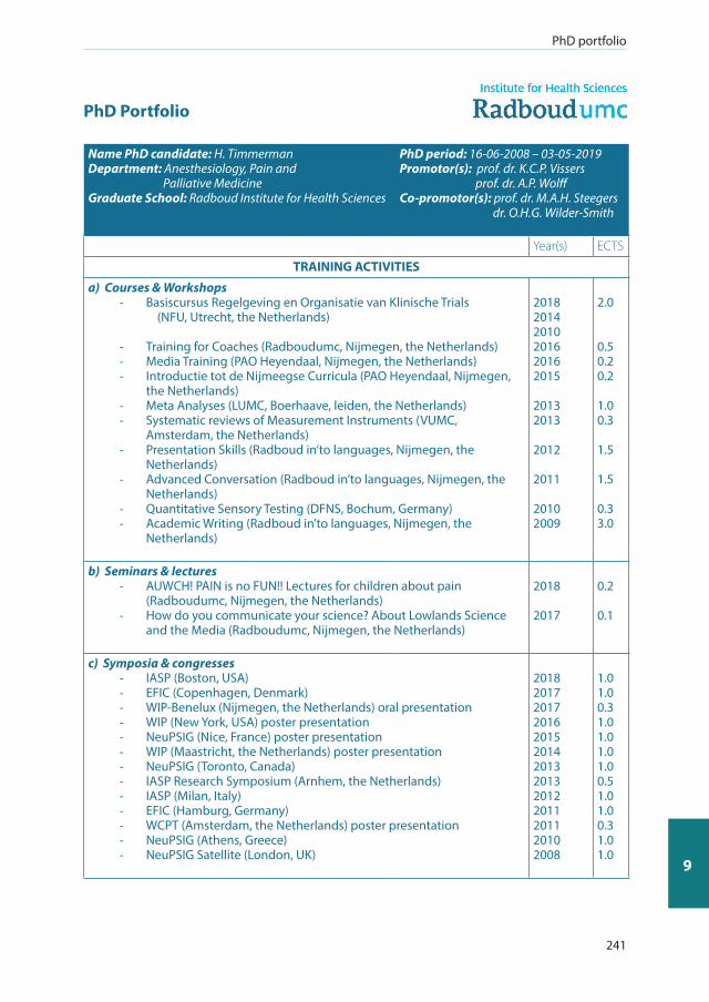
PhD portfolio
241
9
PhD Portfolio
Name PhD candidate: H. TimmermanDepartment: Anesthesiology, Pain and
Palliative MedicineGraduate School: Radboud Institute for Health Sciences
PhD period: 16-06-2008 – 03-05-2019Promotor(s): prof. dr. K.C.P. Vissers
prof. dr. A.P. WolffCo-promotor(s): prof. dr. M.A.H. Steegers
dr. O.H.G. Wilder-Smith
Year(s) ECTS
TRAINING ACTIVITIES
a) Courses & Workshops- Basiscursus Regelgeving en Organisatie van Klinische Trials
(NFU, Utrecht, the Netherlands)
- Training for Coaches (Radboudumc, Nijmegen, the Netherlands)- Media Training (PAO Heyendaal, Nijmegen, the Netherlands)- Introductie tot de Nijmeegse Curricula (PAO Heyendaal, Nijmegen,
the Netherlands)- Meta Analyses (LUMC, Boerhaave, leiden, the Netherlands)- Systematic reviews of Measurement Instruments (VUMC,
Amsterdam, the Netherlands)- Presentation Skills (Radboud in’to languages, Nijmegen, the
Netherlands)- Advanced Conversation (Radboud in’to languages, Nijmegen, the
Netherlands)- Quantitative Sensory Testing (DFNS, Bochum, Germany)- Academic Writing (Radboud in’to languages, Nijmegen, the
Netherlands)
201820142010201620162015
20132013
2012
2011
20102009
2.0
0.50.20.2
1.00.3
1.5
1.5
0.33.0
b) Seminars & lectures- AUWCH! PAIN is no FUN!! Lectures for children about pain
(Radboudumc, Nijmegen, the Netherlands)- How do you communicate your science? About Lowlands Science
and the Media (Radboudumc, Nijmegen, the Netherlands)
2018
2017
0.2
0.1
c) Symposia & congresses- IASP (Boston, USA)- EFIC (Copenhagen, Denmark)- WIP-Benelux (Nijmegen, the Netherlands) oral presentation- WIP (New York, USA) poster presentation- NeuPSIG (Nice, France) poster presentation- WIP (Maastricht, the Netherlands) poster presentation- NeuPSIG (Toronto, Canada)- IASP Research Symposium (Arnhem, the Netherlands)- IASP (Milan, Italy)- EFIC (Hamburg, Germany)- WCPT (Amsterdam, the Netherlands) poster presentation- NeuPSIG (Athens, Greece)- NeuPSIG Satellite (London, UK)
2018201720172016201520142013201320122011201120102008
1.01.00.31.01.01.01.00.51.01.00.31.01.0
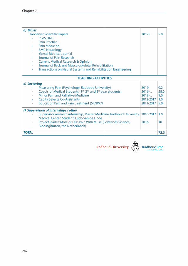
Chapter 9
242
d) Other Reviewer Scientific Papers
- PLoS ONE- Pain Practice- Pain Medicine- BMC Neurology- Yonsei Medical Journal- Journal of Pain Research- Current Medical Research & Opinion- Journal of Back and Musculoskeletal Rehabilitation- Transactions on Neural Systems and Rehabilitation Engineering
2012-... 5.0
TEACHING ACTIVITIES
e) Lecturing- Measuring Pain (Psychology, Radboud University)- Coach for Medical Students (1st, 2nd and 3rd year students)- Minor Pain and Palliative Medicine - Capita Selecta Co-Assistants- Education Pain and Pain treatment (5KNW7)
20192016-...2018-...2012-20172011-2017
0.228.01.01.05.0
f) Supervision of internships / other- Supervisor research internship, Master Medicine, Radboud University
Medical Center. Student: Ludo van de Linde- Project leader ‘More or Less Pain With Muse’ (Lowlands Science,
Biddinghuizen, the Netherlands)
2016-2017
2016
1.0
10
TOTAL 72.3

PhD portfolio
243
9


PINS AND NEEDLES, FACTS AND FEELINGSEFFICACY OF SCREENING TOOLS FOR NEUROPATHIC PAIN IN DAILY CLINICAL PRACTICE
HANS TIMMERMAN
PINS A
ND
NEED
LES, FAC
TS AN
D FEELIN
GS
HA
NS TIM
MERM
AN
Copyright © 2022 FDOKUMEN