université de strasbourg
231
UNIVERSITÉ DE STRASBOURG ÉCOLE DOCTORALE DES SCIENCES DE LA VIE ET DE LA SANTE (ED 414) Génétique Moléculaire, Génomique, Microbiologie (GMGM) – UMR 7156 & Institut de Génétique et de Biologie Moléculaire et Cellulaire (IGBMC) UMR 7104 – INSERM U 964 THÈSE présentée par : Matthieu RAESS soutenue le : 13 octobre 2017 pour obtenir le grade de : Docteur de l’université de Strasbourg Discipline/ Spécialité : Aspects moléculaires et cellulaires de la biologie Deciphering the functional and molecular differences between MTM1 and MTMR2 to better understand two neuromuscular diseases. THÈSE dirigée par : Mme FRIANT Sylvie Directrice de recherche, Université de Strasbourg & Mme COWLING Belinda Chargée de recherche, Université de Strasbourg RAPPORTEURS : Mme BOLINO Alessandra Directrice de recherche, Institut San Raffaele de Milan M. BITOUN Marc Chargé de recherche, Institut de Myologie AUTRES MEMBRES DU JURY : M. ECHARD Arnaud Directeur de recherche, Institut Pasteur M. VITALE Nicolas Directeur de recherche, Université de Strasbourg
-
Upload
khangminh22 -
Category
Documents
-
view
1 -
download
0
Transcript of université de strasbourg

UNIVERSITÉ DE STRASBOURG
ÉCOLE DOCTORALE DES SCIENCES DE LA VIE ET DE LA SANTE (ED 414)
Génétique Moléculaire, Génomique, Microbiologie (GMGM) – UMR 7156
&
Institut de Génétique et de Biologie Moléculaire et Cellulaire (IGBMC)
UMR 7104 – INSERM U 964
THÈSE présentée par :
Matthieu RAESS
soutenue le : 13 octobre 2017
pour obtenir le grade de : Docteur de l’université de Strasbourg
Discipline/ Spécialité : Aspects moléculaires et cellulaires de la biologie
Deciphering the functional and molecular differences between MTM1 and MTMR2
to better understand two neuromuscular diseases.
THÈSE dirigée par :
Mme FRIANT Sylvie Directrice de recherche, Université de Strasbourg & Mme COWLING Belinda Chargée de recherche, Université de Strasbourg
RAPPORTEURS : Mme BOLINO Alessandra Directrice de recherche, Institut San Raffaele de Milan M. BITOUN Marc Chargé de recherche, Institut de Myologie
AUTRES MEMBRES DU JURY : M. ECHARD Arnaud Directeur de recherche, Institut Pasteur M. VITALE Nicolas Directeur de recherche, Université de Strasbourg

1
Acknowledgements
It is my pleasure to acknowledge the roles of many people who made this PhD
research possible.
I will start by respectfully thanking the members of the jury Dr. Alessandra Bolino,
Dr. Marc Bitoun, Dr. Arnaud Echard and Dr. Nicolas Vitale for accepting to read and evaluate
my PhD work.
I would like to sincerely thank Dr. Sylvie Friant and Dr. Jocelyn Laporte for
welcoming me in their respective teams. Both of you have been a constant and powerful
source of advice and motivation. Of course many thanks to Dr. Belinda Cowling for accepting
to be my co-director (and my official Aurora specialist) for the last two years, your help and
your enthusiasm have been a great support to me.
I would like to express my appreciation to the Association Française contre les
Myopathies (AFM Téléthon) for financially supporting my thesis project during 3 years.
When it comes to my team(s) members, it is difficult to individually express all my
gratitude. I will simply thank all of you for your precious help, your fruitful discussions and
most importantly your kindness and positive atmosphere. It has always been a pleasure and a
privilege to share your scientific and social life. Special thanks go to Bruno Rinaldi, Christine
Kretz, Pascal Kessler (except Pascal’s jokes), Hichem Tasfaout and Raphael Schneider for their
significant technical assistance.
I would also like to thank our collaborators for this work: Dr. Bernard Payrastre and
Jean-Marie Xuereb for the yeast lipid dosage and Dr. Norma Romero for sharing precious
patient biopsies. I am also grateful to Alessandra Bolino and Marta Guerrero for sharing their
mouse tissues.
This work would not have been possible without our technical platforms. I especially
thank Nadia Messaddeq, Josiane Hergueux and Coralie Spiegelhalter for their help in electron

2
microscopy; Philippe Hammann, Lauriane Kuhn and Johana Chicher (IBMC) for all the work
on mass spectrometry; Pascale Koebel and Paola Rossolillo (IGBMC) for virus production;
and finally the IGBMC animal facility, cell culture facility and antibody facility.
I gratefully thank all members and organizers of the OpenLAB operation. Going in all
these high schools through Alsace was an exciting opportunity to practice scientific
vulgarization and really confirmed my project of becoming a teacher in biology.
Special thanks go to my loving partner Florine, who is a constant support and source
of happiness in my life.
I would like to finish my acknowledgements by thanking my parents, my brothers
Vincent, Christophe and Sébastien, and my sister Anne. They supported me during all my life
and made all this possible for me.

3
Table of contents
Acknowledgements .................................................................................................................... 1
Table of contents ........................................................................................................................ 3
List of tables ............................................................................................................................... 7
List of figures ............................................................................................................................. 8
Essential abbreviations ............................................................................................................. 10
Part One - Introduction ........................................................................................... 11
I. Setting the scene ............................................................................................................. 12
II. Myotubularin-related diseases ...................................................................................... 12
A. The X-linked centronuclear myopathy ......................................................................... 12
1. The causative gene ................................................................................................... 12
2. Clinical and histological features ............................................................................. 15
3. Animal models ......................................................................................................... 18
B. The Charcot-Marie-Tooth neuropathy Type 4B1 ........................................................ 23
1. The causative gene ................................................................................................... 23
2. Clinical and histological features ............................................................................. 27
3. Animal models ......................................................................................................... 28
III. The myotubularin family ........................................................................................... 29
A. Introduction .................................................................................................................. 29
B. Myotubularins: protein domains and interactions ........................................................ 31
C. Myotubularins: tissue expression ................................................................................. 35
D. Myotubularin: mRNA isoforms ................................................................................... 37
E. Myotubularins: protein structure. ................................................................................. 39
F. Conclusion .................................................................................................................... 42
IV. Phosphoinositides: key lipids in intracellular trafficking ....................................... 43
A. The metabolism of membrane phosphoinositides ........................................................ 43
1. Lipids are the main membrane constituents ............................................................. 43
2. Phosphoinositides are lipid signaling molecules ...................................................... 46
3. Phosphatidylinositol is the precursor of phosphoinositides ..................................... 46
B. The PtdIns3P is essential for endosomal trafficking .................................................... 47
1. PtdIns3P synthesis .................................................................................................... 47
2. PtdIns3P physiological role ..................................................................................... 49

4
C. PtdIns(3,5)P2 is a regulator of endosomal-lysosomal trafficking ................................ 51
1. PtdIns(3,5)P2 synthesis ............................................................................................. 51
2. Physiological role of PtdIns(3,5)P2 .......................................................................... 52
D. PtdIns5P is an underappreciated phosphoinositide ...................................................... 53
1. PtdIns5P synthesis .................................................................................................... 53
2. PtdIns5P physiological role ..................................................................................... 54
E. A word about the other phosphoinositides ................................................................... 55
V. Objectives of this thesis .................................................................................................. 55
Part Two – Results ...................................................................................................... 56
I. Differences in sequence and regulation between MTM1 and MTMR2 .................... 57
A. MTM1 comparison to MTMR2-L and MTMR2-S ...................................................... 57
B. The MTMR2-Δ2-24 truncated construct ...................................................................... 60
C. MTMR2-L function is regulated by the S58 phosphorylation on its N-terminal
extension ............................................................................................................................... 60
D. MTMR2 constructs used for this study ........................................................................ 61
II. Detection of MTMR2 proteins ...................................................................................... 62
III. MTM1 and MTMR2 display different phosphatase activities in vivo ................... 64
A. MTMR2 expression is regulated in yeast ..................................................................... 65
B. MTM1 and MTMR2 display different intracellular localizations in yeast .................. 66
C. MTM1 and MTMR2 display different phosphatase activities in yeast ........................ 68
IV. Study of MTM1 and MTMR2 localization and functions in mammalian cells .... 72
A. MTMR2 localization depends on its N-terminal extension ......................................... 72
B. MTMR2 N-terminal extension includes at least two phosphorylation sites ................ 75
C. Study of MTM1 and MTMR2 in C2C12 muscle cells................................................. 77
V. MTMR2 isoforms rescue the myopathic phenotypes of Mtm1 KO mouse muscles . 82
A. Is MTMR2 expressed in muscle? ................................................................................. 82
1. Expression of the MTMR2-S short isoform is reduced in Mtm1 KO mice muscles. ... 82
2. Expression of the MTMR2-S short isoform is also reduced in the XLCNM patient
muscles ............................................................................................................................. 84
3. Detection of MTMR2 protein isoforms in mice ....................................................... 86
B. Overexpression of MTM1 and MTMR2 in Mtm1 KO mouse muscles using AAV
vectors .................................................................................................................................. 88

5
C. Exogenous expression of MTMR2 short isoform in the Mtm1 KO mice rescues muscle
weight and force similarly to MTM1 expression ................................................................. 90
D. The MTMR2 isoforms rescue the histopathological hallmarks of the Mtm1 KO mouse
92
E. MTMR2 isoforms rescue Mtm1 KO muscle disorganization ...................................... 97
F. Exploring the mechanistic of the rescue ...................................................................... 99
VI. Both MTMR2 isoforms are able to improve the Mtm1 KO mouse phenotypes . 103
A. Overexpression of both MTMR2 isoforms ameliorates the lifespan and body weight of
Mtm1 KO mice ................................................................................................................... 105
B. Overexpression of both MTMR2 isoforms rescues the muscle strength of Mtm1 KO
mice ……………………………………………………………………………………….107
C. Overexpression of both MTMR2 isoforms rescues the histopathology of Mtm1 KO
limb muscles ....................................................................................................................... 109
Part Three - Discussion and Perspectives (in French) ..................... 111
I. Les isoformes de MTMR2 et l’extension N-terminale .............................................. 112
II. Les spécificités de MTM1 et MTMR2 ........................................................................ 116
III. Mieux comprendre la correction de la myopathie ................................................ 118
IV. Stratégies thérapeutiques ........................................................................................ 123
V. Epilogue ......................................................................................................................... 126
Materials and Methods ........................................................................................... 127
I. Plasmids and constructs............................................................................................... 128
II. In vivo models ............................................................................................................... 129
A. Bacteria strains and culture conditions ....................................................................... 129
B. Yeast strains and culture conditions ........................................................................... 130
C. Mammalian cells and culture conditions .................................................................... 130
D. Mice and housing conditions ...................................................................................... 131
III. Antibodies ................................................................................................................. 131
IV. Biopsies from patients .............................................................................................. 132
V. Bacteria transformation and plasmid production..................................................... 132
VI. Production of monoclonal anti-MTMR2 antibody ............................................... 132
VII. AAV production ....................................................................................................... 133
VIII. Lentiviral production ............................................................................................... 133
IX. Expression analysis .................................................................................................. 134

6
X. Protein extraction and Western blot .......................................................................... 135
XI. Mass spectrometry ................................................................................................... 136
XII. Bioinformatics analysis ............................................................................................ 137
XIII. Protocols specific to yeast ........................................................................................ 137
A. Transformation of yeast cells ..................................................................................... 137
B. Subcellular fractionation ............................................................................................ 138
C. Yeast phenotyping ...................................................................................................... 138
XIV. Protocols specific to mammalian cells .................................................................... 139
A. Cell transfection ......................................................................................................... 139
B. Cell transduction ........................................................................................................ 139
C. Immunofluorescence .................................................................................................. 140
D. C2C12 myotubes phenotyping ................................................................................... 140
XV. Protocols specific to mice ......................................................................................... 141
A. AAV transduction in mice .......................................................................................... 141
B. Clinical tests ............................................................................................................... 141
C. Dissection and sample preparation ............................................................................. 142
D. Functional analysis of the muscle .............................................................................. 142
E. Histology .................................................................................................................... 142
F. Immunofluorescence on muscle sections ................................................................... 143
G. Electron microscopy ................................................................................................... 143
H. PtdIns3P quantification by ELISA in muscle extracts ............................................... 143
XVI. Statistical analysis .................................................................................................... 144
Bibliography .................................................................................................................. 145
Appendix .......................................................................................................................... 163

7
List of tables
Table 1: Correlation between expression, localization and phosphatase activity of myotubularins expressed in
ymr1Δ yeast cells .................................................................................................................................................. 71
Table 2 : Rescuing effects of MTM1 and MTMR2 isoforms on several hallmarks of myotubular myopathy 102
Table 3: List of systemic injections .................................................................................................................... 103

8
List of figures
Figure 1: Position of XLCNM-linked nonsense and missense mutations on human MTM1 protein .................. 14
Figure 2: Muscle histology and ultrastructure of XLCNM patients ..................................................................... 17
Figure 3: Morphological and histological phenotypes of the XLCNM zebrafish model ..................................... 19
Figure 4: Clinical and histological phenotypes of the XLCNM mouse model. ..................................................... 20
Figure 5: Clinical and histological phenotypes of the XLCNM canine model ...................................................... 22
Figure 6: Genomic structure and mRNA isoforms of MTMR2 in humans (A) and mice (B) ................................ 24
Figure 7: Position of CMT4B1-linked nonsense and missense mutations on human MTMR2 protein .............. 25
Figure 8: Clinical and histological phenotypes of CMT4B1 patients and associated mouse model................... 27
Figure 9: Human myotubularins: domain organization and interactome .......................................................... 32
Figure 10: Myotubularins tissue expression ........................................................................................................ 35
Figure 11: Myotubularin mRNA isoforms. ........................................................................................................... 38
Figure 12: The myotubularins protein structure .................................................................................................. 40
Figure 13: Phosphoinositide metabolism in yeast and human cells ................................................................... 44
Figure 14: intracellular localization of the different phosphoinositides and the membrane trafficking
pathways .............................................................................................................................................................. 50
Figure 15: MTMR2-L has an N-terminal extension compared to MTM1 and MTMR2-S .................................... 58
Figure 16: Truncated forms of MTMR2-L induce an MTM1-like phenotype ....................................................... 59
Figure 17: MTMR2 localization in mammalian cells is regulated by its S58 phosphorylation site .................... 61
Figure 18: Production and characterization of a new anti-MTMR2 antibody .................................................... 63
Figure 19: MTMR2 expression is regulated in yeast............................................................................................ 65
Figure 20: MTM1 and MTMR2 display different intracellular localizations in yeast. ........................................ 67
Figure 21: MTM1 and MTMR2 display different phosphatase activities in yeast .............................................. 69
Figure 22: MTM1 and MTMR2-S localize to specific punctuate structures in COS cells membrane projections ... 73
Figure 23: Detection of human MTMR2 phosphorylation sites by mass spectrometry ..................................... 76
Figure 24: Mtm1 knowckdown C2C12 myotubes are shorter and have a lower fusion index ........................... 77
Figure 25: Independent expression of myotubularins and GFP in C2C12 using a unique lentiviral vector ........ 79
Figure 26: Detection and quantification of MTMR2 mRNA isoforms in mouse.................................................. 83
Figure 27: MTMR2-S expression is reduced in XLCNM patient muscles.. ........................................................... 85
Figure 28: Detection of endogenous MTMR2 protein isoforms in mouse and cultured cells............................. 87
Figure 29: Detection of overexpressed myotubularins after intramuscular injections in mice. ........................ 89
Figure 30: The MTMR2 short isoform rescues muscle weight and force similarly as MTM1 in the Mtm1 KO
myopathic mouse ................................................................................................................................................. 91
Figure 31: All MTMR2 constructs increase the myofiber size of Mtm1 KO mice ................................................ 92
Figure 32: Fiber size heterogenitiy in Mtm1 KO rescued muscles. ...................................................................... 93
Figure 33: All MTMR2 constructs rescue the nuclei positioning in Mtm1 KO mice. ........................................... 94
Figure 34: All MTMR2 constructs rescue the mitochondria organization in Mtm1 KO mice ............................. 95

9
Figure 35: Localization in overexpressed myotubularins in Mtm1 KO muscles fibers. ...................................... 96
Figure 36: All MTMR2 isoforms ameliorate the muscle ultrastructure of Mtm1 KO mice ................................. 98
Figure 37: Mechanistic of Mtm1 KO mouse muscle rescue by MTMR2 ............................................................ 100
Figure 38: Detection of overexpressed myotubularins after systemic injections in mice. ............................... 104
Figure 39: MTMR2 isoforms rescue the body weight of myopathic mice......................................................... 106
Figure 40: MTMR2 isoforms rescue the muscle force of Mtm1 KO mice .......................................................... 108
Figure 41: MTMR2 isoforms rescue the histology of limb muscles ................................................................... 109
Figure 42: The N-terminal extension of MTMR2 regulates its protein localization and activity ..................... 114

10
Essential abbreviations
Aa, amino acids
ANOVA, analysis of variance
AAV, adeno-associated virus
BIN1, amphiphysin 2
CMT, Charcot-Marie-Tooth
CNM, centronuclear myopathy
DNM2, dynamin 2
FYVE, Fab1-YOTB-Vac1-EEA1
GFP, green fluorescent protein
HE, hematoxylin-eosin
KD, knockdown
KI, knockin
KO, knockout
MTM, myotubularin
MTMR, myotubularin-related
PH-GRAM, Pleckstrin Homology, Glucosyltransferase, Rab-like GTPase Activator and Myotubularin
PPIn, phosphoinositides
PtdIns, phosphatidylinositol
PtdIns3P, phosphatidylinositol 3-phosphate
PtdIns5P, phosphatidylinositol 5-phosphate
PtdIns(3,5)P2, phosphatidylinositol 3,5-bisphosphate
TA, tibialis anterior
WT, wild type Ymr1, Yeast myotubularin-related protein 1

11
Part One - Introduction

12
12 Part One - Introduction
I. Setting the scene
Myotubularins (MTMs) are active or dead phosphoinositide phosphatases defining a
large protein family conserved through evolution, from yeast to human, and involved in
different neuromuscular diseases. Mutations in the myotubularin MTM1 gene cause the severe
congenital myopathy called myotubular myopathy (or X-linked centronuclear myopathy)
affecting the myocytes, while mutations in the myotubularin-related MTMR2 gene cause the
recessive Charcot-Marie-Tooth peripheral neuropathy CMT4B1 affecting the Schwann cells.
This tissue-specificity is quite intriguing, since MTM1 and MTMR2 are ubiquitously
expressed and are two similar proteins: they have comparable sequences homologies, share
domain organizations and catalytic functions, and dephosphorylate the same lipid substrates,
phosphatidylinositol-3-monophosphate (PtdIns3P) and phosphatidylinositol-3,5-bisphosphate
(PtdIns(3,5)P2).
Before presenting my project, I would like to give an overview of three main aspects:
first the neuromuscular diseases associated with myotubularins, then the large myotubularin
family itself, and finally the phosphoinositide substrates of these phosphatases.
II. Myotubularin-related diseases
A. The X-linked centronuclear myopathy
1. The causative gene
The X-linked centronuclear myopathy (XLCNM, OMIM # 310400) or X-linked
recessive myotubular myopathy (XLMTM; OMIM # 310400) is a congenital muscle disorder
first described in 1969 by Van Wijngaarden et al. (Van Wijngaarden, G.K. et al., 1969).
XLCNM belongs to a group of rare congenital myopathies named centronuclear myopathies
(CNM). This group was initially composed of 3 forms: the X-linked form, the autosomal
dominant form (due to mutations in DNM2) and the autosomal recessive form (due to
mutations in BIN1) (Bitoun et al., 2005; Nicot et al., 2007) but more recently, other genes

13
13 Part One - Introduction
such as RYR1, TTN or SPEG have been related to CNM and make the classification more
difficult (Agrawal et al., 2014; Ceyhan-Birsoy et al., 2013; Wilmshurst et al., 2010).
The MTM1 gene
The causative gene of XLCNM was localized on the Xq28 chromosome (Thomas et
al., 1990) and then isolated by positional cloning and identified for the first time by a
consortium of 3 groups in 1996 (Laporte et al., 1996). Named MTM1, this gene contains 15
exons forming a 3.9 kb transcript that is ubiquitously expressed. A second transcript of 2.4 kb
was specifically detected in muscle and testis, due to an alternative polyadenylation signal
resulting in a shorter transcript. However, no muscle-specific exon that could explain the
muscle-specificity of the associated disease has been identified yet (Laporte et al., 1996).
The MTM1 gene codes for a 603 amino acids protein with a specific phosphoinositide
(PPIn) 3-phosphatase activity. MTM1 was the first described protein of the large
myotubularin family that currently contains 14 identified members. This family deserves a
full presentation and will be more thoroughly characterized in chapter III.
MTM1 mutations
Up to now and according to the Human Gene Mutation Database
(http://www.hgmd.cf.ac.uk), 245 mutations on the MTM1 gene have been identified and
associated with the myotubular myopathy. XLCNM patients have been found in all ethnical
groups including European (de Gouyon et al., 1997; Tanner et al., 1999b), Japanese (Nishino
et al., 1998; Tsai et al., 2005) South American (Zanoteli et al., 1998) and North American
populations (Herman et al., 2002).
These mutations mainly affect the PH-GRAM (Pleckstrin Homology -
Glucosyltransferase, Rab-like GTPase Activator and Myotubularins) domain (lipid binding
domain) and the phosphatase domain of MTM1, with no real hotspot (Figure 1) (Biancalana
et al., 2003; McEntagart et al., 2002). Most of them are nonsense or frameshift mutations that
induce a truncated and non-functional protein and seem correlated with severe muscular
phenotypes (McEntagart et al., 2002). The others (77 up to now) are missense mutations

14
14 Part One - Introduction
which often lead to milder phenotypes (McEntagart et al., 2002), and affect amino acids that
are highly conserved in the MTM1 proteins through evolution. This suggests their important
role in protein structure, interaction or catalytic activity. However, the genotype-phenotype
correlation can be surprising and the same mutation (such as the E404K mutation) can induce
various degrees of XLCNM severity from one patient to another (Hoffjan et al., 2006;
McEntagart et al., 2002), indicating some aggravation of compensation factors.
MTM1 cellular functions
In vitro studies show that MTM1 (and active myotubularins in general) is a specific
phosphoinositide (PPIn) 3-phosphatase that dephosphorylates the phosphatidylinositol-3-
monophosphate (PtdIns3P) and PtdIns(3,5)P2 into PtdIns and PtdIns5P, respectively
(Blondeau et al., 2000; Taylor et al., 2000; Tronchere et al., 2004; Walker et al., 2001).
MTM1 cellular functions are not fully understood, and the specific role of MTM1 in
muscle remains under investigation. The majority of MTM1 mutations result in the loss of
MTM1 protein that is presumably the cause of the disease. But we do not precisely know why
Figure 1: Position of XLCNM-linked nonsense and missense mutations on human MTM1
protein. Each dot represents a nonsense (blue) or missense (red) mutation listed in the Human Gene
Mutation Database (http://www.hgmd.cf.ac.uk) for MTM1. In total, 35 nonsense and 77 missense
mutations were linked to XLCNM in human MTM1. The number of different mutations found for the
same amino acid is indicated by the scale on the left. The mapping was done using the mutation
mapper of cBioPortal (http://www.cbioportal.org/mutation_mapper.jsp). PH-GRAM (Pleckstrin
Homology - Glucosyltransferase, Rab-like GTPase Activator and Myotubularins) domain,
phosphatase domain, CC (coiled-coil) domain and PDZ domain are represented.

15
15 Part One - Introduction
MTM1 mutations specifically affect the muscle. Data show that MTM1 interacts with desmin,
a muscle-specific intermediate filament (Hnia et al., 2011). Moreover, MTM1 or desmin
defects lead to abnormal mitochondrial dynamic and positioning in muscle, where
mitochondrial production of energy is crucial for muscle contraction (Hnia et al., 2011).
MTM1 has also been shown to interact with the amphiphysin BIN1 that is implicated in the
autosomal recessive form of CNM (Royer et al., 2013). MTM1 enhances BIN1 activity
(membrane tubulation) in skeletal muscle, and BIN1 patient mutations alter its binding and
regulation by MTM1 (Royer et al., 2013). Studies in cell cultures suggest that MTM1 is able
to dephosphorylate endosomal pools of PtdIns3P (and thus to decrease its levels) (Kim et al.,
2002), and is implicated in late endosome formation and functions through interactions with
PtdIns(3,5)P2 (Tsujita et al., 2004). In addition to this, surface delivery of endosomal cargo
requires PtdIns3P hydrolysis by MTM1 (Ketel et al., 2016).
Finally, a study in mice lacking MTM1 shows aberrant mTORC1 signaling and
impaired autophagy, suggesting that myotubularin is implicated in these pathways (Fetalvero
et al., 2013).
2. Clinical and histological features
The XLCNM affects about 1/50 000 newborn males, and is generally characterized by
hypotonia at birth, a very severe and generalized muscle weakness, external ophthalmoplegia
and respiratory distress (Jungbluth et al., 2008; Laporte et al., 1996). To date, no specific
therapeutic treatment is available. In 1999, Herman et al. classified XLCNM-affected patients
in 3 categories, according to their phenotype severity: he described a severe (and classical),
moderate and mild XLCNM (Herman et al., 1999).
The most severe form is characterized by prenatal polyhydramnios and reduced fetal
movements, chronic ventilator dependence (with risk of respiratory infection), highly delayed
motor milestones and absence of independent ambulation (Das et al., 1993; Herman et al.,
1999). This severe form often results in neonatal death due to respiratory failure (mostly
because of diaphragm weakness). Many surviving infants need a 24 hours/day ventilatory
support and rarely reach 3 years old. Note that the newborn cases are often similar between
XLCNM and myotonic dystrophy, but can be distinguished by examination of the mother

16
16 Part One - Introduction
who shows mild facial weakness and clinical or electrical myotonia in case of myotonic
dystrophy (Heckmatt et al., 1985).
Moderate and mild forms of XLCNM have been described by several groups (Barth
and Dubowitz, 1998; Biancalana et al., 2003; Hoffjan et al., 2006; Yu et al., 2003). Affected
patients are less dependent on ventilatory support and present less severely delayed motor
milestones with some independent ambulation. Only a few patients with pathogenic variants
in MTM1 were described to reach adulthood (Herman et al., 1999), and only two reached their
sixties (Biancalana et al., 2003; Hoffjan et al., 2006). Also, a rare adult-onset form was
reported, with apparently no clinical symptoms at birth and a progressive development of the
myopathy during adulthood (Biancalana et al., 2003; Hoffjan et al., 2006; Yu et al., 2003).
Due to the X-linked recessive inheritance, the myotubular myopathy almost
exclusively affects newborn males, and the mutation is usually transmitted by the
asymptomatic mother (Herman et al., 2002). The short life expectancy and the severe muscle
weakness of XLCNM patients prevent a transmission by the father. However, some rare
heterozygote female carriers manifest XLCNM phenotypes similar to affected male
phenotypes, with a high variability (Biancalana et al., 2017; Hammans et al., 2000; Jungbluth
et al., 2003; Penisson-Besnier et al., 2007; Schara et al., 2003; Sutton et al., 2001; Tanner et
al., 1999a; Tanner et al., 1999b). The inactivation of the other X chromosome (not carrying
the MTM1 mutations) could explain these female cases (Kristiansen et al., 2003). Moreover,
the degree and tissue-specificity of the X chromosome inactivation seems to determine the
phenotype severity (Grogan et al., 2005; Jungbluth et al., 2003; Tanner et al., 1999a).
Compared to males, manifesting female carriers present less severe phenotypes, with
childhood or adolescent onset and a progressive muscle weakness with no impact on the
lifespan (Grogan et al., 2005; Hammans et al., 2000; Penisson-Besnier et al., 2007; Tanner et
al., 1999a). As these milder phenotypes present as adult-onset limb-girdle myopathy, and as
several of the affected females carry large heterozygous MTM1 deletions not detectable by
Sanger sequencing, the prevalence of affected female carriers is likely to be greatly
underestimated (Biancalana et al., 2017).

17
17 Part One - Introduction
Concerning histology, skeletal muscle biopsies of XLCNM patients reveal similar
structural abnormalities independent of the phenotype severity. Myofibers show
characteristics of fetal myotubes, hence the name “myotubular myopathy”. Compared to
normal myofibers, XLCNM myofibers are smaller and rounder (Silver et al., 1986), present
centralized nuclei instead of being at the periphery (Ambler et al., 1984; Gayathri et al., 2000;
Helliwell et al., 1998) and possess a perinuclear halo (“necklace”) lacking contractile
Figure 2: Muscle histology and ultrastructure of XLCNM patients. (A) H&E staining of a
transverse muscle section, showing small round fibers and centralized nuclei (white arrows). (B)
NADH-TR staining of a transverse muscle section, showing abnormal central accumulation of
peripheral aggregates (necklace) of mitochondria (C) Electron microscopy of a transverse muscle
section, showing two centralized nuclei and abnormal myofibers. (A), (B) and (C) were adapted from
Romero et al., 2010. (D) Electron microscopy of longitudinal muscle sections, showing ultrastructural
changes in triads (black arrows) of 3 different XLCNM patients (MTM) compared to control (CTL).
Triads from XLCNM patients are dilated and disorganized. Scale bar 500nm. Adapted from Dowling
et al. 2009.

18
18 Part One - Introduction
elements and containing mitochondria aggregates and glycogen granules (Figure 2A, B and
C) (Romero, 2010; Sarnat et al., 1981). The characteristic centralized nuclei visible after
Hematoxylin & Eosin (H&E) staining are present in all skeletal muscles and may affect up to
90% of fibers (Romero, 2010), hence the name “centronuclear myopathy”. For now, this
abnormal nuclei positioning is not explained, but some data suggest that desmin intermediate
filaments (that interact with MTM1) are implicated in the actin-driven positioning of the
nucleus in skeletal muscle (Dupin et al., 2011; Ralston et al., 2006). A recessive desmin-null
form of myopathy has also been described, with centralized nuclei in the myofibers
(Henderson et al., 2013). Finally, a predominance of type I fibers (slow contraction, high
oxidative capacity) in XLCNM patients muscles can be seen after succinate dehydrogenase
(SDH) staining or NADH-tetrazolium reductase (NADH-TR) staining (Figure 2B) (Ambler et
al., 1984; Helliwell et al., 1998; Romero, 2010).
Skeletal muscle ultrastructure shows a profound disorganization of myofibrils,
sarcomeres and triads (Figure 2C and D) (Ambler et al., 1984; Dowling et al., 2009; Silver et
al., 1986). Furthermore, the neuromuscular junctions are smaller and the N-CAM protein
essential for a proper neuromuscular junction adhesion is abnormally expressed (Coers et al.,
1976; Fidzianska et al., 1994). Since all these elements are essential for Excitation-
Contraction (E-C) coupling, it partially explains the muscle weakness of the XLCNM
patients.
Noteworthy, truncating mutations leading to an absence of MTM1 protein are usually
related to the presence of very small myofibers and to a severe myopathy phenotype, while
missense mutations are linked to larger myofibers (but still small compared to a normal
muscle) and higher life expectancy (Pierson et al., 2007). Thus, measuring the fiber size could
be an easy way to evaluate the vital prognostic of XLCNM patients.
3. Animal models
To better understand the pathophysiological mechanisms of myotubular myopathy and
the cellular and molecular functions of MTM1, several vertebrate models of XLCNM have
been generated (Lawlor et al., 2016).

19
19 Part One - Introduction
Zebrafish model
A zebrafish (Danio rerio) model of myotubular myopathy was generated by Mtm1
knockdown (KD) using morpholino antisense technology (Dowling et al., 2009). Reduced
levels of MTM1 induce impaired motor functions, myofiber atrophy, central and abnormally
rounded nuclei, organelles mislocalization, sarcomere and triad disorganization, and abnormal
neuromuscular junctions (Figure 3B) (Dowling et al., 2009). These phenotypes are similar to
that seen in XLCNM patients. Dowling et al. also observed increased PtdIns3P levels in
zebrafish muscles. Morphological changes can already been observed at the embryonic stage,
with abnormal dorsal curvature and a diminution of spontaneous muscle contractions.
Figure 3: Morphological and histological phenotypes of the XLCNM zebrafish model. Adapted
from Dowling et al. 2009. (A) Live embryos at 24 and 72 hours post fertilization (hpf) injected with
either control (CTL) or myotubularin (MTM) morpholinos. Abnormal dorsal curvature in MTM
morphant at 24 hpf is indicated by a double star (**). MTM morphants at 72 hpf show a tail shortening
and a selective thinning of the muscle compartment (brackets). (B) H&E staining of longitudinal
muscle sections from control (CTL) and myotubularin (MTM) morphants at 72 hpf. MTM morphants
show abnormally rounded nuclei (arrows) and an increased space between the myofibers. (C) Electron
microscopy of longitudinal muscle sections from control (CTL) and myotubularin (MTM) morphants.
MTM morphants display dilated and dysmorphic triads (arrows). Scale bar represents 500nm.

20
20 Part One - Introduction
Mouse models
Buj-Bello et al. generated constitutive Mtm1 knockout (KO) mice by homologous
recombination (Buj-Bello et al., 2002b). These mice are viable but develop a progressive
centronuclear myopathy starting at around 3-4 weeks of age, leading to death at 5 to 8 weeks
of age.
The myopathy reproduces the majority of clinical phenotypes of the XLCNM patients,
as a respiratory distress, an amyotrophy and a severe progressive muscle weakness (Figure
4A-B). Histologically, skeletal muscles fibers show smaller and rounder myofibers with a
progressive accumulation of central or paracentral nuclei, and a mitochondrial accumulation
in the center or the periphery of the fiber (“necklace” phenotype characteristic of human
XLCNM) (Figure 4C-D). Highly disorganized sarcomeres and triads, with an accumulation of
abnormal longitudinal T-tubules can be seen by electron microscopy (Figure 4E) (Al-Qusairi
et al., 2009; Buj-Bello et al., 2002b; Lawlor et al., 2013). While the XLCNM was proposed to
result from an arrest in myogenesis, as the myofibers exhibit fetal myotube characteristics
(Spiro et al., 1966; van Wijngaarden, G. K. et al., 1969), this Mtm1 KO mouse model revealed
that the cause of the pathology is likely a muscle maintenance defect (Buj-Bello et al., 2002b).
Figure 4: Clinical and histological phenotypes of the XLCNM mouse model. (A) Grid climbing
test of wild type (WT) and Mtm1 KO mice. Mtm1 KO mice are unable to climb. (B) Dissected hind
limbs of WT and Mtm1 KO mice, illustrating the major muscle mass reduction in Mtm1 KO mice. (A)
and (B) were adapted from Buj-Bello et al. 2002b. (C), (D) and (E) represent personal data of tibialis
anterior (TA) sections of 7 week-old WT and Mtm1 KO mice, adapted from my article Raess et al.
2017. (C) H&E staining of transversal tibialis anterior (TA) sections. Scale bar 100 µm. (D) Succinate
dehydrogenase (SDH) staining of transverse TA sections. Scale bar 100 µm. (E) Electron microscopy
of longitudinal TA sections displaying sarcomere, mitochondria and triad organization. Scale bar 1
µm. Representative triads are displayed in the zoom square.

21
21 Part One - Introduction
This model is also frequently used for preclinical development of treatments, with
various strategies to compensate the loss of MTM1 and rescue the myopathic phenotypes.
Gene therapy by systemic injection of adeno-associated virus serotype 8 (AAV8) expressing
MTM1 improves motor activity, contractile force and prolonged survival (Childers et al.,
2014).
Another potential therapeutic strategy is to decrease PtdIns3P levels that are increased
in Mtm1 KO mice: inhibition or muscle-specific depletion of the class II PtdIns 3-kinase also
improved motor functions, calcium release and lifespan (Kutchukian et al., 2016; Sabha et al.,
2016). These results highlight the importance of MTM1 phosphatase activity for muscle
maintenance and function. However, the same disease model can also be partially rescued by
expressing a phosphatase inactive MTM1 protein (Amoasii et al., 2012), supporting the
concept that PPIn-unrelated functions of myotubularin are also implicated in this pathology.
A third approach based on targeting DNM2 has been investigated, as increased levels
of DNM2 dynamin have been detected in XLCNM patients and in Mtm1 KO mice, and
mutations in DNM2 lead to the autosomal dominant form of centronuclear myopathy (Bitoun
et al., 2005; Cowling et al., 2014). Based on this, the downregulation of DNM2 expression by
genetic cross (generating heterozygote mice KO for DNM2) or by antisense oligonucleotide-
mediated knockdown also improved the muscular phenotypes and the survival of Mtm1 KO
mice (Cowling et al., 2014; Tasfaout et al., 2017).
An Mtm1 p.R69C mouse line has been generated based on the patient mutation, to
mimic milder forms of XLCNM. These knockin (KI) mice carry a splice site mutation leading
to the expression of only a low level of functional MTM1 protein (Pierson et al., 2012).
However, this low level is sufficient to ameliorate the myopathic phenotypes compared to
Mtm1 KO mice, with an increased muscular strength and a lifespan often exceeding one year.
This model has also been used to test therapeutic approaches for XLCNM, with variable
degrees of success (Dowling et al., 2012; Lawlor et al., 2014; Lim et al., 2015).

22
22 Part One - Introduction
Canine model
Contrary to other vertebrate models of centronuclear myopathy, the canine model was not
generated but was discovered as a naturally occurring mutation in several related litters of
Labrador Retriever puppies (Beggs et al., 2010). The causative N155K mutation in MTM1
induces a general muscle weakness and atrophy starting at 8 weeks of age and leading to
premature death at 15 to 26 weeks of age (Figure 5A). Expression of this mutant in COS-1
cells shows a sequestration of MTM1-N155K in proteasomes, where it is presumably
degraded. Affected dogs exhibit histological phenotypes similar to mouse models, including
myofiber hypotrophy, centralized nuclei, organelles mislocalization and sarcotubular system
disorganization (Figure 5B-C) (Beggs et al., 2010; Childers et al., 2014). It also presents
specific histological patterns of mislocalized organelles and mitochondria that are absent from
both murine and human XLCNM (Beggs et al., 2010; Childers et al., 2014). Note that another
canine XLCNM (p.Q384P caused by a missense mutation in exon 11 of MTM1) was recently
reported in Rottweiler dogs, presenting similar clinical and histological phenotypes than
Labrador Retriever dogs (Shelton et al., 2015).
Figure 5: Clinical and histological phenotypes of the XLCNM canine model. Adapted from Beggs
et al. 2010. (A) Picture of a 4 month-old male Labrador Retriever affected with XLCNM, illustrating
the generalized muscle atrophy and characteristic kyphosis. (B) H&E and NADH staining of transverse
muscle sections from unaffected female carrier (Unaffected) and Mtm1 KO dogs. Scale bar 100 µm. (C)
Electron microscopy of longitudinal muscle sections from unaffected female carrier (Unaffected) and
Mtm1 KO dogs. Scale bar 2 µm. Representative triads are showed by an arrow and displayed in the
zoom square.

23
23 Part One - Introduction
The Labrador Retriever canine model has been used to test gene therapy for XLCNM:
systemic intravenous injection of serotype 8 adeno-associated virus (AAV8) expressing
MTM1 improves muscle strength, histology and dog survival (Childers et al., 2014; Mack et
al., 2017). Combined with the results obtained in the Mtm1 KO mouse model, this paves the
way for clinical trial and the use of gene therapy to treat XLCNM patients.
B. The Charcot-Marie-Tooth neuropathy Type 4B1
1. The causative gene
The Charcot-Marie-Tooth neuropathy Type 4B1 (CMT4B1, OMIM # 601382) is one
of the various forms of inherited neuropathies grouped under the name of Charcot-Marie-
Tooth (CMT) syndrome, also called hereditary motor and sensory neuropathy (HMSN). CMT
has been named after the 3 investigators who described them in 1886, and gather neurological
diseases affecting the peripheral nervous system.
This pathology affects 1/2500 newborn and is the most common inherited neurological
disorder (Saporta et al., 2011). Even if mutations in more than 30 different genes have been
implicated in CMT, most affected patients share clinical similarities, with onset in the first
two decades of life, a progressive distal weakness and atrophy associated with sensory loss,
pes cavus foot deformity, and absent ankle reflexes (Saporta et al., 2011).
CMT has been divided into 9 subgroups based on clinical features, gene localization
(autosomal or allosomal) and mode of transmission (dominant or recessive). Thus, CMT4
define axonal and demyelinating neuropathies transmitted by autosomal recessive inheritance.
To date mutations in 11 genes have been identified: GDAP1 (CMT4A), MTMR2 (CMT4B1),
MTMR13/SBF2 (CMT4B2), MTMR5/SBF1 (CMT4B3), SH3TC2 (CMT4C), NDRG1
(CMT4D), EGR2 (CMT4E), PRX (CMT4F), HK1 (CMT4G), FGD4 (CMT4H),
FIG4 (CMT4J) and SURF1 (CMT4K).

24
24 Part One - Introduction
MTMR2 gene
MTMR2 localized on chromosome 11q21 is the causative gene of CMT4B1 (Bolino et
al., 2000; Houlden et al., 2001; Laporte et al., 1998). 15 exons were initially identified, but
Bolino et al. described 3 additional exons named 1a, 2a and 2b and their inclusion by
alternative splicing allows the expression of 4 different transcripts variants (V1 to V4) that are
ubiquitously expressed (Figure 6A) (Bolino et al., 2002; Bolino et al., 2000). Transcript
Figure 6: Genomic structure and mRNA isoforms of MTMR2 in humans (A) and mice (B). Inclusion of any shown combination of the alternative exons 1a, 2a or 2b (2b being only present in
humans) brings a premature stop codon and unmasks an alternative start site in exon 3. In humans and
mice, MTMR2 V1 encodes for MTMR2-L while isoforms V2 to V4 encode for MTMR2-S.

25
25 Part One - Introduction
variant V1 does not contain any additional exons and leads to the translation from exon 1
encoding for a 643 amino acids (aa) protein isoform, while V2 to V4 include exon 1a, leading
to a premature STOP codon and to the translation from an alternative start codon in exon 3.
Thus, V2 to V4 are used for the translation of a shorter MTMR2 protein isoform of 571 aa.
This translational start site multiplicity is present in other genes and allowed by a mechanism
of leaky ribosomal scanning (Calkhoven et al., 1994; Kozak, 1995). In this manuscript I
named MTMR2-L (long) the most studied 643 aa isoform and MTMR2-S (short) the less
known 571 aa isoform.
In mice, a similar alternative splicing system has been found (containing only two
additional exons compared to three in human) and also leads to the translation of MTMR2-L
and MTMR2-S (Figure 6B) (Bolino et al., 2002). To date, the specific functions of these
MTMR2 isoforms are not known, and this constitutes one of the main questions addressed in
this thesis.
Finally, a recent paper identified an alternative SOX10-responsive promoter
at MTMR2 that displays strong regulatory activity in immortalized rat Schwann cells. This
promoter directs transcription of a new MTMR2 transcript that is predicted to encode the
short MTMR2-S isoform.
MTMR2 mutations
According to the Human Gene Mutation Database (http://www.hgmd.cf.ac.uk), 23
mutations in MTMR2 have been linked to CMT4B1. As for MTM1, most of the mutations found
in MTMR2 are nonsense or frameshift mutations leading to a premature STOP codon and the
production of a truncated dysfunctional protein (Bolino et al., 2000). Some missense mutations
highlight amino acids that are important for MTMR2 structure and function (Figure 7).
Figure 7: Position of CMT4B1-linked nonsense and missense mutations on human MTMR2
protein. Each dot represents a nonsense (blue) or missense (red) mutation listed in the Human Gene
Mutation Database (http://www.hgmd.cf.ac.uk) for MTMR2. In total, 4 nonsense and 6 missense
mutations were linked to CMT4B1 in human MTMR2. The mapping was done using the mutation
mapper of cBioPortal (http://www.cbioportal.org/mutation_mapper.jsp). PH-GRAM (Pleckstrin
Homology - Glucosyltransferase, Rab-like GTPase Activator and Myotubularins) domain, phosphatase
domain, CC (coiled-coil) domain and PDZ domain are represented.

26
26 Part One - Introduction
MTMR2 cellular functions
In vitro studies show that MTMR2, as MTM1, is a specific phosphoinositide (PPIn) 3-
phosphatase that dephosphorylate the PtdIns3P and PtdIns(3,5)P2 into PtdIns and PtdIns5P,
respectively (Berger et al., 2002; Tronchere et al., 2004). MTMR2 showed high efficiency
and peak activity at neutral pH, suggesting an in vivo function in the cytosol or in non-acidic
vesicles (Berger et al., 2002). However, MTMR2 cellular functions are not fully understood.
In vivo, the ability of MTMR2 to dephosphorylate its substrates is still under debate.
Lorenzo et al. show that MTMR2 (as MTM1) is able to dephosphorylate an endosomal pool
of PtdIns3P when overexpressed in HeLa cells (Lorenzo et al., 2006), while Kim et al. state
that overexpression of MTMR2 in COS-1 cells does not change the PtdIns3P level (Kim et
al., 2002). A third study shows that MTMR2 is able to dephosphorylate a late endosomal pool
of PtdIns3P and affects late endosome trafficking (Cao et al., 2008). Finally, Vaccari et al.
suggest that PtdIns(3,5)P2 is the physiological substrate of MTMR2 in the nerve (Vaccari et
al., 2011) These experimental differences could be due to post-transcriptional modifications
of MTMR2. Indeed, Franklin et al. described two phosphorylation sites (S58 and S631) that
regulate the targeting of MTMR2 to different endosomal compartments, to regulate different
pools of PtdIns3P (Franklin et al., 2013; Franklin et al., 2011). Phosphomimetic MTMR2-
S58E mutant remains in the cytoplasm when overexpressed in HeLa cells, while
unphosphorylable MTMR2-S58A mutant colocalizes with early endosomes. The S631
phosphorylation site regulates the shift of MTMR2 between Rab5-positive and APPL1-
positive endosomes. Through this mechanism, MTMR2 seems to be implicated in endosome
maturation, endosome signaling, and potentially in endocytosis (Franklin et al., 2013; Xhabija
et al., 2011).
Other myotubularins implicated in CMT4
Two other myotubularins, MTMR5/SBF1 and MTMR13/SBF2, have been found to be
mutated in CMT4B3 (OMIM # 615284) and CMT4B2 (OMIM # 604563), respectively
(Alazami et al., 2014; Azzedine et al., 2003; Nakhro et al., 2013; Senderek et al., 2003).
MTMR5 and MTMR13 are two dead (catalytically inactive) members of the myotubularin
family that interact with active MTMR2 to form heterodimers and regulate MTMR2 function
and phosphatase activity (Kim et al., 2003; Robinson and Dixon, 2005). Thus, mutations in

27
27 Part One - Introduction
these two genes induce CMT4 through MTMR2 dysregulation, which may explain why
CMT4B1, 2 and 3 are clinically indistinguishable.
2. Clinical and histological features
CMT4B1, as most of the CMT, is clinically characterized by distal motor and sensory
impairment, pronounced muscular atrophy, and a foot deformity named pes cavus (fixed
plantar flexion) (Figure 8A) (Quattrone et al., 1996). Patients often develop a pes equinovarus
(or “clubfoot”) with internally rotated feet, and facial muscle weakness. Compared to
XLCNM, CMT4B1 is a much less severe disease, with a mean age of onset at 3-5 years old
and a progressive distal and proximal muscle weakness of lower limbs. Patients have no
cognitive defects and a life expectancy of 40-50 years old. Since no therapeutic treatment is
available at this point, CMT patients are only treated for the associated symptoms. Orthopedic
care helps for autonomous ambulation and to prevent falls.
Contrary to MTM1 mutations that specifically affect muscle cells, MTMR2 mutations
affect the peripheral nervous system, and more specifically the Schwann cells. These
specialized cells wrap around axons of motor and sensory neurons to form the myelin sheath,
an essential component that isolates the axon and accelerates the action potential. The main
histopathological features of CMT4B1 are the demyelination and the presence of focally
folded (outfoldings) myelin sheaths around peripheral nerves, inducing a reduced nerve
velocity (Figure 8B-C) (Quattrone et al., 1996).
Figure 8: Clinical and histological phenotypes of CMT4B1 patients and associated mouse model.
(A) Characteristic pes cavus phenotype. (B) Electron microscopy of a transverse section of peripheral
nerves from unaffected human. Adapted from Kumar et al. 2003. (C) Electron microscopy of
transverse sections of peripheral nerves from two CMT4B1 patients, illustrating the characteristic
myelin sheath outfoldings. Adapted from Quattrone et al. 1996 and Gambardella et al. 1998. (D)
Tranverse (left) and longitudinal (right) sections of sciatic nerves from 7 week-old Mtmr2 KO mice.
Myelin outfoldings are indicated by white arrows. Adapted from Bolino et al. 2004.

28
28 Part One - Introduction
3. Animal models
To better understand the pathophysiological mechanisms of CMT4B1 and the cellular
and molecular functions of MTMR2, several mouse models of have been generated.
Bolino et al. generated Mtmr2 KO mice by excision of exon 4 that induces a
frameshift and the production of a dysfunctional protein (Bolino et al., 2004; Houlden et al.,
2001). These mice present a CMT4B1-like neuropathy with myelin outfoldings (as in
CMT4B1 patients) and impaired spermatogenesis (Figure 8D). Histology of the brain, spinal
cord and muscle (including ATPase isotype staining) was normal at 2 months of age (Bolino
et al., 2004). Two other mouse models were generated, with specific depletion of MTMR2 in
either Schwann cells or motor neurons (Bolino et al., 2004; Bolis et al., 2005). Disruption of
MTMR2 in Schwann cells reproduces the same phenotypes as observed in MTMR2-null mice,
while the disruption of MTMR2 in motor neurons has no visible incidence on the peripheral
nervous system. This corroborates a Schwann cells-specific function of MTMR2, which
negatively regulates the membrane/vesicle trafficking and delivery to the plasma membrane,
that is essential during myelination (Bolis et al., 2005; Bolis et al., 2009).
Another team generated a CMT4B1 mouse model that mimics a patient mutation, by
introducing an E276X mutation (G to T mutation in exon 9 of MTMR2) and deleting the 3’
terminal region immediately after the stop codon of MTMR2 (Bolino et al., 2000; Bonneick et
al., 2005). The histology shows complex myelin infoldings and outfolding in peripheral
nerves, and especially in distal nerves. However contrary to CMT4B1 patients, no
electrophysiological change was observed, suggesting that the nerve velocity is not
necessarily linked to the myelin sheath morphology (Bonneick et al., 2005).

29
29 Part One - Introduction
III. The myotubularin family
To better understand MTM1 and MTMR2 specific functions, it is essential to
understand the protein family they belong to. Here I focus on gene expression, protein
interactions and protein structure in the myotubularin family. This chapter is mainly based on
the review “WANTED - Dead or alive: Myotubularins, a large disease-associated protein
family” that we published recently (Raess et al., 2017) (Appendix 1). Several points have
been discussed in more detail here to develop key points relevant to my project.
A. Introduction
Myotubularins constitute a large disease-associated family highly conserved through
evolution with similarities to phosphatases. In humans there are 14 clear paralogs of
myotubularins: the first identified was MTM1 followed by 13 myotubularin-related proteins
MTMR1 to MTMR13 (Laporte et al., 2003; Laporte et al., 1996; Robinson and Dixon, 2006).
Among them, 8 proteins are active phosphatases while 6 are catalytically dead, with a
functional cooperation between members of these two classes (Kim et al., 2003; Nandurkar et
al., 2003). In addition MTMR14 protein (also named hJUMPY) has been described (Tosch et
al., 2006), however phylogenetic studies and protein domain composition suggested it defines
a close but distinct protein family, and therefore this protein will not be discussed further in
this chapter. Additional pseudogenes related to myotubularins also exist (Alonso et al., 2004).
Although active myotubularins have been tentatively classified as Protein Tyrosine
Phosphatases (PTP) based on the presence of a C(X)5R motif, they are specific
phosphoinositide (PPIn) 3-phosphatases that dephosphorylate the phosphatidylinositol-3-
monophosphate (PtdIns3P) and PtdIns(3,5)P2 into PtdIns and PtdIns5P, respectively
(Blondeau et al., 2000; Taylor et al., 2000; Tronchere et al., 2004; Walker et al., 2001).
Conversely, dead myotubularins share a similar organization in domains but lack the
phosphatase activity (Cui et al., 1998; Nandurkar et al., 2001).
PPIn are lipid second messengers implicated in a wide range of cellular processes
from cell growth and survival to cytoskeleton dynamics (Di Paolo and De Camilli, 2006;

30
30 Part One - Introduction
Staiano et al., 2015). More specifically, PtdIns3P and PtdIns(3,5)P2 regulate membrane
trafficking at the endosomal level and autophagy, which are the most studied and
characterized functions of myotubularins (Nicot and Laporte, 2008; Robinson and Dixon,
2006). PtdIns5P is implicated in several cellular processes including oxidative stress
signaling, growth factor signaling and transcriptional regulation (Bulley et al., 2015; Giudici
et al., 2016; Gozani et al., 2003a; Keune et al., 2013; Ramel et al., 2011).
Myotubularins have been found in almost all eukaryotes from yeast to mammals, with
few exceptions as Plasmodium falciparum (Lecompte et al., 2008). Orthologs for the 14
human myotubularins are found in chordates except in rodents where MTMR8 is absent at
least in mice and rats. A co-evolution has been observed between active and dead
myotubularins, as well as between active myotubularins and antagonist kinases (Lecompte et
al., 2008). For example MTM1 with the class-III PtdIns 3-kinase VPS34 (PIK3C3) and its
regulator VPS15 (PIK3R4).
In yeast, there is only one myotubularin homolog, named YMR1 for Yeast
Myotubularin Related 1. YMR1 deletion leads to a 2-fold increase of the myotubularin
substrates PtdIns3P and PtdIns(3,5)P2, leading to a fragmented vacuole phenotype (Parrish,
W.R. et al., 2004; Taylor et al., 2000). The absence of major phenotype despite the depletion
of the unique active myotubularin can be explained by the existence of two close
phosphatases, the synaptojanin-like proteins Sjl2 and Sjl3. These proteins non-specifically
dephosphorylate the D3, D4 or D5 phosphate of phosphoinositides, while Ymr1 (and other
myotubularins) mainly dephosphorylate the D3 phosphate (Guo et al., 1999). Contrary to
ymr1Δ, the triple deletion ymr1Δsjl2Δsjl3Δ is lethal (Parrish, W.R. et al., 2004).
Why have so many myotubularins been duplicated and conserved? Indeed, the
presence of 14 similar proteins in humans could lead to functional redundancy, however this
high evolutionary pressure suggests that each myotubularin has one or several specific
function(s). This specificity could be related to tissue expression or splice isoforms, or
particular protein-protein interactions. This specific point will be developed in this chapter.
To date, mutations were found in 4 myotubularin genes in monogenic human diseases.
MTM1 is mutated in X-linked centronuclear myopathy (XLCNM, OMIM # 310400)
31
31 Part One - Introduction
(Jungbluth et al., 2008; Laporte et al., 1996). Three other myotubularins, MTMR2 (encoding
active phosphatase), MTMR5/SBF1 and MTMR13/SBF2 (both dead phosphatases), are
mutated in Charcot-Marie-Tooth neuropathy type 4B1 (CMT4B1, OMIM # 601382), 4B3
(CMT4B3, OMIM # 615284) and 4B2 (CMT4B2, OMIM # 604563), respectively (Alazami
et al., 2014; Azzedine et al., 2003; Bolino et al., 2000; Nakhro et al., 2013; Senderek et al.,
2003). In addition, several myotubularins are linked to multifactorial diseases as colorectal,
gastric and lung cancers (MTMR3 and 7) (Hu et al., 2011; Song et al., 2010; Weidner et al.,
2016), obesity (MTMR9) (Hotta et al., 2011) and Creutzfeldt–Jakob disease (MTMR7)
(Sanchez-Juan et al., 2012). The fact that ubiquitously expressed myotubularins are
implicated in different tissue-specific diseases again indicates that the apparent biochemical
redundancy is in fact hiding tissue-specific functions.
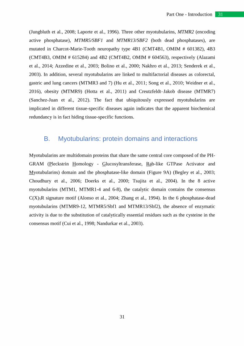
B. Myotubularins: protein domains and interactions
Myotubularins are multidomain proteins that share the same central core composed of the PH-
GRAM (Pleckstrin Homology - Glucosyltransferase, Rab-like GTPase Activator and
Myotubularins) domain and the phosphatase-like domain (Figure 9A) (Begley et al., 2003;
Choudhury et al., 2006; Doerks et al., 2000; Tsujita et al., 2004). In the 8 active
myotubularins (MTM1, MTMR1-4 and 6-8), the catalytic domain contains the consensus
C(X)5R signature motif (Alonso et al., 2004; Zhang et al., 1994). In the 6 phosphatase-dead
myotubularins (MTMR9-12, MTMR5/Sbf1 and MTMR13/Sbf2), the absence of enzymatic
activity is due to the substitution of catalytically essential residues such as the cysteine in the
consensus motif (Cui et al., 1998; Nandurkar et al., 2003).

32
32 Part One - Introduction
C
Figure 9: Human myotubularins: domain organization and interactome. (A) Scaled representation of the protein
domains of human myotubularins. All myotubularins share the PH-GRAM and phosphatase (active or dead) domains.
For each myotubularin, amino acid length of the most described protein isoform is indicated. (B) Classification of
myotubularins into 6 subgroups based on protein organization and phylogenetics (indicated by the vertical bars on the
left). Active myotubularins are represented in green and dead myotubularins in red. (C) Known protein interactions
within the myotubularin family. Published interactions are in orange while interactions found in databases (Biogrid -
thebiogrid.org) are in blue. (D) List of known interactors for each myotubularin. Published interactors are represented in
orange while interactors found in databases with a minimum MUSEscore of 0,95 (Biogrid and Li et al., 2016) are in
blue. Common interactors and interactors of the same protein family are surrounded and related together by a
continuous and stippled line, respectively. Similar interactors found for a specific myotubularin are not surrounded.

33
33 Part One - Introduction
Myotubularins can have several other functional domains: the PDZ binding site
(MTM1, MTMR1 and 2) mediates protein-protein interactions, the PH (Pleckstrin homology)
(MTMR5 and 13) and FYVE (Fab1-YOTB-Vac1-EEA1) (MTMR3 and 4) domains can bind
PPIn (Itoh and Takenawa, 2002), and the DENN domain (MTMR5 and 13) is involved in
small GTPase Rab regulation (Fabre et al., 2000; Jean et al., 2012; Yoshimura et al., 2010),.
By combining domain organization and phylogenetics, 6 different subgroups are highlighted,
each containing exclusively active or dead members (Figure 9B). In addition, all
myotubularins except MTMR10 contain a coiled-coil (CC) domain that is critical for their
homodimerization and/or heterodimerization (Berger et al., 2006; Lorenzo et al., 2006).
Dimerization also appears to depend on the PH-GRAM domain (Berger et al., 2006).
Indeed, all myotubularins except MTMR11 have been shown to interact either with
themselves or with other myotubularins (Figure 9C) (Berger et al., 2006; Gupta et al., 2013;
Kim et al., 2003; Lorenzo et al., 2006; Mochizuki and Majerus, 2003; Nandurkar et al., 2003;
Schaletzky et al., 2003; Zou et al., 2009). Within the 14 members, 9 have been reported to
form homodimers; this could enhance the membrane targeting by coupling two PH-GRAM
domains (Berger et al., 2003). At least for MTM1, homo-oligomerization controls its
allosteric activity, and in vitro MTM1 incubated with its substrate PtdIns3P forms a heptamer
in the presence of PtdIns5P (Schaletzky et al., 2003).
Myotubularins also form heterodimers, and one of the most notable characteristics of
this family is that most heterodimers are formed by a coupling between active/dead
phosphatases. For example, MTMR2 forms heterodimers with its dead homologs
MTMR5/Sbf1, MTMR10, MTMR12 and MTMR13/Sbf2. The fact that mutations in MTMR5
and MTMR13 lead to a similar neuropathy (CMT4B) as defects in MTMR2 confirms the
physiological importance of dead phosphatases. MTMR9 interacts with 3 different active
myotubularins of the same phylogenic subgroup (MTMR6, 7 and 8) and increases their
enzymatic activity (Mochizuki and Majerus, 2003). In the same way, MTMR5 increases the
enzymatic activity of MTMR2 and both are related to male infertility due to impaired
spermatogenesis (Bolino et al., 2004; Firestein et al., 2002; Kim et al., 2003). In some
heterodimers, such as MTM1-MTMR12, MTMR2-MTMR5 and MTMR2-MTMR13, the
dead myotubularin may regulate the cellular localization of the active member (Gupta et al.,
2013; Kim et al., 2003; Nandurkar et al., 2003). Dead myotubularins appear early in evolution

34
34 Part One - Introduction
and are conserved in many species (Laporte et al., 2003; Lecompte et al., 2008). Altogether,
this suggests (1) a co-evolution between active and dead myotubularins and (2) that
myotubularins rely on oligomerization for their function since very early in the evolution.
Heterodimers can also be formed between two active members of the same phylogenic
subgroup, as for MTMR1-MTMR2, MTMR3-MTMR4, MTMR6-MTMR7 and MTMR7-
MTMR8 (Figure 9C).
Numerous interactors have been identified for each myotubularin (Figure 9D) (Biogrid
- thebiogrid.org and Intact - http://www.ebi.ac.uk/intact/) (Agrawal et al., 2014; Berggard et
al., 2006; Cao et al., 2007; Cui et al., 1998; Fabre et al., 2000; Firestein et al., 2000; Jean et
al., 2012; Li et al., 2016; Plant et al., 2009; Royer et al., 2013; Rual et al., 2005; Srivastava et
al., 2005; Yu et al., 2013; Zhang et al., 2005). Some myotubularins share common interactors
or interactors from the same protein family. For example, MTMR6 and MTMR8 both interact
with SOAT1, a protein localized in the endoplasmic reticulum, which is also the presumed
localization of these myotubularins. MTM1 interacts with desmin and MTMR2 with
neurofilament light chain (NFL), that are two intermediate filament proteins specifically
found in muscles and neurons, respectively (Hnia et al., 2011; Previtali et al., 2003). This is
consistent with mutations in MTM1 and desmin leading to myopathies and mutations in
MTMR2 and NFL leading to CMT neuropathies (Goldfarb et al., 1998; Mersiyanova et al.,
2000). Another well-represented group of interactors is the Rab family: MTMR1-RAB6B,
MTMR6-RAB1B, MTMR5-RAB35 and MTMR13-RAB21 (Jean et al., 2012; Mochizuki et
al., 2013). Rabs constitute a very large GTPase family regulating many steps of membrane
trafficking, one of the main cellular functions in which myotubularins are implicated (Barr,
2013). Of note, myotubularins implicated in 3 heterodimers share common or similar
interactors: MTMR1-MTMR2, MTMR1-MTMR5 and MTMR2-MTMR10. For example,
MTMR1 and MTMR5 heterodimerize and interact with similar Rab GTPases (Figure 9D).

35
35 Part One - Introduction
C. Myotubularins: tissue expression
To investigate how myotubularin genes are expressed in human tissues, we mined the
Genotype-Tissue Expression (GTEx) database, which has been built by systematic RNA-
sequencing using samples of 51 different tissues coming from hundreds of donors and 2
transformed cell types in culture.
Figure 10 shows for each gene the relative expression in all tested tissues, and
highlights in which tissue a specific myotubularin is the most/less expressed. This is not
absolute expression, therefore we should keep in mind that each gene cannot be directly
compared to another for the same tissue. A dendrogram was generated using the Pearson
correlation coefficient to highlight hierarchical clustering of myotubularins sharing similar
profiles of expression. Whilst this is one of many possible dendrograms and thus it has to be
interpreted cautiously, two main groups of myotubularins can be distinguished based on
expression profiles: MTMR1-3-8-11-12-13 (upper branch, Figure 10), and the others (lower
branch). Discriminant tissues between the two groups are brain (almost all regions), skin,
vagina and prostate. This does not seem to be directly related to phylogenetic classification, to
active/dead and active/active heterodimers or to myotubularins sharing common interactors.
However, some links can be made. For example, MTMR7 and MTMR9 that form an
Figure 10: Myotubularins tissue expression. Heat map of myotubularin genes relative expression within 51
different tissues and 2 cell types, underlining in which tissue a specific myotubularin is the most/less expressed (left
panel). Expression levels have been obtained by mining the GTEx database (www.gtexportal.org/home/).
Dendrogram highlighting the hierarchical clustering of myotubularins (right panel), using the Pearson correlation
coefficient and average linkage.

36
36 Part One - Introduction
active/dead heterodimer have the closest expression profiles and are both strongly expressed
in brain tissues. A similar link applies to MTM1, MTMR2 and MTMR10, which have
correlated expression patterns: they form two active/dead heterodimers MTM1/MTMR10 and
MTMR2/MTMR10, and MTMR2 and MTMR10 have common interactors (Figure 9C and D)
(Lorenzo et al., 2006).
Concerning myotubularins related to monogenic diseases, while MTM1 has a low
expression level in striated muscles compared to other tissues such as nerves, colon or testis,
mutations in the MTM1 gene lead to a severe myopathy. Thus, the MTM1 tissue-specific
function could be explained by interactions with partners that are only expressed in muscle,
such as desmin (Hnia et al., 2011). On the contrary, MTMR2 and MTMR13 are highly
expressed in nerves, which is consistent with the neuropathies observed due to mutations in
these genes. In addition, MTMR2 binds the neuronal intermediate filament NFL (Previtali et
al., 2003), highlighting a potential molecular basis common to different CMT neuropathy
forms. A link can be observed between MTMR2 and MTMR5, known to form heterodimers;
they both have a high relative expression level in testis, and defects of these genes lead to
male infertility by impaired spermatogenesis (Bolino et al., 2004; Firestein et al., 2002),
therefore adding weight to the physiological significance of these data.
Myotubularins expression levels have also been measured in two cell types,
lymphocytes and fibroblasts that could be easily derived from human biopsies. These cells
could allow diagnosis at the protein level or be dedifferentiated into induced pluripotent stem
(IPS) cells that could be reprogrammed into specific cell types, allowing study of the
pathocellular mechanisms. This time, absolute expression levels of all myotubularins are
compared (Figure 10). Some myotubularins, such as MTMR5 or MTMR2, are well expressed
in both lymphocytes and fibroblasts. Whereas for other myotubularins such as MTMR13,
their expression level is higher in fibroblasts than in lymphocytes, suggesting that their study
in these cell types might be more adapted. Therefore, interpreting these data can be useful in
deciding which cell lines should be used for research and diagnostic purposes.

37
37 Part One - Introduction
D. Myotubularin: mRNA isoforms
The study of gene expression does not take splicing events into account. Indeed, a
specific gene is often spliced into several mRNA isoforms that could be translated into
different protein isoforms. In this review, we use the term “isoform” to define a variant of the
same protein or mRNA, and “homologs” for different genes. Figure 11 summarizes the
myotubularin mRNA isoforms expression within all tissues present in the GTEx database.
Only significantly expressed mRNA isoforms have been represented, and color-coded based
on their predicted protein product: the main protein isoform from the literature, longer/shorter
protein isoforms, or non-coding mRNA isoforms. For each mRNA isoform, the expression
level is indicated as a percentage of total gene expression.
Interestingly, the main mRNA isoform studied in the literature is not always the most
expressed (MTM1, MTMR3, MTMR10, MTMR11 and MTMR12). For MTMR10, the most
expressed isoform encodes only the PH-GRAM; it raises the possibility that this protein
isoform exerts a dominant negative effect on oligomerization of myotubularins or on cellular
functions. MTMR2 has 4 well expressed mRNA isoforms: one translated into the main 643
amino acids (aa) protein isoform and the three others translated into a 571 aa protein isoform
lacking the N-terminal extremity before the PH-GRAM (Bolino et al., 2001). The latter is
present in all tissues except brain, and may have a specific function. In addition, some
isoforms are tissue-specific, as for MTM1 with 2 isoforms only expressed in skeletal muscle.
Corresponding peptides lack a part of the PH-GRAM domain and could support a muscle-
specific function altered in the MTM1-related myopathy.
In total, 10 myotubularins express mRNA isoforms leading to shorter proteins and
MTMR1 displays an isoform predicted to encode a longer protein. These differences can
affect various protein domains as the FYVE domain for MTMR3 and MTMR4 or the DENN
domain for MTMR5, and thus could highly impact on protein conformation or protein-
protein/protein-lipid interactions.

38
38 Part One - Introduction
Figure 11: Myotubularin mRNA isoforms. For each myotubularin indicated on the left, the most expressed isoforms present in
the GTEx database are represented as a percentage of total gene expression. Only the most expressed isoforms are shown. The
mRNA isoforms coding for the main published protein isoforms are indicated in green, shorter isoforms in red and longer
isoforms in blue. Several non-coding isoforms indicated in black have been found well expressed, for which no corresponding
peptides have been described yet. The star indicates specific MTM1 mRNA isoforms in skeletal muscle, the tissue affected in
MTM1-related myopathy. For several myotubularins as MTMR3, MTMR10 and MTMR12, and to a less extend MTMR2, the
main expressed isoforms are different than the published isoforms used for functional characterization of the related proteins; for
MTMR11 and MTMR13 the main expressed isoforms are predicted non-coding.

39
39 Part One - Introduction
For MTM1, MTMR11 and MTMR13, the prevailing mRNA isoforms are non-coding,
or the corresponding peptides have not been identified yet, questioning the function of such
isoforms. Finally, some isoforms described in the literature are not represented here because
they were absent in the GTEx database. This is the case for various MTMR1 mRNA isoforms
that are known to be expressed in some tissues (Buj-Bello et al., 2002a).
In the future, it would be important to characterize the cellular activity of these tissue-
specific isoforms, in order to get insight into their physiological relevance.
E. Myotubularins: protein structure.
Between 2003 and 2016, the crystal structures of 4 active myotubularins have been
determined: MTMR1 (PDB: 5C16), MTMR2 (1LW3, 1ZVR, 1M7R and 1ZSQ), MTMR6
(2YFO) and MTMR8 (4YZI) (Begley et al., 2006; Begley et al., 2003; Bong et al., 2016). A
crystal structure of mouse MTMR5 has also been solved, but only contains the C-terminal PH
domain (1V5U). No major differences have been described between the 4 structures, except
for the orientation of the MTMR6 PH-GRAM domain; this could impact MTMR6
oligomerization properties (Bong et al., 2016).
From a tridimensional point of view, myotubularins are globular proteins with two
main parts: the PH-GRAM domain and the catalytic domain, connected by a linker (Figure
4A) (Begley et al., 2003). N-terminal extremities, coiled-coil domains and PDZ binding sites
are absent of these structures, presumably because they are too flexible or cleaved by
proteolysis. In addition, the cysteine residue of the catalytic C(X)5R motif has been mutated
into a serine for crystallization, except for MTMR6.

40
40 Part One - Introduction
Figure 12: The myotubularins protein structure. (A) Overall view of the myotubularin structure.
The crystal structure of MTMR2 (PDB:1ZSQ) is used as a model, with a front view and a view rotated
at 90°. Domain names and the phosphoinositide substrate (here PtdIns3P) are indicated on the the two
representations. (B) Zoom on the substrate-binding pocket. Residues forming the C(X)5R loop and
other important residues are represented using stick models. Residues affected by missense mutations
in MTM1-linked centronuclear myopathy are colored in yellow. No missense mutations have been
found in MTMR2 or MTMR13 genes in the catalytic pocket. The cysteine residue of the C(X)5R motif
is mutated to serine in the structure for crystallization purposes. Hydrogen bonds between the two
phosphate groups in position D1 and D3 of PtdIns3P/PtdIns(3,5)P2 and surrounding residues of the
active site are represented by stippled lines.

41
41 Part One - Introduction
The PH domain consists in 2 β-strands and 1 α-helix, combined with the 5 β-strands of
the GRAM domain. The main characteristic of the catalytic domain is the substrate binding
pocket that is significantly deeper and wider than that of classical tyrosine phosphatases,
explaining the unique specificity of myotubularins for membrane-embedded PPIn substrates
(Figure 4A) (Begley et al., 2003). Indeed, active myotubularins specifically hydrolyze the D-3
position of PtdIns3P and PtdIns(3,5)P2, two PPIn with large phosphorylated inositol
headgroups that perfectly fit in the catalytic pocket. The D-3 phosphate is then trapped by its
interaction with the main chain of 6 residues from the C(X)5R motif loop (on MTMR2
sequence: Cys417, Ser418, Gly420, Trp421, Asp422 and Arg423) (Figure 4B). Concerning
the catalytic activity, the nucleophile Cys417 residue attacks the phosphorous atom in
position D-3 of the PPIn substrate, forming a phosphoenzyme intermediate, then the aspartic
acid (Asp422 in MTMR2) donates a proton to the released dephosphorylated substrate, before
hydrolysis yielding free enzyme and inorganic phosphate (Begley and Dixon, 2005; Begley et
al., 2006; Nandurkar and Huysmans, 2002). Myotubularins are different from classical PTPs
because the catalytic aspartate residue lies directly in the catalytic C(X)5R loop and not in a
WPD-loop. The D-1 phosphate of the PPIn interacts with the side chain of two residues from
the C(X)5R motif (on MTMR2 sequence: Ser418 and Arg423), but also with Asn330, which
is conserved in all active myotubularins suggesting an important role in PPIn substrate
binding. Some other residues help to maintain the three-dimensional structure of this catalytic
pocket, like Arg463 (on MTMR2) that is also conserved in all myotubularins. However, a
phosphate in position D-4 would generate a steric clash with several residues of the catalytic
pocket, excluding PtdIns(3,4)P2 and PtdIns(3,4,5)P3 from potential substrates.
Another consideration for myotubularin substrate specificity is that active
myotubularins are electrostatically polarized proteins, with one strongly electropositive side
containing the catalytic site (Begley et al., 2006). This would create electrostatic interactions
between the positively charged face of myotubularins and the negatively charged membranes
containing PPIn, contributing to substrate-binding affinity. This electropositive patch around
the pocket seems to be specific for active myotubularins, while several dead myotubularins
have an electronegative surface, suggesting a poor affinity toward lipid membranes.
Furthermore, active myotubularins could bind either membranes or dead-phosphatase
homologs through the same interface.

42
42 Part One - Introduction
The tridimensional structure is also very useful to understand the effect of disease-
associated mutations and thereby to evaluate the importance of mutated residues for the
function or the stability of the protein. The majority of MTM1, MTMR2 and MTMR13
missense mutations affect residues in the hydrophobic core structure of the PH-GRAM and
catalytic domains, and replace native amino acids by bulkier residues, or decrease van der
Waals contacts or alter internal hydrogen bonds, consequently disrupting the protein core
structure. In addition, two clusters of solvent-accessible missense mutation at the surface of
the MTM1 protein can be observed: the Pro179-Asn180-Arg184 cluster and the Asp431-
Asp433 cluster (numbered in MTM1 sequence) that could be potential binding sites for
interactors (Begley et al., 2003). In the active site, the Ser376, Gly378 and Arg421 (numbered
Ser418, Gly420 and Arg463 in MTMR2 structure in Figure 4B) are frequent sites of
mutations found in MTM1: the Ser376 and Gly378 directly bind the D-3 phosphate of the
PPIn and the Arg421 is a key factor to maintain the position of the catalytic loop. Thus,
mutations of these residues would directly prevent any catalytic activity.
F. Conclusion
Myotubularins define a large and highly conserved family of proteins with some
noteworthy characteristics. They are classified in the Protein Tyrosine Phosphatases (PTP)
family but have a specific phosphatase activity against phosphoinositides. One other feature is
the presence of catalytically active and dead phosphatases, where dead myotubularins regulate
their active homologs. Although they are ubiquitously expressed, four myotubularin genes –
MTM1, MTMR2, MTMR5 and MTMR13 – are mutated in tissue-specific neuromuscular
diseases, suggesting tissue-specific splice isoforms or specific protein-protein or protein-lipid
interactions.
Future experiments will be needed to address this tissue specificity. While the function
of myotubularins and PPIn substrates and products was well studied in cell systems, their
physiological role in vivo is still barely understood. Another key issue is the pathological
mechanism(s) associated with myotubularin-related diseases. Preliminary data showed that
MTM1-related myopathy can be rescued in mice by inhibition or muscle-specific ablation of
the class II PtdIns 3-kinase (Sabha et al., 2016; Velichkova et al., 2010). However, the same

43
43 Part One - Introduction
disease model can also be rescued by expressing a phosphatase inactive MTM1 protein,
supporting that PPIn-unrelated functions of myotubularin are implicated in this pathology
(Amoasii et al., 2012). Due to the importance of myotubularins and PPIn pathways in
metabolism and cellular integrity, it would not be surprising that their dysregulation will be
highlighted in other diseases in the future.
IV. Phosphoinositides: key lipids in intracellular
trafficking
It was inconceivable to me to talk about myotubularins without presenting their lipid
substrates: the phosphoinositides. This chapter is based on the review of Dimitri Bertazzi et
al. in which I participated, entitled “Les phosphoinositides, des lipides acteurs essentiels du
trafic intracellulaire.” (Translation: Phosphoinositides, essential lipidic actors in the
intracellular traffic) (Bertazzi et al., 2015) (Appendix 2). As was already mentioned in
previous chapters, myotubularins are specific PPIn 3-phosphatases that dephosphorylate the
PtdIns3P and PtdIns(3,5)P2 into PtdIns and PtdIns5P, respectively (Blondeau et al., 2000;
Taylor et al., 2000; Tronchere et al., 2004; Walker et al., 2001). Therefore I will mainly focus
on these 4 phosphoinositides. The review was published in French in 2015, so I translated and
updated it. The Figure 13 and Figure 14 come from De Craene et al., another review about
PPIn in which I did not take part (De Craene et al., 2017).
A. The metabolism of membrane phosphoinositides
1. Lipids are the main membrane constituents
Membranes are essential cell components with a highly dynamic structure, and
allowed the emergence of life through their physicochemical properties. Indeed, the broader
environment is characterized by variations in temperature, humidity, pH, sun exposure,
osmotic pressure or energy. Thus, each living organism has to adapt to these factors and to
organize itself in order to preserve its internal balance and stability. The plasma membrane is

44
44 Part One - Introduction
the first barrier of the cell that separates the cytoplasm from the external environment. It acts
as a very selective filter that mechanically protects the cell and allows a fine control of the
exchanges between the outside and inside environments.
Figure 13: Phosphoinositide metabolism in yeast and human cells. Adapted from De Craene et al.
2017. (A) The chemical structure of phosphatidylinositol (PtdIns); (B) Phosphoinositides synthesized
in yeast and in human cells with the enzymes involved. Phosphorylation reactions are represented with
black arrows and dephosphorylation reactions by grey arrows. The name of the yeast enzyme (when
relevant) is written on top of its human homolog.

45
45 Part One - Introduction
The plasma membrane is composed of a phospholipid bilayer encompassing proteins,
sterols and glycolipids. Phospholipids are amphiphilic molecules with a hydrophilic head
linked to a hydrophobic tail (Figure 13A). Hydrophobic groups face each other, generating a
hydrophobic space between the two phospholipid layers. This property is required for the
membrane embedding of lipids (as sterols or ceramides), transmembrane proteins or proteins
with a lipid anchor. The lipid composition of the membrane depends on the organism
(eukaryote or prokaryote), the cellular type (within the tissues of a multicellular organism),
the membrane type (plasma membrane, endoplasmic reticulum, endosomes, Golgi apparatus,
mitochondria and other intracellular compartments) and even on the state of the cell (resting
state or under stimulus/stress) (Spector and Yorek, 1985; Zinser et al., 1991).
Membranes are mainly composed of 5 phospholipids: phosphatidylcholine (PC),
phosphatidylethanolamine (PE), phosphatidylserine (PS), phosphatidylinositol (PtdIns) and
sphingomyelin (SM). For note, sterols are also present in the membranes but their function is
primarily to control the membrane fluidity, an essential factor for the lateral diffusion of
molecules in the bilayer. This bilayer also presents an asymmetrical distribution of the
phospholipids between the external and the internal layers, due to the vertical diffusion
between these two layers by the flip-flop mechanism and the activity of membrane proteins
called flippases (Kornberg and McConnell, 1971; Nakano et al., 2009a; Nakano et al., 2009b).
Lipids play an essential role as selective barrier via the plasma membrane, but they are
also the major constituents of eukaryotic intracellular membranes such as compartments,
organelles and transport vesicles. Membrane organization and composition differ depending
on the cellular compartment. On one hand, the endoplasmic reticulum, the Golgi apparatus,
the lysosome (named vacuole in yeast cells), the endosomes and transport vesicles are
surrounded by a single lipid bilayer. The inner space of these intracellular compartments is
called the lumen. On the other hand, the nucleus, mitochondria, chloroplasts of chlorophyll
containing plants, and autophagic vesicles are surrounded by a double lipid bilayer. Each of
these intracellular compartments has specific functions that are essential to cell life (Spector
and Yorek, 1985), and all of them are interconnected by a fine-tuned vesicular transport of
proteins. Thus, it is crucial for the cell to properly discriminate between intracellular
compartments. To this end, each organelle has its own identity defined by molecules in the
cytoplasmic layer of the membranes, allowing for a distinction even between the “cis” and the

46
46 Part One - Introduction
“trans” faces of the Golgi network. Among these molecules, lipids, and specifically
phosphoinositides, play a major role in the spatiotemporal regulation of cellular processes as
the cytoskeleton dynamic, membrane trafficking, proliferation and cell survival.
2. Phosphoinositides are lipid signaling molecules
Phosphoinositides (PPIn) refer to a group of 7 different types of phosphorylated
phosphatidylinositol (PtdIns). If we exclude the PtdIns, PPIn are minor membrane
constituents in terms of their abundance, as they only represent 1% of total cellular
phospholipids (Payrastre et al., 2001). PtdIns itself only represents less than 10% of total
phospholipids. PPIns are composed of a glycerol esterified on SN1 and SN2 positions by two
fatty acid chains and its SN3 position is linked to an inositol ring via a phosphate group
(Figure 13A) (Payrastre et al., 2001). In humans, the most common fatty acids in PtdIns are
stearic acid (lipid number 18:0, 18 defining the number of carbons and 0 the number of
double bonds) on SN1 position and arachidonic acid (20:4) on SN2 position (Marcus et al.,
1969). In yeast Saccharomyces cerevisiae, commonly used fatty acids are oleic acid (18:1),
palmitoleic acid (16:1) and palmitic acid (16:0) (Trevelyan, 1966). Since all PPIns derive
from the non-phosphorylated PtdIns, it is assumed that they have the same composition in
fatty acids as the PtdIns.
The PPIn inositol ring is a polyol cyclohexane of which positions D3, D4 and D5 can
be phosphorylated, potentially generating 7 different PPIns (Figure 13B): phosphatidylinositol
3-phosphate (PtdIns3P), PtdIns4P, PtdIns5P, PtdIns 3,4-bisphosphate (PtdIns(3,4)P2),
PtdIns(3,5)P2, PtdIns(4,5)P2 and PtdIns 3,4,5-trisphosphate (PtdIns(3,4,5)P3). Despite their
low concentrations in lipid membranes, PPIns play an essential role in the recruitment and/or
activation of effector proteins. Moreover, their levels in membranes are regulated by
membrane-specific lipid kinases and phosphatases, allowing for the spatiotemporal regulation
of diverse cell events as budding, fusion or membrane dynamic (Payrastre et al., 2001).
3. Phosphatidylinositol is the precursor of phosphoinositides
Phosphatidylinositol (PtdIns) is a ubiquitous phospholipid in eukaryote cells and the
starting point of PPIns metabolism. Indeed, all PPIns are directly or sequentially synthetized

47
47 Part One - Introduction
from PtdIns (Figure 13B). In yeast, the PtdIns is synthetized by the PtdIns synthase 1 (Pis1) in
the cytoplasmic layer of the endoplasmic reticulum, Golgi, mitochondria and microsomes
membranes (Nikawa and Yamashita, 1984). In human, PtdIns is synthetized by the Pis1
protein homolog, the PtdIns Synthase which has a similar distribution to Pis1 in yeast
(Antonsson, 1994).
PtdIns is also recycled from PtdIns3P, PtdIns4P and PtdIns5P thanks to lipid
phosphatases (Figure 13B). In humans, active myotubularins (MTM1, MTMR1-4, 6, 7 and 8)
are 3-phosphatases dephosphorylating specifically the PtdIns3P into PtdIns (Laporte et al.,
2003). The other PPIns phosphatases are less specific, such as SAC1 which is able to
dephosphorylate the PtdIns3P into PtdIns, but also to dephosphorylate the PtdIns4P (and
probably the PtdIns5P) into PtdIns (Liu and Bankaitis, 2010). In yeast, there is only one
myotubularin named Ymr1, which also shows a phosphatase activity specific to the D3
position. This activity on the PtdIns3P is shared with the less specific phosphatases Sjl2, Sjl3
and Sac1, which are also able to convert PtdIns4P into PtdIns (Parrish, W. R. et al., 2004).
B. The PtdIns3P is essential for endosomal trafficking
1. PtdIns3P synthesis
In yeast, PtdIns3P represents 30% of all the PPIns and is as abundant as PtdIns4P. In
human cells, it accounts for 15% of all the monophosphorylated PPIns and is in minority
compared to PtdIns4P (Payrastre et al., 2001). PtdIns3P is produced by the phosphorylation
of PtdIns at the D3 phosphate of the inositol ring, or by PtdIns(3,4)P2 or PtdIns(3,5)P2
dephosphorylation (Figure 13B).
In yeast, only the lipid kinase Vps34 (Vacuolar Protein Sorting 34) specifically
catalyzes the phosphorylation of PtdIns into PtdIns3P (Herman and Emr, 1990; Schu et al.,
1993). Vps34 activity is regulated by the protein kinase Vps15, and is essential for endosomal
trafficking and autophagy (Kihara et al., 2001). Noteworthy, it has been shown that the
positive regulation of Vps34 by Vps15 is stimulated by the direct interaction between the 7
WD (Trp-Asp) repetitions in the C-terminal region of Vps15 and the G subunit of Gpa1.

48
48 Part One - Introduction
Thus, the C-terminal domain of Vps15 could acts as the γ subunit of the yeast G protein. This
would result in a coupling between the G protein signaling at the plasma membrane and the
receptor sorting at the endosomes. Furthermore, the interaction between the Vps34-Vps15
complex and Gpa1 stimulates the PtdIns3P production at the endosomes (Slessareva et al.,
2006).
The human genome encodes for 8 lipid kinases able to produce PtdIns3P. These
kinases are classified in 3 classes, depending on their substrate specificity and their homology
(Vanhaesebroeck et al., 2010; Vanhaesebroeck et al., 2001):
- 4 members of the class I phosphoinositide 3-kinases (PIK3C) which phosphorylate
predominantly PtdIns(4,5)P2 to generate PtdIns(3,4,5)P3
- 3 members of the class II phosphoinositide 3-kinases (PIK3C2) which phosphorylate
predominantly PtdIns4P to generate PtdIns(3,4)P2
- Only 1 member of the class III phosphoinositide 3-kinase (PIK3C3) which is
homologous to the yeast protein Vps34. As the latter, the human VPS34 is specific for
PtdIns and thus probably generates most of the cellular PtdIns3P. Its activity is
regulated by p150 (also termed PIK3R4/VPS15), the human homolog of the yeast
Vps15 (Panaretou et al., 1997). A phylogenetic study brought to light a coevolution
between the unique PIK3C3 and its regulatory subunit Vps15 in most eukaryotes,
from yeast to human through amoeba and parasites (Lecompte et al., 2008).
In yeast, PtdIns3P is also synthetized by lipid phosphatases Fig4 (Factor Induced Gene
4, also named Sac3), Sjl2, Sjl3 and Sac1. These proteins all contain a SAC catalytic domain
allowing to dephosphorylate different PPIns, such as the PtdIns(3,5)P2 into PtdIns3P (Liu and
Bankaitis, 2010). Fig4 is the only PtdIns 5-phosphatase specific for PtdIns(3,5)P2 (Gary et al.,
2002), and the human homolog FIG4 has similar functions. Interestingly, mutations in human
FIG4 gene are responsible for CMT4J, which is highly similar to the MTMR2-linked CMT4B
(Liu and Bankaitis, 2010). Thus, FIG4 is - with MTM1 and MTMR2 – the third active
phosphatase implicated in both PPIn regulation and a neuromuscular disease (Nicot and
Laporte, 2008).

49
49 Part One - Introduction
2. PtdIns3P physiological role
In yeast and mammalian cells, PtdIns3P is enriched at membranes of early endosomes
and membranes of intraluminal vesicles at the late endosome also termed multivesicular body
(MVB) (Figure 14) (Gillooly et al., 2000). In early endosomes, PtdIns3P plays a central role
in recruiting effector proteins as yeast Vps27 or its human homolog Hrs (Hepatocyte growth
factor-Regulated tyrosine kinase Substrate). These two proteins belong to the ESCRT-0
(Endosomal Sorting Complex Required for Transport) complex and are implicated in
endosomal protein sorting and MVB formation (Henne et al., 2011). They both contain a
FYVE (Fab1, YGL023, Vps27, and EEA1) domain able to bind the endosomal PtdIns3P, and
a specific motif to recruit the ESCRT-1 complex (Gruenberg and Stenmark, 2004). The latter
allows ESCRT-2 and 3 recruitment and their combined action induces the internalization of
membrane proteins into the intraluminal vesicles of the MVB (Henne et al., 2011). This
internalization is necessary to stop the signaling cascades mediated by transmembrane
proteins and to address membrane proteins to the lumen of the vacuole/lysosome.
The human adaptor EEA1 (Early Endosomal Antigen 1) binds with a high affinity to
PtdIns3P via its FYVE domain, and regulates the membrane fusion process between
endosomes, by recruiting the Rab5 GTPase to the endosomes (Gruenberg and Stenmark,
2004).
Similarly, the PtdIns3P 5-kinase Fab1 (yeast)/PIKfyve (human) - that phosphorylate
the PtdIns3P into PtdIns(3,5)P2 - is able to bind the PtdIns3P through its FYVE domain
(Payrastre et al., 2001). Thus, one of PtdIns3P physiological roles is to be a precursor for
PtdIns(3,5)P2 synthesis.
In yeast, the VPS34 deletion is not lethal but induces a strong growth phenotype and a
decreased resistance to many factors (temperature, pH, hyperosmotic stress, or presence of
ethanol, hygromycin B, caffeine or rapamycin). The vps34Δ yeast cells also present important
defects in membrane trafficking, such as an abnormal vacuolar morphology and defective
vacuolar transport with abnormal excretion of vacuolar carboxypeptidase Y (CPY) and
alteration in autophagy (Kihara et al., 2001). Indeed, yeast Vps34 is responsible for the proper

50
50 Part One - Introduction
sorting of proteins to the vacuole via the production of PtdIns3P, which allows the
recruitment of endosomal effector proteins (Henne et al., 2011).
Figure 14: intracellular localization of the different phosphoinositides and the membrane
trafficking pathways. The different phosphoinositides (PPIn) are represented by symbols: circles for
PPIn involved in intracellular trafficking with the corresponding steps they regulate; squares for PPIn
involved in cell signaling, the latter being absent from yeast. The human proteins interacting with the
different PPIn are represented by a rectangle. The MVB stands for multivesicular body and the VPS
for vacuolar protein sorting. Adapted from De Craene et al. 2017.

51
51 Part One - Introduction
C. PtdIns(3,5)P2 is a regulator of endosomal-lysosomal
trafficking
1. PtdIns(3,5)P2 synthesis
PtdIns(3,5)P2 is a poorly abundant PPIn that represents less than 5% of all the PPIns in
human cells and in S. cerevisiae. PtdIns(3,5)P2 is enriched in membranes of the late
endosomes, MVB and lysosome/vacuole (Figure 14) (Di Paolo and De Camilli, 2006).
In yeast, PtdIns(3,5)P2 is sequentially produced, first from the phosphorylation of
PtdIns into PtdIns3P by the PtdIns 3-kinase Vps34, and then from the phosphorylation of
PtdIns3P into PtdIns(3,5)P2 by the PtdIns3P 5-kinase Fab1 (Figure 13B). A hyperosmotic
stress induces a 20-fold increase in PtdIns(3,5)P2 intracellular levels (Dove et al., 1997).
PtdIns(3,5)P2 is regulated by the membrane vacuole proteins Vac7 and Vac14 (Bonangelino
et al., 2002). Indeed, PtdIns(3,5)P2 is not detected in yeast cells where FAB1, VAC7 or VAC14
genes are deleted, and this is independent of osmotic conditions (Bonangelino et al., 2002).
However, Vac7 and Vac14 do not interact with each other: Vac7 is the major activator of
Fab1 in a hyperosmotic stress, whereas Vac14 acts with the lipid phosphatase Fig4 to regulate
PtdIns(3,5)P2 renewal (Duex et al., 2006b). This is quite intriguing, since the stimulation of
PtdIns(3,5)P2 synthesis by an osmotic stress depends on two antagonist processes, PtdIns3P
phosphorylation into PtdIns(3,5)P2, as well as PtdIns(3,5)P2 dephosphorylation into PtdIns3P,
creating a futile loop (Duex et al., 2006a). Thus, there is an essential interdependence between
the lipid kinase Fab1 and the antagonist phosphatase Fig4 to regulate PtdIns(3,5)P2 levels, but
also to activate PtdIns(3,5)P2 synthesis after a stimulus.
In human, PtdIns(3,5)P2 synthesis pathway is similar to the one described in yeast, and
PtdIns(3,5)P2 synthesis is ensured by PIKfyve, the unique Fab1 homolog, which
phosphorylates the PtdIns3P on its 5-position (Figure 14) (Shisheva, 2008). PtdIns3P seems
to be the unique source of PtdIns(3,5)P2, since as of now, no PPIn 4-phosphatase hydrolyzing
the PtdIns(3,4,5)P3 has been identified. Moreover, the regulation of PtdIns(3,5)P2 synthesis is
conserved in humans, and PIKfyve interacts with multiple partners playing a role in
PtdIns(3,5)P2 homeostasis (Shisheva, 2008). For example, PIKfyve interacts with its
antagonist enzyme FIG4 (also named SAC3) that specifically dephosphorylates the

52
52 Part One - Introduction
PtdIns(3,5)P2 into PtdIns3P. This interaction is mediated by the adaptive protein
ArPIKfyve/Vac14 which stabilizes the PIKfyve/FIG4 complex and stimulates PIKfyve
activity (Ikonomov et al., 2010).
A recent study clearly showed the homology between PtdIns(3,5)P2 synthesis
pathways in yeast and mammals (Jin et al., 2008). Indeed, in yeast as well as in mice, Vac14
acts as a protein platform for PtdIns(3,5)P2 synthesis regulation. Vac14 interacts with
Fab1/PIKfyve, Fig4/Sac3 and Vac7, and allows the regulation of the activity of the kinase and
phosphatase (Ikonomov et al., 2010; Jin et al., 2008).
The link between all these PPIn-regulating kinases and phosphatases becomes clearer
with time. Noteworthy, autosomal recessive mutations in FIG4 and MTMR2 are both linked
to CMT4 neuropathy, and Mtmr2 KO mice display elevated PtdIns(3,5)P2 levels (Bolino et
al., 2000; Chow et al., 2007; Vaccari et al., 2011). There is also a genetic interaction between
FIG4 and MTMR2, since reduction of FIG4 expression rescues the myelin outfoldings
phenotype of the Mtmr2 KO mice (Vaccari et al., 2011). In addition, autosomal dominant
mutations in FIG4 induce lateral sclerosis (Chow et al., 2009), and bi-allelic mutations in
VAC14 are responsible for a pediatric-onset neurological disorder (Lenk et al., 2016).
2. Physiological role of PtdIns(3,5)P2
Several studies showed a physiological role for PtdIns(3,5)P2 in the trafficking
regulation between endosomes and the vacuole/lysosome (Figure 14).
In yeast S. cerevisiae, fab1Δ cells have growth defects at 23°C, are not viable at 37°C
and show defaults in vacuole acidification and an enlarged vacuole that occupies up to 80% of
total cell volume (Yamamoto et al., 1995). The abnormal vacuolar size can induce an
incorrect segregation of chromosomes during cell division. Vac7 and Vac14 regulate
PtdIns(3,5)P2 synthesis via Fab1, and thus are necessary for a normal vacuolar morphology
and for a proper vacuole transmission from mother to daughter cell (Bonangelino et al., 2002).
PtdIns(3,5)P2 has an essential function in membrane protein sorting at the late
endosomes/MVB (Odorizzi et al., 1998). Membrane proteins addressed to the vacuole are

53
53 Part One - Introduction
ubiquitinated in endosomes. These ubiquitinated cargos are successively recognized by
ESCRT-0 to 2 complexes for their internalization into the intraluminal vesicles of the MVB
(Gruenberg and Stenmark, 2004). The MVB then fuses with a vacuole, releasing the vesicles
into the lumen of the vacuole. At endosomes, yeast epsins Ent3 and Ent5 interact with
PtdIns(3,5)P2 through their ENTH domain, and are necessary for the sorting of ubiquitinated
cargos to the MVB (Eugster et al., 2004; Friant et al., 2003).
To date, the protein having the highest affinity toward PtdIns(3,5)P2 in vitro is the
yeast Svp1/Atg18, which has a role in yeast autophagy. Interestingly, Svp1/Atg18 also
regulated Fab1 activity, by interacting with Vac7 (that is recruited by Vac14 platform). Thus,
Svp1/Atg18 acts as a sensor for PtdIns(3,5)P2 and regulates its synthesis via a Vac7-Vac14
feedback (Efe et al., 2007).
PIKfyve-/- KO mice do not survive after the embryonic stage (Ikonomov et al., 2011),
highlighting the fundamental role of PtdIns(3,5)P2 in development and cellular processes.
Yeast Fab1 and human PIKfyve both have a FYVE domain able to specifically bind the
PtdIns3P. This interaction with PtdIns3P induces their recruitment to endosomes where
PtdIns3P is enriched, and allows PtdIns(3,5)P2 synthesis (Figure 14) (Sbrissa et al., 2002).
Finally, PIKfyve has a role in diverse processes as endosomal protein sorting,
vacuolar/lysosomal homeostasis or signaling pathway regulation (Payrastre et al., 2001;
Shisheva, 2008). In particular, inhibition or depletion of PIKfyve in mammalian cells induces
an endosomal and lysosomal enlargement similar to the enlarged vacuole observed in yeast
(Shisheva, 2008).
D. PtdIns5P is an underappreciated phosphoinositide
1. PtdIns5P synthesis
PtdIns5P is the most recently identified PPIn monophosphate (Rameh et al., 1997).
This is due to its low basal concentration in mammalian cells (less than 10% of all
monophosphate PPIns) (Payrastre et al., 2001), but also to technical issues rendering it
difficult to study since it co-fractionates with PtdIns4P.

54
54 Part One - Introduction
In humans, PtdIns5P can be produced directly from PtdIns by the lipid kinase PIKfyve
(Figure 14). The in vivo overexpression of PIKfyve induces an increased level of PtdIns5P,
while PIKfyve+/- heterozygote mice are viable and show a reduction in PtdIns5P levels
compared to control mice (Ikonomov et al., 2011).
However, the major source of PtdIns5P in human cells comes from the 3-phosphatase
activity of the myotubularin family (8 active members) on PtdIns(3,5)P2 (Figure 14) (Laporte
et al., 2003). This has been demonstrated in particular for MTM1 in cell cultures and in
myotubes (Tronchere et al., 2004) and for MTMR3 in Drosophila fibroblasts after activation
of cell migration using FGF-1 (fibroblast growth factor) (Oppelt et al., 2013).
Finally, a third possible (and unusual) source of PtdIns5P in human cells comes from
the 4-phosphatase activity of the bacterial IpgD upon infection by Shigella flexneri which is
responsible for dysentery (Niebuhr et al., 2002).
2. PtdIns5P physiological role
The physiological role of PtdIns5P in mammalian cells is still poorly understood. A
fraction of the pool of PtdIns5P has been detected in the nucleus and could be implicated in
stress response, notably by modulating the activity of the transcriptional regulator ING2. The
latter is the first protein with an identified PtdIns5P-specific binding domain, which is a zinc
finger domain named PHD (Plant HomeoDomain finger) (Gozani et al., 2003b). The Dok
protein (downstream of tyrosine kinase) also shows strong binding properties toward
PtdIns5P through its PH domain, and this binding activates the T cell signaling (Guittard et
al., 2009). Moreover, PtdIns5P has been found to be implicated in autophagy since its
addition to cells promotes the autophagosome biogenesis and compensates PtdIns3P depletion
in Vps34 cells (Vicinanza et al., 2015). PtdIns5P also regulates actin cytoskeleton remodeling
at the plasma membrane, by binding to the guanine nucleotide exchange factor (GEF) Tiam1,
thus activating the Rho GTPase Rac1 (Viaud et al., 2014). Finally, PtdIns5P is a regulator of
endosomal protein sorting via its interaction with the endosomal adapter TOM1 (Boal et al.,
2015).

55
55 Part One - Introduction
E. A word about the other phosphoinositides
Other phosphoinositides also exist in eukaryote cells. For example PtdIns4P is mainly
generated by 4-phosphorylation of PtdIns by PI 4-kinases (De Matteis et al., 2002), and is a
key effector of the Golgi trafficking (Audhya et al., 2000; Lemmon, 2003; Mizuno-Yamasaki
et al., 2010). PtdIns(4,5)P2 is the most abundant bis-phosphorylated PPIn, and is implicated in
endocytosis. Finally, PtdIns(3,4)P2 and PtdIns(3,4,5)P3 have not been detected in yeast, and
are essentially localized at the plasma membrane in humans (Figure 14), where they could act
as second messengers (for PtdIns(3,4)P2) and kinase regulators (for PtdIns(3,4,5)P3) in the
PI3K/Akt pathway. However for the remainder of this manuscript I will focus on the
phosphoinositides linked directly to myotubularin function.
V. Objectives of this thesis
As previously established in this introduction, MTM1 and MTMR2 are two very
similar ubiquitously expressed proteins which are surprisingly associated with two different
neuromuscular disorders, a centronuclear myopathy affecting muscles, and a CMT
neuropathy affecting nerves, respectively. The molecular bases of this specificity are still
unknown.
The first aim of my thesis was to investigate the molecular and functional
specificities of MTM1 and MTMR2. I thus studied their differences in terms of sequence,
regulation, in vivo activity and localization, by using different cellular models. The impact of
these differences was assessed by different MTMR2 constructs carrying truncating or point
mutations.
The second aim was to evaluate MTM1 and MTMR2 ability to compensate for
each other in a pathological context. Since our team is focusing on myopathies, I tested here
the ability of MTMR2 to compensate for MTM1 loss in XLCNM, using the Mtm1 KO mouse
model.

56
Part Two – Results

57
57 Part Two - Results
I. Differences in sequence and regulation between
MTM1 and MTMR2
In order to investigate to functional differences between MTM1 and MTMR2, I started
by comparing their sequence, structure and regulation.
Most of the data presented for the remainder of this manuscript in relation to MTMR2-
L and MTMR2-S were published in the article “Expression of the neuropathy-associated
MTMR2 gene rescues MTM1-associated myopathy” in Human Molecular Genetics (Raess
et al., 2017) (Appendix 3).
A. MTM1 comparison to MTMR2-L and MTMR2-S
I first evaluated if MTM1 and MTMR2 functional differences could be linked to
differences in their primary sequences or their domain organization. I started by comparing
MTM1 to the main described MTMR2 protein isoform, MTMR2-L. The sequence alignment
shows that human MTM1 and MTMR2-L present a good overall identity of 64.5% (with a
similarity of 76%) and share the 4 same domains (Figure 15A):
- the PH-GRAM domain that can bind to PPIn or proteins
- the active phosphatase domain that specifically dephosphorylates the PtdIns3P and
PtdIns(3,5)P2.
- the coiled-coil (CC) domain that is essential for their homodimerization and/or
heterodimerization
- finally, the PDZ binding site that mediates protein-protein interactions.
The most conserved domain is the phosphatase domain, which presents 73% of
identity, and more than 80% in its central part around the catalytic site. In comparison, the
PH-GRAM and the C-terminal part (CC and PDZ domains) are much less conserved between
the two proteins, with 55% and 38% of identity, respectively (Figure 15A). Since these
domains interact with lipids and other proteins, and thus impact on the localization and
specificity of MTM1 and MTMR2, this lower conservation could be a first explanation for the
functional differences between the two proteins.

58
58 Part Two - Results
As detailed in the introduction, expression of the MTMR2 gene leads (by an alternative
splicing mechanism described in humans and mice) to two MTMR2 isoforms that we
named MTMR2-L (long) and MTMR2-S (short) (Figure 6) (Bolino et al., 2002). The two
protein isoforms differ only in their translation start sites; MTMR2-S starts right before the
PH-GRAM domain while the MTMR2-L has an extended N-terminal sequence of 72 amino
acids without known homology to any protein domain and that was not resolved in the crystal
structure (Figure 15B) (Begley et al., 2006; Begley et al., 2003). This N-terminal extension
specific to MTMR2-L is mostly absent in MTM1, with the two proteins sharing only a
few amino acids in their N-terminal region (Figure 15).
Figure 15: MTMR2-L has an N-terminal extension compared to MTM1 and MTMR2-S (A) Comparison between MTM1, MTMR2-L and MTMR2-S domain organization. Percentage of identity
between MTM1 and MTMR2-L are indicated for each domain, except the coiled-coil (CC) and PDZ
domains that are grouped. (B) Protein alignment of the first 100 amino acids of human and mouse
MTM1, MTMR2-L and MTMR2-S. The serine S58 of MTMR2-L is indicated by the arrow.

59
59 Part Two - Results
The study of this N-terminal extension of MTMR2-L was initiated by Dimitri
Bertazzi, a previous PhD student in Sylvie Friant’s laboratory, in collaboration with Jocelyn
Laporte laboratory. Dimitri used, among others, an MTMR2-Δ2-73 construct in which amino
acids 2 to 73 are absent. This MTMR2-Δ2-73 exactly corresponds to the MTMR2-S isoform.
To compare the cellular functions of MTM1, MTMR2-L and MTMR2-S proteins in vivo,
Dimitri used the heterologous expression of human myotubularins in ymr1Δ yeast cells. This
Figure 16: Truncated forms of MTMR2-L induce an MTM1-like phenotype. Adapted from
Dimitri Bertazzi thesis manuscript (A) Quantification of the vacuolar phenotypes observed in ymr1Δ
yeast cells with empty vector or overexpressing MTM1, MTMR2-L or truncated forms of MTMR2-L.
The number of cells counted per clone is indicated above. (B) Subcellular distribution of
overexpressed myotubularins in yeast. Protein extracts of ymr1Δ yeast cells with empty vector or
overexpressing MTM1, MTMR2-L or truncated forms of MTMR2-L, were subjected to subcellular
fractionation. MTM1 is mainly in the P13 membrane fraction, while MTMR2-L is mainly in the S100
cytoplasmic fraction. Truncated forms of MTMR2-L display an intermediary phenotype and are also
present in P13 and P100 membrane fractions. Vps10 (membrane protein) and Pgk1 (cytoplasmic) are
used as controls for the subcellular fractionation.

60
60 Part Two - Results
system will be more deeply explained in chapter III. To summarize, data show that MTMR2-
L and MTM1 overexpression induce different vacuolar phenotypes, due to different
subcellular localizations (Figure 16). The vacuole morphology is regulated by the
PtdIns(3,5)P2 level, and thus reflects the myotubularin phosphatase activity. Interestingly,
MTMR2-S induces intermediary phenotypes compared to MTM1 and MTMR2-L.
Altogether, this suggests that MTMR2-S is closer to MTM1 and that the N-terminal
extension of MTMR2-L is important for its specific functions.
B. The MTMR2-Δ2-24 truncated construct
In his thesis, Dimitri Bertazzi started to investigate the N-terminal sequence of
MTMR2-L. Indeed, MTMR2-L is 40 aa longer than MTM1, and this difference is mainly due
to 3 additional sequences (amino acids 1-24, 35-40 and 63-73) in MTMR2-L (Figure 15B).
The truncated constructs MTMR2-Δ2-24, MTMR2-Δ2-40 and MTMR2-Δ2-73 induce the
same phenotypes when overexpressed in ymr1Δ yeast cells (Figure 16). Thus, deleting the
first 24 amino acids of MTMR2-L is sufficient to induce an MTM1-like phenotype in yeast.
This suggests that the functional specificity of MTMR2-L compared to MTMR2-S and
MTM1 is mainly due to the 24 first amino acids of its N-terminal extension.
C. MTMR2-L function is regulated by the S58
phosphorylation on its N-terminal extension
As mentioned in the introduction, Franklin et al. described two phosphorylation sites
(S58 and S631) that regulate the targeting of MTMR2 to different endosomal compartments
in mammalian cells, to regulated different pools of PtdIns3P (Franklin et al., 2013; Franklin et
al., 2011). Indeed, the MTMR2-S58A phosphorylation-defective mutant localizes to
endosomes and is active toward PtdIns3P, contrary to the phosphomimetic mutant MTMR2-
S58E that remains cytoplasmic (Figure 17). The S58 phosphorylation is mediated by Erk1/2
kinase and the endosomal targeting of MTMR2 is regulated through an Erk1/2 negative
feedback mechanism (Franklin et al., 2013; Franklin et al., 2011). The S631 phosphorylation
site regulates the shift of MTMR2 between Rab5-positive and APPL1-positive endosomes.
Through this mechanism, MTMR2 seems to be implicated in endosome maturation,

61
61 Part Two - Results
endosome signaling, and potentially in endocytosis (Franklin et al., 2013; Xhabija et al.,
2011).
This S58 phosphorylation site is in the N-terminal extension of MTMR2-L, and is
absent in MTMR2-S and in human MTM1 (Figure 15B). This suggests that the regulation
of MTMR2-L by its specific S58 phosphorylation site could be implicated in functional
differences between MTM1 and MTMR2. Note that this phosphorylation site is conserved
in mouse MTM1 (S28 in mouse MTM1 sequence, Figure 15B), and thus could potentially be
phosphorylated to regulate MTM1 localization and function in mice but not in human.
D. MTMR2 constructs used for this study
Based on preliminary data investigating MTMR2 molecular specificities, I selected
several MTMR2 constructs for this study:
- MTMR2-L and MTMR2-S: to compare the two physiological isoforms and evaluate
the molecular impact of MTMR2-L N-terminal extension (1-72).
- MTMR2-Δ2-24: as an intermediary truncated form between MTMR2-L and
MTMR2-S, to see if deleting only the 2-24 extremity is sufficient to change MTMR2
localization and function.
- Mutations targeting phosphorylation of MTMR2: the non-phosphorylable MTMR2-
S58A, and the two phosphomimetics MTMR2-S58E and MTMR2-S58D. MTMR2-
Figure 17: MTMR2 localization in mammalian cells is regulated by its S58 phosphorylation site. Adapted from Franklin et al. 2011. HeLa cells were transfected with MTMR2-L or phosphorylation
mutants carrying an N-terminal FLAG. After fixation, cells were immunostained with anti-FLAG
monoclonal antibody. Scale bar 15 µm.

62
62 Part Two - Results
S58D was not studied by Franklin et al. but also mimics a phosphorylated S58 and
was chosen here to confirm MTMR2-S58E results.
To assess the impact of these modifications on MTMR2 function, I overexpressed
these different constructs in several cellular and animal models, using MTM1 and
MTMR2-L as controls. If the modification induces an MTM1-like phenotype, it suggests
that the modified site is implicated in the molecular specificities observed between
MTM1 and MTMR2.
II. Detection of MTMR2 proteins
Before analyzing the effect of myotubularin overexpression in cells and tissues, it is
crucial to determine if they are indeed expressed. Our team previously tested several
commercial antibodies directed against MTMR2. The results were not optimal, especially for
detection in tissues and mammal cells extracts. Thus, we decided to produce new anti-
MTMR2 antibodies.
First, I induced the production of recombinant MTMR2-L protein fused to GST in E.
coli bacteria cells by growth in presence of IPTG. Then I extracted total proteins by
sonication and successive centrifugations, and GST-MTMR2-L was affinity purified by using
glutathione-sepharose beads. The first 5 eluted fractions have been separated by SDS-PAGE,
proteins were stained by Coomassie blue, and the proteins corresponding to the expected size
of GST-MTMR2-L were analyzed by mass-spectrometry (Figure 18A). Finally E2, E3 and E4
fractions that contained GST-MTMR2-L (as stated by mass spectrometry) were pooled and
given to the antibody facility of IGBMC (Illkirch), were the recombinant protein was then
used to immunize mice and produce mouse monoclonal anti-MTMR2 antibodies. I used the
full length MTMR2-L instead of a peptide, to directly obtain several antibodies against
multiple conformational epitopes from throughout the sequence, and thus maximize the
chances to recognize the native protein.
In total, 8 different mouse monoclonal antibodies were produced and tested against
protein extracts from COS cells and yeast cells overexpressing MTMR2-L (Figure 18B).

63
63 Part Two - Results
Among them, only the anti-MTMR2 4G3 (number 7 in Figure 18B) detected properly
MTMR2 at the expected size (73 kDa) in both COS and yeast cells. In comparison, the
commercial antibody gave a very weak signal for the same samples. Since this homemade
antibody has been raised against the full-length MTMR2-L and not a specific peptide, we do
not know its MTMR2 epitope. However, it is able to recognize MTMR2-S, so the epitope is
not in the N-terminal extension of MTMR2-L (Figure 18C). In conclusion, this 4G3
antibody detects both MTMR2-L and -S isoforms. Therefore this antibody was used for
the remainder of the studies presented here, allowing a detection of all overexpressed
MTMR2 constructs in all tested cellular and animal models, by western blot and
immunofluorescence (Figure 18C and D).
Figure 18: Production and characterization of a new anti-MTMR2 antibody. (A) Purification of
MTMR2-L fused to GST. Coomassie staining of SDS-polyacrylamide gel with the 5 elution fractions
of GST-MTMR2-L purification. Total lysate (T.L.) and supernatant after centrifugation (S) were also
tested to control the affinity-purification step. (B) Testing the 8 potential anti-MTMR2 antibodies by
western blot on protein extracts from COS-1 and yeast overexpressing MTMR2-L. The commercial
anti-MTMR2 antibody (CA) from Abnova was used as control. Expected size for MTMR2-L is
indicated by an arrow on the left. The 4G3 antibody (number 7 in the figure) is the only one able to
detect MTMR2-L at the expected size in both COS and yeast extracts. (C) Testing the anti-MTMR2
4G3 antibody by western blot on yeast, HEK293 or TA muscle protein extracts for all MTMR2
constructs. Stainfree has been used as a loading control (no shown). (D) Testing the anti-MTMR2 4G3
antibody by immunofluorescence on COS-1 cells transfected by a pAAV overexpressing MTMR2-L
or MTMR2-S (green signal). Nuclei have been stained by Hoechst (blue signal).

64
64 Part Two - Results
III. MTM1 and MTMR2 display different phosphatase
activities in vivo
MTM1 and MTMR2 are two active myotubularins sharing a specific 3-phosphatase
activity toward PtdIns3P and PtdIns(3,5)P2. In vitro studies show that MTMR2, like MTM1,
dephosphorylates its substrates with high efficiency and peak activity at neutral pH (Berger et
al., 2002; Tronchere et al., 2004). However, when I started my thesis, it was unclear whether
MTMR2 could dephosphorylate its substrates in vivo: one reports that MTMR2 (as MTM1) is
able to dephosphorylate an endosomal pool of PtdIns3P when overexpressed in HeLa cells
(Lorenzo et al., 2006), while another states that overexpression of MTMR2 in COS-1 cells
does not change the PtdIns3P level (Kim et al., 2002). I thus decided to compare MTM1
and MTMR2 phosphatase activities and cellular functions in a unicellular eukaryote
model, the yeast Saccharomyces cerevisiae.
Yeast is a powerful eukaryotic model to study human myotubularins, as its
intracellular organization, membrane trafficking and phosphoinositides substrates metabolism
are well characterized and conserved from yeast to human (Introduction and (Katzmann et al.,
2003)). Ymr1 is the unique yeast myotubularin and the heterologous expression of human
MTM1 myotubularin in ymr1Δ yeast cells rescued the vacuolar fragmentation phenotype
defect, decreased the intracellular levels of PtdIns3P and increased the production of PtdIns5P
(Amoasii et al., 2012)). This ymr1Δ yeast strain also allows the study of the human
myotubularin proteins activity without any competition from another myotubularin.
Moreover, yeast is as very useful model to study proteins from other eukaryotic organisms in
general, with low culture costs and easy gene manipulation compared to mammalian systems
(Byrne et al., 2005; Holz et al., 2003; Joubert et al., 2010). A study investigating the
systematic humanization of hundreds of yeast genes showed that 43% of them could be
replaced by their human ortholog, and most of them coded for metabolic enzymes involved in
conserved pathways, like myotubularins (Kachroo et al., 2015).
To better understand MTM1 and MTMR2 specificities concerning their basic
cellular functions and activities, I transformed the ymr1Δ yeast strain with 2µ plasmids
(high copy number, overexpression) or CEN plasmids (low copy number, expression)

65
65 Part Two - Results
containing the cDNA of human MTM1 and all MTMR2 variants, fused or not with GFP
(green fluorescent protein) at the C-terminal of the protein. Then I analyzed the
expression, localization and phosphatase activity of expressed myotubularins.
A. MTMR2 expression is regulated in yeast
I started by testing protein expression of human myotubularins transfected in yeast, by
SDS-page and western blot analysis. All human myotubularins were well detected at the
expected molecular weights, with 2µ or CEN plasmids (Figure 19).
Figure 19: MTMR2 expression is regulated in yeast. (A) Detection of overexpressed (2µ plasmids)
MTM1 in ymr1Δ yeast cells, by western blot using polyclonal anti-MTM1 antibodies that does not
recognize MTMR2. Wild-type (WT) and ymr1Δ yeast strains with empty vectors are used as controls.
Pgk1p is used as a loading control. (B) Detection of overexpressed (2µ plasmids) MTMR2 constructs
in ymr1Δ yeast cells, by western blot using the homemade monoclonal anti-MTMR2 antibody. The
stain free signal on the membrane is used as loading control. (C) Detection of expressed (CEN
plasmids, low copy number) MTMR2 constructs in ymr1Δ yeast cells, by western blot using the
homemade monoclonal anti-MTMR2 antibody. Pgk1p is used as a loading control. (D) Detection of
overexpressed (2µ plasmids) GFP-tagged MTMR2 constructs in ymr1Δ yeast cells, by western blot
using the homemade monoclonal anti-MTMR2 antibody. (E) Quantification of MTMR2 variants
expression levels compared to MTMR2-L. Three different clones and western blots per construct. The
stain free blots were used to normalize protein levels. Data represent means ± s.e.m. *p<0.05
***p<0.001 compared to MTMR2-L (ANOVA test).

66
66 Part Two - Results
Surprisingly, the expression level of MTMR2 was different depending on the
construct: all variants of MTMR2 displayed a 2 to 3 fold higher expression level compared to
the wild-type MTMR2-L, except MTMR2-S58D which was expressed at the same level as
MTMR2-L (Figure 19B and E). Therefore, there is a specific regulation of the protein
level for MTMR2 and its variants in yeast cells. I could not compare this result with
MTM1 protein expression, as MTM1 and MTMR2 are not detected by the same antibody.
GFP-tagged myotubularins were also detected at the expected sizes, but a second
lower band was also detected for each construct (Figure 19D). This second band was not
detected for untagged myotubularins and was always at the same distance from the upper
band, suggesting some protein degradation induced by the C-terminal GFP-tag.
B. MTM1 and MTMR2 display different intracellular
localizations in yeast
Then I analyzed the intracellular localization of overexpressed GFP-tagged
myotubularins in ymr1Δ cells. MTM1 exhibited a punctate signal adjacent to the vacuole (also
positive for the FM4-64 lipid dye, and probably corresponding to the MVB), whereas
MTMR2-L signal was mainly cytoplasmic (Figure 20A). Interestingly, MTMR2-S isoform
presented a signal in the cytoplasm and some intense dots at the vacuolar membrane, as
observed for MTM1. It was also the case for MTMR2-Δ2-24, MTMR2-S58A and MTMR2-
S58E. The only exception was MTMR2-S58D that presented a cytoplasmic localization
similar to MTMR2-L. This suggests that MTMR2 localization in yeast depends on its N-
terminal extension.
I wanted to complete and quantify this localization study by a subcellular
fractionation. I performed differential centrifugations on protein extracts of ymr1Δ yeast cells
overexpressing MTMR2 variants. I tested several techniques and protocol adjustments to
finally find the suitable conditions for yeast cell lysis to maintain the different compartments
and have the proper fractionation of the endogenous yeast proteins used as controls. The
fractionation results were in accordance with the microscopy: MTM1 was mainly detected in
the P13 membrane fraction, whereas MTMR2-L was mainly in the S100 cytoplasmic fraction
(Figure 20B). All other variants of MTMR2 were detected in the S100 fraction as MTMR2-L,

67
67 Part Two - Results
but they were also detected in the P13 and P100 membrane fractions as MTM1. This time, no
difference was detected for MTMR2-S58D compared to other phosphorylation mutants.
In conclusion, MTM1 and the most described MTMR2-L isoform localize differently
in yeast. Interestingly, the MTMR2-S isoform which lacks the N-terminal extension localizes
similarly to MTM1. Deleting the first 24 amino acids of MTMR2-L or modifying the S58
phosphorylation site also seems to induce an MTM1-like localization. Thus, MTMR2
localization seems to depend on its N-terminal part.
Figure 20: MTM1 and MTMR2 display different intracellular localizations in yeast. (A) Localization of GFP-tagged human myotubularins in ymr1Δ yeast cells. Vacuole morphology is
assessed by the lipophilic dye FM4-64 and Nomarski differential contrast. Scale bar 5 µm. (B)
Subcellular distribution of the untagged constructs overexpressed in ymr1Δ yeast cells. Subcellular
fractionation by differential centrifugations: P13 and P100 are membrane fractions, S100 is the
cytoplasmic fraction. Vph1 (membrane) and PGK1 (cytoplasm) are used as controls.

68
68 Part Two - Results
C. MTM1 and MTMR2 display different phosphatase
activities in yeast
The different human MTM1 and MTMR2 constructs display different localizations
within the yeast cell. A membrane localization could suggest a better access to the lipid
substrates compared to a cytoplasmic localization, and thus have an impact on the enzymatic
activity. I therefor studied MTM1 and MTMR2 enzymatic activity in yeast.
In yeast cells, vacuole volume, morphology, acidity and membrane potential are
controlled by PtdIns(3,5)P2 that is produced through the phosphorylation of PtdIns3P by the
Fab1 kinase. In fab1Δ mutant cells, the vacuole is very large and unilobed due to low levels of
PtdIns(3,5)P2 (Amoasii et al., 2012; Cooke et al., 1998; Dove et al., 1997). On the contrary,
ymr1Δ cells lacking the unique yeast myotubularin have fragmented vacuoles due to excess of
PtdIns(3,5)P2 and/or PtdIns3P (Parrish, W.R. et al., 2004), and this phenotype is
complemented by the expression of the human MTM1 that induces a large vacuole phenotype
(Amoasii et al., 2012) (Figure 20A and Figure 21A). Thus, the vacuolar morphology
reflects the PtdIns(3,5)P2 level, and consequently the myotubularin phosphatase activity.
I assessed the vacuolar morphology upon overexpression (2µ plasmids) or expression
(CEN plasmids) of untagged human myotubularins in ymr1Δ cells by staining the vacuolar
membrane with the lipophilic dye FM4-64. Vacuoles were significantly enlarged upon
overexpression of MTM1 while they remained mainly fragmented with MTMR2-L (Figure
21A). Again, the overexpression of MTMR2-S induced an MTM1-like phenotype with large
vacuoles. It was also the case for MTMR2-Δ2-24, MTMR2-S58A and MTMR2-S58E. Again,
the exception was MTMR2-S58D that showed a lower percentage of large vacuoles compared
to other variants and seemed closer to MTMR2-L. This correlates with the protein
localization, as the enlargement of the vacuole is dependent on the phosphatase activity of
myotubularins, and on their localization at the vacuolar membrane to have access to their lipid
substrate (Figure 20 and Figure 21A and Table 1). Of note, these myotubularins did not
induce a wild-type vacuolar morphology but a large vacuolar morphology mimicking a fab1Δ
phenotype, due to their high expression levels (overexpression plasmid). These results show
that only the membrane localized myotubularin constructs (MTM1, MTMR2-S,

69
69 Part Two - Results
MTMR2-Δ2-24, MTMR2-S58A and MTMR2-S58E) rescued the vacuole morphology
defects of ymr1Δ cells.
Figure 21: MTM1 and MTMR2 display different phosphatase activities in yeast. (A) Quantification of vacuolar morphology in yeast cells over-expressing (2µ plasmids, top) or expressing
(CEN plasmids, bottom) untagged myotubularins. Two to three clones analyzed per constructs; the
number of cells counted per clone is indicated above. Data represent means ± s.e.m. . *p<0.05,
**p<0.01, ***p<0.001, ****p<0.0001 compared to empty vector (ANOVA test). (B) Example of
FYVE punctuated localization in yeast clones expressing untagged myotubularins and DsRED-tagged
FYVE domain that specifically binds PtdIns3P. Scale bar 5 µm. (C) PtdIns3P quantification by
counting the number of FYVE-positive dots per cell, as represented in (B). Data represent means ±
s.e.m. *p<0.05, **p<0.01 (ANOVA test). (D) PtdIns5P quantification by mass assay on total lipid
extract from yeast cells over-expressing untagged myotubularins. Three clones analyzed per
constructs. Data represent means ± s.e.m. *p<0.05 (ANOVA test).

70
70 Part Two - Results
The differences observed for MTMR2-L and MTMR2-S58D could also be explained
by their lower levels of expression compared to other MTMR2 variants (Figure 19B and E).
Indeed, using CEN plasmids (low copy number), only MTM1 was able to induce a large
vacuole phenotype, whereas the vacuole remains mainly fragmented with all MTMR2
variants (Figure 21A). This indicates that the level of expression is also important for
MTMR2 phosphatase activity. However, the expression level alone is not responsible for
the differences observed, as MTMR2-S was less expressed than MTMR2-Δ2-24, MTMR2-
S58A and MTMR2-S58E but had the same phosphatase activity.
Whilst the vacuolar morphology reflects the PtdIns(3,5)P2 level, this lipid is not
abundant enough to be detected in normal growth conditions (Dove et al., 1997). That is why
we focused on PtdIns5P and quantified its in vivo level by mass assay, in collaboration with
Jean-Marie Xuereb (Bernard Payrastre laboratory, Toulouse). Sylvie Friant and I extracted the
total lipids from ymr1Δ cells overexpressing untagged MTM1, MTMR2-L and MTMR2-S,
and Jean-Marie Xuereb performed the PtdIns5P mass assay. As expected by vacuolar
morphology, PtdIns5P level was increased by MTM1 and MTMR2-S overexpression in
ymr1Δ cells, while MTMR2-L had no effect (Figure 21D).
I also quantified the PtdIns3P myotubularin substrate level, by counting the punctate
structures that were positive for DsRED-FYVE, a reporter for PtdIns3P-enriched membranes
(Katzmann et al., 2003) (Figure 21B and C). Overexpression of MTM1 significantly reduced
PtdIns3P level while MTMR2-L had no effect. As for the other tested phenotypes, MTMR2-
S, MTMR2-Δ2-24, MTMR2-S58A and MTMR2-S58E significantly reduce PtdIns3P level
similarly to MTM1. MTMR2-S58D behaves differently and induces an intermediary
PtdIns3P level.
In conclusion, data in yeast cells show that MTM1 and MTMR2 have different
phosphatase activities in vivo, depending on the protein localization: myotubularins
localized to membranes (such as MTM1 and MTMR2-S) are able to dephosphorylate their
lipid substrates, contrary to cytoplasmic myotubularins (such as MTMR2-L). These results
are summarized in Table 1.

71
71 Part Two - Results
MTMR2 activity and localization seems dependent to its N-terminal extension:
MTMR2-L long isoform remains in the cytoplasm where it is unable to dephosphorylate its
lipid substrates, while MTMR2-S lacking the N-terminal extension displays MTM1-like
phenotypes with a membrane localization and low PPIn substrate levels. Deleting only the
first 24 amino acids of MTMR2-L seems enough to induce an MTM1-like phenotype in yeast.
Thus, this N-terminal extension could be the key to understand MTM1 and MTMR2
molecular differences in vivo.
Finally, the S58 phosphorylation site regulates MTMR2 localization and activity
in yeast. Surprisingly, the two phosphomimetics MTMR2-S58E and MTMR2-S58D induce
different phenotypes, and only MTMR2-S58D remains in the cytoplasm and has a low
phosphatase activity, as it was expected based on Franklin et al. results (Franklin et al., 2011).
Conversely, MTMR2-S58E phosphomimetic mutant induces the same MTM1-like
phenotypes than the unphosphorylable MTMR2-S58A. This indicates that in yeast cells, the
MTMR2-S58D variant could be the real phosphomimetic, and not MTMR2-S58E. This S58
phosphorylation site is in the N-terminal extension of MTMR2-L, highlighting the
importance of this extension for MTMR2 cellular function.
Table 1: Correlation between expression, localization and phosphatase activity of
myotubularins expressed in ymr1Δ yeast cells. “+,++,+++,++++”: increasing expression or
activity of myotubularins. “-”: negative control phenotype. positive and negative.
Empty
vector
MTM1 MTMR2-L MTMR2-
Δ2-24
MTMR2-S MTMR2-
S58A
MTMR2-
S58E
MTMR2-
S58D
Expression - + +++ ++ +++ +++ +
Punctate
localization
-
Phophatase
activity
- ++++ +/- +++ +++ +++ +++ ++

72
72 Part Two - Results
IV. Study of MTM1 and MTMR2 localization and
functions in mammalian cells
The yeast model was very useful to study MTM1 and MTMR2 cellular activity on
their PPIns substrates. It also provided novel data on a link between their intracellular
localization and phosphatase activity. In yeast cells, MTM1 preferentially localizes to
membranes where it dephosphorylates its lipid substrates, while the most studied MTMR2
long isoform (MTMR2-L) remains in the cytoplasm. MTMR2-S short isoform lacking the N-
terminal extension of MTMR2-L exhibited a similar membrane localization and phosphatase
activity to MTM1. I thus decided to study MTM1 and MTMR2 localization in
mammalian cells.
In the following experiments in mammalian cells (and further in mice), I focused on
the function of MTMR2-L and -S isoforms to compare with MTM1, as the function of
MTMR2 in skeletal muscle is not known. Indeed, MTMR2-L and MTMR2-S are
endogenously expressed in mice and human and could have specific physiological roles.
MTMR2-Δ2-24 is also used in some experiments to compare the truncation of the first 24
amino acids of MTMR2-L to the total deletion of the N-terminal extension (corresponding to
MTMR2-S).
A. MTMR2 localization depends on its N-terminal
extension
I first cloned human MTM1 and human MTMR2 long and short isoforms into pAAV
plasmids, and then transfected these constructs into COS-1 cells to test for expression. To
avoid any potential bias induced by an N-terminal or C-terminal tag, I decided to use
untagged proteins and then detect them by western blot (Figure 23A) and
immunofluorescence (Figure 22). This also allowed me to test the ability of the homemade
anti-MTMR2 antibody to detect overexpressed MTMR2 constructs by immunostaining on
fixed cells.

73
73 Part Two - Results
MTM1 and MTMR2-L both exhibited a cytosolic staining pattern in COS cells, with
highest intensities in the perinuclear region (Figure 22). However, in most transfected cells
MTM1 also presented a specific localization with a needle-like pattern in large membrane
projections. These patterns have already been reported in the literature, and could result from
oligomerization of MTM1 associated with membrane structures (Kim et al., 2002;
Figure 22: MTM1 and MTMR2-S localize to specific punctuate structures in COS cells
membrane projections. COS-1 cells have been transfected with empty pAAV or pAAV
overexpressing MTM1, MTMR2-L or MTMR2-S. Following fixation, the cells were immunostained
with anti-MTM1 or anti-MTMR2 primary antibodies. Saponin treatment (right panels) was used to
enhance detection of membrane-localized proteins via depletion of cytosolic contents. Scale bar 20
µm.

74
74 Part Two - Results
Spiegelhalter et al., 2014). This needle-like pattern is especially seen in hypo-osmotic
conditions (Spiegelhalter et al., 2014), and was never observed here for MTMR2-L expression
(Figure 22). The membrane projections are also specific to MTM1 expression.
The difference observed between MTM1 and MTMR2-L was exacerbated by 2 min
treatment with 0.01% saponin (Figure 22). Saponin is a detergent that depletes the soluble
cytoplasmic proteins and allows visualization of membrane-localized proteins (Lin et al.,
1990). The MTMR2-L signal seemed to be restrained in the perinuclear region by the saponin
treatment, while MTM1 localized around the nucleus and in numerous punctuate structures in
membrane projections, strengthening the hypothesis that MTM1 is associated with
membranes in the “needles”.
Interestingly, MTMR2-S was different from MTMR2-L and forms large membranes
projections like MTM1. MTMR2-S presented mainly a cytosolic staining with highest
intensities around the nucleus and close to the plasma membrane. In some cells, MTMR2-S
also seemed to form needle-like structures, and the saponin treatment highlighted the same
punctuate structures observed for MTM1. This confirms the MTM1-like localization observed
for MTMR2-S in yeast cells.
In conclusion, in contrast with MTMR2-L, overexpressed MTM1 and MTMR2-S
display a similar localization pattern in COS cells. Thus, MTMR2 localization in
mammalian cells seems to depend on its N-terminal extension. This correlates with data
in HeLa cells showing that the phosphorylation of MTMR2 on the S58 (that belongs to
the N-terminal extension) regulates MTMR2 localization to punctate structures
corresponding to endosomes (Franklin et al., 2011). This also supports the concept that
the N-terminal extension of MTMR2 is mainly responsible for the functional differences
between MTM1 and MTMR2.

75
75 Part Two - Results
B. MTMR2 N-terminal extension includes at least two
phosphorylation sites
Since the N-terminal extension seems to regulate MTMR2 function, I then
investigated if this 72 aa sequence could contain phosphorylation sites or binding sites for
potential interacting partners. The protein extracts of COS cells overexpressing MTMR2-L,
MTMR2-S or MTMR2-Δ2-24 were given to Philippe Hammann (mass spectrometry
platform, Strasbourg) who immunoprecipitated the MTMR2 variants using our anti-MTMR2
monoclonal antibody (Figure 23A). After washing, the phosphorylation sites and interaction
partners were identified by mass-spectrometry.
The analysis on human MTMR2 expression in COS cells allowed the
identification of 3 phosphorylation sites, on serines S6, S58 and S631 (Figure 23B). S58
and S631 phosphorylations were already published for human MTMR2 (Franklin et al., 2013;
Franklin et al., 2011), but the S6 was only reported for mouse MTMR2 (large scale analysis)
(Villen et al., 2007) and we confirm here its conservation in human/primate cells. The S6 and
S58 are both on the N-terminal extension of MTMR2-L, and S6 is logically deleted in the
MTMR2-Δ2-24 construct. Thus, by analyzing MTMR2-L and MTMR2-Δ2-24, I also
indirectly analyzed the function of S6 and S58 phosphorylation sites. A phosphorylation on
the S9 has also been identified by a large scale analysis in mice liver, and could potentially be
found in human cells (Huttlin et al., 2010). However, I did not detect this S9 phosphorylation
in my assays on COS cells.
For note, the S58 phosphorylation was not identified in MTMR2-Δ2-24, while the
concerned peptide (from S41 to R62) was present in both MTMR2-L and MTMR2-Δ2-24
construct. This raises the possibility of a S58 regulation by the first 24 amino acids of
MTMR2-L, and more specifically by the S6 or S9 phosphorylation sites. Indeed, such a
mechanism has already been brought to light in MTMR2 by Franklin et al. who showed that
the S631 phosphorylation had an impact on MTMR2 localization (between early and late
endosomes) only if the S58 was not phosphorylated (Franklin et al., 2013). However it cannot
be excluded that the lack of detection could also be due to the lower level of MTMR2-Δ2-24
after immunoprecipitation compared to MTMR2-L (Figure 23A), rather than a difference in
phosphorylation.

76
76 Part Two - Results
Finally, the differential interactome between the 3 MTMR2 variants is still under
analysis and could provide novel information concerning the specific function and interactors
of MTMR2-L N-terminal extension.
Figure 23: Detection of human MTMR2 phosphorylation sites by mass spectrometry. (A) Detection of MTMR2 constructs before and after immunoprecipitation (IP) using the homemade
monoclonal anti-MTMR2 antibody. Immunoprecipitation of MTMR2-L, MTMR2-S and MTMR2-Δ2-
24 was performed after overexpression in COS cells using pAAV vectors. L = Ladder. Stain free
membrane shows the total proteins loaded in each well. The 55 kDa bands correspond to the anti-
MTMR2 antibody. (B) Mapping of S6, S58 and S631 phosphorylation sites. MTMR2 tryptic peptides
were fractionated and fragment ions are indicated on the m/z spectrum and peptide sequence. The
fragment ions whose m/z value corresponds to y or b ions with the loss of a phosphate group (-98 m/z)
are indicated on the spectrum.

77
77 Part Two - Results
C. Study of MTM1 and MTMR2 in C2C12 muscle cells
As previously mentioned, mutations in the ubiquitously expressed MTM1 gene induce
the muscle-specific XLCNM. Therefore, I next investigated MTM1 and MTMR2 cellular
functions in a muscle cell context. More precisely, I addressed whether MTMR2 could
compensate for MTM1 loss in muscle cells. To achieve this, I used the C2C12 cell line, an
immortalized mouse myoblast cell line commonly used to study myoblast differentiation,
myogenesis or muscle proteins/pathways.
Figure 24: Mtm1 knockdown C2C12 myotubes are shorter and have a lower fusion index. (A)
Cellular morphology of C2C12 control and Mtm1 knockdown (KD) myotubes after 9 days of
differentiation. Caveolin 3 immunostaining and Hoechst staining were done to identify the myotubes
and the nuclei, respectively. Scale bar 250 µm. (B) Quantification of the myotube length. n>300.
****p<0.0001 (Student’s t test). (C) Quantification of the myotube fusion index. n>300.
****p<0.0001 (Student’s t test). (D) Quantification of MTM1 expression by RT-qPCR in Mtm1 KD
myotubes, compared to controls. N=2. **p<0.01 (Student’s t test).

78
78 Part Two - Results
The first step was to identify a specific phenotype in muscle cells lacking MTM1. To
this end, I differentiated control and Mtm1 knockdown (KD) myoblasts into myotubes for 9
days, and then analyzed the myotube morphology (Figure 24). Compared to controls, Mtm1
KD myotubes were approximately 60% shorter and displayed a lower fusion index (Figure
24B and C). The fusion index represents here the number of nuclei per myotube and reflects
the ability of the myoblasts to fuse during their differentiation into myotubes. I also confirmed
that the MTM1 expression level was significantly reduced in Mtm1 KD myotubes after 9 days
of differentiation (Figure 24D). Thus, Mtm1 KD C2C12 myotubes are shorter and have a
lower fusion index.
Then, I next wanted to determine if the expression of human myotubularins could
compensate for the loss of MTM1 in Mtm1 KD myotubes. Plasmid transfection has a very
low efficiency in this cell line, and would not allow a stable and durable expression
throughout differentiation. I thus decided to use lentiviral vectors which transduce C2C12
cells with a good efficiency and integrate into the mammalian cell genome. I transduced
control and Mtm1 KD myoblasts, then I re-plated them in several wells, and induced their
differentiation into myotubes for 9 days. The lentiviral expression plasmid contained the
human myotubularin (MTM1 or MTMR2) coding cDNA and the GFP coding cDNA (used as
a control for transduction), under the control of two independent strong promoters. Indeed,
preliminary immunofluorescence assays to detect overexpressed myotubularins using anti-
MTM1 and anti-MTMR2 antibodies were not successful in C2C12 cells and I could not
identify the transduced cells (data not shown). The independently expressed GFP (bicistronic)
allows to identify the transduced myotubes while they are still living, and is not fused to the
myotubularin to avoid any conformation defects due to the tag. So with this vector system, I
could measure the length and fusion index of exclusively the transduced myotubes
overexpressing the transduced myotubularin construct. Therefore, for the remainder of the
experiments I used GFP as a marker for cell transfection (plasmid) or transduction
(lentivirus).
These plasmids were first tested in HEK293 cells and induced a strong GFP
expression detected by western blot and epifluorescence microscopy (Figure 25A).

79
79 Part Two - Results
Transduction using lentiviral vectors of C2C12 cells also showed a good expression of
MTM1 and MTMR2 constructs after 3 days of differentiation (Figure 25B). However, the
GFP was not detected by western blot in these C2C12 cells, while I could easily detect it after
HEK293 cells transfection with the same plasmids (Figure 25C). Next, I used a new control
Venus GFP lentiviral vector that was transduced in C2C12 myoblasts and this time I could
detect the GFP expression (Figure 25D). This suggests that with the first GFP vector, GFP is
Figure 25: Independent expression of myotubularins and GFP in C2C12 using a unique
lentiviral vector. (A) GFP expression in HEK293 cells transfected by the lentivirus plasmid
overexpressing a myotubularin construct (indicated on the left) and the GFP by two independent
strong promoters. (B) Detection of overexpressed myotubularins in C2C12 myotubes after 3 days of
differentiation. Before differentiation, myoblasts have been transduced by a lentiviral (MOI 20) or
AAV2 (MOI 20 000) vector overexpressing the myotubularin constructs. Empty vectors are used as
negative controls. (C) Detection of overexpressed myotubularins and GFP in C2C12 myotubes after 3
days of differentiation using transduction (TD) of lentivirus, in comparison the same lentiviral plasmid
transfected (TF) into HEK293 cells. GFP is only detected in HEK cells. (D) Detection of
overexpressed MTMR2-S and GFP in C2C12 myotubes after 3 days of differentiation, with different
MOI (multiplicity of infection). The GFP detection is compared to the Venus GFP lentiviral vector
used to transduce the same C2C12 cells with the same protocol. GFP is only detected with the control
Venus GFP lentivirus.

80
80 Part Two - Results
not expressed in C2C12 cells. Even changing the promoter to control GFP expression (PGK
to SV40) did not ameliorate the expression (data not shown), indicating that the plasmid
background could be the problem.
This problem of GFP expression was also observed by fluorescence microscopy:
transduced myoblasts show a weak GFP signal during the first days of differentiation, then
this signal became hard to differentiate from the myotube auto-fluorescence (data not shown).
A potential explanation could be that the cellular volume increases during differentiation due
to myoblast fusion, and becomes too high compared to the GFP level of expression, inducing
a dilution of the GFP signal in the cytoplasm.
Unfortunately, we could not draw conclusions from this experiment as I was not
able to detect the GFP, and thus it was not possible to identify the transduced C2C12
myotubes overexpressing the different myotubularins. However, this system could have
been (and could still be with a new vector) very useful to study the ability of MTMR2 to
complement MTM1 loss in C2C12 myotubes.
Nevertheless, this experiment allowed me to compare lentiviral and AAV2 vectors
ability to transduce C2C12 myoblasts. In a transduction experiment, the MOI (multiplicity of
infection) represents the ratio between the number of viral vectors added to the cultured cells
and the number of cells in the culture well. Even with a 1000x higher MOI, AAV2 vectors
induced a very low myotubularin expression level compared to lentiviral vectors (Figure
25B). Moreover, for lentivirus the myotubularin expression level was proportional to the MOI
(Figure 25D). These results could be useful for the setup of future experiments involving
transduction of C2C12 cell lines.

81
81 Part Two - Results
The study of human MTM1 and MTMR2 in mammalian cells allowed me the better
understand the cellular differences between the two proteins. In COS cells, expression of
human MTM1 and MTMR2-S induces similar phenotypes, while MTMR2-L induced a
significantly different phenotype. These results point to the novel concept that MTMR2
specific localization and function compared to MTM1 may be dependent on the N-terminal
extension. The importance of this extension was confirmed by the identification of the two
phosphorylation sites S6 and S58; S6 phosphorylation could potentially regulate S58
phosphorylation, and the S58 has been shown to regulate MTMR2 functions.
Furthermore I also showed that Mtm1 KD C2C12 myotubes are shorter and have a
lower fusion index compared to control myotubes. This is the first time this phenotype has
been identified, and could therefore be used for future studies on MTM1, MTMR2 or other
myotubularins in a muscle cell context, or to help understand the disease pathophysiology
linked to loss of MTM1.

82
82 Part Two - Results
V. MTMR2 isoforms rescue the myopathic phenotypes
of Mtm1 KO mouse muscles
I have analyzed the structural and functional differences between MTM1 and MTMR2
isoforms in yeast and mammalian cells. I next wanted to analyze MTM1 in MTMR2 in
skeletal muscles in vivo in Mtm1 KO mice, to determine if the differences identified, notably
different results relative to the N-terminal composition of MTMR2, are also observed in mice.
Furthermore based on the functional similarities between MTM1 and MTMR2, I investigated
if MTMR2 can compensate for the loss of MTM1 in mice.
For MTMR2, I focused my analyses on the two physiological isoforms resulting from
alternative splicing: MTMR2-L and MTMR2-S (corresponding to MTMR2-Δ2-73).
Moreover, I also added the MTMR2-Δ2-24 construct in some experiments to have a shorter
N-terminal truncated form.
A. Is MTMR2 expressed in muscle?
Before studying MTM1 and MTMR2 specificities and trying to determine whether
MTMR2 could be used to compensate for MTM1 loss in XLCNM, I wanted to determine
whether MTMR2 could be detected in skeletal muscle. Indeed, the obvious absence of
compensation in affected patients could be explained by an absence of MTMR2 in the
affected tissues.
1. Expression of the MTMR2-S short isoform is reduced in
Mtm1 KO mice muscles.
I first analyzed the gene expression level, by analyzing MTMR2 mRNA variants in
mice skeletal muscles. As described in the introduction, four MTMR2 mRNA variants (V1 to
V4) have been previously reported in peripheral nerves of mice, potentially coding for 2
protein isoforms (Bolino et al., 2002). Variants V2 to V4 differ from variant V1 by the
inclusion of alternative exons 1a and/or 2a leading to a premature stop codon and unmasking

83
83 Part Two - Results
an alternative start site in exon 3 (Figure 26A and B). Variant V1 encodes the most described
MTMR2-L (long) protein isoform (643 aa) while the other variants code for the MTMR2-S
(short) isoform (571 aa) that was previously detected in various cell lines (Bolino et al.,
2002).
Figure 26: Detection and quantification of MTMR2 mRNA isoforms in mouse. (A) Genomic
structure and mRNA isoforms of MTMR2 in mouse. Inclusion of any combination of the alternative
exons 1a or 2a brings a premature stop codon and unmasks an alternative start site in exon 3. Murine
MTMR2 V1 encodes for the MTMR2-L while isoforms V2 to V4 encode for MTMR2-S. (B)
Sequence of mouse alternative exons 1a and 2a from Sanger sequencing of RT-PCR products from
muscle. (C) PCR between exons 1 and 3 of MTMR2 on cDNA from TA muscles isolated from WT
and Mtm1 KO mice and from WT liver. The 4 mRNA variants are detected. (D) Quantification by RT-
qPCR of MTMR2 isoforms (V1 to V4) in the TA muscle of Mtm1 KO mice compared to WT mice.
n>6. Each isoform is presented as an independent ratio, with a value of 1 set for expression in WT
mice. Data represent means ± s.d. **p<0.01, ***p<0.001, ****p<0.0001, ns not significant (Student’s
t test).

84
84 Part Two - Results
I performed an RT-PCR on total mRNA from tibialis anterior (TA) skeletal muscle of
wild type (WT) and Mtm1 KO mice and from WT liver (as control) (Figure 26C). The PCR
amplified a sequence between exons 1 and 3 of MTMR2, allowing to detect the additional
exon 1a and 2a if they are included by alternative splicing. Four different bands were
amplified in each sample, and I confirmed by cloning followed by Sanger sequencing that the
4 amplified sequences correspond to the 4 MTMR2 mRNA variants (V1 to V4) (Figure 26B).
This shows that the four mRNA variants encoding for both MTMR2-L and MTMR2-S
proteins are present in mouse skeletal muscle.
Using specific sets of primers for each mRNA variant, I then quantified by RT-qPCR
the mRNA levels of the different MTMR2 variants (V1 to V4) in TA muscles of Mtm1 KO
compared to wild type (WT) mice (Figure 26D). The results show that MTMR2 mRNA total
level was decreased in Mtm1 KO muscles by 2 fold. This was mainly due to a strong decrease
in the V2 and V3 transcripts encoding the MTMR2-S isoform, while the level of the V1
transcript coding for MTMR2-L remained statistically unchanged between Mtm1 KO and WT
mice. Note that these changes were not observed in Figure 26C since it presents a
conventional RT-PCR that does not allow quantification. These results show a decrease of
MTMR2 expression in Mtm1 KO mice, and more specifically of the V2 and V3
transcripts encoding the short MTMR2-S isoform.
2. Expression of the MTMR2-S short isoform is also
reduced in the XLCNM patient muscles
In human, a similar MTMR2 alternative splicing has been described. 15 exons were
initially identified, but Bolino et al. described 3 additional exons named 1a, 2a and 2b and
their inclusion by alternative splicing allows the expression of 4 different transcripts variants
(V1 to V4) that are ubiquitously expressed (Figure 6A) (Bolino et al., 2002; Bolino et al.,
2000). Transcript variant V1 does not contain any additional exon and leads to the translation
from exon 1 of MTMR2-L long protein isoform (643 aa), while V2 to V4 include at least
exon 1a, leading to a premature STOP codon and to the translation from an alternative start
codon in exon 3. Thus, as in mice, V2 to V4 are used for the translation of the shorter
MTMR2-S protein isoform of 571 aa.

85
85 Part Two - Results
The expression level of these isoforms was first investigated in human through mining
the GTEx expression database encompassing data on 51 human tissues (GTEx_consortium,
2015). Variant V1 is the major MTMR2 RNA in brain, liver and spleen while variant V2 is
predominant in the other tissues (Figure 27A). Concerning their expression level, variant V1
was highest in brain, testis, esophagus, ovary, colon and spleen while V2 was predominant in
Figure 27: MTMR2-S expression is reduced in XLCNM patient muscles. (A) Comparative
expression of MTMR2 mRNA isoforms V1 to V4 in 20 human tissues from GTEx database mining
(top). Tissue expression of each isoform independently (bottom). (B) Quantification by RT-qPCR of
MTMR2 isoforms (V1 to V4) in muscles of MTM1 patients compared to controls. N=3. Each isoform
is presented as an independent ratio, with a value of 1 set for expression in control patients. Data
represent means ± s.d. The P value is indicated for each isoform (Student’s t test).

86
86 Part Two - Results
the other tissues including nerve, adipose and artery. Interestingly the different variants of
MTMR2 are poorly expressed in skeletal muscle.
Using specific sets of primers for each human mRNA variants, I then quantified by
RT-qPCR the mRNA levels of the different MTMR2 variants (V1 to V4) in muscles biopsies
from 3 XLCNM patients compared to 3 unaffected muscles (Figure 27B). The low biopsies
number did not allow to reach a statistical significance, but a clear tendency can be observed.
As observed in mice, human MTMR2 total mRNA levels were reduced by 2 fold in
XLCNM patient muscles compared to non-affected muscles. And as previously observed
in mice muscles, this was mainly due to a strong decrease of V2 and V3 transcripts
encoding the MTMR2-S isoform.
These results show that MTMR2 expression level is low in healthy skeletal muscles
compared to other tissues. Moreover, MTMR2 expression is reduced even further in both
mice and human Mtm1 myopathic muscles. The short MTMR2-S isoform being the most
affected by this decrease is intriguing, since overexpression of this isoform induced a strong
phenotype similar to MTM1 overexpression in yeast and mammalian cells. This suggests
that the lack of compensation of MTM1 loss by endogenous MTMR2 could be due to
two additive effects: the low expression level of MTMR2 in muscles and the even further
decrease in MTMR2-S expression level in affected XLCNM skeletal muscles.
3. Detection of MTMR2 protein isoforms in mice
As mRNA analysis indicated MTMR2 levels were reduced in Mtm1 KO mice, I next
wanted to confirm this at the protein level. I tried to detect endogenous MTMR2 protein
isoforms by western blot, using our homemade antibody. By increasing the anti-MTMR2 4G3
antibody concentration and the membrane exposure time, I could obtain several bands in brain
and TA muscle from Mtm1 KO and WT mice (Figure 28A). Some of them were at the
expected size for MTMR2-L and –S. Unfortunately, all these bands were also detected in
brain and TA control protein extracts from Mtmr2 KO mice, and thus were non-specific
bands. Thus, our homemade antibody did not allow to detect the mouse MTMR2 isoforms at
the endogenous level.

87
87 Part Two - Results
However, I detected what could be the endogenous MTMR2 short and long isoforms
in protein extracts from COS cells, showing that our anti-MTMR2 antibody could be able to
detect the primate (and also probably the human) endogenous MTMR2 in western blots
(Figure 28B). The MTMR2-L expression would then be higher than the MTMR2-S
expression in COS cells. Without a negative control, it is of course possible that what I
detected are non-specific bands, but they would be exactly at MTMR2-L and –S sizes.
Moreover, a faint band that could correspond to MTMR2-L was also detected in
protein extracts from C2C12 control and Mtm1 KD myotubes (Figure 28B). This could
confirm the low expression level of MTMR2 in muscle cells. But since the signal was very
low, it is also possible that this antibody - that was raised against the full length human
Figure 28: Detection of endogenous MTMR2 protein isoforms in mouse and cultured cells. (A) Western blot to detect endogenous MTMR2 in brain and TA from Mtm1 KO and WT mice. Brain and
TA protein extracts from Mtmr2 KO mice were used as negative control, and showed that all detected
bands are non-specific. Expected sizes for MTMR2-L and –S are indicated on the right. Stain free was
used as loading control. L = Ladder. (B) Detection of endogenous MTMR2 isoforms in WT COS cells
and in control and Mtm1 KD C2C12 myotubes after 3 days of differentiation. GAPDH was used as
loading control. A longer exposure for MTMR2 is presented below and expected sizes for MTMR2-L
and –S are indicated on the right. L = Ladder.

88
88 Part Two - Results
MTMR2 and whom we don’t know the target epitope - does not recognize the mouse protein.
The development of an MTMR2 antibody with a better specificity will be important for future
investigations on MTMR2. However, the remainder of this thesis is focused on analyzing
overexpressed MTMR2 in muscles, and the antibody works for this application.
B. Overexpression of MTM1 and MTMR2 in Mtm1 KO
mouse muscles using AAV vectors
To assess whether in an in vivo muscle context MTMR2-S is also functionally
closer to MTM1 compared to MTMR2-L, I overexpressed MTM1, MTMR2-L and
MTMR2-S in the TA muscles of the Mtm1 KO mouse model and analyzed different
myopathy-like phenotypes. MTMR2-Δ2-24 was also used in some experiments to have
an intermediate truncated form of MTMR2.
The different myotubularins were expressed from Adeno-associated virus AAV2/1
under the control of the CMV promoter and the recombinant virions were injected into the TA
muscles of 2-3 week old Mtm1 KO mice. The Mtm1 KO mice develop a progressive muscle
atrophy and weakness starting at 2-3 weeks and leading to death by 8 weeks, the TA muscle
being the most affected muscle detected in this model (Buj-Bello et al., 2002b; Cowling et al.,
2014). Our team has previously shown that AAV-mediated expression of MTM1 for 4 weeks
in the TA muscle, corrects the myopathy phenotype in Mtm1 KO mice (Amoasii et al., 2012).
Therefore to determine the impact of introducing MTMR2-L and MTMR2-S into Mtm1 KO
mice, I followed the previously described protocol for AAV injections (Amoasii et al., 2012),
using MTM1 as a positive control for the rescue, and empty AAV2/1 as a disease control in
the contralateral muscle. Wild type muscles injected with empty AAV2/1 were also used as
positive control for healthy muscles.
The MTM1, MTMR2-L, MTMR2-S and MTMR2-Δ2-24 human myotubularins were
detected in injected TA muscles four weeks after injection, as revealed by anti-MTM1 and
anti-MTMR2 western-blot analyzes (Figure 29A and B). Endogenous MTMR2 proteins were
not detected in muscles injected with empty AAV, most likely due to the low level of this
protein, as previously described (Chapter V.A.).

89
89 Part Two - Results
Interestingly, detection of overexpressed MTMR2-L isoform by western blot revealed
several bands: one at the expected MTMR2-L size, a second at the same size as MTMR2-S
and sometimes a third band at the same size as MTMR2-Δ2-24 (Figure 29B). Moreover,
MTMR2-Δ2-24 also displays a lower additional band at the same size as MTMR2-S (Figure
29B). On the contrary, no additional band was observed for MTMR2-S detection. This
suggests some protein cleavage in the N-terminal extension of MTMR2-L and MTMR2-Δ2-
24, more precisely around the 73rd amino acid of MTMR2-L, leading to the formation of
MTMR2-S or a protein having a similar size. Another possibility is the use of the alternative
start in exon 3 of the overexpressed mRNA, leading to the production of MTMR2-S, as it is
Figure 29: Detection of overexpressed myotubularins after intramuscular injections in mice. TA
muscles from 2-3 week-old Mtm1 KO mice were injected with AAV2/1 expressing myotubularins and
analyzed 4 weeks later. (A) and (B) Detection of exogenously expressed human myotubularins by
western blot using anti-MTM1 or anti-MTMR2 antibodies; GAPDH (A) or Stain free (B) are used as a
loading control. L = Ladder (C) Detection of exogenously expressed human MTMR2-L in Mtm1 KO
mouse TA, in COS-1 cells and in ymr1Δ yeast cells, by western blot. A lower supplementary band for
MTMR2-L is detected in mice and COS cells but not in yeast. (D) Coomassie blue staining of SDS-
Page gel with total protein extract from COS cells overexpressing MTMR2-L. The 3 indicated bands
were given to mass spectrometry. L = Ladder.

90
90 Part Two - Results
the case for endogenous mRNA when exons 1a or 2a are present. Post-transcriptional
modifications might not explain these additional bands, since the distance between the upper
and lower bands is different for MTMR2-L and MTMR2-Δ2-24.
The same 2-3 bands were detected for MTMR2-L when overexpressed in Mtm1 KO
TA muscle and in COS cells, but not in ymr1Δ yeast cells (Figure 29C). Thus, this potential
cleavage or alternative start system seems specific of mammalian cells and may implicate
some pathways or enzymes that are absent in yeast. I separated the 3 MTMR2-L bands for
mass spectrometry analysis to determine whether the lower bands lacked the N-terminal
extension, but unfortunately the results were not conclusive (Figure 29D and data not shown).
In conclusion, the different overexpressed myotubularins were detected in Mtm1
KO mouse muscles after AVV-injection. However, we have to keep in mind for the later
analysis of muscle phenotypes that MTMR2-L and MTMR2-Δ2-24 overexpression could
potentially lead to the production of a shorter form of MTMR2 that might be similar to
MTMR2-S.
C. Exogenous expression of MTMR2 short isoform in the
Mtm1 KO mice rescues muscle weight and force similarly
to MTM1 expression
Next I analyzed the effect of overexpressed myotubularins on muscle weight and
force. Four weeks after empty AAV control injection, the TA muscle weight of the Mtm1 KO
mice was decreased by 2.5 fold compared to WT mice (Figure 30A), as previously observed
(Amoasii et al., 2012; Buj-Bello et al., 2002b). MTM1 or MTMR2-S expression in the TA
muscle of Mtm1 KO mice increased significantly the muscle mass compared to the empty
AAV control (1.5 fold), contrary to MTMR2-L and MTMR2-Δ2-24. To address a potential
hypertrophic effect of human MTM1 or MTMR2 constructs in wild type (WT) mice, TA
muscle weight of injected WT mice was quantified (Figure 30B). No significant muscle mass
increase was noted with any myotubularin indicating that the amelioration observed in the
Mtm1 KO mice was not due to a hypertrophic effect but to a functional rescue.

91
91 Part Two - Results
The Mtm1 KO mice displayed very weak muscle force compared to WT mice, and all
myotubularin constructs including MTMR2-L improved the TA specific muscle force (Figure
30C). Noteworthy, a similar rescue was observed for MTM1 and MTMR2-S, significantly
above that observed for MTMR2-L injected muscles. This time, MTMR2-Δ2-24 induced an
intermediary force rescue between MTMR2-L and –S. These results show that both
MTMR2-L and MTMR2-S isoforms improve the muscle weakness due to loss of MTM1,
but only MTMR2-S expression induces a rescue level similar to that observed for
MTM1.
Figure 30: The MTMR2 short isoform rescues muscle weight and force similarly as MTM1 in the
Mtm1 KO myopathic mouse. TA muscles from 2-3 week-old Mtm1 KO mice were injected with AAV2/1
expressing myotubularins and analyzed 4 weeks later. (A) Ratio of muscle weight of TA expressing
human myotubularins compared to the contralateral leg injected with empty AAV. MTMR2-S improved
muscle mass similarly as MTM1 while MTMR2-L and MTMR2Δ2-24 had no effect. A value of 1 was set
for the Mtm1 KO mice injected with empty AAV. n>9. Data represent means ± s.e.m. ****p<0.0001,
***p<0.001, ns not significant (ANOVA test). (B) Ratio of muscle weight of TA expressing human
myotubularins compared to the contralateral leg injected with empty AAV. A value of 1 is set for the WT
TA muscle weight. n>5. Data represent means ± s.e.m. No significant differences (ANOVA test). (C)
Specific maximal force of TA muscle (absolute values). The 3 MTMR2 constructs improved muscle force.
n>6. Data represent means ± s.e.m. **p<0.01, ****p<0.0001, ns not significant (ANOVA test).

92
92 Part Two - Results
D. The MTMR2 isoforms rescue the histopathological
hallmarks of the Mtm1 KO mouse
In the Mtm1 KO mice, TA injections of AAV2/1 carrying MTM1, MTMR2-L or
MTMR2-S increased muscle mass (except for MTMR2-L) and force. To analyze the rescue at
the histological level, fiber size and nuclei localization were determined four weeks after
injection. HE (hematoxylin-eosin) staining revealed increased fiber size upon AAV-MTM1,
AAV-MTMR2-S and AAV-MTMR2-Δ2-24 injection compared to Mtm1 KO muscle treated
with empty AAV or AAV-MTMR2-L (Figure 31). Morphometric analysis revealed that
among the different myotubularins tested, MTM1 induced a clear shift toward larger fiber
diameters compared to MTMR2 constructs and empty AAV (Figure 31A). This shift is clearer
by analyzing the percentage of fibers superior to 800 µm2 (Figure 31B).
Figure 31: All MTMR2 constructs increase the myofiber size of Mtm1 KO mice. TA muscles
from Mtm1 KO mice were injected with AAV2/1 expressing myotubularins and analyzed 4 weeks
later. (A) Hematoxylin-eosin staining of TA muscle sections. Scale bar 100 µm. (B) Quantification of
fiber area. Fiber size is grouped into 200 µm2 intervals and represented as a percentage of total fibers
in each group. n>1000 for 8 mice per construct. On the right is represented the percentage of fibers
above 800 µm2. n>8. Data represent means ± s.e.m. *p<0.05, **p<0.01, (ANOVA test). The value for
WT is statistically different from all Mtm1 KO injected groups.

93
93 Part Two - Results
For all overexpressed myotubularin constructs, I observed spatial heterogeneity in the
muscle, with some regions very well rescued and other regions still displaying smaller
atrophic fibers characteristic of the XLCNM myopathy (Figure 32). This could be due to
heterogeneity in the injections leading to low diffusion and transduction of the AAVs in some
regions of the muscle.
Since nuclei are abnormally located within muscle fibers in Mtm1 KO mice, I
analyzed the distribution of nuclei. Injection of the different myotubularin constructs into the
TA muscle of Mtm1 KO increased significantly the percentage of nuclei positioned at the
periphery compared with contralateral control muscles injected with empty AAV (Figure
33A). No significant differences have been observed between the different overexpressed
myotubularins MTM1, MTMR2-S or MTMR2-L for this parameter. Interestingly, a
correlation could be made between the nuclei positioning and the fiber size: for all
overexpressed myotubularins, the percentage of well positioned peripheral nuclei increased
Figure 32: Fiber size heterogeneity in Mtm1 KO rescued muscles. Hematoxylin-eosin staining of
TA muscle sections were used to color code the myofibers depending on their area. The color coding
is indicated on the left. Scale bar 1 mm.

94
94 Part Two - Results
with the fiber size, the biggest fibers even reaching WT level (Figure 33B). This correlated
with the spatial heterogeneity observed for the fiber size rescue, suggesting that the
myotubularin constructs were not properly overexpressed in the small fibers. In contrast, the
percentage of peripheral nuclei was independent from the fiber size in the TA muscles of
Mtm1 KO and WT mice. In patients, predominantly type 1 fibers are affected, which are the
smallest fibers (Ambler et al., 1984; Oldfors et al., 1989; Silver et al., 1986). This may
account for the more affected smaller fibers in this study.
The Mtm1 KO mouse muscle histology is also characterized by an abnormal
distribution of the mitochondria. The succinate dehydrogenase (SDH) staining shows the
oxidative fibers and abnormally accumulates at the periphery and center in the Mtm1 KO
fibers (Amoasii et al., 2012), while it is ameliorated with a more homogenous staining upon
expression of the different myotubularin constructs (Figure 34).
Altogether, these results show that all MTMR2 isoforms were able to ameliorate
the histopathological hallmarks of the MTM1 myopathy. Furthermore the truncated
forms MTMR2-S and MTMR2-Δ2-24 provided a better rescue than the long MTMR2-L
isoform, corroborating my previous results obtained in yeast (chapter III).
Figure 33: All MTMR2 constructs rescue the nuclei positioning in Mtm1 KO mice. (A) Nuclei
positioning in TA muscle. Percentage of well-positioned peripheral nuclei. n>6 animals. Data
represent means ± s.e.m. *p<0.05, ***p<0.001, ****p<0.0001 (ANOVA test). (B) Correlation
between the nuclei positioning and the fiber size in TA muscles from Mtm1 KO mice overexpressing
MTM1 and MTMR2 constructs.

95
95 Part Two - Results
Finally, I did experiments to assess the protein localization of the overexpressed
myotubularins in Mtm1 KO myofibers (Figure 35). In contrast with localization experiments
in cultured COS cells, immunofluorescence in mouse muscles was difficult to analyze. A
good signal was obtained with polyclonal anti-MTM1 or our monoclonal anti-MTMR2
antibodies in some TA muscles samples overexpressing MTM1, MTMR2-S or MTMR2-L,
compared to control muscles injected with empty AAV. However, most of the signal was not
localized in the rescued fibers displaying a large fiber size. MTM1 and MTMR2-L signal was
mostly at the plasma membrane and diffused in the cytoplasm. A pattern of aligned structures
was observed for MTMR2-S but this could be an artefact due to the cutting of muscle
sections, independent of MTMR2-S expression. In conclusion, these localization
experiments in injected TA muscles were not conclusive, even though they highlighted
some protein overexpression inside the Mtm1 KO myofibers upon AAV injection of
MTM1, MTMR2-S or MTMR2-L construct.
Figure 34: All MTMR2 constructs rescue the mitochondria organization in Mtm1 KO mice. TA
muscles from Mtm1 KO mice were injected with AAV2/1 expressing myotubularins and analyzed 4
weeks later. Succinate dehydrogenase (SDH) staining of TA muscle sections. Scale bar 100 µm.

96
96 Part Two - Results
Figure 35: Localization in overexpressed myotubularins in Mtm1 KO muscles fibers. Detection of
MTM1 (green) and both MTMR2 isoforms (red) by immunofluorescence on transverse sections of TA
muscles from Mtm1 KO mice injected with empty AAV2/1 or AAV2/1 overexpressing the
myotubularins. Nuclei stained with Hoechst appear in blue. For each construct, the lower montage is a
zoom of the upper montage. Scale bar 10 µm.

97
97 Part Two - Results
E. MTMR2 isoforms rescue Mtm1 KO muscle
disorganization
In skeletal muscle, the excitation-contraction (EC) coupling machinery relates the
muscle excitation to the calcium release from the sarcoplasmic reticulum. This system is
formed by a highly specialized membrane structure, the triad that is formed by a central
transverse (T)-tubule surrounded by two terminal cisternae from the sarcoplasmic reticulum
(Figure 36A). T-tubule biogenesis and triad formation are complex and mostly unknown
mechanisms, but have been linked to several proteins including MTM1 (Al-Qusairi and
Laporte, 2011).
Indeed, patients with myotubular myopathy and the Mtm1 KO mice display an
intracellular disorganization of their muscle fibers at the ultrastructural level (Buj-Bello et al.,
2002b; Spiro et al., 1966; Toussaint et al., 2011).
To determine the organization of the contractile apparatus and triads, the ultrastructure
of the different injected TA muscles was assessed by electron microscopy (Figure 36B). As
previously published, I observed Z-line and mitochondria misalignment, thinner sarcomeres
and lack of well-organized triads in the Mtm1 KO muscle injected with empty AAV (Figure
36B) (Amoasii et al., 2012). Expression of MTM1 and all MTMR2 isoforms improved these
pathological phenotypes, with the observation of well-organized triads with two sarcoplasmic
reticulum cisternae associated with a central transverse-tubule (T-tubule) in muscles injected
with MTM1, MTMR2-L or MTMR2-S. Moreover, AAV-mediated expression of MTM1,
MTMR2-L, MTMR2-Δ2-24 and MTMR2-S increased the number of triads per sarcomere
back to almost WT levels, with a better effect for MTMR2-S compared to MTMR2-L (Figure
36C). MTMR2-Δ2-24 had again an intermediate phenotype between MTMR2 short and long
isoforms.
Thus, similarly to MTM1, the overexpression of MTMR2 isoforms ameliorates
the ultrastructure defects observed in Mtm1 KO muscle fibers. Furthermore, consistent
with my previous results, the MTMR2-S isoform lacking the N-terminal extension
displays better rescuing properties compared to the long MTMR2-L isoform.

98
98 Part Two - Results
Figure 36: All MTMR2 isoforms ameliorate the muscle ultrastructure of Mtm1 KO mice. (A) Triad organization in skeletal muscle. Adapted from Al Qusairi et al. 2011. (B) Electron microscopy
pictures displaying sarcomere, mitochondria and triad organization or Mtm1 KO muscle fibers
overexpressing myotubularins. Scale bar 1 µm. Representative triads are displayed in the zoom
square. (C) Quantification of the number of well-organized triads per sarcomere. n>20 images for 2
mice each. All muscles expressing myotubularins quantify differently than the Mtm1 KO. Data
represent means ± s.e.m. *p<0.05, **p<0.01, ****p<0.0001 (ANOVA test).

99
99 Part Two - Results
F. Exploring the mechanistic of the rescue
To better understand how MTMR2 variants were able to rescue the XLCNM
phenotypes of the Mtm1 KO mice, I explored lipid and protein pathways known to be
modified by MTM1 loss.
In yeast, only MTMR2-S but not MTMR2-L regulated the PtdIns3P myotubularin
substrate level, as well as the one of PtdIns(3,5)P2 as assessed by vacuolar morphology. To
determine whether the rescuing capacity of MTMR2 in mice was linked to its enzymatic
activity, I quantified the intracellular levels of PtdIns3P in the injected TA muscles of Mtm1
KO mice (Figure 37A). PtdIns3P level was 2.3 fold higher in empty AAV injected Mtm1 KO
muscle than in WT muscle, reflecting the impact of the loss of MTM1 on its PtdIns3P lipid
substrate. Upon expression of MTM1, the PtdIns3P level decreased to wild type levels,
reflecting the in vivo phosphatase activity of MTM1. MTMR2-L, MTMR2-S and MTMR2-
Δ2-24 induced a decrease in PtdIns3P level when expressed in the Mtm1 KO mice, however
only the short MTMR2-S isoform normalized PtdIns3P to wild type levels. These results
show that MTMR2 displays an in vivo enzymatic activity in muscle. Moreover, the
MTMR2 catalytic activity correlates with the rescue observed in the muscle of Mtm1 KO
myopathic mice upon MTMR2 expression.
Increased levels of DNM2 dynamin have been found in XLCNM patients and in Mtm1
KO mice, and mutations in DNM2 lead to the autosomal dominant form of centronuclear
myopathy (Bitoun et al., 2005; Cowling et al., 2014). Moreover, the downregulation of DNM2
expression by genetic cross (generating heterozygote mice for DNM2) or by antisense
oligonucleotide-mediated knockdown also improved the muscular phenotypes and the
survival of Mtm1 KO mice (Cowling et al., 2014; Tasfaout et al., 2017). Based on this and
other data, MTM1 and DNM2 are suspected to form with BIN1 amphiphysin the so-called
M.A.D. pathway (Myotubularin Amphiphysin Dynamin) implicated in centronuclear
myopathies. To determine if the rescuing capacity of human MTMR2 was linked to Dnm2
downregulation, I quantified Dnm2 protein levels in the injected TA muscles of Mtm1 KO
mice (Figure 37B). As expected, Dnm2 level was 2 fold higher in empty AAV injected Mtm1
KO muscle than in WT muscle. All overexpressed myotubularins decreased Dnm2 level, to
near WT level in some mice. The high heterogeneity in DNM quantification and the low

100
100 Part Two - Results
samples number did not allow to reach a statistical difference, but the tendency is clear for
each myotubularin variant. These results show that MTMR2, as MTM1, plays a role in the
M.A.D. pathway and regulates DNM2 expression, allowing to compensate the MTM1-
loss in the myopathy.
Figure 37: Mechanistic of Mtm1 KO mouse muscle rescue by MTMR2. (A) Quantification of
PtdIns3P level by competitive ELISA in TA muscles from Mtm1 KO mice expressing different
myotubularins and in WT muscles. n>3 mice. Data represent means ± s.e.m. *p<0.05, **p<0.01
(ANOVA test). PtdIns3P levels in Mtm1 KO muscles expressing the different myotubularins are not
statistically different from the WT controls. (B) Quantification of mouse Dnm2 by western blot. Stain
free was used for normalization. n>4. No statistical difference was detected by ANOVA test. (C)
Quantification of total MTMR2 mRNA by RT-qPCR in WT muscles, Mtm1 KO muscles and Mtm1
KO muscles overexpressing human MTM1. n>4. ****p<0.0001 (ANOVA test). (D) Quantification by
RT-qPCR of MTMR2 isoforms (V1 to V4) in the brain of Mtm1 KO mice compared to WT mice. n>3.
Each isoform is presented as an independent ratio, with a value of 1 set for expression in WT mice.
Data represent means ± s.d. **p<0.01, ***p<0.001, a P value close to be significant is indicated, ns
not significant (Student’s t test).

101
101 Part Two - Results
I previously showed by RT-qPCR that total MTMR2 level was reduced by half in
Mtm1 KO mouse muscles compared to WT muscles (Figure 26D). To better understand the
link between MTM1 and MTMR2, I analyzed the effect on MTMR2 level of MTM1
overexpression by intramuscular injection of AAVs in Mtm1 KO mice. Interestingly,
reintroduction of MTM1 induced an increase of MTMR2 level back to normal level (Figure
37C). It would also be interesting to quantify MTMR2 mRNA variants (V1 to V4) levels
under MTM1 overexpression in Mtm1 KO mice, to see if the upregulation is the same for all
variants. To assess if MTMR2 upregulation was the same in other Mtm1 KO tissues, I
quantified its expression in brain where it is the most expressed (Figure 37D). As in muscles,
MTMR2 total level was reduced almost by half, mainly caused by the downregulation of V2
and V3 mRNA variants responsible for translation of the short MTMR2-S protein isoform.
However, in contrast with muscles, the V1 mRNA variant leading to MTMR2-L was also
highly reduced, suggesting a different regulation of MTMR2 expression in brain compared to
muscles. Altogether, this confirms the genetic link between MTM1 and MTMR2.
In conclusion, intramuscular injections of AAVs overexpressing MTMR2 long and
short isoforms or the artificial MTMR2-Δ2-24 were able to rescue or ameliorate all hallmarks
of the MTM1-linked myopathy: muscle mass and force, fiber size, nuclei positioning and
muscle ultrastructure. For almost all parameters, MTMTR2-S performed better than MTMR2-
L and similarly as MTM1. The artificial MTMR2-Δ2-24 seems to induce intermediary
phenotypes. This highlights the importance of the N-terminal extension of MTMR2 for the
regulation of MTMR2 in vivo functions and for the functional differences between MTM1
and MTMR2-L. And more importantly, this shows that MTMR2 is able to compensate
for the loss of MTM1 in the myopathy, suggesting novel therapeutic perspectives.
Finally, the rescuing properties of MTMR2 seem to implicate the PPIns and Dnm2 pathways
that are affected in myopathic Mtm1 KO mice.

102
102 Part Two - Results
Mtm1 KO +
empty AAV
Mtm1 KO +
MTM1
Mtm1 KO +
MTMR2-L
Mtm1 KO +
Δ2-24
Mtm1 KO +
MTMR2-S
WT +
empty AAV
Muscle weight - ++ - - ++ ++++
Muscle force - +++ + ++ +++ ++++
Fiber size - +++ + ++ ++ ++++
Nuclei
positioning
- ++ ++ ++ ++ ++++
Number of well-
organized
triads/sarcomere
- +++ ++ +++ +++ ++++
PtdIns3P level - ++++ ++ ++ ++++ ++++
Dnm2 level - ++ ++ ++ ++ ++++
Table 2 : Rescuing effects of MTM1 and MTMR2 isoforms on several hallmarks of myotubular
myopathy. “+,++,+++,++++”: increasing rescuing ability of myotubularins, ranging from “-“: no
rescue to “++++”: WT phenotype

103
103 Part Two - Results
VI. Both MTMR2 isoforms are able to improve the Mtm1
KO mouse phenotypes
Intramuscular injections allowed to investigate the muscle-specific functions and
rescuing capacities of MTM1 and MTMR2 in the Mtm1 KO mouse model. To complete this
study and observe the effect of MTMR2 expression on the overall mouse, I performed
systemic injections. Here I present only a preliminary study with few mice per construct. The
MTMR2-Δ2-24 truncated form was not tested this time, to only focus on endogenous
MTMR2-L and MTMR2-S isoforms.
Wild type or Mtm1 KO pups were intraperitoneally injected at birth or at Day 1 by
1.5x1012 units of empty AAV2/9 viral particles or AAV2/9 overexpressing human MTM1,
MTMR2-L or MTMR2-S. The AAV2/9 serotype is supposed to transduce almost all organs
by systemic injections. Then 3 weeks after injection I started to analyze weekly the body
weight and the mice skeletal muscle strength by two different tests: the grip test and the
hanging test. Initially, I was optimistic and planned to sacrifice the mice at 12 weeks of age to
Table 1: List of systemic injections. List of Mtm1 KO and WT mice systemically injected by empty
AAV2/9 or AAV2/9 overexpressing MTM1, MTMR2-L or MTMR2-S. The week of sacrifice or
natural death is indicated.

104
104 Part Two - Results
compare MTM1 and MTMR2 rescuing capacities. However, around 7-8 weeks, 2 mice
injected with MTMR2-L and 1 mouse injected with MTMR2-S died (Table 3). I then decided
to sacrifice them before 12 weeks. For the last injections, I sacrificed the mice at 7 weeks
when Mtm1 KO affected mice injected with empty AAV were still alive, allowing to compare
the myotubularin overexpression to the empty vector.
Figure 38: Detection of overexpressed myotubularins after systemic injections in mice. Detection
of exogenously expressed human myotubularins in Mtm1 KO TA and Diaphragms by western blot
using anti-MTM1 or anti-MTMR2 antibodies. The Stain free membranes are used as a loading control.
L = Ladder.

105
105 Part Two - Results
Overexpression of MTM1 and MTMR2 isoforms were assessed by western blot on
tibialis anterior and diaphragm skeletal muscles (Figure 38). In both cases, myotubularins
were well detected at the expected size, and additional lower bands were observed for
MTMR2-L, as already seen for intramuscular injections. However, this time additional lower
bands were surprisingly also observed for MTMR2-S, while it was never the case for
intramuscular injections (n=13). This suggests either a curious differential regulation of the
potential protein cleavage depending on the AAV serotype (AAV2/1 vs 2/9), or that the
protein modification was very weak for MTMR2-S by intramuscular injections. This
confirmed that all myotubularins were well expressed in skeletal muscles after systemic
delivery of AAV at day 1 postnatally in mice.
A. Overexpression of both MTMR2 isoforms ameliorates
the lifespan and body weight of Mtm1 KO mice
I started by analyzing the effect of systemic expression of the myotubularins on the
lifespan and the body weight of the injected mice. The first major observation was that both
MTMR2-L and MTMR2-S increased the lifespan of Mtm1 KO mice (Table 3). While the
myopathic mice usually die around 5-7 weeks of age, the MTMR2 isoforms allowed two mice
to reach 10 weeks-old (and potentially older, since at this point I sacrificed the mice for
further analysis). MTM1 was already published to have a similar rescuing effect on the
lifespan (Buj-Bello et al., 2008).
Major clinical phenotypes of myopathic Mtm1 KO mice are the lower body weight
since 2 to 3 weeks of age compared to WT mice, and the progressing loss of weight starting
around 5 weeks of age (Buj-Bello et al., 2002b; Cowling et al., 2014). The latter is mainly due
to a loss of muscle mass and in the final steps of the disease to difficulties to reach their food.
In contrast, WT mice continue to progressively gain weight during the first 10-12 weeks of
their life (Figure 39A). Mice overexpressing human MTM1 are initially bigger than Mtm1 KO
mice injected with empty AAV, and perfectly gain body weight at the same rate than WT
mice. In comparison, mice overexpressing MTMR2-L and MTMR2-S also show a good
rescue of the body weight from 3 weeks to 5 weeks old, but then start to lose weight and reach

106
106 Part Two - Results
the Mtm1 KO level (Figure 39A). In correlation, the positive effects of MTMR2 expression
were clinically clear (but not quantified) until 5 weeks of age, then the mice started to
progressively gain the Mtm1 KO typical phenotypes (loss of muscle weight and force,
scoliosis, difficulties to breath and to walk).
Figure 39: MTMR2 isoforms rescue the body weight of myopathic mice. (A) Measure of the body
weight from 3 weeks to maximum 10 weeks of age of Mtm1 KO or WT mice overexpressing the
different myotubularins. (B) Measure of the fat weight divided by the body weight for WT mice
overexpressing the different myotubularins.

107
107 Part Two - Results
Overexpression of the myotubularin constructs in the WT mice seemed to increase the
body weight compared to empty vector. I thus dissected and weighted their abdominal fat, and
showed that WT mice overexpressing MTM1 had more fat than control WT mice (Figure
39B). This was not observed (or slightly) for mice overexpressing MTMR2 isoforms. A link
can be made with MTMR9 that was associated with obesity (Hotta et al., 2011).
These results show that MTMR2 long and short isoform similarly improved the
lifespan and body weight of Mtm1 KO mice. However, this effect was temporary and
lower than the rescue observed with MTM1 overexpression.
B. Overexpression of both MTMR2 isoforms rescues the
muscle strength of Mtm1 KO mice
The other obvious clinical feature of myotubular myopathy is the severe muscle
weakness that is reproduced in the Mtm1 KO mouse model. The grip test showed that
systemic expression of MTMR2-L and MTMR2-S increased similarly the muscle strength
with an effect comparable to MTM1 and to WT mice (Figure 40A). The effect was even more
impressive with the hanging test: overexpression of MTMR2 isoforms allowed the Mtm1
KO mice to progressively hang longer and longer (starting at 30-40 seconds), until they
reach the WT level and were able after 7 weeks to hang for 3 x 60 seconds (Figure 40B).
At the same age, half of the Mtm1 KO mice are usually dead, and the two mice that I tested
were not able to hang more than few seconds after 5 weeks. MTM1 effect was even better and
allowed the Mtm1 KO mice to perfectly hang for 60 seconds after 4 weeks. No negative effect
was observed for any myotubularin on WT mice that were always able to hang for 60
seconds.
These results showed that MTMR2 isoforms similarly rescued the muscle
strength of Mtm1 KO mice. Notably these mice injected with MTMR2 isoforms that
were sick in appearance (difficulties to breath, scoliosis) could hang for 60 seconds as
well as WT mice.

108
108 Part Two - Results
Figure 40: MTMR2 isoforms rescue the muscle force of Mtm1 KO mice. The muscle strength of
Mtm1 KO or WT mice overexpressing the different myotubularins was assessed by grip test (A) or
hanging test (B) each week from 3 to 10 weeks of age. The color and symbol coding is the same for
both figures.

109
109 Part Two - Results
C. Overexpression of both MTMR2 isoforms rescues the
histopathology of Mtm1 KO limb muscles
Mice systemically overexpressing MTMR2 isoforms have improved lifespan, body
weight and strong rescue of limb muscle force. However, these mice still died while MTM1
expression rescues completely the lifespan (Buj-Bello et al., 2008). This suggests that the
rescue of the muscle weakness observed for limb muscles was maybe not present in other
muscles or potentially affected organs.
To explore this hypothesis, I dissected the brain, lungs, heart, liver, diaphragm, testis,
TA, gastrocnemius and sciatic nerves of injected mice, and gave the organs to the histology
platform of the IGBMC. They are currently cutting and H&E staining them, and analyzing
their histological features.
Figure 41: MTMR2 isoforms rescue the histology of limb muscles. Hematoxylin & Eosin (H&E)
staining of tibialis anterior (TA), Gastrocnemius (Gast.) and Diaphragm (Diaph.) transverse sections
from Mtm1 KO mice overexpressing the different myotubularins, compared to Mtm1 KO and WT
mice injected with empty AAVs. Scale bar 50 µm.

110
110 Part Two - Results
They already analyzed the TA, gastrocnemius and diaphragm of injected Mtm1 KO
mice. H&E staining showed that all myotubularin constructs improve the muscle histology of
Mtm1 KO TA and gastrocnemius, with bigger fibers and well positioned nuclei compared to
empty vector (Figure 41). Interestingly, the results were less clear in diaphragm, suggesting
that MTMR2-L and MTMR2-S could have a lower effect than MTM1 in this organ. A lack of
muscle strength in diaphragm and other respiratory muscles (such as intercostal muscles)
could explain why mice overexpressing MTMR2 isoforms have a low life expectancy
compared to MTM1. The analysis of the other organs will provide further information about
the rescuing capacities of MTMR2.
In conclusion, both MTMR2 isoforms allowed to delay the myopathic phenotype onset
in Mtm1 KO mice and significantly rescued their muscle force, at least in the limbs. In
contrast to intramuscular injections, MTMR2-S effect was lower than MTM1 effect, and
similar to MTMR2-L. Mice overexpressing MTMR2 isoforms were still affected but clearly
more mobile than Mtm1 KO mice. A video analysis at each stage highlighted the differences
in the ability of the mice to walk and move (data not shown).
Altogether, this systemic study shows exciting preliminary data, and warrants further
investigation. A future complete study will be necessary to improve the significance of the
results and better understand the functional specificities of MTM1, MTMR2-L and MTMR2-
S in the full organism.

111
Part Three - Discussion and
Perspectives (in French)

112
Etape par étape, depuis l’utilisation du simple (mais efficace) modèle de la levure
jusqu’à l’analyse complexe de la souris et des patients, en passant par des études in silico et
des cultures cellulaires, j’ai cherché à comprendre les spécificités moléculaires et les
redondances de MTM1 et MTMR2. Bien que ces deux protéines appartiennent à la même
famille des myotubularines, les mutations dans les gènes MTM1 et MTMR2 entraînent deux
maladies différentes, respectivement une myopathie et une neuropathie, affectant des tissus
différents.
En surexprimant ces myotubularines humaines dans les cellules de levure, les cellules
de mammifère et dans le muscle squelettique de souris myopathiques Mtm1 KO, j’ai
caractérisé deux isoformes protéiques de MTMR2, nommés MTMR2-L (long) et MTMR2-S
(short, court). Ces deux isoformes présentent des activités catalytiques différentes, liées à leur
capacité respective d’accéder à leurs substrats lipidiques, les PPIn. De plus, j’ai démontré que
la surexpression de MTMR2 permettait de sauver la myopathie causée par la perte de MTM1,
et qu’en comparaison avec MTMR2-L, l’isoforme court MTMR2-S présentait une plus
grande activité phosphatase dans les levures et les souris, en accord avec une meilleure
correction des phénotypes des levures ymr1Δ et des souris Mtm1 KO.
I. Les isoformes de MTMR2 et l’extension N-terminale
Il existe chez l’Homme et la souris quatre variants d’ARNm naturels pour MTMR2,
encodant deux isoformes protéiques (MTMR2-L et –S), qui diffèrent par une extension N-
terminale de 72 acides aminés. Mes travaux montrent que l’isoforme court MTMR2-S, ne
contenant pas cette séquence N-terminale, est concentré dans les membranes lorsqu’il est
surexprimé dans la levure (Figure 20) et présente une plus grande activité vis-à-vis de ses
substrats lipidiques dans la levure (Figure 21) et dans les muscles de souris (Figure 37A),
comparé à MTMR2-L.
Le site de phosphorylation S58 et l’extension N-terminale de MTMR2
Il a été montré que la phosphorylation de la serine 58, au sein de l’extension N-
terminale, était importante pour la localisation de MTMR2 aux membranes des endosomes,

113
ainsi que pour sa fonction catalytique. En effet, dans les cellules mammifères, le mutant non-
phosphorylable MTMR2-S58A était localisé au niveau de structures membranaires et était
actif vis-à-vis du PtdIns3P, contrairement au mutant phosphomimétique MTMR2-S58E
(Franklin et al., 2013; Franklin et al., 2011). J’ai confirmé cette différence dans la levure, en
montrant que le mutant phosphomimétique MTMR2-S58D était localisé dans le cytoplasme et
montrait une activité phosphatase plus faible que le mutant non-phosphorylable MTMR2-
S58A présent aux membranes (Figures 20 et 21). Etonnamment, MTMR2-S58E (qui est censé
être phosphomimétique) présentait la même localisation et activité que MTMR2-S58A. Cela
suggère que les deux mutants phosphomimétiques MTMR2-S58E et -S58D adoptent dans la
levure des conformations différentes, ou ont des interactions différentes, et que seul le mutant
MTMR2-S58D permet d’imiter correctement la phosphorylation.
Ainsi, les résultats sur les formes tronquées et les mutants de phosphorylation de
MTMR2 sont complémentaires et reflètent la même régulation protéique. Il existe deux
formes de MTMR2, MTMR2-S qui est principalement localisé aux membranes et présente
une forte activité phosphatase in vivo, et MTMR2-L dont la localisation membranaire dépend
de sa phosphorylation sur le résidu S58. Ce site de phosphorylation S58 est donc une
différence majeure entre MTMR2-L et –S, et la régulation de sa phosphorylation par la kinase
humaine Erk2 pourrait expliquer les différences fonctionnelles observées entre ces deux
isoformes. De plus, MTMR2-L se comporte dans la levure comme un phosphomimétique,
suggérant que le S58 est principalement phosphorylé, et donc que la kinase de levure Mpk1
(homologue de Erk2) est capable de réguler la version humaine de MTMR2-L. Cela pourrait
également signifier que MTMR2-L adopte dans la levure la même conformation que
MTMR2-S58E dans les cellules humaines. De façon intéressante, dans le cerveau humain
c’est majoritairement le transcrit MTMR2 V1 (codant pour MTMR2-L) qui est exprimé
(Figure 27). De plus, l’expression de la kinase Erk2 est plus forte dans le cerveau que dans les
autres tissus, et est donc corrélée avec l’expression de MTMR2-L (base de données GTEx).

114
Hypothèse pour la fonction et la régulation de l’extension N-terminale de MTMR2
L’extension N-terminale de MTMR2 n’a à ce jour pas pu être résolue dans la structure
cristallographique de la protéine. On peut donc supposer que cette extension peut adopter
différentes conformations, et pourrait ainsi réguler les fonctions de MTMR2 (Begley et al.,
2006; Begley et al., 2003). Une hypothèse intéressante serait que la phosphorylation de la
sérine 58 mette en place (ou révèle par changement de conformation) un site de liaison pour
une ancre cytosolique, c’est-à-dire une protéine qui retiendrait MTMR2-L dans le cytoplasme
(Figure 42). Lorsque S58 est déphosphorylé, le site de liaison disparaîtrait ou serait masqué
par l’extension N-terminale, permettant à MTMR2-L de se détacher de l’ancre et d’atteindre
ses substrats lipidiques au niveau des membranes. MTMR2-S ne possède pas l’extension N-
terminale et la sérine 58, et donc ne serait pas retenu dans le cytoplasme.
Figure 42: L’extension N-terminale de MTMR2 régule la localisation et l’activité de la protéine.
Régulation hypothétique de la localisation de MTMR2 via son interaction avec une ancre cytosolique.
L’ancre se lie à la séquence 2-24 de MTMR2-L lorsque le S58 est phosphorylé. Lorsque le S58 n’est
pas phosphorylé, l’extension N-terminale adopte une conformation qui empêche toute interaction avec
l’ancre cytosolique, permettant à MTMR2 d’atteindre les membranes endosomales et de
déphosphoryler ses substrats lipidiques. MTMR2-S et MTMR2-Δ2-24 ne possèdent pas la séquence 2-
24 et ne peuvent donc pas se lier à l’ancre cytosolique.

115
Cette hypothèse peut être complétée par l’analyse des résultats obtenus pour MTMR2-
Δ2-24. En effet, cette construction est dépourvue des 24 premiers acides aminés de MTMR2-
L, mais possède toujours le site de phosphorylation S58, et pourrait donc être régulé de la
même façon que MTMR2-L. Cependant, MTMR2-Δ2-24 présente dans la levure une
localisation membranaire et une activité phosphatase similaire à MTMR2-S, suggérant que la
séquence 2-24 de MTMR2-L serait le site de fixation de l’hypothétique ancre cytosolique. La
phosphorylation du S58 régulerait alors la conformation de l’extension N-terminale, et
masquerait (forme non phosphorylée, localisation membranaire) ou révèlerait (forme
phosphorylée, localisation cytoplasmique) la séquence 2-24 (Figure 42). Les constructions
MTMR2-S et MTMR2-Δ2-24 ne possèdent pas cette séquence 2-24 et ne pourraient donc pas
se lier à l’ancre cytosolique. L’analyse de l’interactome différentiel effectué sur les
constructions de MTMR2 exprimées dans des cellules COS pourrait permettre d’identifier
cette ancre cytosolique.
Une autre hypothèse pourrait également expliquer les résultats obtenus pour MTMR2-
Δ2-24 : la phosphorylation du résidu S58 pourrait être régulée par le site de phosphorylation
S6, de la même façon que la phosphorylation du résidu S631 dépend de la phosphorylation
S58 (Franklin et al., 2013). Dans cette hypothèse, si le S6 n’est pas phosphorylé, ou s’il est
manquant comme pour MTMR2-Δ2-24, alors le S58 ne pourrait pas non plus être
phosphorylé, permettant une liaison à la membrane via le PH-GRAM. Dans la situation
contraire, la phosphorylation du S6 permet celle du S58, induisant un changement de
conformation qui empêche la liaison du PH-GRAM aux membranes. L’analyse par
spectrométrie de masse semble aller dans le sens de cette hypothèse, puisque la
phosphorylation du S58 n’a pas été détectée pour MTMR2-Δ2-24 (ne contenant pas le résidu
S6) surexprimé dans les cellules COS, alors qu’elle a été détectée pour MTMR2-L.
Perspectives pour l’étude de l’extension N-terminale de MTMR2-L
Plusieurs points restent non élucidés concernant l’extension N-terminale de MTMR2.
MTMR2-Δ2-24 présentait une localisation et activité similaires à MTMR2-S dans la levure.
Cependant, une fois surexprimée dans les muscles de souris myopathiques Mtm1 KO,
MTMR2-Δ2-24 avait une activité plus faible que MTMR2-S (et plus forte que MTMR2-L) et
corrigeait moins bien la myopathie que MTMR2-S (et mieux que MTMR2-L). Cette

116
différence mériterait d’être étudiée. Je n’ai pas non plus analysé l’impact de la
phosphorylation du site S631 sur la fonction des constructions de MTMR2. En effet, Franklin
et al. Ont montré que la phosphorylation du résidu S631 avait un impact sur la localisation de
MTMR2-L (entre endosomes précoces et tardifs) uniquement si le résidu S58 n’était pas
phosphorylé (Franklin et al., 2013). MTMR2-S ne possède pas le site S58 et se comporte
comme le mutant non phosphorylé MTMR2-S58A, donc le site de phosphorylation S631
pourrait avoir un impact sur sa localisation et sa fonction. Ainsi, la régulation de la
localisation membranaire de MTMR2-L semble très complexe, et des études plus
approfondies (utilisant par exemple l’expression de double/triple mutants de phosphorylation
de MTMR2 dans des cellules COS) seront nécessaires afin de mieux comprendre la régulation
de l’extension N-terminale, et de façon plus générale la régulation de MTMR2.
Il serait également intéressant d’analyser l’impact de l’expression des mutants de
phosphorylation S58 dans des souris Mtm1 KO, et de comparer leur capacité à corriger les
phénotypes myopathiques avec les autres constructions de MTMR2. Enfin, il serait très utile
de produire un anticorps monoclonal reconnaissant spécifiquement l’extension N-terminale de
MTMR2-L. Cet anticorps pourrait alors être utilisé pour détecter spécifiquement MTMR2-L
par western blot ou immunofluorescence. On pourrait par exemple déterminer si la seconde
bande détectée en western blot pour MTMR2-L est en effet due à un clivage de l’extension N-
terminale. De façon plus générale, cet anticorps pourrait aider à étudier les spécificités de
MTMR2-L et MTMR2-S et leur abondance dans différents tissus.
II. Les spécificités de MTM1 et MTMR2
Cette étude révèle que l’extension N-terminale en amont du domaine PH-GRAM
constitue la base moléculaire de la différence fonctionnelle entre MTM1 et MTMR2.
MTMR2-S ne possède pas cette extension et présent des fonctions in vivo similaires à MTM1
dans les levures et les souris. La suppression de cette extension N-terminale dans l’isoforme
natif MTMR2-L convertit alors l’activité de MTMR2 en une activité similaire à MTM1.
Ces différences de fonction peuvent encore une fois être associées à des différences de
localisation : contrairement à MTMR2-L et similairement à MTMR2-S, MTM1 est associé
aux membranes et peut accéder à ses substrats lipidiques. De plus, MTM1 ne possède ni la

117
séquence 2-24 de MTMR2-L, ni le site de phosphorylation équivalent au S58 (Figure 15B).
Ainsi, MTM1 ne serait pas retenu dans le cytoplasme par une hypothétique ancre cytosolique
qui se lierait à l’extension N-terminale de MTMR2-L. La localisation membranaire de MTM1
(et potentiellement de MTMR2-S) pourrait également induire des interactions avec des
protéines membranaires, lui conférant des fonctions cellulaires supplémentaires par rapport à
MTMR2-L.
Les spécificités fonctionnelles entre MTM1 et MTMR2 pourraient également être
expliquées par des changements d’acides aminés dans les domaines PH-GRAM, coil-coiled
ou PDZ qui sont moins bien conservés que le domaine catalytique entre les deux protéines
(Figure 15A), et qui permettent d’interagir avec des membranes ou des protéines. Par
exemple, certaines protéines interagissant avec MTM1 ne pourraient pas interagir avec
MTMR2 parce que le site de liaison a été perdu au cours de l’évolution. Encore une fois, ces
interacteurs pourraient conférer une localisation ou fonction spécifique à MTM1 ou MTMR2.
Cette question pourrait être examinée en clonant et en échangeant chacun de ces domaines
entre les deux protéines, par exemple en échangeant les domaines PH-GRAM de MTMR2-L
et MTM1. Si la construction PH-GRAMMTM1-MTMR2-L se comporte comme MTM1, cela
voudra dire que le PH-GRAM est fortement impliqué dans les différences entre MTM1 et
MTMR2.
L’analyse de l’interactome différentiel entre MTM1 et MTMR2 pourrait également
expliquer pourquoi ces protéines sont impliquées dans deux maladies neuromusculaires
différentes, affectant deux tissus différents. En effet, MTM1 interagit avec la desmine et
MTMR2 avec la chaine légère des neurofilaments (NFL), deux protéines de filaments
intermédiaires qui sont également tissu-spécifiques et présentes respectivement dans les
muscles et les neurones (Hnia et al., 2011; Previtali et al., 2003). Cela concorde avec les
mutations dans les gènes MTM1 et desmine qui sont liées à des myopathies, et les mutations
dans les gènes MTMR2 et NFL qui sont liées à des neuropathies (Goldfarb et al., 1998;
Mersiyanova et al., 2000). MTMR2-L compense partiellement la perte de MTM1 dans les
souris Mtm1 KO, mais plus faiblement que MTM1 et MTMR2-S. Il serait donc possible que
MTMR2-L ait (parmi d’autres fonctions) un rôle spécifique dans les cellules de Schwann qui
impliquerait son extension N-terminale, tandis que MTMR2-S aurait une fonction plus
générale/ubiquitaire similaire à MTM1. Enfin, il serait intéressant de chercher pourquoi les

118
mutations dans MTMR2 affectent les deux isoformes exprimés de façon ubiquitaire, et
pourtant semblent avoir uniquement un impact dans les cellules de Schwann.
III. Mieux comprendre la correction de la myopathie
Compensation et régulation au sein de la famille des myotubularines
Il existe 14 myotubularines chez l’Homme, pour la plupart exprimées de façon
ubiquitaire. Cependant, la perte de MTM1 est spécifiquement associée à une sévère
myopathie. Cela implique que les homologues de MTM1, et notamment les plus proches tels
que MTMR1 et MTMR2, ne compensent pas la perte de MTM1 dans les muscles
squelettiques, ou du moins pas par leur expression au niveau endogène. Il est possible que ces
homologues compensent partiellement la perte de MTM1 dans d’autres tissus chez les
patients XLCNM, évitant des phénotypes pathologiques plus sévères (tels qu’une létalité in
utero, par exemple). Puisque la surexpression exogène de MTMR2 dans les muscles de souris
Mtm1 KO permet d’améliorer les marqueurs phénotypiques de la myopathie, avec une
amélioration comparable à MTM1 pour MTMR2-S, nous pouvons supposer que le niveau
d’expression est l’un des points critiques de la capacité de compensation au sein de la famille
des myotubularines.
De par ces travaux, j’ai montré que l’expression de MTMR2-S est diminuée dans les
muscles squelettiques de souris Mtm1 KO. De plus, en comparaison avec le cerveau ou
d’autres tissus, l’expression des transcrits de MTMR2 est basse dans les muscles
squelettiques. L’ensemble suggère que cette faible expression de MTMR2 dans le muscle,
additionnée par la baisse d’expression dans les muscles de souris myopathiques et de patients
XLCNM, est la cause du manque de compensation par MTMR2. En effet, l’isoforme
MTMR2-S est celui qui améliore le plus les phénotypes fonctionnels et structuraux de la
myopathie, et son niveau d’expression est davantage abaissé que celui de MTMR2-L dans les
muscles myopathiques.

119
De plus, l’expression exogène de MTM1 humain dans les muscles de souris Mtm1 KO
permet de corriger l’expression d’ARNm total de MTMR2 jusqu’à son niveau normal,
suggérant que MTM1 ou un de ses partenaires régule l’expression de MTMR2 au niveau du
gène ou du transcrit (Figure 37C). La prochaine étape serait de quantifier le niveau des 4
transcrit de MTMR2 dans des muscles myopathiques où MTM1 est surexprimé, afin de
déterminer si cette correction impacte tous les transcrits de MTMR2 ou seulement certains
d’entre eux. On pourrait également étudier la capacité de la surexpression de MTMR2-L ou
MTMR2-S humain exogène à réguler le niveau d’expression endogène de MTMR2 dans le
muscle, en utilisant des amorces spécifiques des séquences de souris pour la RT-qPCR. Si une
correction similaire est observée, cela signifierait que la régulation fait intervenir la même
voie de signalisation que pour MTM1, et donc potentiellement des interacteurs communs
entre MTM et MTMR2-S et/ou MTMR2-L.
Comment se fait la compensation de la myopathie ?
Dans le cas d’autres maladies congénitales, la perte d’une protéine a déjà été
compensée par la surexpression d’une protéine homologue. Par exemple, l’utrophine est un
paralogue de la dystrophine avec laquelle elle partage 80% d’homologie ainsi que des
fonctions communes (Love et al., 1989). L’expression de la version intégrale de l’utrophine a
permis de d’empêcher le développement de la dystrophie musculaire causée par la perte de
dystrophine fonctionnelle, dans les souris mdx (Tinsley 1998).
La capacité de MTMR2-S à corriger les phénotypes myopathiques des souris Mtm1
KO après expression dans le muscle pourrait être expliquée par la normalisation des niveaux
de PtdIns3P. Une étude précédente dans les mutants de drosophiles mtm (orthologue de
MTM1) ont montré que la déficience de la PtdIns 3-kinase de classe II empêchait les
phénotypes et la mort du mutant mtm (Velichkova et al., 2010). Plus récemment, deux études
ont démontré une correction des phénotypes musculaires de souris Mtm1 KO via 2
mécanismes : la baisse de l’expression de la PtdIns 3-kinase de classe II PIK3C2B, ou
l’inhibition globale par la wortmannine de l’activité PtdIns 3-kinase et donc de la production
de PtdIns3P (Kutchukian et al., 2016; Sabha et al., 2016).

120
D’autres preuves de concept thérapeutiques ont été publiées, ne visant pas la
normalisation des niveaux de PPIn, mais la normalisation/régulation négative du niveau
d’expression de DNM2 (Cowling et al., 2014), l’expression d’une protéine MTM1-C375S
sans activité catalytique (Amoasii et al., 2012) ou encore l’inhibition de l’autophagie
(Fetalvero et al., 2013). Une hypothèse est que la dynamine DNM2 appartienne avec MTM1
et BIN1 à la voie MAD (Myotubularin Amphiphysin Dynamin). Les régulations au sein de
cette voie de signalisation ne sont pas encore bien comprises, mais il est déjà admis que la
dérégulation de DNM2 constitue le facteur clé induisant une myopathie centronucléaire. En
effet, il y a une augmentation de l’activité de DNM2 (par gain de fonction ou perte de la
régulation) dans les patients XLCNM et les souris Mtm1 KO, et une régulation négative de
l’expression de DNM2 dans ces souris permet de corriger les phénotypes myopathiques
(Cowling et al., 2014; Tasfaout et al., 2017). D’où l’hypothèse que la fonction de MTM1 dans
la voie MAD est de réguler négativement l’activité de DNM2, et de la maintenir ainsi à un
niveau basal. Ainsi, puisque MTMR2 permet de compenser la perte de MTM1, cela suggère
que la compensation par MTMR2 s’effectue également par régulation de DNM2. Une
compensation par la voie MAD serait directement liée au trafic membranaire et à la
tabulation. Il est donc possible que l’expression exogène de MTMR2-L ou MTMR2-S
améliore le phénotype des souris Mtm1 KO par un mécanisme indépendant des PPIn. Il est
également intéressant de noter que le mutant MTM1-C375S sans activité catalytique ne
permet pas d’améliorer la forme des triades, alors que celle-ci est très bien corrigée par
l’expression de MTM1 ou MTMR2-S (actives), suggérant un rôle important des PPIn au
niveau de la triade.
Les noyaux centraux (au lieu d’être à la périphérie cellulaire) sont l’un des phénotypes
histologiques majeurs des muscles de patients XLCNM, et sont également observables dans
les modèles animaux tels que les souris Mtm1 KO. Des études récentes ont montré que le
mouvement et le positionnement des noyaux à la périphérie de la fibre musculaire est un
mécanisme régulé par la contraction de la fibre et par l’interaction du noyau avec le
cytosquelette (dont l’actine et la desmine) (Janota et al., 2017; Roman et al., 2017). Ainsi,
puisque la surexpression de MTMR2 (en particulier de MTMR2-S) permet de corriger la
position du noyau dans les muscles de souris Mtm1 KO, ce mécanisme pourrait impliquer
l’interaction de MTMR2 (directe ou indirecte) avec le cytosquelette des fibres musculaires.

121
Les propriétés de compensation de MTMR2-L
Même si MTMR2-L ne présentes pas de capacités de compensation aussi bonnes que
MTM1 et MTMR2-S, cet isoforme long est capable d’améliorer la plupart des phénotypes
myopathiques observés chez les souris Mtm1 KO (Table 2). Cela a déjà été observé dans des
études précédentes utilisant des souris (Thèse de Thibaut Jamet, 2014, IGBMC et Institut de
Myologie, laboratoire de Anna Buj-Bello) ainsi que des poissons zèbre (Dowling et al., 2009).
Cela suggère que MTMR2-L agit sur les mêmes voies de signalisation que MTMR2-S (et
potentiellement que MTM1), mais que MTMR2-L accède de façon moindre à ses substrats
lipidiques ou présente une affinité plus faible pour ses interacteurs protéiques.
Bien que MTMR2-L ne présente pas de forte activité dans la levure, cet isoforme
améliore partiellement les phénotypes des souris Mtm1 KO. Ceci pourrait être expliqué par un
manque de protéines régulatrices dans le système hétérologue de la levure. Le niveau
d’expression de MTMR2-L pourrait également expliquer cette différence entre les résultats
dans les levures et les souris : MTMR2-L et MTMR2-S sont exprimées au même niveau dans
les muscles TA de souris, tandis que MTMR2-L est moins exprimé que MTMR2-S dans la
levure (Figure 19), et j’ai justement montré que le niveau d’expression était important pour
l’activité phosphatase de MTMR2 (Figure 21). Cependant, le niveau d’expression seul ne peut
pas être responsable des différences observées, puisque dans la levure MTMR2-S était moins
exprimée que MTMR2-Δ2-24, MTMR2-S58A et MTMR2-S58E, et pourtant présentait une
activité phosphatase similaire à ces constructions (Figures 19 et 21).
L’analyse des constructions de MTMR2 par western blot suggère que les propriétés de
compensation de MTMR2-L pourraient provenir non pas de ses propriétés intrinsèques, mais
de sa modification en une protéine similaire à MTMR2-S. En effet, après surexpression de
MTMR2-L et MTMR2-Δ2-24, j’ai pu détecter (en plus des bandes attendues) des bandes de
taille inférieure, au même niveau que la bande MTMR2-S. Cela pourrait être expliqué par un
clivage post-traductionnel de l’extension N-terminale de MTMR2-L, produisant une protéine
plus courte, proche de MTMR2-S. Cela pourrait également être lié à la traduction, par
l’utilisation du codon start alternatif dans l’exon 3 de l’ARNm de MTMR2-L, comme c’est le
cas pour la traduction normale de MTMR2-S (Figure 26). Cette hypothèse pourrait être

122
étudiée en mutant le codon start alternatif, ce qui entrainerait alors la perte de la seconde
bande sur le western blot.
La correction de la myopathie par injection systémique dans les souris Mtm1 KO
Des données prometteuses ont été apportées par l’analyse de la surexpression
systémique de MTM1 et MTMR2 dans les souris Mtm1 KO. Les deux isoformes de MTMR2
ont réussi à améliorer la force musculaire et la survie des souris injectées. Cependant, ces
souris restaient malades et certaines d’entre elles sont mortes avant la fin de l’étude, suggérant
que certains muscles ou autres organes étaient moins ou pas corrigés. L’histologie du
diaphragme a montré des anomalies même après expression de MTMR2, ce qui pourrait
induire des difficultés respiratoires. Si certains organes sont moins corrigés que d’autres,
changer le sérotype de l’AAV pourrait changer la spécificité tissulaire et ainsi améliorer le
phénotype.
Cette étude préliminaire a montré des tendances très encourageantes, qui demandent à
être confirmées avec de plus larges cohortes et des investigations plus larges des phénotypes
myopathiques, comme cela a été fait pour les injections intramusculaires d’AAV. En
particulier, il serait intéressant de mesurer par Aurora la force musculaire spécifique des
souris injectées, d’analyser l’ultrastructure de différents tissus (en particulier les muscles et le
système nerveux), et de quantifier les niveaux de DNM2 et de PtdIns3P dans les tissus.
L’utilisation d’un test « footprint » d’empreinte de pattes, d’un test « rotarod » ainsi qu’un
détecteur de mouvements pourraient apporter des informations concernant la mobilité des
souris Mtm1 KO injectées en systémique. Enfin, cette étude pourrait être complétée en
donnant un score de sévérité à différents marqueurs de la progression de la myopathie, tels
que la scoliose. L’ensemble de ces procédés permettrait de déterminer la capacité de MTMR2
à compenser significativement la durée de vie et les phénotypes myopathiques des souris
Mtm1 KO, et également de confirmer si MTMR2-S a une capacité de compensation plus
importante que MTMR2-L.
Le niveau d’expression de MTMR2 pourrait également être déterminé dans un
maximum de tissus, puisque un niveau d’expression plus faible pourrait expliquer une
correction plus faible de la myopathie dans certains organes. J’ai confirmé l’expression des

123
myotubularines dans le TA et le diaphragme, mais n’ai pas comparé ces tissus entre eux et
n’ai pas testé d’autres organes. Une future étude pourrait tester différentes doses d’AAV afin
de déterminer s’il existe une corrélation entre cette dose et le niveau/rapidité de la correction.
Il est possible qu’une dose plus forte, et donc un niveau d’expression de MTMR2 plus élevé,
puisse induire une survie et une correction aussi bonne et rapide que MTM1, et cela sans
aucune toxicité.
IV. Stratégies thérapeutiques
Surexpression de MTMR2 dans les patients XLCNM
Cette étude apporte la preuve de concept que MTMR2 pourrait être utilisée comme
cible thérapeutique. En effet, la surexpression de MTMR2 humain dans les muscles de souris
Mtm1 KO améliore grandement les phénotypes myopathiques, suggérant que que la
correction des phénotypes est cellule-autonome dans le muscle. La compensation de la
myopathie myotubulaire par une protéine MTM1 recombinante ou ré-exprimée grâce à
l’injection d’AAV a déjà été proposée comme stratégie thérapeutique potentielle (Childers et
al., 2014; Lawlor et al., 2013). L’expression de MTMR2 dans le muscle pourrait constituer
une stratégie alternative, présentant l’avantage de ne pas induire de réponse immunitaire
contre le transgène. En effet, la majorité des patients atteints de myopathie myotubulaire ont
un niveau très faible voire nul de protéine MTM1, et leur système immunitaire pourrait donc
considérer la protéine MTM1 exogène comme un antigène (Laporte et al., 2000; Laporte et
al., 2001), tandis que MTMR2 est déjà exprimée de façon endogène dans les patients. Bien
entendu, des études à long terme seraient essentielles afin de déterminer si la surexpression de
MTMR2 n’est pas toxique ou n’induit pas de phénotype neuropathique de type CMT.
Le saut d’exon ou l’inclusion d’exon pour favoriser l’expression de MTMR2-S
Les données que j’ai obtenues montrent que l’isoforme MTMR2-S corrige davantage
les phénotypes myopathiques que l’isoforme MTMR2-L qui était davantage décrit dans la
littérature. L’isoforme court MTMR2-S est un variant naturel présent (si on se base sur les
niveaux d’ARNm) dans tous les tissus, y compris dans les muscles. Puisque le niveau des
transcrits de MTMR2-S est réduit dans les muscles de souris Mtm1 KO et de patients

124
XLCNM, une stratégie serait d’augmenter l’expression de ces transcrits en modulant
l’épissage alternatif de MTMR2, via saut d’exon ou inclusion d’exon. En effet, l’inclusion de
l’exon 1a de MTMR2 durant l’épissage produit le variant d’ARNm V2 chez l’homme (ou V4
dans la souris), qui est alors traduit en MTMR2-S (Figure 6). Ceci utiliserait donc le système
existant d’épissage alternatif qui, nous le savons, est fonctionnel pour MTMR2. On pourrait
également sauter l’exon 2 de MTMR2, induisant un décalage du cadre de lecture (l’exon 2
n’est pas en phase) et un codon STOP prématuré directement au début de l’exon 3, permettant
la traduction de MTMR2-S à partir du codon start alternatif plus loin dans l’exon 3. Cette
stratégie élégante éviterait l’apport de protéine exogène dans les patients, en changeant
simplement la balance entre MTMR2-S et MTMR2-L et en favorisant l’expression de
MTMR2-S qui est plus actif. Des stratégies similaires sont déjà employées en tests cliniques
pour d’autres maladies congénitales, telles que la dystrophie de Duchenne : le saut d’exon est
employé afin de supprimer des exons mutés et ré-établir un cadre normal de lecture dans le
gène de la dystrophine, produisant ainsi une protéine plus courte et induisant une bien moins
sévère dystrophie de Becker (Alter et al., 2006; Bremmer-Bout et al., 2004).
La limitation majeure de cette stratégie prometteuse de saut/inclusion d’exon est le
faible niveau d’expression de MTMR2 dans les muscles, ce qui limite l’expression de
MTMR2-S et donc ses capacités thérapeutiques. En effet, l’épissage alternatif change
uniquement le ratio d’expression des isoformes d’ARNm, mais n’a pas d’impact sur le niveau
d’expression total du gène. Or contrairement à MTM1, les protéines MTMR2 endogènes
n’ont pas pu être détectées, alors que j’ai pu détecter MTMR2 surexprimée dans les mêmes
conditions, confirmant que les isoformes endogènes sont très faiblement exprimées.
Cependant, des études sur le modèle murin Mtm1 p.R69C mimant une mutation de patient
XLCNM ont montré qu’un niveau faible de MTM1 est suffisant pour améliorer les
phénotypes myopathiques, avec une augmentation de la force musculaire et une espérance de
vie excédant souvent 1 an (Pierson et al., 2012). Ainsi, même une faible augmentation du
niveau de MTMR2-S pourrait améliorer les phénotypes myopathiques. Cette hypothèse doit
cependant être modérée par mes résultats obtenus dans la levure, montrant que MTM1 est
bien plus active que MTMR2-L ou –S à faible niveau d’expression (plasmide CEN) (Figure
21A).

125
Une solution pourrait être d’augmenter l’expression de MTMR2 en régulant son
promoteur. Cela a déjà été accompli pour d’autres gènes tels que l’utrophine, dont
l’augmentation de l’expression est actuellement testée en phase 2 d’essais cliniques, afin de
corriger la dystrophie musculaire (Tinsley et al., 2011). L’étude de la régulation du promoteur
de MTMR2 et de ses facteurs de transcriptions serait donc essentielle afin de construire une
thérapie basée sur MTMR2 qui soit efficace pour la XLCNM.
Autres perspectives thérapeutiques
Durant ma thèse, je me suis focalisé sur les différences fonctionnelles entre MTM1 et
MTMR2, qui sont impliquées dans deux maladies neuromusculaires différentes. Après des
résultats dans la levure montrant une homologie fonctionnelle entre MTMR2-S et MTM1, j’ai
comparé les capacités de MTMR2-L et MTMR2-S à corriger les phénotypes myopathiques de
souris Mtm1 KO. Cependant, si l’on se base sur les homologies de séquence protéique,
MTMR1 pourrait également être un bon candidat pour un tel mécanisme de compensation.
MTMR1 est en effet avec MTMR2 le plus proche homologue de MTM1, et montre la même
spécificité de substrat. Six isoformes d’ARNm ont été décrit pour MTMR1, l’un d’entre eux
étant spécifiquement exprimé dans le muscle (Buj-Bello et al., 2002a). Une étude dans le
poisson zèbre montre même que MTMR1 est capable de compenser la perte de MTM1
(Dowling et al., 2009). Ainsi, MTMR1 constitue un nouveau candidat pour la correction de la
myopathie myotubulaire. Un alignement de séquence montre que, comme MTMR2, MTMR1
possède une extension N-terminale comparé à MTM1, juste avant le PH-GRAM. Il est
également intéressant de noter que le S58 de MTMR2 est conservé dans MTMR1 et pourrait
réguler sa localisation et fonction. Il est donc possible que la perte de cette extension N-
terminale confèrerait à MTMR1 des propriétés plus proches de MTM1, et augmenterait son
activité enzymatique in vivo. Bien qu’aucun des 6 ARNm connus de MTMR1 ne présente de
codon STOP dans les premiers exons, on notera que le codon start alternatif de l’exon 3 de
MTMR2 est conservé dans la séquence de MTMR1. Un variant endogène non identifié
d’ARNm de MTMR1 pourrait donc reproduire l’épissage alternatif montré pour MTMR2,
avec l’inclusion d’un exon supplémentaire conduisant à un STOP prématuré et à la traduction
à partir du codon start dans l’exon 3, produisant une protéine MTMR1 plus courte, sans
extension N-terminale. Cela conduirait à une nouvelle thérapie potentielle, basée sur la
surexpression de MTMR1 dans les muscles de patients myopathiques.

126
Enfin, puisque MTMR2 améliore la myopathie liée à la perte de MTM1, il serait
intéressant de tester si l’expression de MTM1 pourrait être une option thérapeutique pour la
neuropathie CMT4B causée par des mutations dans MTMR2.
V. Epilogue
Durant l’été 2017, l’identification de MTMR2-S comme cible thérapeutique
potentielle pour la myopathie myotubulaire a été brevetée par la SATT-Connectus. Cela
pourrait permettre la poursuite de l’étude de MTMR2-S et l’identification de cet isoforme
comme candidat potentiel pour de futurs essais cliniques.

127
Materials and Methods

128
128 Materials and Methods
I. Plasmids and constructs
The human MTM1 (1812 bp, 603 aa) and MTMR2-L (1932 bp, 643 aa) ORFs (with
and without STOP codon) were cloned into the pDONR207 plasmid (Invitrogen) to generate
entry clones. The pDONR207-MTMR2-S (1716 bp, 571 aa) has been obtained by site-
directed mutagenesis on the pDONR207-MTMR2-L vector, to delete the 216 first nucleotides
of MTMR2-L corresponding to the 72 first amino acids. Three other site-directed mutagenesis on
pDONR207-MTMR2-L were performed to obtain the S58A, S58E and S58D variants of MTMR2-L.
Gateway system (Invitrogen, Carlsbad, CA) was used to clone the different
myotubularin constructs into the following destination expression vectors:
Plasmid Insert Selection in
bacteria
Selection
in yeast
Replication
in yeast Promoter Origin
pVV200 ccdB Amp TRP1 2µ PGK1 Euroscarf
pVV204 ccdB Amp TRP1 CEN TetO Euroscarf
pAG424GPD-
ccdB-EGFP ccdB Amp TRP1 2µ GPD
S.
Lindquist
pAAV-MCS ccdB Amp - - CMV
All plasmids in this study possess the E. coli replication origin (ColE1). For
replication in yeast, pVV200 and pAG424GPD-ccdB-EGFP yeast plasmids contain a 2µ
episomal replication origin (high copy number) and pVV204 yeast plasmids contain a CEN
centromeric replication origin (low copy number).

129
129 Materials and Methods
Lentiviral plasmids (pLenti) have been constructed by Paola Rossolillo (IGBMC,
molecular biology facility). They contain a myotubularin construct (MTM1, MTMR2-L or
MTMR2-S cloned by restriction enzymes BstBI/PstI) under the control of a CAG promoter
(containing a CMV enhancer). The same plasmids also contain the EGFP ORF under the
control of the PGK promoter.
The pCS211 DsRED-FYVE plasmid was previously described (Katzmann et al.
2003).
pGGWA GST plasmid (Amp, ColE1 ori) containing MTMR2-L ORF fused by its 5’
extremity to GST (Glutathione-S-Transferase) ORF. This plasmid has been used for GST-
MTMR2-L production in bacteria and purification (as described in Chapter VI).
II. In vivo models
A. Bacteria strains and culture conditions
Three different E. coli bacteria strains were used for this study:
- DH5 alpha was used for amplification of plasmids without the ccdB gene. Genotype:
fhuA2 lac(del)U169 phoA glnV44 Φ80' lacZ(del)M15 gyrA96 recA1 relA1 endA1 thi-1
hsdR17
- DB3.1 was used for amplification of plasmids containing the ccdB gene which is
important for Gateway cloning. This strain is ccdB resistant. Genotype: gyrA462
endA1 ∆(sr1-recA) mcrB mrr hsdS20 glnV44 (=supE44) ara14 galK2 lacY1 proA2
rpsL20 xyl5 leuB6 mtl1.
- Rosetta (pLysS) was used for human protein production. Genotype: F- ompT
hsdSB(rB- mB
-) gal dcm (DE3) pLysSRARE (CamR). Mutations in proteases induce
proteolyse reduction. The pLysSRARE contains seven genes coding for tRNA specific
for codons that are rare in E. coli but common in eukaryotes.

130
130 Materials and Methods
For liquid cultures, bacteria were grown at 37°C under agitation in LB (Lysogeny
Broth, Yeast Extract 5 g/L ; Tryptone 10 g/L , NaCl 10 g/L) supplemented with the adapted
antibiotic (Amp 1/1000 in general).
For culture in Petri dishes, agar-agar (20 g/L, Euromedex) was added to the LB
medium, and bacteria were grown at 37°C.
B. Yeast strains and culture conditions
The S. cerevisiae ymr1∆ (MATa, ura3-52, leu2-3,112, his3-∆200, trp1-∆901, lys2-
801, suc2-∆9 ymr1::HIS3) (14) and WT (MATa, his3∆1, leu2∆0, lys2∆0, ura3∆0) strains
were grown at 30°C in rich medium (YPD): 1% yeast extract, 2% peptone, 2% glucose or
synthetic drop-out medium (SC): 0.67% yeast nitrogen base without amino acids, 2% glucose
and the appropriate amino acids mixture to ensure plasmid maintenance. I did not use the
ymr1Δ (MATa, his3∆1, leu2∆0, lys2∆0, ura3∆0, ymr1::KanMX) in the BY4742 background
from the yeast systematic deletion collection, because it does not have the ymr1∆ phenotype
described by Scott D Emr’s laboratory (Parrish et al. 2004).
C. Mammalian cells and culture conditions
COS-1 and HEK293 cells were grown at 37°C with 5% CO2 in Dulbecco's Modified
Eagle's Medium (DMEM) supplemented with 5% fetal calf serum (FCS).
Knockdown (KD) Mtm1 and control C2C12 mouse myoblast cells were previously
generated in triplicates in Jocelyn Laporte laboratory (Hnia et al. 2011). After selection by
adding of 2 µg/ml puromycin to the culture medium, C2C12 myoblasts were maintained at
37°C in DMEM supplemented with 20% FCS for proliferation. Differentiation of confluent
cells was induced by changing the medium into DMEM supplemented with 5% horse serum
(HS). Cells were then left during 9 days (washing with PBS and medium change every 2
days) to differentiate into myotubes.

131
131 Materials and Methods
D. Mice and housing conditions
In this study I used male wild-type (WT) and Mtm1 KO 129 PAS mice, generated
by y crossing Mtm1 heterozygous females obtained by homologous recombination with WT
males. The Mtm1 KO mice are characterized by a progressive muscle atrophy and weakness
starting at 2-3 weeks and leading to death by 8 weeks. Animals were housed in a temperature-
controlled room (19–22°C) with a 12:12-h light/dark cycle and were given free access to
standard food. DietGel were given after the first signs of myopathy and walking defaults, for
better access to food. Mice were humanely killed by cervical dislocation after injection of
pentobarbital, according to national and European legislations on animal experimentation.
Protocols were approved by the institutional Ethics Committee.
Mtmr2 KO mice tissues were generously given by Alessandra Bolino team.
III. Antibodies
Following primary antibodies were used during this study.
Target protein Animal used for
production
Origin
MTM1 Rabbit Homemade (R2827)
MTMR2 Mouse Homemade (4G3)
DNM2 Rabbit Homemade (R2680)
Pgk1 (yeast) Mouse Invitrogen
Vph1 (yeast) Mouse Molecular probes
GAPDH Mouse Chemicon
CAV3 Goat Santa Cruz

132
132 Materials and Methods
Dilutions depend on the antibody and the technique used. Used secondary antibodies
are specified for each technique.
IV. Biopsies from patients
Human sample collection was performed with written informed consent from the
patients or parents according to the declaration of Helsinki. The 3 XLCNM patients had the
following mutations in MTM1: [c.445-49_445-4 del], [c.523A>G in exon 7, p.Arg175Gly]
and [c.145_161del17 in exon 4, p.Val49Phe fs*6].
V. Bacteria transformation and plasmid production
For transformation, chimiocompetent E. coli cells were unfrozen on ice then
incubated on ice during 10 min with 1 µg of plasmid (1-5 µL). Cells were heat-chocked at
42°C for 45 sec and immediately replaced on ice for 2 min. 1 mL of LB was then added
before incubation at 37°C for 30 min and streaking on LB agar + antibiotic.
For plasmid purification, transformed bacteria were cultured overnight at 37°C in 2-5
mL of LB + antibiotic. Pellets are obtained after centrifugation at 10 000 xg for 3 min.
Plasmids are purified using the QIAprep Spin Miniprep Kit (Quiagen), according to the
manufacturer protocol. Plasmid concentration was then measured using a Nanodrop
spectrophotometer.
VI. Production of monoclonal anti-MTMR2 antibody
Recombinant MTMR2 protein fused to GST was produced using a 2L culture of E.coli
bacteria cells transformed with a pGGWA-MTMR2 expression plasmid. The production of
the fusion protein has been induced by IPTG addition and growth at 18°C overnight. Total
proteins were extracted by sonication and successive centrifugations, and then GST-MTMR2
was affinity-purified using glutathione-sepharose beads. The first 5 eluted fractions have been
analyzed by SDS-PAGE, proteins were stained by Coomassie blue. Finally E2, E3 and E4

133
133 Materials and Methods
fractions that contained GST-MTMR2 (controlled by mass spectrometry) were pooled and
given to the antibody facility of IGBMC (Illkirch). The recombinant protein was then used to
immunize mice and produce mouse monoclonal anti-MTMR2 antibodies.
VII. AAV production
rAAV2/1 and rAAV2/9 vectors were generated by a triple transfection of AAV-293
cell line with pAAV2-insert containing the insert under the control of the CMV promoter and
flanked by serotype-2 inverted terminal repeats, pXR1 containing rep and cap genes of AAV
serotype-1 or serotype-9, and pHelper encoding the adenovirus helper functions. Cell lysates
were subjected to 3 rounds of freeze/thaw, then treated with 50U/mL Benzonase (Sigma) for
30 minutes at 37°C and clarified by centrifugation. Viral vectors were purified by Iodixanol
gradient ultracentrifugation followed by dialysis and concentration against DPBS using
centrifugal filters (Amicon Ultra-15 Centrifugal Filter Devices 30K). Physical particles were
quantified by real time PCR using a plasmid standard pAAV-eGFP. Titers are expressed as
viral genomes per ml (vg/ml) and rAAV titers used here were 5-7.1011 vg/ml for
intramuscular injections (AAV2/1) and 3-6.1013 vg/ml for systemic injections (AAV2/9).
VIII. Lentiviral production
Lentiviral plasmids (pLenti) have been constructed by Paola Rossolillo (IGBMC,
molecular biology facility). They contain a myotubularin construct (MTM1, MTMR2-L or
MTMR2-S cloned by restriction enzymes BstBI/PstI) under the control of a CAG promoter
(containing a CMV enhancer). The same plasmids also contain the EGFP ORF under the
control of the PGK promoter.
Briefly, 293T cells were transfected with lentiviral plasmid and helper plasmids.
Lentiviral supernatant was harvested 48-72 hours after transfection and concentrated by
ultracentrifugation. Lentiviral tittering in 293T cells was performed with serially diluted
lentivirus; after 3 days the 293T cell DNA was isolated for vector-specific quantitative PCR.

134
134 Materials and Methods
IX. Expression analysis
Total RNA was purified from tibialis anterior (TA) muscle, brain and liver of 7 week-
old wild-type and Mtm1 KO mice, from C2C12 cell pellets, or from muscle biopsies of
XLCNM patients and controls, using TRIzol reagent (Invitrogen, Carlsbad, CA) according to
the manufacturer’s instructions. cDNAs were synthesized from 500 ng of total RNA using
Superscript II reverse transcriptase (Invitrogen) and random hexamers.
PCR amplification of 1/10 diluted cDNA from TA muscle and liver was performed
using a forward primer from the 5’-UTR of MTMR2: 5’-
AGCGGCCTCCAGTTTCTCGCGC-3’ and a reverse primer from exon 3: 5’-
TCTCTCCTGGAAGCAGGGCTGGTTCC-3’, for 35 cycles of amplification at 72°C (and
65°C as melting temperature) and 30 min of final extension at 72°C, as previously described
(23). The products were analyzed on a 2% agarose gel, each band has been purified using
Nucleospin Gel and PCR cleanup kit (Macherey-Nagel, Düren, Germany), then cloned into a
pJet2.1 vector using the CloneJet PCR cloning kit (ThermoFisher Scientific, Waltham, MA),
and sequenced by Sanger.
Quantitative PCR amplification (qPCR) of 1/10 diluted cDNAs from mouse TA
muscles or brain or human muscle biopsies was performed on Light-Cycler 480 II instrument
(Roche, Basel, Swiss) using 53°C as melting temperature.
Specific sets of primers were used for each mouse MTMR2 isoform: forward 5’-
GACTCACTGTCCAGTGCTTC-3’ and reverse 5’-CCTCCCTCAGGACCCTCA-3’ for
mouse V1, forward 5’-GACTCACTGTCCAGTGCTTC-3’ and reverse 5’-
CAGCTGGGCACTCCCTCA-3’ for mouse V2, forward 5’-
AAGATAAAACATCTCAAAAATTATAATTGCTTC-3’ and reverse 5’-
CAGCTGGGCACTCCCTCA-3’ for mouse V3, forward 5’-
AAGATAAAACATCTCAAAAATTATAATTGCTTC-3’ and reverse 5’-
GACTCACTGTCCAGTGCTTC-3’ for mouse V4.

135
135 Materials and Methods
Another set of primers (forward 5’-TCCTGTGTCTAATGGCTTGC-3’ and reverse
5’-AACCAAGAGGGCAGGATATG-3’) amplifying a sequence common to all mouse
isoforms has been used to quantify total mouse MTMR2 mRNA.
Other specific sets of primers were used for each human MTMR2 isoform: forward
5’-ACTCCTTGTCCAGTGCCTC-3’ and reverse 5’-GACTCCCTCAGGACCCTC-3’ for
human V1, forward 5’-AAGATAAAACATCTCAAAAATTATAATTGCCTC-3’ and reverse
5’-GACTCCCTCAGGACCCTC-3’ for human V2, forward 5’-
AAGATAAAACATCTCAAAAATTATAATTGCCTC-3’ and reverse 5’-
GAGCGAGACTCCCTCCTC-3’ for human V3, forward 5’-
AAGATAAAACATCTCAAAAATTATAATTGCCTC-3’ and reverse 5’-
CTGGACTGCATGGGCCTC-3’ for human V4.
Another set of primers (forward 5’-TTTCCTGTCTCTAATAACCTGCC-3’ and
reverse 5’-CCAGGAGGGCAGGGTATG-3’) amplifying a sequence common to all human
isoforms has been used to quantify total human MTMR2 mRNA.
Finally, MTM1 was quantified in C2C12 cells using the forward 5’-
CATGCGTCACTTGGAACTGTGG-3’ and reverse 5’- GCAATTCCTCGAGCCTCTTT-3’
primers.
For all qPCR on cDNA from muscle samples and C2C12 myoblasts/myotubes
samples, the HPRT gene expression was used as control because of the non-variation in its
expression between control and myopathic muscles. For qPCR on cDNA from brain samples,
the PGK1 gene expression was used as control.
X. Protein extraction and Western blot
Total proteins were extracted from yeast cells (OD600nm=0.5-0.9, minimum 3 clones
per construct) by TCA precipitation and NaOH lysis. Total proteins were extracted from
mouse tissues and mammalian cells by homogenization (using a tissue homogenizer (Omni
TH) or the pipette) in RIPA buffer supplemented with PMSF 1 mM and complete mini

136
136 Materials and Methods
EDTA-free protease inhibitor cocktail (Roche Diagnostic). Protein concentrations were
determined with the BIO-RAD Protein Assay Kit. Samples were denaturated at 95°C for 5
min in SBR 1x buffer (the 2x buffer being made of: TrisHCl pH6.8 120 mM, SDS 4%, β-
mercaptoethanol 4%, Glycerol 20 %, bromophenol blue 0,004%)
Then an equal protein quantity (usually 20 µg) for each sample was loaded in buffer
containing 50 mM Tris–HCl, 2% SDS, 10% glycerol, separated in 10% SDS– polyacrylamide
gel electrophoresis electrophoretic gel and transferred on nitrocellulose membrane for 1.5 h at
200 mA. Membranes were blocked for 2 h in TBS containing 4% non-fat dry milk and 0.1%
Tween20 before an incubation overnight at 4°C with the suitable primary antibodies (anti-
MTM1 1/500, anti-MTMR2 1/1000, anti-DNM2 1/500, anti-Vph1 1/1000, anti-PGK1 1/1000
and anti-GAPDH 1/1000) followed by incubation 1.5 h at room temperature with the
secondary antibody coupled to HRP, and extensive washing. Membranes were revealed by
ECL chemiluminescent reaction kit (Supersignal west pico kit, ThermoFisher Scientific,
Waltham, MA) and the signal was detected using a ChemiDoc (Bio-Rad).
For most of the western blots, 5% of trichloroethanol (TCE) were added to the
acrylamide before gel preparation, allowing the detection of total protein in the gel and on the
nitrocellulose membrane. This Stain free system was used for normalization and
quantification using the Bio-Rad software.
XI. Mass spectrometry
For differential interactome, MTMR2 constructs were overexpressed in COS-1 cells
after transfection of pAAV vectors, and total protein were extracted using a non-denaturing
lysis buffer with a protease inhibitor cocktail, and lysis by manual grinding using mortar and
pestle on ice. Immunoprecipitation was carried out with μMACS Protein A/G microbeads
(Miltenyi Biotec) and MTMR2 4G3 antibody (homemade), according to the manufacturer's
protocol. Each protein sample was split in half. The second halves were used as negative
controls, omitting antibodies during the immunoprecipitations. Proteins complexes were
eluted out of the magnetic stand with the SDS gel-loading buffer from the kit. Co-
immunoprecipitation experiments were carried out in duplicates for all the samples.

137
137 Materials and Methods
The samples were prepared for mass spectrometry analyses as previously described
(Chicher et al. 2015). Briefly, eluted proteins were precipitated with 0.1 M ammonium acetate
in 100% methanol. After a reduction-alkylation step (dithiothreitol 5 mM–iodoacetamide
10 mM), proteins were digested overnight with 1/25 (W/W) of modified sequencing-grade
trypsin (Promega, Madison, WI, USA) in 50 mM ammonium bicarbonate. Resulting peptides
were vacuum-dried in a SpeedVac concentrator and re-suspended in water containing 0.1%
FA (solvent A) before being injected on nanoLC-MS/MS (NanoLC-2DPlus system with
nanoFlex ChiP module; Eksigent, ABSciex, Concord, Ontario, Canada, coupled to a
TripleTOF 5600 mass spectrometer (ABSciex)). Peptides were eluted from the C-18
analytical column (75 μm ID × 15 cm ChromXP; Eksigent) with a 5–40% gradient of
acetonitrile (solvent B) for 90 min.
XII. Bioinformatics analysis
Expression levels of MTMR2 mRNA isoforms was obtained by mining the Genotype-
Tissue Expression (GTEx, www.gtexportal.org/home/) database, which has been built by
systematic RNA-sequencing using samples of 51 different tissues from hundreds of donors
and two transformed cell types in culture. We then used this data to calculate the relative
expression of MTMR2 mRNA isoforms in the 20 most relevant tissues, and to create a heat
map underlining in which tissue a specific isoform is the most/least expressed.
Alignment of the N-terminal part of MTM1, MTMR2-L and MTMR2-S was done
using Jalview (www.jalview.org/) and aligning amino acids were identified by Clustalx color
coding.
XIII. Protocols specific to yeast
A. Transformation of yeast cells
1 OD600nm unit of overnight cultured yeast cells were pelleted by centrifugation at
4000 xg for 5 min. The pellet was washed with sterile water before adding of 5 µL of salmon
sperm (10 mg/mL), 2µg of plasmid, and 500 µL of transformation buffer (Polyéthylène glycol

138
138 Materials and Methods
4000 40%, AcOLi 0,1M, TrisHCl pH7.5 10mM, EDTA 1mM). After mixing by vortexing,
the tube is incubated at 42°C for 30-40 min. After 2 washings, the pellet is resuspended into
100 µL of sterile water and spread on solid SC medium lacking the suitable amino acid for
plasmid selection.
B. Subcellular fractionation
Yeast cells were grown in 100 mL of suitable SC medium at 30°C to OD600 = 0.8-1.
After wash using 10 mL of ice-cold lysis buffer (PBS 1X + 0.25 M Sorbitol + protease
inhibitors), cell pellet was resuspended in 1 mL of lysis buffer + PMSF 1 mM final and
transferred in 2 mL freezing tubes with 1 mL of glass beads. Cells were lysed by FASTprep
(MP Biomedicals) at 6.5 M/S, 5 x 30 sec, 2 min in ice in between. The solution was then
transferred into a 1.5 mL Eppendorf tube, and centrifuged at 500 xg for 5 min at 4°C to
remove unbroken cells and massive cell wall debris. Supernatant S5 (900 µL) was transferred
into a new tube for a 13 000 xg centrifugation during 10 min at 4°C. The obtained P13 pellet
was resuspended in 200 µL of lysis buffer, while the S13 supernatant was transferred into a
ultracentrifugation tube. The last centrifugation at 100 000 xg for 1h at 4°C separates the
P100 pellet from the S100 supernatant. P13, P100 and S100 were set at equal volume and
conserved in SBR 1x at -20°C for further western blot analysis.
C. Yeast phenotyping
For vacuole staining, 1 OD600nm unit of cells was harvested by a 500 xg centrifugation
for 1 min, incubated in 50 µl YPD medium with 2 µl FM4-64 (200 µM, Invitrogen) for 15
min at 30°C, prior washing with 900 µl YPD and chasing by incubation at 30°C for 10 min
followed by a second wash in SC complete medium, the stained living yeast cells were
observed by fluorescent microscopy. Between 100 and 600 cells per clone (three different
clones per construct) were counted and classified into two categories: large or medium
unilobar vacuole, and small or fragmented vacuole.
For PtdIns3P quantification, yeast cells were co-transformed by a pVV200 plasmid
(empty or containing MTM1, MTMR2-L or MTMR2-S cDNA) and the pCS211 plasmid
expressing the DsRED-FYVE reporter for PtdIns3P-enriched membrane structures

139
139 Materials and Methods
(Katzmann et al. 2003). After fluorescence microscopy, the number of dots per cell was
quantified on minimum 100 cells per clone (2 different clones per construct).
For PtdIns5P quantification, yeast ymr1∆ cells producing the different MTM1 and
MTMR2 constructs were grown to exponential phase. Lipid extraction was done as described
in Hama et al. on 200 OD600nm units of cells (Hama et al. 2000). Quantification of the
PtdIns(5)P level was performed as described by Morris et al. (Morris et al. 2000) and the
results were normalized based on the total lipid concentration.
All fluorescence microscopy observations were done with 100X/1.45 oil objective
(Zeiss) on a fluorescence Axio Observer D1 microscope (Zeiss) using GPF or DsRED filter
and DIC optics. Images were captured with a CoolSnap HQ2 photometrix camera (Roper
Scientific) and treated by ImageJ (Rasband W.S., ImageJ, U. S. National Institutes of Health,
Bethesda, Maryland, USA, http://imagej.nih.gov/ij/).
XIV. Protocols specific to mammalian cells
A. Cell transfection
All mammalian cell transfections were done using the lipofectamine 3000 reagent
(Thermo Fisher Scientific) according to manufacturer instructions.
B. Cell transduction
Transduction of mammalian cells was performed by adding of viral vectors (AAV and
Lentivirus) with polybrene (4µg/mL) to confluent cultured cells. After 24-48h of incubation at
37°C, cells were washed and observed 2-4 days later. In a transduction experiment, the MOI
(multiplicity of infection) represents the ratio between the number of viral vectors added to
the cultured cells and the number of cells in the culture well. Several MOI have been used
depending on the viral vector and the experiment.

140
140 Materials and Methods
C. Immunofluorescence
Adherent mammalian cells were washed by PBS 1X prior fixation by
paraformaldehyde (PFA) 4% 20 min, then neutralization by NH4Cl 15 min and washing by
PBS 1X 5 min. Cell permeabilization was performed with PBS-Triton 0.2% for 10 min before
washing by PBS 1X 5 min and saturation by PBS-Triton 0.1% + 10% FCS for 1h. Cells were
then incubated during 3-4h with the primary antibody diluted in PBS-Triton 0.1% + 3% FCS,
and washed 3 times with PBS 1X before incubation 1h with the secondary antibody also
diluted in PBS-Triton 0.1% + 3% FCS. After 4 washings with PBS1X, cell nuclei were
stained by Hoechst (1/400 for 3-5 min), before a final washing with PBS 1X and mounting
using FluorSave reagent (Merck Millipore). Observations were made using an upright
motorized microscope (Leica DM 4000 B).
Primary antibodies were diluted at 1/200 for MTM1 and MTMR2 and 1/600 for
CAV3 (caveolin 3). Secondary antibodies were goat anti-mouse Alexa 488 (for MTMR2),
goat anti-rabbit Alexa 488 (for MTM1) and donkey anti-goat Alexa 488 (for CAV3).
For the figure 22, cells have been incubated with saponin 0.01% for 2 min prior
fixation, to wash the cytoplasmic background.
D. C2C12 myotubes phenotyping
C2C12 myotube length was quantified using ImageJ by measuring the length of 10
random myotubes on 30 randomly taken images.
C2C12 myotube fusion index was quantified using the cell counter plugin of ImageJ,
by counting the number of nuclei per myotube in 10 random myotubes on 30 randomly taken
images. The fusion index was then calculated as the mean of the number of nuclei per
myotube.

141
141 Materials and Methods
XV. Protocols specific to mice
A. AAV transduction in mice
For intramuscular injections, two- to 3-week-old wild-type or Mtm1 KO male
129PAS mice were anesthetized by intraperitoneal injection of 5 µl/g of ketamine (20 mg/mL;
Virbac) and xylazine (0.4%, Rompun). Tibialis anterior (TA) muscles were injected with 20
µl of AAV2/1 preparations (at 5-7.1011 vg/ml) or sterile AAV2/1 empty vector. Four weeks
later, mice were anesthetized and the TA muscle was either functionally analyzed (as
described below), or directly dissected and frozen in nitrogen-cooled isopentane for protein
extraction and histology, or fixed for electron microscopy (as described below).
For systemic injections, wild type or Mtm1 KO pups were intraperitoneally injected at
birth or at Day 1 by 1.5x1012 units of empty AAV2/9 viral particles or AAV2/9
overexpressing human MTM1, MTMR2-L or MTMR2-S. The AAV2/9 serotype is supposed
to transduce almost all organs by systemic injections. Then 3 weeks after injection I started to
analyze weekly the body weight and the mice skeletal muscle strength by two different tests:
the grip test and the hanging test (described below).
B. Clinical tests
Grip test: Mice were placed on the wire grid of the grip-strength apparatus which was
connected to an isometric force transducer (dynamometer). They were lifted by the tail so that
their all paws grasp the grid and they were gently pulled backward by the tail until they
release the grid. The maximal force exerted by the mouse before losing grip was recorded.
The mean of three measurements for each animal was calculated. Results are represented
relative to whole body weight (specific force).
Hanging test: Mice were placed on a grid (cage lid) then turned upside down; the
suspending animal should hold on to the grid to avoid falling. The latency to fall was
measured three times for each mouse. The three trials were taken at ten minute intervals to
allow a recovery period. The latency time measurements began from the point when the
mouse was hanging free on the wire and ended with the animal falling to the cage underneath

142
142 Materials and Methods
the wire or grid. The maximum time measured was 60 s. The data were expressed as an
average of three trials.
C. Dissection and sample preparation
Organs dissection was performed after the mice were anesthetized and sacrificed by
cervical dislocation. Organs were weighed and then either frozen in isopentane by liquid
nitrogen and stored at -80°C for future cryosection or protein/RNA extraction, or stored 24h
in 4% PFA and then in 70% ethanol for future histological analysis. Abdominal fat depots of
systemically injected mice were also dissected and weighed.
D. Functional analysis of the muscle
Muscle force measurements were evaluated by measuring in situ muscle contraction in
response to nerve and muscle stimulation. Animals were anesthetized by intraperitoneal
injection of pentobarbital sodium (50 mg per kg). The distal tendon of the TA was detached
and tied with a silk ligature to an isometric transducer (Harvard Bioscience, Holliston, MA).
The sciatic nerve was distally stimulated, response to tetanic stimulation (pulse frequency of
50 to 143 Hz) was recorded, and absolute maximal force was determined. After contractile
measurements, the animals were sacrificed by cervical dislocation. To determine specific
maximal force, TA muscles were dissected and weighed.
E. Histology
For intramuscular injections, transverse cryosections (9 µm) of mouse TA skeletal
muscles were stained with hematoxylin and eosin (HE) or Succinate dehydrogenase (SDH)
and viewed with a NanoZoomer 2.0HT slide scanner (Hamamatsu). Fiber area was analyzed
on HE sections, using the RoiManager plugin of ImageJ image analysis software and a
graphic tablet. The percentage of peripheral nuclei was counted using the cell counter plugin
of ImageJ image analysis software. ImageJ plugins coded by Pascal Kessler were used to
correlate the nuclei positioning to the fiber size, and for the color coding of the myofibers
depending on the fiber size.

143
143 Materials and Methods
For systemic injections, 5 µm sections from paraffin-embedded organs were
prepared, fixed and stained by Haematoxylin and Eosin (H&E). Sections were imaged with a
NanoZoomer 2.0HT slide scanner (Hamamatsu).
F. Immunofluorescence on muscle sections
Transverse 9µ cryosections of TA muscles were washed on a slide by PBS 1X for 3 x
3 min. Permeabilization was done with PBS-Tween 0.2% for 10 min, followed by PBS 1X to
rinse the muscle sections. Saturation was done with PBS-Tween 0.1% BSA 5% for 1.5 h,
followed by PBS 1X to rinse. Muscle sections were then incubated overnight at 4°C with the
primary antibody (1/50 for MTM1 and MTMR2), then washed 3 times with PBS 1X, and
incubated 1h with the secondary antibody (1/100 for goat anti-rabbit Alexa 488 and goat anti-
mouse Alexa 647). After washings and Hoechst staining, the muscle sections were mounted
and observed with an inverted confocal microscope (SP8UV, Leica).
G. Electron microscopy
TA muscles of anesthetized mice were fixed with 4% PFA and 2.5% glutaraldehyde in
0.1 M phosphate buffer (pH 7.2) and processed as described (Cowling et al. 2011). Sections
of 70 nm were examined by transmission electron microscopy (TEM) (Morgagni 268D, FEI).
Ratio of triads/sarcomere was calculated by dividing number of triad structure identified by
the total number of sarcomere present on the section (2 mice per genotype, minimum 10
fibers analyzed per mice, minimum 20 triads per fiber).
H. PtdIns3P quantification by ELISA in muscle extracts
PtdIns3P Mass ELISAs were performed on lipid extracts from whole tibialis anterior
(TA) muscle preparations according to the manufacturer's recommendations and using the
PtdIns3P Mass ELISA kit (Echelon Biosciences, Salt Lake City, UT). TA muscles from 7
week-old wild-type or Mtm1 KO mice were weighed, grinded into a powder using a mortar
and pestle under liquid nitrogen and then incubated in ice cold 5% TCA to extract lipids.
Extracted lipids were resuspended in PBS-T with 3% protein stabilizer and then spotted on
PtdIns3P Mass ELISA plates in duplicates. PtdIns3P levels were detected by measuring

144
144 Materials and Methods
absorbance at 450 nm on a plate reader. Specific amounts were determined by comparison of
values to a standard curve generated with known amounts of PtdIns3P.
XVI. Statistical analysis
Data are mean ± s.e.m. or ± s.d. as noted in the figure legend. Statistical analysis was
performed using 1-way ANOVA followed by Tukey's multiple comparisons test for all data
except for expression analysis where an unpaired 2-tailed Student’s t test was performed. A P
value less than 0.05 was considered significant.

145
Bibliography

146
146 Bibliography
Bibliography
Agrawal, P.B., Pierson, C.R., Joshi, M., Liu, X., Ravenscroft, G., Moghadaszadeh, B., Talabere, T.,
Viola, M., Swanson, L.C., Haliloglu, G., Talim, B., Yau, K.S., Allcock, R.J., Laing, N.G.,
Perrella, M.A., Beggs, A.H., 2014. SPEG Interacts with Myotubularin, and Its Deficiency
Causes Centronuclear Myopathy with Dilated Cardiomyopathy. Am. J. Hum. Genet. 95(2),
218-226.
Al-Qusairi, L., Laporte, J., 2011. T-tubule biogenesis and triad formation in skeletal muscle and
implication in human diseases. Skeletal muscle 1(1), 26.
Al-Qusairi, L., Weiss, N., Toussaint, A., Berbey, C., Messaddeq, N., Kretz, C., Sanoudou, D.,
Beggs, A.H., Allard, B., Mandel, J.L., Laporte, J., Jacquemond, V., Buj-Bello, A., 2009. T-
tubule disorganization and defective excitation-contraction coupling in muscle fibers lacking
myotubularin lipid phosphatase. Proc. Natl. Acad. Sci. U. S. A. 106(44), 18763-18768.
Alazami, A.M., Alzahrani, F., Bohlega, S., Alkuraya, F.S., 2014. SET binding factor 1 (SBF1)
mutation causes Charcot-Marie-tooth disease type 4B3. Neurology 82(18), 1665-1666.
Alonso, A., Sasin, J., Bottini, N., Friedberg, I., Friedberg, I., Osterman, A., Godzik, A., Hunter, T.,
Dixon, J.E., Mustelin, T., 2004. Protein tyrosine phosphatases in the human genome. Cell
117(6), 699-711.
Alter, J., Lou, F., Rabinowitz, A., Yin, H., Rosenfeld, J., Wilton, S.D., Partridge, T.A., Lu, Q.L.,
2006. Systemic delivery of morpholino oligonucleotide restores dystrophin expression
bodywide and improves dystrophic pathology. Nat. Med. 12(2), 175-177.
Ambler, M.W., Neave, C., Singer, D.B., 1984. X-linked recessive myotubular myopathy: II. Muscle
morphology and human myogenesis. Hum.Pathol. 15, 1107-1120.
Amoasii, L., Bertazzi, D.L., Tronchere, H., Hnia, K., Chicanne, G., Rinaldi, B., Cowling, B.S.,
Ferry, A., Klaholz, B., Payrastre, B., Laporte, J., Friant, S., 2012. Phosphatase-dead
myotubularin ameliorates X-linked centronuclear myopathy phenotypes in mice. PLoS Genet
8(10), e1002965.
Antonsson, B.E., 1994. Purification and characterization of phosphatidylinositol synthase from
human placenta. Biochem J 297 ( Pt 3), 517-522.
Audhya, A., Foti, M., Emr, S.D., 2000. Distinct roles for the yeast phosphatidylinositol 4-kinases,
Stt4p and Pik1p, in secretion, cell growth, and organelle membrane dynamics. Mol Biol Cell
11(8), 2673-2689.
Azzedine, H., Bolino, A., Taieb, T., Birouk, N., Di Duca, M., Bouhouche, A., Benamou, S., Mrabet,
A., Hammadouche, T., Chkili, T., Gouider, R., Ravazzolo, R., Brice, A., Laporte, J., LeGuern,
E., 2003. Mutations in MTMR13, a New Pseudophosphatase Homologue of MTMR2 and
Sbf1, in Two Families with an Autosomal Recessive Demyelinating Form of Charcot-Marie-
Tooth Disease Associated with Early-Onset Glaucoma. Am. J. Hum. Genet. 72(5), 1141-
1153.
Barr, F.A., 2013. Review series: Rab GTPases and membrane identity: causal or inconsequential? J.
Cell Biol. 202(2), 191-199.
Barth, P.G., Dubowitz, V., 1998. X-linked myotubular myopathy - a long-term follow-up study. .
Eur.J.Paed.Neurol. 1, 49-56.
Beggs, A.H., Bohm, J., Snead, E., Kozlowski, M., Maurer, M., Minor, K., Childers, M.K., Taylor,
S.M., Hitte, C., Mickelson, J.R., Guo, L.T., Mizisin, A.P., Buj-Bello, A., Tiret, L., Laporte, J.,
Shelton, G.D., 2010. MTM1 mutation associated with X-linked myotubular myopathy in
Labrador Retrievers. Proc. Natl. Acad. Sci. U. S. A. 107(33), 14697-14702.

147
147 Bibliography
Begley, M.J., Dixon, J.E., 2005. The structure and regulation of myotubularin phosphatases. Curr.
Opin. Struct. Biol. 15(6), 614-620.
Begley, M.J., Taylor, G.S., Brock, M.A., Ghosh, P., Woods, V.L., Dixon, J.E., 2006. Molecular
basis for substrate recognition by MTMR2, a myotubularin family phosphoinositide
phosphatase. Proc. Natl. Acad. Sci. U. S. A. 103(4), 927-932.
Begley, M.J., Taylor, G.S., Kim, S.A., Veine, D.M., Dixon, J.E., Stuckey, J.A., 2003. Crystal
structure of a phosphoinositide phosphatase, MTMR2: insights into myotubular myopathy
and Charcot-Marie-Tooth syndrome. Mol. Cell 12(6), 1391-1402.
Berger, P., Bonneick, S., Willi, S., Wymann, M., Suter, U., 2002. Loss of phosphatase activity in
myotubularin-related protein 2 is associated with Charcot-Marie-Tooth disease type 4B1.
Hum. Mol. Genet. 11(13), 1569-1579.
Berger, P., Niemann, A., Suter, U., 2006. Schwann cells and the pathogenesis of inherited motor
and sensory neuropathies (Charcot-Marie-Tooth disease). Glia 54(4), 243-257.
Berger, P., Schaffitzel, C., Berger, I., Ban, N., Suter, U., 2003. Membrane association of
myotubularin-related protein 2 is mediated by a pleckstrin homology-GRAM domain and a
coiled-coil dimerization module. Proc. Natl. Acad. Sci. U. S. A. 100(21), 12177-12182.
Berggard, T., Arrigoni, G., Olsson, O., Fex, M., Linse, S., James, P., 2006. 140 mouse brain
proteins identified by Ca2+-calmodulin affinity chromatography and tandem mass
spectrometry. J. Proteome Res. 5(3), 669-687.
Bertazzi, D.L., De Craene, J.O., Bar, S., Sanjuan-Vazquez, M., Raess, M.A., Friant, S., 2015.
[Phosphoinositides: lipidic essential actors in the intracellular traffic]. Biologie aujourd'hui
209(1), 97-109.
Biancalana, V., Caron, O., Gallati, S., Baas, F., Kress, W., Novelli, G., D'Apice, M.R., Lagier-
Tourenne, C., Buj-Bello, A., Romero, N.B., Mandel, J.L., 2003. Characterisation of mutations
in 77 patients with X-linked myotubular myopathy, including a family with a very mild
phenotype. Hum. Genet. 112(2), 135-142.
Biancalana, V., Scheidecker, S., Miguet, M., Laquerriere, A., Romero, N.B., Stojkovic, T., Abath
Neto, O., Mercier, S., Voermans, N., Tanner, L., Rogers, C., Ollagnon-Roman, E., Roper, H.,
Boutte, C., Ben-Shachar, S., Lornage, X., Vasli, N., Schaefer, E., Laforet, P., Pouget, J.,
Moerman, A., Pasquier, L., Marcorelle, P., Magot, A., Kusters, B., Streichenberger, N.,
Tranchant, C., Dondaine, N., Schneider, R., Gasnier, C., Calmels, N., Kremer, V., Nguyen,
K., Perrier, J., Kamsteeg, E.J., Carlier, P., Carlier, R.Y., Thompson, J., Boland, A., Deleuze,
J.F., Fardeau, M., Zanoteli, E., Eymard, B., Laporte, J., 2017. Affected female carriers of
MTM1 mutations display a wide spectrum of clinical and pathological involvement:
delineating diagnostic clues. Acta Neuropathol.
Bitoun, M., Maugenre, S., Jeannet, P.Y., Lacene, E., Ferrer, X., Laforet, P., Martin, J.J., Laporte, J.,
Lochmuller, H., Beggs, A.H., Fardeau, M., Eymard, B., Romero, N.B., Guicheney, P., 2005.
Mutations in dynamin 2 cause dominant centronuclear myopathy. Nat. Genet. 37(11), 1207-
1209.
Blondeau, F., Laporte, J., Bodin, S., Superti-Furga, G., Payrastre, B., Mandel, J.L., 2000.
Myotubularin, a phosphatase deficient in myotubular myopathy, acts on phosphatidylinositol
3-kinase and phosphatidylinositol 3-phosphate pathway. Hum. Mol. Genet. 9(15), 2223-2229.
Boal, F., Mansour, R., Gayral, M., Saland, E., Chicanne, G., Xuereb, J.M., Marcellin, M., Burlet-
Schiltz, O., Sansonetti, P.J., Payrastre, B., Tronchere, H., 2015. TOM1 is a PI5P effector
involved in the regulation of endosomal maturation. J. Cell Sci. 128(4), 815-827.
Bolino, A., Bolis, A., Previtali, S.C., Dina, G., Bussini, S., Dati, G., Amadio, S., Del Carro, U.,
Mruk, D.D., Feltri, M.L., Cheng, C.Y., Quattrini, A., Wrabetz, L., 2004. Disruption of Mtmr2

148
148 Bibliography
produces CMT4B1-like neuropathy with myelin outfolding and impaired spermatogenesis. J.
Cell Biol. 167(4), 711-721.
Bolino, A., Lonie, L.J., Zimmer, M., Boerkoel, C.F., Takashima, H., Monaco, A.P., Lupski, J.R.,
2001. Denaturing high-performance liquid chromatography of the myotubularin-related 2
gene (MTMR2) in unrelated patients with Charcot-Marie-Tooth disease suggests a low
frequency of mutation in inherited neuropathy. Neurogenetics 3(2), 107-109.
Bolino, A., Marigo, V., Ferrera, F., Loader, J., Romio, L., Leoni, A., Di Duca, M., Cinti, R., Cecchi,
C., Feltri, M.L., Wrabetz, L., Ravazzolo, R., Monaco, A.P., 2002. Molecular characterization
and expression analysis of Mtmr2, mouse homologue of MTMR2, the Myotubularin-related 2
gene, mutated in CMT4B. Gene 283(1-2), 17-26.
Bolino, A., Muglia, M., Conforti, F.L., LeGuern, E., Salih, M.A., Georgiou, D.M., Christodoulou,
K., Hausmanowa-Petrusewicz, I., Mandich, P., Schenone, A., Gambardella, A., Bono, F.,
Quattrone, A., Devoto, M., Monaco, A.P., 2000. Charcot-Marie-Tooth type 4B is caused by
mutations in the gene encoding myotubularin-related protein-2. Nat. Genet. 25(1), 17-19.
Bolis, A., Coviello, S., Bussini, S., Dina, G., Pardini, C., Previtali, S.C., Malaguti, M., Morana, P.,
Del Carro, U., Feltri, M.L., Quattrini, A., Wrabetz, L., Bolino, A., 2005. Loss of Mtmr2
phosphatase in Schwann cells but not in motor neurons causes Charcot-Marie-Tooth type 4B1
neuropathy with myelin outfoldings. J. Neurosci. 25(37), 8567-8577.
Bolis, A., Coviello, S., Visigalli, I., Taveggia, C., Bachi, A., Chishti, A.H., Hanada, T., Quattrini,
A., Previtali, S.C., Biffi, A., Bolino, A., 2009. Dlg1, Sec8, and Mtmr2 regulate membrane
homeostasis in Schwann cell myelination. J. Neurosci. 29(27), 8858-8870.
Bonangelino, C.J., Nau, J.J., Duex, J.E., Brinkman, M., Wurmser, A.E., Gary, J.D., Emr, S.D.,
Weisman, L.S., 2002. Osmotic stress-induced increase of phosphatidylinositol 3,5-
bisphosphate requires Vac14p, an activator of the lipid kinase Fab1p. J. Cell Biol. 156(6),
1015-1028.
Bong, S.M., Son, K.B., Yang, S.W., Park, J.W., Cho, J.W., Kim, K.T., Kim, H., Kim, S.J., Kim,
Y.J., Lee, B.I., 2016. Crystal Structure of Human Myotubularin-Related Protein 1 Provides
Insight into the Structural Basis of Substrate Specificity. PloS one 11(3), e0152611.
Bonneick, S., Boentert, M., Berger, P., Atanasoski, S., Mantei, N., Wessig, C., Toyka, K.V., Young,
P., Suter, U., 2005. An animal model for Charcot-Marie-Tooth disease type 4B1. Hum. Mol.
Genet. 14(23), 3685-3695.
Bremmer-Bout, M., Aartsma-Rus, A., de Meijer, E.J., Kaman, W.E., Janson, A.A., Vossen, R.H.,
van Ommen, G.J., den Dunnen, J.T., van Deutekom, J.C., 2004. Targeted exon skipping in
transgenic hDMD mice: A model for direct preclinical screening of human-specific antisense
oligonucleotides. Molecular therapy : the journal of the American Society of Gene Therapy
10(2), 232-240.
Buj-Bello, A., Fougerousse, F., Schwab, Y., Messaddeq, N., Spehner, D., Pierson, C.R., Durand,
M., Kretz, C., Danos, O., Douar, A.M., Beggs, A.H., Schultz, P., Montus, M., Denefle, P.,
Mandel, J.L., 2008. AAV-mediated intramuscular delivery of myotubularin corrects the
myotubular myopathy phenotype in targeted murine muscle and suggests a function in plasma
membrane homeostasis. Hum. Mol. Genet. 17(14), 2132-2143.
Buj-Bello, A., Furling, D., Tronchere, H., Laporte, J., Lerouge, T., Butler-Browne, G.S., Mandel,
J.L., 2002a. Muscle-specific alternative splicing of myotubularin-related 1 gene is impaired in
DM1 muscle cells. Hum. Mol. Genet. 11(19), 2297-2307.
Buj-Bello, A., Laugel, V., Messaddeq, N., Zahreddine, H., Laporte, J., Pellissier, J.F., Mandel, J.L.,
2002b. The lipid phosphatase myotubularin is essential for skeletal muscle maintenance but
not for myogenesis in mice. Proc. Natl. Acad. Sci. U. S. A. 99(23), 15060-15065.

149
149 Bibliography
Bulley, S.J., Clarke, J.H., Droubi, A., Giudici, M.L., Irvine, R.F., 2015. Exploring
phosphatidylinositol 5-phosphate 4-kinase function. Advances in biological regulation 57,
193-202.
Byrne, L.J., O'Callaghan, K.J., Tuite, M.F., 2005. Heterologous gene expression in yeast. Methods
Mol. Biol. 308, 51-64.
Calkhoven, C.F., Bouwman, P.R., Snippe, L., Ab, G., 1994. Translation start site multiplicity of the
CCAAT/enhancer binding protein alpha mRNA is dictated by a small 5' open reading frame.
Nucleic Acids Res. 22(25), 5540-5547.
Cao, C., Backer, J.M., Laporte, J., Bedrick, E.J., Wandinger-Ness, A., 2008. Sequential actions of
myotubularin lipid phosphatases regulate endosomal PI(3)P and growth factor receptor
trafficking. Mol. Biol. Cell 19(8), 3334-3346.
Cao, C., Laporte, J., Backer, J.M., Wandinger-Ness, A., Stein, M.P., 2007. Myotubularin lipid
phosphatase binds the hVPS15/hVPS34 lipid kinase complex on endosomes. Traffic 8(8),
1052-1067.
Ceyhan-Birsoy, O., Agrawal, P.B., Hidalgo, C., Schmitz-Abe, K., DeChene, E.T., Swanson, L.C.,
Soemedi, R., Vasli, N., Iannaccone, S.T., Shieh, P.B., Shur, N., Dennison, J.M., Lawlor,
M.W., Laporte, J., Markianos, K., Fairbrother, W.G., Granzier, H., Beggs, A.H., 2013.
Recessive truncating titin gene, TTN, mutations presenting as centronuclear myopathy.
Neurology 81(14), 1205-1214.
Childers, M.K., Joubert, R., Poulard, K., Moal, C., Grange, R.W., Doering, J.A., Lawlor, M.W.,
Rider, B.E., Jamet, T., Daniele, N., Martin, S., Riviere, C., Soker, T., Hammer, C., Van
Wittenberghe, L., Lockard, M., Guan, X., Goddard, M., Mitchell, E., Barber, J., Williams,
J.K., Mack, D.L., Furth, M.E., Vignaud, A., Masurier, C., Mavilio, F., Moullier, P., Beggs,
A.H., Buj-Bello, A., 2014. Gene therapy prolongs survival and restores function in murine
and canine models of myotubular myopathy. Sci Transl Med 6(220), 220ra210.
Choudhury, P., Srivastava, S., Li, Z., Ko, K., Albaqumi, M., Narayan, K., Coetzee, W.A., Lemmon,
M.A., Skolnik, E.Y., 2006. Specificity of the myotubularin family of phosphatidylinositol-3-
phosphatase is determined by the PH/GRAM domain. J. Biol. Chem. 281(42), 31762-31769.
Chow, C.Y., Landers, J.E., Bergren, S.K., Sapp, P.C., Grant, A.E., Jones, J.M., Everett, L., Lenk,
G.M., McKenna-Yasek, D.M., Weisman, L.S., Figlewicz, D., Brown, R.H., Meisler, M.H.,
2009. Deleterious variants of FIG4, a phosphoinositide phosphatase, in patients with ALS.
Am J Hum Genet 84(1), 85-88.
Chow, C.Y., Zhang, Y., Dowling, J.J., Jin, N., Adamska, M., Shiga, K., Szigeti, K., Shy, M.E., Li,
J., Zhang, X., Lupski, J.R., Weisman, L.S., Meisler, M.H., 2007. Mutation of FIG4 causes
neurodegeneration in the pale tremor mouse and patients with CMT4J. Nature 448(7149), 68-
72.
Coers, C., Telerman-Toppet, N., Gerard, J.M., Szliwowski, H., Bethlem, J., van Wijngaarden, G.K.,
1976. Changes in motor innervation and histochemical pattern of muscle fibers in some
congenital myopathies. Neurology 26(11), 1046-1053.
Cooke, F.T., Dove, S.K., McEwen, R.K., Painter, G., Holmes, A.B., Hall, M.N., Michell, R.H.,
Parker, P.J., 1998. The stress-activated phosphatidylinositol 3-phosphate 5-kinase Fab1p is
essential for vacuole function in S. cerevisiae. Curr. Biol. 8(22), 1219-1222.
Cowling, B.S., Chevremont, T., Prokic, I., Kretz, C., Ferry, A., Coirault, C., Koutsopoulos, O.,
Laugel, V., Romero, N.B., Laporte, J., 2014. Reducing dynamin 2 expression rescues X-
linked centronuclear myopathy. J Clin Invest 124(3), 1350-1363.

150
150 Bibliography
Cui, X., De Vivo, I., Slany, R., Miyamoto, A., Firestein, R., Cleary, M.L., 1998. Association of
SET domain and myotubularin-related proteins modulates growth control. Nat. Genet. 18(4),
331-337.
Das, S., Dowling, J., Pierson, C.R., 1993. X-Linked Centronuclear Myopathy, in: Pagon, R.A.,
Adam, M.P., Ardinger, H.H., Wallace, S.E., Amemiya, A., Bean, L.J.H., Bird, T.D.,
Ledbetter, N., Mefford, H.C., Smith, R.J.H., Stephens, K. (Eds.), GeneReviews(R). Seattle
(WA).
De Craene, J.O., Bertazzi, D.L., Bar, S., Friant, S., 2017. Phosphoinositides, Major Actors in
Membrane Trafficking and Lipid Signaling Pathways. International journal of molecular
sciences 18(3).
de Gouyon, B.M., Zhao, W., Laporte, J., Mandel, J.L., Metzenberg, A., Herman, G.E., 1997.
Characterization of mutations in the myotubularin gene in twenty six patients with X-linked
myotubular myopathy. Hum. Mol. Genet. 6(9), 1499-1504.
De Matteis, M., Godi, A., Corda, D., 2002. Phosphoinositides and the golgi complex. Curr Opin
Cell Biol 14(4), 434-447.
Di Paolo, G., De Camilli, P., 2006. Phosphoinositides in cell regulation and membrane dynamics.
Nature 443(7112), 651-657.
Doerks, T., Strauss, M., Brendel, M., Bork, P., 2000. GRAM, a novel domain in
glucosyltransferases, myotubularins and other putative membrane-associated proteins. Trends
Biochem. Sci. 25(10), 483-485.
Dove, S.K., Cooke, F.T., Douglas, M.R., Sayers, L.G., Parker, P.J., Michell, R.H., 1997. Osmotic
stress activates phosphatidylinositol-3,5-bisphosphate synthesis. Nature 390(6656), 187-192.
Dowling, J.J., Joubert, R., Low, S.E., Durban, A.N., Messaddeq, N., Li, X., Dulin-Smith, A.N.,
Snyder, A.D., Marshall, M.L., Marshall, J.T., Beggs, A.H., Buj-Bello, A., Pierson, C.R.,
2012. Myotubular myopathy and the neuromuscular junction: a novel therapeutic approach
from mouse models. Dis Model Mech 5(6), 852-859.
Dowling, J.J., Vreede, A.P., Low, S.E., Gibbs, E.M., Kuwada, J.Y., Bonnemann, C.G., Feldman,
E.L., 2009. Loss of myotubularin function results in T-tubule disorganization in zebrafish and
human myotubular myopathy. PLoS Genet 5(2), e1000372.
Duex, J.E., Nau, J.J., Kauffman, E.J., Weisman, L.S., 2006a. Phosphoinositide 5-phosphatase Fig
4p is required for both acute rise and subsequent fall in stress-induced phosphatidylinositol
3,5-bisphosphate levels. Eukaryot. Cell 5(4), 723-731.
Duex, J.E., Tang, F., Weisman, L.S., 2006b. The Vac14p-Fig4p complex acts independently of
Vac7p and couples PI3,5P2 synthesis and turnover. J. Cell Biol. 172(5), 693-704.
Dupin, I., Sakamoto, Y., Etienne-Manneville, S., 2011. Cytoplasmic intermediate filaments mediate
actin-driven positioning of the nucleus. J. Cell Sci. 124(Pt 6), 865-872.
Efe, J.A., Botelho, R.J., Emr, S.D., 2007. Atg18 regulates organelle morphology and Fab1 kinase
activity independent of its membrane recruitment by phosphatidylinositol 3,5-bisphosphate.
Mol Biol Cell 18(11), 4232-4244.
Eugster, A., Pecheur, E.I., Michel, F., Winsor, B., Letourneur, F., Friant, S., 2004. Ent5p is required
with Ent3p and Vps27p for ubiquitin-dependent protein sorting into the multivesicular body.
Mol Biol Cell 15(7), 3031-3041.
Fabre, S., Reynaud, C., Jalinot, P., 2000. Identification of functional PDZ domain binding sites in
several human proteins. Mol. Biol. Rep. 27(4), 217-224.
Fetalvero, K.M., Yu, Y., Goetschkes, M., Liang, G., Valdez, R.A., Gould, T., Triantafellow, E.,
Bergling, S., Loureiro, J., Eash, J., Lin, V., Porter, J.A., Finan, P.M., Walsh, K., Yang, Y.,

151
151 Bibliography
Mao, X., Murphy, L.O., 2013. Defective autophagy and mTORC1 signaling in myotubularin
null mice. Mol. Cell. Biol. 33(1), 98-110.
Fidzianska, A., Warlo, I., Goebel, H.H., 1994. Neonatal centronuclear myopathy with N-CAM
decorated myotubes. Neuropediatrics 25(3), 158-161.
Firestein, R., Cui, X., Huie, P., Cleary, M.L., 2000. Set domain-dependent regulation of
transcriptional silencing and growth control by SUV39H1, a mammalian ortholog of
Drosophila Su(var)3-9. Mol. Cell. Biol. 20(13), 4900-4909.
Firestein, R., Nagy, P.L., Daly, M., Huie, P., Conti, M., Cleary, M.L., 2002. Male infertility,
impaired spermatogenesis, and azoospermia in mice deficient for the pseudophosphatase
Sbf1. J Clin Invest 109(9), 1165-1172.
Franklin, N.E., Bonham, C.A., Xhabija, B., Vacratsis, P.O., 2013. Differential phosphorylation of
the phosphoinositide 3-phosphatase MTMR2 regulates its association with early endosomal
subtypes. J. Cell Sci. 126(Pt 6), 1333-1344.
Franklin, N.E., Taylor, G.S., Vacratsis, P.O., 2011. Endosomal targeting of the phosphoinositide 3-
phosphatase MTMR2 is regulated by an N-terminal phosphorylation site. J. Biol. Chem.
286(18), 15841-15853.
Friant, S., Pecheur, E.I., Eugster, A., Michel, F., Lefkir, Y., Nourrisson, D., Letourneur, F., 2003.
Ent3p Is a PtdIns(3,5)P2 effector required for protein sorting to the multivesicular body. Dev
Cell 5(3), 499-511.
Gary, J.D., Sato, T.K., Stefan, C.J., Bonangelino, C.J., Weisman, L.S., Emr, S.D., 2002. Regulation
of Fab1 phosphatidylinositol 3-phosphate 5-kinase pathway by Vac7 protein and Fig4, a
polyphosphoinositide phosphatase family member. Mol. Biol. Cell 13(4), 1238-1251.
Gayathri, N., Das, S., Vasanth, A., Devi, M.G., Ramamohan, Y., Santosh, V., Yasha, T.C., Shankar,
S.K., 2000. Centronuclear myopathy--morphological relation to developing human skeletal
muscle: a clinicopathological evaluation. Neurology India 48(1), 19-28.
Gillooly, D.J., Morrow, I.C., Lindsay, M., Gould, R., Bryant, N.J., Gaullier, J.M., Parton, R.G.,
Stenmark, H., 2000. Localization of phosphatidylinositol 3-phosphate in yeast and
mammalian cells. Embo J 19(17), 4577-4588.
Giudici, M.L., Clarke, J.H., Irvine, R.F., 2016. Phosphatidylinositol 5-phosphate 4-kinase gamma
(PI5P4Kgamma), a lipid signalling enigma. Advances in biological regulation 61, 47-50.
Goldfarb, L.G., Park, K.Y., Cervenakova, L., Gorokhova, S., Lee, H.S., Vasconcelos, O., Nagle,
J.W., Semino-Mora, C., Sivakumar, K., Dalakas, M.C., 1998. Missense mutations in desmin
associated with familial cardiac and skeletal myopathy. Nat. Genet. 19(4), 402-403.
Gozani, O., Karuman, P., Jones, D.R., Ivanov, D., Cha, J., Lugovskoy, A.A., Baird, C.L., Zhu, H.,
Field, S.J., Lessnick, S.L., Villasenor, J., Mehrotra, B., Chen, J., Rao, V.R., Brugge, J.S.,
Ferguson, C.G., Payrastre, B., Myszka, D.G., Cantley, L.C., Wagner, G., Divecha, N.,
Prestwich, G.D., Yuan, J., 2003a. The PHD Finger of the Chromatin-Associated Protein ING2
Functions as a Nuclear Phosphoinositide Receptor. Cell 114, 99-111.
Gozani, O., Karuman, P., Jones, D.R., Ivanov, D., Cha, J., Lugovskoy, A.A., Baird, C.L., Zhu, H.,
Field, S.J., Lessnick, S.L., Villasenor, J., Mehrotra, B., Chen, J., Rao, V.R., Brugge, J.S.,
Ferguson, C.G., Payrastre, B., Myszka, D.G., Cantley, L.C., Wagner, G., Divecha, N.,
Prestwich, G.D., Yuan, J., 2003b. The PHD finger of the chromatin-associated protein ING2
functions as a nuclear phosphoinositide receptor. Cell 114(1), 99-111.
Grogan, P.M., Tanner, S.M., Orstavik, K.H., Knudsen, G.P., Saperstein, D.S., Vogel, H., Barohn,
R.J., Herbelin, L.L., McVey, A.L., Katz, J.S., 2005. Myopathy with skeletal asymmetry and
hemidiaphragm elevation is caused by myotubularin mutations. Neurology 64(9), 1638-1640.

152
152 Bibliography
Gruenberg, J., Stenmark, H., 2004. The biogenesis of multivesicular endosomes. Nat. Rev. Mol.
Cell Biol. 5(4), 317-323.
GTEx_consortium, 2015. Human genomics. The Genotype-Tissue Expression (GTEx) pilot
analysis: multitissue gene regulation in humans. Science 348(6235), 648-660.
Guittard, G., Gerard, A., Dupuis-Coronas, S., Tronchere, H., Mortier, E., Favre, C., Olive, D.,
Zimmermann, P., Payrastre, B., Nunes, J.A., 2009. Cutting edge: Dok-1 and Dok-2 adaptor
molecules are regulated by phosphatidylinositol 5-phosphate production in T cells. J.
Immunol. 182(7), 3974-3978.
Guo, S., Stolz, L.E., Lemrow, S.M., York, J.D., 1999. SAC1-like domains of yeast SAC1, INP52,
and INP53 and of human synaptojanin encode polyphosphoinositide phosphatases. J. Biol.
Chem. 274(19), 12990-12995.
Gupta, V.A., Hnia, K., Smith, L.L., Gundry, S.R., McIntire, J.E., Shimazu, J., Bass, J.R., Talbot,
E.A., Amoasii, L., Goldman, N.E., Laporte, J., Beggs, A.H., 2013. Loss of catalytically
inactive lipid phosphatase myotubularin-related protein 12 impairs myotubularin stability and
promotes centronuclear myopathy in zebrafish. PLoS Genet 9(6), e1003583.
Hammans, S.R., Robinson, D.O., Moutou, C., Kennedy, C.R., Dennis, N.R., Hughes, P.J., Ellison,
D.W., 2000. A clinical and genetic study of a manifesting heterozygote with X-linked
myotubular myopathy. Neuromuscular disorders : NMD 10(2), 133-137.
Heckmatt, J.Z., Sewry, C.A., Hodes, D., Dubowitz, V., 1985. Congenital centronuclear
(myotubular) myopathy: a clinical, pathological and genetic study in eight children. Brain
108, 941-964.
Helliwell, T.R., Ellis, I.H., Appleton, R.E., 1998. Myotubular myopathy: morphological,
immunohistochemical and clinical variation. Neuromuscular disorders : NMD 8(3-4), 152-
161.
Henderson, M., De Waele, L., Hudson, J., Eagle, M., Sewry, C., Marsh, J., Charlton, R., He, L.,
Blakely, E.L., Horrocks, I., Stewart, W., Taylor, R.W., Longman, C., Bushby, K., Barresi, R.,
2013. Recessive desmin-null muscular dystrophy with central nuclei and mitochondrial
abnormalities. Acta Neuropathol. 125(6), 917-919.
Henne, W.M., Buchkovich, N.J., Emr, S.D., 2011. The ESCRT pathway. Dev Cell 21(1), 77-91.
Herman, G.E., Finegold, M., Zhao, W., de Gouyon, B., Metzenberg, A., 1999. Medical
complications in long-term survivors with X-linked myotubular myopathy. J. Pediatr. 134(2),
206-214.
Herman, G.E., Kopacz, K., Zhao, W., Mills, P.L., Metzenberg, A., Das, S., 2002. Characterization
of mutations in fifty North American patients with X- linked myotubular myopathy. Hum.
Mutat. 19(2), 114-121.
Herman, P.K., Emr, S.D., 1990. Characterization of VPS34, a gene required for vacuolar protein
sorting and vacuole segregation in Saccharomyces cerevisiae. Mol. Cell. Biol. 10(12), 6742-
6754.
Hnia, K., Tronchere, H., Tomczak, K.K., Amoasii, L., Schultz, P., Beggs, A.H., Payrastre, B.,
Mandel, J.L., Laporte, J., 2011. Myotubularin controls desmin intermediate filament
architecture and mitochondrial dynamics in human and mouse skeletal muscle. J Clin Invest
121(1), 70-85.
Hoffjan, S., Thiels, C., Vorgerd, M., Neuen-Jacob, E., Epplen, J.T., Kress, W., 2006. Extreme
phenotypic variability in a German family with X-linked myotubular myopathy associated
with E404K mutation in MTM1. Neuromuscular disorders : NMD 16(11), 749-753.

153
153 Bibliography
Holz, C., Prinz, B., Bolotina, N., Sievert, V., Bussow, K., Simon, B., Stahl, U., Lang, C., 2003.
Establishing the yeast Saccharomyces cerevisiae as a system for expression of human proteins
on a proteome-scale. J. Struct. Funct. Genomics 4(2-3), 97-108.
Hotta, K., Kitamoto, T., Kitamoto, A., Mizusawa, S., Matsuo, T., Nakata, Y., Kamohara, S.,
Miyatake, N., Kotani, K., Komatsu, R., Itoh, N., Mineo, I., Wada, J., Yoneda, M., Nakajima,
A., Funahashi, T., Miyazaki, S., Tokunaga, K., Masuzaki, H., Ueno, T., Hamaguchi, K.,
Tanaka, K., Yamada, K., Hanafusa, T., Oikawa, S., Yoshimatsu, H., Sakata, T., Matsuzawa,
Y., Nakao, K., Sekine, A., 2011. Association of variations in the FTO, SCG3 and MTMR9
genes with metabolic syndrome in a Japanese population. J. Hum. Genet. 56(9), 647-651.
Houlden, H., King, R.H., Wood, N.W., Thomas, P.K., Reilly, M.M., 2001. Mutations in the 5'
region of the myotubularin-related protein 2 (MTMR2) gene in autosomal recessive
hereditary neuropathy with focally folded myelin. Brain 124(Pt 5), 907-915.
Hu, J., Prinz, W.A., Rapoport, T.A., 2011. Weaving the Web of ER Tubules. Cell 147(6), 1226-
1231.
Huttlin, E.L., Jedrychowski, M.P., Elias, J.E., Goswami, T., Rad, R., Beausoleil, S.A., Villen, J.,
Haas, W., Sowa, M.E., Gygi, S.P., 2010. A tissue-specific atlas of mouse protein
phosphorylation and expression. Cell 143(7), 1174-1189.
Ikonomov, O.C., Sbrissa, D., Delvecchio, K., Xie, Y., Jin, J.P., Rappolee, D., Shisheva, A., 2011.
The phosphoinositide kinase PIKfyve is vital in early embryonic development:
preimplantation lethality of PIKfyve-/- embryos but normality of PIKfyve+/- mice. J Biol
Chem 286(15), 13404-13413.
Ikonomov, O.C., Sbrissa, D., Fligger, J., Delvecchio, K., Shisheva, A., 2010. ArPIKfyve regulates
Sac3 protein abundance and turnover: disruption of the mechanism by Sac3I41T mutation
causing Charcot-Marie-Tooth 4J disorder. J Biol Chem 285(35), 26760-26764.
Itoh, T., Takenawa, T., 2002. Phosphoinositide-binding domains: Functional units for temporal and
spatial regulation of intracellular signalling. Cell. Signal. 14(9), 733-743.
Janota, C.S., Calero-Cuenca, F.J., Costa, J., Gomes, E.R., 2017. SnapShot: Nucleo-cytoskeletal
Interactions. Cell 169(5), 970-970 e971.
Jean, S., Cox, S., Schmidt, E.J., Robinson, F.L., Kiger, A., 2012. Sbf/MTMR13 coordinates PI(3)P
and Rab21 regulation in endocytic control of cellular remodeling. Mol. Biol. Cell 23(14),
2723-2740.
Jin, N., Chow, C.Y., Liu, L., Zolov, S.N., Bronson, R., Davisson, M., Petersen, J.L., Zhang, Y.,
Park, S., Duex, J.E., Goldowitz, D., Meisler, M.H., Weisman, L.S., 2008. VAC14 nucleates a
protein complex essential for the acute interconversion of PI3P and PI(3,5)P(2) in yeast and
mouse. EMBO J 27(24), 3221-3234.
Joubert, O., Nehme, R., Bidet, M., Mus-Veteau, I., 2010. Heterologous expression of human
membrane receptors in the yeast Saccharomyces cerevisiae. Methods Mol. Biol. 601, 87-103.
Jungbluth, H., Sewry, C.A., Buj-Bello, A., Kristiansen, M., Orstavik, K.H., Kelsey, A., Manzur,
A.Y., Mercuri, E., Wallgren-Pettersson, C., Muntoni, F., 2003. Early and severe presentation
of X-linked myotubular myopathy in a girl with skewed X-inactivation. Neuromuscular
disorders : NMD 13(1), 55-59.
Jungbluth, H., Wallgren-Pettersson, C., Laporte, J., 2008. Centronuclear (myotubular) myopathy.
Orphanet J Rare Dis 3, 26.
Kachroo, A.H., Laurent, J.M., Yellman, C.M., Meyer, A.G., Wilke, C.O., Marcotte, E.M., 2015.
Evolution. Systematic humanization of yeast genes reveals conserved functions and genetic
modularity. Science 348(6237), 921-925.

154
154 Bibliography
Katzmann, D.J., Stefan, C.J., Babst, M., Emr, S.D., 2003. Vps27 recruits ESCRT machinery to
endosomes during MVB sorting. J. Cell Biol. 162(3), 413-423.
Ketel, K., Krauss, M., Nicot, A.S., Puchkov, D., Wieffer, M., Muller, R., Subramanian, D., Schultz,
C., Laporte, J., Haucke, V., 2016. A phosphoinositide conversion mechanism for exit from
endosomes. Nature 529(7586), 408-412.
Keune, W.J., Jones, D.R., Divecha, N., 2013. PtdIns5P and Pin1 in oxidative stress signaling.
Advances in biological regulation 53(2), 179-189.
Kihara, A., Noda, T., Ishihara, N., Ohsumi, Y., 2001. Two distinct Vps34 phosphatidylinositol 3-
kinase complexes function in autophagy and carboxypeptidase Y sorting in Saccharomyces
cerevisiae. J. Cell Biol. 152(3), 519-530.
Kim, S.A., Taylor, G.S., Torgersen, K.M., Dixon, J.E., 2002. Myotubularin and MTMR2,
phosphatidylinositol 3-phosphatases mutated in myotubular myopathy and type 4B Charcot-
Marie-Tooth disease. J. Biol. Chem. 277(6), 4526-4531.
Kim, S.A., Vacratsis, P.O., Firestein, R., Cleary, M.L., Dixon, J.E., 2003. Regulation of
myotubularin-related (MTMR)2 phosphatidylinositol phosphatase by MTMR5, a catalytically
inactive phosphatase. Proc. Natl. Acad. Sci. U. S. A. 100(8), 4492-4497.
Kornberg, R.D., McConnell, H.M., 1971. Lateral diffusion of phospholipids in a vesicle membrane.
Proc Natl Acad Sci U S A 68(10), 2564-2568.
Kozak, M., 1995. Adherence to the first-AUG rule when a second AUG codon follows closely upon
the first. Proc. Natl. Acad. Sci. U. S. A. 92(7), 2662-2666.
Kristiansen, M., Knudsen, G.P., Tanner, S.M., McEntagart, M., Jungbluth, H., Muntoni, F., Sewry,
C., Gallati, S., Orstavik, K.H., Wallgren-Pettersson, C., 2003. X-inactivation patterns in
carriers of X-linked myotubular myopathy. Neuromuscular disorders : NMD 13(6), 468-471.
Kutchukian, C., Lo Scrudato, M., Tourneur, Y., Poulard, K., Vignaud, A., Berthier, C., Allard, B.,
Lawlor, M.W., Buj-Bello, A., Jacquemond, V., 2016. Phosphatidylinositol 3-kinase inhibition
restores Ca2+ release defects and prolongs survival in myotubularin-deficient mice. Proc.
Natl. Acad. Sci. U. S. A. 113(50), 14432-14437.
Laporte, J., Bedez, F., Bolino, A., Mandel, J.L., 2003. Myotubularins, a large disease-associated
family of cooperating catalytically active and inactive phosphoinositides phosphatases. Hum.
Mol. Genet. 12 Spec No 2, R285-292.
Laporte, J., Biancalana, V., Tanner, S.M., Kress, W., Schneider, V., Wallgren-Pettersson, C.,
Herger, F., Buj-Bello, A., Blondeau, F., Liechti-Gallati, S., Mandel, J.L., 2000. MTM1
mutations in X-linked myotubular myopathy. Hum. Mutat. 15(5), 393-409.
Laporte, J., Blondeau, F., Buj-Bello, A., Tentler, D., Kretz, C., Dahl, N., Mandel, J.L., 1998.
Characterization of the myotubularin dual specificity phosphatase gene family from yeast to
human. Hum. Mol. Genet. 7(11), 1703-1712.
Laporte, J., Hu, L.J., Kretz, C., Mandel, J.L., Kioschis, P., Coy, J.F., Klauck, S.M., Poustka, A.,
Dahl, N., 1996. A gene mutated in X-linked myotubular myopathy defines a new putative
tyrosine phosphatase family conserved in yeast. Nat. Genet. 13(2), 175-182.
Laporte, J., Kress, W., Mandel, J.L., 2001. Diagnosis of X-linked myotubular myopathy by
detection of myotubularin. Ann. Neurol. 50(1), 42-46.
Lawlor, M.W., Armstrong, D., Viola, M.G., Widrick, J.J., Meng, H., Grange, R.W., Childers, M.K.,
Hsu, C.P., O'Callaghan, M., Pierson, C.R., Buj-Bello, A., Beggs, A.H., 2013. Enzyme
replacement therapy rescues weakness and improves muscle pathology in mice with X-linked
myotubular myopathy. Hum. Mol. Genet. 22(8), 1525-1538.

155
155 Bibliography
Lawlor, M.W., Beggs, A.H., Buj-Bello, A., Childers, M.K., Dowling, J.J., James, E.S., Meng, H.,
Moore, S.A., Prasad, S., Schoser, B., Sewry, C.A., 2016. Skeletal Muscle Pathology in X-
Linked Myotubular Myopathy: Review With Cross-Species Comparisons. J. Neuropathol.
Exp. Neurol. 75(2), 102-110.
Lawlor, M.W., Viola, M.G., Meng, H., Edelstein, R.V., Liu, F., Yan, K., Luna, E.J., Lerch-Gaggl,
A., Hoffmann, R.G., Pierson, C.R., Buj-Bello, A., Lachey, J.L., Pearsall, S., Yang, L., Hillard,
C.J., Beggs, A.H., 2014. Differential muscle hypertrophy is associated with satellite cell
numbers and Akt pathway activation following activin type IIB receptor inhibition in Mtm1
p.R69C mice. Am. J. Pathol. 184(6), 1831-1842.
Lecompte, O., Poch, O., Laporte, J., 2008. PtdIns5P regulation through evolution: roles in
membrane trafficking? Trends Biochem. Sci. 33(10), 453-460.
Lemmon, M.A., 2003. Phosphoinositide recognition domains. Traffic 4(4), 201-213.
Lenk, G.M., Szymanska, K., Debska-Vielhaber, G., Rydzanicz, M., Walczak, A., Bekiesinska-
Figatowska, M., Vielhaber, S., Hallmann, K., Stawinski, P., Buehring, S., Hsu, D.A., Kunz,
W.S., Meisler, M.H., Ploski, R., 2016. Biallelic Mutations of VAC14 in Pediatric-Onset
Neurological Disease. Am J Hum Genet 99(1), 188-194.
Li, X., Tran, K.M., Aziz, K.E., Sorokin, A.V., Chen, J., Wang, W., 2016. Defining the Protein-
Protein Interaction Network of the Human Protein Tyrosine Phosphatase Family. Mol. Cell.
Proteomics 15(9), 3030-3044.
Lim, H.J., Joo, S., Oh, S.H., Jackson, J.D., Eckman, D.M., Bledsoe, T.M., Pierson, C.R., Childers,
M.K., Atala, A., Yoo, J.J., 2015. Syngeneic Myoblast Transplantation Improves Muscle
Function in a Murine Model of X-Linked Myotubular Myopathy. Cell Transplant. 24(9),
1887-1900.
Lin, A., Krockmalnic, G., Penman, S., 1990. Imaging cytoskeleton--mitochondrial membrane
attachments by embedment-free electron microscopy of saponin-extracted cells. Proc. Natl.
Acad. Sci. U. S. A. 87(21), 8565-8569.
Liu, Y., Bankaitis, V.A., 2010. Phosphoinositide phosphatases in cell biology and disease. Prog
Lipid Res 49(3), 201-217.
Lorenzo, O., Urbe, S., Clague, M.J., 2006. Systematic analysis of myotubularins: heteromeric
interactions, subcellular localisation and endosome related functions. J. Cell Sci. 119(Pt 14),
2953-2959.
Love, D.R., Hill, D.F., Dickson, G., Spurr, N.K., Byth, B.C., Marsden, R.F., Walsh, F.S., Edwards,
Y.H., Davies, K.E., 1989. An autosomal transcript in skeletal muscle with homology to
dystrophin. Nature 339(6219), 55-58.
Mack, D.L., Poulard, K., Goddard, M.A., Latournerie, V., Snyder, J.M., Grange, R.W., Elverman,
M.R., Denard, J., Veron, P., Buscara, L., Le Bec, C., Hogrel, J.Y., Brezovec, A.G., Meng, H.,
Yang, L., Liu, F., O'Callaghan, M., Gopal, N., Kelly, V.E., Smith, B.K., Strande, J.L.,
Mavilio, F., Beggs, A.H., Mingozzi, F., Lawlor, M.W., Buj-Bello, A., Childers, M.K., 2017.
Systemic AAV8-Mediated Gene Therapy Drives Whole-Body Correction of Myotubular
Myopathy in Dogs. Molecular therapy : the journal of the American Society of Gene Therapy
25(4), 839-854.
Marcus, A.J., Ullman, H.L., Safier, L.B., 1969. Lipid composition of subcellular particles of human
blood platelets. J Lipid Res 10(1), 108-114.
McEntagart, M., Parsons, G., Buj-Bello, A., Biancalana, V., Fenton, I., Little, M., Krawczak, M.,
Thomas, N., Herman, G., Clarke, A., Wallgren-Pettersson, C., 2002. Genotype-phenotype
correlations in X-linked myotubular myopathy. Neuromuscular disorders : NMD 12(10), 939-
946.

156
156 Bibliography
Mersiyanova, I.V., Perepelov, A.V., Polyakov, A.V., Sitnikov, V.F., Dadali, E.L., Oparin, R.B.,
Petrin, A.N., Evgrafov, O.V., 2000. A new variant of Charcot-Marie-Tooth disease type 2 is
probably the result of a mutation in the neurofilament-light gene. Am. J. Hum. Genet. 67(1),
37-46.
Mizuno-Yamasaki, E., Medkova, M., Coleman, J., Novick, P., 2010. Phosphatidylinositol 4-
phosphate controls both membrane recruitment and a regulatory switch of the Rab GEF
Sec2p. Dev Cell 18(5), 828-840.
Mochizuki, Y., Majerus, P.W., 2003. Characterization of myotubularin-related protein 7 and its
binding partner, myotubularin-related protein 9. Proc. Natl. Acad. Sci. U.S.A. 100(17), 9768-
9773.
Mochizuki, Y., Ohashi, R., Kawamura, T., Iwanari, H., Kodama, T., Naito, M., Hamakubo, T.,
2013. Phosphatidylinositol 3-phosphatase myotubularin-related protein 6 (MTMR6) is
regulated by small GTPase Rab1B in the early secretory and autophagic pathways. J. Biol.
Chem. 288(2), 1009-1021.
Nakano, M., Fukuda, M., Kudo, T., Matsuzaki, N., Azuma, T., Sekine, K., Endo, H., Handa, T.,
2009a. Flip-flop of phospholipids in vesicles: kinetic analysis with time-resolved small-angle
neutron scattering. J Phys Chem B 113(19), 6745-6748.
Nakano, M., Fukuda, M., Kudo, T., Miyazaki, M., Wada, Y., Matsuzaki, N., Endo, H., Handa, T.,
2009b. Static and dynamic properties of phospholipid bilayer nanodiscs. J Am Chem Soc
131(23), 8308-8312.
Nakhro, K., Park, J.M., Hong, Y.B., Park, J.H., Nam, S.H., Yoon, B.R., Yoo, J.H., Koo, H., Jung,
S.C., Kim, H.L., Kim, J.Y., Choi, K.G., Choi, B.O., Chung, K.W., 2013. SET binding factor 1
(SBF1) mutation causes Charcot-Marie-Tooth disease type 4B3. Neurology 81(2), 165-173.
Nandurkar, H.H., Caldwell, K.K., Whisstock, J.C., Layton, M.J., Gaudet, E.A., Norris, F.A.,
Majerus, P.W., Mitchell, C.A., 2001. Characterization of an adapter subunit to a
phosphatidylinositol (3)P 3- phosphatase: identification of a myotubularin-related protein
lacking catalytic activity. Proc. Natl. Acad. Sci. U. S. A. 98(17), 9499-9504.
Nandurkar, H.H., Huysmans, R., 2002. The myotubularin family: novel phosphoinositide
regulators. IUBMB Life 53(1), 37-43.
Nandurkar, H.H., Layton, M., Laporte, J., Selan, C., Corcoran, L., Caldwell, K.K., Mochizuki, Y.,
Majerus, P.W., Mitchell, C.A., 2003. Identification of myotubularin as the lipid phosphatase
catalytic subunit associated with the 3-phosphatase adapter protein, 3-PAP. Proc. Natl. Acad.
Sci. U. S. A. 100(15), 8660-8665.
Nicot, A.S., Laporte, J., 2008. Endosomal phosphoinositides and human diseases. Traffic 9(8),
1240-1249.
Nicot, A.S., Toussaint, A., Tosch, V., Kretz, C., Wallgren-Pettersson, C., Iwarsson, E., Kingston,
H., Garnier, J.M., Biancalana, V., Oldfors, A., Mandel, J.L., Laporte, J., 2007. Mutations in
amphiphysin 2 (BIN1) disrupt interaction with dynamin 2 and cause autosomal recessive
centronuclear myopathy. Nat. Genet. 39(9), 1134-1139.
Niebuhr, K., Giuriato, S., Pedron, T., Philpott, D.J., Gaits, F., Sable, J., Sheetz, M.P., Parsot, C.,
Sansonetti, P.J., Payrastre, B., 2002. Conversion of PtdIns(4,5)P(2) into PtdIns(5)P by the
S.flexneri effector IpgD reorganizes host cell morphology. Embo J 21(19), 5069-5078.
Nikawa, J., Yamashita, S., 1984. Molecular cloning of the gene encoding CDPdiacylglycerol-
inositol 3-phosphatidyl transferase in Saccharomyces cerevisiae. Eur J Biochem 143(2), 251-
256.

157
157 Bibliography
Nishino, I., Minami, N., Kobayashi, O., Ikezawa, M., Goto, Y., Arahata, K., Nonaka, I., 1998.
MTM1 gene mutations in Japanese patients with the severe infantile form of myotubular
myopathy. Neuromuscul.Disord. 8, 453-458.
Odorizzi, G., Babst, M., Emr, S.D., 1998. Fab1p PtdIns(3)P 5-kinase function essential for protein
sorting in the multivesicular body. Cell 95(6), 847-858.
Oldfors, A., Kyllerman, M., Wahlstrom, J., Darnfors, C., Henriksson, K.G., 1989. X-linked
myotubular myopathy: clinical and pathological findings in a family. Clin.Genet. 36, 5-14.
Oppelt, A., Lobert, V.H., Haglund, K., Mackey, A.M., Rameh, L.E., Liestol, K., Schink, K.O.,
Pedersen, N.M., Wenzel, E.M., Haugsten, E.M., Brech, A., Rusten, T.E., Stenmark, H.,
Wesche, J., 2013. Production of phosphatidylinositol 5-phosphate via PIKfyve and MTMR3
regulates cell migration. EMBO Rep 14(1), 57-64.
Panaretou, C., Domin, J., Cockcroft, S., Waterfield, M.D., 1997. Characterization of p150, an
adaptor protein for the human phosphatidylinositol (PtdIns) 3-kinase. Substrate presentation
by phosphatidylinositol transfer protein to the p150.Ptdins 3-kinase complex. J Biol Chem
272(4), 2477-2485.
Parrish, W.R., Stefan, C.J., Emr, S.D., 2004. Essential role for the myotubularin-related
phosphatase Ymr1p and the synaptojanin-like phosphatases Sjl2p and Sjl3p in regulation of
phosphatidylinositol 3-phosphate in yeast. Mol Biol Cell 15(8), 3567-3579.
Parrish, W.R., Stefan, C.J., Emr, S.D., 2004. Essential role for the myotubularin-related
phosphatase Ymr1p and the synaptojanin-like phosphatases Sjl2p and Sjl3p in regulation of
phosphatidylinositol 3-phosphate in yeast. Mol. Biol. Cell 15(8), 3567-3579.
Payrastre, B., Missy, K., Giuriato, S., Bodin, S., Plantavid, M., Gratacap, M., 2001.
Phosphoinositides: key players in cell signalling, in time and space. Cell. Signal. 13(6), 377-
387.
Penisson-Besnier, I., Biancalana, V., Reynier, P., Cossee, M., Dubas, F., 2007. Diagnosis of
myotubular myopathy in the oldest known manifesting female carrier: a clinical and genetic
study. Neuromuscular disorders : NMD 17(2), 180-185.
Pierson, C.R., Agrawal, P.B., Blasko, J., Beggs, A.H., 2007. Myofiber size correlates with MTM1
mutation type and outcome in X-linked myotubular myopathy. Neuromuscular disorders :
NMD 17(7), 562-568.
Pierson, C.R., Dulin-Smith, A.N., Durban, A.N., Marshall, M.L., Marshall, J.T., Snyder, A.D.,
Naiyer, N., Gladman, J.T., Chandler, D.S., Lawlor, M.W., Buj-Bello, A., Dowling, J.J.,
Beggs, A.H., 2012. Modeling the human MTM1 p.R69C mutation in murine Mtm1 results in
exon 4 skipping and a less severe myotubular myopathy phenotype. Hum. Mol. Genet. 21(4),
811-825.
Plant, P.J., Correa, J., Goldenberg, N., Bain, J., Batt, J., 2009. The inositol phosphatase MTMR4 is
a novel target of the ubiquitin ligase Nedd4. Biochem. J. 419(1), 57-63.
Previtali, S.C., Zerega, B., Sherman, D.L., Brophy, P.J., Dina, G., King, R.H.M., Salih, M.M.,
Feltri, L., Quattrini, A., Ravazzolo, R., Wrabetz, L., Monaco, A.P., Bolino, A., 2003.
Myotubularin-related 2 protein phosphatase and neurofilament light chain protein, both
mutated in CMT neuropathies, interact in peripheral nerve. Hum. Mol. Genet. 12(14), 1713-
1723.
Quattrone, A., Gambardella, A., Bono, F., Aguglia, U., Bolino, A., Bruni, A.C., Montesi, M.P.,
Oliveri, R.L., Sabatelli, M., Tamburrini, O., Valentino, P., Van Broeckhoven, C., Zappia, M.,
1996. Autosomal recessive hereditary motor and sensory neuropathy with focally folded
myelin sheaths: clinical, electrophysiologic, and genetic aspects of a large family. Neurology
46(5), 1318-1324.

158
158 Bibliography
Raess, M.A., Friant, S., Cowling, B.S., Laporte, J., 2017. WANTED - Dead or alive:
Myotubularins, a large disease-associated protein family. Advances in biological regulation
63, 49-58.
Ralston, E., Lu, Z., Biscocho, N., Soumaka, E., Mavroidis, M., Prats, C., Lomo, T., Capetanaki, Y.,
Ploug, T., 2006. Blood vessels and desmin control the positioning of nuclei in skeletal muscle
fibers. J. Cell. Physiol. 209(3), 874-882.
Rameh, L.E., Tolias, K.F., Duckworth, B.C., Cantley, L.C., 1997. A new pathway for synthesis of
phosphatidylinositol-4,5-bisphosphate. Nature 390(6656), 192-196.
Ramel, D., Lagarrigue, F., Pons, V., Mounier, J., Dupuis-Coronas, S., Chicanne, G., Sansonetti,
P.J., Gaits-Iacovoni, F., Tronchere, H., Payrastre, B., 2011. Shigella flexneri infection
generates the lipid PI5P to alter endocytosis and prevent termination of EGFR signaling. Sci
Signal 4(191), ra61.
Robinson, F.L., Dixon, J.E., 2005. The phosphoinositide-3-phosphatase MTMR2 associates with
MTMR13, a membrane-associated pseudophosphatase also mutated in type 4B Charcot-
Marie-Tooth disease. J. Biol. Chem. 280(36), 31699-31707.
Robinson, F.L., Dixon, J.E., 2006. Myotubularin phosphatases: policing 3-phosphoinositides.
Trends Cell Biol. 16(8), 403-412.
Roman, W., Martins, J.P., Carvalho, F.A., Voituriez, R., Abella, J.V.G., Santos, N.C., Cadot, B.,
Way, M., Gomes, E.R., 2017. Myofibril contraction and crosslinking drive nuclear movement
to the periphery of skeletal muscle. Nat. Cell Biol.
Romero, N.B., 2010. Centronuclear myopathies: a widening concept. Neuromuscular disorders :
NMD 20(4), 223-228.
Royer, B., Hnia, K., Gavriilidis, C., Tronchere, H., Tosch, V., Laporte, J., 2013. The myotubularin-
amphiphysin 2 complex in membrane tubulation and centronuclear myopathies. EMBO Rep.
14(10), 907-915.
Rual, J.F., Venkatesan, K., Hao, T., Hirozane-Kishikawa, T., Dricot, A., Li, N., Berriz, G.F.,
Gibbons, F.D., Dreze, M., Ayivi-Guedehoussou, N., Klitgord, N., Simon, C., Boxem, M.,
Milstein, S., Rosenberg, J., Goldberg, D.S., Zhang, L.V., Wong, S.L., Franklin, G., Li, S.,
Albala, J.S., Lim, J., Fraughton, C., Llamosas, E., Cevik, S., Bex, C., Lamesch, P., Sikorski,
R.S., Vandenhaute, J., Zoghbi, H.Y., Smolyar, A., Bosak, S., Sequerra, R., Doucette-Stamm,
L., Cusick, M.E., Hill, D.E., Roth, F.P., Vidal, M., 2005. Towards a proteome-scale map of
the human protein-protein interaction network. Nature 437(7062), 1173-1178.
Sabha, N., Volpatti, J.R., Gonorazky, H., Reifler, A., Davidson, A.E., Li, X., Eltayeb, N.M.,
Dall'Armi, C., Di Paolo, G., Brooks, S.V., Buj-Bello, A., Feldman, E.L., Dowling, J.J., 2016.
PIK3C2B inhibition improves function and prolongs survival in myotubular myopathy animal
models. J Clin Invest 126(9), 3613-3625.
Sanchez-Juan, P., Bishop, M.T., Aulchenko, Y.S., Brandel, J.P., Rivadeneira, F., Struchalin, M.,
Lambert, J.C., Amouyel, P., Combarros, O., Sainz, J., Carracedo, A., Uitterlinden, A.G.,
Hofman, A., Zerr, I., Kretzschmar, H.A., Laplanche, J.L., Knight, R.S., Will, R.G., van Duijn,
C.M., 2012. Genome-wide study links MTMR7 gene to variant Creutzfeldt-Jakob risk.
Neurobiol. Aging 33(7), 1487 e1421-1488.
Saporta, A.S., Sottile, S.L., Miller, L.J., Feely, S.M., Siskind, C.E., Shy, M.E., 2011. Charcot-
Marie-Tooth disease subtypes and genetic testing strategies. Ann. Neurol. 69(1), 22-33.
Sarnat, H.B., Roth, S.I., Jimenez, J.F., 1981. Neonatal myotubular myopathy: neuropathy and
failure of postnatal maturation of fetal muscle. Can.J.Neurol.Sci. 8, 313-320.

159
159 Bibliography
Sbrissa, D., Ikonomov, O.C., Deeb, R., Shisheva, A., 2002. Phosphatidylinositol 5-phosphate
biosynthesis is linked to PIKfyve and is involved in osmotic response pathway in mammalian
cells. J. Biol. Chem. 277(49), 47276-47284.
Schaletzky, J., Dove, S.K., Short, B., Lorenzo, O., Clague, M.J., Barr, F.A., 2003.
Phosphatidylinositol-5-phosphate activation and conserved substrate specificity of the
myotubularin phosphatidylinositol 3-phosphatases. Curr. Biol. 13(6), 504-509.
Schara, U., Kress, W., Tucke, J., Mortier, W., 2003. X-linked myotubular myopathy in a female
infant caused by a new MTM1 gene mutation. Neurology 60(8), 1363-1365.
Schu, P.V., Takegawa, K., Fry, M.J., Stack, J.H., Waterfield, M.D., Emr, S.D., 1993.
Phosphatidylinositol 3-kinase encoded by yeast VPS34 gene essential for protein sorting.
Science 260(5104), 88-91.
Senderek, J., Bergmann, C., Weber, S., Ketelsen, U.P., Schorle, H., Rudnik-Schoneborn, S.,
Buttner, R., Buchheim, E., Zerres, K., 2003. Mutation of the SBF2 gene, encoding a novel
member of the myotubularin family, in Charcot-Marie-Tooth neuropathy type 4B2/11p15.
Hum. Mol. Genet. 12(3), 349-356.
Shelton, G.D., Rider, B.E., Child, G., Tzannes, S., Guo, L.T., Moghadaszadeh, B., Troiano, E.C.,
Haase, B., Wade, C.M., Beggs, A.H., 2015. X-linked myotubular myopathy in Rottweiler
dogs is caused by a missense mutation in Exon 11 of the MTM1 gene. Skeletal muscle 5(1),
1.
Shisheva, A., 2008. PIKfyve: Partners, significance, debates and paradoxes. Cell Biol. Int. 32(6),
591-604.
Silver, M.M., Gilbert, J.J., Stewart, S., Brabyn, D., Jung, J., 1986. Morphologic and morphometric
analysis of muscle in X-linked myotubular myopathy. Hum.Pathol. 17, 1167-1178.
Slessareva, J.E., Routt, S.M., Temple, B., Bankaitis, V.A., Dohlman, H.G., 2006. Activation of the
phosphatidylinositol 3-kinase Vps34 by a G protein alpha subunit at the endosome. Cell
126(1), 191-203.
Song, H.O., Lee, J., Ji, Y.J., Dwivedi, M., Cho, J.H., Park, B.J., Ahnn, J., 2010. Calcineurin
regulates coelomocyte endocytosis via DYN-1 and CUP-4 in Caenorhabditis elegans. Mol.
Cells 30(3), 255-262.
Spector, A.A., Yorek, M.A., 1985. Membrane lipid composition and cellular function. J. Lipid Res.
26(9), 1015-1035.
Spiegelhalter, C., Laporte, J.F., Schwab, Y., 2014. Correlative light and electron microscopy: from
live cell dynamic to 3D ultrastructure. Methods Mol. Biol. 1117, 485-501.
Spiro, A.J., Shy, G.M., Gonatas, N.K., 1966. Myotubular myopathy. Persistence of fetal muscle in
an adolescent boy. Arch. Neurol. 14(1), 1-14.
Srivastava, S., Li, Z., Lin, L., Liu, G., Ko, K., Coetzee, W.A., Skolnik, E.Y., 2005. The
phosphatidylinositol 3-phosphate phosphatase myotubularin- related protein 6 (MTMR6) is a
negative regulator of the Ca2+-activated K+ channel KCa3.1. Mol. Cell. Biol. 25(9), 3630-
3638.
Staiano, L., De Leo, M.G., Persico, M., De Matteis, M.A., 2015. Mendelian disorders of PI
metabolizing enzymes. Biochim. Biophys. Acta 1851(6), 867-881.
Sutton, I.J., Winer, J.B., Norman, A.N., Liechti-Gallati, S., MacDonald, F., 2001. Limb girdle and
facial weakness in female carriers of X-linked myotubular myopathy mutations. Neurology
57(5), 900-902.

160
160 Bibliography
Tanner, S.M., Orstavik, K.H., Kristiansen, M., Lev, D., Lerman-Sagie, T., Sadeh, M., Liechti-
Gallati, S., 1999a. Skewed X-inactivation in a manifesting carrier of X-linked myotubular
myopathy and in her non-manifesting carrier mother. Hum. Genet. 104(3), 249-253.
Tanner, S.M., Schneider, V., Thomas, N.S., Clarke, A., Lazarou, L., Liechti-Gallati, S., 1999b.
Characterization of 34 novel and six known MTM1 gene mutations in 47 unrelated X-linked
myotubular myopathy patients. Neuromuscular disorders : NMD 9(1), 41-49.
Tasfaout, H., Buono, S., Guo, S., Kretz, C., Messaddeq, N., Booten, S., Greenlee, S., Monia, B.P.,
Cowling, B.S., Laporte, J., 2017. Antisense oligonucleotide-mediated Dnm2 knockdown
prevents and reverts myotubular myopathy in mice. Nature communications 8, 15661.
Taylor, G.S., Maehama, T., Dixon, J.E., 2000. Inaugural article: myotubularin, a protein tyrosine
phosphatase mutated in myotubular myopathy, dephosphorylates the lipid second messenger,
phosphatidylinositol 3-phosphate. Proc. Natl. Acad. Sci. U. S. A. 97(16), 8910-8915.
Thomas, N.S., Williams, H., Cole, G., Roberts, K., Clarke, A., Liechti-Gallati, S., Braga, S., Gerber,
A., Meier, C., Moser, H., et al, 1990. X linked neonatal centronuclear/myotubular myopathy:
evidence for linkage to Xq28 DNA marker loci. J.Med.Genet. 27, 284-287.
Tinsley, J.M., Fairclough, R.J., Storer, R., Wilkes, F.J., Potter, A.C., Squire, S.E., Powell, D.S.,
Cozzoli, A., Capogrosso, R.F., Lambert, A., Wilson, F.X., Wren, S.P., De Luca, A., Davies,
K.E., 2011. Daily treatment with SMTC1100, a novel small molecule utrophin upregulator,
dramatically reduces the dystrophic symptoms in the mdx mouse. PloS one 6(5), e19189.
Tosch, V., Rohde, H.M., Tronchere, H., Zanoteli, E., Monroy, N., Kretz, C., Dondaine, N.,
Payrastre, B., Mandel, J.L., Laporte, J., 2006. A novel PtdIns3P and PtdIns(3,5)P2
phosphatase with an inactivating variant in centronuclear myopathy. Hum. Mol. Genet.
15(21), 3098-3106.
Toussaint, A., Cowling, B.S., Hnia, K., Mohr, M., Oldfors, A., Schwab, Y., Yis, U., Maisonobe, T.,
Stojkovic, T., Wallgren-Pettersson, C., Laugel, V., Echaniz-Laguna, A., Mandel, J.L.,
Nishino, I., Laporte, J., 2011. Defects in amphiphysin 2 (BIN1) and triads in several forms of
centronuclear myopathies. Acta Neuropathol. 121(2), 253-266.
Trevelyan, W.E., 1966. Preparation of phosphatidyl inositol from baker's yeast. J Lipid Res 7(3),
445-447.
Tronchere, H., Laporte, J., Pendaries, C., Chaussade, C., Liaubet, L., Pirola, L., Mandel, J.L.,
Payrastre, B., 2004. Production of phosphatidylinositol 5-phosphate by the phosphoinositide
3-phosphatase myotubularin in mammalian cells. J. Biol. Chem. 279(8), 7304-7312.
Tsai, T.C., Horinouchi, H., Noguchi, S., Minami, N., Murayama, K., Hayashi, Y.K., Nonaka, I.,
Nishino, I., 2005. Characterization of MTM1 mutations in 31 Japanese families with
myotubular myopathy, including a patient carrying 240 kb deletion in Xq28 without male
hypogenitalism. Neuromuscular disorders : NMD 15, 245-252.
Tsujita, K., Itoh, T., Ijuin, T., Yamamoto, A., Shisheva, A., Laporte, J., Takenawa, T., 2004.
Myotubularin regulates the function of the late endosome through the gram domain-
phosphatidylinositol 3,5-bisphosphate interaction. J. Biol. Chem. 279(14), 13817-13824.
Vaccari, I., Dina, G., Tronchere, H., Kaufman, E., Chicanne, G., Cerri, F., Wrabetz, L., Payrastre,
B., Quattrini, A., Weisman, L.S., Meisler, M.H., Bolino, A., 2011. Genetic Interaction
between MTMR2 and FIG4 Phospholipid Phosphatases Involved in Charcot-Marie-Tooth
Neuropathies. PLoS Genet 7(10), e1002319.
Van Wijngaarden, G.K., Fleury, P., Bethlem, J., Hugo Meijer, A.E.F., 1969. Familial myotubular
myopathy. Neurology 19, 901-908.
van Wijngaarden, G.K., Fleury, P., Bethlem, J., Meijer, A.E., 1969. Familial "myotubular"
myopathy. Neurology 19(9), 901-908.

161
161 Bibliography
Vanhaesebroeck, B., Guillermet-Guibert, J., Graupera, M., Bilanges, B., 2010. The emerging
mechanisms of isoform-specific PI3K signalling. Nat. Rev. Mol. Cell Biol. 11(5), 329-341.
Vanhaesebroeck, B., Leevers, S.J., Ahmadi, K., Timms, J., Katso, R., Driscoll, P.C., Woscholski,
R., Parker, P.J., Waterfield, M.D., 2001. Synthesis and function of 3-phosphorylated inositol
lipids. Annu Rev Biochem 70, 535-602.
Velichkova, M., Juan, J., Kadandale, P., Jean, S., Ribeiro, I., Raman, V., Stefan, C., Kiger, A.A.,
2010. Drosophila Mtm and class II PI3K coregulate a PI(3)P pool with cortical and
endolysosomal functions. J. Cell Biol. 190(3), 407-425.
Viaud, J., Lagarrigue, F., Ramel, D., Allart, S., Chicanne, G., Ceccato, L., Courilleau, D., Xuereb,
J.M., Pertz, O., Payrastre, B., Gaits-Iacovoni, F., 2014. Phosphatidylinositol 5-phosphate
regulates invasion through binding and activation of Tiam1. Nature communications 5, 4080.
Vicinanza, M., Korolchuk, V.I., Ashkenazi, A., Puri, C., Menzies, F.M., Clarke, J.H., Rubinsztein,
D.C., 2015. PI(5)P regulates autophagosome biogenesis. Mol. Cell 57(2), 219-234.
Villen, J., Beausoleil, S.A., Gerber, S.A., Gygi, S.P., 2007. Large-scale phosphorylation analysis of
mouse liver. Proc. Natl. Acad. Sci. U. S. A. 104(5), 1488-1493.
Walker, D.M., Urbe, S., Dove, S.K., Tenza, D., Raposo, G., Clague, M.J., 2001. Characterization of
MTMR3. an inositol lipid 3-phosphatase with novel substrate specificity. Curr. Biol. 11(20),
1600-1605.
Weidner, P., Sohn, M., Gutting, T., Friedrich, T., Gaiser, T., Magdeburg, J., Kienle, P., Ruh, H.,
Hopf, C., Behrens, H.M., Rocken, C., Hanoch, T., Seger, R., Ebert, M.P., Burgermeister, E.,
2016. Myotubularin-related protein 7 inhibits insulin signaling in colorectal cancer.
Oncotarget.
Wilmshurst, J.M., Lillis, S., Zhou, H., Pillay, K., Henderson, H., Kress, W., Muller, C.R., Ndondo,
A., Cloke, V., Cullup, T., Bertini, E., Boennemann, C., Straub, V., Quinlivan, R., Dowling,
J.J., Al-Sarraj, S., Treves, S., Abbs, S., Manzur, A.Y., Sewry, C.A., Muntoni, F., Jungbluth,
H., 2010. RYR1 mutations are a common cause of congenital myopathies with central nuclei.
Ann. Neurol. 68(5), 717-726.
Xhabija, B., Taylor, G.S., Fujibayashi, A., Sekiguchi, K., Vacratsis, P.O., 2011. Receptor mediated
endocytosis 8 is a novel PI(3)P binding protein regulated by myotubularin-related 2. FEBS
Lett. 585(12), 1722-1728.
Yamamoto, A., DeWald, D.B., Boronenkov, I.V., Anderson, R.A., Emr, S.D., Koshland, D., 1995.
Novel PI(4)P 5-kinase homologue, Fab1p, essential for normal vacuole function and
morphology in yeast. Mol. Biol. Cell 6(5), 525-539.
Yoshimura, S., Gerondopoulos, A., Linford, A., Rigden, D.J., Barr, F.A., 2010. Family-wide
characterization of the DENN domain Rab GDP-GTP exchange factors. J. Cell Biol. 191(2),
367-381.
Yu, J., He, X., Chen, Y.G., Hao, Y., Yang, S., Wang, L., Pan, L., Tang, H., 2013. Myotubularin-
related protein 4 (MTMR4) attenuates BMP/Dpp signaling by dephosphorylation of Smad
proteins. J. Biol. Chem. 288(1), 79-88.
Yu, S., Manson, J., White, S., Bourne, A., Waddy, H., Davis, M., Haan, E., 2003. X-linked
myotubular myopathy in a family with three adult survivors. Clin. Genet. 64(2), 148-152.
Zanoteli, E., Oliveira, A.S.B., Schmidt, B., Gabbai, A.A., 1998. Centronuclear myopathy: clinical
aspects of ten Brazilian patients with childhood onset. J Neurol Sciences 158, 76-82.
Zhang, J., Wong, C.H., Xia, W., Mruk, D.D., Lee, N.P., Lee, W.M., Cheng, C.Y., 2005. Regulation
of Sertoli-germ cell adherens junction dynamics via changes in protein-protein interactions of
the N-cadherin-beta-catenin protein complex which are possibly mediated by c-Src and

162
162 Bibliography
myotubularin-related protein 2: an in vivo study using an androgen suppression model.
Endocrinology 146(3), 1268-1284.
Zhang, Z.Y., Maclean, D., McNamara, D.J., Sawyer, T.K., Dixon, J.E., 1994. Protein tyrosine
phosphatase substrate specificity: size and phosphotyrosine positioning requirements in
peptide substrates. Biochemistry 33(8), 2285-2290.
Zinser, E., Sperka-Gottlieb, C.D., Fasch, E.V., Kohlwein, S.D., Paltauf, F., Daum, G., 1991.
Phospholipid synthesis and lipid composition of subcellular membranes in the unicellular
eukaryote Saccharomyces cerevisiae. J Bacteriol 173(6), 2026-2034.
Zou, W., Lu, Q., Zhao, D., Li, W., Mapes, J., Xie, Y., Wang, X., 2009. Caenorhabditis elegans
myotubularin MTM-1 negatively regulates the engulfment of apoptotic cells. PLoS Genet
5(10), e1000679.

163
Appendix

Appendix 1:
WANTED - Dead or alive:
Myotubularins, a large disease-
associated protein family.
Raess et al. 2017
Advances in Biological Regulation

WANTED e Dead or alive: Myotubularins, a large disease-associated protein family
Matthieu A. Raess a, b, c, d, e, Sylvie Friant e, Belinda S. Cowling a, b, c, d,Jocelyn Laporte a, b, c, d, *
a Institut de G�en�etique et de Biologie Mol�eculaire et Cellulaire (IGBMC), 1 Rue Laurent Fries, BP10142, 67404 Illkirch, Franceb INSERM U964, 67404 Illkirch, Francec CNRS, UMR7104, 67404 Illkirch, Franced F�ed�eration de M�edecine Translationnelle de Strasbourg (FMTS), Universit�e de Strasbourg, 67404 Illkirch, Francee Department of Molecular and Cellular Genetics, UMR7156, Universit�e de Strasbourg and CNRS, 21 Rue Descartes, 67084 Strasbourg,France
a r t i c l e i n f o
Article history:Received 30 August 2016Received in revised form 13 September 2016Accepted 13 September 2016Available online 15 September 2016
Keywords:Congenital myopathyNeuropathyGTEx databaseSbf1Myotubular myopathyMyotubularin
a b s t r a c t
Myotubularins define a large family of proteins conserved through evolution. Severalmembers are mutated in different neuromuscular diseases including centronuclear my-opathies and Charcot-Marie-Tooth (CMT) neuropathies, or are linked to a predisposition toobesity and cancer. While some members have phosphatase activity against the 3-phosphate of phosphoinositides, regulating the phosphorylation status of PtdIns3P andPtdIns(3,5)P2 implicated in membrane trafficking and autophagy, and producing PtdIns5P,others lack key residues in the catalytic site and are classified as dead-phosphatases.However, these dead phosphatases regulate phosphoinositide-dependent cellular path-ways by binding to catalytically active myotubularins. Here we review previous studies onthe molecular regulation and physiological roles of myotubularins. We also used the recentmyotubularins three-dimensional structures to underline key residues that are mutated inneuromuscular diseases and required for enzymatic activity. In addition, through databasemining and analysis, expression profile and specific isoforms of the different myotubu-larins are described in depth, as well as a revisited protein interaction network. Compar-ison of the interactome and expression data for each myotubularin highlights specificprotein complexes and tissues where myotubularins should have a key regulatory role.
© 2016 Elsevier Ltd. All rights reserved.
Contents
1. Introduction . . . . . . . . . . . . . . . . . . . . . . . . . . . . . . . . . . . . . . . . . . . . . . . . . . . . . . . . . . . . . . . . . . . . . . . . . . . . . . . . . . . . . . . . . . . . . . . . . . . . . . . . 502. Myotubularins: protein domains and interactions . . . . . . . . . . . . . . . . . . . . . . . . . . . . . . . . . . . . . . . . . . . . . . . . . . . . . . . . . . . . . . . . . . . . . . . 503. Myotubularins: tissue expression . . . . . . . . . . . . . . . . . . . . . . . . . . . . . . . . . . . . . . . . . . . . . . . . . . . . . . . . . . . . . . . . . . . . . . . . . . . . . . . . . . . . . . 524. Myotubularin: mRNA isoforms . . . . . . . . . . . . . . . . . . . . . . . . . . . . . . . . . . . . . . . . . . . . . . . . . . . . . . . . . . . . . . . . . . . . . . . . . . . . . . . . . . . . . . . . 535. Myotubularins: protein structure . . . . . . . . . . . . . . . . . . . . . . . . . . . . . . . . . . . . . . . . . . . . . . . . . . . . . . . . . . . . . . . . . . . . . . . . . . . . . . . . . . . . . . 536. Conclusion . . . . . . . . . . . . . . . . . . . . . . . . . . . . . . . . . . . . . . . . . . . . . . . . . . . . . . . . . . . . . . . . . . . . . . . . . . . . . . . . . . . . . . . . . . . . . . . . . . . . . . . . . . 56
Abbreviations: MTM, Myotubularins; PPIn, phosphoinositides; PtdIns, phosphatidylinositol; CNM, centronuclear myopathy; CMT, Charcot-Marie-Toothneuropathy; PH-GRAM, Pleckstrin Homology, Glucosyltransferase, Rab-like GTPase Activator and Myotubularins; FYVE, Fab1-YOTB-Vac1-EEA1; NFL,neurofilament light chain.* Corresponding author. Institut de G�en�etique et de Biologie Mol�eculaire et Cellulaire (IGBMC), 1 Rue Laurent Fries, BP10142, 67404 Illkirch, France.
E-mail addresses: [email protected] (M.A. Raess), [email protected] (S. Friant), [email protected] (B.S. Cowling), [email protected] (J. Laporte).
Contents lists available at ScienceDirect
Advances in Biological Regulation
journal homepage: www.elsevier .com/locate/ jbior
http://dx.doi.org/10.1016/j.jbior.2016.09.0012212-4926/© 2016 Elsevier Ltd. All rights reserved.
Advances in Biological Regulation 63 (2017) 49e58

Acknowledgements . . . . . . . . . . . . . . . . . . . . . . . . . . . . . . . . . . . . . . . . . . . . . . . . . . . . . . . . . . . . . . . . . . . . . . . . . . . . . . . . . . . . . . . . . . . . . . . . . . . 56References . . . . . . . . . . . . . . . . . . . . . . . . . . . . . . . . . . . . . . . . . . . . . . . . . . . . . . . . . . . . . . . . . . . . . . . . . . . . . . . . . . . . . . . . . . . . . . . . . . . . . . . . . . 56
1. Introduction
Myotubularins constitute a large disease-associated family highly conserved through evolution with similarities tophosphatases. In humans there are 14 clear paralogs of myotubularins: the first identified was MTM1 followed by 13myotubularin-related proteins MTMR1 to MTMR13 (Laporte et al., 1996, 2003; Robinson and Dixon, 2006). Among them, 8proteins are active phosphatases while 6 are catalytically dead, with a functional cooperation between members of these twoclasses (Kim et al., 2003; Nandurkar et al., 2003). In addition MTMR14 protein (also named hJUMPY) has been described(Tosch et al., 2006), however phylogenetic studies and protein domain composition suggested it defines a close but distinctprotein family, and therefore this protein will not be discussed further in this review. Additional pseudogenes related tomyotubularins also exist (Alonso et al., 2004).
Although active myotubularins have been tentatively classified as Protein Tyrosine Phosphatases (PTP) based on thepresence of a C(X)5R motif, they are specific phosphoinositides (PPIn) 3-phosphatases that dephosphorylate the phospha-tidylinositol-3-monophosphate (PtdIns3P) and PtdIns(3,5)P2 into PtdIns and PtdIns5P, respectively (Blondeau et al., 2000;Taylor et al., 2000; Tronchere et al., 2004; Walker et al., 2001). Conversely, dead myotubularins share a similar organiza-tion in domains but lack the phosphatase activity (Cui et al., 1998; Nandurkar et al., 2001). PPIn are lipid second messengersimplicated in a wide range of cellular processes from cell growth and survival to cytoskeleton dynamics (Di Paolo and DeCamilli, 2006; Staiano et al., 2015). More specifically, PtdIns3P and PtdIns(3,5)P2 regulate membrane trafficking at theendosomal level and autophagy, which are themost studied and characterized functions of myotubularins (Nicot and Laporte,2008; Robinson and Dixon, 2006). PtdIns5P is implicated in several cellular processes including oxidative stress signaling,growth factor signaling and transcriptional regulation (Bulley et al., 2015; Giudici et al., 2016; Gozani et al., 2003; Keune et al.,2013; Ramel et al., 2011).
Myotubularins have been found in almost all eukaryotes fromyeast tomammals, with few exceptions, such as P. falciparum(Lecompte et al., 2008). Orthologs for the 14 humanmyotubularins are found in chordates, except in rodents whereMTMR8 isabsent at least inmice and rats. A co-evolution has been observed between active and deadmyotubularins, as well as betweenactive myotubularins and antagonist kinases (Lecompte et al., 2008); for example MTM1 with the class-III PtdIns 3-kinaseVPS34 (PIK3C3) and its regulator VPS15 (PIK3R4). Why have so many myotubularins been duplicated and conserved?Indeed, the presence of 14 similar proteins in humans could lead to functional redundancy, however this high evolutionarypressure suggests that each myotubularin has one or several specific function(s). This specificity could be related to tissueexpression or splice isoforms, or particular protein-protein interactions. This specific point will be developed in this review.
To date, mutations were found in 3 myotubularin genes in monogenic human diseases. MTM1 is mutated in X-linkedcentronuclear myopathy (XLCNM, OMIM: 310400) also called myotubular myopathy, characterized by hypotonia at birth, avery severe and generalized muscle weakness, external ophthalmoplegia and respiratory distress (Jungbluth et al., 2008;Laporte et al., 1996). Two other myotubularins, MTMR2 (encoding active phosphatase) and MTMR13/SBF2 (dead phospha-tase), are mutated in Charcot-Marie-Tooth neuropathy type 4B1 (4B1, OMIM: 601382) and 4B2 (CMT4B2, OMIM: 604563),respectively (Azzedine et al., 2003; Bolino et al., 2000; Senderek et al., 2003). CMT4B1 and 2 are two distinct but close forms ofautosomal recessive demyelinating neuropathy affecting peripheral nerves and leading to pronounced muscular atrophy andweakness of distal limbs. In addition, several myotubularins are linked tomultifactorial diseases as colorectal, gastric and lungcancers (MTMR3 and 7) (Hu et al., 2011; Song et al., 2010; Weidner et al., 2016), obesity (MTMR9) (Hotta et al., 2011) andCreutzfeldteJakob disease (MTMR7) (Sanchez-Juan et al., 2012). The fact that ubiquitously expressed myotubularins areimplicated in different tissue-specific diseases again indicates that the apparent biochemical redundancy is in fact hidingtissue-specific functions.
This review focuses on recent advances concerning 3 main aspects of the myotubularin family: gene expression, proteininteractions and protein structure. Through database mining and analysis, the interaction network of myotubularins isrevisited and integrated, and their expression profiles and specific isoforms are described.
2. Myotubularins: protein domains and interactions
Myotubularins are multidomain proteins that share the same central core composed of the PH-GRAM (Pleckstrin Ho-mology - Glucosyltransferase, Rab-like GTPase Activator and Myotubularins) domain that could bind to PPIn or proteins andthe phosphatase-like domain (Fig. 1A) (Begley et al., 2003; Choudhury et al., 2006; Doerks et al., 2000; Tsujita et al., 2004). Inthe 8 active myotubularins (MTM1, MTMR1-4 and 6e8), the catalytic domain contains the consensus C(X)5R signature motif(Alonso et al., 2004; Zhang et al., 1994). In the 6 phosphatase dead myotubularins (MTMR9-12, MTMR5/Sbf1 and MTMR13/Sbf2), the absence of enzymatic activity is due to the substitution of catalytically essential residues such as the cysteine in theconsensus motif (Cui et al., 1998; Nandurkar et al., 2003).
M.A. Raess et al. / Advances in Biological Regulation 63 (2017) 49e5850

Myotubularins can have several other functional domains: the PDZ binding site (MTM1, MTMR1 and 2) mediates protein-protein interactions, the PH (Pleckstrin homology) (MTMR5 and 13) and FYVE (Fab1-YOTB-Vac1-EEA1) (MTMR3 and 4)domains can bind PPIn (Itoh and Takenawa, 2002), and the DENN domain (MTMR5 and 13) is involved in small Rab GTPaseregulation (Fabre et al., 2000; Jean et al., 2012; Yoshimura et al., 2010). By combining domain organization and phylogenetics,6 different subgroups are highlighted, each containing exclusively active or dead members (Fig. 1B). In addition, all myotu-bularins except MTMR10 contain a coiled-coil (CC) domain that is critical for their homodimerization and/or hetero-dimerization (Berger et al., 2006; Lorenzo et al., 2006). Dimerization also appears to depend on the PH-GRAM domain (Bergeret al., 2006).
Indeed, all myotubularins except MTMR11 have been shown to interact either with themselves or with other myotubu-larins (Fig. 1C) (Berger et al., 2006; Gupta et al., 2013; Kim et al., 2003; Lorenzo et al., 2006; Mochizuki and Majerus, 2003;Nandurkar et al., 2003; Schaletzky et al., 2003; Zou et al., 2009). Within the 14 members, 9 have been reported to formhomodimers; this could enhance the membrane targeting by coupling two PH-GRAM domains (Berger et al., 2003). At leastfor MTM1, homo-oligomerization controls its allosteric activity, and in vitro MTM1 incubated with its substrate PtdIns3Pforms a heptamer in the presence of PtdIns5P (Schaletzky et al., 2003). One of the most notable characteristics of this family isthat most heterodimers are formed by a coupling between active and dead phosphatases. For example, MTMR2 forms het-erodimers with its dead homologs MTMR5/Sbf1, MTMR10, MTMR12 and MTMR13/Sbf2. The fact that mutations in MTMR13lead to a similar neuropathy (CMT4B) as defects in MTMR2 confirms the physiological importance of dead phosphatases andheterodimers. MTMR9 interacts with 3 different active myotubularins of the same phylogenic subgroup (MTMR6, 7 and 8)
Fig. 1. Human myotubularins: domain organization and interactome. A. Scaled representation of the protein domains of human myotubularins. All myo-tubularins share the PH-GRAM and phosphatase (active or dead) domains. For each myotubularin, amino acid length of the most described protein isoform isindicated. B. Classification of myotubularins into 6 subgroups based on protein organization and phylogenetics (indicated by the vertical bars on the left). Activemyotubularins are represented in green and dead myotubularins in red. C. Known protein interactions within the myotubularin family. Published interactions arein orange while interactions found in databases (Biogrid - the biogrid.org) are in blue. D. List of known interactors for each myotubularin. Published interactorsare represented in orange while interactors found in databases with a minimum MUSEscore of 0,95 (Biogrid and Li et al., 2016) are in blue. Common interactorsand interactors of the same protein family are surrounded and related together by a continuous and stippled line, respectively. Similar interactors found for aspecific myotubularin are not surrounded.
M.A. Raess et al. / Advances in Biological Regulation 63 (2017) 49e58 51

and increases their enzymatic activity (Mochizuki and Majerus, 2003). In the same way, MTMR5 increases the enzymaticactivity of MTMR2 and both are related tomale infertility due to impaired spermatogenesis (Bolino et al., 2004; Firestein et al.,2002; Kim et al., 2003). In some heterodimers, such as MTM1-MTMR12, MTMR2-MTMR5 and MTMR2-MTMR13, the deadmyotubularin may regulate the cellular localization of the active member (Gupta et al., 2013; Kim et al., 2003; Nandurkaret al., 2003). Dead myotubularins appear early in evolution and are conserved in many species (Laporte et al., 2003;Lecompte et al., 2008). Altogether, this suggests (1) a co-evolution between active and dead myotubularins and (2) thatmyotubularins rely on oligomerization for their function since very early in the evolution. Heterodimers can also be formedbetween two active members of the same phylogenic subgroup, as for MTMR1-MTMR2, MTMR3-MTMR4, MTMR6-MTMR7and MTMR7-MTMR8 (Fig. 1C).
Numerous interactors have been identified for each myotubularin (Fig. 1D) (Biogrid - thebiogrid.org and Intact - http://www.ebi.ac.uk/intact/) (Agrawal et al., 2014; Berggard et al., 2006; Cao et al., 2007; Cui et al., 1998; Fabre et al., 2000;Firestein et al., 2000; Jean et al., 2012; Li et al., 2016; Plant et al., 2009; Royer et al., 2013; Rual et al., 2005; Srivastavaet al., 2005; Yu et al., 2013; Zhang et al., 2005). Some myotubularins share common interactors or interactors from thesame protein family. For example, MTMR6 and MTMR8 both interact with SOAT1, a protein localized in the endoplasmicreticulum, which is also the presumed localization of these myotubularins. MTM1 interacts with desmin and MTMR2 withneurofilament light chain (NFL), that are two intermediate filament proteins specifically found in muscles and neurons,respectively (Hnia et al., 2011; Previtali et al., 2003). This is consistent with mutations in MTM1 and desmin leading tomyopathies and mutations in MTMR2 and NFL leading to CMT neuropathies (Goldfarb et al., 1998; Mersiyanova et al., 2000).Another well-represented group of interactors is the Rab family: MTMR1-RAB6B, MTMR6-RAB1B, MTMR5-RAB35 andMTMR13-RAB21 (Jean et al., 2012; Mochizuki et al., 2013). Rabs constitute a very large GTPase family regulating many steps ofmembrane trafficking, one of the main cellular functions in which myotubularins are implicated (Barr, 2013). Of note,myotubularins implicated in 3 heterodimers share common or similar interactors: MTMR1-MTMR2, MTMR1-MTMR5 andMTMR2-MTMR10. For example, MTMR1 and MTMR5 heterodimerize and interact with different Rab GTPases (Fig. 1D).
3. Myotubularins: tissue expression
To investigate howmyotubularin genes are expressed in human tissues, wemined the Genotype-Tissue Expression (GTEx)database, which has been built by systematic RNA-sequencing using samples of 51 different tissues from hundreds of donorsand 2 transformed cell types in culture. Fig. 2 shows for each gene the relative expression in all tested tissues, and highlightsinwhich tissue a specific myotubularin is the most/less expressed. This is not absolute expression, therefore a gene cannot bedirectly compared to another for the same tissue. A dendrogram was generated using the Pearson correlation coefficient tohighlight hierarchical clustering of myotubularins sharing similar profiles of expression. Whilst this is one of many possibledendrograms and thus it has to be interpreted cautiously, two main groups of myotubularins can be distinguished based onexpression profiles: MTMR1-3-8-11-12-13 (upper branch, Fig. 2), and the others (lower branch). Discriminant tissues be-tween the two groups are brain (almost all regions), skin, vagina and prostate. This does not seem to be directly related tophylogenetic classification, to active/dead and active/active heterodimers or to myotubularins sharing common interactors.However, some links can be made. For example, MTMR7 and MTMR9 that form an active/dead heterodimer have the closestexpression profiles and are both strongly expressed in brain tissues. A similar link applies to MTM1, MTMR2 and MTMR10,which have correlated expression patterns: they form two active/dead heterodimers MTM1/MTMR10 and MTMR2/MTMR10,and MTMR2 and MTMR10 have common interactors (Fig. 1C and D) (Lorenzo et al., 2006).
Fig. 2. Myotubularins tissue expression. Heatmap of myotubularin genes relative expression within 51 different tissues underlining in which tissue a specificmyotubularin is the most/least expressed (left panel), and absolute expression in 2 cell types. Expression levels have been obtained by mining the GTEx database(www.gtexportal.org/home/). Dendrogram highlighting the hierachical clustering of myotubularins (right panel), using the Pearson correlation coefficient andaverage linkage.
M.A. Raess et al. / Advances in Biological Regulation 63 (2017) 49e5852

Concerning myotubularins related to monogenic diseases, while MTM1 has a low expression level in striated musclescompared to other tissues such as nerves, colon or testis, mutations in the MTM1 gene lead to a severe myopathy. Thus, theMTM1 tissue-specific function could be explained by interactions with partners that are only expressed in muscle, such asdesmin (Hnia et al., 2011). On the contrary, MTMR2 andMTMR13 are highly expressed in nerves, which is consistent with theneuropathies observed due to mutations in these genes. In addition, MTMR2 binds the neuronal intermediate filament NFL(Previtali et al., 2003), highlighting a potential molecular basis common to different CMT neuropathy forms. A link can beobserved between MTMR2 and MTMR5, known to form heterodimers; they both have a high relative expression level intestis, and defects of these genes lead to male infertility by impaired spermatogenesis (Bolino et al., 2004; Firestein et al.,2002), therefore adding weight to the physiological significance of this data.
Myotubularins expression levels have also been measured in two cell types, lymphocytes and fibroblasts, that are easilyderived from human cells. These cells could allow diagnosis at the protein level or be dedifferentiated into induced plurip-otent stem (IPS) cells that could be reprogrammed into specific cell types, allowing study of the pathocellular mechanisms.This time, absolute expression levels of all myotubularins are compared (Fig. 2). Some myotubularins, such as MTMR5 orMTMR2, are well expressed in both lymphocytes and fibroblasts, whereas for other myotubularins fibroblasts show a higherexpression level, as for MTMR13 for which study in these cell types might be more adapted. Therefore, interpreting this datacan be useful in deciding which cell lines should be used for research and diagnostic purposes.
4. Myotubularin: mRNA isoforms
The study of gene expression does not take splicing events into account. Indeed, a specific gene is often spliced into severalmRNA isoforms that could be translated into different protein isoforms. In this review, we use the term “isoform” to define avariant of the same protein ormRNA, and “homologs” for different genes. Fig. 3 summarizes themyotubularinmRNA isoformsexpression within all tissues present in the GTEx database. Only significantly expressed mRNA isoforms have been repre-sented, and color-coded based on their predicted protein product: the main protein isoform from the literature, longer/shorter protein isoforms, or non-coding mRNA isoforms. For each mRNA isoform, the expression level is indicated as apercentage of total gene expression.
Interestingly, themainmRNA isoform studied in the literature is not always themost expressed (MTM1,MTMR3,MTMR10,MTMR11 and MTMR12). For MTMR10, the most expressed isoform encodes only the PH-GRAM; it raises the possibility thatthis protein isoform exerts a dominant negative effect on oligomerization of myotubularins or on cellular functions. MTMR2has 4 well expressed mRNA isoforms: one translated into the main 643 amino acids (aa) protein isoform and the three otherstranslated into a 571 aa protein isoform lacking the N-terminal extremity before the PH-GRAM (Bolino et al., 2001). The latteris present in all tissues except brain, and may have a specific function. In addition, some isoforms are tissue-specific, as forMTM1 with 2 isoforms only expressed in skeletal muscle. Corresponding peptides lack a part of the PH-GRAM domain andcould support a muscle-specific function altered in the MTM1-related myopathy. In total, 10 myotubularins express mRNAisoforms leading to shorter proteins and MTMR1 displays an isoform predicted to encode a longer protein. These differencescan affect various protein domains as the FYVE domain for MTMR3 and MTMR4 or the DENN domain for MTMR5, and thuscould highly impact on protein conformation or protein-protein/protein-lipid interactions.
For MTM1, MTMR11 and MTMR13, the prevailing mRNA isoforms are non-coding, or the corresponding peptides have notbeen identified yet, questioning the function of such isoforms. Finally, some isoforms described in the literature are notrepresented here because they were absent in the GTEx database. This is the case for various MTMR1mRNA isoforms that areknown to be well expressed in some tissues (Buj-Bello et al., 2002).
In the future, it would be important to characterize the cellular activity of these tissue-specific isoforms, in order to getinsight into their physiological relevance.
5. Myotubularins: protein structure
Between 2003 and 2016, the crystal structures of 4 active myotubularins have been determined: MTMR1 (PDB: 5C16),MTMR2 (1LW3, 1ZVR, 1M7R and 1ZSQ), MTMR6 (2YFO) and MTMR8 (4YZI) (Begley et al., 2003, 2006; Bong et al., 2016). Acrystal structure of mouse MTMR5 has also be resolved, but only contains the C-terminal PH domain (1V5U). No majordifferences have been described between the 4 structures, except for the orientation of the MTMR6 PH-GRAM domain; thiscould impact MTMR6 oligomerization properties (Bong et al., 2016). From a 3D point of view, myotubularins are globularproteins with two main parts: the PH-GRAM domain and the catalytic domain, connected by a linker (Fig. 4A) (Begley et al.,2003). N-terminal extremities, coiled-coil domains and PDZ binding sites are absent of these structures, presumably becausethey are too flexible or cleaved by proteolysis. In addition, the cysteine residue of the catalytic C(X)5R motif has been mutatedinto a serine for crystallization, except for MTMR6.
The PH-GRAMdomain consists in 7 b-strands and 1 a-helix. Themain characteristic of the catalytic domain is the substratebinding pocket that is significantly deeper and wider than that of classical tyrosine phosphatases, explaining the uniquespecificity of myotubularins for membrane-embedded PPIn substrates (Fig. 4A) (Begley et al., 2003). Indeed, active myotu-bularins specifically hydrolyze the D-3 position of PtdIns3P and PtdIns(3,5)P2, two PPIn with large phosphorylated inositolheadgroups that perfectly fit in the catalytic pocket. The D-3 phosphate is then trapped by its interactionwith the main chainof 6 residues from the C(X)5R motif loop (on MTMR2 sequence: Cys417, Ser418, Gly420, Trp421, Asp422 and Arg423) (Fig. 4B).
M.A. Raess et al. / Advances in Biological Regulation 63 (2017) 49e58 53

Fig. 3. Myotubularin mRNA isoforms. For each myotubularin indicated on the left, the most expressed isoforms present in the GTEx database are represented asa percentage of total gene expression. Only the most expressed isoforms are shown. The mRNA isoforms coding for the main published protein isoforms areindicated in green, shorter isoforms in red and longer isoforms in blue. Several non-coding isoforms indicated in black have been found well expressed, for whichno corresponding peptides have been described yet. The star indicates specific MTM1 mRNA isoforms in skeletal muscle, the tissue affected in MTM1-relatedmyopathy. For several myotubularins as MTMR3, MTMR10 and MTMR12, and to a less extend MTMR2, the main expressed isoforms are different than thepublished isoforms used for functional characterization of the related proteins; for MTMR11 and MTMR13 the main expressed isoforms are predicted non-coding.
M.A. Raess et al. / Advances in Biological Regulation 63 (2017) 49e5854

Concerning the catalytic activity, the nucleophile Cys417 residue attacks the phosphorous atom in position D-3 of the PPInsubstrate, forming a phosphoenzyme intermediate, then the aspartic acid (Asp422 in MTMR2) donates a proton to thereleased dephosphorylated substrate, before hydrolysis yielding free enzyme and inorganic phosphate (Begley and Dixon,2005; Begley et al., 2006; Nandurkar and Huysmans, 2002). Myotubularins are different from classical PTPs because thecatalytic aspartate residue lies directly in the catalytic C(X)5R loop and not in a WPD-loop. The D-1 phosphate of the PPIninteracts with the side chain of two residues from the C(X)5R motif (on MTMR2 sequence: Ser418 and Arg423), but also withAsn330, which is conserved in all active myotubularins suggesting an important role in PPIn substrate binding. Some otherresidues help to maintain the three-dimensional structure of this catalytic pocket, like Arg463 (on MTMR2) that is alsoconserved in all myotubularins. A phosphate in position D-4 would generate a steric clash with several residues of the cat-alytic pocket, excluding PtdIns(3,4)P2 and PtdIns(3,4,5)P3 from potential substrates.
Another consideration for myotubularin substrate specificity is that active myotubularins are electrostatically polarizedproteins, with one strongly electropositive side containing the catalytic site (Begley et al., 2006). This would create elec-trostatic interactions between the positively charged face of myotubularins and the negatively charged membranes con-taining PPIn, contributing to substrate-binding affinity. This electropositive patch around the catalytic pocket seems to bespecific for active myotubularins, while several dead myotubularins have an electronegative surface, suggesting a poor af-finity toward lipid membranes. Furthermore, active myotubularins could bind either membranes or dead-phosphatase ho-mologs through the same interface.
The three-dimensional structure can also be very useful to understand the effect of disease-associated mutations andthereby to evaluate the importance of mutated residues for the function or the stability of the protein. The majority ofMTM1, MTMR2 and MTMR13 missense mutations affect residues in the hydrophobic core structure of the PH-GRAM andcatalytic domains, and replace native amino acids by bulkier residues, or decrease van der Waals contacts or alter internalhydrogen bonds, consequently disrupting the protein core structure. In addition, two clusters of solvent-accessible
Fig. 4. The myotubularins protein structure. A. Overall view of the myotubularin structure. The crystal structure of MTMR2 (PDB:1ZSQ) is used as a model, witha front view and a view rotated at 90� . Domain names and the phosphoinositide substrate (here PtdIns3P) are indicated on the two representations. B. Zoom onthe substrate-binding pocket. Residues forming the C(X)5R loop and other important residues are represented using stick models. Residues affected by missensemutations in MTM1-linked centronuclear myopathy are colored in yellow. No missense mutations have been found in MTMR2 or MTMR13 genes in the catalyticpocket. The cysteine residue of the C(X)5R motif is mutated to serine in the structure for crystallization purposes. Hydrogen bonds between the two phosphategroups in position D1 and D3 of PtdIns3P/PtdIns(3,5)P2 and surrounding residues of the active site are represented by stippled lines.
M.A. Raess et al. / Advances in Biological Regulation 63 (2017) 49e58 55

missense mutations at the surface of the MTM1 protein can be observed: the Pro179-Asn180-Arg184 cluster and theAsp431-Asp433 cluster (numbered in MTM1 sequence) that could be potential binding sites for interactors (Begley et al.,2003). In the active site, the Ser376, Gly378 and Arg421 (numbered Ser418, Gly420 and Arg463 in MTMR2 structure inFig. 4B) are frequent sites of mutations found in MTM1: the Ser376 and Gly378 directly bind the D-3 phosphate of thePPIn and the Arg421 is a key factor to maintain the position of the catalytic loop. Thus, mutations of these residues woulddirectly prevent any catalytic activity.
6. Conclusion
Myotubularins define a large and highly conserved family of proteins with some noteworthy characteristics. They areclassified in the Protein Tyrosine Phosphatases (PTP) family but have a specific phosphatase activity against phosphoinosi-tides. One other feature is the presence of catalytically active and dead phosphatases, where dead myotubularins regulatetheir active homologs. Although they are ubiquitously expressed, three myotubularin genes eMTM1,MTMR2 andMTMR13 e
aremutated in tissue-specific neuromuscular diseases, suggesting tissue-specific splice isoforms or specific protein-protein orprotein-lipid interactions.
Future experiments will be needed to address this tissue specificity. While the function of myotubularins and PPIn sub-strates and products was well studied in cell systems, their physiological role in vivo is still barely understood. Another keyissue is the pathological mechanism(s) associated to myotubularin-related diseases. Data showed that MTM1-relatedmyopathy or phenotypes can be rescued in mice and drosophilia by inhibition or muscle-specific ablation of the class IIPtdIns 3-kinase, pointing to the importance of PPIn regulation by myotubularin (Sabha et al., 2016; Velichkova et al., 2010).However, the samemouse disease model can also be rescued by expressing a phosphatase inactive MTM1 protein, supportingthat PPIn-unrelated functions of myotubularin are implicated in this pathology (Amoasii et al., 2012). Due to the importanceof myotubularins and PPIn pathways in metabolism and cellular integrity, it is expected that their dysregulation in morecommon diseases will be highlighted in the future.
Acknowledgements
We thank lab members for fruitful discussions, and R Schneider for help with data mining.This work was funded by the CNRS, Universit�e de Strasbourg (S Friant), the Agence Nationale de la Recherche (ANR-13-
BSV2-0004 to J Laporte and S Friant) and by the Association Française contre les Myopathies AFM-T�el�ethon (AFM-SB/CP/2013-0133/16551 to S Friant and PhD fellowship to MA Raess). BS Cowling and J Laporte are supported by Inserm.
References
Agrawal, P.B., Pierson, C.R., Joshi, M., Liu, X., Ravenscroft, G., Moghadaszadeh, B., Talabere, T., Viola, M., Swanson, L.C., Haliloglu, G., Talim, B., Yau, K.S.,Allcock, R.J., Laing, N.G., Perrella, M.A., Beggs, A.H., 2014. SPEG interacts with myotubularin, and its deficiency causes centronuclear myopathy withdilated cardiomyopathy. Am. J. Hum. Genet. 95 (2), 218e226.
Alonso, A., Sasin, J., Bottini, N., Friedberg, I., Friedberg, I., Osterman, A., Godzik, A., Hunter, T., Dixon, J.E., Mustelin, T., 2004. Protein tyrosine phosphatases inthe human genome. Cell 117 (6), 699e711.
Amoasii, L., Bertazzi, D.L., Tronchere, H., Hnia, K., Chicanne, G., Rinaldi, B., Cowling, B.S., Ferry, A., Klaholz, B., Payrastre, B., Laporte, J., Friant, S., 2012.Phosphatase-dead myotubularin ameliorates X-linked centronuclear myopathy phenotypes in mice. PLoS Genet. 8 (10), e1002965.
Azzedine, H., Bolino, A., Taieb, T., Birouk, N., Di Duca, M., Bouhouche, A., Benamou, S., Mrabet, A., Hammadouche, T., Chkili, T., Gouider, R., Ravazzolo, R.,Brice, A., Laporte, J., LeGuern, E., 2003. Mutations in MTMR13, a new pseudophosphatase homologue of MTMR2 and Sbf1, in two families with anautosomal recessive demyelinating form of charcot-marie-tooth disease associated with early-onset glaucoma. Am. J. Hum. Genet. 72 (5), 1141e1153.
Barr, F.A., 2013. Review series: Rab GTPases and membrane identity: causal or inconsequential? J. Cell Biol. 202 (2), 191e199.Begley, M.J., Dixon, J.E., 2005. The structure and regulation of myotubularin phosphatases. Curr. Opin. Struct. Biol. 15 (6), 614e620.Begley, M.J., Taylor, G.S., Brock, M.A., Ghosh, P., Woods, V.L., Dixon, J.E., 2006. Molecular basis for substrate recognition by MTMR2, a myotubularin family
phosphoinositide phosphatase. Proc. Natl. Acad. Sci. U. S. A. 103 (4), 927e932.Begley, M.J., Taylor, G.S., Kim, S.A., Veine, D.M., Dixon, J.E., Stuckey, J.A., 2003. Crystal structure of a phosphoinositide phosphatase, MTMR2: insights into
myotubular myopathy and Charcot-Marie-Tooth syndrome. Mol. Cell 12 (6), 1391e1402.Berger, P., Niemann, A., Suter, U., 2006. Schwann cells and the pathogenesis of inherited motor and sensory neuropathies (Charcot-Marie-Tooth disease).
Glia 54 (4), 243e257.Berger, P., Schaffitzel, C., Berger, I., Ban, N., Suter, U., 2003. Membrane association of myotubularin-related protein 2 is mediated by a pleckstrin homology-
GRAM domain and a coiled-coil dimerization module. Proc. Natl. Acad. Sci. U. S. A. 100 (21), 12177e12182.Berggard, T., Arrigoni, G., Olsson, O., Fex, M., Linse, S., James, P., 2006. 140 mouse brain proteins identified by Ca2þ-calmodulin affinity chromatography and
tandem mass spectrometry. J. Proteome Res. 5 (3), 669e687.Blondeau, F., Laporte, J., Bodin, S., Superti-Furga, G., Payrastre, B., Mandel, J.L., 2000. Myotubularin, a phosphatase deficient in myotubular myopathy, acts on
phosphatidylinositol 3-kinase and phosphatidylinositol 3-phosphate pathway. Hum. Mol. Genet. 9 (15), 2223e2229.Bolino, A., Bolis, A., Previtali, S.C., Dina, G., Bussini, S., Dati, G., Amadio, S., Del Carro, U., Mruk, D.D., Feltri, M.L., Cheng, C.Y., Quattrini, A., Wrabetz, L., 2004.
Disruption of Mtmr2 produces CMT4B1-like neuropathy with myelin outfolding and impaired spermatogenesis. J. Cell Biol. 167 (4), 711e721.Bolino, A., Lonie, L.J., Zimmer, M., Boerkoel, C.F., Takashima, H., Monaco, A.P., Lupski, J.R., 2001. Denaturing high-performance liquid chromatography of the
myotubularin-related 2 gene (MTMR2) in unrelated patients with Charcot-Marie-Tooth disease suggests a low frequency of mutation in inheritedneuropathy. Neurogenetics 3 (2), 107e109.
Bolino, A., Muglia, M., Conforti, F.L., LeGuern, E., Salih, M.A., Georgiou, D.M., Christodoulou, K., Hausmanowa-Petrusewicz, I., Mandich, P., Schenone, A.,Gambardella, A., Bono, F., Quattrone, A., Devoto, M., Monaco, A.P., 2000. Charcot-Marie-Tooth type 4B is caused by mutations in the gene encodingmyotubularin-related protein-2. Nat. Genet. 25 (1), 17e19.
Bong, S.M., Son, K.B., Yang, S.W., Park, J.W., Cho, J.W., Kim, K.T., Kim, H., Kim, S.J., Kim, Y.J., Lee, B.I., 2016. Crystal structure of human myotubularin-relatedprotein 1 provides insight into the structural basis of substrate specificity. PLoS One 11 (3), e0152611.
M.A. Raess et al. / Advances in Biological Regulation 63 (2017) 49e5856

Buj-Bello, A., Furling, D., Tronchere, H., Laporte, J., Lerouge, T., Butler-Browne, G.S., Mandel, J.L., 2002. Muscle-specific alternative splicing of myotubularin-related 1 gene is impaired in DM1 muscle cells. Hum. Mol. Genet. 11 (19), 2297e2307.
Bulley, S.J., Clarke, J.H., Droubi, A., Giudici, M.L., Irvine, R.F., 2015. Exploring phosphatidylinositol 5-phosphate 4-kinase function. Adv. Biol. Regul. 57,193e202.
Cao, C., Laporte, J., Backer, J.M., Wandinger-Ness, A., Stein, M.P., 2007. Myotubularin lipid phosphatase binds the hVPS15/hVPS34 lipid kinase complex onendosomes. Traffic 8 (8), 1052e1067.
Choudhury, P., Srivastava, S., Li, Z., Ko, K., Albaqumi, M., Narayan, K., Coetzee, W.A., Lemmon, M.A., Skolnik, E.Y., 2006. Specificity of the myotubularin familyof phosphatidylinositol-3-phosphatase is determined by the PH/GRAM domain. J. Biol. Chem. 281 (42), 31762e31769.
Cui, X., De Vivo, I., Slany, R., Miyamoto, A., Firestein, R., Cleary, M.L., 1998. Association of SET domain and myotubularin-related proteins modulates growthcontrol. Nat. Genet. 18 (4), 331e337.
Di Paolo, G., De Camilli, P., 2006. Phosphoinositides in cell regulation and membrane dynamics. Nature 443 (7112), 651e657.Doerks, T., Strauss, M., Brendel, M., Bork, P., 2000. GRAM, a novel domain in glucosyltransferases, myotubularins and other putative membrane-associated
proteins. Trends Biochem. Sci. 25 (10), 483e485.Fabre, S., Reynaud, C., Jalinot, P., 2000. Identification of functional PDZ domain binding sites in several human proteins. Mol. Biol. Rep. 27 (4), 217e224.Firestein, R., Cui, X., Huie, P., Cleary, M.L., 2000. Set domain-dependent regulation of transcriptional silencing and growth control by SUV39H1, a
mammalian ortholog of Drosophila Su(var)3-9. Mol. Cell Biol. 20 (13), 4900e4909.Firestein, R., Nagy, P.L., Daly, M., Huie, P., Conti, M., Cleary, M.L., 2002. Male infertility, impaired spermatogenesis, and azoospermia in mice deficient for the
pseudophosphatase Sbf1. J. Clin. Invest. 109 (9), 1165e1172.Giudici, M.L., Clarke, J.H., Irvine, R.F., 2016. Phosphatidylinositol 5-phosphate 4-kinase gamma (PI5P4Kgamma), a lipid signalling enigma. Adv. Biol. Regul.
61, 47e50.Goldfarb, L.G., Park, K.Y., Cervenakova, L., Gorokhova, S., Lee, H.S., Vasconcelos, O., Nagle, J.W., Semino-Mora, C., Sivakumar, K., Dalakas, M.C., 1998. Missense
mutations in desmin associated with familial cardiac and skeletal myopathy. Nat. Genet. 19 (4), 402e403.Gozani, O., Karuman, P., Jones, D.R., Ivanov, D., Cha, J., Lugovskoy, A.A., Baird, C.L., Zhu, H., Field, S.J., Lessnick, S.L., Villasenor, J., Mehrotra, B., Chen, J., Rao, V.R.
, Brugge, J.S., Ferguson, C.G., Payrastre, B., Myszka, D.G., Cantley, L.C., Wagner, G., Divecha, N., Prestwich, G.D., Yuan, J., 2003. The PHD finger of thechromatin-associated protein ING2 functions as a nuclear phosphoinositide receptor. Cell 114, 99e111.
Gupta, V.A., Hnia, K., Smith, L.L., Gundry, S.R., McIntire, J.E., Shimazu, J., Bass, J.R., Talbot, E.A., Amoasii, L., Goldman, N.E., Laporte, J., Beggs, A.H., 2013. Loss ofcatalytically inactive lipid phosphatase myotubularin-related protein 12 impairs myotubularin stability and promotes centronuclear myopathy inzebrafish. PLoS Genet. 9 (6), e1003583.
Hnia, K., Tronchere, H., Tomczak, K.K., Amoasii, L., Schultz, P., Beggs, A.H., Payrastre, B., Mandel, J.L., Laporte, J., 2011. Myotubularin controls desmin in-termediate filament architecture and mitochondrial dynamics in human and mouse skeletal muscle. J. Clin. Invest. 121 (1), 70e85.
Hotta, K., Kitamoto, T., Kitamoto, A., Mizusawa, S., Matsuo, T., Nakata, Y., Kamohara, S., Miyatake, N., Kotani, K., Komatsu, R., Itoh, N., Mineo, I., Wada, J.,Yoneda, M., Nakajima, A., Funahashi, T., Miyazaki, S., Tokunaga, K., Masuzaki, H., Ueno, T., Hamaguchi, K., Tanaka, K., Yamada, K., Hanafusa, T., Oikawa, S.,Yoshimatsu, H., Sakata, T., Matsuzawa, Y., Nakao, K., Sekine, A., 2011. Association of variations in the FTO, SCG3 and MTMR9 genes with metabolicsyndrome in a Japanese population. J. Hum. Genet. 56 (9), 647e651.
Hu, J., Prinz, W.A., Rapoport, T.A., 2011. Weaving the web of ER tubules. Cell 147 (6), 1226e1231.Itoh, T., Takenawa, T., 2002. Phosphoinositide-binding domains: functional units for temporal and spatial regulation of intracellular signalling. Cell. Signal.
14 (9), 733e743.Jean, S., Cox, S., Schmidt, E.J., Robinson, F.L., Kiger, A., 2012. Sbf/MTMR13 coordinates PI(3)P and Rab21 regulation in endocytic control of cellular remodeling.
Mol. Biol. Cell 23 (14), 2723e2740.Jungbluth, H., Wallgren-Pettersson, C., Laporte, J., 2008. Centronuclear (myotubular) myopathy. Orphanet J. Rare Dis. 3, 26.Keune, W.J., Jones, D.R., Divecha, N., 2013. PtdIns5P and Pin1 in oxidative stress signaling. Adv. Biol. Regul. 53 (2), 179e189.Kim, S.A., Vacratsis, P.O., Firestein, R., Cleary, M.L., Dixon, J.E., 2003. Regulation of myotubularin-related (MTMR)2 phosphatidylinositol phosphatase by
MTMR5, a catalytically inactive phosphatase. Proc. Natl. Acad. Sci. U. S. A. 100 (8), 4492e4497.Laporte, J., Bedez, F., Bolino, A., Mandel, J.L., 2003. Myotubularins, a large disease-associated family of cooperating catalytically active and inactive phos-
phoinositides phosphatases. Hum. Mol. Genet. 12 (Spec No 2), R285eR292.Laporte, J., Hu, L.J., Kretz, C., Mandel, J.L., Kioschis, P., Coy, J.F., Klauck, S.M., Poustka, A., Dahl, N., 1996. A gene mutated in X-linked myotubular myopathy
defines a new putative tyrosine phosphatase family conserved in yeast. Nat. Genet. 13 (2), 175e182.Lecompte, O., Poch, O., Laporte, J., 2008. PtdIns5P regulation through evolution: roles in membrane trafficking? Trends Biochem. Sci. 33 (10), 453e460.Li, X., Tran, K.M., Aziz, K.E., Sorokin, A.V., Chen, J., Wang, W., 2016. Defining the protein-protein interaction network of the human protein tyrosine
phosphatase family. Mol. Cell Proteomics 15 (9), 3030e3044.Lorenzo, O., Urbe, S., Clague, M.J., 2006. Systematic analysis of myotubularins: heteromeric interactions, subcellular localisation and endosome related
functions. J. Cell Sci. 119 (Pt 14), 2953e2959.Mersiyanova, I.V., Perepelov, A.V., Polyakov, A.V., Sitnikov, V.F., Dadali, E.L., Oparin, R.B., Petrin, A.N., Evgrafov, O.V., 2000. A new variant of Charcot-Marie-
Tooth disease type 2 is probably the result of a mutation in the neurofilament-light gene. Am. J. Hum. Genet. 67 (1), 37e46.Mochizuki, Y., Majerus, P.W., 2003. Characterization of myotubularin-related protein 7 and its binding partner, myotubularin-related protein 9. Proc. Natl.
Acad. Sci. U. S. A. 100 (17), 9768e9773.Mochizuki, Y., Ohashi, R., Kawamura, T., Iwanari, H., Kodama, T., Naito, M., Hamakubo, T., 2013. Phosphatidylinositol 3-phosphatase myotubularin-related
protein 6 (MTMR6) is regulated by small GTPase Rab1B in the early secretory and autophagic pathways. J. Biol. Chem. 288 (2), 1009e1021.Nandurkar, H.H., Caldwell, K.K., Whisstock, J.C., Layton, M.J., Gaudet, E.A., Norris, F.A., Majerus, P.W., Mitchell, C.A., 2001. Characterization of an adapter
subunit to a phosphatidylinositol (3)P 3- phosphatase: identification of a myotubularin-related protein lacking catalytic activity. Proc. Natl. Acad. Sci. U.S. A. 98 (17), 9499e9504.
Nandurkar, H.H., Huysmans, R., 2002. The myotubularin family: novel phosphoinositide regulators. IUBMB Life 53 (1), 37e43.Nandurkar, H.H., Layton, M., Laporte, J., Selan, C., Corcoran, L., Caldwell, K.K., Mochizuki, Y., Majerus, P.W., Mitchell, C.A., 2003. Identification of myotubularin
as the lipid phosphatase catalytic subunit associated with the 3-phosphatase adapter protein, 3-PAP. Proc. Natl. Acad. Sci. U. S. A. 100 (15), 8660e8665.Nicot, A.S., Laporte, J., 2008. Endosomal phosphoinositides and human diseases. Traffic 9 (8), 1240e1249.Plant, P.J., Correa, J., Goldenberg, N., Bain, J., Batt, J., 2009. The inositol phosphatase MTMR4 is a novel target of the ubiquitin ligase Nedd4. Biochem. J. 419
(1), 57e63.Previtali, S.C., Zerega, B., Sherman, D.L., Brophy, P.J., Dina, G., King, R.H.M., Salih, M.M., Feltri, L., Quattrini, A., Ravazzolo, R., Wrabetz, L., Monaco, A.P.,
Bolino, A., 2003. Myotubularin-related 2 protein phosphatase and neurofilament light chain protein, both mutated in CMT neuropathies, interact inperipheral nerve. Hum. Mol. Genet. 12 (14), 1713e1723.
Ramel, D., Lagarrigue, F., Pons, V., Mounier, J., Dupuis-Coronas, S., Chicanne, G., Sansonetti, P.J., Gaits-Iacovoni, F., Tronchere, H., Payrastre, B., 2011. Shigellaflexneri infection generates the lipid PI5P to alter endocytosis and prevent termination of EGFR signaling. Sci. Signal 4 (191), ra61.
Robinson, F.L., Dixon, J.E., 2006. Myotubularin phosphatases: policing 3-phosphoinositides. Trends Cell Biol. 16 (8), 403e412.Royer, B., Hnia, K., Gavriilidis, C., Tronchere, H., Tosch, V., Laporte, J., 2013. The myotubularin-amphiphysin 2 complex in membrane tubulation and cen-
tronuclear myopathies. EMBO Rep. 14 (10), 907e915.Rual, J.F., Venkatesan, K., Hao, T., Hirozane-Kishikawa, T., Dricot, A., Li, N., Berriz, G.F., Gibbons, F.D., Dreze, M., Ayivi-Guedehoussou, N., Klitgord, N., Simon, C.,
Boxem, M., Milstein, S., Rosenberg, J., Goldberg, D.S., Zhang, L.V., Wong, S.L., Franklin, G., Li, S., Albala, J.S., Lim, J., Fraughton, C., Llamosas, E., Cevik, S.,Bex, C., Lamesch, P., Sikorski, R.S., Vandenhaute, J., Zoghbi, H.Y., Smolyar, A., Bosak, S., Sequerra, R., Doucette-Stamm, L., Cusick, M.E., Hill, D.E., Roth, F.P.,Vidal, M., 2005. Towards a proteome-scale map of the human protein-protein interaction network. Nature 437 (7062), 1173e1178.
M.A. Raess et al. / Advances in Biological Regulation 63 (2017) 49e58 57

Sabha, N., Volpatti, J.R., Gonorazky, H., Reifler, A., Davidson, A.E., Li, X., Eltayeb, N.M., Dall'Armi, C., Di Paolo, G., Brooks, S.V., Buj-Bello, A., Feldman, E.L.,Dowling, J.J., 2016. PIK3C2B inhibition improves function and prolongs survival in myotubular myopathy animal models. J. Clin. Invest. 126 (9),3613e3625.
Sanchez-Juan, P., Bishop, M.T., Aulchenko, Y.S., Brandel, J.P., Rivadeneira, F., Struchalin, M., Lambert, J.C., Amouyel, P., Combarros, O., Sainz, J., Carracedo, A.,Uitterlinden, A.G., Hofman, A., Zerr, I., Kretzschmar, H.A., Laplanche, J.L., Knight, R.S., Will, R.G., van Duijn, C.M., 2012. Genome-wide study links MTMR7gene to variant Creutzfeldt-Jakob risk. Neurobiol. Aging 33 (7), 1487 e1421e1488.
Schaletzky, J., Dove, S.K., Short, B., Lorenzo, O., Clague, M.J., Barr, F.A., 2003. Phosphatidylinositol-5-phosphate activation and conserved substrate specificityof the myotubularin phosphatidylinositol 3-phosphatases. Curr. Biol. 13 (6), 504e509.
Senderek, J., Bergmann, C., Weber, S., Ketelsen, U.P., Schorle, H., Rudnik-Schoneborn, S., Buttner, R., Buchheim, E., Zerres, K., 2003. Mutation of the SBF2 gene,encoding a novel member of the myotubularin family, in Charcot-Marie-Tooth neuropathy type 4B2/11p15. Hum. Mol. Genet. 12 (3), 349e356.
Song, H.O., Lee, J., Ji, Y.J., Dwivedi, M., Cho, J.H., Park, B.J., Ahnn, J., 2010. Calcineurin regulates coelomocyte endocytosis via DYN-1 and CUP-4 in Caeno-rhabditis elegans. Mol. Cells 30 (3), 255e262.
Srivastava, S., Li, Z., Lin, L., Liu, G., Ko, K., Coetzee, W.A., Skolnik, E.Y., 2005. The phosphatidylinositol 3-phosphate phosphatase myotubularin- related protein6 (MTMR6) is a negative regulator of the Ca2þ-activated Kþ channel KCa3.1. Mol. Cell Biol. 25 (9), 3630e3638.
Staiano, L., De Leo, M.G., Persico, M., De Matteis, M.A., 2015. Mendelian disorders of PI metabolizing enzymes. Biochim. Biophys. Acta 1851 (6), 867e881.Taylor, G.S., Maehama, T., Dixon, J.E., 2000. Inaugural article: myotubularin, a protein tyrosine phosphatase mutated in myotubular myopathy, de-
phosphorylates the lipid second messenger, phosphatidylinositol 3-phosphate. Proc. Natl. Acad. Sci. U. S. A. 97 (16), 8910e8915.Tosch, V., Rohde, H.M., Tronchere, H., Zanoteli, E., Monroy, N., Kretz, C., Dondaine, N., Payrastre, B., Mandel, J.L., Laporte, J., 2006. A novel PtdIns3P and
PtdIns(3,5)P2 phosphatase with an inactivating variant in centronuclear myopathy. Hum. Mol. Genet. 15 (21), 3098e3106.Tronchere, H., Laporte, J., Pendaries, C., Chaussade, C., Liaubet, L., Pirola, L., Mandel, J.L., Payrastre, B., 2004. Production of phosphatidylinositol 5-phosphate
by the phosphoinositide 3-phosphatase myotubularin in mammalian cells. J. Biol. Chem. 279 (8), 7304e7312.Tsujita, K., Itoh, T., Ijuin, T., Yamamoto, A., Shisheva, A., Laporte, J., Takenawa, T., 2004. Myotubularin regulates the function of the late endosome through the
gram domain-phosphatidylinositol 3,5-bisphosphate interaction. J. Biol. Chem. 279 (14), 13817e13824.Velichkova, M., Juan, J., Kadandale, P., Jean, S., Ribeiro, I., Raman, V., Stefan, C., Kiger, A.A., 2010. Drosophila Mtm and class II PI3K coregulate a PI(3)P pool
with cortical and endolysosomal functions. J. Cell Biol. 190 (3), 407e425.Walker, D.M., Urbe, S., Dove, S.K., Tenza, D., Raposo, G., Clague, M.J., 2001. Characterization of MTMR3. an inositol lipid 3-phosphatase with novel substrate
specificity. Curr. Biol. 11 (20), 1600e1605.Weidner, P., Sohn, M., Gutting, T., Friedrich, T., Gaiser, T., Magdeburg, J., Kienle, P., Ruh, H., Hopf, C., Behrens, H.M., Rocken, C., Hanoch, T., Seger, R., Ebert, M.P.,
Burgermeister, E., 2016. Myotubularin-related protein 7 inhibits insulin signaling in colorectal cancer. Oncotarget 7 (31), 50490e50506.Yoshimura, S., Gerondopoulos, A., Linford, A., Rigden, D.J., Barr, F.A., 2010. Family-wide characterization of the DENN domain Rab GDP-GTP exchange factors.
J. Cell Biol. 191 (2), 367e381.Yu, J., He, X., Chen, Y.G., Hao, Y., Yang, S., Wang, L., Pan, L., Tang, H., 2013. Myotubularin-related protein 4 (MTMR4) attenuates BMP/Dpp signaling by
dephosphorylation of Smad proteins. J. Biol. Chem. 288 (1), 79e88.Zhang, J., Wong, C.H., Xia, W., Mruk, D.D., Lee, N.P., Lee, W.M., Cheng, C.Y., 2005. Regulation of Sertoli-germ cell adherens junction dynamics via changes in
protein-protein interactions of the N-cadherin-beta-catenin protein complex which are possibly mediated by c-Src and myotubularin-related protein 2:an in vivo study using an androgen suppression model. Endocrinology 146 (3), 1268e1284.
Zhang, Z.Y., Maclean, D., McNamara, D.J., Sawyer, T.K., Dixon, J.E., 1994. Protein tyrosine phosphatase substrate specificity: size and phosphotyrosinepositioning requirements in peptide substrates. Biochemistry 33 (8), 2285e2290.
Zou, W., Lu, Q., Zhao, D., Li, W., Mapes, J., Xie, Y., Wang, X., 2009. Caenorhabditis elegans myotubularin MTM-1 negatively regulates the engulfment ofapoptotic cells. PLoS Genet. 5 (10), e1000679.
M.A. Raess et al. / Advances in Biological Regulation 63 (2017) 49e5858

Appendix 2:
Phosphoinositides: lipidic essential
actors in the intracellular traffic.
Bertazzi et al. 2015
Biologie Aujourd’hui

Biologie Aujourd’hui, 209 (1), 97-109 (2015)c© Societe de Biologie, 2015DOI: 10.1051/jbio/2015006
Les phosphoinositides, des lipides acteurs essentiels du traficintracellulaire
Dimitri L. Bertazzi, Johan-Owen De Craene, Severine Bar, Myriam Sanjuan-Vazquez, Matthieu A. Raesset Sylvie Friant
Departement de Genetique Moleculaire et Cellulaire, UMR 7156, Universite de Strasbourg et CNRS,67084 Strasbourg, France
Auteur correspondant : Sylvie Friant, [email protected]
Recu le 19 novembre 2014
Resume – Les phosphoinositides sont des lipides impliques dans le transport vesiculairedes proteines entre les differents compartiments. Ils agissent par le recrutement et/oul’activation de proteines effectrices et sont de ce fait impliques dans la regulation dedifferentes fonctions cellulaires telles que le bourgeonnement vesiculaire, la fusion oula dynamique des membranes et du cytosquelette. Bien que presents en faible concen-tration dans les membranes, leur role est indispensable a la survie des cellules et doitetre regule avec precision. Le controle de leur fonction se fait par la phosphoryla-tion/dephosphorylation des positions D3, D4 et/ou D5 de leur anneau inositol pardes kinases et phosphatases specifiques des differentes membranes intracellulaires. Cesenzymes sont en partie conservees entre la levure et l’Homme et leur perte de fonctionpeut entraıner des maladies genetiques graves comme les myopathies.
Mots cles : Lipides / trafic intracellulaire / phosphoinositide / phosphatase / kinase
Abstract – Phosphoinositides: lipidic essential actors in the intracellular traffic.
Phosphoinositides (PPIn) are lipids involved in the vesicular transport of proteinsbetween the different intracellular compartments. They act by recruiting and/oractivating effector proteins and are thus involved in crucial cellular functions includingvesicle budding, fusion and dynamics of membranes and regulation of the cytoskeleton.Although they are present in low concentrations in membranes, their activity is essen-tial for cell survival and needs to be tightly controlled. Therefore, phosphatases andkinases specific of the various cellular membranes can phosphorylate/dephosphorylatetheir inositol ring on the positions D3, D4 and/or D5. The differential phosphoryla-tion determines the intracellular localisation and the activity of the PPIn. Indeed,non-phosphorylated phosphatidylinositol (PtdIns) is the basic component of the PPInand can be found in all eukaryotic cells at the cytoplasmic face of the ER, the Golgi,mitochondria and microsomes. It can get phosphorylated on position D4 to obtainPtdIns4P, a PPIn enriched in the Golgi compartment and involved in the maintenanceof this organelle as well as anterograde and retrograde transport to and from theGolgi. PtdIns phosphorylation on position D3 results in PtdIns3P that is required forendosomal transport and multivesicular body (MVB) formation and sorting. Thesemonophosphorylated PtdIns can be further phosphorylated to produce bisphophory-lated PtdIns. Thus, PtdIns(4,5)P2, mainly produced by PtdIns4P phosphorylation, isenriched in the plasma membrane and involved in the regulation of actin cytoskeletonand endocytosis. PtdIns(3,5)P2, mainly produced by PtdIns3P phosphorylation, isenriched in late endosomes, MVBs and the lysosome/vacuole and plays a role in endo-some to vacuole transport. PtdIns(3,4)P2 is absent in yeast, cells and mainly producedby PtdIns4P phosphorylation in human cells; PtdIns(3,4)P2 is localised in the plasmamembrane and plays an important role as a second messenger by recruiting specific
Article publie par EDP Sciences

98 Societe de Biologie de Paris
protein kinases (Akt and PDK1). Finally the triple phosphorylated PPIn,PtdIns(3,4,5)P3 also absent in yeast, is produced by the phosphorylation ofPtdIns(3,4)P2 and localized at the plasma membrane of human cells where it bindsproteins via their PH domain. Interaction partners include members of the Arf (ADP-ribosylation factors) family, PDK1 (Phosphoinositide Dependent Kinase 1) and Akt.Therefore this last PPIn is essential for the control of cell proliferation and its dereg-ulation leads to the development of numerous cancers. In conclusion, the regulation ofPPIn phosphorylation/dephosphorylation is complex and needs to be very preciselyregulated. Indeed phosphatases and kinases allow the maintenance of the equilibriumbetween the different forms. PPIn play a crucial role in numerous cellular functionsand a loss in their synthesis or regulation results in severe genetic diseases.
Key words: Lipids / intracellular trafficking / phosphoinositides / phosphatase / kinase
AbreviationsENTH Epsin N-Terminal HomologyESCRT Endosomal Sorting Complex
Required for TransportFYVE Fab1, YGL023, Vps27, EEA1MVB MultiVesicular BodyOsh1 Oxysterol binding protein
homologue 1PC PhosphatidylCholine,PE PhosphatidylEthanolaminePH Pleckstrin HomologyPHD Plant HomeoDomain fingerPIKfyve PtdIns3P 5-Kinase (humaine)Pis1 PtdIns synthase 1PPIn PolyPhosphoInositidesPS PhosphatidylSerine,PtdIns PhosphatidylInositolPtdIns3P PhosphatidylInositol 3-phosphatePtdIns(3,4)P2 PhosphatidylInositol 3,
4-bisphosphatePtdIns(3,4,5)P3 PhosphatidylInositol 3,
4,5-trisphosphatePTEN Phosphatase and TENsin
homologSM SphingoMyelineVps34 Vacuolar Protein Sorting 34
1 Le metabolisme des phosphoinositidesmembranaires
1.1 Les lipides, constituants majoritairesdes membranes
Les membranes ne sont pas des structures figees maisdynamiques dont les proprietes physico-chimiques fontpartie des facteurs ayant permis l’apparition de la vie.En effet, l’environnement, au sens large, est caracterisepar des variations de temperature, d’humidite, de pH,
d’ensoleillement, de pression osmotique ou de sourced’energie, autant de facteurs auxquels tout organismevivant doit s’adapter afin de conserver un equilibreinterne. C’est pour gagner en stabilite face a toutesces fluctuations incontrolables que la cellule s’est orga-nisee de facon a limiter les variations intracellulaires.La membrane plasmique est le premier rempart per-mettant de separer le milieu exterieur du cytoplasme.Sa composition lui assure une protection mecanique etun controle des echanges avec l’exterieur grace a unepermeabilite tres selective.
La membrane plasmique s’organise en deuxfeuillets de phospholipides, la bicouche, dans laquelles’enchassent les macromolecules proteiques, sterols etglycolipides. Les phospholipides qui constituent cettebicouche sont amphiphiles avec un groupe hydrophile(tete) lie a un groupe hydrophobe (queue). Au seinde la bicouche, les groupes hydrophobes se font face,generant un espace hydrophobe entre les deux feuilletsmembranaires. Cette propriete est tres importantepour l’ancrage dans la membrane de molecules lipi-diques (sterols ou ceramides), de proteines transmem-branaires ou de proteines ayant une ancre lipidique. Lacomposition en lipides des membranes varie selon lesorganismes (eucaryotes ou procaryotes), les types cel-lulaires (au sein des tissus d’un meme organisme pluri-cellulaire), le type de membrane (plasmique, reticulumendoplasmique, endosomes, appareil de Golgi, mito-chondries et autres compartiments intracellulaires) etmeme l’etat de la cellule (au repos ou en reponse a unstress/stimulus) (Spector & Yorek, 1985 ; Zinser et al.,1991).
Les membranes sont composees principalement decinq phospholipides : la phosphatidylcholine (PC),la phosphatidylethanolamine (PE), la phosphati-dylserine (PS), le phosphatidylinositol (PtdIns) etla sphingomyeline (SM). Les sterols, bien que pre-sents dans les membranes cellulaires, ne formentpas la membrane par eux-memes mais modulent sa

Les phosphoinositides : synthese et roles 99
fluidite, essentielle pour la diffusion laterale desmolecules dans la bicouche. Le double feuillet lipi-dique des membranes presente egalement une distribu-tion asymetrique des phospholipides entre les feuilletsinterne et externe, qui resulte en partie de la diffu-sion verticale entre les deux feuillets par flip-flop, unmecanisme peu efficace.
Si la membrane plasmique joue un role essentielde barriere selective, on retrouve dans les cellules eu-caryotes d’autres structures membranaires intracellu-laires telles que les organites et les vesicules de trans-port. L’organisation de ces membranes ainsi que leurcomposition different selon la nature du comparti-ment. En effet, le reticulum endoplasmique, l’appa-reil de Golgi, les lysosomes (la vacuole chez les le-vures), les endosomes et les vesicules de transportsont entoures d’une seule bicouche lipidique. L’espacea l’interieur de ces compartiments intracellulaires estnomme lumen ou lumiere. En revanche, le noyau, lesmitochondries, les chloroplastes des vegetaux chloro-phylliens et les vesicules autophagiques sont delimitespar une double bicouche lipidique. Chacun de ces com-partiments intracellulaires s’acquitte de fonctions bienprecises necessaires a la vie de la cellule (Spector &Yorek, 1985). Cette derniere doit donc choregraphierle transport vesiculaire de proteines de maniere spatio-temporelle entre ces differents compartiments. Il estpar consequent indispensable pour la cellule de dis-criminer un compartiment intracellulaire d’un autre.Ceci passe par l’attribution d’une identite propre achacun des organites allant meme jusqu’a distinguerles faces �� cis �� des faces �� trans �� de l’appareil deGolgi. Ces �� cartes d’identite �� propres a chaque com-partiment intracellulaire sont essentiellement definiespar les molecules tapissant le feuillet cytoplasmiquedes membranes. Parmi ces molecules, les lipides mem-branaires, et plus particulierement les phosphoinosi-tides jouent un role majeur dans la regulation spatio-temporelle de differents processus cellulaires tels quela dynamique du cytosquelette, le trafic membranaire,la proliferation et la survie cellulaire.
1.2 Les phosphoinositides, des mediateurs lipides
Les polyphosphoinositides (PPIn) sont des consti-tuants mineurs des membranes cellulaires, represen-tant moins de 10 % des phospholipides cellulaires(Payrastre et al., 2001). Les PPIn sont composes d’unemolecule de glycerol esterifiee en position SN1 et SN2par deux chaınes d’acides gras et reliee en positionSN3 a un anneau inositol par l’intermediaire d’ungroupement phosphate (Payrastre et al., 2001). Chezl’Homme, les acides gras les plus courants dans lePtdIns sont l’acide stearique (18:0) en position SN1et l’acide arachidonique (20:4) en SN2 (Marcus et al.,1969). Chez la levure Saccharomyces cerevisiae, la
nature des acides gras est legerement differente ; onretrouve ainsi une majorite d’acide oleique (18:1),d’acide palmitoleique (16:1) et d’acide palmitique(16:0) (Trevelyan, 1966). Etant donne que les diversesespeces de PPIn derivent du PtdIns, on estime queleurs compositions en acides gras sont les memes quecelles du PtdIns.
L’anneau inositol des PPIn est un polyol de cyclo-hexane dont les position D3, D4 et D5 sont phosphory-lables, generant potentiellement sept PPIn differents(figure 1) : le phosphatidylinositol 3-phosphate(PtdIns3P ), le PtdIns4P , le PtdIns5P , le PtdIns3,4-bisphosphate (PtdIns(3,4)P2), le PtdIns(3,5)P2,le PtdIns(4,5)P2 et le PtdIns 3,4,5-trisphosphate(PtdIns(3,4,5)P3). Malgre leur faible concentrationdans les membranes, les PPIn jouent un role essen-tiel dans le recrutement et/ou l’activation de proteineseffectrices. De plus, leur presence dans une mem-brane donnee et leurs niveaux varient grace aux lipideskinases et phosphatases specifiques des differentesmembranes, permettant ainsi la regulation spatio-temporelle de divers evenements tels que le bourgeon-nement, la fusion ou la dynamique des membranes(Payrastre et al., 2001).
1.3 Le phosphatidylinositol PtdIns, precurseurdes phosphoinositides
Le PtdIns, point de depart du metabolisme des PPIn,est un phospholipide ubiquitaire dans les cellules euca-ryotes mais dont la proportion varie selon le type demembrane. En effet, les PPIn sont tous metabolisesdirectement ou sequentiellement a partir du PtdIns(figure 1). Chez la levure S. cerevisiae, le PtdIns estsynthetise par la PtdIns synthase 1 (Pis1) au niveaude la face cytoplasmique de la membrane du reticulumendoplasmique, de l’appareil de Golgi, des mitochon-dries et des microsomes (Nikawa & Yamashita, 1984).Chez l’Homme, cette synthese est catalysee par l’ho-mologue de Pis1, la PtdIns Synthase qui presente unedistribution similaire a celle de Pis1 chez la levure(Antonsson, 1994).
Le PtdIns est egalement recycle par la conver-sion du PtdIns3P , du PtdIns4P et du PtdIns5P enPtdIns par l’action de lipides phosphatases (figure 1).Chez l’Homme, les myotubularines (MTM1, MTMR1-4,6,7) sont des 3-phosphatases dephosphorylantspecifiquement la position D3 et realisant, entreautres, la conversion du PtdIns3P en PtdIns (Laporteet al., 2003). Les autres phosphatases sont moinsspecifiques car elles sont capables de dephosphorylerle PtdIns3P en PtdIns, comme la PPIn phos-phatase SAC1, qui dephosphoryle egalement lePtdIns4P et probablement le PtdIns5P en PtdIns(Liu & Bankaitis, 2010). La levure S. cerevisiaepresente egalement une phosphatase specifique de la

100 Societe de Biologie de Paris
Fig. 1. Synthese des phosphoinositides chez la levure S.cerevisiae et chez l’Homme avec les enzymes impliquees. Lesreactions de phosphorylation sont representees par des fleches noires et celles de dephosphorylation par des fleches grises.Le nom des enzymes de levure (quand elles existent) est indique en haut et leur homologue chez l’Homme est noteen-dessous.
position D3 avec son unique homologue de myotubu-larine Ymr1. Cette activite sur le PtdIns3P est par-tagee avec des phosphatases plus generales que sont lesSynaptojanin-like protein 1 et 2 (Sjl2 et Sjl3) et Sac1,aussi capables de convertir le PtdIns4P en PtdIns(Parrish et al., 2004).
2 Le PtdIns4P, un effecteur cle du traficau niveau de l’appareil de Golgi
2.1 Synthese du PtdIns4P
Le PtdIns4P represente environ 30 % des PPIn to-taux chez la levure, et approximativement 45 % chezl’Homme (Payrastre et al., 2001). Il est enrichi auniveau de l’appareil de Golgi ou il est majoritaire-ment genere par phosphorylation du PtdIns par desPI 4-kinases (De Matteis et al., 2002). Il peut aussiresulter de la dephosphorylation du PtdIns(4,5)P2
et du PtdIns(3,4)P2 par des PI 5-phosphatases etdes PI 3-phosphatases, respectivement (figure 1).Chez S. cerevisiae, les PI 4-kinases Pik1 et Stt4
realisent la conversion du PtdIns en PtdIns4P auniveau de l’appareil de Golgi et de la membraneplasmique, respectivement (Audhya et al., 2000).La dephosphorylation du PtdIns(4,5)P2 en PtdIns4Pest orchestree par les 5-phosphatases Inp51/Sjl1,Inp52/Sjl2, Inp53/Sjl3 et Inp54 (figure 1) (Liu &Bankaitis, 2010). Chez l’Homme, les PPIn 4-kinasesPI4Kα et PI4Kβ synthetisent le PtdIns4P a partirdu precurseur PtdIns. Par ailleurs, le PtdIns4Ppeutetre synthetise a partir du PtdIns(3,4)P2 par la3-phosphatase PTEN ou a partir du PtdIns(4,5)P2 parles 5-phosphatases OCRL1 (Occulo-cerebro-renal syn-drome protein 1 ), INPP5B (Inositol PolyphosphatePhosphatase 5B) et les synaptojanines 1 et 2 (figure 1)(Liu & Bankaitis, 2010). Des mutations dans OCRL1sont a l’origine du syndrome occulo-cerebro-renal deLowe.
2.2 Role physiologique du PtdIns4P
Le PtdIns4P a pendant longtemps ete considerecomme un simple precurseur d’autres PPIn. Chez

Les phosphoinositides : synthese et roles 101
la levure, le PtdIns4P est present dans deux com-partiments distincts : la membrane plasmique et lereseau trans-golgien (Audhya et al., 2000). L’appareilde Golgi constitue un carrefour central dans le tra-fic membranaire, ou les proteines et les membranesde divers compartiments intracellulaires s’echangent,necessitant donc la regulation spatio-temporelle tresprecise de ces processus. Le PtdIns4P serait impliquedans le maintien de l’identite de l’appareil de Golgi,afin de preserver son role dans le trafic intracellulaire.Ainsi, chez la levure, le PtdIns4P a ete montre commeayant une fonction dans le transport de proteines :le transport anterograde a partir du reseau trans-golgien et le transport retrograde vers l’appareil deGolgi (Audhya et al., 2000). De plus, chez la le-vure le PtdIns4P est important pour la formation desvesicules de secretion allant du Golgi vers la mem-brane plasmique (Mizuno-Yamasaki et al., 2010). Denombreuses proteines interagissant avec le PtdIns4Pont ete identifiees et sont localisees a l’appareil deGolgi (Lemmon, 2003). Par exemple, la proteine delevure Osh1 (Oxysterol binding protein homologue),requise pour le transport des sterols, est localisee al’appareil de Golgi via son domaine PH (PleckstrinHomology) qui lie le PtdIns4P (Levine & Munro,2002).
L’homologue humain de Osh1, la proteine OSBP(OxySterol Binding Protein), egalement impliqueedans le transport intracellulaire des sterols, se loca-lise au niveau du Golgi grace a l’interaction entreson domaine PH et le PtdIns4P (Levine & Munro,2002). Cependant, si cette interaction est essentielle ala localisation de OSBP, elle n’est pas suffisante puis-qu’elle necessite egalement l’interaction avec une GT-Pase golgienne (ADP-Ribosylation Factor ou ARF)(Levine & Munro, 2002). Deux autres proteines,FAPP1 et FAPP2 (Four-Phosphate-Adaptor Proteine1 et 2 ) qui regulent le trafic membranaire golgien, sontegalement en complexe avec ARF et interagissent avecle PtdIns4P via leur domaine PH (Lemmon, 2003).Cette interaction proteine-lipide jouerait un role dansle transport vesiculaire anterograde du Golgi versla membrane plasmique en controlant la productionde vesicules bourgeonnant depuis l’appareil de Golgi(Godi et al., 2004).
3 Le PtdIns3P, un lipide essentiel au traficmembranaire au niveau des endosomes
3.1 Synthese du PtdIns3P
Le PtdIns3P represente environ 30 % des PPIn totauxde levure et est present en quantite equivalente auxPtdIns4P . Par ailleurs chez l’Homme, il representemoins de 15 % des PPIn mono-phosphoryles et
est largement minoritaire par rapport au PtdIns4P(Payrastre et al., 2001). Le PtdIns3P est produit parphosphorylation du PtdIns en position D3 de l’inosi-tol ou par dephosphorylation du PtdIns(3,4)P2 ou duPtdIns(3,5)P2 (figure 1).
Chez la levure S. cerevisiae, une seule enzyme cata-lyse specifiquement et uniquement la phosphorylationdu PtdIns en PtdIns3P , la lipide kinase Vps34 (Va-cuolar Protein Sorting 34 ) (Herman & Emr, 1990 ;Schu et al., 1993). L’activite de Vps34 est reguleepar la proteine kinase Vps15 et est indispensablea la formation du MVB (MultiVesicular Body oucorps multivesiculaire). De facon interessante, il a etemontre que la regulation positive de Vps34 par Vps15est stimulee par l’interaction directe entre les septrepetitions WD (Trp-Asp) situees dans la region C-terminale de cette derniere et la sous-unite Gα dela proteine Gpa1. Le domaine C-terminal de Vps15se comporterait ainsi comme la sous-unite β de laproteine G. Ainsi, il y aurait un couplage entre la si-gnalisation mediee par les proteines G au niveau de lamembrane plasmique et le tri des recepteurs de cettesignalisation au niveau des endosomes. De plus, l’inter-action du complexe Vps34-Vps15 avec Gpa1 stimulela production de PtdIns3P aux endosomes (Slessarevaet al., 2006).
Le genome humain code pour huit lipide-kinasescapables de produire du PtdIns3P et regroupees entrois classes, selon leurs specificites de substrat et leurhomologie (Vanhaesebroeck et al., 2001) :– Deux phosphoinositides 3-kinases de classe I (PI3K
I) qui phosphorylent surtout le PtdIns(4,5)P2 enPtdIns(3,4,5)P3.
– Trois phosphoinositides 3-kinases de classe II(PI3K II) qui phosphorylent surtout le PtdIns4Pen PtdIns(3,4)P2.
– La phosphoinositide 3-kinase de classe III, qui estl’homologue de la proteine de levure Vps34. Toutcomme Vps34, la PI3K III humaine est specifiquedu PtdIns et est par consequent probablement al’origine de l’essentiel du PtdIns3P cellulaire. Lasous-unite regulatrice de hVps34 est la proteinep150, l’homologue de Vps15 de levure (Panaretouet al., 1997). Une etude phylogenetique a mis enevidence la coevolution de l’unique PI3K III et desa sous-unite regulatrice Vps15 chez la plupart deseucaryotes, de la levure a l’Homme en passant parles amibes et les parasites (Lecompte et al., 2008).
Chez la levure, le PtdIns3P est egalementsynthetise par les lipides phosphatases Fig4 (Fac-tor Induced Gene 4, egalement appelee Sac3),Sjl2/Inp52, Sjl3/Inp53 et Sac1. Elles possedenttoutes un domaine SAC catalytique permettant dedephosphoryler differents PPIn dont le PtdIns(3,5)P2
en PtdIns3P (Liu & Bankaitis, 2010). Fig4 est la seulePtdIns 5-phosphatase specifique du PtdIns(3,5)P2

102 Societe de Biologie de Paris
(Gary et al., 2002). L’homologue humain FIG4/SAC3remplit des fonctions cellulaires similaires a Fig4. Desmutations dans le gene humain FIG4 sont a l’originedu syndrome de Charcot-Marie-Tooth type 4J, unemaladie neuromusculaire de forme recessive qui setraduit par une demyelinisation des axones (Liu &Bankaitis, 2010). Avec MTMR2, FIG4 est donc ledeuxieme gene codant pour une lipide phosphataseimpliquee dans une maladie neuromusculaire.
3.2 Role physiologique du PtdIns3P
Dans les cellules de levure et de mammiferes, lePtdIns3P est enrichi au niveau de la membranedes endosomes precoces et a la membrane desvesicules internes des endosomes tardifs (ou corps mul-tivesiculaires, ou MVB) (Gillooly et al., 2000). Auniveau des endosomes precoces, il joue un role cen-tral dans le recrutement de proteines effectrices tellesque la proteine de levure Vps27 ou son homologuehumaine Hrs (Hepatocyte growth factor-Regulated ty-rosine kinase Substrate), deux proteines du complexeESCRT-0 (Endosomal Sorting Complex Required forTransport) impliquees dans le tri endosomal desproteines et la formation du MVB (Henne et al., 2011).Ces deux proteines comportent un domaine FYVE(Fab1, YGL023, Vps27, et EEA1) liant le PtdIns3Pendosomal et un motif de recrutement du complexeESCRT-1 (Gruenberg & Stenmark, 2004). Ceci per-met de recruter les deux autres complexes ESCRT-2et 3 dont l’action concertee permet l’internalisationdes proteines membranaires dans les vesicules internesdu MVB (Henne et al., 2011). Cette internalisationest indispensable a l’arret des cascades de signalisa-tion mediees par des recepteurs transmembranaireset a l’adressage de proteines membranaires a la va-cuole/lysosome.
La proteine adaptatrice humaine EEA1 (Early En-dosomal Antigen 1 ) se lie egalement au PtdIns3P avecune forte affinite via son domaine FYVE et regule lesprocessus de fusion membranaire entre endosomes enrecrutant la GTPase Rab5 aux endosomes (Gruenberg& Stenmark, 2004).
De meme, la PtdIns3P 5-kinase Fab1 (S. cerevi-siae) ou PIKfyve (humaine), qui catalyse la phos-phorylation du PtdIns3P en PtdIns(3,5)P2, se fixeau PtdIns3P via son domaine FYVE (Payrastreet al., 2001). Ainsi, un des roles physiologiques duPtdIns3P est de servir de precurseur a la synthesedu PtdIns(3,5)P2.
Chez la levure S. cerevisiae, la deletion du geneVPS34 n’est pas letale mais elle entraıne une crois-sance tres lente et une forte diminution de la resistancea de nombreux facteurs (temperature, pH, ethanol,hygromycine B, stress hyperosmotique, cafeine ou
rapamycine). La levure vps34Δ presente egalementd’importants defauts de trafic membranaire qui se tra-duisent par une morphologie et un transport vacuo-laire anormaux, l’excretion de la carboxypeptidase Y(CPY) et un defaut d’autophagie (Kihara et al., 2001).Un des roles essentiels de cette lipide kinase de levureest donc d’assurer l’integrite du tri des proteines a des-tination de la vacuole en produisant du PtdIns3P , le-quel permettra le recrutement de proteines effectricesdu trafic (Henne et al., 2011).
4 Le PtdIns5P, un phosphoinositidemeconnu
4.1 Synthese du PtdIns5P
Le PtdIns5P est le PPIn mono-phosphate identifiele plus recemment (Rameh et al., 1997). Il est long-temps reste meconnu en raison de sa faible concen-tration basale dans les cellules de mammiferes, maisegalement des difficultes liees aux techniques permet-tant de le separer du PtdIns4P . En effet, en conditionsbasales dans les cellules de mammiferes, le PtdIns5Prepresente moins de 10 % des PPIn monophosphates(Payrastre et al., 2001).
Par ailleurs, aucune etude n’a mis en evidencela presence de PtdIns5P dans une souche sauvagede levure S. cerevisiae. En effet, Ymr1, l’uniquerepresentante des myotubularines dans la levure, estdepourvue d’activite 3-phosphatase a l’encontre duPtdIns(3,5)P2 (Taylor et al., 2000 ; Parrish et al.,2004).
Chez l’humain, le PtdIns5P est produit par la li-pide kinase PIKfyve directement a partir du PtdIns(figure 1). In vivo, la surexpression de PIKfyve induitune augmentation de PtdIns5P , tandis que des sourisheterozygotes PIKfyve +/null presentent des niveauxreduits de PtdIns5P en comparaison avec les souristemoins, sans pour autant que cela affecte la viabilitedes souris mutantes (Ikonomov et al., 2011).
Du cote des lipides phosphatases, on retrouveles membres de la famille des myotubularines(MTM1, MTMR1-4,6,7) qui possedent une acti-vite 3-phosphatase a l’encontre du PtdIns(3,5)P2, etgenerent ainsi du PtdIns5P (figure 1) (Laporte et al.,2003).
4.2 Role physiologique du PtdIns5P
Le role du PtdIns5P dans les cellules de mammiferesest encore relativement mal compris. Une fraction duPtdIns5P a ete detectee au niveau du noyau et se-rait impliquee dans la reponse au stress, notammenten modulant l’activite du regulateur transcriptionnel

Les phosphoinositides : synthese et roles 103
ING2. ING2 est en outre la premiere proteine iden-tifiee comme ayant un domaine de liaison specifiqueau PtdIns5P , nomme PHD (Plant HomeoDomain fin-ger), un domaine en doigt de zinc (Gozani et al., 2003).
5 Le PtdIns(4,5)P2, un phosphoinositideimplique dans l’endocytose
5.1 Synthese du PtdIns(4,5)P2
Chez la levure, le PtdIns(4,5)P2 est present en quan-tite equivalente au PtdIns4P , soit approximativement30 % des PPIn totaux et plus de 90 % des differentsPPIn bis-phosphoryles. Chez l’Homme, il representeenviron 45 % des PPIn totaux et plus de 90 % desdifferents PPIn bis-phosphoryles. Le PtdIns(4,5)P2 estdonc un PPIn majoritaire (Di Paolo & De Camilli,2006).
Le PtdIns(4,5)P2 est synthetise par la PPIn5-kinase Mss4 chez la levure (figure 1) et il est presentessentiellement a la membrane plasmique (figure 2)(Desrivieres et al., 1998).
Chez l’Homme, plusieurs PPIn-kinases a l’originedu PtdIns(4,5)P2 ont ete identifiees : les PIP5K detype I α, β et γ sont localisees a la membrane plas-mique et convertissent le PtdIns4P en PtdIns(4,5)P2
(Ishihara et al., 1998) ; tandis que les PIP4K de typeII sont situees au niveau de l’appareil de Golgi etconvertissent le PtdIns5P en PtdIns(4,5)P2 (Bunceet al., 2008 ; Clarke et al., 2008). Contrairement ala levure, les mammiferes synthetisent egalement duPtdIns(3,4,5)P3 qui peut servir de substrat a des PPIn3-phosphatases telles que PTEN (Phosphatase andTENsin homolog), TPIPα, β et γ pour produire duPtdIns(4,5)P2 (figure 1) (Liu & Bankaitis, 2010).
5.2 Role physiologique du PtdIns(4,5)P2
Chez S. cerevisiae et dans les cellules humaines,le PtdIns(4,5)P2 est majoritairement present dansle feuillet cytoplasmique de la membrane plasmique(figure 2), ou il agit comme un regulateur majeur ducytosquelette d’actine et de l’endocytose. Chez S. ce-revisiae, Mss4 agit en combinaison avec la PtdIns4-kinase Stt4 a la membrane plasmique pour generer lePtdIns(4,5)P2 a partir de PtdIns. Ce PPIn est essen-tiel pour l’activation de la cascade de signalisation desMAP kinases mediee par Rho1/Pkc1. En effet, la lo-calisation correcte de Rom2, la GEF (GTP ExchangeFactor) de la GTPase Rho1, depend directement del’interaction entre son domaine PH et le PtdIns(4,5)P2
membranaire (Audhya & Emr, 2002). L’organisationdu cytosquelette d’actine depend en grande partie de
cette voie de signalisation (Desrivieres et al., 1998).L’activite de Mss4 et la synthese de PtdIns(4,5)P2
remplissent egalement une fonction essentielle dansl’endocytose qui depend de l’actine chez la levure(Sun et al., 2007). En effet, les proteines effectricesde l’endocytose comme les domaines ENTH (EpsinN-Terminal Homology), ANTH (AP-180 N-TerminalHomology) ou PH (Pleckstrin Homology) possedentun domaine de liaison au PtdIns(4,5)P2 (Itoh &Takenawa, 2002 ; De Craene et al., 2012).
Chez l’Homme, le PtdIns(4,5)P2 joue un role ma-jeur dans la regulation de differentes voies de signa-lisation, notamment en raison des variations rapidesauxquelles il peut etre soumis par le jeu des kinaseset phosphatases a PPIn (figure 1). A l’instar de celuide la levure, le PtdIns(4,5)P2 remplit chez l’hommeune fonction importante dans la regulation du cytos-quelette d’actine. En effet, plusieurs etudes ont reveledes interactions entre le PtdIns(4,5)P2 et des proteinesregulatrices de la polymerisation de l’actine. Dans lescellules de mammiferes, le PtdIns(4,5)P2 est principa-lement requis au niveau de la membrane plasmiquepour l’endocytose mediee par la clathrine (figure 2)(Di Paolo & De Camilli, 2006). Il sert alors de si-gnal de recrutement a de nombreuses proteines ef-fectrices de l’etape d’internalisation de l’endocytose,via un domaine proteique (ENTH, ANTH, PH) liantspecifiquement le PtdIns(4,5)P2 (Itoh & Takenawa,2002 ; Di Paolo & De Camilli, 2006 ; De Craeneet al., 2012). Ainsi, le domaine ENTH de l’Epsine1lie le PtdIns(4,5)P2, et cette interaction induit unrearrangement structural de l’extremite N-terminaleen une helice-α qui va penetrer dans la bicouche lipi-dique et generer la courbure membranaire necessairea la formation de la vesicule d’endocytose (Ford et al.,2001).
6 Le PtdIns(3,5)P2, un phosphoinositideregulant le trafic endosome-lysosome
6.1 Synthese du PtdIns(3,5)P2
Le PtdIns(3,5)P2 est egalement un PPIn peu abon-dant puisqu’il represente moins de 5 % des PPIn to-taux chez l’Homme et chez S. cerevisiae ; il est enri-chi au niveau des endosomes tardifs, du MVB et dulysosome/vacuole (figure 2) (Di Paolo & De Camilli,2006).
La levure S. cerevisiae synthetise sequentiellementle PtdIns(3,5)P2, d’abord sous l’action de la PtdIns3-kinase Vps34 qui genere le PtdIns3P , puis par phos-phorylation du PtdIns3P en PtdIns(3,5)P2 par laPtdIns3P 5-kinase Fab1 (figure 1). Chez la levure,un stress hyperosmotique stimule tres fortement la

104 Societe de Biologie de Paris
Fig. 2. Localisation intracellulaire des differents phosphoinositides et voies du trafic membranaire. Les differents phos-phoinositides (PPIn) sont specifiquement enrichis dans des membranes definies. Ils sont representes par des symboles :en rond les PPIn impliques dans le trafic intracellulaire avec les etapes qu’ils regulent ; en carre les PPIn impliques dansla signalisation cellulaire, ces deux types de PPIn sont absents chez la levure.
synthese du PtdIns(3,5)P2 qui voit son taux intracel-lulaire augmenter de 20 fois (Dove et al., 1997). Lasynthese du PtdIns(3,5)P2 est regulee par les proteinesvacuolaires membranaires Vac7 et Vac14 (Bonangelinoet al., 2002). En effet, dans des mutants affectes dansles genes FAB1, VAC7 ou VAC14, le PtdIns(3,5)P2
n’est pas detectable, et ce independamment desconditions osmotiques (Bonangelino et al., 2002). SiVac7 et Vac14 sont indispensables a la synthese dePtdIns(3,5)P2, elles n’interagissent pas puisque Vac7est l’activateur majeur de Fab1 lors d’un choc hyper-osmotique, alors que Vac14 agit dans le cadre d’uncomplexe avec la lipide phosphatase Fig4 pour regulerle renouvellement du PtdIns(3,5)P2 (Duex et al.,2006b). Ceci est tres surprenant car la stimulationde la synthese du PtdIns(3,5)P2 en reponse au stressosmotique est dependante de reactions antagonistesde phosphorylation du PtdIns(3)P en PtdIns(3,5)P2
et de la dephosphorylation de celui-ci en PtdIns3P
dans un cycle futile (Duex et al., 2006a). Il existedonc une interdependance essentielle entre la lipidekinase et la phosphatase pour reguler le taux dePtdIns(3,5)P2 mais egalement pour activer la produc-tion de PtdIns(3,5)P2 en reponse a un stimulus.
Chez l’Homme, la voie de synthese duPtdIns(3,5)P2 est similaire a celle de la levureet c’est PIKfyve, l’unique homologue de Fab1, quiest a l’origine du PtdIns(3,5)P2 (figure 1) (Shisheva,2008). A ce jour, aucune PPIn 4-phosphatase hydroly-sant le PtdIns(3,4,5)P3 n’a pu etre mise en evidence.Par consequent, il semblerait que l’unique voie desynthese du PtdIns(3,5)P2 passe par la phosphoryla-tion du PtdIns3P (figure 1). De plus, la regulationde la synthese du PtdIns(3,5)P2 est conservee chezl’Homme, en effet PIKfyve interagit avec de multiplespartenaires jouant un role dans l’homeostasie duPtdIns(3,5)P2 (Shisheva, 2008). Par exemple, PIK-fyve interagit avec son enzyme antagoniste Sac3/Fig4

Les phosphoinositides : synthese et roles 105
dephosphorylant specifiquement le PtdIns(3,5)P2 enPtdIns3P . Cette interaction indirecte se fait par lebiais de la proteine adaptatrice ArPIKfyve/Vac14 quistabilise le complexe et stimule l’activite de PIKfyve(Ikonomov et al., 2010).
Une etude plus recente demontre clairement l’ho-mologie dans la voie de synthese du PtdIns(3,5)P2
entre la levure et les mammiferes (Jin et al., 2008). Eneffet, que ce soit chez la levure ou la souris, la proteineVac14 joue un role de plate-forme pour la regulationde la synthese du PtdIns(3,5)P2, en interagissant di-rectement avec Fab1/PIKfyve, Fig4/Sac3 et Vac7 quisont les effecteurs de cette voie de synthese. AinsiVac7, Fab1 et Fig4 pourront entrer en contact parl’intermediaire de leur liaison a differentes regions deVac14 (Jin et al., 2008).
6.2 Role physiologique du PtdIns(3,5)P2
Differentes etudes ont montre le role du PtdIns(3,5)P2
dans la regulation du trafic entre les endosomes etla vacuole (l’equivalent du lysosome chez la levure)(figure 2).
Chez S. cerevisiae, les cellules fab1Δ presententun defaut de croissance a 23 ◦C, ne sont pas viablesa 37 ◦C et montrent un defaut d’acidification de lavacuole. Elles montrent aussi d’importants defautsd’homeostasie vacuolaire avec un compartiment va-cuolaire anormalement elargi, occupant jusqu’a 80 %du volume de la cellule. Cet encombrement lie a lataille de la vacuole peut egalement causer une distribu-tion incorrecte des chromosomes lors de la division cel-lulaire (Yamamoto et al., 1995). La presence de Vac7et de Vac14, qui regulent la synthese du PtdIns(3,5)P2
par Fab1, est requise pour permettre le maintien de lamorphologie vacuolaire ainsi que pour la transmissioncorrecte de la vacuole de la cellule mere a la cellulefille (Bonangelino et al., 2002).
Le PtdIns(3,5)P2 joue un role essentiel dans letri des proteines membranaires au niveau des en-dosomes tardifs/MVB (Odorizzi et al., 1998). Lesproteines membranaires a destination de la vacuolesont marquees par de l’ubiquitine au niveau des en-dosomes. Ces cargos ubiquitines sont reconnus suc-cessivement par les complexes ESCRT-0 a -2 pourleur internalisation dans les vesicules internes de l’en-dosome afin de former le MVB, dont la fusion avecla vacuole aboutit au largage des vesicules dans lalumiere de la vacuole (Gruenberg & Stenmark, 2004).Au niveau des endosomes, les epsines de levure Ent3 etEnt5 interagissent avec le PtdIns(3,5)P2 grace a leurdomaine ENTH, et elles sont indispensables pour letri des cargos au MVB (Friant et al., 2003 ; Eugsteret al., 2004). La proteine liant le plus specifiquementet avec le plus d’affinite le PtdIns(3,5)P2 est a l’heureactuelle la proteine de levure Svp1/Atg18, qui joue
un role dans l’autophagie chez la levure. De maniereinteressante, Svp1/Atg18 regule aussi l’activite deFab1, en se liant egalement a la proteine regulatriceVac7 (elle-meme recrutee par la proteine plate-formeVac14). Ainsi, Svp1/Atg18 agirait comme un senseurdu taux de PtdIns(3,5)P2 en permettant la regulationde sa synthese via un retrocontrole impliquant Vac7et Vac14 (Efe et al., 2007).
Chez la souris, le mutant PIKfyveKO/KO meurttres tot au cours du developpement embryonnaire(Ikonomov et al., 2011), ce qui temoigne de l’impor-tance fondamentale de ce lipide membranaire dansles processus cellulaires. Fab1 et PIKfyve possedenttoutes les deux un domaine FYVE capable de lierle PtdIns3P . Cette interaction avec le PtdIns3P vaentraıner leur recrutement aux endosomes, ou lePtdIns3P est enrichi, et permettre la synthese duPtdIns(3,5)P2 (figure 2) (Sbrissa et al., 2002). PIK-fyve a ete decrite comme ayant un role dans diversprocessus tels que le tri endosomal des proteines,l’homeostasie vacuolaire/lysosomale ou la regulationde voies de signalisation (Payrastre et al., 2001).
7 Le PtdIns(3,4)P2, un second messagerlipidique
7.1 Synthese du PtdIns(3,4)P2
Chez la levure S. cerevisiae, aucune etude n’a pumettre en evidence la presence du PtdIns(3,4)P2. Chezl’Homme, le PtdIns(3,4)P2 est essentiellement loca-lise a la membrane plasmique (figure 2) et comptepour moins de 10 % des PPIn totaux en condition ba-sale. Cependant, ces niveaux peuvent transitoirementaugmenter en reponse a une stimulation par des fac-teurs de croissance ou des cytokines (Payrastre et al.,2001). Le PtdIns(3,4)P2 est obtenu majoritairementpar la phosphorylation du PtdIns4P en PtdIns(3,4)P2
par les PI3K II (figure 1). La dephosphorylationdu PtdIns(3,4,5)P3 en PtdIns(3,4)P2 peut etrerealisee par les PPIn 5-phosphatases SHIP1/INPP5D,SHIP2/INPPL1, OCRL1, INPP5B ainsi que par lesSynaptojanines 1 et 2 (figure 1) (Liu & Bankaitis,2010).
7.2 Role physiologique du PtdIns(3,4)P2
Plusieurs etudes suggerent que le PtdIns(3,4)P2 fonc-tionne comme un second messager. En effet, il estpar exemple capable de recruter les proteine-kinasesAkt et PDK1 via leurs domaines PH. Le lien entrele PtdIns(3,4)P2 et la voie de signalisation PI3K/Aktsuggere que ce PPIn pourrait etre implique dansun grand nombre de processus biologiques comme lecontrole du cycle cellulaire, la survie, l’angiogenese, la

106 Societe de Biologie de Paris
proliferation ou le metabolisme du glucose. L’equilibreentre le PtdIns(3,4)P2 et le PtdIns(3,4,5)P3 par le jeudes lipides kinases et phosphatases (figure 1) pour-rait donc jouer un role essentiel dans la regulationdes voies de signalisation en aval de Akt (Hers et al.,2011). Malgre les nombreuses etudes sur ce PPIn,sa fonction a la membrane plasmique est peu docu-mentee, en effet la plupart des etudes se focalisent surle PtdIns(3,4,5)P3.
Parmi les differents domaines proteiques, seulsles domaines PH de la proteine TAPP1 (tan-dem PH domain containing protein 1 ) interagissentspecifiquement avec le PtdIns(3,4)P2. La proteineTAPP1 regule l’organisation du cytosquelette d’ac-tine. Une etude suggere que la liaison de TAPP1 auPtdIns(3,4)P2 favoriserait le recrutement a la mem-brane plasmique de PTPL1 (Protein Tyrosine Phos-phatase Like protein 1, ou FAP-1), une tyrosine phos-phatase ubiquitaire impliquee dans la survie cellulaire(Kimber et al., 2003).
8 Le PtdIns(3,4,5)P3, un effecteur clede la voie de signalisation PI3K/Akt
8.1 Synthese du PtdIns(3,4,5)P3
La levure S. cerevisiae ne presente pas de tauxdetectable de PtdIns(3,4,5)P3 et celui-ci est doncconsidere comme absent de cet organisme. Chezl’Homme, le PtdIns(3,4,5)P3 represente moins de 5 %des PPIn totaux et il est quasiment indetectable dansdes cellules quiescentes. Ses niveaux intracellulairespeuvent cependant augmenter de facon rapide et tran-sitoire, jusqu’a 100 fois en reponse a un agoniste(Milne et al., 2005). Le PtdIns(3,4,5)P3 est essen-tiellement synthetise a la membrane plasmique (fi-gure 2) par les PPIn 3-kinases de classe I a partir duPtdIns(4,5)P2 (figure 1), mais il n’est pas exclu que despools minoritaires de PtdIns(3,4,5)P3 puissent existera la membrane de compartiments intracellulaires enreponse a un agoniste (Payrastre et al., 2001).
La synthese du PtdIns(3,4,5)P3 est finementregulee, etant donne que cette molecule signal est aucentre de nombreuses voies de signalisation. Parmi lesregulateurs du taux intracellulaire de PtdIns(3,4,5)P3,on retrouve la phosphatase PTEN qui catalyse ladephosphorylation en position 3 du PtdIns(3,4,5)P3
pour produire du PtdIns(4,5)P2. PTEN a ete ca-racterisee comme un suppresseur de tumeur et des mu-tations dans PTEN sont impliquees dans de nombreuxcancers (Liu & Bankaitis, 2010). Le role principal dePTEN est de reguler le cycle cellulaire et l’apoptosevia son activite phosphatase requise pour la regulationde la voie de signalisation dependant de la kinase Akt.
8.2 Role physiologique du PtdIns(3,4,5)P3
Bien qu’il soit present a de tres faibles taux, lePtdIns(3,4,5)P3 est sans doute le PPIn dont le roleest le mieux caracterise. En effet, ses effecteurs im-pliques dans differentes voies de signalisation le lientvia leurs domaines PH, et sa deregulation entraınele developpement de nombreux cancers. Parmi leseffecteurs, il y a des facteurs d’echange de petitesproteines G de la famille Arf (ADP-ribosylation fac-tors). D’autres effecteurs sont les serine-threonine ki-nases PDK1 (Phosphoinositide Dependent Kinase 1 )et Akt ainsi que la phospholipase Cγ (PLCγ) qui fontainsi le lien entre ce PPIn et le controle de fonctionscellulaires telles que la proliferation et la survie cel-lulaire, la dynamique du cytosquelette, la mobilite, letrafic membranaire et l’apoptose (Lemmon, 2003).
Un des roles les plus etudies du PtdIns(3,4,5)P3 estcelui de regulateur de la kinase Akt qui lie ce PPIn viason domaine PH. Akt joue un role tres important card’une part elle va permettre l’activation de la PI3K Iet la synthese du PtdIns(3,4,5)P3, et d’autre part saliaison au PtdIns(3,4,5)P3 via son domaine PH va luipermettre d’etre ancree a la membrane plasmique, ouelle sera phosphorylee et activee par la proteine ki-nase PDK1 (Hers et al., 2011). Chez les mammiferes,il existe trois isoformes d’Akt nommees Akt1, Akt2 etAkt3 qui sont activees par des facteurs de croissanceou d’autres stimuli extracellulaires, ainsi que par desmutations oncogeniques dans differents regulateursd’Akt (Ras, les sous-unites p110 et p85 de la PI3KI et PTEN). En effet, le domaine PH d’Akt lie aussi lePtdIns(3,4)P2 qui est produit par PTEN a partir duPtdIns(3,4,5)P3 (figure 1) (Hers et al., 2011). La voiede signalisation PtdIns(3,4,5)P3/PtdIns(3,4)P2/Aktest impliquee dans la regulation de nombreuses fonc-tions biologiques dont la proliferation cellulaire, la sur-vie et le metabolisme. La deregulation d’Akt aboutita des cancers, des diabetes et des maladies cardiovas-culaires et neurologiques. Il est donc tres important detrouver des inhibiteurs agissant sur Akt pour traiterces pathologies. Ainsi, parmi les inhibiteurs couram-ment utilises en therapie, beaucoup agissent sur la liai-son entre les PPIn et le domaine PH d’Akt (Hers et al.,2011).
Conclusion
Les phosphoinositides sont des molecules lipidiquesqui coordonnent le trafic intracellulaire. Les differentesformes phosphorylees des PPIn sont controlees par unjeu de kinases et de phosphatases en reponse a des sti-muli. Les sept PPIn sont presents en quantite variabledans les cellules et certaines especes sont enrichiesdans des compartiments intracellulaires bien precis.Les PPIn sont tous virtuellement inter-convertibles

Les phosphoinositides : synthese et roles 107
sous reserve que l’organisme possede le jeu d’enzymesappropriees. Ainsi, le PtdIns5P , le PtdIns(3,4)P2 et lePtdIns(3,4,5)P3, presents chez les mammiferes, n’ontpu etre mis en evidence dans la levure S. cerevi-siae. Les PPIn jouent un role essentiel dans l’identitedes membranes intracellulaires. De plus, des change-ments locaux dans les niveaux de PPIn permettentune regulation spatiotemporelle de divers evenementstels que le bourgeonnement vesiculaire, la fusion mem-branaire ou la dynamique du trafic intracellulaire.Du fait de leur faible abondance, moins de 10 %des phospholipides cellulaires, les PPIn peuvent etresoumis localement a des variations tres fortes dansleur concentration ; c’est plus particulierement le caspour le PtdIns(3,4,5)P3 en reponse a une stimula-tion cellulaire chez les mammiferes et le PtdIns(3,5)P2
en reponse a un choc osmotique chez la levure.Ainsi de nombreuses etudes se sont penchees sur lemetabolisme des PPIn, leurs localisations et leurs rolesintracellulaires. En effet, malgre leur faible proportion,ils jouent un role essentiel dans le recrutement et/oul’activation de proteines effectrices et interviennentdans la regulation de differentes fonctions cellulaires.
Remerciements. Ce travail a beneficie du soutien duCNRS (ATIP-CNRS 05-00932 et ATIP-Plus 2008-3098 aS.F.), de la Fondation pour la Recherche Medicale (FRMINE20051105238 et FRM-Comite Alsace 2006CX67-1 aS.F. et bourse post-doctorale FRM a J-O.D.C.), del’Agence Nationale de la Recherche (ANR-07-BLAN-0065et ANR-13-BSV2-0004 a S.F.), de l’Association Francaisecontre les Myopathies (AFM-SB/CP/2013-0133/16551 aS.F. et financement de these a D.L.B et M.R.) et duprogramme IDEX de l’Universite de Strasbourg (finance-ment de these a M.S-V.). Nous remercions aussi nos col-laborateurs Bernard Payrastre (I2MC, Toulouse), JocelynLaporte et Jean-Louis Mandel (IGBMC, Illkirch) pour leuraide precieuse, et Bruno Goud (Institut Curie, Paris) pourson soutien sans faille.
References
Antonsson, B.E. (1994). Purification and characterizationof phosphatidylinositol synthase from human placenta.Biochem J, 297, 517-522.
Audhya, A., and Emr, S.D. (2002). Stt4 PI 4-kinase lo-calizes to the plasma membrane and functions in thePkc1-mediated MAP kinase cascade. Dev Cell, 2, 593-605.
Audhya, A., Foti, M., and Emr, S.D. (2000). Distinct rolesfor the yeast phosphatidylinositol 4-kinases, Stt4p andPik1p, in secretion, cell growth, and organelle mem-brane dynamics. Mol Biol Cell, 11, 2673-2689.
Bonangelino, C.J., Nau, J.J., Duex, J.E., Brinkman, M.,Wurmser, A.E., Gary, J.D., Emr, S.D., and Weisman,L.S. (2002). Osmotic stress-induced increase of phos-phatidylinositol 3,5-bisphosphate requires Vac14p, an
activator of the lipid kinase Fab1p. J Cell Biol, 156,1015-1028.
Bunce, M.W., Boronenkov, I.V., and Anderson, R.A.(2008). Coordinated activation of the nuclear ubiquitinligase Cul3-SPOP by the generation of phosphatidyli-nositol 5-phosphate. J Biol Chem, 283, 8678-8686.
Clarke, J.H., Emson, P.C., and Irvine, R.F. (2008).Localization of phosphatidylinositol phosphate kinaseIIgamma in kidney to a membrane trafficking com-partment within specialized cells of the nephron. AmJ Physiol Renal Physiol, 295, F1422-1430.
De Craene, J.O., Ripp, R., Lecompte, O., Thompson, J.D.,Poch, O., and Friant, S. (2012). Evolutionary analy-sis of the ENTH/ANTH/VHS protein superfamily re-veals a coevolution between membrane trafficking andmetabolism. BMC Genomics, 13, 297.
De Matteis, M., Godi, A., and Corda, D. (2002).Phosphoinositides and the Golgi complex. Curr OpinCell Biol, 14, 434-447.
Desrivieres, S., Cooke, F.T., Parker, P.J., and Hall, M.N.(1998). MSS4, a phosphatidylinositol-4-phosphate 5-kinase required for organization of the actin cytoskele-ton in Saccharomyces cerevisiae. J Biol Chem, 273,15787-15793.
Di Paolo, G., and De Camilli, P. (2006). Phosphoinositidesin cell regulation and membrane dynamics. Nature,443, 651-657.
Dove, S.K., Cooke, F.T., Douglas, M.R., Sayers, L.G.,Parker, P.J., and Michell, R.H. (1997). Osmotic stressactivates phosphatidylinositol-3,5-bisphosphate syn-thesis. Nature, 390, 187-192.
Duex, J.E., Nau, J.J., Kauffman, E.J., and Weisman, L.S.(2006a). Phosphoinositide 5-phosphatase Fig 4p is re-quired for both acute rise and subsequent fall in stress-induced phosphatidylinositol 3,5-bisphosphate levels.Eukaryot Cell, 5, 723-731.
Duex, J.E., Tang, F., and Weisman, L.S. (2006b). TheVac14p-Fig4p complex acts independently of Vac7pand couples PI3,5P2 synthesis and turnover. J CellBiol, 172, 693-704.
Efe, J.A., Botelho, R.J., and Emr, S.D. (2007). Atg18 reg-ulates organelle morphology and Fab1 kinase activ-ity independent of its membrane recruitment by phos-phatidylinositol 3,5-bisphosphate. Mol Biol Cell, 18,4232-4244.
Eugster, A., Pecheur, E.I., Michel, F., Winsor, B.,Letourneur, F., and Friant, S. (2004). Ent5p is requiredwith Ent3p and Vps27p for ubiquitin-dependent pro-tein sorting into the multivesicular body. Mol Biol Cell,15, 3031-3041.
Ford, M.G., Pearse, B.M., Higgins, M.K., Vallis, Y.,Owen, D.J., Gibson, A., Hopkins, C.R., Evans, P.R.,and McMahon, H.T. (2001). Simultaneous binding ofPtdIns(4,5)P2 and clathrin by AP180 in the nucleationof clathrin lattices on membranes. Science, 291, 1051-1055.
Friant, S., Pecheur, E.I., Eugster, A., Michel, F., Lefkir,Y., Nourrisson, D., and Letourneur, F. (2003). Ent3p

108 Societe de Biologie de Paris
Is a PtdIns(3,5)P2 effector required for protein sortingto the multivesicular body. Dev Cell, 5, 499-511.
Gary, J.D., Sato, T.K., Stefan, C.J., Bonangelino, C.J.,Weisman, L.S., and Emr, S.D. (2002). Regulation ofFab1 phosphatidylinositol 3-phosphate 5-kinase path-way by Vac7 protein and Fig4, a polyphosphoinosi-tide phosphatase family member. Mol Biol Cell, 13,1238-1251.
Gillooly, D.J., Morrow, I.C., Lindsay, M., Gould, R.,Bryant, N.J., Gaullier, J.M., Parton, R.G., andStenmark, H. (2000). Localization of phosphatidylinos-itol 3-phosphate in yeast and mammalian cells. EMBOJ, 19, 4577-4588.
Godi, A., Di Campli, A., Konstantakopoulos, A., Di Tullio,G., Alessi, D.R., Kular, G.S., Daniele, T., Marra, P.,Lucocq, J.M., and De Matteis, M.A. (2004). FAPPscontrol Golgi-to-cell-surface membrane traffic by bind-ing to ARF and PtdIns(4)P. Nat Cell Biol, 6, 393-404.
Gozani, O., Karuman, P., Jones, D.R., Ivanov, D., Cha,J., Lugovskoy, A.A., Baird, C.L., Zhu, H., Field, S.J.,Lessnick, S.L., Villasenor J., Mehrotra B., Chen J.,Rao V.R., Brugge J.S., Ferguson C.G., Payrastre B.,Myszka D.G., Cantley L.C., Wagner G., Divecha N.,Prestwich GD., and Yuan J. (2003). The PHD fingerof the chromatin-associated protein ING2 functions asa nuclear phosphoinositide receptor. Cell , 114, 99-111.
Gruenberg, J., and Stenmark, H. (2004). The biogenesisof multivesicular endosomes. Nat Rev Mol Cell Biol, 5,317-323.
Henne, W.M., Buchkovich, N.J., and Emr, S.D. (2011).The ESCRT pathway. Dev Cell, 21, 77-91.
Herman, P.K., and Emr, S.D. (1990). Characterization ofVPS34, a gene required for vacuolar protein sortingand vacuole segregation in Saccharomyces cerevisiae.Mol Cell Biol, 10, 6742-6754.
Hers, I., Vincent, E.E., and Tavare, J.M. (2011). Aktsignalling in health and disease. Cell Signal, 23,1515-1527.
Ikonomov, O.C., Sbrissa, D., Delvecchio, K., Xie, Y., Jin,J.P., Rappolee, D., and Shisheva, A. (2011). The phos-phoinositide kinase PIKfyve is vital in early embryonicdevelopment: preimplantation lethality of PIKfyve-/-embryos but normality of PIKfyve+/- mice. J BiolChem, 286, 13404-13413.
Ikonomov, O.C., Sbrissa, D., Fligger, J., Delvecchio,K., and Shisheva, A. (2010). ArPIKfyve regulatesSac3 protein abundance and turnover: disruptionof the mechanism by Sac3I41T mutation causingCharcot-Marie-Tooth 4J disorder. J Biol Chem, 285,26760-26764.
Ishihara, H., Shibasaki, Y., Kizuki, N., Wada, T.,Yazaki, Y., Asano, T., and Oka, Y. (1998). Type Iphosphatidylinositol-4-phosphate 5-kinases. Cloning ofthe third isoform and deletion/substitution analysis ofmembers of this novel lipid kinase family. J Biol Chem,273, 8741-8748.
Itoh, T., and Takenawa, T. (2002). Phosphoinositide-binding domains: Functional units for temporal and
spatial regulation of intracellular signalling. CellSignal, 14, 733-743.
Jin, N., Chow, C.Y., Liu, L., Zolov, S.N., Bronson,R., Davisson, M., Petersen, J.L., Zhang, Y., Park,S., Duex, J.E., Goldowitz D., Meisler M.H., andWeisman L.S. (2008). VAC14 nucleates a protein com-plex essential for the acute interconversion of PI3Pand PI(3,5)P(2) in yeast and mouse. EMBO J, 27,3221-3234.
Kihara, A., Noda, T., Ishihara, N., and Ohsumi, Y. (2001).Two distinct Vps34 phosphatidylinositol 3-kinase com-plexes function in autophagy and carboxypeptidase Ysorting in Saccharomyces cerevisiae. J Cell Biol, 152,519-530.
Kimber, W.A., Deak, M., Prescott, A.R., and Alessi, D.R.(2003). Interaction of the protein tyrosine phosphatasePTPL1 with the PtdIns(3,4)P2-binding adaptor pro-tein TAPP1. Biochem J, 376, 525-535.
Laporte, J., Bedez, F., Bolino, A., and Mandel, J.L. (2003).Myotubularins, a large disease-associated family of co-operating catalytically active and inactive phospho-inositides phosphatases. Hum Mol Genet, 12 SpecNo 2, R285-292.
Lecompte, O., Poch, O., and Laporte, J. (2008). PtdIns5Pregulation through evolution: roles in membrane traf-ficking? Trends Biochem Sci, 33, 453-460.
Lemmon, M.A. (2003). Phosphoinositide recognition do-mains. Traffic, 4, 201-213.
Levine, T.P., and Munro, S. (2002). Targeting of Golgi-specific pleckstrin homology domains involves bothPtdIns 4-kinase-dependent and -independent compo-nents. Curr Biol, 12, 695-704.
Liu, Y., and Bankaitis, V.A. (2010). Phosphoinositidephosphatases in cell biology and disease. Prog LipidRes, 49, 201-217.
Marcus, A.J., Ullman, H.L., and Safier, L.B. (1969). Lipidcomposition of subcellular particles of human bloodplatelets. J Lipid Res, 10, 108-114.
Milne, S.B., Ivanova, P.T., DeCamp, D., Hsueh, R.C., andBrown, H.A. (2005). A targeted mass spectrometricanalysis of phosphatidylinositol phosphate species. JLipid Res, 46, 1796-1802.
Mizuno-Yamasaki, E., Medkova, M., Coleman, J., andNovick, P. (2010). Phosphatidylinositol 4-phosphatecontrols both membrane recruitment and a regulatoryswitch of the Rab GEF Sec2p. Dev Cell, 18, 828-840.
Nikawa, J., and Yamashita, S. (1984). Molecular cloningof the gene encoding CDPdiacylglycerol-inositol 3-phosphatidyl transferase in Saccharomyces cerevisiae.Eur J Biochem, 143, 251-256.
Odorizzi, G., Babst, M., and Emr, S.D. (1998). Fab1pPtdIns(3)P 5-kinase function essential for protein sort-ing in the multivesicular body. Cell, 95, 847-858.
Panaretou, C., Domin, J., Cockcroft, S., and Waterfield,M.D. (1997). Characterization of p150, an adaptor pro-tein for the human phosphatidylinositol (PtdIns) 3-kinase. Substrate presentation by phosphatidylinositoltransfer protein to the p150.Ptdins 3-kinase complex.J Biol Chem, 272, 2477-2485.

Les phosphoinositides : synthese et roles 109
Parrish, W.R., Stefan, C.J., and Emr, S.D. (2004).Essential role for the myotubularin-related phos-phatase Ymr1p and the synaptojanin-like phos-phatases Sjl2p and Sjl3p in regulation of phosphatidyli-nositol 3-phosphate in yeast. Mol Biol Cell, 15,3567-3579.
Payrastre, B., Missy, K., Giuriato, S., Bodin, S., Plantavid,M., and Gratacap, M. (2001). Phosphoinositides: keyplayers in cell signalling, in time and space. Cell Signal,13, 377-387.
Rameh, L.E., Tolias, K.F., Duckworth, B.C., andCantley, L.C. (1997). A new pathway for synthesisof phosphatidylinositol-4,5-bisphosphate. Nature, 390,192-196.
Sbrissa, D., Ikonomov, O.C., Deeb, R., and Shisheva, A.(2002). Phosphatidylinositol 5-phosphate biosynthesisis linked to PIKfyve and is involved in osmotic re-sponse pathway in mammalian cells. J Biol Chem, 277,47276-47284.
Schu, P.V., Takegawa, K., Fry, M.J., Stack, J.H.,Waterfield, M.D., and Emr, S.D. (1993).Phosphatidylinositol 3-kinase encoded by yeastVPS34 gene essential for protein sorting. Science, 260,88-91.
Shisheva, A. (2008). PIKfyve: Partners, significance, de-bates and paradoxes. Cell Biol Int, 32, 591-604.
Slessareva, J.E., Routt, S.M., Temple, B., Bankaitis, V.A.,and Dohlman, H.G. (2006). Activation of the phos-phatidylinositol 3-kinase Vps34 by a G protein alphasubunit at the endosome. Cell, 126, 191-203.
Spector, A.A., and Yorek, M.A. (1985). Membrane lipidcomposition and cellular function. J Lipid Res, 26,1015-1035.
Sun, Y., Carroll, S., Kaksonen, M., Toshima, J.Y., andDrubin, D.G. (2007). PtdIns(4,5)P2 turnover is re-quired for multiple stages during clathrin- and actin-dependent endocytic internalization. J Cell Biol, 177,355-367.
Taylor, G.S., Maehama, T., and Dixon, J.E. (2000).Inaugural article: myotubularin, a protein tyrosinephosphatase mutated in myotubular myopathy, de-phosphorylates the lipid second messenger, phos-phatidylinositol 3-phosphate. Proc Natl Acad Sci USA,97, 8910-8915.
Trevelyan, W.E. (1966). Preparation of phosphatidyl inos-itol from baker’s yeast. J Lipid Res, 7, 445-447.
Vanhaesebroeck, B., Leevers, S.J., Ahmadi, K., Timms, J.,Katso, R., Driscoll, P.C., Woscholski, R., Parker, P.J.,and Waterfield, M.D. (2001). Synthesis and function of3-phosphorylated inositol lipids. Annu Rev Biochem,70, 535-602.
Yamamoto, A., DeWald, D.B., Boronenkov, I.V.,Anderson, R.A., Emr, S.D., and Koshland, D. (1995).Novel PI(4)P 5-kinase homologue, Fab1p, essentialfor normal vacuole function and morphology in yeast.Mol Biol Cell, 6, 525-539.
Zinser, E., Sperka-Gottlieb, C.D., Fasch, E.V., Kohlwein,S.D., Paltauf, F., and Daum, G. (1991). Phospholipidsynthesis and lipid composition of subcellular mem-branes in the unicellular eukaryote Saccharomycescerevisiae. J Bacteriol, 173, 2026-2034.
View publication statsView publication stats

Appendix 3:
Expression of the neuropathy-
associated MTMR2 gene rescues
MTM1-associated myopathy.
Raess et al. 2017
Human Molecular Genetics

1
Expression of the neuropathy-associated MTMR2 gene rescues MTM1-
associated myopathy
Matthieu A Raess1,2,3,4,5, Belinda S Cowling1,2,3,4, Dimitri L Bertazzi5, Christine Kretz1,2,3,4,
Bruno Rinaldi5, Jean-Marie Xuereb6, Pascal Kessler1,2,3,4, Norma B Romero8,9,10, Bernard
Payrastre6,7, Sylvie Friant5*, Jocelyn Laporte1,2,3,4*
1 Department of Translational Medicine, Institut de Génétique et de Biologie Moléculaire et
Cellulaire (IGBMC), 1 Rue Laurent Fries, BP10142, 67404 Illkirch, France
2 INSERM U964, 67404 Illkirch, France
3 CNRS, UMR7104, 67404 Illkirch, France
4 Fédération de Médecine Translationnelle de Strasbourg (FMTS), Université de Strasbourg,
67404 Illkirch, France
5 Université de Strasbourg, CNRS, GMGM UMR7156, F-67000 Strasbourg, France
6 INSERM U1048 and Université Toulouse 3, Institut des Maladies Métaboliques et
Cardiovasculaires (I2MC), 31432 Toulouse, France
7 CHU de Toulouse, Laboratoire d'Hématologie, 31059 Toulouse, France
8 Sorbonne Universities, Pierre and Marie Curie University, National Institute of Health and
Medical Research, National Center for Scientific Research, Center for Research in Myology,
Pitié-Salpêtrière Hospital, 75013 Paris, France
9 Unit of Neuromuscular Morphology, Institute of Myology, Pitié-Salpêtrière Hospital, 75013
Paris, France
10 Reference Center for Neuromuscular Pathology Paris-East, Institute of Myology, Pitié-
Salpêtrière Hospital, Public Hospital Network of Paris, 75013 Paris, France.

2
* Equal contributors
Correspondence to
Jocelyn Laporte
IGBMC
1 rue Laurent Fries, BP10142
67404 Illkirch, France
E-mail: [email protected]
Phone: (+33) 388 65 34 12

3
Abstract
Myotubularins are active or dead phosphoinositides phosphatases defining a large protein
family conserved through evolution and implicated in different neuromuscular diseases. Loss-
of-function mutations in myotubularin (MTM1) cause the severe congenital myopathy called
myotubular myopathy (or X-linked centronuclear myopathy) while mutations in the
myotubularin-related protein MTMR2 cause a recessive Charcot-Marie-Tooth peripheral
neuropathy (CMT4B1). Here we aimed to determine the functional specificity and
redundancy of MTM1 and MTMR2, and to assess their abilities to compensate for a potential
therapeutic strategy. Using molecular investigations and heterologous expression of human
myotubularins in yeast cells and in Mtm1 knockout mice, we characterized several naturally
occurring MTMR2 isoforms with different activities. We identified the N-terminal domain as
responsible for functional differences between MTM1 and MTMR2. An N-terminal extension
observed in MTMR2 is absent in MTM1, and only the short MTMR2 isoform lacking this N-
terminal extension behaved similarly as MTM1 in yeast and mice. Moreover, adeno-
associated virus (AAV)-mediated exogenous expression of several MTMR2 isoforms
ameliorates the myopathic phenotype due to MTM1 loss, with increased muscle force,
reduced myofiber atrophy, and reduction of the intracellular disorganization hallmarks
associated to myotubular myopathy. Noteworthy, the short MTMR2 isoform provided a better
rescue when compared to the long MTMR2 isoform. In conclusion, these results point to the
molecular basis for myotubularins functional specificity. They also provide the proof-of-
concept that expression of the neuropathy-associated MTMR2 gene improves the MTM1-
associated myopathy, thus identifying MTMR2 as a novel therapeutic target for myotubular
myopathy.

4
Introduction
Myotubularin (MTM1) and myotubularin-related proteins (MTMR) define a conserved
protein family implicated in different neuromuscular diseases (1). They have been classified
in the phosphatase super-family. In humans, eight myotubularins share the C(X)5R motif
found in tyrosine and dual-specificity phosphatases and display enzymatic activity, while the
six others lack this motif and are named dead-phosphatases. Unexpectedly, it was found that
enzymatically active myotubularins do not act on proteins but dephosphorylate
phosphoinositides (PPIn), lipids concentrated in specific membrane sub-domains (2, 3). PPIn
are lipid second messengers implicated in a wide range of cellular processes including
signaling and intracellular organization (4). Myotubularins are PPIn 3-phosphatases that
dephosphorylate the phosphatidylinositol 3-phosphate (PtdIns3P) and the
phosphatidylinositol 3,5-bisphosphate (PtdIns(3,5)P2), leading to the production of PtdIns5P
(2, 3, 5-7). PtdIns5P is implicated in transcriptional regulation and growth factor signaling,
while PtdIns3P and PtdIns(3,5)P2 regulate membrane trafficking and autophagy. PtdIns3P is
produced through the phosphorylation of PtdIns by class II and III PtdIns 3-kinases and
PtdIns(3,5)P2 is obtained mainly from the phosphorylation of PtdIns3P by PIKfyve (8, 9).
They recruit proteins to specific endosomal pools or to particular endoplasmic reticulum sites
where autophagosomes are formed. For example, the FYVE (Fab1-YOTB-Vac1-EEA1)
domain of EEA1 binds specifically PtdIns3P concentrated on early endosomes to regulate
endosomal fusion and cargo delivery (9). Dead myotubularins oligomerize with and regulate
the enzymatic activity and/or subcellular localization of their active homologs (10-12). In
addition to the active or dead phosphatase domain, myotubularins share a PH-GRAM
(Pleckstrin Homology, Glucosyltransferase, Rab-like GTPase Activator and Myotubularin)
domain that bind to PPIn or proteins, and a coiled-coil domain implicated in their
oligomerization (1).

5
There are 14 myotubularins in human and one active myotubularin in yeast Saccharomyces
cerevisiae (1, 13). The yeast myotubularin Ymr1 regulates vacuole protein sorting and its
absence induces vacuolar fragmentation (14). Expression of human myotubularin MTM1 in
yeast leads to the enlargement of the vacuole as a consequence of its phosphatase activity and
PtdIns3P decrease (2, 15). In human, mutations in MTM1 cause the severe congenital
myopathy called myotubular myopathy (or X-linked centronuclear myopathy; OMIM
310400) (16), while mutations in either the active MTMR2 or the dead-phosphatase
MTMR13 cause Charcot-Marie-Tooth (CMT) peripheral neuropathies (CMT4B1, OMIM
601382 and CMT4B2, OMIM 604563 respectively)(17-19). In addition, putative mutations in
MTMR5 (Sbf1) were linked to CMT4B3 (OMIM 615284) and axonal neuropathy (20-22).
Thus, lack of one myotubularin is not fully compensated by its homologs, while they are
ubiquitously expressed. Moreover, the related diseases affect different tissues. Of note,
MTM1 and MTMR2 are part of the same evolutionary sub-group based on their sequence
(13). Thus, this suggests uncharacterized tissue-specific functions potentially reflecting
different protein isoforms having specific activities or interactors. Here, we show that there
are two protein isoforms of MTMR2 and we studied their in vivo functions in yeast and in
mice. We report that only the short MTMR2 isoform complements the yeast ymr1Δ mutant
phenotypes at a similar level as MTM1. Moreover, both MTMR2 isoforms ameliorated the
myopathy phenotypes displayed by Mtm1 knockout (KO) mice, and specifically the short
MTMR2 isoform produced a better disease rescue.

6
Results
MTMR2 splicing variants are differentially expressed and encode for long and
short protein isoforms
Mutations in the MTMR2 gene are responsible for Charcot-Marie-Tooth neuropathy
(CMT4B1) whereas mutations in MTM1 lead to X-linked centronuclear myopathy
(XLCNM), suggesting that these two ubiquitously expressed myotubularins have distinct
functions. Most tissues contain more than a single isoform, thus their localization and extent
of expression could help explain their different functions. In order to investigate MTMR2
function, we first defined its tissue expression and isoforms. In mice, four MTMR2 mRNA
isoforms (V1 to V4) have been previously reported in peripheral nerves, potentially coding
for 2 protein isoforms (Supplementary material Fig. S1A-B) (23). Variants V2 to V4 differ
from variant V1 by the inclusion of alternative exons 1a and/or 2a leading to a premature stop
codon and unmasking an alternative start site in exon 3. Variant V1 encodes a 643 amino
acids protein that we named MTMR2-L (long) while the other isoforms code for a 571 aa
protein named MTMR2-S (short) that was previously detected in various cell lines (23). The
two protein isoforms differ only in their translation start sites; MTMR2-S starts right before
the PH-GRAM domain while the MTMR2-L has an extended N-terminal sequence without
known homology to any protein domain and that was not visible in the crystal structure (Fig.
1C; Supplementary material, Fig. S1B) (24, 25). The expression level of these isoforms was
first investigated in human through mining the GTEx expression database encompassing data
on 51 human tissues (26). Variant V1 is the major MTMR2 RNA in brain, liver and spleen
while variant V2 is predominant in the other tissues. The different variants were only poorly
expressed in skeletal muscle (Fig. 1A). In mouse, RT-PCR and Sanger sequencing confirmed
the existence of the four MTMR2 mRNA variants (V1 to V4) in tibialis anterior (TA) skeletal
muscle of wild type (WT) and Mtm1 KO mice and in the liver (Supplementary material Fig.

7
S1C-1D), suggesting that both MTMR2-L and MTMR2-S proteins are present in skeletal
muscle.
Short but not long MTMR2 isoform displays an MTM1-like activity in yeast cells
To compare the cellular function of MTM1, MTMR2-L and MTMR2-S proteins in vivo, we
used heterologous expression of these human myotubularins in yeast cells. Yeast is a good
model to study phosphoinositide-dependent membrane trafficking as it is conserved from
yeast to higher eukaryotes (27). In yeast cells, vacuole volume, morphology, acidity and
membrane potential are controlled by PtdIns(3,5)P2 that is produced through the
phosphorylation of PtdIns3P by Fab1/PIKfyve kinase. In fab1Δ mutant cells, the vacuole is
very large and unilobed due to low levels of PtdIns(3,5)P2 (15, 28, 29). On the contrary,
ymr1Δ cells lacking the unique yeast myotubularin have fragmented vacuoles due to excess of
PtdIns(3,5)P2 and/or PtdIns3P (14), and this phenotype is complemented by the expression of
the human MTM1 that induces a large vacuole phenotype (15). To determine MTM1,
MTMR2-L and MTMR2-S intracellular localization, we overexpressed GFP-tagged fusions in
ymr1Δ cells. MTM1-GFP and MTMR2-S-GFP proteins were concentrated to a membrane
punctate structure adjacent to the vacuole (also positive for the FM4-64 lipid dye), while
MTMR2-L-GFP was mainly in the cytoplasm (Fig. 2C). We next assessed the vacuolar
morphology upon overexpression of either GFP-tagged or untagged human myotubularins in
ymr1Δ cells by staining the vacuolar membrane with the lipophilic dye FM4-64 (Fig. 2B-C).
To detect MTMR2 isoforms, a mouse monoclonal antibody was raised against recombinant
full length human MTMR2-L. This antibody was validated on the transformed yeast protein
extracts, and specifically recognized MTMR2-L and MTMR2-S (Fig. 2A). Vacuoles were
significantly enlarged upon expression of MTM1 or MTMR2-S in ymr1Δ cells while they
remained fragmented with MTMR2-L. MTM1 and MTMR2-S are inducing a large vacuolar
morphology mimicking a fab1Δ phenotype due to the high expression levels of these

8
phosphatases (overexpression plasmid). These results show that only the membrane localized
myotubularin constructs rescued the vacuole morphology defects of ymr1Δ cells. Since the
vacuolar morphology reflects the PtdIns(3,5)P2 level and as PtdIns(3,5)P2 is not abundant
enough to be detected in normal growth conditions (29), we quantified by mass assay the
level of PtdIns5P, the lipid produced by myotubularin phosphatase activity from
PtdIns(3,5)P2 (Fig. 2F). PtdIns5P level was increased by MTM1 and MTMR2-S
overexpression in ymr1Δ cells, while MTMR2-L had no effect. We also quantified the
PtdIns3P myotubularin substrate level, by counting the punctate structures that were positive
for DsRED-FYVE, a reporter for PtdIns3P-enriched membranes (27) (Fig. 2D-E).
Overexpression of MTM1 and MTMR2-S significantly reduced PtdIns3P level while
MTMR2-L had no effect. However, previous data showed MTMR2-L had a strong
phosphatase activity in vitro (5, 7), suggesting that the cytoplasmic localization of this
isoform in yeast cells does not allow PPIn substrate dephosphorylation. In conclusion, only
MTMR2-S has a similar phosphatase activity and localization as MTM1 in yeast cells, while
MTMR2-L behaves differently.
Exogenous expression of MTMR2 short isoform in the Mtm1 KO mice rescues
muscle weight and force similarly to MTM1 expression.
To assess whether in mammals MTMR2-S is also functionally closer to MTM1 compared to
MTMR2-L, we overexpressed MTM1, MTMR2-L and MTMR2-S in the Mtm1 KO mouse
and analyzed different myopathy-like phenotypes. The different myotubularins were
expressed from Adeno-associated virus AAV2/1 under the control of the CMV promoter and
the recombinant virions were injected into the TA muscles of 2-3 week old Mtm1 KO mice.
The Mtm1 KO mice develop a progressive muscle atrophy and weakness starting at 2-3 weeks
and leading to death by 8 weeks, the TA muscle being the most affected muscle detected in

9
this model (30, 31). We have previously shown that AAV-mediated expression of MTM1 for
4 weeks in the TA muscle, corrects the myopathy phenotype in Mtm1 KO mice (15).
Therefore to determine the impact of introducing MTMR2-L and MTMR2-S into Mtm1 KO
mice, we followed our previously described protocol for AAV injections (15), using MTM1
as a positive control for the rescue, and empty AAV2/1 as a disease control in the
contralateral muscle. The MTM1, MTMR2-L and MTMR2-S human myotubularins were
expressed in injected TA, as revealed from anti-MTM1 and anti-MTMR2 western-blot
analyzes (Fig. 3A). Endogenous MTMR2 proteins were not detected in muscle injected with
empty AAV, most likely due to the low level of endogenous expression (Fig. 3A).
Four weeks after AAV injection, the TA muscle weight of the Mtm1 KO mice was decreased
by 2.5 fold compared to WT mice, both injected with empty AAV control. MTM1 or
MTMR2-S expression in Mtm1 KO mice increased muscle mass significantly compared to the
empty AAV control (1.5 fold), contrary to MTMR2-L (Fig. 3B). To address a potential
hypertrophic effect of human MTM1 or MTMR2 constructs in wild type (WT) mice, TA
muscle weight of injected WT mice was quantified (Supplementary material, Fig. S2). No
muscle mass increased was noted with any myotubularins indicating that the amelioration
observed in the Mtm1 KO mice was not due to a hypertrophic effect but to a functional
rescue.
The Mtm1 KO mice displayed very weak muscle force compared to WT mice, and all
myotubularin constructs including MTMR2-L improved the TA specific muscle force (Fig.
3C). Noteworthy, a similar rescue was observed for MTM1 and MTMR2-S, significantly
above that observed for MTMR2-L injected muscles. These results show that both MTMR2-L
and MTMR2-S isoforms improve the muscle weakness due to loss of MTM1, and MTMR2-S
expression induces a rescue akin to that observed by MTM1 gene replacement.

10
The MTMR2 isoforms rescue the histopathological hallmarks of the Mtm1 KO
mouse.
In the Mtm1 KO mice, TA injections of AAV2/1 carrying MTM1, MTMR2-L or MTMR2-S
increased muscle mass (except for MTMR2-L) and force (Fig. 3). To analyze the rescue at the
histological level, fiber size and nuclei localization were determined (Fig. 4). HE
(hematoxylin-eosin) staining revealed increased fiber size in AAV-MTM1 and AAV-
MTMR2-S than in Mtm1 KO muscle treated with empty AAV or MTMR2-L (Fig. 4A), even
though we observed spatial heterogeneity in the muscle, with some regions still displaying
smaller atrophic fibers. Morphometric analysis revealed that among the different
myotubularins tested, MTM1 induced a clear shift toward larger fiber diameters compared to
MTMR2 constructs and empty AAV (Fig. 4C). A very significant difference (P<0.0001) was
observed between AAV-MTM1 (mean 58.4%) and AAV-MTMR2-L (mean 26.2%) in the
percentage of muscle fibers having an area above 800 µm2, and the difference was less
significant (P=0.033) between MTM1 and MTMR2-S (39.8%) (Fig. 4D). Since nuclei are
abnormally located within muscle fibers in Mtm1 KO mice, we analyzed the distribution of
nuclei. Injection of MTM1, MTMR2-S or MTMR2-L into the TA muscle of Mtm1 KO
increased significantly the percentage of well-positioned peripheral nuclei compared with
contralateral control muscles injected with empty AAV (Fig. 4E). The succinate
dehydrogenase (SDH) staining shows accumulation at the periphery and center in the Mtm1
KO fibers (15), while it is greatly ameliorated upon expression of the different myotubularin
constructs (Fig. 4B). These results show that both MTMR2 isoforms were able to ameliorate
the histopathological hallmarks of the MTM1 myopathy, where MTMR2-S was more
effective.

11
MTMR2 isoforms rescue Mtm1 KO muscle disorganization and normalize
PtdIns3P levels.
Patients with myotubular myopathy and the Mtm1 KO mice display an intracellular
disorganization of their muscle fibers at the ultrastructural level (30, 32). To determine the
organization of the contractile apparatus and triads, the ultrastructure of the different injected
TA muscles was assessed by electron microscopy. As previously published, we observed Z-
line and mitochondria misalignment, thinner sarcomeres and lack of well-organized triads in
the Mtm1 KO muscle injected with empty AAV (15) (Fig. 5A). Expression of MTM1 and
both MTMR2 isoforms improved these different phenotypes, with the observation of well-
organized triads with two sarcoplasmic reticulum cisternae associated with a central
transverse-tubule (T-tubule) in muscles injected with MTM1, MTMR2-L or MTMR2-S (Fig.
5A). Moreover, AAV-mediated expression of MTM1, MTMR2-L and MTMR2-S increased
the number of triads per sarcomere back to almost WT levels, with a better effect for
MTMR2-S compared to MTMR2-L (Fig. 5B).
In yeast, only MTMR2-S but not MTMR2-L regulated the PtdIns3P myotubularin substrate
level, as well as the one of PtdIns(3,5)P2 as assessed by vacuolar morphology (Fig. 2B). To
determine whether the rescuing capacity of MTMR2 in mice was linked to its enzymatic
activity, we quantified the intracellular levels of PtdIns3P in the AAV empty, MTM1,
MTMR2-L and MTMR2-S injected TA muscles of Mtm1 KO mice (Fig. 6A). PtdIns3P level
was 2.3 fold higher in empty AAV injected Mtm1 KO muscle than in WT muscle, reflecting
the impact of the loss of MTM1 on its PtdIns3P lipid substrate. Upon expression of MTM1,
the PtdIns3P level decreased to wild type levels, reflecting the in vivo phosphatase activity of
MTM1. Both MTMR2 isoforms induced a decrease in PtdIns3P level when expressed in the
Mtm1 KO mice, however only the short MTMR2-S isoform normalized PtdIns3P to wild type
levels. These results show that MTMR2 displays an in vivo enzymatic activity in muscle.

12
Moreover, the MTMR2 catalytic activity correlates with the rescue observed by exogenous
expression in the Mtm1 KO myopathic mice.
Taken together, the results in Mtm1 KO mice expressing MTM1 or MTMR2 isoforms show
that the different phenotypes associated to the myopathy including reduced muscle force,
myofiber atrophy, nuclei mispositioning, sarcomere and triad disorganization and increased
PtdIns3P levels, were ameliorated compared to the control muscle injected with empty AAV
(Table 1). Noteworthy, as observed in yeast studies, the shorter isoform MTMR2-S provided
a better rescue than MTMR2-L, and was often comparable to MTM1.
Expression of the MTMR2 short isoform is reduced in the Mtm1 KO mice
muscles
Based on the GTEx expression database, the different MTMR2 mRNA variants (V1 to V4)
producing these two MTMR2 protein isoforms are expressed in different tissues, with a low
expression level in the skeletal muscle (Fig. 1). However, despite their strong rescue
properties upon overexpression in TA muscles of Mtm1 KO mice (Fig. 3-5, 6A; Table 1),
endogenous expression of MTMR2 variants does not compensate for the loss of MTM1
function in the myopathy patients. To help understand the difference in rescue observed
between the MTMR2-L and -S isoforms, we quantified mRNA levels of the different
MTMR2 variants (V1 to V4) in TA muscles of Mtm1 KO compared to wild type (WT) mice
(Fig. 6B). The results show that MTMR2 mRNA total level was decreased in Mtm1 KO
muscles by 2 fold. This was mainly due to a strong decrease in the V2 and V3 transcripts
encoding the MTMR2-S isoform, while the level of the V1 transcript coding for MTMR2-L
remained statistically unchanged between Mtm1 KO and WT mice. Note that these decrease
were not observed in Supplementary Fig. S1D since it presents a conventional RT-PCR that
does not allow quantification. A similar downregulation of V2 and V3 transcripts encoding
the MTMR2-S isoform was observed in XLCNM patient muscles (Fig. 6C). These data

13
suggest that the lack of compensation of MTM1 loss by endogenous MTMR2 is linked to the
low expression level of MTMR2 associated to MTMR2-S decreased level in skeletal muscles.
Alternatively, this could be linked to the low level of MTMR2 proteins in the muscle.
Discussion
Here we aimed to determine functional specificities and redundancies of MTM1 and MTMR2
myotubularins belonging to the same family of proteins, but whose mutations result in
different diseases affecting different tissues, a myopathy and a neuropathy, respectively. We
also tested their abilities to compensate for each other as a potential novel therapeutic
strategy. Using molecular investigations and overexpression of these human myotubularins in
yeast cells and in the skeletal muscle of the Mtm1 KO myopathic mice, we characterized two
MTMR2 isoforms with different catalytic activities linked to their ability to access their PPIn
substrates. Moreover, we showed that overexpression of MTMR2 rescues the myopathy due
to MTM1 loss and that compared to MTMR2-L, the short MTMR2-S isoform displayed a
better PtdIns3P phosphatase activity in yeast and in mice, correlating with better rescuing
properties in myotubularin-depleted ymr1Δ yeast cells and in Mtm1 KO mice. The fact that
MTMR2-L partially improved the phenotypes of Mtm1 KO mice despite performing poorly in
yeast assays could be due to a lack of regulatory proteins in the yeast heterologous system.
MTMR2 isoforms and functions
There are four naturally occurring MTMR2 mRNA variants in human and mice encoding two
protein isoforms (MTMR2-L and -S), differing by a 72 aa extension at the N-terminal.
MTMR2-S displayed a higher phosphatase activity than MTMR2-L in vivo in yeast and
mouse, suggesting the N-terminal is important for the regulation of MTMR2 function. The
phosphorylation of the serine 58, within this N-terminal extension, was shown to be important
for MTMR2 endosomal membrane localization and catalytic function (33, 34). Indeed, the

14
MTMR2-S58A phosphorylation-deficient mutant was localized to membrane structures and
was active towards PtdIns3P, contrary to the phosphomimetic mutant MTMR2-S58E (33, 34).
Here, we show that the MTMR2-S protein lacking the N-terminal sequence encompassing the
S58 phosphorylated residue is concentrated to membranes when expressed in yeast (Fig. 2B)
and is more active towards PtdIns3P compared to MTMR2-L in yeast (Fig. 2D) and in murine
muscles (Fig. 6A). The N-terminal extension of MTMR2 was not resolved in the
crystallographic structure, supporting the hypothesis that it can adopt different conformations
and might regulate MTMR2 functions (24, 25). These results show that there are two forms of
MTMR2, MTMR2-S mainly membrane localized and with high phosphatase activity in vivo
and MTMR2-L whose membrane localization is dependent on phosphorylation at the S58
residue. Interestingly, in brain expression is biased towards the MTMR2 V1 variant coding for
MTMR2-L (Fig. 1). The S58 phosphorylation is mediated by Erk2 kinase whose expression
in brain is precisely higher than in other tissues, correlating with MTMR2-L expression
(GTEx database).
Functional redundancy and compensation within myotubularins
There are 14 myotubularins mostly ubiquitously expressed in human tissues, but the loss of
MTM1 leads specifically to a severe congenital myopathy. This reveals that MTM1 homologs,
notably the closer MTMR2 homolog, do not compensate for the lack of MTM1 in the skeletal
muscles when expressed at endogenous levels. We provide evidence that MTMR2-S is
downregulated in the skeletal muscles of the myopathic Mtm1 KO mice. Moreover, compared
to brain and other tissues, the expression of MTMR2 transcripts is low in skeletal muscles.
Altogether this suggests that this low expression of MTMR2 in muscle exacerbated by its
downregulation in the myopathy mouse model and in XLCNM patient muscles is the basis for
the lack of compensation. Indeed, the MTMR2-S improves better both functional and
structural myopathic phenotypes and is more significantly downregulated than MTMR2-L in

15
the myopathic muscles. This reveals that the molecular basis for the functional difference
between MTM1 and MTMR2 resides in the N-terminal extension upstream the PH-GRAM
domain, with the MTMR2-S lacking this extension displaying similar in vivo functions as
MTM1 in yeast and in mice. Removal of this N-terminal extension in the native MTMR2-L
isoform converts MTMR2 activity into an MTM1-like activity.
The ability of MTMR2-S to rescue myopathic phenotypes in Mtm1 KO mice after muscle
expression could be due to the observed normalization of PtdIns3P levels. A previous study in
the drosophila mutant of the MTM1 ortholog (mtm) showed that impairment of class II PtdIns
3-kinase prevents the phenotypes and death of the mtm mutant (35). More recently, two
studies proposed that normalization of PtdIns3P levels through downregulation of class II
PtdIns 3-kinase PIK3C2B or broad inhibition of PtdIns 3-kinase activity and thus PtdIns3P
production by wortmannin, rescues the muscle phenotypes of Mtm1 KO mice (36, 37). Other
therapeutic proof-of-concepts have been reported that do not target PPIn normalization, such
as downregulation/normalization of DNM2 (31), expression of catalytic dead MTM1-C375S
(15) or inhibition of autophagy (38). Thus, it is also possible that the exogenously expressed
MTMR2-L or MTMR2-S act in a PPIn-independent way to improve the Mtm1 KO
phenotypes. Of note, the MTM1-C375S dead-phosphatase mutant does not improve the triad
shape that is well rescued upon expression of active MTM1 or MTMR2-S, supporting an
important role of PPIn at the triad.
MTMR2 as a novel therapeutic target for myotubular myopathy
Here we provide the proof-of-concept that MTMR2 could be used as a therapeutic target.
Intramuscular AAV transduction of human MTMR2 into Mtm1 KO mice greatly improved
the phenotypes, supporting the rescue is cell autonomous in muscle. While this actual protocol
aimed to investigate the cell autonomous compensation by MTMR2 through intramuscular
injection, it was not possible to determine the extent of the rescue and the long-term potential

16
of MTMR2-mediated rescue as Mtm1 KO mice die at around 2 months most probably from
respiratory failure and feeding defect. Complementation of myotubular myopathy by
recombinant MTM1 or MTM1 re-expressed from injected AAVs were previously proposed as
potential therapies (39, 40). Expression of MTMR2 in muscle could be an attractive
alternative that may not elicit immune response against the transgene, as the majority of
patients with myotubular myopathy have a strong decrease or a total loss of MTM1 (41, 42).
Our data support that MTMR2-S isoform has a better rescuing ability than the main described
MTMR2-L isoform and is a naturally occurring variant, including in muscle. Since MTMR2-
S transcripts are decreased in the Mtm1 KO muscles, a potential strategy will be to promote
their expression by modulation of MTMR2 alternative splicing or exogenous expression.
Alternatively, since MTMR2 ameliorates the myopathy due to the lack of MTM1, it would be
interesting to test whether MTM1 delivery may be a therapeutic option for CMT4B caused by
MTMR2 mutations.
Materials and methods
Ethic statement
Mice were humanely killed by cervical dislocation after injection of pentobarbital, according
to national and European legislations on animal experimentation.
Sample collection was performed with written informed consent from the patients or parents
according to the declaration of Helsinki. The 3 XLCNM patients had the following mutations
in MTM1:
Plasmids and constructs
The human MTM1 (1812 bp, 603 aa) and MTMR2-L (1932 bp, 643 aa) ORFs were cloned into
the pDONR207 plasmid (Invitrogen, Carlsbad, CA) to generate entry clones (pSF108 and
pSF98 respectively). The pDONR207-MTMR2-S (1716 bp, 571 aa, pSF101) has been

17
obtained by site-directed mutagenesis on MTMR2-L into the pSF98 vector, to delete the 216
first nucleotides corresponding to the 72 first amino acids. Gateway system (Invitrogen,
Carlsbad, CA) was used to clone the different myotubularin constructs into yeast destination
expression vectors pAG424GPD-ccdB-EGFP (43) and pVV200 (44) obtained from the
European Saccharomyces cerevisiae Archive for Functional Analysis EUROSCARF, or into a
pAAV-MCS vector (CMV promoter). All constructs were verified by sequencing. The
pCS211 DsRED-FYVE plasmid was previously described (27).
Antibodies
Primary antibodies used were rabbit polyclonal anti-MTM1 (2827), mouse monoclonal anti-
MTMR2 (4G3), mouse monoclonal anti-phosphoglycerate Kinase 1 (PGK1, Invitrogen) and
mouse monoclonal anti-glyceraldehyde-3-phosphate dehydrogenase (anti-GAPDH, Chemicon
by Merk Millipore, Darmstadt , Germany). Anti-MTM1 and anti-MTMR2 antibodies were
made onsite at the antibodies facility of the Institut de Génétique et Biologie Moléculaire et
Cellulaire (IGBMC). Anti-MTMR2 antibodies were raised against full length human MTMR2
and validated in this study using transfected COS-7 cells. Secondary antibodies against mouse
and rabbit IgG, conjugated with horseradish peroxidase (HRP) were obtained from Jackson
ImmunoResearch Laboratories (West Grove, PA).
In vivo models
The S. cerevisiae ymr1Δ (MATα, ura3-52, leu2-3,112, his3-Δ200, trp1-Δ901, lys2-801, suc2-
Δ9 ymr1::HIS3) (14) and WT (MATα, his3Δ1, leu2Δ0, lys2Δ0, ura3Δ0) strains were grown
at 30°C in rich medium (YPD): 1% yeast extract, 2% peptone, 2% glucose or synthetic drop-
out medium (SC): 0.67% yeast nitrogen base without amino acids, 2% glucose and the
appropriate amino acids mixture to ensure plasmid maintenance. We did not use the ymr1Δ
(MATα, his3Δ1, leu2Δ0, lys2Δ0, ura3Δ0, ymr1::KanMX) in the BY4742 background from

18
the yeast systematic deletion collection, because it does not have the ymr1Δ phenotype
described by Scott D Emr’s laboratory (14).
In this study we used wild-type and Mtm1 KO 129 PAS mice. The Mtm1 KO mice are
characterized by a progressive muscle atrophy and weakness starting at 2-3 weeks and leading
to death by 8 weeks (30). Animals were housed in a temperature-controlled room (19–22°C)
with a 12:12-h light/dark cycle.
Bioinformatics analysis
Expression levels of MTMR2 mRNA isoforms was obtained by mining the Genotype-Tissue
Expression (GTEx, www.gtexportal.org/home/) database, which has been built by systematic
RNA-sequencing using samples of 51 different tissues from hundreds of donors and two
transformed cell types in culture. We then used this data to calculate the relative expression of
MTMR2 mRNA isoforms in the 20 most relevant tissues, and to create a heat map underlining
in which tissue a specific isoform is the most/least expressed.
Alignment of the N-terminal part of MTM1, MTMR2-L and MTMR2-S was done using
Jalview (www.jalview.org/) and aligning amino acids were identified by Clustalx color
coding.
Expression analysis
Total RNA was purified from tibialis anterior (TA) muscle and liver of 7 week-old wild-type
and Mtm1 KO mice, or from muscle biopsies of XLCNM patients and controls, using trizol
reagent (Invitrogen, Carlsbad, CA) according to the manufacturer’s instructions. cDNAs were
synthesized from 500 ng of total RNA using Superscript II reverse transcriptase (Invitrogen)
and random hexamers.
PCR amplification of 1/10 diluted cDNA from TA muscle and liver was performed using a
forward primer from the 5’-UTR of MTMR2: 5’-AGCGGCCTCCAGTTTCTCGCGC-3’ and

19
a reverse primer from exon 3: 5’-TCTCTCCTGGAAGCAGGGCTGGTTCC-3’, for 35 cycles
of amplification at 72°C (and 65°C as melting temperature) and 30 min of final extension at
72°C, as previously described (23). The products were analyzed on a 2% agarose gel, each
band has been purified using Nucleospin Gel and PCR cleanup kit (Macherey-Nagel, Düren,
Germany), then cloned into a pJet2.1 vector using the CloneJet PCR cloning kit
(ThermoFisher Scientific, Waltham, MA), and sequenced by Sanger.
Quantitative PCR amplification of 1/10 diluted cDNAs from mouse TA muscles or human
muscle biopsies was performed on Light-Cycler 480 II instrument (Roche, Basel, Swiss)
using 53°C as melting temperature. Specific sets of primers were used for each mouse
MTMR2 isoform: forward 5’-GACTCACTGTCCAGTGCTTC-3’ and reverse 5’-
CCTCCCTCAGGACCCTCA-3’ for mouse V1, forward 5’-
GACTCACTGTCCAGTGCTTC-3’ and reverse 5’-CAGCTGGGCACTCCCTCA-3’ for
mouse V2, forward 5’-AAGATAAAACATCTCAAAAATTATAATTGCTTC-3’ and reverse
5’-CAGCTGGGCACTCCCTCA-3’ for mouse V3, forward 5’-
AAGATAAAACATCTCAAAAATTATAATTGCTTC-3’ and reverse 5’-
GACTCACTGTCCAGTGCTTC-3’ for mouse V4. Another set of primers (forward 5’-
TCCTGTGTCTAATGGCTTGC-3’ and reverse 5’-AACCAAGAGGGCAGGATATG-3’)
amplifying a sequence common to all mouse isoforms has been used to quantify total mouse
MTMR2. Other specific sets of primers were used for each human MTMR2 isoform: forward
5’-ACTCCTTGTCCAGTGCCTC-3’ and reverse 5’-GACTCCCTCAGGACCCTC-3’ for
human V1, forward 5’-AAGATAAAACATCTCAAAAATTATAATTGCCTC-3’ and reverse
5’-GACTCCCTCAGGACCCTC-3’ for human V2, forward 5’-
AAGATAAAACATCTCAAAAATTATAATTGCCTC-3’ and reverse 5’-
GAGCGAGACTCCCTCCTC-3’ for human V3, forward 5’-
AAGATAAAACATCTCAAAAATTATAATTGCCTC-3’ and reverse 5’-

20
CTGGACTGCATGGGCCTC-3’ for human V4. Another set of primers (forward 5’-
TTTCCTGTCTCTAATAACCTGCC-3’ and reverse 5’-CCAGGAGGGCAGGGTATG-3’)
amplifying a sequence common to all human isoforms has been used to quantify total human
MTMR2. For all qPCR, the HPRT gene expression was used as control because of the non-
variation in its expression between control and XLCNM muscles.
Western blot
Total proteins were extracted from yeast cells (OD600nm=0.5-0.9, minimum 3 clones per
construct) by TCA precipitation and NaOH lysis (45), and from TA muscles (minimum 10
muscles per construct) by homogenization in RIPA buffer using a tissue homogenizer (Omni
TH, Kennesaw, GA). Protein lysates were analyzed by SDS-PAGE and Western blotting on
nitrocellulose membrane. Proteins were detected using primary antibody (anti-MTM1 1/500,
anti-MTMR2 1/1000, anti-PGK1 1/1000 and anti-GAPDH 1/1000) followed by incubation
with the secondary antibody coupled to HRP, and extensive washing. Membranes were
revealed by ECL chemiluminescent reaction kit (Supersignal west pico kit, ThermoFisher
Scientific, Waltham, MA).
Yeast phenotyping
ymr1Δ yeast cells were transformed using the LiAc-PEG method (46) by yeast expression
plasmids pAG424GPD-ccdB-EGFP (2µ, GFP tag at C-ter) or pVV200 (2µ, no tag) containing
MTM1, MTMR2-L or MTMR2-S cDNA. Yeast cells transformed by empty plasmids were
used as controls.
For vacuole staining, 1 OD600nm unit of cells was harvested by a 500xg centrifugation for 1
min, incubated in 50 µl YPD medium with 2 µl FM4-64 (200 µM, Invitrogen) for 15 min at
30°C, prior washing with 900 µl YPD and chasing by incubation at 30°C for 10 min followed
by a second wash in SC complete medium, the stained living yeast cells were observed by

21
fluorescent microscopy. Between 100 and 600 cells per clone (three different clones per
construct) were counted and classified into two categories: large or medium unilobar vacuole,
and small or fragmented vacuole.
For PtdIns3P quantification, yeast cells were co-transformed by a pVV200 plasmid (empty or
containing MTM1, MTMR2-L or MTMR2-S cDNA) and the pCS211 plasmid expressing the
DsRED-FYVE reporter for PtdIns3P-enriched membrane structures (27). After fluorescence
microscopy, the number of dots per cell was quantified on minimum 100 cells per clone (2
different clones per construct).
For PtdIns5P quantification, yeast ymr1Δ cells producing the different MTM1 and MTMR2
constructs were grown to exponential phase. Lipid extraction was done as described in Hama
et al. on 200 OD600nm units of cells (47). Quantification of the PtdIns(5)P level was performed
as described by Morris et al. (48) and the results were normalized based on the total lipid
concentration.
All fluorescence microscopy observations were done with 100X/1.45 oil objective (Zeiss) on
a fluorescence Axio Observer D1 microscope (Zeiss) using GPF or DsRED filter and DIC
optics. Images were captured with a CoolSnap HQ2 photometrix camera (Roper Scientific)
and treated by ImageJ (Rasband W.S., ImageJ, U. S. National Institutes of Health, Bethesda,
Maryland, USA, http://imagej.nih.gov/ij/).
PtdIns3P quantification by ELISA in muscle extracts
PtdIns3P Mass ELISAs were performed on lipid extracts from whole tibialis anterior (TA)
muscle preparations according to the manufacturer's recommendations and using the PtdIns3P
Mass ELISA kit (Echelon Biosciences, Salt Lake City, UT). TA muscles from 7 week-old
wild-type of Mtm1 KO mice were weighed, grinded into a powder using a mortar and pestle
under liquid nitrogen and then incubated in ice cold 5% TCA to extract lipids. Extracted lipids
were resuspended in PBS-T with 3% protein stabilizer and then spotted on PtdIns3P Mass

22
ELISA plates in duplicates. PtdIns3P levels were detected by measuring absorbance at 450
nm on a plate reader. Specific amounts were determined by comparison of values to a
standard curve generated with known amounts of PtdIns3P.
AAV production
rAAV2/1 vectors were generated by a triple transfection of AAV-293 cell line with pAAV2-
insert containing the insert under the control of the CMV promoter and flanked by serotype-2
inverted terminal repeats, pXR1 containing rep and cap genes of AAV serotype-1, and
pHelper encoding the adenovirus helper functions. Viral vectors were purified and quantified
by real time PCR using a plasmid standard pAAV-eGFP. Titers are expressed as viral
genomes per ml (vg/ml) and rAAV titers used here were 5-7.1011 vg/ml.
AAV transduction of tibialis anterior muscles of wild-type and Mtm1 KO mice
Two- to 3-week-old wild-type or Mtm1 KO male 129PAS mice were anesthetized by
intraperitoneal injection of 5 ml/g of ketamine (20 mg/mL; Virbac, Carros, France) and
xylazine (0.4%, Rompun; Bayer, Wuppertal, Germany). Tibialis anterior (TA) muscles were
injected with 20 ml of AAV2/1 preparations or sterile AAV2/1 empty vector. Four weeks
later, mice were anesthetized and the TA muscle was either functionally analyzed (as
described below), or directly dissected and frozen in nitrogen-cooled isopentane for histology,
or fixed for electron microscopy (as described below).
Functional analysis of the muscle
Muscle force measurements were evaluated by measuring in situ muscle contraction in
response to nerve and muscle stimulation, as described previously (31). Animals were
anesthetized by intraperitoneal injection of pentobarbital sodium (50 mg per kg). The distal
tendon of the TA was detached and tied with a silk ligature to an isometric transducer
(Harvard Bioscience, Holliston, MA). The sciatic nerve was distally stimulated, response to

23
tetanic stimulation (pulse frequency of 50 to 143 Hz) was recorded, and absolute maximal
force was determined. After contractile measurements, the animals were sacrificed by cervical
dislocation. To determine specific maximal force, TA muscles were dissected and weighed.
Histology
Transverse cryosections (9 µm) of mouse TA skeletal muscles were stained with hematoxylin
and eosin (HE) or Succinate dehydrogenase (SDH) and viewed with a NanoZoomer 2.0HT
slide scanner (Hamamatsu, Hamamatsu city, Japan). Fiber area was analyzed on HE sections,
using the RoiManager plugin of ImageJ image analysis software. The percentage of peripheral
nuclei was counted using the cell counter plugin of ImageJ image analysis software.
Electron microscopy
TA muscles of anesthetized mice were fixed with 4% PFA and 2.5% glutaraldehyde in 0.1 M
phosphate buffer (pH 7.2) and processed as described (49). Ratio of triads/sarcomere was
calculated by dividing number of triad structure identified by the total number of sarcomere
present on the section (2 mice per genotype, minimum 10 fibers analyzed per mice, minimum
20 triads per fiber).
Statistical analysis
Data are mean ± s.e.m. or ± SD as noted in the figure legend. Statistical analysis was
performed using 1-way ANOVA followed by Tukey's multiple comparisons test for all data
except for the expression analysis (Fig. 6B-C) where an unpaired 2-tailed Student’s t test was
performed. A P value less than 0.05 was considered significant.
Author contribution
J.L., S.F. and B.C. provided direction for the project, conceived and designed the
experiments; D.L.B. and M.R. performed yeast experiments and data analyses; J.M.X. and

24
B.P. performed the PI5P mass assay on total lipid extracts from yeast; B.R. and D.L.B.
constructed the yeast expression plasmids; M.R. constructed the AAV plasmids; M.R. and
B.C. designed and performed the mice experiments and data analyses; M.R. performed the
experiment on human biopsies; N.R. provided the human muscle biopsies; C.K. maintained
the mouse lines and did the genotyping; P.K. conceived an ImageJ macro to analyze the
nuclei positioning; M.R., J.L., S.F and B.C. analyzed the data and wrote the manuscript.
Acknowledgements
We thank Raphael Schneider, Hichem Tasfaout, Pascale Koebel, Nadia Messadeq, Josiane
Hergueux, Coralie Spiegelhalter, Bruno Rinaldi for excellent technical assistance. We thank
Scott D. Emr (Cornell University, USA) and Christopher J. Stefan (University College
London, UK) for sharing yeast strains and plasmids. We thank V. Laugel (Strasbourg
hospital, France) for sharing an XLCNM patient muscle biopsy. We thank the very helpful
platforms of the IGBMC: the imaging center, the antibodies facility, the animal house, the cell
culture facility, and the molecular biology facility.
This work was supported by the Centre National de la Recherche Scientifique (CNRS); the
Institut National de la Santé et de la Recherche Médicale (INSERM); Université de
Strasbourg; the Agence Nationale de la Recherche
No competing interest declared.
References
1 Raess, M.A., Friant, S., Cowling, B.S. and Laporte, J. (2017) WANTED - Dead or alive: Myotubularins, a large disease-associated protein family. Advances in biological regulation, 63, 49-58. 2 Blondeau, F., Laporte, J., Bodin, S., Superti-Furga, G., Payrastre, B. and Mandel, J.L. (2000) Myotubularin, a phosphatase deficient in myotubular myopathy, acts on phosphatidylinositol 3-kinase and phosphatidylinositol 3-phosphate pathway. Hum. Mol. Genet., 9, 2223-2229.

25
3 Taylor, G.S., Maehama, T. and Dixon, J.E. (2000) Inaugural article: myotubularin, a protein tyrosine phosphatase mutated in myotubular myopathy, dephosphorylates the lipid second messenger, phosphatidylinositol 3-phosphate. Proc. Natl. Acad. Sci. U. S. A., 97, 8910-8915. 4 Vicinanza, M., D'Angelo, G., Di Campli, A. and De Matteis, M.A. (2008) Function and dysfunction of the PI system in membrane trafficking. EMBO J, 27, 2457-2470. 5 Tronchere, H., Laporte, J., Pendaries, C., Chaussade, C., Liaubet, L., Pirola, L., Mandel, J.L. and Payrastre, B. (2004) Production of phosphatidylinositol 5-phosphate by the phosphoinositide 3-phosphatase myotubularin in mammalian cells. J. Biol. Chem., 279, 7304-7312. 6 Walker, D.M., Urbe, S., Dove, S.K., Tenza, D., Raposo, G. and Clague, M.J. (2001) Characterization of MTMR3. an inositol lipid 3-phosphatase with novel substrate specificity. Curr. Biol., 11, 1600-1605. 7 Berger, P., Bonneick, S., Willi, S., Wymann, M. and Suter, U. (2002) Loss of phosphatase activity in myotubularin-related protein 2 is associated with Charcot-Marie-Tooth disease type 4B1. Hum. Mol. Genet., 11, 1569-1579. 8 Jin, N., Lang, M.J. and Weisman, L.S. (2016) Phosphatidylinositol 3,5-bisphosphate: regulation of cellular events in space and time. Biochem. Soc. Trans., 44, 177-184. 9 Schink, K.O., Raiborg, C. and Stenmark, H. (2013) Phosphatidylinositol 3-phosphate, a lipid that regulates membrane dynamics, protein sorting and cell signalling. BioEssays, 35, 900-912. 10 Berger, P., Berger, I., Schaffitzel, C., Tersar, K., Volkmer, B. and Suter, U. (2006) Multi-level regulation of myotubularin-related protein-2 phosphatase activity by myotubularin-related protein-13/set-binding factor-2. Hum. Mol. Genet., 15, 569-579. 11 Kim, S.A., Vacratsis, P.O., Firestein, R., Cleary, M.L. and Dixon, J.E. (2003) Regulation of myotubularin-related (MTMR)2 phosphatidylinositol phosphatase by MTMR5, a catalytically inactive phosphatase. Proc. Natl. Acad. Sci. U. S. A., 100, 4492-4497. 12 Nandurkar, H.H., Layton, M., Laporte, J., Selan, C., Corcoran, L., Caldwell, K.K., Mochizuki, Y., Majerus, P.W. and Mitchell, C.A. (2003) Identification of myotubularin as the lipid phosphatase catalytic subunit associated with the 3-phosphatase adapter protein, 3-PAP. Proc. Natl. Acad. Sci. U. S. A., 100, 8660-8665. 13 Lecompte, O., Poch, O. and Laporte, J. (2008) PtdIns5P regulation through evolution: roles in membrane trafficking? Trends Biochem. Sci., 33, 453-460. 14 Parrish, W.R., Stefan, C.J. and Emr, S.D. (2004) Essential role for the myotubularin-related phosphatase Ymr1p and the synaptojanin-like phosphatases Sjl2p and Sjl3p in regulation of phosphatidylinositol 3-phosphate in yeast. Mol. Biol. Cell, 15, 3567-3579. 15 Amoasii, L., Bertazzi, D.L., Tronchere, H., Hnia, K., Chicanne, G., Rinaldi, B., Cowling, B.S., Ferry, A., Klaholz, B., Payrastre, B. et al. (2012) Phosphatase-dead myotubularin ameliorates X-linked centronuclear myopathy phenotypes in mice. PLoS Genet, 8, e1002965. 16 Laporte, J., Hu, L.J., Kretz, C., Mandel, J.L., Kioschis, P., Coy, J.F., Klauck, S.M., Poustka, A. and Dahl, N. (1996) A gene mutated in X-linked myotubular myopathy defines a new putative tyrosine phosphatase family conserved in yeast. Nat. Genet., 13, 175-182. 17 Bolino, A., Muglia, M., Conforti, F.L., LeGuern, E., Salih, M.A., Georgiou, D.M., Christodoulou, K., Hausmanowa-Petrusewicz, I., Mandich, P., Schenone, A. et al. (2000) Charcot-Marie-Tooth type 4B is caused by mutations in the gene encoding myotubularin-related protein-2. Nat. Genet., 25, 17-19. 18 Senderek, J., Bergmann, C., Weber, S., Ketelsen, U.P., Schorle, H., Rudnik-Schoneborn, S., Buttner, R., Buchheim, E. and Zerres, K. (2003) Mutation of the SBF2 gene, encoding a novel member of the myotubularin family, in Charcot-Marie-Tooth neuropathy type 4B2/11p15. Hum. Mol. Genet., 12, 349-356. 19 Azzedine, H., Bolino, A., Taieb, T., Birouk, N., Di Duca, M., Bouhouche, A., Benamou, S., Mrabet, A., Hammadouche, T., Chkili, T. et al. (2003) Mutations in MTMR13, a New Pseudophosphatase Homologue of MTMR2 and Sbf1, in Two Families with an Autosomal Recessive Demyelinating Form of Charcot-Marie-Tooth Disease Associated with Early-Onset Glaucoma. Am. J. Hum. Genet., 72, 1141-1153.

26
20 Alazami, A.M., Alzahrani, F., Bohlega, S. and Alkuraya, F.S. (2014) SET binding factor 1 (SBF1) mutation causes Charcot-Marie-tooth disease type 4B3. Neurology, 82, 1665-1666. 21 Manole, A., Horga, A., Gamez, J., Raguer, N., Salvado, M., San Millan, B., Navarro, C., Pittmann, A., Reilly, M.M. and Houlden, H. (2017) SBF1 mutations associated with autosomal recessive axonal neuropathy with cranial nerve involvement. Neurogenetics, 18, 63-67. 22 Nakhro, K., Park, J.M., Hong, Y.B., Park, J.H., Nam, S.H., Yoon, B.R., Yoo, J.H., Koo, H., Jung, S.C., Kim, H.L. et al. (2013) SET binding factor 1 (SBF1) mutation causes Charcot-Marie-Tooth disease type 4B3. Neurology, 81, 165-173. 23 Bolino, A., Marigo, V., Ferrera, F., Loader, J., Romio, L., Leoni, A., Di Duca, M., Cinti, R., Cecchi, C., Feltri, M.L. et al. (2002) Molecular characterization and expression analysis of Mtmr2, mouse homologue of MTMR2, the Myotubularin-related 2 gene, mutated in CMT4B. Gene, 283, 17-26. 24 Begley, M.J., Taylor, G.S., Brock, M.A., Ghosh, P., Woods, V.L. and Dixon, J.E. (2006) Molecular basis for substrate recognition by MTMR2, a myotubularin family phosphoinositide phosphatase. Proc. Natl. Acad. Sci. U. S. A., 103, 927-932. 25 Begley, M.J., Taylor, G.S., Kim, S.A., Veine, D.M., Dixon, J.E. and Stuckey, J.A. (2003) Crystal structure of a phosphoinositide phosphatase, MTMR2: insights into myotubular myopathy and Charcot-Marie-Tooth syndrome. Mol. Cell, 12, 1391-1402. 26 GTEx_consortium. (2015) Human genomics. The Genotype-Tissue Expression (GTEx) pilot analysis: multitissue gene regulation in humans. Science, 348, 648-660. 27 Katzmann, D.J., Stefan, C.J., Babst, M. and Emr, S.D. (2003) Vps27 recruits ESCRT machinery to endosomes during MVB sorting. J. Cell Biol., 162, 413-423. 28 Cooke, F.T., Dove, S.K., McEwen, R.K., Painter, G., Holmes, A.B., Hall, M.N., Michell, R.H. and Parker, P.J. (1998) The stress-activated phosphatidylinositol 3-phosphate 5-kinase Fab1p is essential for vacuole function in S. cerevisiae. Curr. Biol., 8, 1219-1222. 29 Dove, S.K., Cooke, F.T., Douglas, M.R., Sayers, L.G., Parker, P.J. and Michell, R.H. (1997) Osmotic stress activates phosphatidylinositol-3,5-bisphosphate synthesis. Nature, 390, 187-192. 30 Buj-Bello, A., Laugel, V., Messaddeq, N., Zahreddine, H., Laporte, J., Pellissier, J.F. and Mandel, J.L. (2002) The lipid phosphatase myotubularin is essential for skeletal muscle maintenance but not for myogenesis in mice. Proc. Natl. Acad. Sci. U. S. A., 99, 15060-15065. 31 Cowling, B.S., Chevremont, T., Prokic, I., Kretz, C., Ferry, A., Coirault, C., Koutsopoulos, O., Laugel, V., Romero, N.B. and Laporte, J. (2014) Reducing dynamin 2 expression rescues X-linked centronuclear myopathy. J Clin Invest, 124, 1350-1363. 32 Spiro, A.J., Shy, G.M. and Gonatas, N.K. (1966) Myotubular myopathy. Persistence of fetal muscle in an adolescent boy. Arch. Neurol., 14, 1-14. 33 Franklin, N.E., Bonham, C.A., Xhabija, B. and Vacratsis, P.O. (2013) Differential phosphorylation of the phosphoinositide 3-phosphatase MTMR2 regulates its association with early endosomal subtypes. J. Cell Sci., 126, 1333-1344. 34 Franklin, N.E., Taylor, G.S. and Vacratsis, P.O. (2011) Endosomal targeting of the phosphoinositide 3-phosphatase MTMR2 is regulated by an N-terminal phosphorylation site. J. Biol. Chem., 286, 15841-15853. 35 Velichkova, M., Juan, J., Kadandale, P., Jean, S., Ribeiro, I., Raman, V., Stefan, C. and Kiger, A.A. (2010) Drosophila Mtm and class II PI3K coregulate a PI(3)P pool with cortical and endolysosomal functions. J. Cell Biol., 190, 407-425. 36 Kutchukian, C., Lo Scrudato, M., Tourneur, Y., Poulard, K., Vignaud, A., Berthier, C., Allard, B., Lawlor, M.W., Buj-Bello, A. and Jacquemond, V. (2016) Phosphatidylinositol 3-kinase inhibition restores Ca2+ release defects and prolongs survival in myotubularin-deficient mice. Proc. Natl. Acad. Sci. U. S. A., 113, 14432-14437. 37 Sabha, N., Volpatti, J.R., Gonorazky, H., Reifler, A., Davidson, A.E., Li, X., Eltayeb, N.M., Dall'Armi, C., Di Paolo, G., Brooks, S.V. et al. (2016) PIK3C2B inhibition improves function and prolongs survival in myotubular myopathy animal models. J Clin Invest, 126, 3613-3625.

27
38 Fetalvero, K.M., Yu, Y., Goetschkes, M., Liang, G., Valdez, R.A., Gould, T., Triantafellow, E., Bergling, S., Loureiro, J., Eash, J. et al. (2013) Defective autophagy and mTORC1 signaling in myotubularin null mice. Mol. Cell. Biol., 33, 98-110. 39 Childers, M.K., Joubert, R., Poulard, K., Moal, C., Grange, R.W., Doering, J.A., Lawlor, M.W., Rider, B.E., Jamet, T., Daniele, N. et al. (2014) Gene therapy prolongs survival and restores function in murine and canine models of myotubular myopathy. Sci Transl Med, 6, 220ra210. 40 Lawlor, M.W., Armstrong, D., Viola, M.G., Widrick, J.J., Meng, H., Grange, R.W., Childers, M.K., Hsu, C.P., O'Callaghan, M., Pierson, C.R. et al. (2013) Enzyme replacement therapy rescues weakness and improves muscle pathology in mice with X-linked myotubular myopathy. Hum. Mol. Genet., 22, 1525-1538. 41 Laporte, J., Biancalana, V., Tanner, S.M., Kress, W., Schneider, V., Wallgren-Pettersson, C., Herger, F., Buj-Bello, A., Blondeau, F., Liechti-Gallati, S. et al. (2000) MTM1 mutations in X-linked myotubular myopathy. Hum. Mutat., 15, 393-409. 42 Laporte, J., Kress, W. and Mandel, J.L. (2001) Diagnosis of X-linked myotubular myopathy by detection of myotubularin. Ann. Neurol., 50, 42-46. 43 Alberti, S., Gitler, A.D. and Lindquist, S. (2007) A suite of Gateway cloning vectors for high-throughput genetic analysis in Saccharomyces cerevisiae. Yeast, 24, 913-919. 44 Van Mullem, V., Wery, M., De Bolle, X. and Vandenhaute, J. (2003) Construction of a set of Saccharomyces cerevisiae vectors designed for recombinational cloning. Yeast, 20, 739-746. 45 Volland, C., Urban-Grimal, D., Geraud, G. and Haguenauer-Tsapis, R. (1994) Endocytosis and degradation of the yeast uracil permease under adverse conditions. J. Biol. Chem., 269, 9833-9841. 46 Gietz, D., St Jean, A., Woods, R.A. and Schiestl, R.H. (1992) Improved method for high efficiency transformation of intact yeast cells. Nucleic Acids Res., 20, 1425. 47 Hama, H., Takemoto, J.Y. and DeWald, D.B. (2000) Analysis of phosphoinositides in protein trafficking. Methods, 20, 465-473. 48 Morris, J.B., Hinchliffe, K.A., Ciruela, A., Letcher, A.J. and Irvine, R.F. (2000) Thrombin stimulation of platelets causes an increase in phosphatidylinositol 5-phosphate revealed by mass assay. FEBS Lett., 475, 57-60. 49 Cowling, B.S., Toussaint, A., Amoasii, L., Koebel, P., Ferry, A., Davignon, L., Nishino, I., Mandel, J.L. and Laporte, J. (2011) Increased expression of wild-type or a centronuclear myopathy mutant of dynamin 2 in skeletal muscle of adult mice leads to structural defects and muscle weakness. Am. J. Pathol., 178, 2224-2235.

28
Figure legends
Figure 1: MTMR2 splicing isoforms are differentially expressed and encode for long and
short protein isoforms. (A) Comparative expression of MTMR2 mRNA isoforms V1 to V4
in 20 human tissues from GTEx database mining (top). Human MTMR2 V2 isoform contains
additional exons 1a and 2a compared to V1, V3 contains exon 1a and V4 contains exons 1a
and 2b. Tissue expression of each isoform independently (bottom). (B) Protein domains
MTMR2-L encoded by V1 mRNA isoform, and MTMR2-S encoded by the other isoforms,
compared to MTM1.
Figure 2: Short but not long MTMR2 isoform displays an MTM1-like activity.
Exogenous expression of human MTM1 and MTMR2 long and short isoforms using the high
copy number plasmid 2µ in ymr1Δ yeast cells. (A) Detection of exogenously expressed
human myotubularins by western blot using anti-MTM1 or anti-MTMR2 antibodies, in two
independents blots with the same samples. Wild-type (WT) and ymr1Δ yeast strains with
empty vectors are used as controls. Pgk1p is used as a loading control. This blot is
representative of at least 3 independent experiments. (B) Quantification of vacuolar
morphology in yeast cells over-expressing untagged myotubularins. Three clones analyzed
per constructs; the number of cells counted per clone is indicated above. Data represent means
± s.e.m. ****p<0.0001, ns not significant (ANOVA test). (C) Localization of GFP-tagged
human myotubularins. Vacuole morphology is assessed by the lipophilic dye FM4-64 and
Nomarski differential contrast. ymr1Δ yeast cells and MTMR2-L expressing cells display a
fragmented vacuole while MTM1 and MTMR2-S over-expressing cells have a large vacuole.
(D) FYVE punctuated localization in yeast clones expressing untagged myotubularins and
DsRED-tagged FYVE domain that specifically binds PtdIns3P. (E) PtdIns3P quantification
by counting the number of FYVE-positive dots per cell, as represented in (D). . PtdIns3P is

29
decreased upon MTM1 and MTMR2-S expression but not with MTMR2-L. Data represent
means ± s.e.m. *p<0.05, **p<0.01 (ANOVA test). (F) PtdIns5P quantification by mass assay
on total lipid extract from yeast cells over-expressing untagged myotubularins. Three clones
analyzed per constructs. Data represent means ± s.e.m. *p<0.05 (ANOVA test).
Figure 3: The MTMR2 short isoform rescues muscle weight and force similarly as
MTM1 in the Mtm1 KO myopathic mouse. TA muscles from 2-3 week-old Mtm1 KO mice
were injected with AAV2/1 expressing myotubularins and analyzed 4 weeks later. (A)
Detection of exogenously expressed human myotubularins by western blot using anti-MTM1
or anti-MTMR2 antibodies; GAPDH is used as a loading control. Unspecific bands are
indicated by a star. This blot is representative for each construct, and at least 10 muscles per
construct were analyzed. (B) Ratio of muscle weight of TA expressing human myotubularins
compared to the contralateral leg injected with empty AAV. MTMR2-S improved muscle
mass similarly as MTM1 while MTMR2-L had no effect. A value of 1 was set for the Mtm1
KO mice injected with empty AAV. n>10. Data represent means ± s.e.m. ****p<0.0001, ns
not significant (ANOVA test). (C) Specific maximal force of TA muscle (absolute values).
Both MTMR2 isoforms improved muscle force. n>7. Data represent means ± s.e.m.
**p<0.01, ****p<0.0001, ns not significant (ANOVA test).
Figure 4: Both long and short MTMR2 isoforms improve the histological hallmarks of
the Mtm1 KO mouse. TA muscles from Mtm1 KO mice were injected with AAV2/1
expressing myotubularins 2-3 week-old and analyzed 4 weeks later. (A) Hematoxylin-eosin
staining of TA muscle sections. Scale bar 100 µm. (B) Succinate dehydrogenase (SDH)
staining of TA muscle sections. Scale bar 100 µm. (C) Quantification of fiber area. Fiber size
is grouped into 200 µm2 intervals and represented as a percentage of total fibers in each
group. n>1000 for 8 mice. (D) Percentage of fibers above 800 µm2. n>8. Data represent

30
means ± s.e.m. *p<0.05, ***p<0.001, ****p<0.0001 (ANOVA test). The value for WT is
statistically different from all Mtm1 KO injected groups. (E) Nuclei positioning in TA muscle.
Percentage of well-positioned peripheral nuclei. n>6 animals. Data represent means ± s.e.m.
***p<0.001, ****p<0.0001 (ANOVA test). The value for WT is statistically different from
all Mtm1 KO injected groups.
Figure 5: MTMR2 isoforms rescue the muscle ultrastructure and triad morphology of
the Mtm1 KO muscles. TA muscles from Mtm1 KO mice were injected with AAV2/1
expressing myotubularins. (A) Electron microscopy pictures displaying sarcomere,
mitochondria and triad organization. Scale bar 1 µm. Representative triads are displayed in
the zoom square. (B) Quantification of the number of well-organized triads per sarcomere.
n>20 images for 2 mice each. All muscles expressing myotubularins quantify differently than
the Mtm1 KO. Data represent means ± s.e.m. *p<0.05, ****p<0.0001 (ANOVA test).
Figure 6: The MTMR2-S short isoform is reduced in the Mtm1 KO mouse and its
overexpression normalizes PtdIns3P level. (A) Quantification of PtdIns3P level by
competitive ELISA in TA muscles from Mtm1 KO mice expressing different myotubularins
and in WT muscles. n>3 mice. Data represent means ± s.e.m. *p<0.05, **p<0.01, ***p<0.001
(ANOVA test). PtdIns3P levels in Mtm1 KO muscles expressing the different myotubularins
are not statistically different from the WT controls. (B) Quantification by qRT-PCR of
MTMR2 isoforms (V1 to V4) in the TA muscle of Mtm1 KO mice compared to WT mice.
n>6. Each isoform is presented as an independent ratio, with a value of 1 set for expression in
WT mice. Data represent means ± s.d. **p<0.01, ***p<0.001, ****p<0.0001, ns not
significant (Student’s t-test). (C) Quantification by qRT-PCR of MTMR2 isoforms (V1 to
V4) in muscles of MTM1 patients compared to controls. N=3. Each isoform is presented as an

31
independent ratio, with a value of 1 set for expression in control patients. Data represent
means ± s.d. The P value is indicated for each isoform (Student’s t-test).

32
Table 1: Rescuing effects of MTM1 and MTMR2 isoforms on several hallmarks of
myotubular myopathy.
Mtm1 KO +
empty AAV
Mtm1 KO +
MTM1
Mtm1 KO +
MTMR2-L
Mtm1 KO +
MTMR2-S
WT + empty
AAV
Muscle weight - ++ - ++ +++
Muscle force - ++ + ++ +++
Fiber size - ++ + + +++
Nuclei
positioning
- ++ ++ ++ +++
Number of well-
organized
triads/sarcomere
- ++ + ++ +++
PtdIns3P level - +++ ++ +++ +++
“+,++,+++”: increasing rescuing ability of myotubularins, ranging from “-“: no rescue to
“+++”: WT phenotype

For Peer Review
Figure 1
151x139mm (300 x 300 DPI)
Page 35 of 39 Human Molecular Genetics
123456789101112131415161718192021222324252627282930313233343536373839404142434445464748495051525354555657585960

For Peer Review
Figure 2
197x271mm (300 x 300 DPI)
Page 36 of 39Human Molecular Genetics
123456789101112131415161718192021222324252627282930313233343536373839404142434445464748495051525354555657585960

For Peer Review
Figure 3
66x161mm (300 x 300 DPI)
Page 37 of 39 Human Molecular Genetics
123456789101112131415161718192021222324252627282930313233343536373839404142434445464748495051525354555657585960

For Peer Review
Figure 4
134x206mm (300 x 300 DPI)
Page 38 of 39Human Molecular Genetics
123456789101112131415161718192021222324252627282930313233343536373839404142434445464748495051525354555657585960

For Peer Review
Figure 5
108x268mm (300 x 300 DPI)
Page 39 of 39 Human Molecular Genetics
123456789101112131415161718192021222324252627282930313233343536373839404142434445464748495051525354555657585960

For Peer Review
Figure 6
198x63mm (300 x 300 DPI)
Page 40 of 39Human Molecular Genetics
123456789101112131415161718192021222324252627282930313233343536373839404142434445464748495051525354555657585960

Supplementary material

Supplementary Figure S1: MTMR2 mRNA and protein isoforms in human and mouse. (A) Genomic
structure and mRNA isoforms of MTMR2 in mouse. Inclusion of any combination of the alternative exons 1a
or 2a brings a premature stop codon and unmasks an alternative start site in exon 3. Murine MTMR2 V1
encodes for the MTMR2-L while isoforms V2 to V4 encode for MTMR2-S. (B) Protein alignment of the N-
terminal region of human and mouse MTM1, MTMR2-L and MTMR2-S. The PH-GRAM domain starts at
position 75. (C) Sequence of mouse alternative exons 1a and 2a from sequencing of RT-PCR products from
muscle. (D) PCR between exons 1 and 3 of MTMR2 on cDNA from TA muscles isolated from WT and Mtm1
KO mice and from WT liver. The 4 mRNA variants are detected.
Supplementary Figure S2: Expression of MTMR2 isoforms does not induce muscle hypertrophy in WT
mice. TA muscles from WT mice were injected with AAV2/1 expressing myotubularins at 3 week-old and
analyzed 4 weeks later. Ratio of muscle weight of TA expressing human myotubularins compared to the
contralateral leg injected with empty AAV. A value of 1 is set for the WT TA muscle weight. n>5. Data
represent means ± s.e.m. No significant differences (ANOVA test).

Matthieu RAESS Deciphering the functional and
molecular differences between MTM1 and MTMR2 to better understand two
neuromuscular diseases.
Résumé MTM1 et MTMR2 sont 2 phosphatases de phosphoinositides appartenant à la famille des
myotubularines, conservée pendant l’évolution. Bien qu’étant très similaires, des mutations dans MTM1
entraînent la sévère myopathie XLCNM alors que les mutations dans MTMR2 entraînent la neuropathie
CMT4B. On ne comprend pas encore les bases moléculaires de cette spécificité de tissu, et il n’existe aucun
traitement spécifique pour ces maladies.
J’ai tout d’abord caractérisé l’activité des 2 isoformes endogènes de MTMR2, nommés MTMR2-L et
MTMR2-S. J’ai démontré que la différence fonctionnelle entre MTM1 et MTMR2 s’explique principalement
par l’extension N-terminale de MTMR2, et que l’isoforme MTMR2-S dépourvu de cette extension entraîne les
mêmes phénotypes que MTM1. Ensuite, grâce à l’injection d’AAV dans les souris Mtm1 KO, j’ai démontré
que l’expression exogène des isoformes de MTMR2, et surtout de MTMR2-S, améliore grandement l’atrophie
musculaire, la force musculaire et les marqueurs histologiques de ces souris myopathiques.
Ces résultats révèlent une première base moléculaire expliquant les spécificités fonctionnelles de
MTM1 et MTMR2, et montrent que MTMR2 est une cible thérapeutique potentielle pour la myopathie
XLCNM.
Mots-clés : myopathie centronucléaire liée à l’X, myotubularine, phosphoinositides, thérapie génique.
Abstract MTM1 and MTMR2 are 2 phosphatases of phosphoinositides that belong to the myotubularin family
conserved through evolution. Despite their high level of similarity, mutations in MTM1 lead to the severe
XLCNM myopathy while mutations in MTMR2 lead to the CMT4B neuropathy. The molecular bases for the
surprising tissue-specific functions of these ubiquitously expressed proteins was unclear. Moreover, there is no
specific therapy for these diseases.
I first characterized the activity of the two naturally occurring isoforms of MTMR2, that we named
MTMR2-L (long) and MTMR2-S (short). I found that the functional differences between MTM1 and
MTMR2 reside mostly in the N-terminal extension of MTMR2-L, and that the endogenous MTMR2-S
isoform lacking this N-terminal extension behaves similarly as MTM1. Then, using the myopathic Mtm1 KO
mouse and AAV-mediated expression, I showed that exogenous expression of MTMR2 isoforms, and
specifically of MTMR2-S, strongly improved the muscle atrophy, muscle force and the histological
hallmarks of the myopathic mice.
These data reveal a first molecular basis for the functional specificities of MTM1 and MTMR2, and
highlight MTMR2 as a therapeutic target for XLCNM myopathy.
Key words: X-linked centronuclear myopathy, myotubularin, phosphoinositides, gene therapy.