UNIVERSIDADE FEDERAL DE SANTA MARIA CENTRO DE ...
118
UNIVERSIDADE FEDERAL DE SANTA MARIA CENTRO DE CIÊNCIAS NATURAIS E EXATAS PROGRAMA DE PÓS-GRADUAÇÃO EM CIÊNCIAS BIOLÓGICAS BIOQUÍMICA TOXICOLÓGICA AVALIAÇÃO DOS EFEITOS DO TRATAMENTO CRÔNICO COM NEUROLÉPTICOS E SUA INTERAÇÃO COM SUBSTÂNCIAS POTENCIALMENTE ANTIOXIDANTES SOBRE PARÂMETROS DE ESTRESSE OXIDATIVO NO FÍGADO E RIM DE RATOS DISSERTAÇÃO DE MESTRADO CRISTIANE LENZ DALLA CORTE Santa Maria, RS, Brasil 2008
-
Upload
khangminh22 -
Category
Documents
-
view
3 -
download
0
Transcript of UNIVERSIDADE FEDERAL DE SANTA MARIA CENTRO DE ...
UNIVERSIDADE FEDERAL DE SANTA MARIA
CENTRO DE CIÊNCIAS NATURAIS E EXATAS
PROGRAMA DE PÓS-GRADUAÇÃO EM CIÊNCIAS BIOLÓGICAS
BIOQUÍMICA TOXICOLÓGICA
AVALIAÇÃO DOS EFEITOS DO TRATAMENTO CRÔNICO COM
NEUROLÉPTICOS E SUA INTERAÇÃO COM SUBSTÂNCIAS
POTENCIALMENTE ANTIOXIDANTES SOBRE PARÂMETROS DE
ESTRESSE OXIDATIVO NO FÍGADO E RIM DE RATOS
DISSERTAÇÃO DE MESTRADO
CRISTIANE LENZ DALLA CORTE
Santa Maria, RS, Brasil
2008

AVALIAÇÃO DOS EFEITOS DO TRATAMENTO CRÔNICO COM
NEUROLÉPTICOS E SUA INTERAÇÃO COM SUBSTÂNCIAS
POTENCIALMENTE ANTIOXIDANTES SOBRE PARÂMETROS
DE ESTRESSE OXIDATIVO NO FÍGADO E RIM DE RATOS
por Cristiane Lenz Dalla Corte
Dissertação apresentada ao Programa de Pós-Graduação em Bioquímica Toxicológica da Universidade Federal de Santa Maria
(UFSM, RS), como requisito parcial para obtenção do grau de Mestre em Bioquímica Toxicológica.
Orientador: Prof. Dr. Félix Alexandre Antunes Soares
Co-orientador: Prof. Dr. João Batista Teixeira da Rocha
Santa Maria, RS, Brasil
2008


iv
AGRADECIMENTOS
Agradeço aos meus pais José Erom e Lourença e ao meu irmão Jefferson pelo
incentivo, pela sua dedicação, pelos seus ensinamentos e princípios e, sobretudo, pelo
seu amor incondicional.
Ao meu orientador, Prof. Félix Alexandre Antunes Soares pela paciência,
confiança, incentivo e suporte para a realização deste trabalho.
Ao meu co-orientador, Prof. João Batista Teixeira da Rocha pela orientação,
pela paciência e principalmente pelos ensinamentos ao longo da minha formação
acadêmica.
Aos Professores Cristina e Gilson pelo exemplo que são e por estarem sempre
dispostos a ajudar.
Aos demais professores do Programa de Pós-Graduação em Bioquímica
Toxicológica, que contribuíram para a minha formação.
Aos amigos e colegas do Laboratório do Prof. João: Rose, Robson, Carol e
Tiago, Daniel, Rafael Ineu, Matheus, Alessandro, Jéssie, Danúbia, Romaiana, Sally,
Alessandra e Cássia. Obrigada pelo carinho, companheirismo e pelo conhecimento
compartilhado.
Aos amigos e colegas do Laboratório do Prof. Félix: Gustavo, Rafael Portella,
Fernando, Luiza, Nelson, Rômulo, Guilherme, Priscila, Daiana, Dirleise e Aline.
Obrigada pela amizade, companheirismo também e pelos momentos de descontração.

v
Em especial gostaria de agradecer aqueles que contribuíram para a realização
deste trabalho: Rose, Romaiana, Jardel, Robson, Carol, Daiana e Dirleise. Sem vocês a
realização deste trabalho não seria possível.
Aos funcionários Angélica, Rinaldo e Márcia pela competência e dedicação com
que realizam seus trabalhos.
Ao CNPq e a CAPES pela bolsa de estudos e pelo suporte financeiro.
Aos animais utilizados, todo o meu respeito, pois, sem eles a realização deste
trabalho não seria possível.
Aos muitos amigos que conquistei ao longo desses anos, àqueles que, devido as
circunstância, estão longe e também àqueles que continuam perto. Em especial ao
Felipe, ao Guilherme, ao Musa, ao Leopoldo, à Fran, à Liz e à Camila. Obrigada por
compartilharem comigo tantos momentos inesquecíveis.
Agradeço novamente e em especial, ao Félix pelo amor, carinho, amizade e
companhia em todos os momentos. Agradeço todos os dias por ter te conhecido.
Enfim agradeço à Universidade Federal de Santa Maria e ao Programa de Pós-
Graduação em Bioquímica Toxicológica pela possibilidade de realização deste curso.

vi
SUMÁRIO
LISTA DE ABREVIATURAS................................................................................ vi
LISTA DE FIGURAS.............................................................................................. vii
APRESENTAÇÃO.................................................................................................. viii
RESUMO................................................................................................................. ix
ABSTRACT............................................................................................................. xi
1. INTRODUÇÃO.................................................................................................. 1
1.1. Neurolépticos.................................................................................................... 1
1.1.1. Histórico............................................................................................. 1
1.1.2. Discinesia Tardia............................................................................... 2
1.1.3. Hepatotoxicidade............................................................................... 4
1.2. Estresse Oxidativo............................................................................................ 6
1.3. Disseleneto de Difenila..................................................................................... 7
1.4. Valeriana officinalis.......................................................................................... 9
2. OBJETIVOS....................................................................................................... 11
3. ARTIGOS CIENTÍFICOS................................................................................ 12
3.1. Artigo 1............................................................................................................. 13
3.2. Artigo 2............................................................................................................. 48
4. DISCUSSÃO....................................................................................................... 84
5. CONCLUSÕES.................................................................................................. 90
6. REFERÊNCIAS BIBLIOGRÁFICAS............................................................. 91

vii
LISTA DE ABREVIATURAS
ALA – ácido 5’-aminolevulínico
δ-ALA-D – delta aminolevulinato desidratase
ALT – alanina aminotransferase
ATP – adenosina trifosfato
AST – aspartato aminotransferase
CAT – catalase
CYP – citocromo P450
DCFH – diclorofluoresceína
DO – discinesia orofacial
DT – discinesia tardia
DTT – DL-ditiotreitol
EROS – espécies reativas de oxigênio
GABA – ácido gama aminobutírico
GAD – ácido glutâmico descarboxilase
GPx – glutationa peroxidase
GSH – glutationa reduzida
GSSG – glutationa oxidada
MDA – ácido malondialdeído
NMDA – N-metil-D-aspartato
SOD – superóxido dismutase
TBARS – espécies reativas ao ácido tiobarbitúrico

viii
LISTA DE FIGURAS E TABELAS
Artigo 1
Figura 1. Effect of diphenyl diselenide and/or fluphenazine treatments on
TBARS production in liver (A) and kidney (B) homogenates……………………
43
Figura 2. Effect of diphenyl diselenide and/or fluphenazine treatments on δ-
ALA-D activity in liver homogenates (A) and on the enzyme reactivation index
(B)………………………………………………………………………………….
44
Figura 3. Effect of diphenyl diselenide and/or fluphenazine treatments on δ-
ALA-D activity in kidney homogenates (A) and on the enzyme reactivation
index (B)…………………………………………………………………………..
45
Figura 4. Effect of diphenyl diselenide and/or fluphenazine treatments on SOD
activity in liver (A) and kidney (B) homogenates…………………………………
46
Figura 5. Effect of diphenyl diselenide and/or fluphenazine treatments on CAT
activity in liver (A) and kidney (B) homogenates…………………………………
47
Artigo 2
Figura 1. Effects of valerian and/ or HP treatments on TBARS production in
liver (A) and kidney (B) homogenates…………………………………………….
78
Figura 2. Effects of valerian and/ or HP treatments on DCFH oxidation in liver
(A) and kidney (B) homogenates………………………………………………….
79
Figura 3. Effects of valerian and/ or HP treatments on δ-ALA-D activity in liver
homogenates (A) and on the enzyme reactivation index
(B)…………………………………………………………………………………
80
Figura 4. Effects of valerian and/ or HP treatments on δ-ALA-D activity in
kidney homogenates (A) and on the enzyme reactivation index
(B)…………………………………………………………………………………
81
Figura 5. Effects of valerian and/ or HP treatments on serum AST (A) and ALT
(B) activities……………………………………………………………………….
82
Figura 6. Effects of valerian and/ or HP treatments on GSH/GSSG ratio in liver
(A) and kidney (B) homogenates………………………………………………….
83

ix
APRESENTAÇÃO
No item INTRODUÇÃO, está descrita uma sucinta revisão bibliográfica sobre
os temas trabalhados nesta dissertação.
Os resultados que fazem parte desta dissertação estão apresentados sob a forma
de artigos, os quais se encontram no item ARTIGOS CIENTÍFICOS. As seções
Materiais e Métodos, Resultados, Discussão dos Resultados e Referências
Bibliográficas, encontram-se nos próprios artigos e representam a íntegra deste estudo.
Os itens, DISCUSSÃO E CONCLUSÕES, encontram-se no final desta
dissertação, apresentam interpretações e comentários gerais sobre os artigos científicos
contidos neste trabalho.
As REFERÊNCIAS BIBLIOGRÁFICAS referem-se somente às citações que
aparecem nos itens INTRODUÇÃO, DISCUSSÃO e CONCLUSÕES desta
dissertação.

x
RESUMO
Dissertação de Mestrado Programa de Pós-Graduação em Bioquímica Toxicológica
Universidade Federal de Santa Maria, RS, Brasil
AVALIAÇÃO DOS EFEITOS DO TRATAMENTO CRÔNICO COM NEUROLÉPTICOS E SUA INTERAÇÃO COM SUBSTÂNCIAS
POTENCIALMENTE ANTIOXIDANTES SOBRE PARÂMETROS DE ESTRESSE OXIDATIVO NO FÍGADO E RIM DE RATOS
AUTORA: Cristiane Lenz Dalla Corte ORIENTADOR: Félix Alexandre Antunes Soares
CO-ORIENTADOR: João Batista Teixeira da Rocha LOCAL E DATA DA DEFESA: Santa Maria, Março de 2008.
O tratamento com drogas neurolépticas tem sido associado a efeitos colaterais como a discinesia tardia (DT) e o dano hepático. Apesar dos inúmeros casos de hepatotoxicidade após a administração de neurolépticos, são escassos os dados na literatura a respeito desses efeitos e o mecanismo exato pelo qual neurolépticos induzem hepatotoxicidade permanece incerto. Da mesma forma, existem poucos estudos relatando os efeitos dos neurolépticos sobre o rim. Dessa forma, o primeiro objetivo deste trabalho foi avaliar os efeitos da exposição crônica à flufenazina em fígado e rim de ratos bem como o efeito protetor do disseleneto de difenila sobre o dano induzido por flufenazina (artigo 1). O tratamento prolongado com flufenazina causou um aumento na peroxidação lipídica no fígado e no rim, uma diminuição na atividade da SOD hepática, e um aumento na atividade da CAT hepática. O disseleneto de difenila foi capaz de proteger o fígado e o rim da peroxidação lipídica, melhorou a atividade da SOD no fígado, e preveniu o aumento na atividade da CAT no fígado. O tratamento com disseleneto de difenila não afetou a atividade da δ-ALA-D, mas a flufenazina e/ou em combinação com disseleneto de difenila demonstrou ter efeito inibitório sobre a atividade da δ-ALA-D no fígado e no rim. O segundo objetivo deste estudo foi determinar se o tratamento com haloperidol (HP), valeriana ou a associação de ambas as drogas pode alterar as funções hepáticas e renais (artigo 2). A valeriana não afetou nenhum parâmetro de estresse oxidativo no fígado e no rim dos ratos. O HP apenas aumentou a depleção de glutationa (GSH) no fígado, mas não no rim. Entretanto, quando o HP foi associado com a valeriana, um aumento na peroxidação lipídica e produção de espécies reativas foram observados no tecido hepático. HP e valeriana quando administrados independentemente não afetaram a atividade da δ-ALA-D hepática e renal, contudo, quando estas drogas foram administradas concomitantemente provocaram uma inibição da atividade da δ-ALA-D hepática. A atividade da aspartato aminotransferase (AST) do soro não foi alterada por nenhum dos tratamentos. No entanto, a atividade da alanina aminotransferase (ALT) do soro estava aumentada nos
xi
grupos tratados com HP e HP mais flufenazina. Juntos estes resultados indicam uma relação entre o tratamento com flufenazina e o estresse oxidativo, e também apontam para o papel protetor do disseleneto de difenila no dano oxidativo induzido por flufenazina no fígado. Nossos dados também sugerem interações adversas no tratamento com haloperidol e valeriana, ocasionando dano hepático associado ao estresse oxidativo.
Palavras-chave: flufenazina; selênio; disseleneto de difenila; haloperidol; Valeriana officinalis; interação planta medicinal-fármaco; estresse oxidativo; TBARS; δ-ALA-D.

xii
ABSTRACT
Dissertation of Master’s Degree Graduate Course in Toxicological Biochemistry Federal University of Santa Maria, RS, Brazil
ASSESSMENT OF THE EFFECTS OF CHRONIC TREATMENT WITH NEUROLEPTICS AND THEIR INTERACTION WITH POTENTIALLY
ANTIOXIDANTS SUBSTANCES ON OXIDATIVE STRESS PARAMETERS IN LIVER AND KIDNEY OF RATS
AUTHOR: Cristiane Lenz Dalla Corte ADVISOR: Félix Alexandre Antunes Soares
CO-ADVISOR: João Batista Teixeira da Rocha PLACE AND DATE OF THE DEFENSE: Santa Maria, March, 2008.
Treatment with neuroleptic drugs has been associated to side effects like tardive diskynesia and hepatic damage. In spite of the several reports of hepatotoxicity after neuroleptic administration, few data are available in the literature about these effects and the precise mechanisms by which neuroleptics induce hepatotoxicity remain unclear. In the same way, there are few studies about the effects of neuroleptics on kidney. In this way, the first aim of the present work was to assess the effects of chronic exposure to fluphenazine in liver and kidney of rats, as well as the protective effect of diphenyl diselenide on the fluphenazine-induced damage (article 1). Long-term treatment with fluphenazine caused an increase in lipid peroxidation levels in liver and kidney homogenates, a decrease in hepatic SOD activity, and an increase in hepatic CAT activity. Diphenyl diselenide was able to protect liver and kidney from lipid peroxidation, ameliorate SOD activity in liver, and prevent the increase in hepatic CAT activity. Diphenyl diselenide treatment did not affect δ-ALA-D activity, but fluphenazine and/or in combination with diphenyl diselenide showed an inhibitory effect on δ-ALA-D activity in liver and kidney. The second objective of this study was to determine whether the treatment with haloperidol (HP), valerian or both in association impairs liver or kidney functions (article 2). Valerian did not affect oxidative stress parameters in the liver or kidney of rats. HP only increased glutathione (GSH) depletion in liver, but not in kidney. However, when HP was associated with valerian, an increase in lipid peroxidation levels and reactive species production was observed in the hepatic tissue. HP and valerian when administered independently did not affect the activity of hepatic and renal δ-ALA-D, however, these drugs administered concomitantly provoked an inhibition of hepatic δ-ALA-D activity. Serum aspartate aminotransferase (AST) activity was not altered by any treatment. However, serum alanine aminotransferase (ALT) activity was higher in the HP group and HP plus valerian group. Taken together, these results indicate the relationship between the treatment with flufenazine and the oxidative stress, and also point to the protective role

xiii
of diphenyl diselenide on the oxidative damage induced by fluphenazine in liver. Our data also suggest adverse interactions between haloperidol and valerian treatments causing hepatic damage related to oxidative stress.
Keywords: fluphenazine; selenium; diphenyl diselenide; haloperidol; Valeriana officinalis; herb-drug interaction; oxidative stress; TBARS; δ-ALA-D.

1
1. INTRODUÇÃO
1.1. Neurolépticos
1.1.1. Histórico
Neurolépticos ou antipsicóticos são drogas utilizadas no tratamento de doenças
psiquiátricas graves, assim como, das psicoses e da mania. A descoberta do primeiro
antipsicótico, a clorpromazina, deu-se, em parte, ao acaso em 1950 por Laborit. No
entanto as primeiras tentativas de tratar as doenças mentais com clorpromazina foram
feitas por Delay e Deniker em 1952. Apesar do surgimento dos neurolépticos ter
representado um dos mais importantes avanços na história da psicofarmacologia e
psiquiatria, estes fármacos possuem eficácia comprometida devido ao surgimento de
efeitos colaterais extrapiramidais como a Discinesia Tardia (DT) e o Parkisonismo.
Atualmente, os esforços concentram-se na busca por drogas com menos efeitos
extrapiramidais e mais eficientes nos tratamento dos sintomas negativos da
esquizofrenia. Estas drogas são denominadas neurolépticos atípicos dentre os quais o
principal é a clozapina (Goodman, 2004; Silva, 2006).
As drogas antipsicóticas são agrupadas em antipsicóticos convencionais (ex.:
clorpromazina, flufenazina, haloperidol) e antipsicóticos atípicos (ex.:clozapina,
olanzapina). As diferenças entre ambos os grupos são definidas em termos clínicos e
farmacológicos. Clinicamente, drogas antipsicóticas atípicas causam menos efeitos
extrapiramidais, e são mais efetivos que as drogas convencionais em tratar os sintomas
negativos da esquizofrenia (ex.: isolamento social, embotamento afetivo) (Konradi e
Heckers, 2003). Farmacologicamente, os antipsicóticos convencionais, tais como o
haloperidol, tem maior afinidade por receptores D2 (Levinson, 1991), enquanto
antipsicóticos atípicos como a clozapina tem afinidade por múltiplos sistemas de
receptores, incluindo os receptores D2 (Remington e Chong, 1999). As drogas
antipsicóticas atuam primariamente sobre os sistemas dopaminérgicos e
serotoninérgicos (5-HT), e embora elas tenham efeitos diretos sobre o sistema
glutamatérgico, em geral estes efeitos são pequenos. Entretanto, através da sua interação

2
com sistemas monoaminérgicos, as drogas antipsicóticas podem modular a função
glutamatérgica através de um potente mecanismo indireto (Leveque e cols., 2000).
Estudos relataram que a potência clínica das drogas antipsicóticas convencionais
está diretamente relacionada com a sua afinidade por receptores dopaminérgicos D2
(Seeman e Lee, 1975; Creese e cols., 1976; Seeman e Van Tol, 1993). Drogas
antipsicóticas convencionais inibem os receptores D2 provocando, incialmente, no
neurônio pré-sináptico, aumento na produção e liberação de dopamina, por aumento de
atividade da enzima tirosina hidroxilase, na tentativa de vencer o bloqueio (Silva, 2006).
Os neurolépticos fenotiazínicos que apresentam um grupamento piperazínico
constituem alguns dos antipsicóticos mais potentes, é o caso da flufenazina. Estes
compostos apresentam atividade anticolinégica relativamente fraca, têm menor
tendência para produzir sedação e causam menos efeitos autonômicos, no entanto
possuem um acentuado risco de induzir efeitos extrapiramidais. Outra classe de
neurolépticos, as butirofenonas incluem o haloperidol. São drogas antipsicóticas
potentes, e frequentemente produzem sintomas extrapiramidais, possívelmente em
decorrência de sua baixa atividade anticolinérgica (Goodman, 2004; Silva, 2006).
1.1.2. Discinesia Tardia
Os efeitos colaterais mais prevalentes e incômodos associados ao uso de
neurolépticos envolvem o sistema motor extrapiramidal. O surgimento destes efeitos é
mais pronunciado nas drogas com menor ação anticolinérgica como butirofenonas
(haloperidol) e fenotiazinas piperazínicas (flufenazina). A DT pode surgir após meses
ou até anos após o uso de neurolépticos (Crane, 1973; Jeste e cols., 1979; Casey, 1985;
Glazer e cols., 1990), e manifesta-se através de movimentos orofaciais involuntários e
estereotipados, que pioram com a suspensão do tratamento. A DT ocorre em 20-25%
dos pacientes que recebem tratamento com neurolépticos clássicos. Esta razão aumenta
consideravelmente com a idade, uma prevalência acima de 50% foi descrita pra
pacientes com mais de 50 anos (Kane e Smith, 1982; Woerner e cols., 1991; Yassa e
Jeste, 1992).

3
Várias hipóteses têm sido propostas para explicar a fisiopatologia da DT, e é
possível que mecanismos diferentes estejam envolvidos no seu desenvolvimento. Uma
das hipóteses que têm recebido grande atenção nas últimas duas décadas é a da
hipersensibilidade dopaminérgica. Segundo esta hipótese, a DT é resultante de uma
supersensibilidade dopaminérgica devido ao bloqueio crônico dos receptores
dopaminérgicos pelos neurolépticos, em locais relacionados ao controle dos
movimentos (Klawans e Rubovits, 1972; Burt e cols., 1977; Rubinstein e cols., 1990).
Em resposta a este bloqueio crônico, há um aumento compensatório do número e
sensibilidade dos receptores dopaminérgicos levando a um estado hiperdopaminérgico e
a manifestações clínicas como, por exemplo, a DT (Cavallaro e Smeraldi, 1995; Kane,
1995). Essa hipótese, no entanto, possui algumas limitações, pois, não consegue
explicar porque a DT se desenvolve apenas em alguns pacientes, porque demora anos
para se desenvolver, porque persiste mesmo após a interrupção do tratamento, e porque
alguns fatores como idade, gênero, diabetes mellitus, etc. aumentam seu risco (Smith,
1988; Sachdev e cols., 1999).
Uma das primeiras hipóteses propostas para a DT diz respeito a alterações na
transmissão gabaérgica provocada por neurolépticos nos glânglios da base (Fibiger e
Lloyd, 1984). Esta hipótese baseia-se em relatos de que macacos e ratos com
movimentos orofaciais induzidos por neurolépticos apresentavam diminuição na
atividade da enzima glutamato descarboxilase (GAD) na substantia nigra, no globus
palidus e no núcleo subtalâmico (Gunne e Haggstrom, 1983; Gunne e cols., 1984;
Johansson e cols., 1990). Estudos em ratos onde agonistas gabaérgicos inibiram o
desenvolvimento de movimentos de mascar no vazio induzidos por neurolépticos
também corroboram com esta hipótese (Kaneda e cols., 1992; Gao e cols., 1994).
Outra hipótese proposta é da excitotoxicidade. Esta hipótese propõe que a
utilização em longo prazo de neurolépticos aumenta a liberação de glutamato a partir
dos terminais córtico-estriatais levando a excitotoxicidade estriatal (De Keyser, 1991).
O envolvimento da excitotoxicidade no dano neuronal agudo já é bem descrito, no
entanto o exato mecanismo para a excitotoxicidade na neurodegeneração crônica e DT
permanece a ser esclarecido. Uma possibilidade é o prejuízo do metabolismo energético

4
o qual é um dos efeitos dos neurolépticos convencionais (Burkhardt e cols., 1993). A
interrupção da síntese de ATP leva a diminuição do potencial de membrana que facilita
a ativação de receptores NMDA devido ao menor bloqueio do Mg2+ dependente da
voltagem. Dessa forma, níveis fisiológicos de glutamato podem induzir um influxo de
Ca2+ excessivo o qual desencadeia uma cascata de reações tóxicas levando à morte
celular (Novelli e cols., 1988).
Um mecanismo que vem ganhando reconhecimento nos últimos anos é a
hipótese dos radicais livres. Os neurolépticos induzem um aumento no “turnover” de
dopamina (See, 1991) o que pode levar a superprodução de espécies reativas de
oxigênio (EROS) (Andreassen e Jorgensen, 2000). Estes níveis aumentados de EROS
podem afetar negativamente a neurotransmissão e a viabilidade celular (Andreassen e
Jorgensen, 2000). Vários estudos dão suporte a esta hipótese: 1) relatos da redução nos
ácidos graxos essenciais em fosfolipídios no plasma de pacientes com DT (Horrobin e
cols., 1989); 2) aumento nos níveis de peroxidação lipídica no fluído cerebroespinhal de
pacientes com DT (Lohr e cols., 1990); 3) possíveis efeitos benéficos da vitamina E e
outros antioxidantes na DT (Egan e cols., 1997; Burger e cols., 2003; Burger e cols.,
2005; Burger e cols., 2006); e 4) o papel da idade, diabetes, fumo, e dano cerebral como
fatores de risco (Sachdev e cols., 1999; Burger e cols., 2004).
1.1.3. Hepatotoxicidade
A estratégia para a escolha de um agente antipsicótico deve tomar em conta a
tolerância hepática com base na significante incidência de desordens hepáticas entre a
população (presença de fatores de risco como alcoolismo, drogas de abuso,
polimedicação incluindo drogas potencialmente hepatotóxicas, etc). Nos Estados
Unidos, em 2003, a injúria hepática induzida por drogas foi responsável por mais de 50
% dos casos de falência hepática aguda (Lee, 2003). A lista de fármacos capazes de
provocar efeitos colaterais hepáticos inclui mais de mil medicamentos, dos quais 16%
são drogas neuropsiquiátricas incluindo os neurolépticos (Dumortier e cols., 2002).
Elevações da atividade de enzimas hepáticas ocorrem frequentemente com drogas
fenotiazínicas (freqüência avaliada em 20%), mas também com outras classes de

5
agentes neurolépticos. Por outro lado, a hepatite clínica é mais raramente descrita para
drogas neurolépticas como as fenotiazínas (0,1-1 %) ou como o haloperidol (0,002 %)
(Dumortier e cols., 2002).
O mecanismo exato pelo qual neurolépticos induzem hepatotoxicidade
permanece incerto (Selim e Kaplowitz, 1999; Dumortier e cols., 2002). Uma substância
pode ser intrinsecamente hepatotóxica, ou então pode dar origem a um metabólito
tóxico, que o tecido hepático pode ter ou não capacidade de depurar. O citocromo P450,
uma família de enzimas largamente envolvida em reações oxidativas no metabolismo
das drogas, é responsável pela produção de intermediários altamente reativos. Assim
xenobióticos podem sofrer bioativação em eletrófilos e radicais livres e provocar
toxicidade pela modificação de macromoléculas celulares (Kaplowitz, 1996; Park e
cols., 2005). Dessa forma, os metabólitos das drogas podem participar de uma série de
reações químicas, como depleção da glutationa (GSH), ligação covalente com proteínas,
lipídios, ou ácidos nucléicos, ou indução de peroxidação lipídica. Todos estes eventos
têm efeitos diretos sobre as organelas celulares e podem também influenciar
indiretamente as organelas através da ativação e inibição de quinases sinalizadoras,
fatores de transcrição e da expressão gênica (Park e cols., 2005). O estresse intracelular
resultante leva à morte celular causada tanto por apoptose quanto por necrose
(Kaplowitz, 2000; 2002). A morte do hepatócito é o principal evento que leva à injúria
hepática, embora as células endoteliais sinusoidais (DeLeve e cols., 1996) e o epitélio
do ducto biliar (Odin e cols., 2001) também possam ser alvos.
Alguns fatores podem aumentar o risco de hepatotoxicidade como o uso
concomitante de compostos que causam indução ou inibição do citocromo P450
hepático (Gopaul, 2003) o que pode interferir no metabolismo e eliminação dos
fármacos. A proliferação de terapias alternativas e produtos naturais, por exemplo,
podem ter conseqüências deletérias. Fitoterápicos são considerados equivocadamente
pela população em geral como medicamentos seguros (Eisenberg e cols., 1998; Haller e
cols., 2002), no entanto, interações indesejáveis podem ocorrer entre fitoterápicos e
drogas convencionais e, portanto, deve se ter cautela com esse tipo de associação (Fugh-
Berman e Ernst, 2001).

6
1.2. Estresse Oxidativo
O balanço entre substâncias pró-oxidantes e antioxidantes é crucial para a
sobrevivência e funcionamento dos organismos aeróbicos. Um desequilíbrio
favorecendo pró-oxidantes e/ou desfavorecendo antioxidantes é denominado estresse
oxidativo sendo potencialmente nocivo (Sies, 1986).
As substâncias pró-oxidantes são naturalmente formadas como produtos do
metabolismo aeróbico, mas durante condições patológicas estas são produzidas em
níveis elevados. Radicas livres podem ser definidos como moléculas ou fragmentos de
moléculas contendo um ou mais elétrons desemparelhados em orbitais atômicos ou
moleculares (Halliwell e Gutteridge, 1999). Os elétrons desemparelhados usualmente
conferem um considerável grau de reatividade aos radicais livres. Radicais derivados de
oxigênio representam a mais importante classe de espécies radicais geradas em sistemas
vivos (Miller e cols., 1990). Os passos intermediários da redução do oxigênio consistem
na formação do radical ânion superóxido, peróxido de hidrogênio e radical hidroxila
correspondendo aos passos de redução por um, dois e três elétrons, respectivamente
(Halliwell e Gutteridge, 1999). Os radicais de oxigênio também podem ocorrer como
radicais alquila e peroxila, (ex.: em lipídios). Outro radical, o óxido nítrico, pode reagir
com o radical ânion superóxido formando o ânion peroxinitrito, o qual é altamente
reativo (Sies, 1997).
As estratégias de defesa fisiológicas e farmacológicas contra as substâncias pró-
oxidantes consistem em três categorias: prevenção, interceptação e reparo. A primeira
linha de defesa contra as espécies reativas é a prevenção contra a sua formação por
meios físicos ou bioquímicos. Na segunda linha de defesa, e interceptação, encontram-
se antioxidantes enzimáticos e não-enzimáticos. As defesas antioxidantes enzimáticas
incluem as enzimas supéroxido dismutase (SOD), glutationa peroxidase (GPx), catalase
(CAT). Os antioxidantes não enzimáticos são representados por ácido ascórbico
(vitamina C), α-tocoferol (vitamina E), glutationa (GSH), carotenóides e flavonóides
(Sies, 1997; Valko e cols., 2007). A proteção contra os efeitos do estresse oxidativo

7
também pode se dar pelo reparo do dano uma vez que este tenha ocorrido (reparo do
DNA, modificações no “turnover” de lipídios, proteólise) (Sies, 1993).
As EROS são bem conhecidas por desempenharem um papel duplo como
espécies deletérias e benéficas. Os efeitos benéficos das EROS ocorrem em
concentrações baixas a moderadas e envolvem papeis fisiológicos na resposta celular à
noxia, como por exemplo, na defesa contra agentes infecciosos, no funcionamento de
diversas vias de sinalização celular, e na indução de resposta mitogênica (Valko e cols.,
2007). Em contraste, em altas concentrações as EROS podem danificar lipídios das
células, proteínas ou DNA, inibindo a sua função normal. Devido a isso, o estresse
oxidativo tem sido implicado em várias doenças bem como no processo de
envelhecimento (Kovacic e Jacintho, 2001; Valko e cols., 2006; 2007).
1.3. Disseleneto de Difenila
Desde a descoberta da presença do elemento selênio, um calcogênio, no centro
ativo das enzimas antioxidantes glutationa peroxidase (GPx) e glutationa peroxidase de
hidroperóxidos lipídicos (PHGPx), os compostos orgânicos de selênio vêm despertando
grande interesse (Rotruck e cols., 1973; Nogueira e cols., 2004). Devido as possíveis
aplicações no tratamento de doenças, novos compostos orgânicos de selênio com
atividade mimética da GPx passaram a ser sintetizados e estudados (Parnham e Graf,
1991; Mugesh e cols., 2001; Nogueira e cols., 2004).
O Ebselen (2-fenil-1,2-benzisoselenazol-3[2H]-ona) é um composto orgânico de
selênio não tóxico que tem sido extensivamente estudado na última década. O interesse
particular neste composto é sua atividade mimética da GPx, especialmente da PHGPx
(Wendel e cols., 1984; Müller e cols., 1985; Nogueira e cols., 2002; Klotz e cols.,
2003). Além da atividade antioxidante, o ebselen demonstrou possuir propriedades
antiinflamatória, antinociceptiva, neuroprotetora e anti-úlcera em vários modelos
animais (Maiorino e cols., 1992; Nogueira e cols., 2004). O fígado é outro alvo
terapêutico dos compostos orgânicos de selênio. O Ebselen protegeu contra o dano
hepático induzido por paracetamol, CCl4, lipopolissacarídio e Propionibacterium acnes,

8
etanol, e injúria por isquemia-reperfusão (Li e cols., 1994; Ozaki e cols., 1997; Kono e
cols., 2001; Koyanagi e cols., 2001; Wasser e cols., 2001).
O disseleneto de difenila, outro composto orgânico de selênio, demonstrou ter
maior atividade tiol-peroxidase que o Ebselen (Wilson e cols., 1989), além de ser menos
tóxicos a roedores (Meotti e cols., 2003; Nogueira e cols., 2003a). O disseleneto de
difenila também possui potenciais antinociceptivo e antiflamatório melhores que o
ebselen (Nogueira e cols., 2003b). O disseleneto de difenila foi testado em vários
modelos de neuroproteção apresentando bons resultados (Ghisleni e cols., 2003;
Nogueira e cols., 2004). Em um modelo agudo de discinesia orofacial (DO), o
disseleneto de difenila protegeu contra a DO e a peroxidação lipídica em cérebro
causada pela administração de reserpina em ratos (Burger e cols., 2004). Da mesma
forma outro estudo demonstrou a proteção do disseleneto de difenila contra a DO em
um tratamento agudo com haloperidol (Burger e cols., 2006). Além disso, pré- e pós-
tratamentos com disseleneto de difenila foram efetivos em proteger contra o dano
hepático induzido por 2-nitropropano (Borges e cols., 2005; 2006).
O mecanismo catalítico para a ação dos compostos orgânicos de selênio é a
interação direta com tióis de baixo peso molecular oxidando-os a dissulfetos ao mesmo
tempo em que decompõem H2O2 (Maiorino e cols., 1988; Nogueira e cols., 2004).
Embora a atividade do tipo tiol-peroxidase dos compostos orgânicos de selênio seja
importante para suas propriedades antioxidantes, também pode contribuir para suas
propriedades toxicológicas devido à oxidação de proteínas e metabólitos tióis
importantes. No caso de enzimas isto pode resultar na perda da atividade catalítica
(Nogueira e cols., 2004). Um exemplo disso é a enzima δ-aminolevulinato desidratase
(δ-ALA-D) uma enzima sulfidrílica extremamente sensível a agentes oxidantes
(Barbosa e cols., 1998; Folmer e cols., 2002; 2003; Soares e cols., 2003; Santos e cols.,
2005) que catalisa a condensação de duas moléculas do ácido 5’-aminolevulínico
(ALA) para formar o porfibilinogênio. Trabalhos demonstram que o tratamento agudo
com o composto disseleneto de difenila inibe a atividade da enzima δ-ALA-D devido à
oxidação dos resíduos cisteinil presentes no sítio ativo da enzima (Farina e cols., 2002).

9
1.4. Valeriana officinalis
A valeriana (Valeriana officinalis L., Valerianaceae) é uma das plantas
medicinais mais utilisadas em todo o mundo (Blumenthal, 2003; Gutierrez e cols.,
2004). É conhecida e utilizada há séculos devido a suas propriedades calmante,
sedativa, ansiolítica, entre outras (Houghton, 1999; Stevinson e Ernst, 2000; Krystal e
Ressler, 2001). Os extratos de valeriana são vendidos como suplementos dietéticos e
estiveram entre os 10 suplementos fitoterápicos mais vendidos nos Estados Unidos em
2002 (Blumenthal, 2003).
Atualmente não existe concordância no meio científico quanto ao mecanismo
pelo qual a valeriana ou seus compostos, exerce sua atividade sedativa, ou os compostos
responsáveis por esta atividade. Diversos estudos apontam para um mecanismo de ação
gabaérgico para esta planta. Valeriana pode interagir com receptores GABAA ativando-
os (Mennini e cols., 1993; Cavadas e cols., 1995; Ortiz e cols., 2004). Também parece
diminuir a degradação do ácido gama aminobutírico (GABA) (Houghton, 1999). O
aumento da concentração de GABA na fenda sináptica é um fator responsável pelas
propriedades sedativas da valeriana. Extratos de valeriana e ácido valerênico parecem
também ter efeito agonista parcial sobre receptores serotoninérgicos (Dietz e cols.,
2005). Além dos seus efeitos sedativos, um trabalho recente demonstrou que o extrato
de V. officinalis possui atividade antioxidante em baixas concentrações em um modelo
in vitro (Rocha e cols., dados não publicados).
Dados recentes demonstraram o efeito indutor da valeriana sobre as enzimas
citocromo P450 3A4 e 2D6 em culturas de hepatócitos de humanos (Hellum e cols.,
2006). Este efeito da valeriana sobre a atividade das enzimas citocromo P450 é
particularmente importante, pois pode afetar a disponibilidade de drogas convencionais
quando valeriana e a droga forem usadas concomitantemente. Vários relatos de casos
clínicos indicaram que a valeriana pode causar alterações nas funções hepáticas (Chan,
1998). Em alguns desses relatos de possível toxicidade hepática da valeriana, o
consumo foi geralmente crônico e, em alguns casos, outras plantas também foram

10
consumidas. Acredita-se que problemas hepáticos associados ao uso agudo de valeriana
sejam improváveis, no entanto, é possível que a valeriana em longo prazo, sozinha ou
em associação com outras plantas ou outras drogas possa causar hepatotoxicidade (Chan
e cols., 1995; Willey e cols., 1995; Chan, 1998).

11
2. OBJETIVOS
Objetivo Geral
Avaliar os efeitos de tratamentos crônicos com neurolépticos e suas interações
com disseleneto de difenila e V. officinalis sobre parâmetros de estresse oxidativo em
fígado e rim de ratos.
Objetivos Específicos
1- Avaliar o efeito de tratamentos com neurolépticos e suas interações com
disseleneto de difenila e V. officinalis sobre a atividade da enzima δ-ALA-D em fígado
e rim de ratos;
2- Investigar o efeito de tratamentos com neurolépticos e suas interações com
disseleneto de difenila e V. officinalis sobre parâmetros de estresse oxidativo e o status
antioxidante em fígado e rim de ratos;
3- Verificar a atividade das enzimas aspartato aminotransferase (AST) e alanina
aminotransferase (ALT) no soro de ratos tratados com haloperidol, V. officinalis ou a
associação dos dois compostos.

12
3. ARTIGOS CIENTÍFICOS
Os resultados que fazem parte desta dissertação estão apresentados sob a forma
de artigos científicos, os quais se encontram aqui organizados. Os itens Materiais e
Métodos, Resultados, Discussão dos Resultados e Referências Bibliográficas,
encontram-se nos próprios artigos. O artigo 1 e o artigo 2 estão dispostos na forma em
que foram submetidos para publicação.

13
3.1. – O TRATAMENTO CRÔNICO COM FLUFENAZINA ALTERA
PARÂMETROS DE ESTRESSE OXIDATIVO EM FÍGADO E RIM DE RATOS
Artigo 1
CHRONIC TREATMENT WITH FLUPHENAZINE ALTERS PARAMETERS
OF OXIDATIVE STRESS IN LIVER AND KIDNEY OF RATS
DALLA CORTE, C.L., FACHINETTO, R., PUNTEL, R., WAGNER, C., NOGUEIRA,
C.W., SOARES, F.A.A., ROCHA, J.B.T.
(Submetido à Archives of Toxicology)

14
Chronic treatment with fluphenazine alters parameters of oxidative stress
in liver and kidney of rats
Cristiane L. Dalla Corte, Roselei Fachinetto, Robson Puntel, Caroline Wagner,
Cristina W. Nogueira, Félix A. Antunes Soares* and João B. T. Rocha.
Universidade Federal de Santa Maria, Centro de Ciências Naturais e Exatas,
Departamento de Química, Programa de Pós-graduação em Ciências
Biológicas: Bioquímica Toxicológica, Camobi, Cep 97105-900, Santa Maria,
RS, Brasil.
*Corresponding author:
Félix Alexandre Antunes Soares
UFSM – CCNE- Dep de Química
Cep 97105-900, Camobi, Santa Maria, RS, Brasil
Tel: #55-55-3220-9522
Fax: #55-55-3220-8978
e-mail: [email protected]

15
Abstract
The aim of this study was to assess the toxic effects of chronic exposure
to fluphenazine in liver and kidney of rats, as well as the protective effect of
diphenyl diselenide on the fluphenazine-induced damage. Treatment with
fluphenazine caused an increase in lipid peroxidation levels in liver and kidney
homogenates, a decrease in hepatic SOD activity, and an increase in hepatic
CAT activity. Diphenyl diselenide was able to protect liver and kidney from lipid
peroxidation, ameliorate SOD activity in liver, and prevent the increase in
hepatic CAT activity. Diphenyl diselenide treatment did not affect δ-
aminolevulinate dehydratase (δ-ALA-D) activity, but fluphenazine alone or in
combination with diphenyl diselenide showed an inhibitory effect on δ-ALA-D
activity in liver and kidney. Diphenyl diselenide and fluphenazine treatment
increased the reactivation index of hepatic δ-ALA-D. Taken together, these
results indicate a relationship between the oxidative stress and fluphenazine
treatment in liver and kidney of rats.
Key words: Fluphenazine; selenium; oxidative stress; δ-ALA-D; TBARS.

16
Introduction
Fluphenazine is one of the three antipsychotic drugs enlisted in the
recent (14th) World Health Organization Model List of Essential Medicines
(Ozdemir et al. 2006). However, the use of this first-generation antipsychotic
medication can be associated with tardive dyskinesia (TD), a debilitating
involuntary hyperkinetic movement disorder, in 20 – 50% of individuals with a
psychotic illness during chronic treatment (Creese et al. 1976; Gunne et al.
1986; Ozdemir et al. 2006; See et al. 1992). Of particular importance, the use of
fluphenazine and the symptoms of TD in humans or orofacial dyskinesia (OD) in
rodents have been associated with oxidative stress (Abílio et al. 2004; Burger et
al. 2003; Cadet et al. 1986; Lohr et al. 1988, 1990).
The use of phenothiazines like fluphenazine has been associated with
hepatic injury (Ishak and Irey 1972; Jones et al. 1983; Regal et al. 1987).
Indeed, agranulocytosis and the release of transaminase enzymes from liver
cells are described as consequences of neuroleptic drug use (Munyon et al.
1987). Zimmerman (1968) reported elevations of serum aspartate
aminotransferase (AST) and serum alanine aminotransferase (ALT) on persons
taking chlorpromazine, a phenothiazine derivative. The available evidence
suggests that the release of AST and ALT can be due to the direct cytotoxic
effect of phenothiazines on liver cells (Dujovne and Zimmerman 1969). Isolated
elevations of hepatic enzymes occur frequently with phenothiazine drugs
(frequency evaluated to 20%) (Dumortier et al. 2002). In this vein, literature has

17
indicated that phenothiazine causes cytotoxicity in hepatocytes, which can be
prevented by the antioxidants (Eghbal et al. 2004).
Seleno-organic compounds have been studied based on their potential
antioxidant properties (Mugesh et al. 2001; Rayman 2000). In fact, this class of
compounds exhibits glutathione peroxidase-like activity and oxidizes sulfhydryl
groups (-SH) during the reduction of H2O2 (Klotz et al. 2003; Muller et al. 1985;
Nogueira et al. 2002; Wendel et al. 1984). Of particular importance, ebselen has
antioxidant properties in a variety of in vitro and in vivo models of neurotoxicity
in rats (Imai et al. 2001; Moussaoui et al. 2000; Namura et al. 2001; Porciúncula
et al. 2001; Rossato et al. 2002). Recent data from our laboratory indicated that
ebselen plays a protective role against reserpine-induced OD and reverses the
increase in thiobarbituric acid-reactive species (TBARS) production caused by
reserpine administration (Burger et al. 2003). Ebselen has also been
demonstrated to protect liver when injury was induced by paracetamol, CCl4,
lipopolysaccharide and Propionibacterium acnes, alcohol, and ischemia-
reperfusion injury (Kono et al. 2001; Koyanagi et al. 2001; Li et al. 1994; Ozaki
et al. 1997; Wasser et al. 2001).
The simplest of diaryl diselenides, diphenyl diselenide, has been shown
to be even more active as a glutathione peroxidase mimic (Wilson et al. 1989)
and less toxic to rodents than ebselen (Meotti et al. 2003; Nogueira at al. 2003).
Recently, data from our laboratory have indicated that diphenyl diselenide
decreased the prevalence of vacuous chewing movements induced by long-

18
term treatment with fluphenazine in rats (Fachinetto et al. 2007). Furthermore,
diphenyl diselenide has a protective role in a variety of experimental models
associated with the overproduction of free radicals in the brain, liver, and kidney
(Borges et al. 2005, 2006; Burger et al. 2004; Ghisleni et al. 2003; Rossato et
al. 2002; Santos et al. 2005a). In contrast, several researches have
demonstrated that liver is a target of selenorganic compound actions, as well as
the various clinical conditions in which hydroperoxides play a role (Nogueira et
al. 2004).
δ-Aminolevulinate dehydratase (δ-ALA-D) is a sulfhydryl-containing
enzyme highly susceptible to oxidizing agents and is inhibited in different pro-
oxidant situations (Barbosa et al. 1998; Farina et al. 2002; Folmer et al. 2002,
2003; Gonçalves et al. 2005; Santos et al. 2005b; Soares et al. 2003). The
inhibition of δ-ALA-D may impair the heme biosynthesis and may result in the
accumulation of aminolevulinic acid (ALA) that has been demonstrated to be a
pro-oxidant molecule under significant physiological conditions (Bechara et al.
1993; Bechara 1996; Emanuelli et al. 2001). Based on this, δ-ALA-D can be
suggested as a marker of oxidative stress.
The hepatotoxicity mechanism of phenothiazines is not completely
understood but may involve a combination of physiochemical, immuno-allergic,
and oxidative stress induced toxicity (Dumortier 2002; Eghbal et al. 2004; Regal
1987). In spite of the several reports of hepatic injury of phenothiazinic drugs,
few data are available about their effects on kidney or whether oxidative stress

19
could be involved on these effects. In this way, the rationale for this study was
to evaluate the oxidative stress in liver and kidney of rats chronically treated
with fluphenazine, a phenothiazine, as well as to assess the potential protective
effect of diphenyl diselenide on the fluphenazine-induced damage.
Materials and Methods
Chemicals
Fluphenazine enantate (Flufenan®) was kindly donated by Cristália
(Brazil). Diphenyl diselenide was synthesized by the method previously
described (Paulmier 1986). Thiobarbituric acid, aminolevulinic acid, and DL-
dithiothreitol (DTT) were obtained from Sigma (St. Louis, MO, USA). HgCl2,
NaCl, K2HPO4, KH2PO4, trichloroacetic acid (TCA), para-
dimethylaminobenzaldehyde, and glacial acetic acid were purchased from
Reagen (Rio de Janeiro, RJ, Brazil). All other chemicals were purchased from
Merck (Darmstadt, Germany).
Animals
Male Wistar rats weighing 270–320 g and with age from 3 to 3.5 months
from our own breeding colony were kept in cages of three or four animals each.
They were placed in a room with controlled temperature (22±3°C) on a 12-h
light/dark cycle with lights on at 7:00 A.M, and had continuous access to food
and water. The animals were maintained and used in accordance to the
guidelines of the Brazilian Association for Laboratory Animal Science.

20
Treatment
For chronic treatment, rats were divided into control, diphenyl diselenide,
fluphenazine, and fluphenazine plus diphenyl diselenide groups. Fluphenazine
enantate was administered intramuscularly (i.m.) every 21 days (25 mg/kg,
i.m.). Diphenyl diselenide was dissolved in soy oil and administered
subcutaneously (s.c.) three times per week in nonconsecutive days (1 mg/kg,
s.c.). The control group received soy oil (1 mL/kg) in the same way as the
diphenyl diselenide group. The treatment was carried out over the course of 6
months and was based on previous studies (See et al. 1992; Van Kampen and
Stoessl 2000; Burger et al. 2006; Fachinetto et al. 2007).
Animals were divided into four groups of 9 animals each:
Control: received soy oil (1mL/kg) every 21 days (i.m.) and 3 times a
week in alternating days (s.c.);
Diphenyl diselenide: received diphenyl diselenide 3 times a week in
alternating days (1mg/kg, s.c.), and the vehicle (1mL/kg soy oil) was
administered at each 21 days (i.m.);
Fluphenazine: received fluphenazine enantate at each 21 days (25
mg/kg, i.m.), and the vehicle (1mL/kg soy oil) was administered 3 times a week
in alternating days (s.c.);
Combined treatment: received fluphenazine enantate at each 21 days
(25 mg/kg, i.m.), and diphenyl diselenide 3 times a week in alternating days
(1mg/kg, s.c.).

21
Tissue preparation
Animals were killed by decapitation. Liver and kidney were quickly
removed, placed on ice, and homogenized at 7 and 5 volumes of 0.9% NaCl,
respectively. The homogenates were centrifuged at 4,000 x g for 10 min to yield
a low-speed supernatant fraction (S1) that was used for the biochemical and
enzymatic assays. In order to perform SOD and CAT assays, S1 was diluted as
described in the respective sections.
Lipid peroxidation assay
Thiobarbituric acid reactive species (TBARS) were determined as
described by Ohkawa et al. (1979). In brief, samples were incubated at 100 ºC
for 1 h in a medium containing 8.1 % sodium dodecyl sulfate, 1.4 M acetic acid,
pH 3.4, and 0.6% thiobarbituric acid. The pink chromogen produced by the
reaction of thiobarbituric acid with malondialdehyde (MDA), a secondary
product of lipid peroxidation, was measured spectrophotometrically at 532 nm.
Results were expressed as nmol of MDA/ gram of tissue.
Enzyme assay
δ-ALA-D activity
δ-ALA-D activity was assayed according to the method of Sassa (1982)
by measuring the rate of product (porphobilinogen/PBG) formation. The reaction
product was determined using modified Ehrlich’s reagent at 555 nm with a
molar absorption coefficient of 6.1×10⁴ M-1 for the Ehrlich-PBG salt. The
incubation medium contained δ-ALA 2.4 mM and potassium phosphate buffer

22
(pH 6.8) 0.084 M. The reaction was initiated by the addition of enzymatic
material and the incubations were carried out for 90 and 150 minutes, for liver
and kidney respectively, at 39°C. Afterwards, the reaction was stopped by the
addition of TCA 10% containing HgCl2 0.01 M. The activity of δ-ALA-D was
expressed as nmol of PBG/ mg of protein/ h. Simultaneously, a set of tubes was
assayed using the same protocol, except that 2 mM DTT was added in order to
obtain the reactivation index. This index indicates the extent of the reactivation
of δ-ALA-D activity. The reactivation index of δ-ALA-D activity was calculated as
follows:
(δ-ALA-D activity with DTT - δ-ALA-D activity without DTT) x 100%
δ-ALA-D activity with DTT
SOD activity
To verify SOD activity, S1 of kidney and liver were adequately diluted to
40 and 60 volumes with 0.9% NaCl, respectively, and the assay was performed
according to the method of Misra and Fridovich (1972). Briefly, epinephrine
rapidly autooxidizes at pH 10.5 producing adrenochrome, a pink-colored
product that can be detected at 480 nm. The addition of samples (10, 20, 30 μL)
containing SOD inhibits the autooxidation of epinephrine. The rate of inhibition
was monitored during 180 seconds at intervals of 30 seconds. The amount of
enzyme required to produce 50% inhibition at 25°C was defined as one unit of
enzyme activity (UI).
CAT activity

23
CAT activity was measured by the method of Aebi (1974). An aliquot of
liver and kidney supernatants (10 µL) diluted with 60 and 40 volumes of 0.9%
NaCl, respectively, was added to a quartz cuvette and the reaction was started
by the addition of freshly prepared H2O2 (30 mM) in phosphate buffer (50 mM,
pH 7). The rate of H2O2 decomposition was measured spectrophotometrically at
240 nm during 120 seconds at intervals of 15 seconds. CAT activity was
expressed as percentage of control.
Protein measurement
Protein was assayed by the method of Lowry et al. (1951) with serum
bovine albumin as standard.
Statistical analysis
Data were analyzed statistically by one-way ANOVA, followed by
Duncan’s post-hoc tests. The results were considered statistically significant
when p<0.05.
Results
Lipid Peroxidation
Chronic treatment with fluphenazine increased TBARS production in the
liver when compared to control and diphenyl diselenide groups (p<0.05).
Diphenyl diselenide administration did not modify hepatic TBARS levels.
However, in the combined treatment, diphenyl diselenide caused a decrease in
24
hepatic TBARS levels observed after fluphenazine treatment, returning TBARS
levels to control values (Fig. 1A).
Long-term treatment with fluphenazine caused an increase in TBARS
levels of about 50% in kidney homogenates when compared to the control
group (p<0.05). Treatment with diphenyl diselenide did not change renal
TBARS levels, whereas when the combined treatment was used, diphenyl
diselenide reduced renal TBARS enhanced by fluphenazine to control level
(Fig. 1B).
δ-ALA-D activity
Diphenyl diselenide treatment did not change hepatic δ-ALA-D activity
(Fig. 2A). However, fluphenazine inhibited δ-ALA-D activity in liver (Fig. 2A,
p<0.05) and the combined treatment did not restore the enzyme activity. In
vitro, DTT, a classical agent that restores oxidized thiol groups, caused an
increase in hepatic δ-ALA-D activity of all groups (data not show). In fact, the
combined treatment increased the hepatic δ-ALA-D reactivation index and this
increase was the highest among the groups (Fig. 2B, p<0.05).
In kidney, fluphenazine alone did not alter δ-ALA-D activity (Fig. 3A).
However, when the combined treatment was used, the δ-ALA-D activity
decreased (Fig. 3A, p<0.05). In vitro, DTT restored δ-ALA-D activity in kidney
homogenates (data not show) as well as in the liver. Renal δ-ALA-D reactivation
index values were not modified by diphenyl diselenide, fluphenazine or
combined treatment (Fig. 3B).

25
SOD activity
Long-term treatment with fluphenazine caused a significant decrease
(about 50%) in hepatic SOD activity (p<0.05). Treatment with diphenyl
diselenide did not modify hepatic SOD activity. However, in the combined
treatment, organoselenium compound recovered hepatic SOD activity inhibited
by fluphenazine. In fact, the activity of the combined treatment was not
significantly different from control or diphenyl diselenide alone groups (Fig. 4A).
Isolated treatment with either diphenyl diselenide or fluphenazine did not
change renal SOD activity. However, the combined treatment caused a
reduction in renal SOD activity (Fig. 4B, p<0.05).
CAT activity
Treatment with fluphenazine caused a significant increase in CAT activity
of rat liver homogenates (p<0.05). Isolated treatment with diphenyl diselenide
did not change hepatic CAT activity. However, combined treatment partially
prevented the increase in CAT activity caused by fluphenazine (Fig. 5A). Renal
catalase activity was not modified by diphenyl diselenide, fluphenazine or the
combined treatment (Fig. 5B).
Discussion
The present investigation was carried out with the purpose to evaluate
oxidative stress in liver and kidney of rats chronically treated with fluphenazine.
We demonstrated here that the long-term treatment with fluphenazine caused
an increase in lipid peroxidation (TBARS), a reduction in SOD activity, and an

26
increase in CAT activity in liver, showing a relationship of fluphenazine
administration and oxidative stress. Fluphenazine also induced lipid
peroxidation in kidney, although CAT and SOD activities were not altered in this
organ.
Phenothiazines are extensively oxidized in the body to form cation
radicals (Yang and Kulkarni 1997), which are believed to be sulfur centered
cation radicals as sulfoxide was the end product (Cheng et al. 1978; Hammerich
and Parker 1983). It has been hypothesized that peroxidase-catalyzed drug
oxidation causes protein binding and oxidative stress, which can contribute with
cell death (Tafazoli and O'Brien 2005). Furthermore, hepatocyte cytotoxicity
induced by phenothiazines was markedly enhanced by nontoxic concentrations
of extracellular H2O2/peroxidase, and also caused ascorbate, GSH and NADH
cooxidation and reactive oxygen species formation (Eghbal et al. 2004). In this
way, we could suggest that the fluphenazine-induced lipid peroxidation could
result from the production of fluphenazine radical metabolites catalyzed by
peroxidases.
Fluphenazine chronic treatment induced alterations in hepatic SOD and
CAT enzymes activities. These findings are consistent with those of Cadet and
Perumal (1990), who reported alterations of SOD and CAT activity in the brain
of rats after chronic fluphenazine treatment. Other researchers have been
documented reduced SOD activity in rat brain treated with haloperidol which
probably was the result of alterations in genetic expression of these enzymes

27
(Parikh et al. 2003). The decrease in hepatic SOD activity caused by
fluphenazine treatment also could contribute to the increase in TBARS levels in
the liver.
Diphenyl diselenide was used in this study based on its hepatoprotective
and antioxidant properties (Borges et al. 2005, 2006). Indeed, diphenyl
diselenide was effective in protecting liver and kidney against lipid peroxidation
induced by fluphenazine. This protective effect on TBARS was accompanied by
a partial restoration of CAT activity in liver. Diphenyl diselenide was able to
ameliorate SOD activity in liver of rats treated with fluphenazine. In this way, the
protective effects of diphenyl diselenide could be attributed to the thiol
peroxidase-like activity that has been described for organoselenium compounds
and to other antioxidant properties of diphenyl diselenide (Arteel et al. 2001;
Nogueira et al. 2004; Rossato et al. 2002). On the other hand, the renal SOD
activity was diminished by the combined treatment. The decrease in SOD
activity in this group was unexpected and may indicate a complex interaction
between the antioxidant properties of the selenium compound and a decrease
in an important antioxidant enzyme in kidney. Nevertheless, the administration
of diphenyl diselenide was not accompanied by any sign of lipid peroxidation
(TBARS) in the kidney.
Fluphenazine treatment caused an inhibition of hepatic δ-ALA-D activity,
and the combination of fluphenazine and diphenyl diselenide was unable to
restore the enzyme activity. In fact, this combination increased the partial

28
inhibition caused by fluphenazine alone. Diphenyl diselenide can oxidize the
cysteinyl residues in the active site of δ-ALA-D maybe due to a thiol nucleophilic
attack in the selenium atom of diphenyl diselenide to give an unstable
intermediate of the type E-Cys-S-SePh and selenophenol. Subsequently, the
other cysteinyl residue attacks the sulfur-selenium bound of the intermediate
producing the oxidized enzyme and regenerates a second molecule of
selenophenol (Barbosa et al. 1998; Farina et al. 2002). However, in this case
diphenyl diselenide alone did not affect δ-ALA-D activity, only when associated
to fluphenazine. In this way, the interaction of fluphenazine and diphenyl
diselenide could provoke the oxidation of δ-ALA-D sulfhydryl groups in a more
pronounced way than fluphenazine alone. This is supported by the fact that
DTT, a reducing agent that restores oxidized thiol containing enzymes
(Perottoni et al. 2005) could restore the inhibition of δ-ALA-D activity, and by
the reactivation index, which was higher in rats treated with the combined
treatment than other treated groups.
Quite the opposite of liver, fluphenazine alone did not cause any effect
on renal δ-ALA-D activity, although the combination of fluphenazine and
diphenyl diselenide resulted in inhibition of δ-ALA-D activity. DTT was able to
restore δ-ALA-D activity in kidney. However, we did not observed differences in
the reactivation index for δ-ALA-D among the groups. In this way, we could
suggest that the mechanism underlying the inhibitory effect of these compounds
on renal δ-ALA-D was not related to the oxidation of -SH groups. The

29
decreased activity of the renal δ-ALA-D in the combined treatment could be
attributed to an additive effect of fluphenazine and diphenyl diselenide on the
enzyme activity.
The treatment carried out in this work was based on previous studies and
is a rat model of TD (Fachinetto et al. 2007; See et al. 1992; Van Kampen and
Stoessl 2000). Several authors presented evidence of the involvement of
reactive oxygen species in the development of TD (Abílio et al. 2004; Burger et
al. 2003; Cadet et al. 1986; Lohr et al. 1988). In this way, the results presented
here could corroborate this hypothesis. The same cause of hepatic oxidative
stress may also trigger brain oxidative stress. In fact, one can suppose that
even a limited hepatotoxicity of neuroleptics might facilitate their neurotoxicity,
particularly, by increasing the susceptibility of the entire organism to the
damaging effect of free radicals.
Drug-induced liver toxicity is common and accounts for approximately
one-half of the cases of acute liver failure (Kaplowitz 2001). Neuroleptic drugs
have been implicated in biological or/and clinical hepatotoxicity although the
precise mechanisms remain unclear (Dumortier 2002). Since antipsychotics are
going to be the drugs of choice for the treatment of psychotic disorders, the
understanding of the effects of their action on oxidative stress and oxidative
cellular injury may be very important (Parikh et al. 2003). This study
demonstrated for the first time an association between oxidative stress and
fluphenazine chronic treatment in liver and kidney of rats. Moreover, these data

30
may provide useful indications about the benefits of diphenyl diselenide
administration to protect liver from a variety of hepatotoxicants since diphenyl
diselenide protected from oxidative damage caused by fluphenazine in liver of
rats. However, we believe that further studies are necessary to test the
hypothesis of whether oxidative stress could contribute to the fluphenazine-
induced hepatotoxicity.
References
Abílio V C, Silva R H, Carvalho R C, Grassl C, Calzavara M B, Registro S,
D’Almeida V, Ribeiro R A, Frussa-Filho R (2004) Important role of striatal
catalase in aging- and reserpine-induced oral dyskinesia.
Neuropharmacology 47:263-272
Aebi H (1974) Catalase. In: Bergmeyer H V (ed) Methods of Enzymatic
Analysis, VEB Chemie Weinheim, pp. 673-677
Arteel G E, Sies H (2001) The biochemistry of selenium and glutathione system.
Environ Toxicol Pharmacol 10:153-158
Barbosa N B V, Rocha J B T, Zeni G, Emanuelli T, Beque M C, Braga A L
(1998) Effect of organic forms of selenium on (-aminolevulinate dehydratase
from liver, kidney and brain of adult rats. Toxicol Appl Pharmacol 49:243-253
Bechara E J H, Medeiros M H G, Monteiro H P, Hermes-Lima M, Pereira B,
Demasi M, Costa C A, Adballa D S P, Onuki J, Wendel C M A, Masci P D
(1993) A free radical hypothesis of lead poisoning and in Born porphyrias
associated with 5-aminolevulinic overload. Quim Nova 16:385-392

31
Bechara E J (1996) Oxidative stress in acute intermittent porphyria and lead
poisoning may be triggered by 5-aminolevulinic acid. Braz J Med Biol Res
29:841-851
Borges L P, Borges V C, Moro A V, Nogueira C W, Rocha J B T, Zeni G (2005)
Protective effect of diphenyl diselenide on acute liver damage induced by 2-
nitropropane in rats. Toxicology 210:1–8
Borges L P, Nogueira C W, Panatieri R B, Rocha J B T, Zeni G (2006) Acute
liver damage induced by 2-nitropropane in rats: Effect of diphenyl diselenide
on antioxidant defenses. Chem Biol Interact 160:99–107
Burger M E, Alves A, Callegari L, Athayde F R, Nogueira C W, Zeni G, Rocha J
B T (2003) Ebselen attenuates reserpine-induced orofacial dyskinesia and
oxidative stress in rat striatum. Prog Neuro-psychopharmacol 27:135-140
Burger M E, Fachinetto R, Calegari L, Paixão M W, Braga A L, Rocha J B T
(2004) Effects of age on orofacial dyskinesia reserpine-induced and possible
protection of diphenyl-diselenide. Brain Res Bull 64:339-345
Burger M B, Fachinetto R, Wagner C, Perottoni J, Pereira R P, Zeni G, Rocha J
B T (2006) Effects of diphenyl diselenide on orofacial dyskinesia model in
rats. Brain Res Bull 70:165–170
Cadet J L, Lohr J B, Jeste D V (1986) Free radical and tardive dyskinesia.
Trends Neurosci 9:107-108
Cadet J L, Perumal A S (1990) Chronic treatment with prolixine causes
oxidative stress in rat brain. Biol Psych 28:738–740

32
Cheng H Y, Sackett P H, McCreeny R L (1978) Reactions of chlorpromazine
cation radical with physiologically occurring nucleophiles. J Med Chem
21:948–952
Creese I, Burt D R, Snyder S H (1976) Dopamine receptor binding predicts
clinical and pharmacological potencies of antischizophrenic drugs. Science
192:481-483
Dujovne C A, Zimmerman H T (1969) Cytotoxicity of phenothiazines on Chang
liver cells as measured by enzyme leakage. Proc Soc Exp Biol Med
131:583-587
Dumortier G, Cabaret W, Stamatiadis L, Saba G, Benadhira R, Rocamora J F,
Aubriot-Delmas B, Glikman J, Januel D (2002) Hepatic tolerance of atypical
antipsychotic drugs. Encephale 28:542-551
Eghbal M A, Tafazoli S, Pennefather P, O'Brien P J (2004) Peroxidase
catalysed formation of cytotoxic prooxidant phenothiazine free radicals at
physiological pH. Chem Biol Interact 151:43-51
Emanuelli T, Pagel F W, Alves L B, Regner A, Souza D O (2001) Inhibition of
adenylate cyclase activity by 5-aminolevulinic acid in rat and human brain.
Neurochem Int 38:213-218
Fachinetto R, Villarinho J G, Wagner C, Pereira R P, Puntel R L, Paixão M W,
Braga A L, Burger M E, Calixto J B, Rocha J B, Ferreira J (2007) Diphenyl
diselenide decreases the prevalence of vacuous chewing movements
induced by fluphenazine in rats. Psychopharmacology (Berl) 194:423-432

33
Farina M, Barbosa N B V, Nogueira C W, Folmer V, Zeni G, Andrade L H,
Braga A L, Rocha J B T (2002) Reaction of diphenyl diselenide with
hydrogen peroxide and inhibition of delta-aminolevulinate dehydratase from
rat liver and cucumber leaves. Braz J Med Biol Res 35:623-631
Folmer V, Soares J C, Rocha J B T (2002) Oxidative stress in mice is
dependent on the free glucose content of the diet. Inter J Biochem Cell Biol
34:1279-1285
Folmer V, Soares J C, Gabriel D, Rocha J B T (2003) A high fat diet inhibits
delta-aminolevulinate dehydratase and increases lipid peroxidation in mice
(Mus musculus). J Nutr 133:2165-2170
Ghisleni G, Porciúncula L, Cimarosti H, Rocha J B T, Salbego C S, Souza D O
(2003) Diphenil diselenide protects rat hippocampal slices submitted to
oxygen–glucose deprivation and diminishes inducible nitric oxide synthase
immunocontent. Brain Res 986:196–199
Gonçalves T L, Erthal F, Corte C L D, Muller L G, Piovezan C M, Nogueira C W,
Rocha J B T (2005) Involvement of oxidative stress in the pre-malignant and
malignant states of cervical cancer in women. Clin Biochem 38:1071-1075
Gunne L M, Andersson U, Bondesson U, Johansson P (1986) Spontaneous
chewing movements in rats during acute and chronic antipsychotic drug
administration. Pharmacol, Biochem, Behav 25:897–901
34
Hammerich O, Parker V D (1983) The kinetics and mechanisms of the reactions
of cation radicals of phenothiazine-derivatives with acetate ion and water in
acetonitrile. Acta Chem Scand. B: Org Chem Biochem 37:303–311
Imai H, Masayasu H, Dewar D, Graham D I, Macrae I M (2001) Ebselen
protects both gray model of focal and white matter in a rodent cerebral
ischemia. Stroke 32:2149–2156
Ishak K G, Irey N S (1972) Hepatic injury associated with the phenothiazines.
Arch Pathol 93:283-304
Jones J K, Van de Carr S W, Zimmerman H, Leroy A (1983) Hepatotoxicity
associated with phenothiazines. Psychopharmacol Bull 19:24-27
Kaplowitz N (2001) Drug-induced liver disorders: implications for drug
development and regulation. Drug Saf. 24:483–490
Klotz L O, Sies H (2003) Defenses against peroxynitrite: selenocompounds and
flavonoids. Toxicol Lett 14:125–132
Kono H, Arteel G E, Rusyn I, Sies H, Thurman R G (2001) Ebselen prevents
early alcohol-induced liver injury in rats. Free Radic Biol Med 30:403-411
Koyanagi T, Nakamuta M, Enjoji M, Iwamoto H, Motomura K, Sakai H, Nawata
H (2001) The selenoorganic compound ebselen suppresses liver injury
induced by Propionibacterium acnes and lipopolysaccharide in rats. Int J Mol
Med 7:321-327
Lee W L (2003) Drug-Induced Hepatotoxicity. New Eng J Med 349:474-485

35
Li Q -J, Bessems J G, Commandeur J N, Adams B, Vermeulen N P (1994)
Mechanism of protection of ebselen against paracetamol-induced toxicity in
rat hepatocytes. Biochem Pharmacol 48:1631-1640
Lohr J B, Cadet J L, Lohr M A, Larson L, Wasli E, Wade L (1988) Vitamin E in
the treatment of tardive dyskinesia: The possible involvement of free radical
mechanisms. Schizophr Bull 14:291-296
Lohr J B, Kuczenski R, Bracha H S, Moir M, Jeste D V (1990) Increased indices
of free radical activity in the cerebrospinal fluid of patients with tardive
dyskinesia. Biol Psychiatry 28:535-539
Lowry O H, Rosebrough N J, Farr A L, Randall R J (1951) Protein measurement
with the Folin phenol reagent. J Biol Chem 193:265-275
Meotti F C, Borges V C, Zeni G, Rocha J B T, Nogueira C W (2003) Potential
renal and hepatic toxicity of diphenyl diselenide, diphenyl ditelluride and
ebselen for rats and mice. Toxicol Lett 143:9–16
Misra H P, Fridovich I (1972) The role of superoxide anion in the autoxidation of
epinephrine and simple assay for superoxide dismutase. J Biol Chem
247:3170–3175
Moussaoui S, Obinu M, Daniel N, Reibaud M, Blanchard V, Imperato A (2000)
The antioxidant ebselen prevents neurotoxicity and clinical symptoms in a
primate model of Parkinson’s disease. Exp Neurol 166:235–245
Mugesh G, du Mont W W, Sies H (2001) Chemistry of biologically important
synthetic organoselenium compounds. Chem Rev 1001:2125–2179

36
Müller A, Gabriel H, Sies H (1985) A novel biologically active selenoorganic
compound protective glutathione-dependent effect of PZ 51 (Ebselen)
against ADP-Fe-induced lipid peroxidation in isolated hepatocytes. Biochem
Pharmacol 34:1185-1189
Munyon W H, Salo R, Briones D F (1987) Cytotoxic effects of neuroleptic drugs.
Psychopharmacology (Berl) 91:182-188
Namura S, Nagata I, Takami S, Masayasu H, Kikuchi H (2001) Ebselen reduces
cytochrome c release from mitochondria and subsequent DNA
fragmentation after transient focal cerebral ischemia in mice. Stroke
32:1906–1911
Nogueira C W, Rotta L N, Zeni G, Souza D O, Rocha J B T (2002) Exposure to
ebselen changes glutamate uptake and release by rat brain synaptosomes.
Neurochem Res 27:283–288
Nogueira C W, Meotti F C, Curte E, Pilissão C, Zeni G, Rocha J B T (2003)
Investigations into the potential neurotoxicity induced by diselenides in mice
and rats. Toxicology 183:29-37
Nogueira C W, Zeni G, Rocha J B T (2004) Organoselenium and
organotellurium compounds: Toxicology Pharmacology. Chem Rev
104:6255-6285
Ohkawa H, Ohishi H, Yagi K (1979) Assay for lipid peroxide in animal tissues by
thiobarbituric acid reaction. Anal Biochem 95:351-358

37
Ozaki M, Nakamura M, Teraoka S, Ota K (1997) Ebselen, a novel anti-oxidant
compound, protects the rat liver from ischemia-reperfusion injury. Transpl Int
10:96-102
Ozdemir V, Aklillu E, Mee S, Bertilsson L, Graham J E, Caligiuri M, Lohr J B,
Reist C (2006) Pharmacogenetics for off-patent antipsychotics: reframing
the risk for tardive dyskinesia and access to essential medicines. Expert
Opin Pharmacother 7:119-133
Parikh V, Khan M M, Mahadik S P (2003) Differential effects of antipsychotics
on expression of antioxidant enzymes and membrane lipid peroxidation in
rat brain. J Psych Res 37:43–51
Paulmier C (1986) Selenoorganic functional groups. In: Paulmier C (ed)
Selenium reagents and intermediates in organic synthesis. 1 ed. Oxford:
Pergamon Press, pp. 25–51
Perottoni J, Meotti F C, Folmer V, Pivetta L, Nogueira C W, Zeni G, Rocha J B
T (2005) Ebselen and diphenyl diselenide do not change the inhibitory effect
of lead acetate on delta aminolevulinate dehydratase. Environ Toxicol
Pharmacol 19:239-248
Porciúncula L O, Rocha J B T, Boeck C R, Vendite D, Souza D O (2001)
Ebselen prevents excitotoxicity provoked by glutamate in rat cerebellar
granule neurons. Neurosci Lett 299:217–220
Rayman M P (2000) The importance of selenium to human health. Lancet
356:233–241

38
Regal R E, Billi J E, Glazer H M (1987) Phenothiazine-induced cholestatic
jaundice. Clin Pharm 10:787-794
Rossato J I, Ketzer L A, Centurião F B, Silva S J N, Lûdtke D S, Zeni G, Braga
A L, Rubin M A, Rocha J B T (2002) Antioxidant properties of new
chalcogenides against lipid peroxidation in rat brain. Neurochem Res
27:297–303
Santos F W, Zeni G, Rocha J B T, Weis S N, Fachinetto J M, Favero A M,
Nogueira C W (2005) Diphenyl diselenide reverses cadmium-induced
oxidative damage on mice tissues. Chem Biol Interac 151:159–165a
Santos F W, Zeni G, Rocha J B T, Nascimento P C, Marques M S, Nogueira C
W (2005) Efficacy of 2,3-dimercapto-1-propanesulfonic acid (DMPS) and
diphenyl diselenide on cadmium induced testicular damage in mice. Food
Chem Toxicol 43:1723-1730b
Sassa S (1982) Delta-aminolevulinic acid dehydratase assay. Enzyme 28:133-
145
See R E, Chapman M A, Klitenick M A (1992) Chronic neuroleptic
administration decreases extracellular GABA in the nucleus accumbens but
not in the caudate-putamen of rats. Brain Res 588:177-180
Soares J C, Folmer V, Rocha J B T (2003) Influence of dietary selenium
supplementation and exercise on thiol-containing enzymes in mice. Nutrition
19:627-632

39
Tafazoli S, O'Brien P J (2005) Peroxidases: a role in the metabolism and side
effects of drugs. Drug Discov Today 10:617-625
Van Kampen J M, Stoessl A J (2000) Dopamine D1A receptor function in a
rodent model of tardive dyskinesia. Neuroscience 101:629-635
Wasser S, Lim G Y, Ong C N, Tan C E (2001) Anti-oxidant ebselen causes the
resolution of experimentally induced hepatic fibrosis in rats. J Gastroenterol
Hepatol 16:1244-1253
Wendel A, Fausel M, Safayhi H, Tiegs G, Otter R (1984) A novel biologically
active seleno-organic compound-II. Activity of PZ 51 in relation to
glutathione peroxidase. Biochem Pharmacol 33:3241–3245
Wilson S R, Zucker P A, Huang R -R C, Spector A (1989) A development of
synthetic compounds with glutathione peroxidase activity. J Am Chem Soc
111:5936–5939
Yang X, Kulkarni A P (1997) Oxidation of phenothiazines by human term
placental peroxidase in non-smokers. Teratog. Carcinog, Mutagen 17:139-
151
Zimmerman H J (1968) Spectrum of hepatotoxicity. Perspect Biol Med 12:135-
161

40
Acknowledgments
Supported by the Financiadora de Estudos e Projetos (FINEP)
research grant “Rede Instituto Brasileiro de Neurociência (IBN-Net)” #
01.06.0842-00. Additional support given by Conselho Nacional de
Desenvolvimento Científico e Tecnológico (CNPq), Fundação de Amparo
a Pesquisa do Estado do Rio Grande do Sul (FAPERGS), Coordenação
de Aperfeiçoamento de Pessoal de Nível Superior (CAPES), and
Cristália-SP. C.L.D.C., R.F., R.P. and C.W. receive fellowship from
CAPES. C.W.N. and J.B.T.R. are the recipients of CNPq fellowships.

41
Legends
Figure 1. Effect of diphenyl diselenide and/or fluphenazine treatments on
TBARS production in liver (A) and kidney (B) homogenates. Data are expressed
as mean ± S.E.M. for nine rats per group. Experiments were performed in
duplicates. (*) represents p<0.05 as compared to control group by Duncan’s
multiple range test.
Figure 2. Effect of diphenyl diselenide and/or fluphenazine treatments on
δ-ALA-D activity in liver homogenates (A) and on the enzyme reactivation index
(B). Data are expressed as mean ± S.E.M. for nine rats per group. Experiments
were performed in duplicates. (*) represents p<0.05 as compared to control
group by Duncan’s multiple range test.
Figure 3. Effect of diphenyl diselenide and/or fluphenazine treatments on
δ-ALA-D activity in kidney homogenates (A) and on the enzyme reactivation
index (B). Data are expressed as mean ± S.E.M. for nine rats per group.
Experiments were performed in duplicates. (*) represents p<0.05 as compared
to control group by Duncan’s multiple range test.
Figure 4. Effect of diphenyl diselenide and/or fluphenazine treatments on
SOD activity in liver (A) and kidney (B) homogenates. Data are expressed as
mean ± S.E.M. for nine rats per group. Experiments were performed in
duplicates. (*) represents p<0.05 as compared to control group by Duncan’s
multiple range test.

42
Figure 5. Effect of diphenyl diselenide and/or fluphenazine treatments on
CAT activity in liver (A) and kidney (B) homogenates. Data are expressed as
mean ± S.E.M. for nine rats per group. Experiments were performed in
duplicates. (*) represents p<0.05 as compared to control group by Duncan’s
multiple range test. The CAT activity in control groups was 291.41 and 173.25
µmol H2O2/mg protein/min for liver and kidney enzyme, respectively.

43
Figure 1.

44
Figure 2.
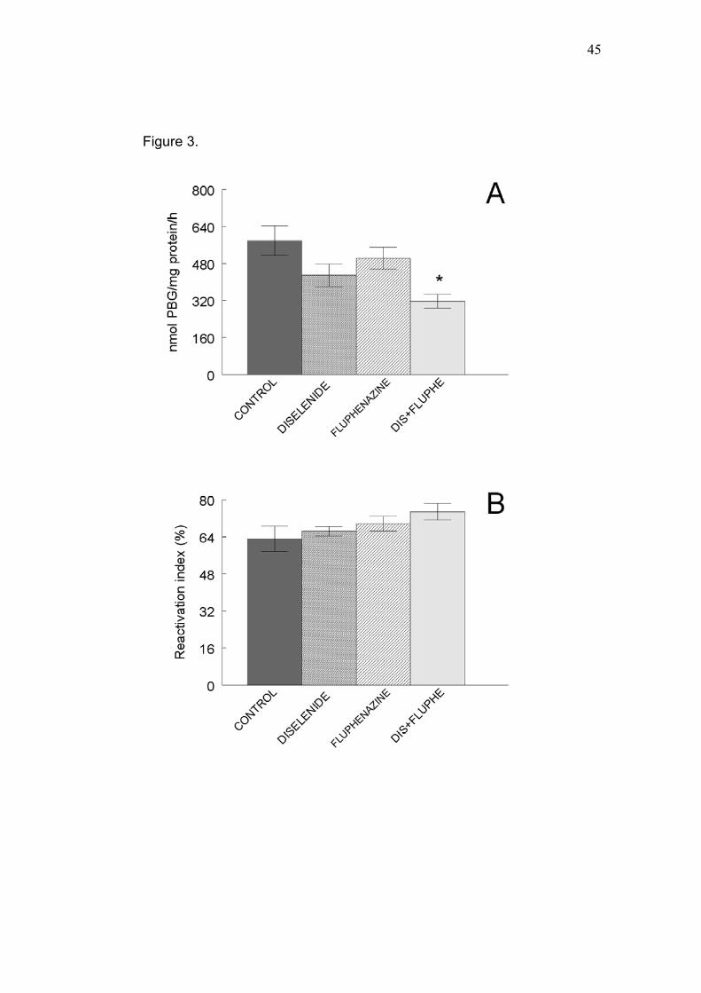
45
Figure 3.

46
Figure 4.

47
Figure 5.

48
3.2 – INTERAÇÕERS POTENCIALMENTE ADVERSAS ENTRE
HALOPERIDOL E VALERIANA
Artigo 2
POTENTIALLY ADVERSE INTERACTIONS BETWEEN HALOPERIDOL
AND VALERIAN
DALLA CORTE, C.L., FACHINETTO, R., COLLE, D., PEREIRA, R.P., ÁVILA,
D.S., VILLARINHO, J.G., WAGNER, C., PEREIRA, M.E., NOGUEIRA, C.W.,
SOARES, F.A.A., ROCHA, J.B.T.
(Aceito para publicação na Food and Chemical Toxicology)

49
Potentially adverse interactions between haloperidol and valerian
Dalla Corte, C.L., Fachinetto, R., Colle, D., Pereira, R.P., Ávila, D.S., Villarinho,
J.G., Wagner, C., Pereira, M.E., Nogueira, C.W., Soares, F.A.A.* and Rocha,
J.B.T.
Universidade Federal de Santa Maria, Centro de Ciências Naturais e Exatas,
Departamento de Química, Programa de Pós-graduação em Ciências
Biológicas: Bioquímica Toxicológica, Camobi, Cep 97105-900, Santa Maria,
RS, Brasil.
*Corresponding author:
Félix Alexandre Antunes Soares - UFSM – CCNE – Dep. de Química
Cep 97105-900, Camobi, Santa Maria, RS, Brasil
Tel: #55-55-3220-9522, Fax: #55-55-3220-8978
e-mail: [email protected]
Running title: Haloperidol, valerian and hepatic damage
Keywords: Haloperidol; Valeriana officinalis; Herb-drug interaction; Oxidative
stress; δ-ALA-D.

50
Abstract
This study was designed to determine whether the treatment of
haloperidol (HP), valerian or both in association impairs the liver or kidney
functions. Valerian alone did not affect oxidative stress parameters in the liver
or kidney of rats. HP alone only increased glutathione (GSH) depletion in liver,
but not in kidney. However, when HP was associated with valerian, an increase
in lipid peroxidation levels and dichlorofluorescein (DCFH) reactive species
production was observed in the hepatic tissue. Superoxide dismutase (SOD)
and Catalase (CAT) activities were not affected by the HP plus valerian
treatment in the liver and kidney of rats. HP and valerian when administered
independently did not affect the activity of hepatic and renal δ-aminolevulinate
dehydratase (δ-ALA-D), however, these drugs administered concomitantly
provoked an inhibition of hepatic δ-ALA-D activity. The δ-ALA-D reactivation
index was higher in rats treated with HP plus valerian than other treated groups.
These results strengthen the view that δ-ALA-D can be considered a marker for
oxidative stress. Serum aspartate aminotransferase (AST) activity was not
altered by any treatment. However, serum alanine aminotransferase (ALT)
activity was higher in the HP group and HP plus valerian group. Our findings
suggest adverse interactions between haloperidol and valerian.

51
1. Introduction
Haloperidol (HP) is a drug with useful properties in the management of
psychosis (Creese et al., 1976). The long-term use of HP, however, is
associated with extrapyramidal side effects, including tardive dyskinesia (See
and Ellison, 1990). This syndrome is characterized by involuntary orofacial
movements and is often irreversible even after drug withdrawal (Gerlach and
Casey, 1988; Glazer et al., 1990; Andreasen and Jorgensen, 2000). The
persistence of tardive dyskinesia after the discontinuation of HP treatment
suggests that this condition may be related to a neuronal lesion induced by HP
or a reactive metabolite(s) derived from its metabolism (Wright et al., 1998).
In line with this, HP can be metabolized to the toxic intermediate 4-(4-
chlorophenyl)-1-[4-(4-fluorophenyl)-4-oxobutyl]pyridinium) (HPP+) in the liver
(Subramanyam et al., 1991; Bloonquist et al., 1994; Rollema et al., 1994; Fang
et al., 1995). This reactive metabolite is thought to share some toxic similarities
with the neurotoxic agent 1-methyl 4-phenyl 1,2,3,6-tetrahydropyridine (MPTP)
and can be involved in the genesis of some Parkinson-like effects associated
with the chronic use of HP (Ablordeppey and Borne, 1993).
Valerian root (Valeriana officinalis L.) is the most commonly used herbal
product to induce sleep in both the United States and Europe (Richman and
Witkowski, 1998; 1999; Houghton, 1999; Ang-Lee et al., 2001; Malva et al.,
2004). Virtually all sleep-aid herbal dietary supplements contain valerian (Ang-
Lee, 2003). The pharmacological activities of valerian are attributed to the
52
different constituents, including valepotriates (valtrate/ isovaltrate and
dihydrovaltrate) (Von der Hude et al., 1985) and valerenic acid. Valerian or its
constituents could induce these effects by interacting with central gamma-
aminobutyric acid (GABA) receptors (Mennini et al., 1993; Cavadas et al.,
1995). In vitro studies testing the binding of valerian extract to GABA receptors
showed that the agonist muscimol was displaced, suggesting valerian binding to
these receptors (Cavadas et al., 1995). Valerenic acid has been shown to inhibit
enzyme-induced breakdown of GABA in the brain resulting in sedation
(Houghton, 1999). Recently data showed the inductive effect of valerian on
cytochrome P450 (CYP) 3A4 and 2D6 enzymes in vitro (Hellum et al., 2006).
This should be seen with caution since alterations of CYP activity could affect
the disposition of conventional pharmaceuticals (Gonzalez, 2005). This is a
major problem in human therapy especially with CYP3A4 involved in
metabolism of over 60% of all therapeutically used drugs (Wrighton et al.,
2000).
Reports of drug interaction with many botanicals, including valerian have
been noted in literature (Assemi, 2001; Brazier and Levine, 2003). Natural
products can interact with drugs by affecting the biological processes that
regulate their metabolism and elimination (Bailey and Dresser, 2004). The
widespread use of valerian supplements suggests that use with conventional
medications is inevitable, and the potential for drug interactions is undefined
(Fugh-Berman and Ernst, 2001).
53
HP was used in this study for the reason that it is extensively use in
clinical practice to treat psychotic disorders and severe behavioral problems,
and because the side effects of HP on the CNS could be attributed to toxic
metabolites and/or oxidative stress (Wright et al., 1998; Burger et al., 2005).
Also, other side effects of HP like anxiety, restlessness, agitation and insomnia
are of interest for this work since valerian major effect is sedation (Malva et al.,
2004). Long-term safety studies with valerian are lack in the literature (Hadley
and Petry, 2003). In view of the fact that valerian has been reported to affect the
metabolism of other drugs, considering that haloperidol is extensively
metabolized in the liver and used in long-term treatments (Forsman et al.,
1977), the present study was undertaken to determine whether the treatment of
haloperidol, valerian or both in association can affect the liver or kidney of rats
in a long-term study.
2. Materials and Methods
2.1. Chemicals
Haloperidol Decanoate was donated by Cristália (São Paulo, Brazil). A
standard tincture of Valeriana officinalis (10 g of valerian roots per 100 mL of
ethanol) was obtained from Bio extracts (São Paulo, Brazil). O-phthalaldehyde,
thiobarbituric acid and DL-dithiothreitol (DTT) were obtained from Sigma (St.
Louis, MO, USA). HgCl2, NaCl, K2HPO4, KH2PO4, trichloroacetic acid, para-
dimethylaminobenzaldehyde, and glacial acetic acid were purchased from

54
Reagen (Rio de Janeiro, RJ, Brazil). All other chemicals were purchased from
Merck (Darmstadt, Germany).
2.2. Animals
Male Wistar rats weighing 295 (± 35.36) g with approximately 3 months
of age, from our own breeding colony were kept in cages with continuous
access to food and V. officinalis or its vehicle (ethanol 1%), in a room with
controlled temperature (22±3°C) and in 12h light/ dark cycle with lights on at
7:00 am. The animals were maintained and used in accordance with the
guidelines of the Committee on Care and Use of Experimental Animal
Resources, School of Veterinary Medicine and Animal Science of Federal
University of Santa Maria, Brazil.
2.3. Treatments
Treatment consisted of 1% ethanol (vehicle of V. officinalis) or 1% (final
concentration of 100 mg/mL) of a standard tincture of V. officinalis (10 g of
valerian roots per 100 mL of ethanol) in the drinking water. The dosage was
calculated every week by the amount of water drunk assuming equal drinking
among the four animals in each cage. Thus, each animal received V. officinalis
extract in a dosage about 200-250 mg/Kg/day. The doses of valerian were
based on the daily recommended dose for an adult human which is 3060 mg
(as inscribed on the commercially bottle of valerian by Nature’s way product).
These solutions were placed daily before the beginning of the dark cycle. It was
not observed a reduction in liquid intake. Haloperidol Decanoate or its vehicle

55
(soy oil) were administered intramuscularly (i.m.) every 28 days (38 mg/kg) that
is equivalent to 1mg/kg/day of unconjugated HP (Fachinetto et al., 2005). V.
officinalis treatment started 15 days before the administration of HP. The
treatment with haloperidol was carried out during 12 weeks concomitantly with
V. officinalis. This treatment was performed according to a previous study of
Fachinetto et al. (2007).
Animals were divided into four groups of 12 animals each:
1 -Control: received soy oil (i.m.) and 1% of ethanol in the drink water;
2 -Valerian: received soy oil (i.m.) and 1% of V. officinalis in the drink
water;
3 -HP: received Haloperidol Decanoate (i.m.) and 1% of ethanol in the
drink water;
4 -Combined treatment: received Haloperidol Decanoate (i.m.) and 1%
of V. officinalis in the drink water.
2.4. Tissue preparation
Animals were euthanized by decapitation. Liver and kidney were quickly
removed, placed on ice and homogenized at 7 and 5 volumes of 0.9% NaCl,
respectively. The homogenates were centrifuged at 4,000 x g for 10 min to yield
a low-speed supernatant fraction (S1) that was used for the biochemical and
enzymatic assays, (except for the GSH/GSSG assay). In order to perform SOD
and CAT assay, S1 was diluted as described in the respective sections (2.7.2
and 2.7.3).
56
2.5. Lipid peroxidation assay
Thiobarbituric acid reactive species (TBARS) were determined as
described by Ohkawa et al. (1979). In brief, samples were incubated at 100 ºC
for 1 h in a medium containing 8.1 % sodium dodecyl sulfate, 1.4 M acetic acid,
pH 3.4 and 0.6% thiobarbituric acid. The pink chromogen produced by the
reaction of thiobarbituric acid with malondialdehyde (MDA), a secondary
product of lipid peroxidation, was measured spectrophotometrically at 532 nm.
Standard curve of MDA was used in order to calculate MDA concentrations.
Results were expresses as nmol of MDA/ mg of protein.
2.6. Estimation of reactive species production
Formation of reactive species was estimated according to a previous
report (Ali et al., 1992). Liver and kidney tissue samples were homogenized in
2.2 mL of saline solution (0.9% NaCl). Aliquots of 2.5 mL were incubated in the
presence of 5 μM 2’,7’-dichlorofluorescein diacetate at 37 ºC for 60 min.
Fluorescent signals were recorded at the end of the incubation at an excitation
wavelength of 488 nm and an emission wavelength of 525 nm. Results were
expressed as fluorescence units.
2.7. Enzyme assays
2.7.1. δ-ALA-D activity
δ-ALA-D activity was assayed according to the method of Sassa (1982)
by measuring the rate of product (porphobilinogen-PBG) formation. The
reaction product was determined using modified Ehrlich’s reagent at 555 nm,

57
with a molar absorption coefficient of 6.1×10⁴ M-1 for the Ehrlich-PBG salt. The
incubation medium contained δ-ALA 2.4 mM and potassium phosphate buffer
(pH 6.8) 0.084 M. The reaction was initiated by the addition of enzymatic
material and the incubations were carried out for 90 and 150 minutes, for liver
and kidney respectively, at 39°C. Afterwards, the reaction was stopped by the
addition of trichloroacetic acid 10% containing HgCl2 0.01 M. The activity of δ-
ALA-D was expressed as nmol of PBG/ mg of protein/ h. Simultaneously, a set
of tubes was assayed using the same protocol, except that 2 mM DTT was
added in order to obtain the reactivation index. This index indicates the extent of
the reactivation of δ-ALA-D activity. The reactivation index of δ-ALA-D activity
was calculated as follows:
(δ-ALA-D activity with DTT - δ-ALA-D activity without DTT) x 100%
δ-ALA-D activity with DTT
2.7.2. SOD activity
S1 of kidney and liver was adequately diluted with 40 and 60 volumes of
0.9% NaCl, respectively, in order to perform the SOD assay (Misra and
Fridovich, 1972). Briefly, epinephrine undergoes auto-oxidation at pH 10.2 to
produce adrenochrome, a colored product that was detected at 480 nm. The
addition of samples (10, 20, 30 µL) containing SOD inhibits the auto-oxidation of
epinephrine. The rate of inhibition was monitored during 180 seconds. The
amount of enzyme required to produce 50% inhibition was defined as one unit
of enzyme activity.

58
2.7.3. CAT activity
CAT activity was measured by the method of Aebi (1974). An aliquot of
liver and kidney supernatants (10 µL) diluted with 60 and 40 volumes of 0.9%
NaCl, respectively, was added to a quartz cuvette and the reaction was started
by the addition of freshly prepared H2O2 (30 mM) in phosphate buffer (50 mM,
pH 7). The rate of H2O2 decomposition was measured spectrophotometrically at
240 nm during 120 seconds. The activity of CAT was expressed as µmol
H2O2/mg protein/min.
2.7.4. Serum transaminases
Serum enzymes, AST and ALT were used as biochemical markers of
hepatic damage, using a commercial Kit (LABTEST, Diagnostica S.A., Minas
Gerais, Brazil) (Reitman and Frankel, 1957).
2.8. Fluorometric assay of reduced (GSH) and oxidized (GSSG) glutathione
For measurement of GSH and GSSG levels, we used a method
previously described by Hissin and Hilf (1976). Briefly, 250 mg of the tissue was
homogenized in 3.75 mL phosphate-EDTA buffer (pH 8) plus 1 mL HPO3 (25%).
Homogenates were centrifuged at 4°C at 100,000 g for 30 min and the
supernatant were separated in two different aliquots for measurement of GSH
and GSSG.
For GSH measurement, 500 μL of the supernatant was added to 4.5 mL
of phosphate buffer. The final assay mixture (2.0 mL) contained 100 μL of the
diluted tissue supernatant, 1.8 μL of phosphate buffer, and 100 μL of o-

59
phthalaldehyde (1 μg/μL). The mixtures were incubated at room temperature for
15 min and their fluorescent signals were recorded in the luminescence
spectrometer at 420 nm of emission and 350 nm of excitation wavelengths.
For measurement of GSSG levels, a 500 μL portion of the original
supernatant was incubated at room temperature with 200 μL of N-
ethylmaleimide (NEM) (0.04 M) for 30 min to react with free GSH to prevent its
oxidation to GSSG. To this mixture, 4.3 μL of NaOH (0.1 N) was added. A 100-
μL portion of this mixture was taken for measurement of GSSG, using the
procedure outlined above for GSH assay, except that NaOH was employed as
diluent rather than phosphate-EDTA buffer. Results were expressed as
GSH/GSSG ratio.
2.9. Protein measurement
Protein was assayed by the method of Lowry et al. (1951), with bovine
serum albumin as standard.
2.10. Statistical analysis
Data were analyzed statistically by one-way ANOVA, followed by
Duncan’s multiple range test when appropriate. The results were considered
statistically significant for p<0.05.
3. Results
Lipid Peroxidation

60
TBARS levels in liver homogenates were not affected by HP or valerian
when compared to the control (Fig. 1A). However, in the combined treatment
group a significant increase in TBARS production (about 15%) was detected
when compared to the other groups (p<0.05, Fig. 1A). Renal TBARS levels
were not modified by any of the treatments (Fig. 1B). <Insert figure 1 here>
Estimation of reactive species production
Reactive species production was measured by DCFH oxidation.
Combined treatment significantly enhanced the oxidation of DCFH (about 70%)
in liver homogenates when compared to the control group (Fig. 2A, p<0.05). HP
and valerian alone did not alter hepatic reactive species production (Fig. 2A);
however, it is worth mentioning that they may have had an additive effect on
DCFH oxidation in liver. In kidney homogenates, DCFH oxidation was not
modified by any treatment (Fig. 2B). <Insert figure 2 here>
Enzyme activity
Hepatic and renal SOD activities were not altered by HP, valerian or
combined treatment (data not shown). CAT activity was not modified by any of
treatments in both organs (data not shown). Isolated HP and valerian treatment
did not affect hepatic or renal δ-ALA-D activity (Fig. 3A and 4A). <Insert figure 3
here> However, HP in combination with valerian showed an inhibitory effect on
δ-ALA-D activity in the liver (Fig 3A, p<0.05) but not in the kidney (Fig 4A). In
vitro, DTT, a classic agent that restores oxidized thiol groups, was able to
restore liver δ-ALA-D activity (Fig 3B). In fact, the combined treatment of HP

61
and valerian had the highest δ-ALA-D reactivation index (Fig 3B, p<0.05). In the
kidney, there was no difference in the δ-ALA-D reactivation index among groups
(Fig 4B). <Insert figure 4 here>
Serum AST activities were not modified by any treatment (Figure 5A).
Rats treated with HP presented an increase in serum ALT activities when
compared to control rats (Figure 5B, p<0.05). The valerian treatment did not
significantly alter serum ALT activities, although its association with HP
statistically increased this parameter (Figure 5B, p<0.05). The magnitude of the
increase observed in serum ALT activities deserves attention however it was
not high enough to be of clinical concern. <Insert figure 5 here>
GSH and GSSG
The HP treatment provoked a decrease in the GSH/GSSG ratio in liver
homogenates and the valerian concomitant treatment did not avoid the HP
effects (Fig. 6A, p<0.05). In fact valerian it self had no effect on the, GSH/GSSG
ratio. In the kidney, none of the treatments modified the GSH/GSSG ratio (Fig.
6B). <Insert figure 6 here>
4. Discussion
The long-term use of HP is associated to side effects such as
Parkinsonism and tardive dyskinesia, and these syndromes have been
attributed to a toxic metabolite of HP, the pyridinium metabolite HPP+ (Wright et

62
al., 1998). Nevertheless, there are few reports demonstrating the toxic effects of
HP on the liver or kidney. Since haloperidol is widely used in clinical practice,
the knowledge of its toxicity is essential in the choice of this antipsychotic agent.
The results presented here showed that HP alone only altered one of the
oxidative stress parameters in the liver (GSH/GSSG) and none in the kidney.
On the other hand, when HP was associated to valerian, a significant increase
in TBARS levels and DCFH reactive species production was observed in the
hepatic tissue. Our results indicate an increase in oxidative damage evoked by
HP plus valerian.
Despite the oxidative damage, SOD and CAT activities were not affected
by combined treatment in the liver. Similarly, HP failed to alter plasma SOD and
CAT activities in patients taking this drug (Yao et al., 1998). Considering that
HP is extensively metabolized in the liver with only approximately 1% of the
administered dose excreted in the urine (Forsman et al., 1977), we were not
surprised that this treatment did not cause any deleterious effects on kidney
from rats of this study.
Valeriana officinalis is widely utilized for its sedative properties,
although there is a lack in the literature of long-term safety studies with
valerian (Hadley and Petry, 2003). In our study, valerian alone did not show
any effect on TBARS levels, DFCH production or SOD and CAT activities. Al-
Majed and colleagues (2006) reported an increase in the concentrations of
MDA and a decrease of non-protein sulfhydryl levels in hepatic cells of mice

63
after sub-acute treatment with valerian. However, in this study we only
observed side effects when valerian was associated to HP.
δ-ALA-D activity was tested here since it is a sulfhydryl-containing
enzyme that is susceptible to oxidizing agents and is inhibited after exposure to
pro-oxidant situations (Folmer et al., 2002, 2003; Soares et al., 2003;
Gonçalves et al., 2005; Santos et al., 2005). The inhibition of δ-ALA-D may
impair heme biosynthesis and can result in the accumulation of aminolevulinic
acid, which has been demonstrated to be a pro-oxidant molecule under
significant physiological conditions (Bechara et al., 1993; Bechara, 1996;
Emanuelli et al., 2001). HP and valerian when administered separately did not
affect the activity of δ-ALA-D, however, these drugs administered in association
provoked an inhibition of hepatic δ-ALA-D activity. This is supported by the δ-
ALA-D reactivation index which presented the highest level in the combined
treatment when compared to the other groups. HP and valerian could be
interacting in a manner that oxidizes δ-ALA-D sulfhydryl groups, probably by
producing some oxidative by-products. These data are in agreement with the
fact that DTT, a classic reducing agent that restores oxidized thiol containing
enzymes (Perottoni et al., 2005), could restore the inhibition caused by the
combined treatment. In fact, it has been reported that valtrate, a component of
valerian, may interact with free sulfhydryl groups (Keochanthala-Bounthanh et
al., 1990). Perhaps HP can enhance this capacity of valerian to interact with

64
sulfhydryl groups or affect the susceptibility of the liver. These results strengths
the view that δ-ALA-D can be considered a marker for oxidative stress.
The release of intracellular enzymes (AST and ALT), which is presumed
to be a result of injury to cellular membranes, was utilized here as an index of
hepatotoxicity. Serum AST activity was not altered by any treatment. However,
serum ALT activity was higher in the HP and combined treatment. Gaertner and
collaborators (2001) reported elevation in transaminases in patients treated with
HP, which support these results. Anyway the increase in ALT activity observed
here was small to be of clinical concern.
In our study, HP caused a decrease in the GSH/GSSG index in the liver,
which was also observed in the HP and valerian combined treatment. This
decrease, which lowers cellular defenses, can contribute to the increase in
TBARS and DCFH oxidation, and lead to the elevations observed in serum ALT
activity.
The results present here point to an interaction between HP and valerian
in the increase of oxidative stress in the liver. Gold and colleagues (2001)
identified a series of potential interactions between herbal natural health
products, including valerian, and conventional drug therapies. Herb–drug
interactions can appear when herbs and chemical drugs are co-administered
and the herbal preparation (one or more components) modulates the
metabolism of the chemical drug by induction or inhibition of CYP enzymes.
Hellum and collaborators (2006) showed the capacity of valerian extracts to

65
induce CYP enzymes in cultured primary human hepatocytes, including
CYP3A4, the most important P450 enzyme responsible for HP metabolic
pathways (Igarashi et al., 1995; Usuki et al., 1996; Fang et al., 2001; Kalgutkar
et al., 2003; Avent et al., 2006). Based on these reports, we suppose that the
mechanism by which valerian-HP association induced oxidative damage may
be related to CYP activity. The inducible effects of valerian on CYP enzymes
could increase the production of toxic HP metabolites, such as HPP+,
responsible for the deleterious effects seen in the valerian plus HP treatment in
the liver. The adverse effects of valerian plus HP treatment observed in the liver
and the absence of these effects in kidney also could be explaining by this
mechanism. CYP enzymes are found at highest levels in the liver, and
expressed at lower levels in kidney (Gonzalez, 2005). Thus, liver are the major
site of herb-drug interaction in which the production of toxic HP metabolites
could be elevated.
The route of administration of Haloperidol Decanoate (i.m.) as well as the
dose of HP used in this study was equivalent to that indicated for humans which
increase the relevance of this work since our model could be translated into the
human situation. The doses of valerian and HP used here was within the normal
range for humans, but a possible negative interaction between these drugs will
occur in a real human situation only after ingestion of supratherapeutic doses of
valerian and HP. Anyway a possible toxic additive effect of these two
compounds must be kept in mind. In this way, the administration of valerian in

66
people taking HP should be better studied to verify possible negative
interactions.
The use of herbs as alternative and/or complementary therapy in the
Western world is on the rise and gaining increasing popularity (Hellum et al.,
2006). Unfortunately, their use has often been accompanied by an unfounded
belief that “natural” is equal to “safe”. The widespread use of valerian
supplements suggests that its use with conventional medications is inevitable.
Moreover, the potential for drug interactions with natural products is undefined
(Fugh-Berman and Ernst, 2001). The findings from our study suggest an
adverse effect in the interactions between haloperidol and valerian causing
hepatic damage related to oxidative stress. However, further work is needed to
confirm the exact mechanism by which these compounds cause oxidative
stress. Moreover, caution is warranted if attempting to make such an
extrapolation to other herbals and drug therapies.
Acknowledgments: Supported by the FINEP research grant “Rede Instituto
Brasileiro de Neurociência (IBN-Net)” # 01.06.0842-00. Additional support
giving by CNPq, FAPERGS, CAPES, Cristália-SP and UFSM (FIPE). C.L.D.C.
and R.F. receive fellowship from CAPES and CNPq (Fomento Tecnológico),
R.P.P. received a scientific initiation fellowship from CNPq-Fomento
Tecnologico, D.E.C. received fellowship from UFSM (FIPE), C.W.N. and
J.B.T.R. are the recipients of CNPq fellowships.

67
References
Ablordeppey, S.Y., Borne, R.F., 1993. Detection of a neurotoxic quaternary
pyridinium metabolite in the liver of haloperidolnext term-treated rats.
Pharmacol. Biochem. Behav. 46, 739-744.
Aebi H., 1974. Catalase, In: Bergmeyer, H.V. (Ed.), Methods of Enzymatic
Analysis. VEB Chemie Weinheim, pp. 673-677.
Ali, S.F., LeBel, C.P., Bondy, S.C., 1992. Reactive oxygen species formation as
a biomarker of methylmercury and trimethyltin neurotoxicity. Neurotoxicology
13, 637-648.
Al-Majed, A.A., Al-Yahya, A.A., Al-Bekairi, A.M., Al-Shabanah, O.A., Qureshi,
S., 2006. Studies on the cytological and biochemical effects of valerian in
somatic and germ cells of Swiss albino mice. Food Chem. Toxicol. 44, 1830-
1837.
Andreasen, O.A., Jorgensen, H.A., 2000. Neurotoxicity associated with
neuroleptic-induced oral dyskinesias in rats. Implications for tardive
dyskinesia? Prog. Neurobiol. 61, 525-541.
Ang-Lee, M.K., Moss, J., Yuan, C.S., 2001. Herbal medicines and perioperative
care. JAMA 286, 208-216.
Ang-Lee, M.K., 2003. Most commonly used herbal medicines in the U.S.A., In:
Yuan, C.S. and Bieber, E.J. (Eds), Textbook of complementary and
alternative medicine. Parthenon, New York, pp. 7–32.
68
Assemi, M., 2001. Herbs affecting the central nervous system: gingko, kava, St.
John’s wort, and valerian. Clin. Obstet. Gynecol. 48, 824-835.
Avent, K.M., DeVoss, J.J., Gillam, E.M., 2006. Cytochrome P450-mediated
metabolism of haloperidol and reduced haloperidol to pyridinium
metabolites. Chem. Res. Toxicol. 19, 914-920.
Bailey, D.G., Dresser, G.K., 2004. Natural products and adverse drug
interactions. CMAJ 170, 1531-1532.
Bechara, E.J.H., Medeiros, M.H.G., Monteiro, H.P., Hermes-Lima, M., Pereira,
B., Demasi, M., Costa, C.A., Adballa, D.S.P., Onuki, J., Wendel, C.M.A.,
Masci, P.D., 1993. A free radical hypothesis of lead poisoning and in Born
porphyrias associated with 5-aminolevulinic overload. Quim. Nova 16, 385-
392.
Bechara, E.J., 1996. Oxidative stress in acute intermittent porphyria and lead
poisoning may be triggered by 5-aminolevulinic acid. Braz. J. Med. Biol.
Res. 29, 841-51.
Bloomquist, J., King, E., Wright, A., Mytilineou, C., Kimura, K., Castagnoli, K.,
Castagnoli Jr, N., 1994. 1-Methyl-4-phenylpyridinium-like neurotoxicity of a
pyridinium metabolite derived from haloperidol: cell culture and
neurotransmitter uptake studies. J. Pharmacol. Exp. Ther. 270, 822-830.
Brazier, N.C., Levine, M.A., 2003. Drug-herb interaction among commonly used
conventional medicines: a compendium for health care professionals. Am. J.
Ther. 10, 163-169.
69
Burger, M.E., Fachineto, R., Alves, A., Callegari, L., Rocha, J.B., 2005. Acute
reserpine and subchronic haloperidol treatments change synaptosomal brain
glutamate uptake and elicit orofacial dyskinesia in rats. Brain Res. 1031,
202-210.
Cavadas, C., Araujo I., Cotrim, M.D., Amaral, T., Cunha, A.P., Macedo, T.,
Ribeiro, C.F., 1995. In vitro study on the interaction of Valeriana officinalis L.
extracts and their amino acids on GABAA receptor in rat brain.
Arzneimittelforschung 45, 753-755.
Creese, I., Burt, D.R., Snyder, S.H., 1976. Dopamine receptor binding predicts
clinical and pharmacological potencies of anti-schizophrenic drugs. Science
192, 481-483.
Emanuelli, T., Pagel, F.W., Alves, L.B., Regner, A., Souza, D.O., 2001.
Inhibition of adenylate cyclase activity by 5- aminolevulinic acid in rat and
human brain. Neurochem. Int. 38, 213-218.
Fachinetto, R., Burger, M.E., Wagner, C., Wondracek, D.C., Brito, V.B.,
Nogueira, C.W., Ferreira, J., Rocha, J.B., 2005. High fat diet increases the
incidence of orofacial dyskinesia and oxidative stress in specific brain
regions of rats. Pharmacol. Biochem. Behav. 81, 585-592.
Fachinetto, R., Villarinho, J., Wagner, C., Pereira, R.P., Ávila, D. S., Burger,
M.E., Calixto, J.B., Rocha, J.B.T., Ferreira, J., 2007. Valeriana officinalis
does not alter the orofacial dyskinesia induced by haloperidol in rats: role of

70
dopamine transporter. Prog. Neuropsychopharmacol. Biol. Psychiatry 31,
1478-1486.
Fang, J., Zuo, D., Yu, P.H., 1995. Comparison of cytotoxicity of a quaternary
pyridinium metabolite of haloperidol (HP+) with neurotoxin N-methyl-4-
phenylpyridinium (MPP+) towards cultured dopaminergic neuroblastoma
cells. Psychopharmacology (Berl.) 121, 373-378.
Fang, J., Mckay, G., Song, J., Remillrd, A., LI, X., Midha, K., 2001. In vitro
characterization of the metabolism of haloperidol using recombinant
cytochrome P450 enzymes and human liver microsomes. Drug Metab.
Dispos. 12, 1638–1643.
Folmer, V., Soares, J.C., Rocha, J.B.T., 2002. Oxidative stress in mice is
dependent on the free glucose content of the diet. Int. J. Biochem. Cell Biol.
34, 1279-1285.
Folmer, V., Soares, J.C., Gabriel, D., Rocha, J.B.T., 2003. A high fat diet
inhibits delta-aminolevulinate dehydratase and increases lipid peroxidation
in mice (Mus musculus). J. Nutr. 133, 2165-2170.
Forsman, A., Folsch, G., Larsson, M., Ohman, R., 1977. The metabolism of
haloperidol in man. Curr. Ther. Res. Clin. Exp. 21, 606-617.
Fugh-Berman, A., Ernst, E., 2001. Herb-drug interactions: review and
assessment of report reliability. Br. J. Clin. Pharmacol. 52, 587-595.

71
Gaertner, I., Altendorf, K., Batra, A., Gaertner, H.J., 2001. Relevance of liver
enzyme elevations with four different neuroleptics: a retrospective review of
7,263 treatment courses. J. Clin. Psychopharmacol. 21, 215-222.
Gerlach, J., Casey, D., 1988. Tardive dyskinesia. Acta Psychiatr. Scand. 77,
369-378.
Glazer, W.M., Morgenstern, H., Shooler, N., Berkman, C.S., Moore, D.C., 1990.
Predictors of improvement in tardive dyskinesia following discontinuation of
neuroleptic medication. Br. J. Psychiatry 157, 585-592.
Gold, J.L., Laxer, D.A., Dergal, J.M., Lanctot, K.L., Rochon, P.A., 2001. Herbal-
drug therapy interaction: a focus on dementia. Curr. Opin. Clin. Nutr. Metab.
Care 4, 29-34.
Gonçalves, T.L., Erthal, F., Corte, C.L.D., Muller, L.G., Piovezan, C.M.,
Nogueira, C.W., Rocha, J.B.T., 2005. Involvement of oxidative stress in the
pre-malignant and malignant states of cervical cancer in women. Clin.
Biochem. 38, 1071-1075.
Gonzalez, F.J., 2005. Role of cytochromes P450 in chemical toxicity and
oxidative stress: studies with CYP2E1. Mutat. Res. 569, 101-110.
Hadley, S., Petry, J.J., 2003. Valerian. Am. Fam. Physician 67, 1755-1758.
Hellum, B.H., Hu, Z., Nilsen, O.G., 2006. The Induction of CYP1A2, CYP2D6
and CYP3A4 by six trade herbal products in cultured primary human
hepatocytes. Basic Clin. Pharmacol. Toxicol. 100, 23–30.

72
Hissin, P.J., Hilf, R., 1976. A fluorimetric method for determination of oxidized
and reduced glutathione in tissues. Anal. Biochem. 74, 214–226.
Houghton, P.J., 1999. The scientific basis for the reputed activity of valerian. J.
Pharm. Pharmacol. 51, 505-512.
Igarashi, K., Kasuya, F., Fukui, M., Usuki, E., Castagnoli Jr., N., 1995. Studies
on the metabolism of haloperidol (HP): the role of CYP3A in the production
of the neurotoxic pyridinium metabolite HPP+ found in rat brain following IP
administration of HP. Life Sci. 57, 2439-2446.
Kalgutkar, A.S., Taylor, T.J., Venkatakrishnan, K., Isin, E.M., 2003. Assesment
of the contributions of CYP3A4 and CYP3A5 in the metabolism of the
antpsychotic agent haloperidol to its potencially neurotoxin pyridinium
metabolite and effect of antidepressants on the bioactivation pathway. Drug
Metab. Dispos. 31, 243-249.
Keochanthala-Bounthanh, C., Haag-Berrurier, M., Beck, J.P., Anton, R., 1990.
Effects of thiol compounds versus the cytotoxicity of valepotriates on
cultured hepatoma cells. Planta Med. 56, 190-192.
Lowry, O.H., Rosebrough, N.J., Farr, A.L., Randall, R.J., 1951. Protein
measurement with the Folin phenol reagent. J. Biol. Chem. 193, 265-275.
Malva, J.O., Santos, S., Macedo, T., 2004. Neuroprotective properties of
Valeriana officinalis extracts. Neurotox. Res. 6, 131–140.
Mennini, T., Bernasconi, P., Bombardelli, E., Morazzoni, P., 1993. In vitro study
on the interaction of extracts and pure compounds from Valeriana officinalis
73
roots with GABA, benzodiazepine and barbiturate receptors in rat brain.
Fitoterapia 64, 291–300
Misra, H.P., Fridovich, I., 1972. The role of superoxide anion in the autoxidation
of epinephrine and simple assay for superoxide dismutase. J. Biol. Chem.
247, 3170–3175.
Ohkawa, H., Ohishi, H., Yagi, K., 1979. Assay for lipid peroxide in animal
tissues by thiobarbituric acid reaction. Anal. Biochem. 95, 351-358.
Perottoni, J., Meotti, F.C., Folmer, V., Pivetta, L., Nogueira, C.W., Zeni, G.,
Rocha, J.B.T., 2005. Ebselen and diphenyl diselenide do not change the
inhibitory effect of lead acetate on delta aminolevulinate dehydratase.
Environ. Toxicol. Pharmacol. 19, 239-248.
Reitman, S., Frankel, S., 1957. A colorimetric method for the determination of
serum glutamic oxalacetic and glutamic pyruvic transaminases. Am. J. Clin.
Pathol. 28, 56–63.
Richman, A., Witkowski, J.P., 1998. 4th Annual Herbal Products Sales Survey,
Whole Foods 21, 19–26.
Richman, A., Witkowski, J.P., 1999. 5th Annual Herbal Products Sales Survey,
Whole Foods 22, 49-56.
Rollema, H., Skolnik, M., D'Engelbronner, J., Igarashi, K., Usuki, E., Castagnoli
Jr, N., 1994. MPP(+)-like neurotoxicity of a pyridinium metabolite derived
from haloperidol: in vivo microdialysis and in vitro mitochondrial studies. J.
Pharmacol. Exp. Ther. 268, 380-387.

74
Santos, F.W., Zeni, G., Rocha, J.B.T., Nascimento, P.C., Marques, M.S.,
Nogueira, C.W., 2005. Efficacy of 2,3-dimercapto-1-propanesulfonic acid
(DMPS) and diphenyl diselenide on cadmium induced testicular damage in
mice. Food Chem. Toxicol. 43, 1723-1730.
Sassa, S., 1982. Delta-aminolevulinic acid dehydratase assay. Enzyme 28,
133-145.
See, R.E., Ellison, G., 1990. Intermittent and continuous haloperidol regimens
produce different types of oral dyskinesias in rats. Psychopharmacology
(Berl.) 100, 404–412.
Soares, J.C., Folmer, V., Rocha, J.B.T., 2003. Influence of dietary selenium
supplementation and exercise on thiol-containing enzymes in mice. Nutrition
19, 627-632.
Subramanyam, B., Woolf, T., Castagnoli Jr, N., 1991. Studies on the in vitro
conversion of haloperidol to a potentially neurotoxic pyridinium metabolite.
Chem. Res. Toxicol. 4, 123-128.
Usuki, E., Pearce, R., Parkinson, A., Castagnol Jr, N., 1996. Studies on the
conversion of haloperidol and its tetrahydropyridine dehydration product to
potentially neurotoxic pyridinium metabolites by human liver microsomes.
Chem. Res. Toxicol. 9, 800-806.
Von der Hude, W., Scheutwinkel-Reich, M., Braun, R., Dittmar, W., 1985. In
vitro mutagenicity of valepotriates. Arch. Toxicol. 56, 267–271.

75
Yao, J.K., Reddy, R., McElhinny, L.G., van Kammen, D.P., 1998. Effects of
haloperidol on antioxidant defense system enzymes in schizophrenia. J.
Psychiatr. Res. 32, 385-391.
Wright, A.M., Bempong, J., Kirby, M.L., Barlow, R.L., Bloomquist, J.R., 1998.
Effects of haloperidol metabolites on neurotransmitter uptake and release:
possible role in neurotoxicity and tardive dyskinesia. Brain Res. 788, 215-
222.
Wrighton, S.A., Schuetz, E.G., Thummel, K.E., Shen, D.D., Korzekwa, K.R.,
Watkins, P.B., 2000. The human CYP3A subfamily: practical considerations.
Drug Metab. Rev. 32, 339-361.

76
Legends
Figure 1. Effects of valerian and/ or HP treatments on TBARS production
in liver (A) and kidney (B) homogenates. Data are expressed as mean ± S.E.M.
for twelve rats per group. Experiments were performed in duplicates. (*)
represents p<0.05 as compared to control group by Duncan’s multiple range
test.
Figure 2. Effects of valerian and/ or HP treatments on DCFH oxidation in
liver (A) and kidney (B) homogenates. Data are expressed as mean ± S.E.M.
for twelve rats per group. Experiments were performed in duplicates. (*)
represents p<0.05 as compared to control group by Duncan’s multiple range
test.
Figure 3. Effects of valerian and/ or HP treatments on δ-ALA-D activity in
liver homogenates (A) and on the enzyme reactivation index (B). Data are
expressed as mean ± S.E.M. for twelve rats per group. Experiments were
performed in duplicates. (*) represents p<0.05 as compared to control group by
Duncan’s multiple range test.
Figure 4. Effects of valerian and/ or HP treatments on δ-ALA-D activity in
kidney homogenates (A) and on the enzyme reactivation index (B). Data are
expressed as mean ± S.E.M. for twelve rats per group. Experiments were
performed in duplicates. (*) represents p<0.05 as compared to control group by
Duncan’s multiple range test.

77
Figure 5. Effects of valerian and/ or HP treatments on serum AST (A) and
ALT (B) activities. Data are expressed as mean ± S.E.M. for twelve rats per
group. Experiments were performed in duplicates. (*) represents p<0.05 as
compared to control group by Duncan’s multiple range test.
Figure 6. Effects of valerian and/ or HP treatments on GSH/GSSG ratio
in liver (A) and kidney (B) homogenates. Data are expressed as mean ± S.E.M.
for twelve rats per group. Experiments were performed in duplicates. (*)
represents p<0.05 as compared to control group by Duncan’s multiple range
test.

78
Figure 1.

79
Figure 2.

80
Figure 3.

81
Figure 4.

82
Figure 5.

83
Figure 6.

84
4. DISCUSSÃO
O uso de drogas neurolépticas tem sido associado a efeitos colaterais como a DT
e o dano hepático. Apesar dos inúmeros casos de hepatotoxicidade após a administração
de neurolépticos, são escassos os dados na literatura a respeito desses efeitos e o
mecanismo exato pelo qual neurolépticos induzem hepatotoxicidade permanece incerto
(Dumortier, 2002).
Um dos objetivos do presente estudo foi avaliar os efeitos do tratamento crônico
com flufenazina sobre parâmetros de estresse oxidativo em fígado e rim de ratos bem
como o possível efeito protetor do disseleneto de difenila sobre o dano induzido por
flufenazina (artigo 1). Demonstramos neste estudo que a exposição prolongada à
flufenazina provocou um aumento na peroxidação lipídica (TBARS), uma diminuição
na atividade da SOD e um aumento na atividade da CAT no fígado de ratos. Estes
resultados indicam que a administração crônica de flufenazina pode estar associada ao
estresse oxidativo. A flufenazina também induziu peroxidação lipídica no rim, no
entanto as atividades da SOD e da CAT não foram afetadas neste órgão.
As drogas fenotiazínicas são extensivamente oxidadas no organismo formando
radicais catiônicos (O’Brien, 1988; Yang e Kulkarni, 1997; Galati e cols., 2002;
Tafazoli e O'Brien, 2005). Eghbal e cols. (2004) demonstraram que em pH fisiológico
enzimas peroxidases catalisam a oxidação de fenotiazinas em radicais pró-oxidantes, os
quais oxidam ascorbato, GSH e NADH bem como induzem estresse oxidativo e
citotoxicidade quando incubados com hepatócitos. Dessa forma, podemos sugerir que a
peroxidação lipídica induzida por flufenazina poderia ocorrer devido à produção de
metabólitos da flufenazina catalisada por enzimas peroxidases.
O disseleneto de difenila foi utilizado neste estudo devido às suas propriedades
antioxidante e hepatoprotetora já descritas (Nogueira e cols., 2004; Borges e cols.,
2005; 2006). Os resultados deste estudo demonstraram que o disseleneto de difenila foi
eficiente em proteger o fígado e o rim da peroxidação lipídica (TBARS) induzida por
flufenazina. Este efeito protetor do disseleneto de difenila sobre o TBARS foi
acompanhado pela restauração parcial da atividade da CAT no fígado. O disseleneto de

85
difenila também foi capaz de restaurar a atividade da SOD no fígado dos ratos tratados
com flufenazina. A diminuição na atividade da SOD causada pelo tratamento com
flufenazina poderia contribuir para o aumento nos níveis de TBARS observados no
fígado. Podemos, então, atribuir a proteção do disseleneto de difenila à sua atividade
tiol-peroxidase e a outras propriedades antioxidantes deste composto (Arteel e cols.,
2001; Rossato e cols., 2002; Nogueira e cols., 2004). Por outro lado, a atividade da SOD
no rim foi diminuída somente quando a flufenazina foi administrada concomitantemente
com o disseleneto de difenila. Este resultado foi inesperado, e pode indicar uma
interação complexa entre as propriedades antioxidantes do composto de selênio e a
diminuição em uma enzima antioxidante importante no rim. Apesar disso, a
administração de disseleneto de difenila não foi acompanhada por nenhum sinal de
peroxidação lipídica (TBARS) no rim.
O tratamento com flufenazina provocou uma inibição na atividade da δ-ALA-D
e a associação da flufenazina com o disseleneto de difenila não foi capaz de restaurar a
atividade da enzima. Esta associação aumentou a inibição parcial causada pela
flufenazina sozinha. A flufenazina e o disseleneto de difenila poderiam estar interagindo
de tal forma que a oxidação dos grupos sulfidrila (-SH) da δ-ALA-D foi mais
pronunciada do que no grupo tratado apenas com a flufenazina. Estes dados são
reforçados pelo fato do DTT, um agente redutor clássico, ter restaurado a inibição da δ-
ALA-D (Perottoni e cols., 2005) e pelo índice de reativação, que foi o mais elevado no
grupo tratado com flufenazina e disseleneto de difenila.
Ao contrário do fígado, a atividade da δ-ALA-D renal não foi alterada pelo
tratamento com flufenazina, no entanto a associação de flufenazina com disseleneto de
difenila resultou em inibição da atividade da enzima. O DTT foi capaz de restaurar a
atividade da δ-ALA-D renal. Contudo, não houve diferença no índice de reativação para
a δ-ALA-D renal entre os grupos. Com base neste resultado podemos sugerir que a ação
inibitória dessas drogas sobre a atividade da δ-ALA-D no rim não está relacionada com
a oxidação de grupos –SH. A redução da atividade da δ-ALA-D renal poderia ser

86
atribuída a um efeito aditivo do disseleneto de difenila e da flufenazina sobre a atividade
da enzima.
O tratamento realizado neste trabalho foi baseado em estudos prévios, sendo um
modelo de DO em roedores (See e cols., 1992; Van Kampen e Stoessl, 2000; Fachinetto
e cols., 2007). Diversos autores já apresentaram evidências do envolvimento de EROS
no desenvolvimento da DT (Cadet e cols., 1986; Lohr e cols., 1988; Burger e cols.,
2003; Abílio e cols., 2004). Os resultados apresentados neste estudo podem contribuir
para uma melhor compreensão dos efeitos colaterais relacionados ao uso de
neurolépticos, pois, se um metabólito reativo é capaz de induzir estresse oxidativo no
fígado o mesmo também poderia ser produzido no cérebro e, portanto, exercer efeitos
deletérios neste tecido. Além disso, podemos supor que mesmo uma hepatotoxicidade
limitada dos neurolépticos poderia facilitar a sua neurotoxicidade particularmente por
aumentar a susceptibilidade do organismo como um todo aos efeitos danosos dos
radicais livres.
Juntos os resultados deste primeiro artigo, contribuem para um maior
entendimento dos danos hepáticos induzidos pela flufenazina. Os dados apresentados
aqui indicam claramente o importante papel das EROS no tratamento crônico com
flufenazina. Além disso, a proteção do disseleneto de difenila sobre o estresse oxidativo
causado pela flufenazina nos dá uma indicação dos benefícios do disseleneto de difenila
para proteger o fígado de uma variedade de agentes hepatotóxicos.
O segundo objetivo deste estudo foi o de avaliar os efeitos do tratamento
crônico com haloperidol e Valeriana officinalis bem como a sua combinação sobre o
fígado e o rim de ratos (artigo 2). Os resultados apresentados aqui demonstram que o
haloperidol apenas alterou um parâmetro de estresse oxidativo no fígado aumentando a
depleção de GSH, enquanto nenhum parâmetro foi alterado no rim. Por outro lado
quando o haloperidol foi associado a valeriana houve um aumento significativo nos
níveis de TBARS e na produção de espécies reativas (DCFH) no fígado. Nossos
resultados indicam um aumento do dano oxidativo provocado pela associação de
haloperidol e valeriana.
87
Apesar do dano oxidativo observado, as atividades das enzimas SOD e CAT não
foram afetadas por nenhum dos tratamentos no fígado. Da mesma forma, o haloperidol
não alterou as atividades da SOD e CAT plasmáticas em pacientes sob esta medicação
(Yao e cols., 1998). O haloperidol é extensivamente metabolizado no fígado, e apenas
40 % da dose administrada é excretado na urina, sendo aproximadamente 1% excretado
na forma inalterada (Forsman e cols., 1977). Dessa forma, não é surpresa que este
tratamento não tenha causado nenhum efeito deletério sobre o rim de ratos neste estudo.
A Valeriana officinalis é largamente utilizada devido às suas propriedades
sedativas, no entanto existe a necessidade de estudos sobre a segurança do uso
prolongado de valeriana quando associada a outras drogas (Hadley e Petry, 2003). Neste
estudo o tratamento com valeriana não alterou os níveis de TBARS, a oxidação da
DCFH, e as atividades da SOD e da CAT. Al-Majed e colaboradores (2006)
demonstraram o aumento nos níveis de MDA e uma diminuição nos níveis de –SH não
protéico em células hepáticas de camundongos após tratamento sub-agudo com
valeriana. Entretanto, no presente estudo apenas observou-se efeitos colaterais quando a
valeriana foi associada ao haloperidol.
A atividade da δ-ALA-D foi avaliada por esta ser uma enzima que contém
grupos SH os quais são suscetíveis a agentes oxidantes e, portanto, a enzima pode ser
inibida após a sua exposição a situações pró-oxidantes (Folmer e cols., 2002, 2003;
Soares e cols., 2003; Gonçalves e cols., 2005; Santos e cols., 2005). Haloperidol e
valeriana quando administrados separadamente não afetaram a atividade da enzima, no
entanto a sua associação resultou na perda de atividade da δ-ALA-D hepática.
Haloperidol e valeriana poderiam estar interagindo de forma a oxidar os grupos –SH da
δ-ALA-D provavelmente por produzirem algum metabólito mais tóxico para enzima.
Este resultado é reforçado pela restauração da atividade da δ-ALA-D pelo DTT
(Perottoni e cols., 2005), e pelo índice de reativação para a δ-ALA-D o qual foi mais
alto no tratamento combinado que nos outros grupos tratados. Este resultado corrobora a
hipótese de que a enzima foi inibida pela oxidação dos seus grupos -SH. Foi relatado
que o valtrato, um componente da valeriana, pode interagir com grupos –SH

88
(Keochanthala-Bounthanh e cols., 1990). Talvez o haloperidol possa aumentar esta
capacidade da valeriana de interagir com grupos –SH ou afetar a suscetibilidade do
fígado. Estes resultados fortalecem a visão de que a δ-ALA-D pode ser considerada um
marcador de estresse oxidativo.
A liberação de enzimas intracelulares (AST e ALT) é um indicativo de dano às
membranas celulares e é utilizado como um índice de hepatotoxicidade (Cohen e
Kaplan, 1979). A ALT é uma enzima citoplasmática e encontra-se principalmente no
fígado, enquanto a AST é predominantemente mitocondrial e pode aparecer elevada em
doenças de outros órgãos, como o coração ou o músculo. Dessa forma, elevações na
ALT representam um marcador mais sensível de função hepática (Motta, 2003). A
atividade da AST no soro não foi alterada por nenhum dos tratamentos. No entanto, a
atividade da ALT no soro foi maior nos ratos tratados com haloperidol e com o
tratamento combinado. Gaertner e colaboradores (2001) reportaram uma elevação na
atividade das transaminases em pacientes tratados com haloperidol, o que corrobora
estes resultados. Contudo, o aumento na atividade da ALT observado neste estudo não
foi alto o suficiente para representar uma preocupação clínica. Aumentos de 3 a 50
vezes na atividade das transaminases no soro são encontrados frequentemente em
doenças hepáticas (Motta, 2003).
Neste estudo o haloperidol causou uma diminuição na razão GSH/GSSG no
fígado, o que também foi observado no tratamento combinado com haloperidol e
valeriana. A razão GSH/GSSG indica o balanço entre antioxidantes e oxidantes. Sendo
assim, um aumento nos níveis de GSSG e uma redução na razão GSH/GSSG é um
indicativo de estresse oxidativo (Jones e cols., 2000). O aumento na depleção de GSH
prejudica as defesas celulares e poderia estar contribuindo para o aumento no TBARS e
para a oxidação da DCFH.
Os resultados apresentados aqui apontam para uma interação entre o haloperidol
e a valeriana no aumento do estresse oxidativo no fígado. Gold e colaboradores (2001)
identificaram uma série de interações em potencial entre produtos naturais, incluindo
valeriana, e terapias com drogas convencionais. Interações plantas medicinais-fármacos

89
podem aparecer quando plantas medicinais e fármacos são administrados
concomitantemente e o preparado da planta (um ou mais componentes) modula o
metabolismo da droga pela indução ou inibição das enzimas citocromo P450. Hellum e
colaboradores (2006) mostraram a capacidade dos extratos de valeriana em induzir as
enzimas CYP em culturas primárias de hepatócitos humanos, incluindo CYP3A4, a
mais importante das enzimas P450 responsáveis pela via do metabolismo do haloperidol
(Igarashi e cols., 1995; Usuki e cols., 1996; Fang e cols., 2001; Kalgutkar e cols., 2003;
Avent e cols., 2006). Com base nesses trabalhos, nós supomos que o mecanismo pelo
qual a associação de valeriana e haloperidol induz dano oxidativo pode estar
relacionado com a atividade da CYP. Os efeitos indutores da valeriana sobre as enzimas
CYP poderiam aumentar a produção de metabólitos tóxicos do haloperidol como o
HPP+, ocasionando os efeitos deletérios observados no tratamento com haloperidol e
valeriana no fígado. Estes efeitos adversos do tratamento com valeriana e haloperidol no
fígado e a ausência destes efeitos no rim também poderiam ser explicados por deste
mecanismo. As enzimas CYP são encontradas nos níveis mais altos no fígado, e
expressa em baixos níveis no rim (Gonzalez, 2005). O fígado, dessa forma, seria o
principal sítio de interação erva-droga onde a produção de metabólitos tóxicos poderia
estar elevada.
As doses de valeriana e haloperidol utilizadas neste estudo estão dentro de uma
faixa normal para humanos, mas uma possível interação negativa entre estas duas
drogas irá ocorrer em uma situação real em humanos apenas após a ingestão de doses
supraterapêuticas de valeriana e haloperidol. De qualquer modo, um possível efeito
aditivo tóxico destes dois compostos deve ser considerado. Dessa forma, a
administração de valeriana em pessoas que estejam sob medicação com antipsicóticos
deve ser melhor estudada para evitar possíveis interações negativas.
O uso de plantas medicinais como terapia alternativa e/ ou complementar nos
países ocidentais vem ganhando grande popularidade (Hellum e cols., 2006).
Infelizmente, o seu uso tem sido acompanhado pela crença de que se é natural é
igualmente seguro. O uso difundido de suplementos de valeriana indica que o seu uso
com medicamentos convencionais é perfeitamente possível. Além disso, o potencial

90
para interações de drogas com produtos naturais ainda permanece bastante indefinido
(Fugh-Berman e Ernst, 2001). As descobertas deste estudo sugerem efeitos adversos nas
interações entre valeriana e haloperidol causando dano hepático relacionado ao estresse
oxidativo. Estudos posteriores são necessários para elucidar o exato mecanismo pelo
qual os compostos testados aqui causam estresse oxidativo. Além disso, é necessário
cautela ao tentar extrapolar estes resultados para outras plantas medicinais e fármacos.

91
5. CONCLUSÕES
Neste trabalho salientamos a importância da vigilância cuidadosa dos efeitos
colaterais de drogas neurolépticas especialmente sobre a função hepática. Os resultados
apresentados nesta dissertação indicam a possibilidade de um dano oxidativo induzido
pela flufenazina no fígado e no rim de ratos. Além disso, o disseleneto de difenila
demonstrou-se eficaz em proteger contra o dano oxidativo causado pela flufenazina no
tecido hepático. De acordo com estes dados podemos concluir que a administração de
flufenazina esta associada à produção de EROS. Além disso, podemos sugerir que o
estresse oxidativo poderia ser um mecanismo para explicar a hepatotoxicidade
provocada pelo tratamento com flufenazina.
A partir deste estudo também podemos concluir que a administração de
haloperidol e V. officinalis separadamente não parece apresentar nenhum risco às
funções hepáticas e renais em ratos. No entanto, a combinação de haloperidol e
valeriana provocou um dano oxidativo no fígado de ratos, talvez pela indução de
enzimas CYP e aumento na produção de metabólitos tóxicos do haloperidol. Dessa
forma, o uso de valeriana não seria recomendado a pacientes que estejam sob tratamento
com haloperidol.

92
6. REFERÊNCIAS BIBLIOGRÁFICAS
ABÍLIO, V.C., SILVA, R.H., CARVALHO, R.C., GRASSL, C., CALZAVARA, M.B.,
REGISTRO, S., D’ALMEIDA, V., RIBEIRO, R.A., FRUSSA-FILHO, R.
Important role of striatal catalase in aging- and reserpine-induced oral dyskinesia.
Neuropharmacology, 47, 263-272, 2004.
AL-MAJED, A.A., AL-YAHYA, A.A., AL-BEKAIRI, A.M., AL-SHABANAH, O.A.,
QURESHI, S. Studies on the cytological and biochemical effects of valerian in
somatic and germ cells of Swiss albino mice. Food Chem Toxicol., 44, 1830-1837,
2006.
ANDREASSEN, O.A., JØRGENSEN H.A. Neurotoxicity associated with neuroleptic-
induced oral dyskinesias in rats Implications for tardive dyskinesia? Prog
Neurobiol., 61, 525-541, 2000.
ARTEEL, G.E., SIES, H. The biochemistry of selenium and glutathione system.
Environ Toxicol Pharmacol., 10, 153-158, 2001.
AVENT, K.M., DEVOSS, J.J., GILLAM, E.M. Cytochrome P450-mediated
metabolism of haloperidol and reduced haloperidol to pyridinium metabolites.
Chem Res Toxicol., 19, 914-920, 2006.
BARBOSA, N.B.V., ROCHA, J.B.T., ZENI, G., EMANUELLI, T., BEQUE, M.C.,
BRAGA, A.L. Effect of organic forms of selenium on (-aminolevulinate
dehydratase from liver, kidney and brain of adult rats. Toxicol Appl Pharmacol., 49,
243-253, 1998.
BECHARA, E.J.H, MEDEIROS, M.H.G., MONTEIRO, H.P., HERMES-LIMA, M.,
PEREIRA, B., DEMASI, M., COSTA, C.A., ADBALLA, D.S.P., ONUKI, J.,
WENDEL, C.M.A., MASCI, P.D. A free radical hypothesis of lead poisoning and in
Born porphyrias associated with 5-aminolevulinic overload. Quim Nova., 16, 385-
392, 1993.
BECHARA, E.J.H. Oxidative stress in acute intermittent porphyria and lead poisoning
may be triggered by 5-aminolevulinic acid. Braz J M Biol Res., 29, 841-851, 1996.

93
BEHL, C., RUPPRECHT, R., SKUTELLA, T., HOLSBOER, F. Haloperidol-induced
cell death-mechanism and protection with vitamin E in vitro. Neuroreport, 7, 360–
364, 1995.
BEHL, C., LEZOULAC’H, F., WIDMANN, M., RUPPRECHT, R., HOLSBOER, F.
Oxidative stress-resistant cells are protected against haloperidol toxicity. Brain Res.,
717, 193–195, 1996.
BLUMENTHAL, M. Herbs continue slides in mainstream market: sales down 14
percent. HerbalGram., 58, 71, 2003.
BORGES, L.P., BORGES, V.C., MORO, A.V., NOGUEIRA, C.W., ROCHA, J.B.T.,
ZENI, G. Protective effect of diphenyl diselenide on acute liver damage induced by
2-nitropropane in rats. Toxicology, 210, 1–8, 2005.
BORGES, L.P., NOGUEIRA, C.W., PANATIERI, R.B., ROCHA, J.B.T., ZENI, G.
Acute liver damage induced by 2-nitropropane in rats: Effect of diphenyl diselenide
on antioxidant defenses. Chem Biol Interac., 160, 99–107, 2006.
BURGER, M.E., ALVES, A., CALLEGARI, L., ATHAYDE, F.R., NOGUEIRA, C.W.,
ZENI, G., ROCHA, J.B.T. Ebselen attenuates reserpine-induced orofacial
dyskinesia and oxidative stress in rat striatum. Prog Neuro-psychopharmacol., 27,
135-140, 2003.
BURGER, M., FACHINETTO, R., CALEGARI, L., PAIXÃO, M.W., BRAGA, A.L.,
ROCHA, J.B. Effects of age on reserpine-induced orofacial dyskinesia and possible
protection of diphenyl diselenide. Brain Res Bull., 64, 339-345, 2004.
BURGER, M.E., FACHINETTO, R., ZENI, G., ROCHA, J.B. Ebselen attenuates
haloperidol-induced orofacial dyskinesia and oxidative stress in rat brain. Pharmacol
Biochem Behav., 81, 608-615, 2005.
BURGER, M.B., FACHINETTO, R., WAGNER, C., PEROTTONI, J., PEREIRA,
R.P., ZENI, G., ROCHA, J.B.T. Effects of diphenyl diselenide on orofacial
dyskinesia model in rats. Brain Res Bull., 70, 165–170, 2006.
BURKHARDT, C., KELLY, J.P., LIM, Y.H., FILLEY, C.M., PARKER JR, W.D.
Neuroleptic medications inhibit complex I of the electron transport chain. Ann
Neurol., 33, 512–517, 1993.

94
BURT, D., CREESE, I., SNYDER, S.H. Antishizophrenic drugs: Chronic treatment
elevates dopamine receptor binding in brain. Science, 196, 326–327, 1977.
CADET, J.L., LOHR, J.B., JESTE, D.V. Free radical and tardive dyskinesia. Trends
Neurosci., 9, 107-108, 1986.
CASEY, D.E. Tardive dyskinesia: reversible and irreversible. Psychopharmacology, 2,
88-97, 1985.
CAVADAS, C., ARAUJO I., COTRIM, M.D., AMARAL, T., CUNHA, A.P.,
MACEDO, T., RIBEIRO, C.F. In vitro study on the interaction of Valeriana
officinalis L. extracts and their amino acids on GABAA receptor in rat brain.
Arzneimittelforschung, 45, 753-755, 1995.
CAVALLARO, R., SMERALDI, E. Antipsychotic-induced tardive dyskinesia:
recognition, prevention and management, CNS Drugs, 4, 278–293, 1995.
CHAN, T.Y., TANG, C.H., CRITCHLEY, J.A. Poisoning due to an over-the-counter
hypnotic, Sleep-Qik (hyoscine, cyproheptadine, valerian). Postgrad Med J., 71, 227-
228, 1995.
CHAN, T.Y.K. An assessment of the delayed effects associated with valerian overdose.
Int J Clin Pharmacol Ther., 36, 569, 1998.
COHEN, J.A., KAPLAN, M.M. The SGOT/SGPT ratio--an indicator of alcoholic liver
disease. Dig Dis Sci., 24, 835-838, 1979.
CRANE, G.E. Persistent dyskinesia. Br J Psychiatry, 122, 395-405, 1973.
CREESE, I., BURT, D.R., SNYDER, S.H. Dopamine receptor binding predicts clinical
and pharmacological potencies of antischizophrenic drugs. Science, 192, 481–483,
1976.
DE KEYSER, K. Excitotoxic mechanisms may be involved in the pathophysiology of
tardive dyskinesia. Clin Neuropharmacol., 14, 562–565, 1991.
DELEVE, L., WANG, X., KUHLENKAMP, J., KAPLOWITZ, N. Toxicity of
azathioprine and monocrotaline in murine sinusoidal endothelial cells and
hepatocytes: the role of glutathione and relevance to hepatic venooclusive disease.
Hepatology, 23, 589–599, 1996.

95
DIETZ, B.M., MAHADY, G.B., PAULI, G.F., FARNSWORTH, N.R. Valerian extract
and valerenic acid are partial agonists of the 5-HT5a receptor in vitro. Mol Brain
Res., 138, 191-197, 2005.
DUMORTIER, G., CABARET, W., STAMATIADIS, L., SABA, G., BENADHIRA,
R., ROCAMORA, J.F., AUBRIOT-DELMAS, B., GLIKMAN, J., JANUEL, D.
Hepatic tolerance of atypical antipsychotic drugs. Encephale, 28, 542-551, 2002.
EGAN, M.F., APUD, J., WYATT, R.J. Treatment of tardive dyskinesia. Schiz Bull., 23,
583–609, 1997.
EGHBAL, M.A., TAFAZOLI, S., PENNEFATHER, P., O'BRIEN, P.J. Peroxidase
catalysed formation of cytotoxic prooxidant phenothiazine free radicals at
physiological pH. Chem Biol Interac., 151, 43-51, 2004.
EISENBERG, D.M., DAVIS, R.B., ETTNER, S.L., APPEL, S., WILKEY, S.,
ROMPAY, M.V., KESSLER, R. Trends in alternative medicine use in United
States, 1990-1997: results of a follow-up national survey. JAMA, 280, 1569-1575,
1998.
FACHINETTO, R., VILLARINHO, J.G., WAGNER, C., PEREIRA, R.P., ÁVILA,
D.S., BURGER, M.E., CALIXTO, J.B., ROCHA, J.B., FERREIRA, J. Valeriana
officinalis does not alter the orofacial dyskinesia induced by haloperidol in rats: role
of dopamine transporter. Prog NeuroPsychopharmacol Biol Psychiatry, 31, 1478-
1486, 2007.
FANG, J., MCKAY, G., SONG, J., REMILLRD, A., LI, X., MIDHA, K. In vitro
characterization of the metabolism of haloperidol using recombinant cytochrome
P450 enzymes and human liver microsomes. Drug Metab Dispos., 12, 1638–1643,
2001.
FARINA, M., BARBOSA, N.B.V., NOGUEIRA, C.W., FOLMER, V., ZENI, G.,
ANDRADE, L.H., BRAGA, A.L., ROCHA, J.B.T. Reaction of diphenyl diselenide
with hydrogen peroxide and inhibition of delta-aminolevulinate dehydratase from
rat liver and cucumber leaves. Braz J Med Biol Res., 35, 623-631, 2002.
FIBIGER, H.C., LLOYD, K.G. Neurobiological substrates of tardive dyskinesia: the
GABA hypothesis. Trends Neurosci., 7, 462–464, 1984.
96
FOLMER, V., SOARES, J.C., ROCHA, J.B.T. Oxidative stress in mice is dependent on
the free glucose content of the diet. Int J Biochem Cell Biol., 34, 1279-1285, 2002.
FOLMER, V., SOARES, J.C., GABRIEL, D., ROCHA, J.B.T. A high fat diet inhibits
delta-aminolevulinate dehydratase and increases lipid peroxidation in mice (Mus
musculus). J Nutr., 133, 2165-2170, 2003.
FORSMAN, A., FOLSCH, G., LARSSON, M., OHMAN, R. On the metabolism of
haloperidol in man. Curr Ther Res., 21, 606-617, 1977.
FUGH-BERMAN, A., ERNST, E. Herb-drug interactions: review and assessment of
report reliability. Br J Clin Pharmacol., 52, 587-595, 2001.
GAERTNER, I., ALTENDORF, K., BATRA, A., GAERTNER, H.J. Relevance of liver
enzyme elevations with four different neuroleptics: a retrospective review of 7,263
treatment courses. J Clin Psychopharmacol., 21, 215-222, 2001.
GALATI, G., TAFAZOLI, S., SABZEVARI, O., CHAN, T.S., O’BRIEN, P.J.
Idiosyncratic NSAID drug induced oxidative stress. Chem Biol Interact., 142, 25–
41, 2002.
GAO, X.M., KAKIGI, T., FRIEDMAN, M.B., TAMMINGA, C.A. Tiagabine inhibits
haloperidol-induced oral dyskinesias in rats. J Neural Transm Gen Sect., 95, 63–69,
1994.
GHISLENI, G., PORCIÚNCULA, L., CIMAROSTI, H., ROCHA, J.B.T., SALBEGO,
C.S., SOUZA, D.O. Diphenil diselenide protects rat hippocampal slices submitted to
oxygen–glucose deprivation and diminishes inducible nitric oxide synthase
immunocontent. Brain Res., 986, 196–199, 2003.
GLAZER, M.N., MORGENSTERN, H., SCHOOLER, N., BERKMAN, C.S., MOORE,
D.C. Predictors of improvement in tardive dyskinesia following discontinuation of
neuroleptic medication. Br J Psychiatry, 157, 585-592, 1990.
GOLD, J.L., LAXER, D.A., DERGAL, J.M., LANCTOT, K.L., ROCHON, P.A.
Herbal-drug therapy interaction: a focus on dementia. Curr Opin Clin Nutr Metab
Care, 4, 29-34, 2001.

97
GONÇALVES, T.L., ERTHAL, F., CORTE, C.L.D., MULLER, L.G., PIOVEZAN,
C.M., NOGUEIRA, C.W., ROCHA, J.B.T. Involvement of oxidative stress in the
pre-malignant and malignant states of cervical cancer in women. Clin Biochem., 38,
1071-1075, 2005.
GONZALEZ, F.J. Role of cytochromes P450 in chemical toxicity and oxidative stress:
studies with CYP2E1. Mutat Res., 569, 101-110, 2005.
GOODMAN, LOUIS S. Goodman & Gilman, As Bases Farmacologicas da Terapeutica
(10 ed.) Rio de Janeiro, Mcgraw-Hill, 2004.
GOPAUL, S., FRREL, K., ABBOUT, F. Effects of age and politherapy risk factors of
valproic acid (VPA) hepatotoxicity on the excretion of thiol conjugates of 4-diene-
VPA in people with epilepsy taking VPA. Epilepsia, 44, 322-328, 2003.
GUNNE, L.M., HAGGSTROM, J.E. Reduction of nigral glutamic acid decarboxylase
in rats with neuroleptic-induced oral dyskinesia. Psychopharmacology, 81, 191–194,
1983.
GUNNE, L.M., HAGGSTROM, J.E., SJOQUIST, B. Association with persistent
neuroleptic-induced dyskinesia of regional changes in brain GABA synthesis.
Nature, 309, 347–349, 1984.
GUTIERREZ, S., ANG-LEE, M.K., WALKER, D.J., ZACNY, J.P. Assessing
subjective and psychomotor effects of the herbal medication valerian in healthy
volunteers. Pharmacol Biochem Behav., 78, 57-64, 2004.
HADLEY, S., PETRY, J.J. Valerian. Am Fam Physician, 67, 1755-1758, 2003.
HALLER, C.A., DYER, J.E., KO, R., OLSON, K.R. Making a diagnosis of herbal-
related toxic hepatitis. West J Med., 176, 39-44, 2002.
HALLIWELL, B., GUTTERIDGE, J. M.C. Free radical in biology and medicine (3rd.)
Oxford University Press, 1999.
HELLUM, B.H., HU, Z., NILSEN, O.G. The Induction of CYP1A2, CYP2D6 and
CYP3A4 by six trade herbal products in cultured primary human hepatocytes. Basic
Clin Pharmacol Toxicol., 100, 23–30, 2006.
HORROBIN, D.F., MANKU, M.S., MORSE-FISHER, N., VADDADI, K.S.,
COURTNEY, P., GLEN, A.I., GLEN, E., SPELLMAN, M., BATES, C. Essential

98
fatty acids in plasma phospholipids in schizophrenics. Biol Psychiatry., 25, 562-568,
1989.
HOUGHTON, P. The scientific basis for the reputed activity of Valerian. J Pharm
Pharmacol., 51, 505-12, 1999.
HUI-LIAN, W., DONG-FANG, Z., ZHAO-FENG, L., YANG, L., QIAN-RONG, L.,
YU-ZHEN, W. In vitro study on the genotoxicity of dichloromethane extracts of
valerian (DEV) in human endothelial ECV304 cells and the effect of vitamins E and
C in attenuating the DEV-induced DNA damages. Toxicol Appl Pharmacol., 188,
36–41, 2003.
IGARASHI, K., KASUYA, F., FUKUI, M., USUKI, E., CASTAGNOLI JR., N.
Studies on the metabolism of haloperidol (HP): the role of CYP3A in the production
of the neurotoxic pyridinium metabolite HPP+ found in rat brain following IP
administration of HP. Life Sci., 57, 2439-2446, 1995.
JACKSON-LEWIS, V., PRZEDBORSKI, S., KOSTIC, V., SUBER, F., FAHN, S.,
CADET, J.L. Partial attenuation of chronic fluphenazine-induced changes in
regional monoamine metabolism by D-alpha-tocopherol in rat brain. Brain Res
Bull., 26, 251–258, 1991.
JESTE, D.V., POTKIN, S.G., SINHA, S., FEDER, S., WYATT, R.J. Tardive
dyskinesia-reversible and persistent. Arch Gen Psychiatry, 36, 585-590, 1979.
JOHANSSON, P.E., TERENIUS, L., HAGGSTROM, J.E., GUNNE, L. Neuropeptide
changes in a primate model (Cebus apella) for tardive dyskinesia. Neuroscience, 37,
563–567, 1990.
JONES, D.P., CARLSON, J.L., MODY, V.C., CAI, J., LYNN, M.J., STERNBERG, P.
Redox state of glutathione in human plasma. Free Radic Biol Med., 28, 625-635,
2000.
KALGUTKAR, A.S., TAYLOR, T.J., VENKATAKRISHNAN, K., ISIN, E.M.
Assesment of the contributions of CYP3A4 and CYP3A5 in the metabolism of the
antpsychotic agent haloperidol to its potencially neurotoxin pyridinium metabolite
and effect of antidepressants on the bioactivation pathway. Drug Metab Dispos., 31,
243-249, 2003.

99
KANE, J.M., SMITH, J.M. Tardive dyskinesia: prevalence and risk factors, 1959 to
1979. Arch Gen Psychiatry, 39, 473–481, 1982.
KANE, J.M. Tardive dyskinesia: epidemiology and clinical presentation. In: BLOOM,
F.E.; KUPFER, D.J. (Ed.) Psychopharmacology: the fourth generation of progress,
vol. 39. Raven Press, New York, pp. 1485–1495, 1995.
KANEDA, H., SHIRAKAWA, O., DALE, J., GOODMAN, L., BACHUS, S.E.,
TAMMINGA, C.A. Co-administration of progabide inhibits haloperidol-induced
oral dyskinesias in rats. Eur J Pharmacol., 212, 43–49, 1992.
KAPLOWITZ, N. Drug metabolism and hepatotoxicity. In: Kaplowitz, N. (Ed.) Liver
and biliary diseases. 2nd ed. Baltimore: Williams & Wilkins, pp. 103–20, 1996.
KAPLOWITZ, N. Cell death at the millennium: implications for liver distress. Clin
Liver Dis., 4, 1–23, 2000.
KAPLOWITZ, N. Biochemical and cellular mechanisms of toxic liver injury. Semin
Liver Dis., 22, 137–144, 2002.
KEOCHANTHALA-BOUNTHANH, C., HAAG-BERRURIER, M., BECK, J.P.,
ANTON, R. Effects of thiol compounds versus the cytotoxicity of valepotriates on
cultured hepatoma cells. Planta Med., 56, 190-192, 1990.
KLAWANS, H.L., RUBOVITS, R. An experimental model of tardive dyskinesia. J
Neural Trans., 33, 235–246, 1972.
KLOTZ, L.O., SIES, H. Defenses against peroxynitrite: selenocompounds and
flavonoids. Toxicol Lett., 14, 125–132, 2003.
KONO, H., ARTEEL, G.E., RUSYN, I., SIES, H., THURMAN, R.G. Ebselen prevents
early alcohol-induced liver injury in rats. Free Rad Biol Med., 30, 403-411, 2001.
KONRADI, A.C., HECKERS, S. Molecular aspects of glutamate dysregulation:
implications for schizophrenia and its treatment. Pharmacol Ther., 97, 153-179,
2003.
KOVACIC, P., JACINTHO, J.D. Mechanisms of carcinogenesis: Focus on oxidative
stress and electron transfer. Curr Med Chem., 8, 773–796, 2001.
KOYANAGI, T., NAKAMUTA, M., ENJOJI, M., IWAMOTO, H., MOTOMURA, K.,
SAKAI, H., NAWATA, H. The selenoorganic compound ebselen suppresses liver

100
injury induced by Propionibacterium acnes and lipopolysaccharide in rats. Int J Mol
Med., 7, 321-327, 2001.
KRYSTAL, A.D., RESSLER, I. The use of valerian in neuropsychiatry. CNS Spectr., 6,
841-847, 2001.
LEE, A. Drug-induced hepatotoxicity. N Engl J Med., 349, 474-85, 2003.
LEVEQUE, J.-C., MACÍAS, W., RAJADHYAKSHA, A., CARLSON, R.R.,
BARCZAK, A., KANG, S., LI, X.-M., COYLE, J.T., HUGANIR, R.L., HECKERS
S., KONRADI, C. Intracellular modulation of NMDA receptor function by
antipsychotic drugs. J Neurosci., 20, 4011–4020, 2000.
LEVINSON, D.F. Pharmacologic treatment of schizophrenia. Clin Ther., 13, 326–352,
1991.
LI, Q.-J., BESSEMS, J.G., COMMANDEUR, J.N., ADAMS, B., VERMEULEN, N.P.
Mechanism of protection of ebselen against paracetamol-induced toxicity in rat
hepatocytes. Biochem Pharmacol., 48, 1631-1640, 1994.
LOHR, J.B., CADET, J.L., LOHR, M.A., LARSON, L., WASLI, E., WADE, L.
Vitamin E in the treatment of tardive dyskinesia: The possible involvement of free
radical mechanisms. Schizophr Bull., 14, 291-296, 1988.
LOHR, J.B., KUCZENSKI, R., BRACHA, H.S., MOIR, M., JESTE, D.V. Increased
indices of free radical activity in the cerebrospinal fluid of patients with tardive
dyskinesia. Biol Psychiatry., 28, 535-539, 1990.
MAIORINO, M., ROVERI, A., COASSIN, M., URSINI, F. Kinetic mechanism and
substrate specificity of glutathione peroxidase activity of ebselen (PZ51). Biochem
Pharmacol., 37, 2267-2271, 1988.
MAIORINO, M., ROVERI, A., URSINI, F. Antioxidant effect of ebselen (PZ 51):
Peroxidase mimetic activity on phospholipid and cholesterol hydroperoxides vs free
radical scavenger activity. Arch Biochem Biophys., 295, 404-409, 1992.
MELTZER, H.Y., STAHL, S.M. The dopamine hypothesis of schizophrenia: a review.
Schizophr Bull., 2, 19–76, 1976.
MENNINI, T., BERNASCONI, P., BOMBARDELLI, E., MORAZZONI, P. In vitro
study on the interaction of extracts and pure compounds from Valeriana officinalis

101
roots with GABA, benzodiazepine and barbiturate receptors in rat brain. Fitoterapia,
64, 291–300, 1993.
MEOTTI, F.C., BORGES, V.C., ZENI, G., ROCHA, J.B.T., NOGUEIRA, C.W.
Potential renal and hepatic toxicity of diphenyl diselenide, diphenyl ditelluride and
ebselen for rats and mice. Toxicol Lett., 143, 9–16, 2003.
MILLER, D.M., BUETTNER, G.R., AUST, S.D. Transition metals as catalyst of
‘’autoxidation’’ reactions. Free Radic Biol Med., 8, 95-108, 1990.
MOTTA, V.T. Bioquímica Clínica para o Laboratório: princípios e interpretações. (4
ed.) Porto Alegre, Editora Médica Missau, 2003.
MUGESH, G., PANDA, A., SINGH, H.B., PUNEKAR, N.S., BUTCHER, R.J.
Glutathione peroxidase-like antioxidant activity of diaryl diselenides: a mechanistic
study. J Am Chem Soc., 123, 839-850, 2001.
MÜLLER, A., GABRIEL, H., SIES, H. A novel biologically active selenoorganic
compound-IV. Protective glutathione-dependent effect of PZ51 (Ebselen) against
ADP-Fe induced lipid peroxidation in isolated hepatocytes. Biochem Pharmacol.,
34, 1185–1189, 1985.
NOGUEIRA, C.W., ROTTA, L.N., ZENI, G., SOUZA, D.O., ROCHA, J.B.T.
Exposure to ebselen changes glutamate uptake and release by rat brain
synaptosomes. Neurochem Res., 27, 283–288, 2002.
NOGUEIRA, C.W., MEOTTI, F.C., CURTE, E., PILISSÃO, C., ZENI, G., ROCHA,
J.B.T. Investigations into the potential neurotoxicity induced by diselenides in mice
and rats. Toxicology, 183, 29-37, 2003a.
NOGUEIRA, C.W., QUINHONES, E.B., JUNG, E.A., ZENI, G., ROCHA, J.B. Anti-
inflammatory and antinociceptive activity of diphenyl diselenide. Inflamm Res., 52,
56-63, 2003b.
NOGUEIRA, C.W., ZENI, G., ROCHA, J.B.T. Organoselenium and organotellurium
compounds: Toxicology Pharmacology. Chem Rev., 104, 6255-6285, 2004.
NOVELLI, A., REILLY, J.A., LYSKO, P.G., HENNEBERRY, R.C. Glutamate
becomes neurotoxic via the N-methyl-D-aspartate receptor when intracellular
energy levels are reduced. Brain Res., 451, 205–212, 1988.

102
O'BRIEN, P.J. Radical formation during the peroxidase catalyzed metabolism of
carcinogens and xenobiotics: the reactivity of these radicals with GSH, DNA, and
unsaturated lipid. Free Radic Biol Med., 4, 169–183, 1988.
ODIN, J.A., HUEBERT, R.C., CASCIOLA-ROSEN, L., LARUSSO, N.F., ROSEN, A.
Bcl-2–dependant oxidation. J Clin Invest., 108, 223–232, 2001.
ORTIZ, J.G., NIEVES-NATAL, J., CHAVEZ, P. Effects of Valeriana Officinalis
extracts on [3H]Flunitrazepam binding, synaptosomal [3H]GABA uptake, and
hippocampal [3H]GABA release. Neurochem Res., 24, 1373-1378, 2004.
OZAKI, M., NAKAMURA, M., TERAOKA, S., OTA, K. Ebselen, a novel anti-oxidant
compound, protects the rat liver from ischemia-reperfusion injury. Transpl Int., 10,
96-102, 1997.
PARK, B.K., KITTERINGHAM, N.R., MAGGS, J.L., PIRMOHAMED, M.,
WILLIAMS, D.P. The role of metabolic activation in drug-induced hepatotoxicity.
Annu Rev Pharmacol Toxicol., 45, 177–202, 2005.
PARNHAM, M.J., GRAF, E. Pharmacology of synthetic organic selenium compounds.
Prog Drug Res., 36, 9-47, 1991.
PEROTTONI, J., MEOTTI, F.C., FOLMER, V., PIVETTA, L., NOGUEIRA, C.W.,
ZENI, G., ROCHA, J.B.T. Ebselen and diphenyl diselenide do not change the
inhibitory effect of lead acetate on delta aminolevulinate dehydratase. Environ
Toxicol Pharmacol., 19, 239-248, 2005.
REMINGTON, G., CHONG, S.A. Conventional versus novel antipsychotics: changing
concepts and clinical implications. J Psychiatry Neurosci., 24, 431–441, 1999.
ROSSATO, J.I., KETZER, L.A., CENTURIÃO, F.B., SILVA, S.J.N., LÛDTKE, D.S.,
ZENI, G., BRAGA, A.L., RUBIN, M.A., ROCHA, J.B.T. Antioxidant properties of
new chalcogenides against lipid peroxidation in rat brain. Neurochem Res., 27, 297–
303, 2002.
ROTRUCK, J.T., POPE, A.L., GANTHER, H.E., SWANSON, A.B., HAFEMAN,
D.G., HOEKSTRA, W.G. Selenium: biochemical role as a component of
glutathione peroxidase. Science, 179, 588-590, 1973.

103
RUBINSTEIN, M., MUSCHIETTI, J.P., GERSHANIK, O., FLAWIA, M.M.,
STEFANO, F.J. Adaptive mechanisms of striatal D1 and D2 dopamine receptors in
response to a prolonged reserpine treatment in mice. J Pharmacol Exp Ther., 252,
810-816, 1990.
SACHDEV, P., SAHAROV, T., CATHCART, S. The preventative role of antioxidants
(selegiline and vitamin E) in a rat model of tardive dyskinesia. Biol Psychiatry., 46,
1672-1681, 1999.
SANTOS, F.W., ZENI, G., ROCHA, J.B.T., NASCIMENTO, P.C., MARQUES, M.S.,
NOGUEIRA, C.W. Efficacy of 2,3-dimercapto-1-propanesulfonic acid (DMPS) and
diphenyl diselenide on cadmium induced testicular damage in mice. Food Chem
Toxicol., 43, 1723-1730, 2005.
SEE, R.E. Striatal dopamine metabolism increases during long-term haloperidol
administration in rats but shows tolerance in response to acute challenge with
raclopride. Neurosci Lett., 129, 265–268, 1991.
SEE, R.E., CHAPMAN, M.A., KLITENICK, M.A. Chronic neuroleptic administration
decreases extracellular GABA in the nucleus accumbens but not in the caudate-
putamen of rats. Brain Res., 588, 177-180, 1992.
SEEMAN, P., LEE, T. Antipsychotic drugs: direct correlation between clinical potency
and presynaptic action on dopamine neurons. Science, 188, 1217–1219, 1975.
SEEMAN, P., VAN TOL, H.H.M. Dopamine receptor pharmacology. Curr Opin Neurol
Neurosurg., 6, 602–608, 1993.
SELIM, K., KAPLOWITZ, N. Hepatotoxicity of psychotropic drugs. Hepatology, 29,
1347-1351, 1999.
SIES, H. Biochemistry of oxidative stress. Angew Chem Int Ed Engl., 25, 1058, 1986.
SIES, H. Strategies of antioxidant defense. Eur J Biochem., 215, 213, 1993.
SIES, H. Phisiological Society Symposium: Impaired endothelial and smooth muscle
cell function in oxidative stress. Oxidative stress: oxidants and antioxidants.
Experim Physiol., 82, 291-295, 1997.
SILVA, P. FARMACOLOGIA (7 ed.) Rio De Janeiro, Guanabara Koogan, 2006.

104
SMITH, R.C. Is the dopaminergic supersensitivity theory of tardive dyskinesia valid?.
In: Wolf, M.E., Mosnaim, A.D. (Eds). Tardive Dyskinesia: Biological Mechanisms
and Clinical Aspects. American Psychiatric Press, Washington DC, pp. 1–22, 1988.
SOARES, J.C., FOLMER, V., ROCHA, J.B.T. Influence of dietary selenium
supplementation and exercise on thiol-containing enzymes in mice. Nutrition, 19,
627-632, 2003.
STEVINSON, C., ERNST, E. Valerian for insomnia: a systematic review of
randomized clinical trials. Sleep Med., 1, 91-99, 2001.
TAFAZOLI, S., O'BRIEN, P.J. Peroxidases: a role in the metabolism and side effects of
drugs. Drug Discov Today, 10, 617-625, 2005.
USUKI, E., PEARCE, R., PARKINSON, A., CASTAGNOL JR, N. Studies on the
conversion of haloperidol and its tetrahydropyridine dehydration product to
potentially neurotoxic pyridinium metabolites by human liver microsomes. Chem
Res Toxicol., 9, 800-806, 1996.
VALKO, M., RHODES, C.J., MONCOL, J., IZAKOVIC, M., MAZUR, M. Free
radicals, metals and antioxidants in oxidative stress-induced cancer. Chem Biol
Interact., 160, 1–40, 2006.
VALKO, M., LEIBFRITZ, D., MONCOL, J., CRONIN, M.T., MAZUR, M., TELSER,
J. Free radicals and antioxidants in normal physiological functions and human
disease. Int J Biochem Cell Biol., 39, 44-84, 2007.
VAN KAMPEN, J.M., STOESSL, A.J. Dopamine D1A receptor function in a rodent
model of tardive dyskinesia. Neuroscience, 101, 629-635, 2000.
WASSER, S., LIM, G.Y., ONG, C.N., TAN, C.E. Anti-oxidant ebselen causes the
resolution of experimentally induced hepatic fibrosis in rats. J Gastroenterol
Hepatol., 16, 1244-1253, 2001.
WENDEL, A., FAUSEL, M., SAFAYACHI, H., TIEGS, G., OTTER., R. A novel
biologically active seleno-organic compound—II Activity of PZ 51 in relation to
Glutathione Peroxidase. Biochem Pharmacol., 33, 3241–3245, 1984.
WILLEY, L.B., MADY, S.P., COBAUGH, D.J., WAX, P.M. Valerian overdose: a case
report. Vet Hum Toxicol., 37, 364-365, 1995.

105
WILSON, S.R., ZUCKER, P.A., HUANG, R.-R.C., SPECTOR, A. A development of
synthetic compounds with glutathione peroxidase activity. J Am Chem Soc., 111,
5936–5939, 1989.
WOERNER, M.G., KANE, J.M., LIEBERMAN, J.A., ALVIR, J., BERGMANN, K.J.,
BORENSTEIN, M., SCHOOLER, N.R., MUKHERJEE, S. ROTROSEN, J.,
RUBINSTEIN, M., e cols. The prevalence of tardive dyskinesia. J Clin
Psychopharmacol., 11, 34-42, 1991.
YANG, X., KULKARNI, A.P. Oxidation of phenothiazines by human term placental
peroxidase in non-smokers. Teratog Carcinog Mutagen., 17, 139-151, 1997.
YAO, J.K., REDDY, R., MCELHINNY, L.G., VAN KAMMEN, D.P. Effects of
haloperidol on antioxidant defense system enzymes in schizophrenia. J Psychiatr
Res., 32, 385-391, 1998.
YASSA, R., JESTE, D.V. Gender differences in tardive dyskinesia: a critical review of
the literature. Schizophr Bull., 18, 701-715, 1992.