
Performance evaluation of the microPET R4 PET scanner for rodents
Unique Hydrophobic Extension of the RGS2 AmphipathicHelix Domain Imparts Increased Plasma Membrane Bindingand Function Relative to Other RGS R4/BSubfamily Members*□S
Received for publication, March 28, 2007, and in revised form, September 6, 2007 Published, JBC Papers in Press, September 11, 2007, DOI 10.1074/jbc.M702685200
Steven Gu‡, Janet He‡, Wing-Ting Ho‡, Suneela Ramineni§, David M. Thal¶, Ramanathan Natesh‡1,John J. G. Tesmer¶, John R. Hepler§, and Scott P. Heximer‡2
From the ‡Department of Physiology, Heart and Stroke/Richard Lewar Centre of Excellence in Cardiovascular Research, Universityof Toronto, Toronto, Ontario M5S 1A8, Canada, the §Department of Pharmacology, Emory University School of Medicine,Atlanta, Georgia 30329, and the ¶Chemical Biology Program and the Department of Pharmacology, Life Sciences Institute,University of Michigan, Ann Arbor, Michigan 48109
RGS2 and RGS5 are inhibitors of G-protein signaling belong-ing to the R4/B subfamily of RGS proteins. We here show thatRGS2 is amuchmorepotent attenuator ofM1muscarinic recep-tor signaling than RGS5. We hypothesize that this difference ismediated by variation in their ability to constitutively associatewith the plasma membrane (PM). Compared with full-lengthRGS2, the RGS-box domains of RGS2 and RGS5 both showreduced PM association and activity. Prenylation of both RGS-box domains increases activity to RGS2 levels, demonstratingthat lipid bilayer targeting increases RGS domain function.Amino-terminal domain swaps confirm that key determinantsof localization and function are foundwithin this important reg-ulatory domain. An RGS2 amphipathic helix domain mutantdeficient for phospholipid binding (L45D) shows reduced PMassociation and activity despite normal binding to the M1mus-carinic receptor third intracellular loop and activated G�q.Replacement of a unique dileucine motif adjacent to the RGS2helixwith correspondingRGS5 residues disrupts bothPM local-ization and function. These data suggest that RGS2 contains ahydrophobic extension of its helical domain that imparts highefficiency binding to the inner leaflet of the lipid bilayer. In sup-port of this model, disruption of membrane phospholipid com-position with N-ethylmaleimide reduces PM association ofRGS2, without affecting localization of theM1 receptor or G�q.Together, these data indicate that novel features within the
RGS2 amphipathic � helix facilitate constitutive PM targetingandmore efficient inhibition of M1muscarinic receptor signal-ing than RGS5 and other members of the R4/B subfamily.
Heterotrimeric G-protein-coupled receptors mediate cellu-lar responses to a variety of extracellular ligands, including hor-mones, neurotransmitters, and sensory stimuli. Proper coordi-nation of G-protein-coupled receptor signaling at the cellularand tissue level is required to ensure appropriate physiologicresponses to rapidly changing environmental conditions. Animportant component of G-protein-coupled receptors signalcoordination in human cells is the RGS (regulator of G-proteinsignaling) superfamily of proteins. RGS proteins inhibit G-pro-tein signaling via their activities as GTPase-activating proteinsfor G-protein � subunits (1–4).The human genome contains 37 RGS proteins (5) character-
ized by a �120-amino acid domain called the “RGS-box.” TheRGS protein superfamily can be further subclassified based onthe architectural organization and RGS-box function of itsmembers (6).Many subfamilies aremade up of larger RGS-box-containing proteins that include the RGS7-like, RGS12-like,RhoGEF-containing, and G-protein-coupled receptor kinases.These subfamilies are mainly composed of members with mul-tiple modular signaling domains. By contrast, the RGSZ-likeand RGS4-like (R4/B) subfamilies are made up of smaller pro-teins containing a RGS-box domain flanked by short amino-terminal and carboxyl-terminal extensions. These two familiesare distinguished by the presence of a polycysteine domain inthe amino terminus of the RGSZ-like members (7, 8) that isbelieved to be reversibly palmitoylated (9). Members of themammalian R4/B subfamily include RGS1, RGS2, RGS3, RGS4,RGS5, RGS8, RGS16, RGS18, and RGS21. Each R4/B subfamilymember contains a domain in its short amino terminus thatmay form an amphipathic � helix upon association with a lipidbilayer (10). It is believed that this domain is important forproper plasmamembrane association and function ofmamma-lian RGS proteins (11–13).The R4/B subgroup members are important regulators of
G�q function in the cardiovascular system. Our laboratorystudies RGS proteins as regulators of vascular function.
* This work received technical and core facility support from the Cell Biologyof Atherosclerosis Program, supported by Heart and Stroke Foundation ofCanada Grant PRG 5738. This work was also supported by Heart and StrokeFoundation of Canada Grant NA 5291 (to S. P. H.), the Canadian Institute forHealth Research Canada Research Chairs Program (to S. P. H.), and NationalInstitutes of Health Grants R01s-NS37112 and GM61847 (to J. R. H.) andR01s-HL071818 and HL086865 (to J. J. G. T.). The costs of publication of thisarticle were defrayed in part by the payment of page charges. This articlemust therefore be hereby marked “advertisement” in accordance with 18U.S.C. Section 1734 solely to indicate this fact.
□S The on-line version of this article (available at http://www.jbc.org) containssupplemental Figs. 1 and 2.
1 Recipient of short term fellowship support from the International HumanFrontier Science Program Organization.
2 The Canada Research Chair in Cardiovascular Physiology. To whom all cor-respondence should be addressed: Dept. of Physiology, Heart and Stroke/Richard Lewar Centre of Excellence in Cardiovascular Research, Universityof Toronto, 1 King’s College Circle, Toronto, Ontario M5S 1A8, Canada. Tel.:416-978-6048; Fax: 416-978-4373; E-mail: [email protected].
THE JOURNAL OF BIOLOGICAL CHEMISTRY VOL. 282, NO. 45, pp. 33064 –33075, November 9, 2007© 2007 by The American Society for Biochemistry and Molecular Biology, Inc. Printed in the U.S.A.
33064 JOURNAL OF BIOLOGICAL CHEMISTRY VOLUME 282 • NUMBER 45 • NOVEMBER 9, 2007
by guest on August 30, 2016
http://ww
w.jbc.org/
Dow
nloaded from
by guest on August 30, 2016
http://ww
w.jbc.org/
Dow
nloaded from
by guest on August 30, 2016
http://ww
w.jbc.org/
Dow
nloaded from
by guest on August 30, 2016
http://ww
w.jbc.org/
Dow
nloaded from

Because RGS2 and RGS5 mRNAs are both highly expressed invascular smooth muscle cells, we set out to characterize thedeterminants that are important for their relative function. Pre-liminary data from our laboratory showed that RGS2 localizesto the plasmamembranemore efficiently andbehaves as amorepotent inhibitor of muscarinic receptor-dependent G�q-medi-ated signaling than RGS5. This observation was at odds with arecent report that showed that RGS2 and RGS5 were similarlyeffective as inhibitors of muscarinic receptors (14). To helprationalize this discrepancy, we investigated the mechanismsregulating differential localization and function of the R4/Bsubfamily members.It is known that RGS2 localizes to themammalian cell plasma
membrane more efficiently than other R4/B subfamily mem-bers and also functions as amore potent inhibitor ofmuscarinicreceptor G�q signaling (15–17). However, the relative contri-bution of three previously reportedmembrane targetingmech-anisms to its localization and receptor inhibition has not beendetermined. First, the amino terminus of RGS2 can bind to thethird intracellular loop of theM1muscarinic receptor, whereasother R4/B subfamilymembers cannot (17). Second, RGS2 con-tains three unique residues in its RGS-box that confer G�qselectivity (15) compared with other R4/B subfamily members.Third, the amino-terminal amphipathic helix of RGS2 is suffi-cient to confer lipid binding and plasmamembrane localizationin mammalian cells, whereas the same domain in RGS4 local-izes evenly throughout the cell (13). Moreover, this domain isnecessary for increased recruitment of RGS2 to the plasmamembrane by constitutively active G�q, an effect that is notseen for any of the other R4/B subfamilymembers tested exceptRGS3 (18). Here, we exploited the functional differencesbetween RGS2 and RGS5 to dissect the relative contribution ofthese molecular determinants to membrane targeting and M1muscarinic receptor inhibition. The data show that uniquehydrophobic residues adjacent to the core amphipathic � helixdomain in RGS2 are necessary to mediate its tonic plasmamembrane association and increased function relative to RGS5.Since changing these residues does not interfere with receptoror G�q binding, and interfering with the anionic lipid compo-sition on the inner leaflet of the plasma membrane results inRGS2 dissociation, these data point to differences in steadystate association with the lipid bilayer in mammalian cells as akey explanation for the functional difference between RGS2and RGS5.
EXPERIMENTAL PROCEDURES
Materials—The cytomegalovirus promoter of the pEYFP-C1plasmid (Clontech/BD Biosciences) was used to drive expres-sion of all RGS protein constructs used in this study. The poly-clonal anti-green fluorescent protein antibody (catalog number632376) was also from Clontech/BD Biosciences. Fura-2/AMand all tissue culture media and transfection reagents werefrom Invitrogen. HEK293 cells stably expressing the M1 mus-carinic receptor (M1-HEK) were a kind gift from P. Burgon andE. Peralta (Harvard University, Cambridge, MA). The greenfluorescent protein-tagged Gq expression construct was a kindgift from C. Berlot (Weis Center for Research, Danville, PA).
Unless otherwise stated, all other reagents and chemicals werefrom Sigma.Cell Culture—HEK cells were grown in maintenance
medium (Dulbecco’s modified Eagle’s medium/Ham’s F-12medium (1:1), supplemented with 10% (v/v) heat-inactivatedfetal calf serum, 2 mM glutamine, 10 �g/ml streptomycin, and100 units/ml penicillin) at 37 °C in a humidified atmospherewith 5% CO2. M1-HEK cells were grown as above with 0.5mg/ml Geneticin added to the medium.cDNA Constructs—All RGS protein cDNA constructs were
prepared by PCR cloning using Pfu polymerase (Fermentas,Burlington, Canada). The indicatedRGSprotein chimerasweregenerated by PCR-mediated ligation of cDNA sequencesencoding amino-terminal (RGS2, aa3 1–71; RGS5, aa 1–51) andcarboxyl-terminal (RGS2, aa 72–211; RGS5, aa 52–184) resi-dues. All RGS proteins were made as carboxyl-terminal YFPfusions by insertion into unique NheI and AgeI restriction sitesahead of YFP in pEYFP-C1. Robust expression was ensured byinclusion of an optimized translation initiation signal (19) in thecontext of the first methionine codon (“Kozak consensus,”GCCACCATGGCG). Point mutations were introduced by theQuikChange site-directed mutagenesis kit (Qiagen, Missis-sauga, Canada), and primer sequences are available uponrequest. All constructs were purified using the Endofree Maxikit (Qiagen) and verified by sequencing of the complete pro-tein-coding region.Phosphoinositide Hydrolysis Assays—COS-7 cells (�0.2 �
106 cells in 12-well plates) were transiently transfectedwith 250ng of M1muscarinic receptor and 250 ng of either control vec-tor or RGS protein expression constructs. Inositol phosphateproduction was measured 48 h after transfection according tothe method of Ventkatakrishnan and Exton (20). Briefly,�0.5� 106 cells/well in a 12-well dish were labeled in completeDulbecco’s modified Eagle’s medium (without inositol) con-taining 4 �Ci/ml myo-[3H]inositol for 17–24 h, washed twice,and treatedwith 200�Mcarbachol in the presence of 5mMLiCl.Incubationswere carried out for exactly 45min before stoppingthem bymedia aspiration and the immediate addition of 750 �lof ice-cold 20 mM formic acid. The entire contents of the wellwere collected and spun at 13,000 � g for 15 min in a micro-centrifuge. The supernatant fraction (700 �l) was neutralizedwith 214 �l of 0.7 M NH4OH before proceeding to the ionexchange chromatography steps. For each well to bemeasured,a separate 3-ml Dowex resin (AG 1-X8, 200–400-mesh, for-mate form) columnwas prepared. The entire sample was addedto the column, and unbound 3H-labeled material consisting ofthe total inositol-containing fraction was collected after theaddition of 4 ml of water. Following a wash with 4 ml of 40 mMammonium formate, the inositol phosphate-containing frac-tion was eluted into collection tubes with 5 ml of 1.2 M ammo-nium formate. 0.5 ml of each sample from total inositol-con-taining and inositol phosphate-containing fractions was addedto 10 ml of scintillation fluid and counted. Inositol phosphatelevels were expressed as the fraction of the total soluble 3H-la-
3 The abbreviations used are: aa, amino acids; YFP, yellow fluorescent protein;FR, fluorescence ratio; CHAPS, 3-[(3-cholamidopropyl)dimethylammonio]-1-propanesulfonic acid; NEM, N-ethylmaleimide.
RGS2 Amino Terminus Directs Increased Membrane Targeting
NOVEMBER 9, 2007 • VOLUME 282 • NUMBER 45 JOURNAL OF BIOLOGICAL CHEMISTRY 33065
by guest on August 30, 2016
http://ww
w.jbc.org/
Dow
nloaded from

beled inositolmaterial (inositol phosphate� total inositol-con-taining fraction) for each sample.Intracellular Calcium Imaging—M1-HEK cells were seeded
at 50% confluence on polylysine-coated #1 glass coverslips in6-well plates prior to transfection with 2 �g of plasmid DNA inLipofectamine (Invitrogen) according to the manufacturer’sinstructions. After 24 h of transfection, coverslips were washedand incubated in calcium imaging buffer (11 mM glucose, 130mMNaCl, 4.8mMKCl, 1.2mMMgCl2, 17mMHEPES, and 1mMCaCl2, pH 7.3) containing 5 �M Fura-2/AM and 0.05% pluronicacid for 40 min at 37 °C. Fura-2-loaded cells were washed againand incubated for at least 10 min in calcium imaging buffer toallow hydrolysis of the acetoxymethyl ester. Coverslips weremounted in a TC1-SL25 open bath chamber (BioScience Tools,San Diego, CA) and imaged on an Olympus BX51WI uprightmicroscope using a �10 water-dipping objective. Excitationlight was provided by a DeltaRam V monochrometer (PTI,Lawrenceville, NJ) Fluorescence imaging was performed withImageMaster imaging software (PTI). Images were acquiredwith a Photometrics Cascade 512B cooled charge-coupleddevice camera (Roper Scientific, Tucson, AZ). YFP-expressingcells were identified using 505 � 3-nm excitation light in con-junction with a YFP filter set (Chroma Technology Corp.,Brattleboro, VT) and selected as regions of interest within theImageMaster software. Relative YFP fluorescence (RGS expres-sion) and Fura-2 ratiometric (intracellular calcium) was deter-mined for each region of interest and was calculated as meanpixel fluorescence value following 50 and 100 ms of exposure,respectively. For Fura-2 imaging, alternating excitation wave-lengths (355 � 5/396 � 5 nm) were provided at �1 excitationpair/s in conjunction with a 495-nm dichroic mirror and a510 � 20-nm emission filter (Chroma Technology Corp.), andpaired images were collected after a 100-ms exposure. Fluores-cent ratio values for the image pairs were determined forregions of interest previously selected on the basis of their YFPexpression. Base-line fluorescence ratios (FRs) of nonstimu-lated cells were collected for 30 frames prior to the addition of100 �M carbachol. The percentage increase from base-line FRlevels to the peak stimulated FR was calculated only for cellswith a relative YFP fluorescence of �12,000. Identical excita-tion/emission conditions and data collection parameters weremaintained for all individual experiments performed in thisstudy. For determination of absolute calcium levels, FR datawere converted to intracellular calciumconcentration ([Ca2�]i)using a look-up table generated using a mixture of two Molec-ular ProbesTM Ca2� calibration kits (catalog numbers C3009and C3722; Invitrogen) to a final [Mg2�] of 375 nM.Confocal Microscopy—Polylysine-coated 25-mm circular #1
glass coverslips containing transfected cells were mounted in amodified Leyden chamber containing calcium imaging buffer.Confocal microscopy was performed on live cells at 37 °C usingan Olympus FluoView 1000 laser-scanning confocal micro-scope. Images represent single planes on the basal side of thecell obtained with a �60 oil objective. Confocal images wereprocessed with Adobe Photoshop 7.0.Measurement of RGS Protein Binding to M1 Muscarinic
Receptor Third Intracellular Loop—HEK293T cells at 80% con-fluence in 10-cm tissue culture dishes were transiently trans-
fected with the indicated constructs using Lipofectamine (40ml of Lipofectamine to 8 ml of total plasmid DNA). 24 h post-transfection, cells were lysed in hypotonic lysis buffer (10 mMHEPES, pH 8.0, 50 mM NaCl, 5 mM EDTA, and 0.5% TritonX-100) supplemented with protease inhibitors (CompleteMinimixture; Roche Applied Science) and incubated on ice for 30min. Total cell lysates were then spun at 100,000� g for 30minto remove cellular debris. The supernatant was snap-frozen inliquid nitrogen until use. The RGS pull-down assay usingrecombinant glutathione S-transferase-tagged M1 muscarinicreceptor third intracellular loop protein (GST-M1i3) was per-formed aswe previously described (17). Briefly, cell lysates weremixed with GST-M1i3 prebound to glutathione-Sepharosebeads (10 ml) and incubated by rotating at 4 °C overnight. Glu-tathione-Sepharose beads were collected by centrifugation at500� g for 5min at 4 °C andwashed twice in harvest buffer, thesecond time in the absence of Triton X-100. Protein was elutedfrom the beads by the addition of 2� SDS sample buffer. Bothload and bound RGS proteins were detected by subsequentimmunoblotting for YFP as described above.Fluorescent Labeling of RGS2 Proteins—Recombinant histi-
dine-tagged wild type and mutant RGS2 proteins wereexpressed in bacteria and purified as previously described (21)before dialysis in storage buffer (50mMHEPES, pH 8.0, 500mMNaCl, 1 mM EDTA, 1 mM DTT, and 10% glycerol). RGS2 pro-teins in storage buffer were fluorescently labeled with AlexaFluor 532 carboxylic acid succinimidyl ester (Invitrogen) at a3:1 fluorophore/protein ratio. The reaction was incubated at4 °C in the dark for 1 h and then quenched with a 10-fold excessof glycine for 15 min. Excess fluorophore was removed by fil-tration through a Sephadex G25 spin column.Biotinylation ofG�q—PurifiedG�q��was chemically biotin-
ylated using biotinamidohexanoic acid N-hydroxysuccinimideester (Sigma) at a 5:1 biotin/G�q�� ratio for 1 h at room tem-perature. The reaction was quenched with a 10-fold excess ofglycine for 15 min at room temperature. G�q�� was then acti-vated by a 10-fold dilution into activation buffer (20 mMHEPES, pH 8, 100 mM NaCl, 50 mM MgCl2, 10 mM �-mercap-toethanol, 50 mM (GDP, 30 mM AlCl3, 10 mM NaF, and 10 mMCHAPS) and allowed to incubate for 20 min at room tempera-ture. AMF (50 mM GDP, 30 �M AlCl3, and 10 mM NaF)-acti-vated G�q�� was then loaded onto a 1-ml Ni2�-nitrilotriaceticacid (Qiagen) column. Biotinylated-G�q was eluted with elu-tion buffer (20mMHEPES, pH8, 100mMNaCl, 5mMMgCl2, 10mM �-mercaptoethanol, 50mMGDP, 30mMAlCl3, 10mMNaF,and 20 mM imidazole).Flow Cytometry Protein Interaction Assay—Equilibrium
binding for AF532-RGS2 proteins and biotin-G�q was deter-mined using the flow cytometry protein interaction assaymethod, asdescribedpreviously (22).Briefly, LuminexLumAvidinbeads were washed with bead-coupling buffer (phosphate-buff-ered saline, 1% bovine serum albumin, and 1 mM dithiothreitol)and incubated for30min in thedarkwithbiotin-G�qata5nMfinalconcentration. Beads were then washed with a flow buffer (50mM HEPES, pH 8, 100 mM NaCl, 0.1% Lubrol, and 1 mM dithi-othreitol), and AMF was added for AMF-activated biotin-G�qsamples. Fluorescently labeled RGS2 proteins were diluted intoflow buffer (with or without AMF) and were dispensed into a
RGS2 Amino Terminus Directs Increased Membrane Targeting
33066 JOURNAL OF BIOLOGICAL CHEMISTRY VOLUME 282 • NUMBER 45 • NOVEMBER 9, 2007
by guest on August 30, 2016
http://ww
w.jbc.org/
Dow
nloaded from

96-well plate according to their concentration. 50 ml of biotin-G�q (with or without AMF)-labeled beads were dispensed intoeach well and allowed to equilibrate at room temperature for atleast 30 min in the dark. The plate was then read on a Luminex100IS 96-well plate-reading flow cytometer. Median fluores-cence intensity values, measured in each experiment as dupli-cates, were plotted against the RGS2 concentration and fit to asingle binding hyperbola (MFI � Bmax � [AF532-RGS2]/(Kd �[AF532-RGS2]) using GraphPad Prism 4.Statistical Analysis—For calcium imaging studies, shown are
the averages of �50 cells on one experimental day that is rep-resentative of at least three independent experiments carriedout on separate days. Confocal microscopy pictures show rep-resentative images from at least 50 cells. One-way analysis ofvariancewas used to indicate statistical differences between themultiple groups studied in each experiment. Pairwise compar-isons between groups weremade using the unpaired Student’s ttest. A p value of �0.05 was deemed significant.
RESULTS
RGS2 Is a More Potent Inhibitor of M1 Muscarinic Receptor-mediated Phosphatidylinositol Hydrolysis Compared withRGS5—Data from several groups, including our own, haveshown that RGS2 is a more potent inhibitor of G�q-coupledmuscarinic receptor signaling than other members of the R4/Bsubfamily of RGS proteins. Anger et al. (14), however, reportedthat RGS5 inhibited G�q-dependent muscarinic receptor-cou-pled signaling with similar efficiency to that of RGS2. To inves-tigate this apparent discrepancy, we compared the ability ofRGS2 and RGS5 to inhibit muscarinic receptor-mediated stim-ulation of phosphatidylinositol hydrolysis. Carbachol-medi-ated increases in inositol phosphate accumulation were meas-ured in COS-7 cells cotransfectedwithM1muscarinic receptorand either control vector (pEYFP-C1), RGS2-YFP, or RGS5-YFP (Fig. 1A). Immunoblotting confirmed that these constructswere expressed in similar amounts as single protein bands ofthe expected size (Fig. 1A, inset). At odds with the report ofAnger et al. (14), RGS2 was found to be amore potent inhibitorof M1 muscarinic receptor-mediated inositol phosphate accu-mulation in this system.RGS2 Is a More Potent Inhibitor of M1 Muscarinic Receptor-
mediated Intracellular Calcium Signaling Compared withRGS5—We next used intracellular calcium measurements todetermine whether RGS2 and RGS5 were also different in theirability to regulate rapid physiologic responses (Fig. 1, B and C).pEYFP control plasmid or a RGS-YFP fusion construct wastransfected into HEK293 cells containing stably expressed M1muscarinic receptor. Fura-2-loaded cells were stimulated withcarbachol, and intracellular calcium was measured using micros-copywithratiometric imagingsoftware.RGSfunction ismeasuredin single cells, preselected on the basis of their RGS-YFP expres-sion levels, to afford ameans for standardizing RGS function withprotein expression in individual cells.Cells expressing low to intermediate levels (3500 � relative
YFP fluorescence � 12,000) of YFP fluorescence were selectedfor study. This YFP expression range corresponded to RGS5protein expression levels that are on the linear portion of itsexpression versus function curve (data not shown). In 13 sepa-
rate experiments, the intracellular calcium data for �750 indi-vidual YFP-expressing (control) cells showed resting and peakcarbachol-stimulated intracellular [Ca2�] of 105� 2 and 274�12 nM, respectively. Fig. 1B shows the typical kinetic responsefollowing the addition of carbachol to YFP-, RGS2-YFP-, andRGS5-YFP-expressing cells (n � 50). Notably, the Fura-2 ratioresponse is reproducibly blunted in RGS2-expressing com-pared with YFP-expressing cells (55 � 3%), whereas RGS5 typ-ically showedmuch lower levels of inhibition (21� 2%). Fig. 1Cshows the average percentage increase from base line of thefluorescent ratios for the traces in Fig. 1B. When all 13 experi-ments were analyzed in this manner, peak stimulated ratioincreases were significantly lower for RGS2-YFP and RGS5-YFP compared with YFP controls in 13 of 13 experiments and11 of 13 experiments, respectively. Moreover, peak stimulatedFura-2 ratios were significantly lower for RGS2-YFP comparedwith RGS5-YFP in 12 of 13 experiments. Taken together, thesedata indicate that RGS2 is more effective than RGS5 as aninhibitor of rapid intracellular calcium responses to M1 mus-carinic receptor activation.PlasmaMembraneTargeting Is aMore Important Functional
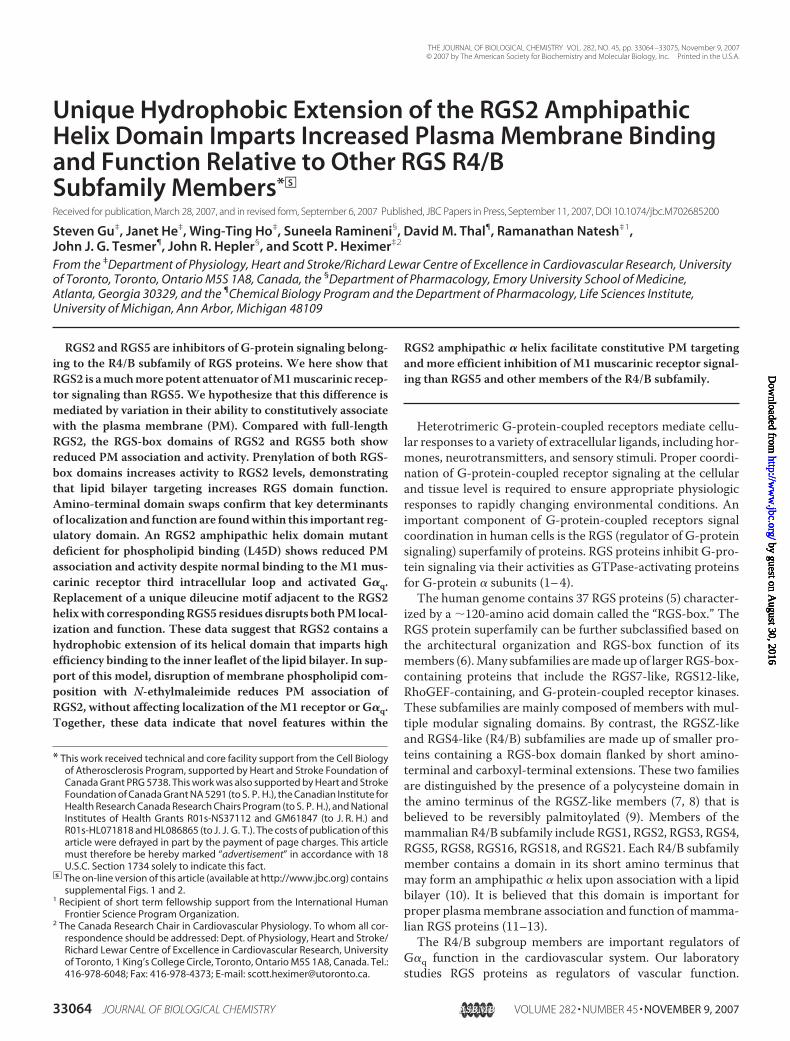
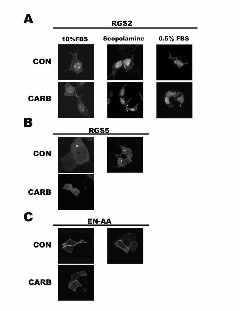
Determinant than Intrinsic RGS-box Function—Previous datafrom our laboratory showed that RGS2 contained three uniqueamino acid residues in its RGS-box domain that mediate itsselective inhibition ofG�q- versusG�i-mediated signaling (15).To determine whether G�q selectivity could explain theenhanced relative function of RGS2, we compared the ability ofthe RGS-box domains of RGS2 and RGS5 to act as inhibitors ofM1muscarinic receptor signaling. YFP fusions of the carboxyl-terminal RGS-box domains or RGS-box domains tagged withthe K-Ras plasmamembrane targeting sequence (CAAXmotif)were constructed according to the schematic in Fig. 2A. Immu-noblotting for YFP confirmed that all of the constructsexpressed as protein bands of the expected sizes (Fig. 2B). Con-focal microscopy was used to follow the localization of theseclones inHEK cells (Fig. 2C). RGS2 localized to the nucleus andplasma membrane, whereas RGS5 was distributed uniformlythrough the cell. Notably, RGS2 stably associated with theplasma membrane under normal culture conditions (no carba-chol) and after treatment with the muscarinic antagonist sco-polamine (supplemental materials). Moreover, the stable RGS2association with the plasma membrane was not decreased byserum starvation or increased by agonist addition. In dramaticcontrast, RGS5 did not stably associate with the plasma mem-brane under any of the experimental conditions tested. Weused the isolated RGS-box domains to study the relative func-tional contribution of membrane association and intrinsicRGS-box function for RGS2 and RGS5.The carboxyl-terminal RGS-box domains of RGS2 and RGS5
showed no measurable activity in two of three and one of threeexperiments, respectively. In cases where RGS-box activity ofRGS5 could be detected compared with no RGS controls, itsfunction was similar to that of full-length RGS5. By contrast,the function of the RGS-box of RGS2 was markedly impairedrelative to the full-length protein (Fig. 3A). Moreover, append-ing a carboxyl-terminal prenylation (CAAX-box) membrane-targeting sequence increased the inhibitory function of theRGS-box domains for both RGS2 and RGS5 to levels similar to
RGS2 Amino Terminus Directs Increased Membrane Targeting
NOVEMBER 9, 2007 • VOLUME 282 • NUMBER 45 JOURNAL OF BIOLOGICAL CHEMISTRY 33067
by guest on August 30, 2016
http://ww
w.jbc.org/
Dow
nloaded from

that of the wild type RGS2 (Fig. 3B). These data argue stronglyagainst a role for differences in the RGS-box domains as themain functional difference between RGS2 and RGS5. Instead,these studies suggested that promoting constitutive plasmamembrane association of the RGS-box domain of RGS2 and
FIGURE 1. Comparison of RGS2 and RGS5 function as inhibitors of M1muscarinic receptor-mediated inositol phosphate accumulation andpeak intracellular calcium elevation. A, inhibition of agonist-stimulatedproduction of inositol phosphate in COS-7 cells expressing RGS2 or RGS5.COS-7 cells were transiently co-transfected with M1 muscarinic receptor andthe indicated RGS or control construct. After 24 h, wells containing the trans-fected cells were labeled with [3H]inositol and treated with the indicated con-centrations of carbachol in the presence of 10 mM LiCl for 45 min beforedetermination of inositol phosphate accumulation values as described under“Experimental Procedures.” Inositol phosphate values represent the averageof duplicate or triplicate samples (shown in parentheses) and were expressedas the percentages of soluble IPx relative to total soluble inositol-labeledmaterial; error bars indicate the range of experimental values for each condi-tion. RGS-YFP protein expression (inset) was determined by immunoblotting,as described under “Experimental Procedures.” B, M1-HEK cells on coverslipswere transiently transfected with the indicated construct and loaded withFura-2/AM. Transfected cells identified as low to medium fluorescence intensity(3500 � relative YFP fluorescence units � 12,000) were selected for analysis
of their intracellular calcium responsiveness to carbachol. Changes in intracellu-lar calcium levels were recorded as changes in fluorescence ratio (FR � (emissionat 510 nm upon excitation at 355 nm)/(emission at 510 nm upon excitation at 396nm)). Shown are mean FR trace values (n � 50 kinetic cells) expressing YFP con-trol, RGS2, and RGS5 in a typical experiment showing base-line and relative FRchange following the addition of 100 �M carbachol (arrow). C, peak calciumresponse to carbachol in M1-HEK cells expressing RGS2 and RGS5. TransfectedM1-HEK cells were processed and selected for calcium imaging as described inB. Peak relative increases in intracellular calcium levels for each cell were calcu-lated as follows: percentage of FR increase above base line � ((peak stimulatedFR/unstimulated base-line FR) � 1) � 100%. All experiments show mean per-centage FR increase above base line � S.E. for n � 50 cells. All data are represent-ative of three independent experiments. *, p � 0.005.
FIGURE 2. Expression and subcellular localization of YFP-tagged full-lengthand carboxyl-terminal RGS-box domains of RGS2 and RGS5 in HEK cells.A, shown is the schematic representation for construction of the full-length, car-boxyl-terminal RGS-box domain and CAAX-tagged RGS-box domains of RGS2(dark shading) and RGS5 (light shading). All RGS constructs are tagged with YFP atthe carboxyl terminus. B, HEK293 cells were transfected with the indicated RGS-YFP constructs, and expression was examined by immunoblotting as describedunder “Experimental Procedures.” C, HEK cells were transfected with the indi-cated constructs and seeded onto #1 circular glass coverslips, and subcellularlocalization of YFP-tagged proteins was analyzed in live untreated cells by laser-scanning confocal microscopy as described under “Experimental Procedures.”Pictures are of cells with low to intermediate fluorescence intensity and are rep-resentative of at least 50 cells transfected with the same construct.
RGS2 Amino Terminus Directs Increased Membrane Targeting
33068 JOURNAL OF BIOLOGICAL CHEMISTRY VOLUME 282 • NUMBER 45 • NOVEMBER 9, 2007
by guest on August 30, 2016
http://ww
w.jbc.org/
Dow
nloaded from
RGS5 is a critical determinant of their function as inhibitors ofM1 muscarinic receptor signaling.Unique Sequences in the RGS2 Amino Terminus Impart
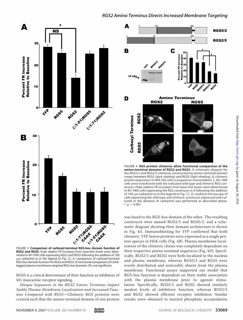
Stable Plasma Membrane Localization and Increased Func-tion Compared with RGS5—Chimeric RGS proteins werecreated such that the amino-terminal domain of one protein
was fused to the RGS-box domain of the other. The resultingconstructs were named RGS2/5 and RGS5/2, and a sche-matic diagram showing their domain architecture is shownin Fig. 4A. Immunoblotting for YFP confirmed that bothchimeric-YFP fusion proteins were expressed as a single pro-tein species in HEK cells (Fig. 4B). Plasma membrane local-ization of the chimeric clones was completely dependent ontheir respective amino-terminal sequences (Fig. 4D). Specif-ically, RGS2/5 and RGS2 were both localized to the nucleusand plasma membrane, whereas RGS5/2 and RGS5 wereevenly distributed and noticeably absent from the plasmamembrane. Functional assays supported our model thatRGS-box function is dependent on their stable associationwith the plasma membrane prior to agonist stimu-lation. Specifically, RGS5/2 and RGS5 showed similarlymodest levels of inhibitory function, whereas RGS2/5and RGS2 showed efficient receptor inhibition. Similarresults were obtained in inositol phosphate accumulation
FIGURE 3. Comparison of carboxyl-terminal RGS-box domain function ofRGS2 and RGS5. Peak relative FR increases from base-line levels were deter-mined in M1-HEK cells expressing RGS2 and RGS5 following the addition of 100�M carbachol as in the legend to Fig. 1C. A, comparison of carboxyl-terminalRGS-box domain function for RGS2 and RGS5. B, functional comparison of CAAX-tagged plasma membrane-targeted RGS-box domains. NS, not significant.
FIGURE 4. RGS protein chimeras allow functional comparison of theamino-terminal domains of RGS2 and RGS5. A, schematic diagram forthe RGS5/2 and RGS2/5 chimeras constructed by amino-terminal domainswaps between RGS2 (dark shading) and RGS5 (light shading). B, chimericprotein expression in HEK 293 cells is assayed on immunoblots. C, M1-HEKcells were transfected with the indicated wild type and chimeric RGS con-structs. Peak relative FR increases from base-line levels were determinedin M1-HEK cells expressing the RGS constructs in A following the additionof 100 �M carbachol as in the legend to Fig. 1C. D, confocal microscopy ofcells expressing the wild type and chimeric constructs expressed and cul-tured in the absence of carbachol was performed as described above.*, p � 0.001.
RGS2 Amino Terminus Directs Increased Membrane Targeting
NOVEMBER 9, 2007 • VOLUME 282 • NUMBER 45 JOURNAL OF BIOLOGICAL CHEMISTRY 33069
by guest on August 30, 2016
http://ww
w.jbc.org/
Dow
nloaded from

studies.4 These data confirm the existence of a distinctmolecular determinant within the amino-terminal domainof RGS2 that is capable of directing stable plasma membranelocalization and increased function.Disruption of the Amphipathic Helix Domain in RGS2
Reduces Plasma Membrane Association and Function—TheRGS2 amino terminus has been shown to bind to the phospho-lipid bilayer (13) and the third intracellular loop of theM1mus-carinic receptor (17). Since both of these interactions couldpotentially regulate plasma membrane targeting efficiency, wedetermined whether RGS2 and RGS5 behaved differently withrespect to one or both of these activities. Introduction of a sin-gle acidic residue on the core hydrophobic surface of the RGS2amino-terminal amphipathic � helix (L45D) disrupts its inter-action with phospholipid bilayers containing physiologic levels(40–60%) of negatively charged phospholipids (13). To studythe functional consequence of disrupting the amphipathichelix, the L45D mutation was introduced into the RGS2 andRGS2/5. Immunoblotting for YFP indicated expression of a sin-gle protein species for both mutant constructs (Fig. 5A). Con-
focal microscopy of cells transfected with constructs contain-ing the L45Dmutation confirmed that thismutation resulted ina nearly complete loss of plasma membrane association (Fig.5B) compared with wild type RGS2. In transfected M1-HEKcells expressing L45Dvariants of RGS2 andRGS2/5, the relativeextent ofM1muscarinic signal inhibitionwas reduced to a sim-ilar level as that of wild type RGS5 (Fig. 5D), indicating that thefunctional advantage conferred by the RGS2 amino terminuswas ablated by disruption of the hydrophobic interface of thisamphipathic helix domain. However, it remained to be deter-mined whether the amphipathic helix domain mediated tonicmembrane localization via lipid binding, receptor binding, orassociation with other signaling components, such as G�q.Mutation of the RGS2 Amphipathic Helix Domain Does Not
Interfere with Receptor Third Intracellular Loop Binding—Theamino terminus of RGS2 has also been reported by our groupand others to interact either directly or indirectly with the thirdintracellular loop of several G-protein-coupled receptors,including theM1muscarinic receptor (17, 23, 24). The specificresidues in RGS2 that mediate this interaction have yet to beidentified. Using the L45Dmutant, we asked whether the RGS2amphipathic helix mediated stable plasma membrane associa-tion via its interaction with the M1 receptor. Using theRGS2/M1 muscarinic receptor interaction assay developed inour laboratory (17), lysates from HEK cells expressing the var-ious RGS protein constructs were screened for protein-proteininteractions with a glutathione S-transferase-taggedM1i3 pep-tide bait (Fig. 6). RGS2 and RGS2/5 interacted with the M1muscarinic receptor third loop peptide, whereas RGS5 did notinteract in this assay. L45D mutants of the amphipathic helixdomain, however, did not prevent binding to the third looppeptide. Together, these data suggested that a functionalamphipathic helix was not required for muscarinic receptorbinding and that decreased plasma membrane binding of theL45D mutation is not the result of reduced affinity for the M1muscarinic receptor.Unique Dileucine Motif in RGS2 Mediates Tonic Plasma
Membrane Association and Regulatory Function—Previously,we showed that increased plasmamembrane targeting of RGS2by activated G�q was prevented bymutation of hydrophobic orbasic residues in the amphipathic helix (13). Thus, the amino-terminal helix domain may mediate plasma membrane associ-4 W.-T. Ho and S. P. Heximer, unpublished data.
FIGURE 5. Testing whether mutation of the conserved hydrophobic resi-dues in amphipathic � helix in RGS2 and RGS2/5 alters tonic plasmamembrane localization and function. A negatively charged aspartate resi-due was introduced into the conserved hydrophobic face of the amphipathichelix in RGS2 and RGS2/5. Immunoblotting (A), live cell confocal microscopyin the absence of carbachol (B), and peak relative FR increases from base-linelevels following the addition of 100 �M carbachol (C) were used to describethe expression, subcellular localization, and inhibitory function of the indi-cated RGS protein constructs, respectively, as described for Fig. 4. *, p � 0.001.
FIGURE 6. Comparison of wild type, chimeric, and mutant RGS proteinbinding to the M1 muscarinic receptor third intracellular loop domain.Cell lysates from HEK293T cells transfected with YFP, RGS2, RGS5, RGS2/5,RGS2(L45D), or RGS2/5(L45D) were incubated with equal amounts of recom-binant GST-M1i3 protein (6.5 �g) prebound to glutathione-Sepharose beads(see “Experimental Procedures”). Proteins in the starting cell lysates (Load)and bound to the recovered beads (Bound) were analyzed by SDS-PAGE sep-aration and immunoblotting for YFP as described under “Experimental Pro-cedures.” Shown is one of at least three representative experiments.
RGS2 Amino Terminus Directs Increased Membrane Targeting
33070 JOURNAL OF BIOLOGICAL CHEMISTRY VOLUME 282 • NUMBER 45 • NOVEMBER 9, 2007
by guest on August 30, 2016
http://ww
w.jbc.org/
Dow
nloaded from
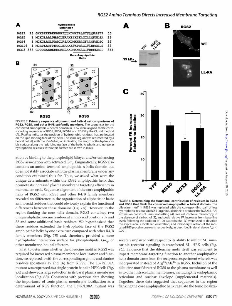
ation by binding to the phospholipid bilayer and/or enhancingRGS2 association with activated G�q. Enigmatically, RGS5 alsocontains an amino-terminal amphipathic � helix domain butdoes not stably associate with the plasmamembrane under anycondition examined thus far. Thus, we asked what were theunique determinants within the RGS2 amphipathic helix thatpromote its increased plasmamembrane targeting efficiency inmammalian cells. Sequence alignment of the core amphipathichelix of RGS2 with RGS5 and other R4/B family membersrevealed no difference in the organization of aliphatic or basicamino acid residues that could obviously explain the functionaldifferences between these domains (Fig. 7A). However, in theregion flanking the core helix domain, RGS2 contained twounique aliphatic leucine residues at amino acid positions 37 and38 and some additional basic residues. We hypothesized thatthese residues extended the hydrophobic face of the RGS2amphipathic helix by one extra turn compared with other R4/Bfamily members (Fig. 7B) and, therefore, provided a morehydrophobic interaction surface for phospholipids, G�q, orother membrane-bound effectors.First, to determine whether the dileucine motif in RGS2 was
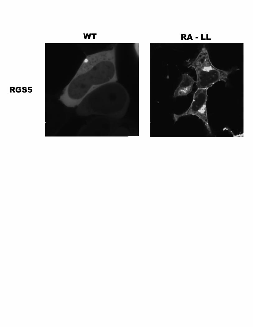
required for increased plasmamembrane localization and func-tion,we replaced itwith the corresponding arginine and alanineresidues (positions 15 and 16) from RGS5. The L37R/L38Amutantwas expressed as a single protein band inHEK cells (Fig.8A) and showed a large reduction in its basal plasmamembranelocalization (Fig. 8B). Consistent with previous data showingthe importance of tonic plasma membrane localization as adeterminant of RGS function, the L37R/L38A mutant was
severely impaired with respect to its ability to inhibit M1 mus-carinic receptor signaling in transfected M1-HEK cells (Fig.8C). Evidence that the dileucine motif itself was sufficient toimpart membrane-targeting function to another amphipathichelix domain came from the reciprocal experimentwhere itwasincorporated instead of Arg15/Ala16 in RGS5. Inclusion of thedileucine motif directed RGS5 to the plasmamembrane as wellas to other intracellularmembranes, including the endoplasmicreticulum and nuclear envelope (supplemental materials).Together, these data suggested that sequences in the regionflanking the core amphipathic helix regulate the tonic localiza-
FIGURE 7. Primary sequence alignment and helical net comparisons ofRGS2, RGS5, and other R4/B subfamily members. The sequences for theconserved amphipathic � helical domain in RGS2 were aligned to the corre-sponding sequences of RGS5, RGS4, RGS16, and RGS3 by the Clustal method(A). Shading indicates the position of hydrophobic residues that are locatedon the lipid-binding face of the helix. The same region was represented by ahelical net (B), with the shaded region indicating the length of the hydropho-bic surface along the lipid-binding face of the helix. Aliphatic and nonpolarhydrophobic residues within this surface are shown in black.
FIGURE 8. Determining the functional contribution of residues in RGS2and RGS5 that flank the conserved amphipathic � helical domain. Thedileucine motif in RGS2 was replaced with the corresponding pair of lesshydrophobic residues in RGS5 (arginine, alanine) to produce the RGS2(LL-RA)expression construct. Immunoblotting (A), live cell confocal microscopy inthe absence of carbachol (B), and peak relative FR increases from base-linelevels following the addition of 100 �M carbachol (C) were used to describethe expression, subcellular localization, and inhibitory function of the indi-cated RGS protein constructs, respectively, as described in detail above. *, p �0.001.
RGS2 Amino Terminus Directs Increased Membrane Targeting
NOVEMBER 9, 2007 • VOLUME 282 • NUMBER 45 JOURNAL OF BIOLOGICAL CHEMISTRY 33071
by guest on August 30, 2016
http://ww
w.jbc.org/
Dow
nloaded from

tion and function of differentmembers of the R4/B subfamily ofRGS proteins, depending on the extent to which they contrib-ute to the amphipathic nature of the helical domain.Mutation of Amphipathic Helix and Dileucine Motif Does
Not Alter G�q Binding—Although disruption the hydrophobicsurface of the helical domain is known to prevent RGS2 inter-action with the phospholipid bilayer, we could not exclude thepossibility that the extended hydrophobic surface acted as asupplemental interaction site for G�q. In this case, theincreased function and plasma membrane targeting efficiencyof RGS2 could be a consequence of its increased affinity forG�q. The contribution of the RGS2 amphipathic helix domainduring G�q binding was studied using the flow cytometry pro-tein interaction assay as previously described by Neubig andco-workers (22). First, the affinity of histidine-tagged wild typeand mutant RGS2 proteins for GDP-bound (inactive) andAlF4�-activated G�q was measured. RGS2 wild type, L45D, andL37R/L38A proteins all boundmuchmore efficaciously to acti-vated than to inactive G�q, whereas the G-protein binding-defective mutant E108A/N109A did not bind to G�q in eitheractivation state (Fig. 9A). These data suggested that the RGS-box domain of RGS2 was necessary to promote G�q binding,whereas the amino terminus could not direct G�q binding inthe context of a full-length RGS2 containing a nonfunctionalRGS-box.Wenext used dissociation constantmeasurements totest whether the RGS2 amino terminus played an importantancillary role during G�q binding. TheKd of wild type RGS2 foractivated G�q was similar to that of L45D and L37R/L38A (Fig.9B, summarized in Table 1), indicating that the amphipathic
helix and flanking dileucine motif did not contribute signifi-cantly to G�q-binding.Interfering with Plasma Membrane Lipid Composition Dis-
rupts Localization of RGS2 without Affecting M1 MuscarinicReceptor or G�q—Last, we examined the role of anionic phos-pholipid head groups in targeting the unique RGS2 helicaldomain to the lipid bilayer. Specifically, we determinedwhetherchemical disruption of the anionic lipid composition on theinner leaflet of the plasma membrane affected RGS2 localiza-tion.Mammalian cells continuously shuttle phosphatidylserine(PS) and phosphatidylethanolamine from the outer to the innerleaflet of the plasma membrane to maintain a higher concen-tration of anionic lipids on the cytoplasmic face. N-Ethylmale-imide (NEM) has been shown to rapidly inhibit PS shuttling,resulting in shuffling of phospholipid pools and increased expo-sure of PS on the extracellular surface. Treatment of HEK cellswith NEM resulted in the expected level of lipid shuffling andextracellular exposure of PS, as indicated by fluorescein isothio-cyanate-labeled annexin V-binding using fluorescence-acti-vated cell-sorting analysis (Fig. 10A). Notably, as levels of PS onthe inner leaflet decreased, RGS2 rapidly (�2 min) delocalizedfrom this site. By contrast, M1 muscarinic receptor and G�q,two other signaling proteins that are known to affect RGS2localization, remained tightly associated with the lipid bilayerin NEM-treated cells (Fig. 10B). Together, these data suggestthat the highly anionic lipid interface of the inner leaflet of theplasmamembrane is required for stable RGS2 targeting. More-over, it confirms our biochemical data showing that the aminoterminus of RGS2directs plasmamembrane localization by fac-tors that are independent from itsG�q orM1muscarinic recep-tor binding. Thus, the differential affinity of RGS amphipathichelices for plasmamembrane phospholipids appears to be a keydeterminant of RGS protein function that can explain theobserved functional differences between RGS2 and RGS5.
DISCUSSION
To understand the biologic function of RGS proteins, wemust first determine the spectrum of signaling pathways theyregulate in living organisms. These efforts are complicated bythe fact that most tissues express several RGS proteins withoverlapping functions. Thus, a critical step is to define the keyfunctional determinants that specify differences in regulationof G-protein signaling by different RGS proteins.RGS2 Is a More Potent Inhibitor of M1 Muscarinic Receptor-
mediated Signaling Compared with RGS5—When the activitiesof RGS2 and RGS5 as inhibitors of M1 muscarinic receptorsignaling were compared in assaysmeasuring phosphatidylino-sitol and intracellular calcium, RGS2 was found to be a moreefficacious inhibitor of this receptor. These data appeared to
TABLE 1Affinity of AF532-RGS2 proteins for biotin-G�q
Kd � S.E.nM
Wild type RGS2 17.6 � 2L45D 8.6 � 3LL-RA 9.1 � 2EN-AA NBa
a No binding.
FIGURE 9. Binding of AF532-labeled RGS2, RGS2(L45D), RGS2(LL-RA), andRGS2(EN-AA) to biotin-labeled G�q using the flow cytometry proteininteraction assay method. A, binding of AF532-RGS2 proteins is dependenton AMF activation of G�q. The curves shown are representative of four differ-ent experiments with n � 2 for each data point. MFI, mean fluorescence inten-sity. B, specific binding of AF532-RGS2 to biotin-G�q. RGS2, RGS2(L45D), andRGS2(LL-RA) exhibit similar Kd values, whereas RGS2(EN-AA), which has a dis-rupted RGS-box, does not bind biotin-G�q. The fitted Kd values are given inTable 1.
RGS2 Amino Terminus Directs Increased Membrane Targeting
33072 JOURNAL OF BIOLOGICAL CHEMISTRY VOLUME 282 • NUMBER 45 • NOVEMBER 9, 2007
by guest on August 30, 2016
http://ww
w.jbc.org/
Dow
nloaded from

contradict those ofAnger et al. (14), who reported no differencein the function of RGS2 and RGS5 as inhibitors of M3 musca-rinic receptor signaling. There are two potential differences inthe experimental design of the two studies thatmay explain thisdiscrepancy. First, we used M1 muscarinic receptor comparedwith M3 muscarinic receptor used in the study by Anger et al.(14). Although it is possible that receptor-selectivemechanismsexplain this discrepancy, RGS2 has been shown to inhibit bothM1muscarinic receptor andM3muscarinic receptor signalingwith higher relative efficiency relative to other R4/B class pro-teins (15, 16). Second, our study measured RGS activity underconditions of similar RGS protein expression as determined byeither Western blotting or relative green fluorescent proteinfluorescence. By contrast, Anger et al. (14)measuredRGSactiv-ities relative to the amount of plasmid used per transfection, amethod that would not account for potential differences in therates of synthesis or degradation of the different RGS proteins.Constitutive Plasma Membrane Targeting Mediates Increased
RGS Function in Mammalian Cells—We set out to determinewhether the increased activity of RGS2 compared with RGS5reflected its stronger constitutive association with plasmamembrane. Support for this idea came initially from the obser-vation that CAAX-box targeting to the lipid bilayer greatly
increased the function of the isolated RGS-box domains, evenin the absence of amino-terminal regulatory sequences.Consistent with this notion, RGS2/5 chimera data showed
that the RGS2 amino terminus is necessary to promote stableplasma membrane localization and increased RGS-box func-tion in mammalian cells, whereas the RGS5 amino terminuslacks this key function.Thiswas the first demonstration that func-tional amino/carboxyl-terminal chimeras can be used to study therelative function of different R4/B subfamily members. Althoughthe RGS2 amino terminus is known to be sufficient to promoteplasma membrane targeting, the relative functional contributionof phospholipid binding via the amphipathic helix and receptorbinding via an unknown domain is unknown. Attempts to studythe role of phospholipid binding using constitutive G�q (13) sug-gested that amphipathic helix-mediated lipid bilayer targeting ofRGS2 was not critical for its function. Specifically, the L45Dmutant did not bind to the plasma membrane but showed wildtype inhibitory activity toward constitutively active G�q. Theinconsistency between those and our current data may be theresult of limitations in the previous experimental protocols.Our current studies allow us to select cells based on their levelof RGS protein expression. Thus, we are able to compare indi-vidual cells with similar RGS protein expression. In previousexperiments, overexpression may have masked any potentialfunctional differences between wild type RGS2 and the L45Dmutant. Furthermore, Mende and co-workers (14) haverecently shown that in the presence of constitutive G�q, RGS2is dramatically up-regulated, leading to its even higher overex-pression. Our current experimental protocol allowed us totitrate expression levels and examine the more subtle differ-ences between RGSmutants; therefore, we felt it was importantto characterize the functional contribution of the amphipathichelix in this assay.Extension of the Hydrophilic Face of the RGS2Amphipathic�
Helix Promotes Plasma Membrane Association—The molecu-lar characteristics of amphipathic helix function in the R4/Bsubfamily have not been rigorously studied. Chimera studiesshowed that the amino-terminal domain of RGS2 was able todirect the RGS-box domains of both RGS2 and RGS5 to theplasma membrane, whereas the amino-terminal domain ofRGS5 lacked this important function. By mutating the RGS2amphipathic � helix domain (L45D), we demonstrated theimportance of this feature inmediating both plasmamembranelocalization and increased relative function of RGS2 andRGS/5. Although RGS2 and RGS5 both contain amphipathichelix domains in the same position relative to their RGS-box,sequence alignment revealed two unique leucine residues andtwo other basic residues that we predicted should extend thelength of its amphipathic nature by one additional turn of thehelix. We thus hypothesized that the extended helix in RGS2contributes to its more efficient association with the lipidbilayer component of the plasma membrane. In support of thismodel, replacement of the two leucine residues in RGS2 withtheir less hydrophobic counterparts in RGS5 (arginine and ala-nine) was sufficient to disrupt the majority of plasma mem-brane localization and function. Thus, for the first time, ourstudies prove that the length of the amphipathic surface of theamino-terminal helical domain is a critical functional determi-
FIGURE 10. Effect of NEM-mediated phospholipid shuffling on tonicplasma membrane localization of RGS2, M1 muscarinic receptor, andG�q. A, HEK cells were treated with medium with or without 10 mM NEM for 5min before incubation with fluorescein isothiocyanate-conjugated annexinV. Phosphatidylserine exposure on the extracellular surface of the lipid bilayerwas determined by the extent of annexin V binding using cell sorting analysisas previously described (30). B, HEK cells were transiently transfected withgreen fluorescent protein-tagged RGS2, RGS2-CAAX, M1 muscarinic recep-tor, or G�q (cotransfected with untagged G�2 and G�1). NEM was addedto a final concentration of 10 mM for 5 min. Shown are representativeconfocal images of at least 50 low expressing cells captured before andafter the addition of NEM.
RGS2 Amino Terminus Directs Increased Membrane Targeting
NOVEMBER 9, 2007 • VOLUME 282 • NUMBER 45 JOURNAL OF BIOLOGICAL CHEMISTRY 33073
by guest on August 30, 2016
http://ww
w.jbc.org/
Dow
nloaded from

nant of the function of the R4/B subgroup of RGS proteins.Furthermore, the only other R4/B subfamily member that has asimilar amphipathic extension, RGS3, is also a more efficientinhibitor of muscarinic receptor signaling than RGS4 (16) and,like RGS2, can bemore efficiently recruited to the plasmamem-brane by constitutive G�q expression (13, 18, 25). Recent stud-ies have suggested that the RGS2 amino terminus can interactwith several other proteins that impact its function. We specif-ically examined whether the amphipathic helix domain inRGS2 directed plasma membrane association via interactionswith the M1 muscarinic receptor or G�q.Mutations That Prevent RGS2 Plasma Membrane Localiza-
tion Do Not Affect M1 Muscarinic Receptor Binding—Datafromour group and others have shown that the amino terminusof RGS2 interacts with the intracellular loops ofM1muscarinicand other G-protein-coupled receptors (17, 23, 24). Althoughthe direct mechanism mediating this interaction is still notknown, it has been proposed that thismodel can account for theincreased membrane targeting and function of RGS2 towardspecific receptors (26). Our current studies show that muta-tionswithin the amino-terminal amphipathic�helix, while dis-rupting both function and plasma membrane localization, donot affect receptor interaction.Furthermore, NEM treatment causes the dissociation of
RGS2 from the plasma membrane without affecting the M1muscarinic receptor localization. Thus, although it is possiblethat receptor association is one component required for plasmamembrane targeting, it is probably not the primary determi-nant. One potential possibility that would incorporate both ofthesemodels is thatonceRGS2 is stablyassociatedwith theplasmamembranevia its extendedamphipathichelix, its ability to interactwith receptor imparts an ability to select specific targets fromwithin the receptorome. Future studies in this area could also beused to elucidate themechanisms that explain the reported selec-tivity of RGS5 for the angiotensin II receptor AT1 (27).Mutations That Prevent RGS2 Plasma Membrane Localiza-
tion DoNot Affect G�q Binding—Interaction between the RGS-box domain and G�q is critical for proper RGS2 function. TheG-protein binding-defective E108A/N109Amutation (EN-AA)of RGS2 does not have measurable affinity for activated G�qand does not inhibit calcium signaling in our assay.5 Ourmuta-tions resulting in both total disruption of helix function (L45D)or removal of the dileucinemotif (LL-RA) both resulted in largechanges in localization and function without changing RGS2affinity for G�q. Finally, NEM treatment resulted in the disso-ciation of RGS2 from the plasma membrane without anychange inG�q localization. Together, these data suggest that, asin the case of the M1 receptor interaction, G�q-RGS2 interac-tions are not sufficient to promote plasma membrane localiza-tion and increased relative function.Extended Hydrophobic Helix of RGS2 Binds Anionic Phos-
pholipids on the PlasmidMembrane—Taken together, our datashow that disruption of RGS2 plasma membrane localizationand function occur independently from both receptor and G�qinteraction. The fact that NEM, a compound that alters anionic
phospholipid concentrations on the plasma membrane, is ableto decrease RGS2 localization providesmechanistic support forour hypothesis that it is the lipid bilayer that binds to the elon-gated amphipathic helix of RGS2. Although the possibility doesremain that interaction between RGS2 and another NEM-sen-sitive factor recruits RGS2 to the plasma membrane, no suchfactor has yet been identified. Indeed, all of our present datapoint to the phospholipid bilayer as themost likely target of thisspecialized helix domain.Can the Length of the Amphipathic Helix be Regulated by
Palmitoylation in Mammalian Cells?—If the lipid bilayer isindeed the primary target of the RGS helices, it raises the pos-sibility that palmitoylation of a conserved cysteine at position12 in RGS5, RGS4, and RGS16 could extend the hydrophobicface of their respective amphipathic helices (see Fig. 7) and thusprovide a potential regulatory site for membrane targeting andfunction. Consistent with this notion, alanine mutations at thisposition have been shown to inhibit the localization and func-tion of RGS16 (28, 29). It remains to be demonstrated whetherRGS5 is similarly modified and, if so, whether constitutivelypalmitoylated RGS4, RGS5, or RGS16 proteins would showincreased efficiency of membrane association and function inmammalian cells.
Acknowledgments—We thank SusanNgo and Vincent Huang for val-uable technical assistance. We also thank Dr. Steffen-Sebastian Bolzfor comments during preparation of the manuscript. We are gratefulto Dr. Brian Bennett (Washington University School of Medicine, St.Louis, MO) for helpful advice during development of the functionalassays using RGS-YFP and to Drs. Richard Neubig and David Roman(University of Michigan Medical School) for assistance with develop-ing the G�q flow cytometry protein interaction assay and for use of theLuminex flow cytometer.
REFERENCES1. Berman, D. M., Kozasa, T., and Gilman, A. G. (1996) J. Biol. Chem. 271,
27209–272122. Berman, D. M., Wilkie, T. M., and Gilman, A. G. (1996) Cell 86, 445–4523. Watson, N., Linder, M. E., Druey, K. M., Kehrl, J. H., and Blumer, K. J.
(1996) Nature 383, 172–1754. Hunt, T.W., Fields, T. A., Casey, P. J., and Peralta, E. G. (1996)Nature 383,
175–1775. Siderovski, D. P., and Willard, F. S. (2005) Int. J. Biol. Sci. 1, 51–666. Ross, E. M., and Wilkie, T. M. (2000) Annu. Rev. Biochem. 69, 795–8277. Barker, S. A., Wang, J., Sierra, D. A., and Ross, E. M. (2001) Genomics 78,
223–2298. Glick, J. L., Meigs, T. E., Miron, A., and Casey, P. J. (1998) J. Biol. Chem.
273, 26008–260139. Jones, T. L. (2004)Methods Enzymol. 389, 33–5510. Hollinger, S., and Hepler, J. R. (2002) Pharmacol. Rev. 54, 527–55911. Bernstein, L. S., Grillo, A. A., Loranger, S. S., and Linder, M. E. (2000)
J. Biol. Chem. 275, 18520–1852612. Chen, C., Seow, K. T., Guo, K., Yaw, L. P., and Lin, S. C. (1999) J. Biol.
Chem. 274, 19799–1980613. Heximer, S. P., Lim, H., Bernard, J. L., and Blumer, K. J. (2001) J. Biol.
Chem. 276, 14195–1420314. Anger, T., Zhang,W., andMende, U. (2004) J. Biol. Chem. 279, 3906–391515. Heximer, S. P., Srinivasa, S. P., Bernstein, L. S., Bernard, J. L., Linder,M. E.,
Hepler, J. R., and Blumer, K. J. (1999) J. Biol. Chem. 274, 34253–3425916. Tovey, S. C., and Willars, G. B. (2004)Mol. Pharmacol. 66, 1453–146417. Bernstein, L. S., Ramineni, S., Hague, C., Cladman, W., Chidiac, P., Levey,
A. I., and Hepler, J. R. (2004) J. Biol. Chem. 279, 21248–212565 S. Gu and S. P. Heximer, unpublished data.
RGS2 Amino Terminus Directs Increased Membrane Targeting
33074 JOURNAL OF BIOLOGICAL CHEMISTRY VOLUME 282 • NUMBER 45 • NOVEMBER 9, 2007
by guest on August 30, 2016
http://ww
w.jbc.org/
Dow
nloaded from
18. Dulin, N. O., Sorokin, A., Reed, E., Elliott, S., Kehrl, J. H., and Dunn, M. J.(1999)Mol. Cell Biol. 19, 714–723
19. Kozak, M. (1994) Biochimie (Paris) 76, 815–82120. Venkatakrishnan, G., and Exton, J. H. (1996) J. Biol. Chem. 271,
5066–507221. Heximer, S. P. (2004)Methods Enzymol. 390, 65–8222. Roman, D. L., Talbot, J. N., Roof, R. A., Sunahara, R. K., Traynor, J. R., and
Neubig, R. R. (2007)Mol. Pharmacol. 71, 169–17523. Hague, C., Bernstein, L. S., Ramineni, S., Chen, Z., Minneman, K. P., and
Hepler, J. R. (2005) J. Biol. Chem. 280, 27289–2729524. Wang, X., Zeng, W., Soyombo, A. A., Tang, W., Ross, E. M., Barnes, A. P.,
Milgram, S. L., Penninger, J. M., Allen, P. B., Greengard, P., and Muallem,S. (2005) Nat. Cell Biol. 7, 405–411
25. Roy, A. A., Lemberg, K. E., and Chidiac, P. (2003) Mol. Pharmacol. 64,
587–59326. Xu, X., Zeng,W., Popov, S., Berman, D.M., Davignon, I., Yu, K., Yowe, D.,
Offermanns, S., Muallem, S., and Wilkie, T. M. (1999) J. Biol. Chem. 274,3549–3556
27. Wang, Q., Liu, M., Mullah, B., Siderovski, D. P., and Neubig, R. R. (2002)J. Biol. Chem. 277, 24949–24958
28. Druey, K. M., Ugur, O., Caron, J. M., Chen, C. K., Backlund, P. S., andJones, T. L. (1999) J. Biol. Chem. 274, 18836–18842
29. Hiol, A., Davey, P. C., Osterhout, J. L., Waheed, A. A., Fischer, E. R., Chen,C. K., Milligan, G., Druey, K.M., and Jones, T. L. (2003) J. Biol. Chem. 278,19301–19308
30. Teichert-Kuliszewska, K., Kutryk, M. J., Kuliszewski, M. A., Karoubi, G.,Courtman,D.W., Zucco, L., Granton, J., and Stewart, D. J. (2006)Circ. Res.98, 209–217
RGS2 Amino Terminus Directs Increased Membrane Targeting
NOVEMBER 9, 2007 • VOLUME 282 • NUMBER 45 JOURNAL OF BIOLOGICAL CHEMISTRY 33075
by guest on August 30, 2016
http://ww
w.jbc.org/
Dow
nloaded from

Natesh, John J. G. Tesmer, John R. Hepler and Scott P. HeximerSteven Gu, Janet He, Wing-Ting Ho, Suneela Ramineni, David M. Thal, Ramanathan
Subfamily MembersIncreased Plasma Membrane Binding and Function Relative to Other RGS R4/B Unique Hydrophobic Extension of the RGS2 Amphipathic Helix Domain Imparts
doi: 10.1074/jbc.M702685200 originally published online September 11, 20072007, 282:33064-33075.J. Biol. Chem.
10.1074/jbc.M702685200Access the most updated version of this article at doi:
Alerts:
When a correction for this article is posted•
When this article is cited•
to choose from all of JBC's e-mail alertsClick here
Supplemental material:
http://www.jbc.org/content/suppl/2007/09/11/M702685200.DC1.html
http://www.jbc.org/content/282/45/33064.full.html#ref-list-1
This article cites 30 references, 20 of which can be accessed free at
by guest on August 30, 2016
http://ww
w.jbc.org/
Dow
nloaded from
Copyright © 2022 FDOKUMEN