Ultrastructure of the ciliated cells of the free-swimming larva, and sessile stages, of the marine...
14
Ultrastructure of the Ciliated Cells of the Free-Swimming Larva, and Sessile Stages, of the Marine Sponge Haliclona indistincta (Demospongiae: Haplosclerida) Kelly M. Stephens, 1 * Alexander Ereskovsky, 2,3 Pierce Lalor, 4 and Grace P. McCormack 1 1 Molecular Evolution and Systematics laboratory, Zoology, Ryan Institute and School of Natural Sciences, National University of Ireland Galway, Galway, Ireland 2 Mediterranean Institute of Biodiversity and Ecology Marine and Continental (IMBE), UMR 7263, CNRS Aix- Marseille University, Station marine d’Endoume, Marseille 13007, France 3 Biological Faculty, Saint-Petersburg State University, Universitetskaya nab. 7/9, Saint-Petersburg 199034, Russia 4 Department of Anatomy, School of Medicine, National University of Ireland Galway, Galway, Ireland ABSTRACT We provide a detailed, comparative study of the ciliated cells of the marine haplosclerid sponge Haliclona indistincta, in order to make data available for future phylogenetic comparisons at the ultrastruc- tural level. Our study focuses on the description and analysis of the larval epithelial cells, and choanocytes of the metamorphosed juvenile sponge. The ultrastruc- ture of the two cell types is sufficiently different to pre- vent our ability to conclusively determine the origin of the choanocytes from the larval ciliated cells. However, ciliated, epithelial cells were observed in a migratory position within the inner cell mass of the larval stages. Some cilia were observed within the cell’s cytoplasm, which is indicative of the ciliated epithelial cell under- going transdifferentiation into a choanocyte; while traces of other ciliated epithelial cells were contained within phagosomes, suggesting they are phagocytosed. We compared our data with other species described in the literature. However, any phylogenetic inference must wait until further detailed comparisons can be made with species whose phylogenetic position has been determined by other means, such as phylogenom- ics, in order to more closely link genomic, and morpho- logical information. J. Morphol. 000:000–000, 2013. V C 2013 Wiley Periodicals, Inc. KEY WORDS: Haliclona indistincta; sponge; larvae; ultrastructure; ciliated cells INTRODUCTION The general organization of a typical demosponge comprises three layers: external and internal epi- thelial layers [sensu lato (Schmidt, 1866; Schneider, 1902; Starck and Siewing, 1980]), made up of exopi- nacocytes, basopinacocytes, and endopinacocytes, and an extracellular matrix, or mesohyl, which is composed of proteins, polysaccharides, and fibrous collagen (Bergquist, 1978; Ereskovsky, 2010). The term “epithelium” defined as closely associated, laterally contacted, polarized cells, (Schmidt, 1866; Schneider, 1902; Starck and Siewing, 1980; Nickel et al., 2011), was originally applied to sponges in a broad, functional sense (Nickel et al., 2011). It is currently a matter of some controversy whether or not sponges can be said to possess a “true” epithelium. This is primarily because, con- trary to the more recent definition, most Porifera taxa lack the belt-like junctions between cells, as well as a basal lamina (Rieger, 1986; Ax et al., 1989; Ax, 1995; Tyler, 2003; Ereskovsky and Don- dua, 2006; Leys et al., 2009; Adams et al., 2010; Nickel et al., 2011). In recent years however, data suggests the term “epithelium” can be applied to sponges, as their polarized epithelial cells show functional similarities to other animal epithelia (e.g., ion and solute transportation regulation), and should therefore, not be discounted (Leys et al., 2009; Adams et al., 2010; Nickel et al., 2011). It is generally accepted that sponges are the most basal of the metazoans (Nielsen, 2001; Philippe et al. 2009). Thus, information concerning the evolution of multicellular animals from unicellular protists could be inferred through genetic, morphological, and developmental investigations in this group of animals (Ruiz-Trillo et al., 2008; Fahey and Degnan, 2010; Ereskovsky, 2010). Grant sponsor: National University of Ireland; Grant sponsor: Thomas Crawford Hayes Trust Fund Scheme (NUI Galway). *Correspondence to: Kelly M Stephens; Lab 211, Ryan Institute, NUI Galway, Galway, Ireland. E-mail: [email protected] Received 24 January 2013; Revised 28 May 2013; Accepted 31 May 2013. Published online 00 Month 2013 in Wiley Online Library (wileyonlinelibrary.com). DOI 10.1002/jmor.20177 V C 2013 WILEY PERIODICALS, INC. JOURNAL OF MORPHOLOGY 00:00–00 (2013)
-
Upload
independent -
Category
Documents
-
view
0 -
download
0
Transcript of Ultrastructure of the ciliated cells of the free-swimming larva, and sessile stages, of the marine...
Ultrastructure of the Ciliated Cells of theFree-Swimming Larva, and Sessile Stages,of the Marine Sponge Haliclona indistincta(Demospongiae: Haplosclerida)
Kelly M. Stephens,1* Alexander Ereskovsky,2,3 Pierce Lalor,4 and Grace P. McCormack1
1Molecular Evolution and Systematics laboratory, Zoology, Ryan Institute and School of Natural Sciences, NationalUniversity of Ireland Galway, Galway, Ireland2Mediterranean Institute of Biodiversity and Ecology Marine and Continental (IMBE), UMR 7263, CNRS Aix-Marseille University, Station marine d’Endoume, Marseille 13007, France3Biological Faculty, Saint-Petersburg State University, Universitetskaya nab. 7/9, Saint-Petersburg 199034, Russia4Department of Anatomy, School of Medicine, National University of Ireland Galway, Galway, Ireland
ABSTRACT We provide a detailed, comparative studyof the ciliated cells of the marine haplosclerid spongeHaliclona indistincta, in order to make data availablefor future phylogenetic comparisons at the ultrastruc-tural level. Our study focuses on the description andanalysis of the larval epithelial cells, and choanocytesof the metamorphosed juvenile sponge. The ultrastruc-ture of the two cell types is sufficiently different to pre-vent our ability to conclusively determine the origin ofthe choanocytes from the larval ciliated cells. However,ciliated, epithelial cells were observed in a migratoryposition within the inner cell mass of the larval stages.Some cilia were observed within the cell’s cytoplasm,which is indicative of the ciliated epithelial cell under-going transdifferentiation into a choanocyte; whiletraces of other ciliated epithelial cells were containedwithin phagosomes, suggesting they are phagocytosed.We compared our data with other species described inthe literature. However, any phylogenetic inferencemust wait until further detailed comparisons can bemade with species whose phylogenetic position hasbeen determined by other means, such as phylogenom-ics, in order to more closely link genomic, and morpho-logical information. J. Morphol. 000:000–000, 2013. VC
2013 Wiley Periodicals, Inc.
KEY WORDS: Haliclona indistincta; sponge; larvae;ultrastructure; ciliated cells
INTRODUCTION
The general organization of a typical demospongecomprises three layers: external and internal epi-thelial layers [sensu lato (Schmidt, 1866; Schneider,1902; Starck and Siewing, 1980]), made up of exopi-nacocytes, basopinacocytes, and endopinacocytes,and an extracellular matrix, or mesohyl, which iscomposed of proteins, polysaccharides, and fibrouscollagen (Bergquist, 1978; Ereskovsky, 2010). Theterm “epithelium” defined as closely associated,laterally contacted, polarized cells, (Schmidt,
1866; Schneider, 1902; Starck and Siewing, 1980;Nickel et al., 2011), was originally applied tosponges in a broad, functional sense (Nickel et al.,2011). It is currently a matter of some controversywhether or not sponges can be said to possess a“true” epithelium. This is primarily because, con-trary to the more recent definition, most Poriferataxa lack the belt-like junctions between cells, aswell as a basal lamina (Rieger, 1986; Ax et al.,1989; Ax, 1995; Tyler, 2003; Ereskovsky and Don-dua, 2006; Leys et al., 2009; Adams et al., 2010;Nickel et al., 2011). In recent years however, datasuggests the term “epithelium” can be applied tosponges, as their polarized epithelial cells showfunctional similarities to other animal epithelia(e.g., ion and solute transportation regulation), andshould therefore, not be discounted (Leys et al.,2009; Adams et al., 2010; Nickel et al., 2011). It isgenerally accepted that sponges are the most basalof the metazoans (Nielsen, 2001; Philippe et al.2009). Thus, information concerning the evolutionof multicellular animals from unicellular protistscould be inferred through genetic, morphological,and developmental investigations in this group ofanimals (Ruiz-Trillo et al., 2008; Fahey and Degnan,2010; Ereskovsky, 2010).
Grant sponsor: National University of Ireland; Grant sponsor:Thomas Crawford Hayes Trust Fund Scheme (NUI Galway).
*Correspondence to: Kelly M Stephens; Lab 211, Ryan Institute,NUI Galway, Galway, Ireland. E-mail: [email protected]
Received 24 January 2013; Revised 28 May 2013;Accepted 31 May 2013.
Published online 00 Month 2013 inWiley Online Library (wileyonlinelibrary.com).DOI 10.1002/jmor.20177
VC 2013 WILEY PERIODICALS, INC.
JOURNAL OF MORPHOLOGY 00:00–00 (2013)

Flagella and cilia are characteristic organelles ofeukaryotic cells (Nielsen, 1987). Consistent differ-ences in structure or function between organellespossessing either label (i.e., cilium or flagellum)have not been found (Mitchell, 2007). These termshave also been used interchangeably throughoutsponge literature; we therefore, employ the termcilia for those described here. Cilia are present inseveral different sponge cell types: ciliated epithelialcells of the larvae, endopinacocytes in Homosclero-morpha and some Demospongiae, apopendacocytes,spermatozoids and choanocytes (Boury-Esnaultet al., 1999; De Vos et al., 1991; Ereskovsky, 2010).Choanocytes are ciliated, collared cells that utilizetheir cilium to facilitate ambient water in, andthroughout, the body in a complex canal system.These cells are suggested to be a key cell type forunderstanding the evolutionary transition from aunicellular to a multicellular state (Karpov and Efre-mova, 1994; Gonobobleva and Maldonado, 2009).Choanocytes are unique to sponges (Simpson, 1984;Barnes and Harrison, 1991; Maldonado, 2004; Niel-sen, 2012). However, the cilium is utilized by nearlyall metazoan phyla for a variety of functions, includ-ing locomotion and feeding (Nielsen, 1987, 2012).The basic ultrastructure of this organelle, and itsbasal apparatus, is almost constant in ciliated celltypes of adult sponges, but is more complex in thelarvae (Woollacott and Pinto, 1995; Nielsen, 2012).In an effort to investigate the utility of this organellefor phylogenetic applications, Woollacott and Pinto(1995) reviewed 6 components of the basal apparatusof the ciliated cells in sponge larvae: basal body,basal foot, accessory centriole, transverse cytos-keletal system, longitudinal cytoskeletal system, andassociation with Golgi body, from a number of taxo-nomic groups and found consistent morphologiesbetween the basal apparatus of closely related taxa.
Sponges use cilia for locomotion while at thelarval stage, and for feeding during the sessilestage (Maldonado, 2004). Certain demosponge andcalcareous sponge larvae use the ciliated cells forboth functions. When they metamorphose, larvaeof some sponge groups undergo “layer inversion”(Delage, 1892) during which the epithelial ciliatedcells migrate inward and transdifferentiate intochoanocytes (for review see: Maldonado, 2004;Leys and Ereskovsky, 2006; Ereskovsky, 2007;Ereskovsky et al., 2007, 2009, 2010). Central tostudies investigating the transdifferentiation oflarval ciliated cells into choanocytes during meta-morphosis, was the close examination of the cili-ated epithelial cells of the larvae (in particular,the cilia and accompanying basal apparatus of thecell), in comparison to the choanocytes of the set-tled sponge (Gonobobleva and Ereskovsky, 2004;Ereskovsky et al., 2007, 2009; Maldonado, 2009;Ereskovsky, 2010). There have been a numberof studies concerning the basal apparatus ofeither the ciliated larval epithelial cells, or the
choanocytes in different sponge species (e.g., Fjer-dingstad, 1961; Feige, 1969; Garrone, 1969; Efre-mova et al., 1988; Woollacott and Pinto, 1995;Karpov and Efremova, 1994; Leys and Degnan,2001; Boury-Esnault et al., 2003; Maldonado et al.,2003; Ereskovsky and Tokina, 2004; Gonoboblevaand Ereskovsky, 2004; Usher and Ereskovsky,2005; Ereskovsky and Willenz, 2008). However,studies comparing both cell types in the same spe-cies are not common (e.g., Seravin, 1986; Efremovaet al., 1988; Gonobobleva and Maldonado, 2009).The parenchymella larva of the marine haplo-sclerid sponge Haliclona indistincta (Bowerbank,1866) is oval in shape (approximately 477 mm inlength and approximately 200 mm in width) whenfirst released from the parent sponge; and has auniform ciliation pattern (L�evi, 1956; Stephenset al., 2012). The larva has distinct anterior andposterior ends that diminish as they progressthrough two additional mobile stages, becomingmore compact and angular (Stage 2) and subse-quently becoming spherical (Stage 3) just prior tosettlement (Stephens et al., 2012). However, whatoccurs internally during this change in the larvalmorphology has not been investigated until now.Here, we describe and compare the ciliated epithe-lial cells during metamorphosis in this species.This study also aims to add new information tothe limited data that exists of this nature, explorethe potential for phylogenetic information in suchcharacters (i.e., basal apparatus of ciliated cells),and build the basis for future ultrastructurallybased, comparative, phylogenetic studies.
MATERIALS AND METHODSMaterial
Larvae of Haliclona indistincta (Bowerbank, 1866) were col-lected from Corranroo (Clare, Ireland) (53� 8028.5000N and 9�
0033.70"). Six free-swimming larvae and four postsettled juve-niles were sectioned for electron microscopy (TEM).
TEM
To describe and compare the ultrastructure of the ciliatedepithelial cells of the free-swimming larvae and the choano-cytes of the juvenile sponge, transmission TEM was employed.For ultrastructural observations, larvae were fixed in a pri-mary fixation solution composed of 2.5% glutaraldehyde, 1%paraformaldehyde in 0.2 mol l21 sodium cacodylate/HCL buffer(pH 5 7.2) (1,400 mOsm) for 24 h. Specimens were then post-fixed in 1% osmium tetroxide in 0.2 mol l21 sodium cacodylate/HCL buffer (pH 5 7.2) (with a 1:1 volume ratio; 420 mOsm) forapproximately 2 h. Fixed specimens were dehydrated in agraded ethanol series and embedded in agar low viscosity resin(R1078 resin kit, Agar Scientific, Essex, United Kingdom). Toensure detailed observations of the cells, serial ultra and semi-thin sections were obtained with a Reichert-Jung Z00M Stereo-Star Ultra-cut ultra-microtome (Wetzlar, Germany). The sec-tions were mounted on copper (200 mm) mesh grids, andstained with Ultrostain 2 for 36 min via Leica EM (AC20), Wet-zlar, Germany. Hitachi H7000 TEM (Tokyo, Japan), operatingat 75 kV, was used to conduct the observations of the grids. All
2 K.M. STEPHENS ET AL.
Journal of Morphology
measurements of organelles and cells were taken at the longestdiameter.
RESULTSCiliated Cells of the Larval Epithelial Layer
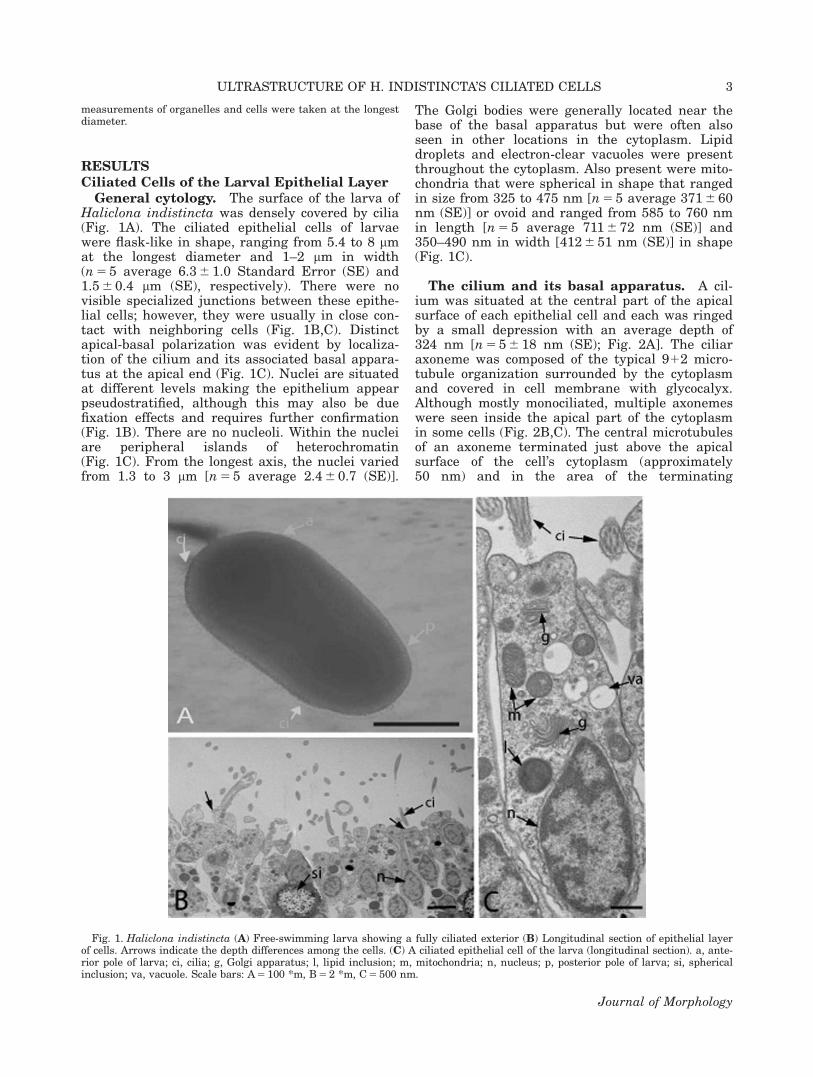
General cytology. The surface of the larva ofHaliclona indistincta was densely covered by cilia(Fig. 1A). The ciliated epithelial cells of larvaewere flask-like in shape, ranging from 5.4 to 8 mmat the longest diameter and 1–2 mm in width(n 5 5 average 6.3 6 1.0 Standard Error (SE) and1.5 6 0.4 mm (SE), respectively). There were novisible specialized junctions between these epithe-lial cells; however, they were usually in close con-tact with neighboring cells (Fig. 1B,C). Distinctapical-basal polarization was evident by localiza-tion of the cilium and its associated basal appara-tus at the apical end (Fig. 1C). Nuclei are situatedat different levels making the epithelium appearpseudostratified, although this may also be duefixation effects and requires further confirmation(Fig. 1B). There are no nucleoli. Within the nucleiare peripheral islands of heterochromatin(Fig. 1C). From the longest axis, the nuclei variedfrom 1.3 to 3 mm [n 5 5 average 2.4 6 0.7 (SE)].
The Golgi bodies were generally located near thebase of the basal apparatus but were often alsoseen in other locations in the cytoplasm. Lipiddroplets and electron-clear vacuoles were presentthroughout the cytoplasm. Also present were mito-chondria that were spherical in shape that rangedin size from 325 to 475 nm [n 5 5 average 371 6 60nm (SE)] or ovoid and ranged from 585 to 760 nmin length [n 5 5 average 711 6 72 nm (SE)] and350–490 nm in width [412 6 51 nm (SE)] in shape(Fig. 1C).
The cilium and its basal apparatus. A cil-ium was situated at the central part of the apicalsurface of each epithelial cell and each was ringedby a small depression with an average depth of324 nm [n 5 5 6 18 nm (SE); Fig. 2A]. The ciliaraxoneme was composed of the typical 912 micro-tubule organization surrounded by the cytoplasmand covered in cell membrane with glycocalyx.Although mostly monociliated, multiple axonemeswere seen inside the apical part of the cytoplasmin some cells (Fig. 2B,C). The central microtubulesof an axoneme terminated just above the apicalsurface of the cell’s cytoplasm (approximately50 nm) and in the area of the terminating
Fig. 1. Haliclona indistincta (A) Free-swimming larva showing a fully ciliated exterior (B) Longitudinal section of epithelial layerof cells. Arrows indicate the depth differences among the cells. (C) A ciliated epithelial cell of the larva (longitudinal section). a, ante-rior pole of larva; ci, cilia; g, Golgi apparatus; l, lipid inclusion; m, mitochondria; n, nucleus; p, posterior pole of larva; si, sphericalinclusion; va, vacuole. Scale bars: A 5 100 *m, B 5 2 *m, C 5 500 nm.
3ULTRASTRUCTURE OF H. INDISTINCTA’S CILIATED CELLS
Journal of Morphology

microtubules, there was an electron-dense area, ofapproximately 150 nm in length, ascending up theshaft of the cilium. Directly below the terminatedcentral microtubules, there was an electron-clearspace approximately 41–81 nm in length and 97–114 nm in diameter between the external microtu-bule doublets that remain (Fig. 2A).
The basal body was located directly below theelectron-clear zone and was in contact with theouter cilia microtubule doublets (Fig. 2A). Overall,the basal body was approximately 286 nm at thelongest axis, and approximately 244 nm in width.A basal foot (approximately 94 nm long) emergedfrom the side of the basal body and was essentiallytrapezoid in shape, being approximately 110 nm indiameter at the base and approximately 104 nm atits apical end. An accessory centriole was not iden-tified. Alar sheets radiated approximately 80 nmfrom the basal body and connected to anchoring
points. The anchor points were electron densespheres, just under the surface of the apical regionof the cytoplasm, and were 75 nm in width onaverage [n 5 5 6 13.5 (SE); Fig. 3A,B]. The longitu-dinal cytoskeleton of the cilium and basal appara-tus consisted of a microtubule that descended fromthe tip of the basal foot and ran parallel to thebasal body deep into the cytoplasm of the celltoward the nucleus, as well as a skirt of parallelfibers, which were coupled with microtubulestrands that were attached to the base of the basalbody (Fig. 3C).
No differences were observed in the ultrastruc-ture of the ciliated epithelial cells that occupiedthe anterior and the posterior poles of the larvae.Some epithelial ciliated cells were found within theinner cell mass (ICM) of the larvae at both larvalpoles from the first free-swimming larval stageonwards (Fig. 4A). Remnants of ciliated cells were
Fig. 2. Haliclona indistincta (A) Longitudinal section of apical part of an epithelial cell of a larva with the ciliar basal apparatus.(B) Cross-section of cilia showing the typical 912 configuration of the microtubules of the cilia. Also showing more than one ciliumaxoneme present in one cytoplasm. (C) Longitudinal sections of epithelial cells showing more than one cilia present in one cytoplasm.ap, anchoring point; bb, basal body; bf, basal foot; ci, cilia; pf, parallel fibers; t, terminating area; tr, transitional region; g, Golgi appa-ratus; gl, glycocalyx; m, mitochondria; mt, microtubules; n, nucleus. A 5 500 nm, B 5 100 nm; C 5 500 nm.
4 K.M. STEPHENS ET AL.
Journal of Morphology

also present within the ICM of the larvae, e.g.,remnants of cilia basal apparatus were containedwithin phagosomes of amoeboid cells, and rem-nants of cilia were observed in cells (not withinphagosomes), which were reminiscent of modifiedepithelial cells (e.g., Fig. 4B–D). Within the ICM,numerous larval ciliated cells were attached tocells containing spherical inclusions, which werevery prevalent within the larvae (Fig. 5). The ori-gin and chemical composition of these inclusionsare so far unknown. They were variable in size 1–4mm (n 5 5 average 2.9 6 1.2 mm (SE), were oftenjoined together to form complexes and appeared tocontain fibrous material (Fig. 5).
Ciliated Cells of the Juvenile Haliclonaindistincta
General cytology. Choanocytes of the juve-nile Haliclona indistincta were the only cell typewith cilia. These cells were irregular in shape,however, two general forms existed; 1) pseudocy-lindrical with a length of 4–6 mm [n 5 5 average4.5 6 0.85 mm (SE)] and width of 1.6-3 mm (n 5 5average 2.4 6 0.7 mm (SE)) and 2) oval to rounded,2.8-3.1 mm in length [n 5 5 average 2.9 6 0.1 mm(SE)] and 3–4.5 mm in width [n 5 5 average3.5 6 0.6 mm (SE); (Fig. 6A,B)]. The shape of thechoanocyte was not associated with their position
in the sponge body as both forms were observedlining choanocyte chambers. A spherical nucleusthat ranged from 1.1 to 2 mm in diameter [n 5 5average 1.5 6 0.2 mm (SE)] was located at the basalpart of the cell (Fig. 6A,B). Mitochondria thatranged from 335 to 440 nm [n 5 5 average 380nm 6 37 nm (SE)] were also seen within the cyto-plasm (Fig. 6B) and were more consistent in shape(i.e., rounded) than those observed in the ciliatedepithelial cells (which could be ovoid). As in thelarval ciliated cells, numerous lipid droplets andelectron clear vacuoles were seen throughout thecytoplasm, but were much more plentiful (Fig. 6A–D). Glycocalyx was present on the apical area ofthe cell’s surface, as well as on the cilia. No speci-alized junction between the choanocytes was evi-dent. Choanocytes were apical-basally polarizedwith a cilium encircled by a collar of microvilli(approximately 24–30) protruding from the apicalsurface (Fig. 6C). The apical parts of the cellswere orientated toward the lumen of the choano-cyte chamber (Fig. 6D).
The cilia and the basal apparatus. Themicrotubules that composed the cilia axonemewere arranged in the typical 912 configuration.The central microtubules terminated further fromthe apical surface of the cell’s cytoplasm than inthe larval ciliated cells (150 nm compared to 50nm). As in the larval cells there was an electrondense area that extended from the terminatingend of the microtubules to approximately 120 nmup the cilia shaft (Fig. 7A). Below the terminatedcentral microtubules, there was a larger electron-clear space that ranged approximately from 102 to173 nm in length and 61–123 nm in widthbetween the external microtubule doublets thatremained (Fig. 7A). The basal body of the choano-cyte cilium, located directly below this electron-clear zone, had a transitional region between theproximal end of the cilia shaft and the basal body.From measurements taken along the longest axis,the basal body was approximately 265 nm inlength and approximately 240 nm in width. Anaccessory centriole in the ciliar basal apparatus ofthe choanocytes was absent. Alar sheets, radiatedfrom the basal body (approximately 100 nm inlength) and connected to electron-dense spheres,anchoring points, 61 nm in diameter on average[n 5 5 6 8.2 nm (SE)], and were positioned immedi-ately under the surface of the apical region of thecell membrane (Fig. 7B,C,E). Below the alarsheets, extended a basal foot approximately 95 nmfrom the side of the basal body. The basal foot wasroughly pyramidal in shape, being approximately60 nm in diameter at the apical end and approxi-mately 105 nm at the base (Fig. 7A,D). Directlybelow the alar sheets, microtubules radiated in aspiral-like fashion, parallel to the surface(Fig. 7E), which may be due to the angle at whichthe cells were cut. These microtubules, and a
Fig. 3. Haliclona indistincta (A,B) Cross-section of the apicalpart of an epithelial cell of a showing the transitional regionbetween the proximal end of the cilia shaft and the basal body.(C) Longitudinal section of anterior pole of larval epithelial cellshowing the basal apparatus and microtubules descending fromthe basal body. ap, anchoring point; as, alar sheet; bb, basalbody; bf, basal foot; mt, microtubules; pf, parallel fibers. Scalebars: A-B 5 500 nm, C 5 100 nm.
5ULTRASTRUCTURE OF H. INDISTINCTA’S CILIATED CELLS
Journal of Morphology

forked microtubule that extended from the tip ofthe basal foot, comprised the transverse cytoskele-ton (Fig. 7D,E). There were three microtubulesattached above the base of the basal body insteadof only one as in the larval epithelial cells(attached to the basal foot). Two extended from thebasal foot, one parallel to the basal body and oneparallel to the surface of the apical region of thechoanocyte (as mentioned above) (Fig. 7D). Thethird microtubule was attached to the basal bodyopposite to the basal foot, and extended downward,
toward the nucleus, also parallel to the basal body(Fig. 7D). Attached at the tip of the basal body wasa bundle of microtubules that extended to a Golgiapparatus, which along with the two longitudinallyrunning microtubules mentioned previously, com-prised the longitudinal cytoskeleton (Fig. 7A).
Comparison of ciliated cells of larva andjuvenile. The gross morphology of the ciliatedepithelial cell of the larva and of the choanocyte ofthe juvenile (e.g., cell and nuclei shape, and size)was very different (Table 1; Fig. 8). The vacuoles
Fig. 4. Haliclona indistincta (A) Epithelial cell present within the ICM during the first free-swimming larval stage. (B) Longitudi-nal section of a cell within the ICM of a free-swimming stage larva with the basal apparatus (bb) of a ciliated epithelial cell within aphagosome (ph). (C) Longitudinal section showing epithelial cell of the first free-swimming larval stage of an H. indistincta larvawith its cilium and basal apparatus inside the cytoplasm. (D) Longitudinal section of epithelial cell migrated inward during the thirdof the free-swimming stages of this species (note the cilia enveloped by the cytoplasm). ci, cilia; bb, basal body; e, epithelial cell; pc,phagocytosing cell; ph, phagosome; n, nucleus; Scale bars: A–D 5 500 nm.
6 K.M. STEPHENS ET AL.
Journal of Morphology

Fig. 5. Haliclona indistincta (A) Longitudinal section of an epithelial layer of the larva of H. indistincta with cells containingspherical inclusions, associated with some epithelial cells. (B,C) Longitudinal sections of epithelial layer with cells containing spheri-cal inclusions associated with some epithelial cells. (D) Close-up of a spherical inclusion. bb, basal body; ci, cilia; si, spherical inclu-sion; e, epithelia cell. Scale bars: A 5 10 lm, B-D 5 500 nm.
Fig. 6. The ciliated cells of Haliclona indistincta juveniles. (A) Longitudinal section of a pseudocylindrically shaped choanocyte.(Section from juvenile settled 8 days). (B) Longitudinal section of an ovate shaped choanocyte. (Section from juvenile settled 32days). (C) Longitudinal section of an apical part of a choanocyte, showing the microvilli protruding from the surface. (D) View of achoanocyte chamber showing apical ends of the choanocytes facing toward the lumen. c, choanocyte; ci, cilium; l, lipid; lu, lumen; m,mitochondria; mi, microvilli; n, nucleus; va, vacuoles. Scale bars: A 5 2 lm, B-C 5 500 nm, D 5 2 mm.
7ULTRASTRUCTURE OF H. INDISTINCTA’S CILIATED CELLS
Journal of Morphology

were much more numerous, the nucleus was morecompact and mitochondria were more regular inshape in the choanocytes, than in the ciliatedlarval cell. While both cell types unsurprisinglyshared the same cilium axoneme configuration(912) and shape and size of the basal foot andbasal body, the basal apparatus of the choanocytehad a noticeably more elaborate microtubular cyto-skeleton than that of the larva (Table 1; Fig. 8).The choanocyte had a transverse cytoskeleton com-prised of a collar of microtubules that extendedfrom the basal body in a spiral formation, as wellas a forked microtubule that extended from the tipof the basal foot that also ran parallel to the apicalsurface of the cell. By comparison, the ciliated epi-thelial cell had only a longitudinal cytoskeleton.While the ciliated epithelial cell of the larva had asingle microtubule extending from the tip of thebasal foot running longitudinally toward thenucleus of the cell, a microtubule extending fromthe tip of the basal foot, running parallel to thebasal body, was also present in the choanocyte, as
well as a third microtubule extending from thebasal body on the opposite side of the basal footthat also ran parallel to the basal body. However,the remaining features of the longitudinal cytoskel-eton of the larval epithelial cell was more elaboratethan the choanocyte of the juvenile, with a skirt ofparallel fibres as well as microtubule strands,while only a bundle of microtubules were attachedto the base of the basal apparatus of the choano-cyte (Table 2).
DISCUSSION
Our initial hypothesis was that, through compari-son of the cells that formed the ciliated epitheliallayer of the larvae and the cells that formed thechoanoderm of the juvenile, we would be able toshow a direct relationship between the two celltypes, thus tracing the derivation of choanocytesfrom epithelial cells. This hypothesis was based onthe relationship between larval and juvenile ciliatedcells observed in other sponges (e.g., Maldonado,
Fig. 7. Choanocyte of young Haliclona indistincta sponge. (A) Longitudinal section including microtubule protruding from basalbody. (B,C) Cross-sections of apical part of the cell, showing the transition from the alar sheets attached to microtubules that radiatefrom the basal body in a spiral-like configuration. (D) Longitudinal section of apical part of the cell including microtubules protrudingfrom tip of the basal foot (mt 1 and mt 2) and basal body (mt 3). Also showing the distal end of the basal body and a typical exampleof the transitional region electron clear area and terminating area of the cilia seen. (E) Cross-sections showing branched microtu-bules. ap, anchoring point; as, alar sheet; bb, basal body; bf, basal foot; de, distal end; ec, electron clear zone; g, Golgi apparatus; mi,microvilli; mt, microtubules; t, terminating area; tr, transitional region. Scale bars: A 5 100 nm, B–E 5 500 nm.
8 K.M. STEPHENS ET AL.
Journal of Morphology

2004; Leys and Ereskovsky, 2006; Ereskovsky,2007, 2010; Ereskovsky et al., 2007, 2009).Although the cell types were sufficiently different toprevent a direct link between the cells of the twodevelopmental stages (i.e., free-swimming and ses-sile), changes in the positioning of larval ciliatedepithelial cells throughout metamorphosis, sug-gested two possible fates for the ciliated, epithelialcells. Larval ciliated cells were observed intact inan internal position throughout the three presettle-ment stages. Migration of the larval epithelial cellsinward has been described in some Calcinea species(Borojevic, 1969) and in the demosponge Halisarcadujardini (Gonobobleva and Ereskovsky, 2004). Thefirst fate of these migrating cells is suggested bythe reabsorption of cilia, which was evident in thecytoplasm of some cells. This process, we suggest, ispart of the transformation of the epithelial cells ofH. indistincta into choanocytes, a process that hasbeen described in other haplosclerids, such as Cha-linula sp. (Ilan and Loya, 1990), Haliclona permolis(Amano and Hori, 1996), Amphimedon queeslandica(Leys and Degnan, 2002), the freshwater spongesEunapius fragilis, Ephydatia muelleri, and Spon-gilla lacustris (Ivanova, 1997a,b) as well as otherdemosponges (Borojevic and L�evi, 1965; Gonobo-bleva and Ereskovsky, 2004). Second, phagosomeswith remnants of cilium and basal apparatus wereobserved within amoeboid cells suggesting thatsome cells are destroyed.
No previous studies concerning the metamor-phosis of sponge larvae have mentioned an associ-ation between a type of spherulous cell and/ortheir inclusions, and larval epithelial cells, as has
TABLE 1. Comparison of ciliated cells of larvae and juvenile of Haliclona indistincta: gross morphology of cell types, and ciliarybasal apparatus. Mt 5 microtubules
Cell characteristics Larval ciliated epithelial cell Juvenile choanocyte
Gross morphologyof cell
Cell shape Flask-like PseudocylindricalOvate to spherical
Cell length and width 5.4–8 mm 31–2 mm (n 5 5) 4–6 mm 3 1.6–3 mm (n 5 5)2.8–3.1 mm 3 3–4.5 mm (n 5 5)
Glycocalyx Abundant SparseNuclei shape Pear-shaped SphericalMitochondria shape Spherical Spherical
OvateMitochondria length and
width325–475 nm (n 5 5) 335–440 nm (n 5 5)585–760 nm 3 350–490 nm (n 5 5)
Golgi Apparatus Numerous FewVacuoles Few Numerous
Cilium and basalapparatus
Cilia axonemaconfiguration
912 912
Electron-free area atbase of cilium
41–81 nm 3 97–114 nm 102–173 nm 3 61–123 nm
Basal foot shape Trapezoid TrapezoidTransverse cytoskeleton:
microtubules (mt)None Two components: 1) collar of mt
above basal foot; 2) single mt(forked) from tip of basal footrunning parallel to the apicalsurface of cell
Longitudinalcytoskeleton
Two components: 1) some mtattached to tip of basal footparallel to basal body; 2) skirtof parallel fibers with few mtstrands
Three components: 1) mt attached totip of basal foot which ran parallelto basal body; 2) few mt locatedopposite side of basal foot; 3)grouping of mt attached to basalend of basal body (lacking fineparallel fibers)
Fig. 8. A Schematic drawing showing the ciliar basal appara-tus and relevant organelles observed in the longitudinal sectionsof the ciliated epithelial cell of the larva of H. indistincta (A),and choanocyte of the juvenile sponge (B) of H. indistincta. ap,anchoring point; as, alar sheet; bb, basal body; bf, basal foot; de,distal end; ec, electron clear zone; g, Golgi apparatus; gl, glycoca-lyx; l, lipid; m, mitochondria; mi, microvilli; mt, microtubules; n,nucleus; pf, parallel fibers; r, root; t, terminating area; tr, transi-tional region; va, vacuoles.
9ULTRASTRUCTURE OF H. INDISTINCTA’S CILIATED CELLS
Journal of Morphology

TA
BL
E2.
Com
pa
riso
nof
cili
ar
ba
sal
ap
pa
ratu
sin
dif
fere
nt
larv
al
typ
esfo
un
din
dem
osp
onges
HA
P(M
)H
AP
(FW
)H
AL
PO
ED
EN
Ord
erS
pec
ies
Ha
licl
ona
ind
isti
nct
aH
ali
clon
atu
bif
era
Ha
licl
ona
per
mol
lis
Sig
ma
doc
iaca
eru
lea
Eu
na
piu
sfr
agil
isE
ph
yda
tia
mu
elle
riS
pon
gil
lala
cust
ris
Ha
lich
ond
ria
mel
an
ad
ocia
Myc
ale
ceci
lia
Myc
ale
syri
nx
Den
dri
lla
cact
us
(Ap
lysi
lla
sp.)
DIC
Irci
nia
oros
AS
TT
hoo
sam
ism
alo
lli
CH
OC
hon
dri
lla
au
stra
lien
sis
HA
LI
Ha
lisa
rca
du
jard
ini
Larv
al
typ
eP
AP
AP
AP
AP
AP
AP
AP
AP
AP
AP
AP
AH
OP
CL
(CO
)D
IB
asa
lbod
yT
yp
e2
22
2?
??
22
?2
2(?
)N
/A2(?
)2
Basa
lfo
otY
Y?
YY
??
YY
?Y
YN
/AY
YB
asa
lfo
otsh
ap
eT
M?
S?
??
MA
?S
ON
/AO
SA
cces
sory
cen
trio
leN
N?
NY
YY
YN
?Y
YN
/AY
YT
ran
sver
secy
tosk
elet
onN
Y?
NY
??
YN
?Y
YN
/AY
YC
ross
-str
iate
dro
otle
t(s)
NN
?N
??
?N
N/A
?N
NN
/AN
NL
ate
ral
arm
sN
Y?
N?
??
NN
/A?
YN
N/A
NN
Basa
lfo
otm
icro
tubu
le(s
)Y
Y?
Y?
??
YN
/A?
YN
N/A
NY
Lon
git
ud
inal
cyto
skel
eton
YY
YY
YY
YY
Y?
YY
N/A
YY
Fib
rou
sro
otle
t(s)
YY
YY
YY
YY
N?
NY
N/A
YY
Lam
inar
shee
tsN
NN
NN
NN
NY
?Y
NN
/AN
NG
olgi
ass
ocia
ted
wit
hro
otle
t(s)
YN
NN
Y(?
)?
?Y
Y?
YY
(?)
N/A
NY
Ref
eren
ces
Ste
ph
ens
etal.
(th
isw
ork
)
Woo
llaco
tt,
‘93;
Woo
llaco
ttan
dP
into
,‘9
5;
Am
an
oan
dH
ori,
‘96
Mald
onad
oet
al.
,2003
Ivan
ova,
‘97a;
‘97b
Ivan
ova,
‘97a;
‘97b;
Ivan
ova,
‘97a;
‘97b;
Woo
llaco
ttan
dP
into
,‘9
5
Woo
llaco
ttan
dP
into
,‘9
5
Wil
son
,‘3
5W
ooll
aco
ttan
dH
ad
fiel
d,
‘89;
Woo
llaco
ttan
dP
into
,‘9
5;
Ere
skov
sky
an
dT
okin
a‘0
4
Bau
tist
a-
Gu
erre
roet
al.
,‘1
0;
Ush
eran
dE
resk
ovsk
y,‘0
5
Gon
obob
leva,
Ere
skov
sky,
2004;G
onob
o-ble
va
an
dM
ald
onad
o,‘0
9
HA
P(M
)5H
ap
losc
leri
da(m
ari
ne)
;H
AP
(FW
)5H
ap
losc
leri
da(f
resh
wate
r);
HA
L5
Hali
chon
dri
da;
PO
E5
Poe
cilo
scle
rid
a;
DE
N5
Den
dro
cera
tid
a;
DIC
5D
icty
ocer
ati
da;
AS
T5
Ast
rop
hor
ida;
CH
O5
Ch
ond
rosi
da;
HA
LI
5H
ali
sarc
ida;
PA
5P
are
nch
ym
ella
;H
OP
5H
opli
tom
ella
;C
L5
Cla
vabla
stu
la;
CO
5C
oelo
bla
stu
la;
DI
5D
isp
her
ula
;N
5n
o;Y
5yes
;N
/A5
not
ap
pli
cable
;?
5m
issi
ng
oram
big
uou
sin
form
ati
on;
basa
lbod
yty
pes
1an
d2:
Sen
sust
rict
oW
ooll
aco
ttan
dP
into
(1995);
A5
art
icu
late
d(s
ust
rict
oW
ooll
aco
ttan
dP
into
,1995),
M5
mu
shro
om,
stalk
edor
cam
paig
nco
rk;
O5
ovoi
d,
S5
sim
ple
orci
rcu
lar,
T5
trap
ezoi
d.
been observed in this study. The inclusions grewto a comparatively large size (expanding to over3 times their original size), and we have not yetdetermined if they remain inside the spherulouscell (obscuring other cell components), or havebeen released when they attain this size. Not allciliated epithelial cells were associated with thesespherulous cell/inclusions. When it occurred, theassociation involved the attachment of the epithe-lial cells to the spherulous cell/inclusions by thebasal end of the epithelial cell. The spherulous cellmay have a role in the positioning and fate oflarval cells. These cells were concentrated beneaththe layer of epithelial cells of the larvae. This isthe same location as the layer of an unidentifiedmaterial described in Stephens et al. (2012) thatseparated the epithelial layer of the embryos fromthe ICM. However, throughout the presettlementand postsettlement stages, the spherulous cells(with enlarged inclusions) were visible deep withinthe ICM of the larvae that were not observed inthe embryos examined using histology. The larvaeof H. indistincta were sticky. This study suggeststhat the sticky nature of the larvae, and the adultof this species, [as described by L�evi (1956)], maybe used as a distinguishing taxonomic feature.
Multiple ciliar axonemes were commonlyobserved within single cytoplasms, which is a fea-ture that has been reported in several other spongelarval studies (e.g., L�evi, 1964; Boury-Esnault andVacelet, 1994; Boury-Esnault et al., 1999; Maldo-nado et al., 2003; Maldonado, 2004). Haliclonaindistincta parenchymella differ from the larvae ofother haplosclerid species described by lacking spi-cules, a trait which is shared by some species of thedemosponge orders: Halisarcida, Chondrosida, Dic-tyoceratida, Dendroceratida, and Halichondrida(Woollacott and Hadfield, 1989; Woollacott andPinto, 1995; Leys and Degnan, 2001; Maldonadoet al., 2003; Ereskovsky and Tokina 2004; Gonobo-bleva and Ereskovsky, 2004; Usher and Ereskovsky,2005; Gonobobleva, 2007; Ereskovsky, 2010). Giventhe variation in the appearance of spicules acrosssponges in the literature (e.g., Ivanova, 1997a,b),the timing of spicule production is likely to be vari-able, and not of phylogenetic importance. Haliclonaindistincta larvae also differ from other haplo-sclerid species described, in the ciliation pattern,being uniformly ciliated and lacking longer cilia atthe posterior pole.
The positioning of the choanocyte chambers inH. indistincta adults (being directly in contact withthe mesohyl) is more similar to poecilosclerids thanto other haplosclerid species (i.e., H. oculata,H. rosea, H. simulans, H. fistulosa, H. elegans, andNiphates digitalis), whose chambers are separatedfrom the mesohyl by pinacocytes and the walls ofincurrent canals. This is a feature that led Langen-bruch and Jones (1990) to suggest that H. indis-tincta is of poecilosclerid, and not haplosclerid,
origin. Ciliar basal apparatus configuration of theciliated larval epithelial cells of H. indistincta andH. tubifera [as described in Woollacott and Pinto(1995)] were very different. The most apparent dis-tinction was the more simple microtubule configu-ration of H. indistincta, which lacked the lateralarms observed in H. tubifera (Woollacott and Pinto,1995). Phylogenetic reconstructions from two inde-pendent gene loci, 28S ribosomal RNA and themitochondrial cytochrome oxidase subunit 1(mtCOI) genes confirm H. indistincta’s place withinthe marine haplosclerids (Stephens PhD thesis)but only distantly related to the type Haliclonaspecies H. oculata.
L�evi (1956) and Bergquist et al. (1979) suggestedlarval morphology to be an informative characterfor sponge taxonomy. The following features arecharacteristic of the larvae of marine haplosclerida:the absence of flagellated cells in the posterior pole,which is fringed by a circle of cells with longer flag-ella; the presence of a skeleton representing a densebundle of oxeas located in the posterior portion ofthe larva; and the concentration of pigment in thecells of the posterior pole devoid of flagellum (Ere-skovsky, 1999, 2010). These characters have diag-nostic value in the systematics of sponges(Bergquist et al., 1979; Simpson, 1984; Wapstra andvan Soest, 1987; Woollacott, 1993; Fromont, 1994;Ereskovsky, 1999). The ciliation pattern of Hali-clona tubifera larvae is considered typical of Hali-clona species (Woollacott, 1993; Woollacott andPinto, 1995). Haliclona oculata larvae also sharethis ciliation pattern (Wapstra and van Soest,1987). H. indistincta however, does not, which isconsistent with the fact that they belong to a differ-ent clade than H. oculata and H. tubifera, beingmore closely related to members of Niphates, thanto other Haliclona (Stephens unpublished PhD the-sis). However, Ilan and Loya (1988) and Ilan et al.(2004), described Niphates rowi also as having lon-ger cilia forming a ring around the posterior pole.Until more larvae of different species are investi-gated with both morphological and molecular data,we will not be able to determine how plastic the cil-iation pattern is among larvae, and whether or notit has phylogenetic relevance within certain groups.
Woollacott and Pinto (1995) showed that basalciliar apparatus characters can be useful in identi-fying closely related species, as congruence wasfound among the poecilosclerid species studied(i.e., Mycale cecilia, M. contarenii, and Hamigerahamigera) and within the halichondrids, (i.e., Hal-ichondria melanadocia, H. coerulea, and H. helio-phila), using basal ciliar apparatus morphology.These authors also compared the haploscleridH. tubifera to two additional Haliclona spp., whichlacked a transverse cytoskeleton as also observedin H. indistincta (Woollacott and Pinto, 1995).Despite the differences between the larval andjuvenile ciliated cells in H. indistincta, the overall
11ULTRASTRUCTURE OF H. INDISTINCTA’S CILIATED CELLS
Journal of Morphology

shape of the cells was similar to that of othersponge orders and freshwater sponges [i.e., E. fra-gilis, E. muelleri, and S. lacustris (Ivanova,1997a,b)] (Table 2). Seravin (1986) suggest thatmicrotubules, like those that projected from thebasal body in a spiral-like arrangement of thechoanocyte of H. indistincta, are a typical featureof sponge choanocytes, e.g., Spongilla lacustris(Fjerdingstad, 1961), Ephydatia fluviatilis (Feige,1969), H. rosea (Garrone, 1969), Baikalospongiabacillifera (Efremova et al., 1988), and Halisarcadujardini (Gonobobleva and Maldonado, 2009).H. indistincta, however also possesses a bundle ofmicrotubules attached to the base of the basalbody that is uncommon in other sponge choano-cytes (Woollacott and Pinto, 1995; Gonoboblevaand Maldonado, 2009). A common structure pres-ent in ciliated eukaryote epithelial cells is theaccessory centriole, which is involved during celldivision as microtubule organizing centers (Niel-sen, 1987; Gonobobleva, 2007). However, accessorycentrioles were not observed in ciliated larval cellsand in the choanocytes of H. indistincta. They arealso absent in H. tubifera, and M. cecilia (Woolla-cott and Pinto, 1995) but are otherwise commonlyfound in sponge larva (e.g., Homoscleromorpha,Dictyoceratida and Halisarcida; Table 2; (Maldo-nado et al., 2003; Boury-Esnault et al., 2003; Ere-skovsky and Tokina 2004; Gonobobleva andEreskovsky, 2004; Ereskovsky et al., 2007, 2009;Gonobobleva and Maldonado, 2009).
Unfortunately, the level of detail included inthis and Woollacott and Pinto’s (1995) study is notyet available for many sponges species so it is notpossible to accurately determine the phylogeneticsignal associated with the six characters includedin Woollacott and Pinto (1995) (i.e., basal body,basal foot, accessory centriole, transverse and lon-gitudinal cytoskeletal system, and the associationof the cytoskeleton, i.e., rootlet system, with Golgibody). Therefore, additional comparative studiestargeting the basal apparatus of putatively closelyrelated species of H. indistincta (e.g., H. viscosaand Niphates spp.) and additional specimens ofthis species, are necessary for determining thelevel that these, and other characters, are inform-ative. However, in addition to shedding some lighton the origin of the choanocytes in this species,this study provides valuable information for suchcomparative morphological analyses in the future,which will be necessary to further understand evo-lutionary relationships in this important group ofanimals.
ACKNOWLEDGMENTS
This project has been funded through a NationalUniversity of Ireland Galway PhD fellowship, andthe Thomas Crawford Hayes Trust Fund Scheme(NUI Galway) awarded to Kelly Stephens. Authors
would like to thank the two anonymous reviewersfor their suggestions, which have improved themanuscript.
LITERATURE CITED
Adams EDM, Goss GG, Leys SP. 2010. Freshwater spongeshave functional, sealing epithelia with high transepithelialresistance and negative transepithelial potential. PLoS ONE0:e15040.
Amano S, Hori I. 1996. Transdifferentiation of larval flagellatedcells to choanocytes in the metamorphosis of the DemospongeHaliclona permollis. Biol Bull 190:161–172.
Ax P. 1995. Das System der Metazoa. Stuttgart: Gustav FischerVerlag.
Ax P, Sopott-Ehlers B, Ehlers U, Bartolomaeus T. 1989. Wasleistet das Elektronenmikroskop fu€ur die Aufdeckung derStammesgeschichte der Tiere? In Akademie der Wissenschaf-ten und der Literatur Mainz: 1949–1989, Stuttgart: FranzSteiner Verlag, pp. 73–86.
Barnes RD, Harrison FW. 1991. Introduction to the metazoa.In: Harrison FW, Westfall JA, editors. Microscopic Anatomyof Invertebrates. Vol. 2. Placoza, Porifera, Cnidaria, and Cte-nophora. New York: Wiley-Liss. pp 1–12.
Bautista-Guerrero E, Carballo JL, Maldonado M. 2010. Repro-ductive cycle of the coral-excavating sponge Thoosa misma-lolli (Clionaidae) from Mexican Pacific coral reefs. Invert Biol129:285–296.
Bergquist PR. 1978. Sponges. Los Angeles, CA: University ofCalifornia Press.
Bergquist PR, Sinclair ME, Green CR, Silyn-Roberts H. 1979.Comparative morphology and behaviour of larvae of Demo-spongiae. In: L�evi C, Boury-Esnault N, editors. Biologie desSpongiaires. Colloque Internat du Cent Nat de la Rech Scien-tifiq 191:103–111.
Borojevic R. 1969. Etude du d�eveloppement et d�e la differentia-tion cellulaire d’�eponges calcaires Calcin�ees (genres Clathrinaet Ascandra). Anns d Embryol et de Morph 2:15–36.
Borojevic R, L�evi C. 1965. Morphogenese experimentale d’uneeponge a partir de cellules de la larve negeante dissociee.Zeitsch f€ur Zellfor und mikrosko Anato (Vienna, Austria) 68:57–69.
Boury-Esnault N, Vacelet J. 1994. Preliminary studies on theorganisation and development of a hexactinellid sponge froma Mediterranean cave, Oopsacas minuta. In: van Soest RWM,van Kempen TMG, Braekman JC, editors. Sponges in Timeand Space. Rotterdam/Amsterdam: Balkema.
Boury-Esnault N, Efremova SM, B�ezac C, Vacelet J. 1999.Reproduction of a hexactinellid sponge: First description ofgastrulation by cellular delamination in the Porifera. InvertReprod Dev 35:187–201.
Boury-Esnault N, Ereskovsky A, B�ezac C, Tokina D. 2003.Larval development in the Homoscleromorpha (Porifera,Demospongiae). Invert Biol 122:187–202.
Bowerbank JS. 1866. A Monograph of the British Spongiadae,Vol. 2. Ray Society London. I–XX. pp 1–388.
De Vos L, Rutzler K, Boury-Esnault N, Donadey C, Vacelet J.1991. Atlas de morphologie des Eponges–atlas of sponge mor-phology. Washington, DC: Simthsonian Institution Press.
Delage Y. 1892. Embryogen�ese des �eponges silicieuses. ArchsZool Exp Gen 10:345–498.
Efremova SM, Sukhodolskaya AN, Alekseeva NP. 1988. The dif-ferent structure of kinetosome rootlet systems in flagellatedcells of the larvae and the choanocytes of sponges. In: KoltunBM, Stepaniants CD, editors. Porifera and Cnidaria. Modernand Perspective Investigations. Leningrad: USSA Academy ofSciences, Zoological institute. pp 22–23.
Ereskovsky AV. 1999. Development of Haplosclerida sponges(Demospongiae, Ceractinomorpha). Russ J Mar Biol 25:333–343.
12 K.M. STEPHENS ET AL.
Journal of Morphology
Ereskovsky AV. 2007. Sponge embryology: the past, the presentand the future. In: Cust�odio MR, Hajdu E, Muricy G, editors.Porifera Research: Biodiversity, Innovation and Sustainabil-ity. Rio de Janeiro: Museu Nacional. pp 41–52.
Ereskovsky AV. 2010. The Comparative Embryology of Sponges.Springer Science and Business Media B.V. Netherlands.
Ereskovsky AV, Dondua AK. 2006. The problem of germ layersin sponges (Porifera) and some issues concerning early meta-zoan evolution. Zoologischer Anzeiger. 245:65–76.
Ereskovsky AV, Tokina DB. 2004. Morphology and fine struc-ture of the swimming larvae of Ircinia oros (Porifera, Demo-spongiae, Dictyoceratida). Invert Reprod Dev 45:137–150.
Ereskovsky AV, Willenz P. 2008. Larval development in Guan-cha arnesenae (Porifera; Calcispongiae, Calcinea). Zoomor-phology 127:175–187.
Ereskovsky AV, Tokina DB, B�ezac C, Boury-Esnault N. 2007.Metamorphosis of cinctoblastula larvae (Homoscleromorpha,Porifera). J Morphol 268:518–528.
Ereskovsky AV, Konyukov PY, Tokina DB. 2009. Morphogenesisaccompanying larval metamorphosis in Plakina trilopha (Por-ifera, Homoscleromorpha). Zoomorphology 129:21–31.
Fahey B, Degnan BM. 2010. Origin of animal epithelia: insightsfrom the sponge genome. Evol Dev 12:601–617.
Feige W. 1969. Die Feinstruktur der epithelien von Ephydatiafluviatilis. Zoologische Jahrb 86:177–237.
Fjerdingstad EJ. 1961. The ultrastructure of choanocytecollars in Spongilla lacustris (L.). Zeit Zellforsch 53:645–657.
Fromont J, Bergquist PR. 1994. Repoductive blology of threesponge species of the genus Xetospongla (Por�feraD: emo-spongiae: Petrosida) from the Great Barrier Reef. Coral Reefs13:119–126.
Garrone R. 1969. Collagene, spongine et squelette min�eral chezl’Eponge Haliclona rosea (O.S.) (D�emosponge, Haploscl�eride).J Microsc 8:581–598.
Gonobobleva EL. 2007. Basal apparatus formation in externalflagellated cells of Halisarca dujardini larvae (Demospongiae:Halisarcida) in the course of embryonic development. In:Cust�odio MR, Lobo-Hajdu G, Hajdu E, Muricy G, editors. Por-ifera Research: Biodiversity, Innovation and Sustainability.Rio de Janeiro: Museu Nacional. pp 345–351.
Gonobobleva EL, Ereskovsky AV. 2004. Metamorphosis of thelarvae of Halisarca dujardini (Demospongiae, Halisarcida).Bull Inst R Sci Nat Belg 74:101–115.
Gonobobleva E, Maldonado M. 2009. Choanocyte ultrastructurein Halisarca dujardini (Demospongiae, Halisarcida).J Morphol 270:615–627.
Ilan M, Loya Y. 1988. Reproduction and settlement of thecoral reef sponge Niphates sp (Red Sea) Proceedings of the6th International Coral Reef Symposium, Townsville, Aus-tralia, Vol 2. pp 745–749.
Ilan M, Loya Y. 1990. Sexual reproduction and settlement ofthe coral reef sponge Chalinula sp. from the Red Sea. MarBiol 105:25–31.
Ilan M, Gugel J, van Soest RMW. 2004. Taxonomy, reproductionand ecology of new and known Red Sea sponges. Sarsia 89:388–410.
Ivanova LV. 1997a. New data about morphology and metamor-phosis of the spongillid larvae (Porifera, Spongillidae). 1. Mor-phology of the free-swimming larvae. In: Ereskovsky AV,Keupp H, Kohring HR, editors. Modern Problems of PoriferanBiology. Berliner Geowiss Abh: Freie University, Berlin. pp55–71.
Ivanova LV. 1997b. New data about morphology and metamor-phosis of the spongillid larvae (Porifera, Spongillidae). 2. Themetamorphosis of the spongillid larvae. In: Ereskovsky AV,Keupp H, Kohring HR, editors. Modern Problems of PoriferanBiology. Berliner Geowiss Abh, Freie University, Berlin. pp73–91.
Karpov SA, Efremova SM. 1994. Ultrathin structure of flagellarapparatus in choanocyte of sponge Ephydatia fluviatilis. Tsi-tologia 36:403–408.
Langenbruch PF, Jones CW. 1990. Body structure of marinesponges. VI Choanocyte chamber structure in the Haploscler-ida (Porifera, Demospongiae) and its relevance to the phylo-genesis of the group. J Morphol 204:1–8.
L�evi C. 1956. Etude des Halisarca de Roscoff. Embryologie etsystematique des D�emosponges. Trav Stat Biol Roscoff N.S 7:3–181.
L�evi C. 1964. Ultrastructure de la larve parenchymella deD�emosponge. I. Mycale contarenii (Martens). Cah Biol Mar 5:97–104.
Leys SP, Degnan BM. 2001. Cytological basis of photorespon-sive behaviour in a sponge larvae. Biol Bull 201:323–338.
Leys S, Degnan BM. 2002. Embryogenesis and metamorphosisin a haplosclerid demosponge: Gastrulation and transdifferen-tiation of larval ciliated cells to choanocytes. Invert Biol 121:171–189.
Leys SP, Ereskovsky AV. 2006. Embryogenesis and larval differ-entiation in sponges. Can J Zool 84:262–287.
Leys SP, Nichols SA, Adams EDM. 2009. Epithelia and integra-tion in sponges. Integr Comp Biol icp038.
Maldonado M. 2004. Choanoflagellates, choanocytes, and ani-mal multicellularity. Invert Biol 123:1–22.
Maldonado M. 2009. Embryonic development of verongid demo-sponges supports the independent acquisition of spongin skel-etons as an alternative to the siliceous skeleton of sponges.Biol J Linn Soc 97:427–447.
Maldonado M, Dufort M, McCarthy DA, Young CM. 2003. Thecellular basis of photobehavior in the tufted parenchymellalarva of demosponges. Mar Biol 143:427–441.
Mitchell DR. 2007. The evolution of eukaryotic cilia and flagellaas motile and sensory organelles. Adv Exp Med Biol 607:130–140.
Nickel M, Scheer C, Hammel J U, Herzen J, Beckmann F.2011. The contractile sponge epithelium sensu lato–body con-traction of the demosponge Tethya wilhelma is mediated bythe pinacoderm. J Exp Biol 214:1692–1698.
Nielsen C. 1987. Structure and function of metazoan ciliarybands and their phylogenetic significance. Acta Zool 68:205–262.
Nielsen C. 2001. Animal Evolution: Interrelationships ofthe Living Phyla, 2nd ed. Oxford: Oxford UniversityPress.
Nielsen C. 2012. Animal evolution interrelationships of the liv-ing phyla, 3rd ed. Oxford: Oxford University Press.
Philippe H, Derelle R, Lopez P, Pick K, Borchiellini C, Boury-Esnault N, Vacelet J, Renard E, Houliston E, Queinnec E, DaSilva C, Wincker P, Le Guyader H, Leys S, Jackson DJ,Schreiber F, Erpenbeck D, Morgenstern B, W€orheide G, Man-uel M. 2009. Phylogenomics revives traditional views on deepanimal relationships. Current Biology 19:706–712.
Rieger R. 1986. €Uber den Ursprung der Bilateria: dieBedeutung der Ultrastrukturforschung fu€ur ein neues Ver-stehen der Metazoenevolution. Verh Dtsch Zool Ges 79:31–50.
Ruiz-Trillo I, Rodger AJ, Burger G, Gray MW, Lang BF. 2008.A phylogenetic investigation into the origin of Metazoa. MolBiol Evol 25:664–672.
Schmidt O. 1866. Zweites Supplement der Spongien des Adria-tischen Meeres enthaltend die Vergleichung der Adriatischenund Britischen Spongiengattungen. Leipzig: Verlag von Wil-helm Engelmann.
Schneider K. 1902. Lehrbuch der vergleichenden Histologie derTiere. Jena: Verlag G. Fischer.
Seravin LN. 1986. The nature and the origin of the Spongia.In: Krylov MV, editor. Systematics of Protozoa and Their Phy-logenetic Links with Lower Eukariotes. Leningrad: Proc ZoolInst UEER Acad Sci pp 94–112.
Simpson TL. 1984. The Cell Biology of Sponges. New York:Springer.
Starck D, Siewing R. 1980. Concerning the discussion of theterms mesenchym and mesoderm. Zool Jahrb Abt Anat Onto-genie Tiere 103:374–388.
13ULTRASTRUCTURE OF H. INDISTINCTA’S CILIATED CELLS
Journal of Morphology

Stephens KM, Galvin J, Lawless A, McCormack GP. 2012.Reproduction of Haliclona indistincta (Bowerbank, 1866)(Phylum Porifera: Order Haplosclerida). J Mar Biol AssUK; Available at: http://dx.doi.org/10.1017/S0025315412001300. Last accessed 24/06/2013.
Tyler S. 2003. Epithelium-the primary building block for meta-zoan complexity. Integr Comp Biol 43:55–63.
Usher KM, Ereskovsky AV. 2005. Larval development, ultra-structure and metamorphosis in Chondrilla australiensisCarter, 1873 (Demospongiae, Chondrosida, Chondrillidae).Invert Reprod Dev 47:51–62.
Vacelet J, Boury-Esnault N, de Vos L, Donadey C. 1989. Com-parative study of the choanosome of Porifera: II. The keratosesponges. J Morphol 201:119–129.
Wapstra, M, van Soest RWM. 1987. Sexual reproduction, larvalmorphology and behaviour in demosponges from the southwestof the Netherlands. In: Vacelet J, Boury-Esnault N, editors.Taxonomy of Porifera. Berlin: Springer-Verlag, pp 281–307.
Wilson HV. 1935. Some critical points in the metamorphosis ofthe halichondrine sponge larvae. J Morphol 58:285–353.
Woollacott RM. 1993. Structure and swimming behavior of thelarvae of Haliclona tubifera (Porifera: Demospongiae).J Morphol 218:301–321.
Woollacott RM, Hadfield MG. 1989. Larva of the sponge Den-drilla cactus (Demospongiae: Dendroceratida). Trans AmMicro Soc 108:410–413.
Woollacott RM, Pinto RL. 1995. Flagellar basal apparatus andits utility in phylogenetic analyses of the porifera. J Morphol226:247–265.
14 K.M. STEPHENS ET AL.
Journal of Morphology