Understanding the Aspiration to Stay: a Case Study of Young Adults in Senegal
Medicine Science 2014;3(2):1249-61 UG-FNAB of Thyroid Nodules
Original Investigation doi: 10.5455/medscience.2014.03.8131
www.medicinescience.org | Med-Science 1249
Ultrasound Guided-Fine Needle Aspiration Biopsy of Thyroid Nodules:
18 Months Retrospective Review of One Center Experience
Gulsah Elbuken1, Bahadir Yazicioglu
2, Onur Ozturk
2, Recep Aktimur
3, Omer Alici
4, Aysu
Basak Ozbalci5, Nilufer Bicakci
6, Mehmet Derya Demirag
7
1 Department of Endocrinology and Metabolism, Samsun Training and Research Hospital,
Turkey
2 Department of Family Medicine, Ondokuz Mayis University Medical Faculty, Turkey
3 Department of General Surgery, Samsun Training and Research Hospital, Turkey
4 Department of Pathology, Samsun Training and Research Hospital, Turkey
5 Department of Radiology, Samsun Training and Research Hospital, Turkey
6 Department of Nuclear Medicine, Samsun Training and Research Hospital, Turkey
7 Chief of Internal Medicine Department, Samsun Training and Research Hospital, Turkey
Abstract
The objective of this study is to elucidate the use of ultrasonography guided fine-needle
aspiration biopsies (UG-FNAB) in the evaluation of thyroid nodules. The study population
consisted of 790 UG-FNAB in 719 patients whom were admitted to the our endocrinology clinic.
The cytological results were classified as benign, follicular lesion or suspicious for malignancy,
malignant and non-diagnostic. The results of 790 UG-FNAB of 719 patients were as follows:
567 (71.77%) benign, 97 (12.28%) non-diagnostic cytology, 43 (5.44%) atypia with
undetermined significance, 81 (10.25%) follicular lesion or suspicious for malignancy, 2
(0.25%) malignant. In our study, thyroid carcinoma rate was found to be lower than current
literature. Of the malignant nodules which were reported as papillary thyroid carcinoma by UG-
FNAB cytology, both were female, euthyroid and have single nodule in their thyroid gland. One
of the patient (39 years old) had a micro nodule (< 1 cm in diameter) that it had an isoechoic
pattern with regular margin on thyroid ultrasonography; whereas the other one (79 years old)
had macro nodule (>1 cm diameter) that it had, hypoechoic pattern with irregular margin. None
of them have calcifications in their nodules. Multidisciplinary approach including
endocrinologists, radiologists and pathologists is essential for the management of patients with
thyroid nodules to avoid missestimation of the risk of thyroid cancer.
Key words: Thyroid nodules, ultrasound, fine needle aspiration biopsy, histopathology
(Rec.Date: Nov 26, 2013 Accept Date: Jan 15, 2014)
Corresponding Author: Gulsah Elbuken, Samsun Training and Research Hospital, Department
of Endocrinology and Metabolism, 55090, Ilkadim, Samsun, Turkey.
E-mail: [email protected] Phone: +90 362 277 85 98 Fax: +90 362 277 88 65

Medicine Science 2014;3(2):1249-61 UG-FNAB of Thyroid Nodules
Original Investigation doi: 10.5455/medscience.2014.03.8131
www.medicinescience.org | Med-Science 1250
Introduction
It was reported that 50% of the population have thyroid nodules verified by ultrasonographic
examination [1]. Although thyroid nodular disease is common, especially among women, only a
limited number of nodules are malignant. Thyroid cancer accounts for 1% of all malignancies
[2,3]. Ultrasound guided-fine needle aspiration biopsy (UG-FNAB) is the most preferred method
in the evaluation of thyroid nodules. UG-FNAB has proven to be an important and widely
accepted, cost-effective, simple, safe, and accurate method for triaging patients with thyroid
nodules [4]. Approximately, 5% of nodules are malignant [5,6]. UG-FNAB is effective for
differentiating malignant thyroid nodules from benign.
The primary aim of this study was to elucidate the use of ultrasonography guided fine-needle
aspiration biopsies (UG-FNAB) in the evaluation of thyroid nodules. The secondary aim was the
compare ultrasonography features of the benign nodules with malign nodules.
Materials and Methods
Patients admitted to Outpatient Clinic of Endocrinology Department of Samsun Training and
Research Hospital with thyroid nodules from October 2011 to May 2013 (18 months) were
included in the study. 790 UG-FNAB specimens from 719 patients were evaluated
retrospectively.
Biopsy procedure: After physical examination, the patient was kept in the supine position with
hyperextension of the neck with a rolled pillow. The nodule is localized with a 10 MHz
transducer prior to biopsy. The overlying skin is cleansed with iodine. Local anesthetic,
lidocaine sprey (Vemcaine® 10% pump sprey) was routinely administered to the neck region of
the skin. Sampling was carried out using a fine needle (caliber 22 Gauge), manual aspiration and
a 5- 10 ml syringe. Once the needle was introduced into the solid part of the nodule, negative
syringe pressure was applied. After aspiration, smear was placed on slides and air-dried. Two to
three slides from each patient were stained with Wright-Giemsa stain to confirm the presence of
follicular cells. The slides were subsequently stained with May-Grünwald-Giemsa stain.
Physical examination, UG-FNAB, and clinical follow-up of the patients were carried out by the
same physician. Informed and signed consent were obtained from all patients. No serious
complications were seen during the procedure.

Medicine Science 2014;3(2):1249-61 UG-FNAB of Thyroid Nodules
Original Investigation doi: 10.5455/medscience.2014.03.8131
www.medicinescience.org | Med-Science 1251
Ultrasonographic features of the nodules were evaluated and recorded by the experienced
radiologists in our hospital.
FNAB results, classified according to the Bethesda 2010 recommendations (The Bethesda
System for Reporting Thyroid Cytopathology - TBSRTC categories), in order to optimize
diagnostic and therapeutic management of nodules [7]. FNAB results were classified into 5
groups as benign, suspicious for malignancy, malignant, atypia with undetermined significance,
and non-diagnostic cytology. Pathological investigations were carried out by experienced
pathologists in our hospital. Furthermore, we compared US finding and cytological result of UG-
FNAB specimens in patients underwent thyroidectomy operations.
Statistical analyses were performed by using x2 test with Statistical Package for Social Sciences
(SPSS for Windows, version 15; Chicago; IL).
Results
A total of 719 patients with mean age 51.59 ±13.83 years (range 16–90 years) were investigated.
Of these 103 (14.3 %) were male (mean age 56.05 years; range 21–86 years), and 616 (85.7%)
were female (mean age 50.84 years; range 16–90 years). 503 patients (70%) were in euthyroid,
and 106 patients (14.7%) were hyperthyroid, whereas 110 patients (15.3%) were hypothyroid
status.
In 295 (41%) of the patients, there were only one nodule; in 49 (6.8 %) of patients there were 2
nodules; and in 375 (52.2 %) of patients there were 3 or more nodules. 274 (38.1%) nodules
were solid, whereas 61 (8.5%) of had cystic, and 384 (53.4 %) of had solid and cystic (mixed)
pattern. 407 (56.6%) of the nodules were > 1cm, and 54 (7.5%) were around 1 cm, 258 (35.9%)
were < 1cm diameter on ultrasonographic evaluation. 604 nodules were hypoechoic (84%), 82
were isoechoic (%11.4), and 33 were hyperechoic pattern on ultrasonographic evaluations. 108
(15%) of 719, had macro; 11 (1.5%) of had microcalcifications. 700 (97.4%) of them had
regular margin whereas 19 (2.6 %) had irregular (Table1).
106 patients (14.7 %) had been using levothyroxine therapy for hypothyrodism, and 15 (2.1 %)
antithyroid therapy for hyperthyroidism. 27 (3.8%) of the all patients had prior partial
thyroidectomy history. Of 149 patients who had thyroid scintigraphy; 47 (31.5%) had
hyperactive, 55 (37%) had hypoactive, 47 (31.5%) had both hypo and hyperactive nodules.
Medicine Science 2014;3(2):1249-61 UG-FNAB of Thyroid Nodules
Original Investigation doi: 10.5455/medscience.2014.03.8131
www.medicinescience.org | Med-Science 1252
67 of 719 patients were performed twice UG-FNAB, 5 of them were performed 3 times. UG-
FNAB repeated in 52 patients due to prior non-diagnostic cytology, in 10 patients for routine
follow up and in 5 patients for prior atypia of undetermined significance. Some patients even
underwent three times UG-FNAB due to prior non-diagnostic cytology.
The results of 790 UG-FNAB of 719 patients were as follows: 567 (71.77%) benign cytology, 97
(12.28%) non-diagnostic cytology, 43 (5.44%) atypia of undetermined significance, 81 (10.25%)
suspicious for malignancy and 2 (0.25%) malignant.
606 (84.2%) of the patients were underwent clinical follow-up. Total thyroidectomy was offered
to 95 (13.2%) patients, due to suspicious UG-FNAB cytology or clinical suspicion for
malignancy or multinodular goitre. Of 17 (2.4%) patients were given radioactive iodine therapy
for thyrotoxycosis. The patients whom we have offered to total thyroidectomy, 60 of them were
underwent thyroid surgery at our hospital whereas 35 of them were lost to follow up.
Postoperative histopathological examination of 60 thyroid glands specimens have showed that 51
(85%) of them had nodular goitre; whereas 3 (5%) of had follicular adenoma, and 6 (%15) of
them had thyroid papillar carcinoma.
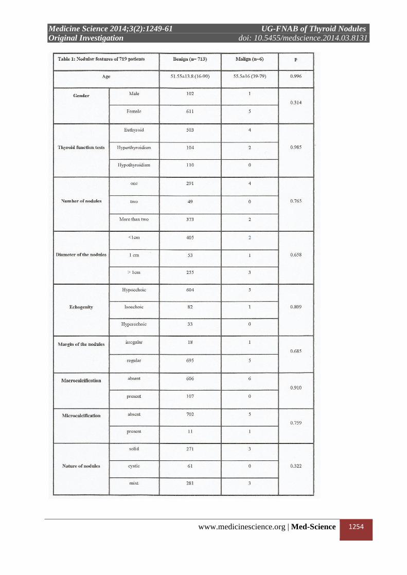
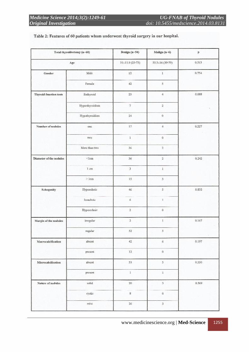
Demographic and nodular features of the patients were summarized at Table 1. If we compared
to ultrasonographic features of the benign and malign nodules proven by total thyroidectomy; no
features were found to be significantly different between benign and malign nodules (Table 1).
Morphological and clinical features of 60 patients who underwent total thyroidectomy were
shown at Table 2. The overall differentiated thyroid carcinoma ratio (confirmed by postoperative
histopathological examination) was 0.84% for 719 patients. All of our nodules (n=6) which
reported as “malignant” did not exceed 3 cm in diameter.
If we focuse on 6 thyroid papillar carcinoma cases which were detected after total
thyroidectomy; 2 of them were already determined as “malignant” by UG-FNAB at first biopsy.
Four of them had a malignant tumor greater than 1 cm, whereas 2 of them were < 1cm. Patient
who had micropapillar thyroid carcinoma was 49 years old male. He had multinodular goitre.
Tumor diameter was 0.8 cm and maximum nodule diameter was also 0.9 cm. And he had also
had a follicular adenoma at another thyroid nodule < 1 cm at the other side of thyroid gland.
Similarly, 71 years old female had a tumor in one of her thyroid nodules, and also had a
follicular adenoma at another thyroid nodule < 1 cm. Four of the papillar carsinoma cases were
Medicine Science 2014;3(2):1249-61 UG-FNAB of Thyroid Nodules
Original Investigation doi: 10.5455/medscience.2014.03.8131
www.medicinescience.org | Med-Science 1253
detected by malignant or suspicious cytology at their first UG-FNAB. But in 2 patients UG-
FNAB had to be repeated because of prior non-diagnostic cytology.
If we analyze the subgroup of 308 nodules which were equal or greater than 1 cm, number of
thyroid carsinoma cases were 4 ( 1.3%).
Medicine Science 2014;3(2):1249-61 UG-FNAB of Thyroid Nodules
Original Investigation doi: 10.5455/medscience.2014.03.8131
www.medicinescience.org | Med-Science 1254
Medicine Science 2014;3(2):1249-61 UG-FNAB of Thyroid Nodules
Original Investigation doi: 10.5455/medscience.2014.03.8131
www.medicinescience.org | Med-Science 1255

Medicine Science 2014;3(2):1249-61 UG-FNAB of Thyroid Nodules
Original Investigation doi: 10.5455/medscience.2014.03.8131
www.medicinescience.org | Med-Science 1256
Discussion
Although thyroid nodular disease is common, only a small number of thyroid nodules are
malignant. UG-FNAB plays an important role in differentiating benign thyroid nodules from
malignant nodules. It is essential to identify which thyroid nodules require UG-FNAB. The
recent guidelines were published by The American Thyroid Association (ATA) in 2009 [8]. The
American Association of Clinical Endocrinologists (AACE), the Italian Association of Medical
Endocrinologists (AME), and European Thyroid Association (ETA) published new in guidelines
in 2010, and then a revision was made in 2011 [9,10]. ATA guidelines recommend UG-FNAB in
all suspicious nodules > 5 mm (grade A recommendation) [8]. The ETA and AACE guidelines
also recommend UG-FNAB, that nodules < 1cm if the clinical and/or ultrasonographic
suspicious is present [9,10]. Atypical UG-FNAB results should be interpreted in the context of
the clinical presentation of thyroid nodular disease, imaging data, and the presence of risk factors
Medicine Science 2014;3(2):1249-61 UG-FNAB of Thyroid Nodules
Original Investigation doi: 10.5455/medscience.2014.03.8131
www.medicinescience.org | Med-Science 1257
for malignancy, including patient age, sex, ethnic background, exposure to ionizing radiation,
and familial history of thyroid malignancy [9,11]. Recent literature indicates that patients with
multiple thyroid nodules have the same risk of malignancy as patients with solitary thyroid
nodules [12,13]. Ultrasonographic characteristics are more reliable indicators of potential
malignancy than nodule size [14-16].
On histopathological examination of various FNAB specimens, 5-20% of them are reported to be
insufficient or non-diagnostic [17-20]. Compatible with current data, in our study, non-
diagnostic cytology were reported to be as 12.28%.
Instead of UG-FNAB, ultrasonographic monitoring is preferred in thyroid nodules which have
diameter less than 5 mm, because of the difficulties to reach the nodule and the risk of obtaining
inadequate material. In our study the nodules which surgery were performed with diameter of <
10 mm, and ≥ 5 mm, 2 micropapillar carcinoma was detected. On cytopathological examination
of their FNAB, one of them was reported to be suspicious for malignancy, other was malignant.
None of them have any calcifications on their ultrasonographic examinations. Both of them also
had a follicular adenoma at another thyroid nodule < 1 cm. (Table 3).
In our study, thyroid carcinoma rate was found to be lesser than in current literature. One of the
reason for this may be higher ratio of nodules that underwent evaluation were lesser than 1 cm in
diameters. But if we analyze the subgroup of the nodules equal to or greater than 1 cm, papillar
carcinoma rate was still lesser (1.3%). There was no significance between the groups which have
nodule < 1cm, and ≥ 1 cm (p>0.05). Our results may indicate that our region were associated
with lesser papillar carcinoma rate. If we compere our results with some studies which were
performed in our country, similar cytological malignancy rates could be seen [21-24].
Sengoz et al. were reported UG-FNAB cytological results as 781 (84.1%) benign, 35 (3.8%)
malignant suspicious, 21 (2.3%) malignant nodules and 92 (9.9%) inadequate materials [21].
Similarly with ours, this malignancy rates were also lesser than current literature. In another

Medicine Science 2014;3(2):1249-61 UG-FNAB of Thyroid Nodules
Original Investigation doi: 10.5455/medscience.2014.03.8131
www.medicinescience.org | Med-Science 1258
study including 1420 patients; 30 (3.7%) malignant suspicious, 8 (0.9%) malignant nodules were
reported, in 808 cytological findings of FNAB specimens [22]. By histopatological examination
of l52 surgical specimens after thyroid operation, 35 of them were diagnosed as thyroid
carcinoma. If we thought that, we have already had UG-FNAB cytology reports of 808, before
the thyroid surgery; we could conclude that their malignancy rate proven by surgery was 4.3%
(n=808) [22]. In another Turkish study, 197 cases who had been diagnosed as multinodular
goitre were analyzed. After 26 inadequate cytological results were excluded, 171 specimens
were examined. In a group of dominant nodules, malignancy and suspicious for malignancy rates
were 1.8% and 9.4% whereas they were 0.6% and 11.1%, respectively. In this study, dominant
and non-dominant nodule diameters were determined as 24.96±9.08, and 15.4±5.89 mm. If we
thought that, nodule diameters in our study were lesser than 30 mm, lesser malignancy rates in
our study could explained by having smaller nodule diameters [23]. In another study, one group
(group A), consisted of thyroid nodules which had ultrasonographically suspicious criteria for
malignancy, the other group (group B) which were not ultrasonographically suspicious for
malignancy but referred to biopsy according to clinicians view. FNAB cytologies were as
follows: In group A; 28 (50.9%) benign, 16 (29.1%) malign, 8 (14.5%) suspicious for
malignancy, and 3 (5.5%) inadequate material. In group B; 74 (96.1%) benign, 1 (1.3%) malign,
1 (1.3%) suspicious for malignancy, and 1 (1.3%) inadequate material [24]. If we focuse on our
malignancy results in nodules > 1 cm, this result is similar to ours. By being the one center
which could perform UG-FNAB, other clinicians had been directed patients with thyroid nodule
to our outpatient clinic. So, because of that, our indications for UG-FNAB might be higher than
already needed. We thought that, thyroid cancer risk could be detected by using UG-FNAB even
in thyroid nodules smaller than 1 cm, if the nodule has sonographically suspicious features for
malignancy. So, it requires stronger collaboration between radiologist and endocrinologist.

Medicine Science 2014;3(2):1249-61 UG-FNAB of Thyroid Nodules
Original Investigation doi: 10.5455/medscience.2014.03.8131
www.medicinescience.org | Med-Science 1259
In conclusion, fine needle aspiration biopsy is a functional and reliable diagnosing method.
Using UG-FNAB leads to identify discrete nodules and direct needle localization and provides
adequate specimen. UG-FNAB has become a predominant method for the diagnosis of benign
and malign thyroid nodules. Atypical UG-FNAB results need to be considered in combination
with clinical presentation, imaging data, and individual risk factors. Collaboration between
endocrinologists, radiologists and pathologists is essential for patients with thyroid nodules to
avoid missestimation of the risk of thyroid cancer.
Conflict of interest: None.
References
1. Mazzaferri EL. Thyroid cancer in thyroid nodules: finding a needle in the
haystack. Am J Med. 1992;93(4):359-62.
2. Eng CY, Quraishi MS, Bradley PJ. Management of Thyroid nodules in adult
patients. Head Neck Oncol. 2010;2:11.
3. Datta RV, Petrelli NJ, Ramzy J. Evaluation and management of incidentally
discovered thyroid nodules. Surg Oncol. 2006;15(1):33-42.
4. Sakorafas GH Thyroid nodules; interpretation and importance of fine-needle
aspiration (FNA) for the clinician -practical considerations. Surg Oncol.
2010;19(4):e130-9.
5. Castro MR, Gharib H. Thyroid nodules and cancer. When to wait and watch,
when to refer. Postgrad Med. 2000;107(1):113-6.
6. Tunbridge WM, Evered DC, Hall R, Appleton D, Brewis M, Clark F, Evans
JG, Young E, Bird T, Smith PA. The spectrum of thyroid disease in a
community: the Wickham survey. Clin Endocrinol (Oxf). 1977;7(6):481-93.
7. Baloch ZW, Alexander EK, Gharib H, Raab SS. Chapter 1; Overview of
Diagnostic Terminology and Reporting. In: Ali SZ, Cibas ES, eds, The
Bethesda System for Reporting Thyroid Cytopathology. New York: Springer.
2010; 1–4.
8. American Thyroid Association (ATA) Guidelines Taskforce on Thyroid
Nodules and Differentiated Thyroid Cancer, Cooper DS, Doherty GM,
Haugen BR, Kloos RT, Lee SL, Mandel SJ, Mazzaferri EL, McIver B, Pacini
F, Schlumberger M, Sherman SI, Steward DL, Tuttle RM. Revised American
Thyroid Association management guidelines for patients with thyroid nodules
and differentiated thyroid cancer. Thyroid. 2009;19(11):1167-214.

Medicine Science 2014;3(2):1249-61 UG-FNAB of Thyroid Nodules
Original Investigation doi: 10.5455/medscience.2014.03.8131
www.medicinescience.org | Med-Science 1260
9. Gharib H, Papini E, Paschke R, Duick DS, Valcavi R, Hegedüs L, Vitti P,
Balafouta ST, Baloch Z, Crescenzi A, Dralle H, Gärtner R, Guglielmi R,
Mechanick JI, Reiners C, Szabolcs I, Zeiger MA, Zini M. American
Association of Clinical Endocrinologists, Associazione Medici
Endocrinologi, and EuropeanThyroid Association Medical Guidelines for
Clinical Practice for the Diagnosis and Management of Thyroid Nodules.
Endocr Pract. 2010;16 Suppl 1:1-43.
10. Paschke R,Hegedus L, Alexander E,Valcavi R, Papini E,Gharib H. Thyroid
nodule guidelines: agreement, disagreement and need for future research. Nat
Rev Endocrinol. 2011;7(6):354-61.
11. Clark JR, Eski SJ, Freeman JL. Risk of malignancy in Filipinosis with thyroid
nodules: a matched pair analysis. Head Neck. 2006;28(5):427-31.
12. Papini E, Guglielmi R, Bianchini A, Crescenzi A, Taccogna S, Nardi F,
Panunzi C, Rinaldi R, Toscano V, Pacella CM. Risk of malignancy in non-
palpable thyroid nodules: predictive value of ultrasound and color-Doppler
features. J Clin Endocrinol Metab. 2002;87(5):1941-6.
13. Tollin SR, Mery GM, Jelveh N, Fallon EF, Mikhail M, Blumenfeld W,
Perlmutter S. The use of fine-needle aspiration biopsy under ultrasound
guidance to assess the risk of malignancy in patients with a multinodular
goiter. Thyroid. 2000;10(3):235-41.
14. Kang HW, No JH, Chung JH, Min YK, Lee MS, Lee MK, Yang JH, Kim
KW. Prevalence, clinical and ultrasonographic characteristics of thyroid
incidentalomas. Thyroid. 2004;14(1):29-33.
15. Lin JD, Huang BY, Weng HF, Jeng LB, Hsueh C. Thyroid ultrasonography
with fine-needle aspiration cytology for the diagnosis of thyroid cancer. J
Clin Ultrasound. 1997;25(3):111-8.
16. Chan BK, Desser TS, McDougall IR, Weigel RJ, Jeffrey RB Jr. Common and
uncommon sonographic features of papillary thyroid carcinoma. J Ultrasound
Med. 2003;22(10):1083-90.
17. Gharib H, Goellner JR. Fine-needle aspiration biopsy of the thyroid: an
appraisal. Ann Intern Med. 1993;118(4):282-9.
18. Goellner JR, Gharib H, Grant CS, Johnson DA. Fine-needle aspiration
cytology of the thyroid, 1980 to 1986. Acta Cytol. 1987;31(5):587-90.
19. Carmeci C, Jeffrey RB, McDougall IR, Nowels KW, Weigel RJ. Ultrasound-
guided fine needle aspiration biopsy of thyroid masses. Thyroid.
1998;8(4):283-9.
20. Danase D, Sciacchitano S, Farsetti A, Andreoli M, Pontecorvi A. Diagnostic
accuracy of conventional versus sonography-guided fine needle aspiration
biopsy of thyroid nodules. Thyroid. 1998;8(1):15-21.
21. Şengöz T, Çubuk R, Kaya H, Arıbal E. Tiroid nodüllerinde ultrason
rehberliğinde ince iğne aspirasyon biopsisi. Düzce Tıp Fakültesi Dergisi.
2009;11(2):26-32.
Medicine Science 2014;3(2):1249-61 UG-FNAB of Thyroid Nodules
Original Investigation doi: 10.5455/medscience.2014.03.8131
www.medicinescience.org | Med-Science 1261
22. Önver H, Özbey AO, Duymuş M, Yılmaz Ö, Koşar PN. Tiroit nodüllerinin
ultrasonografik, sitolojik ve histopatolojik bulgularının incelenmesi. Kafkas J
Med Sci. 2013;3(2):80-7.
23. Yazgan Ö, Beşir FH, Aydın Y, Yazgan S, Erkan ME, Yazıcı B, Büyükkaya
R, Önbaş Ö. Ötiroid multinodüler guatrlı olguların sitoloji ve histopatoloji
sonuçları: Ultrasonografi özellikleri ile karşılaştırılması. Dicle Med J.
2012;39(2):201-6.
24. Hasanefendioğlu-Bayrak A, Özel A, Peker K. Tiroid nodüllerinde
endikasyonlara göre ince iğne aspirasyon biyopsisi sonuçları. Dicle Med J.
2007;34(1):42-7.
Copyright © 2022 FDOKUMEN