Two new species of Sarsinebalia (Crustacea, Leptostraca) from the Northeast Atlantic, with comments...
21
Two new species of Sarsinebalia (Crustacea, Leptostraca) from the Northeast Atlantic, with comments on the genus Juan Moreira, Loreto Gestoso & Jesu ´s S. Troncoso Moreira J, Gestoso L, Troncoso JS. 2003. Two new species of Sarsinebalia (Crustacea, Leptostraca) from the Northeast Atlantic, with comments on the genus. Sarsia 88:189–209. SARSIA Two new species of Leptostraca (Crustacea, Malacostraca) belonging to the genus Sarsinebalia are described from shallow waters off Galicia (northwest Spain). The new species differ from S. typhlops, the only known member of the genus, mainly in having ommatidia externally visible and pigmented eyes, as well as a second maxilla bearing an exopod that is longer than the first article of the endopod. The validity of Sarsinebalia is discussed in the light of the new species described. Juan Moreira, Loreto Gestoso & Jesu ´s S. Troncoso, Departamento de Ecoloxı ´a e Bioloxı ´a Animal, Facultade de Ciencias, Campus de Lagoas-Marcosende s/n, Universidade de Vigo, ES-36200 Vigo, Spain. E-mail: [email protected] Keywords: Leptostraca; Sarsinebalia; Sarsinebalia cristoboi sp. nov; Sarsinebalia urgorrii sp. nov; Spain. INTRODUCTION In recent years, the number of new species of Lepto- straca has greatly increased (Kazmi & Tirmizi 1989; Modlin 1991; Escobar-Briones & Villalobos-Hiriart 1995; Vetter 1996; Martin & al. 1996; Walker-Smith 1998, 2000; Olesen 1999; Haney & Martin 2000; Haney & al. 2001; Walker-Smith & Poore 2001). However, until a few years ago, it was supposed that certain leptostracan genera, such as Nebalia Leach, 1814 and Paranebalia Claus, 1880, were composed of a limited number of highly variable species, which would have a more or less cosmopolitan distribution. This view changed after the revision of the European shelf species by Dahl (1985), in which a model for the description of leptostracans was set up. This and subsequent works have demonstrated that a number of reliable characters can be used to differentiate species within this group of crustaceans, and have highlighted the importance of scanning electron microscopy for the study of the different appendage structures (Martin & al. 1996; Haney & Martin 2000; Walker-Smith 2000). In that context, Dahl (1985) erected the genus Sarsinebalia with N. typhlops G.O. Sars, 1870 as type species, which had been regarded as a somewhat aberrant Nebalia. He pointed out that this species showed a number of distinct characteristics not present in the other known Nebalia species, justifying the erection of this new genus. Such characteristics are the possession of a rostrum with a ventral keel(s) ending in a short spine, disc-shaped eyes lacking pigment or externally discernible visual elements, an exopod of the first pleopod without a row of spines on the lateral border, and as a supplementary character the fact that the exopod of the second maxilla is shorter than the first article of the endopod. Dahl (1985) strengthens his opinion regarding the existence of some specimens belonging to several undescribed species from the Red Sea and Australia, which would share these charac- teristics with S. typhlops. However, descriptions of these specimens have remained unpublished to date, as new species assignable to Sarsinebalia from other parts of the world have not yet been described. In the same work, and in the light of his observations on Sarsine- balia, Dahl (1985) suggested that a revision of the taxonomic status of the subspecies S. typhlops occi- dentalis (Hessler & Sanders, 1965) would be necessary. Hessler & Sanders (1965) pointed out that this sub- species, described from the continental slope off the coast of New Jersey (USA), differs from the typical form mainly in having a carapace fold with a more broadly rounded posterior margin, a rostrum that is broader near the base, tapering prominently at the anterior end, and an antennular scale that is proportion- ally broader. Olesen (1999) presented a phylogenetic analysis of the extant genera of Leptostraca, questioning the mono- phyly of Nebalia, and highlighting its phylogenetic proximity to both Dahlella Hessler, 1984 and Sarsine- balia. DOI 10.1080/00364820310001390 # 2003 Taylor & Francis Published in collaboration with the University of Bergen and the Institute of Marine Research, Norway
-
Upload
independent -
Category
Documents
-
view
3 -
download
0
Transcript of Two new species of Sarsinebalia (Crustacea, Leptostraca) from the Northeast Atlantic, with comments...
Two new species of Sarsinebalia (Crustacea, Leptostraca) from the NortheastAtlantic, with comments on the genus
Juan Moreira, Loreto Gestoso & Jesus S. Troncoso
Moreira J, Gestoso L, Troncoso JS. 2003. Two new species of Sarsinebalia (Crustacea, Leptostraca)from the Northeast Atlantic, with comments on the genus. Sarsia 88:189–209.
SARSIA
Two new species of Leptostraca (Crustacea, Malacostraca) belonging to the genus Sarsinebalia aredescribed from shallow waters off Galicia (northwest Spain). The new species differ from S. typhlops,the only known member of the genus, mainly in having ommatidia externally visible and pigmentedeyes, as well as a second maxilla bearing an exopod that is longer than the first article of the endopod.The validity of Sarsinebalia is discussed in the light of the new species described.
Juan Moreira, Loreto Gestoso & Jesus S. Troncoso, Departamento de Ecoloxıa e Bioloxıa Animal,Facultade de Ciencias, Campus de Lagoas-Marcosende s/n, Universidade de Vigo, ES-36200 Vigo,Spain.E-mail: [email protected]
Keywords: Leptostraca; Sarsinebalia; Sarsinebalia cristoboi sp. nov; Sarsinebalia urgorrii sp. nov;Spain.
INTRODUCTION
In recent years, the number of new species of Lepto-straca has greatly increased (Kazmi & Tirmizi 1989;Modlin 1991; Escobar-Briones & Villalobos-Hiriart1995; Vetter 1996; Martin & al. 1996; Walker-Smith1998, 2000; Olesen 1999; Haney & Martin 2000; Haney& al. 2001; Walker-Smith & Poore 2001). However,until a few years ago, it was supposed that certainleptostracan genera, such as Nebalia Leach, 1814 andParanebalia Claus, 1880, were composed of a limitednumber of highly variable species, which would have amore or less cosmopolitan distribution. This viewchanged after the revision of the European shelfspecies by Dahl (1985), in which a model for thedescription of leptostracans was set up. This andsubsequent works have demonstrated that a number ofreliable characters can be used to differentiate specieswithin this group of crustaceans, and have highlightedthe importance of scanning electron microscopy for thestudy of the different appendage structures (Martin &al. 1996; Haney & Martin 2000; Walker-Smith 2000).
In that context, Dahl (1985) erected the genusSarsinebalia with N. typhlops G.O. Sars, 1870 as typespecies, which had been regarded as a somewhataberrant Nebalia. He pointed out that this speciesshowed a number of distinct characteristics not presentin the other known Nebalia species, justifying theerection of this new genus. Such characteristics are thepossession of a rostrum with a ventral keel(s) ending in
a short spine, disc-shaped eyes lacking pigment orexternally discernible visual elements, an exopod of thefirst pleopod without a row of spines on the lateralborder, and as a supplementary character the fact thatthe exopod of the second maxilla is shorter than the firstarticle of the endopod. Dahl (1985) strengthens hisopinion regarding the existence of some specimensbelonging to several undescribed species from the RedSea and Australia, which would share these charac-teristics with S. typhlops. However, descriptions ofthese specimens have remained unpublished to date, asnew species assignable to Sarsinebalia from other partsof the world have not yet been described. In the samework, and in the light of his observations on Sarsine-balia, Dahl (1985) suggested that a revision of thetaxonomic status of the subspecies S. typhlops occi-dentalis (Hessler & Sanders, 1965) would be necessary.Hessler & Sanders (1965) pointed out that this sub-species, described from the continental slope off thecoast of New Jersey (USA), differs from the typicalform mainly in having a carapace fold with a morebroadly rounded posterior margin, a rostrum that isbroader near the base, tapering prominently at theanterior end, and an antennular scale that is proportion-ally broader.
Olesen (1999) presented a phylogenetic analysis ofthe extant genera of Leptostraca, questioning the mono-phyly of Nebalia, and highlighting its phylogeneticproximity to both Dahlella Hessler, 1984 and Sarsine-balia.
DOI 10.1080/00364820310001390 � 2003 Taylor & Francis
Published in collaboration with the University of Bergen and the Institute of Marine Research, Norway
Walker-Smith & Poore (2001) proposed a newhypothesis for the phylogenetic relationships betweenthe leptostracan genera, erecting a new family, Levine-baliidae, for Levinebalia Walker-Smith, 2000 andParanebalia. In their analysis, insufficient characterswere found to maintain the generic status of the stillmonotypic Sarsinebalia, and therefore Nebalia andSarsinebalia were regarded as synonymous.
An examination of leptostracan specimens collectedoff the coast of Galicia (northwest Spain) revealed thatseveral of them belong to two undescribed specieswhose characteristics agree well with the genericdiagnosis of Sarsinebalia given by Dahl (1985),differing only in having pigmented eyes with externallydiscernible ommatidia, and a second maxilla bearing anexopod that is longer than the first article of theendopod. In light of these new findings, we maintainthe genus Sarsinebalia in this work, in which two newspecies are described: S. cristoboi sp. nov. and S.urgorrii sp. nov.
MATERIAL AND METHODS
Specimens were collected in several locations off theGalician coast (northwest Spain) during 1995, 1996,1997 and 2000. Samples were taken by means of a VanVeen grab. The specimens were sorted out from thesediment, fixed in 10% buffered formalin, and thentransferred to 70% ethanol. At each sampling point anadditional sample was taken to provide sediment forphysicochemical analysis. The particle size distributionof the sediment was determined following the proce-dures given in Buchanan (1984). The carbonate contentof the sediment was estimated from the volume of CO2
delivered after treating the sample with concentratedhydrochloric acid, while the organic matter content wasestimated from the weight loss after placing sedimentsamples in a furnace for 4 h at 450 °C.
Line drawings were made using a camera lucidaconnected to an Olympus BX40 light microscope.Several specimens were subjected to scanning electronmicroscope analysis, after being dehydrated via agraded ethanol series, liquid CO2 critical point dried,and sputter coated with gold. These specimens wereexamined with a Philips SEM XL30 at CACTI (Centrode Apoyo Cientıfico y Tecnologico a la Investigacion,Universidade de Vigo). Descriptions of the new speciesare of females, following the model of Dahl (1985) andother recent works (Martin & al. 1996; Olesen 1999).Measurements were estimated by means of an ocularmicrometer. Total length (TL) was measured from thearticulation of the rostrum and carapace to the posteriorend of the caudal rami, excluding setation; carapace
length (CL) was considered as the distance between thearticulation of the rostrum and the margin of theposterodorsal cleft; rostrum length (RL) wasmeasured along the midline. The type series isdeposited in the Museo Nacional de Ciencias Naturales,Madrid (MNCN), while remaining specimens are keptin the collection of the first author.
SYSTEMATICS
Family Nebaliidae Samouelle, 1819Genus Sarsinebalia Dahl, 1985
Type species: Nebalia typhlops G.O. Sars, 1870
DiagnosisRostrum with ventral keel(s) ending in a thread-likestructure, that projects past tip of rostrum and tapers toform a short spine. Eyes disc-shaped or more or lessoval, with or without ommatidia and pigment. Exopodof first pleopod without row of short, complex spines.
RemarksThe original diagnosis of the genus Sarsinebalia wasestablished by Dahl (1985) around three main charac-ters: the rostrum ending in a thread-like structureforming a short spine, disc-shaped eye without exter-nally discernible visual elements and pigment, and theexopod of the first pleopod without the typical row ofspines present in other genera, such as Nebalia.Additionally, the exopod of the second maxilla didnot surpass the length of the first article of the endopod,but this last character was considered by Dahl (1985) tobe only a supplementary one. By that time, the onlydescribed species assigned to Sarsinebalia was S.typhlops, although Dahl (1985) pointed out the exis-tence of at least three undescribed species from the RedSea and Australia, whose existence strengthened hispoint of view on the validity of Sarsinebalia.
Walker-Smith & Poore (2001) suggested thatNebalia and the still monotypic Sarsinebalia shouldbe considered synonymous, because they did not findsufficient distinctive characters to maintain the genericstatus of Sarsinebalia in their phylogenetic analysis.Hence, the presence of a subrostral spine and theabsence of a spine row on the exopod of the firstpleopod were considered as not having enough rele-vance at a generic level to separate these genera. Thepossession of a subrostral spine is shared with Para-nebalia and Levinebalia, and the absence of a spine rowis shared with Speonebalia cannoni Bowman, Yager &Iliffe, 1985. According to Walker-Smith & Poore(2001) two autapomorphies would unite Sarsinebalia
190 Sarsia 88:189-209 – 2003
with Nebalia: the presence of a rostral keel shorter thanthe rostrum and the possession of robust setae in thefourth article of the antennule. However, the recentlydescribed N. schizophthalma Haney, Hessler & Martin,2001, which was not included in the analysis byWalker-Smith & Poore (2001), does not present robustsetae in the fourth article of the antennule, and insteadpresents several simple setae.
The finding of the two new species described herein,S. cristoboi sp. nov. and S. urgorrii sp. nov., whichagree well with the original diagnosis of Sarsinebaliaeven though they possess eyes with ommatidia andpigment and a second maxilla bearing an exopod that islonger than the first article of the endopod, suggests thatSarsinebalia species would form a homogenous groupsharing a number of characteristics not found togetherin any described Nebalia. The presence of ommatidiaand pigment in S. cristoboi sp. nov. and S. urgorrii sp.nov. would be a plesiomorphic state within the genus.Additionally, the three Sarsinebalia species possess adisc-shaped eye which is different from the typicallydorsally convex eye found in most of the species ofNebalia, even though some aberrant eyes have beendescribed in several Nebalia species, such as thedistally bilobed eyes of N. daytoni Vetter, 1996 andN. schizophthalma, as well as in the closely relatedDahlella caldariensis Hessler, 1984 with a long anddistally curved eye, armed with sclerotized denticles.With these considerations, the validity of the genusSarsinebalia is retained in this paper, following Dahl(1985, unpublished).
NOTE ABOUT AN UNPUBLISHED DESCRIPTION OF ANEW SPECIES OF SARSINEBALIA BY E. DAHL
During the course of this work, Dr E. Vetter (HawaiiPacific University) informed us about the existence ofan unpublished manuscript by Dr E. Dahl in which anew species of Sarsinebalia is described from twofemales collected on the northwest African shelf. Dahlpoints out that in his previous work on the Europeanshelf leptostracan species, these specimens werewrongly said to belong to Red Sea samples, when hementioned the existence of some undescribed species ofSarsinebalia from other parts of the world (Dahl, 1985).Unfortunately, this unfinished manuscript has not beenpublished due to illness. Type specimens were sup-posed to have been deposited at the Museum ofZoology of Lund University (Sweden), but they havenot been found ever since the manuscript was written(Dr L. Cederholm, in litteris).
These specimens present the following distinctivecharacteristics: a subrostral spine projecting past the tip
of the rostrum, disc-shaped and nearly quadratic com-pound eyes with numerous ommatidia, a supra-ocularplate reaching to the middle of the eye, the third articleof the mandibular palp not expanding distally, thecorner of the posterolateral border of the fourth pleoniteproduced to form an obtuse point, with short roundeddenticles on the posterior margin, and short and broadlyrounded denticles on the dorsal and lateral posteriormargins of pleonites 6 and 7. Taking into account thisdescription, these females would be very similar tospecimens of S. urgorrii sp. nov. described herein,particularly regarding the shape and characteristics ofthe eyes. However, a direct comparison between them isnot possible, and therefore we cannot assure that theybelong to the same species.
Sarsinebalia cristoboi sp. nov.(Figs 1–7, 15A)
Type seriesHolotype: female (MNCN 20.04/5326a), TL = 5.5 mm,CL = 2.5 mm, RL = 1.0 mm, Rıa de Vigo, 42°13�54�N8°46�30�W, October 2000, 14 m, “maerl” mixed withgravel. Allotype: male (MNCN 20.04/5326b),TL = 5.6 mm, CL = 2.6 mm, RL = 1.0 mm, Rıa deVigo, 42°13�54�N 8°46�30�W, October 2000, 14 m,“maerl” mixed with gravel. Paratypes: three females(MNCN 20.04/5326c), Rıa de Vigo, 42°13�54�N8°46�30�W, October 2000, 14 m, “maerl” mixed withgravel; one female (MNCN 20.04/5327), Rıa de Aldan,42°19�15�N 8°50�45�W, August 1997, 33 m, coarsesand.
Additional non-type materialOne female: Ensenada de Baiona, 42°07�50�N8°50�52�W, December 1995, 9 m, coarse sand. Onefemale: Ensenada de Baiona, 42°08�10�N 8°50�52�W,December 1995, 12 m, coarse sand. Two females:Ensenada do Grove, 42°30�45�N 8°52�15�W, Novem-ber 1996, 16 m, “maerl”. One female: Rıa de Aldan,42°18�45�N 8°49�15�W, July 1997, 15 m, medium sand.Three females: Rıa de Vigo, 42°13�54�N 8°46�30�W,October 2000, 14 m, “maerl” mixed with gravel.
Type localityRıa de Vigo, 42°13�54�N 8°46�30�W, 14 m, “maerl”mixed with gravel.
EtymologyThis new species is named in honour of Dr Francisco J.Cristobo (University of Santiago, Spain), a Galicianspecialist for the Porifera, in regard to his friendship andhis contributions to the field of marine biology.
Moreira & al. – Two new species of Leptostraca 191
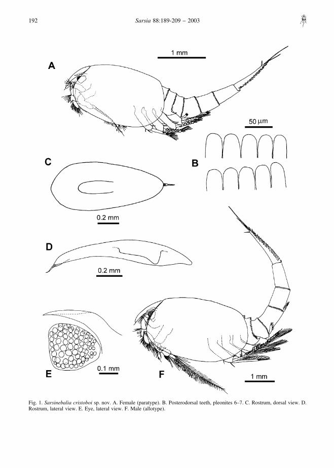
Fig. 1. Sarsinebalia cristoboi sp. nov. A. Female (paratype). B. Posterodorsal teeth, pleonites 6–7. C. Rostrum, dorsal view. D.Rostrum, lateral view. E. Eye, lateral view. F. Male (allotype).
192 Sarsia 88:189-209 – 2003
Diagnosis of femaleEye disc-shaped, slightly longer than wide, withexternally discernible ommatidia and red pigmentcovering almost entire eye; supra-orbital plate reachingdistal end of eye dorsally. Rostrum slightly turningdownwards, with paired ventral keels fused proximally.Antennular flagellum with up to eight articles. Exopodof second maxilla longer than first article of endopod.Posterolateral border of fourth pleonite with acutespine-like teeth, ending in larger tooth. Protopod offourth pleopod with several crenulations along posteriorborder, ending in acute tooth at posterolateral corner.Fifth pleopod with six large spines along distolateralborder. Sixth pleopod with five large spines ondistolateral border. Pleonites 6 and 7 with distallyrounded teeth along dorsal and lateral posterior borders.Furcal rami about as long as telson and pleonite 7combined.
Description of femaleLargest female 5.6 mm TL, 2.6 mm CL and 1.0 mm RL.Carapace oval, not totally covering fourth pleonite (Fig.1A).
Rostrum: long, about 2.2 times as long as wide,distally tapering and slightly turning downwards. Pairedventral keels, fused proximally (Fig. 1C, D). Tip ofrostrum provided with a terminal spine, segmentedproximally (Figs 1D, 7A).
Compound eye: well developed, disc-shaped, slightlylonger than wide, distal border straight, with ommatidiaand red pigmentation covering almost entire eye (Fig.1E). Supra-orbital plate with acute tip, surpassing orextending to end of eye.
Antennule: peduncle four-segmented. Second articlewidest at midpoint, about three times as long as wide,with two clusters of setae distally and subdistally, andwith plumose seta dorsally at midpoint (Fig. 2A). Thirdarticle shorter than second, widest distally, withterminal dorsal cluster of setae and additional distalseta placed ventrally. Fourth article shorter than third,with row of about eight setae and one stout terminalspine, all of them along dorsolateral border; next toinsertion of antennular scale an additional row of setaeis present; a long and thicker seta is attached neararticulation of flagellum. Antennular scale oval andelongate, about 2.3 times as long as wide, with several
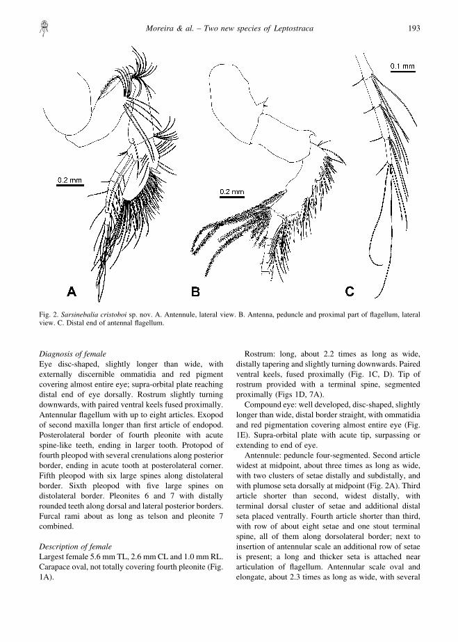
Fig. 2. Sarsinebalia cristoboi sp. nov. A. Antennule, lateral view. B. Antenna, peduncle and proximal part of flagellum, lateralview. C. Distal end of antennal flagellum.
Moreira & al. – Two new species of Leptostraca 193
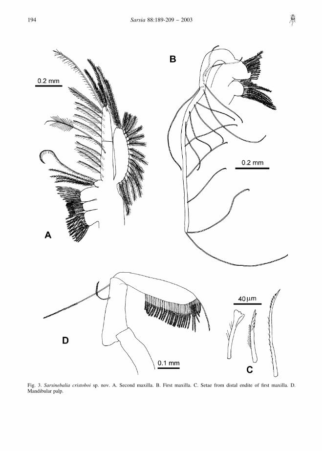
Fig. 3. Sarsinebalia cristoboi sp. nov. A. Second maxilla. B. First maxilla. C. Setae from distal endite of first maxilla. D.Mandibular palp.
194 Sarsia 88:189-209 – 2003
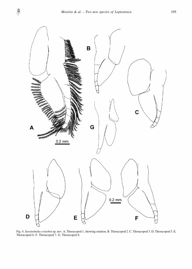
Fig. 4. Sarsinebalia cristoboi sp. nov. A. Thoracopod 1, showing setation. B. Thoracopod 2. C. Thoracopod 3. D. Thoracopod 5. E.Thoracopod 6. F. Thoracopod 7. G. Thoracopod 8.
Moreira & al. – Two new species of Leptostraca 195
rows of setae of different lengths: (a) long simple setae,(b) stout curved setae, proximally smooth and distallyprovided with blunt serrations (Fig. 7C), and (c) thinnerand longer setae with sharp teeth (Fig. 7D). Flagellumless than half length of peduncle, with seven articles(occasionally six or eight), each of them with pair ofaesthetascs and several setae, the latter increasing inlength in distal articles (Fig. 7B).
Antenna: peduncle three-segmented (Fig. 2B). Firstarticle with thin dorsal tooth placed distally. Secondarticle about twice as long as wide, with curved dorsalterminal tooth, larger than that of first article. Thirdarticle longer than second, with several stout spinesand setae along dorsolateral border, long ventralplumose seta subdistally and row of about 15–18 longplumose setae placed sub- and distally. Flagellumslightly shorter than peduncle, with 16–17 articles,each of them with several long and short setae (Figs 2C,7E).
Mandible: palp well developed, with three articles(Fig. 3D); first article shorter than second, the latterwith two setae, the shortest placed subdistally and thelongest distally; third article longer than second, notexpanding distally; from proximal third to terminus, a
row of setae bearing lanceolate setules; distal borderwith shorter row of stouter setae with lateral serrula-tions (Fig. 7F).
First maxilla: protopod with two endites (Fig. 3B);first endite with plumose setae; second endite largerthan first, with setae arranged in two rows comprisingabout seven spatulate, two setulose, and 16 distallydenticulate (Fig. 3C). Palp well developed, about fivetimes the length of protopod, with long and spacedsetae.
Second maxilla: protopod with four endites; endites 1and 3 the largest and endite 4 the smallest (Fig. 3A).Endopod longer than exopod, composed of two articles,distal one shorter than proximal one; exopod clearlysurpassing articulation between two articles of endo-pod. All endites, exopod, and endopod with plumosesetae. Distal seta of second article of endopod as long asentire ramus.
Thoracopods (Fig. 4): endopod exceeding length ofexopod, segmented distally, with plumose setae alongmargin, longest ones on distal article (Fig. 7G); exopodwith shorter setae along lateral margin; epipod ofthoracopod 8 reduced in size.
Pleonites: posterior border of first pleonite lacking
Fig. 5. Sarsinebalia cristoboi sp. nov. A. First pleopod, ventral view. B. Fourth pleopod, protopod, lateral view. C. Secondpleopod, lateral view.
196 Sarsia 88:189-209 – 2003
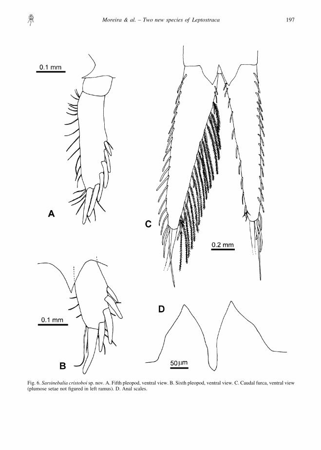
Fig. 6. Sarsinebalia cristoboi sp. nov. A. Fifth pleopod, ventral view. B. Sixth pleopod, ventral view. C. Caudal furca, ventral view(plumose setae not figured in left ramus). D. Anal scales.
Moreira & al. – Two new species of Leptostraca 197
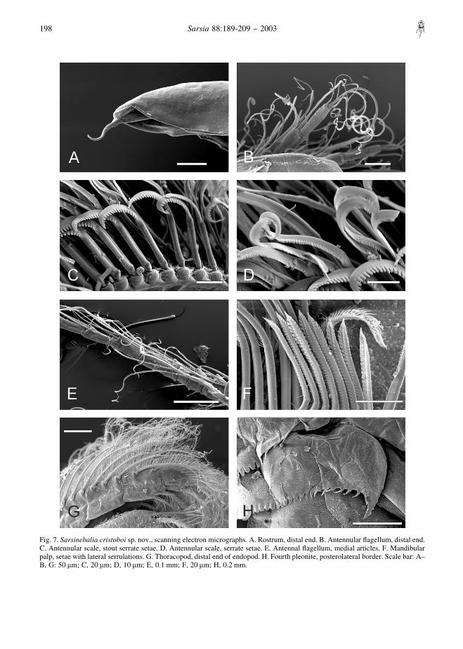
Fig. 7. Sarsinebalia cristoboi sp. nov., scanning electron micrographs. A. Rostrum, distal end. B. Antennular flagellum, distal end.C. Antennular scale, stout serrate setae. D. Antennular scale, serrate setae. E. Antennal flagellum, medial articles. F. Mandibularpalp, setae with lateral serrulations. G. Thoracopod, distal end of endopod. H. Fourth pleonite, posterolateral border. Scale bar: A–B, G: 50 �m; C, 20 �m; D, 10 �m; E, 0.1 mm; F, 20 �m; H, 0.2 mm.
198 Sarsia 88:189-209 – 2003
denticles. Second pleonite bearing row of denticlesalong posterodorsal border; denticles distally roundedbecoming acute and more spaced towards both ends ofrow. Third pleonite with denticles along dorsal andlateral posterior borders; denticles showing same shapevariation as in second pleonite. Fourth pleonite withdistally rounded denticles along posterodorsal border;posterolateral border bearing acute spine-like teeth,ending in larger tooth (Fig. 7H). Pleonites 5–7 withdistally rounded denticles on posterior border (Fig. 1B).
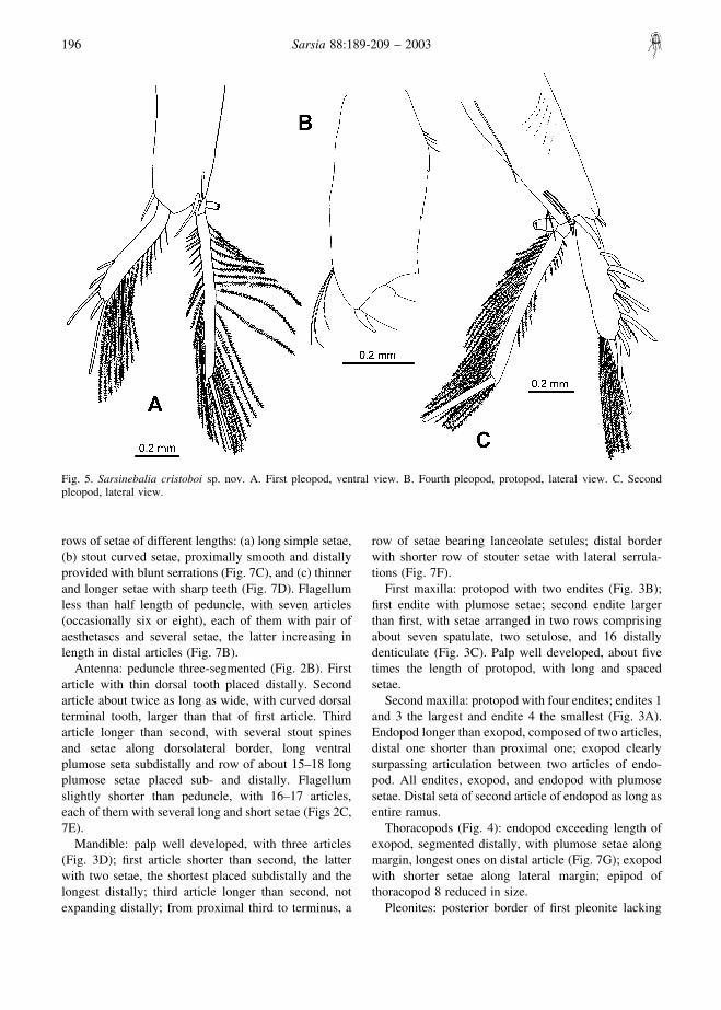
First pleopod: protopod with several basal thin spines(Fig. 5A); exopod about 0.8 times the length ofprotopod, with five subdistal and distal spines andseveral plumose setae along medial margin; endopodtwo-segmented with plumose setae on lateral andmedial margins of distal segment, basal segment withappendix interna.
Second pleopod: protopod with several lateral andsubdistal simple setae, one long proximal seta, one stoutspine next to exopod base, and larger stout spinebetween both rami (Fig. 5C). Exopod with row of spinepairs, each pair consisting of one long and one shorterblunt spine, between both spines a short setulose seta;medial margin with long plumose setae, three stoutspines distally. Endopod two-segmented, distal segmentwith long plumose setae on lateral and medial marginsand large terminal stout spine.
Pleopods 3–4: similar to second pleopod; protopod offourth pleopod with posterior margin crenate (Fig. 5B).
Fifth pleopod (Fig. 6A): uniramous, two-segmented;distal article about 3.4 times as long as wide, with sixspines along distolateral and terminal border, increasingin length distally, and with several simple setae alongmedial and distal border. Acute triangular processbetween both pleopods.
Sixth pleopod (Fig. 6B): uniramous, one-segmented,with five lateral and distal spines increasing in lengthdistally and a few simple setae along medial and distalborder. Acute triangular process between both pleopods.
Telson, anal scales and furca: anal scales with acutepoints, with no distinct “shoulder” (Fig. 6D). Furcalrami tapering distally (Fig. 6C), approximately equal tolength of telson and pleonite 7 combined (Fig. 1A);bearing 15–17 short spines along lateral margin and10–12 along medial margin, with plumose setae onmedial margins; distally two long spines, terminal onetwice length of penultimate spine, thicker basally, andabout 0.7 times length of furcal rami.
Coloration: specimens in life more or less transparentor whitish, having conspicuous red pigment in eyes.
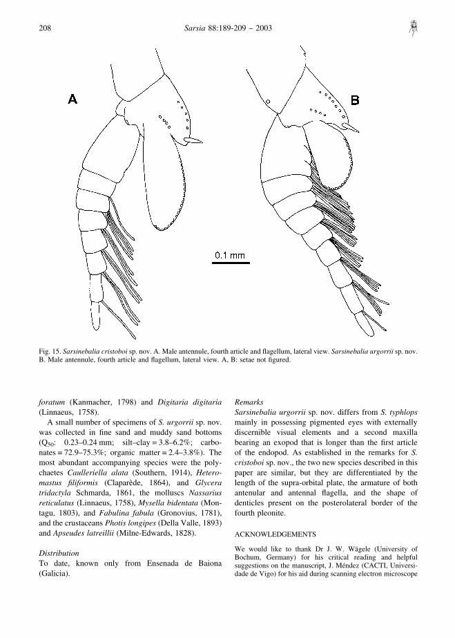
MaleA subadult male has also been found between 13
females (Fig. 1F). Antennular flagellum is eight-segmented, with articles thicker than those of thefemale, bearing long aesthetascs (Fig. 15A). Antennalflagellum longer than peduncle and extending to the endof the carapace, with about 40 articles; pleopodscomparatively longer in relation to body length.
EcologyThis species has been collected at depths of between 9and 33 m, mainly in clean coarse sand bottomscorresponding with the Branchiostoma lanceolatum–Clausinella fasciata community, and in coarse sandbottoms with a high presence of calcareous algaebelonging to the genera Lithothamnion and Phymatoli-thon, which are commonly known as “maerl”. Thecoarser granulometric fractions were predominant (Q50:0.85–0.9 mm), and the percentage of the silt–clayfraction was small (0.025–2.35%). These sedimentspresent a high content in carbonates (61.3–74.6%) and alow content in organic matter (1.3–2.5%).
The following companion fauna were found in theclean coarse sand bottoms: the polychaetes Pisioneremota (Southern, 1914), P. parapari Moreira, Quintas& Troncoso, 2000, Parapionosyllis cabezali Parapar,San Martın & Moreira, 2000, and Polygordius appendi-culatus Fraipont, 1887, the bivalves Goodallia triangu-laris (Montagu, 1803) and Clausinella fasciata (daCosta, 1778), and the cephalochordate Branchiostomalanceolatum (Pallas, 1778), while the following specieswere collected in the “maerl” bottoms: the molluscsLepidochitona cinereus (Linnaeus, 1767), Acmaeavirginea (Muller, 1776), Jujubinus exasperatus (Pen-nant, 1777), and Kellia suborbicularis (Montagu, 1803),and the crustaceans Phtisica marina Slabber, 1769 andPisidia longicornis (Linnaeus, 1767).
DistributionTo date, known only from the Galician coast (Rıa deVigo, Ensenada de Baiona, Rıa de Aldan, and Ensenadado Grove).
RemarksThis species can be distinguished from S. typhlops by itseyes, which have ommatidia and red pigmentation, andby a second maxilla bearing an exopod longer than thefirst article of the endopod. Additionally, in S. typhlopsthe third article of the mandibular palp presents a longseta proximally (see Dahl 1985, fig. 101), which wasnot observed in S. cristoboi sp. nov. The species closestto S. cristoboi sp. nov. is S. urgorrii sp. nov., alsodescribed herein, which has a similar body appearancebut differs from S. cristoboi sp. nov. in having longereyes, the supra-orbital plate never reaching the distal
Moreira & al. – Two new species of Leptostraca 199
end of the eye (Fig. 8F), an antennular flagellum withmore aesthetascs per article (Figs 9A, 14B), an antennalflagellum with spines and coarser setae (Figs 9B, 14E),and the posterolateral border of the fourth pleonitebearing rounder denticles (Fig. 14G) that distinctly donot present a spiny shape as in S. cristoboi sp. nov. (Fig.7H). Additionally, both species have not been foundcoexisting in the same bottoms, even in the samelocation, S. cristoboi sp. nov. apparently preferringsediments with predominantly coarser elements(“maerl”, gravel, coarse sand).
Sarsinebalia urgorrii sp. nov.(Figs 8–14, 15B)
Type seriesHolotype: ovigerous female (MNCN 20.04/5328),TL = 5.25 mm, CL = 2.0 mm, RL = 0.75 mm, Ensenadade Baiona, 42°08�10�N 8°50�15�W, May 1996, 10 m,medium sand. Allotype: male (MNCN 20.04/5329a),TL = 5.45 mm, CL = 2.0 mm, RL = 0.85 mm, Ensenadade Baiona, 42°08�10�N 8°50�15�W, November 1996,10 m, medium sand. Paratypes: Ensenada de Baiona:one female (MNCN 20.04/5330), 42°08�10�N8°50�15�W, December 1995, 10 m, medium sand; onepost-ovigerous female (MNCN 20.04/5331),42°08�10�N 8°50�15�W, July 1996, 10 m, medium sand;one female (MNCN 20.04/5332), 42°08�10�N8°50�15�W, October 1996, 10 m, medium sand; onefemale (MNCN 20.04/5329b), 42°08�10�N 8°50�15�W,November 1996, 10 m, medium sand; one female(MNCN 20.04/5333), 42°07�50�N 8°49�44�W, Decem-ber 1995, 9 m, medium sand; one subadult male(MNCN 20.04/5334), 42°07�30�N 8°50�15�W, Decem-ber 1995, 7 m, muddy sand.
Additional non-type materialEnsenada de Baiona: one female, 42°08�10�N8°49�44�W, December 1995, 8 m, muddy sand; twofemales, 42°07�50�N 8°50�15�W, December 1995, 8 m,medium sand; one female, 42°07�50�N 8°49�44�W,December 1995, 9 m, medium sand; three females,42°07�30�N 8°49�44�W, December 1995, 8 m, finesand, 42°08�10�N 8°50�15�W, 10 m, medium sand:April 1996, one subadult male; June 1996, four females;July 1996, two females; August 1996, one female;September 1996, two females; October 1996, sixfemales, two subadult males; November 1996, threefemales.
Type localityEnsenada de Baiona, 42°08�10�N 8°50�15�W, 10 m,medium sand.
EtymologyThis new species is named in honour of Dr VictorianoUrgorri (University of Santiago, Spain), eminentGalician malacologist, in appreciation of his teachingin marine biology, his kindness and his friendship.
Diagnosis of femaleEye disc-shaped, longer than wide, with ommatidia andred pigment; supra-orbital plate about 0.6 times lengthof eye. Rostrum slightly turning downwards, withpaired ventral keels fused proximally. Antennularflagellum with up to eight articles. Exopod of secondmaxilla longer than first article of endopod. Postero-lateral border of fourth pleonite bearing rounded teeth,ending in a larger tooth. Protopod of fourth pleopodwith several crenulations along posterior border, endingin acute tooth at posterolateral corner. Fifth pleopodwith six large spines on distolateral border. Sixthpleopod with four large spines on distolateral border.Pleonites 6–7 with distally rounded teeth along poster-ior border. Furcal rami equal to or slightly shorter thantelson and pleonite 7 combined.
Description of femaleLargest female of 5.25 mm TL, 2.0 mm CL and0.75 mm RL. Carapace oval, not covering fourthpleonite (Fig. 8A).
Rostrum: long, about twice as long as wide, distallytapering and turning downwards. Paired ventral keels,fused proximally (Fig. 8D). Tip of rostrum providedwith terminal spine, segmented proximally (Figs 8C,14A).
Compound eye: well developed, disc-shaped, longerthan wide, distal border straight, with ommatidia andred pigmentation covering almost entire eye (Fig. 8F).Supra-orbital plate with acute tip, extending about two-thirds of dorsal surface of eye.
Antennule: peduncle four-segmented (Fig. 9A).Second article widest at midpoint, about three timesas long as wide, with two clusters of smooth andplumose setae distally and subdistally, and withplumose seta dorsally at midpoint. Third article shorterthan second, widest distally, with terminal dorsalcluster of setae and additional distal seta placedventrally. Fourth article shorter than third, with rowof seven setae and one stout terminal spine, all of themalong dorsolateral border; next to insertion of anten-nular scale an additional row of setae; a very long andthicker seta attached near articulation of flagellum.Antennular scale oval, about 1.8 times as long as wide,with several rows of setae of different lengths: (a) longsimple setae, (b) stout curved setae, proximally smoothand distally provided with blunt serrations (Fig. 14C),
200 Sarsia 88:189-209 – 2003
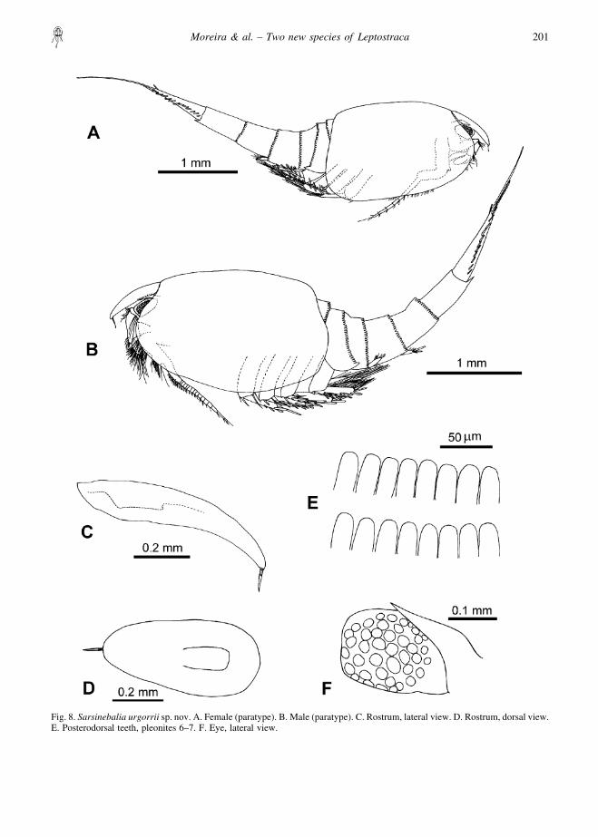
Fig. 8. Sarsinebalia urgorrii sp. nov. A. Female (paratype). B. Male (paratype). C. Rostrum, lateral view. D. Rostrum, dorsal view.E. Posterodorsal teeth, pleonites 6–7. F. Eye, lateral view.
Moreira & al. – Two new species of Leptostraca 201
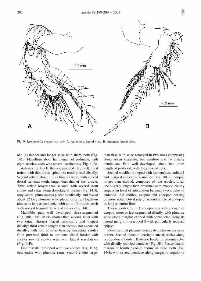
and (c) thinner and longer setae with sharp teeth (Fig.14C). Flagellum about half length of peduncle, witheight articles, each with several aesthetascs (Fig. 14B).
Antenna: peduncle three-segmented (Fig. 9B). Firstarticle with thin dorsal spine-like tooth placed distally.Second article about 1.3 as long as wide, with curveddorsal terminal tooth, larger than that of first article.Third article longer than second, with several stoutspines and setae along dorsolateral border (Fig. 14D),long ventral plumose seta placed subdistally, and row ofabout 12 long plumose setae placed distally. Flagellumalmost as long as peduncle, with up to 13 articles, eachwith several terminal setae and spines (Fig. 14E).
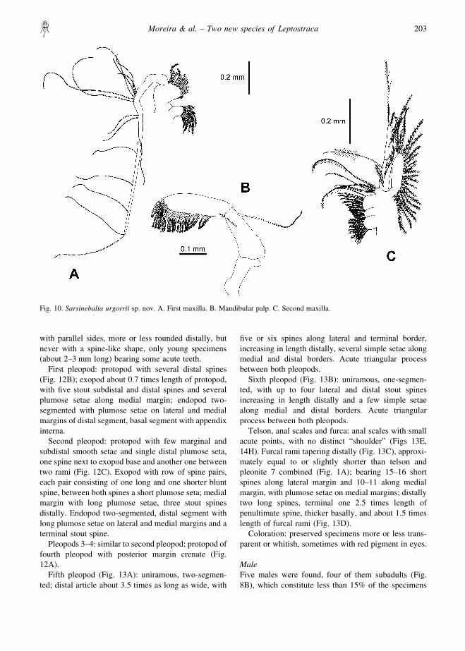
Mandible: palp well developed, three-segmented(Fig. 10B); first article shorter than second, latter withtwo setae, shortest placed subdistally and longestdistally; third article longer than second, not expandeddistally; with row of setae bearing lanceolate setulesfrom proximal third to terminus; distal border withshorter row of stouter setae with lateral serrulations(Fig. 14F).
First maxilla: protopod with two endites (Fig. 10A),first endite with plumose setae; second endite larger
than first, with setae arranged in two rows comprisingabout seven spatulate, two setulose and 14 distallydenticulate. Palp well developed, about five timeslength of protopod, with long spaced setae.
Second maxilla: protopod with four endites; endites 1and 3 largest and endite 4 smallest (Fig. 10C). Endopodlonger than exopod, composed of two articles, distalone slightly longer than proximal one; exopod clearlysurpassing level of articulation between two articles ofendopod. All endites, exopod and endopod bearingplumose setae. Distal seta of second article of endopodas long as entire limb.
Thoracopods (Fig. 11): endopod exceeding length ofexopod, more or less segmented distally, with plumosesetae along margin; exopod with some setae along itslateral margin; thoracopod 8 with particularly reducedepipod.
Pleonites: first pleonite lacking denticles on posteriorborder. Second pleonite bearing acute denticles alongposterodorsal border. Posterior border of pleonites 3–7with distally rounded denticles (Fig. 8E). Posterolateralmargin of fourth pleonite ending in large tooth (Fig.14G); with several denticles along margin, triangular or
Fig. 9. Sarsinebalia urgorrii sp. nov. A. Antennule, lateral view. B. Antenna, lateral view.
202 Sarsia 88:189-209 – 2003
with parallel sides, more or less rounded distally, butnever with a spine-like shape, only young specimens(about 2–3 mm long) bearing some acute teeth.
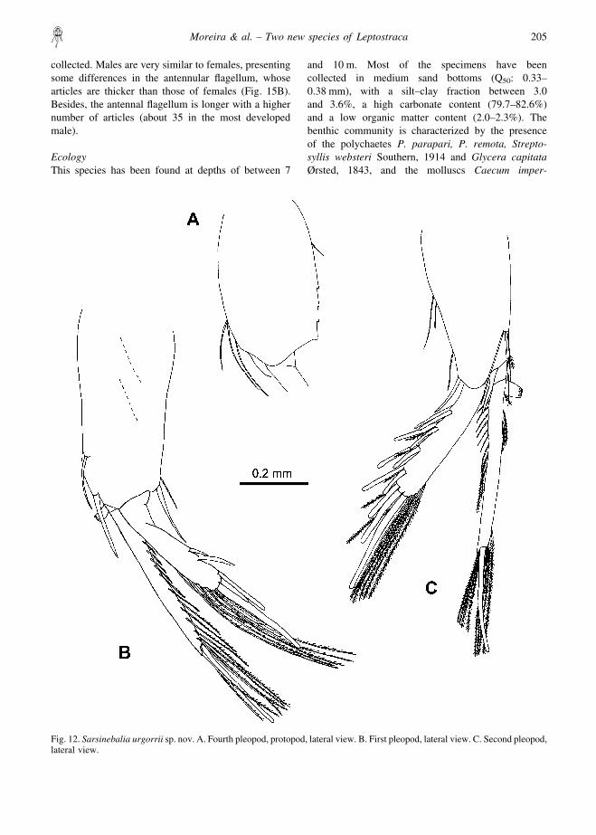
First pleopod: protopod with several distal spines(Fig. 12B); exopod about 0.7 times length of protopod,with five stout subdistal and distal spines and severalplumose setae along medial margin; endopod two-segmented with plumose setae on lateral and medialmargins of distal segment, basal segment with appendixinterna.
Second pleopod: protopod with few marginal andsubdistal smooth setae and single distal plumose seta,one spine next to exopod base and another one betweentwo rami (Fig. 12C). Exopod with row of spine pairs,each pair consisting of one long and one shorter bluntspine, between both spines a short plumose seta; medialmargin with long plumose setae, three stout spinesdistally. Endopod two-segmented, distal segment withlong plumose setae on lateral and medial margins and aterminal stout spine.
Pleopods 3–4: similar to second pleopod; protopod offourth pleopod with posterior margin crenate (Fig.12A).
Fifth pleopod (Fig. 13A): uniramous, two-segmen-ted; distal article about 3.5 times as long as wide, with
five or six spines along lateral and terminal border,increasing in length distally, several simple setae alongmedial and distal borders. Acute triangular processbetween both pleopods.
Sixth pleopod (Fig. 13B): uniramous, one-segmen-ted, with up to four lateral and distal stout spinesincreasing in length distally and a few simple setaealong medial and distal borders. Acute triangularprocess between both pleopods.
Telson, anal scales and furca: anal scales with smallacute points, with no distinct “shoulder” (Figs 13E,14H). Furcal rami tapering distally (Fig. 13C), approxi-mately equal to or slightly shorter than telson andpleonite 7 combined (Fig. 1A); bearing 15–16 shortspines along lateral margin and 10–11 along medialmargin, with plumose setae on medial margins; distallytwo long spines, terminal one 2.5 times length ofpenultimate spine, thicker basally, and about 1.5 timeslength of furcal rami (Fig. 13D).
Coloration: preserved specimens more or less trans-parent or whitish, sometimes with red pigment in eyes.
MaleFive males were found, four of them subadults (Fig.8B), which constitute less than 15% of the specimens
Fig. 10. Sarsinebalia urgorrii sp. nov. A. First maxilla. B. Mandibular palp. C. Second maxilla.
Moreira & al. – Two new species of Leptostraca 203
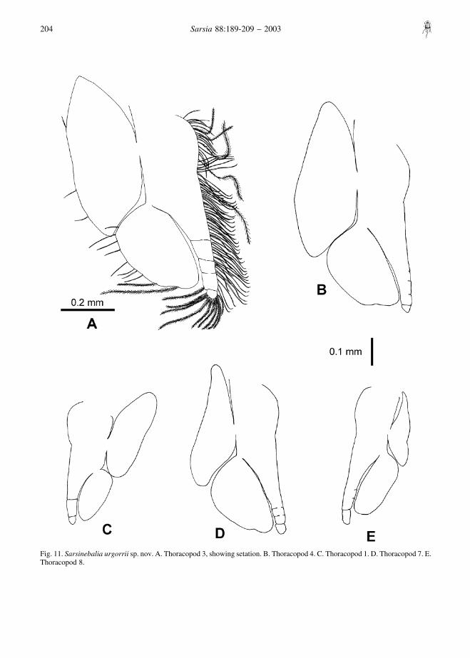
Fig. 11. Sarsinebalia urgorrii sp. nov. A. Thoracopod 3, showing setation. B. Thoracopod 4. C. Thoracopod 1. D. Thoracopod 7. E.Thoracopod 8.
204 Sarsia 88:189-209 – 2003
collected. Males are very similar to females, presentingsome differences in the antennular flagellum, whosearticles are thicker than those of females (Fig. 15B).Besides, the antennal flagellum is longer with a highernumber of articles (about 35 in the most developedmale).
EcologyThis species has been found at depths of between 7
and 10 m. Most of the specimens have beencollected in medium sand bottoms (Q50: 0.33–0.38 mm), with a silt–clay fraction between 3.0and 3.6%, a high carbonate content (79.7–82.6%)and a low organic matter content (2.0–2.3%). Thebenthic community is characterized by the presenceof the polychaetes P. parapari, P. remota, Strepto-syllis websteri Southern, 1914 and Glycera capitataØrsted, 1843, and the molluscs Caecum imper-
Fig. 12. Sarsinebalia urgorrii sp. nov. A. Fourth pleopod, protopod, lateral view. B. First pleopod, lateral view. C. Second pleopod,lateral view.
Moreira & al. – Two new species of Leptostraca 205
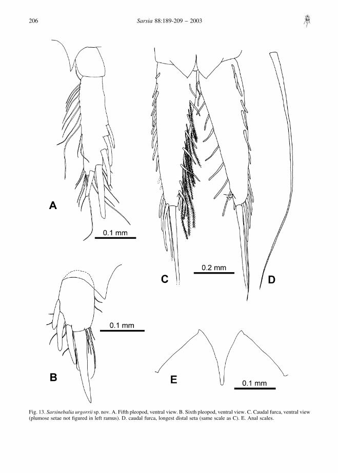
Fig. 13. Sarsinebalia urgorrii sp. nov. A. Fifth pleopod, ventral view. B. Sixth pleopod, ventral view. C. Caudal furca, ventral view(plumose setae not figured in left ramus). D. caudal furca, longest distal seta (same scale as C). E. Anal scales.
206 Sarsia 88:189-209 – 2003
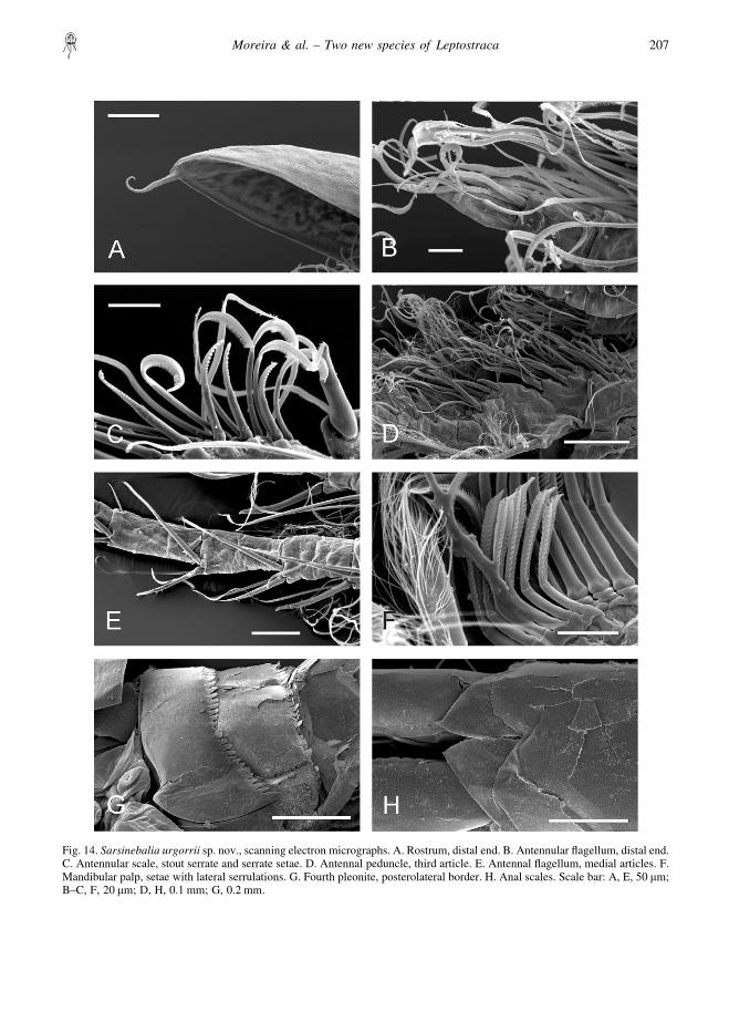
Fig. 14. Sarsinebalia urgorrii sp. nov., scanning electron micrographs. A. Rostrum, distal end. B. Antennular flagellum, distal end.C. Antennular scale, stout serrate and serrate setae. D. Antennal peduncle, third article. E. Antennal flagellum, medial articles. F.Mandibular palp, setae with lateral serrulations. G. Fourth pleonite, posterolateral border. H. Anal scales. Scale bar: A, E, 50 �m;B–C, F, 20 �m; D, H, 0.1 mm; G, 0.2 mm.
Moreira & al. – Two new species of Leptostraca 207
foratum (Kanmacher, 1798) and Digitaria digitaria(Linnaeus, 1758).
A small number of specimens of S. urgorrii sp. nov.was collected in fine sand and muddy sand bottoms(Q50: 0.23–0.24 mm; silt–clay = 3.8–6.2%; carbo-nates = 72.9–75.3%; organic matter = 2.4–3.8%). Themost abundant accompanying species were the poly-chaetes Caulleriella alata (Southern, 1914), Hetero-mastus filiformis (Claparede, 1864), and Glyceratridactyla Schmarda, 1861, the molluscs Nassariusreticulatus (Linnaeus, 1758), Mysella bidentata (Mon-tagu, 1803), and Fabulina fabula (Gronovius, 1781),and the crustaceans Photis longipes (Della Valle, 1893)and Apseudes latreillii (Milne-Edwards, 1828).
DistributionTo date, known only from Ensenada de Baiona(Galicia).
RemarksSarsinebalia urgorrii sp. nov. differs from S. typhlopsmainly in possessing pigmented eyes with externallydiscernible visual elements and a second maxillabearing an exopod that is longer than the first articleof the endopod. As established in the remarks for S.cristoboi sp. nov., the two new species described in thispaper are similar, but they are differentiated by thelength of the supra-orbital plate, the armature of bothantenular and antennal flagella, and the shape ofdenticles present on the posterolateral border of thefourth pleonite.
ACKNOWLEDGEMENTS
We would like to thank Dr J. W. Wagele (University ofBochum, Germany) for his critical reading and helpfulsuggestions on the manuscript, J. Mendez (CACTI, Universi-dade de Vigo) for his aid during scanning electron microscope
Fig. 15. Sarsinebalia cristoboi sp. nov. A. Male antennule, fourth article and flagellum, lateral view. Sarsinebalia urgorrii sp. nov.B. Male antennule, fourth article and flagellum, lateral view. A, B: setae not figured.
208 Sarsia 88:189-209 – 2003
sessions, P. Quintas and N. Troncoso (Universidade de Vigo)for providing us with some specimens, and the students ofCiencias del Mar (Universidade de Vigo) who patiently sortedout part of the samples. We are also grateful to two anonymousreferees and Dr E. Vetter (Hawaii Pacific University, USA) fortheir helpful comments on the manuscript. Special thanks are
given to Dr C. Dahl (Uppsala University, Sweden) for herkindness and for allowing us to consult an unpublished paperby Dr E. Dahl. This work is a contribution to the researchprojects XUGA30101A98 and PGIDTT00PX130119PRfinanced by the Xunta de Galicia.
REFERENCES
Buchanan JB. 1984. Sediment analysis. In: Holme NA,McIntyre AD, editors. Methods for the study of marinebenthos. IBP Handbook 16, 2nd edn. Oxford: BlackwellScientific. p 41–65.
Dahl E. 1985. Crustacea Leptostraca, principles of taxonomyand a revision of European shelf species. Sarsia70:135–165.
Escobar-Briones E, & Villalobos-Hiriart JL. 1995. Nebalialagartensis (Leptostraca) a new species from theYucatan Peninsula, Mexico. Crustaceana 68:1–11.
Haney TA, Hessler RR, & Martin JW. 2001. Nebalia schizo-phthalma, a new species of leptostracan (Malacostraca)from deep waters off the east coast of the United States.Journal of Crustacean Biology 21:192–201.
Haney TA, & Martin JW. 2000. Nebalia gerkenae, a newspecies of leptostracan (Crustacea: Malacostraca: Phyl-locarida) from the Bennett Slough region of MontereyBay, California. Proceedings of the Biological Societyof Washington 113:996–1014.
Hessler RR, & Sanders HL. 1965. Bathyal Leptostraca fromthe continental slope of the Northeastern United States.Crustaceana 9:71–74.
Kazmi QB, & Tirmizi NM. 1989. A new species of Nebaliafrom Pakistan (Leptostraca). Crustaceana 56:293–298.
Martin JW, Vetter EW, & Cash-Clark CE. 1996. Description,external morphology, and natural history observations
of Nebalia hessleri, new species (Phyllocarida: Lepto-straca), from Southern California, with a key to theextant families and genera of the Leptostraca. Journalof Crustacean Biology 16:347–372.
Modlin RF. 1991. Paranebalia belizensis, a new species fromshallow waters off Belize, Central America (Crustacea:Malacostraca: Leptostraca). Proceedings of the Bio-logical Society of Washington 104:603–612.
Olesen J. 1999. A new species of Nebalia (Crustacea,Leptostraca) from Unguja Island (Zanzibar), Tanzania,East Africa, with a phylogenetic analysis of leptostracangenera. Journal of Natural History 33:1789–1809.
Vetter EW. 1996. Nebalia daytoni n. sp. a leptostracanfrom Southern California (Phyllocarida). Crustaceana69:379–386.
Walker-Smith GK. 1998. A review of Nebaliella (Crustacea:Leptostraca) with the description of a new species fromthe continental slope of southeastern Australia.Memoirs of the Museum of Victoria 57:39–56.
Walker-Smith GK. 2000. Levinebalia maria, a new genus andnew species of Leptostraca (Crustacea) from Australia.Memoirs of the Museum of Victoria 58:137–148.
Walker-Smith GK, & Poore GCB. 2001. A phylogeny of theLeptostraca (Crustacea) with keys to families andgenera. Memoirs of the Museum of Victoria 58:383–410.
Accepted 24 April 2002 – Printed 30 June 2003Editorial responsibility: Egil Karlsbakk
Moreira & al. – Two new species of Leptostraca 209