Extended Hückel calculations on the chemisorption of acetylene on tungsten

RESEARCH ARTICLE Open Access
Tungsten carbide cobalt nanoparticles exerthypoxia-like effects on the gene expression levelin human keratinocytesWibke Busch1*, Dana Kühnel1, Kristin Schirmer2, Stefan Scholz1
Abstract
Background: Tungsten carbide (WC) and tungsten carbide cobalt (WC-Co) nanoparticles are of occupational healthrelevance because of the increasing usage in hard metal industries. Earlier studies showed an enhanced toxicpotential for WC-Co compared to WC or cobalt ions alone. Therefore, we investigated the impact of these particles,compared to cobalt ions applied as CoCl2, on the global gene expression level in human keratinocytes (HaCaT) invitro.
Results: WC nanoparticles exerted very little effects on the transcriptomic level after 3 hours and 3 days ofexposure. In contrast, WC-Co nanoparticles caused significant transcriptional changes that were similar to thoseprovoked by CoCl2. However, CoCl2 exerted even more pronounced changes in the transcription patterns. Geneset enrichment analyses revealed that the differentially expressed genes were related to hypoxia response,carbohydrate metabolism, endocrine pathways, and targets of several transcription factors. The role of thetranscription factor HIF1 (hypoxia inducible factor 1) is particularly highlighted and aspects of downstream eventsas well as the role of other transcription factors related to cobalt toxicity are considered.
Conclusions: This study provides extensive data useful for the understanding of nanoparticle and cobalt toxicity. Itshows that WC nanoparticles caused low transcriptional responses while WC-Co nanoparticles are able to exertresponses similar to that of free cobalt ions, particularly the induction of hypoxia-like effects via interactions withHIF1a in human keratinocytes. However, the enhanced toxicity of WC-Co particles compared to CoCl2 could notbe explained by differences in gene transcription.
BackgroundEngineered nanomaterials are used in large amounts inseveral industries and an increasing demand, includingnew types of particles, is anticipated in the future [1].Their physico-chemical properties, i.e. the small size andthe high surface to volume ratio are one of the mostinteresting characteristics, which is useful for many appli-cations in medicine, chemistry, material sciences andphysics. However, these physico-chemical characteristicsmay be associated with undesired health effects notknown for, or different from, the bulk materials. Hence,the field of nanotoxicology is emerging to assess possiblehazards of nanomaterials. Several reviews have sum-marised the potential cellular mechanisms of
nanoparticles toxicity such as increase in the productionof reactive oxygen species (ROS) and induction of inflam-matory responses [2-4]. The cellular responses appear tobe dependent on the physical and chemical properties ofthe particles, such as particle size, dissolution behaviour,surface reactivity and binding ability [2,5,6].So far, the majority of in vivo and in vitro studies in
nanotoxicology have focussed on endpoints such as vital-ity, production of reactive oxygen species, immunologicalparameters or cell death. However, the elucidation of themode of action and identification of subacute effects withpotential implications for chronic toxicity are difficult toobtain from these studies. Therefore, modern toxicoge-nomic approaches established already in pharmacologyand toxicology [7-10] could be used to unravel the toxi-codynamics of nanomaterials. First studies on the effectsof nano- or ultrafine particles on global gene expression
* Correspondence: [email protected] - Helmholtz-Centre for Environmental Research Leipzig, Department ofBioanalytical Ecotoxicology, Permoserstr. 15, 04318 Leipzig, Germany
Busch et al. BMC Genomics 2010, 11:65http://www.biomedcentral.com/1471-2164/11/65
© 2010 Busch et al; licensee BioMed Central Ltd. This is an Open Access article distributed under the terms of the Creative CommonsAttribution License (http://creativecommons.org/licenses/by/2.0), which permits unrestricted use, distribution, and reproduction inany medium, provided the original work is properly cited.

patterns revealed compound-specific but no generalresponses due to the exposure to particles [11-14].Hence, the chemical composition of the particles seemsto play a major role for transcriptional responses. Griffittand colleagues [11] showed that metal ions (silver andcopper) caused similar expression patterns as nanoparti-cles of the same materials in zebrafish, but the numbersof affected genes were always higher after exposure tothe particles. In an in vitro study by Waters et al. [12] itwas found that changes in cell viability provoked by silicaexhibited a higher correlation with particle surface areathan either particle mass or number in macrophages.The majority of biological processes represented by thedifferentially expressed genes were nearly identical, irre-spective of particle diameter.A toxicogenomic approach has been used in this study
to analyse the mode of action of tungsten carbide (WC)and tungsten carbide cobalt (WC-Co) nanoparticles.These nanoparticles are intended to be increasingly usedin hard metal industries for the production of wearresistant and hard tools. The major advantage of usingWC and WC-Co nano-scaled instead of micro-scaledparticles is the increased hardness of resulting compo-site materials and therefore a prolonged wear lifespan oftools and other products [15]. Cobalt serves as bindingagent improving the sintering of hard metals from WCnanoparticles. Therefore, the use of WC-Co particles isfavoured in hard metal industries. Potential health impli-cations may be of concern for workers involved in themanufacturing process. Previous studies using μm-sizedparticles have indicated a lack of toxicity for WC parti-cles but a hazardous potential for cobalt metal particlesin vivo and in vitro [16-18]. A mixture of these μm-scaled powders (WC-Co) exhibited an enhanced toxicityif compared to cobalt metal powder alone [16-20]. Ourprevious research also showed toxicity enhancing effectsfor nano-sized WC-Co compared to WC or CoCl2[21,22]. The increased toxicity was proposed to resultfrom specific interactions of WC and cobalt. Since theInternational Agency for Research on Cancer (IARC)has classified cobalt as ‘possibly carcinogenic’ and tung-sten carbide cobalt as ‘probably carcinogenic’ to humans[23] research on the elucidation of the mode of actionof nano-sized particles of these materials is of high rele-vance for occupational health. Ionic cobalt (Co2+) isknown to exert hypoxia like responses via stabilising thea subunit of the hypoxia inducible transcription factor(HIF1) [24-26]. Ubiquitously expressed HIF1a isdegraded via oxygen-dependent prolyl-4-hydroxylationunder normoxic conditions [27]. These degradation pro-cesses are blocked by cobalt binding or oxygen defi-ciency (hypoxia) which results in enriched HIF1a levelsin the cells. HIF1 is a transcription factor that mediatesresponse to hypoxia by regulating the transcription of
genes encoding proteins that play key roles in angiogen-esis, glucose and energy metabolism, cell survival andproliferation, iron metabolism, and vascular functions[28,29]. Comparative gene expression studies showedHIF1-mediated responses to be similar for hypoxia andCoCl2 exposure [8,30].Whether metallic cobalt in nanoparticles, particularly
in combination with tungsten, provokes specific toxiceffects deviating from or exceeding those observed fordissolved ionic cobalt is not finally clarified yet. Lisonand co-workers [31] described the formation of reactiveoxygen species (ROS) after a rapid dissolution of cobaltions out of WC-Co micrometer sized particles in a cellfree system, a phenomenon that could not be foundwith metal cobalt particles or a combination of WC par-ticles with CoCl2. Furthermore, another study that eval-uated the role of ROS in the interactive toxicity ofcarbide-cobalt mixtures found no evidence that produc-tion of ROS contributed to the toxicity of WC-Co inmacrophages [32]. Lombaert and co-workers [33] inves-tigated gene expression in macrophages exposed tomicrometer sized WC-Co particles (a mixture of cobaltmetal with a median particle size (d50) of 4 μm and WCparticles d50 < 1 μm). They identified differential expres-sion of genes involved in apoptosis regulation, stressresponse, glucose metabolism, cell signalling, immuneresponse and other pathways. The effects were discussedto be at least partially provoked by dissolved cobalt ions.In a previous study [21] we have investigated the
impact of WC and WC-Co nanoparticles on the vitalityof various mammalian cells (lung, skin, colon and oligo-dendrocyte cell lines; primary neural cell and astroglialcultures). Significant cytotoxic effects were observed fornano-sized WC-Co (33 μg/ml). About 15% of WC and76% of Co were found to be dissolved after 1 week ofstorage of the stock solution. Interestingly, WC-Co par-ticles (consisting of 30 μg/ml WC and 3 μg/ml cobalt)showed a higher toxicity than equivalent concentrationsof CoCl2 (3 μg/ml) indicating that leaching of cobaltalone may not explain the toxic effects. It was alsodemonstrated that the tungsten based nanoparticlescould enter various cell types [21,22]. Based on thisstudy we selected the human skin cell line HaCaT toinvestigate the effects of WC and WC-Co nanoparticlesand cobalt ions on gene expression patterns. Our majorgoal was to elucidate (1) whether the observed effectsindicate specific mode of actions of WC-Co nanoparti-cles and/or (2) whether the effects can primarily beexplained by dissolved Co.
ResultsWe compared the mode of action of WC and WC-Conanoparticles and dissolved CoCl2 in HaCaT cells byrecording changes in transcription profiles by microarray
Busch et al. BMC Genomics 2010, 11:65http://www.biomedcentral.com/1471-2164/11/65
Page 2 of 21

analysis. HaCaT cells were exposed to the lowest concen-tration of WC-Co causing a reduction of cell vitality (33μg/ml; [21]) and corresponding concentrations of WCand CoCl2. RNA isolated after 3 h and 3 d of exposurefrom 5 independent biological replicates per treatmentwas analysed using a commercial human whole genomemicroarray. Various analyses routines were performed toidentify differentially expressed genes, treatment clustersand affected biological pathways.Identification of differentially expressed genesSAM analysis of normalised microarray fluorescenceintensities for all treatments revealed 1956 significantlydifferentially expressed genes with about 1146 showingan induction or repression of more than 2fold. The high-est number of genes with a significantly altered expres-sion above 2fold was observed after 3 d of exposure(Table 1). Among the different treatments, exposure toCoCl2 provoked the strongest changes in gene expression(373 and 826 genes for 3 h and 3 d of exposure, respec-tively) followed by WC-Co (37 and 248, respectively) andWC nanoparticles (28 and 49 respectively). Comparisonof the genes affected by the different treatments revealeda considerable overlap of transcription profiles. The high-est commonalities were observed between the geneexpression patterns of CoCl2 and WC-Co after 3 d ofexposure (184 genes differentially expressed in bothtreatments), followed by the exposure to CoCl2 at 3 hand 3 d (134 genes) and WC/WC-Co at 3 d (31 genes). Alist of the genes with the strongest differential expression(>5fold) can be found in Table 2, the complete set ofgenes is available in the Additional file 1.Confirmation of microarray dataIn order to verify the microarray results, RT-PCR analysiswas conducted using the same set of samples used for themicroarrays as well as RNA samples from independentexposure experiments. Twelve genes with significantlydifferential expression and a minimum of 2fold up- ordownregulation were selected for RT-PCR confirmation.
However, care was taken that genes with weak (close to2fold differential expression) and strong changes (up to23fold) in expression were included (Figure 1). For eightof the selected genes the significant changes of expres-sion could be confirmed (BNIP3, LOXL2, ANGPTL4,CA9, PFKFB4, KRT1, MAL, MMP1). Trends (inductionor repression) were conserved between microarray andRT-PCR data for each treatment. The remaining genes(GAPDH, ID2, OLFM4, DSG4) exhibited a high variabil-ity and could not be confirmed as statistically significantfrom controls by RT-PCR.PCA and cluster analysesTwo methods of descriptive statistics - PCA (principalcomponent analysis) and HCA (hierarchical cluster ana-lysis) - were applied to identify commonalities or differ-ences between treatments based on the patterns ofsignificantly differentially expressed genes. By PCA ana-lysis about 65% of the variability in different treatmentswas represented by the first 3 components. Four clearlyseparated clusters, i.e. cells treated with CoCl2 for 3 h,the same treatment for 3 d, cells treated with WC-Cofor 3 d and cells treated with WC for 3 d were identified(Figure 2). All other treatments, including the controls,were not separated and formed a large cluster withapparently weak gene expression changes if compared tocontrols. WC-treated cells were less clearly separatedfrom controls. This was indicated by the observationthat a clear distinct cluster was only demonstrated forPC axis 2 and 3.Similar results were obtained by HCA, which identi-
fied 3 treatment clusters. Both CoCl2 treatments and the3 d exposures of WC-Co formed a distinct cluster. Allother treatments (controls, WC3d, WC3h, WC-Co3h)were grouped in one cluster (Additional file 2). Geneclustering revealed two clusters with strongly inducedgenes. Genes of these clusters (i.e. LOXL2, BNIP3, CA9,PDK1, ASB2, EGLN3, ANKRD37, PNCK) are coding forproteins with diverse functions, but some of them are
Table 1 Number of significant repressed or induced genes > 2fold per treatment*.
Treatment up down total Treatment Overlapping genes between treatments
WC3h 26 2 28 WC3h WC3h
WCCo3h 13 24 37 WCCo3h 8 WCCo3h
CoCl3h 242 131 373 CoCl3h 8 17 CoCl3h
WC3d 18 31 49 WC3d 3 2 11 WC3d
WCCo3d 141 107 248 WCCo3d 2 8 29 31 WCCo3d
CoCl3d 541 285 826 CoCl3d 8 15 134 19 184
* identified with SAM; the pair wise comparison indicates genes that were differentially expressed in both of the considered treatments
Busch et al. BMC Genomics 2010, 11:65http://www.biomedcentral.com/1471-2164/11/65
Page 3 of 21
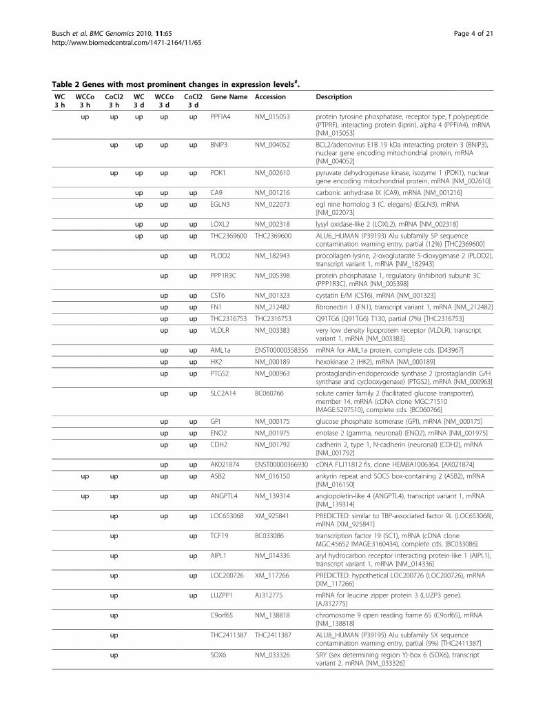
Table 2 Genes with most prominent changes in expression levels#.
WC3 h
WCCo3 h
CoCl23 h
WC3 d
WCCo3 d
CoCl23 d
Gene Name Accession Description
up up up up up PPFIA4 NM_015053 protein tyrosine phosphatase, receptor type, f polypeptide(PTPRF), interacting protein (liprin), alpha 4 (PPFIA4), mRNA[NM_015053]
up up up up BNIP3 NM_004052 BCL2/adenovirus E1B 19 kDa interacting protein 3 (BNIP3),nuclear gene encoding mitochondrial protein, mRNA[NM_004052]
up up up up PDK1 NM_002610 pyruvate dehydrogenase kinase, isozyme 1 (PDK1), nucleargene encoding mitochondrial protein, mRNA [NM_002610]
up up up CA9 NM_001216 carbonic anhydrase IX (CA9), mRNA [NM_001216]
up up up EGLN3 NM_022073 egl nine homolog 3 (C. elegans) (EGLN3), mRNA[NM_022073]
up up up LOXL2 NM_002318 lysyl oxidase-like 2 (LOXL2), mRNA [NM_002318]
up up up THC2369600 THC2369600 ALU6_HUMAN (P39193) Alu subfamily SP sequencecontamination warning entry, partial (12%) [THC2369600]
up up PLOD2 NM_182943 procollagen-lysine, 2-oxoglutarate 5-dioxygenase 2 (PLOD2),transcript variant 1, mRNA [NM_182943]
up up PPP1R3C NM_005398 protein phosphatase 1, regulatory (inhibitor) subunit 3C(PPP1R3C), mRNA [NM_005398]
up up CST6 NM_001323 cystatin E/M (CST6), mRNA [NM_001323]
up up FN1 NM_212482 fibronectin 1 (FN1), transcript variant 1, mRNA [NM_212482]
up up THC2316753 THC2316753 Q91TG6 (Q91TG6) T130, partial (7%) [THC2316753]
up up VLDLR NM_003383 very low density lipoprotein receptor (VLDLR), transcriptvariant 1, mRNA [NM_003383]
up up AML1a ENST00000358356 mRNA for AML1a protein, complete cds. [D43967]
up up HK2 NM_000189 hexokinase 2 (HK2), mRNA [NM_000189]
up up PTGS2 NM_000963 prostaglandin-endoperoxide synthase 2 (prostaglandin G/Hsynthase and cyclooxygenase) (PTGS2), mRNA [NM_000963]
up up SLC2A14 BC060766 solute carrier family 2 (facilitated glucose transporter),member 14, mRNA (cDNA clone MGC:71510IMAGE:5297510), complete cds. [BC060766]
up up GPI NM_000175 glucose phosphate isomerase (GPI), mRNA [NM_000175]
up up ENO2 NM_001975 enolase 2 (gamma, neuronal) (ENO2), mRNA [NM_001975]
up up CDH2 NM_001792 cadherin 2, type 1, N-cadherin (neuronal) (CDH2), mRNA[NM_001792]
up up AK021874 ENST00000366930 cDNA FLJ11812 fis, clone HEMBA1006364. [AK021874]
up up up up ASB2 NM_016150 ankyrin repeat and SOCS box-containing 2 (ASB2), mRNA[NM_016150]
up up up up ANGPTL4 NM_139314 angiopoietin-like 4 (ANGPTL4), transcript variant 1, mRNA[NM_139314]
up up up LOC653068 XM_925841 PREDICTED: similar to TBP-associated factor 9L (LOC653068),mRNA [XM_925841]
up up TCF19 BC033086 transcription factor 19 (SC1), mRNA (cDNA cloneMGC:45652 IMAGE:3160434), complete cds. [BC033086]
up up AIPL1 NM_014336 aryl hydrocarbon receptor interacting protein-like 1 (AIPL1),transcript variant 1, mRNA [NM_014336]
up up LOC200726 XM_117266 PREDICTED: hypothetical LOC200726 (LOC200726), mRNA[XM_117266]
up up LUZPP1 AJ312775 mRNA for leucine zipper protein 3 (LUZP3 gene).[AJ312775]
up C9orf65 NM_138818 chromosome 9 open reading frame 65 (C9orf65), mRNA[NM_138818]
up THC2411387 THC2411387 ALU8_HUMAN (P39195) Alu subfamily SX sequencecontamination warning entry, partial (9%) [THC2411387]
up SOX6 NM_033326 SRY (sex determining region Y)-box 6 (SOX6), transcriptvariant 2, mRNA [NM_033326]
Busch et al. BMC Genomics 2010, 11:65http://www.biomedcentral.com/1471-2164/11/65
Page 4 of 21

Table 2: Genes with most prominent changes in expression levels#. (Continued)
up SULT2A1 NM_003167 sulfotransferase family, cytosolic, 2A,dehydroepiandrosterone (DHEA)-preferring, member 1(SULT2A1), mRNA [NM_003167]
up C1orf67 BC042869 cDNA clone IMAGE:5270407. [BC042869]
up CRB1 NM_201253 crumbs homolog 1 (Drosophila) (CRB1), mRNA[NM_201253]
up up EDN2 NM_001956 endothelin 2 (EDN2), mRNA [NM_001956]
up CBLN4 NM_080617 cerebellin 4 precursor (CBLN4), mRNA [NM_080617]
up G65686 ENST00000332107 A117 Human STS cDNA, sequence tagged site. [G65686]
up XAGE2 NM_130777 X antigen family, member 2 (XAGE2), mRNA [NM_130777]
up PELO AF118075 PRO1770 mRNA, complete cds. [AF118075]
up SAA3P AY209188 truncated serum amyloid A3 precursor (SAA3) mRNA,complete cds. [AY209188]
up ITIH5 NM_030569 inter-alpha (globulin) inhibitor H5 (ITIH5), transcript variant1, mRNA [NM_030569]
up MCHR1 NM_005297 melanin-concentrating hormone receptor 1 (MCHR1),mRNA [NM_005297]
up MGAT4A NM_012214 mannosyl (alpha-1,3-)-glycoprotein beta-1,4-N-acetylglucosaminyltransferase, isozyme A (MGAT4A), mRNA[NM_012214]
up GPR65 NM_003608 G protein-coupled receptor 65 (GPR65), mRNA[NM_003608]
up DFNB31 AK056190 cDNA FLJ31628 fis, clone NT2RI2003344, weakly similar toPRESYNAPTIC PROTEIN SAP97. [AK056190]
up LRP8 NM_033300 low density lipoprotein receptor-related protein 8,apolipoprotein e receptor (LRP8), transcript variant 2, mRNA[NM_033300]
dn up up up SORCS3 NM_014978 sortilin-related VPS10 domain containing receptor 3(SORCS3), mRNA [NM_014978]
dn up HLA-DPA1 NM_033554 major histocompatibility complex, class II, DP alpha 1 (HLA-DPA1), mRNA [NM_033554]
up dn dn AKR1C1 BC040210 aldo-keto reductase family 1, member C1, mRNA (cDNAclone MGC:42600 IMAGE:4825338), complete cds.[BC040210]
up dn up WDR64 NM_144625 WD repeat domain 64 (WDR64), mRNA [NM_144625]
dn up ESCO2 NM_001017420 establishment of cohesion 1 homolog 2 (S. cerevisiae)(ESCO2), mRNA [NM_001017420]
dn up CACNG5 NM_145811 calcium channel, voltage-dependent, gamma subunit 5(CACNG5), transcript variant 1, mRNA [NM_145811]
dn up ANPEP NM_001150 alanyl (membrane) aminopeptidase (aminopeptidase N,aminopeptidase M, microsomal aminopeptidase, CD13,p150) (ANPEP), mRNA [NM_001150]
dn up GPX7 NM_015696 glutathione peroxidase 7 (GPX7), mRNA [NM_015696]
dn dn up TTLL7 NM_024686 tubulin tyrosine ligase-like family, member 7 (TTLL7), mRNA[NM_024686]
dn dn up THC2371963 THC2371963 AIP1_HUMAN (Q86UL8) Atrophin-1 interacting protein 1(Atrophin-1 interacting protein A) (MAGI-2), partial (3%)[THC2371963]
up dn dn GDF15 NM_004864 growth differentiation factor 15 (GDF15), mRNA[NM_004864]
dn dn ALDH1A1 NM_000689 aldehyde dehydrogenase 1 family, member A1 (ALDH1A1),mRNA [NM_000689]
dn dn DCN NM_001920 decorin (DCN), transcript variant A1, mRNA [NM_001920]
dn dn SOX2 NM_003106 SRY (sex determining region Y)-box 2 (SOX2), mRNA[NM_003106]
dn dn TAF9B NM_015975 TAF9B RNA polymerase II, TATA box binding protein (TBP)-associated factor, 31 kDa (TAF9B), mRNA [NM_015975]
Busch et al. BMC Genomics 2010, 11:65http://www.biomedcentral.com/1471-2164/11/65
Page 5 of 21

Table 2: Genes with most prominent changes in expression levels#. (Continued)
dn dn THC2302184 THC2302184 GAL2_HUMAN (Q01415) N-acetylgalactosamine kinase(GalNAc kinase) (Galactokinase 2), partial (21%)[THC2302184]
dn dn PTPRZ1 NM_002851 protein tyrosine phosphatase, receptor-type, Z polypeptide1 (PTPRZ1), mRNA [NM_002851]
dn dn DMRT2 NM_006557 doublesex and mab-3 related transcription factor 2(DMRT2), transcript variant 1, mRNA [NM_006557]
dn dn CLEC3A NM_005752 C-type lectin domain family 3, member A (CLEC3A), mRNA[NM_005752]
dn dn ATP10B AB018258 mRNA for KIAA0715 protein, partial cds. [AB018258]
dn dn PROS1 NM_000313 protein S (alpha) (PROS1), mRNA [NM_000313]
dn dn RAXLX NM_001008494 RAX-like homeobox (RAXLX), mRNA [NM_001008494]
dn dn OLFM4 NM_006418 olfactomedin 4 (OLFM4), mRNA [NM_006418]
dn dn DSG4 NM_177986 desmoglein 4 (DSG4), mRNA [NM_177986]
dn dn dn IQGAP2 NM_006633 IQ motif containing GTPase activating protein 2 (IQGAP2),mRNA [NM_006633]
dn dn dn MAL NM_002371 mal, T-cell differentiation protein (MAL), transcript variant a,mRNA [NM_002371]
dn dn dn KRT1 NM_006121 keratin 1 (epidermolytic hyperkeratosis) (KRT1), mRNA[NM_006121]
dn dn dn MMP1 NM_002421 matrix metallopeptidase 1 (interstitial collagenase) (MMP1),mRNA [NM_002421]
dn dn dn LAMP3 NM_014398 lysosomal-associated membrane protein 3 (LAMP3), mRNA[NM_014398]
dn dn dn HLA-DMB NM_002118 major histocompatibility complex, class II, DM beta (HLA-DMB), mRNA [NM_002118]
dn dn dn HERC5 NM_016323 hect domain and RLD 5 (HERC5), mRNA [NM_016323]
dn BTN3A3 NM_006994 butyrophilin, subfamily 3, member A3 (BTN3A3), transcriptvariant 1, mRNA [NM_006994]
dn C6orf130 NM_145063 chromosome 6 open reading frame 130 (C6orf130), mRNA[NM_145063]
dn CRY2 NM_021117 cryptochrome 2 (photolyase-like) (CRY2), mRNA[NM_021117]
dn TMEM140 NM_018295 transmembrane protein 140 (TMEM140), mRNA[NM_018295]
dn ZNF438 NM_182755 zinc finger protein 438 (ZNF438), mRNA [NM_182755]
dn DNMT3L NM_013369 DNA (cytosine-5-)-methyltransferase 3-like (DNMT3L),transcript vari ant 1, mRNA [NM_013369]
dn CTAGE3 AF338231 CTAGE-3 protein mRNA, complete cds. [AF338231]
dn MOSPD2 NM_152581 motile sperm domain containing 2 (MOSPD2), mRNA[NM_152581]
dn OR4N4 NM_001005241 olfactory receptor, family 4, subfamily N, member 4(OR4N4), mRNA [NM_001005241]
dn SPATA7 NM_018418 spermatogenesis associated 7 (SPATA7), transcript variant 1,mRNA [NM_018418]
dn TMC1 NM_138691 transmembrane channel-like 1 (TMC1), mRNA [NM_138691]# listed genes were statistically significant different and exhibited at least a 5fold induction or repression in one of the treatments (with respect to controls andbased on normalised fluorescence intensity ratios in the microarray analysis); type of gene regulation is indicated as “up” for induction (> 2fold) and “dn” forrepression (> 2fold); fields are empty when induction or repression was < 2fold; full table is provided as Additional File 1
Busch et al. BMC Genomics 2010, 11:65http://www.biomedcentral.com/1471-2164/11/65
Page 6 of 21

BNIP3
Control WC-Co CoCl2 WC1000
50
100
150
200* *
n=4, p=0,0083
Treatment
% G
ene
exp
ress
ion
(re
lati
ve)
LOXL2
Control WC-Co CoCl2 WC1000
50
100
150
200
** **
n=4, p<0,0001
Treatment
CA9
Control WC-Co CoCl2 WC1000
50
100
150
200 ** **n=6, p<0,0001
Treatment
PFKFB4
Control WC-Co CoCl2 WC1000
50
100
150
200**
n=5, p=0,0003
*
Treatment
% G
ene
exp
ress
ion
(re
lati
ve)
Angptl4
Control WC-Co CoCl2 WC1000
50
100
150
200**
n=6, p<0.0001
**
Treatment
GAPDH
Control WC-Co CoCl2 WC1000
50
100
150
n=6, n.s.
Treatment
upregulated genes
KRT1
Control WC-Co CoCl2 WC1000
50
100
150
**
n=7, p=0,0009
*
Treatment
% G
ene
exp
ress
ion
(re
lati
ve)
MAL
Control WC-Co CoCl2 WC1000
50
100
150
n=6, p<0.0001
** ** **
Treatment
MMP1
Control WC-Co CoCl2 WC1000
50
100
150
n=6, p=0.0047
** **
Treatment
OLFM4
Control WC-Co CoCl2 WC1000
50
100
150
n=7, n.s.
Treatment
% G
ene
exp
ress
ion
(re
lati
ve)
ID2
Control WC-Co CoCl2 WC1000
50
100
150
n=3, n.s.
Treatment
DSGL4
Control WC-Co CoCl2 WC1000
50
100
150
n=6, n.s.
Treatment
downregulated genes
Figure 1 Validation of microarray data by RT-PCR. Relative gene expression of arbitrarily selected genes in HaCaT cells after 3 d of exposureto 30 μg/ml WC and 33 μg/ml WC-Co nanoparticles and 3 μg/ml CoCl2 was analysed by semiquantiative RT-PCR. Selected genes representgenes with significant changes (2 to 23fold) of expression levels in microarrays. Gene expression values were converted to percent of the meanof controls and are presented as mean + standard deviation (SD). Statistical differences were analysed with one-way ANOVA followed byDunnett’s post test (treatment vs. control). Values of p < 0.05 were considered statistically significant; *p < 0.05, **p < 0.01.
Busch et al. BMC Genomics 2010, 11:65http://www.biomedcentral.com/1471-2164/11/65
Page 7 of 21

known to be direct targets of the transcription factorHIF1a (see below). For the remaining clusters it wasnot possible to identify predominating groups of geneontology. Therefore, gene clusters were not analysed indetail but two types of pathway analysis were used toidentify signalling pathways and biological functionsassociated with the differentially expressed genes.Gene set enrichment analysis (GSEA) and identification ofaffected signalling pathwaysPathway analysis was performed by two approaches,with (1) GSEA software [34,35] using the complete setof gene expression data from the microarray experi-ments, and (2) the DAVID database [36] using the listof more then 2fold differentially expressed genes pre-viously identified with SAM.GSEA identified communalities with existing gene sets
(enrichment) primarily for induced genes. The highestnumber of affected gene sets was found for the 3 dWC-Co treatment. The majority of pathways associatedwith down-regulated gene sets were found after 3 h ofexposure with WC-Co. An overview of pathway relatedgene sets with the highest commonality (based on statis-tical significance analysis) to the observed patterns ofdifferentially expressed genes is given in Table 3.Gene sets related to the hypoxia pathway as well as
carbohydrate metabolism were induced by WC-Co andCoCl2 after 3 d. A significant association with the induc-tion of the hypoxia gene sets was also observed after 3 hof exposure with WC-Co. As indicated by the enrich-ment of genes for the transcription factor HIF1a
(hypoxia inducible factor 1 alpha), regulation via HIF1amay play a major role in provoking the observedchanges in hypoxia and carbohydrate metabolism genes.Furthermore, GSEA detected an enrichment of genesrelated to RNA metabolism and processing as well asgenes coding for proteins of the nucleus and the nuclearmembrane. These gene sets referred mainly to genesdown-regulated after 3 h of exposure to WC-Co nano-particles. Some of the genes with strong differentialrepression (> 5fold; e.g. MAL, KRT1, GDF15, MMP1;identified by SAM) were not found to be included inthese pathways.DAVID revealed similar results as GSEA. However,
small gene sets, for instance a down-regulation of metal-lothioneins in the 3 h CoCl2 exposure, were additionallyidentified by DAVID. Furthermore, genes coding forseveral proteins containing a functional prolyl-4-hydro-xylase alpha subunit were highlighted as up-regulatedfor the 3 d of exposure with CoCl2 and WC-Co.The complete results of the GSEA and DAVID ana-
lyses are provided in the Additional file 3.
DiscussionThe increasing use of nanoparticles may also lead to anincreased human exposure and adverse health effects.Occupational exposure is one of the most relevant expo-sure routes. In order to estimate the potential humanhealth impacts of nanoparticles a precise knowledge ontheir mechanism of action is indispensable. This knowl-edge allows, for instance, clarifying whether effects are
-60
-40
-20
0
20
40
60
-60
-40
-20
0
20
40
60
-60 -40 -20 0 20 40 60 80
Component 1 (41.5%)
-60 -40 -20 0 20 40 60
Component 2 (15.1%)
Com
pone
nt 2
(15
.1%
) C
ompo
nent
3 (
9.2%
)
H2O 3h WC 3h WC-Co 3h CoCl2 3h H2O 3d WC 3d WC-Co 3d CoCl2 3d
Figure 2 Principle component analyses. PCA of differentially expressed genes in HaCaT cells exposed for 3 h and 3 d to 30 μg/ml WC, 33 μg/ml WC-Co nanoparticles and 3 μg/ml CoCl2. Each symbol represents a biological replicate.
Busch et al. BMC Genomics 2010, 11:65http://www.biomedcentral.com/1471-2164/11/65
Page 8 of 21

Table 3 Gene sets with strongest overlaps with observed differentially expressed genes.*
up-regulated gene sets description WC3 h
WCCo3 h
CoCl23 h
WC3 d
WCCo3 d
CoCl23 d
hypoxia related gene sets
HIF1_TARGETS Hif-1 (hypoxia-induciblefactor 1) transcriptionaltargets
0.17 0.01
HIFPATHWAY BIOCARTA: Under normalconditions, HIF-1 isdegraded; under hypoxicconditions, it activatestranscription of genescontrolled by hypoxicresponse elements (HREs)
0.02 0.20
HYPOXIA_FIBRO_UP Up-regulated by hypoxia innormal fibroblasts fromboth young and olddonors
0.20 0.65 0.05
HYPOXIA_NORMAL_UP Up-regulated by hypoxia innormal, RPTEC renal cells
0.50 0.02
HYPOXIA_REG_UP Up-regulated by hypoxia inrenal cells, and down-regulated withreoxygenation
0.05 0.01 0.03
HYPOXIA_REVIEW Genes known to beinduced by hypoxia
0.22 0.75 0.00
MANALO_HYPOXIA_UP Genes up-regulated inhuman pulmonaryendothelial cells underhypoxic conditions or afterexposure to AdCA5, anadenovirus carryingconstitutively activehypoxia-inducible factor 1(HIF-1alpha)
0.24 0.01 0.21
MENSE_HYPOXIA_UP List of Hypoxia-inducedgenes found in bothAstrocytes and HeLa Cell
0.00 0.02 0.13
RESPONSE_TO_HYPOXIA GO:0001666. Change instate or activity of a cell oran organism (in terms ofmovement, secretion,enzyme production, geneexpression, etc.) as a resultof a stimulus indicatinglowered oxygen tension
0.12 0.08
carbohydrate metabolism
POLYSACCHARIDE_METABOLIC_PROCESS GO:0005976. Chemicalreactions and pathwaysinvolving polysaccharides,a polymer of more than 10monosaccharide residuesjoined by glycosidiclinkages
0.06
FRUCTOSE_AND_MANNOSE_METABOLISM Genes involved in fructoseand mannose metabolism
0.21 0.02
HSA00010_GLYCOLYSIS_AND_ GLUCONEOGENESIS KEGG: Genes involved inglycolysis andgluconeogenesis
0.00 0.14
GLYCOGEN_METABOLISM Genes involved inglycogen metabolism
0.04
GALACTOSE_METABOLISM Genes involved ingalactose metabolism
0.04
Busch et al. BMC Genomics 2010, 11:65http://www.biomedcentral.com/1471-2164/11/65
Page 9 of 21

Table 3: Gene sets with strongest overlaps with observed differentially expressed genes.* (Continued)
PENTOSE_PHOSPHATE_PATHWAY Genes involved in pentosephosphate pathway
0.61 0.14
STARCH_AND_SUCROSE_ METABOLISM Genes involved in starchand sucrose metabolism
0.01 0.50
endocrine metabolism
GN_CAMP_GRANULOSA_UP Up-regulated in humangranulosa cells by thegonadotropins LH andFSH, as well as by cAMP-stimulator forskolin
0.01 0.17
LH_GRANULOSA_UP Up-regulated in humangranulosa cells stimulatedwith luteinizing hormone(LH)
0.01
FSH_GRANULOSA_UP Up-regulated in humangranulosa cells stimulatedwith follicle stimulationhormone (FSH)
0.01
BREAST_CANCER_ESTROGEN_SIGNALING Genes preferentiallyexpressed in breastcancers, especially thoseinvolved in estrogen-receptor- dependent signaltransduction
0.05
PROSTAGLANDIN_SYNTHESIS_ REGULATION WIKIPATHWAYS:Prostaglandin Synthesisand Regulation
0.09
HSA04150_MTOR_SIGNALING_ PATHWAY KEGG: Genes involved inmTOR signalling pathway
0.19
cell adhesion, structure, cytoskeleton
HSA04510_FOCAL_ADHESION KEGG: Genes involved infocal adhesion
0.07 0.46
ACTIN_CYTOSKELETON GO:0015629. Part of thecytoskeleton (the internalframework of a cell)composed of actin andassociated proteins
0.04
ACTIN_BINDING GO:0003779. Interactingselectively with monomericor multimeric forms ofactin, including actinFilaments
0.06
CYTOSKELETON_DEPENDENT_ INTRACELLULAR_TRANSPORT GO:0030705. The directedmovement of substancesalong cytoskeletalelements such asmicrofilaments ormicrotubules within a cell
0.07
ANATOMICAL_STRUCTURE_FORMATION GO:0048646. Processpertaining to the initialformation of an anatomicalstructure from unspecifiedparts
0.04
VASCULATURE_DEVELOPMENT GO:0001944. Processwhose specific outcome isthe progression of thevasculature over time, fromits formation to the maturestructure
0.04
Busch et al. BMC Genomics 2010, 11:65http://www.biomedcentral.com/1471-2164/11/65
Page 10 of 21

Table 3: Gene sets with strongest overlaps with observed differentially expressed genes.* (Continued)
ANGIOGENESIS GO:0001525. Blood vesselformation when newvessels emerge from theproliferation of pre-existingblood vessels
0.05
HSA04512_ECM_ RECEPTOR_INTERACTION KEGG: Genes involved inECM-receptor interaction
0.06 0.29
miscellaneous
G13_SIGNALING_PATHWAY G13 signaling pathway 0.10 0.51
NUCLEOTIDE_BIOSYNTHETIC_PROCESS GO:0009165. Chemicalreactions and pathwaysresulting in the formationof nucleotides
0.09
CARBON_CARBON_LYASE_ACTIVITY GO:0016830. Catalysis ofthe cleavage of C-C bondsby other means than byhydrolysis or oxidation, orconversely adding a groupto a double bond
0.10
ALKPATHWAY Activin receptor-like kinase3 (ALK3) is required duringgestation for cardiacmuscle development
0.29
CARDIACEGFPATHWAY BIOCARTA: Cardiachypertrophy, a response tohigh blood pressure, isstimulated by GPCR ligandssuch as angiotensin II thatactivate the EGF pathway
0.21 0.01
WNT_SIGNALING Wnt signaling genes 0.01
HSA05211_RENAL_CELL_CARCINOMA Genes involved in renalcell carcinoma
0.07
NKTPATHWAY BIOCARTA: T celldifferentiation into Th1 andTh2 cells occurs bydifferential chemokinereceptor expression, whichmediates tissue localizationand immune response
0.29
BIOGENIC_AMINE_SYNTHESIS WIKIPATHWAYS: Genesinvolved in synthesis ofbiogenic amines
0.23
HSA00591_LINOLEIC_ACID_METABOLISM Genes involved in linoleicacid metabolism
0.22
HSA00361_GAMMA_HEXACHLOROCYCLOHEXANE_DEGRADATION KEGG: Genes involved ingamma-hexachlorocyclohexanedegradation
0.17
down-regulated gene sets description WC3 h
WCCo3 h
CoCl23 h
WC3 d
WCCo3 d
CoCl23 d
RNA metabolism and processing
MRNA_METABOLIC_PROCESS GO:0016071. Chemicalreactions and pathwaysinvolving mRNA
0.23
RIBONUCLEOPROTEIN_COMPLEX_ GO:0022613. The cellularprocess by which acomplex containing RNAand proteins, issynthesized,
0.25
BIOGENESIS_AND_ASSEMBLY aggregates, and bondstogether
Busch et al. BMC Genomics 2010, 11:65http://www.biomedcentral.com/1471-2164/11/65
Page 11 of 21

Table 3: Gene sets with strongest overlaps with observed differentially expressed genes.* (Continued)
RNA_PROCESSING GO:0006396. Any processinvolved in the conversionof one or more primaryRNA transcripts into one ormore mature RNAmolecules
0.20
RNA_SPLICING__VIA_TRANSESTERIFICATION_ REACTIONS GO:0000375. Splicing ofRNA via a series of twotransesterification reactions
0.20
SEQUENCE_SPECIFIC_ DNA_BINDING GO:0043565. Interactingselectively with DNA of aspecific nucleotidecomposition, e.g. GC-richDNA binding, or with aspecific sequence motif ortype of DNA e.g. promotorbinding or rDNA binding
0.33
TRNA_METABOLIC_PROCESS GO:0006399. Chemicalreactions and pathwaysinvolving tRNA
0.27
nucleus and the nuclear membrane related gene sets
PORE_COMPLEX GO:0046930. Any smallopening in a membranethat allows the passage ofgases and/or liquids.
0.28
NUCLEAR_PORE GO:0005643. Any of thenumerous similar discreteopenings in the nuclearenvelope of a eukaryoticcell, where the inner andouter nuclear membranesare joined
0.19
NUCLEAR_LUMEN GO:0031981. The volumeenclosed by the nuclearinner membrane
0.31
NUCLEAR_MEMBRANE GO:0031965. Either of thelipid bilayers that surroundthe nucleus and form thenuclear envelope; excludesthe intermembrane space
0.31
enzyme and receptor activity
UBIQUITIN_PROTEIN_ LIGASE_ACTIVITY GO:0004842. Catalysis ofthe reaction: ATP +ubiquitin + protein lysine= AMP + diphosphate +protein N-ubiquityllysine
0.32
SMALL_PROTEIN_CONJUGATING_ ENZYME_ACTIVITY GO:0008639. Catalysis ofthe covalent attachment ofsmall proteins, such asubiquitin or ubiquitin-likeproteins, to lysine residueson a target protein. Thisfunction may beperformed alone or inconjunction with an E3,ubiquitin-like protein ligase
0.21
CASPASEPATHWAY BIOCARTA: Caspases arecysteine proteases active inapoptosis
0.30
* identified by GSEA (gene set enrichment analysis), using the databases MSigDB C2 and C5; number of entries is limited to gene sets with a false discovery rate(FDR) < 0.35 in at least one of the treatments and FDR < 0.1 for the WC-Co 3 d treatment; only gene sets obviously related to biochemical pathways or cellularorganelles were selected; full table provided as Additional File 3
Busch et al. BMC Genomics 2010, 11:65http://www.biomedcentral.com/1471-2164/11/65
Page 12 of 21

specifically associated with or enhanced by the nano-sized dimensions or whether the same type of effects asknown for corresponding bulk material or dissolvedcompounds occurs.In the present paper we focussed on WC and WC-Co
nanoparticles which are used in hard metal industries.Dermal uptake, inhalation or accidental oral uptake pre-sent possible routes for occupational exposure for theseparticles. Our previous study has demonstrated theirincorporation into various types of cells. Toxicity waslow but enhanced for WC-Co compared to pure WCparticles [21,22]. A transcriptome analysis of humanmacrophages exposed to μm-sized WC-Co revealed dif-ferential expression of genes known to be affected bycobalt as well [33], providing first evidence that dis-solved cobalt seems to play a role in WC-Co toxicity.However, no direct comparison of transcription patternsprovoked by nano-sized WC-Co, WC and dissolvedcobalt is available so far. In order to model human skinexposure, the human keratinocyte cell line (HaCaT) wasselected as experimental model to perform microarrayanalyses. A number of statistical methods and databaseanalysis tools were used to compare the data sets andperform a detailed pathway analysis.Transcriptional changes in WC exposed cellsIdentification of significantly altered genes revealed onlylittle changes for the exposure of HaCaT cells to WC.Similar observations were made for WC in larger particlesize in vitro and in vivo [16,19]. The weak transcriptomicresponse may be explained by the physicochemical charac-teristics, since WC nanoparticles were shown to be chemi-cally inert [21]. The genes detected as differentiallyexpressed with WC were mostly also affected by WC-Coand CoCl2 (e.g. EGLN3, CA9, BNIP3, LOXL2, PDK1,KRT1, MMP1). This might be due to traces of cobalt andother metals in WC nanoparticle preparations that havebeen reported at low concentrations of about 5 × 10⁻⁴ μM(described by Bastian et al., 2009 [21]). Some of the genes,however, showed a reciprocal differential expression pat-tern. For example, while induced by CoCl2, a repressionwas detected for WC and WC-Co nanoparticles, for e.g.TTLL7, KIT, CHST6, NODAL, WDR64, DES, HS6ST3,DLX2, GPR158. In order to identify potential effects asso-ciated with the dimensions of nanoparticles but not relatedto the chemical compound, we compared our expressiondata set with 503 genes that were found to be affected byexposure to amorphous silica nanoparticles [12]. In thisstudy, transcriptomic profile of macrophages exposed toamorphous silica particles in two different sizes and differ-ent concentrations were recorded. Similar to WC, amor-phous silica is known to be chemically inert. Only 29 outof 503 of the silica-sensitive genes were also found to bedifferentially expressed in our study. Since these genesshowed an altered expression with CoCl2 rather than with
WC particles, they may reflect a general unspecific stressresponse.Transcriptional changes in WC-Co and CoCl2 exposedcellsCompared to the effects with WC nanoparticles, moregenes were affected by the WC-Co nanoparticles. Mostof them were altered by CoCl2 as well. We found strongoverlaps of the expression data of WC-Co and CoCl2treated cells, whereas the highest number of genes dif-ferentially expressed was found with CoCl2. As demon-strated by GSEA analysis, the differentially expressedgenes involved in the transcriptional response to WC-Co and CoCl2 could be associated to various biologicalfunctions or signalling pathways which are discussed indetail in the following paragraphs.Whereas most of the affected genes were induced, a
number of genes repressed by WC, WC-Co and CoCl2exposure have been found as well (e.g. MAL, OLFM4,KRT1, CLCA2, MMP1, IQGAP2). For most of thesegenes the mechanisms of transcriptional regulation arenot known and special pathways related to this group ofgenes could not be identified.The role of HIF1 for differential gene expression in WC-Coand CoCl2 treated cellsComparison of the pattern of significant genes and geneset enrichment analyses demonstrated similar responsesand signalling pathways for cells exposed to WC-Co andCoCl2, e.g. genes involved in the metabolism of glycoly-sis and gluconeogenesis, cell adhesion and the responseto hypoxia. Under hypoxic conditions, the a subunit ofhypoxia inducible factor 1 (HIF1a) accumulates andinduces transcription of diverse target genes. HIF1a is atranscription factor that is ubiquitously expressed butrapidly degraded under normoxic conditions. Cobaltions are known to stabilise HIF1a under normoxic con-ditions and therefore exert hypoxia-like cellularresponses [24-26,37]. Several genes sorted into gene setsrelated to hypoxia and other pathways e.g. glycolysis andgluconeogenesis, are primary targets of the transcriptionfactor HIF1. To analyse whether such HIF1 target geneswere affected by our treatments we generated a list ofHIF1target genes (list contained two gene sets from theGSEA C3 TFT database and the “HIF1_Targets” geneset (C2) that was generated after Semenza (2001) [28]).When the expression of HIF1 primary targets is com-pared, WC-Co nanoparticles provoke almost the samepattern of induction or repression as CoCl2 (Figure 3).HIF1 as an initial factor for downstream regulationThe list of affected HIF1 targets contained transcriptionfactors that could be involved in the regulation of sec-ondary HIF1 targets. One example is SOX2. SOX2 con-tains a putative HIF1 promotor binding site and wasfound to be strongly repressed after 3 d of exposurewith WC-Co and CoCl2. SOX2 is known to play a key
Busch et al. BMC Genomics 2010, 11:65http://www.biomedcentral.com/1471-2164/11/65
Page 13 of 21

role in stem cell generation and pluripotency [38-40].Greber et al. (2007) [41] studied the transcription profileof embryonic stem cells and embryonic carcinoma cellsfollowing a knock down of SOX2. In HaCaT cellsexposed to CoCl2, 97 genes with differential expression(26 repressed, 71 induced genes) showed a similarexpression pattern if compared to the SOX2 knockdown. None of these genes is known as direct HIF1 tar-gets or exhibit HIF1 binding sites and quite a few ofthem were mentioned in the context of hypoxia earlier.These genes might be regulated by the SOX2 transcrip-tion factor as potential secondary HIF1 targets.Endothelin 2 (EDN2) is another example for a gene
with a putative promoter HIF1 binding site that couldbe involved in the differential expression of genes in theCoCl2 treatment. In agreement with the HIF1 promotorbinding site, Na et al. (2008) [42] reported the inductionof EDN2 after 3 and 6 hours of hypoxic treatment ingranulosa cells. Similarly, EDN2 was found to beinduced in HaCaT cells exposed to WC-Co and CoCl2after 3 h. An induction of other genes of the endothelincomplex (EDNRB, EDNRA, ECE2) was detected with
the same treatments after 3 d. The induction of collagenmRNA levels and the repression of the matrix metallo-peptidase 1 (MMP1) by the endothelin complex wasdescribed by Shi-Wen and colleagues [43]. Indeed, aninduction of the collagen gene COL5A1 and the repres-sion of the endothelin downstream target MMP1 weredetected in HaCaT cells exposed to WC-Co and CoCl2for 3 d.In addition to the sets of genes regulated by HIF1,
GSEA identified sets of genes that are regulated byother transcription factors. However, similar to HIF1,transcripts of the genes encoding the transcription fac-tors themselves were not found to be differentiallyexpressed. The list (see Additional file 3) containedtranscription factors known to be HIF1a interactionpartners - e.g. ARNT (aryl hydrocarbon nuclear translo-cator, [44]) - or HIF1 supporting factors - e.g. (AP1,[45]; Smad3/4; [46]), but also a number of the enrichedgene sets not known to be related to HIF1 or targets ofHIF1 (such as BACH2, NEF2, ALX4, PAX3).By comparing HIF1 target genes with known hypoxia
responsive genes it becomes obvious that only part of
-5 -4 -3 -2 -1 0 1 2 3 4 5
H2O 3d
WC 3d
WC-Co 3d
CoCl2 3d
HIF1 target genes
-5 -4 -3 -2 -1 0 1 2 3 4 5
H2O 3d
WC 3d
WC-Co 3d
CoCl2 3d
SOX2MLLT7HSPA9BPRDM13ATP1B3ESRRAPRKAR2ASTC1P4HA2FGF11DDIT4GYS1PKM2G6PCPIM1NOS2AJMJD1APPME1NAV2GAPDHSLC2A1ALDOACCND2ENO1ERO1LKCNG3GBE1PTPRJKIAA0090TPI1PGK1P4HA1IL1RAPSLC2A3SERPINE1LRP8ALDOCHK2LDHASORCS3PPP1R3CCA9BNIP3
Relative gene expression (log2 ratio)
Figure 3 Expression of HIF1a target genes. HIF1a target genes and their expression levels after 3 d of exposure of HaCaT cells to 30 μg/mlWC and 33 μg/ml WC-Co nanoparticles and 3 μg/ml CoCl2. Bars indicate the mean microarray expression levels of 5 biological replicates. Thisfigure represents all affected HIF target genes identified as significantly differentially expressed by SAM without a fold change threshold.
Busch et al. BMC Genomics 2010, 11:65http://www.biomedcentral.com/1471-2164/11/65
Page 14 of 21

the hypoxia related genes are known to be direct orindirect targets of HIF1. Nevertheless, the fact that mostof the genes and pathways affected with WC-Co andCoCl2 were also observed in toxicogenomic studiesinvestigating hypoxia [8,47,48] led us to conclude thatthe stabilisation of HIF1a via cobalt is an initial stepand most of the reactions that are not directly related toHIF1a might reflect downstream events.Cobalt ions as co-factor substituteIn CoCl2 treated cells the YY1 transcription factor wasidentified as a potential master regulating factor withGSEA. YY1 is a ubiquitous transcription factor with fun-damental biological functions. Its role in cancer biologyis also intensely discussed [49]. An interaction of YY1with cobalt was not yet described but might be concei-vable, because YY1 contains four zinc finger domains.The substitution of zinc ions and other divalent metalions by Co2+ is often discussed to play a role in tran-scription factor domains, DNA repair mechanisms andcalcium metabolism [50-52]. The transcriptional changesof YY1 target genes after the CoCl2 exposure indicate aninduction or enrichment of the YY1 protein but itremains unclear, whether the substitution of zinc ionsby Co2+ is responsible for that.In HaCaT cells, a significant depletion of intracellular
Zn2+ and Mg2+ after CoCl2 exposure was described byOrtega and colleagues [53]. A substitution of magnesiaions by Co2+ may result in the interruption of ATPasesand the energy balance of the cell [54]. It is proposedthat ion substitution plays a role in uptake mechanismsof cobalt ions into cells, which evidently happens viacation-dependent ionic pumps [50,53]. Although wefound gene sets related to metal ion or cation ion bind-ing proteins to be affected, we did not detect an enrich-ment of gene sets connected to the described effectsresulting from ion substitution.Differential expression of protein kinases andphosphatasesIt was noticed that in WC-Co and CoCl2 exposures sev-eral kinases and phosphatases exhibited a differentialexpression. Kinases are a major group of proteinsinvolved in endocytosis [55,56]. In a knock-down studyof Pelkmans and colleagues (2005) [56] 209 kinases withknown important functions in several pathways wereidentified to be involved in endocytosis. Interestingly,most of the pathways they could link with endocytoticprocesses were also affected in our study (e.g. mTOR,Wnt, integrin/adhesion, RTKs/RSTKs, GPCR).Protein tyrosine phosphatases (PTPs) and kinases as well
as enzymes of the phosphoinositol-3-kinase family (PI3Ks)seem to play a special role in the response to cobalt. Thelatter have been described as possible upstream regulatorsof HIF1a [57-60] and have functions in some of the path-ways found to be induced (e.g. Insulin-, IGF1-, PPARa-
pathway; based on GSEA analysis). PTPs are known to besignalling molecules that regulate a variety of cellular pro-cesses including cell growth, differentiation, mitotic cycle,and oncogenic transformation. Some genes coding forPTPs were not only affected by the cobalt containingtreatments but also by WC (PPFIA4, PTPRT, PTPRZ1).Since kinases and phosphatases are also involved in thecellular response to various kinds of environmental stress,their altered expression may be related to a cobalt-inducedand/or a particle uptake related stress response.Oxidative stress and transcriptional responseThe production of reactive oxygen species (ROS) andthe subsequent induction of oxidative stress are dis-cussed as major modes of action of nanoparticles[2,61,62] and was also described to be involved in thecellular response to cobalt ions [54,63,64]. Nevertheless,genes or gene clusters that are related to oxidative stressresponses could not be found within our data set of dif-ferentially expressed genes. This was confirmed by alack of ROS production in HaCaT cells for any of thetreatments (manuscript in preparation).Cobalt ions and WC-Co toxicityCobalt is an essential trace element for humans, butbecomes toxic at high concentrations. In a previousstudy, we analysed acute toxicity by measuring cell viabi-lity of HaCaT cells after same exposure conditions asperformed in this study [21]. Altered proliferation ormorphological changes of the cells were not observed.Toxicity of CoCl2 was indicated by decreasing cell viabi-lity at concentration of 100 μM (corresponds to 6 μg/ml)and above. Lower concentrations of CoCl2 have not beenobserved to cause acute toxicological reactions in severalcells in vitro, including in HaCaT cells [21,54,65]. Intensetranscriptional changes were observed in this study atconcentrations slightly below those causing in vitro toxi-city. The differentially expressed genes may serve as indi-cators for potential long term effects and may also beuseful for investigations of molecular mechanisms.WC-Co nanoparticles exhibited an increased toxicity in
previous studies performed in different types of cell lines(human and fish) when compared to WC particles andCoCl2 [21,22]. Viability of HaCaT cells was slightly (15%)decreased after 3 days of exposure. This enhanced toxi-city was discussed as either a result of increased cellularcobalt uptake associated with the uptake of WC particles- the so called “Trojan horse” hypothesis [61] - or a resultof unknown combinatory effects of WC particles andcobalt. The “trojan horse” theory is supported by studiesshowing increased toxicity of nanoparticles with leachingions compared to the ions alone [11,66-68]. However,analysing the global transcriptional response of HaCaTcells to WC-Co nanoparticles and equivalent WC andcobalt treatments, no evidence for either of these theoriescould be provided. The number of transcriptional
Busch et al. BMC Genomics 2010, 11:65http://www.biomedcentral.com/1471-2164/11/65
Page 15 of 21
changes was more pronounced in CoCl2 exposed cells,but particularly the regulation of genes resulting fromcobalt dependent stabilisation of HIF1a was similar forboth, WC-Co and CoCl2. The patterns of transcriptionalregulation clearly indicate that the majority of the effectswere associated with cobalt ions and did not indicate aspecial type of interaction between WC and cobalt. How-ever, the enhanced toxicity of WC-Co with respect toCoCl2 appears to be mediated via unknown non-tran-scriptionally regulated pathways.
ConclusionAnalysis of gene expression patterns in the human kera-tinocyte cell line HaCaT demonstrated that the tran-scriptional response to WC-Co nanoparticles is mainlycaused by cobalt ions leaching from the particles. WhileWC nanoparticles alone do only show very weak effectsin expression patterns, WC-Co and CoCl2 exhibited sig-nificant transcriptional changes in genes involved in car-bohydrate metabolism, hypoxia response, endocrinepathways, cell adhesion and others. The cobalt-sensitivetranscription factor HIF1 plays an important role in theregulation of genes involved in these pathways, showingthat WC-Co nanoparticles exert hypoxia-like responsessimilar to CoCl2. The subacute response to CoCl2 wasanalysed and discussed with respect to downstreamevents of HIF1 and involvement of other transcriptionfactors (e.g. SOX2, YY1) in cobalt toxicity. A simplifiedscheme of potential major pathways resulting fromcobalt reactions within the cell is provided in Figure 4.However, while detailed analyses of transcriptional
regulations clearly indicate that leached cobalt is likelyto be the major trigger for gene regulation in cellsexposed to WC-Co, the changes in transcription pat-terns do not explain the enhanced toxicity of WC-Co ifcompared to equivalent concentrations of WC or CoCl2.This enhanced toxicity is suggested to be mediated byunknown combinatory effects of WC and cobalt notreflected primarily at the transcriptional response level.However, support or rejection of this hypothesisrequires further investigations.
MethodsPreparation and characterisation of particle suspensionsand cobalt chloride solutionsParticles and exposure conditions used in this studywere identical to those in a previous study which alsodescribes details of the particle preparation, characteri-sation and behaviour in cell culture media [21]. Briefly,particle suspensions with a concentration of 100 μg/mlwere prepared from tungsten carbide (WC) and tung-sten carbide cobalt (WC-Co; 10 mass % cobalt content)powders as described in detail by Bastian et al. [21]. Par-ticles exhibited a Brunauer-Emmett-Teller (BET) specific
surface area of 6.9 m2/g (WC) and 6.6 m2/g (WC-Co). Amean particle size 56 and 62 nm was calculated fromBET values (dBET) for WC and WC-Co, respectively. Bydynamic light scattering a mean particle size of 145 ± 5nm for both WC and WC-Co was estimated (calculatedaccording to DIN ISO 13321 [69]). Similar ranges ofparticle size distribution and morphology were mea-sured for both types of particles. Particle size was shownto be stable in cell culture media supplemented withFBS (see also [21]). One week after the preparation ofthe suspension about 6% of tungsten from WC and 15%of tungsten and 76% of cobalt from WC-Co were foundto be dissolved [21].Exposure of cells to nanoparticles was performed
using stock suspensions of 300 μg/ml WC (in water)and 330 μg/ml WC-Co (consisting of 300 μg/ml WCand 30 μg/ml cobalt, in 0.03% sodium polyphosphate,see Bastian et al. [21]). Particle suspensions were steri-lised by autoclaving and treated for 10 min with ultra-sound (Merck Eurolab, Darmstadt, Germany) todisperse aggregates before exposure of cells. Previousinvestigations did not reveal any changes in particlecharacteristics by autoclaving and re-dispersing [21].A cobalt chloride (Fluka/Sigma-Aldrich, Seelze, Ger-
many) stock solution of 10 mM was prepared in distilledwater, sterilised by autoclaving and diluted with cell cul-ture grade water (PAA Laboratories, Pasching, Austria).All suspensions and solutions were kept at 4°C.HaCaT cell culture and exposure of cellsThe permanent human keratinocyte cell line, HaCaT(purchased from CLS - Cell Lines Service, Eppelheim,Germany) [70], was maintained in RPMI medium (’Ros-well Park Memorial Institute’ medium; Biochrom, Karls-ruhe, Germany) supplemented with 5% (v/v) FBS and1% (v/v) penicillin/streptomycin. Cells were cultured inmonolayers at 37°C in a humidified, 5% (v/v) CO2-atmo-sphere and sub-cultured twice a week in 75 cm2 flasks(Techno Plastic Products AG, Trasadingen, Switzerland);passages 30 to 40 were used for experiments. For sub-culturing, cells were washed three times with Versene(Invitrogen/Gibco, Berlin, Germany) and detached bytrypsin (0.25% (v/v) in phosphate-buffered saline (Bio-west, Renningen, Germany).Cells were counted using a haemocytometer and
seeded at densities of 2 × 10⁵ cells/ml for 3 d of expo-sure or 5 × 10⁵ cells/ml for 3 h of exposure, respectivelyin a final volume of 10 ml per 75 cm2 flasks. In order tosynchronise proliferation prior to exposure with nano-particles, cells were allowed to grow for 24 h in RPMIwith 5% FBS and subsequently for 24 h in RPMI with-out FBS for synchronisation [71]. Subsequently, cellswere exposed to 30 μg/ml WC, 33 μg/ml WC-Co(cobalt content was 3 μg/ml), or 3 μg/ml cobalt chlorideby mixing RPMI containing 5% FBS with 10 fold
Busch et al. BMC Genomics 2010, 11:65http://www.biomedcentral.com/1471-2164/11/65
Page 16 of 21

concentrated stock solutions. Exposure was performedin the dark with 5 independent replicates (performed atdifferent days using different cell passage numbers).Controls were performed with the water used for the
preparation of particle suspensions. The WC-Co suspen-sion also contained polyphosphate (0.003% v/v). How-ever, polyphosphate was not included in controls sincethe final polyphosphate concentration did not exceedthe normal sodium phosphate concentration in cell cul-ture media. Furthermore, no evidences for any effect oflow polyphosphate concentrations on cell vitality andfunction was observed in a previous study [21]. As alsoshown previously [21], nanoparticles did not aggregateduring the exposure period if exposure was performedin FBS supplemented cell culture medium.RNA extractionTotal RNA was extracted from 75 cm2 cell culture flaskswith 1 ml Trizol reagent (Invitrogen, Karlsruhe, Ger-many) according to the manufacturer’s instructions. RNAsamples were additionally purified using the RNeasy Kit(Qiagen, Hilden, Germany), RNA qualities and quantities
were determined with the Experion detection system(Biorad, Munich, Germany).Microarray experimentsThe effect of the different treatments on transcriptionprofiles of HaCaT cells was compared by microarrayanalysis (whole genome human 44K array, Agilent Tech-nologies, Böblingen, Germany). Therefore, microarrayhybridisations were performed for each treatment (con-trol, WC 30 μg/ml, WC-Co 33 μg/ml, CoCl2 3 μg/ml; 3h and 3 d exposure each) with 5 independent biologicalreplicates. All hybridisations were performed against acommon reference RNA [72] consisting of a mixture ofequal amounts of RNA from all treatments. Synthesis ofcDNA, cRNA and cRNA-labeling was performed withthe Agilent Low RNA Input Linear Amplification Kitaccording to the manufacturer’s instructions. cRNA waslabelled with Cy3 (controls and treatments) and Cy5(common reference). Cy3 and Cy5 labelled cRNA werecombined and hybridised to the microarray slides in theDNA Microarrays Hybridisation Oven (Agilent Technol-ogies). Slides were scanned with the Agilent DNA
Figure 4 Scheme of affected cellular pathways. Illustration of the major cellular signalling pathways that were indicated by analyses of thetranscriptional responses to WC-Co nanoparticles and cobalt chloride. Arrows indicate known (full lines) or potential (dashed lines) interactions.(Complex Proteins = orange, Transcription Factors = green).
Busch et al. BMC Genomics 2010, 11:65http://www.biomedcentral.com/1471-2164/11/65
Page 17 of 21

Microarray Scanner (Agilent Technologies). Hybridisa-tion and scanning were performed according to standardprotocols of the manufacturer.Microarray data analysisDye-normalised fluorescent intensities of individualmicroarray spots were extracted using the Agilent Fea-ture Extraction software 9.5. Data were further normal-ised by dividing the Cy3/Cy5 ratio of each treatment bythe mean Cy3/Cy5 ratio of the controls. Data were thenanalysed using the TMEV software version 4.3 (http://www.tm4.org/) [73]. Genes with significantly alteredexpression patterns were identified by a modified t-statis-tic (SAM = significance analysis of microarrays) [74].Multiple comparison of the complete data set was per-formed using the lowest possible false discovery rate thatallows identification of significantly differentiallyexpressed gene (FDR < 0.03%). Further descriptive analy-sis by hierarchical clustering (TMEV 4.3) and principalcomponent analysis (PCA, JMP 8.0, SAS institute; http://www.jmp.com) was restricted to the statistically signifi-cant genes. Fold changes (FC) of expression levels werecalculated using the mean values of each treatment andthe mean of the respective controls. A complete list ofFC-values of all significantly differentially expressedgenes is included in the supplementary information sec-tion of this paper (Additional file 1). The microarray datahave been submitted to the Gene Expression Omnibus(GEO) database (series no. GSE16727, http://www.ncbi.nlm.nih.gov/geo/query/acc.cgi?acc=GSE16727).Gene set enrichment and pathway analysisIn order to identify biological pathways and functionsassociated with the changes in gene expression patterns,transcription profiles were analysed by Gene Set Enrich-ment Analysis (GSEA) [34,35](http://www.broad.mit.edu/gsea/). GSEA is based on ranking of the genesaccording to their statistical significance and comparisonof the patterns to sets of predefined genes. These
predefined gene sets are provided by the Molecular Sig-natures Database (MSigDB) and include five differenttypes of databases (C1 to C5). For our analyses we usedthe databases C2 (gene sets collected from varioussources such as online pathway databases, publicationsin PubMed including microarray studies, and knowledgeof domain experts), C3 (transcription factor targets, i.e.genes that share a transcription factor binding sitedefined in the TRANSFAC database version 7.4, http://www.gene-regulation.com/) and C5 (gene sets of theGene Ontology (GO) database, http://www.geneontol-ogy.org). Further details are explained on the MSigDBhomepage http://www.broad.mit.edu/gsea/msigdb/index.jsp. Since GSEA does not allow the analysis of multipledatasets, analysis was performed pair wise comparingeach treatment with the control.Furthermore, pathway analysis was performed by
means of the Database for Annotation, Visualisation andIntegrated Discovery (DAVID) [36] (http://david.abcc.ncifcrf.gov/)using the list of differentially expressedgenes identified by SAM (see above).RT-PCRcDNA was synthesised from RNA using the RevAid™First Strand cDNA Synthesis Kit (MBI Fermentas, St.Leon-Rot, Germany) according to the manufacturer’sinstructions. Primers were designed using the computerprogram Primer3 [75] or Beacon Designer 7 (PremierBiosoft, Palo Alto, USA; (http://www.PremierBiosoft.com) and purchased from Invitrogen. Primer sequencesare listed in Table 4.Target genes and the reference gene RPL41 [76] were
amplified from 1 μl of cDNA using 1 unit of Taq Poly-merase (Promega, Mannheim, Germany), 50 mM TRIS-HCl (pH 9.0, Serva, Heidelberg, Germany), 1.5 mMMgCl2 (Sigma, Steinheim, Germany), 15 mM (NH4)2SO4
(Sigma), 0.1% (v/v) Triton-X 100 (Merck, Darmstadt,Germany), 0.2 mM dNTPs (MBI Fermentas) and 0.6
Table 4 Sequences of primers used for the validation of microarray data by RT-PCR
Gene Name GenBank Accession Forward Primer Sequence Reverse Primer Sequence
RPL41 NM_021104 AAGATGAGGCAGAGGTCCAA TCCAGAATGTCACAGGTCCA
LOXL2 NM_002318 AGCTTCTGCTTGGAGGACACA TGAAGGAACCACCTATGTGGCA
ANGPTL4 NM_139314 GTCCTCGCACCTGGAACCC CTTCGGGCAGGCTTGGCCAC
PFKFB4 NM_004567 TCCCCACGGGAATTGACAC GAGAGTTGGGCAGTTGGTCAT
BNIP3 NM_004052 ACACCACAAGATACCAACAGG TCTTCATGACGCTCGTGTTCCTC
GAPDH NM_002046 AGGCTGAGAACGGGAAGC AGAGGGGGCAGAGATGATG
CA9 NM_001216 AACCAGACAGTGATGCTGAGT TGGCATAATGAGCAGGACAGGA
MAL NM_002371 AAACATTGCTGCCGTGGTGTTC AGGTTAGACACAGCAAGCTCCCA
OLFM4 NM_006418 ATTGGGTGGCGCCATTGAATA TGGTGTTCATAGTACGGGTGGCA
ID2 NM_002166 GACCCGATGAGCCTGCTATAC AATAGTGGGATGCGAGTCCAG
DSG4 NM_177986 TGAAGATGAAGGTCGACCAGC GGGTTGCACACATGGATCAGCAT
KRT1 NM_006121 AGAATGCCCTCAAGGATGCCA TTCTCCGGTAAGGCTGGGACAAA
MMP1 NM_002421 AAGAGGCTGGGAAGCCATCAC TCAGTGAGGACAAACTGAGCCA
Busch et al. BMC Genomics 2010, 11:65http://www.biomedcentral.com/1471-2164/11/65
Page 18 of 21

μM of each primer in a 25 μl reaction volume. Thenumber of cycles was adjusted to obtain amplified DNAduring the exponential phase of the reaction. Annealingwas performed at 55°C. PCR-fragments were analysedby agarose gel electrophoresis (1.5% w/v agarose) andethidium bromide staining (0.005% w/v). mRNA abun-dance was evaluated by either visual comparison ofband intensity or densitometric analysis using the imageanalysis software ImageJ (Version 1.33u, available athttp://rsb.info.nih.gov/ij/). Relative gene expressionlevels were calculated by normalisation of band intensi-ties to the reference gene. These relative gene expres-sion values were converted to percent of the averagecontrol values. Statistical differences were analysed afterconfirmation of normal distribution (Kolmogorov-Smir-nov test) with one-way ANOVA followed by Dunnett’spost test using GraphPad Prism 4.0 software (GraphPadSoftware, San Diego California USA, http://www.graph-pad.com). Values of p < 0.05 were considered statisti-cally significant.
Additional file 1: Table contains all significantly altered expressedgenes and fold changes for all treatmentsClick here for file[ http://www.biomedcentral.com/content/supplementary/1471-2164-11-65-S1.XLS ]
Additional file 2: Heat map of differentially expressed genes andhierarchical clustering of all replicatesClick here for file[ http://www.biomedcentral.com/content/supplementary/1471-2164-11-65-S2.PDF ]
Additional file 3: Table contains all gene sets and pathways identifiedas enriched by GSEA and DAVIDClick here for file[ http://www.biomedcentral.com/content/supplementary/1471-2164-11-65-S3.XLS ]
AbbreviationsBET: Brunauer-Emmett-Teller specific surface area; DLS: dynamic lightscattering; FBS: foetal bovine serum; FC: fold change; FDR: false discoveryrate; GO: gene ontology; GSEA: gene set enrichment analyses; HCA:hierarchical cluster analyses; IARC: International Agency for Research onCancer; LOEC: lowest observed effect level; MSigDB: Molecular SignatureDatabase; NOEC: no observed effect concentration; PCA: principalcomponent analyses; PI3Ks: phosphoinositol-3-kinases; ROS: reactive oxygenspecies; RPMI: Roswell Park Memorial Institute medium; RT-PCR: reversetranscriptase polymerase chain reaction; SAM: significance analyses ofmicroarrays; TFT: transcription factor targets; v: volume; w: weight; WC:tungsten carbide; WC-Co: tungsten carbide cobalt; xPCS: mean particle size
AcknowledgementsThis research was supported by a PhD - scholarship to WB by the MaxBuchner Forschungsstiftung and a grant of the German Federal Ministry forEducation and Research (BMBF) to the project INOS (Identification andEvaluation of Health and Environmental Effects of Technical NanoscaledParticles; grant # 03X0013C).We would like to thank Dr. Hans Mollenkopf from the Max Planck Institutefor Infection Biology Berlin (MPI-IB) and Julia Jarells from the Max PlanckInstitute of Cell Biology and Genetics Dresden (MPI-CBG) for providingfacilities and advise for microarray scanning and data extraction. Dr. Andreas
Beyer from the Biotechnology Centre of the University of Dresden isacknowledged for support of data analyses.
Author details1UFZ - Helmholtz-Centre for Environmental Research Leipzig, Department ofBioanalytical Ecotoxicology, Permoserstr. 15, 04318 Leipzig, Germany. 2Eawag,Swiss Federal Institute of Aquatic Science and Technology, 8600 Dübendorf,Switzerland and ETH Zürich, Institute of Biogeochemistry and PollutantDynamics, 8092 Zürich, Switzerland.
Authors’ contributionsWB carried out the experiments, the computational analyses and manuscriptpreparation. DK assisted with the cell culture and exposures. KS and SSconceived and supervised the study, participated in the design of the studyand in manuscript preparation. SS helped with the data analyses. All authorshave read and approved the manuscript.
Received: 25 August 2009Accepted: 27 January 2010 Published: 27 January 2010
References1. The Royal Society and the Royal Academy of Engineering: Nanoscience
and Nanotechnologies: Opportunities and Uncertainties. London: RoyalSociety Publications 2004, 1-23.
2. Nel A, Xia T, Madler L, Li N: Toxic potential of materials at the nanolevel.Science 2006, 311(5761):622-627.
3. Medina C, Santos-Martinez MJ, Radomski A, Corrigan OI, Radomski MW:Nanoparticles: pharmacological and toxicological significance. Br JPharmacol 2007, 150(5):552-558.
4. Li N, Xia T, Nel AE: The role of oxidative stress in ambient particulatematter-induced lung diseases and its implications in the toxicity ofengineered nanoparticles. Free Radic Biol Med 2008, 44(9):1689-1699.
5. Warheit DB: How meaningful are the results of nanotoxicity studies inthe absence of adequate material characterization?. Toxicol Sci 2008,101(2):183-185.
6. Oberdorster G, Oberdorster E, Oberdorster J: Nanotoxicology: an emergingdiscipline evolving from studies of ultrafine particles. Environ HealthPerspect 2005, 113(7):823-839.
7. Waring JF, Gum R, Morfitt D, Jolly RA, Ciurlionis R, Heindel M, Gallenberg L,Buratto B, Ulrich RG: Identifying toxic mechanisms using DNAmicroarrays: evidence that an experimental inhibitor of cell adhesionmolecule expression signals through the aryl hydrocarbon nuclearreceptor. Toxicology 2002, 181-182:537-550.
8. Vengellur A, Phillips JM, Hogenesch JB, LaPres JJ: Gene expressionprofiling of hypoxia signaling in human hepatocellular carcinoma cells.Physiol Genomics 2005, 22(3):308-318.
9. Andrew AS, Warren AJ, Barchowsky A, Temple KA, Klei L, Soucy NV,O’Hara KA, Hamilton JW: Genomic and proteomic profiling of responsesto toxic metals in human lung cells. Environ Health Perspect 2003,111(6):825-835.
10. Stierum R, Heijne W, Kienhuis A, van Ommen B, Groten J: Toxicogenomicsconcepts and applications to study hepatic effects of food additives andchemicals. Toxicol Appl Pharmacol 2005, 207(2 Suppl):179-188.
11. Griffitt RJ, Hyndman K, Denslow ND, Barber DS: Comparison of molecularand histological changes in zebrafish gills exposed to metallicnanoparticles. Toxicol Sci 2009, 107(2):404-415.
12. Waters KM, Masiello LM, Zangar RC, Tarasevich BJ, Karin NJ,Quesenberry RD, Bandyopadhyay S, Teeguarden JG, Pounds JG, Thrall BD:Macrophage responses to silica nanoparticles are highly conservedacross particle sizes. Toxicol Sci 2009, 107(2):553-569.
13. Gottipolu RR, Wallenborn JG, Karoly ED, Schladweiler MC, Ledbetter AD,Krantz T, Linak WP, Nyska A, Johnson JA, Thomas R, et al: One-monthdiesel exhaust inhalation produces hypertensive gene expressionpattern in healthy rats. Environ Health Perspect 2009, 117(1):38-46.
14. Thomson EM, Williams A, Yauk CL, Vincent R: Toxicogenomic analysis ofsusceptibility to inhaled urban particulate matter in mice with chroniclung inflammation. Part Fibre Toxicol 2009, 6:6.
15. Richter V, Von Ruthendorf M: On hardness and toughness of ultrafine andnanocrystalline hard materials. International Journal of Refractory Metals &Hard Materials 1999, 17(1-3):141-152.
Busch et al. BMC Genomics 2010, 11:65http://www.biomedcentral.com/1471-2164/11/65
Page 19 of 21

16. Lison D, Lauwerys R: Study of the mechanism responsible for the electivetoxicity of tungsten carbide-cobalt powder toward macrophages. ToxicolLett 1992, 60(2):203-210.
17. Lasfargues G, Lison D, Maldague P, Lauwerys R: Comparative study of theacute lung toxicity of pure cobalt powder and cobalt-tungsten carbidemixture in rat. Toxicol Appl Pharmacol 1992, 112(1):41-50.
18. Lasfargues G, Lardot C, Delos M, Lauwerys R, Lison D: The delayed lungresponses to single and repeated intratracheal administration of purecobalt and hard metal powder in the rat. Environ Res 1995, 69(2):108-121.
19. Lison D, Lauwerys R: In vitro cytotoxic effects of cobalt-containing dustson mouse peritoneal and rat alveolar macrophages. Environ Res 1990,52:187-198.
20. Roesems G, Hoet PH, Dinsdale D, Demedts M, Nemery B: In vitrocytotoxicity of various forms of cobalt for rat alveolar macrophages andtype II pneumocytes. Toxicol Appl Pharmacol 2000, 162(1):2-9.
21. Bastian S, Busch W, Kühnel D, Springer A, Meissner T, Holke R, Scholz S,Iwe M, Pompe W, Gelinsky M, et al: Toxicity of tungsten carbide andcobalt-doped tungsten carbide nanoparticles in mammalian cells invitro. Environ Health Perspect 2009, 117(4):530-536.
22. Kühnel D, Busch W, Meissner T, Springer A, Potthoff A, Richter V,Gelinsky M, Scholz S, Schirmer K: Agglomeration of tungsten carbidenanoparticles in exposure medium does not prevent uptake and toxicitytoward a rainbow trout gill cell line. Aquat Toxicol 2009, 93(2-3):91-99.
23. IARC: Cobalt in hard-metals and cobalt sulfate, gallium arsenide, indiumphosphide and vanadium pentoxide. IARC Monographs on the Evaluationof Carcinogenic Risks to Humans 2006, 86.
24. Yuan Y, Hilliard G, Ferguson T, Millhorn DE: Cobalt inhibits the interactionbetween hypoxia-inducible factor-alpha and von Hippel-Lindau proteinby direct binding to hypoxia-inducible factor-alpha. J Biol Chem 2003,278(18):15911-15916.
25. Vengellur A, LaPres JJ: The role of hypoxia inducible factor 1alpha incobalt chloride induced cell death in mouse embryonic fibroblasts.Toxicol Sci 2004, 82(2):638-646.
26. Kaczmarek M, Cachau RE, Topol IA, Kasprzak KS, Ghio A, Salnikow K: Metalions-stimulated iron oxidation in hydroxylases facilitates stabilization ofHIF-1 alpha protein. Toxicol Sci 2009, 107(2):394-403.
27. Bruick RK, McKnight SL: A conserved family of prolyl-4-hydroxylases thatmodify HIF. Science 2001, 294(5545):1337-1340.
28. Semenza GL: Hypoxia-inducible factor 1: oxygen homeostasis anddisease pathophysiology. Trends Mol Med 2001, 7(8):345-350.
29. Wenger RH: Cellular adaptation to hypoxia: O2-sensing proteinhydroxylases, hypoxia-inducible transcription factors, and O2-regulatedgene expression. FASEB J 2002, 16(10):1151-1162.
30. Olbryt M, Jarzab M, Jazowiecka-Rakus J, Simek K, Szala S, Sochanik A: Geneexpression profile of B 16(F10) murine melanoma cells exposed tohypoxic conditions in vitro. Gene Expr 2006, 13(3):191-203.
31. Lison D, Carbonnelle P, Mollo L, Lauwerys R, Fubini B: Physicochemicalmechanism of the interaction between cobalt metal and carbideparticles to generate toxic activated oxygen species. Chem Res Toxicol1995, 8(4):600-606.
32. Lison D, Lauwerys R: Evaluation of the role of reactive oxygen species inthe interactive toxicity of carbide-cobalt mixtures on macrophages inculture. Arch Toxicol 1993, 67(5):347-351.
33. Lombaert N, Lison D, Van Hummelen P, Kirsch-Volders M: In vitroexpression of hard metal dust (WC-Co)–responsive genes in humanperipheral blood mononucleated cells. Toxicol Appl Pharmacol 2008,227(2):299-312.
34. Subramanian A, Tamayo P, Mootha VK, Mukherjee S, Ebert BL, Gillette MA,Paulovich A, Pomeroy SL, Golub TR, Lander ES, et al: Gene set enrichmentanalysis: a knowledge-based approach for interpreting genome-wideexpression profiles. Proc Natl Acad Sci USA 2005, 102(43):15545-15550.
35. Mootha VK, Lindgren CM, Eriksson KF, Subramanian A, Sihag S, Lehar J,Puigserver P, Carlsson E, Ridderstrale M, Laurila E, et al: PGC-1alpha-responsive genes involved in oxidative phosphorylation are coordinatelydownregulated in human diabetes. Nat Genet 2003, 34(3):267-273.
36. Huang da W, Sherman BT, Lempicki RA: Systematic and integrativeanalysis of large gene lists using DAVID bioinformatics resources. NatProtoc 2009, 4(1):44-57.
37. Moroz E, Carlin S, Dyomina K, Burke S, Thaler HT, Blasberg R, Serganova I:Real-time imaging of HIF-1alpha stabilization and degradation. PLoS One2009, 4(4):e5077.
38. Kim JB, Zaehres H, Wu G, Gentile L, Ko K, Sebastiano V, Arauzo-Bravo MJ,Ruau D, Han DW, Zenke M, et al: Pluripotent stem cells induced fromadult neural stem cells by reprogramming with two factors. Nature 2008,454(7204):646-650.
39. Loh YH, Ng JH, Ng HH: Molecular framework underlying pluripotency.Cell Cycle 2008, 7(7):885-891.
40. Pei D: Regulation of pluripotency and reprogramming by transcriptionfactors. J Biol Chem 2009, 284(6):3365-3369.
41. Greber B, Lehrach H, Adjaye J: Silencing of core transcription factors inhuman EC cells highlights the importance of autocrine FGF signaling forself-renewal. BMC Dev Biol 2007, 7:46.
42. Na G, Bridges PJ, Koo Y, Ko C: Role of hypoxia in the regulation ofperiovulatory EDN2 expression in the mouse. Can J Physiol Pharmacol2008, 86(6):310-319.
43. Shi-Wen X, Denton CP, Dashwood MR, Holmes AM, Bou-Gharios G,Pearson JD, Black CM, Abraham DJ: Fibroblast matrix gene expression andconnective tissue remodeling: role of endothelin-1. J Invest Dermatol2001, 116(3):417-425.
44. Jiang BH, Rue E, Wang GL, Roe R, Semenza GL: Dimerization, DNA binding,and transactivation properties of hypoxia-inducible factor 1. J Biol Chem1996, 271(30):17771-17778.
45. Yamashita K, Discher DJ, Hu J, Bishopric NH, Webster KA: Molecularregulation of the endothelin-1 gene by hypoxia. Contributions ofhypoxia-inducible factor-1, activator protein-1, GATA-2, AND p300/CBP. JBiol Chem 2001, 276(16):12645-12653.
46. Sanchez-Elsner T, Botella LM, Velasco B, Langa C, Bernabeu C: Endoglinexpression is regulated by transcriptional cooperation between thehypoxia and transforming growth factor-beta pathways. J Biol Chem2002, 277(46):43799-43808.
47. Mense SM, Sengupta A, Zhou M, Lan C, Bentsman G, Volsky DJ, Zhang L:Gene expression profiling reveals the profound upregulation of hypoxia-responsive genes in primary human astrocytes. Physiol Genomics 2006,25(3):435-449.
48. Manalo DJ, Rowan A, Lavoie T, Natarajan L, Kelly BD, Ye SQ, Garcia JG,Semenza GL: Transcriptional regulation of vascular endothelial cellresponses to hypoxia by HIF-1. Blood 2005, 105(2):659-669.
49. Gordon S, Akopyan G, Garban H, Bonavida B: Transcription factor YY1:structure, function, and therapeutic implications in cancer biology.Oncogene 2006, 25(8):1125-1142.
50. Kasten U, Hartwig A, Beyersmann D: Mechanisms of cobalt(II) uptake intoV79 Chinese hamster cells. Arch Toxicol 1992, 66(8):592-597.
51. Kopera E, Schwerdtle T, Hartwig A, Bal W: Co(II) and Cd(II) substitute forZn(II) in the zinc finger derived from the DNA repair protein XPA,demonstrating a variety of potential mechanisms of toxicity. Chem ResToxicol 2004, 17(11):1452-1458.
52. Beyersmann D, Hartwig A: Carcinogenic metal compounds: recent insightinto molecular and cellular mechanisms. Arch Toxicol 2008, 82(8):493-512.
53. Ortega R, Bresson C, Fraysse A, Sandre C, Deves G, Gombert C, Tabarant M,Bleuet P, Seznec H, Simionovici A, et al: Cobalt distribution in keratinocytecells indicates nuclear and perinuclear accumulation and interactionwith magnesium and zinc homeostasis. Toxicol Lett 2009, 188(1):26-32.
54. Karovic O, Tonazzini I, Rebola N, Edstrom E, Lovdahl C, Fredholm BB, Dare E:Toxic effects of cobalt in primary cultures of mouse astrocytes.Similarities with hypoxia and role of HIF-1alpha. Biochem Pharmacol 2007,73(5):694-708.
55. Doherty GJ, McMahon HT: Mechanisms of endocytosis. Annu Rev Biochem2009, 78:857-902.
56. Pelkmans L, Fava E, Grabner H, Hannus M, Habermann B, Krausz E, Zerial M:Genome-wide analysis of human kinases in clathrin- and caveolae/raft-mediated endocytosis. Nature 2005, 436(7047):78-86.
57. Jin HO, An S, Lee HC, Woo SH, Seo SK, Choe TB, Yoo DH, Lee SB, Um HD,Lee SJ, et al: Hypoxic condition- and high cell density-inducedexpression of Redd1 is regulated by activation of hypoxia-induciblefactor-1alpha and Sp1 through the phosphatidylinositol 3-kinase/Aktsignaling pathway. Cell Signal 2007, 19(7):1393-1403.
58. Tang TT, Lasky LA: The forkhead transcription factor FOXO4 induces thedown-regulation of hypoxia-inducible factor 1 alpha by a von Hippel-Lindau protein-independent mechanism. J Biol Chem 2003,278(32):30125-30135.
59. Wang X, Li C, Chen Y, Hao Y, Zhou W, Chen C, Yu Z: Hypoxia enhancesCXCR4 expression favoring microglia migration via HIF-1[alpha]
Busch et al. BMC Genomics 2010, 11:65http://www.biomedcentral.com/1471-2164/11/65
Page 20 of 21

activation. Biochemical and Biophysical Research Communications 2008,371(2):283-288.
60. Mottet D, Dumont V, Deccache Y, Demazy C, Ninane N, Raes M, Michiels C:Regulation of Hypoxia-inducible Factor-1{alpha} Protein Level duringHypoxic Conditions by the Phosphatidylinositol 3-Kinase/Akt/GlycogenSynthase Kinase 3{beta} Pathway in HepG2 Cells. J Biol Chem 2003,278(33):31277-31285.
61. Limbach LK, Wick P, Manser P, Grass RN, Bruinink A, Stark WJ: Exposure ofengineered nanoparticles to human lung epithelial cells: influence ofchemical composition and catalytic activity on oxidative stress. EnvironSci Technol 2007, 41(11):4158-4163.
62. Schins RP, Knaapen AM: Genotoxicity of poorly soluble particles. InhalToxicol 2007, 19(Suppl 1):189-198.
63. Zou W, Yan M, Xu W, Huo H, Sun L, Zheng Z, Liu X: Cobalt chlorideinduces PC12 cells apoptosis through reactive oxygen species andaccompanied by AP-1 activation. J Neurosci Res 2001, 64(6):646-653.
64. Chandel NS, Maltepe E, Goldwasser E, Mathieu CE, Simon MC,Schumacker PT: Mitochondrial reactive oxygen species trigger hypoxia-induced transcription. Proc Natl Acad Sci USA 1998, 95(20):11715-11720.
65. Ermolli M, Menne C, Pozzi G, Serra MA, Clerici LA: Nickel, cobalt andchromium-induced cytotoxicity and intracellular accumulation in humanhacat keratinocytes. Toxicology 2001, 159(1-2):23-31.
66. Li H, Zhang J, Wang T, Luo W, Zhou Q, Jiang G: Elemental seleniumparticles at nano-size (Nano-Se) are more toxic to Medaka (Oryziaslatipes) as a consequence of hyper-accumulation of selenium: acomparison with sodium selenite. Aquat Toxicol 2008, 89(4):251-256.
67. Navarro E, Piccapietra F, Wagner B, Marconi F, Kaegi R, Odzak N, Sigg L,Behra R: Toxicity of silver nanoparticles to Chlamydomonas reinhardtii.Environ Sci Technol 2008, 42(23):8959-8964.
68. Xia T, Kovochich M, Liong M, Madler L, Gilbert B, Shi H, Yeh JI, Zink JI,Nel AE: Comparison of the mechanism of toxicity of zinc oxide andcerium oxide nanoparticles based on dissolution and oxidative stressproperties. ACS Nano 2008, 2(10):2121-2134.
69. DIN ISO 13321: Partikelgrößenanalyse -Photonenkorrelationsspektroskopie., DIN ISO 13321 2004-10..
70. Boukamp P, Petrussevska RT, Breitkreutz D, Hornung J, Markham A,Fusenig NE: Normal keratinization in a spontaneously immortalizedaneuploid human keratinocyte cell line. J Cell Biol 1988, 106(3):761-771.
71. Khammanit R, Chantakru S, Kitiyanant Y, Saikhun J: Effect of serumstarvation and chemical inhibitors on cell cycle synchronization ofcanine dermal fibroblasts. Theriogenology 2008, 70(1):27-34.
72. Sterrenburg E, Turk R, Boer JM, van Ommen GB, den Dunnen JT: Acommon reference for cDNA microarray hybridizations. Nucleic Acids Res2002, 30(21):e116.
73. Saeed AI, Sharov V, White J, Li J, Liang W, Bhagabati N, Braisted J, Klapa M,Currier T, Thiagarajan M, et al: TM4: a free, open-source system formicroarray data management and analysis. Biotechniques 2003,34(2):374-378.
74. Tusher VG, Tibshirani R, Chu G: Significance analysis of microarraysapplied to the ionizing radiation response. Proc Natl Acad Sci USA 2001,98(9):5116-5121.
75. Rozen S, Skaletsky H: Primer3 on the WWW for general users and forbiologist programmers. Methods Mol Biol 2000, 132:365-386.
76. Waxman S, Wurmbach E: De-regulation of common housekeeping genesin hepatocellular carcinoma. BMC Genomics 2007, 8:243.
doi:10.1186/1471-2164-11-65Cite this article as: Busch et al.: Tungsten carbide cobalt nanoparticlesexert hypoxia-like effects on the gene expression level in humankeratinocytes. BMC Genomics 2010 11:65.
Submit your next manuscript to BioMed Centraland take full advantage of:
• Convenient online submission
• Thorough peer review
• No space constraints or color figure charges
• Immediate publication on acceptance
• Inclusion in PubMed, CAS, Scopus and Google Scholar
• Research which is freely available for redistribution
Submit your manuscript at www.biomedcentral.com/submit
Busch et al. BMC Genomics 2010, 11:65http://www.biomedcentral.com/1471-2164/11/65
Page 21 of 21
Copyright © 2022 FDOKUMEN