Recurrent Posterior Strokes in Inflammatory Bowel Disease Patients
Upload
khangminh22Category
view
1download
0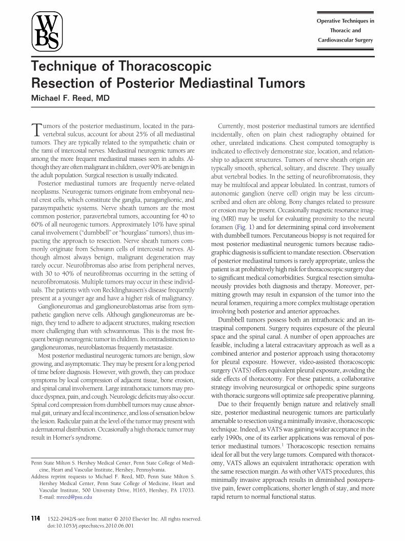
TRM
Tttatt
nrpc6cpmtrwnup
pnmqg
gosadSmtar
P
A
1
echnique of Thoracoscopicesection of Posterior Mediastinal Tumorsichael F. Reed, MD
ioistamasoifwmgoptnmni
tsfcfsssw
satetiotmt
umors of the posterior mediastinum, located in the para-vertebral sulcus, account for about 25% of all mediastinal
umors. They are typically related to the sympathetic chain orhe rami of intercostal nerves. Mediastinal neurogenic tumors aremong the more frequent mediastinal masses seen in adults. Al-houghtheyareoftenmalignant inchildren,over90%arebenign inhe adult population. Surgical resection is usually indicated.
Posterior mediastinal tumors are frequently nerve-relatedeoplasms. Neurogenic tumors originate from embryonal neu-al crest cells, which constitute the ganglia, paraganglionic, andarasympathetic systems. Nerve sheath tumors are the mostommon posterior, paravertebral tumors, accounting for 40 to0% of all neurogenic tumors. Approximately 10% have spinalanal involvement (“dumbbell” or “hourglass” tumors), thus im-acting the approach to resection. Nerve sheath tumors com-only originate from Schwann cells of intercostal nerves. Al-
hough almost always benign, malignant degeneration mayarely occur. Neurofibromas also arise from peripheral nerves,ith 30 to 40% of neurofibromas occurring in the setting ofeurofibromatosis. Multiple tumors may occur in these individ-als. The patients with von Recklinghausen’s disease frequentlyresent at a younger age and have a higher risk of malignancy.Ganglioneuromas and ganglioneuroblastomas arise from sym-
athetic ganglion nerve cells. Although ganglioneuromas are be-ign, they tend to adhere to adjacent structures, making resectionore challenging than with schwannomas. This is the most fre-
uent benign neurogenic tumor in children. In contradistinction toanglioneuromas, neuroblastomas frequently metastasize.
Most posterior mediastinal neurogenic tumors are benign, slowrowing, and asymptomatic. They may be present for a long periodf time before diagnosis. However, with growth, they can produceymptoms by local compression of adjacent tissue, bone erosion,nd spinal canal involvement. Large intrathoracic tumors may pro-ucedyspnea,pain, andcough.Neurologicdeficitsmayalsooccur.pinal cord compression from dumbbell tumors may cause abnor-algait,urinaryandfecal incontinence,and lossof sensationbelow
he lesion. Radicular pain at the level of the tumor may present withdermatomaldistribution.Occasionally ahigh thoracic tumormayesult in Horner’s syndrome.
enn State Milton S. Hershey Medical Center, Penn State College of Medi-cine, Heart and Vascular Institute, Hershey, Pennsylvania.
ddress reprint requests to Michael F. Reed, MD, Penn State Milton S.Hershey Medical Center, Penn State College of Medicine, Heart andVascular Institute, 500 University Drive, H165, Hershey, PA 17033.
rE-mail: [email protected]
14 1522-2942/$-see front matter © 2010 Elsevier Inc. All rights reserved.doi:10.1053/j.optechstcvs.2010.06.001
Currently, most posterior mediastinal tumors are identifiedncidentally, often on plain chest radiography obtained forther, unrelated indications. Chest computed tomography isndicated to effectively demonstrate size, location, and relation-hip to adjacent structures. Tumors of nerve sheath origin areypically smooth, spherical, solitary, and discrete. They usuallybut vertebral bodies. In the setting of neurofibromatosis, theyay be multifocal and appear lobulated. In contrast, tumors of
utonomic ganglion (nerve cell) origin may be less circum-cribed and often are oblong. Bony changes related to pressurer erosion may be present. Occasionally magnetic resonance imag-
ng (MRI) may be useful for evaluating proximity to the neuraloramen (Fig. 1) and for determining spinal cord involvementith dumbbell tumors. Percutaneous biopsy is not required forost posterior mediastinal neurogenic tumors because radio-
raphic diagnosis is sufficient to mandate resection. Observationf posterior mediastinal tumors is rarely appropriate, unless theatient is at prohibitively high risk for thoracoscopic surgery dueo significant medical comorbidities. Surgical resection simulta-eously provides both diagnosis and therapy. Moreover, per-itting growth may result in expansion of the tumor into theeural foramen, requiring a more complex multistage operation
nvolving both posterior and anterior approaches.Dumbbell tumors possess both an intrathoracic and an in-
raspinal component. Surgery requires exposure of the pleuralpace and the spinal canal. A number of open approaches areeasible, including a lateral extracavitary approach as well as aombined anterior and posterior approach using thoracotomyor pleural exposure. However, video-assisted thoracoscopicurgery (VATS) offers equivalent pleural exposure, avoiding theide effects of thoracotomy. For these patients, a collaborativetrategy involving neurosurgical or orthopedic spine surgeonsith thoracic surgeons will optimize safe preoperative planning.Due to their frequently benign nature and relatively small
ize, posterior mediastinal neurogenic tumors are particularlymenable to resection using a minimally invasive, thoracoscopicechnique. Indeed, as VATS was gaining wider acceptance in thearly 1990s, one of its earlier applications was removal of pos-erior mediastinal tumors.1 Thoracoscopic resection remainsdeal for all but the very large tumors. Compared with thoracot-my, VATS allows an equivalent intrathoracic operation withhe same resection margin. As with other VATS procedures, thisinimally invasive approach results in diminished postopera-
ive pain, fewer complications, shorter length of stay, and more
apid return to normal functional status.
Thoracoscopic resection of posterior mediastinal tumors 115
Operative Technique
Figure 1 MRI demonstrates that this left posterior mediastinal neurogenic tumor abuts the vertebral body, but does notinvade the intervertebral foramen. (A) Axial view; (B) coronal view; (C) sagittal view. Pathologic analysis demonstrateda schwannoma.
116 M.F. Reed
Figure 2 After the induction of general anesthesia, a double lumen endotracheal tube is inserted and its position isverified bronchoscopically. Alternatively, a bronchial blocker may be utilized to achieve single-lung ventilation. Thepatient is then placed in the lateral decubitus position. Proper positioning of the patient is critical to preventing certaincomplications.2 The operating table flexion point is located between the iliac crest and the costal margin. The arms areextended, but shoulder hyperextension greater than 90° must be avoided to prevent brachial plexus neuropathy. Theupper arm is supported with pillows or a padded armrest, ensuring that both ulnar nerves are protected from pressuredamage. The operating table is flexed to widen the intercostal spaces on the operative side, a simple maneuver that maydiminish postoperative pain. Either soft rolls or a beanbag is used to secure the patient on the table. The patient is
draped as for standard posterolateral thoracotomy.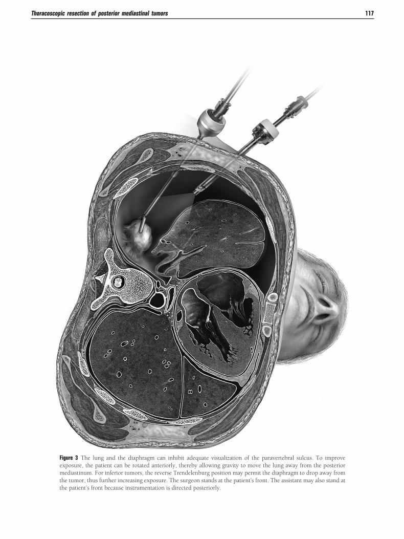
Thoracoscopic resection of posterior mediastinal tumors 117
Figure 3 The lung and the diaphragm can inhibit adequate visualization of the paravertebral sulcus. To improveexposure, the patient can be rotated anteriorly, thereby allowing gravity to move the lung away from the posteriormediastinum. For inferior tumors, the reverse Trendelenburg position may permit the diaphragm to drop away fromthe tumor, thus further increasing exposure. The surgeon stands at the patient’s front. The assistant may also stand at
the patient’s front because instrumentation is directed posteriorly.
118 M.F. Reed
Figure 4 VATS allows operative exposure using small incisions. Even when a limited utility incision is required, no ribspreading occurs. As with thoracotomy, morbidity is related to intercostal nerve trauma. During VATS, intercostal nerveinjury can be minimized by using the smallest port that is feasible, depending on the necessary instrumentation. An angled,usually 30°, thoracoscope can be used to diminish pressure on the nerve. Port insertion should be directed toward theintrathoracic region where the majority of the surgery will occur to avoid using the inferior edge of a rib as a fulcrum for thecamera when attempting visualization at awkward angles. Typically 3 port sites are chosen. Long-acting local anesthetic, suchas bupivacaine, can be injected into the first planned port site, anesthetizing the intercostal nerve. Before subsequent portinsertion, each site can be injected with local anesthetic using thoracoscopic visualization to verify medication delivery in theextrapleural plane. The first port is typically placed at the anterior axillary line. For higher tumors, this port is located at the fourthorfifth intercostal space.For lower tumors, thefirstport canbeplaced in thesixthor seventh intercostal space.Twoadditionalportsare inserted, placed at a sufficient distance from each other, triangulating the ports, to facilitate best instrument conversion on thetarget without interfering with each other (“swordfighting”). The ports can be used interchangeably as needed. If the lung ordiaphragm interferes with exposure, a fourth port can be placed for a retractor. Additionally, low pressure insufflation can be usedtomorerapidlyachievepulmonaryatelectasisandtopushthediaphragminferiorly.Wheninsufflating,attentionshouldbedirected
toward hemodynamics, although instability is rare with low insufflation pressure.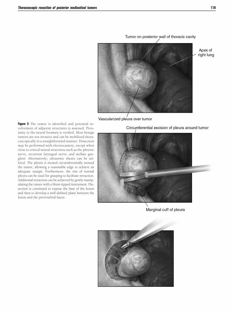
FvitcmcngltapAusal
Thoracoscopic resection of posterior mediastinal tumors 119
igure 5 The tumor is identified and potential in-olvement of adjacent structures is assessed. Prox-mity to the neural foramen is verified. Most benignumors are not invasive and can be mobilized thora-oscopically in a straightforward manner. Dissectionay be performed with electrocautery, except when
lose to critical neural structures such as the phrenicerve, recurrent laryngeal nerve, and stellate gan-lion. Alternatively, ultrasonic shears can be uti-ized. The pleura is incised circumferentially aroundhe tumor, allowing a reasonable edge to achieve andequate margin. Furthermore, the rim of normalleura can be used for grasping to facilitate retraction.dditional retraction can be achieved by gently manip-lating the tumor with a blunt-tipped instrument. Dis-ection is continued to expose the base of the lesionnd then to develop a well-defined plane between theesion and the prevertebral fascia.
120 M.F. Reed
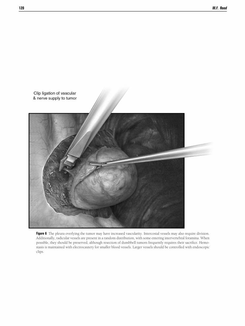
Figure 6 The pleura overlying the tumor may have increased vascularity. Intercostal vessels may also require division.Additionally, radicular vessels are present in a random distribution, with some entering intervertebral foramina. Whenpossible, they should be preserved, although resection of dumbbell tumors frequently requires their sacrifice. Hemo-stasis is maintained with electrocautery for smaller blood vessels. Larger vessels should be controlled with endoscopic
clips.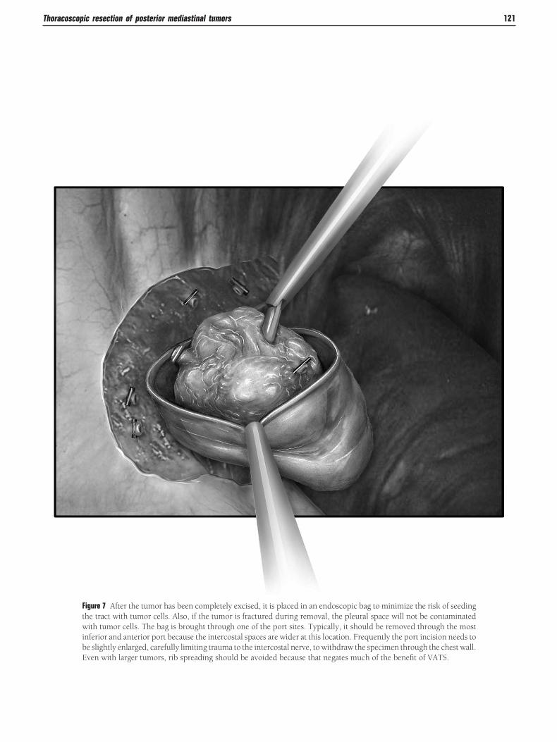
Thoracoscopic resection of posterior mediastinal tumors 121
Figure 7 After the tumor has been completely excised, it is placed in an endoscopic bag to minimize the risk of seedingthe tract with tumor cells. Also, if the tumor is fractured during removal, the pleural space will not be contaminatedwith tumor cells. The bag is brought through one of the port sites. Typically, it should be removed through the mostinferior and anterior port because the intercostal spaces are wider at this location. Frequently the port incision needs tobe slightly enlarged, carefully limiting trauma to the intercostal nerve, to withdraw the specimen through the chest wall.
Even with larger tumors, rib spreading should be avoided because that negates much of the benefit of VATS.
122 M.F. Reed
Figure 8 Multimodal analgesic strategies should be employed for minimally invasive thoracoscopic surgery, as forthoracotomy. With shorter lengths of stay and less acute pain, thoracic epidural anesthesia is less frequently requiredfor VATS procedures. However, it should be considered in patients at increased risk of severe postoperative pain,pulmonary dysfunction, or postthoracotomy pain syndrome. For most patients undergoing VATS, other regionalanalgesic strategies can be applied. The intercostal nerve at port sites can be locally anesthetized and multilevel injectionmay achieve intercostal nerve blockade. Long-acting local anesthetic such as 0.25% bupivacaine can be injected usingthoracoscopic visualization. The needle is placed percutaneously and is carefully advanced into the intercostal space,avoiding injury to the vessels. With the needle tip in the extrapleural plane, approximately 2 mL can be injected,thereby raising a pleural bubble. By placing intercostal blocks from at least 1 level above the highest port site, to at least1 level below the most inferior port site, effective analgesia can be achieved during the early postoperative period.Additional analgesia can be achieved using continuous catheter infusion of local anesthetic into the paravertebralextrapleural space, providing multilevel intercostal nerve blockade. Patients can be safely discharged with the contin-
uous infusion apparatus in place.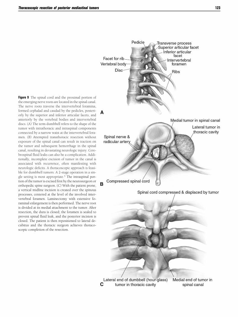
FtTfoadtcmetcbtanbgtoapvrirpccs
Thoracoscopic resection of posterior mediastinal tumors 123
igure 9 The spinal cord and the proximal portion ofhe emerging nerve roots are located in the spinal canal.he nerve roots traverse the intervertebral foramina,
ormed cephalad and caudad by the pedicles, posteri-rly by the superior and inferior articular facets, andnteriorly by the vertebral bodies and intervertebraliscs. (A) The term dumbbell refers to the shape of theumor with intrathoracic and intraspinal componentsonnected by a narrow waist at the intervertebral fora-en. (B) Attempted transthoracic resection without
xposure of the spinal canal can result in traction onhe tumor and subsequent hemorrhage in the spinalanal, resulting in devastating neurologic injury. Cere-rospinal fluid leaks can also be a complication. Addi-ionally, incomplete excision of tumor in the canal isssociated with recurrence, often manifesting witheurologic deficits. A thoracoscopic approach is feasi-le for dumbbell tumors. A 2-stage operation in a sin-le setting is most appropriate.3 The intraspinal por-ion of the tumor is excised first by the neurosurgeon orrthopedic spine surgeon. (C) With the patient prone,vertical midline incision is created over the spinousrocesses, centered at the level of the involved inter-ertebral foramen. Laminectomy with extensive fo-aminal enlargement is then performed. The nerve roots divided at its medial attachment to the tumor. Afteresection, the dura is closed; the foramen is sealed torevent spinal fluid leak, and the posterior incision islosed. The patient is then repositioned to lateral de-ubitus and the thoracic surgeon achieves thoraco-copic completion of the resection.
CStrsrpsDrpncai
tiisq
acaiMpri
rhr
rttmttifrpmfqpr
R1
2
3
4
124 M.F. Reed
onclusionsurgical resection is the ideal treatment for posterior medias-inal neurogenic tumors. Benign neurogenic tumors rarelyecur and no adjuvant therapy is required. Minimally inva-ive resection with thoracoscopic techniques is safe. Standardisks of thoracic surgery, including bleeding, infection, andulmonary complications, are rare. The risks specific to re-ection of neurogenic tumors are related to nerve injury.eficits such as Horner’s syndrome, partial sympathectomy,
ecurrent laryngeal nerve injury, and phrenic nerve injury areossible when the tumors originate from these specificerves. Many of these complications can be avoided by pre-ise identification of the specific neurologic structures andvoidance of electrocautery when dissecting in close proxim-ty to them.
For benign posterior mediastinal lesions, complete resec-ion is typically achieved with a thoracoscopic strategy. Min-mally invasive resection is not usually indicated when annvasive malignant tumor is identified preoperatively. In thisetting, resection of adjacent bony structures is often re-uired, typically negating the benefits of VATS.Dumbbell tumors are amenable to VATS resection. When
n intraspinal component is clearly compressing the cord, aombined posterior and anterior approach is indicated tochieve negative margins. Even when slight intraforaminalnvolvement is present, based on computed tomography and
RI, a combined thoracic surgery and spinal surgery ap-roach is safest. Resection of the posterior mediastinal neu-ogenic tumor with intraforaminal involvement, while rely-
ng only on VATS exposure, or even thoracotomy alone, mayequire excessive retraction of the mass. Potential undetectedemorrhage in the spinal canal can result in devastating neu-ologic sequelae.
Thoracoscopic resection of posterior mediastinal tumorsesults in shorter duration of the operation, compared withhe open approach. This is primarily due to avoidance of theime required to open and close a thoracotomy. As with nu-erous other thoracic procedures, the adoption of VATS
echniques for resection of posterior mediastinal neurogenicumors is associated with diminished postoperative morbid-ty, including shorter length of stay, less pain, better shoulderunction, fewer pulmonary complications, and more rapideturn to normal functional status.4 It is the preferred ap-roach for the resection of benign posterior mediastinal tu-ors, including dumbbell tumors. Thoracotomy is reserved
or very large tumors, in which rib spreading would be re-uired for removal through the chest wall, as well as foratients with significant adhesions from prior ipsilateral tho-acic surgery or pleural intervention.
eferences. Landreneau RJ, Dowling RD, Ferson PF: Thoracoscopic resection of a
posterior mediastinal neurogenic tumor. Chest 102:1288-1290, 1992. Reed MF: Thoracic incisions, in: Complications in Cardiothoracic Sur-
gery. Avoidance and Treatment (ed 2). West Sussex, UK, Wiley-Black-well, 2010, pp 22-53
. Vallieres E, Findlay JM, Fraser RE: Combined microsurgical and thora-coscopic removal of neurogenic dumbbell tumors. Ann Thorac Surg59:469-472, 1995
. McKenna RJ Jr, Houck W, Fuller CB: Video-assisted thoracic surgerylobectomy: experience with 1,100 cases. Ann Thorac Surg 81:421-426,
2006Copyright © 2022 FDOKUMEN


![[Posterior cortical atrophy]](https://static.fdokumen.com/doc/165x107/6331b9d14e01430403005392/posterior-cortical-atrophy.jpg)