Trypanosoma evansi: A comparative study of four diagnostic techniques for trypanosomosis using...
6
Trypanosoma evansi: A comparative study of four diagnostic techniques for trypanosomosis using rabbit as an experimental model J.R. Ramírez-Iglesias a , M.C. Eleizalde a , E. Gómez-Piñeres a,b , M. Mendoza a,⇑ a Universidad Nacional Experimental Simón Rodríguez, Instituto de Estudios Científicos y Tecnológicos (IDECYT), Centro de Estudios Biomédicos y Veterinarios (CEBIV), Apartado Postal 479245, Caracas 1041A, Venezuela b Universidad de Oriente, Núcleo Monagas, Escuela de Zootecnia, Departamento de Biología y Sanidad Animal, Maturín, Venezuela article info Article history: Received 14 August 2010 Received in revised form 2 February 2011 Accepted 8 February 2011 Available online 12 February 2011 Keywords: Trypanosoma evansi Diagnosis of trypanosomosis PCR Indirect ELISA Parasitological techniques Rabbit as an experimental model abstract The goal of this study was to compare two parasitological diagnostic techniques, such as by Micro- Haematocrit Centrifugation Technique (MHCT) and Direct Microscopic Examination (DME) with a serological method (iELISA), and a molecular procedure PCR, in rabbits experimentally infected with Trypanosoma evansi, in order to determine their sensitivity throughout the course of disease. The parasitological methods were not able of detecting the presence of the parasite during the phases of low parasitemia, the prepatency period and the chronic phase. In contrast, PCR detected T. evansi in the prepatency and chronic phase, when increase the amount of DNA from 100 to 300 ng. 100% detection was observed with iELISA only in the chronic stage of the disease. In the acute phase, all samples were positively diagnosed using either MHCT or PCR, whereas only few samples were diagnosed by DME. Samples obtained from day 15 post infection were also detected by iELISA. The highest diagnostic register during the course of infection was achieved by the PCR technique (93.8%), followed by iELISA (71.1%), MHCT (59%) and DME (13.6%). Therefore, we recommend the use of PCR in epidemiological studies in order to implement sanitary control plans for the improvement of livestock productivity in the country. Ó 2011 Elsevier Inc. All rights reserved. 1. Introduction Trypanosomosis is a disease caused by various protozoan of the Trypanosomatidae family, which can affect either humans or ani- mals. In particular, animal trypanosomosis are of great concern in inter-tropical areas of Asia, Africa and Latin America, where most of the pathogenic Trypanosoma species are present in their hosts, or vectors. In South America, Trypanosoma evansi causes equine trypanomososis, a disease known as ‘‘Derrengadera’’ in Venezuela and ‘‘Mal de Caderas’’ in Argentina, Brazil and Paraguay. This parasitic disease produces a decrease in the productivity of horses used in livestock, which are indispensable for traditional extensive cattle ranching. Despite the great economical losses that this disease generates in affected areas, almost no epidemiological data is available. In Argentina, for example, antibody detection has shown that 20% of the animals are positive in the Formosa region where approximately 57,000 horses are exposed (Monzón et al., 1995). In the Pantanal region of Brazil, about 13% of the horses are lost annually and no appropriate control is used (Seidl et al., 2001). In the Venezuela savannah, it has been reported a seroprev- alence of 80% and 50% for T. evansi in horses (Reyna-Bello et al., 1998) and capybaras (Arias et al., 1997), respectively. There are studies on trypanosomosis caused by T. evansi in rab- bits as experimental models, where characteristic symptoms are reported: elevated levels of parasitemia, descent in haematocrit, weight loss, cellular infiltrations and the development of a marked immune humoral response (Uche and Jones, 1992; Uche et al., 1992). Also, biochemical alterations are described like hyperpro- teinemia, hyperglobulinemia, increment in the levels of urea and decrease of the levels of serum albumin (Da Silva et al., 2007). The implementation of the rabbit, an animal easy to manipulate in the laboratory, as a chronic experimental model for the study of trypanosomosis will allow the development of research in diag- nosis, prevention and possible treatments with direct application in the field. This disease starts with a prepatency period (PP) followed by acute (AP) and chronic (CP) phases. There are diverse diagnostic methods to detect animal trypanosomosis: parasitological, immu- nological and molecular, which show advantages and disadvan- tages according to the stage of the disease in which they are applied. The parasitological methods are based on the direct obser- vation of the parasite under the light microscope. These techniques are simple and of low cost, but have low sensitivity; therefore they are only effective in the AP of the disease when high levels of par- asitemia are present. The immunological methods as the ELISA (en- zyme-linked immunosorbent assay), are based on the detection of antigens (Ag-ELISA) or antibodies (indirect-ELISA) related to the 0014-4894/$ - see front matter Ó 2011 Elsevier Inc. All rights reserved. doi:10.1016/j.exppara.2011.02.010 ⇑ Corresponding author. Fax: +58 2126719289. E-mail address: [email protected] (M. Mendoza). Experimental Parasitology 128 (2011) 91–96 Contents lists available at ScienceDirect Experimental Parasitology journal homepage: www.elsevier.com/locate/yexpr
Transcript of Trypanosoma evansi: A comparative study of four diagnostic techniques for trypanosomosis using...

Experimental Parasitology 128 (2011) 91–96
Contents lists available at ScienceDirect
Experimental Parasitology
journal homepage: www.elsevier .com/locate /yexpr
Trypanosoma evansi: A comparative study of four diagnostic techniquesfor trypanosomosis using rabbit as an experimental model
J.R. Ramírez-Iglesias a, M.C. Eleizalde a, E. Gómez-Piñeres a,b, M. Mendoza a,⇑a Universidad Nacional Experimental Simón Rodríguez, Instituto de Estudios Científicos y Tecnológicos (IDECYT), Centro de Estudios Biomédicos y Veterinarios (CEBIV),Apartado Postal 479245, Caracas 1041A, Venezuelab Universidad de Oriente, Núcleo Monagas, Escuela de Zootecnia, Departamento de Biología y Sanidad Animal, Maturín, Venezuela
a r t i c l e i n f o
Article history:Received 14 August 2010Received in revised form 2 February 2011Accepted 8 February 2011Available online 12 February 2011
Keywords:Trypanosoma evansiDiagnosis of trypanosomosisPCRIndirect ELISAParasitological techniquesRabbit as an experimental model
0014-4894/$ - see front matter � 2011 Elsevier Inc. Adoi:10.1016/j.exppara.2011.02.010
⇑ Corresponding author. Fax: +58 2126719289.E-mail address: [email protected] (M. Mendo
a b s t r a c t
The goal of this study was to compare two parasitological diagnostic techniques, such as by Micro-Haematocrit Centrifugation Technique (MHCT) and Direct Microscopic Examination (DME) with a serologicalmethod (iELISA), and a molecular procedure PCR, in rabbits experimentally infected with Trypanosomaevansi, in order to determine their sensitivity throughout the course of disease. The parasitologicalmethods were not able of detecting the presence of the parasite during the phases of low parasitemia,the prepatency period and the chronic phase. In contrast, PCR detected T. evansi in the prepatency andchronic phase, when increase the amount of DNA from 100 to 300 ng. 100% detection was observed withiELISA only in the chronic stage of the disease. In the acute phase, all samples were positively diagnosedusing either MHCT or PCR, whereas only few samples were diagnosed by DME. Samples obtained fromday 15 post infection were also detected by iELISA. The highest diagnostic register during the course ofinfection was achieved by the PCR technique (93.8%), followed by iELISA (71.1%), MHCT (59%) andDME (13.6%). Therefore, we recommend the use of PCR in epidemiological studies in order to implementsanitary control plans for the improvement of livestock productivity in the country.
� 2011 Elsevier Inc. All rights reserved.
1. Introduction
Trypanosomosis is a disease caused by various protozoan of theTrypanosomatidae family, which can affect either humans or ani-mals. In particular, animal trypanosomosis are of great concernin inter-tropical areas of Asia, Africa and Latin America, wheremost of the pathogenic Trypanosoma species are present in theirhosts, or vectors. In South America, Trypanosoma evansi causesequine trypanomososis, a disease known as ‘‘Derrengadera’’ inVenezuela and ‘‘Mal de Caderas’’ in Argentina, Brazil and Paraguay.This parasitic disease produces a decrease in the productivity ofhorses used in livestock, which are indispensable for traditionalextensive cattle ranching. Despite the great economical losses thatthis disease generates in affected areas, almost no epidemiologicaldata is available. In Argentina, for example, antibody detection hasshown that 20% of the animals are positive in the Formosa regionwhere approximately 57,000 horses are exposed (Monzón et al.,1995). In the Pantanal region of Brazil, about 13% of the horsesare lost annually and no appropriate control is used (Seidl et al.,2001). In the Venezuela savannah, it has been reported a seroprev-alence of 80% and 50% for T. evansi in horses (Reyna-Bello et al.,1998) and capybaras (Arias et al., 1997), respectively.
ll rights reserved.
za).
There are studies on trypanosomosis caused by T. evansi in rab-bits as experimental models, where characteristic symptoms arereported: elevated levels of parasitemia, descent in haematocrit,weight loss, cellular infiltrations and the development of a markedimmune humoral response (Uche and Jones, 1992; Uche et al.,1992). Also, biochemical alterations are described like hyperpro-teinemia, hyperglobulinemia, increment in the levels of urea anddecrease of the levels of serum albumin (Da Silva et al., 2007).The implementation of the rabbit, an animal easy to manipulatein the laboratory, as a chronic experimental model for the studyof trypanosomosis will allow the development of research in diag-nosis, prevention and possible treatments with direct applicationin the field.
This disease starts with a prepatency period (PP) followed byacute (AP) and chronic (CP) phases. There are diverse diagnosticmethods to detect animal trypanosomosis: parasitological, immu-nological and molecular, which show advantages and disadvan-tages according to the stage of the disease in which they areapplied. The parasitological methods are based on the direct obser-vation of the parasite under the light microscope. These techniquesare simple and of low cost, but have low sensitivity; therefore theyare only effective in the AP of the disease when high levels of par-asitemia are present. The immunological methods as the ELISA (en-zyme-linked immunosorbent assay), are based on the detection ofantigens (Ag-ELISA) or antibodies (indirect-ELISA) related to the

92 J.R. Ramírez-Iglesias et al. / Experimental Parasitology 128 (2011) 91–96
causative agent of the disease. Several attempts to develop Ag-ELISAhave not yet reached a satisfactory level to be recommended forroutine diagnosis (Desquesnes, 1996; Monzón, 2006). This test haslow sensitivity because it relies on a single or restricted numberof monoclonal antibodies, and hence, of circulating target antigens(Desquesnes, 1996, 2004). Additionally, in the first days of infec-tion or when the parasitemia is cryptic, the circulating antigensare below the detection level of the test (Monzón et al., 2010).Moreover, the Antibody-ELISA or indirect-ELISA (iELISA) has poorspecificity because of the strong cross-reactivity between patho-genic Trypanosoma spp. (Desquesnes, 2004). Several studies re-ported a higher sensitivity of iELISA compared with AgELISA forthe diagnosis of T. evansi in horses (Monzón et al., 2010), buffaloes(Davison et al., 1999) and camels (Olaho-Mukani et al., 1993), rec-ommending its use for clinical or epidemiological purposes. Themolecular methods are based on the detection of DNA or RNA fromthe parasite by the amplification of the infectious agent geneticmaterial. These assays have a high sensitivity and specificity, beingable to detect down to 1–10 trypanosomes/ml of blood by conven-tional polymerase chain reaction (PCR) (Dávila et al., 2003) and dis-tinguish different species involved.
In this study we compared the parasitological diagnostic meth-ods, such as Micro-Haematocrit Centrifugation Technique (MHCT)and Microscopic Direct Examination (DME), with the serologicaltechnique iELISA and the molecular technique PCR (polymerasechain reaction) for the detection of T. evansi throughout the courseof infection in rabbits.
2. Materials and methods
2.1. Biological material
2.1.1. ParasitesVenezuelan isolate of Trypanosoma evansi TEVA1 (Desquesnes,
2004), originated from naturally infected horse blood found inthe Venezuelan savannah in the state of Apure, cryopreserved in li-quid nitrogen (Phosphate Buffer Saline containing Glucose 1%(PBS-G), Dimethyl sulphoxide 10%) and expanded in rats.
2.1.2. Laboratory animalsFour female Spragüe-Dawley rats with an approximate weight
of 250 g were intraperitoneally inoculated with 200 ll of cryopre-served blood (108 parasites/ml). The viability of the inoculum wasverified-through the direct observation in the light microscope toensure infection.
Five female New Zealand rabbits, approximate weight of 2 kg:two in the control group and three in the infection group, whichwere inoculated intraperitoneally considering that the parasite-mias are often low in field and the amount of blood contained ina horsefly proboscis varies from nl–500 ll (Desquesnes, 2004)depending on the vector species, we choose 350 parasites/animalto simulated a natural infection, which normally varies from 1 toseveral thousand parasites. The inoculum was obtained from theblood of the rats infected experimentally in peak of parasitemia(108 parasites/ml).
2.2. Sampling
All the animals were sampled at irregular intervals during95 days post infection (PI), during the first hour of the day. About2.5 ml of peripheral blood were extracted from the rabbit’s earmarginal vein. Each individual of infection group (R1, R2 and R3)was evaluated 4 times in the PP, 13 times in the AP and 5 timesin the CP, obtaining for each stage of the disease 12, 39 and 15 sam-ples respectively, which gives a total number of 66 evaluations
during the infection. In the control group, each rabbit was sam-pling 22 times.
2.3. Parasitological analysis
2.3.1. Micro-Haematocrite Centrifugation Technique (MHCT)About 75 ll of fresh blood were taken with a heparinized capil-
lary. The tubes were examined using a light microscope (10� eye-piece and 10� objective) at 100� magnification, to detect motiletrypanosomes near the buffy coat (Woo, 1970).
2.3.2. Microscopic Direct Examination (MDE)Wet smears of 5 ll of fresh blood were examined by direct
observation of 100 fields (cover sliders 22 � 22 mm) using a phasecontrast microscope (10� eyepiece and 40� objective) at 400�magnification to detect the presence of trypanosomes (Brener,1962).
2.4. Immunological diagnosis by iELISA
The sera were obtained from 1 ml of blood by centrifugation(2000g, 10 min). The clarified antigenic extract was prepared fromtrypanosomes according to Uzcanga et al. (2002). Polyvinyl 96 wellplates were sensitised overnight in a humid chamber at 4 �C with100 ll/well of the clarified antigenic extract (20 lg/ml) in 50 mMcarbonate–bicarbonate buffer (pH 9.6). A blocking solution consist-ing of non-fat milk (5%) dissolved in 0.02 M PBS (pH 7.2) was thenadded. Afterwards, 100 ll/well of the rabbit serum, diluted 1/400in PBS-T (0.1% Tween 20 in PBS) was added, followed by 100 ll/well of the secondary antibody anti-IgG (H+L) goat anti-rabbit con-jugated to peroxidase, diluted 1/200,000 in PBS-T. Between eachstep, 3 washes were performed with 200 ll/well of 150 mM NaCl,0.1% Tween 20. Incubations were carried out for 1 h in a humidchamber at 37 �C. Finally, 100 ll/well of the chromogen 2% ABTS(2,20-azino-bis(3-ethylbenzthiazoline-6-sulphonic acid)) dilutedin 0.05 M phosphate-citrate buffer (pH 5.0), 0.5% H2O2 was addedand agitated for 30 min. The optical density at 405 nm (OD405)was determined with an ELISA microplate reader (BioRad� Model3550). For the serological diagnosis the cutoff value was deter-mined as the mean plus 3 times the standard deviation (X+3 SD)of the OD405 lectures of 30 negative reference sera taken fromhealthy and pre-infection rabbits that were parasitologically diag-nosed as negative by MHCT.
2.5. Molecular diagnosis by PCR
Genomic DNA was purified from 300 ll of the buffy coat ob-tained by centrifugation (1 min, 15,000 rpm in an Eppendorfmicrocentrifuge) of the whole blood (1 ml), using the commercialkit Wizard� Promega. The DNA concentration (ng/ll) was deter-mined from 1 ll of sample, using a Nano Drop™ 1000 Spectropho-tometer at 260 nm. PCR was performed using the specific primersESAG 6/7 (fwd 50 ACATTCCAGCAGGAGTTGGAG 30; rev 50 CAC-GTGAATCCTCAATTTGT 30), and the cycling programme describedby Holland et al. (2001) for the detection of T. evansi. The reactionmixture for a final volume of 25 ll contained 1.5 U of Taq polymer-ase, Tris/HCl 1� buffer, 3 mM MgCl2, 200 lM dNTPs and 0.5 lM ofeach primer. According to the phase of the infection to be evalu-ated, 100 and/or 300 ng of DNA were used. The amplification prod-ucts were analysed by electrophoresis in 2% agarose gels (1 h,100 V) and were visualised with SYBR-safe™ in a UV transillumi-nator. Diagnosis was considered positive when a specific productof 237 bp was amplified by PCR.

J.R. Ramírez-Iglesias et al. / Experimental Parasitology 128 (2011) 91–96 93
2.6. Comparison of MHCT, DME, iELISA and PCR for detection of T.evansi throughout the course of infection in a chronic model
The samples were evaluated by the different diagnostic meth-ods employed throughout the course of infection. The sensitivityfor the different diagnostic methods used in this study was deter-mined and compared using the percentage of detection (D), de-fined as the probability of a test to correctly diagnose as positivea diseased individual. It is calculated using the equation:D = NPD/TNS � 100, expressed as %, where NPD is the number ofpositively diagnosed samples with the method and TNS is the totalnumber of samples analysed with the method.
3. Results and discussion
3.1. Evaluation of parasitological (MHCT, DME) immunological(iELISA) and molecular (PCR) diagnostic methods for the detection of T.evansi during the course of infection
Tables 1–3 show the results obtained for the different diagnos-tic methods that were used throughout the course of three rabbits.By the parasitological techniques (DME and MHCT) the presence ofthe parasite in the PP (days 3, 5, 7 and 9) was not detected. Later inthe AP, the MHCT detected the presence of the parasite in all sam-ples tested, while the DME detects parasites one day later andintermittently (days 11, 26 and 43 for R1 and R2 and 11, 26 and59 for R3). In the CP, none of the parasitological methods was capa-ble for detecting the parasite.
The sera obtained for the three rabbits in the course of infectionwere evaluated by iELISA (Tables 1–3). In the PP, between days 3and 10 PI, the OD readings remained low, below the cutoff value(0.42), similar to the negative reference sera (0.32), so these sam-ples were diagnosed negative. In the AP only from day 15 PI, thesamples gave OD405 readings above the cutoff value for the sero-conversion, and the diagnosis was positive for the rest of this phaseand as well as in CP.
The evaluation by PCR is shown in Tables 1–3 for the tree rabbitevaluated, using 100 ng and 300 ng of DNA. The evaluation for PCRin the PP was carried out with DNA from the blood samples taken ondays 3, 5, 7 and 9 PI for the three infected individuals. Note, a positive
Table 1Comparison of the diagnostic methods for rabbit 1 (R1) throughout the course of infectionNumber of evaluated samples.
Phase of infection Days post-infection N = 22 Parasitological methods
DME MHCT
Prepatency period 3 � �5 � �7 � �9 � �
Acute phase 10 � +11 + +15 � +17 � +19 � +26 + +31 � +38 � +43 + +48 � +55 � +59 � +61 � +
Chronic phase 77 � �80 � �85 � �89 � �93 � �
D = % of detection 13.6 59
diagnosis was obtained on day 9 PI for individuals 1 and 2, whilea negative diagnosis was obtained for individual 3 using 100 ng ofDNA. However, when 300 ng of DNA were used in the assay, a posi-tive diagnosis was obtained from day 5 PI in individuals 1 and 2 andfrom day 7 PI in individual 3. On day 3 PI, no amplification productwas observed even when the amount of DNA was increased. Duringthe CP of the disease between days 77 and 95 PI for the three in-fected rabbits, a negative diagnosis was obtained when 100 ng ofDNA were used in the reaction. Hence in the CP as in the PP, theuse of 300 ng of DNA was required to assure the detection of the par-asite by PCR in all of the evaluated samples. Additionally, no ampli-fication product was obtained from the DNA of the healthyindividuals in any time of infection using 100 or 300 ng of DNA.
The detection of T. evansi by PCR at phase of low parasitemia,depended on the amount of DNA used, the percentage of detectionincrease as the amount of DNA was raised from 100 to 300 ng.These results were consistent with those reported previously(Fernández et al., 2009; González et al., 2006), and confirmed thatduring the low parasitemia periods, the ratio of DNA from theinfectious agent relative to total DNA in the blood sample, is crucialto diagnose the illness. Moreover, the problem is not the sensitivityof this method, since a sensitivity of 1 parasite/ml of blood andeven less in experimental infections has been reported (Dávilaet al., 2003; Desquesnes and Davila, 2002). Therefore, the limita-tion of using the PCR during the first days PI could be the absenceof the pathogenic agent in the evaluated sample (Desquesnes andDavila, 2002).
In the AP of the disease, between days 10 and 70 PI, all the eval-uated samples were diagnosed positively by PCR (Tables 1–3). Inthis phase there is a high parasitemia, therefore an amplificationproduct is obtained with lower amounts of total DNA. In agree-ment with other authors (Singh et al., 2004), the variation of theintensity in the amplification products correlates well with the lev-els of parasitemia during the course of infection (data not shown).
3.2. Comparison of the parasitological diagnostic techniques DME andMHCT with iELISA and PCR for the detection of T. evansi
Table 4 shows the sensitivity of the four methods evaluatedand compared them based on the percentage of detection in the
. Positively (+) and negatively (�) diagnosed by DME, MHCT, iELISA and PCR. (N = 22)
Immunological method Molecular method
iELISA PCR (100 ng) PCR (300 ng)
� � �� � +� � +� + +� + +� + ++ + ++ + ++ + ++ + ++ + ++ + ++ + ++ + ++ + ++ + ++ + ++ � ++ � ++ � ++ � ++ � +72.7 63.6 95.4
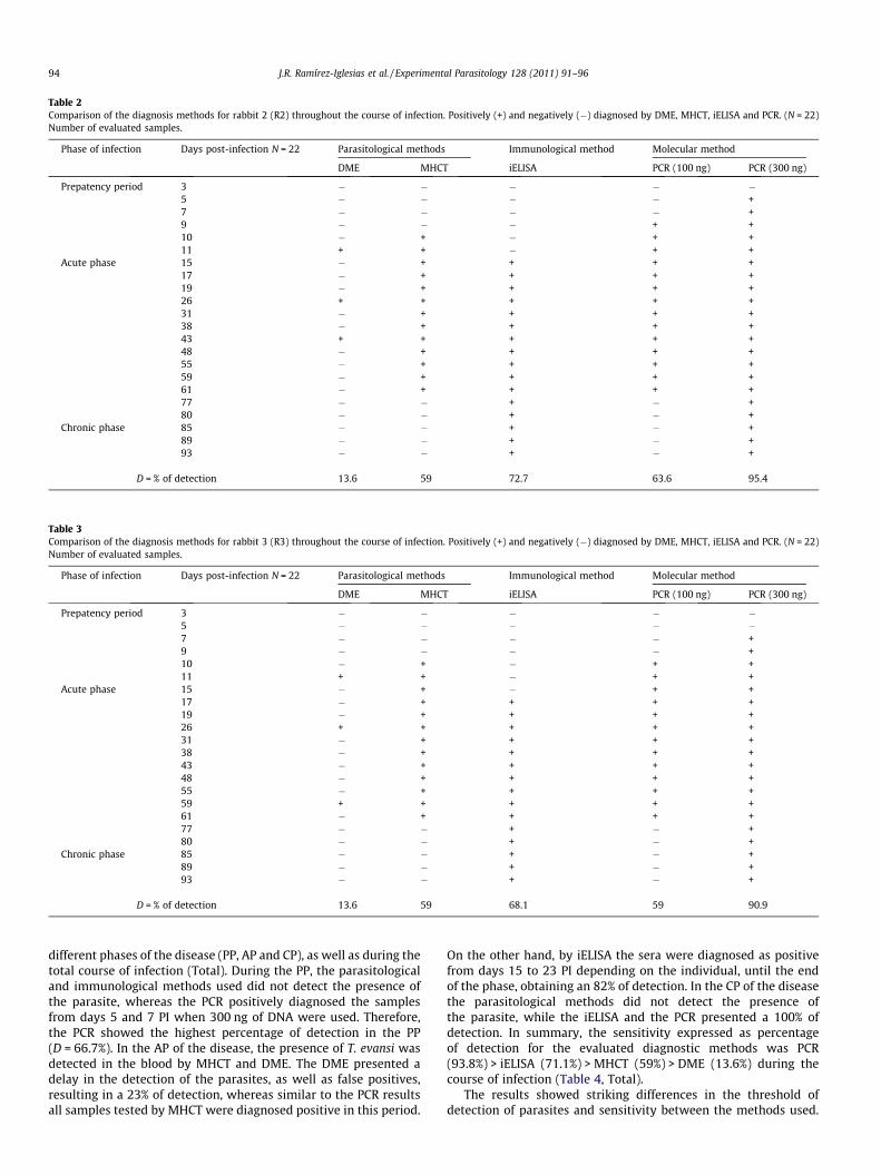
Table 2Comparison of the diagnosis methods for rabbit 2 (R2) throughout the course of infection. Positively (+) and negatively (�) diagnosed by DME, MHCT, iELISA and PCR. (N = 22)Number of evaluated samples.
Phase of infection Days post-infection N = 22 Parasitological methods Immunological method Molecular method
DME MHCT iELISA PCR (100 ng) PCR (300 ng)
Prepatency period 3 � � � � �5 � � � � +7 � � � � +9 � � � + +10 � + � + +11 + + � + +
Acute phase 15 � + + + +17 � + + + +19 � + + + +26 + + + + +31 � + + + +38 � + + + +43 + + + + +48 � + + + +55 � + + + +59 � + + + +61 � + + + +77 � � + � +80 � � + � +
Chronic phase 85 � � + � +89 � � + � +93 � � + � +
D = % of detection 13.6 59 72.7 63.6 95.4
Table 3Comparison of the diagnosis methods for rabbit 3 (R3) throughout the course of infection. Positively (+) and negatively (�) diagnosed by DME, MHCT, iELISA and PCR. (N = 22)Number of evaluated samples.
Phase of infection Days post-infection N = 22 Parasitological methods Immunological method Molecular method
DME MHCT iELISA PCR (100 ng) PCR (300 ng)
Prepatency period 3 � � � � �5 � � � � �7 � � � � +9 � � � � +10 � + � + +11 + + � + +
Acute phase 15 � + � + +17 � + + + +19 � + + + +26 + + + + +31 � + + + +38 � + + + +43 � + + + +48 � + + + +55 � + + + +59 + + + + +61 � + + + +77 � � + � +80 � � + � +
Chronic phase 85 � � + � +89 � � + � +93 � � + � +
D = % of detection 13.6 59 68.1 59 90.9
94 J.R. Ramírez-Iglesias et al. / Experimental Parasitology 128 (2011) 91–96
different phases of the disease (PP, AP and CP), as well as during thetotal course of infection (Total). During the PP, the parasitologicaland immunological methods used did not detect the presence ofthe parasite, whereas the PCR positively diagnosed the samplesfrom days 5 and 7 PI when 300 ng of DNA were used. Therefore,the PCR showed the highest percentage of detection in the PP(D = 66.7%). In the AP of the disease, the presence of T. evansi wasdetected in the blood by MHCT and DME. The DME presented adelay in the detection of the parasites, as well as false positives,resulting in a 23% of detection, whereas similar to the PCR resultsall samples tested by MHCT were diagnosed positive in this period.
On the other hand, by iELISA the sera were diagnosed as positivefrom days 15 to 23 PI depending on the individual, until the endof the phase, obtaining an 82% of detection. In the CP of the diseasethe parasitological methods did not detect the presence ofthe parasite, while the iELISA and the PCR presented a 100% ofdetection. In summary, the sensitivity expressed as percentageof detection for the evaluated diagnostic methods was PCR(93.8%) > iELISA (71.1%) > MHCT (59%) > DME (13.6%) during thecourse of infection (Table 4, Total).
The results showed striking differences in the threshold ofdetection of parasites and sensitivity between the methods used.

Table 4Comparison of the percentage of detection for the diagnostic methods throughout the course of infection. D is the % of detection, calculated as the number of samples testedpositive with the method divided by the total number of samples analysed with the method multiplied by 100, in each phase (PP, AP and CP) and in the 3 phases (total N = 66) ofthe infection.
Diagnostic method D = % of detection
Prepatency period (PP) Acute phase (AP) Chronic phase (CP) Total N = 66
DME 0 23 0 13.6MHCT 0 100 0 59iELISA 0 82 100 71.1PCR (100 ng) 16.7 100 0 62.1PCR (300 ng) 66.7 100 100 93.8
J.R. Ramírez-Iglesias et al. / Experimental Parasitology 128 (2011) 91–96 95
In the PP, the PCR could detect the presence of the parasite and thepercentage of detection increased as higher amounts of total DNAwere used. The period between the initial infection and the detec-tion of the first parasites is related to the technique being em-ployed. A shorter PP has been reported by PCR, in comparisonwith parasitological techniques (Fernández et al., 2009; Hollandet al., 2001; Ijaz et al., 1998). A higher percentage of detectionfor the PCR in the PP in mice has been described, reporting 100%detection 48 h PI for the PCR in contrast to 47% and 70% for DMEand MHCT, respectively (González et al., 2006). Similarly, 80% ofdetection was obtained in sheep 7 days PI for the PCR technique,10% for the MHCT and 5% for DME (El-Metanaway et al., 2009).Here, the presence of the parasites were also detected by PCR priorto by DME and/or MHCT. All these results confirm the superiorityof PCR for the detection of T. evansi in the PP. The diagnosis by thistechnique would allow starting early treatments of the disease be-fore the appearance of the clinical symptomatology and the pre-vention of the physical deterioration of the animal.
In the AP, previous studies carried out using experimentalinfections with T. evansi described a 100% of detection by PCRand a lower percentage of detection by parasitological methods(El-Metanaway et al., 2009; Holland et al., 2001). In this study,100% of detection by PCR and MHCT methods was determined,while the DME detected a lower percentage. During this phase,the fluctuations in the parasitemia were responsible for the falsenegatives obtained with the DME, but the parasitemia levels weremaintained above the sensitivity threshold for the MHCT, resultingin a high % of detection for both MHCT and PCR. Additionally, theMHCT concentrates the parasites in the buffy coat, which facilitatestheir detection in comparison with the DME (Fernández et al.,2009). These results confirm that the MHCT is effective for thedetection of the parasite at high parasitemias, yielding high per-centages of detection in experimental infections (Holland et al.,2001; Wernery et al., 2001). On the other hand, the 82% for theiELISA resulted in a delay in the positive diagnosis compared tothe parasitological methods (Table 4). These results agree withthose described in other studies with T. evansi (Aquino et al.,1999; Espinoza et al., 2002; Reyna-Bello et al., 1998; Werneryet al., 2001) in which the seroconversion by iELISA is reported afterthe detection of the parasites by DME and/or MHCT.
In the CP, the parasitological techniques yield a negative diag-nosis, indicating the limitations of these methods for this phase,which is characterised by low parasitemias. However, recurrencesmay occur at this phase and the parasites can be detected by MHCT(Wernery et al., 2001). On the contrary, the PCR continued to detectT. evansi until the end of the experiments, in agreement whit thereports of El-Metanaway et al. (2009) and Ijaz et al. (1998), whodetected the presence of the parasite by PCR in experimental infec-tions using goats until week 56 PI, and using mice until 30 days PI,respectively. Likewise, the iELISA presented a 100% of detection inthis phase; as has been previously shown (Aquino et al., 1999;Marques et al., 2001; Monzón et al., 2003; Wernery et al., 2001).Earlier report point out the maintenance of the titer of anti-T.
evansi IgG antibodies up to 77 days PI (Marques et al., 2001) and22 months PI (Monzón et al., 2003).
Field infections are usually in the CP of the disease, where theparasitemias are low, limiting the effectivity of classical parasito-logical diagnostic tools. Various studies compare different diagnos-tic methods in natural infections, reporting that PCR could detectT. evansi in a higher number of animals in comparison with the par-asitological techniques (Gonzales et al., 2003; Herrera et al., 2004;Singh et al., 2004). The higher percentage of detection that hasbeen described for iELISA in comparison with the PCR (Gonzaleset al., 2003; Holland et al., 2001), is probably caused by the main-tenance of the titer of anti-T. evansi IgG antibodies in field animals,even after they have been treated with trypanocidal drugs. Thisconstitutes a disadvantage for the iELISA as a diagnostic method,since it does not discriminate between active or inactive infections(Desquesnes, 2004; Desquesnes et al., 2009). Consequently, someauthors have proposed the use of Ag-ELISA in order to detect activeinfections (Rae and Luckins, 1984), however this method presentvery low sensitivity (Desquesnes, 1996, 2004; Dia et al., 1997).The presence of a cryptic parasitemia affects the physical conditionas well as the reproductive capacity of the animals (Enwezor andSackey, 2009; Rodrigues et al., 2005), causing a diminished produc-tivity of the herd. Therefore, the use of the PCR technique is recom-mended as a diagnostic tool to evaluate trypanosomosis in fieldsamples.
Despite the existence of numerous of diagnostic techniques, thediagnosis of T. evansi remains problematic, and commonly usedtests have important limitations. The selection of the diagnosticmethod in conducting an epidemiological study should take intoaccount issues related to sensitivity, specificity, samples handling,runtime and costs. Thus, direct parasitological techniques arewidely used for rapid diagnosis of field, and have a low cost, butare not reliable, especially for naturally occurring field cases whereparasitemias are often low and sporadic. Additionally, the bloodsamples need to be analysed in a period of less than 3 h becauseof the limited viability of the parasites. Serological test for circulat-ing antibody as the iELISA has the advantage that is easy to stan-dardise and a large number of samples can be simultaneouslyand rapidly analysed, but samples required to be transported to alaboratory for further analysis. iELISA has low specificity due tocross-reactivity with other trypanosomes, and IgG antibodies arenot present during the first few weeks of infection and can persistafter chemotherapy. PCR have the advantage that DNA samples canbe stored for future analysis. Blood samples are often taken asblood spots on filter paper, allowing carry out epidemiologicalstudies in remote regions. Nowadays, with the decrease in theprice for PCR reagents and equipment, the possibility to use a verylow volume and run many samples at the same time, PCR can beenvisaged as a chosen diagnostic tool for epidemiological studies.When using PCR for diagnosis of trypanosomes, it is necessary totake into account the quality of the DNA and therefore the extrac-tion method being used. In this study, we used DNA purified frominfected blood. There are other more efficient methods for DNA

96 J.R. Ramírez-Iglesias et al. / Experimental Parasitology 128 (2011) 91–96
extraction, as the affinity chromatography technique, but this pro-cedure increases significantly the cost of the test (Desquesnes,2004).
RT_PCR has the advantage that is the effective in the evaluationof parasitemia in the sample, which is essential for the determina-tion of disease stage and risk of transmission. However, a poor sen-sitivity for detection of T. evansi in buffaloes was reported usingRT-PCR (25–100 parasites/ml) (Konnai et al., 2009). Therefore, forthe implementation of this technique is necessary to improve theconditions of the test, which in the future will improve the sensi-tivity of the assay (Konnai et al., 2009). Whereas, RT-PCR has beenemployed for the diagnosis of other trypanosomatids, showing ahigher sensitivity (0.1–0.01 parasites/ml) compared to the conven-tional PCR (Duffy et al., 2009).
In conclusion, the results of the comparison of the four appliedtechniques in the course of the infection in a chronic model, clearlyconfirmed that PCR is the most sensitive diagnostic technique forthe detection of T. evansi in any phase of the infection. Moreover,PCR appears not to be as expensive as previously repeated giventhat in our laboratory, diagnostic PCR for trypanosomes has anapproximate cost of 100 BsF (local currency), which is �23 US $.Therefore, we recommends the use of PCR for the establishmentof large scale epidemiological studies that will be used to reportthe actual prevalence of the trypanosome infection, which wouldin turn, permit the implementation of animal welfare strategiesfor the control of the disease.
Acknowledgments
We thank to Dr. José Bubis and Dra. Mary Isabel Gonzatti forcritical review of this paper. This study was supported and fundedby the Venezuelan Ministry of Popular Power for Science, Technol-ogy and Intermediate Industries, Project: ‘‘Centro piloto de referen-cia nacional para el diagnóstico de los hemoparásitos de interésveterinario’’.
References
Aquino, L.P.C.T., Machado, R.Z., Alessi, A.C., Marques, L.C., De Castro, M.B., Malheiros,E.B., 1999. Clinical, parasitological and immunological aspects of experimentalinfection with Trypanosoma evansi in dogs. Mem. Inst. Oswaldo Cruz. 94, 255–260.
Arias, F.J., García, F., Rivera, M., Lopez, R., 1997. Trypanosoma evansi in capibara fromVenezuela. J. Wildl. Dis. 33, 359–361.
Brener, Z., 1962. Therapeutic activity and criterion of cure on mice experimentallyinfected with Trypanosoma cruzi. Rev. Inst. Med. Trop. Sao Paulo 4, 389–396.
Da Silva, A.S., Costa, M.M., Cargnelutti, J.F., Lopes, S.T., Monteiro, S.G., 2007.Biochemical changes in rabbits experimentally infected by Trypanosoma evansi.Rev. Bras. Parasitol. Vet. 16, 43–46.
Dávila, A.M., Herrera, H.M., Schlebinger, T., Souza, S.S., Traub-Cseko, Y.M., 2003.Using PCR for unravelling the cryptic epizootiology of livestock trypanosomosisin the Pantanal. Brazil. Vet. Parasitol. 117, 1–13.
Davison, H.C., Thrusfield, M.V., Muharsini, S., Husein, A., Partoutomo, S., Rae, P.F.,Masake, R., Luckins, A.G., 1999. Evaluation of antigen detection and antibodydetection tests for Trypanosoma evansi infections of buffaloes in Indonesia.Epidemiol. Infect. 123, 149–155.
Desquesnes, M., 1996. Evaluation of three antigen detection tests (monoclonaltrapping ELISA) for African trypanosomes, with an isolate of Trypanosoma vivaxfrom French Guyana. Ann. NY Acad. Sci. 791, 172–184.
Desquesnes, M., Davila, A.M.R., 2002. Applications of PCR-based tools for detectionand identification of animal trypanosomes: a review and perspectives. Vet.Parasitol. 109, 213–231.
Desquesnes, M. (Ed.), 2004. Livestock Trypanosomosis and their vectors in LatinAmerica. OIE World Organization for Animal Health, Paris, pp. 7–32.
Desquesnes, M., Bossard, G., Thévenon, S., Patrel, D., Ravel, S., Pavlovic, D., Herder, S.,Patout, O., Lepetitcolin, E., Hollzmuller, P., Berthier, D., Jacquiet, P., Cuny, G.,2009. Development and application of an antibody-ELISA to follow up aTrypanosoma evansi outbreak in a dromedary camel herd in France. Vet.Parasitol. 162, 214–220.
Dia, M.L., Magnus, E., Van Merveinne, N., Luckins, A.G., Diop, C., Thiam, A., Jacquiet,P.H., Hamers, R., 1997. Evaluation de quatre tests de diagnostic: Frottissanguins, CATT, IFI et ELISA-Ag dans letude de lepidemiologie de laTrypanosomose camelina a Trypanosoma evansi en Mauritanie. Revue Elev.Med. Vet. Pays Trop. 50, 29–36.
Duffy, T., Bisio, M., Altcheh, J., Burgos, J.M., Diez, M., Levin, M.J., Favaloro, R.R., Freilij,H., Schijman, A.G., 2009. Accurate real-time PCR strategy for monitoringbloodstream parasitic loads in chagas disease patients. PLoS Negl. Trop. Dis. 3,e419.
El-Metanaway, T.M., El-Beih, N.M., El-Aziz, M.M., Hassanane, M.S., El-Aziz, T.H.,2009. Comparative studies on diagnosis of Trypanosoma evansi inexperimentally infected goats. Global Veterinaria 3, 348–353.
Enwezor, F.N.C., Sackey, A.K.B., 2009. Camel trypanosomosis – a review. Vet. Arhiv.75, 439–452.
Espinoza, E., González, N., Primera, G., Rivero, E., Hidalgo, L., González, B., 2002.Efectos del Trypanosoma evansi en cabras (Capra hircus) infectadasexperimentalmente. Científica 12, 103–107.
Fernández, D., González-Baradat, B., Eleizalde, M., González-Marcano, E., Perrone, T.,Mendoza, M., 2009. Trypanosoma evansi: a comparison of PCR andparasitological diagnostic tests in experimentally infected mice. Exp. Parasitol.121, 1–7.
Gonzales, J.L., Jones, T.W., Picozzi, K., Cuellar, H.R., 2003. Evaluation of a polymerasechain reaction assay for the diagnosis of bovine trypanosomiasis andepidemiological surveillance in Bolivia. Kinetoplastid. Biol. Dis. 2, 1–14.
González, E., González-Baradat, B., González, R., Linares, N., Mijares, A., Perrone, T.,Mendoza, M., 2006. Desarrollo de la técnica de reacción en cadena de lapolimerasa para el diagnóstico de la trypanosomosis animal causada porTrypanosoma evansi. Agronomia Trop. 56, 496–500.
Herrera, H.M., Davila, A.M.R., Norek, A., Abreu, U.G., Souza, S.S., D’Andrea, P.S.,Jansen, A.M., 2004. Enzotiology of Trypanosoma evansi in Pantanal. Brazil. Vet.Parasitol. 125, 263–275.
Holland, G., Claes, F., My, L.N., Than, N.G., Tam, P.T., Verloo, D., Büscher, P.,Goddeeris, B., Vercruysse, J., 2001. A comparative evaluation of parasitologicaltests and a PCR for Trypanosoma evansi diagnosis in experimentally infectedwater buffaloes. Vet. Parasitol. 97, 23–33.
Ijaz, M.K., Nur-E-Kamal, M.S.K., Mohamed, A.A., Dar, F.K., 1998. Comparative studieson the sensitivity of polymerase chain reaction and microscopic examinationfor the detection of Trypanosoma evansi in experimentally infected mice. Comp.Immunol. Microbiol. Infect. Dis. 21, 215–223.
Konnai, S., Mekata, H., Mingala, C.N., Abes, N.S., Gutierrez, C.A., Herrera, J.R.,Dargantes, A.P., Witola, W.H., Cruz, L.C., Inoue, N., Onuma, M., Ohashi, K., 2009.Development and application of a quantitative real-time PCR for the diagnosisof Surra in water buffaloes. Infect. Genet. Evol. 9, 449–452.
Marques, L.C., Machado, R.Z., Alessi, A.C., Aquino, L.P.C.T., 2001. Semina: humoralimmune response of horses experimentally infected with Trypanosoma evansi.Ci. Agrárias, Londrina. 22, 131–133.
Monzón, C.M., Hoyos, C.B., Jara, G.A., 1995. Outbreaks of equine trypanosomiasiscaused by Trypanosoma evansi in Formosa Province, Argentina. Rev. Sci. Tech.14, 747–752.
Monzón, C.M., Mancebo, O.A., Ruso, A.M., 2003. Antibody levels by indirect ELISAtest in Trypanosoma evansi infected horses following treatment withquinapyramine sulphate. Vet. Parasitol. 111, 59–63.
Monzón, C.M., 2006. Detección antigénica de Trypanosoma evansi en suero deequinos mediante un test ELISA con anticuerpo monoclonal. Vet. Arg. XXIII 223,178–189.
Monzón, C.M., Mancebo, O., Ruso, A.M., 2010. Evaluación de las pruebas de ELISA-Spara la detección de antígenos y anticuerpos en el diagnóstico de Surra (mal decaderas) de los equinos. Sitio Argentino de Producción Animal. 69.- Producciónequina en general. www.produccion, animal.com.ar.
Olaho-Mukani, W., Munyua, W.K., Mutugi, M.W., Njogu, A.R., 1993. Comparison ofantibody- and antigen-detection enzyme immunoassays for the diagnosis ofTrypanosoma evansi infections in camels. Vet. Parasitol. 45, 231–240.
Rae, P.F., Luckins, A.G., 1984. Detection of circulating trypanosomal antigens byenzyme immunoassay. Ann. Trop. Med. Parasitol. 78, 587–596.
Reyna-Bello, A., García, A.F., Rivera, M., Sanso, B., Aso, P.M., 1998. Enzyme-Linkedimmunosorbent assay (ELISA) for detection of anti-Trypanosoma evansi equineantibodies. Vet. Parasitol. 80, 149–157.
Rodrigues, A., Fighera, R., Souza, T., Schild, A., Soares, M., Milano, J., Barros, C., 2005.Surtos de tripanossomíase por Trypanosoma evansi em eqüinos no Rio Grandedo Sul: aspectos epidemiológicos, clínicos, hematológicos e patológicos. Pesq.Vet. Bras. 25, 239–249.
Seidl, A.F., Moraes, A.S., Silva, R.A.M.S., 2001. Trypanosoma evansi control and horsemortality in the Brazilian Pantanal. Mem. Inst. Oswaldo Cruz. 96, 599–602.
Singh, N., Pathak, K.M.L., Kumar, R., 2004. A comparative evaluation ofparasitological, serological and DNA amplification methods for diagnosis ofnatural Trypanosoma evansi infection in camels. Vet. Parasitol. 126, 365–373.
Uche, U.E., Jones, T.W., 1992. Pathology of experimental Trypanosoma evansiinfection in rabbits. J. Comp. Pathol. 106, 299–309.
Uche, U.E., Jones, T.W., Boid, R., 1992. Antibody patterns in rabbits showing differentlevels of susceptibility to an experimental Trypanosoma evansi infection. ActaTrop. 52, 139–147.
Uzcanga, G., Mendoza, M., Aso, P., Bubis, J., 2002. Purification of a 64 kDa antigenfrom Trypanosoma evansi that exhibits cross-reactivity with Trypanosoma vivax.Parasitology 124, 287–299.
Wernery, U., Zachariah, R., Mumford, J.A., Luckins, T., 2001. Preliminary evaluationof diagnostic tests using horses experimentally infected with Trypanosomaevansi. Vet. J. 161, 287–300.
Woo, P.T.K., 1970. The haematocrit centrifuge technique for the diagnosis of AfricanTrypanosomiasis. Acta Trop. 27, 384–386.