Trypanosoma and trypanosomiasis, with special reference to surra ...
282
MUSGRAVE TRYPANOSOMA AND TRY- PANOSOMIASIS GRAI) M!)8f BUIIR
-
Upload
khangminh22 -
Category
Documents
-
view
1 -
download
0
Transcript of Trypanosoma and trypanosomiasis, with special reference to surra ...
MUSGRAVE
TRYPANOSOMA
AND
TRY-
PANOSOMIASIS
GRAI)
M!)8f
BUIIR
iJhyi ",-.-:-.,.'Jf
jT< T * 3 3 c y 2 N Tl A VSfrt * A^


1903.—No. 5.
DEPARTMENT OF THE INTERIOR.
BUREAU OF GOVERNMENT LABORATORIES.
BIOLOGICAL LABORATORY.
TRYPANOSOMA AND TRYPANOSOMIASIS, WITH SPECIAL REFERENCE
TO SURRA IN THE PHILIPPINE ISLANDS.
By
W. E. MUSGRAVE, M. D.,
Acting Director Biological Laboratory,
MOSES T. CLEGG,Assistant Bacteriologist, Biological Laboratory.
MANILA:BUREAU OF PUBLIC PRINTING.
7881 1903.

Storage
Hot
5-^3
A '-.v-t-
WJ ^^~
'JU-tr
LETTER OF TRANSMITTAL.
Bureau of Government Laboratories,
Office of the Superintendent,
Manila, P. L, July 4, 1903.
Sir: I have the honor to transmit herewith a report on "Trypanosoma
and Trypanosomiasis, with special reference to surra, in the Philippine
Islands,'1 by W. E. Musgrave, M. D., Acting Director of the Biological
Laboratory, and Moses T. ,Clegg, assistant bacteriologist, Biological
Laboratory.
Very respectfully, Paul C. Freer,
Superintendent of Government Laboratories.
Hon. Dean C. Worcester,
Secretary of the Interior.
LETTER OF SUBMITTAL.
Bureau of Government Laboratories,
Biological Laboratory,
Manila, P. L, July 4, 190S.
Sir: In compliance with your request of July 1, 1902, I have the
honor to submit herewith a report on "Trypanosoma and Trypanoso-
miasis, with special reference to Surra in the Philippine Islands."
Very respectfully,
W. E. Musgrave, M. D.,
Acting Director Biological Laboratory.
Dr. Paul C. Freer,
Superintendent of Government Laboratories.
4
CONTENTS.
Page.
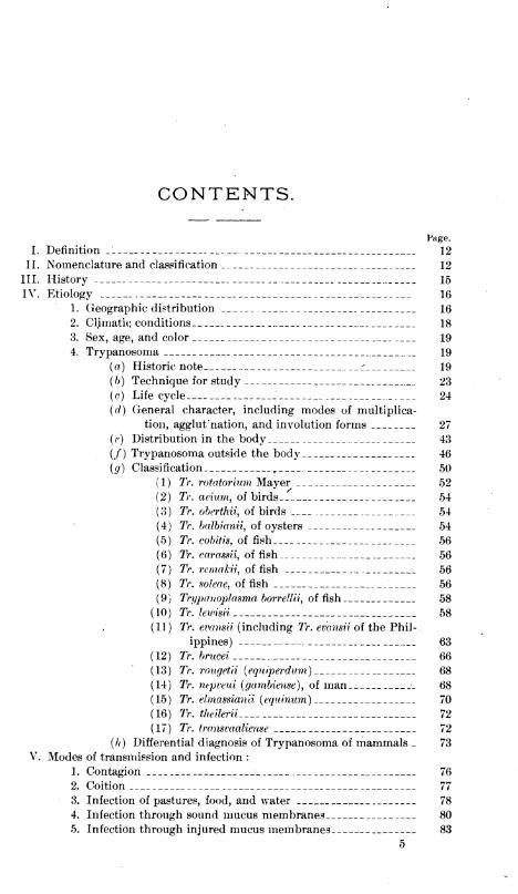
I. Definition 12
II. Nomenclature and classification 12
III. History 15
IV. Etiology 16
1. Geographic distribution 16
2. Climatic conditions 18
3. Sex, age, and color 19
4. Trypanosoma 19
(a) Historic note i 19
(b) Technique for study 23
(c) Life cycle 24
(d) General character, including modes of multiplica-
tion, agglut' nation, and involution forms 27
(e) Distribution in the body 43
(/) Trypanosoma outside the body 46
(g) Classification ,. 50
(1) Tr. rotatorium Mayer 52
(2) Tr. avium, of birds 54
(3) Tr. oberthii, of birds _..__ 54
(4) Tr. balbianii, of oysters 54
(5) Tr. cobitis, of fish 56
(6) Tr. carassii, of fish 56
(7) Tr. remakii, of fish 56
(8) Tr. soleae, of fish 56
(9) Trypanoplasma borrellii, of fish .___ 58
(10) Tr. lewisii 58
(11) Tr. evansii (including Tr. evansii of the Phil-
ippines) 63
(12) Tr. brucei 66
(13) Tr. rougetu (equiperdum) 68
(14) Tr. nepveui (gambieiise), of man 68
( 15 ) Tr. elmassiai ivi (equinum
)
70
(16) Tr. theilerii 72
(17) Tr. transvaaliense 72
(h) Differential diagnosis of Trypanosoma of mammals _ 73
V. Modes of transmission and infection :
1. Contagion 76
2. Coition 77
3. Infection of pastures, food, and water 78
4. Infection through sound mucus membranes 80
5. Infection through injured mucus membranes 83
5
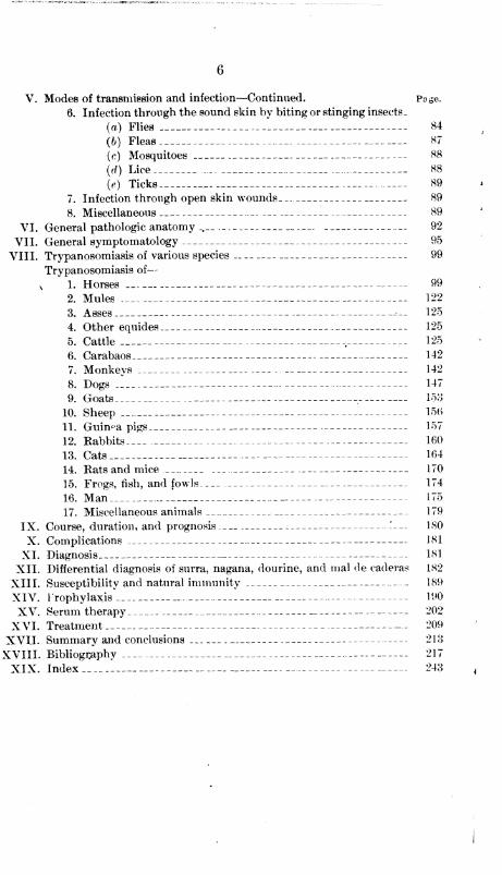
6
V. Modes of transmission and infection—Continued. Page.
6. Infection through the sound skin by biting or stinging insects
_
(a) Flies . 1 84
(6) Fleas 87
(c) Mosquitoes 88
((/) Lice 88
(e) Ticks . 89
7. Infection through open skin wounds 89
8. Miscellaneous 89
VI. General pathologic anatomy _, 92
VII. General symptomatology 95
VIII. Trypanosomiasis of various species 99
Trypanosomiasis of
—
v 1. Horses - 99
2. Mules 122
3. Asses --- 125
4. Other equides 125
5. Cattle - - 125
6. Carabaos 142
7. Monkeys 142
8. Dogs 147
9. Goats 153
10. Sheep 15B
11. Guinea pigs 157
12. Rabbits.„ 160
13. Cats 164
14. Rats and mice 170
15. Frogs, fish, and fowls 174
16. Man 175
17. Miscellaneous animals 179
IX. Course, duration, and prognosis '___ 180
X. Complications 181
XI. Diagnosis 181
XII. Differential diagnosis of surra, nagana, dourine, and mal de caderas 182
XIII. Susceptibility and natural immunity 189
XIV. Prophylaxis 190
XV. Serum therapy 202
XVI. Treatment ___*_ 209
XVII. Summary and conclusions 213
XVIII. Bibliography 217
XIX. Index 243
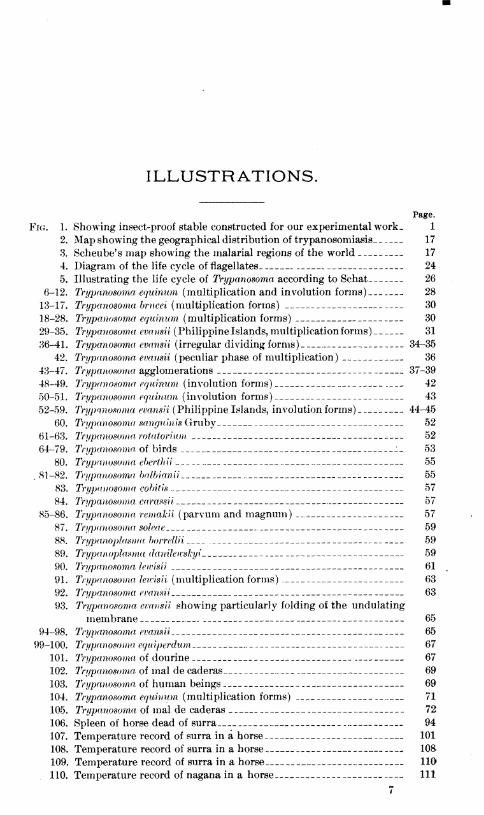
ILLUSTRATIONS.
Fig. 1.
2.
3.
4.
5.
6-12.
13-17.
18-28.
29-35.
36-41.
42.
43-47.
48-49.
50-51.
52-59.
60.
61-63.
64-79.
80.
.81-82.
83.
84.
85-86.
87.
88.
89.
90.
91.
92.
93.
94-98.
99-100.
101.
102.
103.
104.
105.
106.
107.
108.
109.
110.
Page.
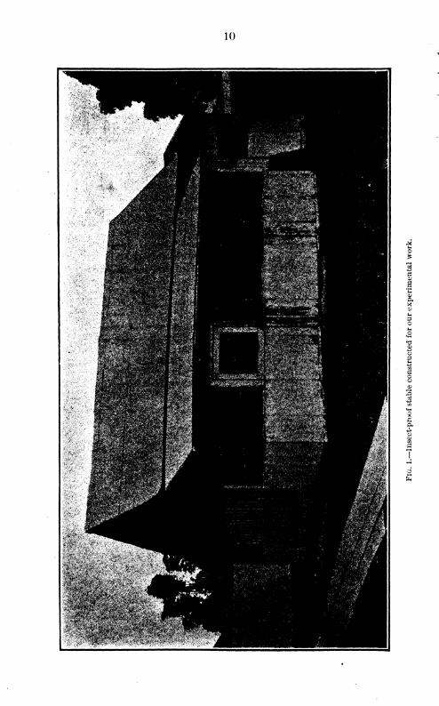
Showing insect-proof stable constructed for our experimental work, 1
Map showing the geographical distribution of trypanosomiasis 17
Scheube's map showing the malarial regions of the world 17
Diagram of the life cycle of flagellates. 24
Illustrating the life cycle of Trypanosoma according to Schat 26
Trypanosoma equinum (multiplication and involution forms) 28
Trypanosoma brucei (multiplication forms) 30
Trypanoso7na equinum (multiplication forms) 30
Trypanosoma evansii (Philippine Islands, multiplication forms) 31
Irypanosoma evansii (irregular dividing forms) 34-35
Trypanosoma evansii (peculiar phase of multiplication) 36
Trypanosoma agglomerations 37-39
Trypanosoma equinum (involution forms) 42
Trypanosoma equinum (involution forms) 43
Trypanosoma evansii (Philippine Islands, involution forms) 44-45
Trypanosoma sanguinis Gruby 52
Trypanosoma rotatorium 52
Trypanosoma of birds .:_ 53
Trypanosoma eberthii 55
Trypanosoma balb ian ii 55
Trypanosoma cobitis 57
Trypanosoma carassii 57
Trypanosoma remakii (parvum and magnum) __._, 57
Trypanosoma soleae 59
Trypanoplasma borrellii 59
Trypanoplasma danileivskyi 59
Trypanosoma lewisii 61
Trypanosoma lewisii (multiplication forms) 63
Trypanosoma evansii 63
Trypanosoma evansii showing particularly folding of the undulating
membrane 65
Trypanosoma evansii 65
Trypanosoma equiperdnm 67
Trypanosoma of dourine 67
Trypanosoma of mal de caderas 69
Trypanosoma, of human beings 69
Trypanosoma equinum (multiplication forms) 71
Trypanosoma of mal de caderas 72
Spleen of horse dead of surra 94
Temperature record of surra in a horse 101
Temperature record of surra in a horse 108
Temperature record of surra in a horse 110
Temperature record of nagana in a horse— 111
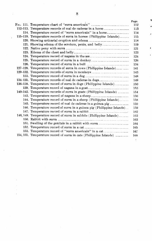
Page.
Fig. 111. Temperature chart of'
'surra americain' ' 112
112-113. Temperature records of mal de caderas in a horse US114. Temperature record of ' 'surra americain" in a horse 114
115-119. Temperature records of surra in horses (Philippine Islands) 115
120. Showing urticarial eruption and edema 118
121. Showing edema of the scrotum, penis, and belly 119
122. Native pony with surra 121
123. Edema of the chest and belly 123
124. Temperature record of nagana in theass__ 124
125. Temperature record of surra in a donkey 126
126. Temperature record of surra in a bull 134
127-128. Temperature records of surra in cows (Philippine Islands) 141
129-132. Temperature records of surra in monkeys 143
133. Temperature record of surra in a dog 148
134-135. Temperature records of mal de caderas in dogs 149
136-138. Temperature record of surra in dogs (Philippine Islands) 150
139. Temperature record of nagana in agoat 153
140-142. Temperature records of surra in goats (Philippine Islands) 154
143. Temperature record of nagana in a sheep 156
144. Temperature record of surra in a sheep (Philippine Islands) 158
145. Temperature record of mal de caderas in a guinea pig 159
146. Temperature record of surra in a guinea pig (Philippine Islands) __ 159
147. Temperature record of surra in a rabbit _ _• 162
148, 149. Temperature record of surra in rabbits (Philippine Islands) 163
150. Rabbit with surra 163
151. Swelling of the genitals in a rabbit with surra 164
152. Temperature record of surra in a cat 165
153. Temperature record of "surra americain" in a cat 167
154, 155. Temperature record of surra in cats (Philippine Islands) 168

10
INTRODUCTION.
Before entering upon a discussion of the text of this paper, a few
remarks regarding the facilities at our disposal appear advisable.
Figure 1 illustrates a specially constructed insect-proof stable in which
all experiments necessitating extraordinary precaution were performed
.
To obtain satisfactory results such a structure is an absolute necessity.
This stable is screened on all sides, the stalls are separated by wire
netting, and each is provided with a door of the same kind. On each side
there is an additional hall entirely screened and with a single outside
door. At one end an insect-proof operating room was built and provided
with a protected entrance to the different stalls.
Because of these precautions, experiments have been conducted by us
with an absolute certainty of results; and owing to a lack of facilities
similar to ours, many of the conclusions contained in reports relating
especially to the transmission of the disease do not appear to have been
based upon accurate observations.
Discussions of the transmission of the disease by feeding, based upon
observations made without protecting animals from insects, do not, of
course, lead to a final settlement of the question ; and so with many other
conclusions in the voluminous literature relating to this subject.
In reviewing literature we have tried in each instance to give credit to
the person to whom due, but in this we may sometimes have failed. Theworks of Voges, Lingard, Kanthuck, Durham, and Blandford, Laveran
and Mesnil, Kabinowitch and Kempner, Wasielewski and Senn, Schat,
Schilling, Bruce, and many others, have been freely used.
We desire to express our obligations to Miss Mary Polk, librarian of
the Bureau of Government Laboratories, for her assistance in editing
bibliography and preparing the index, and to Mr. C. J. Arnell, sten-
ographer and translator, and Mr. Charles Martin, photographer of the
Bureau of Government Laboratories, . for valuable assistance.
Finally, this report has been made possible by the promptness in allow-
ing requisitions for the necessary supplies and the constant advice of
Dr. Paul C. Freer, Superintendent of Government Laboratories.
Biological Laboratory, Manila, P. /., July k, 1903.
11
TRYPANOSOMA AND TRYPANOSOMIASIS.
I. DEFINITION.
The disease is a specific infection of many of the lower animals, and
occasionally of man, caused by Trypanosoma. It occurs in epidemic form
over large areas of tropical countries, and is usually more severe during
the rainy season. It is characterized by a period of incubation, followed,
in most animals, by a remittent, intermittent, or, less frequently, relaps-
ing fever; by the presence of Trypanosoma in the circulating blood,
which in some animals are numerous in proportion to the temperature;
by progressive anemia and emaciation; by a catarrhal condition of the
mucous membrances of the eyes and nose; by roughness of the hair,
which in many instances, falls out; and by subcutaneous edema, more
commonly of the posterior extremities, genitals, and belly. In the later
stages paresis of the posterior extremities is very common. The mor-
tality among most animals of economic importance is 100 per cent.
There are found in most animals at post-mortem, in addition to the
evidence of severe anemia, certain changes in the spleen, the most con-
stant being enlargement and a peculiar mottling. Taken with the other
principal lesions, such as lymphatic hyperplasia;peculiar, yellowish, gel-
atinous subcutaneous and suberous infiltrations; an enlarged liver and
the. accumulation of fluid in the serous cavities, it makes an anatomic
picture which is rarely excelled in chronic diseases peculiar to man.
II. NOMENCLATURE AND CLASSIFICATION.
A list of the various names used to designate Trypanosomiasis in diffcr-
ent parts of the world has been compiled from literature as follows
:
Adjce. Doaia.
Andar-tap. Dourine.
Anemic pernicieuse. • Equine relapsing fever.
Berbad. Equine surra.
Beschalkrankheit. Equine syphilis.
Beschalseuche. Exantheme eoitale.
Blaschenausschlag. Fish surra.
Bovine surra. Flagellose de equina.
Buffalo surra. Galtah.
Camel surra. Galtia.
Canine surra. Glossinose.
Dog surra. Horse pox.
12
13
Horse surra.
Juckkrankheit.
Khanhog.
Khusk-zaharbad.
La mouche.
Leuma equorum.
Mai de caderas.
Maladie a trypanosome.
Maladie benigne du coit.
Maladie de la ts€tse\
Maladie de Soemedang.
Maladie du coit.
Maladie du prurit.
Maladie venerienne du cheval.
Marri.
Xagana.
Nikalgaya.
N'gana.
Nygana.
Oae.
Pernicious anemia of horses.
Peste de cadeiras.
Pferdestaupe.
Phenta.
Phenta-ka-darad.
Phera.
Phcta.
Phetra.
Phitgaya.
Photra.
Polynevrite infectieuse du cheval.
Poona.
Purama.
Rat surra.
Relapsing fever of equines.
Sar.
Sara.
Schleuchende fieber.
Sokra.
Sukal.
Surra.
Surra americain.
Surrakrankheit.
Tap.
Tap-dik.
Tape-dik.
Taraf.
Tebersa.
Tibarsa.
Trypanosomose.
TsetsS-fly disease.
Tsetstkrankheit.
Tumby-a.
Tumby-baba.
Wabai-ki-bokhar.
Zaharbad.
Zherbad.
Zuchtlahme.
In this report the term "Trypanosomiasis," as suggested by Salmon and
Stiles, is used, being in a general sense comparable to the terms "filaria-
sis" and "uncinariasis." Following this classification further, according
to the animal infected, we would have Trypanosomiasis of man, Trypano-
somiasis of horses, Trypanosomiasis of cattle, etc. Such a nomenclature
would apply satisfactorily, whether the infecting parasites are identical or
not, and also regardless of the manner and place of infection. For ex-
ample, the term Trypanosomiasis of horses would apply equally well to
nagana contracted by the bite of an infected tsetse fly in South Africa and
to surra produced by the hypodermic injection of infected Trypanoso-
miatic blood in Manila.
If the parasites causing the diseases known under the old names
have been or are shown to be different, there could be no very great
necessity for interfering with the better part of the established nomen-
clature. Surra would then be the Trypanosomiasis of horses and of
other animals due to an infection with Tr. evansii, nagana would be
the Trypanosomiasis of horses and of other animals due to an infection
with Tr. brucei, etc.
On the other hand, if these parasites are shown to be the same, or,
probably more correctly, until they are shown to be different, there
14
does not appear any valid reason why any of the names except that of
surra, accepted by Evans, the original discoverer of the pathogenicity
of the parasite in animals, should be retained.
Without entering in detail upon a discussion, which will be taken
up later in the report, regarding the identity or nonidentity of Tr.
evansii, Tr. brucei, Tr. rougetti (equiperdum), and Tr. elmassianii (equi-
num), and hence the identity or nonidentity of surra, nagana, dourine
and mal de caderas, there is considerable difference of opinion, and
also considerable inconsistency in some of the writings, especially with
reference to some of the diseases discovered and named since Evans's
original report.
Numerous writers on Trypanosomiasis base their diagnoses on the
presence of Tr. brucei, and, after carefully describing the parasite, state
that they do not know whether or not it is identical with Tr. evansii.
How can such writers, not having previously studied either of the para-
sites, state that it was not Tr. evansii rather than Tr. brucei they
were working with? If they are positive that the parasite is Tr. brucei,
then they affirm it to be different from Tr. evansii. It is obvious that,
if these parasites are identical, Tr. brucei is not entitled to a place in
the nomenclature of Trypanosoma, for Tr. evansii was known and de-
scribed years before Bruce performed his work. Bruce himself, in his
original report, considered his parasite probably identical with Tr.
evansii.
Some eminent authorities criticise Koch and many other writers for
stating that Tr. evansii and Tr. brucei are identical, without offering
detailed proof of their statements. Such criticism seems to us unjust.
The proof demanded is that. they are different parasites, and until
this proof is furnished, writers, in our opinion, are perfectly entitled to
consider the Trypanosomiasis of horses and a number of other animals
as being due to an infection with Tr. evansii.
The practical importance of deciding this question is forcibly-brought
home to workers in the Philippine epidemic, a fact which has already
been emphasized by other writers. We have to deal with an extensive
epidemic of Trypanosomiasis in several species of domestic animals,
particularly in horses, and the parasite causing the disease seems to
be the same in all. This parasite answers the description given of
Tr. evansii, Tr. brucei, and others, and it is necessary either to intro-
duce a new name or to classify from description. We have decided,
after a careful review of all available literature pertaining to the sub-
ject, that we are dealing with Tr. evansii redescribed by Bruce as the
causative agent in nagana and named Tr. brucei by Buffard and Schnei-
der, and also described and given other names by various authors.
To be consistent with this statement, "surra" would be the only allow-
able popular name for the disease caused by this parasite, the numerous
other names becoming mere synonyms.
15
In those forms of the disease due to other species of Trypanosoma
other names would of course be allowable; but, with the possible excep-
tion of dourine, we have nowhere else met so much confusion and such
a multiplication of names as is found in the group of which Tr. evansii
is the cause.
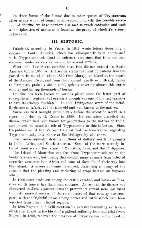
III. HISTORIC.
Caladrini, according to Vogcs, in 1842 wrote letters describing a
disease in South America, which has subsequently been determined
to be Trypanosomiasis (mal de caderas), and since that time has been
discussed under various names and by several authors,
Sivori and Lecler are satisfied that this disease existed in South
America before 1850, while Lacerda states that mal de caderas was im-
ported to the mainland about 1850 from Marajo, an island at the mouth
of the Amazon liiver, and from there spread rapidly over Brazil, thence
to Paraguay, probably about 1860, quickly covering almost this entire
country and killing thousands of horses.
Dourine has been known in various places since the latter part of
the eighteenth century, but curiously enough was one of the last varieties
to have its etiology elucidated. In 1858 Livingstone wrote of the tsetse-
fly disease in Africa, at that time old and well known to the natives.
Surra was first brought prominently before the scientific world in a
report published by G. Evans in 1880. He accurately described the
disease, which had been known for generations to the natives of India,
and proved the causative role of Trypanosoma in this infection. Since
the publication of Evans's report a great deal has been written regarding
Trypanosomiasis, as a glance at the bibliography will show.
The disease annually destroys millions of dollars' worth of animals
in India, Africa, and South America. Some of the more recently in-
fected countries are the Island of Mauritius, Java, and the Philippines.
The Island of Mauritius was free from Trypanosomiasis up to the
South African war, but during that conflict many animals from infected
countries were sent into Africa and some of them found their way into
this island. A severe epidemic developed, destroying so many of the
animals that the planting and gathering of crops became an impossi-
bility.
In 1900 surra broke out among the cattle, carabao, and horses of Java,
since which time it has there been endemic. As soon as the disease was
discovered in Java vigorous plans to prevent its spread were instituted
and with marked success, if the small losses of that country are com-
pared with the frightful havoc among horses and cattle which have been
reported from other infected regions.
In 1886 Bignami and Celli mentioned a parasite resembling Tr. lewisii
which they found in the blood of a patient suffering from malarial fever.
Ncpveu, in 1898, reported the presence of Trypanosoma in the blood of
16
seven patients, six of whom were suffering from malaria and the seventh
was healthy. He described and illustrated the parasite. During 1902,
Dutton, Ford, and Manson reported Trypanosoma in human beings, and
in 1903 Manson and others reported a number of cases.
The first published report which we have of Trypanosomiasis in the
Philippines was by Smith and Kinyoun in 1901. The history of the
epidemic in this country has been reported by Musgrave and Williamson
in a preliminary published as Bulletin ~No. 3, Bureau of Government
Laboratories. This report was read before the Manila Medical Society
and brought out considerable discussion. The only point at issue was
our statement that the disease was introduced here in 1901. We have
investigated as far as possible the arguments brought forth that surra
was here prior to that time, but have found nothing to justify any change
in our original statement. The subject is not of great importance
one way or the other, except for its 'historic interest. There is one
thing absolutely certain, that the disease was introduced at that time,
and, whether this was its original appearance or not, the frightful epi-
demic which has raged here is positively connected with this infection.
Our statements regarding the manner of its spread were absolutely
convincing at the time of the publication of the preliminary report, and
additional work along these lines has since confirmed the conclusions
there given.
During the past month we have had proof of the reimportation of
the disease, this time in a cow received from Java.
Since its introduction the infection has been spreading throughout
the Archipelago, and at the present time areas in which it is prevalent are
reported from almost the entire group of islands.
IV. ETIOLOGY.
GEOGRAPHIC DISTRIBUTION.
The geographic range of the various forms of Trypanosomiasis is shownin the following table :
Continent. Country.Province, territory,
division, district, etc.Form.
Surra_-_—do_ _
Reported by—
Asia India Bengal _ _ __ Lingard.
Annam _
Berars _ Do.Bombay Presidency —Burma. _
—do„_—do_ _
Do.Lingard, Steel.
\ Cashar _ do _ Do.Kohat _ _. _do___ Lingard, Gunn.Konkan _ . _. __ _do-_ Do.
.Kumaon ProvinceNaga Hills_
—do____do_ _
Do.Do.
Manipur ___do— Do.Northwest Provinces-Punjab
—do—___do-
Do.Lingard, Evans.
Rajputana _ —do—Cochin China (Ton-kin).
Indo-China (NhaTrang).
Surra „_
„._do——do—
Blanchard, Molle-
Korea
reau.Carogeau.
W. G. Campbell.
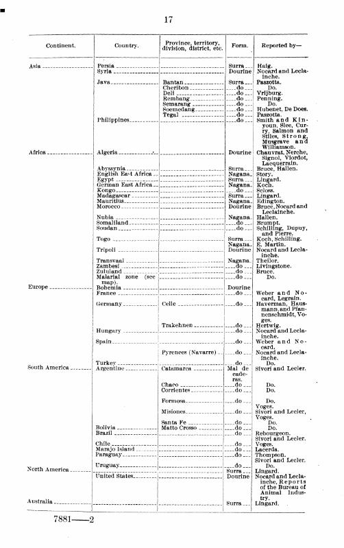
17
Continent. Country. Province, territory,division, district, etc.
Form. Reported by—
Asia Persia __ _ _ _ _ Surra ...
Dourine
Surra ___
__ _do___
Haig.Svria _ Nocard and Lecla-
Java __ _ Bantan _ _ _ _ _ .
inche.Paszotta.
Philippines
Cheribon Do.Deli ....do— Vrijburg.Rembang do— Penning.Semarang _„_do— Do.Soemedang _ . —.do... Hubenet, De Does.Tegal ___ _ - _—do— Paszotta.
____do - Smith and Kin-
Africa _ _. _ Algeria Dourine
Surra ___
Nagana_Surra—Nagana.
youn, Slee, Cur-ry, Salmon andStiles, Strong,Musgrave andWilliamson.
Chauvrat, Nerche,
Abvssynia _
Signol, Viordot,Lacquerrain.
Bruce, Hallen.English East Africa Story.
Lingard.Koch.
Egypt _ _
German East Africa __
Kongo Scloss.Madagascar Surra ___
Nagana.Dourine
Nagana.do
Lingard.Edington.Mauritius _
Morocco Bruce, Nocard and
Nubia _
Leclainche.Hallen.
Somaliland Brumpt.Schilling, Dupuy,and Pierre.
Koch, Schilling.
Soudan _ ._ _ _do. .
Togo _ _ Surra...Nagana.Dourine
Nagana.do_
TripoliE. Martin.Nocard and Lecla-
Transvaalinche.
Theilor.Zambesi Livingstone.
Bruce.Zululand doMalarial zone (seemap).
do_ Do.
Europe DourinedoFrance Weber and No-
Germany
Hungary _ _
Celle _ - docard, Legrain.
Haverman, Haus-
Trakehnen _ do
mann,andPfan-nenschmidt, Vo-ges.
Hertwig.Nocard and Lecla-._ .do
Spain doinche.
Weber and No-
! Pyrenees (Navarre) __
Turkev _ <
—do-do _.
card.Nocard and Lecla-inche.Do.
South America ___ _ _ Argentine _ _. _ C».tsi.ma.ro.». Mai decade-ras.
__ do .
Sivori and Lecler.
Bolivia _
Chaco _ _ _ Do.Corrientes _ do Do.
Formosa _ do . Do.
Misiones ._ _do _
Voges.Sivori and Lecler,
Santa Fe __ .do__Voges.
Do.Matto Crosso _ do Do.
Brazil do Rebourgeon.Sivori and Lecler.Voges.Chile do
Marajo Island doParaguay - - \ _ _ do_ Thompson.
Sivori and Lecler.Do.Uruguay . i do
North America Surra _._
Dourine
Surra .__
Lingard.Nocard and Lecla-United States .. _ _
Australia
inche, Reportsof the Bureau ofAnimal Indus-try.
Lingard..
7881-

18
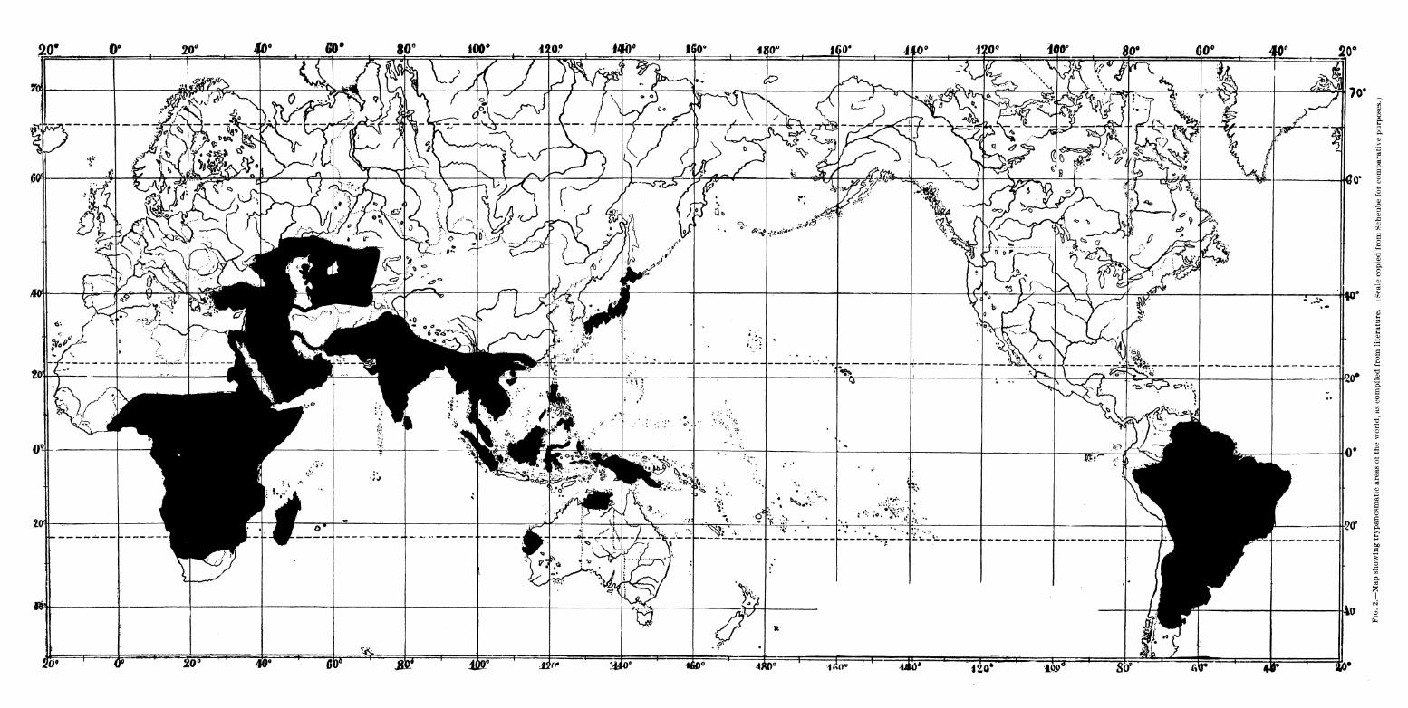
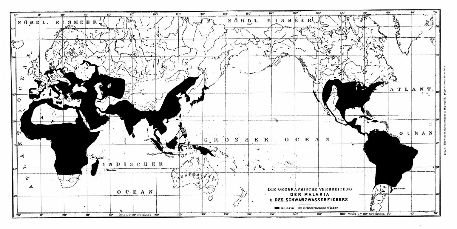
Fig. 2 gives a schematic representation of the infected areas, drawn
from Scheube's map, fig. 3, illustrating the regions of the world in
which malaria prevails. Fig. 3 is reproduced to show the relation in
geographic distribution of Trypanosomiasis and malaria.
The table and maps given above illustrate the wide geographic distri-
bution of Trypanosomiasis and its special prevalence in the tropical and
subtropical zones. New points of infection are being reported from time
to time. Neither the table nor the map are complete, and both maycontain some inaccuracies owing to the conflicting reports and the fact
that some of the references given are not available.
CLIMATIC CONQITIONS.
All the different forms of this disease are infections incident to the
periods of wet weather. This statement is made in all discussions of the
subject which we have been able to review. The reasons given for the
fact are varied, but the true explanation, namely, that biting flies are
much more numerous during this season than during any other, is con-
firmed by nearly all recent writers. Not only this, but the rainy season
offers another and equally important condition, which will be fully dis-
cussed under modes of transmission—i. e., the dark, cloudy days with
great relative humidity make it possible for the fly mechanically to carry
the infection for a much longer time. We have shown conclusively that
bright sunlight quickly destroys the Trypanosoma; and even if the
proper flies ivere more numerous during dry weather, this factor alone
would greatly limit their ability to carry infection.
To sum up, the transmission of the disease is greatest exactly under
the climatic conditions most favorable to insect life and to the insect's
ability to carry the living infection. Such conditions occur in low-lying,
marshy lands during the darlc, cloudy days of the rainy season.
Trypanosomiasis prevails to a limited extent under other circum-
stances, but we have reason to fear epidemics only when those above
described are realized.
We know of no other predisposing causes for surra. All species of
animals within certain geographic zones may contract the disease by
experimental methods. As will be shown, natural infection is a mechan-
ical processs, so that no reason exists against the supposition that all
animals are susceptible to the usual methods of transmission.
A number of writers have stated that a greater percentage of foreign
horses coming into an infected zone than of native animals contract the
disease. Of 80 horses observed by Lingard, 16 per cent died during the
first year, and 70 per cent during the first seven years while under obser-
vation. Australian horses were found by him to be more susceptible
than the native horses of India.
In our experience in Manila, Australian, Chinese, and American horses
do not appear more susceptible than native ponies. In several instances
19
we have been able to observe the infection in large stables containing both
native and American horses, and under these conditions one appeared
to contract it as readily as the other. The greatest percentage affected
in either case is always found in large groups of animals; and as Amer-
ican horses are more frequently collected in large stables, a superficial
deduction from this fact might be misleading. In reality, the higher
percentages we have encountered have occurred in stables containing
native ponies.
Lingard considers both sexes to be equally susceptible. We have had
no opportunity either to confirm or to disprove this statement, as nearly
all the horses in Manila are males. Sex certainly plays no part in the
communication of the disease in other animals, and there is no reason to
suppose it would do so in horses.
In 1885 Steel stated that white and grey mules are more susceptible
to surra than darker-colored animals; and, among others, Laveran and
Mesnil believe this to be the case with horses as well. They attribute
this phenomenon to the supposed fact that flies bite light-colored animals
more readily than dark ones. This fact is questioned by some authors,
alt! 10ugh it is true that on the former animals the flies may be more
noticeable than on the latter. We have been unable to verify this state-
ment. White and grey animals have not been infected in greater pro-
portion than others, nor do they more readily attract biting flies. Asa matter of fact, our statistics of the Philippine epidemic show themto be less frequently attacked. No importance can therefore be attrib-
uted to color as a factor in the spread of the disease.
In general, no material difference in the percentage of infection in
horses of varying ages has been found. The greater proportion of
animals in Manila are older than four years. Our investigation of
rats has shown us that the older animals contain Trypanosoma moreoften than the j'ounger ones. The difference is probably not due to
the greater susceptibility of the former, but is accounted for by the
fact that, like dogs, they are prone to fight, and hence very frequently
have wounds, particularly about the head, which naturally favor the
entrance of the parasites.
TRYPANOSOMA.
HISTORIC NOTE.
In this discussion the species of Trypanosoma have been followed in
part at the expense of the chronologic order of publications.
Valentin (1841) discovered hematozoon, and Glugge (1842) para-
sites, the former in trout (Salmo fario) and the latter in the blood
of frogs. Both were probably Trypanosoma, Doflein considering Glugge's
description sufficient for the- recognition of the genus. In 1843 Grubyobserved a flagellate infusorium in frogs, naming it Tr. sanguinis', anddespite the previous work of others, he has generally been credited with
20
its discovery, his work being subsequently confirmed by a number of
investigators.
Lankester (1871) discovered a sausage-shaped parasite in the blood
of frogs, naming it undulina. Gaule (1880) made some further observa-
tions on those bodies, which he considered protoplasmic portions of
the blood corpuscles separated for a short period of independent life
and more prevalent in very dry, warm weather. Leucocytes were seen
to be converted into flagellates and then back to leucocytes. Blutschli
and Lankester, commenting on Gaule's work, stated, independently, that
the conversion of ameboid bodies into flagellates and the reconversion
of flagellates into bodies resembling white corpuscles did not prove
the latter to be leucocytes. Grassi (1882) observed in frogs a parasite
which was named paramecioides.
Blanchard (1890) confirmed Gruby's work and gave the following syn-
onyms: Paramecium loricatum Mayer, 1843; Ameba rotatorium Mayer,
1843; Globularia radiata Wedl, 1848; Paramecium costatum Chaussat,
1850; Undulina ray Lankester, 1871; Paramecioides costatus Grassi,
1882 ; and Hematamonas Mitraphamow, 1883.
Danilewsky (1885) described at least six varieties of parasites in
the blood of frogs. He noted the change in the blood at rest from the
flagellate to the ameboid stage, as had already been mentioned by others.
Ameboid forms were seen to segment into 64 spores, which gradually
assumed nomad forms and divided by longitudinal division. Trans-
verse division also was occasionally seen. Flugge (1896) stated that
these parasites very closely resembled Tr. lewisii. Multiplication con-
sisted in longitudinal and transverse division and spore formation, the
latter sometimes being preceded by an ameboid stage. He gave the
length as 80 microns and mentioned that the parasites were provided
with undulating membranes and flagella. He said that they were found
in frogs, tortoises, fish, birds, oysters, chickens, and geese. In general
structure they resembled Trypanosoma. Their pathogenic action wasnot known.
Kominski (1901) again called attention to the probability of an
increase in the occurrence of Tr. sanguinis Gruby with the age of the
animals. They were found at all seasons of the year, and were morecommon in males than in females. No disease in frogs was produced,
and there was no evidence of the mode of transmission in these animals,
even when they were kept together for months.
Eberth (1861) discovered in the intestines of birds a parasite whichwas named by Kent Tr. eberthi, but which in all probability was a
trichomonas.
Lewis (1879 and 1880) described Trypanosoma found in the rats
of India. In a second pap£r published in 1884 he considered these
Trypanosoma identical with Tr. evansii. Opie, Flugge, and some other
21
writers give the credit for discovering Tr. lewisii to Osier, but we have
been unable to verify the reference, and Osier does not indicate that
such is the case in his article on the hematozoon of malaria (B. M. J.
March 12, 1887), in which he reviews the work of Lewis and others on
the Trypanosoma.
Butschli (1880) found flagellates in the intestinal canal of a noma-
tocle (Tribolus gracilis) and also in the intestines of domestic flies.
Including flagella they measured about 33 microns in length and were
sometimes observed in stellate colonies.
Wittich (1881) discovered in the blood of hamsters a Trypanosoma
which he considered identical with Tr. lewisii. This observation was
confirmed by Koch. Wittich's work was done in Germany on 12 ham-
sters imported from Africa. He states that his organism answered in
all respects to Lewis's description of the Trypanosoma of rats in India.
Eleven of his hamsters died; but he did not consider trypanosoma to
Lave played any part in the malady to which they succumbed, although
present in all.
G. Evans (1880) discovered Trypanosoma in the blood of horses
suffering with the well known surra of India. He proved the causative
agency of these parasites in the production of the disease. Steele (1885)
confirmed Evans's work, and named the parasite Spirocheta evansi.
Crookshank (1886) made a report on these parasites, confirming Evans's
and Steele's work. He considered these Trypanosoma identical with
Mitraphanow's Trypanosoma of carp.
Kent (1881) discovered "herpetomonas" in the intestines of the
domestic fly. His parasite had no undulating membrane and was prob-
ably not a Trypanosoma.
Certes (1882) found in the digestive tube of an oyster a parasite
which has been named Tr. balbianii. The general description follows that
of Trypanosoma, but slight differences of internal structure were noted.
Undulating membrane and flagella were present, but nucleus, nucleolus,
and vacuole were not observed. In a later paper he demonstrated a
nucleus. He considered this Trypanosoma closely related to Mitrapha-
mow's "hematomonas" (Trypanosoma) of fish. Laveran and Mesnil
(1901) found these parasites rarely in Portugese oysters and frequently
in common oysters. They say that the bodies were not flagellates and
that the presence of an undulating membrane was questionable. They
do not consider Certes's organism a Trypanosoma, but rather a bacteria.
Mitraphamow (1883) described Trypanosoma in mudfish (Cobatus
fossilis). His parasite was 1 to 1^ microns broad and 30 to 40 microns
long. He gave a very careful description of these organisms, which
occurred in nearly all the fish examined and were more numerous in hot
than in cold weather. He gave the group the name "hematomonas" and
described two species.
Moebius (1883) found Trypanosoma in oysters (Tapes decussata and
22
Tapes pullastra). These parasites were studied by Lustrac (1896), who
considered them Trypanosoma.
Bignami and Celli (1885) found, in the blood of a patient with
malarial fever, parasites very closely resembling the Trypanosoma of
frogs, birds, and fish. Nepveu (1898) described Trypanosoma in seven
cases occuring in men, six of whom were suffering from malarial fever.
Barron is quoted by Laveran as having seen flagellates in the blood of an
anemic woman. During 1902, Dutton, Ford, Sambon, Manson, and
others have described the occurrence of Trypanosoma in human beings.
Dutton first published an account of these parasites, found in the blood
of Dr. Ford's patient.
Danilewsky (1890) found a Trypanosoma in the blood of birds,
naming it Tr. sanguinis evansii. Like Blutschlr's parasite it had a long
flagellum and an undulating membrane. Division was longitudinal,
transverse, or by segmentation from the ameboid stage. No symptoms
were produced in the host. Danilewsky thought this was probably due to
the high temperature of the birds or the tolerance acquired by generations
of infection.
Laveran and Mesnil (1901) found Trypanosoma in three kinds of
fish—brochet, sole, and redeye. That found in the brochet closely
resembled Tr. evansii, etc., and was named by them Tr. remakii, after
Eemak, who they say first observed the parasite in 1842. The Try-
panosoma from the sole was also of the same general type, and they
designated it as Tr. solew. Laveran and Mesnil state that Trypanosoma
had not previously been observed in salt-water fish, but in this they are
probably mistaken, for Flugge (1896) reported finding them in the
fish of the Mediterranean Sea. The organism which they found in the
redeye had a flagellum at each end; they placed it in a separate genus
which they called Trypan oplasma, giving the parasite the name Try-
panoplasma borrelii.
Rouget (1896) described Trypanosoma found in the blood of a horse
suffering from dourine (beschalseuche), and for two and one-half years
continued the study of this organism in susceptible animals. Wasi-
lewsky and Senn (1899) confirmed Rouget's work, and determined the
pathogenic action of this Trypanosoma for the horse, passing it through
other animals and back to the horse, reproducing the disease. Voges
says that this Trypanosoma was discovered by Chauvrat in 1892. x Lave-
ran and Mesnil (1901) proposed the name Tr. rougetii for the parasite
of dourine. Doflein (July, 1901) named it Tr. equiperdum, which is
the name used also by Salmon and Stiles.
Elmassian, according to Voges, in 1901 first differentiated the Try-
panosoma of mal de caderas in South America; while Voges (1902)
very carefully described the parasite, proved its pathogenic action, and
named it Tr. equinum.
1 Reference not available.
23
Theiler, in an article published by Bruce (1902), is credited with
the discovery of a new Trypanosoma of cattle in South Africa. Bruce
proposed the name Tr. theilerii. During the same year Theiler found
a different species of Trypanosoma in the cattle of the Transvaal, send-
ings specimens of it to Laveran, who bases his statement that it is a
distinct species particularly on the location of the centrosome near the
center of the body, close to and sometimes united to the nucleus. Heproposed the name Tr. transvaaliense for this parasite.
In 1901 Smith and Kinyoun described a parasite which had been
observed by Dr. Jobling in the blood of a sick horse in Manila, and this
parasite was afterwards determined to be a Trypanosoma. Later in the
same year Smith made some additional notes on the organism, and con-
sidered it Tr. evansii.
Curry (1902) described the parasite and classed it as a Trypanosoma,
but was unable to state whether it was Tr. evansii or Tr. brucei. His
description was the first accurate one of the parasite found here.
TECHNIQUE FOR THE STUDY OF TRYPANOSOMA.
For the determination of the presence of Trypanosoma in the blood,
only a fresh preparation such as is used for the examination for malaria
is needed. The parasites when numerous are readily observed with a
DD. or even an AA. objective and No. 4 ocular Zeiss. If they are
scarce, considerable time may be necessary to find one, but once seen the
diagnosis is determined, and may be facilitated by staining the specimen
by any of the approved methods. For a careful study stained specimens
are essential.
Fairly good results are obtained by any of the methods used for stain-
ing malarial parasites. Komanowsky's method, or any of its modifica-
tions, particularly Wright's, gives beautiful pictures. Laveran & Mesnil
have also published directions for an excellent stain for these parasites.
A most satisfactory stain for Trypanosoma is one prepared by Dr.
Paul G. Woolley, pathologist in this Laboratory, and published here for
the first time with his permission.
Fix smears in absolute alcohol for ten minutes.
A. Eosin (w) (Grubler) gram.... 1
Distilled water grams.... 1,000
B. Polychrome methylene blue (Unna).
C. Methylene blue (Med) (Grubler) gram.... 1
Distilled water grams.... 100
D. Of solution B parts.... 2
Of solution C part.... 1
Mix and add 1 c. c. of A to each 4.5 c. c. of D.
Stain by immersion for twenty or thirty minutes. Wash with water.
Wash for two to five seconds with solution A and blot immediately.
The effects on protoplasm and nuclear material may be regulated by
longer and shorter exposure to the eosin solution in the last step.
24
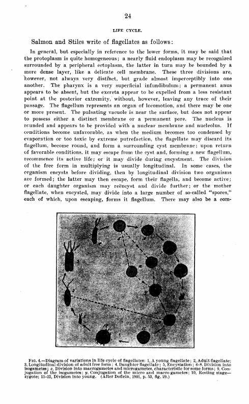
LIFE CYCLE.
Salmon and Stiles write of flagellates as follows:
In general, but especially in reference to the lower forms, it may be said that
the protoplasm is quite homogeneous; a nearly fluid endoplasm may be recognized
surrounded by a peripheral ectoplasm, the latter in turn may be bounded by a
more dense layer, like a delicate cell membrane. These three divisions are,
however, not always very distinct, but grade almost imperceptibly into one
another. The pharynx is a very superficial infundibulum ; a permanent anus
appears to be absent, but the excreta appear to be expelled from a less resistant
point at the posterior extremity, without, however, leaving any trace of their
passage. The flagellum represents an organ of locomotion, and there may be one
or more present. The pulsating vacuole is near the surface, but does not appear
to possess either a distinct membrane or a permanent pore. The nucleus is
rounded and appears to be provided with a nuclear membrane and nucleolus. If
conditions become unfavorable, as when the medium becomes too condensed by
evaporation or too toxic by extreme putrefaction, the flagellate may discard its
flagellum, become round, and form a surrounding cyst membrane; upon return
of favorable conditions, it may escape from the cyst and, forming a new flagellum,
recommence its active life; or it may divide during encystment. The division
of the free form in multiplying is usually longitudinal. In some cases, the
organism encysts before dividing, then by longitudinal division two organisms
are formed; the latter may then escape, form their flagella, and become active;
or each daughter organism may reencyst and divide further; or the mother
flagellate, when encysted, may divide into a large number of so-called "spores,"
each of which, upon escaping, forms it flagellum. There may also be a com-
^2m
Fig. 4.—Diagram of variations in life cycle of flagellates: 1, A young flagellate ; 2, Adult flagellate;
3, Longitudinal division of adult free form ; 4, Daughter flagellate ; 5, Encystation ; 6-8. Division intoisogametes ; x, Division into macrogametes and microgametes, characteristic for some forms ; 9, Con-jugation of the isogametes; y, Conjugation of the micro and macrogametes; 10, Resting stage—zygote; 11-12, Division into voung. (After Doflein, 1901, p. 53, fig. 29.)
25
plete conjugation of the two individuals, followed by encystment and division
into numerous young.
The illustration (fig. 4) taken from Doflein is intended to show the*
variations in the life cycle of the flagellates.
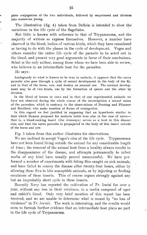
But little is known with reference to that of Trypanosoma, and the
majority of writers so express themselves. However, a number have
observed in the blood, bodies of various kinds, which they have considered
as having to do with the phases in the cycle of development. Voges and
others consider the entire life cycle of the parasite to be acted out in
the blood, and present very good arguments in favor of their conclusions.
Schat is the only author, among those whom we have been able to review,
who believes in an intermediate host for the parasites.
He says:
Analogously to what is known to be true in malaria, it appears that the surra
parasite also goes through a cycle of sexual development in the body of the fly,
and in that of the horse, cow, and donkey an asexual one. The asexual develop-
ment may be of two kinds, one by the formation of spores and the other by
division.
In the blood of horses or cows and in that of our experimental animals wehave not observed during the whole course of the investigation a sexual union
of the parasites, which is contrary to the observations of Penning and Plimmerand Bradford, who make mention of forms of conjugation.
In this regard we feel justified in supposing that an hypothesis similar to
that which Hanson proposed for malaria holds true also in the case of surra
—
that is, a blood-sucking insect (the stomoxys) serves as a host in this disease
also, and that the surra parasite is propagated in the body of this insect outside
of the horse and cow.
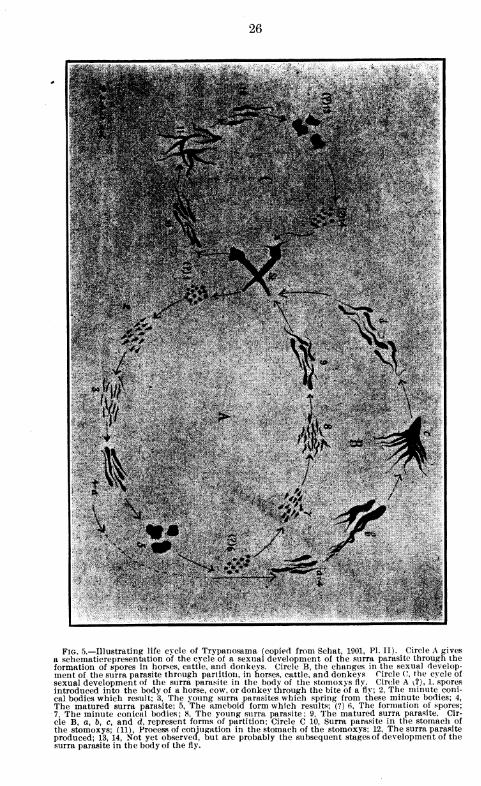
Fig. 5 taken from this author illustrates his observations.
We are inclined to accept Voges's idea of the life cycle. Trypanosoma
have not been found living outside the animal for any considerable length
of time ; the removal of the animal host from a locality always results in
the disappearance of the disease, and attempts permanently to infect
media of any kind have usually proved unsuccessful. We have per-
formed a number of experiments with biting flies caught on sick animals,
and have failed to convey the disease after twenty-four hours, either by
allowing these flics to bite susceptible animals, or by injecting or feeding
emulsions of these insects. This of course argues strongly against any
but an improbably short cycle in these insects.
Recently Xovy has reported the cultivation of Tr. lewisi for over a
year, without any loss in their virulence, in a media composed of agar
and rabbit's blood. Only very brief mention of this report has been
received, and we are unable to determine what is meant by "no loss of
virulence" in Tr. leivisii. The work is interesting, and the results would
seem to furnish further evidence that an intermediate host plays no part
in the life cycle of Trypanosoma.
26
/VJ
L4& '-J
j-?'^'V.
.-*V#
I'll .• • , - <\ ?. - , • .2h»-» -* - «*•"> , ? .y-vf/ . .'<<•". I *;• ,
Fig. 5.—Illustrating life cycle of Trypanosama (copied from Schat, 1901, PL II). Circle A gives
a schematicrepresentation of the cycle of a sexual development of the surra parasite through theformation of spores in horses, cattle, and donkeys. Circle B, the changes in the sexual develop-ment of the surra parasite through partition, in horses, cattle, and donkeys Circle C, the cycle of
sexual development of the surra parasite in the body of the stomoxys fly. Circle A ^?), 1, sporesintroduced into the body of a horse, cow, or donkey through the bite of a fly; 2, The minute coni-
cal bodies which result; 3, The young surra parasites which spring from these minute bodies; 4,
The matured surra parasite; 5, The ameboid form which results; (?) 6, The formation of spores;
7, The minute conical bodies ; 8, The young surra parasite ; 9, The matured surra parasite. Cir-
cle B, a, b, c, and d, represent forms of partition; Circle C 10, Surra parasite in the stomach of
the stomoxys; (11), Process of conjugation in the stomach of the stomoxys; 12, The surra parasite
produced; 13, 14, Not yet observed, but are probably the subsequent stages of development of thesurra parasite in the body of the fly.
GENERAL CHARACTER.
Trypanosoma of all species are in general similar organisms. The
family diagnosis, as given by Salmon and Stiles, is as follows
:
Flagellate parasitic forms with one chief flagellum directed anteriorly; in some
forms a secondary flagellum directed posteriorly; body usually with two angles,
and wound more or less in the form of a spiral; one angle of the body provided
with an undulating membrane. One nucleus and one centrosome present.
The morphology varies greatly in the same species of Trypanosoma,
and also to a greater extent in different species. In general the para-
sites may be said to measure from 1 to 5 microns in thickness and from
15 to 45 microns in length, including flagellum. They all show very
active eel-like movements and some motility. In some species the latter
is very slight, the parasites undulating with extreme rapidity, but cov-
ering so short a distance as to be easily followed under the microscope;
while in others, especially Tr. lewisii, the movements are often so rapid
in freshly drawn blood that it is impossible to keep the parasite in the
held. Some writers have used this variation in motility as a diagnostic
point in differentiating the organisms, and in general some importance
may be attached to it, but there are so many exceptions, due to condi-
tions which are not understood, that its value in differential diagnosis
may partly be disregarded. Variations are occasionally found in one
species, often, indeed, in a single preparation, which are nearly as great
as those observed between different species.
The flagellum at the anterior end of the parasite, in all forms which wehave studied, varies greatly in length. It is always actively motile,
pointed, and continuous, with the thickened margin of the undulating
membrane ending at or near the centrosome or micronucleus. It maybe entirely homogeneous, or it may contain from one to sceveral distinct
granules extending well out from the body of the parasite.
The undulating membrane extends along one border of the organism
from near the centrosome in the posterior portion to the anterior end of
the parasite, from where it continues as the free flagellum. Its breadth
and folds vary considerably in the same and in different species of para-
sites, and also, no doubt, to a considerable extent with the age of the
Trypanosoma. Many authors assert that the young forms are entirely
free from this membrane.
The nucleus is usually situated in the anterior half of the parasite and
varies considerably in size and shape. It is generally oval or round, and
assumes other contours with the different stages of division.
The centrosome is usually in the posterior and more blunt end, and
appears to have an intimate association with the flagellum and undulat-
ing membrane. Its varying distance from the posterior end has been
used as a diagnostic point in determining the species ; but not much im-
portance can be attached to this, for it has been shown that the posterior
end of the Trypanosoma is undoubtedly contractile, so that the distance
28
from the extremity at which the centrosome is found and also, to a cer-
tain extent, the degree of bluntness of this part, a feature which has been
so much discussed, depends partly upon its contraction or elongation at
the time of fixation for staining and study.
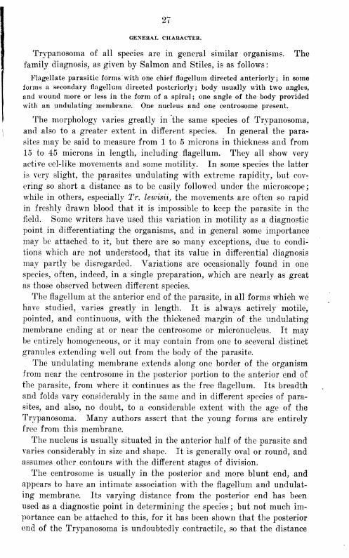
The protoplasm is homogeneous or granular, depending upon the age
• T- •
'"^; ' ' w^- K;^fe^^^^ntA-
to
Figs. 6-12—6, Young adult Tr.equinum; 7, Degeneration forms ; 8, Young T. ypanosoma ; 9, Mul-tinuclear adult Trypanosoma ; 10a, Longitudinal division ; 10ft, Transverse division ; 10c, Multipledivision; 11, Irregular form; 12, Two young Trypanosoma not yet separated. (After Voges, 1901.)

29
of the parasite, its environment and no doubt, to a certain extent, upon
the species. The granules may vary in number and size from a very few
small ones situated in the anterior portion of the Trypanosoma to numer-
ous large ones scattered throughout the protoplasm.
Multiplication.—Yoges gives three forms of multiplication, i. e.,
longitudinal and transverse fission and segmentation. He did not ob-
serve conjugation. The chromatin divides into from 3 to 10 segments,
which assume irregular shapes and locations, and some of which are
often found well up in the flagellum. The nucleus usually divides into
equal parts, but may break into several segments. After division the
protoplasm may assume various irregular forms. The young nuclei
arrange themselves in groups, and the parasite twists and splits by
longitudinal or more often by transverse fission. The new division
forms are often bowl-shaped, but gradually assume their regular outline.
Sometimes a parasite assumes the appearance of a globular mass ; nuclei,
showing a number of flagella, form around the periphery, and division
into several segments occurs.
Plimmer and Bradford consider longitudinal and transverse division
the more frequent modes of reproduction, although they observed also
conjugation, which consisted in the fusion of the micronuclei, followed
by an ameboid stage and division by segmentation. The ameboid stage
at times occurred independently of conjugation.
Martini, who has recently worked with Trypanosoma obtained from
an infected pony imported to Berlin from Togo, gives five stages of
multiplication, as follows : First stage : Broadening out of the chromatin
grains of the nucleus; flagellum thickens; nucleolus appears to be a
thick streak; chromatin granules loosen. Second stage: Two chromatin
heaps; two nuclei; pairs remain together; beginning division of the
undulating membrane. Third stage: Two distinct membranes seen.
Fourth stage : Two flagella, one slightly shorter than the other. Fifth
stage: Young Trypanosoma attached only at the posterior end; some-
times one of these is already seen in the process of fission. He did not
observe any other forms of multiplication or conjugation.
Schilling did not see multiplication forms in the circulating blood in
connection with surra in Togo. He considers the mode of division to
be influenced by the number of chromatin granules found in the para-
site and to be usually by longitudinal fission. He did not observe
ameboid forms or conjugation. He gives two stages in the usual mode
of multiplication. In the first one a double undulating membrane is
seen, and in the second the whole undulating membrane divides longi-
tudinally and gradually separates the parasite, the posterior end being
the last to part. Young forms have no undulating membrane. Daugh-
ter parasites are always smaller than the parents.
Laveran and Mesnil have studied the forms of multiplication very
carefully, and consider that with the Trypanosoma of nagana, multi-
30
plication in the blood is by longitudinal division only and into young
of equal size, which are also nearly as large as the adults. Dividing
forms are always present in the blood, and just before division begins
the parasite increases in size. The order of division is as follows: (1)
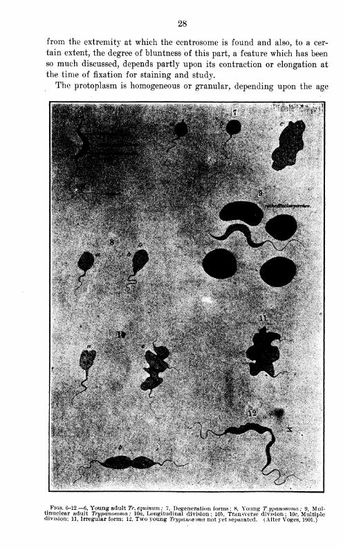
13 14 15 16 17
Figs 13-17.—13, Tr. bmcci (n, nucleus; c, centrosome; m, undulating membrane; /, flagellum); 14,
Beginning division, showing two centrosomes and partial division of flagellum; 15, 16, 17, Further
stages of division. (After Laveran and Mesnil, 1901, figs. 1 to 5.)
Figs 18-28 —T . equinum, showing various stages of division. (After Sivori and Lecler, 1902, PI.
Ill ) 1 Trypanosoma with two chromatin corpuscles of the flagellum; 2 Trypanosoma with
two nuclei ; 3 Trypanosoma with a nucleus, two chromatin corpuscles and a short age urn
which steks from the posterior chromatin corpuscle and is united or not to the other flagellum;
£•7 Trvmnosoma with a large nucleus, slightly elongated, two chromatin corpuscles; two flagella,
one sho?ter than the other Ind united to it or not? 8-10, Large Trypanosoma with two separate
nucllHhe protoplasm is accumulated toward the poles of the nuclei and is rarer in the middle;
"wo flagella^ne longer than the other, or equal and separate; 11, Trypanosoma similar to the
preceding, but the flagellated or anterior extremities have begun to separate.
31
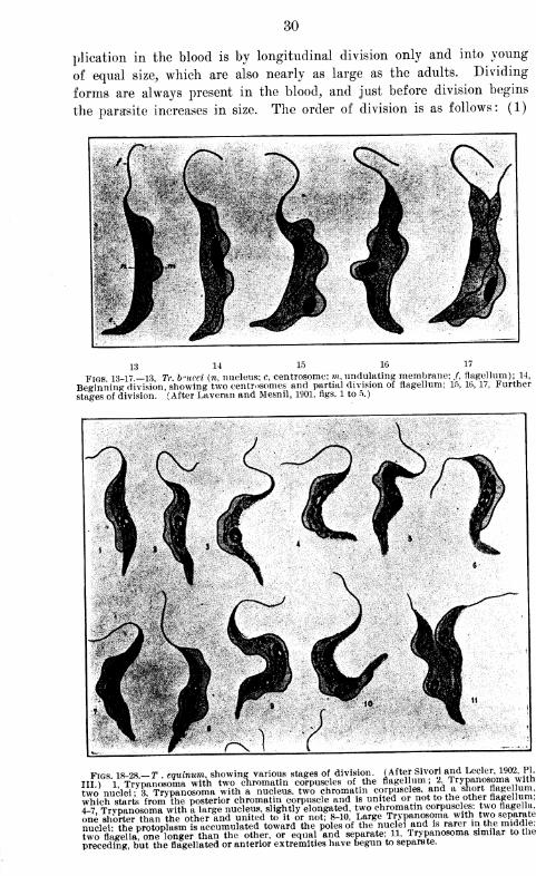
Figs. 29-31.— Tr. evansii, showing dividing forms.
32
Centrosome, (2) flagellum, and (3) nucleus and protoplasm. The cen-
trosome first elongates and divides into two round bodies, followed by
a division of the flagellum. The nucleus increases in size. New nuclei
are then formed by direct division. The protoplasm follows the nucleus
in separation and may begin at the free end. Two parasites may remain
attached at the posterior ends for some time after division, and both
may then divide again before separation is complete. These authors have
not yet seen the young forms of Kanthuds, Durham, and Blandford,
or the ameboid forms of Plimmer and Bradford. They give some varia-
tions from the parasite as described by Lewis, but this point will be
more fully discussed under "Differential diagnosis of Trypanosoma."
Sivori and Lecler agree with Laveran and Mesnil as to the modes of
multiplication illustrated by figs. 18-28.
Rosette formations of Trypanosoma have been extensively noticed, but
considerable difference of opinion as to their cause has been expressed.
Some consider them as entirely a multiplication phase, others as agglu-
tination, while the majority agree that such formations may be the
result of either of these phenomena. There certainly can be no question
that these figures occasionally result as a phase of multiplication. Rabi-
nowitsch and Kempner compare them to the segmenting malarial
parasite.
The methods of reproduction described comprise those of the most
importance and represent the views of many of the writers whom wehave been able to review. Schat, as has been seen in the discussion of
the life cycle of Trypanosoma, holds some very original opinions. So
far as his work has to do with multiplication, he maintains that the
asexual, longitudinal division occurs in the blood of infected animals
and that the sexual reproduction takes place in certain flies.
In our studies we have never observed conjugation, and in blood under
normal conditions reproduction by transverse division or segmentation
is very rare. Longitudinal division is by far the most frequent form,
and usually takes place in the order given by Laveran and Mesnil. This
is not constant, however, for in the same specimen of the parasites taken
from the blood of an infected dog, horse, or other animal, we have seen
individuals showing this order and others in which the division certainly
differed from the course described by these authors. (Figs. 32-35 illus-
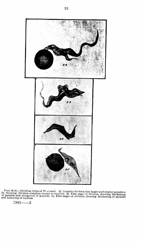
trate this point.) Elmassian, working with the South American disease,
has recently reported results similar to ours.

20° 60" Ostl L v. 80° Greenwich 100° 100' Westl L.v. 80* Greenwich 60°

38
3S <Sin^w"7Slf«lng formf
!of Tr - er
!in
?u
- <?2 >Irr^ular division into larger and smaller parasites;
of'n^JESwS ?°n c°mPle^ except at nucleus; 34, First stage of division, showing thickening
anISseningof'nuie^ ^ ^ First sta*es of divisi™> sh™inS thickenhfg of parasftl
7881 3
84
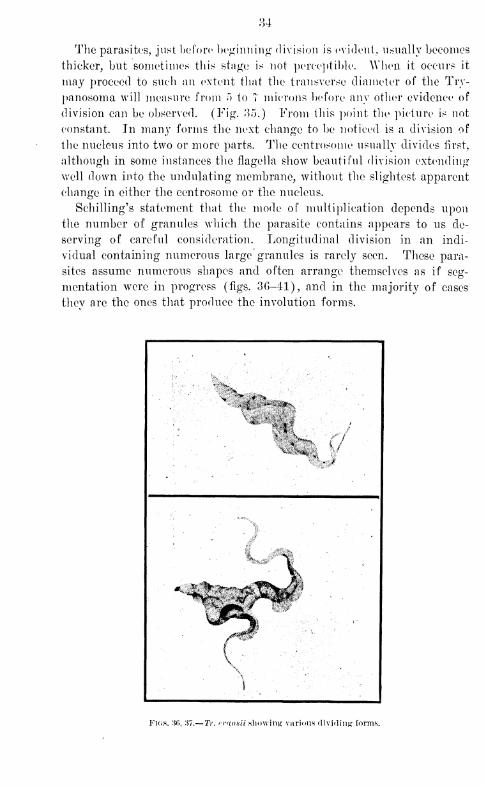
The parasites, just before beginning division is evident, usually becomes
thicker, but sometimes this stage is not perceptible. When it occurs it
may proceed to such an extent that the transverse diameter of the Try-
panosoma will measure from ~> to T microns before any other evidence of
division can be observed. (Fig. 35.) From this point the picture is not
constant. In many forms the next change to be noticed is a division of
the nucleus into two or more parts. The centrosome usually divides first,
although in some instances the flagella show beautiful division extending
well down into the undulating membrane, without the slightest apparent
change in either the centrosome or the nucleus.
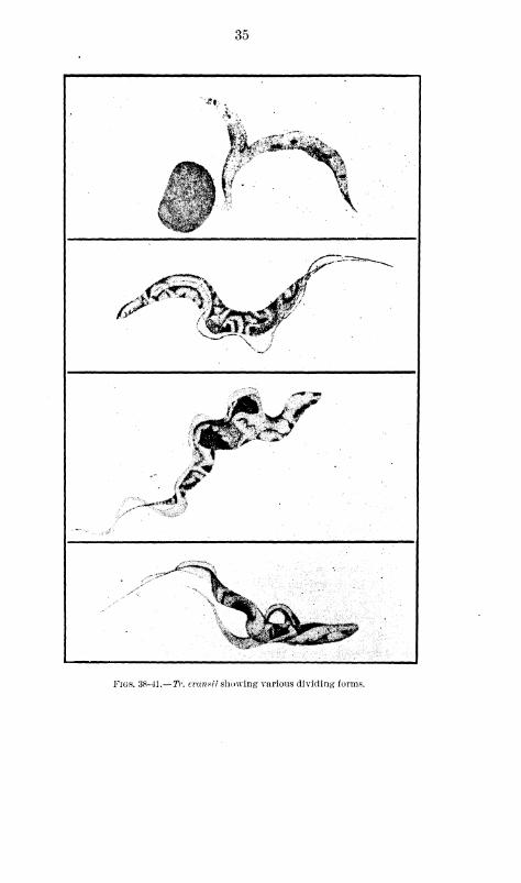
Schilling's statement that the mode of multiplication depends upon
the number of granules which the parasite contains appears to us de-
serving of careful consideration. Longitudinal division in an indi-
vidual containing numerous large granules is rarely seen. These para-
sites assume numerous shapes and often arrange themselves as if seg-
mentation were in progress (figs. 36-41), and in the majority of cases
they are the ones that produce the involution forms.
K.*l^x
*idP
)
Fk;s. 3f>, 37.— TV. rv<innii showing various dividing forms.
35
*i .
> *$SP** j
N -
^%
Figs. 38-41.— TV-, evanvii showing various dividing forms.
36
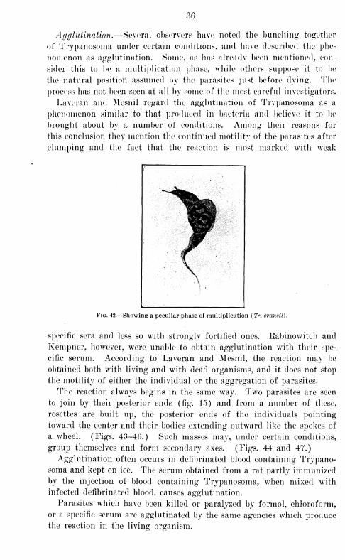
Agglutination.—Several observers have noted the bundling together
of Trypanosoma under certain conditions, and have described the phe-
nomenon as agglutination. Some, as has already been mentioned, con-
sider this to be a multiplication phase, while others suppose it to be
the natural position assumed by the parasites just before dying. The
process has not been seen at all by some of the most careful investigators.
Laveran and Mesnil regard the agglutination of Trypanosoma as a
phenomenon similar to that produced in bacteria and believe it to be
brought about by a number of conditions. Among their reasons for
this conclusion they mention the continued motility of the parasites after
clumping and the fact that the reaction is most marked with weak
Fig. 42.—Showing a peculiar phase of multiplication(Tr. evansii).
specific sera and less so with strongly fortified ones, llabinowiteh and
Kempner, however, were unable to obtain agglutination with their spe-
cific serum. According to Laveran and Mesnil, the reaction may be
obtained both with living and wTith dead organisms, and it does not stop
the motility of either the individual or the aggregation of parasites.
The reaction always begins in the same way. Two parasites are seen
to join by their posterior ends (fig. 45) and from a number of these,
rosettes are built up, the posterior ends of the individuals pointing
toward the center and their bodies extending outward like the spokes of
a wheel. (Figs. 43-46.) Such masses may, under certain conditions,
group themselves and form secondary axes. (Figs. 44 and 47.)
Agglutination often occurs in defibrinated blood containing Trypano-
soma and kept on ice. The serum obtained from a rat partly immunizedby the injection of blood containing Trypanosoma, when mixed with
infected defibrinated blood, causes agglutination.
Parasites which have been killed or paralyzed by formol, chloroform,
or a specific serum are agglutinated by the same agencies which produce
the reaction in the living organism.

Fh;s. 48-44.—Showing union of two Tragglomeration. (After Stiles and Salmon, 1902, PI. IJ.)
l/pavosowa. 48, Primary agglomeration ; 44, Secondary
38
Agglutinations often are not permanent, and under certain conditions,
according to Lave ran and Mesnil, "disagglonieration" takes place. In
this the secondary formations are first broken up, and the primery rosettes
disunite or lose a part of their elements. They consider this "disag-
glomeration" to be in inverse ratio to the agglutinating value of the
serum employed.
Normal rat's blood has no agglutinative action, but when fortified
by inoculations does gain this power. Five to ten c. c. of Trvpanosomatic
blood injected into a rat will produce a serum capable of agglutinating
Trypanosoma in deflbrinated blood in a dilution of 1-5 to 1-50.
One of Laveran and Mesnil's rats, which in seven months had received
13 inoculations of blood containing Trypanosoma, gave a serum which in
a dilution of 1-10 so paralyzed the Trypanosoma that rosettes were not
formed.
Serum exposed to a temperature of 55° to 58° C. during one-half
to three-fourths of an hour did not lose its power to agglutinate, but
was materially weakened. Exposure to 63° to 65° C. for half an hour
completely destroyed its agglutinative properties.
Adult guinea pigs were immunized by several injections of infected
blood. Their serum had a feeble agglutinative reaction for Tr. brucei.
With a similar serum from young guinea pigs no agglutinative reaction
was obtained. The serum of a pigeon, guinea pig, or frog did not show
an agglutinative reaction for Tr. letvisii, but that of a sheep, dog, or rabbit
gave a slight one for these parasites. With sera from the horse and the
chicken agglutinations were more definite and occurred in dilutions of
1-2 to 1-10.
Of all the animals examined, the serum from the horse was the most
active and that of the chicken second, but in both of these the reaction was
greater for red blood cells than for Trypanosoma. Human serum did
not agglutinate Tr. brucei, but the sera of guinea pigs and of pigs, which
have no curative properties, gave beautiful agglutinations when mixed
with trypanosomatic blood. This would seem to prove that agglutinat-
ing and curative properties are separate and distinct. Agglutinations
once formed had a tendency to disagglutinate in most sera as well as
in other substances. In the rabbit this was accomplished at the end of
several hours. They persisted best in the sera of the dog and the sheep.
Eats immunized by repeated injections of Tr. lewisii showed but feeble
agglutinative reaction with their own parasites.
According to Rost, surra blood mixed with goat serum in the hanging
drop in a moist chamber killed the Trypanosoma in two and one-half
minutes, sometimes with agglutination; control parasites were all dead
in twenty-three hours.
Sivori and Lecler, in a preparation of horse's blood containing numer-
ous Trypanosoma, sometimes observed two, three, and even six individ-
uals or more, united at their posterior extremities and arranged in a
:\\)
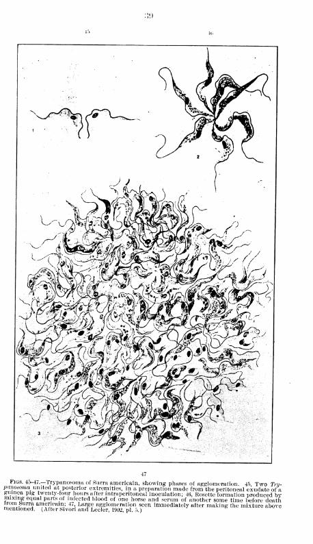
Figs. 4o-47—Trypanosoma of Surra americain, showing phases of agglomeration. 45 Two Try-panosoma united at posterior extremities, in a preparation made from the peritoneal exudate of aguinea pig twenty-four hours after intraperitoneal inoculation; 46, Rosette formation produced bvmixing equal parts of infected blood of one horse and serum of another some time before deathirom Surra americain: 47, Large agglomeration seen immediately after making the mixture abovementioned. (After Sivon and Lecler, 1902, pi. 5.)
40
radiate figure. The center of the figure was sometimes near a red cor-
puscle or a leucocyte. The parasites so united preserved their motility.
In the blood of a young cat, containing numerous Trypanosoma and
prepared in a hanging drop, there were visible at the end of an hour 8,
10, or VI agglomerated parasites. Many of these agglomerations sep-
arated after a certain length of time.
Laveran and Mesnil write:
The Trypanosoma of nagana sometimes unite; under certain conditions they
form primary agglomerations in rosettes; rarely large secondary agglomerations,
which are common in blood containing Tr. Icirisii, are observed.
These Trypanosoma united two by two would suggest conjugation, but this
interpretation is not admissible, as the agglomeration is not observed in pure,
fresh blood, and is produced only under conditions which may be called abnormal.
The number of individuals which agglomerate is exceedingly variable.
In Tr. bruccii, as in Tr. larisii, the agglomerations may be seen to separate
after varying lengths of time.
We have seen agglomerations of Trypanosoma in the pure blood taken from
the heart, after one-half to one hour, in the peritoneal exudates, after an injection
of blood rich in Trypanosoma into the peritoneum of rats or mice, and in blood
mixed with physiologic water after being preserved for twenty-four hours on
ice or heated for half an hour at 41° C.
On mixing, in equal parts, the defibrinated blood of a rat or mouse, rich in
Trypanosoma, and the serum of a horse, we have obtained beautiful persistent
agglomerations. The Trypanosoma separated at the end of a few hours. Onmixing one part of the serum of a horse and ten parts of blood no agglomera-
tions were produced. The serum of the blood of a pig also gave beautiful
agglomerations.
The serum of a sheep, mixed in equal parts with the blood of a rat or mouse,
rich in Trypanosoma, gave, in one case, a beautiful agglomeration; in another
the agglomerations were not so beautiful and less persistent. The serum of a
deer gave small nonpersistent agglomerations.
The serum of human blood did not show itself either agglutinative or
microbicidal.
The following sera mixed in equal parts with the blood of a rat or mouse,
rich in Tr. bruccii. did not show any agglutinative properties: The serum of a
rat, normal or immunized against Tr. Icirisii, and agglutinative for these
Trypanosoma, the scrum of a normal chicken, the serum of a chicken inoculated
several times with Tr. bruccii. the serum of a normal goose, and the serum of a
goose inoculated several times with blood rich in Trypanosoma of nagana.
If there is added to a few drops of blood rich in Tr. bruccii a drop of water
slightly acidulated with acetic acid, Trypanosoma are seen to agglomerate and
change their forms rapidly. On adding a drop of water slightly alkalized with
soda no agglomeration occurs.
Trypanosoma when dead still tend to agglomerate, but the process then takes
place very irregularly.
Hefferan, commenting on Laveran and Mesnil's statements regarding
agglutination of Tr. Icirisii, doubts the correctness of their observations,
giving her reasons for so doing. (Central!), f. P>akt., etc., hd. tt, \o. 22,
May 2f>, 1902.)
Curry noted that parasites in infected monkeys' blood mixed with
41
human blood lost their motility in twenty minutes and agglutinated.
Chicken's blood mixed with infected monkeyV blood gave similar results.
Schilling states that in cattle immunized with the peritoneal exudate
of dogs inoculated with infected blood, the serum killed the Trypano-
soma on the fourteenth and iifteentb days, and in the hanging drop in
from thirteen to twenty-five minutes; but he lias little to say of agglu-
tination.
On reviewing the work done on the agglutination of Trypanosoma, it
will be seen that results have been uncertain and inconstant, the subject
being left in an unsatisfactory state.
HefYeran's criticisms of Laveran and MesniTs work in this line, and
the statement of Rabinowitch and Kempner that no agglutinations wen 4
obtained with their specific serum makes the value of other results doubt-
ful.
So far our work lias developed nothing convincing. We have seen
the rosettes and other described figures of agglutination, but they have
been too inconstant and have occurred under too many conditions to
be of any very great significance. Circumstances under which these
figures have at one time appeared have at other times produced no
results; and they have even occurred under conditions which are not
supposed to favor agglutination.
Our results in the agglutination of 7Y. evansii by various substances
described as producing this phenomenon have been at variance with
much of the recent work done along this line and more in accord
with Rabinowitch and Kempner's conclusions. We have not observed
a single condition which constantly gave agglutination figures. Such
results were obtained occasionally with various substances, but reactions
indistinguishable from these sometimes occur in infected blood without
any additions.
Cow No. 158 was immunized up to 3,000 c. c. doses of infected
blood and failed to produce a serum which would agglutinate Try-
panosoma with any degree of constancy. Similar results were obtained
with chicken and human serum as well as writh those secured from'
numerous other sources. Various mixtures of these sera were likewise
unsatisfactory. Several chemicals, such as thymol, turpentine, and
chloral, would occasionally give what appeared to be agglutination;
but no regularity could be observed.
After weighing all evidence in the case and applying our own results,
Ave must conclude with several others that the so-called phenomenon
of agglutination is of no value from a diagnostic point of view; and
if it is in reality an agglutination, it is too uncertain in its occurrence
to serve as an index of immunity or susceptibility.
Involution forms.—Involution forms are produced by surroundings
unfavorable to the life of the parasite. Laveran and Mesnil mention
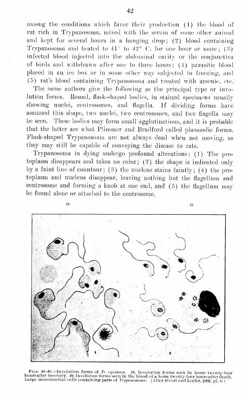
42
among the conditions which favor their production (1) the blood of
rat rich in Trypanosoma, mixed with the serum of some other animal
and kept for several hours in a hanging drop; (2) blood containing
Trypanosoma and heated to 41~ to 42° (\ for one hour or more; (:>)
infected blood injected into the abdominal cavity or the conjunctiva
of birds and withdrawn after one to three hours; (1) parasitic blood
placed in an ice box or in some other way subjected to freezing, and
(5) rat's blood containing Trypanosoma and treated with arsenic, etc.
The same authors give the following as the principal type or invo-
lution forms. Round, flask-shaped bodies, in stained specimens usually
showing nuclei, centrosomes, and flagella. If dividing forms have
assumed this shape, two nuclei, two centrosomes, and two flagella maybe seen. These bodies may form small agglutinations, and it is probable
that the latter are what Plimmer and Bradford called plasmodic forms.
Flask-shaped Trypanosoma are not always dead when not moving, as
they may still be capable of conveying the disease to rats.
Trypanosoma in dying undergo profound alterations: (1) The pro-
toplasm disappears and takes no color; (2) the shape is indicated only
by a faint line of countour; (3) the nucleus stains faintly; (4) the pro-
toplasm and nucleus disappear, leaving nothing but the flagellum andcentrosome and forming a knob at one end, and (5) the flagellum maybe found alone or attached to the centrosome.
*ios 48-49.—Involution forms of Tr. e.qxinnm. 48, Involution forms seen in horse twentv-fourhours after recovery; 49, Involution forms seen in the blood of a horse twentv-four hours after death.Large mononuclear cells containing parts of Trypanosoma. (After Sivori and Lecler, 1902 pi (i )
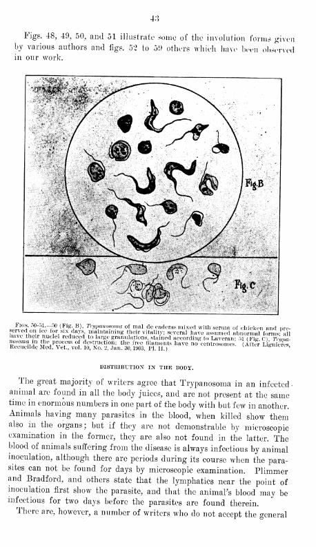
4.°>
Figs. 48, 49, 50, and 51 illustrate some of the involution forms givenby various authors and figs. 52 to 51) others which have been observedin our work.
-;>:><>** ^v^, „ \*".
^\: ji/m; { *../'#*
';*'
.4;'
' -"r ;•' vt .-,
&c
Figs. 50-51.-50 (Fig B), Trypanosoma of mal de eaderas mixed with serum of chicken and pre-served on ice for six days, maintaining their vitality; several have assumed abnormal£ Pihave their nuclei reduced to large granulations, stained according to Laveran: 51 (Fig C) Tnma-ess i^pvx ™°f.ta^^sr have no controsomos - (iit5 ^"^
DISTRIBUTION IN THE BODY.
The great majority of writers agree that Trypanosoma in an infected-animal are found in all the body juices, and are not present at the sametime in enormous numbers in one part of the body with but few in another.Animals having many parasites in the blood, when killed show themalso in the organs; but if they are not demonstrable by microscopicexamination in the former, they are also not found in the latter. Theblood of animals suffering from the disease is always infectious by animalinoculation, although there are periods during its course wdien the para-sites can not be found for days by microscopic examination. Plimmerand Bradford, and others state that the lymphatics near the point ofinoculation first show the parasite, and that the animal's blood may beinfectious for two days before the parasites are found therein.There are, however, a number of writers who do not accept the general
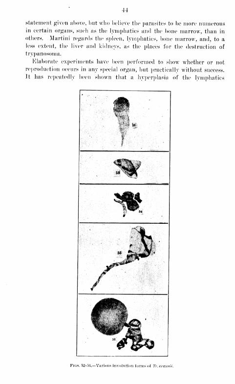
44
statement given above, hut who believe the parasites to be more numerousin certain organs, such as the lymphatics and the bone marrow, than in
others. Martini regards the spleen, lymphatics, bone marrow, and, to a
less extent, the liver and kidneys, as the places for the destruction of
trypanosoma.
Elaborate experiments have been performed to show whether or not
reproduction occurs in any special organ, but practically without success.
It lias repeatedly been shown that a hyperplasia of the lymphatics
: . ^^
if %f|
.-o>;|.:'::] \x:
Figs. 52-f>6.—Various involution forms of Tr.evansii.
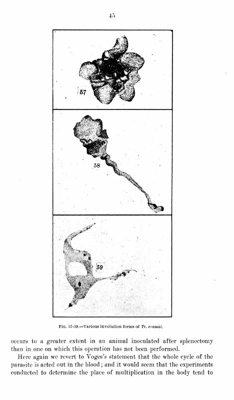
4r>
Fig. 57-59.—Various involution forms of 7V. evansii.
occurs to a greater extent in an animal inoculated after splenectomy
than in one on which this operation has not been performed.
Here again we revert to Voges's statement that the whole cycle of the
parasite is acted out in the blood; and it would seem that the experiments
conducted to determine the place of multiplication in the body tend to

40
support this statement. It is certainly true that the Trypanosoma are
pretty evenly distributed in the body juices and that similar forms are
found in all parts.
Jt seems to be a very generally accepted opinion that Trypanosoma
inoculated into the peritoneal cavity undergo a considerable multiplica-
tion before entering the circulating blood, and by some this time has
been considered as constituting the true period of incubation.
Parasites in the dead body.—Trypanosoma live only a short time in the
body after death. Within two hours signs of degeneration begin; the
parasites shrink, assume irregular shapes and then disappear. Motile
parasites are not usually found two hours post-mortem. Ample work
fully demonstrating this important point has been done. In exceptional
cases living parasites have been found as late as sixteen hours after
death; but this condition is rare 1. Our work in this line has consisted
in determining the longest time post-mortem during which the blood
could be proved infectious by inoculation into susceptible animals.
Results are conclusive that this is rarely greater than twenty-four hours.
However, in one instance blood has been found to convey the disease
forty-eight hours after death.
When an animal, in the blood of which trypanosoma are present,
dies, the parasites are then found in all the organs; and conversely, where
none appear in the former, they are also absent in the latter. They are
generally distributed, and multiplication forms do not appear in ex-
ceptional numbers in any one organ; however, they are usually somewhat
more numerous in the spleen, liver, and lymphatic glands than in the
bone marrow, and are seldom present in the medullary canal. They are
found in the serous fluids and exudates of the joints, but rarely in the
urine.
Schilling's results were somewhat exceptional. He says that "Try-
panosoma were not found in the spleen when positive in the blood, and
the peritoneal exudate and bone marrow showed parasites of a budding
form;" and again, "that Trypanosoma might be absent from the fluids
and tissues, but were constantly present in the bone marrow. The
number of parasites in the spleen varied greatly, but there was never a
great accumulation or multiplication of forms.7
' He draws the con-
clusion that multiplication of parasites occurs in certain organs, while
others destroy them.
TRYPANOSOMA OUTSIDE OF THE I10DY.
Although Trypanosoma in a natural condition are not known outside
the body and propagate only to a limited degree in any known artificial
media, yet under favorable conditions they can be kept for a consid-
erable time outside the body, a fact which has been believed to afford
certain diagnostic points for different species. Berg kept Trypanosoma
of fish for six days in blood at 12° C, and Mitraphanow from three to
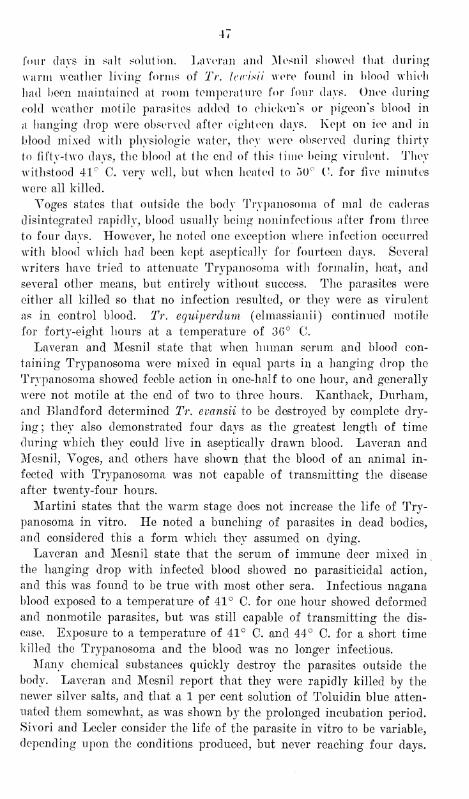
four (lavs in salt solution. Laveran and Mesnil showed that during
warm weather living- forms of Tr. Icirisii were found in blood which
had been maintained at room temperature for four days. Once during
cold weather motile parasites added to chicken's or pigeon's blood in
a hanging drop were observed after eighteen days. Kept on ice and in
blood mixed with physiologic water, they were observed during thirty
to fifty-two days, the blood at the end of this time being virulent. They
withstood 41° C. very well, but when heated to 50° C. for five minutes
were all killed.
Yoges states that outside the body Trypanosoma of mal de caderas
disintegrated rapidly, blood usually being noninfectious after from three
to four days. However, he noted one exception where infection occurred
with blood which had been kept aseptically for fourteen days. Several
writers have tried to attenuate Trypanosoma with formalin, heat, and
several other means, but entirely without success. The parasites were
either all killed so that no infection resulted, or they were as virulent
as in control blood. Tr. equiperdum (elmassianii) continued motile
for forty-eight hours at a temperature of 3G° C.
Laveran and Mesnil state that when human serum and blood con-
taining Trypanosoma wrere mixed in equal parts in a hanging drop the
Trypanosoma showed feeble action in one-half to one hour, and generally
were not motile at the end of twro to three hours. Kanthack, Durham,
and El and ford determined Tr. evansii to be destroyed by complete dry-
ing; they also demonstrated four days as the greatest length of time
during which they could live in aseptically drawn blood. Laveran and
Mesnil, Voges, and others have shown that the blood of an animal in-
fected with Trypanosoma was not capable of transmitting the disease
after twrenty-four hours.
Martini states that the wTarm stage does not increase the life of Try-
panosoma in vitro. He noted a bunching of parasites in dead bodies,
and considered this a form which they assumed on dying.
Laveran and Mesnil state that the serum of immune deer mixed in
the hanging- drop with infected blood showed no parasinoidal action,
and this was found to be true wT
ith most other sera. Infectious nagana
blood exposed to a temperature of 41° C. for one hour showed deformed
and nonmotile parasites, but was still capable of transmitting the dis-
ease. Exposure to a temperature of 41° C. and 44° C. for a short time
killed the Trypanosoma and the blood wras no longer infectious.
Many chemical substances quickly destroy the parasites outside the
body. Laveran and Mesnil report that they were rapidly killed by the
newer silver salts, and that a 1 per cent solution of Toluidin blue atten-
uated them somewhat, as was shown by the prolonged incubation period.
Sivori and Lecler consider the life of the parasite in vitro to be variable,
depending upon the conditions produced, but never reaching four days.
48
Schilling noted that Trypanosoma wore soon destroyed by a GO per
cent solution of bile.
Bruce showed that dried blood was infectious after twenty-four hours
in one out of three experiments, with an incubation period of sixteen
days. In two cases it was not infectious at the end of forty-eight hours.
Aseptically drawn, virulent blood was infectious for four days, and after
seven did not produce the disease.
Laveran and Mesnil state that the movements of Trypanosoma are
retarded by cooling and accelerated by warming the blood in which they
are contained. They were not immediately killed by a temperature of
50° C. to 55° C. below zero. Their experiments were as follows:
Experiment I.—Eat blood with many Tr. bruceii diluted with potas-
sium citrate solution and kept at 18° C. for one-half hour. One and
one-half hours after returning to room temperature it still showed many
normal looking motile Trypanosoma. Mice injected in the conjunctiva
with this blood died in the usual time with Trypanosomiasis.
Experiment II.—Similar dilutions of blood exposed twenty minutes
to 15° C. and eight minutes to 25°-30°. After two hours warmed
blood showed normal looking motile Trypanosoma and was infectious
in the usual time for mice.
Experiment III.—Similar solution of blood exposed for one-half hour
at 15° C. and five minutes at 50°-55° C. After twro hours the thawed
and warmed blood contained normal looking and motile Trypanosoma
and was infectious for mice in the usual time.
Experiment IV.—Same as No. 3, except that the freezing and thaw-
ing was repeated. It was still pathogenic for mice, but was slightly
slower in its action.
These authors demonstrated that blood infected with Tr. brucei heated
three hours at 40° C. or one hour at 42° was still virulent. Blood in-
fected with Tr. brucei heated twenty minutes at 40° to 44° C. killed
nearly all the Trypanosoma, and when heated to 44° to 45° C. all the
parasites wTere quickly destroyed.
Blood taken aseptically and mixed with citrate solution at room
temperature, according to Laveran and Mesnil, was virulent for three
days, and Trypanosoma lived longer in a mixture of blood and serum
than in blood alone. In the defibrinated blood of a rat mixed with the
serum of the host, motile Trypanosoma were still observed after three
days.
Human serum and that of refractory animals was not considered less
adapted to the preservation of Trypanosoma than that of more susceptible
animals. Tr. lewisii lived longer on ice than at room temperature ; but
this was not true of Tr. brucei. Blood containing Tr. brucei, after being
kept on ice for three to five days, was often noninfectious, though it still
contained slightly motile Trypanosoma. Involution forms quickly ap-
49
pearod in blood kept on ice, their morphology differing in no respect
from that of the involution forms produced by other causes.
We have experimented extensively with the object of determining
the length of life in vitro of the Trypanosoma with which we have
been able to work, and on the whole have obtained results similar to
those arrived at by most recent writers on other Trypanosoma.
Our experiments made to determine the action of heat and cold on
parasites confirm, in the main, the conclusions drawn by Laveran and
Mesnil, including the differentiation of Tv. lewisii by its ability to live
longer than Tr. evansii in the ice box. We failed to find any constancy
in the agglutinations these authors describe. They did occasionally
occur, following exposure to conditions especially adverse to life; but
they were not constant and also took place in the hanging drop. We can
not attach to this phenomenon the importance given it by some authors.
Several specific sera mixed in the hanging drop in equal parts with
blood rich in Trypanosoma gave no appreciable results, with the possible
exception of the mixtures containing antiplague and antirinderpest sera.
The Trypanosoma were usually nonmotile in the plague serum at the
end of forty minutes, and in the rinderpest serum sometimes as early
as thirty minutes. In two out of five experiments made with the latter,
the blood was noninfectious for rats at the end of one hour. In most
of the experiments made with serum the parasites lived as long as in
the control drop, and in some instances much longer.
A 1-500 solution of quinine mixed in equal parts with blood con-
taining Trypanosma arrested the motility of the parasites in from five
to ten minutes. With a 1-1,000 solution of methylene blue the Trypano-
soma lost their motility from fi\e to twenty minutes earlier than in the
control. They are less affected by solutions of alcohol, glycerin, or
ether.
Xo perceptible action was produced an the Trypanosoma by mixing
the infected blood with equal parts of the following substances : A 1-1,000
solution of acetozone; 1-1,000 solutions of the soluble eosins; alcohol;
potassium acetate; potassium chlorate; potassium cyanide; salt solu-
tion; picric acid; oxalic acid; and the chlorides of magnesium, calcium,
and barium. Indeed, in many of these solutions the parasites remainedactive longer than in control.
The Trypanosoma were quickly destroyed by mixing infected bloodin the hanging drop with equal parts of the following substances: A1-1,000 solution of arsenious acid; a 1-1,000 solution of turpentine; a
1-1,000 solution of corrosive sublimate; a 1-500 solution of chloral
hydrate; a 1-500 solution of carbolic acid; a 1-500 solution of formalin;a 1-1,000 solution of potassium permanganate; and a 1-200 solution
of quinine.
7881 4
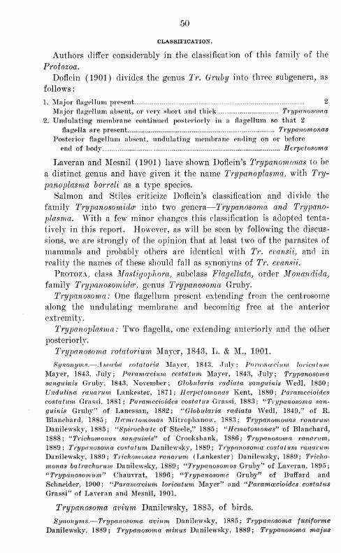
50
CLASSIFICATION.
Authors differ considerably in the classification of this family of the
Protozoa.
Doflein (1901) divides the genus Tr. Gruhy into three subgenera, as
follows
:
1. Major flagellum present 2
Major flagellum absent, or very short and thick Trypanosoma
2. Undulating membrane continued posteriorly in a flagellum so that 2
flagella are present Trypanomonas
Posterior flagellum absent, undulating membrane ending on or before
end of body Herpctosoma
Laveran and Mesnil (1901) have shown Doflein's Trypanomonas to be
a distinct genus and have given it the name Trypanoplasma, with Try-
panoplasma borreli as a type species.
Salmon and Stiles criticize Doflein's classification and divide the
family Trypanosomidce into two genera
—
Trypanosoma and Trypano-
plasma. With a few minor changes this classification is adopted tenta-
tively in this report. However, as will be seen by following the discus-
sions, we are strongly of the opinion that at least two of the parasites of
mammals and probably others are identical with Tr. evansii, and in
reality the names of these should fall as synonyms of Tr. evansii.
Protoza, class Mastigophora, subclass Flagellata, order Monandida,
family Trypanosomidce, genus Trypanosoma Gruby.
Trypanosoma: One flagellum present extending from the centrosome
along the undulating membrane and becoming free at the anterior
extremity.
Trypanoplasma: Two flagella, one extending anteriorly and the other
posteriorly.
Trypanosoma rotatorium Mayer, 1843, L. & M., 1901.
Synonyms.—Ama'ba rotatoria Mayer, 1843. July; Pa ram crewm loricatum
Mayer, 1843, July; Paramecium cestatnm Mayer, 1843, July; Trypanosoma
sanguinis Gruby, 1843, November; Globularia radiata sanguinis Wedl, 1850;
Undulina ranarum Lankester, 1871; Herpetomonas Kent, 1880; Paramecioides
costatum Grassi, 1881; Paramecioides costatus Grassi, 1883; ''Trypanosoma san-
guinis Gruby" of Lanessan, 1882; "Globtdaria radiata Wedl, 1849," of R.
Blanchard, 1885; ]Jwmctomonas Mitrophanow, 1883; Trypanomonas ranarum
Danilewsky, 1885; "Spirochete of Steele," 1885; "Hemotomonas" of Blanchard,
1888; "Trichomonas sanguinis" of Crookshank, 1886; Trypanosoma ranarum.
1889; Trypanosoma costatum Danilewsky, 1889; Trypanosoma costatum ranarum
Danilewsky, 1889; Trichomonas ranarum (Lankester) Danilewsky, 1889; Tricho-
monas batrachorum Danilewsky, 1889; "Trypanosomos Gruby" of Laveran, 1895;
"Trypanosomion" Chauvrat, 1896; "Trypanosome Gruby" of Buffard and
Schneider, 1900; "Parametrium loricatum Mayer" and "Paramcecioides costatus
Grassi" of Laveran and Mesnil, 1901.
Trypanosoma avium Danilewsky, 1885, of birds.
Synonyms.—Trypanosoma avium Danilewsky, 1885; Trypanosoma fusiforme
Danilewsky, 1889; Trypanosoma minus Danilewsky, 1889; Trypanosoma majus
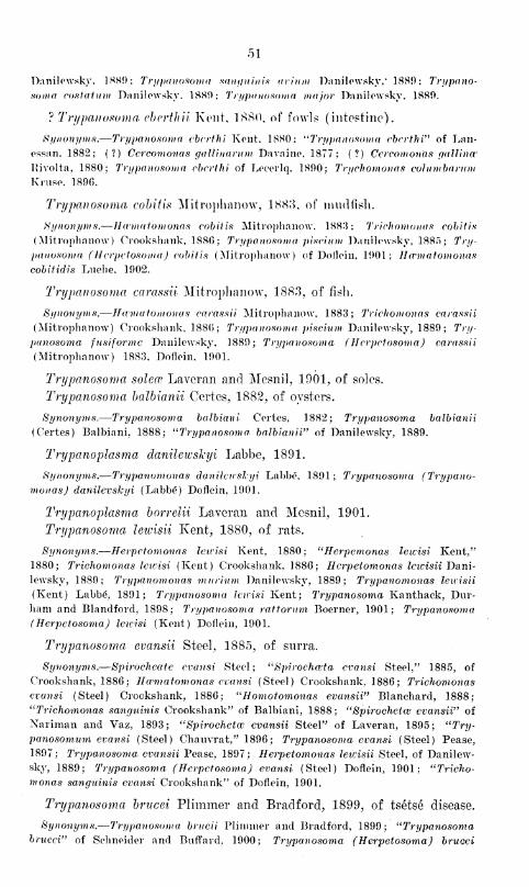
51
Danilewsky, 1889; Trypanosoma sanguinis arinm Danilewsky," 1880; Trypano-
soma costafum Danilowsky. 1889; Trypanosoma major Danilowsky, 1880.
? Trypanosoma cbcrthii Kent, 1880, of fowls (intestine).
Synonyms.—Trypanosoma ebvrthi Kent. 1880; "Trypanosoma cbcrthi" of Lan-
essan, 1882; (?) Ccrcomonas gaUinarum Davaine, 1877; (?) Ccrcomonas gallinw
Kivolta, 1880; Trypanosoma cbcrthi of Lecerlq. 1800; Trichomonas columbarumKmse. 1890.
Trypanosoma cobitis Mitrophanow, 1883, of mudfish.
Synonyms.—Ihvmatomonas cobitis Mitrophanow, 1883: Trichomonas cobitis
(Mitrophanow) Crookshank, 1880; Trypanosoma piscium Danilowsky, 1885; Try-
panosoma ( Hcrpctosoma) cobitis (Mitrophanow) of Dofloin, 1901; Ihvmatomonas
cobitidis Luehe, 1902.
Trypanosoma carassii Mitrophanow, 1883, of fish.
Synonyms.—Haniatomonas carassii Mitroplianow, 1883; Trichomonas carassii
(Mitrophanow) Crookshank, 1880; Trypanosoma piscium Danilowsky, 1889; Try-
panosoma fusiformc Danilewsky, 1889; Trypanosoma (Hcrpctosoma) carassii
(Mitrophanow) 1883, Dofloin, 1901.
Trypanosoma soleo? Laveran and Mesnil, 1901, of soles.
Trypanosoma balbianii Certes, 1882, of oysters.
Synonyms.—Trypanosoma balbiani Cortes, 1882; Trypanosoma balbianii
(Certes) Balbiani. 1888; "Trypanosoma, balbianii" of Danilewsky, 1889.
Trypanoplasma danilewshyi Labbe, 1891.
Synonyms.—Trypanomonas danilewsl-yi Labbe, 1891; Trypanosoma (Trypano-
monas) danilevskyi (Labbe) Dofloin, 1901.
Trypanoplasma borrelii Laveran and Mesnil, 1901.
Trypanosoma lewisii Kent, 1880, of rats.
Synonyms.—Herpctomonas lewisi Kent, 1880; "Herpemonas lewisi Kent/'
1880; Trichomonas letcisi (Kent) Crookshank, 1886; Herpctomonas lewisii Dani-
lewsky, 1889; Trypanomonas murium Danilewsky, 1889; Trypanomonas lewisii
(Kent) Labbe, 1891; Trypanosoma lewisi Kent; Trypanosoma Kanthack, Dur-
ham and Blandford, 1898; Trypanosoma rattorum Boerner, 1901; Trypanosoma(Herpetosoma) lewisi (Kent) Dofloin, 1901.
Trypanosoma evansii Steel, 1885, of surra.
Synonyms.—Spirocheate evansi Steel; "Spirochwta cvansi Steel," 1885, of
Crookshank, 1886; Hcvmatomonas evansi (Steel) Crookshank, 1886; Trichomonas
evansi (Steel) Crookshank, 1886; "Homotomonas evansii" Blanchard, 1888;
"Trichomonas sanguinis Crookshank" of Balbiani, 1888; "Spirochetal evansii" of
Nariman and Vaz, 1893; "Spirochete evansii Steel" of Laveran, 1895; "Try-
panosomum evansi (Steel) Chauvrat," 1896; Trypanosoma evansi (Steel) Pease,
1897; Trypanosoma evansii Pease, 1897; Herpetomonas leicisii Steel, of Danilew-
sky, 1889; Trypanosoma (Hcrpctosoma) evansi (Steel) Doflein, 1901; "Tricho-
mo?ias sanguinis evansi Crookshank" of Dofloin, 1901.
Trypanosoma brucei Plimmer and Bradford, 1899, of tsetse disease.
Synonyms.—Trypanosoma brucii Plimmer and Bradford, 1899; "Trypanosomabrucei" of Schneider and Buffard, 1900; Trypanosoma (HerpetosomaJ brucei
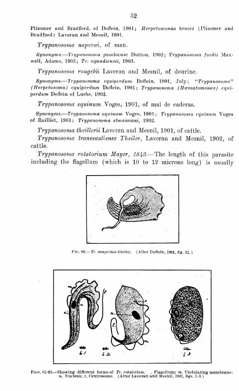
52
Plimmer and Bradford, of Doflein, 1901; Tlcrpctomonas brucei (Plimmer and
Bradford) Laveran and Mesnil, 1901.
Trypanosoma nepveui, of man.
Synonyms.—Trypanosoma gambicnsc Dutton, 1902; Trypanosoma fordii Max-well, Adams, 1903; Tr. ugandiensii, 1903.
Trypanosoma rougetii Laveran and Mesnil, of dourine.
Synonyms.—Trypanosoma cquipcrdum Doflein, 1901, July; "Trypanosoma"(Herpetosoma) equiperdum Doflein, 1901 ; Trypanosoma (Hccmatomonas) equi-
perdum Doflein of Luehe, 1902.
Trypanosoma equinum Voges, 1901, of mal de caderas.
Synonyms.—Trypanosoma equinum Voges, 1901; Trypanosoma equinum Vogesof Railliet, 1901; Trypanosoma elmassiani, 1902.
Trypanosoma thcitterii Laveran and Mesnil, 1901, of cattle.
Trypanosoma transvaaliense Theiler, Laveran and Mesnil, 1902, of
cattle.
Trypanosoma rotatorium Mayer, 1843.—The length of this parasite
including the flagellum (which is 10 to 12 microns long) is usually
Fig. 60.— Tr. sanguinis Gruby. (After Doflein, 1901, rig. 32.
)
Figs. 61-63.—Showing different forms of Tr. rotatorium. , Flagellum; to, Undulating membrane;n, Nucleus; c, Centrosome. (After Laveran and Mesnil, 1901, figs. 1-3.)
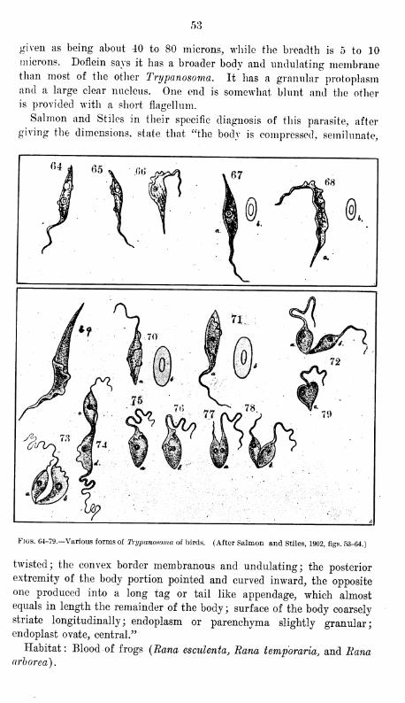
53
given as being about 40 to 80 microns, while the breadth is 5 to 10microns. Doflein says it has a broader body and undulating membranethan most of the other Trypanosoma. It has a granular protoplasmand a large clear nucleus. One end is somewhat blunt and the other
is provided with a short flagellum.
Salmon and Stiles in their specific diagnosis of this parasite, after
giving the dimensions, state that "the body is compressed, semilunate,
Figs. 64-79.—Various forms of Trypanosoma of birds. (After Salmon and Stiles, 1902, figs. 53-64.)
twisted; the convex border membranous and undulating; the posterior
extremity of the body portion pointed and curved inward, the oppositeone produced into a long tag or tail like appendage, which almostequals in length the remainder of the body; surface of the body coarselystriate longitudinally; endoplasm or parenchyma slightly granular;endoplast ovate, central."
Habitat: Blood of frogs (Rana esculenta, Rana temporaria, and Ranaarborea).
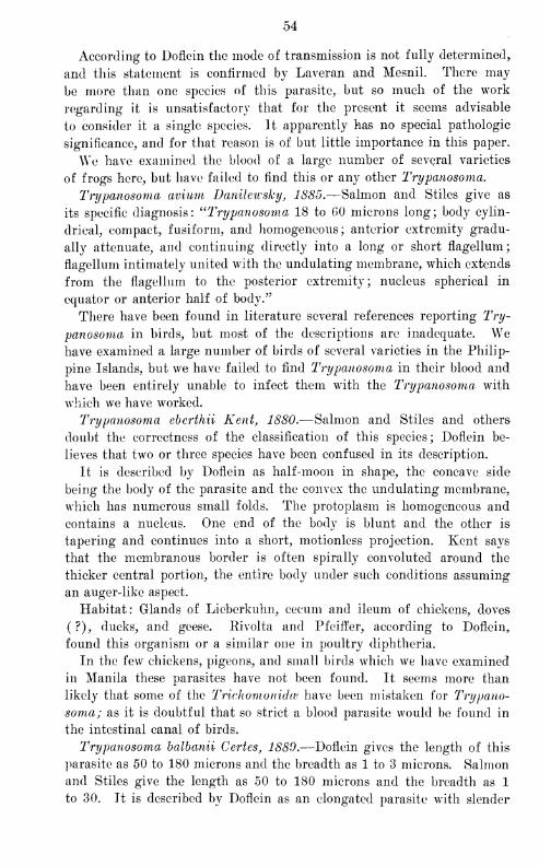
54
According to Doflein the mode of transmission is not fully determined,
and this statement is confirmed by Laveran and Mesnil. There may
be more than one species of this parasite, but so much of the work
regarding it is unsatisfactory that for the present it seems advisable
to consider it a single species. It apparently has no special pathologic
significance, and for that reason is of but little importance in this paper.
We have examined the blood of a large number of several varieties
of frogs here, but have failed to find this or any other Trypanosoma.
Trypanosoma avium Danileicsky, 1885.—Salmon and Stiles give as
its specific diagnosis: ^'Trypanosoma 18 to GO microns long; body cylin-
drical, compact, fusiform, and homogeneous; anterior extremity gradu-
ally attenuate, and continuing directly into a long or short flagellum;
flagellum intimately united with the undulating membrane, which extends
from the flagellum to the posterior extremity; nucleus spherical in
equator or anterior half of body."
There have been found in literature several references reporting Try-
panosoma in birds, but most of the descriptions are inadequate. Wehave examined a large number of birds of several varieties in the Philip-
pine Islands, but we have failed to find Trypanosoma in their blood and
have been entirely unable to infect them with the Trypanosoma with
which we have worked.
Trypanosoma eberthii Kent, 1880.—Salmon and Stiles and others
doubt the correctness of the classification of this species; Doflein be-
lieves that two or three species have been confused in its description.
It is described by Doflein as half-moon in shape, the concave side
being the body of the parasite and the convex the undulating membrane,
which has numerous small folds. The protoplasm is homogeneous and
contains a nucleus. One end of the body is blunt and the other is
tapering and continues into a short, motionless projection. Kent says
that the membranous border is often spirally convoluted around the
thicker central portion, the entire body under such conditions assuming
an auger-like aspect.
Habitat: Glands of Lieberkuhn, cecum and ileum of chickens, doves
( ?), ducks, and geese. Rivolta and Pfeiffer, according to Doflein,
found this organism or a similar one in poultry diphtheria.
In the few chickens, pigeons, and small birds which we have examined
in Manila these parasites have not been found. It seems more than
likely that some of the Trichomonidw have been mistaken for Trypano-
soma; as it is doubtful that so strict a blood parasite would be found in
the intestinal canal of birds.
Trypanosoma balbanii Certes, 1880.—Doflein gives the length of this
parasite as 50 to 180 microns and the breadth as 1 to 3 microns. Salmon
and Stiles give the length as 50 to 180 microns and the breadth as 1
to 30. It is described by Doflein as an elongated parasite with slender
Fig. 80.— TV. cbcrthii. (After Doflein, 1901, fig. 33.)
Fig. 81.— Tr. balbianii Certes. (After Lustrac.)
Fig. 82.— Tr. balbianii Certes. (After Salmon and Stiles, 1902, figs. 76-79.)
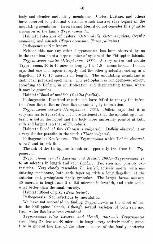
56
body and slender undulating membrane. Certes, Lustrac, and others
have observed longitudinal division, which Lustrac says begins in the
undulating membrane. Laveran and Mesnil do not consider this parasite
a member of the family Trypanosomida.
Habitat: Intestines of oysters (Ostra edulis, Ostra angulata, Grypha
angulata) and mussels (Tapes decussata, Tapes pallustra).
Pathogenesis : Not known.
Neither this nor any other Trypanosoma has been observed by us
in the examination of a large number of oysters of the Philippine Islands.
Trypanosoma cobitis Mitrophanoiv, 1883.—A very active and motile
Trypanosoma, 30 to 40 microns long by 1 to 1.5 microns broad. Doflein
says that one end tapers abruptly and the other gradually, ending in a
flagellum 10 to 15 microns in length. The undulating membrane is
distinct in prepared specimens. The protoplasm is homogeneous, except,
according to Doflein, in multiplication and degenerating forms, where
it may be granular.
Habitat: Blood of mudfish (Cobitis fossilis).
Pathogenesis : Described experiments have failed to convey the infec-
tion from fish to fish or from fish to animals, by inoculation.
Trypanosoma carassii Mitrophanoiv, 1883.—Doflein says that it is
very similar to Tr. cobitis, but more flattened; that the undulating mem-brane is better developed and the body more uniformly pointed at both
ends and larger than that of Tr. cobitis.
Habitat: Blood of fish (Carassius vulgaris). Doflein observed it or
a very similar parasite in the tench (Tinea vulgaris).
Pathogenesis: Not known. The Trypanosoma which Doflein observed
were found in sick fish.
The fish of the Philippine Islands are apparently free from this Try-
panosoma.
Trypanosoma remaki Laveran and Mesnil, 1901.—Trypanosoma 28
to 30 microns in length and very slender. Two sizes and possibly two
varieties. Very closely resembles Tr. lewisii, actively motile, with un-
dulating membrane, both ends tapering with a long flagellum at the
anterior end, protoplasm finely granular. The larger forms measure
45 microns in length and 2 to 2.5 microns in breadth, and stain some-
what better than the small variety.
Habitat: Blood of pike (Esox lucius).
Pathogenesis: Not infectious by inoculation.
We have not succeeded in finding Trypanosoma in the blood of fish
in the Philippine Islands, although several varieties of both salt andfresh water fish have been examined.
Trypanosoma solece Laveran and Mesnil, 1901.—A Trypanosomaresembling Tr. lewisii, 40 microns in length, very actively motile, struc-
ture in general like that of the other members of the family, posterior
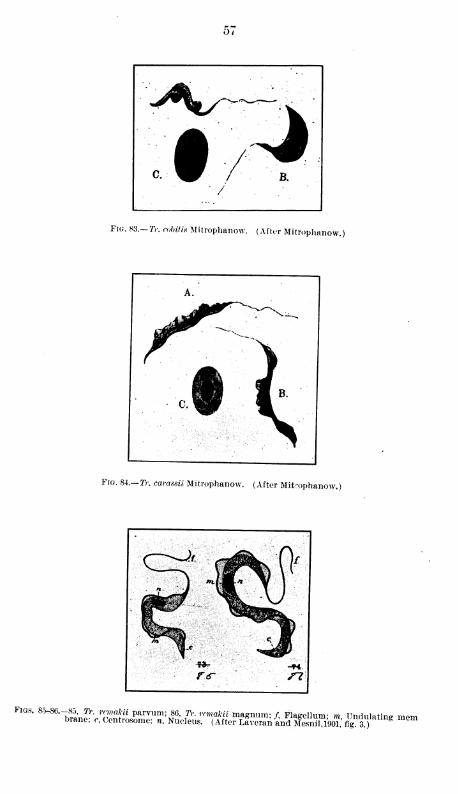
Fig. 83.— TV. cubit is Mitrophanow. (After Mitrophanow.]
Fig. 84.— TV. carassii Mitrophanow. (After Mitrophanow.)
Figs. 8.5-86.-85, Tr. rrmakii parvum; 86, TV. rcmakii magnum; f, Flagellum- m Undulating mpmbrane; c, Centrosome: », Nucleus. (After ilveran and Mesnil 19oT; fig?3 )
g
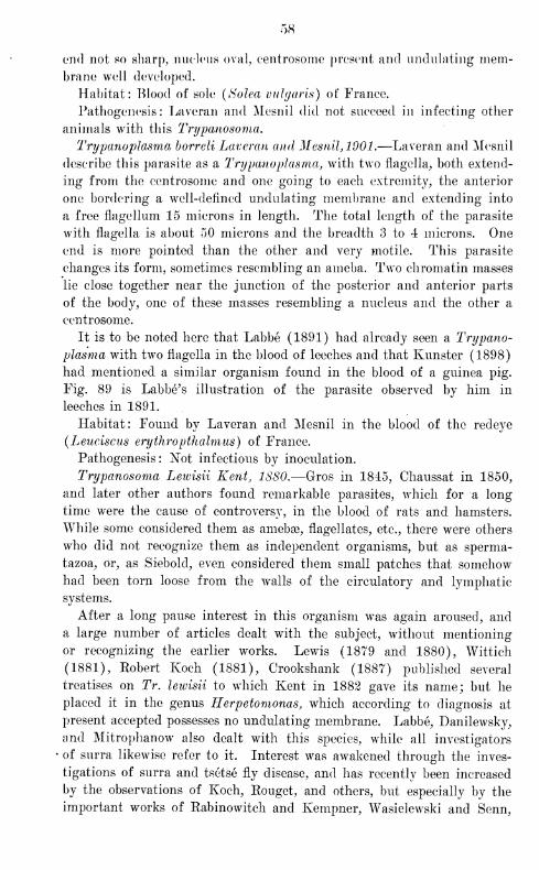
58
end not so sharp, nucleus oval, eentrosome present and undulating mem-brane well developed.
Habitat: Blood of sole (Solea vulgaris) of France.
Pathogenesis: Laveran and Mesnil did not succeed in infecting other
animals with this Trypanosoma,
Trypanoplasma borreli Laveran and Mesnil, 1901.—Laveran and Mesnil
describe this parasite as a Trypanoplasma, with two flagella, both extend-
ing from the eentrosome and one going to each extremity, the anterior
one bordering a well-defined undulating membrane and extending into
a free flagellum 15 microns in length. The total length of the parasite
with flagella is about 50 microns and the breadth 3 to 4 microns. One
end is more pointed than the other and very motile. This parasite
changes its form, sometimes resembling an ameba. Two chromatin masses
lie close together near the junction of the posterior and anterior parts
of the body, one of these masses resembling a nucleus and the other a
eentrosome.
It is to be noted here that Labbe (1891) had already seen a Trypano-
plasma with two flagella in the blood of leeches and that Kunster (1898)
had mentioned a similar organism found in the blood of a guinea pig.
Fig. 89 is Labbe's illustration of the parasite observed by him in
leeches in 1891.
Habitat: Found by Laveran and Mesnil in the blood of the redeye
(Leuciscns erythropthalmus) of France.
Pathogenesis: Not infectious by inoculation.
Trypanosoma Lewisii Kent, 1S80.—Gros in 1845, Chaussat in 1850,
and later other authors found remarkable parasites, which for a long
time were the cause of controversy, in the blood of rats and hamsters.
While some considered them as ameba3, flagellates, etc., there were others
who did not recognize them as independent organisms, but as sperma-
tazoa, or, as Siebolcl, even considered them small patches that somehowhad been torn loose from the walls of the circulatory and lymphatic
systems.
After a long pause interest in this organism was again aroused, and
a large number of articles dealt with the subject, without mentioning
or recognizing the earlier works. Lewis (1879 and 1880), Wittich
(1881), Eobert Koch (1881), Crookshank (1887) published several
treatises on Tr. lewisii to which Kent in 1882 gave its name; but he
placed it in the genus Herpetomonas, which according to diagnosis at
present accepted possesses no undulating membrane. Labbe, Danilewsky,
and Mitrophanow also dealt with this species, while all investigators
of surra likewise refer to it. Interest was awakened through the inves-
tigations of surra and tsetse fly disease, and has recently been increased
by the observations of Koch, Eouget, and others, but especially by the
important works of Rabinowitch and Kempner, Wasielewski and Scnn,
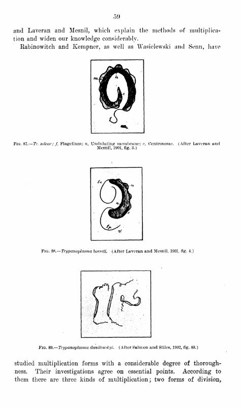
50
and Laveran and Mesnil, which explain the methods of multiplica-
tion and widen our knowledge considerably.
Rabinowitch and Kempner, as well as Wasielewski and Senn, have
Fig. 87.— Tr. solcac; f, Flagellum; n, Undulating- membrane; c, Oentrosonie. (After Laveran andMesnil, 1901, fig. 3.)
Fig. 8S.—Trypanoplasma borrcli. (After Laveran and Mesnil, 1901, fig. 4.)
Fig. 89.— Trypanoplasma danilewd-yi. (After Salmon and Stiles, 1902, fig. 83.)
studied multiplication forms with a considerable degree of thorough-
ness. Their investigations agree on essential points. According to
them there are three kinds of multiplication; two forms of division,
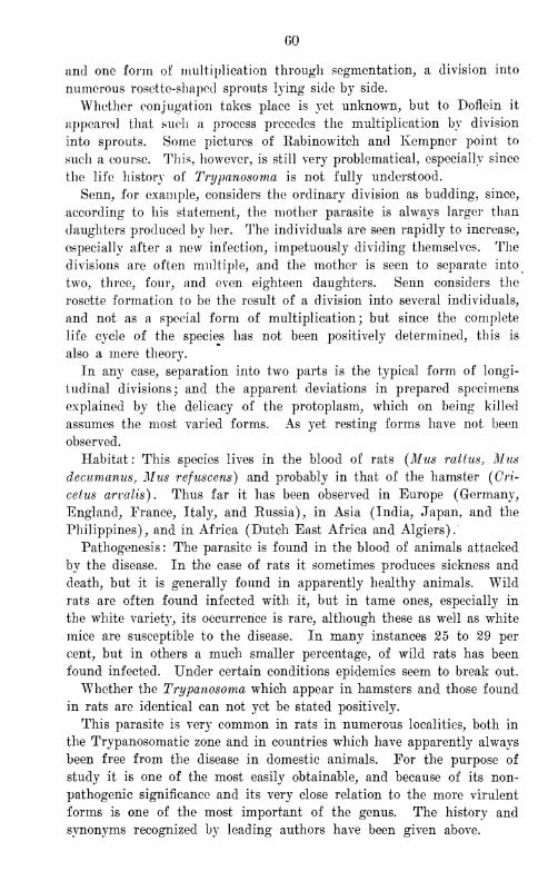
GO
and one form of multiplication through segmentation, a division into
numerous rosette-shaped sprouts lying side by side.
Whether conjugation takes place is yet unknown, but to Doflein it
appeared that such a process precedes the multiplication by division
into sprouts. Some pictures of Rabinowitch and Kempner point to
such a course. This, however, is still very problematical, especially since
the life history of Trypanosoma is not fully understood.
Senn, for example, considers the ordinary division as budding, since,
according to his statement, the mother parasite is always larger than
daughters produced by her. The individuals are seen rapidly to increase,
especially after a new infection, impetuously dividing themselves. The
divisions are often multiple, and the mother is seen to separate into
two, three, four, and even eighteen daughters. Senn considers the
rosette formation to be the result of a division into several individuals,
and not as a special form of multiplication; but since the complete
life cycle of the species has not been positively determined, this is
also a mere theory.
In any case, separation into two parts is the typical form of longi-
tudinal divisions; and the apparent deviations in prepared specimens
explained by the delicacy of the protoplasm, which on being killed
assumes the most varied forms. As yet resting forms have not been
observed.
Habitat : This species lives in the blood of rats (Mus rattus, Musdecumanus, Mus refuscens) and probably in that of the hamster (Cri-
cetus arvalis). Thus far it has been observed in Europe (Germany,
England, France, Italy, and Eussia), in Asia (India, Japan, and the
Philippines), and in Africa (Dutch East Africa and Algiers).
Pathogenesis : The parasite is found in the blood of animals attacked
by the disease. In the case of rats it sometimes produces sickness and
death, but it is generally found in apparently healthy animals. Wild
rats are often found infected with it, but in tame ones, especially in
the white variety, its occurrence is rare, although these as well as white
mice are susceptible to the disease. In many instances 25 to 29 per
cent, but in others a much smaller percentage, of wild rats has been
found infected. Under certain conditions epidemics seem to break out.
Whether the Trypanosoma which appear in hamsters and those found
in rats are identical can not yet be stated positively.
This parasite is very common in rats in numerous localities, both in
the Trypanosomatic zone and in countries which have apparently always
been free from the disease in domestic animals. For the purpose of
study it is one of the most easily obtainable, and because of its non-
pathogenic significance and its very close relation to the more virulent
forms is one of the most important of the genus. The history and
synonyms recognized by leading authors have been given above.
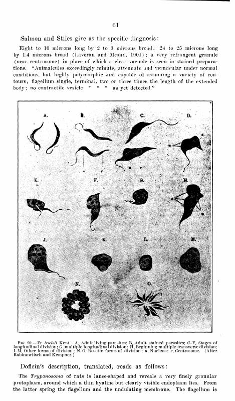
()1
Salmon and Stiles give as the specific diagnosis
:
Eight to 10 microns long by 2 to 3 microns broad ; 24 to 25 microns long
by 1.4 microns broad (Laveran and Mesnil, 1901); a very refrangcnt granule
(near centrosome) in place of which a clear vacuole is seen in stained prepara-
tions. "Animalcules exceedingly minute, attenuate and vermicular under normal
conditions, but highly polymorphic and capable of assuming a variety of con-
tours; flagellum single, terminal, two or three times the length of the extended
body; no contractile vesicle * * * as yet detected.''
K.
?&ir
Fig. 90.— TV. Icwisii Kent. A, Adult living parasites; B, Adult stained parasites; C-F, Stages oflongitudinal division; G, multiple longitudinal division; H, Beginning multiple transverse division;I-M, Other forms of division; N-O, Rosette forms of division; n, Nucleus; c, Centrosome. (AfterRabinowitsch and Kempner.)
Doflein's description, translated, reads as follows
:
The Trypanosoma of rats is lance-shaped and reveals a very finely granular
protoplasm, around which a thin hyaline but clearly visible endoplasm lies. Fromthe latter spring the flagellum and the undulating membrane. The flagellum is
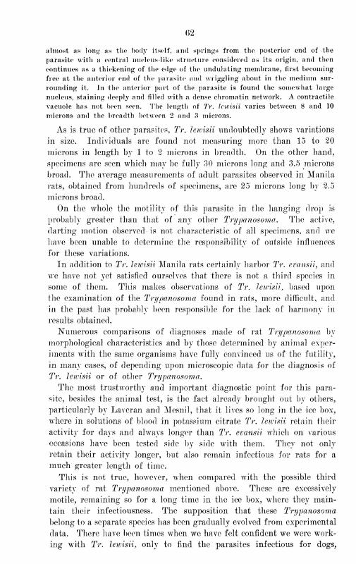
(>2
almost as long as tfio body itself, and springs from the posterior end of the
parasite with a central nucleus-like structure considered as its origin, and then
continues as a thickening of the edge of the undulating membrane, first becoming
free at the anterior end of the parasite and wriggling about in the medium sur-
rounding it. In the anterior part of the parasite is found the somewhat large
nucleus, staining deeply and filled with a dense chromatin network. A contractile
vacuole has not been seen. The length of Tr. Icwisii varies between 8 and 10
microns and the breadth between 2 and 3 microns.
As is true of other parasites, Tr. Icwisii undoubtedly shows variations
in size. Individuals are found not measuring more than 15 to 20
microns in length by 1 to 2 microns in breadth. On the other hand,
specimens are seen which may be fully 30 microns long and 3.5 microns
broad. The average measurements of adult parasites observed in Manila
rats, obtained from hundreds of specimens, are 25 microns long by 2.5
microns broad.
On the whole the motility of this parasite in the hanging drop is
probably greater than that of any other Trypanosoma. The active,
darting motion observed is not characteristic of all specimens, and we
have been unable to determine the responsibility of outside influences
for these variations.
In addition to Tr. leivisii Manila rats certainly harbor Tr. evansii, and
we have not yet satisfied ourselves that there is not a third species in
some of them. This makes observations of Tr. leivisii, based upon
the examination of the Trypanosoma found in rats, more difficult, and
in the past has probably been responsible for the lack of harmony in
results obtained.
Numerous comparisons of diagnoses made of rat Trypanosoma by
morphological characteristics and by those determined by animal exper-
iments with the same organisms have fully convinced us of the futility,
in many cases, of depending upon microscopic data for the diagnosis of
Tr. leivisii or of other Trypanosoma.
The most trustworthy and important diagnostic point for this para-
site, besides the animal test, is the fact already brought out by others,
particularly by Laveran and Mesnil, that it lives so long in the ice box,
where in solutions of blood in potassium citrate Tr. leivisii retain their
activity for days and always longer than Tr. evansii which on various
occasions have been tested side by side with them. They not only
retain their activity longer, but also remain infectious for rats for a
much greater length of time.
This is not true, however, when compared with the possible third
variety of rat Trypanosoma mentioned above. These are excessively
motile, remaining so for a long time in the ice box, where they main-
tain their infectiousness. The supposition that these Trypanosoma
belong to a separate species has been gradually evolved from experimental
data. There have been times when we have felt confident we were work-
ing with Tr. leivisii, only to find the parasites infectious for dogs,
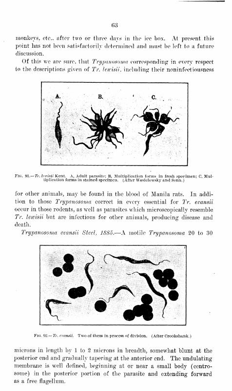
(>3
monkeys, etc.. after two or three days in the ice box. At present this
point has not been satisfactorily determined and must be left to a future
discussion.
Of this we are sure, that Trypanosoma corresponding in every respect
to the descriptions given of Tr. lewisii. including their noninfeetiousness
Fig. 91.— Tr. leirisii Kent. A, Adult parasite; B, Multiplication forms in fresh specimen; C, Mul-tiplication forms in stained specimen. (After Wasielevvsky and Senn.)
for other animals, may be found in the blood of Manila rats. In addi-
tion to those Trypanosoma correct in every essential for Tr. evansii
occur in those rodents, as well as parasites which microscopically resemble
Tr. lewisii but are infectious for other animals, producing disease and
death.
Trypanosoma evansii Steel, 1885.—A motile Trypanosoma 20 to 30
Fig. 92.— Tr. evansii. Two of them in process of division. (After Crookshank.)
microns in length by 1 to 2 microns in breadth, somewhat blunt at the
posterior end and gradually tapering at the anterior end. The undulating
membrane is well defined, beginning at or near a small body (centro-
some) in the posterior portion of the parasite and extending forward
as a free fla&ellum.
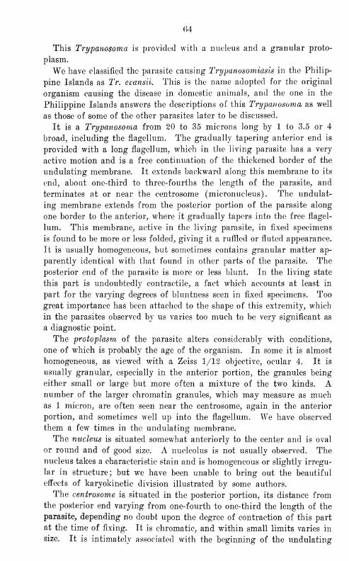
<>4
This Trypanosoma is provided with a nucleus and a granular proto-
plasm.
We have classified the parasite causing Trypanosomiasis in the Philip-
pine Islands as Tr. evansii. This is the name adopted for the original
organism causing the disease in domestic animal s, and the one in the
Philippine Islands answers the descriptions of this 'Trypanosoma as well
as those of some of the other parasites later to be discussed.
It is a Trypanosoma from 20 to 35 microns long by 1 to 3.5 or 4
broad, including the flagellum. The gradually tapering anterior end is
provided with a long flagellum, which in the living parasite has a very
active motion and is a free continuation of the thickened border of the
undulating membrane. It extends backward along this membrane to its
end, about one-third to three-fourths the length of the parasite, and
terminates at or near the centrosome (micronucleus). The undulat-
ing membrane extends from the posterior portion of the parasite along
one border to the anterior, where it gradually tapers into the free flagel-
lum. This membrane, active in the living parasite, in fixed specimens
is found to be more or less folded, giving it a ruffled or fluted appearance.
It is usually homogeneous, but sometimes contains granular matter ap-
parently identical with that found in other parts of the parasite. The
posterior end of the parasite is more or less blunt. In the living state
this part is undoubtedly contractile, a fact which accounts at least in
part for the varying degrees of bluntness seen in fixed specimens. Too
great importance has been attached to the shape of this extremity, which
in the parasites observed by us varies too much to be very significant as
a diagnostic point.
The protoplasm of the parasite alters considerably with conditions,
one of which is probably the age of the organism. In some it is almost
homogeneous, as viewed with a Zeiss 1/12 objective, ocular 4. It is
usually granular, especially in the anterior portion, the granules being
either small or large but more often a mixture of the two kinds. Anumber of the larger chromatin granules, which may measure as muchas 1 micron, are often seen near the centrosome, again in the anterior
portion, and sometimes wrell up into the flagellum. We have observed
them a few times in the undulating membrane.
The nucleus is situated somewhat anteriorly to the center and is oval
or round and of good size. A nucleolus is not usually observed. Thenucleus takes a characteristic stain and is homogeneous or slightly irregu-
lar in structure; but wTe have been unable to bring out the beautiful
effects of karyokinetic division illustrated by some authors.
The centrosome is situated in the posterior portion, its distance fromthe posterior end varying from one-fourth to one-third the length of the
parasite, depending no doubt upon the degree of contraction of this part
at the time of fixing. It is chromatic, and within small limits varies in
size. It is intimately associated with the beginning of the undulating
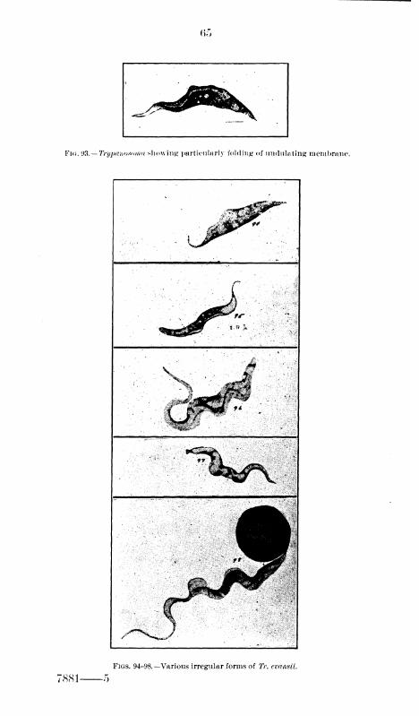
(>">
Fig. W.— Trypanosoma showing particularly folding of undulating membrane.
Figs. 94-98.—Various irregular forms of TV. evansii.
7881 o
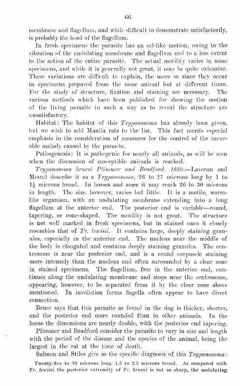
m
membrane and flagellum, and while difficult io demonstrate satisfactorily,
is probably the head of the flagellum.
In fresh specimens the parasite lias an eel-like motion, owing to the
vibration of the undulating membrane and flagellum and to a less extent
to the action of the entire parasite. The actual motility varies in some
specimens, and while it is generally not great, it may be quite extensive.
These variations are difficult to explain, the more so since they occur
in specimens prepared from the same animal but at different times.
For the study of structure, fixation and staining are necessary. The
various methods which have been published for showing the motion
of the living parasite in such a way as to reveal the structure are
unsatisfactory.
Habitat: The habitat of this Trypanosoma has already been given,
but we wish to add Manila rats to the list. This fact merits especial
emphasis in the consideration of measures for the control of the incur-
able malady caused by the parasite.
Pathogenesis : It is pathogenic for nearly all animals, as will be seen
when the discussion of susceptible animals is reached.
Trypanosoma brucei Plimmer and Bradford, 1889.—Laveran and
Mesnil describe it as a Trypanosoma, 26 to 27 microns long by 1 to
]^ microns broad. In horses and asses it may reach 26 to 30 microns
in length. The size, however, varies but little. It is a motile, worm-like organism, with an undulating membrane extending into a long
flagellum at the anterior end. The posterior end is variable—round,
tapering, or cone-shaped. The motility is not great. The structure
is not well marked in fresh specimens, but in stained ones it closely
resembles that of Tr. hivisii. It contains large, deeply staining gran-
ules, especially in the anterior end. The nucleus near the middle of
the body is elongated and contains deeply staining granules. The cen-
trosome is near the posterior end, and is a round corpuscle staining
more intensely than the nucleus and often surrounded by a clear zone
in stained specimens. The flagellum, free in the anterior end, con-
tinues along the undulating membrane and stops near the centrosome,
appearing, however, to be separated from it by the clear zone above
mentioned. In involution forms flagella often appear to have direct
connection.
Bruce says that this parasite as found in the dog is thicker, shorter,
and the posterior end more rounded than in other animals. In the
horse the dimensions are nearly double, with the posterior end tapering.
Plimmer and Bradford consider the parasite to vary in size and length
with the period of the disease and the species of the animal, being the
largest in the rat at the time of death.
Salmon and Stiles give as the specific diagnosis of this Trypanosoma:
Twenty-five to 30 microns long, 1.5 to 2.5 microns broad. As compared with
Tr. leicisii the posterior extremity of Tr. brucei is not so sharp, the undulating
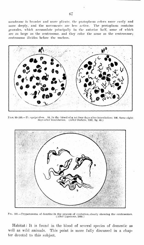
07
membrane is broader and more plicate, the protoplasm colors more easily and
more deeply, and the movements are less active. The protoplasm contains
granules, which accumulate principally in the anterior half, some of which
are as large as the centrosonie. and they color the same as the centrosome;
centrosome divides before the nucleus.
Figs. 99-100.— Tr. cquipcrdum. 99, In the blood of a rat four days after inoculation; 100, Same eightdays after inoculation. (After l)orlein,1901, tig. 40.)
^^mf9^^^tti§m^^i§§S§ti;Si
Fig. 101.—Trypanosoma of dourine in the process of evolution, clearlv showing the centrosomes.(After Lignieres, 1903.)
Habitat : It is found in the blood of several species of domestic as
well as wild animals. This point is more fully discussed in a chap-
ter devoted to this subject.
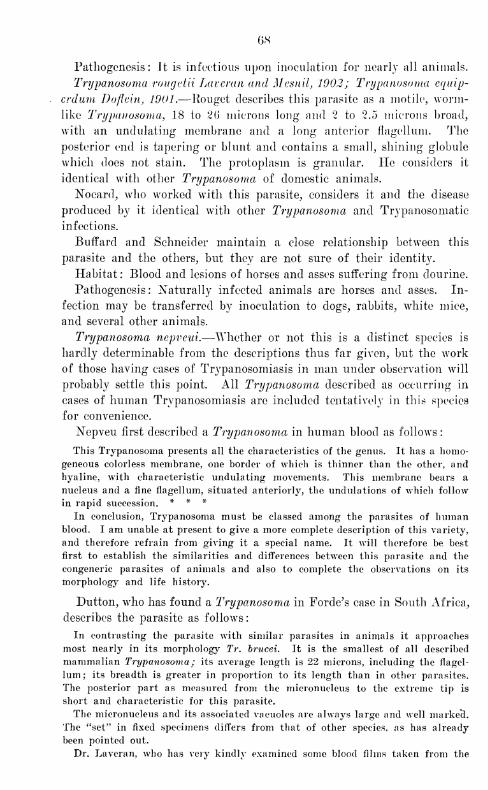
OS
Pathogenesis: It is infectious upon inoculation for nearly all animals.
Trypanosoma rougetii Lavcran and Mesnil, 1902; Trypanosoma equip-
crdum Doflein, 1001.—Rouget describes this parasite as a motile, worm-
like Trypanosoma, 18 to 2(> microns long and 2 to 2.5 microns broad,
with an undulating membrane and a long anterior fiagellum. The
posterior end is tapering or blunt and contains a small, shining globule
which does not stain. The protoplasm is granular. He considers it
identical with other Trypanosoma of domestic animals.
Nocard, who worked with this parasite, considers it and the disease
produced by it identical with other Trypanosoma and Trypanosomatic
infections.
Buffard and Schneider maintain a close relationship between this
parasite and the others, but they are not sure of their identity.
Habitat: Blood and lesions of horses and asses suffering from dourine.
Pathogenesis : Naturally infected animals are horses and asses. In-
fection may be transferred by inoculation to dogs, rabbits, white mice,
and several other animals.
Trypanosoma nepveui.—Whether or not this is a distinct species is
hardly determinable from the descriptions thus far given, but the work
of those having cases of Trypanosomiasis in man under observation will
probably settle this point. All Trypanosoma described as occurring in
cases of human Trypanosomiasis are included tentatively in this species
for convenience.
Nepveu first described a Trypanosoma in human blood as follows:
This Trypanosoma presents all the characteristics of the genus. It has a homo-
geneous colorless membrane, one border of which is thinner than the other, and
hyaline, with characteristic undulating movements. This membrane bears a
nucleus and a fine fiagellum, situated anteriorly, the undulations of which follow
in rapid succession. * * *
In conclusion, Trypanosoma must be classed among the parasites of humanblood. I am unable at present to give a more complete description of this variety,
and therefore refrain from giving it a special name. It will therefore be best
first to establish the similarities and differences between this parasite and the
congeneric parasites of animals and also to complete the observations on its
morphology and life history.
Dutton, who has found a Trypanosoma in Forde's case in South Africa,
describes the parasite as follows
:
In contrasting the parasite wTith similar parasites in animals it approaches
most nearly in its morphology Tr. brucei. It is the smallest of all described
mammalian Trypanosoma ; its average length is 22 microns, including the fiagel-
lum; its breadth is greater in proportion to its length than in other parasites.
The posterior part as measured from the micronucleus to the extreme tip is
short and characteristic for this parasite.
The micronucleus and its associated vacuoles are always large and well marked.
The "set" in fixed specimens differs from that of other species, as has already
been pointed out.
Dr. Laveran, who has very kindly examined some blood films taken from the
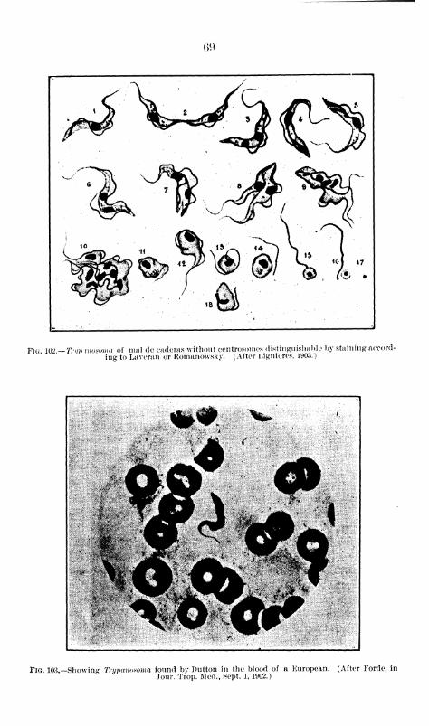
()9
kbr™15
Fi<; 102 —Trim momma of mal de caderas without centrosomes distinguishable by staining accord-
ing to Laveran or Romanowsky. (After Ligniercs, 1903.)
Fig. 103.—Showing Trypanosoma found bv Dutton in the blood of a European. (After Forde, in
Jour. Trop. Med., Sept. 1, 1902.)
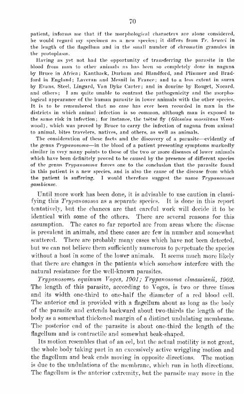
70
patient, informs me that if the morphological characters are alone considered,
he would regard my specimen as a new species; it differs from Tr. brucci in
the length of the flagellum and in the small number of chromatin granules in
the protoplasm.
Having as yet not had the opportunity of transferring the parasite in the
blood from man to other animals as has been so completely done in nagana
by Bruce in Africa; Kanthack, Durham and Blandford, and Plimmer and Brad-
ford in England; Laveran and Mesnil in France; and to a less extent in surra
by Evans, Steel, Lingard, Van Dyke Carter; and in dourine by Rouget, Nocard,
and others; I am quite unable to contrast the pathogenicity and the morpho-
logical appearance of the human parasite in lower animals with the other species.
It is to be remembered that no case has ever been recorded in man in the
districts in which animal infection is so common, although man is exposed to
the same risk in infection; for instance, the tsetse fly (Glossina morsitans West-
wood), which wTas proved by Bruce to carry the infection of nagana from animal
to animal, bites travelers, natives, and others, as well as animals.
The consideration of these facts and the discovery of a parasite—evidently of
the genus Trypanosoma—in the blood of a patient presenting symptoms markedly
similar in very many points to those of the two or more diseases of lower animals
which have been definitely proved to be caused by the presence of different species
of the genus Trypanosoma forces one to the conclusion that the parasite found
in this patient is a new species, and is also the cause of the disease from which
the patient is suffering. I wrould therefore suggest the name Trypanosomagambiense.
Until more work has been done, it is advisable to use caution in classi-
fying this Trypanosoma as a separate species. It is clone in this report
tentatively, but the chances are that careful work Avill decide it to be
identical with some of the others. There are several reasons for this
assumption. The cases so far reported are from areas where the disease
is prevalent in animals, and these cases are few in number and somewhat
scattered. There are probably many cases which have not been detected,
but we can not believe them sufficiently numerous to perpetuate the species
without a host in some of the lower animals. It seems much more likely
that there are changes in the patients which somehow interfere with the
natural resistance for the well-known parasites.
Trypanosoma equinum Voges, 1901; Trypanosoma elmassianii, 1902.
The length of this parasite, according to Voges, is two or three times
and its width one-third to one-half the diameter of a red blood cell.
The anterior end is provided with a flagellum about as long as the body
of the parasite and extends backward about two-thirds the length of the
body as a somewhat thickened margin of a distinct undulating membrane.
The posterior end of the parasite is about one-third the length of the
flagellum and is contractile and somewhat beak-shaped.
Its motion resembles that of an eel, but the actual motility is not great,
the whole body taking part in an excessively active wriggling motion and
the flagellum and beak ends moving in opposite directions. The motion
is due to the undulations of the membrane, which run in both directions.
The flagellum is the anterior extremity, but the parasite may move in the
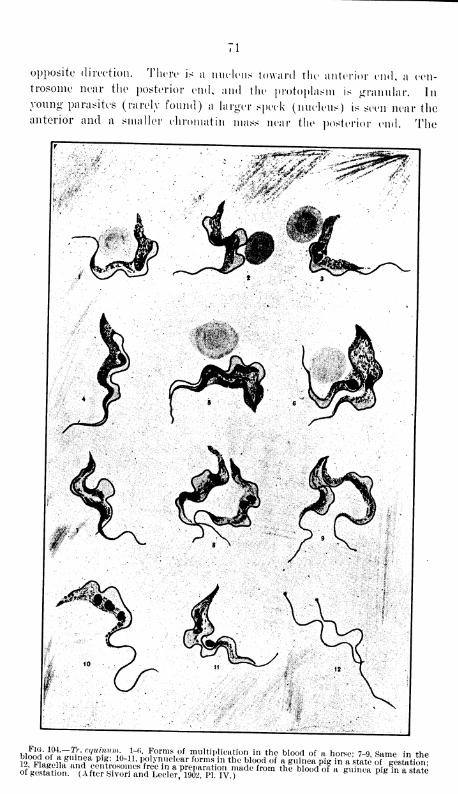
71
opposite direction. There is a nucleus toward the anterior end, a cen-
trosome near the posterior end, and the protoplasm is granular. Inyoung- parasites (rarely found) a larger speck (nucleus) is seen near theanterior and a smaller chromatin mass near the posterior end. The
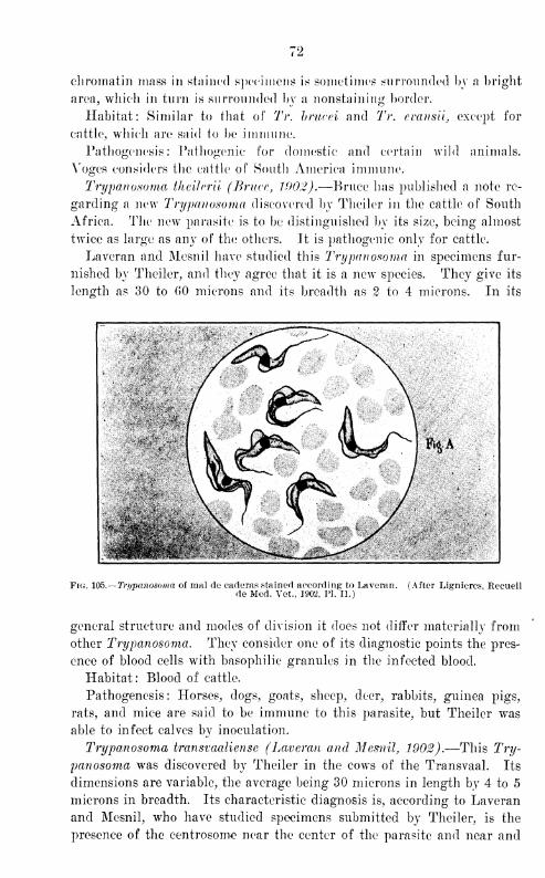
72
chromatin mass in stained specimens is sometimes surrounded by a bright
area, which in turn is surrounded by a nonstaining border.
Habitat: Similar to that of Tr. bnwei and Tr. cuansii, except for
cattle, which are said to be immune.
Pathogenesis: Pathogenic for domestic and certain wild animals.
Yoges considers the cattle of South America immune.
Trypanosoma theilcrii (Bruce, 190.2).—Bruce has published a note re-
garding a new Trypanosoma discovered by Theiler in the cattle of South
Africa. The new parasite is to be distinguished by its size, being almost
twice as large as any of the others. It is pathogenic only for cattle.
Laveran and Mesnil have studied this Trypanosoma in specimens fur-
nished by Theiler, and they agree that it is a new species. They give its
length as 30 to GO microns and its breadth as 2 to 4 microns. In its
Fig. 105. -Trypanosoma of mal de caderas stained according to Laveran. (After Lignieres, Reeueilde Med. Vet., 1902, PI. II.)
general structure and modes of division it does not differ materially from
other Trypanosoma. They consider one of its diagnostic points the pres-
ence of blood cells with basophilic granules in the infected blood.
Habitat: Blood of cattle.
Pathogenesis : Horses, dogs, goats, sheep, deer, rabbits, guinea pigs,
rats, and mice are said to be immune to this parasite, but Theiler was
able to infect calves by inoculation.
Trypanosoma transvaaliense (Laveran and Mesnil, 1902).—This Try-
panosoma was discovered by Theiler in the cows of the Transvaal. Its
dimensions are variable, the average being 30 microns in length by 4 to 5
microns in breadth. Its characteristic diagnosis is, according to Laveran
and Mesnil, who have studied specimens submitted by Theiler, is the
presence of the centrosome near the center of the parasite and near and
73
sometimes united to the nucleus. The altred corpuscles seen in blood
infected with Tr. theilerii have not been seen with this parasite.
Habitat: Blood of Transvaal cattle.
Differential diagnosis of Trypanosoma of mammals.—Regarding the
differential diagnosis of the Trypanosoma which are of the greatest im-
portance in the disease of domestic animals, there seems to be considerable
difference of opinion; and the work will probabalv not be satisfactorily
completed until all the supposed varieties are studied in one country
in similar environments.
According to Laveran and Mesnil the distinction between Tr. brucei
and Tr. lewisii is marked. Tr. lewisii is thinner and more tapering, and
its undulating membrane is smaller and less folded. Its protoplasm
colors less deeply. Its chromatin granules are not so large and numerous.
Its posterior extremity is always thin and tapering and never has the
appearance of a truncated cone. It lives longer on ice than does Tr.
brucei. However, individuals showing no material differences are found
in both varieties. In fresh blood without the presence of dividing forms
they are differentiated with great difficulty. In Tr. brucei the centro-
some always divides first, following which the flagellum, nucleus, and
protoplasm separate in the order named. In Tr. leivisii division maybegin in the nucleus, and before it takes place the parasite sometimes
reaches 5 microns or more in breadth.
Tr. equiperdum, according to the same authors, closely resembles Tr.
brucei, but the morphologic differences between the two are appreciable.
Tr. brucei has much greater dimensions; its protoplasm colors more
deeply and nearly always contains large chromatin granules, which are
absent in Tr. equiperdum, which is never more than 20 microns in length.
However, we have, in the blood of animals, seen Tr. equiperdum which
so closely resembled Tr. brucei that methods of differential diagnosis were
unsatisfactory.
Tr. brucei and Tr. equinum, according to Laveran and Mesnil, have
almost the same length and form. The protoplasm, the nucleus, the
undulating membrane, and the flagellum have the greatest resemblance
in the two Trypanosoma, but this is not true of the centrosomes. The
centrosome of Tr. brucei colors easily and deeply and measures about
1-J microns in diameter; that of Tr. equinum does not measure more
than-J
or l microns, and colors rose, like the flagellum, and not violet,
like the centrosome of Tr. brucei. A number of chromatin granules
difficult to examine are often found near it. Some observers have came
to the conclusion that the centrosome is defective in Tr. equinum.
Owing to the fact that the centrosome differ in stained specimens of
the blood of mice infected simultaneously with Tr. brucei and Tr. equi-
num, they have been able to distinguish the two species of Trypanosoma.
The forms of multiplication are the same, double partition being the
74
rule. Large divisions into three or four parts, which are somewhat more
common in Tr. equinum than in Tr. brucei, are sometimes observed.
Martini considers the posterior end of Tr. brucei more blunt than that
of Tr. leivisii, Tr. equinum, or Tr. evansii.
Buffard and Schneider and several others believe Tr. roiigetii to be
identical with other Trypanosoma producing Trypanosomiasis in domes-
tic animals.
Scheube, Bruce, liost, Koch, and many others consider Tr. evansii, Tr.
brucei, and some of the other parasites probably identical.
Ligneres has recently written elaborately regarding the distinctions
between the various Trypanosoma of mammals. In the main his results
agree with those of Laveran and Mesnil already given.
A consideration of this subject resolves itself into two headings
:
First, a differentiation based upon microscopic observations of the para-
sites, and, secondly, that based upon their pathogenic action. As might
be expected, the more two parasites differ when compared by one of these
methods the greater will be the difference between the two as determined
by the other.
Tr. lewisii differs morphologically from the parasite of nagana, surra,
etc., and these differences are confirmed by their pathogenic action. Con-
currence of opinion on the individuality of Tr. lewisii as found in dif-
ferent countries is so universal that further attention need not be paid
to it.
When we come to consider the identity or nonidentity of Tr. evansii,
Tr. brucei, Tr. equipcrdum (rougetii), and Tr. elmassianii (equinum),
we are compelled to obtain our data for all of these parasites except Tr.
evansii from the work of other authors.
Taking up first the study of morphologic differences, we fail to see
any justification for the extremely careful and guarded conclusions
of Laveran and Mesnil or the very sweeping ones of several more recent
authors. They have disregarded the fact of the variability of these
organisms in the same species of animal in the same country, their
greater variability and different species of animals in the same and in dif-
ferent countries, and other conditions requisite to identity of environment
and to a conclusive comparative study. It must be remembered that weare dealing with organized animal life and that environment has an
important influence on its physical condition.
The most important differences which conservative writers generally
point out between these organisms are variations in the shape of the
posterior extremity, the centrosome and the undulating membrane andthe granular condition of the parasite itself.
From careful observations we are confident that the posterior endof Tr. evansii is contractile, a condition which a few writers have noted
in other parasites. This results in a variation of the shape of this-
to
extremity in Tr. evansii, and doubtlessly in other parasites, as great as
that given for any two members of the group.
The same statement applies to some of the other differences, such
as variations in the undulating membrane and the general morphology
of the parasite.
Another important factor which has not been given due considera-
tion is the age of the parasite, as is also the condition with reference
to life, of the media from which the preparations are taken for study.
Jn Tr. evansii and probably in some of the other parasites the number
and size and to a certain extent the location of the granules depend
upon the conditions mentioned. The difference in the staining of the
centrosome in Tr. brucei and Tr. elmassianii (equinum) , first empha-
sized by Laveran and Mesnil, we are not in a position to comment upon.
Laveran and Mesnil have written very carefully regarding the dif-
ferentiation of these parasites, and are most conservative in considering
differences in pathogenic action, regarding the latter as secondary in
importance to the morphologic differences of the organisms. More
effusive and less careful writers have drawn emphatic conclusions from
pathogenic manifestations alone. There is undoubtedly a great simi-
larity in the pathogenic action of the various Trypanosoma, in general
the same animals being susceptible and showing similar symptoms and
post-mortem lesions. The chief differences are those of degree, and they
vary almost as much in different animals of the same species when
inoculated with the same Trypanosoma as with different Trypanosoma.
Cattle, for example, have been used to show differences in the parasites,
when as a matter of fact in Manila these animals, when inoculated
with Tr. evansii, show variations as great those noted in literature
for any two diseases. We have seen a cow die in twTenty-four days
from surra (see fig. 127), and all degrees of resistance above this to
an almost complete natural immunity have been observed.
After carefully reviewing literature and taking our own observations
into account, we do not feel justified in forming a positive conclusion;
but it seems to us that proof sufficient to establish the individuality
of the Trypanosoma causing Trypanosomiasis in domestic animals has
not yet been advanced.
V. MODES OF TRANSMISSION AND INFECTION.
Believing, as we do, that the transmission of Trypanosoma by biting
and stinging insects is the only method deserving consideration from a
practical standpoint, we are in harmony with the best thought of modernliterature on the subject. To demonstrate that this is the only practical
method requires (1) evidence of a host constantly present in infected
zones, (2) direct evidence of transmission from this host to the healthy
animal, and (3) evidence that in the absence of either the host or the
insects the disease is not communicated.
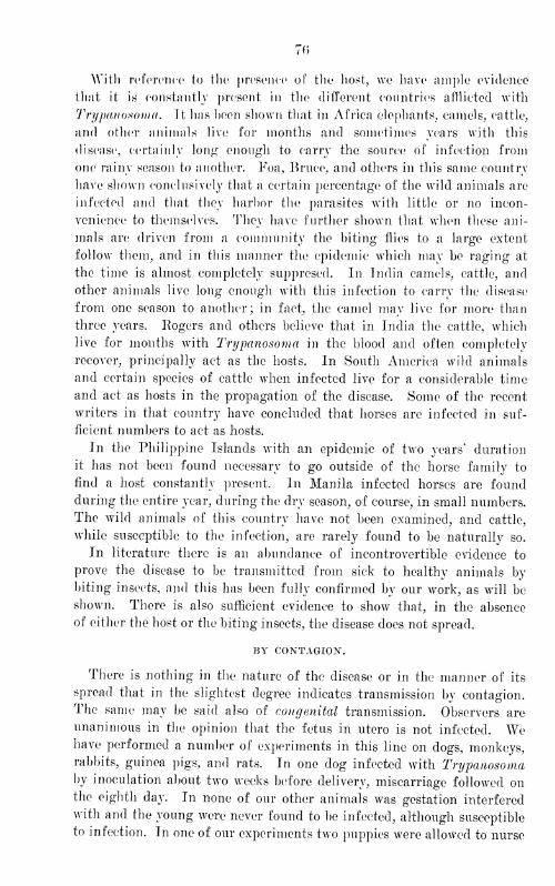
70
With reference 1 to the presence of the host, we have ample evidence
that it is constantly present in the different countries afflicted with
Trypanosoma. It has heen shown that in Africa elephants, camels, cattle,
and other animals live for months and sometimes years with this
disease, certainly long enough to carry the source of infection from
one rainy season to another. Foa, Bruce, and others in this same country
have shown conclusively that a certain percentage of the wild animals are
infected and that they harhor the parasites with little or no incon-
venience to themselves. They have further shown that when these ani-
mals arc; driven from a community the biting flies to a large extent
follow them, and in this manner the epidemic which may be raging at
the time is almost completely suppresed. In India camels, cattle, and
other animals live long enough with this infection to carry the disease
from one season to another; in fact, the camel may live for more than
three years. Rogers and others believe that in India the cattle, which
live for months with Trypanosoma in the blood and often completely
recover, principally act as the hosts. In South America wild animals
and certain species of cattle when infected live for a considerable time
and act as hosts in the propagation of the disease. Some of the recent
writers in that country have concluded that horses are infected in suf-
ficient numbers to act as hosts.
In the Philippine Islands with an epidemic of two years' duration
it has not been found necessary to go outside of the horse family to
find a host constantly present. In Manila infected horses are found
during the entire year, during the dry season, of course, in small numbers.
The wild animals of this country have not been examined, and cattle,
while susceptible to the infection, are rarely found to be naturally so.
In literature there is an abundance of incontrovertible evidence to
prove the disease to be transmitted from sick to healthy animals by
biting insects, and this has been fully confirmed by our work, as will be
shown. There is also sufficient evidence to show that, in the absence
of either the host or the biting insects, the disease does not spread.
BY CONTAGION.
There is nothing in the nature of the disease or in the manner of its
spread that in the slightest degree indicates transmission by contagion.
The same may be said also of congenital transmission. Observers are
unanimous in the opinion that the fetus in utero is not infected. Wehave performed a number of experiments in this line on dogs, monkeys,rabbits, guinea pigs, and rats. In one dog infected with Trypanosomaby inoculation about two weeks before delivery, miscarriage followed onthe eighth day. In none of our other animals was gestation interfered
with and the young were never found to be infected, although susceptible
to infection. In one of our experiments two puppies were allowed to nurse
from an infected mother in an insect-proof stable, and at the time of the
death of their mother, eight days after their birth, they were free from
infection.
uy corn ox.
Almost all writers, referring- to dourine, state that it is transmitted
by coition, and a considerable 1 number believe this to be the only method
of transmission for this disease, while others believe it to be the excep-
tion, even in this form of Trypanosomiasis. This method of transmission
has been given little consideration in relation to the other forms.
Recently some writers upon this subject have offered at least sug-
gestive explanations. Schilling believes that transmission by coition
occurs in those regions free from the usual infecting insects and from
other conditions favorable to the propagation of the disease; and the
geographic distribution of dourine tends to support Schilling's con-
clusions. In infected countries, if the infection takes place by coition
it is lost sight of in the more frequent methods and could be determined
only by careful experimentation.
We have performed a number of experiments looking to the eluci-
dation of this point in the epidemic now raging in this country. Con-
siderable difficulty has been experienced in obtaining direct evidence.
Horses are not available for the work, and with the native ponies it has
been almost impossible to find at the same time infected animals and
those desiring intercourse. In several instances infected blood has been
introduced into the vagina of female dogs by the use of a catheter. All
these experiments except one have given negative results, and in the
positive one infection is attributed to a lesion of the vagina. In those
cases in which the mucous membrane of the vagina wTas injured pur-
posely, infection following the introduction of virulent blood occurred
in every case, with an incubation period equal to that following infec-
tion through the injured mucosa of the mouth.
We have not observed an absolutely conclusive result following coition.
One male dog contracted the disease after intercourse with an infected
female; but a small number of biting and stinging insects were present
at the same time, so that it was probably transmitted by them. Uponanother occasion an infected male goat was seen to copulate with a
healthy female, but no infection followed. This was hardly the most
desirable kind of an experiment, since the goat in question rarely showedparasites by microscopic examination, though his blood was infectious
by animal experiment. Again, the manner in which goats effect cop-
ulation would be much less likely to result in infection than in the case
of the horse, provided, as has been suggested, it depended upon trauma-tism. It seems more than probable, however, that in many animals,
and especially in the case of the horse, infection might often follow
sexual intercourse as the result of traumatic injury of the genitals.
78
It is not uncommon to sec a few drops of blood after sexual intercourse
between these animals, and it would certainly require no stretch of
the imagination to suggest the possible presence of small injuries in
the mucosa of the genitals of both animals. Tt does not appear, how-
ever, that much importance should be given to this as a practical means
of transmission of the disease. Any disease of horses transmitted only
by coition could, of course, be eradicated with the greatest ease.
RATS AS CARRIERS OF THE INFECTION.
In referring to the part played by rats, we come to a much more
important subject. To bring out its full significance necessitates a
brief consideration of the natural infection of rats with Tr. cvansii,
a point which will be thoroughly discussed under "Trypanosomiasis of
rats," and a consideration of the bearing such infection has upon the
transmission of surra in animals of economic importance.
Hats in an indefinite way have been blamed for the transmission of
the disease. Lingard has made elaborate experiments to show that
grain soiled with the excrement of these little pests and eaten by horses
played an important part in the spread of the infection. Pie dem-
onstrated that rat surra was transferable to horses by inoculation; but,
owing to his failure properly to protect his animals from insects, his
work has received but little consideration. Musgrave and Williamson
have shown conclusively that a certain percentage of rats in Manila
harbor Tr. cvansii.
In a preliminary report they say:
A varying percentage of rats are known to harbor a Trypanosoma somewhatresembling the one found in the horse, and it has been conclusively shown that
a certain number of rats in Manila harbor the same Trypanosoma which causes
the disease in other animals. These parasites have been determined both
morphologically and in their pathogenic action to be identical with the parasites
causing Trypanosomiasis in horses.
With this information we are prepared to consider the part they
play in transmitting the disease to domestic animals and in perpetuating
the epidemic. Rats frequently fight, and it has been shown that the
infection passes from one to another in this way. In addition, the
wounds so caused attract biting insects, particularly fleas, which are
known to transmit Trypanosoma from rat to rat, and, as will be seen
later, from rats to other animals. In Manila the number of rats in-
fected with Tr. cvansii makes it necessary seriously to consider themin discussing means of controlling the disease in this city.
INFECTION OF PASTURES, FOOD, AND WATER.
Infection through the sound mucosa and through the injured mucousmembranes presupposes to a large extent as its source infected food anddrink. Lingard, the great champion of this method of transmission,

considers the ingestion of stagnant water and of grass from land sub-
ject to inundation a source of infection, the latter being a favorable
breeding place for low forms of animal life and possibly of Trypanosoma.
Salmon and Stiles believe that there is nothing at present known
in connection with the life history of any Trypanosoma to lead us to
look seriously upon grass as a source of infection. We know that
Trypanosoma die very rapidly under all usual conditions outside 1 of
the living body. If they are to be found in either food or drink, they
must be in some phase of the life cycle with which we are unacquainted.
If food and water should be infected, as Lingard maintains, they would
still be harmless in the presence of the demonstrated fact that the
disease as we know it is not transmittable through the sound mucosa
of the alimentary canal.
In a preliminary report Musgrave and Williamson make the following
statement regarding this subject
:
The existence of an extracorporeal stage of Trypanosoma, living on grass
and on water in marshy places, in this stage taken into the stomach of sus-
ceptible animals eating and drinking in these localities, and from this organ
or other part of the animal economy passing into the blood in the forms werecognize, is without sufficient evidence to warrant consideration in this paper.
Before leaving this subject, however, for a fuller discussion in a subsequent
paper, it is proper to state that Trypanosoma quickly die under all tried environ-
ments outside of the body of some living creature, and no evidence of their
existence in water, on grass, or other similar places can be advanced. Both
water and grass have been inoculated with large numbers of Trypanosoma andhave failed to convey the disease, after days, weeks, or months, when fed to
susceptible animals, and also when injected under the skin.
Malaria is similar to Trypanosomiasis in that both are parasitic diseases and
both are prevalent in low-lying, marshy lands and during wet weather. Malaria
was for ages attributed to the air, the water, etc., of these marshes, and it
took years to make the public understand that all these conditions are harmless
in producing it, provided the patient is protected from the bite of mosquitoes,
so prevalent in these regions.
Take the epidemic of Trypanosomiasis in Manila. At the time of its outbreak,
and for some time afterwards, it was confined to the city. The grass and water'
given the horses was the same which had been fed for years, and no disease
resulted.
The disease started from a focus of infection and spread directly with exposure
to infected animals, and attacked alike animals fed exclusively on hay and oats
and those fed on grass. The disease is prevalent in Manila at the present timeand has been so continuously since its introduction. The majority of horses
having the disease, under our observation for the past four months, had beenfed previously entirely on hay and oats.
In one large stable, with both American and native horses, four of the Americanhorses and mules (fed entirely on dry feed) and two of the native ponies havedied of the disease, and one of the two ponies was the only horse of this class
in the stable which received dry feed only.
The statement is made that certain districts in India are avoided by cavalryon the march because of the danger in these districts from food and drinkthrough which animals may contract the disease. Similar conditions are found
SO
in South America, but in addition it has been shown that infected districts on
this continent are just as dangerous to horses provided with dry food and pure
water while passing through them as they are to animals eating forage grown
on the spot.
It has been shown by a number of observers that an infected animal taken
to a new place becomes a focus for the spread of the disease, provided biting
flies are present, no matter whether the territory is marshy or dry.
Since that time the work has been continued, but no evidence has been
obtained to show that food, water, or pastures are ever factors in the
spread of the disease.
Attempts artificially to infect water and grass have always failed. It
is true that when these substances are used as culture media parasites
may live for a short time under certain conditions, but multiplication
to an appreciable extent does not occur. It is of course possible that
infection may occasionally take place in localities where sick and healthy
horses graze on the same ground or drink water from the same place,
provided both classes of animals have lesions of the mucous membranes
or cutaneous wounds on those parts of the legs which come in contact
with the grass or the water. It is absolutely certain, however, that if
food and water are ever infected to any degree it must be with some form
of the parasite not yet known. Even if such places served as culture
media for Trypanosoma, as we know them, they would still be compara-
tively harmless to those animals having sound mucous membranes and
free from cutaneous wounds.
INFECTION THROUGH THE SOUND MUCOSA.
There is some difference of opinion, especially among recent writers,
in regard to infection by food and drink through the sound mucosa of
the alimentary canal. Lingard affirms emphatically that the disease is
so transmitted, but the great majority of recent investigators state with
equal positiveness that the disease can not be so produced.
Kanthuck, Durham, and Blandford attempted to transmit Trypano-
soma by feeding, performing a number of experiments. At times they
were successful, but in most cases they failed. They concluded that the
possibility of infection depends upon accidental lesions of the mucous
membranes of the upper portion of the alimentary canal. Continuing,
they say
:
Of a number of rats fed on the organs of nagana animals only a few acquired
the disease, and these invariably showed superficial lesions of the snout and ears,
due to lice. When fed upon infected material, they buried their snouts in it
and scratched their ears with their blood-stained forepaws. Furthermore, in
rats which acquired the disease through feeding, the cervical glands were always
enlarged most, a fact which proves that hematozoal infection must have taken
place in the head, for, as we shall show, the primary infection travels by the
lymphatics.
A cat fed repeatedly on the soft tissues of bodies of infected dogs and cats
and subsequently on the bodies of dead rats, died at a time corresponding by
SI
lethal period to an infection at the first meal on rats. We regard it as probable
that some splinter of bone caused superficial lesions, through which the hema-
to/oa were enabled to enter.
A rabbit, fed carefully by means of a pipette with large quantities of infected
blood, did not show the slightest sign of the disease. Kouget (lS!Mi) also failed
to infect animals by the mouth.
Evans and Steel believe tbat surra can be transmitted either by inges-
tion or by inoculation in dogs, horses, and mules, provided that fresh
infected blood is used. With blood kept twenty-four hours or more they
did not succeed in conveying the disease.
Writing in 1891), Lingard considered that natural infection with surra
might occur in one of four ways: (1) From eating grass and other vege-
tation grown upon land subject to inundation; (2) from drinking stag-
nant water during certain months of the year; (3) from the bite of
certain species of flies, probably as carriers of the virus; and (4) from
the ingestion of corn soiled with the excrement of rats and bandicoots.
Having determined that the Trypanosoma of rats in Tndia was the
cause 1 of Trypanosomiasis in a certain percentage of other animals, such
as the horse, he experimented to prove whether this was of any practical
importance in the spread of the disease. He mixed the excreta of rats
with grain, which was fed to horses during the dry season in an area free
from surra, with negative results. With similar experiments, however,
under favorable conditions of moisture and heat, he claims to have ob-
tained positive ones. With regard to these he writes as follows:
At a later date, during the latter half of the hot season and the rains of 1893,
further experiments with the same end in view were carried out in Bombay, where
the climate is favorable from its humidity and the disease is enzootic. Fromthese positive results were obtained, the incubation period occupying four months(about June to September) ; but the incubation period of the disease will prob-
ably be found to vary with the amount of moisture in the atmosphere and the
amount of materies morbi ingested by the animal. This points to the probability
that a number of surra cases in Bombay were contracted through the ingestion
of rats' or bandicoots' excreta mixed with the corn.
Salmon and Stiles doubt the accuracy of these results. They say that
Trypanosoma have not been found in the excrement of either horses or
rats, that it is not clear that Lingard used the necessary precautions to
exclude fly bites and other factors in his experiments, and that if rats
harbored Tr. evansii transmission from them would have been as possible
by rats and fleas as by their excrement. Yoges fed animals with several
liters of infected blood mixed into a paste, without results. Laveran
and Mesnil also failed to convey the disease through the sound mucosa.
Eouget's feeding experiments were negative, but he obtained positive
results by dropping infected blood into the conjunctiva. Rogers was
unable to convey the disease through the sound mucosa, and says that in
7881 6
82
those cases in which infection occurred by feeding there were injuries
of the mucous membranes.
Host writes that grass, grain infected by rat's excreta, and ilies must
be considered in the etiology. Salmon and Stiles consider ingestion as
a possible means of infection, but state that it can deserve no special
consideration as a means of transferring the disease from horse to horse.
It appears to us that one of the strongest arguments against the trans-
mission of the disease^ through food and drink is furnished by Lingard
himself. He states that a street-car company which had lost hundreds
of horses from surra finally provided their animals with boiled drinking
water only and with grass brought from an elevation of (5,000 feet, pro-
hibited the feeding of green vegetables, and also ordered the animals to
be isolated, without success. It is to be noted that these stables were
situated on a street along which infected horses were traveling, so that the
isolation of the company's animals did not protect the healthy ones from
flies. The manner of feeding did exclude infection by this means, and
seems to us a strong point in determining that food and drink play abso-
lutely no part in the transmission of the disease.
The numerous citations of instances in which dogs contracted Tryp-
anosomiasis by eating the carcasses of animals dead of the disease seem
to us to be no argument in favor of its transmission through the sound
mucosa. Such animals are always fighting, and the infection might
readily take place through wounds, and in addition these animals are
usually well provided with biting insects. Curry states that it is not
known whether the disease in the Philippines can be transmitted by food
and drink.
Our investigations have failed to produce the slightest evidence that
infection by food, drink, or otherwise ever occurs through the sound
mucosa. Musgrave and Williamson, discussing this mode of transmis-
sion, say:
The groat majority of writers agree that infection can not take place throughthe sound mucosa of the alimentary tract, and that the occasional infection
following the administration by mouth of the virulent blood and organs of
animals recently dead of the disease are probably due to the fact that these
animals had damaged mucous membranes of the mouth or upper part of thealimentary canal, which would, of course, result in infection, just as would occurin any other part of the body by bringing an injured surface into contact withinfectious material, or vice versa.
In nearly all feeding experiments large doses of the infective agent have beengiven, and in this sense they have not approached natural infection, which, fromthe nature of things and whether administered through the mucous membraneor the skin, would be in small doses.
Lingard attempted infection through the digestive canal by the administrationof very small doses of infected blood given frequently in large dilutions of water.One of his horses that had received such treatment, and in addition one dose of
13 c. c. of infected blood, developed the disease on the one hundred thirtieth
day after the beginning of the experiment. He fed a second horse 200 minims of
S3
fresh virulent blood at one dose, with an ineuhation period of seventy-five days.
He does not state that these horses were protected from insects during the periods
of the experiments, which were made in an infected country, and it is more than
probable, considering the incubation periods of one hundred thirty and seventy-
live days, that his animals were infected in some other way.
So far as we have been able to discover, there is not in literature any absolute
proof of infection through the sound mucosa by feeding.
Jn this preliminary report but one of our many feeding experiments will be
given.
Monkey No. 1'2(J—healthy adult male monkey—was isolated, temperature taken
and blood examined daily for a week. The temperature remained normal and
the blood negative for Trypanosoma. After twelve hours' fasting he was fed
weekly for six weeks on cooked rice (the usual diet) soaked with fresh warmvirulent blood, rich in Trypanosoma, from different animals at different feedings.
On two occasions he was given to drink infected blood in weak potassium citrate
solution, in which Trypanosoma live longer than in any other known solution
outside of the body.
At the end of six weeks the animal was apparently in good health, temperature
had remained normal, and the blood free from parasites. ]n order absolutely
to exclude the existence of infection, a drop of blood was injected subcutaneously
into another monkey, which remained well and was afterwards proved susceptible.
During the feeding the infected rice would often be stored in his chops and remain
there for hours.
After proving the animal not infected at the end of six weeks, a small scratch
was made in the mucosa of the mouth and he was again fed as before. Thedisease developed on the fourth day. as was evidenced by rise in temperature
and the presense of Trypanosoma in the blood. He ran the regular course of
the disease, and died on the eighteenth day after infection.
Tli is work has now been continued for more than a year, involving the
use of hundreds of animals. We have experimented with horses, dogs,
goats, rabbits, guinea pigs, monkeys, eats, and rats; Ave have fed them
large and small doses of virulent blood and all kinds of preparations of
both blood and infected organs. All experiments were performed under
circumstances which made accidental infection impossible. Particular
attention to this part of the subject was deemed necessary in order intel-
ligently to recommend measures for the control of the epidemic. Ourresults have given absolutely convincing proof that infection does not
occur through the sound mucous membranes by Trypanosoma, no matter
in what form administered.
JXFKCTJOX THROUGH THE INJURED MUCOUS MEMBRANES.
As stated by Musgrave and Williamson in a preliminary report, surra
is essentially a wound disease, and transmission through the injured
mucous membranes results when infected material is brought in contact
witli it. When infection occurs through the alimentary tract, it does so
through a wounded mucous membrane. Monkey Xo. 12v> was one of
the many animals experimented upon to determine this point as well as
the possibility of transmission through the sound mucosa. Of course it
was easy to prove that infection could occur through an injured mucous
84
surface; hut definitely to show of what importance this would he in the
practical transmission of Trypanosomiasis required more work.
Jt was necessary first to determine what percentage of a munher of
animals in the natural course of events have sufficient injuries to make
possible an infection in a given period of time. Naturally this was
found to vary greatly. In dogs and rats injuries are so frequent that
in order to obtain animals without lesions to be used in determining the
possibility of transmission through the sound mucosa, we were com-
pelled to confine each animal by itself for some time. In horses, cattle,
and other animals of economic importance natural lesions of the mucous
membranes are more rarely found. In horses nsed for carriage 1 and
draft purposes, lesions in the corner of the mouth caused by the bridle
bit are quite frequent. In the case of cattle and carabao, lesions are
seen in the nose around the ring which is placed through the septum and
which usually has a rope fastened to it. Small wounds might occa-
sionally be produced in the mouth by eating rough dry food.
With injuries present in the mucous membranes, the next question to
consider was the manner in which infectious material may be brought in
contact with these wounds; and from a practical standpoint (excluding
flies) there are not many possibilities. All such means are of course
mechanical and readily suggest themselves; they are, changing bridle
bits from sick animals to healthy ones, allowing healthy animals to lick
sores on sick ones, placing animals to graze upon the same ground, or
allowing them to drink the same water, etc.
Here may be introduced a strong argument against the theory that
food and water play a serious part in the transmission of the disease.
Tf such were the case, animals with wounds would contract the disease
from eating and drinking food and water which had been allowed to
stand just as readily as they do when freshly infected material is fed to
them; but this is not the case. We have experimented on animals with
fresh wounds, feeding them water and grass infected from forty-eight
hours to three months previously and kept under all kinds of conditions,
but have failed to convey the disease in this manner.
Flies.—Of the biting insects that play a part in the transmission of
the disease, certain varieties of flies are of the greatest significance.
Of flies credited with playing an etiologic role in this disease, the
tsetse fly (Glossina morsikans) is among the most important. 1
Schilling states that Glossina logipalpis is very prevalent in Togo, as
well as Stomoxys calcitrans and three species of Tabani. He regards all
1 We had hoped to introduce here a short illustrated description of each species
of the flies which have been credited with playing a role in the transmission of
Trypanosoma ; but the pressure of other work has prevented the entomologist
to whom the duty was assigned from completing the undertaking.. At some
future time we hope to be able to do this, when we shall also finish the dis-
cussion of some of the points left open in this paper.
So
these varieties as able to transmit the infection. lie proved conclusively
that at least two species of these Hies can transfer the infection in dogs.
Martini found normal Trypanosoma twenty-three hours after feeding
the Stomo.vys calcitrant: on infected blood, the insect itself being kept
in a room at a temperature of ^> (\; but he did not see dividing
forms. On the following day the blood was digested and the parasites
could not be observed. On dogs biting experiments with these tlies failed.
Healthy horses and asses standing next to sick horses, all covered with
^lomo.rys calcitrans, did not contract the disease. Martini's work was
performed in Berlin.
Working in .Manila, Curry considered the stomo.vys calcitrans as the
principal agent of transmission and found enormous numbers of them
feeding on sick animals. lie observed active Trypanosoma in the probos-
cides and stomachs of these flies twenty-four hours after their feeding
upon infected blood. He was unable to find Trypanosoma in any other
species of flies. Kost found Trypanosoma in juices squeezed from the
horsefly some hours after feeding on a surra animal, and he was con-
vinced that these transmit the disease.
Lingard describes an epidemic in India in which flies were very
numerous and no precautions to isolate or destroy infected ponies were
taken. Sixty-five animals died in the region in question. During the
next season, however, in the same locality and among the same class of
horses, the infected animals were isolated and only two deaths occurred,
lie demonstrated that the body fluids of certain flies contain Trypano-
soma- after feeding on infected animals and that the inoculation of
such fluids in susceptible animals causes the disease. He and his assist-
ants examined large numbers of Diplcra of some ten varieties, during
both the dry and the wret season, but Trypanosoma were not found in
any case except in adult flies which had fed on infected animals.
Sivori and Lecler in South America proved that the Musca brava, the
^tomoxys calcitrans, and the Taon can transmit the disease from horse
to horse. The relative of these flies, elderetatus obifasciatus, also
showed Trypanosoma after feeding on a sick horse, but did not prove
capable of transmitting the infection.
Yoges states that the disease is transmitted only through wounds,
injections, etc. Several varieties of Tdbani are found in South America,
and the Musca brava is also prevalent, as well as various species of mos-
quitoes.
Buffard and Schneider consider Tabani as active factors in the trans-
mission of the disease.
Bogers proved conclusively that surra is transmitted by flies which
have recently bitten infected animals. Infected flies kept from one to
four days did not transmit the disease by biting and were harmless when
fed to rats.
Bruce considers the tsetse flv able to convey the disease for fortv-eisrht
80
hours after feeding on infected blood. He placed live muzzled horses
in areas containing ilies, and notwithstanding the fact that they were
all prevented from using the food and water found there, they con-
tracted the disease. Suspected flies transferred to a district free from
nagana conveyed Trypanosomiasis by biting two dogs after four to
seven hours. He found living Trypanosoma in the proboscidcs of these
insects forty-six hours after they had fed on infected blood, and in their
stomachs one hundred eighteen hours after feeding; but at the end of
one hundred forty hours no parasites were present.
Schilling noticed that at a certain place in Africa on one side of a
lagoon three kilometers in width, surra and tsetse flies were prevalent,
while on the other side neither could be found.
Without going into further detail, there is an abundance of incontro-
vertible evidence that the disease is transmitted by a number of species
of biting ilies, as was the opinion of the natives of India and Africa long-
before science demonstrated the fact. It has thus far been conclusively
shown that the tsetse fly (Glossina mom tans), at least one other variety
of Glossina, Sfonio.ri/s calcitrans, 31 asm brava ( ?), Taon, and at least one
variety of Tabani trajism.it the disease. All other biting insects have
been looked upon with suspicion, but absolute proof of transmission by
them has not been furnished.
Is it a mechanical action or is it one phase of the life cycle of the
parasite which takes place in these insects? The time limit of infection
in all conclusive experiments has been too small to admit of any other
construction than that the action is mechanical. This is the conclusion
of nearly all modern writers. Schat, whose work has been mentioned
above, is the only exception, but his experiments do not confirm his theory
that a phase of the life cycle of the parasite takes place in the fly. Thetime limit of infection for these insects is given by most authors at
twenty-four hours, and a few place it as high as forty-eight. Agreeing
with Salmon and Stiles, we must say that a phase of the life cycle in
insects is certainly not necessary to a definite continuation of the infec-
tion, and it is extremely doubtful that it exists.
We have transferred the infection to monkeys by biting Hies in experi-
ments so guarded as to make the results absolutely conclusive. Wehave also transmitted it by means of the common house fly from aninfected dog having a wound to a healthy one in a similar condition.
Monkeys are difficult to experiment with, for the reason that thev take
pleasure in destroying all flies coming near them; but we succeeded
in performing our experiments by placing one of the animal's legs
in a test tube containing flies which had recently been foci on infected
blood. These experiments have been repeated, and the transmission
of the disease to the horse, dog, monkey, rat, and guinea pig by biting
flies has been confirmed. Our work has boon conducted with the great-
est care and in all cases controlled by animal experiment. In short,
the transmission of the infection in this way has boon so froquontly
shown that furthor donionstration hardly appears nooossary.
With thoso faots before us, howovor, wo havo yot to disouss the vory
important part of tlio subjoot which rofors to tbo role whioli those
insoots play in tlio practical dissemination of the disease. This of course
is difficult to prove by direct experiment without great expense and
trouble; but the evidence showing this to be the usual method of trans-
mission is so conclusive 1 as to render such experiments unnecessary.
Summarized, this evidence is as follows: It has boon repeatedly
shown that biting flies can transmit the infection. It lias been proved
t hat animals protected from insects do not contract the disease, but
that when protected from every other source of infection but this one
Trypanosomiasis occurs with the usual regularity. Finally, no other
suspected method of transmission explains why they are found in zones
where the disease is epidemic.
Fleas.—Plimmer and Bradford and Rabinowitch and Kompner have
shown conclusively that fleas may transmit Tr. lewisii from rat to
rat; and it has been suspected that they may play a similar role in
the more important animals, although this suspicion had not at the
time this work was taken up been demonstrated.
With reference to the transmission of the disease by fleas, our ownwork has been absolutely convincing. Surra has been transferred by
these insects from dog to dog, from rat to rat, and from rat to dog.
Horses have not been used for this work because of their cost andthe apparent useiessness of wasting expensive animals without material
gain in information.
Sources of error in this work were eliminated in the following man-ner : The dogs were placed in an area covered with sand and protected
from insects by screens. All but one of the dogs were then taken fromthe stable, and this one we proved to be free from Trypanosoma by
animal experiment. A surra-infected dog was then placed near by,
but separated from the other one by a screen to prevent them fromtouching each other but at the same time allowing free passage to the
lieas. The healthy dog developed the disease on the sixth day, as evi-
denced by a rise of temperature and the presence of parasites in the
blood. This experiment was repeated a number of times with positive
results. The time intervening between the admission of a dog and the
first positive evidence of disease (as nearly as the incubation period
could be estimated) varied from five and one-half to twelve days.
All animals were then removed from the flea-infected area and only
healthy ones introduced after periods of time varying from twenty-
four hours to four weeks. Although the fleas in the sand remainedquite plentiful, no infection occurred.
Eats, in screened cages placed in this flea-infected area in close prox-
88
imity to an infected dog, contracted the disease. The dog was then
removed and a cage of healthy rats substituted, some of which con-
tracted the disease.
These observations have removed fleas from the doubtful means of
transmission and proved them to be a factor to be carefully considered
in our efforts at prevention. Fleas are very numerous in Manila, espec-
ially so during the wet season, when Trypanosomiasis is most prevalent.
Mosquitoes.—Heyond the bare mention of the possibility that these
insects may transmit Trypanosoma, we have been unable to find any
records in literature; and because of the lack of time our experiments
in this direction have not been as thorough as we desired.
A number of experiments have been performed, all of which have
been negative and were carried out with young monkeys and guinea
pigs in the following manner: A young guinea pig with parasites in
its blood and a healthy one were placed in a large mosquito-proof cage
and separated from each other by a coarse wire screen. Mosquitoes
were then placed in the cage in large numbers, and more were added
when necessary. The experiment Avas continued for a month without
results.
Mosquitoes were allowed to feed upon infected blood and then placed
in a large test tube, into which a monkey's arm was introduced and
allowed to remain for three hours at a time. The experiment was re-
peated daily for a week, but no infection occurred.
The mosquitoes used in these experiments were Anophales, Stegonyia,
and Cutex. These varieties are all very prevalent in Manila, but do not
appear to disturb domestic animals greatly; and for that reason, even if
they sometimes carry the infection, as is likely, they deserve only sec-
ondary consideration in discussing the spread of surra in horses. It is
more than likely that the large gnats found in swampy places and which
attack large animals are important factors in the transmission of
Trypan oso miosis.
Lice.—These little pests repeatedly are mentioned in literature as
possible agents in the transmission of the different forms of Trypanoso-
miasis; but so far as we have been able to determine, there is no direct-
evidence furnished that such is the case. It certainly appears possible
that the disease may be transferred by lice, especially in the case of
animals, such as rats and other small animals, suffering from wounds;but as far as the larger ones are concerned, we can hardly consider these
insects as a practical means of dissemination for the disease. We have
repeatedly performed experiments on monkeys, dogs, and guinea pigs in
order to determine this point, but so far they have failed to give positive
results; nor have we been able to find Trypanosoma in. lice caught on the
bodies of infected animals.
It has already been pointed out that lice are very numerous on rats
S9
and help to cause small wounds on these animals, especially around the
oars. They are also exceedingly numerous on earahaos. hut do not
appear to produce any injury to these thick-skinned animals.
Ticks.—As in the case of lice, ticks have hcen hlamed by a numher of
writers as agents in the transmission of the disease. Yoges does not
helievo they play an important role, for the reason that a tick no sooner
injures the skin than it ahsorhs the blood of the surrounding tissue by
suction, and as soon as this is accomplished falls to the ground. The
feedings of the tick are so far apart that Trypanosoma would not likely
be carried by them from one animal to another. This logic 1 seems to us
to be very satisfactory and has been accepted without experiment.
Enormous numbers of ticks are present in certain sections of the Phil-
ippines, and if they were agents in the spread of the disease they would
merit very careful consideration.
INFECTION TIIKOUOII OPEN SKIN WOIWDS.
From what has already been said in discussing infection through the
injured mucosa, it will readily be seen that skin wounds are a constant
source of danger during the prevalence of the disease. Such places
entice flies, which can undoubtedly infect wounds with greater ease and
certainty than they can uninjured skin surfaces. With the presence of
skin lesions there are many additional possibilities to be considered,
which may be disregarded in dealing with sound animals. These mayvary somewhat with environment, but in no case are they of frequent
occurrence. Musgrave and Williamson mention among these agencies
the passing of curry combs, brushes, etc., over wounds on sick animals
and then over similar places on healthy ones.
With a fuller understanding of rat infection, a more important point
may be raised here. This has not been actually demonstrated, but with
the knowledge already possessed of the transmission of the disease by
fleas, open sores on the iowrer extremities would be subject to infection
by these insects, and in this way it would not be surprising to find them
playing an important part in transmission, especially in a country like
this, where minor injuries to horses' legs are so common and fleas so
numerous.
MISCELLANEOUS.
Lingard has brought out the point that the disease may accidentally
be transferred by birds' picking sores on infected animals and following
this on non infected ones.
liailways have been mentioned as factors in the transmission of Try-
panosomiasis, and when they become more numerous in some of the in-
fected zones, they will no doubt play an important part in the spread of
the infection, since they make possible a more rapid distribution of ani-
mals and also a greater dissemination of flies. However, as a means of

90
transmission deserving consideration from a practical point of view in
the "Philippines, the possibility is very remote.
Theiler believes that in South Africa the propagation of the disease
to a limited extent is influenced by the immunization of cattle against
rinderpest with defibrinated blood. In Manila we have bad a striking
example of this possibility. \)v. Jobling, Director of the Serum Labora-
tory, received from Java a number of cattle for serum work. One of
these developed rinderpest and was bled to death, the blood obtained
being used on four serum animals, three of which promptly developed
surra and of course became useless for serum purposes. The infection in
these cases undoubtedly came from the Java cow and illustrates too well
what, as suggested by Theiler, no doubt frequently happens.
In following the discussion of the modes of transmission and infec-
tion, it is seen that but few of the headings which have been chose 1!] really
have any practical significance; and we may well close this part of the
subject with the statement, already emphasized, that 'Trypanosomiasis is
essentially a wound disease and that infection takes place when materies
inorbi are brought in contact with, an injured surface and in no other
way. The most common agents in bringing this condition about arc
biting and stinging insects, and of these certain flies and to a less extent
fleas arc the most important.
The manner of perpetuation.—The manner of perpetuation of a dis-
ease of this kind is interesting, since it has a practical bearing upon
methods for its control. The eradication of the disease may with pro-
priety be discussed at this point.
Musgravo and Williamson in a preliminary report referring to this
question say
:
The manner of perpetuation of an epidemic of Trypanosomiasis in any country
is a very important point in considering the prevention and eradication of the
disease.
By the very nature of parasites and of parasitic diseases in general, we knowthat, unless the disease is continuous, the parasite must have a natural host, or
there must be a stage in its life cycle in which it can exist for an indefinite
period outside the living body; otherwise the infection will disappear.
Many of the large tsetse-fly areas are absolutely destitute of domestic animals,
and probably some of them have never had one within their borders, and yetthe flies in these districts are capable of infecting domestic animals. As it hasbeen quite conclusively shown that the fly is not capable of carrying the infection
at most but a short time, it necessarily means that there is a source of infection
from which the flies are supplied, and the natural inference would be that thenative source of infection is the wild animals in which the country abounds. It
has already been shown that some of these animals are susceptible to the diseaseand that others harbor the parasites with little or no inconvenience. It is veryprobable that, were inoculation methods instead of microscopic blood examinationsused for diagnostic purposes, a much larger percentage of these wild animalswould be found infected than has generally been supposed.
Conditions in certain sections of South America are very much like those in
m
Africa, and the indications arc that the epidemic is perpetuated there in the sane
manner.
In India some observers claim that the cow acts as a host for the parasites
over the long dry period in which there are not enough eases in horses to con-
tinue the infection. The probabilities are that a number of wild animals which
exist in at least certain districts of India aid in this continuation. It is claimed
that in certain sections of the country there are two varieties of animals which
live in the bush, are susceptible to rinderpest and foot-and-mouth disease, but
are not known to have "surra." though they are in infected areas. At certain
seasons the Hies are so numerous that these animals seek the open for protection
from them. It would he interesting to test the blood of these animals by inocu-
lation to see whether or not they are free from Trypanosomiasis. In India, camels
also may play an important part in carrying the infection from one lly season
to the next, as it is stated that these animals may live as long as three years
after infection.
From the foregoing, and if the disease continues to spread, it is evident that
the wild animals of this country must be considered in dealing with the epidemic
If a certain portion of them are not already infected, it is only a question of
time until they will be. and another difficult point, in the solution of the problem
will thus be produced.
The part that rats play in perpetuating an epidemic has not yet been fully
determined, but the fact that at this time, in the middle of the dry season, a
considerable number of these animals are found to be infected, and with the
knowledge before us that the infection may be transmitted from one rat to
another by fleas, which are numerous on rats at all seasons of the year, makes
it probable that these animals play a very important part in perpetuating the
infection in the Philippine Islands and in other countries.
However, so far as the city of Manila is concerned, it does not appear necessary
to leave the horse family to discover how the infection is perpetuated. Now, even
during the dry season, one can. almost daily, see a horse, sick witli surra, driven
along the streets, and biting flies, although not by any means as numerous as
during the wet season, are still plentiful enough to continue the infection.
The existence of an extracorporeal stage of the Trypanosoma, living on grass
and in water in marshy places, in this stage taken into the stomach of sus-
ceptible animals eating and drinking in these localities, and from this organ
or other part of the animal economy passing into the blood in the forms werecognize, is without suflicient evidence to warrant consideration in this paper.
Before leaving this subject, however, for a fuller discussion in a subsequent
paper, it is proper to state that the Trypanosoma quickly die under all tried
environments outside the body of some living creature, and no evidence of their
existence in water, on glass, or other similar places can be advanced. Both
water and grass have been inoculated with large numbers of Trypanosoma and
have failed to convey the disease, after days, weeks, or months, when fed to
susceptible animals, and also when injected under the skin.
Since writing the ahove, much information lias been obtained with
regard to the part rats play in the spread of the disease, and the evidence
makes it important that these pests be taken into account in discussing
the perpetuation of Trypanosomiasis.
It is not believed that the rats of Manila were infected previously to
the advent of surra, and this is supported by the evidence obtained by an
examination of rats taken from zones free from the disease.
Surra beyond question is transferred from rat to rat, so that these
1)2
animals soon become important factors in its perpetuation in certain
zones. In Manila tbe annual destruction of thousands of rats with the
purpose of preventing the spread of plague will also have a beneficial
effect in limiting one agent in the transmission and perpetuation of
surra.
VI. GENERAL PATHOLOGIC ANATOMY.
Cenerally speaking, writers have been very brief in their description of
the anatomical lesions of the different forms of Trypanosomiasis, the
majority agreeing that they are not constant or pathognomic. Some,
however, have held different views and have given excellent descriptions
of their post-mortem findings, particularly Yoges and Sivori and Lecler.
La vera n and Mesnil say thatunagana is certainly one of the diseases
in which, at the autopsies of animals dead from the disease, there are
slight lesions. Nearly all authors agree 4 in giving as a constant lesion
hypertrophy of the spleen. At the autopsy of one horse lesions of the
internal organs were insignificant.r?
Yoges states that in mal dc caderas post-mortem examinations show
edema and roughened hair, just as they are present during life. The
skin is removed with difficulty, and when taken off a very dry flesh sim-
ilar to that seen in cholera is observed. Cloudy serous fluids are usually
found in the serous cavities, especially of the chest, which may contain
several liters. There are fibrous layers on the pleura and other serous
surfaces, especially marked on the abdominal organs. The subpleural
lymphatic glands are often enlarged. The spleen is enormously enlarged,
sometimes hard and firm and in other cases soft and friable. The fol-
licles are often increased in size until they resemble grains of sago. The
liver is enlarged. The kidneys are pale and sometimes enlarged, the
lymphatics are slightly so. Fluids are often found in the larger socket
joints.
Schilling, writing of surra in Africa, says that post-mortem examina-
tions show marked general anemia, and numerous discrete, dark-red sub-
pleural spots, enlarged soft spleen, enlarged follicles, and slight swelling
and softness of the lymphatics of the neck, which contains a small amount
of yellowish, opaque fluid. Xo special changes are observed in the liver
and kidneys.
Sivori and Lecler, writing of "surra amerieain," state that the adipose
tissue is replaced by a gelatinous mass. The muscles have a rose tinge,
but are pale and in paretic cases may atrophy. Citron-colored fluid
is present in. the peritoneal cavity. The liver shows some 1 increase in
consistency and is darker in color. The spleen is enlarged and the
glomeruli's are prominent and red; in the paraplegic form it is greatly
increased in size and the lesions intensified, sometimes producing acute
splenic tumor. There is enlargement of the malpighian corpuscles,
which are often so increased in volume as to stand out in relief on the
<);*
surface. The bladder contains pale urine, which may show tract's of
albumen or without blood or biliary pigments. The pleural cavity
sometimes shows a small amount of exudate. Tin* lungs may he normal
or edematous and in places congested. 'The pericardial cavity contains
a yellowish serous iluid. The myocardium is pale and friahle. The
cardiac fat shows the same gelatinous changes as those of the suhcu-
taneous tissues, and may show ecchymoses. Tin 1 endocardium, particu-
larly the left, has suhserous punctate hemorrhages. 'The hrain shows
only an increased amount of subarachnoid fluid. Sections of the spinal
cord usually show no lesions, hut there may he small hemorrhages in
the gray matter. These authors do not consider all the lesions attri-
hutahle to Trypanosoma, some of them being due to secondary infection.
They do not believe Trypanosoma destroy the red blood cells mechanically,
but think they are destroyed by mononuclear macrophages. The mode
of production of edema they say is unknown.
Bruce mentions as the anatomic lesions in nagana a gelatinous atrophy
of the subcutaneous tissues, subserous and subcutaneous ecchymoses and
enlargement of the spleen.
Weber and Xocard, writing of dourino, say that the section shows
cachexia and hemorrhagic softening of the spinal marrow. The para-
sites found in these areas and in the serous effusions resemble those of
surra, nagana, etc.
Kanthuek, Durham, and Blandford give as marked lesions enlarged
lymph glands, especially around the inoculated wound, hypertrophy of
the spleen and liver, and often fatty degeneration of the latter. Iron
reaction is obtained with the liver, the spleen, and the kidneys.
Curry gives as the principal anatomic lesions marked pernicious and
progressive anemia, general subserous edema, especially of the belly,
sheath, and legs, moderate glandular enlargement, frequently broncho-
pneumonia and bronchitis, and often profuse muco-purulent nasal dis-
charges, with marked glandular enlargment, particularly of the sub-
maxillary glands.
In our experience necropsies have revealed anatomic pictures rarely as
characteristic as in the chronic diseases peculiar to man. The ante-mor-
tem lesions, provided the necropsy is made shortly after death, are still
present. The body is emaciated, and the bands of edema so prominentduring life may still be seen. A small amount of yellowish-tinged,
gelatinous mucoid substance is found in the inner canthi of the eyes
and even in the nostrils. The hair is roughened and often scant, leaving
a scrawny, roughened epidermis. The mucous membranes are pale anddry looking and often show a yellowish tinge. The hide, as pointed out
by Yoges, is removed only with great difficulty, except in areas of sub-
cutaneous infiltration, and when removed exposes the dry, hard, pale
subcutaneous tissues. In the regions corresponding to the edema duringlife are found yellowish-tinged, gelatinous infiltrations, which are of a
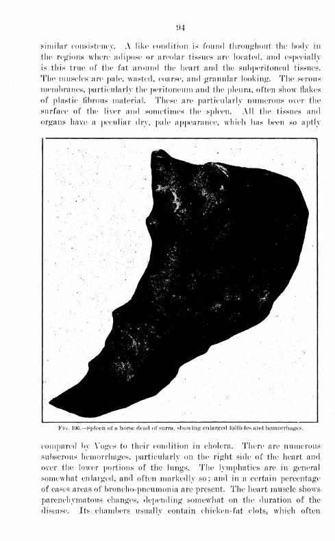
<)4
similar consistency. A like condition is found throughout the body in
the regions where adipose or areolar tissues are located, and especially
is this true of the fat around the heart and the subperitoneal tissues.
The muscles are pale, wasted, coarse, and granular looking. The serous
membranes, particularly the peritoneum and the pleura, often show flakes
of plastic fibrous material. These are particularly numerous over the
surface of the liver and sometimes the spleen. All the tissues and
organs have a peculiar (\v\\ pale appearance, which has been so aptly
IH^I^n11 -'-# Tvl^^^^^^i^^^^H
B
Fig. 10G.—Spleen of a horse dead of surra, showing enlarged follicles and hemorrhages.
compared by Yoges to their condition in cholera. There are numerous
subserous hemorrhages, particularly on the right side of the heart and
over the lower portions of the lungs. The lymphatics are in general
somewhat enlarged, and often markedly so; and. in a certain percentage*
of cases areas of broncho-pneumonia are present. The heart muscle shows
parenchymatous changes, depending somewhat on the duration of the
disease. Its chambers usually contain chicken- fat clots, which often
extend for a foot or more into the aorta. The appearance of the spleen
varies somewhat, hut in the majority of eases it is considerably enlarged,
friable, and somewhat soft. The surface is uneven, due to the enormously
swollen corpuscles, which stand out prominently. On section a typicalk4sago
vspleen is often seen, while in other cases a typical acute splenic
tumor such as is present in infectious diseases of man. In a few cases
the spleen is but slightly swollen, but the dotted appearance, due to the
swelling of the corpuscles, is a constant lesion and gives to this organ,
regardless of its size, a most characteristic picture.
The liver is usually somewhat enlarged, pale, and cloudy. The intestine
shows lesions due to anemia with now and then small ulcerations in the:
upper portion and sometimes in the cecum. The aqueous humor is often
cloudy and contains Trypanosoma. The urticarial eruption observed
during life is no longer evident. In some of the lower animals the
scrotum and even the testicle in the male and the vulva in the female
are greatly swollen, and in the male rabbit the tension may be so great
as to rupture the scrotum. Small prepucial or labial ulcers are not
uncommon.
VII. GENERAL REMARKS ON SYMPTOMATOLOGY.
Before proceeding to the study of Trypanosomiasis in the various
animals, it might be well to make a few remarks on the symptomatology
of the disease.
There is considerable variation in the clinical picture of surra in
the same class of animals, even when the infecting parasites are known
to be the same, and it is therefore not surprising that variations should
exist in different species. In reviewing the literature relating to
dourine, nagana, surra, and mal d.e caderas, one is struck with the
great similarity in the descriptions of the so-called different diseases.
A comparison of the descriptions of any one of them as given by dif-
ferent writers shows as great a divergence as may be found with those
of the diseases acknowledged to be different. In all of them, however,
there are a number of practically constant characteristic symptoms in
the well-established infection such as to make, when taken together,
a clinical picture easy to determine accurately even without the aid
of the microscope.
After an incubation period, which varies in the same class of animals
and in those of different species as well as with the conditions of
infection, and during which the animal remains perfectly well, the
first symptom to be noticed is a rise of temperature, and for somedays a remittent or intermittent fever may be the only evidence of
illness. Later on the animal becomes somewhat stupid; watery, ca-
tarrhal discharges from the nose and eyes appear; the hair becomes
somewhat roughened and falls out in places. Finally the catarrhal
m
discharges become more profuse and the secretion more tenacious and
even purulent; marked emaciation develops; edema of the genitals and
dependent parts appears: a staggering gait, particularly of the hind
parts, comes on, and is followed by death.
Yoges divides the symptoms of mal de caderas into two stages, as
follows: On the fourth or fifth day after inoculation the temperature
rises rapidly, sometimes to 40° or 41° („ and then suddenly falls to
normal or nearly so, usually on the second day. Within five days
there is another rapid rise, reaching 40° C, followed by another sud-
den fall. These reactions may be repeated from two to eight times
during the course of the disease. This is called by Yoges the first
stage 4
. During this period the appetite is good and there is no ema-
ciation. The thirst is increased. The feces arc normal, but in rare
instances may show a little clotted blood. Transient hemoglobinuria.
The reflexes are normal. The coat is smooth, and the hearing and sight
normal. Toward the end of this stage weakness becomes noticeable.
In the second stage the fever becomes less intermittent, and exacerba-
tions to 40° C. or over are exceptional. The remissions are also less
marked. The animal becomes inactive and sluggish and allows the
head to drop carelessly. Progressive emaciation takes place : great
thirst; progressive weakening. Edema, particularly of the hind legs,
belly, and scrotum. The hair loses its gloss. Decreased sensibilities;
digestion and respiration remain good. The gait becomes staggering
and finally causes the animal to fall over. Some animals die sud-
denly during the later stages of the disease. Just before death the
temperature variations usually become greater, vacillating between 34°
and 39° C. ; and death generally occurs when the temperature is low.
The fever is not continuous or characteristic in any of these diseases.
It may be intermittent, remittent, or, according to some writers, re-
lapsing in character, varying from 39° to 4.1.5° C. It is nearly always
higher in the afternoons.
In describing the epidemic of surra which visited Java in 1900 Schat
gives among the symptoms of the disease some which are remarkable
in that they differ radically in many respects from those described
by other observers. In reading his work one is struck by the fact that
they form a very clear description of rinderpest in caralmo. He found
Trypanosoma, however, and it would appear to us that lie was working
with a combination of the two diseases.
A great deal of work has been done by various writers to demonstrate
the relation of the temperature to the number of parasites in the cir-
culating blood, some maintaining that these are numerous in proportion
to the temperature, and others showing that no relation whatever can
be determined. Kanthuck, Durham, and Blandford say that the para-
sites appear in the blood early in the disease and continue to increase
until death. The accounts vary and may reach ^,000,000 to 3,000,000
per cubic centimeter. The parasites appear to multiply first in the
lymphatic glands around the point of inoculation and then to enter
the blood, a fact which they believe explains the short latent period
during which after inoculation they can not be found in the blood.
Multiplication occurs also in the infected area.
During the incubation period, parasites are not found in the blood by
microscopic examination. This has been explained in various ways.
Voges considers this to be because of their rarity, while others maintain
that a stage of multiplication goes on in the lymphatics. A few conclu-
sive experiments by animal inoculation, however, have shown that the
circulating blood is infectious for one, two, or even several days before
parasites are found by microscopic examination to be present, which
would seem to indicate, as has been suggested, that they exist in the
blood in small numbers during at least a portion of the time.
The periodical disappearance of the parasites from the circulating
blood for varying lengths of time during the course of the disease has
not been satisfactorily explained. All kinds of conjectures have been
offered with but little evidence to support them. Voges's idea regarding
this seems to us to be the most plausible. He states that the parasites
are numerous during the first few days after the rise in temperature,
following which they mysteriously disappear; and this appearance and
disappearance may take place a number of times during the course of the
disease. He considers it due to a certain amount of immunity, which
is acquired by all animals in the early stages and which may be repeated
a number of times before the animal becomes too weakened. This view
is advanced in preference to the opinion that the temperature plays any
part in this phenomenon. In the later stages of the disease parasites
may be found in the blood in larger numbers and with a higher tempera-
ture than at any time previously.
Another symptom in this disease difficult of explanation is the ane-
mia, which is progressive from the first. It has been explained that the
parasites mechanically destroy the red blood cells and interfere with
their manufacture in the bone marrow, and finally that on account of
certain changes produced in the blood by the parasites the red blood
cells are taken up by the large mononuclear macrophages. A number
of less important explanations have been given. According to Voges,
anemia is a constant, prominent symptom; and 10,000,000, the normal
number of red blood cells in the horse, may be reduced to 4,000,000,
3,000,000, and even 800,000 in some cases before death. The sedimen-
tation ring of leucocytes is increased and may be larger than that of the
red blood cells. The hemoglobin is decreased in proportion to the dimi-
nution in the number of red blood cells; from the normal of 13.1 per
cent it may fall to 3 or 4 per cent.
7881 7
98
Several observers have spoken of the mechanical destruction of red
blood cells by the parasites. Yoges saw Trypanosoma hold erythrocytes
against the body by their fixed ends until the cells disappeared. Crook-
shank, on the other hand, was unable to satisfy himself that they attack
the red blood cells.
Laveran and Mesnil do not consider anemia sufficient to account for
the death of animals. They say that the manner in which the parasites
act is still unknown.
There is certainly something very curious about the presence of anemia
in this disease and in the action of the parasites in producing it as well
as bringing about death. The mechanical destruction of the red blood
cells is not of the importance many writers attach to it. It seems to us
that there is some factor in addition to mechanical action, which causes
such profound changes, and strong evidence in favor of this is found in
the peculiarity of the gelatinous deposits. The parasites appear in some
manner to produce, or cause some part of the animal economy to liberate,
this peculiar substance, which is not a simple infiltration of fatty tissue
but is of different character. The deposit undoubtedly is formed grad-
ually, as is inferred from the slow change in the secretions. The dis-
charges from the nose and eyes, at first watery, gradually take on the
same character, and before death become very tenacious and solidified in
the nose and corners of the eyes.
Eeferring to the action of Typanosoma in the animal economy, Laveran
and Mesnil state:
Evidently in the presence of an infection so intense as that which is shown at
the moment of deatli of rats, mice, and other animals, the mechanical action of
such a number of parasites is to be considered. This alone, however, in our
opinion, does not suffice to explain the cause of death. Rats infected with Tr.
lewisii may have a great number of parasites and yet feel no inconvenience. In
certain attempts at treatment, of which we shall speak later, a rat has had for
more than fifteen days as many parasites in the blood as red cells.
One is led to think of a toxic action of the parasites and of the intervention
of the soluble products excreted by them. The observations of the course of the
disease in rabbits, guinea pigs, and cows, in which the parasites are not so large
as in other animals, even at the time of death, and the profound dejection of
certain animals when infected, as the ass. plainly corroborate this view.
Strong thought that it was perhaps chiefly through its mechanical
destruction of the red blood corpuscles that the Trypanosoma caused
harm. Experiments were performed to show whether the parasite elab-
orated any toxic substance which acted injuriously upon its host.
Large amounts of blood taken from monkeys suffering with experimentally
produced Trypanosomiasis of severe type were passed through a Berkefeld filter
and the filtrate injected into other monkeys. No symptoms of Trypanosomiasis
were produced. Large celloidin capsules containing blood with many parasites
were placed in the abdominal cavities of sheep, but the results were also negative.
All attempts to isolate a toxin have proved futile. It does not seem
99
to us that a toxin can enter into consideration; hut. as has already heen
said, the character of the action which results in the formation of the
peculiar gelatinous deposits will probably lead to a solution of the
problem.
Voges believes that in mal de caderas death usually results from
a gradually progressive heart failure, and in some cases occurs suddenly
from the same cause. He says that death usually comes on with a fall
of the temperature, rarely at its height.
This statement is true, but does not go far enough; an explanation
of the cause of heart failure is what is desired. The condition described
above may offer some solution, in that the gelatinous deposits are oft cm
abundant around the heart; and a microscopic examination of the tis-
sues shows that a similar condition is present in the myocardium.
The incubation period of the disease in different animals has an
important bearing upon the application of methods for the control of
epidemics. It will be discussed in detail as the study of the disease
in different animals is taken up. The evidence of previous workers
and our own observations show that it varies with the manner of inoc-
ulation as well as with other conditions.
In all the forms of Trypanosomiasis the infection seems to involve
particularly the genitalia, the skin, and the organs of special sense.
The skin symptoms consist in a roughening of the hair, which also falls
out in places; a thickening of the epidermis, often with exfoliation,
and in some stages of the disease various skin eruptions. These may
be simple erythema, and more rarely they may assume the severer forms,
as urticaria, or in extreme cases a distinct localized ulceration may
occur. The scrotum and penis in the male and the vulva in the female
are often swollen, and ulcerations of the penis or vulva are frequent
symptoms, especially in clourine.
VIII. TRYPANOSOMIASIS OF VARIOUS SPECIES.
TRYPANOSOMIASIS OF HORSES.
We shall begin the discussion of trypanosomiasis in the different spe-
cies of animals with that of the horse, which from an economic standpoint
is the most important animal naturally susceptible to the disease in the
Philippine Islands.
Most of the writings relating to Trypanosomiasis deal particularly with
the infection in equides, and as a consequence literature is rich in descrip-
tions which in many points can not be improved upon. It is our inten-
tion briefly to review the most important writings on surra, nagana,
dourine, and mal de caderas in each species of animal, following this
with our own observations, and finally, when through with the species
discussed, to devote a chapter to the discussion of the individuality of
these diseases.
100
Surra, according to Lingard, manifests itself, after a period of incuba-
tion, in fever, a stumbling gait, and general or localized eruptions with
the presence of Trypanosoma in the blood. A period of apyrexia may
here supervene lasting for a day or so, during which the animal appears
better. These apyrexial periods may occur a number of times during the
course of the disease.
In every instance, however, they are followed by a fever usually from
138.7° to 40° C, thirst, slight loss of appetite, ecchymoses of the conjunc-
tivae, with increased lachrymation and mucous discharge from the nos-
trils. The submaxillary glands may be enlarged, and edema beginning
on the legs or sheath may develop. Emaciation is rapid and progressive.
With each exacerbation of fever the other symptoms become intensified,
and the animal is made weaker. The edema spreads, the mucous surfaces
become very pale and tinged with yellow, and the respiration is quickened.
The appetite remains good. Toward the end paresis of the hind quar-
ters becomes noticeable. Paralysis of the sphincter ani is frequent.
Shortly before the end the heart's action in many cases becomes violent,
and death may result suddenly from heart failure. When this does not
occur, the animal finally falls to the ground and dies from exhaustion.
Nagana is carefully described by Bruce, who gives as the principal
symptoms : Fever of a remittent or intermittent type ; catarrhal secre-
tions from the nose and eyes; staring of the coat; and edema of the
abdominal region, the prepuce and the posterior extremities. The ani-
mal becomes markedly emaciated and has a dejected apearance, the head
hangs, the hair becomes very rough and in places falls out, the mucous
membranes of the eyes and genitals become very pale, and there is gen-
erally a slight opacity of the cornea. Just before death the animal falls
to the ground and dies apparently without suffering.
Kanthuck, Durham, and Blandford inoculated two horses, one of which,
a well-fed Hussian cart horse, lived seven weeks, and the other eight days
;
wasting was very conspicuous. The period of incubation was followed
by a smart rise in temperature and by the appearance of parasites in the
circulating blood. A sudden rise of temperature immediately followed
each increase in the number of parasites in the blood. At the time of
death there was marked fever.
Laveran and Mesnil report two cases as follows: The first symptoms
were the appearance of parasites in the blood and fever. One of the
horses, which was not in a good condition at the time of inoculation, died
in sixteen days; the other in forty-three days. With one or two excep-
tions parasites were always found in the blood by microscopic examina-
tion. The red blood cells gradually diminished and at death were re-
duced to half their original number, or even less. Parasites appeared in
the blood in less than four days after subcutaneous inoculation, and their
appearance was coincident with the first rise in temperature, which in a
few days reached 41.4°. This was followed by a drop to 38° C. and a
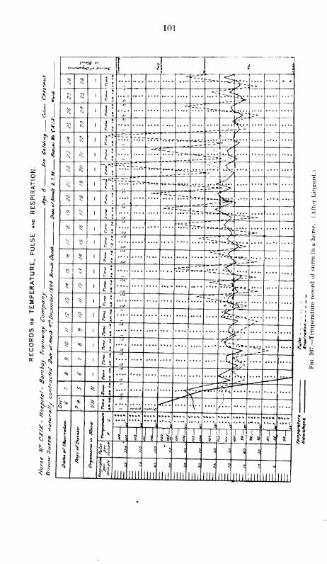
101
o
3a.
o
</>
aa:
oo
h
fc
i J 1
i
' ~]
$»«
( r~
" - L"^.~"
,<+». • .
, V>- -^
^ r5
1 *-- V^,ii
" "i-t-^*'
$*^
A/ - -rr __ ~$At
•-
3 5 1
1
A/
...*:.'._ :,:y,
^* 5
--"- " -"-"_ _,r~. ~._.
•-...L-_ :z-:--f.:
S <\5 J
**
~ ~%="- --- __^ '"- =bv;u ...._
^[
T\T?.~ -.-.-. T«-»-» ~±\ frV-m
<\Ov
T\ ~.f.~. T.-.t ~ir*T ,.~- *-Kt£. -•-•il.... ....
<5>
Nt
....
»r»-.
^«j
^ j
*
"'
* J-r- -IV* ....
S> ^IA *-~ -" —^L
:
ItA [1 : ,.
- ^ i;//A -~"
~iHA ^'
^P *%
//A *~"--<
*---„A
3 -'.5 i
».-. -'I-
: r.-'i<:**
^-:*
il"
——-
v <\
i!
' HA --V
f'A
*•>$ *
.< 5
l/A -•= TJ^_
HA
* ^ ^ :..*%- <- "->
•s OS
Ji •4^ '
'
^ °0
i\./\ "7^
* .*.-» •.
1"-/A
"To> Nil
—IA ~> .• >
05 vo
i\ "ia"....
<JJK ^ *
l\ A
£ <©
^ £ il....
\v.Os6 WO <rrT ....•v-
.... /. . . .... ....
—....
l
\
5s
s
i-
1**
-H ^A 5-T 1>-—
.
J
"."''
«<•»» ««»N
£
rr.r. • •• •
s
^»*»
"*^
T^ rw tl i\JI 1 I u h -Tf
^
5
<; K
5
k.
i^
5<: ^
<» ^
U1L ml III III till
5
III III III llll 111 llll III
I*II
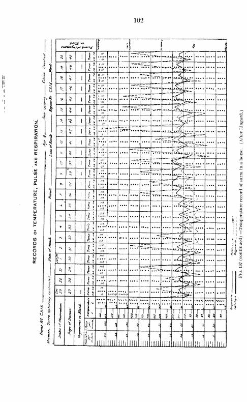
102
i ,5
1 I ' i
\1?
/// 1—--- -r-T_— "•-•A . C «'
•r*•*//*
' •...j.... ^*JL
3 1 (*> 1!
i ///
rrrF-T'Sz ^i . _
.S° * 1 /// ^^*
5 > \\ i ill
f
-**«:
^ J ^T= =*-\
^ ¥ "1!h /// -«r :z:~~
„~a -iSiieil*S « ///
..j-...
^ ^ S 1!J. /// -r- "c; _
.
rn-»
^ 1, 7// ^~
4*'5 >
> 1! * ' *•*7
#—
-
~^ -*-^5^a
r~~=>
t •*1!
"'"
'
.'C -iT.z
*
Al .^y....
I <
5N* 1 $
*V ?.7r ~~l*~i-r »•->-,—— J^i..
r%~-.....
0) ^ ^^ ^ \\
» *1
-.=-
ir%ifa :•" ....'i A
tt* * x \
' //f -==-
..U^^,
' si
1
*
//....[-....
£^tKAA^ -1!* * *
*
"
z ^//./ — -
~~^**<i.T
!
(
<'1*
1?>~~- ;-=*-
(Soo
M V1
-1 IIA t^>-
Q. 03*) l\
H.1 -U-rHA ^ „.
or V ^"//'a'
"Trr "••"••r1 C
''
D~-M-->S
^"~ ^ '>
t s6«.->
*) 1:HA
...+r^
q: IIA h>iS"Q. * V
|!
HA <
^S • HA£Li
h * "? * ..'".,..<]S^ s
>
.... ....u. £ J ///f
in*>
N S ///I
"*^-
7V»T-^^j^:r
...- ... .
o^
*5 l; * HA.... r.Tf Tr.v ~.T.— -»*»-».-< *>- i^-'
.... ....
liU ^
^ *s
IIA
..^^ lr^».... ....
cc IIA
*5 i;HA ^=-~: ."^J" ._.^ ^2-'-/A
§«0
M J!M *c=^
i^i ^.** *IA
KN Vx
IA *: r Cl_>..<
'<
iA s-„^5 *u»S^ *p: V
If
1 1
'iI
1^
H»»N
i*__J
*<e*S
"T
^ "V? : V "
)
V/.1%>»»H
11
?rI
1 5 W IX Ji1
1 1
J1 ! ! i
1 ^ U 1
* 4 s^ \
1^ * 5 ? c % $
*\. 1
]
l1
l<1
Ull llll llll lllllllll llll llll llll llll III! mi llll
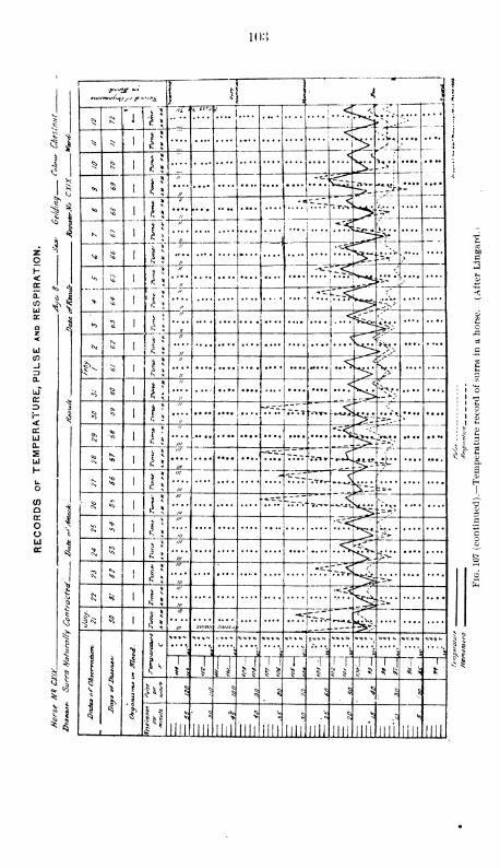
lo:{
1
1
"*lL r h i4
ii
3
<
,5n!
<M"j
jiT' :vwK..J....
1 'Irf — -—»
Js* **
^ * \
'
* j3 £«* ' i
5 1
^ £ 1!<.
1
i
"5
^ 1
^>-
?-~±
;
1*
*> 1!
"**-i*vSv
*
1 *j
$1> *
K
-— l\
-« ;J„
I
ill
" *^ _
t —
I
.. —H T^3TT
,8
>
"
:~VV
;... S)
/; ^- C// •-v-"-
-"^vJ
-ir-!
»-"*.
i>
<i i.::+-"<^ ....
<
$i „
J
1
1
t
"0 J!
" < i;
„
CSi <« 1!
n ^>V
N;.... .... aJ
ac
^O J: ,<^ <
S-,# .... 2„ ^
S 1! -:fc --*•/-'.... ....
1'
'©
! ! ^
S ^ *^
" ~--- '--Ij-.-*^ ...ST....
~V--* ....
! ' t-1
$ j „ ^>_ • 1 o>1 1 *->
<*> 5 i;"
-c" r ! ^ <v
-f--•-•i. ff
' 1 ^
<*> - a----- Z.ZZ F" <> ^ g
w ——
-
r_-3=
..•• ^~* t-«-a0>
<\ 5 i\
**5vv«
-*-1
* ....m
1
* £ £///
*"~"r T.-^:i^
....
•-r, >^
1 5
/// C
///
'
;^» 1=1
S £///
...J .... [^K 3/// i>^* o
3 l\
///-=.-
•"-T-.^
///:~»
'fe
3 *> l\
"' •*-<
in _.__.
--. *.
----
S l\'" -.-
in «/,«« „UJSJ i,
1
i
f
5
5
.11 *I
*<•*•. <.<•». *><0»N **»<* t**^
3 ^
1
^1
^
1
1 J
J J J! |
"7 1
ii•1
i J U ^J ^ ^1
J§ |
^5 § $ 5
S*< ^ S g
.?
* 8
Jill
5
llli llll llll
V
ill! lllli mi llll llll ii if llll llll
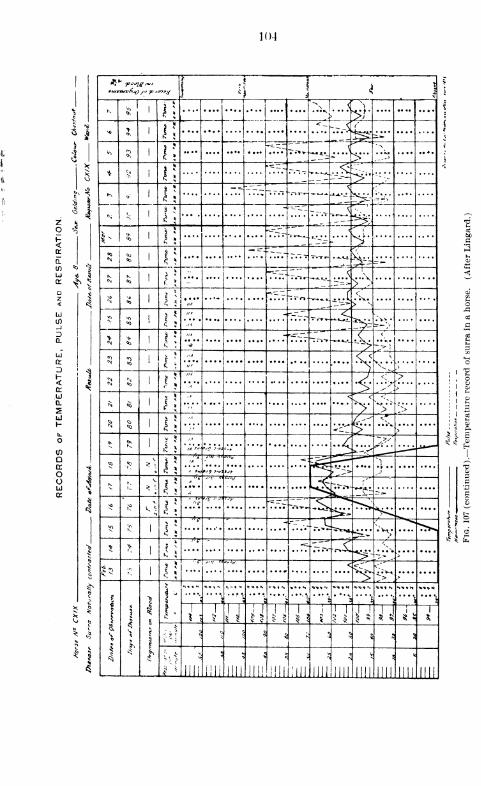
104
f <! \ » i
^ v> 11
II
i r
N>•••• >}•
>« >l!
<r _-.~A^*~ -»
^ r;..rr
_— *•-. ..J 'h
.... •• ••
* y i!»~~^
^___ < ^,.-"*-=-—
f-\o- 1!
*" — --TJT I ™ . J^- '~\:\'TiT*.-
<\i * 5 <
~---~z -..._ -~"
"1^ <& |;'vr
-^*-I T*-*-'Z.
<,
>'«0 (JO
I!"" "~"
*r~ v.-^--~~
<*>£
* ~-~--n"~
,,,5\
- >:'
^5 <*> 1 ; *//V*"rr.T ~~<"»-^.^Ifi> N
.... ....
*T>
<o
"* r~~~ l-.Tv-^^r "S^>~-b;.,<
>N <*> 1:
,,A
^^._<!
•* ~~~> ->
)
2<*> t\
„A <T ^ K
"'
/^ 1>
4 | ;
»4 J < >.A J>
,.^S\>
N ^.A <
> > ".>
S <0
''
/".r"*<.*"
> >
t N1
5
^ ~~>s'j*~-,A*« »,« „ ui r->
^ t"*\
1;>A
k t"V° -*' ZP~~1 '
"~\
*. 'H vs9 „u tjf > z.> J
^ ^ *;
1
1
~tr
...WJ
.... .... ....4C*^^t. ~»
v*S k $ I 1
r. e.... ... .... Tr.iv,^^**i
T^
s N?1 J
!
" '"*? *"."..'."T.^ >.V Vv^. .... "^
* >1
-=. zrSl^ *£•- ~V * .. . . . ....
\ 3 ;:•I l\
<vV<^ ^>
....
t
\
Xi
5
1:
**>- ..... **.N •"••*• -- "+« ^N **.,— *--
;,
? ; 5 3, ft
1
s J
1
* >J
! 1
\\ i\ i
!1
^ 1 ^ 5 * 5!
1
*
3."
" j
5 $ 1 § % - $ H
£ Jill mi mi III! llll mi mi nn llll
5
llll llll llll
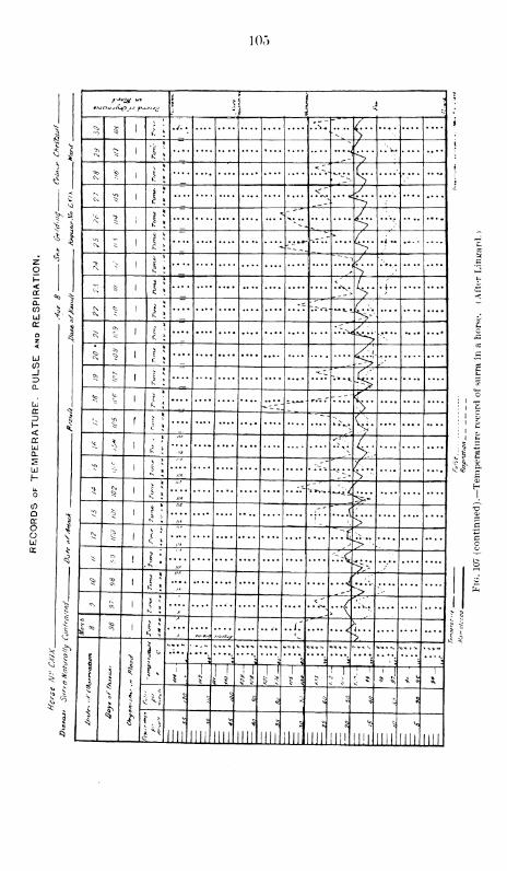
105
idcr
_JID
D
<jrUJa.
u
Qq:
OoUJ
* 1
! i I < \
^ $ 1 £ > C t
>s 1 r;
' t
•***i> '
ok *
*~c
.
«
. >-V ;
c; 3 1 i;<" y^_s ~y
^> 7
v ~. >c\
-1 | |
'**'*'\ *
/•
^>
> 1 li
" *~ --,/->
-§ i |; / .
>^ I 5
J
-c —•*-!
H-'
<\?
1t ^
>>^ i | ; ^
5 § 1 s >
--. &
_*" Jt>
^ Jc i~ _ , <
.
--_-: -V'<
^J^ ?
Q.^c
^ .... 4- ; 5
**•
§
*
. :<~
"
5; 1; ^s
Cmi *
"''
-i'— ~Us
a* ****-
Kf)
<
HA,-'
.(
1,* [ S>"r\, ^
s
^ *
""
/•/. >si £
'
::.<
^ */M
/«. ^^ £ 1 j ; . .\C ...(
J-& 1^ 1
"
..].'
-* t—
*
>.
'
^ t
1
>3
\ i
u„V,Cjf,
*>«>•.
r.::«<c>=*
:r:
>.
::v:— ^ *
s
t* 1 9J J
T-rV 1 15
< ^ $ $; >; " 5;
< M $ 5^5 * § * 5 ^ § 5 S
i •*
n ill in llll III!
*
llll llll ml llll mi III! llll llll
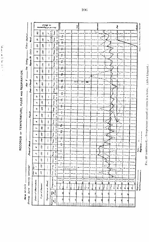
!()(>
zo
Q.
U)
or
a:
D<or
(O
Q
OO
txurmf&v^, f>**»x I 4 *y 1
> 1!..r; ">/ • ••• i
$ J ^ill ..<[/J>^
...,;. \ ..
i
i
»,
5 I;
*']>*£ >...
1
Al »
»v
5 1!
At „e
Al
5
K
* 5
1;*9 ••
i
v
*-<
.
l\i
i
Al V
t
*
, *<•.
iZ\...
r~-55
•J I
!
•l^-~*i
•V**"i<~
>5 i >
—j7" J't /*/ INt v
Wi\
5
•5
— 1177*"
zAl41
v ••
—-— —«>
$ I
* Al — :v._ —I;
it
<fpi
4- .4.
.
6!
A""
—A,•-V..W
Ii
K
HA
—'*A
>
> A
,>
....
^
W
^ 1 \\
HA'—
....
5 i;.... &
....
-{ 1
5'
N U
./!v f.^x.^ .... HN $
1 •^
>
h —4"
^*
..".. ....J.-»,tJf
.... .... ....
- AT::l
J*i*. .\. ....
——
-
0>
? *.... .... .... .... ....
**•"
\'.s^ *v» *
.
....
*.•«
is.
1
J-
i 5 -
i
1*
>
V.V.I
—X1—1-2-—r—t*-"'\
*%* v°r.r
'
s. '
t>1
.J1 .! a kl l* T"\l TVw u 1
III
1!
i4 i § * sv
5 *? «*
I* lil III' Ill nil! in" mi Oil.llli mi ill' ill!
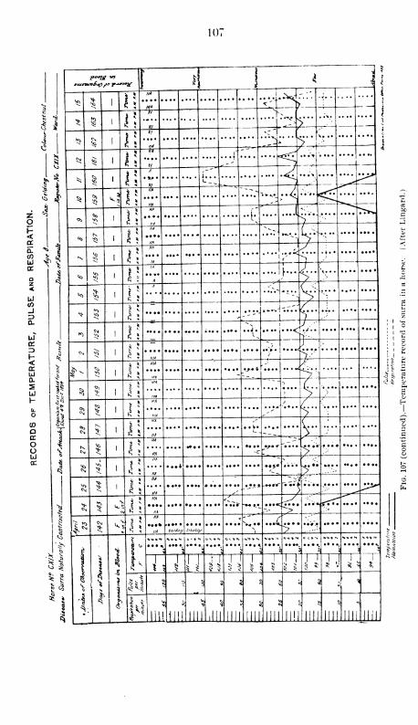
107
zo
DC
a.COaJ
CO
Uio:
I-<DCLiQ-
SUJ
I-
pooig inj j
!
i ]4
^ ^ £ 1
in*
... .... ...
<;T-;••
i
^ § 1 1!
'Hi"
>" —*>
i
*3 § 1 1!i
^ ;.->' ^i
ft
^ ^ 1 i;~n*
...;"
*1 ^ | 1 1!• ** •
t-'-L
i j ^ 5 V.J
i/a
....*lz -r^
^©> 2? 1
ii/i
..f;.~.K
<*>N
1 1!..**.. ..'.:
4 i
*4N.
<01 n
imJ
ia !
''*~_r__
<o 1
I \••;• ...Ji...
^ 1*1
£ X
.:..
i
S:
n-^^ '
1 n y . ..
*""Y*"
*> S 1 i\ " L#
! 1
5!
0^ ^ 1
-V/ \
m .ymmmm* IIIA
'
f ,
^1 1 ;
IIIA
,y % .. . *£%'
| !*
<>>
1 11
m ~ ~ -
(.
<^^
m s^ —
^^ 1 II
'"* ->
/UiA
^' >|-
Cs, 1 J!
IIIA <'
\ ,
r/A
L. . -___ ><0
1 I:
IM ,- '
SIIA
' ' >.'
• ^^ 1 l\
I'A
. J-->
- .-
IIA^^ >"
CM ^ 1
IIA
-*•**- ---'i]'•;•*-•
IIA s,J*l
1 §
II
£s
<<
5 J J
IIA *'-*• ...<>•>
•* IIA
i EIIA ^"
,,/ki "/M* su/CJi JlViSJt*
N, >
» J
—
1 \
3-s
\
i
4^
\:
%V.*N....
s** **
5
*"-**•
» l""
3
•^^ l J "Ji un
1 f 11
J !
'J»l
$ >j
I
cI " g
^n 1 i'
ilii\\\\ III! LIU mi'mi
i 8
.Jill iin
S
|l!l
rs ^
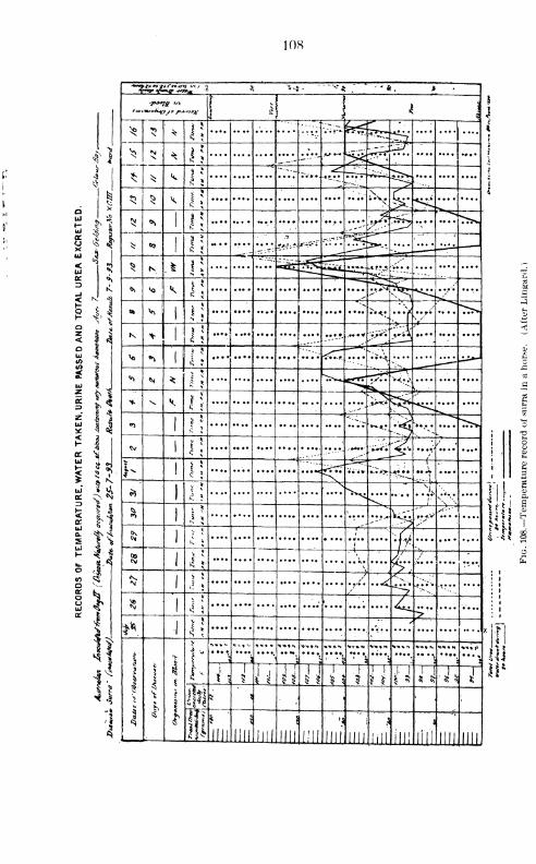
108
!•!
4.
N> 1
Ji
UJon
a.
S ii
H
*
4*
Y«j» fwp vwacp^ *> .*» * ** " *•*»_.» '
! il ! . i
^ * ^ \5 .... .... .... vTi-^ ^rt
v-^2
^ i\~~ -^n>
t:'r**'--"
^ ^ $*
* s
-.. __Ll
"
^5 l
,... .... ....?—:._
5J ^ ^< *
^ >>*-*
<NJ ^ , l n . ...r
tv*-»
* 00|
.... ....•-••V
•:7 '*** ?r.T25^ N ^ ll
zJJi*-^SZ^ .* J»j5-
f'~--
.... ....
&> ^ ! K l!-i»s^
_^
"* x^* ^
l
^^
• ••V<--
•>y'
K *l
* 5
£ ^
•*•• . ... .... .... ... •_ ...-»* -;>-^ .... .... ....
Vo *>l
ft- 5 ^-^ \• •^
" -^
^ N ^v *
£"~~f?
^.
* \ ^ l\
"
^^„.^'
<o1
v* r^
^***•. **.• * >^
< *
**1
.... .... .... .... .... .••-••• ~. ;^ir."^ *. ••/
J- 1.... .... .... .... ...^tt • • •« •-•">> j». ..
••i
.... ....
fc1
<**••*
sfl1
v *< ^ s'
/-•'
....
1
., »c
; '
:
fy'k ; i
1 H \ J/-
1
s 5.... .... .... .... .... .... ../. ..-<
"*'^-^
«1 ^
* <c^
**». 1
V * y
^*
I
1
5
1
3^ —1—r*
....
—1—
£
v'i....
s
NMYN "*»*•.
•••\a
... %J
i$
1
ii ii 111
1 1
-1 I 1 ^ JJ •^ I
IH•»
^,
id JUL mi mi III! mi mi Illl illl ini lill Illl Illl
i i
Hi1? s
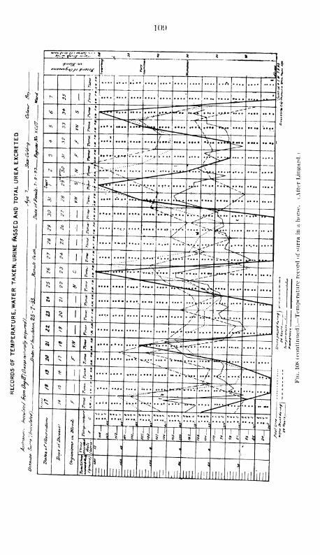
1(H)
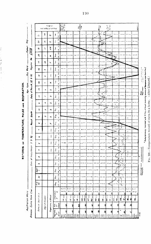
110
i*
Si?CDUi
2 t
<Uio.
2
tooao
$ I
>oo,g u>i,
><-
"
H*
«o j ;
> -' L^>C> «*>
«o i !"•••-S
£ * 5 i *^
.... *>*I
CO- *
^n^ ^v»-«>-7r^
^j>
t § k.. /-
$ ^ 1
* -^^ ^^^?
/*(
v»<t> 1
....V.l"l =*e.-.-
^ N *,
*
-•
j
- **o
i ? ~*r~r"
/> V
'
- - »0*,
*
...<& .... ^
* * «0
* r~,
^
X£-'- ,'
£ h «0 £ >
....^ >.,
^ u<o •f!
\-—*" "
<^-.._
""^-s
~--•.-
to - k if *
TTrr- \mtm
N1 k »
J...
\91 \l i v
>...
.
^1 VJ * > />\
...•
*1
i I
\
....
*>
» / < ^£ » '5
> r''
<V * I/ "v. !
.^s^^.-O 1
*
i-
,;
*T S- v<*
& i1%S
X / 1
k 1
1
*1'*
^
3
"^
Q
5
<$
11
•••- *., .... .., .... ..... ..... ....
3
»..
i,
*•< \ i
•; 3
\
5 <
1
* $ 111 1
1
1 1
1 !
i 1
5 $
1 1
15 0, »> J» J
* Ql
ft£ $ 5
M III! llll lilt.mi
3
IHI IHI mi nil mi llll HlL*
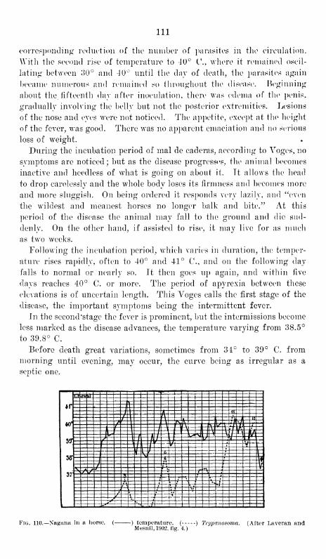
Ill
corresponding reduction of the number of parasites in the circulation.
With the second rise of temperature to 40° C, where it remained oscil-
lating between 30° and 40° until the day of death, the parasites again
became numerous and remained so throughout the disease. Beginning
about the fifteenth day after inoculation, there was edema of the penis,
gradually involving the belly but not the posterior extremities. Lesions
of the nose and eyes were not noticed. The appetite, except at the height
of the fever, was good. There was no apparent emaciation and no serious
loss of weight.
During the incubation period of mal de cad eras, according to Voges, no
symptoms are noticed; but as the disease progresses, the animal becomes
inactive and heedless of what is going on about it. Tt allows the head
to drop carelessly and the whole body loses its firmness and becomes more
and more sluggish. On being ordered it responds very lazily, and "even
the wildest and meanest horses no longer balk and bite." At this
period of the disease the animal may fall to the ground and die sud-
denly. On the other hand, if assisted to rise, it may live for as much
as two weeks.
Following the incubation period, which varies in duration, the temper-
ature rises rapidly, often to 40° and 41° C, and on the following day
falls to normal or nearly so. It then goes up again, and within five
days reaches 40° C. or more. The period of apyrexia between these
elevations is of uncertain length. This Voges calls the first stage of the
disease, the important symptoms being the intermittent fever.
In the second'stage the fever is prominent, but the intermissions become
less marked as the disease advances, the temperature varying from 38.5°
to 39.8° C.
Before death great variations, sometimes from 34° to 39° C. from
morning until evening, may occur, the curve being as irregular as a
septic one.
ns&r
ImiI!
£j
si
Ilil^?
Fig. 110.—Nagana in a horse. ( ) temperature. ( ) Trypanosoma. (After Laveran andMesnil,1902, fig. 4.)
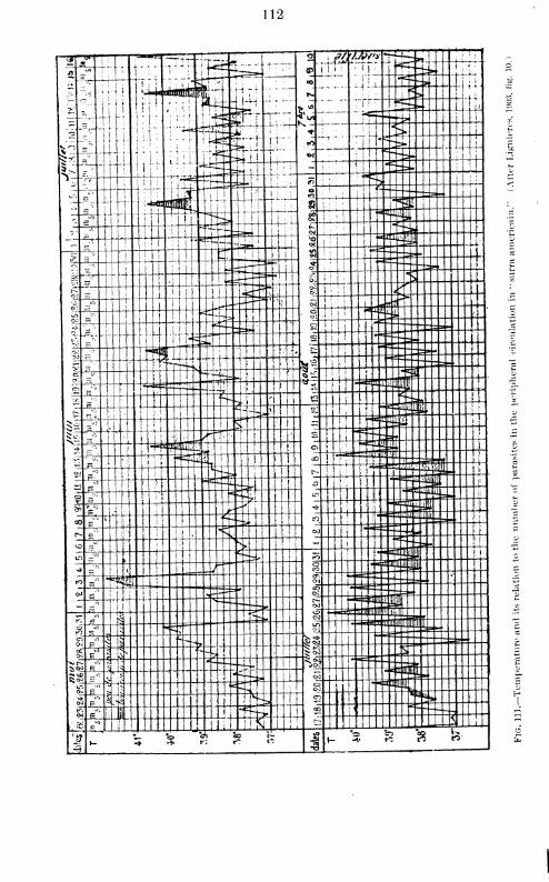
112

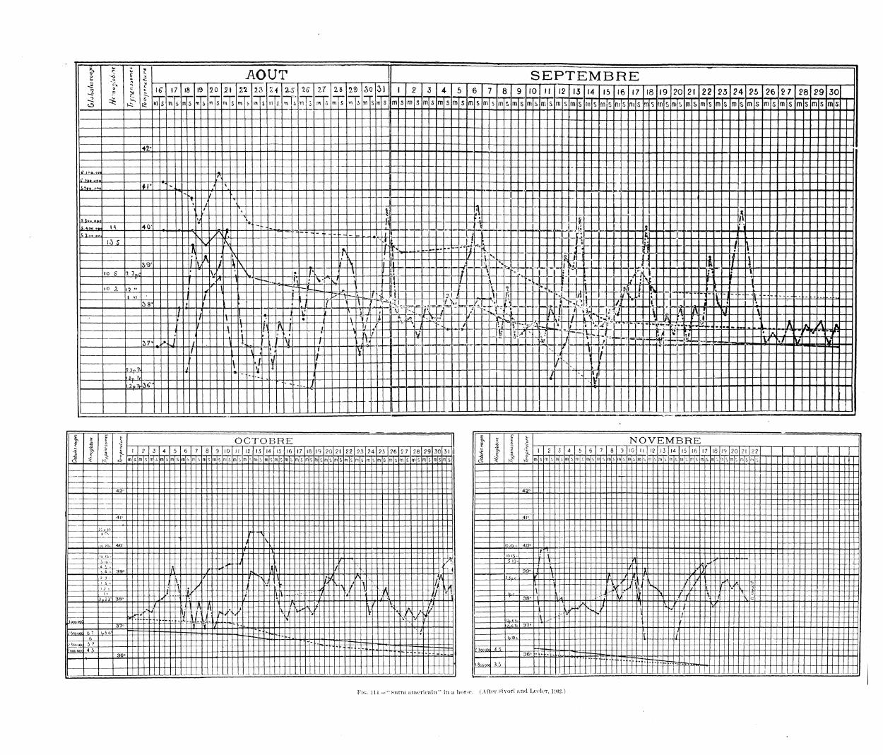
o
o
o
c
3
4
o
%> AOUT SEPTEMBRE* l£ 17 18 13 2C 2\ 21 23 24 2.5 'IC 11
o mo m S
29
m ,5
30 3)
m s w s
1 2 3 4 5 6 7 8 9 10 II 12 13 14 15 16 17 18 19 20 21 221 2^S 241- 25 26 2 7 28 29 30m s m s m s m s m s m S m s m «
» m s m s m 5i rn i rn <> rn s rn s m s ni s m $ rn s dismsm!> m 5> m 5> m s fn s m s m s m s ms
42'
A
C »oe ooo*
5)«o, ooo ±il*x* J-
\
% '
'>
1 /
i /- 4 H l
SSoo ooo »/- — -\ - — -'
_ii• •
|?'
ff "
i_t£*_°l<»4 «• - -.,- $.+ L^^-^LL
r
_ ai
„«-L«» - \ KIBWi. ?.r" «r.. t*— -4j- -S.^oo.co.
15 s : zz^Vr
\1
-=-s-*.^-X
<Lr4
*>*
^- — i_x—/I
- hn
Jj-_U .
.
iB hi - i £%.--
^
r/
il t»
- u ^ r; a f \ 'i
v 1l \. / ^r- c1
1
' .j . ., .. .. J 41--X39- t^
AI $x zL~ ? \ L L^ ' W ~ V lz%
VJ
1
>ij-L. „. 1 1 n^ t .£- Tio 5 1.3nf £ -V i v ?. K >
\ 1;
;V "*"!» XE L i'
""Vr * ^ zt3;^zf^_ • »
s'1
T 1
i_"•
fM•
I'Jhuh- - ^
:t" '
io p. 1.2"
\ h~"
( V f ^i 1
rj"- -1-
1^ ' 1 1
"i ~ —I <J / L_ '
' hu ^ "t :, TW M I
n \ 1r T^
-- vJtl : ^:-584
r { '
, •
•,
>"
| f\ 1 L~21t4JT
'
„::srs: ~*/^ 1_
L_-p "it^ -
T;y*
\•
r i 1 . f 1i 1\ \ .Jl J ^ T "35"53"-4. -_ 4
v i r t " 7 \ • *,
\ 4X i __£k.Z5 x Vpo i £L:^•if
v'- /. A3 •«^
L
*,' ^^ "sit<
,r » ^ C £±~
j
\ ~£j - ~ vt '-. • V4" \ **- »**
"
•^«>< » .«.^»«,^ -J.^.^.J4,. « • * m • . ...--^.r^k. A A-^\ Af
j .1 • L_ • ' L_I1
\ FT? it- 1--.1y ^
*2 ^^ ^_ St..
• S357 %..<-*-J /
H t„ rui " T^ 1
t; -•to/ --f rj^a--^^i: . ^«i£_! ^ ^ TU -
/ \t L *
-V'\
4
f-1
l- t a 2 f^/ \
/
2 JoT, 7v 5{ f — r 1/ ^ >
|
1
a-—-—*. n / s1^
3C** -- ...
___/
V-"^—-
—
.
i
-61
1 1
OCTOBRE 1
1 3t
1
1
1 NOVEMBREi 2 3 4 5 6 7 8 9 10 II 12 13 14 15 16 17 18 l
(3 20 21 22 23 24 25 26 2 7 28 29 30 31 I2 .3 4 5 6 7 8 9 10 II 12 13 14 15 16 17 18 19 20 21 22
m s m s nn s m s m s m m s ms rn s rti j m i m < rn b m s rn smsmsmsm smsmsmsms m s m s msms msms msms V^ m s m s m s m 5 m £ m s in m s m 5 ni o n s s m c rn s m s rn ^ rr s m s m o rn i m s m s rn
42" 42 o
41" 4l o
21a jOpiV,
1 \
J N i
10 20- 40' v 10 £0 c 40°"/"-t^
'o i5"' H
- - U I0I5-f \
L io ••
/r t /T 5 10-
h 1 '\,*"~"
4 5- i I'-
v\- / -^ - --Vi / , A^"i 4 .. ^39°
Af
"
"r / ;L ] i 39° / _ t A _l
2 J • / / •\ jl V-^l *. ""'^"f" 2jpc f T A / X r /i
'
I i ->I
^ >
;± ....
Jt' iu j -L jl ;
_i '\ < M / ^ ^ /*- r- ^
'*-,£» ti 2 >•
\
-J,-^ 4l__3 T _
>L. ,/ ^ Lr
\
i *V'
1 \i ^"mv.~7
/ i-i f"^ v 11
•>
> Xt ---'-.-^ / u -1_ 7i J?-4^ fpc \I A \L \ t V A -4
- -^ ^
2p
2 3c 38-
/ -!--*-/-.: l _.- .. ,.^ r- „- j• -i 38"
I " VA V/
^ "7 — -i- t
A1
v':**_
--\ _c~; 1 • /" x . ti
i
1"j
r*s I \ ^ f " ^"^:^"""
\•
H—1
cV_ v-^'V- C - v, a ,Ji ^_. U^v "S '
1*w /
«•
v >l _.:-_ T \4-±'s& i
V >»'V
/v •^ •- 4- I l-2p + 5c
V ^~i
37" -X A _,.ii-_•« - - ^-;—— !^p 'J^±, 37° T "/
/ f»oooa ft'
6
l
r5bc - sr^ -^^ —••> - - — ::::r IplOo I /
/ 3oo (jwj 5 7
4 5
- -H*"'
"
^^r,S:~~~[ £" "~ffffl 4-S
36° "4.^
c»
36° -
._. ._. -- .... - _.J_LJ_LJ_L_.±1•
_- . . i
Fi<i. Ill -"Surra americain" in a lior^. (After Hivori and Loclor, 1W2.)
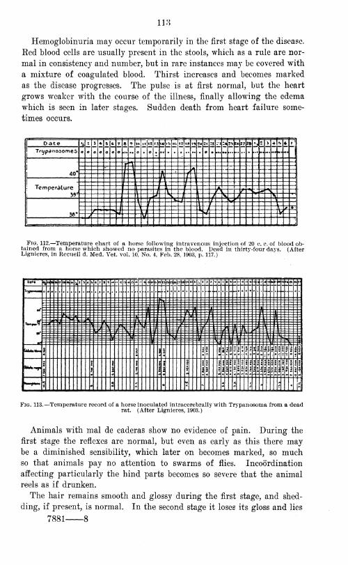
118
Hemoglobinuria may occur temporarily in the first stage of the disease.
Ked blood cells are usually present in the stools, which as a rule are nor-
mal in consistency and number, but in rare instances may be covered with
a mixture of coagulated blood. Thirst increases and becomes marked
as the disease progresses. The pulse is at first normal, but the heart
grows weaker with the course of the illness, finally allowing the edema
which is seen in later stages. Sudden death from heart failure some-
times occurs.
i Datey,
z
o
i
o
4
o o
LIo c
Ol
o
II it
o
rj j* lb IO »7 I* <2 So 2l .'* u !*S6 27 !5 b 2 5 *IlfL
fj.
TrypanoaomeS o
1
1 40'
x •\
I \
\ 1 t \
\' A \ j
Temperature39
u
I \ Vi
I I \\ r \ A
v 5? "N\ j V n r ^
\J N
L , / / \ J'
»•
\ f V / J v *\ I f L
I1
J \l / 4-j
/ *t, f
Li %f V/ V
1
Fig. 112.—Temperature chart of a horse following intravenous injection of 20 c. c. of blood ob-tained from a horse which showed no parasites in the blood. Dead in thirtv-four davs. (AfterLignieres, in Recueil d. Med. Vet. vol. 10, No. 4, Feb. 28, 1903, p. 117.)
Odt-««J
<NHIl HW »• 4« » > c 7 s 9 - .. » 4 •> •»JtO f I »t tlunit 27 te i» JOJl'/.
' 3 j
ht ) • 9 t •» «• «» Ifi ;lifl<«Bo r»rt« n n « V
'•*—
•
.... •
. m- 115
GU*.U~ I I I3 * s i *!! :!:
*fI
« *c?1
£|©
*-,{ 2
8
2
i
fi
I 1 8
4 II Ill
Hi § |
i i £ | ssill I j
5 I
5 J*
- 5*
5 ^ * *' ,'*"'*
Fig. 113.- -Temperature record of a horse inoculated intracerebrally with Trypanosoma from a deadrat. (After Lignieres, 1903.)
Animals with mal de caderas show no evidence of pain. During the
first stage the reflexes are normal, but even as early as this there maybe a diminished sensibility, which later on becomes marked, so muchso that animals pay no attention to swarms of flies. Incoordination
affecting particularly the hind parts becomes so severe that the animal
reels as if drunken.
The hair remains smooth and glossy during the first stage, and shed-
ding, if present, is normal. In the second stage it loses its gloss and lies
7881 8
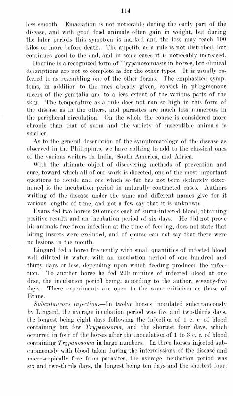
114
less smooth. Emaciation is not noticeable during the early part of the
disease, and with good food animals often gain in weight, but during
the later periods this symptom is marked and the loss may reach 100
kilos or more before death. The appetite as a rule is not disturbed, but
continues good to the end, and in some cases it is noticeably increased.
Dourine is a recognized form of Trypanosomiasis in horses, but clinical
descriptions are not so complete as for the other types. It is usually re-
ferred to as resembling one of the other forms. The emphasized symp-
toms, in addition to the ones already given, consist in phlegmonous
ulcers of the genitalia and to a less extent of the various parts of the
skin. The temperature as a rule does not run so high in this form of
the disease as in the others, and parasites are much less numerous in
the peripheral circulation. On the whole the course is considered more
chronic than that of surra and the variety of susceptible animals is
smaller.
As to the general description of the symptomatology of the disease as
observed in the Philippines, we have nothing to add to the classical ones
of the various writers in India, South America, and Africa.
With the ultimate object of discovering methods of prevention and
cure, toward which all of our work is directed, one of the most important
questions to decide and one which so far has not been definitely deter-
mined is the incubation period in naturally contracted cases. Authors
writing of the disease under the same and different names give for it
various lengths of time, and not a few say that it is unknown.
Evans fed two horses 20 ounces each of surra-infected blood, obtaining
positive results and an incubation period of six days. He did not prove
his animals free from infection at the time of feeding, does not state that
biting insects were excluded, and of course can not say that there were
no lesions in the mouth.
Lingard fed a horse frequently with small quantities of infected blood
well diluted in water, with an incubation period of one hundred and
thirty days or less, depending upon which feeding produced the infec-
tion. To another horse he fed 200 minims of infected blood at one
dose, the incubation period being, according to the author, seventy-five
days. These experiments are open to the same criticism as those of
Evans.
Subcutaneous injection.—In twelve horses inoculated subcutaneously
by Lingard, the average incubation period was five and two-thirds days,
the longest being eight days following the injection of 1 c. c. of blood
containing but few Trypanosoma, and the shortest four days, which
occurred in four of the horses after the inoculation of 1 to 3 c. c. of blood
containing Trypanosoma in large numbers. In three horses injected sub-
cutaneously with blood taken during the intermissions of the disease and
microscopically free from parasites, the average incubation period was
six and two-thirds days, the longest being ten days and the shortest four.
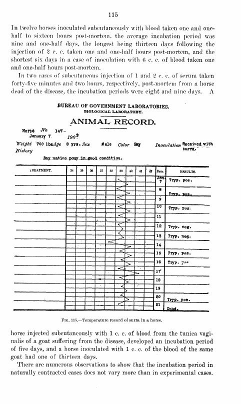
115
In twelve horses inoculated subcutaneously with blood taken one and one-
half to sixteen hours post-mortem, the average incubation period was
nine and one-half days, the longest being thirteen days following the
injection of 2 c. c. taken one and one-half hours post-mortem, and the
shortest six days in a case of inoculation with (> c. c. of blood taken one
and one-half hours post-mortem.
In two cases of subcutaneous injection of 1 and 2 c. c. of serum taken
forty-five minutes and two hours, respectively, post-mortem from a horse
dead of the disease, the incubation periods were eight and nine days. A
BUREAU OF GOVERNMENT LABORATORIES.BIOLOGICAL LABORATOBT.
ANIMAL RECORD.Hor$e Wo H7~
-January 7 190?
Weight "700'lbsMge
jfistory
Byrs.^ Mule Color 1
rpony.:in.fi&«d. condition.
39y Inoculation deceived vitt
surra*
'
TREATMENT. 34 35 36 37 38 39 44 41 42 Date. RESULTS.
V.k
Jan7 Txyp. pos.
>•
<\ 8> Tryp. Dos-
.' <r 9• >< id TTyp. pos<
,, >< IX
,• * ;>
< 12i >
TVyp. neg.
<13> Ttyp. nog.
-c U<
15 TTyp. poa.>•<;
16> Tryp. ??«
•<17"
18> ;
< 19>
< BO> ;
Tryp. po3.^ 811 _2aMi ,
Fig. 115.—Temperature record of surra in a horse.
horse injected subcutaneously with 1 c. c. of blood from the tunica vagi-
nalis of a goat suffering from the disease, developed an incubation period
of five days, and a horse inoculated with 1 c. c. of the blood of the same
goat had one of thirteen days.
There are numerous observations to show that the incubation period in
naturally contracted cases does not vary more than in experimental cases.
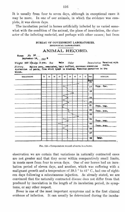
116
It is usually from four to seven days, although in exceptional cases it
may be more. In one of our animals, in which the evidence was com-
plete, it was eleven days.
The incubation period in horses artificially infected by us varied some-
what with the condition of the animal, the place of inoculation, the char-
acter of the infecting material, and perhaps with other causes ; but from
BUREAU OF GOVERNMENT LABORATORIES.BIOLOGICAL LABORATORY.
ANIMAL RECORD.Horse No 5°
September 16, jgo *
Weight 450 Wsdge.S yrs. Sex Male Colo?' Inoculation deceived.with
History . Native potty , . emaciated,, hair .ruffled, enormous .edematousUTTa"
distention of penis, from which lymph is exuding. Numerous parasites in \he
blood.
TREATMENT. & 35 36 37 38 39 40 .41 42 Date. KESULT8.
3?S<
*<r' 17 Tryp. few.<
18J^r >
19"<
<:k 20>
<*21 Tryp. neg.I
^ 22V Tryp. poa»,
23
. 24^ » Tryp. Tieg.
£5Ttvp. POS.
< 86^'
27
28 Dfcfcd.
Fig. 116.—Temperature record of surra in a horse.
observation we are certain that variations in naturally contracted cases
are not greater and that they occur within comparatively small limits,
in mosts cases from four to seven days. One of our horses had an incu-
bation period of eleven days, and another, which was suffering with a
malignant growth and a temperature of39.5°to41 C, had one of eight-
een days following a subcutaneous injection. As already stated, we are
convinced that the naturally contracted disease does not differ from that
produced by inoculation in the length of its incubation period, its symp-
toms, or any other respect.
Fever is one of the most important symptoms and is the first clinical
evidence of infection. It can usually be determined during the incuba-

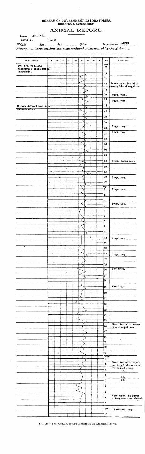
BUREAU OF GOVERNMENT LABORATORIES.BIOLOGICAL LABORATORY.
ANIMAL RECORaHorse Wo .2*0...
April 9, , 190 3
Weight Age. ....... Sex Color „ Inoculation..**^™
History. „„. iArgeJMiy.A^^^
TREATMENT 34 & 36 37 38 39 40 Date. RESULTS.
100 «„c. virulent1
Apr9
rinderpest Tjlood svibc U- 1
twneously
.
10* [<'j 11--
»
< f
18>
<13>
<"14> Tryp. neg.
<r 13
> Tryp. -neg.
< 162 o.c. surra blood su >eutarteousiy.. •<r 17>
<r18>
«*
19>/ 20,
Tryp. neg.
<
V 21Tryp. neg.
s>
<; 22^ V><"
S3>
<, 84t>
<*25
>A
28 Tryp. surra, poe.'^
< 27>23>
n 29 Tryp. pos.'
V
\ SO; '
<< «ay
1> Tryp. pos.
<3>
<j
3i >i
<*
4fry?* pos.
j
s>. :\
5i i {
i' 1^ €
i
! ! >j
! |i< 7
|'
1 ! ^
! | 1
i <r 8!
' \\
9j* i >t
: ; ^10
1 Tryp. neg.
1 11i
;^
;< 12i ...j_^ 1
i ! < 13i ,
-> Tryp, neg.1
•
< <1 14"
1 >
| 1 <1 15i >
1 < ie Fe*r tryp.i
^-~!>
|
i <j IT!
>
f 18
{V19 F»w tryp.
>
/30
J<
21><r
22^--^
V
33
<"""
24><
25?
f 28Reaction with human
/ Mood. n«5Ative.A
27\ >
<> &
<^ZS
><"
301 > i
I <:31
i
Vi
^ June
i
v
> !
is ]
2Reaction with equal
i
"T > parts of blood sur-!
3ra animal, neg.
:
;r do.;\ j
4>
1 do.j /
5 do.
!><T e>•4 j
77^j
! !^ i
8Very sick. No gTeat
! U\
enlargement of sheath!
'1
9
—i
1
1
10j i
Numerous tryp.i
111
] 1
Fig. 118.—Temperature record of surra in an American horse.

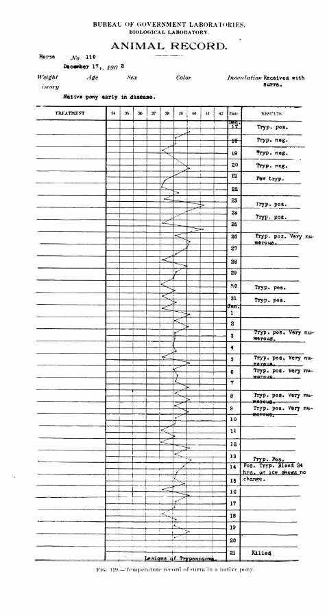
BUREAU OF GOVERNMENT LABORATORIES.BIOLOGICAL LABORATORY.
ANIMAL RECORD.Horse jyb HO — ""
December 17,f290 s
Weight Age Sex Color Inocr/lrrSwr? Received with
isrory surra «
Mat ive pony early in .disease
.
treatment. 34 35 36 37 38 39|j
40 j 41 42 Date. RESULTS.
! ! BeTJTIT Tryp. pos.
^ -*' L
f* 19
<"19 "hyp. neg.V ;:
\ !
:
20 Tryp. weg.^P»
f 21
,^ ,
Pew tryp.
^ , 22ij
'
23^ Tryp. pos.
24^^Tryp. pos.
25
26 Tryp. pos. Very nu-
merous."->
>1
27y<r 28
Y 29/ i
< SO Tryp. pos.*VJ>
<v 31"^?•
Tryp. pos*
<- Jon;
1
<^2
Z^ STryp. pos. Very nu-
> merous.
4*
5 Tryp. pos, Very nu-mflrpun.
<^ 1€ Tryp. pos. Very nu-
>i
C 7\<^
e Tryp. pos. Very nu-! j
^v.>
<r !
9 Tryp. pos. Very nu-*->>
I"^ 10i j1 < 11!
*\
^ 12^u<r 13
i> Tryp. Pos.
^ ! 14 Pos. Tryp. Blood 24'
/ !
i
hrs. on ice shows no^ !
15 change
.
>.
|
•:
161
r^ 17J:
1 <. '
18-N
1
' \< 19I
j:
i ^F
! '
^-'"j
20! ;
•
, ; ! 1
21 Killed.i
Lesions of TrtannfisniiM.
Fig . 119.—To nperatun > reef »rd f snr *a in a 11 a tive tony.
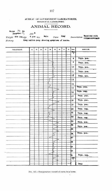
Ill
Horse A"° 94December 7,
tfUREAl OF GOVERNMENT LABORATORIES.BIOLOGICAL LABORATORY.
ANIMAL RECORD.
,2inn
'
„vghf «00 v»,ite 6 yr* .,,,HU.
Colorem lnonlMion £££0^
History G^Y native pony showing symptoms of surra*
TREATMENT. 34 35 ;w 37 -38 39 40 ' 41 42 Date. RESULTS.
7
8 Tryp. pos.•^\
^ 9 Tryp. pos./
I 10 Tryp. pos.>
{ 11 Tryp. pos.
\ l£ Tryp. pos.>f 13)
i
14>
15Tryp. pos.
r^ 16Tryp. neg.
(
17 Tryp. pos.
r18 Tryp. pos.
^19 Tryp. pos.^20 Tryp. ne«.>
r^ 21 Tryp. nog./
22 Tryp. nef
.
~~->»
.<_ 23 Tryp. pos.
< 24Tryp. pos.<'
25^>
^ 26 Tryp. pos.>^ 27
r 23
/
{
>29
/
)0^•<-
;i Tryp. neg.
< Jan,
>-" "
\ Dead
Fig. 117.—Temperature record of surra in a horse.
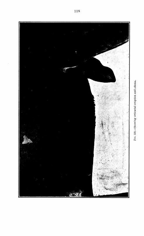
118
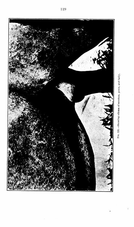
II!)
120
tion period in exposed animals. The temperature as a rule rises very
rapidly and during the first few days may reach 40° to 41° C, after which
it becomes irregularly intermittent, remittent, or, in rare instances, re-
lapsing in type, always being higher in the afternoon.
The especial value of the temperature in diagnosis is its mere presence
in connection with other symptoms. There is at any time very little in
its variations which is characteristic enough to be very significant; but
taken over a long period, its course, while not constant, makes a sugges-
tive picture.
The illustrations (see figs. 115-118) give an idea of the course of the
fever in horses in Manila.
Simple observations of a horse suffering with this disease do not afford
sufficiently exact data during the very early stages, but as the disease
progresses they become of the greatest diagnostic value.
The hair, at first normal, soon becomes rough and shows a tendency
to fall out in places, especially the long hair around the nose and
eyes, though this never progresses to the same extent as it is seen to
do in some of the smaller animals. The skin becomes dry and scrawny
and sometimes shows eruptions in places, which may be erythematous,
macular, or urticarial. We have occasionally noted in well-advanced
cases bunches of macules, somewhat concentric in form, which became
pustular and finally covered with a scaly uxudate caused by the drying
of the purulent-looking discharge from the pustules.
Catarrhal symptoms, of the eyes particularly and less noticeably also
of the nose, manifest themselves rather early in the disease, although
they may come on late and in some cases are almost wholly absent
throughout its course. At first the discharge is small in amount and
watery, but as the disease progresses becomes more abundant and mucus-
like, and finally takes on a yellowish tinge, becomes tenacious and often
coagulates in the inner canthi of the eyes.
Subcutaneous edema, more or less marked, is a constant symptom,
but varies greatly in the time of its appearance. In some cases it
is seen early in the disease, becoming a very prominent symptom,
while in others it comes on late and is very insignificant. Its intensity
does not appear to bear any relation to the duration of the disease. In
nearly every instance it is first noticed around the genitalia (fig. 120),
which remain in the majority of cases the most severely involved part;
from the sheath it spreads forward along the belly on both sides of
the linea alba in two well-defined bands (Hg. 121), which may extend
between the forelegs, where they then unite and pass on to the chest
as a large pod, as illustrated in fig. 122. When the swelling becomes
marked, the two bands under the belly may unite. The edema extends
also down the hind legs, being most marked below the hock; the fore-
legs may also be involved, but to a less extent. Other places which
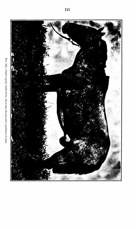
121
122
occasionally show this swelling are the nose and the loose tissues around
the eyes, the base of the jaw, the throat, and the base of the ears; but
it is rare to see these parts involved to any considerable extent.
One of the earliest symptoms following a rise in temperature is
the pallor of the mucous membranes, which first become pearly white
and then take on a decided yellowish tinge. In a well-marked case
this symptom alone is almost sufficient to make a diagnosis. The mucosa
of the mouth, tongue, eyes, and nose assume a ghastly whiteness, which
is out of all proportion to the pleural anemia. Before death these
membranes become somewhat yellowish, a change which gives a jaundice-
like appearance but is of the same character as that seen in some of
the other tissues of the body and described as a gelatinous infiltration.
Symptoms of the organs of special sense are important. In manycases vision is impaired and total blindness may be brought about,
usually by the clouding of the aqueous humor, which in such cases
as a rule contains parasites. The sense of hearing is also involved,
but generally to a much less extent.
Enlargement of the submaxillary and to a less degree of the other
subcutaneous glands is a frequent symptom. In some instances the
submaxillary glands may be greatly swollen and very sensitive to the
touch; and again wre have seen animals without any apparent enlarge-
ment or tenderness of these organs during the entire course of the
disease. When Trypanosomiasis is once wTell established, respiration
is usually quickened and in many instances more or less labored, as
is evidenced by the bellows-like movement of the abdominal walls.
These symptoms are intensified whenever broncho-pneumonia compli-
cates the disease.
There are usually no gastrointestinal symptoms of importance, but
in many cases a very severe diarrhea develops during the later stages,
generally ten or twelve days before death.
The nervous symptoms vary considerably in the horse. In the larger
number of cases the described incoordination of movement and the
partial paralysis of the hind quarters are present to a certain extent,
while cases are met with in wrhich these symptoms do not at any time
manifest themselves.
The morbid anatomy has already been considered in the chapter
devoted to a general discussion of this subject.
TRYPANOSOMIASIS OF MULES.
Very little in regard to Trypanosomiasis of mules is found in literature
excepting the bare statement that these animals are susceptible and
have a long period of illness, "Voges says that the duration of the
disease may be a year or more but that often the parasites are not found
by microscopic examination for days or weeks at a time, and that he
would not be surprised at some future time to find an immune animal.

123
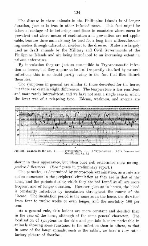
124
The disease in these animals in the Philippine Islands is of longer
duration, just as is true in other infected zones. This fact might be
taken advantage of in bettering conditions in countries where surra is
prevalent and where means of eradication and prevention are not appli-
cable, because these animals may be used for a long time without becom-
ing useless through exhaustion incident to the disease. Mules are largely
used as draft animals by the Military and Civil Governments of the
Philippine Islands and are being introduced to an increasing extent in
private enterprises.
By inoculation they are just as susceptible to Trypanosomatic infec-
tion, as horses, but they appear to be less frequently attacked by natural
infection; this is no doubt partly owing to the fact that flies disturb
them less.
The symptoms in general are similar to those described for the horse,
but there are certain slight differences. The temperature is less remittent
and more rarely intermittent, and we have not seen a single case in which
the fever was of a relapsing type. Edema, weakness, and anemia are
55 56 57 58 50 60
Fig. 124.—Nagana in the ass. (- -) Temperature. ( ) Trypanosoma. (After Laveran andMesnil, 1902, fig. 3.)
slower in their appearance, but when once well established show no sug-
gestive differences. (See figures in preliminary report.)
The parasites, as determined by microscopic examination, as a rule are
not so numerous in the peripheral circulation as they are in that of the
horse, and the periods during which they are not found at all are morefrequent and of longer duration. However, just as in horses, the blood
is constantly infectious by inoculation throughout the course of the
disease. The incubation period is the same as in the horse, the duration
from four to twelve weeks or even longer, and the mortality 100 per
cent.
As a general rule, skin lesions are more constant and decided thanin the case of the horse, although of the same general character. Thelocalization of symptons in the skin and genitals is more noticeable in
animals showing some resistance to the infection than in others, so that
in some of the lower animals, such as the rabbit, we have a very satis-
factory picture of dourine.
125
The morbid anatomy in mules is very similar to that of the horse,
which has already been discussed.
TRYPANOSOMIASIS OF ASSES.
According to Laveran and Mesnil, nagana shows the same general
character of infection in these animals as it does in the horse. The course
of the temperature is more irregular and the relation of the number of
parasites in the peripheral circulation to the temperature is more con-
stant. At a rule, they are less numerous in the blood and may be entirely
absent for longer periods of time. The local symptons, particularly
edema, are said to be scarcely noticeable in these animals. The average
duration of the disease is given by these authors as fifteen days.
Voges states that asses are invariably susceptible to mal de caderas,
and that the symptoms of the disease in them do not differ from those
described for mules. Koch found the asses of Massai immune to the
infection. He does not appear, however, to have demonstrated the
immunity of these animals by inoculation; and as all other writers,
referring to the susceptibility of the various species of asses, have always
found them to be capable of contracting the disease, we can not but feel
sceptical about Koch's conclusions. Lingard mentions particularly the
chronic course of surra in the donkey. There are no asses in the Philip-
pines, so that we have been unable to perform any work on these animals.
TRYPANOSOMIASIS OF OTHER EQUIDES.
Nearly all other equides, including hybrids, have been shown to be
susceptible by various writers, although very little on the course of the
disease in these animals is given in detail. As they are not of any
economic importance in these Islands, no consideration will be given
them here.
TRYPANOSOMIASIS OF CATTLE.
The great variations in results obtained in the study of this family of
animals and their undoubted great importance in perpetuating epidemics,
make it one of the most important to be considered. In nearly all coun-
tries where the infection is prevalent, cattle have been found to be
susceptible.
With reference to the course of Trypanosomiasis in cattle and the
mortality of these animals, there is wide difference of opinion in the same
country as well as in different ones. So far as we know, Yoges in South
America is the only writer who states positively that some cattle are. not
susceptible. He is certain the cattle of that country do not contract the
disease after being inoculated.
Certain writers in Africa maintain that cattle are very susceptible to
nagana, with a high mortality. Others, as Schilling, have shown the
infection not to be invariably virulent for these animals, the course of
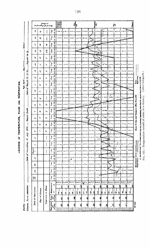
1
. 2<>
**
pee/0 <//
4*v|
i
©> »«
1 \\f -r~
) i
5? Jo
1
c. ....
^M-H ,
* ^ *" <
VS s? £ *! <s£T ^. v\
^>^
<q n k- 5
.... .... .... ...\ r77
* * k I* .... "£J •-•-•*,*
*> ^> k l\ >T.'. x»
Cv.©> k l\ * N»
^ «o k l\4$7<k
>j_^ K
1
„ X < <
- vo1 l\ l
**••/*•
.:;,
*> k ^ l\ Sf *x~&
N * toX 5 -rrr" .... .... H .£"; ;*-...
••••.:.•.... .... ...,.
X> <n <o —«.^^ .... .... ..^r"
• *•?• • • •• .... .... ....
^ cs. * \\•
^j.
^.... .... ....
* - k^ 1
<*>
1l\ ..,/
-~J-~*"J...
N1
% *
< >....^
kr"7
V
1
...:^^«5 *{ 4 „.. ....
•
3 - w >' S'--^
Nv * s.W: .... ....c *
^v J
..-I-" "
« *
1
I
|
1
*a*
S5<:
O
1*
•(.••N
>
•»•»«• t«H
•< •5. "l
»! J
5 5
!! 1
5 § 5 5 u 1 1 1
8 §
i i
1 1 1 : 1
n 5 | § % 8 3 5 $ ? 3
illH
5
III!
5?
ill!
1
iiii III!
3
III! JILL
3
nil nil III! mi nil
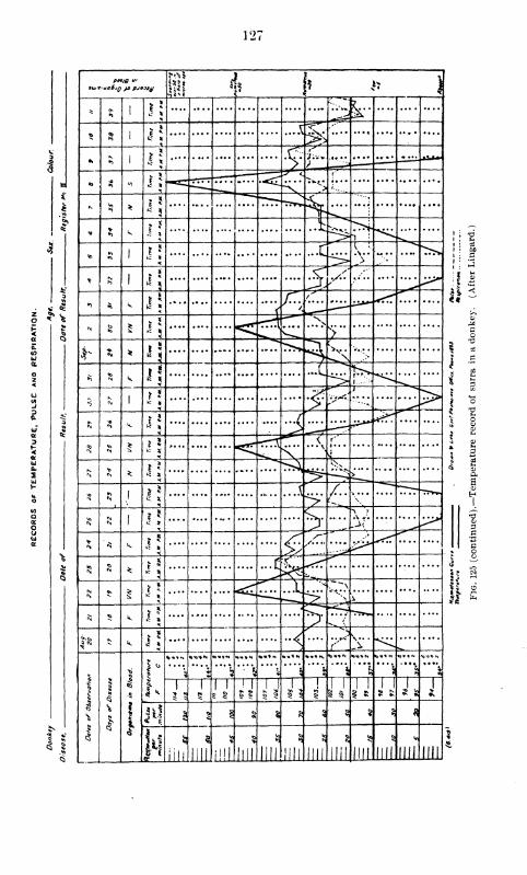
127
I
fito/g ui
[> h *» )
*1
| Ji,
','-* ^v
Q % 1 J i
* *> 1 1 *.... .... .... .... ....9 "S ZZ- :in JLZ±
« »> "0 1 *.^r
T- <
13r
- $ * 1 *
"'"".
« V.1
J.... .... .... ..fc.^i^f 1
1
1 ,
v>*>
11 I
.... .... .... .... ^cs^ >>.. .... >w
1•'
1:
* >> I •I < t it
*) Si k *!r / ti
<*
•„
f* to£ I g < -rr\
^-*"' X•— >
^ -*N * i\
^v^>5 i
frj
<*>
k i\ ;><?
1
S J5 1 i* M
7
1
n<«
k i j J
* 1 <c ...y
f' s
S-..
*
<\>
*.....
V*•»••> >*** <$
3 SI 1
.... ...X
iC £! 1 i| .... .... .... s .V *'. .... ....
^'* k
l£<n *
£-; ^5?
<\tt.
£*£? 'JL7.N
v
18
H$ ^ k r:
.
2l ^*J-.
fe k ••<^
t''
1
<5
I03
I
r1 *
*"**»«
"\*
«>«t»l
3 rv1 2;
•• •••
5"1
5 5
1 1 ! 7T§ 5
i j5 5
1
1 1
i
5 jj1 1
1 * J J1 1
3 «i s g 3 § % ^ ^
firi
53
Nil
§ 5?
i'NM mil mi
<3
Jili mi
55
HII
5
mi 1111 llli
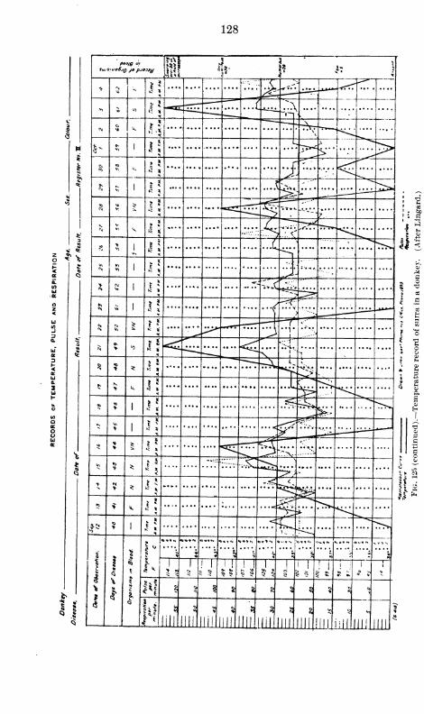
128
p«0/g J,
sujsiuoSjq jo p-/0)ty
3 *
1-
1
J{
.**• •\^~« •</",
*i 5 » 4 5\—
\
<\to
u.
s- 5 1 It 7^\~\-~~
<;-••:-
§co
- ?*
^ *
i.<
* 1
* 2.. 1 v*.**^ * _^jr^i
3 s5; i?
f?-•-•-•j ....
f-4 * s J J,... .... .... ....
.;;,- v;.*,V\ .
.
{rrr.
S**
I1
* '
^
J5 5 1 '!.... .... .... ,:i »••-
••••:....
3; <fl 1
*"- :.>£*& .,.. .... .... ....
SI « I J J,
'••«
5; 8 1 ^ ~rrr "VI
/••••
.
N * <o l! < **1
'' ....
$«0
*l^
'«*'»••
»^>'.... .... .... ....
* > k.
<**>
* 1^^r*»:
«
1
.... .... .... .... .... ...<W~Z*&....
** r .... •>A
~-**X••;v
.... ....
* * * .,.. .... •us^^ -
*> * * 5 .... .... .... .... .... #:
<^¥^.... .... .... ....
5 > u. l\
1 J J (^f
^"•^
|
1
i
1
t
•>
1
1"'""v.
.....
\ >i ? 1 *>
'\ *****,
;
> 5\
1 | i!
1
1
! 1 1
? 5 5 5
"T"i '
i
1 TT8 ^
i ;
5 i
i
>
H § "J | § ^1 a Q§ ^
?»
>
in*Hi lif mlllmiim
55
JJi.llll III ml ml
•i B
I!"
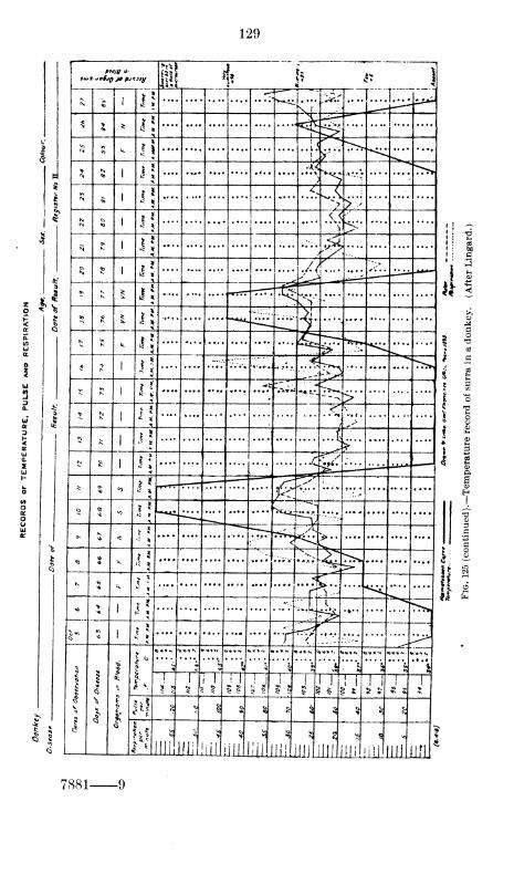
129
ii
shu t uoSjq jo pjfi)ty S 4 h ^ ^
X
N1
J 5-»~^
\l
;
.p^* 1 •
N > * li1 ^
N B k IVI
. . 1
.
....1
> IS4<
.... >*
«S *..tv
p> ;
M 5< <
N ^-'
N £ J 5 ^N JO i! .... — .... .... .... •/< .... ....^t K
£ *i"
#Qv
v>v
00 S *^ .... .... 7^ ..-.v .... .... .... .... ....
K ^k l\ i5^ ^r
5 v1
.... .:;-;• Vv* .... .-•*•,. 5&>
* R 1
1 ? .... .... .... .... ••r«r
<s~
>^ N
1
...
5 ?:1
j
«
...t
5 fe1
ti, ^
=^>* *'
* 5 <91* ....
F~.
"~~ **:.-.-* ,..
:.... ....
o to * Lui
=^fccc
-•-"-*;
f^s;•---..*
* < ••5 *r-^' ^''^
....v6 k I*"-• N
^5- >rv .... ....
kX -'- <^
1 i:
*'••"-
A.• *•
m1 1 * "
/ —
^
1V
1
|
1
*>
1:
1}to
&«»N 'B»l*w «»N o.*N < »<»»N
* :n t
* $ ;,
- *3 a $; \1
V 51
•
i 1
5 5 $ U 11
.1 I
1
5 §1 i
1
J J1
1
W §«s
§ <b§ a fe
5 $ $ S
m \tf III ill ml III III III Hi! ill II! in
*
\iif
a
Ia'
7881 9
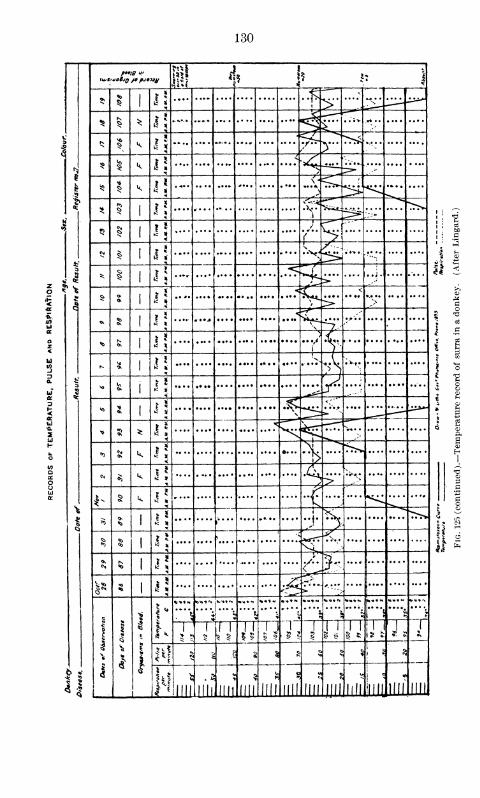
130
? i: 1
**$ i
i j.... .... .... .... .... .... .* >.. ...: "-•" ;^- ^^
^ 5 * i\ .... ^r^ ....•*,;•
(9
tNMi
k ^ >'"
^ ^ k j!^
V"*"*
?if
><J
*5 v,
.... ,..N '
~^5»-^1 5* ^ IJ .... .... .... .... .... ....
»
--.,.... "^v
ti
t,
* 5CM
J!/ <c 1 >
<
s*\i ;
CM| j! .... .... .... .... .... .... .\V <
P^» *•» .
.
•-.:•.... 1
3* 1 *!
<:^ u*J 5>
..-. .... .... .... «... t
<c >' •• •• ,.... .... ....
j J.... •r ••* .... .... .... •• ••
.. , \,•-*'•'• .... .... .... %
"0 c*
i i
1^ >ft* *
*tL-4
• v f .
V"5
- fe * * ^^'
<o 1 J •vv.«:.'' t
*
-* 5 * .1* v^ V
r^? Q
*> k J J $ ....
CM*> k j! %.> >
4- § k 2'fe
$ <*> 1 *!* .... .... .... >^ K
6
$\" *
| 5
§ §1
1 5 \
s^ • *••
if
CMK*>
1 j! sL
3 N 1i! € >
!
i
t 1•
i
*<«*N
3
'"\H*»« •»•«
"
*<•**.
"*\ '")
1
5 5
r U 1
.'
5 ?
1 T2
I i 1 1
1 *\
J j,[
M <s
8 § JS 3 ?!
g
6
2..5
llll llll mi llll llll
c
UJi
CN
llll llll llll
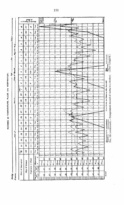
131
y *
tl §o> |J TTTT ^^r^ »•* *•*: .... .... ....
* \ I* ^fr^
$>*« 1 «
^>K. -^ ;,... ...... .... >>v
V $ |5
<,4<*
& 1*
„—
Kv»
1*
<[***t**-
(
*o ii .£^"?•——
.
w»** t\ "5>
* *i. si
•y"-
^sz.... ;,~.
rl
">v*. $
*"~**v I^r
iH*.... .... ....
%
<N fM k k *
'
1- 11 n
v^r*-
>*"*>
§o»
1] ;
\ ... 4
Ok ^?1
1*"
^~y*•
,
a £ 1
V^v #
S
,. ^vi .X*«
1
4
V"
*** ^ *
5 *
^5"^ ;:•--•.
^ k i\ ^<* CJ
1 il....^ r
>*
3 N1 *\
.*^.-* ^ .
i
.
—^ 1
?5 .... .... .5. . .v>
>,....
«* £1 -S ^ ..... ....
i*•5 1
<Vv-* ^i^
7
1
si
I
\V.H
.
*<**N
*i'"\
? 1*v.v.
*»*N
J1
l JJ 111
J5
J
1
5 $ JJ IJ J* J
i>! a c § %«:
5 1 5 3
1 *
it?
Jin III! IIJI mi
55
llll mi 'mi mi llll llll
«0
llli llll
\\r
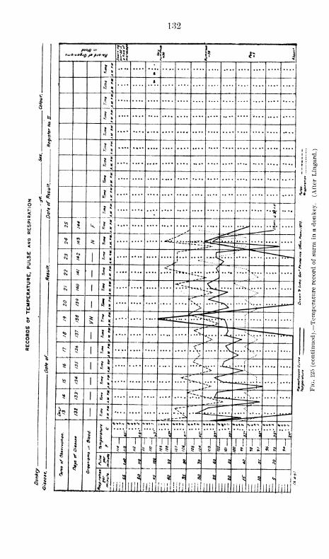
132
sujSiuoBjq jb pjovy^1 J
t * 1
*! 4
<
i
i\
J?
$• ?** S
i•
J;i ;
» ;
i!i :
^ »
3t
*
£ vti
<fc
1
& ? k
*•*
5»
1 »
*• *j$*
j
CM 5 ^ i\,'««: '•KfM^ .-.- *
*^*
1
1
\~~"
.*£—
*
5
« *i ----- £ t
"5
$ J; t.i .... —c\ I ^ 5
*-r..""»*«•:.S>" \v.w.
%
*
5 4! «5
<Jv
^ I! rrr . .*V> t^rr!'
^ 5<-"
^. . . —K
3
<*"'v.
1
s
S
$
^
§ *\ ..\v_^s.
•C>-*S li-
*^
""-Vli
li .... .... *--*..4_ •-
1-•M 1 T~- <
^J-
Vi*
1
1
!
1
1•ft
1 "
4
3><«»N <
"•\«*N «»*»N «it* W
^*
tt ll
1 1 a l •
1' 5
T"2 s
1 1 ! i 1 | ! !
w « OJ * i ^ % S Q
$ * *i $
c•>
o*> N iillllil lllliiiiliili lili! OIL Ill HI
5
III' mli.
nip
*
133
the disease to be long and the mortality low, some eases of complete
recovery being reported. Laveran and Mesnil working with Trypano-
soma of nagana in France obtained a long course of the disease in in-
fected animals, and the mortality was certainly much lower than the
reports usually received from Africa would indicate for that country.
Writers in India practically all agree that cattle are susceptible to the
disease, but mention its long course, lower mortality, and some cases of
complete recovery.
Bruce states that the duration of nagana varies from a week to six
months or more. The symptoms are much less marked than in horses or
dogs. Emaciation is rapid; the hair becomes rough and falls out; fluid
runs from the nose and eyes; and there is a tendency to diarrhea. The
dewdap becomes edematous, but the edema is not so prominent on the
abdomen and posterior extremities as in the infection of the horse.
Fever is constant, but not so high as in the case of horses, occasionally
reaching 41° C. Parasites are rare in the blood.
Schat, working with the surra of Java, maintains that the disease is
very virulent in that country for these animals, stating that the mor-
tality is enormous, the infection sometimes being acute and of short dura-
tion and at others chronic with a longer course. However, as already
stated, from Schat's description of the disease we can not but believe
that there was some other element present to complicate many of his cases.
He unquestionably worked with Trypanosomiasis, and Trypanosoma were
no doubt present, but his description of the symptomatology and the
morbid lesions greatly resembles that of rinderpest. It seems more than
likely that he was working with a combination of these two diseases.
Theilcr observed in the cattle of South Africa an acute pernicious ane-
mia with only slight fever, which he considered due to a special Trypano-
soma named by Laveran and Mesnil Trypanosoma theilerii.
Lingard speaks particularly of recurrent transient urticarial eruptions
in cattle suffering from surra. Great variations were observed in the
temperature, the remissions always being observed in the mornings.
Emaciation was usually marked. Parasites were few and intermit-
tently present in the peripheral circulation. He gives the usual incuba-
tion period in inoculated cases as four to eleven days, and states that the
course is chronic with no mortality in uncomplicated cases.
In the Philippines very little has been written about this disease in
cattle, except that they are susceptible. In our work, which has nowcovered a period of nearly one year, we have examined the blood of hun-
dreds of animals, and but few cases of natural infection have been found.
It may be that during the wet season a large number of these animals
suffer from Trypanosomiasis. By inoculation they are susceptible, and
the course of the disease as well as its symptomatology corresponds to the
descriptions given by the best authors in other countries.
The incubation period is about the same as in the horse, and the

134
2
£ la r \
>o *>
* <;i„. ../
•
* N<
<. ___. -
3
<3 . •
50 *J"*-"'
fet ^ 5 1
;
. . A''
£55 0*
*s 1 ">.,
* £ *>J ./>
1 $ to 1 »*£
> __
.":-iiH.
1
,
I * N <01 i
t £>•',i
*"-.-#
1;
1 :
u <* ^. «0
c
:vv..'"''::•'»
.... l
'.
<>K V) <0
.<..... ....' ....
i;i
v* Y <0
1K
,
< '<[*?:".
\
^ * k *\
. (..
.
'•/ **
> «M k T \ ?
>. > ..> !
* - k
*
•C 1
ivN
1 >5 *• •••
^
4"->
s.4
*.
^^
1
\*
> ;
«s
*>
*
j
;
ON50
5
* ^>" —.1
4
On
>
..*;
>:5 1
*
fc 1 v<
«5 >
^> N 1 •
.1. ||
.1
fcs x
<,i»-
*
)1
5 ^>a /
; * *
i
3 5
i 1
9
%
«$
^
1
.... '.v..
< \n
* T5
*
1
5 5:
M t n11
I
J.1
! 5
1
1
5 »vJ.
«? i
t> a
1» § 5 s J i s\
'1111*.•<
!'!.jm ill. 'ill ..mi III 'mi'ill 'mi"1111 mi •
ISo
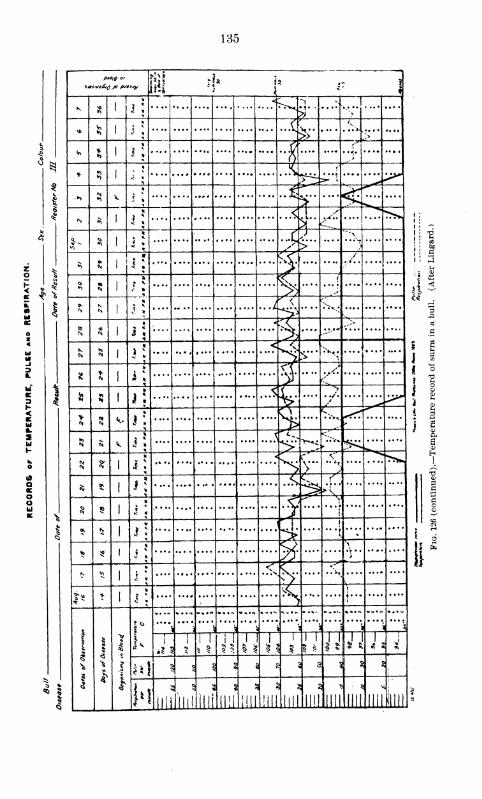
135
pat/g ur
* * j
;
....1^ ..-
* Ss'
i <f 'n.
?>> >
* s'
1 * 1fJ
* s) 1
1 * ». S.
>^ :=»- '«
» 5i.
i
;
..*!f'
<\i U\'
*
«0>
1 J * ^) '**\
])
4 1 i I
, 5 «1
« 1
rJ 3*
i
**1
^ /
«> -*i^
(\, c1
Y 1 j I
<
.'*%••-** .... ....
^ 1
* 1 J
!
^'v;>" *».
& ((
s 1 i i\V
1
! * -•» 1 l\
.<,L&
i I ^^^
r^
5 5 k § ^...J
j<.^
^ p» ;!>*J
8f > 1 i <. ._/'
* 5 1 I ; >\
^ ......$ |
.<?*
> i
£ $ s 1 i j > ^
«B 5"1 j
:
& >
N ^1 ^
.£h-
!
*
t
1 _
- 1«c *
V t«»«.>
« 1
....'
; i
'
j> i^
. )>
"*
T1 I
? 5
1 1 i !i
ji1
5
1 1
1 $ 5 * U J
§ i i :i s s 5 5 8 s
llil
i. ._.
illl1*1
iJllll'llii'mi
s
H!l illl lill Illl Illl
5
Illl
ui s
1^
~a[i s
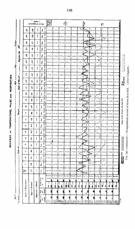
136
I!!! $S 5 1 i
;
<^•^5 ...j.
$ 1«; »
\ i
£ 00
ft 1 i: .... .... ....
«*
3S
$ 1 i; X->
>•*•*• . ...
1
,.i i
S 1 k *>
"•»If
«j
i
1 i
;
<1 •
;
^ ^....
4 >CN 5 1 3 *
•
,,r^
i i^
%.,,
*>
8 1
4
2LC
I \• ^
5'^- s 5>1
1* ...$% <.^.
"**<.
rf i
ui <35 1 J i .*:^ ^Vv
*s.... .... ....
*
S
11
*si
'> 1
£2
z ^k ?» a
UJ
-J
I?
* .... — .... .... .... .f.?'% .... 1 g
uT ^$ %
1|
* <
§ '-N
1 -s
K s N* 1 I
* *w '">§
H *• > i
s<ccUJ0.
C * 1 *: ...j. ...jI g
s5 > 1
.... ...i' <
<4 £
h ^L. .jj.
su. >
1 i * I .... ;-.,. .... ....
-- a,
1 ^....
1
a1 j j
->ux
T3
or
o..t . *
-
—
3
UJ N> 1 *
*3^^ /••
s* >
••/*o
1* $
1 j
!
"<r.>(
^-c- 3
r§
«0 1
**\
S" rK
1 j
»
.<?
1j 4L
»
'^^ f•
V
i
1
'"'\
T* ,'7i j
'"\% s j « )
'"\
1 9
1
5
i : 1 1 i
1 I
1
1
1 1
5 $J I'
ISI ! h !
X
ai $ i \s a £
,« % 3«>
. «
llllill! llif M5
liii liii
*
llll liii llll III! III!
4
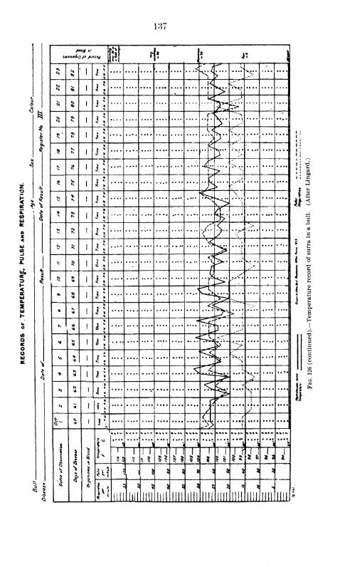
137
«*
5 - ?
eg 5
ii * i' i1 i: |
8 5i ' 1
>
>SE -..^' >'
51 5» 1^>•.,
* 5> 1
1 *
»<
?~j
....
\ i
*
..[7^"""'"*
N 'S>
I
t! * 4^ " v r •
.* * j: It: ....
NflI
|
*
3T„.. . ....
$ \» | ~x .;
\\>
''
* It- .
i» >
\
v J^> 1
*..:. .... .... .... .... ....
7 "'I.... .... ....
5 ?* 1
>
s>•• ..... /:..
S ?
> 3?> .. .1
* 5» 1 .... .... .... .... »'\ . .-.; ..... .... ....
§ '
*
i
:
.... .... .... .... ....
1-
.... .... ....
* !1
• >
^^>* 5 i 1
<^
>;:'
> t
-
K56
1 t\ .... .... .... ....
u•••? .... .... ....
* 5 » ' i\<^S
£v
1 ••>.... .... ....
Vj * 1* 1 i »^5r "'i
* * 1 1 i
•'H'«
•^
...»
*> 5-I
* ^><
.! .*
*..•
N 5 1
*r;
7
f ,
_z ,j
1- * i
V6 1
* < ^
^2 «
11 1
* 1
%"\\ \"!i . * 4 *v*
"\',"'S VI 'Vi i s
5 3
i ! 1
77 a it1
5
! 1 J ?S'J J s i
n! I; I § $ * 2 $ 5 5 s
H 5
till llll
5
Jlii llll
5 g
i 1
i o
i I
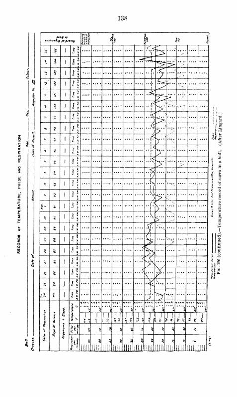
138
*<*
Mil %1
* \ Jis<
^ > ;>
> * .... ....
5 §. iSr'"<
... |
.
i > ../:-
N N5 l> .J. ^•1 ^ ?" '*
'•••»
* «7S
T~-:>
* ..j. .... ....
5 <5 Ji ••f
*>~^
1 ;
1 .
* o>
v *
..e ! :
»C V *>1 ;
<0
j 5
i\ r-il
V, »\<
j
>V) £ 1 V
!< 5* * N •
* *»>
t5
^ 5
<* "f
' V S
"> ] |..•-,..
s
1/ !
N Nv * lA
^
*> j>*
1" £ j
;
1
1 F »
*>Q ,\
> ....--
$ «0 3i.... ;.... .... ....
<*>
*'s fc
N M vKs /»
«S|
Vo . . .
.'
s. ...
\
«s>
1
1
*..
5
1
>*^J
•C
1
i
I
\
I
1 1*
1 *
«-»»N «.<•»% ».,* ,.-, —" — '•• • ••N >»<« *»«N ^**N
>%
k $ i * s»1 k *> %
Vl
s
1
J1 1 1 1
1 !
1 1
5 5
1
5 1 ? r
1 1 1 1
1 51 1 1
« is
1
J
KE
^ § |^ 8 ft
Q$ « ^ ^
3
JUL hii'
?
il!l:
ft) 8
IliliHI'iiii.mi ,11! till
<«1
IIII IIII
i
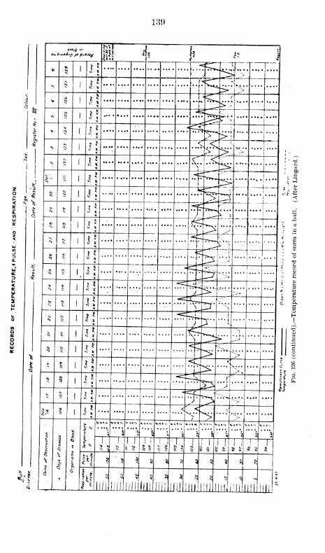
189
"ST" ?i 1
v. ** | *-
-ct^T U* 5 ' K * . i.... .. ^r\; •
£. 1 £ * ,C'^7 l""
^f
-- - *
• "r ^^"p'.;a i s
*. . ..LtC.d"; ...;..
'"""
- j^e :r
,5 1 S* "f~-^*L-^* - ' £ *
*, 5, | ^ .... ....' •^^% :
h:- -—••,
N S .** <"
l <^T ' !;
*• - 1 tc * *\* * ^>#
j*"•
j
, - ? "IF*"--.^" '
----- < , ^i> ' •i'" #
o * 1 | * ^^—' ^
k> ^ 1 li * *SC3* j""
. . 1 f 8 #* ^ i ! « y "^j^"'-!
i
""
v. ** <o 1 C » ........ (tcci
|
* ?; \ ik t .
£ ^ 1 | ••
* ^ 1 K *
^ptL.-i...: -;
* • I 1 J
£ i> is * ^.s:-: i
> * 1 .5 * ....; -i.:\"
1' «
• ••;- —"Vrr'*K- ^
N C^ 1 £*•.... i »
i
*« - • e *
, *i
t
«
..-^ * i < *
"V "**^h>:" r;J
g S 5 * i^rTF-vf "l
—* 5 1 « *
fe.zgtbi
5 § 1*5 -<t£-^. r- " |!
5
* 5 |** - trftZ<.jv ^ 1 >c * .^.^...J,.... .
|.*' ? i li -• . .. <|" '^<1..:.<J
^ V> ....... .
»c»«, <*<*<, *«,c» *>«<•« »<,»m m,^
i . i f i < a ? \ \ \T li li 1 J"J J i r i rj i
%J. 1 n\ ? § 1 § §s a a $ s $ s
... r«|iiiiiiiiil!iii.iU]j.Jli[jJil
^ § «C 5 * ?
Hi ilil.iiniiiii'iiii iiii"
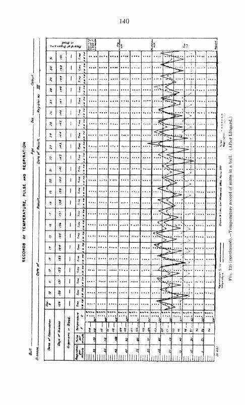
140
ptoig a<
fill ii, ^
*>J i{ .•>*
>
«>>
<»
to *
<'>>
N r;-=3,
* *.
<c p.. *j
'
N > ...:^>
,* >^ r
£> ':>
** >> **">
"
> J J
<<-">
>X J; !>**
NN> 1 i
<
81*
;.^•
"t>
5 > '! % >"' .....
*0\•0 r J < '
•* *';>
•*« ? 5 5^H
* 5*5 <
f>>l.... ....
*«•*> ..ft "C
>^> *>
$<*> <$c
53>
* **» ^ *v^
£ §
'
*r^ * > ——
-
^ 5> i\/ 5—
*""
„>;'.»
_._.
* $*.
.... •••• —
•
..&<.
>.....
$ § i;« ^l :
•rE£^ ?»;
*»*S j!
«c«^ r
1
i
!
1
c
1
1' -3 "A !",* \ ?
•-,»1
s,«> m
it *
5 5
1
51
1
5 §
!
i j ji
"~
1
3 \
! i
3 f
1 i 1 1
5S*! § |
o § ^ * 2!
r
* $ N
nil
<5
mi mi mi mi in
«0
III' ilil, lili"
5
lili lili lili.
*
s
:
? £
,M
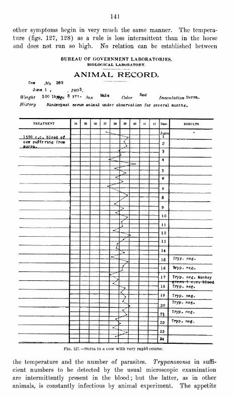
141
other symptoms begin in very much the same manner. The tempera-
ture (figs. 127, 128) as a rule is less intermittent than in the horse
and does not run so high. No relation can be established between
BUREAU OF GOVERNMENT LABORATORIES.BIOLOGICAL LABORATORY.
ANIMAL RECORD.Cow Jf 880
June 1 ,f190^.
Weight 500 lbflj^ 8 yrs. Sev MaleC(j/or
Redinoculation Surra.
History .Rinderpest scrum animal under observation for several months.
TREATMENT 34 35 36 37 38 39 40 41 42 Date RESULTS
June11500 c.c. bleed of
cow suffering from V 2aurra. >
3\
4
b">< 6>
«:—-~
7
> ay<
9><
10">
/11y
<<12^
y 13fK U:>
•<r15 Tryp. neg.^->
/ 16 *ryp. neg.JKr-""
17 Tryp. neg. Monkey><
18
given 1 c.c, bluud
> Tryp. neg.
< 19>
Tryp. neg.
i
20Tryp. neg.X
<2 )'
Tryp. neg.>
<T
22 Tryp. neg.>
<23^>*
- =====
Fig. 127.—Surra in a cow with very rapid course.
the temperature and the number of parasites. Trypanosoma in suffi-
cient numbers to be detected by the usual microscopic examination
are intermittently present in the blood; but the latter, as in other
animals, is constantly infectious by animal experiment. The appetite
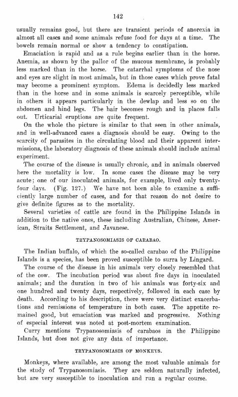
142
usually remains good, but there are transient periods of anorexia in
almost all cases and some animals refuse food for days at a time. The
bowels remain normal or show a tendency to constipation.
Emaciation is rapid and as a rule begins earlier than in the horse.
Anemia, as shown by the pallor of the mucous membrane, is probably
less marked than in the horse. The catarrhal symptoms of the nose
and eyes are slight in most animals, but in those cases which prove fatal
may become a prominent symptom. Edema is decidedly less marked
than in the horse and in some animals is scarcely perceptible, while
in others it appears particularly in the dewlap and less so on the
abdomen and hind legs. The hair becomes rough and in places falls
out. Urticarial eruptions are quite frequent.
On the whole the picture is similar to that seen in other animals,
and in well-advanced cases a diagnosis should be easy. Owing to the
scarcity of parasites in the circulating blood and their apparent inter-
missions, the laboratory diagnosis of these animals should include animal
experiment.
The course of the disease is usually chronic, and in animals observed
here the mortality is low. In some cases the disease may be very
acute; one of our inoculated animals, for example, lived only twenty-
four days. (Eig. 127.) We have not been able to examine a suffi-
ciently large number of cases, and for that reason do not desire to
give definite figures as to the mortality.
Several varieties of cattle are found in the Philippine Islands in
addition to the native ones, these including Australian, Chinese, Amer-
ican, Straits Settlement, and Javanese.
TRYPANOSOMIASIS OF CARABAO.
The Indian buffalo, of which the so-called carabao of the Philippine
Islands is a species, has been proved susceptible to surra by Lingard.
The course of the disease in his animals very closely resembled that
of the cow. The incubation period was about five days in inoculated
animals; and the duration in two of his animals was forty-six and
one hundred and twenty days, respectively, followed in each case by
death. According to his description, there were very distinct exacerba-
tions and remissions of temperature in both cases. The appetite re-
mained good, but emaciation was marked and progressive. Nothing
of especial interest was noted at post-mortem examination.
Curry mentions Trypanosomiasis of carabaos in the Philippine
Islands, but does not give any data of importance.
TRYPANOSOMIASIS OF MONKEYS.
Monkeys, where available, are among the most valuable animals for
the study of Trypanosomiasis. They are seldom naturally infected,
but are very susceptible to inoculation and run a regular course.

BUREAU OF GOVERNMENT LABORATORIESIlIOLOiilCAL. LABORATORY
ANIMAL RECORD.Bull Jfo 368
tlay i5,
Weijht Ag»
Hirtory
Xvjc Od*r IruKHtotion Tr. ov«xaii
TREATMENT M » *> f7 3* w 40 41 « r)mtt> kESULTS.
~
| "Sj|
| 16
<; 17^T:^
IB
«19
•<r 20"">
<HI>
22 Tryp. pos#>< 23S.
\ 24A
• :as"> Tryp* n©e.
\ < 86
><, 27\
<'38
,^.
29:>: . rzyp* naczi 30
>- /31
zzV June Trjrp* nee->
<£_s
s"~ 3
/4
<5"^3
6\\ 7\ Trrpr n«/: B
<. 8>
<9>
<;
10^-~b»
11 Try*]?, pos. (few )S<C
13>•3 13r>«£
*•*
14><
<rIB> „
<~16JT
31 '
17IJ<
18?ryp# nee.>
<r19>
•^20->
< 21
22 Yryp. ncp.
<> 23
•; ,24^
25'
i >r,.-*
.3 2«\
-
ITrrp* nogr» (posir'>
,_^ perioent
)
^>i -c 20*>
«c^'^
- 30>«^-
- Juljr> i
' 8
•c
.,-**"
i
J
/J " 3
•<- 6
' •<„ 7 *jyp, nee* XTbsi-
' r
tive br «niip«4 «x-' ^*~?. - r— '
"*
parljwnt )
,
' ^ -*
;
/ .
**":
'
rf
i»-
' }°i
" --""Kille* tMIo yet
" '
n"
,
1 - r**diU«U
Fig. 128.—Surra in a bull.
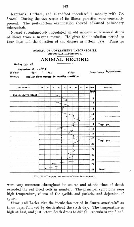
143
Kanthuck, Durham, and Blandford inoculated a monkey with Tr.
brucei. During the two weeks of its illness parasites were constantly
present. The post-mortem examination showed advanced pulmonary
tuberculosis.
Nocard subcutaneously inoculated an old monkey with several drops
of blood from a nagana mouse. He gives the incubation period as
four days and the duration of the disease as fifteen days. Parasites
BUREAU OF GOVERNMENT LABORATORIES.BIOLOGICAL LABORATORY.
ANIMAL RECORD.Monkey JW> 4*
190 2September 11
,
Weight Age Sex Color
History Medium-sized monkey, in he&tthy. Condition-
InoculationTrypanosoma.
TREATMENT. 34 35 36 37 38 39 40 41 42 Dat*>. RKSn.TS.
*!?* '
3.e.e. sum blood •^
12>< 13
r" 14
\^ 15"-
7
k 16^~>
.--'"
17 Tryp. po,
18
<\ 19Y
I 20» Trjrp pos.
f 21
V 2Z><"
23vs»^
2*
?5
~--- M
Fig. 129.—Temperature record of surra in a monkey.
were very numerous throughout its course and at the time of death
exceeded the red blood cells in number. The principal symptoms were
high temperature, edema of the eyelids and pockets, and dejection of
spirit.
Sivori and Lecler give the incubation period in "surra americain" as
three days, followed by death about the sixth day. The temperature is
high at first, and just before death drops to 36° C. Anemia is rapid and
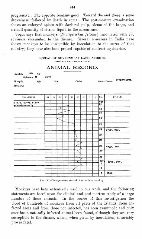
144
progressive. . The appetite remains good. Toward the end there is some
drowsiness, followed by death in coma. The post-mortem examination
shows an enlarged spleen with dark-red pulp, edema of the lungs, and
a small quantity of citrine liquid in the serous sacs.
Voges says that monkeys (Nictipitechus felinus) inoculated with Tr,
equinum succumbed to the disease. Several observers in India have
shown monkeys to be susceptible by inoculation to the surra of that
country ; they have also been proved capable of contracting dourine.
BUREAU OF GOVERNMENT LABORATORIES.BIOLOGICAL LABORATORY.
ANIMAL RECORD.Monkey JVo 66
October 21 . 190*
Height Age Sex
History
Color InoculationTrypanosoma,
TKKATMKNT 34 \h 3t> 37 38 39 40 41 M Datt- KKKILTS.
5 c .c. surra blood Oct.21
3ubcut>aneously
.
m
•<^ 22
•<"" 2'6
>< 24
25
26 Tryp. pos.
\
\ Z7
}C 29>•
< s>d Tryp. pos.>
< 30/
^L 3L>
< Nov1
Xryp. pos*
^ ZVs
3 Dead,.
Fig. 130.—Temperature record of surra in a monkey.
Monkeys have been extensively used in our work, and the following
statements are based upon the clinical and post-mortem study of a large
number of these animals. In the course of this investigation the
blood of hundreds of monkeys from all parts of the Islands, from in-
fected areas and from those not infected, has been examined; and only
once has a naturally infected animal been found, although they are very
susceptible to the disease, which, when given by inoculation, invariably
proves fatal.
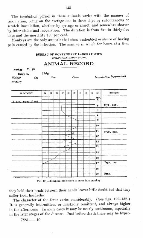
145
The incubation period in these animals varies with the manner of
inoculation, being on the average one to three days by subcutaneous or
scratch inoculation, whether by syringe or insect, and somewhat shorter
by inter-abdominal inoculation. The duration is from five to thirty-five
days and the mortality 100 per cent.
Monkeys are the only animals that show undoubted evidence of having
pain caused by the infection. The manner in which for hours at a time
BUREAU OF GOVERNMENT LABORATORIES.BIOLOGICAL LABORATORY.
ANIMAL RECORD.Monkey No 18
March 5,
Weight 4ge
History
190%
Sex Color Inoculation Trypanosoma
TREATMENT. 34 35 36 37 38 39 40 41 42 Date. RESULTS.
Mar.
5X c.c. Burra .blood
6 TTyp. pos.
7\8
9
|
J 10
11 Tryp. po$.
^ 18>•
--—13
.- H>^ *
15/
/ 16
17 Try p. pof
18
19 Oca*.
Fig. 131.—Temperature record of surra in a monkey.
they hold their heads between their hands leaves little doubt but that they
suffer from headache.
The character of the fever varies considerably. (See figs. 129-132.)
It is generally intermittent or markedly remittent, and always higher
in the afternoons. In some cases it may be nearly continuous, especially
in the later stages of the disease. Just before death there may be hyper-
7881 10
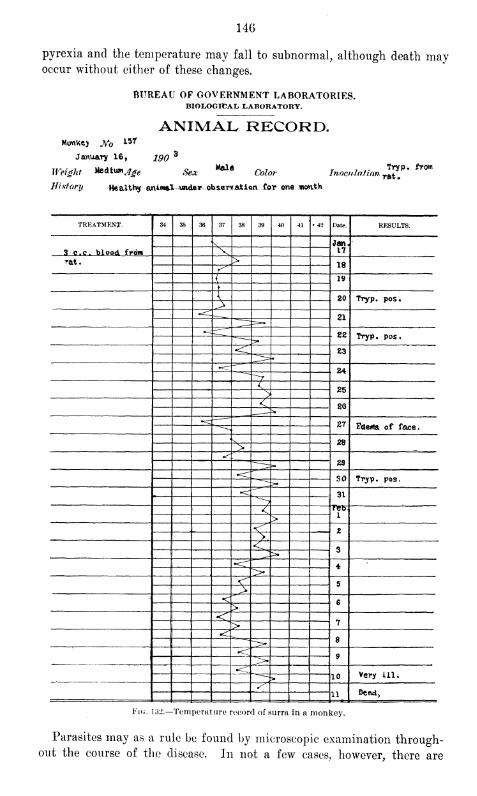
146
pyrexia and the temperature may fall to subnormal, although death mayoccur without either of these changes.
6TTREAU OF GOVERNMENT LABORATORIES.BIOLOGICAL, LABORATORY.
ANIMAL RECORD.
January 16, 190'
MaleWeight He&t\mjge $ex ««•*«
Coior Inoculation
History Healthy animal under observation for one month
Try p. from
rat.
TREATMENT. 34 35 36 37 38 39 40 41 • 42 Date. RESULTS.
\ Jan.173 c.c. blood from V
Tat. >18JS
{19
{ 20 Tryp. pos.>21
22 Tryp. pos.
'<:83
*=cT 24^7< 25>< 20
zt Fdewia of face.
^ 28">
^29
30 Tryp. pos,
31
^7-1 Feb
i>«< 2V<
3>.
4s>< 5
„ >< 6>
<*7>8
•<9^
«<^10 Very ill.
•-
11—»-Dead>
Fig. 132.—Temperature record of surra in a monkey.
Parasites may as a rule be found by microscopic examination through-out the course of the disease. In not a few cases, however, there are
147
intermissions of short duration ; and in one case we found parasites
only once in daily examinations for seven days, though the hlood was
infectious by animal experiment during this time.
Edema as a rule is not a prominent symptom, but is sometimes
noticeable about the face and genitals. Anemia is rapid and severe,
but the emaciation is not so great as that observed in other animals.
Some interference with the gait is occasionally noticed, but it is neither
constant nor very severe. Gastro-intestinal symptoms are absent in most
cases, but diarrhea is sometimes noticed toward the end.
Necropsy shows the general lesions seen in other animals. In addition
to the evidence of severe anemia, the most constant changes are an
enlarged mottled spleen, enlarged lymphatics, fluid in the serous cavities,
and flakes of fibrin over the surfaces of the organs.
TRYPANOSOMIASIS OF DOGS.
Dogs are susceptible to surra in India, and show the same general
symptoms as those seen in other animals. The incubation period is short
and the course rapid.
Lingard mentions as the principal symptoms: Paroxysmal fever, ano-
rexia, later a swelling of the skin about the head and throat, injec-
tion of the conjunctiva, increased lachrymation, in some cases effusions
into the joints, marked edema of the limbs and the belly, extravasa-
tion of the blood into the anterior chambers, opacity of the cornea and
later total blindness. He gives as the principal anatomic lesions sub-
pleural extravasations and sometimes localized consolidation of the lungs,
enlarged spleen and kidneys with subcapsular petechiae.
Eouget proved dogs susceptible to dourine. The symptoms were
edema, particularly marked in the genitals, paralysis of the hind quar-
ters, and conjunctivitis, sometimes followed by keratitis.
Dogs, according to Yoges, contract mal de caderas by eating the flesh
of animals dead of the disease, not, however, through the sound mucosa,
but through injuries, which are always found to be present on examina-
tion and are caused by fighting. The incubation period is short, and the
duration of the illness varies from two to three months.
He describes the symptoms as follows :
The animal becomes stupid, emaciates, no longer responds when called, sleeps,
hides in dark corners, and its head becomes swollen (bulldog appearance) as a
result of the edema, which affects particularly the eyelids. The conjunctiva are
involved to a pitiful extent and secretions similar to those seen in the rabbit
are observed. The hair about the eyes falls out. The vision is likewise impaired
by the chronic conjunctivitis. There is marked edema of the scrotum, which is
first revealed by the swelling of the testicles. * * * The penis is not involved.
On section enlarged spleen and serous exudates are observed. * * * There are
days when Trypanosoma are not found in the blood.
Nagana, according to Bruce, has a rapid course in these animals (eight
to sixteen days) and is invariably fatal. He mentions as the principal
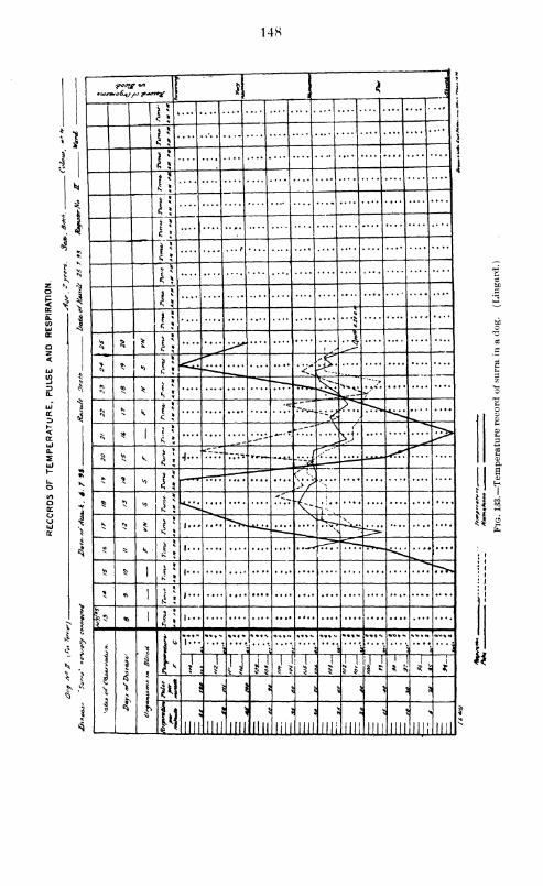
14S
*i ,
\
*
u1
*1?
r;... *
.. . 13 1
l!.1 ^J
* l!....
i\4
u«)
*K
I
s
,2 N
N Nl
1!1 £
11 1)
s l\1
1 >. ^
> * n 1; <i! %.*v
«0 *xT"~ *^V. • *,v
.
3
* * v. 1!•c rj*j""
*^£ s...
^
* 5}1
~'~--"""
vi* >__ ----
^ |*>V. 1!
•j..-"77.- ".v.-— —At. -v-d
•t *o r;
c":--
K J>'/^1 "'
^ <o
*c_ •L j
T: ..
K NS *:
!•-
> 4 * «<> l\R1
>*> 51 i;
\ «s1
i 5
<
* 1 M$ *
^ ^
•? <
i
< 15 ^
il
J
3
4
'1
I
I
|:
V.V.j
*»*•«
8 1
* . T J1
j« i ;t il] 1
1
i
*
M ,
! 6 S J^ ^ I ! ;
'
5 1* jui
-
Illl
8 )
Illl
1
Jill lill Illl
5
MDN
Illl'mi
5
Illl'ill! inr
T
il
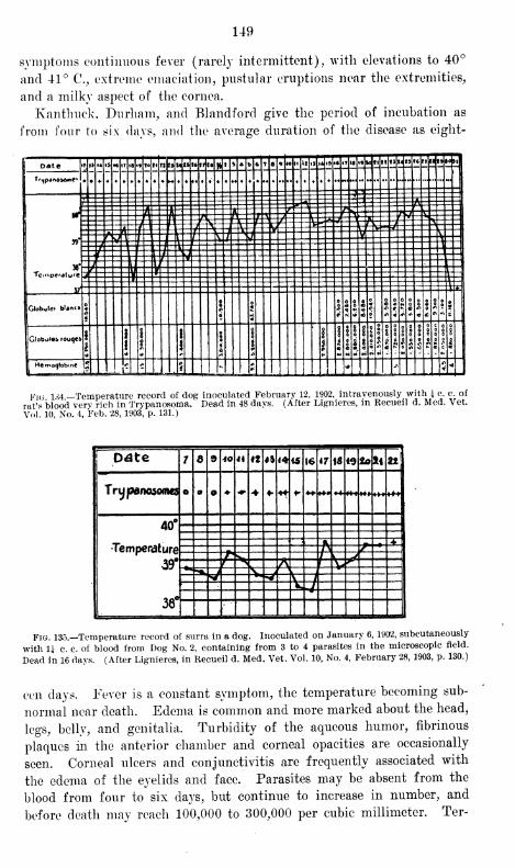
149
symptoms continuous fever (rarely intermittent), with elevations to 40°
and 41° C, extreme emaciation, pustular eruptions near the extremities,
and a milky aspect of the cornea.
Kanthuck, Durham, and Blandford give the period of incubation as
from four to six days, and the average duration of the disease as eight-
Date |«»»» «* i&«tTi8i«t«t<ttr3MtSl«tfeatft » * rj» 7 6 » WH it ,j,4i5.«!lU»»kfllltJl<fSt«fTMt»»0»|
T<-«lp4ne»o«ie< ' o • «. • x - • -
u ft\\fi\ \f\\ LfN 1/MI1K1 11 Hi1]44JWff RTTI
w""3 ?=| t=:==5=tz-53==3===-=$======2==-===-==a==~H~ t£ Sj - - -
«£ 1 ai L I* ™ -t — ftr y _ ri » ... .._._...__ _ - rIf V
,=£_. 11 z J E
.. .JZ
Globule* blanc» «
6
T S £ SSSS*SS£SoSl§51 o «< rK\»«;<«*<<>'<i ««:*'**s
Globules rouqes t "|fi is! i III !|| 11.11 Ills 1
fHemoqlob.ne
j5|| _^±i_j.„_J :
; 1.J „• r ,J:_
Fig 1.^4 —Temperature record of dog inoculated February 12, 1902, intravenously with £ c. c. of
rat's blood very rich in Trypanosoma. Dead in 48 days. (After Lignieres, in Recueil d. Med. Vet.
Vol'. 10, No. 4, Feb. 28, 1903, p. 131.)
I Date 7 £ 9 40 4i 4*44 4
1
15 16 IMS Otofti 22
1 Trypanosoma o o o «•>+«- * •* +mm «*+
40° .
Temperature -•, Z
J Hlff:^3
|
38 -••"
Fig. 135.—Temperature record of surra in a dog. Inoculated on January 6, 1902, subcutaneously
with U c. c. of blood from Dog No. 2, containing from 3 to 4 parasites in the microscopic field.
Dead in 16 days. (After Lignieres, in Recueil d. Med. Vet. Vol. 10, No. 4, February 28, 1903, p. 130.)
eon days. Fever is a constant symptom, the temperature becoming sub-
normal near death. Edema is common and more marked about the head,
legs, belly, and genitalia. Turbidity of the aqueous humor, fibrinous
plaques in the anterior chamber and corneal opacities are occasionally
seen. Corneal ulcers and conjunctivitis are frequently associated with
the edema of the eyelids and face. Parasites may be absent from the
blood from four to six days, but continue to increase in number, and
before death may reach 100,000 to 300,000 per cubic millimeter. Ter-
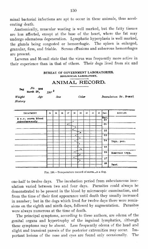
150
minal bacterial infections are apt to occur in these animals, thus accel-
erating death.
Anatomically, muscular wasting is well marked, but the fatty tissues
are less affected, except at the base of the heart, where the fat may
undergo edematous degeneration. Lymphatic hyperplasia is well marked,
the glands being congested or hemorrhagic. The spleen is enlarged,
granular, firm, and friable. Serous effusions and subserous hemorrhages
are present.
Laveran and Mesnil state that the virus was frequently more active in
their experience than in that of others. Their dogs lived from six and
BUREAU OF GOVERNMENT LABORATORIES.BIOLOGICAI* [LABORATORY.,
ANIMAL RECORD.Dog No £W
March 20,
Weight Age
History
190*
Sex Color Inoculation 1 r...Evans i
TREATMENT 34 35 36 37 38 39 40 41 42 Date RESULTS.
2 c.c. surra Mood Mar.20
aubcutaneously ^< 21
><
L 22>^ 23
>< 24 Tryp. pos.
>( 85>
*<^ 26 Numerous tryp*>
<T 37>y
28Dead.
Fig. 136.—Temperature record of surra ^.n a dog.
one-half to twelve days. The incubation period from subcutaneous inoc-
ulation varied between two and four days. Parasites could always be
demonstrated to be present in the blood by microscopic examination, and
from the time of their first appearance until death they usually increased
in number; but in the dogs which lived for twelve days there were remis-
sions on the eighth and ninth days, followed by augmentation. Parasites
were always numerous at the time of death.
The principal symptoms, according to these authors, are edema of the
genital organs and hypertrophy of the inguinal lymphatics, although
these symptoms may be absent. Less frequently edema of the head and
slight and transient paresis of the posterior extremities may occur. Im-
portant lesions of the nose and eyes are found only occasionally. The
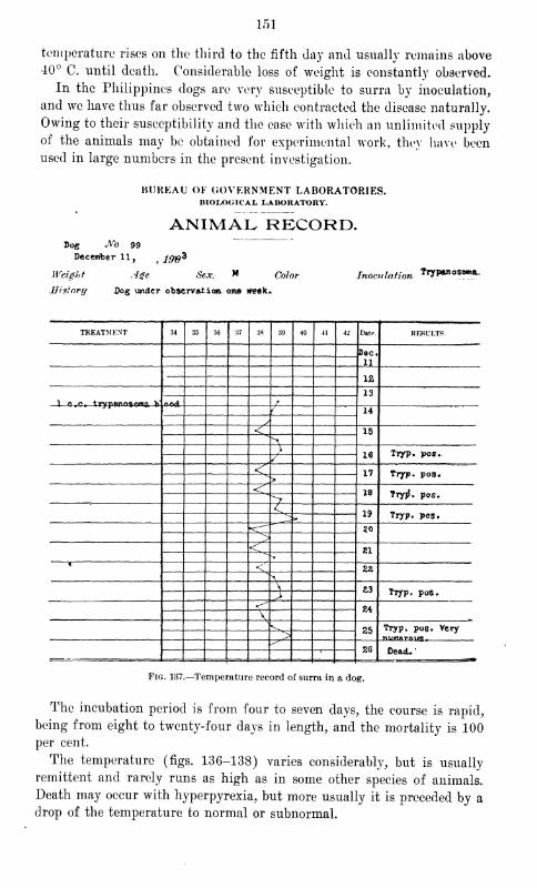
151
temperature rises on the third to the fifth day and usually remains above40° C. until death. Considerable loss of weight is constantly observed.
In the Philippines dogs are very susceptible to surra by inoculation,
and we have thus far observed two which contracted the disease naturally.
Owing to their susceptibility and the ease with which an unlimited supply
of the animals may be obtained for experimental work, they have been
used in large numbers in the present investigation.
BUREAU OF GOVERNMENT LABORATORIES.BIOLOGICAL LABORATORY.
ANIMAL RECORD.Dog No 99
December 11, ,-m*Weight Age Sex. *» Color
History Dog under observation one week*
Inoculation 7r*»*os<m*..
TREATMENT 34 35 36 37 38 39 40 41 42 Date. RESULTS.
Dec*
M12
13r
/ 14
y
<\ 15^
\
16 Tryp. pos.J
<^ 17 Tryp. pos»>
< 18 Tryp\ pos.><_
19 Tryp. pos.
20>
< 21
* <"ZZ
\\ £3> Tryp. pos.
«-"24
V^ 25 Tryp, pos. Very>
26 De&cL'-
Fig. 137.—Temperature record of surra in a dog.
The incubation period is from four to seven days, the course is rapid,
being from eight to twenty-four days in length, and the mortality is 100per cent.
The temperature (figs. 136-138) varies considerably, but is usually
remittent and rarely runs as high as in some other species of animals.
Death may occur with hyperpyrexia, but more usually it is preceded by a
drop of the temperature to normal or subnormal.
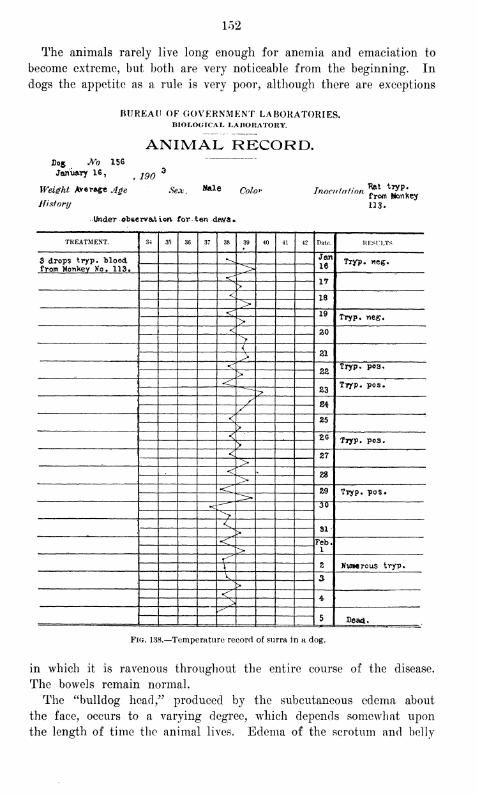
152
The animals rarely live long enough for anemia and emaciation to
become extreme, but both are very noticeable from the beginning. In
dogs the appetite as a rule is very poor, although there are exceptions
BUREAU OF GOVERNMENT LABORATORIES.BIOLOGICAL LABORATORY.
ANIMAL RECORD.D05 Wo 156
Jomi&ry 16, jqq3
Weight Average Age Sex, M**
History
IMder obse rvat iorv for ten dav<s *
Color
U3.
TREATMENT. 34 35 36 37 38 39 40 41 42 Date. RESULTS.
3 drops tryp. blood Jeon
16Tryp. vieg.
from Monkey Ko. 113. ">< 17
>< 18><* 19 Tryp. neg.>< 20
>{ 21><
22Tryp. pos.
>-<
23TTyp. pos.
/ S4/< 25>
< 26Tryp. pos.
>< 27
">
< .28>
•<*29 Tryp. pos.
30
><
k 31>
< Feb.i^>
^ 2 Numerous tryp.
\ a>
<4-
>^5 Dead.
Fig. 138.—Temperature record of surra in a dog.
in which it is ravenous throughout the entire course of the disease.
The bowels remain normal.
The "bulldog head," produced by the subcutaneous edema about
the face, occurs to a varying degree, which depends somewhat upon
the length of time the animal lives. Edema of the scrotum and belly
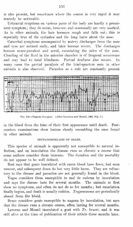
158
is also present, but sometimes where the course is very rapid it may
scarcely be noticeable.
Urticarial eruptions on various parts of the body are hardly a promi-
nent symptom; they do occur, however, and occasionally are very marked.
As in other animals, the hair becomes rough and falls out; this is
especially true of the eyelashes and the long hairs about the nose.
Catarrhal symptoms accompanied by watery discharges from the nose
and eyes are noticed early, and later become severe. The discharges
become muco-purulent and acrid, excoriating the sides of the nose.
Clouding of the fluid in the anterior chamber is of frequent occurrence
and may lead to total blindness. Partial deafness also occurs. In
many cases the partial paralysis of the hind-quarters seen in other
animals is also observed. Parasites as a rule are constantly present
Fig. 139.—Nagana in a goat. (After Laveran and Mesnil, 1902, Fig. 5.)
in the blood from the time of their first appearance until death. Post-
mortem examinations show lesions closely resembling the ones found
in other animals.
TRYPANOSOMIASIS OF GOATS.
This species of animals is apparently not susceptible to natural in-
fection, and on inoculation the disease runs so chronic a course that
some authors consider them immune. The duration and the mortality
do not appear to be well defined.
Post says that goats inoculated with surra blood have fever, but soon
recover, and subsequent doses do but very little harm. They are refrac-
tory to the disease and parasites are not generally found in the blood.
Voges considers them susceptible to mal de caderas by inoculation
and says the disease lasts for several months. The animals at first
show no symptoms, and often do not do so for months; but emaciation
finally begins, and death is usually sudden. Trypanosoma are periodically
absent from the blood.
Bruce considers goats susceptible to nagana by inoculation, but says
that the disease runs a chronic course, often lasting for several months.
Laveran and Mesnil inoculated a goat with Tr. brucei, and it was
still alive at the time of publication of their article three months later.
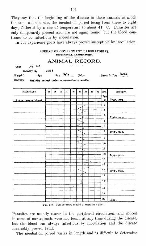
154
They say that the beginning of the disease in these animals is much
the same as in horses, the incubation period being from three to eight
days, followed by a rise of temperature to about 41° C. Parasites are
only temporarily present and are not again found, but the blood con-
tinues to be infectious by inoculation.
In our experience goats have always proved susceptible by inoculation.
BUREAU OF GOVERNMENT LABORATORIES.BIOLOGICAL LABORATORY.
ANIMAL RECORD.Goat JVo 1*2
January 2, , 190 *
Weight Age Sex Hale Color InoculationSurra
Historyjfealthy amwal under obe«r*at ion -a month*
TREATMENT. 34 35 36 37 38 39 40 41 42 Date. RESULTS.
c. surra blood
Jan
22 C 7ryp. ne«.
3
<, 4>
<\5\ * TrrP. nes.
-< 6>
<?' 7>
<^ 8 Tryp. pos.>•^
9><^
10~>
11><"
12^ • Tryp. pos.^ 13
-~
.^ 14-
-,
15 Tryp. pos."_>
< 16
• 17^~
18
19
20 Dead.
Fig. 140.—Temperature record of surra in a goat.
Parasites are usually scarce in the peripheral circulation, and indeed
in some of our animals were not found at any time during the disease,
but the blood was always infectious by inoculation and the disease
invariably proved fatal.
The incubation period varies in length and is difficult to determine
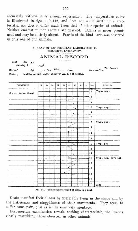
155
accurately without daily animal experiment. The temperature curve
is illustrated in figs. 140-142, and does not show anything charac-
teristic, nor does it differ much from that of other species of animals.
Neither emaciation nor anemia are marked. Edema is never promi-
nent and may be entirely absent. Paresis of the hind parts was observed
in only one of our animals.
BUREAU OF GOVERNMENT LABORATORIES.DIOLOOICAL. LABORATORY.
ANIMAL RECORD.Goat No 143
January 2, 190*
Weight Age Sex Male Color Inoculation
History Healthy animal under observation for 2 months..
Tr. Evansi
TREATMENT. M :*5
!
;
'
*» 37 3ft 39 40 41 AJ 1 >at.-. HF.SILTS.
.c. surra, blood1
Jan.2 TTyp. neg.
Z qi
3.,
<4>
<5>. Tryp. neg.
<6
y7
<c 8 Tryp. pos.
.--C 9^~^-,--
10
V.
11:>
<^12'>. Tryp. pos.
.c-""
13>
14^.
•<^_15 Tryp. neg. Very ill
•c-""
16><"
17>
~r..
18
19
20.
Dead.
Fig. 141.—Temperature record of surra in a goat.
Goats manifest their illness by preferably lying in the shade and bythe listlessness and sluggishness of their movements. They seem to
suffer some pain, just as is the case with monkeys.
Post-mortem examination reveals nothing characteristic, the lesions
closely resembling those observed in other animals.
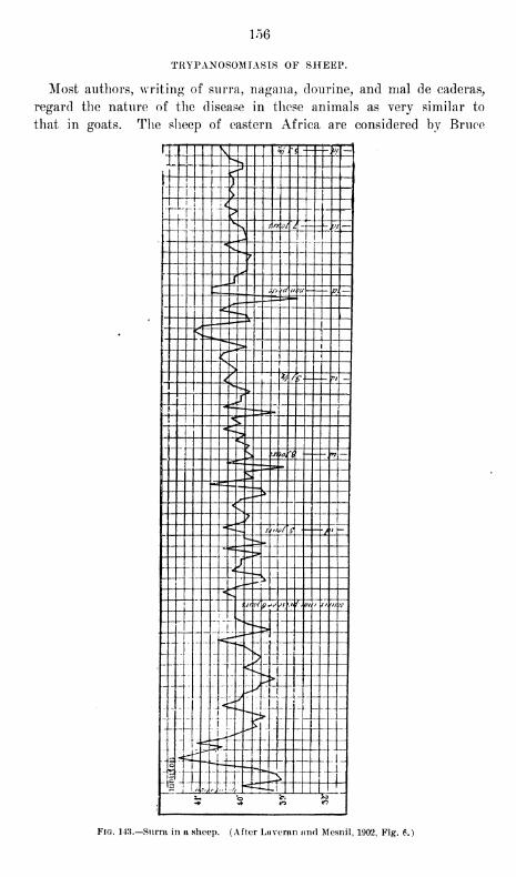
156
TRYPANOSOMIASIS OF SHEEP.
Most authors, writing of surra, nagana, dourine, and mal de caderas,
regard the nature of the disease in these animals as very similar to
that in goats. The sheep of eastern Africa are considered by Bruce
t "T" \ \ u/~' - < - w"""
1"^^
j i j^r, ! )
j j y i
1
* '* I1
~<\
| ^v. Arffl / -j , M —jj
*v^ i
:±" „-i*:T] __ 3pj(d i/ t >u (— p. —
^,— -J ^*=-*~ ,e"-^
v 1
*
: <—.., - :
L <ll_ ^_ .._"~
X X XS 1 . ^ /£ 5- vi _± x ^-"x: jz._±..__ ,-JT.. TTil '— = >. ^x^ sx i.~ -=?:: : :J:__ s
.n io,"i "? —"*~ am
-,—S"~~ =-=-v -*—*- -^ 1 X X^n: ~"
4: f
" — -> s>th/ g />j h~
---~ c— > _*
')'"
"*— — -.
1 ^-- '<j // of p*<> jy j / . out ii/. m 7s-
' 'Ss
J i
'^""^
y^T""^**s
_p f
^^T ^*j
----- |ly^-lX-44---^|-
p:.::.:]f^£tl-.:::±:±1
" c^»» >
- "M^'
'
"1 —f U—, 1
"Jl :
" " _
X:> ~ l_\_
i 's ft 8
Fig. 143.—Surra in a sheep. (After Laveran and Mesnil, 1902, Fig. 6.)
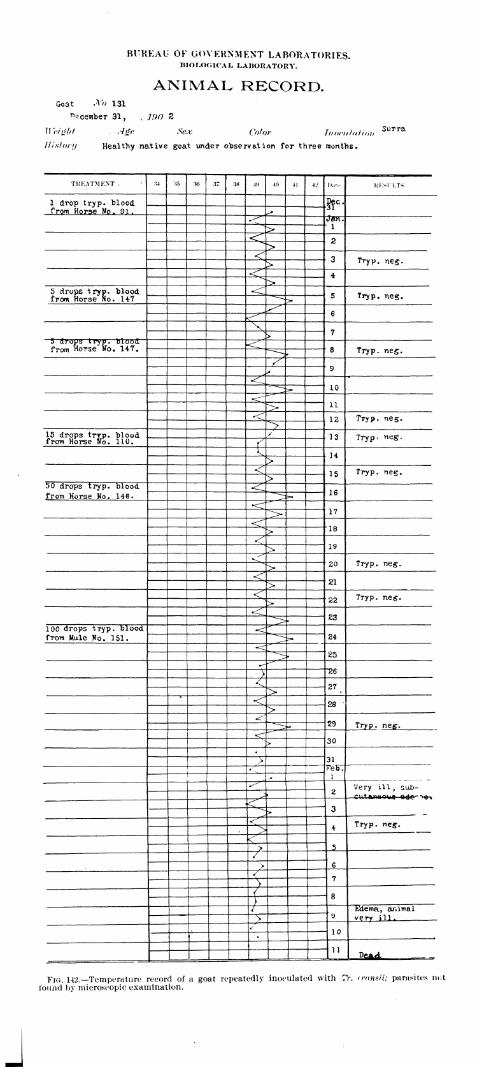
BUREAU OF GOVERNMENT LABORATORIES.BIOLOGICAL LABORATORY.
ANIMAL RECORD.Goat No 131
^cember 31, . 190 Z
Height Age Sex Color I>,<><-,, hit ,,,>,
History Healthy native goat under observation for three months.
TUKATMENT , M :* :« 37 :w :w 40 41 -J.' I»..r-. KKSU.TS
1 drop tryp. blood ftc-from Horse No. 91
.
->«^"
i><r 2>< 3 Tryp. neg.>
<r 4>
5 drops tryp. blood <5 Tryp. neg.from Horse No. 147
6^
N7>
5 drops tryp. bloodfrom Horse No. 147. <:
8 Tryp. neg.
^9
«CL0
•<L"11^>
<^ 12 TTyp. neg.^>
15 drops tryp. bloodfrom Horse fro. 110.
/ 13 Tryp- neg.
(I 14>
<:15 Tryp. neg.
>50 drops tryp. blood <
16from Horse No. 146.
17^»<^~
"18>^
19>•^
20 Tryp. neg.><?
21><
22 Tryp. neg.>
-<23
100 drops tryp. blood24from Male No. 151.
<25^
T "26
< 27^>
*
<f 28*>
^29 Tryp. neg.
<-30>
31>1
J-'eb.
l- _«^
2Very ill, sub-
^ —cuteunwoue ede^ -^^\
3*---> _ _
<^4
Tryp. neg.>
<5>
/6>
/7
>< 8
)i
yEdema, animal
s very jll .
*-'
10
11DCAd
Fig. 142.—Temperature record of a goat repeatedly inoculated with TV. transit; parasites not
found by microscopic examination.
J

157
to be somewhat refractory. The disease in these animals, he says, runs
a very chronic course and some of them live for five months.
Laveran and Mesnil inoculated a sheep with Tr. brucei, and in
an article published three months later stated that it was still living.
They consider the beginning of the disease much the same as in horses.
There is generally an incubation period of three days, followed by the
appearance of parasites and an elevation of the temperature to 41° 0.
Trypanosoma then become so rare that they are not found in the blood
by microscopic examination, although the blood is constantly infectious
when injected into mice. The temperature remains near 40° C. with
occasional intermissions, although it sometimes rises to 41° C.
Eegarding one sheep, which lived exactly one hundred and ninety-
seven days, they write as follows
:
On the sixth day after inoculation it showed a temperature of about 41° C,
which shortly afterwards fell to between 39° and 40° C; on the twenty-fourth
day there was another rise to 41.5° C; after which the temperature remained for
a long time in the neighborhood of 41° C, taking thirty days to return to 40° C.
;
multiple edematous areas appeared in the face and eyes and then in the testicles.
It was only during this period that Trypanosoma could be found by microscopic
examination, and for eight days there were several in the field. The edema in-
creased and extended to the neck and shoulders (end of the third month). Its
disappearance was rapid; the animal (during the fourth and fifth months and
the first half of the sixth) appeared well (the temperature being between 39°
and 40° C.) ; but the blood was still virulent. During the last month the animal
emaciated rapidly and died with profound lesions of cachexia and gelatinous
exudates of the throat, the pericardium, and the lips.
As with some of the other animals, neither the study nor the descrip-
tion of the infection in sheep is as accurate as might be desired for pur-
poses of comparison. In the Philippine Islands they do not appear to
be naturally susceptible to the disease. We have kept a sheep in the
same ground with surra animals for several months, but no infection
has resulted.
The symptoms, the course, and the duration of the disease in these
animals are so similar to those observed in the goat that a description
of them is considered unnecessary.
TRYPANOSOMIASIS OF GUINEA PIGS.
There are on record a few instances in which guinea pigs have been
found naturally infected with Trypanosoma, but literature affords little
detailed study of any of the forms of the disease in these animals. It
is admitted that they are susceptible by inoculation to Tr. evansii, Tr.
brucei, and Tr. elmassianii, while some of them show a transient infec-
tion from Tr. lewisii.
Laveran and Mesnil noticed multiplication forms on the second andfifth days after inoculating one of these animals in the abdominal cavity
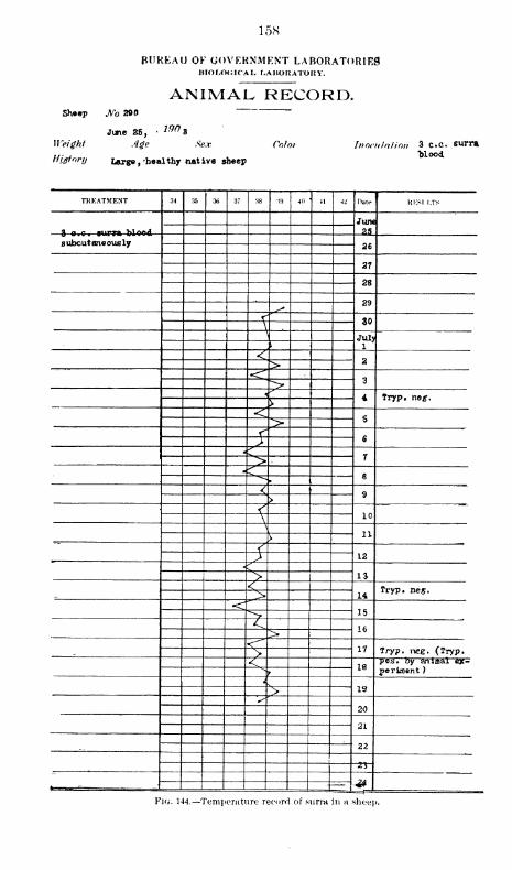
158
BUREAU OF GOVERNMENT LABORATORIESIHOLOCICAL LABORATORY.
ANIMAL RECORD.Sheep JVo 2©0
1M»June 25,
Weight Age Sex
HUtonj Largg f-healthy native sheep
Color Inoculation 3 c.c. eurra"blood
TREATMENT 34 35 36 37 38 •<9 40 41 42 Date KKSI l.TS
JUTK
35
subcutaneously 26
21
28
29, s"
< 30\\
July1\
<2>
<?3>
5 4 Tryp. ne*.\
<5>
<6)
<^ 7><^ 6^
< 9>
< 10\\
11\
{12y
?IS><14
Tryp. neg._ ><
15>< 16>«^-"~
17> Tryp. neg. (Tryp.
< 18pos. by anioal ex-
*>
fperiment )
*
19^>S
20
21
221
<<!i
<#*-«-«,—-—
.
. :
Fig. 144.—Temperature record of surra in a sheep.
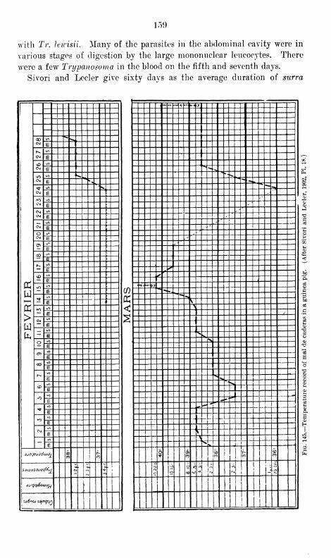
i:>9
with Tr. h'lrisii. Many of the parasites in the abdominal cavity were in
various stages of digestion by the large mononuclear leucocytes. There
were a few 'Trypanosoma in the blood on the fifth and seventh days.
Sivori and Lecler give sixty days as the average duration of surra
PC
u
>w
00CM
*=*-E
CM
">
E
CM<o
E
CM^
E "^ .
CM
to '^E _j
CM<s>
ECMCM
VO;
E
CMto i
E L_
OCM
</>
E05
</? !
EJ
03 C/}
Er>.
P</>
ri"
<£> oE
iO CO
E
3*:/)
;
E i
»o mE
Cm(/i
E
- to
EO i/j
E
<T><o ]
E
COCO
E
r-»«/>
E
<£>i/>
£
iOO0
c
^ to
E
»o<o
E
CMCO
E</>
E
jjnjfjddujdi 0000
sjiaosouvd/tji <m
.
"...*>
j
it/igto/Sou/juI
I SdSnoj so/rxp/rj i r
•
'
••<*i
"> **i j __.
,
'^- ~* * , ^
**- <^^""s*
—.
'
i.^
.'*~
-*
:>zfc** -t ,
..?*«.«
co ~^-^ :
rV'"*"*
,v --
Qh c<! x "^
:^2 X XI^-t
^;^k-jf
mi
"> -C-
1 _L_
^
- -
5^_J^-fL-- ,.
— \ i^^tii-r-
6 O)^ CO co j Iro "
o ^ O on -oj
x hi- i- 4^ -i •
_L —
- n f 1
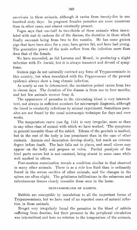
160
americain in these .animals, although it varies from twenty-five to one
hundred sixty days. In pregnant females parasites are more numerous
than in other eases, and almost constantly present.
Voges says that one-half to two-thirds of these animals when inocu-
lated with mal de caderas die of the disease, the duration in those which
finally succumb being from two to five months. He has some guinea
pigs that have been alive for a year, have grown fat, and have had young.
The generative power of the male suffers from the infection more than
does that of the female.
We have succeeded, as did Laveran and Mesnil, in producing a slight
infection with Tr. lewisii, but it is always transient and devoid of symp-
toms.
Guinea pigs do not naturally contract any form of Trypanosomiasis in
this country, but when inoculated with the Trypanosoma of the present
epidemic always show a long chronic infection.
As nearly as can be determined, the incubation period varies from two
to eleven days. The duration of the disease is from one to four months,
and but few animals recover from it.
The appearance of parasites in the circulating blood is very intermit-
tent, not always in sufficient numbers for microscopic diagnosis, although
the blood is constantly infectious by animal experiment. Sometimes para-
sites are not found by the usual microscopic technique for days and even
weeks.
The temperature curve (see fig. 145) is very irregular, more so than
in any other class of animal with which we have worked. 'The symptoms
in general resemble those of the rabbit. Edema of the genitals is marked,
but in the rest of the body is less prominent than in the case of other
animals. Anemia and emaciation develop slowly, but reach an extreme
degree before death. The hair falls out in places, and small ulcers mayappear on the belly and prepuce or vulva. Partial paralysis of the
hind parts occurs but is not constant, being absent in some cases while
well marked in others.
Post-mortem examination reveals a condition similar to that observed
in many other animals. There is as a rule less fluid than is ordinarily
found in the serous cavities of other animals, and the changes in the
spleen are often slight. The gelatinous infiltrations in the subserous and
subcutaneous tissues closely resemble those seen in the horse.
TRYPANOSOMIASIS OF KABBITS.
Eabbits are susceptible by inoculation to all the important forms of
Trypanosomiasis, but we have read of no reported cases of natural infec-
tion in these animals.
Eouget very irregularly found the parasites in the blood of rabbits
suffering from dourine, but their presence in the peripheral circulation
was intermittent and bore no relation to the temperature of the animals.

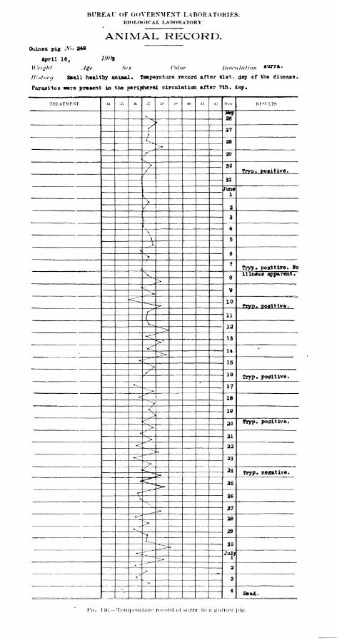
BUREAl OF GOVERNMENT LABORATORIES.BIOLOGICAL LABORATORY
ANIMAL RECORD.Guinea pig M> 24*
April 16, 190s
Weight Age Sex Color Inoeu lotion bmtta.
History Small healthy aninal. Temperature record after 41st. day of the disease.
Parasites were present in the peripheral circulation after 7th. day.
TKKATMF.NT :W :{.') , .,: :tx ,, 4,, (1 4J Hatr . KKSfLTS.
^J4ay
26^
/ 27
)<, 28>r^" 29
(-
^ 30/ Tryp. positive.f
31
June1
K
\2
f
3\I
4\
V 5Jt
6>/
7Tryp. positive. Ho
v8
illness apparent.
><
^
9^<^ 10
tryp- positive.s
11(^ 12^^r 13>*<:
14':>•
15^>/ 10 Tryp. positive.
•^*
17~~"-^>
<!-" '
18->< 19>^
20 fryp. positive.>
•4-"
21>22^23
^>c 24 Tryp. negative.X
26
26
\27
28>^ 29>
30
-c:U-
—
July";>•
-
2^<r 3
•>.
.-
4Dead.
Km.. \U>.—Temperature record of surra in a guinea pi#.
161
As the principal symptoms he gives edema of the ears, falling out of the
hair, paralysis of the hind parts, mucous conjunctivitis, winch later
becomes purulent, and edema of the genitals. The duration of the dis-
ease is from one to four months. The principal anatomic lesions are
hyperemia of the abdominal layers, inflammation of the spleen and liver
and of the lymphatics near the point of inoculation.
Mai de caderas, according to Voges, lasts for one to three months in
these animals. Parasites may not be found in the blood for the first
four weeks. The rabbits remain active for weeks, but toward the end
emaciation is apparent. The fever is very irregular. Catarrhal con-
junctivitis supervenes, becoming purulent and causing the lids to adhere
to each other. The hairs of the eyelids always drop out, and sight finally
becomes greatly impaired. Inflammation of the nose occurs, causing
the hairs to stick and fall out. The testicles swell, but the penis is
unaffected. Trypanosoma are found in the inflamed testicles and vulva,
and necropsy, indeed, in all parts of the body, the spleen is enlarged, and
serous exudates are always observed.
Sivori and Lecler, working with the "surra americain" of South Amer-
ica, say that parasites may be found in the blood eight hours following
intraperitoneal inoculation, and that the duration of the disease is from
sixteen to one hundred and sixteen days. The disease is characterized by
emaciation, tumefaction of the eyelids, purulent blefero-conjunctivitis,
and marked edema of the nose, penis, and vulva. The temperature is
irregular and without relation to the number of parasites in the circu-
lating blood.
Kanthuck, Durham, and Blandford regard the rabbit as relatively
immune to nagana. They say that the incubation period is from five to
seven days and the duration from twenty to one hundred and eighty-three
days, the average being about fifty.
Laveran and Mesnil consider it to be a susceptible animal. The incu-
bation period is from two to four days and the duration of the disease
from fifteen to thirty, although death sometimes occurs in five to six
days. Parasites appear in the peripheral circulation intermittently.
When the disease lasts twenty days or more, conjunctivitis sets in and
the hair about the eyelids falls out; edema of the vulva or prepuce and
forelegs occurs, but the lesions are always slight. During the whole
course of the disease the rabbit has a continuous temperature, with rare
intermissions; it is generally above 40° C.
In this country rabbits have not been found naturally infected. They
are susceptible by inoculation and afford most interesting subjects for
the study of the disease. Fig. 146 illustrates the course of surra in these
animals as given by Lingard.
The incubation period is difficult to determine accurately for the
reason that, contrary to the results obtained, in many animals, it is not,
7881 11
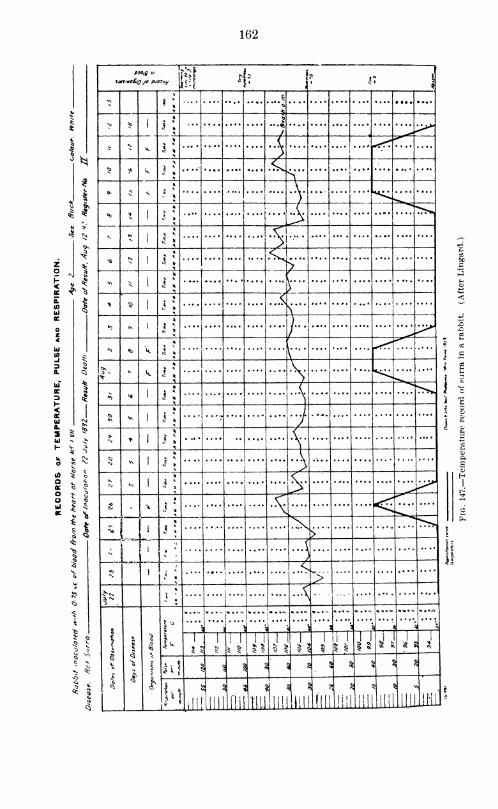
162
s
i
*
IWSmoSJQ JO ?JO0*y
^,15
v r. i
»>I \
*«
$
- *>1 \
&
/
>N
k 1 ».{
)^ ^ k J *
<,>
O « ^
7<fe >
1 J *
(>K 5
1 ? » $^ <SJ
1J
* <. .^h.
>V> * I c *
y,..
> §1
<l
« *
. «.4> ....
»0 * 1
« >) ,
1"/
* <fe k 1 ». ... 1 . . .
!
^ k i *....
1.... i
*> ** 1 c *(
i
o* V>
1 J * 7I
s *1 c *
....
•vM * 1
{j
:>
(M CM 1
Jv.......... > ^>*0N
*>*
~ k ^ _L...- * ^-w^
{$ i
. ..
.
'1
*; — .
*)\
j* .... .
<
J:
..*
f 5
1'
1
1° ....
ii
'•
i]' .'
'
'-—i1! ^
5 "
l i 1
$ 1
1
1
1
1 1
1 1
SI! 1
5 1>!.
1 „ 1
^1<:
s 1 !i;i a
aV 5$ 5i !\ s
a
L Ifct
lill"
3 S
llll ll'lii
i
mi III
\
'III
•a
.ill] III
5
llll'ill
5 <
iUlikil"

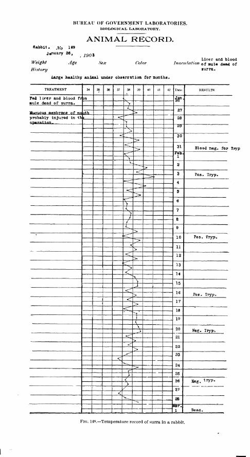
BUREAU OF GOVERNMENT LABORATORIES.BIOLOGICAL LABORATORY.
ANIMAL RECORD.Rabbit - jf 169
January 26, igQZ
Weight Age Sex
History
ColorLiver and blood
Inoculation f nuU dMkd of
large healthy animal under observation for months.
TREATMENT 34 35 36 37 38 39 40 41 42 Date. RESULTS.
F«d 1 iver and blood t^ >m V *Fmule dead of surra. ^
< S7Mun-uous membrane of mo \%h >probably injured in th I 28nporntton. >
K 29
>*< 30
<^"~31 Blood neg, for T
<" Feb.1
\L
3^
<:8 Poa. Tryp.
4
S">6
)I 7
>/ 8
/Cu
9><^ 10 Pos. Tryp.
< 11><, 12
p>r 13>< 14^
\\ 15J
< 16^> Pos . Tryp
.
<"17>
<f IS*
>J 191
\ 20, ^ Neg. Tryp-.
21^>^ 22>^^ 33
! <24->•
<25
}i 36 Neg. tryP-s
/ 27<
28^Mar.11
Dead.
Fig. 148.—Temperature record of surra in a rabbit.
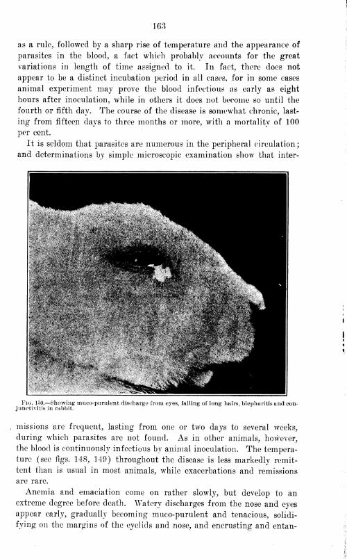
163
as a rule, followed by a sharp rise of temperature and the appearance of
parasites in the blood, a fact which probably accounts for the great
variations in length of time assigned to it. In fact, there does not
appear to be a distinct incubation period in all cases, for in some cases
animal experiment may prove the blood infectious as early as eight
hours after inoculation, while in others it does not become so until the
fourth or fifth day. The course of the disease is somewhat chronic, last-
ing from fifteen clays to three months or more, with a mortality of 100
per cent.
It is seldom that parasites are numerous in the peripheral circulation;
and determinations by simple microscopic examination show that inter-
Fig. 150.—Showing mucopurulent discharge from eyes, falling of long hairs, blepharitis and con-junctivitis in rabbit.
missions are frequent, lasting from one or two days to several weeks,
during which parasites arc not found. As in other animals, however,
the blood is continuously infectious by animal inoculation. The tempera-
ture (see figs. 148, 149) throughout the disease is less markedly remit-
tent than is usual in most animals, while exacerbations and remissions
are rare.
Anemia and emaciation come on rather slowly, but develop to anextreme degree before death. Watery discharges from the nose and eyes
appear early, gradually becoming muco-purulent and tenacious, solidi-
fying on the margins of the eyelids and nose, and encrusting and entan-
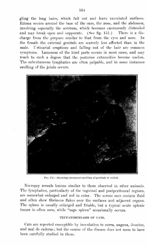
164
gling the long hairs, which fall out and leave excoriated surfaces.
Edema occurs around the base of the ears, the nose, and the abdomen,
involving especially the scrotum, which becomes enormously distended
and may break open and suppurate. (See fig. 151.) There is a dis-
charge from the prepuce similar to that from the eyes and nose. In
the female the external genitals are scarcely less affected than in the
male. Urticarial eruptions and falling out of the hair are commonsymptoms. Lameness of the hind parts occurs in most cases, and mayreach to such a degree that the posterior extremities become useless.
The subcutaneous lymphatics are often palpable, and in some instances
swelling of the joints occurs.
Fig. 151.—Showing enormous swelling of genitals in rabbit.
Necropsy reveals lesions similar to those observed in other animals.
The lymphatics, particularly of the inguinal and postperitoneal regions,
are somewhat enlarged and red in color. The serous sacs contain fluid
and often show fibrinous flakes over the surfaces and adjacent organs.
The spleen is usually enlarged and friable, but a typical acute splenic
tumor is often seen, while "sago spleen" occasionally occurs.
TRYPANOSOMIASIS OF CATS.
Cats are reported susceptible by inoculation to surra, nagana, dourine,
and mal de caderas; but the course of the disease does not seem to have
been careful lv studied in them.
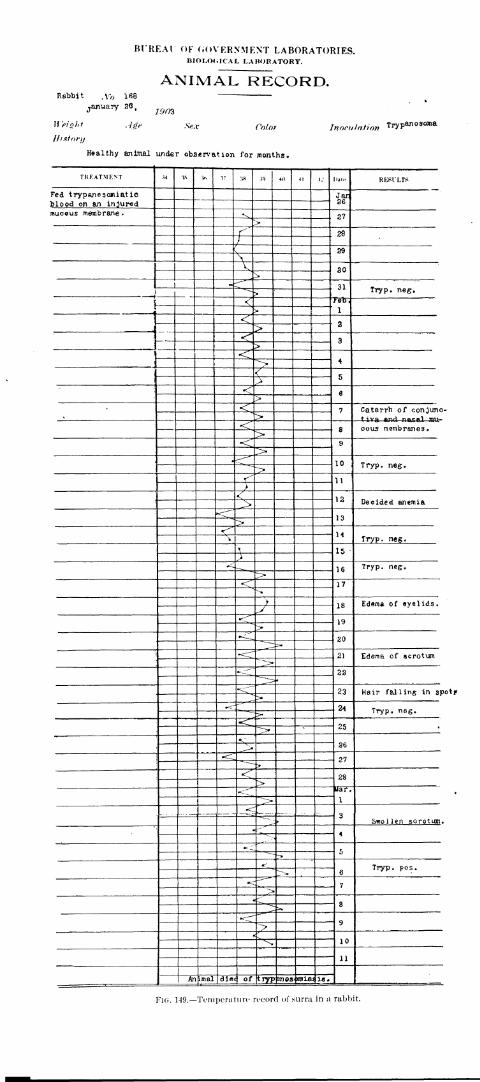
Bl'REAl OF GOVERNMENT LABORATORIES.BIOLOGICAL LABORATORY.
ANIMAL RECORD.Rabbit ,y 168 —
January 20, ^"""'£''' •#<• Sex Color Inoculation Tryp^ 030Jna
Ifistori/
Sex Color
Healthy animal under observation for months.
TKFATMKNT M 'l* V* - :w :ts 40 « *- I»at<- RESl'LTS
Fed trypanosojuatic Jar26blood on an injured
mucous membrane
•
*\ 27>
r" 28/
29\v,
30>
1«--'"r
31J
Tryp. neg.
I<
1>
<2
-, >< 3
><:
4
<"
5><r 6>
< 7 Catarrh of conjuno-><
8~>
< 9>10 Tryp. neg.~~>
r 11>
'
s 12;
1 > Decided anemia
t<^13>^14
V Tryp. neg.
\ 15 "
>
16 Tryp. neg.
><^ 17
^.
> 18>•<"
19>
•c^ 20
21 Edema of scrotumz^»
-C 23~~"~~^=
•^""23
i»
^r" 24 Tryp. neg.>•
< 25~^ *
-
•C'36^J>
•< 27>,.
*"28
->_,.^" Uar.
1>•-< _. .
3... ^~~---Swollen sorotum.
:'
4
•c^5
<^6
Tryp. pos.
7>c
8>»
9>>,
L 10^11
An imal di« Of tryp mos »miCL is.r
Fig. 149.—Temperature record of surra in a rabbit.

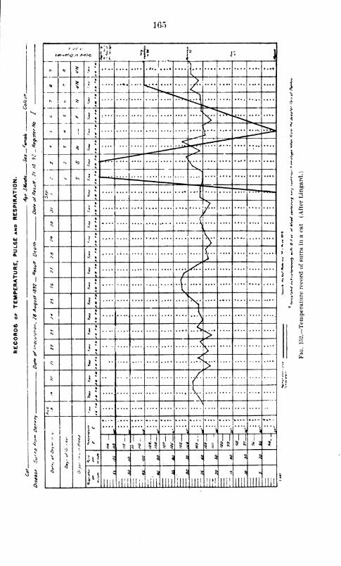
165
smiuee'jQ jo pjohi 1* Y , |
<t §
« V"""* » v
i
* 1
*
*.... - ' + - t
> ^v,"*>*^T
5 « *d*«w^v. ..
>....j...
' - 5
*
1*.
s <Ni <0
1
-. - <o
>
^>
j
§**
^
*> i i y*>
*
1 5
A...x ....
'\.V><
y/
_lv*
*(.
5 ** X
N
V ys<,
^>
*
,I
)<\i
* ^<>••
j »
* j i
^
*
t
*
i »
^
»
j:
j »*
: 1 5, \ , ^ ,
*'
\ \, t \ a?
. s ?
£
Q
1
5 3
1 1 ;
=5 §
I i1
^ is
Ti
j
i
I I
*'J U i
n! * 1 1 § s a % « 5 «• 5
U* »-
* *
ilii
31
Hi!
3
Ilii Hill llll
. *
(III mi
3
III!
5
llll llll Hi!
j !
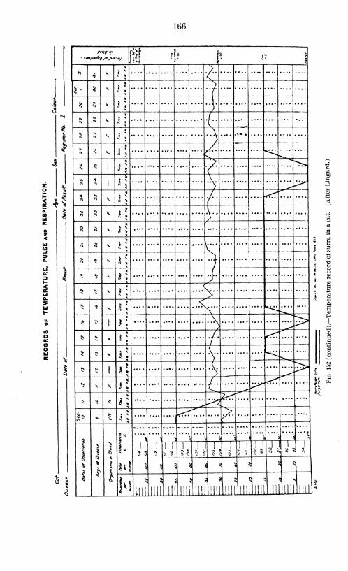
166
zo
5
or
6Uia.
ShJ
oo
Ill *!? i? i: \
NS
k 1 * *•
S- § k | * j
8 M V. 1 «
Onc\J k
5N«M k I *
* k i*
^ *
$ 1 I * ?N
1j
*• \>
«1£n k * ^
^ */
CM N k §* I
)CM
«N k § ^
N $ k ^ * {-
5 * k j* ^
/»* «D k J ^
* * k j 1
<
\. .
.
>
5$
58 k * *^ *<\
51 j i
\ >^> ^^,
!$ * k j* ,/
/* *> k i *
(....
$ w1 i; (..>
C\l * k i
* /..c
* 5 * 1 *
&5 »> S 1 *Ty
*
sV
i
1
11
r* j
' '
_—
•
\ '"'\
\
1
2 *
1 1
5 |
1 1
11j§ 5
i
1 !
"T 11 l
5 ^J,' j K1 J .
1
4*1 ^ § |
ay a
2 i&
5 ;
5
!'!lib
3
III II! III "in
\
III
§\
a
15
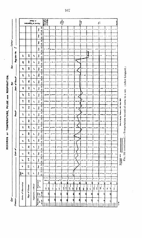
1(57
I
.4
& '5
? r. \
r;
*
*
^ «*.
*
1
.
o* ••
* > V .%.
* * k«i
. .«y
<5
K* k IS ... ....
^ k
5$ * k r, ,
* s k '"< ^7
> •0
> k *.
<S k*
~f
5! > k*
.... • ••• .... .... ....
"f• •— ••• • •• .. .... •••• »•••
* ? k r.
^ k ....
Ov 3 k*
•0 k.
* "v.
K S k*
•fv* s? k
*
j* 1.
£ * >«o k
* {\
> SJ k / ....
** S k,. J .../>? %
\
1
I
]
1
>>
.$
*
1
r
** Y&,
,\
,
.\i
3,
)
, \ \ :•>
S
~~r1
!
JTT11
1
1
1
1
i,~r 1 1 j i
3^ § 5 l § s<a
5 $ 3 $
1 *
!3
Jill mi llll Jin III! llll JLLUi
5
ill! iih III!
*i
mi
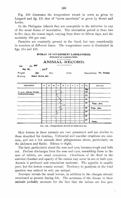
168
Fig. 150 illustrates the temperature record in surra as given by
Lingard and fig. 151 that of "surra americain" as given by Sivori and
Lecler.
In the Philippine Islands they are susceptible to the infection by uny
of the usual forms of inoculation. The incubation period is from two
to five days, the course rapid, varying from three to fifteen days, and the
mortality 100 per cent.
Parasites are constantly present in the blood, but vary considerably
in numbers at different times. The temperature curve is illustrated in
figs. 154 and 155.
BUREAU OF GOVERNMENT LABORATORIES.BIOLOGICAL. LABORATORY.
ANIMAL RECORD.Cat JVo 8C*
May 28,, J90 3
Weight Age Sex
History Small white cat-
Color Inoc-ulotion Tr . IVa/isi
TREATMENT 34 35 36 37 38 39 40 41 42 Date RESULTS
May
821 c.c. surra. Moodsubcutaneoualy. <*
Z3\\ £4
>
< 25 Tryp. poa.>< £6 Tryp. pos.>
27>
28X»
29Dead*
Fig. 154.—Temperature record of surra in a cat.
Skin lesions in these animals are very prominent and are similar to
those described for dourine* Urticarial and macular eruptions are com-
mon, and not a few animals show phlegmanous ulcers, particularly on
the abdomen and flanks. Edema is slight.
The hair, particularly about the nose and eyes, becomes rough and falls
out. Profuse discharges from the nose and eyes, resembling those in the
case of rabbits, are usual symptoms. Cloudiness of the fluid in the
anterior chamber and opacity of the cornea may occur in one or both eyes.
Anemia is profound and emaciation moderate. The appetite is usually
poor, but the bowels remain normal. Tendency to paralysis in the hind
quarters was noticed in only one animal.
Necropsy reveals the usual lesions, in addition to the changes already
mentioned as present during life. The acuteness of the disease in these
animals probably accounts for the fact that the lesions are less pro-
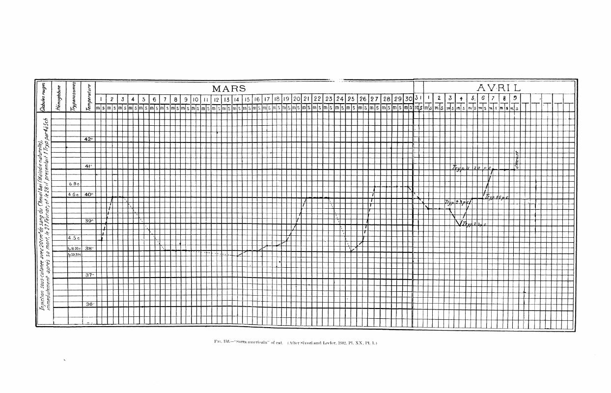
1is
55
8
J
1
|
MARS AYRI L1 2 3 4 5 6 7 8 9 10 II 12 13 14 15 16 17 18 19 20 21 22 23 24 25 26 27 28 29 30 3 l 1 2 3
m
* s :$ 7
n s
8
m s
•
m s m s m s m s m s m S m s m s m 5 m s rn s m s rn S rn s rn s m s m s m s m s m s m s m s m s m s m S m s m S m s m s m s III s 5 Vf\ 6 s m s 5 TTl % >
Is
^1
-
r~
42°
<X"
&41-
J rii fti ;f 2 p f _|/
1
)
r
6-8cJ
/^ -f-
/4.6 c 40° /
, ^ 7 : 6 c c/ \ /
"71_i _ 7k a3.2. Ip f/
**? *-£*<
—-/-
1\
T>s
<JT~ r/
'
f- ^
7*~V 1
f
.
1\
^~\ ~t
39"-4
t « / \/r< ? 1
\4-\ 1
H"^--*¥
4- L»_/
»1
4.5ci
,f
f
^j < <" • /1p 10.20 c 38" ^ \ /
1 p.10.30 c-- -- «~_ ^
37"
—i »
36°— 1
•
1
-
I -1- . . 1
. ,
F\a. lf>8.—"Surm americani" of cat. (After Sivori and Lecler, 1<J02. PI. XX, I't. 1.)

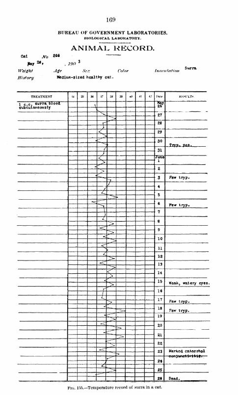
169
BUREAU OF GOVERNMENT LABORATORIES.BIOLOGICAL LABORATORY.
ANIMAL RECORD.Cat No 266
¥** 26,, 190
3
Weight Age Sec:
History Medium-sized healthy cat
Color InovulntioSurra
TREATMENT M 35 3t> 37 38 39 -H) 41 1.' Date HKSl'LTS
1 c.c. surra blood,subcutaneous ly
\May
> SV. 27
,>f^^
28
)<\ 29>
130
Tryp. pop.,
{ ! 31)< K
June:1>'
:
2>;3 Few tryp.
"7
/ 4/< 5
!6"> Fe* tryp.
\ ! ?)^
8><r 9><
10; >
< 11N>
412>
•<"13>
< 14>< 15> Weak, watery eyes.
f'
16/^ 17> Few tryp.
18i Few tryp.
19>•<:
20
21-^-~"^^
22s~<
23
r^ 2*y-C
ZZ>26 Dead.
Fig. 155.—Temperature record of surra in a cat.
170
nounced. The spleen is enlarged, but assumes more nearly the type
of acute splenic tumor, while subserous hemorrhages are numerous.
TRYPANOSOMIASIS OF RATS AND MICE.
Tr. lewisii, the common Trypanosoma of rats, has already been
described. Most writers agree that it is harmless for rats and nonin-
fectious, by inoculation or otherwise, for other animals.
Large numbers of rats have been found naturally infected in various
parts of the world ; a partial list of the regions is as follows : Lewis in
Calcutta found 29 per cent infected; in Bombay Carter found 12 per
cent infected and Lingard 30 per cent; Koch in Africa, 41.7 per cent;
Crookshank in London, 25 per cent; Kabinowitsch and Kempner in
Berlin, 41.8 per cent; Laveran and Mesnil in Paris, 4.6 per cent; Eaillet
in Alfort, a large percentage; Chemette at Lille, a large percentage;
Sivori and Lecler at Buenos Ayres, 3 per cent; and Chalachnikow and
Danilewsky in Kussia found the infection present. In Togo eight rats
examined were negative (Luman). Voges in South America did not
find these animals naturally infected. In a personal letter Kitasato in
Japan states that many of the rats t'here harbor the parasite.
In Manila rats Tr. lewisii has been found in from 20 to 65 per cent
of the individuals examined, varying according to the season and the
locality from which they were received.
Van Dyke Carter inoculated dogs, cats, horses, and monkeys with rat
Trypanosoma, and always obtained negative results. Koch failed per-
manently to transfer the infection to other animals. He gave a rat Tr.
lewisii and Tr. evansii and afterwards found both in its blood. Hethen inoculated a dog with some of this rat's blood and the animal
contracted the disease, but its blood was found to contain only Tr. evansii.
The efforts of Kabinowitsch and Kempner to inoculate other animals
with Tr. lewisii proved unsuccessful. In their experiments they used
white and gray mice, field mice, guinea pigs, rabbits, dogs, goats, horses,
and hamsters.
Lingard, on the other hand, inoculated twelve horses with infected
rat blood, and four of them, after an average of seven days, died of a
virulent form of surra. He argues that some Tr. lewisii are infectious
for other animals, as demonstrated by his experiments. This statement
has caused considerable confusion and his work has been questioned.
Judging from our observations in Manila, it wrould appear, however, that
Lingard's mistake consisted in considering these pathogenic parasites
Tr. lewisii.
Kanthuck, Durham, and Blandford showed rats to be refractory to
a second inoculation writh Tr. lewisii. Kabinowitsch and Kempnernoticed that some rats are absolutely refractory to these parasites, but
that most of them are susceptible. After inoculations into the abdom-
inal cavity, Trypanosoma were found to be present in the blood after
171
from three to seven days, and occasionally at the end of twenty-four
hours. In rats inoculated in the abdominal cavity they found multi-
plication forms to be numerous in the exudate during twenty-four to
thirty-six hours, at the end of which they disappeared permanently
from the abdominal cavity to reappear in the blood. Rats could not
be given a second infection, no matter how large the dose of infected
blood used; and they employed the important fact that some of the
animals are refractory as a basis for the preparation of a specific serum.
Laveran and Mesnil in general confirmed Rabinowitsch and Kemp-ner's work and in addition showed that the young born of immunemothers are very slightly if at all immune. They also demonstrated
that the agglutinating properties of the blood are not transferred to
young. In immune rats Trypanosoma are destroyed in the abdominal
cavity, and the agglutinative power of the blood lasts no longer than
the preventive. In their opinion the treatment of rats with serum
is unsuccessful. They state that as a rule the parasites appear in the
blood at the end of twenty-four hours, and that in not a few cases,
especially in young rats, they are found in considerable numbers before
this time. Howrever, many cases came under their observation in which
the parasites did not appear in the blood until two to seven days after
inoculation, and, indeed, in a few instances no infection resulted at all.
Adult parasites are the first to be seen in the blood, then there is a
period, rarely extending beyond the eighth day, when multiplication forms
may be observed, after wrhich adult parasites are visible throughout the
course of the disease. They believe multiplication to take place in the
abdominal cavity during the period from the first to the fourth day, and
in the blood between the fourth and the eighth days, after which time
multiplication forms are no longer present.
Infection of these animals with Tr. lewisii lasts from twenty days to
four months or more. In old ones the Trypanosoma often disappear in
two to ten days, and in these no active immunity is established. Subcu-
taneous inoculations, according to these authors, give infections which
are less severe than those produced by other methods.
Blood infected with Tr. lewisii and kept on ice does not give an in-
creased incubation period or produce any alteration of the infection, as
long as motile parasites are found to be present. If, however, the blood
is allowed to remain for a considerable length of time (forty-seven days),
the incubation period is increased from six to nine days, and the infection
is less severe. After fifty-one days or more on ice the blood no longer
contains living parasites, but is still infectious for rats, with an incuba-
tion period of twenty-seven days. All writers agree that rats infected
with Tr. leivisii show no symptoms of illness. The constant anatomic
lesion is hypertrophy of the spleen.
Our work as a whole has been in conformity with that of others. In
the thousands of rats found to be naturally infected with Tr. lewisii and

172
in those inoculated, we have never observed either apparent illness or
death which could positively be attributed to infection with this para-
site, although it was present in enormous numbers in the circulating
blood. Our observations have been exceptionally easy on account of the
great number of rats furnished the Laboratory for examination to deter-
mine the possibility of their infection with plague, since this disease has
become endemic in Manila.
Very little is found in literature relating to the clinical manifestations
and morbid anatomy of surra in rats. We have not seen a specific refer-
ence to the natural infection of these animals by Tr. evansii, though from
our experience, which is to be described later, it is believed that Lingard
must in some of his experiments have been working with such an
infection.
Lingard states that the latent period in Mus decumanus as developed by
the subcutaneous inoculation of 0.1 to 0.2 c. c. of virulent blood varies
from one to two days; and in Nesol'ia providens, by subcutaneous inocu-
lation of 0.2 c. c, from two to five days.
Kanthuck, Durham, and Bland ford state that rats, when inoculated
with nagana-infected blood, often exhibit convulsive seizures just before
death, but otherwise show no symptoms of the disease, except dullness in
the later stages. Transmission from one animal to another by coition,
by suckling, or by any other method than by direct inoculation was not
observed. Infected mice lived from eight to twenty-five days and rats
from six to twenty-six days.
As the morbid anatomy of nagana in rats and mice they describe lym-
phatic hyperplasia, most noticeable near the point of inoculation, conges-
tion, edema, and occasionally hemorrhages of the glands, great enlarge-
ment of the spleen, which is generally firm, friable and dark in color,
enlargement and fattiness of the liver, and occasionally a small amountof fluid in the pleural cavity.
Laveran and Mesnil state that in rats and mice of all varieties, includ-
ing white ones, nagana has a perfectly regular course. Xo symptoms are
manifest, the animal appearing well until near death. Mice appear
sleepy and die without suffering from dyspnea. One mouse had convul-
sions two hours before death. Some rats die with the same symptoms as
mice, but most of them show great agitation just before death, crying out
and dying in severe convulsions. There are no sensible variations of
temperature in these animals. Parasites appear in the blood twenty-four
hours or more after inoculation in the abdominal cavity, and on the
second or third day after subcutaneous inoculation; they progressively
increase in number until death. In white rats and mice death occurs in
two and one-half to three days after intraperitoneal inoculations, andafter three and one-half to rive days following subcutaneous inoculations.
Wharf rats (Mus decumanus) die in about the same length of time as
white rats (Mus rattus).
173
According to Rouget, mice inoculated with Trypanosoma of dourine
show no symptoms until near death, when the hair becomes rough and
the cornea cloudy. The post-mortem changes observed are hyperemia
of the abdominal layers, inflammation of the liver and spleen and of
the lymphatics near the point of inoculation. Parasites are found in
all of the organs and fluids, except the intestines and the urine.
Voges says that death may occur without any preceding signs of
illness. In rats and mice inoculated with the Trypanosoma of mal
de caderas there may be a few hours of comatose condition. The dura-
tion of the disease in these animals is given at about four weeks.
As has been stated above, the natural infection of rats with the Try-
panosoma which cause the disease in domestic animals has not before
been reported, except perhaps by Lingard, who, if such was the case
in his work, misinterpreted his results.
In a preliminary report on Trypanosomiasis in the Philippine Islands,
published as Bulletin No. 3 of the Bureau of Government Laboratories,
1903, Musgrave and Williamson reported that a certain number of rats
in Manila were found to be infected with the Trypanosoma which in
this country causes surra in horses. This discovery was brought about
by accident, while the authors were attempting to immunize a monkey
with a parasite supposed to be Tr. lewisii. As a result the animal
contracted surra and died. At first, following Lingard, we thought this
to be a pathogenic Tr. lewisii, or supposed accidental infection with
Tr. evansii to have occurred; but more careful study and the repetition
of the experiment with the greatest precautions has demonstrated that
a small percentage of these rats harbor the parasite causing the disease
in the horse. This Trypanosoma, has the same morphologic character-
istics, and upon inoculation is infectious for the same animals, producing
the same disease with the same incubation period, course, termination,
and lesions.
Wild rats are very unsatisfactory material to work with, because so
many die after being in captivity a short time. The duration of life
is so uncertain that but little confidence can be placed in results, even
when control animals are used. The ones which live for a week or
more after caging are likely to survive for some time, and, in order
to obtain the best results, these have been employed as far as possible
in our experiments.
As has been shown in other countries during the study of the disease
in these animals, the infection is not evidenced by any symptoms of
moment until just before death, when convulsions often occur. Too
much importance should not be attached to this symptom, for time
and again we have seen our control animals die in the same manner.
Wild rats suffer such great excitement during the taking of tempera-
ture that no conclusions can be based upon the results. We must there-
174
fore believe that rats and mice infected with surra show no constant
symptoms of practical significance. Some of the rare manifestations
are cloudiness of the fluid in the anterior chamber, falling out of the
long hairs about the head, and in a few cases edema and a tendency
to paralysis of the hind parts. The incubation period, as determined
by the appearance of parasites in the peripheral circulation, varies
from a few hours to five days or more, and the duration is from two
to twelve days.
The post-mortem examination shows an enlarged spleen, which may
be hard and friable or more nearly approach acute splenic tumor with
the organ usually dark in color; and there is generally enlargement
of the lymphatics, especially of the inguinal regions, which may be
hemorrhagic. The changes in the other organs are not significant,
except in a small number of cases which show gelatinous infiltration
of the subcutaneous and subserous tissues.
These lesions, particularly the enlarged spleen, are found in rats dying
from Tr. lewisii, which is considered harmless. After discovering that
a number of rats harbor Tr. evansii, we suspected the ones which died
supposedly from Tr. lewisii and showed enlarged spleen, etc., on post-
mortem to be in reality infected with Tr. evansii ; but animal experiment
absolutely disproved this theory.
The study of Trypanosomiasis in rats has not been completed. It
i? certain that rats in Manila may be infected both with a Trypanosoma
harmless by inoculation to other animals and with one pathogenic for
them. These parasites correspond microscopically to the descriptions
respectively of Tr. lewisii and of Tr. evansii. We are not fully con-
vinced that Tr. lewisii is always harmless for rats, or that some of
the rats in Manila do not even harbor a third species of parasite.
TRYPANOSOMIASIS OF FROGS, FISH, AND FOWLS.
Trypanosoma have been found in frogs obtained from points scattered
over a large area of the world, but they are usually considered harmless in
these animals. The infection, so far as we have been able to determine, is
not artificially transferable from frog to frog or from frogs to other
animals. As already mentioned, we have not been able to find Trypano-
soma in the blood of frogs in this country, and these batrachians have not
been proved to be susceptible by inoculation with any of the Trypano-
soma we have studied.
A number of observers have found fish harboring Trypanosoma, but
all agree that no symptoms are produced. The species reported as infected
are mudfish, trout, pike, redeyes, soles, and salt-water fish of the Medi-
terranean (species not given). The different species of these parasites
have already been discussed.
Laveran and Mesnil, whose work is the most important in this line,
found the infection of the redeye with Trypanoplasma to be very com-
175
nion, but proved the young rarely to be infected. In the sole, however,
Trypanosomiasis is uncommon, being found in only four eases out of a
large number of fish examined. They did not observe any symptoms
of disease in fish caused by Trypanosoma, and were unable to transfer
the infection by inoculation. Doflein, during a fatal epidemic in fish,
found Trypanosoma in a number of the diseased ones, but he was not
sure of any pathogenic action of the parasites.
We have examined a number of fresh and salt water species of fish
in Manila, but have been unable to find Trypanosoma in any of them, nor
have we been able to infect them with Tr. lewisii or Tr. evansii.
It has already been shown in the discussion of Trypanosoma that
some birds are occasionally found to harbor a specific Trypanosoma,
which, however, does not appear harmful to them. With reference to the
inoculation of Tr. evansii, Tr. bruceii, Tr. rougetii, and Tr. elmassianii
into birds, the evidence is somewhat contradictory for the different
species. Most writers agree that birds are not susceptible to infection,
but Voges, writing of mal de caderas, considers turkeys, ducks, and
chickens susceptible by inoculation. He says that chickens die in from
two to three weeks in great agony, from either subcutaneous or intra-
peritoneal injections. The only symptom noticed is emaciation, and
parasites are very few in number in the circulation.
We have so far experimented with several varieties of birds, including
maya, pigeons, doves, and chickens, but have been unable to infect them.
No symptoms are produced, parasites are not found, and the blood proves
noninfectious when inoculated into susceptible animals.
TRYPANOSOMIASIS OF MAN.
In 1898 Nepveu published an account of the occurrence of Trypano-
soma in human beings. An extract from his article, translated, reads
as follows:
So far Trypanosoma have been found only in the blood of animals. In India
they have been found in the blood of the rat (Lewis), the horse (surra epi-
demics) , the dog, and the domesticated elephant. In Africa they have been
discovered in the disease caused by the tsetse" fly, and in Europe in the blood of
the rat, the rabbit, various birds, and the frog. No one seems to have as yet
observed them in man, although Laveran states that Barron found certain flagel-
lated protozoa of an undetermined genus in the blood of an anemic woman. In
1890, while making researches on malarial parasites in Algiers, I found flagellates
in the blood of a patient, besides Laveranii, and I was able to count three to
each preparation of 18 square millimeters. At about that time (see Nepveu,
Etudes sur les Parasites du Sang chez les Paludiques, 21, 1891, in Bulletin et
Memoires de la Societe de Biologie) I published some of the drawings I had so
far collected. I hoped then that I might be able to complete my first observa-
tions by a more detailed study, but since then I have rarely been able to find
the parasites. I have therefore decided to publish the following facts in the
hope of drawing the attention of such naturalists and physicians as will have
the opportunity of completing these researches. * * *
176
In over 200 patients, mostly malarial, of whom I have examined the blood, I
have found these various forms of Trypanosoma in only six, three of whomwere suffering from quotidian fever (Khill, Langevelle, and Bichielli), one from
double tertian (Hendrick), and two from pernicious comatose fever (Cabane and
Ginestet), while the seventh observation was made on Dr. X., who was apparently
in good health. In none of these patients have I been able to observe any symp-
toms characteristic of this special parasitic invasion. They were almost all
suffering from the effects of Laverania, which prevailed everywhere in its various
forms. This seems, therefore, purely a coincidence, which has appeared to meworthy of notice.
His article attracted very little attention, some writers mentioning
his work with the remark that his descriptions were inaccurate, and manyoverlooking it entirely. He did not attach clinical importance to the
appearance of these parasites in the blood, but his remarks regarding
them seem to us perfectly clear as to the occurrence of Trypanosoma
in the blood, sufficiently so as to entitle him to the credit for priority in
the discovery of Trypanosoma in the human blood. His description
does not appear sufficiently ample to designate the species of his parasite,
but when one considers the confusion which exists even at the present
time regarding the classification of these organisms, Nepveu can not
be denied credit because in 1898 he failed to classify his Trypanosoma.
The parasites more recently discovered in human blood, as in his case,
have not been clearly classified. Xepveu observed seven cases.
The eighth case of Trypanosomiasis in man is published in the British
Medical Journal for January 1, 1902, in an editorial and telegram
from Dutton, in which he announces the discovery of a Trypanosomain a European, who displayed peculiar symptoms. The same Journal
for January 11, 1902, contains a letter from Eoss, in which he gives
Dutton credit for the following clinical data
:
The patient has been suffering from a form of relapsing fever with peculiar
edema of the eyelids and puffiness of the face, also edema of the legs, general
weakness, abnormal frequency of pulse and respiration, and enlarged spleen.
There was no organic lesions of the heart and kidneys, and no malarial parasites
were found after repeated examination. The relapsing fever recalls that of horses
suffering from the same parasite. It is not yet certain whether the parasite
approximates Tr. bruceii or Tr. leivisii.
Dutton considers the most valuable features presented by his case as
(1) its chronic course, (2) the general wasting and weakness, (3) the
irregular rise of temperature, which is never very high, and of a relapsing
type, (4) the local edemas, (5) the congested areas of the skin, (6) the
enlargement of the spleen, and (7) the constant increased frequency of
pulse and respiration (hurried breathing).
He examined the blood of one hundred and fifty healthy children
between the ages of one and fifteen years, natives of Gambia, and says
that he found Trypanosoma apparently identical with those observed in
the European in the ninth case. The child is reported as showing no
clinical evidence of the disease.
177
Forde (Journal Tropical Medicine, September 1, 1902) publishes the
case already described by Dutton. He deals particularly with its history
and symptoms previous to Dutton's personal observations.
The patient was a European, 42 years old, and at the time he came
under Dr. Forde's care, May 10, 1901, at the Colonial Hospital, Bathurst,
was a man of robust constitution, living a regular and steady life.
He was at first thought to be suffering from malarial fever, but quinine
produced very little change in the course of the temperature. His blood
was examined and malarial parasites were not found, but in nearly every
specimen Forde found "small worm-like bodies," which he at first con-
sidered a species of Filaria. After repeated observations, however, the
diagnosis became doubtful, and he associated these bodies with the symp-
toms of the disease.
The patient was invalided to Europe and returned to Bathurst in
December, 1901. At this time Dr. Dutton, being informed of the case
and examining the patient's blood, again found parasites, and immedi-
ately recognized them as Trypanosoma.
Forde gives as the chief characteristics of this case : ( 1 ) The irregular
intermittent temperature, (2) the edematous condition of the face and
lower extremities, (3) the rapid and variable pulse and respiration,
unaccompanied by any evident cause, (4) the loss of weight with marked
debility, wasting and lassitude, (5) the persistence of these symptoms
and their resistance to treatment.
In the Journal of Tropical Medicine for November 1, 1902, is published
"A Case of Trypanosomiasis in a European/' under the care of Dr. Man-
son—the tenth case. Hanson had been struck by the peculiar clinical
features of Forde's case, its chronic irregular fever, the enlargement of
the spleen, the edema, especially of the face, and the very well-marked
erythema multiforma scattered over the trunk and limbs.
The patient under Hanson's observation was the wife of a missionary,
who had resided on the Upper Congo for about a year and had been sent
to Hanson by Habershon. She had been suffering while on the Congo
from an irregular fever, which was still present when she came under
observation, though she had been living in England for the past eight
months and had been drugged with quinine and arsenic.
On examining her Hanson recognized the same grouping of symptoms
he had seen in Forde's and Dutton's case. The patient was admitted to
the hospital and her blood was examined daily for two weeks, but no
Trypanosoma were found. While arrangements were being made to test
the tentative diagnosis by injecting blood into animals, Dr. Daniels,
while making a blood count, found a Trypanosoma, and on subsequent
examinations more parasites were observed. In the British Hedical
Journal, Hay 30, 1903, this case is accurately described by Hanson
and Daniels.
7881 12
ITS
Manson published the eleventh case of Trypanosomiasis in humanbeings. The patient was a European lady who had resided on the Congo.
In this case an erythematous rash was a prominent symptom, preceding
or accompanying the attacks of fever, which occurred every ten days and
lasted each time for about three. The cause of the disease is attributed to
the bite of some insect on the foot. Manson is inclined to attribute sig-
nificance to a tick (Argas moubata) as a transmitter of the infection.
Broeden, according to a letter received by Dr. Manson, has discovered
two more cases in human beings—the twelfth and thirteenth. Both of
these were Europeans. Baker has recently reported three more cases in
human subjects in Uganda.
Of the sixteen cases of Trypanosomiasis in man, two have been in
apparently healthy persons, six associated with malarial fever, and eight
have shown clinical symptoms apparently entirely due to the infection
with Trypanosoma. In these five cases the clinical picture, which has
already been reviewed, was peculiar and quite similar.
Castellani has reported the discovery of a Trypanosoma in*the cerebro-
spinal fluid of twenty out of thirty-four cases of sleeping sickness. Hehas described the parasite and proposed the name Tr. ugandense.
Bruce has continued Castellani's work and has reported to the Eoyal
Society the discovery of Trypanosoma in the fluid obtained by lumbar
punctures in all of the thirty-eight cases examined, and in twelve out of
thirteen of these cases he found the Trypanosoma also in the blood. The
importance of this discovery can hardly be estimated at the present time,
but it is certainly additional evidence of the increasing importance of
this subject. Lieshman has also recently reported the possible appearance
of Trypanosoma in the blood of patients suffering with "'dumdum" fever
in India.
Since the beginning of the present work in Manila constant vigilance
has been observed in order to discover the infection in man, but so far
with negative results. Neither the clinical symptoms nor the parasites
have been found. The blood of hundreds of cases of persons ill and in
health has been examined, particularly that of persons who have come in
close and frequent contact with animals suffering from the disease. Both
writers of this article have repeatedly performed post-mortem examina-
tions on animals which had been but a few minutes dead of the disease,
and have been bitten by flies covering the infected organs, but without the
slightest inconvenience. A number of assistants during the course of the
work have often exposed open skin wounds to infection both by blood
directly and by biting flies, but all with negative results.
The reported cases show conclusively that human beings may become
infected, and we shall continue our observations in this country, where
the constant presence of the disease in animals, the sanitary conditions
and the anemic state of most of the inhabitants would lead to expect a
case eventually to occur in a human being.
170
TRYPANOSOMA OF MISCELLANEOUS ANIMALS.
Here we shall briefly mention the notes found in literature relating to
such animals as have contracted the disease but in the case of which the
clinical manifestations have been given but little detailed discussion and
on which, owing to the lack of animals, no observations can be made by us.
Bruce proved by animal experiments nine out of thirty-five wild ani-
mals examined in South Africa to be infected with Tr. bnicci. The posi-
tive ones included one buffalo, three niedbeuste, three koodoo (Slrepsi-
ceros kudu), one buch-buck, and one hyena. Many of these animals
showed no clinical evidence of disease.
Laveran and Mesnil, in mentioning the animals susceptible to nagana,
give several species of antelopes, the dromedary, the hare, the mullet, the
hyena, the lapian, the hedgehog, the racque. Bruce mentions the babale
and Brumpt the chamois. Lingard and others say that buffaloes and
elephants are susceptible. Sivori and Lecler refer to the carpincho and
Voges to the nutria (Myopotamus coypus) among animals subject to the
infection.
Voges considers the nutria extremely susceptible. Death occurs sud-
denly about ten days after inoculation, without symptoms.
Laveran and Mesnil say that wolves have an incubation period of two
to three days and that death takes place in from five to twelve days after
inoculation. Parasites vary in number, but are usually to be found
throughout the disease by microscopic examination of the blood. Cach-
exia and irregular fever are the prominent symptoms.
In strong, healthy animals the course of the disease is much longer,
being twelve to fourteen days or more, and the period of incubation is
from four to five days. The blood is always infectious, but parasites are
not usually found in it by microscopic examination. Local symptoms
appear in twenty to thirty days. Emaciation is not noticed until just
before death.
The prominent symptoms when once established are conjunctivitis,
coryza ; edema, particularly of the head, the legs, and the genitals ; conges-
tion of the testicles, or even a true orchitis; falling out of the hair about
the eyes, nose, and base of the ears ; opacity of the cornea, sometimes puru-
lent conjunctivitis and blindness in the late stages ; ulcers around the
eyes, nose, and other parts of the body, similar to those seen in dourine.
In animals dying from twenty to thirty days after inoculation the marked
clinical symptoms are not observed. English writers give the incubation
period at eight days and the duration of the disease at twelve to fifty-eight
(average, thirty) days.
Several writers mention hamsters as susceptible.
Hagger states that surra runs a very chronic course in camels and that
the natives believe that a small portion of those surviving for three years
to recover. He gives as the principal symptoms fever, swelling of the
180
right side of the chest, in the scrotum, and sheath of males and in the
udder of females, frequent abscess formations in these regions, progressive
anemia, and rapid emaciation. The appetite remains good. Parasites
are present in the blood daring fever, which sometimes reaches 42° C,
and are absent during intermissions.
The several forms of the disease are said to run a chronic course, some-
what similar to that of the goat and the deer.
IX. COURSE, DURATION, AND PROGNOSIS.
The course of the disease varies in the same and considerably more
so in different species of animals. In the language of Laveran and
Mesnil, "it always shows the general characteristics of blood infection."
A temperature of remittent, intermittent, or relapsing type is present
in nearly all animals, including man. Progressive anemia and ema-
ciation are also constant manifestations. It is rarely a very acute
infection, although in exceptional cases it becomes so intense as to
suggest septicemia.
The duration is also variable both in like and unlike animals. Schil-
ling says that the surra of South Africa lasts from thirty-six days to
eight months in horses. He considers the acuteness of the disease to
be influenced somewhat by the number of parasites in the blood. Bruce
says that horses live for weeks and months with nagana.
In the Philippine Islands the duration of the disease in horses does
not show a greater variation than it does in other countries. It is from
fourteen days to three months, and is about the same for American,
Australian, Chinese and native horses.
The length of time the disease lasts in cattle is usually somewhat
longer. Bruce had a cow sick with nagana under observation during
eighteen months.
The prognosis is influenced to a certain extent by the species of the
animal infected. Most writers agree that it is invariably fatal in horses,
but there are some exceptions. Schilling believes that some horses
recover; and Laveran and Mesnil say that recoveries have been reported
in South America, although they have seen none. A varying percentage
of cattle, according to careful observers, recover. Bruce says that cattle
occasionally recover from nagana, and Laveran and Mesnil have had
similar results. Lingard believes that a large proportion of cattle recover.
Voges, on the other hand, considers the cattle of South America immune.
The annual report of the Division of the Chief Quartermaster of
the Philippine Islands for the year ending June 30, 1902, records the
death of 3,693 horses and mules out of a total of 17,220 on hand. Headds that most of this havoc was produced by surra and glanders.
The course, duration, and prognosis of the disease have been con-
sidered somewdiat in detail under the discussion of the diiferent species
of animals.
1S1
The duration in particular varies so much with environment, the
constitution of the animal, and probably with other conditions which
we do not understand, that it can not be fixed except within wide limits.
The prognosis is always grave, the mortality in most species of animals
being 100 per cent. The only exception to this fact among domestic
animals of economic importance is found in cattle, a varying percentage
of which recover.
X. COMPLICATIONS.
Broncho-pneumonia, observed especially in h< rses, has been men-
tioned by writers as a frequent complication. In this country edema
and congestion of the lungs is common, while broncho-pneumonia, more
or less extensive in character, is occasionally seen.
Nephritis, hydropericardium, and hydrothorax sometimes explain
unusual symptoms. Tuberculosis and surra are not infrequently asso-
ciated, especially in monkeys. We have had two cases of surra and
glanders in the same animals. Filariasis and surra often occur together
in dogs. Einderpest sometimes develops in cattle suffering with surra.
Other diseases which we have found associated wdth surra are foot-
and-mouth disease, pseudoactinomycosis, pseudofarcy, malignant neo-
plasms, and at least two septicemic conditions not fully understood.
XI. DIAGNOSIS.
In order to carry into effect methods looking to prevention an early
diagnosis is very desirable in all cases. Fortunately, in the horse, the
most frequently infected of all animals, this is in the majority of cases
easily done by a microscopic examination of the blood, which consists in
examining a specimen prepared in the same manner as one to be examined
for malarial parasites. The Trypanosoma are readily observed with a
Zeiss DD or AA and ocular 4, and are usually in sufficient numbers to be
quickly seen. In many cases, however, they may be so few as to require
considerable time and the examination of several specimens before they
are found ; and as has already been said, they may not be observed at all
for several days at a time by this method.
When infection is suspected and parasites are not found in the blood,
there are two courses open. First, microscopic examination, carried on
for several days if necessary, will usually suffice for making the diagnosis
in horses and in several other animals in which the parasites are but
rarely absent for more than a few days at a time in the early stages of the
disease. The second course is to test the blood by animal experiment.
For this determination any of the smaller animals, particularly dogs,
monkeys, and white rats and mice, are satisfactory. A few drops to 1 c. c.
of blood from the suspected animal may be injected under the skin, or,
preferably, into the abdominal cavity on account of the shorter incubation
period, after which the parasites may be demonstrated in the usual way.
182
The objection to the latter method is the expense of the animal and the
length of time necessary for the appearance of the parasites. On the
other hand, results are certain, and in the case of some animals time is
saved. This method, however, is absolutely necessary in many cases of
infection in cattle, goats, sheep, and some other animals, and should be
employed in all cases of doubt from any cause.
Whatever the method used, a determination of the blood infection is
absolutely the only way to make an early positive diagnosis of the disease
in any species of animal, and it is well constantly to bear this in mind in
performing work which means so much in suppressing an epidemic.
As has already been said, the early clinical manifestations are slight.
The temperature, always highest in the afternoon, is constant in most
animals immediately after the incubation period, but may drop to normal
again very quickly and remain so for days. When present during an
epidemic it is significant, but its frequent absence leaves much to be
desired. The next symptoms to appear are catarrhal discharges from the
nose and eyes and a beginning pallor of the mucous membranes. Evenwith all of these symptoms, which may not be fully developed for a week,
the diagnosis is still not absolutely certain without a determination of
parasites in the blood.
With the development of other symptoms, such as edema and incoordi-
nations, in addition to those already mentioned, a diagnosis upon appear-
ance alone is justifiable.
XII. DIFFERENTIAL DIAGNOSIS OF SURRA, NAGANA,DOURINE, AND MAL DE CADERAS.
Very little convincing work has been done to solve this important ques-
tion, for the reason that very few workers have had the opportunity of
studying more than one of the affections. In most cases authors have con-
tented themselves with the conclusion that if not identical they are closely
allied. Koch, who worked particularly with surra and nagana, considered
the parasites and the resulting infections identical, and many others have
formed similar conclusions, while Voges and Laveran and Mesnil and
others maintain certain differences to exist.
Writing of nagana and mat de caderas, Laveran and Mesnil consider
them distinct morbid entities, which can not be separated by their clinical
symptoms, and they further maintain that species of animals which are
susceptible to one can also be infected by the other. They classify their
reasons for considering the two diseases to be distinct under three head-
ings: (1) Constant morphologic differences between Tr. brucei and Tr.
equinum; (2) animals immunized against nagana do not have for Tr.
equinum the same activity that they possess for Tr. brucei, and (3) ani-
mals immunized against nagana are susceptible to mat de caderas.
183
The morphologic differences between the two Trypanosoma have
already been considered, and regarding the other points of difference
Laveran and Mesnil explain themselves in substance as follows
:
A deer recovered from nagana at the end of eight months, and,
having received during the interval fifteen inoculations of 10 to GO
c. c. of the blood of a dog affected with nagana, without contracting
the infection again, was inoculated in the skin with 1 c. c. of dilute
blood of a rat suffering with mal de caderas. Blood taken from the
deer five days after this inoculation was infectious for mice by intra-
peritoneal injection.
A sheep cured from nagana after a period of one month and which
had received during this time inoculations of 10 c. c. to 20 c. c. of
blood from a dog suffering with nagana, was inoculated subcutaneously
with 0.5 c. c. of diluted blood from a mouse sick with mal de caderas.
The blood of this sheep, obtained five days after the last inoculation
and injected into the peritoneum of a rat (with a dose of 3 c. c.) and
of two mice (with doses of 0.25 c. c.) gave to them an infection
caused by the Trypanosoma of mal de caderas with an incubation period
of less than four days.
Blood taken again after fifteen days from mal de caderas and injected
into the peritoneum of a rat and a mouse conveyed the disease with an in-
cubation period of four to six days. The blood of a control sheep, which
had not yet received an injection of Trypanosoma of nagana, examined
on the fifth and thirteenth days after an inoculation of mal de caderas,
showed the same virulence as the blood of sheep recovered from nagana
and infected with mal de caderas.
The question as to whether the serum of animals immunized against
nagana is active for Tr. brucei and without action for Tr. equinum,
is discussed by Laveran and Mesnil as follows
:
I. The serum of a deer immunized against nagana, when given in a dose of
1 c. c. containing from one-fifth to one-twentieth c. c. of blood of mal de caderas,
showed no action on the incubation period or on the progress of the infection
in mice inoculated with the mixture. The same quantity of this serum, mixed
with corresponding doses of blood of nagana, prolonged the incubation period of
the disease about five days.
II. The serum of a sheep which had recovered from nagana, when given in a
dose of 1 c. c. or even 2 c. c. mixed with doses varying from one-tenth to one-
twentieth c. c. of diluted blood of a dog having mal de caderas, had no action
on the incubation period or on the progress of the infection in mice inoculated
with the mixture. The same serum, in a dose of 0.5 c. c. mixed with one-tenth
c. c. of diluted blood of a dog, prevented all infection in the rats inoculated with
this mixture. We also experimented with a mixture of 1 c. c. of the serum
with 0.5 c. c. of the same diluted blood.
Bruce considers nagana and surra analogous, if not homologous, dis-
eases. Weber and Nocard have concluded that surra, nagana, and dourine
are the same disease with slightly different symptoms. Schilling con-

184
siders them all closely related or identical. Curry believes surra and
nagana probably to be the same, but does not know with which to
place the Philippine epidemic.
Salmon and Stiles state that the majority of writers consider surra
and nagana the same disease, but that they maintain dourine to be
different. Sivori and Lecler from their studies think that the para-
sites of surra and nagana are identical.
Voges will not venture a decision as to whether mal de caderas and
dourine are the same disease. He points out in detail their great sim-
ilarity. He considers these diseases different from surra for the fol-
lowing reasons
:
(1) "Dourine and mal de caderas can not be transmitted to cattle,
which animals are directly attacked by surra." (2) "In regions where
mal de caderas exists cattle do not die from surra." (3) "We have
no reason to believe that Trypanosoma show the same irregularities
of virulence as bacteria, so that the different forms of the disease maybe said to be produced by different degrees of virulence in the same
Trypanosoma. On the contrary, during our four years of experimenta-
tion, the latter have shown a constant virulence." The fourth reason,
which he considers decisive, is based upon the morphologic differences
in the parasites, which have already been discussed. In conclusion
he says: "I think these four proofs are entirely sufficient to establish
for all time the difference between surra and dourine as well as be-
tween surra and mal de caderas:'
In another article Laveran and Mesnil give extensive consideration
to the differences between surra and nagana, which in substance is as
follows
:
The same animals are susceptible to both of them: the horse, the ass (except
perhaps certain races), the mule, the goat (in the Dutch East Indies they are
refractory to surra), the sheep, the cow, the camel, the dog, the cat, the monkey(long-tailed macayo), the rabbit, the guinea pig, and the rat. In the horse, the
course of the disease is the same, whether surra or nagana. The animal dies at
the end of the same time (30 days on the average). In the case of experimental
inoculation, the incubation period is the same, there are the same lesions of
the eye and lids, the same edema, the same degree of anemia, the same emacia-
tion, followed by final paresis preceding death. The fever is of the same type,
except that it is perhaps more clearly intermittent in the case of surra; besides,
during the intermissions, which may last from one to six days, the parasites are
not seen in surra by microscopical examination, whereas they are very rarely
absent in nagana (Lingard insists particularly on this difference). In short, the
differences are minimum.The other equides, the goat, the sheep, and the dog die of the two diseases in
the same length of time and with practically the same symptoms. * * * Rab-
bits, guinea pigs, and rats (Mus dccumanus) succumb to surra with about the
same symptoms as to nagana.
Cows remain to be considered. Few survive nagana (according to Bruce, Koch,
and African explorers in general). On the contrary, they generally recover fromsurra. According to Lingard, death from this disease is in fact exceptional. The
185
animal becomes considerably emaciated, but recovers its health; and a second
inoculation does no harm. This appears to be a sharply marked difference
between the two diseases. Perhaps this is owing to a difference of race, as Rogers
has supposed, recalling the experience of Koch relating to the asses of Massai.
In any case the question should be settled by experimental methods. If the
supposition of Rogers is considered incorrect, the question may be determined
by proving whether cows inoculated with several doses of surra blood are sus-
ceptible to nagana. Not until these experiments have been made can a positive
conclusion be given.
Mal de caderas.—Passing next to mal dc caderas, Laveran and Mosnil
believe that in most of its principal symptoms
—
It does not differ at all from surra and nagana', but hematuria is frequently
present. Paralysis of the posterior extremities, * * * a marked symptom of
the South American disease, * * * is undoubtedly more pronounced than
in surra or nagana.
The dog, the sheep, the goat, the cat, the monkey, the rabbit, the guinea pig,
the rat, and the mouse succumbed at the end of various periods, according to
the species of the animal (five to twelve days for the rat, four to eight days
for the mouse, ten to fifteen days for the monkey, and three months or more for
the goat and the sheep) . In the rabbit the course is slow, and the animal presents
the same symptoms of the eyes and genital organs as wre have noted in nagana.
Cows are absolutely refractory; Argentine scientists mention a bull which wasinoculated every eight days for a year and a half with 200 to 300 c. c. of blood
from a sick horse, without showing any signs of the disease. They do not say
whether an examination of the blood was made or a susceptible animal inoculated
with it, especially during the month which followed the first injection.
In short, Laveran and Mesnil consider mal de caderas very closely
related to surra and nagana.
Continuing the discussion with reference to nagana, Laveran and
Mesnil say:
We have already shown * * * that the Trypanosoma of dourine presents
morphological differences from that of nagana. This is an important argument
in favor of the nonidentity of the two diseases. * * * The etiology is com-
pletely different. Contagion by coition seems to be the only natural mode of
infection for dourine, as no spontaneous cases are known in geldings and mules.
Insects, then, play no part in the propagation of dourine.
May nagana be contracted by coition? It is not probable, as no contagion
results from depositing the virus on an unbroken mucous membrane. Neverthe-
less the experiment should be made, especially in the case of the rabbit.
The first symptoms (in the horse) appear ten to twenty days after the infecting
coition. * * * In the male there is edematous enlargement of the foreskin,
then of the extremity of the penis, and a slight muco-purulent oozing from the
urethral mucous membrane, which is inflamed. In the female there is an enlarge-
ment of the two lips, or of one alone, with a muco-purulent discharge from the
inflamed vaginal mucous membranes.
When this has persisted for a short time, other phenomena are manifested, as
edema of the limbs and abdominal regions, progressive anemia, constant emacia-
tion in spite of good appetite, weakness of the muscles, especially of the posterior
extremities, and often sharp flexions of the joints. Certain symptoms are patho-
gnomonic, so to speak, as the cutaneous patches seen on various parts of the body.
There is hardly any fever; the temperature rarely passes 30° C.
ISO
The disease generally lasts from four to ten months, and has never the acute
character of nagana or surra.
Toward the end of life ocular troubles (conjunctivitis, ulcerative keratitis) are
sometimes noticed ; the pareses are accentuated ; there may be pronounced or very
nearly complete paraplegia; and at autopsy foci of softening of the medulla maybe observed, which is never the case in nagana or surra. * * *
The common symptoms of dourinc and nagana then are striking. As to the
special symptoms of dourine (cutaneous plaques, foci of softening of the medulla)
,
they are not constant (the cutaneous plaques, for example, generally being absent
in the ass) and may be considered in accord wTith the slower course of the disease.
'Nocard "has been able to kill horses in four, six, and eight weeks, with a tem-
perature curve identical to that which characterizes surra and nagana"
Considering dourinc in their animals, they say:
The dog, the rabbit, the rat, and the mouse are susceptible, but with exceptions
and degrees of illness that show variations in the virulence of the infecting agent.
* * * Rouget killed white mice in five to ten days with a general infection
like nagana in its course. Only a small number of sewer rats succumbed, others
recovered after having a sanguinary infection, while some were absolutely re-
fractory. In the beginning of their studies, Buffard and Schneider had the same
experience as Rouget on rats and mice; but Nocard, who their Trypanosoma after
passage through a dog, found rats and mice almost absolutely refractory, and
it was only with the greatest difficulty that the virulence was sufficiently in-
creased to make rats sensitive. In the rabbit and the dog the course of the
disease is much the same as in the horse, and contagion may take place by
coition.
We wish to call particular attention to the lesions in the infected rabbit, already
well described by the authors we have cited. They are much like those of rabbits
infected with nagana. * * * With nagana, however, they never live longer
than two months after inoculation, while in the case of dourinc they may survive
for more than six months with characteristic lesions. Experimental methods,
therefore, do not show a sharp difference between dourine and nagana. * * *
Cows are scarcely susceptible to surra and absolutely refractory to mal de caderas,
the two diseases which we have shown to be so closely related to nagana.
Finally, a recent experiment of Nocard shows that there is a difference between
dourine and nagana, * * * which corroborates our morphological observa-
tions. A number of dogs highly immunized against dourine were incoulated with
a very small quantity of blood taken from one of our mice and rich in Trypano-
soma of nagana at the same time as a control. The two immunized dogs died of
nagana in eleven days, the control in fourteen days.
The statement of Weber and Nocard and others that dourine is found
naturally only in horses and donkeys has been used as an argument for the
individuality of this disease. In looking over the work of these writers,
however, it will be noticed, as has been pointed out by others, that their
statements are based upon observations made in localities free from
other forms of the disease, and in at least one case in a country free
from the known insects of transmission. When dourine is transferred
to a country where surra is prevalent, it has recently been shown that
transmission takes place just as it does in this disease; so that it appears
that Schilling's remark that coition is the natural mode of transmission
only in the absence of the usual insects and other necessary environments,
187
is rational and goes a long way toward refuting one of the arguments for
the individuality of this disease.
We have studied surra with special reference to the particular points
brought out by writers in various other countries and have been unable
to find any clinical evidence that it is materially different from the
description of any one of the other diseases already described or that they
differ sufficiently from each other to justify the continuation of so manynames.
A comparative study of the Trypanosoma has already been discussed
in a chapter devoted to that subject and it is unnecessary to repeat con-
clusions here.
In summing np the whole matter it appears to us, when we take into
consideration the work done by others and add our own results, that we
are justified in believing surra, nagana, mat de caderas, and probably
dourine the same disease, and that all are caused by Tr. evansii.
We recently received from Java a cow suffering with surra when
it arrived. Kinderpest also developed in this animal shortly after land-
ing, and the surra not yet having been discovered, the blood of this animal
was used by Dr. Jobling, Director of the Serum Laboratory, in the im-
munization of three other cattle, two of which promptly developed Try-
panosomiasis. We have studied the parasites in these animals and the
course of the infection in different animals, and have satisfied ourselves
that it is the same disease with which we are working in Manila. If
the transfer of surra from Java to this country causes such a change
in the nature of the infection in cattle, it is not at all surprising that
similar ones may be brought about by transferring it in other countries.
Laveran and Mesnil found that the parasite of nagana is not so viru-
lent for their cattle in France as it is usually reported to be for those of
Africa.
The immunization of animals against one form of the disease with
attempts to prove them susceptible to another has been undertaken by
several authors, and most flattering results have been reported; but on
going over the work it does not appear that the presence of the first
disease was disapproved by animal experiment in some of the so-called
immune animals before the second one was administered.
A discussion of the very interesting and important question of the
identity or difference of these various diseases, to be of any great value,
must take into consideration two factors: (1) The morphology of the
parasites and (2) their pathogenesis in full.
It is the old story of parasitic infections over again, the zoologist pay-
ing particular attention to the first of these considerations without full
investigation of the second, and the strictly medical men doing the
opposite.
In this case both points deserve careful consideration, but as the value
of conclusions in sanitation and therapeutics are enhanced more by what
188
the parasite does than by what it is, we acknowledge the pathogenesis
to be of the greater importance and shall so apply it in our discussion.
A number, if not all, of these diseases show a special tendency to lesions
of the genitals, and if, as asserted, dourine is transmitted only by coition,
this fact would serve merely to emphasize a symptom shown as a tendency
in the others and therefore does not place the diseases so far apart as
might appear at a glance. Competent observers have shown, however,
that in this disease as in all others of Trypanosomatic origin the infection-
may be transferred by inoculation and the typical disease reproduced
in this manner.
The supposed natural transmission of dourine only through coition
has been explained by a recent writer as probably owing to the absence of
the usual transmitting insects in the regions affected. In any event it
does not appear to have been demonstrated that insects are not capable
of transmitting the infection. The other described characteristic mani-
festations of the disease—the peculiar skin lesions—are found by a
careful review of literature not to be confined entirely to dourine and
not to be constant in this disease, especially when produced by inocula-
tion. So far, unfortunately, but little work appears to have been done
with a view to determining the probability of transmission of the other
forms through coition and the study of the disease so produced.
In the early reports on surra by Evans and others it was stated that the
disease was very fatal for cattle as well as for horses, but in late years
most of the writers say that these animals are somewhat more resistant
and that many of them recover. So, too, with nagana, in certain parts of
Africa the disease is reported to be very fatal for cattle, but in other sec-
tions of that country and in other countries a greater resistance has been
shown, and, as with surra, a certain number recover. When we look over
carefully the literature relating to these diseases in cattle, we fail to find
the marked differences so strongly emphasized by some authors as being
diagnostic points in differentiating the two diseases ; and indeed there are
as great variations reported for the action of either of these diseases in
cattle as there are reported for any two of the diseases in them, so that
we are justified in concluding that there is nothing in the course of the
various forms of trypanosomiasis in the same or different animals to
warrant considering them distinct pathologic entities; in fact, the con-
trary would appear reasonable.
Absolute proof of the identity or individuality of these infections as
they exist in various countries can be obtained only by importing infected
animals for each individual disease into one place and carrying on their
study under like environment. Until this is done we are inclined, as
before stated, to regard surra, nagana, mal de caderas, and probably dou-
rine as the same disease. This is a very interesting question and one
which should be settled ; but as far as providing means to combat the
infection is concerned, a solution of this problem would not be likely to
1S<)
add anything of value. Means which prove efficacious for one form of
the disease will probably do so for all.
XIII. SUSCEPTIBILITY AND NATURAL IMMUNITY.
A full discussion of this subject would involve a great deal of repeti-
tion, but owing to its importance in dealing with epidemics it will be
briefly reviewed.
On the whole there is a most remarkable similarity in the degrees of
susceptibility and immunity of various animals to surra, nagana, and mal
de caderas. There are individual differences, but it must be remembered
that many factors contribute to such differences, since experimentation is
carried on in various parts of the world.
Schilling mentions pigs as the only animals refractory to African
surra; others, however, have showm that they may contract this disease,
and they are considered to be susceptible to the other forms of Trypanoso-
miasis as well. Penning says that the cattle of Java do not contract
surra, while Schat considers them susceptible. The chronic course of
surra and nagana in cattle and the reported natural immunity of these
animals to mal de caderas are not sufficiently at variance to justify the
statement that the difference is diagnostic.
Lingard considers buffaloes susceptible, but says they may recover from
the infection. These results do not differ from those reported in relation
to the same animals in other countries. He also cites the case of a horse
which he cured of surra with arsenic, iodide of arsenic, and mercury ; but
twelve months after the cure the animal died of the disease from the
inoculation of one drop of virulent blood.
Sivori and Lecler were unable to find Trypanosoma in carpinchos,
tapirs, peccaries, stags, small. deer, pumas, tigers, and the Lutra orasiliens.
Birds, according to Foa, are absolutely refractory to large doses
injected subcutaneously, abdominally, or through the eye. Voges, on the
other hand, claims that chickens, ducks, and turkeys are susceptible by
inoculation to mal de caderas. Later reports state that the birds of South
America are immune.
Voges believes that the cow is the only animal naturally immune to
mal de caderas. He proved horses, mules, donkeys, sheep, goats, rabbits,
dogs, guinea pigs, white and grey rats, and white and grey mice to be
susceptible.
Rouget determined birds, bats, and guinea pigs to be resistant to Tr.
equiperdum. Sewer rats also showed a partial immunity. Kanthuck,
Durham, and Blandford consider the sheep and deer of Africa resistant
to nagana, and Koch says that the asses of Massai and the crosses of these
asses with those of Mosket are immune to the same disease.
Salmon and Stiles mention horses, asses, mules, camels, elephants, cats,
dogs, cattle, buffaloes, sheep, goats, rabbits, guinea pigs, rats {Mus decu-
manus and Nesolcia providens) , and monkeys as susceptible to surra.
190
They say that birds, reptiles, amphibia, and fish are immune to the Try-
panosoma of mammals. The gaur (Indian bison) and the tsaing, accord-
ing to Evans, have never been observed to have surra.
Ducks, roosters, doves, sucking pigs, and kids (a short-legged variety
of goat found in Togo) were inoculated by Ziemann and recovered per-
manently from the disease.
Curry considers chickens immune by inoculation. He found horses,
cows, carabaos, monkeys, dogs, cats, and rats susceptible to infection with
Tr. evansii of the Philippines.
The only animals naturally susceptible to Tr. leivisii are wild and grey
rats and mice; white rats and mice and tachetes may acquire the disease
by inoculation. Laveran and Mesnil and others have shown that guinea
pigs inoculated in the abdominal- cavity are temporarily infected in a
certain percentage of cases. To these may be added monkeys and puppies.
Eouget showed that white and grey rats and mice, rabbits, and dogs
inoculated subcutaneously, intraperitoneally, in the abdominal cavity or
by dropping infected blood into the conjunctivae, are susceptible to Tr.
equiperdum. Wasielewski and Senn found Tr. equiperdum in horses and
asses, and successfully inoculated horses and dogs with it.
Voges mentions dogs, horses, rabbits, rats, mice, and guinea pigs as
susceptible by inoculation to dourine. Weber and Nocard say that this
disease may be inoculated subcutaneously, abdominally, or in the serotum
or vagina in the case of the dog, the horse, the donkey, the rabbit, and the
mouse.
Bruce proved the horse, mule, ass, cow, dog, cat, buffalo, hyena,
bobale, and several other animals susceptible to nagana.
Laveran and Mesnil mention among the animals which may contract
mal de caderas horses, asses, mules, cattle, sheep, goats, rabbits, guinea
pigs, dogs, cats, rats, camels, elephants, and monkeys. Brumpt found
the chamois susceptible.
There is no doubt that cattle have a relative natural immunity, which
seems to vary somewhat in different countries; but this variation is
not great enough to be of especial diagnostic significance.
The value of keeping in view the relative immunity of certain animals,
in addition to its scientific interest, lies in its practical significance
in providing measures for the suppression of an epidemic. The animals
^showing this tendency in the greatest degree are those which also give
the least physical evidence of infection, and consequently are dangerous
in that they provide hosts for its perpetuation. It is in these animals
that parasites are often present only in small numbers in the peripheral
circulation, necessitating animal experiment fully to establish a diagnosis.
XIV. PROPHYLAXIS.
The question of prophylaxis constitutes the next important part of
this subject. All efforts to cure the disease having failed and there
191
being but slight prospect of working out methods which will be suc-
cessful in the treatment of an animal once infected, our highest hope
lies in being able to bring about a practical and at the same time
efficient condition of prevention.
A curative treatment of animals suffering with this disease is not
at all necessary to the welfare of the community which has the infec-
tion in its midst. It is a disease belonging to the class readily controlled
by preventive measures, just as the case with many of the infectious
diseases of man for which we have no cure. Practical rules for the
control and even for the suppression of an epidemic may be prepared
and enforced with no great difficulty; and the failure to do so shows
a lack of progress in proportion to that evidenced in the control of
the less important diseases.
With the aggregate of the findings on the nature and mode of trans-
mission of this disease before us and considering the practicable manner
in which this knowledge may be applied efficiently to control the in-
fection, one can not help wondering that the annual loss of millions
of animals from this scourge is permitted.
With the possible exception of rinderpest, it is the most important
disease of animals writh which a large part of the tropical world is
infected. From an economic standpoint, measures looking to its con-
trol are of greater importance to the public welfare than are manyof the diseases of man on which annually are spent millions of dollars.
Quarantine regulations governing the importation of animals are
obviously the first point to consider in the discussion of preventive
measures. Very few countries have efficient quarantine laws. France
prohibits the importation of animals from infected countries and the
United States does not allow animals to be imported from the Phil-
ippines. Kecently a few other countries have been considering sim-
ilar steps.
Leveran and Mesnil state that "the importation of animals from
infected countries should be prohibited or greatly restricted. All ani-
mals arriving at a port should be examined, and if any are found to
'be infected, these should be killed and the others isolated. 'If the dis-
ease gains entrance to a new country, preventive measures, if established
early, should prove efficient."
In a preliminary report, Bulletin No. 3, Bureau of GovernmentLaboratories, Musgrave and Williamson in part say as follows:
1. Prevention of reinfection of the country by proper quarantine laws.
2. Eradication of the present infection by enforcing efficient sanitary regulations.
It is believed that the methods to be described are practicable and, if adopted,will prove sufficient to control the epidemic and eventually to eradicate it fromthe country, but to give the best results work should be begun at once, duringthe dry season, while the cases are comparatively rare and before the wet seasoncomes with its great increase in the number of biting flies and the consequentspread of infection.

192
Had vigorous methods been adopted when the disease first appeared in this
country in 1901, there would not have been an epidemic, and even now, were
proper procedure followed persistently, the disease should be eradicated from
the Islands. If, however, no more efficient course is adopted than the one in
use now, the disease will go on spreading until the whole country is involved
and the epidemic becomes perpetuated, as it has been in Africa, South America,
India, and other countries.
The subject is an all-important one to the country, and it is imperative that
facts and suggestions as to remedies be placed before our legislators. Without
legal authority, municipal sanitation (as history so well demonstrates) must
always be a failure, but with the authority given by proper ordinances, a disease
such as Trypanosomiasis of horses should be controlled from the start and finally
eradicated from any country in which it has obtained a foothold.
In considering quarantine regulations against the introduction of Trypanoso-
miasis into a noninfected country, a safe but hardly justifiable procedure would
be to forbid the entrance of any animals from an infected port, as was so promptly
done by the United States against the Philippine Islands when the disease was
first reported here. Whether our home country enforces the same stringent laws
against all others infected with Trypanosomiasis and against all animals which
have been in infected countries but are shipped to America from noninfected ports,
can not, without full knowiedge of the quarantine laws, be stated, but, granting
this to be so, there still remain reasons for stating that there must be forces
other than quarantine laws wrhich prevent the introduction of Trypanosomiasis
into the United States. Wild animals for circuses and other purposes are cer-
tainly admitted in considerable numbers from infected countries,, and when weconsider the fact that many of these animals harbor the parasite without incon-
venience, the introduction of the infecting agent into America at some time or
other seems very probable. Not alone quarantine laws, but other factors, such
as possibly conditions of temperature, moisture, and carrying agents, probably
play a part in preventing the spread of the disease.
Howrever, Trypanosomiasis has gained admission to the Philippine Islands, and
so far as we are concerned, there is no need of discussing the quarantine laws
necessary to prevent infection in a virgin country. It would have been entirely
feasible, as is shown by accumulated experience, to have prevented the introduc-
tion of the disease into the Philippine Islands with its subsequent disastrous
results by the enforcement of proper quarantine regulations without actually
prohibiting the importation of animals. That this was not done is owing to
the fact that the disease was not recognized until after its introduction and to
our inexperience in dealing with tropical conditions, but it would appear in place
to sound a note of warning to other countries, especially those within the geo-
graphically infected zone, and which are as yet without the disease. It is a
question of economic importance second to none in a large area of the world,
and deserves the closest attention and prompt action of the sanitary guardians
of the public welfare.
In framing quarantine laws particular attention should be paid to
circus animals and to wild animals in general.
It has already been shown in discussing the etiology and modes of
transmission of the disease, that every case of infection is entirely
dependent upon exposure to biting insects, and that this brings us in the
outset face to face with the necessity of (1) destroying all infected ani-
mals, (2) destroying biting insects, (3) employing a combination of these
methods, or (4) rendering susceptible animals immune.
15)3
Before beginning a discussion of these points, we shall review some-
what fully the recommendations of Voges, of South America, and Sehat,
of Java, who have written in detail upon this subject.
Voges considers preventive measures under two headings—general
means and specific means. The burning of cadavers need no longer be
recommended, since we know that twenty-four hours after death the
tissues and fluids are no longer infectious. It is sufficient to protect the
bodies from biting insects during twenty-four hours. When the disease
breaks out in pastures, the animals should be transferred to high dry
grounds, and those already infected should be killed. Animals should
not be allowed to run at large, but should be kept in stalls; and especially
valuable ones may be protected from biting flies by screens.
Voges compares mal de caderas to malaria in the manner of its spread,
etc., and suggests preventive measures along the lines used for the latter.
He states that there are two methods of preventing malaria, that of the
Italian school, which bases its work upon the destruction of the inter-
mediate host, and the other, that of the German school, as recommended
by Koch. Continuing, he writes as follows
:
Quite different is Koch's system, which strikes the evil at its roots. Kochfights the cause of the disease, the plasmodium; he seeks to remove it, and in
effecting his purpose does essentially nothing different from what has been done
with considerable success in other infectious diseases.
If I am working in the laboratory with a culture and wish to transmit it, I
use a platinum point. If the platinum point is taken away from me, for the
time being I can make no inoculations, for I must first make a new point. If,
on the other hand, my cultures are taken away from me and every crack and
corner where these might be is ransacked and I am deprived of every possible
opportunity to make a new culture, then my inoculations are at an end and I
can not proceed with them even with the best of points.
I always use this illustration when I wish to explain Koch's malaria theory.
In malaria the mosquito serves as the point; and I no sooner kill thousands of
them, than hundreds of thousands again appear.
The reagent glass, the holder of the culture, is the person; the nourishment
(agar, bouillon, etc.) is the blood. R. Koch puts into the reagent glass—the
human being—a disinfectant, quinine, and the culture is destroyed. If, then, I
disinfect everybody who has been inoculated with the virus of malaria, all the
cultures are destroyed, and no matter how much virus (blood) the point (mos-
quito) takes from the reagent glass (human being), the transmission of the
disease is no longer possible. Could anything be simpler and at the same time
more effective? Is it not. therefore, an outrage and a shame that even in ourday civilized nations place their hands in their laps, at the most raising them-selves to a few efforts, while they allow their subjects to die before their eyes?
Should not Koch's results, then, be applicable to mal de caderas? We know that
the blood of horses suffering with mal de caderas harbors parasites. We knowfurther that the infection is transmitted only through the bite of a blood-sucking
insect. We also know that the infection occurs during times of rain and flood.
And finally, we know that the virus maintains its vitality in horses for fromtwo to five months and in donkeys and mules as long as one year, and can nearly
always be detected during this time.
7881 13

194
In malaria the conditions are such that we must seek the proper host in the
human being, and in the mosquito the intermediate host. In the human being
the virus maintains its vitality for years, but in the mosquito only a short time.
Evidently the conditions are very similar in mal de caderas. We have not yet
discovered the intermediate host, but we presume that it is a blood-sucking insect,
while the horse is the proper host. I draw this conclusion from the fact that
in the case of the horse, and especially in that of the donkey and mule, sick
animals are found throughout the year. Virus can be obtained at all seasons,
although the disease occurs periodically, a fact which can be attributed only to
the periodical appearance of the intermediate host (insect). With a knowledge
of the intermediate host this would be still more evident, but for our object wereally do not need this. Its periodical appearance is one of the most favorable
preliminary conditions possible for a successful fight against the epidemic.
W^e shall next state that there are certain periods, dependent upon the rains,
during wThich the transmission of the disease through the intermediate host does
not occur. The virus exists then only in the proper host. * * * The rainless
season is the period of which we should take advantage to destroy the Trypano-
soma, for it is then that their distribution is most confined.
For our purpose we need only two things—first, a means of detecting the
presence of the Trypanosoma, and, secondly, a disinfectant with which to destroy
them. The second thing we do not yet possess, at least not one that will workin the same manner as quinine does in malaria. There remains only one possible
solution, that is to destroy the Trypanosoma cultures; in other words, to kill the
diseased horses.
This measure is radical and yet very practicable, for such horses are of no
value, and this is almost universally true with native horses, for wiien once sick
a horse may as well be considered lost, and it is useless to continue feeding him.
Thus, if we have killed all the diseased horses, mules, and donkeys during the
season which is free from the epidemic, when, with the beginning of the rainy
season, the intermediate hosts return, there will be no more Trypanosoma, and
the disease will at once have been stamped out, just as Koch exterminated
malaria by poisoning the plasmodia with quinine. This is not only possible but
absolutely certain. It depends on searching out all the animals having Trypano-
soma. If the rainy season were longer, most of the animals affected with mal
de caderas, at least horses, would die. Donkeys and mules, however, would con-
tinue to live; but they' show signs of weakness so early that it would be easy
to pick them out and render them harmless.
Kimmerich, according to Voges, successfully combated the disease
on his estate by converting the marshy regions into open ponds. Heconsidered running water harmless. A South American company using
a large number of horses lost annually the larger part of them for
two years, the animals being kept in the fields., as is the custom there.
Stalls were then built and the horses were kept and fed in them, as
a result of which the epidemic disappeared. If infected herds are
removed to high, dry grounds, the disease generally ceases. Horses
quartered in stalls seldom contract it. The influence of stalling animals
on the suppression of the infection is mentioned also by writers in India
and Africa.
So far as we have been able to determine, Java is the only country
which has adopted and enforced regulations for the suppression of the
195
infection, and as a result the disease is well under control in that
country.
Keferring to the statement that it is impossible to destroy biting
insects, Schat (Java) says:
It is the opinion of Voges that blood-sucking insects are the carriers of the
infection, but he has not proved it, probably because he considers the destruction
of the insects as impracticable. Contrary to his opinion, we think it both
desirable and possible to do so, nor is the measure to be left out of consideration.
To search out all the hosts, that is the animals suffering with chronic surra,
is, in the Tropics, where sufficient expert assistance is lacking, not yet entirely
feasible; the more so, since Dr. Bruce, of South Africa, has found that the tsetse"
fly may become infected by sucking the blood of wild animals. The detection
and destruction of all infected animals is therefore under the circumstances
almost impossible in the Tropics. We should look for other measures, such as
to insure the cooperation of the owners of cattle and in general of all laymen;
we should make warfare on all flies found on horses and cattle.
In the prevention of an epidemic of surra by veterinary means, the three
following measures should, in our opinion, be particularly observed.
1. Limit the extent of influence of the flies which carry the infection.
2. Protect the sick as well as the healthy animals from the bites of flies in
places infected with the disease.
3. Destroy the hosts, that is the diseased animals, or else render them harmless.
These measures alone should have a good chance of success, if the following
rule of preventing the spread of an infectious disease at its first appearance is
observed. The rule may be given as follows: Whenever an infectious disease
appears, the cases of sicknes sand death should be reported as soon as possible.
This applies to all infectious diseases in general, but it is particularly necessary
in the case of surra, because its detection is often difficult and requires a great
deal of time.
Let us now, with special reference to surra, discuss the three above-mentioned
measures one by one and with more detail.
First measure.—To limit the extent of influence of the flies wrhich carry the
infection. Whenever a case of surra is determined by the clinical symptoms or
by the discovery of Trypanosoma in an examination of the blood, or by animal
experiment, the sick animal should first of all be isolated, or killed and buried.
The animals which are kept on the same ground, as suspects, should not be
transferred to any other place. In this way a spread of the disease as well as
of the flies found on the animals will be prevented; for, as is known, flies swarmwith horses and cattle when they are quickly removed.
For the purpose of making it as certain as possible that no infected flies shall
be transferred to other places in this manner, the transfer of cattle from grounds
bordering on those where surra has been discovered should be forbidden. This
measure may, according to the circumstances, be extended to the entire inhabited
part of a district or to a portion of it. In short we would recommend the
prohibition of the importation, exportation, and transference of cattle, etc., in
the inhabited part of a whole district or in portions of it.
Second measure.—To try to protect the sick as well as the sound animals
against the bits of flies.
The sick and the healthy animals should be separated as quickly as possible
and transferred to dark, spacious, well-ventilated stalls, since experience proves
that in dark places few. if any, flies are found.
This precaution the inland cattle owner can take without great trouble.
196
Furthermore, the greatest cleanliness should be observed in the stalls, although
this would appear to be very difficult in the villages here, even. with the greatest
perseverance. The fecal matter should immediately be removed and collected in
one place, not|too close to the cattle stalls, in order to combat not only the flies
but also the larvae; we know that the larva? of the Stomoxys live mostly in
manure, where they develop into a light reddish-brown chrysalis, from which
after from four to six weeks the young fly appears.
It might perhaps be recommended that the leaves, pieces of wood, etc., found
on the spot where a case has occurred be collected and burned. This should be
done in the morning, on the wind side of the kraal, so that the smoke will drive
away the flies, which appear on the animals just at this time of the day, in
order to suck themselves full of blood.
Whenever, in the rainy season, the smoking of the stalls in this manner
becomes difficult or impossible, the same purpose may be accomplished by placing
earthen or iron pans in them, burning damp wood, leaves, etc., in the containers;
the cattle owner may also easily make use of ash-water, with which he will be
able to keep the flies- from his cattle. In catching flies the inlander can without
much trouble make use of sticky twigs, there will always be found on his ground
a plant which produces one or another kind of gum or other viscous substance
(getah). Getah, mixed with some kind of a treacle (t£tes), smeared on a piece
of paper or stick of wood and hung up in the cattle stall, serves the purpose
very well.
Third measure.—To destroy or render harmless the hosts of the infection, i. e.,
the sick animals. * * *
Thus far we are obliged to resort to * * * a very radical measure, that
of destroying the infectious material in as short a time as possible, i. e., killing
the sick animals.
On the outbreak of an epidemic of surra, so long as another and less costly
measure is wanting, authority should be requested from the head of the provin-
cial government to take possession of animals sick with the disease and to kill
them, in the hope of checking its spread from the very beginning. If, however,
too large a proportion of the animals have become infected, so harsh a measure
could hardly be carried out; but we would suggest in that event that the spread
of the infection may to a large extent be prevented by a strict isolation of
animals in spacious, dark stalls, where great cleanliness is observed.
I wish also to call attention to the following point. In the blood of emaciated
animals we repeatedly found Trypanosoma, while clinically individuals suffer
with emaciation for a considerable time, without showing, however, any other
single definite symptom. It is just these animals from which a spread of the
disease through flies should be feared, since we have been able to determine
that whenever such chronic sufferers from surra die their blood contains large
quantities of parasites. In the eradication as well as in the prevention of surra
it is therefore of great importance to know this fact, in order that the emaciated
animals in the district may be looked after. Since this is so and since in the
examination of the blood of these animals surra parasites appear to be present,
these hosts of the infection should first of all be rendered harmless.
Our purpose is only to put these rules in such a form that they may best
be applied to the purpose in view, that is the prevention of surra.
Some of the earlier recommendations for the suppression of the
epidemic in the Philippine Islands were not only unsatisfactory but
dangerous. Smith recommended isolation of sick animals at a distance
of one-half mile.
197
Curry, in his original communication, recommended (1) the isola-
tion (at least one-half mole) of infected animals, (2) the protection
of infected animals from flies, (3) the protection of healthy animals,
and (4) the keeping of flies out of the stables.
However, early in 1902 proper advice in regard to measures for the
control of the epidemic were prepared by the Director of the Biological
Laboratory and given to the public in a popular article in the Spanish
press. In a later publication Curry realized fully the weakness of his
first recommendations, and made additional suggestions. Had the direc-
tions of both these observers been followed at that time, the saving of
millions of dollars to the country would have resulted. In substance
these were the destruction of all infected animals and of as many flies
as possible, and the protection of healthy animals.
Considerable delay in carrying out rules which will control the infec-
tion here has been occasioned by the efforts of some to minimize the
importance of the agency which flies constitute in the transmission of
the disease and to push forward the untenable theory that food and
water are the principal transmitting agents.
Misguided local efforts have been made in communities, causing un-
necessary expense and trouble and utterly failing to accomplish the
purpose for which they were intended. Several municipalities have
absolutely forbidden the use of native food for animals and have pro-
mulgated long, useless rules for isolation, etc.
A set of rules which was adopted by one of the provincial boards
of health and which was sent to us for remarks is as follows
:
1. All sick horses must be isolated to a pasture separate from the rest of
the herd.
2. Inspections must be made daily and sick horses transferred to isolated
pastures.
3. The bodies of dead horses must be burned or buried in trenches at least
ten feet in depth.
4. Those who attend to diseased horses must not take care of other horses.
5. No efforts must be made to preserve the hides of diseased horses, for their
sale will not be permitted.
6. The pasture used for diseased horses shall not be used for other horses or
cattle for a period of two years after the epidemic.
7. Diseased horses shall not be watered in running streams, nor shall they
be bathed in streams or rivers used for other horses.
Of course such rules need no comment. They show, on the other hand,
that the intelligent element of the country is aroused to the necessity of
doing something, and not knowing just what is best, follows out the dan-
gerous suggestions of some of the earlier writers in regard to the epidemic
in these Islands. These are, however, in substance, the antiquated ones of
literature. In the face of all this, it behooves us to reach the root of this
subject for the information of the better classes and to suggest regulations
198
which will place the execution of efficient measures in the hands of the
proper officials.
We have already dealt with the methods for preventing infection in a
virgin country and reinfection in countries where the disease is already
prevalent, and will now take up the discussion of methods looking to the
control and suppression of surra in our own country. To be fully efficient
recommendations must, of course, be practicable and of such a nature
that the cooperation of the public may be obtained. The necessary con-
siderations are:
1. The destruction of all infected animals; (a) animals of economic
importance; (&) rats and mice; (c) game and other wild animals.
2. The destruction of biting insects.
3. The protection of contact animals from flies during the incubation
period.
4. Miscellaneous measures.
First. The destruction of all infected animals would make other meas-
ures unnecessary, but this offers so many difficulties, especially in the
provinces and outlying districts, that it will be found advisable to rein-
force this measure by the other methods discussed.
(a) Animals of economic importance.—In Manila and other cities
with organized sanitary corps, diseased animals should be located without
any difficulty. To do this to the best advantage and to make the work
systematic necessitates that the whole matter be placed in the hands of a
single bureau, and that bureau should, of course, be the Insular Board of
Health. Other points in the accomplishment of this task are matters of
detail, which may readily be solved by the Board of Health. There can
be no question that this matter properly belongs under the control of the
sanitary bureau, just as do the infectious diseases of man. In fact, as
already shown, there have been many cases of trypanosomiasis in manand the chances are that many more will be found.
The work of detecting all diseased animals will necessitate some kind
of a systematic inspection, which may easily be carried out in Manila and
other cities by those charged with such duties in guarding the public
welfare. In addition to the daily inspection by sanitary inspectors of all
horses found in stables, all other officials, such as medical, veterinary, and
police officers, might be required to report all sick animals coming under
their cognizance to an official of the board of health.
How best to secure the cooperation of the general public in reporting
sick animals is a problem open for discussion. Several officials interested
in this work have suggested the advisability of the Government's buying
all sick animals, arguing that a reasonable offer would cause all such to
be brought in and thus prove economical in the end, whereas, if coercion
were attempted, the more ignorant people would hide their animals for
days and probably for weeks before they could be found by the authorities,
thus adding materially to the spread of the disease. This argument has
199
something in its favor, but it seems to us on the whole a dangerous
policy—the purchase of good citizenship—which, although it might give
immediate results of a temporary nature, would in the end prove unsatis-
factory. The purchase of superstitution or malice, whether it be with
candy in the case of children or with money in that of adults and whether
in the schoolroom or in the municipal office, invariably leads to disaster.
It appears to us that the recommendations made should be just and
equible, and such as will be supported by the intelligent part of the com-
munity and in the end will result in the elevation of the standard of citi-
zenship of the ignorant. Undoubtedly right demands the cooperation of
the public in such an undertaking, and we believe that the law should
require owners, agents, and custodians of animals to report those sick, and
that the failure to do so should be punishable by law.
The manner and time of inspections and the reporting of sick animals
in cities are matters of detail which need no discussion here, while rules
applicable to the provinces and outlying districts may be drawn up to
meet conditions. The methods best adapted for searching out the infected
animals may be subject to discussion, but the disposition of them when
once found does not admit of argument. There is only ono thing to do
with a horse suffering from trypanosomiasis, and that is to destroy him
immediately. To do this, efficiently and quickly necessitates the placing
of authority in such a manner as to avoid the loss of time.
Immediately after the death or destruction of an animal suffering from
this disease, the body should be protected from flies and other insects and
disposed of as soon as possible. This may be done in one of several ways.
In cities probably the most satisfactory way is to take the body to the
crematory in a fly-proof wagon and have it burned at once. Where means
for such disposition are not at hand, which is generally the case in rural
districts, the body may be buried at a sufficient depth to keep dogs and
flies away from the carcass for forty-eight hours, or it may simply be pro-
tected by mosquito netting or otherwise for the same length of time, and
then disposed of in any sanitary manner.
It is well to call attention to the care which should be exercised in per-
forming necropsies on animals dead of the disease. They should either be
done under protection from flies, or, if this is not practicable, all living
animals should be removed from the immediate vicinity and kept away
for forty-eight hours. In any event, the blood, organs, etc., should be
protected from flies and dogs, in the same manner as the wdiole body, for
from twenty-four to forty-eight hours.
(b) Destruction of rats.—There is no longer any doubt that a certain
number of these pests harbor Tr. evansii and that they have a practical
significance in the spread of the disease. The annual destruction of thou-
sands of them in Manila on account of the plague, as already mentioned,
will no doubt reduce the danger of the spread of surra from this cause to
a minimum. In the provinces and other cities, however, where this
200
wholesale destruction is not carried on, the importance of looking after
this work with special reference to surra is more urgent.
Fortunately, whether in the city or in the country, great difficulty is
rarely experienced in destroying large numbers of rats by poisoning, and
it is recommended that such a procedure be carried out as a matter of
routine in methods adopted for the control of the disease under dis-
cussion.
(c) Destruction of game and other .wild animals.—In other countries
the spread and perpetuation of the infection is undoubtedly carried on by
wild animals, and if our work in stamping out the infection here is not
prompt and vigorous it will in all probability become one of the condi-
tions that we shall have to face here later on, if it does not already exist
to a limited extent. This is a matter to be kept constantly in mind in
dealing with this disease, and our plans might well be broad enough to
cover this point. This source of danger will, even under the most favor-
able conditions, rarely become a great menace in cities, but some precau-
tions are nevertheless necessary.
A law which will prevent the reinfection of the country by such animals
is of the highest importance. With the prohibition of the admission of
circus and other wild animals, except under certain regulated conditions,
there still remain for consideration the public animals of our parks.
While the disease is constantly prevalent in Manila there is nothing to
prevent some of the animals in the Zoological Garden from contracting it,
and, on account of the harmlessness of the infection of these animals,
they might become an indefinite focus for the diffusion of the disease. It
seems to us that the easiest and most practical way of avoiding this danger
would be to inclose all such animals in fly and mosquito proof screened
areas, as is now being done in many of the large zoological gardens in
other countries. Researches of recent years have shown the necessity of
this precaution, since many wild animals act as hosts for other parasitic
diseases communicable to man, which need not here be discussed.
2. Destruction of stinging and biting insects.—As has already been
stated, if the destruction of infected animals (the hosts) were carried out
systematically and with thoroughness, flies and other insects as carriers of
the infection would be harmless. While we have sacrificed the ideal in
the disposition of infected animals to the practical, as far as possible
within the limits of efficiency, we fully realize that conditions may make
still further sacrifices necessary. It is principally for this reason that we
have taken up the consideration of auxiliary measures to supply what may
necessarily be lost in the efficiency of the more desirable ones.
Insects are becoming of so much importance in the propagation of
other diseases, both of man and of animals, especially in the Tropics, that
we are urged to recommend their partial destruction on account of surra
with more assurance, knowing that the fulfillment of our hopes would
be a distinct step in advance in preventive medicine in general..
201
To illustrate what we shall some day be compelled to face here, the
best medical thought of Europe and America is already concerning itself
with the effect which fast travel to the Orient is likely to have upon the
introduction of yellow fever to this part of the world. The proposed
interoceanic canal has brought this subject afresh before the world.
The mosquito of yellow fever is one of the most common in the Philip-
pine Islands, and with the shortening of the voyage from fever-infected
countries by the proposed canal, and for that matter without the 4 canal
by the increased speed of our modern ocean-going vessels, the time
required to travel from those countries to the Orient will be brought
within the limit in which the disease may be transmitted. Without deal-
ing further with present and prospective problems, depending to a cer-
tain extent upon the disposition of certain insects for their solution, we
shall resume the discussion in hand.
The most important insects to be destroyed because of the part they
play in carrying surra are the biting flies. To accomplish this, in the
most extensive manner and with the least amount of work, resolves itself
into the destruction of their breeding places by the proper disposition of
fecal matter. As has been recommended by various writers, this maybest be done by burning all offal during the dry season. Methods upon
a smaller scale looking to the same end are too well known to need dis-
cussion here. Cleanliness around livery stables and large corrals belong-
ing to the Government and to other persons is particularly desirable,
because it is naturally in such places that the danger is the greatest on
account of the close proximity of the animals and the large number of
flies usually found there.
Kecommendations and methods for the destruction of mosquitoes are
so recent and so well known that this part of the subject may be omitted.
The destruction of fleas, which are second in importance to flies in
the transmission of surra, is so far as we are informed an unsolved
problem.
3. Treatment of contact animals.—The blood of an infected animal
is infectious before the symptons are present or parasites found in the
blood by microscopic examination, so that the necessity of protecting
contact animals during the incubation period of the disease is evident.
There are several ways of accomplishing this. When surra is found
in a stable, contact animals should be quarantined where they are, for
at least seven days. The contact horse, after removal, should be pro-
tected from flies during the first forty-eight hours, and if several are
present they should be protected from each other. Where there are only
one or two contact animals, they may be protected by mosquito bars, by
smearing with iodoform ointment, washing with solution of creolin,
burning smudges in the stables, or by other well-known means. Wherethere is a large number, as in a livery stable, it will be found easier to
destroy or remove the flies by smudges, darkening of the stalls, etc.

202
All rats around such places should be poisoned and the general sanita-
tion, especially with reference to breeding insects, should be improved.
Temperatures of all such contact animals should of course be taken
twice daily during the incubation period, and the animals should be
carefully examined for other symptons. It is hardly necessary to repeat
here that as soon as an infected animal is found it should be destroyed.
4. Miscellaneous measures.—Musgrave and Williamson, in a prelimi-
nary report, offered the following suggestions to owners of private stables
or individual horses
:
When using the animals in the daytime, as much as possible avoid allowing
a horse to stand in a group of other horses. To illustrate: Only a few days
ago we observed standing in front of a Government building some thirty or
forty horses, and one of them, hitched to a public carromata, had a well-advanced
case of Trypanosomiasis. Should such a thing happen during the season of
biting flies, the danger of infection to all would be very great.
Stables should be kept scrupulously clean and well ventilated and excreta
and waste should be promptly removed.
All sores of whatever character on horses should be kept covered with a
suitable ointment to keep off the flies.
Especially valuable horses may be provided with screened stalls.
There is no conclusive evidence, so far as Trypanosomiasis is concerned, of
any danger from allowing horses to drink the city water or to eat food supplied
in the Manila market.
Upon the appearance of illness in a horse, a competent observer should be
asked to examine the animal.
All kinds of sores on animals should be kept covered with tar, iodo-
form ointment, or some other substance disagreeable to insects. The legs
and edges of the hoofs should be carefully looked after. Eats should be
kept away from stables by systematic poisoning or should be caught with
traps. Other little points worth looking after will suggest themselves
to the thoughful mind.
That from which the public in general will derive the greatest benefit
and which will give results to every stock owner is the moral support of
the officials entrusted with the handling of this problem.
XV. SERUM THERAPY.
In this day of scientific advance in medicine, the trained mind nat-
urally turns to the possibility of preparing prophylactic or curative sera
for disease, and as all other remedial measures have proved a failure
in surra, this seems to be the only hope. Considerable work in this
direction has been done during the past few years, and while as yet
not successful, the outlook is not altogether discouraging.
Koch professed to have established a successful method of preventive
inoculation based upon the attenuation of the parasites by succesive
passages through other animals. His experiments are given as follows:
On the 8th of September, 1897, there were inoculated with the defibrinated
blood of an ox, rich in Trypanosoma, the following animals: One ass of Massai,
208
1 cow, 2 calves, 2 monkeys, 2 guinea pigs, 2 rats, and 1 dog. The ass of Massai,
the monkeys, and the guinea pigs remained in good health ; no sign of infection
was observed in them. The cow died at the end of thirty-nine days, the calves
at the end of forty-one and forty-nine days, the rats at the end of thirty-four
and fifty-two days, and the dog at the end of nineteen days.
On the 15th of October, 1897, the blood of one of the rats inoculated on the
6th of September was injected into 2 rats and 1 dog. One of these rats was
found dead six days after inoculation, showing the appearance of Trypanosoma
in the blood; the second rat showed Trypanosoyna thirteen days after inoculation,
but did not die until sixty-eight days thereafter. The dog died at the end of
forty-two days, and its blood was utilized for the third passage.
On the 30th of October there were inoculated with the blood of the dog 2 dogs,
2 oxen, 4 asses of Massai, and 3 rats. The dogs died after nineteen and twenty-
six days, the rats at the end of sixty-seven, seventy-three, and eighty days, and
the asses were not infected.
Laveran and Mesnil criticise Koch's work, stating that a certain per-
centage of cows are known to recover from both nagana and surra and
that the attenuation of Trypanosoma by successive passages through
different species of animals is very slight.
Nocard immunized a calf that had recovered from nagana with increas-
ing doses of virulent blood until it had received 850 c. c. in all. The
animal was then proved free from infection by animal experiment, but
its serum had neither preventive nor curative properties in mice, and
mixed with infected blood produced the disease with a prolonged incu-
bation. This serum was very agglutinative for Trypanosoma.
Kost gave a pony sick with surra 10 drops of normal goat blood
subcutaneously, and 10 more drops four days afterwards in the same
manner. A temporary diminution of the parasites and a fall of the
temperature followed each injection.
Another pony, which had the disease in an advanced form, was given
20 drops of mule's blood by subcutaneous injection. The parasites
temporarily disappeared, but returned in greater numbers, and the
animal died on the fifth day after inoculation.
Another pony infected with surra and the blood of which was rich
in Trypanosoma was given subcutaneously 30 drops of the blood of
a goat which had recovered from a single injection of surra blood. Theparasites temporarily disappeared. Another injection was given on the
twenty-third day and a temporary disappearance of the parasites again
resulted. On the twenty-eighth the parasites were again numerous;
and another dose of goat serum, which had been highly fortified by
injections of surra blood, was given, followed by another fall of tem-
perature and the permanent disappearance of the parasites. The animal
continued to grow fat, but on the twenty-third day after the last injec-
tion the temperature again rose and death finally occurred from
tuberculosis.
Sterilized filtered goat serum caused a temporary disappearance of
the Trypanosoma in a horse sick with surra, but the disease followed
204
its regular course and the animal was shot before death. Ten mules
were treated with serum without encouraging results; in fact, the con-
trary was true, for Host concluded that the sterilized serum of immunized
goats produced exacerbations of the disease in mules.
His best results were obtained from normal or slightly fortified goat's
serum, for the more highly he immunized his goats the worse were
the results obtained. He immunized a goat with surra blood taken
from the same species of animals which he afterwards treated with
the serum, and during a period of eight days he sterilized it daily for
four hours at 57° C. He says that the immunized goat's serum killed
surra parasites under the microscope.
Voges inoculated a cow for eighteen successive months with virulent
blood, but the serum obtained from the animal was worthless either
as a preventive or curative measure.
In Sehat's work the serum obtained from cows wrhich had recovered
from surra was injected into other cattle and into rabbits, and in some
cases it seemed to exercise certain preventive and curative influences
on the disease.
He immunized a cow against Trypanosoma by increasing doses of
defibrinated blood during a period of two months. The serum obtained
from this cow was injected in doses of 10 c. c. into two calves known to
be nonimmune. Twenty-four hours after the last injection of serum the
animals wrere inoculated with surra blood; a control was inoculated at
the same time. On the sixth day parasites appeared in the blood of all
the three animals, disappearing from that of the immunized calves at
the end of four days and from the control at the end of five days. The
protected calves returned to health, but parasites again appeared in the
case of the control and the latter went through the regular course of the
disease.
A rabbit was protected by 5 c. c. of 1 the same serum, with only a
temporary appearance of the parasites. Three rabbits wTere inoculated
with mixtures of cow's serum and blood containing Trypanosoma. One
of them developed the disease, while the other two, where the mixture
was kept from five to fifteen minutes before injection, did not contract the
disease.
Laveran and Mesnil, experimenting with mice and rats and a few
dogs, state that human serum injected in sufficient quantities shows
manifest action on the disease. Trypanosoma disappear from the blood
at least temporarily, the evolution of the disease is retarded, and some-
times a complete cure results in the case of mice and rats. The serum of
adults is more active than that of children, and maintains its activity for
a considerable time when preserved aseptically. Pleural effusions are
less active than the serum from the blood, while the activity of ascetic
fluid is still less. In infected mice thev used doses of 0.5 to 1 c. c. and
205
in rats doses of 1 to 2 c. c, which caused a disappearance of the para-
sites in eighteen to twenty-four hours after infection.
They had four successful cures out of a very large number of rats and
mice so treated, and in all of these cases it was obtained after one or two
injections. In those animals from the blood of which the parasites dis-
appeared only temporarily, they were caused to disappear time after time
by repeating the injection, and if they recurred after the iirst injection
a complete cure was never produced.
By alternating the injections of human serum with arsenic the influ-
ence exercised on the longevity of animals was still more favorable, but
there were no complete cures. One rat so treated lived for one hundred
and twenty-seven days and a mouse for one hundred and three days.
Human serum was determined to be just as active for mal de caderas
as for nagana, but Tr. lewisii were unaffected by the treatment. The
sera of birds, chickens, and geese highly fortified with Trypanosoma
blood had no curative power. Large numbers of sheep, cows, and deer
recover from nagana, but their sera fail to show either preventive or cura-
tive properties, and do not acquire them when immune animals are
further protected by large doses of virulent blood.
According to these authors, sheep, deer, and cattle which have recovered
from nagana possess an active immunity to this disease.
Human serum has a very weak preventive power. Mice given 1 c. c.
of blood mixed with 4 c. c. of human serum show no infection. Some-
times this result may be obtained by injecting serum and blood simul-
taneously in different parts of the body. If, however, the serum is given
twenty-four hours after the virulent blood, the disease appears, the only
result noticed being an increase of from five to nine days in the length
of the incubation period. If human serum is injected first and infected
blood twenty-four hours later, the infection, as before, takes place with
a prolonged incubation. In those mammals which do not contract the
disease by the injection of human serum and infected blood an active
immunity is established.
The sera of dogs, sheep, deer, horses, geese, and chickens, when mixed
with Trypanosomatic blood, are still infectious and the disease runs the
regular course with a normal incubation. The sera of animals that have
recovered from nagana are without value as either preventive or cura-
tive agents in the disease. The serum of sheep which have recovered from
nagana and have afterwards been further immunized shows neither pre-
ventive nor curative properties. Fortified sera from the chicken and the
goose are worthless as a means of prevention. Chicken's serum mixed
with equal parts of infected blood killed a mouse in fifteen days, while
a control lived seven days.
Strong, commenting on Laveran and MesniPs earlier work, writes :
We had already previously tried injections of human blood into monkeyssuffering from experimentally produced Trypanosomiasis from injections of Tr.
206
cransii, but found, while the parasites disappeared temporarily, after a few
days they were always again present in the circulating blood. Goat's blood and
bile from monkeys that had died of the disease were also tried, but with like
results. Goat's serum was used, as these animals are relatively immune to the
parasite. Experiments with the intravenous injection of benzoyl-acetyl peroxide
will be performed as soon as the animals for experimental purposes can be
secured.
Laveran and Mesnil also showed that infected blood kept on ice or at
the temperature of the room until the death of the parasites has almost
resulted is still infectious, producing no change in the course or the
duration of the disease excepting a prolongation of the incubation period.
Similar results were obtained with blood heated to different temperatures
for different periods of time. The addition of toluidin blue to infectious
blood, in the proportion of 1 to 100 parts, did not modify the virulence,
except to prolong the incubation.
They passed Trypanosoma through sheep six times and then through a
dog, but the blood remained just as virulent for rats and mice as the
original control. They failed to confirm Schilling's and Koch's work.
The difference in action between their Tr. brucei and that of South Africa
may be owing to the difference in species of the cattle or to an attenua-
tion of their virus. They suggest the possibility of obtaining practical
good by infecting the South African cattle with the milder Tr. brucei,
from which a large percentage of Paris cattle recover.
Laveran and Mesnil come to the conclusion that all attempts at preven-
tion or cure have, for practical purposes, been negative, and that prophy-
lactic measures which may be found of service in one form of Trypanoso-
miasis will probably prove equally efficacious for all.
Schilling states that he had attempted to attenuante the parasites by
passing them through different animals. He inoculated three horses with
Trypanosoma which had passed through five dogs ; they all contracted the
disease and died. He then inoculated two horses with Trypanosoma after
eight passages through dogs, as a result of which they both contracted the
disease and died. Parasites which had been passed through four cows
were still virulent for the horse.
In a second paper Schilling writes that he immunized a bull which had
recovered from surra. Parasites were found in the blood from nine to
twelve days after the first injection of 10 c. c, but none were found after
the second of 19 c. c, which was given in the abdominal cavity a month
later. A month after this injection the serum in thirty-one minutes
killed the parasites in the hanging drop. Further immunization for two
and one-half months did not make it more effective in vitro, and when
employed in treatment it was useless. After about eight months the
animal died of hemorrhagic enterocolitis, but Trypanosoma were not
present.
Schilling simultaneously * inoculated three calves with the peritoneal
exudate of a dog which had been infected by an abdominal inoculation of
207
Trypanosoma after they had heen passed through the peritoneum of other
dogs. After twenty-one, twelve, and fifteen days, respectively, parasites
were no longer found in these calves. The serum showed no reaction with
Trypanosoma. These animals were then transferred to an infected
region, one dying during transportation, while the other two were well at
the end of three months.
The same author immunized a steer in a similar manner, except that as
a first inoculation the peritoneal exudate of a dog inoculated directly
from a horse wras used, and a like injection was given sixteen days after
the first. Five days after the first injection the serum showed no reaction
with the Trypanosoma, but four days after the second one it agglutinated
them, only a few motile ones being left at the end of thirty minutes.
Two young steers were immunized with doses consisting of from 3 to
10 c. c. of the peritoneal exudate of dogs, which seven days before had
been inoculated in the abdominal cavity with virulent blood. On the
fourteenth and fifteenth days after the last injection the serum of both
these steers showed marked antiparasitic action on the Trypanosoma, kill-
ing them in thirteen to twenty-five minutes. Parasites were absent in
both of these animals.
Schilling later immunized thirty-six cattle with the peritoneal exudate
of dogs which had been given intraperitoneal inoculations of Trypano-
soma attenuated by passing them through seven dogs and rats and then
through eighteen to twenty-one dogs. The peritoneal exudate of dogs
used on cattle always showed numerous Trypanosoma.
Of twenty-four cattle twelve showed parasites in the blood on the tenth
clay after inoculation. The number in all of these cases, however, was
very small, and in ten animals they disappeared in from one to two days.
In one out of nine cows one Trypanosoma was found in a preparation
made on the ninth day after the second inoculation,- wrhile the others were
negative. The temperature rose to 40.3° C. on the fifth day after the first
injection in the case of one of these animals, but it fell to normal within
three days. On the same day a single parasite was seen, but after that
none were to be found; indeed, following the second injection of large
numbers of Trypanosoma the temperature remained normal and para-
sites were constantly absent from the blood.
Eight of the animals previously used were examined (subsequently to
the last injection) to determine the parasiticidal power of the blood
serum. In five cases the parasites wrere killed in twenty minutes, in one
the reaction was very weak, while in two there was no reaction whatever.
What the factors in the production of such differences were he could not
then say, because of the limited time and the small amount of material
at his disposal. He believed, however, that the cause lay neither in the
quantity of injected parasites nor in the time which had elapsed between
the first and second inoculations.
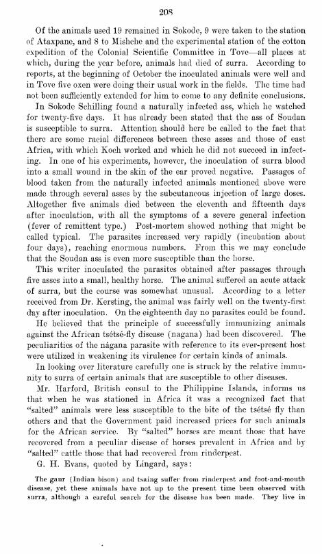
208
Of the animals used 19 remained in Sokode, 9 were taken to the station
of Ataxpane, and 8 to Mishche and the experimental station of the cotton
expedition of the Colonial Scientific Committee in Tovc—all places at
which, during the year before, animals had died of surra. According to
reports, at the beginning of October the inoculated animals were well and
in Tove five oxen were doing their usual work in the fields. The time had
not been sufficiently extended for him to come to any definite conclusions.
In Sokode Schilling found a naturally infected ass, which he watched
for twenty-five days. It has already been stated that the ass of Soudan
is susceptible to surra. Attention should here be called to the fact that
there are some racial differences between these asses and those of east
Africa, with which Koch worked and which he did not succeed in infect-
ing. In one of his experiments, however, the inoculation of surra blood
into a small wound in the skin of the ear proved negative. Passages of
blood taken from the naturally infected animals mentioned above were
made through several asses by the subcutaneous injection of large doses.
Altogether five animals died between the eleventh and fifteenth days
after inoculation, with all the symptoms of a severe general infection
(fever of remittent type.) Post-mortem showed nothing that might be
called typical. The parasites increased very rapidly (incubation about
four days), reaching enormous numbers. From this we may conclude
that the Soudan ass is even more susceptible than the horse.
This writer inoculated the parasites obtained after passages through
Rye asses into a small, healthy horse. The animal suffered an acute attack
of surra, but the course was somewhat unusual. According to a letter
received from Dr. Kersting, the animal was fairly well on the twenty-first
day after inoculation. On the eighteenth day no parasites could be found.
He believed that the principle of successfully immunizing animals
against the African tsetse-fly disease (nagana) had been discovered. The
peculiarities of the nagana parasite with reference to its ever-present host
were utilized in weakening its virulence for certain kinds of animals.
In looking over literature carefully one is struck by the relative immu-
nity to surra of certain animals that are susceptible to other diseases.
Mr. Harford, British consul to the Philippine Islands, informs us
that when he was stationed in Africa it was a recognized fact that
"salted" animals were less susceptible to the bite of the tsetse fly than
others and that the Government paid increased prices for such animals
for the African service. By "salted" horses are meant those that have
recovered from a peculiar disease of horses prevalent in Africa and by
"salted" cattle those that had recovered from rinderpest.
G. H. Evans, quoted by Lingard, says:
The gaur (Indian bison) and tsaing suffer from rinderpest and foot-and-mouth
disease, yet these animals have not up to the present time been observed with
surra, although a careful search for the disease has been made. They live in
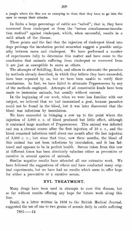
209
a jungle where the flies are so annoying to them that they have to go into the
open to escape their attacks.
In India a large percentage of cattle are "salted"; that is, they have
recovered from rinderpest or from the "serum simultaneous-inocula-
tion method" against rinderpest, which, when successful, results in a
mild attack of the disease.
These points and the fact that the injection of rinderpest blood into
dogs prolongs the incubation period somewhat suggest a possible antip-
athy between surra and rinderpest. We have performed a number
of experiments fully to determine this matter and have come to the
conclusion that animals suffering from rinderpest or recovered from
it are just as susceptible to surra as others.
The attempts of Schilling, Koch, and others to attenuate the parasites
by methods already described, in which they believe they have succeeded,
have been repeated by us, but we have been unable to verify their
conclusions. In fact, we have failed to attenuate Trypanosoma by any
of the methods employed. Attempts of all conceivable kinds have been
made to immunize animals, but usually without success.
In the beginning of our work, when we were less familiar with our
subject, we believed that we had immunized a goat, because parasites
could not be found in the blood, but it was later discovered that the
blood was infectious by inoculation.
We have succeeded in bringing a cow up to the point where the
injection of 3,000 c. c. of blood produced but little effect, although
it contained large numbers of Trypanosoma. This animal was infected
and ran a chronic course after the first injection of 10 c. c, and the
blood remained infectious until about one month after the last injection
of 3,000 c. c. ; but since that time, now three months, the blood of
this animal has not been infectious by inoculation, and it has fat-
tened and appears to be in perfect health. Serum taken from this cow
at different times has been absolutely valueless either as preventive or
curative in several species of animals.
Similar negative results have attended all our extensive work. Wehave followed the suggestions of others and have conducted many orig-
inal experiments, but we have had no results which seem to offer hope
for either a preventive or a curative serum.
XVI. TREATMENT.
Many drugs have been used in attempts to cure this disease, but
so far without results offering any hope for future work along this
line.
Braid, in a letter written in 1858 to the British Medical Journal,
suggested the use of one to two grains of arsenic daily in cattle suffering
7881 14
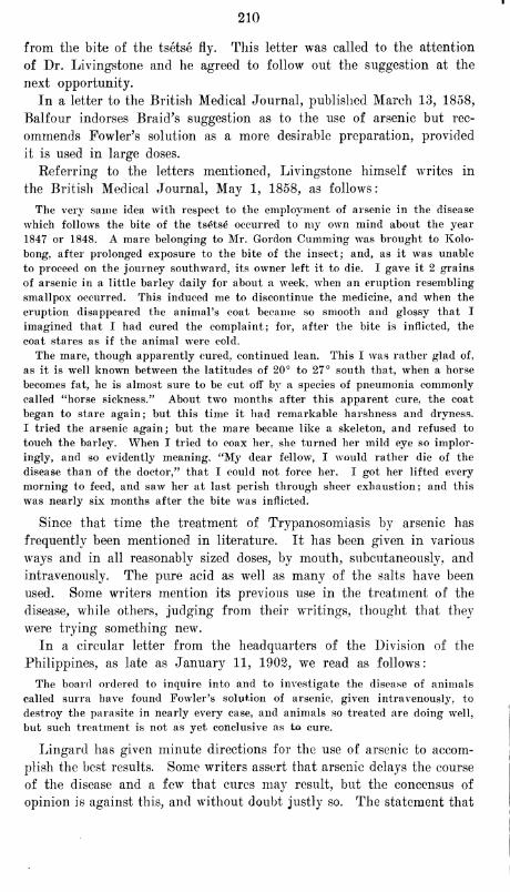
210
from the bite of the tsetse fly. This letter was called to the attention
of Dr. Livingstone and he agreed to follow out the suggestion at the
next opportunity.
In a letter to the British Medical Journal, published March 13, 1858,
Balfour indorses Braid's suggestion as to the use of arsenic but rec-
ommends Fowler's solution as a more desirable preparation, provided
it is used in large doses.
Referring to the letters mentioned, Livingstone himself writes in
the British Medical Journal, May 1, 1858, as follows
:
The very same idea with respect to the employment of arsenic in the disease
which follows the bite of the tsetse occurred to my own mind about the year
1847 or 1848. A mare belonging to Mr. Gordon dimming was brought to Kolo-
bong, after prolonged exposure to the bite of the insect; and, as it was unable
to proceed on the journey southward, its owner left it to die. I gave it 2 grains
of arsenic in a little barley daily for about a week, when an eruption resembling
smallpox occurred. This induced me to discontinue the medicine, and when the
eruption disappeared the animal's coat became so smooth and glossy that I
imagined that I had cured the complaint; for, after the bite is inflicted, the
coat stares as if the animal were cold.
The mare, though apparently cured, continued lean. This I was rather glad of,
as it is well known between the latitudes of 20° to 27° south that, when a horse
becomes fat, he is almost sure to be cut off by a species of pneumonia commonly
called "horse sickness." About two months after this apparent cure, the coat
began to stare again; but this time it had remarkable harshness and dryness.
I tried the arsenic again; but the mare became like a skeleton, and refused to
touch the barley. When I tried to coax her, she turned her mild eye so implor-
ingly, and so evidently meaning, "My dear fellow, I would rather die of the
disease than of the doctor," that I could not force her. I got her lifted every
morning to feed, and saw her at last perish through sheer exhaustion; and this
was nearly six months after the bite was inflicted.
Since that time the treatment of Trypanosomiasis by arsenic has
frequently been mentioned in literature. It has been given in various
ways and in all reasonably sized doses, by mouth, subcutaneously, and
intravenously. The pure acid as well as many of the salts have been
used. Some writers mention its previous use in the treatment of the
disease, while others, judging from their writings, thought that they
were trying something new.
In a circular letter from the headquarters of the Division of the
Philippines, as late as January 11, 1902, we read as follows
:
The board ordered to inquire into and to investigate the disease of animals
called surra have found Fowler's solution of arsenic, given intravenously, to
destroy the parasite in nearly every case, and animals so treated are doing well,
but such treatment is not as yet conclusive as to cure.
Lingard has given minute directions for the use of arsenic to accom-
plish the best results. Some writers assert that arsenic delays the course
of the disease and a few that cures may result, but the concensus of
opinion is against this, and without doubt justly so. The statement that
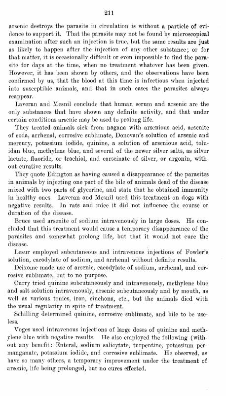
211
arsenic destroys the parasite in circulation is without a particle of evi-
dence to support it. That the parasite may not he found by microscopical
examination after such an injection is true, but the same results are just
as likely to happen after the injection of any other substance; or for
that matter, it is occasionally difficult or even impossible to find the para-
site for days at the time, when no treatment whatever has been given.
However, it has been shown by others, and the observations have been
confirmed by us, that the blood at this time is infectious when injected
into susceptible animals, and that in such cases the parasites always
reappear.
Laveran and Mesnil conclude that human serum and arsenic are the
only substances that have shown any definite activity, and that under
certain conditions arsenic may be used to prolong life.
They treated animals sick from nagana with arsenious acid, arsenite
of soda, arrhenal, corrosive sublimate, Donovan's solution of arsenic and
mercury, potassium iodide, quinine, a solution of arsenious acid, tolu-
idan blue, methylene blue, and several of the newer silver salts, as silver
lactate, fluoride, or trachiol, and carseinate of silver, or argonin, with-
out curative results.
They quote Edington as having caused a disappearance of the parasites
in animals by injecting one part of the bile of animals dead of the disease
mixed with two parts of glycerine, and state that he obtained immunity
in healthy ones. Laveran and Mesnil used this treatment on dogs with
negative results. In rats and mice it did not influence the course or
duration of the disease.
Bruce used arsenite of sodium intravenously in large doses. He con-
cluded that this treatment would cause a temporary disappearance of the
parasites and somewhat prolong life, but that it would not cure the
disease.
Lesur employed subcutaneous and intravenous injections of Fowler's
solution, cacodylate of sodium, and arrhenal without definite results.
Deixome made use of arsenic, cacodylate of sodium, arrhenal, and cor-
rosive sublimate, but to no purpose.
Curry tried quinine subcutaneously and intravenously, methylene blue
and salt solution intravenous^, arsenic subcutaneously and by mouth, as
well as various tonics, iron, cinchona, etc., but the animals died with
the usual regularity in spite of treatment.
Schilling determined quinine, corrosive sublimate, and bile to be use-
less.
Voges used intravenous injections of large doses of quinine and meth-
ylene blue with negative results. He also employed the following (with-
out any benefit: Enteral, sodium salicylate, turpentine, potassium per-
manganate, potassium iodide, and corrosive sublimate. He observed, as
have so many others, a temporary improvement under the treatment of
arsenic, life being prolonged, but no cures effected.
212
Three native ponies were treated with daily intravenous injections of
large quantities of 1-1,000 solution of acetozone. A temporary drop
of the temperature often followed treatment, and, as in the case of
almost any kind of an injection, the parasites sometimes disappeared
for a short time from the circulation, but definite or permanent results
were not obtained, although the course of the disease was somewhat
shortened.
As has already been shown, several substances have a destructive action
for Trypanosoma in the hanging drop, but no such favorable action was
obtained from any of them in treatment, whether by mouth, subcutane-
ously, or intravenously. The following have been used by us in the treat-
ment of animals ill of the disease, but in none of them with hopeful
results
:
Lysol, creolin, infusion of pepo granatum, santonin in the form of
freshly prepared santonate of soda, strychnine arsenate, Fowler's solution,
spigellia, copper arsenite, pelleterine, eucalyptus, quinine hydrochlorate
and urea, and combinations of several other salts and quinine, thymol,
chloral hydrate, glycerine, methyl alcohol, acetic alcohol, barium chloride,
calcium chloride, magnesium chloride, picric acid, oxalic acid, and vari-
ous strengths of salt solutions alone and in combination with other drugs,
carbolic acid, formalin, potassium permanganate, cyanide of potassium,
urotropin, turpentine, cuprous sulphate, cupric sulphate, eosin water
soluble, eosin alcohol soluble, potassium acetate, potassium chlorate, cor-
rosive sublimate, arsenious acid, methylene blue, and several other aniline
dyes.
The following serums have also been used: Antidiptheritic, antistrep-
tococcic, antirinderpestic, antiplague, antidysenteric, antitetanic, and all
available prophylactic preparations.
Toxins, toxic cultures, and fresh cultures of numerous organisms have
been used, including plague, dysentery, typhoid, paracolon, malta fever,
streptococcus cholera, and several strains of colon bacilli.
Blood parasites have been inoculated, including malaria and two
varieties of filaria.
Extensive use has been made of human blood taken from fresh necrop-
sies and in the following diseases : Cholera, dysentery, plague, malaria,
typhoid, Bright's disease, leprosy, and malignant neoplasms.
Blood from the lower animals, both in health and in disease, has
been employed; from healthy cows, as well as those suffering from rin-
derpest and foot-and-mouth disease; from sheep, goats, deer, rabbits,
guinea pigs, frogs, chickens, ducks, pigeons, and several other species
of animals.
Bile and other excretions and secretions, including urine from both
healthy and diseased animals as well as from those dead of surra, have
been used.
Use has been made of the extracts from the lymphatics, the adrenal,
213
and the thymus, as well as from organs of animals affected with surra
and other diseases.
Kecourse has been had to X-ray and other light treatments, various
emulsions and preparations of blood, and attempted attenuations of
parasites.
As already mentioned under serum therapy, considerable time has
been devoted to the preparation of specific sera; and numerous injec-
tions of aspirated serous fluids and the contents of collodion sacs have
been kept in the abdominal cavity of susceptible animals for varying
lengths of time.
In all this work we have not obtained a single recovery, nor have
we been able to bring about conditions that would indicate the slightest
hope of effecting a cure in animals when once they have contracted
the disease.
In conclusion, we see no hope whatever for curative treatment along
lines so far investigated, and the outlook for preventive treatment is
hardly more encouraging.
From a casual observation the conditions seem unfavorable, but if
we go more deeply into the matter we find that they are not so bad
after all. The disease is one which can certainly be prevented in a
country not yet infected and can as surely be eradicated from one where
it is already epidemic by means which are thoroughly practicable. There
is presented to us in the Philippine Islands to-day an opportunity to
accomplish results which will be gratifying to the scientific world and
which should save the country from the annual loss of thousands of
dollars.
XVII. SUMMARY AND CONCLUSIONS.
Trypanosomiasis is considered to be a general- infection caused by
Trypanosoma. The term Trypanosomiasis in a general sense is used
to designate all varieties of the infection as found in different animals.
The long list of vernacular names now in use, except surra, should be
discarded or else allowed to fall merely as synonyms, save in those
cases where the infecting parasite is shown to be a species distinct
from that of Tr. evansii.
A study of the history of the disease shows it to be of remote origin,
records of it in some countries dating back for centuries.
It is distributed over large areas of the tropical and subtropical wrorld,
corresponding closely in its dissemination to the malarial zones.
Trypanosoma in general are discussed with reference to history;
methods of study; general characteristics, including modes of multi-
plication, agglutination and involution forms; distribution in the body
and outside the body; life cycle.
The life cycle is as yet unknown, but is believed to be acted out
entirely within the animal economy.
214
A tentative classification has been adopted for purposes of study,
and each Trypanosoma of importance has been discussed with refer-
ence to its principal characteristics, habitat, and pathogenesis.
The differential diagnosis of Trypanosoma of mammals, like the life
cycle, is left an open question, but the weight of evidence in literature
and our own observations tend to the conclusion that at least three
of the species to which separate names have been given are in reality
identical with Tr. evansii.
Under the discussion of modes of transmission and infection, the only
point upon which emphasis need be placed is the conveyance of the disease
through wounded surfaces, in which biting insects, particularly flies and
fleas, serve as the principal agencies. It is clear that the prevalence of
the disease is dependent upon the presence of a host for the Trypanosoma
and of insects for their transmission. The animals which serve as hosts
for the perpetuation of the disease through the dry season vary in differ-
ent countries. In Manila sick horses exist in sufficient numbers to carry
the infection from one rainy season to another. Cows and rats may also
aid in its perpetuation.
Statements concerning the infection of pastures and water and the
transmission of the Trypanosoma through sound mucous membranes have
nothing to support them.
After describing the general pathologic anatomy and symptomatology
we have taken up the discussion of the infection in various species of
animals, paying particular attention to the disease in those of economic
importance. The manner in which the symptoms vary in different ani-
mals has made this necessary, in order to enable us to make satisfactory
diagnoses and intelligently to control the epidemic.
The course, the duration, the prognosis, the complications, and the
diagnosis have all received a general consideration.
A chapter has been devoted to the consideration of the identity or indi-
viduality of surra, nagana, clourine, and mal de caderas. This is an
important subject from a scientific standpoint, but as an aid to the evolu-
tion of means of prevention or cure it is of little consequence. As in the
case of the parasite, we have with most other writers left the subject open,
but we are strongly inclined to believe them the same disease, in which
case surra would be the only vernacular name allowable. There is cer-
tainly nothing in the clinical study of these diseases to differentiate them.
The only real arguments in favor of their individuality are based upon
morphologic differences in the parasites, and, as has already been said,
these appear to us to be so slight that a positive classification can not be
founded on them.
The study of prophylaxis has included the consideration of quarantine
laws intended to prevent the infection or reinfection of a country, as the
case may be, and of methods for the control and eradication of the disease
in territories where it alreadv has a foothold. In discussing this matter
215
we have limited ourselves almost entirely to the consideration of means
adapted for destroying the hosts and supplemented by those suitable for
combating the carrying agents. It has been thought necessary to go into
this subject with considerable detail, and miscellaneous conditions have
been given full consideration.
Prophylactic and curative serum therapy have thus far failed to give
successful results, but if recent reports from Africa are to be trusted,
preventive inoculation is not wholly without promise of success.
All methods tried for the treatment of the disease have been without
results of practical importance or significance.

AUTHOR INDEX TO BIBLIOGRAPHY
Anacker, 1893.
Baker, C. J., 1903.
Balbiani, E. G., 188(5, 1887, 1888.
Balfour, George AV., 1858.
Barron, 1895.
Blanchard, 1888, 1889.
Blanchard, R., 1887, 1888, 1889, 1901.
Blandford, W. F. H., 1896, 1898, 1899.
Borner, Carl, 1901.
Bosquier, R., 1902.
Boyce, R. W., 1902, 1903.
Bradford, J. Rose, 1899, 1902.
Braid, James, 1858.
Braun, Max, 1895.
Bruce, David, 1895, 1896, 1897, 1902.
Brumpt, Emile, 1901.
Buard, G., 1902.
Buffard, M., 1899, 1900, 1901, 1902.
Burke, Richard W., 1887, 1888, 1889,
1892, 1897.
Busy, 1899.
Butler, R. C, 1888.
Biitschli, O., 1878, 1884, 1889, 1902.
Butt, A. W., 1902.
Campbell, C. W., 1892.
Capitan, 1891.
Carougeau, 1901.
Carter, H. Vandyke, 1888, 1889.
Castellani, Aldo, 1903.
Certes, A., 1882, 1891.
Chalachnikow, A., 1888, 1889."
Chastry, II., 1898.
Chaussat, J. B., 1850.
Chauvrat, J., 1896, 1900.
Claude, A., 1897.
Corbold, T.'S., 1879.
Crookshank, Edgar M., 1886, 1887, 1896.
Cunningham, D. Douglas, 1888.
Curry, J. J., 1902.
Dangeard, 1901.
Danilew>ky, B., 1885, 1886, 1887, 1888,
1889, 1890.
Davaine, C. J., 1877.
Delages, 1896.
DeDoes, 1900, 1901.
Doflein, F., 1901. .
Duclaux, 1896.
Durham, II. E., 1894, 1898, 1899.
Dutton, J. E., 1902, 1903.
Eberth, 1861.
Elmassian, M., 1901, 1902, 1903.
Evans, George H., 1899.
Evans, Griffith, 1880, 1881, 1882, 1898.
Faville, George G, 1893, 1896.
Friedberger, Franz, 1889.
Frohner, Eugen, 1889.
Foa, 1897.
Forde, R. M., 1902.
Gal tier, 1887.
Gaule, J., 1880.
Gazeau, 1901.
Gluge, G., 1842.
Grassi, B., 1881, 1883, 1888.
Grieg, E. D. W., 1902.
Gros, 1845.
Gruby, David, 1843, 1844.
Gunn, \V. D., 1887, 1891.
Hassall, Albert, 1902.
Hefferan, 1902.
Herouard, 1896.
Hertwig, 1842, 1847, 1852.
Ishikawa, 1894, 1899.
Jolyet, F., 1891.
Jurgens, 1902.
Kanthack, A. A., 1898, 1899.
Kay-Lees, 1888.
Kempner, Walter, 1899, 1902.
Kent, W. Saville, 1880.
Kermorgant, 1902.
Kinyoun, J. J., 1901.
Koch, Robert, 1881, 1897, 1898, 1901.
Kruse, W., 1896.
Ki'mstler, G., 1883.
Labbe, Alphonse, 1891.
Laboulbene, A., 1888.
DeLacerda, J. B., 1885.
Lafosse, 1860.
De Lanessan, J. L. , 1882.
217
218
Lankester, E. Ray, 1871, 1882.
Laquerriere, 1883, 1884, 1889.
Laveran, Alphonse, 1892, 1893, 1895, 1899,
1900, 1901, 1902.
Law, James, 1889.
Leclainche, E., 1898,1903.
Lecler, E., 1890, 1899, 1902.
Leger, Louis, 1902.
Legrain, M. A., 1896.
Leishman, W. B., 1903.
Lesage, 1903.
Leuckart, Rudolf, 1886.
Lewis, Timothy Richards, 1878, 1879,
1884, 1888.
Lignieres, J., 1902, 1903.
Lingard, Alfred, 1893, 1894, 1895, 1896,
1897, 1898, 1899, 1900, 1901, 1902.
Livingstone, David, 1857, 1858.
Liihe, Max, 1900.
DeLustrac, A., 1896.
McWeeney, E. J., 1899.
Malbran, 1901.
Malkmus, Bernard, 1901.
Manson, Patrick, 1903.
Marchal, E., 1903.
Marchand, F., 1894.
Marek, 1900.
Marotel, G., 1902.
Marshall, W., 1885.
Martini, Erich, 1903.
Maxwell, Thos., 1857.
Mayer, A. F. I. C, 1843.
Megnin, J. P., 1875.
Mesnil, F., 1900, 1901, 1902.
Mitrophanow, P., 1883.
Mobius, K., 1883.
Mollereau, 1888.
Musgrave, W. E., 1903.
DeNabias, B., 1891.
Nariman, 1893.
Nepveu, G., 1898.
Neumann, L. G., 1888, 1892.
Nocard, E., 1892, 1898, 1900, 1901, 1902,
1903.
Nockolds, Coleman, 1901,1902.
Novikoff, 1902.
Nuttall, G. H. F., 1898,1899.
(Efele, 1899.
Opie, Eugene L., 1901.
Osborn, Herbert, 1896.
Osier, William, 1887.
Oswell, W., 1852,1853.
Paszotta, 1889.
Pease, II. J., 1897.
Penning, C. A. , 1899, 1900.
Peuch, 1880.
Pfeiffer, L., 1889.
Plimmer, H. G., 1899, 1902.
Rabinowitsch, Lydia, 1899,1902.
Railliet, Alcide, 1884, 1893, 1895.
Ranking, George, 1891.
Rattig, A., 1875.
Raver, P., 1843.
Rebourgeon, 1889.
Regulato, 1896.
Remak, 1842.
Rin^e, R. II., 1887.
Rogers, Leonard, 1901.
Ross, Ronald, 1902.
Rost, E. R., 1901.
Rouget, J., 1896, 1900, 1903.
Sabrazio, Muratet, 1902. -
Saint-Cyr, 1877, 1878.
Sherrington, 0. S., 1902, 1903.
Salmon, D. E., 1902.
Sambon, L. W., 1902.
Sand, R., 1899.
Sander, 1902.
Sawtchenko, 1900.
Schat, P., 1902, 1903.
Schilling, 1901, 1902, 1903.
Schneidemiihl, George, 1896.
Schneider, G., 1899, 1900, 1901,1902.
Schooch, G. M. T., 1883.
Senn, G., 1900, 1902.
Signol, 1854.
Sivori, F., 1902.
Slee, John G., 1902.
Smith, Allen M., 1901, 1902.
Smith, Fred, 1886.
Smith, Th., 1893.
j
Stassano, Henri, 1901.
j
Steel, John Henry, 1881, 1885, 1886, 1888.
!Stiles, Ch. Wardell, 1902.
! Stordy, Robert J., 1899.
|
Strong, Richard P., 1902.' Stuhlmann, F., 1902.
Szewczyk, M. J., 1903.
Tanguy, 1880.
Von Thanhoffer, L., 1882, 1883.
Theiler, A., 1901.
Todd, J. II., 1903.
Trasbot, 1878.
Valentin, G., 1841.
Vaz, 1893.
Viardot, 1866.

219
Voges, 0., 1901, 1902.
Vrijburg, A., 1902.
Walley, R. St. John, 1893.
Wasieiewski, V., 1900, 1902.
Weber, 1902.
Wedl, C, 1850.
Weichselbauin, Anton, 1898.
Williams, W., 1889, 1892.
Williamson, N. E., 1903.
Wilson-Barker, J., 1892, 1893.
Wittich, 1881.
Yoes, 1896.
Zabala, 1901.
Zieniann, Hans, 189<>, 1898, 1900, 1901,
1902, 1903.

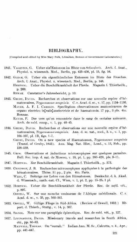
BIBLIOGRAPHY.
[Compiled and edited by Miss Mary Polk, Librarian, Bureau of Government Laboratories.]
1841. Valentin, G. Ueber ein'Entozoon im Blute \on tialmofario. Arch. f. Anat.,
Physiol, u. wissensch. Med., Berlin, pp. 435-436, pi. 15, rig. 16.
1842. Gluge, G. Ueber ein eigenthiimliches Entozoon im Blute des Frosches.
Arch. f. Anat., Physiol, u. wissensch. Med., Berlin, p. 148.
Hertwig. Ueber die Beschiilkrankheit der Pferde. Magazin f. T.hierheilk.,
p. 269.
Remak. Canstatter's Jahresbericht, p. 10.
1843. Gruby, David. Recherches et observations sur une nouvelle espece d'he-
matozoaires, Trypanosoma sanguinis. C. r. Acad. d. sc, v. 17, pp. 1134-1136.
Mayer, A. F. I. Carolus. Specilegium observationum anatomicarum de
organo electrico injraiisjanelectricis et de haematozois. 17 pp., 3 pis. 4to.
Bonnae.
Rayer, P. Des vers qu'on rencontree dans le sang de certains animaux.
Arch, de nied. comp., v. 1, pp. 40-45.
1844. Gruby, David. Recherches et observations sur une nouvelle espece d'he-
matozoaires, Trypanosoma sanguinis. Ann. d. sc. nat., zool., 3. s., v. 1, pp.
104-107, pi. IB, figs. 1-7.
Gruby, David. On a new species of Haematozoon, Trypanosoma sanguinis
(Transl. of Gruby, 1843). Ann. Mag. Nat. Hist., Lond., v. 13, Feb., pp.
158-159.
1845. Gros. Observations et inductions microscopiques sur quelques parasites.
Bull. Soc. imp. d. nat. de Moscou, v. 18, pt. 1, pp. 380-428, pis. 9-11.
1847. Hertwig. Zur Beschiilkrankheit. Magazin f. Thierheilk., p. 373.
1850. Ciiaussat, J. B. Recherches microscopiques appliquees a la pathologie des
hematozoaires. These. 51 pp., 2 pis. 4to. Paris.
Wedl, C. Beitriige zur Lehre von den Hiimatozoen. Denkschr. d. k. Akad.
d. Wissensch., math. -nat. CI., Wien, v. 1, pt. 2, pp. 15-25, 1 pi.
1852. Hertwig. Ueber die Beschiilkrankheit der Pferde. Rec. de ined. vet.,
p. 897.
Oswell, W. Sur une mouche venimeuse de l'Afrique meridionale. C. r.
Acad. d. sc, v. 35, pp. 560-561.
1853. Oswell, W. Giftige /Fliege in Siid-Africa. (Review of Oswell, 1852.) Re-
pert, cl. Thierh., Stuttg., v. 14, p. 367.
1854. Signol. Note sur une paraplegie epizootique. Rec. de med. v^t., p. 127.
1857. Livingstone, David. Missionary travels and researches in South Africa,
etc., pp. 80-83.
Maxwell, Thomas. On "surrah." Indian Ann. M. Sc, Calcutta, v. 4., Apr.,
pp. 441-447.221
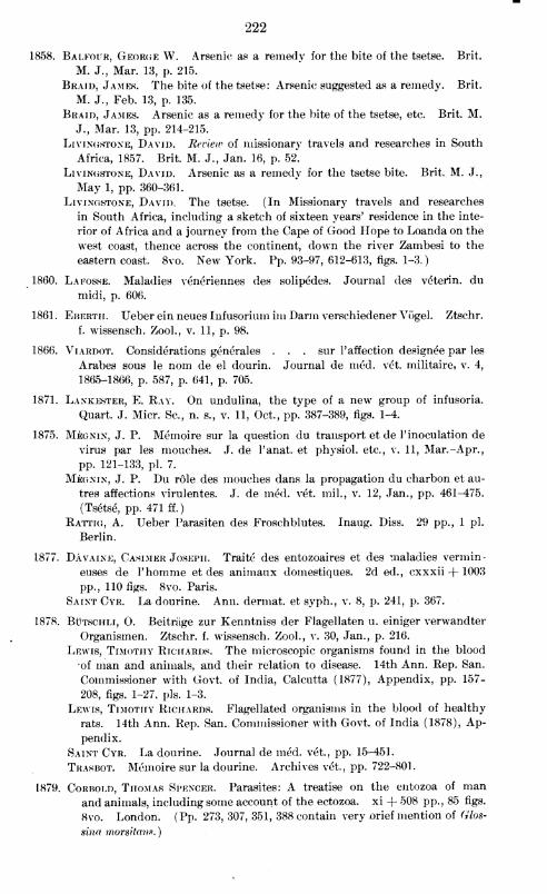
222
1858. Balfour, George W. Arsenic as a remedy for the bite of the tsetse. Brit.
M. J., Mar. 13, p. 215.
Braid, James. The bite of the tsetse: Arsenic suggested as a remedy. Brit.
M. J., Feb. 13, p. 135.
Braid, James. Arsenic as a remedy for the bite of the tsetse, etc. Brit. M.
J., Mar. 13, pp. 214-215.
Livingstone, David. Review of missionary travels and researches in South
Africa, 1857. Brit. M. J., Jan. 16, p. 52.
Livingstone, David. Arsenic as a remedy for the tsetse bite. Brit, M. J.,
May 1, pp. 360-361.
Livingstone, David. The tsetse. (In Missionary travels and researches
in South Africa, including a sketch of sixteen years' residence in the inte-
rior of Africa and a journey from the Cape of Good Hope to Loanda on the
west coast, thence across the continent, down the river Zambesi to the
eastern coast. 8vo. New York. Pp. 93-97, 612-613, figs. 1-3.
)
1860. La fosse. Maladies veneriennes des solipedes. Journal des v£terin. du
midi, p. 606.
1861. Eberth. Ueber ein neues Infusorium im Darm verschiedener Vogel. Ztschr.
f. wissensch. Zool., v. 11, p. 98.
1866. Viardot. Considerations generates . . . sur l'affection designee par les
Arabes sous le nom de el dourin. Journal de med. vet. militaire, v. 4,
1865-1866, p. 587, p. 641, p. 705.
1871. Lankester, E. Ray. On undulina, the type of a new group of infusoria.
Quart. J. Micr. Sc., n. s., v. 11, Oct., pp. 387-389, figs. 1-4.
1875. Megnin, J. P. Memoire sur la question du transport et de V inoculation de
virus par les mouches. J. de Panat. et physiol. etc., v. 11, Mar.-Apr.,
pp. 121-133, pi. 7.
Megnin, J. P. Du role des mouches dans la propagation du charbon et au-
tres affections virulentes. J. de med. vet. mil., v. 12, Jan., pp. 461^75.
(Tsetse, pp. 471 ff.)
Rattig, A. Ueber Parasiten des Froschblutes. Inaug. Diss. 29 pp., 1 pi.
Berlin.
1877. Davaine, Casimer Joseph. Traite des entozoaires et des maladies vermin
-
euses de l'homme et des animaux domestiques. 2d ed., cxxxii -f 1003
pp., 110 figs. 8vo. Paris.
Saint Cyr. La dourine. Ann. dermat. et syph., v. 8, p. 241, p. 367.
1878. BtiTSCHLi, O. Beitriige zur Kenntniss der Flagellaten u. einiger verwandter
Organismen. Ztschr. f. wissensch. Zool., v. 30, Jan., p. 216.
Lewis, Timothy Richards. The microscopic organisms found in the blood
of man and animals, and their relation to disease. 14th Ann. Rep. San.
Commissioner with Govt, of India, Calcutta (1877), Appendix, pp. 157-
208, figs. 1-27. pis. 1-3.
Lewis, Timothy Richards. Flagellated organisms in the blood of healthy
rats. 14th Ann. Rep. San. Commissioner with Govt, of India (1878), Ap-
pendix.
Saint Cyr. La dourine. Journal de med. vet., pp. 15-451.
Trasbot. Memoire sur la dourine. Archives vet., pp. 722-801.
1879. Corbold, Thomas Spencer. Parasites: A treatise on the entozoa of manand animals, including some account of the ectozoa. xi -f 508 pp., 85 figs.
8vo. London. (Pp. 273, 307, 351, 388 contain very orief mention of Glos-
sina morsitans. )

223
1879. Lewis, Timothy Richards. Flagellated organisms in the blood of healthy
rats. Quart. J. Micr. Sc, n. s., v. 19, Jan., pp. 109-114, 1 fig.
Lewis, Timothy Richards. Microscopic organisms in the blood of man and
animals, Calcutta, 1879.
1880. Evans, Griffith. Report on "surra" disease in the Dera Ismail Khan Dis-
trict, Nov. 13, Military Dept.
Evans, Griffith. Report by Punjab Govt., Military Dept, Dec. 3, No. 439.
Gaule, J. Ueber Wurmchen welche aus den Froschblutkorperchen auswan-
dern. Archiv f. Anat. u. Physiologie, p. 57.
Gaule, J. Beobachtungen der farblosen Elemente des Froschblutes. Arch.
f. Physiol., Oct. 1, pp. 376-392, pi. 5.
Kent, W. Saville. A manual of the infusoria, including a description of all
known flagellate, ciliate and tentaculiferous protozoa, British and foreign,
and an account of the organization and affinities of the sponges. Pts. 1-3,
pp. 1-432, pis. 1-24. 8vo. London. (Trypanosoma, pp. 218-220.)
Peuch. £tude du horse-pox simulant la maladie du coit. Revue v6t., p. 297,
p. 345.
Tanguy. La gourme coi'tale dans les arrondissements de Brest et de Morlaix.
Arch. v6t., v. 5, pp. 345-349.
1881. Evans, Griffith. On a horse disease in India known as "surra," probably
due to a hitmatozoon. Vet. J., v. 13, July, pp. 1-10; Aug., pp. 83-88;
Sept., pp. 180-200; Nov., pp. 326-333.
Grassi, B. Interno ad alcuni endoparassitici ed appartenenti alle classi dei
flagellati lobosi, sporozoi e ciliati. Atti Soc. ital. d. sc. nat., Milano, v. 24,
pp. 135-224, pis. 1-4.
Koch, Robert. Zur Untersuchung von pathogenen Organismen. Mitth.
a. d. kaiserlich. Gsndhtsamte, Berl., v. 1, pp. 1-42, pis. 1-14.
Steel, John Henry. A treatise on the diseases of the ox; being a manual of
bovine pathology especially adapted for the use of veterinary practitioners
and students, xxi -f 498 pp., 116 figs. 8vo. London.
Wittich. Spirillen im Blute von Hamstern. Centralbl. f. d. med. Wis-
sensch., v. 19, Jan. 22, pp. 65-66.
1882. Certes, A. Note sur les parasites et les commensaux de l'huitre. Bull.
Soc. zool. de France, v. 7, June 13, pp. 347-353, pi. 7.
Certes, A. Sur les parasites intestinaux de Thuitre. C. r. Acad. d. sc,
v. 95, Sept. 4, pp. 463-465.
(Certes.) Intestinal Parasites of Oysters. Review of Certes' article in C. r.
Acad. d. sc, v. 95, pp. 463-465. J. Roy. Micr. Soc, p. 804.
Certes, A. Sur les parasites intestinaux de Thuitre. J. de microg., v. 6,
Oct., pp. 529-530.
Evans, Griffith. On a horse disease in India known as' 'surra," probably
due to a hamatozoon. (Continued.) Vet. J., v., 14, Feb., pp. 97-110;
Mar., pp. 181-187.
(Gaule. ) Development of Trypanosoma. Review of Gaule' s article in Arch.
f. Physiol., 1880, pp. 375-392. J. Roy. Micr. Soc, pp. 520-521.
De Lanessan, J. L. Manuel d'histoire naturelle m&licale. Pt. 3, Zoologie,
728 pp., 523 figs., 12mo. Paris. (P. 71, fig. 60, brief mention of Undalina
ranarum.)
De Lanessan, J. L. Traite* de zoologie protozoaires. vii + 336 pp., 281
figs. 8vo. Paris. (Pp. 166-167, fig. 142, brief reference to Trypanosoma
sanguinis Gruby and Trypanosoma Eberthi.
)
224
1882. Lankester, E. Ray. On Drepanidium ranarum, the cell-parasite of the frog's
blood and spleen (Gaule's Wurmschen). Quart. J. Micr. Sc, n. s., v. 22,
Jan., pp. 53-65, tigs. 1-5.
(Lankester.) Cell-parasite of frog's blood and spleen [Drepanidium rana-
rum). Review of Lankester's article in Quart. J. Micr. Sc, v. 22, pp. 53-65.
J. Roy. Micr. Soc, p. 519.
Vox Tiianhoffer, L. IJeber Zuchtlahme. Broch. Buda-Pesth.
1883. (Certes. ) Intestinal parasites of the oyster, ifanm' of Certes' article in Bull.
Soc. zool. de France, v. 7, 1882, pp. 347-353, J. Roy. Micr. Soc, p. 81.
Grassi, B. Sur quelques protistes endo-parasites appartenant aux classes des
flagellata, lobosa, sporazoa et ciliata. (Transl. of Grassi, 1881.) Arch. ital. de
biol., v. 2, Feb. 1, pp. 402-444; v. 3, Apr. 15, pp. 23-37, pis. 1-3.
Kunstler, G. Recherches sur les infusoires parasites. Sur quinze protozoaires
nouveaux. C. r. Acad, de Sc, v. 97, Oct. 1, pp. 755-757.
Laquerriere. De la syphilis equine. Gaz. hebd. de med. 2. s., v. 20, p. 515,
p. 543, p. 560.
Laquerriere. De la syphilis equine. Presse vet., v. 3, p. 652.
Mitropiianow, P. Beitriige zur Kenntniss der Hiimatozoon. Neue monadfor-
mige Parasiten des Fischblutes, ihnen iihnliche Organismen und ihre
Beziehung zu den Blutelementen. Biol. Centralbl., v. 3, Mar. 15, pp.
35-44, figs. 1-2.
Mobius, K. Trypanosoiha Balbianii Certes in Krystallstiel schleswig-hol-
steinischer Austern. Zool. Anz., v. 4, Mar. 19, p. 148.
Schoch, G. M. T. Die Tsetsefliege Afrikas. Mitth. d. schweiz. entom. Ge-
sellsch., Schaffhausen, v. 6, Oct., pp. 685-686.
Von Tiianhoffer, L. Leber Zuchtlahme. Revue vet., p. 92.
L884. Butschli, 0. Protozoa. Mit einen Beitrag; Palaeontologische Entwicklung
der Rhizopoda von C. Schwager. Second Part: Mastogophora. Bronn's
Klassen Ordnungen des Thier-Reichs, v. 1, pp. 785-864, 1 fig. (Pp. 811-812,
genus Trypanosoma.
)
Laquerriere. De la syphilis equine. Presse vet., v. 4, p. 102, p. 376.
Lewis, Timothy Richard. Further observations on flagellated organisms in
the blood of animals. Quart. J. Micr. Sc, n. s. v. 24, July, pp. 357-369,
figs. 1-5.
Railliet, Alcide. Du role de la tsetse dans la propagation des maladies.
Bull. Soc. centr. de med. vet., n. s., v. 2, pp. 78-79.
1885. Damlewsky, B. Zur Parasitologic desBlutes. Biol. Centralbl., v. 5, Nov. 1,
pp. 529-537.
De Lacerda, J. B. Pathogenesia comparada. Peste de Cadeiras ouepizootia
de Marajo suas analogias con Beriberi, vii -f 98 PP-, 3 pis., 2 phot. 8vo.
Rio de Janeiro.
Marshall, W. Ueber die Tsetsefliege. Biol. Centralbl., v. 5, May 15, pp.
183-184.
Steel, John Henry. An investigation into an obscure and fatal disease amongtransport mules in British Burma.
1886. Balbiani, Edouard Gerard. Evolution des microorganismes animaux et
vegetaux. J. de niicrog., v. 10, Dec, pp., 535-544, pi. 5.
Crookshank, Edgar M. Flagellated protozoa in the blood of diseased and
apparently healthy animals. J. Roy. Micr. Soc, Dec, pp. 913-928, figs
193-199, pi. 17. [Trichomonas Lewisii, p. 928.)
225
1886. Danilewsky, B. I. Materiaux pourservir a la parasitologie du sang. II. Re-
cherches sur la parasitologie du sang. Arch, slaves de biol., v. I, Jan. 15, pp.
85-91; Mar. 15, pp. 364-396, pis. 5-6.
(Daxilewsky.) Parasites of the blood. Review of Danilewsky's article in
Arch, slaves de biol., v. 1, pp. 85-91; J. Roy. Micr. Soc, pp. 1006-1007.
(Daxilewsky.) Parasites of the blood. I. Ti-ypanosome mmjuimx Gruby.
Review of article by Danilewsky in Biol. Centralbl., v. 5, 1885, pp. 529-
537; J. Roy. Micr. Soc, pp. 635-637.
Daxilewsky, B. Zur Frage ueber die Identitiit der pathogenen Blutpara-
siten des Menschen und der Hamatozoen der gesunden Thiere. Centralbl.
f. d. med. Wissensch., v. 24, Oct. 9, pp. 737-739; Oct. 16, pp. 753-755.
Leuckart, Rudolf. Die Parasiten des Menschen. 3 vols., 2d ed., Leipz. u.
Heidelberg. Flagellata, v. 1, pp. 300 ff.
Smith, Fred. Fieber. Encycl. d. ges. Thierh. u. Thierzucht, Wien u. Leip-
zig, v. 3, pp. 133-137, rig. 539.
Steel, Johx Henry. On relapsing fever of equines. Vet. J., v. 22, Mar., pp.
166-174; Apr., pp. 248-257.
1887. Balbiani, Edouard Gerard. Evolution des mieroorganismes animaux et
vegetaux. J. de microg., v. 11, Feb., pp. 54-62; Apr., pp. 134-142; May,
pp. 170-177 and 196-205; June, pp. 233-240; Aug., pp. 365-373; Sept., 393-
406; Oct., pp. 434-446; Nov., pp. 463-476 and 499-511; Dec, pp. 534-544.
Blaxciiard, Raphael. Bibliographia des hematozoaires. Bull. Soc. zool.
de France, v. 12, Aug. 1, pp. 500-507.
Burke, Richard W. "Surra" or progressive pernicious anemia. Vet. J., v.
25, July, pp. 5-11.
Burke, Richard W. "Surra" or progressive pernicious anemia. J. Oomp.
M. and S., v. 8, July, pp. 223-230.
Crookshaxk, Edgar M. Photomicrographs of flagellated protozoa in the
blood. J. Roy. Micr. Soc, Apr., p. 358.
Daxilewsky, B. Recherches sur la parasitologie du sang. (Continuation of
Danilewsky, 1886.) Arch, slaves de biol., v. 3, Jan. 15, pp. 33-49; Mar.
15, pp. 157-176; May 15, pp. 370-417.
(Daxilewsky.) Parasites in the blood. Review of DanilewTsky's article in
Arch, slaves de biol., v. 3, pp. 370-417. J. Roy. Micr. Soc, pp. 977-978.
Daxilewsky, B. Contribution a la question de l'identite des parasites path-
ogenes du sang chez l'homme avec les hematozoaires chez les animaux
sains. Arch, slaves de biol., v. 3, Mar. 15, pp. 257-264.
Galtier. Horse-pox simulant la dourine. J. denied, vet., 3. s., v. 12, pp.
341-362.
Guxx. Equine relapsing fever in Kohat. Quart. J. of Vet. Science in
India, v. 5, p. 235.
Osler, William. An address on the haematozoa of malaria. Brit. M. J.,
Mar. 12, pp. 556-562, figs. 1-15.
Rixge, R. H. "Surra" in grass-cutters' ponies. Quart. J. of Vet. Science in
India, v. 6, Oct., pp. 47-49, temperature chart.
1888. Balbiani, Edouard Gerard. Evolution des mieroorganismes animaux et
vegetaux. J. de microg. , v. 12, Jan., pp. 1 1-19 and 41-49; Mar.
, pp. 134-142;
Apr., pp. 173-182; May, pp. 205-212; June, pp. 235-243; July, pp. 266-273;
Aug., pp. 303-308; Oct., pp. 394-400; Nov., pp. 421-429; Dec, pp. 517-523,
1 fig-
7881 15

226
1888. Blancharj) (Veterinarian in French Army). Etude sur une maladie spe-
ciaie des mulcts imported au Tonkin pour le service de l'armee et conside-
rations sur l'acclimatement de l'espece chevaline en Extreme-Orient. Bull,
de la Soc. centr. de med. vet., v. 6, pp. 694-701.
Blanchard, Raphael. Hematozoaires. Diet, encyc. d. sc. med., 4. s., v. 13,
pp. 43-73, figs. 1-4.
Burke, Richard W. Surra, or pernicious anemia in the lower animals.
Vet. J., v. 26, Jan., pp. 1-12; Feb., pp. 83-86.
Burke, Richard W. The discussion of the pathology of "surra." Vet. J.,
v. 26, Apr., pp. 309-311.
Burke, Richard W. Harmless organisms found in the blood of horses.
Veterinarian, v. 61, Dec, pp. 785-787.
Burke, Richard W. The tropical diseases of the horse, 2d ed., p. 23.
Butler, R. C. Surra in upper Burma. Quart. J. of Vet. Science in India,
v. 6, pt. 1, July, pp. 379-385.
Carter, H. Vandyke. Note on the occurrence of a minute blood-spirillum
in an Indian rat. Scientific Memoirs by Medical Officers of the Army of
India, 1887, pt. 3, pp. 45-48, pi. 4.
Carter, H.Vandyke. On the lately demonstrated blood contamination andinfective disease of the rat and equines in India. Scientific Memoirs byMedical Officers of the Army of India, 1887, pt. 3, pp. 49-78, pi. 5.
Chalaciinikow (Schalaschnikoff), A. Recherches sur les parasites du sang
chez les animaux a sang froid et a sang chaud. Kharkov.
Cunningham, D. Douglas. On the occurrence of peculiar organisms in the
blood of horses and their supposed pathogenic nature. Scientific Memoirsby Medical Officers of the Army of India, 1887, pt. 3, pp. 1-4, pi. 1, fig. 1.
Danilewsky, B. Recherches sur la parasitologic comparee du sang des
oiseaux. Kharkov.
Grasst, B. Atti d. r. Accad. dei Lincei, 4. s., v. 4, p. 5.
Kay-Lees. Glomrm morwlans or tsetse fly. Vet. J., v. 27, Aug., pp. 79-84.
Laboulbene, A. Sur une mouche tsetse de l'Afrique australe. Bull. Acad,
de med., 2. s., v. 19, June 4, pp. 721-724.
Lewis, Timothy Richards. Flagellated organisms in the blood of healthy
animals. (In Physiological and pathological researches, being a reprint of
the principal scientific writings of the late Timothy Richards Lewis. Ar-
ranged and edited by William Aitken, G. E. Dobson, and A. E. Brown.
8vo. London, pp. 604-609.
)
Mollereau. Sur une maladie des mulets au Tonkin. Bull, de la Soc. centr.
de med. vet., p. 694.
Neumann, L. G. Traite des maladies parasitaires non microbiennes des ani-
maux domestiques. xv+673 pp., 306 figs. 8vo. Paris.
Steel, John Henry. A veterinary glimpse at the last Burma war. Edito-
rial, Quart. J. of Vet. Science in India, v. 6, Jan., pp. 135-144.
Steel, John Henry. Le surra, maladie contagieuse des animaux domesti-
ques, dans PInde. Rec. de med. vet., 7. s., v. 5, May 15, pp. 298-304.
1889. [Blanchard (Veterinarian in French Army).] Surra in Annam. Revieiv of
Blanchard, 1888. Vet. J., v. 28, Feb., p. 101.
Blanchard, Raphael. Traite de zoologie medicale, v. 1, pp. 69-72, pis. 39-
41. 8vo. Paris.
Burke, Richard W. Microorganisms and disease, especially with reference
to the question, What is the pathology of "surra" in animals? Vet. J., v.
28, Jan., pp. 25-27.
Butschli, O. Protozoa. Bronn's Classen u. Ordnungen des Thierreichs.
227
1889. Carter, H. Vandyke. On the lately demonstrated blood contamination andinfective diseases of the rat and of equines in India. Vet. J., v. 28, Mar.,
pp. 180-184 ; May, pp. 339-346 ; June, pp. 409-422.
Ciialachnikow (Scitalaschnikoff), A. Izslied. nad. Krovoparazit. eholod.i teplskrov. jivotnych. Shorn, trud. Kharkov vet. inst., v. 1, pp. 195-304,
3 pis.
Ciialachnikow (Sciialaschnikoff), A. Recherches sur les parasites du sangdes oiseaux. Kharkov.
Danilew>ky, B. La parasitologie comparee du sang. I. Nouvelle recherchessur les parasites du sang des oiseaux, 93 pp., 3 pis. 8vo. Kharkov. II.
Recherches sur les hematozoaires des tortues, 86 pp. , 2 pis. 8vo. Kharkov.Friedberger, Franz, u. Frohner, Eugen. Lehrbuch der speciellen Path-
ologie und Therapie der Hausthiere, 2d ed., v. 2, p. 737.
Laquerriere. Rapport sur les maladies veneriennes du cheval. Bull, de la
Soc. centr. de med. vet., p. 372.
Law, James. "Maladie de coit," or Venereal Diseases of Horses. Ann. Re-port of the Bureau of Animal Industry, U. S. Dept. of Agric, 1887-8, pp.490-492.
Paszotta. Pferdestaupe (Leuma equorum). Veearts. Bladen v. Nederl.-
Indie, v. 3, p. 505.
Pleiffer, L. Beitriige zur Kenntniss der pathogenen Gregarinen. III. Ue-ber Gregarinose, ansteckendes Epitheliom und Flagellaten-Diphtherie der
Vogel. Ztschr. f. Hyg., v. 5, Jan. 31, pp. 363-401, figs. 1-15, pi. 3.
Rebourgeon. Note sur le mal de cadera. Rec. de med. vet., 7. s., v. 6, Feb.
15, pp. 85-93.
Williams, W. L. Maladie du coi't. Am. Vet. Rev., v. 12, p. 295, p. 341, p.
402, p. 445.
The tsetse fly and man. Vet. J., v. 28, Feb., pp. 140-141.
1890. Danilewsky, B. Biol. Centralbl., v. 10, pp. 396-403.
(Damlewsky. ) Parasites of the blood of birds and tortoises: Review of Dan-ilewsky's article in Biol. Centralbl., v. 10, pp. 396-403. J. Roy. Micr. Soc,
p. 731.
Leclercq, Emma. Les microorganismes intermediates aux deux regnes.
Bull. Soc. beige de micr., v. 16, pp. 70-131, figs. 1-7.
Notes on "surra." Veterinarian, v. 63, Jan., pp. 1-3.
1891. Burke, Richard W. The general pathology of surra in animals. Vet. J., v.
33, Oct., pp. 234-272.
Capitan. Sur un hematozoaires du lapin domestique. Med. mod., v. 2, pp.239-240.
Certes, A. Sur le Trypanosoma Balbianii. Bull. Soc. zool. de France, v. 16,
Mar., p. 95; May, pp. 130-131, 1 fig.
(Certes.) Trypanosoma Balbianii. Review of Certes' article in Bull. Soc.
zool. de France, v. 16, p. 95. J. Roy. Micr. Soc, p. 753.
Gunn, W. D. The etiology of surra. Vet. J., v. 33, Oct., pp. 309-310.
1891. Jolyet, F., et De Nabias, B. Sur un hematozoaires du lapin domestique. J.
de med. de Bordeaux, v. 20, Mar. 1, pp. 325-326.
Labbe, Alpiionse. Note sur un nouveau parasite du sang( Trypanosoma Dani-
lewskyi). Bull. Soc. zool. de France, v. 16, Oct., pp. 229-231.
Ranking, George. A preliminary note on the nature and pathology of the
disease known as "surra" affecting horses and mules in India. Vet. J., v.
32, June, pp. 399-405.
Ranking, George. Surra in horses. Vet. J., v. 33, Dec, pp. 402-403.
Surra. Veterinarian, v. 64, Mar., pp. 169-170.
228
1892. Campbell, C. W. A journey through North Korea to the Changpia Shan. J.
Roy. Geograph. Soc, Mar., p. 155.
Burke, Richard W. The tropical diseases of the horse, 3d ed., p. 26.
Laveran, Alfhonse. Des trypanosomes parasites du sang. Arch, de mod.
exper. et d'anat. path., v. 1, Mar. 1, pp. 257-269, pi. 1.
Laveran, Alpiionse. Des trypanosomes parasites du sang. Bibliotheque
med. Charcot.
Neumann, L. G. Traite des maladies parasitaires non microbiennes des ani-
maux domestiques, 2d ed., xvi-f767 pp., 364 figs. 8vo. Paris.
Neumann, L.. G. A treatise on the parasites and the parasitic diseases of do-
mesticated animals, translated and edited by George Fleming, xxiii-f800
pp., 364 figs. 8vo. London.
Nocard, E. Sur l'inoculabilite de la dourine. C. r. Acad. d. sc, v. 114, p.
188.
Williams, William. The principles and practice of veterinary medicine,
3d ed., XIX+833 pp., 36 figs., 7 pis. 8vo. London, Edinburgh and
Glasgow.
Wilson-Barker, J. Maladie du coi't in Nebraska. Vet. J., v. 35, Dec, pp.
424-429.
1893. Anacker (Dusseldorf). Surra. Encyc. d. ges. Thierh. u. Thierzucht, v. 10,
p. 140.
Faville, George C. Supposed maladie du coi't among horses in Nebraska.
Annual Report of the Bureau of Animal Industry, U. S. Dept. of Agric.
for 1891-1893, pp. 359-366.
Laveran, Alpiionse. Paludism. xii-|-197 pp., 7 figs., 6pls. 8vo. London.
Lingard, Alfred. Report on horse surra, v. 1, cclxxix {-125 pp., 11 pis.
Fol. Bombay.Nariman and Vaz. The disease "surra." Vet. J., v. 36, June, pp. 403-406.
Raillet, Alcide. Traite de zoologie medicale et agricole, v. 1, 736 pp., 494
figs. 8vo. Paris.
Smith, Til, and Kilborne. Investigation into the nature, causation and pre-
vention of Texas cattle fever. Bull. U. S. Dept. of Agric, Bureau of
Animal Industry.
Walley, Ralph St. John. Surra in fox hounds. J. Comp. Path, and
Therap., Edinburgh and London, v. 6, Sept. 30, pp. 285-286.
Wilson-Barker, J. Maladie du coi't. Vet. J., v. 36, June, pp. 406-408.
1894. Durham. Tsetse disease in mammals. Proceedings Fourth International
Cong, of Zool., Cambridge.
Ishikawa. J. coll. sc, Tokyo.
Lingard, Alfred. Summary of further report on surra, 8 pp. Fol.
Bombay.Lingard, Alfred. Summary of further report on surra. Vet. J., v. 39, Oct.,
pp. 250-258; Nov., pp. 311-315.
(Lingard.) The surra disease in solipeds and camels. Review of Lingard,
1893. Vet. J., v. 38, Mar., pp. 172-175.
Marchand, F. Ueber das Vorkonimen von Trichomonas im Harne eines
Marines, nebst Bemerkungen ueber Trichomojias vaginalis. Centralbl. f.
Bakt., v. 15, May 19, p. 709.
1895. Barron. Liverpool Medico-Chirurgical J., Jan., p. 232.
Bra un, Max. Die thierischen Parasiten des Menschen, 2d ed., 283 pp.,
147 figs. 8vo. Wurzburg.
Bruce, David. Preliminary report on the tsetse-fly disease or nagana in
Zululand, 28 pp., 7 pis. Fol. Durban.
229
1895. Laverax, Alphonse. Les hematozoaires de l'homme et des animaux. I.
Protozoaires du sang, iv-f-154 pp., 9 figs. Paris.
Lixgard, Alfred. Re}>ort on the outbreak of surra and filariasis at Karnal,
Govt. Depot.
Lixgard, Alfred. Suinmary of further report on surra, 13 pp. Fol.
Bombay.Raillet, Alcide. Traite de zoologie m£dicale.et agrieole, v. 2, pp. 737-
1303, figs. 495-892. 8vo. Paris.
1896. Blandford. Tsetse-fly disease.
(Bruce.) Review of Bruce's Preliminary report, Nature, v. 53, pp. 566-568,
2 figs.
(Blaxdford-Bruce. )' Tsetse-fly disease. Review of Blandford's article on
Bruce's Preliminary report, Nature, v. 53, pp. 566-568. J. Roy. Micr.
Soc, p. 428.
Bruce, David. Further report on the tsetse-fly disease or nagana in Zulu-
land. Ubombo, Zululand.
(Bruce.) Preliminary report on the tsetse-fly disease or nagana in Zulu-
land. Review. Arch. f. wissensch. u. prakt. Thierh., v. 22, pp. 366-370.
(Bruce.) Preliminary report on the tsetse-fly disease or nagana in Zulu-
land. Review. Centralbl. f. Bakt., 1. Abt., v. 19, June 30, pp. 955-957.
Ciiauvrat, J. Un cas d'an^mie pernicieuse du cheval en Algerie causae par
un trypanosome. Rec. de med. vet., 8. s., v. 3, June 15, pp. 344-348.
Crookshaxk, Edgar M. A text-book of bacteriology, including the etiology
and prevention of infective diseases, and a short account of yeast and molds,
haematozoa and psorosperms, 4th ed., xxx-f-715 pp., 273 figs., 22 pis. 8vo.
London.
Delages, Yoes, and Herouard. Traite de zoologie concrete, v. 1, La cellule
et les protozoaires. xxx-j-584 pp., 870 figs. 8vo. Paris.
Duclaux. Ann. de l'lnst. Pasteur, v. 10, p. 189.
Faville, George C. Extirpation of Maladie du coi't. Ann. Report of the
Bureau of Animal Industry, U. S. Dept. of Agric, for 1893-1894, pp. 62-65.
Kruse, W. Systematik der Protozoen. In Flugge's Die Mikroorganismen,
3d ed., Leipzig, v. 2, pp. 601-700. Flagellaten, pp. 626-634.
Legraix, M. A. Trypanosome des bovides. Progres vet., Mar. 15.
Legraix, M. A. Trypanosome des bovides. Rec. vet., v. 13.
Lixgard, Alfred. Annual report of the imperial bacteriologist for 1895-96.
61 pp., 1 map. 4to. Bombay.(Lixgard.) Surra. Review of Lingard, 1895. Vet. J., v. 42, Mar., pp.
177-183.
De Lustrac, A. Trypanosoma Balbianii, Certes. Actes Soc. Linn, de Bor-
deaux, v. 50, pp. 265-275, pis. 1-2.
Osborx, Herbert. Insects affecting domestic animals; an account of the spe-
cies of importance in North America, with mention of related forms occur-
ring on other animals. Bull. No. 5, n. s., U. S. Dept. Agric, Div. Entom.,
302 pp., 170 figs. 8vo.
Regulato. Trypanosome des bovides. Progres v6t., p. 177.
Rouget, J. Contribution a l'etude du trypanosome des mammiferes. Ann.
de l'lnst. Pasteur, v. 10, Dec, pp. 716-728, 3 figs.
Sciixeidemuhl, George. Lehrbuch der vergleichenden Pathologie und The-
rapie des Menschen und der Hausthiere, pp. 209-448. 8vo. Leipzig.
Ziem ann, Hans. Blutparasiten bei heimischer und tropischer Malaria. Cen-
tralbl. f. Bakt,, v. 20, pp. 653-672.
230
1896. Le trypanosome du boeuf et les tsetses. Rec. de med. vet., 8. s., v. 3, Apr.
15, p. 266.
The tsetse-fly disease or nagana. Agric. J., Cape Town, v. 9, July 9, pp.358-360.
1897. Bruce, David. Further report on the tsetse-fly disease or nagana in Zululand,
69 pp., 6 pis. 4to. London.
Burke, Richard W. Malarial fever in horses in India, or "surra." Veterinarian, v. 70, Apr., pp. 185-194.
Claude, A. La mouche tsetse et le guaco. Bull. Soc. homoeop. de France,
v. 21, July, pp. 165-177.
Foa. Du cap au lac Nyassa. Paris.
Koch, Robert. Berichte des Herrn Prof. Dr. Koch uber seine in Kimber-ley gemachten Versuche beziiglich Bekampfung der Rinderpest. Centralbl.
f. Bakt., v. 21, pp. 526-537. (TransL from Agric. J., Jan. 14 and Feb. 18,
1897.)
Koch, Robert. Reise-Berichte iiber Rinderpest. Berlin.
Lingard, Alfred. Summary of further report on surra. Bombay.Pease, H. J. Surra and malarial fever. Veterinarian, v. 70, Oct., pp.
549-565.
(Rouget. ) Trypanosomata of mammalia. Renew of Rouget' s article in Ann.de l'Inst. Pasteur, v. 10, 1896, pp.. 716-728. J. Roy. Micr. Soc, p. 133.
1898. Ciiastry, H. La mouche tsetse. Rev. sc, 4. s., v. 9, Mar. 26, pp. 406-407.
Durham, II . E. Tsetse disease. Veterinarian, v. 71, Oct., pp. 535-538.
(Summary'of a paper read before the Fourth International Zoological Con-gress, Cambridge, 1898.)
Evans, Griffith. Annual report of the imperial bacteriologist for 1896-97.
iii+77 pp. Fol. Calcutta, p. 20.
(Hayes, M. H.) Surra. (Infective diseases of animals, in Friedberger andFrohner's Veterinary pathology, translated and edited by Hayes; notes onbacteriology by G. Neumann.
)
Kanthack, A. A., Durham, II. E., and Blandford, W. F. H. On nagana or
tsetse-fly disease. Proc. Roy Soc, v. 64, Oct. 27, pp. 100-118.
(Kanthack, A. A., Durham, H. E., and Blandford, W. F. H. ) Nagana or
tsetse-fly disease. Review of Kanthack, Durham, and Blandford, Proc. Roy.
Soc, v. 64, pp. 100-118. Brit, M. J., Dec. 10, pp. 1768-1769.
(Kanthack, A. A., Durham, H. E., and Blandford, W. F. H.) Nagana or
tsetse-fly disease. Abstract. J. Comp. Path, and Therap., Edinburgh andLondon, v. 11, Dec. 31, pp. 370-380.
Nuttall, G. H. F. Ueber Nagana oder die Tsetsefliegenkrankheit, TransL
of Kanthack, Durham and Blandford, Proc. Roy. Soc, v. 64, pp. 100-118.
Hyg. Rundschau, v. 8, Dec 15, pp. 1185-1202, 1 fig.
Koch, Robert. Reise-Berichte iiber Rinderpest, Bubonenpest in Indien
und Afrika, Tsetse-oder Surrakrankheit, Texas-Fieber, tropische Malaria.
Schwarzwasserfieber, 136 pp., 10 figs. 8vo. Berlin.
Kocn, Robert. Report on the surra disease. Brit, M. J., Apr. 9, p. 983.
Kunstler. Bull, sc France et Belgique, v. 31, p. 206.
Lingard, Alfred. Annual report of the imperial bacteriologist for 1896-97,
iii+77 pp. Fol. Calcutta.
Lingard, Alfred. Surra report. Appendices, including records of cases,
charts and illustrations, v. 2, pt. 2, 230 pp., 3 pis. Fol. Bombay.Nepveu, G. Sur un trypanosome dans le sang de Thornine. C. r. soc. de
biol., 10. s., v. 5, Dec 30, pp. 1172-1174.
231
1898. Nocard, E., and Leclaixciie, E. I^es maladies microbiennes des animaux,
2ded., pp. 841-856. :
Nuttall, G. H. F. Neuere Untersuehungem uber Malaria, Texas-neber
und Tsetsefliegenkrankheit. Hyg. Rundschau, v. 8, Nov. 15, pp. 1084-1102.
Weiciiselbaum, Anton. Parasitologic. Handb. d. Hyg., v. 9, pp. 63-336,
figs. 1-78.
Ziemann, Hans. Eine Methode der Doppelfarbung bei Flagellaten, Pilzen,
Spirillen und Bakterien, sowie bei einigen Amoeben. Centralbl. f. Rakt..,
1. Abt., v. 24, Dec. 30, pp. 945-955, pi. 9.
Ziemann, Hans. Neue Untersuchungen ueber die Malaria und den Malaria-
erregern nahestehende Blutparasiten. Deutsche med. Wchnschr.
1899. Buffard, M., et Schneider, G. Note sur une parasite trouve dans le sang
d'animaux atteints de dourine. Acad, de med., July 25.
Ruffard, M., et Schneider, G. Contribution a l'£tude de la dourine. Acad.
denied., Sept. 19, Oct. 3.
Busy. Rapport a M. le Colonel Directeur des Remontes, Sept. 20.
Evans, George H. Some notes on surra in Rurmah. Outbreak amongst
military police ponies at Mandalay, July, 1896. (In Lingard, 1899, v. 2,
pt. 1, pp. 37-41.)
ISHIKAWA. J. COll. SO., Tokyo.
Kanthack:, A. A., Durham, H. E., and Blandford, W. F. H. Onnaganaor
tsetse-fly disease. Veterinarian, v. 72, Jan., pp. 1-11; Feb. pp. 68-79.
Kanthack, A. A., Durham, H. E., and Rlandford, W. F. H. Tsetse disease
in mammals. Proc. Fourth Internat. Cong. Zool., Cambridge, Aug. 22-27
1898, pp. 166-168.
Laveran, Alphonse. C. r. soc. de biol., Apr. 15.
Lecler, E. El Mai de caderas, 15 pp. 4to. Ruenos Ayres.
Lingard, Alfred. Report on surra in equines, bovines, buffaloes and can-
ines, together with an account of experiments conducted with the Try-
panosoma of rats, bandicoots, and fish, v. 2, pt. 1, ii+160 pp., pis. 1-2. Fol.
Rom bay.
McWeeney, E. J. Reports on recent bacteriological and hygienic re-
searches. II. Koch's work on the plague in. Africa, tsetse-fly disease,
and Texas fever. J. State M., v. 7, Feb., pp. 115-120.
Nuitall, G. H. F. Die Rolle der Insekten, Arachniden und Myr'iapoden
als Triiger bei der Verbreitung von durch Rakterien und thierische Para-
siten verursacht^n Krankheiten des Menschen und der Thiere. Hyg.
Rundschau, v. 9, Mar., pp. 209-220, 275-289; Apr., pp. 393-408; May, pp.
503-520; June, pp. 606-620.
Oefele. Nagana vor drei- bis viertausend Jahren. Deutsche tierarztl.
Wchnschr., p. 333.
Penning. Over het voorkomen van anaemia perniceusa infectiosa of wel
surra onder de paarden in Nederl. -Indie. Veearts. Rladen v. Nederl. -Indie,
v. 12, p. 123.
Pltmmer, H. G., and Bradford, J. Rose. A preliminary note on the mor-
phology and distribution of the organism found in the tsetse- fly disease.
Proc. Roy Soc, v. 65, June 15, pp. 274-281.
(Plimmer and Bradford.) Organism of the tsetse-fly disease. Review of
Plimmer and Bradford's article in Proc. Roy. Soc, v. 65, pp. 274-281. J.
Roy. Micr. Soc, p. 603.
Plimmer, H. G., and Bradford, J. Rose. A preliminary note on the mor-
phology and distribution of the organism found in the tsetse-fly disease.
Veterinarian, v. 72, Sept., pp. 648-657.
232
1899. (Pmmmer, H. G., and Bradford, J. Rose. ) Vorliiufige Notiz ueber die Mor-
phologie und Verbreitung des in der Tsetsekrankheit gefundenen Parasiten.
Traml. of Plimmer and Bradford in Veterinarian, v. 72, pp. 648-657. Cen-
tralbl. f. Bakt., 1. Abt., v. 26, Oct. 28, pp. 440-447.
Rabinowitsch, Lydia, and Kempner, Walter. Beitrag zur Kentniss der
Blutparasiten, speciell der Rattentrypanosomen. Ztschr. f. Hyg., v. 30,
Apr. 18, pp. 251-294, pi. 2.
Sand, R. Bull, de la Soc. beige de micro., v. 24, p. 45.
Stordy, Robert J. Report as to the causes which tend to render the island
of Mombasa uninhabitable for horses and donkeys. Veterinarian, v. 72,
p. 385.
Stopdy, Robert J. The Uganda transport, "Through the tsetse-fly belt of
British East Africa." Veterinarian, v. 72, Jan., pp. 11-20, figs. 1-8, 1 map.
Maladie du coi't in Italy for 1897 and 1898 (Table). Ann. Report, Bureau of
Animal Industry, U. S. Dept. of Agric, for 1893-1894, pp. 62-65.
1900. Buffard, M. Affection parasitaire simulant la dourine. Bull, de la Soc.
centr. de med. vet., p. 197.
Buffard, M., and Schneider, G. Le trypanosome de la dourine. Arch, de
parasit., v. 3, pp. 124-133.
Buffard, M., and Schneider, G. Rapport sur les notes de MM. Buffard
et Schneider, concernant l'etude experimentale de la dourine du cheval, au
nom d'une Commission composee de MM. Weber et Nocard, rapporteurs.
Rev. vet., v. 25, Sept. 1, pp. 589-598.
Buffard, M., and Schneider, G. Sur des notes de MM. Buffard et Schnei-
der, concernant Petude experimentale de la dourine du cheval, au nomd'une Commission composee de MM. Weber et Nocard. Bull. Acad, de
med., 3. s., v. 44, pp. 154-163.
Buffard, M., and Schneider, G. Sur des notes de MM. Buffard et Schnei-
der, concernant V etude experimentale de la dourine du cheval au nom d'une
Commission composee de MM: Weber et Nocard. Rec. de med. vet., 8.
s., v. 7, pp. 81, 157, 220.
(Buffard, M., and Schneider, G. ) La dourine et son parasite. Abstract of
article of Buffard and Schneider, Bull. Acad, de med., v. 44, pp. 154-163.
(Buffard, and Schneider.) Review of la dourine et son parasite by Victor
E. Mertens. Centralbl. f. Bakt., 1. Abt., v. 28, Dec. 31, p. 882.
Chauvrat. Deutsche tierarztl. Wchnschr., pp. 234-235.
De Does. Boozardige dekziekte in het Soemedadgsche. Veearts. Bladen v.
Nederl. -Indie, v. 13, p. 104.
Laveran, Alphonse, et Mesnil, F. De la longue conservation A la glaciere
des trypanosomes du rat par et de 1' agglomeration de ces parasites. C. r.
Soc. de biol., v. 52, Oct. 12, pp. 816-819.
Laveran, Alphonse, et Mesnil, F. Sur 1'agglutination des trypanosomes du
rat par divers serums. C. r. Soc. de biol., v. 52, Nov. 16, pp. 939-942.
Laveran, Alphonse, et Mesnil, F. Sur le mode de multiplication du trypan-
osome du rat. C. r. Soc de biol., v. 52, Nov. 17, p. 976.
Lingard, Alfred. Annual report of the imperial bacteriologist for 1899-1900,
Calcutta, p. 18.
(Lingard, Alfred.) Report on surra disease in horses. Review of Lingard,
1899. Vet. J., v. 51, Nov., pp. 340-345.
Luhe,. Max. Ergebnisse der neuern Sporazoenforschung. Zusammenfas-
sende Darstellung, mit besonderer Berucksichtigung der Malaria-parasiten
und ihrer nachsten Verwandten. Centralbl. f. Bakt., 1. Abt, v. 27, Mar.
23, pp. 367-384, figs. 1-9; Mar. 31, pp. 436-460, figs. 1-9; v. 28, Aug. 22,
pp. 205-209; Sept. 4, pp. 258-264, figs. 1-6; Sept. 20, pp. 316-324, figs. 7-10;
. Oct. 6, pp. 384-392.
233
1900. Li? he, Max. Ergebnisse der neuern Sporazoenforschung. Zusainnienfas-
sende Darstellung mit l>esonderer Beriieksiehtigung der Malaria-parasiten
und ihrer naehsten Verwandten. iv-f-100 pp., 35 figs. 8vo. Jena.
Marek. Die Zuchtlahme der Pferde. Ztschr. f. Thiennedicin, v. 4, p. 401.
Nocard, E. Rapport coneernant l'etude expe>imentale de MM. Buffard et
Schneider sur la dourine du cheval. Bull. Acad, de med., July 31, p. 154.
Penning, C. A. Verdere Waarnemingen betreffende Surra in Nederl.-lndic.
Yeearts. Bladen v. Nederl. -Indie Batavia, v. lb, May, pp. 25-52, pi. 1.
(Penning.) Verdere Warnemingen betreffende Surra in Nederl. -Indie.
Review of Penning in Veearts. Bladen v. Nederl.-lndic, v. 13, pp. 25-52,
by Tjaden. Centralbl. f. Bakt., 1. Abt., v. 28, Nov. 17, pp. 613-614.
Rouget, J. Sang colore contenant des trypanosomes. J. de n\6d. de Bor-
deaux, v. 30, Jan. 7, p. 12.
Sawtchenko. Archives russes de path, et de bakt.
Vrijburg, A. Surra. Veearts. Bladen v. Nederl.-lndic, Batavia, v. 13, May,
pp. 53-58.
Wasielewski, V., and Senn, G. Beitrage zur Kenntnizz der Flagellaten des
Rattenblutes. Ztschr. f. Hyg., v. 33, pp. 444-472, pis. 7-9.
Ziemann, Hans. Zweiter Bericht ueber Malaria und Moskitos an der Afri-
kanischen West-kiiste. (Nach einem Vortrag im Institut Pasteur, Aug. 6,
1900, auf dem internationalen medicinischen Congres in Paris.) Deutsche
med. Wchnschr., v. 26, Nov. 22, pp. 753-756; Nov. 29, pp. 769-772.
Second Outbreak of Maladie du coit in Nebraska. Ann. Report, Bureau of
Animal Industry, U. S. Dept. of Agric. for 1899, pp. 134-144.
-1901. Blanchard, Raphael. Bull. Acad, de med., v. 46.
Borner, Carl. Untersuchungen ueber Hiimosporidien. I. Ein Beitrag zur
Kenntniss des Genus Haemogregarlna Danilewsky. Ztsch. f. wissensch.
Zool., v. 69, Mar. 19, pp. 398-416, pi. 33.
Brumpt, Emile. Notes et observations sur les maladies parasitaires. Archiv.
de parasitologic, v. 4, pp. 563-580.
Brumpt, Emile. Notes et observations sur les maladies parasitaires. Bull.
Acad, de med., 3. s., v. 46, Nov. 4, pp. 400-402.
Buffard, M., and Schneider, G. Prophylaxia de la dourine, 15 pp. 8vo.
Buffard, M., and Schneider, G. Prophylaxia de la dourine. J. de m6d.
vet., p. 390.
Carougeau. Note relative a Pexistence du trypanosome en Indo-Chine.
Bull, de la Soc. centr. de med. vet., 8. s., v. 8, June 30, pp. 295-296.
Carougeau. Note relative a Pexistence du trypanosome en Indo-Chine.
Rec. med. vet., v. 8, July, p. 295.
Carougeau. Notarelativa all' esistenzadel "tripanosoma" nelF Indo-China.
Gior. r. Soc. ed Accad. vet. ital., v. 50, Aug. 3, pp. 748-749.
Dangeard. Le Botaniste, Apr. 10.
De Does, J. K. F. Bijdrage tot de kennis der trypanosomenziekten, in het
bijzonder die, welke op Java voorkomen. Veearts. Bladen v. Nederl. -
Indie, v. 13, pp. 313-347.
De Does, J. K. F. Bijdrage tot de kennis der trypanosomenziekten die,
welke op Java voorkomen. Geneesk. Tijdschr. v. Nederl. -Indie, v. 41,
pp. 138-175.
De Does, J. K. F. Boosardige dekziekte in het soemedangsche. Veearts.
Bladen v. Nederl. -Indie, v. 14, pp. 20-45, p. 207.
Doflein, F. Die Protozoen als Parasiten und Krankheitserreger. xiii-f274
pp., 220 figs. 8vo. Jena. Trypanosomidae, pp. 57-72, figs. 32-43.
Elmassian, M. Mai de Caderas. Broch., Asuncion, May.
Elmassian, M. Mai de Caderas. Rev. vet., v. 7, p. 7.
234
1901. Elmassian, M. Mai de Caderas. Trad, en Anales de la Sociedad Rural Ar-
gentina, v. 36, June 30, p. 195.
Elmassian, M. Mai de Caderas. Anales de la Universidad National, Asun-
cion, v. 1.
Elmassian, M. Mai de Caderas. Berl. tienirztl. Wchnschr., Oct. 3, p. 604,
I fig
(Elmassian.) Mai de Caderas. Reck>w by Railliet. Rec.de mod. vet., 8. g.,
v. 8, Dec. 15, pp. 786-788.
Koch, Robert. Ein Versuch zur Immunisirung von Rindern gegen die
Tsetsekrankheit (Surra). Deutsehes Colonialblatt, Dec. 15, No. 24.
(Koninski.) Trypanosoma sanguinis in batrachians. Review of work of Karl
Koninski reported in Arch, biol., v. 17, 1900 (published 1901), pp. 381-
652, 4 pis. J. Roy. Micr. Soc, pp. 425-426.
Laveran, Alphonse, and Mesnil, F. Sur le mode de multiplication du
trypanosome du nagana. C. r. Soc. de biol., v. 53, Mar. 29, pp. 326-329.
Laveran, Alphonse, and Mesnil, F. Sur le nature centrosomique du cor-
puscle chromatique posterieur des trypanosomes. C. r. Soc. de biol., v.
53, Mar. 29, pp. 329-331.
Laveran, Alphonse, and Mesnil, F. Sur la structure du trypanosomes des
grenouilles et sur Y extension du genre Trypanosoma (Iruby. C. r. Soc.
de biol., v. 53, June 22, pp. 678-680, figs. 1-3.
Laveran, Alphonse, and Mesnil, F. Sur la morphologie et la sistematique
des Flagelles a membrane ondulante (genres Trypanosoma (Iruby et Trich-
omonas Donne). C. r. Acad. d. sc, v. 133, July 15, pp. 131-137, tigs. 1-5.
Laveran, Alphonse, and Mesnil, F. Recherches morphologiques et exper-
imentales sur le trypanosome des rats {Trypanosoma, Leirisii, Kent. ) Ann.
de l'Inst. Pasteur, v. 15, Sept. 25, pp. 673-714, pis. 11-12.
Laveran, Alphonse, and Mesnil, F. Sur la nature bacterienne du pretendu
trypanosome des huitres(Trypanosoma Balbianii, Certes. ) C. r. Soc. de
biol., v. 53, Oct. 25, pp. 883-885.
Laveran, Alphonse, and Mesnil, F. Sur les Flagelles a membrane ondulante
des poissons (genres Trypanosoma (rruby et Trypanoplaswa nouveau genre).
C. r. Acad. d. sc, v. 133, Oct, 28, pp. 670-675.
Lino art), Alfred. Ann. report of the imperial bacteriologist for 1900-1901,
p. 3, Calcutta.
(Lingard.) Report on surra in equines, bovines, buffaloes, canines, etc.
Revietr of Lingard, 1899, v. 2, pt. 1, Bombay, by Railliet. Rec. de med.
vet., 8 s., June 15, pp. 377-378.
Malrran, Zabala, Voges. Mai de Caderas. Anales del Departamiento Na-
cional de Higiene, v. 9, p. 49, Nov. Buenos Ay res.
Malbran, Zabala, Voges. Mai de Caderas. Broch. Buenos Ayres.
Malkmus, Bernard. Outlines of clinical diagnostics of the internal diseases
of domestic animals. Transl. by White and Fischer, 205 pp., 45 rigs.
8vo. Chicago.
Mesnil, Alphonse, and Gazeau. Les trypanosomes et leur role pathogene.
Arch. med. naval, v. 4.
Nocard, E. Sur les rapports qui existent entre la dourine et le surra on le
nagana. C. r. Soc. de biol., v. 53, May 4, pp. 464-466.
(N )CARD, E. ) Sur les rapports qui existent entre la dourine et le surra ou na-
gana. Review of Nocard's article in C. r. Soc. de biol., v. 53, pp. 464-466.
Rec. de med. vet., 8. s., v. 8, June 15, pp. 379-380.
Nuckolds, Coleman. "Surra" in the Philippines. Am. Vet. Rev., v. 25,
Dec, pp. 743-745.
235
1901. Opie, Eugene L. Trypanosomata. Malaria and Microorganism?, v. 19 of
Twentieth Century Practice of Medicine. Texas fever, p. 795; Trypano-somata, pp. 804-805.
Rogers, Leonard. The transmission of the Trypanosoma Evan*ii by horse-
flies, and other experiments pointing to the probable identity of surra of
India and nagana or tsetse-fly disease of Africa. (Read before Tsetse-fly
Committee, Roy. Soc, Feb. 14, 1901.) Proc. Roy. Soc, v. 68, Mar. 14,
pp. 163-170.
Rost, E. R. Report on the possibility of treating "surra" by injections of
an anti-parasitic serum. J. Path, and Bacteriol., v. 7, June, pp. 285-290.
Schilling. Bericht ueber die Surra-Krankheit der Pferde. Centralbl. f.
Bakt., 1. Abt., v. 30, Oct. 30, pp. 545-551.
Smith, Allen M., and Kinyoun, J. J. A preliminary report on parasitic
diseases of horses, 3 pp., 2 photomicrographs. Army Pathological Labo-
ratory, Manila, P. I.
Smith, Allen M., and Kinyoun, J. J. A preliminary report on parasitic
diseases of horses. Reprint, with additions by Smith. General Orders,
No. 390, Hdqrs. Div. of Philippines, Dec. 11.
Stassano, Henri. Contribution a l'etude du trypanosome. C. r. Soc. de
biol., v. 53, Jan. 5, pp. 14-16.
Stassano, Henri. Sur la fonction de relation du petit noyau des trypano-
somes. C. r. Soc. de biol., v. 53, pp. 468-470.
Theiler, A. Die Tsetsekrankheit. Schweiz.-Arch. f. Thierheilkunde, v.
43, May-June, pp. 97-112; July-Aug., pp. 153-162.
(Theiler. ) Die Tsetsekrankheit. Review of Theiler's article in Schweiz.
Arch. f. Thierheilkunde, v. 43, by Francke. Berl. tieriirztl. Wchnschr.,
Dec. 26, pp. 794-796.
Voges, O. Das Mai de Caderas der Pferde in Si'idamerika. Berl. tieriirztl.
Wchnschr., Oct. 3, pp. 597-598.
(Voges. ) Le mal de caderas des chevaux dans l'Amerique du sud. Reekwof Voges' a article in Berl. tieriirztl. Wchnschr., 1901, pp. 794-796. Rec. de
med. vet., 8. s., v. 8, Dec. 15, pp. 788-789.
Ziemann, Hans. Ueber das endemische Vorkommen der seuchhaften Hii-
moglobinurie der Rinder in Deutschland. Deutsche med. Wchnschr.,
May 23, pp. 347-348.
1902. Bosquier, R. J. de sc. med. de Lille, Oct. 25.
Bruce, David. Note on the discovery of a new trypanosome. Lancet, Mar.
8, p. 664.
Buffard, M., and Schneider, G. Note sur l'existence en Algerie d'une
trypanosome autre que la dourine. Rec. med. vet., pp. 721-727.
Buffard, M., and Schneider, G. Parasitisme latent et immunisation dans
la dourine. J. de med. vet, 5 s., v. 6, Mar. 31, pp. 144-146.
Butsciili, O. Beitriige zur Kenntniss der Flagellaten und einiger verwand-
ten Organismen. Review. Centralbl. f. Bakt., 2. Abt., v. 8, May 26,p. 697.
Butsciili, O. Quart, J. Micr. Sc, v. 73, pp. 63-103, pi. 6.
Butt, A. W. Surra. Ann. Report Chief Quartermaster, Div. of Philippines,
for 1902, Exhibit O.
Buard, G. De la frequence des trypanosomes dans le sang des rats d'egouts.
C. r. Soc. de biol., v. 54, pp. 877-878.
(Buard.) De la frequence des trypanosomes dans le sang des rats d'egouts.
Reriew of Buard' s article in C. r. Soc. de Biol., v. 54, pp. 877-878. Cen-
tralbl. f. Bakt., etc., 1. Abt, Referate, v. 32, Dec. 20, p. 591.
236
1902. Curry, J. J. Report on a parasitic disease in horses, mules, and carabao in
the Philippine Islands. (From Report to the Surgeon-General, IT. S.
Army, Dec. 17, 1901.) Am. Med., v. 3, Mar. 29, pp. 512-513.
Curry, J. J. ' 'Surra" or nagana. Am. Med., July 19, p. 95.
Curry, J. J. .Report on a parasitic disease in horses, mules, and carabao in
the Philippine Islands. Vet. J., v. 54, p. 292.
Dutton, J. E. Trypanosoma in man. Notice of telegram from Dutton.
Brit. M. J., Jan. 4, p. 42.
Dutton, J. E. Trypanosoma in man. Further remarks on Dutton' s investi-
gations, sent to journal by Maj. Ronald Ross. Brit. M. J., Jan. 11, p. 100.
Dutton, J. E. Thompson-Yates Laboratory Reports, v. 4, pt. 2, May.
Dutton, J. E. Note on a trypanosoma occurring in the blood of man. Brit.
M. J., Sept. 20, p. 881.
Dutton, J. E. Note on a trypanosoma occurring in the blood of man. J.
Trop. Med., Dec. 1, p. 362.
Elmassian, M. Revista de la Sociedad Medica Argentina, v. 10, Jan.
Forde, R. M. Some clinical notes on a European patient in whose blood a
trypanosoma was observed. J. Trop. Med., Sept. 1, p. 261.
Forde, R. M. The discovery of the human trypanosoma. Brit. M. J.,
Nov. 29, p. 1741.
Forde, R. M. The discovery of the human trypanosoma. J. Trop. Med.,
Dec. 1, p. 361.
Grieg, E. D. W. Indian Med. Gaz., Feb.
Hassall, Albert. Bibliography of surra and allied trypanosomatic diseases,
Bull. 42, Bureau of Animal Industry, U. S. Dept. of Agric, pp. 131-148.
Hassall, Albert. Bibliography of surra and allied trypanosomatic diseases.
Ann. Report, Bureau of Animal Industry, U. S. Dept. of Agric, for 1901,
pp. 161-178.
Hefferan, Mary. An unusual bacterial grouping. Centralbl. f. Bakt.,
2. Abt., v. 8, May 26, pp. 689-699.
Jurgens. Beitrag zur Biologie der Ratten Trypanosomen. Arch. f. Hyg., v.
42, pp. 265-288.
Kermorgant. Le nagana au Chari. Bull, de PAcad. de m?d., Dec. 15,
pp. 574-577.
Laveran, Alphonse, and Mesnil, F. Recherches morphologiques et expe-
rimentales sur le trypanosome du nagana ou maladie de la mouche tsetse.
Ann. de l'lnst. Pasteur, v. 16, Jan. 25, pp. 1-55, figs. 1-13 and A-C, 1 pi.,
charts 1-6.
Laveran, Alphonse. Sur un nouveau trypanosoma des bovides. C. r. Acad.
d. Sc, v. 134, Mar. 3, p. 512.
Laveran, Alphonse. Methode de coloration. C. r. Acad. d. sc, Mar. 10,
v. 134.
Laveran, Alphonse, and Mesnil, F. Study of the trypanosoma of tsetse-fly
disease. J. Am. Med. Association, v. 38, Mar. 15, p. 738.
Laveran, Alphonse. De Taction du serum humain sur le trypanosome dunagana. C. r. Acad. d. sc, v. 134, Apr. 1, p. 735.
Laveran, Alphonse. Methode de coloration. Semaine medicale, v. 22,
Apr. 10.
(Laveran, Alphonse, and Mesnil, F. ) Recherches morphologiques et ex-
perimentales sur les trypanosomes des rats. Review. Centralbl. f. Bakt.,
2. Abt., May 26, p. 697. •
Laveran, Alphonse, and Mesnil, F. De revolution du nagana et de sa
variabilite suivant les especes animales. Bull, de l'Acad. de med., v. 47,
June 3, p. 646.
237
.1902. Laverax, Alphonse, and Mesnil, F. Sur le mode de multiplication des
trypanosomes des poissons. C. r. Acad. d. sc., v. 134, June 16, p. 1405.
Layeran, Alphonse, and Nocard, E. Mesures prophylactiques a prendre
contre les maladies A trypanosomes. Bull, de l'Acad. de med., v. 48, July
1, p. 27.
Laveran, Alphonse, and Mesnil, F. Rec. mod. vet., July 15.
(Laverax, Alphonse, and Mesnil, F. ) Recherches morphologiques et ex-
perimentales sur le trypanosome du nagana ou maladie de la mouchetsetse. Review. Centralbl. f. Bakt., 1. Abt. Ref., v. 32, Oct. 1, p. 272.
Laveran, Alphonse. Bull, de I' Acad, de med., v. 48, Oct. 28.
Laverax, Alphonse, and Mesnil, F. Le nagana et le mal de caderas sont
deux entites morbides bien distincte*1. C. r. Acad. d. sc, v. 135, Nov. 17,
pp. 838-840.
Laveran, Alphonse, and Mesnil, F. Recherches sur le traitement et la pre-
vention du nagana. Ann. de l'lnst. Pasteur, v. 16, Nov. 25, pp. 785-817.
Laveran, Alphonse, and Mesnil, F. Morphological and experimental re-
searches on tropanosoma of rats(Trypanosoma Lewisii). Exp. Sta. Rec, U.
S. Dept. of Agric, v. 13, p. 532.
Laveran, Alphonse, and Mesnil, F. Les trypanosomes des poissons. Arch,
f. Protistenkunde, v. 1, pp. 475-498, 15 figs.
Laveran, Alphonse. Au sujet de deux trypanosomes des bovides du Trans-
vaal. C. r. Acad. d. sc, v. 135, pp. 717-721.
Laveran, Alphonse. Sur l'epizoote de surra que a regne en 1902 a File
Maurice. Bull, de l'Acad. de med., p. 361.
Leger, Louis. Sur la structure et le mode de multiplication des Flagelles du
genre Herpetomonas Kent. C. r. Soc de biol, v. 54, pp. 398-400, figs. 1-7.
Lignieres, J. Contribution a 1'etude de la trypanosome des equides sud-
americaine connue sous le nom de "Mal de cad era" (Trypanosoma Elmas-
sianri). Rev. de la Soc. med., Argentina, July-Aug.
Lignieres, J. Contribution al estudio de la tripanosomiajis de los equideos
sud-americanos. Boletin de Agricultura y Ganaderia (Republica Argen-
tina), Sept. 1, p. 843.
Lignieres, J' Contribucion al estudio de la tripanosomiasis de los equideos
sud-americanos conocida bajo el nombre de "Mal de cadera." Semana Me-dica, Nos. 37-38, Buenos Ayres.
Lignieres, J. Revista de la Sociedad Medica Argentina, v. 10, p. 481, 14
tables.
Lingard, Alfred. Ann. report of the imperial bacteriologist for 1901-1902,
p. 5, Calcutta.
Marotel, G. Les hematozoaires et leurs modes de transmission. J. de med.
vet., 5. s., v. 6, Feb. 28, pp. 97-107.
(Nepveu. ) Trypanosoma in man. Review of Nepveu's article in C. r. Soc.
de biol., Dec 30, 1898, pp. 1172-1174. Brit. M. J., Jan. 18, p. 164.
Nockolds, Coleman. Some further remarks on "surra." Am. Vet. Rev., v.
25, Feb., pp. 900-903.
Novikoff. Traitement de la dourine par les preparations arsenicales et fer-
rugineuses. Bull, de la Soc. prat, de med. vet., p. 136.
Plimmer, H. G., and Bradford, J. Rose. Trypanosoma Brucei, the organism
found in nagana or tsetse-fly disease. Quart. J. Micr. Sc, v. 45, pt. 3,
Feb., p. 449.
(Rabinowitsch and Kempner.( Ueber Blutparasiten speziell Rattentrypa-
nosomen. Review. Centralbl. f., Bakt., 2. Abt. v. 8, May 26, p. 697.
Ross, Ronald. Trypanosomiasis in man. Lancet, Jan. 11, p. 107. Note onDutton's work.
238
1902. Ross, R., Sherrington, C. S., and Boyce, R. Note on the discovery of the
human trypanosome. Brit. M. J., Nov. 22, p. 1680.
Ross, R., Sherrington, C. S., and Boyce, R. Note on the discovery of the
human trypanosome. J. Trop. Med., Dec. 1, p. 361.
Fourteen students of the London School of Tropical Medicine. Reply to the
letter of Ross, Sherrington, and Boyce. Brit. M. J., Nov. 29, pp. 1741-1742.
Fourteen students of the London School of Tropical Medicine. Reply to the
letter of Ross, Sherrington, and Boyce. Lancet, Nov. 29, pp. 1487-1488.
Fourteen students of the London School of Tropical Medicine. Reply to the
letter of Ross, Sherrington, and Boyce. J. Trop. Med., Dec. 1, p. 362.
Sabrazio, Muratet. Trypanosome de l'anguille. Proc. de la Soc. Linn, de
Bordeaux, pp. 57-124.
Salmon, 1). E., and Stiles, Ch. Wardell. Emergency report on surra.
Bull. No. 42, Bureau of Animal Industry, U. S. Dept. of Agric, pp.
1-130, figs. 1-112.
Salmon, D. E., and Stiles, Ch. Wardell. Emergency report on surra.
Ann. Report of Bureau of Animal Industry, I T. S. Dept, of Agric, for 1901,
pp. 41-182.
Sambon, L. W. Reply to letter of Ross, Sherrington, and Boyce. J. Trop.
Med., Dec. 1, p. 362.
Sambon, L. W. Note on the discovery of the human trypanosome. Lancet,
v. 163, Dec. 6, p. 1576.
Sander. Beitrage zur afrikanischen Tsetsekrankheit, Vortrag in Section II
des deutschen Colonial Congresses, 1902.
Schat, P. Einige mededeelingen over surra en omtrent onderzoekingen
daarmee in verband staande. Mededeelingen van het Proefstation Oost-
Java. 3. s., No. 37.
Schat, P. Einige mededeelingen over surra en omtrent onderzoekingen
daarmee in verband staande. Overgedrukt uit het Archief voor de Java-
Suikerindustrie, No. 18.
Schat, P. Verdere Onderzoekingen over "surra." (Voorloofige Mede-
deelingen.) Mededeelingen van het Proefstation Oost-Java, 3. s., No. 41.
Sverabaia.
Schilling. Bericht ueber die Surra-Krankheit der Pferde und Rinder im
Schutzgebiete Togo. Centralbl. f. Bakt., 1. Abt., Orig., v. 31, Apr. 16,
p. 452.
Schilling. Bericht ueber die Surra-Krankheit der Rinder im Schutzgebiete,
Togo. Deutsches Colonialblatt, No. 14.
Sivori, F., and Lecler, E. Le surra americain ou Mai de caderas. Anales
del Ministerio de Agricultura, v. 1, Oct., Buenos Ayres.
Sivori, F., and Lecler, E. Le surra americain ou Mai de caderas. Centralbl.
f. allg. Path., p. 963.
Slee, John G. Notes on a new disease of horses. Am. Vet. Rev., v. 25,
Jan. 25, pp. 819-821.
Slee, John G. Notes on a new disease of horses. Rec. med. vet,, June 15,
1902.
Slee, John G. Notes on a new disease of horses. Bull. No. 42, Bureau of
Animal Industry, U. S. Dept. of Agric, pp. 12-13.
Smith, Allen M., and Kinyoun, J. J. A preliminary note on parasitic dis-
eases of horses. Bull. No. 42, Bureau of Animal Industry, IT. S. Dept. of
Agric, pp. 13-15.
Stiles, Ch. Wardell. Trypanosoma in a new role. Am. Med., v. 3, Feb. 8,
pp. 240-241.
239
1902. Stiles, Or. Wardell. Voces' description of Mai de caderas, a South Amer-ican trypanosomatic disease of domestic animals. J. Comp. Med., pp.
565-571.
Stroxo, Richard P. Surra. Ann. Report, .Bureau of Govt. Laboratories,
Manila, P. I. In Report of Philippine Commission for 1902, pp. 577-578.
Stuhlmaxx, F. Notizen ueber die Tsetsefliege (Ghwina morsitans, West-
wood) und die (lurch sie uebertragene Surrakrankheit in deutsch Ostafrika.
Centralbl. f. Bakt., v. 32, p. 435.
Stithlmaxx, F. Vorkommen von Gloasina tabaniformis (Westwood) bei Dar-
es-Salam. Centralbl. f. Bakt., v. 32, p. 436.
Voges, 0. Das Mai de Caderas. Ztschr. f. Hyg., v. 39, Mar. 13, pp. 323-372,
figs. 1-7, pi. 5.
(Vo(;es. ) Das Mai de Caderas. Renew of Voges' article in Ztsch. f. Hyg.,
v. 39, pp. 323-372. Centralbl. f. Bakt., 1. Abt., Ref., v. 31, July 22, p. 755.
Vruburg, A. Surra. Veearts. Bladen v. Nederl. -Indie, v. 14, pp. 207-215,
ipi.
Weber and Nocard. Sur des notes de Buffard et Schneider concernant
1' etude experimentale de la dourine du cheval. Review. Centralbl. f.
Bakt,, 1. Abt, Ref., v. 31, Feb. 26, p. 188.
( W/vsiELEwsKi, V., and Sexn, G. ) Beitmge zur Kenntniss der Flagellaten des
Rattenblutes. Review. Centralbl. f. Bakt., 2. Abt., v. 8, May 26, p. 697.
Ziemann, Hans. Ueber Lomadera eine Art iiusserst verbreiteten Texas-
fiebers in Venezuela. Deutsche med. Wchnschr., May 15, pp. 366-367*,
May 22, p. 385.
Ziemann, Hans. On Lomadera, a species of exceedingly widespread Texas
fever in Venezuela. Transl. of Zieman in Deutsche med. Wchnschr., May15 and May 22, by P. Falcke. J. Trop. Med., v. 5, Aug. 1, pp. 239-242.
Ziemaxx, Hans. Tsetsekrankheit in Togo (West Africa). Berl. klin. Wchn-schr., v. 34, Oct. 6, p. 930.
Ziemaxx, Hans. Tsetse disease in Togo (West Africa). Transl of Ziemann'
s
article in Berl. klin. Wchnschr., v. 34, p. 930, by P. Falke. J. Trop. Med.,
Dec. 1, p. 367.
Editorial. A new epidemic disease among horses in the Philippine Islands.
Review of Monthly report of the Board of Health, Manila, P. I., for Sept.,
1901. Phil. M. X, v. 9, Jan. 25, p. 149.
Xews. Surra. Am. Med., Feb. 1, p. 175.
Editorial. Inoculations against malaria. Am. Med., v. 3, Feb. 1, p. 199.
Surra. Am. Med., v. 3, Feb. 1, p. 175.
Surra among the army mules in the Philippines. Med. Rec, v. 61, Feb. 8,
p. 221.
Editorial. Trypanosoma in a new role. Am Med., v. 3, Feb. 8, p. 240.
N. Y. Med. J.", Feb. 8.
Treatment and prevention of "surra" in horses, mules, and other animals.
Review of Circular, Jan. 11, Military Governor, Philippines. Med. Rec,v. 61, Mar. 15, p. 420.
Editorial. Trypanosoma. Am. Med., Mar. 15, p. 461.
Review. Study of trypanosoma of tsetse-fly disease. J. Am. Med. Associa-
tion, v. 38, Mar. 15, p. 738.
Kolonial-Wirthschaftliches Komitee, Berlin, June 2.
Rec. med. vet., June 16.
Editorial. A recent addition to tropical parasitologv. J. Trop. Med., v. 5,
Nov. 1, p. 330.
.Editorial. A case of trypanosoma in a European. J. Trop. Med., v. 5,
Nov. 1, p. 330.
240
1902. Editorial. The clinical features of trypanosomiasis. Brit. M. J., Nov. 1,
p. 1452.
Editorial. Trypanosoma in the blood of man. J. Trop. Med., Dec. 1, p.
360.
Editorial. Trypanosomatous diseases. Med. News, v. 81, Dec. 8, p. 1075.
1903. Baker, C. J. Three cases of trypanosoma in man in Entebbe, Uganda. Brit.
M. J., May 30, pp. 1254-1256.
Boyce, R. W., and Sherrington, C. S. The history of the discovery of
trypanosoma in man. Lancet, v. 164, Feb. 21, pp. 509-513.
Castellan r, Aldo. Researches on the etiology of sleeping sickness. J.
Trop. Med., v. 6, June 1, pp. 167-171.
(Castellani. ) Trypanosoma in sleeping sickness. Report of presentation
of Castellani' s paper, "On the discovery of a species of trypanosoma in the
cerebro-spinal fluid of cases of sleeping sickness," to Malaria Committee of
Royal Society. Brit. M. J., May 23, p. 1218.
Dutton, J. E., and Todd, J. H. Preliminary account of the investigations
of the Liverpool expedition to Senegambia (1902). Brit. M. J., Feb. 7,
pp. 304-305.
Elmassian, M. Sur le mal de caderas, ou flagellose paresiante des equides
sud-americains. Ann. de l'lnst. Pasteur, v. 17, Apr. 25, pp. 241-267.
(Kermorgant.) Le nagana au Chari. Revieiv of Kermorgant's article in
Bull, de l'Acad. de med., Dec. 15, 1902, pp. 574-577. Rec. de med. vet.,
v. 10, Jan. 15, p. 41.
(Laveran and Mehnil.) Le nagana et le mal de caderas sont deux entites
morbides bien distinctes. Rev ten' of Laveran and Mesnil's article in C. r.
Acad. d. sc, v. 135, Nov. 15, pp. 838-840. Rec. de med. vet., v. 10, Jan.
15, p. 40.
Lesage, J. Traitement arsenical de la dourine. Review of work of Arkhan-
ghelski and Tshernogorov published in J. d'equiculture and Berlin thier.
Wchnschr. Rec. de med. vet., v. 10, p. 67.
Lesage, J. Traitement arsenical de la dourine. Review of work of Trelut
and Novikov published in Bull. Soc. vet. prat., June 11, 1902. Rec. de
med. vet., v. 10, pp. 67-68.
Leisiiman, W. B. On the possibility of the occurrence of trypanosomiasis in
India. Brit. M. J., May 30, pp. 1252-1254.
Ljgnjeres, M. J. Contribution a 1' etude de la trypanosome des equides sud-
americains. Rec. de med. vet., v. 10, Jan. 30, pp. 51-69, 2 pis.; Feb. 28,
pp. 109-134, 19 charts; Mar. 30, pp. 164-190, 9 charts.
Manson, Patrick. Trypanosomiasis on the Congo. Report of several cases
of infection. J. Trop. Med., v. 6, Mar. 16, pp. 85-87.
Manson, Patrick, and Daniels, C. W. Remarks on a case of trypanoso-
miasis. Brit. M. J., May 30, pp. 1249-1252.
Marciial, E. Du traitement de la dourine par les cacodylates. Rec. de
med. vet., v. 10. Apr. 15, pp. 230-232.
Martini, Erich. L^eber die Entwickelung der Tsetseparasiten in Siiuge-
thieren. Ztschr. f. Hyg., v. 42, Feb. 10, pp. 341-350.
Martini, Erich. Ueber eine Filaria sanguinis equi. Ztschr. f. Hyg., v. 42,
Feb. 10, pp. 351-352.
Musgrave, W. E., and Williamson, N. E. A preliminary report on tryp-
anosomiasis of horses in the Philippine Islands. Bull. No. 3, Bureau of
Govt. Laboratories, Manila, P. I., 26 pp., 2 pis., 3 charts.
241
1903. Musgrave, \V. K, and Williamson, N. E. Fna relation preliminar sobre
la trypanosomiasis de los cabal los en las Islas Filipinas. Bull. No. 3 (Span-
ish ed.), Bureau of Govt. Laboratories, Manila, P. I., 28 pp., 2 pis.,
3 charts.
Nocari), E., and Leclaincue, K. Maladies a trvpanosomes. Les maladies
microbiennes des aniniaux, 3d ed., v. 2, pp. 575-640.
(Nockolds. ) Statistiques sur la robe des ehevaux mortsde surra. Rcrietv of
Nockolds' article in Am. Vet. Rev., Dec, 1902. Rec. med. v<H., v. 10,
Feb. 15, pp. 134-135.
Roiget, M. J. Contribution a l'etude de ladourine. Rec. med. vet., v. 10,
Feb. 15, pp. 81-90.
Schat, P. Verdere Mededeelingen over "surra." Soerabaia mededeelingen
van bet Proefstation Oost-Java, 3. s., No. 44.
Schat, P. Verdere Mededeelingen over "surra." Overgedrukt uit het Ar-
chief voor de Java-Suikerindustrie, No. 2.
Schilling. Dritter Bericht ueber die Surrakrankheit der Kinder und Pferde
im Schutzgebiete Togo. Centralbl. f. Bakt., 1. Abt., Orig., v. 33, Jan.
17, pp. 184-189.
Schilling. On nagana and other trvpanosomes. ,1. Trop. Med., v. 6, Feb.
2, pp. 45-47.
Szewczyk, M. J. Note sur une trypanosome observce dans lVxtreiue sud
oranais. Rec. med. vet., v. 10, pp. 218-221.
Ziemann, Hans. Tsetse Disease in Togo (West Africa). TnmsL by P. Falcke,
continued from 1902. J. Trop. Med., v. 6, Jan. 1, pp. 16-18. (From Ber-
liner klin. Wchnschr. No. 40, 1902.)
(Ziemann.) Ueber ein neues Halteridium und ein Trypanosoma bei einer
kleinen weissen Eule in Kamerun. Vorlaufige Mitteilung. Herieir of
Ziemann's article in Arch. f. Schiffs- u. Tropenhyg., v. 6 (1902), p. 389.
Centralbl. f. Bakt., 1. Abt,, Ref., v. 33, Feb. 27, pp. 14-15.
Editorial. Trypanosomiasis in Gambia. Brit. M. J., May 23, p. 1227.
7881 16

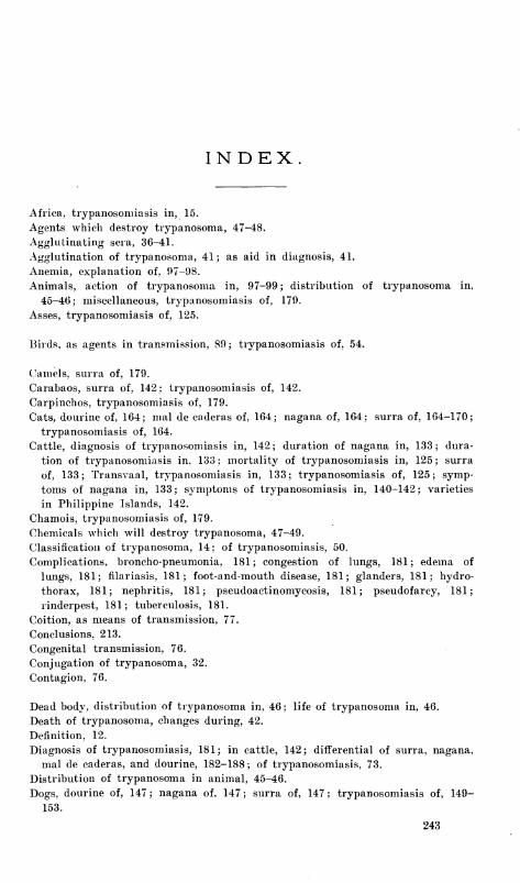
INDEX.
Africa, trypanosomiasis in, 15.
Agents which destroy trypanosoma, 47-48.
Agglutinating sera, 36-41.
Agglutination of trypanosoma, 41; as aid in diagnosis, 41.
Anemia, explanation of, 97-98.
Animals, action of trypanosoma in, 97-99; distribution of trypanosoma in,
45-46; miscellaneous, trypanosomiasis of, 179.
Asses, trypanosomiasis of, 125.
Birds, as agents in transmission, 89; trypanosomiasis of, 54.
Camels, surra of, 179.
Carabaos, surra of, 142 ; trypanosomiasis of, 142.
Carpinchos, trypanosomiasis of, 179.
Cats, dourine of, 164; mal de caderas of, 164; nagana of, 164; surra of, 164-170;
trypanosomiasis of, 164.
Cattle, diagnosis of trypanosomiasis in, 142; duration of nagana in, 133; dura-
tion of trypanosomiasis in, 133; mortality of trypanosomiasis in, 125; surra
of, 133; Transvaal, trypanosomiasis in, 133; trypanosomiasis of, 125; symp-
toms of nagana in, 133; symptoms of trypanosomiasis in, 140-142; varieties
in Philippine Islands, 142.
Chamois, trypanosomiasis of, 179.
Chemicals which will destroy trypanosoma, 47-49.
Classification of trypanosoma, 14; of trypanosomiasis, 50.
Complications, broncho-pneumonia, 181; congestion of lungs, 181; edema of
lungs, 181; filariasis, 181; foot-and-mouth disease, 181; glanders, 181; hydro-
thorax, 181; nephritis, 181; pseudoactinomycosis, 181; pseudofarcy, 181;
rinderpest, 181; tuberculosis, 181.
Coition, as means of transmission, 77.
Conclusions, 213.
Congenital transmission, 76.
Conjugation of trypanosoma, 32.
Contagion, 76.
Dead body, distribution of trypanosoma in, 46; life of trypanosoma in, 46.
Death of trypanosoma, changes during, 42.
Definition, 12.
Diagnosis of trypanosomiasis, 181; in cattle, 142; differential of surra, nagana,
mal de caderas, and dourine, 182-188; of trypanosomiasis, 73.
Distribution of trypanosoma in animal, 45-46.
Dogs, dourine of, 147 ; nagana of, 147 ; surra of, 147 ; trypanosomiasis of, 149-
153.
243
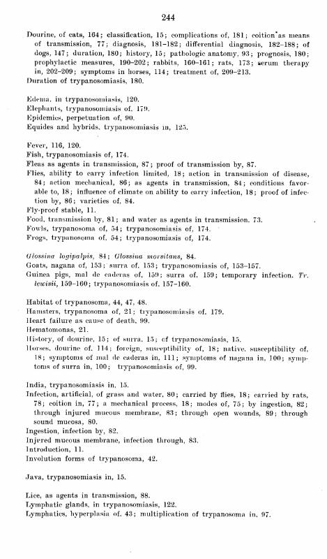
244
Dourine, of cats, 164; classification, 15; complications of, 181; coition'as meansof transmission, 77; diagnosis, 181-182; differential diagnosis, 182-188; of
dogs, 147; duration, 180; history, 15; pathologic anatomy, 93; prognosis, 180;
prophylactic measures, 190-202; rabbits, 160-161; rats, 173; serum therapy
in, 202-209; symptoms in horses, 114; treatment of, 209-213.
Duration of trypanosomiasis, 180.
Edema, in trypanosomiasis, 120.
Elephants, trypanosomiasis of. 179.
Epidemics, perpetuation of, 90.
Equides and hybrids, trypanosomiasis in, 125.
Fever, 116, 120.
Fish, trypanosomiasis of, 174.
Fleas as agents in transmission, 87; proof of transmission by, 87.
Flies, ability to carry infection limited, 18; action in transmission of disease,
84; action mechanical, 86; as agents in transmission, 84; conditions favor-
able to, 18; influence of climate on ability to carry infection, 18; proof of infec-
tion by, 86; varieties of, 84.
Fly-proof stable, 11.
Food, transmission by, 81; and water as agents in transmission. 73.
Fowls, trypanosoma of, 54; trypanosomiasis of, 174.
Frogs, trypanosoma of, 54; trypanosomiasis of, 174.
(jlossina logipalpis, 84; Glossina morsitans, 84.
Goats, nagana of, 153; surra of, 153; trypanosomiasis of, 153-157.
Guinea pigs, mal de caderas of, 159; surra of. 159; temporary infection. Tr.
lewisii, 159-160; trypanosomiasis of, 157-160.
Habitat of trypanosoma, 44, 47, 48.
Hamsters, trypanosoma of, 21; trypanosomiasis of, 179.
Heart failure as cause of death, 99.
Hematom onas, 21.
History, of dourine, 15; of surra. 15; of trypanosomiasis, 15.
Horses, dourine of, 114; foreign, susceptibility of, 18; native, susceptibility of,
18; symptoms of mal de caderas in, 111; symptoms of nagana in, 100; symp-toms of surra in, 100; trypanosomiasis of, 99.
India, trypanosomiasis in, 15.
Infection, artificial, of grass and water, 80; carried by flies, 18; carried by rats,
78; coition in, 77; a mechanical process, 18; modes of, 75; by ingestion, 82;
through injured mucous membrane, 83; through open wounds, 89; through
sound mucosa, 80.
Ingestion, infection by, 82.
Injured mucous membrane, infection through, 83.
Introduction, 11.
Involution forms of trypanosoma, 42.
Java, trypanosomiasis in, 15.
Lice, as agents in transmission, 88.
Lymphatic glands, in trypanosomiasis, 122.
Lymphatics, hyperplasia of, 43; multiplication of trypanosoma in, 97.
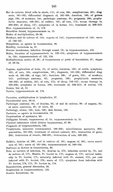
245
Mai de caderas, blood cells in stools, 113; of cats, 164; complications, 181; diag-
nosis, 181-182; differential diagnosis of, 182-188; duration, 180; of guinea
pigs, 159; of monkeys, 144; pathologic anatomy, 90; prognosis, 180; prophy-
lactic measures, 190-202; of rabbits, 161; of rats, 173; serum therapy in,
202-209; of sheep, 157; symptoms of, in horses, 111; treatment of, 209-213.
Man, trypanosomiasis of, 15, 178.
Mauritius Island, trypanosomiasis in, 15.
Modes of multiplication, 29-36.
Monkeys, mal de caderas of, 144; nagana of, 143; trypanosomiasis of, 142; surra
of, 144-147.
Mosquitoes, as agents in transmission, 88.
Motility, variations in, 27.
Mucous membranes, infection through sound, 79; in trypanosomiasis, 122.
Mules, duration of trypanosomiasis in, 122-124; symptoms of trypanosomiasis
in, 124; trypanosomiasis of, 122, 124.
Multiplication, modes of, 29 ; of trypanosoma at point of inoculation, 97 ; stages
of, 29-34.
Nagana, application of term, 13; of cattle, duration, 133; of cattle, symptoms,
133; of cats, 164; complications, 181; diagnosis, 181-182; differential diag-
nosis of, 182-188; of dogs, 147; duration, 180; of goats, 153; of monkeys,
143; pathologic anatomy, 92; prognosis, 180; prophylactic measures,
190-202; of rabbits, 161; of rats, 172; of sheep, 156-157; serum therapy in,
202-209; symptoms in horses, 100; treatment of, 209-213; of wolves, 179.
Names, list of, 12.
Nutria, trypanosomiasis of, 179.
Parasites, multiplication in lymphatics, 97.
Parasiticidal sera, 36-41.
Pathologic anatomy, 92; of dourine, 93; of mal de caderas, 90; of nagana, 92;
of surra americain, 92; of surra, 93.
Pathology, edema, 120; hair, 120; skin lesions, 120.
Pastures, as agents in transmission, 78.
Perpetuation of epidemics, 90.
Philippine Islands, trypanosoma of, 14; trypanosomiasis in, 15.
Physical substances which destroy trypanosoma, 47-48.
Prognosis of trypanosomiasis, 180.
Prophylaxis, measures recommended, 190-202; miscellaneous measures, 202;
quarantine, 191-192; treatment of contact animals, 201; destruction of game,
200; destruction of insects, 200-201; destruction of rats, 199-200.
Rabbits, dourine of, 160; mal de caderas of, 161; nagana of, 161; surra ameri-
cain of, 161; surra of, 161-164; trypanosomiasis of, 160-164.
Railways as factors in transmission, 89.
Rats, as carriers of infection, 78; dourine in, 173; infection carried by, 78; mal
de caderas of, 173; Manila, Tr. lewisii in, 170; nagana of, 172; natural immu-
nity to Tr. lewisii, 171; naturally infected with Tr. evansii, 173; per cent
infected with Tr. leicisii, 170; surra of, 172; symptoms from infection with
Tr. lewisii, 170, 172; Tr. lewissi in, 170.
Reproduction, general discussion, 29.
Respiration in trypanosomiasis, 122.
Rosette formations, 32.
246
Sera, parasiticidal, 36-38; agglutinating. 40-41.
Serum therapy, 202-209.
Sheep, mal de caderas of, 157; nagana of, 156, 157; surra of, 156, 157; trypano-
somiasis of, 156, 157.
Skin wounds, infection through, 89.
Sleeping sickness, caused by trypanosoma, 178.
Sound mucosa, infection through, 80.
South America, trypanosomiasis in, 15.
Splenectomy, in trypanosomiasis, 43.
Btomoxys calcitrans, 84, 85.
Summary, 213.
Sunlight, trypanosoma destroyed by, 18.
Surra americain, pathologic anatomy, 92; of monkeys, 143; of rabbits, 161.
Surra, application of term, 13; of camels, 179; of cattle, 133; duration in cattle,
133; of cattle in Philippine Islands, 133; of cats, 164, 168, 170; complications,
181; diagnosis of, 181, 182; differential diagnosis of, 182-188; of dogs, 147;
duration of, 180; of goats, 153; of guinea pigs, 159-160; history of, 15; of
monkeys, 144-147; only allowable vernacular name, 13; pathologic anatomy
of, 93; prognosis of, 180; prophylactic measures in, 190-202; of rabbits, 161-
164; of rats, 172; serum therapy in, 202-209; susceptibility and natural im-
munity, 189-190; symptoms in horses, 100; treatment of, 209-213.
Susceptibility, age in, 19; color of animals in, 19; of foreign horses, 18; of native
horses, 18; and natural immunity, 189-190; relative, of foreign and native
horses in Philippine Islands, 18.
Symptomology, 95.
Symptoms, catarrhal, 120; early, 95; gastro-intestinal, 122.
Ticks, as agents in transmission, 89.
Transmission, birds as agents in, 89; coition in, 77; congenital, 76; contagion in,
76; by feeding, 81; by fleas, 87; by fleas, proof of, 87; by flies, 83; food,
water, and pastures in, 78; and infection, 75; lice as agents in, 88; mosquitoes
as agents in, 89; railways as agents, 89; by rat-soiled grain, 78; ticks as
agents in, 89.
Trypanoplasma borrelii, 22, 51, 58; diagnosis of, 58; habitat of, 58; patho-
genesis of, 58.^
Trypanoplasma danilewskyi, 57; synonyms of, 51.
Trypanoplasma of Kunster, 58.
Trypanoplasma of LabbS, 58.
Trypanosoma, action in animal, 97-99; agglutination of, 36; agglutination as an
aid in diagnosis, 41; in birds, 22; centrosome of, 27; centrosome, variation in
location of, 27; changes occurring in dying, 42; chemicals which destroy, 47-
49; in circulation, intermittent disappearance of, 97; classification of, 14, 50;
classification of in Philippine Islands, 14; conjugation of, 27, 29, 32; in dead
body, 46; destroyed by sunlight, 18; diagnosis, differential, of, 73; discovered
in fish, 19; discovered in frogs, 19; discovered in hamsters, 21; distribution in
body, 43; distribution in organs, 43-44; even distribution in body juices, 45,
46; in fish, 22; rlagellum of, 27; general description, 27; general morphology,
27; history, 19; identity of different parasites, 14; involution forms of, 41;
conditions producing involution forms, 42; types of, 42; life cycle of trypano-
soma, 24 ; life cycle acted out in blood, 25 ; life cycle unknown, 25 ; life in dead
body, 46; of man, 22; of man, tentative classification, 70; modes of multipli-
cation, 29; more numerous in certain organs, 43, 44; motility, 27; multiplica-
tion, mode of, 29-36; nucleus of, 27; in a nomatode, 21; outside animal body.
247
46; phase of life cycle in fly, 25; posterior end contractile, 27; protoplasm of,
27 ; rosette formations of, 32 ; in salt-water fish, 22 ; selective organs, 99 ; sporu-
lation of, 20; stages of, multiplication of, 29-34; stain for (Woolley), 23;
staining methods, 23; substances causing agglutination in, 36, 38-41; substan-
ces which destroy outside the body, 47, 48; substances favoring life outside the
body, 46-48; technique for study, 23; undulating membrane, 27; absence of, in
young forms, 27; variations in motility, 27.
Trypanosoma avium, 54; habitat, 54; pathogenesis, 54; specific diagnosis, 54;
synonyms, 50; Tr. avium danilewskyi, 50.
Trypanosoma balbianii, 51, 54; diagnosis, 54; discovery of, 21; habitat, 56; not
a trypanosoma, 21; pathogenesis, 56; synonyms, 51.
Trypanosoma brucei, 51, 66; diagnosis, 66; habitat, 67; pathogenesis, 68; syno-
nyms, 51.
Trypanosoma carassii, 51, 56; diagnosis, 56; habitat, 56; pathogenesis, 56; syn-
onyms, 51.
Trypanosoma colitis, 51, 56; diagnosis of, 56; habitat of, 56; pathogenesis of,
56; synonyms of, 51.
Trypanosoma of dourine, 22.
Trypansoma eberthii, 51, 54; diagnosis, 54; discovery of, 20; habitat, 54; patho-
genesis, 54; synonyms, 51.
Trypanosoma elmassianii, 52, 70.
Trypanosoma equinum, 22, 52, 70; diagnosis, 70; habitat, 72; pathogenesis, 72;
synonyms, 52.
Trypanosoma equiperdum, 22, 52, 68; diagnosis, 68; habitat, 68; pathogenesis,
68.
Trypanosoma evansii, 51, 63; centrosome, 64; diagnosis, 63, 64; discovery of, 21;
habitat, 66; history of discovery in Philippine Islands, 23; nucleus of, 64;
pathogenesis, 66; of Philippine Islands, 14; protoplasm, 64; of rats, 62, 173;
synonyms, 51.
Trypanosoma fordii, 52.
Trypanosoma gambiense, 52.
Trypanosoma Gruby, animals found in, 20; relation of number of, to age of ani-
mals, 20; synonyms, 20.
Trypanosoma lewisii, 51, 58, 170; action of cold on, 171; cultivation of, 25; defi-
nition, 61-62; differential diagnosis of, 62; discovery of, 20; habitat, 60; in-
fection of guinea pigs with, 159, 160; motility, 62; multiplication, 59, 60;
pathogenesis, 60, 170, 171; in rats, 170-173; synonyms, 51; variations in size,
62.
Trypanosoma of mal de caderas, 22; history, 22.
Trypanosoma nepveui, 52, 68; definition, 68; synonyms, 52.
Trypanosoma remaki, 22, 56; diagnosis, 56; habitat, 56; pathogenesis, 56.
Trypanosoma rotatorium, 52; habitat, 53; mode of transmission, 54; morphol-
ogy, 52; pathogenesis, 54.
Trypanosoma rotatorium Mayer, 50.
Trypanosoma rougetti, 22, 52, 68; diagnosis, 68; habitat, 68; pathogenesis, 68;
synonyms, 52.
Trypanosoma solae, 22, 51, 56; diagnosis, 56; habitat, 58; pathogenesis, 68*
Trypanosoma theilerii, 52, 72; diagnosis, 72; discovery, 23; habitat, 72; patho-
genesis, 72.
Trypanosoma, third species in Manila rats, 62, 63. #
Trypanosoma transvaaHensv, 52, 72; diagnosis, 72; discovery", 23; habitat, 73.
Trypanosoma ugandieriseii, 52.
248
Trypanosomiasis, in Africa, 15; of antelopes, 179; application of term, 13; of
asses, 125; of babale, 179; of bue-buc, 179; of buffaloes, 179; of camels, 179;
of carabaos, 142; of carpinchos, 179; catarrhal symptoms in, 120; of cattle,
125; of cattle, diagnosis, 142; of cattle, duration, 133; of cattle, emaciation,
142; of cattle, mortality, 125; of cattle in Transvaal, 133; of cattle, urticarial
eruption, 133; of cats, 164; of chamois, 179; climatic conditions in, 18; com-
plications of, 181; course of, 180; diagnosis of, 181; of dogs, 147, 149-153; of
dromedaries, 179; duration of, 180; early symptoms of, 95; edema in, 120; of
elephants, 179; of equides and hybrids, 125; fever' in, 116, 120; of fish, 174;
of frogs, 174; of fowls, 174; gastro-intestinal symptoms in, 122; general symp-tomology of, 95; geographic distribution, maps of, 18; geographical distribu-4
tion, table of, 16; of goats, 153-157; of guinea pigs, 157-160; hair in, 120;
of hamsters, 179; of hare, 179; of hedgehog, 179; history of, 15; history in
Philippine Islands, 16; of horses, 99; of hyenas, 179; hyperplasia of lym-phatics after splenectomy, 45; identity of, 14; incubation period of, 95, 97, 114-
116; in India, 15; in Java, 15; of koodoo, 179; of lapians, 179; lymphatic
glands in, 122; of man, 15, 175; of man, discovery of parasite, 175; of man, in
Philippine Islands, 178; of man, as sleeping sickness, 178; of man, symptoms,176-178; and malarial fever in man, 21; in Mauritius Island, 15; of mice andrats, 170; of miscellaneous animals, 179; of monkeys, 142; mucous membrane in,
122; of mules, 122; of mules, duration in, 122, 124; of mules, skin lesions in,
124; of mules, symptoms of, 124; of mules, trypanosoma in blood, 124; of
mullets, 179; of nutria, 179; in Philippine Islands, 15; prognosis of, 180;
prophylaxis in, 190-202; of rabbits, 160-164; of racque, 179; of rats and mice,
170; respiration in, 122; of sheep, 156, 157; skin lesions in, 120; in SouthAmerica, 15; susceptibility and natural immunity in, 189, 190; susceptibility,
sex in, 19; toxins in, 98, 99; treatment of, 209-213; treatment, prophylactic,
of, 190-202; of wild animals, 179; of wolves, 179.
Tsetse fly, 84.
Vernacular names, list of, 13.
Water, and grass, artificial infection of, 80; and food, as agents in transmis-
sion, 78.
Wild animals, trypanosomiasis of, 179.
Wolves, trypanosomiasis of, 179. .
o•
*V
"$
M W


To renew Hie charge, book musl be brought to the desk.
DO NOT RETURN BOOKS ON SUNDAY
DATE DUE
<MtQ -? * 'OS-
Worm 707»a 4*0 SM «T4t4

UNIVERSITY OF MICHIGAN
milium3 9015 02156 1256
A«-
/$ i
P*>
fcrtfc