Treatment of Peripheral Nerve Injury with Tension Stitch Method
7
JOURNAL OF CONTEMPORARY MEDICINE Journal of Contemporary Medicine Original Article / Orijinal Araştırma DOI:10.16899/jcm.1039032 J Contemp Med 2022;12(3):437-443 Corresponding (İletişim): Ali Sakinsel, Department of Plastic Surgery, Private LeonArt Polyclinic, Istanbul, Turkey E-mail (E-posta): [email protected] Received (Geliş Tarihi): 07.01.2022 Accepted (Kabul Tarihi): 05.05.2022 Treatment of Peripheral Nerve Injury with Tension Stitch Method: an Experimental Study Gergi Dikişi Yöntemi ile Periferik Sinir Yaralanmalarının Tedavisi: Deneysel Bir Çalışma Objective: The aim of our study is to examine the tension stitch method we use to prevent unwanted tissue deficiency between the cut nerve endings in rats that will be kept waiting for secondary neurorrhaphy. Material and Method: 30 male Wistar Albino rats were randomly divided into three groups. The right sciatic nerve was released proximally from the sciatic nerve 1/3 bifurcation area in the first and second groups and anastomosed with the tibial nerve in the third group. After 4 weeks, the region was reopened, unhealthy nerve endings were cut under the microscope, and secondary neurorrhaphy was performed end-to-end. Results: In the third experimental group, it was observed that there was no change in the position of the tension stitches placed on the distal and proximal ends of the sciatic nerve, and the nerve endings adhered to the area where they were positioned by suture. At the eighth week, it was observed that the rats that could not use their right lower extremities in the preoperative and early postoperative periods used their extremities more actively. At the twelfth week, it was observed that the rats in all groups had complete recovery of trophic disturbances and the animals started to walk better visually. Conclusion: In our study, the electrophysiological and histopathological data obtained at the eighth week and obtained at the twelfth week were significantly better in the tension-stitched group compared to the other groups, indicating that the best early and late nerve healing was in this group. Keywords: Neurorrhaphy, tension stitch, nerve healing, end-to-end Öz Abstract Ali Sakinsel1, Mert Sizmaz2, Lutfu Bas3 Amaç: Çalışmamızın amacı sekonder nörorafi için bekletilecek ratlarda kesilen sinir uçları arasındaki istenmeyen doku eksikliğini önlemek için kullandığımız gergi dikiş yöntemini incelemektir. Gereç ve Yöntem: 30 adet erkek Wistar Albino rat randomize olarak üç gruba ayrıldı. Sağ siyatik sinir, birinci ve ikinci grupta siyatik sinirin 1/3 bifurkasyon alanından proksimalde serbest bırakılırken, üçüncü grupta tibial sinir ile anastomoz yapıldı. Dört hafta sonra bölge yeniden açıldı, mikroskop altında sağlıksız sinir uçları kesildi ve uçtan uca sekonder nörorafi yapıldı. Bulgular: Üçüncü deney grubunda siyatik sinirin distal ve proksimal uçlarına konulan gergi dikişlerinin pozisyonunda herhangi bir değişiklik olmadığı, sinir uçlarının sütür ile yerleştirildiği bölgeye yapıştığı gözlendi. Sekizinci haftada ameliyat öncesi ve ameliyat sonrası erken dönemde sağ alt ekstremitesini kullanamayan ratların ekstremitelerini daha aktif kullandıkları görüldü. On ikinci haftada tüm gruplardaki ratlarda trofik bozuklukların tamamen düzeldiği ve hayvanların görsel olarak daha iyi yürümeye başladıkları görüldü. Sonuç: Çalışmamızda sekizinci ve on ikinci haftada elde edilen elektrofizyolojik ve histopatolojik veriler gergi dikişli grupta diğer gruplara göre anlamlı olarak daha iyiydi, bu da en iyi erken ve geç sinir iyileşmesinin bu grupta olduğunu göstermektedir. Anahtar Kelimeler: Nörorafi, gergi dikişi, sinir iyileşmesi, uçtan uca 1Department of Plastic Surgery, Private LeonArt Polyclinic, Istanbul, Turkey 2Department of Plastic, Reconstructive and Aesthetic Surgery, Kars Harakani State Hospital, Kars, Turkey 3Department of Plastic Surgery, Private Clinic, Istanbul, Turkey
-
Upload
khangminh22 -
Category
Documents
-
view
0 -
download
0
Transcript of Treatment of Peripheral Nerve Injury with Tension Stitch Method
JOURNAL OF
CONTEMPORARY MEDICINEJournal ofContemporary Medicine
Original Article / Orijinal Araştırma
DOI:10.16899/jcm.1039032J Contemp Med 2022;12(3):437-443
Corresponding (İletişim): Ali Sakinsel, Department of Plastic Surgery, Private LeonArt Polyclinic, Istanbul, TurkeyE-mail (E-posta): [email protected] (Geliş Tarihi): 07.01.2022 Accepted (Kabul Tarihi): 05.05.2022
Treatment of Peripheral Nerve Injury with Tension Stitch Method: an Experimental Study
Gergi Dikişi Yöntemi ile Periferik Sinir Yaralanmalarının Tedavisi: Deneysel Bir Çalışma
Objective: The aim of our study is to examine the tension stitch method we use to prevent unwanted tissue deficiency between the cut nerve endings in rats that will be kept waiting for secondary neurorrhaphy.
Material and Method: 30 male Wistar Albino rats were randomly divided into three groups. The right sciatic nerve was released proximally from the sciatic nerve 1/3 bifurcation area in the first and second groups and anastomosed with the tibial nerve in the third group. After 4 weeks, the region was reopened, unhealthy nerve endings were cut under the microscope, and secondary neurorrhaphy was performed end-to-end.
Results: In the third experimental group, it was observed that there was no change in the position of the tension stitches placed on the distal and proximal ends of the sciatic nerve, and the nerve endings adhered to the area where they were positioned by suture. At the eighth week, it was observed that the rats that could not use their right lower extremities in the preoperative and early postoperative periods used their extremities more actively. At the twelfth week, it was observed that the rats in all groups had complete recovery of trophic disturbances and the animals started to walk better visually.
Conclusion: In our study, the electrophysiological and histopathological data obtained at the eighth week and obtained at the twelfth week were significantly better in the tension-stitched group compared to the other groups, indicating that the best early and late nerve healing was in this group.
Keywords: Neurorrhaphy, tension stitch, nerve healing, end-to-end
ÖzAbstract
Ali Sakinsel1, Mert Sizmaz2, Lutfu Bas3
Amaç: Çalışmamızın amacı sekonder nörorafi için bekletilecek ratlarda
kesilen sinir uçları arasındaki istenmeyen doku eksikliğini önlemek için
kullandığımız gergi dikiş yöntemini incelemektir.
Gereç ve Yöntem: 30 adet erkek Wistar Albino rat randomize olarak üç
gruba ayrıldı. Sağ siyatik sinir, birinci ve ikinci grupta siyatik sinirin 1/3
bifurkasyon alanından proksimalde serbest bırakılırken, üçüncü grupta
tibial sinir ile anastomoz yapıldı. Dört hafta sonra bölge yeniden açıldı,
mikroskop altında sağlıksız sinir uçları kesildi ve uçtan uca sekonder
nörorafi yapıldı.
Bulgular: Üçüncü deney grubunda siyatik sinirin distal ve proksimal
uçlarına konulan gergi dikişlerinin pozisyonunda herhangi bir
değişiklik olmadığı, sinir uçlarının sütür ile yerleştirildiği bölgeye
yapıştığı gözlendi. Sekizinci haftada ameliyat öncesi ve ameliyat
sonrası erken dönemde sağ alt ekstremitesini kullanamayan ratların
ekstremitelerini daha aktif kullandıkları görüldü. On ikinci haftada
tüm gruplardaki ratlarda trofik bozuklukların tamamen düzeldiği ve
hayvanların görsel olarak daha iyi yürümeye başladıkları görüldü.
Sonuç: Çalışmamızda sekizinci ve on ikinci haftada elde edilen
elektrofizyolojik ve histopatolojik veriler gergi dikişli grupta diğer
gruplara göre anlamlı olarak daha iyiydi, bu da en iyi erken ve geç sinir
iyileşmesinin bu grupta olduğunu göstermektedir.
Anahtar Kelimeler: Nörorafi, gergi dikişi, sinir iyileşmesi, uçtan uca
1Department of Plastic Surgery, Private LeonArt Polyclinic, Istanbul, Turkey2Department of Plastic, Reconstructive and Aesthetic Surgery, Kars Harakani State Hospital, Kars, Turkey
3Department of Plastic Surgery, Private Clinic, Istanbul, Turkey
438Ali Sakinsel, Peripheral Nerve Injury and Tension Stitch
INTRODUCTIONTraumatic injury, congenital anomalies and tumor destruction are among the situations that can cause peripheral nerve injury with serious consequences on quality of life. Since 1964, it has been clearly demonstrated that the outcome of epineural anastomosis under magnification is better than operative corrections,[1,2] which made microsurgical end-to-end (ETE) neurorrhaphy the gold standard for nerve repair;[3] however, in cases of extensive or long-standing injuries, ETE nerve repair is nigh impossible. Other repair methods, such as nerve grafting, nerve transfer and direct neurotization, have been used as alternative procedures.[4,5]
The mainstay of end-to-end nerve coaptation is to repair an injured nerve by borrowing axons from an intact donor nerve without damaging its function. Although it has been reported for a long time that end-to-end neurorrhaphy attracts axonal sprouts without damaging the donor nerve function,[6-11] its functional results remain unclear. Despite an increasing number of studies, quantitative research assessing this topic is limited and evidence is scarce concerning the early and late impacts on donor nerves.[12] As the potential role of end-to-end nerve coaptation remains controversial, various authors have discussed the theoretical foundations for applying the method in the clinical setting.[13-15]
The gold standard treatment for peripheral nerve repair is autologous nerve grafting with the aid of a surgical microscope. Although this technique procures positive outcomes, there are some disadvantageous factors associated with the procedure, including limitation of the caliber, length and area of the donor nerves, and the likelihood of secondary morbidity.[16-18] In this sense, the tubularization technique has been shown to be an appropriate and alternative method in clinical practice, which is a method performed by fixing and directing a tubular segment (of biological or synthetic origin) between the stumps of the injured nerve.[19,20]
The aim of our study was to examine the tension stitch method which we use to prevent unwanted tissue deficiency between the cut nerve endings, by utilizing a rat model with delayed application of secondary neurorrhaphy.
MATERIAL AND METHODThirty male young adult Wistar Albino rats weighing 223-300 g were randomly divided into three experimental groups. The animals were allowed ad libitum access to standard rodent chow and tap water. The living conditions were: temperature was kept at 22±2 °C, humidity was 50±10 %, and the day-night cycle was automated with lights on from 06:00 to 18:00. There were no cases of wound infection during the experiment. In this research, the data before 2020 was used and the research was concluded before 2020. This study was prepared in accordance with the 2013 Brazil revision of the Helsinki Declaration and guidelines for Good Clinical Practice. Ethics committee approval was not obtained because there was no ethical committee approval obligation at the time of the study (1998).
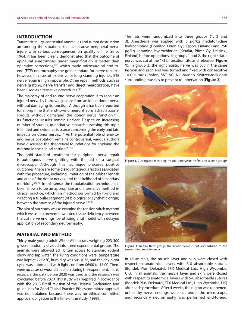
The rats were randomized into three groups (1, 2 and 3). Anesthesia was applied with 5 μg/kg medetomidine hydrochloride (Domitor, Orion Oyj, Espoo, Finland) and 750 μg/kg ketamine hydrochloride (Ketalar, Pfizer Oy, Helsinki, Finland) before operations. In groups 1 and 2, the right sciatic nerve was cut at the 1/3 bifurcation site and released (Figure 1). In group 3, the right sciatic nerve was cut in the same fashion and each end was turned and fixed with consecutive 10-0 sutures (Nylon, S&T AG, Neuhausen, Switzerland) onto surrounding muscles to prevent re-innervation (Figure 2).
In all animals, the muscle layer and skin were closed with respect to anatomical layers with 5-0 absorbable sutures (Bondek Plus, Deknatel, TFX Medical Ltd., High Wycombe, UK). In all animals, the muscle layer and skin were closed with respect to anatomical layers with 5-0 absorbable sutures (Bondek Plus, Deknatel, TFX Medical Ltd., High Wycombe, UK) after each procedure. After 4 weeks, the region was reopened, unhealthy nerve endings were cut under the microscope, and secondary neurorrhaphy was performed end-to-end.
Figure 1. Cutting and releasing the sciatic nerve in the first and second groups
Figure 2. In the third group, the sciatic nerve is cut and sutured to the surrounding muscle fascia.
439 Journal of Contemporary Medicine
In group 1, secondary neurorrhaphy was applied under the present tension without grafting. In group 2, a Wild M3Z microscope (Wild Leitz Ltd., Heerbrugg, Switzerland) was used for secondary neurorrhaphy without grafting which was performed with grafting. In group 3, primary neurorrhaphy was applied although both nerve edges appeared to be healthy, they were renewed minimally. At the eighth and twelfth weeks after initial intervention, the right sciatic nerves of the rats were evaluated electrophysiologically and histopathologically in groups of five.Perioperatively, animals were injected subcutaneously with 5 ml sodium chloride 9 mg/ml (Fresenius Kabi AB, Uppsala, Sverige) to maintain fluid balance. The animals were given a subcutaneous injection of 5 mg / kg carprofen (Rimadyl, Vericode Ltd., Dundee, UK) as an analgesic drug for three postoperative days.Finally, animals were sacrificed by an overdose of ketamine hydrochloride (Dopalen®) and Xylazine Hydrochloride (Antisedan®) at postoperative week 12, and tissues were obtained, including the soleus (SO) and extensor digitorum longus (EDL) muscles.
Histological and Electrophysiological ExaminationThe SO and EDL muscle samples were reduced to cylindrical pieces after resection and the Optimal Critical Temperature Tissue-Tek® gel (OCT, Sakura Finetek, Torrance, USA) was used for the subsequent snap-freezing via immersion in liquid nitrogen. All frozen samples were stored at -80 °C until analysis. Histological sections were obtained at -20 °C in a Leica cryostat - Model CM 1850 and stained with Hematoxylin and Eosin (HE) and Masson's trichrome for morphometric analysis. Bioscience 10550 Kymograph, stimulator, Polygraph, Pentium 75 computer, and electrodes were used for electrophysiological examination.
Morphometric AnalysisCross sectional area and minimum diameter: Images were taken with an Olympus BX 50, a photomicroscope connected to a computer, and three different areas of each sample were photographed with a 20X objective. For morphometric analysis, 220 muscle fibers of each animal were manually measured via the Image Pro-Plus® 6.2 program (Media Cybernetics, Bethesda, MD, USA); in addition, area and minimum diameter were measured.Connective tissue density: Using Masson's trichrome staining, a photograph of the transverse cross-sectional area of the entire muscle was taken using a 2X lens to measure total area, and then a 20X lens was used to obtain an image for the ProPlus® 6.2 software, in order for it to recognize Aniline Blue. After the measurements, the percentage of connective tissue in the muscle was calculated.This study was produced from Dr. Ali Sakinsel's Specialization Thesis in Plastic and Reconstructive Surgery numbered 70646, which was presented and approved at Istanbul Şişli Hamidiye Etfal Training and Research Hospital / Department of Plastic
and Reconstructive Surgery in 1998 (thesis title: Evaluation of neurorrhaphy results with and without tension suture in secondary nerve repairs)
Statistical AnalysisFor statistical analysis of muscle weight, muscle fiber morphometry, connective tissue morphometry and functional index, groups were compared with analysis of variance (ANOVA), and if this analysis detected a significant difference, the Tukey test was used for pairwise comparisons. A P-value of ≤0.05 was considered to demonstrate statistical significance.
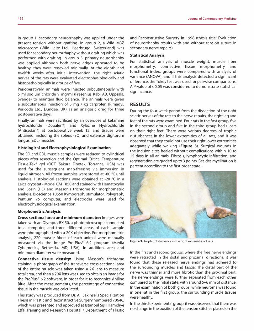
RESULTSDuring the four-week period from the dissection of the right sciatic nerves of the rats to the nerve repairs, the right leg and feet of the rats were examined. Four rats in the first group, five in the second group and five in the third group had ulcers on their right feet. There were various degrees of trophic disturbances in the lower extremities of all rats, and it was observed that they could not use their right lower extremities adequately while walking (Figure 3). Surgical wounds in the incision sites healed without complications within 10 to 15 days in all animals. Fibrosis, lymphocytic infiltration, and regeneration are graded up to 3 points. Besides myelination is percent according to the first-order state.
In the first and second groups, where the free nerve endings were retracted in the distal and proximal directions, it was found that these released nerve endings had adhered to the surrounding muscles and fascia. The distal part of the nerve was thinner and more fibrotic than the proximal part. The nerve endings were further separated from each other compared to the initial state, with around 5–6 mm of distance. In the examination of both groups, while neuroma was found in one rat in the first group, the surrounding muscle tissues were healthy.In the third experimental group, it was observed that there was no change in the position of the tension stitches placed on the
Figure 3. Trophic disturbance in the right extremities of rats.
440Ali Sakinsel, Peripheral Nerve Injury and Tension Stitch
distal and proximal ends of the sciatic nerve, and the nerve endings adhered to the area where they were positioned by suture. In this group, the distal nerve section was thinner and more fibrotic than proximal. In this group, the distance between nerve endings remained within acceptable levels, with around 1-2 mm of gap. Neuroma was observed in one rat in this group.
Findings in Eighth and Twelfth Weeks After RepairIn the eighth week, in the first group, it was found that the ulcers which had developed in four animals had healed in three of them. In the second group, all of the ulcers (five animals) had healed. In the third group, leg ulcers present in five animals had healed in four, while the remaining rat suffered from a loss of about 1/3 of the distal tip of the right foot. It was observed that the rats, which could not use their right lower extremities in the preoperative and early postoperative periods, were able to use their extremities more actively in the eighth week. By the twelfth week, it was observed that all rats had complete recovery of trophic disturbances and their gait had considerably improved. Average histopathological evaluation results of the groups at the eighth and twelfth weeks are shown in Table 1.
Table 1. Average histopathological evaluation results of the groups at the eighth and twelfth weeks.
Groups Fibrosis Myelination Lymphocytic Infiltration Regeneration
Group 1 3+ \3+ 50%\56% 3+ \3+ 1+ \1+Group 2 2+ \3+ 67%\74% 2+ \2+ 2+ \2+Group 3 1+ \1+ 85%\95% 1+ \1+ 3+ \3+
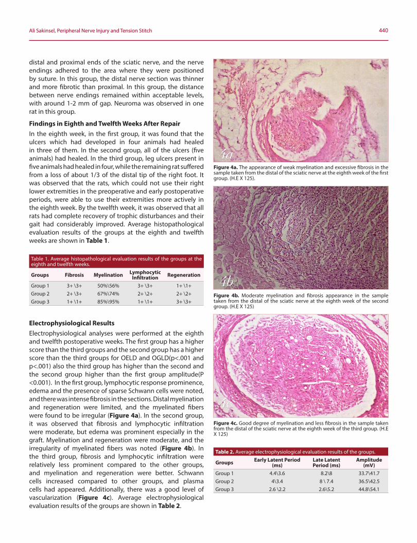
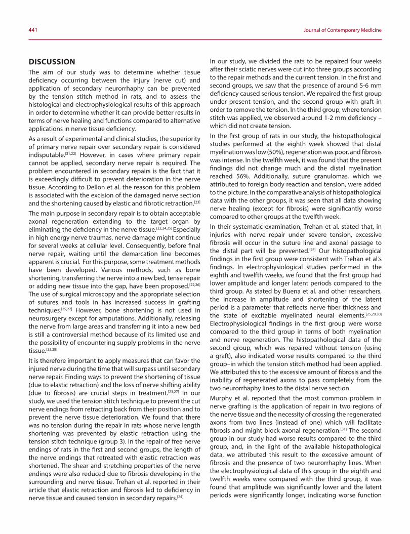
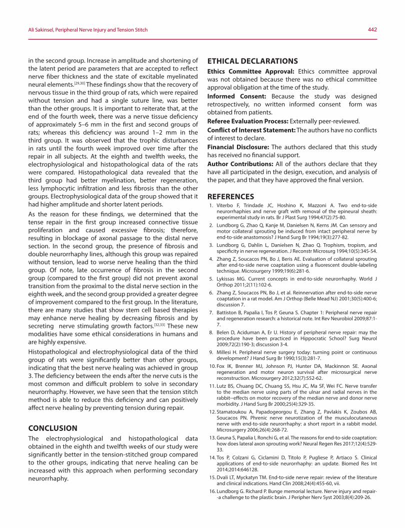
Electrophysiological ResultsElectrophysiological analyses were performed at the eighth and twelfth postoperative weeks. The first group has a higher score than the third groups and the second group has a higher score than the third groups for OELD and OGLD(p<.001 and p<.001) also the third group has higher than the second and the second group higher than the first group amplitude(P <0.001). In the first group, lymphocytic response prominence, edema and the presence of sparse Schwann cells were noted, and there was intense fibrosis in the sections. Distal myelination and regeneration were limited, and the myelinated fibers were found to be irregular (Figure 4a). In the second group, it was observed that fibrosis and lymphocytic infiltration were moderate, but edema was prominent especially in the graft. Myelination and regeneration were moderate, and the irregularity of myelinated fibers was noted (Figure 4b). In the third group, fibrosis and lymphocytic infiltration were relatively less prominent compared to the other groups, and myelination and regeneration were better. Schwann cells increased compared to other groups, and plasma cells had appeared. Additionally, there was a good level of vascularization (Figure 4c). Average electrophysiological evaluation results of the groups are shown in Table 2.
Table 2. Average electrophysiological evaluation results of the groups.
Groups Early Latent Period (ms)
Late Latent Period (ms)
Amplitude(mV)
Group 1 4.4\3.6 8.2\8 33.7\41.7Group 2 4\3.4 8 \ 7.4 36.5\42.5Group 3 2.6 \2.2 2.6\5.2 44.8\54.1
Figure 4a. The appearance of weak myelination and excessive fibrosis in the sample taken from the distal of the sciatic nerve at the eighth week of the first group. (H.E X 125).
Figure 4b. Moderate myelination and fibrosis appearance in the sample taken from the distal of the sciatic nerve at the eighth week of the second group. (H.E X 125)
Figure 4c. Good degree of myelination and less fibrosis in the sample taken from the distal of the sciatic nerve at the eighth week of the third group. (H.E X 125)
441 Journal of Contemporary Medicine
DISCUSSIONThe aim of our study was to determine whether tissue deficiency occurring between the injury (nerve cut) and application of secondary neurorrhaphy can be prevented by the tension stitch method in rats, and to assess the histological and electrophysiological results of this approach in order to determine whether it can provide better results in terms of nerve healing and functions compared to alternative applications in nerve tissue deficiency.As a result of experimental and clinical studies, the superiority of primary nerve repair over secondary repair is considered indisputable.[21,22] However, in cases where primary repair cannot be applied, secondary nerve repair is required. The problem encountered in secondary repairs is the fact that it is exceedingly difficult to prevent deterioration in the nerve tissue. According to Dellon et al. the reason for this problem is associated with the excision of the damaged nerve section and the shortening caused by elastic and fibrotic retraction.[23]
The main purpose in secondary repair is to obtain acceptable axonal regeneration extending to the target organ by eliminating the deficiency in the nerve tissue.[22,24,25] Especially in high energy nerve traumas, nerve damage might continue for several weeks at cellular level. Consequently, before final nerve repair, waiting until the demarcation line becomes apparent is crucial. For this purpose, some treatment methods have been developed. Various methods, such as bone shortening, transferring the nerve into a new bed, tense repair or adding new tissue into the gap, have been proposed.[22,26] The use of surgical microscopy and the appropriate selection of sutures and tools in has increased success in grafting techniques.[25,27] However, bone shortening is not used in neurosurgery except for amputations. Additionally, releasing the nerve from large areas and transferring it into a new bed is still a controversial method because of its limited use and the possibility of encountering supply problems in the nerve tissue.[23,28]
It is therefore important to apply measures that can favor the injured nerve during the time that will surpass until secondary nerve repair. Finding ways to prevent the shortening of tissue (due to elastic retraction) and the loss of nerve shifting ability (due to fibrosis) are crucial steps in treatment.[23,27] In our study, we used the tension stitch technique to prevent the cut nerve endings from retracting back from their position and to prevent the nerve tissue deterioration. We found that there was no tension during the repair in rats whose nerve length shortening was prevented by elastic retraction using the tension stitch technique (group 3). In the repair of free nerve endings of rats in the first and second groups, the length of the nerve endings that retreated with elastic retraction was shortened. The shear and stretching properties of the nerve endings were also reduced due to fibrosis developing in the surrounding and nerve tissue. Trehan et al. reported in their article that elastic retraction and fibrosis led to deficiency in nerve tissue and caused tension in secondary repairs.[24]
In our study, we divided the rats to be repaired four weeks after their sciatic nerves were cut into three groups according to the repair methods and the current tension. In the first and second groups, we saw that the presence of around 5-6 mm deficiency caused serious tension. We repaired the first group under present tension, and the second group with graft in order to remove the tension. In the third group, where tension stitch was applied, we observed around 1-2 mm deficiency –which did not create tension.In the first group of rats in our study, the histopathological studies performed at the eighth week showed that distal myelination was low (50%), regeneration was poor, and fibrosis was intense. In the twelfth week, it was found that the present findings did not change much and the distal myelination reached 56%. Additionally, suture granulomas, which we attributed to foreign body reaction and tension, were added to the picture. In the comparative analysis of histopathological data with the other groups, it was seen that all data showing nerve healing (except for fibrosis) were significantly worse compared to other groups at the twelfth week.In their systematic examination, Trehan et al. stated that, in injuries with nerve repair under severe tension, excessive fibrosis will occur in the suture line and axonal passage to the distal part will be prevented.[24] Our histopathological findings in the first group were consistent with Trehan et al.’s findings. In electrophysiological studies performed in the eighth and twelfth weeks, we found that the first group had lower amplitude and longer latent periods compared to the third group. As stated by Buena et al. and other researchers, the increase in amplitude and shortening of the latent period is a parameter that reflects nerve fiber thickness and the state of excitable myelinated neural elements.[25,29,30] Electrophysiological findings in the first group were worse compared to the third group in terms of both myelination and nerve regeneration. The histopathological data of the second group, which was repaired without tension (using a graft), also indicated worse results compared to the third group–in which the tension stitch method had been applied. We attributed this to the excessive amount of fibrosis and the inability of regenerated axons to pass completely from the two neurorrhaphy lines to the distal nerve section.Murphy et al. reported that the most common problem in nerve grafting is the application of repair in two regions of the nerve tissue and the necessity of crossing the regenerated axons from two lines (instead of one) which will facilitate fibrosis and might block axonal regeneration.[31] The second group in our study had worse results compared to the third group, and, in the light of the available histopathological data, we attributed this result to the excessive amount of fibrosis and the presence of two neurorrhaphy lines. When the electrophysiological data of this group in the eighth and twelfth weeks were compared with the third group, it was found that amplitude was significantly lower and the latent periods were significantly longer, indicating worse function
442Ali Sakinsel, Peripheral Nerve Injury and Tension Stitch
in the second group. Increase in amplitude and shortening of the latent period are parameters that are accepted to reflect nerve fiber thickness and the state of excitable myelinated neural elements.[29,30] These findings show that the recovery of nervous tissue in the third group of rats, which were repaired without tension and had a single suture line, was better than the other groups. It is important to reiterate that, at the end of the fourth week, there was a nerve tissue deficiency of approximately 5–6 mm in the first and second groups of rats; whereas this deficiency was around 1–2 mm in the third group. It was observed that the trophic disturbances in rats until the fourth week improved over time after the repair in all subjects. At the eighth and twelfth weeks, the electrophysiological and histopathological data of the rats were compared. Histopathological data revealed that the third group had better myelination, better regeneration, less lymphocytic infiltration and less fibrosis than the other groups. Electrophysiological data of the group showed that it had higher amplitude and shorter latent periods.As the reason for these findings, we determined that the tense repair in the first group increased connective tissue proliferation and caused excessive fibrosis; therefore, resulting in blockage of axonal passage to the distal nerve section. In the second group, the presence of fibrosis and double neurorrhaphy lines, although this group was repaired without tension, lead to worse nerve healing than the third group. Of note, late occurrence of fibrosis in the second group (compared to the first group) did not prevent axonal transition from the proximal to the distal nerve section in the eighth week, and the second group provided a greater degree of improvement compared to the first group. In the literature, there are many studies that show stem cell based therapies may enhance nerve healing by decreasing fibrosis and by secreting nerve stimulating growth factors.[32,33] These new modalities have some ethical considerations in humans and are highly expensive.Histopathological and electrophysiological data of the third group of rats were significantly better than other groups, indicating that the best nerve healing was achieved in group 3. The deficiency between the ends after the nerve cuts is the most common and difficult problem to solve in secondary neurorrhaphy. However, we have seen that the tension stitch method is able to reduce this deficiency and can positively affect nerve healing by preventing tension during repair.
CONCLUSIONThe electrophysiological and histopathological data obtained in the eighth and twelfth weeks of our study were significantly better in the tension-stitched group compared to the other groups, indicating that nerve healing can be increased with this approach when performing secondary neurorrhaphy.
ETHICAL DECLARATIONS Ethics Committee Approval: Ethics committee approval was not obtained because there was no ethical committee approval obligation at the time of the study.Informed Consent: Because the study was designed retrospectively, no written informed consent form was obtained from patients.Referee Evaluation Process: Externally peer-reviewed. Conflict of Interest Statement: The authors have no conflicts of interest to declare. Financial Disclosure: The authors declared that this study has received no financial support.Author Contributions: All of the authors declare that they have all participated in the design, execution, and analysis of the paper, and that they have approved the final version.
REFERENCES1. Viterbo F, Trindade JC, Hoshino K, Mazzoni A. Two end-to-side
neurorrhaphies and nerve graft with removal of the epineural sheath: experimental study in rats. Br J Plast Surg 1994;47(2):75-80.
2. Lundborg G, Zhao Q, Kanje M, Danielsen N, Kerns JM. Can sensory and motor collateral sprouting be induced from intact peripheral nerve by end-to-side anastomosis? J Hand Surg Br 1994;19(3):277-82.
3. Lundborg G, Dahlin L, Danielsen N, Zhao Q. Trophism, tropism, and specificity in nerve regeneration. J Reconstr Microsurg 1994;10(5):345-54.
4. Zhang Z, Soucacos PN, Bo J, Beris AE. Evaluation of collateral sprouting after end-to-side nerve coaptation using a fluorescent double-labeling technique. Microsurgery 1999;19(6):281-6.
5. Lykissas MG. Current concepts in end-to-side neurorrhaphy. World J Orthop 2011;2(11):102-6.
6. Zhang Z, Soucacos PN, Bo J, et al. Reinnervation after end-to-side nerve coaptation in a rat model. Am J Orthop (Belle Mead NJ) 2001;30(5):400-6; discussion 7.
7. Battiston B, Papalia I, Tos P, Geuna S. Chapter 1: Peripheral nerve repair and regeneration research: a historical note. Int Rev Neurobiol 2009;87:1-7.
8. Belen D, Aciduman A, Er U. History of peripheral nerve repair: may the procedure have been practiced in Hippocratic School? Surg Neurol 2009;72(2):190-3; discussion 3-4.
9. Millesi H. Peripheral nerve surgery today: turning point or continuous development? J Hand Surg Br 1990;15(3):281-7.
10. Fox IK, Brenner MJ, Johnson PJ, Hunter DA, Mackinnon SE. Axonal regeneration and motor neuron survival after microsurgical nerve reconstruction. Microsurgery 2012;32(7):552-62.
11. Lutz BS, Chuang DC, Chuang SS, Hsu JC, Ma SF, Wei FC. Nerve transfer to the median nerve using parts of the ulnar and radial nerves in the rabbit--effects on motor recovery of the median nerve and donor nerve morbidity. J Hand Surg Br 2000;25(4):329-35.
12. Stamatoukou A, Papadogeorgou E, Zhang Z, Pavlakis K, Zoubos AB, Soucacos PN. Phrenic nerve neurotization of the musculocutaneous nerve with end-to-side neurorrhaphy: a short report in a rabbit model. Microsurgery 2006;26(4):268-72.
13. Geuna S, Papalia I, Ronchi G, et al. The reasons for end-to-side coaptation: how does lateral axon sprouting work? Neural Regen Res 2017;12(4):529-33.
14. Tos P, Colzani G, Ciclamini D, Titolo P, Pugliese P, Artiaco S. Clinical applications of end-to-side neurorrhaphy: an update. Biomed Res Int 2014;2014:646128.
15. Dvali LT, Myckatyn TM. End-to-side nerve repair: review of the literature and clinical indications. Hand Clin 2008;24(4):455-60, vii.
16. Lundborg G. Richard P. Bunge memorial lecture. Nerve injury and repair--a challenge to the plastic brain. J Peripher Nerv Syst 2003;8(4):209-26.
443 Journal of Contemporary Medicine
17. Félix SP, Pereira Lopes FR, Marques SA, Martinez AM. Comparison between suture and fibrin glue on repair by direct coaptation or tubulization of injured mouse sciatic nerve. Microsurgery 2013;33(6):468-77.
18. Grinsell D, Keating CP. Peripheral nerve reconstruction after injury: a review of clinical and experimental therapies. Biomed Res Int 2014;2014:698256.
19. Battiston B, Geuna S, Ferrero M, Tos P. Nerve repair by means of tubulization: literature review and personal clinical experience comparing biological and synthetic conduits for sensory nerve repair. Microsurgery 2005;25(4):258-67.
20. Gonzalez-Perez F, Cobianchi S, Geuna S, et al. Tubulization with chitosan guides for the repair of long gap peripheral nerve injury in the rat. Microsurgery 2015;35(4):300-8.
21. Rodríguez FJ, Valero-Cabré A, Navarro X. Regeneration and functional recovery following peripheral nerve injury. Drug Discov Today Dis Models 2004;1(2):177-85.
22. Echaniz G, De Miguel M, Merritt G, et al. Bilateral suprazygomatic maxillary nerve blocks vs. infraorbital and palatine nerve blocks in cleft lip and palate repair: A double-blind, randomised study. Eur J Anaesthesiol 2019;36(1):40-7.
23. Beris A, Gkiatas I, Gelalis I, Papadopoulos D, Kostas-Agnantis I. Current concepts in peripheral nerve surgery. Eur J Orthop Surg Traumatol 2019;29(2):263-9.
24. Dellon AL, Ferreira MC, Williams EH, Rosson GD. Which end is up? Terminology for terminolateral (end-to-side) nerve repair: a review. J Reconstr Microsurg 2010;26(5):295-301.
25. Trehan SK, Model Z, Lee SK. Nerve Repair and Nerve Grafting. Hand Clin 2016;32(2):119-25.
26. Buena ITP, Fichman M. Sural Nerve Graft. StatPearls [Internet] 2021.27. Griffin JW, Hogan MV, Chhabra AB, Deal DN. Peripheral nerve repair and
reconstruction. J Bone Joint Surg Am 2013;95(23):2144-51.28. Li R, Liu Z, Pan Y, Chen L, Zhang Z, Lu L. Peripheral nerve injuries treatment:
a systematic review. Cell Biochem Biophys 2014;68(3):449-54.29. Gordon T. Peripheral Nerve Regeneration and Muscle Reinnervation. Int
J Mol Sci 2020;21(22).30. Benfield C, Isaacs J, Mallu S, Kurtz C, Smith M. Comparison of Nylon Suture
Versus 2 Fibrin Glue Products for Delayed Nerve Coaptation in an Animal Model. J Hand Surg Am 2021;46(2):119-25.
31. Wang X, Yuan C, Wo Y, et al. Will Repeated Ablative Er:YAG Laser Treatment Sessions Cause Facial Skin Sensitivity? Results of a 12-Month, Prospective, Randomized Split-Face Study. Rejuvenation Res 2020;23(2):122-9.
32. Kubiak CA, Grochmal J, Kung TA, Cederna PS, Midha R, Kemp SWP. Stem-cell-based therapies to enhance peripheral nerve regeneration. Muscle Nerve. 2020;61(4):449-459.
33. Armaiz Flores A, Wang H. The Use and Delivery of Stem Cells in Nerve Regeneration: Preclinical Evidence and Regulatory Considerations. Ann Plast Surg. 2018;80(4):448-456.