Transcriptome profiling of lactococcal mixed culture activity in milk by fluorescent RNA arbitrarily...
14
Original article Transcriptome profiling of lactococcal mixed culture activity in milk by fluorescent RNA arbitrarily primed-PCR Fabien DACHET 1 , Daniel ST -GELAIS 2 , Denis ROY 1 , Gisèle LAPOINTE 1 * 1 Institute for Nutraceuticals and Functional Foods, STELA Dairy Research Centre, Université Laval, Québec, QC, G1V 0A6, Canada 2 Agriculture et Agroalimentaire Canada, CRDA, Saint-Hyacinthe, QC, J2S 8E3, Canada Received 18 July 2009 – Revised 4 January 2010 – Accepted 28 January 2010 Published online 30 March 2010 Abstract – Thermal treatment of milk is widely used to reduce milk contamination, while CO 2 can be used to prevent bacterial growth and maintain milk quality during storage. These treatments applied before or during cheese manufacture could alter the metabolic activity of starter cultures. Changes in gene expression can be evaluated by differential display methods, so that effects on bacterial metabolic activity can be estimated by variation in transcription profiles. The aim of this study was to develop fluorescent RNA arbitrarily primed-polymerase chain reaction (RAP-PCR) as a method to evaluate the influence of milk CO 2 acidification as well as rennet and salt concentrations on starter gene expression. Comparison with reference conditions showed that gene transcription was influenced according to the extent of thermal treatment as well as by CO 2 acidification followed by different neutralization procedures. Thus, simple acid neutralization after CO 2 acidification was not sufficient to regain the reference transcriptome profile. Starter gene transcription profiles showed important modifications following an increase in NaCl concentration or a decrease in rennet activity from standard conditions used in Cheddar cheese making. Increasing rennet activity results in small changes in the starter RNA profile. Fluorescent RAP-PCR is a promising method for obtaining a better understanding of gene expression profiles of mixed cultures during cheese making. Lactococcus / starter culture / dairy product / carbon dioxide / RAP-PCR / transcription profile 摘要 – 荧光 RNA 随机引物 PCR 方法检测乳中乳球菌混合培养物转录谱○ 热处理广泛地用于 降低乳的污染,而在贮藏过程中 CO 2 能防止细菌生长并维持乳的品质○ 干酪加工过程 中或者加工前的这些处理,能够改变发酵剂的代谢活动○ 基因表达的变化可以通过不同 的显示方法来评价,因此可以通过转录谱的变化来评价不同因素对细菌代谢活动的 影响○ 本研究旨在建立荧光 RNA 随机引物 PCR (RAP-PCR) 方法来评价乳 CO 2 酸化、皱 胃酶浓度、盐浓度对发酵剂基因表达的影响○ 与参照条件相比,热处理、伴随着不同中和 处理过程的 CO 2 的酸化均使发酵剂基因转录水平受到了影响○ 因此 CO 2 酸化后,简单酸 中和不能充分地恢复基本的转录水平○ 在 Cheddar 干酪制作过程中的标准条件的基础上,随 着 NaCl 浓度的增加,或者皱胃酶活性的降低,发酵剂的基因转录出现了重要的改变○ 皱胃 酶活性的增加导致了发酵剂 RNA 谱发生小的变化○ 荧光 RAP-PCR 为更好地理解干酪制作过 程中混合发酵剂的基因表达谱的变化,提供了好的方法○ Lactococcus / 发酵剂 / 乳制品 / CO 2 / RAP-PCR / 转录谱 *Corresponding author (通讯作者): [email protected] Dairy Sci. Technol. 90 (2010) 399–412 © INRA, EDP Sciences, 2010 DOI: 10.1051/dst/2010020 Available online at: www.dairy-journal.org Article published by EDP Sciences
Transcript of Transcriptome profiling of lactococcal mixed culture activity in milk by fluorescent RNA arbitrarily...

Original article
Transcriptome profiling of lactococcal mixedculture activity in milk by fluorescent RNA
arbitrarily primed-PCR
Fabien DACHET1, Daniel ST-GELAIS
2, Denis ROY1, Gisèle LAPOINTE1*
1 Institute for Nutraceuticals and Functional Foods, STELA Dairy Research Centre, Université Laval,Québec, QC, G1V 0A6, Canada
2 Agriculture et Agroalimentaire Canada, CRDA, Saint-Hyacinthe, QC, J2S 8E3, Canada
Received 18 July 2009 – Revised 4 January 2010 – Accepted 28 January 2010
Published online 30 March 2010
Abstract – Thermal treatment ofmilk is widely used to reducemilk contamination, while CO2 can beused to prevent bacterial growth and maintain milk quality during storage. These treatments appliedbefore or during cheese manufacture could alter the metabolic activity of starter cultures. Changes ingene expression can be evaluated by differential displaymethods, so that effects on bacterialmetabolicactivity can be estimated by variation in transcription profiles. The aim of this study was to developfluorescent RNAarbitrarily primed-polymerase chain reaction (RAP-PCR) as amethod to evaluate theinfluence ofmilkCO2 acidification aswell as rennet and salt concentrations on starter gene expression.Comparison with reference conditions showed that gene transcription was influenced according to theextent of thermal treatment as well as by CO2 acidification followed by different neutralizationprocedures. Thus, simple acid neutralization after CO2 acidification was not sufficient to regain thereference transcriptome profile. Starter gene transcription profiles showed important modificationsfollowing an increase in NaCl concentration or a decrease in rennet activity from standard conditionsused in Cheddar cheese making. Increasing rennet activity results in small changes in the starter RNAprofile. Fluorescent RAP-PCR is a promising method for obtaining a better understanding of geneexpression profiles of mixed cultures during cheese making.
Lactococcus / starter culture / dairy product / carbon dioxide / RAP-PCR / transcription profile
摘要 – 荧光 RNA 随机引物 PCR 方法检测乳中乳球菌混合培养物转录谱○ 热处理广泛地用于降低乳的污染,而在贮藏过程中 CO2 能防止细菌生长并维持乳的品质○ 干酪加工过程中或者加工前的这些处理,能够改变发酵剂的代谢活动○ 基因表达的变化可以通过不同的显示方法来评价,因此可以通过转录谱的变化来评价不同因素对细菌代谢活动的影响 ○ 本研究旨在建立荧光 RNA 随机引物 PCR (RAP-PCR) 方法来评价乳 CO2酸化、皱胃酶浓度、盐浓度对发酵剂基因表达的影响○ 与参照条件相比,热处理、伴随着不同中和处理过程的 CO2 的酸化均使发酵剂基因转录水平受到了影响○ 因此 CO2 酸化后,简单酸中和不能充分地恢复基本的转录水平○ 在 Cheddar 干酪制作过程中的标准条件的基础上,随着 NaCl 浓度的增加,或者皱胃酶活性的降低,发酵剂的基因转录出现了重要的改变○ 皱胃酶活性的增加导致了发酵剂 RNA 谱发生小的变化○ 荧光 RAP-PCR 为更好地理解干酪制作过程中混合发酵剂的基因表达谱的变化,提供了好的方法○
Lactococcus / 发酵剂 / 乳制品 / CO2 / RAP-PCR / 转录谱
*Corresponding author (通讯作者): [email protected]
Dairy Sci. Technol. 90 (2010) 399–412© INRA, EDP Sciences, 2010DOI: 10.1051/dst/2010020
Available online at:www.dairy-journal.org
Article published by EDP Sciences

Résumé – Analyse du profil transcriptomique d’une culture mixte de lactocoques dans le laitpar RAP-PCR fluorescente. Pour réduire la contamination microbienne du lait, un traitementthermique est généralement appliqué et une dissolution de CO2 peut être utilisée pour ralentir laprolifération des bactéries et augmenter la durée de conservation du lait. Ces traitements appliquésavant ou pendant la fabrication fromagère peuvent modifier l’activité métabolique du ferment. Lesméthodes d’affichage différentiel révèlent les changements dans l’expression des gènes, donc leseffets sur l’activité métabolique peuvent être estimés par la variation des profils de transcription.Cette étude avait pour but de développer la méthode RAP-PCR fluorescente pour évaluer l’influ-ence de trois facteurs sur l’expression des gènes du ferment : l’acidification du lait par le CO2, laconcentration en présure et la concentration en sel. La comparaison des profils de transcription avecun ferment cultivé en conditions de référence a montré que l’expression des gènes était influencéepar un traitement thermique excessif et par une acidification par ajout de CO2 suivie d’une étape deneutralisation. Ainsi, une simple neutralisation de l’acide présent après l’acidification d’un lait parl’ajout de CO2 n’était pas suffisante pour rétablir le profil de transcription identique au profil deréférence. Par comparaison aux conditions de références rencontrées lors de la fabrication d’unfromage de type Cheddar, le profil de transcription du ferment apparaissait principalement influencépar une forte concentration de NaCl et une faible activité de la présure. Une forte activité de laprésure n’avait qu’un faible effet sur le profil des ARN du ferment. La RAP-PCR fluorescente estune technique prometteuse pour obtenir une meilleure compréhension de la transcription globaledes gènes de cultures mixtes durant une fermentation fromagère.
Lactococcus / ferment / produit laitier / dioxyde de carbone / RAP-PCR / transcriptome
1. INTRODUCTION
During Cheddar cheese making, startercultures produce enzymes responsible foracidification, proteolysis and metaboliteproduction. These enzymes will influencethe organoleptic characteristics of thefermented product. Numerous parametersdetermine the stability and success of fer-mentations by affecting the metabolic activ-ity of the microorganisms, including, forexample, milk contaminants such as antibi-otics and bacteriophages. Thermal or CO2
treatment of milk, minerals and rennet couldaffect milk components and thus modifybacterial metabolic activity resulting in apotential fermentation deviation leading tovariations in cheese quality.
Monitoring the metabolic activity ofstrains and starters is most commonly doneby pH measurement and lactic acid determi-nation during fermentation. Individual bio-chemical tests do not describe the entireenzymatic activity of starters, where from1000 to2800different proteinscanbe synthe-sized during fermentation [13]. Thus,
variation in pH kinetics does not reveal devi-ations in other enzymatic activities that couldaffect cheese quality, which is usually deter-mined by sensory evaluation and analyticaltests after the costly ripening process. Eachenzyme results from mRNA translation, sometabolic activity should be correlated withgene expression by the culture. Our hypothe-sis is that milk treatments which alter starteractivity during fermentation could bedetected by comparison of RNA profiles.Microarrays [26], which analyze whole tran-scriptomes, can be applied when the genomeis known. However, if the genome has notbeen sequenced, the techniques of differen-tial display [30, 31] canbe applied to evaluatemodifications in transcriptomes according tovarious conditions either in eucaryotes [10,29] or in procaryotes [32]. The twomoleculartechniques of cDNA amplified fragmentlength polymorphism (cDNA-AFLP) [1]and RNA arbitrarily primed-polymerasechain reaction (RAP-PCR) [32] are themost frequently used for evaluating modifi-cations in transcriptomes [15]. However,cDNA-AFLP requires restriction sites so
400 F. Dachet et al.

transcripts without correct sites will not berevealed at all, while RAP-PCR uses acryl-amide electrophoresis that lacks the resolu-tion necessary to efficiently separateamplimers [2]. Fluorescent RAP-PCR orFRAP-PCR [4] uses fluorescent primersand an automated capillary sequencerinstead of acrylamide electrophoresis. Thedifferentially expressed amplimers are notidentified directly [11, 27], but they can beseparated without ambiguity and increasethe throughput of differential display analy-sis [8], while allowing statistical comparisonof profiles. The aim of this study was toinvestigate the effect of CO2 acidificationof milk, NaCl and rennet on global starteractivity by comparing transcriptome profilesobtained by FRAP-PCR. Reference condi-tions for standardizing the comparison oftranscriptome profiles were determined bystudying the influence of milk preparation(whole milk versus skim milk powder,SMP) and thermal treatment (UHT,microfil-tration, pasteurization and autoclaving).
2. MATERIALS AND METHODS
2.1. Strains and culture conditions
The defined mixed starter culture con-sisted of three Lactococcus lactis subsp.cremoris strains in equal proportions:LL074, LL225 and LL390 (DSM Food
Specialities, Inc., NJ, USA). Two successivecultures inoculated at 1% were incubated at22 °C in UHT milk, the first for 18 h andthe second for 16 h. The final culture wasinoculated at 3% in each type of milk to beanalyzed (Tab. I) at the T0 time point of thePearce test (Supplementary Material,Fig. S1, available at www.dairy-journal.org). The incubationparameters of thePearcetest simulate the characteristic temperatureand time profile of Cheddar cheese making[19]. Fermentations were done in triplicateand pHwas recorded.When required, CaCl2at the final concentration of 0.2 g·L−1 andrennet were added at T0. Rennet (Chymax,Fromagex, Rimouski, QC, Canada) wasapplied at low, standard or high concentra-tions: 0.04, 0.08 or 0.12 g·L−1, respectively.NaCl was added at the T4 time point at oneof the three concentrations: 15 g·L−1 (low),22.5 g·L−1 (standard) or 25 g·L−1 (high).For each concentration of rennet tested, onlythe standard NaCl concentration was usedand for each NaCl concentration tested, thestandard rennet concentration was applied.
2.2. Preparation of acidifiedmilk and neutralization
Skim milk was commercially pasteurizedmilk with 0.1% MF (milk fat). Raw wholemilk was obtained from Agropur (Natreldivision, QC, Canada) prior to homogeniza-tion and then pasteurized at 65 °C for
Table I. pH of thermal treated milk during fermentation using defined mixed starter culture at theT1 and T2 time points of the modified Pearce test.
Abbreviation Milk types and thermal treatment pH ± SD*
T1 T2
SMPA10 Skim milk powder autoclaved 10 min at 120 °C 5.31 ± 0.07 4.81 ± 0.08SMPA15 Skim milk powder autoclaved 15 min at 120 °C 5.08 ± 0.05** 4.78 ± 0.04SMPP Skim milk powder pasteurized 30 min at 65 °C 5.16 ± 0.07 4.74 ± 0.07UHT Commercial milk treated 2 s at 140 °C 5.46 ± 0.16 4.73 ± 0.06MF Commercial microfiltered milk 5.40 ± 0.11 4.70 ± 0.09
* SD is standard deviation.** P < 0.05 within each time point.
Transcriptome profiling of starter in milk 401
30 min. The same day, separate pasteurizedmilk aliquots of 100 mL were mixed withfiltered CO2 gas (pores: 0.2 μmol·L−1) untilpH 6.2. This acidification led to a CO2 con-centration of around 30.0–38.6 mmol·L−1
[12, 14]. As a comparative control, milkacidification was also performed with5 mol·L−1 HCl until the pH attained 6.2.As a pH of 6.7 is necessary in order toensure high-quality cheese, acidified milkmust be neutralized before cheese making.So in this study, acidified milk was neutral-ized in three ways until pH returned to thereference value of 6.7, by adding 5 mol·L−1
of NaOH or Na2CO3 powder (0.8 ± 0.2 gfor 100 mL of milk) or by degassing withagitation at 4 °C (Tab. II). After acidificationand neutralization, each milk treatment wasthen inoculated at 3% with defined mixedstarter at the T0 time point of the Pearce test.
2.3. Cell harvesting
Seven-milliliter samples withdrawn ateither T1 and T2, or T4 or T5 of the Pearcetest were mixed in equal ratio withRNAProtect® (Qiagen, Mississauga, ON,Canada). Centrifugation at 4500× g (noholding time) was first applied to separatethe protein debris (casein micelles), whilethe liquid containing the bacteria was
transferred to a new tube for a centrifugationat 16 000× g (no holding time). Lipids(from milk fat) were rapidly removed fromthe tube top with a brush and the liquidphase discarded. The pellet was suspendedin 2 mL of RNAProtect® in a 2-mLscrew-capped tube and incubated for5 min at room temperature. After centrifu-gation at 20 000× g (no holding time), thefloating material was discarded by inver-sion, and the pellet was washed in the sametube with 1.5 mL of RNAProtect® followedby centrifugation at 20 000× g (no holdingtime).
2.4. Cell lysis and RNA purification
After evacuating the RNAProtect®, thecell pellet was suspended in 500 μL of lysisbuffer (100 g·L−1 lysozyme and 10%sucrose, pH 5) and incubated for 5 min at46 °C. A volume of 1 mL of Trizol®
(Invitrogen, Burlington, ON, Canada) at46 °C, 200 μL of chloroform and 100 μLof β-SDS (10% sodium dodecyl sulfateand 1% β-mercaptoethanol) were addedsuccessively and mixed. After incubationfor 5 min at 46 °C, the phases were blendedby agitation then separated by centrifugationin a precooled centrifuge (20 000× g for5 min at 4 °C).
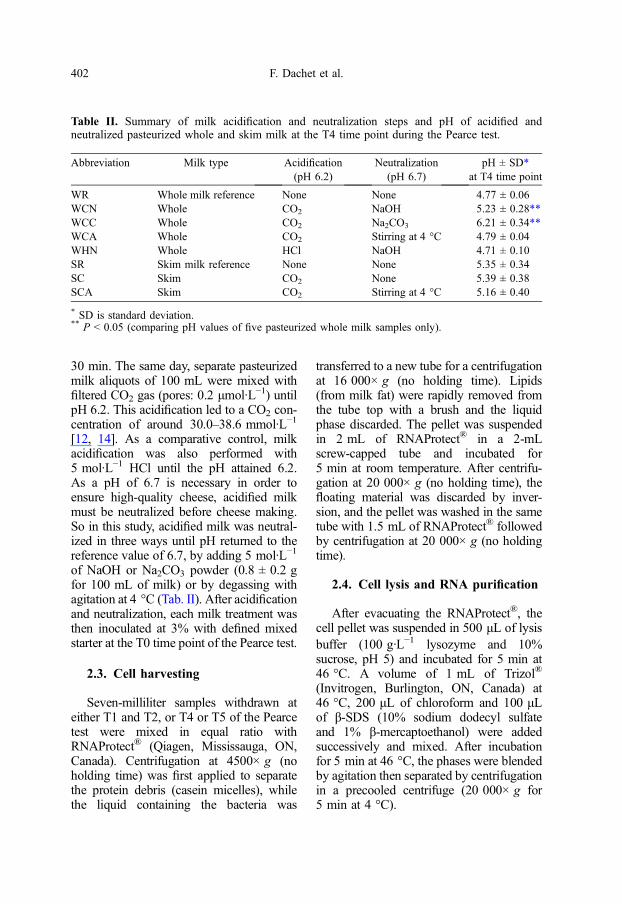
Table II. Summary of milk acidification and neutralization steps and pH of acidified andneutralized pasteurized whole and skim milk at the T4 time point during the Pearce test.
Abbreviation Milk type Acidification(pH 6.2)
Neutralization(pH 6.7)
pH ± SD*at T4 time point
WR Whole milk reference None None 4.77 ± 0.06WCN Whole CO2 NaOH 5.23 ± 0.28**WCC Whole CO2 Na2CO3 6.21 ± 0.34**WCA Whole CO2 Stirring at 4 °C 4.79 ± 0.04WHN Whole HCl NaOH 4.71 ± 0.10SR Skim milk reference None None 5.35 ± 0.34SC Skim CO2 None 5.39 ± 0.38SCA Skim CO2 Stirring at 4 °C 5.16 ± 0.40
* SD is standard deviation.** P < 0.05 (comparing pH values of five pasteurized whole milk samples only).
402 F. Dachet et al.

One milliliter of the aqueous phase wasmixed with 500 μL of isopropanol at roomtemperature and then passaged twice on anRNeasy® column (Qiagen, Mississauga,ON, Canada) by centrifugation at14 000× g for 15 s. Themanufacturer’s pro-tocol was followed for washing and on-col-umn DNase treatment, except that 33 unitsof SUPERaseIn™ (Ambion, Applied Bio-systems, Foster City, USA) were added.The RNA elution was carried out on ice with10 μL of RNase-free water. The RNA con-centration was quantified at 260 nm with aNanoDrop™ 1000 (Thermo Fisher Scientific,Wilmington, USA) and adjusted to a finalconcentrationof100 ng·μL−1withRNase-freewater (Qiagen, Mississauga, ON, Canada).
2.5. Fluorescent RAP-PCR
Primer design is described in the Supple-mentary Material, Sections 1 and 2. RNAwas used at a concentration of 15 ng·μL−1
with 19 units of SUPERase-In and2.5 μmol·L−1 of an equal ratio of primersST1–ST13 (Supplementary Material,Tab. SI). The mixture was incubated at65 °C for 5 min then immediately placedon ice for 1 min. The final volume ofthe retrotranscription reaction was 19 μLand contained: 2 mmol·L−1 of total dNTPs,190 units of SuperScript™ III (Invitrogen,Burlington, ON, Canada), 3.7 μL of 5X buf-fer and 5 mmol·L−1 of DTT (dithiothreitol).The reverse transcription reactions wereincubated at 25 °C for 10 min. The polymer-ization step was carried out at 45 °C for 2 h,and a final reverse transcriptase inactivationstep was applied at 70 °C for 15 min.
A quantity of 20 ng of cDNA was usedas template for PCR amplification. For20 μL of final volume, each reaction mix-ture contained: 1.67 mmol·L−1 of MgSO4,700 μmol·L−1 of total dNTPs, 1.34 unitsof Hot Start Kod polymerase (EMDBiosciences, Inc., Novagen®, Madison,WI, USA), 2 μmol·L−1 of one nonlabeledprimer and 2 μmol·L−1 of one fluorescent
primer. The first step was 95 °C for10 min for RNA degradation and Hot Startpolymerase activation. The second step wasa low stringency annealing at 35 °C for40 min, enabling saturation of all the poten-tial hybridizing sites [30]. The third stepwas a final elongation of the second strandcDNA at 72 °C for 5 min. Then thefollowing 30 PCR cycles were performedwith high stringency cycling: 95 °C, 30 s;55 °C, 40 s; and 72 °C, 60 s. The PCRamplification was mixed with 500 μL ofTE (10 mmol·L−1 Tris and 1 mmol·L−1
EDTA, pH 8) and loaded on a Microcon®
YM100 (Millipore, Billerica, MA, USA).The amplicons were washed four times withsuccessive passages of 500 μL of TE on thesame column. Elution was carried out with50 μL of TE, and PCR products were quan-tified by spectrophotometry at 260 nm.
2.6. Separation by polyacrylamidegel electrophoresis
PCR amplifications (3 μg) were sepa-rated on 6% polyacrylamide gels (acrylam-ide:bisacrylamide with a 29:1 ratio).Electrophoresis was run at 100 V for 24 hat 4 °C. Ethidium bromide was used forstaining DNA, which was visualized byUV transillumination.
2.7. Amplicon separationby capillary electrophoresis
A quantity of 75 ng of purified ampli-cons was mixed with 10 μL of form-amide and 0.3 μL of MapMarker® 1000(Bio-Ventures, Murfreesboro, TN, USA).The mixture was heated at 99 °C for5 min then cooled on ice for 1 min andinjected (injection voltage: 1 kV, injectionduration: 30 s) in the ABI Prism® 3100Genetic Analyzer (Applied Biosystems,Foster City, USA).
Separation was performed using capillar-ies filled with POP6 polymer under theseparameters: run voltage: 15 kV, total
Transcriptome profiling of starter in milk 403

data points for each run: 24 178. Theelectropherograms were acquired withthe GeneMapper® version 3.7 software(Applied Biosystems) under these advancedmicrosatellite parameters: no smoothing;baseline = 51; peak threshold (blue) = 1;minimum peak half width = 0; polynomialdegree search = 5; and window size = 15.To standardize MapMarker® sizing, flagquality was set at low-quality range from0 to 1E − 6 then pass-range from 1E − 5to 1. Panel manager marker sizing andmicrosatellite analysis was applied from50 to 1500 bases. An AFLP analysis wasperformed and for each replicate, the size(in bases) and the height (in fluorescentunits) of peaks were exported to Excel.
2.8. Statistical analysis
To prevent statistical problems resultingfrom peak absence of some very smallpeaks (no height reported), the minimumheight of the peaks detected in the run (usu-ally from 1 to 10 under a 6000 scale gradu-ation) was substituted for each of the peaksnot detected with GeneMapper (around 1%of the peaks were not detected by the soft-ware). Height data were transformed byNeperian logarithm to linearize peak distri-bution then standardized with AMIADA[33] using the sum of all the heights. Stan-dardization was carried out to a null averageand a unitary standard deviation, allowingcomparisons among replicates. For eachsample withdrawn at one time point, theresulting eight electropherograms were con-catenated under Excel® (for milk thermaltreatments, the electropherograms from eachtime point were also concatenated).
Hierarchical clustering (HC) and princi-pal component analysis (PCA) were carriedout using “R” statistical software. Euclideandistance was used as the metric because itis a direct similarity measure, allowing rapidand intuitive interpretation of informationproduced (equation (1)). HC used averagelinkage and was computed with a bootstrap
of 10 000 permutations. PCA and statisticalsignificance (using ANOVA and Tukeyhonest significant differences) were com-puted with the “R” package. The numberof differentially expressed peaks was identi-fied using the SAM algorithm (FDR = 0.4,10 000 permutations) from the MEV 4 soft-ware [24] and divided by the total numberof peaks:
SimilarityA=B ¼ 1
1þ DistanceA!B
DistanceA!B ¼ffiffiffiffiffiffiffiffiffiffiffiffiffiffiffiffiffiffiffiffiffiffiffiffiffiffiffiffiffiffiffiffiffiffiffiffiffiffiffiffiffi2ð1� SimilarityA=BÞ:
q
ð1Þ
3. RESULTS AND DISCUSSION
The mixed starter culture responded tothe state of milk nutrient accessibility byadapting their global RNA profiles. Thebacterial transcriptome is composed mainlyof rRNA and tRNA, while the fraction ofmRNA is low (around 5%). Fragments gen-erated by FRAP-PCR can thus be attributedto all RNA transcripts. While manydifferent individual mRNA molecules arerepresented in the transcriptome (high com-plexity), the multiple copies of rRNA andtRNA are of similar sequence (low com-plexity), and thus the same rRNA ampli-cons will migrate to the same distance.This will reduce its effect on the overall pro-file composed of 1000 peaks, as long asprimers and reaction components are pres-ent in sufficient quantity to avoid depletion.
3.1. Effect of milk typeand treatment on definedmixed starter transcriptionprofiles by FRAP-PCR
In order to determine the reproducibilityof the method and to select reference condi-tions, different milk types and thermal treat-ments were first compared. The techniqueshowed high reproducibility, as each peak
404 F. Dachet et al.

of electropherograms was present in tripli-cate experiments (Supplementary Material,Fig. S2) and replicates were grouped byHC (Supplementary Material, Fig. S3) aswell as by PCA (Supplementary Material,Fig. S4). Cluster separation could beattributed to milk type used during the fer-mentation even if pH could not differentiatemost of the milk types (Tab. I). Clusters ofelectropherograms coming from cultures inmicrofiltered, pasteurized and UHT milkwere located near to one another andseparated from the profiles obtained withautoclaved SMP. The first two componentsof PCA (Supplementary Material, Fig. S3)explain > 50% of the total variance. The firstcomponent, explaining nearly 40% of thevariance, separated autoclaved fromnonautoclaved SMP, whereas the secondcomponent divided SMP autoclaved for 10or 15 min.
Pasteurization and microfiltration are notdamaging for milk constituents as there arefew physicochemical changes compared toraw milk [20, 25], but milk contains variousactive RNases [16] and could have micro-bial contaminants [18]. The RNases couldalter starter RNA during extraction and themicrobial contaminants could be cocultivat-ed with starter (especially during the twosubcultures) and these will give irreproduc-ible RNA profiles. The autoclaving of milkinactivates RNases and kills all microbialcontaminants, but the results of this studyshow that autoclaved SMP induced signifi-cant transcriptome changes in the startercompared to microfiltered milk. The auto-claved SMP is not recommended for starteractivity study for two reasons: (i) the starterRNA profile of autoclaved SMP was verydifferent from the same SMP treated by pas-teurization; (ii) with only 5 min differencebetween them, the two autoclaving timesinduced significant starter RNA modifica-tions, indicating a potential deviation ofstarter activity if the autoclaving of milk isnot perfectly time controlled (e.g. coolingtime).
Starter fermenting UHT or pasteurizedSMP has nearly the same RNA profile asin microfiltered milk, so these two milktypes did not notably influence gene tran-scription. SMP has well-known solubiliza-tion problems and so could lead tomodification of water activity between dif-ferent experiments. UHT milk was the mostsuitable milk for starter gene transcriptionstudy, as there was no need of solubiliza-tion, no RNA or bacterial contaminants[3], and milk RNases are inactivated bythe high temperature reached (around140 °C) [16]. The UHT treatment did notinfluence the starter transcriptome as muchas autoclaved SMP, so this high temperaturetreated milk could be used for preparingstarter cultures for RNA study of cheesefermentation without important risk ofcontamination.
3.2. Effect of CO2 acidification andneutralization on transcriptionprofiles of defined mixed startercultivated in whole milk
Oxygen displacement by carbon dioxideprevents the growth of aerobic bacteria, butanaerobic bacteria can also be affected byCO2 [22]. Milk carbon dioxide dissolutionleads to a rapid drop of pH caused by car-bonic acid formation in milk aqueous phase,but this important acid production is not theonly cause of bacterial growth inhibition[9]. Carbon dioxide has important effectson bacterial membrane permeability [7]but more complex effects have been stud-ied, such as interference with bacterialmetabolism leading to changes in carbohy-drate utilization [17], lowering intracellularpH [7] and varying enzyme activities [6](such as extracellular lipase [23]). All thesecombined effects induce a stress thatdecreases bacterial multiplication and acidi-fication rate.
Samples were taken at the T4 time point,and the pasteurized whole milk cluster wasused as control to discover the influence
Transcriptome profiling of starter in milk 405

of the acidification and neutralization proce-dures (Tab. II) on RNA profiles. The pHwas significantly higher at T4 in CO2-acid-ified milk that was neutralized by NaOH orNa2CO3 than after the other treatments ofpasteurized whole milk. The first two com-ponents of PCA totalized around 50% of thevariance (Fig. 1). The first component(32.75% of the variance) separates CO2-acidified milk from pasteurized milk andHCl-acidified milk. The second componentexplained 16.17% of the variance and could
also be associated with CO2 treatment, asthe CO2 profiles are further from the centralaxis than those of pasteurized and HCl-acid-ified milk.
Compared to pasteurized whole milk, theRNA profile of starter fermenting CO2-acid-ified whole milk which was neutralizedby NaOH or Na2CO3 showed importantmodifications. The RNA profile of starterfermenting HCl-acidified whole milk cou-pled with NaOH for the neutralization didnot show any statistical difference from
Figure 1. PCA of fluorescent RAP-PCR amplicon profiles obtained from RNA extracted fromdefined mixed culture fermentation of pasteurized whole milk pretreated with different acidificationand neutralization procedures. WR is the pasteurized whole milk reference profile; WCN is wholemilk acidified with CO2 and neutralized with NaOH; WCC is whole milk acidified with CO2 thenneutralized with Na2CO3; WCA is whole milk acidified with CO2 and neutralized by agitation; andWHN is whole milk acidified with HCl and neutralized with NaOH. Gray shading represents theclustering of the three experimental repetitions (●) inside an ellipse which represents a confidencelevel of 95%. (□) Cluster’s barycenter.
406 F. Dachet et al.
whole milk, suggesting that it was not theacidification and NaOH neutralization stepsthat were causing RNA profile modifica-tions. Therefore, milk acidification by CO2
could modify RNA profiles of the mixedculture. The type of neutralization step alsoinfluenced gene transcription. The cluster ofelectropherograms from starter cultivatedon CO2-acidified and Na2CO3-neutralizedwhole milk was the farthest removed fromthe whole milk profile, indicating moreextensive modification of the starter RNAprofile. Furthermore, this type of neutraliza-tion led to important pH buffer action,which could introduce significant delays inacidification needed for cheese manufactur-ing. Carbon dioxide dissipation by agitationseems a good technique for neutralization toregain similar reference starter activitybecause this treatment led to only small star-ter RNA changes.
3.3. Effect of CO2 acidification andneutralization on transcriptionprofiles of defined mixed startercultivated in skim milk
Pasteurized skim milk without CO2 treat-ment or neutralization was used as thefermentation reference. Two different treat-ments were applied to pasteurized skimmilk: acidification with CO2 to pH 6.2 with-out neutralization and acidification withCO2 to pH 6.2 followed by neutralizationby agitation at 4 °C. Experiments using pas-teurized whole milk were repeated for a ref-erence profile among the various replicates.The PCA results (Fig. 2) show that FRAP-PCR profiles from mixed cultures in skimmilk with different carbonation or agitationtreatments were separated from those ofpasteurized whole milk along the first com-ponent. The PCA shows clustering of allskim milk samples into one group, whereastreated whole milks were well separatedfrom one another. All the FRAP-PCR pro-files from skim milk were electrophoresedon a 6% acrylamide gel (Supplementary
Material, Fig. S5). For each primer pair,no differences between the band profilescould be detected. Thus, CO2 acidificationexperiments using pasteurized skim milkdid not induce detectable modifications inelectropherograms obtained either byFRAP-PCR or by polyacrylamide gel elec-trophoresis. Protein and mineral contentsare essentially the same between whole milkand skim milk [21], leading to the conclu-sion that there was no irreversible actionof CO2 on casein or colloidal calcium phos-phate that could influence starter gene tran-scription during fermentation. Therefore,the fat globules could be the origin of thedifference between pasteurized whole milkand skim milk in the effect of CO2 acidifi-cation on starter RNA profiles. Carbondioxide treatment could lead to imprison-ment of carbonated molecules in milk fatglobules [28], which could be released dur-ing cheese manufacture, leading to interac-tions with starter bacteria even if theL. lactis subsp. cremoris strains used wereresistant to CO2 influence [5].
3.4. Rennet and NaCl influenceon defined mixed startertranscriptome profiles
At the salt concentrations used in thisstudy, the three individual strains have aboutthe same growth (data not shown). Mixedstarter fermented with different NaCl andrennet concentrations did not show anyinfluence on the final pH attained in thePearce test (Tab. III). Differentiallyexpressed peaks (either activated orrepressed compared to the standard condi-tion) were identified by SAM analysis(Tab. IV). The high rennet treatment resultedin the same proportion of activated peaks(i.e. peaks of greater height than in the stan-dard condition) as for repressed peaks, whilethe low rennet condition showed double thenumber of repressed peaks compared to acti-vated ones (Tab. IV). High NaCl concentra-tion resulted in almost the same number
Transcriptome profiling of starter in milk 407

Figure 2. PCA of fluorescent RAP-PCR profiles obtained from RNA extracted from defined mixedculture fermentation of pasteurized whole or skim milk treated by acidification and neutralization.WR is the whole milk reference profile; WCN is whole milk acidified with CO2 and neutralizedwith NaOH; WCC is whole milk acidified with CO2 then neutralized with Na2CO3; SR ispasteurized skim milk reference with no treatment; SC is pasteurized skim milk treated with CO2;and SCA is pasteurized skim milk treated with CO2 then neutralized by agitation. Gray shadingrepresents the clustering of the three experimental repetitions (●) inside an ellipse which representsa confidence level of 95%. (□) Cluster’s barycenter.
Table III. pH of UHT milk at T5 (1 h and 45 min after the T4 time point) of the modified Pearcestarter activity test.
Condition NaCl (g·L−1) Rennet (g·L−1) pH ± SD*
Standard condition 22.5 0.08 5.10 ± 0.06High rennet 22.5 0.12 5.14 ± 0.05Low rennet 22.5 0.04 5.00 ± 0.06High NaCl 25 0.08 5.11 ± 0.05Low NaCl 15 0.08 5.06 ± 0.06
* SD is standard deviation.
408 F. Dachet et al.

of activated peaks as the number ofrepressed peaks identified when low NaClconcentration was used.
The PCA clusters are separated by the twocomponents simultaneously, indicating thateach of the two first eigenvectors was a com-bination of NaCl and rennet influence(Fig. 3). These eigenvectors were nearlyequal in the variance explained (24.14% forthefirst and 16.25% for the second). The pro-files obtained from the RNA extracted frommixed cultureswith low rennet concentrationwere the farthest from the standard clusternear the center and showed the highest posi-tive vector in the first dimension. Compari-son with the reference cluster shows thatboth the salt and rennet vectors (high andlow) are nearly opposite in direction, andare in opposing quadrants, indicating oppos-ing correlation of the variance caused bycondition-specific peaks. The cluster corre-sponding to the standard NaCl concentrationwas equally centered between the two profileclusters representing low and high NaClconcentrations, even if the concentration gra-dient varied (7.5 g·L−1 between low andstandard compared to only 2.5 g·L−1
between standard and high concentration).This indicates an important influence on thetranscriptome caused by NaCl addition andless difference when NaCl decreased. Asthe vector of the clusters is equally distancedfrom both principal components, this indi-cates a combination of the influence of bothNaCl and rennet. Rennet concentration thushad an opposite effect from NaCl, as thecluster of profiles from the lowest rennet
concentration was at a greater distance fromthe profile obtained with the standard con-centration of rennet. This indicates that genetranscription of the starter was greatly modi-fied by low rennet condition and less by highrennet addition.
4. CONCLUSION
RNA profiles of starter fermenting CO2-acidified milk were modified by CO2 mole-cules presumably dissolved in fat globulesand slowly released. Agitation was moreeffective than NaOH or Na2CO3 for neutral-ization to return the profile to that obtainedin whole milk. FRAP-PCR was able toshow that starter gene transcription wasmore influenced by an increase in NaClcompared to a decrease in NaCl. Rennet ata high concentration had only a little influ-ence on gene transcription, but low rennetactivity had great influence. This indicatesthat an increase in NaCl must be compen-sated by an increase in rennet. For this star-ter, rennet activity appeared to be optimal atstandard concentration.
This is the first report of the application ofFRAP-PCR to the study of starter activity incultures emulating cheese fermentation. Thismethod could be used to determine the levelof influence of parameters and help to under-stand the way changes in Cheddar cheesefermentation procedures could interfere withglobal gene transcription of the starter. Bydetermining starter gene responses duringCheddar cheese manufacturing, optimal
Table IV. Number of peaks differentially expressed for each condition compared to standardconditions identified using the SAM algorithm (FDR = 0.4%, 10 000 permutations) of the MEV4.0 software. Percentage of the total is reported in parentheses.
Condition Activated peaks Repressed peaks
High rennet 22 (2%) 17 (2%)Low rennet 18 (2%) 50 (4%)High NaCl 42 (4%) 28 (2%)Low NaCl 8 (1%) 50 (4%)
Transcriptome profiling of starter in milk 409

cheese fermentation could be predicted bycomparisonwith a reference such as a cheesewith an excellent grade. Another potential ofFRAP-PCR could be the detection of differ-entially expressed peaks due to starterexpressing phage RNA, thus revealingpotential fermentation failure due to phage.This technique could also be used for study-ing bacterial associations in order to furtherour understanding of microbial interactionsin food matrices.
Acknowledgments: The authors gratefullyacknowledge the financial support provided bythe Fonds Québécois de recherche sur la nature
et les technologies (FQRNT), Novalait, Inc., theMinistère de l’Agriculture, des Pêcheries et del’Alimentation du Québec (MAPAQ) as well asAgriculture and Agri-Food Canada.
REFERENCES
[1] Bachem C.W., van der Hoeven R.S.,de Bruijn S.M., Vreugdenhil D., Zabeau M.,Visser R.G., Visualization of differential geneexpression using a novel method of RNAfingerprinting based on AFLP: analysis ofgene expression during potato tuber develop-ment, Plant J. 9 (1996) 745–753.
[2] Bauer D., Muller H., Reich J., Riedel H.,Ahrenkiel V., Warthoe P., Strauss M.,Identification of differentially expressed
Figure 3. PCA of fluorescent RAP-PCR profiles obtained from RNA extracted from fermentationwith low, standard and high concentrations of rennet and NaCl (as listed in Sect. 2). Gray shadingrepresents the clustering of the three experimental repetitions (●) inside an ellipse which representsa confidence level of 95%. (□) Cluster’s barycenter.
410 F. Dachet et al.
mRNA species by an improved displaytechnique (DDRT-PCR), Nucleic AcidsRes. 21 (1993) 4272–4280.
[3] Birlouez-Aragon I., Sabat P., Gouti N.,A new method for discriminating milk heattreatment, Int. Dairy J. 12 (2002) 52–57.
[4] Crump D., Chiu S., Trudeau V.L., KennedyS.W., Fluorescent RNA arbitrarily primedpolymerase chain reaction. A new differen-tial display approach to detect contaminant-induced alterations of gene expression inwildlife species, in: Cristofre Martin C.(Ed.), Methods in Molecular Biology: Envi-ronmental Genomics, Vol. 410, HumanaPress Inc., Totowa, USA, 2008, pp. 15–27.
[5] Enfors S.O., Molin G., Effect of highconcentrations of carbon dioxide on growthrate of Pseudomonas fragi, Bacillus cereusand Streptococcus cremoris, J. Appl. Bacte-riol. 48 (1980) 409–416.
[6] Gill C.O., Tan K.H., Effect of carbondioxide on growth of Pseudomonas fluores-cens, Appl. Environ. Microbiol. 38 (1979)237–240.
[7] Hon S.I., Pyun Y.R., Membrane damage andenzyme inactivation of Lactobacillus plan-tarum by high pressure CO2 treatment, Int.J. Food Microbiol. 63 (2001) 19–28.
[8] Jones S.W., Cai D., Weislow O.S., Esmaeli-Azad B., Generation of multiple mRNAfingerprints using fluorescence-based differ-ential display and an automated DNAsequencer, Biotechniques 22 (1997) 536–543.
[9] King J.S., Mabbitt L.A., Preservation of rawmilk by addition of carbon dioxide, J. DairyRes. 49 (1982) 439–447.
[10] Liang P., Pardee A.B., Differential display ofeukaryotic messenger RNA by means of thepolymerase chain reaction, Science 257(1992) 967–971.
[11] Luehrsen K.R., Marr L.L., van der Knaap E.,Cumberledge S., Analysis of differentialdisplay RT-PCR products using fluorescentprimers and GENESCAN software, Biotech-niques 22 (1997) 168–174.
[12] Ma Y., Barbano D.M., Effect of temperatureof CO2 injection on the pH and freezingpoint of milks and creams, J. Dairy Sci. 86(2003) 1578–1589.
[13] Makarova K., Slesarev A., Wolf Y.,Sorokin A., Mirkin B., Koonin E.,Pavlov A., Pavlova N., Karamychev V.,Polouchine N., Shakhova V., Grigoriev I.,Lou Y., Rohksar D., Lucas S., Huang K.,
Goodstein D.M., Hawkins T., Plengvidhya V.,Welker D., Hughes J., Goh Y., Benson A.,Baldwin K., Lee J.H., Diaz-Muniz I., Dosti B.,Smeianov V., Wechter W., Barabote R.,Lorca G., Altermann E., Barrangou R.,Ganesan B., Xie Y., Rawsthorne H.,Tamir D., Parker C., Breidt F., Broadbent J.,Hutkins R., O’Sullivan D., Steele J., UnluG., Saier M., Klaenhammer T., RichardsonP., Kozyavkin S., Weimer B., Mills D.,Comparative genomics of the lactic acidbacteria, Proc. Natl. Acad. Sci. USA 103(2006) 15611–15616.
[14] Martin J.D., Werner B.G., Hotchkiss J.H.,Effects of carbon dioxide on bacterialgrowth parameters in milk as measured byconductivity, J. Dairy Sci. 86 (2003) 1932–1940.
[15] Meade J.D., Cho Y.J., Fisher J.S.,Walden J.C., Guo Z., Liang P., Automationof fluorescent differential display with dig-ital readout, in: Liang P., Meade J.D., PardeeA.B. (Eds.), Methods in Molecular Biology:Differential Display Methods and Protocols,Vol. 317, Humana Press Inc., Totowa, USA,2006, pp. 23–57.
[16] Meyer D.H., Kunin A.S., Maddalena J.,Meyer W.L., Ribonuclease activity and iso-enzymes in raw and processed cows’ milkand infant formulas, J. Dairy Res. 70 (1987)1797–1803.
[17] Molin G., Effect of carbon dioxide ongrowth of Pseudomonas putida ATCC11172 on asparagine, citrate, glucose, andlactate in batch and continuous culture, Can.J. Microbiol. 31 (1985) 763–766.
[18] Nielsen E.W., Principles of cheese produc-tion, in: Hui Y., Meunier-Goddik L., HansenA., Josephsen J., Nip W.-K., Stanfield P.,Toldrá F. (Eds.), Handbook of Foodand Beverage Fermentation Technology,Marcel Dekker, New York, USA, 2004,pp. 219–239.
[19] Pearce L.E., Activity tests for cheese startercultures, New Zealand, J. Dairy Technol. 4(1969) 246–247.
[20] Pedersen P.J., Microfiltration for the reduc-tion of bacteria in milk and brine, newapplications of membrane processes, Int.Dairy Fed. Special Issue No. 9201 (1992)33–50.
[21] Posati L.P., Orr M.L., Composition of foods:dairy and egg products; raw, processed,prepared, Agriculture Handbook No. 8-1,Agricultural Research Service, USDA,Washington, USA, 1976.
Transcriptome profiling of starter in milk 411

[22] Roberts R.F., Torrey G.S., Inhibition ofpsychrotrophic bacterial growth in refriger-ated milk by addition of carbon dioxide,J. Dairy Sci. 71 (1988) 52–60.
[23] Rowe M.T., Effect of carbon dioxide ongrowth and extracellular enzyme productionby Pseudomonas fluorescens B52, Int.J. Food Microbiol. 6 (1988) 51–56.
[24] Saeed A.I., Sharov V., White J., Li J., LiangW., Bhagabati N., Braisted J., Klapa M.,Currier T., Thiagarajan M., Sturn A., SnuffinM., Rezantsev A., Popov D., Ryltsov A.,Kostukovich E., Borisovsky I., Liu Z.,Vinsavich A., Trush V., Quackenbush J.,TM4: a free open-source system for micro-array data management and analysis, Bio-techniques 34 (2003) 374–378.
[25] Scaloni A., Perillo V., Franco P., Fedele E.,Froio R., Ferrara L., Bergamo P., Character-ization of heat-induced lactosylation prod-ucts in caseins by immunoenzymatic andmass spectrometric methodologies, Biochim.Biophys. Acta 1598 (2002) 30–39.
[26] Schena M., Shalon D., Davis R.W.,Brown P.O., Quantitative monitoring of geneexpression patterns with a complementaryDNA microarray, Science 270 (1995)467–470.
[27] Sturtevant J., Applications of differential-display reverse transcription-PCR to molec-ular pathogenesis and medical mycology,Clin. Microbiol. Rev. 13 (2000) 408–427.
[28] Walstra P., Wouters J.T.M., Geurts T.J.,Dairy Science and Technology, CRC Press,Boca Raton, USA, 2006.
[29] Welsh J., Chada K., Dalal S.S., Cheng R.,Ralph D., McClelland M., Arbitrarilyprimed PCR fingerprinting of RNA, NucleicAcids Res. 20 (1992) 4965–4970.
[30] Welsh J., McClelland M., Fingerprintinggenomes using PCR with arbitrary primers,Nucleic Acids Res. 18 (1990) 7213–7218.
[31] Williams J.G., Kubelik A.R., Livak K.J.,Rafalski J.A., Tingey S.V., DNA polymor-phisms amplified by arbitrary primers areuseful as genetic markers, Nucleic AcidsRes. 18 (1990) 6531–6535.
[32] Wong K.K., McClelland M., Stress-induciblegene of Salmonella typhimurium identifiedby arbitrarily primed PCR of RNA, Proc.Natl. Acad. Sci. USA 91 (1994) 639–643.
[33] Xia X., Xie Z., AMADA: analysis ofmicroarray data, Bioinformatics 17 (2001)569–570.
412 F. Dachet et al.