trans-Resveratrol induces apoptosis in human breast cancer cells MCF-7 by the activation of MAP...
11
RESEARCH PAPER trans-Resveratrol induces apoptosis in human breast cancer cells MCF-7 by the activation of MAP kinases pathways G. Filomeni I. Graziani G. Rotilio M. R. Ciriolo Received: 19 January 2007 / Accepted: 1 July 2007 / Published online: 18 October 2007 Ó Springer-Verlag 2007 Abstract Polyphenols represent a large class of plant- derived molecules with a general chemical structure that act as potent free radical scavengers. They have long been recognized to possess several therapeutic activities ranging from anti-thrombotic to antioxidant. Moreover, the capa- bility of polyphenols to act as reducing or oxidizing molecules depends on the presence of environmental metals and on the concentrations used. In this work we demonstrated that the stilbene trans-resveratrol was able to commit human breast cancer MCF-7 cells to apoptosis. Mainly, we evidenced a pivotal role of the mitochondria in this phenomenon as cytochrome c release into the cytosol was found after the treatment. We further showed that trans-resveratrol was able to affect cellular redox state. In particular, it induced an early production of ROS and lipid oxidation, and only later compromised the GSH/GSSG ratio. This mode of action was mirrored by a temporally different activation of JNK and p38 MAPK , with the former rapidly induced and the latter weakly activated at long intervals. The results obtained demonstrate a pro-apoptotic activity for trans-resveratrol, and suggest a preferential activation of different classes of MAP kinases in response to different oxidative stimuli (ROS versus GSH/GSSG alteration). Keywords Resveratrol Apoptosis Glutathione ROS MAP kinases Oxidative stress Introduction Apoptosis is a programmed mode of cell death induced physiologically either during organ development, or for the maintenance of cellular homeostasis by means of the elimination of damaged or unnecessary cells [54, 61]. Recently, it has been demonstrated that phosphorylation/ de-phosphorylation state of some regulatory proteins are crucial events along the pathways controlling cell growth and apoptosis. A well established apoptotic signaling cas- cade is that regulated by mitogen activated protein (MAP) kinases, such as the p42/44 extracellular signal-related kinases (ERK1/2), c-Jun N-terminal protein kinase (JNK) and p38 MAPK , which directly modulate the phospho-active levels of pro-apoptotic factors [38]. MAP kinases are ser- ine/threonine kinases that are regulated by a variety of extracellular stimuli including growth factors, mitogens, cytokines and environmental stresses [39]. Oxidative stress-mediated apoptosis is induced by phosphorylative cascades; indeed, in the last few years a hypothesis has emerged that reactive oxygen species (ROS) are not only the downstream damaging species produced by radical chain reactions, but also the second messengers of signal- ing networks [15]. Considerable scientific interest in the anticancer ther- apy has been focused, for long time, on the identification of compounds able to efficiently commit cells to apopto- sis; in this context several chemotherapeutic agents can generate ROS at high extent, by catalyzing one-electron redox cycles with oxygen [55]. The specificity of action of these compounds towards tumor histotypes is supposed G. Filomeni and I. Graziani are recipients of fellowships from the Italian Association for Cancer Research (AIRC-FIRC). G. Filomeni I. Graziani G. Rotilio M. R. Ciriolo (&) Department of Biology, University of Rome ‘‘Tor Vergata’’, via della Ricerca Scientifica, 00133 Rome, Italy e-mail: [email protected] G. Rotilio M. R. Ciriolo IRCCS San Raffaele Pisana, via della Pisana, 235, 00163 Rome, Italy 123 Genes Nutr (2007) 2:295–305 DOI 10.1007/s12263-007-0059-9
Transcript of trans-Resveratrol induces apoptosis in human breast cancer cells MCF-7 by the activation of MAP...

RESEARCH PAPER
trans-Resveratrol induces apoptosis in human breast cancer cellsMCF-7 by the activation of MAP kinases pathways
G. Filomeni Æ I. Graziani Æ G. Rotilio ÆM. R. Ciriolo
Received: 19 January 2007 / Accepted: 1 July 2007 / Published online: 18 October 2007
� Springer-Verlag 2007
Abstract Polyphenols represent a large class of plant-
derived molecules with a general chemical structure that
act as potent free radical scavengers. They have long been
recognized to possess several therapeutic activities ranging
from anti-thrombotic to antioxidant. Moreover, the capa-
bility of polyphenols to act as reducing or oxidizing
molecules depends on the presence of environmental
metals and on the concentrations used. In this work we
demonstrated that the stilbene trans-resveratrol was able to
commit human breast cancer MCF-7 cells to apoptosis.
Mainly, we evidenced a pivotal role of the mitochondria in
this phenomenon as cytochrome c release into the cytosol
was found after the treatment. We further showed that
trans-resveratrol was able to affect cellular redox state. In
particular, it induced an early production of ROS and lipid
oxidation, and only later compromised the GSH/GSSG
ratio. This mode of action was mirrored by a temporally
different activation of JNK and p38MAPK, with the former
rapidly induced and the latter weakly activated at long
intervals. The results obtained demonstrate a pro-apoptotic
activity for trans-resveratrol, and suggest a preferential
activation of different classes of MAP kinases in response
to different oxidative stimuli (ROS versus GSH/GSSG
alteration).
Keywords Resveratrol � Apoptosis � Glutathione �ROS � MAP kinases � Oxidative stress
Introduction
Apoptosis is a programmed mode of cell death induced
physiologically either during organ development, or for the
maintenance of cellular homeostasis by means of the
elimination of damaged or unnecessary cells [54, 61].
Recently, it has been demonstrated that phosphorylation/
de-phosphorylation state of some regulatory proteins are
crucial events along the pathways controlling cell growth
and apoptosis. A well established apoptotic signaling cas-
cade is that regulated by mitogen activated protein (MAP)
kinases, such as the p42/44 extracellular signal-related
kinases (ERK1/2), c-Jun N-terminal protein kinase (JNK)
and p38MAPK, which directly modulate the phospho-active
levels of pro-apoptotic factors [38]. MAP kinases are ser-
ine/threonine kinases that are regulated by a variety of
extracellular stimuli including growth factors, mitogens,
cytokines and environmental stresses [39]. Oxidative
stress-mediated apoptosis is induced by phosphorylative
cascades; indeed, in the last few years a hypothesis has
emerged that reactive oxygen species (ROS) are not only
the downstream damaging species produced by radical
chain reactions, but also the second messengers of signal-
ing networks [15].
Considerable scientific interest in the anticancer ther-
apy has been focused, for long time, on the identification
of compounds able to efficiently commit cells to apopto-
sis; in this context several chemotherapeutic agents can
generate ROS at high extent, by catalyzing one-electron
redox cycles with oxygen [55]. The specificity of action
of these compounds towards tumor histotypes is supposed
G. Filomeni and I. Graziani are recipients of fellowships from the
Italian Association for Cancer Research (AIRC-FIRC).
G. Filomeni � I. Graziani � G. Rotilio � M. R. Ciriolo (&)
Department of Biology, University of Rome ‘‘Tor Vergata’’,
via della Ricerca Scientifica, 00133 Rome, Italy
e-mail: [email protected]
G. Rotilio � M. R. Ciriolo
IRCCS San Raffaele Pisana, via della Pisana,
235, 00163 Rome, Italy
123
Genes Nutr (2007) 2:295–305
DOI 10.1007/s12263-007-0059-9
to be due to: (i) low concentration of antioxidant enzymes
in transformed cells; (ii) high rate of proliferation with
respect to differentiated cells, a feature that does not allow
tumor cells repairing oxidative injuries, especially those
involving DNA [23, 32, 49]. More recently, there has
been a focus of attention on natural anti-oxidant mole-
cules that, besides their role in scavenging-free adicals,
could function as pro-oxidants, when highly concentrated
or in the presence of transition metals [51]. Previous
studies have indicated that some nutraceuticals exhibit
potent anti-tumor properties and can modulate apoptosis,
differentiation and cell cycle, probably by virtue of their
anti-oxidant functions. However, in vitro experiments
reported that most of them behave as potent pro-oxidants
molecules [6, 9, 40]. For example, organosulfur molecules
from garlic, such as the oil-soluble diallyl disulfide [12,
13], diallyl trisulfide [22], or the water-soluble S-allylm-
ercaptocysteine [63] have been demonstrated to induce
growth arrest and apoptosis dose-dependently by
increasing ROS production.
Among nutraceuticals, polyphenols represent the most
intriguing and studied class of compounds that can be
therapeutics for a large spectrum of the most common
diseases including cancer [20]. Nevertheless, despite sev-
eral studies regarding their function, the anti-oxidant/pro-
oxidant properties of these compounds remain somewhat
debatable and the detailed molecular mechanisms of their
effects continue to be largely unknown. A pro-apoptotic
function has been often proposed for many polyphenols
[21, 35, 42]; however, although a huge amount of data
support these assumptions, the molecular mechanism(s) by
which apoptosis is triggered are long to be elucidated.
One of the well-characterized polyphenols is trans-
resveratrol (3,5,40-trihydroxy-trans-stilbene), a phyto-
alexin found in edible material, such as grape skin,
peanuts and red wine. Jang and coworkers demonstrated
that trans-resveratrol is an anticancer compound able to
inhibit each phase of cell transformation both in in
vitro and in in vivo models. Particularly, by acting as
antioxidant, anti-mutagen as well as by inducing phase II
drug-metabolizing enzymes, trans-resveratrol inhibits the
initiation phase of tumorigenesis; concomitantly, it
mediates the anti-inflammatory response and induces cell
differentiation, thereby inhibiting tumor promotion and
progression, respectively [28]. Subsequently, other groups
reported the anti-proliferative and pro-apoptotic activity of
trans-resveratrol in several tumor cell lines [2, 27, 41, 56],
suggesting that it can function by modulating and inter-
acting with a broad range of cellular targets, ranging from
cell surface receptors [5, 7] to caspases [43]; from mito-
chondria [8] and mitochondria-associated proteins [43, 50,
60], to intracellular protein kinases, such as protein kinase
B (PKB)/Akt, PKC, MAP kinases [34, 57, 58, 62, 65] and
transcription factors (e.g. p53, pRb, c-Jun and NF-jB)
[30, 37, 45, 59].
Although resveratrol, and polyphenols in general have
been reported to be antioxidants, the idea that they can
function as pro-oxidant compounds is emerging. Galati
and colleagues demonstrated that several flavonoids,
including trans-resveratrol, show anti-cancer properties
when present at high concentration, by virtue of their
capability to react with cellular peroxidases or with cel-
lular thiols, thereby transforming into highly reactive
phenoxyl radicals [17, 18]. The capability of these phe-
noxyl radicals to selectively deplete the cells of
glutathione (GSH), to form GSH-conjugates or to oxidize
GSH into the disulfide form of the tripeptide (GSSG)
mainly depends on their chemical structure, in particular it
seems to correlate with the number and the position of
hydroxyl groups [19].
Besides this hypothesis of a direct production of oxy-
radicals (especially superoxide, O2–), trans-resveratrol and
other polyphenols could induce the production of ROS by
means of several indirect pathways, such as those down-
stream of surface receptors, which have been often
suggested to be associated with an NAD(P)H-dependent
ROS production. It has been also demonstrated that, under
the phenoxyl radical form, polyphenols can cause mito-
chondrial toxicity by collapsing the mitochondrial
transmembrane potential, thereby allowing ROS being
generated at high extent [19]. Moreover it has been
reported that several natural-occurring flavonoids, and
trans-resveratrol itself, are able to affect mitochondrial
respiration [24] by inhibiting NADH oxidase, succinoxi-
dase and ATP synthesis [3], thus inducing the generation of
partially reduced oxygen species [26, 48]. Particularly, a
direct relationship between flavonoids redox potential and
the above mentioned mitochondrial phenomena, has been
proposed [25].
An intimate relationship between ROS and activation
of phosphorylative cascades in the apoptotic response
downstream polyphenols treatment has been argued [52]
and, especially for what trans-resveratrol concerns, sev-
eral pathways involving MAP kinases have been
proposed to be activated upon pharmaceutical (micro-
molar) doses, as modulators of apoptosis [24]. Therefore,
in this study, we have focused on the role of oxidative
stress and oxidative stress-induced MAPK as downstream
effectors of high (pharmaceutical) doses of trans-resve-
ratrol. In particular, we demonstrate that both JNK and
p38MAPK were involved in the cytotoxic effects of trans-
resveratrol, although with a temporally different
296 Genes Nutr (2007) 2:295–305
123
activation, and sensitivity to ROS production or GSH/
GSSG alteration.
Materials and methods
Cell culture
Human breast cancer cells MCF-7 were purchased from the
American Type Culture Collection and grown in minimum
essential medium (SIGMA, St.Louis, MO, USA) supple-
mented with 1% sodium pyruvate, 1% non-essential amino
acids, 1 mg/l bovine insulin 10% fetal calf serum, at 37�C
in an atmosphere of 7.5% CO2 in air. Cells were routinely
trypsinized, plated at 4 · 104/ cm2 flasks. Cell viability was
assessed by Trypan blue exclusion.
Treatments
About 5 mg/ml solution of trans-resveratrol (SIGMA) was
prepared just before the experiments by dissolving the
powder in dimethyl sulfoxide. Treatments were performed
with different amounts of trans-resveratrol ranging from
6.25 to 50 lg/ml at 37�C in medium supplemented with
serum. Unless specified, the concentration of trans-resve-
ratrol selected for all the experiments was 50 lg/ml since it
gives a valuable degree of apoptosis on MCF-7. As control,
equal amount of dimethyl sulfoxide (0.1%) was added to
untreated cells.
Treatment with the cell permeable JNK and p38MAPK
inhibitors, SP600125 and SB203580 (Calbiochem-Nova-
biochem, La Jolla, CA, USA), respectively, were performed
at concentration of 10 lM because lower concentrations did
not show significant inhibition of MAP kinases phosphory-
lation and higher concentrations were toxic. SP600125 and
SB203580 were added 1 h before the addition of trans-res-
veratrol and maintained throughout the experiment.
Analysis of cell viability and apoptosis
Adherent (after trypsinization) and detached cells were
combined, washed in PBS and counted after Trypan blue
staining by optic microscope on hemocytometer. Nuclei
were detected by fluorescent microscopy using vital
staining assay with DNA-specific cell-permeable dye
Hoechst 33342. Images of cells were rapidly digitized with
a Cool Snap video camera connected to Nikon Eclipse
TE200 epifluorescence microscopy. All images were cap-
tured under constant exposure time, gain, and offset. Cells
were also stained with 50 lg/ml propidium iodide (dis-
solved in 0.1% Triton X-100) prior to analysis by a
FACScalibur instrument (Becton Dickinson, San Jose, CA,
USA). The percentages of cells in each phase of cell cycle
were evaluated according to Nicoletti et al. [47] by cal-
culating peak areas of nuclei with different amounts of
DNA, Alternatively, cells were washed with PBS, stained
with an annexin V-FITC/propidium iodide kit (Bender
MedSystem, Vienna, Austria) and analyzed by FACScali-
bur instrument.
Measurements of ROS levels and oxidative damages
Detection of intracellular ROS was performed as previ-
ously described [9]. Briefly, cells were incubated with
50 lM 2’,7’-dichlorodihydrofluorescein diacetate (DHCF-
DA) (Molecular Probes, Eugene, OR, USA) (dissolved in
dimethyl sulfoxide) for 30 min at 37�C followed by treat-
ment with trans-resveratrol. Treatment with 100 lM tert-
butyl hydroperoxide was used as a positive control.
Carbonylated proteins were detected using the Oxyblot
Kit (Intergen, Purchase, NY, USA) after reaction with 2,4-
dinitrophenylhydrazine (DNP) for 15 min at 25�C. Sam-
ples were resolved on 12% SDS-polyacrylamide gels and
DNP-derivatized proteins were identified by immunoblot
using an anti-DNP antibody.
Levels of malondialdehyde (MDA) and 4-hydroxynon-
enal (4-HNE) were measured by a colorimetric method
using the Lipid Peroxidation Assay Kit (Calbiochem-No-
vabiochem) according to manufacturer instructions. Lipid
peroxidation was evaluated with reference to standard
curves obtained with known amounts of MDA and 4-HNE
and expressed as lmol MDA+4-HNE/mg protein.
Glutathione determination
Intracellular glutathione was assayed upon formation of S-
carboxymethyl derivatives of free thiols with iodoacetic
acid, followed by the conversion of free amino groups to
2,4-dinitrophenyl derivatives by the reaction with 1-fluoro-
2,4-dinitrobenzene. Cells were lysed by repeated cycles of
freezing and thawing under liquid nitrogen. Lysates were
then utilized for GSH and GSSG assay as previously
described [16]. Data are expressed as nmoles of GSH or
GSSG/mg protein.
Preparation of cell lysates and Western blot analyses
Cytosolic caspase-9, cytochrome c and b-actin
determination
Cells were washed with PBS and collected by centrifuga-
tion at 700·g for 7 min at 4�C. The cell pellet was
Genes Nutr (2007) 2:295–305 297
123

resuspended in extraction buffer containing 220 mM
mannitol, 68 mM sucrose, 50 mM PIPES-KOH, pH 7.4,
50 mM KCl, 5 mM EGTA, 2 mM MgCl2, 1 mM dithio-
threitol (DTT), and protease inhibitors [11]. After 30 min
incubation on ice, cells were homogenized with a glass
Dounce. Cell homogenates were spun at 14,000·g for
15 min at 4�C; supernatants and pellets were collected and
stored at –80�C until analysis by gel electrophoresis; 20 lg
of cytosolic protein or 10 lg of pellets extracts were loaded
onto each lane of a 12% SDS-polyacrylamide gel, sepa-
rated, and then blotted to nitrocellulose membrane (Bio-
Rad). Purified mouse anti-cytochrome c (PharMingen, San
Diego, CA, USA), anti-caspase-9 (Upstate Biotechnology,
Lake Placid, NY, USA) and anti-b-actin (SIGMA) mono-
clonal antibodies were used as primary antibodies
(1:3,000).
Phospho-JNK, phospho-p38MAPK, phospho-c-Jun
Cell pellet was resuspended in lysis buffer containing
62.5 mM Tris-HCl pH 6.8, 2% SDS, 10% glycerol, 50 mM
DTT, 0.01% bromophenol blue, sonicated for 15 s to shear
DNA and reduce sample viscosity. About 20 ll of sample
was loaded on 12% polyacrylamide gel and transferred
onto a nitrocellulose membrane (BioRad). Polyclonal anti-
phospho-p38MAPK (Cell Signaling, Beverly, MA, USA),
and monoclonal anti-phospho-JNK and c-Jun (Santa Cruz
Biotechnology, Santa Cruz, CA, USA) were used as pri-
mary antibodies (1:1,000). The protein complex formed
upon specific secondary antibody treatments (1:5,000), was
identified using a Fluorchem Imaging system (Alpha
Innotech–Analitica De Mori, Italy) after incubation with
ChemiGlow chemiluminescence substrate (Alpha Inno-
tech). Densitometric analyses were calculated using
Quantity One Software (Bio-Rad) and data were normal-
ized with respect to the actin band. Data are reported as
arbitrary units.
Proteins were determined by the method of Lowry et al.
[36].
Data presentation
All experiments were done at least 5 different times
(n = 5) unless otherwise indicated. The results are
presented as means ± SD. Statistical evaluation was car-
ried out by ANOVA, followed by correction with
Bonferroni’s. Comparisons were considered to be signifi-
cant at P \ 0.05.
Results
trans-Resveratrol induces apoptosis in MCF-7 cells
via the mitochondrial pathway
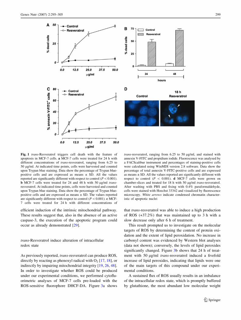
We studied the effects of trans-resveratrol treatment on cell
viability of human breast cancer cells MCF-7. Figure 1a
shows the percentages of Trypan blue-positive cells
after 24 h of treatment with different concentrations
of trans-resveratrol. The data reported evidenced that
trans-resveratrol decreased cell viability in a dose- and
time-dependent manner (Fig. 1a, b). In particular, at con-
centration of 50 lg/ml, the percentage of dead cells at 24 h
was about 48%, phenomenon that became more evident
after 48 h of treatment, reaching values of about 72%
(Fig. 1b).
In order to distinguish which kind of cell death was
induced under our experimental conditions (apoptosis
versus necrosis), MCF-7 cells were treated for 24 h with
the different concentrations of trans-resveratrol and then
utilized for cytofluorimetric analyses upon staining with
annexin V-FITC. Figure 1c shows the percentages of
annexin V-FITC positive cells the amount of which par-
alleled the data previously obtained by direct counts upon
Trypan blue exclusion (Fig. 1a). Moreover, fluorescence
microscope analysis of nuclei upon staining with DNA-
specific dye Hoechst 33342, showed that MCF-7 cells,
treated for 18 h with 50 lg/ml trans-resveratrol, exhibited
areas of condensed chromatin confirming that this com-
pound is able to commit MCF-7 cell to death with the
features of apoptosis (Fig. 1d).
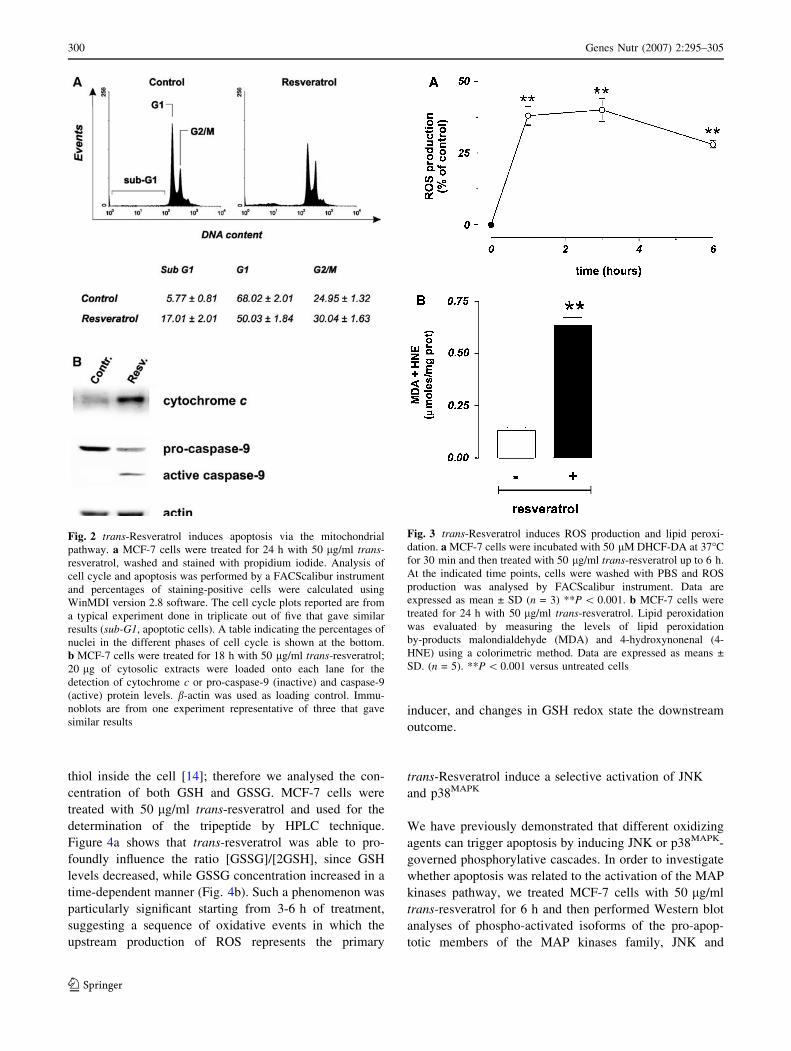
MCF-7 cells represent a well-established hystotype
lacking of caspase-3, for this reason, they undergo apop-
tosis without showing inter-nucleosomal cleavages [29].
This was confirmed by cytofluorimetric analyses of cell
cycle carried out upon staining with propidium iodide. As
shown in Fig. 2a the representative histograms of MCF-7,
cells treated for 24 h with 50 lg/ml trans-resveratrol,
evidenced a low but significant arrest of the cell cycle in
G2/M phase (see table below). As a consequence of the
deficiency of caspase-3 activity, the rate of sub-G1
(apoptotic) cells was lower with respect to the data
obtained by the previous analyses (see Figs. 1, 2).
To characterize the mechanism through which apoptosis
occurs, MCF-7 cells were treated with 50 lg/ml trans-
resveratrol and used for sub-cellular fractionation in
order to isolate mitochondria and the cytosolic fractions.
Figure 2b shows the Western blot analyses of cytosolic
cytochrome c and caspase-9 after 18 h of treatment; in
particular, cytochrome c was efficiently released from
mitochondria, and caspase-9 activated, indicating an
298 Genes Nutr (2007) 2:295–305
123
efficient induction of the intrinsic mitochondrial pathway.
These results suggest that, also in the absence of an active
caspase-3, the execution of the apoptotic program could
occur as already demonstrated [29].
trans-Resveratrol induce alteration of intracellular
redox state
As previously reported, trans-resveratrol can produce ROS,
directly by reacting as phenoxyl radical with O2 [17, 18], or
indirectly by impairing mitochondrial integrity [19, 26, 48].
In order to investigate whether ROS could be produced
under our experimental conditions, we performed cytoflu-
orimetric analyses of MCF-7 cells pre-loaded with the
ROS-sensitive fluorophore DHCF-DA. Figure 3a shows
that trans-resveratrol was able to induce a high production
of ROS (+37.2%) that was maintained up to 3 h with a
slow decrease only after 6 h of treatment.
This result prompted us to investigate on the molecular
targets of ROS by determining the content of protein oxi-
dation and the extent of lipid peroxidation. No increase in
carbonyl content was evidenced by Western blot analyses
(data not shown); conversely, the levels of lipid peroxides
significantly changed. Figure 3b shows that 24 h of treat-
ment with 50 lg/ml trans-resveratrol induced a fivefold
increase of lipid peroxides, indicating that lipids were one
of the main targets of this compound under our experi-
mental conditions.
A sustained flux of ROS usually results in an imbalance
of the intracellular redox state, which is promptly buffered
by glutathione, the most abundant low molecular weight
Fig. 1 trans-Resveratrol triggers cell death with the feature of
apoptosis in MCF-7 cells. a MCF-7 cells were treated for 24 h with
different concentrations of trans-resveratrol, ranging from 6.25 to
50 lg/ml. At indicated time points, cells were harvested and counted
upon Trypan blue staining. Data show the percentage of Trypan blue-
positive cells and are expressed as means ± SD. All the values
reported are significantly different with respect to control (P\0.001).
b MCF-7 cells were treated for 24 and 48 h with 50 lg/ml trans-
resveratrol. At indicated time points, cells were harvested and counted
upon Trypan blue staining. Data show the percentage of Trypan blue-
positive cells and are expressed as means ± SD. The values reported
are significantly different with respect to control (P\0.001). c MCF-
7 cells were treated for 24 h with different concentrations of
trans-resveratrol, ranging from 6.25 to 50 lg/ml, and stained with
annexin V-FITC and propidium iodide. Fluorescence was analysed by
a FACScalibur instrument and percentages of staining-positive cells
were calculated using WinMDI version 2.8 software. Data show the
percentage of total annexin V-FITC-positive cells and are expressed
as means ± SD. All the values reported are significantly different with
respect to control (P \ 0.001). d MCF-7 cells were grown on
chamber-slices and treated for 18 h with 50 lg/ml trans-resveratrol.
After washing with PBS and fixing with 0.4% paraformaldehyde,
cells were stained with Hoechst 33342 and visualized by fluorescence
microscopy. White arrows indicate condensed chromatin character-
istic of apoptotic nuclei
Genes Nutr (2007) 2:295–305 299
123
thiol inside the cell [14]; therefore we analysed the con-
centration of both GSH and GSSG. MCF-7 cells were
treated with 50 lg/ml trans-resveratrol and used for the
determination of the tripeptide by HPLC technique.
Figure 4a shows that trans-resveratrol was able to pro-
foundly influence the ratio [GSSG]/[2GSH], since GSH
levels decreased, while GSSG concentration increased in a
time-dependent manner (Fig. 4b). Such a phenomenon was
particularly significant starting from 3-6 h of treatment,
suggesting a sequence of oxidative events in which the
upstream production of ROS represents the primary
inducer, and changes in GSH redox state the downstream
outcome.
trans-Resveratrol induce a selective activation of JNK
and p38MAPK
We have previously demonstrated that different oxidizing
agents can trigger apoptosis by inducing JNK or p38MAPK-
governed phosphorylative cascades. In order to investigate
whether apoptosis was related to the activation of the MAP
kinases pathway, we treated MCF-7 cells with 50 lg/ml
trans-resveratrol for 6 h and then performed Western blot
analyses of phospho-activated isoforms of the pro-apop-
totic members of the MAP kinases family, JNK and
Fig. 2 trans-Resveratrol induces apoptosis via the mitochondrial
pathway. a MCF-7 cells were treated for 24 h with 50 lg/ml trans-
resveratrol, washed and stained with propidium iodide. Analysis of
cell cycle and apoptosis was performed by a FACScalibur instrument
and percentages of staining-positive cells were calculated using
WinMDI version 2.8 software. The cell cycle plots reported are from
a typical experiment done in triplicate out of five that gave similar
results (sub-G1, apoptotic cells). A table indicating the percentages of
nuclei in the different phases of cell cycle is shown at the bottom.
b MCF-7 cells were treated for 18 h with 50 lg/ml trans-resveratrol;
20 lg of cytosolic extracts were loaded onto each lane for the
detection of cytochrome c or pro-caspase-9 (inactive) and caspase-9
(active) protein levels. b-actin was used as loading control. Immu-
noblots are from one experiment representative of three that gave
similar results
Fig. 3 trans-Resveratrol induces ROS production and lipid peroxi-
dation. a MCF-7 cells were incubated with 50 lM DHCF-DA at 37�C
for 30 min and then treated with 50 lg/ml trans-resveratrol up to 6 h.
At the indicated time points, cells were washed with PBS and ROS
production was analysed by FACScalibur instrument. Data are
expressed as mean ± SD (n = 3) **P \ 0.001. b MCF-7 cells were
treated for 24 h with 50 lg/ml trans-resveratrol. Lipid peroxidation
was evaluated by measuring the levels of lipid peroxidation
by-products malondialdehyde (MDA) and 4-hydroxynonenal (4-
HNE) using a colorimetric method. Data are expressed as means ±
SD. (n = 5). **P \ 0.001 versus untreated cells
300 Genes Nutr (2007) 2:295–305
123
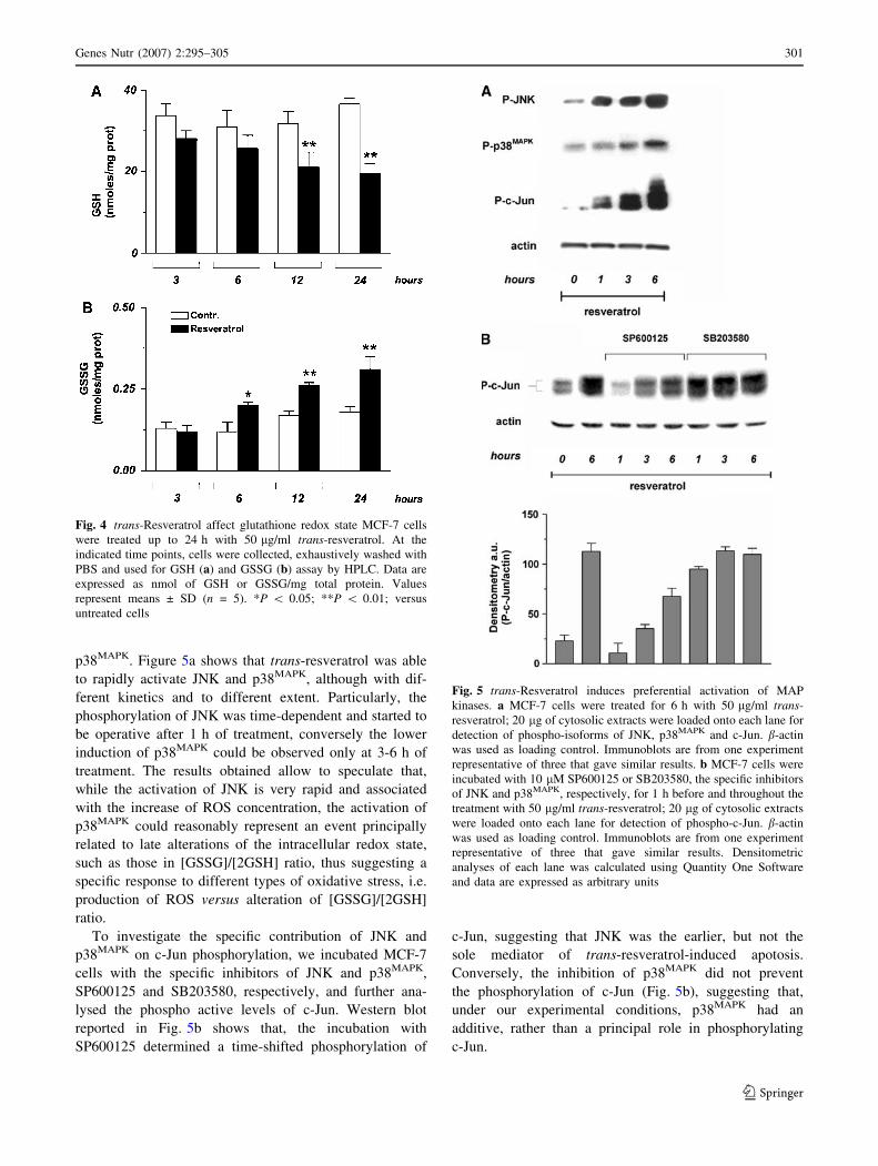
p38MAPK. Figure 5a shows that trans-resveratrol was able
to rapidly activate JNK and p38MAPK, although with dif-
ferent kinetics and to different extent. Particularly, the
phosphorylation of JNK was time-dependent and started to
be operative after 1 h of treatment, conversely the lower
induction of p38MAPK could be observed only at 3-6 h of
treatment. The results obtained allow to speculate that,
while the activation of JNK is very rapid and associated
with the increase of ROS concentration, the activation of
p38MAPK could reasonably represent an event principally
related to late alterations of the intracellular redox state,
such as those in [GSSG]/[2GSH] ratio, thus suggesting a
specific response to different types of oxidative stress, i.e.
production of ROS versus alteration of [GSSG]/[2GSH]
ratio.
To investigate the specific contribution of JNK and
p38MAPK on c-Jun phosphorylation, we incubated MCF-7
cells with the specific inhibitors of JNK and p38MAPK,
SP600125 and SB203580, respectively, and further ana-
lysed the phospho active levels of c-Jun. Western blot
reported in Fig. 5b shows that, the incubation with
SP600125 determined a time-shifted phosphorylation of
c-Jun, suggesting that JNK was the earlier, but not the
sole mediator of trans-resveratrol-induced apotosis.
Conversely, the inhibition of p38MAPK did not prevent
the phosphorylation of c-Jun (Fig. 5b), suggesting that,
under our experimental conditions, p38MAPK had an
additive, rather than a principal role in phosphorylating
c-Jun.
Fig. 4 trans-Resveratrol affect glutathione redox state MCF-7 cells
were treated up to 24 h with 50 lg/ml trans-resveratrol. At the
indicated time points, cells were collected, exhaustively washed with
PBS and used for GSH (a) and GSSG (b) assay by HPLC. Data are
expressed as nmol of GSH or GSSG/mg total protein. Values
represent means ± SD (n = 5). *P \ 0.05; **P \ 0.01; versus
untreated cells
Fig. 5 trans-Resveratrol induces preferential activation of MAP
kinases. a MCF-7 cells were treated for 6 h with 50 lg/ml trans-
resveratrol; 20 lg of cytosolic extracts were loaded onto each lane for
detection of phospho-isoforms of JNK, p38MAPK and c-Jun. b-actin
was used as loading control. Immunoblots are from one experiment
representative of three that gave similar results. b MCF-7 cells were
incubated with 10 lM SP600125 or SB203580, the specific inhibitors
of JNK and p38MAPK, respectively, for 1 h before and throughout the
treatment with 50 lg/ml trans-resveratrol; 20 lg of cytosolic extracts
were loaded onto each lane for detection of phospho-c-Jun. b-actin
was used as loading control. Immunoblots are from one experiment
representative of three that gave similar results. Densitometric
analyses of each lane was calculated using Quantity One Software
and data are expressed as arbitrary units
Genes Nutr (2007) 2:295–305 301
123

The inhibition of JNK and p38MAPK inhibits apoptosis
induced by trans-resveratrol
The involvement of JNK and p38MAPK in the induction of
apoptosis upon treatment with trans-resveratrol was
investigated by incubating MCF-7 cells with specific
inhibitors of JNK and p38MAPK. Figure 6a shows the cy-
tofluorimetric analyses upon propidium iodide staining; in
particular, the inhibition of JNK induced a significant
decrease of apoptotic cells, indicating that JNK-dependent
phosphorylative cascade plays a pivotal role in the cell
death induced by trans-resveratrol. On the other hand, the
inhibition of p38MAPK did not affect the content of apop-
totic cells after treatment with trans-resveratrol, indicating
that p38MAPK pathway was not effective in the induction of
cell death. Moreover, Western blot analyses showed that
the inhibition of p38MAPK only partially decreased the level
of cytosolic cytochrome c (–30%), whereas the inhibition
of JNK induced a significant decrease of the protein in the
cytosol (–65%), confirming the pivotal role played by the
latter kinase in the apoptotic process. This trend was con-
firmed by fluorescence microscopy analyses of MCF-7
nuclei stained with Hoechst 33342. Figure 6c shows that,
after incubation with SP600125, chromatin distribution
within the nuclei was not affected by trans-resveratrol,
whereas the inhibition of p38MAPK-mediated pathway was
not able to efficiently prevent the canonical apoptotic
changes of chromatin. In particular, apoptotic bodies were
still visible after 18 h of treatment (white arrows).
Discussion
trans-Resveratrol has been recently reported to possess
several beneficial effects for human health. Besides its
antioxidant activity, it has been suggested that trans-res-
veratrol inhibits each phase of tumor growth, by interacting
with several cellular targets, thereby activating cell cycle
arrest and death. At molecular level, it has been suggested
that high concentration of polyphenols can induce the
Fig. 6 JNK and p38MAPK are differently responsible for trans-
resveratrol-induced apoptosis. MCF-7 cells were incubated with
10 lM SP600125 or SB203580, the specific inhibitors of JNK and
p38MAPK, respectively, for 1 h before and throughout the treatment
with 50 lg/ml trans-resveratrol. a After 24 h treatment, cells were
stained with propidium iodide for cytofluorimetric analyses. Percent-
ages of apoptotic cells are expressed as means ± S.D. (n = 5). **P\0.001. b. After 18 h 20 lg of cytosolic extracts were loaded onto each
lane for detection of cytochrome c. b-actin was used as loading
control. Immunoblots are from one experiment representative of three
that gave similar results. Densitometric analyses of each lane were
calculated using Quantity One Software and data are expressed as
arbitrary units. c Alternatively, cells were grown on chamber-slices
and treated for 18 h. After washing with PBS and fixing with 0.4%
paraformaldehyde, cells were stained with Hoechst 33342 and
visualized by fluorescence microscopy. White arrows indicate
condensed chromatin or apoptotic bodies after trans-resveratrol
treatment
302 Genes Nutr (2007) 2:295–305
123

production of ROS, rather than reinforcing the antioxidant
defense, and it has been hypothesized that this pro-oxidant
function can play a pivotal role in resveratrol-induced
apoptosis in tumor cells [19]. In fact, the generation of
phenoxyl radicals of polyphenols by the peroxidase–H2O2
system, which co-oxidizes cellular glutathione or NADH,
accompanied by O2 uptake to form ROS has been demon-
strated [17, 18]. In addition, polyphenols have been
proposed to induce oxidative stress indirectly by targeting
mitochondrial electron transport chain, thereby generating a
downstream flux of ROS [26, 48]. In agreement with pre-
vious works, which identified a role for oxyradicals in
cytotoxicity of polyphenols [6, 46, 19], we observed an
early increase of ROS levels after treatment with high doses
of trans-resveratrol in human breast cancer cells MCF-7.
The involvement of ROS has been proposed as upstream
event occurring during treatment with high (micro-molar)
concentrations of trans-resveratrol [1, 21], as well as with
other polyphenols, including epigallocatechin-3-gallate [44,
53), woodfordin I [35] and quercetin [33, 64]. In our
experimental system ROS burst occurs precociously (within
the first hour) and the detrimental effects produced were
confirmed by the increase in lipid peroxidation by-products,
MDA and 4-HNE. On the other hand, no damage on protein
in terms of carbonyls content was observed, implying that
the hydrophobicity of trans-resveratrol renders the lipids
the preferential targets of its pro-oxidant action. It is worth
to notice that, even though the pro-oxidant effects observed
in this study were referred to the highest dose of trans-
resveratrol (50 lg/ml), we suggest that apoptosis observed
at the lowest dose (6.25 lg/ml) is still mediated by ROS
production and oxidative insults. However, since differen-
tiated cells are equipped with high levels of both enzymatic
and non-enzymatic antioxidants, we can presume that,
as previously demonstrated with other diet-derived
pro-oxidant molecules [12], trans-resveratrol could be non
toxic towards normal tissues.
We also found that trans-resveratrol induced a signifi-
cant alteration of GSH/GSSG ratio in agreement with the
knowledge that the phenoxyl radical form of polyphenols
can produce ROS in association with a selective depletion
of intracellular GSH, to form GSH-conjugates or GSSG.
However, under our experimental conditions, GSH deple-
tion was a late event, which allows speculating that it
represents just a epiphenomenon rather than the pivotal
redox change governing resveratrol-induced apoptosis.
The direct relationship between alteration of intracellu-
lar redox state and activation of MAP kinases-mediated
phosphorylative pathways has been exhaustively demon-
strated [15]. Moreover, a role for polyphenols in the
activation of different MAP kinases members has been
suggested as necessary event for downstream induction of
apoptosis in cancer cells [31]. Here we reported that the
oxidative stress deriving from treatments with trans-res-
veratrol is differently associated with MAP kinases
activation. In particular, trans-resveratrol seems to prefer-
entially induce the JNK/c-Jun-mediated phosphorylative
cascade as the principal mediator of apoptosis. Moreover,
if we consider the relationship with the oxidative altera-
tions observed (ROS or GSH-dependent) we could
hypothesize a selective activation of different MAP kina-
ses, with JNK more responsive to the increase of ROS
concentration and p38MAPK preferentially responsive to the
alteration of GSH redox state. This different sensitivity of
MAP kinases to ROS- or GSH-dependent intracellular
redox changes is in line with our recent results suggesting
that different types of oxidative stress could preferentially
drive different phosphorylative pathways [10, 12, 13]. On
Fig. 7 Scheme of MAPK-
mediated pathways downstream
of trans-resveratrol treatment
Genes Nutr (2007) 2:295–305 303
123
this basis, we can outline the results obtained as shown in
Fig. 7. In particular trans-resveratrol activates JNK and
p38MAPK, but its different capability to alter intracellular
redox environment concur to make JNK activation the
principal response, and p38MAPK a redundant kinase for the
induction of cell death. Finally, further works should be
done in animal models in order to determine the concen-
trations that allow trans-resveratrol to behave as
chemotherapeutic other than antioxidant.
Acknowledgments This work was partially supported by grants
from FIRB, MIUR, and Ministero della Sanita.
References
1. Azmi AS, Bhat SH, Hanif S, Hadi SM (2006) Plant polyphenols
mobilize endogenous copper in human peripheral lymphocytes
leading to oxidative DNA breakage: a putative mechanism for
anticancer properties. FEBS Lett 580:533–538
2. Bhat KPL, Kosmeder JW, Pezzuto JM (2001) Biological effects
of resveratrol. Antioxid Redox Signal 3:1041–1064
3. Bohmont C, Aaronson LM, Mann K, Pardini RS (1987) Inhibi-
tion of mitochondrial NADH oxidase, succinoxidase, and ATPase
by naturally occurring flavonoids. J Nat Prod 50:427–433
4. Briviba K, Pan L, Rechkemmer G (2002) Red wine polyphenols
inhibit the growth of colon carcinoma cells and modulate the
activation pattern of mitogen-activated protein kinases. J Nutr
132:2814–2818
5. Clement MV, Hirpara JL, Chawdhury SH, Pervaiz S (1998)
Chemopreventive agent resveratrol, a natural product derived
from grapes, triggers CD95 signaling-dependent apoptosis in
human tumor cells. Blood 92:996–1002
6. Chichirau A, Flueraru M, Chepelev LL, Wright JS, Willmore
WG, Durst T, Hussain HH, Charron M (2005) Mechanism of
cytotoxicity of catechols and a naphthalenediol in PC12-AC
cells: the connection between extracellular autoxidation and
molecular electronic structure. Free Radic Biol Med 38:344–355
7. Delmas D, Rebe C, Lacour S, Filomenko R, Athias A, Gambert P,
Cherkaoui-Malki M, Jannin B, Dubrez-Daloz L, Latruffe N,
Solary E (2003) Resveratrol-induced apoptosis is associated with
Fas redistribution in the rafts and the formation of a death-
inducing signaling complex in colon cancer cells. J Biol Chem
278:41482–41490
8. Dorrie J, Gerauer H, Wachter Y, Zunino SJ (2001) Resveratrol
induces extensive apoptosis by depolarizing mitochondrial
membranes and activating caspase-9 in acute lymphoblastic
leukemia cells. Cancer Res 61:4731–4739
9. Elbling L, Weiss RM, Teufelhofer O, Uhl M, Knasmueller S,
Schulte-Hermann R, Berger W, Micksche M (2005) Green tea
extract and (-)-epigallocatechin-3-gallate, the major tea catechin,
exert oxidant but lack antioxidant activities. FASEB J 19:807–809
10. Filomeni G, Aquilano K, Civitareale P, Rotilio G, Ciriolo MR
(2005) Activation of c-Jun-N-terminal kinase is required for
apoptosis triggered by glutathione disulfide in neuroblastoma
cells. Free Radic Biol Med 39:345–354
11. Filomeni G, Aquilano K, Rotilio G, Ciriolo MR (2005) Antiap-
optotic response to GSH depletion: involvement of heat shock
proteins and NF-jB activation. Antioxid Redox Signal 7:446–455
12. Filomeni G, Aquilano K, Rotilio G, Ciriolo MR (2003) Reactive
oxygen species-dependent c-Jun NH2-terminal kinase/c-Jun sig-
naling cascade mediates neuroblastoma cell death induced by
diallyl disulfide. Cancer Res 63:5940–5949
13. Filomeni G, Aquilano K, Rotilio G, Ciriolo MR (2005) Gluta-
thione-related systems and modulation of extracellular signal-
regulated kinases are involved in resistance of AGS adenocarci-
noma gastric cells to diallyl disulfide-induced apoptosis. Cancer
Res 65:11735–11742
14. Filomeni G, Rotilio G, Ciriolo MR (2002) Cell signalling and the
glutathione redox system. Biochem Pharmacol 64:1057–1064
15. Filomeni G, Rotilio G, Ciriolo MR (2005) Disulfide relays and
phosphorylative cascades: partners in redox-mediated signalling
pathways. Cell Death Differ 12:1555–1563
16. Filomeni G, Rotilio G, Ciriolo MR (2003) Glutathione disulfide
induces apoptosis in U937 cells by a redox-mediated p38 MAP
kinase pathway. FASEB J 17:64–66
17. Galati G, Chan T, Wu B, O’Brien PJ (1999) Glutathione-
dependent generation of reactive oxygen species by the peroxi-
dase-catalyzed redox cycling of flavonoids. Chem Res Toxicol
12:521–525
18. Galati G, Sabzevari O, Wilson JX, O’Brien PJ (2002) Prooxidant
activity and cellular effects of the phenoxyl radicals of dietary
flavonoids and other polyphenolics. Toxicology 177:91–104
19. Galati G, O’Brien PJ (2004) Potential toxicity of flavonoids and
other dietary phenolics: significance for their chemopreventive
and anticancer properties. Free Radic Biol Med 37:287–303
20. Garg AK, Buchholz TA, Aggarwal BB (2005) Chemosensitiza-
tion and radiosensitization of tumors by plant polyphenols.
Antioxid Redox Signal 7:1630–1647
21. Hadi SM, Asad SF, Singh S, Ahmad A (2000) Putative mecha-
nism for anticancer and apoptosis-inducing properties of plant-
derived polyphenolic compounds. IUBMB Life 50:167–171
22. Herman-Antosiewicz A, Singh SV (2005) Checkpoint kinase 1
regulates diallyl trisulfide-induced mitotic arrest in human pros-
tate cancer cells. Journal of Biol Chem 280:28519–28528
23. Hileman EO, Liu J, Albitar M, Keating MJ, Huang P (2003)
Intrinsic oxidative stress in cancer cells: a biochemical basis for
therapeutic selectivity. Cancer Chemother Pharmacol 53:209–219
24. Hodnick WF, Kung FS, Roettger WJ, Bohmont CW, Pardini RS
(1986) Inhibition of mitochondrial respiration and production of
toxic oxygen radicals by flavonoids: a structure–activity study.
Biochem Pharmacol 35:2345–2357
25. Hodnick WF, Milosavljevic EB, Nelson JH, Pardini RS (1988)
Electrochemistry of flavonoids: relationships between redox
potentials, inhibition of mitochondrial respiration, and production
of oxygen radicals by flavonoids. Biochem Pharmacol 37:2607–
2611
26. Hodnick WF, Ahmad S, Pardini RS (1998) Induction of oxidative
stress by redox active flavonoids. Adv Exp Med Biol 439:131–150
27. Hsieh TC, Wu JM (1999) Differential effects on growth, cell
cycle arrest, and induction of apoptosis by resveratrol in human
prostate cancer cell lines. Exp Cell Res 249:109–115
28. Jang M, Cai L, Udeani GO, Slowing KV, Thomas CF, Beecher
CW, Fong HH, Farnsworth NR, Kinghorn AD, Mehta RG, Moon
RC, Pezzuto JM (1997) Cancer chemopreventive activity of
resveratrol, a natural product derived from grapes. Science
275:218–220
29. Kagawa S, Gu J, Honda T, McDonnell TJ, Swisher SG, Roth JA,
Fang B (2001) Deficiency of caspase-3 in MCF7 cells blocks
Bax-mediated nuclear fragmentation but not cell death. Clin
Cancer Res 7:1474–1480
30. Kim YA, Lee WH, Choi TH, Rhee SH, Park KY, Choi YH (2003)
Involvement of p21WAF1/CIP1, pRB, Bax and NF-kappaB in
induction of growth arrest and apoptosis by resveratrol in human
lung carcinoma A549 cells. Int J Oncol 23:1143–1149
31. Kong AN, Owuor E, Yu R, Hebbar V, Chen C, Hu R, Mandlekar
S (2001) Induction of xenobiotic enzymes by the MAP kinase
pathway and the antioxidant or electrophile response element
(ARE/EpRE). Drug Metab Rev 33:255–271
304 Genes Nutr (2007) 2:295–305
123

32. Kong Q, Beel JA, Lillehei KO (2000) A threshold concept for
cancer therapy. Med Hypothesis 55:29–35
33. Lee ER, Kang YJ, Kim JH, Lee HAT, Cho SG (2005) Modulation
of apoptosis in HaCaT keratinocytes via differential regulation of
ERK signaling pathway by flavonoids. J Biol Chem 280:31498–
31507
34. Li Y, Liu J, Liu X, Xing K, Wang Y, Li F, Yao L (2006) Res-
veratrol-induced cell inhibition of growth and apoptosis in MCF7
human breast cancer cells are associated with modulation of
phosphorylated Akt and caspase-9. Appl Biochem Biotechnol
135:181–192
35. Liu MJ, Wang Z, Li HX, Wu RC, Liu YZ, Wu QY (2004)
Mitochondrial dysfunction as an early event in the process of
apoptosis induced by woodfordin I in human leukemia K562
cells. Toxicol Appl Pharmacol 194:141–155
36. Lowry OH, Rosebrough NJ, Farr AL, Randall RJ (1951) Protein
measurement with the Folin-phenol reagent. J Biol Chem
193:265–275
37. Manna SK, Mukhopadhyay A, Aggarwal BB (2000) Resveratrol
suppresses TNF-induced activation of nuclear transcription fac-
tors NF-kappa B, activator protein-1, and apoptosis: potential role
of reactive oxygen intermediates and lipid peroxidation. J
Immunol 164:6509–6519
38. Matsuzawa A, Ichijo H (2005) Stress-responsive protein kinases
in redox-regulated apoptosis signaling. Antioxid Redox Signal
7:472–481
39. Matsuzawa A, Nishitoh H, Tobiume K, Takeda K, Ichijo H
(2002) Physiological roles of ASK1-mediated signal transduction
in oxidative stress- and endoplasmic reticulum stress-induced
apoptosis: advanced findings from ASK1 knockout mice. Anti-
oxid Redox Signal 4:415–425
40. Meunier S, Hanedanian M, Desage-El Murr M, Nowaczyk S, Le
Gall T, Pin S, Renault JP, Boquet D, Creminon C, Mioskowski C,
Taran F (2005) High-throughput evaluation of antioxidant and
pro-oxidant activities of polyphenols with thymidine protection
assays. Chem Biochem 6:1234–1241
41. Mgbonyebi OP, Russo J, Russo IH (1998) Antiproliferative effect
of synthetic resveratrol on human breast epithelial cells. Int J
Oncol 12:865–869
42. Michels G, Watjen W, Niering P, Steffan B, Thi QH, Chovolou
Y, Kampkotter A, Bast A, Proksch P, Kahl R (2005) Pro-apop-
totic effects of the flavonoid luteolin in rat H4IIE cells.
Toxicology 206:337–348
43. Mohan J, Gandhi AA, Bhavya BC, Rashmi R, Karunagaran D,
Indu R, Santhoshkumar TR (2006) Caspase-2 triggers Bax-Bak-
dependent and -independent cell death in colon cancer cells
treated with resveratrol. J Biol Chem 281:17599–17611
44. Nakazato T, Ito K, Ikeda Y, Kizaki M (2005) Green tea com-
ponent, catechin, induces apoptosis of human malignant B cells
via production of reactive oxygen species. Clin Cancer Res
11:6040–6049
45. Narayanan BA, Narayanan NK, Re GG, Nixon DW (2003) Dif-
ferential expression of genes induced by resveratrol in LNCaP
cells: p53-mediated molecular targets. Int J Cancer 104:204–212
46. Nemeikaite-Ceniene A, Imbrasaite A, Sergediene E, Cenas N
(2005) Quantitative structure-activity relationships in prooxi-
dant cytotoxicity of polyphenols: role of potential of phenoxyl
radical/phenol redox couple. Arch Biochem Biophys 441:182–
190
47. Nicoletti I, Migliorati G, Pagliacci MC, Grignani F, Riccardi C
(1991) A rapid and simple method for measuring thymocyte
apoptosis by propidium iodide staining and flow cytometry. J
Immunol Methods 139:271–279
48. Pardini RS (1995) Toxicity of oxygen from naturally occurring
redox-active pro-oxidants. Arch Insect Biochem Physiol 29:101–
118
49. Pelicano H, Carney D, Huang P (2004) ROS stress in cancer cells
and therapeutic implications. Drug Resist Updat 7:97–110
50. Pellecchia M, Reed JC (2004) Inhibition of anti-apoptotic Bcl-2
family proteins by natural polyphenols: new avenues for cancer
chemoprevention and chemotherapy. Curr Pharm Des 10:1387–
1398
51. Podmore ID, Griffiths HR, Herbert KE, Mistry N, Mistry P,
Lunec J (1998) Vitamin C exhibits pro-oxidant properties. Nature
392:559
52. Qanungo S, Das M, Haldar S, Basu A (2005) Epigallocatechin-3-
gallate induces mitochondrial membrane depolarization and
caspase-dependent apoptosis in pancreatic cancer cells. Carci-
nogenesis 26:958–967
53. Raza H, John A (2005) Green tea polyphenol epigallocatechin-3-
gallate differentially modulates oxidative stress in PC12 cell
compartments. Toxicol Appl Pharmacol 207:212–220
54. Rossi L, Aquilano A, Filomeni G, Lombardo MF, Rotilio G,
Ciriolo MR (2004) Putative mechanisms of apoptosis related to
redox unbalance. In: Ozben K, Chevion M (eds) Frontiers in
neurodegenerative disorders and aging: fundamental aspect,
clinical perspectives and new insight. IOS press, NATO Sciences
Series, The Netherlands, pp 207–250
55. Rotilio G, Mavelli I, Rossi L, Ciriolo MR (1985) Biochemical
mechanism of oxidative damage by redox-cycling drugs. Environ
Health Perspect 64:259–264
56. Schneider Y, Vincent F, Duranton B, Badolo L, Gosse F, Berg-
mann C, Seiler N, Raul F (2000) Anti-proliferative effect of
resveratrol, a natural component of grapes and wine, on human
colonic cancer cells. Cancer Lett 158:85–91
57. Sexton E, Van Themsche C, LeBlanc K, Parent S, Lemoine P,
Asselin E (2006) Resveratrol interferes with AKT activity and
triggers apoptosis in human uterine cancer cells. Mol Cancer
175:45
58. She QB, Bode AM, Ma WY, Chen NY, Dong Z (2001) Resve-
ratrol-induced activation of p53 and apoptosis is mediated by
extracellular-signal-regulated protein kinases and p38 kinase.
Cancer Res 61:1604–1610
59. Subbaramaiah K, Michaluart P, Chung WJ, Tanabe T, Telang, N,
Dannenberg AJ (1999) Resveratrol inhibits cyclooxygenase-2
transcription in human mammary epithelial cells. Ann N Y Acad
Sci 889:214–223
60. Tinhofer I, Bernhard D, Senfter M, Anether G, Loeffler M,
Kroemer G, Kofler R, Csordas A, Greil R (2001) Resveratrol, a
tumor-suppressive compound from grapes, induces apoptosis via
a novel mitochondrial pathway controlled by Bcl-2. FASEB J
15:1613–1615
61. Ueda S, Masutani H, Nakamura H, Tanaka T, Ueno M, Yodoi J
(2002) Redox control of cell death. Antioxid Redox Signal
4:405–414
62. Woo JH, Lim JH, Kim YH, Suh SI, Min DS, Chang JS, Lee YH,
Park JW, Kwon TK (2004) Resveratrol inhibits phorbol myristate
acetate-induced matrix metalloproteinase-9 expression by inhib-
iting JNK and PKC delta signal transduction. Oncogene 23:1845–
1853
63. Xiao D, Pinto JT, Soh JW, Deguchi A, Gundersen GG, Palazzo AF,
Yoon JT, Shirin H, Weinstein IB (2003) Induction of apoptosis bythe garlic-derived compound S-allylmercaptocysteine (SAMC) is
associated with microtubule depolymerization and c-Jun NH2-
terminal kinase 1 activation. Cancer Res 63:6825–6837
64. Yamashita N, Kawanishi S (2000) Distinct mechanisms of DNA
damage in apoptosis induced by quercetin and luteolin. Free
Radic Res 33:623–633
65. Yu R, Hebbar V, Kim DW, Mandlekar S, Pezzuto JM, Kong AN
(2001) Resveratrol inhibits phorbol ester and UV-induced acti-
vator protein 1 activation by interfering with mitogen-activated
protein kinase pathways. Mol Pharmacol 60:217–224
Genes Nutr (2007) 2:295–305 305
123