Cortical auditory processing in preterm newborns: An ERP study
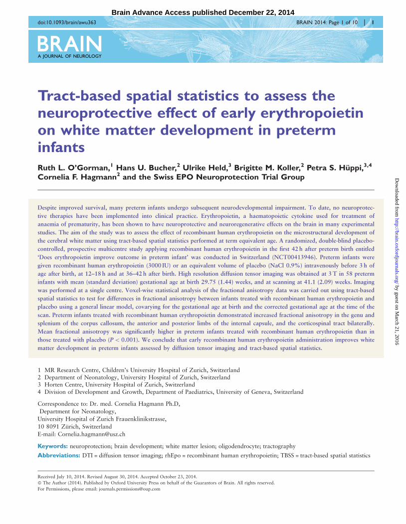
Tract-based spatial statistics to assess theneuroprotective effect of early erythropoietinon white matter development in preterminfants
Ruth L. O’Gorman,1 Hans U. Bucher,2 Ulrike Held,3 Brigitte M. Koller,2 Petra S. Huppi,3,4
Cornelia F. Hagmann2 and the Swiss EPO Neuroprotection Trial Group
Despite improved survival, many preterm infants undergo subsequent neurodevelopmental impairment. To date, no neuroprotec-
tive therapies have been implemented into clinical practice. Erythropoietin, a haematopoietic cytokine used for treatment of
anaemia of prematurity, has been shown to have neuroprotective and neuroregenerative effects on the brain in many experimental
studies. The aim of the study was to assess the effect of recombinant human erythropoietin on the microstructural development of
the cerebral white matter using tract-based spatial statistics performed at term equivalent age. A randomized, double-blind placebo-
controlled, prospective multicentre study applying recombinant human erythropoietin in the first 42 h after preterm birth entitled
‘Does erythropoietin improve outcome in preterm infant’ was conducted in Switzerland (NCT00413946). Preterm infants were
given recombinant human erythropoietin (3000 IU) or an equivalent volume of placebo (NaCl 0.9%) intravenously before 3 h of
age after birth, at 12–18 h and at 36–42 h after birth. High resolution diffusion tensor imaging was obtained at 3 T in 58 preterm
infants with mean (standard deviation) gestational age at birth 29.75 (1.44) weeks, and at scanning at 41.1 (2.09) weeks. Imaging
was performed at a single centre. Voxel-wise statistical analysis of the fractional anisotropy data was carried out using tract-based
spatial statistics to test for differences in fractional anisotropy between infants treated with recombinant human erythropoietin and
placebo using a general linear model, covarying for the gestational age at birth and the corrected gestational age at the time of the
scan. Preterm infants treated with recombinant human erythropoietin demonstrated increased fractional anisotropy in the genu and
splenium of the corpus callosum, the anterior and posterior limbs of the internal capsule, and the corticospinal tract bilaterally.
Mean fractional anisotropy was significantly higher in preterm infants treated with recombinant human erythropoietin than in
those treated with placebo (P5 0.001). We conclude that early recombinant human erythropoietin administration improves white
matter development in preterm infants assessed by diffusion tensor imaging and tract-based spatial statistics.
1 MR Research Centre, Children’s University Hospital of Zurich, Switzerland2 Department of Neonatology, University Hospital of Zurich, Switzerland3 Horten Centre, University Hospital of Zurich, Switzerland4 Division of Development and Growth, Department of Paediatrics, University of Geneva, Switzerland
Correspondence to: Dr. med. Cornelia Hagmann Ph.D,
Department for Neonatology,
University Hospital of Zurich Frauenklinikstrasse,
10 8091 Zurich, Switzerland
E-mail: [email protected]
Keywords: neuroprotection; brain development; white matter lesion; oligodendrocyte; tractography
Abbreviations: DTI = diffusion tensor imaging; rhEpo = recombinant human erythropoietin; TBSS = tract-based spatial statistics
doi:10.1093/brain/awu363 BRAIN 2014: Page 1 of 10 | 1
Received July 10, 2014. Revised August 30, 2014. Accepted October 23, 2014.
� The Author (2014). Published by Oxford University Press on behalf of the Guarantors of Brain. All rights reserved.
For Permissions, please email: [email protected]
Brain Advance Access published December 22, 2014 by guest on M
arch 21, 2016http://brain.oxfordjournals.org/
Dow
nloaded from
IntroductionDespite improved survival of preterm infants, the majority
still go on to develop some degree of neurodevelopmental
impairment later in life (Saigal and Doyle, 2008; Latal,
2009). These neurodevelopmental impairments are asso-
ciated with periventricular white matter injury (Woodward
et al., 2006), which is the most common brain injury
observed in preterm infants. Periventricular white matter
disease is also frequently accompanied by neuronal/axonal
disease, as part of the newly described encephalopathy of
prematurity (Volpe, 2009).
In vivo assessment of this neuronal/axonal disease has
become possible with the development of new diffusion
tensor imaging (DTI) protocols in preterm infants (Huppi
and Inder, 2001; Huppi and Dubois, 2006). DTI is a
well-studied MRI technique that allows in vivo assessment
of biological tissues at a microstructural level. In preterm
infants, DTI studies have revealed lower fractional anisot-
ropy values relative to those in healthy term control
infants (Huppi et al., 1998, 2001; Miller et al., 2002;
Mukherjee et al., 2002; Counsell et al., 2003, 2006;
Partridge et al., 2004; Berman et al., 2005; Anjari et al.,
2007; Dudink et al., 2007; Cheong et al., 2009; Skiold
et al., 2010), indicative of altered white matter
development.
Tract-based spatial statistics (TBSS) is an automated,
observer-independent approach for assessing fractional an-
isotropy in the major white matter tracts on a voxel-wise
basis across groups of subjects (Smith et al., 2006). It has
been applied to investigate cerebral microstructure in pre-
term infants at term equivalent age (Anjari et al., 2007),
revealing group differences based on either clinical details
such as chronic lung disease (Ball et al., 2010; Alexandrou
et al., 2014) or birth weight (Lepomaki et al., 2013) or
additional imaging findings such as local white matter
abnormalities (Bassi et al., 2011). A recent TBSS study
has shown that cognitive and motor outcome correlate
with fractional anisotropy in the corpus callosum (van
Kooij et al., 2012), suggesting that TBSS of DTI data has
the potential to be used as a surrogate biomarker for subse-
quent neurodevelopmental outcome. In term infants with
perinatal asphyxial encephalopathy, TBSS analysis has
been used to detect the treatment effects of therapeutic
hypothermia in a small group of patients (n = 10 per
group) (Porter et al., 2010), indicating that TBSS may
also be used to evaluate neuroprotective interventions
(Ball et al., 2013).
The high incidence of neurodevelopmental impairment in
this population is reflected in the ongoing search for
neuroprotective interventions that can prevent injury or
enhance repair of the immature brain, with the goal of
improving long-term motor and cognitive outcome
(Gonzalez and Ferriero, 2009). However, to date no such
neuroprotective intervention has been implemented into
routine clinical care for preterm infants.
Erythropoietin, a haematopoetic cytokine that was ori-
ginally identified for its role in erythropoiesis, is widely
used for the treatment of anaemia in premature infants
(Ohlsson and Aher, 2006; Ghezzi et al., 2010).
Erythropoietin has been shown to have neuroprotective
and neuroregenerative effects on the brain (Dame et al.,
2001; Juul, 2004; van der Kooij et al., 2008), and erythro-
poietin receptors are present on neuron progenitors, neu-
rons, astrocytes, microglia, endothelial cells and erythrocyte
progenitors (Juul and Felderhoff-Mueser, 2007).
Erythropoietin has anti-inflammatory, anti-excitotoxic,
antioxidant and anti-apoptotic effects on neurons and
oligodendrocytes and promotes neurogenesis and angiogen-
esis, which are essential for injury repair and normal
neurodevelopment (Shingo et al., 2001; Juul, 2012).
The safety of recombinant human erythropoietin (rhEpo)
in preterm infants has been established by two clinical trials
with high dose administration (Fauchere et al., 2008; Juul
et al., 2008a, b). Early high-dose rhEpo is well tolerated,
causing no increased mortality or side effects (Fauchere
et al., 2008, 2012). More recently, erythropoietin adminis-
tration has also be shown to be well tolerated by term
infants with hypoxic ischaemic or post-asphyctic encephal-
opathy (Wu et al., 2012).
One clinical trial of rhEpo for treatment of anaemia of
prematurity reporting on the effect of early rhEpo on neu-
rodevelopmental outcome has been published. The neuro-
developmental outcome of 20 preterm infants treated with
early rhEpo was assessed at 18–24 months of age. No ad-
verse or beneficial effect of rhEpo on outcome could be
shown; however, the erythropoietin treatment group
included more infants with intraventricular haemorrhage
than the control group (Newton et al., 1999; Ohls et al.,
2004).
A large retrospective trial indicated clear benefits of
rhEpo for preterm infants, demonstrating that infants trea-
ted with rhEpo scored significantly better in the overall
assessment at school age as well as in the psychological
evaluation (Neubauer et al., 2010). These potential neuro-
protective effects are thought to arise from a putative re-
duction in brain injury, but to date the effects of rhEPO on
cerebral microstructure in preterm infants have not been
established.
The aim of the present study was to assess the effect of
early administration of rhEpo on white matter development
in preterm infants using TBSS.
Materials and methodsEthical approval was granted by the local ethical committee(KEK StV-36/04), and the study was approved by the Swissdrug surveillance unit (Swissmedic, 2005DR3179). The trial isregistered at ClinicalTrials.gov (number NCT00413946). Fulldetails of the trial protocol can be provided on request. Themembers of The Swiss EPO Neuroprotection Trial Group arelisted in Appendix 1.
2 | BRAIN 2014: Page 2 of 10 R. L. O’Gorman et al.
by guest on March 21, 2016
http://brain.oxfordjournals.org/D
ownloaded from
Patient selection
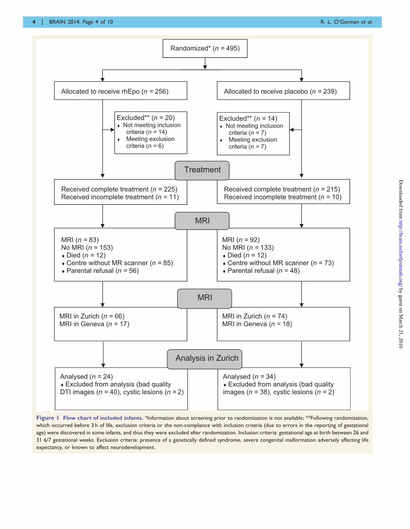
Preterm infants in this study represent a subgroup of infantsenrolled in the randomized, double-blind placebo-controlled,prospective multicentre study entitled ‘Does erythropoetinimprove outcome in preterm infants’ (NCT00413946). Allinfants born between 26 and 31 gestational weeks, fromwhose parents informed consent could be obtained in the first3 h after birth, were included unless they met exclusion criteria(presence of a genetically defined syndrome, severe congenitalmalformation adversely affecting life expectancy, and severecongenital malformation adversely affecting neurodevelopmentor prior palliative care). A total of 495 preterm infants wererecruited into the study between 2005 and 2013, of whom 58infants were excluded for an exclusion criterion and/or deathbefore term equivalent age (Fig. 1). The primary outcome meas-ure of this study is neurodevelopmental outcome assessed at 2and 5 years of age. A secondary outcome measure is braininjury and development assessed by conventional MRI at termequivalent age, measured in a subgroup of 165 infants (Leuchteret al., 2014). For this study, only infants with DTI performed inZurich (n = 140) were included. Clinical co-morbidities such assepsis (blood culture proven), necrotizing enterocolitis, patentductus arteriosus, and chronic lung disease (defined as oxygenrequirement at corrected 36 weeks of gestation) were noted.
Randomization, neuroprotectiveintervention and blinding
The treatment protocol was based on the previously publishedsafety trial (Fauchere et al., 2008). Study medication was ran-domly assigned to each patient number in a 1:1 allocationusing a computer-based random-number generator. Erythro-poetin or an equivalent volume of normal saline (NaCl0.9%) placebo was administered intravenously before 3 h ofage after birth, at 12–18 h and at 36–42 h after birth. A singledose consists of 25 mg (3000 IU) human erythropoietin perkilogram body weight dissolved in 1 ml sterile water. Hospitalstaff, parents, the outcome assessors and data analysts werekept blinded to the allocation.
MRI studies
In Zurich, cerebral MRI was performed with a 3.0 T GEHD.xt MRI scanner (GE Medical Systems), using an 8-channelreceive-only head coil. All infants were scanned under naturalsleep using a vacuum mattress. Ear plugs (attenuation: 24 dB;Earsoft; Aearo) and Minimuffs (attenuation: 7 dB; Natus) wereapplied for noise protection. Oxygen saturation was monitoredduring scanning, and a neonatologist and/or neonatal nursewere present during the MRI investigation.
The structural MRI protocol included T1-weighted imagesacquired with a 3D fast spoiled gradient echo sequence (reso-lution 0.7 � 0.7 � 1.4 mm3, repetition time = 5.7 ms, echotime = 2.6 ms, inversion time = 750 ms, flip angle = 12�), andT2-weighted images acquired with a fast recovery fast spinecho sequence (resolution 0.7 � 0.7 � 1.5 mm3, repetitiontime = 6600 ms, echo time = 126 ms). The structural magneticresonance images were assessed for brain injury based on aclinical scoring system which was adapted from Woodwardand colleagues (Woodward et al., 2006; Leuchter et al.,
2014). The assessor was not aware of the group assignmentof the infants.
DTI was performed in a single centre, using a pulsed gradientspin echo echo planar imaging sequence with echo time = 77 ms,repetition time = 9 s, field of view = 18 cm, matrix = 128 � 128,slice thickness = 3 mm. The diffusion encoding scheme included21 non-collinear gradient encoding directions with b = 1000 andfour interleaved b = 0 images.
DTI data demonstrating motion artefacts (n = 78) or frominfants with cystic lesions (n = 4) apparent on structural MRIwere excluded from further analysis, resulting in a final groupsize of 58 infants, of whom 24 were treated with rhEpo and34 were treated with placebo.
Data analysis
Fractional anisotropy maps were calculated with dtifit andcorrected for eddy current distortions with eddy_correct, partof the FSL software library (http://www.fmrib.ox.ac.uk/fsl/)(Smith et al., 2004). The skull was removed from the b0images using the Brain Extraction Tool (BET) (Smith, 2002)and the corresponding brain mask was applied to the frac-tional anisotropy maps. The fractional anisotropy maps fromall infants were then aligned to the most representative frac-tional anisotropy image from the cohort, using an automatedmethod implemented in the TBSS pipeline, which applies anon-linear registration of each fractional anisotropy map toall other fractional anisotropy images and then selects the frac-tional anisotropy image with the lowest mean warp displace-ment score. An age-appropriate diffusion MRI template wasthen generated from the average of the normalized fractionalanisotropy maps, aligned to the target image corresponding tothe most representative subject. The non-linear registrationswere performed using FNIRT (Andersson et al., 2007a, b),which uses a b-spline representation of the registration warpfield (Rueckert et al., 1999).
Voxel-wise statistical analysis of the fractional anisotropydata was carried out using TBSS (Smith et al., 2006). Afteralignment of the fractional anisotropy maps to the neonataltemplate, a mean image was created and thinned to create amean fractional anisotropy skeleton, which represents the cen-tres of all tracts common to the group. This skeleton wasthresholded at a fractional anisotropy level of 40.15, andvoxel-wise cross-subject statistics were used with Randomise(v2.1) to test for differences in fractional anisotropy betweeninfants treated with rhEpo and placebo using a general linearmodel, including the gestational age at birth and the correctedgestational age at the time of the scan as covariates. A statis-tical threshold of P5 0.05 was applied after family-wise error(FWE) correction for multiple comparisons following thresh-old-free cluster enhancement (Smith and Nichols, 2009).
Student’s t-test or the Mann-Whitney test as appropriate forcontinuous parameters, and chi-square test or Fisher exact testas appropriate for categorical parameters were used to assessdifferences between clinical and demographic parameters inthe erythropoietin and the placebo group. The Pearson coeffi-cient was used to quantify the linear correlation between con-tinuous variables. A multiple linear regression model was fittedto fractional anisotropy, with erythropoietin/placebo as thedeterminant of interest and adjusted for gender and correctedgestational age. All analyses were performed with the statis-tical software R for windows (R Core Team, 2013).
TBSS to assess the neuroprotective effect of erythropoietin in preterm infants BRAIN 2014: Page 3 of 10 | 3
by guest on March 21, 2016
http://brain.oxfordjournals.org/D
ownloaded from
Randomized* (n = 495)
Excluded** (n = 14) ♦ Not meeting inclusion
criteria (n = 7) ♦ Meeting exclusion
criteria (n = 7)
MRI in Zurich (n = 66) MRI in Geneva (n = 17)
MRI (n = 83) No MRI (n = 153) ♦ Died (n = 12) ♦ Centre without MR scanner (n = 85) ♦ Parental refusal (n = 56)
Received complete treatment (n = 225) Received incomplete treatment (n = 11)
MRI (n = 92) No MRI (n = 133) ♦ Died (n = 12) ♦ Centre without MR scanner (n = 73) ♦ Parental refusal (n = 48)
Received complete treatment (n = 215) Received incomplete treatment (n = 10)
MRI in Zurich (n = 74) MRI in Geneva (n = 18)
Treatment
MRI
MRI
Allocated to receive rhEpo (n = 256) Allocated to receive placebo (n = 239)
Excluded** (n = 20) ♦ Not meeting inclusion
criteria (n = 14) ♦ Meeting exclusion
criteria (n = 6)
Analysed (n = 24) ♦ Excluded from analysis (bad quality DTI images (n = 40), cystic lesions (n = 2)
Analysed (n = 34) ♦ Excluded from analysis (bad quality images (n = 38), cystic lesions (n = 2)
Analysis in Zurich
Figure 1 Flow chart of included infants. *Information about screening prior to randomization is not available; **Following randomization,
which occurred before 3 h of life, exclusion criteria or the non-compliance with inclusion criteria (due to errors in the reporting of gestational
age) were discovered in some infants, and thus they were excluded after randomization. Inclusion criteria: gestational age at birth between 26 and
31 6/7 gestational weeks. Exclusion criteria: presence of a genetically defined syndrome, severe congenital malformation adversely affecting life
expectancy, or known to affect neurodevelopment.
4 | BRAIN 2014: Page 4 of 10 R. L. O’Gorman et al.
by guest on March 21, 2016
http://brain.oxfordjournals.org/D
ownloaded from

Results
Patients
The TBSS cohort (n = 58) did not differ from the remaining
infants in the main study (n = 437) with regard to birth
weight, chronic lung disease, sepsis, meningitis, necrotizing
enterocolitis, or retinopathy of prematurity (Table 1).
Infants included in the TBSS study had older mean (SD)
gestational age at birth [n = 58, 29.29 (1.48) weeks] than
the infants in the main study [n = 437, 28.74 (1.8) weeks].
Although this difference was only 3 days it reached statis-
tical significance.
Table 2 summarizes the demographic and clinical details
of the two groups. No significant differences were found in
corrected gestational age at scanning or in any other neo-
natal or clinical details between the two treatment groups
after randomization.
Magnetic resonance
On the basis of the recently published scoring system
(Leuchter et al., 2014), 33 (56.8%) infants had normal
white matter and 42.2% had abnormal white matter.
Three infants had punctate white matter lesions in the cere-
bellar hemispheres.
Tract-based spatial statisitics
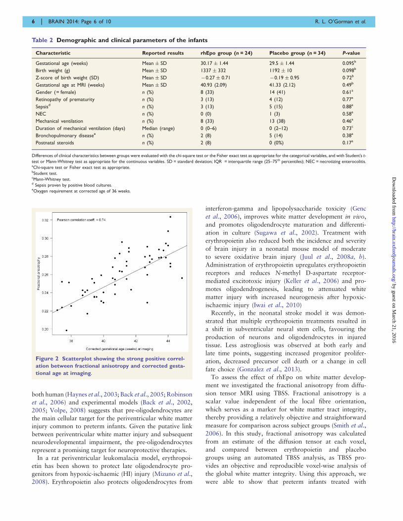
These findings are provided corrected for gestational age at
birth and corrected gestational age at scanning. Fractional
anisotropy correlated strongly with corrected age at scanning
[Pearson correlation coefficient r = 0.74 (P5 0.001), Fig. 2].
Preterm infants treated with rhEpo demonstrated increased
fractional anisotropy in the genu and splenium of the
corpus callosum, the external capsule, the corona radiata
and centrum semiovale, the anterior and posterior limbs of
the internal capsule, and the corticospinal tract bilaterally
(P5 0.01, corrected; Fig. 3). Fractional anisotropy was sig-
nificantly higher in preterm infants treated with rhEpo than
in those treated with placebo (Fig. 4). There were no voxels
where fractional anisotropy was significantly higher in
preterm infants treated with placebo. The multiple linear re-
gression model resulted in an adjusted effect of erythropoietin
on fractional anisotropy of 0.02 (P5 0.001, Table 3). The
amount of variability in fractional anisotropy explained by
the model was 70%. No significant differences were found in
radial or axial diffusivity between the two groups.
Gender effect
Gender effects were analysed using a multiple linear regres-
sion model with fractional anisotropy as dependent vari-
able, gender and intervention (rhEpo or placebo) as
independent factors. No significant differences were found
in mean fractional anisotropy between male and female
infants (P = 0.46). Furthermore no interaction between
intervention and gender could be shown (P = 0.11).
Clinical co-morbidities
Chronic lung disease (n = 9) correlated with mean frac-
tional anisotropy; however, after adjusting for gestational
age at birth this correlation became insignificant. Sepsis
(n = 8) and necrotizing enterocolitis (n = 1) showed no sig-
nificant correlation with mean fractional anisotropy.
DiscussionThis is the first clinical imaging study showing that early
erythropoietin administration can improve white matter de-
velopment in preterm infants. These results are consistent
with experimental imaging data from an immature animal
model showing higher fractional anisotropy values in the
corpus callosum in erythropoietin-treated animals compared
to those treated with placebo (Chatagner et al., 2010). The
results are also consistent with the conventional MRI find-
ings showing a reduced risk of white matter and grey matter
injury, white matter signal abnormalities, and periventricular
white matter volume loss (Leuchter et al., 2014).
To date, many experimental studies investigating erythro-
poietin as a neuroprotective strategy in adult and neonatal
models of brain injury have been published, showing
significant promising neuroprotective effects (van der Kooij
et al., 2008). One possible mechanism by which erythropoi-
etin may protect against brain injury is by protecting the vul-
nerable pre-oligodendrocytes. Accumulating evidence from
Table 1 Comparison of infants with and without TBSS analysis
Variable Modality No MRI n = 437 TBSS n = 58 P-value
Gender (female) n (%) 192 (44) 22 (38) 0.235
Gestational age (weeks) Mean � SD 28.94 � 1.8 29.7 � 1.46 50.001
Birth weight (g) Mean � SD 1176 � 353 1252 � 325 0.100
Z-score of birth weight Mean � SD �0.06 � 0.84 �0.22 � 0.85 0.185
Broncho-pulmonary dysplasia n (%) 53 (11%) 7 (19%) 0.590
Necrotizing enterocolitis n (%) 10 (3%) 6 (3%) 1
Sepsis n (%) 36 (13%) 24 (14%) 0.837
Meningitis n (%) 2 (1%) 1 (1%) 1
Retinopathy of prematurity n (%) 27 (11%) 13 (8%) 0.383
TBSS to assess the neuroprotective effect of erythropoietin in preterm infants BRAIN 2014: Page 5 of 10 | 5
by guest on March 21, 2016
http://brain.oxfordjournals.org/D
ownloaded from
both human (Haynes et al., 2003; Back et al., 2005; Robinson
et al., 2006) and experimental models (Back et al., 2002,
2005; Volpe, 2008) suggests that pre-oligodendrocytes are
the main cellular target for the periventricular white matter
injury common to preterm infants. Given the putative link
between periventricular white matter injury and subsequent
neurodevelopmental impairment, the pre-oligodendrocytes
represent a promising target for neuroprotective therapies.
In a rat periventricular leukomalacia model, erythropoi-
etin has been shown to protect late oligodendrocyte pro-
genitors from hypoxic-ischaemic (HI) injury (Mizuno et al.,
2008). Erythropoietin also protects oligodendrocytes from
interferon-gamma and lipopolysaccharide toxicity (Genc
et al., 2006), improves white matter development in vivo,
and promotes oligodendrocyte maturation and differenti-
ation in culture (Sugawa et al., 2002). Treatment with
erythropoietin also reduced both the incidence and severity
of brain injury in a neonatal mouse model of moderate
to severe oxidative brain injury (Juul et al., 2008a, b).
Administration of erythropoietin upregulates erythropoietin
receptors and reduces N-methyl D-aspartate receptor-
mediated excitotoxic injury (Keller et al., 2006) and pro-
motes oligodendrogenesis, leading to attenuated white
matter injury with increased neurogenesis after hypoxic-
ischaemic injury (Iwai et al., 2010)
Recently, in the neonatal stroke model it was demon-
strated that multiple erythropoietin treatments resulted in
a shift in subventricular neural stem cells, favouring the
production of neurons and oligodendrocytes in injured
tissue. Less astrogliosis was observed at both early and
late time points, suggesting increased progenitor prolifer-
ation, decreased precursor cell death or a change in cell
fate choice (Gonzalez et al., 2013).
To assess the effect of rhEpo on white matter develop-
ment we investigated the fractional anisotropy from diffu-
sion tensor MRI using TBSS. Fractional anisotropy is a
scalar value independent of the local fibre orientation,
which serves as a marker for white matter tract integrity,
thereby providing a relatively objective and straightforward
measure for comparison across subject groups (Smith et al.,
2006). In this study, fractional anisotropy was calculated
from an estimate of the diffusion tensor at each voxel,
and compared between erythropoietin and placebo
groups using an automated TBSS analysis, as TBSS pro-
vides an objective and reproducible voxel-wise analysis of
the global white matter integrity. Using this approach, we
were able to show that preterm infants treated with
Table 2 Demographic and clinical parameters of the infants
Characteristic Reported results rhEpo group (n = 24) Placebo group (n = 34) P-value
Gestational age (weeks) Mean � SD 30.17 � 1.44 29.5 � 1.44 0.095b
Birth weight (g) Mean � SD 1337 � 332 1192 � 10 0.098b
Z-score of birth weight (SD) Mean � SD �0.27 � 0.71 �0.19 � 0.95 0�72b
Gestational age at MRI (weeks) Mean � SD 40.93 (2.09) 41.33 (2.12) 0.49b
Gender (= female) n (%) 8 (33) 14 (41) 0.61a
Retinopathy of prematurity n (%) 3 (13) 4 (12) 0.77a
Sepsisd n (%) 3 (13) 5 (15) 0.88a
NEC n (%) 0 (0) 1 (3) 0.58a
Mechanical ventilation n (%) 8 (33) 13 (38) 0.46a
Duration of mechanical ventilation (days) Median (range) 0 (0–6) 0 (2–12) 0.73c
Bronchopulmonary diseasee n (%) 2 (8) 5 (14) 0.38a
Postnatal steroids n (%) 2 (8) 0 (0%) 0.17a
Differences of clinical characteristics between groups were evaluated with the chi-square test or the Fisher exact test as appropriate for the categorical variables, and with Student’s t-
test or Mann-Whitney test as appropriate for the continuous variables. SD = standard deviation; IQR = interquartile range (25–75th percentiles); NEC = necrotizing enterocolitis.aChi-square test or Fisher exact test as appropriate.bStudent test.cMann-Whitney test.d Sepsis proven by positive blood cultures.eOxygen requirement at corrected age of 36 weeks.
Figure 2 Scatterplot showing the strong positive correl-
ation between fractional anisotropy and corrected gesta-
tional age at imaging.
6 | BRAIN 2014: Page 6 of 10 R. L. O’Gorman et al.
by guest on March 21, 2016
http://brain.oxfordjournals.org/D
ownloaded from

erythropoietin have higher fractional anisotropy in most
major white matter tracts compared to those preterm in-
fants treated with placebo. As fractional anisotropy is
largely dependent on axonal thickness, including pre-
myelination oligodendrocyte wrapping and myelination or
axonal attenuation (Sakuma et al., 1991), our results sug-
gest that infants treated with rhEpo have more coherently
bundled, more mature fibres along the axis of greatest
diffusion, likely reflecting the protective effects of rhEpo
on reducing white matter injury and on the promotion of
proliferation, maturation and differentiation of pre-
oligodendrocytes. These results confirm the conventional
MRI findings, with rhEpo treated infants having less
white and grey matter injury, fewer white matter signal
abnormalities and periventricular white matter loss [re-
ported elsewhere by Leuchter et al. (2014) on the entire
multicentre cohort].
Cumulative erythropoietin
Cumulative erythropoietin doses have been shown to pro-
vide superior neuroprotection when compared to single
dose administration following brain injury in a rat model
(Kellert et al., 2007; Gonzalez et al., 2009). This improved
neuroprotection is likely to arise from multiple effects of
erythropoietin during the evolving injury response.
Specifically, erythropoietin decreases the early inflammatory
response, decreases both early and late neuronal apoptosis
Figure 3 Mean fractional anisotropy skeleton (green) overlaid on the mean fractional anisotropy map in the axial and coronal
planes. Regions of the fractional anisotropy skeleton in green represent voxels where there was no difference in fractional anisotropy between
infants treated with erythropoietin and placebo. Voxels demonstrating significantly higher fractional anisotropy in the erythropoietin-treated
group are overlaid in red-yellow.
Figure 4 Effect of erythropoietin treatment. Boxplot
showing the differences in fractional anisotropy (median and inter-
quartile range) between preterm infants treated with erythropoietin
(EPO) and treated with placebo.
Table 3 Results of the linear regression model for
fractional anisotrophy
Coefficient Standard error P-value
Intercept �0.132 0.037 0.001
Epo (versus placebo) 0.019 0.004 _0.001
Gender Male �0.001 0.004 0.793
Corrected gestational age 0.009 0.001 50.001
TBSS to assess the neuroprotective effect of erythropoietin in preterm infants BRAIN 2014: Page 7 of 10 | 7
by guest on March 21, 2016
http://brain.oxfordjournals.org/D
ownloaded from

(Juul et al., 2008a, b), and stimulates late repair processes
such as neurogenesis, angiogenesis and migration of regen-
erating neurons (Tsai et al., 2006). As the infants in this
study were given rhEpo within 2 days of birth and MRI
was performed after an interval of several weeks, our data
suggest that three early rhEpo doses also improve neuro-
protection for preterm neonates, providing a similar long-
term improvement to that seen in the neonatal stroke
model (Gonzalez et al., 2013). The neuroprotective effect
of erythropoietin may be further enhanced by giving rhEpo
for a longer period than just within the first 42 h.
Gender effect
Male preterm infants are known to be at higher risk for
abnormal neurological outcome than female preterm infants.
A recent study has shown that male infants have higher rates
for disability and cognitive delay even after adjustment for
gestational age at birth and birth weight (Peacock et al.,
2012). This observation is in agreement with data from pub-
lished studies showing constitutional differences between
genders, which are not explained by perinatal, neonatal or
postnatal factors (Brothwood et al., 1986; Stevenson et al.,
2000; Hintz et al., 2006). Therefore, we performed a linear
regression to evaluate the interaction between erythropoietin
and gender with respect to the mean fractional anisotropy.
There was no significant gender effect on mean fractional
anisotropy in the measured white matter tracts (P = 0.46),
and no significant interaction between gender and interven-
tion (rhEpo versus placebo) (P = 0.23), indicating that in our
sample, gender does not significantly influence the effects of
erythropoietin treatment on mean fractional anisotropy.
However, the lack of a statistically significant gender effect
may also be due to the small numbers in each subgroup (8
and 14 female infants treated with rhEpo and placebo, re-
spectively, 16 and 20 male infants treated with rhEpo and
placebo, respectively).
A recent study showed that gestational age in itself
was not a strong predictor for adverse brain development
as measured by DTI, and that co-morbidities seem to play
a larger role in brain development than the degree of
prematurity (Bonifacio et al., 2010). In our cohort there
was no correlation between mean fractional anisotropy
and clinical co-morbidities such as sepsis, necrotizing
enterocolitis and chronic lung disease, but this might
be due to the small number of infants in our cohort suffering
from chronic lung disease (15.5% versus 34.6%; Bonifacio
et al., 2010), necrotizing enterocolitis or sepsis (Table 1).
Limitations
One limitation of this study is that the TBSS analysis could
only be performed in a subgroup of infants. As the infants
were scanned under natural sleep, some DTI studies were
incomplete or corrupted by motion artefacts, which made
the data unusable. However, we believe that this subgroup
is representative of the whole study group, as clinical
co-morbidities did not differ between the TBSS and the
main cohort. Another limitation is that the TBSS cohort
was significantly older than the infants for whom no ima-
ging was performed. However, as the results did not alter
after applying a family-wise error (FWE) correction for
multiple comparisons following threshold-free cluster en-
hancement with gestational age at birth as covariate, it
seems unlikely that the results are driven by differences in
gestational age. A strength of the study is that the magnetic
resonance scans of the TBSS cohort were acquired in a
single centre; hence, additional variance from between-
scanner effects was eliminated.
At present, the neurodevelopmental outcome of this cohort
is insufficient to be able to determine the precise relationship
between mean fractional anisotropy and motor, cognitive and
language outcome. However, as many studies show good cor-
relation between fractional anisotropy and neurodevelop-
mental outcome (Arzoumanian et al., 2003; van Kooij
et al., 2012) in preterm infants, we speculate that preterm
infants treated with rhEpo will show improved outcome.
The study used a very short dosing regimen (up to 42 h
post-birth) in accordance with the experimental data avail-
able at the time of the study design, and therefore did not
take into consideration recent experimental data on extended
chronic erythropoietin treatment (van de Looij et al., 2014).
ConclusionEarly rhEpo administration improves white matter develop-
ment in preterm infants assessed by DTI and TBSS. This
promising result is consistent with similar results in an im-
mature animal model of neuroprotection through erythro-
poietin treatment. Neurodevelopmental follow-up at 2 and
5 years are ongoing to further define the neuroprotective
effect of erythropoietin.
FundingNone of the authors had any conflicts of interests including
any financial interests, activities, relationships, and affili-
ations. The study was supported by a grant received from
the Swiss National Foundation (SNF 3200B0-108176).
C.H. was supported by a personal grant ‘For Women in
Science’ by the Swiss Academics for Arts and Science,
Unesco and L’Oreal. The funders had no role in study
design, data collection and analysis, decision to publish,
or preparation of the manuscript. All authors had full
access to all the data in the study and responsibility for
the decision to submit for publication.
ClinicalTrials.gov Identifier: NCT00413946.
ReferencesAlexandrou G, Martensson G, Skiold B, Blennow M, Aden U,
Vollmer B. White matter microstructure is influenced by extremely
8 | BRAIN 2014: Page 8 of 10 R. L. O’Gorman et al.
by guest on March 21, 2016
http://brain.oxfordjournals.org/D
ownloaded from

preterm birth and neonatal respiratory factors. Acta Paediatr 2014;
103: 48–56.
Andersson JL, Jenkinson M, Smith M. Non-linear registration, aka
Spatial normalisation FMRIB technical report TR07JA2, 2007a.
www.fmrib.ox.ac.uk/analysis/techrep.
Andersson JL, Jenkinson M, Smith M. Non-linear optimisation.
FMRIB technical report TR07JA1, 2007b. www.fmrib.ox.ac.uk/
analysis/techrep.
Anjari M, Srinivasan L, Allsop JM, Hajnal JV, Rutherford MA,
Edwards AD, et al. Diffusion tensor imaging with tract-based spatial
statistics reveals local white matter abnormalities in preterm infants.
Neuroimage 2007; 35: 1021–7.
Arzoumanian Y, Mirmiran M, Barnes PD, Woolley K, Ariagno RL,
Moseley ME, et al. Diffusion tensor brain imaging findings at term-
equivalent age may predict neurologic abnormalities in low birth
weight preterm infants. AJNR Am J Neuroradiol 2003; 24: 1646–53.Back SA, Han BH, Luo NL, Chricton CA, Xanthoudakis S, Tam J,
et al. Selective vulnerability of late oligodendrocyte progenitors to
hypoxia-ischemia. J Neurosci 2002; 22: 455–63.
Back SA, Luo NL, Mallinson RA, O’Malley JP, Wallen LD, Frei B,
et al. Selective vulnerability of preterm white matter to oxidative
damage defined by F2-isoprostanes. Ann Neurol 2005; 58: 108–20.
Ball G, Boardman JP, Arichi T, Merchant N, Rueckert D,
Edwards AD, et al. Testing the sensitivity of Tract-Based Spatial
Statistics to simulated treatment effects in preterm neonates. PLoS
One 2013; 8: e67706.
Ball G, Counsell SJ, Anjari M, Merchant N, Arichi T, Doria V, et al.
An optimised tract-based spatial statistics protocol for neonates: ap-
plications to prematurity and chronic lung disease. Neuroimage
2010; 53: 94–102.Bassi L, Chew A, Merchant N, Ball G, Ramenghi L, Boardman J, et al.
Diffusion tensor imaging in preterm infants with punctate white
matter lesions. Pediatr Res 2011; 69: 561–6.Berman JI, Mukherjee P, Partridge SC, Miller SP, Ferriero DM,
Barkovich AJ, et al. Quantitative diffusion tensor MRI fiber tracto-
graphy of sensorimotor white matter development in premature in-
fants. Neuroimage 2005; 27: 862–71.
Bonifacio SL, Glass HC, Chau V, Berman JI, Xu D, Brant R, et al.
Extreme premature birth is not associated with impaired develop-
ment of brain microstructure. J Pediatr 2010; 157: 726–32 e1.
Brothwood M, Wolke D, Gamsu H, Benson J, Cooper D. Prognosis of
the very low birthweight baby in relation to gender. Arch Dis Child
1986; 61: 559–64.
Chatagner A, Van der Looij Y, Huppi P, Gruetter R, Sizonenko S.
Neuroprotective effects of chronic erythropoietin treatment on neo-
natal rat following hypoxia-ischemia. Pediatric Academic Societies,
Society for Pediatric Research 2011: Baltimore; 2010.
Cheong JL, Thompson DK, Wang HX, Hunt RW, Anderson PJ,
Inder TE, et al. Abnormal white matter signal on MR imaging is
related to abnormal tissue microstructure. AJNR Am J Neuroradiol
2009; 30: 623–8.
Counsell SJ, Allsop JM, Harrison MC, Larkman DJ, Kennea NL,
Kapellou O, et al. Diffusion-weighted imaging of the brain in pre-
term infants with focal and diffuse white matter abnormality.
Pediatrics 2003; 112 (1 Pt 1): 1–7.
Counsell SJ, Shen Y, Boardman JP, Larkman DJ, Kapellou O, Ward P,
et al. Axial and radial diffusivity in preterm infants who have diffuse
white matter changes on magnetic resonance imaging at term-
equivalent age. Pediatrics 2006; 117: 376–86.
Dame C, Juul SE, Christensen RD. The biology of erythropoietin in
the central nervous system and its neurotrophic and neuroprotective
potential. Biol Neonate 2001; 79: 228–35.Dudink J, Lequin M, van Pul C, Buijs J, Conneman N, van
Goudoever J, et al. Fractional anisotropy in white matter tracts of
very-low-birth-weight infants. Pediatr Radiol 2007; 37: 1216–23.Fauchere JC, Dame C, Vonthein R, Koller B, Arri S, Wolf M, et al. An
approach to using recombinant erythropoietin for neuroprotection
in very preterm infants. Pediatrics 2008; 122: 375–82.
Fauchere JC, Koller B, Tschopp A, Bucher HU, Group SENT. Safety
of high-dose erythopoietin for neuroprotection in preterm infants
archives of disease in childhood. fetal and neonatal edition 2012;
97 (Suppl 2): A16.
Genc K, Genc S, Baskin H, Semin I. Erythropoietin decreases cytotox-
icity and nitric oxide formation induced by inflammatory stimuli in
rat oligodendrocytes. Physiol Res 2006; 55: 33–8.
Ghezzi P, Bernaudin M, Bianchi R, Blomgren K, Brines M,
Campana W, et al. Erythropoietin: not just about erythropoiesis.
Lancet 2010; 375: 2142.
Gonzalez FF, Abel R, Almli CR, Mu D, Wendland M, Ferriero DM.
Erythropoietin sustains cognitive function and brain volume after
neonatal stroke. Dev Neurosci 2009; 31: 403–11.
Gonzalez FF, Ferriero DM. Neuroprotection in the newborn infant.
Clin Perinatol 2009; 36: 859–80, vii.
Gonzalez FF, Larpthaveesarp A, McQuillen P, Derugin N,
Wendland M, Spadafora R, et al. Erythropoietin increases neuro-
genesis and oligodendrogliosis of subventricular zone precursor cells
after neonatal stroke. Stroke 2013; 44: 753–8.
Haynes RL, Folkerth RD, Keefe RJ, Sung I, Swzeda LI, Rosenberg PA,
et al. Nitrosative and oxidative injury to premyelinating oligo-
dendrocytes in periventricular leukomalacia. J Neuropathol Exp
Neurol 2003; 62: 441–50.
Hintz SR, Kendrick DE, Vohr BR, Kenneth Poole W, Higgins RD.
Gender differences in neurodevelopmental outcomes among ex-
tremely preterm, extremely-low-birthweight infants. Acta Paediatr
2006; 95: 1239–48.
Huppi PS, Dubois J. Diffusion tensor imaging of brain development.
Semin Fetal Neonatal Med 2006; 11: 489–97.
Huppi PS, Inder TE. Magnetic resonance techniques in the evaluation
of the perinatal brain: recent advances and future directions. Semin
Neonatol 2001; 6: 195–210.
Huppi PS, Maier SE, Peled S, Zientara GP, Barnes PD, Jolesz FA, et al.
Microstructural development of human newborn cerebral white
matter assessed in vivo by diffusion tensor magnetic resonance ima-
ging. Pediatr Res 1998; 44: 584–90.
Huppi PS, Murphy B, Maier SE, Zientara GP, Inder TE, Barnes PD,
et al. Microstructural brain development after perinatal cerebral
white matter injury assessed by diffusion tensor magnetic resonance
imaging. Pediatrics 2001; 107: 455–60.
Iwai M, Stetler RA, Xing J, Hu X, Gao Y, Zhang W, et al. Enhanced
oligodendrogenesis and recovery of neurological function by
erythropoietin after neonatal hypoxic/ischemic brain injury. Stroke
2010; 41: 1032–7.
Juul S. Recombinant erythropoietin as a neuroprotective treatment:
in vitro and in vivo models. Clin Perinatol 2004; 31: 129–42.
Juul S. Neuroprotective role of erythropoietin in neonates. J Matern
Fetal Neonatal Med 2012; 25 (Suppl 4): 105–7.Juul S, Felderhoff-Mueser U. Epo and other hematopoietic factors.
Semin Fetal Neonatal Med 2007; 12: 250–8.Juul SE, McPherson RJ, Bammler TK, Wilkerson J, Beyer RP,
Farin FM. Recombinant erythropoietin is neuroprotective in a
novel mouse oxidative injury model. Dev Neurosci 2008a; 30:
231–42.
Juul SE, McPherson RJ, Bauer LA, Ledbetter KJ, Gleason CA,
Mayock DE. A phase I/II trial of high-dose erythropoietin in ex-
tremely low birth weight infants: pharmacokinetics and safety.
Pediatrics 2008b; 122: 383–91.
Keller M, Yang J, Griesmaier E, Gorna A, Sarkozy G, Urbanek M,
et al. Erythropoietin is neuroprotective against NMDA-receptor-
mediated excitotoxic brain injury in newborn mice. Neurobiol Dis
2006; 24: 357–66.
Kellert BA, McPherson RJ, Juul SE. A comparison of high-dose recom-
binant erythropoietin treatment regimens in brain-injured neonatal
rats. Pediatr Res 2007; 61: 451–5.
Latal B. Prediction of neurodevelopmental outcome after preterm
birth. Pediatr Neurol 2009; 40: 413–9.
TBSS to assess the neuroprotective effect of erythropoietin in preterm infants BRAIN 2014: Page 9 of 10 | 9
by guest on March 21, 2016
http://brain.oxfordjournals.org/D
ownloaded from
Lepomaki V, Matomaki J, Lapinleimu H, Lehtonen L, Haataja L,
Komu M, et al. Effect of antenatal growth on brain white matter
maturation in preterm infants at term using tract-based spatial stat-
istics. Pediatr Radiol 2013; 43: 80–5.
Leuchter RH, Gui L, Poncet A, Hagmann C, Lodygensky GA,
Martin E, et al. Association between early administration of high-
dose erythropoietin in preterm infants and brain MRI abnormality
at term-equivalent age. JAMA 2014; 312: 817–24.Miller SP, Vigneron DB, Henry RG, Bohland MA, Ceppi-Cozzio C,
Hoffman C, et al. Serial quantitative diffusion tensor MRI of the
premature brain: development in newborns with and without injury.
J Magn Reson Imaging 2002; 16: 621–32.
Mizuno K, Hida H, Masuda T, Nishino H, Togari H. Pretreatment
with low doses of erythropoietin ameliorates brain damage in peri-
ventricular leukomalacia by targeting late oligodendrocyte progeni-
tors: a rat model. Neonatology 2008; 94: 255–66.
Mukherjee P, Miller JH, Shimony JS, Philip JV, Nehra D, Snyder AZ,
et al. Diffusion-tensor MR imaging of gray and white matter devel-
opment during normal human brain maturation. AJNR Am J
Neuroradiol 2002; 23: 1445–56.
Neubauer AP, Voss W, Wachtendorf M, Jungmann T. Erythropoietin
improves neurodevelopmental outcome of extremely preterm infants.
Ann Neurol 2010; 67: 657–66.Newton NR, Leonard CH, Piecuch RE, Phibbs RH.
Neurodevelopmental outcome of prematurely born children treated
with recombinant human erythropoietin in infancy. J Perinatol
1999; 19 (6 Pt 1): 403–6.
Ohls RK, Ehrenkranz RA, Das A, Dusick AM, Yolton K, Romano E,
et al. Neurodevelopmental outcome and growth at 18 to 22 months’
corrected age in extremely low birth weight infants treated with
early erythropoietin and iron. Pediatrics 2004; 114: 1287–91.
Ohlsson A, Aher SM. Early erythropoietin for preventing red blood
cell transfusion in preterm and/or low birth weight infants.
Cochrane Database Syst Rev 2006; CD004863.
Partridge SC, Mukherjee P, Henry RG, Miller SP, Berman JI, Jin H, et al.
Diffusion tensor imaging: serial quantitation of white matter tract ma-
turity in premature newborns. Neuroimage 2004; 22: 1302–14.
Peacock JL, Marston L, Marlow N, Calvert SA, Greenough A.
Neonatal and infant outcome in boys and girls born very prema-
turely. Pediatr Res 2012; 71: 305–10.Porter EJ, Counsell SJ, Edwards AD, Allsop J, Azzopardi D. Tract-
based spatial statistics of magnetic resonance images to assess dis-
ease and treatment effects in perinatal asphyxial encephalopathy.
Pediatr Res 2010; 68: 205–9.
Robinson S, Li Q, Dechant A, Cohen ML. Neonatal loss of gamma-
aminobutyric acid pathway expression after human perinatal brain
injury. J Neurosurg 2006; 104 (6 Suppl): 396–408.
Rueckert D, Sonoda LI, Hayes C, Hill DL, Leach MO, Hawkes DJ.
Nonrigid registration using free-form deformations: application to
breast MR images. IEEE Trans Med Imaging 1999; 18: 712–21.
Saigal S, Doyle LW. An overview of mortality and sequelae of preterm
birth from infancy to adulthood. Lancet 2008; 371: 261–9.
Sakuma H, Nomura Y, Takeda K, Tagami T, Nakagawa T,
Tamagawa Y, et al. Adult and neonatal human brain: diffusional
anisotropy and myelination with diffusion-weighted MR imaging.
Radiology 1991; 180: 229–33.
Shingo T, Sorokan ST, Shimazaki T, Weiss S. Erythropoietin regulates
the in vitro and in vivo production of neuronal progenitors by mam-
malian forebrain neural stem cells. J Neurosci 2001; 21: 9733–43.
Skiold B, Horsch S, Hallberg B, Engstrom M, Nagy Z, Mosskin M,
et al. White matter changes in extremely preterm infants, a popula-
tion-based diffusion tensor imaging study. Acta Paediatr 2010; 99:
842–9.Smith SM. Fast robust automated brain extraction. Hum Brain Mapp
2002; 17: 143–55.Smith SM, Jenkinson M, Johansen-Berg H, Rueckert D, Nichols TE,
Mackay CE, et al. Tract-based spatial statistics: voxelwise analysis
of multi-subject diffusion data. Neuroimage 2006; 31: 1487–505.
Smith SM, Jenkinson M, Woolrich MW, Beckmann CF, Behrens TE,Johansen-Berg H, et al. Advances in functional and structural MR
image analysis and implementation as FSL. Neuroimage 2004; 23
(Suppl 1): S208–19.
Smith SM, Nichols TE. Threshold-free cluster enhancement: addressingproblems of smoothing, threshold dependence and localisation in
cluster inference. Neuroimage 2009; 44: 83–98.
Stevenson DK, Verter J, Fanaroff AA, Oh W, Ehrenkranz RA,
Shankaran S, et al. Sex differences in outcomes of very low birth-weight infants: the newborn male disadvantage. Arch Dis Child
Fetal Neonatal Ed 2000; 83: F182–5.
Sugawa M, Sakurai Y, Ishikawa-Ieda Y, Suzuki H, Asou H. Effects oferythropoietin on glial cell development; oligodendrocyte mat-
uration and astrocyte proliferation. Neurosci Res 2002; 44: 391–403.
R Core Team. R: A language and environment for statistical comput-
ing. R Foundation for Statistical Computing, Vienna, Austria, 2013.http://www.R-project.org/.
Tsai PT, Ohab JJ, Kertesz N, Groszer M, Matter C, Gao J, et al. A
critical role of erythropoietin receptor in neurogenesis and post-
stroke recovery. J Neurosci 2006; 26: 1269–74.van de Looij Y, Chatagner A, Quairiaux C, Gruetter R, Huppi PS,
Sizonenko SV. Multi-modal assessment of long-term erythropoietin
treatment after neonatal hypoxic-ischemic injury in rat brain. PLoS
One 2014; 9: e95643.van der Kooij MA, Groenendaal F, Kavelaars A, Heijnen CJ, van
Bel F. Neuroprotective properties and mechanisms of erythropoietin
in in vitro and in vivo experimental models for hypoxia/ischemia.Brain Res Rev 2008; 59: 22–33.
van Kooij BJ, de Vries LS, Ball G, van Haastert IC, Benders MJ,
Groenendaal F, et al. Neonatal tract-based spatial statistics findings
and outcome in preterm infants. AJNR Am J Neuroradiol 2012; 33:188–94.
Volpe J. Neurology of the newborn. 5th edn. Philadelphia, PA:
Saunders Elsevier; 2008.
Volpe JJ. Brain injury in premature infants: a complex amalgam of destruc-tive and developmental disturbances. Lancet Neurol 2009; 8: 110–24.
Woodward LJ, Anderson PJ, Austin NC, Howard K, Inder TE.
Neonatal MRI to predict neurodevelopmental outcomes in preterminfants. N Engl J Med 2006; 355: 685–94.
Wu YW, Bauer LA, Ballard RA, Ferriero DM, Glidden DV, Mayock DE,
et al. Erythropoietin for neuroprotection in neonatal encephalopathy:
safety and pharmacokinetics. Pediatrics 2012; 130: 683–91.
Appendix 1
The Swiss EPONeuroprotection Trial GroupThe following local investigators and hospitals
participated in this study (study sites are listed in alphabetical
order): Aarau: Kinderklinik Kantonsspital Aarau (Georg
Zeilinger, MD; Sylviane Pasquier, MD); Basel:
Universitatskinderklinik UKBB (Christoph Buhrer, MD;
Rene Glanzmann, MD; Sven Schulzke, MD); Chur:
Abteilung fur Neonatologie, Kantons- und Regionalspital
(Brigitte Scharrer, MD; Walter Bar, MD); Geneve: Unite de
Neonatologie, Clinique de Pediatrie HCUG (Riccardo Pfister,
MD); Zurich: Klinik fur Neonatologie (Jean-Claude
Fauchere, MD); Zurich: Zentrum fur MR-Forschung (Ernst
Martin, MD; Ianina Scheer, MD; Hadwig Speckbacher,
MTRA).
10 | BRAIN 2014: Page 10 of 10 R. L. O’Gorman et al.
by guest on March 21, 2016
http://brain.oxfordjournals.org/D
ownloaded from
Copyright © 2022 FDOKUMEN