
Trace-Level Detection of Secondary Explosives Using Hybrid Silver− Gold Nanoparticles and...
12
Trace-Level Detection of Secondary Explosives Using Hybrid Silver− Gold Nanoparticles and Nanostructures Achieved with Femtosecond Laser Ablation G. Krishna Podagatlapalli, † Syed Hamad, ‡ and S. Venugopal Rao* ,† † Advanced Center of Research in High Energy Materials (ACRHEM) and ‡ School of Physics, University of Hyderabad, Prof. C. R. Rao Road, Hyderabad 500046, Telangana, India * S Supporting Information ABSTRACT: Hybrid silver−gold targets were achieved by effortless mixing of pure silver (Ag) and gold (Au) metals at different ratios (Ag 0.65 Au 0.35 , Ag 0.5 Au 0.5 , and Ag 0.35 Au 0.65 ) and embracing a manual melting process. The obtained targets were ablated by ultrafast (∼40 fs) laser pulses in acetone ensuing the fabrication of Ag−Au bimetallic nanoparticles (NPs) and nanostructures (NSs) in a single experiment. UV−visible extinction spectra of Ag−Au colloids demonstrated the tuning of localized surface plasmon resonance (LSPR) in the spectral range of 406−524 nm. The morphologies of NSs were investigated by the field emission scanning electron microscopy (FESEM) technique. Ag−Au NPs and NSs were utilized as surface enhanced Raman scattering (SERS) platforms to detect secondary explosive molecules such as 1,1-diamino-2,2-dinitroethene (FOX-7, 5 μM concentration) and 1- nitro pyrazole (1NPZ, 20 nM concentration). Our experimental observations clearly demonstrated that the increment in gold percentage reduced the surface activity of Ag−Au NPs/NSs. The estimated enhancement factors (EFs) from the SERS data were typically >10 8 . Our detailed investigations revealed that the NPs and NSs of Ag 0.65 Au 0.35 exhibited significant EFs compared to other ratios and pure metals of Ag and Au. ■ INTRODUCTION Fabrication of pure plasmonic metal (Au, Ag, and Cu) nanoparticles (NPs) achievable through sophisticated chemical methodologies is well established and understood by the scientific community. However, many of these methods demands hours of monitoring and post production processes such as cleaning the nanomaterials to remove chemical dopants and impurities. Similarly, shape controlled production of bimetallic NPs through utilizing surfactants and reagents is also well-known in chemical methods. Many of the earlier reports revealed that bimetallic (Au−Ag, Ag−Cu, and Au−Cu) NPs were fabricated by adding individual colloidal solutions of gold, silver, and copper with different proportions to achieve hybridization of the localized surface plasmon resonances (LSPRs). Ultrafast laser ablation in liquids (ULAL) is a clean, green method which does not utilize chemicals for fabrication of NPs/NSs, and importantly, it does not necessitate extreme cleaning of NPs/NSs. Moreover, simultaneous fabrication of NPs and NSs is possible in the ULAL technique 1−15 in contrast to other chemical methods. Even though some of the solution (chemical) methods are fast (in terms of time taken) compared to various ablation techniques, the main problem with them is that the capped ligand molecules sit on surface of NPs and thus blocks the analyte molecules to achieve direct contact with NP surface. The main objective of fabricating alloy nanomaterials is to find out the exact proportion of individual metals, which exhibits superior performance (in this case, our interest is in the surface enhanced Raman signal) and, hence, are versatile and compatible in many fields such as biomedicine, spectroscopy, and photonics. For example, it has been demonstrated that Ag− Au NPs possessed a higher ability as strong catalysts compared to their individual metal counterparts. 17−21 In the specific case of Ag−Au NPs, individual demerits of silver (often suffers oxidation effects compared to gold) and gold (weak SPR strength compared to silver) can be circumvented through the hybridization process. As a consequence, new mechanical, thermal properties can be incorporated in hybridized nanoma- terials through which one can produce both chemically stable and thriving optically responsive materials such as Ag−Au NPs/NSs. Particularly, hybridization significantly influences the SPR peak position and its bandwidth, which primarily depends upon the individual proportion of the metals in the composite. Hybridization of silver and gold leads to surface plasmon (SP) absorption tunability in the absorption bands of individual Ag and Au NPs. 22 The tunability of combined resonance of Au− Ag NPs over a wide spectral range is essential in many applications in which the optical field enhancements (mediated by surface plasmons) are exploited. 21 Due to the tunability of SPRs, these Ag−Au NPs have many applications in nonlinear optics as optical limiters. 22 In addition, Au−Ag NPs have Received: April 25, 2015 Revised: June 24, 2015 Published: June 24, 2015 Article pubs.acs.org/JPCC © 2015 American Chemical Society 16972 DOI: 10.1021/acs.jpcc.5b03958 J. Phys. Chem. C 2015, 119, 16972−16983
Transcript of Trace-Level Detection of Secondary Explosives Using Hybrid Silver− Gold Nanoparticles and...

Trace-Level Detection of Secondary Explosives Using Hybrid Silver−Gold Nanoparticles and Nanostructures Achieved with FemtosecondLaser AblationG. Krishna Podagatlapalli,† Syed Hamad,‡ and S. Venugopal Rao*,†
†Advanced Center of Research in High Energy Materials (ACRHEM) and ‡School of Physics, University of Hyderabad, Prof. C. R.Rao Road, Hyderabad 500046, Telangana, India
*S Supporting Information
ABSTRACT: Hybrid silver−gold targets were achieved by effortless mixingof pure silver (Ag) and gold (Au) metals at different ratios (Ag0.65Au0.35,Ag0.5Au0.5, and Ag0.35Au0.65) and embracing a manual melting process. Theobtained targets were ablated by ultrafast (∼40 fs) laser pulses in acetoneensuing the fabrication of Ag−Au bimetallic nanoparticles (NPs) andnanostructures (NSs) in a single experiment. UV−visible extinction spectraof Ag−Au colloids demonstrated the tuning of localized surface plasmonresonance (LSPR) in the spectral range of 406−524 nm. The morphologies ofNSs were investigated by the field emission scanning electron microscopy(FESEM) technique. Ag−Au NPs and NSs were utilized as surface enhancedRaman scattering (SERS) platforms to detect secondary explosive moleculessuch as 1,1-diamino-2,2-dinitroethene (FOX-7, 5 μM concentration) and 1-nitro pyrazole (1NPZ, 20 nM concentration). Our experimental observationsclearly demonstrated that the increment in gold percentage reduced the surface activity of Ag−Au NPs/NSs. The estimatedenhancement factors (EFs) from the SERS data were typically >108. Our detailed investigations revealed that the NPs and NSs ofAg0.65Au0.35 exhibited significant EFs compared to other ratios and pure metals of Ag and Au.
■ INTRODUCTIONFabrication of pure plasmonic metal (Au, Ag, and Cu)nanoparticles (NPs) achievable through sophisticated chemicalmethodologies is well established and understood by thescientific community. However, many of these methodsdemands hours of monitoring and post production processessuch as cleaning the nanomaterials to remove chemical dopantsand impurities. Similarly, shape controlled production ofbimetallic NPs through utilizing surfactants and reagents isalso well-known in chemical methods. Many of the earlierreports revealed that bimetallic (Au−Ag, Ag−Cu, and Au−Cu)NPs were fabricated by adding individual colloidal solutions ofgold, silver, and copper with different proportions to achievehybridization of the localized surface plasmon resonances(LSPRs). Ultrafast laser ablation in liquids (ULAL) is a clean,green method which does not utilize chemicals for fabricationof NPs/NSs, and importantly, it does not necessitate extremecleaning of NPs/NSs. Moreover, simultaneous fabrication ofNPs and NSs is possible in the ULAL technique1−15 in contrastto other chemical methods. Even though some of the solution(chemical) methods are fast (in terms of time taken) comparedto various ablation techniques, the main problem with them isthat the capped ligand molecules sit on surface of NPs and thusblocks the analyte molecules to achieve direct contact with NPsurface. The main objective of fabricating alloy nanomaterials isto find out the exact proportion of individual metals, whichexhibits superior performance (in this case, our interest is in the
surface enhanced Raman signal) and, hence, are versatile andcompatible in many fields such as biomedicine, spectroscopy,and photonics. For example, it has been demonstrated that Ag−Au NPs possessed a higher ability as strong catalysts comparedto their individual metal counterparts.17−21 In the specific caseof Ag−Au NPs, individual demerits of silver (often suffersoxidation effects compared to gold) and gold (weak SPRstrength compared to silver) can be circumvented through thehybridization process. As a consequence, new mechanical,thermal properties can be incorporated in hybridized nanoma-terials through which one can produce both chemically stableand thriving optically responsive materials such as Ag−AuNPs/NSs. Particularly, hybridization significantly influences theSPR peak position and its bandwidth, which primarily dependsupon the individual proportion of the metals in the composite.Hybridization of silver and gold leads to surface plasmon (SP)absorption tunability in the absorption bands of individual Agand Au NPs.22 The tunability of combined resonance of Au−Ag NPs over a wide spectral range is essential in manyapplications in which the optical field enhancements (mediatedby surface plasmons) are exploited.21 Due to the tunability ofSPRs, these Ag−Au NPs have many applications in nonlinearoptics as optical limiters.22 In addition, Au−Ag NPs have
Received: April 25, 2015Revised: June 24, 2015Published: June 24, 2015
Article
pubs.acs.org/JPCC
© 2015 American Chemical Society 16972 DOI: 10.1021/acs.jpcc.5b03958J. Phys. Chem. C 2015, 119, 16972−16983

applications in solar cell fabrication, microelectronics, andsensor fabrication.23 In solar cell fabrication, the efficiency ofthe cell can be improved by utilizing the plasmonic metallic/bimetallic NPs which scatter the light efficiently and trap opticalenergy through SPR.24,25
Our earlier works on ULAL focused on optimizing thegeneration of NPs and NSs by changing the surroundingliquid,5 scanning parameters,6 input angle of incidence,7 theshape of input laser pulses,8 etc. A few of the earlier reportssuggested the fabrication of Ag−Au NPs (a) based on postirradiation of colloidal mixtures by laser pulses inducing analloying process,16 which was reported to occur through core−shell intermediates18 (b) entailing ablation of silver targets inthe presence of gold nanoparticles.21 In the present study, Ag−Au NPs have been fabricated along with Ag−Au NSs from thealloy targets prepared prior to ablation. The prepared NPs andNSs were characterized using UV−visible absorption and fieldemission scanning electron microscopy (FESEM) techniques.Significantly, both the NPs and NSs were utilized as surfaceenhanced Raman scattering (SERS) targets for trace leveldetection of secondary explosive molecules such as 1,1-diamino-2,2-dinitroethene (FOX-7, 5 μM concentration) and1-nitro pyrazole (1NPZ, 20 nM concentration). A substantialenhancement in the Raman signals was observed for the alloyAg0.65Au0.35 compared to others. The advantages with nano-structured targets prepared using these techniques are that theycan be prepared over a large area (few mm2, easily scalable tofew inch2, and is only limited by the translation stages used forscanning the target placed in liquids) and are equitablyreproducible. Furthermore, recent advances and detailedunderstanding of the ultrafast ablation phenomena enablesgeneration of NPs at grams/hour rates.14 Rapid advances in thefs laser technologies and adaptive optics enables the preparation
of these nanostructured targets in large quantities and over ashort period of time (multiplexing is another possibility).Furthermore, compared to nanoparticles, nanostructures aremore robust and rugged and need a lesser amount ofpreparation for performing SERS studies. Our earlier reports10
based on copper nanostructures revealed that these substratescan be used multiple times through adoption of an appropriatecleaning procedure, thereby enabling detection of multiple,diverse molecules using a single nanostructured substrate. Tothe best of our knowledge, this is the first report on utilizationof both NPs (colloids) and NSs (laser fabricated targets)prepared in a single experiment for the SERS measurements ofexplosive molecules.
■ EXPERIMENTAL DETAILSAg−Au Metal Target Preparation. Most of the bimetallic
(Ag−Au, Au−Cu, Ag−Cu) nanomaterial preparations reportedso far were based on chemical methods which are usuallycomplicated and require practice of vigorous cleaningprocedures post fabrication.26−30 Any residual chemical moetiesleft on the nanomaterial surface lead to unwanted con-sequences. Most of the researchers prepared Ag−Au NPs bymixing individual Ag and Au colloids (at different proportions)followed by laser beam irradiation to produce bimetallic/alloyNPs.31−33 Some of them are a mixture of chemical and physicalprocesses such as ablating a bulk plasmonic metal target insolutions such as HAuO4 or AgNO3.
34,35 However, in thepresent study, we tried to prepare bulk Ag−Au targets bymixing through melting the gold and silver at differentproportions (1 g of Ag + 0.5 g of Au, 0.5 g of Ag + 0.5 g ofAu, 0.5 g of Ag + 1 g of Au) and were successful. The alloyswere prepared by mixing the melts of Ag and Au thoroughlyand subsequent to cooling the targets were immediately (within
Figure 1. Complete picture of laser ablation metals in aqueous media and their utilization for surface enhanced Raman spectroscopic studies.
The Journal of Physical Chemistry C Article
DOI: 10.1021/acs.jpcc.5b03958J. Phys. Chem. C 2015, 119, 16972−16983
16973

2−3 h) used for ablation to avoid any undesirable surfaceeffects. Prior to ablation, the Ag−Au metal targets were cleanedto remove any surface contaminants produced in the duecourse of melting and subsequently small strips of 1 cm × 1 cmdimensions were prepared. The cleaned Ag−Au strips werethen utilized to fabricate NPs and NSs through ablationutilizing ∼40 fs laser pulses in the presence of acetone. Theimportant difference in the present ablation case (compared toother studies) is that the target was translated while ablationtook place.A detailed experimental procedure of ablation in liquid media
was reported in our earlier works.5−9 Briefly, the Ag−Au targetwas immersed in HPLC grade acetone in a Pyrex cell and wasplaced on a motorized nanodirect XYZ stage. The thickness ofthe liquid layer on the target surface in the Pyrex cell was ∼6mm. Fs laser pulses were focused vertically onto the Ag−Autarget through a plano-convex lens ( f = 25 cm). The focal planewas adjusted on the target surface by observing the plasma,which appeared with a cracking sound, as the reference.36,37 Toestimate the displacement of the focal plane in the presence ofacetone, initially the focus was adjusted on the target surface inthe absence of acetone. Later, depending on the thickness ofthe liquid layer, the amount of focal plane displacement wasestimated using the relation d = l × (n − 0.5), which is thefunction of the linear refractive index (n) of the liquidmedium.38 In the nanodirect XYZ stage, the vertical stage (Z)was utilized to adjust the focal point on the target surface andthe other two stages were utilized to draw periodic lines with aseparation of ∼25 μm. The length of each line was ∼3 mm andspeeds of X, Y stages were ∼100 μm/s, ∼500 μm/s,respectively. The duration of ablation for each sample wastypically 40 min. Typical input energies used were ∼150 μJ.The beam waist on the target surface in acetone was measured(from SEM data) to be ∼50 μm. The repetition rate was 1 kHz,and the speed of the stage was ∼100 μm/s. Taking these valuesinto account, the approximate number of pulses per spot isω(z)/d, where ω(z) is the laser spot and d is the pulse-to-pulseseparation distance (0.1 μm). The effective number of pulsesestimated for the double line ablation case was 2 × 50/0.1 =1000. Thus, the estimated fluence was ∼8 J/cm2. Aftercompletion of ablation, the targets were removed and cleanedthoroughly. Similarly, colloidal solutions were collected in airtightened vessels and stored, preventing it from oxidation. Inthe present work, we have (a) fabricated bulk Ag−Au targetsthrough combining individual Ag and Au metal melts atdifferent proportions, (b) investigated fabrication of Ag−Aubimetallic NPs and NSs through ultrafast laser ablation inacetone, and (c) utilized the prepared Ag−Au NPs and NSs asplatforms for SERS studies of explosive molecules. Theevaluation of the surface activity of Ag−Au NPs and NSs wasperformed on the basis of Ag and Au proportions. A completeschematic of the ablation scheme and utilization of both thenanoparticles (nanocolloids in solution) and nanostructures (fslaser modified solid targets) for SERS studies is illustrated inFigure 1.
■ RESULTS AND DISCUSSIONXRD Characterization. After preparing the Ag−Au targets,
their bimetallic phase was confirmed by X-ray diffraction(XRD) with the Cu Kα line and the data is presented in Figure2. Four peaks in each spectrum of Figure 2 represent the Ag−Au phase with the planes (1 1 1), (2 0 0), (2 2 0), and (3 1 1).The planes observed in the bimetallic phase of Ag−Au are
similar to that of individual Ag or Au, since their latticeconstants are similar [Au (0.408 nm) and Ag (0.4089 nm)].39
This similarity of Ag and Au lattice constants facilitates themixing of Ag and Au at any proportion. After the confirmationof the bimetallic phase, we proceeded to ablate Ag−Au targetsin acetone. The main objective behind the fabrication of Ag−Au bimetal nanomaterials in the present study is to investigatewhether mere mixing of melts leads to hybridization oflocalized surface plasmon resonances (LSPRs) or not. More-over, both Ag and Au are similar metals with nearly equal latticeconstants enabling their easier amalgamation compared toother combinations such as Ag−Cu and Au−Cu. Following theexperiments, we confirmed that mixing of Ag and Au was aneasy task compared to the other combinations, since the Ag−Au system can be considered as a model due to their extrememiscibility at all proportions.
UV−vis Absorption Data. The UV−vis absorption spectraof colloidal Au−Ag bimetallic NPs were recorded immediatelyafter preparation using a Jasco-V-670 spectrometer equippedwith an integrating sphere in the spectral range 250−1000 nm.Figure 3a depicts the normalized UV−vis absorption spectra ofAg−Au colloids of different proportions prepared in acetone.The recorded spectra demonstrated the tuning of LSPresonance in the ∼406 nm (pure Ag colloids) to ∼524 nm(pure Au colloids) spectral range. LSPR peak positions fordifferent volumetric ratios of Ag and Au, namely, Ag0.65Au0.35,Ag0.5Au0.5, and Ag0.35Au0.65 colloids, were located at ∼430,∼452, and ∼470 nm, respectively. LSPR peak positions of pureAg and Au NPs in acetone were at ∼406 and ∼524 nm,respectively. It was observed that the LSPR peak position ofAg−Au bimetallic colloids was located at an intermediateposition of intrinsic Ag and Au LSPRs. The single LSPR peak inthe UV−visible spectra of alloy NPs clearly suggests theabsence of any core−shell type of NPs in colloidal solution.32,40
In contrast to ordinary Au−Ag bimetallic NPs, Au−Agbimetallic core−shell NPs comprise two LSPR peak positions.32
Therefore, two plasmon bands are expected if the clusterscomprise the individual Au and Ag NPs/Ag−Au bimetallic NPswith core−shell structure.22,40,41Figure 3a shows the normalized (with the strongest value)
absorption data of the UV−vis absorption spectrum of eachvolumetric ratio. It is well understood that the intensity of
Figure 2. XRD spectra of bimetallic targets after preparation. Red,Ag0.65Au0.35; blue, Ag0.5Au0.5; violet, Ag0.35Au0.65.
The Journal of Physical Chemistry C Article
DOI: 10.1021/acs.jpcc.5b03958J. Phys. Chem. C 2015, 119, 16972−16983
16974
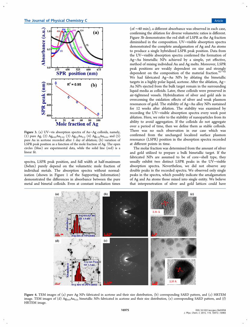
spectra, LSPR peak position, and full width at half-maximum(fwhm) purely depend on the volumetric mole fraction ofindividual metals. The absorption spectra without normal-ization (shown in Figure 1 of the Supporting Information)demonstrated the differences in absorbance between the puremetal and bimetal colloids. Even at constant irradiation times
(of ∼40 min), a different absorbance was observed in each case,confirming the ablation for diverse volumetric ratios is different.Figure 3b demonstrates the red shift of LSPR as the Ag fractiondiminished in the composition. UV−visible absorption spectrademonstrated the complete amalgamation of Ag and Au atomsto produce a single hybridized LSPR peak position. Data fromthe UV−visible absorption spectra confirmed the formation ofAg−Au bimetallic NPs achieved by a simple, yet effective,method of mixing individual Au and Ag melts. Moreover, LSPRpeak positions are weakly dependent on size and stronglydependent on the composition of the material fraction.42−45
We had fabricated Ag−Au NPs by ablating the bimetallictargets in a highly polar liquid, acetone. After the ablation, Ag−Au NPs ejected from the bulk target remain in the surroundingliquid media as colloids. Later, these colloids were preserved inair-tightened vessels. Hybridization of silver and gold aids inovercoming the oxidation effects of silver and weak plasmonresonances of gold. The stability of Ag−Au alloy NPs sustainedfor 12 weeks after ablation. The stability was examined byrecording the UV−visible absorption spectra every week postablation. Here, we refer to the stability of nanoparticles from itsability to avoid aggregation. If the colloids do not aggregateover a period of time, then we define them as stable colloids.There was no such observation in our case which wasconfirmed from the unchanged localized surface plasmonresonance (LSPR) position in the absorption spectra recordedat different points in time.The molar fraction was determined from the amount of silver
and gold utilized to prepare a bulk bimetallic target. If thefabricated NPs are assumed to be of core−shell type, theyusually exhibit two distinct LSPR peaks in the UV−visibleabsorption spectra. Nevertheless, we did not observe anydouble peaks in the recorded spectra. We observed only singlepeaks in the spectra, which possibly indicate the amalgamationof Ag and Au atoms those mixed into single entity. We believethat interpenetration of silver and gold lattices could have
Figure 3. (a) UV−vis absorption spectra of Au−Ag colloids, namely,(1) pure Ag, (2) Ag0.65Au0.35, (3) Ag0.5Au0.5, (4) Ag0.35Au0.65, and (5)pure Au in acetone recorded after 1 day of ablation; (b) variation ofLSPR peak position as a function of the mole fraction of Ag. The opencircles (blue) are experimental data, while the solid line (red) is alinear fit.
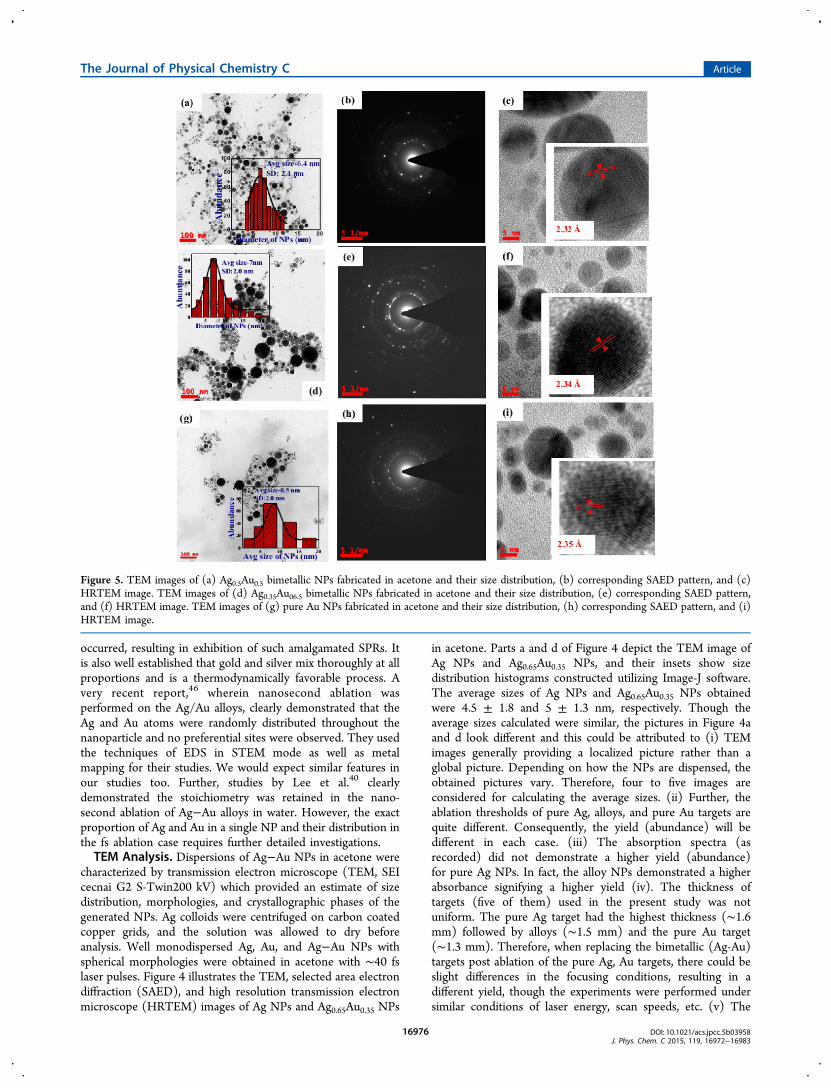
Figure 4. TEM images of (a) pure Ag NPs fabricated in acetone and their size distribution, (b) corresponding SAED pattern, and (c) HRTEMimage. TEM images of (d) Ag0.65Au0.35 bimetallic NPs fabricated in acetone and their size distribution, (e) corresponding SAED pattern, and (f)HRTEM image.
The Journal of Physical Chemistry C Article
DOI: 10.1021/acs.jpcc.5b03958J. Phys. Chem. C 2015, 119, 16972−16983
16975
occurred, resulting in exhibition of such amalgamated SPRs. Itis also well established that gold and silver mix thoroughly at allproportions and is a thermodynamically favorable process. Avery recent report,46 wherein nanosecond ablation wasperformed on the Ag/Au alloys, clearly demonstrated that theAg and Au atoms were randomly distributed throughout thenanoparticle and no preferential sites were observed. They usedthe techniques of EDS in STEM mode as well as metalmapping for their studies. We would expect similar features inour studies too. Further, studies by Lee et al.40 clearlydemonstrated the stoichiometry was retained in the nano-second ablation of Ag−Au alloys in water. However, the exactproportion of Ag and Au in a single NP and their distribution inthe fs ablation case requires further detailed investigations.TEM Analysis. Dispersions of Ag−Au NPs in acetone were
characterized by transmission electron microscope (TEM, SEIcecnai G2 S-Twin200 kV) which provided an estimate of sizedistribution, morphologies, and crystallographic phases of thegenerated NPs. Ag colloids were centrifuged on carbon coatedcopper grids, and the solution was allowed to dry beforeanalysis. Well monodispersed Ag, Au, and Ag−Au NPs withspherical morphologies were obtained in acetone with ∼40 fslaser pulses. Figure 4 illustrates the TEM, selected area electrondiffraction (SAED), and high resolution transmission electronmicroscope (HRTEM) images of Ag NPs and Ag0.65Au0.35 NPs
in acetone. Parts a and d of Figure 4 depict the TEM image ofAg NPs and Ag0.65Au0.35 NPs, and their insets show sizedistribution histograms constructed utilizing Image-J software.The average sizes of Ag NPs and Ag0.65Au0.35 NPs obtainedwere 4.5 ± 1.8 and 5 ± 1.3 nm, respectively. Though theaverage sizes calculated were similar, the pictures in Figure 4aand d look different and this could be attributed to (i) TEMimages generally providing a localized picture rather than aglobal picture. Depending on how the NPs are dispensed, theobtained pictures vary. Therefore, four to five images areconsidered for calculating the average sizes. (ii) Further, theablation thresholds of pure Ag, alloys, and pure Au targets arequite different. Consequently, the yield (abundance) will bedifferent in each case. (iii) The absorption spectra (asrecorded) did not demonstrate a higher yield (abundance)for pure Ag NPs. In fact, the alloy NPs demonstrated a higherabsorbance signifying a higher yield (iv). The thickness oftargets (five of them) used in the present study was notuniform. The pure Ag target had the highest thickness (∼1.6mm) followed by alloys (∼1.5 mm) and the pure Au target(∼1.3 mm). Therefore, when replacing the bimetallic (Ag-Au)targets post ablation of the pure Ag, Au targets, there could beslight differences in the focusing conditions, resulting in adifferent yield, though the experiments were performed undersimilar conditions of laser energy, scan speeds, etc. (v) The
Figure 5. TEM images of (a) Ag0.5Au0.5 bimetallic NPs fabricated in acetone and their size distribution, (b) corresponding SAED pattern, and (c)HRTEM image. TEM images of (d) Ag0.35Au06.5 bimetallic NPs fabricated in acetone and their size distribution, (e) corresponding SAED pattern,and (f) HRTEM image. TEM images of (g) pure Au NPs fabricated in acetone and their size distribution, (h) corresponding SAED pattern, and (i)HRTEM image.
The Journal of Physical Chemistry C Article
DOI: 10.1021/acs.jpcc.5b03958J. Phys. Chem. C 2015, 119, 16972−16983
16976
larger sized particles were clearly present only in pure Autargets and alloy targets. Pure Ag colloids did not demonstratesuch large sized NPs, indicating the ablation mechanism wasdifferent in the alloys case and the pure Au NPs case. Perhaps,the cavitation bubble dynamics played an important role in thecase of alloys and pure Au ablation, resulting in differentparticles and yields. Parts b and e of Figure 4 represent the
SAED pattern of the fabricated Ag NPs and Ag0.65Au0.35 NPswhose first ring diameter (∼2.34 Å) gives the information ofthe lattice plane separations of the Miller plane (111).Measured plane separations were 2.35 and 2.26 Å for thementioned NPs. Parts c and f of Figure 4 illustrate the HRTEMimage of Ag NPs and Ag0.65Au0.35 NPs and the measured planeseparations were 2.31 and 2.33 Å.
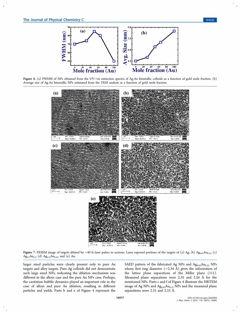
Figure 6. (a) FWHM of NPs obtained from the UV−vis extinction spectra of Ag-Au bimetallic colloids as a function of gold mole fraction. (b)Average size of Ag-Au bimetallic NPs estimated from the TEM analysis as a function of gold mole fraction.
Figure 7. FESEM image of targets ablated by ∼40 fs laser pulses in acetone. Laser exposed portions of the targets of (a) Ag, (b) Ag0.65Au0.35, (c)Ag0.5Au0.5, (d) Ag0.35Au0.65, and (e) Au.
The Journal of Physical Chemistry C Article
DOI: 10.1021/acs.jpcc.5b03958J. Phys. Chem. C 2015, 119, 16972−16983
16977

Figure 5 demonstrates TEM, SAED, and HRTEM images ofAg0.5Au0.5 NPs, Ag0.35Au0.65 NPs, and Au NPs in acetone. Partsa, d, and g of Figure 5 depict the TEM images of Ag0.5Au0.5NPs, Ag0.35Au0.65 NPs, and Au NPs, and their insets show sizedistribution histograms. The average size of Ag0.5Au0.5 NPs,Ag0.65Au0.35 NPs, and Au NPs obtained were 6.4 ± 2.1, 7 ± 2,and 8.5 ± 2 nm, respectively. While constructing thehistograms of size distribution, NPs with sizes >20 nm werenot considered, since their density was low compared to smallersized NPs. Ag−Au NPs fabricated in the present set ofexperiments are of smaller dimensions compared to the earlierreports, which demonstrated the fabrication of Ag−Au NPs bylaser ablation in liquids.46 Parts b, e, and h of Figure 5 representSAED patterns of the fabricated Ag0.5Au0.5 NPs, Ag0.65Au0.35NPs, and Au NPs whose first ring diameter provides theinformation on the lattice plane separations of the Miller plane(111). The measured plane separations were 2.26, 2.23, and2.27 Å for Ag0.5Au0.5 NPs, Ag0.65Au0.35 NPs, and Au NPs,respectively. Parts c, f, and i of Figure 5 illustrate the HRTEMimages of Ag0.5Au0.5 NPs, Ag0.65Au0.35 NPs, and Au NPs, andthe measured plane separations were 2.32, 2.34, and 2.35 Å,respectively. Lattice plane separations obtained throughHRTEM images clearly indicate the miscible nature of Agand Au metals at all proportions. This was evident from themeasured lattice plane separations which were more or lessequal to lattice constants of 2.36 and 2.35 Å for Ag and Au,respectively. The change in the unit cell size of Ag−Au alloy istypically less than 1% for all fractions of Ag and Au with respectto pure Ag and Au. Additionally, no super lattice reflectionswere observed in the prepared alloys.47
Parts a and b of Figure 6 illustrate the FWHM from UV−visible spectra of Ag−Au bimetallic NPs and the average size ofNPs from TEM images as a function of Au mole fraction. Asevident from the data presented in Figure 6a, the FWHM was
observed to be highest for the combination of Ag0.5Au0.5compared to the pure Ag or Au metal NPs. Similarly, theaverage size of Ag−Au NPs (Figure 6b) was highest for thefraction Ag0.5Au0.5. The FWHM of absorption/reflectionspectra of Au−Ag NPs is an important parameter whichenhances the efficiency of plasmonic solar cells48,24 and trapslight inside. An important feature of these Ag−Au bimetallicNPs, compared to pure Ag or Au NPs, is that they are moresuitable for fabrication of efficient plasmonic solar cells, sincethe bandwidth and scattering efficiency can be tuned in theentire intense solar spectrum. The data presented in Figure 6brevealed the differences in average particle size could, probably,be due to the variation of the ablation threshold of Ag−Aumetals at different proportions.
FESEM Analysis. Morphologies of the laser exposedportions of Ag−Au bimetallic targets along with pure Au andAg ablated targets were characterized by field emission scanningelectron microscope [FESEM (Ultra 55 from Carl ZEISS)].The fs laser ablated portions of the metal targets werecharacterized after a nominal cleaning and sonication inacetone. Figure 7 illustrates the FESEM images of laserexposed portions of Ag−Au bimetal targets along with ablatedportions of pure Au and Ag targets. Specifically, Figure 7depicts the NSs formed on (a) Ag, (b) Ag0.65Au0.35, (c)Ag0.5Au0.5, (d) Ag0.35Au0.65, and (e) Au targets, respectively.Surface morphologies of Ag, Au, and Ag−Au nanostructuredtargets at higher resolution are shown in Figure 2 of theSupporting Information. From the data presented in the imagesof Figure 7, it is apparent that the laser exposed portions of thementioned five targets comprise grains of NPs and randomgrating structures which are capable of providing higher localfields when illuminated by laser beam at a suitable wavelength(through the propagating surface plasmons). The grating kindof structures play a significant role in SERS studies. As the
Figure 8. SERS spectra of 1NPZ (20 nM dissolved in acetone) adsorbed on (a) Ag, (b) Ag0.65Au0.35, (c) Ag0.5Au0.5, and (d) Ag0.35Au0.65 NPs dropcasted on a glass slide. The time of integration was ∼0.5 s.
The Journal of Physical Chemistry C Article
DOI: 10.1021/acs.jpcc.5b03958J. Phys. Chem. C 2015, 119, 16972−16983
16978

fraction of Au increased, the randomness of the surfacemorphology of laser exposed Ag−Au bimetal targets alsoincreased. All five substrates exhibited the presence ofnanostructures as well as NP grains.SERS Studies of Au−Ag Nanoparticles and Nano-
structures. The performance of fabricated Ag−Au bimetallicNPs and NSs was evaluated by recording the Raman spectra ofexplosive molecules such as 1NPZ (1 Nitro-Pyrazole) andFOX-7, which is a well-known derivative of DADNE, i.e., 1,1-diamino-2,2-dinitroethene. The Raman spectra of 1NPZ wererecorded at 20 nM concentration from Ag−Au NPs (in filmform), and the FOX-7 was recorded at 5 μM from Ag−Au NSs.Initially, five films of Ag−Au NPs along with pure Ag and AuNPs were grown by a simple method, wherein colloidalsolution of quantity 10 μL was dropped on individual glassslides. These films were dried for some time, and later, a dropof (5 μL) analyte solution was placed on the film to form amonolayer. After evaporation of solvent in the analyte, a glassslide was placed underneath an objective lens (100×) of amicro-Raman (WiTec ALPHA 300 instrument) spectrometer.The micro-Raman spectrometer utilized a continuous wave(cw) Nd:YAG laser at 532 nm. The Raman spectrometer wascalibrated by recording the Raman peak of a silicon wafer at520 cm−1. In the micro-Raman spectrometer, the laser beamwas focused onto the analyte molecules (adsorbed on the filmof Ag−Au NPs) using a high numerical aperture objective lens(100×) and the estimated theoretical beam waist was ∼700 nm.Raman signals were collected in back scattering geometry. Theacquisition time used for recording spectra was 0.5 s for eachtrial. Figure 8 shows the SERS spectra of 1NPZ (20 nMdissolved in acetone) adsorbed on (a) Ag, (b) Ag0.65Au0.35, (c)Ag0.5Au0.5, and (d) Ag0.35Au0.65 NPs drop casted on a glass slide.It was observed that Raman signature elevation from the film ofAg0.65Au0.35 was significant compared to the others. The Ramanspectra of 1NPZ powder are presented in Figure 3 of theSupporting Information. It was also observed that Au NP filmdid not show significant modes of Raman signatures of 1NPZ(data is shown in Figure 4a of the Supporting Information).The enhancement factors (EFs)49 for Ag−Au films of NPs
were obtained by comparing the SERS spectra with the normalRaman spectra of 1NPZ at a higher concentration recorded ona nonplasmonic (silicon) substrate (shown in Figure 4b of theSupporting Information).
=II
NN
EF SERS
Raman
Raman
SERS
where
η= =N N VCA
AN N VC
AA
,SERS A SolLaser
SubstrateR A Sol
Laser
Substrate
ISERS/IR was calculated from the area under the signature peakof the particular mode of interest. NSERS - number of moleculesfrom the laser machined Ag substrate giving rise to the surfaceenhanced Raman signal, NRaman - number of molecules givingrise to the Raman signal from the non-SERS surface, NA -Avogadro number, V - total volume of the solution added ontothe substrate, Alaser - area of the laser spot, Asubstrate - total area ofthe substrate, η - adsorption factor. The adsorption factor wasestimated as explained in the procedure given in our earlierreports.10 The EFs of Ag−Au bimetallic films of NPs wereestimated for the mode corresponding to 1320 cm−1 (ringdeformation + NO2 stretch), i.e., highly elevated in the SERS
spectra and less prevailed in the normal Raman spectra of1NPZ (0.1 M) (shown in Figure 4b of the SupportingInformation). The estimated enhancement factors were ∼4.4 ×10⁸, ∼2 × 10⁹, ∼1 × 10⁸, and ∼2.6 × 10⁸ from films of Ag,Ag0.65Au0.35, Ag0.5Au0.5, and Ag0.35Au0.65 NPs, respectively.Other modes of 1NPZ and their assignments50 are presented inTable 1. The EFs of Raman signatures of 1NPZ from Ag−Au
NPs films were evaluated by considering an adsorption factor of∼0.3 (percentage of 1NPZ molecules adsorbed on the NSs)following the procedure reported in our earlier works.10 Ramanspectra of 1NPZ from gold film of NPs did not exhibit anysignificant signatures. The EFs were estimated by comparingthe SERS spectra with Raman spectra of 1NPZ (0.1 M) fromthe Si target.Similarly, the Raman activity of Ag−Au NSs was evaluated
with another high explosive molecule FOX-7 (derivative ofDADNE-1,1-diamino-2,2-dinitroethene) at a concentration of 5μM. Before recording the Raman spectra, Ag−Au NSs wereproperly cleaned and the analyte solution (10 μL) wasadsorbed on the laser exposed portions of the targets. Theanalyte solution was allowed to dry, and later, the Ramanspectra (smoothened to eliminate background and for visualclarity) were recorded. Figure 9 presents the SERS spectra ofFOX-7 (5 μM dissolved in acetone) adsorbed on (a) Ag, (b)Ag0.65Au0.35, (c) Ag0.5Au0.5, and (d) Ag0.35Au0.65 nanostructuredsubstrates obtained using fs laser ablation. The EFs wereestimated for the laser exposed Ag−Au NSs via considering themode corresponding to 860 cm−1 (NO + NH rocking) in theSERS spectra. The EFs were estimated to be ∼7 × 10⁵, ∼1 ×10⁷, ∼3 × 10⁵, and ∼1.2 × 10⁶, for Ag, Ag0.65Au0.35, Ag0.5Au0.5,and Ag0.35Au0.65 NSs, respectively. The EFs of elevated Raman
Table 1. Observed Active Raman Modes of 1NPZ Adsorbedon Ag−Au Film of NPs Fabricated by fs Ablation inAcetonea
S. No.
reportedRamanshifts
(in cm−1) assignments
observed shiftsfrom INPZpowder(in cm−1)
observed SERS shiftsfrom bimetallicnanomaterials(in cm−1)
1. 303 ring deforma-tion + NO2bend
2. 438 N−NO2stretch
460 428
3. 593 ring deforma-tion + NO2bend
570
4. 648 ring puckering 674, 679
5. 865 ring deforma-tion, in plane
821
6. 1033 ring deforma-tion
1036 1044
7. 1076 ring deforma-tion
1062 1078, 1086
8. 1178 ring deforma-tion
1165 1112, 1150, 1160
9. 1263 ring deforma-tion
1266 1231, 1256, 1270
10. 1321 ring deforma-tion + NO2stretch
1320 1323, 1330, 1331,1340
11. 1482 NO2 stretch 1480 1463, 1465, 1470,1475
12 1555 NO2 stretch 1530 1557, 1565, 1594
13. 1607 NO2 stretch 1619 1609, 1645aThe time of integration was 0.5 s for all measurements.
The Journal of Physical Chemistry C Article
DOI: 10.1021/acs.jpcc.5b03958J. Phys. Chem. C 2015, 119, 16972−16983
16979

signatures of FOX-7 from Ag−Au NSs were evaluated byconsidering the adsorption factor to be ∼0.4 (percentage ofFOX-7 molecules adsorbed on the NSs) following theprocedure reported in our earlier works.10 Similar to the caseof Au film of NPs, Au NSs demonstrated good elevation of theRaman signatures whose shift is greater than 1145 cm−1 andother modes were not elevated much (as shown in Figure 5a ofthe Supporting Information). The above-mentioned enhance-ment factors were estimated by comparing the SERS spectrawith normal Raman spectra of FOX-7 (0.1 M) on a glass slide(shown in Figure 5b of the Supporting Information). Theobserved modes of FOX-7 and their detailed assignments51 arepresented in Table 2.
In the above-mentioned two cases of (a) detection of 1NPZand (b) detection of FOX-7, the combination of Ag0.65Au0.35was observed to demonstrate superior Raman enhancementscompared to other combinations and pure Ag and Aunanomaterials. The tunability of LSP resonances plays a crucialrole in the optical properties of the materials. Probably thehybridization of metals at these proportions was superiorcompared to other combinations. Consequently, elevation ofRaman signatures was observed due to the enhanced local fieldsprovided by Ag0.65Au0.35 nanomaterials compared to othercombinations. Our observations demonstrated that obviousSERS enhancement was observed when the analyte moleculesadsorbed on the Ag−Au system of appropriate proportions.
Figure 9. SERS spectra of FOX-7 (5 μM dissolved in acetone) adsorbed on (a) Ag, (b) Ag0.65Au0.35, (c) Ag0.5Au0.5, and (d) Ag0.35Au0.65nanostructured substrates obtained using femtosecond laser ablation. The time of integration was ∼0.5 s.
Table 2. Observed Active Raman Modes of FOX-7 Adsorbed on Ag−Au Nanostructures Fabricated by fs Ablation in Acetonea
S. No.reported Raman shifts of
FOX-7 (in cm−1) assignmentsobserved Raman shifts from FOX-7
powder (in cm−1)observed surface enhanced Raman shifts from
bimetallic nanomaterials (in cm−1)
1. 318 out of layer H wagging 318 3482. 447 in layer sym. NH wagging 455 410, 4203. 481 sym. NO and NH wagging 477 482, 4894. 622 out of layer sym. NH
wagging620 620, 622
5. 789 C−NO2 rocking 790 775, 794, 795, 8056. 856 NO and NH rocking 856 860, 861, 862, 8707. 1024 in layer asym. NH wagging 1024 1021, 10388. 1070 in layer sym. NH wagging 1063 1071, 10769. 1208 asym. C−NO2 stretch + NH
wagging1206 1202, 1247
10. 1343 sym. C−NO2 stretch + NHwagging
1346 1340, 1357
11. 1464 C−C stretch + NH wagging 146112. 1528 C−NH2 bend + C−C
stretch1524
aThe time of integration was 0.5 s for all measurements.
The Journal of Physical Chemistry C Article
DOI: 10.1021/acs.jpcc.5b03958J. Phys. Chem. C 2015, 119, 16972−16983
16980

The combination of Ag−Au metals provides chemical stabilityto the nanomaterials which could have enhanced the efficacy ofSERS activity. When the analyte molecules adsorbed inbetween the dimers of Ag−Au combination, local electric fieldsenhance gigantically as a function of separation. Zheng et al.52
reported that the increment of Au percentage in Ag−Au core−shell NPs diminishes the SERS activity. One more possiblereason could be the dimensionality (smaller dimensionscompared to others) of the fabricated NPs/NSs at theproportion used in Ag0.65Au0.35. Fan et al.53 recentlydemonstrated that SERS enhancements not only depend onthe Ag:Au ratio but also on the chemical nature of the analytemolecule. In their studies, they obtained higher Ramansignature enhancements from alloy (Ag−Au) NPs with highergold proportion for the positively charged probes of oxazine720 (Oxa) and Nile Blue A (NBA). On the other hand, Ag−AuNPs with a higher Ag proportion exhibited superior SERSenhancements for the molecular probes 4-hydroxythiophenol(HTP) and thiophenol (TP) which are negatively charged.This was explained through their DFT calculations whichconcluded that the charge transfer between Au and Ag atoms inalloy NPs provoke positively charged regions in Ag atoms,whereas negatively charged regions prevail in Au atoms. Thus,obtained charges on surfaces of Ag−Au NPs influence theattachment of analyte molecules on top of the nanomaterialsand, consequently, the enhancement factors. Many otherstudies54−64 on Ag−Au bimetallic systems were mainlyconfined to NPs and their related studies, but in the presentstudy, we have investigated the SERS activity of both bimetallicNPs in acetone (colloidal solutions) as well as NSs (fs lasermodified Ag solid targets).The data (Raman measurements) presented here were an
average of several measurements (depending on the integrationtime). In the case of NPs in film form (drop casted), the Ramanintensity at different points could be slightly different(depending on the way the drop is placed on the glass slide).By increasing the number of averages (integration time), wetried to minimize the fluctuations. For practical applications,one requires consistent spectra over a large area of the substrateachieved in the shortest period of time. We anticipate thatscreen printing65 of these NPs could be one possible solutionfor producing highly reproducible measurements. However, inthe case of nanostructures, we achieved reproducibility over alarge area, since the nanostructures remained intact after eachmeasurement. We had earlier demonstrated the recyclingcapability of Cu nanostructures prepared using the sametechnique.10 Possible improvements in the present worksinclude (a) grafting of Ag−Au NPs on a germanium/siliconsubstrate for extra uniform SERS substrates which is a two-stepprocess further enhancing the SERS sensitivity, as documentedearlier by Wang et al.;66 (b) grafting of Ag−Au NPs on plaingold or silver substrates which incorporates the generation ofhot spots those determined by the nanogap between the plainAg/Au surface and Ag−Au NPs;34 (c) Ag−Au bimetallic NPscan be utilized as SERS sensors on paper through the screenprinting technique which facilitates the fabrication of movableSERS sensors and those are directly usable for onsite detectionof molecular probes;65 (d) Ag−Au NPs with smallerdimensions can be encapsulated with silicon quantum dots inpolymeric NPs, enhancing the capability of luminescence andthe SERS activity of molecular probes significantly.67 Thecombination of NPs and NSs (by placing NPs on thenanostructured surface as reported by Jin et al.68) is another
possibility of improving the detection limits. NPs in thenanogaps of NSs generate an enormous number of hot spotsdue to the combined plasmonic resonances enhancing the localfields which elevate the Raman signatures of adsorbed analytemolecules. Our future studies will also focus on the photonicapplications of such NPs.69,70
■ CONCLUSIONSAg−Au bimetallic targets at different proportions were made bymixing the required proportions of metals followed by meltingand cooling the mixture. Thus, prepared mixtures were utilizedto make small target strips (10 × 10 mm2) to carry out ablation.Later, these targets were ablated with ∼40 fs laser pulses inacetone and thus Ag−Au bimetallic nanomaterials (NPs andNSs) were fabricated in a single experiment. Post fabricationAg−Au NPs and NSs were utilized to detect explosivemolecules of FOX-7 and 1NPZ. Our SERS measurementsdemonstrated enhancement factors in the range 106−108. Thisstudy demonstrated that a specific proportion, Ag0.65Au0.35, isresponsible for obtaining very significant SERS enhancements(compared to other proportions) compatible with the explosiveprobe molecules FOX-7 and 1NPZ. This enhancement istentatively attributed to the formation of positively chargedregions in Ag rich NPs, resulting in significant binding ofanalyte molecules to the surface of Ag−Au NPs and NSs.
■ ASSOCIATED CONTENT*S Supporting Information(a) UV−visible absorption spectra of “as prepared colloids”. (b)Raman spectra of nitropyrazole powder, on AuNP film, and ona glass slide. (c) High resolution FESEM images of thenanostructured targets. (d) Raman spectra of the FOX-7molecule on the Au target and a glass slide. The SupportingInformation is available free of charge on the ACS Publicationswebsite at DOI: 10.1021/acs.jpcc.5b03958.
■ AUTHOR INFORMATIONCorresponding Author*E-mail: [email protected] or [email protected]: +91-040-23138811.
NotesThe authors declare no competing financial interest.
■ ACKNOWLEDGMENTSThe authors acknowledge DRDO, India, for continuousfinancial support and UPE-II, UoH, for partial financial support.
■ REFERENCES(1) Amendola, V.; Meneghetti, M. Laser Ablation Synthesis inSolution and Size Manipulation of Noble Metal Nanoparticles. Phys.Chem. Chem. Phys. 2009, 11, 3805−3821.(2) Itina, T. E. On Nanoparticles Formation by Laser Ablation inLiquids. J. Phys. Chem. C 2011, 115, 5044−5048.(3) Amendola, V.; Polizzi, S.; Meneghetti, M. Laser AblationSynthesis of Gold Nanoparticles in Organic Solvents. J. Phys. Chem.B 2006, 110, 7232−7237.(4) Amendola, V.; Riello, P.; Meneghetti, M. Magnetic Nanoparticlesof Iron Carbide, Iron Oxide, Iron@Iron Oxide, and Metal IronSynthesized by Laser Ablation in Organic Solvents. J. Phys. Chem. C2011, 115, 5140−5146.(5) Podagatlapalli, G. K.; Hamad, S.; Sreedhar, S.; Tewari, S. P.;Venugopal Rao, S. Fabrication and Characterization of Aluminum
The Journal of Physical Chemistry C Article
DOI: 10.1021/acs.jpcc.5b03958J. Phys. Chem. C 2015, 119, 16972−16983
16981

Nanostructures and Nanoparticles Obtained Using FemtosecondAblation Technique. Chem. Phys. Lett. 2012, 530, 93−97.(6) Podagatlapalli, G. K.; Hamad, S.; Tewari, S. P.; Sreedhar, S.;Prasad, M. D.; Venugopal Rao, S. Silver Nano-entities ThroughUltrafast Double Ablation in Aqueous media for Surface EnhancedRaman Scattering and Photonics Applications. J. Appl. Phys. 2013, 113,073106.(7) Podagatlapalli, G. K.; Hamad, S.; Mohiddon, M. A.; VenugopalRao, S. Effect of Oblique incidence on Silver Nanomaterials Fabricatedin Water via Ultrafast Laser Ablation for Photonics and ExplosivesDetection. Appl. Surf. Sci. 2014, 303, 217−232.(8) Podagatlapalli, G. K.; Hamad, S.; Mohiddon, M. A.; VenugopalRao, S. Fabrication of Nanoparticles and Nanostructures usingUltrafast Laser Ablation of Silver with Bessel Beams. Laser Phys.Lett. 2015, 12, 036003.(9) Hamad, S.; Podagatlapalli, G. K.; Vendamani, V. S.; NageswaraRao, S. V. S.; Pathak, A. P.; Venugopal Rao, S. Femtosecond Ablationof Silicon in Acetone: Tunable Photoluminescence from GeneratedNanoparticles and Fabrication of Surface Nanostructures. J. Phys.Chem. C 2014, 118, 7139−7151.(10) Hamad, S.; Podagatlapalli, G. K.; Mohiddon, M. A.; VenugopalRao, S. Cost Effective Nanostructured Copper Substrates Preparedwith Ultrafast Laser Pulses for Explosives Detection using SurfaceEnhanced Raman Scattering. Appl. Phys. Lett. 2014, 104, 263104.(11) Venugopal Rao, S.; Podagatlapalli, G. K.; Hamad, S. UltrafastLaser Ablation in Liquids for Nanomaterials and Applications. J.Nanosci. Nanotechnol. 2014, 14, 1364−1388.(12) Barcikowski, S.; Mafune, F. Trends and Current Topics in theField of Laser Ablation and Nanoparticle Generation in Liquids. J.Phys. Chem. C 2011, 115, 4985.(13) Dallaire, A.-M.; Rioux, D.; Rachkov, A.; Patskovsky, S.; Meunier,M. Laser-Generated Au-Ag Nanoparticles for Plasmonic Nucleic AcidSensing. J. Phys. Chem. C 2012, 116, 11370−11377.(14) Sajti, C. L.; Sattari, R.; Chichkov, B. N.; Barcikowski, S. GramScale Synthesis of Pure Ceramic Nanoparticles by Laser Ablation inLiquid. J. Phys. Chem. C 2010, 114, 2421−2427.(15) Rehbock, C.; Jakobi, J.; Gamrad, L.; van der Meer, S.;Tiedemann, D.; Taylor, U.; Kues, W.; Rath, D.; Barcikowski, S.Current State of Laser Synthesis of Metal and Alloy Nanoparticles asLigand-free Reference Materials for Nano-toxicological Assays.Beilstein J. Nanotechnol. 2014, 5, 1523−1541.(16) Hajiesmaeilbaigi, F.; Motamedi, A. Synthesis of Au/Ag AlloyNanoparticles by Nd:YAG Laser Irradiation. Laser Phys. Lett. 2007, 4,133−137.(17) Toshima, N.; Yonezawa, T. Bimetallic NanoparticlesNovelMaterials for Chemical and Physical Applications. New J. Chem. 1998,22, 1179−1201.(18) Compagnini, G.; Messina, E.; Puglisi, O.; Cataliotti, R. S.;Nicolosi, V. Spectroscopic Evidence of a Core−Shell Structure in theEarlier Formation Stages of Au−Ag Nanoparticles by Pulsed LaserAblation in Water. Chem. Phys. Lett. 2008, 457, 386−390.(19) Wang, A. Q.; Liu, J. H.; Lin, S. D.; Lin, T. S.; Mou, C. Y. ANovel Efficient Au−Ag Alloy Catalyst System: Preparation, Activity,and Characterization. J. Catal. 2005, 233, 186−197.(20) Wang, A. Q.; Chang, C. M.; Mou, C. Y. Evolution of CatalyticActivity of Au−Ag Bimetallic Nanoparticles on Mesoporous Supportfor CO Oxidation. J. Phys. Chem. B 2005, 109, 18860−18867.(21) Intartaglia, R.; Das, G.; Bagga, K.; Gopalakrishnan, A.;Genovese, A.; Povia, M.; Di Fabrizio, E.; Cingolani, R.; Diaspro, A.;Brandi, F. Laser Synthesis of Ligand Free Bimetallic Nanoparticles forPlasmonic Applications. Phys. Chem. Chem. Phys. 2013, 15, 3075−3082.(22) Kuladeep, R.; Jyothi, L.; Ali, S. S.; Deepak, K. L. N.; NarayanaRao, D. Laser-assisted Synthesis of Au-Ag Alloy Nanoparticles withTunable Surface Plasmon Resonance Frequency. Opt. Mater. Express2012, 2, 161−172.(23) Shah, A.; Rahman, L.-U.; Qureshi, R.; Rehman, Z.-U. Synthesis,Characterization and Applications of Bimetallic (Au-Ag, Au-Pt, Au-Ru)Alloy Nanoparticles. Rev. Adv. Mater. Sci. 2012, 30, 133−149.
(24) Bansal, A.; Sekhon, J. S.; Verma, S. S. Scattering Efficiency ofLSPR Tunability of Bimetallic Ag, Au, and Cu Nanoparticles.Plasmonics 2014, 9, 143−150.(25) Adamovic, N.; Schmid, U. Potential of Plasmonics inPhotovoltaic Solar Cells. Elektrotech. Inftech. 2011, 128, 342−347.(26) Tan, K. S.; Cheong, K. Y. Advances of Ag, Cu, and Ag-Cu alloyNanoparticles Synthesized via Chemical Reduction Route. J. Nanopart.Res. 2013, 15, 1537.(27) Lu, L.; Burkey, G.; Halaciuga, I.; Goia, D. V. Core-shell Gold/Silver Nanoparticles: Synthesis and Optical Properties. J. ColloidInterface Sci. 2013, 392, 90−95.(28) Beyene, H. T.; Chakravadhanula, V. S. K.; Hanisch, C.;Strunskus, T.; Zaporojtchenko, V.; Elbahri, M.; Faupel, F. Vapor PhaseDeposition, Structure, and Plasmonic Properties of Polymer-basedComposites Containing Ag-Cu Bimetallic Nanoparticles. Plasmonics2012, 7, 107−114.(29) Shin, K. S.; Kim, J. H.; Kim, I. H.; Kim, K. Novel Fabricationand Catalytic Applications of Poly(enthylenimine)-Stabilized Gold-Silver Alloy Nanoparticles. J. Nanopart. Res. 2012, 14, 735.(30) Liu, S.; Chen, G.; Prasad, P. N.; Swihart, M. T. Synthesis ofMonodisperse Au, Ag, and Au-Ag Alloy Nanoparticles with TunableSize and Surface Plasmon Resonance Frequency. Chem. Mater. 2011,23, 4098−4101.(31) Hodak, J. H.; Henglein, A.; Giersig, M.; Hartland, G. V. LaserInduced Diffusion in Au-Ag Core Shell Nanoparticles. J. Phys. Chem. B2000, 104, 11708−11718.(32) Chen, Y.-H.; Yeh, C.-S. A New Approach for the Formation ofAlloy Nanoparticles: Laser Synthesis of Gold-Silver Alloy from Gold-Silver Colloidal Mixtures. Chem. Commun. 2001, 4, 371−372.(33) Liusman, C.; Li, H.; Lu, G.; Wu, J.; Boey, F.; Li, S.; Zhang, H.Surface-Enhanced Raman Scattering of Ag-Au Nano-disk Hetero-dimers. J. Phys. Chem. C 2012, 116, 10390−10395.(34) Kim, K.; Choi, J.; Shin, K. S. Enhanced Raman Scattering inGaps Formed by Planar Au and Au/Ag Alloy Nanoparticles. J. Phys.Chem. C 2013, 117, 11421−11427.(35) Kim, K.; Kim, K. L.; Choi, J.; Lee, H. B.; Shin, K. S. SurfaceEnrichment of Ag Atoms in Au/Ag Alloy Nanoparticles Revealed bySurface-Enhanced Raman Scattering of 2,6-Dimethylphenyl Isocya-nide. J. Phys. Chem. C 2010, 114, 3448−3453.(36) Nedderson, J.; Chumanov, G.; Cotton, T. M. Laser Ablation ofMetals: A New Method for Preparing SERS Active Colloids. Appl.Spectrosc. 1993, 47, 1959−1964.(37) Prochazka, M.; Stephanek, J.; Vicova, B.; Stephanek, J.; Turpin,P. Y. Probing Applications of Laser-Ablated Ag Colloids in SERSSpectroscopy: Improvement of Ablation Procedure and SERS SpectralTesting. Anal. Chem. 1997, 69, 5103−5108.(38) Menendez-Manjon, A.; Wagener, P.; Barcikowski, S. Transfer-matrix Method for Efficient Ablation by Pulsed Laser Ablation andNanoparticle Generation in Liquids. J. Phys. Chem. C 2011, 115,5108−5114.(39) Chen, D. H.; Chen, C. J. Formation and Characterization ofAu−Ag Bimetallic Nanoparticles in Water-in-oil Micro-emulsions. J.Mater. Chem. 2002, 12, 1557−1562.(40) Lee, I.; Han, S. W.; Kim, K. Production of Au−Ag alloyNanoparticles by Laser Ablation of Bulk Alloys. Chem. Commun. 2001,18, 1782−1783.(41) Han, S. W.; Kim, Y.; Kim, K. Dodecanethiol-Derivatized Au/AgBimetallic Nanoparticles: TEM, UV/VIS, XPS, and FTIR Analysis. J.Colloid Interface Sci. 1998, 208, 272−278.(42) Papavassiliou, G. C. Surface plasmons in Small Au-Ag alloyParticles. J. Phys. F: Met. Phys. 1976, 6, L103.(43) Teo, B. K.; Keating, K.; Kao, Y.-H. Observation of PlasmonFrequency in the Optical Spectrum of Au18Ag2, Cluster: TheBeginning of the Collective Phenomenon Characteristic of the Bulk?J. Am. Chem. Soc. 1987, 109, 3494.(44) Sinzig, J.; Radtke, U.; Quinten, M.; Kreibig, U. Binary clusters:Homogeneous Alloys and Nucleus-shell Structures. Z. Phys. D: At.,Mol. Clusters 1993, 26, 242−245.
The Journal of Physical Chemistry C Article
DOI: 10.1021/acs.jpcc.5b03958J. Phys. Chem. C 2015, 119, 16972−16983
16982

(45) Link, S.; Wang, Z. L.; El-Sayed, M. A. Alloy Formation of Gold−Silver Nanoparticles and the Dependence of the Plasmon Absorptionon Their Composition. J. Phys. Chem. B 1999, 103, 3529−3533.(46) Olea-Mejía, O.; Fernandez-Mondragon, M.; Rodríguez-de laConcha, G.; Camacho-Lopez, M. SERS-Active Ag, Au and Ag−AuAlloy Nanoparticles Obtained by Laser Ablation in Liquids for SensingMethylene Blue. Appl. Surf. Sci. 2015, 348, 66−70.(47) LeBlanc, M.; Erler, W. Rbntgenograplaische Tintersuchungemdes Mischkristallsystems Gold-Silber und Umtersuchungemiiber seineArtgreif barkeit durch Salpetersdiure. Ann. Phys. 1933, 408, 321.(48) Catchpole, K. R.; Polman, A. Plasmonic Solar Cells. Opt. Express2008, 16, 21793−21800.(49) Le Ru, E. C.; Etchegoin, P. G. Phenomenological Local FieldEnhancement Factor Distributions Around Electromagnetic HotSpots. J. Chem. Phys. 2009, 130, 181101−4.(50) Nageswara Rao, E.; Ravi, P.; Tewari, S. P.; Venugopal Rao, S.Experimental and Theoretical Studies on the Structure and VibrationalProperties of Nitropyrazoles. J. Mol. Struct. 2013, 1043, 121−131.(51) Dreger, Z. A.; Tao, Y.; Gupta, Y. M. High-Pressure Vibrationaland Polymorphic Response of 1,1-Diamino-2,2-dinitroethene SingleCrystals: Raman Spectroscopy. J. Phys. Chem. A 2014, 118, 5002−5012.(52) Zheng, Z.; Shan, G.; Li, J.; Chen, Y.; Liu, Y. Au/Ag Nano AlloyShells as Near Infrared SERS Nano-probe for the Detection of Protein.Mater. Res. Express 2014, 1, 045408.(53) Fan, M.; Lai, F.; Chou, H.; Lu, W.; Hwang, B.; Brolo, A. G.Surface-enhanced Raman Scattering (SERS) from Au:Ag BimetallicNanoparticles: The Effect of the Molecular Probe. Chem. Sci. 2013, 4,509−515.(54) Kim, K.; Choi, J.; Shin, K. S. Raman Scattering Characterizationof 1,4-phenylenediisocyanide in Au−Au and Ag−Au Nanogaps.Spectrochim. Acta, Part A 2013, 100, 3−9.(55) Kim, K.; Kim, K. L.; Lee, S. J. Surface Enrichment of Ag atomsin Au/Ag alloy Nanoparticles Revealed by Surface Enhanced RamanScattering Spectroscopy. Chem. Phys. Lett. 2005, 403, 77−82.(56) Liao, X.; Chen, Y.; Qin, M.; Chen, Y.; Yang, L.; Zhang, H.; Tian,Y. Au−Ag−Au Double Shell Nanoparticles-based Localized SurfacePlasmon Resonance and Surface-enhanced Raman Scattering Bio-sensor for Sensitive Detection of 2-mercapto-1-methylimidazole.Talanta 2013, 117, 203−208.(57) Gellner, M.; Kustner, B.; Schlucker, S. Optical Properties andSERS Efficiency of Tunable Gold/Silver Nanoshells. Vib. Spectrosc.2009, 50, 43−47.(58) Wu, P.; Gao, Y.; Zhang, H.; Cai, C. Aptamer-Guided Silver-GoldBimetallic Nanostructures with Highly Active Surface-EnhancedRaman Scattering for Specific Detection and Near-Infrared Photo-thermal Therapy of Human Breast Cancer Cells. Anal. Chem. 2012, 84,7692−7699.(59) Bu, Y.; Lee, S. Influence of Dopamine Concentration andSurface Coverage of Au Shell on the Optical Properties of Au, Ag, andAg Nanoparticles. ACS Appl. Mater. Interfaces 2012, 4, 3923−3931.(60) Bai, T.; Sun, J.; Che, R.; Xu, L.; Yin, C.; Guo, Z.; Gu, N.Controllable Preparation of Core-Shell Au-Ag Nanoshuttles withImproved Refractive Index Sensitivity and SERS Activity. ACS Appl.Mater. Interfaces 2014, 6, 3331−3140.(61) Yang, Y.; Zhang, Q.; Fu, Z. W.; Qin, D. Transformation of AgNanocubes into Ag-Au Hollow Nanostructures with Enriched AgContents to Improve SERS Activity and Chemical Stability. ACS Appl.Mater. Interfaces 2014, 6, 3750−3757.(62) Mulvaney, P. Surface Plasmon Spectroscopy of Nano SizedMetal Particles. Langmuir 1996, 12, 788−800.(63) Sebastian, S.; Linslal, C. L.; Vallbhan, C. P. G.; Nampoori, V. P.N.; Radhakrishnan, P.; Kailasnath, M. Formation of Au-Ag NanoalloyThrough Au core/Ag Shell Intermediate Phase by Laser Ablation.Chem. Phys. Lett. 2015, 628, 25−29.(64) Wang, T.; Hu, F.; Ikhile, E.; Liao, F.; Li, Y.; Shao, M. Two-step-route to Ag−Au Nanoparticles Grafted on Ge Wafer for Extra-uniformSERS Substrates. J. Mater. Chem. C 2015, 3, 559−563.
(65) Ou, Y.; Wang, L.; Zhu, L.; Wan, L.; Xu, Z. In-SituImmobilization of Silver Nanoparticles on Self-Assembled Honey-comb-patterned Films Enables Surface-Enhanced Raman Scattering(SERS) Substrates. J. Phys. Chem. C 2014, 118, 11478−11484.(66) Qu, L.-L.; Song, Q.; Li, Y.; Peng, M.; Li, D.; Chen, L.; Fossey, J.S.; Long, Y. Fabrication of Bimetallic Microfluidic Surface-EnhancedRaman Scattering Sensors on Paper by Screen Printing. Anal. Chim.Acta 2013, 792, 86−92.(67) Harun, N. A.; Horrocks, B. R.; Fulton, D. A. Enhanced Ramanand Luminescence Spectra from Co-encapsulated Silicon QuantumDots and Ag-Au Nano alloys. Chem. Commun. 2014, 50, 12389−12391.(68) Jin, Z.; Gu, W.; Shi, X.; Wang, Z.; Jiang, Z.; Liao, L. A NovelRoute to Surface-Enhanced Raman Scattering: Ag NanoparticlesEmbedded in the Nano-gaps of a Ag Substrate. Adv. Opt. Mater. 2014,2, 588−596.(69) Hamad, S.; Podagatlapalli, G. K.; Tewari, S. P.; Venugopal Rao,S. Influence of Picosecond Multiple/single Line Ablation on CopperNanoparticles Fabricated for Surface Enhanced Raman Spectroscopyand Photonics Applications. J. Phys. D: Appl. Phys. 2013, 46, 485501.(70) Papagiannouli, I.; Aloukos, P.; Rioux, D.; Meunier, M.; Couris,S. Effect of the Composition on the Nonlinear Optical Response ofAuxAg1−x Nano-Alloys. J. Phys. Chem. C 2015, 119, 6861−6872.
The Journal of Physical Chemistry C Article
DOI: 10.1021/acs.jpcc.5b03958J. Phys. Chem. C 2015, 119, 16972−16983
16983