topic: Tooth Development – Bell & Advanced Bell Stages Dr.n ...
56
topic: Tooth Development – Bell & Advanced Bell Stages Dr.n.vaishnavi dhanvantri Department of Oral Pathology & Microbiology SRM Dental College, Ramapuram, Chennai, India
-
Upload
khangminh22 -
Category
Documents
-
view
0 -
download
0
Transcript of topic: Tooth Development – Bell & Advanced Bell Stages Dr.n ...

topic:Tooth Development –
Bell & Advanced Bell Stages
Dr.n.vaishnavi dhanvantri
Department of Oral Pathology
& Microbiology
SRM Dental College,
Ramapuram, Chennai, India

Tooth Development –Bell & Advanced Bell Stages
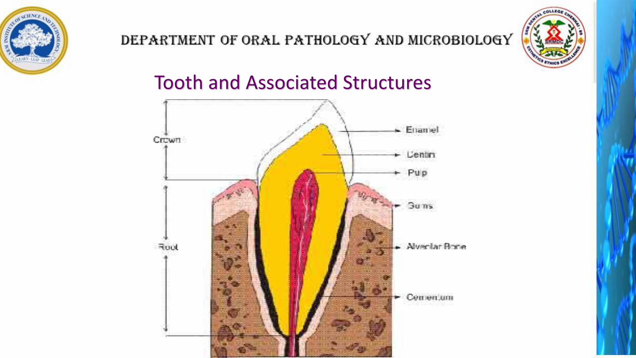
Tooth and Associated Structures
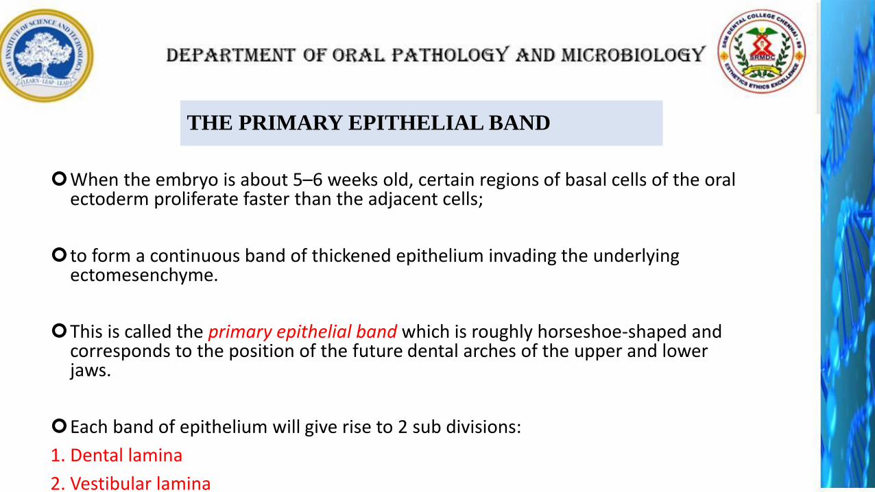
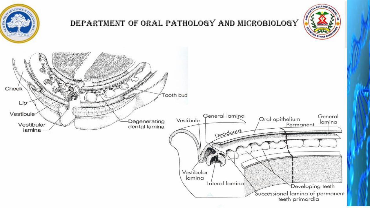
THE PRIMARY EPITHELIAL BAND
When the embryo is about 5–6 weeks old, certain regions of basal cells of the oral ectoderm proliferate faster than the adjacent cells;
to form a continuous band of thickened epithelium invading the underlying ectomesenchyme.
This is called the primary epithelial band which is roughly horseshoe-shaped and corresponds to the position of the future dental arches of the upper and lower jaws.
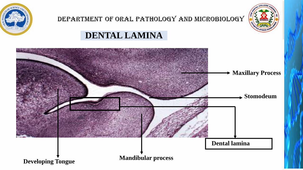
Each band of epithelium will give rise to 2 sub divisions:
1. Dental lamina
2. Vestibular lamina
Primary epithelial
band
Ectomesenchyme
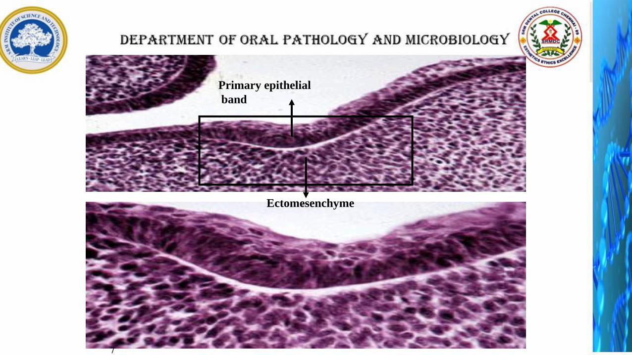
/
Stomodeum
Developing Tongue
Dental lamina
Maxillary Process
Mandibular process
DENTAL LAMINA

Dental Lamina
• Dental lamina appears as a thickening
of the oral epithelium adjacent to
condensation of ectomesenchyme
• 20 areas of enlargement or knobs
appear, which will form tooth buds
for the 20 primary teeth
• Distal Extention – permanent molars
• Lingual Extention –Successional lamina: lamina
from which permanent teeth develop
• The dental lamina begins to function
at 6th prenatal week and continues to
15th year of birth (3rd molar)
• Remnants – cell rests of serre
Vestibular Lamina

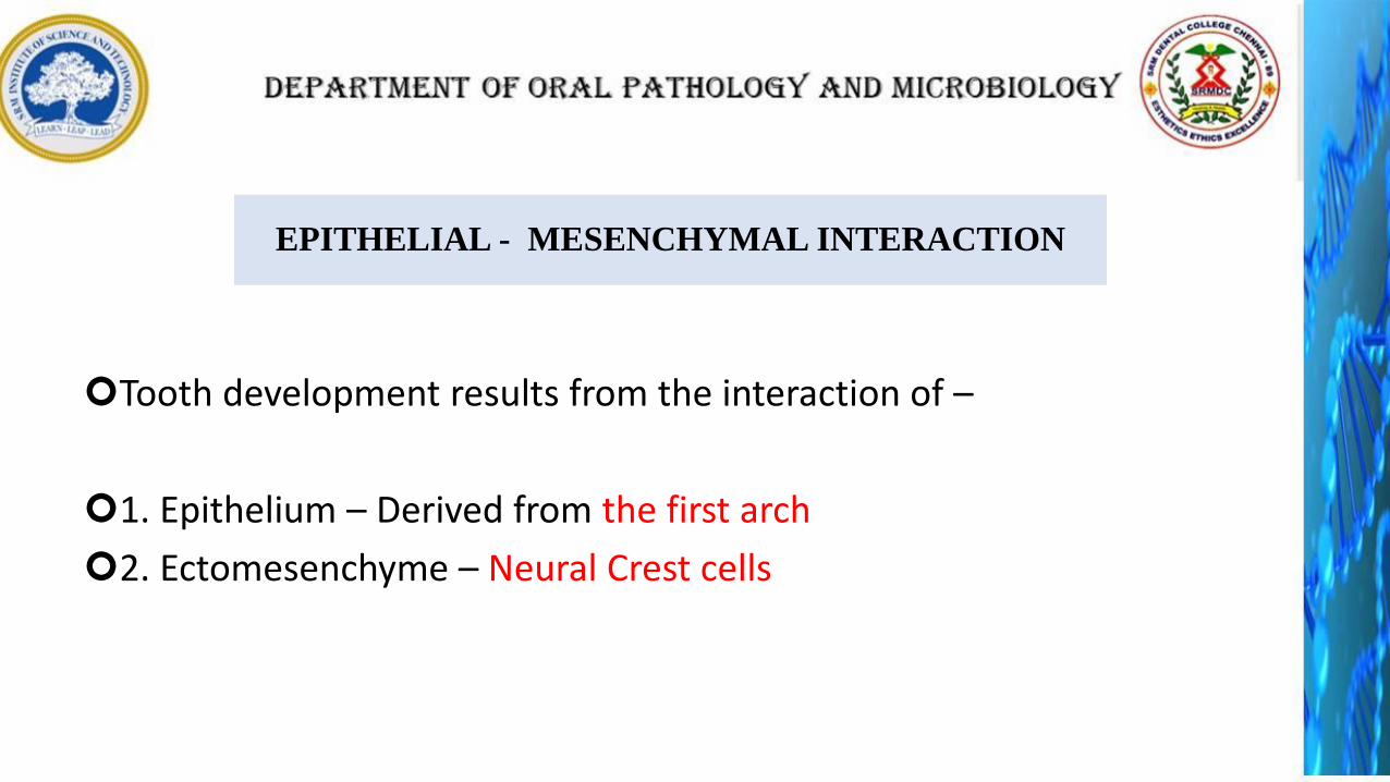
EPITHELIAL - MESENCHYMAL INTERACTION
Tooth development results from the interaction of –
1. Epithelium – Derived from the first arch
2. Ectomesenchyme – Neural Crest cells
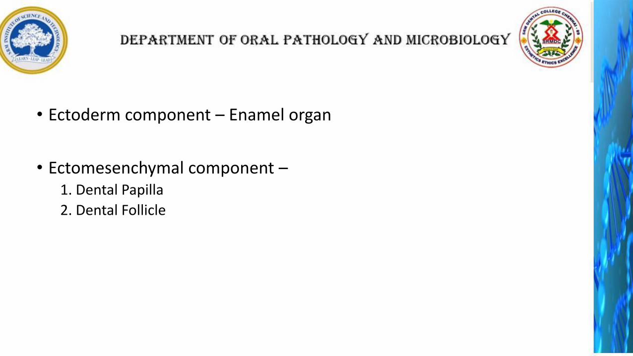
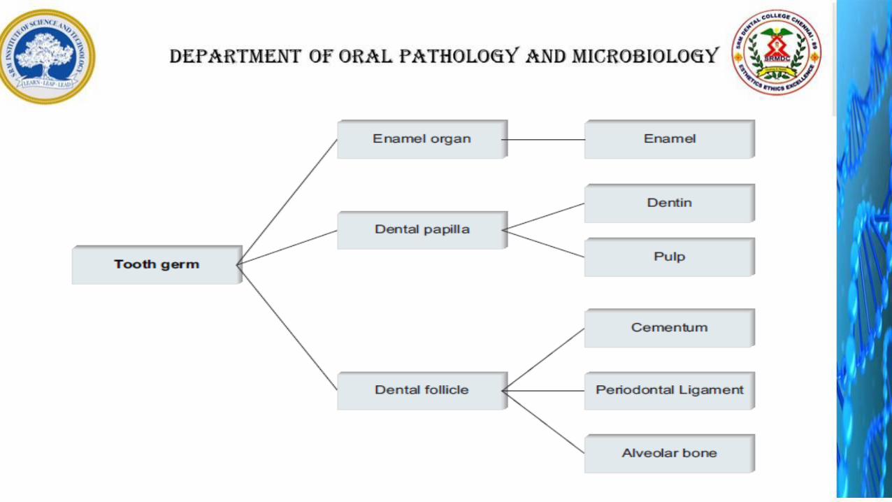
• Ectoderm component – Enamel organ
• Ectomesenchymal component –1. Dental Papilla
2. Dental Follicle
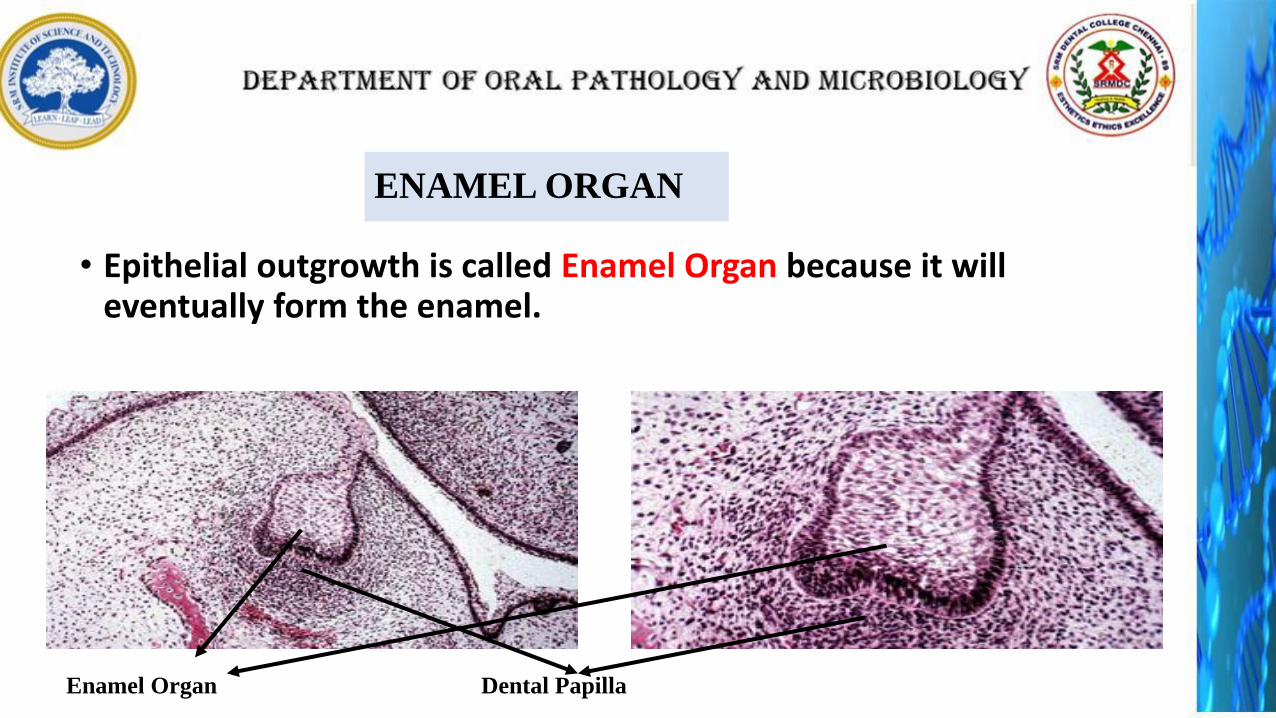
ENAMEL ORGAN
• Epithelial outgrowth is called Enamel Organ because it will eventually form the enamel.
Enamel Organ Dental Papilla

DENTAL PAPILLA
• The condensed ectomesenchyme immediately under the enamel organ is the dental papilla.
Enamel Organ Dental Papilla
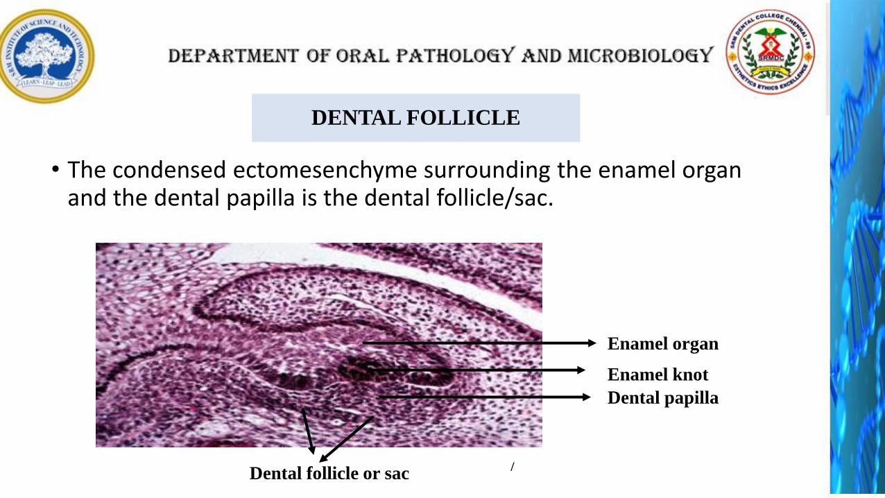
DENTAL FOLLICLE
• The condensed ectomesenchyme surrounding the enamel organ and the dental papilla is the dental follicle/sac.
Enamel organ
Dental papilla
Dental follicle or sac
Enamel knot
/
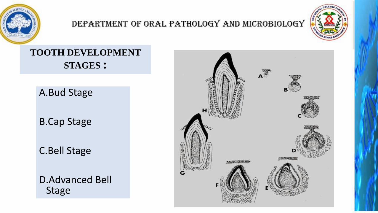
TOOTH DEVELOPMENT
STAGES :
A.Bud Stage
B.Cap Stage
C.Bell Stage
D.Advanced Bell Stage
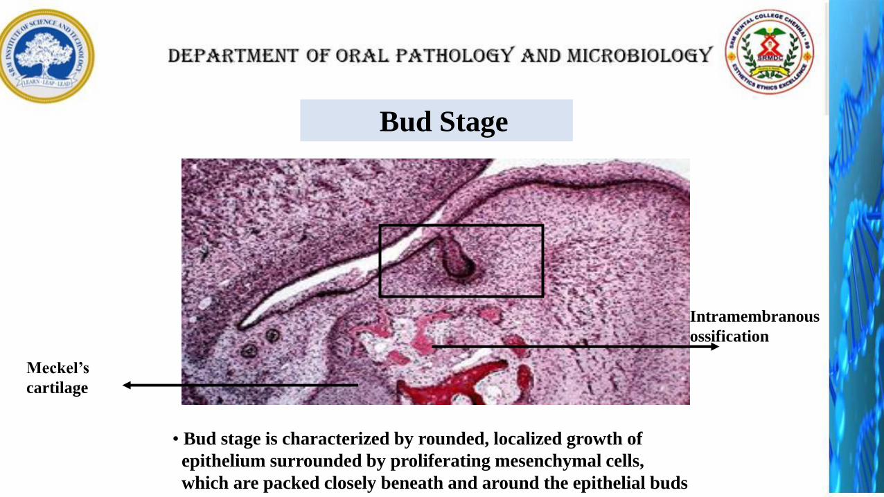
Bud Stage
• Bud stage is characterized by rounded, localized growth of
epithelium surrounded by proliferating mesenchymal cells,
which are packed closely beneath and around the epithelial buds
Meckel’s
cartilage
Intramembranous
ossification
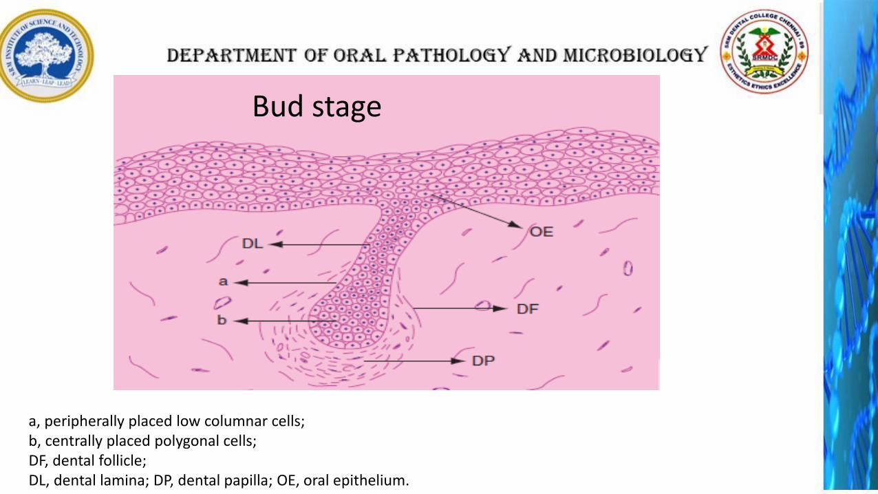
a, peripherally placed low columnar cells; b, centrally placed polygonal cells; DF, dental follicle;DL, dental lamina; DP, dental papilla; OE, oral epithelium.
Bud stage
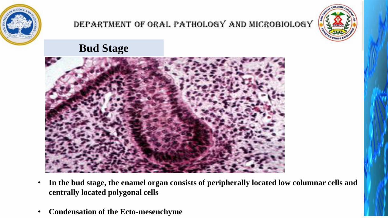
Bud Stage
• In the bud stage, the enamel organ consists of peripherally located low columnar cells and
centrally located polygonal cells
• Condensation of the Ecto-mesenchyme
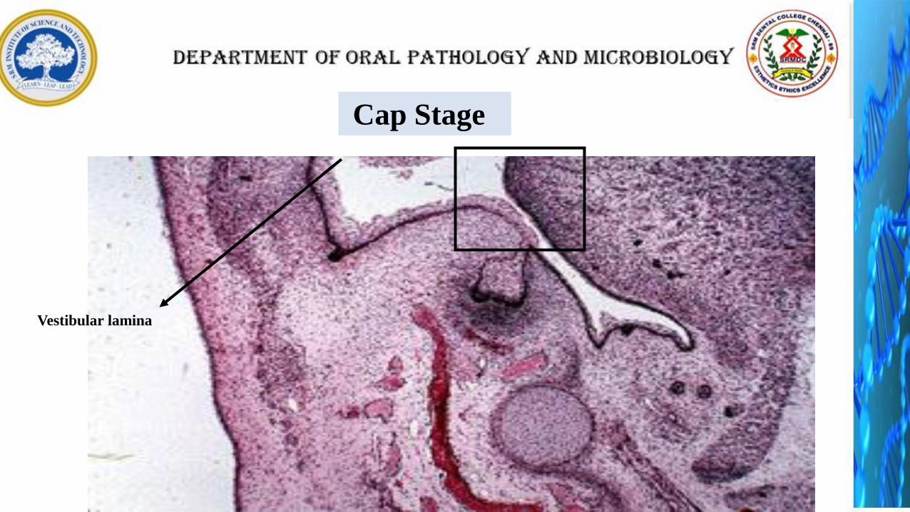
Cap Stage
Vestibular lamina
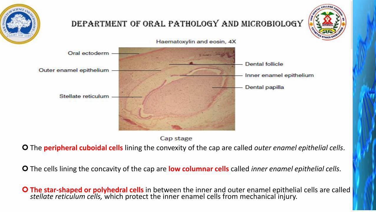
The peripheral cuboidal cells lining the convexity of the cap are called outer enamel epithelial cells.
The cells lining the concavity of the cap are low columnar cells called inner enamel epithelial cells.
The star-shaped or polyhedral cells in between the inner and outer enamel epithelial cells are called stellate reticulum cells, which protect the inner enamel cells from mechanical injury.
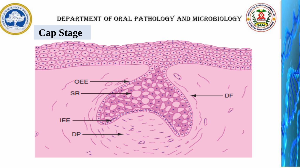
Cap Stage
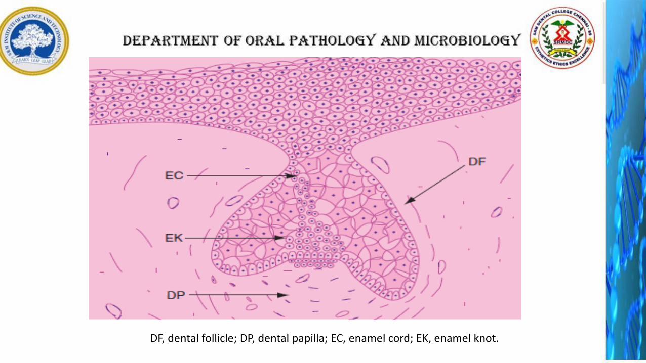
DF, dental follicle; DP, dental papilla; EC, enamel cord; EK, enamel knot.
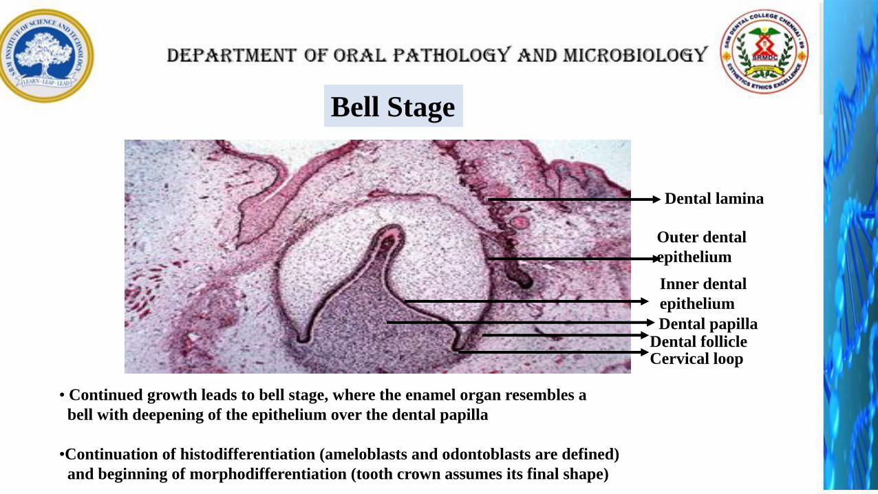
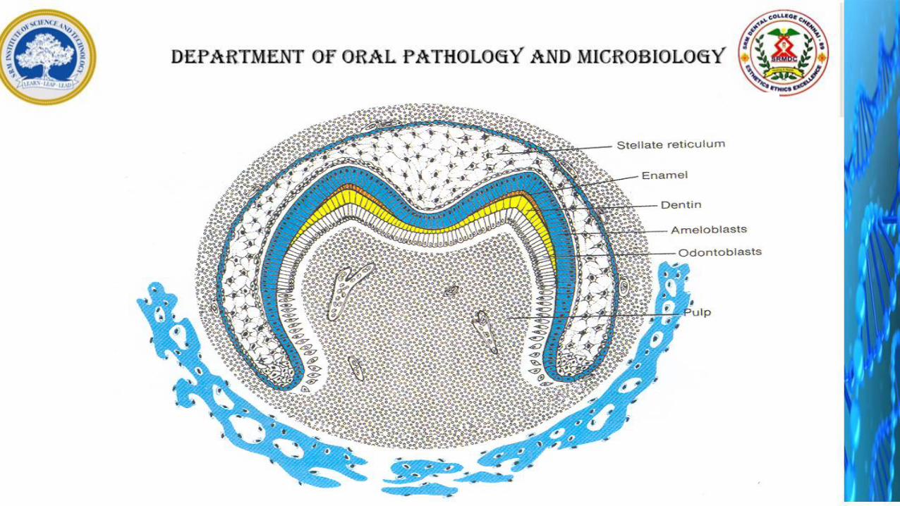
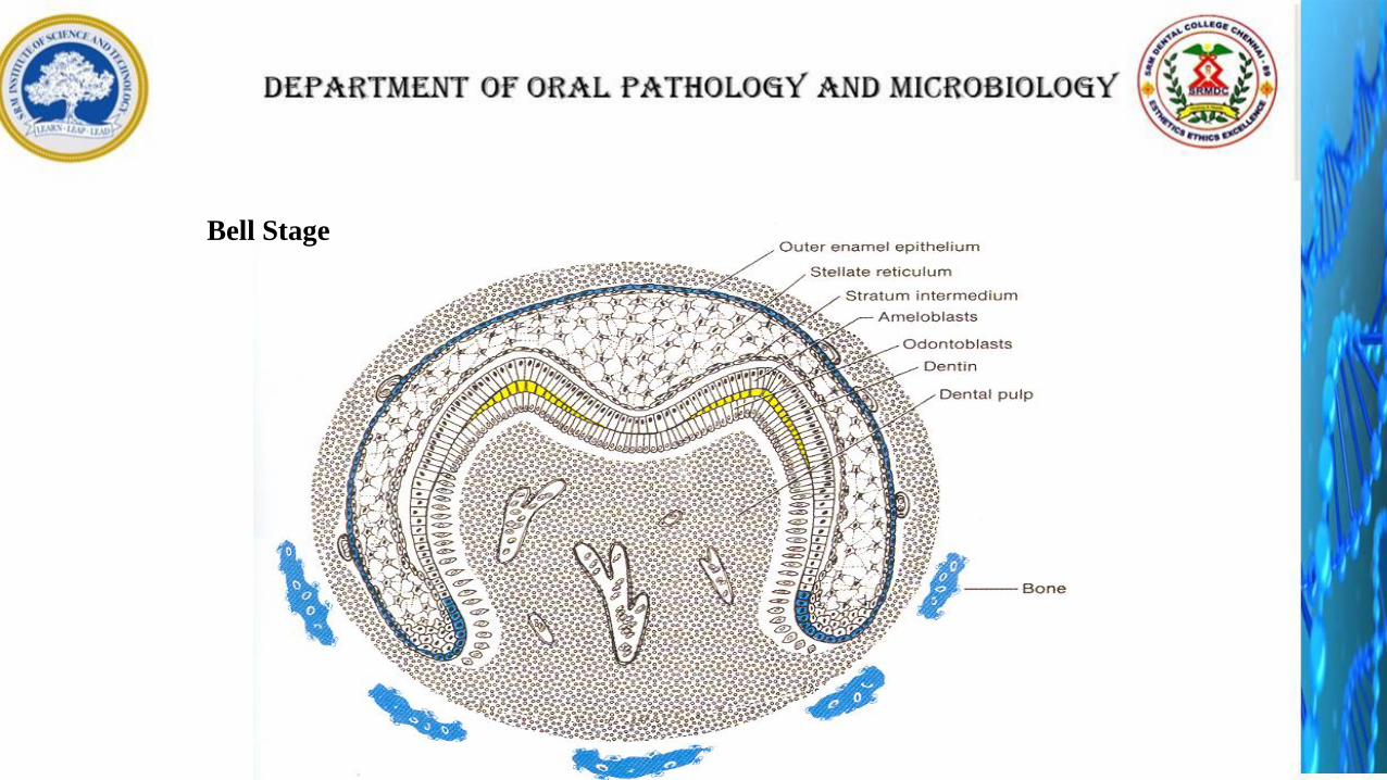
Bell Stage
• Continued growth leads to bell stage, where the enamel organ resembles a
bell with deepening of the epithelium over the dental papilla
•Continuation of histodifferentiation (ameloblasts and odontoblasts are defined)
and beginning of morphodifferentiation (tooth crown assumes its final shape)
Dental lamina
Outer dental
epithelium
Inner dental
epithelium
Dental papillaDental follicleCervical loop
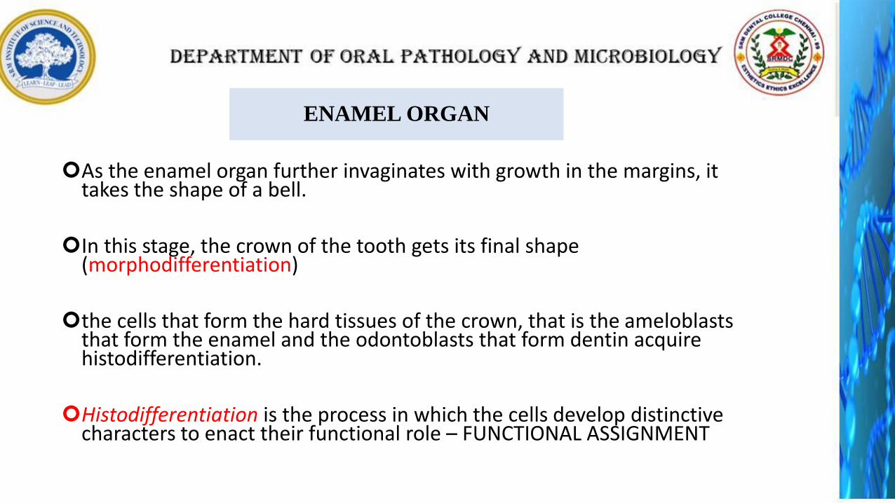
ENAMEL ORGAN
As the enamel organ further invaginates with growth in the margins, it takes the shape of a bell.
In this stage, the crown of the tooth gets its final shape (morphodifferentiation)
the cells that form the hard tissues of the crown, that is the ameloblaststhat form the enamel and the odontoblasts that form dentin acquire histodifferentiation.
Histodifferentiation is the process in which the cells develop distinctive characters to enact their functional role – FUNCTIONAL ASSIGNMENT
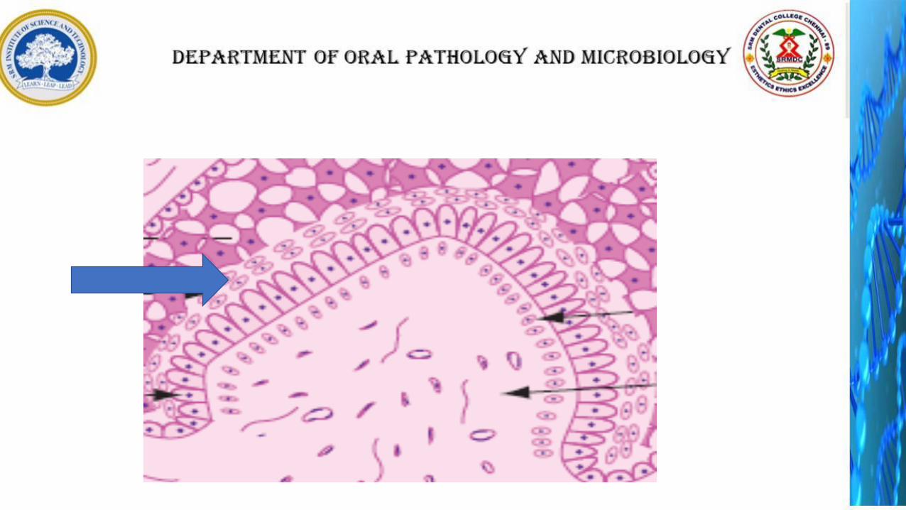
Four different types of epithelial cells of the enamel organ can be identified in this stage:
1. Inner enamel epithelium
2. Stratum intermedium
3. Stellate reticulum
4. Outer enamel epithelium
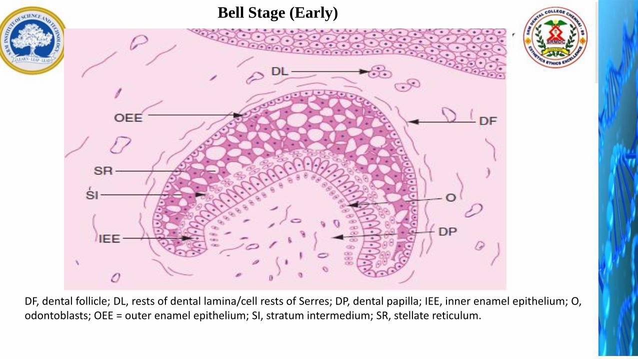
DF, dental follicle; DL, rests of dental lamina/cell rests of Serres; DP, dental papilla; IEE, inner enamel epithelium; O, odontoblasts; OEE = outer enamel epithelium; SI, stratum intermedium; SR, stellate reticulum.
Bell Stage (Early)
INNER ENAMEL EPITHELIUM:
Short columnar cells bordering the dental papilla.
These will eventually become ameloblasts that will form the enamel of the tooth crown by differentiating into tall columnar cells.
The desmosomes connect these inner enamel epithelial cells with one another and with the cells of the stratum intermedium.
The cells of inner dental epithelium exert an organizing influence on the underlying mesenchymal cells in the dental papilla, which later differentiate into odontoblasts.
STRATUM INTERMEDIUM:
Some layers of squamous cells seen between inner enamel epithelium and stellate reticulum are called stratum intermedium.
The cells of the stratum intermedium are connected to one another and to the cells of the inner enamel epithelium and stellate reticulum by desmosomes and gap junctions.
They are associated with protein synthesis and transport of nutrients to the ameloblasts.
They exhibit high enzyme alkaline phosphatase activity.
This layer plays an important role in regulating the formation of the enamel.
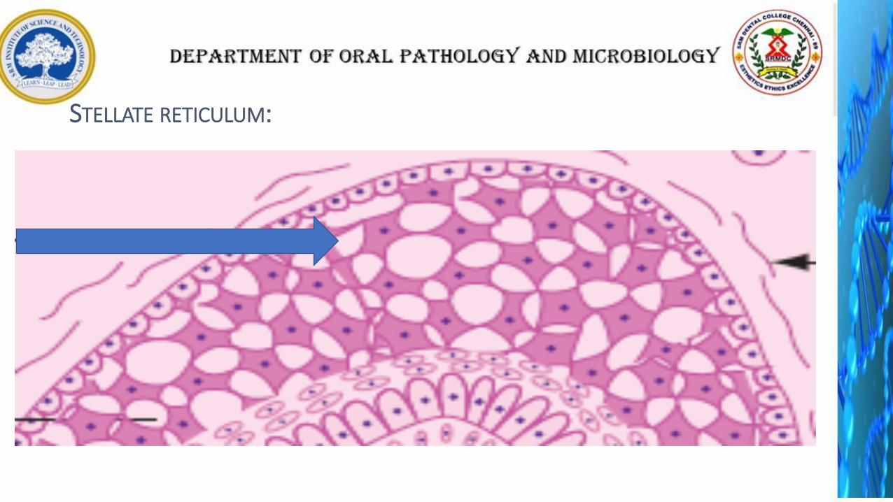
STELLATE RETICULUM:
These star-shaped cells are connected to one another and to the cells of the outer enamel epithelium and the stratum intermedium by desmosomes.
These serve to protect the underlying inner enamel epithelial cells.
These cells secrete glycosaminoglycans, which attract water, thereby swelling the cells and pushing them apart.
However, they still maintain contact with each other, thus becoming star-shaped.
They have a cushion-like consistency that may support and protect the delicate enamel organ.
It is absent in the portion that outlines the root portions.
STELLATE RETICULUM:
After a layer of dentin is laid down, the nutritional supply for the inner enamel epithelial cells from the dental papilla is cut.
The stellate reticulum collapses before the formation of enamel, bringing the capillaries in the dental follicle closer to the ameloblaststo provide nutrition.
This collapse begins at the deepest point of invagination of the enamel organ and progresses cervically.
The collapsed stellate reticulum cannot be distinguished from the stratum intermedium.

OUTER ENAMEL EPITHELIUM:
The outer enamel epithelium has cuboidal cells.
They are connected to adjacent cells by junctional complexes.
The outer enamel epithelial layer is smooth in the initial stages.
As the stellate reticulum collapses before enamel formation, this layer is laid into folds bringing the capillaries present in the dental follicle more closer to the ameloblasts.
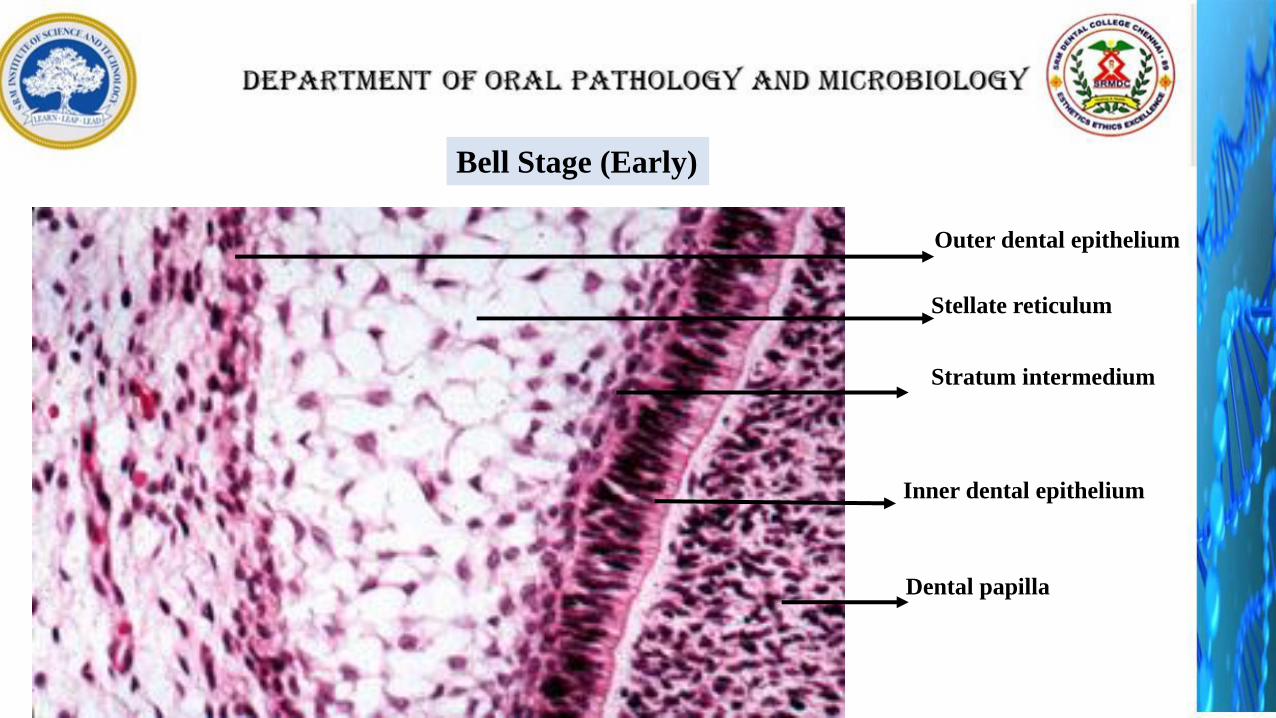
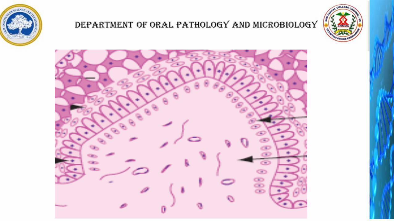
Bell Stage (Early)
Stellate reticulum
Inner dental epithelium
Stratum intermedium
Dental papilla
Outer dental epithelium

Inner dental epithelium
Outer dental epithelium
Cervical loop
Cervical loop: Area where the inner and the outer dental epithelium meet at
the rim of the enamel organ. This point is where the cells will continue to
divide until the tooth crown attains its full size and which after crown
formation will give rise to the epithelium for root formation. Is also called
“Zone of Reflexion”.

DENTAL PAPILLA:
An acellular zone is present between the basal lamina and the dental papilla and is devoid of cells.
The peripherally placed undifferentiated ectomesenchymal cells of the dental papilla increase in size, before the enamel formation begins.
They are initially cuboidal and later become columnar and differentiate into odontoblasts.
This increase in size of the outermost cells of the dental papilla eliminates the acellular zone between the dental papilla and the inner enamel epithelium.
Membrana preformativa is the term given to the basement membrane which separates the enamel organ and dental papilla just before the dentin formation.

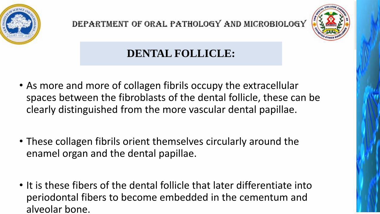
DENTAL FOLLICLE:
• As more and more of collagen fibrils occupy the extracellular spaces between the fibroblasts of the dental follicle, these can be clearly distinguished from the more vascular dental papillae.
• These collagen fibrils orient themselves circularly around the enamel organ and the dental papillae.
• It is these fibers of the dental follicle that later differentiate into periodontal fibers to become embedded in the cementum and alveolar bone.

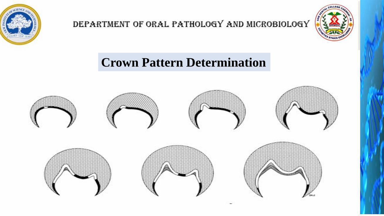
Crown Pattern Determination
CROWN PATTERN DETERMINATION
The other events that occur during the bell stage are the breaking up of dental lamina
and determination of the crown pattern of the tooth (morphodifferentiation).
invagination or infolding of the inner layer of the enamel organ, that is inner enamel epithelium, decides the shape of the crown of the tooth.
The infolding gives shape to the crown, occurs as a result of the differential rates of mitotic division within the cells of the inner enamel epithelium and the difference in time at which these cells undergo differentiation.
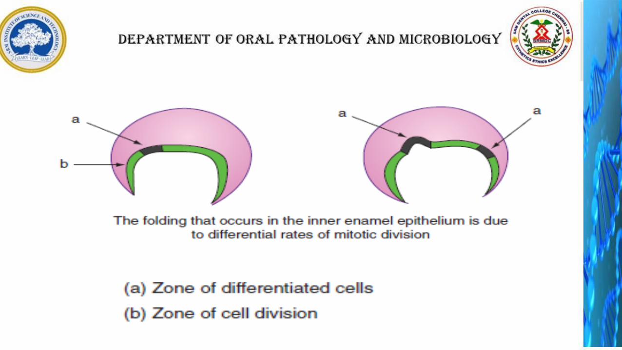
Those inner enamel epithelial cells which lie in the future cusp tip region stop dividing earlier and begin to differentiate first to assume their function of producing enamel.
As these cells at the cusp tip have undergone differentiation and cease to divide, the other cells of the enamel organ continue to divide, exerting pressure on the cells in the cusp tip region.
This causes the cells in the region of cusp tip to be pushed up into the enamel organ towards outer enamel epithelium.
The cells in another future cusp area begin to differentiate and form a cusp tip by a similar process.
Cell differentiation proceeds cervically and those at the cervical loop are the last to differentiate. In this way, the shape of the crown is determined.
Bell Stage
ADVANCED BELL STAGE
The advanced bell stage is characterised by the deposition and mineralisation of the first layer of the dentin, followed by the enamel, along the future dentinoenameljunction.
In this stage, stellate reticulum cells start collapsing before deposition starts.
The cells of the outer enamel epithelium assume a flattened low cuboidal shape and the surfaces become folded.
Mineralisation starts at the cuspal tip and proceeds cervically.
Hertwig’s epithelial root sheath, which initiates root formation, starts as a downward growth of outer enamel epithelial and inner enamel epithelial cells after dentin and enamel formation reaches the future cementoenamel junction.

Advanced bell stage.
A, ameloblasts; D, denti n matrix; DF, dental follicle; DP, dental papilla; E, enamelmatrix; O, odontoblasts; OEE, outer enamel epithelium; SI, stratum intermedium; SR, stellate reti culum.
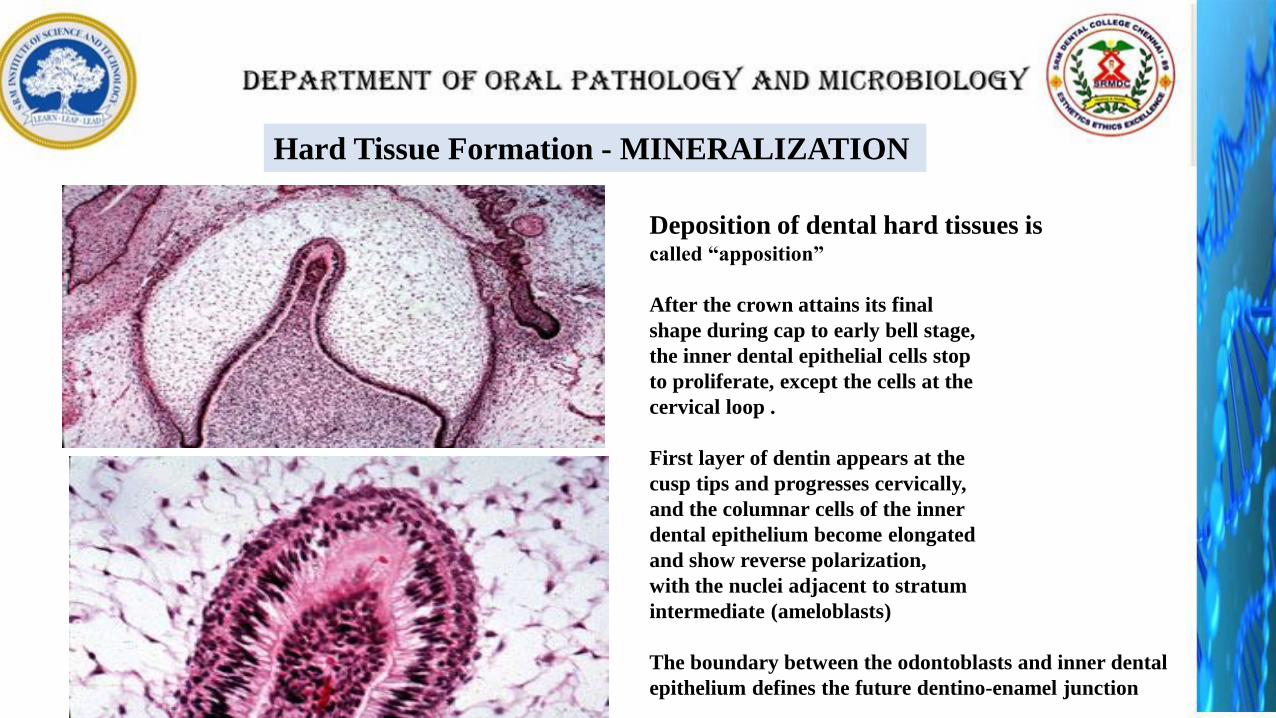
Hard Tissue Formation - MINERALIZATION
Deposition of dental hard tissues iscalled “apposition”
After the crown attains its final
shape during cap to early bell stage,
the inner dental epithelial cells stop
to proliferate, except the cells at the
cervical loop .
First layer of dentin appears at the
cusp tips and progresses cervically,
and the columnar cells of the inner
dental epithelium become elongated
and show reverse polarization,
with the nuclei adjacent to stratum
intermediate (ameloblasts)
The boundary between the odontoblasts and inner dental
epithelium defines the future dentino-enamel junction
RECIPROCAL INDUCTION
• The important step in the development of tooth was the terminal differentiation of ameloblasts and odontoblasts and the formation of two principal hard tissue of the tooth, enamel and dentin.
• For dentinogenesis and amelogenesis to take place normally, the differentiating odontoblasts and ameloblasts will receive signals form each other – “reciprocal induction”
• The differentiation of odontoblasts from the peripheral ectomesenchymal cells of the dental papilla is under the influence of the inner enamel epithelial cells.
• A single layer of dentin is laid down by the odontoblasts.
• This aids the inner enamel epithelial cells to differentiate into functional ameloblasts with reversal of polarity and form enamel matrix against the newly formed dentin..

Stages of Apposition
1. Elongation of inner dental epithelium
2. Differentiation of odontoblasts
3. Formation of dentin
4. Formation of enamel

Ameloblasts
First layer of enamel
Dentin
Odontoblasts