Tissue engineered model for real time monitoring of liver inflammation
10
Tissue-Engineered Model for Real-Time Monitoring of Liver Inflammation Rohit Jindal, Ph.D., 1,2 Suraj J. Patel, B.S., 1 and Martin L. Yarmush, M.D., Ph.D. 1,2 Tissue-engineered in vitro models have the potential to be used for investigating inflammation in the complex microenvironment found in vivo. We have developed an in vitro model of hepatic tissue that facilitates real-time monitoring of endothelium activation in liver tissue. This was achieved by creating a layered coculture model in which hepatocytes were embedded in collagen gel and a reporter clone of endothelial cells, which synthesizes green fluorescent protein in response to nuclear factor-kappa B (NF-kB) activation, was overlaid on top of the gel. The efficacy of our approach was established by monitoring in real time the dynamics of NF-kB-regulated fluorescence in response to tumor necrosis factor a. Our studies revealed that endothelial cells in coculture with hepatocytes exhibited a similar NF-kB-mediated fluorescence to both pulse and step stimulation of lipopoly- saccharide. By contrast, endothelial cells in monoculture displayed enhanced NF-kB-regulated fluorescence to step in comparison to pulse lipopolysaccharide stimulation. The NF-kB-mediated fluorescence correlated with endothelial cell expression of NF-kB-regulated genes such as intercellular adhesion molecule 1, vascular cell adhesion molecule 1, and E-Selectin, as well as with leukocyte adhesion. These findings suggest that our model provides a powerful platform for investigating hepatic endothelium activation in real time. Introduction I nflammation is the basic response of a tissue for repairing damage caused by injury or infection. However, if inflammation is not resolved it can cause further damage. 1 In response to injury, different cell types in a tissue exhibit dynamic transcriptional patterns that ultimately regulate levels of growth factors, cytokines, and chemokines in a spatiotemporal fashion for the activation of an inflammatory response. Identifying mechanisms at cellular and tissue level that regulate inflammation will facilitate development of superior treatment strategies. In the liver, there are a number of diseased states in which inflammation is implicated, including sepsis, 2 ischemia/reperfusion injury, 3 steatohepa- titis, 4 fibrosis, 5 and certain drug-induced injury. 6 Tissue- engineered models of liver that capture the complex signaling microenvironment found in vivo can provide an important platform for investigating hepatic inflammation. These mod- els have the potential of bridging the gap between cell culture and animal experiments, and also provide surrogate models for human studies. Nuclear factor-kappa B (NF-kB) is the central transcription factor implicated in regulation of several inflammation- associated genes. 7 Previous studies with animals have es- tablished elevated levels of NF-kB activation in liver in response to various insults, including endotoxin 8 and viral infection. 9 However, localizing the NF-kB activation patterns in individual cell types was complicated by other con- founding factors such as stress associated with isolating cells from the liver, which resulted in an artifactual elevation of NF-kB activation. 8 Since NF-kB is involved in expression of several cytokines, chemokines, and adhesion molecules, 10 which ultimately regulate the dynamic microenvironment of liver tissue, development of in vitro models that facilitate identifying activation patterns of NF-kB in individual cell types under complex multicellular environments is highly desirable. Traditional methods for gene expression analysis include reverse transcription polymerase chain reaction (RT-PCR), DNA microarray, and northern blot. These methods are es- sentially destructive and not well suited for measuring dy- namics of gene expression patterns. Further, in tissue models that employ multiple cell types there is an additional com- plexity of separating individual cell types to localize the cellular source of gene expression. Fluorescent reporters provide a nondestructive method for measuring gene ex- pression in live cells. 11–14 These clones are prepared by transfecting cells with plasmid DNA that encodes for easily detectable proteins such as green fluorescent protein (GFP) under the regulation of a specific transcription factor. When 1 Center for Engineering in Medicine, Massachusetts General Hospital, Harvard Medical School, and the Shriners Hospitals for Children, Boston, Massachusetts. 2 Department of Biomedical Engineering, Rutgers University, Piscataway, New Jersey. TISSUE ENGINEERING: Part C Volume 17, Number 1, 2011 ª Mary Ann Liebert, Inc. DOI: 10.1089/ten.tec.2009.0782 113
Transcript of Tissue engineered model for real time monitoring of liver inflammation

Tissue-Engineered Model for Real-Time Monitoringof Liver Inflammation
Rohit Jindal, Ph.D.,1,2 Suraj J. Patel, B.S.,1 and Martin L. Yarmush, M.D., Ph.D.1,2
Tissue-engineered in vitro models have the potential to be used for investigating inflammation in the complexmicroenvironment found in vivo. We have developed an in vitro model of hepatic tissue that facilitates real-timemonitoring of endothelium activation in liver tissue. This was achieved by creating a layered coculture model inwhich hepatocytes were embedded in collagen gel and a reporter clone of endothelial cells, which synthesizesgreen fluorescent protein in response to nuclear factor-kappa B (NF-kB) activation, was overlaid on top of thegel. The efficacy of our approach was established by monitoring in real time the dynamics of NF-kB-regulatedfluorescence in response to tumor necrosis factor a. Our studies revealed that endothelial cells in coculture withhepatocytes exhibited a similar NF-kB-mediated fluorescence to both pulse and step stimulation of lipopoly-saccharide. By contrast, endothelial cells in monoculture displayed enhanced NF-kB-regulated fluorescence tostep in comparison to pulse lipopolysaccharide stimulation. The NF-kB-mediated fluorescence correlated withendothelial cell expression of NF-kB-regulated genes such as intercellular adhesion molecule 1, vascular celladhesion molecule 1, and E-Selectin, as well as with leukocyte adhesion. These findings suggest that our modelprovides a powerful platform for investigating hepatic endothelium activation in real time.
Introduction
Inflammation is the basic response of a tissue forrepairing damage caused by injury or infection. However,
if inflammation is not resolved it can cause further damage.1
In response to injury, different cell types in a tissue exhibitdynamic transcriptional patterns that ultimately regulatelevels of growth factors, cytokines, and chemokines in aspatiotemporal fashion for the activation of an inflammatoryresponse. Identifying mechanisms at cellular and tissue levelthat regulate inflammation will facilitate development ofsuperior treatment strategies. In the liver, there are a numberof diseased states in which inflammation is implicated,including sepsis,2 ischemia/reperfusion injury,3 steatohepa-titis,4 fibrosis,5 and certain drug-induced injury.6 Tissue-engineered models of liver that capture the complex signalingmicroenvironment found in vivo can provide an importantplatform for investigating hepatic inflammation. These mod-els have the potential of bridging the gap between cell cultureand animal experiments, and also provide surrogate modelsfor human studies.
Nuclear factor-kappa B (NF-kB) is the central transcriptionfactor implicated in regulation of several inflammation-associated genes.7 Previous studies with animals have es-tablished elevated levels of NF-kB activation in liver in
response to various insults, including endotoxin8 and viralinfection.9 However, localizing the NF-kB activation patternsin individual cell types was complicated by other con-founding factors such as stress associated with isolating cellsfrom the liver, which resulted in an artifactual elevation ofNF-kB activation.8 Since NF-kB is involved in expression ofseveral cytokines, chemokines, and adhesion molecules,10
which ultimately regulate the dynamic microenvironment ofliver tissue, development of in vitro models that facilitateidentifying activation patterns of NF-kB in individual celltypes under complex multicellular environments is highlydesirable.
Traditional methods for gene expression analysis includereverse transcription polymerase chain reaction (RT-PCR),DNA microarray, and northern blot. These methods are es-sentially destructive and not well suited for measuring dy-namics of gene expression patterns. Further, in tissue modelsthat employ multiple cell types there is an additional com-plexity of separating individual cell types to localize thecellular source of gene expression. Fluorescent reportersprovide a nondestructive method for measuring gene ex-pression in live cells.11–14 These clones are prepared bytransfecting cells with plasmid DNA that encodes for easilydetectable proteins such as green fluorescent protein (GFP)under the regulation of a specific transcription factor. When
1Center for Engineering in Medicine, Massachusetts General Hospital, Harvard Medical School, and the Shriners Hospitals for Children,Boston, Massachusetts.
2Department of Biomedical Engineering, Rutgers University, Piscataway, New Jersey.
TISSUE ENGINEERING: Part CVolume 17, Number 1, 2011ª Mary Ann Liebert, Inc.DOI: 10.1089/ten.tec.2009.0782
113
the desired transcription factor gets activated, it leads toexpression of GFP and the fluorescence level increases,thereby enabling real-time measurement of transcriptionalactivity in live cells.
In this work, we have combined tissue engineering with afluorescent reporter clone for creating a dynamic system thatenables real-time investigation of endothelium activation in ahepatic tissue model. We have developed and integrated aNF-kB reporter clone of primary rat endothelial cells in ourpreviously described organotypical model of liver tissue.15
The model consists of hepatocytes embedded in collagen geland a NF-kB reporter clone of endothelial cells overlaid ontop of the gel. The model was exposed to tumor necrosisfactor a (TNFa) and fluorescence was measured tempo-rally in endothelial cells for evaluating dynamics of NF-kB-regulated gene expression in endothelium of hepatic tissueunder inflammatory conditions. Our studies with this modelrevealed a differential NF-kB response of endothelial cellsto pulse and step of lipopolysaccharide (LPS), dependingon whether they were in coculture with hepatocytes or inmonoculture. Finally, we were able to correlate NF-kB-mediated fluorescence to expression of adhesion moleculeson endothelial cells and their adhesion to leukocytes.
Materials and Methods
Materials
Williams E basal medium, collagenase, and LPS werepurchased from Sigma-Aldrich (St. Louis, MO). Epidermalgrowth factor (EGF), penicillin-streptomycin, geneticin, andCM-DiI were obtained from Invitrogen Life Technologies(Carlsbad, CA). Glucagon was acquired from Lilly (Indiana-polis, IN), insulin was purchased from Squibb (Princeton,NJ), and hydrocortisone from Upjohn (Kalamazoo, MI).MCDB-131-complete medium was obtained from VECTechnologies (Rensselaer, NY). TNFa was purchased fromR&D Systems (Minneapolis, MN).
Construction of NF-kB reporter cloneof endothelial cells
NF-kB reporter plasmid consisted of multiple response el-ements upstream of destabilized GFP gene that encodes fordestabilized enhanced green fluorescent protein (d2EGFP)reporter protein. The details of plasmid construction aredescribed elsewhere.11–12 Primary rat heart microvesselendothelial cells (RHMEC) were purchased from VEC Tech-nologies and maintained in MCDB-131 medium supple-mented with 10% fetal bovine serum, 10 ng/mL EGF, 1mg/mL hydrocortisone, 200mg/mL EndoGro, 90 mg/mL heparin,and 1% antimycotic solution. RHMEC were cultured in ahumidified incubator maintained at 378C and 5% CO2. Earlypassage endothelial cells (2.5 million) were electroporatedwith NF-kB reporter plasmid (10 mg) using a BTX Electro CellManipulator 600 (Biotechnology and Experimental Research,San Diego, CA) at 280 V and 960 mF. Cells were selected forplasmid integration by addition of geneticin to a final con-centration of 700mg/mL. Clones that grew in the selectionmedia were harvested using cloning rings. The clone thatexhibited maximum shift in fluorescence (Supplementary Fig.S1, available online at www.liebertonline.com/ten) uponstimulation with TNFa was used in all the experiments.
Isolation and culture of hepatocytes
Hepatocytes were isolated from female Lewis rats(Charles River Laboratories, Wilmington, MA) weighing180–200 g. Hepatocytes were isolated by a two-step collage-nase perfusion technique originally described by Seglen16
and modified by Dunn et al.17 Isolation yields ranged from200 to 300 million hepatocytes per rat with viabilities rangingfrom 85% to 95%, and purity was >95%. All animals weretreated in accordance with National Research Councilguidelines, and the studies were approved by the Sub-committee on Research Animal Care at Massachusetts Gen-eral Hospital.
The culture medium for hepatocytes consisted of WilliamsE supplemented with 20 ng/mL EGF, 14 ng/mL glucagon,0.5 U/mL insulin, 7.5 mg/mL hydrocortisone, 100 U/mLpenicillin, and 100mg/mL streptomycin. Type I collagen wasprepared by extracting acid-soluble collagen from rat tailtendons as previously described.18 Hepatocytes were sus-pended in the ice-cold culture medium at a concentration of2�106 cells/mL. Collagen solution was prepared on ice bymixing nine parts of type I collagen (1.25 mg/mL) with onepart of 10�Dulbecco’s modified Eagle’s medium (phenol redfree). For embedding hepatocytes in collagen gel, cell sus-pension was mixed with collagen solution in a 1:1 ratio byvolume. Typically, 500mL of this mixture was introduced inone well of a 12-well plate. The cell suspension was allowedto gel at 378C for 90 min and then the hepatocyte culturemedium was added on top of the gel.
Monoculture and coculture of endothelial cellswith hepatocytes
For preparing layered tissue model, endothelial cells weretrypsinized and suspended at a concentration of 2�106 cells/mL in a medium prepared by mixing hepatocyte and en-dothelial cell culture media at a ratio of 1:1 by volume. Ty-pically, 500mL of cell suspension was introduced on top ofthe gel, 1 day after embedding of hepatocytes in collagen gel.In another set of experiments, collagen gel was prepared bymixing collagen solution and the hepatocyte culture mediumat 1:1 ratio by volume as described above (in previous sec-tion), except in this case no hepatocytes were present. Next,500 mL of endothelial cell suspension was introduced on topof the gel. In all the experiments, both cocultures andmonocultures of endothelial cells were maintained in theinitial suspension medium for 24 h. Next, both coculturesand monocultures were washed with phosphate-bufferedsaline (PBS) once, and then maintained in the hepatocyteculture medium with medium changes every alternate day.All the experiments were conducted between days 4–6 of thestart of culture.
Time lapse imaging using fluorescence microscope
The layered tissue model was exposed to TNFa (10 ng/mL) prepared in the hepatocyte culture medium. At the be-ginning of time lapse imaging, phase-contrast images ofhepatocytes and endothelial cells were acquired by focusingthe microscope to different planes in the layered tissuemodel. During time-lapse imaging, the microscope was fo-cused on the endothelial cell plane. Images were acquired at1 h interval for the duration of experiment using Zeiss 200 M
114 JINDAL ET AL.
microscope (Carl Zeiss, Thornwood, NY) fitted with incuba-tion chamber and humidifier. The phase-contrast and fluo-rescence images were captured using a CCD camera (CarlZeiss) and Zeiss imaging software (Axiovision LE). Unlessotherwise noted, the same CCD camera and Zeiss imagingsoftware were used throughout for capturing images. Theautofocus feature was selected during image acquisition,which facilitated maintaining focus on the endothelium plane.Mean fluorescence intensity of the images was quantifiedusing Image J software (National Institute of Health, Bethes-da, MD). Mean fluorescence was normalized to valuesbetween 0 and 1 using the intensity levels recorded at 1 and12 h, respectively.
Fluorescence response for pulseand step treatment of LPS
Cocultures and monocultures were exposed to LPS(100 ng/mL) prepared in the hepatocyte culture medium.Cells were exposed to two types of stimulation profiles—2-hpulse and step treatment. In the case of pulse stimulation,cells were treated with the LPS-containing medium for 2 h.At the end of 2 h, the LPS-containing medium was removedand cells were washed three times with PBS (1�). Next, thefresh hepatocyte culture medium was added and cells weremaintained in that medium for rest of the duration of theexperiment. For step treatment, the LPS-containing mediumwas added on top of the cells. In this case, cells were main-tained throughout in the LPS-containing medium for theentire duration of the experiment. In the case of control, cellswere exposed to the hepatocyte culture medium for the en-tire duration of the experiment. For measuring NF-kB-mediated fluorescence response, images were acquired at 9 hfor all three conditions—control, pulse, and step. In mono-cultures and cocultures, fluorescence intensity (quantifiedusing Image J software) for control and pulse treatment wasexpressed as percentage of step stimulation.
Real-time PCR
At 6 h after introduction of LPS, both monocultures andcocultures were treated with collagenase prepared at a con-centration of 1 mg/mL in PBS. Cocultures and monocultureswere incubated with collagenase at 378C for 5 and 30 min,respectively. Shorter incubation time in cocultures resulted inendothelial cells detaching as a floating sheet in the medium,while hepatocytes remained embedded in collagen asshown in the Supplementary Figure S2 (available online atwww.liebertonline.com/ten). Purity of endothelial cells recov-ered from cocultures was >95%. Endothelial cells recoveredfrom cocultures and monocultures were pelleted and thenfrozen at �808C before further analysis. RNA was extractedfrom cells using nucleospin RNA II kit (Macherey-Nagel,Bethlehem, PA) according to the manufacturer’s instructions.Quantitative RT-PCR was performed using the SuperscriptIII two-Step qRT-PCR kit purchased from Invitrogen. Fivehundred nanograms of cellular RNA was reverse transcribedaccording to the manufacturer’s directions. Real-time quan-titative PCR was performed using the Stratagene (La Jolla,CA) MX5000P QPCR system. Each reaction was carried outwith 10 ng cDNA and 0.6mM primers. During amplification,the cycling temperatures were 958C for 15 s, 578C for 1 min and728C for 30 s. The following primers were used for amplifying
DNA—E-Selectin forward primer: CAACACATCCTGCAGTGGTC; E-Selectin reverse primer: AGCTGAAGGAGCAGGATGAA; intercellular adhesion molecule 1 (ICAM-1) forwardprimer: CCTCTTGCGAAGACGA GAAC; ICAM-1 reverseprimer: ACTCGCTCTGGGAACGAATA; vascular cell adhe-sion molecule 1 (VCAM-1) forward primer: TGAAGGGGCTACATCCACAC; VCAM-1 reverse primer: GACCGTGCAGTTGACAGTGA b-actin forward primer: GTCGTACCACTGGCATTGTG; and b-actin reverse primer: CTCTCAGCTGTGGTGG TGAA. Expression levels of VCAM-1, ICAM-1, andE-Selectin were measured relative to b-actin. In monoculturesand cocultures, gene expression levels for pulse stimulationwere calculated as percentage of step treatment with LPS. Inthe case of control (without LPS), gene expression levels formonoculture were calculated relative to the coculture.
Leukocyte adhesion experiments
Peripheral blood leukocytes were purified from heparin-ized peripheral blood of rat using Histopaque density gra-dient (Sigma) according to manufacturer’s instructions.Leukocytes were labeled with CM-DiI (2.5 mg/mL) at 378Cfor 10 min and then suspended in the hepatocyte culturemedium at a concentration of 0.8�106 cells/mL. At 8 h afterintroduction of LPS, 500 mL of leukocyte suspension was in-troduced in 12-well plates containing monocultures and co-cultures. Leukocytes were allowed to adhere for 60 min at378C and then washed three times with PBS for removingnonadhered cells. Fluorescence images were captured andanalyzed using Image J to estimate the degree of leukocyteadhesion to endothelial cells. In monocultures and cocul-tures, leukocyte adhesion (fluorescence intensity) for controland pulse treatment was expressed as percentage of stepstimulation.
Statistical analysis
Results are reported as mean� standard deviation. Sta-tistical analysis was performed using the Student’s t-test,with p< 0.05 considered significant.
Results
Real-time dynamics of NF-kB-regulated geneexpression in endothelium of an organotypicalmodel of liver
In the liver sinusoid, endothelial cells are closely associ-ated but separated from hepatocytes by the extracellularmatrix of the Space of Disse. During inflammation, inter-cellular communication between hepatocytes and endothe-lial cells can create dynamic NF-kB activation profiles in theendothelium. For real-time monitoring of NF-kB activationin endothelium, a reporter clone of endothelial cells wasintegrated in our previously developed organotypical modelof liver. The model consists of hepatocytes embedded incollagen gel and the reporter clone of endothelial cellsoverlaid on top of the gel. Figure 1B and C shows phase-contrast images of hepatocytes and the reporter clone ofendothelial cells in different planes of the layered coculturestructure. Hepatocytes display characteristic polygonalmorphology,19–21 whereas endothelial cells exhibit typicalcobblestone morphology.22
REAL-TIME MONITORING OF LIVER INFLAMMATION 115

For measuring the NF-kB response under inflammatoryconditions, the layered model was exposed to TNFa, aclassical inducer of NF-kB.23 Figure 1D shows fluorescenceimages acquired at different time intervals by focusing themicroscope on the endothelial cell plane in the layered sys-tem. To make sure that the images were obtained from thesame population of cells, the field of view in the plate was
fixed during image acquisition. Figure 1E illustrates nor-malized mean fluorescence intensity of images acquired at1 h frequency. The fluorescence response starts increasing at4 h after stimulation and reaches maxima around 11 h. Theseresults signify the utility of this approach for monitoring thedynamics of NF-kB-mediated expression in real time in atissue-like multicellular microenvironment.
FIG. 1. Hepatic tissue model forreal-time measurement of dynamicNF-kB-regulated fluorescence re-sponse in endothelium. (A) Schematicillustrating hepatocytes embedded incollagen gel and reporter clone ofendothelial cells overlaid on top of thegel. The reporter clone synthesizesGFP in response to activation andbinding of NF-kB to its response ele-ment in the promoter region of GFP.(B) Phase-contrast image of hepato-cytes on the bottom layer of theconstruct. (C) Phase-contrast image ofreporter clone of endothelial cells onthe top layer of the construct. (D)Time-lapse fluorescence images ofendothelium in layered tissue modelacquired at 2, 4, 6, 8, and 12 h afterstimulation with tumor necrosisfactor a. (E) Normalized mean fluo-rescence intensity levels for (D) withsampling frequency of 1 h. Scale bar50 mm. NF-kB, nuclear factor-kappaB; GFP, green fluorescent protein.Color images available online atwww.liebertonline.com/ten.
116 JINDAL ET AL.
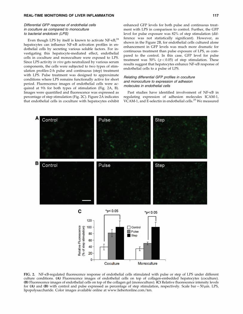
Differential GFP response of endothelial cellsin coculture as compared to monocultureto bacterial endotoxin (LPS)
Even though LPS by itself is known to activate NF-kB,24
hepatocytes can influence NF-kB activation profiles in en-dothelial cells by secreting various soluble factors. For in-vestigating this hepatocyte-mediated effect, endothelialcells in coculture and monoculture were exposed to LPS.Since LPS activity in vivo gets neutralized by various serumcomponents, the cells were subjected to two types of stim-ulation profiles-2-h pulse and continuous (step) treatmentwith LPS. Pulse treatment was designed to approximateconditions where LPS remains functionally active for shortperiod. Fluorescence images of endothelial cells were ac-quired at 9 h for both types of stimulation (Fig. 2A, B).Images were quantified and fluorescence was expressed aspercentage of step stimulation (Fig. 2C). Figure 2A indicatesthat endothelial cells in coculture with hepatocytes exhibit
enhanced GFP levels for both pulse and continuous treat-ment with LPS in comparison to control. Further, the GFPlevel for pulse exposure was 82% of step stimulation (dif-ference was not statistically significant). However, asshown in the Figure 2B, for endothelial cells cultured aloneenhancement in GFP levels was much more dramatic forcontinuous treatment than pulse exposure of LPS, as com-pared to the control. In this case, GFP level for pulsetreatment was 50% ( p< 0.05) of step stimulation. Theseresults suggest that hepatocytes enhance NF-kB response ofendothelial cells to a pulse of LPS.
Relating differential GFP profiles in cocultureand monoculture to expression of adhesionmolecules in endothelial cells
Past studies have identified involvement of NF-kB inregulating expression of adhesion molecules ICAM-1,VCAM-1, and E-selectin in endothelial cells.25 We measured
FIG. 2. NF-kB-regulated fluorescence response of endothelial cells stimulated with pulse or step of LPS under differentculture conditions. (A) Fluorescence images of endothelial cells on top of collagen-embedded hepatocytes (coculture).(B) Fluorescence images of endothelial cells on top of the collagen gel (monoculture). (C) Relative fluorescence intensity levelsfor (A) and (B) with control and pulse expressed as percentage of step stimulation, respectively. Scale bar¼ 50mm. LPS,lipopolysaccharide. Color images available online at www.liebertonline.com/ten.
REAL-TIME MONITORING OF LIVER INFLAMMATION 117

expression of these adhesion molecules for probing the ef-ficacy of GFP profiles as an indicator of NF-kB-regulatedgene expression in endothelial cells. Figure 3A shows geneexpression levels (6 h) of VCAM-1, E-selectin, and ICAM-1in endothelial cells exposed to pulse of LPS relative to stepstimulation. It indicates that VCAM-1 gene expression levelfor pulse exposure was 28% of step treatment in the case ofendothelial cells cultured alone. By contrast, in response topulse treatment, endothelial cells in co-culture with hepa-tocytes exhibited VCAM-1 gene expression levels (93%)that approached those for step stimulation. Similarly, geneexpression levels of E-selectin and ICAM-1 for pulseexposure relative to step stimulation was lower in mono-cultures (24% and 57%) of endothelial cells in comparisonto those in coculture (80% and 78%) with hepatocytes. Thisis in agreement with GFP profiles observed in Figure 2where endothelial cells in coculture showed similar level ofactivation for pulse and step exposure of LPS, while en-dothelial cells in monoculture demonstrated higher levelof activation for step in comparison to pulse treatment ofLPS. For estimating if coculture with hepatocytes devoidof any LPS stimulation has any effect on the activation stateof endothelial cells, we compared gene expression levels ofadhesion molecules in the control condition of coculturesand monocultures. Figure 3B indicates that expression ofall three adhesion molecules in endothelial cells is elevatedfor monocultures relative to cocultures. These results sug-gest that hepatocytes alter the activation state of endothe-
lial cells both in the presence and absence of any LPSstimulation.
Leukocyte adhesion to endothelial cells in monocultureand coculture with hepatocytes
Leukocytes recruited during endotoxemia have been im-plicated in hepatic damage. Leukocytes adhesion to endo-thelial cells is an important step in leukocytes recruitment.We evaluated if differences observed in fluorescence re-sponse of our reporter were sufficient in inducing disparityin leukocytes adhesion to endothelial cells. Figure 4 showsleukocytes adhesion to endothelial cells in monoculture andcoculture with hepatocytes subjected to control, pulse, andstep stimulation of LPS. Figure 4A indicates that endothelialcells in coculture with hepatocytes show enhanced bindingof leukocytes both for pulse and step treatment of LPS incomparison to control. Additionally, leukocyte binding re-sponse for pulse treatment was more than control but lessthan step stimulation of LPS (Fig. 4C). However, in the caseof endothelial cells cultured alone (Fig. 4B) enhanced leu-kocyte adhesion was only observed for step treatment ofLPS, while pulse exposure exhibited similar leukocytebinding as control (Fig. 4C). Further, in the absence of LPSstimulation, endothelial cells cultured alone displayed ahigher level of leukocyte binding than those in coculturewith hepatocytes (controls of Fig. 4A, B).
Discussion
In this report, we describe the development of an in vitromodel of hepatic tissue that facilitates real-time monitoringof NF-kB activation in the endothelium in a complex tissue-like microenvironment. This was achieved by integrating aGFP reporter clone of endothelial cells in our previouslydeveloped organotypical model of liver tissue. We demon-strate the efficacy of our approach by identifying a differ-ential NF-kB-regulated fluorescence response of endothelialcells to pulse and step of LPS, depending on whether theywere in coculture with hepatocytes or in monoculture. En-dothelial cells in coculture with hepatocytes exhibited similarfluorescence response for both pulse and step stimulation ofLPS. By contrast, endothelial cells in monoculture displayedenhanced fluorescence response for step in comparison topulse of LPS. We were able to correlate the disparity inthe NF-kB-mediated fluorescence response to the expressionlevel of several NF-kB-regulated genes such as ICAM-1,VCAM-1, and E-Selectin, which ultimately lead to differ-ences in leukocyte adhesion to endothelial cells.
In the liver sinusoid, endothelial cells and hepatocytes arearranged in layers with the intervening space occupied bythe extracellular matrix of the space of Disse. Our three-dimensional model mimics this arrangement by overlayingendothelial cells on top of collagen-embedded hepatocytes.In a previous report, we demonstrated the efficacy of thisthree-dimensional layering in maintaining hepatocyte dif-ferentiated function. This work focuses on utilizing thismodel for investigating hepatic inflammation. In the contextof inflammation, our three-dimensional layered tissue modelfacilitates the creation of more in vivo-like conditions whereendothelial cells are the first cell type to come in contact withleukocytes, while at the same time their adhesion is influ-enced by a signaling environment affected by hepatocytes.
FIG. 3. Relative changes in gene expression of VCAM-1,ICAM-1, and E-Selectin in endothelial cells cultured underdifferent conditions. (A) Gene expression levels were mea-sured for pulse and step treatment of LPS in both mono-cultures and cocultures. Relative mRNA levels for pulse areexpressed as percentage of step treatment. (B) Gene expres-sion levels were measured in the control (no LPS stimulation)cocultures and monocultures. mRNA levels for monocul-tures expressed relative to cocultures. VCAM-1, vascular celladhesion molecule 1; ICAM-1, intercellular adhesion mole-cule 1.
118 JINDAL ET AL.

There are several advantages of integrating reporter cellsin tissue models for investigating gene expression patterns.In a tissue model that employs multiple cell types, conven-tional methods for gene expression analysis require separa-tion of individual cell types before ascertaining geneexpression levels in the cell type of interest. Additionally,each measurement requires destruction of cells, which is notoptimal for obtaining dynamic information. In contrast, uti-lization of reporter cells not only obviates the need for sep-arating cells, but temporal expression patterns can beobtained from the same population of cells. In our layeredtissue model, dynamic NF-kB-regulated fluorescence re-sponse was measured in real time from the endotheliumplane under inflammatory conditions. Since hepatocytes andendothelial cells were in different planes, the backgroundautofluorescence from hepatocytes was minimized when themicroscope was focused on the endothelium plane. Thetemporal fluorescence profile was similar to that observed inother reporter clones with a characteristic delay associatedwith transcription, translation, and maturation of GFP afteractivation of NF-kB.11,12,14
Although, in vivo, the liver sinusoid is lined by the sinu-soidal endothelial cells,26 we used a GFP reporter clone ofprimary RHMEC in our model. Liver sinusoidal endothelialcells have limited proliferation capacity, which renders themunsuitable for creating a GFP reporter clone. We employedprimary rat heart endothelial cells for creating a GFP reporterclone due to their capacity to proliferate and ease of main-taining them in culture. Use of heart endothelial cells is alimitation of our model as endothelial cells from differentorgans are known to exhibit heterogeneous phenotype.Nevertheless, we reasoned that primary rat heart endothelialcells will be a compromise in terms of creating a GFP re-porter clone that enables real-time monitoring of NF-kB ac-tivation at the tissue level, while at the same time usefulstudies in the context of hepatic inflammation can be con-ducted as intercellular communication by various growthfactors and cytokines are not omitted, since they belong tothe same species as the hepatocytes. In fact, a wide variety ofcell types, including cells belonging to other species, haveshown remarkable ability to interact with hepatocytes andinfluence their function.27
FIG. 4. Leukocyte (labeled with a fluorescent dye CM-DiI) adhesion to endothelial cells stimulated with pulse or step of LPSunder different culture conditions. (A) Fluorescence images of leukocytes adhering to endothelial cells on top of collagenembedded hepatocytes (coculture). (B) Fluorescence images of leukocytes adhering to endothelial cells on top of the collagengel (monoculture). (C) Relative leukocyte adhesion (or fluorescence intensity) levels for (A) and (B) with control and pulseexpressed as percentage of step stimulation. Scale bar¼ 50 mm. Color images available online at www.liebertonline.com/ten.
REAL-TIME MONITORING OF LIVER INFLAMMATION 119

LPS released from the outer cell membrane of gram-negative bacteria is a potent inducer of the inflammatoryresponse.28 We used our model for investigating the roleplayed by intercellular communication between hepatocytesand endothelial cells during LPS-induced inflammatory re-sponse. In vivo, LPS present in circulation can get neutralizedby various proteins present in plasma.29 This can lead toconditions where LPS remains functionally active onlytransiently. To mimic such conditions, we subjected oursystem to a pulse of LPS, where LPS was introduced for abrief period and then cells were exposed to the medium forthe remainder of the experiment. Pulse response was com-pared to step stimulation where LPS was not externally re-moved. Our results revealed that in the presence ofhepatocytes, endothelial cells get activated similarly to bothpulse and step stimulation. However, when endothelial cellswere cultured alone, the level of activation was muchstronger for step than pulse. Additionally, in the absence ofany stimulation, hepatocytes reduce the activated state ofendothelial cells. This suggests that hepatocytes play animportant role in modulating the endothelial responsewhereby under noninflammatory conditions they contributeto maintaining the endothelium in a nonactivated state,while under inflammatory conditions they enhance the ac-tivation of endothelial cells upon encountering pathogen-associated products even for brief periods.
Tissues respond to injury or infection by recruiting leu-kocytes as part of the host defense strategy to ward offsource of injury. However, in liver leukocytes recruitedduring endotoxemia play an important role in causing he-patic damage.30–31 There are several reports where blockingof leukocyte recruitment to liver results in diminisheddamage.31–33 Since NF-kB is believed to play a critical role inleukocyte recruitment,34 we evaluated the applicability ofour model in applying the NF-kB-mediated fluorescence re-sponse to the functional output of leukocyte adhesion toendothelium, which is a key step in their recruitment. Ourexperiments showed that the disparity observed in the levelof fluorescence response was sufficient in eliciting differen-tial leukocyte adhesion to endothelial cells cultured underdifferent conditions. Hepatocytes seemed to reduce adhesionof leukocytes to endothelial cells under noninflammatory(control) condition, while stimulation with pulse of LPS re-sulted in reversal of trend. This could be a useful strategywhereby hepatocytes communicate with the endothelium forascertaining if hepatic tissue needs to recruit leukocytes—presence of mediators of inflammation induces leukocyterecruitment, whereas somewhat healthy condition is not fa-vorable for leukocyte recruitment.
Hepatocytes can influence the microenvironment of en-dothelial cells dynamically by both secretion and/or uptakeof factors from the medium. It is plausible that endothelialcells in coculture were subjected to different combination offactors under control condition and pulse stimulation of LPS,which resulted in reversal of trend from reduced to enhancedactivation as the medium was supplemented with LPS forshort duration. The factor(s) responsible for enhanced acti-vation of endothelial cells in monoculture as compared tococulture under control condition are unknown. It remainsunclear if somewhat higher activation of endothelial cells inmonoculture (control condition) before LPS stimulationplayed any role in reduced responsiveness to LPS pulse.
Although, step stimulation of LPS was able to further en-hance the activation of endothelial cells in monoculturedemonstrating that the cells had not reached maximum ac-tivation under control condition and retained ability to re-spond to LPS. These results suggest that the differentialresponse of endothelial cells in monoculture and coculture toLPS pulse was hepatocytes mediated. The mechanism re-sponsible for this differential response was not investigatedin this study. Nevertheless, based on literature reports, in-volvement of certain factors secreted by hepatocytes can bespeculated. Hepatocytes are known to secrete various factorssuch as vascular endothelial growth factor (VEGF)35 and LPSbinding protein (LBP),36 which can influence the cocultureresponse of endothelial cells to LPS. In a recent report, LPSadministered to mice resulted in increased levels of VEGF inliver.37 VEGF is known to activate expression of adhesionmolecules on endothelial cells.25 LBP is an acute-phase pro-tein that binds to LPS and is thought to enhance LPS sig-naling.38,39 LBP in conjunction with other unidentifiedfactors has also been implicated in LPS-induced higher cy-tokine production by nonparenchymal cells of liver in co-culture with hepatocytes.40
In summary, our model provides a powerful platformfor evaluating hepatic endothelium activation in real timeunder inflammatory conditions. Since endothelium activa-tion and leukocyte recruitment play critical role in a num-ber of disease states associated with inflammation, it canpotentially be used as a high-throughput tool for identify-ing drug targets and gaining mechanistic insight into therole played by intercellular mediators in regulating hepaticinflammation.
Acknowledgments
We thank Candice Calhoun, and Carley Shulman for theisolation of cells from rat livers. This work was supportedby NIH BioMEMS Resource Center Grant P41 EB-002503and NIH Grant RO1AI063795. Microscopic imaging studieswere made possible by a Core Morphology Facility, andgene expression measurements were carried out at theGenomic and Proteomic Facility at Boston’s Shriners BurnsHospital.
Disclosure Statement
No competing financial interests exist.
References
1. Schmid-Schonbein, G.W. Analysis of inflammation. AnnRev Biomed Eng 8, 93, 2006.
2. Szabo, G., Romics, L., Jr., and Frendl, G. Liver in sepsis andsystemic inflammatory response syndrome. Clin Liver Dis6, 1045, 2002.
3. Jaeschke, H. Molecular mechanisms of hepatic ischemia-reperfusion injury and preconditioning. Am J Physiol 284,
G15, 2003.4. Wieckowska, A., Mccullough, A.J., and Feldstein, A.E.
Noninvasive diagnosis and monitoring of nonalcoholicsteatohepatitis: present and future. Hepatology (Baltimore,MD) 46, 582, 2007.
5. Brenner, D.A. Molecular pathogenesis of liver fibrosis. TransAm Clin Climatol Assoc 120, 361, 2009.
120 JINDAL ET AL.

6. Ganey, P.E., and Roth, R.A. Concurrent inflammation as adeterminant of susceptibility to toxicity from xenobioticagents. Toxicology 169, 195, 2001.
7. Liu, S.F., and Malik, A.B. NF-kappa B activation as a path-ological mechanism of septic shock and inflammation. Am JPhysiol Lung Cell Mol Physiol 290, L622, 2006.
8. Essani, N.A., Mcguire, G.M., Manning, A.M., and Jaeschke,H. Endotoxin-induced activation of the nuclear transcriptionfactor kappa B and expression of E-selectin messenger RNAin hepatocytes, Kupffer cells, and endothelial cells in vivo.J Immunol 156, 2956, 1996.
9. Kuhnel, F., Zender, L., Paul, Y., Tietze, M.K., Trautwein, C.,Manns, M., et al. NFkappaB mediates apoptosis throughtranscriptional activation of Fas (CD95) in adenoviral hep-atitis. J Biol Chem 275, 6421, 2000.
10. Barnes, P.J., and Karin, M. Nuclear factor-kappaB: a pivotaltranscription factor in chronic inflammatory diseases.N Engl J Med 336, 1066, 1997.
11. King, K.R., Wang, S., Irimia, D., Jayaraman, A., Toner, M.,and Yarmush, M.L. A high-throughput microfluidic real-time gene expression living cell array. Lab Chip 7, 77,2007.
12. Wieder, K.J., King, K.R., Thompson, D.M., Zia, C., Yarmush,M.L., and Jayaraman, A. Optimization of reporter cells forexpression profiling in a microfluidic device. Biomed Mi-crodevices 7, 213, 2005.
13. Canton, I., Sarwar, U., Kemp, E.H., Ryan, A.J., Macneil, S.,and Haycock, J.W. Real-time detection of stress in 3D tissue-engineered constructs using NF-kappaB activation in tran-siently transfected human dermal fibroblast cells. Tissue Eng13, 1013, 2007.
14. Thompson, D.M., King, K.R., Wieder, K.J., Toner, M., Yar-mush, M.L., and Jayaraman, A. Dynamic gene expressionprofiling using a microfabricated living cell array. AnalChem 76, 4098, 2004.
15. Jindal, R., Nahmias, Y., Tilles, A.W., Berthiaume, F., andYarmush, M.L. Amino acid-mediated heterotypic interactiongoverns performance of a hepatic tissue model. Faseb J 23,
2288, 2009.16. Seglen, P.O. Preparation of isolated rat liver cells. Methods
Cell Biol 13, 29, 1976.17. Dunn, J.C., Tompkins, R.G., and Yarmush, M.L. Long-term
in vitro function of adult hepatocytes in a collagen sandwichconfiguration. Biotechnol Prog 7, 237, 1991.
18. Moghe, P.V., Coger, R.N., Toner, M., and Yarmush, M.L.Cell-cell interactions are essential for maintenance of hepa-tocyte function in collagen gel but not on Matrigel. Bio-technol Bioeng 56, 706, 1997.
19. Dunn, J.C., Yarmush, M.L., Koebe, H.G., and Tompkins,R.G. Hepatocyte function and extracellular matrix geometry:long-term culture in a sandwich configuration. Faseb J 3,
174, 1989.20. Park, J., Berthiaume, F., Toner, M., Yarmush, M.L., and Tilles,
A.W. Microfabricated grooved substrates as platforms forbioartificial liver reactors. Biotechnol Bioeng 90, 632, 2005.
21. Ryan, C.M., Carter, E.A., Jenkins, R.L., Sterling, L.M., Yar-mush, M.L., Malt, R.A., et al. Isolation and long-term cultureof human hepatocytes. Surgery 113, 48, 1993.
22. Deroanne, C.F., Lapiere, C.M., and Nusgens, B.V. In vitrotubulogenesis of endothelial cells by relaxation of the cou-pling extracellular matrix-cytoskeleton. Cardiovasc Res 49,
647, 2001.23. Wallach, D., Varfolomeev, E.E., Malinin, N.L., Goltsev, Y.V.,
Kovalenko, A.V., and Boldin, M.P. Tumor necrosis factor
receptor and Fas signaling mechanisms. Ann Rev Immunol17, 331, 1999.
24. Faure, E., Equils, O., Sieling, P.A., Thomas, L., Zhang, F.X.,Kirschning, C.J., et al. Bacterial lipopolysaccharide activatesNF-kappaB through toll-like receptor 4 (TLR-4) in culturedhuman dermal endothelial cells. Differential expressionof TLR-4 and TLR-2 in endothelial cells. J Biol Chem 275,
11058, 2000.25. Kim, I., Moon, S.O., Kim, S.H., Kim, H.J., Koh, Y.S., and Koh,
G.Y. Vascular endothelial growth factor expression of in-tercellular adhesion molecule 1 (ICAM-1), vascular cell ad-hesion molecule 1 (VCAM-1), and E-selectin through nuclearfactor-kappa B activation in endothelial cells. J Biol Chem276, 7614, 2001.
26. Tilles, A.W., Baskaran, H., Roy, P., Yarmush, M.L., andToner, M. Effects of oxygenation and flow on the viabilityand function of rat hepatocytes cocultured in a micro-channel flat-plate bioreactor. Biotechnol Bioeng 73, 379,2001.
27. Chan, C., Berthiaume, F., Nath, B.D., Tilles, A.W., Toner, M.,and Yarmush, M.L. Hepatic tissue engineering for adjunctand temporary liver support: critical technologies. LiverTransplant 10, 1331, 2004.
28. Moulin, F., Copple, B.L., Ganey, P.E., and Roth, R.A. He-patic and extrahepatic factors critical for liver injury duringlipopolysaccharide exposure. Am J Physiol 281, G1423,2001.
29. Dedrick, R.L., and Conlon, P.J. Prolonged expression of li-popolysaccharide (LPS)-induced inflammatory genes inwhole blood requires continual exposure to LPS. Infect Im-mun 63, 1362, 1995.
30. Hewett, J.A., Schultze, A.E., Vancise, S., and Roth, R.A.Neutrophil depletion protects against liver injury frombacterial endotoxin. Lab Invest J Tech Methods Pathol 66,
347, 1992.31. Jaeschke, H., Farhood, A., and Smith, C.W. Neutrophil-
induced liver cell injury in endotoxin shock is a CD11b/CD18-dependent mechanism. Am J Physiol 261, G1051,1991.
32. Li, X., Klintman, D., Liu, Q., Sato, T., Jeppsson, B., andThorlacius, H. Critical role of CXC chemokines in en-dotoxemic liver injury in mice. J Leukoc Biol 75, 443,2004.
33. Gujral, J.S., Liu, J., Farhood, A., Hinson, J.A., and Jaeschke,H. Functional importance of ICAM-1 in the mechanism ofneutrophil-induced liver injury in bile duct-ligated mice. AmJ Physiol 286, G499, 2004.
34. Alcamo, E., Mizgerd, J.P., Horwitz, B.H., Bronson, R., Beg,A.A., Scott, M., et al. Targeted mutation of TNF receptor Irescues the RelA-deficient mouse and reveals a critical rolefor NF-kappa B in leukocyte recruitment. J Immunol 167,
1592, 2001.35. Shimizu, H., Miyazaki, M., Wakabayashi, Y., Mitsuhashi,
N., Kato, A., Ito, H., et al. Vascular endothelial growthfactor secreted by replicating hepatocytes induces sinu-soidal endothelial cell proliferation during regenera-tion after partial hepatectomy in rats. J Hepatol 34, 683,2001.
36. Grube, B.J., Cochane, C.G., Ye, R.D., Green, C.E., Mcphail,M.E., Ulevitch, R.J., et al. Lipopolysaccharide binding proteinexpression in primary human hepatocytes and HepG2hepatoma cells. J Biol Chem 269, 8477, 1994.
37. Yano, K., Liaw, P.C., Mullington, J.M., Shih, S.C., Okada, H.,Bodyak, N., et al. Vascular endothelial growth factor is an
REAL-TIME MONITORING OF LIVER INFLAMMATION 121
important determinant of sepsis morbidity and mortality.J Exp Med 203, 1447, 2006.
38. Wright, S.D., Ramos, R.A., Tobias, P.S., Ulevitch, R.J.,and Mathison, J.C. CD14, a receptor for complexes oflipopolysaccharide (LPS) and LPS binding protein. Science249, 1431, 1990.
39. Jack, R.S., Fan, X., Bernheiden, M., Rune, G., Ehlers, M.,Weber, A., et al. Lipopolysaccharide-binding protein is re-quired to combat a murine gram-negative bacterial infection.Nature 389, 742, 1997.
40. Scott, M.J., Liu, S., Su, G.L., Vodovotz, Y., and Billiar, T.R.Hepatocytes enhance effects of lipopolysaccharide on livernonparenchymal cells through close cell interactions. Shock(Augusta, GA) 23, 453, 2005.
Address correspondence to:Martin L. Yarmush, M.D., Ph.D.
Center for Engineering in MedicineMassachusetts General Hospital
Harvard Medical SchoolThe Shriners Hospitals for Children
51 Blossom St.Boston, MA 02114
E-mail: [email protected]
Received: December 05, 2009
Accepted: July 29, 2010
Online Publication Date: September 21, 2010
122 JINDAL ET AL.