Time course and functional neuroanatomy of speech segmentation in adults
13
Time course and functional neuroanatomy of speech segmentation in adults Toni Cunillera a , Estela Càmara b,c , Juan M. Toro a , Josep Marco-Pallares b , Nuria Sebastián-Galles d , Hector Ortiz e,f , Jesús Pujol e,f , Antoni Rodríguez-Fornells b,g, ⁎ a Department of Basic Psychology, Faculty of Psychology, University of Barcelona, 08035, Barcelona, Spain b Department of Physiology, Faculty of Medicine, Campus de Bellvitge – IDIBELL, University of Barcelona, 08907, Barcelona, Spain c Department of Neuropsychology, Otto-von Guericke University, 39106, Magdeburg, Germany d Brain and Cognition Unit, Universitat Pompeu Fabra, 08018 Barcelona, Spain e Institut d’Alta Tecnologia-PRBB, CRC Corporació Sanitària, Barcelona, Spain f Networking Research Center on Bioengineering, Biomaterials and Nanomedicine (CIBER-BBN), Barcelona, Spain g Institució Catalana de Recerca i Estudis Avançats (ICREA), Spain abstract article info Article history: Received 21 January 2009 Revised 2 June 2009 Accepted 28 June 2009 Available online 4 July 2009 The present investigation was devoted to unraveling the time-course and brain regions involved in speech segmentation, which is one of the first processes necessary for learning a new language in adults and infants. A specific brain electrical pattern resembling the N400 language component was identified as an indicator of speech segmentation of candidate words. This N400 trace was clearly elicited after a short exposure to the words of the new language and showed a decrease in amplitude with longer exposure. Two brain regions were observed to be active during this process: the posterior superior temporal gyrus and the superior part of the ventral premotor cortex. We interpret these findings as evidence for the existence of an auditory–motor interface that is responsible for isolating possible candidate words when learning a new language in adults. © 2009 Elsevier Inc. All rights reserved. Introduction The present study was focused on understanding one of the initial processes in language learning: speech segmentation. The difficulty of the task lies in the lack of reliable acoustic cues that indicate where a word begins and ends. Thus, unlike the blank spaces that appear between printed words, the spoken acoustic signal could be considered, in many respects, continuous. Listeners, therefore, must parse the speech signal in order to start learning the new language. Notice that this initial process of isolating words is mandatory for subsequent language processes; for example, associating the possible lexical trace with a specific meaning. To segment the auditory stream into words, the learner could exploit different acoustical cues such as allophonic variation, stress patterns, prosody, or/and distributional cues such as phonotactic regularities and transitional probabilities of syllable combinations (see for a review Jusczyk et al. (1999)). In relation to the distributional cues, several models have proposed the existence of a powerful statistical learning mechanism in order to explain speech segmenta- tion (Brent, 1999). Supporting this idea, several experiments have shown that listeners are able to exploit the distributional properties of the input, regardless of whether it consists of syllables, tones or visual information (Fiser and Aslin, 2001; Saffran et al., 1999), and it likely proceeds in an incidental fashion (Saffran et al., 1997; Toro et al., 2005; Turk-Browne et al., 2005). Thus, statistical learning is understood as a domain-general mechanism that profits from the regularities of the environment to drive learning. The computational implementation of this hypothesis has been successfully applied by connectionist models (Christiansen et al., 1998; Elman, 1990; for a different account see Brent, 1997). The underlying idea is that word boundaries are in locations where the transitional probabilities between two sounds are low. In other words, word boundaries can be inferred based on the fact that transitional probabilities are higher for word-internal than for word-external pairs of syllables. For instance, in the sentence “look, a balloon” the string “ba/lloon” is more likely to occur together across other sentences than the string “a/ba” as the latter string would not be heard in phrases such as “the balloon”, “the red balloon”, etc. In fact, it has been demonstrated in infants and adults that computing the transitional probabilities between syllables is sufficient for isolating new words that are embedded in an artificial continuous speech stream when no acoustical cues are available (Saffran et al., 1996a,b). Remarkably, this process is also accomplished by other animal species (Hauser et al., 2001; Toro and Trobalon, 2005), highlighting its generality. Interestingly, an alternative computational proposal suggests that the formation of syllabic chunks might be the only process required to isolate possible words (Perruchet and Vinter,1998). Accordingly to the authors, a chunk can be considered that brings together the elements that are at the attentional focus at a particular moment. In subsequent NeuroImage 48 (2009) 541–553 ⁎ Corresponding author. Department of Ciencias Fisiològiques, Faculty of Medicine, Campus de Bellvitge, IDIBELL, Feixa Llarga s/n, 08907. L'Hospitalet (Barcelona), Spain. Fax: +34 934024268. E-mail address: [email protected] (A. Rodríguez-Fornells). 1053-8119/$ – see front matter © 2009 Elsevier Inc. All rights reserved. doi:10.1016/j.neuroimage.2009.06.069 Contents lists available at ScienceDirect NeuroImage journal homepage: www.elsevier.com/locate/ynimg
-
Upload
independent -
Category
Documents
-
view
0 -
download
0
Transcript of Time course and functional neuroanatomy of speech segmentation in adults
NeuroImage 48 (2009) 541–553
Contents lists available at ScienceDirect
NeuroImage
j ourna l homepage: www.e lsev ie r.com/ locate /yn img
Time course and functional neuroanatomy of speech segmentation in adults
Toni Cunillera a, Estela Càmara b,c, Juan M. Toro a, Josep Marco-Pallares b, Nuria Sebastián-Galles d,Hector Ortiz e,f, Jesús Pujol e,f, Antoni Rodríguez-Fornells b,g,⁎a Department of Basic Psychology, Faculty of Psychology, University of Barcelona, 08035, Barcelona, Spainb Department of Physiology, Faculty of Medicine, Campus de Bellvitge – IDIBELL, University of Barcelona, 08907, Barcelona, Spainc Department of Neuropsychology, Otto-von Guericke University, 39106, Magdeburg, Germanyd Brain and Cognition Unit, Universitat Pompeu Fabra, 08018 Barcelona, Spaine Institut d’Alta Tecnologia-PRBB, CRC Corporació Sanitària, Barcelona, Spainf Networking Research Center on Bioengineering, Biomaterials and Nanomedicine (CIBER-BBN), Barcelona, Spaing Institució Catalana de Recerca i Estudis Avançats (ICREA), Spain
⁎ Corresponding author. Department of Ciencias FisioCampus de Bellvitge, IDIBELL, Feixa Llarga s/n, 08907. LFax: +34 934024268.
E-mail address: [email protected] (A. Rodríg
1053-8119/$ – see front matter © 2009 Elsevier Inc. Aldoi:10.1016/j.neuroimage.2009.06.069
a b s t r a c t
a r t i c l e i n f oArticle history:Received 21 January 2009Revised 2 June 2009Accepted 28 June 2009Available online 4 July 2009
The present investigation was devoted to unraveling the time-course and brain regions involved in speechsegmentation, which is one of the first processes necessary for learning a new language in adults and infants.A specific brain electrical pattern resembling the N400 language component was identified as an indicator ofspeech segmentation of candidate words. This N400 trace was clearly elicited after a short exposure to thewords of the new language and showed a decrease in amplitude with longer exposure. Two brain regionswere observed to be active during this process: the posterior superior temporal gyrus and the superior part ofthe ventral premotor cortex. We interpret these findings as evidence for the existence of an auditory–motorinterface that is responsible for isolating possible candidate words when learning a new language in adults.
© 2009 Elsevier Inc. All rights reserved.
Introduction
The present study was focused on understanding one of the initialprocesses in language learning: speech segmentation. The difficulty ofthe task lies in the lack of reliable acoustic cues that indicate where aword begins and ends. Thus, unlike the blank spaces that appearbetween printed words, the spoken acoustic signal could beconsidered, in many respects, continuous. Listeners, therefore, mustparse the speech signal in order to start learning the new language.Notice that this initial process of isolating words is mandatory forsubsequent language processes; for example, associating the possiblelexical trace with a specific meaning.
To segment the auditory stream into words, the learner couldexploit different acoustical cues such as allophonic variation, stresspatterns, prosody, or/and distributional cues such as phonotacticregularities and transitional probabilities of syllable combinations(see for a review Jusczyk et al. (1999)). In relation to the distributionalcues, several models have proposed the existence of a powerfulstatistical learning mechanism in order to explain speech segmenta-tion (Brent, 1999). Supporting this idea, several experiments haveshown that listeners are able to exploit the distributional properties ofthe input, regardless of whether it consists of syllables, tones or visual
lògiques, Faculty of Medicine,'Hospitalet (Barcelona), Spain.
uez-Fornells).
l rights reserved.
information (Fiser and Aslin, 2001; Saffran et al., 1999), and it likelyproceeds in an incidental fashion (Saffran et al., 1997; Toro et al., 2005;Turk-Browne et al., 2005). Thus, statistical learning is understood as adomain-general mechanism that profits from the regularities of theenvironment to drive learning.
The computational implementation of this hypothesis has beensuccessfully applied by connectionist models (Christiansen et al.,1998; Elman, 1990; for a different account see Brent, 1997). Theunderlying idea is that word boundaries are in locations where thetransitional probabilities between two sounds are low. In other words,word boundaries can be inferred based on the fact that transitionalprobabilities are higher for word-internal than for word-external pairsof syllables. For instance, in the sentence “look, a balloon” the string“ba/lloon” is more likely to occur together across other sentences thanthe string “a/ba” as the latter string would not be heard in phrasessuch as “the balloon”, “the red balloon”, etc. In fact, it has beendemonstrated in infants and adults that computing the transitionalprobabilities between syllables is sufficient for isolating new wordsthat are embedded in an artificial continuous speech streamwhen noacoustical cues are available (Saffran et al., 1996a,b). Remarkably, thisprocess is also accomplished by other animal species (Hauser et al.,2001; Toro and Trobalon, 2005), highlighting its generality.
Interestingly, an alternative computational proposal suggests thatthe formation of syllabic chunks might be the only process required toisolate possiblewords (Perruchet and Vinter,1998). Accordingly to theauthors, a chunk can be considered that brings together the elementsthat are at the attentional focus at a particular moment. In subsequent
542 T. Cunillera et al. / NeuroImage 48 (2009) 541–553
encounters with the initially formed chunks, those are forgotten orstrengthened according to the associativememory laws. In this regard,recently Pacton and Perruchet (2008) have proposed a generalassociative learning model that asserts attention as the necessaryand sufficient condition for associative learning to occur and in whichis included the formation of linguistic chunks. From this perspective,statistical computations would come into play solely after chunks areselected, and only for reinforcing or weakening the formed chunksthat are repeatedly selected or varied, respectively (Perruchet andPacton, 2006; Perruchet and Vinter, 1998).
During the last decade, the use of ERP measures has providedimportant data about the cognitive mechanisms involved in speechsegmentation (Cunillera et al., 2006, 2008; Sanders et al., 2002; Sandersand Neville, 2003a,b). Specifically, the N100 and the N400 ERPcomponents have been proposed as speech segmentation indexes(Abla et al., 2008; Cunillera et al., 2006; Sanders et al., 2002). The N100seems to be sensitive to word onset perception, whereas the N400appears to indicate the identification of recently segmented words(Abla et al., 2008; Cunillera et al., 2006; Sanders et al., 2002). In thisvein, the N400 component has been related to the learning process ofnonsense words and has been regarded as an indirect electrophysio-logical brain signal that indicates that speech segmentation has beenaccomplished. Recently, Abla et al. (2008) have explored the time-course of the on-line segmentation process using non-linguistic items.Specifically, participants listened to a continuous stream of tonescomposed by groups of tri-tones organized randomly in a continuoussequence. Participants were classified as high, middle and low learnersas a function of their behavioral performance in a target recognition testthat followed the segmentation phase. The authors measured ERPresponses corresponding to the average signal recorded separately inthree consecutive 6-min periods of the learning phase. For highlearners, the results showed enhanced amplitudes of the N400component in the early session, whereas for the group of middlelearners the same enhancement was observed in a latter session. Lowlearners did not show any ERP effect along the exposure to the tonesstream. The authors concluded that their results revealed the on-linestatistical learningprocess andmore important, that theN400 indicatesthe degree of on-line statistical learning (see also Buiatti et al., 2009).
In the neuroscientific literature no clear hypothesis has been raisedabout which brain mechanisms and regions might be involved instatistical learning and, particularly, while segmenting the speechsignal into potential new words. Thus far, the only brain imagingexperiment that has studied speech segmentation (McNealy et al.,2006) used functional Magnetic Resonance Imaging (fMRI) in order toinvestigate the neural correlates of speech segmentation. The authorsshowed that a left frontotemporal network was activated during theon-line speech segmentation process. In particular, while listening tothe language streams composed of nonsense words, the activation ofthe superior temporal gyrus (STG) was found to increase over timecorrelating with an independent word discrimination task.
Several proposals in the context of language processing convergewith the idea that an auditory–motor interface that directly links theauditory input with the motor representations of speech mightsupport the decoding of speech (Hickok and Poeppel, 2000, 2004,2007; Scott et al., 2000; Scott and Wise, 2004; Wise et al., 2001). Thisauditory–motor interface might engage the left posterior temporalregions and the frontal regions, sustaining motor speech representa-tions. Furthermore, it has been proposed that this dorsal circuit ofspeech processing comes into play when it is necessary to keepauditory representations in an active state during the performance ofa task (i.e., phonological short-term memory, Aboitiz et al., 2006;Buchsbaum et al., 2005; Jacquemot and Scott, 2006; Scott et al., 2006).More important to the present study, Hickok and Poeppel (2007) haverecently suggested that this dorsal network could also be involved inthe acquisition of new vocabulary, that is, in language learning. Thisprocess might involve generating a new sensory representation of the
novel word by linking a sequence of syllables or segments while, at thesame time, the newly created traces guide the motor articulatorysequences. This auditory–motor interface hypothesis is reminiscent ofthe early motor theory of speech perception (Liberman andMattingly,1985) and the hypothesis that word learning is mediated by thephonological working memory loop (Baddeley et al., 1998). Indeed,this auditory–motor interface is postulated to be very important inspeech development because speaking inherently requires fine-tunedmotor learning. This mechanism is proposed to be active in adults aswell (Hickok and Poeppel, 2000).
Objectives
The present study was devoted to exploring the time-course of theon-line process of speech segmentation. Thus, the combination ofERPs (Experiment 1) and fMRI (Experiment 2) allowed us first, toclosely inspect the time required for isolating novel words from fluentspeech, and in a second experiment to unravel the brain architectureunderlying the detection of word boundaries. Based on previousfindings, in the ERP experiment, we predicted a time-dependentmodulation of the N100 and N400 components associated with theprocess of segmenting and, afterwards, recognizing the isolated words(see Cunillera et al., 2006; Sanders et al., 2002).
Based on the hypothesis of an auditory–motor interface involved inword learning, in the fMRI experiment, we predicted that this lefthemisphere auditory–motor network would be recruited during thespeech segmentation process. The reasons are the following: (i) theprocess of speech segmentation involves keeping the incomingsyllable information active, (ii) this information is required either tocompute transitional probabilities (Cunillera et al., 2006; Saffran et al.,1996b) or to create possible newword-templates or chunks (Perruchetand Vinter, 1998), and (iii) the information from the isolated wordunits might be feed-forwarded to the frontal cortex (premotor andmotor regions) in order to create corresponding motor articulatorysequences. Most importantly, the activation of this network has to betime-dependent: increased activation should be observed only duringthe initial learning period (isolation of the word-like units) but notafter the new, possible token has been identified (time-dependenthypothesis; Poldrack, 2000; Raichle et al., 1994).
Experiment 1
In the first experiment of this study, event-related brain potentials(ERPs) were used to investigate the time-course underlying speechsegmentation in adult listeners who were exposed to language andrandom streams.
Methods
ParticipantsAfter giving informed consent, fifteen healthy adult (5 males)
undergraduate Psychology students at the University of Barcelonawith normal hearing and no neurological impairment participated inthe experiment. All participants [mean age 23.4±4.9 (SD)] wereright-handed and were Spanish–Catalan native speakers. The experi-ment was approved by the local ethical committee of the University ofBarcelona. All participants were paid at the end of the experiment orreceived extra course credit for their participation.
Materials and procedureFive language streams were developed with the same structure as
those originally created by Saffran et al. (1996a). Each streamconsistedof 4 different trisyllabic nonsense words (e.g., pirutabagolitokudapir-utagukibo…) with an exact duration of 232 ms per syllable. Nonsensewords were concatenated to form a text stream that was subsequentlytransformed into an acoustic stream using the MBROLA speech
543T. Cunillera et al. / NeuroImage 48 (2009) 541–553
synthesizing software (Dutoit et al., 1996; Dutoit and Gosselin, 1996),which is based on concatenation of diphones and includes a Spanishmale diphone database at 16 kHz (available at http://tcts.fpms.ac.be/synthesis/mbrola.html). Afterwards, the Cooledit software was usedto equate the length of all the streams into millisecond resolution,which was necessary for the exact ERP triggering. Words wereconcatenated in a pseudo-random order with the restriction thatthere were no immediate repetitions of words. All phonemes had thesame duration (116 ms) and pitch (200 Hz; equal pitch rise and fall,with pitchmaximum at 50% of the phoneme) in the language streams.Thus, theflat acoustic stream thatwas created had no acousticmarkersbetween words and, therefore, only statistical cues were available forsegmenting the possible words. The transitional probability betweenthe syllables forming awordwas 1.0, while the transitional probabilitybetween syllables spanning word boundaries was 0.33. New syllableswere used for every language stream (see appendix). Because therewere not enough syllables for the 10 languages to be created, only onesyllablewas repeated in two streams. The stimuliwere displayed usingthe Presentation 0.52 software (Neurobehavioral Systems). In 1min ofa language stream, each word was repeated 26 times. Based on aprevious behavioral experiment, the exact duration of all streams wasset to 8 min and 48 s in which the four words were repeated each one208 times along the stream.
Five different auditory random streams were created as a Randomcondition (e.g., “tatopidalirubagutarugopikubo…”). The idea under-lying this condition is that participants could not extract or segmentany word because transitional probabilities could not be used in orderto identify clear word boundaries. With this aim, the transitionalprobability across syllables was maintained equal; therefore, eachsyllable within the random stream had the same probability to befollowed by any syllable of the corresponding language stream (thetransitional probability across all syllables was always 0.09).
During the experiment, participants were required to listencarefully to the 10 streams of sounds and were told that their maintask would be to discover the words of those “alien” languages. Thefirst stream was always one of the languages that contained possiblewords. The presentation order of the streams was randomized foreach participant with the only constrain that no more than twostreams of each type (language or random) followed each other.
After exposure to each stream, a two alternative forced-choice(2AFC) behavioral test was administered to determine whether theparticipants were able to identify the words heard previously. No ERPswere recorded during this phase. The test comprised eight pairs ofauditory test items (a word and a part-word) that were randomlypresented. Part-words were made by the concatenation of the thirdsyllable of a word and the first two syllables of another word (3-1-2part-words; e.g., rutaba, dapiru, bopiru, litoku) or the last two syllablesof aword and the first syllable of another word (2-3-1 part-words; e.g.,tabago, golito, kudagu, kibopi). For random streams, test items werecomposed of 16 different trisyllabic groupings. After the presentation ofeach test pairing, participants had to press a response button indicatingwhether it was the first or the second word in the pair that belonged tothe stream they just heard. The presentation of the next test pair wasnot delivered until a response was produced. The order of presentationofwords and part-words in the test pairs was balanced. Brief rests wereallowed after each stream. The next stream began after listeners feltcomfortable enough to maintain their attention for the next stream.
Data acquisition and analysisERPs were recorded from the scalp using tin electrodesmounted in
an electrocap (Electro-Cap International) and located at 29 standardpositions. Biosignals were rereferenced off-line to the mean of theactivity at the two mastoid processes. Vertical eye movements weremonitored with an electrode at the infraorbital ridge of the right eye.Electrode impedances were kept below 3 kΩ. The electrophysiologicalsignals were filtered on-line with a bandpass of 0.01–50 Hz (half-
amplitude cutoffs) and digitalized at a rate of 250 Hz. Trials with base-to-peak electro-oculogram (EOG) amplitude of more than 50 μV,amplifier saturation, or a baseline shift exceeding 200 μV/s wereautomatically rejected off-line. ERPs were averaged for epochs of1024 ms, starting 50 ms prior to the stimulus.
A mean amplitude measure of the 70–130 and 350–550 ms timewindows were taken in order to encompass the N100 and the N400component, respectively. These mean voltage measures were sub-mitted to an omnibus repeated measures analysis of variance(ANOVA) including two within-subject factors: Word/Non-word(language vs. random streams) and 15 levels of Electrode (F7, F3, Fz,F4, F8, T3, C3, Cz, C4, T4, T5, P3, Pz, P4, and T6). This analysis wascarried out on data that was corrected using the vector normalizationprocedure (Mccarthy and Wood, 1985). For all statistical effectsinvolving two or more degrees of freedom in the numerator, theGreenhouse–Geisser epsilon was used to correct for possible viola-tions of the sphericity assumption (Jennings and Wood, 1976). Thecorrected P-value is reported.
Results
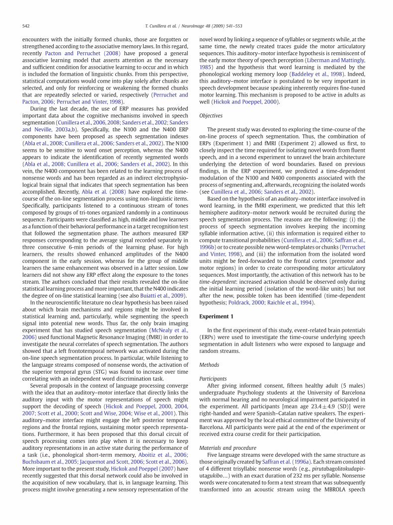
After the presentation of each language stream, a behavioral testwas performed inwhich participants were required to identify possiblewords against a set of part-words. The mean percentage of recognitionof possiblewordswas67.3±10.2% (see Fig.1A),whichwas significantlydifferent from chance (one-sample t-test with chance level at 50%, t(14)=6.58, Pb0.001). Thus, the listenerswere able to discover possiblewords from the speech stream using only statistical cues.
Fig. 1B shows the ERP signature in each of the 2-minute blockspooled across the five languages and the five random streamscomposing both conditions. Notice that the presentation of eachsyllable (one every 232 ms) elicited a clear evoked potential auditorypattern, showing the P50, N100, andP200 components. At thefirst block(first 2min), a negative increasewas observed in the 350–550ms rangewhen comparing the Language against the Randomcondition; however,this difference was not statistically significant (F(1,14)=3.06, PN0.05).This difference reached significant values in the second block (2–4min;F(1,14)=17.6, Pb0.001), and remained significant in the third andfourth block [third block (4–6 min): Word/Non-words, F(1,14)=9.7,Pb0.01; fourth block (6–8 min): Word/Non-words, F(1,14)=7.0,Pb0.05)]. The observed N350–N550 component resembles the well-known N400 component, which has been associated with lexical-semantic processing (Kutas and Federmeier, 2000). No differenceswerefound in the N100 component across blocks or when comparinglanguage streams against random streams (all PN0.1).
Interestingly, in our study, the N400 component began to reduceits amplitude in the third and fourth blocks, although remained highlysignificant as it was expected based on the time-dependent hypoth-esis (see Table 1). At Fz location, a significant quadratic trend wasencountered for the interaction between Word/Non-word and Block(F(1,14)=4.84; P=0.045). In order to further investigate whether theN400 component had already developed during the first 2 min, wedecomposed the first block into two 1-minute averages (see Fig. 1C).As could be observed, a clear N400 component appeared in the secondminute of exposure (Word/Non-words, F(1,14)=8.6, Pb0.01), whileit was not present in the first minute (Fb1).
A subsequent behavioral experiment was conducted with 48 newparticipants in order to further investigate the performance ofparticipants exposed to language streams of different durations (1,2, 4, and 8 min, each language composed with different syllables)using a Latin-square design. In convergence with the previous ERPexperiment, after just 1 min of exposure to the new language subjectswere able to isolate its constituent words (segmentation rate 69.6±19.6%; t(47)=6.95, Pb0.001). Interestingly, performance was notimproved as a function of time (F(3,141)b1, PN0.5; 1-minute: 69.6±19.6%, 2-minute: 71.7±22.4%, 4-minute: 72.7±18.8%, and 8-minute:
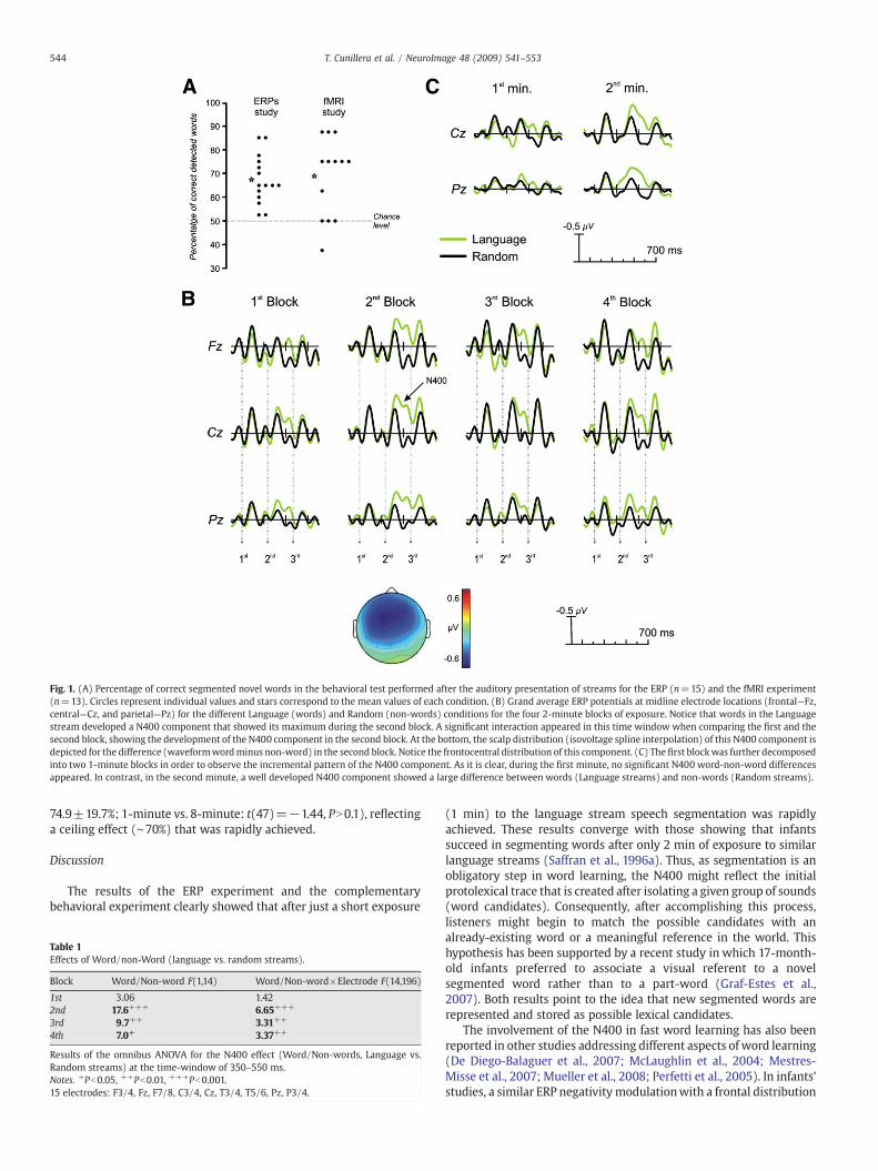
Fig. 1. (A) Percentage of correct segmented novel words in the behavioral test performed after the auditory presentation of streams for the ERP (n=15) and the fMRI experiment(n=13). Circles represent individual values and stars correspond to the mean values of each condition. (B) Grand average ERP potentials at midline electrode locations (frontal—Fz,central—Cz, and parietal—Pz) for the different Language (words) and Random (non-words) conditions for the four 2-minute blocks of exposure. Notice that words in the Languagestream developed a N400 component that showed its maximum during the second block. A significant interaction appeared in this time window when comparing the first and thesecond block, showing the development of the N400 component in the second block. At the bottom, the scalp distribution (isovoltage spline interpolation) of this N400 component isdepicted for the difference (waveformwordminus non-word) in the second block. Notice the frontocentral distribution of this component. (C) The first blockwas further decomposedinto two 1-minute blocks in order to observe the incremental pattern of the N400 component. As it is clear, during the first minute, no significant N400 word-non-word differencesappeared. In contrast, in the second minute, a well developed N400 component showed a large difference between words (Language streams) and non-words (Random streams).
544 T. Cunillera et al. / NeuroImage 48 (2009) 541–553
74.9±19.7%; 1-minute vs. 8-minute: t(47)=−1.44, PN0.1), reflectinga ceiling effect (∼70%) that was rapidly achieved.
Discussion
The results of the ERP experiment and the complementarybehavioral experiment clearly showed that after just a short exposure
Table 1Effects of Word/non-Word (language vs. random streams).
Block Word/Non-word F(1,14) Word/Non-word×Electrode F(14,196)
1st 3.06 1.422nd 17.6+++ 6.65+++
3rd 9.7++ 3.31++
4th 7.0+ 3.37++
Results of the omnibus ANOVA for the N400 effect (Word/Non-words, Language vs.Random streams) at the time-window of 350–550 ms.Notes. +Pb0.05, ++Pb0.01, +++Pb0.001.15 electrodes: F3/4, Fz, F7/8, C3/4, Cz, T3/4, T5/6, Pz, P3/4.
(1 min) to the language stream speech segmentation was rapidlyachieved. These results converge with those showing that infantssucceed in segmenting words after only 2 min of exposure to similarlanguage streams (Saffran et al., 1996a). Thus, as segmentation is anobligatory step in word learning, the N400 might reflect the initialprotolexical trace that is created after isolating a given group of sounds(word candidates). Consequently, after accomplishing this process,listeners might begin to match the possible candidates with analready-existing word or a meaningful reference in the world. Thishypothesis has been supported by a recent study in which 17-month-old infants preferred to associate a visual referent to a novelsegmented word rather than to a part-word (Graf-Estes et al.,2007). Both results point to the idea that new segmented words arerepresented and stored as possible lexical candidates.
The involvement of the N400 in fast word learning has also beenreported in other studies addressing different aspects of word learning(De Diego-Balaguer et al., 2007; McLaughlin et al., 2004; Mestres-Misse et al., 2007; Mueller et al., 2008; Perfetti et al., 2005). In infants'studies, a similar ERP negativitymodulationwith a frontal distribution
545T. Cunillera et al. / NeuroImage 48 (2009) 541–553
was found in 14-month-old infants performing a fast learning object–word mapping task (Friedrich and Friederici, 2008) and for knowncompared to unknownwords in 19- to 22-month-old infants (Conboyand Mills, 2006). Likewise, in a recent ERP study conducted with20-month-olds infants, Mills et al. (2005) observed a larger N400 totrainedwords paired previouslywith an object compared to untrainedwords.
Interestingly, in our study, the N400 component began to reduce inamplitude in the third and fourth blocks as was expected based on thetime-dependent hypothesis, although it remained highly significant.This dynamic pattern of the N400 is similar to the one reported in Ablaet al. (2008) for a group of high learners using streams composed bytones. In the present study, the inverted U-shape amplitude patternmight reflect the interplay between the initial and mandatory speechsegmentation process (2–4 min of the learning phase) and the laterrecognition process of the already-segmented words. Repeated wordsdevelop a reduced N400 component (Rugg and Coles, 1995), whichmight explain why the N400 component tends to be reduced withlonger exposure times. However, this hypothesis has to be confirmedin further experiments.
Our results concur with and extend the interesting study of Sanderset al. (2002) in which the comparison between trained vs. untrainedwords of an artificial language showed a posteriorly distributed N400component. However, as shown in Fig. 1C, the scalp distribution of ourN400 component was frontocentral. The differences observed in theseexperiments might be attributed to the differences in the design. In thepresent study, we specifically focused on the on-line learning processof word segmentation while in the work by Sanders et al., the N400component most probably reflected the effect of word-recognition (ofpreviously learned words) in a segmented language stream. In thesame vein, the authors proposed that changes in the amplitude of theN100 component could index word segmentation. However, ourresults point to the fact that when speech segmentation is exploredon-line (without previous training with the new possible words), theN100 seems to maintain its amplitude across time, and, mostimportantly, it does not seem to distinguish between a string ofrandom ordered syllables and a unitary group of syllables.
It seems so that in adult and infant studies a negative polarityincrease in the time range of 200–500 ms appears to be related to theword learning process. In the present study, the N400 modulationobserved may reflect the learners' ability to extract the co-occurrencestatistics found in the syllabic language streams. Our interpretation ofthe observed modulation of the N400 differs from the typical lexical-semantic interpretation associated with it. While, for the latter, theamplitude of the component decreases when lexical-semanticintegration demands are reduced (e.g., with repetition or semanticcontextual priming), the learning-related N400 shows the oppositepattern, with progressive amplitude enhancement as a function ofincreased exposure to the new-word (Mueller et al., 2008). Inaddition, the topographic distribution of this learning-related N400component is more frontocentral, whereas the classical semanticN400 typically shows a right centro-parietal distribution (Kutas andFedermeier, 2000). These differences in the dynamics and topographyof the N400 may indicate that these ERP modulations might not sharethe same cognitive processes and neural generators.
Experiment 2
Using functional neuroimaging (fMRI) we evaluated the involve-ment of the dorsal auditory–motor network in speech segmentation(Hickok and Poeppel, 2007). An important aim of fMRI learningdesigns is to observe increases and decreases in activation over thetraining period (Poldrack, 2000). An increased activation of a brainregion concomitant to learning is assumed to indicate the engagementof a particular cognitive process, or the development of new learning-related representations (Friston et al., 1992; Poldrack et al., 1998;
Raichle et al., 1994). Thus, a particular regionmight remain active untillearning is accomplished and then decrease its activation (time-dependent hypothesis). Based on the previous behavioral and ERPresults, the exposure to the languages and random streams weemployed in Experiment 1 was limited to only two blocks of 2 minusing a block fMRI design.
Methods
ParticipantsThirteen new right-handed healthy native speakers of Spanish
(mean age 21.1±2.9 years; 12 women) participated in this experi-ment. The experiment was approved by the local ethical committee ofthe University of Barcelona. Written consent was obtained from allparticipants. Participants were either paid or received extra coursecredit for their participation at the end of the experiment.
Materials and procedureWe used four artificial languages, as described previously in
Experiment 1. The structure was the same as that in Saffran et al.(1996a). Language and random streams were identical to thosedescribed in the ERP experiment (see Appendix), but only twolanguage and two random streams were used in the presentexperiment. Each participant was exposed to one language and onerandom stream (different syllables formed the streams in eachcondition). Half of the subjects were exposed to Language A andRandomB and the other half were exposed to Language B and RandomA. Each functional run corresponded to one experimental conditionand lasted 8 min. Within each run, eight 30 s blocks of rest (OFF) wereinterleavedwith eight 30 s task blocks (ON). The order of presentationof the two experimental conditions was counterbalanced acrossparticipants.
We used the Presentation 0.52 software (Neurobehavioral Sys-tems) for stimulus presentation. Auditory stimuli were deliveredusing MRI-compatible headphones with pneumatic sound transmis-sion, which also attenuated the scanner noise. At the end of eachstream, the two alternative forced-choice test (2AFC) was presentedexactly as described in the ERP experiment.
Before the scanning protocol began, participants were instructedto close their eyes throughout the whole run and were encouraged tokeep still and try to identify the words of the “alien” language thatwere going to be presented. Participants were also informed, beforeentering in the scanner, about the behavioral task presented at the endof each run. At the end of the session the participants were debriefed.
Data acquisition and analysisWe collected fMRI images while the participants listened to 8 min
of alternating periods of 30 s of an artificial language, separated with30 s of rest periods. In a different run, a Random streamwas presentedusing the same on-off distribution. We restricted the amount ofexposure to the streams to 4 min (eight periods of 30 s) in accordancewith the previous ERP results, which showed that the critical timewindow for speech segmentation occurs during the first 4 min.
Imaging was performed using a 1.5 T whole body MRI scanner(General Electric Cardio Vascular System) with standard head coil. Apillow and a restraining belt were used to minimize head movement.First, an axial anatomical localizer image was acquired covering thewhole brain parallel to the anterior–posterior commissure plane usingT1-weighted sequence (slice thickness=5mm; gap=1mm; numberof slices=20; repetition time (TR)=460ms; echo time (TE)=14ms;matrix=256×192; field of view FOV=26 cm). Subsequently, func-tional images were obtained by using a single-shot T2⁎-weightedgradient-echo EPI sequence (slice thickness=5 mm; gap=1 mm;number of slices=20; TR=3000 ms; TE=50 ms; flip angle=90°;matrix=96×64; field of view FOV=26 cm). Each functional runconsisted of 160 sequential whole-brain acquisitions.
546 T. Cunillera et al. / NeuroImage 48 (2009) 541–553
Preprocessing. Preprocessing steps were implemented usingStatistical Parametric Mapping (SPM99, Friston et al., 1995a,b, 1998).First, for each participant, functional volumes were phase-shifted intime with reference to the first slice in order to minimize purelyacquisition-dependent signal variations across slices. Second, headmovement artifacts were corrected based on an affine rigid bodytransformation with reference to the first image of the first run. Third,structural and functional data were coregistered and the meanfunctional image was normalized to a standard stereotactic spaceusing the EPI derived MNI template (ICBM 152, Montreal NeurologicalInstitute) provided by SPM (Cocosco et al., 1997), using a 12-parameteraffine transformation along with a nonlinear transformation usingcosine basis functions. Functional EPI volumes were resampled into4 mm cubic voxels and then spatially smoothed with an 8 mm full-
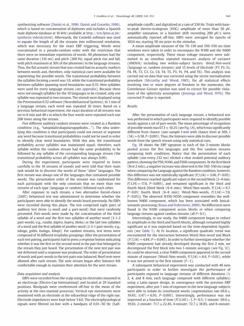
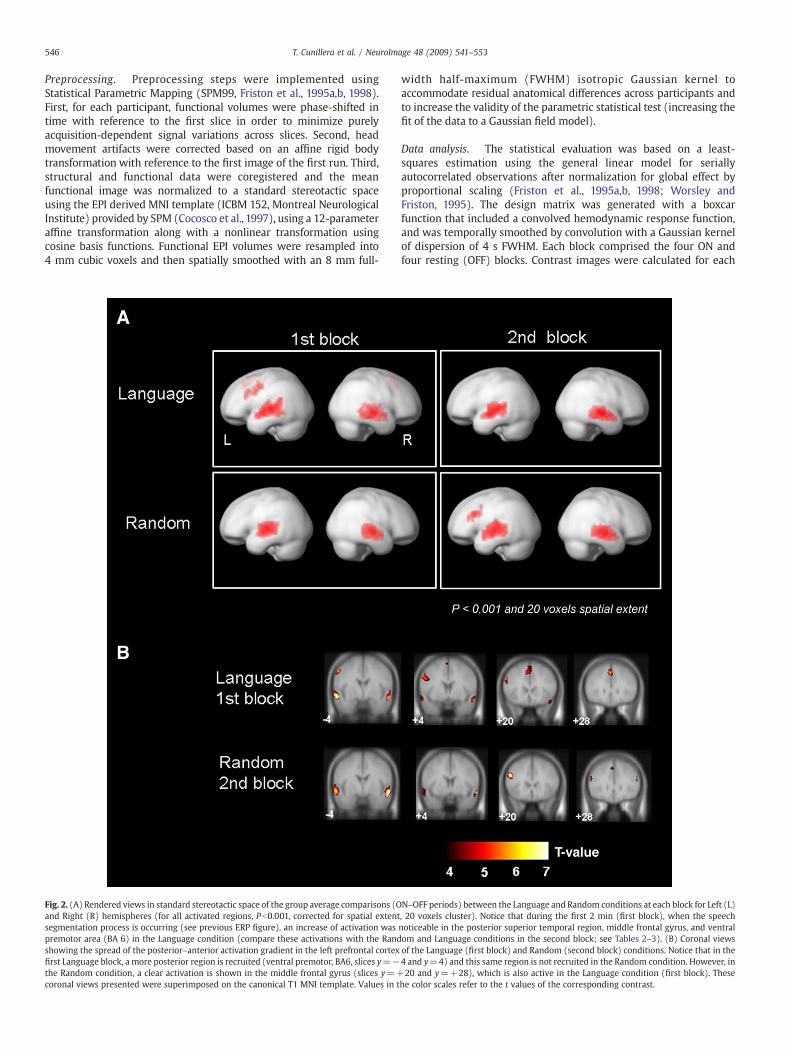
Fig. 2. (A) Rendered views in standard stereotactic space of the group average comparisons (Oand Right (R) hemispheres (for all activated regions, Pb0.001, corrected for spatial extentsegmentation process is occurring (see previous ERP figure), an increase of activation waspremotor area (BA 6) in the Language condition (compare these activations with the Randshowing the spread of the posterior–anterior activation gradient in the left prefrontal cortexfirst Language block, a more posterior region is recruited (ventral premotor, BA6, slices y=−the Random condition, a clear activation is shown in the middle frontal gyrus (slices y=+coronal views presented were superimposed on the canonical T1 MNI template. Values in t
width half-maximum (FWHM) isotropic Gaussian kernel toaccommodate residual anatomical differences across participants andto increase the validity of the parametric statistical test (increasing thefit of the data to a Gaussian field model).
Data analysis. The statistical evaluation was based on a least-squares estimation using the general linear model for seriallyautocorrelated observations after normalization for global effect byproportional scaling (Friston et al., 1995a,b, 1998; Worsley andFriston, 1995). The design matrix was generated with a boxcarfunction that included a convolved hemodynamic response function,and was temporally smoothed by convolution with a Gaussian kernelof dispersion of 4 s FWHM. Each block comprised the four ON andfour resting (OFF) blocks. Contrast images were calculated for each
N–OFF periods) between the Language and Random conditions at each block for Left (L), 20 voxels cluster). Notice that during the first 2 min (first block), when the speechnoticeable in the posterior superior temporal region, middle frontal gyrus, and ventralom and Language conditions in the second block; see Tables 2–3). (B) Coronal viewsof the Language (first block) and Random (second block) conditions. Notice that in the4 and y=4) and this same region is not recruited in the Random condition. However, in20 and y=+28), which is also active in the Language condition (first block). These
he color scales refer to the t values of the corresponding contrast.
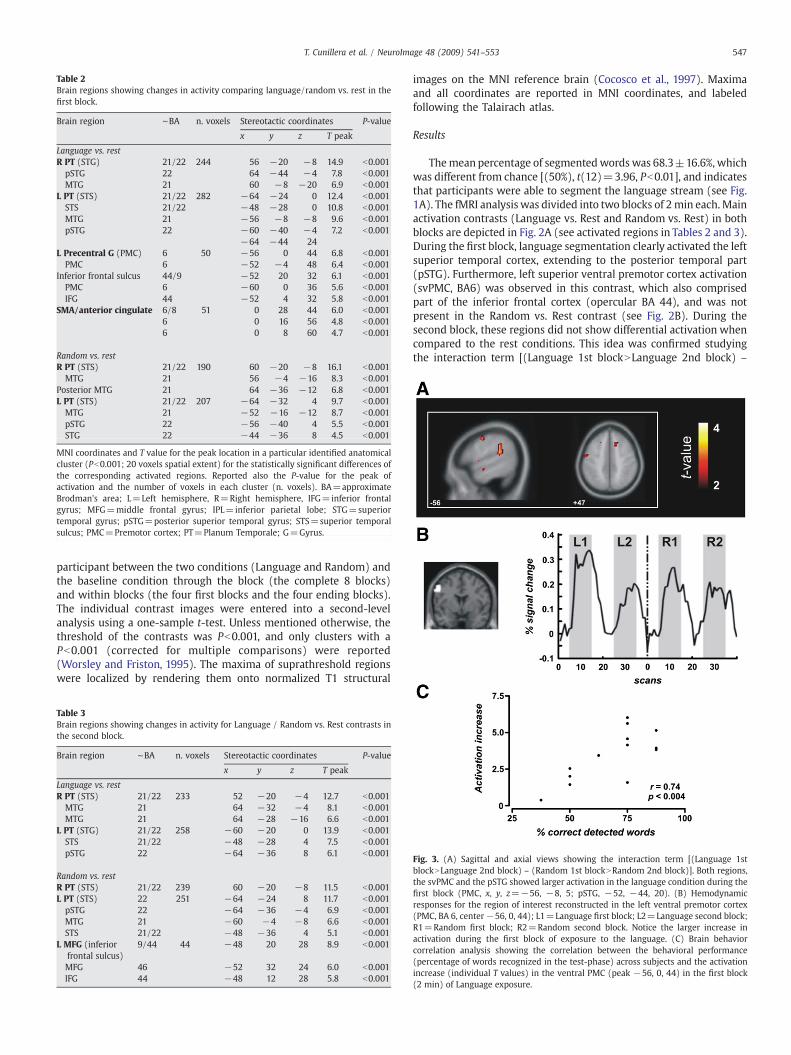
Table 2Brain regions showing changes in activity comparing language/random vs. rest in thefirst block.
Brain region ∼BA n. voxels Stereotactic coordinates P-value
x y z T peak
Language vs. restR PT (STG) 21/22 244 56 −20 −8 14.9 b0.001pSTG 22 64 −44 −4 7.8 b0.001MTG 21 60 −8 −20 6.9 b0.001
L PT (STS) 21/22 282 −64 −24 0 12.4 b0.001STS 21/22 −48 −28 0 10.8 b0.001MTG 21 −56 −8 −8 9.6 b0.001pSTG 22 −60 −40 −4 7.2 b0.001
−64 −44 24L Precentral G (PMC) 6 50 −56 0 44 6.8 b0.001PMC 6 −52 −4 48 6.4 b0.001
Inferior frontal sulcus 44/9 −52 20 32 6.1 b0.001PMC 6 −60 0 36 5.6 b0.001IFG 44 −52 4 32 5.8 b0.001
SMA/anterior cingulate 6/8 51 0 28 44 6.0 b0.0016 0 16 56 4.8 b0.0016 0 8 60 4.7 b0.001
Random vs. restR PT (STS) 21/22 190 60 −20 −8 16.1 b0.001MTG 21 56 −4 −16 8.3 b0.001
Posterior MTG 21 64 −36 −12 6.8 b0.001L PT (STS) 21/22 207 −64 −32 4 9.7 b0.001MTG 21 −52 −16 −12 8.7 b0.001pSTG 22 −56 −40 4 5.5 b0.001STG 22 −44 −36 8 4.5 b0.001
MNI coordinates and T value for the peak location in a particular identified anatomicalcluster (Pb0.001; 20 voxels spatial extent) for the statistically significant differences ofthe corresponding activated regions. Reported also the P-value for the peak ofactivation and the number of voxels in each cluster (n. voxels). BA=approximateBrodman's area; L=Left hemisphere, R=Right hemisphere, IFG=inferior frontalgyrus; MFG=middle frontal gyrus; IPL=inferior parietal lobe; STG=superiortemporal gyrus; pSTG=posterior superior temporal gyrus; STS=superior temporalsulcus; PMC=Premotor cortex; PT=Planum Temporale; G=Gyrus.
547T. Cunillera et al. / NeuroImage 48 (2009) 541–553
participant between the two conditions (Language and Random) andthe baseline condition through the block (the complete 8 blocks)and within blocks (the four first blocks and the four ending blocks).The individual contrast images were entered into a second-levelanalysis using a one-sample t-test. Unless mentioned otherwise, thethreshold of the contrasts was Pb0.001, and only clusters with aPb0.001 (corrected for multiple comparisons) were reported(Worsley and Friston, 1995). The maxima of suprathreshold regionswere localized by rendering them onto normalized T1 structural
Table 3Brain regions showing changes in activity for Language / Random vs. Rest contrasts inthe second block.
Brain region ∼BA n. voxels Stereotactic coordinates P-value
x y z T peak
Language vs. restR PT (STS) 21/22 233 52 −20 −4 12.7 b0.001MTG 21 64 −32 −4 8.1 b0.001MTG 21 64 −28 −16 6.6 b0.001
L PT (STG) 21/22 258 −60 −20 0 13.9 b0.001STS 21/22 −48 −28 4 7.5 b0.001pSTG 22 −64 −36 8 6.1 b0.001
Random vs. restR PT (STS) 21/22 239 60 −20 −8 11.5 b0.001L PT (STS) 22 251 −64 −24 8 11.7 b0.001pSTG 22 −64 −36 −4 6.9 b0.001MTG 21 −60 −4 −8 6.6 b0.001STS 21/22 −48 −36 4 5.1 b0.001
L MFG (inferiorfrontal sulcus)
9/44 44 −48 20 28 8.9 b0.001
MFG 46 −52 32 24 6.0 b0.001IFG 44 −48 12 28 5.8 b0.001
images on the MNI reference brain (Cocosco et al., 1997). Maximaand all coordinates are reported in MNI coordinates, and labeledfollowing the Talairach atlas.
Results
Themean percentage of segmentedwords was 68.3±16.6%, whichwas different from chance [(50%), t(12)=3.96, Pb0.01], and indicatesthat participants were able to segment the language stream (see Fig.1A). The fMRI analysis was divided into two blocks of 2min each. Mainactivation contrasts (Language vs. Rest and Random vs. Rest) in bothblocks are depicted in Fig. 2A (see activated regions in Tables 2 and 3).During the first block, language segmentation clearly activated the leftsuperior temporal cortex, extending to the posterior temporal part(pSTG). Furthermore, left superior ventral premotor cortex activation(svPMC, BA6) was observed in this contrast, which also comprisedpart of the inferior frontal cortex (opercular BA 44), and was notpresent in the Random vs. Rest contrast (see Fig. 2B). During thesecond block, these regions did not show differential activation whencompared to the rest conditions. This idea was confirmed studyingthe interaction term [(Language 1st blockNLanguage 2nd block) –
Fig. 3. (A) Sagittal and axial views showing the interaction term [(Language 1stblockNLanguage 2nd block) – (Random 1st blockNRandom 2nd block)]. Both regions,the svPMC and the pSTG showed larger activation in the language condition during thefirst block (PMC, x, y, z=−56, −8, 5; pSTG, −52, −44, 20). (B) Hemodynamicresponses for the region of interest reconstructed in the left ventral premotor cortex(PMC, BA 6, center−56, 0, 44); L1=Language first block; L2=Language second block;R1=Random first block; R2=Random second block. Notice the larger increase inactivation during the first block of exposure to the language. (C) Brain behaviorcorrelation analysis showing the correlation between the behavioral performance(percentage of words recognized in the test-phase) across subjects and the activationincrease (individual T values) in the ventral PMC (peak −56, 0, 44) in the first block(2 min) of Language exposure.
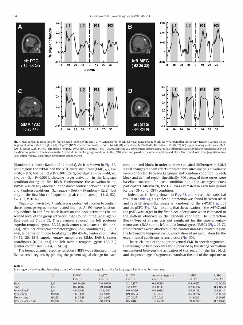
Fig. 4. Hemodynamic responses for four selected regions of interest. L1=Language first block; L2=Language second block; R1=Random first block; R2=Random second block.Regions of interest (left to right): (A) left pSTG (BA22, center coordinates−64,−44, 24), (B) left anterior MFG (BA 45/46, center−52, 20, 32), (C) supplementary motor area (SMA,BA6/8, center 0, 28, 44); (D) left middle temporal gyrus (BA 21, center,−64,−24, 0), depicted as a control non-task related area (no differences across blocks or conditions). Noticethe different pattern of activation in the first block for the Language condition in the pSTG when compared to the other condition and block. Horizontal axis: time [repetition time(TR) units]. Vertical axis: mean percentage signal change.
548 T. Cunillera et al. / NeuroImage 48 (2009) 541–553
(Random 1st blockNRandom 2nd block)]. As it is shown in Fig. 3A,both regions the svPMC and the pSTG were significant (PMC, x, y, z=−56,−8, 5; t-value=2.9, Pb0.007; pSTG, coordinates−52,−44, 20;t-value=3.4, Pb0.003), showing larger activation in the languagecondition during the first block. Furthermore, the activation in thesvPMC was clearly observed in the direct contrast between Languageand Random conditions [(Language – Rest) – (Random – Rest)], butonly in the first block of exposure [peak coordinate: (−44, 8, 52);t=3.35, Pb0.02].
Region-of-interest (ROI) analysiswas performed in order to confirmthese language segmentation-related findings. All ROI were function-ally defined in the first block based on the peak activations in thesecond level of the group activation maps found in the Language vs.Rest contrast (Table 2). These regions covered the left posteriorsuperior temporal gyrus [BA 22, peak center coordinates (−64, −44,24)], left superior ventral premotor region [BA 6, coordinates (−56, 0,44)], left anterior middle frontal gyrus [BA 45/46, center coordinates(−52, 20, 32)], supplementary motor area [SMA, BA6/8, centercoordinates (0, 28, 44)] and left middle temporal gyrus (BA 21)[center coordinates (−64, −24, 0)].
The hemodynamic response function (HRF) was estimated in thefive selected regions by plotting the percent signal change for each
Table 4Brain regions showing the alternating pattern of task-rest blocks changes in activity for Lan
d.f. L PMC L pSTGF=, Pb F=, Pb
Type 1,12 0.6, 0.430 3.9, 0.069Block 1,12 0.6, 0.107 3.9, 0.070Type×Block 1,12 4.9, 0.045 20.2, 0.001Type×time(t) 19,228 1.8, 0.124 3.4, 0.006Block×time 19,228 1.9, 0.088 2.3, 0.042Type×block×time 19,228 1.1, 0.385 2.8, 0.014
condition and block. In order to draw statistical differences in BOLDsignal changes random effects repeated measures analysis of variancewere conducted between Language and Random condition in eachblock and defined region. Specifically, ROI averaged time series werebaseline corrected for each condition and then averaged acrossparticipants. Afterwards, the HRF was estimated in each task periodfor the (ON) and (OFF) condition.
Indeed, as is clearly shown in Figs. 3B and 4 (see the statisticalresults in Table 4), a significant interaction was found between Blockand Type of stream (Language vs. Random) for the svPMC (Fig. 3B)and the pSTG (Fig. 4A), indicating that the activation in the svPMC andthe pSTG was larger in the first block of exposure when compared tothe pattern observed in the Random condition. The interactionBlock×Type of stream was not significant for the supplementarymotor area (SMA) or the left middle frontal gyrus (lMFG) (Figs. 4B, C).No differences were observed in the control non-task related region,the left middle temporal gyrus, which showed no modulation for theexperimental conditions across blocks (Fig. 4D).
The crucial role of the superior ventral PMC in speech segmenta-tion during the first block was also supported by the strong correlationencountered between the activation of this region in the first blockand the percentage of segmented words at the end of the exposure to
guage / Random vs. Rest contrasts.
R pSTG Anterior cingulate L MFG L STGF=, Pb F= , Pb F=, Pb F=, Pb
2.1, 0.177 0.4, 0.533 0.3, 0.617 1.2, 0.2947.2, 0.020 2.4, 0.144 0.7, 0.426 0.3, 0.5800.9, 0.359 4.4, 0.058 2.9, 0.111 2.9, 0.1140.9, 0.504 1.1, 0.375 0.5, 0.766 1.2, 0.3322.7, 0.027 2.7, 0.029 1.3, 0.243 1.5, 0.1970.5, 0.847 1.2, 0.298 1.9, 0.100 0.7, 0.641
549T. Cunillera et al. / NeuroImage 48 (2009) 541–553
the language streams (r=0.74, Pb0.004, see Fig. 3C). This correlationwas not significant for either the second-language block (r=0.4,PN0.1) or for the same computations performed for the pSTG in boththe first and the second block.
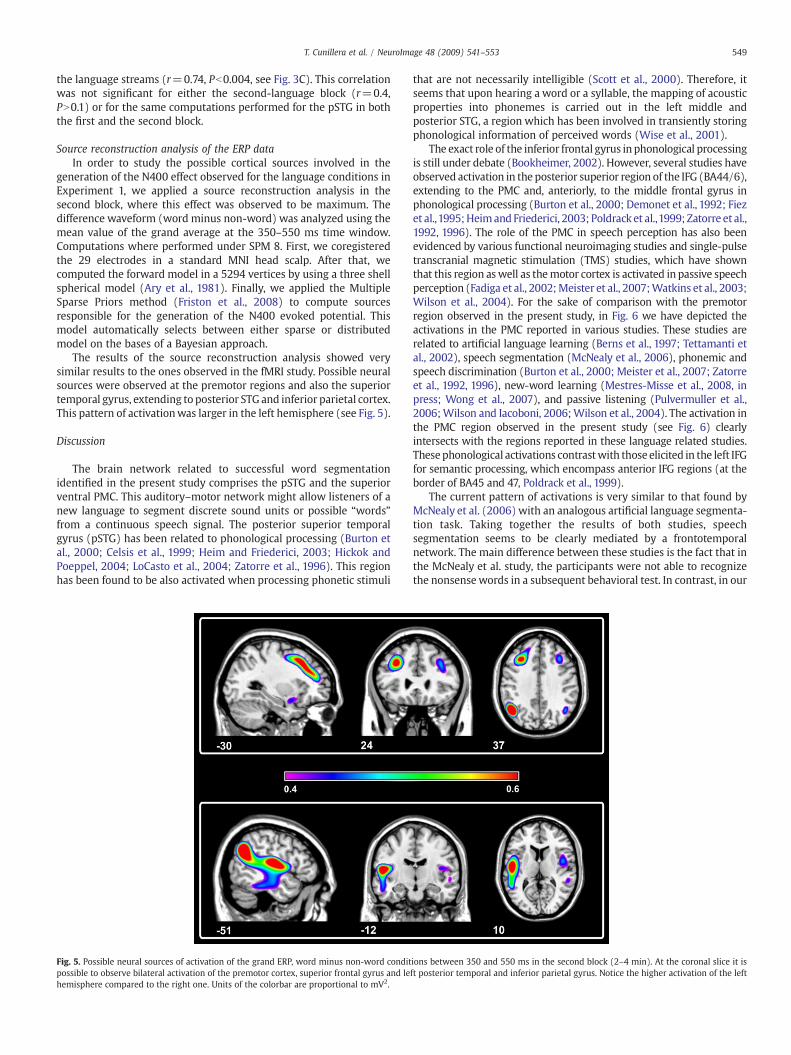
Source reconstruction analysis of the ERP dataIn order to study the possible cortical sources involved in the
generation of the N400 effect observed for the language conditions inExperiment 1, we applied a source reconstruction analysis in thesecond block, where this effect was observed to be maximum. Thedifference waveform (word minus non-word) was analyzed using themean value of the grand average at the 350–550 ms time window.Computations where performed under SPM 8. First, we coregisteredthe 29 electrodes in a standard MNI head scalp. After that, wecomputed the forward model in a 5294 vertices by using a three shellspherical model (Ary et al., 1981). Finally, we applied the MultipleSparse Priors method (Friston et al., 2008) to compute sourcesresponsible for the generation of the N400 evoked potential. Thismodel automatically selects between either sparse or distributedmodel on the bases of a Bayesian approach.
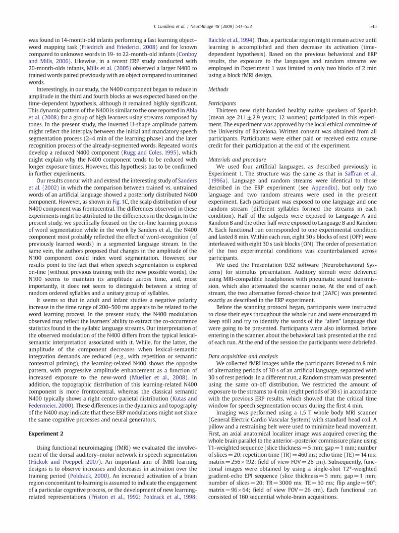
The results of the source reconstruction analysis showed verysimilar results to the ones observed in the fMRI study. Possible neuralsources were observed at the premotor regions and also the superiortemporal gyrus, extending to posterior STG and inferior parietal cortex.This pattern of activationwas larger in the left hemisphere (see Fig. 5).
Discussion
The brain network related to successful word segmentationidentified in the present study comprises the pSTG and the superiorventral PMC. This auditory–motor network might allow listeners of anew language to segment discrete sound units or possible “words”from a continuous speech signal. The posterior superior temporalgyrus (pSTG) has been related to phonological processing (Burton etal., 2000; Celsis et al., 1999; Heim and Friederici, 2003; Hickok andPoeppel, 2004; LoCasto et al., 2004; Zatorre et al., 1996). This regionhas been found to be also activated when processing phonetic stimuli
Fig. 5. Possible neural sources of activation of the grand ERP, word minus non-word conditpossible to observe bilateral activation of the premotor cortex, superior frontal gyrus and lehemisphere compared to the right one. Units of the colorbar are proportional to mV2.
that are not necessarily intelligible (Scott et al., 2000). Therefore, itseems that upon hearing a word or a syllable, the mapping of acousticproperties into phonemes is carried out in the left middle andposterior STG, a region which has been involved in transiently storingphonological information of perceived words (Wise et al., 2001).
The exact role of the inferior frontal gyrus in phonological processingis still under debate (Bookheimer, 2002). However, several studies haveobserved activation in the posterior superior region of the IFG (BA44/6),extending to the PMC and, anteriorly, to the middle frontal gyrus inphonological processing (Burton et al., 2000; Demonet et al., 1992; Fiezet al.,1995;HeimandFriederici, 2003; Poldrack et al.,1999; Zatorre et al.,1992, 1996). The role of the PMC in speech perception has also beenevidenced by various functional neuroimaging studies and single-pulsetranscranial magnetic stimulation (TMS) studies, which have shownthat this region aswell as themotor cortex is activated in passive speechperception (Fadiga et al., 2002;Meister et al., 2007;Watkins et al., 2003;Wilson et al., 2004). For the sake of comparison with the premotorregion observed in the present study, in Fig. 6 we have depicted theactivations in the PMC reported in various studies. These studies arerelated to artificial language learning (Berns et al., 1997; Tettamanti etal., 2002), speech segmentation (McNealy et al., 2006), phonemic andspeech discrimination (Burton et al., 2000; Meister et al., 2007; Zatorreet al., 1992, 1996), new-word learning (Mestres-Misse et al., 2008, inpress; Wong et al., 2007), and passive listening (Pulvermuller et al.,2006;Wilson and Iacoboni, 2006;Wilson et al., 2004). The activation inthe PMC region observed in the present study (see Fig. 6) clearlyintersects with the regions reported in these language related studies.Thesephonological activations contrastwith those elicited in the left IFGfor semantic processing, which encompass anterior IFG regions (at theborder of BA45 and 47, Poldrack et al., 1999).
The current pattern of activations is very similar to that found byMcNealy et al. (2006) with an analogous artificial language segmenta-tion task. Taking together the results of both studies, speechsegmentation seems to be clearly mediated by a frontotemporalnetwork. The main difference between these studies is the fact that inthe McNealy et al. study, the participants were not able to recognizethe nonsensewords in a subsequent behavioral test. In contrast, in our
ions between 350 and 550 ms in the second block (2–4 min). At the coronal slice it isft posterior temporal and inferior parietal gyrus. Notice the higher activation of the left
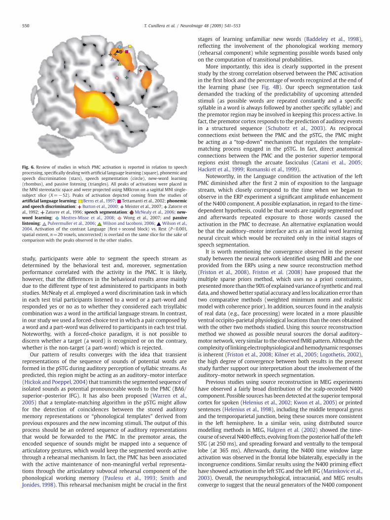
Fig. 6. Review of studies in which PMC activation is reported in relation to speechprocessing, specifically dealing with artificial language learning (square), phonemic andspeech discrimination (stars), speech segmentation (circle), new-word learning(rhombus), and passive listening (triangles). All peaks of activations were placed inthe MNI stereotactic space and were projected using MRIcron on a sagittal MNI single-subject slice (X=−52). Peaks of activation depicted coming from the studies ofartificial language learning: Berns et al., 1997; Tettamanti et al., 2002; phonemicand speech discrimination: Burton et al., 2000; Meister et al., 2007; Zatorre etal., 1992; Zatorre et al., 1996; speech segmentation: McNealy et al., 2006; new-word learning: Mestres-Misse et al., 2008; Wong et al., 2007; and passivelistening: Pulvermuller et al., 2006; Wilson and Iacoboni, 2006; Wilson et al.,2004. Activation of the contrast Language (first+second block) vs. Rest (Pb0.001,spatial extent, n=20 voxels, uncorrected) is overlaid on the same slice for the sake ofcomparison with the peaks observed in the other studies.
550 T. Cunillera et al. / NeuroImage 48 (2009) 541–553
study, participants were able to segment the speech stream asdetermined by the behavioral test and, moreover, segmentationperformance correlated with the activity in the PMC. It is likely,however, that the differences in the behavioral results arose mainlydue to the different type of test administered to participants in bothstudies. McNealy et al. employed a word discrimination task in whichin each test trial participants listened to a word or a part-word andresponded yes or no as to whether they considered each trisyllabiccombination was a word in the artificial language stream. In contrast,in our study we used a forced-choice test inwhich a pair composed byaword and a part-wordwas delivered to participants in each test trial.Noteworthy, with a forced-choice paradigm, it is not possible todiscern whether a target (a word) is recognized or on the contrary,whether is the non-target (a part-word) which is rejected.
Our pattern of results converges with the idea that transientrepresentations of the sequence of sounds of potential words areformed in the pSTG during auditory perception of syllabic streams. Aspredicted, this region might be acting as an auditory–motor interface(Hickok and Poeppel, 2004) that transmits the segmented sequence ofisolated sounds as potential pronounceable words to the PMC (BA6/superior–posterior IFG). It has also been proposed (Warren et al.,2005) that a template-matching algorithm in the pSTG might allowfor the detection of coincidences between the stored auditorymemory representations or “phonological templates” derived fromprevious exposures and the new incoming stimuli. The output of thisprocess should be an ordered sequence of auditory representationsthat would be forwarded to the PMC. In the premotor areas, theencoded sequence of sounds might be mapped into a sequence ofarticulatory gestures, which would keep the segmented words activethrough a rehearsal mechanism. In fact, the PMC has been associatedwith the active maintenance of non-meaningful verbal representa-tions through the articulatory subvocal rehearsal component of thephonological working memory (Paulesu et al., 1993; Smith andJonides, 1998). This rehearsal mechanism might be crucial in the first
stages of learning unfamiliar new words (Baddeley et al., 1998),reflecting the involvement of the phonological working memory(rehearsal component) while segmenting possible words based onlyon the computation of transitional probabilities.
More importantly, this idea is clearly supported in the presentstudy by the strong correlation observed between the PMC activationin the first block and the percentage of words recognized at the end ofthe learning phase (see Fig. 4B). Our speech segmentation taskdemanded the tracking of the predictability of upcoming attendedstimuli (as possible words are repeated constantly and a specificsyllable in a word is always followed by another specific syllable) andthe premotor region may be involved in keeping this process active. Infact, the premotor cortex responds to the prediction of auditory eventsin a structured sequence (Schubotz et al., 2003). As reciprocalconnections exist between the PMC and the pSTG, the PMC mightbe acting as a “top-down” mechanism that regulates the template-matching process engaged in the pSTG. In fact, direct anatomicalconnections between the PMC and the posterior superior temporalregions exist through the arcuate fasciculus (Catani et al., 2005;Hackett et al., 1999; Romanski et al., 1999).
Noteworthy, in the Language condition the activation of the leftPMC diminished after the first 2 min of exposition to the languagestream, which closely correspond to the time when we began toobserve in the ERP experiment a significant amplitude enhancementof the N400 component. A possible explanation, in regard to the time-dependent hypothesis, could be that words are rapidly segmented outand afterwards repeated exposure to those words caused theactivation in the PMC to decrease. An alternative explanation wouldbe that the auditory–motor interface acts as an initial word learningneural circuit which would be recruited only in the initial stages ofspeech segmentation.
It is worth mentioning the convergence observed in the presentstudy between the neural network identified using fMRI and the oneprovided from the ERPs using a new source reconstruction method(Friston et al., 2008). Friston et al. (2008) have proposed that themultiple sparse priors method, which uses no a priori constraints,presentedmore than the 90% of explained variance of synthetic and realdata, and showedbetter spatial accuracy and less localization error thantwo comparative methods (weighted minimum norm and realisticmodel with coherence prior). In addition, sources found in the analysisof real data (e.g., face processing) were located in a more plausibleventral occipito-parietal physiological locations than the ones obtainedwith the other two methods studied. Using this source reconstructionmethod we showed as possible neural sources the dorsal auditory–motor network, very similar to the observed fMRI pattern. Although thecomplexityof linkingelectrophysiological andhemodynamic responsesis inherent (Friston et al., 2008; Kilner et al., 2005; Logothetis, 2002),the high degree of convergence between both results in the presentstudy further support our interpretation about the involvement of theauditory–motor network in speech segmentation.
Previous studies using source reconstruction in MEG experimentshave observed a fairly broad distribution of the scalp-recorded N400component. Possible sources has been detected at the superior temporalcortex for spoken (Helenius et al., 2002; Kwon et al., 2005) or printedsentences (Helenius et al., 1998), including the middle temporal gyrusand the temporoparietal junction, being these sources more consistentin the left hemisphere. In a similar vein, using distributed sourcemodelling methods in MEG, Halgren et al. (2002) showed the time-course of several N400 effects, evolving from the posterior half of the leftSTG (at 250 ms), and spreading forward and ventrally to the temporallobe (at 365 ms). Afterwards, during the N400 time window largeactivation was observed in the frontal lobe bilaterally, especially in theincongruence conditions. Similar results using the N400 priming effecthave showed activation in the left STG and the left IFG (Marinkovic et al.,2003). Overall, the neuropsychological, intracranial, and MEG resultsconverge to suggest that the neural generators of the N400 component
Language 1 Words PIRUTA, BAGOLI, TOKUDA, GUKIBOPart-words RUTABA, TABAGO, GOLITO, LITOKU, KUDAPI, DAPIRU,
GOLIGU, LIGUKI, KIBOBA, BOBAGO, RUTAGU, TAGUKI,KIBOTO, BOTOKU, KUDABA, DABAGO, GOLIPI, LIPIRU,RUTATO, TATOKU, KUDAGU, DAGUKI, KIBOPI, BOPIRU
Language 2⁎ Words PABELA, DINEKA, LUFARI, JISODULanguage 3 Words MAJUPE, JEROGA, DEMUSI, FOLETILanguage 4⁎ Words PUKEMI, RAFINU, BINAPO, MEDOGILanguage 5 Words NONIGE, BULOTE, REMOFU, KOTUSA
551T. Cunillera et al. / NeuroImage 48 (2009) 541–553
are located in a large portion of the temporal lobe as well as the inferiorprefrontal cortex (Halgren et al., 1994; Nobre et al., 1994; see also vanPetten and Luka, 2006).
Considering these N400 related studies, the source reconstructionsolution encountered in the present study seems very similar, pointingto the involvement of the superior temporal gyrus. However, thedistribution of the N400 effect modelled (difference between non-word and words) was more frontal than the standard parietocentraldistribution of the N400 component. This topographical differencemight be explained by the large involvement of the premotor regionsand probablymore anterior prefrontal regions. The frontocentral N400component encountered in our speech segmentation experiment is inagreement with the idea of the involvement of the inferior frontalgyrus in pseudoword processing (Clark andWagner, 2003). Activationin the precentral and inferior frontal gyrus has also been observedwhen learning new words from congruent and incongruent contexts(Mestres-Misse et al., 2008, in press), in phonological new-wordlearning (Gronholm et al., 2005), in second-language learning of novelpitch patterns of words (Wong et al., 2007) and statistical segmenta-tion of tone sequences (using near-infrared spectroscopy, Abla andOkanoya, 2008). Similar frontal shifts in the N400 component havebeen observed in other speech segmentation tasks using ERPs (see DeDiego-Balaguer et al., 2007; Abla et al., 2008). This involvement ofprecentral and inferior frontal regions in word learning might explainthe different topography observed in several studies for the N400 innew learned words. It would be interesting to investigate in follow-upEEG–fMRI combined recordings, in which degree the neural hemody-namic activations could be used as spatial priors to constrainequivalent dipole or distributed estimates of the N400 componentobserved during speech segmentation (Dale et al., 2000).
All in all, the auditory–motor brain network identified for speechsegmentation might be essential for second-language learning and,most probably, for language acquisition in infants (Doupe and Kuhl,1999; Warren et al., 2005). Language perception and production in thedeveloping infant brain requires a specific tuning to the language soundsencountered during the first year of life. First words imitated by a childare guided by the “gestural” features of the sound, i.e. by the actions ofthe mouth rather than by a sound's acoustic features (Studdert-Kennedy, 1987). Because perception and production of sounds are soclosely tied from the first years of life, it is possible that the dorsalauditory perception stream might play an important role duringlanguage acquisition (Hickok and Poeppel, 2007). This dorsal streammight recruit the posterior superior temporal regions for encoding andstoring sequences of sounds and acting as a sensory-motor interface viathe participation of the PMC. Importantly, the identified pathway mustalso be related to the brain network subserving imitation of simplemovements (Iacoboni et al., 1999). In fact, the ability to mimic sounds isessential for learning a new language. This idea has been revitalized bythe discovery of mirror neurons, recorded in macaques in thehomologue of the ventral PMC region (including the superior part ofBroca's region) and in humans (Fadiga and Craighero, 2006; Rizzolattiand Arbib, 1998). These specific audiovisual mirror neurons dischargenot onlywhenperformingandobserving a specific action, but alsowhenhearing a specific sound representative of the observed action (Kohler etal., 2002). Mirror neurons also provide a mechanism for integratingperception and action at the neuronal level, which, at the same time,might contribute to various developmental processes such as theimitative behavior of infants, the necessity to integrate perceived andperformed actions (Meltzoff andDecety, 2003) and communicative acts(Rizzolatti and Arbib, 1998).
Conclusions
Recently, much progress has been made in the identification of thebrain mechanisms involved in language learning, but much stillremains to be done (Gullberg and Indefrey, 2006; Kuhl, 2004). In the
present study we investigated the time-course and brain regionsinvolved in segmenting a new language. The brain network involvedin this process recruited, selectively, the pSTG gyrus and the superiorpart of ventral PMC (superior IFG). After a short exposure towords of anew language, the brain appears to elicit a protolexical trace,evidenced by the appearance of the N400. As speech segmentationis pivotal at the early stages of language learning, the describedauditory–motor network for speech segmentation might be essentialfor second-language learning and for language acquisition in infants.Further studies will be needed to disentangle the exact role of thedifferent brain structures previously described, how they interact, andhow exactly they contribute to statistical learning, speech segmenta-tion, and language learning in general.
Acknowledgments
Special thanks to D. Cucurell, R. de Diego Balaguer, T. Gomila, M.Laine, J. Müller, andM. Guxens for their support and comments duringvarious stages of the project. We also thank J. Riba, S. Romero, X.Mayoral, and C. Soriano for their technical help. This research wassupported by the Spanish Government (MCYT) to ARF (SEJ2005-06067/PSIC; Ramon y Cajal program) and NS/JT (Consolider Ingenio2010, CSD2007-00012 and SEJ 2007-60751). Support for the researchof N.S. was received through the prize “ICREA Academia” forexcellence in research, Generalitat de Catalunya.
Appendix
Artificial languages used in the ERP experiment. Part-wordconstruction in each language followed the same structure asexemplified in the first language.
⁎Language streams used in the fMRI experiment. Random streamwere constructed as a result of combining the syllables of theselanguages streams.
References
Abla, D., Okanoya, K., 2008. Statistical segmentation of tone sequences activates the leftinferior frontal cortex: a near-infrared spectroscopy study. Neuropsychologia 46,2787–2795.
Abla, D., Katahira, K., Okanoya, K., 2008. On-line assessment of statistical learning byevent-related potentials. J. Cogn. Neurosci. 20, 952–964.
Aboitiz, F., García, R., Brunetti, E., Bosman, C., 2006. The origin of Broca's area and itsconnections from an ancestral working/active memory network. In: Amunt, K.,Grodzinsky, Y. (Eds.), Broca's Region. Oxford University Press, Oxford, pp. 3–16.
Ary, J.P., Klein, S.A., Fender, D.H., 1981. Location of sources of evoked scalp potentials:corrections for skull and scalp thicknesses. IEEE Trans. Biomed. Eng. 28, 447–452.
Baddeley, A., Gathercole, S., Papagno, C., 1998. The phonological loop as a languagelearning device. Psychol. Rev. 105, 158–173.
Berns, G.S., Cohen, J.D., Mintun, M.A., 1997. Brain regions responsive to novelty in theabsence of awareness. Science 276, 1272–1275.
Bookheimer, S., 2002. Functional MRI of language: new approaches to understandingthe cortical organization of semantic processing. Ann. Rev. Neurosci. 25, 151–188.
Brent, M.R., 1997. Toward a unified model of lexical acquisition and lexical access. J.Psycholinguist. Res. 26, 363–375.
Brent, M.R., 1999. Speech segmentation and word discovery: a computationalperspective. Trends Cogn. Sci. 3, 294–301.
Buchsbaum, B.R., Olsen, R.K., Koch, P., Berman, K.F., 2005. Human dorsal and ventralauditory streams subserve rehearsal-based and echoic processes during verbalworking memory. Neuron 48, 687–697.

552 T. Cunillera et al. / NeuroImage 48 (2009) 541–553
Buiatti, M., Pena, M., haene-Lambertz, G., 2009. Investigating the neural correlates ofcontinuous speech computation with frequency-tagged neuroelectric responses.Neuroimage 44, 509–519.
Burton,M.W., Small, S.L., Blumstein, S.E., 2000. The role of segmentation in phonologicalprocessing: an fMRI investigation. J. Cogn. Neurosci. 12, 679–690.
Catani, M., Jones, D.K., Ffytche, D.H., 2005. Perisylvian language networks of the humanbrain. Ann. Neurol. 57, 8–16.
Celsis, P., Boulanouar, K., Doyon, B., Ranjeva, J.P., Berry, I., Nespoulous, J.L., Chollet, F.,1999. Differential fMRI responses in the left posterior superior temporal gyrus andleft supramarginal gyrus to habituation and change detection in syllables and tones.Neuroimage 9, 135–144.
Christiansen, M.H., Allen, J., Seidenberg, M.S., 1998. Learning to segment speech usingmultiple cues: a connectionist model. Lang. Cogn. Processes 13, 221–268.
Clark, D., Wagner, A.D., 2003. Assembling and encoding word representations:fMRI subsequent memory effects implicate a role for phonological control.Neuropsychologia 41, 304–317.
Cocosco, C.A., Kollokian, V., Kwan, R.K.S., Evans, A.C., 1997. BrainWeb: online interface toa 3D MRI simulated brain database. Neuroimage 5 (4), S425.
Conboy, B.T., Mills, D.L., 2006. Two languages, one developing brain: event-relatedpotentials to words in bilingual toddlers. Dev. Sci. 9, F1–12.
Cunillera, T., Toro, J.M., Sebastian-Galles, N., Rodriguez-Fornells, A., 2006. The effects ofstress and statistical cues on continuous speech segmentation: an event-relatedbrain potential study. Brain Res. 1123, 168–178.
Cunillera, T., Gomila, A., Rodriguez-Fornells, A., 2008. Beneficial effects of word finalstress in segmenting a new language: evidence from ERPs. BMC Neurosci. 9, 23.
Dale, A.M., Liu, A.K., Fischl, B.R., Buckner, R.L., Belliveau, J.W., Lewine, J.D., et al., 2000.Dynamic statistical parametric mapping: combining fMRI and MEG for high-resolution imaging of cortical activity. Neuron 26, 55–67.
De Diego-Balaguer, R., Toro, J.M., Rodriguez-Fornells, A., Bachoud-Levi, A.C., 2007.Different neurophysiological mechanisms underlying word and rule extractionfrom speech. PLoS ONE e1175, 2.
Demonet, J.F., Chollet, F., Ramsay, S., Cardebat, D., Nespoulous, J.L., Wise, R., Rascol, A.,Frackowiak, R., 1992. The anatomy of phonological and semantic processing innormal subjects. Brain 115 (Pt 6), 1753–1768.
Doupe, A.J., Kuhl, P.K., 1999. Birdsong and human speech: common themes andmechanisms. Ann. Rev. Neurosci. 22, 567–631.
Dutoit, T., Gosselin, B., 1996. On the use of a hybrid harmonic/stochastic model for TTSsynthesis-by-concatenation. Speech Commun. 19, 119–143.
Dutoit, T., Pagel, N., Pierret, F., Bataille, O., van der Vreken, O.,1996. TheMBROLA project:towards a set of high-quality speech synthesizers free of use fornon-commercialpurposes. 3rd ed., Philadelphia, pp. 1393–1396.
Elman, J.L., 1990. Finding structure in time. Cogn. Sci. 14, 179–211.Fadiga, L., Craighero, L., 2006. Hand actions and speech representation in Broca's area.
Cortex 42, 486–490.Fadiga, L., Craighero, L., Buccino, G., Rizzolatti, G., 2002. Speech listening specifically
modulates the excitability of tongue muscles: a TMS study. Eur. J. Neurosci. 15,399–402.
Fiez, J.A., Raichle, M.E., Miezin, F.M., Petersen, S.E., Tallal, P., Katz, W.F., 1995. Pet studiesof auditory and phonological processing — effects of stimulus characteristics andtask demands. J. Cogn. Neurosci. 7, 357–375.
Fiser, J., Aslin, R.N., 2001. Unsupervised statistical learning of higher-order spatialstructures from visual scenes. Psychol. Sci. 12, 499–504.
Friedrich, M., Friederici, A.D., 2008. Neurophysiological correlates of online wordlearning in 14-month-old infants. Neuroreport 19, 1757–1761.
Friston, K.J., Frith, C.D., Passingham, R.E., Liddle, P.F., Frackowiak, R.S., 1992. Motorpractice and neurophysiological adaptation in the cerebellum: a positrontomography study. Proc. Biol. Sci. 248, 223–228.
Friston, K.J., Frith, C.D., Turner, R., Frackowiak, R.S.J., 1995a. Characterizing evokedhemodynamics with fMRI. Neuroimage 2, 157–165.
Friston, K.J., Holmes, A.P., Worsley, K.J., Poline, J.B., Frith, C.D., Frackowiak, R., 1995b.Statistical Parametric Maps in functional imaging: a general linear approach.Hum. Brain Mapp. 2, 189–210.
Friston, K.J., Josephs, O., Rees, G., Turner, R., 1998. Nonlinear event-related responses infMRI. Magn. Reson. Med. 39, 41–52.
Friston, K.J., Harrison, L., Daunizeau, J., Kiebel, S., Phillips, C., Trujillo-Barreto, N., Henson,R., Flandin, G., Mattout, J., 2008. Multiple sparse priors for the M/EEG inverseproblem. Neuroimage 39, 1104–1120.
Graf-Estes, K., Evans, J.L., Alibali, M.W., Saffran, J.R., 2007. Can infants map meaning tonewly segmented words? Statistical segmentation and word learning. Psychol. Sci.18, 254–260.
Gronholm, P., Rinne, J.O., Vorobyev, V., Laine, M., 2005. Naming of newly learnedobjects: a PET activation study. Brain Res. Cogn. Brain Res. 25, 359–371.
Gullberg, M., Indefrey, P., 2006. Cognitive Neuroscience of Second Language Acquisition.Oxford.
Hackett, T.A., Stepniewska, I., Kaas, J.H., 1999. Prefrontal connections of the parabeltauditory cortex in macaque monkeys. Brain Res. 817, 45–58.
Halgren, E., Baudena, P., Heit, G., Clarke, J.M., Marinkovic, K., Clarke, M., 1994. Spatio-temporal stages in face and word processing. I. Depth-recorded potentials in thehuman occipital, temporal and parietal lobes [corrected]. J. Physiol. Paris 88,1–50.
Halgren, E., Dhond, R.P., Christensen, N., Van, P.C., Marinkovic, K., Lewine, J.D., Dale, A.M.,2002. N400-like magnetoencephalography responses modulated by semanticcontext, word frequency, and lexical class in sentences. Neuroimage 17, 1101–1116.
Hauser, M.D., Newport, E.L., Aslin, R.N., 2001. Segmentation of the speech stream in anon-human primate: statistical learning in cotton-top tamarins. Cognition 78,B53–B64.
Heim, S., Friederici, A.D., 2003. Phonological processing in language production: timecourse of brain activity. Neuroreport 14, 2031–2033.
Helenius, P., Salmelin, R., Service, E., Connolly, J.F., 1998. Distinct time courses of wordand context comprehension in the left temporal cortex. Brain 121 (Pt 6), 1133–1142.
Helenius, P., Salmelin, R., Service, E., Connolly, J.F., Leinonen, S., Lyytinen, H., 2002.Cortical activation during spoken-word segmentation in nonreading-impaired anddyslexic adults. J. Neurosci. 22, 2936–2944.
Hickok, G., Poeppel, D., 2000. Towards a functional neuroanatomy of speech perception.Trends Cogn. Sci. 4, 131–138.
Hickok, G., Poeppel, D., 2004. Dorsal and ventral streams: a framework for under-standing aspects of the functional anatomy of language. Cognition 92, 67–99.
Hickok, G., Poeppel, D., 2007. Opinion — the cortical organization of speech processing.Nat. Rev. Neurosci. 8, 393–402.
Iacoboni, M., Woods, R.P., Brass, M., Bekkering, H., Mazziotta, J.C., Rizzolatti, G., 1999.Cortical mechanisms of human imitation. Science 286, 2526–2528.
Jacquemot, C., Scott, S.K., 2006. What is the relationship between phonological short-term memory and speech processing? Trends Cogn. Sci. 10, 480–486.
Jennings, J.R., Wood, C.C., 1976. Epsilon-adjustment procedure for repeated-measuresanalyses of variance. Psychophysiology 13, 277–278.
Jusczyk, P.W., Houston, D.M., Newsome, M., 1999. The beginnings of word segmentationin English-learning infants. Cogn. Psychol. 39, 159–207.
Kilner, J.M., Mattout, J., Henson, R., Friston, K.J., 2005. Hemodynamic correlates of EEG: aheuristic. Neuroimage 28, 280–286.
Kohler, E., Keysers, C., Umilta, M.A., Fogassi, L., Gallese, V., Rizzolatti, G., 2002. Hearingsounds, understanding actions: action representation in mirror neurons. Science297, 846–848.
Kuhl, P.K., 2004. Early language acquisition: cracking the speech code. Nat. Rev.Neurosci. 5, 831–843.
Kutas, M., Federmeier, K.D., 2000. Electrophysiology reveals semantic memory use inlanguage comprehension. Trends Cogn. Sci. 4, 463–470.
Kwon, H., Kuriki, S., Kim, J.M., Lee, Y.H., Kim, K., Nam, K., 2005. MEG study on neuralactivities associated with syntactic and semantic violations in spoken Koreansentences. Neurosci. Res. 51, 349–357.
Liberman, A.M., Mattingly, I.G., 1985. The motor theory of speech perception revised.Cognition 21, 1–36.
LoCasto, P.C., Krebs-Noble, D., Gullapalli, R.P., Burton, M.W., 2004. An fMRI investigationof speech and tone segmentation. J. Cogn. Neurosci. 16, 1612–1624.
Logothetis, N.K., 2002. The neural basis of the blood-oxygen-level-dependentfunctional magnetic resonance imaging signal. Philos. Trans. R. Soc. Lond. B Biol.Sci. 357, 1003–1037.
Marinkovic, K., Dhond, R.P., Dale, A.M., Glessner, M., Carr, V., Halgren, E., 2003.Spatiotemporal dynamics of modality-specific and supramodal word processing.Neuron 38, 487–497.
Mccarthy, G., Wood, C.C., 1985. Scalp distributions of event-related potentials: anambiguity associated with analysis of variance models. Electroencephalogr. Clin.Neurophysiol. 62, 203–208.
McLaughlin, J., Osterhout, L., Kim, A., 2004. Neural correlates of second-language wordlearning: minimal instruction produces rapid change. Nat. Neurosci. 7, 703–704.
McNealy, K., Mazziotta, J.C., Dapretto, M., 2006. Cracking the language code: neuralmechanisms underlying speech parsing. J. Neurosci. 26, 7629–7639.
Meister, I.G., Wilson, S.M., Deblieck, C., Wu, A.D., Lacoboni, M., 2007. The essential role ofpremotor cortex in speech perception. Curr. Biol. 17, 1692–1696.
Meltzoff, A.N., Decety, J., 2003. What imitation tells us about social cognition: arapprochement between developmental psychology and cognitive neuroscience.Philos. Trans. R. Soc. Lond B Biol. Sci. 358, 491–500.
Mestres-Misse, A., Rodriguez-Fornells, A., Munte, T.F., 2007. Watching the brain duringmeaning acquisition. Cerebral Cortex 17, 1858–1866.
Mestres-Misse, A., Camara, E., Rodriguez-Fornells, A., Rotte, M., Munte, T.F., 2008.Functional neuroanatomy of meaning acquisition from context. J. Cogn. Neurosci.20, 2153–2166.
Mestres-Misse, A., Munte, T.F., Rodriguez-Fornells, A., in press. Functional neuroanat-omy of contextual acquisition of concrete and abstract words. J. Cogn. Neurosci.doi:10.1162/jocn.2008.21171.
Mills, D.L., Plunkett, K., Prat, C., Schafer, G., 2005. Watching the infant brain learnwords:effects of vocabulary size and experience. Cogn. Dev. 20, 19–31.
Mueller, J.L., Bahlmann, J., Friederici, A.D., 2008. The role of pause cues in languagelearning: the emergence of event-related potentials related to sequence processing.J. Cogn. Neurosci. 20, 892–905.
Nobre, A.C., Allison, T., Mccarthy, G., 1994. Word recognition in the human inferiortemporal lobe. Nature 372, 260–263.
Pacton, S., Perruchet, P., 2008. An attention-based associative account of adjacentand nonadjacent dependency learning. J. Exp. Psychol. Learn. Mem. Cogn. 34,80–96.
Paulesu, E., Frith, C.D., Frackowiak, R.S.J., 1993. The neural correlates of the verbalcomponent of working memory. Nature 362, 342–345.
Perfetti, C.A., Wlotko, E.W., Hart, L.A., 2005. Word learning and individual differences inword learning reflected in event-related potentials. J. Exper. Psychol.-Learn. Mem.Cogn. 31, 1281–1292.
Perruchet, P., Vinter, A., 1998. PARSER: amodel for word segmentation. J. Mem. Lang. 39,246–263.
Perruchet, P., Pacton, S., 2006. Implicit learning and statistical learning: onephenomenon, two approaches. Trends Cogn. Sci. 10, 233–238.
Poldrack, R.A., 2000. Imaging brain plasticity: conceptual andmethodological issues— atheoretical review. Neuroimage 12, 1–13.
Poldrack, R.A., Desmond, J.E., Glover, G.H., Gabrieli, J.D., 1998. The neural basis of visualskill learning: an fMRI study of mirror reading. Cereb. Cortex 8, 1–10.
553T. Cunillera et al. / NeuroImage 48 (2009) 541–553
Poldrack, R.A., Wagner, A.D., Prull, M.W., Desmond, J.E., Glover, G.H., Gabrieli, J.D., 1999.Functional specialization for semantic and phonological processing in the leftinferior prefrontal cortex. Neuroimage 10, 15–35.
Pulvermuller, F., Huss, M., Kherif, F., Moscoso del Prado, M.F., Hauk, O., Shtyrov, Y., 2006.Motor cortex maps articulatory features of speech sounds. Proc. Natl. Acad. Sci.U.S.A. 103, 7865–7870.
Raichle, M.E., Fiez, J.A., Videen, T.O., MacLeod, A.M., Pardo, J.V., Fox, P.T., Petersen, S.E.,1994. Practice-related changes in human brain functional anatomy duringnonmotor learning. Cereb. Cortex 4, 8–26.
Rizzolatti, G., Arbib,M.A.,1998. Languagewithin our grasp. TrendsNeurosci. 21,188–194.Romanski, L.M., Tian, B., Fritz, J., Mishkin, M., Goldman-Rakic, P.S., Rauschecker, J.P.,
1999. Dual streams of auditory afferents target multiple domains in the primateprefrontal cortex. Nat. Neurosci. 2, 1131–1136.
Rugg, M.D., Coles, M.G.H., 1995. Electrophysiology of Mind. Event-Related BrainPotentials and Cognition. Oxford University Press, Oxford.
Saffran, J.R., Aslin, R.N., Newport, E.L., 1996a. Statistical learning by 8-month-old infants.Science 274, 1926–1928.
Saffran, J.R., Newport, E.L., Aslin, R.N., 1996b. Word segmentation: the role ofdistributional cues. J. Mem. Lang. 35, 606–621.
Saffran, J.R., Newport, E.L., Aslin, R.N., Tunick, R.A., Barrueco, S., 1997. Incidentallanguage learning: listening (and learning) out of the corner of your ear. Psychol.Sci. 8, 101–105.
Saffran, J.R., Johnson, E.K., Aslin, R.N., Newport, E.L., 1999. Statistical learning of tonesequences by human infants and adults. Cognition 70, 27–52.
Sanders, L.D., Neville, H.J., 2003a. An ERP study of continuous speech processing I.Segmentation, semantics, and syntax in native speakers. Cogn. Brain Res. 15,228–240.
Sanders, L.D., Neville, H.J., 2003b. An ERP study of continuous speech processing II.Segmentation, semantics, and syntax in non-native speakers. Cogn. Brain Res. 15,214–227.
Sanders, L.D., Newport, E.L., Neville, H.J., 2002. Segmenting nonsense: an event-relatedpotential index of perceived onsets in continuous speech. Nat. Neurosci. 5, 700–703.
Schubotz, R.I., von Cramon, D.Y., Lohmann, G., 2003. Auditory what, where, and when: asensory somatotopy in lateral premotor cortex. Neuroimage 20, 173–185.
Scott, S.K., Wise, R.J.S., 2004. The functional neuroanatomy of prelexical processing inspeech perception. Cognition 92, 13–45.
Scott, S.K., Blank, C.C., Rosen, S., Wise, R.J., 2000. Identification of a pathway forintelligible speech in the left temporal lobe. Brain 123 (Pt 12), 2400–2406.
Scott, S.K., Rosen, S., Lang, H., Wise, R.J.S., 2006. Neural correlates of intelligibility inspeech investigated with noise vocoded speech — a positron emission tomographystudy. J. Acoust. Soc. Am. 120, 1075–1083.
Smith, E.E., Jonides, J., 1998. Neuroimaging analyses of human working memory. Proc.Natl. Acad. Sci. U. S. A. 95, 12061–12068.
Studdert-Kennedy, M., 1987. The phoneme as a perceptuomotor structure. In: Allport,D., MacKay, D., Prinz, W., Scheerer, E. (Eds.), Language Perception and Production.Academic Press, London, pp. 67–84.
Tettamanti, M., Alkadhi, H., Moro, A., Perani, D., Kollias, S., Weniger, D., 2002. Neuralcorrelates for the acquisition of natural language syntax. NeuroImage 17, 700–709.
Toro, J.M., Trobalon, J.B., 2005. Statistical computations over a speech stream in a rodent.Percept. Psychophys. 67, 867–875.
Toro, J.M., Sinnett, S., Soto-Faraco, S., 2005. Speech segmentation by statistical learningdepends on attention. Cognition 97, B25–B34.
Turk-Browne, N.B., Junge, J.A., Scholl, B.J., 2005. The automaticity of visual statisticallearning. J. Exp. Psychol.-Gen. 134, 552–564.
van Petten, C., Luka, B.J., 2006. Neural localization of semantic context effects inelectromagnetic and hemodynamic studies. Brain Lang. 97, 279–293.
Warren, J.E., Wise, R.J.S., Warren, J.D., 2005. Sounds do-able: auditory–motortransformations and the posterior temporal plane. Trends Neurosci. 28, 636–643.
Watkins, K.E., Strafella, A.P., Paus, T., 2003. Seeing and hearing speech excites the motorsystem involved in speech production. Neuropsychologia 41, 989–994.
Wilson, S.M., Iacoboni, M., 2006. Neural responses to non-native phonemes varying inproducibility: evidence for the sensorimotor nature of speech perception. Neuro-image 33, 316–325.
Wilson, S.M., Saygin, A.P., Sereno, M.I., Iacoboni, M., 2004. Listening to speech activatesmotor areas involved in speech production. Nat. Neurosci. 7, 701–702.
Wise, R.J.S., Scott, S.K., Blank, S.C., Mummery, C.J., Murphy, K., Warburton, E.A., 2001.Separate neural subsystems within ‘Wernicke's area’. Brain 124, 83–95.
Wong, P.C., Perrachione, T.K., Parrish, T.B., 2007. Neural characteristics of successful andless successful speech and word learning in adults. Hum. Brain Mapp. 28,995–1006.
Worsley, K.J., Friston, K.J., 1995. Analysis of fMRI time-series revisited — again.Neuroimage 2, 173–181.
Zatorre, R.J., Evans, A.C., Meyer, E., Gjedde, A., 1992. Lateralization of phonetic and pitchdiscrimination in speech processing. Science 256, 846–849.
Zatorre, R.J., Meyer, E., Gjedde, A., Evans, A.C., 1996. PET studies of phonetic processing ofspeech: review, replication, and reanalysis. Cereb. Cortex 6, 21–30.