Clinical Neuroanatomy (Snell, 7th Ed. 2010) - Internet Archive
543
-
Upload
khangminh22 -
Category
Documents
-
view
0 -
download
0
Transcript of Clinical Neuroanatomy (Snell, 7th Ed. 2010) - Internet Archive

Authors: Snell, Richard S.Title: Clinical Neuroanatomy, 7th Edition
Copyright ©2010 Lippincott Williams & Wilkins
2010
Lippincott Williams & WilkinsPhiladelphia351 West Camden Street, Baltimore, MD 21201, 530 Walnut Street, Philadelphia, PA 19106
978-0-7817-9427-5
Seventh Edition
Copyright © 2010, 2006, 2001, 1997, 1992, 1987, 1980 Lippincott Williams & Wilkins, a Wolters Kluwer business.
351 West Camden Street, Baltimore, MD 21201
530 Walnut Street, Philadelphia, PA 19106
Printed in China
All rights reserved. This book is protected by copyright. No part of this book may be reproduced or transmitted in any form or by anymeans, including as photocopies or scanned-in or other electronic copies, or utilized by any information storage and retrieval systemwithout written permission from the copyright owner, except for brief quotations embodied in critical articles and reviews. Materialsappearing in this book prepared by individuals as part of their official duties as U.S. government employees are not covered by the above-mentioned copyright. To request permission, please contact Lippincott Williams & Wilkins at 530 Walnut Street, Philadelphia, PA 19106, viaemail at [email protected], or via website at lww.com (products and services).
9 8 7 6 5 4 3 2 1
Acquisitions Editor: Crystal Taylor
Managing Editor: Kelly Horvath
Marketing Manager: Emilie Linkins
Managing Editor, Production: Eve Malakoff-Klein
Designer: Stephen Druding
Compositor: Aptara
Library of Congress Cataloging-in-Publication Data
Snell, Richard S.
Clinical neuroanatomy / Richard S. Snell. — 7th ed.
p. ; cm.
Includes bibliographical references and index.
ISBN 978-0-7817-9427-5
1. Neuroanatomy. I. Title.
[DNLM: 1. Nervous System—anatomy & histology. WL 101 S671c 2010]
QM451.S64 2010
616.8—dc22
2008040897
DISCLAIMER
Care has been taken to confirm the accuracy of the information present and to describe generally accepted practices. However, theauthors, editors, and publisher are not responsible for errors or omissions or for any consequences from application of the information inthis book and make no warranty, expressed or implied, with respect to the currency, completeness, or accuracy of the contents of thepublication. Application of this information in a particular situation remains the professional responsibility of the practitioner; the clinicaltreatments described and recommended may not be considered absolute and universal recommendations.
The authors, editors, and publisher have exerted every effort to ensure that drug selection and dosage set forth in this text are inaccordance with the current recommendations and practice at the time of publication. However, in view of ongoing research, changes ingovernment regulations, and the constant flow of information relating to drug therapy and drug reactions, the reader is urged to checkthe package insert for each drug for any change in indications and dosage and for added warnings and precautions. This is particularlyimportant when the recommended agent is a new or infrequently employed drug.
Some drugs and medical devices presented in this publication have Food and Drug Administration (FDA) clearance for limited use inrestricted research settings. It is the responsibility of the health care provider to ascertain the FDA status of each drug or device plannedfor use in their clinical practice.
To purchase additional copies of this book, call our customer service department at (800) 638-3030 or fax orders to (301) 223-2320.International customers should call (301) 223-2300.

Visit Lippincott Williams & Wilkins on the Internet: http://www.lww.com. Lippincott Williams & Wilkins customer service representatives areavailable from 8:30 am to 6:00 pm, EST.

Authors: Snell, Richard S.Title: Clinical Neuroanatomy, 7th Edition
Copyright ©2010 Lippincott Williams & Wilkins
> Fr ont of Book > Author s
Author
Richard S. Snell M.R.C.S., L.R.C.P., MB, BS, MD, PhDEmeritus Professor of AnatomyGeorge Washington University, School of Medicine and Health Sciences, Washington, DC; Formerly Associate Professor of Anatomy andMedicine, Yale University Medical School; Lecturer in Anatomy King's College University of London; and Visiting Professor of Anatomy,Harvard Medical School.

Authors: Snell, Richard S.Title: Clinical Neuroanatomy, 7th Edition
Copyright ©2010 Lippincott Williams & Wilkins
> Fr ont of Book > Pr eface
Preface
This book contains the basic neuroanatomical facts necessary for the practice of medicine. It is suitable for medical students, dentalstudents, nurses, and allied health students. Residents fnd this book useful during their rotations.
The functional organization of the nervous system has been emphasized and indicates how injury and disease can result in neurologicdeficits. The amount of factual information has been strictly limited to that which is clinically important.
In this edition, the content of each chapter has been reviewed, obsolete material has been discarded, and new material added.
Each chapter is divided into the following categories:
Clinical Example. A short case report that serves to dramatize the relevance of neuroanatomy introduces each chapter.
Chapter Objectives. This section details the material that is most important to learn and understand in each chapter.
Basic Neuroanatomy. This section provides basic information on neuroanatomical structures that are of clinical importance. Numerousexamples of normal radiographs, CT scans, MRIs, and PET scans are also provided. Many cross-sectional diagrams have been included tostimulate students to think in terms of three-dimensional anatomy, which is so important in the interpretation of CT scans and MRIimages.
Clinical Notes. This section provides the practical application of neuroanatomical facts that are essential in clinical practice. Itemphasizes the structures that the physician will encounter when making a diagnosis and treating a patient. It also provides theinformation necessary to understand many procedures and techniques and notes the anatomical “pitfalls” commonly encountered.
Clinical Problem Solving. This section provides the student with many examples of clinical situations in which a knowledge ofneuroanatomy is necessary to solve clinical problems and to institute treatment; solutions to the problems are provided at the end ofthe chapter.
Review Questions. The purpose of the questions is threefold: to focus attention on areas of importance, to enable students to assesstheir areas of weakness, and to provide a form of self-evaluation when questions are answered under examination conditions. Some ofthe questions are centered around a clinical problem that requires a neuroanatomical answer. Solutions to the problem are providedat the end of each chapter.
In addition to the full text from the book, an interactive Review Test, including over 450 questions, is provided online.
The book is extensively illustrated. The majority of the figures have been kept simple and are in color. As in the previous edition, a conciseColor Atlas of the dissected brain is included prior to the text. This small but important group of colored plates enables the reader toquickly relate a particular part of the brain to the whole organ.
References to neuroanatomical literature are included should readers wish to acquire a deeper knowledge of an area of interest.R. S. S.

Authors: Snell, Richard S.Title: Clinical Neuroanatomy, 7th Edition
Copyright ©2010 Lippincott Williams & Wilkins
> Fr ont of Book > Acknow ledgments
Acknowledgments
Iam greatly indebted to the following colleagues who provided me with photographic examples of neuroanatomical material: Dr. N. Cauna,Emeritus Professor of Anatomy, University of Pittsburgh School of Medicine; Dr. F. M. J. Fitzgerald, Professor of Anatomy, UniversityCollege, Galway, Ireland; and Dr. A. Peters, Professor of Anatomy, Boston University School of Medicine.
My special thanks are owed to Larry Clerk, who, as a senior technician in the Department of Anatomy at the George Washington UniversitySchool of Medicine and Health Sciences, greatly assisted me in the preparation of neuroanatomical specimens for photography.
I am also grateful to members of the Department of Radiology for the loan of radiographs and CT scans that have been reproduced indifferent sections of this book. I am most grateful to Dr. G. Size of the Department of Radiology at Yale University Medical Center forexamples of CT scans and MRI images of the brain. I also thank Dr. H. Dey, Director of the PET Scan Unit of the Department of Radiology,Veterans Affairs Medical Center, West Haven, Connecticut, for several examples of PET scans of the brain. I thank the medicalphotographers of the Department of Radiology at Yale for their excellent work in reproducing the radiographs.
As in the past, I express my sincere thanks to Myra Feldman and Ira Grunther, AMI, for the preparation of the very fine artwork.
Finally, to the staff of Lippincott Williams & Wilkins, I again express my great appreciation for their continued enthusiasm and supportthroughout the preparation of this book.

Authors: Snell, Richard S.Title: Clinical Neuroanatomy, 7th Edition
Copyright ©2010 Lippincott Williams & Wilkins
> Fr ont of Book > Color Atlas of Br ain
Color Atlas of Brain
Figure CA-1 Top: Superior view of the brain. Bottom: Inferior view of the brain.

Figure CA-2 Top: Anterior view of the brain. Bottom: Posterior view of the brain.

Figure CA-3 Top: Right lateral view of the brain. Bottom: Medial view of the right side of the brain following median sagitttalsection.

Figure CA-4 Coronal sections of the brain passing through the anterior horn of the lateral ventricle (top), the mammillary bodies(middle), and the pons (bottom).

Figure CA-5 Top: Horizontal section of the cerebrum showing the lentiform nucleus, the caudate nucleus, the thalamus, and theinternal capsule. Bottom: Oblique coronal section of the brain.

Figure CA-6 Top: Inferior view of the brain showing cranial nerves. The abducent and facial nerves cannot be seen. Bottom:Enlarged inferior view of the central part of the brain.

Figure CA-7 Top: Posterior view of the brainstem. The greater part of the cerebellum had been removed to expose the floor of thefourth ventricle. Middle: Superior view of the cerebellum showing the vermis and right and left cerebellar hemispheres. Bottom:Inferior view of the cerebellum showing the vermis and right and left cerebellar hemispheres.

Figure CA-8 Enlarged medial view of the right side of the brain following median sagittal section, showing the continuity of thecentral canal, fourth ventricle, cerebral aqueduct, and the third ventricle and entrance into the lateral ventricle through theinterverntricular foramen.

Authors: Snell, Richard S.Title: Clinical Neuroanatomy, 7th Edition
Copyright ©2010 Lippincott Williams & Wilkins
> Table of Contents > Chapter 1 - Intr oduction and Or ganization of the Ner vous System
Chapter 1
Introduction and Organization of the Nervous System
A 23-year-old student was driving home from a party and crashed his car head-on into a tree. On examination in the emergencydepartment of the local hospital, he had a fracture dislocation of the seventh thoracic vertebra, with signs and symptoms of severedamage to the spinal cord. Later, he was found to have paralysis of the left leg. Testing of cutaneous sensibility revealed a band ofcutaneous hyperesthesia (increased sensitivity) extending around the abdominal wall on the left side at the level of the umbilicus. Justbelow this, he had a narrow band of anesthesia and analgesia. On the right side, he had total analgesia, thermoanesthesia, and partial lossof the sensation of touch of the skin of the abdominal wall below the level of the umbilicus and involving the whole of the right leg.
With knowledge of anatomy, a physician knows that a fracture dislocation of the 7th thoracic vertebra would result in severe damage tothe 10th thoracic segment of the spinal cord. Because of the small size of the vertebral foramen in the thoracic region, such an injuryinevitably results in damage to the spinal cord. Knowledge of the vertebral levels of the various segments of the spinal cord enables thephysician to determine the likely neurologic deficits. The unequal sensory and motor losses on the two sides indicate a left hemisection ofthe cord. The band of anesthesia and analgesia was caused by the destruction of the cord on the left side at the level of the 10ththoracic segment; all afferent nerve fibers entering the cord at that point were interrupted. The loss of pain and thermal sensibilities andthe loss of light touch below the level of the umbilicus on the right side were caused by the interruption of the lateral and anteriorspinothalamic tracts on the left side of the cord.
To comprehend what has happened to this patient, a knowledge of the relationship between the spinal cord and its surrounding vertebralcolumn must be understood. The various neurologic deficits will become easier to understand after the reader has learned how thenervous pathways pass up and down the spinal cord. This information will be discussed in Chapter 4.
Chapter Objectives
To understand the basic organization of the main structures that form the nervous systemTo gain a three-dimensional appreciation of the parts of the brain and their relative positions to one another.
The nervous system and the endocrine system control the functions of the body. The nervous system is composed basically of specializedcells, whose function is to receive sensory stimuli and to transmit them to effector organs, whether muscular or glandular (Fig. 1-1). Thesensory stimuli that arise either outside or inside the body are correlated within the nervous system, and the efferent impulses arecoordinated so that the effector organs work harmoniously together for the well-being of the individual. In addition, the nervous system ofhigher species has the ability to store sensory information received during past experiences. This information, when appropriate, isintegrated with other nervous impulses and channeled into the common efferent pathway.
Central and Peripheral Nervous SystemsThe nervous system is divided into two main parts, for purposes of description: the central nervous system (Fig. 1-2A), whichconsists of the brain and spinal cord, and the peripheral nervous system (Fig. 1-2B), which consists of the cranial and spinal
nerves and their associated ganglia.
In the central nervous system, the brain and spinal cord are the main centers where correlation and integration of nervous informationoccur. Both the brain and spinal cord are covered with a system of membranes, called meninges, and are suspended in the cerebrospinalfluid; they are further protected by the bones of the skull and the vertebral column (Fig. 1-3).
The central nervous system is composed of large numbers of excitable nerve cells and their processes, called neurons, which aresupported by specialized tissue called neuroglia (Fig. 1-4). The long processes of a nerve cell are called axons or nerve fibers.
The interior of the central nervous system is organized into gray and white matter. Gray matter consists of nerve cells embedded inneuroglia; it has a gray color. White matter consists of nerve fibers embedded in neuroglia; it has a white color due to the presence oflipid material in the myelin sheaths of many of the nerve fibers.
In the peripheral nervous system, the cranial and spinal nerves, which consist of bundles of nerve fibers or axons, conduct information toand from the central nervous system. Although the nerves are surrounded by fibrous sheaths as they run to different parts of the body,they are relatively unprotected and are commonly damaged by trauma.
Autonomic Nervous SystemThe autonomic nervous system is the part of the nervous system concerned with the innervation of involuntary structures, such as theheart, smooth muscle, and glands within the body. It is distributed throughout the central and peripheral nervous systems. The autonomicsystem may be divided into two parts, the sympathetic and the parasympathetic, and in both parts, there are afferent and efferent nervefibers. The activities of the sympathetic part of the autonomic system prepare the body for an emergency. The activities of theparasympathetic part of the autonomic system are aimed at conserving and restoring energy.
P.4
Major Divisions of the Central Nervous SystemBefore proceeding to a detailed description of the spinal cord and brain, it is essential to understand the main features of thesestructures and their general relationship to one another (Table 1-1).
Spinal CordThe spinal cord is situated within the vertebral canal of the vertebral column and is surrounded by three meninges (Figs. 1-3A, 1-5, and 1-6): the dura mater, the arachnoid mater, and the pia mater. Further protection is provided by the cerebrospinal fluid, which surroundsthe spinal cord in the subarachnoid space.
The spinal cord is roughly cylindrical (Fig. 1-6) and begins superiorly at the foramen magnum in the skull, where it is continuous with themedulla oblongata of the brain (Figs. 1-5 and 1-6). It terminates inferiorly in the lumbar region. Below, the spinal cord tapers off into theconus medullaris, from the apex of which a prolongation of the pia mater, the filum terminale, descends to attach to the back of thecoccyx (Fig. 1-5B).
Along the entire length of the spinal cord are attached 31 pairs of spinal nerves by the anterior or motor roots and the posterior orsensory roots (Figs. 1-6 and 1-7). Each root is attached to the cord by a series of rootlets, whichextend the whole length of the corresponding segment of the cord. Each posterior nerve root possesses a posterior rootganglion, the cells of which give rise to peripheral and central nerve fibers.
Figure 1-1 The relationship of afferent sensory stimuli to memory bank, correlation and coordinating centers, and common efferentpathway.

Figure 1-2 A: The main divisions of the central nervous system. B: The parts of the peripheral nervous system (the cranial nerveshave been omitted).
Table 1-1 Major Divisions of the Central and Peripheral Nervous Systems
Central Nervous System Brain Forebrain Cerebrum Diencephalon (between brain) M idbrain Hindbrain Medulla oblongata Pons CerebellumSpinal cord Cervical segments Thoracic segments Lumbar segments Sacral segments Coccygeal segmentsPeripheral Nervous SystemCranial nerves and their ganglia—12 pairs that exit the skull through the foraminaSpinal nerves and their ganglia—31 pairs that exit the vertebral column through the intervertebral foramina 8 Cervical 12 Thoracic 5 Lumbar

5 Sacral 1 Coccygeal
Structure of the Spinal CordThe spinal cord is composed of an inner core of gray matter, which is surrounded by an outer covering of white matter (Fig. 1-7). Thegray matter is seen on cross section as an H-shaped pillar with anterior and posterior gray columns, or horns, united by a thin graycommissure containing the small central canal. The white matter, for purposes of description, may be divided into anterior, lateral, andposterior white columns (Fig. 1-7).
BrainThe brain (Fig. 1-8) lies in the cranial cavity and is continuous with the spinal cord through the foramen magnum (Fig. 1-6A). It issurrounded by three meninges (Fig. 1-3): the dura mater, the arachnoid mater, and the pia mater; these are continuous with thecorresponding meninges of the spinal cord. The cerebrospinal fluid surrounds the brain in the subarachnoid space.
The brain is conventionally divided into three major divisions. These are, in ascending order from the spinal cord, the hindbrain, themidbrain, and the forebrain. The hindbrain may be subdivided into the medulla oblongata, the pons, and the cerebellum. The forebrainmay also be subdivided into the diencephalon (between brain), which is the central part of the forebrain, and the cerebrum. Thebrainstem (a collective term for the medulla oblongata, pons, and midbrain) is that part of the brain that remains after the cerebralhemispheres and cerebellum are removed.
Hindbrain
Medulla OblongataThe medulla oblongata is conical in shape and connects the pons superiorly to the spinal cord inferiorly (Fig. 1-9). It contains manycollections of neurons, called nuclei, and serves as a conduit for ascending and descending nerve fibers.
PonsThe pons is situated on the anterior surface of the cerebellum, inferior to the midbrain and superior to the medulla oblongata (Figs. 1-9and 1-10). The pons, or bridge, derives its name from the large number of transverse fibers on its anterior aspect connecting the twocerebellar hemispheres. It also contains many nuclei and ascending and descending nerve fibers.
CerebellumThe cerebellum lies within the posterior cranial fossa of the skull (Figs. 1-8, 1-9, 1-9 and 1-10), posterior to the pons and the medullaoblongata. It consists of two laterally placed hemispheres connected by a median portion, the vermis. The cerebellum is connected tothe midbrain by the superior cerebellar peduncles, to the pons by the middle cerebellar peduncles, and to the medulla by the inferiorcerebellar peduncles (see Fig. 6-9). The peduncles are composed of large bundles of nerve fibers connecting the cerebellum to theremainder of the nervous system.
The surface layer of each cerebellar hemisphere is called the cortex and is composed of gray matter (Fig. 1-12). The cerebellar cortex isthrown into folds, or folia, separated by closely set transverse fissures. Certain masses of gray matter are found in the interior of thecerebellum, embedded in the white matter; the largest of these is known as the dentate nucleus (see Fig. 6-7).
The medulla oblongata, the pons, and the cerebellum surround a cavity filled with cerebrospinal fluid, called the fourth ventricle. This isconnected superiorly to the third ventricle by the cerebral aqueduct; inferiorly, it is continuous with the central canal of the spinal cord(Figs. 1-11 and 1-12). It communicates with the subarachnoid space through three openings in the inferior part of the roof. It is throughthese openings that the cerebrospinal fluid within the central nervous system can enter the subarachnoid space.

P.7P.8P.9P.10
Figure 1-3 A: Protective coverings of the spinal cord. B: Protective coverings of the brain.
MidbrainThe midbrain is the narrow part of the brain that connects the forebrain to the hindbrain (Figs. 1-2A and 1-11). The narrow cavity of themidbrain is the cerebral aqueduct, which connects the third and fourth ventricles (Fig. 1-11). The midbrain contains many nuclei andbundles of ascending and descending nerve fibers.
DiencephalonThe diencephalon is almost completely hidden from the surface of the brain. It consists of a dorsal thalamus and a ventral hypothalamus(Fig. 1-11). The thalamus is a large, egg-shaped mass of gray matter that lies on either side of the third ventricle. The anterior end of thethalamus forms the posterior boundary of the interventricular foramen, the opening between the third and lateral ventricles (Fig. 1-11).The hypothalamus forms the lower part of the lateral wall and floor of the third ventricle (Fig. 1-11).
CerebrumThe cerebrum, the largest part of the brain, consists of two cerebral hemispheres, which are connected by a mass of white matter calledthe corpus callosum (Figs. 1-10 and 1-11). Each hemisphere extends from the frontal to the occipital
bones in the skull, superior to the anterior and middle cranial fossae; posteriorly, the cerebrum lies

above the tentorium cerebelli (see Fig. 15-3). The hemispheres are separated by a deep cleft, the longitudinal fissure, into which projectsthe falx cerebri (see Fig. 15-1).
Figure 1-4 Photomicrograph of several large nerve cells with surrounding neuroglia.

Figure 1-5 A: Fetus with the brain and spinal cord exposed on the posterior surface. Note that the spinal cord extends the fulllength of the vertebral column. B: Sagittal section of the vertebral column in an adult showing the spinal cord terminating inferiorlyat the level of the lower border of the first lumbar vertebra. C: Adult spinal cord and covering meninges showing the relationship tosurrounding structures.
Figure 1-6 A: Brain, spinal cord, spinal nerve roots, and spinal nerves as seen on their posterior aspect. B: Transverse sectionthrough the thoracic region of the spinal cord showing the anterior and posterior roots of a spinal nerve and the meninges. C:Posterior view of the lower end of the spinal cord and cauda equina showing their relationship with the lumbar vertebrae, sacrum,and coccyx.

Figure 1-7 A: Transverse section through the lumbar part of the spinal cord, oblique view. B: Transverse section through thelumbar part of the spinal cord, face view, showing the anterior and posterior roots of a spinal nerve.

Figure 1-8 Lateral view of the brain within the skull.

Figure 1-9 Inferior view of the brain.

Figure 1-10 Brain viewed from its right lateral aspect.
Figure 1-11 Median sagittal section of the brain to show the third ventricle, the cerebral aqueduct, and the fourth ventricle.
The surface layer of each hemisphere, the cortex, is composed of gray matter. The cerebral cortex is thrown into folds, or gyri,separated by fissures, or sulci (Fig. 1-10). The surface area of the cortex is greatly increased by this means. A number of the large sulci areconveniently used to subdivide the surface of each hemisphere into lobes. The lobes are named from the bones of the cranium underwhich they lie.
Within the hemisphere is a central core of white matter, containing several large masses of gray matter, the basal nuclei or ganglia. A fan-shaped collection of nerve fibers, termed the corona radiata (Fig. 1-13), passes in the white matter to and from the cerebral cortex to thebrainstem. The corona radiata converges on the basal nuclei and passes between them as the internal capsule. The tailed nucleussituated on the medial side of the internal capsule is referred to as the caudate nucleus (Fig. 1-14), and the lens-shaped nucleus on thelateral side of the internal capsule is called the lentiform nucleus.
The cavity present within each cerebral hemisphere is called the lateral ventricle (see Figs. 16-2 and 16-3). The lateral ventriclescommunicate with the third ventricle through the interventricular foramina.
During the process of development, the cerebrum becomes enormously enlarged and overhangs the diencephalon, the midbrain, and thehindbrain.
Structure of the BrainUnlike the spinal cord, the brain is composed of an inner core of white matter, which is surrounded by an outer covering of gray matter.However, as mentioned previously, certain important masses of gray matter are situated deeply within the white matter. For example,within the cerebellum, there are the gray cerebellar nuclei, and within the cerebrum, there are the gray thalamic, caudate, and lentiformnuclei.
Major Divisions of the Peripheral Nervous SystemThe peripheral nervous system consists of the cranial and spinal nerves and their associated ganglia.

Figure 1-12 Sagittal section through the brainstem and the cerebellum.
Figure 1-13 Right lateral view showing continuity of the corona radiata, the internal capsule, and the crus cerebri of the cerebralpeduncles. Note the position of the lentiform nucleus lateral to the internal capsule.

Figure 1-14 Diagram showing the relationship between the lentiform nucleus, the caudate nucleus, the thalamus, and the internalcapsule, as seen from the left lateral side.
Cranial and Spinal NervesThe cranial and spinal nerves are made up of bundles of nerve fibers supported by connective tissue.
There are 12 pairs of cranial nerves (Fig. 1-9), which leave the brain and pass through foramina in the skull. There are 31 pairs of spinalnerves (Fig. 1-6), which leave the spinal cord and pass through intervertebral foramina in the vertebral column. The spinal nerves arenamed according to the regions of the vertebral column with which they are associated: 8 cervical, 12 thoracic, 5 lumbar, 5 sacral, and 1coccygeal. Note that there are 8 cervical nerves and only 7 cervical vertebrae and that there is 1 coccygeal nerve and there are 4coccygeal vertebrae.
Each spinal nerve is connected to the spinal cord by two roots: the anterior root and the posterior root1 (Fig. 1-6B). The anterior rootconsists of bundles of nerve fibers carrying nerve impulses away from the central nervous system. Such nerve fibers are called efferentfibers. Those efferent fibers that go to skeletal muscles and cause them to contract are called motor fibers. Their cells of origin lie in theanterior gray horn of the spinal cord.
The posterior root consists of bundles of nerve fibers, called afferent fibers, that carry nervous impulses to the central nervous system.Because these fibers are concerned with conveying information about sensations of touch, pain, temperature, and vibration, they arecalled sensory fibers. The cell bodies of these nerve fibers are situated in a swelling on the posterior root called the posterior rootganglion (Fig. 1-6).
The spinal nerve roots pass from the spinal cord to the level of their respective intervertebral foramina, where they unite to form a spinalnerve (Fig. 1-15). Here, the motor and sensory fibers become mixed together; thus, a spinal nerve is made up of a mixture of motor andsensory fibers.
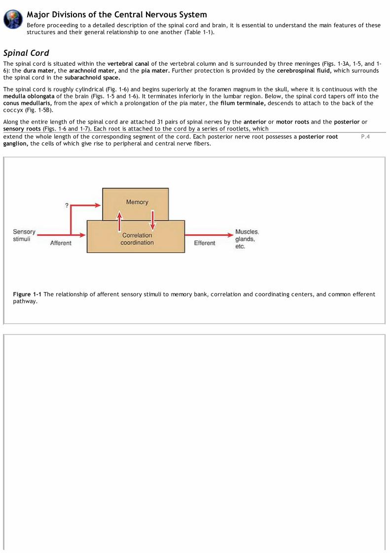
Because of the disproportionate growth in length of the vertebral column during development, compared with that of the spinal cord, thelength of the roots increases progressively from above downward (Fig. 1-15). In the upper cervical region, the spinal nerve roots are shortand run almost horizontally, but the roots of the lumbar and sacral nerves below the level of the termination of the cord (lower border ofthe first lumbar vertebra in the adult) form a vertical leash of nerves around the filum terminale (Fig. 1-16). Together, these lower nerveroots are called the cauda equina.
After emerging from the intervertebral foramen, each spinal nerve immediately divides into a large anterior ramus and a smaller posteriorramus, each containing both motor and sensory fibers. The posterior ramus passes posteriorly around the vertebral column to supply the

muscles and skin of the back. The anterior ramus continues anteriorly to supply the muscles and skin over the anterolateral body wall andall the muscles and skin of the limbs.
The anterior rami join one another at the root of the limbs to form complicated nerve plexuses (Fig. 1-2B). The cervical and brachialplexuses are found at the root of the upper limbs, and the lumbar and sacral plexuses are found at the root of the lower limbs.
Figure 1-15 Posterior view of the spinal cord showing the origins of the roots of the spinal nerves and their relationship to thedifferent vertebrae. On the right, the laminae have been removed to expose the right half of the spinal cord and the nerve roots.
GangliaGanglia may be divided into sensory ganglia of spinal nerves (posterior root ganglia) and cranial nerves and autonomic ganglia.
Sensory GangliaSensory ganglia are fusiform swellings (Fig. 1-6) situated on the posterior root of each spinal nerve just proximal to the root's junction witha corresponding anterior root. They are referred to as posterior root ganglia. Similar ganglia that are also found along the course ofcranial nerves V, VII, VIII, IX, and X are called sensory ganglia of these nerves.
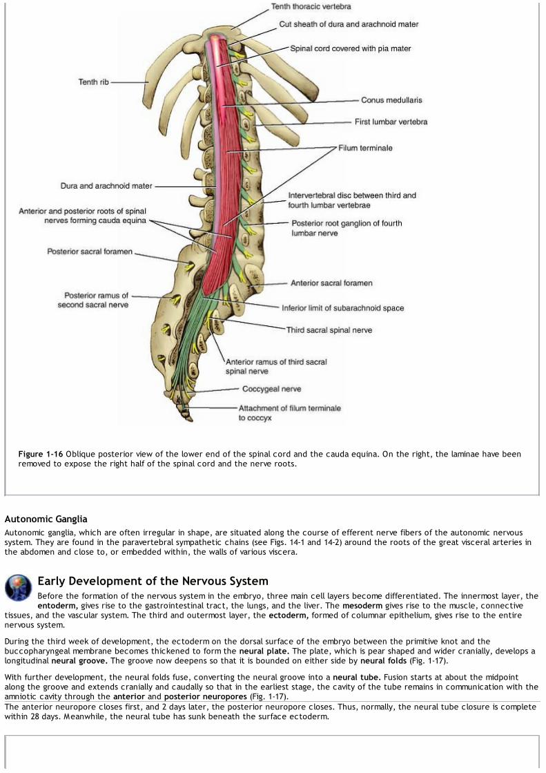
Figure 1-16 Oblique posterior view of the lower end of the spinal cord and the cauda equina. On the right, the laminae have beenremoved to expose the right half of the spinal cord and the nerve roots.
Autonomic GangliaAutonomic ganglia, which are often irregular in shape, are situated along the course of efferent nerve fibers of the autonomic nervoussystem. They are found in the paravertebral sympathetic chains (see Figs. 14-1 and 14-2) around the roots of the great visceral arteries inthe abdomen and close to, or embedded within, the walls of various viscera.
Early Development of the Nervous SystemBefore the formation of the nervous system in the embryo, three main cell layers become differentiated. The innermost layer, theentoderm, gives rise to the gastrointestinal tract, the lungs, and the liver. The mesoderm gives rise to the muscle, connective
tissues, and the vascular system. The third and outermost layer, the ectoderm, formed of columnar epithelium, gives rise to the entirenervous system.
During the third week of development, the ectoderm on the dorsal surface of the embryo between the primitive knot and thebuccopharyngeal membrane becomes thickened to form the neural plate. The plate, which is pear shaped and wider cranially, develops alongitudinal neural groove. The groove now deepens so that it is bounded on either side by neural folds (Fig. 1-17).
With further development, the neural folds fuse, converting the neural groove into a neural tube. Fusion starts at about the midpointalong the groove and extends cranially and caudally so that in the earliest stage, the cavity of the tube remains in communication with theamniotic cavity through the anterior and posterior neuropores (Fig. 1-17).The anterior neuropore closes first, and 2 days later, the posterior neuropore closes. Thus, normally, the neural tube closure is completewithin 28 days. Meanwhile, the neural tube has sunk beneath the surface ectoderm.

Figure 1-17 Formation of the neural plate, neural groove, and neural tube. The cells of the neural crest differentiate into the cellsof the posterior root ganglia, the sensory ganglia of cranial nerves, autonomic ganglia, neurilemmal cells (Schwann cells), the cellsof the suprarenal medulla, and melanocytes.
Table 1-2 The Primary Divisions of the Developing Brain
Pr imary Vesicle Pr imary Div ision Subdiv ision Adult Structures
Forebrainvesicle
Prosencephalon(forebrain) Telencephalon Cerebral hemisphere, basal ganglia,
hippocampus
Diencephalon Thalamus, hypothalamus, pineal body,infundibulum

Midbrainvesicle
Mesencephalon(midbrain)
Mesencephalon(midbrain) Tectum, tegmentum, crus cerebri
Hindbrainvesicle
Rhombencephalon(hindbrain)
MetencephalonMyelencephalon
Pons, cerebellumMedulla oblongata
During the invagination of the neural plate to form the neural groove, the cells forming the lateral margin of the plate do not becomeincorporated in the neural tube but instead form a strip of ectodermal cells that lie between the neuraltube and the covering ectoderm. This strip of ectoderm is called the neural crest (Fig. 1-17); subsequently, this group of cells will migrateventrolaterally on each side around the neural tube. Ultimately, the neural crest cells will differentiate into the cells of the posterior rootganglia, the sensory ganglia of the cranial nerves, autonomic ganglia, the cells of the suprarenal medulla, and the melanocytes. It is alsobelieved that these cells give rise to mesenchymal cells in the head and neck.
Figure 1-18 A: Expansion of the cephalic end of the neural tube to form the forebrain, midbrain, and hindbrain vesicles. B, C: Crosssection of the developing neural tube in the region of the spinal cord. The cells of the neuroepithelial layer have been widelyseparated for clarity.
Meanwhile, the proliferation of cells at the cephalic end of the neural tube causes it to dilate and form three primary brain vesicles: theforebrain vesicle, the midbrain vesicle, and the hindbrain vesicle (Fig. 1-18 and Table 1-2). The rest of the tube elongates and remainssmaller in diameter; it will form the spinal cord.
The subsequent differentiation of cells in the neural tube is brought about by the inductive interactions of one group of cells withanother. The inducing factors influence the control of the gene expression in the target cells. Ultimately, the simplest progenitor cell willdifferentiate into neurons and neuroglial cells. It is interesting to note that excessive numbers of neurons and neuroglial cells aredeveloped, and many (nearly half of the developing neurons) will be programmed to die by a process known as programmed cell death.Research into the identification of neurotrophic factors that promote the development and survival of neurons is of great importance, asthe results could possibly be applied to the problem of regeneration of the spinal cord neurons following trauma or the inhibition ofdegenerative diseases, such as Alzheimer disease.
The further development of the nervous system will be fully described in Chapter 18 following the description of the different parts of thenervous system and their neuronal connections.

P.18P.19P.20P.21P.22P.23P.24P.25P.26P.27
Table 1-3 Relationship of Spinal Cord Segments to Vertebral Numbers
Vertebrae Spinal Seg ment
Cervical vertebrae Add 1
Upper thoracic vertebrae Add 2
Lower thoracic vertebrae (7–9) Add 3
10th thoracic vertebra L1-2 cord segments
11th thoracic vertebra L3-4 cord segments
12th thoracic vertebra L5 cord segment
1st lumbar vertebra Sacral and coccygeal cord segments
Clinical NotesRelationship of Spinal Cord Segments to Vertebral NumbersBecause the spinal cord is shorter than the vertebral column, the spinal cord segments do not correspond
numerically with the vertebrae that lie at the same level (Fig. 1-15). The following table will help a physician determinewhich spinal segment is related to a given vertebral body (Table 1-3).On examination of a patient's back, one can see that the spinous processes lie approximately at the same level as thevertebral bodies. In the lower thoracic region, however, because of the length and extreme obliquity of the spinousprocesses, the tips of the spines lie at the level of the vertebral body below.
Injuries to the Spinal Cord and BrainThe spinal cord and brain are well protected. Both are suspended in fluid, the cerebrospinal fluid, and are surroundedby the bones of the vertebral column and skull (see Chapters 4 and 5). Unfortunately, if the forces of violence aresufficiently great, these protective structures can be overcome, with consequent damage to the delicate underlyingnervous tissue. Moreover, the cranial and spinal nerves and blood vessels are also likely to be injured.
Spinal Cord InjuriesThe degree of spinal cord injury at different vertebral levels is governed largely by anatomical factors. In the cervicalregion, dislocation or fracture dislocation is common, but the large size of the vertebral canal often prevents severeinjury to the spinal cord. However, when there is considerable displacement of the bones or bone fragments, the cordis sectioned. Respiration ceases if the cord is completely severed above the segmental origin of the phrenic nerves(C3-5), since the intercostal muscles and the diaphragm are paralyzed, and death occurs.In fracture dislocations of the thoracic region, displacement is often considerable, and because of the small size of thevertebral canal, severe injury to this region of the spinal cord results.In fracture dislocations of the lumbar region, two anatomical facts aid the patient. First, the spinal cord in the adultextends down only as far as the level of the lower border of the first lumbar vertebra (Fig. 1-16). Second, the largesize of the vertebral foramen in this region gives the roots of the cauda equina ample room. Nerve injury may,therefore, be minimal in this region.Injury to the spinal cord may produce partial or complete loss of function at the level of the lesion and partial orcomplete loss of function of afferent and efferent nerve tracts below the level of the lesion. The symptoms and signs ofsuch injuries are considered after the detailed structure of the spinal cord is discussed, and the ascending anddescending tracts are considered in Chapter 4.
Spinal Nerve InjuriesDisease and the Intervertebral ForaminaThe intervertebral foramina (Fig. 1-19) transmit the spinal nerves and the small segmental arteries and veins, all ofwhich are embedded in areolar tissue. Each foramen is bounded superiorly and inferiorly by the pedicles of adjacentvertebrae, anteriorly by the lower part of the vertebral body and by the intervertebral disc, and posteriorly by thearticular processes and the joint between them. In this situation, the spinal nerve is very vulnerable and may bepressed on or irritated by disease of the surrounding structures. Herniation of the intervertebral disc, fractures of thevertebral bodies, and osteoarthritis involving the joints of the articular processes or the joints between the vertebralbodies may all result in pressure, stretching, or edema of the emerging spinal nerve. Such pressure would give rise todermatomal pain, muscle weakness, and diminished or absent reflexes.

Herniated Intervertebral DiscsHerniation of the intervertebral discs occurs most commonly in those areas of the vertebral column where a mobile partjoins a relatively immobile part—for example, the cervicothoracic junction and the lumbosacral junction. In these areas,the posterior part of the anulus fibrosus of the disc ruptures, and the central nucleus pulposus is forced posteriorly liketoothpaste out of a tube. This herniation of the nucleus pulposus may result either in a central protrusion in themidline under the posterior longitudinal ligament of the vertebrae or in a lateral protrusion at the side of the posteriorligament close to the intervertebral foramen (Fig. 1-20).Cervical disc herniations are less common than in the lumbar region. The discs most susceptible to this condition arethose between the fifth and sixth and the sixth and seventh cervical vertebrae. Lateral protrusions cause pressure ona spinal nerve or its roots. Each spinal nerve emerges above the corresponding vertebra; thus, the protrusion of thedisc between the fifth and sixth cervical vertebrae may compress the C6 spinal nerve or its roots. Pain is felt near thelower part of the back of the neck and shoulder and along the area in the distribution of the spinal nerve involved.Central protrusions may press on the spinal cord and the anterior spinal artery and involve the various spinal tracts.Lumbar disc herniations are more common than cervical disc herniations (Fig. 1-20). The discs usually affected arethose between the fourth and fifth lumbar vertebrae and between the fifth lumbar vertebra and the sacrum. In thelumbar region, the roots of the cauda equina run posteriorly over a number of intervertebral discs (Fig. 1-20). A lateralherniation may press on one or two roots and often involves the nerve root going to the intervertebral foramen justbelow. The nucleus pulposus occasionally herniates directly backward, and if it is a large herniation, the whole caudaequina may be compressed, producing paraplegia.In lumbar disc herniations, pain is referred down the leg and foot in the distribution of the affected nerve. Because thesensory posterior roots most commonly pressed on are the fifth lumbar and first sacral, pain is usually felt down theback and lateral side of the leg, radiating to the sole of the foot. This condition is often called sciatica. In severe cases,paresthesia or actual sensory loss may occur.Pressure on the anterior motor roots causes muscle weakness. Involvement of the fifth lumbar motor root weakensdorsiflexion of the ankle, whereas pressure on the first sacral motor root causes weakness of plantar flexion. Theankle jerk reflex may be diminished or absent (Fig. 1-20).A large, centrally placed protrusion may give rise to bilateral pain and muscle weakness in both legs. Acute retention ofurine may also occur.

Figure 1-19 A: Joints in the cervical, thoracic, and lumbar regions of the vertebral column. B: Third lumbar vertebra seen fromabove showing the relationship between the intervertebral disc and the cauda equina. C: Sagittal section through three lumbarvertebrae showing the ligaments and the intervertebral discs. Note the relationship between the emerging spinal nerve in anintervertebral foramen and the intervertebral disc.
Figure 1-20 A, B: Posterior views of the vertebral bodies in the cervical and lumbar regions showing the relationship that mightexist between a herniated nucleus pulposus and spinal nerve roots. Note there are eight cervical spinal nerves and only sevencervical vertebrae. In the lumbar region, for example, the emerging L4 nerve roots pass out laterally close to the pedicle ofthe fourth lumbar vertebra and are not related to the intervertebral disc between the fourth and fifth lumbar vertebrae. C:Posterolateral herniation of the nucleus pulposus of the intervertebral disc between the fifth lumbar vertebra and the firstsacral vertebra showing pressure on the S1 nerve root. D: An intervertebral disc that has herniated its nucleus pulposusposteriorly. E: Pressure on the L5 motor nerve root produces weakness of dorsiflexion of the ankle; pressure on the S1 motornerve root produces weakness of plantar flexion of the ankle joint.

Spinal TapSpinal tap (lumbar puncture) may be performed to withdraw a sample of cerebrospinal fluid for microscopic orbacteriologic examination or to inject drugs to combat infection or induce anesthesia. Fortunately, the spinal cordterminates inferiorly at the level of the lower border of the first lumbar vertebra in the adult. (In the infant, it mayreach inferiorly to the third lumbar vertebra.) The subarachnoid space extends inferiorly as far as the lower border ofthe second sacral vertebra. The lower lumbar part of the vertebral canal is thus occupied by the subarachnoid space,which contains the lumbar and sacral nerve roots and the filum terminale (the cauda equina). A needle introduced intothe subarachnoid space in this region usually pushes the nerve roots to one side without causing damage.With the patient lying on his or her side or in the upright sitting position, with the vertebral column well flexed, thespace between adjoining laminae in the lumbar region is opened to a maximum (Fig. 1-21). An imaginary line joiningthe highest points on the iliac crests passes over the fourth lumbar spine. Using a careful aseptic technique and localanesthesia, the physician passes the lumbar puncture needle, fitted with a stylet, into the vertebral canal above orbelow the fourth lumbar spine. The needle will pass through the following anatomical structures before it enters thesubarachnoid space: (a) skin, (b) superficial fascia, (c) supraspinous ligament, (d) interspinous ligament, (e)ligamentum flavum, (f) areolar tissue containing the internal vertebral venous plexus, (g) dura mater, and (h)arachnoid mater. The depth to which the needle will have to pass will vary from 1 inch (2.5 cm) or less in a child to asmuch as 4 inches (10 cm) in an obese adult.
Figure 1-21 Sagittal section through the lumbar part of the vertebral column in a position of flexion. Note that the spines andlaminae are well separated in this position, allowing introduction of the spinal tap needle into the subarachnoid space.
As the stylet is withdrawn, a few drops of blood commonly escape. This usually indicates that the point of the needle is

situated in one of the veins of the internal vertebral plexus and has not yet reached the subarachnoid space. If theentering needle should stimulate one of the nerve roots of the cauda equina, the patient will experience a fleetingdiscomfort in one of the dermatomes or a muscle will twitch, depending on whether a sensory or a motor root wasimpaled.The cerebrospinal fluid pressure may be measured by attaching a manometer to the needle. When the patient is in therecumbent position, the normal pressure is about 60 to 150 mm of water. The pressure shows oscillationscorresponding to the movements of respiration and the arterial pulse.A block of the subarachnoid space in the vertebral canal, which may be caused by a tumor of the spinal cord or themeninges, may be detected by compressing the internal jugular veins in the neck. This raises the cerebral venouspressure and inhibits the absorption of cerebrospinal fluid in the arachnoid granulations, thus producing an increase inthe manometer reading of the cerebrospinal fluid pressure. If this rise fails to occur, the subarachnoid space is blocked,and the patient is said to exhibit a positive Queckenstedt sign.
Caudal AnesthesiaAnesthetic solutions may be injected into the sacral canal through the sacral hiatus. The solutions pass upward in theloose connective tissue and bathe the spinal nerves as they emerge from the dural sheath (Fig. 1-22). Obstetriciansuse this method of nerve block to relieve the pains of the first and second stages of labor. The advantage is that whenanesthetic is administered by this method, the infant is not affected. Caudal anesthesia may also be used inoperations in the sacral region, including anorectal surgery.
Head InjuriesA blow to the head may cause the scalp to be merely bruised; severe blows may cause the scalp to be torn or split.Even if the head is protected by a crash helmet, the brain may be severely damaged without clinical evidence of scalpinjury.
Figure 1-22 Posterior view of the sacrum. Laminae have been removed to show the sacral nerve roots lying within the sacralcanal.
Fractures of the SkullSevere blows to the head often result in the skull changing shape at the point of impact. Small objects may penetratethe skull and produce local laceration of the brain. Larger objects applied with great force may shatter the skull, and

fragments of bone are driven into the brain at the site of impact.In the adult, fractures of the skull are common, but in the young child, they are less common. In the infant, the skullbones are more resilient than in the adult, and they are separated by fibrous sutural ligaments. In the adult, the innertable of the skull is particularly brittle. Moreover, the sutural ligaments begin to ossify during middle age.The type of fracture that occurs in the skull will depend on the age of the patient, the severity of the blow, and thearea of the skull receiving the trauma. The adult skull may be likened to an eggshell, because it possesses a certainlimited resilience beyond which it splinters. A severe, localized blow will produce a local indentation, often accompaniedby splintering of the bone. Blows to the vault often result in a series of linear fractures, which radiate out through thethin areas of the bone. The petrous parts of the temporal bones and the occipital crests (see p. 193) strongly reinforcethe base of the skull and tend to deflect linear fractures.The young child's skull may be likened to a table tennis ball, because a localized blow produces a depression withoutsplintering. This common type of circumscribed lesion is referred to as a “pond” fracture.
Brain InjuriesBrain injuries are produced by displacement and distortion of the neuronal tissues at the moment of impact (Fig. 1-23).The brain, which is incompressible, may be likened to a water-soaked log suspended in water. The brain is floating inthe cerebrospinal fluid in the subarachnoid space and is capable of a certain amount of anteroposterior and lateralgliding movement. The anteroposterior movement is limited by the attachment of the superior cerebral veins to thesuperior sagittal sinus. Lateral displacement of the brain is limited by the falx cerebri. The tentorium cerebelli and thefalx cerebelli also restrict displacement of the brain.From these anatomical facts, it follows that blows on the front or back of the head lead to displacement of the brain,which may produce severe cerebral damage, stretching and distortion of the brainstem, and stretching and eventearing of the commissures of the brain. Blows to the side of the head produce less cerebral displacement, and theinjuries to the brain consequently tend to be less severe. It should be noted, however, that the falx cerebri is a toughstructure and may cause considerable damage to the softer brain tissue in cases where there has been a severe blowto the side of the head (Fig. 1-23). Furthermore, it is important to remember that glancing blows to the head maycause considerable rotation of the brain, with shearing strains and distortion of the brain, particularly in areas wherefurther rotation is prevented by bony prominences in the anterior and middle cranial fossae. Brain lacerations are verylikely to occur when the brain is forcibly thrown against the sharp edges of bone within the skull (see p. 193)—thelesser wings of the sphenoid, for example.
Figure 1-23 A: Mechanisms of acute cerebral injury when a blow is applied to the lateral side of the head. B: Varieties of

intracranial hemorrhage. C: Mechanism of cerebral trauma following a blow on the chin. The movement of the brain within theskull can also tear the cerebral veins.
When the brain is suddenly given momentum within the skull, the part of the brain that moves away from the skull wallis subjected to diminished pressure because the cerebrospinal fluid has not had time to accommodate to the brainmovement. This results in a suction effect on the brain surface, with rupture of surface blood vessels.A sudden severe blow to the head, as in an automobile accident, may result in damage to the brain at two sites: atthe point of impact and at the pole of the brain opposite the point of impact, where the brain is thrown against theskull wall. This is referred to as contrecoup injury.The movement of the brain within the skull at the time of head injuries not only is likely to cause avulsion of cranialnerves but commonly leads to rupture of tethering blood vessels. Fortunately, the large arteries found at the base ofthe brain are tortuous, and this, coupled with their strength, explains why they are rarely torn. The thin-walled corticalveins, which drain into the large dural venous sinuses, are very vulnerable and can produce severe subdural orsubarachnoid hemorrhage (Fig. 1-23).
Traumatic Brain Injury following an Explosion or BlastSoldiers deployed to Afghanistan and Iraq are frequently exposed to explosive devices, which may result in extensiveinjuries to the limbs, eyes, and ears. Open injuries to the skull, where shrapnel has entered the brain, are clearlyvisible and are dealt with accordingly.However, in closed injuries, where the skull remains intact, the underlying brain may be damaged but left untreated. Inthese cases, the explosion produces a blast of air that strikes the skull and shakes up the brain, resulting in multipleinjuries to the soft brain tissue as it is driven against the hard bony projections within the skull. The symptoms andsigns will depend on the extent of the neurologic damage and will be mild, moderate, or severe. While the moderateand severe cases are quickly recognized by medical personnel, it is the mild cases that may be missed and may laterdevelop headaches, nausea, mood changes, and memory loss. Since studies of these patients have shown that mildneurologic damage can be successfully treated, early diagnosis is imperative. Individuals who have been exposed toexplosive devices should receive a computed tomography (CT) scan or magnetic resonance imaging (MRI) beforereturning to civilian life.
Intracranial HemorrhageAlthough the brain is cushioned by the surrounding cerebrospinal fluid in the subarachnoid space, any severehemorrhage within the relatively rigid skull will ultimately exert pressure on the brain.Intracranial hemorrhage may result from trauma or cerebral vascular lesions (Fig. 1-21). Four varieties are consideredhere: (1) epidural, (2) subdural, (3) subarachnoid, and (4) cerebral.Epidural (extradural) hemorrhage results from injuries to the meningeal arteries or veins (see p. 432). The anteriordivision of the middle meningeal artery is the common artery to be damaged. A comparatively minor blow to the side ofthe head, resulting in fracture of the skull in the region of the anterior inferior portion of the parietal bone, may severthe artery (Fig. 1-23). Arterial or venous injury is especially likely to occur if the vessels enter a bony canal in thisregion. Bleeding occurs and strips the meningeal layer of dura from the internal surface of the skull. The intracranialpressure rises, and the enlarging blood clot exerts local pressure on the underlying precentral gyrus (motor area).Blood may also pass laterally through the fracture line to form a soft swelling on the side of the head. To stop thehemorrhage, the torn artery must be ligated or plugged. The burr hole through the skull wall should be placed about1-1/2 inches (4 cm) above the midpoint of the zygomatic arch.Subdural hemorrhage results from tearing of the superior cerebral veins where they enter the superior sagittal sinus(see Figs. 15-1 and 17-5). The cause is usually a blow to the front or back of the head, resulting in excessiveanteroposterior displacement of the brain within the skull. This condition, which is much more common than middlemeningeal hemorrhage, can be produced by a sudden minor blow. Once the vein is torn, blood under low pressurebegins to accumulate in the potential space between the dura and the arachnoid. In a few patients, the condition isbilateral.Acute and chronic forms of the clinical condition occur, depending on the speed of accumulation of fluid in the subduralspace. For example, if the patient starts to vomit, the venous pressure will rise as the result of a rise in theintrathoracic pressure. Under these circumstances, the subdural blood clot will rapidly increase in size and produceacute symptoms. In the chronic form, over a course of several months, the small blood clot will attract fluid by osmosis,in which case a hemorrhagic cyst forms and gradually expands and produces pressure symptoms. In both forms, theblood clot must be removed through burr holes in the skull.Subarachnoid hemorrhage results from nontraumatic leakage or rupture of a congenital aneurysm on the cerebralarterial circle (circle of Willis) or, less commonly, from an arteriovenous malformation. The symptoms, which are suddenin onset, will include severe headache, stiffness of the neck, and loss of consciousness. The diagnosis is established byperforming CT or MRI or by withdrawing heavily blood-stained cerebrospinal fluid through a lumbar puncture.With regard to cerebral hemorrhage, spontaneous intracerebral hemorrhage (Fig. 1-23) is most common in patientswith hypertension. It is generally due to rupture of the thin-walled lenticulostriate artery (see Fig. 17-11), a branch ofthe middle cerebral artery (Fig. 17-4). The hemorrhage involves important descending nerve fibers in the internalcapsule and produces hemiplegia on the opposite side of the body. The patient immediately loses consciousness, andthe paralysis is evident when consciousness is regained. The diagnosis is established by performing brain CT or MRI.
The Shaken-Baby SyndromeInflicted head injury is the most common cause of traumatic death in infancy. It is believed that sudden deceleration,which occurs when an infant is held by the arms or trunk and shaken or the head is forcefully struck against a hardsurface, is responsible for the brain injuries. Biomechanical studies have shown that the rotation of the floating brainabout its center of gravity causes diffuse brain injuries, including diffuse axonal injury and subdural hematoma. Inshaken-baby syndrome, major rotational forces have to occur that clearly exceed those encountered in normal childplay activities.Most cases of shaken-baby syndrome take place during the first year of life, and they are usually restricted to infantsunder 3 years of age. Common symptoms include lethargy, irritability, seizures, altered muscle tone, and symptoms

indicating raised intracranial pressure, such as impaired consciousness, vomiting, breathing abnormalities, and apnea.In severe cases, the baby may be unresponsive, the fontanelles are bulging, and the child may have retinalhemorrhages. Spinal tap may reveal blood in the cerebrospinal fluid. Subdural or subarachnoid hemorrhages can bereadily detected on CT or MRI scans. Autopsy findings commonly include localized subdural hemorrhage in the parietal-occipital region and subarachnoid blood, associated with massive cerebral swelling and widespread neuronal loss.
Space-Occupying Lesions within the SkullSpace-occupying or expanding lesions within the skull include tumor, hematoma, and abscess. Since the skull is a rigidcontainer of fixed volume, these lesions will add to the normal bulk of the intracranial contents.An expanding lesion is first accommodated by the expulsion of cerebrospinal fluid from the cranial cavity. Later, theveins become compressed, interference with the circulation of blood and cerebrospinal fluid begins, and the intracranialpressure starts to rise. The venous congestion results in increased production and diminished absorption ofcerebrospinal fluid, the volume of the cerebrospinal fluid begins to rise, and thus, a vicious circle is established.The position of the tumor within the brain may have a dramatic effect on the signs and symptoms. For example, atumor that obstructs the outflow of cerebrospinal fluid or directly presses on the great veins will cause a rapid increasein intracranial pressure. The signs and symptoms that enable the physician to localize the lesion will depend on theinterference with the brain function and the degree of destruction of the nervous tissue produced by the lesion.Severe headache, possibly due to the stretching of the dura mater, and vomiting, due to pressure on the brainstem,are common complaints.
Figure 1-24 Sudden displacement of the cerebral hemispheres through the tentorial notch into the posterior cranial fossafollowing a lumbar puncture; the cerebral tumor is situated in the right cerebral hemisphere. CT or MRI should be used ratherthan lumbar puncture when investigating a cerebral tumor.
A spinal tap should not be performed in patients with suspected intracranial tumor. The withdrawal of cerebrospinalfluid may lead to a sudden displacement of the cerebral hemisphere through the notch in the tentorium cerebelli intothe posterior cranial fossa (Fig. 1-24) or herniation of the medulla oblongata and cerebellum through the foramenmagnum. CT scans or MRIs are used in making the diagnosis.
Computed TomographyCT is used for the detection of intracranial lesions. The procedure is quick, safe, and accurate. The total dose ofirradiation is no greater than for a conventional skull radiograph.CT relies on the same physics as conventional x-rays, in that structures are distinguished from one another by theirability to absorb energy from x-rays. The x-ray tube emits a narrow beam of radiation as it passes in a series ofscanning movements through an arc of 180 degrees around the patient's head. The x-rays having passed through thehead are collected by a special x-ray detector. The information is fed to a computer that processes the information,which is then displayed as a reconstructed picture on a televisionlike screen. Essentially, the observer sees an imageof a thin slice through the head, which may then be photographed for later examination (Fig. 1-25).The sensitivity is such that small differences in x-ray absorption can be easily displayed. The gray matter of thecerebral cortex, white matter, internal capsule, corpus callosum, ventricles, and subarachnoid spaces can all berecognized. An iodine-containing medium can be injected intravascularly, which enhances greatly the contrast betweentissues having a different blood flow.Since a CT scan can be performed in 5 to 10 minutes, it is the method of choice in an emergency situation with patientswith head trauma or suspected intracranial hemorrhage.
Magnetic Resonance ImagingThe technique of MRI uses the magnetic properties of the hydrogen nucleus excited by radiofrequency radiation

transmitted by a coil surrounding the head. The excited hydrogen nuclei emit a signal that is detected as inducedelectric currents in a receiver coil. MRI is absolutely safe to the patient, and because it provides better differentiationbetween gray and white matter, MRI can be more revealing than CT. The reason for this is that gray matter containsmore hydrogen in the form of water than does white matter, and the hydrogen atoms are less bound in fat (Fig. 1-26).MRI is the best imaging method for detecting low-contrast lesions such as brain tumors or small multiple sclerosisplaques. It is also capable of showing clear images of the brain stem, cerebellum, and the pituitary fossa, which in thecase of a CT scan are overshadowed by the dense bones of the base of the skull. The spinal cord structure is muchmore clearly visualized with MRI.Unfortunately, an MRI takes longer and costs two-thirds more than a CT scan.
Positron Emission TomographyPositron emission tomography (PET) uses radioactive isotopes that decay with the emission of positively chargedelectrons (positrons) to map the biochemical, physiologic, and pharmacologic processes taking place in the brain.The appropriate isotope is incorporated into molecules of known biochemical behavior in the brain and then is injectedinto the patient. The metabolic activity of the compound can then be studied by making cross-sectional tomographicimages of the brain using the same principles as in CT (Fig. 1-27). By making a series of time-lapse images at differentanatomical sites, it is possible to study the variations in brain metabolism at these sites. This technique has been usedto study the distribution and activity of neurotransmitters, the variations in oxygen utilization, and cerebral blood flow.

Figure 1-25 CT scan showing the structure of the brain. A, B: Horizontal cuts (axial sections).

Figure 1-26 MRI showing the structure of the brain. A: Sagittal. B: Coronal. Compare with Figure 1-25. Note the betterdifferentiation between gray and white matter.
PET has been successfully used in the evaluation of patients with brain tumors (Figs. 1-28 and 1-29), movementdisorders, seizures, and schizophrenia.
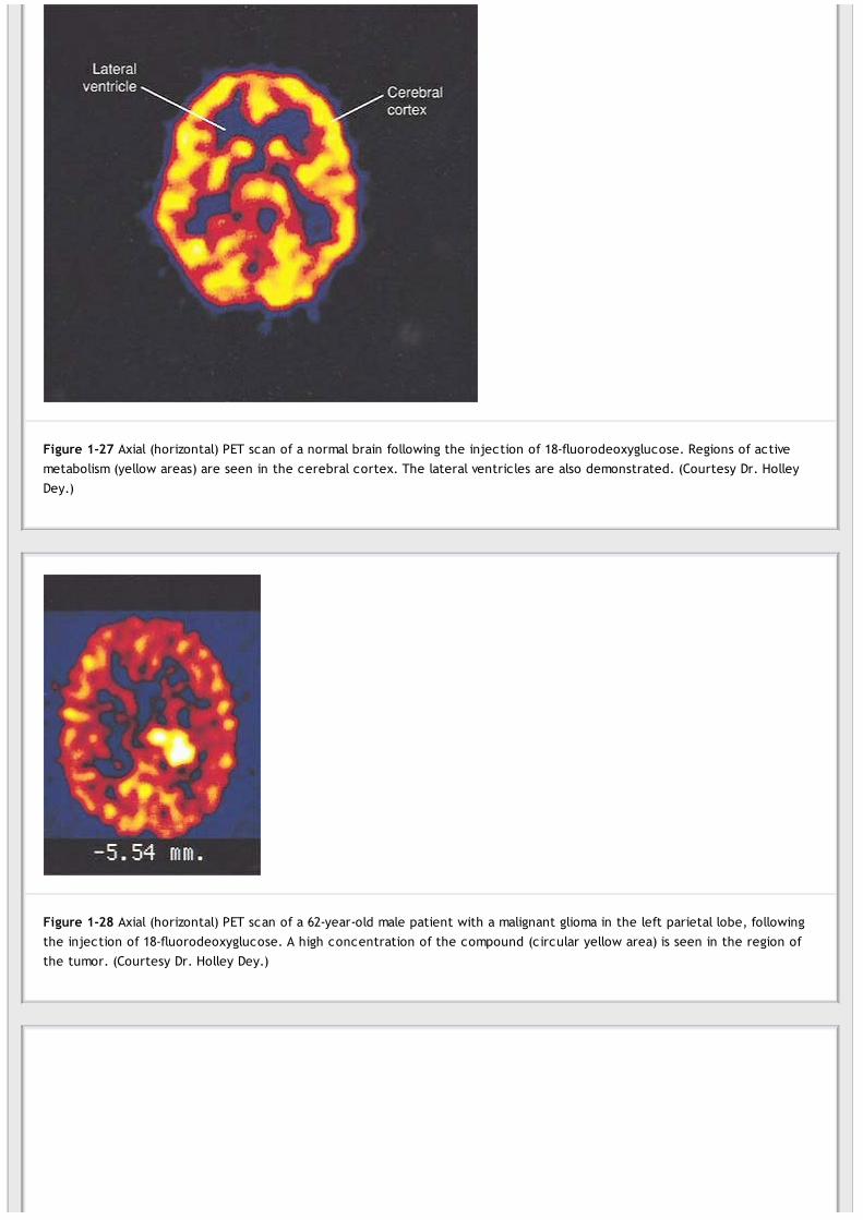
Figure 1-27 Axial (horizontal) PET scan of a normal brain following the injection of 18-fluorodeoxyglucose. Regions of activemetabolism (yellow areas) are seen in the cerebral cortex. The lateral ventricles are also demonstrated. (Courtesy Dr. HolleyDey.)
Figure 1-28 Axial (horizontal) PET scan of a 62-year-old male patient with a malignant glioma in the left parietal lobe, followingthe injection of 18-fluorodeoxyglucose. A high concentration of the compound (circular yellow area) is seen in the region ofthe tumor. (Courtesy Dr. Holley Dey.)

P.29
Figure 1-29 Coronal PET scan of a 62-year-old male patient with a malignant glioma in the left parietal lobe, following theinjection of 18-fluorodeoxyglucose (same patient as in Fig. 1-26). A high concentration of the compound (circular yellow area)is seen in the region of the tumor. (Courtesy Dr. Holley Dey.)
Clinical Problem Solving1. A 45-year-old woman was examined by her physician and found to have carcinoma of the thyroid gland. Apart fromthe swelling in the neck, the patient also complained of back pain in the lower thoracic region, with a burning sorenessradiating around the right side of her thorax over the 10th intercostal space. Although the back pain was oftenrelieved by changing posture, it was worsened by coughing and sneezing. A lateral radiograph of the thoracic part ofthe vertebral column revealed a secondary carcinomatous deposit in the 10th thoracic vertebral body. Further physicalexamination revealed muscular weakness of both legs. Using your knowledge of neuroanatomy, explain the following:(a) the pain in the back, (b) the soreness over the right 10th intercostal space, (c) the muscular weakness of bothlegs, and (d) which segments of the spinal cord lie at the level of the 10th thoracic vertebral body.View Answer
2. A 35-year-old coal miner was crouching down at the mine face to inspect a drilling machine. A large rock suddenlybecame dislodged from the roof of the mine shaft and struck the miner on the upper part of his back. Examination by aphysician showed an obvious forward displacement of the upper thoracic spines on the eighth thoracic spine. Whatanatomical factors in the thoracic region determine the degree of injury that may occur to the spinal cord?View Answer
3. A 20-year-old man with a long history of tuberculosis of the lungs was examined by an orthopedic surgeon becauseof the sudden development of a humpback (kyphosis). He also had symptoms of a stabbing pain radiating around bothsides of his thorax intensified by coughing or sneezing. A diagnosis of tuberculous osteitis of the fifth thoracic vertebrawas made, with the collapse of the vertebral body responsible for the kyphosis. Using your knowledge ofneuroanatomy, explain why the collapse of the fifth thoracic vertebral body should produce pain in the distribution ofthe fifth thoracic segmental nerve on both sides.View Answer
4. A 50-year-old man woke up one morning with a severe pain near the lower part of the back of the neck and leftshoulder. The pain was also referred along the outer side of the left upper arm. Movement of the neck caused anincrease in the intensity of the pain, which was also accentuated by coughing. A lateral radiograph of the neck showeda slight narrowing of the space between the fifth and sixth cervical vertebral bodies. An MRI showed disruption of theintervertebral disc between the fifth and sixth cervical vertebrae. Using your knowledge of anatomy, state which nerveroot was involved. Also, state the nature of the disease.View Answer
5. A medical student offered to help a fellow student straighten out the bumper of his foreign sports car. He had justfinished his course in neuroanatomy and was in poor physical shape. Undaunted, he attempted to lift the end of thebumper while his friend stood on the other end. Suddenly, he felt an acute pain in the back that extended down theback and outer side of his right leg. Later, he was examined by an orthopedic surgeon, who found that the pain wasaccentuated by coughing. A lateral radiograph of the lumbar vertebral column revealed nothing abnormal. An MRI,taken in the sagittal plane, showed a small posterior prolapse of the nucleus pulposus in the disc between the fifthlumbar and the first sacral vertebrae. A diagnosis of herniation of the intervertebral disc between the fifth lumbar andfirst sacral vertebrae was made. Using your knowledge of neuroanatomy, explain the symptoms of this disease. Whichspinal nerve roots were pressed on?View Answer

P.31
6. A 5-year-old child was seen in the emergency department, and a diagnosis of acute meningitis was made. Theresident decided to perform a lumbar puncture in order to confirm the diagnosis. Using your knowledge ofneuroanatomy, where would you perform a lumbar puncture? Name, in order, the structures pierced when a lumbarpuncture needle is introduced into the subarachnoid space.View Answer
7. A pregnant young woman told her friends that she hated the idea of going through the pain of childbirth but thatshe equally detested the thought of having a general anesthetic. Is there a specialized local analgesic technique thatwill provide painless labor?View Answer
8. While crossing the road, a pedestrian was struck on the right side of his head by a passing car. He fell to the groundbut did not lose consciousness. After resting for an hour and then getting up, he appeared to be confused andirritable. Later, he staggered and fell to the floor. On questioning, he was seen to be drowsy, and twitching of thelower left half of his face and left arm was noted. A diagnosis of epidural hemorrhage was made. Which artery is likelyto have been damaged? What is causing the drowsiness and muscle twitching?View Answer
9. A 45-year-old woman was examined by a neurologist and found to have an intracranial tumor. She complained ofsevere headaches, which occurred during the night and early morning. She described the pain as “bursting” in nature,and although at first, 6 months ago, the headaches were intermittent, they were now more or less continuous.Coughing, stooping, and straining at stool made the pain worse. The pain was accompanied by vomiting on threerecent occasions. What is the sequence of events that occurs within the skull as the intracranial pressure rises? Wouldyou perform a routine lumbar puncture on every patient you suspected of having an intracranial tumor?View Answer
10. While examining an unconscious 18-year-old man admitted to the emergency room following a motorcycle accident,the neurosurgeon asked the attending medical student what happens to the brain in an accident in which it issuddenly decelerated within the skull. What is the value of wearing a crash helmet?View Answer
Review QuestionsDirections: Each of the incomplete statements in this section is followed by completions of the statement. Selectthe ONE lettered completion that is BEST in each case.1. The spinal cord has
(a) an outer covering of gray matter and an inner core of white matter.(b) an enlargement below that forms the conus medullaris.(c) anterior and posterior roots of a single spinal nerve attached to a single segment.(d) cells in the posterior gray horn that give rise to efferent fibers that supply skeletal muscles.(e) a central canal that is situated in the white commissure.
View Answer
2. The medulla oblongata has(a) a tubular shape.(b) the fourth ventricle lying posterior to its lower part.(c) the midbrain directly continuous with its upper border.(d) no central canal in its lower part.(e) the spinal cord directly continuous with its lower end in the foramen magnum.
View Answer
3. The midbrain has(a) a cavity called the cerebral aqueduct.(b) a large size.(c) no cerebrospinal fluid around it.(d) a cavity that opens above into the lateral ventricle.(e) a location in the middle cranial fossa of the skull.
View Answer
Directions: Each of the numbered items in this section is followed by answers. Select the ONE lettered answerthat is CORRECT.4. The following statements concern the cerebellum:
(a) It lies within the middle cranial fossa.(b) The cerebellar cortex is composed of white matter.(c) The vermis is the name given to that part joining the cerebellar hemispheres together.

(d) The cerebellum lies anterior to the fourth ventricle.(e) The dentate nucleus is a mass of white matter found in each cerebellar hemisphere.
View Answer
5. The following statements concern the cerebrum:(a) The cerebral hemispheres are separated by a fibrous septum called the tentorium cerebelli.(b) The bones of the vault of the skull are named for the lobes of the cerebral hemisphere over which they lie.(c) The corpus callosum is a mass of gray matter lying within each cerebral hemisphere.(d) The internal capsule is an important collection of nerve fibers, which has the caudate nucleus and thethalamus on its medial side and the lentiform nucleus on its lateral side.(e) The cavity present within each cerebral hemisphere is called the cerebral ventricle.
View Answer
6. The following statements concern the peripheral nervous system:(a) There are ten pairs of cranial nerves.(b) There are eight pairs of cervical spinal nerves.(c) The posterior root of a spinal nerve contains many efferent motor nerve fibers.(d) A spinal nerve is formed by the union of an anterior and a posterior ramus in an intervertebral foramen.(e) A posterior root ganglion contains the cell bodies of autonomic nerve fibers leaving the spinal cord.
View Answer
7. The following statements concern the central nervous system:(a) A CT brain scan cannot distinguish between white matter and gray matter.(b) The lateral ventricles are in direct communication with the fourth ventricle.(c) An MRI of the brain uses the magnetic properties of the hydrogen nucleus excited by radiofrequency radiationtransmitted by a coil surrounding the patient's head.(d) Following trauma and sudden movement of the brain within the skull, the large arteries at the base of thebrain are commonly torn.(e) The movement of the brain at the time of head injuries is unlikely to damage the small sixth cranial nerve.
View Answer
8. The following statements concern the cerebrospinal fluid:(a) The cerebrospinal fluid in the central canal of the spinal cord is unable to enter the fourth ventricle.(b) With the patient in the recumbent position, the normal pressure is about 60 to 150 mm of water.(c) It plays only a minor role in the protection of the brain and spinal cord from traumatic injury.(d) Compression of the internal jugular veins in the neck lowers the cerebrospinal fluid pressure.(e) The subdural space is filled with cerebrospinal fluid.
View Answer
9. The following statements concern the vertebral levels and the spinal cord segmental levels:(a) The first lumbar vertebra lies opposite the L3-4 segments of the cord.(b) The third thoracic vertebra lies opposite the third thoracic spinal cord segment.(c) The fifth cervical vertebra lies opposite the seventh cervical spinal cord segment.(d) The eighth thoracic vertebra lies opposite the ninth thoracic spinal cord segment.(e) The third cervical vertebra lies opposite the fourth cervical spinal cord segment.
View Answer
Directions: Each case history is followed by questions. Select the ONE BEST lettered answer.A 23-year-old woman was unconscious when admitted to the emergency department. While crossing the road, shehad been hit on the side of the head by a bus. Within an hour, she was found to have a large doughlike swelling overthe right temporal region. She also had signs of muscular paralysis on the left side of the body. A lateral radiograph ofthe skull showed a fracture line running downward and forward across the anterior inferior angle of the right parietalbone. Her coma deepened, and she died 5 hours after the accident.10. Select the most likely cause of the swelling over the right temporal region in this patient.
(a) Superficial bruising of the skin(b) Hemorrhage from a blood vessel in the temporalis muscle(c) Rupture of the right middle meningeal vessels(d) Edema of the skin(e) Hemorrhage from a blood vessel in the superficial fascia
View Answer
11. Select the most likely cause of the muscular paralysis of the left side of the body in this patient.(a) Laceration of the right side of the cerebral hemisphere

(b) Right-sided epidural hemorrhage(c) Left-sided epidural hemorrhage(d) Injury to the cerebral cortex on the left side of the brain(e) Injury to the right cerebellar hemisphere
View Answer
A 69-year-old man was admitted to the neurology unit complaining of severe discomfort of the lower back. Radiologicexamination of the lumbar region of the vertebral column revealed significant narrowing of the spinal canal caused byadvanced osteoarthritis.12. Explain the discomfort in the lower back experienced by this patient.
(a) Muscle fatigue(b) Prolapsed intervertebral disc(c) Torn ligament in the joints of the lumbar region of the spine(d) Compression of the cauda equina(e) Bad posture
View Answer
Later, in this same patient, the back pain became more severe and now radiated down the back of the left leg; thepatient was also experiencing difficulty walking. Examination of the patient revealed weakness and some wasting ofthe muscles of the left leg. Radiologic examination showed that the osteoarthritic changes had spread to involve theboundaries of many of the lumbar intervertebral foramina.13. Explain the change in the symptoms and signs found in this patient.
(a) The sciatic nerve was compressed in the pelvis by a spreading rectal cancer.(b) The patient had developed advanced atherosclerosis of the arteries of the right lower limb.(c) The osteoarthritic process had produced osteophytes that encroached on the intervertebral foramina,compressing the segmental spinal nerve roots.(d) Neuritis had developed in the sciatic nerve trunk.(e) The patient was experiencing psychiatric problems.
View Answer
Additional Reading
American Academy of Neurology Therapeutics Subcommittee. Positron emission tomography. Neurology 41:163, 1991.
Becker, D. P., Gudeman, S. K. Textbook of Head Injury. Philadelphia: Saunders, 1989.
Brooks, D. J. PET: Its clinical role in neurology. J. Neurol. Neurosurg. Psychiatry 54:1, 1991.
Duhaime, A. C., Christian, C. W., Rorke, L. B., et al. Nonaccidental head injury in infants—The “shaken-baby syndrome.” N. Engl. J.Med. 338:1822–1829, 1998.
Goetz, C. G. Textbook of Clinical Neurology (2nd ed.). Philadelphia: Saunders, 2003.
Rowland, L. P. (ed.). Merritt's Neurology (10th ed.). Philadelphia: Lippincott Williams & Wilkins, 2000.
Snell, R. S. Clinical Anatomy by Systems. Philadelphia: Lippincott Williams & Wilkins, 2007.
Snell, R. S. Clinical Anatomy by Regions (8th ed.). Philadelphia: Lippincott Williams & Wilkins, 2008.
Standing, S. (ed). Gray's Anatomy (39th Br. ed.). London: Elsevier Churchill Livingstone, 2005.

Authors: Snell, Richard S.Title: Clinical Neuroanatomy, 7th Edition
Copyright ©2010 Lippincott Williams & Wilkins
> Table of Contents > Chapter 2 - The Neur obiology of the Neur on and the Neur oglia
Chapter 2
The Neurobiology of the Neuron and the Neuroglia
A 38-year-old man with a history of involuntary movements, personality changes, and mental deterioration was referred to a neurologist.The symptoms started insidiously 8 years ago and have gotten progressively worse. The first symptoms were involuntary, abrupt, andpurposeless movements of the upper limbs associated with clumsiness and dropping of objects. At presentation, the patient had difficultywalking, speaking, and swallowing. Associated with these movement defects were an impairment of memory and loss of intellectualcapacity. Impulsive behavior and bouts of depression also occurred. Close questioning of the patient and his wife revealed that thepatient's father and his older brother had had similar symptoms before they died. A diagnosis of Huntington disease was made.
Huntington disease is an autosomal dominant disorder with the defect localized to the short arm of chromosome 4. Histologically, thecaudate nucleus and the putamen show extensive degeneration, mainly involving the acetylcholine and gamma-aminobutyric acid (GABA)-producing neurons; the dopamine neurons are unaffected. There is also secondary degeneration of the cerebral cortex. This case is anexample of a hereditary disorder that mainly involves a particular group of neurons.
Chapter Objectives
To define the neuron and name its processesTo learn the varieties of neurons and identify them in the different parts of the nervous systemTo review the cell biology of a neuron and understand the function of a nerve cell and its processesTo review the structure of the plasma membrane as it is related to its physiologyTo learn the transport of materials from the cell body to the axon terminalsTo understand the structure and function of synapses and neurotransmittersTo review the supporting function of the neuroglial cells for nerve cells and the possible role that they play inneuronal metabolism, function, and neuronal death
The purpose of this chapter is to prepare the student to understand how the basic excitable cell—the neuron—communicates with otherneurons. It also considers certain injuries to the neuron and the effects of drugs on the mechanism by which neurons communicate withone another.
Definition of a NeuronNeuron is the name given to the nerve cell and all its processes (Fig. 2-1). Neurons are excitable cells that are specialized for thereception of stimuli and the conduction of the nerve impulse. They vary considerably in size and shape, but each possesses a cell
body from whose surface project one or more processes called neurites (Fig. 2-2). Those neurites responsible for receiving informationand conducting it toward the cell body are called dendrites. The single long tubular neurite that conducts impulses away from the cellbody is called the axon. The dendrites and axons are often referred to as nerve fibers.
Neurons are found in the brain and spinal cord and in ganglia. Unlike most other cells in the body, normal neurons in the mature individualdo not undergo division and replication.
Varieties of NeuronsAlthough the cell body of a neuron may be as small as 5 µm or as large as 135 µm in diameter, the processes or neurites mayextend over a distance of more than 1 m. The number, length, and mode of branching of the neurites provide a morphologic
method for classifying neurons.
Unipolar neurons are those in which the cell body has a single neurite that divides a short distance from the cell body into two branches,one proceeding to some peripheral structure and the other entering the central nervous system (Fig. 2-3). The branches of this singleneurite have the structural and functional characteristics of an axon. In this type of neuron, the fine terminal branches found at theperipheral end of the axon at the receptor site are often referred to as the dendrites. Examples of this form of neuron are found in theposterior root ganglion.
Bipolar neurons possess an elongated cell body, from each end of which a single neurite emerges (Fig. 2-3). Examples of this type ofneuron are found in the retinal bipolar cells and the cells of the sensory cochlear and vestibular ganglia.
Multipolar neurons have a number of neurites arising from the cell body (Fig. 2-3). With the exception of the long process, the axon, theremainder of the neurites are dendrites. Most neurons of the brain and spinal cord are of this type.
Neurons may also be classified according to size:
Golgi type I neurons have a long axon that may be 1 m or more in length in extreme cases (Figs. 2-4, 2-5, and 2-6). The axons of theseneurons form the long fiber tracts of the brain and spinal cord and the nerve fibers of peripheral nerves. The pyramidal cells of the

cerebral cortex, the Purkinje cells of the cerebellar cortex, and the motor cells of the spinal cord are good examples.
Golgi type II neurons have a short axon that terminates in the neighborhood of the cell body or is entirely absent (Figs. 2-5 and 2-6).They greatly outnumber the Golgi type I neurons. The short dendrites that arise from these neurons give them a star-shapedappearance. These neurons are numerous in the cerebral and cerebellar cortex and are often inhibitory in function. Table 2-1summarizes the classification of neurons.
Structure of the Neuron
Nerve Cell BodyThe nerve cell body, like that of other cells, consists essentially of a mass of cytoplasm in which a nucleus is embedded (Figs. 2-7 and 2-8),bounded externally by a plasma membrane. It is interesting to note that the volume of cytoplasm within the nerve cell body is often farless than the total volume of cytoplasm in the neurites. The cell bodies of the small granular cells of the cerebellar cortex measure about5 µm in diameter, whereas those of the large anterior horn cells may measure as much as 135 µm in diameter.
Nucleus
The nucleus, which stores the genes, is commonly centrally located within the cell body and is typically large and rounded.1 In matureneurons, the chromosomes no longerduplicate themselves and function only in gene expression. The chromosomes are, therefore, not arranged as compact structures butexist in an uncoiled state. Thus, the nucleus is pale, and the fine chromatin granules are widely dispersed (Figs. 2-6 and 2-7). There isusually a single prominent nucleolus, which is concerned with ribosomal ribonucleic acid (rRNA) synthesis and ribosome subunit assembly.The large size of the nucleolus probably is due to the high rate of protein synthesis, which is necessary to maintain the protein level inthe large cytoplasmic volume that is present in the long neurites as well as in the cell body.
In the female, one of the two X chromosomes is compact and is known as the Barr body. It is composed of sex chromatin and is situated atthe inner surface of the nuclear envelope.
The nuclear envelope (Figs. 2-8 and 2-9) can be regarded as a special portion of the rough endoplasmic reticulum of the cytoplasm and iscontinuous with the endoplasmic reticulum of the cytoplasm. The envelope is double layered and possesses fine nuclear pores, throughwhich materials can diffuse into and out of the nucleus (Fig. 2-8). The nucleoplasm and the cytoplasm can be considered as functionallycontinuous. Newly formed ribosomal subunits can be passed into the cytoplasm through the nuclear pores.

P.37P.38
Figure 2-1 A neuron.
Figure 2-2 Photomicrograph of a smear preparation of the spinal cord showing a neuron with its cell body and its processes orneurites.
CytoplasmThe cytoplasm is rich in granular and agranular endoplasmic reticulum (Figs. 2-9 and 2-10) and contains the following organelles andinclusions: (a) Nissl substance; (b) the Golgi complex; (c) mitochondria; (d) microfilaments; (e) microtubules; (f) lysosomes; (g) centrioles;and (h) lipofuscin, melanin, glycogen, and lipid.
Nissl substance consists of granules that are distributed throughout the cytoplasm of the cell body, except for the region close to theaxon, called the axon hillock (Fig. 2-11). The granular material also extends into the proximal parts of the dendrites; it is not present inthe axon.
Electron micrographs show that the Nissl substance is composed of rough-surfaced endoplasmic reticulum (Fig. 2-12) arranged in the formof broad cisternae stacked one on top of the other. Although many of the ribosomes are attached to the surface of the endoplasmicreticulum, many more lie free in the intervals between the cisternae.Since the ribosomes contain RNA, the Nissl substance is basophilic and can be well demonstrated by staining withtoluidine blue or other basic aniline dyes (Fig. 2-11) and using the light microscope.

Figure 2-3 The classification of neurons according to the number, length, and mode of branching of the neurites.
Figure 2-4 Photomicrograph of a silver-stained section of the cerebellar cortex showing two Purkinje cells. These are examples ofGolgi type I neurons.

Figure 2-5 Photomicrograph of a silver-stained section of the cerebral cortex. Note the presence of large pyramidal cells, whichare examples of Golgi type I neurons, and numerous Golgi type II neurons.

Figure 2-6 Different types of neurons.
Table 2-1 Classification of Neurons
Morpholog icClassification Arrang ement of Neur ites Location
Number, Length, and Mode of Branching of Neurites
Unipolar Single neurite divides a shortdistance from cell body Posterior root ganglion
Bipolar Single neurite emerges from eitherend of cell body Retina, sensory cochlea, and vestibular ganglia
Multipolar Many dendrites and one long axon Fiber tracts of brain and spinal cord, peripheral nerves, andmotor cells of spinal cord
Size of Neuron
Golgi type I Single long axon Fiber tracts of brain and spinal cord, peripheral nerves, andmotor cells of spinal cord
Golgi type II Short axon that with dendritesresembles a star Cerebral and cerebellar cortex
The Nissl substance is responsible for synthesizing protein, which flows along the dendrites and the axon and replaces the proteins thatare broken down during cellular activity. Fatigue or neuronal damage causes the Nissl substance to move and become concentrated at theperiphery of the cytoplasm. This phenomenon, which gives the impression that the Nissl substance has disappeared, is known aschromatolysis.
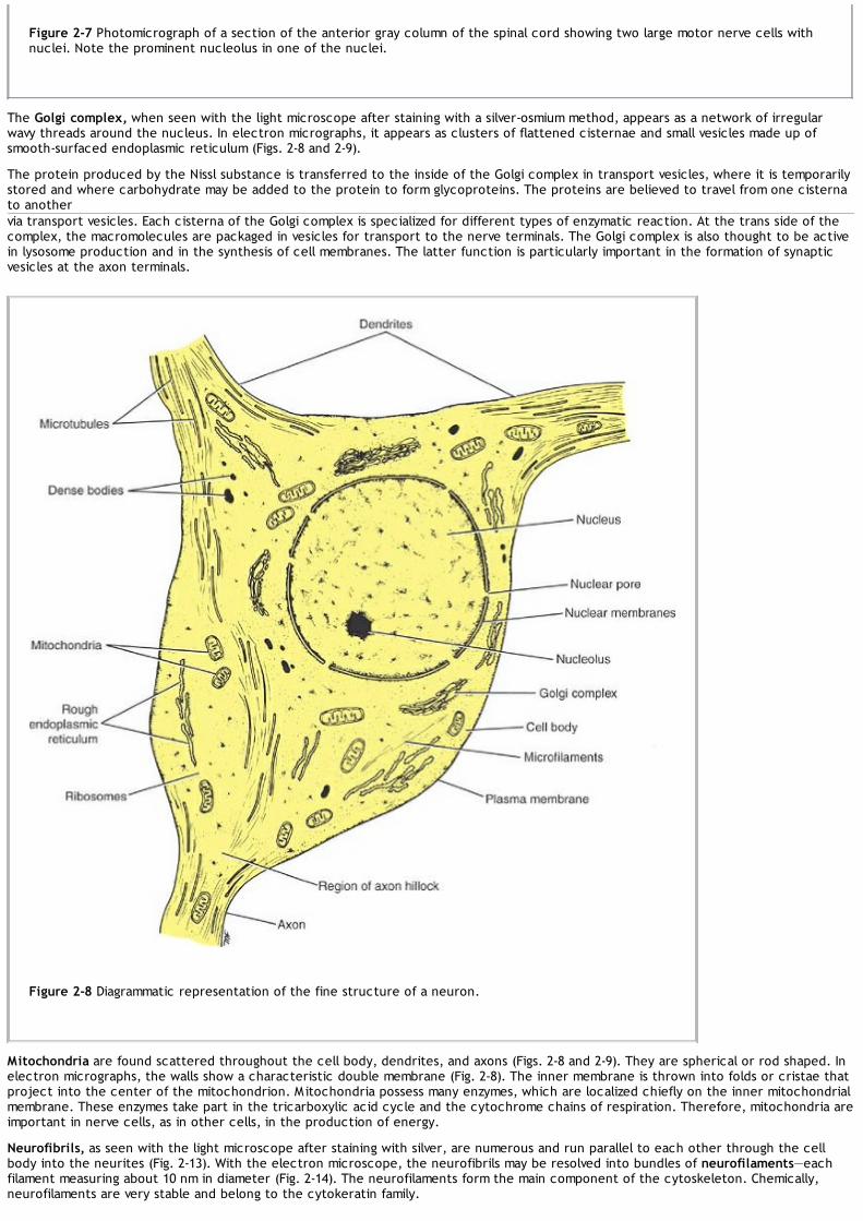
Figure 2-7 Photomicrograph of a section of the anterior gray column of the spinal cord showing two large motor nerve cells withnuclei. Note the prominent nucleolus in one of the nuclei.
The Golgi complex, when seen with the light microscope after staining with a silver-osmium method, appears as a network of irregularwavy threads around the nucleus. In electron micrographs, it appears as clusters of flattened cisternae and small vesicles made up ofsmooth-surfaced endoplasmic reticulum (Figs. 2-8 and 2-9).
The protein produced by the Nissl substance is transferred to the inside of the Golgi complex in transport vesicles, where it is temporarilystored and where carbohydrate may be added to the protein to form glycoproteins. The proteins are believed to travel from one cisternato anothervia transport vesicles. Each cisterna of the Golgi complex is specialized for different types of enzymatic reaction. At the trans side of thecomplex, the macromolecules are packaged in vesicles for transport to the nerve terminals. The Golgi complex is also thought to be activein lysosome production and in the synthesis of cell membranes. The latter function is particularly important in the formation of synapticvesicles at the axon terminals.
Figure 2-8 Diagrammatic representation of the fine structure of a neuron.
Mitochondria are found scattered throughout the cell body, dendrites, and axons (Figs. 2-8 and 2-9). They are spherical or rod shaped. Inelectron micrographs, the walls show a characteristic double membrane (Fig. 2-8). The inner membrane is thrown into folds or cristae thatproject into the center of the mitochondrion. M itochondria possess many enzymes, which are localized chiefly on the inner mitochondrialmembrane. These enzymes take part in the tricarboxylic acid cycle and the cytochrome chains of respiration. Therefore, mitochondria areimportant in nerve cells, as in other cells, in the production of energy.
Neurofibrils, as seen with the light microscope after staining with silver, are numerous and run parallel to each other through the cellbody into the neurites (Fig. 2-13). With the electron microscope, the neurofibrils may be resolved into bundles of neurofilaments—eachfilament measuring about 10 nm in diameter (Fig. 2-14). The neurofilaments form the main component of the cytoskeleton. Chemically,neurofilaments are very stable and belong to the cytokeratin family.

P.41
Microfilaments measure about 3 to 5 nm in diameter and are formed of actin. M icrofilaments are concentrated at the periphery of thecytoplasm just beneath the plasma membrane where they form a dense network. Together with microtubules, microfilaments play a keyrole in the formation of new cell processes and the retraction of old ones. They also assist the microtubules in axon transport.
Microtubules are revealed with the electron microscope and are similar to those seen in other types of cells. They measure about 25 nmin diameter and are foundinterspersed among the neurofilaments (Fig. 2-14). They extend throughout the cell body and its processes. In the axon, all themicrotubules are arranged in parallel, with one end pointing to the cell body and the other end pointing distally away from the cell body.
Figure 2-9 Electron micrograph of a neuron showing the structure of the nucleus and a number of cytoplasmic organelles.(Courtesy Dr. J. M. Kerns.)

Figure 2-10 Electron micrograph of a neuron showing nuclear and plasma membranes and cytoplasmic organelles. (Courtesy Dr. J.M. Kerns.)
Figure 2-11 Photomicrograph of a section of the anterior gray column of the spinal cord stained with toluidine blue. Note thepresence of dark-staining Nissl substance in the cytoplasm of four neurons.

The microtubules and the microfilaments provide a stationary track that permits specific organelles to move by molecular motors. Thestop-and-start movement is caused by the periodic dissociation of the organelles from the track or the collision with other structures.
Figure 2-12 Electron micrograph of the cytoplasm of two neurons showing the structure of Nissl bodies (substance). (Courtesy Dr.J. M. Kerns.)
Cell transport involves the movement of membrane organelles, secretory material, synaptic precursor membranes, large dense corevesicles, mitochondria, and smooth endoplasmic reticulum.
Cell transport can take place in both directions in the cell body and its processes. It is of two kinds: rapid (100 to 400 mm per day) andslow (0.1 to 3.0 mm per day).
Rapid transport (100 to 400 mm per day) is brought about by two motor proteins associated with the microtubule adenosine triphosphate(ATP)-ase sites; these arekinesin for anterograde (away from the cell) movement and dynein for retrograde movement. It is believed that in anterograde movement,kinesin-coated organelles are moved toward one end of the tubule, and that in retrograde movement, dynein-coated organelles are movedtoward the other end of the tubule. The direction and speed of the movement of an organelle can be brought about by the activation ofone of the motor proteins or of both of the motor proteins simultaneously.
Slow transport (0.1 to 3.0 mm per day) involves the bulk movement of the cytoplasm and includes the movement of mitochondria and otherorganelles. Slow axonal transport occurs only in the anterograde direction. The molecular motor has not been identified but is probablyone of the kinesin family.
Lysosomes are membrane-bound vesicles measuring about 8 nm in diameter. They serve the cell by acting as intracellular scavengers andcontain hydrolytic enzymes. They are formed by the budding off of the Golgi apparatus. Lysosomes exist in three forms: (1) primarylysosomes, which have just been formed; (2) secondary lysosomes, which contain partially digested material (myelin figures); and (3)residual bodies, in which the enzymes are inactive and the bodies have evolved from digestible materials such as pigment and lipid.

Figure 2-13 Photomicrograph of a silver-stained section of a neuron showing the presence of large numbers of neurofibrils in thecytoplasm of the cell body and the neurites.
Centrioles are small, paired structures found in immature dividing nerve cells. Each centriole is a hollow cylinder whose wall is made up ofbundles of microtubules. They are associated with the formation of the spindle during cell division and in the formation of microtubules.Centrioles are also found in mature nerve cells, where they are believed to be involved in the maintenance of microtubules.
Lipofuscin (pigment material) occurs as yellowish-brown granules within the cytoplasm (Fig. 2-15). It is believed to be formed as the resultof lysosomal activity, and it represents a harmless metabolic by-product. Lipofuscin accumulates with age.
Melanin granules are found in the cytoplasm of cells in certain parts of the brain (e.g., the substantia nigra of the midbrain). Theirpresence may be related to the catecholamine-synthesizing ability of these neurons, whose neurotransmitter is dopamine.
The main structures present in a nerve cell body are summarized in Table 2-2.
Plasma MembraneThe plasma membrane forms the continuous external boundary of the cell body and its processes, and in the neuron, it is the site for theinitiation and conduction of the nerve impulse (Figs. 2-10 and 2-14). The membrane is about 8 nm thick, which is too thin to be seen withthe light microscope. When viewed under the electron microscope, the plasma membrane appears as two dark lines with a light linebetween them.
The plasma membrane is composed of an inner and an outer layer of very loosely arranged protein molecules, each layer being about 2.5nm thick, separated by a middle layer of lipid about 3 nm thick. The lipid layer is made up of two rows of phospholipid molecules arrangedso that their hydrophobic ends are in contact with each other and their polar ends are in contact with the protein layers. Certain proteinmolecules lie within the phospholipid layer and span the entire width of the lipid layer. These molecules provide the membrane withhydrophilic channels through which inorganic ions may enter and leave the cell. Carbohydrate molecules are attached to the outside ofthe plasma membrane and are linked to the proteins or the lipids, forming what is known as the cell coat, or glycocalyx.
The plasma membrane and the cell coat together form a semipermeable membrane that allows diffusion of certain ions through it butrestricts others. In the resting state (unstimulated state), the K+ ions diffuse through the plasma membrane from the cell cytoplasm to thetissue fluid (Fig. 2-16). The permeability of the membrane to K+ ions is much greater than that to the Na+ ions; thus, the passive efflux of K+
is much greater than the influx of Na+. This results in a steady potential difference of about -80 mV, which can be measured across theplasma membrane since the inside of the membrane is negative with respect to the outside. This potential is known as the restingpotential.

Figure 2-14 Electron micrograph of dendrites showing the presence of neurofilaments and microtubules within their cytoplasm.(Courtesy Dr. J. M. Kerns.) A: Longitudinal section of two adjacent dendrites. B: Transverse section of a dendrite.

Figure 2-15 Photomicrograph of a longitudinal section of a posterior root ganglion showing the presence of lipofuscin granuleswithin the cytoplasm of sensory neurons.
Table 2-2 The Main Structures in a Nerve Cell Body
Structure Shape Appearance Location Function
Nucleus Large,rounded
Pale, chromatinwidely scattered;single prominentnucleolus; Barrbody present infemale
Centrally placed, displaced toperiphery in cell injury Controls cell activity
Cytoplasmic organelles
Nisslsubstance
Granules ofroughendoplasmicreticulum
Broad cisternae;ribosomes arebasophilic
Throughout cytoplasm andproximal part of dendrites,absent from axon hillock andaxon, fatigue and injury resultin concentration at periphery
Synthesizes protein
Golgi complex
Wavythreads;clusters offlattenedcisternaeand smallvesicles
Smoothendoplasmicreticulum
Close to the nucleus
Adds carbohydrate toprotein molecule;packages products fortransport to nerveterminals; forms cellmembranes
M itochondria Spherical,rod shaped
Double membranewith cristae Scattered Form chemical energy
Neurofibrils Linear fibrils
Run parallel toeach other;composedbundles ofmicrofilaments,each 10 nm indiameter
Run from dendrites throughcell body to axon
Determines the shapeof the neuron
M icrofilaments Linear fibrils Filaments 3–5 nmin diameter
Form a dense networkbeneath the plasma membrane
Role in formation andretraction of cellprocesses and in celltransport
M icrotubules Lineartubes
Run betweenneurofibrils, 25nm in diameter
Run from dendrites throughcell body to axon Cell transport
Lysosomes Vesicles
8 nm in diameter;three forms:primary,secondary, andresidual bodies
Throughout cell Cell scavengers
CentriolesPairedhollow
Wall made up ofbundles of Confined to cytoplasm of cell Take part in cell
division; maintain

cylinders microtubules body microtubules
Lipofuscin Granules Yellowish brown Scattered through cytoplasm Metabolic by-product
Melanin Granules Yellowish brown Substantia nigra of midbrain Related to formation ofdopa
Excitation of the Plasma Membrane of the Nerve Cell Body
When the nerve cell is excited (stimulated) by electrical, mechanical, or chemical means, a rapid change in membrane permeability to Na+
ions takes place, and Na+ ions diffuse through the plasma membrane into the cell cytoplasm from the tissue fluid (Fig. 2-16). This results inthe membrane becoming progressively depolarized. The sudden influx of Na+ ions followed by the altered polarity produces the so-calledaction potential, which is approximately +40 mV. This potential is very brief, lasting about5 msec. The increased membrane permeability for Na+ ions quickly ceases, and membrane permeability for K+ ions increases. Therefore,the K+ ions start to flow from the cell cytoplasm and return the localized area of the cell to the resting state.
Once generated, the action potential spreads over the plasma membrane, away from the site of initiation, and is conducted along neuritesas the nerve impulse. This impulse is self-propagated, and its size and frequency do not alter (Fig. 2-16). Once the nerve impulse hasspread over a given region of plasma membrane, another action potential cannot be elicited immediately. The duration of this nonexcitablestate is referred to as the refractory period, and it controls the maximum frequency that the action potentials can be conducted alongthe plasma membrane (see p. 46).
The greater the strength of the initial stimulus, the larger the initial depolarization and the greater will be the spread into the surroundingareas of the plasma membrane. Should multiple excitatory stimuli be applied to the surface of a neuron, then the effect can be summated.For example, subthreshold stimuli may pass over the surface of the cell body and be summated at the origin of the axon and so initiate anaction potential.

Figure 2-16 Ionic and electrical changes that occur in a neuron when it is stimulated.
Inhibitory stimuli are believed to produce their effect by causing an influx of Cl- ions through the plasma membrane into the neuron, thusproducing hyperpolarization and reducing the excitatory state of the cell (Fig. 2-17).
Sodium and Potassium ChannelsThe sodium and potassium channels, through which the sodium and potassium ions diffuse through the plasma membrane, are formed ofthe protein molecules that extend through the full thickness of the plasma membrane(Fig. 2-18). Why a particular channel permits the passage of K+ ions while excluding Na+ ions is difficult to explain. The selectivity cannot bedue to the diameter of the ions, since the K+ ion is larger than the Na+ ion. However, the movement of ions in solution depends not onlyon the size of the ion but also on the size of the shell of water surrounding it. K+ ions have weaker electric fields than Na+ ions; thus, K+
ions attract less water than Na+ ions. Therefore, K+ ions behave as if they are smaller than Na+ ions. This physicochemical explanation doesnot entirely account for why a channel is selective. It is possible that the channels have narrow regions along their length that act assieves or molecular filters. The ions may also participate in electrostatic interactions with the amino acid residues lining the walls of thechannel.

Figure 2-17 Ionic and electrical changes that occur in a neuron during hyperpolarization.
Figure 2-18 Ionic permeability of the plasma membrane. Diagram shows the interactions of the ions with water, the membrane lipidbilayer, and the ion channels.
The ion channel proteins are relatively stable, but they exist in at least two conformational states, which represent an open functionalstate and a closed functional state. The mechanism responsible for the opening and closing of a channel is not understood but may belikened to a gate that is opened and closed. Gating may involve the twisting and distortion of the channel, thus creating a wider ornarrower lumen. Gating appears to occur in response to such stimuli as voltage change, the presence of a ligand, or stretch or pressure.
In the nonstimulated state, the gates of the potassium channels are open wider than those of the sodium channels, which are nearlyclosed. This permits the potassium ions to diffuse out of the cell cytoplasm more readily than the sodium ions can diffuse in. In thestimulated state, the gates of the sodium channels are at first wide open; then, the gates of the potassium channels are opened, and thegates of the sodium channels are nearly closed again. It is the opening and closing of the sodium and potassium channels that is thought toproduce the depolarization and repolarization of the plasma membrane.
The absolute refractory period, which occurs at the onset of the action potential when a second stimulus is unable to produce a furtherelectrical change, is thought tobe due to the inability to get the sodium channels open. During the relative refractory period, when a very strong stimulus can producean action potential, presumably the sodium channels are opened.
The Nerve Cell ProcessesThe processes of a nerve cell, often called neurites, may be divided into dendrites and an axon.

The dendrites are the short processes of the cell body (Fig. 2-19). Their diameter tapers as they extend from the cell body, and theyoften branch profusely. In many neurons, the finer branches bear large numbers of small projections called dendritic spines. Thecytoplasm of the dendrites closely resembles that of the cell body and contains Nissl granules, mitochondria, microtubules, microfilaments,ribosomes, and agranular endoplasmic reticulum. Dendrites should be regarded merely as extensions of the cell body to increase thesurface area for the reception of axons from other neurons. Essentially, they conduct the nerve impulse toward the cell body.
Figure 2-19 A: Light photomicrograph of a motor neuron in the anterior gray column of the spinal cord showing the nerve cellbody, two dendrites, and the surrounding neuropil. B: Electron micrograph of a dendrite showing axodendritic synapses. (CourtesyDr. J. M. Kerns.)
During early embryonic development, there is an overproduction of dendrites. Later, they are reduced in number and size in response toaltered functional demand from afferent axons. There is evidence that dendrites remain plastic throughout life and elongate and branchor contract in response to afferent activity.
Axon is the name given to the longest process of the cell body. It arises from a small conical elevation on the cell body, devoid of Nisslgranules, called the axon hillock (Figs. 2-8 and 2-20). Occasionally, an axon arises from the proximal part of a dendrite. An axon is tubularand is uniform in diameter; it tends to have a smooth surface.
Axons usually do not branch close to the cell body; collateral branches may occur along their length. Shortly before their termination,axons commonly branch profusely. The distal ends of the terminal branches of the axons are often enlarged; they are called terminals(Fig. 2-21). Someaxons (especially those of autonomic nerves) near their termination show a series of swellings resembling a string of beads; these swellingsare called varicosities.
Axons may be very short (0.1 mm), as seen in many neurons of the central nervous system, or extremely long (3.0 m), as seen when theyextend from a peripheral receptor in the skin of the toe to the spinal cord and thence to the brain.
The diameter of axons varies considerably with different neurons. Those of larger diameter conduct impulses rapidly, and those of smallerdiameter conduct impulses very slowly.
The plasma membrane bounding the axon is called the axolemma. The cytoplasm of the axon is termed the axoplasm. Axoplasm differsfrom the cytoplasm of the cell body in possessing no Nissl granules or Golgi complex. The sites for the production of protein, namely RNAand ribosomes, are absent. Thus, axonal survival depends on the transport of substances from the cell bodies.
The initial segment of the axon is the first 50 to 100 µm after it leaves the axon hillock of the nerve cell body (Fig. 2-20). This is the mostexcitable part of the axon and is the site at which an action potential originates. It is important to remember that under normalconditions, an action potential does not originate on the plasma membrane of the cell body but, instead, always at the initial segment.

Figure 2-20 Electron micrograph of a longitudinal section of a neuron from the cerebral cortex showing the detailed structure ofthe region of the axon hillock and the initial segment of the axon. Note the absence of Nissl substance (rough endoplasmicreticulum) in the axon hillock and the presence of numerous microtubules in the axoplasm. Note also the axon terminals (arrows)forming axoaxonal synapses with the initial segment of the axon. (Courtesy Dr. A. Peters.)
An axon always conducts impulses away from the cell body. The axons of sensory posterior root ganglion cells are an exception; here, thelong neurite, which is indistinguishable from an axon, carries the impulse toward the cell body. (See unipolar neurons, p. 36.)
Axon TransportMaterials are transported from the cell body to the axon terminals (anterograde transport) and to a lesser extent in the oppositedirection (retrograde transport).
Fast anterograde transport of 100 to 400 mm per day refers to the transport of proteins and transmitter substances or their precursors.Slow anterograde transport of 0.1 to 3.0 mm per day refers to the transport of axoplasm and includes the microfilaments andmicrotubules.

Figure 2-21 Electron micrograph showing multiple axodendritic synapses. Note the presence of large numbers of presynapticvesicles within the axons. The definition has come to include the site at which a neuron comes into close proximity with a skeletalmuscle cell and functional communication occurs. (Courtesy Dr. J. M. Kerns.)
Retrograde transport explains how the cell bodies of nerve cells respond to changes in the distal end of the axons. For example, activatedgrowth factor receptors can be carried along the axon to their site of action in the nucleus. Pinocytotic vesicles arising at the axonterminals can be quickly returned to the cell body. Worn-out organelles can be returned to the cell body for breakdown by thelysosomes.
Axon transport is brought about by microtubules assisted by the microfilaments.
SynapsesThe nervous system consists of a large number of neurons that are linked together to form functional conducting pathways. Where twoneurons come into close proximity and functional interneuronal communication occurs, the site of such communication is referred to as asynapse2 (Fig. 2-22). Most neurons may make synaptic connections to a 1,000 or more other neurons and may receive up to 10,000connections from other neurons. Communication at a synapse, under physiologic conditions, takes place in one direction only. Synapsesoccur in a number of forms (Fig. 2-22). The most common type is that which occurs between an axon of one neuron and the dendrite orcell body of the second neuron. As the axon approaches the synapse, it may have a terminal expansion (bouton terminal), or it may have aseries of expansions (bouton de passage), each of which makes synaptic contact. In other types of synapses, the axon synapses on theinitial segment of another axon—that is, proximal to where the myelin sheath begins—or there may be synapses between terminalexpansions from different neurons. Depending on the site of the synapse, they are often referred to as axodendritic, axosomatic, oraxoaxonic (Fig. 2-22).
The manner in which an axon terminates varies considerably in different parts of the nervous system. For example, a single axon mayterminate on a single neuron, or a single axon may synapse with multiple neurons, as in the case of the parallel fibers of the cerebellarcortex synapsing with multiple Purkinje cells. In the same way, a single neuron may have synaptic junctions with axons of many differentneurons. The arrangement of these synapses will determine the means by which a neuron can be stimulated or inhibited. Synaptic spines,extensions of the surface of a neuron, form receptive sites for synaptic contact with afferent boutons (Fig. 2-22).
Synapses are of two types: chemical and electrical. Most synapses are chemical, in which a chemical substance,the neurotransmitter, passes across the narrow space between the cells and becomes attached to a protein molecule in the postsynapticmembrane called the receptor.

Figure 2-22 A–D. Different types of chemical synapses.
In most chemical synapses, several neurotransmitters may be present. One neurotransmitter is usually the principal activator and actsdirectly on the postsynaptic membrane, while the other transmitters function as modulators and modify the activity of the principaltransmitter.
Chemical Synapses
Ultrastructure of Chemical SynapsesOn examination with an electron microscope, synapses are seen to be areas of structural specialization (Figs. 2-21 and 2-23). The apposedsurfaces of the terminal axonal expansion and the neuron are called the presynaptic and postsynaptic membranes, respectively, and theyare separated by a synaptic cleft measuring about 20 to 30 nm wide. The presynaptic and postsynaptic membranes are thickened, and theadjacent underlying cytoplasm shows increased density. On the presynaptic side, the dense cytoplasm is broken up into groups; on thepostsynaptic side, the density often extends into a subsynaptic web. Presynaptic vesicles, mitochondria, and occasional lysosomes arepresent in the cytoplasm close to the presynaptic membrane (Fig. 2-23). On the postsynaptic side, the cytoplasm often contains parallelcisternae. The synaptic cleft contains polysaccharides.

Figure 2-23 High-power electron micrograph of axodendritic synapses showing the thickening of the cell membranes at thesynaptic sites, presynaptic vesicles, and the presence of mitochondria within the axons near their termination. (Courtesy Dr. J. M.Kerns.)
The presynaptic terminal contains many small presynaptic vesicles that contain the molecules of the neurotransmitter(s). The vesicles fusewith the presynaptic membrane and discharge the neurotransmitter(s) into the synaptic cleft by a process of exocytosis (Fig. 2-24).
When synapses are first formed in the embryo, they are recognized as small zones of density separated by a synaptic cleft. Later, theymature into well-differentiated structures. The presence of simple, undifferentiated synapses in the postnatal nervous system has led tothe suggestion that synapses can be developed as required and possibly undergo atrophy when redundant. This plasticity of synapses maybe of great importance in the process of learning and in the development and maintenance of memory.
Neurotransmitters at Chemical SynapsesThe presynaptic vesicles and the mitochondria play a key role in the release of neurotransmitter substances at synapses. The vesiclescontain the neurotransmitter substance that is released into the synaptic cleft; the mitochondria provide adenosine triphosphate (ATP)for the synthesis of new transmitter substance.
Most neurons produce and release only one principal transmitter at all their nerve endings. For example, acetylcholine is widely used as atransmitter by different neurons in the central and peripheral parts of the nervous system, whereas dopamine is released by neurons inthe substantia nigra. Glycine, another transmitter, is found principally in synapses in the spinal cord.
The following chemical substances act as neurotransmitters, and there are many more: acetylcholine (ACh), norepinephrine, epinephrine,dopamine, glycine, serotonin, gamma-aminobutyric acid (GABA), enkephalins, substance P, and glutamic acid.
It should be noted that all skeletal neuromuscular junctions use only acetylcholine as the transmitter, whereas synapses between neuronsuse a large number of different transmitters.
Action of NeurotransmittersAll neurotransmitters are released from their nerve endings by the arrival of the nerve impulse (action potential). This results in an influx ofcalcium ions, which causes the synaptic vesicles to fuse with the presynaptic membrane. The neurotransmitters are then ejected into theextracellular fluid in the synaptic cleft. Once in the cleft, they diffuse across the gap to the postsynaptic membrane. There they achievetheir objective by raising or lowering the resting potential of the postsynaptic membrane for a brief period of time.

Figure 2-24 Release of neurotransmitters. A: Acetylcholine. B: Catecholamines.
Table 2-3 Examples of Principal (Classic) Neurotransmitters and Neuromodulators at Synapses
Neuromediatorsa Function ReceptorMechanism Ionic Mechanism Location
Principal Neurotransmitters
Acetylcholine (nicotinic), L-glutamate Rapid excitation Ion channel
receptorsOpens cation channel (fastEPSP)
Main sensoryand motorsystems
GABA Rapid inhibition Opens anion channel for Cl-
(fast IPSP)
Neuromodulators
Acetylcholine (muscarinic),serotonin, histamine,adenosine
Modulation andmodification ofactivity
G-protein–coupledreceptors
Opens or closes K+ or Ca2+
channels (slow IPSP and slowEPSP)
Systems thatcontrolhomeostasis
GABA, gamma-aminobutyric acid; EPSP, excitatory postsynaptic potential; IPSP, inhibitory postsynaptic potential.

aNote that these are only a few examples of an ever-increasing number of known neuromediators.
The receptor proteins on the postsynaptic membrane bind the transmitter substance and undergo an immediate conformational changethat opens the ion channel, generating an immediate but brief excitatory postsynaptic potential (EPSP) or an inhibitory postsynapticpotential (IPSP). The rapid excitation is seen with acetylcholine (nicotinic) and L-glutamate, or the inhibition is seen with GABA (Table 2-3).Other receptor proteins bind the transmitter substance and activate a second messenger system, usually through a molecular transducer,a G-protein. These receptors have a longer latent period, and the duration of the response may last several minutes or longer.Acetylcholine (muscarinic), serotonin, histamine, neuropeptides, and adenosine are good examples of this type of transmitter, which isoften referred to as a neuromodulator (see next section).
The excitatory and the inhibitory effects on the postsynaptic membrane of the neuron will depend on the summation of the postsynapticresponses at the different synapses. If the overall effect is one of depolarization, the neuron will be excited, an action potential will beinitiated at the initial segment of the axon, and a nerve impulse will travel along the axon. If, on the other hand, the overall effect is oneof hyperpolarization, the neuron will be inhibited and no nerve impulse will arise.
Distribution and Fate of NeurotransmittersThe distribution of the neurotransmitters varies in different parts of the nervous system. Acetylcholine, for example, is found at theneuromuscular junction, in autonomic ganglia, and at parasympathetic nerve endings. In the central nervous system, the motor neuroncollaterals to the Renshaw cells are cholinergic. In the hippocampus, the ascending reticular pathways, and the afferent fibers for thevisual and auditory systems, the neurotransmitters are also cholinergic.
Norepinephrine is found at sympathetic nerve endings. In the central nervous system, it is found in highconcentration in the hypothalamus. Dopamine is found in high concentration in different parts of the central nervous system, such as inthe basal nuclei (ganglia).
The effect produced by a neurotransmitter is limited by its destruction or reabsorption. For example, in the case of acetylcholine, theeffect is limited by the destruction of the transmitter in the synaptic cleft by the enzyme acetylcholinesterase (AChE) (Fig. 2-24).However, with the catecholamines, the effect is limited by the return of the transmitter to the presynaptic nerve ending (Fig. 2-24).
Neuromodulators at Chemical SynapsesIt is interesting to note that in many synapses, certain substances other than the principal neurotransmitters are ejected from thepresynaptic membrane into the synaptic cleft. These substances are capable of modulating and modifying the activity of the postsynapticneuron and are called neuromodulators.
Action of NeuromodulatorsNeuromodulators can coexist with the principal neurotransmitter at a single synapse. Usually, but not always, the neuromodulators are inseparate presynaptic vesicles. Whereas on release into the synaptic cleft the principal neurotransmitters have a rapid, brief effect on thepostsynaptic membrane, the neuromodulators on release into the cleft do not have a direct effect on the postsynaptic membrane. Rather,they enhance, prolong, inhibit, or limit the effect of the principal neurotransmitter on the postsynaptic membrane. The neuromodulatorsact through a second messenger system, usually through a molecular transducer, such as a G-protein, and alter the response of thereceptor to the neurotransmitter. In a given area of the nervous system, many different afferent neurons can release several differentneuromodulators that affect the postsynaptic neuron. Such an arrangement can lead to a wide variety of responses, depending on theinput from the afferent neurons.
Electrical SynapsesElectrical synapses are gap junctions containing channels that extend from the cytoplasm of the presynaptic neuron to that of thepostsynaptic neuron: They are rare in the human central nervous system. The neurons communicate electrically; there is no chemicaltransmitter. The bridging channels permit ionic current flow to take place from one cell to the other with a minimum of delay. Inelectrical synapses, the rapid spread of activity from one neuron to another ensures that a group of neurons performing an identicalfunction act together. Electrical synapses also have the advantage that they are bidirectional; chemical synapses are not.
Definition of NeurogliaThe neurons of the central nervous system are supported by several varieties of nonexcitable cells, which together are calledneuroglia (Fig. 2-25). Neuroglial cells are generally smaller than neurons and outnumber them by five to ten times; they comprise
about half the total volume of the brain and spinal cord.
There are four types of neuroglial cells: (1) astrocytes, (2) oligodendrocytes, (3) microglia, and (4) ependyma (Fig. 2-25). A summary of thestructural features, location, and functions of the different neuroglial cells is provided in Table 2-4.
AstrocytesAstrocytes have small cell bodies with branching processes that extend in all directions. There are two types of astrocytes:fibrous and protoplasmic.
Fibrous astrocytes are found mainly in the white matter, where their processes pass between the nerve fibers (Fig. 2-26). Each process islong, slender, smooth, and not much branched. The cell bodies and processes contain many filaments in their cytoplasm.
Protoplasmic astrocytes are found mainly in the gray matter, where their processes pass between the nerve cell bodies (Figs. 2-27 and 2-28). The processes are shorter, thicker, and more branched than those of the fibrous astrocyte. The cytoplasm of these cells containsfewer filaments than that of the fibrous astrocyte.
Many of the processes of astrocytes end in expansions on blood vessels (perivascular feet), where they form an almost complete covering

on the external surface of capillaries. Large numbers of astrocytic processes are interwoven at the outer and inner surfaces of thecentral nervous system, where they form the outer and inner glial limiting membranes. Thus, the outer glial limiting membrane is foundbeneath the pia mater, and the inner glial limiting membrane is situated beneath the ependyma lining the ventricles of the brain and thecentral canal of the spinal cord.
Astrocytic processes are also found in large numbers around the initial segment of most axons and in the bare segments of axons at thenodes of Ranvier. Axon terminals at many sites are separated from other nerve cells and their processes by an envelope of astrocyticprocesses.
Functions of AstrocytesAstrocytes, with their branching processes, form a supporting framework for the nerve cells and nerve fibers. Their processes arefunctionally coupled at gap junctions. In the embryo, they serve as a scaffolding for the migration of immature neurons. By covering thesynaptic contacts between neurons, they may serve as electrical insulators preventing axon terminals from influencing neighboring andunrelated neurons. They may even form barriers for the spread of neurotransmitter substances released at synapses. Astrocytes have beenshown to be affected by GABA and glutamic acid secreted by the nerve terminals, thereby limiting the influence of theseneurotransmitters. Astrocytes appear to be able to take up excess K+ ions from the extracellular space so that they may have an importantfunction during repetitive firing of a neuron. They store glycogen within their cytoplasm. The glycogen can be broken down into glucoseand even further into lactate, both of which arereleased to surrounding neurons in response to norepinephrine.
Figure 2-25 Diagrammatic representation of the arrangement of different types of neuroglial cells.
Astrocytes may serve as phagocytes by taking up degenerating synaptic axon terminals. Following the death of neurons due to disease,astrocytes proliferate and fill in the spaces previously occupied by the neurons, a process called replacement gliosis. It is possible thatastrocytes can serve as a conduit for the passage of metabolites or raw materials from blood capillaries to the neurons through their

P.56P.57
perivascular feet. The fact that astrocytes are linked together by gap junctions would enable ions to pass from one cell to anotherwithout entering the extracellular space. Astrocytes may produce substances that have a trophic influence on neighboring neurons.Recent research has suggested that astrocytes secrete cytokines that regulate the activity of immune cells entering the nervous system indisease. Finally, astrocytes play an important role in the structure of the blood-brain barrier. Here, the astrocyte processes terminate asexpanded feet at the basement membrane of blood vessels (see p. 463).
OligodendrocytesOligodendrocytes have small cell bodies and a few delicate processes; there are no filaments in their cytoplasm. Oligodendrocytesare frequently found in rows along myelinated nerve fibers and surround nerve cell bodies (Fig. 2-29). Electron micrographs show
the processes of a single oligodendrocyte joining the myelin sheaths of several nerve fibers (Fig. 2-30). However, only one process joins themyelin between two adjacent nodes of Ranvier.
Functions of OligodendrocytesOligodendrocytes are responsible for the formation of the myelin sheath of nerve fibers in the central nervous system, much as the myelinof peripheral nerves is formed from Schwann cells. This formation and maintenance of myelin around many of the axons of the centralnervous system provides the axons with an insulating coat and greatly increases the speed of nerve conduction along these axons (see p.86).Because oligodendrocytes have several processes, unlike Schwann cells, they can each form several internodalsegments of myelin on the same or different axons. A single oligodendrocyte can form as many as 60 internodal segments. It should also benoted that unlike Schwann cells in the peripheral nervous system, oligodendrocytes and their associated axons are not surrounded by abasement membrane. Myelination begins at about the 16th week of intrauterine life and continues postnatally until practically all themajor nerve fibers are myelinated by the time the child is walking.
Table 2-4 The Structural Features, Location, and Functions of the Different Neuroglial Cells
Neurog lial Cell Structure Location Function
Astrocytes
Fibrous
Small cell bodies, longslender processes,cytoplasmic filaments,perivascular feet
White matterProvide supporting framework, areelectrical insulators, limit spread ofneurotransmitters, take up K+ ions
Protoplasmic
Small cell bodies, shortthick processes, manybranches, few cytoplasmicfilaments, perivascular feet
Gray matter
Store glycogen, have a phagocytic function,take place of dead neurons, are a conduitfor metabolites or raw materials, producetrophic substances
OligodendrocytesSmall cell bodies, fewdelicate processes, nocytoplasmic filaments
In rows alongmyelinatednerves,surroundingneuron cellbodies
Form myelin in CNS, influence biochemistryof neurons
Microglia Smallest of neuroglial cells,wavy branches with spines
Scatteredthroughout CNS
Are inactive in normal CNS, proliferate indisease and phagocytosis, joined by bloodmonocytes
Ependyma
EpendymocytesCuboidal or columnar inshape with cilia andmicrovilli, gap junctions
Line ventricles,central canal Circulate CSF, absorb CSF
Tanycytes Long basal processes withend feet on capillaries
Line floor ofthird ventricle
Transport substances from CSF tohypophyseal-portal system

Choroidalepithelial cells
Sides and bases thrown intofolds, tight junctions
Cover surfacesof choroidplexuses
Produce and secrete CSF
CNS, central nervous system; CSF, cerebrospinal fluid.
Figure 2-26 A: Photomicrograph of a section of the gray matter of the spinal cord showing fibrous astrocytes. B: Electronmicrograph showing an astrocyte. (Courtesy Dr. J. M. Kerns.)

Figure 2-27 Photomicrograph of a protoplasmic astrocyte in the cerebral cortex.
Figure 2-28 Electron micrograph of a protoplasmic astrocyte in the cerebral cortex. (Courtesy Dr. A. Peters.)

Figure 2-29 A: Photomicrograph of a group of oligodendrocytes. B: Electron micrograph of two oligodendrocytes. (Courtesy Dr. J.M. Kerns.)
Oligodendrocytes also surround nerve cell bodies (satellite oligodendrocytes) and probably have a similar function to the satellite orcapsular cells of peripheral sensory ganglia. They are thought to influence the biochemical environment of neurons.
MicrogliaThe microglial cells are embryologically unrelated to the other neuroglial cells and are derived from macrophages outside the
nervous system. They are the smallest of the neuroglial cells and are found scattered throughout the central nervous system (Fig. 2-31).From their small cell bodies arise wavy branching processes that give off numerous spinelike projections. They closely resemble connectivetissue macrophages. They migrate into the nervous system during fetal life. M icroglial cells increase in number in the presence of damagednervous tissue resulting from trauma and ischemic injury and in the presence of diseases including Alzheimer disease, Parkinson disease,multiple sclerosis, and AIDS. Many of these new cells are monocytes that have migrated from the blood.
Function of Microglial CellsMicroglial cells in the normal brain and spinal cord appear to be inactive and are sometimes called resting microglial cells. In inflammatorydisease of the central nervous system, they become the immune effector cells. They retract their processes and migrate to the site of thelesion. Here, they proliferate and become antigen presenting cells, which together with the invading T lymphocytes confront invadingorganisms. They are also actively phagocytic; their cytoplasm becomes filled with lipids and cell remnants. Themicroglial cells are joined by monocytes from neighboring blood vessels.

Figure 2-30 A single oligodendrocyte whose processes are continuous with the myelin sheaths of four nerve fibers within thecentral nervous system.
EpendymaEpendymal cells line the cavities of the brain and the central canal of the spinal cord. They form a single layer of cells that arecuboidal or columnar in shape and possess microvilli and cilia (Fig. 2-32). The cilia are often motile, and their movements
contribute to the flow of the cerebrospinal fluid. The bases of the ependymal cells lie on the internal glial limiting membrane.
Ependymal cells may be divided into three groups:
1. Ependymocytes, which line the ventricles of the brain and the central canal of the spinal cord and are in contact with thecerebrospinal fluid. Their adjacent surfaces have gap junctions, but the cerebrospinal fluid is in free communication with theintercellular spaces of the central nervous system.
2. Tanycytes, which line the floor of the third ventricle overlying the median eminence of the hypothalamus. These cells have long basalprocesses that pass between the cells of the median eminence and place end feet on blood capillaries.
3. Choroidal epithelial cells, which cover the surfaces of the choroid plexuses. The sides and bases of these cells are thrown into folds,and near their luminal surfaces, the cells are held together by tight junctions that encircle the cells. The presence of tight junctionsprevents the leakage of cerebrospinal fluid into the underlying tissues.
Functions of Ependymal CellsEpendymocytes assist in the circulation of the cerebrospinal fluid within the cavities of the brain and the central canal of the spinal cordby the movements of the cilia. The microvilli on the free surfaces of the ependymocytes would indicate that they also have an absorptivefunction. Tanycytes are thought to transport chemical substancesfrom the cerebrospinal fluid to the hypophyseal portal system. In this manner, they may play a part in the control of the hormoneproduction by the anterior lobe of the pituitary. Choroidal epithelial cells are involved in the production and secretion of cerebrospinalfluid from the choroid plexuses.

Figure 2-31 Electron micrograph of a microglial cell in the cerebral cortex. (Courtesy Dr. A. Peters.)
Extracellular SpaceWhen nervous tissue is examined under an electron microscope, a very narrow gap separates the neurons and the neuroglial cells.These gaps are linked together and filled with tissue fluid; they are called the extracellular space. The extracellular space is in
almost direct continuity with the cerebrospinal fluid in the subarachnoid space externally and with the cerebrospinal fluid in theventricles of the brain and the central canal of the spinal cord internally. The extracellular space also surrounds the blood capillaries inthe brain and spinal cord. (There are no lymphatic capillaries in the central nervous system.)
The extracellular space thus provides a pathway for the exchange of ions and molecules between the blood and the neurons and glialcells. The plasma membrane of the endothelial cells of most capillaries is impermeable to many chemicals, and this forms the blood-brainbarrier.

P.62
Figure 2-32 A: Photomicrograph of ependymal cells lining the central canal of the spinal cord. B: Electron micrograph of ependymalcells lining the cavity of the third ventricle. (Courtesy Dr. J. M. Kerns.)
Clinical NotesGeneral ConsiderationsThe neuron is the basic functional unit of the nervous system. In the mature human, if it is destroyed by trauma
or disease, it is not replaced. It is incapable of undergoing cell division.

The neuron consists of the cell body and its processes, the axons, and the dendrites. All three parts are concernedwith the process of conduction. The cell body is necessary for the normal metabolism of all its processes. Should theseprocesses become separated from the cell body as the result of disease or simple trauma, they will quicklydegenerate. This would explain the necessity for the transport of macromolecules down the axon from the cell bodyand also emphasizes the dependence of the axon on the cell body. The rate of axoplasmic transport is insufficient tosatisfy the release of transmitter substances at the nerve terminals. This problem is overcome in two ways. First,enzymes are present within the nerve terminals in order to synthesize the transmitters from amino acids derived fromthe extracellular fluid, and second, at some terminals, the transmitter is reabsorbed back into the terminal following itsrelease. Clinically, by the use of drugs, it is possible to influence this reuptake mechanism.Neuroglial cells, in contrast to neurons, are nonexcitable and do not have axons; furthermore, axon terminals do notsynapse on them. They are smaller than neurons and yet outnumber them five to ten times. They comprise about one-half the total volume of the central nervous system.
Reaction of a Neuron to InjuryThe first reaction of a nerve cell to injury is loss of function. Whether the cell recovers or dies will depend on theseverity and duration of the damaging agent. If death occurs quickly, say in a few minutes from lack of oxygen, nomorphologic changes will be immediately apparent. Morphologic evidence of cell injury requires a minimum of 6 to 12hours of survival. The nerve cell becomes swollen and rounded off, the nucleus swells and is displaced toward theperiphery of the cell, and the Nissl granules become dispersed toward the periphery of the cytoplasm. At this stage,the neuron could recover. If the kind of neuronal injury were not so severe as to cause death, the reparative changeswould start to appear. The cell would resume its former size and shape, the nucleus would return to the center of thecell body, and the Nissl granules would take up their normal position.When cell death is imminent or has just occurred, the cell cytoplasm stains dark with basic dyes (hyperchromatism),and the nuclear structure becomes unclear. The final stage occurs after cell death. The cytoplasm becomes vacuolated,and the nucleus and cytoplasmic organelles disintegrate. The neuron now is dissolved and removed by the activity ofthe phagocytes. In the central nervous system, this function is performed by the microglial cells, and in the peripheralnervous system, this function is performed by local members of the reticuloendothelial system.In chronic forms of injury, the size of the cell body is reduced, the nucleus and cytoplasm show hyperchromatism, andthe nuclear membranes and those of the cytoplasmic organelles show irregularity.
Axonal Reaction and Axonal DegenerationAxonal reaction and axonal degeneration are the changes that take place in a nerve cell when its axon is cut orinjured. The changes start to appear within 24 to 48 hours after injury; the degree of change will depend on theseverity of the injury to the axon and will be greater if the injury occurred close to the cell body. The nerve cellbecomes rounded off and swollen, the nucleus swells and becomes eccentrically placed, and the Nissl granules becomedispersed toward the periphery of the cytoplasm. These changes reach their maximum in about 12 days.In the peripheral nervous system, section of an axon is followed by attempts at regeneration, and reparative changestake place in the cell body.In the central nervous system, degeneration is not followed by regeneration. If the corticospinal tracts, for example,are destroyed by disease, the nerve cells that give rise to these axons degenerate and disappear completely.There is an important exception to the axonal reaction of nerve cells described above. This occurs in the nerve cells ofthe posterior root ganglia of the spinal nerves. If the peripheral axons are sectioned, the nerve cells showdegenerative changes; if, however, the central axons are sectioned or destroyed by disease, such as tabes dorsalis,the nerve cells show no degenerative changes.
Axonal Transport and the Spread of DiseaseRabies, which is an acute viral disease of the central nervous system, is transmitted by the bite of an infected animal.The virus is present in the saliva of the infected animal and following a bite travels to the central nervous system byway of axonal transport in both sensory and motor nerves. The incubation period is related to the length of theperipheral nerve involved. The longer the nerve, the longer will be the duration of the incubation period. Herpessimplex and herpes zoster are viral diseases that also involve axonal transport to spread to different parts of thebody. Axonal transport is also believed to play a role in the spread of the poliomyelitis virus from the gastrointestinaltract to the motor cells of the anterior gray horns of the spinal cord and the brainstem.
Tumors of NeuronsWhen considering tumors of the nervous system, it must not be forgotten that the nervous system is made up of manydifferent types of tissues. In the central nervous system, there are neurons, neuroglia, blood vessels, and meninges,and in the peripheral nervous system, there are neurons, Schwann cells, connective tissue, and blood vessels. Tumorsof neurons in the central nervous system are rare, but tumors of peripheral neurons are not uncommon.The neuroblastoma occurs in association with the suprarenal gland; it is highly malignant and occurs in infants andchildren. The ganglioneuroma occurs in the suprarenal medulla or sympathetic ganglia; it is benign and occurs inchildren and adults. The pheochromocytoma occurs in the suprarenal medulla; it is usually benign and gives rise tohypertension since it secretes norepinephrine and epinephrine.
Synaptic Blocking AgentsTransmission of a nervous impulse across a synapse is accomplished by the release of neurotransmitters into thesynaptic cleft. Transmission occurs in one direction, and subthreshold stimulation of many synapses leads tosummation. The released transmitter then exerts its effect on the postsynaptic membrane by increasing thepermeability of the postsynaptic membrane to sodium and causing excitation or by increasing the permeability of thepostsynaptic membrane to chloride and causing inhibition.The synapse is a region in which transmission is easily blocked. As a general rule, long chains of neurons with multiplesynapses are more easily blocked than shorter, simpler chains of neurons. General anesthetic agents are effectivebecause they have the ability to block synaptic transmission.At autonomic ganglia, preganglionic fibers enter the ganglia and synapse with the postganglionic sympathetic orparasympathetic neurons. The nerve impulse, on reaching the termination of the preganglionic nerve, brings about therelease of acetylcholine, which excites a nervous impulse in the postganglionic neuron.Ganglionic blocking agents may be divided into three groups, depending on their mechanism of action. The first group

of agents, which includes the hexamethonium and tetraethylammonium salts, resembles acetylcholine at thepostsynaptic membrane; thus, these agents inhibit transmission across a synapse. The second group of agents, whichincludes nicotine, has the same action as acetylcholine on the postsynaptic membrane, but these agents are notdestroyed by the cholinesterase. This results in a prolonged depolarization of the postsynaptic membrane; therefore,it is insensitive to further stimulation by acetylcholine. Unfortunately, this depolarization block is associated with initialstimulation, so these drugs are not suitable for clinical use. The third group of agents, which includes procaine, inhibitsthe release of acetylcholine from the preganglionic fibers.In the central nervous system, it is much more difficult to demonstrate the release of a particular transmitter substanceat specific synapses due to inaccessibility. For example, it is impossible to perfuse specific localized brain areas throughtheir vascular system, and it is very difficult to stimulate an isolated neuronal pathway within the brain or spinal cord.The motor neuron collaterals to the Renshaw cells have been shown to liberate acetylcholine at their endings. Manysynapses in the central nervous system are also cholinergic. The development of monoclonal antibody techniques hasopened a whole new approach to the identification and localization of chemical mediators in the central nervoussystem. Substance P, somatostatin, and cholecystokinin are a few examples of the neuropeptides that have beenlocated in the central nervous system.The nonuniform concentrations of norepinephrine in the central nervous system have led many investigators to believethat it might function as a central neurotransmitter. The concentrations are greater in gray matter than in whitematter, and the highest concentrations are found in the hypothalamus. Dopamine is found in high concentrations inthe central nervous system and is secreted by neurons that originate in the substantia nigra.Many of the cholinergic blocking agents used in the peripheral nervous system have little or no effect on the cholinergicsynapses of the central nervous system because they are unable to cross the blood-brain barrier in significantconcentrations. Atropine, scopolamine, and diisopropylphosphorofluoridate (DPF) can effectively cross the barrier,and their effects on human behavior have been extensively studied. Similarly, it is believed that many psychotropicdrugs bring about changes in the activities of the central nervous system by influencing the release of catecholaminesat synaptic sites. The phenothiazines, for example, are thought to block dopamine receptors on postsynaptic neurons.
Treatment of Certain Neurologic Diseases by the Manipulation of NeurotransmittersThe increasing numbers of neurotransmitters being discovered in the central nervous system and the location of theirsite of action are raising the possibility that certain diseases can be modified by the administration of specific drugs. InHuntington chorea, for example, there is a loss of neurons that use GABA and acetylcholine as transmitters. GABA isunable to cross the blood-brain barrier, but physostigmine, a cholinesterase inhibitor, can cross the barrier, and its usehas brought about some improvement. The use of L-dopa in the treatment of parkinsonism has been most successful;in this disease, it replaces the deficiency of dopamine, which is normally released to the basal ganglia by the neuronsof the substantia nigra.Drugs are now rapidly being developed to modify the process of synaptic transmission in a number of ways: (1) byinterfering with the process of neurotransmitter synthesis. (2) by inhibiting the uptake of drugs by the postsynapticmembrane, (3) by binding the neurotransmitter at the receptor site on the postsynaptic membrane, and (4) byterminating the neurotransmitter action.
Reactions of Neuroglia to InjuryThe reaction of neuroglial cells to injury, whether caused by physical trauma or by vascular occlusion, is characterizedby the hyperplasia and hypertrophy of the astrocytes, which become fibrous irrespective of their antecedentmorphology. The proliferation of the astrocytes is referred to as astrocytosis or gliosis. The loss of neuronal tissue isnot compensated for in volume by the glial hypertrophy. The cytoplasm of the enlarged astrocytes contains largenumbers of fibrils and glycogen granules. The dense feltwork of astrocytic processes that occurs in the areas ofneuronal degeneration produces the so-called gliotic scar. The degree of gliosis is much greater in the presence ofresidual damaged neuronal tissue as compared with a clean surgical excision in which no traumatized brain remains.This is why in patients with focal epilepsy due to a large gliotic scar, the scar is excised surgically, leaving a minimalglial reaction.Oligodendrocytes respond to injury by expanding and showing vacuolation of their cytoplasm; the nuclei also tend tobecome pyknotic. Severe damage to oligodendrocytes would result in demyelination.Microglial cells in inflammatory and degenerative lesions of the central nervous system retract their processes andmigrate to the site of the lesion. Here, they proliferate and are actively phagocytic, and their cytoplasm becomes filledwith lipids and cell remnants. They are joined in their scavenger activity by monocytes that migrate from theneighboring blood vessels.Microglial cells are active in a number of diseases including multiple sclerosis, dementia in AIDS, Parkinson disease, andAlzheimer disease.
Neoplasms of NeurogliaTumors of neuroglia account for 40% to 50% of intracranial tumors. Such tumors are referred to as gliomas. Tumors ofastrocytes are those most commonly encountered and include astrocytomas and glioblastomas. Apart from theependymomas, tumors of the neuroglia are highly invasive. This explains the difficulty in achieving complete surgicalremoval and the great possibility of recurrence after surgery. Another feature is that as these tumors infiltrate, theyoften do so without interfering with the function of neighboring neurons. As a result, the tumor is often very muchlarger than the symptoms and physical signs would indicate.
Multiple SclerosisMultiple sclerosis is one of the most common central nervous system diseases, affecting about 250,000 Americans. It ischaracterized by the appearance of patches of demyelination in the white matter of the central nervous system,generally starting in the optic nerve, spinal cord, or cerebellum. The myelin sheaths degenerate, and the myelin isremoved by microglial cells. Astrocytes proliferate, leading to the formation of a gliotic scar. As demyelination occurs,the conduction of the nerve impulses in the axons is impeded. Because raising the temperature shortens the durationof the action potential, one of the early signs of multiple sclerosis is that the symptoms and signs can be improved bycooling and made worse by heating by a hot bath. Most cases occur between the ages of 20 and 40 years. The causeof the disease is unknown, although an interplay between a viral infection and a host immune response may beresponsible. For further discussion of this disease, see Chapter 4.
Cerebral Edema

P.64
Cerebral edema is a very common clinical condition that can follow head injuries, cerebral infections, or tumors. Theresultant swelling of the brain may lead to flattening of the cerebral gyri, herniation of the brain through the tentorialnotch or the foramen magnum, and even death.Cerebral edema may be defined as an abnormal increase in the water content of the tissues of the central nervoussystem. There are three forms: (1) vasogenic, (2) cytotoxic, and (3) interstitial. Vasogenic edema is the most commontype and is due to the accumulation of tissue fluid in the extracellular space following damage to the vascular capillarywalls or the presence of new capillaries without fully formed blood-brain barriers. It can result from infections, trauma,and tumors. Cytotoxic edema is due to the accumulation of fluid within the cells of nervous tissue (neurons and glial),resulting in cellular swelling. The cause may be toxic or metabolic and produces a failure in the plasma membrane ATPsodium pump mechanism. Interstitial edema occurs in obstructive hydrocephalus when the rise in cerebrospinal fluidpressure forces the fluid out of the ventricular system into the extracellular space.Two anatomical factors must always be remembered in cerebral edema: (1) the brain volume is restricted by thesurrounding skull, and (2) the tissue fluid is drained primarily into the venous sinuses via cerebral veins because thereis no lymphatic drainage.
Clinical Problem Solving1. During an operation for the repair of a sectioned radial nerve in the arm, the neurosurgeon understood that he wasoperating on a large bundle of nerve fibers supported by connective tissue. He realized that the nerve fibers wereeither axons or dendrites or the nerve was made up of a mixture of axons and dendrites. What is your understandingof the composition of the radial nerve?View Answer
2. A well-known textbook of neurosurgery makes the following statements regarding the prognosis followingperipheral nerve repair: (a) the younger the patient, the better will be the return of function; (b) the more distal theinjury to a nerve, the more effective will be the regenerationok; (c) the closer a lesion is to the nerve cell body, themore profound will be the effect on this trophic center; and (d) sensory nerve cells are affected more by thisretrograde phenomenon than are motor nerve cells. Comment on these statements.View Answer
3. An 18-year-old male patient was examined by a neurosurgeon 12 months after injury to the right forearm in whichthe median nerve was severed. At the initial operation, shortly after the injury had occurred, debridement wasperformed, and the separated nerve ends were tagged with radiopaque sutures. Unfortunately, the wound wasinfected, and surgical repair of the nerve was deferred. Is it practical to consider repairing a peripheral nerve after adelay of 12 months?View Answer
4. While examining a pathology specimen of nervous tissue under a microscope, the pathologist was able to determinethe sex of the individual from whom the tissue had been removed. How would you be able to do this?View Answer
5. Axoplasmic flow is involved in the transport of certain viruses in the nervous system. What structures present in thecytoplasm of the neuron take part in this process?View Answer
6. About 1% of all deaths are due to intracranial tumors. Many different tissues are present within the skull in additionto the nervous system. Moreover, the nervous system itself is composed of many different types of tissues. In fact,tumors that arise as neoplasms of nerve cells and fibers are rare. Name the different types of tissues that are found inthe central nervous system and in the peripheral nervous system.View Answer
7. When a nerve cell is stimulated, the permeability of the plasma membrane changes, permitting certain ionicmovements to take place across the membrane. (a) What is the structure of the plasma membrane? (b) Is thepermeability of the plasma membrane increased or decreased when the nerve cell is stimulated? (c) What is the actionof local analgesics on the cell membrane?View Answer
8. The synapse is a region where nervous transmission is easily blocked. Clinically, the ganglion-blocking drugs usedact by competing with acetylcholine released from the nerve endings in the ganglia. Name two groups of drugs thathave been used for this purpose, and indicate the site at which they act.View Answer
9. A 2-year-old boy was taken to a pediatrician because his mother had noticed that his right eye was protruding(proptosis). When questioned, the mother stated that she had first noticed this protrusion 1 month previously andthat it had progressively worsened since that time. The child was otherwise perfectly fit. On physical examination, thechild was observed to be healthy in every respect except for the marked proptosis of the right eye. A careful palpationof the abdomen, however, revealed a large, soft mass in the upper part of the abdomen that extended across themidline. X-ray examination, including a computed tomography (CT) body scan, revealed a large, soft tissue mass thatdisplaced the right kidney downward. A diagnosis of malignant tumor of the suprarenal or neighboring sympatheticnervous tissue, with metastases in the right orbital cavity, was made, the latter being responsible for the right-sideproptosis. Name a tumor of the suprarenal gland or sympathetic nervous tissue that occurs commonly in children andmay metastasize in the bones of the orbit.

P.66P.67
View Answer
10. At an autopsy, a third-year medical student was handed a slice of the cerebrum and was asked what proportion ofcentral nervous tissue is made up by neuroglia. How would you have answered that question? Which cells are presentin the largest numbers—neurons or neuroglial cells?View Answer
11. A 23-year-old man, while in the army in Vietnam, received a penetrating gunshot wound to the left side of hishead. At the operation, the neurosurgeon was able to remove the bullet from the left frontal lobe of his brain. Apartfrom a slight weakness of his right leg, the patient made an uneventful recovery. Eighteen months later, the patientstarted to have severe generalized attacks of convulsions, during which he lost consciousness. Since this time, theattacks have occurred irregularly at about monthly intervals. Each attack is preceded by a feeling of mental irritability,and twitching of the right leg occurs. A diagnosis of epilepsy was made by the examining neurologist. Is it possible thatthis patient's attacks of epilepsy are related to his gunshot wound in Vietnam? Is traumatic epilepsy a commoncondition? What treatment would you recommend?View Answer
12. A 42-year-old woman visited her physician because she was suffering from very severe headaches. Until 6 monthsago, she experienced only an occasional mild headache. Since that time, her headaches gradually have become moresevere, and their duration has increased. They now last 3 to 4 hours and are so intense that she has to lie down. Shehas felt sick on two occasions, but she has vomited on only one occasion. The headaches are generalized in natureand are made worse by coughing or straining. A physical examination revealed swelling of both optic discs withcongestion of the retinal veins and the presence of multiple retinal hemorrhages. Weakness of the lateral rectusmuscle of the right eye also was detected. Anteroposterior radiographs of the skull showed displacement of thecalcified pineal gland to the left side. Anteroposterior and lateral radiographs of the skull showed some degree ofcalcification in a localized area in the right cerebral hemisphere. These findings, together with those obtained from CTscans of the brain and magnetic resonance imaging (MRI), made the diagnosis of a right-sided cerebral tumor certain.Surgical exploration confirmed the presence of a large infiltrating tumor of the right parietal lobe. What is the mostcommon type of tumor found in such a site in a middle-aged patient? How would you treat such a patient?View Answer
Review QuestionsDirections: Each of the numbered items in this section is followed by answers. Select the ONE lettered answerthat is CORRECT.1. The following statements concern the cytology of a neuron:
(a) A unipolar neuron is one that gives rise to a single neurite that divides a short distance from the cell bodyinto two branches, one proceeding to some peripheral structure and the other entering the central nervoussystem.(b) A bipolar neuron is one that has two neurites that emerge together from the cell body.(c) Nissl substance is found in the axon of a neuron.(d) The Golgi complex does not synthesize cell membranes.(e) Melanin granules are not found in the neurons of the substantia nigra.
View Answer
2. The following statements concern the cytology of a neuron:(a) The protein molecules projecting from the surface of the microtubules take no part in rapid transport inaxoplasm.(b) The protein molecules that extend through the full thickness of the plasma membrane of a neuron serve assodium and potassium channels.(c) There is strong experimental evidence to suggest that the gates of the sodium and potassium channels areformed by actin molecules.(d) The size of the nucleolus in a neuron is unrelated to the volume of cytoplasm possessed by neurons.(e) A synapse is the site where two neurons come together and their membranes are in contact; interneuronalcommunication occurs.
View Answer
3. The following statements concern the axon:(a) The initial segment of the axon is the first 500 µm after it leaves the axon hillock.(b) The nerve impulse generated by a neuron does not originate at the initial segment of an axon but on thedendrite.
(c) The action potential is produced by the sudden influx of Na+ ions into the cytoplasm.
(d) Following the influx of Na+ ions in the production of the action potential, the permeability for Na+ ionsincreases further, and the permeability for K+ ions ceases.(e) The spread of the action potential along the microtubules of the axon constitutes the nerve impulse.
View Answer

4. The following statements concern a nerve impulse:(a) The refractory period is the duration of the nonexcitable state of the plasma membrane following the passageof a wave of repolarization.(b) Subthreshold stimuli, when applied to the surface of a neuron, cannot be summated.
(c) Inhibitory stimuli are believed to produce their effect by causing an influx of K+ ions through the plasmamembrane of the neuron.
(d) Hyperpolarization can be produced by causing an influx of K+ ions through the plasma membrane.(e) The axolemma is the site of nerve conduction.
View Answer
5. The following statements concern the structure of a synapse:(a) Synapses may be axodendritic, axosomatic, or axoaxonic.(b) The synaptic cleft is the space between the presynaptic and postsynaptic membranes and measures about200 nm.(c) The subsynaptic web lies beneath the presynaptic membrane.(d) Presynaptic vesicles do not contain the neurotransmitter substance.(e) All neurons produce and release several types of transmitter substances at all their nerve endings.
View Answer
6. The following statements concern a neuron:(a) Nerve fibers are the dendrites and axons of a neuron.(b) The volume of cytoplasm within the nerve cell body always far exceeds that found in the neurites.(c) Golgi type I neurons have very short axons.(d) Golgi type II neurons have very long axons.(e) Golgi type II neurons form the Purkinje cells of the cerebellar cortex.
View Answer
7. The following statements concern the neuron organelles and inclusions:(a) Centrioles are not found in mature nerve cells.(b) Lipofuscin granules tend to disappear with age.(c) The Nissl substance fills the axon hillock but is absent from other areas of the cytoplasm.(d) Microfilaments contain actin and do not assist in cell transport.(e) Mitochondria are found in the dendrites and axons.
View Answer
8. The following statements concern dendrites:(a) A dendrite conveys a nerve impulse away from the nerve cell body.(b) Dendritic spines are small projections of the plasma membrane that increase the receptor surface area of thedendrite.(c) The cytoplasm of dendrites does not contain ribosomes and agranular endoplasmic reticulum.(d) Most dendrites expand in width as they extend from the nerve cell body.(e) Dendrites rarely branch.
View Answer
9. The following statements concern neuromodulators:(a) Neuromodulators may coexist with the principal (classic) transmitter at a single synapse.(b) They often diminish and shorten the effect of the principal transmitter.(c) They never act through a second messenger.(d) They have a brief effect on the postsynaptic membrane.(e) Acetylcholine (muscarinic) is not a good example of a neuromodulator.
View Answer
10. The following statements concern the neurobiology of neuron structures:(a) A lysosome is a membrane-bound vesicle covered with ribosomes.(b) A terminal bouton is the postsynaptic part of an axon.(c) A receptor is a protein molecule on the postsynaptic membrane.(d) Nissl substance is formed of the smooth surfaced endoplasmic reticulum.(e) Microtubules provide a mobile track that allows specific organelles to move by molecular motors.
View Answer
11. The following statements concern neuroglia:(a) Fibrous astrocytes are located mainly in the gray matter of the central nervous system.

P.69
(b) Replacement gliosis follows the death of neurons in the central nervous system and is due to the proliferationof astrocytes.(c) Astrocytes are not involved in the absorption of gamma-aminobutyric acid (GABA)GABA secreted by the nerveterminals.(d) Oligodendrocytes are responsible for the formation of the myelin of nerve fibers in the peripheral nervoussystem.(e) A single oligodendrocyte can form, by means of its processes, only one internodal segment of myelin on thesame axon.
View Answer
12. The following statements concern the microglial cells:(a) Microglial cells resemble connective tissue mast cells.(b) Microglial cells are larger than astrocytes or oligodendrocytes.(c) Microglial cells migrate into the central nervous system during adult life.(d) In the presence of damaged neurons, microglial cells become branched.(e) In degenerative lesions of the central nervous system, the circulating blood contributes cells to thepopulation of microglial cells.
View Answer
13. The following statements concern the ependymal cells:(a) Choroidal epithelial cells do not secrete cerebrospinal fluid.(b) The ependymocytes line the ventricular system but do not permit the cerebrospinal fluid to enter theextracellular spaces of the nervous tissue.(c) Tanycytes have short, unbranched basal processes, many of which have end feet placed on the capillaries ofthe median eminence.(d) The ependymal cells form a single layer, and many possess microvilli and cilia.(e) Ependymal cells are incapable of absorbing substances from the cerebrospinal fluid.
View Answer
14. The following statements concern the extracellular space:(a) The space is formed by the gaps between the neurons and not the gaps between the neuroglial cells.(b) The space surrounds the lymphatic capillaries present in the brain and spinal cord.(c) The space is not in continuity with the subarachnoid space.(d) The space is filled with tissue fluid.(e) The space is not continuous with the synaptic cleft between two neurons.
View Answer
15. The following statements concern tumors of neuroglia:(a) They form about 5% of all intracranial tumors.(b) Apart from the ependymomas, tumors of neuroglia grow slowly and are not highly invasive.(c) They commonly infiltrate between neurons, causing the minimum disturbance of function.(d) They are nonmalignant and easily removed surgically.(e) As they expand, they raise the intracranial pressure.
View Answer
16. The following statements concern neuroglial cells:(a) They tend to be larger than nerve cell bodies.(b) Heat increases the action potential in an axon and reduces the signs and symptoms in multiple sclerosis.(c) Oligodendrocytes are found some distance away from nerve cell bodies and their neurites.(d) Multiple sclerosis is a disease involving the oligodendrocyte.(e) Like Schwann cells, oligodendrocytes are surrounded by a basement membrane.
View Answer
17. The following general statements concern the neuroglial cells:(a) The microglial cells have straight processes with spinelike projections.(b) The astrocytes form a scaffold for developing neurons.(c) Oligodendrocyte processes are not continuous with the myelin sheaths.(d) The ependymal cells have no cilia on their free borders.(e) Macroglia is the term used to distinguish the larger oligodendrocytes from the smaller astrocyte.
View Answer

Additional Reading
Adams, J. H., and Duchen, L. W. (eds.). Greenfield's Neuropathology (5th ed.). New York: Oxford University Press, 1992.
Alberts, B., Bray, D., Lewis, J., et al. Molecular Biology of the Cell (3rd ed.). New York: Garland, 1994.
Andersen, O. S., and Koeppe, R. E. Molecular determinants of channel function. Physiol. Rev. 72:S89–S158, 1992.
Armstrong, C. M. Voltage-dependent ion channels and their gating. Physiol. Rev. 72:S5–S13, 1992.
Armstrong, C. M., and Hille, B. Voltage-gated ion channels and electrical excitability. Neuron 20:371–378, 1998.
Barr, M. L. The significance of the sex chromatin. Int. Rev. Cytol. 19:35, 1966.
Bernstein, J. J., and Bernstein, E. M. Axonal regeneration and formation of synapses proximal to the site of lesion followinghemisection of the rat spinal cord. Exp. Neurol. 30:336, 1971.
Berry, M. Regeneration of axons in the central nervous system. In V. Navaratnam and R. J. Harrison (eds.), Progress in Anatomy (vol.3, p. 213). New York: Cambridge University Press, 1983.
Berry, M. Neurogenesis and gliogenesis in the human brain. Food Chem. Toxicol. 24:79, 1986.
Boron, W. F., and Boulpaep, E. L. Medical Physiology. Philadelphia: Saunders, 2003.
Butt, A. M., and Berry, M. Oligodendrocytes and the control of myelination in vivo: New insights from the rat anterior medullaryvelum. J. Neurosci. Res. 59:477–488, 2000.
Catterall, W. A. Structure and function of voltage-gated ion channels. Trends Neurosci. 16:500–506, 1993.
Cooper, J. R., Bloom, F. E., and Roth, R. H. The Biochemical Basis of Neuropharmacology (7th ed.). New York: Oxford University Press,1996.
Haines, D. E. Fundamental Neuroscience (2nd ed.). Philadelphia: Churchill Livingstone, 2002.
Hayes, G. M., Woodroofe, M. N., and Cuzner, M. L. Characterization of microglia isolated from adult human brain. J. Neuroimmunol.19:177, 1988.
Henn, F. A., and Henn, S. W. The psychopharmacology of astroglial cells. Prog. Neurobiol. 15:1, 1983.
Hille, B. Ionic Channels of Excitable Membranes (2nd ed.). Sunderland, MA: Sinauer, 1992.
Hille, B. Modulation of ion-channel function by G-protein-coupled receptors. Trends Neurosci. 17:531–536, 1994.
Imamoto, V. Origin of microglia: Cell transformation from blood monocytes into macrophagic ameboid cells and microglia. In Glial andNeuronal Cell Biology (p. 125). New York: Liss, 1981.
Jessell, T. M., and Kandel, E. R. Synaptic transmission: A bidirectional and self-modifiable form of cell-cell communication. Cell 72:1–30,1993.
Kandel, E. R., and Schwarz, J. H. Principles of Neural Scienceok (4th ed). New York: McGraw-Hill, 2000.

Kelly, R. B. Storage and release of neurotransmitters. Cell 72:43–53, 1993.
Koester, J. Passive membrane properties of the neuron. In E. R. Kandel, J. H. Schwartz, and T. M. Jessell (eds.), Principles of NeuralScience (3rd ed., p. 95). New York: Elsevier, 1991.
Koester, J. Voltage-gated ion channels and the generation of the action potential. In E. R. Kandel, J. H. Schwartz, and T. M. Jessell(eds.), Principles of Neural Science (3rd ed., p. 104). New York: Elsevier, 1991.
Kukuljan, M., Labarca, P., and Latorre, R. Molecular determination of ion conduction and inactivation in K+ channels. Am. J. Physiol.268:C535–C556, 1993.
Lemke, G. The molecular genetics of myelination: An update. Glia 7:263–271, 1993.
Matthews, G. Synaptic exocytosis and endocytosis: Capacitance measurements. Curr. Opin. Neurobiol. 6:358–364, 1996.
McCormick, D. A. Membrane properties and neurotransmitter actions. In G. M. Shepherd (ed.), The Synaptic Organization of the Brain(3rd ed., p. 32). New York: Oxford University Press, 1990.
Nicholls, J. G., Martin, A. R., and Wallace, B. G. From Neuron to Brain: A Cellular and Molecular Approach to the Function of theNervous System (3rd ed.). Sunderland, MA: Sinauer, 1992.
Perry, V. H. M icroglia in the developing and mature central nervous system. In: K. R. Jessen, and W. D. Richardson (eds.), Glial CellDevelopment: Basic Principles and Clinical Relevance (pp. 123–140). Oxford: Biosok, 1996.
Peters, A., Palay, S. L., and Webster, H. de F. The Fine Structure of the Nervous System: Neurons and Their Supporting Cells (3rd ed.).New York: Oxford University Press, 1991.
Scherer, S. S., and Arroyo, E. J. Recent progress on the molecular organization of myelinated axons. J. Peripher. Nerv. Syst. 7:1–12,2002.
Siegel, G. J., Agranoff, B. W., Albers, R. W., et al. (eds.). Basic Neurochemistry: Molecular, Cellular, and Medical Aspects (6th ed.).Philadelphia: Lippincott-Raven, 1999.
Standring, S. (ed.) Gray's Anatomyok (39th Br. ed.). London: Elsevier Churchill Livingstone, 2005.
Sudarsky, L. Pathophysiology of the Nervous System. Boston: Little, Brown, 1990.
Von Gersdorff, H., and Matthews, G. Dynamics of synaptic fusion and membrane retrieval in synaptic terminals. Nature 367:735–739,1994.
Waxman, S. G. Demyelinating diseases—New pathological insights, new therapeutic targets. N. Engl. J. Med. 338:323–325, 1998.

Authors: Snell, Richard S.Title: Clinical Neuroanatomy, 7th Edition
Copyright ©2010 Lippincott Williams & Wilkins
> Table of Contents > Chapter 3 - Ner ve Fiber s, Per ipher al Ner ves, Receptor and Effector Endings, Der matomes, and Muscle Activity
Chapter 3
Nerve Fibers, Peripheral Nerves, Receptor and Effector Endings, Dermatomes, and MuscleActivity
A 45-year-old man was recovering from a mild upper respiratory tract infection when he suddenly noticed weakness in both legs whilewalking up stairs. He also developed a numb sensation over the lower part of both legs and the feet. Two days later, while shaving, henoticed a weakness of the muscles on the right side of his face.
On physical examination, the patient did not appear to be ill. He had no pyrexia. Examination of his leg muscles showed obvious signs ofmuscle weakness involving both legs, especially below the knees. Both ankle reflexes were absent, and the right knee jerk was diminished.He had sensory deficits for touch and pain sensations in the distribution of the stocking area of both feet and lower legs and a mild formof facial nerve palsy involving the right side of the face. There was no neurologic evidence of loss of function of the brain or spinal cord.
The patient was suspected of having Guillain-Barré syndrome and was admitted to the hospital for observation. The cause of this disease isunknown, although it is believed to be viral and involve the immune system. Histologically, the peripheral nerves show focal scattered areasof demyelination with an accumulation of lymphocytes and macrophages. As the myelin is lost, the axons are left naked and the Schwanncell bodies remain intact. In the majority of patients, recovery occurs in 2 to 4 weeks as remyelination occurs. Hospitalization is necessaryin the early stages because the disease can spread rapidly to involve the intercostal and phrenic nerves, resulting in paralysis of theintercostal muscles and diaphragm. For the same reason, the coughing and swallowing reflexes should be watched carefully. A physicianwould find this disease impossible to understand without a knowledge of the structure of peripheral nerves.
Chapter Objectives
To consider the basic structure and function of nerve fibersTo understand the process of nerve degeneration and regenerationTo review the special organs that lie at the ends of sensory and motor nervesTo examine the different sensory modalitiesTo learn the terms used in assessing skin sensory loss and abnormal muscle activityTo review the special organs that lie at the ends of sensory and motor nerves
In this chapter, the process of nerve degeneration and regeneration is described in detail because nerve lesions are very common inclinical practice and can be caused by a wide variety of diseases, including trauma, neoplasms, infection, metabolic dysfunction (diabetes),and chemical toxins such as lead. The process of nerve degeneration is fast and can take place in nerves in the central and peripheralnervous systems. The regeneration of nerves is slow and confined to the peripheral nervous system. Because so much research today isbeing devoted to investigating why regeneration in the central nervous system ceases within 2 weeks, the histologic changes that occurmust be learned.
The material in this chapter commonly forms the basis for examination questions.
Nerve FibersNerve fiber is the name given to an axon (or a dendrite) of a nerve cell. The structure of axons and dendrites is described onpage 47. Bundles of nerve fibers found in the central nervous system are often referred to as nerve tracts (Fig. 3-1); bundles of
nerve fibers found in the peripheral nervous system are called peripheral nerves (Fig. 3-2).
Two types of nerve fibers are present in the central and peripheral parts of the nervous system: myelinated and nonmyelinated.
Myelinated Nerve FibersA myelinated nerve fiber is one that is surrounded by a myelin sheath. The myelin sheath is not part of the neuron but is formed by asupporting cell (Figs. 3-2 and 3-3). In the central nervous system, the supporting cell is called the oligodendrocyte; in the peripheralnervous system, it is called the Schwann cell.
The myelin sheath is a segmented, discontinuous layer interrupted at regular intervals by the nodes of Ranvier (Figs. 3-4 and 3-6). Eachsegment of the myelin sheath measures approximately 0.5 to 1.0 mm in length. In the central nervous system, each oligodendrocyte mayform and maintain myelin sheaths for as many as 60 nerve fibers (axons). In the peripheral nervous system, there is only one Schwann cellfor each segment of one nerve fiber.
Formation of MyelinMyelin sheaths begin to form before birth and during the first year postnatally. The process has been studied with the electronmicroscope.

In the peripheral nervous system, the nerve fiber or axon first indents the side of a Schwann cell (Fig. 3-4). Later, as the axon sinksfarther into the Schwann cell, the external plasma membrane of the Schwann cell forms a mesaxon, which suspends the axon within theSchwann cell. Subsequently, it is thought, the Schwann cell rotates on the axon so that the plasma membrane becomes wrapped aroundthe axon in a spiral. The direction of the spiral is clockwise in some segments and counterclockwise in others. To begin with, thewrappings are loose, but gradually the cytoplasm between the layers of the cell membrane disappears, leaving cytoplasm near the surfaceand in the region of the nucleus. The wrappings become tight with maturation of the nerve fiber. The thickness of the myelin depends onthe number of spirals of Schwann cell membrane. Some nerve fibers are surrounded by only a few turns of the membrane, while othershave as many as 50 turns. In electron micrographs of cross sections of mature myelinated nerve fibers, the myelin is seen to be laminated(Fig. 3-5). Each lamella measures 13 to 18 nm thick. The dark major dense line, about 2.5 nm thick, consists of two inner protein layers ofthe plasma membrane that are fused together. The lighter minor dense line, about 10 nm thick, is formed by the approximation of theouter surfaces of adjacent plasma membranes and is made up of lipid. The fused outer protein layers of the plasma membranes are verythin and form a thin intraperiod line situated in the center of the lighter lipid layer (Figs. 3-4 and 3-5). At the node of Ranvier, twoadjacent Schwann cells terminate, and the myelin sheaths become thinner by the turning off of the lamellae (Fig. 3-6). At these regions,the plasma membrane of the axon, the axolemma, is exposed.
The incisures of Schmidt-Lanterman are seen on longitudinal sections of myelinated nerve fibers. They represent areas where the darkmajor dense line is not formed as a result of the localized persistence of Schwann cell cytoplasm (Fig. 3-7). This persistence of cytoplasminvolves all the layers of the myelin, and thus, there is a continuous spiral of cytoplasm from the outermost region of the Schwann cell tothe region of the axon. This spiral of cytoplasm may provide a pathway for the conduction of metabolites from the surface region of theSchwann cell to the axon.
Figure 3-1 Sections through the thoracic region of the spinal cord showing examples of nerve fibers entering or leaving the centralnervous system; ascending and descending nerve fibers (tracts or pathways) are also shown.

Figure 3-2 Exploded view of a peripheral nerve showing the connective tissue sheaths and the structure of myelinated andnonmyelinated nerve fibers.

Figure 3-3 The relationship between an oligodendrocyte and myelinated nerve fibers in the central nervous system. Note theabsence of a basement membrane.

Figure 3-4 A myelinated nerve fiber in the peripheral nervous system. A–D: Cross sections showing the stages in the formation ofthe myelin sheath. E: A longitudinal section of a mature myelinated nerve fiber showing a node of Ranvier. Note the presence of abasement membrane.

Figure 3-5 Electron micrograph of a transverse section of a peripheral nerve showing a myelinated axon with spiral myelin lamellae(center). Note the mesaxon (arrow). Parts of two other myelinated fibers are also shown. A number of nonmyelinated axons areenclosed in the peripheral cytoplasm of a Schwann cell (top). The mesaxons are indicated by arrows (÷28,000). (Courtesy Dr. H. deF. Webster.)

Figure 3-6 Electron micrograph of a longitudinal section of several myelinated axons showing the structure of a node of Ranvier(arrow). At the node, two adjacent Schwann cells terminate, and the myelin sheaths become thinner by the turning off of thelamellae. Note the many microtubules and microfilaments within the axons (÷12,220). (Courtesy Dr. H. de F. Webster.)

Figure 3-7 Schmidt-Lanterman incisures in the myelin sheath of a peripheral nerve. A: Transverse section of a myelinated nervefiber. B: Schematic diagram of a myelinated nerve fiber in which the myelin sheath has been unrolled.
In the central nervous system, oligodendrocytes are responsible for the formation of the myelin sheaths. The plasma membrane of theoligodendrocyte becomes wrapped around the axon, and the number of layers will determine the thickness of the myelin sheath (Fig. 3-3).The nodes of Ranvier are situated in the intervals between adjacent oligodendrocytes. A single oligodendrocyte may be connected to themyelin sheaths of as many as 60 nerve fibers. For this reason, the process of myelination in the central nervous system cannot take placeby rotation of the oligodendrocyte on the axon, as did the Schwann cell in the peripheral nervous system. It is possible that myelination inthe central nervous system occurs by the growth in length of the process of the oligodendrocyte, the process wrapping itself around theaxon. There are incisures of Schmidt-Lanterman in nerve fibers of the central nervous system. Table 3-1 provides a summary of factsconcerning myelination in the central and peripheral nervous systems.
Nonmyelinated Nerve FibersThe smaller axons of the central nervous system, the postganglionic axons of the autonomic part of the nervoussystem, and some fine sensory axons associated with the reception of pain are nonmyelinated.
Table 3-1 Myelination in the Peripheral and Central Nervous System
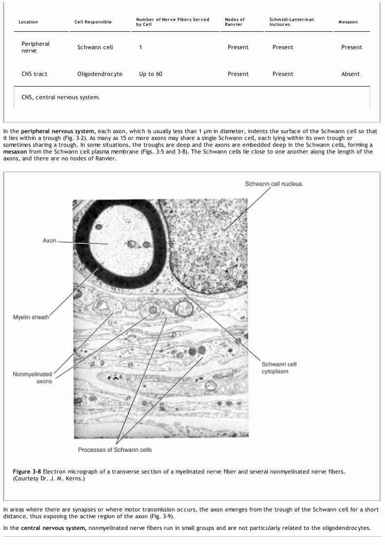
Location Cell Responsible Number of Nerve Fibers Servedby Cell
Nodes ofRanv ier
Schmidt-LantermanIncisures Mesaxon
Peripheralnerve Schwann cell 1 Present Present Present
CNS tract Oligodendrocyte Up to 60 Present Present Absent
CNS, central nervous system.
In the peripheral nervous system, each axon, which is usually less than 1 µm in diameter, indents the surface of the Schwann cell so thatit lies within a trough (Fig. 3-2). As many as 15 or more axons may share a single Schwann cell, each lying within its own trough orsometimes sharing a trough. In some situations, the troughs are deep and the axons are embedded deep in the Schwann cells, forming amesaxon from the Schwann cell plasma membrane (Figs. 3-5 and 3-8). The Schwann cells lie close to one another along the length of theaxons, and there are no nodes of Ranvier.
Figure 3-8 Electron micrograph of a transverse section of a myelinated nerve fiber and several nonmyelinated nerve fibers.(Courtesy Dr. J. M. Kerns.)
In areas where there are synapses or where motor transmission occurs, the axon emerges from the trough of the Schwann cell for a shortdistance, thus exposing the active region of the axon (Fig. 3-9).
In the central nervous system, nonmyelinated nerve fibers run in small groups and are not particularly related to the oligodendrocytes.

Figure 3-9 Autonomic neuromuscular junction between a nonmyelinated axon and a smooth muscle fiber.
Peripheral NervesPeripheral nerves is a collective term for the cranial and spinal nerves. Each peripheral nerve consists of parallel bundles of nervefibers, which may be efferent or afferent axons, may be myelinated or nonmyelinated, and are surrounded by connective tissue
sheaths (Figs. 3-10 and 3-11).
The nerve trunk is surrounded by a dense connective tissue sheath called the epineurium (Fig. 3-12). Within the sheath are bundles ofnerve fibers, each of which is surrounded by a connective tissue sheath called the perineurium. Between the individual nerve fibers is aloose, delicate connective tissue referred to as the endoneurium. The connective tissue sheaths serve to support the nerve fibers andtheir associated blood vessels and lymph vessels. Peripheral nerve fibers can be classified according to their speed of conduction and size(Table 3-2).

Figure 3-10 Photomicrograph of a longitudinal section of a peripheral nerve stained with hematoxylin and eosin (÷400).
Figure 3-11 Photomicrograph of a transverse section of a peripheral nerve stained with hematoxylin and eosin (÷275).
Spinal Nerves and Spinal Nerve RootsThere are 31 pairs of spinal nerves, which leave the spinal cord and pass through intervertebral foramina in the vertebral column. (Fordetails, see p. 12.) Each spinal nerve is connected to the spinal cord by two roots: the anterior root and the posterior root (Fig. 3-13).The anterior root consists of bundles of nerve fibers carrying nerve impulses away from the central nervous system; these nerve fibers arecalled efferent fibers. The posterior root consists of bundles of nerve fibers carrying nerve impulses to the central nervous system; thesenerve fibers are called afferent fibers. Because these fibers are concerned with conveying information to the central nervous system,they are calledsensory fibers. The cell bodies of these nerve fibers are situated in a swelling on the posterior root called the posterior root ganglion.

Figure 3-12 Structure of a peripheral nerve.
Table 3-2 Classification of Nerve Fibers by Speed of Conduction and Size
Fiber Type ConductionVelocity (m/s)
FiberDiameter (µm) Functions Myelin Sensitiv ity to Local
Anesthetics
A Fibers
Alpha 70–120 12–20 Motor, skeletal muscle Yes Least
Beta 40–70 5–12 Sensory, touch, pressure, vibration Yes
P.83
Gamma 10–50 3–6 Muscle spindle Yes
Delta 6–30 2–5 Pain (sharp, localized), temperature, touch Yes
B Fibers 3–15 <3 Preganglionic autonomic Yes
C Fibers 0.5–2.0 0.4–1.2 Pain (diffuse, deep), temperature,postganglionic autonomic No Most
Cranial NervesThere are 12 pairs of cranial nerves (Fig. 3-13), which leave the brain and pass through foramina in the skull. Some of these nerves arecomposed entirely of afferent nerve fibers bringing sensations to the brain (olfactory, optic, and vestibulocochlear), others are composedentirely of efferent fibers (oculomotor, trochlear, abducent, accessory, and hypoglossal), while the remainder possess both afferent andefferent fibers (trigeminal, facial, glossopharyngeal, and vagus). The cranial nerves are described in detail in Chapter 11.
Sensory GangliaThe sensory ganglia of the posterior spinal nerve roots and of the trunks of the trigeminal, facial, glossopharyngeal, and vagal cranial nerveshave the same structure. Each ganglion is surrounded by a layer of connective tissue that is continuous with the epineurium andperineurium of the peripheral nerve. The neurons are unipolar, possessing cell bodies that are rounded or oval in shape (Fig. 3-14). Thecell bodies tend to be aggregated and separated by bundles of nerve fibers. A single nonmyelinated process leaves each cell body and,after a convoluted course, bifurcates at a T junction into peripheral and central branches. The former axon terminates in a series ofdendrites in a peripheral sensory ending, and the latter axon enters the central nervous system. The nerve impulse, on reaching the Tjunction, passes directly from the peripheral axon to the central axon, thus bypassing the nerve cell body.
Each nerve cell body is closely surrounded by a layer of flattened cells called capsular cells or satellite cells (Fig. 3-14). The capsular cellsare similar in structure to Schwanncells and are continuous with these cells as they envelop the peripheral and central processes of each neuron.

Figure 3-13 A: Transverse section of the thoracic region of the spinal cord showing the formation of a spinal nerve from the unionof an anterior and a posterior nerve root. B: Transverse section of the pons showing the sensory and motor roots of the trigeminalnerve.

Figure 3-14 Photomicrograph of a longitudinal section of a posterior root ganglion of a spinal nerve stained with hematoxylin andeosin (÷400).
Autonomic GangliaThe autonomic ganglia (sympathetic and parasympathetic ganglia) are situated at a distance from the brain and spinal cord. They are foundin the sympathetic trunks, in prevertebral autonomic plexuses (e.g., in the cardiac, celiac, and mesenteric plexuses), and as ganglia in orclose to viscera. Each ganglion is surrounded by a layer of connective tissue that is continuous with the epineurium and perineurium ofthe peripheral nerve. The neurons are multipolar and possess cell bodies that are irregular in shape (Fig. 3-15). The dendrites of theneurons make synaptic connections with the myelinated axons of preganglionic neurons. The axons of the neurons are of small diameter (Cfibers) and unmyelinated, and they pass to viscera, blood vessels, and sweat glands.
Each nerve cell body is closely surrounded by a layer of flattened cells called capsular cells or satellite cells. The capsular cells, likethose of sensory ganglia, are similar in structure to Schwann cells and are continuous with them as they envelop the peripheral andcentral processes of each neuron.
Peripheral Nerve PlexusesPeripheral nerves are composed of bundles of nerve fibers. In their course, peripheral nerves sometimes divide into branches that joinneighboring peripheral nerves. If this occurs frequently, a network of nerves, called a nerve plexus, is formed. It should be emphasizedthat the formation of a nerve plexus allows individual nerve fibers to pass from one peripheral nerve to another, and in most instances,branching of nerve fibers does not take place. A plexus thus permits a redistribution of the nerve fibers within the different peripheralnerves.

Figure 3-15 Photomicrograph of a longitudinal section of a ganglion of the sympathetic trunk stained with hematoxylin and eosin(÷300).
Figure 3-16 Brachial plexus.

At the root of the limbs, the anterior rami of the spinal nerves form complicated plexuses. The cervical and brachial plexuses are at theroot of the upper limbs (Fig. 3-16), and the lumbar and sacral plexuses are at theroot of the lower limbs. This allows the nerve fibers derived from different segments of the spinal cord to be arranged and distributedefficiently in different nerve trunks to the various parts of the upper and lower limbs.
Cutaneous nerves, as they approach their final destination, commonly form fine plexuses that again permit a rearrangement of nerve fibersbefore they reach their terminal sensory endings.
The autonomic nervous system also possesses numerous nerve plexuses that consist of preganglionic and postganglionic nerve fibers andganglia.
Conduction in Peripheral NervesIn the resting unstimulated state, a nerve fiber is polarized so that the interior is negative to the exterior; the potentialdifference across the axolemma is about -80 mV and is called the resting membrane potential (Fig. 3-17). As has been explained
previously (see p. 42), this so-called resting potential is produced by the diffusion of sodium and potassium ions through the channels ofthe plasma membrane and is maintained by the sodium-potassium pump. Three Na+ ions are pumped to the outside for each two K+ ions tothe inside. The pump involves active transport across the membrane and requires adenosine triphosphate (ATP) to provide the energy.
Figure 3-17 Ionic and electrical changes that occur in a nerve fiber when it is conducting an impulse.
A nerve impulse (action potential) starts at the initial segment of the axon and is a self-propagating wave of electrical negativity thatpasses rapidly along the surface of the plasma membrane (axolemma). The wave of electrical negativity is initiated by an adequate stimulusbeing applied to the surface of the neuron (Fig. 3-18). Under normal circumstances, this occurs at the initial segment of the axon, which isthe most sensitive part of the neuron. The stimulus alters the permeability of the membrane to Na+ ions at the point of stimulation. Now,Na+ ions rapidly enter the axon (Fig. 3-17). The positive ions outside the axolemma quickly decrease to zero. Therefore, the membranepotential is reduced to zero and is said to be depolarized. A typical resting potential is -80 mV, with the outside of the membrane positiveto the inside; the action potential is about +40 mV, with the outside of the membrane negative to the inside. In small-diameter axons, theaction potential may not rise to as much as 40 mV.

Figure 3-18 Creation of the action potential by the arrival of a stimulus from a single presynaptic terminal. Note that the actionpotential generated at the initial segment will only occur if the threshold for excitation is reached at the initial segment. (FromSnell, R. S. Clinical Neuroanatomy: A Review with Questions and Explanations [3rd ed., p. 7]. Baltimore: Lippincott Williams &Wilkins.)
The negatively charged point on the outside of the axolemma now acts as a stimulus to the adjacent positively charged axolemma, and inless than 1 msec, the polarity of the adjacent resting potential is reversed (Fig. 3-17). The action potential now has moved along theaxolemma from the point originally stimulated to the adjacent point on the membrane. It is in this manner that the action potential travelsalong the full length of a nerve fiber to its end.
As the action potential moves along the nerve fiber, the entry of the Na+ ions into the axon ceases, and the permeability of the axolemmato K+ ions increases. Now, K+ ions rapidly diffuse outside the axon (since the concentration is much higher within the axon than outside sothat the original resting membrane potential is restored. The permeability of the axolemma now decreases, and the status quo is restoredby the active transport of the Na+ ions out of the axon and the K+ ions into the axon. The outer surface of the axolemma is againelectrically positive compared with that of the inner surface. This is a simplistic description of the movements of the Na+ and K+ ions. Forfurther details on the voltage-gated Na+ and K+ channels, the Na+ and K+ pumps, and the Na+ and K+ leak channels, refer to a textbook ofphysiology.
For a short time after the passage of a nerve impulse along a nerve fiber, while the axolemma is still depolarized, a second stimulus,however strong, is unable to excite the nerve. This period of time is called the absolute refractory period. The underlying reason for therefractory period is that the Na+ channels become inactivated, and no stimulation, however strong, will open the Na+ gates. This period isfollowed by a further short interval during which the excitability of the nerve gradually returns to normal. This latter period is called therelative refractory period. It is clear from this that the refractory period makes a continuous excitatory state of the nerve impossible and

limits the frequency of the impulses.
The conduction velocity of a nerve fiber is proportional to the cross-sectional area of the axon, with the thicker fibers conducting morerapidly than those of smaller diameter. In the large motor fibers (alpha fibers), the rate may be as high as 70 to 120 m/s; the smaller sensoryfibers have slower conduction rates (see Table 3-2).
In nonmyelinated fibers, the action potential passes continuously along the axolemma, progressively exciting neighboring areas ofmembrane (Fig. 3-19). In myelinated fibers, the presence of a myelin sheath serves as an insulator, and few ions can flow through thesheath. Consequently, a myelinated nerve fiber can be stimulated only at the nodes of Ranvier, where the axon is naked and the ions canpass freely through the plasma membrane between the extracellular fluid and the axoplasm. In these fibers, the actionpotential jumps from one node to the next (Fig. 3-19). The action potential at one node sets up a current in the surrounding tissue fluid,which quickly produces depolarization at the next node. This leaping of the action potential from one node to the next is referred to assaltatory conduction (Fig. 3-19). This is a more rapid mechanism than is found in nonmyelinated fibers (120.0 m/s in a large myelinated fibercompared with 0.5 m/s in a very small unmyelinated fiber).
Figure 3-19 Electrical changes that occur in stimulated myelinated axon (saltatory conduction) (A) and stimulated nonmyelinatedaxon (B).
Receptor EndingsAn individual receives impressions from the outside world and from within the body by special sensory nerve endings or receptors.
Sensory receptors can be classified into five basic functional types:
Mechanoreceptors. These respond to mechanical deformation.
Thermoreceptors. These respond to changes in temperature; some receptors respond to cold and others to heat.
Nociceptors. These respond to any stimuli that bring about damage to the tissue.
Electromagnetic receptors. The rods and cones of the eyes are sensitive to changes in light intensity and wavelength.
Chemoreceptors. These respond to chemical changes associated with taste and smell and oxygen and carbon dioxide concentrations inthe blood.
Anatomical Types of ReceptorsFor convenience, the sensory endings can be classified, on a structural basis, into nonencapsulated and encapsulated receptors. Table 3-3

P.88P.89
classifies and compares the receptor types.
Nonencapsulated Receptors
Free Nerve EndingsFree nerve endings are widely distributed throughout the body (Fig. 3-20). They are found between the epithelial cells of the skin, thecornea, and the alimentary tract, and in connective tissues, including the dermis, fascia, ligaments, joint capsules, tendons, periosteum,perichondrium, haversian systems of bone, tympanic membrane, and dental pulp; they are also present in muscle.
The afferent nerve fibers from the free nerve endings are either myelinated or nonmyelinated. The terminal endings are devoid of a myelinsheath, and there are no Schwann cells covering their tips.
Most of these endings detect pain, while others detect crude touch, pressure, and tickle sensations, and possibly cold and heat.
Merkel DiscsMerkel discs are found in hairless skin, for example, the fingertips (Figs. 3-21 and 3-22), and in hair follicles. The nerve fiber passes into theepidermis and terminates as a disc-shaped expansion that is applied closely to a dark-staining epithelial cell in the deeper part of theepidermis, called theMerkel cell. In hairy skin, clusters of Merkel discs, known as tactile domes, are found in the epidermis between thehair follicles.
Table 3-3 Classification and Comparison of Receptor Types
Type of Receptor Location Stimulus Sensory Modality Adaptability Fibers
Nonencapsulated Receptors
Free nerveendings
Epidermis, cornea, gut,dermis, ligaments, jointcapsules, bone, dental pulp,etc.
Mechanoreceptor
Pain (fast), pain(slow), touch(crude), pressure,? heat and cold
RapidAdelta,C
Merkel discs Hairless skin Mechanoreceptor Touch Slow Abeta
Hair folliclereceptors Hairy skin Mechanoreceptor Touch Rapid A
beta
Encapsulated Receptors
Meissner'scorpuscles
Dermal papillae of skin ofpalm and sole of foot Mechanoreceptor Touch Rapid A
beta
Paciniancorpuscles
Dermis, ligaments, jointcapsules, peritoneum,external genitalia, etc.
Mechanoreceptor Vibration Rapid Abeta
Ruffinicorpuscles Dermis of hairy skin Mechanoreceptor Stretch Slow A
beta
Neuromuscularspindles Skeletal muscle Mechanoreceptor Stretch— muscle
length Fast
Aalpha,Abeta
Neurotendinousspindles Tendons Mechanoreceptor Compression—
muscle tension Fast Aalpha

Figure 3-20 Free nerve endings in the skin. The nerve fibers in the epidermis are naked.
Figure 3-21 Merkel discs in the skin. A: Low magnification. B: Merkel disc showing the expanded ending of an axon with a stippledtactile cell.

Figure 3-22 Photomicrograph of digital skin showing fine nerve terminals ending in Merkel discs, stained by the silver method.(Courtesy Dr. N. Cauna.)
Merkel discs are slowly adapting touch receptors that transmit information about the degree of pressure exerted on the skin, such aswhen one is holding a pen.
Hair Follicle ReceptorsNerve fibers wind around the follicle in its outer connective tissue sheath below the sebaceous gland. Some branches surround thefollicle, while others run parallel to its long axis (Figs. 3-23 and 3-24). Many naked axon filaments terminate among the cells of the outerroot sheath.
Bending of the hair stimulates the follicle receptor, which belongs to the rapidly adapting group of mechanoreceptors. While the hairremains bent, the receptor is silent, but when the hair is released, a further burst of nerve impulses is initiated.
Encapsulated ReceptorsEncapsulated receptors show wide variations in size and shape, and the termination of the nerve is covered by a capsule.
Meissner's CorpusclesMeissner's corpuscles are located in the dermal papillae of the skin (Figs. 3-25 and 3-26), especially that of the palm of the hand and thesole of the foot. Many also are present in the skin of the nipple and the external genitalia. Each corpuscle is ovoid in shape and consists ofa stack of modified flattened Schwann cells arranged transversely across the long axis of the corpuscle. The corpuscle is enclosed by acapsule of connective tissue that is continuous with the endoneurium of the nerves that enter it. A few myelinated nerve fibers enter thedeep end of the corpuscle; myelinated and unmyelinated branches decrease in size and ramify among the Schwann cells. There is aconsiderable reduction in the number of Meissner's corpuscles between birth and old age.

P.91
Figure 3-23 Nerve endings around a hair follicle.
Meissner's corpuscles are very sensitive to touch and are rapidly adapting mechanoreceptors. They enable an individual to distinguishbetween two pointed structures when they are placed close together on the skin (two-point tactile discrimination).
Pacinian CorpusclesPacinian corpuscles (Figs. 3-27 and 3-28) are widely distributed throughout the body and are abundant in the dermis, subcutaneous tissue,ligaments, joint capsules, pleura, peritoneum, nipples, and external genitalia. Each corpuscle is ovoid in shape, measuring about 2 mm longand about 100 to 500 µm across. It consists of a capsule and a central core containing the nerve ending. The capsule consists of numerousconcentric lamellae of flattened cells. A large myelinated nerve fiber enters the corpuscle and loses its myelin sheath and thenits Schwann cell covering. The naked axon, surrounded by lamellae formed of flattened cells, passes through the center of the core andterminates in an expanded end.
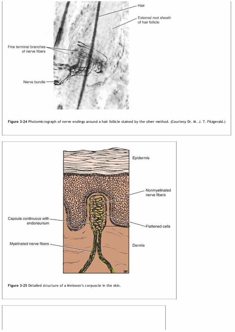
Figure 3-24 Photomicrograph of nerve endings around a hair follicle stained by the silver method. (Courtesy Dr. M. J. T. Fitzgerald.)
Figure 3-25 Detailed structure of a Meissner's corpuscle in the skin.

Figure 3-26 Photomicrograph of a Meissner's corpuscle of the skin. (Courtesy Dr. N. Cauna.)

Figure 3-27 Detailed structure of a pacinian corpuscle in the skin.
Figure 3-28 Photomicrograph of part of a pacinian corpuscle of the skin seen in transverse section showing concentric lamellae offlattened cells. (Courtesy Dr. N. Cauna.)
The pacinian corpuscle is a rapidly adapting mechanoreceptor that is particularly sensitive to vibration. It can respond to up to 600 stimuli

per second.
Ruffini CorpusclesRuffini corpuscles are located in the dermis of hairy skin. Each corpuscle consists of several large unmyelinated nerve fibers ending withina bundle of collagen fibers and surrounded by a cellular capsule. These slowly adapting mechanoreceptors are stretch receptors, whichrespond when the skin is stretched.
Function of Cutaneous ReceptorsIn the past, it was believed that the different histologic types of receptors corresponded to specific types of sensation. It was soonpointed out that there are areas of the body that have only one or two histologic types of receptors and yet they are sensitive to avariety of different stimuli. Moreover, although the body has these different receptors, all nerves only transmit nerve impulses. It is nowgenerally agreed that the type of sensation felt is determined by the specific area of the central nervous system to which the afferentnerve fiber passes. For example, if a pain nerve fiber is stimulated by heat, cold, touch, or pressure, the individual will experience onlypain.
Transduction of Sensory Stimuli Into Nerve ImpulsesTransduction is the process by which one form of energy (the stimulus) is changed into another form of energy(electrochemical energy of the nerve impulse). The stimulus, when applied to the receptor, brings about a change in potential of theplasma membrane of the nerve ending. Since this process takes place in the receptor, it is referred to as the receptor potential. Theamplitude of the receptor potential is proportional to the intensity of the stimulus. By opening more ion channels for a longer time, astronger mechanical pressure, for example, can produce a greater depolarization than does weak pressure. With chemoreceptors andphotoreceptors, the receptor potential is produced by second messengers activated when the stimulus agent binds to the membranereceptors coupled to G proteins. If large enough, the receptor potential will generate an action potential that will travel along theafferent nerve fiber to the central nervous system.

Figure 3-29 Neuromuscular spindle showing two types of intrafusal fibers: the nuclear bag and nuclear chain fibers.
Joint ReceptorsFour types of sensory endings can be located in the capsule and ligaments of synovial joints. Three of these endings are encapsulated andresemble pacinian, Ruffini, and tendon stretch receptors. They provide the central nervous system with information regarding the positionand movements of the joint. A fourth type of ending is nonencapsulated and is thought to be sensitive to excessive movements and totransmit pain sensations.
Neuromuscular SpindlesNeuromuscular spindles, or muscular spindles (Figs. 3-29 and 3-30), are found in skeletal muscle and are most numeroustoward the tendinous attachment of the muscle. They provide the central nervous system with sensory information regarding the musclelength and the rate of change in the muscle length. This information is used by the central nervous system in the control of muscleactivity.
Figure 3-30 Photomicrograph of a neuromuscular spindle.
Each spindle measures about 1 to 4 mm in length and is surrounded by a fusiform capsule of connective tissue. Within the capsule are 6 to14 slender intrafusal muscle fibers; the ordinary muscle fibers situated outside the spindles are referred to as extrafusal fibers. Theintrafusal fibers of the spindles are of two types: the nuclear bag and nuclear chain fibers. The nuclear bag fibers are recognized by thepresence of numerous nuclei in the equatorial region, which consequently is expanded; also, cross striations are absent in this region. Inthe nuclear chain fibers, the nuclei form a single longitudinal row or chain in the center of each fiber at the equatorial region. Thenuclear bag fibers are larger in diameter than the nuclear chain fibers, and they extend beyond the capsule at each end to be attachedto the endomysium of the extrafusal fibers.
There are two types of sensory innervation of muscle spindles: the annulospiral and the flower spray. The annulospiral endings aresituated at the equator of the intrafusal fibers. As the large myelinated nerve fiber pierces the capsule, it loses its myelin sheath, and thenaked axon winds spirally around the nuclear bag or chain portions of the intrafusal fibers.
The flower-spray endings are situated mainly on the nuclear chain fibers some distance away from the equatorial region. A myelinatednerve fiber slightly smaller than that for the annulospiral ending pierces the capsule and loses its myelin sheath, and the naked axonbranches terminally and ends as varicosities; it resembles a spray of flowers.
Stretching (elongation) of the intrafusal fibers results in stimulation of the annulospiral and flower-spray endings, and nerve impulses passto the spinal cord in the afferent neurons.
Motor innervation of the intrafusal fibers is provided by fine gamma motor fibers. The nerves terminate in small motor end-plates situatedat both ends of the intrafusal fibers. Stimulation of the motor nerves causes both ends of the intrafusal fibers to contract and activate thesensory endings. The equatorial region, which is without cross striations, is noncontractile. The extrafusal fibers of the remainder of themuscle receive their innervation in the usual way from large alpha-size axons.

Function of the Neuromuscular SpindleUnder resting conditions, the muscle spindles give rise to afferent nerve impulses all the time, and most of this information is notconsciously perceived. When muscle activity occurs, either actively or passively, the intrafusal fibers are stretched, and there is anincrease in the rate of passage of nerve impulses to the spinal cord or brain in the afferent neurons. Similarly, if the intrafusal fibers arenow relaxed due to the cessation of muscle activity, the result is a decrease in the rate of passage of nerve impulses to the spinal cord orbrain. The neuromuscular spindle thus plays a very important role in keeping the central nervous system informed about the length of amuscle and the rate of change of its length, thereby indirectly influencing the control of voluntary muscle.
Stretch ReflexThe neurons of the spinal cord involved in the simple stretch reflex are as follows. Stretching a muscle results in elongation of theintrafusal fibers of the muscle spindle and stimulation of the annulospiral and flower-spray endings. The nerve impulses reach the spinalcord in the afferent neurons and synapse with the large alpha motor neurons situated in the anterior gray horns of the spinal cord. Nerveimpulses now pass via the efferent motor nerves and stimulate the extrafusal muscle fibers, and the muscle contracts. This simple stretchreflex depends on a two-neuron arc consisting of an afferent neuron and an efferent neuron. It is interesting to note that the musclespindle afferent impulsesinhibit the alpha motor neurons supplying the antagonist muscles. This effect is called reciprocal inhibition.
Control of the Intrafusal Fibers of the Neuromuscular SpindleIn the brain and spinal cord, there are centers that give rise to tracts that synapse with gamma motor neurons in the spinal cord. Thereticular formation, the basal ganglia, and the cerebellum are examples of such centers. It is by these means that these centers cangreatly influence voluntary muscle activity. The gamma efferent motor fibers cause shortening of the intrafusal fibers, stretching theequatorial regions and stimulating the annulospiral and flower-spray endings. This, in turn, initiates the reflex contraction of the extrafusalfibers described previously.
It is estimated that about one-third of all the motor fibers passing to a muscle are gamma efferents; the remaining two-thirds are the largealpha motor fibers. It is believed that the nuclear bag fibers are concerned with dynamic responses and are associated more with positionand velocity of contraction, whereas the nuclear chain fibers are associated with slow static contractions of voluntary muscle.
Neurotendinous SpindlesNeurotendinous spindles (Golgi tendon organs) are present in tendons and are located near the junctions of tendons with muscles (Fig. 3-31). They provide the central nervous system with sensory information regarding the tension of muscles.
Figure 3-31 A neurotendinous spindle.

Each spindle consists of a fibrous capsule that surrounds a small bundle of loosely arranged tendon (collagen) fibers (intrafusal fibers). Thetendon cells are larger and more numerous than those found elsewhere in the tendon. One or more myelinated sensory nerve fiberspierce the capsule, lose their myelin sheath, branch, and terminate in club-shaped endings.
The nerve endings are activated by being squeezed by the adjacent tendon fibers within the spindle when tension develops in the tendon.Unlike the neuromuscular spindle, which is sensitive to changes in muscle length, the neurotendinous organ detects changes in muscletension.
Function of the Neurotendinous SpindleIncreased muscle tension stimulates the neurotendinous spindles, and an increased number of nerve impulses reach the spinal cordthrough the afferent nerve fibers. These fibers synapse with the large alpha motor neurons situated in the anterior gray horns of thespinal cord. Unlike the muscle spindle reflex, this reflex is inhibitory and inhibits muscle contraction. In this manner, the tendon reflexprevents the development of too much tension in the muscle. Although this function is probably important as a protective mechanism, itsmain function is to provide the central nervous system with information that can influence voluntary muscle activity.
Effector Endings
Innervation of Skeletal MuscleSkeletal muscle is innervated by one or more nerves. In the limbs and head and neck, the innervation is usually single, but in the largemuscles of the abdominal wall, the innervation is multiple, the latter muscles having retained their embryonic segmental nerve supply.
The nerve supply and blood supply to a muscle enter it at a more or less constant position called the neurovascular hilus. The nerve to amuscle contains motor and sensory fibers. The motor fibers are of three types: (1) large alpha myelinated fibers, (2) small gammamyelinated fibers, and (3) fine unmyelinated C fibers. The large myelinated axons of the alpha anterior horn cells supply the extrafusalfibers that form the main mass of the muscle. The small gamma myelinated fibers supply the intrafusal fibers of the neuromuscular spindles.The fine unmyelinated fibers are postganglionic autonomic efferents that supply the smooth muscle in the walls of blood vessels.
The sensory fibers are of three main types: (1) the myelinated fibers, which originate in the annulospiral and flower-spray endings of theneuromuscular spindles; (2) the myelinated fibers, which originate in the neurotendinous spindles; and (3) the myelinated andnonmyelinated fibers, which originate from a variety of sensory endings in the connective tissue of the muscle.
Figure 3-32 Simple reflex arc consisting of an afferent neuron arising from neuromuscular spindles and neurotendinous spindles andan efferent lower motor neuron whose cell body is an alpha anterior horn cell within the spinal cord. Note that the efferentneuron terminates on muscle fibers at motor end-plates.
Motor UnitThe motor unit may be defined as the single alpha motor neuron and the muscle fibers that it innervates (Fig. 3-32). The muscle fibers of asingle motor unit are widely scattered throughout the muscle. Where fine, precise muscle control is required, such as in the extraocularmuscles or the small muscles of the hand, the motor units possess only a few muscle fibers. Where precise control is not necessary,however, as in a large limb muscle such as the gluteus maximus, a single motor nerve may innervate many hundreds of muscle fibers.

P.97
Neuromuscular Junctions in Skeletal MuscleSkeletal muscle fibers are innervated by large, alpha myelinated nerve fibers derived from large motor neurons in the anterior gray columns(horns) of the spinal cord or from the motor nuclei of cranial nerves. As each myelinated fiber enters a skeletal muscle, it branches manytimes. The number of branches depends on the size of the motor unit. A single branch then terminates on a muscle fiber at a site referredto as a neuromuscular junction or motor end-plate (Figs. 3-33 and 3-34).
The great majority of muscle fibers are innervated by just one motor end-plate. On reaching the muscle fiber, the nerve loses its myelinsheath and breaks up into a number of finebranches. Each branch ends as a naked axon and forms the neural element of the motor end-plate (Fig. 3-35). The axon isexpanded slightly and contains many mitochondria and vesicles (approximately 45 nm in diameter). At the site of the motor end-plate, thesurface of the muscle fiber is elevated slightly to form the muscular element of the plate, often referred to as the sole plate (Fig. 3-33).The elevation is due to the local accumulation of granular sarcoplasm beneath the sarcolemma and the presence of numerous nuclei andmitochondria, the latter providing the ATP, which is the energy source for the synthesis of the transmitter acetylcholine (ACh).
Figure 3-33 A: A skeletal neuromuscular junction. B: Enlarged view of a muscle fiber showing the terminal naked axon lying in thesurface groove of the muscle fiber.

Figure 3-34 Photomicrograph showing nerve fibers terminating on skeletal muscle fibers at motor end-plates, stainedhistochemically for acetylcholinesterase and counterstained with silver. (Courtesy Dr. M. J. T. Fitzgerald.)
Figure 3-35 A: Photomicrograph of a motor end-plate showing terminal branching of a nerve fiber. B: Electron micrograph of aterminal axon at a motor end-plate showing the axon lying in a groove on the surface of the muscle fiber. (Courtesy Dr. J. M.Kerns.)
The expanded naked axon lies in a groove on the surface of the muscle fiber outside the plasma membrane (sarcolemma). Each groove isformed by the infolding of the plasma membrane. The groove may branch many times, with each branch containing a division of the axon. Itis important to realize that the axons are truly naked; the Schwann cells merely serve as a cap or roof to the groove and never projectinto it. The floor of the groove is formed of the plasma membrane, which is thrown into numerous small folds, called junctional folds;these serve to increase the surface area of the plasma membrane that lies close to the naked axon (Fig. 3-36).

The plasma membrane of the axon (the axolemma or presynaptic membrane) is separated, by a space about 30 to 50 nm wide, from theplasma membrane of the muscle fiber (the sarcolemma or postsynaptic membrane). This space constitutes the synaptic cleft. The synapticcleft is filled with the basement membranes of the axon and the muscle fiber (Fig. 3-33). The motor end-plate is strengthened by theconnective tissue sheath of the nerve fiber (endoneurium), which becomes continuous with the connective tissue sheath of the musclefiber (endomysium).
Figure 3-36 Electron micrograph of a cross section of an axon at a motor end-plate showing the axon lying in a groove of foldedsarcolemma. (Courtesy Dr. J. M. Kerns.)
A nerve impulse (action potential), on reaching the presynaptic membrane of the motor end-plate, causes the opening of voltage-gatedCa2+ channels that allow Ca2+ ions to enter the axon. This stimulates the fusion of some of the synaptic vesicles with the presynapticmembrane and causes the release of acetylcholine into the synaptic cleft. The acetylcholine is thus discharged into the cleft by a processof exocytosis and diffuses rapidly across the cleft to reach the nicotinic type of ACh receptors on the postsynaptic membrane of thejunctional folds. The postsynaptic membrane possesses large numbers of ACh-gated channels.
Once the ACh-gated channels are opened, the postsynaptic membrane becomes more permeable to Na+ ions, which flow into the musclecell, and a local potential called the end-plate potential is created. (The ACh-gated channels are also permeable to K+ ions, which flowout of the cell but to a lesser extent.) If the end-plate potential is large enough, the voltage-gated channels for Na+ ions are opened, andan action potential will be initiated and will spread along the surface of the plasma membrane (sarcolemma). The wave of depolarization iscarried into the muscle fiber to the contractile myofibrils through the system of T tubules. This leads to the release of Ca2+ ions from thesarcoplasmic reticulum, which, in turn, causes the muscle to contract.
The amount of acetylcholine released at the motor end-plate will depend on the number of nerve impulses arriving at the nerve terminal.Once the acetylcholine crosses the synaptic cleft and triggers the ionic channels on the postsynaptic membrane, it immediatelyundergoes hydrolysis due to the presence of the enzyme acetylcholinesterase (AChE) (Fig. 3-34). The enzyme is adherent to the finecollagen fibrils of the basement membranes in the cleft; some of the acetylcholine diffuses away from the cleft. The acetylcholine remainsfor about 1 msec in contact with the postsynaptic membrane, and it is rapidly destroyed to prevent reexcitation of the muscle fiber. Afterthe fall in concentration of ACh in the cleft, the ionic channels close and remain closed until the arrival of more ACh.
Skeletal muscle fiber contraction is thus controlled by the frequency of the nerve impulses that arrive at the motor nerve terminal. Aresting muscle fiber shows small occasional depolarizations (end-plate potentials) at the motor end-plate, which are insufficient to causean action potential and make the fiber contract. These are believed to be due to the sporadic release of acetylcholine into the synapticcleft from a single presynaptic vesicle.
The sequence of events that takes place at a motor end-plate on stimulation of a motor nerve can be briefly summarized as follows:
ACh → Nicotinic type of ACh receptor, ACh-gated channels opened → Na+ influx → End-plate potential created.
End-plate potential (if large enough) → Na+-gated channels opened → Na+ influx → Action potential created.
Action potential → Increased release of Ca2+ → Muscle fiber contraction.
Immediate hydrolysis of ACh by AChE → ACh-gated channels closed → Muscle fiber repolarization.
If drugs having a similar chemical structure to acetylcholine were to arrive at the receptor site of a motor end-plate, they might bring

about the same changes as acetylcholine and mimic its action. Two examples of such drugs are nicotine and carbamylcholine. If, on theother hand, drugs having a similar chemical structure to acetylcholine were to arrive at the receptor site of a motor end-plate and wereunable to bring about the sequence of changes normally induced by acetylcholine, they would occupy the receptor site and block accessof acetylcholine. Such drugs would be competing with acetylcholine and are called competitive blocking agents. An example of such adrug is d-tubocurarine, which causes skeletal muscle to relax and not contract by preventing the action of locally produced acetylcholine(see p. 116).
Neuromuscular Junctions in Smooth MuscleIn smooth muscle, where the action is slow and widespread, such as within the wall of the intestine, the autonomic nerve fibers branchextensively; thus, a single neuron exerts control over a large number of muscle fibers. In some areas (e.g., the longitudinal layer of smoothmuscle in the intestine), only a few muscle fibers are associated with autonomic endings, the wave of contraction passing from one musclecell to another by means of gap junctions (Fig. 3-37).
Figure 3-37 Autonomic neuromuscular junction. The exposed axons are close to the smooth muscle fibers.
In smooth muscle, in which the action is fast and precision is required, such as in the iris, the branching of the nerve fibers is lessextensive; thus, a single neuron exerts control over only a few muscle fibers.
The autonomic nerve fibers, which are postganglionic, are nonmyelinated and terminate as a series of varicosed branches. An interval of 10to 100 nm may exist between the axon and the muscle fiber. At the site where transmission is to occur, the Schwann cell is retracted sothat the axon lies within a shallow groove on its surface (Fig. 3-37). Therefore, part of the axon is naked, permitting free diffusion of thetransmitter substance from the axon to the muscle cell (Fig. 3-37). Here, the axoplasm contains numerous vesicles similar to those seen atthe motor end-plate of skeletal muscle.
Smooth muscle is innervated by sympathetic and parasympathetic parts of the autonomic system. Those nerves that are cholinergicliberate acetylcholine at their endings by a process of exocytosis, with the acetylcholinebeing present in the vesicles at the nerve ending. Those nerves that are noradrenergic liberate norepinephrine at their endings by aprocess of exocytosis, with the norepinephrine being present in dark-cored vesicles at the nerve endings. Both acetylcholine andnorepinephrine bring about depolarization of the muscle fibers innervated, which thereupon contract. The fate of these neurotransmittersubstances differs. The acetylcholine is hydrolyzed in the presence of acetylcholinesterase in the synaptic cleft of the muscle fiber, andthe norepinephrine is taken up by the nerve endings. It is noteworthy that in some areas of the body (e.g., bronchial muscle), thenorepinephrine liberated from postganglionic sympathetic fibers causes smooth muscle to relax and not contract.
Neuromuscular Junctions in Cardiac Muscle

Nonmyelinated postganglionic sympathetic and parasympathetic autonomic nerves extend into the connective tissue between the musclefibers and terminate in close proximity to the individual cardiac muscle fibers. At the site where transmission takes place, the axonbecomes naked because of the retraction of the Schwann cell. This permits free diffusion of the neurotransmitter substance from theaxon to the muscle fiber. Because of the presence of intermittent desmosomes and gap junctions between abutting muscle fibers,excitation and contraction of one muscle fiber rapidly spread from fiber to fiber.
Nerve Endings on Secretory Cells of GlandsNonmyelinated postganglionic autonomic nerves extend into the connective tissue of glands and branch close to the secretory cells (Fig.3-38). In many glands, the nerve fibers have been found to innervate only the blood vessels.
Figure 3-38 Nerve fibers ending around glandular acini.
Segmental Innervation of SkinThe area of skin supplied by a single spinal nerve and, therefore, a single segment of the spinal cord is called a dermatome. Onthe trunk, the dermatomes extend round the body from the posterior to the anterior median plane. Adjacent dermatomes overlap
considerably, so to produce a region of complete anesthesia, at least three contiguous spinal nerves have to be sectioned. It should benoted that the area of tactile loss is always greater than the area of loss of painful and thermal sensations. The reason for this differenceis that the degree of overlap of fibers carrying pain and thermal sensations is much more extensive than the overlap of fibers carryingtactile sensations. Dermatomal charts for the anterior and posterior surfaces of the body are shown in Figures 3-39 and 3-40.
In the limbs, the arrangement of the dermatomes is more complicated because of the embryologic rotation of the limbs as they grow outfrom the trunk. (For details, see Figs. 3-39 and 3-40.)
In the face, the divisions of the trigeminal nerve supply a precise area of skin, and there is little or no overlap to the cutaneous area ofanother division.
Segmental Innervation of MusclesSkeletal muscle also receives a segmental innervation. Most of these muscles are innervated by more than one spinal nerve and,therefore, by the same number of segments of the spinal cord. Thus, to paralyze a muscle completely, it would be necessary to
section several spinal nerves or destroy several segments of the spinal cord.
To learn the segmental innervation of all the muscles of the body is an impossible task. Nevertheless, the segmentalinnervation of the following muscles should be known because it is possible to test them by eliciting simple muscle reflexes in the patient(Fig. 3-41):

Figure 3-39 Anterior aspect of the body showing the distribution of cutaneous nerves on the left side and dermatomes on the rightside.
Biceps brachii tendon reflex C5-6 (flexion of the elbow joint by tapping the biceps tendon).
Triceps tendon reflex C6-7 and C8 (extension of the elbow joint by tapping the triceps tendon).
Brachioradialis tendon reflex C5-6 and C7 (supination of the radioulnar joints by tapping the insertion of the brachioradialis tendon).
Abdominal superficial reflexes (contraction of underlying abdominal muscles by stroking the skin). Upper abdominal skin T6-7; middleabdominal skin T8-9; lower abdominal skin T10-12.
Patellar tendon reflex (knee jerk) L2, L3, and L4 (extension of knee joint on tapping the patellar tendon).
Achilles tendon reflex (ankle jerk) S1 and 2 (plantar flexion of ankle joint on tapping the Achilles tendon—tendo calcaneus).
Muscle Tone and Muscle ActionA motor unit consists of a motor neuron in the anterior gray column (horn) of the spinal cord and all the muscle fibers it supplies
(Fig. 3-42). In a large buttock muscle, such as the gluteus maximus, where fine control is unnecessary, a given motor neuron may supply asmany as 200 muscle fibers. In contrast, in the small muscles of the hand or the extrinsic muscles of the eyeball, where fine control isrequired, one nerve fiber supplies only a few muscle fibers.
Every skeletal muscle, while resting, is in a partial state of contraction. This condition is referred to as muscle tone. Since there is no

P.103
intermediate stage, muscle fibers are either fully contracted or relaxed; it follows that a few muscle fibers within a muscle are fullycontracted all the time. To bring about this state and to avoid fatigue, different groups of motor units and, thus, different groups ofmuscle fibers are brought into action at different times. This isaccomplished by the asynchronous discharge of nervous impulses in the motor neurons in the anterior gray horn of the spinalcord.
Figure 3-40 Posterior aspect of the body showing the distribution of cutaneous nerves on the left side and dermatomes on theright side.

Figure 3-41 Biceps brachii tendon reflex. Note that the reflex arc passes through the fifth and sixth cervical segments of the spinalcord. This is usually monosynaptic, and the internuncial neuron (black) is absent (see p. 103).
Figure 3-42 Components of a motor unit.
Basically, muscle tone is dependent on the integrity of a simple monosynaptic reflex arc composed of two neurons in the nervous system(Fig. 3-43). The lengthening and shortening in a muscle are detected by sensitive sensory endings called muscle spindles (see p. 93), andthe tension is detected by tendon spindles (see p. 94). The nervous impulses travel in the large afferent fibers to the spinal cord. There,they synapse with the motor neurons situated in the anterior gray column, which, in turn, send impulses down their axons to the musclefibers (Fig. 3-43). The muscle spindles themselves are innervated by small gamma efferent fibers that regulate the response of the musclespindles, acting synergically with external stretch. In this manner, muscle tone is maintained reflexly and adjusted to the needs of postureand movement.

Figure 3-43 Simple reflex arc consisting of an afferent neuron arising from neuromuscular spindles and neurotendinous spindles andan efferent neuron whose cell body lies in the anterior gray column (horn) of the spinal cord. Note that for simplicity, the afferentfibers from the neurotendinous spindle and the neuromuscular spindle are shown as one pathway; in fact, the neurotendinousreceptor is inhibitory and reduces tone, whereas the neuromuscular spindle is excitatory and increases tone.
Should the afferent or efferent pathways of the reflex arc be cut, the muscle would lose its tone immediately and become flaccid. Aflaccid muscle, on palpation, feels like a mass of dough that has completely lost its resilience. It quickly atrophies and becomes reduced involume. It is important to realize that the degree of activity of the motor anterior column cells and, therefore, the degree of muscle tonedepend on the summation of the nerve impulses received by these cells from other neurons of the nervous system.
Muscle movement is accomplished by bringing into action increasing numbers of motor units and, at the same time, reducing the activity ofthe motor units of muscles that will oppose or antagonize the movement. When the maximum effort is required, all the motor units of amuscle are thrown into action.
Summation of Motor UnitsWhen a muscle begins to contract, the smaller motor units are stimulated first. The reason for this is that the smaller motor unitsare innervated by smaller neurons in the spinal cord and brainstem, and they have a lower threshold of
excitability. As the contraction increases, progressively larger motor units are brought into action. This phenomenon causes a gradualincrease in muscle strength as the muscle contracts.
Muscle FatigueThe progressive loss of strength of a muscle with prolonged strong contraction is due to the reduction in the amounts of ATPwithin the muscle fibers. Nerve impulses continue to arrive at the neuromuscular junction, and normal depolarization of the
plasma membrane of the muscle fiber occurs.
PosturePosture may be defined as the position adopted by the individual within his or her environment. In the standing position, the lineof gravity passes through the odontoid process of the axis, behind the centers of the hip joints, and in front of the knee and
ankle joints (Fig. 3-44). In order to stabilize the body and prevent it from collapsing, it is not surprising to find that in humans, theantigravity muscles are well developed and exhibit the greatest degree of tone. Therefore, one can say that posture depends on thedegree and distribution of muscle tone, which, in turn, depends on the normal integrity of simple reflex arcs centered in the spinal cord.
An individual may assume a particular posture (sitting or standing) over long periods of time with little evidence of fatigue. The reason forthis is that muscle tone is maintained through different groups of muscle fibers contracting in relays, with only a small number of musclefibers within a muscle being in a state of contraction at any one time. The active muscle fiber groups are scattered throughout themuscle.
In order to maintain posture, the simple muscle reflex, on which muscle tone is dependent, must receive adequate nervous input fromhigher levels of the nervous system (Fig. 3-45). For example, impulses arising from the labyrinths and neck muscles, information arising fromthe cerebellum, midbrain, and cerebral centers, and general information arising from other muscle groups, joints, and even skin receptorswill result in nervous impulses impinging on the large anterior gray column cells (i.e., the final common pathway) controlling the musclefibers.
When an individual assumes a given posture, the tone of the muscles controlling that posture is constantly undergoing fine adjustments sothat the posture is maintained. Normal posture thus depends not only on the integrity of the reflex arc but also on the summation of thenervous impulses received by the motor anterior gray column cells from other neurons of the nervous system (Fig. 3-46). The detail of thedifferent nervous pathways involved in bringing the information to the anterior gray column cells is dealt with in Chapter 4.

Figure 3-44 Lateral view of the skeleton showing the line of gravity. Since the greater part of body weight lies anterior to thevertebral column, the deep muscles of the back are important in maintaining normal postural curves of the vertebral column in thestanding position.

Figure 3-45 Nervous input from higher levels of the central nervous system, which can influence the activity of the anterior graycolumn (horn) cells of the spinal cord.

P.108P.109P.110P.111P.112P.113P.114P.115P.116P.117P.118P.119
Figure 3-46 Normal postural tone of skeletal muscle is dependent not only on the integrity of the reflex arc but also on thesummation of the nervous impulses received by the motor anterior gray column (horn) cells from other neurons of the nervoussystem.
ClinicalNotesResponse of Neurons to Injury
The survival of the cytoplasm of a neuron depends on its being connected, however indirectly, with the nucleus. Thenucleus plays a key role in the synthesis of proteins, which pass into the cell processes and replace proteins that havebeen metabolized by cell activity. Thus, the cytoplasm of axons and dendrites will undergo degeneration quickly ifthese processes are separated from the nerve cell body.
Injury of the Nerve Cell BodySevere damage of the nerve cell body due to trauma, interference with the blood supply, or disease may result indegeneration of the entire neuron, including its dendrites and synaptic endings. In the brain and spinal cord, theneuronal debris and the fragments of myelin (if the processes are myelinated) are engulfed and phagocytosed by themicroglial cells. Later, the neighboring astrocytes proliferate and replace the neuron with scar tissue.

Figure 3-47 A–D: Degeneration and regeneration in a divided nerve.
In the peripheral nervous system, the tissue macrophages remove the debris, and the local fibroblasts replace theneuron with scar tissue.
Injury of the Nerve Cell ProcessIf the axon of the nerve cell is divided, degenerative changes will take place in (1) the distal segment that is separatedfrom the cell body, (2) a portion of the axon proximal to the injury, and (3) possibly the cell body from which the axonarises.
Changes in the Distal Segment of the AxonThe changes spread distally from the site of the lesion (Fig. 3-47) and include its terminations; the process is referredto as wallerian degeneration. In the peripheral nervous system, on the first day, the axon becomes swollen andirregular; by the third or fourth day, the axon is broken into fragments (Fig. 3-47), and the debris is digested by thesurrounding Schwann cells and tissue macrophages. The entire axon is destroyed within a week.Meanwhile, the myelin sheath slowly breaks down, and lipid droplets appear within the Schwann cell cytoplasm (Fig. 3-47). Later, the droplets are extruded from the Schwann cell and subsequently are phagocytosed by tissuemacrophages. The Schwann cells now begin to proliferate rapidly and become arranged in parallel cords within thepersistent basement membrane. The endoneurial sheath and the contained cords of Schwann cells are sometimes

referred to as a band fiber. If regeneration does not occur, the axon and the Schwann cells are replaced by fibroustissue produced by local fibroblasts.In the central nervous system, degeneration of the axons and the myelin sheaths follows a similar course, and thedebris is removed by the phagocytic activity of the microglial cells. Little is known about the role of oligodendrocytes inthis process. The astrocytes now proliferate and replace the axons.
Changes in the Proximal Segment of the AxonThe changes in the proximal segment of the axon are similar to those that take place in the distal segment (Fig. 3-47)but extend only proximally above the lesion as far as the first node of Ranvier. The proliferating cords of Schwann cellsin the peripheral nerves bulge from the cut surfaces of the endoneurial tubes.
Changes in the Nerve Cell Body From Which the Axon ArisesThe changes that occur in the cell body following injury to its axon are referred to as retrograde degeneration; thechanges that take place in the proximal segment of the axon commonly are included under this heading. The possiblereason for these changes is that section of the axon cuts off the cell body from its supply of trophic factors derivedfrom the target organs at the distal end of the axon.The most characteristic change occurs in the cell body within the first 2 days following injury and reaches its maximumwithin 2 weeks. The Nissl material becomes fine, granular (Figs. 3-48 and 3-49), and dispersed throughout thecytoplasm, a process known as chromatolysis. Chromatolysis begins near the axon hillock and spreads to all parts ofthe cell body. In addition, the nucleus moves from its central location toward the periphery of the cell, and the cell bodyswells and becomes rounded (Fig. 3-49). The degree of chromatolysis and the degree of swelling of the cell aregreatest when the injury to the axon is close to the cell body. In some neurons, very severe damage to the axon closeto the cell body may lead to death of the neuron. On the other hand, damage to the most distal process may lead tolittle or no detectable change in the cell body. The dispersal of the Nissl material—that is, the cytoplasmic RNA—and theswelling of the cell are caused by cellular edema. The apparent loss of staining affinity of the Nissl material is due tothe wide dispersal of the cytoplasmic RNA. The movement of the nucleus away from the center of the cell may be dueto the cellular edema.Synaptic terminals are seen to withdraw from the surface of the injured nerve cell body and its dendrites and arereplaced by Schwann cells in the peripheral nervous system and microglial cells or astrocytes in the central nervoussystem. This process is called synaptic stripping. The possible causes of synaptic stripping are (1) loss of plasmamembrane adhesiveness following injury and (2) stimulation of the supporting cells by chemicals released from theinjured neuron. If the injury is sufficiently great, the cells of the immune system—namely, monocytes and macrophages—may migrate into the area.
Recovery of Neurons Following InjuryIn contrast to the rapid onset of retrograde degeneration, the recovery of the nerve cell body and regeneration of itsprocesses may take several months.
Recovery of the Nerve Cell BodyThe nucleolus moves to the periphery of the nucleus, and polysome clusters reappear in the cytoplasm. This indicatesthat RNA and protein synthesis is being accelerated in preparation for the reformation of the axon. Thus, there is areconstitution of the original Nissl structure, a decrease in the swelling of the cell body, and a return of the nucleus toits characteristic central position (Fig. 3-49).
Regeneration of Axons in Peripheral NervesRegrowth of the axons (motor, sensory, and autonomic) is possible in peripheral nerves and appears to depend on thepresence of endoneurial tubes and the special qualities possessed by Schwann cells. Sprouts from the axons growfrom the proximal stump and into the distal stump toward the nerve's end-organs. The following mechanisms arethought to be involved: (1) the axons are attracted by chemotropic factors secreted by the Schwann cells in the distalstump, (2) growth-stimulating factors exist within the distal stump, and (3) inhibitory factors are present in theperineurium to inhibit the axons from leaving the nerve.The satisfactory regeneration of axons and the return of normal function depend on the following factors:
1. In crush nerve injuries, where the axon is divided or its blood supply has been interfered with but the endoneurialsheaths remain intact, the regenerative process may be very satisfactory.
2. In nerves that have been completely severed, there is much less chance of recovery because the regeneratingfibers from the proximal stump may be directed to an incorrect destination in the distal stump—that is, cutaneousfibers entering incorrect nerve endings or motor nerves supplying incorrect muscles.
3. If the distance between the proximal and distal stumps of the completely severed nerve is greater than a fewmillimeters or the gap becomes filled with proliferating fibrous tissue or is simply filled by adjacent muscles thatbulge into the gap, then the chances of recovery are very poor. The outgrowing axonal sprouts escape into thesurrounding connective tissue and form a tangled mass or neuroma. In these cases, early close surgicalapproximation of the severed ends, if possible, greatly facilitates the chances of recovery.
4. When mixed nerves (those containing sensory, motor, and autonomic fibers) are completely severed, the chancesof a good recovery are very much less than when the nerve is purely sensory or purely motor. The reason for this isthat the regenerating fibers from the proximal stump may be guided to an incorrect destination in the distal stump;for example, cutaneous fibers may enter motor endoneurial tubes and vice versa.

Figure 3-48 Photomicrographs of motor neurons of the anterior gray column of the spinal cord. A: Nissl substance innormal neurons. B: Following section of anterior roots of spinal nerve, showing chromatolysis.
5. Inadequate physiotherapy to the paralyzed muscles will result in their degeneration before the regenerating motoraxons have reached them.
6. The presence of infection at the site of the wound will seriously interfere with the process of regeneration.
If one assumes that the proximal and distal stumps of the severed nerve are in close apposition, the followingregenerative processes take place (Fig. 3-47). The Schwann cells, having undergone mitotic division, now fill the spacewithin the basal lamina of the endoneurial tubes of the proximal stump as far proximally as the next node of Ranvierand in the distal stump as far distally as the end-organs. Where a small gap exists between the proximal and distalstumps, the multiplying Schwann cells form a number of cords to bridge the gap.

Figure 3-49 The changes that may take place in a nerve cell body following division of one of its processes.
Each proximal axon end now gives rise to multiple fine sprouts or filaments with bulbous tips. These filaments, as theygrow, advance along the clefts between the Schwann cells and thus cross the interval between the proximal and distalnerve stumps. Many such filaments now enter the proximal end of each endoneurial tube and grow distally in contactwith the Schwann cells (Fig. 3-50). It is clear that the filaments from many different axons may enter a singleendoneurial tube. However, only one filament persists, the remainder degenerate, and that one filament grows distallyto reinnervate a motor or sensory end-organ. While crossing the gap between the severed nerve ends, manyfilaments fail to enter an endoneurial tube and grow out into the surrounding connective tissue. It is interesting tonote that the formation of multiple sprouts or filaments from a single proximal axon greatly increases the chances thata neuron will become connected to a sensory or motor ending. It is not known why one filament within a singleendoneurial tube should be selected to persist while the remainder degenerate.Once the axon has reached the end-organ, the adjacent Schwann cells start to lay down a myelin sheath. This processbegins at the site of the original lesion and extends in a distal direction. By this means, the nodes of Ranvier and theSchmidt-Lanterman incisures are formed.

Figure 3-50 Photomicrograph of a longitudinal section of the distal stump of the sciatic nerve showing evidence ofdegeneration and axon regeneration following injury. (Courtesy Dr. M. J. T. Fitzgerald.)
Many months may elapse before the axon reaches its appropriate end-organ, depending on the site of the nerveinjury. The rate of growth has been estimated to be approximately 2 to 4 mm per day. If, however, one takes intoconsideration the almost certain delay incurred by the axons as they cross the site of the injury, an overallregeneration rate of 1.5 mm per day is a useful figure to remember for clinical use. Even if all the difficulties outlinedabove are overcome and a given neuron reaches the original end-organ, the enlarging axonal filament within theendoneurial tube reaches only about 80% of its original diameter. For this reason, the conduction velocity will not beas great as that of the original axon. Moreover, a given motor axon tends to innervate more muscle fibers thanformerly; thus, the control of muscle is less precise.
Regeneration of Axons in the Central Nervous SystemIn the central nervous system, there is an attempt at regeneration of the axons, as evidenced by sprouting of theaxons, but the process ceases after about 2 weeks. Long-distance regeneration is rare, and the injured axons makefew new synapses. There is no evidence that restoration of function takes place. The regeneration process is abortedby the absence of endoneurial tubes (which are necessary to guide the regenerating axons), the failure ofoligodendrocytes to serve in the same manner as Schwann cells, and the laying down of scar tissue by the activeastrocytes. It has also been suggested that there is an absence of nerve growth factors in the central nervous systemor that the neuroglial cells may produce nerve growth-inhibiting factors.Research has shown that the Schwann cell basal laminae contain laminin and cell adhesion molecules of theimmunoglobulin family, both of which stimulate axon growth. The central nervous system contains only lowconcentrations of these molecules. In the embryo, when axon growth actively takes place in both the central andperipheral nervous systems, growth-promoting factors are present in both systems. Later in development, thesefactors disappear in the central nervous system. Myelin in the central nervous system inhibits axonal growth, and it isinteresting to note that myelination in the central nervous system occurs late in the development process whengrowth of the main nervous pathways is complete.Central axons may not be as good at regeneration as peripheral axons. In tissue culture, peripheral axons are moresuccessful at growth than central axons. Moreover, the ability of central axons to grow decreases with age.
Neurobiologic Research Into Central Nervous System RegenerationBecause traumatic injury to the central nervous system produces such devastating disabilities that are largelyirreversible, neurobiologists are now enthusiastically pressing forward with research in this area. It is no longerdoubted that differences exist between the environment in the central and peripheral systems. Moreover, the ability ofcentral axons in lower vertebrates, such as frogs, to regenerate provides an enormous stimulus for future work.Research has taken the following directions:
1. Molecules present in the peripheral nervous system, such as laminins and neurotropins, have been introduced intothe central nervous system at the site of injury to promote axon growth.
2. Schwann cells have been grafted into the central nervous system, and it has been found that central axons willgrow into the graft.
3. Attempts have been made to reduce the inhibitory factors present in the central nervous system. Infusion ofantibodies at the site of injury has been carried out with some success.
4. The introduction of anti-inflammatory agents to suppress the neuroglial and monocyte response has been usedwith success. Methylprednisolone is now commonly used as soon as possible after the incident in all patients withspinal cord injuries.
Although a vast amount of research still needs to be done, a combination of treatments may provide the return ofsome function to these patients with central nervous system injuries.
Transneuronal DegenerationThe responses of a single neuron to injury were considered in the previous section. In the central nervous system, ifone group of neurons is injured, then a second group farther along the pathway, serving the same function, may also

show degenerative changes. This phenomenon is referred to as anterograde transneuronal degeneration. Forexample, if the axons of the ganglion cells of the retina are severed, not only do the distal ends of the axons that goto the lateral geniculate bodies undergo degeneration but also the neurons in the lateral geniculate bodies with whichthese axons form synapses undergo degeneration. In fact, a further set of neurons may be involved in thedegenerative process in the visual cortex.In situations in the central nervous system in which multiple neurons synapse with a single distal neuron, injury to oneof the proximal neurons is not followed by degeneration of the distal neuron.Experimentation on animals with artificial lesions of the central nervous system has shown that retrogradetransneuronal degeneration may occur in certain situations.
Neuronal Degeneration Associated With SenescenceMany neurons degenerate and disappear during fetal development. This process is believed to be due to their failureto establish adequate functional connections. During postnatal life, further gradual neuronal degeneration continues tooccur. It has been estimated that in old age, an individual may have lost up to 20% of the original number of neurons.This may account to some extent for the loss of efficiency of the nervous system that is associated with senescence.
Atrophy of Voluntary Muscle and Other End-Organs Following Peripheral Nerve DegenerationVoluntary muscle undergoes degenerative changes following motor nerve section. First, there is an altered responseto acetylcholine, followed by gradual wasting of the sarcoplasm, and finally loss of the fibrils and striations. Eventually,the muscle completely atrophies and is replaced by fibrous tissue. Reinnervation of the muscle halts its degeneration,and if the muscle atrophy is not too advanced, normal structure and function return.Furthermore, if the motor nerve that supplies fast white voluntary muscle fibers is exchanged for a motor nerve thatsupplies slow red voluntary muscle fibers, the muscle fibers change their structural and functional properties to complywith the new type of innervation. This experimental result strongly suggests not only that voluntary muscle cells aredependent on the presence of intact motor nerves but also that the nerve has some trophic influence on the muscleand even determines the type of muscle that it innervates.Another end-organ, the taste bud, also depends on the integrity of the sensory nerve. If the nerve is sectioned, thetaste bud quickly atrophies. Once the sensory nerve regenerates into the mucous membrane, new taste budsdevelop.
Traumatic Lesions of Peripheral NervesSeddon (1944) described three clinical types of nerve injury:
Neuropraxia is the term applied to a transient block. The paralysis is incomplete, recovery is rapid and complete,and there is no microscopic evidence of nerve degeneration. Pressure is the most common cause. It is essentially atemporary interference in function.Axonotemesis is the term applied to a nerve lesion in which the axons are damaged but the surroundingconnective tissue sheaths remain more or less intact. Wallerian degeneration occurs peripherally. Functionalrecovery is more rapid and more complete than after complete section of the nerve trunk. The explanation of themore rapid and more complete recovery is that the nerve fibers, although severely damaged, for the most partretain their normal anatomical relationships to one another, owing to the preservation of the connective tissuesheaths. Crush injuries, traction, and compression are the most common causes.Neurotmesis is the term applied to complete section of the nerve trunk.
Symptoms and Signs of NeurotmesisMotor ChangesThe muscles that are innervated show flaccid paralysis and rapidly waste. The reflexes in which the muscles participateare lost. The paralyzed muscle ceases to respond to faradic stimulation after 4 to 7 days. After 10 days, the muscleresponds only sluggishly to galvanic stimulation, and the strength of the current must be greater than that required fora normal innervated muscle. This altered response of muscle to electrical stimulation is known as the reaction ofdegeneration.
Sensory ChangesThere is a total loss of cutaneous sensibility over the area exclusively supplied by the nerve. This area is surroundedby a zone of partial sensory loss where adjacent sensory nerves overlap. The skin area in which the sensation of lighttouch is lost is much greater than the area lost to pinprick.
Vasomotor, Sudomotor, and Trophic ChangesSection of a peripheral nerve results in the interruption of postganglionic sympathetic fibers traveling in the nerve. As aresult of the loss of vascular control, the skin area at first becomes red and hot. Later, the affected area becomes blueand colder than normal, especially in cold weather. Because of the loss of sudomotor control, the sweat glands ceaseto produce sweat, and the skin becomes dry and scaly. Nail growth becomes retarded as the direct result of poorperipheral circulation. If a large area of the body is denervated, as in cases in which the sciatic nerve is sectioned, thebones undergo decalcification as a result of disuse and loss of circulatory control.
Symptoms and Signs of Recovery Following NeurotmesisAssuming that the divided peripheral nerve has been carefully sutured together, a physician must be aware of thesymptoms and signs of recovery and their sequence.
Motor RecoveryRegenerating motor axons grow at an average rate of about 1.5 mm per day. The proximal muscles will recover first,and the distal muscles will recover later. The muscles may respond to faradic stimulation before voluntary controlreturns.
Sensory RecoverySensory recovery occurs before there is a return of voluntary movement. The part of the nerve distal to the sectionbecomes very sensitive to mechanical stimulation once the regenerating sensory axons have entered the distal

segment. Simple tapping of the distal nerve trunk gives rise to a tingling sensation in the area of cutaneousdistribution of the nerve. This sign is referred to as the Tinel sign. Recovery of deep cutaneous sensibility—that is, paincaused by deep pressure—is the first sign of recovery. This is followed by the return of poorly localized, superficialcutaneous pain. Vasomotor control also returns at about this time. Later, the sensations of heat and cold arerecovered. Light touch and tactile discrimination are the last sensations to return; these sensations return manymonths later and are often incomplete.
Specific Spinal Nerve InjuriesWhile a detailed description of the neurologic deficits following the many spinal nerve injuries seen in clinical practice isbeyond the scope of this book, it seems appropriate to include a table that summarizes the important features foundin cervical and lumbosacral root syndromes (Table 3-4). Tables that summarize the branches of the brachial (Table 3-5)and lumbar and sacral (Table 3-6) plexuses and their distribution are also included. These tables can assist the readerin determining the specific nerve lesion associated with a particular motor or sensory deficit in the upper or lowerlimbs.Cranial nerve injuries are considered in Chapter 11.
Some Basic Clinical Principles Underlying Peripheral Nerve Injuries
In open, dirty wounds, where there is a high risk of infection, the sectioned nerve should be ignored, and thewound infection should be treated. Later, when the wound has healed satisfactorily, the nerve should be explored,and the cut ends of the nerve should be sutured together.
Table 3-4 Important Features Found in Cervical and Lumbosacral Root Syndromes
RootInjury
Dermatome Pain Muscles Supplied Movement Weakness Reflex Involved
C5Lateral side of upperpart of arm
Deltoid and biceps brachiiShoulder abduction,elbow flexion
Biceps
C6 Lateral side of forearmExtensor carpi radialislongus and brevis
Wrist extensors Brachioradialis
C7 Middle fingerTriceps and flexor carpiradialis
Extension of elbow andflexion of wrist
Triceps
C8 Medial side of forearmFlexor digitorum superficialisand profundus
Finger flexion None
L1 Groin Iliopsoas Hip flexion Cremaster
L2 Anterior part of thighIliopsoas, sartorius, hipadductors
Hip flexion, hipadduction
Cremaster
L3 Medial side of kneeIliopsoas, sartorius,quadriceps, hip adductors
Hip flexion, kneeextension, hipadduction
Patellar
L4 Medial side of calf Tibialis anterior, quadricepsFoot inversion, kneeextension
Patellar
L5Lateral side of lowerleg and dorsum of foot
Extensor hallucis longus,extensor digitorum longus
Toe extension, ankledorsiflexion
None
S1 Lateral edge of foot Gastrocnemius, soleus Ankle plantar flexion Ankle jerk

S2 Posterior part of thighFlexor digitorum longus,flexor hallucis longus
Ankle plantar flexion,toe flexion
None
For a patient with a healed wound and no evidence of nerve recovery, the treatment should be conservative.Sufficient time should be allowed to elapse to enable the regenerating nerve fibers to reach the proximal muscles.If recovery fails to occur, the nerve should be explored surgically.In those cases in which connective tissue, bone fragments, or muscles come to lie between the cut ends of asevered nerve, the nerve should be explored; if possible, the cut ends of the nerve should be brought together andsutured.The nutrition of the paralyzed muscles must be maintained with adequate physiotherapy. Warm baths, massage,and warm clothing help to maintain adequate circulation.The paralyzed muscles must not be allowed to be stretched by antagonist muscles or by gravity. Moreover,excessive shortening of the paralyzed muscles leads to contracture of these muscles.Mobility must be preserved by daily passive movements of all joints in the affected area. Failure to do this results inthe formation of adhesions and consequent limitation of movement.
Once voluntary movement returns in the most proximal muscles, the physiotherapist can assist the patient inperforming active exercises. This not only aids in the return of a normal circulation to the affected part but also helpsthe patient to learn once again the complicated muscular performance of skilled movements.
Table 3-5 Branches of the Brachial Plexus and Their Distribution
Branches Distr ibution
Roots
Dorsal scapularnerve (C5)
Rhomboid minor, rhomboid major, levator scapulae muscles
Long thoracicnerve (C5-7)
Serratus anterior muscle
Upper trunk
Suprascapularnerve (C5-6)
Supraspinatus and infraspinatus muscles
Nerve tosubclavius (C5-6)
Subclavius
Lateral cord
Lateral pectoralnerve (C5-7)
Pectoralis major muscle
Musculocutaneousnerve (C5-7)
Coracobranchialis, biceps brachii, brachialis muscles; supplies skin along lateral border offorearm when it becomes the lateral cutaneous nerve of forearm
Lateral root ofmedian nerve (C5-7) See medial root of median nerve

Posterior cord
Upper subscapularnerve (C5-6)
Subscapularis muscle
Thoracodorsalnerve (C6-8)
Latissimus dorsi muscle
Lower subscapularnerve (C5-6)
Subscapularis and teres major muscles
Axillary nerve (C5-6)
Deltoid and teres minor muscles; upper lateral cutaneous nerve of arm supplies skin overlower half of deltoid muscle
Radial nerve (C5-8,T1)
Triceps, anconeus, part of brachialis, brachioradialis, extensor carpi radialis longus; via deepradial nerve branch supplies extensor muscles of forearm: supinator, extensor carpi radialisbrevis, extensor carpi ulnaris, extensor digitorum, extensor digiti minimi, extensor indicis,abductor pollicis longus, extensor pollicis longus, extensor pollicis brevis; skin, lower lateralcutaneous nerve of arm, posterior cutaneous nerve of arm, and posterior cutaneous nerve offorearm; skin on lateral side of dorsum of hand and dorsal surface of lateral 3½ fingers;articular branches to elbow, wrist, and hand
Medial cord
Medial pectoralnerve (C8, T1)
Pectoralis major and minor muscles
Medial cutaneousnerve of arm joinedby intercostalbrachial nerve fromsecond intercostalsnerve (C8, T1-2)
Skin of medial side of arm
Medial cutaneousnerve of forearm(C8, T1)
Skin of medial side of forearm
Ulnar nerve (C8, T1)
Flexor carpi ulnaris and medial half of flexor digitorum profundus, flexor digiti minimi,opponens digiti minimi, abductor digiti minimi, adductor pollicis, third and fourth lumbricals,interossei, palmaris brevis, skin of medial half of dorsum of hand and palm, skin of palmar anddorsal surfaces of medial 1½ fingers
Medial root ofmedian nerve (withlateral root) formsmedian nerve (C5-8,T1)
Pronator teres, flexor carpi radialis, palmaris longus, flexor digitorum superficialis, abductorpollicis brevis, flexor pollicis brevis, opponens pollicis, first two lumbricals (by way of anteriorinterosseous branch), flexor pollicis longus, flexor digitorum profundus (lateral half), pronatorquadratus, palmar cutaneous branch to lateral half of palm and digital branches to palmarsurface of lateral 3½ fingers; articular branches to elbow, wrist, and carpal joints

Nerve TransplantationNerve grafts have been used with some success to restore muscle tone in facial nerve palsy. In mixed nerve injuries,nerve grafts have succeeded only in restoring some sensory function and slight muscle activity. The presence of twosuture lines and the increased possibility of mixing the nerve fibers is probably the reason for the lack of success withnerve grafts. In most nerve injuries, even when the gap between the proximal and distal ends is as great as 10 cm, itis usually possible to mobilize the nerve or alter its position in relation to joints so that the proximal and distal endsmay be brought together without undue tension; the ends are then sutured together.
Tumors of Peripheral NervesA peripheral nerve consists essentially of nerve fibers (axons), each of which is associated with Schwann cells; thefibers are either myelinated or nonmyelinated. The nerve fibers are arranged in parallel bundles and are surroundedby connective tissue sheaths.
Table 3-6 Branches of the Lumbar and Sacral Plexuses and Their Distribution
Branches Distr ibution
Femoralnerve (L2-4)
Iliacus, pectineus, sartorius, quadriceps femoris muscles; skin, medial cutaneous and intermediatecutaneous nerves of thigh, saphenous nerve to medial side of leg, medial side of foot as far as ball ofbig toe; articular branches to hip and knee joints
Obturatornerve (L2-4)
Pectineus, adductor longus, adductor brevis, adductor magnus (adductor portion), gracilis muscles;skin, medial side of thigh; articular branches to hip and knee joints
Sciatic nerve (L4-5, S1-3)
Commonperonealnerve
Biceps femoris muscle (short head); skin, lateral cutaneous nerve of calf, sural communicating branchto lateral side of leg, lateral side of foot, and little toe
Superficialperonealnerve
Peroneus longus and brevis muscles; skin, lower leg, and dorsum of foot
Deepperonealnerve
Tibialis anterior, extensor hallucis longus, extensor digitorum longus, peroneus tertius, extensordigitorum brevis muscles; skin, cleft between the first and second toes; articular branches totibiofibular, ankle, and foot joints
Tibialnerve
Semitendinosus, biceps femoris (long head), semimembranosus, adductor magnus (hamstring part),gastrocnemius, soleus, plantaris, popliteus, tibialis posterior, flexor digitorum longus, flexor hallucislongus muscles; skin, medial side of ankle; articular branches to hip, knee, and ankle joints
Medialplantarnerve
Abductor hallucis, flexor digitorum brevis, flexor hallucis brevis, first lumbrical muscles; skin, medialside of sole of foot; articular branches to foot joints
Lateralplantarnerve
Flexor accessorius, abductor digiti minimi, flexor digiti minimi brevis, second, third, and fourthlumbricals, adductor hallucis, all interossei muscles; skin of lateral side of sole of foot
A benign fibroma or a malignant sarcoma may arise in the connective tissue of the nerve and does not differ fromsimilar tumors elsewhere. Neurilemmomas are believed to arise from Schwann cells. They arise from any nerve trunk,

cranial or spinal, and in any part of its course. Primary tumors of the axons are very rare.
Blood Vessels, Lymphatics, and Endoneurial Spaces Within Peripheral NervesPeripheral nerves receive branches from arteries in the regions through which they pass. The anastomotic networkthat exists within a nerve is considerable, and local ischemia does not occur if a single artery is obstructed.A plexus of lymph vessels lies within the epineurial connective tissues, and this drains to regional lymph nodes.As demonstrated by the results of experiments in which dyes have been injected into peripheral nerves, spaces existbetween individual nerve fibers. There seems to be little doubt that these endoneurial spaces provide a potentialroute for the ascent of tetanus toxin to the spinal cord.
Action of Local Anesthetics on Nerve ConductionLocal anesthetics are drugs that block nerve conduction when applied locally to a nerve fiber in suitableconcentrations. Their site of action is the axolemma (plasma membrane), and they interfere with the transient increasein permeability of the axolemma to Na+, K+, and other ions. The sensitivity of nerve fibers to local anesthetics is relatedto the size of the nerve fibers (Table 3-2). Small nerve fibers are more susceptible than large fibers; small fibers arealso slower to recover.Cocaine has been used clinically to block nerve conduction. Unfortunately, it is a strong stimulant of the cerebral cortexand readily causes addiction. Procaine is a synthetic compound that is widely used as a local anesthetic agent.
Apparent Recovery of Function of the Central Nervous System Following InjuryAxon regeneration in the brain and spinal cord is minimal following a lesion, yet considerable functional recovery oftenoccurs. Several explanations exist, and more than one mechanism may be involved.
1. The function of nerve fibers may be interfered with as the result of compression by edema fluid. Once the edemasubsides, a substantial recovery may take place.
2. The damaged nerve fiber proximal to the lesion may form new synapses with neighboring normal neurons.3. Following a lesion to branches of a nerve, all the neurotransmitters may pass down the remaining branches,
producing a greater effect.4. Following a lesion of an afferent neuron, an increased number of receptor sites may develop on a postsynaptic
membrane. This may result in the second neuron responding to neurotransmitter substances from neighboringneurons.
5. Nonfunctioning neurons may take over the function of damaged neurons.6. The damaged nerve fiber proximal to the lesion may form new synapses with neighboring normal neurons.7. The normal neighboring nerve fibers may give off branches distal to the lesion, which then follow the pathway
previously occupied by the damaged fibers.8. If a particular function, such as the contraction of voluntary muscle, is served by two neural pathways in the central
nervous system and one pathway is damaged, the remaining undamaged pathway may take over the entirefunction. Thus, it is conceivable that if the corticospinal tract is injured, the corticoreticulospinal tract may take overthe major role of controlling the muscle movement.
9. It is possible with intensive physiotherapy for patients to be trained to use other muscles to compensate for theloss of paralyzed muscles.
Herpes ZosterHerpes zoster, or shingles, is a relatively common condition caused by the reactivation of the latent varicella-zostervirus in a patient who has previously had chickenpox. The infection is found in the first sensory neuron in a cranial orspinal nerve. The lesion is seen as an inflammation and degeneration of the sensory neuron with the formation ofvesicles with inflammation of the skin. The first symptom is pain in the distribution of the sensory neuron, followed in afew days by a skin eruption. The condition occurs most frequently in patients over the age of 50 years.
PolyneuropathyPolyneuropathy is an impairment of function of many peripheral nerves simultaneously. There are many causes,including infection (endotoxin of diphtheria, Guillain-Barré syndrome [see Clinical Example at beginning of chapter]),metabolic disorders (vitamins B1 and B12 deficiency, poisoning by heavy metals, drugs), and endocrine disorders(diabetes). Axon degeneration and/or segmental demyelination may take place, and the neuron cell body may beinvolved. In mild cases, the condition is reversible, but in severe cases, it may be permanent. Both sensory and motorsymptoms and signs may be evident.
ReceptorsSensory endings are found throughout the body in both somatic and visceral areas. It is fortunate that they are sowidely distributed, because they enable the human subject to react to changes in the external and internalenvironment.To make a diagnosis or study the effect of treatment on a disease process, a physician relies almost entirely on thepatient's ability to describe changes in subjective sensations or to respond to specific stimuli during a physicalexamination. Such descriptions, such as “knifelike pain,” “dull and aching pain,” “colicky pain,” “pins and needles,” and“cannot feel anything” are very familiar to the practicing physician. Each main type of sensation that can beexperienced, such as pain, temperature, and touch and pressure, is called a modality of sensation. The type ofmodality felt by a patient from a particular part of the body is determined by the specific area of the central nervoussystem to which the afferent nerve fiber passes. However, it is clinically useful to remember that axons carrying specificmodalities are associated with one or more anatomically distinct receptors (Table 3-7).
Table 3-7 Receptors and Associated Functions

Receptor Associated Function
Free nerve endings Pain, touch, pressure, tickle sensations, ?cold and heat
Merkel discs Touch and pressure
Hair follicle receptor Touch
Meissner's corpuscles Touch (two-point tactle discrimination)
Pacinian corpuscles Pressure and vibration
Ruffini corpuscles Stretch
Neuromuscular spindles Elongation of muscle (stretch)
Neurotendinous spindles Tension
Sensory Receptors and AgeWith life expectancy increasing, many patients now reach the age when sensory receptor degeneration can causedisequilibrium. This critical age varies in different individuals, but once it starts, there is a progressive deterioration inthe sensory systems, involving not only visual and auditory systems but also proprioception and the ability to integratethe afferent information entering the central nervous system.
Examination of Individual Sensory ModalitiesAn accurate physical examination may enable the neurologist to make a precise diagnosis. He or she may be able todetermine whether a particular sensation can or cannot be appreciated or whether it is less than normal. Thephysician will be able to determine the precise area over the surface of the body where impairment of sensation isfound. The following sensations are usually tested:
1. Light touch. This is tested by gently touching the skin with a wisp of cotton; the patient has the eyes closed andresponds “yes” whenever the stimulus is felt. It is important to realize that different areas of the skin normallyexhibit different thresholds for touch. The back and buttocks are less sensitive than the face or fingertips. On hairysurfaces, the slightest movement of a hair usually can be felt.
2. Localization of touch. After detecting the light touch with the eyes closed, the patient is asked to place a finger onthe exact site touched. Failure to accomplish this may be due to damage to the cerebral cortex.
3. Two-point tactile discrimination. Two blunt points are applied to the skin surface while the patient's eyes areclosed. Gradually, the points are brought closer together until the patient is unable to distinguish two definitepoints. A normal person is able to distinguish two separate points on the tip of the index finger when they areseparated by a distance greater than about 3 mm. On the back, however, they have to be separated by as muchas 3 to 4 cm.
4. Pain. The skin may be touched with the sharp end of a pin. First, the pain threshold is established and then theareas of diminished or lost pain sensation are mapped out. It is advisable to apply the stimulus in an irregularmanner, first using the sharp end of the pin and then the dull head, with the patient responding “sharp” or “dull.”In certain diseases, such as tabes dorsalis or polyneuropathy (polyneuritis), there is a delay of up to 3 secondsbefore the patient recognizes the sharp pain.
5. Pressure pain. This poorly localized pain is perceived by deep pressure on a muscle or by squeezing a tendon.6. Temperature testing. Test tubes filled with hot or cold water may be used. When the test tubes are applied to the
skin, the patient responds with either “warm” or “cold.” First, the temperature threshold is established and thenthe areas of diminished or lost temperature sensation are mapped out.
7. Vibration. When the handle of a vibrating tuning fork is applied to the skin over bone (e.g., the medial malleolus ofthe tibia or the olecranon process of the ulna), a tingling sensation is felt. This is due to the stimulation ofsuperficial and deep pressure receptors. The patient is asked to respond when the first vibration is felt as well aswhen the vibration can no longer be detected. The perception of vibration in the legs is usually diminished after theage of 60 years.
8. Appreciation of form (stereognosis). With the patient's eyes closed, the examiner places common objects, suchas coins or keys, in the patient's hands. The patient normally should be able to identify objects by moving themaround in the hand and feeling them with the fingers.

Table 3-8 Drugs and Diseases Affecting the Motor End-Plates in Skeletal Muscle
Drug or DiseaseIncreasing AChRelease
Decreasing AChRelease
Acting on ACh Receptors
AChEInhibition
Depolar izing BlockadeACh ReceptorBlockade
Drug
4-Aminopyridines Yes
Guanidinehydrochloride
Yes
Succinylcholine Yes
d-Tubocurarine, Yes
dimethyltubocurarine, Yes
gallamine, Yes
benzoquinonium Yes
Physostigmine, Yes
neostigmine Yes
Disease
Botulinum toxin Yes
Myasthenia gravis Destruction ofreceptors
ACh, acetylcholine; AChE, acetylcholinesterase.
9. Passive movements of joints. This test may be carried out on the fingers or toes. With the patient completelyrelaxed and in the supine position with eyes closed, the digit is flexed or extended irregularly. After eachmovement, the patient is asked, “Is the digit ‘up’ or ‘down’?” A normal individual not only can determine thatpassive movement is taking place but also is aware of the direction of the movement.
10. Postural sensibility. This is the ability to describe the position of a limb when it is placed in that position while thepatient's eyes are closed. Another way to perform the test is to ask the patient, with eyes closed, to place the limbon the opposite side in the same position as the other limb. The application and interpretation of the results ofthese tests will be understood more fully when the afferent or sensory pathways have been discussed (see p.143).

Phantom LimbWherever a particular sensory pathway is stimulated along its course from the receptor to the sensory cortex of thebrain, the sensation experienced by the individual is referred to the site of the receptor. For example, if the pain fibersfrom the receptors in the little finger are stimulated in the ulnar nerve at the elbow, the individual will experience painin the little finger and not at the elbow.Following the amputation of a limb, the patient may experience severe pain in the absent limb due to pressure on thenerve fibers at the end of the stump. This phenomenon is referred to clinically as phantom limb.
Action of Drugs and Other Agents on Skeletal Neuromuscular JunctionsTable 3-8 gives some examples of drugs and diseases affecting the motor end-plates in skeletal muscle.
Neuromuscular Blocking Agentsd-Tubocurarine produces flaccid paralysis of skeletal muscle, first affecting the extrinsic muscles of the eyes and thenthose of the face, the extremities, and finally the diaphragm. Dimethyltubocurarine, gallamine, and benzoquinoniumhave similar effects.These drugs combine with the receptor sites at the postsynaptic membrane normally used by acetylcholine and thusblock the neurotransmitter action of acetylcholine. Therefore, they are referred to as competitive blocking agents, sincethey are competing for the same receptor site as acetylcholine. As these drugs are slowly metabolized, the paralysispasses off.Decamethonium and succinylcholine also paralyze skeletal muscle, but their action differs from that of competitiveblocking agents because they produce their effect by causing depolarization of the motor end-plate. Acting likeacetylcholine, they produce depolarization of the postsynaptic membrane, and the muscle contracts once. This isfollowed by a flaccid paralysis and a blockage of neuromuscular activity. Although the blocking action endures for sometime, the drugs are metabolized, and the paralysis passes off. The actual paralysis is produced by the continueddepolarization of the postsynaptic membrane. It must be remembered that continuous depolarization does notproduce continuous skeletal muscle contraction. Repolarization has to take place before further depolarization canoccur.Neuromuscular blocking agents are commonly used with general anesthetics to produce the desired degree of musclerelaxation without using larger doses of general anesthetics. Because the respiratory muscles are paralyzed, facilitiesfor artificial respiration are essential.
AnticholinesterasesPhysostigmine and neostigmine have the capacity to combine with acetylcholinesterase and prevent the esterasefrom inactivating acetylcholine. In addition, neostigmine has a direct stimulating action on skeletal muscle. The actionsof both drugs are reversible, and they have been used with success in the treatment of myasthenia gravis.
Bacterial ToxinsClostridium botulinum, the causative organism in certain cases of food poisoning, produces a toxin that inhibits therelease of acetylcholine at the neuromuscular junction. Death results from paralysis of the respiratory muscles. Thecourse of the disease can be improved by the administration of calcium gluconate or guanidine, which promote therelease of ACh from the nerve terminals.
Motor Nerve and Skeletal MuscleNot only does the motor nerve control the activity of the muscle it supplies but also its integrity is essential for themuscle's normal maintenance. Following section of a motor nerve, the muscle fibers rapidly atrophy and are replacedby connective tissue. The total bulk of the muscle may be reduced by three-fourths in as little as 3 months. This degreeof atrophy does not occur if the muscle simply is immobilized; that is, it is not just disuse atrophy. It is apparent thatthe maintenance of normal muscle is dependent on the continued reception of acetylcholine at the postsynapticmembrane at the neuromuscular junction.
Denervation Supersensitivity of Skeletal MuscleAfter approximately 2 weeks of denervation, skeletal muscle fibers respond to externally applied acetylcholine at sitesother than the neuromuscular junctions. This supersensitivity could be explained on the basis that many newacetylcholine receptors have developed along the length of the muscle fibers following denervation.
Myasthenia GravisMyasthenia gravis is a common disease characterized by drooping of the upper eyelids (ptosis), double vision(diplopia), difficulty in swallowing (dysphagia), difficulty in talking (dysarthria), and general muscle weakness andfatigue. Initially, the disease most often involves the muscles of the eye and the pharynx, and the symptoms can berelieved with rest. In the progressive form of the disease, the weakness becomes steadily worse, and ultimately deathoccurs.The condition is an autoimmune disorder in which antibodies are produced against the nicotinic acetylcholine receptorson the postsynaptic membrane. The cause of the autoimmune disorder is unknown. The antibodies interfere with thesynaptic transmission by reducing the number of receptors or by blocking the interaction of ACh with its receptors. Thesize of the junctional folds is also reduced, and the width of the synaptic cleft is increased. Together, these changesresult in a reduced amplitude in end-plate potentials. The condition can be temporarily relieved by anticholinesterasedrugs such as neostigmine, which potentiates the action of acetylcholine.In adults with myasthenia gravis, about 70% show evidence of hyperplasia of their thymus glands. It is in the thymusthat T cells, which mediate immune protection, undergo maturation. Excessive synthesis of thymic hormones thatstimulate the development of T cells may contribute to the autoimmune response.A rare congenital form of myasthenia gravis may exist from birth, and in this form, there is no abnormal antibodypresent. The causes of the congenital disease include a deficiency of acetylcholinesterase at the motor end-plates,impaired release of ACh, impaired capacity of the receptors to interact with ACh, and a reduced number of AChreceptors.
Hypokalemic Periodic Paralysis and Hyperkalemic ParalysisHypokalemic periodic paralysis and hyperkalemic paralysis are diseases due to decreased or increased bloodpotassium levels. It is known that the ability of acetylcholine to initiate electrical changes in the postsynaptic

membrane of the neuromuscular junction can be greatly influenced by the level of blood potassium, and it is this bloodchange that is responsible for the paralysis in these patients.
Action of Drugs on Neuromuscular Junctions in Smooth Muscle, Cardiac Muscle, and NerveEndings on Secretory CellsIt has been stated that in normal body physiology, acetylcholine released from postganglionic parasympathetic fiberscan bring about depolarization and resulting contraction of smooth muscle fibers. Acetylcholine, however, is a uselessdrug to be administered by the physician, because it is rapidly destroyed by the cholinesterases. As well, its actionsare so widespread that it cannot be used selectively. By slightly changing the structure, as in the case ofmethacholine chloride or carbachol, the drugs are less susceptible to destruction by the cholinesterases but stillpossess the ability to react with the receptors.Atropine and scopolamine are drugs that compete with acetylcholine for the same receptors. These drugs arecompetitive antagonists of acetylcholine at receptor sites of smooth muscle, cardiac muscle, and various secretorycells.Norepinephrine is released from postganglionic sympathetic fibers and can bring about depolarization of smoothmuscle in the walls of arteries, for example, resulting in their contraction. At other sites, such as the bronchi, it causessmooth muscle relaxation. Sympathetic receptors have been classified as alpha and beta. The functions associatedwith alpha receptors are vasoconstriction, mydriasis (dilatation of the pupil), and relaxation of the smooth muscle ofthe intestine. Beta receptors are associated with vasodilatation, cardioacceleration, bronchial relaxation, and intestinalrelaxation.Phenoxybenzamine has been found to block alpha receptors, while propranolol blocks beta receptors. The structureof these receptors is not known.
Abnormalities in Sensory PerceptionAbnormalities in sensory perception should be looked for on the face, trunk, and limbs. Areas of diminished painsensation (hypalgesia) or touch sensation (hypesthesia) or heightened sensation (hyperesthesia) should beidentified. Abnormal sensations (paresthesia), such as pins and needles, may be experienced by a patient who has alesion located anywhere along the sensory pathway from the peripheral nerve to the cerebral cortex. The areas ofsensory abnormality should be precisely defined and recorded, with each modality being recorded separately.Testing sensory function requires practice and experience. Many patients have difficulty in responding to a physician'sexamination of the sensory system. Some individuals try to assist the examiner by wrongfully anticipating the correctresponse. This problem can largely be overcome by testing for cutaneous sensibility with the patient's eyes closed. Inthis way, the patient cannot see which areas of skin are being tested. Other patients find it difficult to understandexactly what information is required of them. Some intelligent patients respond more to differences in intensity ofstimulation rather than giving a simple “yes” or “no” answer to the question “Can you feel anything?” The physicianmust always be aware of the possibility of hysteria, which is when a patient complains of sensory loss that has noneuroanatomical explanation. For example, a total loss of skin sensation on one side of the face, including the angle ofthe jaw, would infer that the patient has a lesion involving the fifth cranial nerve in the pons and the greater auricularnerve (C2-3), which is anatomically very unlikely. Patience and objectivity are required, and if doubt exists as to theaccuracy of the assessment, the patient should be reexamined on another occasion.
Segmental Innervation of the SkinBecause large nerve plexuses are present at the roots of the upper and lower limbs, it follows that a single spinalnerve may send both motor and sensory fibers to several peripheral nerves, and conversely, a single peripheral nervemay receive nerve fibers from many spinal nerves. Moreover, it follows that a lesion of a segment of the spinal cord, orposterior root, or spinal nerve will result in a sensory loss that is different from that occurring after a lesion of aperipheral nerve.The area of skin supplied by a single spinal nerve and, therefore, a single segment of the spinal cord is called adermatome. A physician should remember that dermatomes overlap and that in the trunk, at least three contiguousspinal nerves have to be sectioned to produce a region of complete anesthesia. A physician should remember also thatthe degree of overlap for painful and thermal sensations is much greater than that for tactile sensation. A physicianshould have a working knowledge of the segmental (dermatomal) innervation of skin, since with the help of a pin or apiece of cotton, he or she can determine whether the sensory function of a particular spinal nerve or segment of thespinal cord is normal. When examining the dermatomal charts, one should note that because of the development ofthe upper limbs, the anterior rami of the lower cervical and first thoracic spinal nerves have lost their cutaneousinnervation of the trunk anteriorly, and at the level of the second costal cartilage, the fourth cervical dermatome iscontiguous with the second thoracic dermatome. In the sensory innervation of the head, the trigeminal (fifth cranial)nerve supplies a large area of the face and scalp, and its cutaneous area is contiguous with that of the second cervicalsegment.Since the dermatomes run longitudinally along the long axis of the upper limbs, sensation should be tested bydragging a wisp of cotton or a pin along the long axis of the medial and lateral borders of the limbs. On the trunk, thedermatomes run almost horizontally, so the stimulus should be applied by moving in a vertical direction.
Segmental Innervation of the MusclesIt is important to remember that most skeletal muscles are innervated by more than one spinal nerve and, therefore,by the same number of segments of the spinal cord. Complete destruction of one segment of the spinal cord as theresult of trauma or pressure from a tumor will cause weakness of all the muscles that are innervated from thatsegment. To paralyze a muscle completely, several adjacent segments of the spinal cord have to be destroyed.Because of the presence of the cervical, brachial, and lumbosacral plexuses, the axons of motor anterior gray columncells are redistributed into a number of peripheral nerves. A physician, knowing this, is able to distinguish between alesion of a segment of the spinal cord, an anterior root, or a spinal nerve on the one hand and a lesion of a peripheralnerve on the other hand. For example, the musculocutaneous nerve of the arm, which receives nerve fibers from thefifth, sixth, and seventh cervical segments of the spinal cord, supplies a finite number of muscles—namely, the bicepsbrachii, the brachialis, and the coracobrachialis muscles— and section of that nerve would result in total paralysis ofthese muscles; a lesion of the fifth, sixth, and seventh cervical spinal segments, or their anterior roots or their spinalnerves, would result in paralysis of these muscles; a lesion of the fifth, sixth, and seventh cervical spinal segments, ortheir anterior roots or their spinal nerves, would result in paralysis of these muscles and also partial paralysis of many

other muscles, including the deltoid, supraspinatus, teres minor, and infraspinatus.The segmental innervation of the biceps brachii, triceps, brachioradialis, muscles of the anterior abdominal wall,quadriceps femoris, gastrocnemius, and soleus should be memorized, as it is possible to test them easily by elicitingtheir reflex contraction (see p. 100).
Muscle ToneSkeletal muscle tone is due to the presence of a few muscle fibers within a muscle being in a state of full contraction allthe time. Muscle tone is controlled reflexly from afferent nerve endings situated in the muscle itself. It follows,therefore, that any disease process that interferes with any part of the reflex arc will abolish the muscle tone. Someexamples are syphilitic infection of the posterior root (tabes dorsalis); destruction of the motor anterior gray columncells, as in poliomyelitis or syringomyelia; destruction of a segment of the spinal cord by trauma or pressure from atumor; section of an anterior root; pressure on a spinal nerve by a prolapsed intervertebral disc; and section of aperipheral nerve, as in a stab wound. All these clinical conditions will result in loss of muscle tone.Although it has been emphasized that the basic mechanism underlying muscle tone is the integrity of the spinalsegmental reflex, it must not be forgotten that this reflex activity is influenced by nervous impulses received by theanterior horn cells from all levels of the brain and spinal cord. Spinal shock, which follows injury to the spinal cord andis caused by loss of functional activity of neurons, will result in diminished muscle tone. Cerebellar disease also resultsin diminished muscle tone because the cerebellum facilitates the stretch reflex. The reticular formation normally tendsto increase muscle tone, but its activity is inhibited by higher cerebral centers. Therefore, it follows that if the highercerebral control is interfered with by trauma or disease, the inhibition is lost and the muscle tone is exaggerated(decerebrate rigidity). It must not be forgotten that primary degeneration of the muscles themselves (myopathies) cancause loss of muscle tone.
PostureThe posture of an individual depends on the degree and distribution of muscle tone and, therefore, on the activity ofthe motor neurons that supply the muscles. The motor neurons in the anterior gray columns of the spinal cord are thepoints on which converge the nervous impulses from many posterior nerve roots and the descending fibers from manydifferent levels of the brain and spinal cord. The successful coordination of all these nervous influences results in anormal posture.When one is in the standing posture, there is remarkably little muscular activity taking place in the muscles of the limbsand trunk. The reason for this is that the center of gravity of any part of the body is mainly above the joints on whichits weight is directed. Moreover, in many joints, such as the hip and the knee, the ligaments are very strong andsupport the body in the erect posture. However, it must be stressed that a person cannot remain standing if allmuscles are paralyzed. Once a person starts to fall, either forward, backward, or laterally, the muscle spindles andother stretch receptors immediately increase their activity, and the reflex arcs come into play; thus, reflexcompensatory muscle contractions take place to restore the state of balance. The eyes and the receptors in themembranous labyrinth also play a vital part in the maintenance of balance. The importance of the eyes in maintainingthe erect position can easily be tested in a normal person. Once the eyes are closed, the person shows a tendency tosway slightly because he or she now must rely exclusively on muscle and labyrinthine receptors to preserve his or herbalance.It follows that a pathologic alteration in muscle tone will affect posture. For example, in hemiplegia or in Parkinsondisease, in which there is hypertonicity, posture will be changed. As with cerebellar disease, hypotonicity will causedrooping of the shoulder on the affected side. Lesions involving peripheral nerves that innervate antigravity muscleswill produce wristdrop (radial nerve) and footdrop (common peroneal nerve).
Clinical Observation of Muscular ActivityMuscular PowerAsk the patient to perform movements for which the muscle under examination is primarily responsible. Next, ask thepatient to perform each movement against resistance and compare the strengths of the muscles on the two sides ofthe body. Section of the peripheral nerve that supplies the muscle or disease affecting the anterior gray column cells(e.g., poliomyelitis) will clearly reduce the power of or paralyze the muscles involved.
Muscle WastingMuscle wasting occurs within 2 to 3 weeks after section of the motor nerve. In the limbs, it is easily tested bymeasuring the diameter of the limbs at a given point over the involved muscle and comparing the measurementobtained with that at the same site on the opposite limb.
Muscular FasciculationTwitching of groups of muscle fibers is seen most often in patients with chronic disease that affects the anterior horncells (e.g., progressive muscular atrophy).
Muscular ContractureMuscular contracture occurs most commonly in the muscles that normally oppose paralyzed muscles. The musclescontract and undergo permanent shortening.
Muscle ToneA muscle without tone—that is, one in which the simple spinal reflex arcs are not functioning—is noncontractile anddoughlike on palpation. Degrees of loss of tone may be tested by passively moving the joints and comparing theresistance to the movements by the muscles on the two sides of the body. Increase in muscle tone can occur followingthe removal of the cerebral inhibition on the reticular formation (see p. 168).
Muscular CoordinationTo determine muscular coordination, ask the patient to touch, with the eyes open, the tip of the nose with the tip ofthe forefinger and then ask to repeat the process with the eyes closed. A similar test of the lower limbs may be carriedout with the patient lying down. Ask the patient to place one heel on the opposite knee, with the eyes open and thenask to repeat the process with the eyes closed.Another test is to ask the patient to quickly supinate and pronate both forearms simultaneously. Disease of thecerebellum, for example, which coordinates muscular activity, would result in impaired ability to perform these rapidrepetitive movements.

P.121P.122P.123P.124P.125
Involuntary Movement of Muscles
Tic. This is a coordinated, repetitive movement involving one or more muscles.Choreiform movements. These are quick, jerky, irregular movements that are nonrepetitive. Swift grimaces andsudden movements of the head or limbs are examples of this condition.Athetosis. This consists of slow, sinuous, writhing movements that most commonly involve the distal segments ofthe limbs.Tremor. This is the alternating contraction of the agonists and antagonists of a joint.Myoclonus. This consists of shocklike muscular contractions of a portion of a muscle, an entire muscle, or a group ofmuscles.Tonic spasm. This term refers to a sustained contraction of a muscle or group of muscles, as in the tonic phase ofan epileptic seizure.
Neurologic Sensory and Motor Symptoms—Are They Always of Primary Neurologic Origin?A neurologic diagnosis depends on determining the site of the lesion and the nature of the pathology causing thedisease. The physician cannot consider the nervous system in isolation, because the neurologic symptoms and signsmay depend on disorders mainly involving another system. For example, a cerebral embolism may follow the formationof a blood clot on the ventricular wall of a patient with coronary thrombosis. A cerebral abscess may follow theformation of a lung abscess. It follows that a neurologic examination in many patients should be accompanied by amore general physical examination involving other systems.
Clinical Problem Solving1. A 20-year-old man was seen in the emergency department following an automobile accident. A diagnosis of fracturedislocation of the fourth thoracic vertebra was made, with injury to the spinal cord as a complication. A laminectomywas performed to decompress the spinal cord in order to avoid permanent injury to the tracts of the cord. What is anerve tract in the spinal cord? How does this differ in structure from a peripheral nerve?View Answer
2. Multiple sclerosis is an example of a demyelinating disease of the nervous system. Many other diseases of thenervous system also have the common pathologic feature of destruction of the myelin sheaths of nerve fibers. Howdoes myelination normally take place in peripheral nerves and central nervous system tracts? When does myelinationof nerves normally take place?View Answer
3. The myelin sheath is said to be formed in the peripheral nervous system by the rotation of Schwann cells on theaxon so that the plasma membrane becomes wrapped around the axon in a spiral. In the central nervous system, dothe oligodendrocytes

Authors: Snell, Richard S.Title: Clinical Neuroanatomy, 7th Edition
Copyright ©2010 Lippincott Williams & Wilkins
> Table of Contents > Chapter 4 - The Spinal Cor d and the Ascending and Descending Tr acts
Chapter 4
The Spinal Cord and the Ascending and Descending Tracts
A 35-year-old man was galloping his horse when he attempted to jump over a farm gate. The horse refused to jump, and he was thrown tothe ground. His head struck a log, and his head and neck were excessively flexed. On initial evaluation in the emergency department afterhe had regained consciousness, he was found to have signs and symptoms of severe neurologic deficits in the upper and lower extremities.A lateral radiograph of the cervical region of the spine showed fragmentation of the body of the fourth cervical vertebra with backwarddisplacement of a large bony fragment on the left side.
After stabilization of the vertebral column by using skeletal traction to prevent further neurologic damage, a complete examinationrevealed that the patient had signs and symptoms indicating incomplete hemisection of the spinal cord on the left side.
Any medical personnel involved in the evaluation and treatment of a patient with spinal cord injuries must know the structure of the spinalcord and the arrangement and functions of the various nerve tracts passing up and down this vital conduit in the central nervous system.Because of the devastating nature of spinal cord injuries and the prolonged disability that results, it is vital that all concerned with thecare of such patients are trained to prevent any additional cord injury and provide the best chance for recovery. All medical personnelmust have a clear picture of the extent of the cord lesion and the possible expectations for the return of function.
Chapter Objectives
To learn how injuries to the spinal cord can occurTo understand the position of the main nervous pathways and nerve cell groups in the spinal cord as well as beable to correlate radiologic evidence of bone injury with segmental levels of the spinal cord and neurologic deficitsTo review the basic structure of the delicate spinal cord and the positions and functions of the various ascendingand descending tracts that lie within itTo make simple line drawings of each of the ascending and descending tracts, showing their cells of origin, theircourse through the central nervous system, and their destination
Spinal cord injuries are common and can occur as a result of automobile and motorcycle accidents, falls, sports injuries, and gunshotwounds. Spinal cord and spinal nerve damage may also be associated with vertebral fractures; vertebral infections; vertebral tumors, bothprimary and secondary; and herniated intervertebral discs. The student must learn the course and connections of the varuous tractswithin the spinal cord in order to be able to diagnose and understand the treatment of cord injuries. Particular attention should be paidas to whether a specific tract crosses the midline to the opposite side of the central nervous system or remains on the same side. If thetract does cross the midline, the level of the crossover is important.
The assessment of neurologic damage requires not only an understanding of the main nervous pathways within the spinal cord but anability to correlate radiologic evidence of bone injury with segmental levels of the spinal cord. The close relationship of the spinal cord tothe bony vertebral column necessitates a brief review of the vertebral column before the spinal cord is considered.
A Brief Review of the Vertebral ColumnThe vertebral column is the central bony pillar of the body. It supports the skull, pectoral girdle, upper limbs, and thoracic cageand, by way of the pelvic girdle, transmits body weight to the lower limbs. Within its cavity lie the spinal cord, the roots of the
spinal nerves, and the covering meninges, to which the vertebral column gives great protection.
Composition of the Vertebral ColumnThe vertebral column (Figs. 4-1 and 4-2) is composed of 33 vertebrae—7 cervical, 12 thoracic, 5 lumbar, 5 sacral (fused to form the sacrum),and 4 coccygeal (the lower 3 are commonly fused). Because it is segmented and made up of vertebrae, joints, and pads of fibrocartilagecalled intervertebral discs, it is a flexible structure. The intervertebral discs form about one-fourth the length of the column.
General Characteristics of a VertebraAlthough vertebrae show regional differences, they all possess a common pattern (Fig. 4-2).
A typical vertebra consists of a rounded body anteriorly and a vertebral arch posteriorly. These enclose a space called the vertebralforamen, through which run the spinal cord and its coverings. The vertebral arch consists of a pair of cylindrical pedicles, which form thesides of the arch, and a pair of flattened laminae, which complete the arch posteriorly.
The vertebral arch gives rise to seven processes: one spinous, two transverse, and four articular (Fig. 4-2).
The spinous process, or spine, is directed posteriorly from the junction of the two laminae. The transverse processes are directedlaterally from the junction of the laminae and the pedicles. Both the spinous and transverse processes serve as levers and receiveattachments of muscles and ligaments.

The articular processes are vertically arranged and consist of two superior and two inferior processes. They arise from the junction of thelaminae and the pedicles. The two superior articular processes of one vertebral arch articulate with the two inferior articular processesof the arch above, forming two synovial joints.
The pedicles are notched on their upper and lower borders, forming the superior and inferior vertebral notches. On each side, thesuperior notch of one vertebra and the inferior notch of an adjacent vertebra together form an intervertebral foramen. These foramina,in an articulated skeleton, serve to transmit the spinal nerves and blood vessels. The anterior and posterior nerve roots of a spinal nerveunite within these foramina with their coverings of dura to form the segmental spinal nerves.
Joints of the Vertebral ColumnBelow the axis the vertebrae articulate with each other by means of cartilaginous joints between their bodies and by synovial jointsbetween their articular processes. A brief review will be given here.
Joints Between Two Vertebral BodiesSandwiched between the vertebral bodies is an intervertebral disc of fibrocartilage (Fig. 4-3).
Intervertebral DiscsThe intervertebral discs (Fig. 4-3) are thickest in the cervical and lumbar regions, where the movements of the vertebral column aregreatest. They serve as shock absorbers whenthe load on the vertebral column is suddenly increased. Unfortunately, their resilience is gradually lost with advancing age.
Figure 4-1 Posterior view of the skeleton showing the vertebral column. The surface marking of the external occipital

protuberance of the skull, the ligamentum nuchae (solid black line) and some important palpable spines (solid dots) are also shown.
Each disc consists of a peripheral part, the anulus fibrosus, and a central part, the nucleus pulposus (Fig. 4-3).
The anulus fibrosus is composed of fibrocartilage, which is strongly attached to the vertebral bodies and the anterior and posteriorlongitudinal ligaments of the vertebral column.
The nucleus pulposus in the young is an ovoid mass of gelatinous material. It is normally under pressure and situated slightly nearer to theposterior than to the anterior margin of the disc. The upper and lower surfaces of the bodies of adjacent vertebrae that abut onto thedisc are covered with thin plates of hyaline cartilage.
The semifluid nature of the nucleus pulposus allows it to change shape and permits one vertebra to rock forward or backward on another.A sudden increase in the compression load on the vertebral column causes the nucleus pulposus to become flattened, and this isaccommodated by the resilience of the surrounding anulus fibrosus. Sometimes, the outward thrust is too great for the anulus fibrosus andit ruptures, allowing the nucleus pulposus to herniate and protrude into the vertebral canal, where it may press on the spinal nerve roots,the spinal nerve, or even the spinal cord.
With advancing age, the nucleus pulposus becomes smaller and is replaced by fibrocartilage. The collagen fibers of the anulus degenerate,and as a result, the anulus cannot always contain the nucleus pulposus under stress. In oldage, the discs are thin and less elastic, and it is no longer possible to distinguish the nucleus from the anulus.
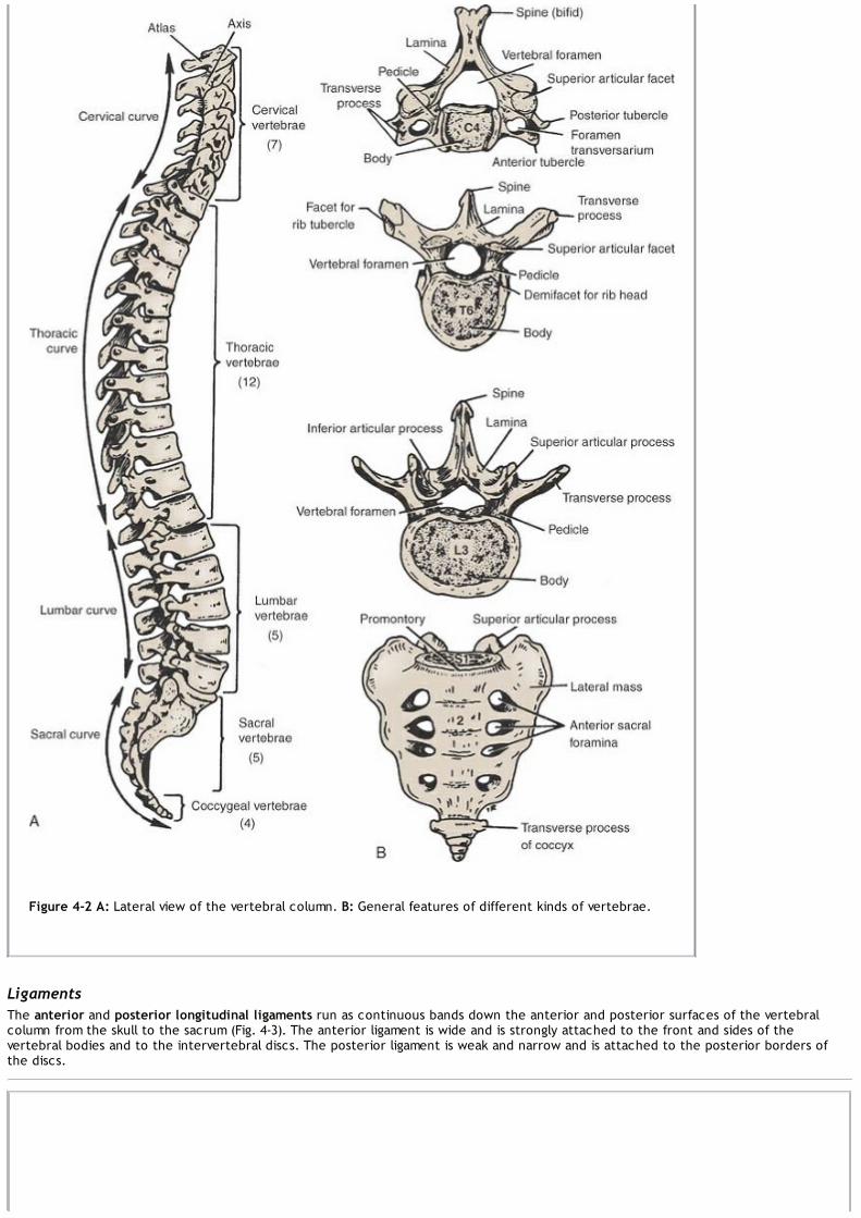
Figure 4-2 A: Lateral view of the vertebral column. B: General features of different kinds of vertebrae.
LigamentsThe anterior and posterior longitudinal ligaments run as continuous bands down the anterior and posterior surfaces of the vertebralcolumn from the skull to the sacrum (Fig. 4-3). The anterior ligament is wide and is strongly attached to the front and sides of thevertebral bodies and to the intervertebral discs. The posterior ligament is weak and narrow and is attached to the posterior borders ofthe discs.

Figure 4-3 A: Joints in the cervical, thoracic, and lumbar regions of the vertebral column. B: Third lumbar vertebra seen from aboveshowing the relationship between intervertebral disc and cauda equina.
Joints Between Two Vertebral ArchesThe joints between two vertebral arches consist of synovial joints between the superior and inferior articular processes of adjacentvertebrae (Fig. 4-3).
Ligaments
Supraspinous ligament (Fig. 4-3): This runs between the tips of adjacent spines.
Interspinous ligament (Fig. 4-3): This connects adjacent spines.
Intertransverse ligaments: These run between adjacent transverse processes.
Ligamentum flavum (Fig. 4-3): This connects the laminae of adjacent vertebrae.
In the cervical region, the supraspinous and interspinous ligaments are greatly thickened to form the strong ligamentum nuchae.

Figure 4-4 The innervation of vertebral joints. At any particular vertebral level, the joints receive nerve fibers from two adjacentspinal nerves.
Nerve Supply of Vertebral JointsThe joints between the vertebral bodies are innervated by the small meningeal branches of each spinal nerve (Fig. 4-4). The jointsbetween the articular processes are innervated by branches from the posterior rami of the spinal nerves (Fig. 4-4); the joints of anyparticular level receive nerve fibers from two adjacent spinal nerves.
The atlanto-occipital joints and the atlanto-axial joints should be reviewed in a textbook of gross anatomy.
Gross Appearance of the Spinal CordThe spinal cord is roughly cylindrical in shape. It begins superiorly at the foramen magnum in the skull, where it is continuous withthe medulla oblongata of the brain, and it terminates inferiorly in the adult at the level of the lower border of the first lumbar
vertebra. In the young child, it is relatively longer and usually ends at the upper border of the third lumbar vertebra. Thus, it occupies theupper two-thirds of the vertebral canal of the vertebral column and is surrounded by the three meninges, the dura mater, the arachnoidmater, and the pia mater. Further protection is provided by the cerebrospinal fluid, which surrounds the spinal cord in the subarachnoidspace.
In the cervical region, where it gives origin to the brachial plexus, and in the lower thoracic and lumbar regions, where it gives origin tothe lumbosacral plexus, the spinal cord is fusiformly enlarged; the enlargements are referred to as the cervical and lumbar enlargements(Fig. 4-5). Inferiorly, the spinal cord tapers off into the conus medullaris, from the apex of which a prolongation of the pia mater, thefilum terminale, descends to be attached to the posterior surface of the coccyx. The cord possesses a deep longitudinal fissure calledthe anterior median fissure in the midline anteriorly and a shallow furrow called the posterior median sulcus on the posterior surface(Fig. 4-5).
Along the entire length of the spinal cord are attached 31 pairs of spinal nerves by the anterior1 or motor roots and the posterior orsensory roots (Fig. 4-5). Each root is attached to the cord by a series of rootlets, which extend the whole length of the correspondingsegment of the cord. Each posterior nerve root possesses a posterior root ganglion, the cells of which give rise to peripheral and centralnerve fibers.

Figure 4-5 Spinal cord. A: Posterior view, showing cervical and lumbar enlargements. B: Three segments of the spinal cord showingthe coverings of dura mater, arachnoid mater, and pia mater.
Structure of the Spinal CordThe spinal cord is composed of an inner core of gray matter, which is surrounded by an outer covering of white matter (Figs. 4-6,4-7, 4-8, 4-9 and 4-10); there is no indication that the cord is segmented.
For a comparison of the structural details in different regions of the spinal cord, see Table 4-1.
Gray MatterOn cross section, the gray matter is seen as an H-shaped pillar with anterior and posterior gray columns, or horns, united by a thin graycommissure containing the small central canal (Fig. 4-6). A small lateral gray column or horn is present in the thoracic and upper lumbarsegments of the cord. The amount of gray matter present at any given level of the spinal cord is related to the amount of muscleinnervated at that level. Thus, its size is greatest within the cervical and lumbosacral enlargements of the cord, which innervate themuscles of the upper and lower limbs, respectively (Figs. 4-6, 4-7, 4-8, 4-9 and 4-10).
Table 4-1 Comparison of Structural Details in Different Regions of the Spinal Corda

Reg ion Shape White Matter Gray Matter
Anter ior Gray Column Poster ior Gray Column Lateral Gray Column
Cervical Oval
Fasciculuscuneatusandfasciculusgracilispresent
Medial group of cells forneck muscles; centralgroup of cells for accessorynucleus (C1-5) and phrenicnucleus (C3-5); lateral groupof cells for upper limbmuscles
Substantia gelatinosapresent, continuous withSp.N. of cranial nerve Vat level C2; nucleusproprius present;nucleus dorsalis (Clarke'scolumn) absent
Absent
Thoracic Round
Fasciculuscuneatus(T1-6) andfasciculusgracilispresent
Medial group of cells fortrunk muscles
Substantia gelatinosa,nucleus proprius,nucleus dorsalis (Clarke'scolumn), and visceralafferent nucleus present
Present; givesrise topreganglionicsympatheticfibers
LumbarRoundtooval
Fasciculuscuneatusabsent;fasciculusgracilispresent
Medial group of cells forlower limb muscles; centralgroup of cells forlumbosacral nerve
Substantia gelatinosa,nucleus proprius,nucleus dorsalis (Clarke'scolumn) at L1-4, andvisceral afferent nucleuspresent
Present (L1-2[3]); gives riseto preganglionicsympatheticfibers
Sacral Round
Smallamount;fasciculuscuneatusabsent;fasciculusgracilispresent
Medial group of cells forlower limb and perinealmuscles
Substantia gelatinosa andnucleus proprius present
Absent; groupof cells presentat S2-4, forparasympatheticoutflow
aThe information in this table is useful for identifying the specific level of the spinal cord from which a section has beentaken.
StructureAs in other regions of the central nervous system, the gray matter of the spinal cord consists of a mixture of nerve cells and theirprocesses, neuroglia, and blood vessels. The nerve cells are multipolar, and the neuroglia forms an intricate network around the nerve cellbodies and their neurites.
Nerve Cell Groups in the Anterior Gray ColumnsMost nerve cells are large and multipolar, and their axons pass out in the anterior roots of the spinal nerves as alpha efferents, whichinnervate skeletal muscles. The smaller nerve cells are also multipolar, and the axons of many of these pass out in the anterior roots of thespinal nerves as gamma efferents, which innervate the intrafusal muscle fibers of neuromuscular spindles.
For practical purposes, the nerve cells of the anterior gray column may be divided into three basic groups or columns: medial, central, andlateral (Fig. 4-6).2
The medial group is present in most segments of the spinal cord and is responsible for innervating the skeletal muscles of the neck andtrunk, including the intercostal and abdominal musculature.
The central group is the smallest and is present in some cervical and lumbosacral segments (Figs. 4-6 and 4-7). In the cervical part of thecord, some of these nerve cells (segments C3-5) specifically innervate the diaphragm and are collectively referred to as the phrenicnucleus (Fig. 4-7). In the upper five or six cervical segments, some of the nerve cells innervate the sternocleidomastoid and trapeziusmuscles and are referred to as the accessory nucleus (Figs. 4-6 and 4-7). The axons of these cells form the spinal part of the accessorynerve. The lumbosacral nucleus present in the second lumbar down to the first sacral segment of the cord is made up of nerve cellswhose axons have an unknown distribution.
The lateral group is present in the cervical and lumbosacral segments of the cord and is responsible for innervating the skeletal muscles ofthe limbs (Figs. 4-6, 4-7, 4-9, and 4-10).
Nerve Cell Groups in the Posterior Gray ColumnsThere are four nerve cell groups of the posterior gray column: two that extend throughout the length of the cord and two that arerestricted to the thoracic and lumbar segments.

The substantia gelatinosa group is situated at the apex of the posterior gray column throughout the length of the spinal cord (Figs. 4-6, 4-7, 4-8, 4-9 and 4-10). It is largely composed of Golgi type II neurons and receives afferent fibers concerned withpain, temperature, and touch from the posterior root. Furthermore, it receives input from descending fibers from supraspinal levels. It isbelieved that the inputs of the sensations of pain and temperature are modified by excitatory or inhibitory information from other sensoryinputs and by information from the cerebral cortex.
Figure 4-6 Transverse sections of the spinal cord at different levels showing the arrangement of the gray matter and white matter.
The nucleus proprius is a group of large nerve cells situated anterior to the substantia gelatinosa throughout the spinal cord (Figs. 4-6, 4-7,4-8, 4-9 and 4-10). This nucleus constitutes the main bulk of cells present in the posterior gray column and receives fibers from theposterior white column that are associated with the senses of position and movement (proprioception), two-point discrimination, andvibration.
The nucleus dorsalis (Clarke's column) is a group of nerve cells situated at the base of the posterior gray column and extending from theeighth cervical segment caudally to the third or fourth lumbar segment (Figs. 4-6, 4-7, 4-8 and 4-9). Most of the cells are comparativelylarge and are associated with proprioceptive endings (neuromuscular spindles and tendon spindles).

Figure 4-7 Transverse section of the spinal cord at the level of the fifth cervical segment. (Weigert stain.)
The visceral afferent nucleus is a group of nerve cells of medium size situated lateral to the nucleus dorsalis; it extends from the firstthoracic to the third lumbar segment of the spinal cord. It is believed to be associated with receiving visceral afferent information.
Nerve Cell Groups in the Lateral Gray ColumnsThe intermediolateral group of cells form the small lateral gray column, which extends from the first thoracic to the second or thirdlumbar segment of the spinal cord (Figs. 4-6 and 4-8). The cells are relatively small and give rise to preganglionic sympathetic fibers.
Figure 4-8 Transverse section of the spinal cord at the level of the second thoracic segment. (Weigert stain.)
A similar group of cells found in the second, third, and fourth sacral segments of the spinal cord give rise to preganglionic parasympatheticfibers (Figs. 4-6 and 4-10).
The Gray Commissure and Central CanalIn transverse sections of the spinal cord, the anterior and posterior gray columns on each side are connected by a transverse gray

commissure; the gray matter resembles the letter H (Figs. 4-6, 4-7, 4-8, 4-9 and 4-10). In the center of the gray commissure is situated thecentral canal. The part of the gray commissure that is situated posterior to the central canal is often referred to as the posterior graycommissure; similarly,the part that lies anterior to the canal is called the anterior gray commissure.
Figure 4-9 Transverse section of the spinal cord at the level of the fourth lumbar segment. (Weigert stain.)
The central canal is present throughout the spinal cord (Figs. 4-6, 4-7, 4-8, 4-9 and 4-10). Superiorly, it is continuous with the central canalof the caudal half of the medulla oblongata, and above this, it opens into the cavity of the fourth ventricle. Inferiorly in the conusmedullaris, it expands into the fusiform terminal ventricle and terminates below within the root of the filum terminale. It is filled withcerebrospinal fluid and is lined with ciliated columnar epithelium, the ependyma. Thus, the central canal is closed inferiorly and openssuperiorly into the fourth ventricle.
Figure 4-10 Transverse section of the spinal cord at the level of the second sacral segment. (Weigert stain.)

Figure 4-11 Transverse section of the spinal cord at the midcervical level showing the general arrangement of the ascending tractson the right and the descending tracts on the left.
White MatterThe white matter, for purposes of description, may be divided into anterior, lateral, and posterior white columns or funiculi (Figs. 4-5, 4-6, 4-7, 4-8, 4-9 and 4-10). The anterior column on each side lies between the midline and the point of emergence of the anterior nerveroots; the lateral column lies between the emergence of the anterior nerve roots and the entry of the posterior nerve roots; theposterior column lies between the entry of the posterior nerve roots and the midline.
StructureAs in other regions of the central nervous system, the white matter of the spinal cord consists of a mixture of nerve fibers, neuroglia, andblood vessels. It surrounds the gray matter, and its white color is due to the high proportion of myelinated nerve fibers.
Arrangement of Nerve Fiber TractsThe arrangement of the nerve fiber tracts within the spinal cord has been deduced as the result of animal experimentation and study ofthe human spinal cord for degenerative nerve fibers resulting from injury or disease. Although some nerve tracts are concentrated incertain areas of the white matter, it is now generally accepted that considerable overlap is present. For purposes of description, thespinal tracts are divided into ascending, descending, and intersegmental tracts, and their relative positions in the white matter aredescribed below. A simplified diagram, showing the general arrangement of the major tracts, is shown in Figure 4-11.
The Ascending Tracts of the Spinal CordOn entering the spinal cord, the sensory nerve fibers of different sizes and functions are sorted out and segregated into nervebundles or tracts in the white matter (Figs. 4-11 and 4-12). Some of the nerve fibers serve to link different segments of the spinal
cord, while others ascend from the spinal cord to higher centers and thus connect the spinal cord with the brain. It is the bundles of theascending fibers that are referred to as the ascending tracts.
The ascending tracts conduct afferent information, which may or may not reach consciousness. The information may be divided into twomain groups: (1) exteroceptive information, which originates from outside the body, such as pain, temperature, and touch, and (2)proprioceptive information, which originates from inside the body, for example, from muscles and joints.
Anatomical OrganizationGeneral information from the peripheral sensory endings is conducted through the nervous system by a series of neurons. In itssimplest form, the ascending pathway to consciousness consists of three neurons (Fig. 4-12). The first neuron, the first-order
neuron, has its cell body in the posterior root ganglion of the spinal nerve. A peripheral process connects with a sensory receptorending, whereas a central process enters the spinal cord through the posterior root to synapse on the second-order neuron.The second-order neuron gives rise to an axon that decussates (crosses to the opposite side) and ascends to a higher level of the centralnervous system, where it synapses with the third-order neuron (Fig. 4-12). The third-order neuron is usually in the thalamus and gives riseto a projection fiber that passes to a sensory region of the cerebral cortex (Fig. 4-12). This three-neuron chain is the most commonarrangement, but some afferent pathways use more or fewer neurons. Many of the neurons in the ascending pathways branch and give amajor input into the reticular formation, which, in turn, activates the cerebral cortex, maintaining wakefulness. Other branches pass to

motor neurons and participate in reflex muscular activity.
Figure 4-12 Simplest form of the ascending sensory pathway from the sensory nerve ending to the cerebral cortex. Note the threeneurons involved.
Functions of the Ascending TractsPainful and thermal sensations ascend in the lateral spinothalamic tract; light (crude) touch and pressure ascend in the anteriorspinothalamic tract (Fig. 4-13). Discriminative touch—that is, the ability to localize accurately the area of the body touched and
also to be aware that two points are touched simultaneously, even though they are close together (two-point discrimination)— ascends inthe posterior white columns (Fig. 4-13). Also ascending in the posterior white columns is information from muscles and joints pertaining tomovement and position of different parts of the body. In addition, vibratory sensations ascend in the posterior white column. Unconsciousinformation from muscles, joints, the skin, and subcutaneous tissue reaches the cerebellum by way of the anterior and posteriorspinocerebellar tracts and by the cuneocerebellar tract (Fig. 4-13). Pain, thermal, and tactile information is passed to the superiorcolliculus of the midbrain through the spinotectal tract for the purpose of spinovisual reflexes (Fig. 4-13). The spinoreticular tract providesa pathway from the muscles, joints, and skin to the reticular formation, while the spino-olivary tract provides an indirect pathway forfurther afferent information to reach the cerebellum (Fig. 4-13).
Pain and Temperature Pathways

Lateral Spinothalamic Tract3
The pain and thermal receptors in the skin and other tissues are free nerve endings. The pain impulses are transmitted to the spinal cordin fast-conducting delta A-type fibers and slow-conducting C-type fibers. The fast-conducting fibers alert the individual to initial sharppain, and the slow-conducting fibers are responsible for prolonged burning, aching pain. The sensations of heat and cold also travel bydelta A and C fibers.
The axons entering the spinal cord from the posterior root ganglion proceed to the tip of the posterior gray column and divide intoascending and descending branches (Fig. 4-14). These branches travel for a distance of one or two segments of the spinal cord and formthe posterolateral tract of Lissauer (Fig. 4-14). These fibers of the first-order neuron terminate by synapsing with cells in the posteriorgray column, including cells in the substantia gelatinosa. Substance P, a peptide, is thought to be the neurotransmitter at these synapses.
The axons of the second-order neurons now cross obliquely to the opposite side in the anterior gray and white commissures within onespinal segment of the cord, ascending in the contralateral white column as the lateral spinothalamic tract (Fig. 4-14). The lateralspinothalamic tract lies medial to the anterior spinocerebellar tract. Asthe lateral spinothalamic tract ascends through the spinal cord, new fibers are added to the anteromedial aspect of the tract. Thus, inthe upper cervical segments of the cord, the sacral fibers are lateral and the cervical segments are medial. The fibers carrying pain aresituated slightly anterior to those conducting temperature.
Figure 4-13 Transverse section of the spinal cord showing the origin of the main ascending sensory tracts. Note that the sensationsof pain and temperature ascend in the lateral spinothalamic tract, and light touch and pressure ascend in the anteriorspinothalamic tract.
As the lateral spinothalamic tract ascends through the medulla oblongata, it lies near the lateral surface and between the inferior olivarynucleus and the nucleus of the spinal tract of the trigeminal nerve. It is now accompanied by the anterior spinothalamic tract and thespinotectal tract; together they form the spinal lemniscus (Fig. 4-14).
The spinal lemniscus continues to ascend through the posterior part of the pons (Fig. 4-14). In the midbrain, it lies in the tegmentumlateral to the medial lemniscus. Many of the fibers of the lateral spinothalamic tract end by synapsing with the third-order neuron in theventral posterolateral nucleus of the thalamus (Fig. 4-14). It is believed that here crude pain and temperature sensations are appreciatedand emotional reactions are initiated.
The axons of the third-order neurons in the ventral posterolateral nucleus of the thalamus now pass through the posterior limb of theinternal capsule and the corona radiata to reach the somesthetic area in the postcentral gyrus of the cerebral cortex (Fig. 4-14). Thecontralateral half of the body is represented as inverted, with the hand and mouth situated inferiorly and the leg situated superiorly, andwith the foot and anogenital region on the medial surface of the hemisphere. (For details, see p. 291.) From here, the information istransmitted to other regions of the cerebral cortex to be used by motor areas and the parietal association area. The role of the cerebralcortex is interpreting the quality of the sensory information at the level of consciousness.
Pain Reception

The perception of pain is a complex phenomenon that is influenced by the emotional state and past experiences of the individual. Pain is asensation that warns of potential injury and alerts the person to avoid or treat it.
Pain can be divided into two main types: fast pain and slow pain. Fast pain is experienced within about 0.1 second after the pain stimulusis applied; slow pain is felt 1.0 second or later after the stimulation. Fast pain is described by the patient as sharp pain, acute pain, orpricking pain and is the type of pain felt after pricking the finger with a needle. Fast pain is almost confined to the skin.
Slow pain is described as burning pain, aching pain, and throbbing pain and is produced when there is tissue destruction, as for example,in the development of an abscess or in severe arthritis. Slow pain can occur in any tissue of the body.
Figure 4-14 Pain and temperature pathways.
All types of pain reception take place in free nerve endings. Fast pain is experienced by mechanical or thermal types of stimuli, and slowpain may be elicited by mechanical, thermal, and chemical stimuli.
Many chemical substances have been found in extracts from damaged tissue that will excite free nerve endings. These include serotonin;
histamine; bradykinin; acids, such as lactic acid; and K ions. The threshold for pain endings can be lowered by prostaglandins andsubstance P, but they cannot stimulate the endings directly by themselves.
The individual should be aware of the existence of stimuli that, if allowed to persist, will bring about tissue destruction; pain receptorshave little or no adaptation.
Conduction of Pain to the Central Nervous SystemFast pain travels in peripheral nerves in large diameter A delta axons at velocities of between 6 and 30 msec. Slow pain travels in the smalldiameter C fibers at velocities between 0.5 and 2.0 msec. The fast pain impulses reach consciousness first to alert the individual to dangerso that a suitable protective response may take place. Slow pain is appreciated later and lasts much longer.
Conduction of Pain in the Central Nervous SystemThe afferent pain fibers enter the spinal cord, for example, in the posterior roots of a spinal nerve and terminate predominantly in thesuperficial layers of the posterior gray horn. The main excitatory neurotransmitter released by the A delta fibers and the C fibers is theamino acid glutamate. Substance P, a neuropeptide, is also released from the C fibers. Whereas glutamate is a fast-acting localizedneurotransmitter, substance P has a slow release and diffuses widely in the posterior horn and can influence many neurons.
The initial sharp, pricking, fast-acting pain fibers stimulate the second-order neurons of the lateral spinothalamic tract. The axonsimmediately cross to the opposite side of the spinal cord and ascend to the thalamus where they are relayed to the sensory post centralgyrus. The burning, aching, slow-acting pain fibers also stimulate the second-order neurons of the lateral spinal thalamic tract in theposterior gray horn and ascend with the axons of the fast-acting pain fibers. It is now believed, however, that most of the incoming slowfibers to the spinal cord take part in additional relays involving several neurons in the posterior horn before ascending in the spinal cord.The repeated arrival of noxious stimuli through the C fibers in the posterior gray horn during severe injury results in an increased responseof the second-order neurons. This winding up phenomenon is attributed to the release of the neurotransmitter glutamate from the Cfibers.
The fast type of pain is precisely localized. For example, if one hits the thumb with a hammer, there is no doubt where the injury hasoccurred. The slow type of pain is only poorly localized. For example, in a patient with osteoarthritis of the hip joint, the individual canonly vaguely localize the pain to the hip area and not to the specific site of the disease. This may be explained by the fact that fast painfibers directly ascend the spinal cord in the lateral spinothalamic tract, whereas the slow pain fibers take part in multiple relays in theposterior gray horn before ascending to higher centers.
Other Terminations of the Lateral Spinothalamic TractIt is now generally agreed that the fast pain impulses travel directly up to the ventral posterolateral nucleus of the thalamus and are thenrelayed to the cerebral cortex.
The majority of the slow pain fibers in the lateral spinothalamic tract terminate in the reticular formation, which then activates the entirenervous system. It is in the lower areas of the brain that the individual becomes aware of the chronic, nauseous, suffering type of pain.
As the result of research using the positron emission tomography scan, the postcentral gyrus, the cingulate gyrus of the limbic system, andthe insular gyrus are sites concerned with the reception and interpretation of the nociceptor information. The postcentral gyrus isresponsible for the interpretation of pain in relation to past experiences. The cingulate gyrus is involved with the interpretation of theemotional aspect of pain, whereas the insular gyrus is concerned with the interpretation of pain stimuli from the internal organs of thebody and brings about an autonomic response.
The reception of pain information by the central nervous system can be modulated first in the posterior gray horns of the spinal cord andat other sites at higher levels.
Pain Control in the Central Nervous System
The Gating TheoryMassage and the application of liniments to painful areas in the body can relieve pain. The technique of acupuncture, which wasdiscovered several thousand years ago in China, is also beneficial in relieving pain. Low-frequency electrical stimulation of the skin alsorelieves pain in certain cases. Although the precise mechanism for these phenomena is not understood, the gating theory was proposedsome years ago. It was suggested that at the site where the pain fiber enters the central nervous system, inhibition could occur by meansof connector neurons excited by large, myelinated afferent fibers carrying information of nonpainful touch and pressure. The excesstactile stimulation produced by massage, for example, “closed the gate” for pain. Once the nonpainful tactile stimulation ceased,however, “the gate was opened,” and information on the painful stimuli ascended the lateral spinothalamic tract. Although the gatetheory may partially explain the phenomena, the analgesia system is probably involved with the liberation of enkephalins and endorphins inthe posterior gray columns.
The Analgesia SystemStimulation of certain areas of the brainstem can reduce or block sensations of pain. These areas include the periventricular area of thediencephalon, the periaqueductal gray matter of the midbrain, and midline nuclei of the brainstem. It is believed that fibers of thereticulospinal tract pass down to the spinal cord and synapse on cells concerned with pain sensation in the posterior gray column. Theanalgesic system can suppress both sharp pricking pain and burning pain sensations.
Recently, two compounds with morphinelike actions, called the enkephalins and the endorphins, have been isolated in the centralnervous system. These compounds and serotonin serve as neurotransmitter substances in the analgesic system of the brain, and they mayinhibit the release of substance P in the posterior gray column.
Light (Crude) Touch and Pressure Pathways

Anterior Spinothalamic TractThe axons enter the spinal cord from the posterior root ganglion and proceed to the tip of the posterior gray column,where they divide into ascending and descending branches (Fig. 4-15). These branches travel for a distance of one or two segments of thespinal cord, contributing to the posterolateral tract of Lissauer. It is believed that these fibers of the first-order neuron terminate bysynapsing with cells in the substantia gelatinosa group in the posterior gray column (Fig. 4-15).
Figure 4-15 Light touch and pressure pathways.
The axons of the second-order neuron now cross very obliquely to the opposite side in the anterior gray and white commissures withinseveral spinal segments and ascend in the opposite anterolateral white column as the anterior spinothalamic tract (Fig. 4-15). As theanterior spinothalamic tract ascends through the spinal cord, new fibers are added to the medial aspect of the tract. Thus, inthe upper cervical segments of the cord, the sacral fibers are mostly lateral and the cervical segments are mostly medial.
As the anterior spinothalamic tract ascends through the medulla oblongata, it accompanies the lateral spinothalamic tract and the

spinotectal tract, all of which form the spinal lemniscus (Fig. 4-15).
The spinal lemniscus continues to ascend through the posterior part of the pons, and the tegmentum of the midbrain and the fibers of theanterior spinothalamic tract terminate by synapsing with the third-order neuron in the ventral posterolateral nucleus of the thalamus (Fig.4-15). Crude awareness of touch and pressure is believed to be appreciated here.
The axons of the third-order neurons in the ventral posterolateral nucleus of the thalamus pass through the posterior limb of the internalcapsule (Fig. 4-15) and the corona radiata to reach the somesthetic area in the postcentral gyrus of the cerebral cortex. The contralateralhalf of the body is represented inverted, with the hand and mouth situated inferiorly, as described previously. (For details, see p. 291.)The conscious appreciation of touch and pressure depends on the activity of the cerebral cortex. The sensations can be only crudelylocalized, and very little discrimination of intensity is possible.
Discriminative Touch, Vibratory Sense, and Conscious Muscle Joint Sense
Posterior White Column: Fasciculus Gracilis and Fasciculus CuneatusThe axons enter the spinal cord from the posterior root ganglion and pass directly to the posterior white column of the same side (Fig. 4-16). Here, the fibers divide into long ascending and short descending branches. The descending branches pass down a variable number ofsegments, giving off collateral branches that synapse with cells in the posterior gray horn, with internuncial neurons, and with anteriorhorn cells (Fig. 4-16). It is clear that these short descending fibers are involved with intersegmental reflexes.
The long ascending fibers may also end by synapsing with cells in the posterior gray horn, with internuncial neurons, and with anteriorhorn cells. This distribution may extend over numerous segments of the spinal cord (Fig. 4-16). As in the case of the short descendingfibers, they are involved with intersegmental reflexes.
Many of the long ascending fibers travel upward in the posterior white column as the fasciculus gracilis and fasciculus cuneatus (Fig. 4-16).The fasciculus gracilis is present throughout the length of the spinal cord and contains the long ascending fibers from the sacral, lumbar,and lower six thoracic spinal nerves. The fasciculus cuneatus is situated laterally in the upper thoracic and cervical segments of the spinalcord and is separated from the fasciculus gracilis by a septum. The fasciculus cuneatus contains the long ascending fibers from the uppersix thoracic and all the cervical spinal nerves.
The fibers of the fasciculus gracilis and fasciculus cuneatus ascend ipsilaterally and terminate by synapsing on the second-order neuronsin the nuclei gracilis and cuneatus of the medulla oblongata (Fig. 4-16). The axons of the second-order neurons, called the internalarcuate fibers, sweep anteromedially around the central gray matter and cross the median plane, decussating with the correspondingfibers of the opposite side in the sensory decussation (Fig. 4-16). The fibers then ascend as a single compact bundle, the mediallemniscus, through the medulla oblongata, the pons, and the midbrain (Fig. 4-16). The fibers terminate by synapsing on the third-orderneurons in the ventral posterolateral nucleus of the thalamus.
The axons of the third-order neuron leave and pass through the posterior limb of the internal capsule and corona radiata to reach thesomesthetic area in the postcentral gyrus of the cerebral cortex (Fig. 4-16). The contralateral half of the body is represented inverted,with the hand and mouth situated inferiorly, as described previously. (For details, see p. 291.) In this manner, the impressions of touchwith fine gradations of intensity, exact localization, and two-point discrimination can be appreciated. Vibratory sense and the position ofthe different parts of the body can be consciously recognized.
Many fibers in the fasciculus cuneatus from the cervical and upper thoracic segments, having terminated on the second-order neuron ofthe nucleus cuneatus, are relayed and travel as the axons of the second-order neurons to enter the cerebellum through the inferiorcerebellar peduncle of the same side (Fig. 4-16). The pathway is referred to as the cuneocerebellar tract, and the fibers are known as theposterior external arcuate fibers. The function of these fibers is to convey information of muscle joint sense to the cerebellum.
The main somatosensory pathways are summarized in Table 4-2.
Muscle Joint Sense Pathways to the Cerebellum
Posterior Spinocerebellar TractThe axons entering the spinal cord from the posterior root ganglion enter the posterior gray column and terminate by synapsing on thesecond-order neurons at the base of the posterior gray column (Fig. 4-17). These neurons are known collectively as the nucleus dorsalis(Clarke's column). The axons of the second-order neurons enter the posterolateral part of the lateral white column on the same side andascend as the posterior spinocerebellar tract to the medulla oblongata. Here, the tract joins the inferior cerebellar peduncle andterminates in the cerebellar cortex (Fig. 4-17). Note that it does not ascend to the cerebral cortex. Because the nucleus dorsalis (Clarke'scolumn) extends only from the eighth cervical segment caudally to the third or fourth lumbar segment, axons entering the spinal cordfrom the posterior roots of the lower lumbar and sacral segments ascend in the posterior white column until they reach the third orfourth lumbar segment, where they enter the nucleus dorsalis.
The posterior spinocerebellar fibers receive muscle joint information from the muscle spindles, tendon organs, and joint receptors of thetrunk and lower limbs. This information concerning tension of muscle tendons and the movements of muscles and joints is used by thecerebellum in the coordination of limb movements and the maintenance of posture.

P.152
Figure 4-16 Discriminative touch, vibratory sense, and conscious muscle joint sense pathways.
Anterior Spinocerebellar TractThe axons entering the spinal cord from the posterior root ganglion terminate by synapsing with the second-order neurons in the nucleusdorsalis at the base of the posterior gray column (Fig. 4-17). The majority of the axons of the second-order neurons cross to the oppositeside and ascend as the anterior spinocerebellar tract in the contralateral white column; the minority of the axons ascend as the anteriorspinocerebellar tract in the lateral white column of the same side (Fig. 4-17). The fibers, having ascended through the medulla oblongataand pons, enter the cerebellum through the superior cerebellar peduncle and terminate in the cerebellar cortex. It is believed that thosefibers that crossed over to the opposite side in the spinal cord cross back within the cerebellum (Fig. 4-17). The anterior spinocerebellartract conveys muscle joint information from themuscle spindles, tendon organs, and joint receptors of the trunk and the upper and lower limbs. It is also believed that thecerebellum receives information from the skin and superficial fascia by this tract.
Table 4-2 The Main Somatosensory Pathways to Consciousnessa

Sensation Receptor Fir st-OrderNeuron
Second-OrderNeuron
Third-OrderNeuron Pathways Destination
Pain andtemperature
Free nerveendings
Posteriorrootganglion
Substantiagelatinosa
Ventralposterolateralnucleus ofthalamus
Lateralspinothalamic,spinallemniscus
Posteriorcentralgyrus
Light touch andpressure
Free nerveendings
Posteriorrootganglion
Substantiagelatinosa
Ventralposterolateralnucleus ofthalamus
Anteriorspinothalamic,spinallemniscus
Posteriorcentralgyrus
Discriminativetouch, vibratorysense,consciousmuscle jointsense
Meissner'scorpuscles,paciniancorpuscles,muscle spindles,tendon organs
Posteriorrootganglion
Nucleigracilisandcuneatus
Ventralposterolateralnucleus ofthalamus
Fasciculigracilis andcuneatus,mediallemniscus
Posteriorcentralgyrus
aNote that all ascending pathways send branches to the reticular activating system.

Figure 4-17 Unconscious muscle joint sense pathways to the cerebellum.
Table 4-3 Muscle Joint Sense Pathways to the Cerebellum
Sensation Receptor Fir st-Order Neuron Second-Order Neuron Pathways Destination
Unconsciousmusclejoint sense
Musclespindles,tendonorgans,jointreceptors
Posterior rootganglion Nucleus dorsalis
Anterior andposteriorspinocere-bellar
Cerebellarcortex
The muscle joint sense pathways to the cerebellum are summarized in Table 4-3.
Cuneocerebellar TractThese fibers have already been described on page 149. They originate in the nucleus cuneatus and enter the cerebellum through theinferior cerebellar peduncle of the same side (Fig. 4-16). The fibers are known as the posterior external arcuate fibers, and their functionis to convey information of muscle joint sense to the cerebellum.
Other Ascending Pathways
Spinotectal TractThe axons enter the spinal cord from the posterior root ganglion and travel to the gray matter where they synapse on unknown second-order neurons (Fig. 4-18). The axons of the second-order neurons cross the median plane and ascend as the spinotectal tract in theanterolateral white column lying close to the lateral spinothalamic tract. After passing through the medulla oblongata and pons, theyterminate by synapsing with neurons in the superior colliculus of the midbrain (Fig. 4-18). This pathway provides afferent information forspinovisual reflexes and brings about movements of the eyes and head toward the source of the stimulation.
Spinoreticular TractThe axons enter the spinal cord from the posterior root ganglion and terminate on unknown second-order neurons in the gray matter (Fig.4-18). The axons from these second-order neurons ascend the spinal cord as the spinoreticular tract in the lateral white column mixedwith the lateral spinothalamic tract. Most of the fibers are uncrossed and terminate by synapsing with neurons of the reticular formationin the medulla oblongata, pons, and midbrain (Fig. 4-18). The spinoreticular tract provides an afferent pathway for the reticular formation,which plays an important role in influencing levels of consciousness. (For details, see p. 297.)
Spino-olivary TractThe axons enter the spinal cord from the posterior root ganglion and terminate on unknown second-order neurons in the posterior graycolumn (Fig. 4-18). The axons from the second-order neurons cross the midline and ascend as the spino-olivary tract in the white matterat the junction of the anterior and lateral columns. The axons end by synapsing on third-order neurons in the inferior olivary nuclei in themedulla oblongata (Fig. 4-18). The axons of the third-order neurons cross the midline and enter the cerebellum through the inferiorcerebellar peduncle. The spino-olivary tract conveys information to the cerebellum from cutaneous and proprioceptive organs.
Visceral Sensory TractsSensations that arise in viscera located in the thorax and abdomen enter the spinal cord through the posterior roots. The cell bodies ofthe first-order neuron are situated in the posterior root ganglia. The peripheral processes of these cells receive nerve impulses from pain4
and stretch receptor endings in the viscera. The central processes, having entered the spinal cord, synapse with second-order neurons inthe gray matter, probably in the posterior or lateral gray columns.
The axons of the second-order neurons are believed to join the spinothalamic tracts and ascend and terminate on the third-orderneurons in the ventral posterolateral nucleus of the thalamus. The final destination of the axons of the third-order neurons is probably inthe postcentral gyrus of the cerebral cortex.
Many of the visceral afferent fibers that enter the spinal cord branch participate in reflex activity.

Figure 4-18 Spinotectal, spinoreticular, and spino-olivary tracts.
The Descending Tracts of the Spinal CordThe motor neurons situated in the anterior gray columns of the spinal cord send axons to innervate skeletal muscle through theanterior roots of the spinal nerves. These motor neurons are sometimes referred to as the lower motor neurons and constitute
the final common pathway to the muscles (Fig. 4-19).
The lower motor neurons are constantly bombarded by nervous impulses that descend from the medulla, pons, midbrain, and cerebralcortex as well as those that enter along sensory fibers from the posterior roots. The nerve fibers that descend in the white matter fromdifferent supraspinal nerve centers are segregated into nerve bundles called the descending tracts. These supraspinal neurons and theirtracts are sometimes referred to as the upper motor neurons, and they provide numerous separate pathways that can influence motoractivity.
Anatomical OrganizationControl of skeletal muscle activity from the cerebral cortex and other higher centers is conducted through the nervous system bya series of neurons (Fig. 4-19). The descending pathway from the cerebral cortex is often made up of three
neurons. The first neuron, the first-order neuron, has its cell body in the cerebral cortex. Its axon descends to synapse on the second-order neuron, an internuncial neuron, situated in the anterior gray column of the spinal cord (Fig. 4-19). The axon of the second-orderneuron is short and synapses with the third-order neuron, the lower motor neuron, in the anterior gray column (Fig. 4-19). The axon ofthe third-order neuron innervates the skeletal muscle through the anterior root and spinal nerve. In some instances, the axon of the first-

order neuron terminates directly on the third-order neuron (as in reflex arcs).
Figure 4-19 Simple form of the descending motor pathway from the cerebral cortex to the skeletal muscle. Note the three neuronsinvolved.
Functions of the Descending TractsThe corticospinal tracts (Fig. 4-20) are the pathways concerned with voluntary, discrete, skilled movements, especially those ofthe distal parts of the limbs. The reticulospinal tracts may facilitate or inhibit the activity of the alpha and gamma motor neurons
in the anterior gray columns and may, therefore, facilitate or inhibit voluntary movement or reflex activity. The tectospinal tract (Fig. 4-20)is concerned withreflex postural movements in response to visual stimuli. Those fibers that are associated with the sympathetic neurons in the lateral graycolumn are concerned with the pupillodilation reflex in response to darkness. The rubrospinal tract (Fig. 4-20) acts on both the alpha andgamma motor neurons in the anterior gray columns and facilitates the activity of flexor muscles and inhibits the activity of extensor orantigravity muscles. The vestibulospinal tract (Fig. 4-20), by acting on the motor neurons in the anterior gray columns, facilitates theactivity of the extensor muscles, inhibits the activity of the flexor muscles, and is concerned with the postural activity associated withbalance. The olivospinal tract (Fig. 4-20) may play a role in muscular activity, but there is doubt that it exists. The descending autonomicfibers are concerned with the control of visceral activity.

Figure 4-20 Transverse section of the spinal cord showing the termination of the descending motor tracts. Note that there is nowconsiderable doubt as to the existence of the olivospinal tract as a separate pathway.
Corticospinal TractsFibers of the corticospinal tract arise as axons of pyramidal cells situated in the fifth layer of the cerebral cortex (Fig. 4-21).About one-third of the fibers originate from the primary motor cortex (area 4), one-third originate from the secondary motor
cortex (area 6), and one-third originate from the parietal lobe (areas 3, 1, and 2); thus, two-thirds of the fibers arise from the precentralgyrus, and one-third of the fibers arise from the postcentral gyrus.5 Because electrical stimulation of different parts of the precentralgyrus produces movements of different parts of the opposite side of the body, we can represent the parts of the body in this area of thecortex. Such a homunculus is shown in Figure 4-21. Note that the region controlling the face is situated inferiorly, and the regioncontrolling the lower limb is situated superiorly and on the medial surface of the hemisphere. The homunculus is a distorted picture of thebody, with the various parts having a size proportional to the area of the cerebral cortex devoted to their control. It is interesting to findthat the majority of the corticospinal fibers are myelinated and are relatively slow-conducting, small fibers.
The descending fibers converge in the corona radiata and then pass through the posterior limb of the internal capsule (Fig. 4-21). Here,the fibers are organized so that those closest to the genu are concerned with cervical portions of the body, while those situated moreposteriorly are concerned with the lower extremity. The tract then continues through the middle three-fifths of the basis pedunculi ofthe midbrain (Fig. 4-21). Here, the fibers concerned with cervical portions of the body are situated medially, while those concerned withthe leg are placed laterally.
On entering the pons, the tract is broken into many bundles by the transverse pontocerebellar fibers (see Figs. 5-0, 5-0, and 5-0). In themedulla oblongata, the bundles become grouped together along the anterior border to form a swelling known as the pyramid (hence thealternative name, pyramidal tract) (see Fig. 5-0). At the junction of the medulla oblongata and the spinal cord, most of the fibers cross themidline at the decussation of the pyramids (Fig. 4-21) and enter the lateral white columnof the spinal cord to form the lateral corticospinal tract (Fig. 4-20). The remaining fibers do not cross in the decussation but descend inthe anterior white column of the spinal cord as the anterior corticospinal tract (Figs. 4-20 and 4-21). These fibers eventually cross themidline and terminate in the anterior gray column of the spinal cord segments in the cervical and upper thoracic regions.

Figure 4-21 Corticospinal tracts.
The lateral corticospinal tract descends the length of the spinal cord; its fibers terminate in the anterior gray column of all the spinalcord segments.
Most corticospinal fibers synapse with internuncial neurons, which, in turn, synapse with alpha motor neurons and some gamma motorneurons. Only the largest corticospinal fibers synapse directly with the motor neurons.
The corticospinal tracts are not the sole pathway for serving voluntary movement. Rather, they form the pathway that confers speed andagility to voluntary movements and is thus used in performing rapid skilled movements. Many of the simple, basic voluntary movements aremediated by other descending tracts.
Branches
1. Branches are given off early in their descent and return to the cerebral cortex to inhibit activity in adjacent regions of the cortex.
2. Branches pass to the caudate and lentiform nuclei, the red nuclei, and the olivary nuclei and the reticular formation. These branches
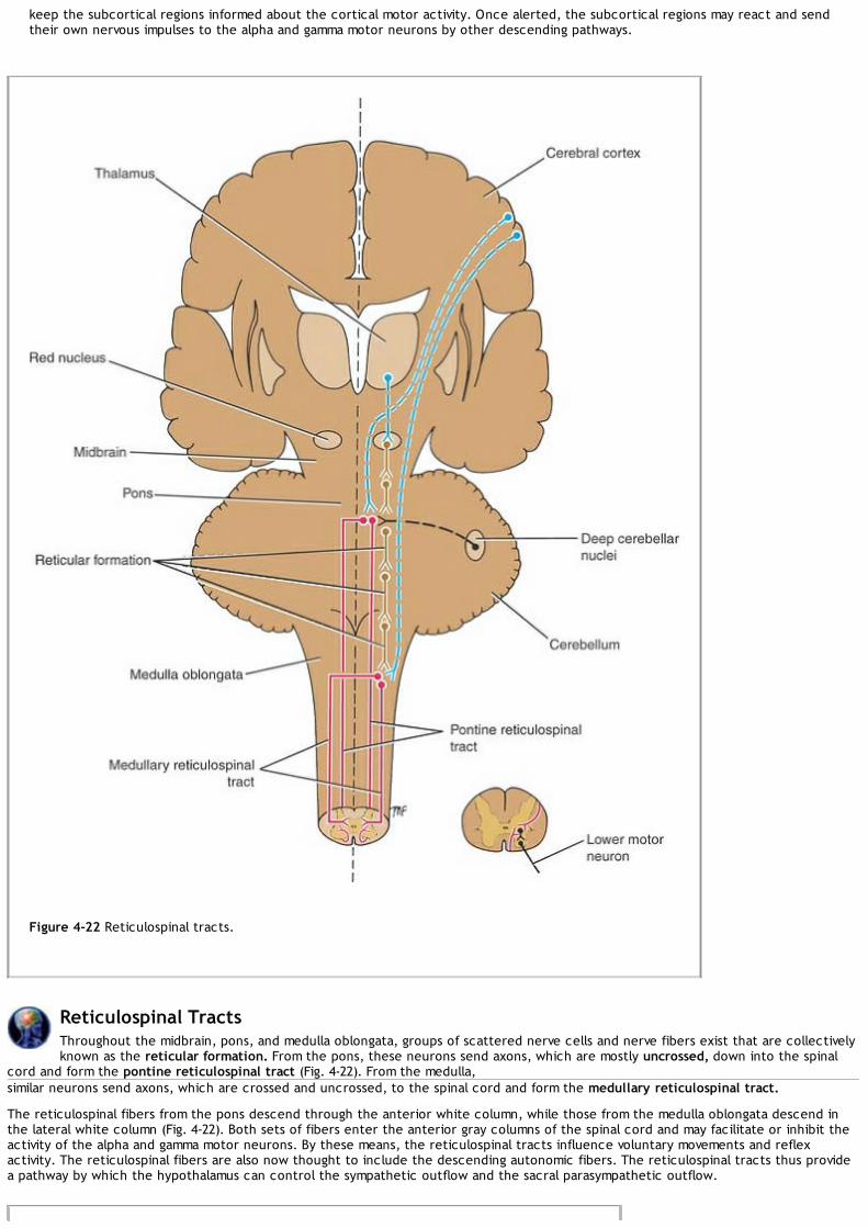
keep the subcortical regions informed about the cortical motor activity. Once alerted, the subcortical regions may react and sendtheir own nervous impulses to the alpha and gamma motor neurons by other descending pathways.
Figure 4-22 Reticulospinal tracts.
Reticulospinal TractsThroughout the midbrain, pons, and medulla oblongata, groups of scattered nerve cells and nerve fibers exist that are collectivelyknown as the reticular formation. From the pons, these neurons send axons, which are mostly uncrossed, down into the spinal
cord and form the pontine reticulospinal tract (Fig. 4-22). From the medulla,similar neurons send axons, which are crossed and uncrossed, to the spinal cord and form the medullary reticulospinal tract.
The reticulospinal fibers from the pons descend through the anterior white column, while those from the medulla oblongata descend inthe lateral white column (Fig. 4-22). Both sets of fibers enter the anterior gray columns of the spinal cord and may facilitate or inhibit theactivity of the alpha and gamma motor neurons. By these means, the reticulospinal tracts influence voluntary movements and reflexactivity. The reticulospinal fibers are also now thought to include the descending autonomic fibers. The reticulospinal tracts thus providea pathway by which the hypothalamus can control the sympathetic outflow and the sacral parasympathetic outflow.

Figure 4-23 Tectospinal tract.
Tectospinal TractFibers of this tract arise from nerve cells in the superior colliculus of the midbrain (Fig. 4-23). Most of the fibers cross the midlinesoon after their origin and descend through the brainstem close to the medial longitudinal fasciculus. The tectospinal tract
descends through the anterior white column of the spinal cord close to the anterior median fissure (Figs. 4-20 and 4-23). The majority ofthe fibers terminate in the anterior gray column in the upper cervical segments of the spinal cord by synapsing with internuncial neurons.These fibers are believed to be concerned with reflex postural movements in response to visual stimuli.
Rubrospinal TractThe red nucleus is situated in the tegmentum of the midbrain at the level of the superior colliculus (Fig. 4-24). The axons ofneurons in this nucleus cross the midline at the level of the nucleus and descend as the rubrospinal tract through the pons and
medulla oblongata to enter the lateral white column of the spinal cord (Figs. 4-20 and 4-24). The fibers terminate by synapsing withinternuncial neurons in the anterior gray column of the cord.
The neurons of the red nucleus receive afferent impulses through connections with the cerebral cortex and the cerebellum. This isbelieved to be an important indirect pathway by which the cerebral cortex and the cerebellum can influence the activity of the alpha andgamma motor neurons of the spinal cord. The tract facilitates the activity of the flexor muscles and inhibits the activity of the extensor or

antigravity muscles.
Figure 4-24 Rubrospinal tract.
Vestibulospinal TractThe vestibular nuclei are situated in the pons and medulla oblongata beneath the floor of the fourth ventricle (Fig. 4-25). Thevestibular nuclei receive afferent fibers from the inner ear through the vestibular nerve and from the cerebellum. The neurons of
the lateral vestibular nucleus give rise to the axons that form the vestibulospinal tract. The tract descends uncrossed through the medullaand through the length of the spinal cord in the anterior white column(Figs. 4-20 and 4-25). The fibers terminate by synapsing with internuncial neurons of the anterior gray column of the spinal cord.

Figure 4-25 Vestibulospinal tract.
The inner ear and the cerebellum, by means of this tract, facilitate the activity of the extensor muscles and inhibit the activity of theflexor muscles in association with the maintenance of balance.
Olivospinal TractThe olivospinal tract was thought to arise from the inferior olivary nucleus and to descend in the lateral white column of thespinal cord (Fig. 4-26), to influence the activity of the motor neurons in the anterior gray column. There is now considerable
doubt that it exists.
Descending Autonomic FibersThe higher centers of the central nervous system associated with the control of autonomic activity are situated in the cerebralcortex, hypothalamus, amygdaloid complex, and reticular formation. Although distinct tracts
have not been recognized, investigation of spinal cord lesions has demonstrated that descending autonomic tracts do exist and probablyform part of the reticulospinal tract.

Figure 4-26 Olivospinal tract. There is now considerable doubt as to the existence of this tract as a separate pathway.
The fibers arise from neurons in the higher centers and cross the midline in the brainstem. They are believed to descend in the lateralwhite column of the spinal cord and to terminate by synapsing on the autonomic motor cells in the lateral gray columns in the thoracicand upper lumbar (sympathetic outflow) and midsacral (parasympathetic) levels of the spinal cord.
A summary of the main descending pathways in the spinal cord is shown in Table 4-4.
Intersegmental TractsShort ascending and descending tracts that originate and end within the spinal cord exist in the anterior, lateral, and posteriorwhite columns. The function of these pathways is to interconnect the neurons of different segmental levels, and the pathways
are particularly important in intersegmental spinal reflexes.
Table 4-4 The Main Descending Pathways to the Spinal Corda
Pathway Function Or ig in Site of Crossover Destination Branches to

Corticospinaltracts
Rapid, skilled,voluntarymovements,especially distalends of limbs
Primary motorcortex (area4), secondarymotor cortex(area 6),parietal lobe(areas 3, 1, and2)
Most cross atdecussation of pyramidsand descend as lateralcorticospinal tracts;some continue asanterior corticospinaltracts and cross over atlevel of destination
Internuncialneurons oralpha motorneurons
Cerebralcortex,basalnuclei,rednucleus,olivarynuclei,reticularformation
Reticulospinaltracts
Inhibit orfacilitatevoluntarymovement;hypothalamuscontrolssympathetic,para-sympatheticoutflows
Reticularformation
Some cross at variouslevels
Alpha andgamma motorneurons
Multiplebranchesas theydescend
Tectospinaltract
Reflex posturalmovementsconcerningsight
Superiorcolliculus Soon after origin
Alpha andgamma motorneurons
?
Rubrospinaltract
Facilitatesactivity offlexor musclesand inhibitsactivity ofextensormuscles
Red nucleus ImmediatelyAlpha andgamma motorneurons
?
Vestibulospinaltract
Facilitatesactivity ofextensorinhibits flexormuscles
Vestibularnuclei Uncrossed
Alpha andgamma motorneurons
?
Olivospinaltract ?? Inferior olivary
nuclei Cross in brainstem? Alpha andgamma motorneurons
—
Descendingautonomicfibers
Controlsympatheticand para-sympatheticsystems
Cerebralcortex,hypothalamus,amygdaloidcomplex,reticularformation
Sympatheticandparasympatheticoutflows
—
aNote that the corticospinal tracts are believed to control the prime mover muscles (especially the highly skilled movements),whereas the other descending tracts are important in controlling the simple basic movements. For simplicity, theinternuncial neurons are omitted from this table.
Reflex ArcA reflex may be defined as an involuntary response to a stimulus. It depends on the integrity of the reflex arc (Fig. 4-27). In itssimplest form, a reflex arc consists of the following anatomical structures: (1) a receptor organ, (2) an afferent neuron, (3) an
effector neuron, and (4) an effector organ. A reflex arc involving only one synapse is referred to as a monosynaptic reflex arc.Interruption of the reflex arc at any point along its course would abolish the response.
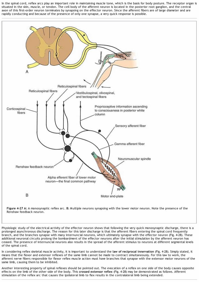
In the spinal cord, reflex arcs play an important role in maintaining muscle tone, which is the basis for body posture. The receptor organ issituated in the skin, muscle, or tendon. The cell body of the afferent neuron is located in the posterior root ganglion, and the centralaxon of this first-order neuron terminates by synapsing on the effector neuron. Since the afferent fibers are of large diameter and arerapidly conducting and because of the presence of only one synapse, a very quick response is possible.
Figure 4-27 A: A monosynaptic reflex arc. B: Multiple neurons synapsing with the lower motor neuron. Note the presence of theRenshaw feedback neuron.
Physiologic study of the electrical activity of the effector neuron shows that following the very quick monosynaptic discharge, there is aprolonged asynchronous discharge. The reason for this later discharge is that the afferent fibers entering the spinal cord frequentlybranch, and the branches synapse with many internuncial neurons, which ultimately synapse with the effector neuron (Fig. 4-28). Theseadditional neuronal circuits prolong the bombardment of the effector neurons after the initial stimulation by the afferent neuron hasceased. The presence of internuncial neurons also results in the spread of the afferent stimulus to neurons at different segmental levelsof the spinal cord.
In considering reflex skeletal muscle activity, it is important to understand the law of reciprocal innervation (Fig. 4-28). Simply stated, itmeans that the flexor and extensor reflexes of the same limb cannot be made to contract simultaneously. For this law to work, theafferent nerve fibers responsible for flexor reflex muscle action must have branches that synapse with the extensor motor neurons of thesame limb, causing them to be inhibited.
Another interesting property of spinal reflexes should be pointed out. The evocation of a reflex on one side of the body causes oppositeeffects on the limb of the other side of the body. This crossed extensor reflex (Fig. 4-28) may be demonstrated as follows. Afferentstimulation of the reflex arc that causes the ipsilateral limb to flex results in the contralateral limb being extended.
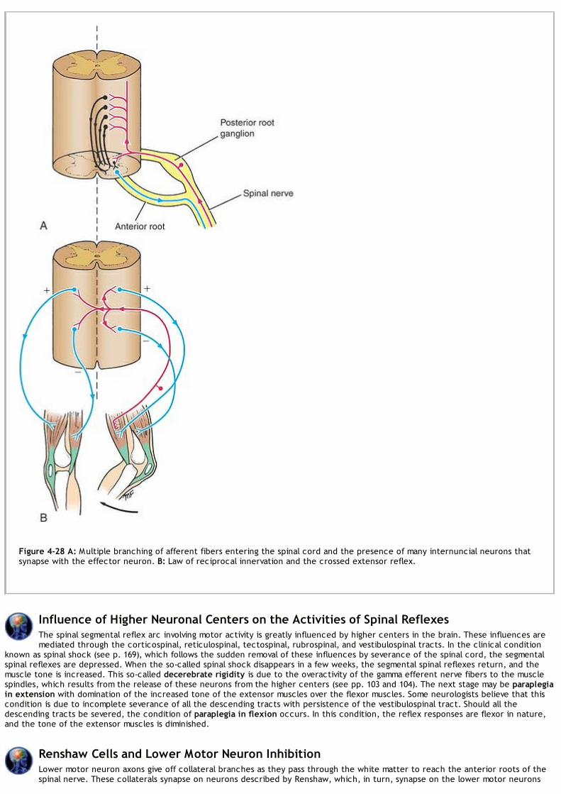
Figure 4-28 A: Multiple branching of afferent fibers entering the spinal cord and the presence of many internuncial neurons thatsynapse with the effector neuron. B: Law of reciprocal innervation and the crossed extensor reflex.
Influence of Higher Neuronal Centers on the Activities of Spinal ReflexesThe spinal segmental reflex arc involving motor activity is greatly influenced by higher centers in the brain. These influences aremediated through the corticospinal, reticulospinal, tectospinal, rubrospinal, and vestibulospinal tracts. In the clinical condition
known as spinal shock (see p. 169), which follows the sudden removal of these influences by severance of the spinal cord, the segmentalspinal reflexes are depressed. When the so-called spinal shock disappears in a few weeks, the segmental spinal reflexes return, and themuscle tone is increased. This so-called decerebrate rigidity is due to the overactivity of the gamma efferent nerve fibers to the musclespindles, which results from the release of these neurons from the higher centers (see pp. 103 and 104). The next stage may be paraplegiain extension with domination of the increased tone of the extensor muscles over the flexor muscles. Some neurologists believe that thiscondition is due to incomplete severance of all the descending tracts with persistence of the vestibulospinal tract. Should all thedescending tracts be severed, the condition of paraplegia in flexion occurs. In this condition, the reflex responses are flexor in nature,and the tone of the extensor muscles is diminished.
Renshaw Cells and Lower Motor Neuron InhibitionLower motor neuron axons give off collateral branches as they pass through the white matter to reach the anterior roots of thespinal nerve. These collaterals synapse on neurons described by Renshaw, which, in turn, synapse on the lower motor neurons

P.166P.167P.168P.169P.170P.171P.172P.173P.174P.175P.176
(Fig. 4-27). These internuncial neurons are believed to provide feedback on the lower motor neurons, inhibiting their activity.
Clinical NotesGeneral Anatomical Features of Clinical ImportanceThe spinal cord may be described, for practical purposes, as consisting of columns of motor and sensory nerve
cells, the gray matter, surrounded by ascending and descending tracts, the white matter. It lies within the vertebralcanal and is protected by three surrounding fibrous membranes, the meninges. It is cushioned against trauma by thecerebrospinal fluid and is held in position by the denticulate ligaments on each side and the filum terminale inferiorly.The spinal cord is segmented, and paired posterior (sensory) and anterior (motor) roots corresponding to eachsegment of the cord leave the vertebral canal through the intervertebral foramina.The spinal cord is shorter than the vertebral column and terminates inferiorly in the adult at the level of the lowerborder of the first lumbar vertebra. The subarachnoid space extends inferiorly beyond the end of the cord and ends atthe level of the lower border of the second sacral vertebra.Because of the shortness of the spinal cord relative to the length of the vertebral column, the nerve roots of thelumbar and sacral segments have to take an oblique course downward to reach their respective intervertebralforamina; the resulting leash of nerve roots forms the cauda equina.A spinal tap needle may be inserted into the subarachnoid space below the level of the second lumbar vertebrawithout damaging the spinal cord. (For details, see p. 19.)
Lesions of the Anterior and Posterior Nerve RootsEach nerve root has a covering of pia, arachnoid, and dura mater. The anterior and posterior roots unite in theintervertebral foramina to form the spinal nerves. Here, the meninges fuse with the epineurium of the spinal nerves.Either or both spinal nerve roots may be involved in syphilitic spinal meningitis or pyogenic meningitis. The posteriorroots may be involved in tabes dorsalis and herpes zoster. Their anatomical location, both in the vertebral canal and inthe intervertebral foramina, exposes them to compression from tumors of the vertebral column and to irritation fromabnormal constituents of the cerebrospinal fluid, such as blood following a subarachnoid hemorrhage. A herniatedintervertebral disc, a primary or secondary vertebral tumor, vertebral destruction by tumor or infection, or a fracturedislocation can press on the spinal nerve roots in the intervertebral foramina. Even severe scoliosis may result incompression of the nerve roots.A lesion of one posterior spinal nerve root will produce pain in the area of skin innervated by that root and in themuscles that receive their sensory nerve supply from that root. Movements of the vertebral column in the region of thelesion will heighten the pain, and coughing and sneezing will also make it worse by raising the pressure within thevertebral canal. Before there is actual loss of sensation in the dermatome, there may be evidence of hyperalgesia andhyperesthesia.A lesion of an anterior root will result in paralysis of any muscle that is supplied exclusively by that root and a partialparalysis of any muscle that is supplied partially by that root. In both cases, fasciculation and muscle atrophy occur.
Clinical Significance of Lamination of the Ascending TractsWithin the anterolateral white column of the spinal cord, the axons of the spinothalamic tracts from the sacral andlumbar segments of the body are deflected laterally by axons crossing the midline at successively higher levels. Withinthe posterior white column, the axons from the sacral and lumbar segments of the body are pushed medially by theaxons from higher segments of the body. This deflection of the tracts produces lamination; thus, in the spinothalamictracts (anterolateral system), the cervical to sacral segments are located from medial to lateral, whereas in theposterior white column (medial lemniscus system) the sacral to cervical segments are located from medial to lateral.This is shown diagrammatically in Figure 4-29.This detailed information is of practical value in patients in whom there is external pressure exerted on the spinal cordin the region of the spinothalamic tracts. It explains, for example, why patients will experience a loss of pain andtemperature sensations first in the sacral dermatomes of the body and, if the pressure increases, in the other highersegmental dermatomes of the body.
Injury to the Ascending Tracts Within the Spinal CordLateral Spinothalamic TractDestruction of this tract produces contralateral loss of pain and thermal sensibilities below the level of the lesion. Thepatient will not, therefore, respond to pinprick or recognize hot and cold objects placed in contact with the skin.
Anterior Spinothalamic TractDestruction of this tract produces contralateral loss of light touch and pressure sensibilities below the level of thelesion. Remember that discriminative touch will still be present, because this information is conducted through thefasciculus gracilis and fasciculus cuneatus. The patient will not feel the light touch of a piece of cotton placed againstthe skin or feel pressure from a blunt object placed against the skin.
Fasciculus Gracilis and Fasciculus CuneatusDestruction of these tracts cuts off the supply of information from the muscles and joints to consciousness; thus, theindividual does not know about the position and movements of the ipsilateral limbs below the level of the lesion. Withthe patient's eyes closed, he or she is unable to tell where the limb or part of the limb is in space. For example, if youpassively dorsiflex the patient's big toe, he or she is unable to tell you whether the toe is pointing upward ordownward. The patient has impaired muscular control, and the movements are jerky or ataxic.The patient also has loss of vibration sense below the level of the lesion on the same side. This is easily tested byapplying a vibrating tuning fork to a bony prominence, such as the lateral malleolus of the fibula or the styloid processof the radius.There will also be a loss of tactile discrimination on the side of the lesion. This is tested most easily by graduallyseparating the two points of a compass until the patient can appreciate them as two separate points, not as one,when they are applied to the skin surface. Tactile discrimination varies from one part of the body to another. In anormal individual, the points have to be separated by about 3 to 4 mm before they are recognized as separate pointson the tips of the fingers. On the back, however, the points have to be separated by 65 mm or more before they can

be recognized as separate points.
Figure 4-29 Segmental organization of the tracts in the posterior, lateral, and anterior white columns of the spinal cord.
The sense of general light touch would be unaffected, as these impulses ascend in the anterior spinothalamic tracts.It should be pointed out that it is extremely rare for a lesion of the spinal cord to be so localized as to affect onesensory tract only. It is more usual to have several ascending and descending tracts involved.
Somatic and Visceral PainSomatic pain has been considered extensively in this chapter. The sense organs for somatic pain are the naked nerveendings. The initial sharp pain is transmitted by fast-conducting fibers, and the more prolonged burning pain travels inthe slow-conducting nerve fibers (see p. 147).In the viscera, there are special receptors, chemoreceptors, baroreceptors, osmoreceptors, and stretch receptors thatare sensitive to a variety of stimuli, including ischemia, stretching, and chemical damage. Afferent fibers from thevisceral receptors reach the central nervous system via the sympathetic and parasympathetic parts of the autonomicnervous system. Once within the central nervous system, the pain impulses travel by the same ascending tracts as thesomatic pain and ultimately reach the postcentral gyrus.Visceral pain is poorly localized and often associated with salivation, nausea, vomiting, tachycardia, and sweating.Visceral pain may be referred from the organ involved to a distant area of the body (referred pain).
Treatment of Acute PainDrugs such as salicylates can be used to reduce the synthesis of prostaglandin, a substance that sensitizes free nerveendings to painful stimuli. Local anesthetics, such as procaine, can be used to block nerve conduction in peripheralnerves.Narcotic analgesics, such as morphine and codeine, reduce the affective reaction to pain and act on the opiatereceptor sites in the cells in the posterior gray column of the spinal cord, as well as other cells in the analgesic systemin the brain. It is believed that opiates act by inhibiting the release of glutamate, substance P, and other transmittersfrom the sensory nerve endings. To minimize the side effects of morphine given by systemic injection, the narcotic canbe given by local injection directly into the posterior gray horn of the spinal cord or by injection indirectly into thecerebrospinal fluid in the subarachnoid space. Long-term cancer pain has been treated successfully by the continuousinfusion of morphine into the spinal cord.
Treatment of Chronic PainNew techniques, such as acupuncture and electrical stimulation of the skin, are now being used with success. Relief ofpain can be achieved by the use of placebos in a few patients. The anticipation of the relief of pain is thought tostimulate the release of endorphins, which inhibit the normal pain pathway.
Relief of Pain by Rhizotomy or CordotomySurgical relief of pain has been used extensively in patients with terminal cancer. Posterior rhizotomy or division of theposterior root of a spinal nerve effectively severs the conduction of pain into the central nervous system. It is arelatively simple procedure, but, unfortunately, the operation deprives the patient of other sensations besides pain.Moreover, if the pain sensation is entering the spinal cord through more than one spinal nerve, it may be necessary todivide several posterior roots.Thoracic cordotomy has been performed with success in patients with severe pain originating from the lower abdomenor pelvis. Essentially, the operation consists of dividing the lateral spinothalamic tracts by inserting a knife into theanterolateral quadrant of the spinal cord. It is important to remember that the lateral spinothalamic fibers haveoriginated in cells of the substantia gelatinosa in the opposite posterior gray column and that they cross the spinalcord obliquely and reach their tract in the white column three or four segments higher than their posterior root ofentry. Cervical cordotomy has been performed successfully in patients with intractable pain in the neck or thorax.

Tabes DorsalisTabes dorsalis is caused by syphilis. The organism causes a selective destruction of nerve fibers at the point ofentrance of the posterior root into the spinal cord, especially in the lower thoracic and lumbosacral regions (Fig. 4-30).The following symptoms and signs may be present: (1) stabbing pains in the lower limbs, which may be very severe;(2) paresthesia, with numbness in the lower limbs; (3) hypersensitivity of skin to touch, heat, and cold; (4) loss ofsensation in the skin of parts of the trunk and lower limbs and loss of awareness that the urinary bladder is full; (5)loss of appreciation of posture or passive movements of the limbs, especially the legs; (6) loss of deep pain sensation,such as when the muscles are forcibly compressed or when the tendo Achillis is compressed between the finger andthumb; (7) loss of pain sensation in the skin in certain areas of the body, such as the side of the nose or the medialborder of the forearm, the thoracic wall between the nipples, or the lateral border of the leg; (8) ataxia of the lowerlimbs as the result of loss of proprioceptive sensibility (the unsteadiness in gait is compensated to some extent byvision; however, in the dark or if the eyes are closed, the ataxia becomes worse and the person may fall); (9)hypotonia as the result of loss of proprioceptive information that arises from the muscles and joints; and (10) loss oftendon reflexes, owing to degeneration of the afferent fiber component of the reflex arc (the knee and ankle tendonjerks are lost early in the disease).
Muscle ActivityMuscle ToneMuscle tone is a state of continuous partial contraction of a muscle and is dependent on the integrity of amonosynaptic reflex arc (see description on pp. 101 and 103). The receptor organs are the muscle spindles. Theafferent neuron enters the spinal cord through the posterior root and synapses with the effector neuron or lowermotor neuron in the anterior gray column. The lower motor neuron supplies the muscle fibers by traveling through theanterior roots, the spinal nerves, and peripheral nerves. Muscle tone is abolished if any part of that simple reflex arc isdestroyed. An atonic muscle feels soft and flabby and atrophies rapidly.
Figure 4-30 Site of a syphilitic lesion on the spinal cord.
Normal muscle tone exhibits a certain resilience or elasticity, and when a muscle is passively stretched by moving ajoint, a certain degree of resistance is felt. Normal muscle tone depends on the integrity of the monosynaptic reflex arcdescribed above and the control superimposed on it by impulses received through the descending tracts fromsupraspinal levels. Note that muscle spindles are excitatory to muscle tone, whereas neurotendinous receptors areinhibitory to muscle tone.
Voluntary MovementVoluntary movement is initiated by the individual. A series of different muscles are made to contract for the purpose ofreaching a goal. This would suggest that the descending tracts that influence the activity of the lower motor neuronsare driven by information received by the sensory systems, the eyes, the ears, and the muscles themselves and areaffected further by past afferent information that has been stored in the memory. Moreover, the whole process may becolored by past and present emotional input. The limbic structures appear to play a role in emotion, motivation, andmemory and may influence the initiation process of voluntary movement by their projections to the cerebral cortex.The descending pathways from the cerebral cortex and the brainstem, that is, the upper motor neurons, influence theactivity of the lower motor neurons either directly or through internuncial neurons. Most of the tracts originating in thebrainstem that descend to the spinal cord also are receiving input from the cerebral cortex.The corticospinal tracts are believed to control the prime mover muscles, especially those responsible for the highlyskilled movements of the distal parts of the limbs. The other supraspinal descending tracts play a major role in thesimple basic voluntary movements and, in addition, bring about an adjustment of the muscle tone so that easy andrapid movements of the joints can take place.It is interesting to note that the basal ganglia and the cerebellum do not give rise directly to descending tracts thatinfluence the activities of the lower motor neuron, and yet, these parts of the nervous system greatly influencevoluntary movements. This influence is accomplished indirectly by fibers that project to the cerebral cortex andbrainstem nuclei, which are the sites of origin of the descending tracts.
Pyramidal and Extrapyramidal TractsThe term pyramidal tract is used commonly by clinicians and refers specifically to the corticospinal tracts. The termcame into common usage when it was learned that the corticospinal fibers become concentrated on the anterior part

of the medulla oblongata in an area referred to as the pyramids.The term extrapyramidal tracts refers to all the descending tracts other than the corticospinal tracts.
Upper Motor Neuron LesionsLesions of the Corticospinal Tracts (Pyramidal Tracts)Lesions restricted to the corticospinal tracts produce the following clinical signs:
1. The Babinski sign is present. The great toe becomes dorsally flexed, and the other toes fan outward in responseto scratching the skin along the lateral aspect of the sole of the foot. The normal response is plantar flexion of allthe toes. Remember that the Babinski sign is normally present during the first year of life because the corticospinaltract is not myelinated until the end of the first year of life.The explanation for the Babinski sign is thought to be the following. Normally, the corticospinal tracts produceplantar flexion of the toes in response to sensory stimulation of the skin of the sole. When the corticospinal tractsare nonfunctional, the influence of the other descending tracts on the toes becomes apparent, and a kind ofwithdrawal reflex takes place in response to stimulation of the sole, with the great toe being dorsally flexed andthe other toes fanning out.
2. The superficial abdominal reflexes are absent. The abdominal muscles fail to contract when the skin of theabdomen is scratched. This reflex is dependent on the integrity of the corticospinal tracts, which exert a tonicexcitatory influence on the internuncial neurons.
3. The cremasteric reflex is absent. The cremaster muscle fails to contract when the skin on the medial side of thethigh is stroked. This reflex arc passes through the first lumbar segment of the spinal cord. This reflex is dependenton the integrity of the corticospinal tracts, which exert a tonic excitatory influence on the internuncial neurons.
4. There is loss of performance of fine-skilled voluntary movements. This occurs especially at the distal end of thelimbs.
Lesions of the Descending Tracts Other Than the Corticospinal Tracts (Extrapyramidal Tracts)The following clinical signs are present in lesions restricted to the other descending tracts:
1. Severe paralysis with little or no muscle atrophy (except secondary to disuse).2. Spasticity or hypertonicity of the muscles. The lower limb is maintained in extension, and the upper limb is
maintained in flexion.3. Exaggerated deep muscle reflexes and clonus may be present in the flexors of the fingers, the quadriceps femoris,
and the calf muscles.4. Clasp-knife reaction. When passive movement of a joint is attempted, there is resistance owing to spasticity of the
muscles. The muscles, on stretching, suddenly give way due to neurotendinous organ-mediated inhibition.
It should be pointed out that in clinical practice, it is rare to have an organic lesion that is restricted only to thepyramidal tracts or only to the extrapyramidal tracts. Usually, both sets of tracts are affected to a variable extent,producing both groups of clinical signs. As the pyramidal tracts normally tend to increase muscle tone and theextrapyramidal tracts inhibit muscle tone, the balance between these opposing effects will be altered, producingdifferent degrees of muscle tone.
Lower Motor Neuron LesionsTrauma, infection (poliomyelitis), vascular disorders, degenerative diseases, and neoplasms may all produce a lesion ofthe lower motor neuron by destroying the cell body in the anterior gray column or its axon in the anterior root or spinalnerve. The following clinical signs are present with lower motor neuron lesions:
1. Flaccid paralysis of muscles supplied.2. Atrophy of muscles supplied.3. Loss of reflexes of muscles supplied.4. Muscular fasciculation. This is twitching of muscles seen only when there is slow destruction of the lower motor
neuron cell.5. Muscular contracture. This is a shortening of the paralyzed muscles. It occurs more often in the antagonist muscles
whose action is no longer opposed by the paralyzed muscles.6. Reaction of degeneration. Normally innervated muscles respond to stimulation by the application of faradic
(interrupted) current, and the contraction continues as long as the current is passing. Galvanic or direct currentcauses contraction only when the current is turned on or turned off. When the lower motor neuron is cut, a musclewill no longer respond to interrupted electrical stimulation 7 days after nerve section, although it still will respond todirect current. After 10 days, the response to direct current also ceases. This change in muscle response toelectrical stimulation is known as the reaction of degeneration.
Types of Paralysis
Hemiplegia is a paralysis of one side of the body and includes the upper limb, one side of the trunk, and the lowerlimb.Monoplegia is a paralysis of one limb only.Diplegia is a paralysis of two corresponding limbs (i.e., arms or legs).Paraplegia is a paralysis of the two lower limbs.Quadriplegia is a paralysis of all four limbs.

Relationship of Muscular Signs and Symptoms to Lesions of the Nervous SystemAbnormal Muscle ToneHypotoniaHypotonia exists when the muscle tone is diminished or absent. It occurs when any part of the monosynaptic stretchreflex arc is interrupted. It also occurs in cerebellar disease as the result of diminished influence on the gamma motorneurons from the cerebellum.
HypertoniaHypertonia (spasticity, rigidity) exists when the muscle tone is increased. It occurs when lesions exist that involvesupraspinal centers or their descending tracts but not the corticospinal tract. It also may occur at the local spinalsegmental level and be produced by local excitation of the stretch reflex by sensory irritation (e.g., spasm of backmuscles secondary to prolapsed intervertebral disc, spasm of abdominal muscles secondary to peritonitis).
TremorsTremors are rhythmic involuntary movements that result from the contraction of opposing muscle groups. These maybe slow, as in parkinsonism, or fast, as in toxic tremors from thyrotoxicosis. They may occur at rest, as inparkinsonism, or with action, the so-called intention tremor, as seen in cerebellar disease.
SpasmsSpasms are sudden, involuntary contractions of large groups of muscles. Examples of spasms are seen in paraplegiaand are due to lesions involving the descending tracts but not the corticospinal tract.
AthetosisAthetosis means continuous, slow, involuntary, dysrhythmic movements that are always the same in the same patientand disappear during sleep. They impede voluntary movement. Athetosis occurs with lesions of the corpus striatum.
ChoreaChorea consists of a series of continuous, rapid, involuntary, jerky, coarse, purposeless movements, which may occurduring sleep. Chorea occurs with lesions of the corpus striatum.
DystoniaDystonia consists of frequent, maintained contractions of hypertonic muscles, leading to bizarre postures. It occurswith lesions of the lentiform nucleus.
MyoclonusMyoclonus is a sudden contraction of an isolated muscle or part of a muscle. It occurs irregularly and commonlyinvolves a muscle of a limb. It may be present with diseases that involve the reticular formation and the cerebellum.Normal myoclonic jerks sometimes occur in individuals as they are falling asleep and are believed to be due to asudden temporary reactivation of the reticular formation.
HemiballismusHemiballismus is a rare form of involuntary movement confined to one side of the body. It usually involves the proximalextremity musculature, and the limb involved is made to fly about in all directions. The lesion responsible occurs in theopposite subthalamic nucleus.
Spinal Cord InjuriesAcute Spinal Cord InjuriesThe incidence of acute spinal cord injuries in the United States is about 10,000 per year. The injury is catastrophic,since little or no regeneration of the severed nerve tracts takes place (see p. 110) and the individual is permanentlydisabled. Treatment has been restricted to anatomical realignment and stabilization of the vertebral column ordecompression of the spinal cord. During the recovery process, the patient goes through intensive rehabilitation tooptimize the remaining neurologic function. Apart from improved management of medical complications, very little newtherapy has been successful despite an enormous amount of research into the problem of neuronal regeneration inthe spinal cord. Recently, the use of certain drugs (GM1 ganglioside and methylprednisolone) administered to thepatient soon after injury has resulted in some improvement in the neurologic deficit. Animal experiments appear toindicate that these drugs enhance the functional recovery of damaged neurons.
Chronic Compression of the Spinal CordIf injuries to the spinal cord are excluded (see p. 17), the causes of compression may be divided into extradural andintradural. The intradural causes may be divided into those that arise outside the spinal cord (extramedullary) andthose that arise within the cord (intramedullary).The extradural causes include herniation of an intervertebral disc, infection of the vertebrae with tuberculosis, andprimary and secondary tumors of the vertebra; leukemic deposits and extradural abscesses may also compress thespinal cord. The two common extramedullary tumors are meningiomas and nerve fibromas. Intramedullary causesinclude primary tumors of the spinal cord, such as gliomas.The clinical signs and symptoms are produced by an interference with the normal anatomical and physiologic functionsof the spinal cord. Pressure on the spinal arteries causes ischemia of the spinal cord with degeneration of nerve cellsand their fibers. Pressure on the spinal veins causes edema of the spinal cord with interference in the function of theneurons. Finally, direct pressure on the white and gray matter of the spinal cord and the spinal nerve roots interfereswith nerve conduction. At the same time, the circulation of the cerebrospinal fluid is obstructed, and the composition ofthe fluid changes below the level of obstruction.
Clinical SignsOne of the earliest signs is pain. This may be local pain in the vertebra involved or pain radiating along the distributionof one or more spinal nerve roots. The pain is made worse by coughing or sneezing and is usually worse at night,when the patient is recumbent.Interference with motor function occurs early. Involvement of the anterior gray column motor cells at the level of thelesion results in partial or complete paralysis of muscles, with loss of tone and muscle wasting. The early involvementof the corticospinal and other descending tracts produces muscular weakness, increased muscle tone (spasticity),

increased tendon reflexes below the level of the lesion, and an extensor plantar response. The degree of sensory losswill depend on the nerve tracts involved. A lesion of the posterior white columns of the spinal cord will cause loss ofmuscle joint sense (proprioception), vibration sense, and tactile discrimination below the level of the lesion on thesame side. Involvement of the lateral spinal thalamic tracts will cause loss of pain and heat and cold sensations on theopposite side of the body below the level of the lesion. A more detailed discussion of the symptoms and signsfollowing injury to the ascending and descending tracts in the spinal cord is given on pages 165 and 167.Since many spinal tumors are benign and can be successfully removed (provided that irreversible damage to the spinalcord has not occurred as a result of compression of the blood supply), an early accurate diagnosis is essential. Thefollowing investigations should be performed: (1) radiography of the vertebral column, including computed tomography(CT) and magnetic resonance imaging (MRI); (2) spinal tap; and (3) myelography for those cases where determiningthe diagnosis is difficult.
Clinical Syndromes Affecting the Spinal CordSpinal Shock SyndromeSpinal shock syndrome is a clinical condition that follows acute severe damage to the spinal cord. All cord functionsbelow the level of the lesion become depressed or lost, and sensory impairment and a flaccid paralysis occur. Thesegmental spinal reflexes are depressed due to the removal of influences from the higher centers that are mediatedthrough the corticospinal, reticulospinal, tectospinal, rubrospinal, and vestibulospinal tracts. Spinal shock, especiallywhen the lesion is at a high level of the cord, may also cause severe hypotension from loss of sympathetic vasomotortone.In most patients, the shock persists for less than 24 hours, whereas in others, it may persist for as long as 1 to 4weeks. As the shock diminishes, the neurons regain their excitability, and the effects of the upper motor neuron losson the segments of the cord below the lesion, for example, spasticity and exaggerated reflexes, will make theirappearance.The presence of spinal shock can be determined by testing for the activity of the anal sphincter reflex. The reflex canbe initiated by placing a gloved finger in the anal canal and stimulating the anal sphincter to contract by squeezing theglans penis or clitoris or gently tugging on an inserted Foley catheter. An absent anal reflex would indicate theexistence of spinal shock. A cord lesion involving the sacral segments of the cord would nullify this test, since theneurons giving rise to the inferior hemorrhoidal nerve to the anal sphincter (S2-4) would be nonfunctioning.
Destructive Spinal Cord SyndromesWhen neurologic impairment is identified following the disappearance of spinal shock, it can often be categorized intoone of the following syndromes: (1) complete cord transection syndrome, (2) anterior cord syndrome, (3) central cordsyndrome, or (4) Brown-Séquard syndrome or hemisection of the cord. The clinical findings often indicate a combinationof lower motor neuron injury (at the level of destruction of the cord) and upper motor neuron injury (for thosesegments below the level of destruction).
Complete Cord Transection SyndromeComplete cord transection syndrome (Fig. 4-31) results in complete loss of all sensibility and voluntary movementbelow the level of the lesion. It can be caused by fracture dislocation of the vertebral column, by a bullet or stabwound, or by an expanding tumor. The following characteristic clinical features will be seen after the period of spinalshock has ended:
1. Bilateral lower motor neuron paralysis and muscular atrophy in the segment of the lesion. This results from damageto the neurons in the anterior gray columns (i.e., lower motor neuron) and possibly from damage to the nerve rootsof the same segment.
2. Bilateral spastic paralysis below the level of the lesion. A bilateral Babinski sign is present, and depending on thelevel of the segment of the spinal cord damaged, bilateral loss of the superficial abdominal and cremaster reflexesoccurs. All these signs are caused by an interruption of the corticospinal tracts on both sides of the cord. Thebilateral spastic paralysis is produced by the cutting of the descending tracts other than the corticospinal tracts.
3. Bilateral loss of all sensations below the level of the lesion. The loss of tactile discrimination and vibratory andproprioceptive sensations is due to bilateral destruction of the ascending tracts in the posterior white columns. Theloss of pain, temperature, and light touch sensations is caused by section of the lateral and anterior spinothalamictracts on both sides. Because these tracts cross obliquely, the loss of thermal and light touch sensations occurstwo or three segments below the lesion distally.
4. Bladder and bowel functions are no longer under voluntary control, since all the descending autonomic fibers havebeen destroyed.
If there is a complete fracture dislocation at the L2-3 vertebral level (i.e., a level below the lower end of the cord in theadult), no cord injury occurs and neural damage is confined to the cauda equina, and lower motor neuron, autonomic,and sensory fibers are involved.
Anterior Cord SyndromeAnterior cord syndrome (Fig. 4-31) can be caused by cord contusion during vertebral fracture or dislocation, from injuryto the anterior spinal artery or its feeder arteries with resultant ischemia of the cord, or by a herniated intervertebraldisc. The following characteristic clinical features are seen after the period of spinal shock has ended:
1. Bilateral lower motor neuron paralysis in the segment of the lesion and muscular atrophy. This is caused by damageto the neurons in the anterior gray columns (i.e., lower motor neuron) and possibly by damage to the anteriornerve roots of the same segment.
2. Bilateral spastic paralysis below the level of the lesion, the extent of which depends on the size of the injured areaof the cord. The bilateral paralysis is caused by the interruption of the anterior corticospinal tracts on both sides ofthe cord. The bilateral muscular spasticity is produced by the interruption of tracts other than the corticospinaltracts.
3. Bilateral loss of pain, temperature, and light touch sensations below the level of the lesion. These signs are caused

by interruption of the anterior and lateral spinothalamic tracts on both sides.4. Tactile discrimination and vibratory and proprioceptive sensations are preserved because the posterior white
columns on both sides are undamaged.
Central Cord SyndromeCentral cord syndrome is most often caused by hyperextension of the cervical region of the spine (Fig. 4-31). The cordis pressed on anteriorly by the vertebral bodies and posteriorly by the bulging of the ligamentum flavum, causingdamage to the central region of the spinal cord. Radiographs of these injuries often appear normal because nofracture or dislocation has occurred. The following characteristic clinical features are seen after the period of spinalshock has ended:
1. Bilateral lower motor neuron paralysis in the segment of the lesion and muscular atrophy. This is caused by damageto the neurons in the anterior gray columns (i.e., lower motor neuron) and possibly by damage to the nerve rootsof the same segment.
2. Bilateral spastic paralysis below the level of the lesion with characteristic sacral “sparing.” The lower limb fibers areaffected less than the upper limb fibers because the descending fibers in the lateral corticospinal tracts arelaminated, with the upper limb fibers located medially and the lower limb fibers located laterally (Fig. 4-29).
3. Bilateral loss of pain, temperature, light touch, and pressure sensations below the level of the lesion withcharacteristic sacral “sparing.” Because the ascending fibers in the lateral and anterior spinothalamic tracts are alsolaminated, with the upper limb fibers located medially and the lower limb fibers located laterally, the upper limbfibers are more susceptible to damage than the lower limb fibers (Fig. 4-29).
It follows from this discussion that the clinical picture of a patient with a history of a hyperextension injury of the neck,presenting with motor and sensory tract injuries involving principally the upper limb, would strongly suggest centralcord syndrome. The sparing of the lower part of the body may be evidenced by (1) the presence of perianal sensation,(2) good anal sphincter tone, and (3) the ability to move the toes slightly. In patients whose damage is caused byedema of the spinal cord alone, the prognosis is often very good. A mild central cord syndrome that consists only ofparesthesias of the upper part of the arm and some mild arm and hand weakness can occur.

Figure 4-31 Spinal cord syndromes.
Brown-Séquard Syndrome or Hemisection of the CordHemisection of the spinal cord can be caused by fracture dislocation of the vertebral column, by a bullet or stab wound,or by an expanding tumor (Fig. 4-31). Incomplete hemisection is common; complete hemisection is rare. The followingcharacteristic clinical features are seen in patients with a complete hemisection of the cord (Fig. 4-32) after the periodof spinal shock has ended:
1. Ipsilateral lower motor neuron paralysis in the segment of the lesion and muscular atrophy. These signs are causedby damage to the neurons on the anterior gray column and possibly by damage to the nerve roots of the samesegment.

2. Ipsilateral spastic paralysis below the level of the lesion. An ipsilateral Babinski sign is present, and depending onthe segment of the cord damaged, an ipsilateral loss of the superficial abdominal reflexes and cremasteric reflexoccurs. All these signs are due to loss of the corticospinal tracts on the side of the lesion. Spastic paralysis isproduced by interruption of the descending tracts other than the corticospinal tracts.
3. Ipsilateral band of cutaneous anesthesia in the segment of the lesion. This results from the destruction of theposterior root and its entrance into the spinal cord at the level of the lesion.
4. Ipsilateral loss of tactile discrimination and of vibratory and proprioceptive sensations below the level of the lesion.These signs are caused by destruction of the ascending tracts in the posterior white column on the same side ofthe lesion.
5. Contralateral loss of pain and temperature sensations below the level of the lesion. This is due to destruction ofthe crossed lateral spinothalamic tracts on the same side of the lesion. Because the tracts cross obliquely, thesensory loss occurs two or three segments below the lesion distally.
6. Contralateral but not complete loss of tactile sensation below the level of the lesion. This condition is brought aboutby destruction of the crossed anterior spinothalamic tracts on the side of the lesion. Here, again, because the tractscross obliquely, the sensory impairment occurs two or three segments below the level of the lesion distally. Thecontralateral loss of tactile sense is incomplete because discriminative touch traveling in the ascending tracts in thecontralateral posterior white column remains intact.
Figure 4-33 Skin area in which the sensations of pain and temperature are lost in syringomyelia.
SyringomyeliaSyringomyelia, which is due to a developmental abnormality in the formation of the central canal, most often affectsthe brainstem and cervical region of the spinal cord. At the site of the lesion, there is cavitation and gliosis in thecentral region of the neuroaxis (Fig. 4-33). The following characteristic signs and symptoms are found:
1. Loss of pain and temperature sensations in dermatomes on both sides of the body related to the affectedsegments of the cord. This loss commonly has a shawllike distribution caused by the interruption of the lateralspinothalamic tracts as they cross the midline in the anterior gray and white commissures. The patient commonlycomplains of accidental burning injuries to the fingers.
2. Tactile discrimination, vibratory sense, and proprioceptive sense are normal. The reason is that the ascending tractsin the posterior white column are unaffected.
3. Lower motor neuron weakness is present in the small muscles of the hand. It may be bilateral, or one hand maysuffer before the other. As the lesion expands in the lower cervical and upper thoracic region, it destroys theanterior horn cells of these segments. Later, the other muscles of the arm and shoulder girdles undergo atrophy.
4. Bilateral spastic paralysis of both legs may occur, with exaggerated deep tendon reflexes and the presence of apositive Babinski response. These signs are produced by the further expansion of the lesion laterally into the whitecolumn to involve the descending tracts.

Figure 4-32 Brown-Séquard syndrome with a spinal cord lesion at the right 10th thoracic level.
5. Horner syndrome may be present. This is caused by the interruption of the descending autonomic fibers in thereticulospinal tracts in the lateral white column by the expanding lesion.
PoliomyelitisPoliomyelitis is an acute viral infection of the neurons of the anterior gray columns of the spinal cord (Fig. 4-31) and themotor nuclei of the cranial nerves. Immunization has greatly reduced the incidence of poliomyelitis, which was once afeared disease. Following death of the motor nerve cells, there is paralysis and wasting of the muscles. The muscles ofthe lower limb are more often affected than the muscles of the upper limb. In severe poliomyelitis, respiration may bethreatened due to the paralysis spreading to the intercostal muscles and diaphragm. The muscles of the face, pharynx,larynx, and tongue may also be paralyzed. Improvement usually begins at the end of the first week as the edema inthe affected area subsides and function returns to the neurons that have not been destroyed.
Multiple SclerosisMultiple sclerosis is a common disease confined to the central nervous system, causing demyelination of the ascendingand descending tracts. It is a disease of young adults, and the cause is unknown. Autoimmunity, infection, andheredity, alone or in combination, may play a role in its etiology. It has been suggested that a breach in the integrity ofthe blood-brain barrier in an individual who is genetically predisposed to the disease may be responsible. This couldresult in the invasion of the brain and spinal cord by some infection allowing leukocytes to enter the normallyimmunologically protected central nervous system. The inflammation and demyelination with loss of the myelin sheathresults in the breakdown of the insulation around the axons, and the velocity of the action potentials is reduced andultimately becomes blocked. Although myelin is relatively rich in lipid (70% to 80%), it also contains proteins that play arole in myelin compaction. It has been found that many of the proteins in the myelin of the central nervous systemdiffer from those in the peripheral nervous system. Experimentally, it has been shown that basic myelin proteinsinjected into animals can produce a strong immune response and demyelination in the central nervous system occurs.It is possible that mutations in the structure of myelin protein can occur and be responsible for some inherited forms ofdemyelination. It is also possible that autoantigens develop in multiple sclerosis.The course of multiple sclerosis is chronic with exacerbations and remissions. Because of the widespread involvementof different tracts at different levels of the neuroaxis, the signs and symptoms are multiple, but remissions do occur.Weakness of the limbs is the most common sign of the disease. Ataxia due to involvement of the tracts of thecerebellum may occur, but spastic paralysis may also be present.Recent research has suggested that the remissions in multiple sclerosis may in part be explained by the remodeling ofthe demyelinated axonal plasma membrane so that it acquires a higher than normal number of sodium channels, whichpermit conduction of action potentials despite the loss of myelin.In patients who have the progressive form of the disease without remissions, it is has been shown that they have asubstantial damage to the axons as well as the myelin. This would suggest that multiple sclerosis is not just ademyelinating disease but one in which there is axonal pathology.
Amyotrophic Lateral SclerosisAmyotrophic lateral sclerosis (Lou Gehrig disease) is a disease confined to the corticospinal tracts and the motorneurons of the anterior gray columns of the spinal cord (Fig. 4-31). It is rarely familial and is inherited in about 10% ofpatients. Amyotrophic lateral sclerosis is a chronic progressive disease of unknown etiology. Typically, it occurs in latemiddle age and is inevitably fatal in 2 to 6 years. The lower motor neuron signs of progressive muscular atrophy,paresis, and fasciculations are superimposed on the signs and symptoms of upper motor neuron disease with paresis,spasticity, and Babinski response. The motor nuclei of some cranial nerves may also be involved.
Parkinson Disease

Parkinson disease is associated with neuronal degeneration in the substantia nigra and, to a lesser extent, in theglobus pallidus, putamen, and caudate nucleus. The degeneration of the inhibitory nigrostriate fibers results in areduction in the release of the neurotransmitter dopamine within the corpus striatum. This leads to hypersensitivity ofthe dopamine receptors in the postsynaptic neurons in the corpus striatum, which become overactive. Thecharacteristic signs of the disease include tremor and cogwheel rigidity (hyperkinetic activity) and difficulty initiatingvoluntary movements, which are slow (hypokinetic activity). (For further details, see p. 322.)
Pernicious AnemiaPernicious anemia, a form of megaloblastic anemia, is caused by vitamin B12 deficiency. The disease may produceextensive damage to the tracts in the posterior and lateral white columns of the spinal cord as well as peripheralnerve degeneration. Widespread sensory and motor losses may be present due to involvement of the ascending anddescending tracts of the spinal cord.
Radiographic Appearances of the Vertebral ColumnThe views commonly used in radiography are anteroposterior, lateral, and oblique. Vertebral destruction due totuberculosis or primary or secondary tumors of the vertebrae or fractures due to trauma usually can be revealed byradiographic examination. Erosion of the pedicles by a tumor within the intervertebral foramina may be seen.Narrowing of the space between the vertebral bodies with bony spurs because of osteoarthritic changes in adjacentvertebral bodies can also be seen.
Computed Tomography and Magnetic Resonance Imaging of the Vertebral Column and SpinalCordCT scans of the vertebrae and joints can be obtained (Fig. 4-34). A protrusion of an intervertebral disc can be identifiedand the presence of narrowing of the vertebral canal (spinal stenosis) can be diagnosed.Sagittal MRI is increasingly being used to replace CT and myelography. The parts of a vertebra, the intervertebral disc,the posterior longitudinal ligament, and meningeal sac (thecal sac) can easily be identified (Fig. 4-35).
Figure 4-34 Horizontal (axial) CT scan of the fourth lumbar vertebra.
MyelographyThe subarachnoid space can be studied radiographically by the injection of a contrast medium into the subarachnoidspace by spinal tap. Iodized oil has been used with success. This technique is referred to as myelography (Figs. 4-36and 4-37).If the patient is sitting in the upright position, the oil sinks to the lower limit of the subarachnoid space at the level ofthe lower border of the second sacral vertebra. By placing the patient on a tilting table, the oil can be made togravitate gradually to higher levels of the vertebral column.A normal myelogram will show pointed lateral projections at regular intervals at the intervertebral space levels. The

reason for this is that the opaque medium fills the lateral extensions of the subarachnoid space around each spinalnerve. The presence of a tumor or a prolapsed intervertebral disc may obstruct the movement of the oil from oneregion to another when the patient is tilted.With the recent technologic advances in CT scans and MRIs, it is now unusual to require an intrusive procedure, suchas myelography, to make a diagnosis.
Figure 4-36 Posteroanterior myelogram of the cervical region of a 22-year-old woman.

Figure 4-35 Sagittal MRI of the lumbosacral part of the vertebral column showing several prolapsed intervertebral discs.(Courtesy Dr. Pait.)

P.178P.179P.180
Figure 4-37 Diagrammatic explanation of the myelogram shown in Figure 4-36.
Clinical Problem Solving1. A 53-year-old widower was admitted to the hospital complaining of a burning pain over his right shoulder region andthe upper part of his right arm. The pain had started 3 weeks previously and, since that time, had progressivelyworsened. The pain was accentuated when the patient moved his neck or coughed. Two years previously, he hadbeen treated for osteoarthritis of his vertebral column. The patient stated that he had been a football player atcollege, and since that time, he continued to take an active part in the game until he was 42 years old. Physicalexamination revealed weakness, wasting, and fasciculation of the right deltoid and biceps brachii muscles. The rightbiceps tendon reflex was absent. Radiologic examination revealed extensive spur formation on the bodies of thefourth, fifth, and sixth cervical vertebrae. The patient demonstrated hyperesthesia and partial analgesia in the skinover the lower part of the right deltoid and down the lateral side of the arm. Using your knowledge of neuroanatomy,make the diagnosis. How is the pain produced? Why is the pain made worse by coughing?View Answer

2. A 66-year-old woman was admitted to the hospital because of her increasing difficulty with walking. Two weeksbefore admission, she had been able to walk with the help of a stick. Since that time, walking had become increasinglydifficult, and for the past 2 days, she could not walk at all. She had complete control of micturition and defecation. Onexamination, the handgrip was weak on both sides, but power was normal in the proximal segments of the upperextremities. The tendon reflexes of the upper limbs and the sensory function were normal. Both lower limbs showedmuscular weakness with increased muscle tone, especially on the left side. The knee jerks and ankle jerks (tendonreflexes) in both lower limbs were grossly exaggerated, and there were bilateral extensor plantar responses. Thepatient had a loss of sensation of pain below the fifth thoracic dermatome on both sides of the body. Postural sensewas impaired in both great toes, and vibration sense was absent below the fifth thoracic segmental level. Radiologicexamination, including an MRI, of the vertebral column showed nothing abnormal. A myelogram in the lumbar regionrevealed a complete block at the lower border of the fourth thoracic vertebra. Using your knowledge of neuroanatomy,suggest a possible diagnosis. How would you treat this patient? Name the tracts in the spinal cord that areresponsible for conduction of the sensation of pain. What is the position of these tracts in the spinal cord? Name thetracts responsible for the conduction of postural sense and vibration sense from the spinal cord to the brain. Why didthe patient have increasing difficulty in walking? Why were the tendon reflexes in the lower limbs exaggerated, andwhy did the patient exhibit bilateral extensor plantar responses?View Answer
3. A 20-year-old male student celebrated the passing of an examination by drinking several beers at a party. On theway home, he drove his car head-on into a bridge abutment. On examination in the emergency department, he wasfound to have a fracture dislocation of the ninth thoracic vertebra with signs and symptoms of severe damage to thespinal cord. On physical examination, he had an upper motor neuron paralysis of the left leg. He also had loss ofmuscle joint sense of the left leg. On testing of cutaneous sensibility, he had a band of cutaneous hyperesthesiaextending around the abdominal wall on the left side at the level of the umbilicus. Just below this, he had a narrowband of anesthesia and analgesia. On the right side, there was total analgesia, thermoanesthesia, and partial loss oftactile sense of the skin of the abdominal wall below the level of the umbilicus and involving the whole of the right leg.Using your knowledge of neuroanatomy, state the level at which the spinal cord was damaged. Was the spinal cordcompletely sectioned? If not, on which side did the hemisection occur? Explain the sensory losses found onexamination in this patient.View Answer
4. A 35-year-old woman was admitted to the hospital for investigation. She had symptoms of analgesia andthermoanesthesia on the medial side of the left hand that persisted for 6 months. Three weeks prior to heradmittance, she had severely burned the little finger of her left hand on a hot stove and was unaware that the burnhad occurred until she smelled the burning skin. On physical examination, she was found to have considerably reducedpain and temperature sense involving the eighth cervical and first thoracic dermatomes of the left hand. However, hersense of tactile discrimination was perfectly normal in these areas. Examination of the right arm showed a similar butmuch less severe dissociated sensory loss involving the same areas. No further abnormal signs were discovered. Usingyour knowledge of neuroanatomy, state which tract or tracts were involved in this pathologic process. Name thisdisease.View Answer
5. A 60-year-old man walked into the neurology clinic, and the physician paid particular attention to his gait. Thepatient raised his feet unnecessarily high and brought them to the ground in a stamping manner. While he waswaiting for the physician, it was noticed that he stood with his feet wide apart. On questioning, the patient said thathe was finding it increasingly difficult to walk and was starting to use a stick, especially when he went out for walks inthe dark. The physician asked the patient to stand with his toes and heels together and to close his eyes. The patientimmediately started to sway, and the nurse had to steady him to prevent him from falling. On further examination, thepatient was found to have loss of muscle joint sense of both legs and was unable to detect any feeling of vibrationwhen a vibrating tuning fork was placed on the medial malleolus of either leg. No other sensory losses were noted.Using your knowledge of neuroanatomy, name the ascending pathways that are involved, by disease, in this patient.Name a disease that could be responsible for these findings.View Answer
6. A 68-year-old man had an advanced inoperable carcinoma of the prostate with multiple metastases in the lumbarvertebrae and hip bones. Apart from the severe intractable pain, the patient was still able to enjoy life among hisfamily. After a full discussion of the prognosis with the patient and his wife, the wife turned to the physician and said,“Can't you do something to stop this terrible pain so that my husband can die happy?” What can a physician do tohelp a patient under these circumstances?View Answer
7. A third-year medical student attended a lecture on the effects of trauma on the vertebral column. The orthopedicsurgeon described very superficially the different neurologic deficits that may follow injury to the spinal cord. At the endof the lecture, the student said he did not understand what was meant by the term spinal shock. He could notunderstand what the underlying mechanism for this condition was. He also asked the surgeon to explain what wasmeant by paraplegia in extension and paraplegia in flexion. Could the surgeon explain why one condition sometimespasses into the other? These are good questions. Can you answer them?View Answer
8. While examining a patient with a right-sided hemiplegia caused by a cerebrovascular accident, the neurologistasked the student which clinical signs could be attributed to an interruption of the corticospinal tracts and which signscould be attributed to damage to other descending tracts. Using your knowledge of neuroanatomy, answer thisquestion.View Answer

P.182P.183P.184
9. A large civilian aircraft was forced to abort its takeoff because three tires had burst as the plane sped along therunway. The pilot miraculously managed to halt the plane as it veered off the runway and came to an abrupt halt in aditch. All the passengers escaped injury, but one of the stewardesses was admitted to the emergency departmentwith suspected spinal cord injury. On questioning, the 25-year-old patient said that although she had her seat beltfastened, she was thrown violently forward on impact. She said she could not feel anything in either leg and could notmove her legs. On examination, there was complete motor and sensory loss of both legs below the inguinal ligamentand absence of all deep tendon reflexes of both legs. Twelve hours later, it was noted that she could move the toesand ankle of her left lower limb, and she had a return of sensations to her right leg except for loss of tactilediscrimination, vibratory sense, and proprioceptive sense. She had a band of complete anesthesia over her rightinguinal ligament. Her left leg showed a total analgesia, thermoanesthesia, and partial loss of tactile sense. Her rightleg was totally paralyzed, and the muscles were spastic. There was a right-sided Babinski response, and it waspossible to demonstrate right-sided ankle clonus. The right knee jerk was exaggerated. Using your knowledge ofneuroanatomy, explain the symptoms and signs found in this patient. Which vertebra was damaged?View Answer
10. Why is it dangerous to move a patient who is suspected of having a fracture or dislocation of the vertebral column?View Answer
11. An 18-year-old man was admitted to the hospital following a severe automobile accident. After a completeneurologic investigation, his family was told that he would be paralyzed from the waist downward for the rest of hislife. The neurologist outlined to the medical personnel the importance of preventing complications in these cases. Thecommon complications are the following: (a) urinary infection, (b) bedsores, (c) nutritional deficiency, (d) muscularspasms, and (e) pain. Using your knowledge of neuroanatomy, explain the underlying reasons for these complications.How long after the accident do you think it would be possible to give an accurate prognosis in this patient?View Answer
12. A 67-year-old man was brought to the neurology clinic by his daughter because she had noticed that his right armhad a tremor. Apparently, this had started about 6 months previously and was becoming steadily worse. Whenquestioned, the patient said he noticed that the muscles of his limbs sometimes felt stiff, but he had attributed this toold age. It was noticed that while talking, the patient rarely smiled and then only with difficulty. It was also noted thathe infrequently blinked his eyes. The patient tended to speak in a low, faint voice. When asked to walk, the patientwas seen to have normal posture and gait, although he tended to hold his right arm flexed at the elbow joint. Whenhe was sitting, it was noted that the fingers of the right hand were alternately contracting and relaxing, and there wasa fine tremor involving the wrist and elbow on the right side. It was particularly noticed that the tremor was worsewhen the arm was at rest. When he was asked to hold a book in his right hand, the tremor stopped momentarily, butit started again immediately after the book was placed on the table. The daughter said that when her father fallsasleep, the tremor stops immediately. On examination, it was found that the passive movements of the right elbowand wrist showed an increase in tone, and there was some cogwheel rigidity. There was no sensory loss, eithercutaneous or deep sensibility, and the reflexes were normal. Using your knowledge of neuroanatomy, make adiagnosis. Which regions of the brain are diseased?View Answer
13. Name a center in the central nervous system that may be responsible for the following clinical signs: (a) intentiontremor, (b) athetosis, (c) chorea, (d) dystonia, and (e) hemiballismus.View Answer
Review QuestionsDirections: Each of the numbered items in this section is followed by answers. Select the ONE lettered answerthat is CORRECT.1. The following statements concern the spinal cord:
(a) The anterior and posterior gray columns on the two sides are united by a white commissure.(b) The terminal ventricle is the expanded lower end of the fourth ventricle.(c) The larger nerve cell bodies in the anterior gray horns give rise to the alpha efferent nerve fibers in theanterior roots.(d) The substantia gelatinosa groups of cells are located at the base of each posterior gray column.(e) The nucleus dorsalis (Clarke's column) is a group of nerve cells found in the posterior gray column andrestricted to the lumbar segments of the cord.
View Answer
2. The following statements concern the white columns of the spinal cord:(a) The posterior spinocerebellar tract is situated in the posterior white column.(b) The anterior spinothalamic tract is found in the anterior white column.(c) The lateral spinothalamic tract is found in the anterior white column.(d) The fasciculus gracilis is found in the lateral white column.(e) The rubrospinal tract is found in the anterior white column.
View Answer
3. The following statements concern the spinal cord:

(a) The spinal cord has a cervical enlargement for the brachial plexus.(b) The spinal cord possesses spinal nerves that are attached to the cord by anterior and posterior rami.(c) In the adult, the spinal cord usually ends inferiorly at the lower border of the fourth lumbar vertebra.(d) The ligamentum denticulatum anchors the spinal cord to the pedicles of the vertebra along each side.(e) The central canal does not communicate with the fourth ventricle of the brain.
View Answer
Directions: Matching Questions. Questions 4 through 9 apply to the following figure. Match the numbers listed onthe left with the appropriate lettered structure listed on the right. Each lettered option may be selected once,more than once, or not at all.
4. Number 1(a) Nucleus proprius(b) Preganglionic sympathetic outflow(c) Nucleus dorsalis(d) Substantia gelatinosa(e) None of the above
View Answer
5. Number 2(a) Nucleus proprius(b) Preganglionic sympathetic outflow(c) Nucleus dorsalis(d) Substantia gelatinosa(e) None of the above
View Answer
6. Number 3(a) Nucleus proprius(b) Preganglionic sympathetic outflow(c) Nucleus dorsalis(d) Substantia gelatinosa(e) None of the above
View Answer
7. Number 4(a) Nucleus proprius(b) Preganglionic sympathetic outflow(c) Nucleus dorsalis(d) Substantia gelatinosa(e) None of the above
View Answer
8. Number 5(a) Nucleus proprius(b) Preganglionic sympathetic outflow

(c) Nucleus dorsalis(d) Substantia gelatinosa(e) None of the above
View Answer
9. Number 6(a) Nucleus proprius(b) Preganglionic sympathetic outflow(c) Nucleus dorsalis(d) Substantia gelatinosa(e) None of the above
View Answer
Directions: Each of the numbered items in this section is followed by answers. Select the ONE lettered answerthat is CORRECT.10. The following statements concern the cell of origin of the tracts listed below:
(a) The fasciculus cuneatus arises from the cells in the substantia gelatinosa.(b) The anterior spinal thalamic arises from the cells in posterior root ganglion.(c) The fasciculus gracilis arises from the cells in the nucleus dorsalis (Clarke's column).(d) The anterior spinocerebellar arises from the cells in the posterior root ganglion.(e) The lateral spinothalamic arises from the cells in the substantia gelatinosa.
View Answer
11. The following statements concern the courses taken by the tracts listed below:(a) The fasciculus gracilis does not cross to the opposite side of the neural axis.(b) The spinotectal tract does not cross to the opposite side of the spinal cord.(c) The lateral spinothalamic tract does not cross to the opposite side of the spinal cord.(d) The posterior spinocerebellar tract does cross to the opposite side of the neural axis.(e) The anterior spinothalamic tract immediately crosses to the opposite side of the spinal cord.
View Answer
12. The following statements concern the nucleus of termination of the tracts listed below:(a) The posterior white column tracts terminate in the inferior colliculus.(b) The spinoreticular tract terminates on the neurons of the hippocampus.(c) The spinotectal tract terminates in the inferior colliculus.(d) The anterior spinothalamic tract terminates in the ventral posterolateral nucleus of the thalamus.(e) The anterior spinocerebellar tract terminates in the dentate nucleus of the cerebellum.
View Answer
13. The following statements relate sensations with the appropriate nervous pathways:(a) Two-point tactile discrimination travels in the lateral spinothalamic tract.(b) Pain travels in the anterior spinothalamic tract.(c) Unconscious muscle joint sense travels in the anterior spinocerebellar tract.(d) Pressure travels in the posterior spinothalamic tract.(e) Vibration travels in the posterior spinocerebellar tract.
View Answer
14. The following statements concern the gating theory of pain:(a) Stimulation of small non-pain-conducting fibers in a peripheral nerve may reduce pain sensitivity.(b) Massage applied to the skin over a painful joint may reduce pain sensitivity.(c) Stimulation of delta A- and C-type fibers in a posterior root of a spinal nerve may decrease pain sensitivity.(d) Degeneration of large non-pain-conducting fibers in a peripheral nerve decreases pain sensitivity.(e) Inhibition of pain conduction in the spinal cord does not involve connector neurons.
View Answer
15. The following statements concern the reception of pain:(a) Serotonin is not a transmitter substance in the analgesic system.(b) Substance P, a protein, is thought to be the neurotransmitter at the synapses where the first-order neuronterminates on the cells in the posterior gray column of the spinal cord.(c) The enkephalins and endorphins may serve to stimulate the release of substance P in the posterior graycolumn of the spinal cord.(d) Many of the tracts conducting the initial, sharp, pricking pain terminate in the dorsal anterolateral nucleus of

the thalamus.(e) The slow-conducting C-type fibers are responsible for prolonged, burning pain.
View Answer
16. The following statements concern the corticospinal tracts:(a) They occupy the posterior limb of the internal capsule.(b) They are mainly responsible for controlling the voluntary movements in the proximal muscles of the limbs.(c) They arise as axons of the pyramidal cells in the fourth layer of the cerebral cortex.(d) Those that control the movements of the upper limb originate in the precentral gyrus on the medial side ofthe cerebral hemisphere.(e) Those that are concerned with the movements of the lower limb are located in the medial area of the middlethree-fifths of the basis pedunculi.
View Answer
17. The following statements concern the course taken by the tracts listed below:(a) The rubrospinal tract crosses the midline of the neuroaxis in the medulla oblongata.(b) The tectospinal tract (most of the nerve fibers) crosses the midline in the posterior commissure.(c) The vestibulospinal tract crosses the midline in the midbrain.(d) The lateral corticospinal tract has crossed the midline in the medulla oblongata.(e) The anterior corticospinal tract crosses the midline in the midbrain.
View Answer
18. The following statements concern the nerve cells of origin for the tracts listed below:(a) The vestibulospinal tract originates from cells of the medial vestibular nucleus situated in the pons.(b) The tectospinal tract originates from cells in the inferior colliculus.(c) The lateral corticospinal tract originates from cells in area 4 of the cerebral cortex.(d) The rubrospinal tract originates from cells in the reticular nucleus.(e) The reticulospinal tract originates from cells in the reticular formation that is confined to the midbrain.
View Answer
19. The following statements concern muscle movement:(a) Muscular fasciculation is seen only when there is rapid destruction of the lower motor neurons.(b) Muscle spindle afferent nerve fibers send information only to the spinal cord.(c) In Parkinson disease, there is a degeneration of dopamine-secreting neurons that originate in the vestibularnucleus.(d) Brain neuronal activity preceding a voluntary movement is limited to the precentral gyrus (area 4).(e) Hyperactive ankle-jerk reflexes and ankle clonus indicate a release of the lower motor neurons fromsupraspinal inhibition.
View Answer
20. After a hemorrhage into the left internal capsule in a right-handed person, the following sign or symptom might bepresent:
(a) Left homonymous hemianopia(b) Right astereognosis(c) Left hemiplegia(d) Normal speech.(e) Left-sided positive Babinski response
View Answer
21. A patient with a traumatic lesion of the left half of the spinal cord at the level of the eighth cervical segment mightpresent the following sign(s) and symptom(s):
(a) Loss of pain and temperature sensations on the left side below the level of the lesion(b) Loss of position sense of the right leg(c) Right hemiplegia(d) Left positive Babinski sign(e) Right-sided lower motor paralysis in the segment of the lesion and muscular atrophy
View Answer
Directions: Each of the numbered items in this section is followed by answers. Select the ONE lettered answerthat is BEST in each case.22. Which of the signs and symptoms listed below is indicative of a cerebellar lesion?
(a) Cogwheel rigidity(b) Hemiballismus

(c) Chorea(d) Intention tremor(e) Athetosis
View Answer
23. Which of the following regions of white matter would not contain corticospinal fibers?(a) Pyramid of medulla oblongata(b) Lateral white column of the spinal cord(c) Cerebral peduncle of the midbrain(d) Anterior limb of the internal capsule(e) Corona radiata
View Answer
Directions: Each case history is followed by questions. Read the case history, then select the ONE BEST letteredanswer.A 59-year-old woman was experiencing pain in the back and showed evidence of loss of pain and temperaturesensations down the back of her left leg. Three years previously, she underwent a radical mastectomy followed byradiation and chemotherapy for advanced carcinoma of her right breast.On examination, it was found that she was experiencing pain over the lower part of the back, with loss of the skinsensations of pain and temperature down the back of her left leg in the area of the S1-3 dermatomes. No otherneurologic deficits were identified. Radiographic examination of the vertebral column showed evidence of metastasesin the bodies of the 9th and 10th thoracic vertebrae. An MRI revealed an extension of one of the metastases into thevertebral canal, with slight indentation of the spinal cord on the right side.24. The pain in the back could be explained in this patient by the following facts except:
(a) Osteoarthritis of the joints of the vertebral column.(b) The presence of metastases in the bodies of the 9th and 10th thoracic vertebrae.(c) The pressure of the tumor on the posterior roots of the spinal nerves.(d) A prolapsed intervertebral disc pressing on the spinal nerves.(e) Spasm of the postvertebral muscles following pressure of the tumor on the posterior white columns of thespinal cord.
View Answer

Authors: Snell, Richard S.Title: Clinical Neuroanatomy, 7th Edition
Copyright ©2010 Lippincott Williams & Wilkins
> Table of Contents > Chapter 5 - The Br ainstem
Chapter 5
The Brainstem
A 58-year-old woman was referred to a neurologist because of recent onset of difficulty with walking. It was noted that she stood andwalked with her left arm flexed at the elbow and the left leg extended (left hemiparesis). While walking, the patient had difficulty flexingthe left hip and knee and dorsiflexing the ankle; the forward motion was possible by swinging the left leg outward at the hip to avoiddragging the foot on the ground. The left arm remained motionless.
Neurologic examination showed no signs of facial paralysis, but there was evidence of weakness of the movements of the tongue. Onprotrusion, the tongue deviated toward the right side (right hypoglossal nerve palsy). Cutaneous sensations were found to be normal, butthere was evidence of impairment of muscle joint sense, tactile discrimination, and vibratory sense on the left side of the body.
Based on the neurologic findings, a diagnosis of right-sided medial medullary syndrome was made. The medial part of the right side of themedulla oblongata receives its arterial supply from the right vertebral artery. Occlusion of this artery or its branch to the medulla resultsin destruction of the right pyramid (left hemiparesis), destruction of the right hypoglossal nucleus and nerve (right hypoglossal palsy), anddestruction of the medial lemniscus on the right side (left-sided loss of muscle joint sense, vibratory sense, and tactile discrimination). Theabsence of facial palsy showed that the facial nerve nuclei, the facial nerves, and the corticobulbar fibers to the facial nuclei were intact.The sparing of the sensations of touch, pain, and temperature showed that the spinal lemniscus was intact.
This diagnosis was possible as the result of carefully sorting out the neurologic findings. A clear knowledge of the position and function ofthe various nerve tracts and nuclei in the medulla oblongata is essential before one can reach a diagnosis in this case.
Chapter Objectives
To review the anatomy of the brainstemTo develop a three-dimensional picture of the interior of the brainstemTo know the positions of several of the cranial nerve nuclei, the olivary nuclear complex, and the paths taken by thevarious ascending and descending nerve tracts as they ascend to the higher brain centers or descend to the spinalcordTo assess the signs and symptoms presented by a patient and identify the exact location of a structural lesion
Head injuries from blunt trauma and penetrating missiles are associated with a high mortality and disabling morbidity. Because of the closerelationship that exists between the skull and the underlying brain and cranial nerves, as well as their common involvement in manydiseases, a brief review of the anatomy of the skull will first be considered.
A Brief Review of the Skull
Adult Skull
CompositionThe skull is composed of several separate bones united at immobile joints called sutures. The connective tissue between the bones iscalled a sutural ligament. The mandible is an exception to this rule, for it is united to the skull by the mobile temporomandibular joint.
The bones of the skull can be divided into those of the cranium and those of the face. The vault is the upper part of the cranium, andthe base of the skull is the lowest part of the cranium (Fig. 5-1).
The skull bones are made up of external and internal tables of compact bone separated by a layer of spongy bone called the diploë (Fig.5-2). The internal table is thinner and more brittle than the external table. The bones are covered on the outer and inner surfaces withperiosteum.
The cranium consists of the following bones, two of which are paired (Figs. 5-1 and 5-3):
• Fr ontal bone 1
• Par ietal bones 2
• Occipital bone 1
• Tempor al bones 2
• Sphenoid bone 1

• Ethmoid bone 1
The facial bones consist of the following, two of which are single:
• Zygomatic bones 2
• Max illae 2
• Nasal bones 2
• Lacr imal bones 2
• Vomer 1
• Palatine bones 2
• Infer ior conchae 2
• Mandible 1
It is unnecessary for students to know the detailed structure of each individual skull bone. However, they should be familiar with the skullas a whole, and it would be helpful to have a dried skull available for reference as they read the following description.
Anterior View of the SkullThe frontal bone, or forehead bone, curves downward to make the upper margins of the orbits (Fig. 5-1). The superciliary arches can beseen on either side, and the supraorbital notch, or foramen, can be recognized. Medially, the frontal bone articulates with the frontalprocesses of the maxillae and with the nasal bones. Laterally, the frontal bone articulates with the zygomatic bone.
The orbital margins are bounded by the frontal bone superiorly, the zygomatic bone laterally, the maxilla inferiorly, and the processes ofthe maxilla and frontal bone medially.
Within the frontal bone, just above the orbital margins, are two hollow spaces lined with mucous membrane called the frontal air sinuses.These communicate with the nose and serve as voice resonators.
The two nasal bones form the bridge of the nose. Their lower borders, with the maxillae, make the anterior nasal aperture. The nasalcavity is divided into two by the bony nasal septum, which is largely formed by the vomer. The superior and middle conchae are shelves ofbone that project into the nasal cavity from the ethmoid on each side; the inferior conchae are separate bones.
The two maxillae form the upper jaw, the anterior part of the hard palate, part of the lateral walls of the nasal cavities, and part of thefloors of the orbital cavities. The two bones meet in the midline at the intermaxillary suture and form the lower margin of the nasalaperture. Below the orbit, the maxilla is perforated by the infraorbital foramen. The alveolar process projects downward and, togetherwith the fellow of the opposite side, forms the alveolar arch, which carries the upper teeth. Within each maxilla is a large, pyramid-shapedcavity lined with mucous membrane called the maxillary sinus. This communicates with the nasal cavity and serves as a voice resonator.
The zygomatic bone forms the prominence of the cheek and part of the lateral wall and floor of the orbital cavity. It articulates with themaxilla medially and with the zygomatic process of the temporal bone laterally to form the zygomatic arch. The zygomatic bone isperforated by two foramina for the zygomaticofacial and zygomaticotemporal nerves.
The mandible, or lower jaw, consists of a horizontal body and two vertical rami.

Figure 5-1 Bones of the anterior aspect of the skull.
Lateral View of the SkullThe frontal bone forms the anterior part of the side of the skull and articulates with the parietal bone at the coronal suture (Fig. 5-3).
The parietal bones form the sides and roof of the cranium and articulate with each other in the midline at the sagittal suture. Theyarticulate with the occipital bone behind, at the lambdoid suture.
The skull is completed at the side by the squamous part of the occipital bone; parts of the temporal bone, namely, the squamous,tympanic, mastoid process, styloid process, and zygomatic process; and the greater wing of the sphenoid. Note the position of theexternal auditory meatus. The ramus and body of the mandible lie inferiorly.
Note that the thinnest part of the lateral wall of the skull is where the anteroinferior corner of the parietal bone articulates with thegreater wing of the sphenoid; this point is referred to as the pterion.
Clinically, the pterion is an important area because it overlies the anterior division of the middle meningeal artery and vein.
Identify the superior and inferior temporal lines, which begin as a single line from the posterior margin of the zygomatic process of thefrontal bone and diverge as they arch backward. The temporal fossa lies below the inferior temporal line.
The infratemporal fossa lies below the infratemporal crest on the greater wing of the sphenoid. The pterygomaxillary fissure is a verticalfissure that lies within the fossa between the pterygoid process of the sphenoid bone and back of the maxilla. It leads medially into thepterygopalatine fossa.

Figure 5-2 Coronal section of the upper part of the head showing the layers of the scalp, the sagittal suture of the skull, the falxcerebri, the superior and inferior sagittal venous sinuses, the arachnoid granulations, the emissary veins, and the relation ofcerebral blood vessels to the subaracnoid space.

Figure 5-3 Bones of the lateral aspect of the skull.
The inferior orbital fissure is a horizontal fissure between the greater wing of the sphenoid bone and the maxilla. It leads forward into theorbit.
The pterygopalatine fossa is a small space behind and below the orbital cavity. It communicates laterally with the infratemporal fossathrough the pterygomaxillary fissure, medially with the nasal cavity through the sphenopalatine foramen, superiorly with the skull throughthe foramen rotundum, and anteriorly with the orbit through the inferior orbital fissure.
Posterior View of the SkullThe posterior parts of the two parietal bones (Fig. 5-4) with the intervening sagittal suture are seen above. Below, the parietal bonesarticulate with the squamous part of the occipital bone at the lambdoid suture. On each side, the occipital bone articulates with thetemporal bone. In the midline of the occipital bone is a roughened elevation called the external occipital protuberance, which givesattachment to muscles and the ligamentum nuchae. On either side of the protuberance, the superior nuchal lines extend laterally towardthe temporal bone.
Superior View of the SkullAnteriorly, the frontal bone (Fig. 5-4) articulates with the two parietal bones at the coronal suture. Occasionally, the two halves of thefrontal bone fail to fuse, leaving a midline metopic suture. Behind, the two parietal bones articulate in the midline at the sagittal suture.
Inferior View of the SkullIf the mandible is discarded, the anterior part of this aspect of the skull is seen to be formed by the hard palate (Fig. 5-5).
The palatal processes of the maxillae and the horizontal plates of the palatine bones can be identified. In the midline anteriorly is theincisive fossa and foramen. Posterolaterally are the greater and lesser palatine foramina.
Above the posterior edge of the hard palate are the choanae (posterior nasal apertures). These are separated from each other by the

P.192
posterior margin of the vomer and are bounded laterally by the medial pterygoid plates of the sphenoid bone. The inferior end of themedial pterygoid plate is prolonged as a curved spike of bone, the pterygoid hamulus.
Posterolateral to the lateral pterygoid plate, the greater wing of the sphenoid is pierced by the large foramen ovale and the smallforamen spinosum. Posterolateral to the foramen spinosum is the spine of the sphenoid.
Behind the spine of the sphenoid, in the interval between the greater wing of the sphenoid and the petrous part of the temporal bone, isa groove for the cartilaginous part of the auditory tube. The opening of the bony part of the tube can be identified.
The mandibular fossa of the temporal bone and the articular tubercle form the upper articular surfaces for the temporomandibular joint.Separating the mandibular fossa from the tympanic plate posteriorly is the squamotympanic fissure, through the medial end of which thechorda tympani exits from the tympanic cavity.
The styloid process of the temporal bone projects downward and forward from its inferior aspect. The opening of the carotid canal canbe seen on the inferior surface of the petrous part of the temporal bone.
The medial end of the petrous part of the temporal bone is irregular and, together with the basilar part of the occipital bone and thegreater wing of the sphenoid, forms the foramen lacerum. During life, the foramen lacerum is closed with fibrous tissue, and only a fewsmall vessels pass through this foramen from the cavity of the skull to the exterior.
The tympanic plate, which forms part of the temporal bone, is C shaped on section and forms the bony part of the external auditorymeatus. While examining this region, identify the suprameatal crest on the lateral surface of the squamous part of the temporal bone, thesuprameatal triangle, and the suprameatal spine.
In the interval between the styloid and mastoid processes, the stylomastoid foramen can be seen. Medial to the styloid process, thepetrous part of the temporal bone has a deep notch, which, together with a shallower notch on the occipital bone, forms the jugularforamen.
Behind the posterior apertures of the nose and in front of the foramen magnum are the sphenoid bone and the basilar part of theoccipital bone.
The occipital condyles should be identified; they articulate with the superior aspect of the lateral mass of the first cervical vertebra, theatlas. Superior to the occipital condyle is the hypoglossal canal for transmission of the hypoglossal nerve (Fig. 5-6).
Posterior to the foramen magnum in the midline is the external occipital protuberance.
Neonatal SkullThe newborn skull (Fig. 5-6), compared with the adult skull, has a disproportionately large cranium relative to the face. In childhood, thegrowth of the mandible, the maxillary sinuses, and the alveolar processes of the maxillae results in a great increase in length of the face.
The bones of the skull are smooth and unilaminar, there being no diploë present. Most of the skull bones are ossified at birth, but theprocess is incomplete, and the bones are mobile on each other, being connected by fibrous tissue or cartilage. The bones of the vault arenot closely knit at sutures, as in the adult, but are separated by unossified membranous intervals called fontanelles. Clinically, the anteriorand posterior fontanelles are most important and are easily examined in the midline of the vault.
The anterior fontanelle is diamond shaped and lies between the two halves of the frontal bone in front and the two parietal bones behind(Fig. 5-6). The fibrous membrane forming the floor of the anterior fontanelle is replaced by bone and is closed by 18 months of age. Theposterior fontanelleis triangular and lies between the two parietal bones in front and the occipital bone behind. By the end of the first year, thefontanelle is usually closed and can no longer be palpated.

Figure 5-4 Bones of the skull viewed from the posterior (A) and superior (B) aspects.

Figure 5-5 Inferior surface of the base of the skull.
The tympanic part of the temporal bone is merely a C-shaped ring at birth, compared with a C-shaped curved plate in the adult. Themastoid process is not present at birth (Fig. 5-7) and develops later in response to the pull of the sternocleidomastoid muscle when thechild moves his or her head.
The mandible has right and left halves at birth, united in the midline with fibrous tissue. The two halves fuse at the symphysis menti bythe end of the first year.
The Cranial CavityThe cranial cavity contains the brain and its surrounding meninges, portions of the cranial nerves, arteries, veins, and venoussinuses.
Vault of the SkullThe internal surface of the vault shows the coronal, sagittal, and lambdoid sutures. In the midline is a shallow sagittal groove that lodgesthe superior sagittal sinus. Several narrow grooves are present for the anterior and posteriordivisions of the middle meningeal vessels as they pass up the side of the skull to the vault.

Figure 5-6 Internal surface of the base of the skull.
Base of the SkullThe interior of the base of the skull (Fig. 5-6) is divided into three cranial fossae: anterior, middle, and posterior. The anterior cranial fossais separated from the middle cranial fossa by the lesser wing of the sphenoid, and the middle cranial fossa is separated from the posteriorcranial fossa by the petrous part of the temporal bone.
Anterior Cranial FossaThe anterior cranial fossa lodges the frontal lobes of the cerebral hemispheres. It is bounded anteriorly by the inner surface of the frontalbone, and in the midline is a crest for the attachment of the falx cerebri. Its posterior boundary is the sharp lesser wing of the sphenoid,which articulates laterally with the frontal bone and meets the anteroinferior angle of the parietal bone, or the pterion. The medial endof the lesser wing of the sphenoid forms the anterior clinoid process on each side, which gives attachment to the tentorium cerebelli.The median part of the anterior cranial fossa is limited posteriorly by the groove for the optic chiasma.
The floor of the fossa is formed by the ridged orbital plates of the frontal bone laterally and by the cribriform plate of the ethmoidmedially (Fig. 5-6). The crista galli is a sharp upward projection of the ethmoid bone in the midline for the attachment of the falx cerebri.Between the crista galli and the crest of the frontal bone is a small aperture, the foramen cecum, for the transmission of a small vein fromthe nasal mucosa to the superior sagittal sinus.Alongside the crista galli is a narrow slit in the cribriform plate for the passage of the anterior ethmoidal nerve into the nasal cavity. Theupper surface of the cribriform plate supports the olfactory bulbs, and the small perforations in the cribriform plate are for the olfactorynerves.

Figure 5-7 Neonatal skull as seen from the anterior (A) and lateral (B) aspects.
Middle Cranial FossaThe middle cranial fossa consists of a small median part and expanded lateral parts (Fig. 5-6). The median raised part is formed by the bodyof the sphenoid, and the expanded lateral parts form concavities on either side, which lodge the temporal lobes of the cerebralhemispheres.
It is bounded anteriorly by the sharp posterior edges of the lesser wings of the sphenoid and posteriorly by the superior borders of thepetrous parts of the temporal bones. Laterally lie the squamous parts of the temporal bones, the greater wings of the sphenoid, and theparietal bones.
The floor of each lateral part of the middle cranial fossa is formed by the greater wing of the sphenoid and the squamous and petrousparts of the temporal bone.
The sphenoid bone resembles a bat having a centrally placed body with greater and lesser wings that are outstretched on each side. Thebody of the sphenoid contains the sphenoid air sinuses, which are lined with mucous membrane and communicate with the nasal cavity;they serve as voice resonators.
Anteriorly, the optic canal transmits the optic nerve and the ophthalmic artery, a branch of the internal carotid artery, to the orbit. Thesuperior orbital fissure, which is a slitlike opening between the lesser and greater wings of the sphenoid, transmits the lacrimal, frontal,trochlear, oculomotor, nasociliary, and abducent nerves, together with the superior ophthalmic vein. The sphenoparietal venous sinusruns medially along the posterior border of the lesser wing of the sphenoid and drains into the cavernous sinus.
The foramen rotundum, which is situated behind the medial end of the superior orbital fissure, perforates the greaterwing of the sphenoid and transmits the maxillary nerve from the trigeminal ganglion to the pterygopalatine fossa.

The foramen ovale lies posterolateral to the foramen rotundum (Fig. 5-6). It perforates the greater wing of the sphenoid and transmits thelarge sensory root and small motor root of the mandibular nerve to the infratemporal fossa.
The small foramen spinosum lies posterolateral to the foramen ovale and also perforates the greater wing of the sphenoid. The foramentransmits the middle meningeal artery from the infratemporal fossa into the cranial cavity. The artery then runs forward and laterally in agroove on the upper surface of the squamous part of the temporal bone and the greater wing of the sphenoid see Fig. 5-6). After a shortdistance, the artery divides into anterior and posterior branches. The anterior branch passes forward and upward to the anteroinferiorangle of the parietal bone (see Fig. 15-5). Here, the bone is deeply grooved or tunneled by the artery for a short distance before it runsbackward and upward on the parietal bone. It is at this site that the artery may be damaged after a blow to the side of the head. Theposterior branch passes backward and upward across the squamous part of the temporal bone to reach the parietal bone.
The large and irregularly shaped foramen lacerum lies between the apex of the petrous part of the temporal bone and the sphenoid bone(Fig. 5-6). The inferior opening of the foramen lacerum in life is filled by cartilage and fibrous tissue, and only small blood vessels passthrough this tissue from the cranial cavity to the neck.
The carotid canal opens into the side of the foramen lacerum above the closed inferior opening. The internal carotid artery enters theforamen through the carotid canal and immediately turns upward to reach the side of the body of the sphenoid bone. Here, the arteryturns forward in the cavernous sinus to reach the region of the anterior clinoid process. At this point, the internal carotid artery turnsvertically upward, medial to the anterior clinoid process, and emerges from the cavernous sinus (see p. 475).
Lateral to the foramen lacerum is an impression on the apex of the petrous part of the temporal bone for the trigeminal ganglion. On theanterior surface of the petrous bone are two grooves for nerves; the largest medial groove is for the greater petrosal nerve, a branch ofthe facial nerve; the smaller lateral groove is for the lesser petrosal nerve, a branch of the tympanic plexus. The greater petrosal nerveenters the foramen lacerum deep to the trigeminal ganglion and joins the deep petrosal nerve (sympathetic fibers from around theinternal carotid artery), to form the nerve of the pterygoid canal. The lesser petrosal nerve passes forward to the foramen ovale.
The abducent nerve bends sharply forward across the apex of the petrous bone, medial to the trigeminal ganglion. Here, it leaves theposterior cranial fossa and enters the cavernous sinus.
The arcuate eminence is a rounded eminence found on the anterior surface of the petrous bone and is caused by the underlying superiorsemicircular canal.
The tegmen tympani, a thin plate of bone, is a forward extension of the petrous part of the temporal bone and adjoins the squamous partof the bone (Fig. 5-6). From behind forward, it forms the roof of the mastoid antrum, the tympanic cavity, and the auditory tube. This thinplate of bone is the only major barrier that separates infection in the tympanic cavity from the temporal lobe of the cerebral hemisphere.
The median part of the middle cranial fossa is formed by the body of the sphenoid bone (Fig. 5-6). In front is the sulcus chiasmatis, whichis related to the optic chiasma and leads laterally to the optic canal on each side. Posterior to the sulcus is an elevation, the tuberculumsellae. Behind the elevation is a deep depression, the sella turcica, which lodges the hypophysis cerebri. The sella turcica is boundedposteriorly by a square plate of bone called the dorsum sellae. The superior angles of the dorsum sellae have two tubercles, called theposterior clinoid processes, which give attachment to the fixed margin of the tentorium cerebelli.
The cavernous sinus is directly related to the side of the body of the sphenoid (see p. 434). It carries in its lateral wall the third andfourth cranial nerves and the ophthalmic and maxillary divisions of the fifth cranial nerve (see Fig. 15-6). The internal carotid artery and thesixth cranial nerve pass forward through the sinus.
Posterior Cranial FossaThe posterior cranial fossa is deep and lodges the parts of the hindbrain, namely, the cerebellum, pons, and medulla oblongata.Anteriorly, the fossa is bounded by the superior border of the petrous part of the temporal bone; posteriorly, it is bounded by theinternal surface of the squamous part of the occipital bone (Fig. 5-6). The floor of the posterior fossa is formed by the basilar, condylar,and squamous parts of the occipital bone and the mastoid part of the temporal bone.
The roof of the fossa is formed by a fold of dura, the tentorium cerebelli, which intervenes between the cerebellum below and theoccipital lobes of the cerebral hemispheres above (see Fig. 15-3).
The foramen magnum occupies the central area of the floor and transmits the medulla oblongata and its surrounding meninges, theascending spinal parts of the accessory nerves, and the two vertebral arteries.
The hypoglossal canal is situated above the anterolateral boundary of the foramen magnum (Fig. 5-6) and transmits the hypoglossal nerve.
The jugular foramen lies between the lower border of the petrous part of the temporal bone and the condylar part of the occipital bone.It transmits the following structures from before backward: the inferior petrosal sinus; the 9th, 10th, and 11th cranial nerves; and thelarge sigmoid sinus. The inferior petrosal sinus descends in the groove on the lower border of the petrous part of the temporal bone toreach the foramen. The sigmoid sinus turns down through the foramen to become the internal jugular vein.
The internal acoustic meatus pierces the posterior surface of the petrous part of the temporal bone. It transmits the vestibulocochlearnerve and the motor and sensory roots of the facial nerve.
The internal occipital crest runs upward in the midline posteriorly from the foramen magnum to the internal occipital protuberance; to itis attached the small falx cerebelli over the occipital sinus.
On each side of the internal occipital protuberance is a wide groove for the transverse sinus (Fig. 5-6). This groove sweeps around oneither side, on the internal surface of the occipital bone, to reach the posteroinferior angle or corner of the parietal bone. The groovenow passes onto the mastoid part of the temporal bone; at this point, the transverse sinus becomes the sigmoid sinus. The superiorpetrosal sinus runs backward along the upper border of the petrous bone in a narrow groove and drains into the sigmoid sinus. As thesigmoid sinus descends to the jugular foramen, it deeply grooves the back of the petrous bone and the mastoid part of the temporal bone.Here, it lies directly posterior to the mastoid antrum.
Table 5-1 provides a summary of the more important openings in the base of the skull and the structures that pass through them.

Table 5-1 Summary of the More Important Openings in the Base of the Skull and the Structures That Pass Through Them
Opening in Skull Bone of Skull Structures Transmitted
Anterior Cranial Fossa
Perforations incribriform plate Ethmoid Olfactory nerves
Middle Cranial Fossa
Optic canal Lesser wing of sphenoid Optic nerve, opthalmic artery
Superior orbitalfissure
Between lesser and greater wings ofsphenoid
Lacrimal, frontal, trochlear oculomotor, nasociliary, andabducent nerves; superior ophthalmic vein
Foramenrotundum Greater wing of sphenoid Maxillary division of the trigeminal nerve
Foramen ovale Greater wing of sphenoidMandibular division of the trigeminal nerve, lesser petrosalnerve
Foramenspinosum Greater wing of sphenoid Middle meningeal artery
Foramenlacerum
Between petrous part of temporaland sphenoid Internal carotid artery
Posterior Cranial Fossa
Foramenmagnum Occipital Medulla oblongata, spinal part of accessory nerve, and
right and left vertebral arteries
Hypoglossalcanal Occipital Hypoglossal nerve
Jugular foramen Between petrous part of temporaland condylar part of occipital
Glossopharyngeal, vagus, and accessory nerves; sigmoidsinus becomes internal jugular vein
Internal acousticmeatus Petrous part of temporal Vestibulocochlear and facial nerves
MandibleThe mandible, or lower jaw, is the largest and strongest bone of the face, and it articulates with the skull at the temporamandibular joint(Fig. 5-3).
The mandible consists of a horseshoe-shaped body and a pair of rami (Fig. 5-1). The body of the mandible meets the ramus on each side atthe angle of the mandible.
Introduction to the BrainstemThe brainstem is made up of the medulla oblongata, the pons, and the midbrain and occupies the posterior cranial fossa of the

skull (Fig. 5-8). It is stalklike in shape and connects the narrow spinal cord with the expanded forebrain (see Atlas Plates 1, 2, 3, 4, 5, 6, 7,8).
The brainstem has three broad functions: (1) it serves as a conduit for the ascending tracts and descending tracts connecting the spinalcord to the different parts of the higher centers in the forebrain; (2) it contains important reflex centers associated with the control ofrespiration andthe cardiovascular system and with the control of consciousness; and (3) it contains the important nuclei of cranial nerves III through XII.
Figure 5-8 Posterior view of the brainstem after removal of the occipital and parietal bones and the cerebrum, the cerebellum, andthe roof of the fourth ventricle. Laminae of the upper cervical vertebrae have also been removed.
Gross Appearance of the Medulla OblongataThe medulla oblongata connects the pons superiorly with the spinal cord inferiorly (Fig. 5-8). The junction of the medulla andspinal cord is at the origin of the anterior and posterior roots of the first cervical spinal nerve, which corresponds approximately
to the level of the foramen magnum. The medulla oblongata is conical in shape, its broad extremity being directed superiorly (Fig. 5-9). Thecentral canal of the spinal cord continues upward into the lower half of the medulla; in the upper half of the medulla, it expands as thecavity of the fourth ventricle (Fig. 5-9).
On the anterior surface of the medulla is the anterior median fissure, which is continuous inferiorly with the anterior median fissure ofthe spinal cord (Fig. 5-9). On each side of the median fissure, there is a swelling called the pyramid. The pyramids are composed ofbundles of nerve fibers, called corticospinal fibers, which originate in large nerve cells in the precentral gyrus of the cerebral cortex. Thepyramids taper inferiorly, and it is here that the majority of the descending fibers cross over to the opposite side, forming the decussationof the pyramids (Fig. 5-9). The anterior external arcuate fibers are a few nerve fibers that emerge from the anterior median fissure abovethedecussation and pass laterally over the surface of the medulla oblongata to enter the cerebellum. Posterolateral to the pyramids are theolives, which are oval elevations produced by the underlying inferior olivary nuclei. In the groove between the pyramid and the oliveemerge the rootlets of the hypoglossal nerve. Posterior to the olives are the inferior cerebellar peduncles (Fig. 5-9), which connect themedulla to the cerebellum. In the groove between the olive and the inferior cerebellar peduncle emerge the roots of theglossopharyngeal and vagus nerves and the cranial roots of the accessory nerve (Fig. 5-9).

Figure 5-9 Medulla oblongata. A: Anterior view. B: Posterior view. Note that the roof of the fourth ventricle and the cerebellumhave been removed.
The posterior surface of the superior half of the medulla oblongata forms the lower part of the floor of the fourth ventricle (Fig. 5-9). Theposterior surface of the inferior half of the medulla is continuous with the posterior aspect of the spinal cord and possesses a posteriormedian sulcus. On each side of the median sulcus, there is an elongated swelling, the gracile tubercle, produced by the underlyinggracile nucleus (Fig. 5-9). Lateral to the gracile tubercle is a similar swelling, the cuneate tubercle, produced by the underlying cuneatenucleus.
Internal StructureAs in the spinal cord, the medulla oblongata consists of white matter and gray matter, but a study of transverse sections of thisregion shows that they have been extensively rearranged. This rearrangement can be explained embryologically by the
expansion of the neural tube to form the hindbrain vesicle, which becomes the fourth ventricle (Fig. 5-10). The extensive lateral spread ofthe fourth ventricle results in an alteration in the position of the derivatives of the alar and basal plates of the embryo. To assist inunderstanding this concept, remember that in the spinal cord, the derivatives of the alar and basal plates are situated posterior andanterior to the sulcus limitans, respectively, and in the case of the medulla oblongata, they are situated lateral and medial to the sulcuslimitans, respectively (Fig. 5-10).
The internal structure of the medulla oblongata is considered at four levels: (1) level of decussation of pyramids, (2) level of decussation oflemnisci, (3) level of the olives, and (4) level just inferior to the pons. See Table 5-2 for a comparison of the different levels of the medullaoblongata and the major structures present at each level.

Figure 5-10 Stages in the development of the spinal cord (A–D) and the medulla oblongata (E, F). The neural crest cells will formthe first afferent sensory neurons in the posterior root ganglia of the spinal nerves and the sensory ganglia of the cranial nerves.
Level of Decussation of PyramidsA transverse section through the inferior half of the medulla oblongata (Figs. 5-11A and 5-12) passes through the decussation of thepyramids, the great motor decussation. In the superior part of the medulla, the corticospinal fibers occupy and form the pyramid, butinferiorly, about three-fourths of the fibers cross the median plane and continue down the spinal cord in the lateral white columnas the lateral corticospinal tract. As these fibers cross the midline, they sever the continuity between the anterior column of the graymatter of the spinal cord and the gray matter that surrounds the central canal.
Table 5-2 Comparison of the Different Levels of the Medulla Oblongata Showing the Major Structures at Each Levela
Level Cav ity Nuclei Motor Tracts Sensory Tracts
Decussationof pyramids
Centralcanal
Nucleus gracilis, nucleus cuneatus,spinal nucleus of cranial nerve V,accessory nucleus
Decussationofcorticospinaltracts,pyramids
Spinal tract of cranial nerve V,posterior spinocerebellartract, lateral spinothalamictract, anterior spinocerebellartract
Decussation Nucleus gracilis, nucleus cuneatus,
Decussation of medial lemnisci,fasciculus gracilis, fasciculuscuneatus, spinal tract of

P.202P.203P.204
of mediallemnisci
Centralcanal
spinal nucleus of cranial nerve V,accessory nucleus, hypoglossalnucleus
Pyramids cranial nerve V, posteriorspinocerebellar tract, lateralspinothalamic tract, anteriorspinocerebellar tract
Olives,inferiorcerebellarpeduncle
Fourthventricle
Inferior olivary nucleus, spinalnucleus of cranial nerve V, vestibularnucleus, glossopharyngeal nucleus,vagal nucleus, hypoglossal nucleus,nucleus ambiguus, nucleus oftractus solitarius
Pyramids
Medial longitudinal fasciculus,tectospinal tract, mediallemniscus, spinal tract ofcranial nerve V, lateralspinothalamic tract, anteriorspinocerebellar tract
Justinferior topons
Fourthventricle
Lateral vestibular nucleus, cochlearnuclei
No major changes indistribution of gray and whitematter
aNote that the reticular formation is present at all levels.
The fasciculus gracilis and the fasciculus cuneatus continue to ascend superiorly posterior to the central gray matter (Figs. 5-11A and 5-12). The nucleus gracilis and the nucleus cuneatus appear as posterior extensions of the central gray matter.
The substantia gelatinosa in the posterior gray column of the spinal cord becomes continuous with the inferior end of the nucleus of thespinal tract of the trigeminal nerve. The fibers of the tract of the nucleus are situated between the nucleus and the surface of themedulla oblongata.
The lateral and anterior white columns of the spinal cord are easily identified in these sections, and their fiber arrangement is unchanged(Figs. 5-11A and 5-12).
Level of Decussation of LemnisciA transverse section through the inferior half of the medulla oblongata, a short distance above the level of the decussation of thepyramids, passes through the decussation of lemnisci, the great sensory decussation (Figs. 5-11B and 5-13). The decussation of the lemniscitakes place anterior to the central gray matter and posterior to the pyramids. It should be understood that the lemnisci have been formedfrom the internal arcuate fibers, which have emerged from the anterior aspects of the nucleus gracilis and nucleus cuneatus. The internalarcuate fibers first travel anteriorly and laterally around the central gray matter. They then curve medially toward the midline, where theydecussate with the corresponding fibers of the opposite side (Figs. 5-11B and 5-13).
The nucleus of the spinal tract of the trigeminal nerve lies lateral to the internal arcuate fibers. The spinal tract of the trigeminal nervelies lateral to the nucleus (Figs. 5-11B and 5-13).
The lateral and anterior spinothalamic tracts and the spinotectal tracts occupy an area lateral to the decussation of the lemnisci (Fig. 5-11B). They are very close to one another and collectively are known as the spinal lemniscus. The spinocerebellar, vestibulospinal, andthe rubrospinal tracts are situated in the anterolateral region of the medulla oblongata.
Level of the OlivesA transverse section through the olives passes across the inferior part of the fourth ventricle (Figs. 5-14 and 5-15).The amount of gray matter has increased at this level owing to the presence of the olivary nuclear complex;the nuclei of the vestibulocochlear, glossopharyngeal, vagus, accessory, and hypoglossal nerves; and the arcuate nuclei.
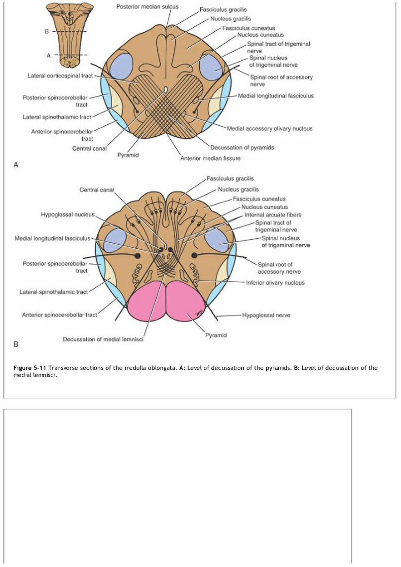
Figure 5-11 Transverse sections of the medulla oblongata. A: Level of decussation of the pyramids. B: Level of decussation of themedial lemnisci.

Figure 5-12 Transverse section of the medulla oblongata at the level of decussation of the pyramids. (Weigert stain.)
Figure 5-13 Transverse section of the medulla oblongata at the level of decussation of the medial lemnisci. (Weigert stain.)

Figure 5-14 Transverse sections of the medulla oblongata at the level of the middle of the olivary nuclei (A) and the superior partof the olivary nuclei just inferior to the pons (B).

Figure 5-15 Transverse section of the medulla oblongata at the level of the middle of the olivary nuclei. (Weigert stain.)
Olivary Nuclear ComplexThe largest nucleus of this complex is the inferior olivary nucleus (Figs. 5-14 and 5-15). The gray matter is shaped like a crumpled bag withits mouth directed medially; it is responsible for the elevation on the surface of the medulla called the olive. Smaller dorsal and medialaccessory olivary nuclei also are present. The cells of the inferior olivary nucleus send fibers medially across the midline to enter thecerebellum through the inferior cerebellar peduncle. Afferent fibers reach the inferior olivary nuclei from the spinal cord (the spino-olivary tracts) and from the cerebellum and cerebral cortex. The function of the olivary nuclei is associated with voluntary musclemovement.
Vestibulocochlear NucleiThe vestibular nuclear complex is made up of the following nuclei: (1) medial vestibular nucleus, (2) inferior vestibular nucleus, (3)lateral vestibular nucleus, and (4) superior vestibular nucleus. The details of these nuclei and their connections are discussed later. Themedial and inferior vestibular nuclei can be seen on section at this level (Figs. 5-14 and 5-15).
There are two cochlear nuclei. The anterior cochlear nucleus is situated on the anterolateral aspect of the inferior cerebellar peduncle,and the posterior cochlear nucleus is situated on the posterior aspect of the peduncle lateral to the floor of the fourth ventricle (Figs. 5-14 and 5-15). The connections of these nuclei are described later (see page 350).
The Nucleus AmbiguusThe nucleus ambiguus consists of large motor neurons and is situated deep within the reticular formation (Figs. 5-14 and 5-16). Theemerging nerve fibers join the glossopharyngeal, vagus, and cranial part of the accessory nerve and are distributed to voluntary skeletalmuscle.
Central Gray MatterThe central gray matter lies beneath the floor of the fourth ventricle at this level (Figs. 5-14 and 5-15). Passing from medial to lateral (Fig.5-16), the following important structures may be recognized: (1) the hypoglossal nucleus, (2) the dorsal nucleus of the vagus, (3) thenucleus of the tractus solitarius, and (4) the medial and inferior vestibular nuclei (see previous column). The nucleus ambiguus, referredto above, has become deeply placed within the reticular formation (Fig. 5-14). The connections and functional significance of these nucleiare described in Chapter 11.

Figure 5-16 Position of the cranial nerve nuclei within the brainstem. The hatched area indicates the position of the vestibularnuclei.
The arcuate nuclei are thought to be inferiorly displaced pontine nuclei (see p. 208) and are situated on the anterior surface of thepyramids (Fig. 5-14). They receive nerve fibers from the cerebral cortex and send efferent fibers to the cerebellum through the anteriorexternal arcuate fibers.
The pyramids containing the corticospinal and some corticonuclear fibers are situated in the anterior part of the medulla separated bythe anterior median fissure (Figs. 5-14 and 5-15); the corticospinal fibers descend to the spinal cord, and the corticonuclear fibers aredistributed to the motor nuclei of the cranial nerves situated within the medulla.
The medial lemniscus forms a flattened tract on each side of the midline posterior to the pyramid (Figs. 5-7 and 5-15). These fibers emergefrom the decussation of the lemnisci and convey sensory information to the thalamus.
The medial longitudinal fasciculus forms a small tract of nerve fibers situated on each side of the midline posterior to the mediallemniscus and anterior to the hypoglossal nucleus (Figs. 5-14 and 5-15). It consists of ascending and descending fibers, the connections ofwhich are described on page 208.
The inferior cerebellar peduncle is situated in the posterolateral corner of the section on the lateral side of the fourth ventricle (Figs. 5-14 and 5-15).
The spinal tract of the trigeminal nerve and its nucleus are situated on the anteromedial aspect of the inferior cerebellar peduncle (Figs.5-14 and 5-15).
The anterior spinocerebellar tract is situated near the surface in the interval between the inferior olivary nucleus and the nucleus of thespinal tract of the trigeminal nerve (Figs. 5-14 and 5-15). The spinal lemniscus, consisting of the anterior spinothalamic, the lateralspinothalamic, and spinotectal tracts, is deeply placed.
The reticular formation, consisting of a diffuse mixture of nerve fibers and small groups of nerve cells, is deeply placed posterior to theolivary nucleus (Figs. 5-14 and 5-15). The reticular formation represents, at this level, only a small part of this system, which is also presentin the pons and midbrain.
The glossopharyngeal, vagus, and cranial part of the accessory nerves can be seen running forward and laterally through the reticularformation (Fig. 5-14). The nerve fibers emerge between the olives and the inferior cerebellar peduncles. The hypoglossal nerves also runanteriorly and laterally through the reticular formation and emerge between the pyramids and the olives (Fig. 5-14).
Level Just Inferior to the PonsThere are no major changes, in comparison to the previous level, in the distribution of the gray and white matter (Figs. 5-14 and 5-16). Thelateral vestibular nucleus has replaced the inferior vestibular nucleus, and the cochlear nuclei now are visible on the anterior andposterior surfaces of the inferior cerebellar peduncle.

Gross Appearance of the PonsThe pons is anterior to the cerebellum (Fig. 5-17; see also Fig. 6-1) and connects the medulla oblongata to the midbrain. It is about1 inch (2.5 cm) long and owes its name to the appearance presented on the anterior surface, which is that of a bridge connecting
the right and left cerebellar hemispheres.
The anterior surface is convex from side to side and shows many transverse fibers that converge on each side to form the middlecerebellar peduncle (Fig. 5-17). There is a shallow groove in the midline, the basilar groove, which lodges the basilar artery. On theanterolateral surface of the pons, the trigeminal nerve emerges on each side. Each nerve consists of a smaller, medial part, known as themotor root, and a larger, lateral part, known as the sensory root. In the groove between the pons and the medulla oblongata, thereemerge, from medial to lateral, the abducent, facial, and vestibulocochlear nerves (Fig. 5-17).
Figure 5-17 Anterior surface of the brainstem showing the pons.
The posterior surface of the pons is hidden from view by the cerebellum (Fig. 5-18). It forms the upper half of the floor of the fourthventricle and is triangular in shape. The posterior surface is limited laterally by the superior cerebellar peduncles and is divided intosymmetrical halves by a median sulcus. Lateral to this sulcus is an elongated elevation, the medial eminence, which is bounded laterally bya sulcus, the sulcus limitans (Fig. 5-18). The inferior end of the medial eminence is slightly expanded to form the facial colliculus, which isproduced by the root of the facial nerve winding around the nucleus of the abducent nerve (Fig. 5-19). The floor of the superior part ofthe sulcus limitans is bluish-gray in color and is called the substantia ferruginea; it owes its color to a group of deeply pigmented nervecells. Lateral to the sulcus limitans is the area vestibuli produced by the underlying vestibular nuclei (Fig. 5-18).

Figure 5-18 Posterior surface of the brainstem showing the pons. The cerebellum has been removed.
Figure 5-19 Transverse section through the caudal part of the pons at the level of the facial colliculus.

Internal Structure of the PonsFor purposes of description, the pons is commonly divided into a posterior part, the tegmentum, and an anterior basal part bythe transversely running fibers of the trapezoid body (Fig. 5-19).
The structure of the pons may be studied at two levels: (1) transverse section through the caudal part, passing through the facialcolliculus, and (2) transverse section through the cranial part, passing through the trigeminal nuclei. See Table 5-3 for a comparison of thetwo levels of the pons and the major structures present at each level.
Transverse Section Through the Caudal PartThe medial lemniscus rotates as it passes from the medulla into the pons. It is situated in the most anterior part of the tegmentum, withits long axis running transversely (Fig. 5-19). The medial lemniscus is accompanied by the spinal and lateral lemnisci.
The facial nucleus lies posterior to the lateral part of the medial lemniscus. The fibers of the facial nerve wind around the nucleus of theabducent nerve, producing the facial colliculus (Fig. 5-19). The fibers of the facial nerve then pass anteriorly between the facial nucleusand the superior end of the nucleus of the spinal tract of the trigeminal nerve.
The medial longitudinal fasciculus is situated beneath the floor of the fourth ventricle on either side of the midline (Fig. 5-19). The mediallongitudinal fasciculus is the main pathway that connects the vestibular and cochlear nuclei with the nuclei controlling the extraocularmuscles (oculomotor, trochlear, and abducent nuclei).
The medial vestibular nucleus is situated lateral to the abducent nucleus (Fig. 5-19) and is in close relationship to the inferior cerebellarpeduncle. The superior part of the lateral and the inferior part of the superior vestibular nucleus are found at this level. The posteriorand anterior cochlear nuclei are also found at this level.
Table 5-3 Comparison of the Different Levels of the Pons Showing the Major Structures at Each Levela
Level Cav ity Nuclei Motor Tracts Sensory Tracts
Facialcolliculus
Fourthventricle
Facial nucleus, abducent nucleus,medial vestibular nucleus, spinalnucleus of cranial nerve V, pontinenuclei, trapezoid nuclei
Corticospinal andcorticonuclear tracts,transverse pontine fibers,medial longitudinalfasciculus
Spinal tract ofcranial nerve V;lateral, spinal,and mediallemnisci
Trigeminalnuclei
Fourthventricle
Main sensory and motor nucleus ofcranial nerve V, pontine nuclei,trapezoid nuclei
Corticospinal andcorticonuclear tracts,transverse pontine fibers,medial longitudinalfasciculus
Lateral, spinal,and mediallemnisci
aNote that the reticular formation is present at all levels.
The spinal nucleus of the trigeminal nerve and its tract lie on the anteromedial aspect of the inferior cerebellar peduncle (Fig. 5-19).
The trapezoid body is made up of fibers derived from the cochlear nuclei and the nuclei of the trapezoid body. They run transversely (Fig.5-19) in the anterior part of the tegmentum (see p. 207).
The basilar part of the pons, at this level, contains small masses of nerve cells called pontine nuclei (Fig. 5-19). The corticopontine fibersof the crus cerebri of the midbrain terminate in the pontine nuclei. The axons of these cells give origin to the transverse fibers of thepons, which cross the midline and intersect the corticospinal and corticonuclear tracts, breaking them up into small bundles. Thetransverse fibers of the pons enter the middle cerebellar peduncle and are distributed to the cerebellar hemisphere. This connectionforms the main pathway linking the cerebral cortex to the cerebellum.
Transverse Section Through the Cranial PartThe internal structure of the cranial part of the pons is similar to that seen at the caudal level (Figs. 5-20, 5-21 and 5-22), but it nowcontains the motor and principal sensory nuclei of the trigeminal nerve.
The motor nucleus of the trigeminal nerve is situated beneath the lateral part of the fourth ventricle within the reticular formation (Figs.5-20 and 5-21). The emerging motor fibers travel anteriorly through the substance of the pons and exit on its anterior surface.
The principal sensory nucleus of the trigeminal nerve is situated on the lateral side of the motor nucleus (Figs. 5-20 and 5-21); it iscontinuous inferiorly with the nucleus of the spinal tract. The entering sensory fibers travel through the substance of the pons and lielateral to the motor fibers (Fig. 5-20).

Figure 5-20 Transverse section through the pons at the level of the trigeminal nuclei.
Figure 5-21 Photomicrograph of a transverse section of the pons at the level of the trigeminal nuclei.

Figure 5-22 Photomicrograph of a transverse section of the most rostral part of the pons.
The superior cerebellar peduncle is situated posterolateral to the motor nucleus of the trigeminal nerve (Figs. 5-20 and 5-21). It is joinedby the anterior spinocerebellar tract.
The trapezoid body and the medial lemniscus are situated in the same position as they were in the previous section (Fig. 5-20). Thelateral and spinal lemnisci lie at the lateral extremity of the medial lemniscus (Figs. 5-20 and 5-22).
Gross Appearance of the MidbrainThe midbrain measures about 0.8 inch (2 cm) in length and connects the pons and cerebellum with the forebrain (Fig. 5-23). Itslong axis inclines anteriorly as it ascends through the opening in the tentorium cerebelli. The midbrain is traversed by a narrow
channel, the cerebral aqueduct, which is filled with cerebrospinal fluid (Figs. 5-24, 5-25, 5-26, 5-27 and 5-28).
On the posterior surface are four colliculi (corpora quadrigemina). These are rounded eminences that are divided into superior andinferior pairs by a vertical and a transverse groove (Fig. 5-26). The superior colliculi are centers for visual reflexes (see p. 338), and theinferior colliculi are lower auditory centers. In the midline below the inferior colliculi, the trochlear nerves emerge. These are small-diameter nerves that wind around the lateral aspect of the midbrain to enter the lateral wall of the cavernous sinus.
On the lateral aspect of the midbrain, the superior and inferior brachia ascend in an anterolateral direction (Fig. 5-23). The superiorbrachium passes from the superior colliculus to the lateral geniculate body and the optic tract. The inferior brachium connects theinferior colliculus to the medial geniculate body.
On the anterior aspect of the midbrain (Fig. 5-23), there is a deep depression in the midline, the interpeduncular fossa, which is boundedon either side by the crus cerebri. Many small blood vessels perforate the floor of the interpeduncular fossa, and this region is termed theposterior perforated substance (Fig. 5-23). The oculomotor nerve emerges from a groove on the medial side of the crus cerebri and passesforward in the lateral wall of the cavernous sinus.
Internal Structure of the MidbrainThe midbrain comprises two lateral halves, called the cerebral peduncles; each of these is divided into an anterior part, the cruscerebri, and a posterior part, the tegmentum, by a pigmented band of gray matter, the substantia nigra (Figs. 5-24 and 5-25). The
narrow cavity of themidbrain is the cerebral aqueduct, which connects the third and fourth ventricles. The tectum is the part of the midbrain posterior tothe cerebral aqueduct; it has four small surface swellings referred to previously; these are the two superior and two inferior colliculi(Figs. 5-24 and 5-25). The cerebral aqueduct is lined by ependyma and is surrounded by the central gray matter. On transverse sections ofthe midbrain, the interpeduncular fossa can be seen to separate the crura cerebri, whereas the tegmentum is continuous across themedian plane (Fig. 5-24).

Figure 5-23 The midbrain. A: Anterior view. B: Lateral view.
Transverse Section of the Midbrain at the Level of the Inferior ColliculiThe inferior colliculus, consisting of a large nucleus of gray matter, lies beneath the corresponding surface elevation and forms part ofthe auditory pathway (Figs. 5-25A and 5-27). It receives many of the terminal fibers of the lateral lemniscus. The pathway then continuesthrough the inferior brachium to the medial geniculate body.

P.214P.215
Figure 5-24 Transverse section of the midbrain through the inferior colliculi shows the division of the midbrain into the tectum andthe cerebral peduncles. Note that the cerebral peduncles are subdivided by the substantia nigra into the tegmentum and the cruscerebri.
The trochlear nucleus is situated in the central gray matter close to the median plane just posterior to the medial longitudinal fasciculus.The emerging fibers of the trochlear nucleus pass laterally and posteriorly around the central gray matter and leave the midbrain justbelow the inferior colliculi. The fibers of the trochlear nerve now decussate completely in the superior medullary velum. Themesencephalic nuclei of the trigeminal nerve are lateral to the cerebral aqueduct (Figs. 5-25A and 5-27). The decussation of the superiorcerebellar peduncles occupies the central part of the tegmentum anterior to the cerebral aqueduct. The reticular formation is smallerthan that of the pons and is situated lateral to the decussation.
The medial lemniscus ascends posterior to the substantia nigra; the spinal and trigeminal lemnisci are situated lateral to the mediallemniscus (Figs. 5-25 and 5-27). The lateral lemniscus is located posterior to the trigeminal lemniscus.
The substantia nigra (Figs. 5-25 and 5-27) is a large motor nucleus situated between the tegmentum, and the crus cerebri and is foundthroughout the midbrain. The nucleus is composed of medium-size multipolar neurons that possess inclusion granules of melanin pigmentwithin their cytoplasm. The substantia nigra is concerned with muscle tone and is connected to the cerebral cortex, spinal cord,hypothalamus, and basal nuclei.
The crus cerebri contains important descending tracts and is separated from the tegmentum by the substantia nigra (Figs. 5-25 and 5-27).The corticospinal and corticonuclear fibers occupy the middle two-thirds of the crus. The frontopontine fibers occupy the medial part ofthe crus, and the temporopontine fibers occupy the lateral part of the crus (Figs. 5-25 and 5-27). These descending tracts connect thecerebral cortex to the anterior gray column cells of the spinal cord, the cranial nerve nuclei, the pons, and the cerebellum (Table 5-4).
Transverse Section of the Midbrain at the Level of the Superior ColliculiThe superior colliculus (Figs. 5-25B and 5-28), a large nucleus of gray matter that lies beneath the corresponding surface elevation, formspart of the visual reflexes. It is connected to the lateral geniculate body by the superior brachium. It receives afferent fibers from theoptic nerve, thevisual cortex, and the spinotectal tract. The efferent fibers form the tectospinal and tectobulbar tracts, which areprobably responsible for the reflex movements of the eyes, head, and neck in response to visual stimuli. The afferent pathway for the lightreflex ends in the pretectal nucleus. This is a small group of neurons situated close to the lateral part of the superior colliculus. Afterrelaying in the pretectal nucleus, the fibers pass to the parasympathetic nucleus of the oculomotor nerve (Edinger-Westphal nucleus). Theemerging fibers then pass to the oculomotor nerve. The oculomotor nucleus is situated in the central gray matter close to the medianplane, just posterior to the medial longitudinal fasciculus (Figs. 5-25B and 5-28). The fibers of the oculomotor nucleus pass anteriorlythrough the red nucleus to emerge on the medial side of the crus cerebri in the interpeduncular fossa. The nucleus of the oculomotornerve is divisible into a number of cell groups.

Figure 5-25 Transverse sections of the midbrain. A: At the level of the inferior colliculus. B: At the level of the superior colliculus.Note that trochlear nerves completely decussate within the superior medullary velum.

Figure 5-26 Posterior view of the brainstem showing the two superior and the two inferior colliculi of the tectum.
Figure 5-27 Photomicrograph of a transverse section of the midbrain at the level of the inferior colliculus. (Weigert stain.)

Figure 5-28 Photomicrograph of a transverse section of the midbrain at the level of the superior colliculus. (Weigert stain.)
Table 5-4 Comparison of Two Levels of the Midbrain Showing the Major Structures at Each Levela
Level Cav ity Nuclei Motor Tract Sensory Tracts
Inferiorcolliculi
Cerebralaqueduct
Inferior colliculus, substantianigra, trochlear nucleus,mesencephalic nuclei of cranialnerve V
Corticospinal andcorticonuclear tracts,temporopontine,frontopontine, mediallongitudinal fasciculus
Lateral, trigeminal,spinal, and mediallemnisci;decussation ofsuperior cerebellarpeduncles
Superiorcolliculi
Cerebralaqueduct
Superior colliculus, substantianigra, oculomotor nucleus,Edinger-Westphal nucleus, rednucleus, mesencephalic nucleusof cranial nerve V
Corticospinal andcorticonuclear tracts,temporopontine,frontopontine, mediallongitudinal fasciculus,decussation of rubrospinaltract
Trigeminal, spinal,and medial lemnisci
aNote that the reticular formation is present at all levels.
The medial, spinal, and trigeminal lemnisci form a curved band posterior to the substantia nigra, but the lateral lemniscus does notextend superiorly to this level (Figs. 5-25B and 5-28).
The red nucleus (Figs. 5-25B and 5-28) is a rounded mass of gray matter situated between the cerebral aqueduct and the substantia nigra.Its reddish hue, seen in fresh specimens, is due to its vascularity and the presence of an iron-containing pigment in the cytoplasm of manyof its neurons.Afferent fibers reach the red nucleus from (1) the cerebral cortex through the corticospinal fibers, (2) the cerebellum through thesuperior cerebellar peduncle, and (3) the lentiform nucleus, subthalamic and hypothalamic nuclei, substantia nigra, and spinal cord.Efferent fibers leave the red nucleus and pass to (1) the spinal cord through the rubrospinal tract (as this tract descends, it decussates),(2) the reticular formation through the rubroreticular tract, (3) the thalamus, and (4) the substantia nigra.
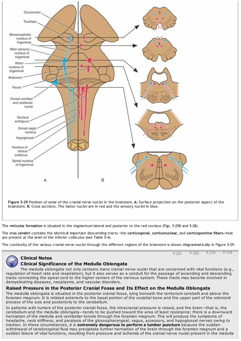
P.218P.219P.220P.221
Figure 5-29 Position of some of the cranial nerve nuclei in the brainstem. A: Surface projection on the posterior aspect of thebrainstem. B: Cross sections. The motor nuclei are in red and the sensory nuclei in blue.
The reticular formation is situated in the tegmentum lateral and posterior to the red nucleus (Figs. 5-25B and 5-28).
The crus cerebri contains the identical important descending tracts—the corticospinal, corticonuclear, and corticopontine fibers—thatare present at the level of the inferior colliculus (see Table 5-4).
The continuity of the various cranial nerve nuclei through the different regions of the brainstem is shown diagrammatically in Figure 5-29.
Clinical NotesClinical Significance of the Medulla OblongataThe medulla oblongata not only contains many cranial nerve nuclei that are concerned with vital functions (e.g.,
regulation of heart rate and respiration), but it also serves as a conduit for the passage of ascending and descendingtracts connecting the spinal cord to the higher centers of the nervous system. These tracts may become involved indemyelinating diseases, neoplasms, and vascular disorders.
Raised Pressure in the Posterior Cranial Fossa and Its Effect on the Medulla OblongataThe medulla oblongata is situated in the posterior cranial fossa, lying beneath the tentorium cerebelli and above theforamen magnum. It is related anteriorly to the basal portion of the occipital bone and the upper part of the odontoidprocess of the axis and posteriorly to the cerebellum.In patients with tumors of the posterior cranial fossa, the intracranial pressure is raised, and the brain—that is, thecerebellum and the medulla oblongata—tends to be pushed toward the area of least resistance; there is a downwardherniation of the medulla and cerebellar tonsils through the foramen magnum. This will produce the symptoms ofheadache, neck stiffness, and paralysis of the glossopharyngeal, vagus, accessory, and hypoglossal nerves owing totraction. In these circumstances, it is extremely dangerous to perform a lumbar puncture because the suddenwithdrawal of cerebrospinal fluid may precipitate further herniation of the brain through the foramen magnum and asudden failure of vital functions, resulting from pressure and ischemia of the cranial nerve nuclei present in the medulla

oblongata.
Figure 5-30 Arnold-Chiari phenomenon. This coronal section of the skull shows the herniation of the cerebellar tonsil and themedulla oblongata through the foramen magnum into the vertebral canal.
Arnold-Chiari PhenomenonThe Arnold-Chiari malformation is a congenital anomaly in which there is a herniation of the tonsils of the cerebellumand the medulla oblongata through the foramen magnum into the vertebral canal (Fig. 5-30). This results in theblockage of the exits in the roof of the fourth ventricle to the cerebrospinal fluid, causing internal hydrocephalus. It iscommonly associated with craniovertebral anomalies or various forms of spina bifida. Signs and symptoms related topressure on the cerebellum and medulla oblongata and involvement of the last four cranial nerves are associated withthis condition.
Vascular Disorders of the Medulla OblongataLateral Medullary Syndrome of WallenbergThe lateral part of the medulla oblongata is supplied by the posterior inferior cerebellar artery, which is usually abranch of the vertebral artery. Thrombosis of either of these arteries (Fig. 5-31) produces the following signs andsymptoms: dysphagia and dysarthria due to paralysis of the ipsilateral palatal and laryngeal muscles (innervated bythe nucleus ambiguus); analgesia and thermoanesthesia on the ipsilateral side of the face (nucleus and spinal tract ofthe trigeminal nerve); vertigo, nausea, vomiting, and nystagmus (vestibular nuclei); ipsilateral Horner syndrome(descending sympathetic fibers); ipsilateral cerebellar signs—gait and limb ataxia (cerebellum or inferior cerebellarpeduncle); and contralateral loss of sensations of pain and temperature (spinal lemniscus—spinothalamic tract).

Figure 5-31 Transverse section of the medulla oblongata at the level of the inferior olivary nuclei showing the extent of thelesion producing the lateral medullary syndrome.
Medial Medullary SyndromeThe medial part of the medulla oblongata is supplied by the vertebral artery. Thrombosis of the medullary branch (Fig.5-32) produces the following signs and symptoms: contralateral hemiparesis (pyramidal tract), contralateral impairedsensations of position and movement and tactile discrimination (medial lemniscus), and ipsilateral paralysis of tonguemuscles with deviation to the paralyzed side when the tongue is protruded (hypoglossal nerve).
Clinical Significance of the PonsThe pons, like the medulla oblongata and the cerebellum, is situated in the posterior cranial fossa lying beneath thetentorium cerebelli. It is related anteriorly to the basilar artery, the dorsum sellae of the sphenoid bone, and thebasilar part of the occipital bone. In addition to forming the upper half of the floor of the fourth ventricle, it possessesseveral important cranial nerve nuclei (trigeminal, abducent, facial, and vestibulocochlear) and serves as a conduit forimportant ascending and descending tracts (corticonuclear, corticopontine, corticospinal, medial longitudinal fasciculusand medial, spinal, and lateral lemnisci). It is not surprising, therefore, that tumors, hemorrhage, or infarcts in this areaof the brain produce a variety of symptoms and signs. For example, involvement of the corticopontocerebellar tractswill produce marked cerebellar ataxia, and voluntary movements are accompanied by a rhythmic tremor that developsand becomes further accentuated as the movements proceed (intention tumor).
Tumors of the PonsAstrocytoma of the pons occurring in childhood is the most common tumor of the brainstem. The symptoms and signsare those of ipsilateral cranial nerve paralysis and contralateral hemiparesis: weakness of the facial muscles on thesame side (facial nerve nucleus), weakness of the lateral rectus muscle on one or both sides (abducent nervenucleus), nystagmus (vestibular nucleus), weakness of the jaw muscles (trigeminal nerve nucleus), impairment ofhearing (cochlear nuclei), contralateral hemiparesis, quadriparesis (corticospinal fibers), anesthesia to light touch withthe preservation of appreciation of pain over the skin of the face (principal sensory nucleus of trigeminal nerveinvolved, leaving spinal nucleus and tract of trigeminal intact), and contralateral sensory defects of the trunk and limbs(medial and spinal lemnisci). Involvement of the corticopontocerebellar tracts may cause ipsilateral cerebellar signs andsymptoms. There may be impairment of conjugate deviation of the eyeballs due to involvement of the mediallongitudinal fasciculus, which connects the oculomotor, trochlear, and abducent nerve nuclei.
Pontine HemorrhageThe pons is supplied by the basilar artery and the anterior, inferior, and superior cerebellar arteries. If the hemorrhageoccurs from one of those arteries and is unilateral, there will be facial paralysis on the side of the lesion (involvementof the facial nerve nucleus and, therefore, a lower motor neuron palsy) and paralysis of the limbs on the opposite side(involvement of the corticospinal fibers as they pass through the pons). There is often paralysis of conjugate oculardeviation (involvement of the abducent nerve nucleus and the medial longitudinal fasciculus).

Figure 5-32 Transverse section of the medulla oblongata at the level of the inferior olivary nuclei showing the extent of thelesion producing the medial medullary syndrome.
When the hemorrhage is extensive and bilateral, the pupils may be “pinpoint” (involvement of the ocular sympatheticfibers); there is commonly bilateral paralysis of the face and the limbs. The patient may become poikilothermic becausesevere damage to the pons has cut off the body from the heat-regulating centers in the hypothalamus.
Infarctions of the PonsUsually, infarction of the pons is due to thrombosis or embolism of the basilar artery or its branches. If it involves theparamedian area of the pons, the corticospinal tracts, the pontine nuclei, and the fibers passing to the cerebellumthrough the middle cerebellar peduncle may be damaged. A laterally situated infarct will involve the trigeminal nerve,the medial lemniscus, and the middle cerebellar peduncle; the corticospinal fibers to the lower limbs also may beaffected.The clinical conditions mentioned above will be understood more clearly if the ascending and descending tracts of thebrain and spinal cord are reviewed (see pp. 143 and 153).
Clinical Significance of the MidbrainThe midbrain forms the upper end of the narrow stalk of the brain or brainstem. As it ascends out of the posteriorcranial fossa through the relatively small rigid opening in the tentorium cerebelli, it is vulnerable to traumatic injury. Itpossesses two important cranial nerve nuclei (oculomotor and trochlear), reflex centers (the colliculi), and the rednucleus and substantia nigra, which greatly influence motor function, and the midbrain serves as a conduit for manyimportant ascending and descending tracts. As in other parts of the brainstem, it is a site for tumors, hemorrhage, orinfarcts that will produce a wide variety of symptoms and signs.
Trauma to the MidbrainAmong the mechanisms of injuries to the midbrain, a sudden lateral movement of the head could result in the cerebralpeduncles impinging against the sharp rigid free edge of the tentorium cerebelli. Sudden movements of the headresulting from trauma cause different regions of the brain to move at different velocities relative to one another. Forexample, the large anatomical unit, the forebrain, may move at a different velocity from the remainder of the brain,such as the cerebellum. This will result in the midbrain being bent, stretched, twisted, or torn.Involvement of the oculomotor nucleus will produce ipsilateral paralysis of the levator palpebrae superioris; thesuperior, inferior, and medial rectus muscles; and the inferior oblique muscle. Malfunction of the parasympatheticnucleus of the oculomotor nerve produces a dilated pupil that is insensitive to light and does not constrict onaccommodation.Involvement of the trochlear nucleus will produce contralateral paralysis of the superior oblique muscle of the eyeball.Thus, it is seen that involvement of one or both of these nuclei, or the corticonuclear fibers that converge on them, willcause impairment of ocular movements.
Blockage of the Cerebral AqueductThe cavity of the midbrain, the cerebral aqueduct, is one of the narrower parts of the ventricular system. Normally,cerebrospinal fluid that has been produced in the lateral and third ventricles passes through this channel to enter thefourth ventricle and so escapes through the foramina in its roof to enter the subarachnoid space. In congenitalhydrocephalus, the cerebral aqueduct may be blocked or replaced by numerous small tubular passages that areinsufficient for the normal flow of cerebrospinal fluid. A tumor of the midbrain (Fig. 5-33A) or pressure on the midbrainfrom a tumor arising outside the midbrain may compress the aqueduct and produce hydrocephalus. When the cerebralaqueduct is blocked, the accumulating cerebrospinal fluid within the third and lateral ventricles produces lesions in themidbrain. The presence of the oculomotor and trochlear nerve nuclei, together with the important descendingcorticospinal and corticonuclear tracts, will provide symptoms and signs that are helpful in accurately localizing a lesionin the brainstem.

P.223
Figure 5-33 Pathology of the midbrain. A: Tumor of the midbrain blocking the cerebral aqueduct. B: Weber syndrome involvingthe oculomotor nerve and the crus cerebri following occlusion of the blood supply to the midbrain. C: Benedikt syndromeinvolving the red nucleus and the medial lemniscus following occlusion of the blood supply to the midbrain.
Vascular Lesions of the MidbrainWeber SyndromeWeber syndrome (Fig. 5-33B), which is commonly produced by occlusion of a branch of the posterior cerebral arterythat supplies the midbrain, results in the necrosis of brain tissue involving the oculomotor nerve and the crus cerebri.There is ipsilateral ophthalmoplegia and contralateral paralysis of the lower part of the face, the tongue, and the armand leg. The eyeball is deviated laterally because of the paralysis of the medial rectus muscle; there is drooping(ptosis) of the upper lid, and the pupil is dilated and fixed to light and accommodation.
Benedikt SyndromeBenedikt syndrome (Fig. 5-33C) is similar to Weber syndrome, but the necrosis involves the medial lemniscus and rednucleus, producing contralateral hemianesthesia and involuntary movements of the limbs of the opposite side.
Clinical Problem Solving1. While carrying out a physical examination of a patient with an intracranial tumor, the neurologist turned to a medicalstudent and asked, “What signs or symptoms would you look for that would enable you to localize the tumor to theregion of the medulla oblongata?” How would you have answered that question?View Answer
2. A 6-month-old boy died with hydrocephalus and a myelocele in the lower thoracic region. At autopsy, the hindbrainwas found to be deformed. The lower part of the medulla oblongata extended inferiorly through the foramen magnuminto the vertebral canal as far as the third cervical vertebra. The lower four cranial nerves were longer than normal,and the upper cervical nerve roots ascended to reach their exit from the vertebral canal. The cerebellum on the leftside extended inferiorly through the foramen magnum to the third cervical vertebra, where it was adherent to thespinal cord. The roof of the fourth ventricle was abnormally low. (a) What is the name of this malformation? (b) Ishydrocephalus common in this condition? (c) Is there a possible association between the thoracic myelocele and thepresence of part of the hindbrain in the vertebral canal?View Answer
3. A 68-year-old man was admitted to the hospital with the sudden onset of severe dizziness (vertigo), hiccups, andvomiting. He also complained of a hot, painful sensation in the skin of the right side of the face. On physical

examination, the soft palate was drawn up to the left side when the patient was asked to say “ah,” and there waslack of mobility of the right vocal cord as seen on laryngoscopic examination. The patient also showed drooping of theright upper eyelid (ptosis), sunken right eye (enophthalmos), and a constricted right pupil (myosis). When asked toprotrude his tongue straight out of his mouth, the patient tried to do so, but the tip of the tongue pointed to the rightside. There was evidence of impairment of pain and temperature sensation in the trunk and extremities on the leftside. Using your knowledge of anatomy, make the diagnosis.View Answer
4. A pathologist, while exploring the posterior cranial fossa during an autopsy, was endeavoring to determine wherethe 9th, the 10th, and the cranial part of the 11th cranial nerves emerged from the hindbrain. Describe where thesenerves emerge from the hindbrain.View Answer
5. A 10-year-old girl was taken to a physician because her mother had noticed that the right half of her face was weakand did not appear to react to emotional changes. It was noted also that her mouth was pulled over slightly to theleft, especially when she was tired. On questioning, the patient admitted that food tended to stick inside her rightcheek and that the right side of her face “felt funny.” The mother had first noticed the facial changes 3 monthspreviously, and the condition had progressively worsened. On examination, there was definite weakness of the facialmuscles on the right side; the facial muscles on the left side were normal. Skin sensation on stimulation of the facewas normal. On testing of the ocular movements, there was evidence of slight weakness of the lateral rectus muscleon the right side. Examination of the movements of the arm and leg showed slight weakness on the left side. Usingyour knowledge of neuroanatomy, relate these symptoms and signs to a lesion in the pons.View Answer
6. A 65-year-old man was admitted to the emergency department with a diagnosis of a severe pontine hemorrhage.On examination, he was found to have bilateral “pinpoint” pupils and quadriplegia. How can you explain the presenceof the “pinpoint” pupils?View Answer
7. A 46-year-old man with symptoms of deafness, vertigo, and double vision (diplopia) visited his physician. Onquestioning, he said that he also suffered from severe headaches, which were increasing in frequency and severity.The week before, he vomited several times during one of the headache attacks. On examination, he was found tohave a slight right internal strabismus, a flattening of the skin furrows on the right side of his forehead, and a slightdrooping of the right corner of his mouth. There was definite evidence of impairment of hearing on the right side. Ontesting for sensory loss, there was definite sensory impairment on the right side of the face in the areas supplied bythe maxillary and mandibular divisions of the trigeminal nerve. Using your knowledge of anatomy, explain thesymptoms and signs.View Answer
8. After a severe automobile accident that resulted in the death of the driver of one of the vehicles, an autopsy wasperformed, and the skull was opened. A massive subdural hematoma was found in the middle cranial fossa. The rapidaccumulation of blood within the skull had exerted pressure on the brain above the tentorium cerebelli. The uncus ofthe temporal lobe had been forced inferiorly through the hiatus in the tentorium cerebelli. What effect do you thinkthese intracranial changes had on the midbrain of this patient?View Answer
9. A 3-month-old girl was taken to a pediatrician because her mother was concerned about the large size of her head.The child was perfectly normal in every other respect. Examination of the child showed that the diameter of the headwas larger than normal for the age; the fontanelles were larger than normal and were moderately tense. The scalpwas shiny, and the scalp veins were dilated. The eyes were normal, and the mental and physical development of thechild was within normal limits. CT and MRI of the head revealed gross dilation of the third and lateral ventricles of thebrain. What is your diagnosis? What possible treatment should be suggested to the mother of this child?View Answer
10. A 20-year-old man was seen by a neurologist because he had a 3-month history of double vision. On examinationof the patient, both eyes at rest were turned downward and laterally. The patient was unable to move the eyesupward or medially. Both upper lids were drooping (ptosis). Examination of both pupils showed them to be dilated, andthey did not constrict when a light was shone into either eye. Facial movements and sensation were normal.Movements of the upper and lower limbs were normal. There was no evidence of loss of or altered skin sensations inthe upper or the lower limbs. Using your knowledge of neuroanatomy, make a diagnosis and accurately locate the siteof the lesion. Is the lesion unilateral or bilateral?View Answer
11. A 57-year-old man with hypertension was admitted to the hospital with a diagnosis of hemorrhage into themidbrain, possibly from a branch of the posterior cerebral artery. He was found, on physical examination, to haveparalysis on the right side of the levator palpebrae superioris, the superior rectus, medial rectus, inferior rectus, andinferior oblique muscles. Furthermore, his right pupil was dilated and failed to constrict on exposure to light or onaccommodation. The left eye was normal in every respect. He displayed hypersensitivity to touch on the skin of the leftside of his face and had loss of skin sensation on the greater part of his left arm and left leg. The left leg alsodisplayed some spontaneous slow writhing movements (athetosis). Using your knowledge of neuroanatomy, explainthe signs and symptoms exhibited by this patient.View Answer
12. A 41-year-old woman was diagnosed as having a lesion in the midbrain. Physical examination revealed an
P.225P.226P.227P.228
oculomotor nerve palsy on the left side (paralysis of the left extraocular muscles except the lateral rectus and thesuperior oblique muscles) and an absence of the light and accommodation reflexes on the left side. There was someweakness but no atrophy of the muscles of the lower part of the face and the tongue on the right side. There wasevidence of spastic paralysis of the right arm and leg. There was no evidence of any sensory loss on either side of thehead, trunk, or limbs. Using your knowledge of neuroanatomy, precisely place the lesion in the midbrain of this patient.View Answer
Review QuestionsDirections: Each of the numbered items in this section is followed by answers. Select the ONE lettered answerthat is CORRECT.1. The following statements concern the anterior surface of the medulla oblongata:
(a) The pyramids taper inferiorly and give rise to the decussation of the pyramids.(b) On each side of the midline, there is an ovoid swelling called the olive, which contains the corticospinal fibers.(c) The hypoglossal nerve emerges between the olive and the inferior cerebellar peduncle.(d) The vagus nerve emerges between the pyramid and the olive.(e) The abducent nerve emerges between the pons and the midbrain.
View Answer
2. The following general statements concern the medulla oblongata:(a) The caudal half of the floor of the fourth ventricle is formed by the rostral half of the medulla.(b) The central canal extends throughout the length of the medulla oblongata.(c) The nucleus gracilis is situated beneath the gracile tubercle on the anterior surface of the medulla.(d) The decussation of the medial lemnisci takes place in the rostral half of the medulla.(e) The cerebellum lies anterior to the medulla.
View Answer
3. The following statements concern the interior of the lower part of the medulla:(a) The decussation of the pyramids represents the crossing over from one side of the medulla to the other ofone-quarter of the corticospinal fibers.(b) The central canal of the spinal cord is not continuous upward into the medulla.(c) The substantia gelatinosa is not continuous with the nucleus of the spinal tract of the trigeminal nerve.(d) The medial lemniscus is formed by the anterior spinothalamic tract and the spinotectal tract.(e) The internal arcuate fibers emerge from the nucleus gracilis and nucleus cuneatus.
View Answer
4. The following statements concern the interior of the upper part of the medulla:(a) The reticular formation consists of nerve fibers, and there are no nerve cells.(b) The nucleus ambiguus constitutes the motor nucleus of the vagus, cranial part of the accessory, andhypoglossal nerves.(c) Beneath the floor of the fourth ventricle are located the dorsal nucleus of the vagus and the vestibular nuclei.(d) The medial longitudinal fasciculus is a bundle of ascending fibers on each side of the midline.(e) The inferior cerebellar peduncle connects the pons to the cerebellum.
View Answer
5. The following statements concern the Arnold-Chiari phenomenon:(a) It is an acquired anomaly.(b) The exits in the roof of the fourth ventricle may be blocked.(c) The cerebellum never herniates through the foramen magnum.(d) It is not associated with various forms of spina bifida.(e) It is safe to perform a spinal tap in this condition.
View Answer
6. The following statements concern the medial medullary syndrome:(a) The tongue is paralyzed on the contralateral side.(b) There is ipsilateral hemiplegia.(c) There are ipsilateral impaired sensations of position and movement.(d) It is commonly caused by thrombosis of a branch of the vertebral artery to the medulla oblongata.(e) There is contralateral facial paralysis.
View Answer
7. The following statements concern the lateral medullary syndrome:

(a) The condition may be caused by a thrombosis of the anterior inferior cerebellar artery.(b) The nucleus ambiguus of the same side may be damaged.(c) There may be analgesia and thermoanesthesia on the contralateral side of the face.(d) Contralateral trunk and extremity hypalgesia and thermoanesthesia may occur.(e) There may be evidence of seizures.
View Answer
Directions: Matching Questions. The following questions apply to Figure 5-34. Match the numbers listed on theleft with the appropriate lettered structure listed on the right. Each lettered option may be selected once, morethan once, or not at all.8. Number 1
(a) Inferior cerebellar peduncle(b) Medial lemniscus(c) Hypoglossal nucleus(d) Reticular formation(e) None of the above
View Answer
9. Number 2(a) Inferior cerebellar peduncle(b) Medial lemniscus(c) Hypoglossal nucleus(d) Reticular formation(e) None of the above
View Answer
10. Number 3(a) Inferior cerebellar peduncle(b) Medial lemniscus(c) Hypoglossal nucleus(d) Reticular formation(e) None of the above
View Answer
11. Number 4(a) Inferior cerebellar peduncle(b) Medial lemniscus(c) Hypoglossal nucleus(d) Reticular formation(e) None of the above
View Answer
12. Number 5(a) Inferior cerebellar peduncle(b) Medial lemniscus(c) Hypoglossal nucleus(d) Reticular formation(e) None of the above
View Answer
13. Number 6(a) Inferior cerebellar peduncle(b) Medial lemniscus(c) Hypoglossal nucleus(d) Reticular formation(e) None of the above
View Answer
Directions: Each of the numbered items in this section is followed by answers. Select the ONE lettered answerthat is CORRECT.14. The following statements concern the pons:
(a) The trigeminal nerve emerges on the lateral aspect of the pons.

(b) The glossopharyngeal nerve emerges on the anterior aspect of the brainstem in the groove between thepons and the medulla oblongata.(c) The basilar artery lies in a centrally placed groove on the anterior aspect of the pons.(d) Many nerve fibers present on the posterior aspect of the pons converge laterally to form the middle cerebellarpeduncle.(e) The pons forms the lower half of the floor of the fourth ventricle.
View Answer
Figure 5-34 Photomicrograph of transverse section of the medulla oblongata. (Weigert stain.)
15. The following important structures are located in the brainstem at the level stated:(a) The red nucleus lies within the midbrain.(b) The facial colliculus lies in the cranial part of the pons.(c) The motor nucleus of the trigeminal nerve lies within the caudal part of the pons.(d) The abducent nucleus lies within the cranial part of the pons.(e) The trochlear nucleus lies within the midbrain at the level of the superior colliculus.
View Answer
16. The following statements concern the posterior surface of the pons:(a) Lateral to the median sulcus is an elongated swelling called the lateral eminence.(b) The facial colliculus is produced by the root of the facial nerve winding around the nucleus of the abducentnerve.(c) The floor of the inferior part of the sulcus limitans is pigmented and is called the substantia ferruginea.(d) The vestibular area lies medial to the sulcus limitans.(e) The cerebellum lies anterior to the pons.
View Answer
17. The following statements concern a transverse section through the caudal part of the pons:(a) The pontine nuclei lie between the transverse pontine fibers.(b) The vestibular nuclei lie medial to the abducent nucleus.(c) The trapezoid body is made up of fibers derived from the facial nerve nuclei.(d) The tegmentum is the part of the pons lying anterior to the trapezoid body.(e) The medial longitudinal fasciculus lies above the floor of the fourth ventricle on either side of the midline.
View Answer
18. The following statements concern a transverse section through the cranial part of the pons:

(a) The motor nucleus of the trigeminal nerve lies lateral to the main sensory nucleus in the tegmentum.(b) The medial lemniscus has rotated so that its long axis lies vertically.(c) Bundles of corticospinal fibers lie among the transverse pontine fibers.(d) The medial longitudinal fasciculus joins the thalamus to the spinal nucleus of the trigeminal nerve.(e) The motor root of the trigeminal nerve is much larger than the sensory root.
View Answer
19. The following statements concern the pons:(a) It is related superiorly to the dorsum sellae of the sphenoid bone.(b) It lies in the middle cranial fossa.(c) Glial tumors of the pons are rare.(d) The corticopontine fibers terminate in the pontine nuclei.(e) The pons receives its blood supply from the internal carotid artery.
View Answer
Directions: Matching Questions. The following questions apply to Figure 5-35. Match the numbers listed on theleft with the appropriate lettered structure listed on the right. Each lettered option may be selected once, morethan once, or not at all.20. Number 1
(a) Basilar groove(b) Medial longitudinal fasciculus(c) Superior cerebellar peduncle(d) Superior medullary velum(e) None of the above
View Answer
21. Number 2(a) Basilar groove(b) Medial longitudinal fasciculus(c) Superior cerebellar peduncle(d) Superior medullary velum(e) None of the above
View Answer
22. Number 3(a) Basilar groove(b) Medial longitudinal fasciculus(c) Superior cerebellar peduncle(d) Superior medullary velum(e) None of the above
View Answer
23. Number 4(a) Basilar groove(b) Medial longitudinal fasciculus(c) Superior cerebellar peduncle(d) Superior medullary velum(e) None of the above
View Answer
24. Number 5(a) Basilar groove(b) Medial longitudinal fasciculus(c) Superior cerebellar peduncle(d) Superior medullary velum(e) None of the above
View Answer
25. Number 6(a) Basilar groove(b) Medial longitudinal fasciculus(c) Superior cerebellar peduncle

(d) Superior medullary velum(e) None of the above
View Answer
Directions: Each of the numbered items in this section is followed by answers. Select the ONE lettered answerthat is CORRECT.26. The following statements concern the midbrain:
(a) It passes superiorly between the fixed and free borders of the tentorium cerebelli.(b) The oculomotor nerve emerges from the posterior surface below the inferior colliculi.(c) The superior brachium passes from the superior colliculus to the medial geniculate body.(d) The cavity of the midbrain is called the cerebral aqueduct(e) The interpeduncular fossa is bounded laterally by the cerebellar peduncles.
View Answer
27. The following statements concern the midbrain:(a) The oculomotor nucleus is found within it at the level of the inferior colliculus.(b) The trochlear nerve emerges on the anterior surface of the midbrain and decussates completely in thesuperior medullary velum.(c) The trochlear nucleus is situated in the central gray matter at the level of the inferior colliculus.(d) The lemnisci are situated medial to the central gray matter.(e) The trigeminal lemniscus lies anterior to the medial lemniscus.
View Answer
Figure 5-35 Photomicrograph of transverse section of the pons. (Weigert stain.)
28. The following statements concern the internal structures of the midbrain:(a) The tectum is the part situated posterior to the cerebral aqueduct.(b) The crus cerebri on each side lies posterior to the substantia nigra.(c) The tegmentum lies anterior to the substantia nigra.(d) The central gray matter encircles the red nuclei.(e) The reticular formation is limited to the lower part of the midbrain.
View Answer

29. The following statements concern the colliculi of the midbrain:(a) They are located in the tegmentum.(b) The superior colliculi are concerned with sight reflexes.(c) The inferior colliculi lie at the level of the oculomotor nerve nuclei.(d) The inferior colliculi are concerned with reflexes of smell.(e) The superior colliculi lie at the level of the trochlear nuclei.
View Answer
30. The following statements concern the third cranial nerve nuclei:(a) The oculomotor nucleus is situated lateral to the central gray matter.(b) The sympathetic part of the oculomotor nucleus is called the Edinger-Westphal nucleus.(c) The oculomotor nucleus lies posterior to the cerebral aqueduct.(d) The nerve fibers from the oculomotor nucleus pass through the red nucleus.(e) The oculomotor nucleus lies close to the lateral longitudinal fasciculus.
View Answer
Directions: Matching Questions. The following questions apply to Figure 5-36. Match the numbers listed on theleft with the appropriate lettered structure listed on the right. Each lettered option may be selected once, morethan once, or not at all.31. Number 1
(a) Medial longitudinal fasciculus(b) Inferior colliculus(c) Medial lemniscus(d) Trochlear nucleus(e) None of the above
View Answer
32. Number 2(a) Medial longitudinal fasciculus(b) Inferior colliculus(c) Medial lemniscus(d) Trochlear nucleus(e) None of the above
View Answer
33. Number 3(a) Medial longitudinal fasciculus(b) Inferior colliculus(c) Medial lemniscus(d) Trochlear nucleus(e) None of the above
View Answer
34. Number 4(a) Medial longitudinal fasciculus(b) Inferior colliculus(c) Medial lemniscus(d) Trochlear nucleus(e) None of the above
View Answer
35. Number 5(a) Medial longitudinal fasciculus(b) Inferior colliculus(c) Medial lemniscus(d) Trochlear nucleus(e) None of the above
View Answer
36. Number 6(a) Medial longitudinal fasciculus(b) Inferior colliculus

(c) Medial lemniscus(d) Trochlear nucleus(e) None of the above
View Answer
Figure 5-36 Transverse section of the midbrain.
Directions: Each case history is followed by questions. Read the case history, then select the ONE BEST letteredanswer.A 63-year-old man complaining of difficulty in swallowing, some hoarseness of his voice, and giddiness was seen by aneurologist. All these symptoms started suddenly 4 days previously. On physical examination, he was found to have aloss of the pharyngeal gagging reflex on the left side, left-sided facial analgesia, and left-sided paralysis of the vocalcord.37. Based on the clinical history and the results of the physical examination, select the most likely diagnosis.
(a) A meningeal tumor in the posterior cranial fossa on the right side(b) Lateral medullary syndrome on the left side(c) Medial medullary syndrome on the left side(d) Lateral medullary syndrome on the right side(e) Medial medullary syndrome on the right side
View Answer
A 7-year-old girl was seen by a neurologist because she complained to her mother that she was seeing double.Careful physical examination revealed that the double vision became worse when she looked toward the left. Thepatient also had evidence of a mild motor paralysis of her right lower limb without spasticity. There was also a slightfacial paralysis involving the whole left side of the face.38. Based on the clinical history and the clinical examination, the following neurologic deficits could have been presentexcept:
(a) The double vision caused by weakness of the left lateral rectus muscle.(b) The complete left-sided facial paralysis caused by involvement of the left seventh cranial nerve nucleus or itsnerve.(c) The mild right hemiparesis produced by damage to the corticospinal tract on the right side.(d) An MRI revealed the presence of a tumor of the lower part of the pons on the left side.(e) There was damage to the left sixth cranial nerve nucleus.
View Answer
A 42-year-old woman complaining of a severe, persistent headache visited her physician. At first, the headache wasnot continuous and tended to occur during the night. Now, the headache was present all the time and was felt overthe whole head. Recently, she has begun to feel nauseous, and this has resulted in several episodes of vomiting. Lastweek, on looking in the mirror, she noted that her right pupil looked much larger than the left. Her right upper lidappeared to droop.39. The physical examination revealed the following most likely findings except:
(a) There was weakness in raising the right eyelid upward.(b) There was severe ptosis of the right eye.(c) There was obvious dilatation of the right pupil.(d) Ophthalmoscopic examination revealed bilateral papilledema.

(e) There was no evidence of paralysis of either superior oblique muscle.(f) Examination of the lower limbs revealed a mild spasticity of the left lower limb muscles.(g) Ataxia of the right upper limb was also present.(h) There was a loss of taste sensation on the posterior one-third of the tongue on the left side.
View Answer
40. The combination of the clinical history and the findings in the physical examination enabled the physician to makethe following most likely diagnosis.
(a) A tumor involving the left cerebral hemisphere(b) A tumor involving the right side of the midbrain at the level of the superior colliculi(c) Severe migraine(d) A cerebral hemorrhage involving the left cerebral hemisphere(e) A tumor of the left side of the midbrain
View Answer
Additional Reading
Brazis, P. W., Masdeu, J. C., and Biller, J. Localization in Clinical Neurology (2nd ed.). Boston: Little, Brown, 1990.
Brodal, A. Neurological Anatomy in Relation to Clinical Medicine (3rd ed.). New York: Oxford University Press, 1981.
Crosby, E. C., Humphrey, T., and Lauer, E. W. Correlative Anatomy of the Nervous System. New York: Macmillan, 1962.
Goetz, C. G. Textbook of Clinical Neurology. Philadelphia: Saunders, 2003.
Martin, J. H. Neuroanatomy: Text and Atlas (2nd ed.). Stamford, CT: Appleton & Lange, 1996.
Patten, J. P. Neurological Differential Diagnosis. London: Harold Stark, 1980.
Paxinos, G. The Human Nervous System. San Diego: Academic Press, 1990.
Rowland, L. P. (ed.). Merritt's Textbook of Neurology (10th ed.). Philadelphia: Lippincott Williams & Wilkins, 2000.
Snell, R. S. Clinical Anatomy by Regions (8th ed.). Philadelphia: Lippincott Williams & Wilkins, 2008.
Snell, R. S. Clinical Anatomy by Systems. Philadelphia: Lippincott Williams & Wilkins, 2007.
Standring, S. (ed.). Gray's Anatomy (39th Br. ed.). London: Elsevier Churchill Livingstone, 2005.
Walton, J. N. Brain's Diseases of the Nervous System (9th ed.). New York: Oxford University Press, 1984.

Authors: Snell, Richard S.Title: Clinical Neuroanatomy, 7th Edition
Copyright ©2010 Lippincott Williams & Wilkins
> Table of Contents > Chapter 6 - The Cer ebellum and Its Connections
Chapter 6
The Cerebellum and Its Connections
A 56-year-old woman was examined by a neurologist for a variety of complaints, including an irregular swaying gait and a tendency to driftto the right when walking. Her family recently noticed that she had difficulty in keeping her balance when standing still, and she foundthat standing with her feet apart helped her keep her balance.
On examination, it was apparent that she had diminished tone of the muscles of her right upper limb, as seen when the elbow and wristjoints were passively flexed and extended. Similar evidence was found in the right lower limb. When asked to stretch out her arms in frontof her and hold them in position, she demonstrated obvious signs of right-sided tremor. When asked to touch the tip of her nose with theleft index finger, she performed the movement without any difficulty, but when she repeated the movement with her right index finger,she either missed her nose or hit it due to the irregularly contracting muscles. When she was asked to quickly pronate and supinate theforearms, the movements were normal on the left side but jerky and slow on the right side. A mild papilledema of both eyes was found. Noother abnormal signs were demonstrated.
The right-sided hypotonia, static tremor, and intention tremor associated with voluntary movements, right-sided dysdiadochokinesia, andthe history were characteristic of right-sided cerebellar disease. A computed tomography scan revealed a tumor in the right cerebellarhemisphere.
Understanding the structure and the nervous connections of the cerebellum and, in particular, knowing that the right cerebellarhemisphere influences voluntary muscle tone on the same side of the body enable the neurologist to make an accurate diagnosis andinstitute treatment.
Chapter Objectives
To review the structure and functions of the cerebellum
The cerebellum plays a very important role in the control of posture and voluntary movements. It unconsciously influences the smoothcontraction of voluntary muscles and carefully coordinates their actions, together with the relaxation of their antagonists. It is suggestedthat the reader commit the functions of the connections of the cerebellum to the remainder of the central nervous system to memory, asthis will greatly assist in the retention of the material. In this chapter, great emphasis is placed on the fact that each cerebellarhemisphere controls muscular movements on the same side of the body and that the cerebellum has no direct pathway to the lower motorneurons but exerts its control via the cerebral cortex and the brainstem.
Gross Appearance of the CerebellumThe cerebellum is situated in the posterior cranial fossa and is covered superiorly by the tentorium cerebelli. It is the largest partof the hindbrain and lies posterior to the fourth ventricle, the pons, and the medulla oblongata (Fig. 6-1). The cerebellum is
somewhat ovoid in shape and constricted in its median part. It consists of two cerebellar hemispheres joined by a narrow median vermis.The cerebellum is connected to the posterior aspect of the brainstem by three symmetrical bundles of nerve fibers called the superior,middle, and inferior cerebellar peduncles (see Atlas Plates 1, 2, 3 and 5, 6, 7, 8).
The cerebellum is divided into three main lobes: the anterior lobe, the middle lobe, and the flocculonodular lobe. The anterior lobe maybe seen on the superior surface of the cerebellum and is separated from the middle lobe by a wide V-shaped fissure called the primaryfissure (Figs. 6-2 and 6-3). The middle lobe (sometimes called the posterior lobe), which is the largest part of the cerebellum, is situatedbetween the primary and uvulonodular fissures. The flocculonodular lobe is situated posterior to the uvulonodular fissure (Fig. 6-3). Adeep horizontal fissure that is found along the margin of the cerebellum separates the superior from the inferior surfaces; it is of nomorphologic or functional significance (Figs. 6-2 and 6-3).
Structure of the CerebellumThe cerebellum is composed of an outer covering of gray matter called the cortex and inner white matter. Embedded in thewhite matter of each hemisphere are three masses of gray matter forming the intracerebellar nuclei.
Structure of the Cerebellar CortexThe cerebellar cortex can be regarded as a large sheet with folds lying in the coronal or transverse plane. Each fold or folium contains acore of white matter covered superficially by gray matter (Fig. 6-1).
A section made through the cerebellum parallel with the median plane divides the folia at right angles, and the cut surface has a branchedappearance, called the arbor vitae (Fig. 6-1).
The gray matter of the cortex throughout its extent has a uniform structure. It may be divided into three layers: (1) an external layer, themolecular layer; (2) a middle layer, the Purkinje cell layer; and (3) an internal layer, the granular layer (Figs. 6-4 and 6-5).

P.233
Molecular LayerThe molecular layer contains two types of neurons: the outer stellate cell and the inner basket cell (Fig. 6-4). These neurons arescattered among dendritic arborizations and numerous thin axons that run parallel to the long axis of the folia. Neuroglial cells are foundbetween these structures.
Purkinje Cell LayerThe Purkinje cells are large Golgi type I neurons. They are flask shaped and are arranged in a single layer (Figs. 6-4 and 6-5). In a planetransverse to the folium, the dendrites of these cells are seen to pass into the molecular layer, where they undergo profuse branching(Figs. 6-4 and 6-5). The primary and secondary branches are smooth, and subsequent branches are covered by short, thick dendriticspines. It has been shown that the spines form synaptic contacts with the parallel fibers derived from the granule cell axons.
At the base of the Purkinje cell, the axon arises and passes through the granular layer to enter the white matter. On entering the whitematter, the axon acquires a myelin sheath, and it terminates by synapsing with cells of one of the intracerebellar nuclei. Collateralbranches of the Purkinje axon make synaptic contacts with the dendrites of basket and stellate cells of the granular layer in the samearea or in distant folia. A few of the Purkinje cell axons pass directly to end in the vestibular nuclei of the brainstem.
Granular LayerThe granular layer is packed with small cells with densely staining nuclei and scanty cytoplasm (Figs. 6-4 and 6-5). Each cell gives rise tofour or five dendrites, which makeclawlike endings and have synaptic contact with mossy fiber input (see p. 236). The axon of each granule cell passes into themolecular layer, where it bifurcates at a T junction, the branches running parallel to the long axis of the cerebellar folium (Fig. 6-4). Thesefibers, known as parallel fibers, run at right angles to the dendritic processes of the Purkinje cells. Most of the parallel fibers makesynaptic contacts with the spinous processes of the dendrites of the Purkinje cells. Neuroglial cells are found throughout this layer.Scattered throughout the granular layer are Golgi cells (Fig. 6-4). Their dendrites ramify in the molecular layer, and their axons terminateby splitting up into branches that synapse with the dendrites of the granular cells (Fig. 6-5).
Figure 6-1 Sagittal section through the brainstem and the vermis of the cerebellum.

Figure 6-2 The cerebellum. A: Superior view. B: Inferior view.

Figure 6-3 A: Flattened view of the cerebellar cortex showing the main cerebellar lobes, lobules, and fissures. B: Relationshipbetween the diagram in (A) and the cerebellum.
Functional Areas of the Cerebellar CortexClinical observations by neurologists and neurosurgeons and the experimental use of the positron emission tomography scan have shownthat it is possible to divide up the cerebellar cortex into three functional areas.
The cortex of the vermis influences the movements of the long axis of the body, namely, the neck, the shoulders, the thorax, theabdomen, and the hips (Fig. 6-6). Immediately lateral to the vermis is a so-called intermediate zone of the cerebellar hemisphere. This areahas been shown to control the muscles of the distal parts of the limbs, especially the hands and feet (Fig. 6-6). The lateral zone of eachcerebellar hemisphere appears to be concerned with the planning of sequential movements of the entire body and is involved with theconscious assessment of movement errors.
Intracerebellar NucleiFour masses of gray matter are embedded in the white matter of the cerebellum on each side of the midline (Fig. 6-7). From lateral tomedial, these nuclei are the dentate, the emboliform, the globose, and the fastigial.

Figure 6-4 Cellular organization of the cerebellar cortex. Note the afferent and efferent fibers.
Figure 6-5 Photomicrograph of a cross section of a cerebellar folium, showing the three layers of the cerebellar cortex.

Figure 6-6 Somatosensory projection areas in the cerebellar cortex.
The dentate nucleus is the largest of the cerebellar nuclei. It has the shape of a crumpled bag with the opening facing medially (Fig. 6-7).The interior of the bag is filled with white matter made up of efferent fibers that leave the nucleus through the opening to form a largepart of the superior cerebellar peduncle.
The emboliform nucleus is ovoid and is situated medial to the dentate nucleus, partially covering its hilus (Fig. 6-7).
The globose nucleus consists of one or more rounded cell groups that lie medial to the emboliform nucleus (Fig. 6-7).
Figure 6-7 Position of the intracerebellar nuclei.
The fastigial nucleus lies near the midline in the vermis and close to the roof of the fourth ventricle; it is larger than the globose nucleus(Fig. 6-7).
The intracerebellar nuclei are composed of large, multipolar neurons with simple branching dendrites. The axons form the cerebellaroutflow in the superior and inferior cerebellar peduncles.
White MatterThere is a small amount of white matter in the vermis; it closely resembles the trunk and branches of a tree and thus is termed the arborvitae (Fig. 6-1). There is a large amount of white matter in each cerebellar hemisphere.

The white matter is made up of three groups of fibers: (1) intrinsic, (2) afferent, and (3) efferent.
The intrinsic fibers do not leave the cerebellum but connect different regions of the organ. Some interconnect folia of the cerebellarcortex and vermis on the same side; others connect the two cerebellar hemispheres together.
The afferent fibers form the greater part of the white matter and proceed to the cerebellar cortex. They enter the cerebellum mainlythrough the inferior and middle cerebellar peduncles.
The efferent fibers constitute the output of the cerebellum and commence as the axons of the Purkinje cells of the cerebellar cortex.The great majority of the Purkinje cell axons pass to and synapse with the neurons of the cerebellar nuclei (fastigial, globose, emboliform,and dentate). The axons of the neurons then leave the cerebellum. A few Purkinje cell axons in the flocculonodular lobe and in parts ofthe vermis bypass the cerebellar nuclei and leave the cerebellum without synapsing.
Fibers from the dentate, emboliform, and globose nuclei leave the cerebellum through the superior cerebellarpeduncle. Fibers from the fastigial nucleus leave through the inferior cerebellar peduncle.
Cerebellar Cortical MechanismsAs a result of extensive cytological and physiological research, certain basic mechanisms have been attributed to the cerebellarcortex. The climbing and the mossy fibers constitute the two main lines of input to the cortex and are excitatory to the Purkinje
cells (Fig. 6-8).
The climbing fibers are the terminal fibers of the olivocerebellar tracts (Fig. 6-8). They are so named because they ascend through thelayers of the cortex like a vine on a tree. They pass through the granular layer of the cortex and terminate in the molecular layer bydividing repeatedly. Each climbing fiber wraps around and makes a large number of synaptic contacts with the dendrites of a Purkinje cell.A single Purkinje neuron makes synaptic contact with only one climbing fiber. However, one climbing fiber makes contact with 1 to 10Purkinje neurons. A few side branches leave each climbing fiber and synapse with the stellate cells and basket cells.
Figure 6-8 Functional organization of the cerebellar cortex. The arrows indicate the direction taken by the nervous impulses.
The mossy fibers are the terminal fibers of all other cerebellar afferent tracts. They have multiple branches and exert a much morediffuse excitatory effect. A single mossy fiber may stimulate thousands of Purkinje cells through the granule cells (Fig. 6-8). What then isthe function of the remaining cells of the cerebellar cortex, namely, the stellate, basket, and Golgi cells? Neurophysiologic research, usingmicroelectrodes, would indicate that they serve as inhibitory interneurons. It is believed that they not only limit the area of cortexexcited but influence the degree of Purkinje cell excitation produced by the climbing and mossy fiber input. By this means, fluctuatinginhibitory impulses are transmitted by the Purkinje cells to the intracerebellar nuclei, which, in turn, modify muscular activity throughthe motor control areas of the brainstem and cerebral cortex. It is thus seen that the Purkinje cells form the center of a functional unitof the cerebellar cortex.

Intracerebellar Nuclear MechanismsThe deep cerebellar nuclei receive afferent nervous information from two sources: (1) the inhibitory axons from the Purkinje cells of theoverlying cortex and (2) the excitatory axons that are branches of the afferent climbing and mossyfibers that are passing to the overlying cortex. In this manner, a given sensory input to the cerebellum sends excitatory information to thenuclei, which a short time later receive cortical processed inhibitory information from the Purkinje cells. Efferent information from thedeep cerebellar nuclei leaves the cerebellum to be distributed to the remainder of the brain and spinal cord.
Cerebellar Cortical NeurotransmittersPharmacologic research has suggested that the excitatory climbing and mossy afferent fibers use glutamate (gamma-aminobutyric acid[GABA]) as the excitatory transmitter on the dendrites of the Purkinje cells. Further research has indicated that other afferent fibersentering the cortex liberate norepinephrine and serotonin at their endings that possibly modify the action of the glutamate on thePurkinje cells.
Cerebellar PedunclesThe cerebellum is linked to other parts of the central nervous system by numerous efferent and afferent fibers that are grouped togetheron each side into three large bundles, or peduncles (Fig. 6-9). The superior cerebellar peduncles connect the cerebellum to the midbrain,the middle cerebellar peduncles connect the cerebellum to the pons, and the inferior cerebellar peduncles connect the cerebellum tothe medulla oblongata.
Figure 6-9 Three cerebellar peduncles connecting the cerebellum to the rest of the central nervous system.
Cerebellar Afferent Fibers
Cerebellar Afferent Fibers From the Cerebral CortexThe cerebral cortex sends information to the cerebellum by three pathways: (1) the corticopontocerebellar pathway, (2) the cerebro-olivocerebellar pathway, and (3) the cerebroreticulocerebellar pathway.
Corticopontocerebellar PathwayThe corticopontine fibers arise from nerve cells in the frontal, parietal, temporal, and occipital lobes of the cerebral cortex and descendthrough the corona radiata and internal capsule and terminate on the pontine nuclei (Fig. 6-10). The pontine nuclei give rise to thetransverse fibers of the pons, which cross the midline and enter the opposite cerebellarhemisphere as the middle cerebellar peduncle (see Figs. 5-13, 5-14 and 5-15).

Figure 6-10 Cerebellar afferent fibers from the cerebral cortex. The cerebellar peduncles are shown as ovoid dotted lines.
Cerebro-olivocerebellar PathwayThe cortico-olivary fibers arise from nerve cells in the frontal, parietal, temporal, and occipital lobes of the cerebral cortex and descendthrough the corona radiata and internal capsule to terminate bilaterally on the inferior olivary nuclei (Fig. 6-10). The inferior olivary nucleigive rise to fibers that cross the midline and enter the opposite cerebellar hemisphere through the inferior cerebellar peduncle. Thesefibers terminate as the climbing fibers in the cerebellar cortex.
Cerebroreticulocerebellar PathwayThe corticoreticular fibers arise from nerve cells from many areas of the cerebral cortex, particularly the sensorimotor areas. Theydescend to terminate in the reticular formation on the same side and on the opposite side in the pons and medulla (Fig. 6-10). The cells inthe reticular formation give rise to the reticulocerebellar fibers that enter the cerebellar hemisphere on the same side through theinferior and middle cerebellar peduncles.
This connection between the cerebrum and the cerebellum is important in the control of voluntary movement. Information regarding theinitiation of movement in the cerebral cortex is probably transmitted to the cerebellum so that the movement can be monitored andappropriate adjustments in the muscle activity can be made.
Cerebellar Afferent Fibers From the Spinal CordThe spinal cord sends information to the cerebellum from somatosensory receptors by three pathways: (1) the anterior spinocerebellartract, (2) the posterior spinocerebellar tract, and (3) the cuneocerebellar tract.
Anterior Spinocerebellar Tract

The axons entering the spinal cord from the posterior root ganglion terminate by synapsing with the neurons in the nucleus dorsalis(Clarke's column) at the base of the posterior gray column. Most of the axons of these neurons cross to the opposite side and ascend asthe anterior spinocerebellar tract in the contralateral white column; some of the axons ascend as the anterior spinocerebellar tract inthe lateral white column of the same side (Fig. 6-11). The fibers enter the cerebellum through the superior cerebellar peduncle andterminate as mossy fibers in the cerebellar cortex. Collateral branches that end in the deep cerebellar nuclei are also given off. It isbelieved that those fibers that cross over to the opposite side in the spinal cord cross back within the cerebellum.
Figure 6-11 Cerebellar afferent fibers from the spinal cord and internal ear. The cerebellar peduncles are shown as ovoid dottedlines.
The anterior spinocerebellar tract is found at all segments of the spinal cord, and its fibers convey muscle joint information from themuscle spindles, tendon organs, and joint receptors of the upper and lower limbs. It is also believed that the cerebellum receivesinformation from the skin and superficial fascia by this tract.
Posterior Spinocerebellar TractThe axons entering the spinal cord from the posterior root ganglion enter the posterior gray column and terminate bysynapsing on the neurons at the base of the posterior gray column. These neurons are known collectively as the nucleus dorsalis (Clarke'scolumn). The axons of these neurons enter the posterolateral part of the lateral white column on the same side and ascend as theposterior spinocerebellar tract to the medulla oblongata (Fig. 6-11). Here, the tract enters the cerebellum through the inferior cerebellarpeduncle and terminates as mossy fibers in the cerebellar cortex. Collateral branches that end in the deep cerebellar nuclei are also givenoff. The posterior spinocerebellar tract receives muscle joint information from the muscle spindles, tendon organs, and joint receptors ofthe trunk and lower limbs.

Table 6-1 The Afferent Cerebellar Pathways
Pathway Function Or ig in Destination
Corticopontocerebellar Conveys control fromcerebral cortex
Frontal, parietal,temporal, and occipitallobes
Via pontine nuclei and mossyfibers to cerebellar cortex
Cerebro-olivocerebellar Conveys control fromcerebral cortex
Frontal, parietal,temporal, and occipitallobes
Via inferior olivary nuclei andclimbing fibers to cerebellarcortex
Cerebroreticulocerebellar Conveys control fromcerebral cortex Sensorimotor areas Via reticular formation
Anterior spinocerebellar Conveys information frommuscles and joints
Muscle spindles,tendon organs, andjoint receptors
Via mossy fibers to cerebellarcortex
Posterior spinocerebellar Conveys information frommuscles and joints
Muscle spindles,tendon organs, andjoint receptors
Via mossy fibers to cerebellarcortex
CuneocerebellarConveys information frommuscles and joints ofupper limb
Muscle spindles,tendon organs, andjoint receptors
Via mossy fibers to cerebellarcortex
Vestibular nerveConveys information ofhead position andmovement
Utricle, saccule, andsemicircular canals
Via mossy fibers to cortex offlocculonodular lobe
Other afferents Conveys information frommidbrain Red nucleus, tectum Cerebellar cortex
Cuneocerebellar TractThese fibers originate in the nucleus cuneatus of the medulla oblongata and enter the cerebellar hemisphere on the same side throughthe inferior cerebellar peduncle (Fig. 6-10). The fibers terminate as mossy fibers in the cerebellar cortex. Collateral branches that end inthe deep cerebellar nuclei are also given off. The cuneocerebellar tract receives muscle joint information from the muscle spindles,tendon organs, and joint receptors of the upper limb and upper part of the thorax.
Cerebellar Afferent Fibers From the Vestibular NerveThe vestibular nerve receives information from the inner ear concerning motion from the semicircular canals and position relative togravity from the utricle and saccule. The vestibular nerve sends many afferent fibers directly to the cerebellum through the inferiorcerebellar peduncle on the same side. Other vestibular afferent fibers pass first to the vestibular nuclei in the brainstem, where theysynapse and are relayed to the cerebellum (Fig. 6-11). They enter the cerebellum through the inferior cerebellar peduncle on the sameside. All the afferent fibers from the inner ear terminate as mossy fibers in the flocculonodular lobe of the cerebellum.
Other Afferent FibersIn addition, the cerebellum receives small bundles of afferent fibers from the red nucleus and the tectum.
The afferent cerebellar pathways are summarized in Table 6-1.
Cerebellar Efferent FibersThe entire output of the cerebellar cortex is through the axons of the Purkinje cells. Most of the axons of the Purkinje cells endby synapsing on the neurons of the deep cerebellar nuclei (Fig. 6-4). The axons of the neurons that form the cerebellar nuclei
constitute the efferent outflow from the cerebellum. A few Purkinje cell axons pass directly out of the cerebellum to the lateralvestibular nucleus. The efferent fibers from the cerebellum connect with the red nucleus, thalamus, vestibular complex, and reticular

formation.
Figure 6-12 Cerebellar efferent fibers. The cerebellar peduncles are shown as ovoid dotted lines.
Globose-Emboliform-Rubral PathwayAxons of neurons in the globose and emboliform nuclei travel through the superior cerebellar peduncle and cross the midline to theopposite side in the decussation of the superior cerebellar peduncles (Fig. 6-12). The fibers end by synapsing with cells of thecontralateral red nucleus, which give rise to axons of the rubrospinal tract (Fig. 6-12). Thus, it is seen that this pathway crosses twice,once in the decussation of the superior cerebellar peduncle and again in the rubrospinal tract close to its origin. By this means, theglobose and emboliform nuclei influence motor activity on the same side of the body.
Dentothalamic PathwayAxons of neurons in the dentate nucleus travel through the superior cerebellar peduncle and cross the midline to the opposite side in thedecussation of the superior cerebellar peduncle (Fig. 6-12). The fibers end by synapsing with cells in the contralateral ventrolateralnucleus of the thalamus. The axons of the thalamic neurons ascend through the internal capsule and corona radiata and terminate in theprimary motor area of the cerebral cortex. By this pathway, the dentate nucleus can influence motor activity by acting on the motorneurons of the opposite cerebral cortex; impulses from the motor cortex are transmitted tospinal segmental levels through the corticospinal tract. Remember that most of the fibers of the corticospinal tract cross to the oppositeside in the decussation of the pyramids or later at the spinal segmental levels. Thus, the dentate nucleus is able to coordinate muscleactivity on the same side of the body.

Fastigial Vestibular PathwayThe axons of neurons in the fastigial nucleus travel through the inferior cerebellar peduncle and end by projecting on the neurons of thelateral vestibular nucleus on both sides (Fig. 6-12). Remember that some Purkinje cell axons project directly to the lateral vestibularnucleus. The neurons of the lateral vestibular nucleus form the vestibulospinal tract. The fastigial nucleus exerts a facilitatory influencemainly on the ipsilateral extensor muscle tone.
Fastigial Reticular PathwayThe axons of neurons in the fastigial nucleus travel through the inferior cerebellar peduncle and end by synapsing with neurons of thereticular formation (Fig. 6-12). Axons of these neurons influence spinal segmental motor activity through the reticulospinal tract. Theefferent cerebellar pathways are summarized in Table 6-2.
Table 6-2 The Efferent Cerebellar Pathwaysa
Pathway Function Or ig in Destination
Globose-emboliform-rubral
Influencesipsilateralmotoractivity
Globoseandemboliformnuclei
To contralateral red nucleus, then via crossed rubrospinal tract toipsilateral motor neurons in spinal cord
Dentothalamic
Influencesipsilateralmotoractivity
Dentatenucleus
To contralateral ventrolateral nucleus of thalamus, then tocontralateral motor cerebral cortex; corticospinal tract crossesmidline and controls ipsilateral motor neurons in spinal cord
Fastigialvestibular
Influencesipsilateralextensormuscle tone
Fastigialnucleus
Mainly to ipsilateral and to contralateral lateral vestibular nuclei;vestibulospinal tract to ipsilateral motor neurons in spinal cord
Fastigialreticular
Influencesipsilateralmuscle tone
Fastigialnucleus
To neurons of reticular formation; reticulospinal tract to ipsilateralmotor neurons to spinal cord
aNote that each cerebellar hemisphere influences the voluntary muscle tone on the same side of the body.
Functions of the CerebellumThe cerebellum receives afferent information concerning voluntary movement from the cerebral cortex and from the muscles,tendons, and joints. It also receives information concerning balance from the vestibular nerve and possibly concerning sight
through the tectocerebellar tract. All this information is fed into the cerebellar cortical circuitry by the mossy fibers and the climbingfibers and converges on the Purkinje cells (Fig. 6-8). The axons of the Purkinje cells project with few exceptions on the deep cerebellarnuclei. The output of the vermis projects to the fastigial nucleus, the intermediate regions of the cortex project to the globose andemboliform nuclei, and the output of the lateral part of the cerebellar hemisphere projects to the dentate nucleus. A few Purkinje cellaxons pass directly out of the cerebellum and end on the lateral vestibular nucleus in the brainstem. It is now generally believed that thePurkinje axons exert an inhibitory influence on the neurons of the cerebellar nuclei and the lateral vestibular nuclei.
The cerebellar output is conducted to the sites of origin of the descending pathways that influence motor activity at the segmental spinallevel. In this respect, the cerebellum has no direct neuronal connections with the lower motorneurons but exerts its influence indirectly through the cerebral cortex and brainstem.

Figure 6-13 Cerebellum serving as a comparator.
Physiologists have postulated that the cerebellum functions as a coordinator of precise movements by continually comparing the output ofthe motor area of the cerebral cortex with the proprioceptive information received from the site of muscle action; it is then able to bringabout the necessary adjustments by influencing the activity of the lower motor neurons (Fig. 6-13). This is accomplished by controlling thetiming and sequence of firing of the alpha and gamma motor neurons. It is also believed that the cerebellum can send back information tothe motor cerebral cortex to inhibit the agonist muscles and stimulate the antagonist muscles, thus limiting the extent of voluntarymovement.
Clinical NotesGeneral ConsiderationsEach cerebellar hemisphere is connected by nervous pathways principally with the same side of the body; thus,
a lesion in one cerebellar hemisphere gives rise to signs and symptoms that are limited to the same side of thebody. The main connections of the cerebellum are summarized in Figure 6-14.The essential function of the cerebellum is to coordinate, by synergistic action, all reflex and voluntary muscularactivity. Thus, it graduates and harmonizes muscle tone and maintains normal body posture. It permits voluntarymovements, such as walking, to take place smoothly with precision and economy of effort. It must be understood thatalthough the cerebellum plays an important role in skeletal muscle activity, it is not able to initiate muscle movement.
Signs and Symptoms of Cerebellar DiseaseWhile the importance of the cerebellum in the maintenance of muscle tone and the coordination of muscle movementhas been emphasized, it should be remembered that the symptoms and signs of acute lesions differ from thoseproduced by chronic lesions. Acute lesions produce sudden, severe symptoms and signs, but there is considerableclinical evidence to show that patients can recover completely from large cerebellar injuries. This suggests that otherareas of the central nervous system can compensate for loss of cerebellar function. Chronic lesions, such as slowlyenlarging tumors, produce symptoms and signs that are much less severe than those of acute lesions. The reason forthis may be that other areas of the central nervous system have time to compensate for loss of cerebellar function.The following symptoms and signs are characteristic of cerebellar dysfunction.
HypotoniaThe muscles lose resilience to palpation. There is diminished resistance to passive movements of joints. Shaking thelimb produces excessive movements at the terminal joints. The condition is attributable to loss of cerebellar influenceon the simple stretch reflex.
Postural Changes and Alteration of GaitThe head is often rotated and flexed, and the shoulder on the side of the lesion is lower than on the normal side. Thepatient assumes a wide base when he or she stands and is often stiff legged to compensate for loss of muscle tone.When the individual walks, he or she lurches and staggers toward the affected side.

Figure 6-14 Some of the main connections of the cerebellum. The cerebellar peduncles are shown as ovoid dashed lines.
Disturbances of Voluntary Movement (Ataxia)The muscles contract irregularly and weakly. Tremor occurs when fine movements, such as buttoning clothes, writing,and shaving, are attempted. Muscle groups fail to work harmoniously, and there is decomposition of movement.When the patient is asked to touch the tip of the nose with the index finger, the movements are not properlycoordinated, and the finger either passes the nose (past-pointing) or hits the nose. A similar test can be performed onthe lower limbs by asking the patient to place the heel of one foot on the shin of the opposite leg.
DysdiadochokinesiaDysdiadochokinesia is the inability to perform alternating movements regularly and rapidly. Ask the patient to pronateand supinate the forearms rapidly. On the side of the cerebellar lesion, the movements are slow, jerky, andincomplete.
Disturbances of ReflexesMovement produced by tendon reflexes tends to continue for a longer period of time than normal. The pendular kneejerk, for example, occurs following tapping of the patellar tendon. Normally, the movement occurs and is self-limited bythe stretch reflexes of the agonists and antagonists. In cerebellar disease, because of loss of influence on the stretchreflexes, the movement continues as a series of flexion and extension movements at the knee joint; that is, the legmoves like a pendulum.
Disturbances of Ocular MovementNystagmus, which is essentially an ataxia of the ocular muscles, is a rhythmical oscillation of the eyes. It is more easilydemonstrated when the eyes are deviated in a horizontal direction. This rhythmic oscillation of the eyes may be of thesame rate in both directions (pendular nystagmus) or quicker in one direction than in the other (jerk nystagmus). Inthe latter situation, the movements are referred to as the slow phase away from the visual object, followed by a quickphase back toward the target. The quick phase is used to describe the form of nystagmus. For example, a patient issaid to have a nystagmus to the left if the quick phase is to the left and the slow phase is to the right. The movementof nystagmus may be confined to one plane and may be horizontal or vertical, or it may be in many planes when it isreferred to as rotatory nystagmus.The posture of the eye muscles depends mainly on the normal functioning of two sets of afferent pathways. The first is

P.246
the visual pathway whereby the eye views the object of interest, and the second pathway is much more complicatedand involves the labyrinths, the vestibular nuclei, and the cerebellum.
Disorders of SpeechDysarthria occurs in cerebellar disease because of ataxia of the muscles of the larynx. Articulation is jerky, and thesyllables often are separated from one another. Speech tends to be explosive, and the syllables often are slurred.In cerebellar lesions, paralysis and sensory changes are not present. Although muscle hypotonia and incoordinationmay be present, the disorder is not limited to specific muscles or muscle groups; rather, an entire extremity or theentire half of the body is involved. If both cerebellar hemispheres are involved, then the entire body may showdisturbances of muscle action. Even though the muscular contractions may be weak and the patient may be easilyfatigued, there is no atrophy.
Cerebellar SyndromesVermis SyndromeThe most common cause of vermis syndrome is a medulloblastoma of the vermis in children. Involvement of theflocculonodular lobe results in signs and symptoms related to the vestibular system. Since the vermis is unpaired andinfluences midline structures, muscle incoordination involves the head and trunk and not the limbs. There is a tendencyto fall forward or backward. There is difficulty in holding the head steady and in an upright position. There also may bedifficulty in holding the trunk erect.
Cerebellar Hemisphere SyndromeTumors of one cerebellar hemisphere may be the cause of cerebellar hemisphere syndrome. The symptoms and signsare usually unilateral and involve muscles on the side of the diseased cerebellar hemisphere. Movements of the limbs,especially the arms, are disturbed. Swaying and falling to the side of the lesion often occur. Dysarthria and nystagmusare also common findings. Disorders of the lateral part of the cerebellar hemispheres produce delays in initiatingmovements and inability to move all limb segments together in a coordinated manner but show a tendency to moveone joint at a time.
Common Diseases Involving the CerebellumOne of the most common diseases affecting cerebellar function is acute alcohol poisoning. This occurs as the result ofalcohol acting on GABA receptors on the cerebellar neurons.The following frequently involve the cerebellum: congenital agenesis or hypoplasia, trauma, infections, tumors, multiplesclerosis, vascular disorders such as thrombosis of the cerebellar arteries, and poisoning with heavy metals.The many manifestations of cerebellar disease can be reduced to two basic defects: hypotonia and loss of influence ofthe cerebellum on the activities of the cerebral cortex.
Clinical Problem Solving1. A 10-year-old girl was taken to a neurologist because her parents had noticed that her gait was becomingawkward. Six months previously, the child had complained that she felt her right arm was clumsy, and she hadinadvertently knocked a teapot off the table. More recently, her family had noticed that her hand movements werebecoming jerky and awkward; this was particularly obvious when she was eating with a knife and fork. The mothercommented that her daughter had had problems with her right foot since birth and that she had a clubfoot. She alsohad scoliosis and was attending an orthopedic surgeon for treatment. The mother said she was particularly worriedabout her daughter because two other members of the family had similar signs and symptoms.On physical examination, the child was found to have a lurching gait with a tendency to reel over to the right. Intentiontremor was present in the right arm and the right leg. When the strength of the limb muscles was tested, those of theright leg were found to be weaker than those of the left leg. The muscles of the right arm and right lower leg werealso hypotonic. She had severe pes cavus of the right foot and a slight pes cavus of the left foot. Kyphoscoliosis of theupper part of the thoracic vertebral column also was present.On examination of her sensory system, she was found to have loss of muscle joint sense and vibratory sense of bothlegs. She also had loss of two-point discrimination of the skin of both legs. Her knee jerks were found to beexaggerated, but her ankle jerks were absent. The biceps and triceps jerks of both arms were normal. She hadbilateral Babinski responses. Slight nystagmus was present in both eyes. Using your knowledge of neuroanatomy,explain the symptoms and signs listed for this patient. Did the disease process involve more than one area of thecentral nervous system? Explain.View Answer
2. Two physicians were talking in the street when one turned to the other and said, “Look at that man over there.Look at the way he is walking. He is not swinging his right arm at all; it is just hanging down by his side. I wonder if hehas a cerebellar lesion.” Does a person with a unilateral cerebellar hemisphere tumor tend to hold the arm limply atthe side when he walks?View Answer
3. A 37-year-old man visited his physician because he had noticed clumsiness of his right arm. The symptoms hadstarted 6 months previously and were getting worse. He also noticed that his right hand had a tremor when heattempted fine movements or tried to insert a key in a lock. When he walked, he noticed that now and again hetended to reel over to the right, “as if he had too much alcohol to drink.” On physical examination, the face was tiltedslightly to the left, and the right shoulder was held lower than the left. Passive movements of the arms and legsrevealed hypotonia and looseness on the right side. When asked to walk heel to toe along a straight line on the floor,the patient swayed over to the right side. When he was asked to touch his nose with his right index finger, the righthand displayed tremor, and the finger tended to overshoot the target. Speech was normal, and nystagmus was notpresent. Using your knowledge of neuroanatomy, explain each sign and symptom. Is the lesion of the cerebellum likelyto be in the midline or to one side?View Answer

P.248P.249
4. A 4-½-year-old boy was taken to a neurologist because his mother was concerned about his attacks of vomiting onwaking in the morning and his tendency to be unsteady on standing up. The mother also noticed that the child walkedwith an unsteady gait and often fell backward. On physical examination, the child tended to stand with the legs wellapart—that is, broad based. The head was larger than normal for his age, and the suture lines of the skull could beeasily felt. A retinal examination with an ophthalmoscope showed severe papilledema in both eyes. The muscles of theupper and lower limbs showed some degree of hypotonia. Nystagmus was not present, and the child showed notendency to fall to one side or the other when asked to walk. Using your knowledge of neuroanatomy, explain thesymptoms and signs. Is the lesion in the cerebellum likely to be in the midline or to one side?View Answer
5. During a ward round, a third-year student was asked to explain the phenomenon of nystagmus. How would youhave answered that question? Why do patients with cerebellar disease exhibit nystagmus?View Answer
6. What is the essential difference between the symptoms and signs of acute and chronic lesions of the cerebellum?Explain these differences.View Answer
Review QuestionsDirections: Each of the numbered items in this section is followed by answers. Select the ONE lettered answerthat is CORRECT.1. The following statements concern the gross appearance of the cerebellum:
(a) It is separated from the occipital lobes of the cerebral hemispheres by the tentorium cerebelli.(b) It lies anterior to the medulla oblongata and the pons.(c) The anterior lobe is separated from the middle (posterior) lobe by the uvulonodular fissure.(d) The flocculonodular lobe is separated from the middle (posterior) lobe by the horizontal fissure.(e) The third ventricle lies anterior to the cerebellum.
View Answer
2. The following general statements concern the cerebellum:(a) The cerebellum greatly influences the activity of smooth muscle.(b) The cerebellum has no influence on the skeletal muscles supplied by the cranial nerves.(c) Each cerebellar hemisphere controls the tone of skeletal muscle supplied by spinal nerves on the same side ofthe body.(d) The important Purkinje cells are Golgi type II neurons.(e) The Purkinje cells exert a stimulatory influence on the intracerebellar nuclei.
View Answer
3. The following statements concern the structure of the cerebellum:(a) The cerebellum consists of two cerebellar hemispheres joined by a narrow median vermis.(b) The inferior surface of the cerebellum shows a deep groove formed by the superior surface of the vermis.(c) The inferior cerebellar peduncles join the cerebellum to the pons.(d) The gray matter is confined to the cerebellar cortex.(e) The gray matter of folia of the dentate nucleus has a branched appearance on the cut surface, called thearbor vitae.
View Answer
4. The following statements concern the structure of the cerebellar cortex:(a) The cortex is folded by many vertical fissures into folia.(b) The structure of the cortex differs widely in different parts of the cerebellum.(c) The Purkinje cells are found in the most superficial layer of the cortex.(d) The Golgi cells are found in the most superficial layer of the cerebellar cortex.(e) The axons of the Purkinje cells form the efferent fibers from the cerebellar cortex.
View Answer
5. The following statements concern the intracerebellar nuclei:(a) The nuclei are found within the superficial layers of the white matter.(b) The nuclei are located in the walls of the fourth ventricle.(c) The nuclei are composed of many small unipolar neurons.(d) The axons of the nuclei form the main cerebellar outflow.(e) From medial to lateral, the nuclei are named as follows: dentate, emboliform, globose, and fastigial.
View Answer

6. The following statements concern the cerebellar peduncles:(a) In the superior cerebellar peduncle, most of the fibers are afferent and arise from the neurons of the spinalcord.(b) The anterior spinocerebellar tract enters the cerebellum through the superior cerebellar peduncle.(c) The inferior cerebellar peduncle is made up exclusively of fibers that pass from the inferior olivary nuclei to themiddle lobe of the cerebellar hemisphere.(d) The middle cerebellar peduncle is formed of fibers that arise from the dentate nuclei.(e) The cerebellar peduncles are surface structures that are difficult to see even by brain dissection.
View Answer
7. The following statements concern the afferent fibers entering the cerebellum:(a) The mossy fibers end by making synaptic contacts with the dendrites of the Purkinje cells.(b) The fibers enter the cerebellum mainly through the internal and external arcuate fibers.(c) The climbing and mossy fibers constitute the two main lines of input to the cerebellar cortex.(d) The afferent fibers are inhibitory to the Purkinje cells.(e) The afferent fibers to the cerebellum are nonmyelinated.
View Answer
8. The following statements concern the functions of the cerebellum:(a) The cerebellum influences the actions of muscle tendons.(b) The cerebellum controls voluntary movement by coordinating the force and extent of contraction of differentmuscles.(c) The cerebellum stimulates the contraction of antagonistic muscles.(d) The cerebellum directly influences skeletal muscle activity without the assistance of the cerebral cortex.(e) The cerebellum coordinates the peristaltic waves seen in intestinal muscle.
View Answer
9. The following statements concern the cerebellum:(a) The afferent climbing fibers make single synaptic contacts with individual Purkinje cells.(b) The afferent mossy fibers may stimulate many Purkinje cells by first stimulating the stellate cells.(c) The neurons of the intracerebellar nuclei send axons without interruption to the opposite cerebralhemisphere.(d) The output of the cerebellar nuclei influences muscle activity so that movements can progress in an orderlysequence from one movement to the next.(e) Past pointing is caused by the failure of the cerebral cortex to inhibit the cerebellum after the movement hasbegun.
View Answer
10. The following statements concern the cerebellum:(a) The cerebellar cortex has a different microscopic structure in different individuals.(b) The axons of the Purkinje cells exert an inhibitory influence on the neurons of the deep cerebellar nuclei.(c) Each cerebellar hemisphere principally influences movement on the opposite hand.(d) The part of the cerebellum that lies in the midline is called the flocculus.(e) Intention tremor is a sign of cerebellar disease.
View Answer
Directions: Matching Questions. Following thrombosis of the posterior inferior cerebellar artery, a patientpresents the numbered signs and symptoms listed below; match the signs and symptoms with the appropriatelettered structures involved. Each lettered option may be selected once, more than once, or not at all.11. Loss of pain and temperature on the left side of the bodyView Answer
12. NystagmusView Answer
13. Hypotonicity of the muscles on the right with a tendency to fall to the right(a) Right reticulospinal tract(b) Right inferior cerebellar peduncle(c) None of the above
View Answer
Directions: Match the numbered nerve tracts listed below with the lettered pathways by which they leave thecerebellum. Each lettered option may be selected once, more than once, or not at all.14. Corticopontocerebellar

(a) Superior cerebellar peduncle(b) Corpus callosum(c) Striae medullaris(d) Inferior cerebellar peduncle(e) Middle cerebellar peduncle(f) None of the above
View Answer
15. Cuneocerebellar(a) Superior cerebellar peduncle(b) Corpus callosum(c) Striae medullaris(d) Inferior cerebellar peduncle(e) Middle cerebellar peduncle(f) None of the above
View Answer
16. Cerebellar reticular(a) Superior cerebellar peduncle(b) Corpus callosum(c) Striae medullaris(d) Inferior cerebellar peduncle(e) Middle cerebellar peduncle(f) None of the above
View Answer
17. Cerebellar rubral(a) Superior cerebellar peduncle(b) Corpus callosum(c) Striae medullaris(d) Inferior cerebellar peduncle(e) Middle cerebellar peduncle(f) None of the above
View Answer
Directions: Each case history is followed by questions. Read the case history, then select the ONE BEST letteredanswer.A 45-year-old man, who was an alcoholic, started to develop a lurching, staggering gait even when he was notintoxicated. The condition became slowly worse over a period of several weeks and then appeared to stabilize. Friendsnoticed that he had difficulty in walking in tandem with another person and tended to become unsteady on turningquickly.18. A thorough physical examination of this patient revealed the following findings except:
(a) The patient exhibited instability of trunk movements and incoordination of leg movements.(b) While standing still, the patient stood with his feet together.(c) He had no evidence of polyneuropathy.(d) The ataxia of the legs was confirmed by performing the heel-to-shin test.(e) Magnetic resonance imaging showed evidence of atrophy of the cerebellar vermis.
View Answer
19. The following additional abnormal signs might have been observed in this patient except:(a) Nystagmus in both eyes(b) Dysarthria(c) Tremor of the left hand when reaching for a cup(d) Paralysis of the right upper arm muscles(e) Dysdiadochokinesia
View Answer
Additional Reading
Adams, R. D., and Victor, M. Principles of Neurology. New York: McGraw-Hill, 1994.
Angevine, J. B., Mancall, E. L., and Yakovlev, P. I. The Human Cerebellum. Boston: Little, Brown, 1961.
Arshavsky, Y. I., Gelfand, I. M., and Olovsky, G. N. The cerebellum and control of rhythmical movements. Trends Neurosci. 6:417, 1983.
Bloedel, J. R., and Courville, J. Cerebellar afferent systems. In V. B. Brooks (ed.), Handbook of Physiology (sec. 1, vol. II, p. 735).Bethesda, MD: American Physiological Society, 1981.
Brodal, P. The Central Nervous System: Structure and Function. New York: Oxford University Press, 1992.
Colin, F., Manil, J., and Desclin, J. C. The olivocerebellar system. 1. Delayed and slow inhibitory effects: An overlooked salient featureof cerebellar climbing fibers. Brain Res. 187:3, 1980.
Cordo, P., and Harnad, S. Movement Control. New York: Cambridge University Press, 1994.
Fields, W. D., and Willis, W. D. Jr. The Cerebellum in Health and Disease. St. Louis: Warren H. Green, 1970.
Forssberg, H., and Hirschfeld, H. Movement Disorders in Children. Farmington, CT: S. Karger Publishers Inc., 1992.
Gilman, S. The cerebellum: Its role in posture and movement. In M. Swash and C. Kennard (eds.), Scientific Basis of Clinical Neurology(p. 36). Edinburgh: Churchill Livingstone, 1985.
Gilman, S. The mechanisms of cerebellar hypotonia. Brain 92:621, 1969.
Goetz, C. G. Textbook of Clinical Neurology (2nd ed.). Philadelphia: Saunders, 2003.
Guyton, A. C., and Hall, J. E. Textbook of Medical Physiology (11th ed.). Philadelphia: Elsevier Saunders, 2006.
Ito, M. The Cerebellum and Neural Control. New York: Raven, 1984.
Kennedy, P. R., Ross, H. G., and Brooks, V. B. Participation of the principal olivary nucleus in neurocerebellar control. Exp. Brain Res.47:95, 1982.
Leigh, R. J., and Zee, D. S. The Neurology of Eye Movements (2nd ed.). Philadelphia: Davis, 1991.
Lewis, A. J. Mechanisms of Neurological Disease. Boston: Little, Brown, 1976.
Llinas, R. R. The cortex of the cerebellum. Sci. Am. 232:56, 1975.
Llinas, R. R. Electrophysiology of the cerebellar networks. In V. B. Brooks (ed.), Handbook of Physiology (sec. 1, vol. II, p. 831).Bethesda, MD: American Physiological Society, 1981.
Nestler, E. J., Hyman, S. E., and Malenka, R. C. Molecular Neuropharmacology. New York: McGraw-Hill, 2001.
Palay, S. L., and Chan-Palay, V. Cortex and organization. In Cerebellar Cortex. Berlin: Springer, 1974.
Rowland, L. P. Merritt's Neurology (10th ed.). Philadelphia: Lippincott Williams & Wilkins, 2000.
Schweighofer, N. Doya, K, Kuroda, S. Cerebellar aminergic neuromodulation: Towards a functional understanding. Brain Res. Rev.44:103, 2004.

Standring, S. (ed.). Gray's Anatomy (39th Br. ed.). London: Elsevier Churchill Livingstone, 2005.
Thach, W. T. On the specific role of the cerebellum in motor learning and cognition: Clues from PET activation and lesion studies inhumans. Behav. Brain Sci. 19:411–431, 1996.
Thach, W. T., Goodkin, H. G., Keating, J. G. Cerebellum and the adaptive coordination of movement. Ann. Rev. Neurosci. 15:403–442,1992.
Thach, W. T., Perry, J. G., Kane, S. A., Goodkin, H. P. Cerebellar nuclei: Rapid alternating movement, motor somatotopy, and amechanism for the control of muscle synergy. Rev. Neurol. 149:607–628, 1993.

Authors: Snell, Richard S.Title: Clinical Neuroanatomy, 7th Edition
Copyright ©2010 Lippincott Williams & Wilkins
> Table of Contents > Chapter 7 - The Cer ebr um
Chapter 7
The Cerebrum
A 23-year-old man was referred to a neurologist because of intermittent attacks of headaches, dizziness, and weakness and numbness ofthe left leg. On close questioning, the patient admitted that the headache was made worse by changing the position of his head. Acomputed tomography (CT) scan revealed a small white opaque ball at the anterior end of the third ventricle. A diagnosis of a colloid cystof the third ventricle was made.
The aggravation of the headache caused by changing the position of the head could be explained by the fact that the cyst was mobile andsuspended from the choroid plexus. When the head was moved into certain positions, the ball-like cyst blocked the foramen of Monro onthe right side, further raising the intracerebral pressure and increasing the hydrocephalus. The weakness and numbness of the left legwere due to pressure on the right thalamus and the tracts in the right internal capsule, produced by the slowly expanding tumor. Thepatient made a complete recovery after surgical excision of the tumor.
Chapter Objectives
To introduce the student to the complexities of the forebrainTo understand the definition of the diencephalon and accurately localize the thalamus and hypothalamus bystudying the sagittal, coronal, and horizontal sections of the brainTo understand the exact position of the main conduit of the ascending and descending tracts, namely the internalcapsule, which is so often the site of pathologic lesions
The cerebral hemispheres are developed from the telencephalon and form the largest part of the brain. Each hemisphere has a covering ofgray matter, the cortex and internal masses of gray matter, the basal nuclei, and a lateral ventricle. The basic anatomical structure of thisarea is described so that the student can be prepared for the complexities associated with functional localization.
Subdivisions of the CerebrumThe cerebrum is the largest part of the brain, situated in the anterior and middle cranial fossae of the skull and occupying thewhole concavity of the vault of the skull. It may be divided into two parts: the diencephalon, which forms the central core, and
the telencephalon, which forms the cerebral hemispheres.
DiencephalonThe diencephalon consists of the third ventricle and the structures that form its boundaries (Figs. 7-1 and 7-2). It extendsposteriorly to the point where the third ventricle becomes continuous with the cerebral aqueduct and anteriorly as far as the
interventricular foramina (Fig. 7-3). Thus, the diencephalon is a midline structure with symmetrical right and left halves. Obviously, thesesubdivisions of the brain are made for convenience, and from a functional point of view, nerve fibers freely cross the boundaries.
Gross FeaturesThe inferior surface of the diencephalon is the only area exposed to the surface in the intact brain (Fig. 7-2; see also Atlas Plate 1). It isformed by hypothalamic and other structures, which include, from anterior to posterior, the optic chiasma, with the optic tract on eitherside; the infundibulum, with the tuber cinereum; and the mammillary bodies.
The superior surface of the diencephalon is concealed by the fornix, which is a thick bundle of fibers that originates in the hippocampusof the temporal lobe and arches posteriorly over the thalamus (Fig. 7-3; see also Atlas Plate 8) to join the mammillary body. The actualsuperior wall of the diencephalon is formed by the roof of the third ventricle. This consists of a layer of ependyma, which is continuouswith the rest of the ependymal lining of the third ventricle. It is covered superiorly by a vascular fold of pia mater, called the telachoroidea of the third ventricle. From the roof of the third ventricle, a pair of vascular processes, the choroid plexuses of the thirdventricle, project downward from the midline into the cavity of the third ventricle.
The lateral surface of the diencephalon is bounded by the internal capsule of white matter and consists of nerve fibers that connect thecerebral cortex with other parts of the brainstem and spinal cord (Fig. 7-1).
Since the diencephalon is divided into symmetrical halves by the slitlike third ventricle, it also has a medial surface. The medial surface ofthe diencephalon (i.e., the lateral wall of the third ventricle) is formed in its superior part by the medial surface of the thalamus and in itsinferior part by the hypothalamus (Fig. 7-3; see also Atlas Plate 8). These two areas are separated from one another by a shallow sulcus,the hypothalamic sulcus. A bundle of nerve fibers, which are afferent fibers to the habenular nucleus, forms a ridge along the superiormargin of the medial surface of the diencephalon and is called the stria medullaris thalami (Fig. 7-1).
The diencephalon can be divided into four major parts: (1) the thalamus, (2) the subthalamus, (3) the epithalamus, and (4) thehypothalamus.

ThalamusThe thalamus is a large ovoid mass of gray matter that forms the major part of the diencephalon. It is a region of great functionalimportance and serves as a cell station to all the main sensory systems (except the olfactory pathway). The activities of the thalamus areclosely related to that of the cerebral cortec and damage to the thalamus causes great loss of cerebral function.
The thalamus is situated on each side of the third ventricle (Fig. 7-3; see also Atlas Plate 5). The anterior end of the thalamus is narrow androunded and forms the posterior boundary of the interventricular foramen. The posterior end (Fig. 7-4) is expanded to form the pulvinar,which overhangs the superior colliculus and the superior brachium. The lateral geniculate body forms a small elevation on the underaspect of the lateral portion of the pulvinar.
Figure 7-1 Horizontal section of the brain showing the third and lateral ventricles exposed by dissection from above.
The superior surface of the thalamus is covered medially by the tela choroidea and the fornix, and laterally, it is covered by ependyma andforms part of the floor of the lateral ventricle; the lateral part is partially hidden by the choroid plexus of the lateral ventricle (Fig. 7-1).The inferior surface is continuous with the tegmentum of the midbrain (Fig. 7-3).
The medial surface of the thalamus forms the superior part of the lateral wall of the third ventricle and is usually connected to theopposite thalamus by a band of gray matter, the interthalamic connection (interthalamic adhesion) (Fig. 7-3).
The lateral surface of the thalamus is separated from the lentiform nucleus by the very important band of white matter called the internalcapsule (Fig. 7-1).
The subdivisions of the thalamus (Fig. 7-4) and the detailed description of the thalamic nuclei and their connections are given on page 372.
The thalamus is a very important cell station that receives the main sensory tracts (except the olfactory pathway). It should be regardedas a station where much of the information is integrated and relayed to the cerebral cortex and many other subcortical regions. It alsoplays a key role in the integration of visceral and somatic functions. For more information on the function of the thalamus, see page 375.
SubthalamusThe subthalamus lies inferior to the thalamus and, therefore, is situated between the thalamus and the tegmentum of the midbrain;craniomedially, it is related to the hypothalamus.

The structure of the subthalamus is extremely complex, and only a brief description is given here. Among the collections of nerve cellsfound in the subthalamus are the cranial ends of the red nuclei and the substantia nigra. The subthalamic nucleus has the shape of abiconvex lens. The nucleus has important connections with the corpus striatum (see p. 263); as a result, it is involved in the control ofmuscle activity.
Figure 7-2 Inferior surface of the brain showing parts of the diencephalon.

Figure 7-3 Sagittal section of the brain showing the medial surface of the diencephalon.

Figure 7-4 Nuclei of the thalamus. A: Transverse section through the anterior end of the thalamus. B: Diagram showing the positionof the thalamus within the right cerebral hemisphere and the relative position of the thalamic nuclei to one another.
The subthalamus also contains many important tracts that pass up from the tegmentum to the thalamic nuclei; the cranial ends of themedial, spinal, and trigeminal lemnisci are examples.
EpithalamusThe epithalamus consists of the habenular nuclei and their connections and the pineal gland.
Habenular NucleusThe habenular nucleus is a small group of neurons situated just medial to the posterior surface of the thalamus. Afferent fibers arereceived from the amygdaloid nucleus in the temporal lobe (see p. 310) through the stria medullaris thalami; other fibers pass from thehippocampal formation through the fornix. Some of the fibers of the stria medullaris thalami cross the midline and reach the habenularnucleus of the opposite side; these latter fibers form the habenular commissure(Fig. 7-3). Axons from the habenular nucleus pass to the interpeduncular nucleus in the roof of the interpeduncular fossa, the tectum ofthe midbrain, the thalamus, and the reticular formation of the midbrain. The habenular nucleus is believed to be a center for integrationof olfactory, visceral, and somatic afferent pathways.
Pineal Gland (Body)The pineal gland is a small, conical structure that is attached by the pineal stalk to the diencephalon. It projects backward so that it liesposterior to the midbrain (Fig. 7-3; see also Atlas Plate 8). The base of the pineal stalk possesses a recess that is continuous with the cavityof the third ventricle (Fig. 7-3). The superior part of the base of the stalk contains the habenular commissure; the inferior part of the

base of the stalk contains the posterior commissure.
On microscopic section, the pineal gland is seen to be incompletely divided into lobules by connective tissue septa that extend into thesubstance of the gland from the capsule. Two types of cells are found in the gland, the pinealocytes and the glial cells. Concretions ofcalcified material called brain sand progressively accumulate within the pineal gland with age (Fig. 7-5).
The pineal gland possesses no nerve cells, but adrenergic sympathetic fibers derived from the superior cervical sympathetic ganglia enterthe gland and run in association with the blood vessels and the pinealocytes.
Functions of the Pineal GlandThe pineal gland, once thought to be of little significance, is now recognized as an important endocrine gland capable of influencing theactivities of the pituitary gland, the islets of Langerhans of the pancreas, the parathyroids, the adrenal cortex and the adrenal medulla,and the gonads. The pineal secretions, produced by the pinealocytes, reach their target organs via the bloodstream or through thecerebrospinal fluid. Their actions are mainly inhibitory and either directly inhibit the production of hormones or indirectly inhibit thesecretion of releasing factors by the hypothalamus. It is interesting to note that the pineal gland does not possess a blood-brain barrier.
Figure 7-5 Photomicrograph of a section of the pineal gland stained with hematoxylin and eosin.
Animal experiments have shown that pineal activity exhibits a circadian rhythm that is influenced by light. The gland has been found to bemost active during darkness. The probable nervous pathway from the retina runs to the suprachiasmatic nucleus of the hypothalamus,then to the tegmentum of the midbrain, and then to the pineal gland to stimulate its secretions. The latter part of this pathway mayinclude the reticulospinal tract, the sympathetic outflow of the thoracic part of the spinal cord, and the superior cervical sympatheticganglion and postganglionic nerve fibers that travel to the pineal gland on blood vessels.
Melatonin and the enzymes needed for its production are present in high concentrations within the pineal gland. Melatonin and othersubstances are released into the blood or into the cerebrospinal fluid of the third ventricle where they pass to the anterior lobe of thepituitary gland and inhibit the release of the gonadotrophic hormone. In humans, as in animals, the plasma melatonin level rises in darknessand falls during the day. It would appear that the pineal gland plays an important role in the regulation of reproductive function.
HypothalamusThe hypothalamus is that part of the diencephalon that extends from the region of the optic chiasma to the caudal border of themammillary bodies (Fig. 7-2; see also Atlas Plate 8). It lies below the hypothalamic sulcus on the lateral wall of the third ventricle. It is thusseen that anatomically the hypothalamus is a relatively small area of the brain that is strategically well placed close to the limbic system,the thalamus, the ascending and descending tracts, and the hypophysis. M icroscopically, the hypothalamus is composed of small nervecells that are arranged in groups or nuclei. The arrangement of these nuclei and their connections are fully described in Chapter 13.
Physiologically, there is hardly any activity in the body that is not influenced by the hypothalamus. The hypothalamus controls andintegrates the functions of the autonomic nervous system and the endocrine systems and plays a vital role in maintaining bodyhomeostasis. It is involved in such activities as regulation of body temperature, body fluids, drives to eat and drink, sexual behavior, andemotion.
Relations of the HypothalamusAnterior to the hypothalamus is an area that extends forward from the optic chiasma to the lamina terminalis and the anterior commissure;it is referred to as the preoptic area. Caudally, the hypothalamus merges into the tegmentum of the midbrain. The thalamus lies superiorto the hypothalamus, and the subthalamic region lies inferolaterally to the hypothalamus.
When observed from below, the hypothalamus is seen to be related to the following structures, from anterior to posterior: (1) the opticchiasma, (2) the tuber cinereum and the infundibulum, and (3) the mammillary bodies.
Optic Chiasma
The optic chiasma is a flattened bundle of nerve fibers situated at the junction of the anterior wall and floor of the third ventricle (Figs. 7-2 and 7-3; see also Atlas Plate 8). The superior surface is attached to the lamina terminalis, and inferiorly, it is related to the hypophysiscerebri, from which it is separated by the diaphragma sellae. The anterolateral corners of the chiasma are continuous with the opticnerves, and the posterolateral corners are continuous with the optic tracts. A small recess, the optic recess of the third ventricle, lies onits superior surface.
It is important to remember that the fibers originating from the nasal half of each retina cross the median plane at the chiasma to enterthe optic tract of the opposite side.
Tuber CinereumThe tuber cinereum is a convex mass of gray matter, as seen from the inferior surface (Figs. 7-2 and 7-3; see also Atlas Plate 8). It iscontinuous inferiorly with the infundibulum. The infundibulum is hollow and becomes continuous with the posterior lobe of thehypophysis cerebri. The median eminence is a raised part of the tuber cinereum to which is attached the infundibulum. The medianeminence, the infundibulum, and the posterior lobe (pars nervosa) of the hypophysis cerebri together form the neurohypophysis.
Mammillary BodiesThe mammillary bodies are two small hemispherical bodies situated side by side posterior to the tuber cinereum (Figs. 7-2 and 7-3; see alsoAtlas Plate 8). They possess a central core of gray matter invested by a capsule of myelinated nerve fibers. Posterior to the mammillarybodies lies an area of the brain that is pierced by a number of small apertures and is called the posterior perforated substance. Theseapertures transmit the central branches of the posterior cerebral arteries.
Third VentricleThe third ventricle, which is derived from the forebrain vesicle, is a slitlike cleft between the two thalami (Figs. 7-1 and 7-3; see also AtlasPlates 5 and 8). It communicates anteriorly with the lateral ventricles through the interventricular foramina (foramina of Monro), and itcommunicates posteriorly with the fourth ventricle through the cerebral aqueduct. The third ventricle has anterior, posterior, lateral,superior, and inferior walls and is lined with ependyma.
The anterior wall is formed by a thin sheet of gray matter, the lamina terminalis, across which runs the anterior commissure (Fig. 7-3).The anterior commissure is a round bundle of nerve fibers that are situated anterior to the anterior columns of the fornix; they connectthe right and left temporal lobes.
The posterior wall is formed by the opening into the cerebral aqueduct (Fig. 7-3). Superior to this opening is the small posteriorcommissure. Superior to the commissure is the pineal recess, which projects into the stalk of the pineal body. Superior to the pinealrecess is the small habenular commissure.
The lateral wall is formed by the medial surface of the thalamus superiorly and the hypothalamus inferiorly (Fig. 7-3). These twostructures are separated by the hypothalamic sulcus. The lateral wall is limited superiorly by the stria medullaris thalami. The lateral wallsare joined by the interthalamic connection.
The superior wall or roof is formed by a layer of ependyma that is continuous with the lining of the ventricle. Superior to this layer is atwo-layered fold of pia mater called the tela choroidea of the third ventricle. The vascular tela choroidea projects downward on eachside of the midline, invaginating the ependymal roof to form the choroid plexuses of the third ventricle. Within the tela choroidea lie theinternal cerebral veins. Superiorly, the roof of the ventricle is related to the fornix and the corpus callosum.
The inferior wall or floor is formed by the optic chiasma, the tuber cinereum, the infundibulum, with its funnel-shaped recess, and themammillary bodies (Figs. 7-2 and 7-3). The hypophysis is attached to the infundibulum. Posterior to these structures lies the tegmentumof the cerebral peduncles.
The ventricular system is fully described in Chapter 16.
General Appearance of the Cerebral HemispheresThe cerebral hemispheres are the largest part of the brain; they are separated by a deep midline sagittal fissure, the longitudinalcerebral fissure (Fig. 7-6; see also Atlas Plates 1 and 2). The fissure contains the sickle-shaped fold of dura mater, the falx cerebri,
and the anterior cerebral arteries. In the depths of the fissure, the great commissure, the corpus callosum, connects the hemispheresacross the midline (Fig. 7-6). A second horizontal fold of dura mater separates the cerebral hemispheres from the cerebellum and is calledthe tentorium cerebelli.

P.260
Figure 7-6 Superior view of the cerebral hemispheres.
To increase the surface area of the cerebral cortex maximally, the surface of each cerebral hemisphere is thrown into folds or gyri,which are separated from each other by sulci or fissures (Fig. 7-6). For ease of description, it is customary to divide each hemisphere intolobes, which are named according to the cranial bones under which they lie. The central and parieto-occipital sulci and the lateral andcalcarine sulci are boundaries used for the division of the cerebral hemisphere into frontal, parietal, temporal, and occipital lobes (Figs.7-7 and 7-11).
Main SulciThe central sulcus (Fig. 7-7; see also Altas Plate 3) is of great importance because the gyrus that lies anterior to it contains themotor cells that initiate the movements of the opposite side of the body; posterior to it lies the general sensory cortex that
receives sensory information from the opposite side of the body. The central sulcus indents the superior medial border of the hemisphereabout 0.4 inch (1 cm) behind the midpoint (Fig. 7-8). It runs downward and forward across the lateral aspect of the hemisphere, and itslower end is separated from the posterior ramus of the lateral sulcus by a narrow bridge of cortex. The central sulcus is the only sulcus ofany length on this surface of the hemisphere that indents the superomedial border and lies between two parallel gyri.
The lateral sulcus (Fig. 7-7; see also Atlas Plate 3) is a deep cleft found mainly on the inferior and lateral surfaces of the cerebralhemisphere. It consists of a short stem that divides into three rami. The stem arises on the inferior surface, and on reaching the lateralsurface, it divides into the anterior horizontal ramus and the anterior ascending ramus and continues as the posterior ramus (Figs. 7-7and 7-10). An area of cortex called the insula lies at the bottomof the deep lateral sulcus and cannot be seen from the surface unless the lips of the sulcus are separated (Fig. 7-9).

Figure 7-7 Lateral view of the right cerebral hemisphere.
Figure 7-8 Medial view of the right cerebral hemisphere.

Figure 7-9 Lateral view of the right cerebral hemisphere dissected to reveal the right insula.
The parieto-occipital sulcus begins on the superior medial margin of the hemisphere about 2 inches (5 cm) anterior to the occipital pole(Figs. 7-8 and 7-10; see also Atlas Plate 3). It passes downward and anteriorly on the medial surface to meet the calcarine sulcus (Fig. 7-8).
The calcarine sulcus is found on the medial surface of the hemisphere (Figs. 7-8 and 7-10; see also Atlas Plate 3). It commences under theposterior end of the corpus callosum and arches upward and backward to reach the occipital pole, where it stops. In some brains,however, it continues for a short distance onto the lateral surface of the hemisphere. The calcarine sulcus is joined at an acute angle bythe parieto-occipital sulcus about halfway along its length.
Lobes of the Cerebral Hemisphere
Superolateral Surface of the Hemisphere (Atlas Plate 3)The frontal lobe occupies the area anterior to the central sulcus and superior to the lateral sulcus (Figs. 7-10 and 7-11). The superolateralsurface of the frontal lobe is divided by three sulci into four gyri. The precentral sulcus runs parallel to the central sulcus, and theprecentral gyrus lies between them (Figs. 7-7 and 7-10). Extending anteriorly from the precentral sulcus are the superior and inferiorfrontal sulci. The superior frontal gyrus lies superior to the superior frontal sulcus, the middle frontal gyrus lies between the superiorand inferior frontal sulci, and the inferior frontal gyrus lies inferior to the inferior frontal sulcus (Figs. 7-7 and 7-10). The inferior frontalgyrus is invaded by the anterior and ascending rami of the lateral sulcus.
The parietal lobe occupies the area posterior to the central sulcus and superior to the lateral sulcus; it extends posteriorly as far as theparieto-occipital sulcus (Figs. 7-7, 7-8, 7-9, 7-10 and 7-11). The lateral surface of the parietal lobe is divided by two sulci into three gyri.The postcentral sulcus runs parallel to the central sulcus, and the postcentral gyrus lies between them. Running posteriorly from themiddle of the postcentral sulcus is the intraparietal sulcus (Figs. 7-7 and 7-10). Superior to the intraparietal sulcus is the superior parietallobule (gyrus), and inferior to the intraparietal sulcus is the inferior parietal lobule (gyrus).
The temporal lobe occupies the area inferior to the lateral sulcus (Figs. 7-7, 7-8, 7-9, 7-10 and 7-11). The lateral surface of the temporallobe is divided into three gyri by two sulci. The superior and middle temporal sulci run parallel to the posterior ramus of the lateralsulcus and divide the temporal lobe into the superior, middle, and inferior temporal gyri; the inferior temporal gyrus is continued ontothe inferior surface of the hemisphere (Figs. 7-7 and 7-10).

Figure 7-10 A: Lateral view of the right cerebral hemisphere showing the main sulci. B: Medial view of the right cerebralhemisphere showing the main sulci.
The occipital lobe occupies the small area behind the parieto-occipital sulcus (Figs. 7-7, 7-8, 7-9, 7-10 and 7-11).
Medial and Inferior Surfaces of the Hemisphere (Atlas Plates 3, 6, and 8)The lobes of the cerebral hemisphere are not clearly defined on the medial and inferior surfaces. However, there are many importantareas that should be recognized. The corpus callosum, which is the largest commissure of the brain, forms a striking feature on thissurface (Figs. 7-8 and 7-10). The cingulate gyrus begins beneath the anterior end of the corpus callosum and continues above the corpuscallosum until it reaches its posterior end (Figs. 7-8 and 7-10). The gyrus is separated from the corpus callosum by the callosal sulcus. Thecingulate gyrus is separated from the superior frontal gyrus by the cingulate sulcus (Fig. 7-10).
The paracentral lobule is the area of the cerebral cortex that surrounds the indentation produced by the central sulcus on the superiorborder (Figs. 7-8 and 7-10). The anterior part of this lobule is a continuation of the precentral gyrus on the superior lateral surface, andthe posterior part of the lobule is a continuation of the postcentral gyrus.

Figure 7-11 A: Lateral view of the right cerebral hemisphere showing the lobes. B: Medial view of the right cerebral hemisphereshowing the lobes. Note that the dashed lines indicate the approximate position of the boundaries where there are no sulci.
The precuneus (Figs. 7-8 and 7-10) is an area of cortex bounded anteriorly by the upturned posterior end of the cingulate sulcus andposteriorly by the parieto-occipital sulcus.
The cuneus (Figs. 7-8 and 7-10) is a triangular area of cortex bounded above by the parieto-occipital sulcus, inferiorly by the calcarinesulcus, and posteriorly by the superior medial margin.
The collateral sulcus is situated on the inferior surface of the hemisphere (Figs. 7-8 and 7-12). This runs anteriorly below the calcarinesulcus. Between the collateral sulcus and the calcarine sulcus is the lingual gyrus. Anterior to the lingual gyrus is the parahippocampalgyrus; the latter terminates in front as the hooklike uncus (Fig. 7-12).
The medial occipitotemporal gyrus extends from the occipital pole to the temporal pole (Fig. 7-12). It is bounded medially by thecollateral and rhinal sulci and laterally by the occipitotemporal sulcus. The occipitotemporal gyrus lies lateral to the sulcus and iscontinuous with the inferior temporal gyrus (Fig. 7-12).
On the inferior surface of the frontal lobe, the olfactory bulb and tract overlie a sulcus called the olfactory sulcus (Fig. 7-12). Medial tothe olfactory sulcus is the gyrus rectus, and lateral to the sulcus are a number of orbital gyri.
Internal Structure of the Cerebral Hemispheres (Atlas Plates 4 and 5)The cerebral hemispheres are covered with a layer of gray matter, the cerebral cortex; the structure and function of thecerebral cortex are discussed in Chapter 15. Located in the interior of the cerebral hemispheres are the lateral ventricles,
masses of gray matter, the basal nuclei, and nerve fibers. The nerve fibers are embedded in neuroglia and constitute the white matter(Fig. 7-13).

Figure 7-12 Inferior view of the brain; the medulla oblongata, the pons, and the cerebellum have been removed.
Lateral VentriclesThere are two lateral ventricles, and one is present in each cerebral hemisphere (Figs. 7-13 and 7-14). Each ventricle is a roughly C-shapedcavity lined with ependyma and filled with cerebrospinal fluid. The lateral ventricle may be divided into a body, which occupies theparietal lobe, and from which anterior, posterior, and inferior horns extend into the frontal, occipital, and temporal lobes, respectively.The lateral ventricle communicates with the cavity of the third ventricle through the interventricular foramen (Figs. 7-8 and 7-14). Thisopening, which lies in the anterior part of the medial wall of the lateral ventricle, is bounded anteriorly by the anterior column of thefornix and posteriorly by the anterior end of the thalamus.
Basal NucleiThe term basal nuclei (basal ganglia) is applied to a collection of masses of gray matter situated within each cerebral hemisphere. Theyare the corpus striatum, the amygdaloid nucleus, and the claustrum.
Corpus StriatumThe corpus striatum is situated lateral to the thalamus. It is almost completely divided by a band of nerve fibers, the internal capsule, intothe caudate nucleus and the lentiform nucleus (Figs. 7-13 and 7-18).
The caudate nucleus, a large C-shaped mass of gray matter that is closely related to the lateral ventricle, lieslateral to the thalamus (Fig. 7-15). The lateral surface of the nucleus is related to the internal capsule, which separates it from thelentiform nucleus.

Figure 7-13 Horizontal section of the cerebrum, as seen from above, showing the relationship between the lentiform nucleus, thecaudate nucleus, the thalamus, and the internal capsule.
The lentiform nucleus is a wedge-shaped mass of gray matter whose broad convex base is directed laterally and its blade medially (Figs. 7-13 and 7-15). It is buried deep in the white matter of the cerebral hemisphere and is related medially to the internal capsule, whichseparates it from the caudate nucleus and the thalamus. The lentiform nucleus is related laterally to a thin sheet of white matter, theexternal capsule (Fig. 7-13), that separates it from a thin sheet of gray matter, called the claustrum (Fig. 7-13). The claustrum, in turn,separates the external capsule from the subcortical white matter of the insula. Inferiorly at its anterior end, the lentiform nucleus iscontinuous with the caudate nucleus.
The detailed structure and connections of the corpus striatum are considered in Chapter 10. Briefly, it may be stated that the corpusstriatum receives afferent fibers from different areas of the cerebral cortex, the thalamus, subthalamus, and brainstem. Efferent fibersthen travel back to the same areas of the nervous system. The function of the corpus striatum is concerned with muscular movement,which is accomplished by controlling the cerebral cortex rather than through direct descending pathways to the brainstem and spinalcord.
Amygdaloid NucleusThe amygdaloid nucleus is situated in the temporal lobe close to the uncus (Fig. 7-15). The amygdaloid nucleus is considered part of thelimbic system and is described in Chapter 9 (see p. 310).
ClaustrumThe claustrum is a thin sheet of gray matter that is separated from the lateral surface of the lentiform nucleus by the external capsule(Fig. 7-13). Lateral to the claustrum is the subcortical white matter of the insula. The function of the claustrum is unknown.

Figure 7-14 Ventricular cavities of the brain. A: Lateral view. B: Superior view.
White Matter of the Cerebral HemispheresThe white matter is composed of myelinated nerve fibers of different diameters supported by neuroglia. The nerve fibers may be classifiedinto three groups according to their connections: (1) commissural fibers, (2) association fibers, and (3) projection fibers.
Commissure FibersCommissure fibers essentially connect corresponding regions of the two hemispheres. They are as follows: the corpus callosum, theanterior commissure, the posterior commissure, the fornix, and the habenular commissure.
The corpus callosum, the largest commissure of the brain, connects the two cerebral hemispheres (Figs. 7-8 and 7-16; see also Atlas Plate8). It lies at the bottom of the longitudinal fissure. For purposes of description, it is divided into the rostrum, the genu, the body, and thesplenium.
The rostrum is the thin part of the anterior end of the corpus callosum, which is prolonged posteriorly to be continuous with the upperend of the lamina terminalis (Fig. 7-8).
The genu is the curved anterior end of the corpus callosum that bends inferiorly in front of the septum pellucidum (Figs. 7-8 and 7-16).
The body of the corpus callosum arches posteriorly and ends as the thickened posterior portion called the splenium (Fig. 7-16).
Traced laterally, the fibers of the genu curve forward into the frontal lobes and form the forceps minor (Fig. 7-16). The fibers of the bodyextend laterally as the radiation of the corpus callosum (Fig. 7-16). They intersect with bundles of association and projection fibers asthey pass to the cerebral cortex. Some of the fibers form the roof and lateral wall of the posterior horn of the lateral ventricle and thelateral wall of the inferior horn of the lateral ventricle; these fibers are referred to as the tapetum. Traced laterally, the fibers in thesplenium arch backward into the occipital lobe and form the forceps major (Fig. 7-16).
The anterior commissure is a small bundle of nerve fibers that crosses the midline in the lamina terminalis (Fig. 7-8). When tracedlaterally, a smaller or anterior bundle curves forward on each side toward the anterior perforated substance and the olfactory tract. Alarger bundle curves posteriorly on each side and grooves the inferior surface of the lentiform nucleus to reach the temporal lobes.
The posterior commissure is a bundle of nerve fibers that crosses the midline immediately above the opening of the cerebral aqueductinto the third ventricle (Fig. 7-3); it isrelated to the inferior part of the stalk of the pineal gland. Various collections of nerve cells are situated along its length. The destinations

P.268
and functional significance of many of the nerve fibers are not known. However, the fibers from the pretectal nuclei involved in thepupillary light reflex are believed to cross in this commissure on their way to the parasympathetic part of the oculomotor nuclei.
Figure 7-15 Lateral view of the right cerebral hemisphere dissected to show the position of the lentiform nucleus, the caudatenucleus, the thalamus, and the hippocampus.
The fornix is composed of myelinated nerve fibers and constitutes the efferent system of the hippocampus that passes to the mammillarybodies of the hypothalamus. The nerve fibers first form the alveus (see Fig. 9-5), which is a thin layer of white matter covering theventricular surface of the hippocampus, and then converge to form the fimbria. The fimbriae of the two sides increase in thickness and,on reaching the posterior end of the hippocampus, arch forward above the thalamus and below the corpus callosum to form the posteriorcolumns of the fornix. The two columns then come together in the midline to form the body of the fornix (Fig. 7-17). A more detaileddescription of the fornix is given on page 310. The commissure of the fornix consists of transverse fibers that cross the midline from onecolumn to another just before the formation of the body of the fornix. The function of the commissure of the fornix is to connect thehippocampal formations of the two sides.
The habenular commissure is a small bundle of nerve fibers that crosses the midline in the superior part of the root of the pineal stalk(Fig. 7-3). The commissure is associated with the habenular nuclei, which are situated on either side of the midline in this region. Thehabenular nuclei receive many afferents from the amygdaloid nuclei and the hippocampus. These afferent fibers pass to the habenularnuclei in the stria medullaris thalami. Some of the fibers cross the midline to reach the contralateral nucleus through the habenularcommissure. The function of the habenular nuclei and its connections in humans is unknown.
Association FibersAssociation fibers are nerve fibers that essentially connect various cortical regions within the same hemisphere and may be divided intoshort and long groups (Fig. 7-19). The short association fibers lie immediately beneath the cortex and connect adjacent gyri; these fibersrun transversely to the long axis of the sulci (Fig. 7-19). The long association fibers are collected into named bundles that can bedissected in a formalin-hardened brain. The uncinate fasciculus connects the first motor speech area and the gyri on the inferior surfaceof the frontal lobe with the cortex of the pole of the temporal lobe. The cingulum is a long, curved fasciculus lying within the whitematter of the cingulate gyrus (Fig. 7-8). It connects the frontal and parietal lobes with parahippocampal and adjacent temporal corticalregions. The superior longitudinal fasciculus is the largest bundle of nerve fibers. It connects the anterior part of the frontal lobe to theoccipital and temporal lobes. The inferior longitudinal fasciculus runs anteriorly from the occipital lobe, passing lateral to the opticradiation,and is distributed to the temporal lobe. The fronto-occipital fasciculus connects the frontal lobe to the occipital and temporallobes. It is situated deep within the cerebral hemisphere and is related to the lateral border of the caudate nucleus.

Figure 7-16 A: Coronal section of the brain passing through the anterior horn of the lateral ventricle and the optic chiasma. B:Superior view of the brain dissected to show the fibers of the corpus callosum and the corona radiata.

Figure 7-17 Horizontal section of the brain leaving the fornix in position.
Projection FibersAfferent and efferent nerve fibers passing to and from the brainstem to the entire cerebral cortex must travel between large nuclearmasses of gray matter within the cerebral hemisphere. At the upper part of the brainstem, these fibers form a compact band known as theinternal capsule, which is flanked medially by the caudate nucleus and the thalamus and laterally by the lentiform nucleus (Fig. 7-13).Because of the wedge shape of the lentiform nucleus, as seen on horizontal section, the internal capsule is bent to form an anterior limband a posterior limb, which are continuous with each other at the genu (Figs. 7-18 and 7-20). Once the nerve fibers have emergedsuperiorly from between the nuclear masses, they radiate in all directions to the cerebral cortex. These radiating projection fibers areknown as the corona radiata (Fig. 7-20). Most of the projection fibers lie medial to the association fibers, but they intersect thecommissural fibers of the corpus callosum and the anterior commissure. The nerve fibers lying within the most posterior part of theposterior limb of the internal capsule radiate toward the calcarine sulcus and are known as the optic radiation (Fig. 7-18). The detailedarrangement of the fibers within the internal capsule is shown in Figure 7-18.
Septum PellucidumThe septum pellucidum is a thin vertical sheet of nervous tissue consisting of white and gray matter covered on either side by ependyma(Figs. 7-8 and 7-13; see also Atlas Plate 8). It stretches between the fornix and the corpus callosum. Anteriorly, it occupies the intervalbetween the body of the corpus callosum and the rostrum. It is essentially a double membrane with a closed, slitlike cavity between themembranes. The septum pellucidum forms a partition between the anterior horns of the lateral ventricles.
Tela ChoroideaThe tela choroidea is a two-layered fold of pia mater. It is situated between the fornix superiorly and the roof of the third ventricle andthe upper surfaces of the two thalami inferiorly. When seen from above, the anterior end is situated at the interventricular foramina (seeFig. 16-6). Its lateral edges are irregular and project laterally into the body of the lateral ventricles. Here, they are covered by ependymaand form the choroid plexuses of the lateral ventricle. Posteriorly, the lateral edges continue into the inferior horn of the lateral ventricleand are covered with ependyma so that the choroid plexus projects through the choroidal fissure.
On either side of the midline, the tela choroidea projects down through the roof of the third ventricle to form the choroid plexuses ofthe third ventricle.
The blood supply of the tela choroidea and, therefore, also of the choroid plexuses of the third and lateral ventricles is derived from thechoroidal branches of the internal carotid and basilar arteries. The venous blood drains into the internal cerebral veins, which unite toform the great cerebral vein. The great cerebral vein joins the inferior sagittal sinus to form the straight sinus.

Figure 7-18 Horizontal section of the right cerebral hemisphere showing the relationships and different parts of the internalcapsule.

P.272P.273P.274P.275P.276
Figure 7-19 Lateral view of the right cerebral hemisphere, which has been dissected to show some of the principal associationfibers.
Figure 7-20 Medial view of the right cerebral hemisphere, which has been dissected to show the internal capsule and the coronaradiata. The thalamus has been removed. Note the interdigitation of the horizontally running fibers of the corpus callosum and thevertical fibers of the corona radiata.

Clinical NotesLesions of the ThalamusLesions of the thalamus usually result from thrombosis or hemorrhage of one of the arteries that supply the
thalamus. Since the thalamus is concerned with receiving sensory impulses from the opposite side of the body, thedisability resulting from a lesion within it will be confined to the contralateral side of the body. There may be a majorimpairment of all forms of sensation, which could include light touch, tactile localization and discrimination, and loss ofappreciation of joint movements.
Subthalamic LesionsThe subthalamus should be regarded as one of the extrapyramidal motor nuclei and has a large connection with theglobus pallidus. Lesions of the subthalamus result in sudden, forceful involuntary movements in a contralateralextremity. The movements may be jerky (choreiform) or violent (ballistic).
Pineal GlandThe pineal gland consists essentially of pinealocytes and glial cells supported by a connective tissue framework. As theresult of regressive changes that occur with age, calcareous concretions accumulate within the glial cells andconnective tissue of the gland. These deposits are useful to the radiologist, since they serve as a landmark and assistin determining whether the pineal gland has been displaced laterally by a space-occupying lesion within the skull.The functions of the pineal gland are mainly inhibitory and have been shown to influence the pituitary gland, the isletsof Langerhans, the parathyroids, the adrenals, and the gonads.Clinical observation of patients with pineal tumors or tumors of neighboring areas of nervous tissue that may press onthe pineal gland has shown severe alteration of reproductive function.
HypothalamusThe hypothalamus is an area of the nervous system that is of great functional importance. Not only does it controlemotional states, but it also assists in the regulation of fat, carbohydrate, and water metabolism. Among its manyother activities, it influences body temperature, genital functions, sleep, and food intake. The pituitary and thehypothalamus constitute a closely integrated unit, and the hypothalamus plays a role in the release of pituitaryhormones.
Syndromes of the HypothalamusLesions of the hypothalamus may result from infection, trauma, or vascular disorders. Tumors, such as acraniopharyngioma or chromophobe adenoma of the pituitary and pineal tumors, may interfere with the function ofthe hypothalamus. The most common abnormalities include genital hypoplasia or atrophy, diabetes insipidus,obesity, disturbances of sleep, irregular pyrexia, and emaciation. Some of these disorders may occur together, suchas in the adiposogenital dystrophy syndrome.
Cerebral Cortex, Sulci, and Lobes of the Cerebral HemisphereThe cerebral cortex is composed of gray matter. Only about one-third lies on the exposed convexity of the gyri; theremaining two-thirds form the walls of the sulci. Moreover, different areas of the cortex have different functions, andthe anatomical division of the cortex into lobes and gyri by sulci enables the physician to localize loss of function oraccurately place a brain lesion. For example, focal lesions of the precentral gyrus will produce contralateralhemiparesis, while lesions of the postcentral gyrus will result in contralateral hemisensory loss. More widespreadlesions of the frontal lobe might cause symptoms and signs indicative of loss of attention span or change in socialbehavior. Widespread degeneration of the cerebral cortex gives rise to symptoms of dementia.
Lateral VentriclesEach lateral ventricle contains about 7 to 10 mL of cerebrospinal fluid. This fluid is produced in the choroid plexus of thelateral ventricle and normally drains into the third ventricle through the interventricular foramen (foramen of Monro).Blockage of the foramen by a cerebral tumor would result in distention of the ventricle, thus producing a type ofhydrocephalus.The choroid plexus of the lateral ventricle is continuous with that of the third ventricle through the interventricularforamen. The choroid plexus is largest where the body and posterior and inferior horns join, and it is here where itmay become calcified with age. It is important that this calcification of the choroid plexus, as seen on radiographs, isnot confused with that of the pineal gland.In the past, the size and shape of the lateral ventricle were investigated clinically by pneumoencephalography (Figs.7-21, 7-22, 7-23 and 7-24). In this procedure, small amounts of air were introduced into the subarachnoid space bylumbar puncture with the patient in the sitting position. If the patient already had a raised intracranial pressure, thismethod was dangerous (see p. 24), and air or radiopaque fluid was injected directly into the lateral ventricles througha burr hole in the skull (this procedure was referred to as ventriculography). This procedure has now been replacedby CT and magnetic resonance imaging (MRI) (Figs. 7-25, 7-26, 7-27 and 7-28).

Figure 7-21 Anteroposterior pneumoencephalogram of a 28-year-old man.

Figure 7-22 Explanation of the radiograph seen in Figure 7-21. Note the position of the x-ray gun relative to the head and thefilm cassette.
Figure 7-23 Lateral pneumoencephalogram of a 28-year-old man.

Basal NucleiThe basal nuclei, in this discussion, refers to the masses of gray matter that are deeply placed within the cerebrum.They include the caudate nucleus, the lentiform nucleus, the amygdaloid nucleus, and the claustrum.Because of the close relationship that exists between these nuclei and the internal capsule, tumors of the caudate orlentiform nuclei may cause severe motor or sensory symptoms on the opposite side of the body. Tumors pressing onthe anterior two-thirds of the posterior limb of the internal capsule will cause progressive spastic hemiplegia, whilemore posteriorly situated tumors will produce impairment of sensation on the opposite side.Disorders of function of the basal nuclei are considered after the connections of these nuclei are discussed in Chapter10.
Commissures of the CerebrumThe major commissure is the large corpus callosum. The majority of the fibers within the corpus callosum interconnectsymmetrical areas of the cerebral cortex. Because it transfers information from one hemisphere to another, the corpuscallosum is essential for learned discrimination, sensory experience, and memory.Occasionally, the corpus callosum fails to develop, and in these individuals, no definite signs or symptoms appear.Should the corpus callosum be destroyed by disease in later life, however, each hemisphere becomes isolated, and thepatient responds as if he or she has two separate brains. The patient's general intelligence and behavior appearnormal, since over the years both hemispheres have been trained to respond to different situations. If a pencil isplaced in the patient's right hand (with the eyes closed), he or she will recognize the object by touch and be able todescribe it. If the pencil is placed in the left hand, the tactile information will pass to the right postcentral gyrus. Thisinformation will not be able to travel through the corpus callosum to the speech area in the left hemisphere; therefore,the patient will be unable to describe the object in his or her left hand.Section of the corpus callosum has been attempted surgically, with some success, in order to prevent the spread ofseizures from one hemisphere to the other.
Lesions of the Internal CapsuleThe internal capsule is an important compact band of white matter. It is composed of ascending and descending nervefibers that connect the cerebral cortex to the brainstem and spinal cord. The internal capsule is flanked medially by thecaudate nucleus and thalamus and laterally by the lentiform nucleus. The arrangement of the nerve fibers within theinternal capsule is shown in Figure 7-18.The internal capsule is frequently involved in vascular disorders of the brain. The most common cause of arterialhemorrhage is atheromatous degeneration in an artery in a patient with high blood pressure. Because of the highconcentration of important nerve fibers within the internal capsule, even a small hemorrhage can cause widespreadeffects on the contralateral side of the body. Not only is the immediate neural tissue destroyed by the blood, whichlater clots, but also neighboring nerve fibers may be compressed or be edematous.
Alzheimer DiseaseAlzheimer disease is a degenerative disease of the brain occurring in middle to late life, but an early form of thedisease is now well recognized. The disease affects more than 4 million people in the United States, resulting in over100,000 deaths per year. The risk of the disease rises sharply with advancing years.The cause of Alzheimer disease is unknown, but there is evidence of a genetic predisposition. Several abnormal geneshave been found, each of which leads to a similar clinical and pathologic syndrome, with only variations in the age ofonset and the rate of progression to suggest that there are differences in the pathogenetic mechanisms. Some casesof familial Alzheimer disease, for example, have been shown to have mutations in several genes (App, presenilin 1,and presenilin 2).Early memory loss, a disintegration of personality, complete disorientation, deterioration in speech, and restlessnessare common signs. In the late stages, the patient may become mute, incontinent, and bedridden and usually dies ofsome other disease.Microscopically, changes eventually occur throughout the cerebral cortex, but to begin with, certain regions of the brainare selectively involved. The early sites include the hippocampus, the entorhinal cortex, and the associated areas ofthe cerebral cortex. Many so-called senile plaques are found in the atrophic cortex. The plaques result from theaccumulation of several proteins around deposits of beta amyloid. In the center of each plaque is an extracellularcollection of degenerating nervous tissue; surrounding the core is a rim of large abnormal neuronal processes,probably presynaptic terminals, filled with an excess of intracellular neurofibrils that are tangled and twisted, formingneurofibrillary tangles. The neurofibrillary tangles are aggregations of the microtubular protein tau, which ishyperphosphorylated. There is a marked loss of choline acetyltransferase, the biosynthetic enzyme for acetylcholine, inthe areas of the cortex in which the senile plaques occur. This is thought to be due to loss of the ascending projectionfibers rather than a loss of cortical cells. As these cellular changes occur, the affected neurons die.

Figure 7-24 Explanation of the radiograph seen in Figure 7-23. Note the position of the x-ray gun relative to the head and thefilm cassette.

Figure 7-25 Horizontal (axial) CT scan of the brain.
Figure 7-26 Horizontal (axial) CT scan of the brain (contrast enhanced).

Figure 7-27 Horizontal (axial) MRI of the brain.
Figure 7-28 Coronal MRI of the brain.
At this time, there is no clinical test for making the definite diagnosis of Alzheimer disease. Reliance is placed on takinga careful history and carrying out numerous neurologic and psychiatric examinations spaced out over time. In this way,other causes of dementia can be excluded. Alterations in the levels of amyloid peptides or tau in the serum orcerebrospinal fluid may be helpful. CT scans or MRIs are also used, and abnormalities in the medial part of thetemporal lobe occur in this disease. In advanced cases, a thin, atrophied cerebral cortex and dilated lateral ventricles

P.278
may be found. The recent use of positron emission tomography (PET) shows evidence of diminished cortical metabolism(Fig. 7-29).The use of cholinesterase inhibitors for the treatment of Alzheimer disease has been found to be helpful. These drugsprobably act by increasing the presence of acetylcholine at the sites of the disease where there is a deficiency of thisneurotransmitter.
Figure 7-29 Axial (horizontal) PET scan of a male patient with Alzheimer disease, showing defects (arrowheads) in metabolism inthe bitemporoparietal regions of the cerebral cortex, following the injection of 18-fluorodeoxyglucose. The yellow areasindicate regions of high metabolic activity. (Courtesy Dr. Holley Dey.)
Clinical Problem Solving1. A 53-year-old woman was admitted to an emergency department after she had collapsed in the street. Apart frombeing confused and disoriented, she exhibited violent, uncoordinated movements of her right arm and right leg andslight spontaneous movements on the right side of her face. The physician was able to ascertain from a friend that thepatient had been perfectly fit that morning and had no previous history of this condition. On examination, theinvoluntary movements of the right limbs were mainly confined to the muscles of the proximal part of the limbs. Oneweek later, the patient died of cardiac failure. What is the medical term used to describe this condition? Which area ofthe brain is likely to be involved in the production of this condition?View Answer
2. A 64-year-old man was admitted to a hospital on the suspicion that he had a cerebral tumor. One of theinvestigations asked for by the physician was a simple anteroposterior radiograph and lateral radiograph of the head.Using your knowledge of neuroanatomy, name the structure that would assist the radiologist in this case indetermining whether lateral displacement of the brain had occurred within the skull.View Answer
3. A 12-year-old boy was seen by a pediatrician because his parents were concerned about his excessive weight andlack of development of the external genitalia. On examination, the child was seen to be tall for his age and very obese.The excessive fat was concentrated especially in the lower part of the anterior abdominal wall and the proximal partsof the limbs. His penis and testes were small. Is it possible that disease of the diencephalon might account for thiscondition?View Answer
4. A neurosurgeon explained to her residents that she would attempt to remove a glioma located in the right middlefrontal gyrus by turning back a flap of the scalp and removing a rectangular piece of the overlying skull. Where exactlyis the right middle frontal gyrus in the brain? What are the names of the sulci that lie above and below this gyrus?Which skull bone overlays this gyrus?View Answer
5. While performing an autopsy, a pathologist had great difficulty in finding the central sulcus in each cerebralhemisphere. Since finding this sulcus is the key to localizing many other sulci and gyri, what landmarks would you useto identify the central sulcus? Are the sulci and gyri in the two hemispheres similar in size and shape? Are thereindividual variations in the arrangement of the sulci and gyri?View Answer
P.280P.281P.282
6. A fourth-year medical student was shown coronal and horizontal MRIs of the brain and was asked to comment onhis observations. The patient was a 55-year-old man. The student responded by saying that the left lateral ventriclewas larger than normal and that there was an area of low signal intensity close to the left interventricular foramensuggesting the presence of a brain tumor. On looking at a standard lateral radiograph of the skull and brain, he noteda small area of “calcification” situated in the region of the posterior part of the left ventricle. Using your knowledge ofneuroanatomy, describe the location of the lateral ventricle in the brain. What are the different parts of the lateralventricle? Where is the cerebrospinal fluid in the lateral ventricle produced, and what does it normally drain into? Whatis responsible for the calcification seen in the left lateral ventricle in this patient?View Answer
7. A medical student, while performing an autopsy, found that the patient had no corpus callosum. On consulting thepatient's clinical notes, she was surprised to find no reference to a neurologic disorder. Are you surprised that thispatient had no recorded neurologic signs and symptoms?View Answer
Review QuestionsDirections: Each of the numbered items in this section is followed by answers. Select the ONE lettered answerthat is CORRECT.1. The following statements concern the diencephalon:
(a) It extends anteriorly as far as the optic chiasma.(b) It is bounded laterally by the internal capsule.(c) The thalamus is located in the medial wall of the third ventricle.(d) The epithalamus is formed by the cranial end of the substantia nigra and the red nuclei.(e) It extends posteriorly as far as the interthalamic connection.
View Answer
2. The following statements concern the pineal gland:(a) It produces a secretion that is opaque to x-rays.(b) It contains high concentrations of melatonin.(c) Melatonin stimulates the release of the gonadotrophic hormone from the anterior lobe of the pituitary gland.(d) There is a decrease in the production of secretions of the pineal gland during darkness.(e) The pinealocytes are inhibited by the sympathetic nerve endings.
View Answer
3. The following statements concern the thalamus:(a) It is the largest part of the diencephalon and serves as a relay station to all the main sensory tracts (exceptthe olfactory pathway).(b) It is separated from the lentiform nucleus by the external capsule.(c) It forms the anterior boundary of the interventricular foramen.(d) It is completely separate from the thalamus on the opposite side.(e) The thalamus is a small rectangular mass of gray matter.
View Answer
4. The following statements concern the hypothalamus:(a) It is formed by the upper part of the lateral wall and roof of the third ventricle.(b) Caudally, the hypothalamus merges with the tectum of the midbrain.(c) The nuclei are composed of groups of large nerve cells.(d) Functionally, it plays a role in the release of pituitary hormones.(e) The mammillary bodies are not part of the hypothalamus.
View Answer
5. The following statements concern the hypothalamus:(a) The hypothalamus has no influence on the activities of the autonomic and endocrine systems.(b) It receives few afferent visceral and somatic sensory fibers.(c) It gives off efferent fibers that pass to the sympathetic and parasympathetic outflows in the brain and spinalcord.(d) It does not assist in the regulation of water metabolism.(e) The hypothalamus plays no role in controlling emotional states.
View Answer
6. The following statements concern the third ventricle:(a) The posterior wall is formed by the opening into the cerebral aqueduct and the pineal recess.

(b) It does not communicate directly with the lateral ventricles.(c) The vascular tela choroidea projects from the floor to form the choroid plexus.(d) Lying in the floor of the ventricle, from posterior to anterior, are the optic chiasma, the tuber cinereum, andthe mammillary bodies.(e) The wall of the ventricle is not lined with ependyma.
View Answer
Matching Questions. Directions: The following questions apply to Figure 7-30. Match the numbers listed on theleft with the appropriate lettered structure listed on the right. Each lettered option may be selected once, morethan once, or not at all.7. Number 1
(a) Genu of corpus callosum(b) Interventricular foramen(c) Body of fornix(d) Anterior commissure(e) None of the above
View Answer
8. Number 2(a) Genu of corpus callosum(b) Interventricular foramen(c) Body of fornix(d) Anterior commissure(e) None of the above
View Answer
9. Number 3(a) Genu of corpus callosum(b) Interventricular foramen(c) Body of fornix(d) Anterior commissure(e) None of the above
View Answer
10. Number 4(a) Genu of corpus callosum(b) Interventricular foramen(c) Body of fornix(d) Anterior commissure(e) None of the above
View Answer
11. Number 5(a) Genu of corpus callosum(b) Interventricular foramen(c) Body of fornix(d) Anterior commissure(e) None of the above
View Answer
12. Number 6(a) Genu of corpus callosum(b) Interventricular foramen(c) Body of fornix(d) Anterior commissure(e) None of the above
View Answer
13. Number 7(a) Genu of corpus callosum(b) Interventricular foramen(c) Body of fornix

(d) Anterior commissure(e) None of the above
View Answer
Directions: Each of the numbered items in this section is followed by answers. Select the ONE lettered answerthat is CORRECT.14. The following statements concern the longitudinal cerebral fissure:
Figure 7-30 Sagittal section of the brain showing the medial surface of the diencephalon.
(a) The fissure contains the fold of dura mater, the falx cerebelli.(b) The fissure contains the middle cerebral arteries.(c) The superior sagittal sinus lies below it.(d) In the depths of the fissure, the corpus callosum crosses the midline.(e) The inferior sagittal sinus lies above it.
View Answer
15. The following statements concern the central sulcus:(a) The central sulcus extends onto the medial surface of the cerebral hemisphere.(b) The frontal lobe lies posterior to it.(c) The parietal lobe lies anterior to it.(d) The central sulcus is continuous inferiorly with the lateral sulcus.(e) The arachnoid mater extends into the central sulcus.
View Answer
16. The following statements concern the lateral ventricle:(a) Each ventricle is J shaped and filled with cerebrospinal fluid.(b) It communicates with the third ventricle through the interventricular foramen.(c) The body of the ventricle occupies the frontal lobe.(d) The lateral ventricle does not possess a choroid plexus.(e) The anterior horn occupies the parietal lobe.
View Answer
17. The following statements concern the corpus callosum:(a) It is connected to the fornix by the lamina terminalis.(b) The rostrum connects the genu to the septum pellucidum.(c) Most of the fibers within the corpus callosum interconnect symmetrical areas of the cerebral cortex.(d) The fibers of the genu curve forward into the frontal lobes as the forceps major.

(e) The corpus callosum is related inferiorly to the falx cerebri.View Answer
18. The following statements concern the anterior commissure:(a) It is embedded in the superior part of the septum pellucidum.(b) When traced laterally, an anterior bundle of fibers curves forward to join the olfactory tract.(c) Some of the fibers are concerned with the sensations of taste.(d) It forms the anterior boundary of the interventricular foramen.(e) It is formed by a large bundle of nerve fibers.
View Answer
19. The following statements concerning the internal capsule are correct except:(a) It is continuous below with the tectum of the midbrain.(b) It has an anterior limb and a posterior limb, which are in a straight line.(c) The genu and the anterior part of the posterior limb contain the corticobulbar and corticospinal fibers.(d) It is related medially to the lentiform nucleus.(e) It is continuous below with the corona radiata.
View Answer
20. The following statements concern the basal ganglia:
Figure 7-31 Horizontal section of the cerebrum, as seen from above.
(a) The caudate nucleus is not attached to the lentiform nucleus.(b) The corpus striatum is concerned with muscular movement.(c) The lentiform nucleus is related medially to the external capsule.(d) The lentiform nucleus is oval shaped, as seen on horizontal section.(e) The amygdaloid nucleus does not form one of the basal ganglia.
View Answer
Matching Questions. Directions: The following questions apply to Figure 7-31. Match the numbers listed on theleft with the appropriate lettered structure listed on the right. Each lettered option may be selected once, morethan once, or not at all.21. Number 1
(a) Optic radiation(b) Lateral sulcus(c) Lentiform nucleus

(d) Anterior horn of lateral ventricle(e) None of the above
View Answer
22. Number 2(a) Optic radiation(b) Lateral sulcus(c) Lentiform nucleus(d) Anterior horn of lateral ventricle(e) None of the above
View Answer
23. Number 3(a) Optic radiation(b) Lateral sulcus(c) Lentiform nucleus(d) Anterior horn of lateral ventricle(e) None of the above
View Answer
24. Number 4(a) Optic radiation(b) Lateral sulcus(c) Lentiform nucleus(d) Anterior horn of lateral ventricle(e) None of the above
View Answer
25. Number 5(a) Optic radiation(b) Lateral sulcus(c) Lentiform nucleus(d) Anterior horn of lateral ventricle(e) None of the above
View Answer
The following questions apply to Figure 7-32. Match the numbers listed on the left with the appropriate letteredstructure listed on the right. Each lettered option may be selected once, more than once, or not at all.26. Number 1
(a) Central sulcus(b) Postcentral gyrus(c) Superior temporal gyrus(d) Superior parietal lobule(e) None of the above
View Answer
27. Number 2(a) Central sulcus(b) Postcentral gyrus(c) Superior temporal gyrus(d) Superior parietal lobule(e) None of the above
View Answer
28. Number 3(a) Central sulcus(b) Postcentral gyrus(c) Superior temporal gyrus(d) Superior parietal lobule(e) None of the above
View Answer

29. Number 4(a) Central sulcus(b) Postcentral gyrus(c) Superior temporal gyrus(d) Superior parietal lobule(e) None of the above
View Answer
30. Number 5(a) Central sulcus(b) Postcentral gyrus(c) Superior temporal gyrus(d) Superior parietal lobule(e) None of the above
View Answer
31. Number 6(a) Central sulcus(b) Postcentral gyrus(c) Superior temporal gyrus(d) Superior parietal lobule(e) None of the above
View Answer
Directions: The case histories below are followed by questions. Select the ONE BEST lettered answer.A 70-year-old man with hypertension was admitted to an emergency department, having suddenly developedhemiparesis on the right side and numbness of the right leg. Axial CT and MRI were undertaken. MRI revealed a smallhemorrhage in the left thalamus, which passed horizontally through the lateral ventricles. After careful observation, 2days later the paresis was much improved, and the patient reported that his numbness had disappeared. The patientwas discharged from the hospital 1 week later and made an uneventful recovery. His hypertension was brought undercontrol with suitable medication.
Figure 7-32 Lateral view of the left cerebral hemisphere.
32. Using your knowledge of the relationships of the left thalamus, select the statement that explains the transientright hemiparesis and numbness.
(a) The hemorrhage occurred into the third ventricle.(b) The hemorrhage into the thalamus extended laterally into the posterior limb of the left internal capsule.(c) The hemorrhage was small and confined to the thalamus on the left side.(d) The hemorrhage was small and occurred in the lateral part of the left thalamus, producing transient edema inthe left internal capsule.(e) The hemorrhage extended laterally into the left lateral ventricle.
View Answer

33. This hypertensive patient had a small thalamic hemorrhage. Select the most likely cause for the hemorrhage:(a) One of the small diseased thalamic arteries may have ruptured.(b) One of the small veins draining the thalamus may have ruptured.(c) Vasoconstriction of the thalamic arteries could have occurred.(d) Softening of the neuronal tissue around the thalamic arteries might have taken place.(e) There is no relation between hypertension and the thalamic hemorrhage in this patient.
View Answer
An 8-year-old boy with a severe earache on the right side was taken to a pediatrician. The symptoms had started 7days ago, and the pain had progressively worsened. On examination, the boy was found to have severe right-sidedotitis media with acute mastoiditis. On being questioned, the boy admitted that his head hurt badly all over and thathe felt sick. While he was being examined, he vomited. His body temperature was slightly elevated. In view of theseverity of the headache and the presence of nausea and vomiting, the pediatrician decided to have an MRIperformed. The result showed a small, well-defined, right cerebral abscess.34. The cerebral abscess in this patient was most likely located at which site in the right cerebral hemisphere:
(a) Frontal lobe(b) Thalamus(c) Occipital lobe(d) Temporal lobe(e) Cuneus
View Answer
Additional Reading
Brzezinski, A. Melatonin in humans. N. Engl. J. Med. 336:186, 1997.
Cassone, V. M. Effects of melatonin on vertebrate circadian systems. Trends Neurosci. 13:457, 1990.
Clark, C. M., Ewbank, D., Lee, V. M. Y., and Trojanowski, J. Q. Molecular pathology of Alzheimer's disease: Neuronal cytoskeletalabnormalities. In J. H. Growdon and M. N. Rossor (eds.), The Dementias: Blue Books of Practical Neurology (vol. 19, pp. 285–304).Boston: Butterworth-Heinemann, 1998.
Crosby, E. C., Humphrey, T., and Lauer, E. W. Correlative Anatomy of the Nervous System. New York: Macmillan, 1962.
Goetz, C. G. Textbook of Clinical Neurology (2nd ed.). Philadelphia: Saunders, 2003.
Guyton, A. C., and Hall, J. E. Textbook of Medical Physiology (11th ed.). Philadelphia: Elsevier Saunders, 2006.
Kawas, C. H. Early Alzheimer's disease. N. Engl. J. Med. 349:1056–1063, 2003.
Kehoe, P., Wavrant-De Vrieze, F., Crook, R., et al. A full genome scan for late onset Alzheimer's disease. Hum. Mol. Genet. 8:237–245,1999.
Martin, J. B. Mechanisms of disease: Molecular basis of the neurodegenerative disorders. N. Engl. J. Med. 340:1970–1980, 1999.
Marx, J. New gene tied to common form of Alzheimer's disease. Science 281:507–509, 1998.
Neve, R. L., and Robakis, N. K. Alzheimer's disease: A re-examination of the amyloid hypothesis. Trends Neurosci. 21:15–29, 1998.
Reiman, E. M., Caselli, R. J., Yun, L. S., Chen, K., Bandy, D., M inoshima, S., et al. Preclinical evidence of Alzheimer's disease in personshomozygous for the epsilon 4 allele for apolipoprotein E. N. Engl. J. Med. 334:752, 1996.
Rhoades, R. A., and Tanner, G. A. Medical Physiology. Boston: Little, Brown, 1995.

Selkoe, D. J. Molecular pathology of Alzheimer's disease: The role amyloid. In J. H. Growdon and M. N. Rossor (eds.), The Dementias:Blue Books of Practical Neurology (vol. 19, pp. 257–284). Boston: Butterworth-Heinemann, 1998.
Snell, R. S. Effect of melatonin on mammalian epidermal melanocytes. J. Invest. Dermatol. 44:273, 1965.
Standing, S. (ed.). Gray's Anatomy (39th Br. ed.) London: Elsevier Churchill Livingstone, 2005.
Swanson, L. W., and Sawchenko, P. E. Hypothalamic integration: Organization of the paraventricular and supraoptic nuclei. Annu.Rev. Neurosci. 6:269, 1983.

Authors: Snell, Richard S.Title: Clinical Neuroanatomy, 7th Edition
Copyright ©2010 Lippincott Williams & Wilkins
> Table of Contents > Chapter 8 - The Str uctur e and Functional Localization of the Cer ebr al Cor tex
Chapter 8
The Structure and Functional Localization of the Cerebral Cortex
A 19-year-old woman was involved in an automobile accident. She was not wearing a seat belt and was thrown from the car and sufferedsevere head injuries. On being examined by the emergency medical technicians, she was found to be unconscious and was admitted tothe emergency department. After 5 hours, she recovered consciousness, and over the next 2 weeks, she made a remarkable recovery. Sheleft the hospital 1 month after the accident, with very slight weakness of her right leg. Nothing else abnormal was noted. Four monthslater, she was seen by a neurologist because she was experiencing sudden attacks of jerking movements of her right leg and foot. Theattacks lasted only a few minutes. One week later, the patient had a very severe attack, which involved her right leg and then spread toher right arm. On this occasion, she lost consciousness during the attack.
The neurologist diagnosed jacksonian epileptic seizures, caused by cerebral scarring secondary to the automobile injury. The weakness ofthe right leg immediately after the accident was due to damage to the superior part of the left precentral gyrus. Her initial attacks ofepilepsy were of the partial variety and were caused by irritation of the area of the left precentral gyrus corresponding to the leg. In herlast attack, the epileptiform seizure spread to other areas of the left precentral gyrus, thus involving most of the right side of her body,and she lost consciousness.
Knowledge of the functional localization of the cerebral cortex enabled the physician to make an accurate diagnosis and advise suitabletreatment. The cerebral scar tissue was cleanly excised by a neurosurgeon, and apart from a small residual weakness of the right leg, thepatient had no further epileptiform seizures.
Chapter Objectives
To describe the basic structure and functional localization of the highly complex cerebral cortex
The cerebral cortex is the highest level of the central nervous system and always functions in association with the lower centers. Thecerebral cortex receives vast amounts of information and responds in a precise manner by bringing about appropriate changes. Many ofthe responses are influenced by inherited programs, whereas others are colored by programs learned during an individual's life and storedin the cerebral cortex. The physician can use this information to locate hemispheric lesions based on clinical symptoms and signs.
Structure of the Cerebral CortexThe cerebral cortex forms a complete covering of the cerebral hemisphere. It is composed of gray matter and has been estimatedto contain approximately 10 billion neurons. The surface area of the cortex has been increased by throwing it into convolutions,
or gyri, which are separated by fissures or sulci. The thickness of the cortex varies from 1.5 to 4.5 mm. The cortex is thickest over thecrest of a gyrus and thinnest in the depth of a sulcus. The cerebral cortex, like gray matter elsewhere in the central nervous system,consists of a mixture of nerve cells, nerve fibers, neuroglia, and blood vessels. The following types of nerve cells are present in thecerebral cortex: (1) pyramidal cells, (2) stellate cells, (3) fusiform cells, (4) horizontal cells of Cajal, and (5) cells of Martinotti (Fig. 8-1).
Nerve Cells of the Cerebral CortexThe pyramidal cells are named from the shape of their cell bodies (Fig. 8-1). Most of the cell bodies measure 10 to 50 µm long. However,there are giant pyramidal cells, also known as Betz cells, whose cell bodies measure as much as 120 µm; these are found in the motorprecentral gyrus of the frontal lobe.
The apices of the pyramidal cells are oriented toward the pial surface of the cortex. From the apex of each cell, a thick apical dendriteextends upward toward the pia, giving off collateral branches. From the basal angles, several basal dendrites pass laterally into thesurrounding neuropil. Each dendrite possesses numerous dendritic spines for synaptic junctions with axons of other neurons (Fig. 8-1).The axon arises from the base of the cell body and either terminates in the deeper cortical layers or, more commonly, enters the whitematter of the cerebral hemisphere as a projection, association, or commissural fiber.
The stellate cells, sometimes called granule cells because of their small size, are polygonal in shape, and their cell bodies measure about 8µm in diameter (Fig. 8-1). These cells have multiple branching dendrites and a relatively short axon, which terminates on a nearby neuron.
The fusiform cells have their long axis vertical to the surface and are concentrated mainly in the deepest cortical layers (Fig. 8-1).Dendrites arise from each pole of the cell body. The inferior dendrite branches within the same cellular layer, while the superficialdendrite ascends toward the surface of the cortex and branches in the superficial layers. The axon arises from the inferior part of thecell body and enters the white matter as a projection, association, or commissural fiber.
The horizontal cells of Cajal are small, fusiform, horizontally oriented cells found in the most superficial layers of the cortex (Fig. 8-1). Adendrite emerges from each end of the cell, and an axon runs parallel to the surface of the cortex, making contact with the dendrites ofpyramidal cells.
The cells of Martinotti are small, multipolar cells that are present throughout the levels of the cortex (Fig. 8-1). The cell has shortdendrites, but the axon is directed toward the pial surface of the cortex, where it ends in a more superficial layer, commonly the mostsuperficial layer. The axon gives origin to a few short collateral branches en route.

Nerve Fibers of the Cerebral CortexThe nerve fibers of the cerebral cortex are arranged both radially and tangentially (Figs. 8-2 and 8-3). The radial fibers run at right anglesto the cortical surface. They include the afferent entering projection, association, and commissural fibers, which terminate within thecortex, and the axons of pyramidal, stellate, and fusiform cells, which leave the cortex to become projection, association, andcommissural fibers of the white matter of the cerebral hemisphere.
The tangential fibers run parallel to the cortical surface and are, for the most part, collateral and terminal branches of afferent fibers.They also include the axons of horizontal and stellate cells and collateral branches of pyramidal and fusiform cells. The tangential fibersare most concentrated in layers 4 and 5, where they are referred to as the outer and inner bands of Baillarger, respectively (Figs. 8-2 and8-3). The bands of Baillarger are particularly well developed in the sensory areas due to the high concentration of the terminal parts ofthe thalamocortical fibers. In the visual cortex, the outer band of Baillarger, which is so thick that it can be seen with the naked eye, isknown as the stria of Gennari. Because of this obvious band, or stria, the visual cortex in the walls of the calcarine sulcus is sometimescalled the striate cortex.
Layers of the Cerebral CortexIt is convenient, for descriptive purposes, to divide the cerebral cortex into layers that may be distinguished by the type,density, and arrangement of their cells (Figs. 8-1 and 8-3). The names and characteristic features of the layers are described here; regionaldifferences are discussed later.
Figure 8-1 Main types of neurons found in the cerebral cortex.
1. Molecular layer (plexiform layer). This is the most superficial layer; it consists mainly of a dense network of tangentially orientednerve fibers (Figs. 8-1 and 8-3). These fibers are derived from the apical dendrites of the pyramidal cells and fusiform cells, the axons ofthe stellate cells, and the cells of Martinotti. Afferent fibers originating in the thalamus and in association with commissural fibers alsoare present. Scattered among these nerve fibers are occasional horizontal cells of Cajal. This most superficial layer of the cortex

clearly is where large numbers of synapses between different neurons occur.
2. External granular layer. This layer contains large numbers of small pyramidal cells and stellate cells (Figs. 8-1 and 8-3). The dendrites ofthese cells terminate in the molecular layer, and the axons enter deeper layers, where they terminate or pass on to enter the whitematter of the cerebral hemisphere.
3. External pyramidal layer. This layer is composed of pyramidal cells, whose cell body size increases from the superficial to the deeperborders of the layer (Figs. 8-1 and 8-3). The apical dendrites pass into the molecular layer, and the axons enter the white matter asprojection, association, or commissural fibers.
4. Internal granular layer. This layer is composed of closely packed stellate cells (Figs. 8-1 and 8-3). There is a high concentration ofhorizontally arranged fibers known collectively as the external band of Baillarger.
5. Ganglionic layer (internal pyramidal layer). This layer contains very large and medium-size pyramidal cells (Figs. 8-1 and 8-3). Scatteredamong the pyramidal cells are stellate cells and cells of Martinotti. In addition, there are a large number of horizontally arranged fibersthat form the inner band of Baillarger (Fig. 8-3). In the motor cortex of the precentral gyrus, the pyramidal cells of this layer are verylarge and are known as Betz cells. These cells account for about 3% of the projection fibers of the corticospinal or pyramidal tract.
Figure 8-2 Neuronal connections of the cerebral cortex. Note the presence of the afferent and efferent fibers.
6. Multiform layer (layer of polymorphic cells). Although the majority of the cells are fusiform, many of the cells are modified pyramidalcells, whose cell bodies are triangular or ovoid (Figs. 8-1 and 8-3). The cells of Martinotti also are conspicuous in this layer. Many nervefibers are present that are entering or are leaving the underlying white matter.
Variations in Cortical StructureThe system of numbering and nomenclature of the cortical layers used above is similar to that distinguished by Brodmann (1909). It isimportant, however, to realize that not all areas of the cerebral cortex possess six layers (Fig. 8-3). Those areas of the cortex in which thebasic six layers cannot be recognized are referred to as heterotypical, as opposed to the majority, which are homotypical and possess sixlayers. Two heterotypical areas are described: the granular and the agranular type.
In the granular type, the granular layers are well developed and contain densely packed stellate cells (Fig. 8-3). Thus, layers 2 and 4 are

well developed, and layers 3 and 5 are poorly developed, so layers 2 through 5 merge into a single layer of predominantly granular cells. Itis these cells that receive thalamocortical fibers. The granular type of cortex is found in the postcentral gyrus, in the superior temporalgyrus, and in parts of the hippocampal gyrus.
In the agranular type of cortex, the granular layers are poorly developed, so layers 2 and 4 are practically absent (Fig. 8-3). The pyramidalcells in layers 3 and 5 are densely packed and are very large. The agranular type of cortex is found in the precentral gyrus and other areasin the frontal lobe. These areas give rise to large numbers of efferent fibers that are associated with motor function.
Mechanisms of the Cerebral CortexExtensive research in recent years involving electrophysiology, histochemistry, immunocytochemistry, and other microscopictechniques has resulted in a vast increase in our knowledge of the connections of the neurons of the
cerebral cortex. This information combined with new methods of studying the functions of the human cerebral cortex in the living usingelectroencephalograms (EEG), positron emission tomography (PET), and magnetic resonance imaging (MRI) have led to a new understandingof the functions of the different areas and the different layers of the cerebral cortex. Much of the new information, however, is stillmerely factual data and cannot be used in the clinical setting.
Figure 8-3 Layers of the cerebral cortex showing the neurons on the left and the nerve fibers on the right.
The cerebral cortex is organized into vertical units or columns of functional activity (Fig. 8-2) measuring about 300 to 600 µm wide. In thesensory cortex, for example, each column serves a single specific sensory function. Such a functional unit extends through all six layersfrom the cortical surface to the white matter. Each unit possesses afferent fibers, internuncial neurons, and efferent fibers. An afferentfiber may synapse directly with an efferent neuron or may involve vertical chains of internuncial neurons. A single vertical chain of neuronsmay be involved in isolation, or the wave of excitation may spread to adjacent vertical chains through short axon granular cells. Thehorizontal cells of Cajal permit activation of vertical units that lie some distance away from the incoming afferent fiber (Fig. 8-2). Thespread of incoming information serving one sensory modality laterally from one column to an adjacent column, or to columns somedistance away, may permit the individual to start the process of understanding the nature of the sensory input.
Cortical AreasOver the past century, clinicopathologic studies in humans and electrophysiologic and ablation studies in animals have produced

evidence that different areas of the cerebral cortex are functionally specialized. However, the precise division of the cortex into differentareas of specialization, as described by Brodmann, oversimplifies and misleads the reader. The simple division of cortical areas into motorandsensory is erroneous, for many of the sensory areas are far more extensive than originally described, and it is known that motor responsescan be obtained by stimulation of sensory areas. Until a satisfactory terminology has been devised to describe the various cortical areas,the main cortical areas will be named by their anatomical location.
Table 8-1 Some of the Main Anatomical Connections of the Cerebral Cortex
Function Or ig in Cortical A rea Destination
Sensory
Somatosensory (most to contralateralside of body; oral to same side;pharynx, larynx, and perineumbilateral)
Ventral posteriorlateral and ventralposterior medial nucleiof thalamus
Primarysomesthetic area(B3, 1, and 2),posterior centralgyrus
Secondary somestheticarea; primary motor area
Vision Lateral geniculate body Primary visual area(B17)
Secondary visual area(B18 and 19)
Auditory Medial geniculate body Primary auditoryarea (B41 and 42)
Secondary auditory area(B22)
Taste Nucleus solitarius Posterior centralgyrus (B43)
Smell Olfactory bulb
Primary olfactoryarea;periamygdaloid andprepiriform areas
Secondary olfactory area(B28)
Motor
Fine movements (most to contralateralside of body; extraocular muscles,upper face, tongue, mandible, larynx,bilateral)
Thalamus fromcerebellum, basalganglia; somatosensoryarea; premotor area
Primary motor area(B4)
Motor nuclei ofbrainstem and anteriorhorn cells of spinal cord;corpus striatum
B, Brodmann area.
Some of the main anatomical connections of the cerebral cortex are summarized in Table 8-1.
Frontal LobeThe precentral area is situated in the precentral gyrus and includes the anterior wall of the central sulcus and the posterior parts of thesuperior, middle, and inferior frontal gyri; it extends over the superomedial border of the hemisphere into the paracentral lobule (Fig. 8-4).Histologically, the characteristic feature of this area is the almost complete absence of the granular layers and the prominence of thepyramidal nerve cells. The giant pyramidal cells of Betz, which can measure as much as 120 µm long and 60 µm wide, are concentratedmost highly in the superior part of the precentral gyrus and the paracentral lobule; their numbers diminish as one passes anteriorly in theprecentral gyrus or inferiorly toward the lateral fissure. The great majority of the corticospinal and corticobulbar fibers originate from thesmall pyramidal cells in this area. It has been estimated that the number of Betz cells present is between 25,000 and 30,000 and accountsfor only about 3% of the corticospinal fibers. It is interesting to note that the postcentral gyrus and the second somatosensory areas, aswell as the occipital and temporal lobes, give origin to descending tracts as well; they are involved in controlling the sensory input to thenervous system and are not involved in muscular movement.
The precentral area may be divided into posterior and anterior regions. The posterior region, which is referred to as the motor area,primary motor area, or Brodmann area 4, occupies the precentral gyrus extending over the superior border into the paracentral lobule

(Fig. 8-4). The anterior region is known as the premotor area, secondary motor area, or Brodmann area 6 and parts of areas 8, 44, and 45.It occupies the anterior part of the precentral gyrus and the posterior parts of the superior, middle, and inferior frontal gyri.
The primary motor area, if electrically stimulated, produces isolated movements on the opposite side of the body as well as contraction ofmuscle groups concerned with the performance of a specific movement. Although isolated ipsilateral movements do not occur, bilateralmovements of the extraocular muscles, the muscles of the upper part of theface, the tongue, and the mandible, and the larynx and the pharynx do occur.
Figure 8-4 Functional localization of the cerebral cortex. A: Lateral view of the left cerebral hemisphere. B: Medial view of the leftcerebral hemisphere.
The movement areas of the body are represented in inverted form in the precentral gyrus (Fig. 8-5). Starting from below and passingsuperiorly are structures involved in swallowing and the tongue, jaw, lips, larynx, eyelid, and brow. The next area is an extensive region formovements of the fingers, especially the thumb, hand, wrist, elbow, shoulder, and trunk. The movements of the hip, knee, and ankle arerepresented in the highest areas of the precentral gyrus; the movements of the toes are situated on the medial surface of the cerebralhemisphere in the paracentral lobule. The movements of the anal and vesical sphincters are also located in the paracentral lobule. Thearea of cortex controlling a particular movement is proportional to the skill involved in performing the movement and is unrelated to themass of muscle participating in the movement.
Thus, the function of the primary motor area is to carry out the individual movements of different parts of the body. To assist in thisfunction, it receives numerous afferent fibers from the premotor area, the sensory cortex, the thalamus, the cerebellum, and the basalganglia. The primary motor cortex is not responsible for the design of the pattern of movement but is the final station for conversion ofthe design into execution of the movement.
The premotor area, which is wider superiorly than below and narrows down to be confined to the anterior part of the precentral gyrus,has no giant pyramidal cells of Betz. Electrical stimulation of the premotor area produces muscular movements similar to those obtained bystimulation of the

primary motor area; however, stronger stimulation is necessary to produce the same degree of movement.
Figure 8-5 Motor homunculus on the precentral gyrus.
The premotor area receives numerous inputs from the sensory cortex, the thalamus, and the basal ganglia. The function of the premotorarea is to store programs of motor activity assembled as the result of past experience. Thus, the premotor area programs the activity ofthe primary motor area. It is particularly involved in controlling coarse postural movements through its connections with the basal ganglia.
The supplementary motor area is situated in the medial frontal gyrus on the medial surface of the hemisphere and anterior to theparacentral lobule. Stimulation of this area results in movements of the contralateral limbs, but a stronger stimulus is necessary than whenthe primary motor area is stimulated. Removal of the supplementary motor area produces no permanent loss of movement.
The frontal eye field (Fig. 8-4) extends forward from the facial area of the precentral gyrus into the middle frontal gyrus (parts ofBrodmann areas 6, 8, and 9). Electrical stimulation of this region causes conjugate movements of the eyes, especially toward the oppositeside. The exact pathway taken by nerve fibers from this area is not known, but they are thought to pass to the superior colliculus of themidbrain. The superior colliculus is connected to the nuclei of the extraocular muscles by the reticular formation. The frontal eye field isconsidered to control voluntary scanning movements of the eye and is independent of visual stimuli. The involuntary following of movingobjects by the eyes involves the visual area of the occipital cortex to which the frontal eye field is connected by association fibers.
The motor speech area of Broca (Fig. 8-4) is located in the inferior frontal gyrus between the anterior and ascending rami and theascending and posterior rami of the lateral fissure (Brodmann areas 44 and 45). In the majority of individuals, this area is important on theleft or dominant hemisphere, and ablation will result in paralysis of speech. In those individuals in whom the right hemisphere is dominant,the area on the right side is of importance. The ablation of this region in the nondominant hemisphere has no effect on speech.
The Broca speech area brings about the formation of words by its connections with the adjacent primary motor areas; the muscles of thelarynx, mouth, tongue, soft palate, and the respiratory muscles are appropriately stimulated.
The prefrontal cortex is an extensive area that lies anterior to the precentral area. It includes the greater parts of the superior, middle,and inferior frontal gyri; the orbital gyri; most of the medial frontal gyrus; and the anterior half of the cingulate gyrus (Brodmann areas 9,10, 11, and 12). Large numbers of afferent and efferent pathways connect the prefrontal area with other areas of the cerebral cortex, thethalamus, the hypothalamus, and the corpus striatum. The frontopontine fibers also connect this area to the cerebellum through thepontine nuclei. The commissural fibers of the forceps minor and genu of the corpus callosum unite these areas in both cerebralhemispheres.
The prefrontal area is concerned with the makeup of the individual's personality. As the result of the input from many cortical andsubcortical sources, this area plays a role as a regulator of the person's depth of feeling. It also exerts its influence in determining theinitiative and judgment of an individual.
Parietal LobeThe primary somesthetic area (primary somatic sensory cortex S1) occupies the postcentral gyrus (Fig. 8-4) on the lateral surface of thehemisphere and the posterior part of theparacentral lobule on the medial surface (Brodmann areas 3, 1, and 2). Histologically, the anterior part of the postcentral gyrus is the areathat borders the central sulcus (area 3), is granular in type, and contains only scattered pyramidal cells. The outer layer of Baillarger isbroad and very obvious. The posterior part of the postcentral gyrus (areas 1 and 2) possesses fewer granular cells. The primarysomesthetic areas of the cerebral cortex receive projection fibers from the ventral posterior lateral and ventral posterior medial nuclei ofthe thalamus. The opposite half of the body is represented as inverted. The pharyngeal region, tongue, and jaws are represented in the

most inferior part of the postcentral gyrus; this is followed by the face, fingers, hand, arm, trunk, and thigh. The leg and the foot areasare found on the medial surface of the hemisphere in the posterior part of the paracentral lobule. The anal and genital regions are alsofound in this latter area. The apportioning of the cortex for a particular part of the body is related to its functional importance ratherthan to its size. The face, lips, thumb, and index finger have particularly large areas assigned to them. In fact, the size of the cortical areaallocated to each part of the body is directly proportional to the number of sensory receptors present in that part of the body.
Although most sensations reach the cortex from the contralateral side of the body, some from the oral region go to the same side, andthose from the pharynx, larynx, and perineum go to both sides. On entering the cortex, the afferent fibers excite the neurons in layer IV,and then the signals spread toward the surface of the cerebral unit and toward the deeper layers. From layer VI, large numbers of axonsleave the cortex and pass to lower sensory relay stations of the thalamus, medulla oblongata, and the spinal cord, providing feedback. Thissensory feedback is largely inhibitory and serves to modulate the intensity of the sensory input.
The anterior part of the postcentral gyrus situated in the central sulcus receives a large number of afferent fibers from muscle spindles,tendon organs, and joint receptors. This sensory information is analyzed in the vertical columns of the sensory cortex; it is then passedforward beneath the central sulcus to the primary motor cortex, where it greatly influences the control of skeletal muscle activity.
The secondary somesthetic area (secondary somatic sensory cortex S2) is in the superior lip of the posterior limb of the lateral fissure(Fig. 8-4). The secondary sensory area is much smaller and less important than the primary sensory area. The face area lies most anterior,and the leg area is posterior. The body is bilaterally represented with the contralateral side dominant. The detailed connections of thisarea are unknown. Many sensory impulses come from the primary area, and many signals are transmitted from the brainstem. Thefunctional significance of this area is not understood. It has been shown that the neurons respond particularly to transient cutaneousstimuli, such as brush strokes or tapping of the skin.
The somesthetic association area (Fig. 8-4) occupies the superior parietal lobule extending onto the medial surface of the hemisphere(Brodmann areas 5 and 7). This area has many connections with other sensory areas of the cortex. It is believed that its main function is toreceive and integrate different sensory modalities. For example, it enables one to recognize objects placed in the hand without the helpof vision. In other words, it not only receives information concerning the size and shape of an object but also relates this to past sensoryexperiences; thus, the information may be interpreted, and recognition may occur. A quarter placed in the hand can be distinguishedfrom a dime or a nickel by the size, shape, and feel of the coin without having to use one's eyes.
Occipital LobeThe primary visual area (Brodmann area 17) is situated in the walls of the posterior part of the calcarine sulcus and occasionally extendsaround the occipital pole onto the lateral surface of the hemisphere (Fig. 8-4). Macroscopically, this area can be recognized by thethinness of the cortex and the visual stria; microscopically, it is seen to be a granular type of cortex with only a few pyramidal cellspresent.
The visual cortex receives afferent fibers from the lateral geniculate body. The fibers first pass forward in the white matter of thetemporal lobe and then turn back to the primary visual cortex in the occipital lobe. The visual cortex receives fibers from the temporalhalf of the ipsilateral retina and the nasal half of the contralateral retina. The right half of the field of vision, therefore, is represented inthe visual cortex of the left cerebral hemisphere and vice versa. It is also important to note that the superior retinal quadrants (inferiorfield of vision) pass to the superior wall of the calcarine sulcus, while the inferior retinal quadrants (superior field of vision) pass to theinferior wall of the calcarine sulcus.
The macula lutea, which is the central area of the retina and the area for most perfect vision, is represented on the cortex in theposterior part of area 17 and accounts for one-third of the visual cortex. The visual impulses from the peripheral parts of the retinaterminate in concentric circles anterior to the occipital pole in the anterior part of area 17.
The secondary visual area (Brodmann areas 18 and 19) surrounds the primary visual area on the medial and lateral surfaces of thehemisphere (Fig. 8-4). This area receives afferent fibers from area 17 and other cortical areas as well as from the thalamus. The function ofthe secondary visual area is to relate the visual information received by the primary visual area to past visual experiences, thus enablingthe individual to recognize and appreciate what he or she is seeing.
The occipital eye field is thought to exist in the secondary visual area in humans (Fig. 8-4). Stimulation produces conjugate deviation ofthe eyes, especially to the opposite side. The function of this eye field is believed to be reflex and associated with movements of the eyewhen it is following an object. The occipital eye fields of both hemispheres are connected by nervous pathways and also are thought tobe connected to the superior colliculus. By contrast, the frontal eye field controls voluntary scanning movements of the eye and isindependent of visual stimuli.
Temporal LobeThe primary auditory area (Brodmann areas 41 and 42) includes the gyrus of Heschl and is situated in the inferior wall of the lateral sulcus(Fig. 8-4). Area 41 is a granular typeof cortex; area 42 is homotypical and is mainly an auditory association area.
Projection fibers to the auditory area arise principally in the medial geniculate body and form the auditory radiation of the internalcapsule. The anterior part of the primary auditory area is concerned with the reception of sounds of low frequency, and the posteriorpart of the area is concerned with the sounds of high frequency. A unilateral lesion of the auditory area produces partial deafness in bothears, the greater loss being in the contralateral ear. This can be explained on the basis that the medial geniculate body receives fibersmainly from the organ of Corti of the opposite side as well as some fibers from the same side.
The secondary auditory area (auditory association cortex) is situated posterior to the primary auditory area (Fig. 8-4) in the lateral sulcusand in the superior temporal gyrus (Brodmann area 22). It receives impulses from the primary auditory area and from the thalamus. Thesecondary auditory area is thought to be necessary for the interpretation of sounds and for the association of the auditory input withother sensory information.
The sensory speech area of Wernicke (Fig. 8-4) is localized in the left dominant hemisphere, mainly in the superior temporal gyrus, withextensions around the posterior end of the lateral sulcus into the parietal region. The Wernicke area is connected to the Broca area by abundle of nerve fibers called the arcuate fasciculus. It receives fibers from the visual cortex in the occipital lobe and the auditory cortexin the superior temporal gyrus. The Wernicke area permits the understanding of the written and spoken language and enables a person toread a sentence, understand it, and say it out loud (Figs. 8-6 and 8-7).

Figure 8-6 Probable nerve pathways involved in reading a sentence and repeating it out loud.
Since the Wernicke area represents the site on the cerebral cortex where somatic, visual, and auditory association areas all cometogether, it should be regarded as an area of very great importance.
Other Cortical AreasThe taste area is situated at the lower end of the postcentral gyrus in the superior wall of the lateral sulcus and in the adjoining area ofthe insula (Brodmann area 43). Ascending fibers from the nucleus solitarius probably ascend to the ventral posteromedial nucleus of thethalamus, where they synapse on neurons that send fibers to the cortex.
The vestibular area is believed to be situated near the part of the postcentral gyrus concerned with sensations ofthe face. Its location lies opposite the auditory area in the superior temporal gyrus. The vestibular area and the vestibular part of theinner ear are concerned with appreciation of the positions and movements of the head in space. Through its nerve connections, themovements of the eyes and the muscles of the trunk and limbs are influenced in the maintenance of posture.

Figure 8-7 Probable nerve pathways involved with hearing a question and answering it.
The insula is an area of the cortex that is buried within the lateral sulcus and forms its floor (see Fig. 7-9). It can be examined only whenthe lips of the lateral sulcus are separated widely. Histologically, the posterior part is granular and the anterior part is agranular, thusresembling the adjoining cortical areas. Its fiber connections are incompletely known. It is believed that this area is important for planningor coordinating the articulatory movements necessary for speech.
Association CortexThe primary sensory areas with their granular cortex and the primary motor areas with their agranular cortex form only a small part of thetotal cortical surface area. The remaining areas have all six cellular layers and, therefore, are referred to as homotypical cortex.Classically, these large remaining areas were known as association areas, although precisely what they associate is not known. The originalconcept—that they receive information from the primary sensory areas that is to be integrated and analyzed in the association cortex andthen fed to the motor areas—has not been established. As the result of clinical studies and animal experimentation, it has now becomeapparent that these areas of the cortex have multiple inputs and outputs and are very much concerned with behavior, discrimination, andinterpretation of sensory experiences. Three main association areas are recognized: prefrontal, anterior temporal, and posterior parietal.The prefrontal cortex is discussed on page 296.
The anterior temporal cortex is thought to play a role in the storage of previous sensory experiences. Stimulation may cause the individualto recall objects seen or music heard in the past.
In the posterior parietal cortex, the visual information from the posterior occipital cortex and the sensory input of touch and pressureand proprioception from the anterior parietal cortex is integrated into concepts of size, form, and texture. This ability is known asstereognosis. An appreciation of the body image is also assembled in the posterior parietal cortex. A person is able to develop a bodyscheme that he or she is able to appreciate consciously. The brain knows at all times where each part of the body is located in relation toits environment. This information is so important when performing body movements. The right side of thebody is represented in the left hemisphere, and the left side of the body is represented in the right hemisphere.
Cerebral DominanceAn anatomical examination of the two cerebral hemispheres shows that the cortical gyri and fissures are almost identical.Moreover, nervous pathways projecting to the cortex do so largely contralaterally and equally to identical cortical areas. In
addition, the cerebral commissures, especially the corpus callosum and the anterior commissure, provide a pathway for information that isreceived in one hemisphere to be transferred to the other. Nevertheless, certain nervous activity is predominantly performed by one ofthe two cerebral hemispheres. Handedness, perception of language, and speech are functional areas of behavior that in most individualsare controlled by the dominant hemisphere. By contrast, spatial perception, recognition of faces, and music are interpreted by thenondominant hemisphere (Fig. 8-8).

P.297
Figure 8-8 Nervous activities performed predominantly by dominant and nondominant hemispheres.
More than 90% of the adult population is right-handed and, therefore, is left hemisphere dominant. About 96% of the adult population isleft hemisphere dominant for speech.
Yakolev and Rakic, in their work on human fetuses and neonates, have shown that more descending fibers in the left pyramid cross overthe midline in the decussation than vice versa. This would suggest that in most individuals, the anterior horn cells on the right side of thespinal cord have a greater corticospinal innervation than those on the left side, which might explain the dominance of the right hand.
Other workers have shown that the speech area of the adult cortex is larger on the left than on the right. It is believed that the twohemispheres of the newborn have equipotential capabilities. During childhood, one hemisphere slowly comes to dominate the other, and itis only after the first decade that the dominance becomes fixed. This would explain why a 5-year-old child with damage to the dominanthemisphere can easily learn to become left-handed and speak well, whereas in the adult this is almost impossible.
Clinical NotesGeneral ConsiderationsThe cerebral cortex should be regarded as the last receiving station involved along a line of stations receiving
information from the eyes and ears and organs of general sensation. The function of the cortex is, in simple terms, todiscriminate, and it relates the received information to past memories. The enriched sensory input is then presumablydiscarded, stored, or translated into action. In this whole process, there is interplay between the cortex and basalnuclei provided by the many cortical and subcortical nervous connections.
Lesions of the Cerebral CortexIn humans, the effect of destruction of different areas of the cerebral cortex has been studied by examining patientswith lesions resulting from cerebral tumors, vascular accidents, surgery, or head injuries. Moreover, it has beenpossible to take electrical recordings from different areas of the cortex during surgical exposure of the cerebral cortexor when stimulating different parts of the cortex in the conscious patient. One thing that has emerged from thesestudies is that the human cerebral cortex possesses, in a remarkable degree, the ability to reorganize the remainingintact cortex so that a certain amount of cerebral recovery is possible after brain lesions.
The Motor CortexLesions of the primary motor cortex in one hemisphere result in paralysis of the contralateral extremities, with thefiner and more skilled movements suffering most. Destruction of the primary motor area (area 4) produces moresevere paralysis than destruction of the secondary motor area (area 6). Destruction of both areas produces the mostcomplete form of contralateral paralysis.Lesions of the secondary motor area alone produce difficulty in the performance of skilled movements, with little lossof strength.The jacksonian epileptic seizure is due to an irritative lesion of the primary motor area (area 4). The convulsion

begins in the part of the body represented in the primary motor area that is being irritated. The convulsive movementmay be restricted to one part of the body, such as the face or the foot, or it may spread to involve many regions,depending on the spread of irritation of the primary motor area.
Muscle SpasticityA discrete lesion of the primary motor cortex (area 4) results in little change in the muscle tone. However, largerlesions involving the primary and secondary motor areas (areas 4 and 6), which are the most common, result in musclespasm. The explanation for this is that the primary motor cortex gives origin to corticospinal and corticonuclear tracts,and the secondary motor cortex gives origin to extrapyramidal tracts that pass to the basal ganglia and the reticularformation. The corticospinal and corticonuclear tracts tend to increase muscle tone, but the extrapyramidal fiberstransmit inhibitory impulses that lower muscle tone (see p. 168). Destruction of the secondary motor area removes theinhibitory influence, and consequently, the muscles are spastic.
The Frontal Eye FieldDestructive lesions of the frontal eye field of one hemisphere cause the two eyes to deviate to the side of the lesionand an inability to turn the eyes to the opposite side. The involuntary tracking movement of the eyes when followingmoving objects is unaffected, because the lesion does not involve the visual cortex in the occipital lobe.Irritative lesions of the frontal eye field of one hemisphere cause the two eyes to periodically deviate to the oppositeside of the lesion.
The Motor Speech Area of BrocaDestructive lesions in the left inferior frontal gyrus result in the loss of ability to produce speech, that is, expressiveaphasia. The patients, however, retain the ability to think the words they wish to say, they can write the words, andthey can understand their meaning when they see or hear them.
The Sensory Speech Area of WernickeDestructive lesions restricted to the Wernicke speech area in the dominant hemisphere produce a loss of ability tounderstand the spoken and written word, that is, receptive aphasia. Since the Broca area is unaffected, speech isunimpaired, and the patient can produce fluent speech. However, the patient is unaware of the meaning of the wordshe or she uses and uses incorrect words or even nonexistent words. The patient is also unaware of any mistakes.
The Motor and Sensory Speech AreasDestructive lesions involving both the Broca and Wernicke speech areas result in loss of the production of speech andthe understanding of the spoken and written word, that is, global aphasia.Patients who have lesions involving the insula have difficulty in pronouncing phonemes in their proper order andusually produce sounds that are close to the target word but are not exactly correct.
The Dominant Angular GyrusDestructive lesions in the angular gyrus in the posterior parietal lobe (often considered a part of the Wernicke area)divide the pathway between the visual association area and the anterior part of the Wernicke area. This results in thepatient being unable to read (alexia) or write (agraphia).
The Prefrontal CortexIt is now generally agreed that destruction of the prefrontal region does not produce any marked loss of intelligence.It is an area of the cortex that is capable of associating experiences that are necessary for the production of abstractideas, judgment, emotional feeling, and personality. Tumors or traumatic destruction of the prefrontal cortex result inthe person's losing initiative and judgment. Emotional changes that occur include a tendency to euphoria. The patientno longer conforms to the accepted mode of social behavior and becomes careless of dress and appearance.
The Prefrontal Cortex and SchizophreniaThe prefrontal cortex has a rich dopaminergic innervation. A failure of this innervation may be responsible for some ofthe symptoms of schizophrenia, which include important disorders of thought. It has been shown with PET scans thatthe blood flow in the prefrontal cortex in schizophrenic patients challenged with executive type functions is much lessthan in normal individuals.
Frontal Leukotomy and Frontal LobectomyFrontal leukotomy (cutting the fiber tracts of the frontal lobe) and frontal lobectomy (removal of the frontal lobe) aresurgical procedures that have been used to reduce the emotional responsiveness of patients with obsessiveemotional states and intractable pain. The surgical technique was developed to remove the frontal association activityso that past experience is not recalled and the possibilities of the future are not considered; thus introspection islessened.A patient suffering severe pain, such as may be experienced in the terminal stages of cancer, will still feel the painfollowing frontal lobectomy, but he or she will no longer worry about the pain and, therefore, will not suffer. It shouldbe pointed out that the introduction of effective tranquilizing and mood-elevating drugs has made these operativeprocedures largely obsolete.
The Sensory CortexThe lower centers of the brain, principally the thalamus, relay a large part of the sensory signals to the cerebral cortexfor analysis. The sensory cortex is necessary for the appreciation of spatial recognition, recognition of relativeintensity, and recognition of similarity and difference.Lesions of the primary somesthetic area of the cortex result in contralateral sensory disturbances, which are mostsevere in the distal parts of the limbs. Crude painful, tactile, and thermal stimuli often return, but this is believed to bedue to the function of the thalamus. The patient remains unable to judge degrees of warmth, unable to localize tactilestimuli accurately, and unable to judge weights of objects. Loss of muscle tone may also be a symptom of lesions ofthe sensory cortex.Lesions of the secondary somesthetic area of the cortex do not cause recognizable sensory defects.
The Somesthetic Association AreaLesions of the superior parietal lobule interfere with the patient's ability to combine touch, pressure, andproprioceptive impulses, so he or she is unable to appreciate texture, size, and form. This loss of integration of

sensory impulses is called astereognosis. For example, with the eyes closed, the individual would be unable torecognize a key placed in the hand.Destruction of the posterior part of the parietal lobe, which integrates somatic and visual sensations, will interfere withthe appreciation of body image on the opposite side of the body. The individual may fail to recognize the opposite sideof the body as his or her own. The patient may fail to wash it or dress it or to shave that side of the face or legs.
The Primary Visual AreaLesions involving the walls of the posterior part of one calcarine sulcus result in a loss of sight in the opposite visualfield, that is, crossed homonymous hemianopia. It is interesting to note that the central part of the visual field, whentested, apparently is normal. This so-called macular sparing is probably due to the patient's shifting the eyes veryslightly while the visual fields are being examined. The following clinical defects should be understood. Lesions of theupper half of one primary visual area—the area above the calcarine sulcus—result in inferior quadrantic hemianopia,whereas lesions involving one visual area below the calcarine sulcus result in superior quadrantic hemianopia.Lesions of the occipital pole produce central scotomas. The most common causes of these lesions are vasculardisorders, tumors, and injuries from gunshot wounds.
The Secondary Visual AreaLesions of the secondary visual area result in a loss of ability to recognize objects seen in the opposite field of vision.The reason for this is that the area of cortex that stores past visual experiences has been lost.
The Primary Auditory AreaBecause the primary auditory area in the inferior wall of the lateral sulcus receives nerve fibers from both cochleae, alesion of one cortical area will produce slight bilateral loss of hearing, but the loss will be greater in the opposite ear.The main defect noted is a loss of ability to locate the source of the sound. Bilateral destruction of the primary auditoryareas causes complete deafness.
The Secondary Auditory AreaLesions of the cortex posterior to the primary auditory area in the lateral sulcus and in the superior temporal gyrusresult in an inability to interpret sounds. The patient may experience word deafness (acoustic verbal agnosia).
Cerebral Dominance and Cerebral DamageAlthough both hemispheres are almost identical in structure, in the majority of the adult population, handedness,perception of language, speech, spatial judgment, and areas of behavior are controlled by one hemisphere and notthe other. About 90% of people are right-handed, and the control resides in the left hemisphere. The remainder areleft-handed, and a few individuals are ambidextrous. In 96% of individuals, speech and understanding of spoken andwritten language are controlled by the left hemisphere. Thus, in most adults, the left cerebral hemisphere is dominant.From a clinical point of view, the age at which cerebral dominance comes into effect is important. For example, whencerebral damage occurs before the child has learned to speak, speech usually develops and is maintained in theremaining intact hemisphere. This transference of speech control is much more difficult in older persons.
Cerebral Cortical PotentialsElectrical recordings taken from inside neurons of the cerebral cortex show a negative resting potential of about 60mV. The action potentials overshoot the zero potential. It is interesting to know that the resting potential showsmarked fluctuation, which is probably due to the continuous but variable reception of afferent impulses from otherneurons. Spontaneous electrical activity can be recorded from the cortical surface rather than intracellularly; suchrecordings are known as electrocorticograms. Similar recordings can be made by placing the electrodes on the scalp.The result of this latter procedure is referred to as the electroencephalogram. The changes of electrical potentialrecorded usually are very small and in the order of 50 µV. Characteristically, three frequency bands may be recognizedin the normal individual; they are referred to as alpha, beta, and delta rhythms. Abnormalities of theelectroencephalogram may be of great value clinically in helping to diagnose cerebral tumors, epilepsy, and cerebralabscess. An electrically silent cortex indicates cerebral death.
ConsciousnessA conscious person is awake and aware of himself or herself and the surroundings. For normal consciousness, activefunctioning of two main parts of the nervous system, the reticular formation (in the brainstem) and the cerebral cortex,is necessary. The reticular formation is responsible for the state of wakefulness. The cerebral cortex is necessary forthe state of awareness, that is, the state in which the individual can respond to stimuli and interact with theenvironment. Eye opening is a brainstem function; speech is a cerebral cortex function. Drugs that produceunconsciousness, such as anesthetics, selectively depress the reticular alerting mechanism, while those that causewakefulness have a stimulating effect on this mechanism.A physician should be able to recognize the different signs and symptoms associated with different stages ofconsciousness, namely, lethargy, stupor, and coma (unconsciousness). In a lethargic individual, the speech is slow,and voluntary movement is diminished and slow. The movement of the eyes is slow. A stupored patient will speak onlyif stimulated with painful stimuli. The voluntary movements are nearly absent, the eyes are closed, and there is verylittle spontaneous eye movement. A deeply stupored patient will not speak; there will be mass movements of differentparts of the body in response to severe pain. The eyes will show even less spontaneous movement.An unconscious patient will not speak and will respond only reflexly to painful stimuli, or not at all; the eyes are closedand do not move.Clinically, it is not uncommon to observe a patient with, for example, intracranial bleeding pass progressively fromconsciousness to lethargy, stupor, and coma, and then, if recovery occurs, pass in the reverse direction. For thesealtered states of unconsciousness to occur, the thalamocortical system and the reticular formation must be eitherdirectly involved bilaterally or indirectly affected by distortion or pressure.
Persistent Vegetative StateA person can have an intact reticular formation but a nonfunctioning cerebral cortex. That person is awake (i.e., theeyes are open and move around) and has sleep–awake cycles; however, the person has no awareness and,therefore, cannot respond to stimuli such as a verbal command or pain. This condition, known as a persistentvegetative state, is usually seen following severe head injuries or an anoxic cerebral insult. Unfortunately, the layobserver thinks the patient is “conscious.”

P.299
It is possible to have wakefulness without awareness; however, it is not possible to have awareness withoutwakefulness. The cerebral cortex requires the input from the reticular formation in order to function.
SleepSleep is a changed state of consciousness. The pulse rate, respiratory rate, and blood pressure fall; the eyes deviateupward; the pupils contract but react to light; the tendon reflexes are lost; and the plantar reflex may becomeextensor. A sleeping person is not, however, unconscious, because he or she may be awakened quickly by the cry of achild, for example, even though he or she has slept through the background noise of an air-conditioner.Sleep is facilitated by reducing the sensory input and by fatigue. This results in decreased activity of the reticularformation and the thalamocortical activating mechanism. Whether this decreased activity is a passive phenomenon orwhether the reticular formation is actively inhibited is not known.
EpilepsyEpilepsy is a symptom in which there is a sudden transitory disturbance of the normal physiology of the brain, usuallythe cerebral cortex, that ceases spontaneously and tends to recur. The condition is usually associated with adisturbance of normal electrical activity and, in its most typical form, is accompanied by seizures. In partial seizures, theabnormality occurs in only one part of the brain and the patient does not lose consciousness. In generalized seizures,the abnormal activity involves large areas of the brain bilaterally, and the individual loses consciousness.In some patients with generalized seizures, there may be nonconvulsive attacks, in which the patient suddenly staresblankly into space. This syndrome is referred to as petit mal. In the majority of patients with generalized seizures,there is a sudden loss of consciousness, and there are tonic spasm and clonic contractions of the muscles. There aretransient apnea and often loss of bowel and bladder control. The convulsions usually last from a few seconds to a fewminutes.In most patients with epilepsy, the cause is unknown; in some patients, there appears to be a hereditarypredisposition; and in a few patients, a local lesion, such as a cerebral tumor or scarring of the cortex followingtrauma, is the cause.
Clinical Problem Solving1. The cerebral cortex is made up of six identifiable layers. In the motor cortex in the precentral gyrus, there is a lack ofgranular cells in the second and fourth layers, and in the somesthetic cortex in the postcentral gyrus, there is a lack ofpyramidal cells in the third and fifth layers. The motor cortex is thicker than the sensory cortex.View Answer
2. (a) The area likely to be diseased is the left parietal lobe with advanced destruction of the superior parietal lobule.This is the somesthetic association area, where the sensations of touch, pressure, and proprioception are integrated.(b) Yes. It is essential that the patient be allowed to finger the object so that these different sensations can beappreciated.View Answer
3. This patient had a cerebrovascular lesion involving the left precentral gyrus. The damage to the pyramidal cells thatgive origin to the corticospinal fibers was responsible for the right-sided paralysis. The increased tone of the paralyzedmuscles was due to the loss of inhibition caused by involvement of the extrapyramidal fibers (see p. 296).View Answer
4. Destructive lesions of the frontal eye field of the left cerebral hemisphere caused the two eyes to deviate to the sideof the lesion and an inability to turn the eyes to the opposite side. The frontal eye field is thought to control voluntaryscanning movements of the eye and is independent of visual stimuli.View Answer
5. A small discrete lesion of the primary motor cortex results in little change in muscle tone. Larger lesions involving theprimary and secondary motor areas, which are the most common, result in muscle spasm. The explanation for this isgiven on page 296.View Answer
6. As the result of the patient and extensive histologic research of Brodmann, Campbell, Economo, and the Vogts, ithas been possible to divide the cerebral cortex into areas that have a different microscopic arrangement and differenttypes of cells. These cortical maps are fundamentally similar, and the one proposed by Brodmann is used widely.Because the functional significance of many areas of the human cerebral cortex is not known, it has not been possibleto closely correlate structure with function. In general, it can be said that the motor cortices are thicker than thesensory cortices and that the motor cortex has less prominent second and fourth granular layers and has largepyramidal cells in the fifth layer. Other areas with a different structure may have similar functional roles. More recentstudies using electrophysiologic techniques have indicated that it is more accurate to divide the cerebral cortexaccording to its thalamocortical projections. The vertical chain mechanism of the cerebral cortex is fully described onpage 287.View Answer
7. In this patient, the persistence of coarse voluntary movements of the right shoulder, hip, and knee joints can beexplained on the basis that coarse postural movements are controlled by the premotor area of the cortex and thebasal ganglia, and these areas were spared in this patient.View Answer
8. The professor's altered behavior was due to a severe lesion involving both frontal lobes of the cerebrum secondary

P.301P.302
to the depressed fracture of the frontal bone. While destruction of the prefrontal cortex does not cause a marked lossof intelligence, it does result in the individual losing initiative and drive, and often the patient no longer conforms to theaccepted modes of social behavior.View Answer
9. The understanding of spoken speech requires the normal functioning of the secondary auditory area, which issituated posterior to the primary auditory area in the lateral sulcus and in the superior temporal gyrus. This area isbelieved to be necessary for the interpretation of sounds, and the information is passed on to the sensory speecharea of Wernicke.View Answer
10. The understanding of written speech requires the normal functioning of the secondary visual area of the cerebralcortex, which is situated in the walls of the posterior part of the calcarine sulcus on the medial and lateral surfaces ofthe cerebral hemisphere. The function of the secondary visual area is to relate visual information received by theprimary visual area to past visual experiences. This information is then passed on to the dominant angular gyrus andrelayed to the anterior part of the Wernicke speech area (see p. 293).View Answer
11. (a) Coma is the term applied to an unconscious patient. The patient will not speak and will respond only reflexly topainful stimuli. In deeply comatose individuals, there will be no response. The eyes are closed and do not move. (b)Sleep is a changed state of consciousness; it is discussed on page 298. (c) An electroencephalogram is a recording ofthe electrical activity of the cerebral cortex made by placing electrodes on the scalp. Detection of abnormalities of thealpha, beta, and delta rhythms may assist in the diagnosis of cerebral tumors, epilepsy, and cerebral abscesses.View Answer
Review QuestionsDirections: Each of the numbered items in this section is followed by answers. Select the ONE lettered answer that isCORRECT.1. The following statements concern the cerebral cortex:
(a) The cerebral cortex is thinnest over the crest of a gyrus and thickest in the depth of a sulcus.(b) The largest giant pyramidal cells are found in the postcentral gyrus.(c) In the visual cortex, the outer band of Baillarger is thin and can only be seen under a microscope.(d) The molecular layer is the most superficial layer of the cerebral cortex and is composed of the small cell bodiesof the granular cells.(e) From a functional point of view, the cerebral cortex is organized into vertical units of activity.
View Answer
2. The following statements concern the precentral area of the frontal lobe of the cerebral cortex:(a) The anterior region is known as the primary motor area.(b) The primary motor area is responsible for skilled movements on the opposite side of the body.(c) The function of the primary motor area is to store programs of motor activity, which are conveyed to thepremotor area for the execution of movements.(d) Individual skeletal muscles are represented in the primary motor area.(e) The area of cortex controlling a particular movement is not proportional to the skill involved.
View Answer
3. The following statements concern the motor speech area of Broca:(a) In most individuals, this area is important on the left or dominant hemisphere.(b) The Broca speech area brings about the formation of words by its connections with the secondary motorarea.(c) It is not connected to the sensory speech area of Wernicke.(d) It is located in the superior frontal gyrus between the anterior and ascending rami and the ascending andposterior rami of the lateral fissure.(e) Brodmann areas 34 and 35 represent the motor speech area.
View Answer
4. The following statements concern the primary somesthetic area:(a) It occupies the lower part of the precentral gyrus.(b) Histologically, it contains large numbers of pyramidal cells and few granular cells.(c) The opposite half of the body is represented inverted.(d) Although most sensations reach the cortex from the contralateral side of the body, sensations from the handgo to both sides.(e) The area extends onto the anterior part of the paracentral lobule.
View Answer

5. The following statements concern the visual areas of the cortex:(a) The primary visual area is located in the walls of the parieto-occipital sulcus.(b) The visual cortex receives afferent fibers from the medial geniculate body.(c) The right half of the visual field is represented in the visual cortex of the right cerebral hemisphere.(d) The superior retinal quadrants pass to the inferior portion of the visual cortex.(e) The secondary visual area (Brodmann areas 18 and 19) is surrounded by the primary visual area on themedial and lateral surfaces of the hemisphere.
View Answer
6. The following statements concern the superior temporal gyrus:(a) The primary auditory area is situated in the inferior wall of the lateral sulcus.(b) The main projection fibers to the primary auditory area arise from the thalamus.(c) The sensory speech area of Wernicke is localized in the inferior temporal gyrus in the dominant hemisphere.(d) A unilateral lesion of the auditory area produces complete deafness in both ears.(e) The secondary auditory area is sometimes referred to as Brodmann areas 41 and 42.
View Answer
7. The following statements concern the association areas of the cerebral cortex:(a) They form a small area of the cortical surface.(b) The prefrontal area is concerned with the makeup of the individual's personality.(c) They are concerned with the interpretation of motor experiences.(d) An appreciation of the body image is assembled in the anterior parietal cortex, and the right side of the bodyis represented in the left hemisphere.(e) The association areas have only four layers of cortex.
View Answer
Figure 8-9 Lateral view of the left cerebral hemisphere.
The answers for Figure 8-9, which shows the lateral view of the left cerebral hemisphere, are as follows:8. The following statements concern cerebral dominance:
(a) The cortical gyri of the dominant and nondominant hemispheres are arranged differently.(b) More than 90% of the adult population is right-handed and, therefore, is left hemisphere dominant.(c) About 96% of the adult population is right hemisphere dominant for speech.(d) The nondominant hemisphere interprets handedness, perception of language, and speech.(e) After puberty, the dominance of the cerebral hemispheres becomes fixed.
View Answer
Matching Questions. Directions: The following questions apply to Figure 8-9. Match the numbers listed on the left

with the most likely words designating lettered functional areas of the cerebral cortex listed on the right. Eachlettered option may be selected once, more than once, or not at all.9. Number 1
(a) Primary motor area(b) Secondary auditory area(c) Frontal eye field(d) Primary somesthetic area(e) None of the above
View Answer
10. Number 2(a) Primary motor area(b) Secondary auditory area(c) Frontal eye field(d) Primary somesthetic area(e) None of the above
View Answer
11. Number 3(a) Primary motor area(b) Secondary auditory area(c) Frontal eye field(d) Primary somesthetic area(e) None of the above
View Answer
12. Number 4(a) Primary motor area(b) Secondary auditory area(c) Frontal eye field(d) Primary somesthetic area(e) None of the above
View Answer
The following questions apply to Figure 8-10. Match the numbers listed on the left with the most likely letteredwords designating functional areas of the cerebral cortex listed on the right. Each lettered option may be selectedonce, more than once, or not at all.13. Number 1
(a) Premotor area(b) Primary somesthetic area(c) Primary visual area(d) Primary motor area(e) None of the above
View Answer
14. Number 2(a) Premotor area(b) Primary somesthetic area(c) Primary visual area(d) Primary motor area(e) None of the above
View Answer
15. Number 3(a) Premotor area(b) Primary somesthetic area(c) Primary visual area(d) Primary motor area(e) None of the above
View Answer
16. Number 4

(a) Premotor area(b) Primary somesthetic area(c) Primary visual area(d) Primary motor area(e) None of the above
View Answer
Figure 8-10 Medial view of the left cerebral hemisphere.
The answers for Figure 8-10, which shows the medial view of the left cerebral hemisphere, are as follows:Directions: Each case history is followed by questions. Read the case history, then select the ONE BEST letteredanswer.A 54-year-old woman was seen by a neurologist because her sister had noticed a sudden change in her behavior. Onquestioning, the patient stated that after waking up from a deep sleep about a week ago, she noticed that the leftside of her body did not feel as if it belonged to her. Later, the feeling worsened, and she became unaware of theexistence of her left side. Her sister told the neurologist that the patient now neglects to wash the left side of herbody.17. The neurologist examined the patient and found the following most likely signs except:
(a) It was noted that the patient did not look toward her left side.(b) She readily reacted to sensory stimulation of her skin on the left side.(c) On being asked to move her left leg, she promptly did so.(d) There was definite evidence of muscular weakness of the upper and lower limbs on the left side.(e) On being asked to walk across the examining room, she tended not to use her left leg as much as her rightleg.
View Answer
18. The neurologist made the following likely conclusions except:(a) The diagnosis of left hemiasomatognosia (loss of appreciation of the left side of the body) was made.(b) This condition probably resulted from a lesion of the left parietal lobe.(c) In addition, the patient exhibited left hemiakinesia (unilateral motor neglect).(d) There was probably a lesion in areas 6 and 8 of the medial and lateral premotor regions of the right frontallobe.(e) The failure to look toward the left side (visual extinction) suggested a lesion existed in the right parieto-occipital lobes.
View Answer
Additional Reading
Adams, J. H., and Duchen, L. W. Greenfield's Neuropathology. New York: Oxford University Press, 1992.

Bates, D. The management of medical coma. J. Neurol. Neurosurg. Psychiatry 56:589, 1993.
Benson, D. F. The Neurology of Thinking. New York: Oxford University Press, 1994.
Bloodstein, O. Speech Pathology: An Introduction. Boston: Houghton Mifflin, 1979.
Brodmann, K. Vergleichende Lokalisationslebre der Grossbirnrinde. Leipzig: Barth, 1909.
Bunch, M. Dynamics of the Singing Voice. Vienna: Springer, 1997.
Campbell, A. W. Histological Studies on the Localization of Cerebral Function. Cambridge, MA: Harvard University Press, 1905.
Cowan, N. Attention and Memory: An Integrated Framework. New York: Oxford University Press, 1995.
De Gelder, B., and Morais, J. Speech and Reading. Hillsdale, NJ: Lawrence Erlbaum Associates, 1995.
Easton, J. D. Coma and related disorders. In J. H. Stein (ed.), Internal Medicine (4th ed.). St. Louis: Mosby, 1993.
Goetz, C. G. Textbook of Clinical Neurology (2nd ed.). Philadelphia: Saunders, 2003.
Guyton, A. C., and Hall, J. E. Textbook of Medical Physiology (11th ed.). Philadelphia: Elsevier Saunders, 2006.
Iaccino, J. F. Left Brain–Right Brain Differences: Inquiries, Evidence, and New Approaches. Hillsdale, NJ: Lawrence Erlbaum Associates,1993.
Jackson, J. H. Selected writings of John Hughlings Jackson. In J. Taylor (ed.), On Epilepsy and Epileptiform Convulsions (vol. 1).London: Hodder & Stoughton, 1931.
Jasper, H. H., Ward, A. A., and Pope, A. (eds.). Basic Mechanisms of the Epilepsies. Boston: Little, Brown, 1969.
Kandel, E. R., Schwartz, J. H., and Jessell, T. M. Principles of Neural Science (4th ed.). New York: McGraw-Hill, 2000.
Kapur, N. Memory Disorders in Clinical Practice. Hillsdale, NJ: Lawrence Erlbaum Associates, 1994.
Levin, H. S., Eisenberg, H. M., and Benton, A. L. Frontal Lobe Function and Dysfunction. New York: Oxford University Press, 1991.
Mori, H. Molecular Biology of Alzheimer's Disease and Animal Models. New York: Elsevier, 1998.
Penfield, W., and Boldrey, E. Somatic motor and sensory representation in the cerebral cortex of man as studied by electricalstimulation. Brain 60:389, 1937.
Penfield, W., and Rasmussen, T. The Cerebral Cortex of Man: A Clinical Study of Localization of Function. New York: Macmillan, 1950.
Penfield, W., and Roberts, L. Speech and Brain Mechanisms. Princeton, NJ: Princeton University Press, 1959.
Porter, R. The cerebral cortex and control of movement performance. In M. Swash and C. Kennard (eds.), Scientific Basis of ClinicalNeurology (p. 19). Edinburgh: Churchill Livingstone, 1985.
Rhoades, R. A., and Tanner, G. A. Medical Physiology. Boston: Little, Brown, 1995.

Seeman, P., Gaun, H. C., and Van Tol, H. H. M. Dopamine D4 receptors elevated in schizophrenia. Nature 365:441–445, 1993.
Sholl, D. A. Organization of the Cerebral Cortex. London: Methuen, 1956.
Snell, R. S., and Smith, M. S. Clinical Anatomy for Emergency Medicine. St. Louis: Mosby, 1993.
Sperry, R. W. Lateral specialization in the surgically separated hemispheres. In The Neurosciences, Third Study Program (p. 5).Cambridge, MA: M IT Press, 1974.
Springer, S. P. Left Brain, Right Brain: Perspectives from Cognitive Neuroscience. New York: Freeman, 1998.
Standring, S. (ed). Gray's Anatomy (39th Br. ed.). London: Elsevier Churchill Livingstone, 2005.

Authors: Snell, Richard S.Title: Clinical Neuroanatomy, 7th Edition
Copyright ©2010 Lippincott Williams & Wilkins
> Table of Contents > Chapter 9 - The Reticular For mation and the Limbic System
Chapter 9
The Reticular Formation and the Limbic System
A 24-year-old medical student was rushed by ambulance to the emergency department after an accident on his motorcycle. Onexamination, he was found to be unconscious and showed evidence of severe injury to the right side of his head. He failed to respond tothe spoken word, and he did not make any response to deep painful pressure applied over his supraorbital nerve. The plantar reflexeswere extensor, and the corneal, tendon, and pupillary reflexes were absent. It was clear that the patient was in a deep coma. Furtherneurologic examination revealed nothing that might add to the diagnosis. A computed tomography scan showed a large depressed fractureof the right parietal bone of the skull.
After a week in the intensive care unit, the patient's condition changed. He suddenly showed signs of being awake but not aware of hisenvironment or inner needs. To the delight of his family, he followed them with his eyes and responded in a limited manner to primitivepostural and reflex movements; he did not, however, speak and did not respond to commands. Although he had sleep-awake cycles, he didnot respond appropriately to pain. The patient's neurologic condition was unchanged 6 months later.
The neurologist determined that the patient was awake but not aware of his surroundings. He explained to the relatives that the part ofthe brain referred to as the reticular formation in the brainstem had survived the accident and was responsible for the patient apparentlybeing awake and able to breathe without assistance. However, the tragedy was that his cerebral cortex was dead, and the patient wouldremain in this vegetative state.
Chapter Objectives
To provide a brief overview of the structure and function of the reticular formation and present, in the simplestterms, the parts of the limbic system and its functions
Not very long ago, the reticular system was believed to be a vague network of nerve cells and fibers occupying the central core of thebrainstem with no particular function. Today, it is known to play a key role in many important activities of the nervous system.
Limbic system was a term loosely used to describe the part of the brain between the cerebral cortex and the hypothalamus, a littleunderstood area of the brain. Today, it is known to play a vital role in emotion, behavior, drive, and memory.
Reticular FormationThe reticular formation, as its name would suggest, resembles a net (reticular) that is made up of nerve cells and nerve fibers. Thenet extends up through the axis of the central nervous system from the spinal cord to the cerebrum. It is strategically placed
among the important nerve tracts and nuclei. It receives input from most of the sensory systems and has efferent fibers that descend andinfluence nerve cells at all levels of the central nervous system. The exceptionally long dendrites of the neurons of the reticular formationpermit input from widely placed ascending and descending pathways. Through its many connections, it can influence skeletal muscleactivity, somatic and visceral sensations, the autonomic and endocrine systems, and even the level of consciousness.
General ArrangementThe reticular formation consists of a deeply placed continuous network of nerve cells and fibers that extend from the spinal cord throughthe medulla, the pons, the midbrain, the subthalamus, the hypothalamus, and the thalamus. The diffuse network may be divided into threelongitudinal columns: the first occupying the median plane, called the median column, and consisting of intermediate-size neurons; thesecond, called the medial column, containing large neurons; and the third, or lateral column, containing mainly small neurons (Fig. 9-1).
With the classic neuronal staining techniques, the groups of neurons are poorly defined, and it is difficult to trace an anatomical pathwaythrough the network. However, with the new techniques of neurochemistry and cytochemical localization, the reticular formation isshown to contain highly organized groups of transmitter-specific cells that can influence functions in specific areas of the central nervoussystem. The monoaminergic groups of cells, for example, are located in well-defined areas throughout the reticular formation.
Polysynaptic pathways exist, and both crossed and uncrossed ascending and descending pathways are present, involving many neuronsthat serve both somatic and visceral functions.
Inferiorly, the reticular formation is continuous with the interneurons of the gray matter of the spinal cord, while superiorly, impulses arerelayed to the cerebral cortex; a substantial projection of fibers also leaves the reticular formation to enter the cerebellum.
Afferent ProjectionsMany different afferent pathways project onto the reticular formation from most parts of the central nervous system (Fig. 9-2). From thespinal cord, there are the spinoreticular tracts, the spinothalamic tracts, and the medial lemniscus. From the cranial nerve nuclei, thereare ascending afferent tracts, which include the vestibular, acoustic, and visual pathways. From the cerebellum, there is thecerebelloreticular pathway. From the subthalamic, hypothalamic, and thalamic nuclei and from the corpus striatum and the limbic system,there are further afferent tracts. Other important afferent fibers arise in the primary motor cortex of the frontal lobe and from the

P.307
somesthetic cortex of the parietal lobe.
Efferent ProjectionsMultiple efferent pathways extend down to the brainstem and spinal cord through the reticulobulbar and reticulospinal tracts to neuronsin the motor nuclei of the cranial nerves and the anterior horn cells of the spinal cord. Other descending pathways extend to thesympathetic outflow and the craniosacral parasympathetic outflow of the autonomic nervous system. Additional pathways extend to thecorpus striatum, the cerebellum, the red nucleus, the substantia nigra, the tectum, and the nuclei of the thalamus, subthalamus, andhypothalamus. Most regions of the cerebral cortex receive efferent fibers as well.
Functions of the Reticular FormationFrom the previous description of the vast number of connections of the reticular formation to all parts of the nervous system, it is notsurprising to find that the functions are many. A few of the more important functions are considered here.
1. Control of skeletal muscle. Through the reticulospinal and reticulobulbar tracts, the reticular formation caninfluence the activity of the alpha and gamma motor neurons. Thus, the reticular formation can modulate muscle tone andreflex activity. It can also bring about reciprocal inhibition; for example, when the flexor muscles contract, the antagonistic extensorsrelax. The reticular formation, assisted by the vestibular apparatus of the inner ear and the vestibular spinal tract, plays an importantrole in maintaining the tone of the antigravity muscles when standing. The so-called respiratory centers of the brainstem, described byneurophysiologists as being in the control of the respiratory muscles, are now considered part of the reticular formation.
Figure 9-1 Diagram showing the approximate positions of the median, medial, and lateral columns of the reticular formation inthe brainstem.

Figure 9-2 Diagram showing the afferent fibers of the reticular formation.
The reticular formation is important in controlling the muscles of facial expression when associated with emotion. For example, when aperson smiles or laughs in response to a joke, the motor control is provided by the reticular formation on both sides of the brain. Thedescending tracts are separate from the corticobulbar fibers. This means that a person who has suffered a stroke that involves thecorticobulbar fibers and exhibits facial paralysis on the lower part of the face is still able to smile symmetrically (see p. 361).
2. Control of somatic and visceral sensations. By virtue of its central location in the cerebrospinal axis, the reticular formation caninfluence all ascending pathways that pass to supraspinal levels. The influence may be facilitative or inhibitory. In particular, thereticular formation may have a key role in the “gating mechanism” for the control of pain perception (see p. 147).
3. Control of the autonomic nervous system. Higher control of the autonomic nervous system, from the cerebral cortex, hypothalamus,and other subcortical nuclei, can be exerted by the reticulobulbar and reticulospinal tracts, which descend to the sympatheticoutflow and the parasympathetic craniosacral outflow.
4. Control of the endocrine nervous system. Either directly or indirectly through the hypothalamic nuclei, the reticular formation caninfluence the synthesis or release of releasing or release-inhibiting factors and thereby control the activity of the hypophysis cerebri.
5. Influence on the biologic clocks. By means of its multiple afferent and efferent pathways to the hypothalamus, the reticular formationprobably influences the biologic rhythms.
6. The reticular activating system. Arousal and the level of consciousness are controlled by the reticular formation. Multiple ascendingpathways carrying sensory information to higher centers are channeled through the reticular formation, which, in turn, projects thisinformation to different parts of the cerebral cortex, causing a sleeping person to awaken. In fact, it is now believed that the state ofconsciousness is dependent on the continuous projection of sensory information to the cortex. Different degrees of wakefulness seemto depend on the degree of activity of the reticular formation. Incoming pain sensations strongly increase the activity of the reticularformation, which, in turn, greatly excites the cerebral cortex. Acetylcholine plays a key role as an excitatory neurotransmitter in thisprocess.
From the above description, it must be apparent that the reticular formation, almost totally ignored in the past, is now being shown toinfluence practically all activities of the body.
Limbic SystemThe word limbic means border or margin, and the term limbic system was loosely used to include a group of structures that lie inthe border zone between the cerebral cortex and the hypothalamus. Now it is recognized, as the result of research, that the
limbic system is involved with many other structures beyond the border zone in the control of emotion, behavior, and drive; it also appearsto be important to memory.
Anatomically, the limbic structures include the subcallosal, the cingulate, and the parahippocampal gyri, the hippocampal formation, theamygdaloid nucleus, the mammillary bodies, and the anterior thalamic nucleus (Fig. 9-3). The alveus, the fimbria, the fornix, the

mammillothalamic tract, and the stria terminalis constitute the connecting pathways of this system.
Hippocampal FormationThe hippocampal formation consists of the hippocampus, the dentate gyrus, and the parahippocampal gyrus.
The hippocampus is a curved elevation of gray matter that extends throughout the entire length of the floor of the inferior horn of thelateral ventricle (Fig. 9-4). Its anterior end is expanded to form the pes hippocampus. It is named hippocampus because it resembles a seahorse in coronal section. The convex ventricular surface is covered with ependyma, beneath which lies a thin layer of white matter calledthe alveus (Fig. 9-5). The alveus consists of nerve fibers that have originated in the hippocampus, and these converge medially to form abundle called the fimbria (Figs. 9-4 and 9-5). The fimbria, in turn, becomes continuous with the crus of the fornix (Fig. 9-4). Thehippocampus terminates posteriorly beneath the splenium of the corpus callosum.
The dentate gyrus is a narrow, notched band of gray matter that lies between the fimbria of the hippocampus and the parahippocampalgyrus (Fig. 9-4). Posteriorly, the gyrus accompanies the fimbria almost to the splenium of the corpus callosum and becomes continuouswith the indusium griseum. The indusium griseum is a thin, vestigial layer of gray matter that covers the superior surface of the corpuscallosum (Fig. 9-6). Embedded in the superior surface of the indusium griseum are two slender bundles of white fibers on each side calledthe medial and lateral longitudinal striae. The striae are the remains of the white matter of the vestigial indusium griseum. Anteriorly, thedentate gyrus is continued into the uncus.
The parahippocampal gyrus lies between the hippocampal fissure and the collateral sulcus and is continuous with the hippocampus alongthe medial edge of the temporal lobe (Figs. 9-4 and 9-5).
Figure 9-3 Medial aspect of the right cerebral hemisphere showing structures that form the limbic system.

Figure 9-4 Dissection of the right cerebral hemisphere exposing the cavity of the lateral ventricle, showing the hippocampus, thedentate gyrus, and the fornix.
Figure 9-5 Coronal section of the hippocampus and related structures.
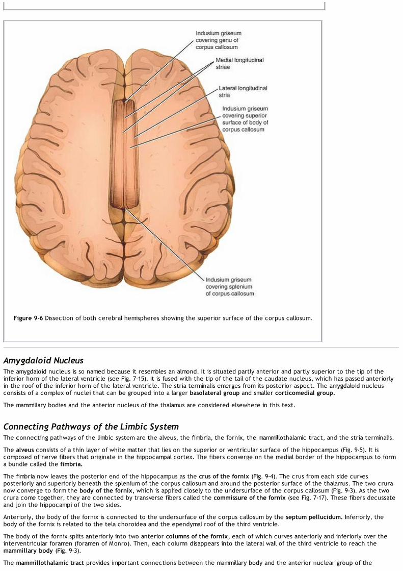
Figure 9-6 Dissection of both cerebral hemispheres showing the superior surface of the corpus callosum.
Amygdaloid NucleusThe amygdaloid nucleus is so named because it resembles an almond. It is situated partly anterior and partly superior to the tip of theinferior horn of the lateral ventricle (see Fig. 7-15). It is fused with the tip of the tail of the caudate nucleus, which has passed anteriorlyin the roof of the inferior horn of the lateral ventricle. The stria terminalis emerges from its posterior aspect. The amygdaloid nucleusconsists of a complex of nuclei that can be grouped into a larger basolateral group and smaller corticomedial group.
The mammillary bodies and the anterior nucleus of the thalamus are considered elsewhere in this text.
Connecting Pathways of the Limbic SystemThe connecting pathways of the limbic system are the alveus, the fimbria, the fornix, the mammillothalamic tract, and the stria terminalis.
The alveus consists of a thin layer of white matter that lies on the superior or ventricular surface of the hippocampus (Fig. 9-5). It iscomposed of nerve fibers that originate in the hippocampal cortex. The fibers converge on the medial border of the hippocampus to forma bundle called the fimbria.
The fimbria now leaves the posterior end of the hippocampus as the crus of the fornix (Fig. 9-4). The crus from each side curvesposteriorly and superiorly beneath the splenium of the corpus callosum and around the posterior surface of the thalamus. The two cruranow converge to form the body of the fornix, which is applied closely to the undersurface of the corpus callosum (Fig. 9-3). As the twocrura come together, they are connected by transverse fibers called the commissure of the fornix (see Fig. 7-17). These fibers decussateand join the hippocampi of the two sides.
Anteriorly, the body of the fornix is connected to the undersurface of the corpus callosum by the septum pellucidum. Inferiorly, thebody of the fornix is related to the tela choroidea and the ependymal roof of the third ventricle.
The body of the fornix splits anteriorly into two anterior columns of the fornix, each of which curves anteriorly and inferiorly over theinterventricular foramen (foramen of Monro). Then, each column disappears into the lateral wall of the third ventricle to reach themammillary body (Fig. 9-3).
The mammillothalamic tract provides important connections between the mammillary body and the anterior nuclear group of the

thalamus.
The stria terminalis emerges from the posterior aspect of the amygdaloid nucleus and runs as a bundle of nerve fibers posteriorly in theroof of the inferior horn of the lateral ventricle on the medial side of the tail of the caudate nucleus (Fig. 9-3). It follows the curve of thecaudate nucleus and comes to lie in the floor of the body of the lateral ventricle.
Structure of the Hippocampus and the Dentate GyrusThe cortical structure of the parahippocampal gyrus is six layered (Fig. 9-5). As the cortex is traced into the hippocampus, there is agradual transition from a six- to a three-layered arrangement. These three layers are the superficial molecular layer, consisting of nervefibers and scattered small neurons; the pyramidal layer, consisting of many large pyramid-shaped neurons; and the inner polymorphiclayer, which is similar in structure to the polymorphic layer of the cortex seen elsewhere.
The dentate gyrus also has three layers, but the pyramidal layer is replaced by the granular layer (Fig. 9-5). The granular layer is composedof densely arranged rounded or oval neurons that give rise to axons that terminate on the dendrites of the pyramidal cells in thehippocampus. A few of the axons join the fimbria and enter the fornix.
Afferent Connections of the HippocampusAfferent connections of the hippocampus may be divided into six groups (Fig. 9-7):
1. Fibers arising in the cingulate gyrus pass to the hippocampus.
2. Fibers arising from the septal nuclei (nuclei lying within the midline close to the anterior commissure) pass posterior in the fornix tothe hippocampus.
3. Fibers arising from one hippocampus pass across the midline to the opposite hippocampus in the commissure of the fornix.
4. Fibers from the indusium griseum pass posteriorly in the longitudinal striae to the hippocampus.
5. Fibers from the entorhinal area or olfactory-associated cortex pass to the hippocampus.
6. Fibers arising from the dentate and parahippocampal gyri travel to the hippocampus.
Efferent Connections of the HippocampusAxons of the large pyramidal cells of the hippocampus emerge to form the alveus and the fimbria. The fimbria continues as the crus of thefornix. The two crura converge to form the body of the fornix. The body of the fornix splits into the two columns of the fornix, whichcurve downward and forward in front of the interventricular foramina. The fibers within the fornix are distributed to the following regions(Fig. 9-7):
1. Fibers pass posterior to the anterior commissure to enter the mammillary body, where they end in the medial nucleus.
2. Fibers pass posterior to the anterior commissure to end in the anterior nuclei of the thalamus.
3. Fibers pass posterior to the anterior commissure to enter the tegmentum of the midbrain.
4. Fibers pass anterior to the anterior commissure to end in the septal nuclei, the lateral preoptic area, and the anterior part of thehypothalamus.
5. Fibers join the stria medullaris thalami to reach the habenular nuclei.
Consideration of the above complex anatomical pathways indicates that the structures comprising the limbicsystem not only are interconnected but also send projection fibers to many different parts of the nervous system. Physiologists nowrecognize the importance of the hypothalamus as being the major output pathway of the limbic system.

Figure 9-7 Diagram showing some important afferent and efferent connections of the limbic system.
Functions of the Limbic SystemThe limbic system, via the hypothalamus and its connections with the outflow of the autonomic nervous system and its control of theendocrine system, is able to influence many aspects of emotional behavior. These include particularly the reactions of fear and anger andthe emotions associated with sexual behavior.
There is also evidence that the hippocampus is concerned with converting recent memory to long-term memory. A lesion of thehippocampus results in the individual being unable to store long-term memory. Memory of remote past events before the lesion developedis unaffected. This condition is called anterograde amnesia. It is interesting to note that injury to the amygdaloid nucleus and thehippocampus produces a greater memory loss than injury to either one of these structures alone.
There is no evidence that the limbic system has an olfactory function. The various afferent and efferent connections of the limbic systemprovide pathways for the integration and effective homeostatic responses to a wide variety of environmental stimuli.
Clinical NotesReticular FormationThe reticular formation is a continuous network of nerve cells and fibers that extend through the neuroaxis
from the spinal cord to the cerebral cortex. The reticular formation not only modulates the control of motor systems butalso influences sensory systems. By means of its multiple ascending pathways, which project to different parts of thecerebral cortex, it is believed to influence the state of consciousness.
Loss of ConsciousnessIn experimental animals, damage to the reticular formation, which spares the ascending sensory pathways, causespersistent unconsciousness. Pathologic lesions of the reticular formation in humans can result in loss of consciousnessand even coma. It has been suggested that the loss of consciousness that occurs in epilepsy may be due to inhibitionof the activity of the reticular formation in the upper part of the diencephalon.
Limbic SystemThe anatomical connections of the limbic system are extremely complex, and since their significance is not fullyunderstood, it is unnecessary for a student of medicine to commit all of them to memory. The results ofneurophysiologic experiments, which have included stimulation and ablation of different parts of the limbic system inanimals, are not entirely clear. Nevertheless, certain important roles have been inferred: (1) The limbic structures areinvolved in the development of sensations of emotion and with the visceral responses accompanying those emotions,and (2) the hippocampus is concerned with recent memory.
SchizophreniaThe symptoms of schizophrenia include chronically disordered thinking, blunted affect, and emotional withdrawal.Paranoid delusions and auditory hallucinations may also be present. Clinical research has shown that if the limbicreceptors to dopamine are blocked by a pharmacologic agent, the worst symptoms of schizophrenia are lessened.Phenothiazine administration, for example, blocks the dopamine receptors in the limbic system. Unfortunately, thisdrug, as well as most other antipsychotic drugs, has major motor side effects on the dopaminergic receptors within theextrapyramidal system, producing abnormal involuntary movements. Research is now concentrating on finding a drugthat will block the limbic dopamine receptors but without effect on the receptors of the extrapyramidal system

P.314
(substantia nigra–corpus striatum).It is clear, however, that there is still no direct evidence that excessive production of dopamine by certain neuronsactually contributes to schizophrenia.
Destruction of the Amygdaloid ComplexUnilateral or bilateral destruction of the amygdaloid nucleus and the para-amygdaloid area in patients suffering fromaggressive behavior in many cases results in a decrease in aggressiveness, emotional instability, and restlessness;increased interest in food; and hypersexuality. There is no disturbance in memory. Monkeys that have been subjectedto bilateral removal of the temporal lobes demonstrate what is known as the Klüver-Bucy syndrome. They becomedocile and show no evidence of fear or anger and are unable to appreciate objects visually. They have an increasedappetite and increased sexual activity. Moreover, the animals indiscriminately seek partnerships with male and femaleanimals.Precise stereotactic lesions in the amygdaloid complex in humans reduce emotional excitability and bring aboutnormalization of behavior in patients with severe disturbances. No loss of memory occurs.
Temporal Lobe DysfunctionTemporal lobe epilepsy may be preceded by an aura of acoustic or olfactory experience. The olfactory aura is usuallyan unpleasant odor. The patient is often confused, anxious, and docile and may perform automatic and complicatedmovements, such as undressing in public or driving a car, and then, following the seizure, may have no memory ofwhat occurred previously.
Clinical Problem Solving1. While discussing the neurologic basis of emotions during a ward round, a neurologist asked a third-year medicalstudent what she knew about the Klüver-Bucy syndrome. What would be your answer to that question? Does Klüver-Bucy syndrome ever occur in humans?View Answer
2. A 23-year-old woman with a 4-year history of epileptic attacks visited her neurologist. A friend of hers vividlydescribed one of her attacks. For a few seconds before the convulsions began, the patient would complain of anunpleasant odor, similar to that encountered in a cow shed. This was followed by a shrill cry as she fell to the floorunconscious. Her whole body immediately became involved in generalized tonic and clonic movements. Clearly, thispatient had a generalized form of epileptic seizure. Using your knowledge of neuroanatomy, suggest which lobe of thebrain was initially involved in the epileptic discharge.View Answer
3. A 54-year-old man died in the hospital with a cerebral tumor. He had always been intellectually very bright and couldeasily recall events in his childhood. For the past 6 months, his family had noticed that he had difficulty in recallingwhere he had placed things, such as his pipe. He also had difficulty in recalling recent news events, and just before hedied, he could not remember that his brother had visited him the day before. Using your knowledge of neuroanatomy,suggest which part of the brain was being affected by the expanding and highly invasive tumor.View Answer
Review QuestionsDirections: Each of the numbered items in this section is followed by answers. Select the ONE lettered answer that isCORRECT.1. The following statements concern the reticular formation:
(a) Reticulobulbar and reticulospinal tracts form the afferent pathways from the reticular formation to the motornuclei of the cranial nerves and the anterior horn cells of the spinal cord, respectively.(b) The reticular formation extends through the neuroaxis from the spinal cord to the midbrain.(c) The main pathways through the reticular formation may easily be traced from one part of the central nervoussystem to another using silver stains.(d) Superiorly, the reticular formation is relayed to the cerebral cortex.(e) Afferent pathways project into the reticular formation from only a few parts of the central nervous system.
View Answer
2. The following statements concern the functions of the reticular formation:(a) It influences the activity of the alpha and gamma motor neurons.(b) It opposes the actions of the vestibular spinal tract.(c) It does not bring about reciprocal inhibition during contraction of the prime mover muscles.(d) It plays no part in maintaining the tone of the antigravity muscles.(e) It cannot modulate reflex activity.
View Answer
3. The following statements concern the functions of the reticular formation:(a) It does not affect the reception of pain.(b) It cannot influence all ascending pathways to the supraspinal levels.

(c) By means of its reticulobulbar and reticulospinal tracts, it can control the parasympathetic and sympatheticoutflows.(d) It has no effect on the biologic rhythms.(e) It does not influence the degree of wakefulness of an individual.
View Answer
4. Anatomically, the following structures collectively form the limbic system:(a) The amygdaloid nucleus, the red nucleus, and the vestibular nuclei(b) The pulvinar of the thalamus and the substantia nigra(c) The hippocampal formation(d) The cingulate gyrus and the uncus(e) The subcallosal, the cingulate, and the parahippocampal gyri, the hippocampal formation, the amygdaloidnucleus, the mammillary bodies, and the anterior thalamic nuclei
View Answer
5. The following statements concern the efferent connections of the hippocampus:(a) They arise from the small granular cells of the cortex.(b) They travel through the fornix.(c) None of the fibers enter the mammillary body.(d) The fibers within the fornix pass posterior to the interventricular foramen.(e) Some of the fibers end in the posterior nuclei of the thalamus.
View Answer
6. The following statements concern the functions of the limbic system:(a) It is not concerned with the reactions of fear and anger.(b) It is concerned with visual experiences.(c) The hippocampus is concerned with recent memory.(d) The limbic system plays an important role in olfactory function.(e) It directly influences the activity of the endocrine system.
View Answer
Figure 9-8 Medial aspect of the right cerebral hemisphere showing structures that form the limbic system.
The answers for Figure 9-8 are as follows:Matching Questions. Directions: The following questions apply to Figure 9-8. Match the numbers listed on the leftwith the appropriate lettered structure listed on the right. Each lettered option may be selected once, more thanonce, or not at all.7. Number 1
(a) Uncus(b) Body of fornix

(c) Parahippocampal gyrus(d) Dentate gyrus(e) None of the above
View Answer
8. Number 2(a) Uncus(b) Body of fornix(c) Parahippocampal gyrus(d) Dentate gyrus(e) None of the above
View Answer
9. Number 3(a) Uncus(b) Body of fornix(c) Parahippocampal gyrus(d) Dentate gyrus(e) None of the above
View Answer
10. Number 4(a) Uncus(b) Body of fornix(c) Parahippocampal gyrus(d) Dentate gyrus(e) None of the above
View Answer
Additional Reading
Aggleton, J. P. (ed.). The Amygdala: Neurobiological Aspects of Emotion, Memory, and Mental Dysfunction. New York: Wiley-Liss, 1993.
Goldman-Rakic, P. S. Working memory and the mind. Sci. Am. 267:110, 1992.
Guyton, A. C., and Hall, J. E. Textbook of Medical Physiology (11th ed.). Philadelphia: Elsevier Saunders, 2006.
Jasper, H. H., Descarries, L., Castelluci, V. F., and Rossignol, S. (eds.). Consciousness: At the Frontiers of Neuroscience. Philadelphia:Lippincott-Raven, 1998.
Kiernan, J. A. The Human Nervous System (7th ed.). Philadelphia: Lippincott Williams & Wilkins, 1998.
Klemm, W. R. Ascending and descending excitatory influences in the brain stem reticulum: A re-examination. Brain Res. 36:444, 1972.
Mega, M. S., Cummings, J. L., Salloway, S., and Malloy, P. The limbic system: An anatomic, phylogenetic, and clinical perspective. J.Neuropsychiatry Clin. Neurosci. 9:315, 1997.
Rowland, L. P. Merritt's Textbook of Neurology. Baltimore: Williams & Wilkins, 1995.
Ryan, P. M. Epidemiology, etiology, diagnosis and treatment of schizophrenia. Am. J. Hosp. Pharm. 48:1271, 1991.
Seeman, P., Guan, H. C., and Van Tol, H. H. M. Dopamine D4 receptors elevated in schizophrenia. Nature 365:441–445, 1993.
Standring, S. (ed.). Gray's Anatomy (39th Br. ed.). London: Elsevier Churchill Livingstone, 2005.

Steriade, M. Arousal: Revisiting the reticular activating system. Science 272:225, 1996.

Authors: Snell, Richard S.Title: Clinical Neuroanatomy, 7th Edition
Copyright ©2010 Lippincott Williams & Wilkins
> Table of Contents > Chapter 10 - The Basal Nuclei (Basal Ganglia) and Their Connections
Chapter 10
The Basal Nuclei (Basal Ganglia) and Their Connections
A 58-year-old man was seen by a neurologist because he had noticed the development of a slight tremor of his left hand. The tremorsinvolved all of the fingers and the thumb and were present at rest but ceased during voluntary movement.
On examination, the patient tended to perform all his movements slowly, and his face had very little expression and was almost masklike.On passively moving the patient's arms, the neurologist found that the muscles showed increased tone, and there was a slight jerkyresistance to the movements. When asked to stand up straight, the patient did so but with a stooped posture, and when he walked, hedid so by shuffling across the examining room.
The neurologist made the diagnosis of Parkinson disease, based on her knowledge of the structure and function of the basal ganglia andtheir connections to the substantia nigra of the midbrain. She was able to prescribe appropriate drug therapy, which resulted in a greatimprovement in the hand tremors.
Chapter Objectives
To describe the basal nuclei, their connections, and their functions and relate them to diseases commonly affectingthis area of the nervous system
The basal nuclei play an important role in the control of posture and voluntary movement. Unlike many other parts of the nervous systemconcerned with motor control, the basal nuclei have no direct input or output connections with the spinal cord.
TerminologyThe term basal nuclei is applied to a collection of masses of gray matter situated within each cerebral hemisphere. They are thecorpus striatum, the amygdaloid nucleus, and the claustrum.
Clinicians and neuroscientists use a variety of different terminologies to describe the basal nuclei. A summary of the terminologiescommonly used is shown in Table 10-1. The subthalamic nuclei, the substantia nigra, and the red nucleus are functionally closely related tothe basal nuclei, but they should not be included with them.
The interconnections of the basal nuclei are complex, but in this account, only the more important pathways are considered. The basalnuclei play an important role in the control of posture and voluntary movement.
Corpus StriatumThe corpus striatum (Fig. 10-1; see also Atlas Plate 5) is situated lateral to the thalamus and is almost completely divided by a bandof nerve fibers, the internal capsule, into the caudate nucleus and the lentiform nucleus. The term striatum is used here
because of the striated appearance produced by the strands of gray matter passing through the internal capsule and connecting thecaudate nucleus to the putamen of the lentiform nucleus (see below).
Table 10-1 Terminology Commonly Used to Describe the Basal Nuclei
Neurolog ic Structure Basal Nucleus (Nuclei)a
Caudate nucleus Caudate nucleus
Lentiform nucleus Globus pallidus plus putamen
Claustrum Claustrum
Corpus striatum Caudate nucleus plus lentiform nucleus

Neostriatum (striatum) Caudate nucleus plus putamen
Amygdaloid body Amygdaloid nucleus
aThe term basal has been used in the past to denote the position of the nuclei at the base of the forebrain.
Caudate NucleusThe caudate nucleus is a large C-shaped mass of gray matter that is closely related to the lateral ventricle and lies lateral to the thalamus(Fig. 10-1). The lateral surface of the nucleus is related to the internal capsule, which separates it from the lentiform nucleus (Fig. 10-2).For purposes of description, it can be divided into a head, a body, and a tail.
The head of the caudate nucleus is large and rounded and forms the lateral wall of the anterior horn of the lateral ventricle (Fig. 10-2; seealso Atlas Plate 5). The head is continuous inferiorly with the putamen of the lentiform nucleus (the caudate nucleus and the putamen aresometimes referred to as the neostriatum or striatum). Just superior to this point of union, strands of gray matter pass through theinternal capsule, giving the region a striated appearance, hence the term corpus striatum.
The body of the caudate nucleus is long and narrow and is continuous with the head in the region of the interventricular foramen. Thebody of the caudate nucleus forms part of the floor of the body of the lateral ventricle.
The tail of the caudate nucleus is long and slender and is continuous with the body in the region of the posterior end of the thalamus. Itfollows the contour of the lateral ventricle and continues forward in the roof of the inferior horn of the lateral ventricle. It terminatesanteriorly in the amygdaloid nucleus (Fig. 10-1).
Lentiform NucleusThe lentiform nucleus is a wedge-shaped mass of gray matter whose broad convex base is directed laterally and whose blade is directedmedially (Fig. 10-2; see also Atlas Plate 5). It is buried deep in the white matter of the cerebral hemisphere and is related medially to theinternal capsule, which separates it from the caudate nucleus and the thalamus. The lentiform nucleus is related laterally to a thin sheetof white matter, the external capsule (Fig. 10-2), which separates it from a thin sheet of gray matter, called the claustrum. The claustrum,in turn, separates the external capsule from the subcortical white matter of the insula. A vertical plate of white matter divides thenucleus into a larger, darker lateral portion, the putamen, and an inner lighter portion, the globus pallidus (Fig. 10-2). The paleness of theglobus pallidus is due to the presence of a high concentration of myelinated nerve fibers. Inferiorly at its anterior end, the putamen iscontinuous with the head of the caudate nucleus (Fig. 10-1).
Figure 10-1 Lateral view of the right cerebral hemisphere dissected to show the position of the different basal nuclei.
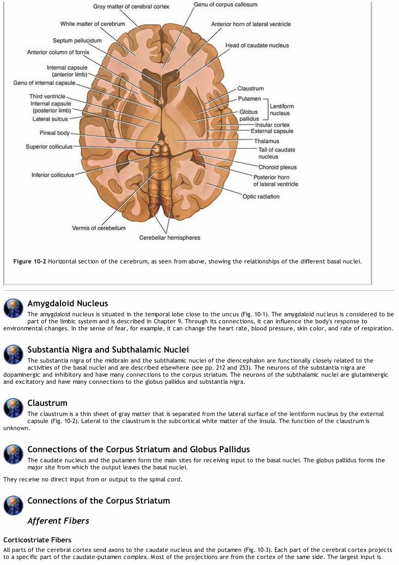
Figure 10-2 Horizontal section of the cerebrum, as seen from above, showing the relationships of the different basal nuclei.
Amygdaloid NucleusThe amygdaloid nucleus is situated in the temporal lobe close to the uncus (Fig. 10-1). The amygdaloid nucleus is considered to bepart of the limbic system and is described in Chapter 9. Through its connections, it can influence the body's response to
environmental changes. In the sense of fear, for example, it can change the heart rate, blood pressure, skin color, and rate of respiration.
Substantia Nigra and Subthalamic NucleiThe substantia nigra of the midbrain and the subthalamic nuclei of the diencephalon are functionally closely related to theactivities of the basal nuclei and are described elsewhere (see pp. 212 and 253). The neurons of the substantia nigra are
dopaminergic and inhibitory and have many connections to the corpus striatum. The neurons of the subthalamic nuclei are glutaminergicand excitatory and have many connections to the globus pallidus and substantia nigra.
ClaustrumThe claustrum is a thin sheet of gray matter that is separated from the lateral surface of the lentiform nucleus by the externalcapsule (Fig. 10-2). Lateral to the claustrum is the subcortical white matter of the insula. The function of the claustrum is
unknown.
Connections of the Corpus Striatum and Globus PallidusThe caudate nucleus and the putamen form the main sites for receiving input to the basal nuclei. The globus pallidus forms themajor site from which the output leaves the basal nuclei.
They receive no direct input from or output to the spinal cord.
Connections of the Corpus Striatum
Afferent Fibers
Corticostriate FibersAll parts of the cerebral cortex send axons to the caudate nucleus and the putamen (Fig. 10-3). Each part of the cerebral cortex projectsto a specific part of the caudate-putamen complex. Most of the projections are from the cortex of the same side. The largest input is

from the sensory-motor cortex. Glutamate is the neurotransmitter of the corticostriate fibers (Fig. 10-4).
Thalamostriate FibersThe intralaminar nuclei of the thalamus send large numbers of axons to the caudate nucleus and the putamen (Fig. 10-3).
Nigrostriate FibersNeurons in the substantia nigra send axons to the caudate nucleus and the putamen (Figs. 10-3 and 10-4) and liberate dopamine at theirterminals as the neurotransmitter. It is believed that these fibers are inhibitory in function.
Brainstem Striatal FibersAscending fibers from the brainstem end in the caudate nucleus and putamen (Figs. 10-3 and 10-4) and liberate serotonin at their terminalsas the neurotransmitter. It is thought that these fibers are inhibitory in function.
Efferent Fibers
Striatopallidal FibersStriatopallidal fibers pass from the caudate nucleus and putamen to the globus pallidus (Fig. 10-3). They have gamma-aminobutyric acid(GABA) as their neurotransmitter (Fig. 10-4).
Striatonigral FibersStriatonigral fibers pass from the caudate nucleus and putamen to the substantia nigra (Fig. 10-3). Some of the fibers use GABA oracetylcholine as the neurotransmitter, while others use substance P (Fig. 10-4).
Connections of the Globus Pallidus
Afferent Fibers
Striatopallidal FibersStriatopallidal fibers pass from the caudate nucleus and putamen to the globus pallidus. As noted previously, these fibers have GABA astheir neurotransmitter (Fig. 10-4).
Efferent Fibers
Pallidofugal FibersPallidofugal fibers are complicated and can be divided into groups: (1) the ansa lenticularis, which pass to the thalamic nuclei; (2) thefasciculus lenticularis, whichpass to the subthalamus; (3) the pallidotegmental fibers, which terminate in the caudal tegmentum of the midbrain; and (4) thepallidosubthalamic fibers, which pass to the subthalamic nuclei.

P.322
Figure 10-3 Some of the main connections between the cerebral cortex, the basal nuclei, the thalamic nuclei, the brainstem, andthe spinal cord.
Functions of the Basal NucleiThe basal nuclei (Fig. 10-5) are joined together and connected with many different regions of the nervous system by a verycomplex number of neurons.
Basically, the corpus striatum receives afferent information from most of the cerebral cortex, the thalamus, the subthalamus, and thebrainstem, including the substantia nigra. The information is integrated within the corpus striatum, and the outflow passes back to theareas listed above. This circular pathway is believed to function as follows.
The activity of the basal nuclei is initiated by information received from the premotor and supplemental areas of the motor cortex, theprimary sensory cortex, the thalamus, and the brainstem. The outflow from the basal nuclei is channeled through the globus pallidus,which then influences the activities of the motor areas of the cerebral cortex orother motor centers in the brainstem. Thus, the basal nuclei control muscular movements by influencing the cerebral cortexand have no direct control through descending pathways to the brainstem and spinal cord. In this way, the basal nuclei assist in theregulation of voluntary movement and the learning of motor skills.

Figure 10-4 Basal nuclei pathways showing the known neurotransmitters.

Figure 10-5 Diagram showing the main functional connections of the basal nuclei and how they can influence muscle activity.
Writing the letters of the alphabet, drawing a diagram, passing a football, using the vocal cords in talking and singing, and using the eyemuscles when looking at an object are a few examples where the basal nuclei influence the skilled cortical motor activities.
Destruction of the primary motor cerebral cortex prevents the individual from performing fine discrete movements of the hands and feeton the opposite side of the body (see pp. 167 and 296). However, the individual is still capable of performing gross crude movements of theopposite limbs. If destruction of the corpus striatum then takes place, paralysis of the remaining movements of the opposite side of thebody occurs.
The basal nuclei not only influence the execution of a particular movement of, say, the limbs but also help prepare for the movements.This may be achieved by controlling the axial and girdle movements of the body and the positioning of the proximal parts of the limbs. Theactivity in certain neurons of the globus pallidus increases before active movements take place in the distal limb muscles. This importantpreparatory function enables the trunk and limbs to be placed in appropriate positions before the primary motor part of the cerebralcortex activates discrete movements in the hands and feet.
Clinical NotesDisorders of the basal nuclei are of two general types. Hyperkinetic disorders are those in which there areexcessive and abnormal movements, such as seen with chorea, athetosis, and ballism. Hypokinetic disorders
include those in which there is a lack or slowness of movement. Parkinson disease includes both types of motordisturbances.
ChoreaIn chorea, the patient exhibits involuntary, quick, jerky, irregular movements that are nonrepetitive. Swift grimaces andsudden movements of the head or limbs are good examples.
Huntington DiseaseHuntington disease is an autosomal dominant inherited disease, with the onset occurring most often in adult life.Death occurs 15 to 20 years after onset. The disease has been traced to a single gene defect on chromosome 4. Thisgene encodes a protein, huntingtin, the function of which is not known. The codon (CAG) that encodes glutamine isrepeated many more times than normal. The disease affects men and women with equal frequency and unfortunatelyoften reveals itself only after they have had children.Patients have the following characteristic signs and symptoms:
1. Choreiform movements first appear as involuntary movements of the extremities and twitching of the face (facialgrimacing). Later, more muscle groups are involved, so the patient becomes immobile and unable to speak orswallow.
2. Progressive dementia occurs with loss of memory and intellectual capacity.
In this disease, there is a degeneration of the GABA-secreting, substance P–secreting, and acetylcholine-secretingneurons of the striatonigral-inhibiting pathway. This results in the dopa-secreting neurons of the substantia nigrabecoming overactive; thus, the nigrostriatal pathway inhibits the caudate nucleus and the putamen (Fig. 10-6). Thisinhibition produces the abnormal movements seen in this disease. Computed tomography scans show enlarged lateralventricles due to degeneration of the caudate nuclei. Medical treatment of Huntington chorea has been disappointing.
Sydenham ChoreaSydenham chorea (St. Vitus' dance) is a disease of childhood in which there are rapid, irregular, involuntarymovements of the limbs, face, and trunk. The condition is associated with rheumatic fever. The antigens of thestreptococcal bacteria are similar in structure to the proteins present in the membranes of striatal neurons. The host'santibodies not only combine with the bacterial antigens but also attack the membranes of the neurons of the basalganglia. This results in the production of choreiform movements, which are fortunately transient, and there is fullrecovery.
HemiballismusHemiballismus is a form of involuntary movement confined to one side of the body. It usually involves the proximalextremity musculature, and the limb suddenly flies about out of control in all directions. The lesion, which is usually asmall stroke, occurs in the opposite subthalamic nucleus or its connections; it is in the subthalamic nucleus that smoothmovements of different parts of the body are integrated.
Parkinson DiseaseParkinson disease is a progressive disease of unknown cause that commences between the ages of 45 and 55 years.It is associated with neuronal degeneration in the substantia nigra and, to a lesser extent, in the globus pallidus,putamen, and caudate nucleus. The disease affects about 1 million people in the United States.The degeneration of the neurons of the substantia nigra that send their axons to the corpus striatum results in areduction in the release of the neurotransmitter dopamine within the corpus striatum (Figs. 10-7 and 10-8). This leadsto hypersensitivity of the dopamine receptors in the postsynaptic neurons in the striatum.

Figure 10-6 Diagram showing the degeneration of the inhibitory pathway between the corpus striatum and the substantia nigraseen in Huntington disease and the consequent reduction in the liberation of GABA, substance P, and acetylcholine in thesubstantia nigra.
Patients have the following characteristic signs and symptoms:
1. Tremor. This is the result of the alternating contraction of agonists and antagonists. The tremor is slow and occursmost obviously when the limbs are at rest. It disappears during sleep. It should be distinguished from the intentiontremor seen in cerebellar disease, which only occurs when purposeful active movement is attempted.
2. Rigidity. This differs from the rigidity caused by lesions of the upper motor neurons in that it is present to an equalextent in opposing muscle groups. If the tremor is absent, the rigidity is felt as resistance to passive movement andis sometimes referred to as plastic rigidity. If the tremor is present, the muscle resistance is overcome as a seriesof jerks, called cogwheel rigidity.
3. Bradykinesis. There is a difficulty in initiating (akinesia) and performing new movements. The movements are slow,the face is expressionless, and the voice is slurred and unmodulated. Swinging of the arms in walking is lost.
4. Postural disturbances. The patient stands with a stoop, and his or her arms are flexed. The patient walks bytaking short steps and often is unable to stop. In fact, he or she may break into a shuffling run to maintain balance.

Figure 10-7 Axial (horizontal) positron emission tomography (PET) scans of a normal brain (A) and the brain of a patient withearly Parkinson disease (B) following the injection of 18- fluoro-6-L-dopa. The normal brain image shows large amounts ofthe compound (yellow areas) distributed throughout the corpus striatum in both cerebral hemispheres. In the patient withParkinson disease, the brain image shows that the total amount of the compound is low, and it is unevenly distributed inthe corpus striatum. (Courtesy Dr. Holley Dey.)
5. There is no loss of muscle power and no loss of sensibility. Since the corticospinal tracts are normal, the superficialabdominal reflexes are normal, and there is no Babinski response. The deep tendon reflexes are normal.
There are a few types of Parkinson disease for which the cause is known. Postencephalitic parkinsonism developedfollowing the viral encephalitis outbreak of 1916–17 in which damage occurred to the basal nuclei. Iatrogenicparkinsonism can be a side effect of antipsychotic drugs (e.g., phenothiazines). Meperidine analogues (used by drugaddicts) and poisoning from carbon monoxide and manganese can also produce the symptoms of parkinsonism.Atherosclerotic parkinsonism can occur in elderly hypertensive patients.Parkinson disease may be treated by elevating the brain dopamine level. Unfortunately, dopamine cannot cross theblood-brain barrier, but its immediate precursor L-dopa can and is used in its place. L-Dopa is taken up by thedopaminergic neurons in the basal nuclei and converted to dopamine. Selegiline, a drug that inhibits monoamineoxidase, which is responsible for destroying dopamine, is also of benefit in the treatment of the disease. There isevidence that selegiline can slow the process of degeneration of the dopa-secreting neurons in the substantia nigra.Transplantation of human embryonic dopamine-producing neurons into the caudate nucleus and putamen has beenshown to lead to improvement in motor function in Parkinson disease (Fig. 10-9). There is evidence that the grafts cansurvive, and synaptic contacts are made. Unfortunately, many of the grafted neurons do not survive, and in manycases, the clinical improvement is counteracted by the continuing degeneration of the patient's own dopa-producingneurons. Autotransplantation of suprarenal medullary cells can be a source of dopa-producing cells, but in the future,genetically engineered cells could be another source of dopa.Since most of the symptoms of Parkinson disease are caused by an increased inhibitory output from the basal nuclei tothe thalamus and the precentral motor cortex, surgical lesions in the globus pallidus (pallidotomy) have been shownto be effective in alleviating parkinsonian signs. At the present time, such procedures are restricted to patients whoare no longer responding to medical treatment.

Figure 10-8 Diagram showing the degeneration of the inhibitory pathway between the substantia nigra and the corpus striatumin Parkinson disease and the consequent reduction in the release of the neurotransmitter dopamine in the striatum.
Drug-Induced ParkinsonismAlthough Parkinson disease (primary parkinsonism) is the most common type of parkinsonism found in clinical practice,drug-induced parkinsonism is becoming very prevalent. Drugs that block striatal dopamine receptors (D2) are oftengiven for psychotic behavior (e.g., phenothiazines and butyrophenones). Other drugs may deplete striatal dopamine(e.g., tetrabenazines). Drug-induced parkinsonism disappears once the agent is withdrawn.
AthetosisAthetosis consists of slow, sinuous, writhing movements that most commonly involve the distal segments of the limbs.Degeneration of the globus pallidus occurs with a breakdown of the circuitry involving the basal nuclei and the cerebralcortex.

P.324P.325P.326P.327
Figure 10-9 Change in 18-F-fluorodopa uptake in the brains of patients with Parkinson disease after transplantation, as shownin fluorodopa PET scans. In the panel on the far left, an axial (horizontal) section through the caudate nucleus and putamen ofa normal subject shows intense uptake of 18-F-fluorodopa (red). On the right side, the upper panels show preoperative and 12-month postoperative scans in a patient in the transplantation group. Before surgery, the uptake of 18-F-fluorodopa wasrestricted to the region of the caudate nucleus. After transplantation, there was increased uptake of 18-F-fluorodopa in theputamen bilaterally. The lower panels show 18-F-fluorodopa scans in a patient in the sham-surgery group. There was nopostoperative change in 18-F-fluorodopa uptake. (Courtesy of Dr. Curt R. Freed, et al. N. Engl. J. Med. 344:710, 2001.)
Clinical Problem Solving1. A 10-year-old girl was seen by a neurologist because of the gradual development of involuntary movements. Tobegin with, the movements were regarded by her parents as general restlessness, but later, abnormal facial grimacingand jerking movements of the arms and legs began to occur. The child was now having difficulty in performing normalmovements of the arms, and walking was becoming increasingly difficult. The abnormal movements appeared to beworse in the upper limbs and were more exaggerated on the right side of the body. The movements were made worsewhen the child became excited but disappeared completely when she slept. The child was recently treated forrheumatic fever. Is there any possible connection between this child's symptoms and the basal nuclei in the cerebralhemispheres?View Answer
2. A 40-year-old man complaining of rapid and jerky involuntary movements involving the upper and lower limbs wasseen by his physician. The condition started about 6 months ago and was getting progressively worse. He said that hewas extremely worried about his health because his father had developed similar symptoms 20 years ago and haddied in a mental institution. His wife told the physician that he also suffered from episodes of extreme depression andthat she had noticed that he had periods of irritability and impulsive behavior. The physician made the diagnosis ofHuntington chorea. Using your knowledge of neuroanatomy, explain how this disease involves the basal nuclei.View Answer
3. A 61-year-old man suddenly developed uncoordinated movements of the trunk and right arm. The right upper limbwould suddenly, vigorously, and aimlessly be thrown about, knocking over anything in its path. The patient wasrecovering from a right-sided hemiplegia, secondary to a cerebral hemorrhage. What is the name given to this clinicalsign? Does this condition involve the basal nuclei?

View Answer
Review QuestionsDirections: Each of the numbered items in this section is followed by answers. Select the ONE lettered answerthat is CORRECT.1. The following statements concern the basal nuclei (ganglia):
(a) The caudate nucleus and the red nucleus form the neostriatum (striatum).(b) The head of the caudate nucleus is connected to the putamen.(c) The tegmentum of the midbrain forms part of the basal nuclei.(d) The internal capsule lies lateral to the globus pallidus.(e) The basal nuclei are formed of white matter.
View Answer
2. The following statements concern the basal nuclei (ganglia):(a) The amygdaloid nucleus is connected to the caudate nucleus.(b) The lentiform nucleus is completely divided by the external capsule into the globus pallidus and the putamen.(c) The claustrum does not form part of the basal nuclei.(d) The corpus striatum lies medial to the thalamus.(e) The function of the claustrum is well known.
View Answer
3. The following statements concern the basal nuclei (ganglia):(a) The corpus striatum is made up of the caudate nucleus and the amygdaloid nucleus.(b) The head of the caudate nucleus lies lateral to the internal capsule.(c) The insula forms part of the basal nuclei.(d) The tail of the caudate nucleus lies in the roof of the lateral ventricle.(e) The subthalamic nuclei are functionally closely related to the basal nuclei and are considered to be part ofthem.
View Answer
4. The following statements concern the caudate nucleus:(a) It is divided into a head, neck, trunk, and tail.(b) It is an M-shaped mass of gray matter.(c) The body of the caudate nucleus forms part of the roof of the body of the lateral ventricle.(d) The head lies medial to the anterior horn of the lateral ventricle.(e) The tail terminates anteriorly in the amygdaloid nucleus.
View Answer
5. The following statements concern the afferent corticostriate fibers to the corpus striatum:(a) Each part of the cerebral cortex is randomly projected to different parts of the corpus striatum.(b) Glutamate is not the neurotransmitter.(c) All parts of the cerebral cortex send fibers to the caudate nucleus and putamen.(d) The smallest input is from the sensory-motor part of the cerebral cortex.(e) Most of the projections are from the cortex of the opposite side.
View Answer
6. The following statements concern the nigrostriate fibers:(a) The neurons in the substantia nigra send axons to the putamen.(b) Acetylcholine is the neurotransmitter.(c) The nigrostriate fibers are stimulatory in function.(d) The caudate nucleus does not receive axons from the substantia nigra.(e) Parkinson disease is caused by an increase in the release of dopamine within the corpus striatum.
View Answer
7. The following statements concern the efferent fibers of the corpus striatum:(a) Many of the efferent fibers descend directly to the motor nuclei of the cranial nerves.(b) Some of the striatopallidal fibers have GABA as the neurotransmitter.(c) The striatonigral fibers pass from the red nucleus to the substantia nigra.(d) Many of the efferent fibers pass directly to the cerebellum.(e) The anterior horn cells of the spinal cord are influenced directly by the efferent fibers from the corpus

striatum.View Answer
8. The following statements concern the functions of the basal nuclei (ganglia):(a) The corpus striatum integrates information received directly from the cerebellar cortex.(b) The outflow of the basal nuclei is channeled through the globus pallidus to the sensory areas of the cerebralcortex, thus influencing muscular activities.(c) The globus pallidus only influences the movements of the axial part of the body.(d) The activities of the globus pallidus precede the activities of the motor cortex concerned with discretemovements of the hands and feet.(e) The activities of the basal nuclei are suppressed by information received from the sensory cortex, thethalamus, and the brainstem.
View Answer
Matching Questions. Directions: The following questions apply to Figure 10-10. Match the numbers listed belowon the left with the appropriate lettered structure listed on the right. Each lettered option may be selected once,more than once, or not at all.
Figure 10-10 Horizontal section of the cerebrum.
The answers for Figure 10-10, which shows a horizontal section of the cerebrum, are as follows:9. Structure 1
(a) Anterior horn of lateral ventricle(b) Internal capsule(c) Claustrum(d) Putamen(e) External capsule(f) Globus pallidus(g) None of the above
View Answer
10. Structure 2(a) Anterior horn of lateral ventricle(b) Internal capsule(c) Claustrum(d) Putamen(e) External capsule

(f) Globus pallidus(g) None of the above
View Answer
11. Structure 3(a) Anterior horn of lateral ventricle(b) Internal capsule(c) Claustrum(d) Putamen(e) External capsule(f) Globus pallidus(g) None of the above
View Answer
12. Structure 4(a) Anterior horn of lateral ventricle(b) Internal capsule(c) Claustrum(d) Putamen(e) External capsule(f) Globus pallidus(g) None of the above
View Answer
13. Structure 5(a) Anterior horn of lateral ventricle(b) Internal capsule(c) Claustrum(d) Putamen(e) External capsule(f) Globus pallidus(g) None of the above
View Answer
14. Structure 6(a) Anterior horn of lateral ventricle(b) Internal capsule(c) Claustrum(d) Putamen(e) External capsule(f) Globus pallidus(g) None of the above
View Answer
Additional Reading
Albin, R. L., Young, A. B., and Penney, J. B. The functional anatomy of disorders of the basal ganglia. Trends Neurosci. 200:63, 1995.
Aron, A. M., Freeman, J. M., and Carter, S. The natural history of Sydenham's chorea. Am. J. Med. 38:83, 1965.
Brooks, D. J. The role of the basal ganglia in motor control: Contributions from PET. J. Neurosci. 128:1–13, 1995.
Craig, C. R., and Stitzel, R. E. Modern Pharmacology (4th ed.). Boston: Little, Brown, 1994.
Dunnett, S. B., and Bjorklund, A. Prospects for new restorative and neuroprotective treatments in Parkinson disease. Nature399(Suppl): A32–A39, 1999.

Elble, R. J. Origins of tremor. Lancet 355:1113, 2000.
Freed, C. R., Greene, P. E., Breeze, R. E., Tsai, W. Y., DuMouchel, W., Kao, R., et al. Transplantation of embryonic dopamine neuronsfor severe Parkinson's disease. N. Engl. J. Med. 344:710, 2001.
Guyton, A. C., and Hall, J. E. Textbook of Medical Physiology (11th ed.). Philadelphia: Elsevier, Saunders, 2006.
Kordower, J. H., Freeman, T. B., Snow, B. J., Vingerhoets, F. J. G., Mufson, E. J., Sanberg, P. R., et al. Neuropathological evidence ofgraft survival and striatal reinnervation after the transplantation of fetal mesencephalic tissue in a patient with Parkinson's disease. N.Engl. J. Med. 332:1118–1124, 1995.
Nestler, E. J., Hyman, S. E., and Malenka, R. C. Molecular Neuropharmacology: A Foundation for Clinical Neuroscience. New York:McGraw-Hill, 2001.
Obeso, J. A, Rodriguez-Oroz, M., Marin, C., Alonso, F., Zamarbide, I., Lanciego, J. L., et al. The origin of motor fluctuations inParkinson's disease: Importance of dopaminergic innervation and basal ganglia circuits. Neurology 62(1 Suppl), 2004.
Olanow, C. W. The scientific basis for the current treatment of Parkinson's disease. Annu. Rev. Med. 55:41, 2004
Onn, S. P., West, A. R., and Grace, A. A. Dopamine-mediated regulation of striatal neuronal and network interactions. TrendsNeurosci. 23(Suppl):S48–S56, 2000.
Westmoreland, B. F., Benarroch, E. E., Daube, J. R., Reagan, T. J., and Sandok, B. A. Medical Neurosciences Boston: Little, Brown,1994.

Authors: Snell, Richard S.Title: Clinical Neuroanatomy, 7th Edition
Copyright ©2010 Lippincott Williams & Wilkins
> Table of Contents > Chapter 11 - The Cr anial Ner ve Nuclei and Their Centr al Connections and Distr ibution
Chapter 11
The Cranial Nerve Nuclei and Their Central Connections and Distribution
A 49-year-old man woke up one morning to find the right side of his face paralyzed. When examined by his local medical practitioner, hewas found to have complete paralysis of the entire right side of the face. He was also found to have severe hypertension. The patienttalked with slightly slurred speech. The physician told the patient that he had suffered a mild stroke, and he was admitted to the hospital.
The patient was later seen by a neurologist who disagreed with the diagnosis. The original physician had grouped together the facialparalysis, the slurred speech, and the hypertension and, in the absence of other findings, made the incorrect diagnosis of cerebralhemorrhage. A lesion of the corticonuclear fibers on one side of the brain will cause paralysis only of the muscles of the lower part of theopposite side of the face. This patient had complete paralysis of the entire right side of the face, which could only be caused by a lesionof the lower motor neuron. The correct diagnosis was Bell palsy, an inflammation of the connective tissue sheath of the facial nerve,which temporarily interfered with the functions of the axons of the right facial nerve. This case provides a good example of howknowledge of the central connections of a cranial nerve enables a physician to make the correct diagnosis.
Chapter Objectives
To learn the basic information regarding the motor and sensory nuclei of the cranial nerves, including their locationsand central connections
The cranial nerves are commonly damaged by trauma or disease, and testing for their integrity forms part of every physical examination.
The 12 Cranial NervesThere are 12 pairs of cranial nerves, which leave the brain and pass through foramina and fissures in the skull. All the nerves aredistributed in the head and neck, except cranial nerve X, which also supplies structures in the thorax and abdomen. The cranial
nerves are named as follows:
1. Olfactory
2. Optic
3. Oculomotor
4. Trochlear
5. Trigeminal
6. Abducent
7. Facial
8. Vestibulocochlear
9. Glossopharyngeal
10. Vagus
11. Accessory
12. Hypoglossal
See Atlas Plates 1, 6, and 8.
Organization of the Cranial NervesThe olfactory, optic, and vestibulocochlear nerves are entirely sensory. The oculomotor, trochlear, abducent, accessory, andhypoglossal nerves are entirely motor. The trigeminal, facial, glossopharyngeal, and vagus nerves are both sensory and motor
nerves. The letter symbols commonly used to indicate the functional components of each cranial nerve are shown in Table 11-1. Thecranial nerves have central motor and/or sensory nuclei within the brain and peripheral nerve fibers that emerge from the brain and exitfrom the skull to reach their effector or sensory organs.
The different components of the cranial nerves, their functions, and the openings in the skull through which the nerves leave the cranialcavity are summarized in Table 11-2.

P.334P.335
Motor Nuclei of the Cranial Nerves
Somatic Motor and Branchiomotor NucleiThe somatic motor and branchiomotor nerve fibers of a cranial nerve are the axons of nerve cells situated within the brain. These nervecell groups form motor nuclei and innervate striated muscle. Each nerve cell with its processes is referred to as a lower motor neuron.Such a nerve cell is, therefore, equivalent to the motor cells in the anterior gray columns of the spinal cord.
The motor nuclei of the cranial nerves receive impulses from the cerebral cortex through the corticonuclear (corticobulbar) fibers. Thesefibers originate from the pyramidal cells in the inferior part of the precentral gyrus (area 4) and from the adjacent part of the postcentralgyrus. The corticonuclear fibers descend through the corona radiata and the genu of the internal capsule. They pass through themidbrain just medial to the corticospinal fibers in the basis pedunculi and end by synapsing either directly with the lower motor neuronswithin the cranial nerve nuclei or indirectly through the internuncial neurons. The corticonuclear fibers thus constitute the first-orderneuron of the descending pathway, the internuncial neuron constitutes the second-order neuron, and the lower motor neuronconstitutes the third-order neuron.
The majority of the corticonuclear fibers to the motor cranial nerve nuclei cross the median plane before reaching the nuclei. Bilateralconnections are present for all the cranial motor nuclei except for part of the facial nucleus that supplies the muscles of the lower partof the face and a part of the hypoglossal nucleus that supplies the genioglossus muscle.
General Visceral Motor NucleiThe general visceral motor nuclei form the cranial outflow of the parasympathetic portion of the autonomic nervous system. They are theEdinger-Westphal nucleus of the oculomotor nerve, the superior salivatory and lacrimal nuclei of the facial nerve, the inferior salivatorynucleus of the glossopharyngeal nerve, and the dorsal motor nucleus of the vagus. These nuclei receive numerous afferent fibers,including descending pathways from the hypothalamus.
Sensory Nuclei of the Cranial NervesSensory nuclei of the cranial nerves include somatic and visceral afferent nuclei. The sensory or afferent parts of a cranial nerve are theaxons of nerve cells outside the brain and are situated in ganglia on the nerve trunks (equivalent to posteriorroot ganglion of a spinal nerve) or may be situated in a sensory organ, such as the nose, eye, or ear. These cells andtheir processes form the first-order neuron. The central processes of these cells enter the brain and terminate by synapsing with cellsforming the sensory nuclei. These cells and their processes form the second-order neuron. Axons from these nuclear cells now cross themidline and ascend to other sensory nuclei, such as the thalamus, where they synapse. The nerve cells of these nuclei form the third-order neuron, and their axons terminate in the cerebral cortex.
Table 11-1 The Letter Symbols Commonly Used to Indicate the Functional Components of Each Cranial Nerve
Component Function Letter Symbols
Afferent Fibers Sensory
General somatic afferent General sensations GSA
Special somatic afferent Hearing, balance, vision SSA
General visceral afferent Viscera GVA
Special visceral afferent Smell, taste SVA
Efferent Fibers
General somatic efferent Somatic striated muscles GSE
General visceral efferent Glands and smooth muscles (parasympathetic innervation) GVE
Special visceral efferent Branchial arch striated muscles SVE

Table 11-2 Cranial Nerves
Number Name Componentsa Function Opening in Skull
I Olfactory Sensory (SVA) Smell
Openings incribriformplate ofethmoid
II Optic Sensory (SSA) Vision Optic canal
III Oculomotor Motor (GSE,GVE)
Raises upper eyelid, turns eyeball upward,downward, and medially; constricts pupil;accommodates eye
Superiororbital fissure
IV Trochlear Motor (GSE) Assists in turning eyeball downward andlaterally
Superiororbital fissure
V Trigeminalb
Ophthalmicdivision Sensory (GSA)
Cornea, skin of forehead, scalp, eyelids, andnose; also mucous membrane of paranasalsinuses and nasal cavity
Superiororbital fissure
Maxillarydivision Sensory (GSA)
Skin of face over maxilla; teeth of upper jaw;mucous membrane of nose, the maxillary sinus,and palate
Foramenrotundum
Mandibulardivision Motor (SVE)
Muscles of mastication, mylohyoid, anteriorbelly of digastric, tensor veli palatini, andtensor tympani
Foramen ovale
Sensory (GSA)
Skin of cheek, skin over mandible and side ofhead, teeth of lower jaw andtemporomandibular joint; mucous membrane ofmouth and anterior part of tongue
VI Abducent Motor (GSE) Lateral rectus muscle turns eyeball laterally Superiororbital fissure
VII Facial Motor (SVE)Muscles of face and scalp, stapedius muscle,posterior belly of digastric and stylohyoidmuscles
Internalacousticmeatus, facialcanal,stylomastoidforamen
Sensory (SVA) Taste from anterior two-thirds of tongue, fromfloor of mouth and palate
Secretomotor(GVE)parasympathetic
Submandibular and sublingual salivary glands,the lacrimal gland, and glands of nose andpalate

VIII Vestibulocochlear
Vestibular Sensory (SSA) From utricle and saccule and semicircularcanals—position and movement of head
Internalacousticmeatus
Cochlear Sensory (SSA) Organ of Corti—hearing
IX Glossopharyngeal Motor (SVE) Stylopharyngeus muscle—assists swallowing Jugularforamen
Secretomotor(GVE)parasympathetic
Parotid salivary gland
Sensory (GVA,SVA, GSA)
General sensation and taste from posteriorone-third of tongue and pharynx; carotid sinus(baroreceptor); and carotid body(chemoreceptor)
X VagusMotor (GVE,SVE) Sensory(GVA, SVA, GSA)
Heart and great thoracic blood vessels; larynx,trachea, bronchi, and lungs; alimentary tractfrom pharynx to splenic flexure of colon; liver,kidneys, and pancreas
Jugularforamen
XI Accessory
Cranial root Motor (SVE)
Muscles of soft palate (except tensor velipalatini), pharynx (except stylopharyngeus),and larynx (except cricothyroid) in branches ofvagus
Jugularforamen
Spinal root Motor (SVE) Sternocleidomastoid and trapezius muscles
XII Hypoglossal Motor (GSE) Muscles of tongue (except palatoglossus)controlling its shape and movement
Hypoglossalcanal
aThe letter symbols are explained in Table 11-1.bThe trigeminal nerve also carries proprioceptive impulses from the muscles of mastication and the facial and extraocularmuscles.

Figure 11-1 A: Distribution of olfactory nerves on the lateral wall of the nose. B: Connections between the olfactory cells and theneurons of the olfactory bulb. C: Connections between the olfactory cell and the rest of the olfactory system.
Olfactory Nerves (Cranial Nerve I)The olfactory nerves arise from the olfactory receptor nerve cells in the olfactory mucous membrane located in the upper part ofthe nasal cavity above the level of the superior concha (Fig. 11-1). The olfactory receptor cells are scattered among supporting
cells. Each receptor cell consists of a small bipolar nerve cell with a coarse peripheral process that passes to the surface of the membraneand a finecentral process. From the coarse peripheral process, a number of short cilia arise, the olfactory hairs, which project into the mucuscovering the surface of the mucous membrane. These projecting hairs react to odors in the air and stimulate the olfactory cells.
The fine central processes form the olfactory nerve fibers (Fig. 11-1). Bundles of these nerve fibers pass through the openings of thecribriform plate of the ethmoid bone to enter the olfactory bulb. The olfactory nerve fibers are unmyelinated and are covered withSchwann cells.
Olfactory BulbThis ovoid structure possesses several types of nerve cells, the largest of which is the mitral cell (Fig. 11-1). The incoming olfactory nerve

fibers synapse with the dendrites of the mitral cells and form rounded areas known as synaptic glomeruli. Smaller nerve cells, calledtufted cells and granular cells, also synapse with the mitral cells. The olfactory bulb, in addition, receives axons from the contralateralolfactory bulb through the olfactory tract.
Olfactory TractThis narrow band of white matter runs from the posterior end of the olfactory bulb beneath the inferior surface of the frontal lobe of thebrain (Fig. 11-1). It consists of the central axons of the mitral and tufted cells of the bulb and some centrifugal fibers from the oppositeolfactory bulb.
As the olfactory tract reaches the anterior perforated substance, it divides into medial and lateral olfactory striae. The lateral striacarries the axons to the olfactory area of the cerebral cortex, namely, the periamygdaloid and prepiriform areas (Fig. 11-1). The medialolfactory stria carries the fibers that cross the median plane in the anterior commissure to pass to the olfactory bulb of the opposite side.
The periamygdaloid and prepiriform areas of the cerebral cortex are often known as the primary olfactory cortex. The entorhinal area(area 28) of the parahippocampal gyrus, which receives numerous connections from the primary olfactory cortex, is called the secondaryolfactory cortex. These areas of the cortex are responsible for the appreciation of olfactory sensations (Fig. 11-1). Note that in contrastto all other sensory pathways, the olfactory afferent pathway has only two neurons and reaches the cerebral cortex without synapsing inone of the thalamic nuclei.
The primary olfactory cortex sends nerve fibers to many other centers within the brain to establish connections for emotional andautonomic responses to olfactory sensations.
Optic Nerve (Cranial Nerve II)
Origin of the Optic NerveThe fibers of the optic nerve are the axons of the cells in the ganglionic layer of the retina. They converge on the optic disc and exitfrom the eye, about 3 or 4 mm to the nasal side of its center, as the optic nerve (Fig. 11-2). The fibers of the optic nerve are myelinated,but the sheaths are formed from oligodendrocytes rather than Schwann cells, since the optic nerve is comparable to a tract within thecentral nervous system.
The optic nerve leaves the orbital cavity through the optic canal and unites with the optic nerve of the opposite side to form the opticchiasma.
Optic ChiasmaThe optic chiasma is situated at the junction of the anterior wall and floor of the third ventricle. Its anterolateral angles are continuouswith the optic nerves, and the posterolateral angles are continuous with the optic tracts (Fig. 11-2). In the chiasma, the fibers from thenasal (medial) half of each retina, including the nasal half of the macula,1 cross the midline and enter the optic tract of the opposite side,while the fibers from the temporal (lateral) half of each retina, including the temporal half of the macula, pass posteriorly in the optictract of the same side.
Optic TractThe optic tract (Fig. 11-2) emerges from the optic chiasma and passes posterolaterally around the cerebral peduncle. Most of the fibersnow terminate by synapsing with nerve cells in the lateral geniculate body, which is a small projection from the posterior part of thethalamus. A few of the fibers pass to the pretectal nucleus and the superior colliculus of the midbrain and are concerned with lightreflexes (Fig. 11-3).
Lateral Geniculate BodyThe lateral geniculate body is a small, oval swelling projecting from the pulvinar of the thalamus. It consists of six layers of cells, on whichsynapse the axons of the optic tract. The axons of the nerve cells within the geniculate body leave it to form the optic radiation (Fig. 11-2).
Optic RadiationThe fibers of the optic radiation are the axons of the nerve cells of the lateral geniculate body. The tract passes posteriorly through theretrolenticular part of the internal capsule and terminates in the visual cortex (area 17), which occupies the upper and lower lips of thecalcarine sulcus on the medial surface of the cerebral hemisphere (Fig. 11-2). The visual association cortex (areas 18 and 19) is responsiblefor recognition of objects and perception of color.
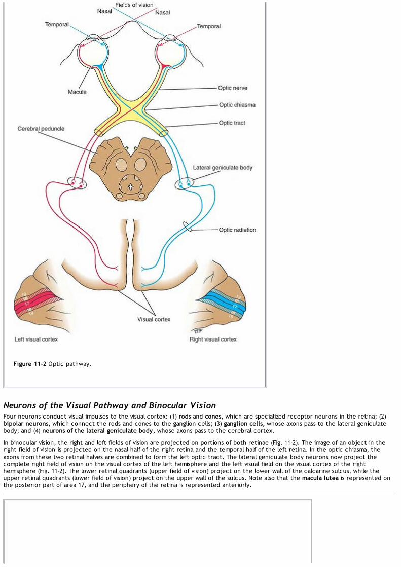
Figure 11-2 Optic pathway.
Neurons of the Visual Pathway and Binocular VisionFour neurons conduct visual impulses to the visual cortex: (1) rods and cones, which are specialized receptor neurons in the retina; (2)bipolar neurons, which connect the rods and cones to the ganglion cells; (3) ganglion cells, whose axons pass to the lateral geniculatebody; and (4) neurons of the lateral geniculate body, whose axons pass to the cerebral cortex.
In binocular vision, the right and left fields of vision are projected on portions of both retinae (Fig. 11-2). The image of an object in theright field of vision is projected on the nasal half of the right retina and the temporal half of the left retina. In the optic chiasma, theaxons from these two retinal halves are combined to form the left optic tract. The lateral geniculate body neurons now project thecomplete right field of vision on the visual cortex of the left hemisphere and the left visual field on the visual cortex of the righthemisphere (Fig. 11-2). The lower retinal quadrants (upper field of vision) project on the lower wall of the calcarine sulcus, while theupper retinal quadrants (lower field of vision) project on the upper wall of the sulcus. Note also that the macula lutea is represented onthe posterior part of area 17, and the periphery of the retina is represented anteriorly.

Figure 11-3 Optic pathway and the visual reflexes.
Visual Reflexes
Direct and Consensual Light ReflexesIf a light is shone into one eye, the pupils of both eyes normally constrict. The constriction of the pupil on which the light is shone iscalled the direct light reflex; the constriction of the opposite pupil, even though no light fell on that eye, is called the consensual lightreflex (Fig. 11-3).
The afferent impulses travel through the optic nerve, optic chiasma, and optic tract (Fig. 11-3). Here, a small number of fibers leave theoptic tract and synapse on nerve cells in the pretectal nucleus, which lies close to the superior colliculus. The impulses are passed byaxons of the pretectal nerve cells to the parasympathetic nuclei (Edinger-Westphal nuclei) of the third cranial nerve on both sides. Here,the fibers synapse and the parasympathetic nerves travel through the third cranial nerve to the ciliary ganglion in the orbit (Fig. 11-3).Finally, postganglionic parasympathetic fibers pass through the short ciliary nerves to the eyeball and the constrictor pupillae muscle ofthe iris. Both pupils constrict in the consensual light reflex because the pretectal nucleus sends fibers to the parasympathetic nuclei onboth sides of the midbrain (Fig. 11-3). The fibers that cross the median plane do so close to the cerebral aqueduct in the posteriorcommissure.
Accommodation ReflexWhen the eyes are directed from a distant to a near object, contraction of the medial recti brings about convergence of the ocular axes;the lens thickens to increase its refractive power by contraction of the ciliary muscle; and the pupilsconstrict to restrict the light waves to the thickest central part of the lens. The afferent impulses travel through the optic nerve, theoptic chiasma, the optic tract, the lateral geniculate body, and the optic radiation to the visual cortex. The visual cortex is connected tothe eye field of the frontal cortex (Fig. 11-3). From here, cortical fibers descend through the internal capsule to the oculomotor nuclei inthe midbrain. The oculomotor nerve travels to the medial recti muscles. Some of the descending cortical fibers synapse with the
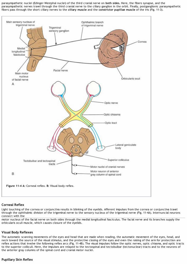
parasympathetic nuclei (Edinger-Westphal nuclei) of the third cranial nerve on both sides. Here, the fibers synapse, and theparasympathetic nerves travel through the third cranial nerve to the ciliary ganglion in the orbit. Finally, postganglionic parasympatheticfibers pass through the short ciliary nerves to the ciliary muscle and the constrictor pupillae muscle of the iris (Fig. 11-3).
Figure 11-4 A: Corneal reflex. B: Visual body reflex.
Corneal ReflexLight touching of the cornea or conjunctiva results in blinking of the eyelids. Afferent impulses from the cornea or conjunctiva travelthrough the ophthalmic division of the trigeminal nerve to the sensory nucleus of the trigeminal nerve (Fig. 11-4A). Internuncial neuronsconnect with themotor nucleus of the facial nerve on both sides through the medial longitudinal fasciculus. The facial nerve and its branches supply theorbicularis oculi muscle, which causes closure of the eyelids.
Visual Body ReflexesThe automatic scanning movements of the eyes and head that are made when reading, the automatic movement of the eyes, head, andneck toward the source of the visual stimulus, and the protective closing of the eyes and even the raising of the arm for protection arereflex actions that involve the following reflex arcs (Fig. 11-4B). The visual impulses follow the optic nerves, optic chiasma, and optic tractsto the superior colliculi. Here, the impulses are relayed to the tectospinal and tectobulbar (tectonuclear) tracts and to the neurons ofthe anterior gray columns of the spinal cord and cranial motor nuclei.
Pupillary Skin Reflex

The pupil will dilate if the skin is painfully stimulated by pinching. The afferent sensory fibers are believed to have connections with theefferent preganglionic sympathetic neurons in the lateral gray columns of the first and second thoracic segments of the spinal cord. Thewhite rami communicantes of these segments pass to the sympathetic trunk, and the preganglionic fibers ascend to the superior cervicalsympathetic ganglion. The postganglionic fibers pass through the internal carotid plexus and the long ciliary nerves to the dilatorpupillae muscle of the iris.
Oculomotor Nerve (Cranial Nerve III)The oculomotor nerve is entirely motor in function.
Oculomotor Nerve NucleiThe oculomotor nerve has two motor nuclei: (1) the main motor nucleus and (2) the accessory parasympathetic nucleus.
The main oculomotor nucleus is situated in the anterior part of the gray matter that surrounds the cerebral aqueduct of the midbrain(Fig. 11-5). It lies at the level of the superior colliculus. The nucleus consists of groups of nerve cells that supply all the extrinsic musclesof the eye except the superior oblique and the lateral rectus. The outgoing nerve fibers pass anteriorly through the red nucleus andemerge on the anterior surface of the midbrain in the interpeduncular fossa. The main oculomotor nucleus receives corticonuclear fibersfrom both cerebral hemispheres. It receives tectobulbar fibers from the superior colliculus and, through this route, receives informationfrom the visual cortex. It also receives fibers from the medial longitudinal fasciculus, by which it is connected to the nuclei of the fourth,sixth, and eighth cranial nerves.
The accessory parasympathetic nucleus (Edinger-Westphal nucleus) is situated posterior to the main oculomotor nucleus (Fig. 11-5). Theaxons of the nerve cells, which are preganglionic, accompany the other oculomotor fibers to the orbit. Here, they synapse in the ciliaryganglion, and postganglionic fibers pass through the short ciliary nerves to the constrictor pupillae of the iris and the ciliary muscles. Theaccessory parasympathetic nucleus receives corticonuclear fibers for the accommodation reflex and fibers from the pretectal nucleus forthe direct and consensual light reflexes (Fig. 11-3).
Course of the Oculomotor NerveThe oculomotor nerve emerges on the anterior surface of the midbrain (Fig. 11-5). It passes forward between the posterior cerebral andthe superior cerebellar arteries. It then continues into the middle cranial fossa in the lateral wall of the cavernous sinus. Here, it dividesinto a superior and an inferior ramus, which enter the orbital cavity through the superior orbital fissure.
The oculomotor nerve supplies the following extrinsic muscles of the eye: the levator palpebrae superioris, superior rectus, medial rectus,inferior rectus, and inferior oblique. It also supplies, through its branch to the ciliary ganglion and the short ciliary nerves,parasympathetic nerve fibers to the following intrinsic muscles: the constrictor pupillae of the iris and ciliary muscles.
Therefore, the oculomotor nerve is entirely motor and is responsible for lifting the upper eyelid; turning the eye upward, downward, andmedially; constricting the pupil; and accommodating the eye.
Trochlear Nerve (Cranial Nerve IV)The trochlear nerve is entirely motor in function.
Trochlear Nerve NucleusThe trochlear nucleus is situated in the anterior part of the gray matter that surrounds the cerebral aqueduct of the midbrain (Fig. 11-6).It lies inferior to the oculomotor nucleus at the level of the inferior colliculus. The nerve fibers, after leaving the nucleus, passposteriorly around the central gray matter to reach the posterior surface of the midbrain.
The trochlear nucleus receives corticonuclear fibers from both cerebral hemispheres. It receives the tectobulbar fibers, which connect itto the visual cortex through the superior colliculus (Fig. 11-6). It also receives fibers from the medial longitudinal fasciculus, by which it isconnected to the nuclei of the third, sixth, and eighth cranial nerves.
Course of the Trochlear NerveThe trochlear nerve, the most slender of the cranial nerves and the only one to leave the posterior surface of the brainstem, emergesfrom the midbrain and immediately decussates with the nerve of the opposite side. The trochlear nerve passes forward through themiddle cranial fossa in the lateral wall of the cavernous sinus and enters the orbit through the superior orbital fissure. The nerve suppliesthe superior oblique muscle of the eyeball. The trochlear nerve is entirely motor and assists in turning the eye downward and laterally.

Figure 11-5 A: Oculomotor nerve nuclei and their central connections. B: The distribution of the oculomotor nerve.
Trigeminal Nerve (Cranial Nerve V)The trigeminal nerve is the largest cranial nerve and contains both sensory and motor fibers. It is the sensory nerve to the greaterpart of the head and the motor nerve to several muscles, including the muscles of mastication (Fig. 11-8).
Trigeminal Nerve NucleiThe trigeminal nerve has four nuclei: (1) the main sensory nucleus, (2) the spinal nucleus, (3) the mesencephalic nucleus, and (4) themotor nucleus.
Main Sensory NucleusThe main sensory nucleus lies in the posterior part of the pons, lateral to the motor nucleus (Fig. 11-7A). It is continuous below with thespinal nucleus.
Spinal NucleusThe spinal nucleus is continuous superiorly with the main sensory nucleus in the pons and extends inferiorly through the whole length ofthe medulla oblongata and into the upper part of the spinal cord as far as the second cervical segment (Fig. 11-7B).

Figure 11-6 A: Trochlear nerve nucleus and its central connections. B: Distribution of the trochlear nerve.
Mesencephalic NucleusThe mesencephalic nucleus is composed of a column of unipolar nerve cells situated in the lateral part of the gray matter around thecerebral aqueduct. It extends inferiorly into the pons as far as the main sensory nucleus (Fig. 11-7).
Motor NucleusThe motor nucleus is situated in the pons medial to the main sensory nucleus (Fig. 11-7).
Sensory Components of the Trigeminal NerveThe sensations of pain, temperature, touch, and pressure from the skin of the face and mucous membranes travel along axons whose cellbodies are situated in the semilunar or trigeminal sensory ganglion (Fig. 11-7B). The central processes of these cells form the largesensory root of the trigeminal nerve. About half the fibers divide into ascending and descending branches when they enter the pons; theremainder ascend or descend without division (Fig. 11-7B). The ascending branches terminate in the main sensory nucleus, and thedescending branches terminate in the spinal nucleus. The sensations of touch and pressure are conveyed by nerve fibers that terminate inthe main sensory nucleus. The sensations of pain and temperature pass to the spinal nucleus (Fig. 11-7B). The sensory fibers from theophthalmic division of the trigeminal nerve terminate in the inferior part of the spinal nucleus; fibers from the maxillary division terminatein the middle of the spinal nucleus; and fibers from the mandibular division end in the superior part of the spinal nucleus.
Proprioceptive impulses from the muscles of mastication and from the facial and extraocular muscles are carried by fibers in the sensoryroot of the trigeminal nerve that have bypassed the semilunar or trigeminal ganglion (Fig. 11-7B).The fibers' cells of origin are the unipolar cells of the mesencephalic nucleus (Fig. 11-7).
The axons of the neurons in the main sensory and spinal nuclei and the central processes of the cells in the mesencephalic nucleus nowcross the median plane and ascend as the trigeminal lemniscus to terminate on the nerve cells of the ventral posteromedial nucleus of thethalamus. The axons of these cells now travel through the internal capsule to the postcentral gyrus (areas 3, 1, and 2) of the cerebralcortex.

Motor Component of the Trigeminal NerveThe motor nucleus receives corticonuclear fibers from both cerebral hemispheres (Fig. 11-7). It also receives fibers from the reticularformation, the red nucleus, the tectum, and the medial longitudinal fasciculus. In addition, it receives fibers from the mesencephalicnucleus, thereby forming a monosynaptic reflex arc.
Figure 11-7 A: Trigeminal nerve nuclei seen in a coronal section of the pons. B: Trigeminal nerve nuclei in the brainstem and theircentral connections.
The cells of the motor nucleus give rise to the axons that form the motor root. The motor nucleus supplies the muscles of mastication,the tensor tympani, the tensor veli palatini, and the mylohyoid and the anterior belly of the digastric muscle.
Course of the Trigeminal NerveThe trigeminal nerve leaves the anterior aspect of the pons as a small motor root and a large sensory root. The nerve passes forward outof the posterior cranial fossa and rests on the upper surface of the apex of the petrous part of the temporal bone in the middle cranialfossa. The large sensory root now expands to form the crescent-shapedtrigeminal ganglion, which lies within a pouch of dura mater called the trigeminal or Meckel cave. The ophthalmic, maxillary, andmandibular nerves arise from the anterior border of the ganglion (Fig. 11-8). The ophthalmic nerve (V1) contains only sensory fibers andleaves the skull through the superior orbital fissure to enter the orbital cavity. The maxillary nerve (V2) also contains only sensory fibersand leaves the skull through the foramen rotundum. The mandibular nerve (V3) contains both sensory and motor fibers and leaves the skullthrough the foramen ovale.

Figure 11-8 Distribution of the trigeminal nerve.
The sensory fibers to the skin of the face from each division supply a distinct zone (Fig. 11-9), there being little or no overlap of thedermatomes (compare with the overlap of the dermatomes of the spinal nerves). As noted previously, the motor fibers in the mandibulardivision are mainly distributed to muscles of mastication.
Abducent Nerve (Cranial Nerve VI)The abducent nerve is a small motor nerve that supplies the lateral rectus muscle of the eyeball.
Abducent Nerve NucleusThe small motor nucleus is situated beneath the floor of the upper part of the fourth ventricle, close to the midline and beneath thecolliculus facialis (Fig. 11-10A). The nucleus receives afferent corticonuclear fibers from both cerebral hemispheres. It receives thetectobulbar tract from the superior colliculus, by which the visual cortex is connected to the nucleus. It also receives fibers from themedial longitudinal fasciculus, by which it is connected to the nuclei of the third, fourth, and eighth cranial nerves (Fig. 11-9).
Course of the Abducent NerveThe fibers of the abducent nerve pass anteriorly through the pons and emerge in the groove between the lower border of the pons andthe medulla oblongata (Fig. 11-10B). It passes forward through the cavernous sinus, lying below and lateral to the internal carotid artery.The nerve then enters the orbit through the superior orbital fissure. The abducent nerve is entirely a motor nerve and supplies the lateralrectus muscle and, therefore, is responsible for turning the eye laterally.

Figure 11-9 Sensory nerve supply to the skin of the head and neck. Note that the skin over the angle of the jaw is supplied by thegreat auricular nerve (C2 and C3) and not by branches of the trigeminal nerve.

Figure 11-10 A: Abducent nerve nucleus and its central connections. B: Distribution of the abducent nerve.
Facial Nerve (Cranial Nerve VII)The facial nerve is both a motor and a sensory nerve.
Facial Nerve NucleiThe facial nerve has three nuclei: (1) the main motor nucleus, (2) the parasympathetic nuclei, and (3) the sensory nucleus.
Main Motor NucleusThe main motor nucleus lies deep in the reticular formation of the lower part of the pons (Fig. 11-11). The part of the nucleus thatsupplies the muscles of the upper part of the face receives corticonuclear fibers from both cerebral hemispheres. The part of the nucleusthat supplies the muscles of the lower part of the face receives only corticonuclear fibers from the opposite cerebral hemisphere.
These pathways explain the voluntary control of facial muscles. However, another involuntary pathway exists; it is separate and controlsmimetic or emotional changes in facial expression. This other pathway forms part of the reticular formation (see p. 307).
Parasympathetic NucleiParasympathetic nuclei lie posterolateral to the main motor nucleus. They are the superior salivatory and lacrimal nuclei (Fig. 11-11). Thesuperior salivatory nucleus receives afferent fibers from the hypothalamus through the descending autonomic pathways. Informationconcerning taste also is received from the nucleus of the solitary tract from the mouth cavity.

Figure 11-11 Facial nerve nuclei and their central connections.
The lacrimal nucleus receives afferent fibers from the hypothalamus for emotional responses and from the sensory nuclei of the trigeminalnerve for reflex lacrimation secondary to irritation of the cornea or conjunctiva.
Sensory NucleusThe sensory nucleus is the upper part of the nucleus of the tractus solitarius and lies close to the motor nucleus (Fig. 11-11). Sensationsof taste travel through the peripheral axons of nerve cells situated in the geniculate ganglion on the seventh cranial nerve. The centralprocesses of these cells synapse on nerve cells in the nucleus. Efferent fibers cross the median plane and ascend to the ventral posteriormedial nucleus of the opposite thalamus and to a number of hypothalamic nuclei. From the thalamus, the axons of the thalamic cells passthrough the internal capsule and corona radiata to end in the taste area of the cortex in the lower part of the postcentral gyrus (Fig. 11-11).
Course of the Facial NerveThe facial nerve consists of a motor and a sensory root. The fibers of the motor root first travel posteriorly around the medial side of theabducent nucleus (Fig. 11-11). They then pass around the nucleus beneath the colliculus facialis in the floor of the fourth ventricle and,finally, pass anteriorly to emerge from the brainstem (Fig. 11-11).
The sensory root (nervus intermedius) is formed of the central processes of the unipolar cells of the geniculate ganglion. It also containsthe efferent preganglionic parasympathetic fibers from the parasympathetic nuclei.
The two roots of the facial nerve emerge from the anterior surface of the brain between the pons and the medulla oblongata. They passlaterally in the posterior cranial fossa with the vestibulocochlear nerve and enter the internal acoustic meatus in the petrous part of thetemporal bone. At the bottom of the meatus, the nerve enters the facial canal and runs laterally through the inner ear. On reaching themedial wall of the tympanic cavity, the nerve expands to form the sensory geniculate ganglion (Fig. 11-12) and turns sharply backwardabove the promontory. At the posterior wall of the tympanic cavity, the facial nerve turns downward on the medial side of the aditus ofthe mastoid antrum, descends behind the pyramid, and emerges from the stylomastoid foramen.

Figure 11-12 A: Distribution of the facial nerve. B: Branches of the facial nerve within the petrous part of the temporal bone; thetaste fibers are shown in black. The glossopharyngeal nerve is also shown.
Distribution of the Facial NerveThe motor nucleus supplies the muscles of facial expression, the auricular muscles, the stapedius, the posterior belly of the digastric, andthe stylohyoid muscles (Fig. 11-12).
The superior salivatory nucleus supplies the submandibular and sublingual salivary glands and the nasal and palatine glands. The lacrimalnucleus supplies the lacrimal gland.
The sensory nucleus receives taste fibers from the anterior two-thirds of the tongue, the floor of the mouth, and the palate.
Vestibulocochlear Nerve (Cranial Nerve VIII)This nerve consists of two distinct parts, the vestibular nerve and the cochlear nerve, which are concerned with thetransmission of afferent information from the internal ear to the central nervous system (Figs. 11-13 and 11-14).

Figure 11-13 Vestibular nerve nuclei and their central connections.
Vestibular NerveThe vestibular nerve conducts nerve impulses from the utricle and saccule that provide information concerning the position of the head;the nerve also conducts impulses from the semicircular canals that provide information concerning movements of the head.
The nerve fibers of the vestibular nerve are the central processes of nerve cells located in the vestibular ganglion, which is situated inthe internal acoustic meatus. They enter the anterior surface of the brainstem in a groove between the lower border of the pons and theupper part of the medulla oblongata (Fig. 11-13). When they enter the vestibular nuclear complex, the fibers divide into short ascendingand long descending fibers; a small number of fibers pass directly to the cerebellum through the inferior cerebellar peduncle, bypassingthe vestibular nuclei.
The Vestibular Nuclear ComplexThis complex consists of a group of nuclei situated beneath the floor of the fourth ventricle (Fig. 11-13). Four nuclei may be recognized:(1) the lateral vestibular nucleus, (2) the superior vestibular nucleus,(3) the medial vestibular nucleus, and (4) the inferior vestibular nucleus (see Fig. 5-7).

Figure 11-14 Cochlear nerve nuclei and their central connections. The descending pathways have been omitted.
The vestibular nuclei receive afferent fibers from the utricle and saccule and the semicircular canals through the vestibular nerve andfibers from the cerebellum through the inferior cerebellar peduncle (Fig. 11-13). Efferent fibers from the nuclei pass to the cerebellumthrough the inferior cerebellar peduncle. Efferent fibers also descend uncrossed to the spinal cord from the lateral vestibular nucleus andform the vestibulospinal tract (Fig. 11-13). In addition, efferent fibers pass to the nuclei of the oculomotor, trochlear, and abducentnerves through the medial longitudinal fasciculus.
These connections enable the movements of the head and the eyes to be coordinated so that visual fixation on an object can bemaintained. In addition, information received from the internal ear can assist in maintaining balance by influencing the muscle tone of thelimbs and trunk.
Ascending fibers also pass upward from the vestibular nuclei to the cerebral cortex, to the vestibular area in the postcentral gyrus justabove the lateral fissure. These fibers are thought to relay in the ventral posterior nuclei of the thalamus. The cerebral cortex probablyserves to orient the individual consciously in space.
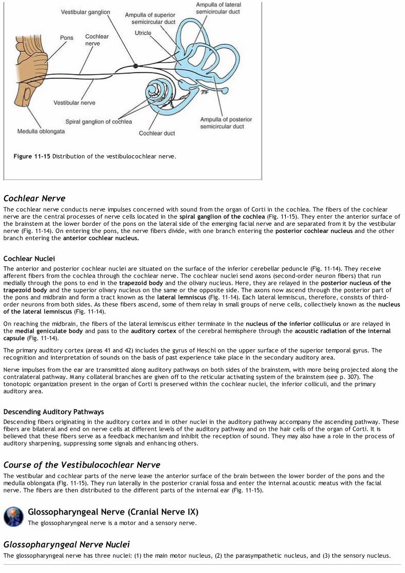
Figure 11-15 Distribution of the vestibulocochlear nerve.
Cochlear NerveThe cochlear nerve conducts nerve impulses concerned with sound from the organ of Corti in the cochlea. The fibers of the cochlearnerve are the central processes of nerve cells located in the spiral ganglion of the cochlea (Fig. 11-15). They enter the anterior surface ofthe brainstem at the lower border of the pons on the lateral side of the emerging facial nerve and are separated from it by the vestibularnerve (Fig. 11-14). On entering the pons, the nerve fibers divide, with one branch entering the posterior cochlear nucleus and the otherbranch entering the anterior cochlear nucleus.
Cochlear NucleiThe anterior and posterior cochlear nuclei are situated on the surface of the inferior cerebellar peduncle (Fig. 11-14). They receiveafferent fibers from the cochlea through the cochlear nerve. The cochlear nuclei send axons (second-order neuron fibers) that runmedially through the pons to end in the trapezoid body and the olivary nucleus. Here, they are relayed in the posterior nucleus of thetrapezoid body and the superior olivary nucleus on the same or the opposite side. The axons now ascend through the posterior part ofthe pons and midbrain and form a tract known as the lateral lemniscus (Fig. 11-14). Each lateral lemniscus, therefore, consists of third-order neurons from both sides. As these fibers ascend, some of them relay in small groups of nerve cells, collectively known as the nucleusof the lateral lemniscus (Fig. 11-14).
On reaching the midbrain, the fibers of the lateral lemniscus either terminate in the nucleus of the inferior colliculus or are relayed inthe medial geniculate body and pass to the auditory cortex of the cerebral hemisphere through the acoustic radiation of the internalcapsule (Fig. 11-14).
The primary auditory cortex (areas 41 and 42) includes the gyrus of Heschl on the upper surface of the superior temporal gyrus. Therecognition and interpretation of sounds on the basis of past experience take place in the secondary auditory area.
Nerve impulses from the ear are transmitted along auditory pathways on both sides of the brainstem, with more being projected along thecontralateral pathway. Many collateral branches are given off to the reticular activating system of the brainstem (see p. 307). Thetonotopic organization present in the organ of Corti is preserved within the cochlear nuclei, the inferior colliculi, and the primaryauditory area.
Descending Auditory PathwaysDescending fibers originating in the auditory cortex and in other nuclei in the auditory pathway accompany the ascending pathway. Thesefibers are bilateral and end on nerve cells at different levels of the auditory pathway and on the hair cells of the organ of Corti. It isbelieved that these fibers serve as a feedback mechanism and inhibit the reception of sound. They may also have a role in the process ofauditory sharpening, suppressing some signals and enhancing others.
Course of the Vestibulocochlear NerveThe vestibular and cochlear parts of the nerve leave the anterior surface of the brain between the lower border of the pons and themedulla oblongata (Fig. 11-15). They run laterally in the posterior cranial fossa and enter the internal acoustic meatus with the facialnerve. The fibers are then distributed to the different parts of the internal ear (Fig. 11-15).
Glossopharyngeal Nerve (Cranial Nerve IX)The glossopharyngeal nerve is a motor and a sensory nerve.
Glossopharyngeal Nerve NucleiThe glossopharyngeal nerve has three nuclei: (1) the main motor nucleus, (2) the parasympathetic nucleus, and (3) the sensory nucleus.
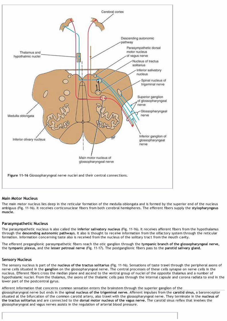
Figure 11-16 Glossopharyngeal nerve nuclei and their central connections.
Main Motor NucleusThe main motor nucleus lies deep in the reticular formation of the medulla oblongata and is formed by the superior end of the nucleusambiguus (Fig. 11-16). It receives corticonuclear fibers from both cerebral hemispheres. The efferent fibers supply the stylopharyngeusmuscle.
Parasympathetic NucleusThe parasympathetic nucleus is also called the inferior salivatory nucleus (Fig. 11-16). It receives afferent fibers from the hypothalamusthrough the descending autonomic pathways. It also is thought to receive information from the olfactory system through the reticularformation. Information concerning taste also is received from the nucleus of the solitary tract from the mouth cavity.
The efferent preganglionic parasympathetic fibers reach the otic ganglion through the tympanic branch of the glossopharyngeal nerve,the tympanic plexus, and the lesser petrosal nerve (Fig. 11-17). The postganglionic fibers pass to the parotid salivary gland.
Sensory NucleusThe sensory nucleus is part of the nucleus of the tractus solitarius (Fig. 11-16). Sensations of taste travel through the peripheral axons ofnerve cells situated in the ganglion on the glossopharyngeal nerve. The central processes of these cells synapse on nerve cells in thenucleus. Efferent fibers cross the median plane and ascend to the ventral group of nuclei of the opposite thalamus and a number ofhypothalamic nuclei. From the thalamus, the axons of the thalamic cells pass through the internal capsule and corona radiata to end in thelower part of the postcentral gyrus.
Afferent information that concerns common sensation enters the brainstem through the superior ganglion of theglossopharyngeal nerve but ends in the spinal nucleus of the trigeminal nerve. Afferent impulses from the carotid sinus, a baroreceptorsituated at the bifurcation of the common carotid artery, also travel with the glossopharyngeal nerve. They terminate in the nucleus ofthe tractus solitarius and are connected to the dorsal motor nucleus of the vagus nerve. The carotid sinus reflex that involves theglossopharyngeal and vagus nerves assists in the regulation of arterial blood pressure.

Figure 11-17 Distribution of the glossopharyngeal nerve.
Course of the Glossopharyngeal NerveThe glossopharyngeal nerve leaves the anterolateral surface of the upper part of the medulla oblongata as a series of rootlets in a groovebetween the olive and the inferior cerebellar peduncle (Fig. 11-16). It passes laterally in the posterior cranial fossa and leaves the skullthrough the jugular foramen. The superior and inferior glossopharyngeal sensory ganglia are situated on the nerve here. The nerve thendescends through the upper part of the neck in company with the internal jugular vein and the internal carotid artery to reach theposterior border of the stylopharyngeus muscle, which it supplies. The nerve then passes forward between the superior and middleconstrictor muscles of the pharynx to give sensory branches to the mucous membrane of the pharynx and the posterior third of thetongue (Fig. 11-17).
Vagus Nerve (Cranial Nerve X)The vagus nerve is a motor and a sensory nerve.
Vagus Nerve NucleiThe vagus nerve has three nuclei: (1) the main motor nucleus, (2) the parasympathetic nucleus, and (3) the sensory nucleus.
Main Motor NucleusThe main motor nucleus lies deep in the reticular formation of the medulla oblongata and is formed by the nucleus ambiguus (Fig. 11-18). Itreceives corticonuclear fibers from both cerebral hemispheres. The efferent fibers supply the constrictor muscles of the pharynx and theintrinsic muscles of the larynx (Fig. 11-19).
Parasympathetic NucleusThe parasympathetic nucleus forms the dorsal nucleus of the vagus and lies beneath the floor of the lower part of the fourth ventricleposterolateral to the hypoglossal nucleus (Fig. 11-18). It receives afferent fibers from the hypothalamusthrough the descending autonomic pathways. It also receives other afferents, including those from the glossopharyngeal nerve (carotidsinus reflex). The efferent fibers are distributed to the involuntary muscle of the bronchi, heart, esophagus, stomach, small intestine, andlarge intestine as far as the distal one-third of the transverse colon (Fig. 11-19).

Figure 11-18 Vagus nerve nuclei and their central connections.
Sensory NucleusThe sensory nucleus is the lower part of the nucleus of the tractus solitarius. Sensations of taste travel through the peripheral axons ofnerve cells situated in the inferior ganglion on the vagus nerve. The central processes of those cells synapse on nerve cells in thenucleus (Fig. 11-18). Efferent fibers cross the median plane and ascend to the ventral group of nuclei of the opposite thalamus as well as toa number of hypothalamic nuclei. From the thalamus, the axons of the thalamic cells pass through the internal capsule and corona radiatato end in the postcentral gyrus.
Afferent information concerning common sensation enters the brainstem through the superior ganglion of the vagus nerve but ends in thespinal nucleus of the trigeminal nerve.
Course of the Vagus NerveThe vagus nerve leaves the anterolateral surface of the upper part of the medulla oblongata as a series of rootlets in a groove betweenthe olive and the inferior cerebellar peduncle (Fig. 11-18). The nerve passes laterally through the posterior cranial fossa and leaves theskull through the jugular foramen. The vagus nerve possesses two sensory ganglia, a rounded superior ganglion, situated on the nervewithin the jugular foramen, and a cylindrical inferior ganglion, which lies on the nerve just below the foramen. Below the inferiorganglion, the cranial root of the accessory nerve joins the vagus nerve and is distributed mainly in its pharyngeal and recurrent laryngealbranches.
The vagus nerve descends vertically in the neck within the carotid sheath with the internal jugular vein and the internal and commoncarotid arteries.
The right vagus nerve enters the thorax and passes posterior to the root of the right lung, contributing to the pulmonary plexus. It thenpasses on to the posterior surface of the esophagus and contributes to the esophageal plexus. It enters the abdomen through theesophageal opening of the diaphragm. The posterior vagal trunk (which is the name now given to the right vagus) is distributed to theposteriorsurface of the stomach and, by a large celiac branch, to the duodenum, liver, kidneys, and small and large intestines as far as the distalthird of the transverse colon. This wide distribution is accomplished through the celiac, superior mesenteric, and renal plexuses.

Figure 11-19 Distribution of the vagus nerve.
The left vagus nerve enters the thorax and crosses the left side of the aortic arch and descends behind the root of the left lung,contributing to the pulmonary plexus. The left vagus then descends on the anterior surface of the esophagus, contributing to theesophageal plexus. It enters the abdomen through the esophageal opening of the diaphragm. The anterior vagal trunk (which is the namenow given to the left vagus) divides into several branches, which are distributed to the stomach, liver, upper part of the duodenum, andhead of the pancreas.
Accessory Nerve (Cranial Nerve XI)The accessory nerve is a motor nerve that is formed by the union of a cranial and a spinal root.
Cranial RootThe cranial root (part) is formed from the axons of nerve cells of the nucleus ambiguus (Fig. 11-20). The nucleus receives corticonuclearfibers from both cerebral hemispheres. The efferent fibers of the nucleus emerge from the anterior surface of the medulla oblongatabetween the olive and the inferior cerebellar peduncle.

Figure 11-20 Cranial and spinal nuclei of the accessory nerve and their central connections.
Course of the Cranial RootThe nerve runs laterally in the posterior cranial fossa and joins the spinal root. The two roots unite and leave the skull through the jugularforamen. The roots then separate, and the cranial root joins the vagus nerve and is distributed in its pharyngeal and recurrent laryngealbranches to the muscles of the soft palate, pharynx, and larynx.
Spinal RootThe spinal root (part) is formed from axons of nerve cells in the spinal nucleus, which is situated in the anterior gray column of the spinalcord in the upper five cervical segments (Fig. 11-20). The spinal nucleus is thought to receive corticospinal fibers from both cerebralhemispheres.
Course of the Spinal RootThe nerve fibers emerge from the spinal cord midway between the anterior and posterior nerve roots of the cervical spinal nerves. Thefibers form a nerve trunk that ascends into the skull through the foramen magnum. The spinal root passes laterally and joins the cranialroot asthey pass through the jugular foramen. After a short distance, the spinal root separates from the cranial root and runs downward andlaterally and enters the deep surface of the sternocleidomastoid muscle, which it supplies (Fig. 11-21). The nerve then crosses theposterior triangle of the neck and passes beneath the trapezius muscle, which it supplies.

Figure 11-21 Distribution of the accessory nerve.
The accessory nerve thus brings about movements of the soft palate, pharynx, and larynx and controls the movement of two large musclesin the neck.
Hypoglossal Nerve (Cranial Nerve XII)The hypoglossal nerve is a motor nerve that supplies all the intrinsic muscles of the tongue as well as the styloglossus, thehyoglossus, and the genioglossus muscles.
Hypoglossal NucleusThe hypoglossal nucleus is situated close to the midline immediately beneath the floor of the lower part of the fourth ventricle (Fig. 11-22). It receives corticonuclear fibers from both cerebral hemispheres. However, the cells responsible for supplying the genioglossusmuscle (Fig. 11-23) only receive corticonuclear fibers from the opposite cerebral hemisphere.
The hypoglossal nerve fibers pass anteriorly through the medulla oblongata and emerge as a series of roots in the groove between thepyramid and the olive (Fig. 11-22).
Course of the Hypoglossal NerveThe hypoglossal nerve fibers emerge on the anterior surface of the medulla oblongata between the pyramid and the olive (Fig. 11-22). Thenerve crosses the posterior cranial fossa and leaves the skull through the hypoglossal canal. The nerve passes downward and forward inthe neck between the internal carotid artery and the internal jugular vein until it reaches the lower border of the posterior belly of thedigastric muscle. Here, it turns forward and crosses the internal and external carotid arteries and the loop of the lingual artery. It passesdeep to the posterior margin of the mylohyoid muscle lying on the lateral surface of the hyoglossus muscle. The nerve then sendsbranches to the muscles of the tongue (Fig. 11-23).
In the upper part of its course, the hypoglossal nerve is joined by C1 fibers2 from the cervical plexus.
Thus, the hypoglossal nerve controls the movements and shape of the tongue.

P.359P.360P.361P.362
Figure 11-23 Distribution of the hypoglossal nerve.
Figure 11-22 Hypoglossal nucleus and its central connections.

Clinical NotesGeneral ConsiderationsThe 12 pairs of cranial nerves supply information to the brain from outlying receptor organs and bring about
changes in peripheral effector organs by means of appropriate motor nerves. Unfortunately for the student, the nervecells are not arranged simply, as in the spinal cord, but are grouped together to form nuclei that are found in differentsituations at different levels of the brainstem. Moreover, whereas spinal nerves possess afferent somatic fibers,afferent visceral fibers, efferent somatic fibers, and efferent visceral fibers, cranial nerves, in addition, possess specialsomatic afferent fibers (e.g., visual and auditory) and special visceral afferent fibers (e.g., taste).When the central connections of the different cranial nerve nuclei were discussed in the previous section, a simplifiedpractical version was given, since many of the precise connections of the cranial nerve nuclei are still not known.Because the delicate movements of the eyes, the larynx, and the face require carefully integrated muscle action andthe fine control of muscle tone, it must be assumed that the motor nuclei of the various cranial nerves receive inputfrom the cerebellum, the red nucleus, the reticular formation, and the corpus striatum in the same manner as the lowermotor neurons of the spinal cord.Three points of clinical value should be remembered:
1. Bilateral corticonuclear connections are present for all the cranial motor nuclei except that part of the facial nucleusthat supplies the muscles of the lower part of the face and that part of the hypoglossal nucleus that supplies thegenioglossus muscle.
2. The cranial nerves that possess afferent sensory fibers have cell bodies that are found in ganglia along the courseof the nerves; these are equivalent to the posterior root ganglia. In the case of the olfactory nerves, the cells arethe olfactory receptors.
3. In situations where the cranial nerve nuclei are close together, it is very rare for a disease process to affect onenucleus only. For example, the cell groups of the nucleus ambiguus serve the glossopharyngeal, the vagus, and thecranial root of the accessory nerve, and functional loss involving all three nerves is a common finding.
Clinical Examination of Cranial NervesThe systematic examination of the 12 cranial nerves is an important part of the examination of every neurologicpatient. It may reveal a lesion of a cranial nerve nucleus or its central connections, or it may show an interruption ofthe lower motor neurons.
Olfactory NerveFirst, determine that the nasal passages are clear. Then, apply some easily recognizable aromatic substance, such asoil of peppermint, oil of cloves, or tobacco, to each nostril in turn. Ask the patient whether he or she can smellanything; if so, ask the patient to identify the smell. It should be remembered that food flavors depend on the senseof smell and not on the sense of taste.Bilateral anosmia can be caused by disease of the olfactory mucous membrane, such as the common cold or allergicrhinitis. Unilateral anosmia can result from disease affecting the olfactory nerves, bulb, or tract. A lesion of theolfactory cortex on one side is unlikely to produce complete anosmia, because fibers from each olfactory tract travel toboth cerebral hemispheres. Fractures of the anterior cranial fossa involving the cribriform plate of the ethmoid couldtear the olfactory nerves. Cerebral tumors of the frontal lobes or meningiomas of the anterior cranial fossa canproduce anosmia by pressing on the olfactory bulb or tract.
Optic NerveFirst, ask the patient whether he or she has noted any change in eyesight. Visual acuity should be tested for nearand distant vision. Near vision is tested by asking the patient to read a card with a standard size of type. Each eye istested in turn, with or without spectacles. Distant vision is tested by asking the patient to read Snellen type at adistance of 20 feet.The visual fields should then be tested. The patient and the examiner sit facing each other at a distance of 2 feet. Thepatient is asked to cover the right eye, and the examiner covers his own left eye. The patient is asked to look into thepupil of the examiner's right eye. A small object is then moved in an arc around the periphery of the field of vision, andthe patient is asked whether he or she can see the object. The extent of the patient's field of vision is compared withthe normal examiner's field. Then, the other eye is tested. It is important not to miss loss or impairment of vision in thecentral area of the field (central scotoma).
Lesions of the Visual PathwayLesions of the optic pathway may have many pathologic causes. Expanding tumors of the brain and neighboringstructures, such as the pituitary gland and the meninges, and cerebrovascular accidents are commonly responsible.The most widespread effects on vision occur where the nerve fibers of the visual pathway are tightly packed together,such as in the optic nerve or the optic tract.
Circumferential BlindnessCircumferential blindness may be caused by hysteria or optic neuritis (Fig. 11-24 [1]). Optic neuritis may occur followingspread of infection from the sphenoid and ethmoid sinuses; the nerve is infected as it passes through the optic canalto enter the orbital cavity.
Total Blindness of One EyeTotal blindness of one eye would follow complete section of one optic nerve (Fig. 11-24 [2]).
Nasal HemianopiaNasal hemianopia would follow a partial lesion of the optic chiasma on its lateral side (Fig. 11-24 [3]).
Bitemporal HemianopiaBitemporal hemianopia would follow a sagittal section of the optic chiasma (Fig. 11-24 [4]). This condition is mostcommonly produced by a tumor of the pituitary gland exerting pressure on the optic chiasma.
Contralateral Homonymous Hemianopia

Contralateral homonymous hemianopia would follow division of the optic tract or optic radiation or destruction of thevisual cortex on one side; the lesion would produce the same hemianopia for both eyes—that is, homonymoushemianopia (Fig. 11-24 [5–7]). If the right optic tract is divided, for example, a left temporal hemianopia and a rightnasal hemianopia will occur.
Figure 11-24 Visual field defects associated with lesions of the optic pathways. 1. Right-sided circumferential blindness due toretrobulbar neuritis. 2. Total blindness of the right eye due to division of the right optic nerve. 3. Right nasal hemianopia dueto a partial lesion of the right side of the optic chiasma. 4. Bitemporal hemianopia due to a complete lesion of the opticchiasma. 5. Left temporal hemianopia and right nasal hemianopia due to a lesion of the right optic tract. 6. Left temporal andright nasal hemianopia due to a lesion of the right optic radiation. 7. Left temporal and right nasal hemianopia due to a lesionof the right visual cortex.
Examination of the FundiThe ocular fundus should be examined with an ophthalmoscope. The patient is asked to look at a distant object. Whenthe right eye is examined, the physician should use his or her right eye and hold the ophthalmoscope in his or her righthand. The physician should systematically examine the fundus, looking first at the optic disc, then at the retina, then atthe blood vessels, and finally at the macula.The optic disc is creamy pink, and the lateral margin is seen clearly. The center of the disc is paler and hollowed out.The retina is pinkish red, and there should be no hemorrhages or exudates.The blood vessels should consist of four main arteries with their accompanying veins. Carefully examine thearteriovenous crossings. The veins should not be indented by the arteries.The macula is examined by asking the patient to look directly at the light of the ophthalmoscope. It should look slightlydarker than the surrounding retina.
Examination of the Extraocular MusclesTo examine the extraocular muscles, the patient's head is fixed, and he or she is asked to move the eyes, in turn, tothe left, to the right, upward, and downward, as far as possible in each direction. The patient should then be asked to

look upward and laterally, upward and medially, downward and medially, and downward and laterally.The pupillary reactions to convergence associated with accommodation and the direct and consensual pupillaryreactions to light are tested. The nervous pathways involved in the pupillary reflexes are described on page 338.
Oculomotor NerveThe oculomotor nerve supplies all the extraocular muscles except the superior oblique and the lateral rectus. It alsosupplies the striated muscle of the levator palpebrae superioris and the smooth muscle concerned withaccommodation, namely, the sphincter pupillae and the ciliary muscle.In a complete lesion of the oculomotor nerve, the eye cannot be moved upward, downward, or inward. At rest, the eyelooks laterally (external strabismus), owing to the activity of the lateral rectus, and downward, owing to the activity ofthe superior oblique. The patient sees double (diplopia). There is drooping of the upper eyelid (ptosis) due to paralysisof the levator palpebrae superioris. The pupil is widely dilated and nonreactive to light, owing to paralysis of thesphincter pupillae and unopposed action of the dilator (supplied by the sympathetic). Accommodation of the eye isparalyzed.Incomplete lesions of the oculomotor nerve are common and may spare the extraocular muscles or the intraocularmuscles. The condition in which the innervation of the extraocular muscles is spared with selective loss of theautonomic innervation of the sphincter pupillae and ciliary muscle is called internal ophthalmoplegia. The condition inwhich the sphincter pupillae and the ciliary muscle are spared with paralysis of the extraocular muscles is calledexternal ophthalmoplegia.The possible explanation for the involvement of the autonomic nerves and the sparing of the remaining fibers is thatthe parasympathetic autonomic fibers are superficially placed within the oculomotor nerve and are likely to be firstaffected by compression. The nature of the disease also plays a role. For example, in cases of diabetes with impairednerve conduction (diabetic neuropathy), the autonomic fibers are unaffected, whereas the nerves to the extraocularmuscles are paralyzed.The conditions most commonly affecting the oculomotor nerve are diabetes, aneurysm, tumor, trauma, inflammation,and vascular disease. See lesions of the oculomotor nerve in the midbrain (Benedikt syndrome) on page 220.
Trochlear NerveThe trochlear nerve supplies the superior oblique muscle, which rotates the eye downward and laterally.In lesions of the trochlear nerve, the patient complains of double vision on looking straight downward, because theimages of the two eyes are tilted relative to each other. This is because the superior oblique is paralyzed, and the eyeturns medially as well as downward. In fact, the patient has great difficulty in turning the eye downward and laterally.The conditions most often affecting the trochlear nerve include stretching or bruising as a complication of head injuries(the nerve is long and slender), cavernous sinus thrombosis, aneurysm of the internal carotid artery, and vascularlesions of the dorsal part of the midbrain. See lesions of the trochlear nerve in the midbrain on pages 219 and 220.
Abducent NerveThe abducent nerve supplies the lateral rectus muscle, which rotates the eye laterally. In a lesion of the abducentnerve, the patient cannot turn the eye laterally. When the patient is looking straight ahead, the lateral rectus isparalyzed, and the unopposed medial rectus pulls the eyeball medially, causing internal strabismus. There is alsodiplopia.Lesions of the abducent nerve include damage due to head injuries (the nerve is long and slender), cavernous sinusthrombosis or aneurysm of the internal carotid artery, and vascular lesions of the pons.
Internuclear OphthalmoplegiaLesions of the medial longitudinal fasciculus will disconnect the oculomotor nucleus that innervates the medial rectusmuscle from the abducent nucleus that innervates the lateral rectus muscle. When the patient is asked to look laterallyto the right or left, the ipsilateral lateral rectus contracts, turning the eye laterally, but the contralateral medial rectusfails to contract, and the eye looks straight forward.Bilateral internuclear ophthalmoplegia can occur with multiple sclerosis, occlusive vascular disease, trauma, orbrainstem tumors. Unilateral internuclear ophthalmoplegia can follow an infarct of a small branch of the basilar artery.
Trigeminal NerveThe trigeminal nerve has sensory and motor roots. The sensory root passes to the trigeminal ganglion, from whichemerge the ophthalmic (V1), maxillary (V2), and mandibular (V3) divisions. The motor root joins the mandibular division.The sensory function may be tested by using cotton and a pin over each area of the face supplied by the divisions ofthe trigeminal nerve (Fig. 11-9). Note that there is very little overlap of the dermatomes and that the skin covering theangle of the jaw is innervated by branches from the cervical plexus (C2 and C3). In lesions of the ophthalmic division,the cornea and conjunctiva will be insensitive to touch.The motor function may be tested by asking the patient to clench his or her teeth. The masseter and the temporalismuscles can be palpated and felt to harden as they contract.
Trigeminal NeuralgiaIn trigenimal neuralgia, the severe, stabbing pain over the face is of unknown cause and involves the pain fibers of thetrigeminal nerve. Pain is felt most commonly over the skin areas innervated by the mandibular and maxillary divisionsof the trigeminal nerve; only rarely is pain felt in the area supplied by the ophthalmic division.
Facial NerveThe facial nerve supplies the muscles of facial expression, supplies the anterior two-thirds of the tongue with tastefibers, and is secretomotor to the lacrimal, submandibular, and sublingual glands.To test the facial nerve, the patient is asked to show the teeth by separating the lips with the teeth clenched.Normally, equal areas of the upper and lower teeth are revealed on both sides. If a lesion of the facial nerve is presenton one side, the mouth is distorted. A greater area of teeth is revealed on the side of the intact nerve, since themouth is pulled up on that side. Another useful test is to ask the patient to close both eyes firmly. The examiner thenattempts to open the eyes by gently raising the patient's upper lids. On the side of the lesion, the orbicularis oculi isparalyzed so that the eyelid on that side is easily raised.

The sensation of taste on each half of the anterior two-thirds of the tongue can be tested by placing small amounts ofsugar, salt, vinegar, and quinine on the tongue for the sweet, salty, sour, and bitter sensations.
Facial Nerve LesionsThe facial nerve may be injured or may become dysfunctional anywhere along its long course from the brainstem to theface. Its anatomical relationship to other structures greatly assists in the localization of the lesion. If the abducentnerve (supplies the lateral rectus muscle) and the facial nerve are not functioning, this would suggest a lesion in thepons of the brain. If the vestibulocochlear nerve (for balance and hearing) and the facial nerve are not functioning, thissuggests a lesion in the internal acoustic meatus. If the patient is excessively sensitive to sound in one ear, the lesionprobably involves the nerve to the stapedius muscle, which arises from the facial nerve in the facial canal.Loss of taste over the anterior two-thirds of the tongue indicates that the facial nerve is damaged proximal to thepoint where it gives off the chorda tympani branch in the facial canal.A firm swelling of the parotid salivary gland associated with impaired function of the facial nerve is strongly indicative ofa cancer of the parotid gland with involvement of the nerve within the gland.
Figure 11-25 Facial expression defects associated with lesions of the upper motor neurons (1) and lower motor neurons (2).
Deep lacerations of the face may involve branches of the facial nerve.The part of the facial nucleus that controls the muscles of the upper part of the face receives corticonuclear fibers fromboth cerebral hemispheres. Therefore, it follows that with a lesion involving the upper motor neurons, only the musclesof the lower part of the face will be paralyzed (Fig. 11-25). However, in patients with a lesion of the facial nerve motornucleus or the facial nerve itself—that is, a lower motor neuron lesion—all the muscles on the affected side of the facewill be paralyzed (Fig. 11-25). The lower eyelid will droop, and the angle of the mouth will sag. Tears will flow over thelower eyelid, and saliva will dribble from the corner of the mouth. The patient will be unable to close the eye and willbe unable to expose the teeth fully on the affected side.In patients with hemiplegia, the emotional movements of the face are usually preserved. This indicates that the uppermotor neurons controlling these mimetic movements have a course separate from that of the main corticobulbarfibers. A lesion involving this separate pathway alone results in a loss of emotional movements, but voluntarymovements are preserved. A more extensive lesion will produce both mimetic and voluntary facial paralysis.
Bell PalsyBell palsy is a dysfunction of the facial nerve, as it lies within the facial canal; it is usually unilateral. The site of thedysfunction will determine the aspects of facial nerve function that do not work.The swelling of the nerve within the bony canal causes pressure on the nerve fibers; this results in a temporary loss offunction of the nerve, producing a lower motor neuron type of facial paralysis. The cause of Bell palsy is not known; it

sometimes follows exposure of the face to a cold draft.
Vestibulocochlear NerveThe vestibulocochlear nerve innervates the utricle and saccule, which are sensitive to static changes in equilibrium; thesemicircular canals, which are sensitive to changes in dynamic equilibrium; and the cochlea, which is sensitive tosound.
Disturbances of Vestibular Nerve FunctionDisturbances of vestibular nerve function include giddiness (vertigo) and nystagmus (see p. 245). Vestibularnystagmus is an uncontrollable rhythmic oscillation of the eyes, and the fast phase is away from the side of the lesion.This form of nystagmus is essentially a disturbance in the reflex control of the extraocular muscles, which is one of thefunctions of the semicircular canals. Normally, the nerve impulses pass reflexly from the canals through the vestibularnerve, the vestibular nuclei, and the medial longitudinal fasciculus, to the third, fourth, and sixth cranial nerve nuclei,which control the extraocular muscles; the cerebellum assists in coordinating the muscle movements.Vestibular function can be investigated with caloric tests. These involve the raising and lowering of the temperature inthe external auditory meatus, which induces convection currents in the endolymph of the semicircular canals(principally the lateral semicircular canal) and stimulates the vestibular nerve endings.The causes of vertigo include diseases of the labyrinth, such as Ménière disease. Lesions of the vestibular nerve, thevestibular nuclei, and the cerebellum can also be responsible. Multiple sclerosis, tumors, and vascular lesions of thebrainstem also cause vertigo.
Disturbances of Cochlear Nerve FunctionDisturbances of cochlear function are manifested as deafness and tinnitus. The patient's ability to hear a whisperedvoice or a vibrating tuning fork should be tested; each ear should be tested separately.Loss of hearing may be due to a defect of the auditory- conducting mechanism in the middle ear, damage to thereceptor cells in the spiral organ of Corti in the cochlea, a lesion of the cochlear nerve, a lesion of the central auditorypathways, or a lesion of the cortex of the temporal lobe.Lesions of the internal ear include Ménière disease, acute labyrinthitis, and trauma following head injury. Lesions ofthe cochlear nerve include tumor (acoustic neuroma) and trauma. Lesions in the central nervous system includetumors of the midbrain and multiple sclerosis. Only bilateral temporal lobe lesions cause deafness.
Glossopharyngeal NerveThe glossopharyngeal nerve supplies the stylopharyngeus muscle and sends secretomotor fibers to the parotid gland.Sensory fibers innervate the posterior one-third of the tongue for general sensation and taste.The integrity of this nerve may be evaluated by testing the patient's general sensation and that of taste on theposterior third of the tongue.Isolated lesions of the glossopharyngeal nerve are rare and usually also involve the vagus nerve.
Vagus NerveThe vagus nerve innervates many important organs, but the examination of this nerve depends on testing the functionof the branches to the pharynx, soft palate, and larynx. The pharyngeal or gag reflex may be tested by touching thelateral wall of the pharynx with a spatula. This should immediately cause the patient to gag; that is, the pharyngealmuscles will contract. The afferent neuron of the pharyngeal reflex runs in the glossopharyngeal nerve, and theefferent neurons run in the glossopharyngeal (to the stylopharyngeus muscle) and vagus nerves (pharyngealconstrictor muscles). Unilateral lesions of the vagus will show little or no gag reflex on that side.The innervation of the soft palate may be tested by asking the patient to say “ah.” Normally, the soft palate rises andthe uvula moves backward in the midline.All the muscles of the larynx are supplied by the recurrent laryngeal branch of the vagus, except the cricothyroidmuscle, which is supplied by the external laryngeal branch of the superior laryngeal branch of the vagus. Hoarsenessor absence of the voice may occur as a symptom of vagal nerve palsy. The movements of the vocal cords may betested by means of a laryngoscopic examination. Lesions involving the vagus nerve in the posterior cranial fossacommonly involve the glossopharyngeal, accessory, and hypoglossal nerves as well.
Accessory NerveThe accessory nerve supplies the sternocleidomastoid and the trapezius muscles by means of its spinal root. Thepatient should be asked to rotate the head to one side against resistance, causing the sternocleidomastoid of theopposite side to come into action. Then, the patient should be asked to shrug the shoulders, causing the trapeziusmuscles to come into action.Lesions of the spinal part of the accessory nerve will result in paralysis of the sternocleidomastoid and trapeziusmuscles. The sternocleidomastoid muscle will atrophy, and there will be weakness in turning the head to the oppositeside. The trapezius muscle will also atrophy, and the shoulder will droop on that side; there will also be weakness anddifficulty in raising the arm above the horizontal.Lesions of the spinal part of the accessory nerve may occur anywhere along its course and may result from tumors ortrauma from stab or gunshot wounds in the neck.
Hypoglossal NerveThe hypoglossal nerve supplies the intrinsic muscles of the tongue and the styloglossus, hyoglossus, and genioglossusmuscles. To test the integrity of the nerve, the patient is asked to put out the tongue; if there is a lower motor neuronlesion, the tongue will be observed to deviate toward the paralyzed side. The tongue will be smaller on the side of thelesion, owing to muscle atrophy, and fasciculation may accompany or precede the atrophy. Remember that the greaterpart of the hypoglossal nucleus receives corticonuclear fibers from both cerebral hemispheres. However, the part ofthe nucleus that supplies the genioglossus receives corticonuclear fibers only from the opposite cerebral hemisphere.If a patient has a lesion of the corticonuclear fibers, there will be no atrophy or fibrillation of the tongue, and onprotrusion, the tongue will deviate to the side opposite the lesion. (Note that the genioglossus is the muscle that pullsthe tongue forward.)Lesions of the hypoglossal nerve may occur anywhere along its course and may result from tumor, demyelinatingdiseases, syringomyelia, and vascular accidents. Injury of the nerve in the neck may also follow stab and gunshot

P.364
wounds.
Clinical Problem Solving1. A 60-year-old woman was seen as an outpatient because she had suddenly developed double vision. She waswatching her favorite television program the day before when it suddenly occurred. She had no other symptoms. Aftera complete physical examination, it was found that her right eye, when at rest, was turned medially, and she wasunable to turn it laterally. A moderate amount of glucose was found in her urine, and she had an abnormally elevatedblood glucose level. When closely questioned, she admitted that recently she had noticed having to pass water morefrequently, especially at night. She also said she often felt thirsty. She had lost 28 pounds during the past 2 years.Using your knowledge of neuroanatomy, explain the problem in her right eye. Do you think there is any connectionbetween her glucosuria, high blood glucose, polyuria, polydipsia, and weight loss and her eye condition?View Answer
2. An 18-year-old man was admitted to the hospital unconscious after a serious motorcycle accident. After a completephysical examination was performed and lateral and anteroposterior radiographs of the skull were taken, it was foundthat the patient had a fracture involving the anterior cranial fossa. It also was noted that he had a slight butcontinuous blood-stained watery discharge from his left nostril. Three days later, he recovered consciousness, and afurther physical examination revealed that he could no longer smell. This was tested by asking him to recognize thesmell of coffee, oil of cloves, and oil of peppermint. Using your knowledge of neuroanatomy, diagnose what is wrongwith this patient. Is it possible for normal individuals with an acute sense of smell to be unable to recognize commonscents? Could a tumor that had destroyed the olfactory cortex of one cerebral hemisphere be responsible for theanosmia in this patient?View Answer
3. A 72-year-old man with a known history of cerebrovascular problems visited his physician because 3 days previouslyhe had begun to have trouble reading the paper. He complained that the print started to tilt and that he wasbeginning to see double. He also said that he found it difficult to see the steps when he descended the staircase tothe physician's office. On physical examination, the patient had weakness of movement of the right eye bothdownward and laterally. Using your knowledge of neuroanatomy, explain this patient's signs and symptoms. If weassume that a cranial nerve nucleus is the site of the lesion, is it the right one or the left one that is involved?View Answer
4. A 73-year-old man consulted his physician because he was becoming deaf. His only other complaints were that hedid not think he was as tall as he used to be, and he was annoyed to find that each year he had to buy a size largerhat. The physician diagnosed osteitis deformans (Paget disease) and explained to the medical students that this is adisease of bones involving bone absorption and new bone formation. These bony changes lead to enlargement of theskull, deformities of the vertebral column, and bowing of the long bones of the legs. The physician asked the studentswhether there was any connection between the bone disease and the patient's deafness and which other cranialnerve they would be particularly interested in testing. How would you have answered these questions?View Answer
5. A neurologist was visited by a 25-year-old man who complained of a feeling of heaviness in both legs and giddinesson walking. On examination, it was found that the patient had widely disseminated lesions involving the corticospinaltracts, the posterior white column, and the optic nerves. A diagnosis of multiple sclerosis was made. This disease ofunknown origin primarily involves the white matter of the brain and spinal cord. Do you think this patient's symptomsof vertigo could be accounted for by this disease?View Answer
6. A 54-year-old woman with left-sided hemiplegia was examined by a fourth-year medical student. He very carefullytested each cranial nerve and noted any defects. During the examination, he stood behind the patient and gentlygrasped the trapezius muscles between his fingers and thumbs and asked the patient to shrug her shoulders againstresistance. He was surprised to find that there was no evidence of weakness in either trapezius muscle, and therewas no muscle wasting. Would you expect to find evidence of weakness or wasting in the trapezius muscles of apatient with hemiplegia?View Answer
7. A 35-year-old man was admitted to the hospital with a complaint of severe pain of the right side of the foreheadand the right eye. The pain had started 3 weeks previously and had progressively increased since then. One weekago, he started to see double, and this morning, his wife noticed that his right eye was turning out laterally. Thephysician in charge made a careful neurologic workup on this patient and found a lateral deviation of the right eye,dilatation of the right pupil with loss of direct and consensual light reflexes, paralysis of accommodation on the right,and paralysis of all right-sided ocular movement except laterally. He initially advised the patient to have computedtomography and magnetic resonance imaging scans of the skull and later ordered a right-sided carotid arteriogram.The film showed an aneurysm of the internal carotid artery on the right side. Explain the signs and symptoms of thispatient. Relate the signs and symptoms to the aneurysm.View Answer
8. During ward rounds, a neurologist demonstrated the signs and symptoms of neurosyphilis to a group of students.The patient was a 62-year-old man. The physician asked the students to note that both the patient's pupils were smalland fixed and were not altered by shining a light in the eyes or shading the eyes. It was noted, however, that thepupils narrowed when the patient was asked to look from a distant object to the tip of his nose. Moreover, the pupilsdilated again when he looked in the distance. “This is a good example of the Argyll Robertson pupil,” said thephysician. Using your knowledge of neuroanatomy, explain this curious pupillary reaction.

P.366P.367P.368
View Answer
9. Describe the effects of a lesion at the following points along the visual pathway of the right eye:(a) Section of the right optic nerve(b) Midline section of the optic chiasma(c) Section of the right optic tract(d) Section of the right optic radiation(e) Destruction of the cortex of the right occipital pole
View Answer
10. A 58-year-old woman was diagnosed as having an advanced carcinoma of the nasopharynx with neoplasticinfiltration of the posterior cranial fossa. How would you test for the integrity of the 9th, 10th, and 11th cranial nerves?View Answer
11. A 32-year-old woman with syringomyelia was found on physical examination to have impairment of appreciation ofpain and temperature of the face but preservation of light touch. Using your knowledge of neuroanatomy, explain thisdissociated sensory loss in the face.View Answer
12. A 51-year-old man complaining of an agonizing, stabbing pain over the middle part of the right side of his face wasseen in the emergency department. The stabs would last a few seconds and were repeated several times. “The pain isthe worst I have ever experienced,” he told the physician. A draft of cold air on the right side of his face or thetouching of a few hairs in the right temporal region of his scalp could trigger the pain. The patient had no othercomplaints and said he felt otherwise very fit. A complete physical examination of the cranial nerves revealed nothingabnormal. In particular, there was no evidence of sensory or motor loss of the right trigeminal nerve. The patientindicated the area on the right side of his face in which he experienced the pain; it was seen to be in the distributionof the maxillary division of the trigeminal nerve. Using your knowledge of neuroanatomy, make the diagnosis.View Answer
13. A physician turned to a group of students and said, “I think this patient has an advanced neoplasm in the posteriorcranial fossa with involvement of the medulla oblongata and in particular the nuclei of the vagus nerve.” What are thenuclei of the vagus nerve? Is it possible to have abnormal movements of the vocal cords in a patient with hemiplegia?Is it possible to have a solitary lesion of the vagal nuclei without involvement of other cranial nerve nuclei?View Answer
Review QuestionsDirections: Each of the numbered items in this section is followed by answers. Select the ONE lettered answer that isCORRECT.1. The cranial nerve nuclei listed below have the following descending tracts terminating on them:
(a) The inferior salivatory nucleus of the glossopharyngeal nerve receives descending tracts from the thalamus.(b) The nucleus of the abducent nerve receives only crossed corticobulbar tracts.(c) The nucleus of the facial nerve supplying the muscles of the lower part of the face receives only crossedcorticobulbar tracts.(d) The trigeminal motor nucleus receives only uncrossed corticobulbar tracts.(e) The nucleus of the trochlear nerve receives only crossed corticobulbar tracts.
View Answer
2. The nuclei associated with the facial nerve include the following:(a) Spinal nucleus(b) Inferior salivatory nucleus(c) Nucleus ambiguus(d) Main sensory nucleus(e) Lacrimal nucleus
View Answer
3. A patient with unilateral upper motor neuron paralysis of the facial muscles can smile with both sides of his face inresponse to a joke but not voluntarily. This can be explained by the following facts:
(a) The main corticobulbar fibers controlling voluntary movement of the facial muscles have been preserved.(b) Reticular fibers, possibly originating in the hypothalamus and descending to the motor nuclei of the facialnerves, are damaged.(c) The facial nerves are damaged.(d) The muscles producing mimetic movements of the face are innervated by corticobulbar fibers that have acourse separate from the main corticobulbar fibers.(e) There is a lesion involving the lower motor neurons.

View Answer
Directions: Each of the numbered items or incomplete statements in this section is followed by answers or bycompletions of the statement. Select the ONE lettered answer or completion that is BEST in each case.4. Which of the following structures participates in the reception of sound?
(a) Trapezoid body(b) Medial lemniscus(c) Nucleus of the trigeminal lemniscus(d) Inferior temporal gyrus(e) Lateral geniculate body
View Answer
5. The cerebral cortex is necessary for which of the following visual reflexes?(a) Corneal reflex(b) Accommodation reflex(c) Consensual light reflex(d) Pupillary light reflex(e) Visual body reflex
View Answer
6. The nasal field of the right eye is projected to the:(a) left lateral geniculate body(b) both banks of the left calcarine fissure(c) left optic tract(d) temporal retina of the right eye(e) left optic radiation
View Answer
7. Right pupillary constriction associated with light directed at the left eye requires the:(a) right optic radiation.(b) left optic nerve(c) left Edinger-Westphal nucleus(d) left oculomotor nerve(e) right optic nerve
View Answer
8. Select the lettered statement concerning the hypoglossal nerve that is correct:(a) A lesion involving the hypoglossal nerve will result in deviation of the tongue toward the same side as thelesion when the tongue is protruded.(b) The hypoglossal nerve conducts taste impulses from the posterior third of the tongue.(c) The hypoglossal nerve emerges from the brainstem between the olive and the inferior cerebellar peduncle.(d) The hypoglossal nerve carries with it fibers from the third and fourth cervical nerves.(e) The fibers of the accessory nerve wind around the motor nucleus of the hypoglossal nerve beneath the floorof the fourth ventricle.
View Answer
9. Select the lettered statement concerning the trigeminal nuclei that is correct:(a) The main sensory nucleus lies within the medulla oblongata.(b) The spinal nucleus extends inferiorly as far as the fifth cervical segment.(c) Proprioceptive impulses from the muscles of mastication reach the mesencephalic nucleus along fibers thatare part of the unipolar neurons of the nucleus.(d) The sensations of pain and temperature terminate in the main sensory nucleus.(e) The trigeminal lemniscus contains only efferent fibers from the ipsilateral sensory nuclei of the trigeminalnerve.
View Answer
Directions: Each of the numbered items in this section is followed by answers. Select the ONE lettered answerthat is CORRECT.10. The cranial nerves listed below are associated with the following functions:
(a) The spinal part of the accessory nerve shrugs the shoulder.(b) The oculomotor nerve closes the eye.(c) The trigeminal nerve is responsible for swallowing.(d) The facial nerve receives the sensation of taste from the posterior two-thirds of the tongue.

(e) The glossopharyngeal nerve receives the sensation of touch from the anterior third of the tongue.View Answer
11. The following statements concern the cranial nerves involved in the process of vision:(a) The nerve fibers of the optic nerve are surrounded by Schwann cells.(b) The optic nerve is surrounded by an extension of the subarachnoid space.(c) Internal ophthalmoplegia is a condition in which the oculomotor nerve supply to the dilator pupillae is lost, butthe innervation of the extraocular muscles is spared.(d) External ophthalmoplegia is a condition in which the oculomotor nerve supply to the extraocular muscles isspared, but the innervation of the sphincter pupillae and the ciliary muscle is lost.(e) The optic nerve leaves the orbital cavity through the optic canal in the greater wing of the sphenoid bone.
View Answer
12. The following statements concern the cranial nerves listed below:(a) The main sensory nucleus of the trigeminal nerve lies in the brainstem medial to the motor nucleus.(b) Proprioceptive impulses from the facial muscles end in the mesencephalic nucleus of the facial nerve.(c) The facial nerve leaves the posterior cranial fossa with the vestibulocochlear nerve by passing through thestylomastoid foramen.(d) The superior salivatory nucleus of the facial nerve innervates the parotid salivary gland.(e) The olfactory receptor cells are located in the mucous membrane of the nasal cavity above the level of thesuperior concha.
View Answer
Directions: Each case history is followed by questions. Read the case history, then select the ONE BEST letteredanswer.A 64-year-old man visited his physician because he had noticed a swelling on the right side of his neck. He mentionedthat he had suffered from a chronic cough for 6 months and was rapidly losing weight.13. On physical examination, the following possible signs emerged except:
(a) The right half of his tongue was wrinkled and wasted.(b) When he was asked to protrude his tongue, it turned to the right.(c) The swelling on the right side of his neck was high up deep to the right sternocleidomastoid muscle and washard and fixed.(d) A chest radiograph revealed an advanced bronchogenic carcinoma of the right lung.(e) The patient had no taste sensation on the anterior two-thirds of the tongue on the right side.
View Answer
14. The physician made the following correct conclusions except:(a) The patient had numerous lung metastases in the deep cervical lymph nodes on the right side.(b) There was a lesion of the right hypoglossal nerve at some point between the nucleus in the medullaoblongata and the tongue muscles supplied.(c) One of the metastases had invaded the right hypoglossal nerve in the neck.(d) The loss of weight could be explained by the presence of the advanced carcinoma in the lung.(e) The tongue was wrinkled because the mucous membrane was atrophied.
View Answer
Directions: Each of the numbered incomplete statements in this section is followed by completions of thestatement. Select the ONE lettered completion that is BEST in each case. For questions 15 through 23, studyFigure 11-26, showing the inferior view of the brain.

Figure 11-26 Inferior view of the brain.
The answers to questions 15 through 23 pertain to Figure 11-26, which shows the inferior view of the brain.15. Structure number 1 is the:
(a) olfactory tract(b) olfactory nerve(c) anterior cerebral artery(d) olfactory bulb(e) inferior frontal gyrus
View Answer
16. Structure number 2 is the:(a) inferior frontal gyrus(b) lateral olfactory stria(c) olfactory nerve(d) olfactory bulb(e) olfactory tract
View Answer
17. Structure number 3 is the:(a) optic nerve(b) optic chiasma(c) anterior perforating substance(d) optic tract(e) oculomotor nerve
View Answer
18. Structure number 4 is the:(a) optic nerve(b) optic tract(c) optic chiasma(d) hypophysis cerebri(e) interpeduncular fossa
View Answer
19. Structure number 5 is the:(a) anterior perforated substance

(b) oculomotor nerve(c) maxillary nerve(d) middle cerebral artery(e) optic tract
View Answer
20. Structure number 6 is the:(a) vertebral artery(b) spinal part of the accessory nerve(c) hypoglossal nerve(d) glossopharyngeal nerve(e) first cervical nerve
View Answer
21. Structure number 7 is the:(a) ophthalmic nerve(b) motor root of the trigeminal nerve(c) sensory root of the trigeminal nerve(d) flocculus of the cerebellum(e) vestibulocochlear nerve
View Answer
22. Structure number 8 is the:(a) motor root of the trigeminal nerve(b) sensory root of the trigeminal nerve(c) vestibular part of the eighth cranial nerve(d) maxillary nerve(e) abducent nerve
View Answer
23. Structure number 9 is the:(a) trochlear nerve(b) abducent nerve(c) facial nerve(d) oculomotor nerve(e) vestibulocochlear nerve
View Answer
For questions 24 through 29, study Figure 11-27, showing a medial view of the right side of the brain following amedian sagittal section.The answers to questions 24 through 29 pertain to Figure 11-27, showing a medial view of the right side of thebrain following a median sagittal section.24. Structure number 1 is the location of the nucleus of the:
Figure 11-27 Medial view of the right side of the brain following a median sagittal section.
(a) abducent nerve(b) trochlear nerve(c) trigeminal nerve(d) facial nerve(e) oculomotor nerve
View Answer
25. Structure number 2 is the location of the nucleus of the:(a) trigeminal nerve(b) trochlear nerve(c) abducent nerve(d) oculomotor nerve(e) vestibulocochlear nerve
View Answer
26. Structure number 3 is the:(a) oculomotor nerve(b) trochlear nerve(c) trigeminal nerve(d) abducent nerve(e) facial nerve
View Answer
27. Structure number 4 is the:(a) trochlear nerve(b) oculomotor nerve(c) trigeminal nerve(d) facial nerve(e) abducent nerve
View Answer
28. Structure number 5 is the:(a) laminal terminalis(b) oculomotor nerve(c) trochlear nerve(d) abducent nerve(e) optic chiasma
View Answer
29. Structure number 6 is the:(a) olfactory bulb(b) crista galli(c) olfactory stria(d) anterior cerebral artery(e) inferior frontal gyrus
View Answer
Additional Reading
Altschuler, R. A., Bobbin, R. P., Clopton, B. M., and Hoffman, D. W. Neurobiology of Hearing. The Central Auditory System. New York:Raven Press, 1991.
Baker, R., Evinger, C., and McCrea, R. A. Some thoughts about the three neurons in the vestibular ocular reflex. Ann. N. Y. Acad. Sci.374:171, 1981.
Bender, M. B., and Bodis-Wollner, I. Visual dysfunction in optic tract lesions. Ann. Neurol. 3:187, 1978.
Brandt, T. Vertigo: Its Multisensory Syndromes. London: Springer-Verlag, 1991.
Büttner, V., and Dichgans, J. The vestibulo-ocular reflex and related functions. In S. Lessell and J. T. W. Van Dalen (eds.), Neuro-Ophthalmology (vol. 3, p. 205). Amsterdam: Elsevier, 1984.
Corbett, J. J. The bedside and office neuro-ophthalmology examination. Semin. Neurol. 23:63, 2003.
Donahue, S. P. Pediatric strabismus. N. Engl. J. Med. 356:1040, 2007.
Dubner, R., Sessle, B. J., and Storey, A. T. Jaw, facial, and tongue reflexes. In R. Dubner, B. J. Sessle, and A. T. Storey (eds.), TheNeural Basis of Oral and Facial Function (p. 246). Amsterdam: Elsevier, 1978.
Farah, M. J., and Ratcliff, G. The Neuropsychology of High Level Vision. Hillsdale, NJ: Lawrence Erlbaum Associates, 1994.
Fitzgerald, M. J. T., Comerford, P. T., and Tuffery, A. Sources of innervation of the neuromuscular spindles in sternomastoid andtrapezius. J. Anat. 134:471, 1982.
Frisen, L. The neurology of visual activity. Brain 103:639, 1980.
Gilden, D. H. Bell's palsy. N. Engl. J. Med. 351:1323, 2004.
Hussein, M., Wilson, L. A., and Illingworth, R. Patterns of sensory loss following fractional posterior fossa Vth nerve section fortrigeminal neuralgia. J. Neurol. Neurosurg. Psychiatry 45:786, 1982.
Kroenke, K., Lucas, C. A., Rosenberg, M. L., Scherokman, B., Herbers, J. E. Jr., Wehrle, P. A., et al. Causes of persistent dizziness: Aprospective study of 100 patients in ambulatory care. Ann. Intern. Med. 117:898, 1992.
Margo, C. E., Hamed, L. M., and Mames, R. N. Diagnostic Problems in Clinical Ophthalmology. Philadelphia: Saunders, 1994.
Masterson, R. B. Neural mechanisms for sound localization. Annu. Rev. Physiol. 46:275, 1984.
McLaughlin, S., and Margolskee, R. The sense of taste. Am. Sci. 82:538, 1994.
Moran, D. T., Rowley, J. C., Jafek, B. W., and Lovell, M. A. The fine structure of the olfactory mucosa in man. J. Neurocytol. 11:721,1982.
Ongeboer de Visser, B. W. The corneal reflex: Electrophysiological and anatomical data in man. Prog. Neurobiol. 15:71, 1980.
Rinn, W. E. The neuropsychology of facial expression: A review of the neurological and psychological mechanisms for producing facialexpressions. Psych. Bull. 95:52, 1984.
Rowland, L P. (ed.). Merritt's Neurology (10th ed.). Philadelphia: Lippincott Williams & Wilkins, 2000.
Sawchenko, P. E. Central connections of the sensory and motor nuclei of the vagus nerve. J. Auton. Nerv. Syst. 9:13, 1983.
Sears, E. S., and Franklin, G. M. Diseases of the Cranial Nerves in Neurology (p. 471). New York: Grune & Stratton, 1980.
Snell, R. S. Clinical Anatomy by Regions (8th ed.). Philadelphia: Lippincott Williams & Wilkins, 2008.
Snell, R. S. Clinical Anatomy by Systems. Philadelphia: Lippincott Williams & Wilkins, 2007.

Snell, R. S., and Lemp, M. A. Clinical Anatomy of the Eye (2nd ed.). Boston: Blackwell, 1998.
Spillane, J. D. An Atlas of Clinical Neurology (2nd ed.). London: Oxford University Press, 1975.
Standring, S. (ed.). Gray's Anatomy (39th Br. ed.) London: Elsevier Churchill Livingstone, 2005.
Sullivan, S. L., Ressler, K. J., and Buck, L. B. Spatial patterning and information coding in the olfactory system. Curr. Opin. Genet. Dev.5:516–523, 1995.

Authors: Snell, Richard S.Title: Clinical Neuroanatomy, 7th Edition
Copyright ©2010 Lippincott Williams & Wilkins
> Table of Contents > Chapter 12 - The Thalamus and Its Connections
Chapter 12
The Thalamus and Its Connections
A 61-year-old man with hypertension was seen in the emergency department, having apparently suffered a stroke. A neurologist was calledand made a complete examination of the patient. The patient was conscious and was unable to feel any sensation down the right side ofhis body. There was no evidence of paralysis on either side of the body, and the reflexes were normal. The patient was admitted to thehospital for observation.
Three days later, the patient appeared to be improving, and there was evidence of return of sensation to the right side of his body. Thepatient, however, seemed to be excessively sensitive to testing for sensory loss. On light pinprick on the lateral side of the right leg, thepatient suddenly shouted out because of excruciating burning pain, and he asked that the examination be stopped. Although the patientexperienced very severe pain with the mildest stimulation, the threshold for pain sensitivity was raised, and the interval between applyingthe pinprick and the start of the pain was longer than normal; also, the pain persisted after the stimulus had been removed. Moreover, thepatient volunteered the information that the pain appeared to be confined to the skin and did not involve deeper structures. Later, it wasfound that heat and cold stimulation excited the same degree of discomfort.
The neurologist made the diagnosis of analgesia dolorosa or Roussy-Dejerine syndrome involving the left thalamus. This condition ofthalamic overreaction is most commonly caused by infarction of the lateral nuclei of the thalamus due to hypertensive vascular disease orthrombosis. Understanding the functional role of the thalamus in the sensory system and knowing the central connections of the thalamusare necessary in making a diagnosis of thalamic disease.
Chapter Objectives
To review a very complex area of the nervous system and to emphasize that the thalamus lies at the center ofmany afferent and efferent neuronal loops to other parts of the nervous system
The thalamus is situated at the rostral end of the brainstem and functions as an important relay and integrative station for informationpassing to all areas of the cerebral cortex, the basal ganglia, the hypothalamus, and the brainstem.
General Appearances of the ThalamusThe thalamus is a large, egg-shaped mass of gray matter that forms the major part of the diencephalon. There are two thalami, andone is situated on each side of the third ventricle (Figs. 7-3 and 12-1; see also Atlas Plates 4, 5, and 8). The anterior end of the
thalamus is narrow and rounded and forms the posterior boundary of the interventricular foramen. The posterior end is expanded to formthe pulvinar, which overhangs the superior colliculus (Fig. 12-2). The inferior surface is continuous with the tegmentum of the midbrain.The medial surface of the thalamus forms part of the lateral wall of the third ventricle and is usually connected to the opposite thalamusby a band of gray matter (Fig. 12-2), the interthalamic connection (interthalamic adhesion).
Subdivisions of the ThalamusThe thalamus is covered on its superior surface by a thin layer of white matter, called the stratum zonale (Fig. 12-1), and on itslateral surface by another layer, the external medullary lamina (Fig. 12-1). The gray matter of the thalamus is divided by a vertical
sheet of white matter, the internal medullary lamina, into medial and lateral halves (Figs. 12-1 and 12-3). The internal medullary laminaconsists of nerve fibers that pass from one thalamic nucleus to another. Anterosuperiorly, the internal medullary lamina splits, resembling aY shape. The thalamus thus is subdivided into three main parts; the anterior part lies between the limbs of the Y, and the medial andlateral parts lie on the sides of the stem of the Y (Fig. 12-3).
Each of the three parts of the thalamus contains a group of thalamic nuclei (Fig. 12-3). Moreover, smaller nuclear groups are situatedwithin the internal medullary lamina, and some are located on the medial and lateral surfaces of the thalamus.
Anterior PartThe anterior part of the thalamus contains the anterior thalamic nuclei (Fig. 12-3). They receive the mammillothalamic tract from themammillary nuclei. These anterior thalamic nuclei also receive reciprocal connections with the cingulate gyrus and hypothalamus. Thefunction of the anterior thalamic nuclei is closely associated with that of the limbic system and is concerned with emotional tone and themechanisms of recent memory.
Medial PartThe medial part of the thalamus contains the large dorsomedial nucleus and several smaller nuclei (Fig. 12-3). The dorsomedial nucleus hastwo-way connections with the whole prefrontal cortex of the frontal lobe of the cerebral hemisphere. It also has similar connections withthe hypothalamic nuclei. It is interconnected with all other groups of thalamic nuclei. The medial part of the thalamus is responsible forthe integration of a large variety of sensory information, including somatic, visceral, and olfactory information, and the relation of thisinformation to one's emotional feelings and subjective states.

Lateral PartThe nuclei are subdivided into a dorsal tier and a ventral tier (Fig. 12-3).
Dorsal Tier of the NucleiThe dorsal tier includes the lateral dorsal nucleus, the lateral posterior nucleus, and the pulvinar. The details of the connections ofthese nuclei are not clear. They are known, however, to have interconnections with other thalamic nuclei and with the parietal lobe,cingulate gyrus, and occipital and temporal lobes.
Ventral Tier of the NucleiThe ventral tier consists of the following nuclei in a craniocaudal sequence:
1. Ventral anterior nucleus (Fig. 12-3). This nucleus is connected to the reticular formation, the substantia nigra, the corpus striatum,and the premotor cortex as well as to many of the other thalamic nuclei. Since this nucleus lies on the pathway between the corpusstriatum and the motor areas of the frontal cortex, it probably influences the activities of the motor cortex.
Figure 12-1 Coronal section of the cerebral hemispheres showing the position and relations of the thalamus.

Figure 12-2 Posterior view of the brainstem showing the thalamus and the tectum of the midbrain.

Figure 12-3 Important thalamocortical projections. (Adapted from an original painting by Frank H. Netter, M.D., from The CIBACollection of Medical Illustrations, copyright by CIBA Pharmaceutical Company, Division of CIBA-GEIGY Corporation.)
2. Ventral lateral nucleus (Fig. 12-3). This nucleus has connections similar to those of the ventral anterior nucleus but, in addition, has amajor input from the cerebellum and a minor input from the red nucleus. Its main projections pass to the motor and premotor regionsof the cerebral cortex. Here again, this thalamic nucleus probably influences motor activity.
3. Ventral posterior nucleus. This nucleus is subdivided into the ventral posteromedial nucleus and the ventral posterolateral nucleus(Fig. 12-3). The ventral posteromedial nucleus receives the ascending trigeminal and gustatory pathways, while the ventralposterolateral nucleus receives the important ascending sensory tracts, the medial and spinal lemnisci. The thalamocortical projectionsfrom these important nuclei pass through the posterior limb of the internal capsule and corona radiata to the primary somatic sensoryareas of the cerebral cortex in the postcentral gyrus (areas 3, 1, and 2).
Other Nuclei of the ThalamusThese nuclei include the intralaminar nuclei, the midline nuclei, the reticular nucleus, and the medial and lateral geniculate bodies.
The intralaminar nuclei are small collections of nerve cells within the internal medullary lamina (Fig. 12-3). They receive afferent fibersfrom the reticular formation as well as fibers from the spinothalamic and trigeminothalamic tracts; they send efferent fibers to otherthalamic nuclei, which in turn project to the cerebral cortex, and fibers to the corpus striatum. The nuclei are believed to influence thelevels of consciousness and alertness in an individual.

The midline nuclei consist of groups of nerve cells adjacent to the third ventricle and in the interthalamic connection (Fig. 12-3). Theyreceive afferent fibers from the reticular formation. Their precise functions are unknown.
The reticular nucleus is a thin layer of nerve cells sandwiched between the external medullary lamina and the posterior limb of theinternal capsule (Fig. 12-3). Afferent fibers converge on this nucleus from the cerebral cortex and the reticular formation, and its output ismainly to other thalamic nuclei. The function of this nucleus is not fully understood, but it may be concerned with a mechanism by whichthe cerebral cortex regulates thalamic activity.
The medial geniculate body forms part of the auditory pathway and is a swelling on the posterior surface of the thalamus beneath thepulvinar (Fig. 12-3). Afferent fibers to the medial geniculate body form the inferior brachium and come from the inferior colliculus. Itshould be remembered that the inferior colliculus receives the termination of the fibers of the lateral lemniscus. The medial geniculatebody receives auditory information from both ears but predominantly from the opposite ear.
The efferent fibers leave the medial geniculate body to form the auditory radiation, which passes to the auditory cortex of the superiortemporal gyrus. The lateral geniculate body forms part of the visual pathway and is a swelling on the undersurface of the pulvinar of thethalamus (Fig. 12-3). The nucleus consists of six layers of nerve cells and is the terminus of all but a few fibers of the optic tract (exceptthe fibers passing to the pretectal nucleus). The fibers are the axons of the ganglion cell layer of the retina and come from the temporalhalf of the ipsilateral eye and from the nasal half of the contralateral eye, the latter fibers crossing the midline in the optic chiasma. Eachlateral geniculate body, therefore, receives visual information from the opposite field of vision.
The efferent fibers leave the lateral geniculate body to form the visual radiation, which passes to the visual cortex of the occipital lobe.
Connections of the ThalamusThe following important neuronal loops exist between the thalamic nuclei and other areas of the central nervous system:
1. Every thalamic nucleus (except the reticular nucleus) sends axons to specific parts of the cerebral cortex (Fig. 12-3), and every part ofthe cerebral cortex sends reciprocal fibers back to the thalamic nuclei. This would indicate that information received by the thalamusis always shared with the cerebral cortex and that the cortex and thalamus can modify each other's activities.
2. The thalamus is an important relay station for two sensory-motor axonal loops involving the cerebellum and the basal nuclei: (1) thecerebellar-rubro-thalamic-cortical-ponto-cerebellar loop and (2) the corticalstriatal-pallidal-thalamic-cortical loop, both of which arenecessary for normal voluntary movement.
A summary of the various thalamic nuclei, their nervous connections, and their functions is provided in Table 12-1. The main connectionsof the various thalamic nuclei are summarized in Figure 12-4.
Function of the ThalamusIt is not considered essential for a practicing physician to have a detailed knowledge of all the thalamic nuclei and theirconnections. Although an enormous amount of research has been devoted to this area, we still know very little about the
functional significance of many of the nuclei.
The following basic principles should be committed to memory:
1. The thalamus is made up of complicated collections of nerve cells that are centrally placed in the brain and are interconnected.
2. A vast amount of sensory information of all types (except smell) converges on the thalamus and presumably is integrated through theinterconnections between the nuclei. The resulting information pattern is distributed to other parts of the central nervous system. Itis probable that olfactory information is first integrated at a lower level with taste and other sensations and is relayed to the thalamusfrom the amygdaloid complex and hippocampus through the mammillothalamic tract.
3. Anatomically and functionally, the thalamus and the cerebral cortex are closely linked. The fiber connections have been established,and it is known that following removal of the cortex, the thalamus can appreciate crude sensations. However, the cerebral cortex isrequired for the interpretation of sensations based on past experiences. For example, if the sensory cortex is destroyed, one can stillappreciate the presence of a hot object in the hand; however, appreciation of the shape, weight, and exact temperature of theobject would be impaired.
4. The thalamus possesses certain very important nuclei whose connections have been clearly established. These include the ventralposteromedial nucleus, the ventral posterolateral nucleus, the medial geniculate body, and the lateral geniculate body. Their positionsand connections should be learned.
Table 12-1 The Various Thalamic Nuclei, Their Nervous Connections, and Their Functions
Thalamic Nucleus A fferent Neuronal Loop Efferent Neuronal Loop Function
AnteriorMammillothalamic tract,cingulate gyrus,hypothalamus
Cingulate gyrus,hypothalamus
Emotional tone, mechanisms ofrecent memory
Prefrontal cortex, Prefrontal cortex, Integration of somatic, visceral,

Dorsomedial hypothalamus, otherthalamic nuclei
hypothalamus, otherthalamic nuclei
and olfactory information andrelation to emotional feelings andsubjective states
Lateraldorsal, lateralposterior,pulvinar
Cerebral cortex, otherthalamic nuclei
Cerebral cortex, otherthalamic nuclei Unknown
Ventralanterior
Reticular formation,substantia nigra, corpusstriatum, premotor cortex,other thalamic nuclei
Reticular formation,substantia nigra, corpusstriatum, premotor cortex,other thalamic nuclei
Influences activity of motorcortex
Ventrallateral
As in ventral anterior nucleus but also major input fromcerebellum and minor input from red nucleus
Influences motor activity of motorcortex
Ventralposteromedial(VPM)
Trigeminal lemniscus,gustatory fibers
Primary somatic sensory(areas 3, 1, and 2) cortex
Relays common sensations toconsciousness
Ventralposterolateral(VPL)
Medial and spinal lemnisci Primary somatic sensory(areas 3, 1, and 2) cortex
Relays common sensations toconsciousness
IntralaminarReticular formation,spinothalamic andtrigeminothalamic tracts
To cerebral cortex viaother thalamic nuclei,corpus striatum
Influences levels of consciousnessand alertness
Midline Reticular formation Unknown Unknown
Reticular Cerebral cortex, reticularformation Other thalamic nuclei ? Cerebral cortex regulates
thalamus
Medialgeniculatebody
Inferior colliculus, laterallemniscus from both earsbut predominantly thecontralateral ear
Auditory radiation tosuperior temporal gyrus Hearing
Lateralgeniculatebody
Optic tract Optic radiation to visualcortex of occipital lobe
Visual information from oppositefield of vision
5. The ventroanterior and the ventrolateral nuclei of the thalamus form part of the basal nuclei circuit and thus are involved in theperformance of voluntary movements. These nuclei receive input from the globus pallidus and send fibers to the prefrontal,supplemental, and premotor areas of the cerebral cortex.
6. The large dorsomedial nucleus has extensive connections with the frontal lobe cortex and hypothalamus. There is considerableevidence that this nucleus lies on the pathway that is concerned with subjective feeling states and the personality of the individual.
7. The intralaminar nuclei are closely connected with the activities of the reticular formation, and they receive much of their informationfrom this source. Their strategic position enables them to control the level of overall activity of the cerebral cortex. The intralaminarnuclei are thus able to influence the levels of consciousness and alertness in an individual.

Figure 12-4 Main connections of the thalamus. Afferent fibers are shown on the left, and efferent fibers are shown on the right.
Clinical NotesSince the thalamus is such an important relay and integrative center, it follows that disease of this area of thecentral nervous system will have profound effects. The thalamus may be invaded by neoplasm, undergo
degeneration following disease of its arterial supply, or be damaged by hemorrhage.
Lesions of the ThalamusSensory LossThese lesions usually result from thrombosis or hemorrhage of one of the arteries supplying the thalamus. Damage tothe ventral posteromedial nucleus and the ventral posterolateral nucleus will result in the loss of all forms ofsensation, including light touch, tactile localization and discrimination, and muscle joint sense from the opposite side ofthe body.The thalamus is centrally located among other important nervous structures. Usually, a thalamic lesion results indysfunction of neighboring structures, producing symptoms and signs that overshadow those produced by thethalamic disease. For example, a vascular lesion of the thalamus may also involve the midbrain, with resulting coma, ora lateral extension of thalamic disease may involve the internal capsule and produce extensive motor and sensorydeficits.
Surgical Relief of Pain by Thalamic CauterizationThe intralaminar nuclei of the thalamus are known to take part in the relay of pain to the cerebral cortex. Cauterizationof these nuclei has been shown to relieve severe and intractable pain associated with terminal cancer.

P.380
Thalamic PainThalamic pain may occur as the patient is recovering from a thalamic infarct. Spontaneous pain, which is oftenexcessive (thalamic overreaction), occurs on the opposite side of the body. The painful sensation may be aroused bylight touch or by cold and may fail to respond to powerful analgesic drugs.
Abnormal Involuntary MovementsChoreoathetosis with ataxia may follow vascular lesions of the thalamus. It is not certain whether these signs in allcases are due to the loss of function of the thalamus or to involvement of the neighboring caudate and lentiformnuclei. The ataxia may arise as the result of the loss of appreciation of muscle and joint movement caused by athalamic lesion.
Thalamic HandThe contralateral hand is held in an abnormal posture in some patients with thalamic lesions. The wrist is pronatedand flexed, the metacarpophalangeal joints are flexed, and the interphalangeal joints are extended. The fingers canbe moved actively, but the movements are slow. The condition is due to altered muscle tone in the different musclegroups.
Clinical Problem Solving1. A 45-year-old man who had suddenly developed a weakness of the left leg 12 hours previously was admitted to amedical ward. On examination, he was found to have paralysis of the left leg and weakness of the muscles of the leftarm. The muscles of the affected limbs showed increased tone, and there was an exaggeration of the tendon reflexeson the left side of the body. There was also considerable sensory loss on the left side of the body, involving both thesuperficial and deep sensations. During the examination, the patient would exhibit spontaneous jerking movements ofthe left leg. When asked to touch the tip of his nose with the left index finger, he demonstrated considerable intentiontremor. The same test with the right arm showed nothing abnormal. Three days later, the patient started to complainof agonizing pain down the left leg. The pain would start spontaneously or be initiated by the light touch of the bedsheet. What is your diagnosis? How can you explain the various signs and symptoms?View Answer
Review QuestionsDirections: Each of the numbered items in this section is followed by answers. Select the ONE lettered answerthat is CORRECT.1. The following statements concern the thalamus:
(a) All types of sensory information, with the exception of smell, reach the thalamic nuclei via afferent fibers.(b) Very few afferent fibers reach the thalamic nuclei from the cerebral cortex.(c) The intralaminar nuclei of the thalamus are not connected to the reticular formation.(d) The intralaminar nuclei cannot influence the levels of consciousness and alertness.(e) The thalamus is covered on its inferior surface by a thin layer of white matter called the stratum zonale.
View Answer
2. The following statements concern the thalamus:(a) The external medullary lamina is an area of gray matter lying on the lateral surface of the thalamus.(b) The Y-shaped external medullary lamina subdivides the thalamus into three main parts.(c) The ventral posteromedial nucleus receives the descending trigeminal and gustatory pathways.(d) The cerebellar-rubro-thalamic-cortical-ponto-cerebellar neuron pathway is important in voluntary movement.(e) The mammillary body-thalamus-amygdaloid nucleus-dentate gyrus neuron pathway is important inmaintaining posture.
View Answer
3. The following statements concern the thalamic nuclei:(a) The intralaminar nuclei lie outside the internal medullary lamina.(b) The ventral posterolateral nucleus receives the descending sensory tracts of the medial and spinal lemnisci.(c) The projections of the anterolateral nucleus ascend to the postcentral gyrus.(d) The reticular nucleus is part of the reticular formation.(e) The projections of the ventral posterolateral nucleus ascend to the postcentral gyrus through the posteriorlimb of the internal capsule.
View Answer
4. The following statements concern the medial geniculate body:(a) The medial geniculate body receives auditory information from the superior colliculus and from the laterallemniscus.(b) Efferent fibers from the medial geniculate body form the inferior brachium.(c) The medial geniculate body receives auditory information from both ears but predominantly from the oppositeear.(d) The medial geniculate body projects to the auditory cortex of the inferior temporal gyrus.

(e) The medial geniculate body is a swelling on the anterior surface of the thalamus.View Answer
5. The following statements concern the lateral geniculate body:(a) The lateral geniculate body receives most of the fibers of the optic nerve.(b) Each lateral geniculate body receives visual information from the opposite field of vision.(c) The lateral geniculate body has a nucleus made up of 12 layers of nerve cells.(d) The lateral geniculate body is part of the midbrain at the level of the red nucleus.(e) The afferent fibers to the lateral geniculate body are the axons of the rods and cones of the retina.
View Answer
Directions: Each of the numbered incomplete statements in this section is followed by completions of thestatement. Select the ONE lettered completion that is BEST in each case. For questions 6 through 13, study Figure12-5, showing a computed tomography (CT) scan of the brain (horizontal cut [axial] section).6. Structure number 1 is the:
(a) falx cerebelli(b) anterior cerebral artery(c) crest of frontal bone(d) sagittal suture(e) longitudinal fissure
View Answer
Figure 12-5 CT scan of the brain showing a horizontal (axial) cut.
The answers for Figure 12-5, which shows a CT scan of the brain (horizontal cut [axial] section), are as follows:7. Structure number 2 is the:
(a) genu of corpus callosum(b) lamina terminalis(c) septum pellucidum(d) anterior column of fornix(e) interthalamic connection
View Answer
8. Structure number 3 is the:(a) lentiform nucleus(b) internal capsule(c) putamen

(d) head of caudate nucleus(e) globus pallidus
View Answer
9. Structure number 4 is the:(a) pineal body(b) falx cerebri(c) third ventricle(d) septum pellucidum(e) great cerebral vein
View Answer
10. Structure number 5 is the:(a) medial geniculate body(b) thalamus(c) choroid plexus of lateral ventricle(d) body of caudate nucleus(e) third ventricle
View Answer
11. Structure number 6 is the:(a) thalamus(b) head of caudate nucleus(c) internal capsule(d) claustrum(e) lentiform nucleus
View Answer
12. Structure number 7 is the:(a) body of lateral ventricle(b) tail of lateral ventricle(c) anterior horn of lateral ventricle(d) third ventricle(e) fourth ventricle
View Answer
13. Structure number 8 is the:(a) septum pellucidum(b) falx cerebri(c) anterior cerebral artery(d) lamina terminalis(e) interthalamic adhesion
View Answer
Additional Reading
Bertrand, G., Jasper, H. H., and Wong, A. M icroelectrode study of the human thalamus, functional organization in the ventrobasalcomplex. Confin. Neurol. 29:81, 1967.
Carpenter, M. B. Ventral tier thalamic nuclei. In D. Williams (ed.), Modern Trends in Neurology (p. 1). London: Butterworth, 1967.
Craig, A. D., Bushnell, M. C., Zhang, E. T., and Blomqvist, A. A thalamic nucleus specific for pain and temperature sensation. Nature372:770–773, 1994.
Davis, K. D., Kwan, C. L., Crawley, A. P., and Mikulis, D. J. Functional MRI study of thalamic and cortical activations evoked bycutaneous heat, cold and tactile stimuli. J. Neurophysiol. 80:1533, 1998.
Dejerine, J., and Roussy, G. Le syndrome thalamique. Rev. Neurol. 14:521–532, 1906.

Guyton, A. C., and Hall, J. E. Textbook of Medical Physiology (11th ed.). Philadelphia: Elsevier, Saunders, 2006.
Jones, E. G. The anatomy of sensory relay functions in the thalamus. In E. Holstege (ed.), Role of the Forebrain in Sensation andBehavior (pp. 29–53). Amsterdam: Elsevier, 1991.
Jones, E. G. The Thalamus. New York: Plenum, 1985.
Markowitsch, H. J. Thalamic mediodorsal nucleus and memory: A critical evaluation of studies in animals and man. Neurosci. Behav.Rev. 6:351, 1982.
Purpura, D., and Yahr, M. D. The Thalamus. New York: Columbia University Press, 1986.
Schuurman, P. R., Bosch, D. A., Bossuyt, P. M. M., Bonsel, G. J., van Someren E. J. W., de Bie, R. M. A., et al. A comparison ofcontinuous thalamic stimulation and thalamotomy for suppression of severe tremor. N. Engl. J. Med. 342:461, 2000.
Steriade, M. Mechanisms underlying cortical activation: Neuronal organization and properties of the midbrain reticular core andintralaminar thalamic nuclei. In O. Pompeiano and C. A. Marsan (eds.), Awareness (p. 327). New York: Raven, 1981.
Steriade, M., Jones, E. G., and McCormick, D. Thalamus. Amsterdam: Elsevier, 1997.
Van Buren, J. M., and Borke, R. C. Variations and Connections of the Human Thalamus. Berlin: Springer, 1972.

Authors: Snell, Richard S.Title: Clinical Neuroanatomy, 7th Edition
Copyright ©2010 Lippincott Williams & Wilkins
> Table of Contents > Chapter 13 - The Hypothalamus and Its Connections
Chapter 13
The Hypothalamus and Its Connections
A 16-year-old girl was taken by her mother to a pediatrician because she was rapidly losing weight. The mother stated that the weight lossstarted about 1 year ago. The child's eating habits had changed from eating practically anything put before her to being a very choosyeater. Her personality had also changed, and she feared meeting strangers. Her relatives had noticed that she was impatient and irritableand would cry a great deal. On being urged to eat more food, the girl countered by saying she was getting fat and must diet to improveher figure. Although she would not admit to having anything wrong with her, she admitted that menstruation had ceased 3 monthspreviously.
On being questioned by the pediatrician, the girl admitted to calorie counting, and sometimes, when she felt she had overeaten, she wentto the toilet and forced herself to vomit by sticking her fingers down her throat. On physical examination, she showed obvious signs ofweight loss with hollow facial features, prominent bones, and wasted buttocks. Apart from having cold extremities and a low bloodpressure of 85/60 mm Hg, no further abnormalities were discovered.
The pediatrician made the diagnosis of anorexia nervosa and admitted her to the local hospital. Psychologic treatment included gettingthe confidence of the patient by using nursing personnel who understood the condition. The primary treatment is to restore the patient'sweight by persuading the individual to eat adequate amounts of food.
Anorexia nervosa is a disorder of feeding and endocrine function that is normally controlled by the hypothalamus. However, there is strongevidence that it is also of psychogenic origin.
Chapter Objectives
To learn the location and boundaries of the hypothalamus and the various nuclei that make up this important areaTo review the main connections of the nuclei, especially the connections between the hypothalamus and thepituitary glandTo review some of the common clinical problems involving the hypothalamus
The hypothalamus, although small (0.3% of the total brain), is a very important part of the central nervous system. It is essential for life. Itcontrols the autonomic nervous system and the endocrine system and thus indirectly controls body homeostasis. The hypothalamus is wellplaced for this purpose, lying in the center of the limbic system. It is the site of numerous converging and diverging neuronal pathways,and through its adequate blood supply, it is able to sample the blood chemistry. The hypothalamus makes appropriate controllingresponses following the integration of its nervous and chemical inputs.
The HypothalamusThe hypothalamus is the part of the diencephalon that extends from the region of the optic chiasma to the caudal border of themammillary bodies. It lies below the thalamus and forms the floor and the inferior part of the lateral walls of the third ventricle
(Fig. 13-1). Anterior to the hypothalamus is an area that, for functional reasons, is often included in the hypothalamus. Because it extendsforward from the optic chiasma to the lamina terminalis and the anterior commissure, it is referred to as the preoptic area. Caudally, thehypothalamus merges into the tegmentum of the midbrain. The lateral boundary of the hypothalamus is formed by the internal capsule.
When observed from below (Fig. 13-2), the hypothalamus is seen to be related to the following structures, from anterior to posterior: (1)the optic chiasma, (2) the tuber cinereum and the infundibulum, and (3) the mammillary bodies.
This small area of the brain will be seen in the following paragraphs to control body homeostasis, through the autonomic nervous systemand the neuroendocrine system, and to play a vital role in emotional behavior.
Hypothalamic NucleiMicroscopically, the hypothalamus is composed of small nerve cells that are arranged in groups or nuclei, many of which are notclearly segregated from one another. For functional reasons, the preoptic area is included as part of the hypothalamus. For
purposes of description, the nuclei are divided by an imaginary parasagittal plane into medial and lateral zones (Fig. 13-3). Lying within theplane are the columns of the fornix and the mammillothalamic tract, which serve as markers (Figs. 13-3 and 13-4).
Medial ZoneIn the medial zone, the following hypothalamic nuclei can be recognized, from anterior to posterior: (1) part of the preoptic nucleus; (2)the anterior nucleus, which merges with the preoptic nucleus; (3) part of the suprachiasmatic nucleus; (4) the paraventricular nucleus;(5) the dorsomedial nucleus; (6) the ventromedial nucleus; (7) the infundibular (arcuate) nucleus; and (8) the posterior nucleus.
Lateral Zone

In the lateral zone, the following hypothalamic nuclei can be recognized, from anterior to posterior: (1) part of the preoptic nucleus, (2)part of the suprachiasmatic nucleus, (3) the supraoptic nucleus, (4) the lateral nucleus, (5) the tuberomammillary nucleus, and (6) thelateral tuberal nuclei.
Some of the nuclei, such as the preoptic nucleus, the suprachiasmatic nucleus, and the mammillary nuclei, overlap both zones. It shouldbe emphasized that most of the hypothalamic nuclei have ill-defined boundaries. With the use of modern technology, includinghistochemical, immunochemical, and anterograde and retrograde tracer studies, groups of neurons and their connections are being moreprecisely identified. Unfortunately, as new nuclear groups are discovered and given names, the reader has difficulty coming to terms withthe old and new nomenclature. Only the major nuclear groups with well-established names and their connections are described in thisaccount.
Hypothalamic Lines of CommunicationThe hypothalamus receives information from the rest of the body through (1) nervous connections, (2) the bloodstream, and (3)cerebrospinal fluid. The neurons of the hypothalamic nuclei respond and exert their control via the same routes. The cerebrospinal fluidmay serve as a conduit between the neurosecretory cells of the hypothalamus and distant sites of the brain.
Figure 13-1 Sagittal section of the brain showing the position of the hypothalamus.

Figure 13-2 Inferior surface of the brain showing parts of the hypothalamus.

Figure 13-3 Sagittal section of the brain showing the hypothalamic nuclei. A: Medial zone nuclei lying medial to the plane of thefornix and the mammillothalamic tract. B: Lateral zone nuclei lying lateral to the plane of the fornix and the mammillothalamic tract.
Afferent Nervous Connections of the HypothalamusThe hypothalamus, which lies in the center of the limbic system, receives many afferent fibers from the viscera, the olfactorymucous membrane, the cerebral cortex, and the limbic system.
The afferent connections are numerous and complex; the main pathways (Fig. 13-5) are as follows:
1. Somatic and visceral afferents. General somatic sensation and gustatory and visceral sensations reach the hypothalamus throughcollateral branches of the lemniscal afferent fibers and the tractus solitarius and through the reticular formation.

Figure 13-4 Coronal section of the cerebral hemispheres showing the position of the hypothalamic nuclei.
Figure 13-5 Sagittal section of the brain showing the main afferent pathways entering the hypothalamus.
2. Visual afferents leave the optic chiasma and pass to the suprachiasmatic nucleus.
3. Olfaction travels through the medial forebrain bundle.
4. Auditory afferents have not been identified, but since auditory stimuli can influence the activities of the hypothalamus, they mustexist.
5. Corticohypothalamic fibers arise from the frontal lobe of the cerebral cortex and pass directly to the hypothalamus.

6. Hippocampohypothalamic fibers pass from the hippocampus through the fornix to the mammillary body. Many neurophysiologistsregard the hypothalamus as the main output pathway of the limbic system.
7. Amygdalohypothalamic fibers pass from the amygdaloid complex to the hypothalamus through the stria terminalis and by a route thatpasses inferior to the lentiform nucleus.
8. Thalamohypothalamic fibers arise from the dorsomedial and midline thalamic nuclei.
9. Tegmental fibers arise from the midbrain.
The main afferent nervous connections of the hypothalamus are summarized in Table 13-1.
Table 13-1 The Main Afferent and Efferent Nervous Connections of the Hypothalamus
Pathway Or ig in Destination
Afferent
Medial and spinal lemnisci, tractussolitarius, reticular formation Viscera and somatic structures Hypothalamic nuclei
Visual fibers Retina Suprachiasmatic nucleus
Medial forebrain bundle Olfactory mucous membrane Hypothalamic nuclei
Auditory fibers Inner ear Hypothalamic nuclei
Corticohypothalamic fibers Frontal lobe of cerebral cortex Hypothalamic nuclei
Hippocampohypothalamic fibers; possiblymain output pathway of limbic system Hippocampus Nuclei of mammillary body
Amygdalohypothalamic fibers Amygdaloid complex Hypothalamic nuclei
Thalamohypothalamic fibers Dorsomedial and midline nuclei ofthalamus Hypothalamic nuclei
Tegmental fibers Tegmentum of midbrain Hypothalamic nuclei
Efferent
Descending fibers in reticular formationto brainstem and spinal cord
Preoptic, anterior, posterior, andlateral nuclei of hypothalamus
Craniosacral parasympathetic andthoracolumbar sympathetic outflows
Mammillothalamic tract Nuclei of mammillary body Anterior nucleus of thalamus; relayedto cingulate gyrus
Mammillotegmental tract Nuclei of mammillary body Reticular formation in tegmentum ofmidbrain
Multiple pathways Hypothalamic nuclei Limbic system
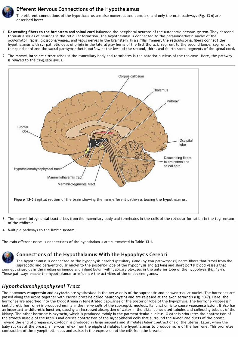
Efferent Nervous Connections of the HypothalamusThe efferent connections of the hypothalamus are also numerous and complex, and only the main pathways (Fig. 13-6) aredescribed here:
1. Descending fibers to the brainstem and spinal cord influence the peripheral neurons of the autonomic nervous system. They descendthrough a series of neurons in the reticular formation. The hypothalamus is connected to the parasympathetic nuclei of theoculomotor, facial, glossopharyngeal, and vagus nerves in the brainstem. In a similar manner, the reticulospinal fibers connect thehypothalamus with sympathetic cells of origin in the lateral gray horns of the first thoracic segment to the second lumbar segment ofthe spinal cord and the sacral parasympathetic outflow at the level of the second, third, and fourth sacral segments of the spinal cord.
2. The mammillothalamic tract arises in the mammillary body and terminates in the anterior nucleus of the thalamus. Here, the pathwayis relayed to the cingulate gyrus.
Figure 13-6 Sagittal section of the brain showing the main efferent pathways leaving the hypothalamus.
3. The mammillotegmental tract arises from the mammillary body and terminates in the cells of the reticular formation in the tegmentumof the midbrain.
4. Multiple pathways to the limbic system.
The main efferent nervous connections of the hypothalamus are summarized in Table 13-1.
Connections of the Hypothalamus With the Hypophysis CerebriThe hypothalamus is connected to the hypophysis cerebri (pituitary gland) by two pathways: (1) nerve fibers that travel from thesupraoptic and paraventricular nuclei to the posterior lobe of the hypophysis and (2) long and short portal blood vessels that
connect sinusoids in the median eminence and infundibulum with capillary plexuses in the anterior lobe of the hypophysis (Fig. 13-7).These pathways enable the hypothalamus to influence the activities of the endocrine glands.
Hypothalamohypophyseal TractThe hormones vasopressin and oxytocin are synthesized in the nerve cells of the supraoptic and paraventricular nuclei. The hormones arepassed along the axons together with carrier proteins called neurophysins and are released at the axon terminals (Fig. 13-7). Here, thehormones are absorbed into the bloodstream in fenestrated capillaries of the posterior lobe of the hypophysis. The hormone vasopressin(antidiuretic hormone) is produced mainly in the nerve cells of the supraoptic nucleus. Its function is to cause vasoconstriction. It also hasan important antidiuretic function, causing an increased absorption of water in the distal convoluted tubules and collecting tubules of thekidney. The other hormone is oxytocin, which is produced mainly in the paraventricular nucleus. Oxytocin stimulates the contraction ofthe smooth muscle of the uterus and causes contraction of the myoepithelial cells that surround the alveoli and ducts of the breast.Toward the end of pregnancy, oxytocin is produced in large amounts and stimulates labor contractions of the uterus. Later, when thebaby suckles at the breast, a nervous reflex from the nipple stimulates the hypothalamus to produce more of the hormone. This promotescontraction of the myoepithelial cells and assists in the expression of the milk from the breasts.

The supraoptic nucleus, which produces vasopressin, acts as an osmoreceptor. Should the osmotic pressure of the blood circulatingthrough the nucleus be too high, the nerve cells increase their production of vasopressin, and the antidiuretic effect of this hormone willincrease the reabsorption of water from the kidney. By this means, the osmotic pressure of the blood will return to normal limits.
Hypophyseal Portal SystemNeurosecretory cells situated mainly in the medial zone of the hypothalamus are responsible for the production of thereleasing hormones and release-inhibitory hormones. The hormones are packaged into granules and are transported along the axons ofthese cells into the median eminence and infundibulum. Here, the granules are released by exocytosis onto fenestrated capillaries at theupper end of the hypophyseal portal system.
Figure 13-7 A: Hypothalamohypophyseal tract. B: Hypophyseal portal system.
The hypophyseal portal system is formed on each side from the superior hypophyseal artery, which is a branch of the internal carotidartery (Fig. 13-7). The artery enters the median eminence and divides into tufts of capillaries. These capillaries drain into long and shortdescending vessels that end in the anterior lobe of the hypophysis by dividing into vascular sinusoids that pass between the secretory cellsof the anterior lobe.
The portal system carries the releasing hormones and the release-inhibiting hormones to the secretory cells of the anterior lobe of thehypophysis. The releasing hormones stimulate the production and release of adrenocorticotropic hormone (ACTH), follicle-stimulatinghormone (FSH), luteinizing hormone (LH), thyrotropic hormone or thyroid-stimulating hormone (TSH), and growth hormone (GH). Therelease of inhibiting hormones inhibits the release of the melanocyte-stimulating hormone (MSH) and luteotropic hormone (LTH).Luteotropic hormone (also known as the lactogenic hormone or prolactin) stimulates the corpus luteum to secrete progesterone and themammary gland to produce milk. The growth hormone inhibitory hormone (somatostatin) inhibits the release of growth hormone. Asummary of the hypothalamic releasing and inhibitory hormones and their effects on the anterior lobe of the hypophysis are shown inTable 13-2.
The neurons of the hypothalamus that are responsible for the production of the releasing hormones and the release-inhibiting hormonesare influenced by the afferent fibers passing to the hypothalamus. They also are influenced by the level of the hormone produced by thetarget organ controlled by the hypophysis. Should the level of thyroxine in the blood fall, for example, then the releasing factor for thethyrotropic hormone would be produced in increased quantities. Table 13-3 summarizes the presumed nuclear origin of the pituitaryreleasing and inhibitory hormones in the hypothalamus.
Functions of the HypothalamusTable 13-4 summarizes the functions of the main hypothalamic nuclei.
Autonomic Control
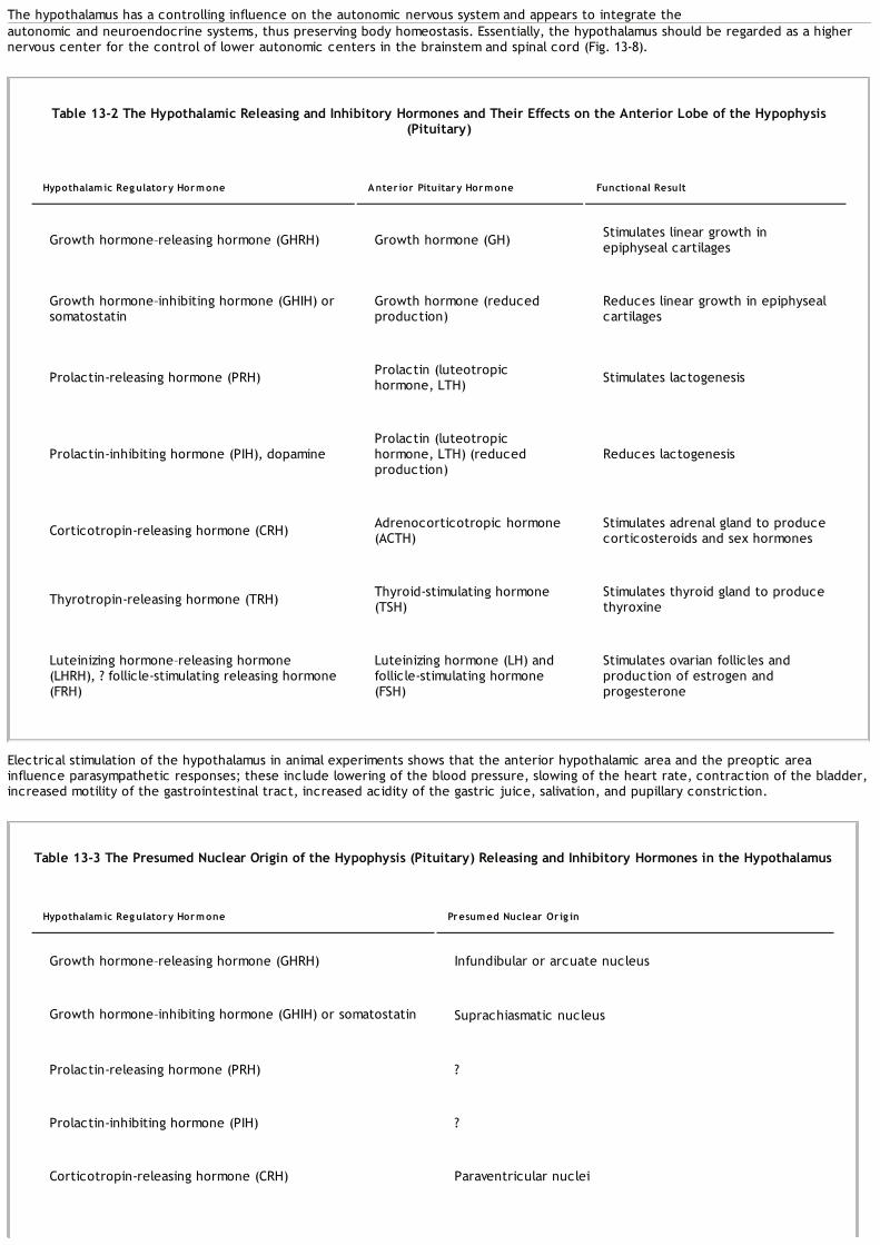
The hypothalamus has a controlling influence on the autonomic nervous system and appears to integrate theautonomic and neuroendocrine systems, thus preserving body homeostasis. Essentially, the hypothalamus should be regarded as a highernervous center for the control of lower autonomic centers in the brainstem and spinal cord (Fig. 13-8).
Table 13-2 The Hypothalamic Releasing and Inhibitory Hormones and Their Effects on the Anterior Lobe of the Hypophysis(Pituitary)
Hypothalamic Reg ulatory Hormone Anter ior Pituitary Hormone Functional Result
Growth hormone–releasing hormone (GHRH) Growth hormone (GH) Stimulates linear growth inepiphyseal cartilages
Growth hormone–inhibiting hormone (GHIH) orsomatostatin
Growth hormone (reducedproduction)
Reduces linear growth in epiphysealcartilages
Prolactin-releasing hormone (PRH) Prolactin (luteotropichormone, LTH) Stimulates lactogenesis
Prolactin-inhibiting hormone (PIH), dopamineProlactin (luteotropichormone, LTH) (reducedproduction)
Reduces lactogenesis
Corticotropin-releasing hormone (CRH) Adrenocorticotropic hormone(ACTH)
Stimulates adrenal gland to producecorticosteroids and sex hormones
Thyrotropin-releasing hormone (TRH) Thyroid-stimulating hormone(TSH)
Stimulates thyroid gland to producethyroxine
Luteinizing hormone–releasing hormone(LHRH), ? follicle-stimulating releasing hormone(FRH)
Luteinizing hormone (LH) andfollicle-stimulating hormone(FSH)
Stimulates ovarian follicles andproduction of estrogen andprogesterone
Electrical stimulation of the hypothalamus in animal experiments shows that the anterior hypothalamic area and the preoptic areainfluence parasympathetic responses; these include lowering of the blood pressure, slowing of the heart rate, contraction of the bladder,increased motility of the gastrointestinal tract, increased acidity of the gastric juice, salivation, and pupillary constriction.
Table 13-3 The Presumed Nuclear Origin of the Hypophysis (Pituitary) Releasing and Inhibitory Hormones in the Hypothalamus
Hypothalamic Reg ulatory Hormone Presumed Nuclear Or ig in
Growth hormone–releasing hormone (GHRH) Infundibular or arcuate nucleus
Growth hormone–inhibiting hormone (GHIH) or somatostatin Suprachiasmatic nucleus
Prolactin-releasing hormone (PRH) ?
Prolactin-inhibiting hormone (PIH) ?
Corticotropin-releasing hormone (CRH) Paraventricular nuclei

Thyrotropin-releasing hormone (TRH) Paraventricular and dorsomedial nuclei and adjacent areas
Luteinizing hormone–releasing hormone (LHRH) Preoptic and anterior nuclei
Stimulation of the posterior and lateral nuclei causes sympathetic responses, which include elevation of blood pressure, acceleration ofthe heart rate, cessation of peristalsis in the gastrointestinal tract, pupillary dilation, and hyperglycemia. These responses would lead oneto believe that in the hypothalamus, there exist areas that might be termed parasympathetic and sympathetic centers. However, it hasbeen shown that considerable overlap of function occurs in these areas.
Endocrine ControlThe nerve cells of the hypothalamic nuclei, by producing the releasing factors or release-inhibiting factors (Table 13-2),control the hormone production of the anterior lobe of the hypophysis (pituitary gland). The anterior lobe hormones include growthhormone, prolactin (luteotropic hormone), adrenocorticotropic hormone, thyroid-stimulating hormone, luteinizing hormone, andfollicle-stimulating hormone. Some of these hormones act directly on body tissues, while others, such as adrenocorticotropic hormone,act through an endocrine organ, which in turn produces additional hormones that influence the activities of general body tissues. Itshould be pointed out that each stage is controlled by negative and positive feedback mechanisms.
Table 13-4 Functions of the Main Hypothalamic Nuclei
Hypothalamic Nucleus Presumed Function
Supraoptic nucleus Synthesizes vasopressin (antidiuretic hormone)
Paraventricular nucleus Synthesizes oxytocin
Preoptic and anterior nuclei Control parasympathetic system
Posterior and lateral nuclei Control sympathetic system
Anterior hypothalamic nuclei Regulate temperature (response to heat)
Posterior hypothalamic nuclei Regulate temperature (response to cold)
Lateral hypothalamic nuclei Initiate eating and increase food intake (hunger center)
Medial hypothalamic nuclei Inhibit eating and reduce food intake (satiety center)
Lateral hypothalamic nuclei Increase water intake (thirst center)
Suprachiasmatic nucleus Controls circadian rhythms

Figure 13-8 Diagram depicting the hypothalamus as the chief center of the brain for controlling the internal milieu of the body.
NeurosecretionThe secretion of vasopressin and oxytocin by the supraoptic and paraventricular nuclei is discussed on page 388.
Temperature RegulationThe anterior portion of the hypothalamus controls those mechanisms that dissipate heat loss. Experimental stimulation of this area causesdilatation of skin blood vessels and sweating, which lower the body temperature. Stimulation of the posterior portion of the hypothalamusresults in vasoconstriction of the skin blood vessels and inhibition of sweating; there also may be shivering, in which the skeletal musclesproduce heat.
Normally, the hypothalamus sets the body temperature at 98.0°F to 98.6°F when measured orally and 1° higher when measured rectally.The temperature set can be altered in response to extremes, such as in environmental temperatures or in infection.
Regulation of Food and Water IntakeStimulation of the lateral region of the hypothalamus initiates the feeling of hunger and results in an increase in food intake. This lateralregion sometimes is referred to as the hunger center. Bilateral destruction of this center results in anorexia, with the consequent loss inbody weight. Stimulation of the medial region of the hypothalamus inhibits eating and reduces food intake. This area is referred to as thesatiety center. Bilateral destruction of the satiety center produces an uncontrolled voracious appetite, causing extreme obesity.
Experimental stimulation of other areas in the lateral region of the hypothalamus causes an immediate increase in the desire to drinkwater; this area is referred to as the thirst center. In addition, the supraoptic nucleus of the hypothalamus exerts a careful control onthe osmolarity of the blood through the secretion of vasopressin (antidiuretic hormone) by the posterior lobe of the hypophysis. Thishormone causes a great increase in the reabsorption of water in the distal convoluted tubules and collecting tubules of the kidneys.
Emotion and BehaviorEmotion and behavior are a function of the hypothalamus, the limbic system, and the prefrontal cortex. Some authorities believe that thehypothalamus is the integrator of afferent information received from other areas of the nervous system and brings about the physicalexpression of emotion; it can produce an increase in the heart rate, elevate the blood pressure, cause dryness of the mouth, flushing orpallor of the skin, and sweating. As well, it can often produce a massive peristaltic activity of the gastrointestinal tract.
Stimulation of the lateral hypothalamic nuclei may cause the symptoms and signs of rage, whereas lesions of these areas may lead topassivity. Stimulation of the ventromedial nucleus may cause passivity, whereas lesions of this nucleus may lead to rage.
Control of Circadian RhythmsThe hypothalamus controls many circadian rhythms, including body temperature, adrenocortical activity, eosinophil count, and renalsecretion. Sleeping and wakefulness, although dependent on the activities of the thalamus, the limbic system, and the reticular activatingsystem, are also controlled by the hypothalamus. Lesions of the anterior part of the hypothalamus seriously interfere with the rhythm ofsleeping and waking. The suprachiasmatic nucleus, which receives afferent fibers from the retina, appears to play an important role incontrolling the biologic rhythms. Nerve impulses generated in response to variations in the intensity of light are transmitted via thisnucleus to influence the activities of many of the hypothalamic nuclei.
Clinical NotesGeneral ConsiderationsIn summary, the activities of the hypothalamus are modified by information received along numerous afferent
pathways from different parts of the central nervous system (especially from the limbic system and the prefrontalcortex) and by the plasma levels of circulating hormones. It exerts its influence on bodily functions through theautonomic nervous system and the endocrine system.Although small, the hypothalamus should not be interpreted as a structure of little importance. It is the chief center ofthe brain for maintaining the internal milieu of the body (Fig. 13-8). There is hardly a tissue in the body that escapes itsinfluence.The connections of the hypothalamus are extremely complicated, and only the major pathways should be committed tomemory for use in clinical work.
Clinical Disorders Associated With Hypothalamic LesionsThe hypothalamus may be the site of inflammation, neoplasm, or vascular disorder. Because of its deep-seated centralposition, it can be pressed on by tumors of the surrounding brain tissue or may be compressed as the result of thedevelopment of internal hydrocephalus. Its widespread influence on many homeostatic and behavioral functions

P.394
means that a lesion of the hypothalamus will produce a large number of different syndromes. Thus, it is important toremember that an acute lesion is more likely to produce signs and symptoms than is a slowly growing tumor.
Obesity and WastingSevere obesity can occur as the result of hypothalamic lesions. It is generally associated with genital hypoplasia oratrophy.Wasting is less common than obesity in hypothalamic disease. Severe cachexia is suggestive of damage to thehypophysis (pituitary gland).
Sexual DisordersIn children, there may be sexual retardation and, rarely, sexual precocity with hypothalamic lesions. After puberty, thepatient with hypothalamic disease may have impotence or amenorrhea.
Hyperthermia and HypothermiaHyperthermia can follow lesions of the hypothalamus caused by head injury or following surgical operations in theregion of the hypothalamus. The patient with hyperthermia is otherwise normal and has no signs of malaise, whichoccurs with pyrexia secondary to infections. Hypothermia also can follow a lesion of the hypothalamus.
Diabetes InsipidusDiabetes insipidus results from a lesion of the supraoptic nucleus or from the interruption of the nervous pathway tothe posterior lobe of the hypophysis. Characteristically, the patient passes large volumes of urine of low specificgravity. As a result, the patient is extremely thirsty and drinks large quantities of fluids. The condition must bedistinguished from diabetes mellitus, in which there is glucosuria.
Disturbances of SleepThe occurrence of either frequent short periods of sleep during the waking hours or insomnia has been observed inpatients with hypothalamic lesions.
Emotional DisordersAttacks of unexplained weeping or laughter, uncontrollable rage, depressive reactions, and even maniacal outburstsall have been observed in patients with hypothalamic lesions.
Clinical Problem Solving1. A 17-year-old boy was admitted into the medical ward for observation. The tentative diagnosis was Fröhlichsyndrome. He had a 3-month history of severe headaches. More recently, he had had attacks of vomiting, and 1 weekago he had noticed problems with his eyesight. The patient said that he had difficulty seeing objects on the lateralside of both eyes. His parents were concerned that he was putting on weight, as he was especially fat over the lowerpart of the trunk. On physical examination, the boy was found to be 6 feet 3 inches tall; he had excessive trunkobesity. The testes and penis were small, and pubic and axillary hair was absent. A lateral radiograph of the skullshowed enlargement of the sella turcica with erosion of the dorsum sellae. An examination of the eye fields confirmedthat the patient had partial bitemporal hemianopia. Using your knowledge of neuroanatomy, explain the symptomsand signs of this patient.View Answer
2. A 40-year-old woman was involved in an automobile accident in which she sustained severe head injuries. Followinga slow but uneventful recovery, she was released from the hospital without any residual signs or symptoms. Sixmonths later, the patient started to complain of frequency of micturition and was passing very large quantities of paleurine. She also said that she always seemed thirsty and would often drink 10 glasses of water in one morning. Usingyour knowledge of neuroanatomy and neurophysiology, do you think there is any connection between the urinarysymptoms and her automobile accident?View Answer
3. Do you think it is possible that a patient with hydrocephalus could have a malfunctioning hypothalamus? If so,explain the connection.View Answer
4. Sherrington once stated in a scientific publication in 1947 that the hypothalamus should be regarded as the “headganglion” of the autonomic nervous system. What is the relationship that exists between the hypothalamus and theautonomic nervous system?View Answer
5. Explain what is meant by the terms hypothalamohypophyseal tract and hypophyseal portal system.View Answer
Review QuestionsDirections: Each of the numbered items in this section is followed by answers. Select the ONE lettered answerthat is CORRECT.1. The following statements concern the hypothalamus:
(a) It lies below the thalamus in the tectum of the midbrain.(b) It is not related to the limbic system.(c) The nuclei of the hypothalamus are divided by an imaginary plane formed by the columns of the fornix and the

mammillothalamic tract into medial and lateral groups.(d) The suprachiasmatic nucleus does not receive nerve fibers from the retina.(e) The lateral boundary of the hypothalamus is formed by the external capsule.
View Answer
2. The following statements concern the hypothalamus:(a) When seen from the inferior aspect, the hypothalamus is related to the following structures, from anterior toposterior: (i) the olfactory stria, (ii) the anterior perforated substance, and (iii) the mammillary bodies.(b) The margins of the different nuclei can be clearly seen with the naked eye.(c) The mammillary body does not overlap the medial and lateral groups of hypothalamic nuclei.(d) The preoptic area of the hypothalamus is located between the septum pellucidum and the optic chiasma.(e) The blood-brain barrier is absent in the median eminence of the hypothalamus, thus permitting the neuronsto sample the chemical content of the plasma directly.
View Answer
3. The following statements concern the afferent fibers passing to the hypothalamus:(a) Fibers pass from the hippocampus to the mammillary bodies, bringing information from the auditory system.(b) Olfactory impulses reach the hypothalamus through the lateral forebrain bundle.(c) The hypothalamus receives many afferent fibers from the viscera via the reticular formation.(d) The dorsomedial nucleus receives axons from the posterior lobe of the pituitary.(e) The pineal gland sends fibers via the habenular commissure to the hypothalamus.
View Answer
4. The following statements concern the hypothalamus:(a) Somatic efferent fibers leave the hypothalamic nuclei via the medial and spinal lemnisci.(b) It does not integrate the autonomic and neuroendocrine systems.(c) The posterior portion of the hypothalamus controls those mechanisms that dissipate heat loss.(d) The nerve cells of the hypothalamus produce releasing and release-inhibiting hormones that control theproduction of various hormones in the anterior lobe of the hypophysis.(e) The hunger center is probably located in the posterior hypothalamic nuclei.
View Answer
5. The following statements concern the functional activities of the hypothalamus:(a) The hypothalamus brings about the physical changes associated with emotion, such as increased heart rateand flushing or pallor of the skin.(b) The medial hypothalamic nuclei are concerned with fluid intake.(c) The corticotropin-releasing hormone (CRH) is produced in the anterior nucleus of the hypothalamus.(d) The suprachiasmatic nucleus plays no part in controlling circadian rhythms.(e) The hypothalamus controls the lower autonomic centers by means of pathways through the tectospinal tract.
View Answer
6. The following statements concern the hypothalamohypophyseal tract:(a) Oxytocin inhibits the contraction of the smooth muscle of the uterus.(b) The nerve cells of the supraoptic and paraventricular nuclei produce the hormones vasopressin and oxytocin.(c) The hormones travel in lymph vessels with protein carriers called neurophysins.(d) Vasopressin stimulates the proximal convoluted tubules of the kidney, causing increased absorption of waterfrom the urine.(e) The hormones are absorbed into the bloodstream in the capillaries of the anterior lobe of the hypophysis.
View Answer
7. The following statements concern the hypophyseal portal system:(a) It carries releasing hormones and release-inhibiting hormones to the secretory cells of the anterior lobe ofthe hypophysis.(b) The production of the releasing hormones and the release-inhibiting hormones cannot be influenced by thelevel of the hormone produced by the target organ controlled by the hypophysis.(c) The blood vessels commence superiorly in the median eminence and end inferiorly in the vascular sinusoids ofthe posterior lobe of the hypophysis cerebri.(d) Efferent nerve fibers leaving the hypothalamus influence the production of the releasing hormones by nervecells.(e) The neuroglial cells of the hypothalamus are responsible for the production of the release-inhibitinghormones.
View Answer

Additional Reading
Boulant, J. A. Hypothalamic neurons regulating body temperature. In Handbook of Physiology. Section 4: Environmental Physiology(pp. 105–126). Oxford: Oxford University Press, 1997.
Bouret, S. G., and Simerly, R. B. Leptin and development of hypothalamic feeding circuits. Endocrinology 145:2621, 2004.
Buijs, R. M. Vasopressin and oxytocin—Their role in neurotransmission. Pharmacol. Ther. 22:127, 1983.
Buijs, R. M., Kalsbeek, A., Romijn, H. J., Pennertz, C. M., and Mirmiran, M. (eds.). Hypothalamic Integration of Circadian Rhythms.Amsterdam: Elsevier, 1997.
Craig, C. R., and Stitzel, R. E. Modern Pharmacology (4th ed.). Boston: Little, Brown, 1994.
Engler, D., Redei, E., and Kola, I. The corticotropin-release inhibitory factor hypothesis: A review of the evidence for the existenceof inhibitory as well as stimulatory hypophysiotropic regulation of adrenocorticotropin secretion and biosynthesis. Endocr. Rev.20:460, 1998.
Ganten, D., and Pfaff, D. Morphology of Hypothalamus and Its Connections. Berlin: Springer-Verlag, 1986.
Gerashchenko, D., and Shiromani, P. J. Different neuronal phenotypes in the lateral hypothalamus and their role in sleep wakefulness.Mol. Neurobiol. 29:41, 2004.
Guyton, A. C., and Hall, J. E. Textbook of Medical Physiology (11th ed.). Philadelphia: Elsevier, Saunders, 2006.
Halford, J. C., and Blundell, J. E. Pharmacology of appetite suppression. Prog. Drug Res. 54:25, 2000.
Long, G., Brown, C. H., and Russell, J. A. Physiological pathways regulating the activity of magnocellular neurosecretory cells. Prog.Neurobiol. 57:625, 1999.
Phelps, E. A. Human emotion and memory: Interactions of amygdala and hippocampal complex. Curr. Opin. Neurobiol. 14:198, 2004.
Standing, S. (ed.). Gray's Anatomy (39th Br. ed.). London: Elsevier Churchill Livingstone, 2005.
Swaab, D. F., Hofman, M. A., M irmiran, M., Ravid, R., and Van Leewen, F. (eds.). The Human Hypothalamus in Health and Disease.Amsterdam: Elsevier, 1993.
Swanson, L. W., and Sawchenko, P. E. Hypothalamic integration: Organization of the paraventricular and supraoptic nuclei. Annu.Rev. Neurosci. 6:269, 1983.

Authors: Snell, Richard S.Title: Clinical Neuroanatomy, 7th Edition
Copyright ©2010 Lippincott Williams & Wilkins
> Table of Contents > Chapter 14 - The Autonomic Ner vous System
Chapter 14
The Autonomic Nervous System
A 46-year-old man who had recently undergone right-sided pneumonectomy for carcinoma of the bronchus was seen by his thoracicsurgeon for follow-up after the operation. The patient said that he felt surprisingly fit and was gaining some of the weight that he had lostprior to the operation. His wife commented that about 1 week ago, the upper lid of his right eye tended to droop slightly when he wastired at the end of the day.
After a careful physical examination, the surgeon noticed that in addition to the ptosis of the right eye, the patient's right pupil wasconstricted and that his face was slightly flushed on the right side. Further examination revealed that the skin on the right side of theface appeared to be warmer and drier than normal. Palpation of the deep cervical group of lymph nodes revealed a large, hard, fixed nodejust above the right clavicle.
Based on his clinical findings, the surgeon made the diagnosis of a right-sided Horner syndrome. These findings were not present beforethe operation. The presence of the enlarged right-sided deep cervical lymph node indicated that the bronchial carcinoma hadmetastasized to the lymph node in the neck and was spreading to involve the cervical part of the sympathetic trunk on the right side. Thisobservation would explain the abnormal eye and facial skin findings.
Knowledge of the sympathetic innervation of the structures of the head and neck enabled the surgeon to make an accurate diagnosis inthis patient.
Chapter Objectives
To understand the structure, physiology, and pharmacology of the autonomic nervous system
The autonomic nervous system and the endocrine system control the internal environment of the body. It is the autonomic nervous systemthat provides a fine discrete control over the functions of many organs and tissues, including heart muscle, smooth muscle, and theexocrine glands. The endocrine system, by means of its blood-borne hormones, exerts a slower more diffuse control.
The autonomic nervous system, like the somatic nervous system, has afferent, connector, and efferent neurons. The afferent impulsesoriginate in visceral receptors and travel via afferent pathways to the central nervous system, where they are integrated throughconnector neurons at different levels and then leave via efferent pathways to visceral effector organs. The majority of the activities of theautonomic system do not impinge on consciousness.
The efferent pathways of the autonomic system are made up of preganglionic and postganglionic neurons. The cell bodies of thepreganglionic neurons are situated in the lateral gray column of the spinal cord and in the motor nuclei of the 3rd, 7th, 9th, and 10thcranial nerves. The axons of these cell bodies synapse on the cell bodies of the postganglionic neurons that are collected together toform ganglia outside the central nervous system.
The control exerted by the autonomic system is extremely rapid; it is also widespread, since one preganglionic axon may synapse withseveral postganglionic neurons. Large collections of afferent and efferent fibers and their associated ganglia form autonomic plexuses inthe thorax, abdomen, and pelvis.
The visceral receptors include chemoreceptors, baroreceptors, and osmoreceptors. Pain receptors are present in viscera, and certaintypes of stimuli, such as lack of oxygen or stretch, can cause extreme pain.
The information provided in this chapter is extensively used in clinical practice. The examples of autonomic innervations given areimportant and are commonly used by examiners to construct good questions.
Organization of the Autonomic Nervous SystemThe autonomic nervous system is distributed throughout the central and peripheral nervous systems. It is divided into two parts,the sympathetic and the parasympathetic and, as emphasized earlier, consists of both afferent and efferent fibers. This division
between sympathetic and parasympathetic is made on the basis of anatomical differences, differences in the neurotransmitters, anddifferences in the physiologic effects.
Both the sympathetic and parasympathetic divisions produce opposite effects in most organs and are thus considered as physiologicantagonists. However, it must be stated that both divisions operate in conjunction with one another, and it is the balance in the activitiesthat maintains a stable internal environment.
Sympathetic Part of the Autonomic SystemThe sympathetic system is the larger of the two parts of the autonomic system and is widely distributed throughout the body, innervatingthe heart and lungs, the muscle in the walls of many blood vessels, the hair follicles and the sweat glands, and many abdominopelvicviscera.
The function of the sympathetic system is to prepare the body for an emergency. The heart rate is increased, arterioles of the skin and

intestine are constricted, arterioles of skeletal muscle are dilated, and the blood pressure is raised. There is a redistribution of blood;thus, it leaves the skin and gastrointestinal tract and passes to the brain, heart, and skeletal muscle. In addition, the sympathetic nervesdilate the pupils; inhibit smooth muscle of the bronchi, intestine, and bladder wall; and close the sphincters. The hair is made to stand onend, and sweating occurs.
The sympathetic system consists of the efferent outflow from the spinal cord, two ganglionated sympathetic trunks, important branches,plexuses, and regional ganglia.
Efferent Nerve Fibers (Sympathetic Outflow)The lateral gray columns (horns) of the spinal cord from the first thoracic segment to the second lumbar segment (sometimes third lumbarsegment) possess the cell bodies of the sympathetic connector neurons (Fig. 14-1). The myelinated axons of these cells leave the cord inthe anterior nerve roots and pass via the white rami communicantes (the white rami are white because the nerve fibers are covered withwhite myelin) to the paravertebral ganglia of the sympathetic trunk. Once these fibers (preganglionic) reach the ganglia in thesympathetic trunk, they are distributed as follows:
1. They synapse with an excitor neuron in the ganglion (Figs. 14-1 and 14-2). The gap between the two neurons is bridged by theneurotransmitter acetylcholine (ACh). The postganglionic nonmyelinated axons leave the ganglion and pass to the thoracic spinalnerves as gray rami communicantes (the gray rami are gray because the nerve fibers are devoid of myelin). They are distributed inbranches of the spinal nerves to smooth muscle in the blood vessel walls, sweat glands, and arrector muscles of the hairs of the skin.
Figure 14-1 General arrangement of the somatic part of the nervous system (on left) compared with the autonomic part of thenervous system (on right).
2. They travel cephalad in the sympathetic trunk to synapse in ganglia in the cervical region (Fig. 14-2). The postganglionic nerve fiberspass via gray rami communicantes to join the cervical spinal nerves. Many of the preganglionic fibers entering the lower part of thesympathetic trunk from the lower thoracic and uppeAfferent Nerve Fibersr two lumbar segments of the spinal cord travel caudad tosynapse in ganglia in the lower lumbar and sacral regions. Here again, the postganglionic nerve fibers pass via gray rami communicantesto join the lumbar, sacral, and coccygeal spinal nerves (Fig. 14-2).
3. They may pass through the ganglia of the sympathetic trunk without synapsing. These myelinated fibers leave the sympathetic trunk asthe greater splanchnic, lesser splanchnic, and lowest or least splanchnic nerves. The greater splanchnic nerve is formed frombranches from the fifth to the ninth thoracic ganglia. It descends obliquely on the sides of the bodies of the thoracic vertebrae andpierces the crus of the diaphragm to synapse with excitor cells in the ganglia of the celiac plexus, the renal plexus, and thesuprarenal medulla. The lesser splanchnic nerve is formed from branches of the 10th and 11th thoracic ganglia. It descends with thegreater splanchnic nerve and pierces the diaphragm to join excitor cells in ganglia in the lower part of the celiac plexus. The lowestsplanchnic nerve (when present) arises from the 12th thoracic ganglion, pierces the diaphragm, and synapses with excitor neurons inthe ganglia of the renal plexus. The splanchnic nerves, therefore, are formed of preganglionic fibers. The postganglionic fibers arisefrom the excitor cells in the peripheral plexuses and are distributed to the smooth muscle and glands of the viscera. A fewpreganglionic fibers, traveling in the greater splanchnic nerve, end directly on the cells of the suprarenal medulla. These medullarycells, which may be regarded as modified sympathetic excitor neurons, are responsible for the secretion of epinephrine andnorepinephrine.
The ratio of preganglionic to postganglionic sympathetic fibers is about 1:10, permitting a wide control of involuntary structures.

Afferent Nerve FibersAfferent myelinated nerve fibers travel from the viscera through the sympathetic ganglia without synapsing. They pass to the spinal nervevia white rami communicantes and reach their cell bodies in the posterior root ganglion of the corresponding spinal nerve (Fig. 14-1). Thecentral axons then enter the spinal cord and may form the afferent component of a local reflex arc or ascend to higher centers, such asthe hypothalamus.
Sympathetic TrunksThe sympathetic trunks are two ganglionated nerve trunks that extend the whole length of the vertebral column (Fig. 14-2). In the neck,each trunk has 3 ganglia; in the thorax, 11 or 12; in the lumbar region, 4 or 5; and in the pelvis, 4 or 5. In the neck, the trunks lie anteriorto the transverse processes of the cervical vertebrae; in the thorax, they areanterior to the heads of the ribs or lie on the sides of the vertebral bodies; in the abdomen, they are anterolateral to the sides of thebodies of the lumbar vertebrae; and in the pelvis, they are anterior to the sacrum. Below, the two trunks end by joining together to forma single ganglion, the ganglion impar.
Figure 14-2 Efferent part of the autonomic nervous system. Preganglionic parasympathetic fibers are shown in solid blue, andpostganglionic parasympathetic fibers are shown in interrupted blue. Preganglionic sympathetic fibers are shown in solid red, andpostganglionic sympathetic fibers are shown in interrupted red.
Parasympathetic Part of the Autonomic SystemThe activities of the parasympathetic part of the autonomic system are directed toward conserving and restoring energy. The heart rate is

slowed, pupils are constricted, peristalsis and glandular activity is increased, sphincters are opened, and the bladder wall is contracted.
Efferent Nerve Fibers (Craniosacral Outflow)The connector nerve cells of the parasympathetic part of the autonomic nervous system are located in the brainstem and the sacralsegments of the spinal cord (Fig. 14-2).
Those nerve cells located in the brainstem form nuclei in the following cranial nerves: the oculomotor (parasympathetic or Edinger-Westphal nucleus), the facial (superior salivatory nucleus and lacrimatory nucleus), the glossopharyngeal (inferior salivatory nucleus), andthe vagus nerves (dorsal nucleus of the vagus). The axons of these connector nerve cells are myelinated and emerge from the brain withinthe cranial nerves.
The sacral connector nerve cells are found in the gray matter of the second, third, and fourth sacral segments of the spinal cord.These cells are not sufficiently numerous to form a lateral gray horn, as do the sympathetic connector neurons in the thoracolumbarregion. The myelinated axons leave the spinal cord in the anterior nerve roots of the corresponding spinal nerves. They then leave thesacral nerves and form the pelvic splanchnic nerves (Fig. 14-2).
The myelinated efferent fibers of the craniosacral outflow are preganglionic and synapse in peripheral ganglia located close to the viscerathey innervate. Here again, acetylcholine is the neurotransmitter. The cranial parasympathetic ganglia are the ciliary, pterygopalatine,submandibular, and otic (Fig. 14-2). In certain locations, the ganglion cells are placed in nerve plexuses, such as the cardiac plexus,pulmonary plexus, myenteric plexus (Auerbach plexus), and mucosal plexus (Meissner plexus); the last two plexuses are associated withthe gastrointestinal tract. The pelvic splanchnic nerves synapse in ganglia in the hypogastric plexuses. Characteristically, thepostganglionic parasympathetic fibers are nonmyelinated and of relatively short length compared with sympathetic postganglionic fibers.
The ratio of preganglionic to postganglionic fibers is about 1:3 or less, which is much more restricted than in the sympathetic part of thesystem.
Afferent Nerve FibersAfferent myelinated fibers travel from the viscera to their cell bodies, located either in the sensory ganglia of the cranial nerves or in theposterior root ganglia of the sacrospinal nerves. The central axons then enter the central nervous system and take part in the formationof local reflex arcs or pass to higher centers of the autonomic nervous system, such as the hypothalamus.
The afferent component of the autonomic system is identical to the afferent component of somatic nerves, and it forms part of thegeneral afferent segment of the entire nervous system. The nerve endings in the autonomic afferent component may not be activated bysuch sensations as heat or touch but rather by stretch or lack of oxygen. Once the afferent fibers gain entrance to the spinal cord orbrain, they are thought to travel alongside, or mixed with, the somatic afferent fibers.
The Large Autonomic PlexusesLarge collections of sympathetic and parasympathetic efferent nerve fibers and their associated ganglia, together with visceralafferent fibers, form autonomic nerve plexuses1 in the thorax, abdomen, and pelvis. Branches from these plexuses innervate the
viscera. In the thorax, there are the cardiac, pulmonary, and esophageal plexuses. In the abdomen, the plexuses are associated with theaorta and its branches, and subdivisions of these autonomic plexuses are named according to the branch of the aorta along which theyare lying: celiac, superior mesenteric, inferior mesenteric, and aortic plexuses. In the pelvis, there are the superior and inferiorhypogastric plexuses.
Autonomic GangliaThe autonomic ganglion is the site where preganglionic nerve fibers synapse on postganglionic neurons (Fig. 14-3). Ganglia aresituated along the course of efferent nerve fibers of the autonomic nervous system. Sympathetic ganglia form part of the
sympathetic trunk or are prevertebral in position (e.g., celiac and superior mesenteric ganglia). Parasympathetic ganglia, on the otherhand, are situated close to or within the walls of the viscera.
An autonomic ganglion consists of a collection of multipolar neurons together with capsular or satellite cells and a connective tissuecapsule. Nerve bundles are attached to each ganglion and consist of preganglionic nerve fibers that enter the ganglion, postganglionicnerve fibers that have arisen from neurons within the ganglion and are leaving the ganglion, and afferent and efferent nerve fibers thatpass through the ganglion without synapsing. The preganglionic fibers are myelinated, small, and relatively slow–conducting B fibers. Thepostganglionic fibers are unmyelinated, smaller, and slower-conducting C fibers.
The structure of synapses in autonomic ganglia shows the characteristic membrane thickening and small clear vesicles. In addition, thereare some larger granular vesicles. The smaller vesicles contain acetylcholine; the content of the granular vesicles is unknown.
Although an autonomic ganglion is the site where preganglionic fibers synapse on postganglionic neurons, the presence of smallinterneurons has been recognized. These cells exhibit catecholamine fluorescence and are referred to as small intensely fluorescent(SIF) cells. In some ganglia, these interneurons receive preganglionic cholinergic fibers, and they may modulate ganglionic transmission. Inother ganglia, they receive collateral branches and may serve some integrative function (Fig. 14-3). Many SIF cells contain dopamine, whichis thought to be their transmitter.
Preganglionic TransmittersAs the preganglionic nerve fibers approach their termination, they wind around and between the dendritic processes of thepostganglionic neuron, making multiple synaptic contacts. When the wave of excitation reaches the synaptic contacts, the
synaptic transmitter is liberated, crosses the synaptic cleft to reach the receptor, and excites the postganglionic neuron (Figs. 14-3B and14-4).
The synaptic transmitter that excites the postganglionic neurons in both sympathetic and parasympathetic ganglia is acetylcholine. Theaction of acetylcholine in autonomicganglia is quickly terminated by hydrolysis by acetylcholinesterase.

Figure 14-3 A: An autonomic ganglion as an integrator. Shows SIF cells. B: The liberation of acetylcholine (ACh) at an autonomicsynapse.
Acetylcholine ReceptorsAcetylcholine receptors are located on the outside of the cell membrane of postganglionic neurons. They are protein complexes that arebound to protein molecules that penetrate the cell membrane. Once the acetylcholine molecule binds with the receptor, the structureof the protein molecule of the cell membrane changes, and excitation or inhibition of the postganglionic neuron takes place. Two types ofacetylcholine receptors exist and are known as nicotinic and muscarinic receptors. These receptors are so named because nicotinicreceptors respond specifically to nicotine (from tobacco), and muscarinic receptors respond specifically to muscarine (poison fromtoadstools). Acetylcholine is capable of binding to both of these receptors.
In preganglionic neurons, both sympathetic and parasympathetic, the release of acetylcholine binds predominantly with the nicotinicreceptors on the postganglionic neurons.
Fast, Slow, and Inhibitory Synaptic PotentialsAcetylcholine activation of the postsynaptic nicotinic receptors on the dendrites and cell bodies of the postganglionic neuronsresults in a depolarization of the membrane, an influx of Na+ and Ca2+ ions, and the generation of the fast excitatory postsynaptic
potential (fast EPSP). Usually, several presynaptic axon terminals must fire simultaneously and summation has to occur for transmissionalong the postsynaptic axon to take place. The fast EPSP reaches a maximum within about 15 msec.
Acetylcholine is also believed to activate small numbers of postsynaptic muscarinic receptors. This results in the development of the slowexcitatory postsynaptic potential

(slow EPSP), which lasts for 2 to 5 seconds. The underlying mechanism is complicated, and the slow potential occurs when the Na+ andCa2+ channels are open and M-type K+ channels close; this leads to membrane depolarization. Late slow EPSP lasting as long as 1 to 2minutes can also be produced by neuropeptide transmitters.
Figure 14-4 Example of the release of acetylcholine from a presynaptic terminal. A single presynaptic stimulus evokes a fastexcitatory postsynaptic potential (fast EPSP) at a nicotinic receptor. Further stimulation may lead to a slow excitatory postsynapticpotential (slow EPSP) or a slow inhibitory postsynaptic potential (slow IPSP) at a muscarinic receptor. In preganglionic neurons,both sympathetic and parasympathetic, the release of acetylcholine binds predominantly with the nicotinic receptors on thepostganglionic neurons.
The activation of postsynaptic muscarinic receptors may also result in the development of the slow inhibitory postsynaptic potential (slowIPSP), which lasts about 10 seconds. The IPSP results from the opening of K+ channels, permitting K+ ions to flow out into the synapticspace, producing hyperpolarization.
The existence of these complex postsynaptic potentials in both sympathetic and parasympathetic ganglia (Fig. 14-4) illustrates how thepostsynaptic membrane potential can be altered and ganglionic transmission modulated.
Ganglion-Stimulating AgentsStimulating drugs, such as nicotine, lobeline, and dimethylphenylpiperazinium, stimulate sympathetic and parasympathetic gangliaby activating the nicotinic receptors on the postsynaptic membrane and producing a fast EPSP.
Ganglion-Blocking AgentsThere are two types of ganglion-blocking agents: depolarizing and nonpolarizing. Nicotine in high concentrations acts as ablocking agent by first stimulating the postganglionic neuron and causing depolarization and then by maintaining depolarization of
the excitable membrane. During this latter phase, the postganglionic neuron will fail to respond to the administration of any stimulant,regardless of the type of receptor it activates.
Hexamethonium and tetraethylammonium block ganglia by competing with acetylcholine at the nicotinic receptor sites.
Postganglionic Nerve EndingsThe postganglionic fibers terminate on the effector cells without special discrete endings. The axons run between the gland cellsand the smooth and cardiac muscle fibers and lose their covering of Schwann cells. At sites where transmission occurs, clusters of
vesicles are present within the axoplasm (see Fig. 3-37). The site on the axon may lie at some distance from the effector cell; thus, thetransmission time may be slow at these endings. The diffusion of the transmitter through the large extracellular distance also permits agiven nerve to have an action on a large number of effector cells.
Postganglionic TransmittersParasympathetic postganglionic nerve endings liberate acetylcholine as their transmitter substance (Fig. 14-5). All neurons thatrelease acetylcholine at their endings are called cholinergic (work like acetylcholine). The acetylcholine traverses the synaptic
cleft and binds reversibly with the cholinergic (muscarinic) receptor on the postsynaptic membrane. Within 2 to 3 msec, it is hydrolyzedinto acetic acid and choline by the enzyme acetylcholinesterase, which is located on the surface of the nerve and receptor membranes.The choline is reabsorbed into the nerve ending and used again for synthesis of acetylcholine.
Most sympathetic postganglionic nerve endings liberate norepinephrine2 as their transmitter substance. In addition, some sympathetic

postganglionic nerve endings, particularly those that end on cells of sweat glands and the blood vessels in skeletal muscle, releaseacetylcholine, which binds with muscarinic receptors on the postsynaptic membrane.
Figure 14-5 Efferent parts of the autonomic nervous system and the chemical transmitter substances released at the nerveendings. (ACh, acetylcholine; N. ep., norepinephrine.)
Sympathetic endings that use norepinephrine are called adrenergic endings. There are two major kinds of receptors in the effectororgans, called alpha and beta receptors.
Two subgroups of alpha receptors (alpha-1 and alpha-2 receptors) and two subgroups of beta receptors (beta-1 and beta-2 receptors) havebeen described. Norepinephrine has a greater effect on alpha receptors than on beta receptors. Phenylephrine is a pure alphastimulator. The bronchodilating drugs, such as metaproterenol and albuterol, mainly act on beta-2 receptors. As a general rule, alphareceptor sites are associated with most of the excitatory functions of the sympathetic system (e.g., smooth muscle contraction,vasoconstriction, diaphoresis), whereas the beta receptor sites are associated with most of the inhibitory functions (e.g., smooth musclerelaxation). Beta-2 receptors are mainly in the lung, and stimulation results in bronchodilatation. Beta-1 receptors are in the myocardium,where they are associated with excitation.
The action of norepinephrine on the receptor site of the effector cell is terminated by reuptake into the nerve terminal, where it isstored in presynaptic vesicles for reuse. Some of the norepinephrine escapes from the synaptic cleft into the general circulation and issubsequently metabolized in the liver.
Other Postganglionic TransmittersSympathetic and parasympathetic postganglionic neurons have been shown to liberate substances other than acetylcholine ornorepinephrine at their endings; these include adenosine triphosphate (ATP), neuropeptide Y, and substance P. These
substances may be released alone or from neurons that release acetylcholine or norepinephrine; they have their own specific receptors.The function of these transmitters is probably to modulate the effects of the primary transmitter.
Blocking of Cholinergic ReceptorsIn the case of the parasympathetic and the sympathetic postganglionic nerve endings that liberate acetylcholine as thetransmitter substance, the receptors on the effector cells are muscarinic. This means that the action can be blocked by
atropine. Atropine competitively antagonizes the muscarinic action by occupying the cholinergic receptor sites on the effector cells.
Blocking of Adrenergic ReceptorsThe alpha-adrenergic receptors can be blocked by agents such as phenoxybenzamine, and the beta-adrenergic receptors can beblocked by agents such as propranolol. The synthesis and storage of norepinephrine at sympathetic endings can be inhibited by

reserpine.
Figure 14-6 Hypothalamus as the control center for the autonomic nervous system and the neuroendocrine system.
Higher Control of the Autonomic Nervous SystemThe hypothalamus has a controlling influence on the autonomic nervous system and appears to integrate the autonomic andneuroendocrine systems, thus preserving body homeostasis (Fig. 14-6). Essentially, the hypothalamus should be regarded as a
higher nervous center for the control of lower autonomic centers in the brainstem and spinal cord.
Stimulation of the anterior region of the hypothalamus can influence parasympathetic responses, whereas stimulation of the posterior partof the hypothalamus gives rise to sympathetic responses. In addition, lower brainstem centers such as vasopressor, vasodilator,cardioaccelerator, cardiodecelerator, and respiratory centers have been found in the reticular formation as the result of experimentalstimulation in lower animals. It is believed that the various levels of control are exerted as the result of interconnections of the differentregions by ascending and descending pathways. The neurons of the thoracolumbar outflow of the sympathetic part of the system and theneurons of the craniosacral outflow of the parasympathetic part of the system receive their control through the descending tracts of thereticular formation.
Stimulation of different parts of the cerebral cortex and the limbic system produces autonomic effects, and it is assumed that this isbrought about through the hypothalamus. The autonomic nervous system can be brought under voluntary control to some extent. This isseen, for example, in young individuals who may blush easily when embarrassed. As they mature, they are usually able to consciously trainthemselves to control this response. It should also be noted that the higher centers of the brain can abnormally influence the activities ofthe autonomic nervous system and induce disease such as cardiac palpitations (arrhythmias) and even myocardial infarction.
The “Enteric Nervous System”Two important plexuses of nerve cells and fibers extend continuously along and around the length of the gastrointestinal tractfrom the esophagus to the anal canal. The submucous or Meissner plexus lies between the mucous membrane and the circular
muscle layer, and the myenteric or Auerbach plexus lies between the circular and longitudinal muscle layers. The submucous plexus ismainly concerned with the control of the glands of the mucous membrane, whereas the myenteric plexus controls the muscle andmovements of the gut wall.
No longer can these plexuses be regarded as simply parasympathetic plexuses containing preganglionic and postganglionic nerve fibers andnerve cells. It has been shown that contractions of the smooth muscle in the gut wall can occur in the absence of the myenteric plexus,but the coordinated purposeful contractions, as seen with peristalsis and segmental movements, require the presence of a nerve plexus,even though this may be isolated from the central nervous system.
Different types of neurons have been recognized in the plexuses. Some neurons are bipolar or unipolar and are thought to be sensory andinvolved in local reflex activity; other neurons send axons to the celiac and mesenteric plexuses. Preganglionic parasympathetic fiberssynapse on nerve cells that give rise to postganglionic fibers that innervate the smooth muscle and glands. Postganglionic sympatheticfibers have been seen to terminate on parasympathetic nerve cells and probably exert an inhibitory role in parasympathetic activity.Internuncial neurons are also present. It is interesting to note that the nerve cells and their processes are surrounded by neuroglialikecells that closely resemble astrocytes in the central nervous system. It has been suggested that while the enteric plexuses can coordinatethe activities of the gut wall, the parasympathetic and sympathetic inputs modulate these activities.
Functions of the Autonomic Nervous SystemThe autonomic nervous system, along with the endocrine system, maintains body homeostasis. The endocrine control is slowerand exerts its influence by means of blood-borne hormones.
The autonomic nervous system functions for the most part at the subconscious level. We are not aware, for example, that our pupils aredilating or that our arteries are constricting. The system should not be regarded as an isolatedportion of the nervous system, for it is known that it can play a role with somatic activity in expressing emotion and that certainautonomic activities, such as micturition, can be brought under voluntary control. The various activities of the autonomic and endocrinesystems are integrated within the hypothalamus.

Figure 14-7 This man is making good use of the sympathetic part of his autonomic nervous system.
The sympathetic and parasympathetic components of the autonomic system cooperate in maintaining the stability of the internalenvironment. The sympathetic part prepares and mobilizes the body in an emergency (Fig. 14-7), when there is sudden severe exercise,fear, or rage. The parasympathetic part aims at conserving and storing energy, for example, in the promotion of digestion and theabsorption of food by increasing the secretions of the glands of the gastrointestinal tract and stimulating peristalsis (Fig. 14-8).
The sympathetic and parasympathetic parts of the autonomic system usually have antagonistic control over a viscus. For example, thesympathetic activity will increase the heart rate, whereas the parasympathetic activity will slow the heart rate. The sympathetic activitywill make the bronchial smooth muscle relax, but the muscle is contracted by parasympathetic activity.
Figure 14-8 There is nothing like a good, large meal and a comfortable armchair to facilitate the activities of the parasympatheticpart of the autonomic nervous system.
It should be pointed out, however, that many viscera do not possess this fine dual control from the autonomic system. For example, thesmooth muscle of the hair follicles (the arrector pili muscle) is made to contract by the sympathetic activity, and there is noparasympathetic control.
The activities of some viscera are kept under a constant state of inhibition by one or the other components of the autonomic nervoussystem. The heart in a trained athlete is maintained at a slow rate by the activities of the parasympathetic system. This is of considerableimportance, because the heart is a more efficient pump when contracting slowly than when contracting very quickly, thus permittingadequate diastolic filling of the ventricles.
Important Anatomical, Physiologic, and Pharmacologic Differences Between theSympathetic and Parasympathetic Parts of the Autonomic Nervous System (Table 14-1)
1. The sympathetic efferent nerve fibers originate (Fig. 14-2) from nerve cells in the lateral gray column of the spinal cord between thefirst thoracic and second lumbar segments (the thoracic outflow). The parasympathetic efferent nerve fibers originate from nerve cellsin the 3rd, 7th, 9th, and 10th cranial nerves and in the gray matter of the 2nd, 3rd, and 4th sacral segments of the cord (thecraniosacral outflow).

2. The sympathetic ganglia are located either in the paravertebral sympathetic trunks or in the prevertebral ganglia, such as the celiacganglion (Fig. 14-2). The parasympathetic ganglion cells are located as small ganglia close to the viscera or within plexuses within theviscera.
Table 14-1 Comparison of Anatomical, Physiologic, and Pharmacologic Characteristics of the Sympathetic and ParasympatheticParts of the Autonomic Nervous System
Sympathetic Parasympathetic
Action Prepares body for emergency Conserves and restores energy
Outflow T1-L2 (3) Cranial nerves III, VII, IX, and X; S2–4
Preganglionic fibers Myelinated Myelinated
GangliaParavertebral (sympathetic trunks),prevertebral (e.g., celiac, superiormesenteric, inferior mesenteric)
Small ganglia close to viscera (e.g.,otic, ciliary) or ganglion cells inplexuses (e.g., cardiac, pulmonary)
Neurotransmitter withinganglia Acetylcholine Acetylcholine
Ganglion-blocking agents Hexamethonium and tetraethylammoniumby competing with acetylcholine
Hexamethonium andtetraethylammonium by competingwith acetylcholine
Postganglionic fibers Long, nonmyelinated Short, nonmyelinated
Characteristic activityWidespread due to many postganglionicfibers and liberation of epinephrine andnorepinephrine from suprarenal medulla
Discrete action with fewpostganglionic fibers
Neurotransmitter atpostganglionic endings
Norepinephrine at most endings andacetylcholine at few endings (sweat glands) Acetylcholine at all endings
Blocking agents on receptorsof effector cells
Alpha-adrenergic receptors-phenoxybenzamine β-adrenergic receptors-propranolol
Atropine, scopolamine
Agents inhibiting synthesisand storage ofneurotransmitter atpostganglionic endings
Reserpine
Agents inhibiting hydrolysis ofneurotransmitter at site ofeffector cells
Acetylcholinesterase blockers (e.g.,neostigmine)
Drugs mimicking autonomicactivity
Sympathomimetic drugs Phenylephrine:alpha receptors; Isoproterenol: betareceptors
Parasympathomimetic drugsPilocarpine Methacholine
Higher control Hypothalamus Hypothalamus
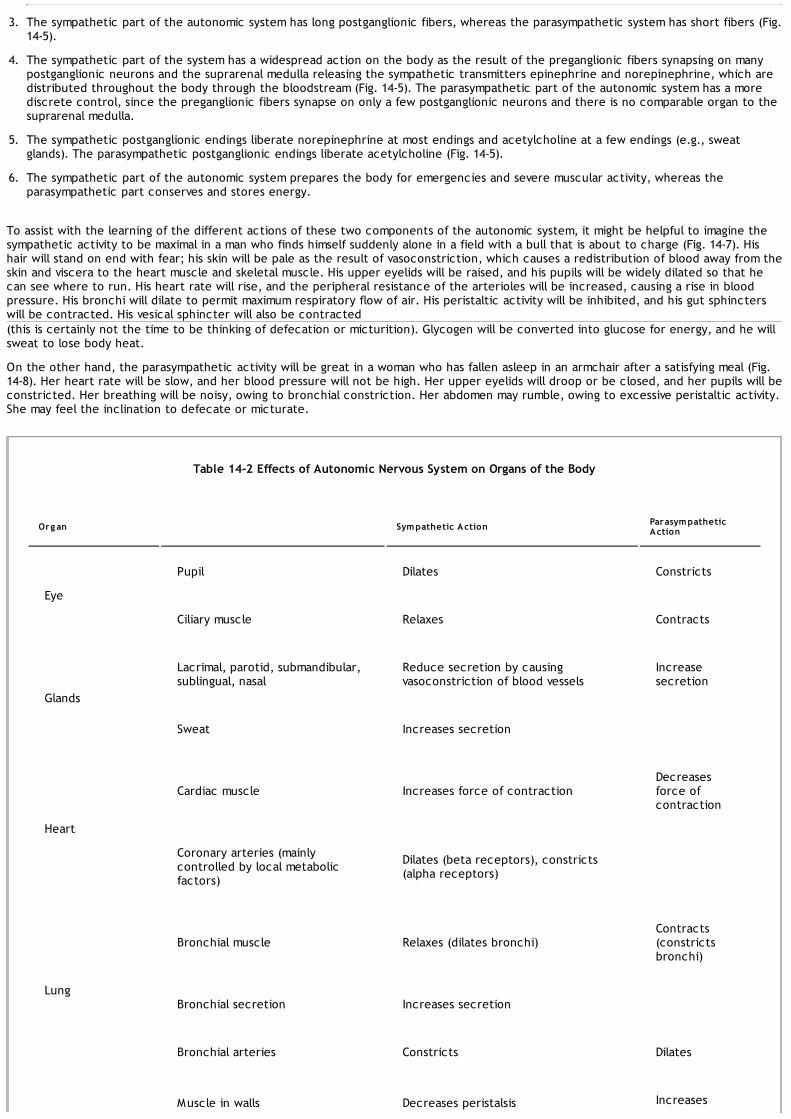
3. The sympathetic part of the autonomic system has long postganglionic fibers, whereas the parasympathetic system has short fibers (Fig.14-5).
4. The sympathetic part of the system has a widespread action on the body as the result of the preganglionic fibers synapsing on manypostganglionic neurons and the suprarenal medulla releasing the sympathetic transmitters epinephrine and norepinephrine, which aredistributed throughout the body through the bloodstream (Fig. 14-5). The parasympathetic part of the autonomic system has a morediscrete control, since the preganglionic fibers synapse on only a few postganglionic neurons and there is no comparable organ to thesuprarenal medulla.
5. The sympathetic postganglionic endings liberate norepinephrine at most endings and acetylcholine at a few endings (e.g., sweatglands). The parasympathetic postganglionic endings liberate acetylcholine (Fig. 14-5).
6. The sympathetic part of the autonomic system prepares the body for emergencies and severe muscular activity, whereas theparasympathetic part conserves and stores energy.
To assist with the learning of the different actions of these two components of the autonomic system, it might be helpful to imagine thesympathetic activity to be maximal in a man who finds himself suddenly alone in a field with a bull that is about to charge (Fig. 14-7). Hishair will stand on end with fear; his skin will be pale as the result of vasoconstriction, which causes a redistribution of blood away from theskin and viscera to the heart muscle and skeletal muscle. His upper eyelids will be raised, and his pupils will be widely dilated so that hecan see where to run. His heart rate will rise, and the peripheral resistance of the arterioles will be increased, causing a rise in bloodpressure. His bronchi will dilate to permit maximum respiratory flow of air. His peristaltic activity will be inhibited, and his gut sphincterswill be contracted. His vesical sphincter will also be contracted(this is certainly not the time to be thinking of defecation or micturition). Glycogen will be converted into glucose for energy, and he willsweat to lose body heat.
On the other hand, the parasympathetic activity will be great in a woman who has fallen asleep in an armchair after a satisfying meal (Fig.14-8). Her heart rate will be slow, and her blood pressure will not be high. Her upper eyelids will droop or be closed, and her pupils will beconstricted. Her breathing will be noisy, owing to bronchial constriction. Her abdomen may rumble, owing to excessive peristaltic activity.She may feel the inclination to defecate or micturate.
Table 14-2 Effects of Autonomic Nervous System on Organs of the Body
Org an Sympathetic Action ParasympatheticAction
Eye
Pupil Dilates Constricts
Ciliary muscle Relaxes Contracts
Glands
Lacrimal, parotid, submandibular,sublingual, nasal
Reduce secretion by causingvasoconstriction of blood vessels
Increasesecretion
Sweat Increases secretion
Heart
Cardiac muscle Increases force of contractionDecreasesforce ofcontraction
Coronary arteries (mainlycontrolled by local metabolicfactors)
Dilates (beta receptors), constricts(alpha receptors)
Lung
Bronchial muscle Relaxes (dilates bronchi)Contracts(constrictsbronchi)
Bronchial secretion Increases secretion
Bronchial arteries Constricts Dilates
Muscle in walls Decreases peristalsis Increases

Gastrointestinaltract
peristalsis
Muscle in sphincters Contracts Relaxes
Glands Reduces secretion by vasoconstrictionof blood vessels
Increasessecretion
Liver Breaks down glycogen into glucose
Gallbladder Relaxes Contracts
Kidney Decreases output due to constrictionof arteries
Urinary bladder
Bladder wall (detrusor) Relaxes Contracts
Sphincter vesicae Contracts Relaxes
Erectile tissue ofpenis and clitoris Relaxes, causes
erection
Ejaculation Contracts smooth muscle of vasdeferens, seminal vesicles, andprostate
Systemic arteries
Skin Constrict
Abdominal Constrict
Muscle Constrict (alpha receptors), dilate(beta receptors), dilate (cholinergic)
Erector pilimuscles Contract
Suprarenal
Cortex Stimulates
Medulla Liberates epinephrine andnorepinephrine
Some Important Autonomic Innervations (Table 14-2)
Eye
Upper Lid

The upper lid is raised by the levator palpebrae superioris muscle. The major part of this muscle is formed by skeletal muscle innervated bythe oculomotor nerve. A small part is composed of smooth muscle fibers innervated bysympathetic postganglionic fibers from the superior cervical sympathetic ganglion (Fig. 14-9).
Figure 14-9 Autonomic innervation of the upper eyelid and iris (A) and the sublingual and submandibular salivary glands (B).
IrisThe smooth muscle fibers of the iris consist of circular and radiating fibers. The circular fibers form the sphincter pupillae, and the radialfibers form the dilator pupillae.
The sphincter pupillae is supplied by parasympathetic fibers from the parasympathetic nucleus (Edinger-Westphal nucleus) of theoculomotor nerve (Fig. 14-9). After synapsing in the ciliary ganglion, the postganglionic fibers pass forward to the eyeball in the shortciliary nerves. (The ciliary muscle of the eye is also supplied by the short ciliary nerves; see p. 340.)
The dilator pupillae is supplied by postganglionic fibers from the superior cervical sympathetic ganglion (Fig. 14-9). The postganglionic fibersreach the orbit along the internal carotid and ophthalmic arteries. They pass uninterrupted through the ciliary ganglion and reach theeyeball in the short ciliary nerves. Other sympathetic fibers reach the eyeball in the long ciliary nerves.
Lacrimal GlandThe parasympathetic secretomotor nerve supply to the lacrimal gland originates in the lacrimatory nucleus of the facial nerve (Fig. 14-10).The preganglionic fibers reach the pterygopalatine ganglion through thenervus intermedius and its great petrosal branch and through the nerve of the pterygoid canal. The postganglionic fibers leave theganglion and join the maxillary nerve. They then pass into its zygomatic branch and the zygomaticotemporal nerve. They reach the

lacrimal gland within the lacrimal nerve.
Figure 14-10 Autonomic innervation of the parotid salivary gland and lacrimal gland.
The sympathetic postganglionic fibers arise from the superior cervical sympathetic ganglion and travel in the plexus of nerves around theinternal carotid artery. They join the deep petrosal nerve, the nerve of the pterygoid canal, the maxillary nerve, the zygomatic nerve,zygomaticotemporal nerve, and finally the lacrimal nerve. They function as vasoconstrictor fibers.
Salivary Glands
Submandibular and Sublingual GlandsThe parasympathetic secretomotor supply originates in the superior salivatory nucleus of the facial nerve (Fig. 14-9). The preganglionicfibers pass to the submandibular ganglion and other small ganglia close to the duct through the chorda tympani nerve and the lingualnerve. Postganglionic fibers reach the submandibular gland either directly or along the duct. Postganglionic fibers to the sublingual glandtravel through the lingual nerve.
Sympathetic postganglionic fibers arise from the superior cervical sympathetic ganglion and reach the glands as a plexus of nerves aroundthe external carotid, facial, and lingual arteries. They function as vasoconstrictor fibers.
Parotid GlandParasympathetic secretomotor fibers from the inferior salivatory nucleus of the glossopharyngeal nerve supply the gland (Fig. 14-10). Thepreganglionic nerve fibers pass to the otic ganglion through the tympanic branch of the glossopharyngeal nerve and the lesser petrosalnerve. Postganglionic fibers reach the gland through the auriculotemporal nerve.
Sympathetic postganglionic fibers arise from the superior cervical sympathetic ganglion and reach the gland as a plexus of nerves aroundthe external carotid artery. They function as vasoconstrictor fibers.

Figure 14-11 Autonomic innervation of the heart and lungs.
HeartThe sympathetic postganglionic fibers arise from the cervical and upper thoracic portions of the sympathetic trunks (Fig. 14-11).Postganglionic fibers reach the heart by way of the superior, middle, and inferior cardiac branches of the cervical portion of thesympathetic trunk and a number of cardiac branches from the thoracic portion of the sympathetic trunk. The fibers pass through thecardiac plexuses and terminate on the sinoatrial and atrioventricular nodes, on cardiac muscle fibers, and on coronary arteries.Activation of these nerves results in cardiac acceleration, increased force of contraction of the cardiac muscle, and dilatation of thecoronary arteries. The coronary dilatation is mainly produced in response to local metabolic needs rather than by direct nerve stimulationof the coronary arteries.
The parasympathetic preganglionic fibers originate in the dorsal nucleus of the vagus nerve and descend into the thorax in the vagusnerves. The fibers terminate by synapsing with postganglionic neurons in the cardiac plexuses. Postganglionic fibers terminate on thesinoatrial and atrioventricular nodes and on the coronary arteries. Activation of these nerves results in a reduction in the rate and forceof contraction of the myocardium and a constriction of the coronary arteries. Here again, the coronary constriction is mainly producedby the reduction in local metabolic needs rather than by neural effects.
LungsThe sympathetic postganglionic fibers arise from the second to the fifth thoracic ganglia of the sympathetic trunk (Fig. 14-11). The fiberspass through the pulmonary plexuses and enter the lung, where they form networks around the bronchi and blood vessels. Thesympathetic fibers produce bronchodilatation and slight vasoconstriction.
The parasympathetic preganglionic fibers arise from the dorsal nucleus of the vagus and descend to the thorax within the vagus nerves.The fibers terminate by synapsing with postganglionic neurons in the pulmonary plexuses. The postganglionic fibers enter the lung, wherethey form networks around the bronchi and blood vessels. The parasympathetic fibers produce bronchoconstriction and slightvasodilatation and increase glandular secretion.
Gastrointestinal Tract
Stomach and Intestine as Far as the Splenic FlexurePreganglionic parasympathetic fibers enter the abdomen in the anterior (left) and posterior (right) vagal trunks (Fig. 14-12). The fibers aredistributed to many abdominal viscera and to the gastrointestinal tract from the stomach to the splenic flexure of the colon. The fibersthat pass to the gastrointestinal tract terminate on postganglionic neurons in the myenteric (Auerbach) and submucosal (Meissner)plexuses.The postganglionic fibers supply the smooth muscle and glands. The parasympathetic nerves stimulate peristalsis and relax the sphincters;they also stimulate secretion.

Figure 14-12 Autonomic innervation of the gastrointestinal tract.
Sympathetic preganglionic nerve fibers pass through the thoracic part of the sympathetic trunk and enter the greater and lessersplanchnic nerves. These descend into the abdomen and synapse with postganglionic neurons in the celiac and superior mesentericganglia. The postganglionic nerve fibers are distributed to the stomach and intestine as nerve plexuses around the branches of the celiacand superior mesenteric arteries. The sympathetic nerves inhibit peristalsis and cause contraction of the sphincters; they also inhibitsecretion (see the enteric nervous system, p. 404).
Descending Colon, Pelvic Colon, and RectumThe preganglionic parasympathetic fibers originate in the gray matter of the spinal cord from the second to the fourth sacral segments(Fig. 14-12). The fibers pass through the pelvic splanchnic nerves and the nerve plexuses around the branches of the inferior mesentericartery. They terminate on postganglionic neurons in the myenteric (Auerbach) and submucosal (Meissner) plexuses. The postganglionicfibers supply the smooth muscle and glands. The parasympathetic nerves stimulate peristalsis and secretion.
The sympathetic preganglionic nerve fibers pass through the lumbar part of the sympathetic trunk and synapse with postganglionicneurons in the inferior mesenteric plexus. Postganglionic fibers are distributed to the bowel as nerve plexuses around the branches ofthe inferior mesenteric arteries. The sympathetic nerves inhibit peristalsis and secretion.
Gallbladder and Biliary DuctsThe gallbladder and biliary ducts receive postganglionic parasympathetic and sympathetic fibers from the hepatic plexus. Parasympatheticfibers derived from the vagus are thought to be motor fibers to the smooth muscle of the gallbladder and bile ducts and inhibitory to thesphincter of Oddi.

Figure 14-13 Autonomic innervation of the kidney and suprarenal gland.
Autonomic afferent fibers are also present. Some of the fibers are believed to leave the hepatic plexus and join the right phrenic nerve,thus partially explaining the phenomenon of referred shoulder pain in the presence of gallbladder disease (see p. 420).
KidneyPreganglionic sympathetic fibers pass through the lower thoracic part of the sympathetic trunk and the lowest thoracic splanchnic nerveto join the renal plexus around the renal artery (Fig. 14-13). The preganglionic fibers synapse with postganglionic neurons in the renalplexus. The postganglionic fibers are distributed to the branches of the renal artery. The sympathetic nerves are vasoconstrictor in actionto the renal arteries within the kidney.
Preganglionic parasympathetic fibers enter the renal plexus from the vagus. Here, they synapse with postganglionic neurons, whose fibersare distributed to the kidney along the branches of the renal artery. The parasympathetic nerves are thought to be vasodilator in action.
Medulla of Suprarenal GlandPreganglionic sympathetic fibers descend to the gland in the greater splanchnic nerve, a branch of the thoracic part of the sympathetictrunk (Fig. 14-13). The nerve fibers terminate on the secretory cells of the medulla, which are comparable to postganglionic neurons.Acetylcholine is the transmitter substance between the nerve endings and the secretory cells, as at any other preganglionic endings. Thesympathetic nerves stimulate the secretory cells of the medulla to increase the output of epinephrine and norepinephrine. There is noparasympathetic innervation of the medulla of the suprarenal gland.
Involuntary Internal Sphincter of the Anal CanalThe circular smooth muscle coat is thickened at the upper end of the anal canal to form the involuntary internal sphincter. The sphincteris innervated by postganglionic sympathetic fibers from the hypogastric plexuses (Fig. 14-14). Each hypogastric plexus receives sympatheticfibers from the aortic plexus and from the lumbar and pelvic parts of the sympathetic trunks. The sympathetic nerves cause the internalanal sphincter to contract.
Urinary BladderThe muscular coat of the bladder is composed of smooth muscle, which at the bladder neck is thickened to form the sphincter vesicae.The nerve supply of the smooth muscle is from the hypogastric plexuses (Fig. 14-14). The sympathetic postganglionic fibers originate in thefirst and second lumbar ganglia of the sympathetic trunk and travel to the hypogastric plexuses. The parasympathetic preganglionic fibersarise as the pelvic splanchnic nerves from the second, third, and fourth sacral nerves; they pass through the hypogastric plexuses toreach the bladder wall, where they synapse with postganglionic neurons.
The sympathetic nerves to the detrusor muscle have little or no action on the smooth muscle of the bladder wall and are distributedmainly to the blood vessels. Thesympathetic nerves to the sphincter vesicae play only a minor role in causing contraction of the sphincter in maintaining urinarycontinence. However, in the male, the sympathetic innervation of the sphincter causes active contraction of the bladder neck duringejaculation (brought about by sympathetic action), thus preventing seminal fluid from entering the bladder. The parasympathetic nervesstimulate the contraction of the smooth muscle of the bladder wall and, in some way, inhibit the contraction of the sphincter vesicae.

Figure 14-14 Autonomic innervation of the sphincters of the anal canal and urinary bladder.
Erection of the Penis and ClitorisIn erection, the genital erectile tissue becomes engorged with blood. The initial vascular engorgement is controlled by theparasympathetic part of the autonomic nervous system. The parasympathetic preganglionic fibers originate in the gray matter of thesecond, third, and fourth sacral segments of the spinal cord (Fig. 14-15). The fibers enter the hypogastric plexuses and synapse on thepostganglionic neurons. The postganglionic fibers join the internal pudendal arteries and are distributed along their branches, which enterthe erectile tissue. The parasympathetic nerves cause vasodilatation of the arteries and greatly increase the blood flow to the erectiletissue.
EjaculationDuring the increasing sexual excitement that occurs during sex play, the external urinary meatus of the glans penis becomes moist as aresult of the secretions of the bulbourethral glands. Friction on the glans penis, reinforced by other afferent nervous impulses, results in adischarge along the sympathetic nerve fibers to the smooth muscle of the duct of the epididymis and the vas deferens on each side, theseminal vesicles, and the prostate. The smooth muscle contracts, and the spermatozoa, together with the secretions of the seminalvesicles and prostate, are discharged into the prostatic urethra. The fluid now joins the secretions of the bulbourethral glands and penileurethral glands and is then ejected from the penile urethra as a result of the rhythmic contractions of the bulbospongiosus muscles,which compress the urethra. Meanwhile, the sphincter of the bladder contracts and prevents a reflux of the spermatozoa into thebladder. The spermatozoa and the secretions of the several accessory glands constitute the seminal fluid, or semen. At the climax of malesexual excitement, a mass discharge of nervous impulses takes place in the central nervous system. Impulses pass down the spinal cord tothe sympathetic outflow (T1-L2). The nervous impulses that pass to the genital organs are thought to leave the cordat the first and second lumbar segments in the preganglionic sympathetic fibers (Fig. 14-15). Many of these fibers synapse withpostganglionic neurons in the first and second lumbar ganglia. Other fibers may synapse in ganglia in the lower lumbar or pelvic parts ofthe sympathetic trunks. The postganglionic fibers are then distributed to the vas deferens, the seminal vesicles, and the prostate throughthe hypogastric plexuses. The sympathetic nerves stimulate the contractions of the smooth muscle in the walls of these structures andcause the spermatozoa, together with the secretions of the seminal vesicles and prostate, to be discharged into the urethra.

Figure 14-15 Autonomic innervation of the male reproductive tract.
UterusPreganglionic sympathetic nerve fibers leave the spinal cord at segmental levels T12 and L1 and are believed to synapse with ganglion cellsin the sympathetic trunk or possibly in the inferior hypogastric plexuses (Fig. 14-16). The postganglionic fibers supply the smooth muscle ofthe uterus. Parasympathetic preganglionic fibers leave the spinal cord at levels S2–4 and synapse with ganglion cells in the inferiorhypogastric plexuses (Fig. 14-16). Although it is recognized that the uterine muscle is largely under hormonal control, sympatheticinnervation may cause uterine contraction and vasoconstriction, whereas parasympathetic fibers have the opposite effect.
Afferent pain fibers from the fundus and the body of the uterus ascend to the spinal cord through the hypogastric plexuses, entering itthrough the posterior roots of the 10th, 11th, and 12th thoracic spinal nerves (Fig. 14-16). Fibers from the cervix run in the pelvicsplanchnic nerves and enter the spinal cord through the posterior roots of the second, third, and fourth sacral nerves.
Arteries of the Upper LimbThe arteries of the upper limb are innervated by sympathetic nerves. The preganglionic fibers originate from cell bodies in the second tothe eighth thoracic segments of the spinal cord (Fig. 14-17). They pass to the sympathetic trunk through white rami and ascend in thetrunk to synapse in the middle cervical, inferior cervical, first thoracic, or stellate ganglia. The postganglionic fibers join the nerves thatform the brachial plexus and are distributed to the arteries within the branches of the plexus. The sympathetic nerves causevasoconstriction of cutaneous arteries and vasodilatation of arteries that supply skeletal muscle.
Arteries of the Lower LimbThe arteries of the lower limb are also innervated by sympathetic nerves (Fig. 14-17). The preganglionic fibers originate from cell bodies inthe lower three thoracic and upper two or three lumbar segments of the spinal cord. The preganglionic fibers pass to the lower thoracicand upper lumbar ganglia of the sympathetic trunk through white rami. The fibers synapse in the lumbar and sacral ganglia, and thepostganglionic fibers reach the arteries through branches of the lumbar and sacral plexuses.

Figure 14-16 Autonomic innervation of the uterus; the pathway taken by the afferent sensory fibers is shown as well.
Some Important Physiological Reflexes Involving the Autonomic Nervous System
Visual Reflexes
Direct and Consensual Light ReflexesAfferent nervous impulses travel from the retina through the optic nerve, optic chiasma, and optic tract (see Fig. 11-3). A small number offibers leave the optic tract and synapse on nerve cells in the pretectal nucleus, which lies close to the superior colliculus. The impulsesare passed by axons of the pretectal nerve cells to the parasympathetic nuclei (Edinger-Westphal nuclei) of the oculomotor nerve on bothsides. Here, the fibers synapse, and the parasympathetic nerves travel through the oculomotor nerve to the ciliary ganglion in the orbit.Finally, postganglionic parasympathetic fibers pass through the short ciliary nerves to the eyeball and to the constrictor pupillae muscleof the iris. Both pupils constrict in the consensual light reflex because the pretectal nucleus sends fibers to the parasympathetic nucleion both sides of the midbrain.
Accommodation ReflexWhen the eyes are directed from a distant to a near object, contraction of the medial recti brings about convergence of the ocular axes,the lens thicken to increase its refractive power by contraction of the ciliary muscle, and the pupils constrict to restrict the light wavesto the thickest central part of the lens. The afferent impulses travel through the optic nerve, the optic chiasma, the optic tract, thelateral geniculate body, and the optic radiation to the visual cortex (see Fig. 11-3). The visual cortex is connected to the eye field of thefrontal cortex. From here, cortical fibers descend through the internal capsule to the oculomotor nuclei in the midbrain. The oculomotornerve travels to the medial recti muscles. Some of the descending cortical fibers synapse with the parasympathetic nuclei (Edinger-Westphal nuclei) of the oculomotor nerve on both sides. The parasympathetic preganglionic fibers then travel through the oculomotornerve to the ciliary ganglion in the orbit where they synapse. Finally, postganglionic parasympathetic fibers pass through the short ciliarynerves to the ciliary muscle and the constrictor pupillae muscle of the iris.
Cardiovascular ReflexesCardiovascular reflexes include the carotid sinus and aortic arch reflexes and the Bainbridge right atrial reflex.
Carotid Sinus and Aortic Arch ReflexesThe carotid sinus, located in the bifurcation of the common carotid artery, and the aortic arch serve as baroreceptors. As the bloodpressure rises, nerve endings situated in the wallsof these vessels are stimulated. The afferent fibers from the carotid sinus ascend in the glossopharyngeal nerve and terminate in thenucleus solitarius (see Figs. 11-16 and 11-17). The afferent fibers from the aortic arch ascend in the vagus nerve. Connector neurons in the

P.418P.419
medulla oblongata activate the parasympathetic nucleus (dorsal nucleus) of the vagus, which slows the heart rate. At the same time,reticulospinal fibers descend to the spinal cord and inhibit the preganglionic sympathetic outflow to the heart and cutaneous arterioles.The combined effect of stimulation of the parasympathetic action on the heart and inhibition of the sympathetic action on the heart andperipheral blood vessels reduces the rate and force of contraction of the heart and reduces the peripheral resistance of the bloodvessels. Consequently, the blood pressure falls. The blood pressure of the individual is thus modified by the afferent information receivedfrom the baroreceptors. The modulator of the autonomic nervous system, namely, the hypothalamus, in turn, can be influenced by other,higher centers in the central nervous system.
Figure 14-17 Sympathetic innervation of the arteries of the upper limb (A) and lower limb (B).
Bainbridge Right Atrial ReflexThis reflex is initiated when the nerve endings in the wall of the right atrium and in the walls of the venae cavae are stimulated by a rise ofvenous pressure. The afferent fibers ascend in the vagus to the medulla oblongata and terminate on the nucleus of the tractus solitarius(see Fig. 11-18). Connector neurons inhibit the parasympathetic nucleus (dorsal) of the vagus, and reticulospinal fibers stimulate thethoracic sympathetic outflow to the heart, resulting in cardiac acceleration.
Clinical NotesGeneral ConsiderationsFrom the foregoing description, it must now be clear to the reader that the autonomic nervous system is not an
isolated part of the nervous system. It should be regarded as the part of the nervous system that, with the endocrinesystem, is particularly involved in maintaining the stability of the internal environment of the body. Its activities aremodified by the hypothalamus, whose function is to integrate vast amounts of afferent information received from otherareas of the nervous system and to translate changing hormonal levels of the bloodstream into appropriate nervousand hormonal activities.Since the autonomic nervous system is so important in maintaining normal body homeostasis, it is not surprising thatthe system is subject to many pharmacologic interventions. Propranolol and atenolol, for example, are beta-adrenergicantagonists that can be used in the treatment of hypertension and ischemic heart disease.

Injuries to the Autonomic Nervous SystemSympathetic InjuriesThe sympathetic trunk in the neck can be injured by stab and bullet wounds. Traction injuries to the first thoracic rootof the brachial plexus can damage sympathetic nerves destined for the stellate ganglion. All these conditions canproduce a preganglionic type of Horner syndrome (see below). Injuries to the spinal cord or cauda equina can disruptthe sympathetic control of the bladder (see p. 418).
Parasympathetic InjuriesThe oculomotor nerve is vulnerable in head injuries (herniated uncus) and can be damaged by compression byaneurysms in the junction between the posterior cerebral artery and posterior communicating artery. Thepreganglionic parasympathetic fibers traveling in this nerve are situated in the periphery of the nerve and can bedamaged. Surface aneurysmal compression characteristically causes dilatation of the pupil and loss of the visual lightreflexes.The autonomic fibers in the facial nerve can be damaged by fractures of the skull involving the temporal bone. Thevestibulocochlear nerve is closely related to the facial nerve in the internal acoustic meatus, so clinical findingsinvolving both nerves are common. Involvement of the parasympathetic fibers in the facial nerve may produce impairedlacrimation in addition to paralysis of the facial muscles.The glossopharyngeal and vagus nerves are at risk in stab and bullet wounds of the neck. The parasympatheticsecretomotor fibers to the parotid salivary gland leave the glossopharyngeal nerve just below the skull; therefore,they are rarely damaged.The parasympathetic outflow in the sacral region of the spinal cord (S2–4) may be damaged by spinal cord and caudaequina injuries, leading to disruption of bladder, rectal, and sexual functions (see p. 418).
Degeneration and Regeneration of Autonomic NervesThe structural changes are identical to those found in other areas of the peripheral and central parts of the nervoussystem. Functional recoveries following sympathectomy operations can be explained only by the assumption eitherthat the operative procedure was inadequate and nerve fibers were left intact or regenerated or that alternativenervous pathways existed and were left undisturbed.The denervation of viscera supplied by autonomic nerves is followed by their increased sensitivity to the agent thatwas previously the transmitter substance. One explanation is that following nerve section, there may be an increase inthe number of receptor sites on the postsynaptic membrane. Another possibility, which applies to endings wherenorepinephrine is the transmitter, is that the reuptake of the transmitter by the nerve terminal is interfered with insome way.
Diseases Involving the Autonomic Nervous SystemDiabetes MellitusDiabetes mellitus is a common cause of peripheral nerve neuropathy. This involves sensory and motor dysfunction andmay also include autonomic dysfunction. The clinical features of autonomic dysfunction include postural hypotension,peripheral edema, pupillary abnormalities, and impaired sweating. The cause is probably associated with chronichyperglycemia.
Horner SyndromeHorner syndrome consists of (1) constriction of the pupil (miosis), (2) slight drooping of the eyelid (ptosis), (3)enophthalmos,3 (4) vasodilation of skin arterioles, and (5) loss of sweating (anhydrosis). All these symptoms resultfrom an interruption of the sympathetic nerve supply to the head and neck. Pathologic causes include lesions in thebrainstem or cervical part of the spinal cord that interrupt the reticulospinal tracts descending from the hypothalamusto the sympathetic outflow in the lateral gray column of the first thoracic segment of the spinal cord. Such lesionsinclude multiple sclerosis and syringomyelia. Traction on the stellate ganglion due to a cervical rib or involvement ofthe ganglion in a metastatic lesion may interrupt the peripheral part of the sympathetic pathway.All patients with Horner syndrome have miosis and ptosis. However, a distinction should be made between lesionsoccurring at the first neuron (the descending reticulospinal fibers within the central nervous system), the secondneuron (the preganglionic fibers), and the third neuron (postganglionic fibers). For example, the clinical signssuggestive of a first-neuron defect (central Horner syndrome) could include contralateral hyperesthesia of the bodyand loss of sweating of the entire half of the body. Signs suggesting a second-neuron involvement (preganglionicHorner syndrome) include loss of sweating limited to the face and neck and the presence of flushing or blanching ofthe face and neck. Signs suggesting third-neuron involvement (postganglionic Horner syndrome) include facial pain orear, nose, or throat disease.The presence or absence of other localizing signs and symptoms may assist in differentiating the three types of Hornersyndrome.
Argyll Robertson PupilArgyll Robertson pupil is characterized by a small pupil, which is of fixed size and does not react to light but doescontract with accommodation. It is usually caused by a neurosyphilitic lesion interrupting the fibers that run from thepretectal nucleus to the parasympathetic nuclei (Edinger-Westphal nuclei) of the oculomotor nerve on both sides. Thefact that the pupil constricts with accommodation implies that the connections between the parasympathetic nuclei andthe constrictor pupillae muscle of the iris are intact.
Adie Tonic Pupil SyndromeIn Adie tonic pupil syndrome, the pupil has a decreased or absent light reflex, a slow or delayed contraction to nearvision, and a slow or delayed dilatation in the dark. This benign syndrome, which probably results from a disorder ofthe parasympathetic innervation of the constrictor pupillae muscle, must be distinguished from the Argyll Robertsonpupil (see above), which is caused by neurosyphilis. Adie syndrome can be confirmed by looking for hypersensitivity tocholinergic agents. Drops commonly used for this test are 2.5% methacholine (Mecholyl) or 0.1% pilocarpine. The Adietonic pupil should constrict when these drops are put in the eye. These cholinergic agents do not cause pupillaryconstriction in mydriasis caused by oculomotor lesion or in drug-related mydriasis.
Frey SyndromeFrey syndrome is an interesting complication that sometimes follows penetrating wounds of the parotid gland. During

the process of healing, the postganglionic parasympathetic secretomotor fibers traveling in the auriculotemporal nervegrow out and join the distal end of the great auricular nerve, which supplies the sweat glands of the overlying facialskin. By this means, a stimulus intended for saliva production instead produces sweat secretion.A similar syndrome may follow injury to the facial nerve. During the process of regeneration, parasympathetic fibersnormally destined for the submandibular and sublingual salivary glands are diverted to the lacrimal gland. Thisproduces watering of the eyes associated with salivation, the so-called crocodile tears.
Hirschsprung DiseaseHirschsprung disease (megacolon) is a congenital condition in which there is a failure of development of the myentericplexus (Auerbach plexus) in the distal part of the colon. The involved part of the colon possesses no parasympatheticganglion cells, and peristalsis is absent. This effectively blocks the passage of feces, and the proximal part of the colonbecomes enormously distended.
Urinary Bladder Dysfunction Following Spinal Cord InjuriesInjuries to the spinal cord are followed by disruption of the nervous control of micturition. The normal bladder isinnervated as follows:Sympathetic innervation is from the first and second lumbar segments of the spinal cord.Parasympathetic innervation is from the second, third, and fourth sacral segments of the spinal cord.Sensory nerve fibers enter the spinal cord at the above segments.The atonic bladder occurs during the phase of spinal shock immediately following the injury and may last from a fewdays to several weeks. The bladder wall muscle is relaxed, the sphincter vesicae is tightly contracted (loss of inhibitionfrom higher levels), and the sphincter urethrae is relaxed. The bladder becomes greatly distended and finallyoverflows. Depending on the level of the cord injury, the patient may or may not be aware that the bladder is full;there is no voluntary control.The automatic reflex bladder occurs after the patient has recovered from spinal shock, provided that the cord lesionlies above the level of the parasympathetic outflow (S2–4). This is the type of bladder normally found in infancy. Sincethe descending fibers in the spinal cord are sectioned, there is no voluntary control. The bladder fills and emptiesreflexly. Stretch receptors in the bladder wall are stimulated as the bladder fills, and the afferent impulses pass to thespinal cord (S2–4). Efferent impulses pass down to the bladder muscle, which contracts; the sphincter vesicae and theurethral sphincter both relax. This simple reflex occurs every 1 to 4 hours.The autonomous bladder is the condition that occurs if the sacral segment of the spinal cord is destroyed or if thecauda equina is severed. The bladder has no reflex control or voluntary control. The bladder wall is flaccid, and thecapacity of the bladder is greatly increased. It fills to capacity and overflows, which results in continual dribbling. Thebladder may be partially emptied by manual compression of the lower part of the anterior abdominal wall, but infectionof the urine and back pressure effects on the ureters and kidneys are inevitable.
Defecation Following Spinal Cord InjuriesThe act of defecation involves a coordinated reflex that results in the emptying of the descending colon, pelvic colon,rectum, and anal canal. It is assisted by a rise in the intra-abdominal pressure brought about by contraction of themuscles of the anterior abdominal wall. The involuntary internal sphincter of the anal canal normally is innervated bypostganglionic sympathetic fibers from the hypogastric plexuses, and the voluntary external sphincter of the anal canalis innervated by the inferior rectal nerve. The desire to defecate is initiated by stimulation of the stretch receptors inthe wall of the rectum.Following severe spinal cord injuries (or cauda equina injuries), the patient is not aware of rectal distention. Moreover,the parasympathetic influence on the peristaltic activity of the descending colon, sigmoid colon, and rectum is lost. Inaddition, control over the abdominal musculature and sphincters of the anal canal may be severely impaired. Therectum, now an isolated structure, responds by contracting when the pressure within its lumen rises. This local reflexresponse is much more efficient if the sacral segments of the spinal cord and the cauda equina are intact. At best,however, the force of the contractions of the rectal wall is small, and constipation and impaction are the usualoutcome. The treatment of patients with spinal cord injuries is to empty the rectum with biweekly enemas; the use ofsuppositories also may be helpful.
Erection and Ejaculation Following Spinal Cord InjuriesAs described previously, erection of the penis or clitoris is controlled by the parasympathetic nerves that originate fromthe second, third, and fourth sacral segments of the spinal cord. Bilateral damage to the reticulospinal tracts in thespinal cord above the second sacral segment of the spinal cord will result in loss of erection. Later, when the effects ofspinal shock have disappeared, spontaneous or reflex erection may occur if the sacral segments of the spinal cord areintact.Ejaculation is controlled by sympathetic nerves that originate in the first and second lumbar segments of the spinalcord. Ejaculation brings about a flow of seminal fluid into the prostatic urethra. The final ejection of the fluid from thepenis is the result of the rhythmic contractions of the bulbospongiosus muscles, which compress the urethra. Thebulbospongiosus muscles are innervated by the pudendal nerve (S2–4). Discharge of the seminal fluid into the bladderis prevented by the contraction of the sphincter vesicae, which is innervated by the sympathetic nerves (L1-2). As inthe case of erection, severe bilateral damage to the spinal cord results in loss of ejaculation. Later, reflex ejaculationmay be possible in patients with spinal cord transections in the thoracic or cervical regions. Some individuals have anormal ejaculation without external emission, and the seminal fluid passes into the bladder, owing to paralysis of thesphincter vesicae.
Disease Caused by Botulinum ToxinA very small amount of botulinum toxin binds irreversibly to the nerve plasma membranes and prevents the release ofacetylcholine at cholinergic synapses and neuromuscular junctions, producing an atropinelike syndrome with skeletalmuscle weakness.
Disease Caused by Black Widow Spider VenomThe venom of a black widow spider causes a brief release of acetylcholine at the nerve endings followed by apermanent blockade.
Disease Caused by Anticholinesterase Agents

Acetylcholinesterase, which is responsible for hydrolyzing and limiting the action of acetylcholine at nerve endings, canbe blocked by certain drugs. Physostigmine, neostigmine, pyridostigmine, and carbamate and organophosphateinsecticides are effective acetylcholinesterase inhibitors. Their use results in an excessive stimulation of the cholinergicreceptors, producing the “SLUD syndrome”—salivation, lacrimation, urination, and defecation.
Sympathectomy as a Method of Treating Arterial DiseaseRaynaud DiseaseRaynaud disease is a vasospastic disorder involving the digital arteries of the upper limb. The disorder is usuallybilateral, and an attack is provoked by exposure to cold. There is pallor or cyanosis of the fingers as well as severepain. Gangrene of the tips of the fingers may occur.In mild cases of Raynaud disease, the treatment is the avoidance of cold and no smoking (smoking causesvasoconstriction). In more severe cases, drugs that inhibit sympathetic activity, such as reserpine, bring about arterialvasodilatation with consequent increase in blood flow to the fingers. Cervicothoracic preganglionic sympathectomy hasbeen used, but the long-term results are disappointing.
Intermittent ClaudicationIntermittent claudication, which is common in men, is due to arterial occlusive disease of the leg. Ischemia of themuscles produces a cramplike pain on exercise. Lumbar preganglionic sympathectomy may be advocated as a form oftreatment in order to bring about vasodilatation and an increase in blood flow through the collateral circulation.Preganglionic sympathectomy is performed by removing the upper three lumbar ganglia and the intervening parts ofthe sympathetic trunk.
HypertensionIn the past, severe essential hypertension was treated by bilateral thoracolumbar sympathectomy to reduce thevasomotor control over the peripheral resistance and thus lower the blood pressure. Today, chemical blocking agentsof the sympathetic system are widely used with great success, and the resulting reduction in the force of myocardialcontraction reduces the arterial blood pressure.
Referred Visceral PainMost viscera are innervated only by autonomic nerves. Therefore, it follows that visceral pain is conducted alongafferent autonomic nerves. Visceral pain is diffuse and poorly localized, whereas somatic pain is intense and discretelylocalized. Visceral pain frequently is referred to skin areas that are innervated by the same segments of the spinal cordas the painful viscus (Fig. 14-18). The explanation for referred pain is not known. One theory is that the nerve fibersfrom the viscus and the dermatome ascend in the central nervous system along a common pathway and the cerebralcortex is incapable of distinguishing between the sites of origin. Another theory is that under normal conditions, theviscus does not give rise to painful stimuli, whereas the skin area repeatedly receives noxious stimuli. Because bothafferent fibers enter the spinal cord at the same segment, the brain interprets the information as coming from the skinrather than from the viscus. Pain arising from the gastrointestinal tract is referred to the midline. This can probably beexplained by the fact that the tract arises embryologically as a midline structure and receives a bilateral nerve supply.
Cardiac PainPain originating in the heart as the result of acute myocardial ischemia is assumed to be caused by oxygen deficiencyand the accumulation of metabolites, which stimulate the sensory nerve endings in the myocardium. The afferent nervefibers ascend to the central nervous system through the cardiac branches of the sympathetic trunk and enter thespinal cord through the posterior roots of the upper four thoracic nerves. The nature of the pain varies considerably,from a severe crushing pain to nothing more than a mild discomfort.The pain is not felt in the heart but is referred to the skin areas supplied by the corresponding spinal nerves. The skinareas supplied by the upper four intercostal nerves and by the intercostobrachial nerve (T2) are therefore affected.The intercostobrachial nerve communicates with the medial cutaneous nerve of the arm and is distributed to skin onthe medial side of the upper part of the arm. A certain amount of spread of nervous information must occur within thecentral nervous system, as the pain is sometimes felt in the neck and the jaw.Myocardial infarction involving the inferior wall or diaphragmatic surface of the heart often gives rise to discomfort inthe epigastrium just below the sternum. One must assume that the afferent pain fibers from the heart ascend in thesympathetic nerves and enter the spinal cord in the posterior roots of the seventh, eighth, and ninth thoracic spinalnerves and give rise to referred pain in the T7–9 thoracic dermatomes in the epigastrium.Since the heart and the thoracic part of the esophagus probably have similar afferent pain pathways, it is notsurprising that pain from acute esophagitis can mimic the pain of myocardial infarction.
Stomach PainReferred pain from the stomach is commonly felt in the epigastrium. The afferent pain fibers from the stomach ascendin company with the sympathetic nerves and pass through the celiac plexus and the greater splanchnic nerves. Thesensory fibers enter the spinal cord at segments T5–9 and give rise to referred pain in the dermatomes T5–9 on thelower chest and abdominal walls.
Appendicular PainVisceral pain from the appendix is produced by distention of its lumen or spasm of its muscle. It travels in nerve fibersthat accompany sympathetic nerves through the superior mesenteric plexus and the lesser splanchnic nerve to thespinal cord (T10 segment). The vague referred pain is felt in the region of the umbilicus, which is innervated by the10th intercostal nerve (T10 dermatome). Later, when the inflammatory process involves the parietal peritoneum in theright iliac fossa, which is innervated by the 12th thoracic and 1st lumbar spinal nerves, the now somatic pain becomessevere and dominates the clinical picture. The somatic pain is localized precisely to the right lower quadrant of theanterior abdominal wall (T12-L1 dermatomes).

P.421
Figure 14-18 Some areas of referred pain from the viscera. In the case of the heart, the pain is usually referred to the leftside of the thorax; occasionally, it is referred to both sides.
Gallbladder PainVisceral pain impulses from the gallbladder (acute cholecystitis, gallstone colic) travel in nerve fibers that accompanysympathetic fibers through the celiac plexus and the greater splanchnic nerves to the spinal cord (segments T5–9).The vague referred pain is felt in the dermatomes (T5–9) on the lower chest and upper abdominal walls. Should theinflammatory process spread to involve the parietal peritoneum of the anterior abdominal wall or peripheraldiaphragm, the now severe somatic pain will be felt in the right upper quadrant of the anterior abdominal wall andthrough to the back below the inferior angle of the scapula. Involvement of the central diaphragmatic parietalperitoneum, which is innervated by the phrenic nerve (C3–5), may give rise to referred pain to the tip of the shoulder,since the skin in this area is innervated by the supraclavicular nerves (C3-4).Some areas of referred pain from viscera are shown in Figure 14-18.
CausalgiaCausalgia is a painful condition of the arm or leg accompanied by trophic changes in the affected skin and nails. Itcommonly follows crushing or partial division of the median nerve in the arm or the tibial nerve in the leg. It is thoughtthat the descending impulses in the sympathetic postganglionic fibers in some way evoke ascending impulses in theafferent pain fibers at the site of injury. In many instances, sympathectomy has relieved the pain of causalgia.
Clinical Problem Solving1. A 35-year-old man was getting off the back of a truck when it started to move. Having placed his feet on theground, he grabbed a rail on the truck with his right hand and held on. The truck continued along the road for oneblock before it stopped. In the meantime, the man had been dragged along the road as he held onto the truck. Hewas seen in the emergency department in a state of shock, with cuts and abrasions to his legs. On carefulexamination of his right arm, the following muscles were found to be paralyzed: the flexor carpi ulnaris, the flexordigitorum profundus, the palmar and dorsal interossei, and the thenar and hypothenar muscles. There was also loss ofsensation on the medial side of the arm, forearm, and hand. The deep tendon reflex for the biceps brachii waspresent, but the triceps reflex was absent. It also was noted that the pupil of the right eye was constricted, and therewas drooping of the right upper eyelid. The right eyeball seemed to be less prominent than the left. The skin of theright cheek felt warmer and drier and was redder in color than the left cheek. Using your knowledge of neuroanatomy,explain the clinical findings.View Answer
2. A 3-year-old boy with a history since infancy of chronic constipation and abdominal distention was taken to apediatrician. The child's mother said that the constipation was getting progressively worse. It was not responding tolaxatives, and she was finding it necessary to give her son an enema once a week to relieve his abdominal distention.On physical examination, the child's abdomen was obviously distended, and a doughlike mass could be palpated alongthe course of the descending colon in the left iliac fossa. Examination of the rectum showed it to be empty and notdilated. Following an enema and repeated colonic irrigation with saline solution, the patient was given a barium enemafollowed by a radiographic examination. The radiograph showed a grossly distended descending colon and an abruptchange in lumen diameter where the descending colon joined the sigmoid colon. It was interesting to note that the

P.423P.424P.425
child failed to empty the colon of the barium. Using your knowledge of the autonomic nerve supply to the colon, what isthe diagnosis? How would you treat this patient?View Answer
3. A nervous 25-year-old woman attended her physician because she was experiencing attacks of painful discolorationof the fourth and fifth fingers of both hands. She said that her symptoms had started 2 years previously, during thewinter, and affected her right hand first and, in subsequent attacks, her left hand as well. Initially, her fingers turnedwhite on exposure to cold and then became deep blue. The color change was confined to the distal half of each fingerand was accompanied by an aching pain. Holding her hands over a hot stove or going into a hot room was the onlytreatment that relieved the pain. As the pain disappeared, she said, her fingers became red and swollen. She told herphysician that she had noticed that her fingers were moist with sweat during some of the attacks. Using yourknowledge of neuroanatomy, make the diagnosis. What is the autonomic nerve supply to the blood vessels of theupper limb? How would you treat this patient?View Answer
4. An obese 45-year-old mother of six children was examined by her physician because her symptoms were suggestiveof gallbladder disease. She complained of having severe attacks of colicky pain beneath the right costal margin, whichoften radiated through to the back beneath the right scapula. The physician turned to a medical student and said,“Note that the patient complains of referred pain to the back.” What did he mean by that statement? Explain thephenomenon of referred pain to the back and sometimes the right shoulder in gallbladder disease.View Answer
5. Examination of a patient with neurosyphilis indicated that the pupil of her left eye was small and fixed and did notreact to light but contracted when she was asked to look at a near object. What is the innervation of the iris? Usingyour knowledge of neuroanatomy, state where you believe the neurologic lesion would be situated to account forthese defects.View Answer
6. A 36-year-old man was admitted to the emergency department following a gunshot wound to the lower back.Radiographic examination revealed that the bullet was lodged in the vertebral canal at the level of the third lumbarvertebra. A complete neurologic examination revealed the symptoms and signs that indicate a complete lesion of thecauda equina. What is the autonomic nerve supply to the bladder? Is this patient going to have any interference withbladder function?View Answer
7. On routine medical examination, a 40-year-old black man was found to have essential hypertension. His bloodpressure readings were 180 systolic and 100 diastolic (mm Hg). How would you treat this patient medically? What isthe action of the various types of drugs that are commonly used in the treatment of hypertension?View Answer
8. What transmitter substances are liberated at the following nerve endings: (a) preganglionic sympathetic, (b)preganglionic parasympathetic, (c) postganglionic parasympathetic, (d) postganglionic sympathetic fibers to the heartmuscle, and (e) postganglionic sympathetic fibers to the sweat glands of the hand?View Answer
Review QuestionsDirections: Each of the numbered items in this section is followed by answers. Select the ONE lettered answerthat is CORRECT.1. The following statements concern the autonomic nervous system:
(a) The enteric nervous system is made up of the submucous plexus of Meissner and the myenteric plexus ofAuerbach.(b) The nerve fibers of the enteric nervous system are naked axons.(c) The activities of the parasympathetic part of the autonomic nervous system are used in an emergency.(d) The parasympathetic part of the autonomic system contains only efferent nerve fibers.(e) The pretectal nucleus is concerned with the auditory reflexes.
View Answer
2. The following statements concern the autonomic nervous system:(a) An Argyll Robertson pupil indicates that the accommodation reflex for near vision is normal but that the lightreflex is lost.(b) White rami communicantes are limited to the thoracic part of the sympathetic trunk.(c) White rami communicantes contain postganglionic sympathetic fibers.(d) The greater splanchnic nerves are formed of nonmyelinated axons.(e) The lesser splanchnic nerves arise from the eighth and ninth ganglia of the thoracic part of the sympathetictrunks.
View Answer

3. The following general statements concern the autonomic nervous system:(a) The hypothalamus has little control over the autonomic nervous system.(b) The cerebral cortex has no control over the autonomic nervous system.(c) A patient with Adie tonic pupil syndrome has an increased light reflex and a fast pupillary contraction to nearvision and a fast dilatation in the dark.(d) Pain arising in the gastrointestinal tract is referred to the midline.(e) Visceral pain frequently is referred to skin areas that are innervated by different segments of the spinal cordas the painful viscus.
View Answer
4. The following statements concern Horner syndrome:(a) The pupil is dilated.(b) The upper eyelid is retracted.(c) The patient has vasodilation of the facial skin arterioles.(d) There is excessive facial sweating.(e) There is exophthalmos.
View Answer
Directions: Each of the numbered items or incomplete statements in this section is followed by answers orcompletions of the statement. Select the ONE lettered answer or completion that is BEST in each case.5. The sympathetic outflow:
(a) arises from nerve cells that are situated in the posterior gray column (horn) of the spinal cord(b) has preganglionic nerve fibers that leave the spinal cord in the posterior roots of the spinal nerves(c) is restricted to the T1-L2 segments of the spinal cord(d) receives descending fibers from supraspinal levels that pass down the spinal cord in the posterior whitecolumn(e) has many preganglionic nerve fibers that synapse in the posterior root ganglia of the spinal nerves
View Answer
6. Norepinephrine is secreted at the endings of the:(a) preganglionic sympathetic fibers(b) preganglionic parasympathetic fibers(c) postganglionic parasympathetic fibers(d) postganglionic sympathetic fibers(e) preganglionic fibers to the suprarenal medulla
View Answer
7. The parasympathetic innervation controlling the parotid salivary gland arises from the:(a) facial nerve.(b) oculomotor nerve.(c) vagus nerve(d) carotid plexus(e) glossopharyngeal nerve
View Answer
8. Which of the following statements best describes the parasympathetic part of the autonomic nervous system?(a) It is associated with the thoracolumbar part of the spinal cord.(b) Effects are local and discrete due to preganglionic neurons synapsing with few postganglionic neurons.(c) It has short preganglionic axons.(d) It is active during an emotional crisis.(e) Its activity mobilizes glucose from glycogen.
View Answer
9. Anticholinesterase drugs act at synapses by:(a) mimicking the action of acetylcholine at its receptor sites(b) preventing the release of acetylcholine(c) increasing the secretion of acetylcholine(d) blocking the breakdown of acetylcholine(e) preventing the uptake of acetylcholine by the nerve ending
View Answer
10. Atropine has the following effect on the autonomic nervous system:

(a) It is an anticholinesterase drug.(b) It increases the activity of norepinephrine.(c) It blocks the action of acetylcholine on effector sites in the parasympathetic system.(d) It blocks norepinephrine reuptake by presynaptic terminals in the sympathetic system.(e) It blocks norepinephrine receptor sites.
View Answer
11. The parasympathetic outflow in the spinal cord occurs at levels:(a) S1-2(b) S3–5(c) S1–3(d) S2–4(e) L1-2
View Answer
Directions: Each of the numbered items in this section is followed by answers. Select the ONE lettered answerthat is CORRECT.12. The following statements concern autonomic innervation of the urinary bladder:
(a) The parasympathetic part brings about relaxation of the bladder wall muscle and contraction of the sphinctervesicae.(b) The sympathetic part in the male causes relaxation of the sphincter vesicae and does not prevent reflux ofsemen into the bladder during ejaculation.(c) The afferent fibers from the bladder reach the spinal cord at the first and second lumbar segments and thesecond, third, and fourth sacral segments.(d) The sympathetic part causes contraction of the sphincter urethrae.(e) The parasympathetic part innervates the blood vessels supplying the bladder wall.
View Answer
13. The following statements concern the autonomic innervation of the heart:(a) The parasympathetic part causes dilation of the coronary arteries.(b) The postganglionic fibers do not terminate on the sinoatrial and atrioventricular nodes.(c) The sympathetic postganglionic fibers liberate acetylcholine at their nerve endings.(d) The sympathetic nerves cause cardiac acceleration and increased force of contraction of the heart.(e) The neural control of dilatation of the coronary arteries is more important than the chemical control exertedby the products of cardiac muscle metabolism.
View Answer
Matching Questions. Directions: Match the numbered glands with the most appropriate lettered autonomicganglion listed below. Each lettered option may be selected once, more than once, or not at all.14. Submandibular gland
(a) Otic ganglion(b) Submandibular ganglion(c) Pterygopalatine ganglion(d) Ciliary ganglion(e) None of the above
View Answer
15. Lacrimal gland(a) Otic ganglion(b) Submandibular ganglion(c) Pterygopalatine ganglion(d) Ciliary ganglion(e) None of the above
View Answer
16. Nasal glands(a) Otic ganglion(b) Submandibular ganglion(c) Pterygopalatine ganglion(d) Ciliary ganglion(e) None of the above
View Answer

17. Parotid gland(a) Otic ganglion(b) Submandibular ganglion(c) Pterygopalatine ganglion(d) Ciliary ganglion(e) None of the above
View Answer
18. Sublingual gland(a) Otic ganglion(b) Submandibular ganglion(c) Pterygopalatine ganglion(d) Ciliary ganglion(e) None of the above
View Answer
Match the numbered autonomic ganglia with the most appropriate lettered viscus or muscle listed below. Eachlettered option may be selected once, more than once, or not at all.
Figure 14-19 Areas of referred pain.
19. Superior cervical ganglion(a) Levator palpebrae superioris (smooth muscle only)(b) Vermiform appendix(c) Constrictor pupillae(d) Descending colon(e) None of the above
View Answer
20. Ciliary ganglion

(a) Levator palpebrae superioris (smooth muscle only)(b) Vermiform appendix(c) Constrictor pupillae(d) Descending colon(e) None of the above
View Answer
21. Celiac ganglion(a) Levator palpebrae superioris (smooth muscle only)(b) Vermiform appendix(c) Constrictor pupillae(d) Descending colon(e) None of the above
View Answer
22. Inferior mesenteric ganglion(a) Levator palpebrae superioris (smooth muscle only)(b) Vermiform appendix(c) Constrictor pupillae(d) Descending colon(e) None of the above
View Answer
23. Superior mesenteric ganglion(a) Levator palpebrae superioris (smooth muscle only)(b) Vermiform appendix(c) Constrictor pupillae(d) Descending colon(e) None of the above
View Answer
Match the numbered cranial nerves with the appropriate lettered nuclei listed below. Each lettered option may beselected once, more than once, or not at all.24. Facial nerve
(a) Inferior salivatory nucleus(b) Edinger-Westphal nucleus(c) Lacrimatory nucleus(d) None of the above
View Answer
25. Oculomotor nerve(a) Inferior salivatory nucleus(b) Edinger-Westphal nucleus(c) Lacrimatory nucleus(d) None of the above
View Answer
26. Glossopharyngeal nerve(a) Inferior salivatory nucleus(b) Edinger-Westphal nucleus(c) Lacrimatory nucleus(d) None of the above
View Answer
27. Hypoglossal nerve(a) Inferior salivatory nucleus(b) Edinger-Westphal nucleus(c) Lacrimatory nucleus(d) None of the above
View Answer
The following questions apply to Figure 14-19. Match the numbered areas of referred pain with the appropriate

lettered viscus originating the pain listed below. Each lettered option may be selected once, more than once, ornot at all.28. Number 1
(a) Heart(b) Appendix(c) Gallbladder(d) Stomach(e) None of the above
View Answer
29. Number 2(a) Heart(b) Appendix(c) Gallbladder(d) Stomach(e) None of the above
View Answer
30. Number 3(a) Heart(b) Appendix(c) Gallbladder(d) Stomach(e) None of the above
View Answer
31. Number 4(a) Heart(b) Appendix(c) Gallbladder(d) Stomach(e) None of the above
View Answer
Additional Reading
Appenzeller, D. The Autonomic Nervous System (4th ed.). Amsterdam: Elsevier Biomedical Press, 1990.
Buckley, N., and Caufield, M. In G. Brunstock and C. H. V. Hoyle (eds.). Transmission: Acetylcholine in Autonomic NeuroeffectorMechanisms (pp. 257–322). Chur, Switzerland: Harwood Academic Publishers, 1992.
Craig, C. R., and Stitzel, R. E. Modern Pharmacology (4th ed.). Boston: Little, Brown, 1994.
Dampney, R. A., Horiuchi, J., Tagawa, T., Fontes, M. A., Potts, P. D., and Polson, J. W. Medullary and supramedullary mechanismsregulating sympathetic vasomotor tone. Acta. Physiol. Scand. 177:209, 2003.
Elfvin, L. G., Lindh, B., and Hokfelt, T. The chemical neuroanatomy of sympathetic ganglia. Annu. Rev. Neurosci. 16:471–507, 1993.
Gershon, M. D. The enteric nervous system. Annu. Rev. Neurosci. 4:227, 1981.
Gershon, M. D. The Second Brain. New York: HarperCollins, 1998.
Gibbins, I. L., Jobling, P., Messenger, J. P., Teo, E. H., and Morris, J. L. Neuronal morphology and the synaptic organisation ofsympathetic ganglia. J. Auton. Nerv. Syst. 81:104, 2000.
Goldstein, D. S., Robertson, D., Esler, M., Straus, S. E., and Eisenhofer, G. Dysautonomias: Clinical disorders of the autonomic nervous

system. Ann. Intern. Med. 137:753, 2002.
Guyton, A. C., and Hall, J. E. Textbook of Medical Physiology (11th ed.). Philadelphia: Elsevier, Saunders, 2006.
Hirst, G. D., and Ward, S. M. Interstitial cells: Involvement in rythmicity and neural control of gut smooth muscle. J. Physiol. 550:337,2003.
Kandel, E. R., Schwartz, J. H., and Jessell, T. M. Principles of Neural Science (4th ed.). New York: McGraw-Hill, 2000.
Kuru, M. Nervous control of micturition. Physiol. Rev. 45:425, 1965.
Lepor, H., Gregerman, M., Crosby, R., Mostofi, F. K., and Walsh, P. C. Precise localization of the autonomic nerves from the pelvicplexus to the corpora cavernosa: A detailed anatomical study of the adult male pelvis. J. Urol. 133:207, 1985.
Limbird, L. E. (ed.). The Alpha-2 Adrenergic Receptors. Clifton, NJ: Humana, 1988.
Mitchell, G. A. G. Anatomy of the Autonomic Nervous System. Edinburgh: Livingstone, 1953.
Mitchell, G. A. G. Cardiovascular Innervation. Edinburgh: Livingstone, 1956.
Nestler, E. J., Hyman, S. E., and Malenka, R. C. Molecular Neuropharmacology: A Foundation for Clinical Neuroscience. New York:McGraw-Hill, 2001.
Perkins, J. D. (ed.). The Beta-Adrenergic Receptors. Clifton, NJ: Humana, 1991.
Procacci, P., and Zoppi, M. Pathophysiology and clinical aspects of visceral and referred pain. In J. J. Bonica, D. Lindblom, A. Iggo, L.E. Jones, and C. Benedetti (eds.). Advances in Pain Research and Therapy (vol. 5, p. 643). New York: Raven, 1983.
Snell, R. S. Clinical Anatomy by Regions (8th ed.). Philadelphia: Lippincott Williams & Wilkins, 2008.
Snell, R. S. Clinical Anatomy by Systems. Philadelphia: Lippincott Williams & Wilkins, 2007.
Snell, R. S. The histochemical appearances of cholinesterase in the parasympathetic nerves supplying the submandibular andsublingual salivary glands of the rat. J. Anat. (Lond.) 92:534, 1958.
Snell, R. S. The histochemical appearances of cholinesterase in the superior cervical sympathetic ganglion and the changes whichoccur after preganglionic nerve section. J. Anat. (Lond.) 92:408, 1958.
Snell, R. S., and Lemp, M. A. Clinical Anatomy of the Eye (2nd ed.). Boston: Blackwell Scientific, 1998.
Snell, R. S., and Smith, M. S. Clinical Anatomy for Emergency Medicine. St. Louis: Mosby, 1993.
Snider, S. R., and Kuchel, O. Dopamine: An important hormone of the sympathoadrenal system. Endocrinol. Rev. 4:291, 1983.
Standing, S. (ed.). Gray's Anatomy (39th Brit. ed.). London: Elsevier Churchill Livingstone, 2005.
Westfall, T. C. Evidence that noradrenergic transmitter release is regulated by presynaptic receptors. Fed. Proc. 43:1352, 1984.

Authors: Snell, Richard S.Title: Clinical Neuroanatomy, 7th Edition
Copyright ©2010 Lippincott Williams & Wilkins
> Table of Contents > Chapter 15 - The Meninges of the Br ain and Spinal Cor d
Chapter 15
The Meninges of the Brain and Spinal Cord
A 44-year-old woman was seen by a neurologist because she was experiencing intense pain in the right eye. On physical examination, shewas found to have a slight medial strabismus of the right eye, and the right pupil was smaller than normal. Further examination revealednumbness over the right cheek. A computed tomography (CT) scan showed the presence of an aneurysm of the right internal carotidartery within the cavernous sinus. The aneurysm was about the size of a pea.
The location of the carotid aneurysm within the cavernous sinus explained the ocular pain; pressure on the right abducent nerve wasresponsible for the paralysis of the lateral rectus muscle, producing the medial strabismus. The small pupil of the right eye was caused bythe aneurysm pressing on the sympathetic plexus surrounding the carotid artery and producing paralysis of the dilator pupillae muscle.The numbness experienced over the right cheek was due to pressure of the aneurysm on the right maxillary division of the trigeminalnerve as it passed forward through the lateral wall of the sinus.
This patient illustrates the necessity of knowing the relationships between the structures within the skull, especially in regions like thecavernous sinus, where so many important neural structures lie close to one another.
Chapter Objectives
To learn the structure and function of the three meninges that surround the brain and spinal cordTo understand the venous sinuses within the skull and see how the meninges contribute to their wallsTo appreciate the relationship of the meninges to the different forms of cerebral hemorrhage
The brain in the skull and the spinal cord in the vertebral column are surrounded by three protective membranes, or meninges: the duramater, the arachnoid mater, and the pia mater.
Meninges of the Brain
Dura MaterThe dura mater of the brain is conventionally described as two layers: the endosteal layer and the meningeal layer (Fig. 15-1). These areclosely united except along certain lines, where they separate to form venous sinuses.
The endosteal layer is nothing more than the periosteum covering the inner surface of the skull bones. At the foramen magnum, it doesnot become continuous with the dura mater of the spinal cord. Around the margins of all the foramina in the skull, it becomes continuouswith the periosteum on the outside of the skull bones. At the sutures, it is continuous with the sutural ligaments (Fig. 15-1). It is moststrongly adherent to the bones over the base of the skull.
The meningeal layer is the dura mater proper. It is a dense, strong fibrous membrane covering the brain (Figs. 15-2 and 15-3) and iscontinuous through the foramen magnum with the dura mater of the spinal cord. It provides tubular sheaths for the cranial nerves as thelatter pass through the foramina in the skull. Outside the skull, the sheaths fuse with the epineurium of the nerves (Fig. 15-2).
The meningeal layer sends inward four septa, which divide the cranial cavity into freely communicating spaces that lodge the subdivisionsof the brain (Figs. 15-1 and 15-3). The function of these septa is to restrict the displacement of the brain associated with acceleration anddeceleration, when the head is moved.
The falx cerebri is a sickle-shaped fold of dura mater that lies in the midline between the two cerebral hemispheres (Figs. 15-1 and 15-3).Its narrow anterior end is attached to the internal frontal crest and the crista galli. Its broad posterior part blends in the midline with theupper surface of the tentorium cerebelli. The superior sagittal sinus runs in its upper fixed margin, the inferior sagittal sinus runs in itslower concave free margin, and the straight sinus runs along its attachment to the tentorium cerebelli.
The tentorium cerebelli is a crescent-shaped fold of dura mater that roofs over the posterior cranial fossa (Figs. 15-1 and 15-4). It coversthe upper surface of the cerebellum and supports the occipital lobes of the cerebral hemispheres. In the anterior edge there is a gap,the tentorial notch, for the passage of the midbrain (Fig. 15-4), which produces an inner free border and an outer attached or fixedborder. The fixed border is attached to the posterior clinoid processes, the superior borders of the petrous bones, and the margins ofthe grooves for the transverse sinuses on the occipital bone. The free border runs forward at its two ends, crosses the attached border,and is affixed to the anterior clinoid process on each side. At the point where the two borders cross, the third and fourth cranial nervespass forward to enter the lateral wall of the cavernous sinus (Fig. 15-4).
Close to the apex of the petrous part of the temporal bone, the lower layer of the tentorium is pouched forward beneath the superiorpetrosal sinus to form a recess for the trigeminal nerve and the trigeminal ganglion.
The falx cerebri and the falx cerebelli are attached to the upper and lower surfaces of the tentorium, respectively. The straight sinusruns along its attachment to the falx cerebri, the superior petrosal sinus runs along its attachment to the petrous bone, and thetransverse sinus runs along its attachment to the occipital bone (Figs. 15-1 and 15-4).

The falx cerebelli, a small, sickle-shaped fold of dura mater attached to the internal occipital crest, projects forward between the twocerebellar hemispheres. Its posterior fixed margin contains the occipital sinus.
The diaphragma sellae is a small, circular fold of dura mater that forms the roof for the sella turcica (Figs. 15-4 and 15-6). A small openingin its center allows passage of the stalk of the hypophysis cerebri (Fig. 15-6).
Dural Nerve SupplyBranches of the trigeminal, vagus, and the first three cervical spinal nerves and branches from the sympathetic trunk pass to the dura.
The dura possesses numerous sensory endings that are sensitive to stretching, which produce the sensation of headache. Stimulation ofthe sensory endings of the trigeminal nerve above the level of the tentorium cerebelli produces referred pain to an area of skin on thesame side of the head. Stimulation of the dural endings below the level of the tentorium produces pain referred to the back of the neckand the back of the scalp along the distribution of the greater occipital nerve.
Figure 15-1 A: Coronal section of the upper part of the head showing the layers of the scalp, sagittal suture of the skull, falxcerebri, venous sinuses, arachnoid granulations, emissary veins, and relation of the cerebral blood vessels to the subarachnoidspace. B: Interior of the skull showing the dura mater and its contained venous sinuses.

Figure 15-2 A: Posterior view of the interior of the skull after removal of the occipital and parietal bones showing the arrangementof the endosteal and meningeal layers of the dura mater. The brainstem has been left in situ. B: The arrangement of the meningesas a cranial nerve passes through a foramen in the skull.
Figure 15-3 Falx cerebri and the tentorium cerebelli. Note the continuity between the meningeal layer of dura mater within theskull and the dura mater of the spinal cord at the foramen magnum.
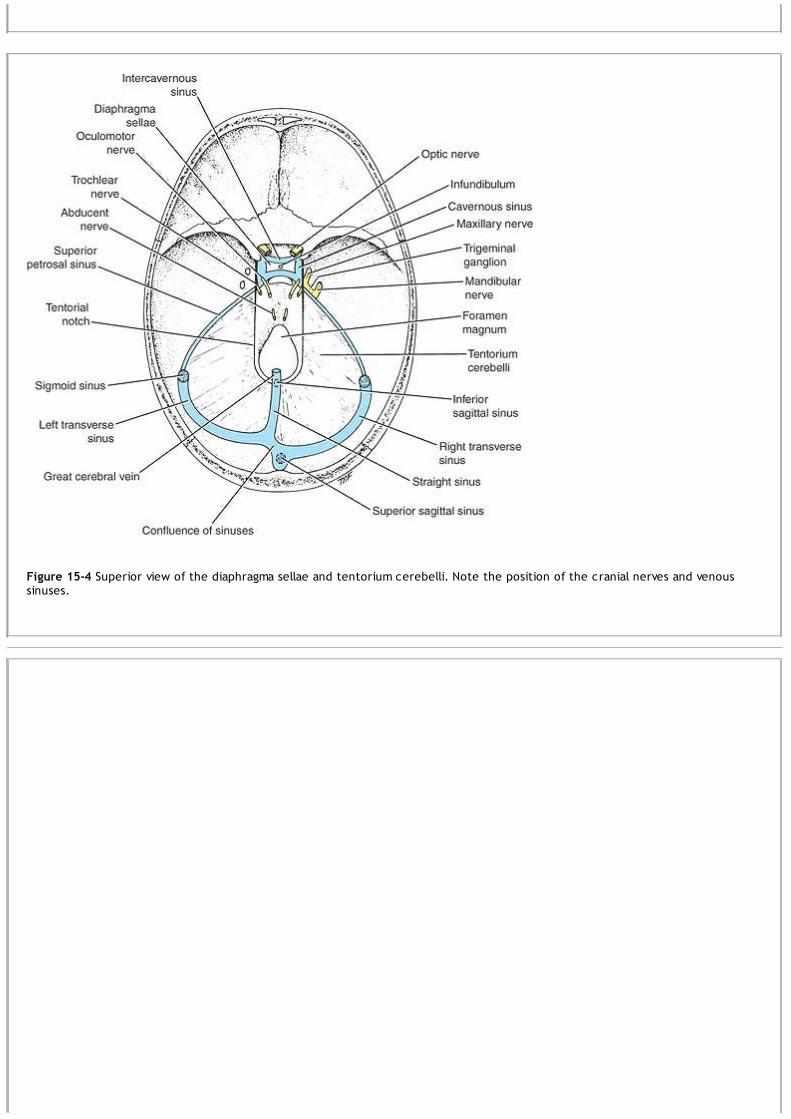
Figure 15-4 Superior view of the diaphragma sellae and tentorium cerebelli. Note the position of the cranial nerves and venoussinuses.

Figure 15-5 Right side of the head showing the relation of the middle meningeal vessels to the layers of the dura mater and theskull.
Dural Arterial SupplyNumerous arteries supply the dura mater from the internal carotid, maxillary, ascending pharyngeal, occipital, and vertebral arteries.From the clinical standpoint, the most important is the middle meningeal artery, which can be damaged in head injuries (Fig. 15-5).
The middle meningeal artery arises from the maxillary artery in the infratemporal fossa. It enters the cranial cavity through the foramenspinosum and then lies between the meningeal and endosteal layers of dura. The artery then runs forward and laterally in a groove on theupper surface of the squamous part of the temporal bone. The anterior branch deeply grooves or tunnels the anterior-inferior angle ofthe parietal bone (Fig. 15-5), and its course corresponds roughly to the line of the underlying precentral gyrus of the brain. The posteriorbranch curves backward and supplies the posterior part of the dura mater (Fig. 15-7).
The meningeal veins lie in the endosteal layer of dura (Fig. 15-5). The middle meningeal vein follows the branches of the middle meningealartery and drains into the pterygoid venous plexus or the sphenoparietal sinus. The veins lie lateral to the arteries.
Dural Venous SinusesThe venous sinuses of the cranial cavity are situated between the layers of the dura mater (Figs. 15-6 and 15-7; see also Figs. 15-3 and 15-4). Their main function is to receive blood from the brain through the cerebral veins and the cerebrospinal fluid from the subarachnoidspace through the arachnoid villi (see Fig. 16-18). The blood in the dural sinuses ultimately drains into the internal jugular veins in theneck. The dural sinuses are lined by endothelium, and their walls are thick but devoid of muscular tissue. They have no valves. Emissaryveins, which are also valveless,connect the dural venous sinuses with the diploic veins of the skull and with the veins of the scalp (Fig. 15-1).

Figure 15-6 Coronal section through the body of the sphenoid bone showing the hypophysis cerebri and cavernous sinuses. Notethe position of the internal carotid artery and the cranial nerves.
The superior sagittal sinus occupies the upper fixed border of the falx cerebri (Figs. 15-1 and 15-4). It begins anteriorly at the foramencecum, where it occasionally receives a vein from the nasal cavity. It runs posteriorly, grooving the vault of the skull; at the internaloccipital protuberance, it deviates to one or the other side (usually the right) and becomes continuous with the corresponding transversesinus. The sinus communicates through small openings with two or three irregularly shaped venous lacunae on each side (Fig. 15-7).Numerous arachnoid villi and granulations project into the lacunae, which also receive the diploic and meningeal veins (Fig. 15-1).
The superior sagittal sinus in its course receives the superior cerebral veins (Fig. 15-1; see Fig. 17-5). At the internal occipitalprotuberance, it is dilated to form the confluence of the sinuses (Fig. 15-4). Here, the superior sagittal sinus usually becomes continuouswith the right transverse sinus; it is connected to the opposite transverse sinus and receives the occipital sinus. The inferior sagittalsinus occupies the free lower margin of the falx cerebri (Fig. 15-1). It runs backward and joins the great cerebral vein at the free marginof the tentorium cerebelli to form the straight sinus (Figs. 15-1 and 15-4). It receives a few cerebral veins from the medial surface of thecerebral hemispheres.
The straight sinus occupies the line of junction of the falx cerebri with the tentorium cerebelli (Figs. 15-1 and 15-4). It is formed by theunion of the inferior sagittal sinus with the great cerebral vein. It ends by turning to the left (sometimes to the right) to form thetransverse sinus.
The transverse sinuses are paired structures that begin at the internal occipital protuberance (Figs. 15-3 and 15-4). The right sinus isusually continuous with the superior sagittal sinus, and the left is continuous with the straight sinus. Each sinus occupies the attachedmargin of the tentorium cerebelli, grooving the occipital bone and the posteroinferior angle of the parietal bone. The transverse sinusesreceive the superior petrosal sinuses, the inferior cerebral and cerebellar veins, and the diploic veins. They end by turning downward asthe sigmoid sinuses (Fig. 15-4).
The sigmoid sinuses are a direct continuation of the transverse sinuses. Each sinus turns downward and medially and grooves the mastoidpart of the temporal bone (Fig. 15-4). It is here that the sinus lies posterior to themastoid antrum. The sinus then turns forward and then inferiorly through the posterior part of the jugular foramen to become continuouswith the superior bulb of the internal jugular vein.

Figure 15-7 Superior view of the head with the calvarium removed. The greater part of the endosteal layer of dura mater has beenremoved, exposing the underlying meningeal layer of dura and the interior of the superior sagittal venous sinus.
The occipital sinus is a small sinus occupying the attached margin of the falx cerebelli. It commences near the foramen magnum, where itcommunicates with the vertebral veins and drains into the confluence of sinuses.
The cavernous sinuses are situated in the middle cranial fossa on each side of the body of the sphenoid bone (Fig. 15-6). Numeroustrabeculae cross their interior, giving them a spongy appearance, hence the name. Each sinus extends from the superior orbital fissure infront to the apex of the petrous part of the temporal bone behind.
The internal carotid artery, surrounded by its sympathetic nerve plexus, runs forward through the sinus (Fig. 15-6). The abducent nervealso passes through the sinus. The internal carotid artery and the nerves are separated from the blood by an endothelial covering.
The third and fourth cranial nerves and the ophthalmic and maxillary divisions of the trigeminal nerve run forward in the lateral wall ofthe sinus (Fig. 15-6). They lie between the endothelial lining and the dura mater. The tributaries are the superior and inferior ophthalmicveins, the inferior cerebral veins, the sphenoparietal sinus, and the central vein of the retina.
The sinus drains posteriorly into the superior and inferior petrosal sinuses and inferiorly into the pterygoid venous plexus.
The two sinuses communicate with each other by means of the anterior and posterior intercavernous sinuses, which run in thediaphragma sellae anterior and posterior to the stalk of the hypophysis cerebri (Fig. 15-4). Each sinus has an important communication withthe facial vein through the superior ophthalmic vein. (This is a route by which infection can travel from the facial skin to the cavernoussinus.)

Figure 15-8 Sagittal section of the eyeball showing the attachment of the meninges to the sclera. Note the extension of thesubarachnoid space around the optic nerve to the eyeball.
The superior and inferior petrosal sinuses are small sinuses situated on the superior and inferior borders of the petrous part of thetemporal bone on each side of the skull (Fig. 15-4). Each superior sinus drains the cavernous sinus into the transverse sinus, and eachinferior sinus drains the cavernous sinus into the internal jugular vein.
Arachnoid MaterThe arachnoid mater is a delicate, impermeable membrane covering the brain and lying between the pia mater internally and the duramater externally (Fig. 15-1). It is separated from the dura by a potential space, the subdural space, filled by a film of fluid; it is separatedfrom the pia by the subarachnoid space, which is filled with cerebrospinal fluid. The outer and inner surfaces of the arachnoid arecovered with flattened mesothelial cells.
The arachnoid bridges over the sulci on the surface of the brain, and in certain situations, the arachnoid and pia are widely separated toform the subarachnoid cisternae. The cisterna cerebellomedullaris lies between the inferior surface of the cerebellum and the roof ofthe fourth ventricle. The cisterna interpeduncularis lies between the two cerebral peduncles. All the cisternae are in freecommunication with one another and with the remainder of the subarachnoid space.
In certain areas, the arachnoid projects into the venous sinuses to form arachnoid villi. The arachnoid villi are most numerous along thesuperior sagittal sinus. Aggregations of arachnoid villi are referred to as arachnoid granulations (Fig. 15-7). Arachnoid villi serve as siteswhere the cerebrospinal fluid diffuses into the bloodstream.
The arachnoid is connected to the pia mater across the fluid-filled subarachnoid space by delicate strands of fibrous tissue.
Structures passing to and from the brain to the skull or its foramina must pass through the subarachnoid space. All the cerebral arteriesand veins lie in the space, as do the cranial nerves (Figs. 15-1 and 15-6). The arachnoid fuses with the epineurium of the nerves at theirpoint of exit from the skull (Fig. 15-2B). In the case of the optic nerve, the arachnoid forms a sheath for the nerve, which extends into theorbital cavity through the optic canal and fuses with the sclera of the eyeball (Fig. 15-8). Thus, the subarachnoid space extends aroundthe optic nerve as far as the eyeball.
The cerebrospinal fluid is produced by the choroid plexuses within the lateral, third, and fourth ventricles of the brain. It escapes fromthe ventricular system of the brain through the three foramina in the roof of the fourth ventricle and so enters the subarachnoid space. Itnow circulates both upward over the surfaces of the cerebral hemispheres and downward around the spinal cord. The spinal subarachnoidspace extends down as far as the second sacral vertebra (see p. 437). Eventually, the fluid enters the bloodstream by passing into thearachnoid villi and diffusing through their walls.
In addition to removing waste products associated with neuronal activity, the cerebrospinal fluid provides a fluid medium in which thebrain floats. This mechanism effectively protects the brain from trauma. In addition, the fluid is now believed to play a role in hormonaltransport.

Figure 15-9 A: Brain, spinal cord, spinal nerve roots, and spinal nerves as seen on their posterior aspect. B: Transverse sectionthrough the thoracic region of the spinal cord showing the anterior and posterior roots of a spinal nerve and the meninges. C:Posterior view of the lower end of the spinal cord and cauda equina showing their relationship with the lumbar vertebrae, sacrum,and coccyx.
Pia MaterThe pia mater is a vascular membrane covered by flattened mesothelial cells. It closely invests the brain, covering the gyri and descendinginto the deepest sulci (Fig. 15-1). It extends out over the cranial nerves and fuses with their epineurium. The cerebral arteries enteringthe substance of the brain carry a sheath of pia with them.
The pia mater forms the tela choroidea of the roof of the third and fourth ventricles of the brain, and it fuses with the ependyma to formthe choroid plexuses in the lateral, third, and fourth ventricles of the brain.
Meninges of the Spinal Cord
Dura MaterThe dura mater is a dense, strong, fibrous membrane that encloses the spinal cord and the cauda equina (Figs. 15-9, 15-10 and 15-11). It iscontinuous above through the foramen magnum with the meningeal layer of dura covering the brain. Inferiorly, it ends on the filumterminale at the level of the lower border of the second sacral vertebra. The dural sheath lies loosely in the vertebral canal and isseparated from the wall of the canal by the extradural space. This contains loose areolar tissue and the internal vertebral venous plexus.The dura mater extends along each nerve root and becomes continuous with the connective tissue surrounding each spinal nerve(epineurium). The inner surface of the dura mater is in contact with the arachnoid mater (see Fig. 4-5).

Figure 15-10 Dissection of the back of the head and neck. The greater part of the occipital bone has been removed, exposing theperiosteal layer of dura. On the right side, a window has been made in the dura below the transverse venous sinus to expose thecerebellum and the medulla oblongata in the posterior cranial fossa. In the neck, the dura and arachnoid have been incised in themidline to expose the spinal cord and rootlets of the cervical spinal nerves. Note the cervical spinal nerves leaving the vertebralcanal enveloped in a meningeal sheath.
Arachnoid MaterThe arachnoid mater is a delicate impermeable membrane that covers the spinal cord and lies between the pia mater internally and duramater externally (Figs. 15-9 and 15-10). It is separated from the pia mater by a wide space, the subarachnoid space, which is filled withcerebrospinal fluid. The subarachnoid space is crossed by a number of fine strands of connective tissue. The arachnoid mater iscontinuous above through the foramen magnum with the arachnoid covering the brain. Inferiorly, it ends on the filum terminale at thelevel of the lower border of the second sacral vertebra (Fig. 15-9). The arachnoid mater continues along the spinal nerve roots, formingsmall lateral extensions of the subarachnoid space.
Pia MaterThe pia mater, a vascular membrane that closely covers the spinal cord (Fig. 15-9), is thickened on either side between the nerve roots toform the ligamentum denticulatum, which passes laterally to adhere to the arachnoid and dura. It is by this means that the spinal cord issuspended in the middle of the dural sheath. The pia mater extends along each nerve root and becomes continuous with the connectivetissue surrounding each spinal nerve (Fig. 15-9).

Figure 15-11 Dissection of the lower part of the back, including a complete laminectomy of the lumbar and sacral regions of thevertebral column. The meningeal sheath has been incised and reflected laterally, exposing the subarachnoid space, the lower endof the spinal cord, and the cauda equina. Note the filum terminale surrounded by the anterior and posterior nerve roots of thelumbar and sacral spinal nerves forming the cauda equina.
Clinical NotesFunctional Significance of the MeningesThe meninges of the brain and spinal cord form three concentric membranous coverings. The outermost
covering, the dura mater, by virtue of its toughness, serves to protect the underlying nervous tissue. The duraprotects the cranial nerves by forming a sheath that covers each cranial nerve for a short distance as it passesthrough foramina in the skull. The dura mater also provides each spinal nerve root with a protective sheath.In the skull, the falx cerebri, which is a vertical sheet of dura between the cerebral hemispheres, and the tentoriumcerebelli, which is a horizontal sheet that projects forward between the cerebrum and cerebellum, serve to limitexcessive movements of the brain within the skull.The arachnoid mater is a much thinner impermeable membrane that loosely covers the brain. The interval between thearachnoid and pia mater, the subarachnoid space, is filled with cerebrospinal fluid. The cerebrospinal fluid givesbuoyancy to the brain and protects the nervous tissue from mechanical forces applied to the skull.The pia mater is a vascular membrane that closely invests and supports the brain and spinal cord.
Excessive Movements of the Brain Relative to the Skull and Meninges in Head InjuriesWhen a moving patient's head is suddenly halted, the momentum of the brain causes it to travel onward until itsmovement is resisted by the skull or the strong septa of the dura mater. In lateral movements, the lateral surface ofone hemisphere hits the side of the skull, and the medial surface of the opposite hemisphere hits the side of the falxcerebri. In superior movements, the superior surfaces of the cerebral hemispheres hit the vault of the skull, and thesuperior surface of the corpus callosum hits the sharp free edge of the falx cerebri; the superior surface of thecerebellum presses against the inferior surface of the tentorium cerebelli.Movements of the brain relative to the skull and dural septa may seriously injure the cranial nerves that are tetheredas they pass through the various foramina. Furthermore, the fragile cortical veins that drain into the dural sinuses maybe torn, resulting in severe subdural or subarachnoid hemorrhage. The tortuous arteries, with their strong walls, arerarely damaged.
Intracranial Hemorrhage and the MeningesEpidural HemorrhageEpidural hemorrhage results from injuries to the meningeal arteries or veins. The most common artery to be damagedis the anterior division of the middle meningeal artery. A comparatively minor blow to the side of the head, resultingin fracture of the skull in the region of the anterior-inferior portion of the parietal bone, may sever the artery. Arterial

or venous injury is especially liable to occur if the vessels enter a bony canal in this region. Bleeding occurs and stripsup the meningeal layer of dura from the internal surface of the skull. The intracranial pressure rises, and the enlargingblood clot exerts local pressure on the underlying motor area in the precentral gyrus. Blood also passes laterallythrough the fracture line to form a soft swelling under the temporalis muscle.To stop the hemorrhage, the torn artery or vein must be ligated or plugged. The burr hole through the skull wallshould be placed about 1-1/2 inches (4 cm) above the midpoint of the zygomatic arch.
Subdural HemorrhageSubdural hemorrhage results from tearing of the superior cerebral veins at their point of entrance into the superiorsagittal sinus. The cause is usually a blow on the front or the back of the head, causing excessive anteroposteriordisplacement of the brain within the skull. Acute and chronic forms of the condition occur.
Computed Tomography Scans of Epidural and Subdural HematomasThe different appearances of the blood clots in these two conditions as seen on CT scans is related to the anatomy ofthe area (Fig. 15-12). In an epidural hemorrhage, the blood strips up the meningeal layer of the dura from theendosteal layer of dura (periosteum of the skull), producing a lens-shaped hyperdense collection of blood thatcompresses the brain and displaces the midline structures to the opposite side. The shape of the blood clot isdetermined by the adherence of the meningeal layer of dura to the periosteal layer of dura.In patients with subdural hematoma, the blood accumulates in the extensive potential space between the meningeallayer of dura and the arachnoid, producing a long crescent-shaped, hyperdense rim of blood that extends fromanterior to posterior along the inner surface of the skull. With a large hematoma, the brain sulci are obliterated, andthe midline structures are displaced to the opposite side.
Subarachnoid and Cerebral HemorrhagesSubarachnoid and cerebral hemorrhages are described on page 484.
Intracranial Hemorrhage in the InfantIntracranial hemorrhage may occur during birth and may result from excessive molding of the head. Bleeding may occurfrom the cerebral veins or the venous sinuses. Excessive anteroposterior compression of the head often tears theanterior attachment of the falx cerebri from the tentorium cerebelli. Bleeding then takes place from the great cerebralveins, the straight sinus, or the inferior sagittal sinus.The shaken-baby syndrome is described on page 23.
HeadacheThe brain itself is insensitive to pain. It follows that headaches are due to the stimulation of receptors outside thebrain.
Meningeal HeadachesThe dura mater receives its sensory nerve supply from the trigeminal and the first three cervical nerves. The duraabove the tentorium is innervated by the trigeminal nerve, and the headache is referred to the forehead and face. Thedura below the tentorium is innervated by the cervical nerves, and the headache is referred to the back of the headand neck. Meningitis, or inflammation of the meninges, causes severe headache over the entire head and back of theneck.
Headaches Caused by Cerebral TumorsAn expanding tumor with its associated raised intracranial pressure produces severe, continuous, and progressiveheadache caused by the irritation and stretching of the dura. A tumor above the tentorium tends to produce aheadache referred to the front of the head, while a tumor below the tentorium produces a headache referred to theback of the head.
Migraine HeadacheMigraine is a common form of headache, which may be unilateral or bilateral, recurring at intervals and associated withprodromal visual disturbances. The prodromal visual disturbances are thought to be due to sympatheticvasoconstriction of the cerebral arteries supplying the visual cortex. The headache is chiefly due to the dilatation andstretching of other cerebral arteries and branches of the external carotid artery. The disease therefore appears toaffect arteries both inside and outside the skull, and its cause is unknown, although genetic, hormonal, andbiochemical factors may initiate an attack. It is has been found that beta-blockers bring relief to some patients due tothe reduction in cerebral vasodilation.
Alcoholic HeadacheAlcoholic headache is due to the direct toxic effect of alcohol on the meninges.
Headaches Due to Diseases of the Teeth, Paranasal Sinuses, and EyesDental infection and sinusitis are common causes of headache. The pain is referred to the skin of the face and theforehead along the branches of the trigeminal nerve. Tonic spasm of the ciliary muscle of the eye, when attempting tofocus on an object for prolonged periods (e.g., reading small print), may cause severe orbital headache. This commonlyoccurs in individuals who need lenses for the correction of presbyopia.

P.440P.441P.442
Figure 15-12 Diagrammatic representation of an epidural hemorrhage and a subdural hemorrhage. A: Epidural hemorrhage fromthe middle meningeal artery or vein on the left side. The hematoma is lens shaped and occupies the space between theendosteal layer of dura (periosteum of the skull) and the meningeal layer of dura (true dura, hence the name epidural). B:Subdural hemorrhage from the cerebral veins at the site of entrance into the venous sinus on the right side. The hematoma iscrescent shaped and occupies the space between the meningeal layer of dura and the arachnoid (i.e., beneath the dura).
Clinical Problem Solving1. In an automobile accident, which structures present within the skull limit damage to the cerebral hemispheres andother parts of the brain? Which blood vessels are damaged more commonly, the cerebral arteries or the cerebralveins? Are cranial nerves likely to be damaged in head injuries? If so, which ones are damaged most commonly, andwhat is the reason for their increased susceptibility?View Answer
2. While performing an autopsy on a patient who had died of a meningioma, the pathologist explained to a group ofstudents that these tumors arise from the arachnoid mater. She explained that they occur in those areas in which thearachnoid pierces the dura to form the arachnoid villi that project into the dural venous sinuses. She then asked thestudents where they would expect to find meningiomas. How would you answer that question?View Answer
3. A 10-year-old girl was admitted to hospital for surgical correction of medial strabismus of the right eye. Twenty-fourhours after successful completion of the operation, it was noted that her right eyeball was projecting forwardexcessively (proptosis), and the conjunctiva of the right eye was inflamed. A watery, purulent discharge could beexpressed from beneath the eyelids. The ophthalmologist was greatly concerned because he did not want thecomplication of cavernous sinus thrombosis to occur. What is the connection between infection of the eye andcavernous sinus thrombosis? Is cavernous sinus thrombosis a serious condition?View Answer
4. On examination, a 41-year-old man was found to have paralysis of the lateral rectus muscle of his left eye; the leftpupil was dilated but reacted slowly to light, and there was some anesthesia of the skin over the left side of the

forehead. A carotid arteriogram revealed the presence of an aneurysm of the right internal carotid artery situated inthe cavernous sinus. Using your knowledge of anatomy, explain the clinical findings on physical examination.View Answer
5. On ophthalmoscopic examination, a 45-year-old woman was found to have edema of both optic discs (bilateralpapilledema) and congestion of the retinal veins. The cause of the condition was found to be a rapidly growingintracranial tumor. Using your knowledge of anatomy, explain the papilledema. Why does the patient exhibit bilateralpapilledema?View Answer
6. A pediatrician was observing a 6-year-old boy playing with his toys. He noted that the child had perfectly normal useof his arms but that his legs were stiff, and when he walked, he tended to cross his legs and had a scissorlike gait. Adiagnosis of cerebral diplegia1 secondary to birth injuries was made. Apparently, the child was born prematurely, andhe was a breech presentation. Using your knowledge of anatomy, explain what happens to the fetal skull bonesduring delivery. Why are the dural venous sinuses likely to be damaged at birth? Why is cerebral hemorrhage morelikely to occur in a premature baby with a malpresentation?View Answer
7. A 25-year-old woman was admitted to the emergency department unconscious. She had apparently been hit on theside of the head by a car while crossing the road. Within an hour, her state of unconsciousness deepened. Onexamination, she was found to have a large, doughlike swelling over the right temporalis muscle. She also had thesigns of right-sided hemiplegia. Later, a right-sided, fixed, dilated pupil developed. A lateral radiograph of the skullshowed a fracture line across the groove for the anterior division of the right middle meningeal artery. Her comadeepened, and she died 4 hours after the accident. Using your knowledge of neuroanatomy, make a diagnosis in thiscase. Explain the clinical findings. How could you account for the homolateral hemiplegia?View Answer
8. A 50-year-old woman complaining of a severe headache of 3 days duration visited her physician. She said that theheadache had started getting very severe about 1 hour after she had hit her head on the mantelpiece after bendingdown to poke the fire. She was admitted to the hospital for observation. Three hours later, it was noticed that shewas becoming mentally confused and also that she was developing a right-sided hemiplegia on the side of the bodyopposite to the head injury. She had exaggeration of the deep reflexes and a positive Babinski response on the rightside. Examination of the cerebrospinal fluid with a spinal tap showed a raised pressure and the presence of blood inthe fluid. Radiographic examination showed no fracture of the skull. A CT scan revealed the presence of a subduralhematoma. What exactly is a subdural hematoma?View Answer
Review QuestionsDirections: Each of the numbered items in this section is followed by answers. Select the ONE lettered answerthat is CORRECT.1. The following statements concern the meninges of the brain:
(a) Both layers of the dura mater covering the brain are continuous through the foramen magnum with the duracovering the spinal cord.(b) The periosteal layer of dura mater is not continuous with the sutural ligaments of the skull.(c) As each cranial nerve passes through a foramen in the skull, it is surrounded by a tubular sheath of arachnoidmater only.(d) The cranial venous sinuses run between the meningeal and endosteal layers of dura mater.(e) The meninges extend anteriorly through the optic canal and fuse with the periosteum of the orbital cavity.
View Answer
2. The following general statements concern the meninges:(a) The cisterna cerebellomedullaris lies between the inferior surface of the cerebellum and the roof of the fourthventricle and contains lymph.(b) The arachnoid mater is permeable to cerebrospinal fluid.(c) The cerebrospinal fluid in the arachnoid villi is able to drain into the venous sinuses through small tubuleslined with endothelial cells.(d) The arachnoid mater surrounding the spinal cord ends inferiorly on the filum terminale at the level of thelower border of the first sacral vertebra.(e) The extradural space that separates the dural sheath of the spinal cord and the walls of the vertebral canalcontains the external vertebral venous plexus.
View Answer
3. The following statements concern the tentorium cerebelli:(a) The free border is attached anteriorly to the posterior clinoid processes.(b) It is formed from the meningeal layer of the dura mater.(c) It separates the cerebellum from the temporal lobes of the brain.(d) The sigmoid sinus lies within its attached border to the occipital bone.
(e) In the anterior edge, there is the occipital venous sinus.View Answer
4. The following statements concern headache:(a) Brain tissue is insensitive to pain.(b) Intracranial pain arises from receptors situated in the pia mater.(c) An expanding cerebral tumor located in the posterior cranial fossa would produce pain referred to the face.(d) Migraine headache is believed to be due to dilation of the cerebral veins.(e) Headaches associated with presbyopia are due to tonic spasm of the frontalis muscles of the forehead.
View Answer
5. The following statements concern the subarachnoid space:(a) It is filled with cerebrospinal fluid.(b) It extends inferiorly as far as the fourth sacral vertebra.(c) The cerebral arteries and veins are not located in the subarachnoid space.(d) The cranial nerves lie outside the subarachnoid space in sheaths derived from the dura mater.(e) The arachnoid villi project into the venous sinuses as large outpouchings of the subarachnoid space.
View Answer
6. The following statements concern the cavernous sinus:(a) It has the external carotid artery passing through it.(b) It has the oculomotor, trochlear, and ophthalmic divisions of the trigeminal nerve in its medial wall.(c) It drains directly posteriorly into the straight sinus.(d) It does not communicate with the facial vein.(e) It is related medially to the pituitary gland and the sphenoid air sinus.
View Answer
7. The following structure limits rotatory movements of the brain within the skull:(a) Tentorium cerebelli(b) Diaphragma sellae(c) Falx cerebri(d) Dorsum cellae(e) Squamous part of the temporal bone
View Answer
8. The following nerves are sensory to the dura mater:(a) Oculomotor nerve(b) Trochlear nerve(c) Sixth cervical spinal nerve(d) Trigeminal nerve(e) Hypoglossal nerve
View Answer
Additional Reading
Clifton, G. L. Hypothermia in Acute Brain Injury. Newsletter (vol. 7, no. 2), The Neuroscience Research Center, The University ofTexas-Houston Health Science Center, 2000.
Dalessio, D. J., and Silberstein, S. D. Wolff's Headache and Other Head Pain (6th ed.). New York: Oxford University Press, 1993.
Durand, M. L., Calderwood, S. B., Weber, D. J., M iller, S. I., Southwick, F. S., Caviness, V. S., Jr., et al. Acute bacterial meningitis inadults. N. Engl. J. Med. 328:21, 1993.
Goetz, C. G. Textbook of Clinical Neurology (2nd ed.). Philadelphia: Saunders, 2003.
Lance, J. W. The Mechanism and Management of Headache (5th ed.). London: Butterworth, 1993.

Livingston, D. H., Lavery, R. F., Mosenthal, A. C., Knudson, M. M., Lee, S., Morabito, D., et al. Recovery at one year following isolatedtraumatic brain injury: A Western Trauma Association prospective multicenter trial. J. Trauma 59:1298, 2005.
Okie, S. Traumatic brain injury in the war zone. N. Engl. J. Med. 352:20, 2005.
Rowland, L. P. Merritt's Neurology (10th ed.). Philadelphia: Lippincott Williams & Wilkins, 2000.
Snell, R. S. Clinical Anatomy by Regions (8th ed.). Philadelphia: Lippincott Williams & Wilkins, 2008.
Snell, R. S. Clinical Anatomy by Systems. Philadelphia: Lippincott Williams & Wilkins, 2007.
Snell, R. S., and Smith, M. S. Clinical Anatomy for Emergency Medicine. St. Louis: Mosby, 1993.
Standring, S. (ed.). Gray's Anatomy (39th Br. ed.). London: Elsevier Churchill Livingstone, 2005.
Welch, K. M. A. The therapeutics of migraine. Curr. Opin. Neurol. Neurosurg. 6:264, 1993.

Authors: Snell, Richard S.Title: Clinical Neuroanatomy, 7th Edition
Copyright ©2010 Lippincott Williams & Wilkins
> Table of Contents > Chapter 16 - The Ventr icular System, the Cer ebr ospinal Fluid, and the Blood-Br ain and Blood–Cer ebr ospinal Fluid Bar r ier s
Chapter 16
The Ventricular System, the Cerebrospinal Fluid, and the Blood-Brain and Blood–CerebrospinalFluid Barriers
A 26-year-old woman involved in an automobile accident was admitted to the emergency department. Her mother, who was also in the car,told the physician that at the time of impact, her daughter's head was thrown forward against the windshield.
On examination, the patient was unconscious and showed evidence of a severe head injury on the left side. After a thorough physicalexamination, the physician decided to perform a spinal tap. The cerebrospinal fluid pressure was 160 mm of water, and two samples of thefluid were collected. The specimens showed red blood cells at the bottom of the tubes, and the supernatant fluid was blood stained.After standing for an hour, the supernatant fluid in both tubes became colorless.
The physician made the diagnosis of subarachnoid hemorrhage secondary to the head injury. The blood could have originated from asevere fracture of the skull, damage to one of the cerebral blood vessels, or a tear involving the brain or covering meninges. The physicianwas confident that the blood in the cerebrospinal fluid specimens did not originate from an accidental puncture of a vertebral vein duringthe spinal tap procedure. He eliminated this possibility by removing the two specimens of the fluid. If a local vein had been punctured bythe needle, the first specimen would have been blood stained, and the second specimen most probably would have been clear. With thispatient, both specimens were uniformly blood stained, so the blood was in the subarachnoid space.
The management of this patient in the emergency department and the evaluation of the spinal tap specimen depended on knowledge ofthe cerebrospinal fluid system and the anatomy involved in the spinal tap procedure.
Chapter Objectives
To learn the locations, the functions, the origins, and the fate of the cerebrospinal fluidTo understand the structure and function of the blood-brain and blood–cerebrospinal fluid barriersTo learn how certain parts of the brain are protected from potentially toxic drugs or other exogenous materials
Ventricular SystemThe ventricles are four fluid-filled cavities located within the brain; these are the two lateral ventricles, the third ventricle, andthe fourth ventricle (Fig. 16-1; see also Atlas Plates 3, 4, 7 and 8). The two lateral ventricles communicate through the
interventricular foramina (of Monro) with the third ventricle. The third ventricle is connected to the fourth ventricle by the narrowcerebral aqueduct (aqueduct of Sylvius). The fourth ventricle, in turn, is continuous with the narrow central canal of the spinal cord and,through the three foramina in its roof, with the subarachnoid space. The central canal in the spinal cord has a small dilatation at itsinferior end, referred to as the terminal ventricle (Fig. 16-1).
The ventricles are lined throughout with ependyma and are filled with cerebrospinal fluid. The ventricles are developmentally derivedfrom the cavity of the neural tube.
Lateral VentriclesThere are two large lateral ventricles, and one is present in each cerebral hemisphere (Fig. 16-2). The ventricle is a roughly C-shapedcavity and may be divided into a body, which occupies the parietal lobe and from which anterior, posterior, and inferior horns extendinto the frontal, occipital, and temporal lobes, respectively. The lateral ventricle communicates with the cavity of the third ventriclethrough the interventricular foramen (Figs. 16-2, 16-3 and 16-4). This opening, which lies in the anterior part of the medial wall of theventricle, is bounded anteriorly by the anterior column of the fornix and posteriorly by the anterior end of the thalamus.
The body of the lateral ventricle extends from the interventricular foramen posteriorly as far as the posterior end of the thalamus. Here,it becomes continuous with the posterior and the inferior horns. The body of the lateral ventricle has a roof, a floor, and a medial wall(Fig. 16-5).
The roof is formed by the undersurface of the corpus callosum (Fig. 16-5). The floor is formed by the body of the caudate nucleus and thelateral margin of the thalamus. The superior surface of the thalamus is obscured in its medial part by the body of the fornix. The choroidplexus of the ventricle projects into the body of the ventricle through the slitlike gap between the body of the fornix and the superiorsurface of the thalamus. This slitlike gap is known as the choroidal fissure; through it, the blood vessels of the plexus invaginate the piamater of the tela choroidea and the ependyma of the lateral ventricle. The medial wall is formed by the septum pellucidum anteriorly;posteriorly, the roof and the floor come together on the medial wall (Fig. 16-5).
The anterior horn of the lateral ventricle extends forward into the frontal lobe (Figs. 16-2 and 16-3). It is continuous posteriorly with thebody of the ventricle at the interventricular foramen. The anterior horn has a roof, a floor, and a medial wall. The roof is formed by theundersurface of the anterior part of the corpus callosum; the genu of the corpus callosum limits the anterior horn anteriorly (Fig. 16-5).The floor is formed by the rounded head of the caudate nucleus; medially, a small portion is formed by the superior surface of therostrum of the corpus callosum. The medial wall is formed by the septum pellucidum and the anterior column of the fornix (Fig. 16-5).

The posterior horn of the lateral ventricle extends posteriorly into the occipital lobe (Figs. 16-2 and 16-3). The roof and lateral wall areformed by the fibers of the tapetum of the corpus callosum. Lateral to the tapetum are the fibers of the optic radiation (Fig. 16-5). Themedial wall of the posterior horn has two elevations. The superior swelling is caused by the splenial fibers of the corpus callosum, calledthe forceps major, passing posteriorly into the occipital lobe; this superior swelling is referred to as the bulb of the posterior horn. Theinferior swelling is produced by the calcarine sulcus and is called the calcar avis (Fig. 16-5).
The inferior horn of the lateral ventricle extends anteriorly into the temporal lobe (Figs. 16-2 and 16-3). The inferior horn has a roof and afloor (Fig. 16-5).
The roof is formed by the inferior surface of the tapetum of the corpus callosum and by the tail of the caudate nucleus (see Fig. 9-5). Thelatter passes anteriorly to end in the amygdaloid nucleus. Medial to the tail of the caudate nucleus is the stria terminalis, which alsoends anteriorly in the amygdaloid nucleus.
The floor is formed laterally by the collateral eminence, produced by the collateral fissure, and medially by the hippocampus (see Figs. 9-3 and 9-4). The anterior end of the hippocampus is expanded and slightly furrowed to form the pes hippocampus. The hippocampus iscomposed of gray matter; however, the ventricular surface of the hippocampus is covered by a thin layer of white matter called thealveus, which is formed from the axons of the cells of the hippocampus. These axons converge on the medial border of the hippocampusto form a bundle known as the fimbria. The fimbria of the hippocampus becomes continuous posteriorly with the posterior column of thefornix.
Figure 16-1 Origin and circulation of the cerebrospinal fluid (pink).

Figure 16-2 Cast of the ventricular cavities of the brain. A: Lateral view. B: Anterior view.

Figure 16-3 Ventricular cavities of the brain. A: Lateral view. B: Superior view.
In the interval between the stria terminalis and the fimbria is the temporal part of the choroidal fissure. It is here that the lower part ofthe choroid plexus of the lateral ventricle invaginates the ependyma from the medial side and closes the fissure (see Fig. 16-8B).
Choroid Plexus of the Lateral VentricleThe choroid plexus projects into the ventricle on its medial aspect and is a vascular fringe composed of pia mater covered with theependymal lining of the ventricular cavity (Fig. 16-6). The choroid plexus is, in fact, the irregular lateral edge of the tela choroidea, whichis a two-layered fold of pia mater situated between the fornix superiorly and the upper surface of the thalamus (Fig. 16-8). At the junctionof the body of the lateral ventricle and the inferior horn, the choroid plexus is continued into the inferior horn and projects through thechoroidal fissure. The function of the choroid plexus is to produce cerebrospinal fluid.
Third VentricleThe third ventricle is a slitlike cleft between the two thalami. It communicates anteriorly with the lateral ventricles through theinterventricular foramina (of Monro) and posteriorly with the fourth ventricle through the cerebral aqueduct (of Sylvius) (Fig. 16-4). Thewalls of the third ventricle are described on page 257.
Choroid Plexuses of the Third VentricleThe choroid plexuses are formed from the tela choroidea situated above the roof of the ventricle (Fig. 16-6). The vascular tela choroideaprojects downward on each side of the midline, invaginating the ependymal roof of the ventricle.The two vascular ridges or fringes that hang from the roof of the third ventricle form the choroid plexuses. The function of the choroidplexuses is to produce cerebrospinal fluid.

Figure 16-4 Sagittal section of the brain showing the third ventricle, the cerebral aqueduct, and the fourth ventricle.
The blood supply of the tela choroidea and, therefore, also of the choroid plexuses of the third and lateral ventricles is derived from thechoroidal branches of the internal carotid and basilar arteries. The venous blood drains into the internal cerebral veins, which unite toform the great cerebral vein. The great cerebral vein joins the inferior sagittal sinus to form the straight sinus.
Cerebral AqueductThe cerebral aqueduct (aqueduct of Sylvius), a narrow channel about ¾ of an inch (1.8 cm) long, connects the third ventricle with thefourth ventricle (Figs. 16-2 and 16-3). It is lined with ependyma and is surrounded by a layer of gray matter called the central gray. Thedirection of flow of cerebrospinal fluid is from the third to the fourth ventricle. There is no choroid plexus in the cerebral aqueduct.
Fourth VentricleThe fourth ventricle is a tent-shaped cavity filled with cerebrospinal fluid. It is situated anterior to the cerebellum and posterior to thepons and the superior half of the medulla oblongata (Figs. 16-4, 16-7, and 16-9). It is lined with ependyma and is continuous above with thecerebral aqueduct of the midbrain and below with the central canal of the medulla oblongata and the spinal cord (Fig. 16-3). The fourthventricle possesses lateral boundaries, a roof, and a rhomboid-shaped floor.
Lateral BoundariesThe caudal part of each lateral boundary is formed by the inferior cerebellar peduncle (Fig. 16-10). The cranial part of each lateralboundary is formed by the superior cerebellar peduncle.

Figure 16-5 Coronal sections of the brain passing through the anterior horn of the lateral ventricle (A), the body of the lateralventricle (B), and the posterior horn of the lateral ventricle (C).

Figure 16-6 Schematic diagram of a coronal section of the third and lateral ventricles at the site of the interventricular foraminashowing the structure of the tela choroidea and its relationship with the ependyma and pia mater.
Figure 16-7 Sagittal section of the fourth ventricle showing the origin and circulation of the cerebrospinal fluid. Note the positionof the foramen of Magendie.

P.455
Figure 16-8 Coronal section of the cavities of the third and lateral ventricles (A) and the cavity of the inferior horn of the lateralventricle (B).
Roof or Posterior WallThe tent-shaped roof projects into the cerebellum (Figs. 16-7 and 16-9). The superior part is formed by the medial borders of the twosuperior cerebellar peduncles and a connecting sheet of white matter called the superior medullary velum (Fig. 16-11). The inferior partof the roof is formed by the inferior medullary velum, which consists of a thin sheet devoid of nervous tissue and formed by theventricular ependyma and its posterior covering of pia mater (Fig. 16-12). This part of the roof is pierced in the midline by a large aperture,the median aperture or foramen of Magendie (Figs. 16-11 and 16-12). Lateral recesses extend laterally around the sides of the medulla andopen anteriorly as the lateral openings of the fourth ventricle, or the foramina of Luschka (Fig. 16-13). Thus, the cavity of the fourthventricle communicates with the subarachnoid space through a single median opening and two lateral apertures. These importantopenings permit the cerebrospinal fluid to flow from the ventricular system into the subarachnoid space.
Floor or Rhomboid FossaThe diamond-shaped floor is formed by the posterior surface of the pons and the cranial half of the medulla oblongata (Fig. 16-10). Thefloor is divided into symmetrical halves by the median sulcus. On each side of this sulcus, there is an elevation, the medial eminence,which isbounded laterally by another sulcus, the sulcus limitans. Lateral to the sulcus limitans, there is an area known as the vestibulararea (Figs. 16-10 and 16-11). The vestibular nuclei lie beneath the vestibular area.

Figure 16-9 Sagittal section through the brainstem and the cerebellum showing the fourth ventricle.
Figure 16-10 Posterior surface of the brainstem showing the floor of the fourth ventricle. The cerebellum has been removed.

Figure 16-11 Posterior view of the cavity of the fourth ventricle. A: The vermis of the cerebellum has been divided in the midline,and the cerebellar hemispheres have been displaced laterally. B: The greater part of the cerebellum has been removed, leaving thesuperior and inferior medullary vela. Note that the right half of the inferior medullary velum has been reflected inferiorly to revealthe choroid plexus.
The facial colliculus is a slight swelling at the inferior end of the medial eminence that is produced by the fibers from the motor nucleusof the facial nerve looping over the abducens nucleus (Fig. 16-14). At the superior end of the sulcus limitans, there is a bluish-gray area,produced by a cluster of nerve cells containing melanin pigment; the cluster of cells is called the substantia ferruginea. Strands of nervefibers, the stria medullaris, derived from the arcuate nuclei, emerge from the median sulcus and pass laterally over the medial eminenceand the vestibular area and enter the inferior cerebellar peduncle to reach the cerebellum (Fig. 16-10).

Figure 16-12 Posterior view of the roof of the fourth ventricle. The cerebellum has been displaced superiorly to show the largemedian aperture (foramen of Magendie).
Inferior to the stria medullaris, the following features should be recognized in the floor of the ventricle. The most medial is thehypoglossal triangle, which indicates the position of the underlying hypoglossal nucleus (Fig. 16-11). Lateral to this is the vagal triangle,beneath which lies the dorsal motor nucleus of the vagus. The area postrema is a narrow area between the vagal triangle and the lateralmargin of the ventricle, just rostral to the opening into the central canal. The inferior part of the vestibular area also lies lateral to thevagal triangle.
Figure 16-13 Posterior view of the roof of the fourth ventricle after removal of the greater part of the cerebellum. Shows thelateral recess and aperture (foramen of Luschka).
Choroid Plexus of the Fourth VentricleThe choroid plexus has a T shape; the vertical part of the T is double (Fig. 16-7). It is suspended from the inferior half of the roof of theventricle and is formed from the highlyvascular tela choroidea. The tela choroidea is a two-layered fold of pia mater that projects through the roof of the ventricle and iscovered by ependyma. The blood supply to the plexus is from the posterior inferior cerebellar arteries. The function of the choroidplexus is to produce cerebrospinal fluid.

Figure 16-14 Transverse section through the fourth ventricle and the pons showing the nuclei of the facial nerve and theirrelationship to the nucleus of the abducent nerve.
Central Canal of the Spinal Cord and Medulla OblongataThe central canal opens superiorly into the fourth ventricle. Inferiorly, it extends through the inferior half of the medulla oblongata andthrough the entire length of the spinal cord. In the conus medullaris of the spinal cord, it expands to form the terminal ventricle (Fig. 16-1). The central canal is closed at its lower end, is filled with cerebrospinal fluid, and is lined with ependyma. The central canal issurrounded by gray matter, the gray commissure. There is no choroid plexus in the central canal.
Subarachnoid SpaceThe subarachnoid space is the interval between the arachnoid mater and pia mater and, therefore, is present where these
meninges envelop the brain and spinal cord (Fig. 16-1). The space is filled with cerebrospinal fluid and contains the large blood vessels ofthe brain (Fig. 16-15). This space is traversed by a network of fine trabeculae, formed of delicate connective tissue. The subarachnoidspace completely surrounds the brain and extends along the olfactory nerves to the mucoperiosteum of the nose. The subarachnoidspace also extends along the cerebral blood vessels as they enter and leave the substance of the brain and stops where the vesselsbecome an arteriole or a venule.
In certain situations around the base of the brain, the arachnoid does not closely follow the surface of the brain. In such a case, thesubarachnoid space expands to form subarachnoid cisterns. The descriptions of the cerebellomedullary cistern, the pontine cistern, andthe interpeduncular cistern, which are the largest cisterns, are on page 435.
Inferiorly, the subarachnoid space extends beyond the lower end of the spinal cord and invests the cauda equina (see Fig. 1-16). Thesubarachnoid space ends below at the level of the interval between the second and third sacral vertebrae.
The subarachnoid space surrounds the cranial and spinal nerves and follows them to the point where they leave the skull and vertebralcanal. Here, the arachnoid mater and pia mater fuse with the perineurium of each nerve.

Figure 16-15 Diagram of the subarachnoid space around the cerebral hemisphere showing the relationship of the cerebral bloodvessel to the meninges and cerebral cortex.
Cerebrospinal FluidThe cerebrospinal fluid is found in the ventricles of the brain and in the subarachnoid space around the brain and spinal cord. Ithas a volume of about 150 mL. It is a clear, colorless fluid and possesses, in solution, inorganic salts similar to those in the blood
plasma. The glucose content is about half that of blood, and there is only a trace of protein. Only a few cells are present, and these arelymphocytes. The normal lymphocyte count is 0 to 3 cells per cubic millimeter. The pressure of the cerebrospinal fluid is kept remarkablyconstant. In the lateral recumbent position, the pressure, as measured by spinal tap, is about 60 to 150 mm of water. This pressure may beraised by straining, coughing, or compressing the internal jugular veins in the neck (see p. 467). Table 16-1 summarizes the physicalcharacteristics and composition of the cerebrospinal fluid.
FunctionsThe cerebrospinal fluid, which bathes the external and internal surfaces of the brain and spinal cord, serves as a cushion between thecentral nervous system and the surrounding bones, thus protecting it against mechanical trauma. Because the density of the brain is onlyslightly greater than that of the cerebrospinal fluid, it provides mechanical buoyancy and support for the brain. The close relationship ofthe fluid to the nervous tissue and the blood enables it to serve as a reservoir and assist in the regulation of the contents of the skull. Forexample, if the brain volume or the blood volume increases, the cerebrospinal fluid volume decreases. Since the cerebrospinal fluid is anideal physiologic substrate, it probably plays an active part in the nourishment of the nervous tissue; it almost certainly assists in theremoval of products of neuronal metabolism. It is possible that the secretions of the pineal gland influence the activities of the pituitarygland by circulating through the cerebrospinal fluid in the third ventricle (see p. 449).
Table 16-2 summarizes the functions of the cerebrospinal fluid.
FormationThe cerebrospinal fluid is formed mainly in the choroid plexuses of the lateral, third, and fourth ventricles; someoriginates from the ependymal cells lining the ventricles and from the brain substance through the perivascular spaces.
Table 16-1 The Physical Characteristics and Composition of the Cerebrospinal Fluid
Appearance Clear and colorless
Volume c. 150 mL
Rate of production 0.5 mL/minute
Pressure (spinal tap with patient in lateral recumbent position) 60–150 mm of water

Composition
Protein 15–45 mg/100 mL
Glucose 50–85 mg/100 mL
Chloride 720–750 mg/100 mL
Number of cells 0–3 lymphocytes/cumm
Table 16-2 The Functions of the Cerebrospinal Fluid
1. Cushions and protects the central nervous system from trauma2. Provides mechanical buoyancy and support for the brain3. Serves as a reservoir and assists in the regulation of the contents of the skull4. Nourishes the central nervous system5. Removes metabolites from the central nervous system6. Serves as a pathway for pineal secretions to reach the pituitary gland
The choroid plexuses have a much-folded surface, and each fold consists of a core of vascular connective tissue covered with cuboidalepithelium of the ependyma (Fig. 16-16). Electron-microscopic examination of the epithelial cells shows that their free surfaces arecovered with microvilli. The blood of the capillaries is separated from the ventricular lumen by endothelium, a basement membrane, andthe surface epithelium. The epithelial cells are fenestrated and permeable to large molecules.
The choroid plexuses actively secrete cerebrospinal fluid, and this creates a small pressure gradient. At the same time, they activelytransport nervous system metabolites from the cerebrospinal fluid into the blood. Active transport also explains the fact that theconcentrations of potassium, calcium, magnesium, bicarbonate, and glucose are lower in the cerebrospinal fluid than in the blood plasma.

Figure 16-16 M icroscopic structure of the choroid plexus showing the path taken by fluids in the formation of cerebrospinal fluid.
The cerebrospinal fluid is produced continuously at a rate of about 0.5 mL per minute and with a total volume of about 150 mL; thiscorresponds to a turnover time of about 5 hours.
It is important to realize that the production of cerebrospinal fluid is not pressure regulated (as in the case of blood pressure), and itcontinues to be produced even if the reabsorption mechanisms are obstructed.
CirculationThe circulation begins with its secretion from the choroid plexuses in the ventricles (and a small amount from the brain surface). The fluidpasses from the lateral ventricles into the third ventricle through the interventricular foramina (Figs. 16-1 and 16-17). It then passes intothe fourth ventricle through the narrow cerebral aqueduct. The circulation is aided by the arterial pulsations of the choroid plexuses andby the cilia on the ependymal cells lining the ventricles.
From the fourth ventricle, the fluid passes slowly through the median aperture and the lateral foramina of the lateral recesses of thefourth ventricle and enters the subarachnoid space. The fluid then moves through the cerebellomedullary cistern and pontine cisternsand flows superiorly through the tentorial notch of the tentorium cerebelli to reach the inferior surface of the cerebrum (Figs. 16-1 and16-17).It then moves superiorly over the lateral aspect of each cerebral hemisphere, assisted by the pulsations of the cerebral arteries. Some ofthe cerebrospinal fluid moves inferiorly in the subarachnoid space around the spinal cord and cauda equina. Here, the fluid is at a deadend, and its further circulation relies on the pulsations of the spinal arteries and the movements of the vertebral column, respiration,coughing, and the changing of the positions of the body.

Figure 16-17 Circulation of the cerebrospinal fluid. The dashed line indicates the course taken by fluid within the cavities of thecentral nervous system.
The cerebrospinal fluid not only bathes the ependymal and pial surfaces of the brain and spinal cord but also penetrates the nervoustissue along the blood vessels.
AbsorptionThe main sites for the absorption of the cerebrospinal fluid are the arachnoid villi that project into the dural venous sinuses, especiallythe superior sagittal sinus (Fig. 16-18). The arachnoid villi tend to be grouped together to form elevations known as arachnoidgranulations. Structurally, each arachnoid villus is a diverticulum of the subarachnoid space that pierces the dura mater. The arachnoiddiverticulum is capped by a thin cellular layer, which, in turn, is covered by the endothelium of the venous sinus. The arachnoidgranulations increase in number and size with age and tend to become calcified with advanced age.
The absorption of cerebrospinal fluid into the venous sinuses occurs when the cerebrospinal fluid pressure exceeds the venous pressurein the sinus. Electron-microscopic studies of the arachnoid villi indicate that fine tubules lined with endothelium permit a direct flow offluid from the subarachnoid space into the lumen of the venous sinuses. Should the venous pressure rise and exceed the cerebrospinalfluid pressure, compression of the tips of the villi closes the tubules and prevents the reflux of blood into the subarachnoid space. Thearachnoid villi thus serve as valves.
Some of the cerebrospinal fluid probably is absorbed directly into the veins in the subarachnoid space, and some possibly escapes throughthe perineural lymph vessels of the cranial and spinal nerves.
Because the production of cerebrospinal fluid from the choroid plexuses is constant, the rate of absorption of cerebrospinal fluid throughthe arachnoid villi controls the cerebrospinal fluid pressure.
Extensions of the Subarachnoid SpaceA sleeve of the subarachnoid space extends around the optic nerve to the back of the eyeball (Fig. 16-19). Here, the arachnoid mater andpia mater fuse with the sclera. The central artery and vein of the retina cross this extension of the subarachnoid space to enter the opticnerve, and they may be compressed in patients with raised cerebrospinal fluid pressure.
Small extensions of the subarachnoid space also occur around the other cranial and spinal nerves (Fig. 16-19). It is here that somecommunication may occur between the subarachnoid space and the perineural lymph vessels.
The subarachnoid space also extends around the arteries and veins of the brain and spinal cord at points where they penetrate thenervous tissue (Fig. 16-15). The pia mater, however, quickly fuses with the outer coat of the blood vessel below the surface of the brainand spinal cord, thus closing off the subarachnoid space.
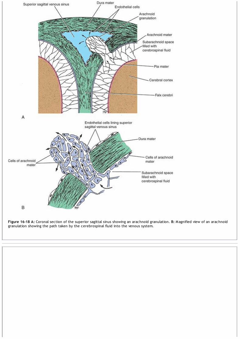
Figure 16-18 A: Coronal section of the superior sagittal sinus showing an arachnoid granulation. B: Magnified view of an arachnoidgranulation showing the path taken by the cerebrospinal fluid into the venous system.
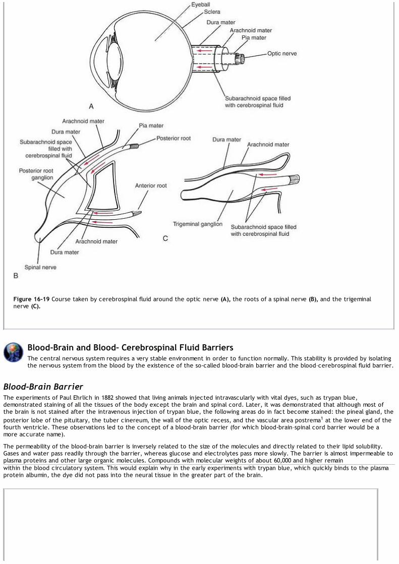
Figure 16-19 Course taken by cerebrospinal fluid around the optic nerve (A), the roots of a spinal nerve (B), and the trigeminalnerve (C).
Blood-Brain and Blood– Cerebrospinal Fluid BarriersThe central nervous system requires a very stable environment in order to function normally. This stability is provided by isolatingthe nervous system from the blood by the existence of the so-called blood-brain barrier and the blood–cerebrospinal fluid barrier.
Blood-Brain BarrierThe experiments of Paul Ehrlich in 1882 showed that living animals injected intravascularly with vital dyes, such as trypan blue,demonstrated staining of all the tissues of the body except the brain and spinal cord. Later, it was demonstrated that although most ofthe brain is not stained after the intravenous injection of trypan blue, the following areas do in fact become stained: the pineal gland, theposterior lobe of the pituitary, the tuber cinereum, the wall of the optic recess, and the vascular area postrema1 at the lower end of thefourth ventricle. These observations led to the concept of a blood-brain barrier (for which blood-brain–spinal cord barrier would be amore accurate name).
The permeability of the blood-brain barrier is inversely related to the size of the molecules and directly related to their lipid solubility.Gases and water pass readily through the barrier, whereas glucose and electrolytes pass more slowly. The barrier is almost impermeable toplasma proteins and other large organic molecules. Compounds with molecular weights of about 60,000 and higher remainwithin the blood circulatory system. This would explain why in the early experiments with trypan blue, which quickly binds to the plasmaprotein albumin, the dye did not pass into the neural tissue in the greater part of the brain.

Figure 16-20 Cross section of a blood capillary of the central nervous system in the area where the blood-brain barrier exists.
StructureExamination of an electron micrograph of the central nervous system shows that the lumen of a blood capillary is separated from theextracellular spaces around the neurons and neuroglia by the following structures: (1) the endothelial cells in the wall of the capillary, (2)a continuous basement membrane surrounding the capillary outside the endothelial cells, and (3) the foot processes of the astrocytes thatadhere to the outer surface of the capillary wall (Fig. 16-20).
The use of electron-dense markers such as lanthanum and horseradish peroxidase (Brightman and Reese, 1969) has shown that thesesubstances do not penetrate between the endothelial cells of the capillaries because of the presence of tight junctions that form beltsaround the cells. When the dense markers are introduced into the extracellular spaces of the neuropil, they pass between theperivascular foot processes of the astrocytes as far as the endothelial lining of the capillary. On the basis of this evidence, it is nowknown that the tight junctions between the endothelial cells of the blood capillaries are responsible for the blood-brain barrier2 (Fig.16-20). In molecular terms, the blood-brain barrier is thus a continuous lipid bilayer that encircles the endothelial cells and isolates thebrain tissue from the blood. This explains how lipophilic molecules can readily diffuse through the barrier, whereas hydrophilic moleculesare excluded.
Although the blood-brain barrier exists in the newborn, there is evidence that it is more permeable to certain substances than it is in theadult.
The structure of the blood-brain barrier is not identical in all regions of the central nervous system. In fact, in those areas where theblood-brain barrier appears to be absent, the capillary endothelium contains fenestrations across which proteins and small organicmolecules may pass from the blood to the nervous tissue (Fig. 16-21). It has been suggested that areas such as the area postrema of thefloor of the fourth ventricle and the hypothalamus may serve as sites at which neuronal receptors may sample the chemical content of theplasma directly. The hypothalamus, which is involved in the regulation of the metabolic activity of the body, might bring about appropriatemodifications of activity, thereby protecting the nervous tissue.
Blood–Cerebrospinal Fluid BarrierThere is free passage of water, gases, and lipid-soluble substances from the blood to the cerebrospinal fluid. Macromolecules such asproteins and most hexoses other than glucose are unable to enter the cerebrospinal fluid. It has been suggested that a barrier similar tothe blood-brain barrier exists in the choroid plexuses.

Figure 16-21 Cross section of a blood capillary of the central nervous system where the blood-brain barrier appears to be absent.Note the presence of fenestrations in the endothelial cells.
StructureElectron-microscopic examination of a villus of a choroid plexus shows that the lumen of a blood capillary is separated from the lumen ofthe ventricle by the following structures: (1) the endothelial cells, which are fenestrated and have very thin walls (the fenestrations arenot true perforations but are filled by a thin diaphragm); (2) a continuous basement membrane surrounding the capillary outside theendothelial cells; (3) scattered pale cells with flattened processes; and (4) a continuous basement membrane, on which rest (5) thechoroidal epithelial cells (Fig. 16-22). The use of electron-dense markers has not been entirely successful in localizing the barrier precisely.Horseradish peroxidase injected intravenously appears as a coating on the luminal surface of the endothelial cells, and in many areasexamined, it did pass between the endothelial cells. It is probable that the tight junctions between the choroidal epithelial cells serveas the barrier (Fig. 16-22).
Figure 16-22 Section of villus of choroid plexus.
Cerebrospinal Fluid–Brain InterfaceAlthough vital dyes given by intravenous injection do not gain access to most brain tissues, if the dyes are injected into the subarachnoidspace or into the ventricles, they soon enter the extracellular spaces around the neurons and glial cells. Thus, there is no comparablephysiologic barrier between the cerebrospinal fluid and the extracellular

compartment of the central nervous system. It is interesting, however, to consider the structures that separate the cerebrospinal fluidfrom the nervous tissue. Three sites must be examined: (1) the pia-covered surface of the brain and spinal cord, (2) the perivascularextensions of the subarachnoid space into the nervous tissue, and (3) the ependymal surface of the ventricles (Fig. 16-23).
Figure 16-23 Section of the cerebrospinal fluid-brain interface. A: Outer surface of the brain. B: Ventricular surface of the brain.
The pia-covered surface of the brain consists of a loosely arranged layer of pial cells resting on a basement membrane (Fig. 16-23). Beneaththe basement membrane are the astrocytic foot processes. No intercellular junctions exist between adjacent pial cells or betweenadjacent astrocytes; therefore, the extracellular spaces of the nervous tissue are in almost direct continuity with the subarachnoidspace.
The prolongation of the subarachnoid space into the central nervous tissue quickly ends below the surface of the brain, where the fusionof the outer covering of the blood vessel with the pial covering of the nervous tissue occurs.
The ventricular surface of the brain is covered with columnar ependymal cells with localized tight junctions (Fig. 16-23).
Intercellular channels exist that permit free communication between the ventricular cavity and the extracellular neuronal space. Theependyma does not have a basement membrane, and there are no specialized astrocytic foot processes because the neuroglial cells areloosely arranged.
Functional Significance of the Blood–Brain and Blood–Cerebrospinal Fluid BarriersIn normal conditions, the blood-brain and blood–cerebrospinal fluid barriers are two important semipermeable barriers that protect thebrain and spinal cord from potentially harmful substances while permitting gases and nutriments to enter the nervous tissue.
Clinical NotesThe Optic Nerve, Raised Cerebrospinal Fluid Pressure, and PapilledemaThe optic nerves are surrounded by sheaths derived from the pia mater, arachnoid mater, and dura mater.
There is an extension of the intracranial subarachnoid space forward around the optic nerve to the back of the eyeball(Fig. 16-19). A rise of cerebrospinal fluid pressure caused by an intracranial tumor will compress the thin walls of theretinal vein as it crosses the extension of the subarachnoid space to enter the optic nerve. This will result incongestion of the retinal vein, bulging forward of the optic disc, and edema of the disc; the last condition is referred toas papilledema. Since both subarachnoid extensions are continuous with the intracranial subarachnoid space, both

eyes will exhibit papilledema. Persistent papilledema leads to optic atrophy and blindness.
HydrocephalusHydrocephalus is an abnormal increase in the volume of the cerebrospinal fluid within the skull. If the hydrocephalus isaccompanied by a raised cerebrospinal fluid pressure, then it is due to one of the following: (1) an abnormal increasein the formation of the fluid, (2) a blockage of the circulation of the fluid, or (3) a diminished absorption of the fluid.Rarely, hydrocephalus occurs with a normal cerebrospinal fluid pressure, and in these patients, there is acompensatory hypoplasia or atrophy of the brain substance.
VarietiesTwo varieties of hydrocephalus are described: noncommunicating and communicating. In noncommunicatinghydrocephalus, the raised pressure of the cerebrospinal fluid is due to blockage at some point between its formationat the choroid plexuses and its exit through the foramina in the roof of the fourth ventricle. In communicatinghydrocephalus, there is no obstruction within or to the outflow from the ventricular system; the cerebrospinal fluidfreely reaches the subarachnoid space and is found to be under increased pressure.
CausesExcessive Formation of Cerebrospinal FluidExcessive formation of cerebrospinal fluid is a rare condition that may occur when there is a tumor of the choroidplexuses.
Blockage of the Circulation of Cerebrospinal FluidAn obstruction of the interventricular foramen by a tumor will block the drainage of the lateral ventricle on that side.The continued production of cerebrospinal fluid by the choroid plexus of that ventricle will cause distention of thatventricle and atrophy of the surrounding neural tissue.An obstruction in the cerebral aqueduct may be congenital or may result from inflammation or pressure from a tumor.This causes a symmetrical distention of both lateral ventricles and distention of the third ventricle.Obstruction of the median aperture (foramen of Magendie) in the roof of the fourth ventricle and the two lateralapertures (foramina of Luschka) in the lateral recesses of the fourth ventricle by inflammatory exudate or by tumorgrowth will produce symmetrical dilatation of both lateral ventricles and the third and fourth ventricles.Sometimes, inflammatory exudate secondary to meningitis will block the subarachnoid space and obstruct the flow ofcerebrospinal fluid over the outer surface of the cerebral hemispheres. Here again, the entire ventricular system of thebrain will become distended.
Diminished Absorption of Cerebrospinal FluidInterference with the absorption of cerebrospinal fluid at the arachnoid granulations may be caused by inflammatoryexudate, venous thrombosis or pressure on the venous sinuses, or obstruction of the internal jugular vein.
Clinical Investigation of the Cerebral VentriclesThe size of the cerebral ventricles may be investigated clinically by the use of (1) computed tomography (CT) andmagnetic resonance imaging (MRI) and, if necessary, (2) intracranial pneumography.CT and MRI are safe and easy to perform. The outline of the ventricles may be demonstrated by using these methods(see Figs. 1-25 and 1-26). Apart from ventricular distention or distortion, the cerebral tumor causing the condition alsomay be demonstrated.Intracranial pneumography is essentially the replacement of cerebrospinal fluid within the ventricles andsubarachnoid space with air or oxygen. Because the air or gas is less dense than the fluid or neural tissue, theventricles and cerebral gyri can be visualized. In an encephalogram, the air or oxygen is introduced through a spinaltap. Radiographs of the skull are then made. In a ventriculogram, the air or oxygen is introduced into the lateralventricle through a needle inserted through a hole in the skull (in a young child, the needle may be inserted through asuture). Radiographs of the skull then are made. In ventriculography, only the ventricles are visualized.
Cerebrospinal Fluid Pressure and Composition in DiseaseThe examination of cerebrospinal fluid can be of great assistance in making a neurologic diagnosis.The clinical measurement of cerebrospinal fluid pressure by means of a spinal tap is described on page 19. An increasein pressure is usually due to meningitis or an increase in volume of the brain produced by edema, tumor formation, acerebral abscess, or the presence of a hematoma.The gross appearance of a specimen of cerebrospinal fluid is of great value. Normally, it is clear and colorless. A cloudyfluid usually indicates the presence of polymorphonuclear leukocytes or an excessive quantity of protein. An increase inthe white cells would suggest inflammation of the meninges or encephalitis. An increase in protein content implies achange in the vascular permeability, and protein escapes into the cerebrospinal fluid. A raised protein content is seenin tuberculous meningitis and poliomyelitis. In multiple sclerosis, the gamma globulin is elevated due to production ofimmunoglobulins in the brain and spinal cord.Normal cerebrospinal fluid does not contain red blood cells. Gross blood in the cerebrospinal fluid is usually caused bycontamination brought about by puncture of a vertebral vein by the spinal tap needle. Uniform blood staining is foundin subarachnoid hemorrhage. Yellow coloration or xanthochromia is caused by the presence of oxyhemoglobin in thefluid some hours after subarachnoid hemorrhage.Normal cerebrospinal fluid contains fewer than four white cells. In bacterial infections, many thousands of cells may bepresent per cubic millimeter. In viral infections of the nervous system, a moderate lymphocyte reaction may occur. Aslight rise in lymphocyte count may also occur in cerebral tumors, cerebral infarction, and multiple sclerosis.The glucose level in the cerebrospinal fluid may disappear completely in acute bacterial meningitis but remains normalin viral infections.The normal physical characteristics and composition of cerebrospinal fluid are summarized in Table 16-1.
Blockage of the Subarachnoid Space in the Vertebral CanalA block of the subarachnoid space in the vertebral canal may be caused by a tumor of the spinal cord or the meninges.Performing a spinal tap is of great value in making a diagnosis. The normal pressure of cerebrospinal fluid with thepatient lying quietly on his or her side and breathing through the mouth is between 60 mm and 150 mm of water. If

P.468
the flow of cerebrospinal fluid in the subarachnoid space is blocked, the normal variations in pressure corresponding tothe pulse and respiration are reduced or absent. Compression of the internal jugular veins in the neck raises thecerebral venous pressure and inhibits the absorption of cerebrospinal fluid in the arachnoid villi and granulations, thusproducing a rise in the manometric reading of the cerebrospinal fluid pressure. If this fails to occur, the subarachnoidspace is blocked, and the patient is exhibiting a positive Queckenstedt sign. Should the tumor completely occupy thevertebral canal in the region of the cauda equina, no cerebrospinal fluid may flow out of the spinal tap needle.Normally, the cerebrospinal fluid is clear. In the presence of a tumor, the cerebrospinal fluid may become yellow andclot spontaneously, owing to the rise in protein content.
Tumors of the Fourth VentricleTumors may arise in the vermis of the cerebellum or in the pons and invade the fourth ventricle. Ependymomas arisingfrom the ependymal cells lining the ventricle also occur. Tumors in this region may invade the cerebellum and producethe symptoms and signs of cerebellar deficiency, or they may press on the vital nuclear centers situated beneath thefloor of the ventricle; the hypoglossal and vagal nuclei, for example, control movements of the tongue, swallowing,respiration, heart rate, and blood pressure.
Blood-Brain Barrier in the Fetus and NewbornIn the fetus, newborn child, or premature infant, where these barriers are not fully developed, toxic substances suchas bilirubin can readily enter the central nervous system and produce yellowing of the brain and kernicterus. This is notpossible in the adult.
Brain Trauma and the Blood-Brain BarrierAny injury to the brain, whether it be due to direct trauma or to inflammatory or chemical toxins, causes a breakdownof the blood-brain barrier, allowing the free diffusion of large molecules into the nervous tissue. It is believed that thisis brought about by actual destruction of the vascular endothelial cells or disruption of their tight junctions.
Drugs and the Blood-Brain BarrierThe systemic administration of penicillin results in only a small amount entering the central nervous system. This isfortunate, because penicillin in high concentrations is toxic to nervous tissue. In the presence of meningitis, however,the meninges become more permeable locally, at the site of inflammation, thus permitting sufficient antibiotic to reachthe infection. Chloramphenicol and the tetracyclines readily cross the blood-brain barrier and enter the nervoustissue. The sulfonamide drugs also easily pass through the blood-brain barrier.Lipid-soluble substances such as the anesthetic agent thiopental rapidly enter the brain after intravenous injection.On the other hand, water-soluble substances such as exogenous norepinephrine cannot cross the blood-brainbarrier. Phenylbutazone is a drug that becomes bound to plasma protein, and the large drug protein molecule isunable to cross the barrier. Most tertiary amines such as atropine are lipid soluble and quickly enter the brain,whereas the quaternary compounds such as atropine methylnitrate do not.In Parkinson disease, there is a deficiency of the neurotransmitter dopamine in the corpus striatum. Unfortunately,dopamine cannot be used in the treatment, as it will not cross the blood-brain barrier. L-Dopa readily crosses thebarrier and has been used with great success.
Tumors and the Blood-Brain BarrierBrain tumors frequently possess blood vessels that have no blood-brain barriers. Anaplastic malignant astrocytomas,glioblastomas, and secondary metastatic tumors lack the normal barriers. However, slow-growing tumors often havenormal vascular barriers.
Clinical Problem Solving1. A 55-year-old man was being investigated for signs and symptoms that suggested the presence of a cerebraltumor. Examination of the CT scan showed gross enlargement and distortion of the left lateral ventricle. What otherinvestigation might be carried out in this patient to display the ventricles of the brain? Using your knowledge ofneuroanatomy, determine the location of the tumor in this patient.View Answer
2. A 3-year-old child had been referred to the children's hospital because the circumference of his head greatlyexceeded the normal limit for his age. After a careful history had been taken and a detailed physical examination hadbeen performed, a diagnosis of hydrocephalus was made. What is your definition of hydrocephalus? Name threecommon causes of hydrocephalus in young children.View Answer
3. A 50-year-old man was found on ophthalmoscopic examination to have edema of both optic discs (bilateralpapilledema) and congestion of the retinal veins. The cause of the condition was found to be a rapidly growingintracranial tumor. Using your knowledge of neuroanatomy, explain the papilledema. Why does the patient exhibitbilateral papilledema?View Answer
4. A 38-year-old man was admitted to the neurosurgery ward with symptoms of persistent headache and vomiting andsome unsteadiness in walking. The headache started 6 weeks previously and became progressively worse. Onexamination, it was found that he could not sit up in bed unsupported. The limbs on the right side of the body showedsome loss of tone. Examination of the patient when he stood up showed a marked loss of equilibrium. Examination ofthe cranial nerves showed central deafness of the right ear. Ophthalmoscopic examination showed severe bilateralpapilledema. Using your knowledge of neuroanatomy, explain the symptoms and signs experienced by this patient,and try to make a diagnosis.View Answer
5. A 4-year-old girl was found to have tuberculous meningitis. She was immediately admitted to the hospital, and
P.470P.471P.472
administration of streptomycin and isoniazid was commenced. As soon as this therapy was started, she was alsoadministered steroid hormones to reduce the incidence of adhesions. She recovered fully, with no complications. Usingyour knowledge of neuroanatomy, explain why it is important to prevent the formation of adhesions in thesubarachnoid space.View Answer
6. A 5-year-old girl with symptoms of headache, general malaise, and vomiting was admitted to the children's hospital.On examination, the body temperature was found to be 104°F, and the pulse rate was rapid. Attempts to flex the neckproduced pain and resulted in the patient flexing her hip and knee joints. A spinal tap was performed; thecerebrospinal fluid was seen to be cloudy, and the pressure was raised to 190 mm of water. Microscopic examinationof the fluid showed a large number of polymorphonuclear leukocytes. A diagnosis of meningitis was made. Subsequentculture revealed the infection to be a meningococcal meningitis. The resident vaguely remembered reading in atextbook the importance of the blood-brain barrier in the use of antibiotics for the treatment of meningitis. What is theblood-brain barrier? Does the presence of the blood-brain barrier influence your choice and dose of antibiotics to beused in this patient?View Answer
7. During a ward round in the children's hospital, the pediatrician informed the students that a 4-day-old baby withjaundice had a serum indirect bilirubin level of 45 mg per 100 mL and that by now the bile pigment was staining thebrain a yellow color (kernicterus). The neuronal damage was revealed clinically by lethargy and poor feeding habitsand by occasional muscle spasms. She said the prognosis was very poor. One of the students observed that he couldnot understand why the bile pigment was having such a dramatic effect on the baby. Recently, he had examined apatient who was dying of inoperable carcinoma of the head of the pancreas with total obstruction of the common bileduct. In that patient, the skin was a deep yellow, but apart from complaints of the intense skin irritation owing to thehigh concentration of bile salts in the blood, and loss of weight, the patient had no symptoms and no neurologicabnormalities. Explain why the baby had neuronal damage and the adult did not.View Answer
8. Name five areas of the brain where the blood-brain barrier appears to be absent. What do you think is thesignificance of the fact that in a few areas of the brain, the barrier is absent?View Answer
Review QuestionsDirections: Each of the numbered items in this section is followed by answers. Select the ONE lettered answerthat is CORRECT.1. The following statements concern the ventricular system:
(a) The cerebral aqueduct connects the third ventricle with the fourth ventricle.(b) The two lateral ventricles communicate directly with one another through the foramen of Monro.(c) The ventricles are developed from the embryonic endoderm.(d) It is lined throughout with squamous epithelium.(e) The choroid plexuses are found only in the lateral ventricles.
View Answer
2. The following statements concern the ventricular system:(a) The fourth ventricle has a rectangular-shaped floor.(b) The pineal body is suspended from the roof of the fourth ventricle.(c) The nerve centers controlling the heart rate and blood pressure lie beneath the floor of the third ventricle.(d) The choroid plexus of the lateral ventricle projects into the cavity on its medial side through the choroidalfissure.(e) The foramen of Magendie is an aperture in the roof of the third ventricle.
View Answer
3. The following statements concern the blood-brain barrier:(a) It protects the brain from toxic compounds of low molecular weight.(b) It is present in the pineal gland.(c) The endothelial cells of the blood capillaries are nonfenestrated.(d) The endothelial cells of the blood capillaries are held together by localized tight junctions.(e) L-Dopa has difficulty passing through the barrier in the treatment of Parkinson disease.
View Answer
4. The following statements concern the blood-brain barrier:(a) Chloramphenicol and the tetracyclines cannot cross the barrier.(b) In the newborn child, the blood-brain barrier is not fully developed.(c) Cerebral trauma or inflammation has little effect on the integrity of the blood-brain barrier.(d) Gases and water pass with difficulty through the barrier.

(e) Glucose and electrolytes pass quickly through the barrier.View Answer
5. The following statements concern the blood–cerebrospinal fluid barrier:(a) The beltlike tight junctions between the choroidal ependymal cells form the barrier.(b) The proteins and most hexoses, other than glucose, are able to cross the barrier.(c) Gases and water cannot pass through the barrier.(d) Lipid-soluble substances have difficulty passing through the barrier.(e) The basement membrane of the endothelial cells plays a vital part in the formation of the barrier.
View Answer
6. The following structures are associated with the roof of the fourth ventricle:(a) Tectum of the midbrain(b) Choroid plexus(c) Pineal gland(d) Corpus callosum(e) Temporal lobes of the cerebral hemispheres
View Answer
7. The following statements concern the cerebrospinal fluid in the fourth ventricle:(a) It is produced mainly by the choroid plexus of the cerebral aqueduct.(b) It leaves the midbrain through the interventricular foramina.(c) It enters the spinal cord through the foramen of Luschka.(d) It is dark yellow in color.(e) It escapes into the subarachnoid space through the apertures in the roof of the fourth ventricle.
View Answer
8. The lateral boundaries of the fourth ventricle are formed by:(a) the tentorium cerebelli.(b) the sulcus limitans.(c) the cerebellar peduncles.(d) the cerebral peduncles.(e) the striae medullares.
View Answer
9. The following important nuclei lie beneath the floor of the fourth ventricle:(a) Oculomotor nucleus(b) Trochlear nucleus(c) Trigeminal nucleus(d) Hypoglossal nucleus(e) Olfactory nucleus
View Answer
10. The following statements concern the third ventricle:(a) It is situated between the thalami.(b) It communicates with the lateral ventricles through the cerebral aqueduct.(c) It is continuous with the fourth ventricle through the interventricular foramen.(d) The choroid plexus is located in the floor.(e) The choroid plexus receives its arterial supply through the posterior cerebral arteries.
View Answer
11. The following statements concern the subarachnoid space:(a) It contains cerebrospinal fluid and the cerebral arteries but not the cerebral veins.(b) It does not communicate with the cisterns.(c) The fourth ventricle drains into it through a single foramen.(d) The space does not surround the cranial and spinal nerves where they leave the skull and the vertebralcanal.(e) It is the interval between the arachnoid mater and the pia mater.
View Answer
12. The following statements concern the formation of the cerebrospinal fluid:(a) None of the fluid originates from the brain substance.

(b) It is largely formed by the choroid plexuses(c) It is passively secreted by the ependymal cells covering the choroid plexuses.(d) The fluid is produced continuously at a rate of about 5 mL per minute.(e) The fluid is drained into the subarachnoid space from the lymphatic vessels of the brain and spinal cord.
View Answer
13. The following statements concern the cerebrospinal fluid:(a) Its circulation through the ventricles is not aided by the pulsations of the choroid plexuses.(b) It extends inferiorly in the subarachnoid space to the level of the fifth sacral vertebra.(c) The cerebrospinal fluid pressure in the subarachnoid space rises if the internal jugular veins in the neck arecompressed.(d) It exits from the ventricular system through the ventricular foramina.(e) Its circulation in the subarachnoid space is aided by the pulsations of the cerebral and spinal veins.
View Answer
14. The following statements concern absorption of the cerebrospinal fluid:(a) The fluid is passed into the blood by active transport through the cells forming the arachnoid villi.(b) The major sites for the absorption of the cerebrospinal fluid are into the veins in the subarachnoid space andthe perineural lymph vessels.(c) The arachnoid villi play an important role in the absorption of cerebrospinal fluid.(d) The fine tubules found within the arachnoid villi play a minor role in the flow of cerebrospinal fluid into thevenous sinuses.(e) In communicating hydrocephalus, there is obstruction to the flow of the cerebrospinal fluid within theventricular system and to the outflow from the ventricular system to the subarachnoid space.
View Answer
Matching Questions. Directions: The following questions apply to Figure 16-24. Match the numbers listed on theleft with the appropriate lettered structures listed on the right. Each lettered option may be selected once, morethan once, or not at all.For answers to Questions 15 to 18, study Figure 16-21.15. Number 1
(a) Cerebral aqueduct(b) Body of lateral ventricle(c) Third ventricle(d) Fourth ventricle(e) None of the above
View Answer
16. Number 2(a) Cerebral aqueduct(b) Body of lateral ventricle(c) Third ventricle(d) Fourth ventricle(e) None of the above
View Answer
17. Number 3(a) Cerebral aqueduct(b) Body of lateral ventricle(c) Third ventricle(d) Fourth ventricle(e) None of the above
View Answer
18. Number 4(a) Cerebral aqueduct(b) Body of lateral ventricle(c) Third ventricle(d) Fourth ventricle(e) None of the above
View Answer
Directions: Each case history is followed by questions. Read the case history, then select the ONE BEST lettered

answer.A 24-year-old woman complaining of recent onset of severe headaches and several attacks of morning vomiting wasseen by a neurologist. A thorough physical examination revealed findings suggesting that she might have anintracranial tumor involving the cerebellum. The physician ordered an MRI of the patient's brain with particularreference to the contents of the posterior cranial fossa.
Figure 16-24 Lateral view of the brain showing an outline of ventricular cavities.
19. Figure 16-25 is a coronal MRI (contrast enhanced) through the fourth ventricle. The radiologist made the followingcorrect observations in his report except:
(a) The bones of the skull showed nothing abnormal. The cerebral cortex appeared to be normal.(b) The midline structures were not deflected to one or the other side.(c) The cavity of the fourth ventricle was distorted and larger than normal.(d) The body of the lateral ventricle had a normal appearance.
View Answer
Figure 16-25 A coronal MRI (contrast enhanced) through the hindbrain showing the fourth ventricle and the surrounding

neural and bony structures.
A 21-year-old pregnant woman was invited to a reunion party, and during the course of the evening, she drankseveral gin and tonics. The party was followed by several others extending over a 3-week period during which shedrank heavily. Six months later, she gave birth to a boy who was diagnosed as having congenital hydrocephalus.20. The pediatric neurologist carefully questioned the mother and came to the following correct conclusions except:
(a) The consumption of a large amount of alcohol during pregnancy usually has no adverse effects on thedeveloping fetus.(b) The high alcoholic intake coincided with the first trimester.(c) The alcohol had crossed the placental barrier and entered the fetal circulation.(d) The alcohol had probably also crossed the fetal blood-brain barrier and entered the brain.(e) The neurologist was of the opinion that the toxic effect of alcohol was probably responsible for thehydrocephalus.
View Answer
Additional Reading
Boron, W. F., and Boulpaep, E. L. Medical Physiology. Philadelphia: Saunders, 2003.
Bradbury, M. W. B. (ed.). Physiology and Pharmacology of the Blood-Brain Barrier. New York: Springer, 1992.
Brightman, M. W. The distribution within the brain of ferritin injected into cerebrospinal fluid compartments: II. Parenchymaldistribution. Am. J. Anat. 117:193, 1965.
Brightman, M. W., and Reese, T. W. Junctions between intimately apposed cell membranes in the vertebrate brain. J. Cell Biol.40:648, 1969.
Broadwell, R. D., and Brightman, M. W. Entry of peroxidase into neurons of the central and peripheral nervous systems fromextracerebral and cerebral blood. J. Comp. Neurol. 166:257, 1976.
Cervos-Navarro, J., Artigas, J., and Mesulja, B. J. Morphofunctional aspects of the normal and pathological blood-brain barrier. ActaNeuropathol. (Berl.) 7(Suppl):1, 1983.
Craig, C. R., and Stitzel, R. E. Modern Pharmacology (4th ed.). Boston: Little, Brown, 1994.
Ermisch, A., Brust, P., Kretzschmar R., and Rühle, H. J. Peptides and blood-brain barrier transport. Physiol. Rev. 73:489, 1993.
Fishman, R. A. Cerebrospinal Fluids in Diseases of the Nervous System. Philadelphia: Saunders, 1992.
Gaab, M. R., and Koos, W. T. Hydrocephalus in infancy and childhood: Diagnosis and indication for operation. Neuropediatrics 15:173,1984.
Klatzo, I. Disturbances of the blood-brain barrier in cerebrovascular disorders. Acta. Neuropathol. (Berl.) 8(Suppl):81, 1983.
Lyons, M. K., and Meyer, F. B. Cerebrospinal fluid physiology and the management of increased intracranial pressure. Mayo Clin. Proc.65:684–707, 1990.
Nestler, E. J., Hyman, S. E., and Malenka, R. C. Molecular Neuropharmacology. A Foundation for Clinical Neurosciences. New York:McGraw-Hill, 2001.
Neuwelt, E. A. (ed.). Implications of the Blood-Brain Barrier and Its Manipulation. New York: Plenum Publishing, 1989.
Oldendorf, W. H. Some clinical aspects of the blood-brain barrier. Hosp. Pract. 17:143, 1982.

Pardridge, W. M. The Blood-Brain Barrier: Cellular and Molecular Biology. New York: Raven Press, 1993.
Paulson, O. B. Blood-brain barrier, brain metabolism and cerebral blood flow. Eur. Neuropsychopharmacol. 12:495, 2002.
Rhoades, R. A., and Tanner, G. A. Medical Physiology. Boston: Little, Brown, 1995.
Strazielle, N., and Ghersi-Egea, J. F. Choroid plexus in the central nervous system: Biology and physiopathology. J. Neuropathol. Exp.Neurol. 59:561, 2000.
Westmoreland, B. F., Benarroch, E. E., Daube, J. R., Reagan, T. J., and Sandok, B. A. Medical Neurosciences (3rd ed.). Boston: Little,Brown, 1994.
Woolburg, H., and Lippoldt, A. Tight junctions of the blood-brain barrier: Development, composition and regulation. Vascul.Pharmacol. 38:323, 2002.
Authors: Snell, Richard S.Title: Clinical Neuroanatomy, 7th Edition
Copyright ©2010 Lippincott Williams & Wilkins
> Table of Contents > Chapter 17 - The Blood Supply of the Br ain and Spinal Cor d
Chapter 17
The Blood Supply of the Brain and Spinal Cord
A 61-year-old woman collapsed in the supermarket and was in a coma when admitted to the emergency department of the local hospital.Twenty-four hours later, she recovered consciousness and was found to have paralysis on the left side of her body, mainly involving thelower limb. There was also some sensory loss of the left leg and foot. She was able to swallow normally and did not appear to havedifficulty with her speech. The left-sided hemiplegia and hemianesthesia strongly suggested a cerebrovascular accident involving the rightcerebral hemisphere. The limitation of the paralysis and anesthesia to the leg and foot indicated that the right anterior cerebral artery orone of its branches was blocked by a thrombus or embolus. The diagnosis was confirmed by positron emission tomography (PET), whichshowed an absence of blood flow through the leg area on the medial surface of the right cerebral hemisphere.
Chapter Objectives
To review the main arteries and veins supplying the brain and spinal cordTo learn the areas of the cerebral cortex and spinal cord supplied by a particular artery and to understand thedysfunction that would result if the artery were blockedTo review the circle of Willis as well as the blood supply to the internal capsule
Cerebrovascular accidents (stroke) still remain the third leading cause of morbidity and death in the United States. Consequently, it isimportant to know the areas of the cerebral cortex and spinal cord supplied by a prarticular artery and to understand the dysfunctionthat would result if the artery were blocked. The internal capsule that contains the major ascending and descending pathways to thecerebral cortex is commonly disrupted by arterial hemorrhage or thrombosis.
Blood Supply of the Brain
Arteries of the BrainThe brain is supplied by the two internal carotid and the two vertebral arteries. The four arteries lie within the subarachnoid space, andtheir branches anastomose on the inferior surface of the brain to form the circle of Willis.
Internal Carotid ArteryThe internal carotid artery begins at the bifurcation of the common carotid artery (Fig. 17-1), where it usually possesses a localizeddilatation, called the carotid sinus. It ascends the neck and perforates the base of the skull by passing through the carotid canal of thetemporal bone. The artery then runs horizontally forward through the cavernous sinus and emerges on the medial side of the anteriorclinoid process by perforating the dura mater. It now enters the subarachnoid space by piercing the arachnoid mater and turnsposteriorly to the region of the medial end of the lateral cerebral sulcus. Here, it divides into the anterior and middle cerebral arteries(Figs. 17-1 and 17-2).
Branches of the Cerebral Portion
1. The ophthalmic artery arises as the internal carotid artery emerges from the cavernous sinus (Fig. 17-1). It enters the orbit throughthe optic canal below and lateral to the optic nerve. It supplies the eye and other orbital structures, and its terminal branches supplythe frontal area of the scalp, the ethmoid and frontal sinuses, and the dorsum of the nose.
2. The posterior communicating artery is a small vessel that originates from the internal carotid artery close to its terminal bifurcation(Figs. 17-1 and 17-2). The posterior communicating artery runs posteriorly above the oculomotor nerve to join the posterior cerebralartery, thus forming part of the circle of Willis.
3. The choroidal artery, a small branch, also originates from the internal carotid artery close to its terminal bifurcation. The choroidalartery passes posteriorly close to the optic tract, enters the inferior horn of the lateral ventricle, and ends in the choroid plexus. Itgives off numerous small branches to surrounding structures, including the crus cerebri, the lateral geniculate body, the optic tract,and the internal capsule.
4. The anterior cerebral artery is the smaller terminal branch of the internal carotid artery (Fig. 17-2). It runs forward and mediallysuperior to the optic nerve and enters the longitudinal fissure of the cerebrum. Here, it is joined to the anterior cerebral artery ofthe opposite side by the anterior communicating artery. It curves backward over the corpus callosum and, finally, anastomoses withthe posterior cerebral artery (Fig. 17-3; see Fig. 17-8). The cortical branches supply all the medial surface of the cerebral cortex as farback as the parieto-occipital sulcus (Fig. 17-3). They also supply a strip of cortex about 1 inch (2.5 cm) wide on the adjoining lateralsurface. The anterior cerebral artery thus supplies the “leg area” of the precentral gyrus. A group of central branches pierces theanterior perforated substance and helps to supply parts of the lentiform and caudate nuclei and the internal capsule.

5. The middle cerebral artery, the largest branch of the internal carotid, runs laterally in the lateral cerebral sulcus (Fig. 17-2). Corticalbranches supply the entire lateral surface of the hemisphere, except for the narrow strip supplied by the anterior cerebral artery, theoccipital pole, and the inferolateral surface of the hemisphere, which are supplied by the posterior cerebral artery (Fig. 17-3). Thisartery thus supplies all the motor area except the “leg area.” Central branches enter the anterior perforated substance and supplythe lentiform and caudate nuclei and the internal capsule (Fig. 17-4).
Vertebral ArteryThe vertebral artery, a branch of the first part of the subclavian artery, ascends the neck by passing through the foramina in thetransverse processes of the upper six cervical vertebrae (Fig. 17-1). It enters the skull through the foramen magnum and pierces the duramater and arachnoid to enter the subarachnoid space. It then passes upward, forward, and medially on the medulla oblongata (Fig. 17-2).At the lower border of the pons, it joins the vessel of the opposite side to form the basilar artery.
Figure 17-1 Origin and courses of the internal carotid and vertebral arteries as they ascend the neck to enter the skull.
Branches of the Cranial Portion
1. The meningeal branches are small and supply the bone and dura in the posterior cranial fossa.
2. The posterior spinal artery may arise from the vertebral artery or the posterior inferior cerebellar artery. It descends on the posteriorsurface of the spinal cord close to the posterior roots of the spinal nerves. The branches are reinforced by radicular arteries thatenter the vertebral canal through the intervertebral foramina. For the detailed distribution of this artery, see page 481.
3. The anterior spinal artery is formed from a contributory branch from each vertebral artery near its termination (Fig. 17-2). The singleartery descends on the anterior surface of the medulla oblongata and spinal cord and is embedded in the pia mater along the anteriormedian fissure. The artery is reinforced by radicular arteries that enter the vertebral canal through the intervertebral foramina. Forthe detailed distribution of this artery, see page 482.
4. The posterior inferior cerebellar artery, the largest branch of the vertebral artery, passes on an irregular course between themedulla and the cerebellum (Fig. 17-2; see Figs. 17-12 and 17-14). It supplies the inferior surface of the vermis, the central nuclei of thecerebellum, and the undersurface of the cerebellar hemisphere; it also supplies the medulla oblongata and the choroid plexus of the

fourth ventricle.
5. The medullary arteries are very small branches that are distributed to the medulla oblongata.
Basilar ArteryThe basilar artery, formed by the union of the two vertebral arteries (Fig. 17-1), ascends in a groove on the anterior surface of the pons(Fig. 17-2; see Figs. 17-13 and 17-14). At theupper border of the pons, it divides into the two posterior cerebral arteries.
Figure 17-2 Arteries of the inferior surface of the brain. Note the formation of the circle of Willis. Part of the right temporal lobehas been removed to show the course of the middle cerebral artery.
Branches
1. The pontine arteries are numerous small vessels that enter the substance of the pons (Fig. 17-2; see Figs. 17-13 and 17-14).
2. The labyrinthine artery is a long, narrow artery that accompanies the facial and the vestibulocochlear nerves into the internalacoustic meatus and supplies the internal ear. It often arises as a branch of the anterior inferior cerebellar artery.
3. The anterior inferior cerebellar artery passes posteriorly and laterally and supplies the anterior and inferior parts of the cerebellum(Fig. 17-2; see Figs. 17-13 and 17-14). A few branches pass to the pons and the upper part of the medulla oblongata.
4. The superior cerebellar artery arises close to the termination of the basilar artery (Fig. 17-2; see Figs. 17-11, 17-12, 17-13 and 17-14). Itwinds around the cerebral peduncle and supplies the superior surface of the cerebellum. It also supplies the pons, the pineal gland,and the superior medullary velum.
5. The posterior cerebral artery curves laterally and backward around the midbrain and is joined by the posterior communicating branchof the internal carotid artery (Figs. 17-1 and 17-2; see Figs. 17-11, 17-12, 17-13 and 17-14). Cortical branches supply the inferolateral andmedial surfaces of the temporal lobe and the lateral and medial surfaces of the occipital lobe (Fig. 17-3). Thus, the posterior cerebralartery supplies the visual cortex. Central branches pierce the brain substance and supply parts of the thalamus and the lentiformnucleus as well as the midbrain, the pineal, and the medial geniculate bodies. A choroidal branch enters the inferior horn of the lateralventricle and supplies the choroid plexus; it also supplies the choroid plexus of the third ventricle.

Figure 17-3 Areas supplied by the cerebral arteries. A: The lateral surface of the right cerebral hemisphere. B: The medial surfaceof the right cerebral hemisphere. The area supplied by the anterior cerebral artery is colored blue, the area supplied by themiddle cerebral artery is pink, and the area supplied by the posterior cerebral artery is brown.
Circle of WillisThe circle of Willis lies in the interpeduncular fossa at the base of the brain. It is formed by the anastomosis between the two internalcarotid arteries and the two vertebral arteries (Fig. 17-2). The anterior communicating, anterior cerebral, internal carotid, posteriorcommunicating, posterior cerebral, and basilar arteries all contribute to the circle. The circle of Willis allows blood that enters by eitherinternal carotid or vertebral arteries to be distributed to any part of both cerebral hemispheres. Cortical and central branches arise fromthe circle and supply the brain substance.
Variations in the sizes of the arteries forming the circle are common, and the absence of one or both posterior communicating arterieshas been reported.

Figure 17-4 Coronal section of the cerebral hemispheres showing the arterial supply to the deep cerebral structures from themiddle cerebral artery.
Arteries to Specific Brain AreasThe corpus striatum and the internal capsule are supplied mainly by the medial and lateral striate central branches of the middle cerebralartery (Fig. 17-4); the central branches of the anterior cerebral artery supply the remainder of these structures.
The thalamus is supplied mainly by branches of the posterior communicating, basilar, and posterior cerebral arteries.
The midbrain is supplied by the posterior cerebral, superior cerebellar, and basilar arteries.
The pons is supplied by the basilar and the anterior, inferior, and superior cerebellar arteries.
The medulla oblongata is supplied by the vertebral, anterior and posterior spinal, posterior inferior cerebellar, and basilar arteries.
The cerebellum is supplied by the superior cerebellar, anterior inferior cerebellar, and posterior inferior cerebellar arteries.
Nerve Supply of Cerebral ArteriesThe cerebral arteries receive a rich supply of sympathetic postganglionic nerve fibers. These fibers are derived from the superior cervicalsympathetic ganglion. Stimulation of these nerves causes vasoconstriction of the cerebral arteries. However, under normal conditions, thelocal blood flow is mainly controlled by the concentrations of carbon dioxide, hydrogen ions, and oxygen present in the nervous tissue; arise in the carbon dioxide and hydrogen ion concentrations and a lowering of the oxygen tension bring about a vasodilatation.
Veins of the BrainThe veins of the brain have no muscular tissue in their very thin walls, and they possess no valves. They emerge from the brain and lie inthe subarachnoid space. They pierce the arachnoid mater and the meningeal layer of the dura and drain into the cranial venous sinuses(Fig. 17-5).
External Cerebral VeinsThe superior cerebral veins pass upward over the lateral surface of the cerebral hemisphere and empty into the superior sagittal sinus(Fig. 17-5).
The superficial middle cerebral vein drains the lateral surface of the cerebral hemisphere. It runs inferiorly in the lateral sulcus andempties into the cavernous sinus (Fig. 17-5).
The deep middle cerebral vein drains the insula and is joined by the anterior cerebral and striate veins to form the basal vein. The basalvein ultimately joins the great cerebral vein, which in turn drains into the straight sinus (Fig. 17-5).
Internal Cerebral VeinsThere are two internal cerebral veins, and they are formed by the union of the thalamostriate vein and the choroid vein at theinterventricular foramen. The two veins run posteriorly in the tela choroidea of the third ventricle and unitebeneath the splenium of the corpus callosum to form the great cerebral vein, which empties into the straight sinus.

Figure 17-5 Venous drainage of the right cerebral hemisphere. A: Lateral surface. B: Medial surface.
Veins of Specific Brain AreasThe midbrain is drained by veins that open into the basal or great cerebral veins.
The pons is drained by veins that open into the basal vein, cerebellar veins, or neighboring venous sinuses.
The medulla oblongata is drained by veins that open into the spinal veins and neighboring venous sinuses.
The cerebellum is drained by veins that empty into the great cerebral vein or adjacent venous sinuses.
Brain CapillariesThe capillary blood supply to the brain is greater in the gray matter than in the white matter. This is to be expected, since themetabolic activity in the neuronal cell bodies in the gray matter is much greater than in the nerve processes in the white matter.
The blood-brain barrier isolates the brain tissue from the rest of the body and is formed by the tight junctions that exist between theendothelial cells in the capillary beds (see p. 462).

Cerebral CirculationThe blood flow to the brain must deliver oxygen, glucose, and other nutrients to the nervous tissue and remove carbon dioxide,lactic acid, and other metabolic by-products. The brain has been shown to be supplied with arterial blood from the two internal
carotid arteries and the two vertebral arteries. The blood supply to half of the brain is provided by the internal carotid and vertebralarteries on that side, and their respective streams come together in the posterior communicating artery at a point where the pressure ofthe two is equal and they do not mix (Fig. 17-6). If, however, the internal carotid or vertebral artery is occluded, the blood passes forwardor backward across that point to compensate for the reduction in blood flow. The arterial circle also permits the blood to flow across themidline, as shown when the internal carotid or vertebral artery on one side is occluded. It also has been shown that the two streams ofblood from the vertebral arteries remain separate and on the same side of the lumen of the basilar artery and do not mix.
Although the cerebral arteries anastomose with one another at the circle of Willis and by means of branches on the surface of thecerebral hemispheres, once they enter the brain substance, no further anastomoses occur.
Figure 17-6 Circle of Willis showing the distribution of blood from the four main arteries.
The most important factor in forcing the blood through the brain is the arterial blood pressure. This is opposed by such factors as a raisedintracranial pressure, increased blood viscosity, and narrowing of the vascular diameter. Cerebral blood flow remains remarkably constantdespite changes in the general blood pressure. This autoregulation of the circulation is accomplished by a compensatory lowering of thecerebral vascular resistance when the arterial pressure is decreased and a raising of the vascular resistance when the arterial pressure isincreased. Needless to say, this autoregulation does not maintain an adequate blood flow when the arterial blood pressure falls to a verylow level.
The diameter of the cerebral blood vessels is the main factor contributing to the cerebrovascular resistance. While it is known thatcerebral blood vessels are innervated by sympathetic postganglionic nerve fibers and respond to norepinephrine, they apparently playlittle or no part in the control of cerebrovascular resistance in normal human beings. The most powerful vasodilator influence on cerebralblood vessels is an increase in carbon dioxide or hydrogen ion concentration; a reduction in oxygen concentration also causesvasodilatation. It has been shown, using PET, that an increase in neuronal activity in different parts of the brain causes a local increase inblood flow. For example, viewing an object will increase the oxygen and glucose consumption in the visual cortex of the occipital lobes.This results in an increase in the local concentrations of carbon dioxide and hydrogen ions and brings about a local increase in blood flow.
The cerebral blood flow in patients can be measured by the intracarotid injection or inhalation of radioactive krypton or xenon. Acerebral blood flow of 50 to 60 mL per 100 g of brain per minute is considered normal.
Blood Supply of the Spinal Cord
Arteries of the Spinal CordThe spinal cord receives its arterial supply from three small arteries: the two posterior spinal arteries and the anterior spinal artery. Theselongitudinally running arteries are reinforced by small segmentally arranged arteries that arise from arteries outside the vertebral columnand enter the vertebral canal through the intervertebral foramina. These vessels anastomose on the surface of the cord and sendbranches into the substance of the white and gray matter. Considerable variation exists as to the size and segmental levels at which thereinforcing arteries occur.
Posterior Spinal ArteriesThe posterior spinal arteries arise either directly from the vertebral arteries inside the skull or indirectly from the posterior inferiorcerebellar arteries. Each artery descends on the posterior surface of the spinal cord close to the posterior nerve roots and gives offbranches that enter thesubstance of the cord (Fig. 17-7). The posterior spinal arteries supply the posterior one-third of the spinal cord.

P.484P.485P.486P.487P.488P.489P.490P.491P.492
Figure 17-7 A: Arterial supply of the spinal cord showing the formation of two posterior spinal arteries and one anterior spinalartery. B: Transverse section of the spinal cord showing the segmental spinal arteries and the radicular arteries.
The posterior spinal arteries are small in the upper thoracic region, and the first three thoracic segments of the spinal cord areparticularly vulnerable to ischemia should the segmental or radicular arteries in this region be occluded.
Anterior Spinal ArteryThe anterior spinal artery is formed by the union of two arteries, each of which arises from the vertebral artery inside the skull. Theanterior spinal artery then descends on the anterior surface of the spinal cord within the anterior median fissure (Fig. 17-7). Branchesfrom the anterior spinal artery enter the substance of the cord and supply the anterior two-thirds of the spinal cord.
In the upper and lower thoracic segments of the spinal cord, the anterior spinal artery may be extremely small. Should the segmental orradicular arteries be occluded in these regions, the fourth thoracic and the first lumbar segments of the spinal cord would be particularlyliable to ischemic necrosis.
Segmental Spinal ArteriesAt each intervertebral foramen, the longitudinally running posterior and anterior spinal arteries are reinforced by small segmental arterieson both sides (Fig. 17-7). The arteries are branches of arteries outside the vertebral column (deep cervical, intercostal, and lumbararteries). Having entered the vertebral canal, each segmental spinal artery gives rise to anterior and posterior radicular arteries thataccompany the anterior and posterior nerve roots to the spinal cord.
Additional feeder arteries enter the vertebral canal and anastomose with the anterior and posterior spinal arteries; however, the numberand size of these arteries vary considerably from one individual to another. One large and important feeder artery, the great anteriormedullary artery of Adamkiewicz, arises from the aorta in the lower thoracic or upper lumbar vertebral levels; it is unilateral and, in themajority of persons, enters the spinal cord from the left side. The importance of this artery lies in the fact that it may be the majorsource of blood to the lower two-thirds of the spinal cord.
Veins of the Spinal CordThe veins of the spinal cord drain into six tortuous longitudinal channels that communicate superiorly within the skull with the veins of thebrain and the venous sinuses. They drain mainly into the internal vertebral venous plexus.
Clinical NotesThe brain receives about 15% of the resting cardiac output. The arterial blood reaches the brain through thetwo internal carotid and the two vertebral arteries; the internal carotid arteries are the major supply of arterial
blood.The distributing arteries—the anterior, middle, and posterior cerebral arteries—that arise from the circle of Willis passover the outer surface of the brain and anastomose with one another. They give rise to branches that penetrate the

brain at right angles. In the brain substance, further branching occurs, but no further anastomoses take place. It isthe anastomoses on the brain surface that provide the vital collateral circulation should one of the arteries be occludedby disease.Despite the recent decrease in cerebrovascular disease, which has been brought about by the treatment of high bloodcholesterol and the aggressive treatment of hypertension, it is estimated that cerebrovascular disease is stillresponsible for about 50% of all adult neurologic hospital admissions.
Cerebral IschemiaUnconsciousness occurs in 5 to 10 seconds if the blood flow to the brain is completely cut off. Irreversible braindamage with death of nervous tissue rapidly follows complete arrest of cerebral blood flow. It has been estimated thatneuronal function ceases after about 1 minute and that irreversible changes start to occur after about 4 minutes,although this time may be longer if the patient's body has been cooled.1 Cardiac arrest due to coronary thrombosis isthe most common cause of this condition.
Interruption of Cerebral CirculationVascular lesions of the brain are extremely common, and the resulting neurologic defect will depend on the size of theartery occluded, the state of the collateral circulation, and the area of the brain involved. Clinical studies and theexamination of postmortem material have focused attention on the high frequency of lesions in the common carotid,internal carotid, and vertebral arteries in the neck.
Cerebral Artery SyndromesAnterior Cerebral Artery OcclusionIf the occlusion of the anterior cerebral artery is proximal to the anterior communicating artery, the collateral circulationis usually adequate to preserve the circulation. Occlusion distal to the communicating artery may produce the followingsigns and symptoms:
1. Contralateral hemiparesis and hemisensory loss involving mainly the leg and foot (paracentral lobule of cortex)2. Inability to identify objects correctly, apathy, and personality changes (frontal and parietal lobes)
Middle Cerebral Artery OcclusionOcclusion of the middle cerebral artery may produce the following signs and symptoms, but the clinical picture will varyaccording to the site of occlusion and the degree of collateral anastomoses:
1. Contralateral hemiparesis and hemisensory loss involving mainly the face and arm (precentral and postcentral gyri)2. Aphasia if the left hemisphere is affected (rarely if the right hemisphere is affected)3. Contralateral homonymous hemianopia (damage to the optic radiation)4. Anosognosia if the right hemisphere is affected (rarely if the left hemisphere is affected)
Posterior Cerebral Artery OcclusionOcclusion of the posterior cerebral artery may produce the following signs and symptoms, but the clinical picture willvary according to the site of the occlusion and the availability of collateral anastomoses:
1. Contralateral homonymous hemianopia with some degree of macular sparing (damage to the calcarine cortex,macular sparing due to the occipital pole receiving collateral blood supply from the middle cerebral artery)
2. Visual agnosia (ischemia of the left occipital lobe)3. Impairment of memory (possible damage to the medial aspect of the temporal lobe)
Internal Carotid Artery OcclusionOcclusion of the internal carotid artery can occur without causing symptoms or signs or can cause massive cerebralischemia depending on the degree of collateral anastomoses.
1. The symptoms and signs are those of middle cerebral artery occlusion, including contralateral hemiparesis andhemianesthesia.
2. There is partial or complete loss of sight on the same side, but permanent loss is rare (emboli dislodged from theinternal carotid artery reach the retina through the ophthalmic artery).
Vertebrobasilar Artery OcclusionThe vertebral and basilar arteries supply all the parts of the central nervous system in the posterior cranial fossa, andthrough the posterior cerebral arteries, they supply the visual cortex on both sides. The clinical signs and symptomsare extremely varied and may include the following:
1. Ipsilateral pain and temperature sensory loss of the face and contralateral pain and temperature sensory loss ofthe body
2. Attacks of hemianopia or complete cortical blindness3. Ipsilateral loss of the gag reflex, dysphagia, and hoarseness as the result of lesions of the nuclei of the
glossopharyngeal and vagus nerves4. Vertigo, nystagmus, nausea, and vomiting5. Ipsilateral Horner syndrome6. Ipsilateral ataxia and other cerebellar signs

7. Unilateral or bilateral hemiparesis8. Coma
Impairment of Cerebral Blood FlowImpairment of cerebral blood flow can be caused by a large number of conditions, and the more important conditionscan be considered under the following headings: (1) diseases that produce alteration in blood pressure, (2) diseasesof arterial walls, and (3) diseases that result in blockage of the arterial lumen.
Diseases That Alter Blood PressurePostural HypotensionPatients who get up after being confined to bed for several days, soldiers who stand at attention for long periods on ahot day, and worshipers kneeling in church may experience the accumulation of venous blood in the limbs or impairedvenous return to the heart, with a consequent fall in the cardiac output and a lowered arterial blood pressure. Thegeneral arterial pressure has to be lowered considerably before the cerebral blood flow is diminished.
Physical and Psychologic ShockThe profound and prolonged fall in blood pressure that may follow physical trauma, such as an automobile accident orextensive surgery, especially in the elderly, in whom the cerebral arteries are already narrowed by disease, may causethe patient to lose consciousness. Hyperventilation in anxiety states may reduce the cerebral blood flow by loweringthe carbon dioxide content of the blood.
Change in Blood ViscosityIn polycythemia vera, the cerebral blood flow is considerably reduced as the result of an increase in the viscosity of theblood.
Carotid Sinus SyndromeThe carotid sinus, situated at the proximal end of the internal carotid artery, is extremely sensitive to changes inarterial blood pressure. Distention of the arterial wall causes a reflex slowing of the heart rate and a fall in bloodpressure. This occurs as the result of an increased number of nervous impulses passing up the sinus nerve, a branchof the glossopharyngeal nerve, which connects with the cardioinhibitory and vasomotor centers. Hypersensitivity of thereflex or external pressure may cause the blood pressure to fall suddenly and produce cerebral ischemia and loss ofconsciousness.
Diseases of the HeartAny severe cardiac disease, such as coronary thrombosis, auricular fibrillation, or heart block, that results in a markedfall in cardiac output will result in a severe fall in general arterial blood pressure and reduction in cerebral blood flow.
Diseases of the Arterial WallsThe most common cause of narrowing of the lumen of the arteries that supply the brain is atheroma. This disease mayaffect the main arteries supplying the brain in their course through the neck as well as their course within the skull.Moreover, the impairment of the cerebral circulation may be worsened by an attack of coronary thrombosis, with itsassociated hypotension, shock due to surgical procedures, severe anemia, or even rotation of the head with externalpressure on the carotid arteries.Atheromatous degeneration of the cerebral arteries occurs most commonly in middle or old age and often complicatesdiabetes and hypertension. When actual blockage of an artery occurs, the effect will depend on the size and locationof the vessel. The nerve cells and their fibers will degenerate in the avascular area, and the surrounding neuroglia willproliferate and invade the area. In patients with generalized narrowing of the cerebral arteries without blockage of asingle artery, the brain will undergo a diffuse atrophy. It should be remembered that a very narrow atheromatousartery may be blocked by a thrombus, thus totally closing the lumen.
Diseases That Result in Blockage of the Arterial LumenEmbolism of a cerebral artery can occur in two forms: (1) a thrombus (by far the most common) and (2) fat globules.The thrombus may develop anywhere on the endothelial lining from the left side of the heart to the parent vessels ofthe cerebral arteries. A common site of origin is an atheromatous plaque on the internal carotid, common carotid, orvertebral artery. Another area is the site of endocarditis on the mitral or aortic valve or the endocardial lining of amyocardial infarction following a coronary thrombosis. In women, cerebral thrombosis is more common among thosetaking oral contraceptives, especially those taking a high-dose estrogen-progesterone combination.Fat embolism usually follows severe fractures of one of the long bones. Fat globules from the macerated yellowmarrow enter the nutrient veins, pass through the pulmonary circulation, and end up blocking multiple small cerebralend arteries.
Cerebral AneurysmsCongenital AneurysmsCongenital aneurysms occur most commonly at the site where two arteries join in the formation of the circle of Willis.At this point, there is a deficiency in the tunica media, and this is complicated by the development of atheroma, whichso weakens the arterial wall that a local dilatation occurs. The aneurysm may press on neighboring structures, such asthe optic nerve or the third, fourth, or sixth cranial nerve, and produce signs or symptoms or may suddenly rupture intothe subarachnoid space. In the latter case, a severe pain in the head suddenly develops, followed by mentalconfusion. Death may quickly occur, or the patient may survive the first bleeding only to die a few days or weeks later.Clipping or ligating the neck of the aneurysm offers the best chance of recovery.Other types of aneurysms are rare and include those due to softening of the arterial wall following the lodging of aninfected embolus; those due to damage of the internal carotid artery as it lies within the cavernous sinus following afracture of the skull; and those that are associated with disease of the arterial wall, such as atheroma.
Intracranial HemorrhageIntracranial hemorrhage can result from trauma or cerebral vascular lesions. Four varieties are considered: (1)epidural, (2) subdural, (3) subarachnoid, and (4) cerebral. Epidural and subdural hemorrhage are described on page23.

Subarachnoid HemorrhageSubarachnoid hemorrhage usually results from leakage or rupture of a congenital aneurysm on the cerebral arterialcircle or, less commonly, from an angioma or contusion and laceration of the brain and meninges. The symptoms, whichare sudden in onset, will include severe headache, stiffness of the neck, and loss of consciousness. The diagnosis isestablished by the use of computed tomography (CT). The dense areas of the blood in the subarachnoid space can beidentified. The withdrawal of heavily blood-stained cerebrospinal fluid through a lumbar puncture is also diagnostic, butthis method has been replaced by the use of CT.
Cerebral HemorrhageCerebral hemorrhage generally is due to rupture of an atheromatous artery and is most common in patients withhypertension. It usually occurs in individuals of middle age and often involves a rupture of the thin-walledlenticulostriate artery, a branch of the middle cerebral artery. The important corticonuclear and corticospinal fibers inthe internal capsule are damaged, producing hemiplegia on the opposite side of the body. The patient immediatelyloses consciousness, and the paralysis is evident when consciousness is regained. In some cases, the hemorrhagebursts into the lateral ventricle, resulting in deeper unconsciousness and corticospinal lesions on both sides of thebody. Hemorrhage may also occur into the pons and cerebellum.
Computed Tomography, Magnetic Resonancy Imaging, and Positron Emission TomographyCT, magnetic resonance imaging (MRI), and PET are techniques that are indispensable in making the diagnosis ofdifferent forms of cerebrovascular disease. The diagnosis can usually be made with speed, accuracy, and safety. Anintracranial blood clot can be recognized by its density. These techniques have largely replaced cerebral angiography(see p. 24).
Cerebral AngiographyThe technique of cerebral angiography is used for the detection of abnormalities of the blood vessels; the detectionand localization of space-occupying lesions such as tumors, hematomas, or abscesses; or the determination of thevascular pattern of tumors to aid in the diagnosis of their pathology. With the patient under general anesthesia and inthe supine position, the head is centered on a radiographic apparatus that will take repeated radiographs at 2-secondintervals. Both anteroposterior and lateral projections are obtained. A radiopaque medium is injected rapidly into thelumen of the common carotid or vertebral artery or is indirectly introduced into one of these arteries through a catheterinserted into the radial or femoral artery. As the radiopaque material is rapidly introduced, a series of films areexposed. By this means, the cerebral arteries, the capillary flush, and the veins may be demonstrated. Examples ofnormal-appearing carotid and vertebral angiograms are shown in Figures 17-8, 17-9, 17-10, 17-11, 17-12, 17-13, 17-14 and 17-15.
Figure 17-8 Lateral internal carotid arteriogram. Male aged 20 years.

Cerebral angiography is an invasive technique that unfortunately has a morbidity of 0.5% to 2.5%. CT and MRI shouldtherefore be used whenever possible. PET is now also used extensively.
Spinal Cord IschemiaThe blood supply to the spinal cord is surprisingly meager considering the importance of this nervous tissue. Theposterior and anterior spinal arteries are of small and variable diameter, and the reinforcing segmental arteries vary innumber and size.The posterior one-third of the spinal cord receives its arterial supply from the posterior spinal arteries. The anteriortwo-thirds of the spinal cord is supplied by the small, tenuous anterior spinal artery. This latter artery thereforesupplies the anterior white column, the anterior gray horns, and the anterior part of the lateral white columns and theroot of the posterior horns.
Figure 17-9 Main features seen in radiograph in Figure 17-8.
Occlusion of the anterior spinal artery may produce the following signs and symptoms (Fig 17-16).
1. Loss of motor function (paraplegia) below the level of the lesion occurs due to bilateral damage to the corticospinaltracts.
2. Bilateral thermoanesthesia and analgesia occur below the level of the lesion due to bilateral damage to thespinothalamic tracts.
3. Weakness of the limb muscles may occur due to damage of the anterior gray horns in the cervical or lumbar regionsof the cord.
4. Loss of bladder and bowel control occurs due to damage of the descending autonomic tracts.5. Position sense, vibration, and light touch are normal due to preservation of the posterior white columns that are

supplied by the posterior spinal arteries.
Ischemia of the spinal cord can easily follow minor damage to the arterial supply as the result of nerve blockprocedures, aortic surgery, or any operation in which severe hypotension occurs. The fourth thoracic and first lumbarsegments of the cord are particularly prone to ischemia.
Figure 17-10 Anteroposterior internal carotid arteriogram. Male aged 20 years.
Spinal Cord Ischemia and Thoracic Aortic DissectionThe thoracic region of the spinal cord receives its segmental arteries from the posterior intercostal arteries, which arisedirectly from the thoracic aorta. In thoracic aortic dissection, the expanding blood clot in the aortic wall can block theorigins of the posterior intercostal arteries, causing ischemia of the spinal cord.
Spinal Cord Ischemia as a Complication of a Leaking Abdominal Aortic AneurysmThe lumbar region of the spinal cord receives its segmental arteries from the lumbar arteries, which are branches ofthe abdominal aorta. The effect of direct pressure on the lumbar arteries by a leaking aneurysm can interfere with theblood supply to the spinal cord.

Figure 17-11 Main features seen in the radiograph in Figure 17-10.

Figure 17-12 Lateral vertebral arteriogram. Male aged 20 years.

Figure 17-13 Main features shown in the radiograph in Figure 17-12.

Figure 17-14 Anteroposterior (angled) vertebral arteriogram. Woman aged 35 years.

Figure 17-15 Main features shown in the radiograph in Figure 17-14.

P.494P.495P.496
Figure 17-16 Anterior spinal artery occlusion. Pink area denotes region of spinal cord affected.
Clinical Problem Solving1. A distinguished neurosurgeon, while giving a lecture on cerebrovascular accidents, made the following statement:“It is generally agreed that there are no anastomoses of clinical importance between the terminal end arteries withinthe brain substance, but there are many important anastomoses between the large arteries, both within and outsidethe skull, and these may play a major role in determining the extent of brain damage in cerebral vascular disease.”Comment on this statement, and name the sites at which important arterial anastomoses take place.View Answer
2. During examination of a carotid angiogram, the contrast medium had filled the anterior and middle cerebral arteriesbut had failed to fill the posterior cerebral artery. Careful following of the contrast medium showed it to enter theposterior communicating artery but to extend no farther. Explain this phenomenon in a normal person.View Answer
3. A 45-year-old man was admitted to the hospital after collapsing in his home 3 days previously. He was in a partialstate of unconsciousness on the floor and was found by a friend. On physical examination, he had right-sidedhomonymous hemianopia, although careful examination of the fields of vision showed that the macular regions werenormal. Right-sided hemianesthesia and hemianalgesia also were present, although the patient complained of severeburning pain in the right leg. During the first 24 hours in the hospital, the patient demonstrated mild right-sidedhemiparesis of the flaccid type, which disappeared within 2 days. What is your diagnosis? Be specific in describing thebranches of the artery that are involved.View Answer
4. During the course of an autopsy on a patient who had recently died of cerebrovascular disease, the pathologistmade the comment that in atherosclerosis of the cerebral arteries, the atheromatous plaques tend to occur where themain arteries divide or where the arteries suddenly curve. It is thought that at these sites, the pressure flow changesmay be a factor in the causation of the disease process. Using your knowledge of anatomy, name as many sites asyou can where the main cerebral arteries divide or undergo abrupt change in their course.View Answer
5. Having carefully examined a male patient with cerebrovascular disease, the physician met with the family to discussthe nature of the illness, the course of treatment, and the prognosis. The daughter asked the physician what wasmeant by the term stroke as well as its common causes. He was also asked why the clinical findings vary so much frompatient to patient. Using your knowledge of the anatomy and physiology of cerebral blood flow, explain why patientswith cerebrovascular disease present such a variety of syndromes.View Answer
6. The classic sign of cerebrovascular disease is hemiplegia, yet we know that most patients also exhibit sensorydeficits of different types. Using your knowledge of the anatomical distribution of the cerebral arteries, discuss themain types of sensory loss that you may find in such patients.View Answer
7. During the discussion of the symptoms and signs of a 70-year-old woman who had been admitted to the hospital fortreatment of cerebrovascular disease, a fourth-year medical student made the comment that she was surprised to findthat many of the signs and symptoms were bilateral in this patient. She said that the three previous patients she hadexamined had displayed only unilateral signs and symptoms. Using your knowledge of neuroanatomy, explain whysome patients exhibit bilateral signs and symptoms, while in others the syndrome is clearly unilateral.View Answer
8. Neurologists speak frequently of the dominant hemisphere, and if cerebrovascular disease should involve thathemisphere, one would expect the patient possibly to have global or total sensorimotor aphasia. Explain thisphenomenon.View Answer
9. Explain why patients with a thrombosis of the middle cerebral artery often present with homonymous hemianopia aswell as hemiplegia and hemianesthesia.View Answer
10. During the neurobiology course, the professor of neuroanatomy emphasized the importance of knowing thestructure and blood supply of the internal capsule. He explained the arrangement of the ascending and descendingtracts within the capsule and showed how they were concentrated into a small area between the thalamus andcaudate nucleus medially and the lentiform nucleus laterally. Clearly, an interruption of the blood supply to this vitalarea would produce widespread neurologic defects. What is the blood supply to the internal capsule?View Answer
11. A 36-year-old man visited his physician with a complaint that on three occasions during the past 6 months, he hadfainted at work. During careful questioning, the patient stated that on each occasion, he had fainted while sitting athis desk and while interviewing office personnel; he added that the person being interviewed sat in a chairimmediately to the right of the desk. He said that before each fainting attack he felt dizzy; then, he lost consciousness

P.498P.499
only to recover within a few moments. The previous evening, he had a similar dizzy spell when he turned his headquickly to the right to talk to a friend in the street. The physician noted that the patient wore a stiff collar that wasrather close fitting. When the physician commented on this, the patient stated that he always wore this type of collarto work. No abnormal physical signs were found. Using your knowledge of anatomy and physiology, what diagnosiswould you make?View Answer
12. A 45-year-old man, a company director, rose to give his annual after-dinner speech to the board when he suddenlyexperienced an “agonizing, crushing” pain over the sternum. Feeling giddy and weak, he fell back in his chair. A fewmoments later, he lapsed into unconsciousness. An attendant at the dinner, who had received some training incardiopulmonary resuscitation while a member of the armed forces, ran forward and noted that the patient hadstopped breathing. He quickly started mouth-to-mouth resuscitation and cardiac compression and kept going untilambulance personnel arrived to take the patient to the hospital. The physician in the intensive care unit at the hospitallater told the patient that his life had been saved by the alertness and competence of the attendant at the dinner.Using your knowledge of neurophysiology, state how long brain tissue can survive when there is complete cardiacarrest and breathing has ceased.View Answer
13. A 62-year-old man with a history of hypertension visited his physician because the day before he had temporarilylost the sight in his right eye. He explained that the sight loss was partial and lasted about half an hour. On closequestioning, the patient admitted that he had had similar episodes of blindness in the same eye during the previous 6months, but they had lasted only a few minutes. The patient also mentioned that there were days when he could notremember the names of people and things. He also had recently experienced severe right-sided headaches. Whenasked about his activities, he said that he could not walk as well as he used to, and his left leg sometimes felt weakand numb. While performing a careful physical examination, the physician heard with his stethoscope a distinct systolicbruit over the right side of the neck. Given that the patient has vascular disease of the brain, which artery is likely tobe involved in the disease process? What special clinical investigations could you perform to confirm the diagnosis?View Answer
14. A 39-year-old man was admitted to the hospital with a history of a sudden excruciating, generalized headachewhile gardening. This was followed, 10 minutes later, by the patient collapsing to the ground in a state ofunconsciousness. After being carried indoors and placed on a settee, the patient regained consciousness butappeared confused. He complained of a severe headache and a stiff neck. Physical examination revealed some rigidityof the neck but nothing further. A careful neurologic examination 3 days later revealed some loss of tone in themuscles of the left leg. Using your knowledge of anatomy, make the diagnosis. What is the reason for the neckrigidity?View Answer
15. A 26-year-old man, on leaving a bar after a few drinks, stepped into the road at 1:00 AM and was struck by apassing car. Fortunately, the car was traveling slowly and struck the patient's head a glancing blow. One hour later, apoliceman found the patient unconscious on the sidewalk. Physical examination at the local hospital found that thepatient had recovered consciousness for a few minutes but then had quickly relapsed into an unconscious state. Theright pupil was dilated, and the muscle tone of the left leg was found to be less than normal. A positive Babinski signwas obtained on the left side. Examination of the scalp showed a severe bruise over the right temple, and a lateralradiograph of the skull showed a fracture of the anterior inferior angle of the parietal bone. A CT scan showed a densearea extending from anterior to posterior along the inner table of the right parietal bone. What is the diagnosis? Letus suppose that the equipment for performing a CT scan was unavailable and that it was decided to perform a lumbarpuncture; this test revealed a raised cerebrospinal fluid pressure, and the fluid was very slightly blood stained. Explainthese additional findings.View Answer
16. A 50-year-old woman complaining of headaches, drowsiness, and mental confusion visited her physician. On closequestioning, the patient distinctly remembered striking her head against a closet door when bending down 3 weekspreviously. A CT scan revealed the presence of a large space-occupying lesion over the right frontal lobe of the brain.What is the possible diagnosis?View Answer
17. A 55-year-old man with a history of hypertension collapsed in the street while walking to work. He complained of asudden severe headache. After 5 minutes, his face began to sag on the right side, and his speech became slurred. Onadmission to the hospital, his right arm and leg were found to be weaker than the left, and the muscles werehypotonic. The eyes were deviated to the left. Later, the right arm and leg showed complete paralysis and wereinsensitive to pinprick. A positive Babinski sign was present on the right side. Two hours later, the patient relapsedinto a deep coma with bilateral dilated fixed pupils. Later, the respirations became deep and irregular, and the patientdied 6 hours later. Using your knowledge of neuroanatomy, make the diagnosis.View Answer
18. What is the blood supply to the spinal cord? Which areas of the spinal cord are supplied by the anterior spinalartery? Which regions of the spinal cord are most susceptible to ischemia?View Answer
Review QuestionsDirections: Each of the numbered items in this section is followed by answers. Select the ONE lettered answer

that is CORRECT.1. The following statements concern the blood supply to the brain:
(a) The brain receives its blood supply directly from the two external carotid arteries.(b) The circle of Willis is formed by the anterior cerebral, the internal carotid, the posterior cerebral, the basilar,and the anterior and posterior communicating arteries.(c) The cerebral arteries do not anastomose on the surface of the brain.(d) There are numerous anastomoses between the branches of the cerebral arteries once they have entered thesubstance of the brain.(e) The main blood supply to the internal capsule is from the central branches of the anterior cerebral artery.
View Answer
2. The areas of the cerebral cortex listed below receive their arterial supply as indicated:(a) The precentral gyrus (face area) is supplied by the middle cerebral artery.(b) The postcentral gyrus (face area) is supplied by the anterior cerebral artery.(c) The cuneus is supplied by the anterior cerebral artery.(d) The inferior temporal gyrus is supplied by the middle cerebral artery.(e) The Wernicke area is supplied by the posterior cerebral artery.
View Answer
3. The arteries listed below arise from the main stem arteries as indicated:(a) The ophthalmic artery is a branch of the middle cerebral artery.(b) The pontine arteries are branches of the internal carotid artery.(c) The posterior communicating artery is a branch of the middle cerebral artery.(d) The posterior spinal artery arises from the vertebral artery.(e) The posterior inferior cerebellar artery is a branch of the basilar artery.
View Answer
4. The veins listed below drain into the venous sinuses indicated:(a) The superior cerebral veins drain into the inferior sagittal sinus.(b) The great cerebral vein drains into the superior sagittal sinus.(c) The superior cerebellar veins drain only into the straight sinus.(d) The spinal veins drain into the external vertebral venous plexus.(e) The inferior sagittal sinus drains into the straight sinus.
View Answer
5. The following statements concern the cerebral blood flow:(a) The sympathetic postganglionic fibers exert great control over the diameter of the cerebral blood vessels.(b) It varies greatly with changes in the general blood pressure.(c) Oxygen tension in the cerebral blood has no effect on the diameter of the cerebral blood vessels.(d) One of the most powerful vasodilators of cerebral blood vessels is carbon dioxide.(e) The blood flow for a particular area of nervous tissue following occlusion of a cerebral artery does not dependon the collateral circulation.
View Answer
6. The following statements concern cerebral ischemia:(a) Atheromatous degeneration of a cerebral artery does not cause degeneration of the nerve cells in theavascular area due to the presence of cerebrospinal fluid.(b) Neuronal function ceases after the blood flow has stopped for about 10 seconds.(c) Irreversible cerebral damage starts to occur after the blood flow has ceased for about 4 minutes.(d) Shock occurring as the result of severe physical trauma does not result in cerebral ischemia.(e) Cooling of the patient's body following a cerebrovascular accident speeds up cerebral degeneration.
View Answer
Matching Questions. Directions: The following questions apply to Figure 17-17. Match the numbered arterieslisted below with the appropriate lettered arteries. Each lettered option may be selected once, more than once, ornot at all.For answers to Questions 7 to 12, study Figure 17-17, which shows the arteries of the inferior surface of thebrain.7. Number 1
(a) Middle cerebral artery(b) Anterior communicating artery(c) Posterior cerebral artery(d) Basilar artery

(e) None of the aboveView Answer
8. Number 2(a) Middle cerebral artery(b) Anterior communicating artery(c) Posterior cerebral artery(d) Basilar artery(e) None of the above
View Answer
9. Number 3(a) Middle cerebral artery(b) Anterior communicating artery(c) Posterior cerebral artery(d) Basilar artery(e) None of the above
View Answer
10. Number 4(a) Middle cerebral artery(b) Anterior communicating artery(c) Posterior cerebral artery(d) Basilar artery(e) None of the above
View Answer
11. Number 5(a) Middle cerebral artery(b) Anterior communicating artery(c) Posterior cerebral artery(d) Basilar artery(e) None of the above
View Answer
12. Number 6(a) Middle cerebral artery(b) Anterior communicating artery(c) Posterior cerebral artery(d) Basilar artery(e) None of the above
View Answer
Directions: In the next item, select the ONE lettered answer that is CORRECT.13. The following statements concern the blood supply to the spinal cord:
(a) The posterior spinal arteries supply the posterior third of the spinal cord.(b) The veins do not communicate with the veins of the brain and the venous sinuses.(c) The arteria radicularis magna (artery of Adamkiewicz) arises in the upper thoracic region from the arch of theaorta.(d) The anterior spinal artery is double but usually arises from one vertebral artery.(e) The spinal arteries are not reinforced by branches of local arteries.
View Answer
Directions: Each case history is followed by questions. Read the case history, then select the ONE BEST letteredanswer.A 58-year-old man, while eating his evening meal, suddenly complained of a severe headache. Moments later, heslumped forward and lost consciousness.

Figure 17-17 The arteries of the inferior surface of the brain.
14. On being admitted to the hospital, the examining physician could have found the following physical signs except:(a) He was in a deep coma, and his breathing was deep and slow.(b) The patient's head was turned to the left.(c) The right side of his face was flattened, and saliva was drooling out of the right corner of his mouth.(d) The muscle tone of the limbs was less on the right side than on the left.(e) The right abdominal reflexes were absent, and there was a positive Babinski response on the left side.
View Answer
15. Three days later, the patient regained consciousness, and the following additional signs could have becomeapparent except:
(a) The right arm and, to a lesser extent, the right leg were paralyzed.(b) Movements of the left arm and leg and the left side of the face were normal.(c) The upper and lower parts of the right side of his face were paralyzed.(d) The patient had difficulty in swallowing.(e) The patient was unable to speak.
View Answer
16. During the next 2 weeks, the following signs could have developed except:(a) The muscles of the limbs on the right side became hypertonic.(b) The tendon reflexes on the right side became hyperactive.(c) The patient had some sensory loss on the right side.(d) The patient was suffering from urinary incontinence.(e) The muscles on the left side exhibited hypotonia.
View Answer
17. The neurologist in charge of this patient interpreted the findings as follows. All his interpretations were likely to becorrect except:
(a) The sudden onset of a severe headache followed by loss of consciousness is a common finding in patientswith a blockage of a cerebral artery.(b) The depth of coma is unrelated to the extent of the arterial blockage.(c) Paralysis of the face on the right side indicated the presence of a lesion on the left side of the brain.(d) The patient's head and eyes were turned to the left (i.e., to the side of the lesion).(e) The loss of right-sided abdominal reflexes indicated the presence of a lesion on the left side of the brain.
View Answer
18. The following physical signs and known anatomical data strongly suggested the involvement of the left middle

cerebral artery except:(a) Paralysis of the right side of the face and the right arm was more severe than that of the right leg.(b) The presence of aphasia.(c) The central branches of the middle cerebral artery do not supply the lentiform nucleus, the caudate nucleus,and the internal capsule.(d) The left middle cerebral artery supplies the entire lateral surface of the cerebral hemisphere, except for thenarrow strip supplied by the anterior cerebral artery.(e) The left posterior cerebral artery supplies the occipital pole and the inferolateral surface of the cerebralhemisphere.
View Answer
A 60-year-old man was admitted to the emergency department, complaining of the sudden onset of excruciating,sharp, tearing pain localized to the back of the chest and the back. After a thorough physical and radiologicexamination, a diagnosis of dissection of the descending thoracic aorta was made. Within a few hours, the patientstarted to experience “girdle” pain involving the fourth thoracic dermatome on both sides. Later, he was found to havebilateral thermoanesthesia and analgesia below the level of the fourth thoracic dermatome. Position sense, vibration,and light touch remained normal. Complete spastic paralysis of both legs quickly developed.19. The sudden onset of “girdle” pain in this patient was most likely caused by:
(a) Pressure on the fourth thoracic spinal nerves(b) Blockage of the origins of the posterior intercostal arteries that give rise to the segmental spinal arteries bythe aortic dissection(c) Discomfort caused by the expanding aneurysm(d) Osteoarthritis of the vertebral column
View Answer
20. The development of bilateral thermoanesthesia and analgesia below the level of the fourth thoracic segment of thecord and the later development of paraplegia could be caused by:
(a) Absent circulation in the posterior spinal arteries(b) Cerebral hemorrhage(c) Absent circulation in the anterior spinal artery(d) Collapse of the fourth thoracic vertebral body
View Answer
Additional Reading
Angerson, W. J., Sheldon, C. D., Barbenel, J. C., Fisher, A. C., and Gaylor, J. D. S. (eds.). Blood Flow in the Brain. New York: OxfordUniversity Press, 1989.
Boron, W. F., and Boulpaep, E. L. Medical Physiology. Philadelphia: Saunders, 2003.
Brazis, P. W., Masden, J. C., and Biller, J. Localization in Clinical Neurology (2nd ed.). Boston: Little, Brown, 1990.
Brust, J. C. M. Cerebral infarction. In L. P. Rowland (ed.), Merritt's Textbook of Neurology (10th ed., p. 232). Philadelphia: Lea &Febiger, 2000.
Del Zoppo, G. J. Stroke and neurovascular protection. N. Engl. Med. 354:553, 2006.
Edvinsson, L., Mackenzie, E. T., and McCulloch, J. Cerebral Blood Flow and Metabolism. New York: Raven Press, 1993.
Goetz, C. G. Textbook of Clinical Neurology (2nd ed.). Philadelphia: Saunders, 2003.
Guyton, A. C., and Hall, J. E. Textbook of Medical Physiology (11th ed.). Philadelphia: Elsevier Saunders, 2006.
Lees, K. R., Zivin, J. A., Ashwood, T., Davalos, A., Davis, S. M., Diener, H-C., et al. NXY-059 for acute ischemic stroke. N. Engl. J. Med.354:588, 2006.
Mayer, S. A., Brun, N. C., Begtrup, K., Broderick, J., Davis, S., Diringer, M. N., et al. Recombinant activated factor VII for acuteintracerebral hemorrhage. N. Engl. J. Med. 352:777, 2005.

Nadareishvili, and Z. Hallenbeck, J. Neuronal regeneration after stroke. N. Engl. Med. 348:2355, 2003.
Pulsinelli, W. A. Cerebrovascular diseases. In J. C. Bennett and F. Plum (eds.), Cecil Textbook of Medicine (20th ed., pp. 2057–2080).Philadelphia: Saunders, 1996.
Snell, R. S. Clinical Anatomy by Regions (8th ed.). Philadelphia: Lippincott Williams & Wilkins, 2008.
Snell, R. S. Clinical Anatomy by Systems. Philadelphia: Lippincott Williams & Wilkins, 2007.
Snell, R. S., and Wyman, A. C. An Atlas of Normal Radiographic Anatomy. Boston: Little, Brown, 1976.
Standring, S. (ed.). Gray's Anatomy (39th Br. ed.). London: Elsevier Churchill Livingstone, 2005.
Suarez, J. I., Tarr, R. W., and Selman, W. R. Aneurysmal subarachnoid hemorrhage. N. Engl. J. Med. 354:387, 2006.

Authors: Snell, Richard S.Title: Clinical Neuroanatomy, 7th Edition
Copyright ©2010 Lippincott Williams & Wilkins
> Table of Contents > Chapter 18 - The Development of the Ner vous System
Chapter 18
The Development of the Nervous System
A pediatrician examined a newborn baby boy after a difficult delivery and found a soft, fluctuant swelling over the vertebral column in thelumbosacral region. The swelling measured about 3 inches (7.5 cm) in diameter and was covered with a thin layer of intact skin.Transillumination of the sac revealed what appeared to be solid nervous tissue. Any neurologic deficit was then carefully looked for, and itwas noted that the baby moved both legs normally and appeared to respond normally to painful stimulation of the leg skin. Examination ofthe anal sphincter showed normal tone. Careful examination for other congenital anomalies, especially hydrocephalus, was then made, butnothing abnormal was detected.
A diagnosis of meningomyelocele was made. In this condition, there is a failure in the development of the vertebral arches with herniationof the meninges and nervous tissue through the defect. Later, the child was operated on; the lower end of the spinal cord and the caudaequina were returned to the vertebral canal, and the vertebral defect was repaired. The child made an uneventful recovery.
Chapter Objectives
To review the development of the nervous systemTo visualize the relationship of different parts of the nervous system to one anotherTo understand how the different nerve tracts insinuate themselves between the central masses of gray matterTo review common congenital anomalies of the nervous system
In Chapter 1, the early development of the nervous system was considered to give the reader some insight into how the brain and spinalcord came into existence (see p. 14). In this chapter, the process of development will be continued, and the different parts of thenervous system will be followed as they evolve. An embryologic explanation for some of the more common congenital anomalies will bediscussed.
Spinal CordIn early develoment, the neural tube is seen to dilate at the cephalic end into the forebrain vesicle, the midbrain vesicle, andthe hindbrain vesicle (Fig. 18-1A). The rest of the tube elongates and remains smaller in diameter; it will form the spinal cord.
The wall of the neural tube consists of a single layer of pseudostratified columnar epithelial cells, called the matrix cells (Fig. 18-1). Thisthick zone of epithelium, which extends from the cavity of the tube to the exterior, is referred to as the ventricular zone. The nuclei ofthese cells move in toward the cavity of the tube to divide and out toward the periphery during the intermitotic phases of the cell cycle(Fig. 18-1C). Repeated division of the matrix cells results in an increase in length and diameter of the neural tube. Eventually, the earlyneuroblasts are formed and are incapable of further division. These cells migrate peripherally to form the intermediate zone (Fig. 18-1).The intermediate zone will form the gray matter of the spinal cord. The neuroblasts now give rise to nerve fibers that grow peripherallyand form a layer external to the intermediate zone called the marginal zone. The nerve fibers in the marginal zone become myelinated andform the white matter of the spinal cord.
While the neuroblasts are being formed, the matrix cells also give rise to the astrocytes and the oligodendrocytes of the neuroglia. Later,the microglial cells, which are derived from the surrounding mesenchyme, migrate into the developing spinal cord along with bloodvessels. The ependymal cells are formed from the matrix cells that line the neural tube.
The cavity of the neural tube now becomes narrowed to form a dorsiventral cleft with thick lateral walls and thin floor and roof plates(Fig. 18-2). The intermediate zone of the lateral wall of the tube forms a large anterior thickening known as the basal plate and a smallerposterior thickening known as the alar plate (Fig. 18-2). The neuroblasts in the basal plate will form the motor cells of the anterior column(horn), while the neuroblasts in the alar plate will become the sensory cells of the posterior column. The motor basal plate and thesensory alar plate are separated on each side by the sulcus limitans. The roof and floor plates remain thin, and the cells contribute to theependyma.
Continued growth of the basal plates on each side of the midline forms a deep longitudinal groove called the anterior median fissure (Fig.18-2). The alar plates also increase in size and extend medially, compressing the posterior part of the lumen of the neural tube. Ultimately,the walls of the posterior portion of the tube fuse, forming the posterior median septum. The lumen of the neural tube becomes thecentral canal (Fig. 18-2).
Further Development of the Motor NeuronsThe medial group of motor neurons form large multipolar cells whose axons will leave the anterior surface of the spinal cord to supply themusculature of the body. Currently occupying the minds of researchers is how the axons from a developing neuron are guided from theirpoints of origin to a specific target. It is believed that the growing end of the axon possesses numerous receptors that respond tochemical cues along the way.
The lateral group of neurons gives rise to axons that will leave the anterior surface of the spinal cord as autonomic preganglionic fibers.Between the first thoracic and second or third lumbar segments of the mature spinal cord, the lateral group of neurons will form the

lateral gray column (horn), that is, the sympathetic outflow. Collectively, the axons leaving the anterior surface of the spinal cord willform the anterior roots of the spinal nerves (Fig. 18-2).
Development of the First Afferent Neurons in the Sensory PathwayThe first neurons in the sensory pathway have their cell bodies situated outside the spinal cord and are derived from the neural crest (seeFigs. 1-17 and 18-2). The neural crest cells migrate to a posterolateral position on either side of the developing spinal cord and becomesegmented into cell clusters. Some of the cells in each cluster now differentiate into neuroblasts. Each neuroblast develops twoprocesses: a peripheral process and a central process. The peripheral processes grow out laterally to become typical axons of sensorynerve fibers. The central processes, also axons, growinto the posterior part of the developing spinal cord and either end in the posterior gray column or ascend through the marginal zone(white matter) to one of the higher brain centers. These central processes are referred to collectively as the posterior root of the spinalnerve (Fig. 18-2). The peripheral processes join the anterior root to form the spinal nerve.
Figure 18-1 A: Expansion of the cephalic end of the neural tube to form the forebrain, midbrain, and hindbrain vesicles. B, C: Crosssection of the developing neural tube in the region of the spinal cord. The cells of the neuroepithelial layer have been widelyseparated for clarity.
Some of the neural crest cells form the capsular or satellite cells, which surround the unipolar nerve cell bodies in a ganglion. Eachposterior root ganglion is thus formed of the unipolar neurons and the capsular cells.
Further Development of the Sensory Neurons in the Posterior Gray ColumnThe neuroblasts that have entered the alar plates now develop processes that enter the marginal zone (white matter) of the cord on thesame side and either ascend or descend to a higher or lower level. Other nerve cells send processes to the opposite side of the cordthrough the floor plate, where they ascend or descend for variable distances (Fig. 18-2).
Development of the Meninges and the Relation of the Spinal Cord to the Vertebral ColumnThe pia mater, arachnoid mater, and dura mater are formed from the mesenchyme (sclerotome) that surrounds the neural tube. Thesubarachnoid space develops as a cavity in the mesenchyme, which becomes filled with cerebrospinal fluid. The ligamentum denticulatumis formed from areas of condensation of the mesenchyme.
During the first 2 months of intrauterine life, the spinal cord is the same length as the vertebral column. Thereafter, the developingvertebral column grows more rapidly than the spinal cord; thus, at birth, the coccygeal end of the cord lies at the level of the thirdlumbar vertebra. In the adult, the lower end of the spinal cord lies at the level of the lower border of the body of the first lumbar

P.506
vertebra. As aresult of this disproportion in the rate of growth of the vertebral column and spinal cord, the anterior and posterior roots of the spinalnerves below the first lumbar segment of the spinal cord descend within the vertebral canal until they reach their appropriate exitsthrough the intervertebral foramina. Moreover, the pia mater, which attached the coccygeal end of the spinal cord to the coccyx, nowextends down as a slender fibrous strand from the lower end of the cord to the coccyx and forms the filum terminale. The obliquelycoursing anterior and posterior roots of the spinal nerves and the filum terminale, which now occupy the lower end of the vertebralcanal, collectively form the cauda equina.
Figure 18-2 Different stages in the development of the spinal cord showing the neural crest cells, which will form the first afferentneurons in the sensory pathway.
It is now understood how the cauda equina is enclosed within the subarachnoid space down as far as the level of the second sacralvertebra. It is in this region, below the level of the lower end of the spinal cord, that a spinal tap can be performed (see p. 19).
As the result of the development of the limb buds during the fourth month and the additional sensory and motor neurons, the spinal cordbecomes swollen in the cervical and lumbar regions to form the cervical and lumbar enlargements.
BrainOnce the neural tube has closed, the three primary vesicles—the forebrain vesicle, the midbrain vesicle, and the hindbrainvesicle—complete their development (Fig. 18-3). The forebrain vesicle will become the forebrain (prosencephalon), the midbrain
vesicle will become the midbrain (mesencephalon), and the hindbrain vesicle will become the hindbrain (rhombencephalon).
By the fifth week, the forebrain and hindbrain vesicles divide into two secondary vesicles (Fig. 18-3). The forebrain vesicle forms (1) thetelencephalon, with its primitive cerebral hemispheres, and (2) the diencephalon, which develops optic vesicles. The hindbrain vesicleforms (1) the metencephalon, the future pons and cerebellum, and (2) the myelencephalon, or medulla oblongata (Table 18-1).
The basic pattern of the ventricular system now is established. The cavity in each cerebral hemisphere is known as the lateral ventricle.The cavity of the diencephalon is known as the third ventricle. With continued growth, the cavity of the midbrain vesicle becomes smalland forms the cerebral aqueduct or aqueduct of Sylvius. The cavity ofthe hindbrain vesicle forms the fourth ventricle, which is continuous with the central canal of the spinal cord. The lateralventricles communicate with the third ventricle through the interventricular foramina. The ventricular system and the central canal ofthe spinal cord are lined with ependyma and are filled with cerebrospinal fluid. In the earliest stages, the cerebrospinal fluid within theventricular system is not continuous with that of the subarachnoid space.

Figure 18-3 Division of the forebrain vesicle into the telencephalon and the diencephalon and the hindbrain vesicle into themetencephalon and myelencephalon. Also shown is the way in which the cerebral hemisphere on each side develops as adiverticulum from the telencephalon.
Table 18-1 The Primary Divisions of the Developing Brain
Pr imary Vesicle Pr imary Div ision Subdiv ision Adult Structures
Forebrainvesicle
Prosencephalon(forebrain) Telencephalon Cerebral hemisphere, basal ganglia,
hippocampus
Diencephalon Thalamus, hypothalamus, pineal body,infundibulum
Midbrainvesicle
Mesencephalon(midbrain)
Mesencephalon(midbrain) Tectum, tegmentum, crus cerebri
Hindbrainvesicle
Rhombencephalon(hindbrain) Metencephalon Pons, cerebellum
Myelencephalon Medulla oblongata
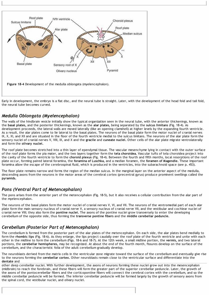
Figure 18-4 Development of the medulla oblongata (myelencephalon).
Early in development, the embryo is a flat disc, and the neural tube is straight. Later, with the development of the head fold and tail fold,the neural tube becomes curved.
Medulla Oblongata (Myelencephalon)The walls of the hindbrain vesicle initially show the typical organization seen in the neural tube, with the anterior thickenings, known asthe basal plates, and the posterior thickenings, known as the alar plates, being separated by the sulcus limitans (Fig. 18-4). Asdevelopment proceeds, the lateral walls are moved laterally (like an opening clamshell) at higher levels by the expanding fourth ventricle.As a result, the alar plates come to lie lateral to the basal plates. The neurons of the basal plate form the motor nuclei of cranial nervesIX, X, XI, and XII and are situated in the floor of the fourth ventricle medial to the sulcus limitans. The neurons of the alar plate form thesensory nuclei of cranial nerves V, VIII, IX, and X and the gracile and cuneate nuclei. Other cells of the alar plate migrate ventrolaterallyand form the olivary nuclei.
The roof plate becomes stretched into a thin layer of ependymal tissue. The vascular mesenchyme lying in contact with the outer surfaceof the roof plate forms the pia mater, and the two layers together form the tela choroidea. Vascular tufts of tela choroidea project intothe cavity of the fourth ventricle to form the choroid plexus (Fig. 18-4). Between the fourth and fifth months, local resorptions of the roofplate occur, forming paired lateral foramina, the foramina of Luschka, and a median foramen, the foramen of Magendie. These importantforamina allow the escape of the cerebrospinal fluid, which is produced in the ventricles, into the subarachnoid space (see p. 453).
The floor plate remains narrow and forms the region of the median sulcus. In the marginal layer on the anterior aspect of the medulla,descending axons from the neurons in the motor areas of the cerebral cortex (precentral gyrus) produce prominent swellings called thepyramids.
Pons (Ventral Part of Metencephalon)The pons arises from the anterior part of the metencephalon (Fig. 18-5), but it also receives a cellular contribution from the alar part ofthe myelencephalon.
The neurons of the basal plates form the motor nuclei of cranial nerves V, VI, and VII. The neurons of the ventromedial part of each alarplate form the main sensory nucleus of cranial nerve V, a sensory nucleus of cranial nerve VII, and the vestibular and cochlear nuclei ofcranial nerve VIII; they also form the pontine nuclei. The axons of the pontine nuclei grow transversely to enter the developingcerebellum of the opposite side, thus forming the transverse pontine fibers and the middle cerebellar peduncle.
Cerebellum (Posterior Part of Metencephalon)The cerebellum is formed from the posterior part of the alar plates of the metencephalon. On each side, the alar plates bend medially toform the rhombic lips (Fig. 18-6). As they enlarge, the lips project caudally over the roof plate of the fourth ventricle and unite with eachother in the midline to form the cerebellum (Figs. 18-6 and 18-7). At the 12th week, a small midline portion, the vermis, and two lateralportions, the cerebellar hemispheres, may be recognized. At about the end of the fourth month, fissures develop on the surface of thecerebellum, and the characteristic folia of the adult cerebellum gradually develop.
The neuroblasts derived from the matrix cells in the ventricular zone migrate toward the surface of the cerebellum and eventually give riseto the neurons forming the cerebellar cortex. Other neuroblasts remain close to the ventricular surface and differentiate into thedentate andother deep cerebellar nuclei. With further development, the axons of neurons forming these nuclei grow out into the mesencephalon(midbrain) to reach the forebrain, and these fibers will form the greater part of the superior cerebellar peduncle. Later, the growth ofthe axons of the pontocerebellar fibers and the corticopontine fibers will connect the cerebral cortex with the cerebellum, and so themiddle cerebellar peduncle will be formed. The inferior cerebellar peduncle will be formed largely by the growth of sensory axons fromthe spinal cord, the vestibular nuclei, and olivary nuclei.

P.509
Figure 18-5 Development of the pons from the anterior part of the metencephalon.
Figure 18-6 Development of the cerebellum. Also shown is the fusion of the rhombic lips in the midline to form the dumbbell-shaped cerebellum.
Midbrain (Mesencephalon)The midbrain develops from the midbrain vesicle, the cavity of which becomes much reduced to form the cerebral aqueduct or aqueductof Sylvius (Fig. 18-8). The sulcus limitans separates the alar plate from the basal plate on each side, as seen in the developing spinal cord.The neuroblasts in the basal plates will differentiate into the neurons forming the nuclei of the third and fourth cranial nerves andpossibly the red nuclei, the substantia nigra, and the reticular formation. The marginal zone of each basalplate enlarges considerably, thus forming the basis pedunculi by the descent of nerve fibers from the cerebral cortex to thelower motor centers in the pons and spinal cord—that is, the corticopontine, corticobulbar, and corticospinal tracts.
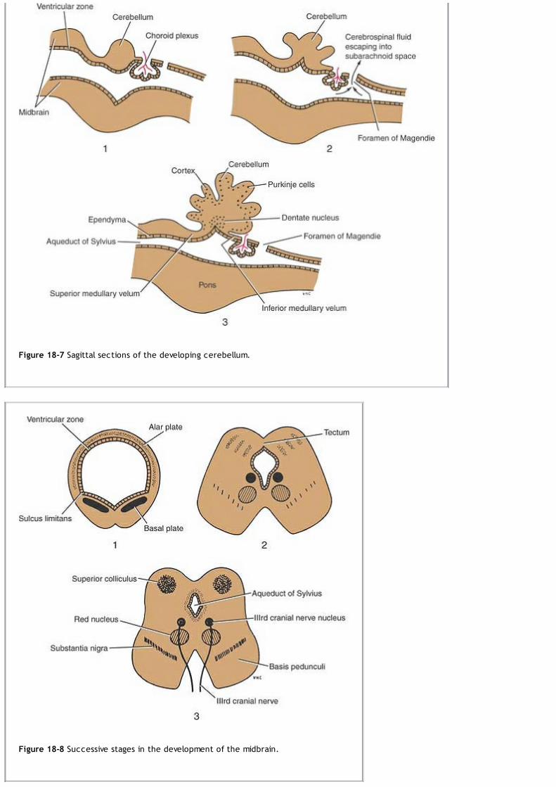
Figure 18-7 Sagittal sections of the developing cerebellum.
Figure 18-8 Successive stages in the development of the midbrain.

Figure 18-9 Division of the forebrain vesicle into the telencephalon and the diencephalon.
The two alar plates and the original roof plate form the tectum. The neuroblasts in the alar plates differentiate into the sensory neuronsof the superior and inferior colliculi (Fig. 18-8). Four swellings representing the four colliculi appear on the posterior surface of themidbrain. The superior colliculi are associated with visual reflexes, and the inferior colliculi are associated with auditory reflexes.
With further development, the fibers of the fourth cranial nerve emerge on the posterior surface of the midbrain and decussatecompletely in the superior medullary velum. The fibers of the third cranial nerve emerge on the anterior surface between the cerebralpeduncles.
Forebrain (Prosencephalon)The forebrain develops from the forebrain vesicle. The roof and floor plates remain thin, whereas the lateral walls become thick, as in thedeveloping spinal cord. At an early stage, a lateral diverticulum called the optic vesicle appears on each side of the forebrain. That part ofthe forebrain that lies rostral to the optic vesicle is the telencephalon, and the remainder is the diencephalon (Fig. 18-9). The opticvesicle and stalk ultimately will form the retina and optic nerve.
The telencephalon now develops a lateral diverticulum on each side of the cerebral hemisphere, and its cavity is known as the lateralventricle. The anterior part of the third ventricle, therefore, is formed by the medial part of the telencephalon and ends at the laminaterminalis, which represents the rostral end of the neural tube. The opening into each lateral ventricle is the future interventricularforamen.
Fate of the DiencephalonThe cavity of the diencephalon forms the greater part of the third ventricle (Fig. 18-9). Its roof shows a small diverticulum immediatelyanterior to the midbrain, which will form the pineal body. The remainder of the roof forms the choroid plexus of the third ventricle (Fig.18-10). In the lateral wall of the third ventricle, the thalamus arises as a thickening of the alar plate on each side. Posterior to thethalamus, the medial and lateral geniculate bodies develop as solid buds. With the continued growth of the two thalami, the ventricularcavity becomes narrowed, and in some individuals, the two thalami may meet and fuse in the midline to form the interthalamic connectionof gray matter that crosses the third ventricle (Fig. 18-11).
The lower part of the alar plate on each side will differentiate into a large number of hypothalamic nuclei. One of these becomesconspicuous on the inferior surface of the hypothalamus and forms a rounded swelling on each side of the midline called the mammillarybody.
The infundibulum develops as a diverticulum from the floor of the diencephalon, and from it will originate the stalk and pars nervosa ofthe hypophysis.
Fate of the TelencephalonThe telencephalon forms the anterior end of the third ventricle, which is closed by the lamina terminalis, while the diverticulum on eitherside forms the cerebral hemisphere.
Cerebral HemispheresEach cerebral hemisphere arises at the beginning of the fifth week of development. As it expands superiorly, its walls thicken, and theinterventricular foramen becomes reduced in size (Figs. 18-9, 18-10 and 18-11). The mesenchyme between each cerebral hemispherecondenses to form the falx cerebri. As development proceeds, the cerebral hemispheres grow and expand rapidly, first anteriorly to formthe frontal lobes, then laterally and superiorly to form the parietal lobes, and finally posteriorly and inferiorly to produce the occipitaland temporal lobes. As the result of this great expansion, the hemispheres cover the midbrain and hindbrain (Fig. 18-12).
The medial wall of the cerebral hemisphere remains thin and is formed by the ependymal cells. This area becomes invaginated by vascular

P.511mesoderm, which forms the choroid plexus of the lateral ventricle (Fig. 18-11). Theoccipital lobe of the cerebral hemisphere is separated from the cerebellum by mesenchyme, which condenses to form thetentorium cerebelli.
Figure 18-10 Diagrammatic representation of a coronal section of the cerebral hemispheres showing the developing choroidplexuses in the third and lateral ventricles.
Figure 18-11 Diagrammatic representation of a coronal section of the cerebral hemispheres showing the choroid plexuses in thethird and lateral ventricles. Also shown are the caudate and lentiform nuclei and the thalami. The ascending and descending nervetracts can be seen passing between the masses of gray matter to form the internal capsule.

Figure 18-12 Successive stages in the development of the cerebral cortex.
Meanwhile, the matrix cells lining the floor of the forebrain vesicle proliferate, producing large numbers of neuroblasts. These collectivelyform a projection that encroaches on the cavity of the lateral ventricle and is known as the corpus striatum (Fig. 18-10). Later, thisdifferentiates into two parts: (1) the dorsomedial portion, the caudate nucleus, and (2) a ventrolateral part, the lentiform nucleus. Thelatter becomes subdivided into a lateral part, the putamen, and a medial part, the globus pallidus (Fig. 18-11). As each hemisphereexpands, its medial surface approaches the lateral surface of the diencephalon; thus, the caudate nucleus and thalamus come in closecontact.
A further longitudinal thickening occurs in the wall of the forebrain vesicle, and the thickening protrudes into the lateral ventricle andforms the hippocampus (Fig. 18-11).
While these various masses of gray matter are developing within each cerebral hemisphere, maturing neurons in different parts of thenervous system are sending axons either to or from the differentiating cortex. These axons form the large ascending and descendingtracts, which, as they develop, are forced to pass between the thalamus and caudate nucleus medially and the lentiform nucleus laterally.The compact bundle of ascending and descending tracts is known as the internal capsule (Fig. 18-11). The external capsule consists of afew cortical projection fibers that pass lateral to the lentiform nucleus.
Cerebral Cortex

P.514P.515
As each cerebral hemisphere rapidly expands, the convolutions or gyri separated by fissures or sulci become evident on its surface. Thecortex covering the lentiform nucleus remains as a fixed area called the insula (Fig. 18-12). Later, this region becomes buried in the lateralsulcus as the result of overgrowth of the adjacent temporal, parietal, and frontal lobes.
The matrix cells lining the cavity of the cerebral hemisphere produce large numbers of neuroblasts and neuroglial cells that migrate outinto the marginal zone. The remaining matrix cells ultimately will form the ependyma, which lines the lateral ventricle. In the 12th week,the cortex becomes very cellular because of the migration of large numbers of neuroblasts. At term, the neuroblasts have becomedifferentiated and have assumed a stratified appearance as the result of the presence of incoming and outgoing fibers. Different areas ofthe cortex soon show specific cell types; thus, the motor cortex contains a large number of pyramidal cells, whereas the sensory areasare characterized mainly by granular cells.
CommissuresThe lamina terminalis, which is the cephalic end of the neural tube, forms a bridge between the two cerebral hemispheres and enablesnerve fibers to pass from one cerebral hemisphere to the other (Fig. 18-9).
The anterior commissure is the first commissure to develop. It runs in the lamina terminalis and connects the olfactory bulb and thetemporal lobe of the cortex on one side with the same structures of the opposite hemisphere.
The fornix is the second commissure to develop and connects the cortex of the hippocampus in each hemisphere.
The corpus callosum, the largest and most important commissure, is the third commissure to develop. Its first fibers connect the frontallobes of both sides and, later, the parietal lobes. As the corpus callosum increases in size because of increased numbers of fibers, itarches back over the roof of the developing third ventricle. The remains of the lamina terminalis, which lie between the corpus callosumand the fornix, become stretched out to form a thin septum, the septum pellucidum. The optic chiasma is formed in the inferior part ofthe lamina terminalis; it contains fibers from the medial halves of the retinae, which cross the midline to join the optic tract of theopposite side and so pass to the lateral geniculate body and the superior colliculus.
Myelination in the Central Nervous SystemThe myelin sheath in the central nervous system is formed and maintained by the oligodendrocytes of the neuroglia (see p. 71).
Myelination in the spinal cord begins first in the cervical region, and from here, the process extends caudally. The process of myelinationbegins within the cord at about the fourth month, and the sensory fibers are affected first. The last affected are the descending motorfibers.
Myelination in the brain begins at about the sixth month of fetal life but is restricted to the fibers of the basal ganglia. Later, the sensoryfibers passing up from the spinal cord myelinate, but the progress is slow; therefore, at birth, the brain still is largely unmyelinated. In thenewborn, there is very little cerebral function; motor reactions such as respiration, sucking, and swallowing are essentially reflex. Afterbirth, the corticobulbar, corticospinal, tectospinal, and corticopontocerebellar fibers begin to myelinate. This process of myelination isnot haphazard but systematic, occurring in different nerve fibers at specific times. The corticospinal fibers, for example, start to myelinateat about 6 months after birth, and the process is largely complete by the end of the second year. It is believed that some nerve fibers inthe brain and spinal cord do not complete myelination until puberty.
Clinical NotesCongenital AnomaliesPractically any part of the nervous system can show defects of development, and these produce a wide variety
of clinical signs and symptoms. Only the common defects of the central nervous system are considered here. Spinabifida, hydrocephalus, and anencephaly each occur about 6 times per 1,000 births and are therefore the more commoncongenital anomalies.
Spina BifidaIn spina bifida, the spines and arches of one or more adjacent vertebrae fail to develop. The condition occurs mostfrequently in the lower thoracic, lumbar, and sacral regions. Beneath this defect, the meninges and spinal cord may ormay not be involved to varying degrees. The condition is a result of failure of the mesenchyme, which grows inbetween the neural tube and the surface ectoderm, to form the vertebral arches in the affected region. The types ofspina bifida are as follows:
1. Spina bifida occulta. The spines and arches of one or more vertebrae, usually in the lumbar region, are absent,and the vertebral canal remains open posteriorly (Fig. 18-13). The spinal cord and nerve roots usually are normal.The defect is covered by the postvertebral muscles and cannot be seen from the surface. A small tuft of hair or afatty tumor may be present over the defect. Most cases are symptomless and are diagnosed by chance when thevertebral column is x-rayed.
2. Meningocele. The meninges project through the defect in the vertebral arches, forming a cystic swelling beneaththe skin and containing cerebrospinal fluid, which communicates with the subarachnoid space (Figs. 18-13 and 18-14). The spinal cord and nerves usually are normal.
3. Meningomyelocele. The normal spinal cord, or cauda equina, lies within the meningeal sac, which projects throughthe vertebral arch defect (Fig. 18-13). The spinal cord or nerve roots are adherent to the inner wall of the sac.

Figure 18-13 Different types of spina bifida.
4. Myelocele. The neural tube fails to close in the region of the defect (Fig. 18-13). An oval raw area is found on thesurface; this represents the neural groove whose lips are fused. The central canal discharges clear cerebrospinalfluid onto the surface.
5. Syringomyelocele. This condition is rare. A meningomyelocele is present, and in addition, the central canal of thespinal cord at the level of the bony defect is grossly dilated (Fig. 18-13).
Spina bifida occulta is the most common defect. The next most common defect is myelocele, and many afflicted infantsare born dead. If the child is born alive, death from infection of the spinal cord may occur within a few days.Most cases of spina bifida occulta require no treatment. A meningocele should be removed surgically within a few daysof birth. Infants with meningomyelocele should also be treated surgically. The sac is opened, and the spinal cord ornerves are freed and carefully replaced in the vertebral canal. The meninges are sutured over the cord and thepostvertebral muscles are approximated.As the result of advances in medical and surgical care, many infants with the severe forms of spina bifida now survive.Unfortunately, these children are likely to have lifelong disabilities and psychosocial problems. The neurologic deficitsalone may result in deformation of the limbs and spine and in bladder, bowel, and sexual dysfunction.

Figure 18-14 A meningocele in the lumbosacral region. (Courtesy Dr. L. Thompson.)
HydrocephalusHydrocephalus is an abnormal increase in the volume of cerebrospinal fluid within the skull. The condition may beassociated with spina bifida and meningocele. Hydrocephalus alone may be caused by stenosis of the cerebralaqueduct or, more commonly, by the normal single channel being represented by many inadequate minute tubules.Another cause, which is progressive, is the overgrowth of neuroglia around the aqueduct. Inadequate development orfailure of development of the interventricular foramen, or the foramina of Magendie and Luschka, may also beresponsible.In cases of hydrocephalus with spina bifida, the Arnold-Chiari phenomenon may occur. During development, thecephalic end of the spinal cord is fixed by virtue of the brain residing in the skull, and in the presence of spina bifida,the caudal end of the cord may also be fixed. The longitudinal growth of the vertebral column is more rapid and greaterthan that of the spinal cord, and this results in traction pulling the medulla and part of the cerebellum through theforamen magnum. This displacement of the hindbrain downward obstructs the flow of cerebrospinal fluid through theforamina in the roof of the fourth ventricle.Hydrocephalus may occur before birth, and if it is advanced, it could obstruct labor. It usually is noticed during the firstfew months of life because of the enlarging head, which may attain a huge size, sometimes measuring more than 30inches in diameter (Fig. 18-15). The cranial sutures are widely separated, and the anterior fontanelle is much enlarged.The veins of the scalp are distended, and the eyes look downward. Cranial nerve paralyses are common. Theventricles of the brain become markedly dilated. This ventricular expansion occurs largely at the expense of the whitematter, and the neurons of the cerebral cortex are mostly spared. This results in the preservation of cerebral function,but the destruction of the tracts, especially the corticobulbar and corticospinal tracts, produces a progressive loss ofmotor function.If the condition is diagnosed by sonography while the fetus is in utero, it is possible to perform prenatal surgery withthe introduction of a catheter into the ventricles of the brain and the drainage of the cerebrospinal fluid into theamniotic cavity. Should the diagnosis be delayed until after birth, a drainage tube fitted with a nonreturn valve canconnect the ventricles to the internal jugular vein in the neck.
AnencephalyIn anencephaly, the greater part of the brain and the vault of the skull are absent (Fig. 18-16). The anomaly is causedby the failure of the rostral end of the neural tube to develop, and as a consequence, its cavity remains open. In placeof the normal neural tissue, there are thin-walled vascular channels resembling the choroid plexus and masses ofneural tissue. Although the eyes are present, the optic nerves are absent. The condition commonly involves the spinalcord, and the neural tube remains open in the cervical region. The condition is commonly diagnosed before birth withsonography or x-ray studies. Most anencephalic infants are stillborn or die shortly after birth.
Prevention of Neural Defects With Folic AcidThe development and closure of the neural tube are normally completed within 28 days. In practical terms, this meansthat neural tube defects have occurred before many women are aware that they are pregnant.Extensive clinical research has demonstrated that environmental and genetic factors have a joint role in the causationof neural tube defects. The increased risk of neural defects in the lower socioeconomic groups suggests that poornutrition may also be an important factor. More recent clinical research has demonstrated that the risk of recurrentneural defects is significantly reduced among women who take 4,000 mg of folic acid daily compared with women whodo not. Further studies have shown that a daily dose that is 10 times lower is effective in preventing the defect. Thesefindings have stimulated much new research to identify the genetic and biochemical bases of neural tube defects.Because as many as 50% of pregnancies in the United States are unplanned and since the neural tube closes beforemost women know that they are pregnant, physicians should strongly urge women capable of becoming pregnant toconsume at least 400 mg of folic acid per day, preferably in a multivitamin supplement.

Embryonic Stem Cells and the Treatment of Neurologic DiseasesFreed et al. (2001) reported treating patients with severe Parkinson disease by transplanting precursors of dopaminenerve cells in fragments of mesencephalon isolated from human fetuses 7 to 8 weeks after conception. The resultsshowed that the transplants survived and produced some clinical benefit in younger but not in older patients. It wasdemonstrated that the cells survived and differentiated, as demonstrated by positron emission tomography or byhistologic examination (see Fig. 10-9). In view of the large numbers of patients with Parkinson disease worldwide, it isunlikely that transplantation of embryonic fragments would be a practical therapy.
Figure 18-15 Hydrocephalus. Note the large size of the head. (Courtesy Dr. G. Avery.)
Embryonic stem cells have the unique property of being able to produce all adult cell types, including those of thenervous system. The successful transplantation of embryonic stem cells has been achieved in animal models ofParkinson disease, motor neuron disease, and spinal cord injury. The great promise of these results has stimulatedthe imagination of scientists and patients. The use of cell lines derived from human embryonic stem cells, however,poses profound ethical questions.
Figure 18-16 Example of anencephaly. Note that the greater part of the brain and the vault of the skull are absent. In theposterior view, the remainder of the brain is exposed. (Courtesy Dr. M. Platt.)

Embryonic stem cells are derived from the inner cell mass of the blastocyst, the stage at which the developing embryois implanted into the uterus. Human embryonic stem cells were first successfully harvested by Thomson et al. in 1998.The isolated inner cell mass cells were then cultured in the laboratory. Although substantial advances have been madein this field (Cowan et al., 2004), it is critical that enormous efforts should now be devoted to this subject so that wecan improve the health of patients with chronic debilitating neurologic diseases.
Clinical Problem Solving1. A 10-year-old boy fell off his bicycle and hurt his back. Following a complete physical examination in the emergencydepartment, nothing abnormal was found. An x-ray examination, however, revealed the complete absence of the spineand laminae of the fifth lumbar vertebra. How would you explain the presence of the bony defect?View Answer
2. A male child was delivered normally to a 20-year-old woman. A pediatrician examined the infant and found a largeswelling in the lower part of his back over the fourth and fifth lumbar vertebrae. On closer examination, the summit ofthe swelling had an oval raw area from which a clear fluid was discharging. The legs showed hyperextension of theknees, and the feet were held in the position of talipes calcaneus. What is the diagnosis? How would you explain thecongenital defect on the back?View Answer
3. A 2-month-old girl was taken to a pediatrician because her mother was concerned about the size of her head. “Shelooks top-heavy,” she said. Examination showed the head to be large and globular in shape. The anterior fontanellewas greatly enlarged and extended posteriorly to the enlarged posterior fontanelle. The enlarged head contrastedmarkedly with the small face. Neurologic examination revealed some evidence of optic atrophy on both sides, andthere was increased tone in the muscles of both lower limbs. What is the diagnosis? How can you explain thiscongenital anomaly? What is the prognosis if the patient is left untreated?View Answer
Review QuestionsDirections: Each of the numbered items in this section is followed by answers. Select the ONE lettered answerthat is CORRECT.1. The following statements concern the neural tube:
(a) It is lined by stratified squamous cells.(b) The neuroblasts migrate medially to form the intermediate zone.(c) The repeated division of the matrix cells does not increase the length and diameter of the tube.(d) The ventricular zone will form the gray matter of the spinal cord.(e) The nerve fibers in the marginal zone become myelinated and form the white matter of the spinal cord.
View Answer
2. The following statements concern the neural crest cells:(a) They are formed from the medial margin of the neural plate.(b) They give rise to the posterior root ganglia.(c) They do not form the neurons of the autonomic ganglia.(d) The Schwann cells of peripheral nerves are not formed from neural crest cells.(e) They form the cells of the suprarenal cortex.
View Answer
3. The following statements concern the developing spinal cord:(a) The alar plates form the neurons in the anterior gray columns.(b) The nerve cells of the sympathetic outflow are not formed from the basal plates.(c) In the adult, the lower end of the spinal cord lies at the level of the lower border of the first lumbar vertebra.(d) At birth, the lower end of the spinal cord lies at the level of the third sacral vertebra.(e) The meninges surrounding the spinal cord are developed from the endoderm.
View Answer
4. The following statements concern the development of the brainstem:(a) The cerebellum is formed from the dorsal part of the alar plates of the metencephalon.(b) The neurons of the deep cerebellar nuclei are derived from the matrix cells lining the cavity of the midbrainvesicle.(c) The neuroblasts in the dorsal plates will form the nuclei of the trochlear and oculomotor nerves.(d) The neuroblasts of the superior and inferior colliculi are also formed from the neurocytes in the basal plates.(e) The pons arises from the alar part of the metencephalon with cellular contributions from the alar part of themyelencephalon.

P.518P.519
View Answer
5. The following statements concern the fate of the forebrain vesicle:(a) The optic vesicle grows out of the midbrain vesicle.(b) The thalamus is formed from the alar plates in the medial walls of the diencephalon.(c) The lamina terminalis is formed from the rostral end of the diencephalon.(d) The pars nervosa of the hypophysis is formed from the floor of the diencephalon.(e) The hypothalamic nuclei are formed from the basal plates of the diencephalon.
View Answer
6. The following statements concern the development of the cerebral hemispheres:(a) The corpus striatum is formed from the proliferation of the matrix cells lining the roof of the forebrain vesicle.(b) The interventricular foramen is formed by the cavity of the diencephalon.(c) The choroid plexus of the lateral ventricle is formed by vascular ectoderm covered by ependymal cells.(d) The internal capsule is formed by the developing ascending and descending tracts growing between thedeveloping thalamus and caudate nucleus medially and the lentiform nucleus laterally.(e) The cortical neurons develop in situ and do not migrate out laterally from the matrix cells lining the cavity ofthe cerebral hemisphere.
View Answer
7. The following statements concern the development of myelination in the brain:(a) Myelination begins at birth.(b) The sensory fibers are myelinated last.(c) The process of myelination is haphazard.(d) Myelination of the nerve tracts is largely complete by the fourth year of life.(e) Myelination is carried out by oligodendrocytes and not by neurons.
View Answer
8. The following statements concern the condition of spina bifida:(a) It is one of the more common congenital anomalies of the central nervous system.(b) The most common form of spina bifida is syringomyelocele.(c) The condition occurs most often in the cervical and upper thoracic regions.(d) In a myelocele, the neural tube closes in the region of the defect.(e) Most cases of spina bifida occulta require explorative surgery.
View Answer
Directions: Each case history is followed by questions. Read the case history, then select the ONE BEST letteredanswer.A 6-month-old girl was seen by the plastic surgeon because of the presence of a swelling at the root of the nose. Themother said that she had noticed the swelling when the child was born and that since then, it had gradually increasedin size.9. The surgeon examined the child and found the following likely signs except:
(a) The swelling was situated at the root of the nose in the midline.(b) The swelling was located between the frontal and nasal bones.(c) The swelling was fluctuant and, on gentle pressure, could be reduced in size.(d) The swelling was pulsatile, and the pulse coincided with the heart rate.(e) The pulse did not coincide with the pulse felt over the anterior fontanelle of the skull.
View Answer
10. The neurosurgeon was consulted, and the following possible additional findings were ascertained except:(a) A lateral radiograph of the skull revealed a defect in the membranous bones involving the nasal process ofthe frontal bone.(b) The defect in the membranous bones is known as cranioschisis.(c) The condition was associated with a cephalic meningocele.(d) There was a herniation of the meninges through the defect in the skull.(e) Brain tissue is never found within the hernia.
View Answer
Additional Reading

Anderson, D. J. Cellular and molecular biology of neural crest cell lineage determination. Trends Genet. 13:276–280, 1997.
Anderson, S. A., Eisenstat, D. D., Shi, L., and Rubenstein, J. L. Interneuron migration from basal forebrain to neocortex: Dependenceon D1x genes. Science 278:474–476, 1997.
Berry, R. J., Li, Z., Erickson, J. D., Li, S., Moore, C. A., Wang, H., et al. Prevention of neural tube defects with folic acid in China. N.Engl. J. Med. 341:1485–1490, 1999.
Botto, L. D., Moore, C. A., Khoury, M. J., and Erickson, J. D. Medical progress: Neural tube defects. N. Engl. J. Med. 341:1509–1519,1999.
Brown, M. Keynes, R. Lumsden, A. The Developing Brain. Oxford. Oxford Unviversity Press, 2001.
Cowan, C. A., Klimanskaya, I., McMahon, J., Atienza, J., Witmyer, J., Zucker, J. P., et al. Derivation of embryonic stem-cell lines fromhuman blastocysts. N. Engl. J. Med. 350:1353, 2004.
Crossley, P. H., Martinez, S., and Martin, G. R. M idbrain development induced by FGF8 in the chick embryo. Nature 380:66–68, 1996.
Ericson, J., Briscoe, J., Rashbass, P., van Heyningen, V., and Jessell, T. M. Graded sonic hedgehog signaling and the specification ofcell fate in the ventral neural tube. Cold Spring Harb. Symp. Quant. Biol. 62:451–466, 1997.
Fischbach, G. D., and McKhann, G. M. Cell therapy for Parkinson's disease. N. Engl. J. Med. 344:763, 2001.
Francis, N. J., and Landis, S. C. Cellular and molecular determinants of sympathetic neuron development. Annu. Rev. Neurosci. 22:541–566, 1999.
Freed, C. R., Greene, P. E., Breeze, R. E., Tsai, W.-Y., DuMouchel W., Kao, R., et al. Transplantation of embryonic dopamine neuronsfor severe Parkinson's disease. N. Engl. J. Med. 344:710, 2001.
Gearhart, J. New human stem-cell lines—more or better. N. Engl. J. Med. 350:1275, 2004.
Hatten, M. E. Central nervous system neuronal migration. Annu. Rev. Neurosci. 22:261–294, 1999.
Henderson, C. E. Programmed cell death in the developing nervous system. Neuron 17:579–585, 1996.
Lee, K. J., and Jessell, T. M. The specification of dorsal cell fates in the vertebrate central nervous system. Annu. Rev. Neurosci.22:261–294, 1999.
Liem, J. F. Jr., Tremml, G., Roelink, H., and Jessell, T. M. Dorsal differentiation of neural plate cells induced by BMP-mediated signalsfrom epidermal ectoderm. Cell 82:969–979, 1995.
Lumsden, A., and Gulisano, M. Neocortical neurons: Where do they come from? Science 278:402–403, 1997.
Marigo, V., Davey, R. A., Zuo, Y., Cunningham, J. M., and Tabin, C. J. Biochemical evidence that patched is the hedgehog receptor.Nature 384:176–179, 1996.
Mueller, B. K. Growth cone guidance: First steps towards a deeper understanding. Annu. Rev. Neurosci. 22:351–388, 1999.
O'Leary, D. D., and Wilkinson, D. G. Eph receptors and ephrins in neural development. Curr. Opin. Neurobiol. 9:65–73, 1999.
Phimister, E. G., and Drazen, J. M. Two fillips for human embryonic stem cells. N. Engl. J. Med. 350:1351, 2004.

Snyder, E. Y., and Loring, J. F. Beyond fraud—Stem-cell research continues. N. Engl. J. Med. 354:321, 2006.
Song, H. J., and Poo, M. M. Signal transduction underlying growth cone guidance by diffusible factors. Curr. Opin. Neurobiol. 9:355–363, 1999.
Standring, S. (ed.). Gray's Anatomy (39th Br. ed.). London: Elsevier Churchill Livingstone, 2005.
Tanabe, Y., and Jessell, T. M. Diversity and pattern in the developing spinal cord. Science 274:1115–1123, 1997.
Tessier-Lavigne, M., and Goodman, C. S. The molecular biology of axon guidance. Science 274:1123–1133, 1996.
Thomson, J. A., Itskovitz-Eldor, J., Shapiro, S. S., Waknitz, M. A., Swiergiel, J. J., Marshall, V. S., et al. Embryonic stem cell linesderived from human blastocysts. Science 282:1145, 1998. (Erratum. Science 282:1827, 1998.)
Wolpert, L., Beddington, R., Brockes, J., Jessell, T. M., Lawrence, P. A., and Meyerowitz, E. Principles of Development. New York:Oxford University Press, 1998.
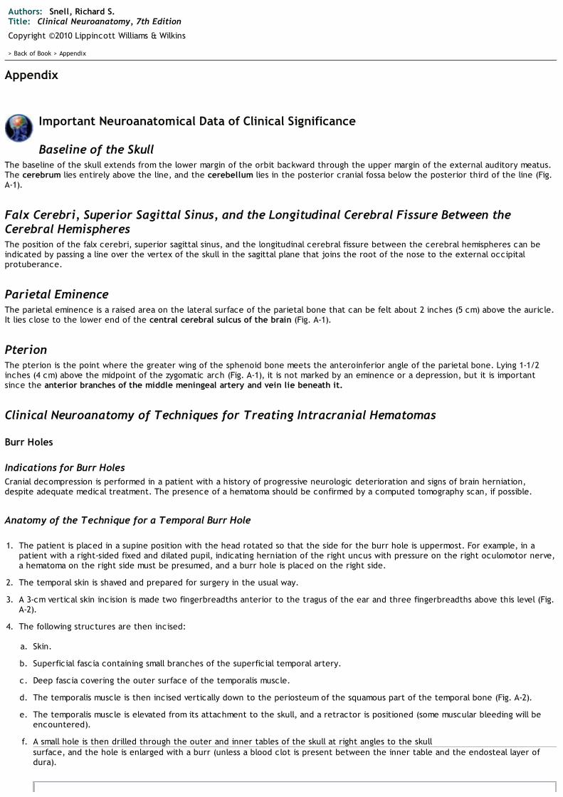
Authors: Snell, Richard S.Title: Clinical Neuroanatomy, 7th Edition
Copyright ©2010 Lippincott Williams & Wilkins
> Back of Book > Appendix
Appendix
Important Neuroanatomical Data of Clinical Significance
Baseline of the SkullThe baseline of the skull extends from the lower margin of the orbit backward through the upper margin of the external auditory meatus.The cerebrum lies entirely above the line, and the cerebellum lies in the posterior cranial fossa below the posterior third of the line (Fig.A-1).
Falx Cerebri, Superior Sagittal Sinus, and the Longitudinal Cerebral Fissure Between theCerebral HemispheresThe position of the falx cerebri, superior sagittal sinus, and the longitudinal cerebral fissure between the cerebral hemispheres can beindicated by passing a line over the vertex of the skull in the sagittal plane that joins the root of the nose to the external occipitalprotuberance.
Parietal EminenceThe parietal eminence is a raised area on the lateral surface of the parietal bone that can be felt about 2 inches (5 cm) above the auricle.It lies close to the lower end of the central cerebral sulcus of the brain (Fig. A-1).
PterionThe pterion is the point where the greater wing of the sphenoid bone meets the anteroinferior angle of the parietal bone. Lying 1-1/2inches (4 cm) above the midpoint of the zygomatic arch (Fig. A-1), it is not marked by an eminence or a depression, but it is importantsince the anterior branches of the middle meningeal artery and vein lie beneath it.
Clinical Neuroanatomy of Techniques for Treating Intracranial Hematomas
Burr Holes
Indications for Burr HolesCranial decompression is performed in a patient with a history of progressive neurologic deterioration and signs of brain herniation,despite adequate medical treatment. The presence of a hematoma should be confirmed by a computed tomography scan, if possible.
Anatomy of the Technique for a Temporal Burr Hole
1. The patient is placed in a supine position with the head rotated so that the side for the burr hole is uppermost. For example, in apatient with a right-sided fixed and dilated pupil, indicating herniation of the right uncus with pressure on the right oculomotor nerve,a hematoma on the right side must be presumed, and a burr hole is placed on the right side.
2. The temporal skin is shaved and prepared for surgery in the usual way.
3. A 3-cm vertical skin incision is made two fingerbreadths anterior to the tragus of the ear and three fingerbreadths above this level (Fig.A-2).
4. The following structures are then incised:
a. Skin.
b. Superficial fascia containing small branches of the superficial temporal artery.
c. Deep fascia covering the outer surface of the temporalis muscle.
d. The temporalis muscle is then incised vertically down to the periosteum of the squamous part of the temporal bone (Fig. A-2).
e. The temporalis muscle is elevated from its attachment to the skull, and a retractor is positioned (some muscular bleeding will beencountered).
f. A small hole is then drilled through the outer and inner tables of the skull at right angles to the skullsurface, and the hole is enlarged with a burr (unless a blood clot is present between the inner table and the endosteal layer ofdura).

Figure A-1 Surface landmarks on the right side of the head. The relation of the middle meningeal artery and the brain to thesurface of the skull is shown.
g. The white meningeal layer of dura is flexible and gives slightly on gentle pressure.
h. The hole may be enlarged with a curette, and bleeding from the diploe may be controlled with bone wax.
The surgical wound is closed in layers with interrupted sutures placed in the temporalis muscle, the deep fascia covering the temporalismuscle, and the scalp.
Burr Hole for Epidural HematomaOnce the inner table of the squamous part of the temporal bone (or the anterior inferior angle of the parietal bone) is pierced with asmall bit and enlarged with a burr, the dark red clotted blood beneath the endosteal layer of dura is usually easily recognized. However,bright red liquid blood means that the middle meningeal artery or one of its branches is bleeding. The meningeal artery is located deep tothe clot and between the endosteal layer of dura and the meningeal layer of dura or in the substance of the endosteal layer of dura; or itmay lie in a tunnel of bone.
Burr Hole for Subdural HematomaWhen the squamous part of the temporal bone is penetrated, as described earlier, the endosteal layer of dura will be exposed. In thiscase, there is no blood clot between the endosteal layer of dura and the meningeal layer of dura, but both fused layers of dura will bedark bluish. The dura (endosteal and meningeal layers) is gently incised to enter the space between the meningeal layer of dura and thearachnoid mater. The subdural blood usually gushes out, leaving the unprotected brain covered only by arachnoid and pia mater in thedepths of the hole.
Clinical Neuroanatomy of the Technique of Ventriculostomy
Indications for VentriculostomyVentriculostomy is indicated in acute hydrocephalus, in which there is a sudden obstruction to the flow of cerebrospinal fluid.
Anatomy of the Technique of VentriculostomyTo perform a ventriculostomy, the needle is inserted into the lateral ventricle through either a frontal or parietal burr hole. The anatomyof these burr holes has been described previously. The needle is inserted through the burr hole using the following anatomical landmarks.
1. Frontal Approach. The needle is inserted through the frontal burr hole and is directed downward and forward in the direction of the

inner canthus of the ipsilateral eye (Fig. A-3).
Figure A-2 A: Surface landmarks for a temporal burr hole. B: The vertical incision passes through the temporalis muscle down tobone. The middle meningeal artery lies between the endosteal and meningeal layers of dura and is embedded in the endosteallayer of dura or lies in a bony tunnel.

Figure A-3 Ventriculostomy. Needles passing through frontal or parietal burr holes to enter the lateral ventricle area areshown. The needle is inserted to a depth of about 2 inches (5.5 cm) from the skull opening in order to enter the lateralventricle.
2. Parietal Approach. The needle is inserted through the parietal burr hole and is directed downward and forward in the direction of thepupil of the ipsilateral eye (Fig. A-3).
The needle is inserted to a depth of about 2 inches (5.5 cm) from the skull opening; in cases of chronic hydrocephalus with grossdilatation of the ventricles, the depth of penetration to the ventricular cavity may be much less.
Vertebral Numbers and Spinal Cord SegmentsTable A-1 relates which vertebral body is related to a particular spinal cord segment.
Table A-1
Vertebrae Spinal Seg ment
Cervical vertebrae Add 1
Upper thoracic vertebrae Add 2
Lower thoracic vertebrae (7–9) Add 3
Tenth thoracic vertebra L1-2 cord segments
Eleventh thoracic vertebra L3-4 cord segments
Twelfth thoracic vertebra L5 cord segment
First lumbar vertebra Sacral and coccygeal cord segments

Segmental Innervation of MusclesIt is possible to test for the integrity of the segmental innervation of muscles by performing the following simple muscle reflexes on thepatient.
Biceps brachii tendon reflex C5-6 (flexion of the elbow joint by tapping the biceps tendon).
Triceps tendon reflex C6-7 and 8 (extension of the elbow joint by tapping the triceps tendon).
Brachioradialis tendon reflex C5-6 and 7 (supination of the radioulnar joints by tapping the insertion of the brachioradialis tendon).
Abdominal superficial reflexes (contraction of underlying abdominal muscles by stroking the skin). Upper abdominal skin T6-7; middleabdominal skin T8-9; lower abdominal skin T10–12.
Patellar tendon reflex (knee jerk) L2, 3, and 4 (extension of knee joint on tapping the patellar tendon).
Achilles tendon reflex (ankle jerk) S1 and 2 (plantar flexion of ankle joint on tapping the Achilles tendon–tendo calcaneus).
Relationship Between Possible Intervertebral Disc Herniations and Spinal Nerve RootsIt is useful to be able to relate possible nucleus pulposus herniations with spinal nerve roots. These are shown for the cervical and lumbarregions in Figure A-4.
A correlation between the nerve roots involved, the pain dermatome, the muscle weakness, and the missing or diminished reflex is shownin Table A-2.
Table A-2 Correlation Between Nerve Roots Involved, the Pain Dermatome, the Muscle Weakness, and the Missing or DiminishedReflex
RootInjury Dermatome Pain Muscles Supplied Movement Weakness Reflex Involved
C5 Lower lateral aspect ofupper arm Deltoid and biceps Shoulder abduction,
elbow flexion Biceps
C6 Lateral aspect of forearm Extensor carpi radialis longusand brevis Wrist extensors Brachioradialis
C7 Middle finger Triceps and flexor carpi radialis Extension of elbow andflexion of wrist Triceps
C8 Medial aspect of forearm Flexor digitorum superficialisand profundus Finger flexion None
L1 Groin Iliopsoas Hip flexion Cremaster
L2 Anterior aspect of thigh Iliopsoas, sartorius, hipadductors Hip flexion, hip adduction Cremaster
L3 Medial aspect of knee Iliopsoas, sartorius, quadriceps,hip adductors
Hip flexion, kneeextension, hip adduction Patellar
L4 Medial aspect of calf Tibialis anterior, quadriceps Foot inversion, kneeextension Patellar
L5 Lateral part of lower legand dorsum of foot
Extensor hallucis longus,extensor digitorum longus
Toe extension, ankledorsiflexion None

S1 Lateral edge of foot Gastrocnemius, soleus Ankle plantar flexion Ankle jerk
S2 Posterior part of thigh Flexor digitorum longus, flexorhallucis longus
Ankle plantar flexion, toeflexion None
Figure A-4 A, B: Posterior views of vertebral bodies in the cervical and lumbar regions showing the relationship that might existbetween herniated nucleus pulposus (pink) and spinal nerve roots. Note that there are eight cervical nerves and only sevencervical vertebrae. In the lumbar region, for example, the emerging L4 nerve roots pass out laterally close to the pedicles of thefourth lumbar vertebra and are not related to the intervertebral disc between the fourth and the fifth lumbar vertebrae. Pressureon the L5 motor nerve root produces weakness of plantar flexion of the ankle joint.
Surface Landmarks for Performing a Spinal TapTo perform a spinal tap, the patient is placed in the lateral prone position or in the upright sitting position. The trunk is then bent wellforward to open up to the maximum the space between adjoining laminae in the lumbar region. A groove runs down the middle of the backover the tips of the spines of the thoracic and the upper four lumbar vertebrae. The spines are made more prominent when the vertebralcolumn is flexed. An imaginary line joining the highest points on the iliac crests passes over the fourth lumbar spine. With a careful aseptictechnique and under local anesthesia, the spinal tap needle, fitted with a stylet, is passed into the vertebral canal above or below thefourth lumbar spine.

Figure A-5 A: Structures penetrated by the spinal tap needle before it reaches the dura mater. B: Important anatomic landmarkswhen performing a spinal tap. Although this is usually performed with the patient in a lateral recumbent position with the vertebralcolumn well flexed, the patient may be placed in the sitting position and bent well forward.
Structures Pierced by the Spinal Tap NeedleThe following structures are pierced by the needle before it enters the subarachnoid space (Fig. A-5):
1. Skin
2. Superficial fascia
3. Supraspinous ligament
4. Interspinous ligament
5. Ligamentum flavum
6. Areolar tissue containing the internal vertebral venous plexus in the epidural space
7. Dura mater
8. Arachnoid mater
Table A-3 The Physical Characteristics and Composition of the Cerebrospinal Fluid

Appearance Clear and colorless
Volume c. 150 mL
Rate of production 0.5 mL/minute
Pressure (lumbar puncture) 60–150 mm of water (with patient in lateral recumbent position)
Composition
Protein 15–45 mg/100 mL
Glucose 50–85 mg/100 mL
Chloride 720–750 mg/100 mL
Number of cells 0–3 lymphocytes/cumm
The depth to which the needle will have to pass will vary from an inch or less in children to as much as 4 inches (10 cm) in obese adults.
The pressure of the cerebrospinal fluid in the lateral recumbent position is normally about 60 to 150 mm of water.
See Table A-3 for physical characteristics and composition of the cerebrospinal fluid.