through some additional trauma or lowering of tissue resistance
10
TENDON AND NERVE SUTURE AFTER WRIST WOUNDS loosened bone and minute fragments of a bullet were removed from many of these wounds. The soldier had been watching intently the German trench opposite him, through a loop-hole, and had apparently been hit by a German bullet which had first struck the sides of the loop- hole and been broken into minute fragments. He himself was firmly convinced that an exploding bullet had caused his injury. In regard to the origin of late infections occurring about foreign bodies, Dr. Martin stated that there were two possibilities. A few pathogenic organisms of low virulence might be encapsulated with a foreign body and remain latent for years and yet finally cause infection through some additional trauma or lowering of tissue resistance; or the encapsulated foreign bodies -might be considered a point of least resistance in the body, where pathogenic micro-organisms entering the blood-stream long afterwards would find conditions favorable for their lodgement and growth. Dr. Martin said he believed the first supposi- tion was correct, as we know that in many instances infection is carried into the tissues by the foreign body. Stated Meeting, held October 27, I9I5 TENDON AND NERVE SUTURE AFTER WRIST WOUNDS DR. JAMES H. KENYON presented a man, thirty years of age, who was admitted to the Saint Francis Hospital, May 27, I915, three hours after having cut his left wrist with the edge of a broken bottle. A temporary dressing and tight bandage to control the hemorrhage had already been applied. On admission the patient showed loss of sensation and inability to flex the fingers and thumb of the left hand. Examination showed a ragged wound extending completely across the anterior surface of the left wrist, just proximal to the joint. A two-inch incision was made from the midpoint of this wound upward. All the flexor tendons were found to be completely divided except the one to the little finger, which was partially cut. The median and ulnar nerves were completely severed. Ulnar artery and veins were severed. Each severed tendon was united with one suture of No. I iodized catgut; the nerves were united with two sutures of linen placed in the edges of the nerves. Wound closed with catgut sutures, rubber tissue drain. Hand, wrist and fingers put in position of extreme flex- -ion, moulded plaster splint applied on dorsal aspect. Wound healed by primary union. Passive motion started on the fourteenth day, splint left off on the sixteenth day, massage, manipula- 119
-
Upload
khangminh22 -
Category
Documents
-
view
1 -
download
0
Transcript of through some additional trauma or lowering of tissue resistance
TENDON AND NERVE SUTURE AFTER WRIST WOUNDS
loosened bone and minute fragments of a bullet were removed frommany of these wounds. The soldier had been watching intently theGerman trench opposite him, through a loop-hole, and had apparentlybeen hit by a German bullet which had first struck the sides of the loop-hole and been broken into minute fragments. He himself was firmlyconvinced that an exploding bullet had caused his injury.
In regard to the origin of late infections occurring about foreignbodies, Dr. Martin stated that there were two possibilities. A fewpathogenic organisms of low virulence might be encapsulated with aforeign body and remain latent for years and yet finally cause infectionthrough some additional trauma or lowering of tissue resistance; orthe encapsulated foreign bodies -might be considered a point of leastresistance in the body, where pathogenic micro-organisms entering theblood-stream long afterwards would find conditions favorable for theirlodgement and growth. Dr. Martin said he believed the first supposi-tion was correct, as we know that in many instances infection is carriedinto the tissues by the foreign body.
Stated Meeting, held October 27, I9I5
TENDON AND NERVE SUTURE AFTER WRIST WOUNDS
DR. JAMES H. KENYON presented a man, thirty years of age, whowas admitted to the Saint Francis Hospital, May 27, I915, three hoursafter having cut his left wrist with the edge of a broken bottle.A temporary dressing and tight bandage to control the hemorrhage
had already been applied.On admission the patient showed loss of sensation and inability to
flex the fingers and thumb of the left hand. Examination showed aragged wound extending completely across the anterior surface of theleft wrist, just proximal to the joint.A two-inch incision was made from the midpoint of this wound
upward. All the flexor tendons were found to be completely dividedexcept the one to the little finger, which was partially cut. The medianand ulnar nerves were completely severed. Ulnar artery and veins weresevered. Each severed tendon was united with one suture of No. Iiodized catgut; the nerves were united with two sutures of linen placedin the edges of the nerves. Wound closed with catgut sutures, rubbertissue drain. Hand, wrist and fingers put in position of extreme flex--ion, moulded plaster splint applied on dorsal aspect.
Wound healed by primary union. Passive motion started on thefourteenth day, splint left off on the sixteenth day, massage, manipula-
119
NEW YORK SURGICAL SOCIETY
tion and bathing in warm water and electricity were given frequentlyfor about three months, at which time there was fairly good returnof flexion power in the fingers, but to less extent in the thumb; sensationbeginning to return; no power in the lumbricales or interossei.
Passive extension showed -marked adherence of the scar to thesutured tendons, rendering this motion incomplete and painful. Flex-ion of the thumb was not as complete as that of the fingers. Becauseof these conditions a second operation was deemed advisable.
Second operation October 7, I9I5, at the New York Hospital. In-cision along the radial side of the old scar, which was found to befirmly adherent to the underlying tendons and median nerve. It wasdissected free. The median nerve and also the ulnar, but to a lessdegree, showed a fusiform neuroma about twice the diameter of thenerve at the point of suture. This mass of tissue was dissected awayuntil good nerve bundles were visible. With this tissue the two linensutures employed at the first operation came away.
The long flexor of the thumb was found to be adherent to the fasciaand muscle immediately beneath it, so that contractions of this muscleterminated here instead of producing flexion of the thumb. This ten-don was dissected free and wrapped with Cargile membrane, whichmaterial was also used to wrap around the nerves. Wound closedwith fine silk, without drainage; moulded plaster-of-Paris splint applied,with wrist straight, fingers hyperextended; primary union. Splint re-moved on the fifth day; active and passive motions begun; suturesremoved on the eighth day.
At the present time the fingers can be readily pulled to full exten-sion without pain; flexion of thumb much improved; sensation good.No return of power in lumbricales and interossei, as yet.
The main points to be noticed in this case are that the tendonsand nerves were all cut at one level, resulting in all the subsequentsutures being placed at one level. This predisposed to the formationof adhesions between the adjacent structures and the skin, which wasstill further favored by the immobilization necessary to insure firm unionin the divided structures.
The non-absorbable suture material in the nerve structure appar-ently favored the formation of the neuroma.
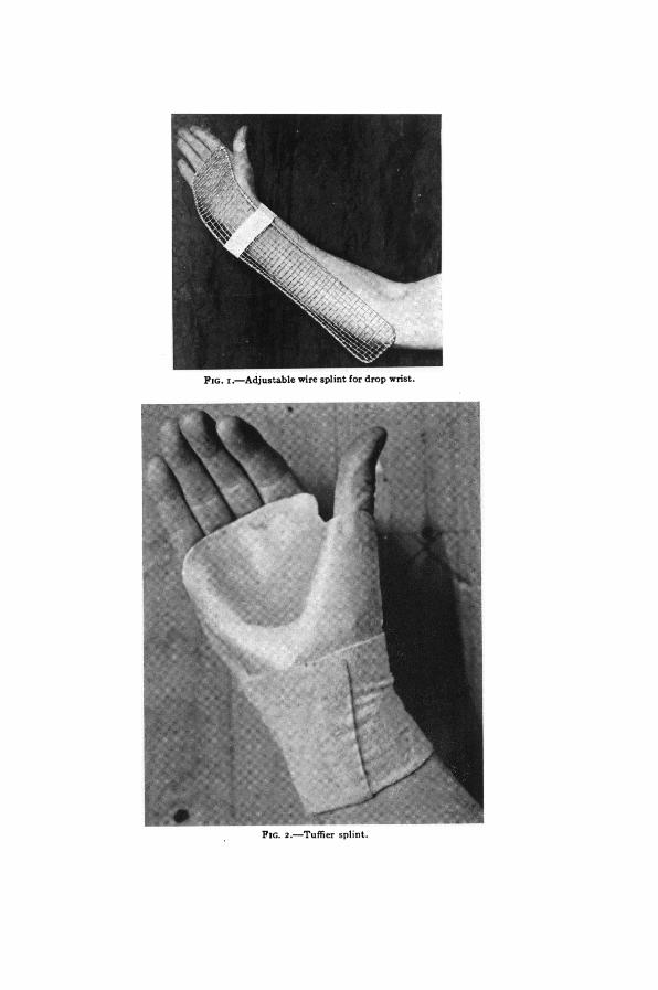
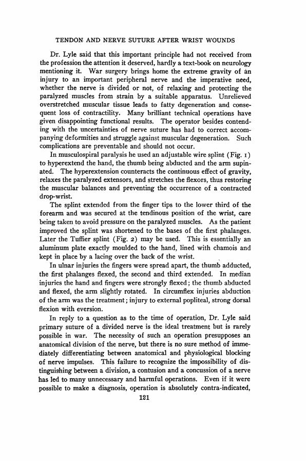
DR. H. H. LyLE called attention to the necessity of the properphysiological splinting of the hand and arm and showed photographsof methods of treating gunshot wounds of the nerves, and demonstratedthe Tuffier splint for drop-wrist resulting from musculospiral paralysis.
120
x1.x ... .
a.." ::
I_L r"; --
FIG. i.-Adjustable wire splint for drop wrist.
FIG. 2.-Tuffier splint.
TENDON. AND NERVE SUTURE AFTER WRIST WOUND;S
Dr. Lyle said that this important principle had not received fromthe profession the attention it deserved, hardly a text-book on neurologymentioning it. War surgery brings home the extreme gravity of aninjury to an important peripheral nerve and the imperative need,whether the nerve is divided or not, of relaxing and protecting theparalyzed muscles from strain by a suitable apparatus. Unrelievedoverstretched muscular tissue leads to fatty degeneration and conse-quent loss of contractility. Many brilliant technical operations havegiven disappointing functional results. The operator besides contend-ing with the uncertainties of nerve suture has had to correct accom-panying deformities and struggle against muscular degeneration. Suchcomplications are preventable and should not occur.
In musculospiral paralysis he used an adjustable wire splint (Fig. I)to hyperextend the hand, the thumb being abducted and the arm supin-ated. The hyperextension counteracts the continuous effect of gravity,relaxes the paralyzed extensors, and stretches the flexors, thus restoringthe muscular balances and preventing the occurrence of a contracteddrop-wrist.
The splint extended from the finger tips to the lower third of theforearm and was secured at the tendinous position of the wrist, carebeing taken to avoid pressure on the paralyzed muscles. As the patientimproved the splint was shortened to the bases of the first phalanges.Later the Tuffier splint (Fig. 2) may be used. This is essentially analuminum plate exactly moulded to the hand, lined with chamois andkept in place by a lacing over the back of the wrist.
In ulnar injuries the fingers were spread apart, the thumb adducted,the first phalanges flexed, the second and third extended. In medianinjuries the hand and fingers were strongly flexed; the thumb abductedand flexed, the arm slightly rotated. In circumflex injuries abductionof the arm was the treatment; injury to external popliteal, strong dorsalflexion with eversion.
In reply to a question as to the time of operation, Dr. Lyle saidprimary suture of a divided nerve is the ideal treatment but is rarelypossible in war. The necessity of such an operation presupposes ananatomical division of the nerve, but there is no sure method of imme-diately differentiating between anatomical and physiological blockingof nerve impulses. This failure to recognize the impossibility of dis-tinguishing between a division, a contusion and a concussion of a nervehas led to many unnecessary and harmful operations. Even if it werepossible to make a diagnosis, operation is absolutely contra-indicated,
121
NEW YORK SURGICAL SOCIETY
as all projectile wounds are potentially infected, and to operate in theface of infection is to court disaster. For these reasons it is best totreat all peripheral nerve lesions on an expectant plan, postponingsuture until the wounds are healed. In dealing with this class ofwounds Dr. Lyle said that it must be kept in mind that one is not dealingwith a simple nerve but with a nerve lesion complicated by comminutedbone, torn muscles, inflammatory exudate, etc.; the whole tending toform obstinate adhesions. These conditions are treated with warmstarch baths, massage, muscle kneading and systematic exercises car-ried out by trained assistants. The galvanic current being a goodstimulant to nutrition was used, but compared with the other measureselectricity plays but a minor part.
Dr. Lyle said that the postural prophylaxis begins with the receptionof the wound and continues after the operation until voluntary motionis restored. A strict adherence to this vital orthopaedic principle aidsin the diagnosis, hastens recovery, prevents many distressing deformi-ties, and will materially diminish the number of useless limbs.
DR. DOWNES said that some three or four years ago he showed acase before the Society similar to Dr. Kenyon's, where all the flexortendons and the median and ulnar nerves had been divided. The resultwas practically perfect. The girl could now play the piano.
SPLENECTOMY FOR VON JAKSCH'S ANJEMIA
DR. EUGENE H. POOL presented a child, aged eighteen months, whowas admitted to the New York Hospital on April I4, I915, with amoderate grade of rickets. She was distinctly anemic. In addition herspleen was markedly enlarged. The examination of the blood at thattime gave the following information: Wassermann reaction negative;red cells, 2,700,000; haemoglobin, 45 per cent.; color index, o.83; whitecells, I2,ooo. Differential count: Polynuclears, 47 per cent.; eosino-philes, 2 per cent.; basophiles, i per cent.; lymphocytes, 29 per cent.;large mononuclears, i8 per cent.; irritation forms, 3 per cent. Foreach ioo white cells there were normoblasts, 5; megaloblasts, 7.
The blood picture remained essentially unchanged until the timeof her operation. Occasionally a myelocyte was seen, but they wereunusual. There was marked anisocytosis and poecilocytosis and poly-chromatophilia, and many of the red cells contained basophilic granules.
On May i her spleen was removed. This was easily and quicklydone through a vertical incision through left rectus. There were afew light adhesions to diaphragm. No transfusion was made as in the
122
SPLENECTOMY FOR VON JAKSCH'S ANJEMIA
case previously reported here. In that case there was a question asto how much the transfusion had to do with the improvement in theblood picture. In this case such a question cannot be raised. Thespleen weighed 230 grammes; was I4/2 cm. x 832 X 332 cm.
Immediately she began to show improvement. During the threeweeks after the operation her red cells rose to 4,500,000 and haemoglobinto 6o per cent. Her convalescence was interrupted by an attack ofmeasles, which she survived, however, and she went home in fair con-dition. She was later sent to the country, where she continued herimprovement.
When seen on October I0, five months after her operation, she waslooking very well. An examination of a stained smear at that timeshows a complete change in the picture. The differential count of500 cells showed: Polynuclears, I7.2 per cent.; eosinophiles, 6.8 percent.; basophiles, 0.2 per cent.; small lymphocytes, 55.0 per cent.; largelymphocytes, I4.4 per cent.; large mononuclears, 4.2 per cent.; transi-tional forms, I.6 per cent.; irritation forms, o.6 per cent.; red cells,6,4oo0,oo; haemoglobin, 52 per cent.
The red cells showed a moderate grade of central pallor and butlittle aniso- or peecilocytosis. There was no polychromatophilia andno nucleated red cells were seen. Only occasionally did a red cellcontain basophilic granules. There was still a moderate grade of leuco-cytosis; weight, 2032 pounds.
The diagnosis of von Jaksch's anmmia was made on the basis of theleucocyte count, well-marked anaemia, and a large spleen associatedwith rickets in an infant. This diagnosis was confirmed by the histo-logical examination of the spleen. This organ showed the extremegrade of myeloidization of the pulp with atrophy of the Malpighianbodies, which is generally considered as characteristic of the condition.The picture in every way was similar to that seen in another case (S. K.)previously reported. In addition there was evidence of a high degreeof blood regeneration, a reversion to an embryonic function of the spleen.
The case can then be definitely considered as belonging to thegroup known as infantile pseudoleukaemia described by von Jaksch.Following her splenectomy she improved and has continued her improve-ment for a period of five months. It is impossible to say, however,what is the significance of the striking change in the blood picture.The percentage of mononuclear cells is much higher than one wouldexpect to find even in an infant and this child was two years old atthe time. One can only say that following the removal of the spleen
123
NEW YORK SURGICAL SOCIETY
there was a termination of a characteristic blood picture and a substi-tution therefor of one which we cannot as yet explain.
Her present weight is 20 pounds, red cells 6,ooo,ooo and hlimoglobin6o per cent. He was indebted to Dr. Ralph G. Stillman of the NewYork Hospital for the study and description of the blood in this case.
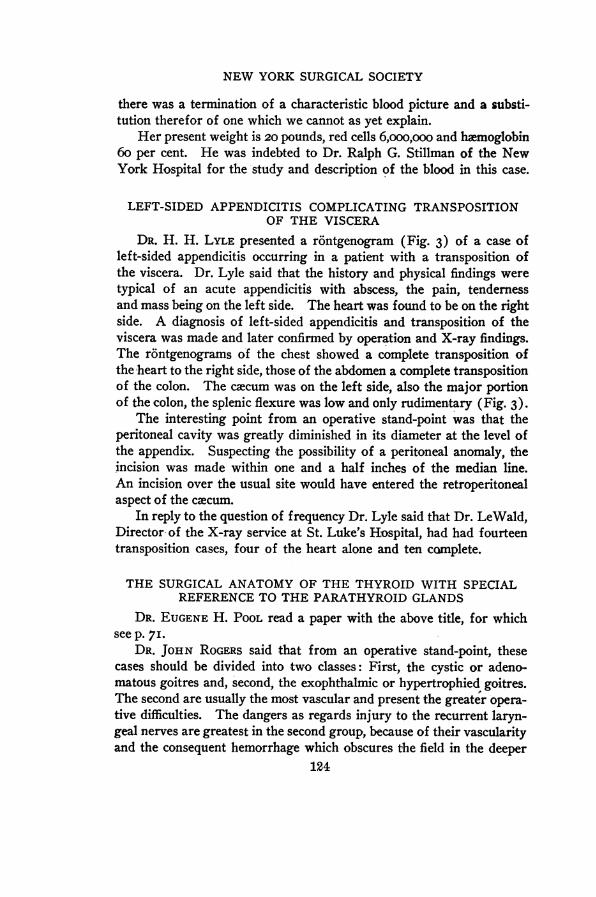
LEFT-SIDED APPENDICITIS COMPLICATING TRANSPOSITIONOF THE VISCERA
DR. H. H. LYLE presented a r6ntgenogram (Fig. 3) of a case ofleft-sided appendicitis occurring in a patient with a transposition ofthe viscera. Dr. Lyle said that the history and physical findings weretypical of an acute appendicitis with abscess, the pain, tendernessand mass being on the left side. The heart was found to be on the rightside. A diagnosis of left-sided appendicitis and transposition of theviscera was made and later confirmed by operation and X-ray findings.The r6ntgenograms of the chest showed a complete transposition ofthe heart to the right side, those of the abdomen a complete transpositionof the colon. The caecum was on the left side, also the major portionof the colon, the splenic flexure was low and only rudimentary (Fig. 3).
The interesting point from an operative stand-point was that theperitoneal cavity was greatly diminished in its diameter at the level ofthe appendix. Suspecting the possibility of a peritoneal anomaly, theincision was made within one and a half inches of the median line.An incision over the usual site would have entered the retroperitonealaspect of the caecum.
In reply to the question of frequency Dr. Lyle said that Dr. LeWald,Director-of the X-ray service at St. Luke's Hospital, had had fourteentransposition cases, four of the heart alone and ten complete.
THE SURGICAL ANATOMY OF THE THYROID WITH SPECIALREFERENCE TO THE PARATHYROID GLANDS
DR. EUGENE H. POOL read a paper with the above title, for whichsee p. 7I.
DR. JOHN ROGERS said that from an operative stand-point, thesecases should be divided into two classes: First, the cystic or adeno-matous goitres and, second, the exophthalmic or hypertrophied goitres.The second are usually the most vascular and present the greater opera-tive difficulties. The dangers as regards injury to the recurrent laryn-geal nerves are greatest in the second group, because of their vascularityand the consequent hemorrhage which obscures the field in the deeper
124
FIG. 3.-Note cacum on left side. R6ntgenogram of chest showed heart on right side. Anomalouscondition of transverse colon. (Rontgenogram by Dr. L. T. Le Wald.)
THE PARATHYROID GLANDS
parts of the neck. This leads, unless one is cautious, to the appli-cation of clamps or haemostats to masses of tissue instead of to bleedingpoints, and the result is not infrequently inclusion of the recurrentnerve. He had had this occur twice in his practice with apparentlypermanent paralysis. At least, in neither case was there a return ofmotion one year and one and a half years, respectively, after theoperation. In each case he discovered the accident before the woundwas closed and traced the nerve through the point of compression, andso proved it had not been cut. In some cases this accident may occurin spite of every precaution. It may, if the hemorrhage is severe andfrom a torn and retracted artery, be practically unavoidable.
He had never been able in the course of an operation to certainlyidentify the parathyroid glands. Dr. Pool has excited his admirationby his ability in one case, which he demonstrated a few years ago beforethis Sbciety, to find a parathyroid and to remove it and then success-fully transplant it into another patient showing evidences of post-operative tetany.
DR. CHARLES H. PECK said that he had followed the principle ofleaving part of the gland both to protect the parathyroid and to protectthe laryngeal nerve. If that is done as a routine, one practically nevershould lose either parathyroids or injure the laryngeal nerve. He wasimpressed by Dr. Rogers's remark about the possibility of injuring thelaryngeal nerve by clamping. He had an unfortunate experience ofthat kind in the very first thyroid he had, and he had had a second experi-ence unfortunately this summer in a very tightly-lodged intrathoracicgoitre. He left a slice of the posterior capsule and cannot understandeven now why he should have injured the nerve, unless he did it inclamping too close or in clamping a nerve that had been dragged for-ward by dislodging the lobe, which was wrapped pretty well around thetrachea. If, however, one follows the principle of dissecting close tothe capsule, pretty well down, then entering the capsule and leavinga thin slice of gland, one would very rarely have any trouble either withthe nerve or the parathyroid.
DR. FREDERIC KAMMERER said that he could remember only onecase of tetany following total extirpation of the thyroid, when thatoperation was still practised at European clinics before its deleteriouseffects had been established. This was a little over thirty years ago.Since that time everybody did a one-sided lobectomy and in the rarecases, in which it became necessary to remove part of the other lobe,it was always advisable to cut through the substance of the gland,leaving a shell of thyroid tissue posteriorly, which procedure prevented
125
NEW YORK SURGICAL SOCIETY
exposure of the parathyroids and the recurrent laryngeal nerve. Hehad had the same experience as Dr. Rogers in trying to demonstrate theparathyroids during operation. He had hardly ever been able to do so,but he had invariably followed the rule, after ligating the superiorthyroid vessels and raising the gland towards the median line, to cutthrough the capsule of the gland and to continue dissection with scis-sors hugging the glandular substance closely. He now felt, afterseeing the instructive cross-sections presented by Dr. Pool, that he hadno doubt occasionally removed parathyroids, which in his experiencewere much more difficult to identify than the recurrent laryngeal nerve,without any untoward symptoms resulting therefrom. The speakerfrankly confessed that he had also on several occasions severed therecurrent nerve, but in his cases no permanent injury resulted from thisunfortunate occurrence beyond a temporary hoarseness.
DR. POOL said there was no attempt on his part, in presenting thissubject, to suggest or to claim any departures from the generallyaccepted procedures and principles. The object was to demonstratethe relationship of the parathyroids, the surgical capsule and the laryn-geal nerve to the posterior part of the lateral lobe. He began thework because he did not know these features himself and could notfind them in any article in sufficient detail.
In regard to the case of tetany mentioned by Dr. Kammerer, hethought that case was one too many. One should not see any casesof tetany parathyreopriva.
The statement that an operator, even in a very large experience,has never seen a parathyroid in an operation upon the thyroid is notsurprising, when one considers how difficult it is to find a parathyroidin an autopsy specimen. In order to locate a parathyroid in an opera-tion one must look for it carefully in a bloodless field; even then itmay be missed.
In regard to the recurrent laryngeal nerve, possibly it may do littleharm to cut it on one side, but he had seen a case in which differentmen had cut a recurrent on each side. As a matter of fact one shouldnot cut one at all.
It is claimed that cutting off its blood supply may produce atrophyof a parathyroid, but it is hard to understand how the same men makethis statement and yet encourage transplantation of a parathyroid.Certainly the parathyroid that has not been taken out and simply hasits blood supply cut off is better able to live and perhaps functionatethan one which is taken from one individual and transplanted intoanother, or even from the same individual and transplanted into adjacenttissue.
126