Neural Signatures of Economic Preferences for Risk and Ambiguity

Thrombin–aptamer recognition: a revealed ambiguityIrene Russo Krauss1, Antonello Merlino1,2, Concetta Giancola1, Antonio Randazzo3,
Lelio Mazzarella1,2 and Filomena Sica1,2,*
1Dip. di Chimica ‘Paolo Corradini’, Universita di Napoli Federico II, Via Cintia, I-80126 Napoli Italia, 2Ist. diBiostrutture e Bioimmagini, CNR, Via Mezzocannone 16, I-80134 Napoli, Italia and 3Dip. di Chimica delleSostanze Naturali, Universita di Napoli Federico II, Via D. Montesano 49, I-8013 Napoli, Italia
Received March 1, 2011; Revised June 6, 2011; Accepted June 8, 2011
ABSTRACT
Aptamers are structured oligonucleotides that rec-ognize molecular targets and can function as directprotein inhibitors. The best-known example is thethrombin-binding aptamer, TBA, a single-stranded15-mer DNA that inhibits the activity of thrombin,the key enzyme of coagulation cascade. TBA foldsas a G-quadruplex structure, as proved by its NMRstructure. The X-ray structure of the complexbetween TBA and human a-thrombin was solved at2.9-A resolution, but did not provide details of theaptamer conformation and the interactions with theprotein molecule. TBA is rapidly processed by nu-cleases. To improve the properties of TBA, anumber of modified analogs have been produced.In particular, a modified TBA containing a 50-50
polarity inversion site, mTBA, has higher stabilityand higher affinity toward thrombin with respect toTBA, although it has a lower inhibitory activity. Wepresent the crystal structure of the thrombin–mTBAcomplex at 2.15-A resolution; the resulting modeleventually provides a clear picture of thrombin–aptamers interaction, and also highlights the struc-tural bases of the different properties of TBA andmTBA. Our findings open the way for a rational de-sign of modified aptamers with improved potency asanticoagulant drugs.
INTRODUCTION
Aptamers are single-stranded nucleic acids, both DNA (1)and RNA (2), which bind molecular targets, including pro-teins, with high affinity and specificity. These peculiar fea-tures are related to a tertiary structure, which presents agood shape complementarity with the target molecule (3).Aptamers have been developed for several different fieldsof applications, in particular, as diagnostic and thera-peutic agents (4). The best-known example is that of the
thrombin-binding aptamer (TBA), a DNA 15-mer consen-sus sequence, namely 50-GGTTGGTGTGGTTGG-30, dis-covered in 1992 through the SELEX (SystematicEvolution of Ligands by Exponential Enrichment) meth-odology (1) when 1013 different DNA molecules weresynthesized and screened for thrombin binding.
a-thrombin (thrombin) is a trypsin-like serine proteasethat plays a pivotal role in haemostasis. Indeed, it is theonly enzyme capable of catalyzing the conversion of sol-uble fibrinogen in insoluble fibrin strands and is the mostpotent platelet activator. Apart from these procoagulantfunctions, thrombin plays also an anticoagulant and anti-fibrinolytic activity in the presence of thrombomodulin (5).
The capability of inhibiting and regulating thrombinactivity in vivo by synthetic compounds is an importantgoal in prevention of thrombosis. The presence on thethrombin surface of two anion-binding subsites or ‘exo-sites’, distinct from the catalytic center, makes it a morediscriminating enzyme as compared to other proteases (6).Exosite I is the recognition site of thrombin physiologicalsubstrate fibrinogen and is also involved in the binding ofleech anticoagulant hirudin, protease-activated receptor-1,thrombomodulin, factors V and VIII, glycoprotein-1baand the acid domain of the serpin heparin cofactor II,whereas exosite II, which is located on the opposite sideof thrombin, is the binding site of heparin and heparin-dependent serpins.
It has been shown that TBA is an ‘exosite inhibitor’(7–9). It has a strong anticoagulant activity in vitro, it isefficient at nanomolar concentration (1) and acts on thetwo procoagulant functions of the enzyme: the activationof fibrinogen and the platelet aggregation. Moreover, it isable to bind both free and clot-bound thrombin (10,11),whereas binding to other serum proteins or proteolyticenzymes is essentially undetectable. The solution structureof TBA, determined by NMR (12,13), revealed that itfolds in a unimolecular antiparallel quadruplex, with achair-like conformation. This conformation is stabilizedby cyclic hydrogen bonding of four guanines (G-tetradsor G-quartets) and by ions coordination between adjacentstacks of G-tetrads that are surrounded by two TT loops
*To whom correspondence should be addressed. Tel: +39081674479; Fax +39081 674090; Email: [email protected]
7858–7867 Nucleic Acids Research, 2011, Vol. 39, No. 17 Published online 28 June 2011doi:10.1093/nar/gkr522
� The Author(s) 2011. Published by Oxford University Press.This is an Open Access article distributed under the terms of the Creative Commons Attribution Non-Commercial License (http://creativecommons.org/licenses/by-nc/3.0), which permits unrestricted non-commercial use, distribution, and reproduction in any medium, provided the original work is properly cited.

on one side and a TGT loop on the other side (12,13). Thecrystal structure of the thrombin–TBA complex was ob-tained at 2.9-A resolution (Model 1) (14). The aptameradopts a very different quadruplex topology with respectto the NMR structure. Indeed, the core formed by twoG-quartets is the same in the two models, but structuraldifferences exist in the way the central bases are con-nected. In particular, there is a difference concerning thedisposition of the loops with respect to the grooves(Figure 1A and B). An alternative model of the crystalstructure of the thrombin–TBA complex (Model 2), builton the basis of the NMR structure of the aptamer, refinedto the same R value as the former Model 1, thus leavingthe doubt on which structure was the correct one (15). Inboth models the TBA is sandwiched between twosymmetry-related thrombin molecules and interacts withexosite I of a thrombin molecule and exosite II of thesecond one. In Model 1, the TGT loop is connected toexosite I and the TT loops are connected to exosite II.Model 2 shows an inverted pattern of protein–ligandinteractions (15).
The uncertainty between these two models was causedby the absence of electron density in the region of TT andTGT loops connecting the G-tetrads. In a more systematicanalysis (16), eight models of the thrombin–aptamercomplex, different for the orientation of the NMRmodel of TBA, were tested on the previously used X-raydiffraction data (14,15). Subtle differences in the crystal-lographic R-factors and the analysis of the aptamer–protein interactions indicated that Model 2 was most likelythe correct one. However, due to the missing density in theloop regions of the aptamer, the details of the ligand–protein interactions could not be properly addressed.Moreover, even recent papers still discuss aptamer-thrombin interactions on the basis of both models (17).In addition, also the stoichiometry of the complex insolution has been recently questioned, as two calorimetricstudies suggest either a 2:1 (18) or a 1:1 (19) thrombin toaptamer molar ratio.
In recent years, several modified TBA have been pro-duced and characterized, with the aim to obtain oligo-nucleotides with improved pharmacological properties,such as higher stability, higher thrombin affinity, longerlife times in vivo, etc. (3). In particular, the main problemassociated with the use of TBA is a rapid degradation bynucleases in vivo. Since it has been reported that the inser-tion of a chain polarity inversion site increases resistanceto endogenous nucleases (20), a modified aptamer(mTBA) containing a 50-50 inversion between Thy3 andThy4 was produced (21). mTBA presents higher thermalstability and higher thrombin affinity with respect to itsunmodified counterpart. The NMR study of mTBA hasrevealed that it folds in a chair-like quadruplex structure,somehow similar to that of TBA (Figure 1C). However,because of the polarity inversion site, mTBA presents a(3+1) topology (22–24), with three strands parallel to eachother and the fourth one oriented in the opposite direc-tion; this topology results in a different syn and anti alter-nation of the bases within the tetrads and in differentgroove sizes. The differences between the two moleculesdo not provide a clear justification of the differentproperties deriving from the inversion site.Here, we report the crystallographic analysis of the com-
plex between thrombin and mTBA at 2.15-A resolution.The higher resolution of the diffraction data, with respectto that of thrombin–TBA complex, has provided a unique,well defined model of the complex, which leaves no doubton thrombin–aptamer interface. Moreover, the details ofthe interactions that the protein molecule makes withmTBA in comparison to TBA also allows to rationalizeon structural grounds the different behavior of the twoaptamers.
MATERIALS AND METHODS
Crystallization, structure determination and refinement
The thrombin–mTBA complex was prepared andcrystallized as previously described (25). Briefly, mTBA
Figure 1. Schematic illustration of (A) X-ray model of TBA (14), (B) NMR model of TBA (12,13), (C) solution (21) and crystallographic (presentpaper) model of the modified aptamer (mTBA). Black arrows indicate 50!30 polarity of the strands. Black circle in mTBA represents the 50–50
polarity inversion site. Anti and syn guanines are depicted as yellow and blue solids, respectively. Wide and narrow grooves are explicitly indicated inthe three pictures. Red arrows indicate the direction of the proton donors and acceptors in Hoogsteen hydrogen bonds.
Nucleic Acids Research, 2011, Vol. 39, No. 17 7859

dissolved in potassium–phosphate buffer 10mM pH 7.1was added to human a-thrombin inhibited by D-Phe-Pro-Arg-chloromethylketone (PPACK) in KCl750mM. After extensive washing, the resulting complexin potassium phosphate 50mM pH 7.1 and KCl 100mMwas crystallized at 20�C using polyethylene glycol 20 00020% w/v, ammonium sulfate 200mM, n-propanol 3% v/v,sodium acetate 100mM at pH 5.8. The structure wassolved by the molecular replacement method, using theprogram AMoRe (26) and the PPACK-inhibitedthrombin (PDB code 1PPB) (27) as a search model. Toavoid bias, inhibitor and water molecules were removedfrom the model. The correlation for the highest solutionwas 0.59, with an R-factor of 0.36. The starting model wassubjected to few cycles of rigid body refinement with CNS(28) followed by several cycles of coordinate minimizationand B-factor refinement with CNS. Each run was alter-nated with manual model building using the program O(29). Fourier difference maps, calculated with (Fo–Fc) and(2Fo–Fc) coefficients, showed continuous electron densityin the active site and in proximity of thrombin exosite I.The analysis of these maps allowed the fitting of PPACKin the active site, the building of the whole aptamermolecule, the identification of an N-acetyl glucosamineresidue linked to Asn60G of the heavy chain, which isnot present in the search model, and the positioning ofseveral water molecules. At the end of the refinement,two strong electron density peaks were assigned to ions:one, placed near the segment 220–225 of thrombin, wasattributed to a sodium ion, according to previous studies(30). The second one, placed between the two G-quartetsand almost equidistant from the O6 atoms of the eightcore guanines, was attributed to a potassium ion. Thefinal model has R-factor/Rfree values of 0.196/0.252. Theprotein geometry of the refined structure was monitoredusing PROCHECK (31) and WHATCHECK (32).Statistics and parameters of the refinement are given inSupplementary Table S1. The drawings were preparedwith Pymol (http://pymol.org). The coordinates of thestructure have been deposited in the Protein Data Bank(Code 3QLP).
Structural analysis of mTBA and thrombin–mTBAcomplex
The geometry of the base assembly in mTBA was analyzedusing a program that calculates the least squares plane ofthe two tetrads, the root mean square deviation of guanineatoms from the best planes, the angle that each guanineresidue makes with the best plane of the tetrad to which itbelongs, and the angle between the two G-quartet planesand their distance along the chain axis.The network of water molecules bound to mTBA was
identified with HBPLUS (33) and by visual inspection ofthe structure. Unconventional CH���O hydrogen bondswere identified using HBAT (34).Features of the protein–aptamer interface, including
number and types of residues at the interface, and gapvolume index, were calculated using the Protein–ProteinInteraction Server (http://www.bioinformatics.sussex.ac.uk/protorp/). For comparison, the same parameters
were calculated as average values from a non-redundantdataset of 25 protein–double-stranded DNA complexessolved at a resolution better than 2.4 A. The shape com-plementarity score, Sc, as defined by Lawrence andColman (35), was calculated using the CCP4 package(36), opportunely modified to include nucleic acid param-eters. For atomic groups in DNA, radii standard values byNadassy et al. (37) were used.
RESULTS
Overall structure
The structure of the complex between thrombin and themodified aptamer (mTBA) has been solved at 2.15-A reso-lution (Figure 2). Detailed statistics of the refinement arereported in Supplementary Table S1.
The polypeptide chain of thrombin is very well definedfor the whole heavy chain (residues 16–247) and for resi-dues 1H–14L of the light chain. All residues in the refinedstructure lie within ‘allowed’ regions of the Ramachandranplot. As expected, the final model maintains all the struc-tural features of the uncomplexed molecule. Indeed, incomparison to the unbound PPACK-inhibited enzyme(PDB code 1PPB) (27) the RMSD after the superpositionof all the Ca protein atoms is only 0.67 A. The largestdeviations are localized in the light chain, at the residuesof the flexible N- and C-terminal tails, whose conform-ation is usually found to be strongly affected by crystalpacking. Similar results are also obtained when the present
Figure 2. Overall structure of the thrombin–mTBA complex.Thrombin molecule is represented as cartoon, with heavy chaincolored purple, light chain colored light pink, exosite I coloredorange and exosite II colored cyan. mTBA molecule is represented assticks.
7860 Nucleic Acids Research, 2011, Vol. 39, No. 17

thrombin structure is compared to that of the molecule incomplex with TBA (PDB code 1HAO) (15). In this case,the slightly larger RMSD of 0.95 A is evenly distributedalong the molecule and is likely related to the significantlylower resolution and quality of the latter structure. Forthis reason further comparisons have been done with theuncomplexed molecule (PDB code 1PPB).
mTBA molecule
The X-ray analysis of mTBA in complex with thrombinreveals that the molecule folds as a chair-like quadruplexwith two stacked G-quartets, the TGT loop on one sideand the two TT loops on the other side. Because ofthe presence of the chain inversion (SupplementaryFigure S1A), mTBA possesses three strands parallel toeach other and the fourth one oriented in the oppositesense, as also found in the solution structure of freemTBA (21) (Figure 1C).
Geometrical details of the G-quartet assembly are givenin Supplementary Table S2 in comparison to those derivedfrom solution structure. For each tetrad, the four guaninemoieties that constitute each quartet are practicallycoplanar and their relative orientation and separationalong the chain axis are in close agreement with theexpected values for the p-stacking of aromatic bases. Incomparison to the solution structure of free mTBA (21),the analysis reveals that in our crystallographic model theG-tetrads are less distorted from the ideal planargeometry. Interestingly, the separation of the tetrads inthe former structure is significantly larger. In thiscontext, it should be pointed out that in the crystallo-graphic structure a potassium ion, which efficiently stabil-izes quadruplex structures (38), is sandwiched between thetwo quartets (Supplementary Figure S1B). The ion has acoordination number of eight and a distortedantiprismatic geometry, at an average distance of 2.8 Afrom the eight O6 atoms. In the refinement of the NMRstructure, the presence of the metal ion was notcontemplated and this may be the reason for the morecompact structure of the two tetrads observed in thecrystal state.
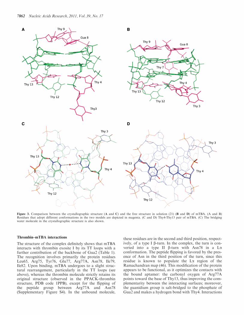
A detailed comparison between the unbound and com-plexed aptamer is of particular interest as it gives a deeperinsight into the dynamics of the aptamer and the way itsstructure modifies to optimize the interaction with theprotein molecule. With respect to free mTBA, the twoTT loops, which form the most extended interactingsurface with the thrombin molecule, move away fromeach other (Figure 3A and B) and the hydrogen bondbetween Thy4 and Thy13 is lost with the insertion of abridging water molecule (Figure 3C and D). These twobases lie on a plane that stacks on the second G-quartetformed by Gua2, Gua5, Gua11, Gua14. Furthermore,Thy3 adopts a conformation that is completely differentin the two models (Figure 3A and B). Interestingly, themore compact structure of the aptamer caused by thepresence of the potassium ion in between the two tetradscombined to the binding of TT loops to thrombin, mayalso explain modifications observed in the TGT loopplaced on the other side of the tetrads with respect to
the TT loops. The TGT loop adopts in the crystal struc-ture a different conformation, which is unlikely caused bythe few and weak packing interactions with a secondthrombin molecule (see below). Gua8 and Thy9, whichin the solution structure stack on the quartet formed byGua1, Gua6, Gua10 and Gua15, loose the interactionwith the G-tetrad, become more parallel to each otherand make direct stabilizing interactions (Figure 3Aand B). This conformational change probably determinesthe expulsion of Thy7 from the wide groove so that thebase becomes more exposed to the solvent and disordered.The analysis of mTBA structure highlights some inter-
esting features. Whereas in nucleic acids structures the C8of purine in anti conformation has been reported to formweak intraresidue C-H���O interactions with the O5’(39–41), in mTBA inter-residue contacts between C8 andO40 are observed (Supplementary Figure S2). A brief ana-lysis of other quadruplex structures (PDB codes: 2AVH,1JPQ, 1I34) shows that this type of interaction is notunusual in antiparallel quadruplex, where guanine residueswith syn and anti conformations alternate along thestrand. On the contrary it is absent in parallel structureswith all anti guanines. This interaction is likely to contrib-ute to some extent to the stability of the G-quadruplex.Special attention was devoted to investigating the
nucleotide-water interactions that are decisive for stabil-ity, dynamics, and recognition. The high resolution of thediffraction data gives the opportunity to study in detailsthe organization of the water molecules bound to theaptamer, particularly in the four grooves. In theseregions, the water molecules arrange themselves in a con-tinuous network of hydrogen bonds, linking the backboneand the bases of the quadruplex. In G-quadruplex, watermolecules are preferentially placed close to N2 and C8atoms of guanines (42–44). Indeed in the modifiedaptamer structure, a conserved network of sequentialN2–water–water–C8 hydrogen bonds is present in thenarrow groove and in one of the two medium grooves; itconnects adjacent strands and involves five of the eightcore guanines (Gua2, Gua10, Gua11, Gua14, Gua15)(Supplementary Figure S3). New and less regularpatterns of hydration, involving N2, N3 or backboneoxygens and a greater number of water moleculesbetween adjacent strands, are observed in the widegroove and in the other medium groove. The mTBAmodel also reveals new features: the traditional C8–water–phosphate oxygen network is not predominantwith respect to interactions where water moleculesbridge C8 and oxygen atoms (O40 or O30) of thebackbone or other water molecules.Thymine bases, being primarily involved in the binding
with thrombin, are less hydrated than guanines. Thenon-conventional binding of waters to the C6 atomobserved, for example, for thymines of the quadruplexof Oxytricha nova telomeric sequence (42) or for allthymines of B-DNA (45), in the case of mTBA involvesonly Thy4. Finally, a water molecule is hydrogen bondedto the methyl group of Thy12.This rich pattern of hydration involves both the four
grooves and the three loops and it is likely to play a keyrole in maintaining the loop conformation.
Nucleic Acids Research, 2011, Vol. 39, No. 17 7861
Thrombin–mTBA interactions
The structure of the complex definitely shows that mTBAinteracts with thrombin exosite I by its TT loops with afurther contribution of the backbone of Gua2 (Table 1).The recognition involves primarily the protein residuesLeu65, Arg75, Tyr76, Glu77, Arg77A, Asn78, Ile79,Ile82. Upon binding, mTBA undergoes to a slight struc-tural rearrangement, particularly in the TT loops (seeabove), whereas the thrombin molecule strictly retains itsoriginal structure (observed in the PPACK-thrombinstructure, PDB code 1PPB), except for the flipping ofthe peptide group between Arg77A and Asn78(Supplementary Figure S4). In the unbound molecule,
these residues are in the second and third position, respect-ively, of a type I b-turn. In the complex, the turn is con-verted into a type II b-turn with Asn78 in a Laconformation. The peptide flipping is favored by the pres-ence of Asn in the third position of the turn, since thisresidue is known to populate the La region of theRamachandran map (46). This modification of the proteinappears to be functional, as it optimizes the contacts withthe bound aptamer: the carbonyl oxygen of Arg77Apoints toward the base of Thy13, thus improving the com-plementarity between the interacting surfaces; moreover,the guanidium group is salt-bridged to the phosphate ofGua2 and makes a hydrogen bond with Thy4. Interactions
Figure 3. Comparison between the crystallographic structure (A and C) and the free structure in solution (21) (B and D) of mTBA. (A and B)Residues that adopt different conformations in the two models are depicted in magenta. (C and D) Thy4-Thy13 pair of mTBA. (C) The bridgingwater molecule in the crystallographic structure is also shown.
7862 Nucleic Acids Research, 2011, Vol. 39, No. 17

between mTBA and thrombin are both hydrophobic andhydrophilic (Table 1). Thy3 occupies a hydrophobic patchlined by Leu65, Ile82 and Tyr76 (Figure 4A) that is in turnin contact with Thy4 (Figure 4B); Thy12 and Thy13 are incontact with Asn78 and Ile79. On the other hand,hydrogen bonds between the guanidium group of Arg75and Thy4 and Thy13, together with those provided byArg77A, contribute substantially to the stabilization ofthis TT pair. Thy12 also interacts with Glu77.Moreover, several water-mediated interactions betweenmTBA and thrombin residues (Thr74, Gly69, Gly25,His71, Lys81, Ser115, Tyr117) have been also identified.
Interestingly, in the crystal structure mTBA interactswith a second thrombin molecule by its TGT loop. Thisinteraction buries a contact area that is less than half of
that involving exosite I (248 A2 versus 570 A2). In particu-lar contacts between TGT loop and the second thrombinmolecule are limited to Thy9 that is involved in cation–pinteraction with Arg97 and in a hydrogen bond withGlu97A, which in turn is hydrogen bonded to Gua15.These two thrombin residues are external to exosite II(Figure 5).
Comparison with thrombin–TBA and other protein–DNAcomplexes
In order to obtain a deeper characterization of the com-plex, we calculated several interface parameters, such asthe buried surface area (�ASA), the ‘gap volume index’,number and type of protein interacting residues, the con-tribution of polar, non-polar and neutral atoms. Thesevalues are given in Table 2 and compared with thoseobtained for Model 2 of thrombin–TBA complex (PDBcode 1HAO) (15) and with the mean values obtained by anon-redundant dataset of 25 protein–DNA complexes.Interestingly, the thrombin–mTBA interface is much
smaller than that generally found in protein–DNAcomplexes, it contains an unusual large number of non-polar atoms and it is constituted by a number of residuesthat have a low propensity to bind nucleic acids, such asleucine, isoleucine and glutamic acid (47). A remarkablefeature of the interface is its tight packing (Figure 4A), asjudged by the surface complementarity index (Sc) that isone of the highest observed for protein–DNA complexes.In particular, with respect to TBA, the chain polarity in-version causes a better fit of the nucleotide in thrombinexosite I (Sc value 0.73 versus 0.56) and allows a largernumber of hydrogen bonds to be formed at the interface.
Figure 4. (A) Surface representation of thrombin–mTBA complex. Thrombin molecule is colored purple, mTBA green, Thy3 is marked in cyan andthe hydrophobic cavity lined by Leu65,Tyr76 and Ile82 side chains is marked in orange, (B) hydrogen bonds involving Thy4. Omit Fo�Fc electrondensity maps (5.0 s level) of Thy4, Arg75 and Arg77A are also shown as example.
Table 1. Interactions between thrombin and mTBA at the most
extended interface
mTBAresidue
Hydrogen bonds Hydrophobicinteraction
Atom Thrombinresidue
Atom Distance(A)
Thrombinresidue
Gua2 O2P Arg77A NH1, NH2 3.17, 3.27Thy3 – – – – Tyr76, Leu65,
Ile82Thy4 O40 Tyr76 N 2.83 Tyr76Thy4 O2 Arg75 NH2 3.07Thy4 O4 Arg77A NH1 2.90Thy12 N3 Glu77 OE2 2.88 Ile79Thy13 O4 Arg75 NH1, NH2 3.15, 2.57 Asn78, Ile79Thy13 O30 Asn78 ND2 2.81
Nucleic Acids Research, 2011, Vol. 39, No. 17 7863

DISCUSSION
Aptamers possess several properties that make them verypromising molecules; indeed, thanks to their high affinityand specificity toward the target protein, therapeuticdosing at sub-micromolar levels should be allowed, byreducing potential non-specific effects. Furthermore, theyare supposed to be non-immunogenic and antidotes can bedesigned to control their pharmacologic activity (48), theycan be completely engineered and possess desirable storageproperties. They are also readily produced and easilymodified by chemical synthesis. The structural modifica-tions are generally aimed to increase thermal and nucle-ases stability, cellular delivery and improve surfaceattachment to the target.Since thrombin is the key enzyme of the coagulation
cascade, it represents an ideal target for anticoagulanttherapeutics and indeed a few years ago the Phase Iclinical trial of TBA was started. Despite this encouragingscenario, up to now the rational design of new aptamer-based antithrombotic agents has been hampered by theabsence of a clear knowledge of interactions betweenthrombin and TBA, due to the low quality of the diffrac-tion data used for structure refinement and analysis (16).In this context, the higher resolution structure of the
complex between thrombin and the modified aptamer
mTBA is of particular interest, because it allows identify-ing the molecular details of the protein–nucleotide recog-nition. The aptamer tightly binds to thrombin exosite I byits TT loops, through a mix of hydrophobic and polarinteractions, consistently with previous biochemicalstudies (8) on TBA. In agreement with the revised inter-pretation of the crystallographic data on the thrombin–TBA complex (16), our results on the thrombin–mTBAcomplex definitely identify in the TT loops a general struc-tural feature for the binding of aptamers to thrombin. Inboth cases, these loops act as a pincer-like system thatembraces the protruding region of exosite I (Figure 2).However, the details of the interactions are different forthe two aptamers (Table 1). Indeed the chain inversion inmTBA determines the formation of a greater number ofcontacts with the enzyme with respect to the unmodifiednucleotide and more generally, leads to an increased shapecomplementarity, as shown by the surface complementar-ity index Sc (Table 2). These results may well account forthe higher affinity toward thrombin of mTBA as comparedto TBA (18). On the other hand, the interaction betweenThy12 and His71, which is present in the TBA complex(15), is lost in the case of mTBA. His71 plays a key role inthe recognition of fibrinogen and its interaction with TBAis considered important for the inhibition of thrombinclotting activity by the aptamer (8). The position of thishistidine is the same in the two complexes, but the chaininversion in mTBA causes a subtle reorganization of the
Figure 5. Packing contacts involving mTBA. Thrombin molecules arerepresented as cartoon, aptamer molecule as sticks. The symmetryrelated thrombin molecule (the symmetry relationship is �x+1/2,y� 1/2, �z+1/2) is colored in blue, with residues interacting withmTBA marked in cyan and residues belonging to exosite II markedin yellow. A zoomed vision of the interactions between Thy9 and thesecond thrombin molecule is reported in the panel.
Table 2. Protein–DNA interface features
Thrombin–mTBA
Thrombin–TBA(Model 2)(15)
Protein–DNAcomplexesa
Sequence segmentation 4 5 7 (4)� ASA (A2) 570 462 1320 (495)� %ASA 4.8 4.0 13 (7)Atoms in interface 60 40 122 (51)Polar atoms contribution
to interface (%)24.3 25.0 30 (7)
Non-polar atoms contributionto interface (%)
54.2 38.0 30 (11)
Neutral atoms contributionto interface (%)
45.5 37.7 40 (9)
Residues in interface (%) 15 12 43 (20)Polar residues in interface (%) 46.7 58.3 37 (10)Non-polar residues in
interface (%)33.3 25.0 28 (9)
Charged residues ininterface (%)
20.0 16.7 35 (9)
Hydrogen bonds 11 2 21 (9)Hydrogen bonds
(/100 A2 �ASA)1.9 0.5 1.6 (0.4)
Bridging water molecules 4 0 17 (10)Bridging water molecules
(/100 A2 �ASA)0.7 0 1.3 (0.7)
Gap volume (A3) 2710 2997 7408 (3050)Gap volume index (A) 2.38 3.49 3.0 (1.0)Sc 0.73 0.56 0.65 (0.05)
aProperties were calculated from a non-redundant data set of 25protein–DNA complexes belonging to different protein–structurefamilies with resolution better than 2.4 A.
7864 Nucleic Acids Research, 2011, Vol. 39, No. 17

77–79 protein region located at the interface close toThy12 (Figure 6A). As result a new salt bridge is formedbetween the side chain of Arg77A and the phosphateoxygen atom of Gua2, which is in close proximitybecause of the 3’!5’ inverted polarity of the strand. InModel 2 TBA backbone is far away from Arg77A, whoseside chain is flipped in the opposite direction. In addition,the movement of Ile79, which is in close contact withThy12, causes in mTBA a displacement of this thymineaway from His71 (Figure 6B). Thus, the greater disponib-ility of His71 to interact with the fibrinogen could explainthe lower anticoagulant activity of mTBA (18).
A stabilizing interface of the aptamer with exosite II ofa second thrombin molecule was also suggested on thebasis of the crystallographic data on the TBA complex(14,15). In the mTBA complex, which assembles in thesolid state with a different crystal packing, the surfacecontact with exosite I is maintained, whereas there areno interactions with exosite II. Contacts with other regionsof the protein are in general fewer when compared withthose involving exosite I. On the other hand, although theassociation of mTBA with exosite I of thrombin involves arather small interface area when compared with those usu-ally found in other DNA–protein complexes, it ischaracterized by an excellent shape complementarity anda high number of intermolecular interactions, which aretypical for aptamers. These findings, together with the factthat in the crystals of both complexes the asymmetric unitcontains one protein molecule and one aptamer, stronglysuggest that thrombin–aptamers complexes have also insolution a 1:1 stoichiometry, in agreement with the morerecent ITC data (19).
In conclusion, the high-resolution structure ofthrombin–mTBA complex here reported clarifies severalquestions regarding thrombin–aptamers interaction. Itdefinitely establishes the way in which the aptamer inter-acts with thrombin and allows a properly characterization
of the contact surface between the two molecules.Moreover, the results highlight the structural bases ofthe different properties of TBA and mTBA, and in par-ticular their different anticoagulant activities.The X-ray analysis confirms the general folding (‘3+1’
arrangement) found by NMR studies of free mTBA, withthree strands parallel to each other and the forth oneoriented in the opposite sense. This finding furtherenforces the idea that the ‘3+1’ core topology is not tobe considered an anomaly, but rather a robustG-quadruplex scaffold, on which new modifications canbe inserted (22–24). Indeed, starting from the mTBAstructure, it is possible to design modified aptamers thatpreserve both stability against nucleases and high affinitytoward thrombin characteristic of mTBA, and in addition,possess a more effective antithrombotic action. A prelim-inary modeling suggests that modifications in the Thy12–Thy13 loop could restore the interaction, active in thethrombin–TBA complex, of the aptamer with His71, akey residue for the recognition between the fibrinogenand exosite I. In particular, chemical modification ofThy12 could create new contacts with this proteinresidue thus improving the anticoagulant action of theaptamer. Experiments are in progress along this line.More in general, the present investigation provides the
scientific community with an enhanced structural and con-formational knowledge that will serve as a platform for arational design of modified aptamers for use in anticoagu-lant therapies.
ACCESSION NUMBER
3QLP.
SUPPLEMENTARY DATA
Supplementary Data are available at NAR Online.
Figure 6. Superposition of thrombin–mTBA (purple) on the Model 2 of thrombin–TBA (PDB code 1HAO) (15) (orange): (A) interactions betweenTBA Thy12 and His71 and between mTBA Gua2 and Arg77A, (B) displacement of mTBA Thy12 from His71.
Nucleic Acids Research, 2011, Vol. 39, No. 17 7865
ACKNOWLEDGEMENTS
The authors thank Giosue Sorrentino and MaurizioAmendola for technical assistance.
FUNDING
Funding for open access charge: financial support fromMinistero dell’Istruzione, dell’Universita e della Ricerca.
Conflict of interest statement. None declared.
REFERENCES
1. Bock,L.C., Griffin,L.C., Latham,J.A., Vermaas,E.H. andToole,J.J. (1992) Selection of single-stranded DNA molecules thatbind and inhibit human thrombin. Nature, 355, 564–566.
2. Burke,J.M. and Berzal-Herranz,A. (1993) In vitro selection andevolution of RNA: applications for catalytic RNA, molecularrecognition, and drug discovery. FASEB J., 7, 106–112.
3. Lancellotti,S. and De Cristofaro,R. (2009) Nucleotide-derivedthrombin inhibitors: a new tool for an old issue. Cardiovasc.Hematol. Agents Med. Chem., 7, 19–28.
4. Gold,L. (1995) Oligonucleotides as research, diagnostic, andtherapeutic agents. J. Biol. Chem., 270, 13581–13584.
5. Huntington,J.A. (2005) Molecular recognition mechanisms ofthrombin. J. Thromb. Haemost., 3, 1861–1872.
6. Bode,W., Turk,D. and Karshikov,A. (1992) The refined 1.9-AX-ray crystal structure of D-Phe-Pro-Argchloromethylketone-inhibited human alpha-thrombin: structureanalysis, overall structure, electrostatic properties, detailedactive-site geometry, and structure-function relationships.Protein Sci., 1, 426–471.
7. Paborsky,L.R., McCurdy,S.N., Griffin,L.C., Toole,J.J. andLeung,L.L. (1993) The single-stranded DNA aptamer-binding siteof human thrombin. J. Biol. Chem., 268, 20808–20811.
8. Tsiang,M., Jain,A.K., Dunn,K.E., Rojas,M.E., Leung,L.L. andGibbs,C.S. (1995) Functional mapping of the surface residues ofhuman thrombin. J. Biol. Chem., 270, 16854–16863.
9. Wu,Q., Tsiang,M. and Sadler,J.E. (1992) Localization of thesingle-stranded DNA binding site in the thrombin anion-bindingexosite. J. Biol. Chem., 267, 24408–24412.
10. Li,W.X., Kaplan,A.V., Grant,G.W., Toole,J.J. and Leung,L.L.(1994) A novel nucleotide-based thrombin inhibitor inhibitsclot-bound thrombin and reduces arterial platelet thrombusformation. Blood, 83, 677–682.
11. Griffin,L.C., Tidmarsh,G.F., Bock,L.C., Toole,J.J. andLeung,L.L. (1993) In vivo anticoagulant properties of a novelnucleotide-based thrombin inhibitor and demonstration ofregional anticoagulation in extracorporeal circuits. Blood, 81,3271–3276.
12. Macaya,R.F., Schultze,P., Smith,F.W., Roe,J.A. and Feigon,J.(1993) Thrombin-binding DNA aptamer forms a unimolecularquadruplex structure in solution. Proc. Natl Acad. Sci. USA, 90,3745–3749.
13. Wang,K.Y., McCurdy,S., Shea,R.G., Swaminathan,S. andBolton,P.H. (1993) A DNA aptamer which binds to and inhibitsthrombin exhibits a new structural motif for DNA. Biochemistry,32, 1899–1904.
14. Padmanabhan,K., Padmanabhan,K.P., Ferrara,J.D., Sadler,J.E.and Tulinsky,A. (1993) The structure of alpha-thrombin inhibitedby a 15-mer single-stranded DNA aptamer. J. Biol. Chem., 268,17651–17654.
15. Padmanabhan,K. and Tulinsky,A. (1996) An ambiguous structureof a DNA 15-mer thrombin complex. Acta Crystallogr.D Biol. Crystallogr., 52, 272–282.
16. Kelly,J.A., Feigon,J. and Yeates,T.O. (1996) Reconciliation ofthe X-ray and NMR structures of the thrombin-binding aptamerd(GGTTGGTGTGGTTGG). J. Mol. Biol., 256, 417–422.
17. Pasternak,A., Hernandez,F.J., Rasmussen,L.M., Vester,B. andWengel,J. Improved thrombin binding aptamer by incorporation
of a single unlocked nucleic acid monomer. Nucleic Acids Res.,39, 1155–1164.
18. Pagano,B., Martino,L., Randazzo,A. and Giancola,C. (2008)Stability and binding properties of a modified thrombin bindingaptamer. Biophys. J., 94, 562–569.
19. Nallagatla,S.R., Heuberger,B., Haque,A. and Switzer,C. (2009)Combinatorial synthesis of thrombin-binding aptamers containingiso-guanine. J. Comb. Chem., 11, 364–369.
20. McGuffie,E.M. and Catapano,C.V. (2002) Design of a noveltriple helix-forming oligodeoxyribonucleotide directed to themajor promoter of the c-myc gene. Nucleic Acids Res., 30,2701–2709.
21. Martino,L., Virno,A., Randazzo,A., Virgilio,A., Esposito,V.,Giancola,C., Bucci,M., Cirino,G. and Mayol,L. (2006) A newmodified thrombin binding aptamer containing a 5’-5’ inversionof polarity site. Nucleic Acids Res., 34, 6653–6662.
22. Zhang,N., Phan,A.T. and Patel,D.J. (2005) (3 + 1) Assembly ofthree human telomeric repeats into an asymmetric dimericG-quadruplex. J. Am. Chem. Soc., 127, 17277–17285.
23. Luu,K.N., Phan,A.T., Kuryavyi,V., Lacroix,L. and Patel,D.J.(2006) Structure of the human telomere in K+ solution: anintramolecular (3 + 1) G-quadruplex scaffold. J. Am. Chem. Soc.,128, 9963–9970.
24. Ambrus,A., Chen,D., Dai,J., Bialis,T., Jones,R.A. and Yang,D.(2006) Human telomeric sequence forms a hybrid-typeintramolecular G-quadruplex structure with mixed parallel/antiparallel strands in potassium solution. Nucleic Acids Res., 34,2723–2735.
25. Russo Krauss,I., Merlino,A., Randazzo,A., Mazzarella,A. andSica,F. (2010) Crystallization and preliminary X-ray analysis ofthe complex of human a-thrombin with a modifiedthrombin-binding aptamer. Acta Crystallographica Sec. F, 66,961–963.
26. Navaza,J. and Saludjian,P. (1997) AMoRe: an automatedmolecular replacement program package. Method Enzymol., 276,581–594.
27. Bode,W., Mayr,I., Baumann,U., Huber,R., Stone,S.R. andHofsteenge,J. (1989) The refined 1.9 A crystal structure of humanalpha-thrombin: interaction with D-Phe-Pro-Argchloromethylketone and significance of the Tyr-Pro-Pro-Trpinsertion segment. EMBO J., 8, 3467–3475.
28. Brunger,A.T., Adams,P.D., Clore,G.M., DeLano,W.L., Gros,P.,Grosse-Kunstleve,R.W., Jiang,J.S., Kuszewski,J., Nilges,M.,Pannu,N.S. et al. (1998) Crystallography & NMR system: a newsoftware suite for macromolecular structure determination.Acta Crystallogr. D Biol. Crystallogr., 54, 905–921.
29. Jones,T.A., Zou,J.Y., Cowan,S.W. and Kjeldgaard,M. (1991)Improved methods for building protein models in electron densitymaps and the location of errors in these models. Acta Crystallogr.A, 47(Pt 2), 110–119.
30. Di Cera,E., Guinto,E.R., Vindigni,A., Dang,Q.D., Ayala,Y.M.,Wuyi,M. and Tulinsky,A. (1995) The Na+ binding site ofthrombin. J. Biol. Chem., 270, 22089–22092.
31. Laskowski,R.A., Macarthur,M.W., Moss,D.S. and Thornton,J.M.(1993) Procheck - a program to check the stereochemical qualityof protein structures. J. Appl. Crystallogr., 26, 283–291.
32. Hooft,R.W., Vriend,G., Sander,C. and Abola,E.E. (1996) Errorsin protein structures. Nature, 381, 272.
33. McDonald,I.K. and Thornton,J.M. (1994) Satisfying hydrogenbonding potential in proteins. J. Mol. Biol., 238, 777–793.
34. Tiwari,A. and Panigrahi,S.K. (2007) HBAT: a complete packagefor analysing strong and weak hydrogen bonds inmacromolecular crystal structures. In Silico Biol., 7, 651–661.
35. Lawrence,M.C. and Colman,P.M. (1993) Shape complementarityat protein/protein interfaces. J. Mol. Biol., 234, 946–950.
36. Collaborative Computational Project, Number 4, (1994) TheCCP4 suite: programs for protein crystallography. ActaCrystallogr. D Biol. Crystallogr., 50, 760–763.
37. Nadassy,K., Tomas-Oliveira,I., Alberts,I., Janin,J. and Wodak,S.J.(2001) Standard atomic volumes in double-stranded DNA andpacking in protein–DNA interfaces. Nucleic Acids Res., 29,3362–3376.
38. Hud,N.V., Smith,F.W., Anet,F.A. and Feigon,J. (1996) Theselectivity for K+ versus Na+ in DNA quadruplexes is dominated
7866 Nucleic Acids Research, 2011, Vol. 39, No. 17

by relative free energies of hydration: a thermodynamic analysisby 1H NMR. Biochemistry, 35, 15383–15390.
39. Shefter,E. and Trueblood,K.N. (1965) The crystal and molecularstructure of D(+)-barium uridine-5’-phosphate. Acta Crystallogr.,18, 1067–1077.
40. Rubin,J., Brennan,T. and Sundaralingam,M. (1972) Crystal andmolecular structure of a naturally occurring dinucleosidemonophosphate. Uridylyl-(3’-5’)-adenosine hemihydrate.Conformational ‘rigidity’ of the nucleotide unit and models forpolynucleotide chain folding. Biochemistry, 11, 3112–3128.
41. Wahl,M.C., Rao,S.T. and Sundaralingam,M. (1996) Crystalstructure of the B-DNA hexamer d(CTCGAG): model for anA-to-B transition. Biophys. J., 70, 2857–2866.
42. Horvath,M.P. and Schultz,S.C. (2001) DNA G-quartets in a 1.86A resolution structure of an Oxytricha nova telomericprotein-DNA complex. J. Mol. Biol., 310, 367–377.
43. Phillips,K., Dauter,Z., Murchie,A.I., Lilley,D.M. and Luisi,B.(1997) The crystal structure of a parallel-stranded guaninetetraplex at 0.95 A resolution. J. Mol. Biol., 273, 171–182.
44. Haider,S., Parkinson,G.N. and Neidle,S. (2002) Crystal structureof the potassium form of an Oxytricha nova G-quadruplex.J. Mol. Biol., 320, 189–200.
45. Kielkopf,C.L., Ding,S., Kuhn,P. and Rees,D.C. (2000)Conformational flexibility of B-DNA at 0.74 A resolution:d(CCAGTACTGG)(2). J. Mol. Biol., 296, 787–801.
46. Deane,C.M., Allen,F.H., Taylor,R. and Blundell,T.L. (1999)Carbonyl-carbonyl interactions stabilize the partially allowedRamachandran conformations of asparagine and aspartic acid.Protein Eng., 12, 1025–1028.
47. Jones,S., van Heyningen,P., Berman,H.M. and Thornton,J.M.(1999) Protein-DNA interactions: a structural analysis.J. Mol. Biol., 287, 877–896.
48. Nimjee,S.M., Rusconi,C.P., Harrington,R.A. and Sullenger,B.A.(2005) The potential of aptamers as anticoagulants. TrendsCardiovasc. Med., 15, 41–45.
Nucleic Acids Research, 2011, Vol. 39, No. 17 7867
Copyright © 2022 FDOKUMEN