Three new Microthoracids (Ciliophora, Nassophorea) from Austria and Venezuela
17
Acta Protozool. (2014) 53: 295–311 http://www.eko.uj.edu.pl/ap doi:10.4467/16890027AP.14.027.2022 ACTA PROTOZOOLOGICA Three New Microthoracids (Ciliophora, Nassophorea) from Austria and Venezuela Atef OMAR 1,2 and Wilhelm FOISSNER 1 1 Universität Salzburg, FB Organismische Biologie, Salzburg, Austria; 2 Al-Azhar University, Department of Zoology, Assiut, Egypt Abstract. Using standard methods, we describe three new microthoracids. Drepanomonas minuta nov. spec. is characterized by a small size (in vivo about 22 × 11 µm) and a curious distribution pattern of the extrusomes, viz., only one trichocyst each in mid of dorsal margin and near posterior end of ventral side. Body size and shape of D. minuta highly resemble D. revoluta – for which a new morphometric charac- terization is provided – which, however, has a deep, broad furrow on the left side and more than 10 extrusomes. Drepanomonas multiden- tata nov. spec. was discovered in ephemeral grassland puddles on the north coast of Venezuela. It is a comparatively large species (in vivo 45 × 25 µm) with a total of nine spines, of which those on the left posterior half form a highly characteristic tridentate pattern. Curiously, this species lacks extrusomes. Leptopharynx lajacola nov. spec. was discovered in an ephemeral puddle on a granitic outcropping (Laja) in Venezuela. This species resembles L. costatus but is unique in having a quadrangular outline and a strongly curved oral basket with the opening directed posteriorly. In the past four years, we have described 10 new microthoracids, showing that their diversity is far from being exhausted. Key words: biodiversity, ciliates, Drepanomonas, Leptopharynx, new species, soil ciliates. Address for correspondence: Wilhelm Foissner, Universität Salz- burg, FB Organismische Biologie, Hellbrunnerstrasse 34, A-5020 Salzburg, Austria; E-mail: [email protected] INTRODUCTION A detailed ontogenetic study (Omar and Foiss- ner 2012b) and molecular data (Foissner et al. 2011, Gong et al. 2009) showed that the microthoracid cili- ates belong to the class Nassophorea Small and Lynn, 1981 and the order Microthoracida Jankowski, 1967, where they represent a distinct family, Microthoracidae Wrześniowski, 1870. The most important morphologi- cal apomorphy of the microthoracids is the mixokinetal origin of the adoral membranelles (Omar and Foissner 2012b). Most microthoracids are difficult to investigate be- cause they are small (20–50 µm) and the ciliature is so strongly reduced that the ciliary rows are difficult to follow. However, as a group the microthoracids are easily recognized by their small size, the rigid and often distinctly sculptured cortex, and the strongly reduced left side ciliature. These features provide the main characteristics for species recognition (Foissner 1985; Foissner et al. 2011; Omar and Foissner 2011, 2013). In the past few years, we described seven new mi- crothoracids: Leptopharynx bromelicola Foissner et al., 2011; L. bromeliophilus Omar and Foissner, 2011;
Transcript of Three new Microthoracids (Ciliophora, Nassophorea) from Austria and Venezuela

Acta Protozool. (2014) 53: 295–311 http://www.eko.uj.edu.pl/ap
doi:10.4467/16890027AP.14.027.2022ACTAPROTOZOOLOGICA
Three New Microthoracids (Ciliophora, Nassophorea) from Austria and Venezuela
Atef OMAR1,2 and Wilhelm FOISSNER1
1 Universität Salzburg, FB Organismische Biologie, Salzburg, Austria; 2Al-Azhar University, Department of Zoology, Assiut, Egypt
Abstract. Using standard methods, we describe three new microthoracids. Drepanomonas minuta nov. spec. is characterized by a small size (in vivo about 22 × 11 µm) and a curious distribution pattern of the extrusomes, viz., only one trichocyst each in mid of dorsal margin and near posterior end of ventral side. Body size and shape of D. minuta highly resemble D. revoluta – for which a new morphometric charac-terization is provided – which, however, has a deep, broad furrow on the left side and more than 10 extrusomes. Drepanomonas multidentata nov. spec. was discovered in ephemeral grassland puddles on the north coast of Venezuela. It is a comparatively large species (in vivo 45 × 25 µm) with a total of nine spines, of which those on the left posterior half form a highly characteristic tridentate pattern. Curiously, this species lacks extrusomes. Leptopharynx lajacola nov. spec. was discovered in an ephemeral puddle on a granitic outcropping (Laja) in Venezuela. This species resembles L. costatus but is unique in having a quadrangular outline and a strongly curved oral basket with the opening directed posteriorly. In the past four years, we have described 10 new microthoracids, showing that their diversity is far from being exhausted.
Key words: biodiversity, ciliates, Drepanomonas, Leptopharynx, new species, soil ciliates.
Address for correspondence: Wilhelm Foissner, Universität Salz-burg, FB Organismische Biologie, Hellbrunnerstrasse 34, A-5020 Salzburg, Austria; E-mail: [email protected]
INTRODUCTION
A detailed ontogenetic study (Omar and Foiss-ner 2012b) and molecular data (Foissner et al. 2011, Gong et al. 2009) showed that the microthoracid cili-ates belong to the class Nassophorea Small and Lynn, 1981 and the order Microthoracida Jankowski, 1967, where they represent a distinct family, Microthoracidae Wrześniowski, 1870. The most important morphologi-cal apomorphy of the microthoracids is the mixokinetal
origin of the adoral membranelles (Omar and Foissner 2012b).
Most microthoracids are difficult to investigate be-cause they are small (20–50 µm) and the ciliature is so strongly reduced that the ciliary rows are difficult to follow. However, as a group the microthoracids are easily recognized by their small size, the rigid and often distinctly sculptured cortex, and the strongly reduced left side ciliature. These features provide the main characteristics for species recognition (Foissner 1985; Foissner et al. 2011; Omar and Foissner 2011, 2013).
In the past few years, we described seven new mi-crothoracids: Leptopharynx bromelicola Foissner et al., 2011; L. bromeliophilus Omar and Foissner, 2011;

A. Omar and W. Foissner296
L. australiensis Omar and Foissner, 2011; L. brasiliensis Foissner and Omar in Omar and Foissner (2012a); L. costatus gonohymen Foissner and Omar in Omar and Foissner (2012a); Drepanomonas hymenofera venezuelensis Omar and Foissner, 2013; and D. vasta Foissner and Omar in Omar and Foissner (2013). Here, we add the detailed description for three new species: Drepanomonas minuta, D. multidentata, and Leptopharynx lajacola. These are 10 new taxa found over many years in about 1000 samples, showing that the diversity of the microthoracids is far from being exhausted. This is emphasized by three other new species whose descrip-tion is in preparation.
MATERIALS AND METHODS
For details on samples and locations, see the individual species descriptions. All were reactivated from the resting cysts of air-dried soil samples by the non-flooded Petri dish method (NFPM). Briefly, the NFPM involves placing 50–500 g litter and soil in a Petri dish (13–18 cm wide, 2–3 cm high) and saturating, but not flooding it, with distilled water. Such a culture is analyzed for ciliates by in-specting about 2 ml of the run-off on days 2, 7, 14, 21, and 28; for a detailed description of the NFPM, see Foissner et al. (2002).
Cells were studied in vivo using a high-power oil immersion objective and differential interference contrast optics. The infracil-iature and various cytological structures were revealed by protargol impregnation and the Klein-Foissner silver nitrate technique (Foiss-ner 1991). Counts and measurements (with ocular micrometer) on silvered specimens were conducted at a magnification of 1000 ×. The “total number of basal bodies” excludes those of the adoral membranelles, which are difficult to count. In vivo measurements were performed at magnifications of 40–1000 ×. For kinety designa-tion and numbering, see Omar and Foissner (2013). The data avail-able suggest classifying the microthoracid body length as (in vivo averages): very small (10–19 µm), small (20–29 µm), middle-sized (30–39 µm), large (40–49 µm), and very large (≥ 50 µm). Terminol-ogy is according to Omar and Foissner (2012b). Drawings of live specimens were based on free-hand sketches and micrographs while those of impregnated cells were made with a drawing device.
RESULTS AND COMPARISON OF SPECIES
Drepanomonas minuta Foissner & Omar nov. spec. (Figs 1–6, 9–15; Table 1)
Diagnosis: Size in vivo about 22 × 11 µm. Body semi-ellipsoidal with tapered anterior and rounded posterior end, dorsal margin convex, ventral straight. Cortex flat, crenellated along somatic kineties. Two ex-
trusomes, one in anterior dorsal half, the other subter-minally in ventral side. Somatic kinety 4 commences with three dikinetids and one monokinetid, followed by a wide break in mid-body and three monokinetids posteriorly. Kineties 6 and 7 partially and fully non-ciliated, respectively. Left portion of postoral complex with three narrowly spaced monokinetids and a sin-gle monokinetid far posteriorly. On average a total of 85 basal bodies. Oral apparatus slightly posterior to mid-body.
Type locality: Upper soil layer from a mixed forest in the surroundings (Neuhaus area) of the town of Salz-burg, Austria, 47°48′N, 13°04′E.
Type material: One holotype and nine paratype slides with protargol- and silver nitrate-impregnated specimens have been deposited in the Biology Centre of the Museum of Upper Austria, Linz (LI). The holotype and important paratype specimens have been marked by black ink circles on the coverslip.
Etymology: The Latin adjective minuta (small) em-phasizes the small size of the species.
Description: Size in vivo 18–25 × 8–13 µm, usu-ally about 22 × 11 µm, as calculated from some in vivo measurements and values shown in Table 1, assuming 15% preparation shrinkage. Body semi-ellipsoidal to slenderly semi-ellipsoidal with a length: width ratio of 1.8–2.5:1 and an average of 2.1:1; dorsal side distinctly convex, ventral straight; anterior end tapered, posterior rounded, cells thus slightly wider posteriorly than ante-riorly; flattened laterally up to 2:1 and lenticular in ven-tral and dorsal view (Table 1; Figs 1–6, 9–15). Nuclear apparatus anterior to mid-body. Macronucleus globular to broadly ellipsoidal, contains some globular masses, probably nucleoli up to 1 µm across. Micronucleus globular, near or attached to ventral side of macronu-cleus (Table 1; Figs 1, 6, 9–12). Contractile vacuole in third quarter of body, slightly posterior and dorsal of buccal cavity, excretory tube difficult to see (Table 1; Fig. 1). Cytopyge posterior and slightly left of contrac-tile vacuole, usually forms a blister containing food remnants; in silver nitrate preparations, represented by a thick, short silverline right of left portion of postoral complex (Figs 1, 14). Extrusomes lenticular, about 4 × 1 µm in size, only one extrusome each in anterior and posterior portion of body: anterior extrusome present in only few specimens, slightly anterior and dorsal of ma-cronucleus; posterior extrusome recognizable in most specimens in vivo, invariably in subterminal ventral side posterior to cytopyge, occasionally impregnated

New Microthoracid Ciliates 297
with protargol (Figs 1, 12, 13). Cytoplasm hyaline, contains some lipid droplets about 1 µm across. Glides rapidly interrupted by short jerks.
Cortex rigid and glossy, smooth except for a distinct crenellation left of somatic kineties (Figs 1, 2). Details of ventral side organized as follows: preoral kineties in distinct furrows; oral opening slightly posterior to mid-body; posterior to oral apparatus a short concavity containing cytopyge and the single cilium of the right portion of the postoral complex (Figs 1, 4, 5, 9, 11, 14). Silverline pattern as described in D. hymenofera by Omar and Foissner (2013), i.e., cortex studded with minute, argyrophilic granules (Figs 14, 15).
Somatic cilia 7–9 µm long in silver nitrate prepara-tions (Fig. 15). Invariably nine somatic and three pre-oral kineties with a total of 85 basal bodies on average (Table 1). Kineties 3, 4 and 6 bipolar; kineties 1, 2, 5 and 7–9 shortened anteriorly and/or posteriorly. Kine-ties 1–4 on right side; kineties 5–7 on the left side; kine-ties 8 and 9 extend ventrally (Table 1; Figs 4–6, 9–11, 13–15).
Kinety 1 along upper right margin of oral cavity, forms an inverted sickle-shaped figure, consists of six narrowly spaced, usually ciliated dikinetids (Figs 4, 9, 11). Kinety 2 begins in second quarter of body with a single dikinetid and a single monokinetid, both cili-ated and spaced very narrowly, resembling the char-acteristic trikinetid of D. sphagni; followed by a very wide break, a single ciliated monokinetid, and one or two ciliated dikinetids near posterior end of body (Figs 4, 5, 14). Kinety 3 commences with a single, ciliated dikinetid followed by: (i) one or two widely spaced, ciliated monokinetids, (ii) a very wide break interrupt-ed in mid-body by a single, non-ciliated monokinetid, and (iii) a few, narrowly spaced, ciliated monokinetids and dikinetids near posterior end of body (Figs 4, 5, 14). Kineties 4 and 5 limit dorsal margin of body, all kinetids ciliated: kinety 4 commences with three wide-ly spaced dikinetids followed by one or two widely spaced monokinetids, a very wide space in mid-body, and three widely spaced monokinetids posteriorly (Figs 1, 5, 9, 14); kinety 5 commences with a single dikinetid followed by five widely spaced monoki-netids, ends near posterior quarter of cell (Figs 2, 6, 10, 15). Kinety 6 composed of widely spaced monoki-netids forming indistinct pairs, usually only two and one kinetid ciliated in anterior and posterior end of row, respectively (Figs 2, 6, 10, 15). Kinety 7 begins in second quarter of body, consists of two widely spaced,
non-ciliated monokinetids (Figs 2, 6, 10, 15). Kinety 8 consists of two segments (Figs 4, 5, 9, 11): anterior segment posterior to and very similar to preoral kinety 3, composed of two ciliated dikinetids and one ciliated monokinetid at left (posterior) end; posterior segment composed of three narrowly spaced monokinetids and a single monokinetid far posteriorly (see postoral com-plex below). Kinety 9 composed of three segments (Figs 4, 5, 9, 11): anterior segment left of adoral mem-branelles, composed of few, likely barren dikinetids recognizable only in appropriately oriented specimens; middle segment composed of one (rarely two) ciliated monokinetid far posterior to buccal cavity (see postoral complex); posterior segment composed of two ciliated monokinetids near rear cell margin. Postoral complex composed of the monokinetidal posterior segment of kinety 8 and the single, ciliated monokinetid in mid of kinety 9 (Figs 1, 4, 5, 9, 11, 14, 15).
Three preoral kineties each composed of ciliated dikinetids and a ciliated monokinetid at left end of kine-ties 2 and 3; occupy preoral ventral side and extend ob-liquely to left side (Table 1; Figs 1, 4, 5, 9, 11, 14, 15).
Oral apparatus slightly posterior to mid-body (Ta-ble 1; Figs 1, 4, 5, 9–11). Buccal cavity deep, contains membranelles 2 and 3 slightly obliquely arranged to main body axis and so close together that details cannot be recognized. Nasse kinetosomes and oral basket not recognizable.
Occurrence: This is a rather common species oc-curring, e.g., in Austria and Venezuela. Before its dis-covery in 1997, we did not separate it from D. revoluta.
Comparison of Drepanomonas minuta with simi-lar species: Drepanomonas minuta belongs to a group of small species that have an acute (vs. rounded) an-terior body end, viz., Drepanomonas revoluta and D. pauciciliata. Both are contained in the same prepa-ration as D. minuta and thus can be easily compared. Drepanomonas revoluta Penard, 1922, as redescribed by Foissner (1987) and Foissner et al. (1994), is slight-ly larger (Table 1), has a deep, broad furrow (vs. flat) on the left side, has several (vs. 1–2) extrusomes, and differs in details of the somatic ciliature (Table 1; Figs 7, 8). Drepanomonas pauciciliata Foissner, 1987 is slightly larger (22 vs. 20 µm in protargol preparations), has a slightly to distinctly convex (vs. flat) ventral side, a rather broad and deep furrow (vs. flat) on the left side, and several (vs. 1–2) extrusomes. In sum, D. minuta has a clear identity, i.e., a very small size, a flat left and right side, and only 1 or 2 extrusomes.

A. Omar and W. Foissner298
Figs 1–8. Drepanomonas minuta (1–6) and Drepanomonas revoluta (7, 8) from life (1–3, 7, 8) and after protargol impregnation (4–6). 1, 2 – right and left side view of a representative specimen, length 20 µm, showing the flat cortex and the crenellations along the somatic kineties. This species has only 1 or 2 extrusomes and several non-ciliated basal bodies (arrowheads) in kineties 6 and 7; 3 – dorsal view showing the slightly convex right and left side; 4 – ventral view of a paratype specimen, showing the ventral ciliary pattern and the postoral complex. The anterior portion of kinety 2 is blended by the deeply impregnated macronucleus; 5, 6 – right and left side view of holotype specimen, length 20 µm. The kinetids of kinety 6 form widely spaced pairs; 7, 8 – Drepanomonas revoluta, right and left side view (from Foissner 1987). Note the deep furrow on the left side and the widely spaced kinetids of the postoral complex. E – extrusomes, K1–9 – somatic kine-ties, M – adoral membranelles, MA – macronucleus, MI – micronucleus, PC – postoral complex, PO(1–3) – preoral kineties, R – cortical ridges. Scale bars: 10 µm.

New Microthoracid Ciliates 299
Figs 9–15. Drepanomonas minuta after protargol (9–13) and Klein-Foissner silver nitrate impregnation (14, 15). 9, 10 – right and left side view of the holotype and a paratype specimen, showing the kinetid pattern and the nuclear apparatus; 11 – ventral view of a paratype speci-men, showing the obliquely arranged preoral kineties and the postoral complex; 12, 13 – right and left side view of paratype specimens, showing the slenderly semi-ellipsoidal body and the characteristic posterior extrusome; 14, 15 – right and left side view, showing the kinetid pattern and the cytopyge. CY – cytopyge, E – extrusomes, K1–9 – somatic kineties, M – adoral membranelles, MA – macronucleus, MI – micronucleus, PC – postoral complex, PO(1–3) – preoral kineties. Scale bars: 10 µm.
Drepanomonas multidentata Foissner & Omar nov. spec. (Figs 16–21, 25–40; Table 1)
Diagnosis: Size in vivo about 45 × 25 µm. Body crescentic with acute ends made by ground cortex. Three conspicuous spines in mid-body: one right and anterior of oral opening, two on a convexity at level and
posterior of oral opening. Right side with an elevated obfalcate plate between kineties 2 and 3; ridge associ-ated with kinety 3 ends in a subapical spine. Left side with a ridge each along kineties 5 and 6 both forming a spine anteriorly and a tridentate pattern posteriorly. Somatic kinety 4 commences with a dikinetid followed

A. Omar and W. Foissner300
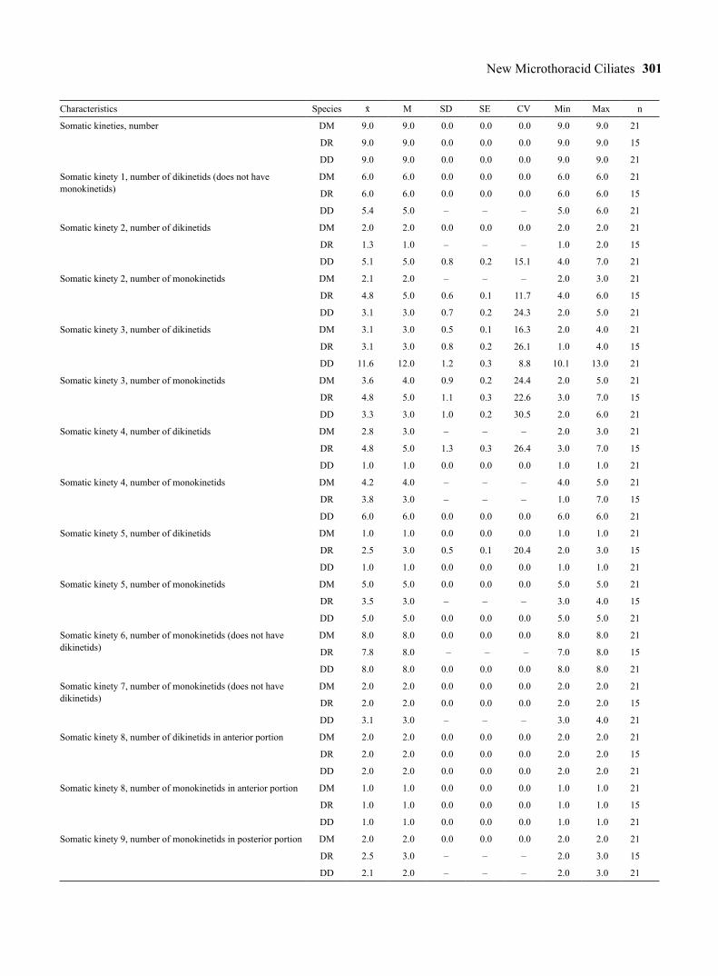
Table 1. Morphometric data on Drepanomonas minuta (DM), D. revoluta (DR), and D. multidentata (DD). Data based, if not mentioned otherwise, on protargol-impregnated, randomly selected specimens from a non-flooded Petri dish culture (D. minuta, D. revoluta) and a raw culture (D. multidentata). Measurements in µm. CV – coefficient of variation in %, M – median, Max – maximum, Min – minimum, n – number of specimens investigated, SD – standard deviation, SE – standard error of mean, – arithmetic mean, – – not measured or counted.
Characteristics Species M SD SE CV Min Max n
Body, length in protargol preparations DM 20.2 20.0 1.3 0.3 6.2 17.0 22.0 21
DR 25.2 25.0 1.3 0.3 5.0 23.0 27.0 15
DD 35.5 35.0 2.5 0.6 7.2 31.0 40.0 21
Body, width in protargol preparations DM 9.3 10.0 1.0 0.2 10.4 7.0 11.0 21
DR 12.1 12.0 1.0 0.3 8.6 11.0 14.0 15
DD 16.8 17.0 1.5 0.3 9.0 14.0 19.0 21
Body length: width, ratio in protargol preparations DM 2.2 2.2 0.2 0.1 7.0 1.9 2.5 21
DR 2.1 2.1 0.1 0.1 6.9 1.8 2.4 15
DD 2.1 2.1 0.2 0.1 9.0 1.8 2.7 21
Body, length in silver nitrate preparations DM 21.5 22.0 1.7 0.4 8.0 18.0 24.0 21
DR 27.7 28.0 1.6 0.5 5.7 25.0 30.0 9
DD 43.4 43.0 2.6 0.6 6.0 38.0 48.0 21
Body, width in silver nitrate preparations DM 10.5 10.0 1.4 0.3 13.0 8.0 13.0 21
DR 14.8 15.0 0.8 0.3 5.6 14.0 16.0 9
DD 22.9 23.0 2.2 0.5 9.5 19.0 27.0 21
Body length: width, ratio in silver nitrate preparations DM 2.1 2.1 0.2 0.1 7.4 1.8 2.5 21
DR 1.9 1.9 0.1 0.1 4.4 1.8 2.0 9
DD 1.9 1.9 0.1 0.1 6.1 1.7 2.2 21
Body length: width, ratio in vivo (from micrographs) DD 1.9 1.9 0.1 0.1 4.5 1.7 2.0 17
Anterior body end to adoral membranelles, distance DM 9.9 10.0 0.6 0.1 5.8 8.0 11.0 21
DR 11.7 12.0 0.6 0.2 5.1 11.0 13.0 15
DD 18.0 18.0 2.0 0.4 11.1 15.0 22.0 21
Body length: anterior body end to adoral membranelles, ratio DM 2.1 2.0 0.1 0.1 4.7 1.8 2.2 21
DR 2.2 2.2 0.1 0.1 2.8 2.1 2.3 15
DD 2.0 1.9 0.2 0.1 7.8 1.7 2.3 21
Anterior body end to macronucleus, distance DM 6.3 6.0 0.7 0.1 10.4 5.0 7.0 21
DR 8.1 8.0 0.5 0.1 6.4 7.0 9.0 15
DD 15.3 15.0 2.1 0.5 14.0 12.0 20.0 21
Macronucleus, length DM 4.2 4.0 0.4 0.1 10.3 4.0 5.0 21
DR 4.9 5.0 0.5 0.1 9.3 4.0 6.0 15
DD 7.8 8.0 1.0 0.2 12.8 6.0 10.0 21
Macronucleus, width DM 3.3 3.0 0.6 0.1 17.1 3.0 5.0 21
DR 4.5 4.0 – – – 4.0 5.0 15
DD 5.2 5.0 0.9 0.2 16.8 4.0 7.0 21
Micronucleus, diameter DM 1.3 1.5 – – – 1.0 1.5 21
DR 1.9 2.0 – – – 1.5 2.0 15
DD 1.9 2.0 – – – 1.5 2.0 21
Anterior body end to excretory pore of contractile vacuole, distance
DM Not recognizable
DR 14.6 15.0 0.7 0.2 5.1 13.0 16.0 15
DD 21.0 21.0 1.9 0.5 9.1 18.0 25.0 21
New Microthoracid Ciliates 301
Characteristics Species M SD SE CV Min Max n
Somatic kineties, number DM 9.0 9.0 0.0 0.0 0.0 9.0 9.0 21
DR 9.0 9.0 0.0 0.0 0.0 9.0 9.0 15
DD 9.0 9.0 0.0 0.0 0.0 9.0 9.0 21
Somatic kinety 1, number of dikinetids (does not have monokinetids)
DM 6.0 6.0 0.0 0.0 0.0 6.0 6.0 21
DR 6.0 6.0 0.0 0.0 0.0 6.0 6.0 15
DD 5.4 5.0 – – – 5.0 6.0 21
Somatic kinety 2, number of dikinetids DM 2.0 2.0 0.0 0.0 0.0 2.0 2.0 21
DR 1.3 1.0 – – – 1.0 2.0 15
DD 5.1 5.0 0.8 0.2 15.1 4.0 7.0 21
Somatic kinety 2, number of monokinetids DM 2.1 2.0 – – – 2.0 3.0 21
DR 4.8 5.0 0.6 0.1 11.7 4.0 6.0 15
DD 3.1 3.0 0.7 0.2 24.3 2.0 5.0 21
Somatic kinety 3, number of dikinetids DM 3.1 3.0 0.5 0.1 16.3 2.0 4.0 21
DR 3.1 3.0 0.8 0.2 26.1 1.0 4.0 15
DD 11.6 12.0 1.2 0.3 8.8 10.1 13.0 21
Somatic kinety 3, number of monokinetids DM 3.6 4.0 0.9 0.2 24.4 2.0 5.0 21
DR 4.8 5.0 1.1 0.3 22.6 3.0 7.0 15
DD 3.3 3.0 1.0 0.2 30.5 2.0 6.0 21
Somatic kinety 4, number of dikinetids DM 2.8 3.0 – – – 2.0 3.0 21
DR 4.8 5.0 1.3 0.3 26.4 3.0 7.0 15
DD 1.0 1.0 0.0 0.0 0.0 1.0 1.0 21
Somatic kinety 4, number of monokinetids DM 4.2 4.0 – – – 4.0 5.0 21
DR 3.8 3.0 – – – 1.0 7.0 15
DD 6.0 6.0 0.0 0.0 0.0 6.0 6.0 21
Somatic kinety 5, number of dikinetids DM 1.0 1.0 0.0 0.0 0.0 1.0 1.0 21
DR 2.5 3.0 0.5 0.1 20.4 2.0 3.0 15
DD 1.0 1.0 0.0 0.0 0.0 1.0 1.0 21
Somatic kinety 5, number of monokinetids DM 5.0 5.0 0.0 0.0 0.0 5.0 5.0 21
DR 3.5 3.0 – – – 3.0 4.0 15
DD 5.0 5.0 0.0 0.0 0.0 5.0 5.0 21
Somatic kinety 6, number of monokinetids (does not have dikinetids)
DM 8.0 8.0 0.0 0.0 0.0 8.0 8.0 21
DR 7.8 8.0 – – – 7.0 8.0 15
DD 8.0 8.0 0.0 0.0 0.0 8.0 8.0 21
Somatic kinety 7, number of monokinetids (does not have dikinetids)
DM 2.0 2.0 0.0 0.0 0.0 2.0 2.0 21
DR 2.0 2.0 0.0 0.0 0.0 2.0 2.0 15
DD 3.1 3.0 – – – 3.0 4.0 21
Somatic kinety 8, number of dikinetids in anterior portion DM 2.0 2.0 0.0 0.0 0.0 2.0 2.0 21
DR 2.0 2.0 0.0 0.0 0.0 2.0 2.0 15
DD 2.0 2.0 0.0 0.0 0.0 2.0 2.0 21
Somatic kinety 8, number of monokinetids in anterior portion DM 1.0 1.0 0.0 0.0 0.0 1.0 1.0 21
DR 1.0 1.0 0.0 0.0 0.0 1.0 1.0 15
DD 1.0 1.0 0.0 0.0 0.0 1.0 1.0 21
Somatic kinety 9, number of monokinetids in posterior portion DM 2.0 2.0 0.0 0.0 0.0 2.0 2.0 21
DR 2.5 3.0 – – – 2.0 3.0 15
DD 2.1 2.0 – – – 2.0 3.0 21

A. Omar and W. Foissner302
Characteristics Species M SD SE CV Min Max n
Preoral kineties, number DM 3.0 3.0 0.0 0.0 0.0 3.0 3.0 21
DR 3.0 3.0 0.0 0.0 0.0 3.0 3.0 15
DD 3.0 3.0 0.0 0.0 0.0 3.0 3.0 21
Preoral kinety 1, number of dikinetids (does not have monokinetids)
DM 2.0 2.0 0.0 0.0 0.0 2.0 2.0 21
DR 2.0 2.0 0.0 0.0 0.0 2.0 2.0 15
DD 2.0 2.0 0.0 0.0 0.0 2.0 2.0 21
Preoral kinety 2, number of dikinetids DM 3.0 3.0 0.0 0.0 0.0 3.0 3.0 21
DR 3.0 3.0 0.0 0.0 0.0 3.0 3.0 15
DD 3.0 3.0 0.0 0.0 0.0 3.0 3.0 21
Preoral kinety 2, number of monokinetids DM 1.0 1.0 0.0 0.0 0.0 1.0 1.0 21
DR 1.0 1.0 0.0 0.0 0.0 1.0 1.0 15
DD 1.0 1.0 0.0 0.0 0.0 1.0 1.0 21
Preoral kinety 3, number of dikinetids DM 3.0 3.0 0.0 0.0 0.0 3.0 3.0 21
DR 3.0 3.0 0.0 0.0 0.0 3.0 3.0 15
DD 3.0 3.0 0.0 0.0 0.0 3.0 3.0 21
Preoral kinety 3, number of monokinetids DM 1.0 1.0 0.0 0.0 0.0 1.0 1.0 21
DR 1.0 1.0 0.0 0.0 0.0 1.0 1.0 15
DD 1.0 1.0 0.0 0.0 0.0 1.0 1.0 21
Left row of postoral complex, number of monokinetidsa DM 4.0 4.0 0.0 0.0 0.0 4.0 4.0 21
DR 4.1 4.0 – – – 4.0 5.0 15
DD 4.1 4.0 – – – 4.0 5.0 21
Right row of postoral complex, number of monokinetidsb DM 1.0 1.0 0.0 0.0 0.0 1.0 1.0 21
DR 1.1 1.0 – – – 1.0 2.0 15
DD 1.0 1.0 0.0 0.0 0.0 1.0 1.0 21
Basal bodies, total numberc DM 84.5 85.0 0.9 0.2 1.1 82.0 86.0 21
DR 92.7 92.0 2.2 0.6 2.3 90.0 96.0 15
DD 106.9 107.0 2.7 0.6 2.5 103.0 112.0 21
a This is the posterior portion of somatic kinety 8.b This is the middle portion of somatic kinety 9.c Except for basal bodies of adoral membranelles and anterior portion of kinety 9, which is difficult to see.
by six monokinetids; kineties 6 and 7 only partially ciliated. On average a total of 107 basal bodies. Extru-somes absent.
Type locality: Slightly saline surface mud and soil from temporary grassland puddles in the surroundings of the village of Chichiriviche, Maracay National Park, about 13 km inshore from the north coast of Venezuela, 11°33′N, 67°13′W.
Type material: One holotype slide with protargol-impregnated specimens and five paratype slides with protargol-impregnated and Klein-Foissner silver ni-trate-impregnated specimens have been deposited in the Biology Centre of the Museum of Upper Austria, Linz
(LI). The holotype and important paratype specimens have been marked by black ink circles on the coverslip.
Etymology: the species name is a composite of the Latin prefix multi (many) and the Latin adjective dentata (toothed), referring to the multitude of body teeth.
Description: Depending on method, body size var-ies considerably (Table 1): in vivo about 47 × 27 µm (n = 4), in Klein-Foissner silver nitrate preparations 43 × 25 µm, while cells shrank to 35.5 × 16.8 µm in pro-targol slides. Taking into account the in vivo measure-ments and 5% shrinkage in the silver nitrate prepara-tions, we obtain an in vivo size of 40–50 × 20–30 µm, on average about 45 × 25 µm, corresponding to a length:

New Microthoracid Ciliates 303
Figs 16–24. Drepanomonas multidentata (16–21) and D. dentata (22–24) from life (16, 17, 22–24) and after protargol impregnation (18–21). 16, 17 – right and left side view of a representative specimen, length 45 µm, showing the crescentic body and the cortical ridges. The right side ridges form an obfalcate plate with a subapical spine and a shallow concavity in the posterior portion (asterisk); the two left side ridges, each with a spine anteriorly, produce a tridentate pattern posteriorly. Note the three highly characteristic ventro-lateral spines in the surrounding of the oral opening. 18, 19 – right and left side view of holotype specimen, length 35 µm, showing the kinetid pattern and the nuclear apparatus; 20 – ventral view of a paratype specimen, showing the obliquely arranged preoral kineties, the circumoral basal bodies, and the postoral complex. The posterior segment of somatic kinety 1 cannot be seen in this specimen because it is over the deeply impregnated macronucleus (outline dotted); 21 – in dividing specimens, three adoral membranelles are recognizable both in proter and opisthe. The arrowheads mark the ventro-lateral spines; 22–24 – D. dentata (22, 23, from Fresenius 1858; 24, from Kreutz 1998), showing the crescentic body, the tooth-like projection on the ventral side (triangle), the serrate dorsal (arrows) and ventral (arrowhead) margin, and the cortical ridges. A – anterior body end, K(1–9) – somatic kineties, LAS – left anterior spines, LVS – left ventro-lateral spines, M(1–3) – adoral membranelles, MA – macronucleus, MI – micronucleus, NK – nasse kinetosomes, P – posterior body end, PC – postoral complex, PO(1–3) – preoral kineties, R – ridges, RVS – right ventro-lateral spine, SAS – subapical spine, T – excretory tube of contractile vacuole, TP – spines of the tridentate pattern. Scale bars: 10 µm.

A. Omar and W. Foissner304
Figs 25–32. Drepanomonas multidentata from life under bright field (25–27) and interference contrast illumination (29–32), and after silver nitrate impregnation (28). 25, 26 – right and left side view of the same specimen, showing the crescentic body shape and the three ventro-lateral spines. The cytoplasm is studded with lipid (?) droplets and minute food vacuoles each containing only one or two bacteria (26). The arrows mark the line where the cortical plates of the right side attach to the ground cortex. The arrowhead denotes the left margin of the preoral region; 27 – right side view of a dividing specimen with division furrow marked by arrowheads; 28 – right side view, showing the dense cortical granulation and the ventro-lateral spines; 29–32 – right side views of the same specimen at four focal planes, showing the right side ridges (29), the subapical and the right ventro-lateral spine (30), the buccal cavity in mid-body (31), and the spines of the left ventro-lateral side (32). The arrowhead marks the concave right ventro-lateral margin. The asterisk denotes the shallow concavity in the ob-falcate plate. A – anterior body end, BC – buccal cavity, CV – contractile vacuole, CY – cytopyge, FV – food vacuoles, K3 – somatic kinety 3, LAS – left anterior spine, LD – lipid droplets, LVS – left ventro-lateral spines, MA – macronucleus, P – posterior body end, PO – preoral kineties, R – ridges, RVS – right ventro-lateral spine, SAS – subapical spine, T – excretory tube of contractile vacuole, VS – ventro-lateral spines. Scale bars: 15 µm.
width ratio of 1.9, which matches values obtained from 17 specimens photographed alive (Figs 25–27, 29–36); protargol-impregnated cells have a ratio of 2.1 and are thus slightly more slender (Table 1; Figs 37, 38).
Body crescentic with acute ends and several ventral and lateral spines; laterally only slightly flattened, ex-
cept for thin dorsal margin; when observed ventrally or dorsally, slenderly elliptical with left side slightly more convex than right side (Table 1; Figs 16–20, 25, 30, 36, 39).
Nuclear apparatus in or near mid-body. Macronucle-us globular to broadly ellipsoidal, contains some globu-

New Microthoracid Ciliates 305
Figs 33–40. Drepanomonas multidentata from life (33–36) and after protargol impregnation (37–40). 33–36 – left side views of different specimens at four focal planes, showing the two conspicuous spines left and posterior of the oral opening, the left side ridges each form-ing an anterior spine, and the two anterior spines of the tridentate pattern; 37, 38 – right and left side view of holotype and of a paratype specimen, showing the ciliary and nuclear pattern. The posterior portion of kinety 9 is either absent or in line with kinety 2. The posterior cilium of kinety 6 appears slightly thickened in the basal portion (arrowhead); 39 – ventral view of a paratype specimen, showing the oblique preoral kineties and the postoral complex; 40 – ventral view of a late divider, showing that kineties 8 and 9 consist of two and three segments, respectively, of which the posterior segment of kinety 9 will possibly align with kinety 2 (see Fig. 37). The left segment of the postoral complex consists of four ciliated monokinetids. A – anterior body end, K1–9 – somatic kineties, LAS – left anterior spines, LVS – left ventro-lateral spines, M – adoral membranelles, MA – macronucleus, MI – micronucleus, NK – nasse kinetosomes, PC – postoral complex, PO(1–3) – preoral kineties, R – ridges, T – excretory tube, TP – spines of the tridentate pattern. Scale bars: 15 µm (Figs 33–36) and 10 µm (Figs 37–40).
lar masses, probably nucleoli up to 2 µm across. Mi-cronucleus globular, near or attached to ventral side of macronucleus (Table 1; Figs 16, 19, 30, 37). Contractile
vacuole in third quarter of body, slightly posterior and dorsal of buccal cavity, with distinct tube extending into buccal cavity posterior to adoral membranelles (Table 1;

A. Omar and W. Foissner306
Figs 16, 18, 31, 37). Cytopyge posterior and slightly left of contractile vacuole, usually forming a bright, oblique, ellipsoidal blister rarely containing food remnants (Figs 16, 30, 31). Extrusomes absent, i.e., we could not find the typical trichocysts present in most microthoracids in vivo (Figs 25–27, 29–36) and in protargol preparations (Figs 37–40). Cytoplasm colourless, contains: (i) many vacuoles about 3 µm across, some appearing empty while others contain one or two bacteria and (ii) rather many globular and dumbbell-shaped inclusions 1 to 3 µm across, possibly lipid droplets (Figs 16, 17, 25–27, 29–36). Glides on bottom of Petri dish and surface of organic masses, rarely swims slowly and clumsily rotat-ing about main body axis.
Cortex glossy, very rigid in vivo but shrinking strongly in protargol preparations (see above); under-lain by countless, minute structures, possibly mitochon-dria (Fig. 33). Right side with two distinct ridges right of kineties 2 and 3, both circumscribing an elevated, ob-falcate plate wide and obliquely truncate anteriorly and gradually narrowed posteriorly where there is a shallow depression and ridges merge into acute rear body end; ridge right of kinety 3 forms a subapical spine. Left of kinety 2 a similar but deeper plate, forming a con-spicuous spine anteriorly, covers somatic kinety 1 and right side of oral apparatus (Figs 16, 18, 25, 29–32). Left side with two ridges along kineties 5 and 6; each ridge with a spine anteriorly (left anterior spines) while forming a tridentate pattern in posterior third (Figs 17, 26, 33–36). Dorsal margin strongly flattened and thus hyaline. Ventral side highly structured: preoral kine-ties and anterior portion of kinety 8 in distinct furrows covered by right side cortical plates and anterior spines of left side. Oral opening in mid-body: right mouth margin produced by a right side cortical plate having a conspicuous, posteriorly directed spine (right ventral spine) anterior to oral opening; postorally, this plate forms a slightly concave, sharp line; left mouth margin made by ground cortex, with two conspicuous spines (left ventral spines) on a convexity at level and pos-terior to oral opening, posterior spine usually slightly longer than anterior (Figs 16–19, 21, 25–28, 30, 32–36). Silverline pattern as described in D. hymenofera by Omar and Foissner (2013), i.e., the cortex is studded with minute, argyrophilic granules (Fig. 28).
Somatic cilia 8–9 µm long in vivo, rapidly lost under slight coverslip pressure. Invariably nine somatic and three preoral kineties with a total of 107 basal bodies on average. Kineties 3, 4 and 6 bipolar; kineties 1, 2, 5
and 7–9 shortened anteriorly and/or posteriorly. Kine-ties 1–4 on right side; kineties 5–7 on left side; and ki-neties 1, 2 and 6–9 on ventral side (Table 1; Figs 16–20, 37–40).
Kinety 1 at right margin of oral cavity, consists of 5 or 6 narrowly spaced, usually ciliated dikinetids (Figs 18, 37). Kinety 2 begins in second quarter of body, composed of two widely spaced, ciliated dikinetids fol-lowed by some widely spaced, ciliated monokinetids, ends posteriorly with some narrowly spaced, ciliated dikinetids (Figs 18, 20, 37). Kinety 3 composed of widely and narrowly spaced, ciliated dikinetids in an-terior and posterior portion, respectively, and of some widely spaced, ciliated monokinetids in middle por-tion (Figs 18, 37). Kineties 4 and 5 limit dorsal mar-gin of right and left body side, both commence with a single, ciliated dikinetid followed by six and five widely spaced, ciliated monokinetids, respectively (Figs 19, 38). Kinety 6 composed of widely spaced monokinetids, third monokinetid usually non-ciliated, occasionally only two and one kinetid ciliated in an-terior and posterior end of row, respectively (Figs 17, 19, 38). Kinety 7 begins in or near mid-body, consists of three widely spaced monokinetids of which only the anterior kinetid is ciliated (Figs 17, 19, 20, 38). Kinety 8 composed of two segments (Figs 18, 20, 37, 39, 40): anterior segment posterior and very similar to preoral kinety 3, consists of two ciliated dikinetids and one cili-ated monokinetid at left (posterior) end; posterior seg-ment composed of three narrowly spaced monokinetids and a single monokinetid near posterior body end (see postoral complex below). Kinety 9 consists of three segments (Figs 18, 20, 39, 40): anterior segment left of adoral membranelles and composed of few, likely bar-ren dikinetids recognizable only in dividers and appro-priately oriented specimens; middle segment a single, ciliated monokinetid far posterior to buccal cavity (see postoral complex); posterior segment possibly absent or composed of two (rarely three) ciliated monokinetids in line with kinety 2 (Figs 18, 20, 37, 40).
Three preoral kineties each composed of ciliated dikinetids and a ciliated monokinetid at left end of ki-neties 2 and 3; occupy preoral ventral side and extend obliquely to left side. Postoral complex composed of the monokinetidal posterior segment of kinety 8 and the single, ciliated monokinetid in mid of kinety 9 (Table 1; Figs 16, 18, 20, 25, 28, 31, 36–40).
Oral apparatus in mid-body (Table 1; Figs 16, 18, 20, 31, 37, 39). Buccal cavity deep contains two or

New Microthoracid Ciliates 307
Table 2. Morphometric data on Leptopharynx lajacola. Based on mounted, protargol-impregnated, and randomly selected specimens from a non-flooded Petri dish culture. Measurements in µm. CV – coefficient of variation in %, M – median, Max – maximum, Min – minimum, n – number of specimens investigated, PC – postoral complex, SD – standard deviation, SE – standard error of mean, – arithmetic mean, – – not measured or counted.
Characteristics M SD SE CV Min Max n
Body, length 31.7 32.0 1.9 0.4 5.9 28.0 34.0 21
Body, width 16.7 17.0 1.4 0.3 8.5 14.0 19.0 21
Body length: width, ratio 1.9 1.9 0.1 0.1 4.7 1.7 2.1 21
Anterior body end to first adoral membranelles, distance 11.5 12.0 0.9 0.2 7.6 10.0 13.0 21
Body length: anterior body end to first adoral membranelles, ratio 2.8 2.8 0.1 0.1 5.1 2.6 3.1 21
Anterior body end to macronucleus, distance 11.6 12.0 0.8 0.2 7.0 10.0 13.0 21
Macronucleus, length 6.5 7.0 0.8 0.2 11.6 5.0 8.0 21
Macronucleus, width 6.3 6.0 0.6 0.1 10.2 5.0 7.0 21
Micronucleus, diameter 1.9 2.0 – – – 1.5 2.0 21
Anterior body end to excretory pore of contractile vacuole, distance 19.1 19.0 1.0 0.2 5.5 17.0 21.0 21
Oral basket, width 2.7 3.0 – – – 2.0 3.0 21
Somatic kineties, number 10.0 10.0 0.0 0.0 0.0 10.0 10.0 21
Somatic kinety 1, number of dikinetids 7.0 7.0 0.0 0.0 0.0 7.0 7.0 21
Somatic kinety 1, number of monokinetids 1.0 1.0 0.0 0.0 0.0 1.0 1.0 21
Somatic kinety 2, number of dikinetids 2.0 2.0 0.0 0.0 0.0 2.0 2.0 21
Somatic kinety 2, number of monokinetids 8.0 8.0 0.4 0.1 4.8 7.0 9.0 21
Somatic kinety 3, number of dikinetidsa 0.0 0.0 – – – 0.0 2.0 22
Somatic kinety 3, number of monokinetids 12.7 12.0 1.2 0.3 9.5 10.0 16.0 21
Somatic kinety 4, number of monokinetids (does not have dikinetids) 22.2 22.0 1.3 0.3 6.0 20.0 25.0 21
Somatic kinety 5, number of monokinetids (does not have dikinetids) 9.5 9.0 0.9 0.2 9.8 8.0 11.0 21
Somatic kinety 6, number of monokinetids (does not have dikinetids) 4.0 4.0 0.0 0.0 0.0 4.0 4.0 21
Somatic kinety 7, number of monokinetids (does not have dikinetids) 7.8 8.0 – – – 7.0 8.0 21
Somatic kinety 8, number of monokinetids (does not have dikinetids) 3.1 3.0 – – – 3.0 4.0 21
Somatic kinety 9, number of dikinetids in anterior portion 4.0 4.0 0.0 0.0 0.0 4.0 4.0 21
Somatic kinety 10, number of monokinetids (for dikinetids, see PC) 4.1 4.0 0.4 0.1 8.7 4.0 5.0 21
Preoral ciliary rows, number 3.0 3.0 0.0 0.0 0.0 3.0 3.0 21
Preoral kinety 1, number of dikinetids (does not have monokinetids) 2.0 2.0 0.0 0.0 0.0 2.0 2.0 21
Preoral kinety 2, number of dikinetids 3.0 3.0 0.0 0.0 0.0 3.0 3.0 21
Preoral kinety 2, number of monokinetids 1.0 1.0 0.0 0.0 0.0 1.0 1.0 21
Preoral kinety 3, number of dikinetids 4.0 4.0 0.0 0.0 0.0 4.0 4.0 21
Preoral kinety 3, number of monokinetids 1.0 1.0 0.0 0.0 0.0 1.0 1.0 21
Oral primordium, number of dikinetids in posterior part 4.0 4.0 0.0 0.0 0.0 4.0 4.0 21
Oral primordium, number of granules (basal bodies?) in anterior portion 4.5 4.0 0.7 0.2 15.2 4.0 6.0 21
Adoral membranelle 2, number of basal body rows 4.0 4.0 – – – 3.0 4.0 21
Adoral membranelle 2, number of basal bodies 11.9 12.0 0.7 0.1 5.5 9.0 12.0 21
Adoral membranelle 3, number of basal body rows 4.0 4.0 0.0 0.0 0.0 4.0 4.0 21
Adoral membranelle 3, number of basal bodies 12.0 12.0 0.0 0.0 0.0 12.0 12.0 21
Left row of postoral complex, number of monokinetidsb 6.0 6.0 – – – 5.0 6.0 21
Right row of postoral complex, number of dikinetids 3.0 3.0 0.0 0.0 0.0 3.0 3.0 21
Basal bodies, total number (except for adoral membranelles) 143.0 143.0 2.6 0.6 1.8 139.0 148.0 21
a Two dikinetids in the anterior portion of kinety 3 in one out of 21 specimens.b Including a single monokinetid, which possibly belongs to preoral kinety 3, posterior of the row.

A. Omar and W. Foissner308
three adoral membranelles. Membranelle 1 distinctly smaller than membranelles 2 and 3, recognizable only in dividers (Fig. 21); membranelles 2 and 3 obliquely arranged to main body axis and so close together that details cannot be recognized. Nasse kinetosomes ante-rior to adoral membranelles (Figs 20, 39); oral basket not recognizable.
Occurrence and ecology: As yet found only at type locality (see above). It grew well in a raw culture with tap water to which were added three squashed wheat grains and a few ml of the soil eluate from the non-flooded Petri dish culture.
Comparison of Drepanomonas multidentata with similar species: Using the species characteristics for Drepanomonas suggested by Omar and Foissner (2013), D. multidentata is unique by the three ventro-lateral spines, the tridentate spine pattern in the poste-rior half of the left side, and the absence of extrusomes. Similar species are D. dentata Fresenius, 1858, D. lunaris Foissner, 1979, and D. obtusa Penard, 1922, all having extrusomes. Further, D. dentata differs from D. multidentata not only by the spine pattern but also by body size (~ 80 µm vs. 45 × 25) and the serrate (vs. smooth) ventral and dorsal margin (Kreutz 1998; cp. Figs 22–24 with Figs 16, 17). Drepanomonas lunaris and D. obtusa differ from D. multidentata not only by the spine pattern but also by the rounded and truncated (vs. acute) posterior body end, respectively.
Leptopharynx lajacola Omar & Foissner nov. spec. (Figs 41–47; Table 1)
Diagnosis: Size about 32 × 17 µm in protargol preparations. Body outline quadrangular to elliptical with flat to slightly convex dorsal side, flat ventral side and slightly oblique preoral region, body ends broad-ly rounded. Ten somatic kineties and a postoral com-plex generated by kineties 9 and 10; kineties 1 and 2 with dikinetids anteriorly; kinety 6 composed of four monokinetids; kinety 10 far posterior of adoral mem-
branelles and without dikinetids; preoral kineties on ventral side. On average 143 basal bodies. Three adoral membranelles, membranelles 2 and 3 form a flat, quad-rangular ciliary field. Oral basket about 3 µm wide, strongly convex with opening directed posteriorly. Oral primordium left of kinety 1.
Type locality: Soil, mud, and moss from ephem-eral puddles (Lajas) with Velosiacean plants in the sur-roundings of the farm of Mr. Eisenberg, outskirts of the town of Puerto Ayacucho, Venezuela, 5°40′N, 67°35′W.
Type material: One holotype and two paratype slides with protargol-impregnated specimens have been deposited in the Biology Centre of the Museum of Upper Austria, Linz (LI). The holotype and important paratype specimens have been marked by black ink cir-cles on the coverslip.
Etymology: Composite of Laja, the Venezuelan word for ephemeral puddles on granitic outcroppings, and the Latin verb colere (to dwell), referring to its habitat.
Description: This species was overlooked in the raw sample where it was much rarer than the common L. costatus. Thus, all observations are from protargol-impregnated specimens. Fortunately, L. lajacola has some outstanding features which are well recogniz-able in protargol preparations, viz., the quadrangular body shape, the lack of dikinetids in kinety 3, and the strongly curved oral basket whose opening is directed posteriorly. Thus, this species should be recognizable also without in vivo data.
Assuming 15% preparation shrinkage, L. lajacola should have an in vivo size of 30–40 × 15–22 µm, on average about 35 × 18 µm (Table 2); length: width ra-tio 1.7–2.1:1, on average 1.9:1; laterally flattened about 2:1, both sides slightly convex. Body usually conspicu-ously quadrangular with both ends broadly rounded, anteriorly slightly narrower than posteriorly due to the slightly receding preoral region (Figs 41–43, 45–47). Nuclear apparatus in or near body centre, usually in
Figs 45–47. Leptopharynx lajacola after protargol impregnation. 45, 46 – right and left side view, showing the quadrangular body shape, the kinetid and nuclear pattern, and the strongly convex oral basket whose opening is directed posteriorly. The arrows mark the line made by the cortex edge right of kinety 1 which extends to body midline. The asterisks mark the crenellation along kinety 4. Kineties 3 and 4 consist of monokinetids throughout. Kinety 6 consists of four basal bodies; 47 – left side view of a paratype specimen, showing, inter alia, the quad-rangular body shape and the steep oral basket. B – oral basket, K1–10 – somatic kineties, LD – lipid droplet, M1–3 – adoral membranelles, MA – macronucleus, MC – membranellar cilia, MI – micronucleus, OP – oral primordium, PC – postoral complex, PO1–3 – preoral kineties, T – excretory tube of contractile vacuole. Scale bars: 10 µm.

New Microthoracid Ciliates 309
Figs 41–44. Leptopharynx lajacola after protargol impregnation. 41, 42 – right and left side view of holotype specimen, length 31 µm, showing the quadrangular body shape, the kinetid and nuclear pattern, and the arrangement of the basal bodies on ventral side. Note the narrow, strongly convex oral basket and the three oblique adoral membranelles. The arrow marks the slightly dislocated posterior end of kinety 1. The arrowhead denotes the anterior portion of the oral primordium. Kineties 3 and 4 consist of monokinetids throughout. Kinety 6 consists of four monokinetids; 43 – left side view of a paratype specimen, showing the quadrangular body shape and the narrow, strongly curved oral basket. 44 – transverse section posterior of adoral membranelles (semi-schematic). B – oral basket, K1–10 – somatic kineties, M1–3 – adoral membranelles, MA – macronucleus, MI – micronucleus, OP – oral primordium, PC – postoral complex, PO(1–3) – preoral kineties, T – excretory tube of contractile vacuole. Scale bars: 10 µm.

A. Omar and W. Foissner310
curve formed by oral basket. Macronucleus globular, contains minute, argyrophilic masses, probably nucle-oli. Micronucleus globular, usually attached to ventral side macronucleus (Table 2; Figs 42, 45–47). Opening of contractile vacuole in third quarter of ventral side, i.e., right of posterior part of oral primordium, tube composed of fibres forming a star-like pattern at tube base (Table 2; Figs 41, 45). Cytopyge slightly posterior to contractile vacuole, extends between posterior por-tions of kineties 2 and 10, in protargol preparations usu-ally marked by a short, thick line (Fig. 45). Extrusomes lenticular, 4–5 µm long. Cytoplasm with few to many lipid (?) droplets 0.5–3 µm across (Fig. 45).
Cortex comparatively smooth, similar to that of L. costatus, margin of kineties 1, 4 and 5 distinctly im-pregnated. Ventral side with conspicuous preoral fur-rows and a shallow concavity containing oral basket opening and adoral membranelles (Figs 41–47).
Cilia 6–8 µm long, arranged in 10 somatic and three preoral rows (kineties), each showing a specific number and pattern of basal bodies; on average a total of 143 kinetids (Table 2). Kineties 2–5 and 7 bipolar, kineties 1, 6 and 8–10 shortened anteriorly and/or posteriorly. Kineties 1–4 on right side, kineties 5–8 on left side, ki-neties 9 and 10 on ventral side (Table 2; Figs 41, 42, 44, 45–47).
Kinety 1 extends along right margin of oral field, ends slightly posterior of mid-body; composed of sev-en narrowly spaced, ciliated dikinetids and a single monokinetid at posterior end; posterior dikinetid and monokinetid usually slightly dislocated to the left (Figs 41, 44, 45). Kinety 2 composed of two widely spaced, ciliated dikinetids anteriorly, followed by 7–9 widely spaced, ciliated monokinetids leaving a more or less wide break in middle third of row (Figs 41, 45). Kinety 3 composed of widely spaced, ciliated monokinetids throughout, a single kinetid-wide break in mid of row (Fig. 45); begins with two dikinetids anteriorly in one out of 21 specimens. Kineties 4 and 5 limit dorsal mar-gin of right and left body side, composed of moderately narrowly and widely spaced, ciliated monokinetids throughout, respectively (Figs 41, 42, 45–47). Kinety 6 composed of two narrowly spaced, oblique, non-cil-iated monokinetids near anterior end of cell and two widely spaced, ciliated monokinetids in middle body third (Figs 42, 46, 47). Kinety 7 composed of widely spaced, likely ciliated monokinetids, forming more or less distinct pairs in anterior half (Figs 42, 46, 47). Ki-nety 8 begins in second quarter of body and consists
of three widely spaced, ciliated monokinetids (Figs 42, 46, 47). Kinety 9 consists of two segments (Figs 41, 45): anterior segment composed of four ciliated diki-netids posterior of preoral kineties; posterior segment posterior to adoral membranelles and composed of a short row of narrowly spaced, ciliated monokinetids (see postoral complex). Kinety 10 consists of three seg-ments (Figs 41, 45–47): anterior segment composed of two dikinetids left of adoral membranelles; middle segment composed of three barren, oblique dikinetids posterior to adoral membranelles; posterior segment composed of 4–5 widely spaced, ciliated monokinetids.
Three preoral kineties each composed of ciliated dikinetids and a ciliated monokinetid posteriorly, oc-cupy preoral ventral side and extend obliquely to left side (Table 2; Figs 41, 43, 45). Postoral complex similar to that of L. costatus, i.e., composed of posterior por-tion of kinety 9 and middle portion of kinety 10 (Table 2; Figs 41, 45–47). Posterior of rear portion of kinety 9 a single monokinetid possibly belonging to preoral kinety 3, as in L. costatus (Figs 41, 45).
Oral apparatus slightly anterior to mid-body within a concave area containing the adoral membranelles; composed of a distinct basket made of nematodesmata, three adoral membranelles, and a paroral (not shown) made of a single row of basal bodies (“nasse kineto-somes”) subapically connected with the basket rods (Table 2; Figs 41–43, 45–47). Adoral membranelles obliquely arranged to main body axis and left of oral basket, membranellar cilia form posteriorly directed bundles up to 8 µm long in protargol preparations (Fig. 47). Adoral membranelle 1 anterior of membranelles 2 and 3 and composed of few, possibly four densely spaced basal bodies. Membranelles 2 and 3 very close together, forming a flat quadrangular field composed of 6 basal body rows, each consisting of an average of four basal bodies; right row possibly barren (Table 2; Figs 41, 45, 47). Left of M2 a very short row of basal bodies belonging to the postoral complex and kinety 10 (Fig. 41). Oral basket about 3 µm wide, of unique shape, i.e., strongly convex with basket opening directed posteri-orly and in second quarter of cell, abruptly narrowed when turning posteriorly (Table 2; Figs 41–43, 45–47). Nasse kinetosomes difficult to recognize, unciliated. Oral primordium composed of two groups of basal bodies: anterior group right of oral basket, barren, usu-ally faintly impregnated and thus difficult to recognize, composed of four to six basal bodies forming a short, slightly convex row; posterior group composed of two

New Microthoracid Ciliates 311
minute rows of ciliated dikinetids left of posterior re-gion of somatic kinety 1, left row usually made by a single dikinetid (Table 2; Figs 41, 45).
Occurrence and ecology: As yet found only at type locality.
Comparison of Leptopharynx lajacola with simi-lar species: This species has a clear identity and is easi-ly recognized by the strongly convex oral basket whose opening is near mid-body and directed posteriorly. Fur-ther characters are the quadrangular body shape and the absence of dikinetids in somatic kinety 3; the last fea-ture is shared with the microstome morph of L. costatus gonohymen Foissner and Omar in Omar and Foissner (2012a) and L. bromelicola Foissner et al., 2011.
Acknowledgement. Supported by the Austrian Science Fund (FWF project P22846-B17). The technical assistance of Dr. Barbara Harl, Robert Schörghofer, and Andreas Zankl is greatly appreciated.
REFERENCES
Foissner W. (1979) Taxonomische Studien über die Ciliaten des Großglocknergebietes (Hohe Tauern, Österreich). Familien Microthoracidae, Chilodonellidae und Furgasoniidae. Sber. öst. Akad. Wiss. Wien 188: 27–43
Foissner W. (1985) Morphologie und Infraciliatur der Genera Microthorax und Stammeridium und Klassifikation der Microtho-racina Jankowski, 1967 (Protozoa: Ciliophora). Zool. Anz. 214: 33–53
Foissner W. (1987) Neue terrestrische und limnische Ciliaten (Pro-tozoa, Ciliophora) aus Österreich und Deutschland. Sber. öst. Akad. Wiss. Wien 195: 217–268
Foissner W. (1991) Basic light and scanning electron microscopic methods for taxonomic studies of ciliated protozoa. Eur. J. Protistol. 27: 313–330
Foissner W., Berger H., Kohmann F. (1994) Taxonomische und ökologische Revision der Ciliaten des Saprobiensystems – Band III: Hymenostomata, Prostomatida, Nassulida. Informationsberichte des Bayer. Landesamtes für Wasserwirtschaft 1/94: 1–548
Foissner W., Agatha S., Berger H. (2002) Soil ciliates (Protozoa, Ciliophora) from Namibia (Southwest Africa), with emphasis on two contrasting environments, the Etosha region and the Na-mib Desert. Denisia 5: 1–1459
Foissner W., Wolf K., Yashchenko V., Stoeck T. (2011) Descrip-tion of Leptopharynx bromelicola n. sp. and characterization of the genus Leptopharynx Mermod, 1914 (Protista, Ciliophora). J. Eukaryot. Microbiol. 58: 134–151
Fresenius G. (1858) Beiträge zur Kenntnis mikroskopischer Organ-ismen. Abh. senckenb. naturforsch. Ges. 2: 211–242
Gong J., Stoeck T., Yi Z., Miao M., Zhang Q., Roberts D. M., War-ren A., Song W. (2009) Small subunit rRNA phylogenies show that the class Nassophorea is not monophyletic (phylum Cili-ophora). J. Eukaryot. Microbiol. 56: 339–347
Jankowski A. V. (1967) A new system of ciliate protozoa (Ciliopho-ra). Trud. zool. Inst., Leningr. 43: 3–52 (in Russian)
Kreutz M. (1998) Beobachtungen an zwei Arten der Ciliatengattung Drepanomonas. Mikrokosmos 87: 321–327
Omar A., Foissner W. (2011) Description of Leptopharynx bromeliophilus nov. spec. and Leptopharynx australiensis nov. spec. (Ciliophora, Nassulida). Acta Protozool. 50: 89 –103
Omar A., Foissner W. (2012a) Description of Leptopharynx brasiliensis nov. spec. and Leptopharynx costatus gonohymen nov. subspec. (Ciliophora, Microthoracida). Eur. J. Protistol. 48: 30–47
Omar A., Foissner W. (2012b) Neotypification and ontogenesis of Leptopharynx costatus costatus Mermod, 1914. J. Eukaryot. Microbiol. 59: 268–286
Omar A., Foissner W. (2013) Description of two new Drepanomonas taxa and an account on features defining species in Drepanomonas Fresenius, 1858 (Ciliophora, Microthoracida). Eur. J. Protistol. 49: 420–437
Penard E. (1922) Études sur les infusoires d’eau douce. Georg et Cie, Genève
Small E. B., Lynn D. H. (1981) A new macrosystem for the phylum Ciliophora Doflein, 1901. BioSystems 14: 387–401
Wrześniowski A. (1870) Beobachtungen über Infusorien aus der Umgebung von Warschau. Z. wiss. Zool. 20: 467–511
Received on 23rd September, 2013; revised on 3rd December, 2013; accepted on 20th December, 2013