Three maize root-specific genes are not correctly expressed in regenerated caps in the absence of...
11
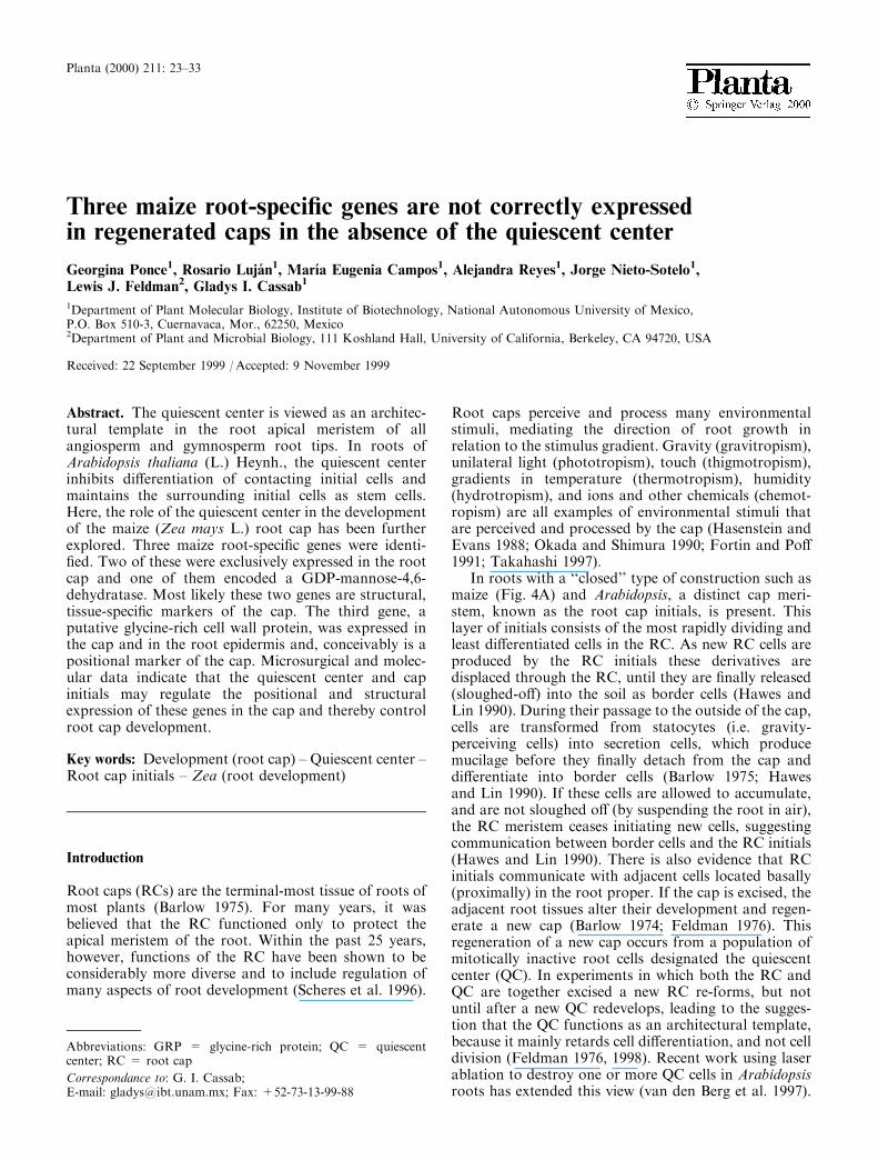
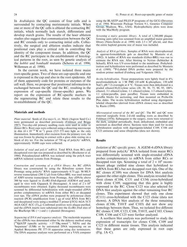
Abstract. The quiescent center is viewed as an architec- tural template in the root apical meristem of all angiosperm and gymnosperm root tips. In roots of Arabidopsis thaliana (L.) Heynh., the quiescent center inhibits dierentiation of contacting initial cells and maintains the surrounding initial cells as stem cells. Here, the role of the quiescent center in the development of the maize (Zea mays L.) root cap has been further explored. Three maize root-specific genes were identi- fied. Two of these were exclusively expressed in the root cap and one of them encoded a GDP-mannose-4,6- dehydratase. Most likely these two genes are structural, tissue-specific markers of the cap. The third gene, a putative glycine-rich cell wall protein, was expressed in the cap and in the root epidermis and, conceivably is a positional marker of the cap. Microsurgical and molec- ular data indicate that the quiescent center and cap initials may regulate the positional and structural expression of these genes in the cap and thereby control root cap development. Key words: Development (root cap) – Quiescent center – Root cap initials – Zea (root development) Introduction Root caps (RCs) are the terminal-most tissue of roots of most plants (Barlow 1975). For many years, it was believed that the RC functioned only to protect the apical meristem of the root. Within the past 25 years, however, functions of the RC have been shown to be considerably more diverse and to include regulation of many aspects of root development (Scheres et al. 1996). Root caps perceive and process many environmental stimuli, mediating the direction of root growth in relation to the stimulus gradient. Gravity (gravitropism), unilateral light (phototropism), touch (thigmotropism), gradients in temperature (thermotropism), humidity (hydrotropism), and ions and other chemicals (chemot- ropism) are all examples of environmental stimuli that are perceived and processed by the cap (Hasenstein and Evans 1988; Okada and Shimura 1990; Fortin and Po 1991; Takahashi 1997). In roots with a ‘‘closed’’ type of construction such as maize (Fig. 4A) and Arabidopsis, a distinct cap meri- stem, known as the root cap initials, is present. This layer of initials consists of the most rapidly dividing and least dierentiated cells in the RC. As new RC cells are produced by the RC initials these derivatives are displaced through the RC, until they are finally released (sloughed-o) into the soil as border cells (Hawes and Lin 1990). During their passage to the outside of the cap, cells are transformed from statocytes (i.e. gravity- perceiving cells) into secretion cells, which produce mucilage before they finally detach from the cap and dierentiate into border cells (Barlow 1975; Hawes and Lin 1990). If these cells are allowed to accumulate, and are not sloughed o (by suspending the root in air), the RC meristem ceases initiating new cells, suggesting communication between border cells and the RC initials (Hawes and Lin 1990). There is also evidence that RC initials communicate with adjacent cells located basally (proximally) in the root proper. If the cap is excised, the adjacent root tissues alter their development and regen- erate a new cap (Barlow 1974; Feldman 1976). This regeneration of a new cap occurs from a population of mitotically inactive root cells designated the quiescent center (QC). In experiments in which both the RC and QC are together excised a new RC re-forms, but not until after a new QC redevelops, leading to the sugges- tion that the QC functions as an architectural template, because it mainly retards cell dierentiation, and not cell division (Feldman 1976, 1998). Recent work using laser ablation to destroy one or more QC cells in Arabidopsis roots has extended this view (van den Berg et al. 1997). Abbreviations: GRP = glycine-rich protein; QC = quiescent center; RC = root cap Correspondance to: G. I. Cassab; E-mail: [email protected]; Fax: +52-73-13-99-88 Planta (2000) 211: 23–33 Three maize root-specific genes are not correctly expressed in regenerated caps in the absence of the quiescent center Georgina Ponce 1 , Rosario Luja´n 1 , Marı´a Eugenia Campos 1 , Alejandra Reyes 1 , Jorge Nieto-Sotelo 1 , Lewis J. Feldman 2 , Gladys I. Cassab 1 1 Department of Plant Molecular Biology, Institute of Biotechnology, National Autonomous University of Mexico, P.O. Box 510-3, Cuernavaca, Mor., 62250, Mexico 2 Department of Plant and Microbial Biology, 111 Koshland Hall, University of California, Berkeley, CA 94720, USA Received: 22 September 1999 / Accepted: 9 November 1999
-
Upload
independent -
Category
Documents
-
view
2 -
download
0
Transcript of Three maize root-specific genes are not correctly expressed in regenerated caps in the absence of...
Abstract. The quiescent center is viewed as an architec-tural template in the root apical meristem of allangiosperm and gymnosperm root tips. In roots ofArabidopsis thaliana (L.) Heynh., the quiescent centerinhibits di�erentiation of contacting initial cells andmaintains the surrounding initial cells as stem cells.Here, the role of the quiescent center in the developmentof the maize (Zea mays L.) root cap has been furtherexplored. Three maize root-speci®c genes were identi-®ed. Two of these were exclusively expressed in the rootcap and one of them encoded a GDP-mannose-4,6-dehydratase. Most likely these two genes are structural,tissue-speci®c markers of the cap. The third gene, aputative glycine-rich cell wall protein, was expressed inthe cap and in the root epidermis and, conceivably is apositional marker of the cap. Microsurgical and molec-ular data indicate that the quiescent center and capinitials may regulate the positional and structuralexpression of these genes in the cap and thereby controlroot cap development.
Key words: Development (root cap) ± Quiescent center ±Root cap initials ± Zea (root development)
Introduction
Root caps (RCs) are the terminal-most tissue of roots ofmost plants (Barlow 1975). For many years, it wasbelieved that the RC functioned only to protect theapical meristem of the root. Within the past 25 years,however, functions of the RC have been shown to beconsiderably more diverse and to include regulation ofmany aspects of root development (Scheres et al. 1996).
Root caps perceive and process many environmentalstimuli, mediating the direction of root growth inrelation to the stimulus gradient. Gravity (gravitropism),unilateral light (phototropism), touch (thigmotropism),gradients in temperature (thermotropism), humidity(hydrotropism), and ions and other chemicals (chemot-ropism) are all examples of environmental stimuli thatare perceived and processed by the cap (Hasenstein andEvans 1988; Okada and Shimura 1990; Fortin and Po�1991; Takahashi 1997).
In roots with a ``closed'' type of construction such asmaize (Fig. 4A) and Arabidopsis, a distinct cap meri-stem, known as the root cap initials, is present. Thislayer of initials consists of the most rapidly dividing andleast di�erentiated cells in the RC. As new RC cells areproduced by the RC initials these derivatives aredisplaced through the RC, until they are ®nally released(sloughed-o�) into the soil as border cells (Hawes andLin 1990). During their passage to the outside of the cap,cells are transformed from statocytes (i.e. gravity-perceiving cells) into secretion cells, which producemucilage before they ®nally detach from the cap anddi�erentiate into border cells (Barlow 1975; Hawesand Lin 1990). If these cells are allowed to accumulate,and are not sloughed o� (by suspending the root in air),the RC meristem ceases initiating new cells, suggestingcommunication between border cells and the RC initials(Hawes and Lin 1990). There is also evidence that RCinitials communicate with adjacent cells located basally(proximally) in the root proper. If the cap is excised, theadjacent root tissues alter their development and regen-erate a new cap (Barlow 1974; Feldman 1976). Thisregeneration of a new cap occurs from a population ofmitotically inactive root cells designated the quiescentcenter (QC). In experiments in which both the RC andQC are together excised a new RC re-forms, but notuntil after a new QC redevelops, leading to the sugges-tion that the QC functions as an architectural template,because it mainly retards cell di�erentiation, and not celldivision (Feldman 1976, 1998). Recent work using laserablation to destroy one or more QC cells in Arabidopsisroots has extended this view (van den Berg et al. 1997).
Abbreviations: GRP = glycine-rich protein; QC = quiescentcenter; RC = root cap
Correspondance to: G. I. Cassab;E-mail: [email protected]; Fax: +52-73-13-99-88
Planta (2000) 211: 23±33
Three maize root-speci®c genes are not correctly expressedin regenerated caps in the absence of the quiescent center
Georgina Ponce1, Rosario Luja n1, Marõ a Eugenia Campos1, Alejandra Reyes1, Jorge Nieto-Sotelo1,Lewis J. Feldman2, Gladys I. Cassab1
1Department of Plant Molecular Biology, Institute of Biotechnology, National Autonomous University of Mexico,P.O. Box 510-3, Cuernavaca, Mor., 62250, Mexico2Department of Plant and Microbial Biology, 111 Koshland Hall, University of California, Berkeley, CA 94720, USA
Received: 22 September 1999 /Accepted: 9 November 1999
In Arabidopsis the QC consists of four cells and issurrounded by contacting meristematic initials. Whenone or more of the QC cells is ablated the contacting RCinitials, which normally lack starch, di�erentiate anddevelop starch grains. The results of the laser ablationstudies also suggest that communication between the QCand RC extends only to contacting initial cells. Collec-tively, the surgical and ablation studies indicate thatpositional cues play a critical role in controlling theidentity of the component tissues. In addition, the QCand the RC participate in the establishment of histolog-ical patterns in the root, as seen by genetic analysis ofthe hobbit and bombadil mutants (Scheres et al. 1996;Willemsen et al. 1998).
In the current work, we have isolated three maizeroot-speci®c genes. Two of these are cap-speci®c and oneis expressed in the cap and also in the root epidermis. Allof them apparently code for proteins or enzymes of thecell wall. Here, we propose that positional information isexchanged between the QC and the RC, resulting in theexpression of cap-speci®c (tissue-speci®c) genes. Wereport on the expression of RC-speci®c markers inthe regenerating RC and relate these results to there-establishment of the QC.
Materials and methods
Plant material. Seeds of Zea mays L. cv. Merit (Asgrow Seed Co.)were germinated as described previously (Feldman and Briggs1987). Two-day-old primary seedling roots were used in order todissect RCs. Root caps were collected using a dissecting microscopein dim (4 ´ 10)14 W m)2) green (515±575 nm) light as the onlyillumination. Immediately after excision from the primary root, thecap was frozen by placing in contact with a glass slide resting on ablock of dry ice. For the isolation of 750 ng of poly(A)+mRNA,approximately 10,000 caps were collected.
Isolation of total and polyA+mRNA. Total RNA from RCs anddecapitated root tips was prepared as described by (Rochester et al.1986). Polyadenylated mRNA was isolated using the polyA tractmRNA isolation systems from Promega.
Construction and screening of a cDNA library. An RC cDNAlibrary was constructed by using the Riboclone system fromPromega using poly(A)+RNA (approximately 0.75 lg). M-MLVreverse transcriptase (200 U/ll) from Gibco-BRL was used insteadof AMV-reverse transcriptase from Promega. Any cDNAs above300 bp were ligated to EcoRI-XbaI-digested kGEM-4 and pack-aged in vitro with Gigapack II (Stratagene). A total of 2 ´ 106
recombinants were obtained. Eighty thousand recombinants werescreened by di�erential hybridization with single-stranded cDNAprobes complementary to mRNA from either RCs or decappedroot tips. The cDNA probes were prepared by polymerase chainreaction (PCR) ampli®cation from 1 lg of total RNA from RCsand decapitated roots using a modi®ed T primer [(TCG AGA TCTAGA GGT ACC (T)18]. Con®rmed positives were plaque-puri®ed,and the cDNA inserts were subcloned into the EcoRI-XbaI sites ofthe BlueScript SK (+) and ()) plasmid (Stratagene).
Sequencing of DNA and sequence analysis. The nucleotide sequenceof the cDNAs was determined either by the dideoxy method using[35S]dATP (speci®c activity: >37 TBq mmol)1; NEN) and the`Sequenase' (USB) or by automated DNA sequencing on anApplied Biosystems PE 377-18 apparatus using dye terminators.The DNA sequence analyses were performed on a VAX computer
using the BLASTP and PILEUP programs of the GCG (Devereuxet al. 1984; Wisconsin Package Version 9.1, Genetics ComputerGroup, Madison, Wis., USA). Hydrophobicity plots were donewith the MacMolly program.
Screening a maize genomic library. A total of 1,380,000 plaque-forming units (pfu) was screened from an ampli®ed maize genomiclibrary (Nieto-Sotelo et al. 1999) with 2.5 ´ 109 pfu/ml, and thusthe entire haploid genome size of maize was included.
Analysis of RNA gel blots. Samples of RNA were electrophoresedin agarose-formaldehyde gels as described by Maniatis et al.(1982). A ladder of RNA size standards (Gibco-BRL) was run toestimate the RNA size. After blotting to Nytran (Schleicher &Schuell), RNA was UV-cross-linked to the membrane. Prehybrid-ization and hybridization were performed as described by Maniatiset al. (1989). 32P-Labeled DNA probes were prepared by therandom primer method (Feinberg and Volgestein 1983).
In-situ hybridization. Tissue preparations were lightly ®xed in 4%paraformaldehyde in 1 M PBS (10 mM K-phosphate, 150 mMNaCl, pH 7.2) overnight at 4 °C. Tissues were then dehydrated in agraded ethanol/H2O/xylene series (20, 30, 50, 75, 90, 95, 100%ethanol, 3:1 ethanol:xylene, 1:1 ethanol:xylene, 1:3 ethanol:xylene,1:1 xylene:para�n, pure para�n). Tissues were embedded inpara�n blocks for sectioning, and then mounted on Probe-OnPlus slides. The in-situ hybridization method using digoxigenin-labeled riboprobes (derived from cDNA clones) was as describedby Ruzin (1999).
Microsurgical removal of the RC and QC. The RC and QC wereremoved surgically from 2-d-old seedling roots as described byFeldman (1976). Subsequent to the surgery, roots were returned tothe dark, sampled periodically, ®xed, dehydrated, embedded andsectioned as described above. Sections were then used for in-situhybridization analysis with digoxigenin-labeled C109, C106 andC123 antisense and sense riboprobes (data not shown).
Results
Isolation of RC-speci®c genes. A kGEM-4 cDNA libraryprepared from poly(A)+RNA isolated from maize RCswas di�erentially screened with single-stranded cDNAprobes complementary to mRNA from either RCs ordecapped root tips. Screening a total of 2 ´ 106 recom-binant phage yielded nine clones that hybridized tomRNA from RCs but not decapped root tips. One of theRC clones (C109) was chosen for DNA blot analysisagainst the other eight clones. This analysis revealed thatthree clones (C104, C131 and C134) cross-hybridizedwith clone C109, suggesting that this gene is highlyexpressed in the RC. Clone C123 was also selected forDNA blot analysis against the other remaining four RCclones. This experiment showed that only one clone(C111) cross-hybridized with the C123 clone (data notshown). A DNA blot analysis of the three remainingclones (C106, T101T and C103) did not show anyhomology between them. Thus, we isolated ®ve di�erentRC clones: C109, C123, C106, T101T, and C103. ClonesC109, C106 and C123 were further analyzed.
A northern blot analysis was performed to study theexpression of transcripts for clones C106, C109 andC123 in di�erent maize tissues. This analysis indicatedthat these genes are only expressed in root caps(Fig. 1A±C).
24 G. Ponce et al.: Root-cap-speci®c genes of maize

Sequence analysis of root cap clones C109, C106 andC123. C109 is a partial cDNA clone of 982 bp in lengthwith two possible open reading frames (GenBankaccesion number: AF134580). The longest open readingframe predicts a protein with 31% identity to both aputative glycosylphosphatidylinositol-anchored arabi-nogalactan protein (AGP) and to an epithelium-associ-ated human mucin JUL7 (Dufosse et al. 1993). Thispredicted protein is highly hydrophilic, like mucin JUL7.However, it also includes a C-terminal hydrophobicregion as seen in AGPPc1 (Chen et al. 1994) that mayserve as a membrane anchor. The shortest open readingframe in clone C109 is identical to a novel plant proteinrecently reported in maize (Matsuyama et al. 1999a).This predicted protein is mostly hydrophobic with aregion of regularly-spaced cysteine residues.
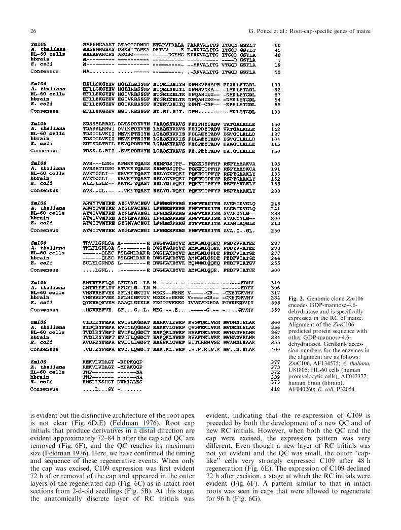
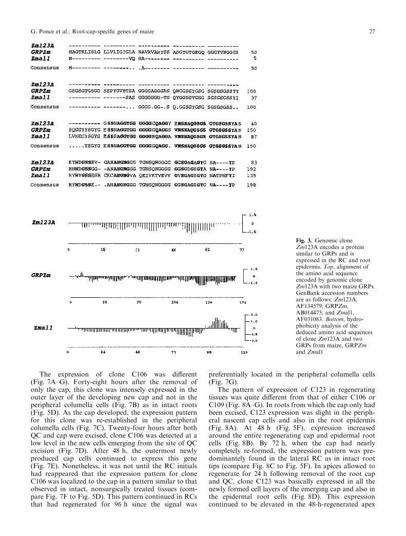
The cDNAs for clones C106 and C123 are also partialand are 452 bp and 428 bp in length, respectively. Sinceall three cDNAs are smaller than their correspondingmRNAs (see northern analysis, Fig. 1), we decided toisolate their related genomic clones. We obtained sixgenomic clones related to cDNA clone C109 (Zm109),one for C106 (Zm106), and two for C123 (Zm123A andZm123B). The predicted amino acid sequence of cloneZmC106 presents a high overall identity to the GDP-D-mannose-4,6-dehydratase of Arabidopsis (77%), Esc-herichia coli (57%), HL-60 cells (55%), and human brain(54%) (Fig. 2). This enzyme catalyzes the ®rst step in thede-novo synthesis of GDP-L-fucose (Bonin et al. 1997).Clone Zm123A codes for a putative glycine-rich cell wallprotein and is highly similar (89% to GRPZm, and 68%to Zmal1, respectively) to two glycine-rich proteins(GRPs) recently reported from maize (Matsuyama et al.1999b) (Fig. 3A). Hydropathy analysis of the threemaize GRPs shows that they are highly hydrophilic butthat only Zmal1 contains an hydrophobic region in theC-terminal (Fig. 3B). Glycine-rich proteins are a class ofstructural wall proteins that seem to play important
roles in the development of the vascular tissue (Cassab1998). Their presence in a non vascular tissue such as theRC suggests that GRPs also participate in otherdevelopmental processes.
Localization of C106, C109 and C123 transcripts in theRC. To determine the cellular localization of thetranscripts for the three cap genes within the root,in-situ hybridization experiments were performed. ThemRNA for clone C109 was restricted to the outer layersof the RC that include 40% of the cap cells of maizeroots (Moore 1984) (Figs. 4B, 5B). These layers containthe mucilage-secreting cells. In addition, the signal wasvery strong and thus this gene may be expressed in largequantities. C109 mRNA was also present in thedetached cells. The mRNA for clone C106 was primarilydetected in the peripheral columella cells (Figs. 4B, 5D).Its expression declined in the mucilage-secreting cells. Bycomparing the signals of C109 and C106 mRNAs intransverse RC sections, we observed that both signalsoverlap in the two outer cell layers of the cap (data notshown). The mRNA for clone C123 was observed solelyin the region that corresponds in dicotyledonous roots tothe lateral RC but it was also present in the rootepidermis (Figs. 4B, 5F). However, the signal disap-peared in epidermal cells proximal to the root meristem.In contrast, transcripts for clone C123 could not bedetected by northern analysis in roots without caps(Fig. 1C).
Expression of RC genes in developing caps after micro-surgery of the RC and QC. Thus far we have shown thatseveral genes are expressed preferentially, if not exclu-sively in the RC, and that the expression pattern isdistinct for each of the three genes. We wished to explorethe timing of the expression of these genes as the RCdevelops and whether this expression pattern is relatedto the development of the QC. For this we used asurgical approach to remove either the cap alone, or thecap plus the QC, and then followed the regeneration ofthe excised tissues. Using this approach we analyzedwhen and in which tissues these genes were expressed.When the cap alone is removed, QC cells at the distal(now exposed) surface of the root di�erentiate starchgrains within about 12 h of the surgery, coincident withthe ability of the root to again perceive gravity. Shortlythereafter, cells of the QC begin dividing, initiating anew RC and new RC initials. Forty-eight hours afterthis surgery the QC has re-formed to its pre-decappingdimensions. In the case of removal of both the RC andQC, regeneration of a new RC also occurs, but there isnot any histological evidence of RC regeneration until anew QC begins to re-form (Feldman 1976). In rootsfrom which only the cap has been excised, a completenew RC regenerates from reprogrammed distal QC cellsafter 72 h (Barlow 1974) (Fig. 6A±C). However, in rootsfrom which both the QC and the cap have beenremoved, regeneration of the damaged apex arises fromthe region of tissue bordering on the excised QC (theproximal root meristem) (Feldman 1976). Thirty-six to48 hours after surgery of the QC, a small but discrete QC
Fig. 1A±D. Accumulation of mRNA of RC clones C106, C109 andC123 in di�erent maize tissues. Lanes 1, 10 lg of total RNA fromdecapped primary roots; lanes 2, 10 lg of total RNA from root caps;lanes 3, 10 lg of total RNA from young leaves. AAutoradiography ofa gel blot using cDNA clone C106 as a probe. BAutoradiography of agel blot using cDNA clone C109 as a probe. C Autoradiography ofa gel blot using cDNA clone C123 as a probe. D Ethidium bromidestaining of the gel used for RNA blotting, which indicates equalloading of total RNA from decapped roots, root caps and leaves fromblots shown in A±C
G. Ponce et al.: Root-cap-speci®c genes of maize 25
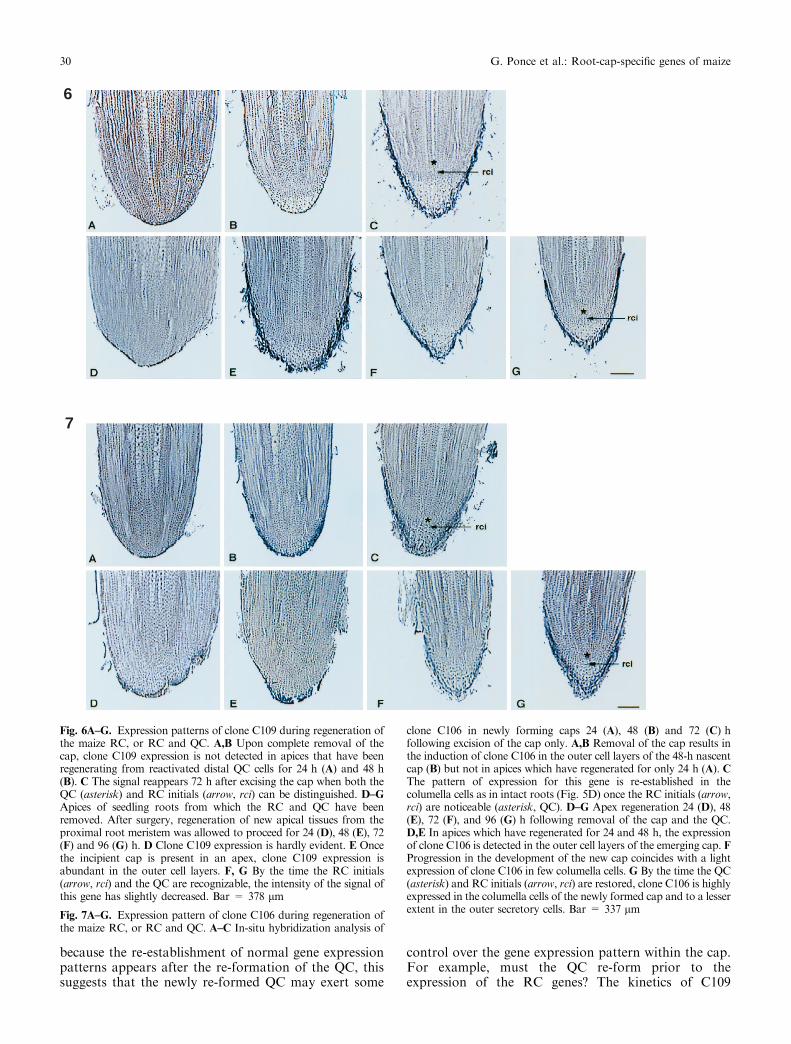
is evident but the distinctive architecture of the root apexis not clear (Fig. 6D,E) (Feldman 1976). Root capinitials that produce derivatives in a distal direction areevident approximately 72±84 h after the cap and QC areremoved (Fig. 6F), and the QC reaches its maximumsize (Feldman 1976). Here, we have con®rmed the timingand sequence of these regenerative events. When onlythe cap was excised, C109 expression was ®rst evident72 h after removal of the cap and appeared in the outerlayers of the regenerated cap (Fig. 6C) as in intact rootsections from 2-d-old seedlings (Fig. 5B). At this stage,the anatomically discrete layer of RC initials was
evident, indicating that the re-expression of C109 ispreceded by both the development of a new QC and ofnew RC initials. However, when both the QC and thecap were excised, the expression pattern was verydi�erent. Even though a new layer of RC initials wasnot yet evident and the QC was small, the outer ``cap-like'' cells very strongly expressed C109 after 48 hregeneration (Fig. 6E). The expression of C109 declined72 h after excision, a stage at which the RC initials wereevident (Fig. 6F). A pattern similar to that in intactroots was seen in caps that were allowed to regeneratefor 96 h (Fig. 6G).
Fig. 2. Genomic clone Zm106encodes GDP-mannose-4,6-dehydratase and is speci®callyexpressed in the RC of maize.Alignment of the ZmC106predicted protein sequence withother GDP-mannose-4,6-dehydratases. GenBank acces-sion numbers for the enzymes inthe alignment are as follows:ZmC106, AF134575; A. thaliana,U81805; HL-60 cells (humanpromyelocytic cells), AF042377;human brain (hbrain),AF040260; E. coli, P32054
26 G. Ponce et al.: Root-cap-speci®c genes of maize
The expression of clone C106 was di�erent(Fig. 7A±G). Forty-eight hours after the removal ofonly the cap, this clone was intensely expressed in theouter layer of the developing new cap and not in theperipheral columella cells (Fig. 7B) as in intact roots(Fig. 5D). As the cap developed, the expression patternfor this clone was re-established in the peripheralcolumella cells (Fig. 7C). Twenty-four hours after bothQC and cap were excised, clone C106 was detected at alow level in the new cells emerging from the site of QCexcision (Fig. 7D). After 48 h, the outermost newlyproduced cap cells continued to express this gene(Fig. 7E). Nonetheless, it was not until the RC initialshad reappeared that the expression pattern for cloneC106 was localized to the cap in a pattern similar to thatobserved in intact, nonsurgically treated tissues (com-pare Fig. 7F to Fig. 5D). This pattern continued in RCsthat had regenerated for 96 h since the signal was
preferentially located in the peripheral columella cells(Fig. 7G).
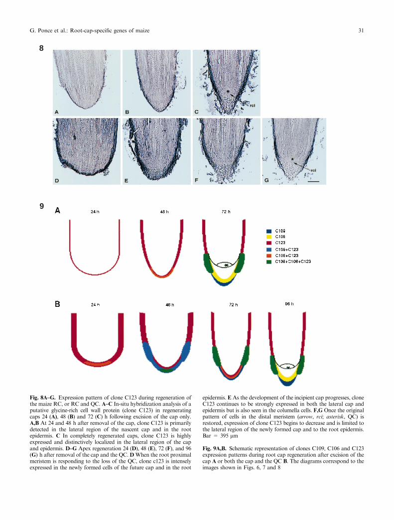
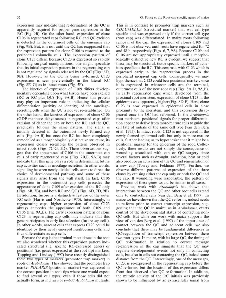
The pattern of expression of C123 in regeneratingtissues was quite di�erent from that of either C106 orC109 (Fig. 8A±G). In roots from which the cap only hadbeen excised, C123 expression was slight in the periph-eral nascent cap cells and also in the root epidermis(Fig. 8A). At 48 h (Fig. 5F), expression increasedaround the entire regenerating cap and epidermal rootcells (Fig. 8B). By 72 h, when the cap had nearlycompletely re-formed, the expression pattern was pre-dominantely found in the lateral RC as in intact roottips (compare Fig. 8C to Fig. 5F). In apices allowed toregenerate for 24 h following removal of the root capand QC, clone C123 was basically expressed in all thenewly formed cell layers of the emerging cap and also inthe epidermal root cells (Fig. 8D). This expressioncontinued to be elevated in the 48-h-regenerated apex
Fig. 3. Genomic cloneZm123A encodes a proteinsimilar to GRPs and isexpressed in the RC and rootepidermis. Top, alignment ofthe amino acid sequenceencoded by genomic cloneZm123A with two maize GRPs.GenBank accession numbersare as follows: Zm123A,AF134579; GRPZm,AB014475; and Zmal1,AF031083. Bottom, hydro-phobicity analysis of thededuced amino acid sequencesof clone Zm123A and twoGRPs from maize, GRPZmand Zmal1
G. Ponce et al.: Root-cap-speci®c genes of maize 27

but was now more accentuated in the lateral region ofthe regenerating root cap than in the central cap(Fig. 8E). However, by the time the regeneration hadproceeded for 72 h, the expression of clone C123 had
decreased in both the central and lateral cap (Fig. 8F).By 96 h after surgery, at a time when the new RC initialsare anatomically distinct and the QC fully re-estab-lished, the expression pattern for this clone was con®ned
4
5
28 G. Ponce et al.: Root-cap-speci®c genes of maize
to the lateral cap cells and to the root epidermis(Fig. 8G), similar to intact roots (Fig. 5F).
Discussion
The isolation and characterization of genes uniquelyexpressed in the RC are the ®rst steps for understandingthe di�erent activities known to occur in the cap.Further, the RC genes which we have here characterizedare two putative cell wall proteins and one cell wallenzyme. Cell walls contain proteins and polysaccharidesable to condition the development of a plant (Cassab1998), and thus the characterization of cap-speci®c cellwall proteins may be an important step towards under-standing how morphogenesis in the cap is regulated.
Structure of the C109, C106 and C123 gene prod-ucts. One of the predicted proteins of the RC generepresented in clone C109 (ORF3) resembles the humantracheobronchial mucin (Dufosse et al. 1993) and aputative glycosylphosphatidylinositol-anchored classicalarabinogalactan protein (Youl et al. 1998). Arabinoga-lactan proteins have been found as a component of maizeRC mucilage (Bacic et al. 1986). The mucilage is theprimary site for colonization of the root by microbialsymbionts and pathogens. Alternatively, ORF2 in cloneC109 predicts a protein that is identical to ZmRCP2, arecently reported novel plant protein, speci®cally ex-pressed in the root cap of maize (Matsuyama et al.1999a). This protein contains six conserved cysteineresidues in three pairs of Cys-X-X-X-Cys, and is similarto several proteins encoded in Arabidopsis expressedsequence tags and genomic sequences, and to a somatic-embryogenesis-associated cDNA from Picea glauca(Dong and Dunstan 1999; Matsuyama et al. 1999a).Since the mRNA for this gene is highly abundant in thesecretory cells of the RC (Figs. 4B, 5D), one can suggestthat the protein product is part of the mucilage secretedby the RC. In the mucilage, it may contribute to thephysicochemical properties of this material and/or, itmay interact with the microorganisms of the rhizosphere.
The other RC gene, clone C106, encodes GDP-mannose-4,6-dehydratase, which catalyzes the ®rst stepin the de-novo synthesis of GDP-fucose (Bonin et al.1997). Fucose is a major component of the mucilagesecreted by the RC of maize and is a distinctive featureof the particular di�erentiation state of the outer RCcells (Harris and Northcote 1970). In previous studies inwhich roots regenerating a cap were fed with [3H]L-fucose, the autoradiographs showed that the label wasconcentrated over the cell walls immediately distal to theRC initials and not over the outermost mucilage-synthesizing cells as anticipated (Barlow 1974). ThemRNA for the fucose-synthesizing enzyme was primar-ily observed in the periphery of the central cap (Figs. 4B,5D), con®rming this observation.
Finally, clone C123 encodes for a putative cell wallGRP. Glycine-rich proteins are structural wall proteinsof unknown function, and their location in the lateralRC and epidermis suggests a new possible functionin root development (Figs. 4B, 5F). In monocots, thelateral RC does not originate independently from the restof the RC. During embryogenesis in monocots, the RCseems to organize as a whole, and thus the cap is not acomposite of cells having two di�erent origins (vonGuttenberg 1968). This is not the case with many dicots,in which the peripheral and columella cells have di�erentorigins during embryogenesis. The columella portion, asfor example in Arabidopsis, arises from cells notconnected to the dermatogen but to the hypophysis,whereas the side (¯ank) portions of the cap have originsin dermatogen-associated cells (Dolan et al. 1993). Thus,in Arabidopsis, the epidermis and lateral root cap cellshave a common origin. Interestingly, in maize, C123mRNA was labelled in both the lateral cap andepidermis, apparently indicating a distinction betweenthe columella and the lateral RC cells. In addition, thislabelling pattern presumably also indicates a similaritybetween the lateral RC and the epidermis.
Expression of C109, C106 and C123 genes in regeneratedcaps after microsurgery. We used a surgical approach toremove either the cap alone, or the cap plus the QC, andthen followed the regeneration of the excised tissues inorder to examine the questions of when and where thethree RC-speci®c genes were expressed. Our long-termgoal is to understand the spatial di�erentiation anddevelopment of the many activities known to occur inthe cap. By monitoring regeneration following removalof the cap only, or the cap plus the QC we have beenable to show that as the cap is re-formed its normal geneexpression activities occur in an atypical pattern(Fig. 9A,B). Even without knowing the exact roles ofthese gene products in RC function, we can neverthelesssuggest that the onset of these activities may be relatedto a recovering of functions by the cap (e.g. gravityperception, mucilage secretion, etc.). In this regard, thesequential regaining of cap gene activities (e.g. C123activities appear ®rst, followed in time by C106 andC109 in decapped roots) may suggest that subsequentlater-expressed genes may be regulated by the productsof the genes expressed earlier (Fig. 9A,B). Moreover,
Fig. 4A,B. Schematic representation of the maize root apex. A Thedi�erent constituents in the root tip. B The expression pattern of twoRC-speci®c genes (clones C109 and C106) and one RC and epidermisgene (clone C123). Clone C109 is expressed in the outer cap made ofsecretory cells. Clone C106 is expressed in the periphery of the centralcap made of columella cells. Clone C123 is expressed in the lateral capand root epidermis
Fig. 5A±F. Di�erential expression of three maize genes in the maizeroot apex. In-situ hybridization analysis of three clones in mediansections of 2-d-old primary maize roots (sc, secretory cell; c, columellacell; lc, lateral cap cells; e, epidermis). A,B Clone C109 expression isonly observed in the secretory cells of the RC, with antisense (B) butnot sense (A) probes. C,D GDP-D-mannose-4,6-dehydratase (cloneC106) mRNA is uniquely present in the peripheral columella cells ofthe cap, using antisense (D) but not sense (C) probes. E,F The GRP(clone C123) mRNA is distinctively shown in the lateral cap and in theroot epidermal cells, using antisense (F) but not sense probes(E). Bar = 385 lm
b
G. Ponce et al.: Root-cap-speci®c genes of maize 29
because the re-establishment of normal gene expressionpatterns appears after the re-formation of the QC, thissuggests that the newly re-formed QC may exert some
control over the gene expression pattern within the cap.For example, must the QC re-form prior to theexpression of the RC genes? The kinetics of C109
6
7
Fig. 6A±G. Expression patterns of clone C109 during regeneration ofthe maize RC, or RC and QC. A,B Upon complete removal of thecap, clone C109 expression is not detected in apices that have beenregenerating from reactivated distal QC cells for 24 h (A) and 48 h(B). C The signal reappears 72 h after excising the cap when both theQC (asterisk) and RC initials (arrow, rci) can be distinguished. D±GApices of seedling roots from which the RC and QC have beenremoved. After surgery, regeneration of new apical tissues from theproximal root meristem was allowed to proceed for 24 (D), 48 (E), 72(F) and 96 (G) h. D Clone C109 expression is hardly evident. E Oncethe incipient cap is present in an apex, clone C109 expression isabundant in the outer cell layers. F, G By the time the RC initials(arrow, rci) and the QC are recognizable, the intensity of the signal ofthis gene has slightly decreased. Bar = 378 lm
Fig. 7A±G. Expression pattern of clone C106 during regeneration ofthe maize RC, or RC and QC. A±C In-situ hybridization analysis of
clone C106 in newly forming caps 24 (A), 48 (B) and 72 (C) hfollowing excision of the cap only. A,B Removal of the cap results inthe induction of clone C106 in the outer cell layers of the 48-h nascentcap (B) but not in apices which have regenerated for only 24 h (A). CThe pattern of expression for this gene is re-established in thecolumella cells as in intact roots (Fig. 5D) once the RC initials (arrow,rci) are noticeable (asterisk, QC). D±G Apex regeneration 24 (D), 48(E), 72 (F), and 96 (G) h following removal of the cap and the QC.D,E In apices which have regenerated for 24 and 48 h, the expressionof clone C106 is detected in the outer cell layers of the emerging cap. FProgression in the development of the new cap coincides with a lightexpression of clone C106 in few columella cells. G By the time the QC(asterisk) and RC initials (arrow, rci) are restored, clone C106 is highlyexpressed in the columella cells of the newly formed cap and to a lesserextent in the outer secretory cells. Bar = 337 lm
30 G. Ponce et al.: Root-cap-speci®c genes of maize
8
9
Fig. 8A±G. Expression pattern of clone C123 during regeneration ofthe maize RC, or RC and QC. A±C In-situ hybridization analysis of aputative glycine-rich cell wall protein (clone C123) in regeneratingcaps 24 (A), 48 (B) and 72 (C) h following excision of the cap only.A,B At 24 and 48 h after removal of the cap, clone C123 is primarilydetected in the lateral region of the nascent cap and in the rootepidermis. C In completely regenerated caps, clone C123 is highlyexpressed and distinctively localized in the lateral region of the capand epidermis. D±G Apex regeneration 24 (D), 48 (E), 72 (F), and 96(G) h after removal of the cap and the QC.DWhen the root proximalmeristem is responding to the loss of the QC, clone c123 is intenselyexpressed in the newly formed cells of the future cap and in the root
epidermis. E As the development of the incipient cap progresses, cloneC123 continues to be strongly expressed in both the lateral cap andepidermis but is also seen in the columella cells. F,G Once the originalpattern of cells in the distal meristem (arrow, rci; asterisk, QC) isrestored, expression of clone C123 begins to decrease and is limited tothe lateral region of the newly formed cap and to the root epidermis.Bar = 395 lm
Fig. 9A,B. Schematic representation of clones C109, C106 and C123expression patterns during root cap regeneration after excision of thecap A or both the cap and the QC B. The diagrams correspond to theimages shown in Figs. 6, 7 and 8
G. Ponce et al.: Root-cap-speci®c genes of maize 31
expression may indicate that re-formation of the QC isapparently required for proper gene expression in theRC (Fig. 9B). On the other hand, expression of cloneC106 in regenerated caps following RC and QC excisionis detected in the outermost cells of the emerging cap(Fig. 9B). But, it is not until the QC has reappeared thatthe expression pattern for clone C106 is restored to theperipheral columella cells. The expression pattern ofclone C123 di�ers. Because C123 is expressed so rapidlyfollowing surgical manipulations, one might speculatethat its initial expression pattern in the regenerating capis not regulated by signals released by the QC (Figs. 8D,9B). However, as the QC is being re-formed, C123expression is seen preferentially in the lateral RC(Fig. 8E±G) as in intact roots (Fig. 5F).
The kinetics of expression of C109 di�ers develop-mentally depending upon what tissues have been excised(RC or RC plus QC) (Fig. 9A,B). Hence, this proteinmay play an important role in indicating the cellulardi�erentiation (activity or identity) of the mucilage-secreting cells when the QC is not yet re-established. Onthe other hand, the kinetics of expression of clone C106(GDP-mannose dehydratase) in regenerated caps afterexcision of either the cap or both the RC and the QC,practically follows the same pattern. The signal isinitially detected in the outermost newly formed capcells (Fig. 9A,B) but once the RC has been completelyremodelled as a morphologically distinctive structure, itsexpression closely resembles the pattern observed inintact roots (Figs. 7C,G, 5D). These observations sug-gest that the appearance of C106 in the outermost capcells of early regenerated caps (Figs. 7B,E, 9A,B) mayindicate that this gene plays a role in determining futurecap activities such as mucilage secretion. In other words,signalling between newly divided cells seems to direct thechoice of developmental pathway and some of thesesignals may arise from the wall itself. In fact, C106expression in the outermost cap cells precedes theappearance of clone C109 after excision of the RC only(Figs. 6B, 7B), and both RC and QC (Figs. 6D, 7D, 9B).In addition, fucose is a distinctive feature of the outerRC cells (Harris and Northcote 1970). Interestingly, inregenerating caps, higher expression of clone C123always antecedes the appearance of both C109 andC106 (Fig. 9A,B). The early expression pattern of cloneC123 in regenerating cap cells may indicate that thisgene participates in early fate selection (future cap cells).In other words, nascent cells that express C123 could beidenti®ed by their newly emerged neighboring cells, andthus di�erentiate as cap cells.
Because the cap is the terminal-most tissue in the rootwe also wondered whether this expression pattern indi-cated structural (i.e. speci®c RC-expressed genes) orpositional (i.e. genes expressed at the root tip) markers.Topping and Lindsey (1997) have recently distinguishedthese two types of markers (promotor trap marker) inroots ofArabidopsis. They showed that the promoter trapmarker POLARIS (positional marker) was expressed inthe correct position in root tips where one would expectto ®nd several cell types, even if those cells did notactually form, as in hydra or emb30 Arabidopsismutants.
This is in contrast to promoter trap markers such asCOLUMELLA (structural marker) that was cell-typespeci®c and was expressed only if the correct cell type(root cap) was di�erentiated. In maize roots followingremoval of the cap, the expression of clones C109 andC106 is not observed until roots have regenerated for 72and 48 h, respectively (Figs. 6, 7, 9A). Because C109 andC106 are not appropriately expressed until a morpho-logically distinctive new RC is evident, we suggest thatthese may be structural, tissue-speci®c markers of activ-ities speci®c to the RC. This contrasts with C123 which isexpressed early in the regeneration process in theperipheral incipient cap cells. Consequently, we mayhypothesize that C123 could be a positional marker, sinceit is expressed in whatever cells are the terminal,outermost cells of the new root cap (Figs. 8A,D, 9A,B).In early regenerated caps which developed from theproximal root meristem, expression of clone C123 in theepidermis was apparently higher (Fig. 8D,E). Here, cloneC123 is now expressed in epidermal cells in closeproximity to the meristem, and this expression disap-peared once the QC had reformed. In the Arabidopsisroot meristem, positional signals for proper di�erentia-tion appear to derive frommore-mature cells to guide thecell fate of initials of the same cell type (van den Berget al. 1995). In intact roots, C123 is not expressed in thenewly formed epidermal cells but only in more-maturecells, further leading us to hypothesize that it might be apositional marker for the epidermis of the root. Collec-tively, these results are not simply the consequence ofwounding associated with cap or QC surgery sinceseveral factors such as drought, radiation, heat or coldalso produce an activation of the QC and regeneration ofa new cap (Torrey and Feldman 1977). Further, weobserve di�erent patterns of expression of the threeclones by excising either the cap only or both the QC andthe cap. If wounding were responsible, the pattern ofexpression of these genes would be similar in all cases.
Previous work with Arabidopsis has shown thatinteractions between the QC and other root cells extendonly to contacting cells (van den Berg et al. 1997). Inmaize we have shown that the QC re-forms, indeed needsto re-form prior to correct transcript expression, sug-gesting that the QC in maize, as in Arabidopsis, exertscontrol of the developmental status of contacting non-QC cells. But while our work with maize supports theview of van den Berg et al. (1997) of the importance ofcontact between the QC and adjacent cells, we alsoconclude that there may be fundamental di�erences inQC-regulation of transcript expression between thesetwo root types. In maize, with its large QC, the timing ofQC re-formation in relation to correct messagere-expression in the cap suggests that the QC mayregulate developmental events not only in contactingcells, but also in cells not contacting the QC, indeed somedistance from the QC. Interestingly, one of the messages,C123, is re-expressed in the regenerating cap before theQC re-forms, but the location of this expression di�ersfrom that observed after QC re-formation. In addition,the mitotic activity of the RC initials was previouslyshown to be in¯uenced by an extracellular signal from
32 G. Ponce et al.: Root-cap-speci®c genes of maize
the cap border cells (Brigham et al. 1998); thus the basisof cross-talk between these two populations of cells hasalready been established. Since the maize root meristempresumably utilizes a di�erent mechanism for controllingcell di�erentiation than Arabidopsis the challenge is nowto identify the QC signals that regulate the tissue-speci®cpattern of gene expression within the RC. It remains tobe seen whether the putative cell wall componentsisolated in this work are implicated in guiding cellulardi�erentiation in the cap. Critical testing of this hypoth-esis will rely on the molecular analysis of the C109, C106and C123 gene activities, and the identi®cation of the QCsignals that control them.
We thank N. Kerk, I. Sussex and S. Gillmor for critical reading ofthe manuscript; S. Ruzin and D. Schichnes from the NSF Center ofPlant Developmental Biology at UC Berkeley for excellent tech-nical assistance with the in-situ hybridization experiments; ReneÂHerna ndez from the Sequencing Facility at IBt-UNAM, and theSequencing Facility at UC Berkeley; and, ®nally Y. Sa nchez, foroutstanding aid with the ®gures. We also acknowledge L. Castrejo nfor collecting root caps. This project was supported by the ConsejoNacional de Ciencia y Tecnologõ a (CONACYT) (Grant No.25186N), and National Science Foundation-CONACYT (GrantNo. E120.508).
References
Bacic A, Moody SF, Clarke AE (1986) Structural analysis ofsecreted root slime from maize (Zea mays L.) Plant Physiol 80:771±777
Barlow PW (1974) Regeneration of the cap of primary roots of Zeamays. New Phytol 73: 937±954
Barlow PW (1975) The root cap. In: Torrey JG, Clarkson DT (eds)The development and function of roots. Academic Press,London, pp 21±54
Bonin PC, Potter I, Vanzin GF, Reiter W-D (1997) TheMUR1 geneof Arabidopsis thaliana encodes an isoform of GDP-D-mannose-4,6-dehydratase, catalyzing the ®rst step in the de novo synthesisof GDP-L-fucose. Proc Natl Acad Sci USA 94: 2085±2090
Brigham LA, Woo H-H, Wen F, Hawes MC (1998) Meristem-speci®c suppression of mitosis and a global switch in geneexpression in the root cap of pea by endogenous signals. PlantPhysiol 118: 1223±1231
Cassab GI (1998) Plant cell wall proteins. Annu Rev Plant PhysiolPlant Mol Biol 49: 281±309
Chen C-G, Pu Z-Y, Moritz RL, Simpson RJ, Bacic A, Clarke AE,Mau S-L (1994) Molecular cloning of a gene encoding anarabinogalactan-protein from pear (Pyrus communis) cell sus-pension culture. Proc Natl Acad Sci USA 91: 10305±10309
Devereux J, Haeberli P, Smithies O (1984) A comprenhensive set ofsequence analysis programs for the VAX. Nucleic Acids Res 12:387±395
Dolan L, Janmaat K, Willemsen V, Linstead P, Poethig S, RobertsK, Scheres B (1993) Cellular organization of the Arabidopsisthaliana root. Development 119: 71±84
Dong J-Z, Dunstan DI (1999) Cloning and characterization of sixembryogenesis-associated cDNAs from somatic embryos ofPicea glauca and their comparative expression during zygoticembryogenesis. Plant Mol Biol 39: 859±864
Dufosse J, Porchet N, Audie JP, Duperat GV, Laine A, SeuningenVI, Marrakchi S, Degand P, Aubert JP (1993) Degenerate 87base pair tandem repeats create hydrophilic/hydrophobicalternating domains in human mucin peptides mapped to11p15. Biochem J 293: 329±337
Feinberg A, Volgestein B (1983) Addendum: a technique forradiolabeling DNA restriction endonuclease fragments to highspeci®c activity. Anal Biochem 132: 6±13
Feldman LJ (1976) The de novo origin of the quiescent center inregenerating root apices of Zea mays. Planta 128: 207±212
Feldman LJ (1998) Not so quiet quiescent centers. Trends Plant Sci3: 80±81
Feldman LJ, Briggs WR (1987) Light-regulated gravitropism inseedling roots of maize. Plant Physiol 83: 241±243
Fortin M-C, Po� KL (1991) Characterization of thermotropism inprimary roots of maize: Dependence on temperature andtemperature gradient, and interaction with gravitropism. Planta184: 410±414
Harris PJ, Northcote DH (1970) Patterns of polysaccharidebiosynthesis in di�erentiating cells of maize root tips. BiochemJ 120: 479±491
Hasenstein KH, Evans ML (1988) E�ects of cations on hormonetransport in primary roots ofZeamays. Plant Physiol 86: 890±894
Hawes MC, Lin HJ (1990) Correlation of pectolytic enzymeactivity with the programmed release of cells from the root capof Pisum sativum. Plant Physiol 94: 1855±1859
Ishikawa H, Evans ML (1990) Electropism of maize roots: role ofthe root cap and relationship with gravitropism. Plant Physiol94: 913±918
Maniatis T, Frisch EF, Sambrook J (1982). Molecular cloning ± alaboratory manual. Cold Spring Harbor Laboratory Press,Cold Spring Harbor, NY
Matsuyama T, Yasumara N, Funakoshi M, Yamada Y, Hashim-oto T (1999a) Maize genes speci®cally expressed in theoutermost cells of root cap. Plant Cell Physiol 40: 469±476
Matsuyama T, Satoh H, Yamada Y, Hashimoto T (1999b) A maizeglycine-rich protein is synthesized in the lateral root cap andaccumulates in the mucilage. Plant Physiol 120: 665±674
Moore R (1984) Cellular volume and tissue portioning in caps ofprimary roots of Zea mays. Am J Bot 71: 1452±1454
Nieto-Sotelo J, Kannan KB, MartõÂ nez LM, Segal C (1999)Characterization of a maize heat-shock protein 101 gene,HSP101, enconding a ClpB/Hsp100 protein homologue. Gene230: 187±195
Okada K, Shimura Y (1990) Reversible root tip rotation inArabidopsis seedlings induced by obstacle-touching stimulus.Science 250: 274±276
Rochester DE, Winter JA, Shah DM (1986) The structure andexpression of maize genes encoding the major heat shockprotein, hsp 70. EMBO J 5: 451±458
Ruzin SE (1999) Plant Microtechnique. Oxford University Press,Oxford and New York
Scheres B, McKhann H, van den Berg C, Willemsen V, WolfenkeltH, de Vrieze G, Weisbeek P (1996) Experimental and geneticanalysis of root development in Arabidopsis thaliana. Plant Soil187: 97±105
Takahashi H (1997) Hydrotropism: the current state of ourknowledge. J Plant Res 110: 163±169
Topping J, Lindsey, K (1997) Promoter trap markers di�erentiatestructural and positional components of polar development inArabidopsis. Plant Cell 9: 1713±1725
Torrey JG, Feldman, LJ (1977) The organization and function ofthe root apex. Am Sci 65: 334±344
van den Berg C, Willemsen V, Hage W, Weisbeek P, Scheres B(1995) Cell fate in the Arabidopsis root meristem determined bydirectional signalling. Nature 378: 62±65
van den Berg, C, Willemsen V, Hendriks G, Weisbeek P, Scheres B(1997) Short-range control of cell di�erentiation in the Arabid-opsis root meristem. Nature 390: 287±289
von Guttenberg H (1968) Der primaÈ re Bau der Angiospermenwur-zel (Handbuch der P¯anzenanatomie. 2, vollig neubearb. Auf.Spezieller Teil, Bd 8, T5. Borntraeger, Berlin
Willemsen V, Wolkenfelt H, de Vrieze G, Weisbeek P, Scheres B(1998) The HOBBIT gene is required for formation of the rootmeristem in the Arabidopsis embryo. Development 125: 521±531
Youl JJ, Bacic A, Oxley D (1998) Arabinogalactan-proteins fromNicotiana alata and Pyrus communis contain glycosylphosphat-idyl-inositol membrane anchors. Proc Natl Acad Sci USA 95:7921±7926
G. Ponce et al.: Root-cap-speci®c genes of maize 33