Three-dimensional Sonography in the Assessment of Normal ...
15
Donald School Journal of Ultrasound in Obstetrics and Gynecology, July-September 2010;4(3):217-231 217 Three-dimensional Sonography in the Assessment of Normal Fetal Anatomy in Late Pregnancy 1 Ulrich Honemeyer, 2 Asim Kurjak, 3 Badreldeen Ahmed 1 Department of Mother and Child, Welcare Hospital, Dubai, United Arab Emirates 2 Department of Obstetrics and Gynecology, Medical School University of Zagreb, Croatia 3 Fetomaternal Unit, Women’s Hospital, Hamad Medical Corporation, Doha, State of Qatar Correspondence: Ulrich Honemeyer, Specialist Obstetrics and Gynecology, Welcare Hospital, Department of Mother and Child Dubai, United Arab Emirates, Phone: +971505512615, e-mail: [email protected] REVIEW ARTICLE DSJUOG INTRODUCTION More recent technological breakthroughs in diagnostic ultrasound have surpassed all expectations. With these advances, clinicians now have the tools needed to contend with many significant diagnostic challenges. However, these new technologies are so numerous and have been introduced in such rapid succession that considerable confusion surrounds their operation and application. Indeed, with the advent and evolution of three- dimensional (3D) ultrasound technology over the last 15 years, we now stand at a new threshold in noninvasive diagnosis. The progression from two to three-dimensions has brought with it a variety of new options for imaging, storing and postprocessing of the ultrasound data. This technology gives ultrasound the multiplanar capabilities that were previously reserved for computed tomography and magnetic resonance imaging. In addition, it can generate surface- rendered and transparent views that provide entirely new diagnostic capabilities. The main advantages of this new technology in obstetrics include improved assessment of the complex anatomic structures, surface-scan analysis of fetal fingers and toes, three-dimensional examination of the fetal skeleton, spatial presentation of blood flow information, and volumetric measurements of the fetal organs. When operating in multiplanar mode, the three-dimensional orientation of tomograms is unlimited, even with limited probe manipu- lation or inadequate position of the fetal structures. These imaging capabilities are extremely important during the first trimester of pregnancy when manipulations with the vaginal probe are restricted and obtainable ultrasound sections are limited. During the transabdominal scanning, frontal planes parallel to the fetal abdominal wall that are unobtainable with conventional ultrasound became visible. Additional progress has been achieved through the possibility of eliminating surrounding structures. It has to be emphasized that, rather than representing an alternative, the three- dimensional technique is complementary to the conventional ultrasound technique in the field of prenatal diagnosis. However, 3D imaging is superior in solving specific diagnostic problems. A comparison of 2D and 3D techniques shows that in a large percentage of cases 3D offers a diagnostic gain owing to the possibility of surface- and transparent mode imaging. As with any new technique, three-dimensional ultrasound scanning has some limitations. For example, fetal and maternal movements during the scanning process lead to motion artifacts that can degrade the image quality. Fetal surface rendering primarily depends on sufficient amniotic Abstract Three-dimensional, multiplanar sonography, using a volume data set acquired with a 3D probe, has revolutionized ultrasonographic imaging and takes sonographers to a new perception of the fetus in 3 dimensions. Real time scanning, until the late nineties only possible in B-mode, can now be performed in 3D with up to 40 frames/sec. Fetal neurology emerged as a new perinatal research field with the 4D visualization of fetal behavior. Doppler ultrasound, diversified and refined from continuous wave and pulsed Doppler to Color – and Power Doppler, when added to 3D sonography, creates fascinating options of noninvasive fetal vascular mapping (sonoangiography) and vascular assessment of placenta. The diagnostic and demonstrative potential of an acquired 3D volume data set can be maxed with the help of postprocessing and rendering software. After storage, the evaluation of fetal 3D data sets can happen without the patient, with the option of specialist consultation, using telemedicine. In the article, the new 3D “modes” like surface rendering, maximum mode, 3D Color and Power Doppler, STIC, volume rendering, and glass body rendering, are described and illustrated in their display of normal fetal anatomy. Keywords: Fetal anatomy, 3D ultrasound, Sonoangiography, Power Doppler, Multiplanar imaging.
-
Upload
khangminh22 -
Category
Documents
-
view
2 -
download
0
Transcript of Three-dimensional Sonography in the Assessment of Normal ...
Three-dimensional Sonography in the Assessment of Normal Fetal Anatomy in Late Pregnancy
Donald School Journal of Ultrasound in Obstetrics and Gynecology, July-September 2010;4(3):217-231 217
Three-dimensional Sonography in the Assessmentof Normal Fetal Anatomy in Late Pregnancy1Ulrich Honemeyer, 2Asim Kurjak, 3Badreldeen Ahmed1Department of Mother and Child, Welcare Hospital, Dubai, United Arab Emirates2Department of Obstetrics and Gynecology, Medical School University of Zagreb, Croatia3Fetomaternal Unit, Women’s Hospital, Hamad Medical Corporation, Doha, State of Qatar
Correspondence: Ulrich Honemeyer, Specialist Obstetrics and Gynecology, Welcare Hospital, Department of Mother and ChildDubai, United Arab Emirates, Phone: +971505512615, e-mail: [email protected]
REVIEW ARTICLE
DSJUOG
INTRODUCTION
More recent technological breakthroughs in diagnosticultrasound have surpassed all expectations. With theseadvances, clinicians now have the tools needed to contendwith many significant diagnostic challenges. However, thesenew technologies are so numerous and have been introducedin such rapid succession that considerable confusionsurrounds their operation and application.
Indeed, with the advent and evolution of three-dimensional (3D) ultrasound technology over the last 15years, we now stand at a new threshold in noninvasivediagnosis. The progression from two to three-dimensionshas brought with it a variety of new options for imaging,storing and postprocessing of the ultrasound data. Thistechnology gives ultrasound the multiplanar capabilities thatwere previously reserved for computed tomography andmagnetic resonance imaging. In addition, it can generatesurface- rendered and transparent views that provide entirelynew diagnostic capabilities.
The main advantages of this new technology in obstetricsinclude improved assessment of the complex anatomicstructures, surface-scan analysis of fetal fingers and toes,three-dimensional examination of the fetal skeleton, spatialpresentation of blood flow information, and volumetricmeasurements of the fetal organs. When operating in
multiplanar mode, the three-dimensional orientation oftomograms is unlimited, even with limited probe manipu-lation or inadequate position of the fetal structures. Theseimaging capabilities are extremely important during the firsttrimester of pregnancy when manipulations with the vaginalprobe are restricted and obtainable ultrasound sections arelimited.
During the transabdominal scanning, frontal planesparallel to the fetal abdominal wall that are unobtainablewith conventional ultrasound became visible. Additionalprogress has been achieved through the possibility ofeliminating surrounding structures. It has to be emphasizedthat, rather than representing an alternative, the three-dimensional technique is complementary to the conventionalultrasound technique in the field of prenatal diagnosis.However, 3D imaging is superior in solving specificdiagnostic problems. A comparison of 2D and 3D techniquesshows that in a large percentage of cases 3D offers adiagnostic gain owing to the possibility of surface- andtransparent mode imaging.
As with any new technique, three-dimensionalultrasound scanning has some limitations. For example, fetaland maternal movements during the scanning process leadto motion artifacts that can degrade the image quality. Fetalsurface rendering primarily depends on sufficient amniotic
Abstract
Three-dimensional, multiplanar sonography, using a volume data set acquired with a 3D probe, has revolutionized ultrasonographic imagingand takes sonographers to a new perception of the fetus in 3 dimensions. Real time scanning, until the late nineties only possible in B-mode,can now be performed in 3D with up to 40 frames/sec. Fetal neurology emerged as a new perinatal research field with the 4D visualizationof fetal behavior. Doppler ultrasound, diversified and refined from continuous wave and pulsed Doppler to Color – and Power Doppler, whenadded to 3D sonography, creates fascinating options of noninvasive fetal vascular mapping (sonoangiography) and vascular assessment ofplacenta. The diagnostic and demonstrative potential of an acquired 3D volume data set can be maxed with the help of postprocessing andrendering software. After storage, the evaluation of fetal 3D data sets can happen without the patient, with the option of specialist consultation,using telemedicine. In the article, the new 3D “modes” like surface rendering, maximum mode, 3D Color and Power Doppler, STIC, volumerendering, and glass body rendering, are described and illustrated in their display of normal fetal anatomy.
Keywords: Fetal anatomy, 3D ultrasound, Sonoangiography, Power Doppler, Multiplanar imaging.

Ulrich Honemeyer et al
218JAYPEE
fluid volume in front of the region of interest (ROI). In somecases, oligohydramnios and superimposed structures likeumbilical cord or limbs make surface rendering impossible.
Technological Improvements in Prenatal Diagnosis
Three-dimensional sonography (3D US) provides com-pletely new modalities of sonographic scanning includingcoronal section imaging, three-dimensional reconstructionand volumetric calculation. Improved visualization rate,depiction of spatial relationship, “sculpture like” plasticimaging and volume measurement are the main benefits ofnew technology.
3D MULTIPLANAR IMAGING
Multiplanar imaging offers an option of synchronousscanning in three-orthogonal sections, including evencoronal section (Fig. 1). Computer data processing providesnumerous sections unobtainable by two-dimensionalsonography (2D US). Multiplanar view results in asimultaneous display of three sections, one orthogonal tothe others. Two of them (transverse and longitudinal) aredependent on the angle of insonation, whereas the third one(coronal) is not. This section is orthogonal to the insonationbeam.
3D SPATIAL RECONSTRUCTION
Integration of data obtained by volume scanning can be usedto depict 3D plastic (sculpture-like) reconstruction of regionof interest (ROI). Three-dimensional reconstruction can bepresented in surface or transparent mode. In the surfacemode, only the signals from the surface of the ROI areextracted and displayed in plastic appearance (Fig. 2A). In
transparent mode, the signals of highest and lowestechogenicity are extracted from the entire volume, resultingin possibility of spatial reconstruction of internal structureof ROI (Fig. 2B).1 This mode is particularly useful for spatialreconstruction and imaging of fetal skeletal parts and theirtopographic relationship.
Spatio-Temporal Image Correlation Mode (STIC)
The breathtaking speed of progress in the development ofcomputer technology with fast processors has led to furtheradvancement of the ultrasound systems. Since 3D fetalechocardiography contributes significantly to the detectionof cardiac malformations by visualization of the cardiacstructures which cannot be demonstrated by 2D echo-cardiography alone (C plane in multiplanar imaging), andbecause 3D ultrasound is less dependent on the angle ofacquisition, the introduction of the STIC technique toovercome non-gated acquisition artifacts in the reconstructedvolume data due to the beating heart, was long awaited.The most promising aspect of this modality is possibility ofstoring and compressing a volume of 3D information forlater offline evaluation by an expert, with the option oftelemedicine.
STIC enables automatic volume acquisition by time-gated production of multiple slices of the beating fetal heart,which are then ordered according to their temporal andspatial reference within the heart cycle. The result is thevolume of a complete fetal cardiac cycle displayed in motionin an endless 3D cine loop sequence. To achieve this, asingle sweep recording 3D data set over an angle of15-40 degrees is performed. This data set of multiple time-gated slices contains information of the entire fetal heart inmotion, including the surrounding structures. If the
Fig. 1: Multiplanar view of the normal fetal face. Three orthogonal planes(frontal, sagittal, and coronal) and the final three-dimensionalreconstruction are visible
Figs 2A and B: (A) Surface rendered fetal foot in plantar projection;(B) changing to transparent mode: normal skeletal structure and softtissue are visualized
BA
Three-dimensional Sonography in the Assessment of Normal Fetal Anatomy in Late Pregnancy
Donald School Journal of Ultrasound in Obstetrics and Gynecology, July-September 2010;4(3):217-231 219
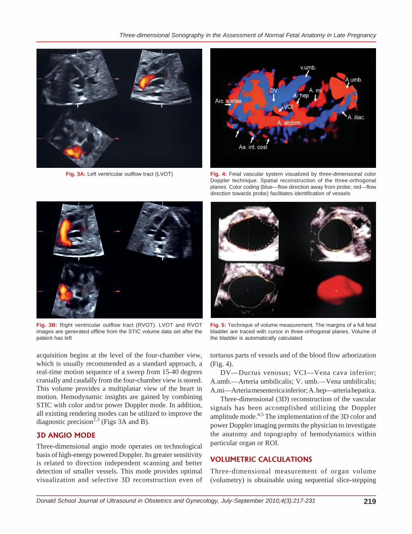
acquisition begins at the level of the four-chamber view,which is usually recommended as a standard approach, areal-time motion sequence of a sweep from 15-40 degreescranially and caudally from the four-chamber view is stored.This volume provides a multiplanar view of the heart inmotion. Hemodynamic insights are gained by combiningSTIC with color and/or power Doppler mode. In addition,all existing rendering modes can be utilized to improve thediagnostic precision2,3 (Figs 3A and B).
3D ANGIO MODE
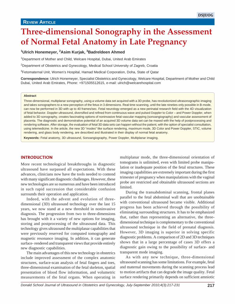
Three-dimensional angio mode operates on technologicalbasis of high-energy powered Doppler. Its greater sensitivityis related to direction independent scanning and betterdetection of smaller vessels. This mode provides optimalvisualization and selective 3D reconstruction even of
tortuous parts of vessels and of the blood flow arborization(Fig. 4).
DV—Ductus venosus; VCI—Vena cava inferior;A.umb.—Arteria umbilicalis; V. umb.—Vena umbilicalis;A.mi—Arteria mesenterica inferior; A. hep—arteria hepatica.
Three-dimensional (3D) reconstruction of the vascularsignals has been accomplished utilizing the Doppleramplitude mode.4,5 The implementation of the 3D color andpower Doppler imaging permits the physician to investigatethe anatomy and topography of hemodynamics withinparticular organ or ROI.
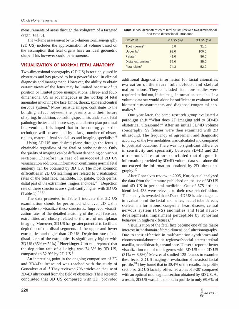
VOLUMETRIC CALCULATIONS
Three-dimensional measurement of organ volume(volumetry) is obtainable using sequential slice-stepping
Fig. 3A: Left ventricular outflow tract (LVOT)
Fig. 3B: Right ventricular outflow tract (RVOT). LVOT and RVOTimages are generated offline from the STIC volume data set after thepatient has left
Fig. 4: Fetal vascular system visualized by three-dimensional colorDoppler technique. Spatial reconstruction of the three-orthogonalplanes. Color coding (blue—flow direction away from probe; red—flowdirection towards probe) facilitates identification of vessels
Fig. 5: Technique of volume measurement. The margins of a full fetalbladder are traced with cursor in three-orthogonal planes. Volume ofthe bladder is automatically calculated
Ulrich Honemeyer et al
220JAYPEE
additional diagnostic information for facial anomalies,evaluation of the neural tube defects, and skeletalmalformations. They concluded that more studies wererequired to find out, if the image information contained in avolume data set would alone be sufficient to evaluate fetalbiometric measurements and diagnose congenital ano-malies.11
One year later, the same research group evaluated aparadigm shift “What does 2D imaging add to 3D/4Dobstetrical ultrasound?” After an initial 3D/4D volumesonography, 99 fetuses were then examined with 2Dultrasound. The frequency of agreement and diagnosticaccuracy of the two modalities was calculated and comparedto postnatal outcome. There was no significant differencein sensitivity and specificity between 3D/4D and 2Dultrasound. The authors concluded that diagnosticinformation provided by 3D/4D volume data sets alone didnot exceed the information obtained by 2D ultrasono-graphy.12
After Goncalves review in 2005, Kurjak et al analyzedthe data from the literature published on the use of 3D USand 4D US in perinatal medicine. Out of 575 articlesidentified, 438 were relevant to their research definition.Their analysis revealed that 3D and 4D US is advantageousin evaluation of the facial anomalies, neural tube defects,skeletal malformations, congenital heart disease, centralnervous system (CNS) anomalies and fetal neuro-developmental impairment perceptible by abnormalbehavior in high-risk fetuses.13
Visualization of the fetal face became one of the majorinterests in the domain of three-dimensional ultrasonography.Due to their affection in malformation syndromes andchromosomal abnormalitie, regions of special interest are fetalmaxilla, mandible arch, ear and nose. Ulm et al reported bettervisualization rate of tooth germs with 3D US than 2D US(31% vs 8.8%)9 Merz et al studied 125 fetuses to examinethe effect of 3D US imaging on evaluation of the axis of facialprofile.10 They found that in 30.4% of the results, the profilesection of 2D US facial profiles had a bias of 3-20º comparedwith an optimal mid-sagittal section obtained by 3D US. Asa result, 2D US was able to obtain profile in only 69.6% of
measurements of areas through the volugram of a targetedorgan (Fig. 5).
The volume assessment by two-dimensional sonography(2D US) includes the approximation of volume based onthe assumption that fetal organs have an ideal geometricshape. This however could be erroneous.
VISUALIZATION OF NORMAL FETAL ANATOMY
Two-dimensional sonography (2D US) is routinely used inobstetrics and has proved to be a powerful tool in clinicaldiagnosis and management. However, the ability to obtaincertain views of the fetus may be limited because of itsposition or limited probe manipulations. Three- and four-dimensional US is advantageous in the workup of fetalanomalies involving the face, limbs, thorax, spine and centralnervous system.6 More realistic images contribute to thebonding effect between the parents and their futureoffspring. In addition, consulting specialists understand fetalpathology better and, if necessary, could better plan postnatalinterventions. It is hoped that in the coming years thistechnique will be accepted by a large number of obstet-ricians, maternal fetal specialists and imaging specialists.6
Using 3D US any desired plane through the fetus isobtainable regardless of the fetal or probe position. Onlythe quality of imaging can be different depending on varioussections. Therefore, in case of unsuccessful 2D USvisualization additional information confirming normal fetalanatomy can be obtained by 3D US. The most commondifficulties in 2D US scanning are related to visualizationrates of the fetal face, mandible, lip, palate, tooth germs,distal part of the extremities, fingers and toes.7-10 Depictionrate of these structures are significantly higher with 3D US(Table 1).2,3,4,9
The data presented in Table 1 indicate that 3D USexamination should be performed whenever 2D US isincapable to visualize these structures. Improved visuali-zation rates of the detailed anatomy of the fetal face andextremities are closely related to the use of multiplanarimaging. Moreover, 3D US has greater potential to facilitatedepiction of the distal segments of the upper and lowerextremities and digits than 2D US. Depiction rate of thedistal parts of the extremities is significantly higher with3D US (85% vs 52%).7 Ploeckinger-Ulm et al reported thatthe depiction rate of all digits was 74.3% by 3D US,compared to 52.9% by 2D US.8
An interesting point in the ongoing comparison of 2Dand 3D/4D ultrasound was reached with the study ofGoncalves et al.11 They reviewed 706 articles on the use of3D/4D ultrasound from the field of obstetrics. Their researchconcluded that 3D US compared with 2D, provided
Table 1: Visualization rates of fetal structures with two-dimensionaland three-dimensional ultrasound
Structure 2D US (%) 3D US (%)
Tooth germs9 8.8 31.0
Upper lip2 93.0 100.0
Palate2 41.0 86.0
Distal extremities3 52.0 85.0
Fetal digits4 74.3 52.9
Three-dimensional Sonography in the Assessment of Normal Fetal Anatomy in Late Pregnancy
Donald School Journal of Ultrasound in Obstetrics and Gynecology, July-September 2010;4(3):217-231 221
the fetuses. The importance of this finding should not beunderestimated. When mid-sagittal plane could not bevisualized, anomalies may be easily missed. Clearly, mainimprovement of 3D US is primarily related to facilitatedvisualization of the morphological details and complexanatomical structures.14-16
FETAL HEAD AND FACE
Assessment of the fetal head is an essential part of routinesonographic examination.17 Even under optimal conditions,the position of the fetal head makes it difficult to obtainadequate images with two-dimensional ultrasonography, andmany cross-sectional images are required to imagine thecomplete structure of the fetal head. Normal anatomy andmajor anomalies of the fetal head, such as exencephaly,anencephaly, encephalocele and holoprosencephaly can berecognized by detailed observation of the skull shape.18
There are numerous reports about first trimester diagnosisof these anomalies using high-frequency 2D transvaginalsonography.19-21 Surface rendering enables assessment ofthe shape of the fetal head, and detailed evaluation of thecranial flat bones, orbits, ears, nos and lips.22 However, 3Dsonography does not provide significant improvementregarding early diagnosis of major malformations. Surfacerendered “sculpture-like” three-dimensional imagesrepresent the most impressive way of fetal visualizationeasily acceptable and recognizable even by parents. Ji et alconducted a study comparing the maternal-fetal bondingafter 2D and 3D US imaging.23 They found that 3D had amore positive influence on maternal-fetal bonding than 2DUS.
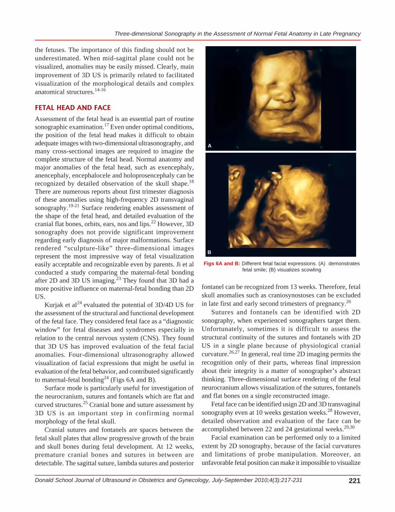
Kurjak et al24 evaluated the potential of 3D/4D US forthe assessment of the structural and functional developmentof the fetal face. They considered fetal face as a “diagnosticwindow” for fetal diseases and syndromes especially inrelation to the central nervous system (CNS). They foundthat 3D US has improved evaluation of the fetal facialanomalies. Four-dimensional ultrasonography allowedvisualization of facial expressions that might be useful inevaluation of the fetal behavior, and contributed significantlyto maternal-fetal bonding24 (Figs 6A and B).
Surface mode is particularly useful for investigation ofthe neurocranium, sutures and fontanels which are flat andcurved structures.25 Cranial bone and suture assessment by3D US is an important step in confirming normalmorphology of the fetal skull.
Cranial sutures and fontanels are spaces between thefetal skull plates that allow progressive growth of the brainand skull bones during fetal development. At 12 weeks,premature cranial bones and sutures in between aredetectable. The sagittal suture, lambda sutures and posterior
fontanel can be recognized from 13 weeks. Therefore, fetalskull anomalies such as craniosynostoses can be excludedin late first and early second trimesters of pregnancy.26
Sutures and fontanels can be identified with 2Dsonography, when experienced sonographers target them.Unfortunately, sometimes it is difficult to assess thestructural continuity of the sutures and fontanels with 2DUS in a single plane because of physiological cranialcurvature.26,27 In general, real time 2D imaging permits therecognition only of their parts, whereas final impressionabout their integrity is a matter of sonographer’s abstractthinking. Three-dimensional surface rendering of the fetalneurocranium allows visualization of the sutures, fontanelsand flat bones on a single reconstructed image.
Fetal face can be identified usign 2D and 3D transvaginalsonography even at 10 weeks gestation weeks.28 However,detailed observation and evaluation of the face can beaccomplished between 22 and 24 gestational weeks.29,30
Facial examination can be performed only to a limitedextent by 2D sonography, because of the facial curvaturesand limitations of probe manipulation. Moreover, anunfavorable fetal position can make it impossible to visualize
Figs 6A and B: Different fetal facial expressions. (A) demonstratesfetal smile; (B) visualizes scowling
A
B
Ulrich Honemeyer et al
222JAYPEE
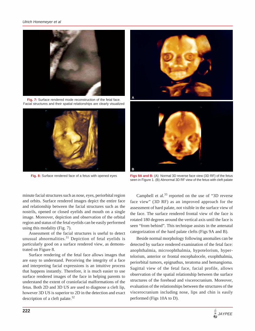
minute facial structures such as nose, eyes, periorbital regionand orbits. Surface rendered images depict the entire faceand relationship between the facial structures such as thenostrils, opened or closed eyelids and mouth on a singleimage. Moreover, depiction and observation of the orbitalregion and status of the fetal eyelids can be easily performedusing this modality (Fig. 7).
Assessment of the facial structures is useful to detectunusual abnormalities.31 Depiction of fetal eyelids isparticularly good on a surface rendered view, as demons-trated on Figure 8.
Surface rendering of the fetal face allows images thatare easy to understand. Perceiving the integrity of a faceand interpreting facial expressions is an intuitive processthat happens instantly. Therefore, it is much easier to usesurface rendered images of the face in helping parents tounderstand the extent of craniofacial malformations of thefetus. Both 2D and 3D US are used to diagnose a cleft lip,however 3D US is superior to 2D in the detection and exactdescription of a cleft palate.32
Campbell et al.33 reported on the use of “3D reverseface view” (3D RF) as an improved approach for theassessment of hard palate, not visible in the surface view ofthe face. The surface rendered frontal view of the face isrotated 180 degrees around the vertical axis until the face isseen “from behind”. This technique assists in the antenatalcategorization of the hard palate clefts (Figs 9A and B).
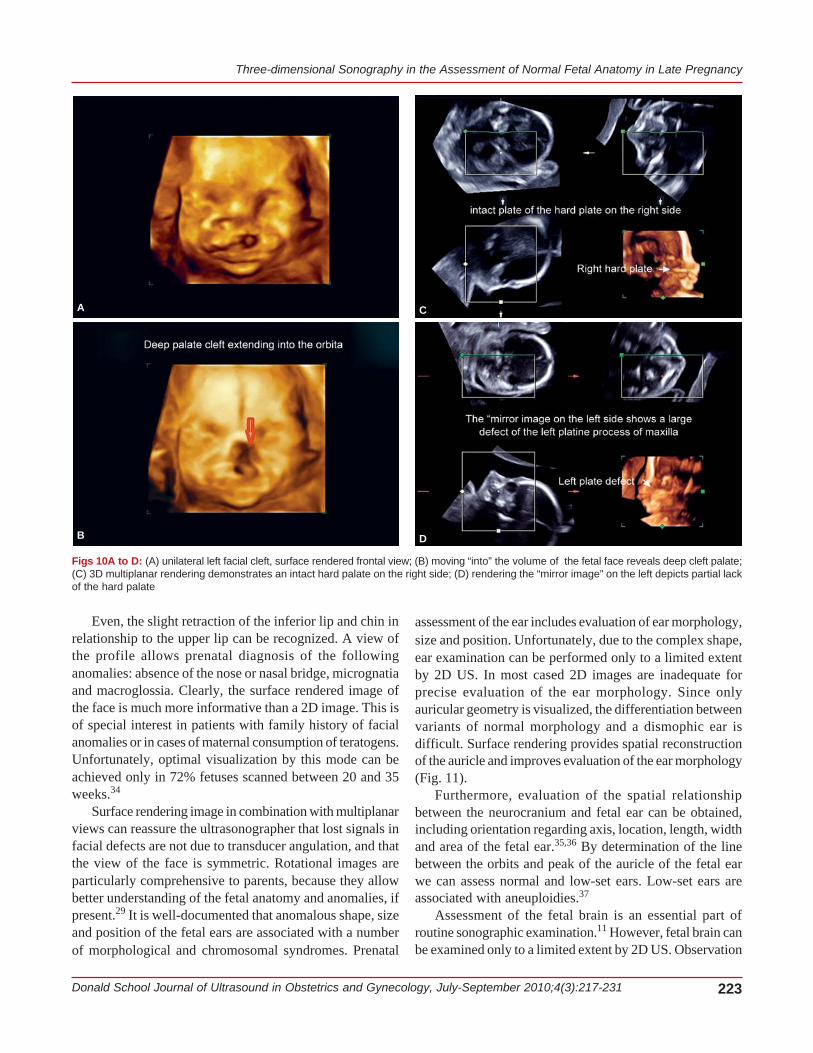
Beside normal morphology following anomalies can bedetected by surface rendered examination of the fetal face:anophthalmia, microophthalmia, hypotelorism, hyper-telorism, anterior or frontal encephalocele, exophthalmia,periorbital tumors, epignathus, teratoma and hemangioma.Sagittal view of the fetal face, facial profile, allowsobservation of the spatial relationship between the surfacestructures of the forehead and viscerocranium. Moreover,evaluation of the relationships between the structures of theviscerocranium including nose, lips and chin is easilyperformed (Figs 10A to D).
Fig. 7: Surface rendered mode reconstruction of the fetal face.Facial structures and their spatial relationships are clearly visualized
Fig. 8: Surface rendered face of a fetus with opened eyes Figs 9A and B: (A) Normal 3D reverse face view (3D RF) of the fetusseen in Figure 1. (B) Abnormal 3D RF view of the fetus with cleft palate
A
B
Three-dimensional Sonography in the Assessment of Normal Fetal Anatomy in Late Pregnancy
Donald School Journal of Ultrasound in Obstetrics and Gynecology, July-September 2010;4(3):217-231 223
Even, the slight retraction of the inferior lip and chin inrelationship to the upper lip can be recognized. A view ofthe profile allows prenatal diagnosis of the followinganomalies: absence of the nose or nasal bridge, micrognatiaand macroglossia. Clearly, the surface rendered image ofthe face is much more informative than a 2D image. This isof special interest in patients with family history of facialanomalies or in cases of maternal consumption of teratogens.Unfortunately, optimal visualization by this mode can beachieved only in 72% fetuses scanned between 20 and 35weeks.34
Surface rendering image in combination with multiplanarviews can reassure the ultrasonographer that lost signals infacial defects are not due to transducer angulation, and thatthe view of the face is symmetric. Rotational images areparticularly comprehensive to parents, because they allowbetter understanding of the fetal anatomy and anomalies, ifpresent.29 It is well-documented that anomalous shape, sizeand position of the fetal ears are associated with a numberof morphological and chromosomal syndromes. Prenatal
assessment of the ear includes evaluation of ear morphology,size and position. Unfortunately, due to the complex shape,ear examination can be performed only to a limited extentby 2D US. In most cased 2D images are inadequate forprecise evaluation of the ear morphology. Since onlyauricular geometry is visualized, the differentiation betweenvariants of normal morphology and a dismophic ear isdifficult. Surface rendering provides spatial reconstructionof the auricle and improves evaluation of the ear morphology(Fig. 11).
Furthermore, evaluation of the spatial relationshipbetween the neurocranium and fetal ear can be obtained,including orientation regarding axis, location, length, widthand area of the fetal ear.35,36 By determination of the linebetween the orbits and peak of the auricle of the fetal earwe can assess normal and low-set ears. Low-set ears areassociated with aneuploidies.37
Assessment of the fetal brain is an essential part ofroutine sonographic examination.11 However, fetal brain canbe examined only to a limited extent by 2D US. Observation
Figs 10A to D: (A) unilateral left facial cleft, surface rendered frontal view; (B) moving “into” the volume of the fetal face reveals deep cleft palate;(C) 3D multiplanar rendering demonstrates an intact hard palate on the right side; (D) rendering the “mirror image” on the left depicts partial lackof the hard palate
A
B
C
D
Ulrich Honemeyer et al
224JAYPEE
of fetal the brain offers sagittal and coronal sections of thebrain from fetal parietal direction through the fontanels and/or sagittal suture as ultrasound windows.38,39
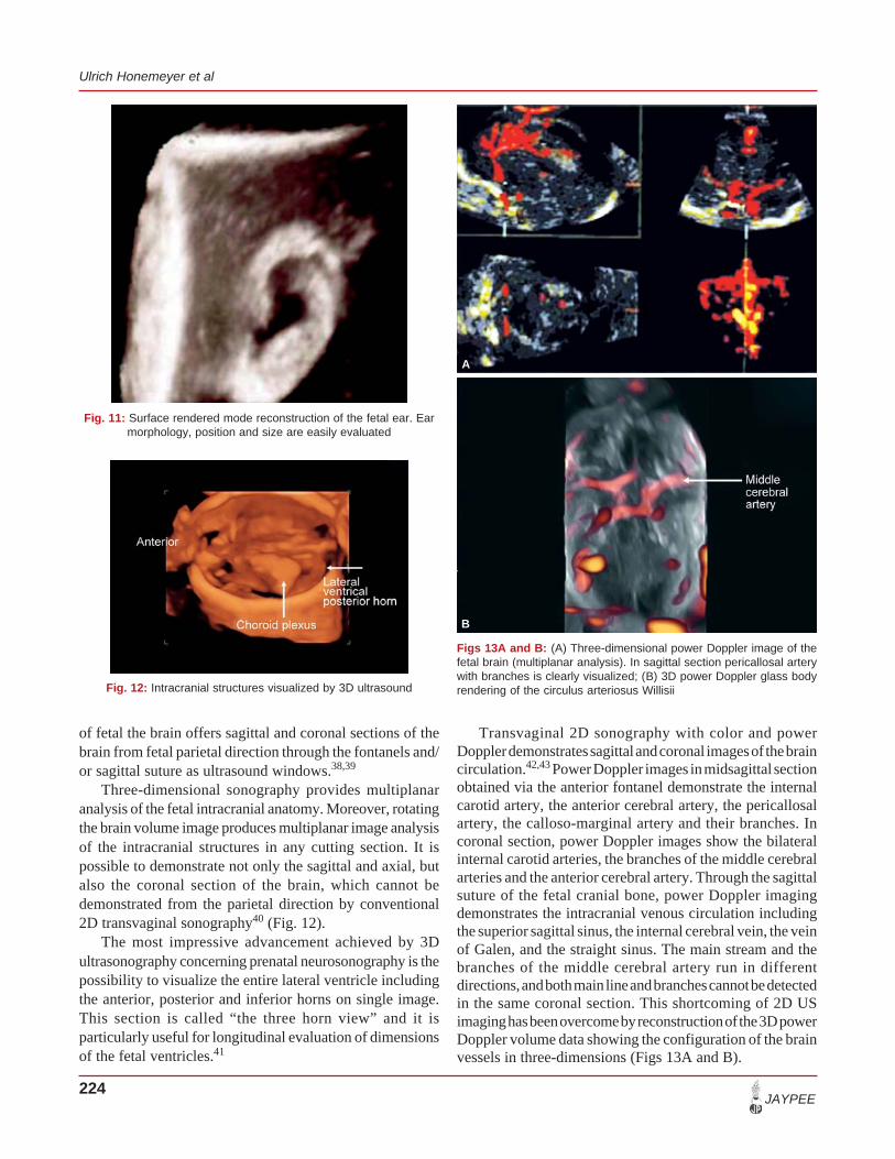
Three-dimensional sonography provides multiplanaranalysis of the fetal intracranial anatomy. Moreover, rotatingthe brain volume image produces multiplanar image analysisof the intracranial structures in any cutting section. It ispossible to demonstrate not only the sagittal and axial, butalso the coronal section of the brain, which cannot bedemonstrated from the parietal direction by conventional2D transvaginal sonography40 (Fig. 12).
The most impressive advancement achieved by 3Dultrasonography concerning prenatal neurosonography is thepossibility to visualize the entire lateral ventricle includingthe anterior, posterior and inferior horns on single image.This section is called “the three horn view” and it isparticularly useful for longitudinal evaluation of dimensionsof the fetal ventricles.41
Transvaginal 2D sonography with color and powerDoppler demonstrates sagittal and coronal images of the braincirculation.42,43 Power Doppler images in midsagittal sectionobtained via the anterior fontanel demonstrate the internalcarotid artery, the anterior cerebral artery, the pericallosalartery, the calloso-marginal artery and their branches. Incoronal section, power Doppler images show the bilateralinternal carotid arteries, the branches of the middle cerebralarteries and the anterior cerebral artery. Through the sagittalsuture of the fetal cranial bone, power Doppler imagingdemonstrates the intracranial venous circulation includingthe superior sagittal sinus, the internal cerebral vein, the veinof Galen, and the straight sinus. The main stream and thebranches of the middle cerebral artery run in differentdirections, and both main line and branches cannot be detectedin the same coronal section. This shortcoming of 2D USimaging has been overcome by reconstruction of the 3D powerDoppler volume data showing the configuration of the brainvessels in three-dimensions (Figs 13A and B).
Fig. 11: Surface rendered mode reconstruction of the fetal ear. Earmorphology, position and size are easily evaluated
Fig. 12: Intracranial structures visualized by 3D ultrasound
Figs 13A and B: (A) Three-dimensional power Doppler image of thefetal brain (multiplanar analysis). In sagittal section pericallosal arterywith branches is clearly visualized; (B) 3D power Doppler glass bodyrendering of the circulus arteriosus Willisii
B
A
Three-dimensional Sonography in the Assessment of Normal Fetal Anatomy in Late Pregnancy
Donald School Journal of Ultrasound in Obstetrics and Gynecology, July-September 2010;4(3):217-231 225
The internal carotid artery, anterior cerebral artery,bilateral middle cerebral arteries and their branches, can bedemonstrated simultaneously on one image. Ultrasoundangiography of the fetal brain allows noninvasive assessmentof the brain circulation. Since 3D vascular images can berotated in any direction, the vessels can be observed fromthe frontal, occipital, lateral, oblique, parietal or basilar partof the brain.
FETAL THORAX AND ABDOMEN
Due to the curvature of the thoracic bones detailedevaluation of the thoracic skeleton is often difficult with2D US.44 Three-dimensional US transparency mode reducesthe echogenicity of the soft tissues, leaving behind echogenicstructures, namely the bones, which enables visualizationof the fetal ribs. The curvature and relationship of the ribsending in the vertebral bodies and the anterior chest wallcan be demonstrated in the entire length. Achiron et alevaluated specific advantages of certain 3D imaging andrendering modes for the optimal diagnostic display of certainmalformations.45 Volume datasets of 23 fetuses withthoracic anomalies were acquired with static 3D and cine4D ultrasound, i.e. spatiotemporal image correlation (STIC)mode. The volumes were analyzed and displayed bymultiplanar and tomographic ultrasound imaging (TUI)modes and static volume contrast imaging (VCI). ColorDoppler was added to the volumes acquired, and variousrendering modes were used to display the volume datasets.The TUI mode achieved optimal display of the thorax,thereby aiding the diagnosis of diaphragmatic hernia andlung dysplasia. High-definition color Doppler with glass-body rendering significantly contributed to the detection ofabnormal vascularization in lung dysplasia. Maximaltransparent mode by transvaginal route provided accuratediagnosis of skeletal dysplasia during the first trimester. Situsabnormalities were best visualized using minimaltransparent mode. This modality has enabled clearidentification of abnormal organs and positions of thevessels. Clearly, 3D/4D US enhances prenatal diagnosis andprovides better spatial visualization of the thoracicanomalies.45
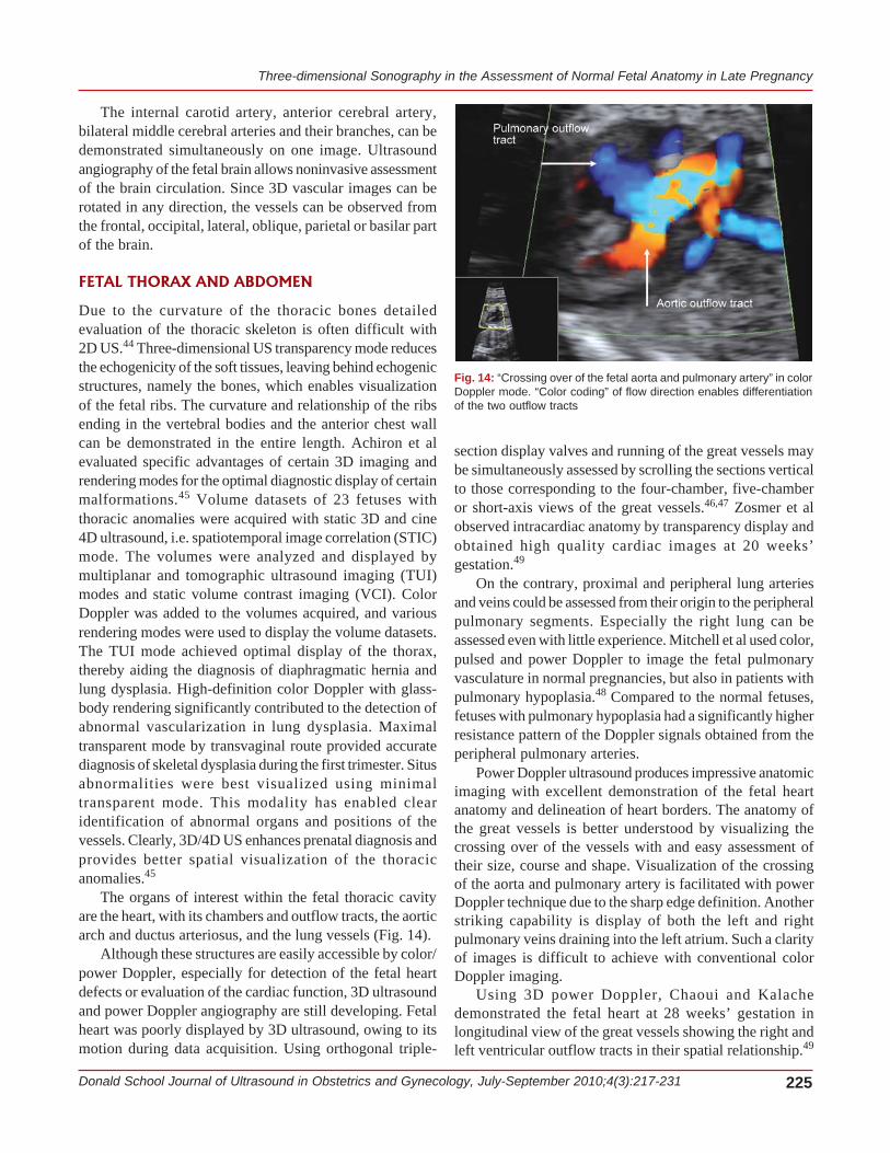
The organs of interest within the fetal thoracic cavityare the heart, with its chambers and outflow tracts, the aorticarch and ductus arteriosus, and the lung vessels (Fig. 14).
Although these structures are easily accessible by color/power Doppler, especially for detection of the fetal heartdefects or evaluation of the cardiac function, 3D ultrasoundand power Doppler angiography are still developing. Fetalheart was poorly displayed by 3D ultrasound, owing to itsmotion during data acquisition. Using orthogonal triple-
section display valves and running of the great vessels maybe simultaneously assessed by scrolling the sections verticalto those corresponding to the four-chamber, five-chamberor short-axis views of the great vessels.46,47 Zosmer et alobserved intracardiac anatomy by transparency display andobtained high quality cardiac images at 20 weeks’gestation.49
On the contrary, proximal and peripheral lung arteriesand veins could be assessed from their origin to the peripheralpulmonary segments. Especially the right lung can beassessed even with little experience. Mitchell et al used color,pulsed and power Doppler to image the fetal pulmonaryvasculature in normal pregnancies, but also in patients withpulmonary hypoplasia.48 Compared to the normal fetuses,fetuses with pulmonary hypoplasia had a significantly higherresistance pattern of the Doppler signals obtained from theperipheral pulmonary arteries.
Power Doppler ultrasound produces impressive anatomicimaging with excellent demonstration of the fetal heartanatomy and delineation of heart borders. The anatomy ofthe great vessels is better understood by visualizing thecrossing over of the vessels with and easy assessment oftheir size, course and shape. Visualization of the crossingof the aorta and pulmonary artery is facilitated with powerDoppler technique due to the sharp edge definition. Anotherstriking capability is display of both the left and rightpulmonary veins draining into the left atrium. Such a clarityof images is difficult to achieve with conventional colorDoppler imaging.
Using 3D power Doppler, Chaoui and Kalachedemonstrated the fetal heart at 28 weeks’ gestation inlongitudinal view of the great vessels showing the right andleft ventricular outflow tracts in their spatial relationship.49
Fig. 14: “Crossing over of the fetal aorta and pulmonary artery” in colorDoppler mode. “Color coding” of flow direction enables differentiationof the two outflow tracts
Ulrich Honemeyer et al
226JAYPEE
The aortic arch and the crossing of the pulmonary trunkwas clearly seen, as well as the connection of the ductusarteriosus to the descending aorta. The left pulmonary arteryarises before the origin of the ductus arteriosus. Three-dimensional power Doppler approach enables imaging ofthe blood flow mainly in the center of the vessel, whichassists in the spatial separation of the aorta and pulmonaryartery despite high persistence. The expectation that 3D fetalechocardiography would include 3D power Doppler inaddition to 3D or 4D gray scale, has come true with theintroduction and perfection of spatio-temporal imagecorrelation (STIC). It is expected that this modality willimprove our understanding of the malformations such ashypoplastic left heart syndrome (HLHS), right heartsyndrome, or other malformations with a singular ventricleand hypoplasia of the great arteries. The majority of thecomplex congenital heart anomalies show a steadilyprogression of the pathological changes during the courseof pregnancy, including subsequent secondary phenomenasuch as arrhythmias or myocardial insufficiency. 3D/4Dultrasound is a tool of choice for follow-up of such aprogression and may have a significant input in prenataltreatment of an abnormal fetus. Better understanding of thepathophysiological causal may encourage some researchersto explore new minimally invasive therapeutic options interms of early pre- and postnatal cardiac palliation.50
Three-dimensional surface mode enables sculpture-likereconstruction of the abdominal wall and normal umbilicalcord insertion. The complete abdominal surface is invisibleby conventional 2D technology, unless the abdominalsurface is scanned in a survey-like manner, involving serialtomographic sections in sagittal and transverse planes. Using3D surface mode we are able to visualize the completeabdominal surface including a umbilical cord insertion in asingle image. Using surface rendering mode continuity ofthe fetal skin can be easily confirmed.
Moreover, fetal fat deposits can be easily differentiatedfrom abnormal protrusion caused by malformations andcutaneous or subcutaneous tumors.51,52
Postprocessing offers opportunity for surface imagingof the fetal intra-abdominal structures. Multiplanar imagingenables construction of the planes nearly parallel to themother’s abdominal wall, thus making it possible to observethe esophageal-gastric junction and pylorus. The electronicscalpel or electronic rubber is used to “cut out” the overlyingbody segments, producing either a longitudinal or transversesection. Once this has been done, the pathologic organ canbe evaluated separately. Three-dimensional ultrasoundconfirms suspected multicystic dysplastic kidney as well asrenal agenesis and abnormal pelvic-ureteral junction.53
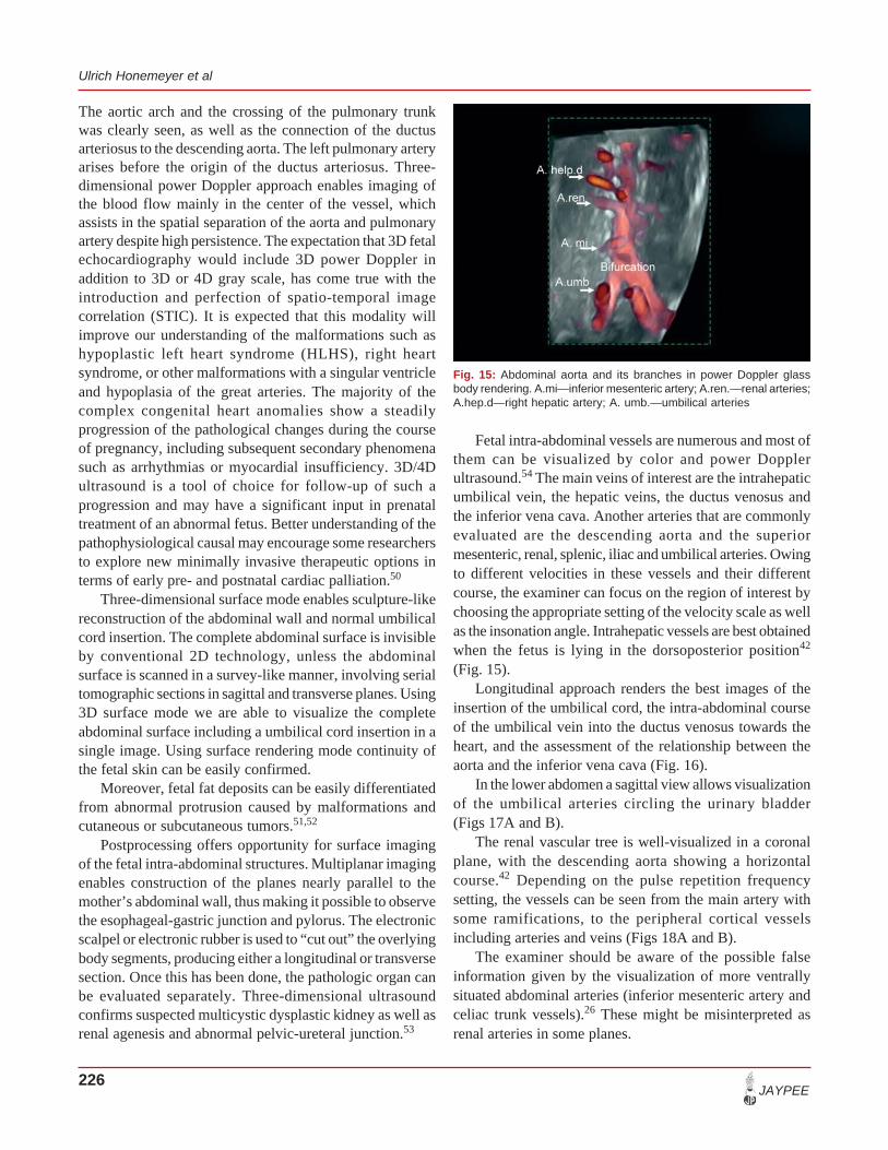
Fetal intra-abdominal vessels are numerous and most ofthem can be visualized by color and power Dopplerultrasound.54 The main veins of interest are the intrahepaticumbilical vein, the hepatic veins, the ductus venosus andthe inferior vena cava. Another arteries that are commonlyevaluated are the descending aorta and the superiormesenteric, renal, splenic, iliac and umbilical arteries. Owingto different velocities in these vessels and their differentcourse, the examiner can focus on the region of interest bychoosing the appropriate setting of the velocity scale as wellas the insonation angle. Intrahepatic vessels are best obtainedwhen the fetus is lying in the dorsoposterior position42
(Fig. 15).Longitudinal approach renders the best images of the
insertion of the umbilical cord, the intra-abdominal courseof the umbilical vein into the ductus venosus towards theheart, and the assessment of the relationship between theaorta and the inferior vena cava (Fig. 16).
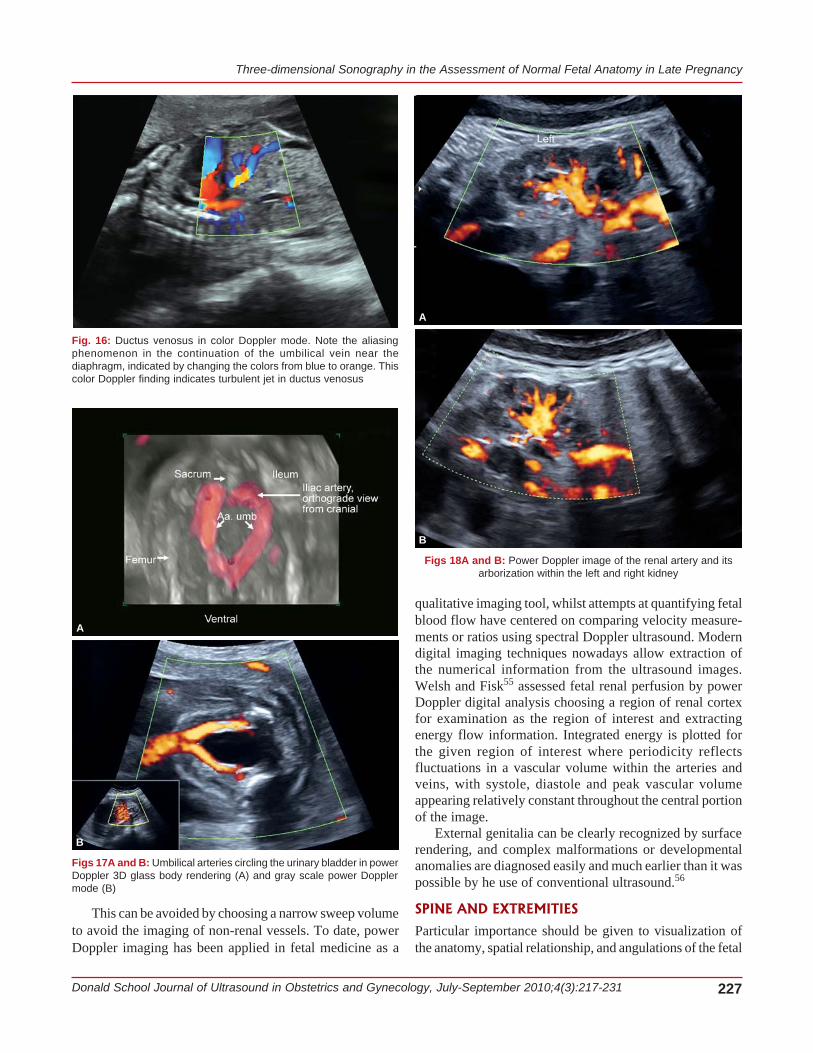
In the lower abdomen a sagittal view allows visualizationof the umbilical arteries circling the urinary bladder(Figs 17A and B).
The renal vascular tree is well-visualized in a coronalplane, with the descending aorta showing a horizontalcourse.42 Depending on the pulse repetition frequencysetting, the vessels can be seen from the main artery withsome ramifications, to the peripheral cortical vesselsincluding arteries and veins (Figs 18A and B).
The examiner should be aware of the possible falseinformation given by the visualization of more ventrallysituated abdominal arteries (inferior mesenteric artery andceliac trunk vessels).26 These might be misinterpreted asrenal arteries in some planes.
Fig. 15: Abdominal aorta and its branches in power Doppler glassbody rendering. A.mi—inferior mesenteric artery; A.ren.—renal arteries;A.hep.d—right hepatic artery; A. umb.—umbilical arteries
Three-dimensional Sonography in the Assessment of Normal Fetal Anatomy in Late Pregnancy
Donald School Journal of Ultrasound in Obstetrics and Gynecology, July-September 2010;4(3):217-231 227
This can be avoided by choosing a narrow sweep volumeto avoid the imaging of non-renal vessels. To date, powerDoppler imaging has been applied in fetal medicine as a
qualitative imaging tool, whilst attempts at quantifying fetalblood flow have centered on comparing velocity measure-ments or ratios using spectral Doppler ultrasound. Moderndigital imaging techniques nowadays allow extraction ofthe numerical information from the ultrasound images.Welsh and Fisk55 assessed fetal renal perfusion by powerDoppler digital analysis choosing a region of renal cortexfor examination as the region of interest and extractingenergy flow information. Integrated energy is plotted forthe given region of interest where periodicity reflectsfluctuations in a vascular volume within the arteries andveins, with systole, diastole and peak vascular volumeappearing relatively constant throughout the central portionof the image.
External genitalia can be clearly recognized by surfacerendering, and complex malformations or developmentalanomalies are diagnosed easily and much earlier than it waspossible by he use of conventional ultrasound.56
SPINE AND EXTREMITIES
Particular importance should be given to visualization ofthe anatomy, spatial relationship, and angulations of the fetal
Fig. 16: Ductus venosus in color Doppler mode. Note the aliasingphenomenon in the continuation of the umbilical vein near thediaphragm, indicated by changing the colors from blue to orange. Thiscolor Doppler finding indicates turbulent jet in ductus venosus
Figs 17A and B: Umbilical arteries circling the urinary bladder in powerDoppler 3D glass body rendering (A) and gray scale power Dopplermode (B)
A
B
Figs 18A and B: Power Doppler image of the renal artery and itsarborization within the left and right kidney
B
A
Ulrich Honemeyer et al
228JAYPEE
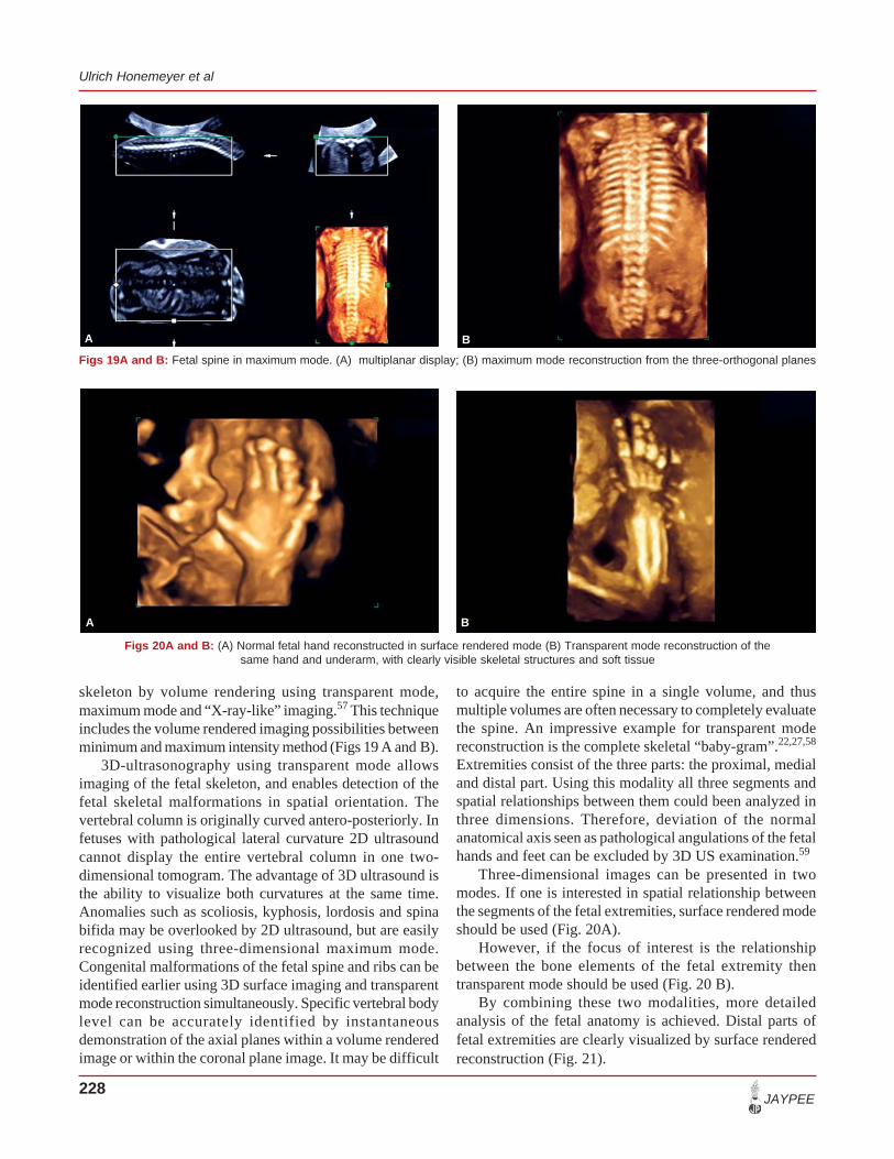
skeleton by volume rendering using transparent mode,maximum mode and “X-ray-like” imaging.57 This techniqueincludes the volume rendered imaging possibilities betweenminimum and maximum intensity method (Figs 19 A and B).
3D-ultrasonography using transparent mode allowsimaging of the fetal skeleton, and enables detection of thefetal skeletal malformations in spatial orientation. Thevertebral column is originally curved antero-posteriorly. Infetuses with pathological lateral curvature 2D ultrasoundcannot display the entire vertebral column in one two-dimensional tomogram. The advantage of 3D ultrasound isthe ability to visualize both curvatures at the same time.Anomalies such as scoliosis, kyphosis, lordosis and spinabifida may be overlooked by 2D ultrasound, but are easilyrecognized using three-dimensional maximum mode.Congenital malformations of the fetal spine and ribs can beidentified earlier using 3D surface imaging and transparentmode reconstruction simultaneously. Specific vertebral bodylevel can be accurately identified by instantaneousdemonstration of the axial planes within a volume renderedimage or within the coronal plane image. It may be difficult
to acquire the entire spine in a single volume, and thusmultiple volumes are often necessary to completely evaluatethe spine. An impressive example for transparent modereconstruction is the complete skeletal “baby-gram”.22,27,58
Extremities consist of the three parts: the proximal, medialand distal part. Using this modality all three segments andspatial relationships between them could been analyzed inthree dimensions. Therefore, deviation of the normalanatomical axis seen as pathological angulations of the fetalhands and feet can be excluded by 3D US examination.59
Three-dimensional images can be presented in twomodes. If one is interested in spatial relationship betweenthe segments of the fetal extremities, surface rendered modeshould be used (Fig. 20A).
However, if the focus of interest is the relationshipbetween the bone elements of the fetal extremity thentransparent mode should be used (Fig. 20 B).
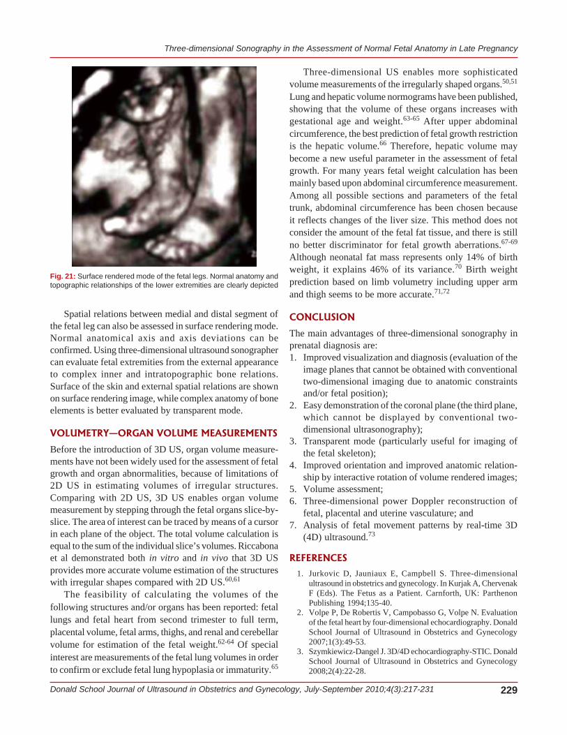
By combining these two modalities, more detailedanalysis of the fetal anatomy is achieved. Distal parts offetal extremities are clearly visualized by surface renderedreconstruction (Fig. 21).
Figs 19A and B: Fetal spine in maximum mode. (A) multiplanar display; (B) maximum mode reconstruction from the three-orthogonal planes
Figs 20A and B: (A) Normal fetal hand reconstructed in surface rendered mode (B) Transparent mode reconstruction of thesame hand and underarm, with clearly visible skeletal structures and soft tissue
A B
A B
Three-dimensional Sonography in the Assessment of Normal Fetal Anatomy in Late Pregnancy
Donald School Journal of Ultrasound in Obstetrics and Gynecology, July-September 2010;4(3):217-231 229
Spatial relations between medial and distal segment ofthe fetal leg can also be assessed in surface rendering mode.Normal anatomical axis and axis deviations can beconfirmed. Using three-dimensional ultrasound sonographercan evaluate fetal extremities from the external appearanceto complex inner and intratopographic bone relations.Surface of the skin and external spatial relations are shownon surface rendering image, while complex anatomy of boneelements is better evaluated by transparent mode.
VOLUMETRY—ORGAN VOLUME MEASUREMENTS
Before the introduction of 3D US, organ volume measure-ments have not been widely used for the assessment of fetalgrowth and organ abnormalities, because of limitations of2D US in estimating volumes of irregular structures.Comparing with 2D US, 3D US enables organ volumemeasurement by stepping through the fetal organs slice-by-slice. The area of interest can be traced by means of a cursorin each plane of the object. The total volume calculation isequal to the sum of the individual slice’s volumes. Riccabonaet al demonstrated both in vitro and in vivo that 3D USprovides more accurate volume estimation of the structureswith irregular shapes compared with 2D US.60,61
The feasibility of calculating the volumes of thefollowing structures and/or organs has been reported: fetallungs and fetal heart from second trimester to full term,placental volume, fetal arms, thighs, and renal and cerebellarvolume for estimation of the fetal weight.62-64 Of specialinterest are measurements of the fetal lung volumes in orderto confirm or exclude fetal lung hypoplasia or immaturity.65
Three-dimensional US enables more sophisticatedvolume measurements of the irregularly shaped organs.50,51
Lung and hepatic volume normograms have been published,showing that the volume of these organs increases withgestational age and weight.63-65 After upper abdominalcircumference, the best prediction of fetal growth restrictionis the hepatic volume.66 Therefore, hepatic volume maybecome a new useful parameter in the assessment of fetalgrowth. For many years fetal weight calculation has beenmainly based upon abdominal circumference measurement.Among all possible sections and parameters of the fetaltrunk, abdominal circumference has been chosen becauseit reflects changes of the liver size. This method does notconsider the amount of the fetal fat tissue, and there is stillno better discriminator for fetal growth aberrations.67-69
Although neonatal fat mass represents only 14% of birthweight, it explains 46% of its variance.70 Birth weightprediction based on limb volumetry including upper armand thigh seems to be more accurate.71,72
CONCLUSION
The main advantages of three-dimensional sonography inprenatal diagnosis are:1. Improved visualization and diagnosis (evaluation of the
image planes that cannot be obtained with conventionaltwo-dimensional imaging due to anatomic constraintsand/or fetal position);
2. Easy demonstration of the coronal plane (the third plane,which cannot be displayed by conventional two-dimensional ultrasonography);
3. Transparent mode (particularly useful for imaging ofthe fetal skeleton);
4. Improved orientation and improved anatomic relation-ship by interactive rotation of volume rendered images;
5. Volume assessment;6. Three-dimensional power Doppler reconstruction of
fetal, placental and uterine vasculature; and7. Analysis of fetal movement patterns by real-time 3D
(4D) ultrasound.73
REFERENCES
1. Jurkovic D, Jauniaux E, Campbell S. Three-dimensionalultrasound in obstetrics and gynecology. In Kurjak A, ChervenakF (Eds). The Fetus as a Patient. Carnforth, UK: ParthenonPublishing 1994;135-40.
2. Volpe P, De Robertis V, Campobasso G, Volpe N. Evaluationof the fetal heart by four-dimensional echocardiography. DonaldSchool Journal of Ultrasound in Obstetrics and Gynecology2007;1(3):49-53.
3. Szymkiewicz-Dangel J. 3D/4D echocardiography-STIC. DonaldSchool Journal of Ultrasound in Obstetrics and Gynecology2008;2(4):22-28.
Fig. 21: Surface rendered mode of the fetal legs. Normal anatomy andtopographic relationships of the lower extremities are clearly depicted
Ulrich Honemeyer et al
230JAYPEE
4. Downey DB, Fenster A, Williams JC. Clinical utility of three-dimensional US. Radiographics 2000;20:559-71.
5. Downey DB, Fenster A. Vascular imaging with a three-dimensional power Doppler system. Am J Roentgenol 1995;165:665-58.
6. Timor-Tritsch IE, Platt LD. Three-dimensional ultrasoundexperience in obstetrics. Curr Opin Obstet Gynecol 2002Dec;14(6):569-75.
7. Budorick NE, Pretorius DH, Johnson DD, Nelson TR, TartarMK, Lou KV. Three-dimensional ultrasonography of the fetaldistal lower extremity: Normal and abnormal. J Ultrasound Med1998;17:649-60.
8. Ploeckinger-Ulm B, Ulm MR, Lee A, Kratochwil A, BernaschekG. Antenatal depiction of fetal digits with three-dimensionalultrasonography. Am J Obstet Gynecol 1996;175:571-74.
9. Ulm MR, Kratochwil A, Ulm B, Lee A, Bettelheim D,Bernaschek G. Three-dimensional ultrasonographic imaging offetal tooth buds for characterization of facial clefts. Early HumDev 1999;55:67-75.
10. Merz E, Weber G, Bahlmann F, Miric-Tesanic D. Applicationof transvaginal and abdominal three-dimensional ultrasound forthe detection or exclusion of malformations of the fetal face.Ultrasound Obstet Gynecol 1997;9:237-43.
11. Gonçalves LF, Lee W, Espinoza J, Romero R. Three- and 4-dimensional ultrasound in obstetric practice: Does it help? JUltrasound Med 2005;24:1599-624.
12. Gonçalves LF, Nien JK, Espinoza J, Kusanovic JP, Lee W,Swope B, Soto E, Treadwell MC, Romero R. What does 2-dimensional imaging add to 3- and 4-dimensional obstetricultrasonography? J Ultrasound Med 2006;25:691-99.
13. Kurjak A, Miskovic B, Andonotopo W, Stanojevic M, AzumendiG, Vrcic H. How useful is 3D and 4D ultrasound in perinatalmedicine? J Perinat Med 2007;35(1):10-27 .
14. Pretorius DH, Nelson TR. Fetal face visualization using three-dimensional ultrasonography. J Ultrasound Med 1995;14:349-56.
15. Pretorius DH, Nelson TR. Prenatal visualization of cranialsutures and fontanelles with three-dimensional ultrasonography.J Ultasound Med 1994;13:871-76.
16. Shih JC, Shyu MK, Lee CN, Wu CH, Lin GJ, Hsieh FJ. Antenataldepiction of the fetal ear with three-dimensional ultrasonography.Obstet Gynecol 1998;91:500-05.
17. Baba K, Okai T. Clinical applications of three-dimensionalultrasound in obstetrics. In: Baba K, Jurkovic D (Eds). ThreeDimensional Ultrasound in Obstetrics and Gynecology.Carnforth, UK:Parthenon Publishing, 1997:29-44.
18. Benoit B, Hafner T, Kurjak A, Kupesic S, Bekavac I, Bozek T.Three-dimensional sonoembryology. J Perinatal Med Med2000;30:63-73.
19. Achiron R, Achiron A. Transvaginal fetal neurosonography: Thefirst trimester of pregnancy. In: Chervenak FA, Kurjak A,Comstock CH (Eds). Ultrasound of the fetal brain. London-NewYork: Parthenon Publishing, 1995:95-102.
20. Pooh RK. B-mode and Doppler studies of the abnormal fetus inthe first trimester. In: Chervenak FA, Kurjak A (Eds). FetalMedicine: The clinical care of the fetus as a patient. London-New York: Parthenon Publishing 1999:46-51.
21. Weissman A, Achiron R. Ultrasound diagnosis of congenitalanomalies in early pregnancy. In: Jurkovic D, Jauninaux E (Eds).
Ultrasound and Early Pregnancy. Canforth, UK: ParthenonPublishing 1996:95-119.
22. Benoit B. Three-dimensional surface mode for demonstrationof normal fetal anatomy in the second and third trimester. In:Merz E (Ed). 3D Ultrasound in Obstetrics and Gynecology.Philadelphia: Lippincot Williams and Wilkins, 1998:95-100.
23. Ji EK, Pretorius DH, Newton R, Uyan K, Hull AD, HollenbachK, Nelson TR. Effects of ultrasound on maternal-fetal bonding:A comparison of two- and three-dimensional imaging.Ultrasound Obstet Gynecol 2005;25(5):473-77.
24. Kurjak A, Azumendi G, Andonotopo W, Salihagic-Kadic A.Three- and four-dimensional ultrasonography for the structuraland functional evaluation of the fetal face. Am J Obstet Gynecol.2007;196(1):16-28.
25. Pooh RK. Fetal cranial bone formation: Sonographic assessment.In: Margulies M, Voto LS, Eik-Nes S (Eds). Proceedings of 9thWorld congress of Ultrasound in Obstetrics and Gynecology.Bologna, Italy: Monduzzi Editore 1999;407-10.
26. Patel MD, Swinford AE, Filly RA. Anatomic and sonographicfeatures of fetal skull. J Ultrasound Med 1994;13:251-57.
27. Pretorius DH, Nelson TR. Prenatal visualization of cranialsutures and fontanelles with three-dimensional ultrasonography.J Ultrasound Med 1994;13:871-76.
28. Bonilla-Musoles F, Raga F, Blanes J, Osborne N, Siles CH.Three-dimensional ultrasound in Reproductive Medicine:Preliminary report. Human Reprod update. Vol. 1, 4 item 21CD Rom 1995.
29. Johnson DD, Pretorius DH, Budorick NE, Jones MC, Lou KV,James GM, Nelson TR. Fetal lip and primary palate: Three-dimensional versus two-dimensional US. Radiology 2000;217:236-39.
30. Chmait RH, Hull AD, James TR, Nelson TR, Pretorius DH.Three-dimensional ultrasound evaluation of fetal face.Ultrasound Rev Obstet Gynecol 2001;1:138-43.
31. Osborne N, Bailao L, Abad L (Jr), Bonilla-Musoles F.Macadores ecográficos de infección fetal: In. Bonilla- MusolesF y Machado L. Ed Ecografía y Reproducción. Panamericana.Madrid, 1999;5:205-31.
32. Johnson DD, Pretorius DH, Budorick NE, Jones MC, Lou KV,James GM, Nelson TR. Fetal lip and primary palate: Three-dimensional versus two-dimensional US. Radiology 2000;217(1):236-39.
33. Campbell S, Lees CC. The three-dimensional reverse face (3DRF) view for the diagnosis of cleft palate. Ultrasound ObstetGynecol 2003;22:552-54.
34. Pretorius DH, Nelson TR. Fetal face visualization using three-dimensional ultrasonography. J Ultrasound Med1995;14:349-56.
35. Shih JC, Shyu MK, Lee CN, Wu CH, Lin GJ, Hsieh FJ. Antenataldepiction of the fetal ear with three-dimensional ultrasonography.Obstet Gynecol 1998;91:500-05.
36. Chang CH, Chang FM, Yu CH, Liang RI, Ko HC, Chen HY.Fetal ear assessment and prenatal detection of aneuploidy bythe quantitative three-dimensional ultrasonography. UltrasoundMed Biol 2000;26:743-49.
37. Shih JC, Chen CP, Hsieh FJ. Three-dimensional ultrasonographyin genetic screening and counselling. Ultrasound Rev ObstetGynecol 2001;1:120-27.
38. Monteagudo A, Timor-Tritsch IE, Moomjy M. In utero detectionof ventriculomegaly during the second and third trimesters by
Three-dimensional Sonography in the Assessment of Normal Fetal Anatomy in Late Pregnancy
Donald School Journal of Ultrasound in Obstetrics and Gynecology, July-September 2010;4(3):217-231 231
transvaginal sonography. Ultrasound Obstet Gynecol 1994;4:193-99.
39. Pooh RK, Maeda K, Pooh KH, Kurjak A. Sonographicassessment of the fetal brain morphology. Prenatal Neonat Med1999;4:18-25.
40. Pooh RK, Pooh K, Nakagawa Y, Nishida S, Ohno Y. Clinicalapplication of thee-dimensional ultrasound in fetal brainassessment. Croat Med J 2000;41:245-52.
41. Timor-Tritsch IE, Monteagudo A, Mayberry P. Three-dimensional ultrasound evaluation of the fetal brain: The threehorn view. Ultrasound Obstet Gynecol 2000;16:32-38.
42. Pooh RK, Aono T. Transvaginal power Doppler angiographyof the fetal brain. Ultrasound Obstet Gynecol 1996;8:417-21.
43. Pooh RK, Pooh KN, Nakagawa Y. Transvaginal Dopplerassessment of fetal intracranial venous flow. Obstet Gynecol1999;93:697-701.
44. Nelson TR, Pretorius DH. Visualization of the fetal thoracicskeleton with three-dimensional sonography: A preliminaryreport. AJR 1995;164:1485-88.
45. Achiron R, Gindes L, Zalel Y, Lipitz S, Weisz B. Three- andfour-dimensional ultrasound: New methods for evaluating fetalthoracic anomalies. Ultrasound Obstet Gynecol 2008;32(1):36-43.
46. Zosmer N, Gruboeck K, Jurkovic D. Three-dimensional fetalcardiac imaging. In: Baba K, Jurkovic D (Eds). Three-dimensional Ultrasound in Obstetrics and Gynaecology. NewYork: Parthenon Publishing 1997;45-53.
47. Nelson T, Sklansky M, Pretorius DH. Fetal heart assessmentusing three-dimensional ultrasound. In: Merz E (Ed). 3DUltrasound in Obstetrics and Gynecology. Philadelphia:Lippincot, Williams and Wilkins 1998;125-33.
48. Mitchell JM, Roberts AB, Lee A. Doppler waveforms from thepulmonary arterial system in normal fetuses and those withpulmonary hypoplasia. Ultrasound Obstet Gynecol 1998;11:167-72.
49. Chaoui R, Kalache KD. Three-dimensional power Dopplerultrasound of the fetal great vessels. Ultrasound Obstet Gynecol2001;17:455-56.
50. Nelle M, Raio L, Pavlovic M, Carrel T, Surbek D, Meyer-Wittkopf M. Prenatal diagnosis and treatment planning ofcongenital heart defects-possibilities and limits. World J Pediatr2009;5(1):18-22.
51. Hosli I, Holzgreve W, Danzer E, Tercanli S. Two case reportsof rare fetal tumors: An indication for surface rendering?Ultrasound Obstet Gynecol 2001;17:522-26.
52. Senoh D, Hanaoka U, Tanaka Y, Tanaka H, Hayashi K,Yanagihara T, Hata T. Antenatal ultrasonographic features offetal giant hemangiolymphangioma. Ultrasound Obstet Gynecol2001;17:252-54.
53. Hata T, et al. Three-dimensional ultrasonographic assessementof umbilical cord during the 2nd and 3rd trimesters of pregnancy.Gynecol Obstet Invest 1998:159-64.
54. Chaoui R, Kalache K, Bollmann R. Three-dimensional colorpower Doppler in the assessment of fetal vascular anatomy undernormal and abnormal conditions. In: Kurjak A (Ed). Three-dimensional Power Doppler in Obstetrics and Gynecology. NewYork-London: Parthenon Publishing Group 2000,113-19.
55. Welsh AW, Fisk NM. Assessment of fetal renal perfusion bypower Doppler digital analysis. Ultrasound Obstet Gynecol2001;17:89-91.
56. Kurjak A, Kupesic S. Slide Atlas of three-dimensional Sono-graphy in Gynecology and Obstetrics. Carnforth, UK:ParthenonPublishing, 1998.
57. Linney AD, et al. Three-dimensional morphometry in ultrasound-review. Proc Inst Mech Eng 1999;213:235-45.
58. Benoit B. Three-dimensional surface mode for demonstrationof normal fetal anatomy in the second and third trimester. In:Merz E (Ed). 3D Ultrasound in Obstetrics and Gynecology.Philadelphia: Lippincott Williams and Wilkins 1998;95-100.
59. Kurjak A, Hafner T, Kos M, Kupesic S, Stanojevic M. Three-dimensional sonography in prenatal diagnosis: A luxury or anecessity? J Pernatal Med 2000;28:194-209.
60. Riccabona M, Nelson TR, Pretorius DH, Davidson TE. Distanceand volume measurement using three-dimensional ultrasono-graphy. J Ultraound Med 1995;14:881-86.
61. Riccabona M, Nelson TR, Pretorius DH. Three-dimensionalultrasound: Accuracy of distance and volume measurements.Ultrasound Obstet Gynecol 1996;7:429-34.
62. Pohls UG, Rempen A. Fetal lung volumetry by three-dimensionalultrasound. Ultrasound Obstet Gynecol 1998;11:6-12.
63. Laudy JA, Janssen MM, Struyk PC, Stijnen T, Wallenburg HC,Wladimiroff JW. Fetal liver volume measurement by three-dimensional ultrasonography: A preliminary study. UltrasoundObstet Gynecol 1998;12:93-96.
64. Lee W, Deter RL, Ebersole JD, Huang R, Blanckaert K, RomeroR. Birth weight prediction by three-dimensional ultrasono-graphy: Fractional limb volume. J Ultrasound Med 2001;20:1283-92.
65. Osada H, Iitsuka Y, Masuda K, Sakamoto R, Kaku K, Seki K,Sekiya S. Application of lung volume measurement by three-dimensional ultrasonography for clinical assessment of fetal lungdevelopment. J Ultrasound Med 2002;21:841-47.
66. Boito SM, Laudy JA, Struijk PC, Stijnen T, Wladimiroff JW.Three-dimensional US assessment of hepatic volume, headcircumference, and abdominal circumference in healthy andgrowth-restricted fetuses. Radiology 2002;223:661-65.
67. Deter RL, Nazar R, Milner LL. Modified neonatal growthassessment score: A multivariate approach to the detection ofintrauterine growth retardation in the neonate. Ultrasound ObstetGynecol 1995;6:400-10.
68. Vintzileos AM, Campbell WA, Rodis JF, Bors-Koefoed R,Nochimson DJ. Fetal weight estimation formulas with head,abdominal, femur, and thigh circumference measurements. AmJ Obstet Gynecol 1987;157:410-14.
69. McLean F, Usher R. Measurements of live born fetalmalnutrition infants compared with similar gestation and withsimilar birth weight normal controls. Biol Neonate 1970;16:215-21.
70. Catalano PM, Tyzbir ED, Allen SR, McBean JH, McAuliffeTL. Evaluation of fetal growth by estimation of neonatal bodycomposition. Obstet Gynecol 1992;79:46-50.
71. Liang RI, Chang FM, Yao BL, Chang CH, Yu CH, Ko HC.Predicting birth weight by fetal upper-arm volume with use ofthree-dimensional ultrasonography. Am J Obstet Gynecol1997;177:632-38.
72. Chang FM, Liang RI, Ko HC, Yao BL, Chang CH, Yu CH.Three-dimensional ultrasound-assessed fetal thigh volumetry inpredicting birth weight. Obstet Gynecol 1997;90:331-39.
73. Kurjak A, Pooh RK, Tikvica A, Stanojevic M, Miskovic B,Ahmed B, Azumendi G. Assessment of fetal neurobehavior by3D/4D ultrasound. In: Pooh RK, Kurjak A (Eds). FetalNeurology. New Delhi: Jaypee Brothers 2009;222-85.