Commission to consider taxes, dogs and traffic - Creative ...
Upload
independentCategory
view
3download
0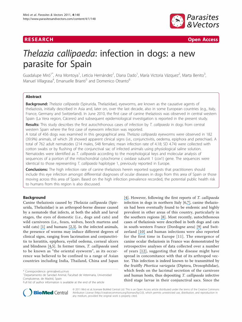
RESEARCH Open Access
Thelazia callipaeda: infection in dogs: a newparasite for SpainGuadalupe Miró1*, Ana Montoya1, Leticia Hernández1, Diana Dado1, María Victoria Vázquez2, Marta Benito3,Manuel Villagrasa4, Emanuelle Brianti5 and Domenico Otranto6
Abstract
Background: Thelazia callipaeda (Spirurida, Thelaziidae), eyeworms, are known as the causative agents ofthelaziosis, initially described in Asia and, later on, over the last decade, also in some European countries (e.g., Italy,France, Germany and Switzerland). In June 2010, the first case of canine thelaziosis was observed in central westernSpain (La Vera region, Cáceres) and subsequent epidemiological investigation is reported in the present study.
Results: This study describes the first autochthonous cases of infection by T. callipaeda in dogs from centralwestern Spain where the first case of eyeworm infection was reported.A total of 456 dogs was examined in this geographical area. Thelazia callipaeda eyeworms were observed in 182(39.9%) animals, of which 28 showed apparent clinical signs (i.e., conjunctivitis, oedema, epiphora and petechiae). Atotal of 762 adult nematodes (214 males, 548 females; mean infection rate of 4.18; SD 4.74) were collected withcotton swabs or by flushing of the conjunctival sac of infected animals using physiological saline solution.Nematodes were identified as T. callipaeda according to the morphological keys and molecular analysis ofsequences of a portion of the mitochondrial cytochrome c oxidase subunit 1 (cox1) gene. The sequences wereidentical to those representing T. callipaeda haplotype 1, previously reported in Europe.
Conclusions: The high infection rate of canine thelaziosis herein reported suggests that practitioners shouldinclude this eye infection amongst differential diagnoses of ocular diseases in dogs from this area of Spain or thosemoving across this area of Spain. Based on the high infection prevalence recorded, the potential public health riskto humans from this region is also discussed.
BackgroundCanine thelaziosis caused by Thelazia callipaeda (Spir-urida, Thelaziidae) is an arthropod-borne disease causedby a nematode that infects, at both the adult and larvalstages, the eyes of domestic (i.e., dogs and cats) andwild carnivores (i.e., foxes, wolves, beech martens andwild cats) [1] and humans [2,3]. In the infected animals,the presence of worms may induce different degrees ofclinical signs, ranging from lacrimation and conjunctivi-tis to keratitis, epiphora, eyelid oedema, corneal ulcersand blindness [4,5]. In former times, T. callipaeda usedto be known as “the oriental eyeworm”, as its occur-rence was believed to be confined to a range of Asiancountries including India, Thailand, China and Japan
[4]. However, following the first reports of T. callipaedainfection in dogs in northern Italy [6,7], canine thelazio-sis had been eventually found to be endemic and highlyprevalent in other areas of this country, particularly inthe southern regions [8]. Most recently, autochthonouscases of thelaziosis were described in both dogs and catsin south-western France (Dordogne area) [9] and Swit-zerland [10] and human infections were also reportedfor the first time in Europe [11]. The emergence ofcanine ocular thelaziosis in France was demonstrated byretrospective analyses of data collected over a numberof years [12], suggesting that the disease might havespread in concomitance with that of its arthropod vec-tor. This infection is indeed known to be transmitted bythe fruitfly Phortica variegata (Diptera, Drosophilidae),which feeds on the lacrimal secretion of the carnivoreand human hosts, thus depositing T. callipaeda infectivethird stage larvae in their conjunctival sacs. Since the
* Correspondence: [email protected] de Sanidad Animal, Facultad de Veterinaria, UniversidadComplutense, de Madrid, SpainFull list of author information is available at the end of the article
Miró et al. Parasites & Vectors 2011, 4:148http://www.parasitesandvectors.com/content/4/1/148
© 2011 Miró et al; licensee BioMed Central Ltd. This is an Open Access article distributed under the terms of the Creative CommonsAttribution License (http://creativecommons.org/licenses/by/2.0), which permits unrestricted use, distribution, and reproduction inany medium, provided the original work is properly cited.
first demonstration of the role played by P. variegata asvector of T. callipaeda under laboratory [13] and nat-ural conditions [14], knowledge on this nematode andits vector has gradually expanded. An ecological nichemodel was computed, showing that vast areas of Europeare indeed suitable for the development of P. variegata[15]. This evidence suggested that this infection couldhave spread in recent years in areas that were previouslyconsidered as non- endemic.On June 2010, a case of canine thelaziosis was
reported in an area of central western Spain (La Veraregion, Cáceres) [16]. The present study describes theoccurrence of autochthonous cases of thelaziosis in dogsin this area, presents clinical scenarios and providesmolecular data on the genetic makeup of a representa-tive sample of the nematodes collected.
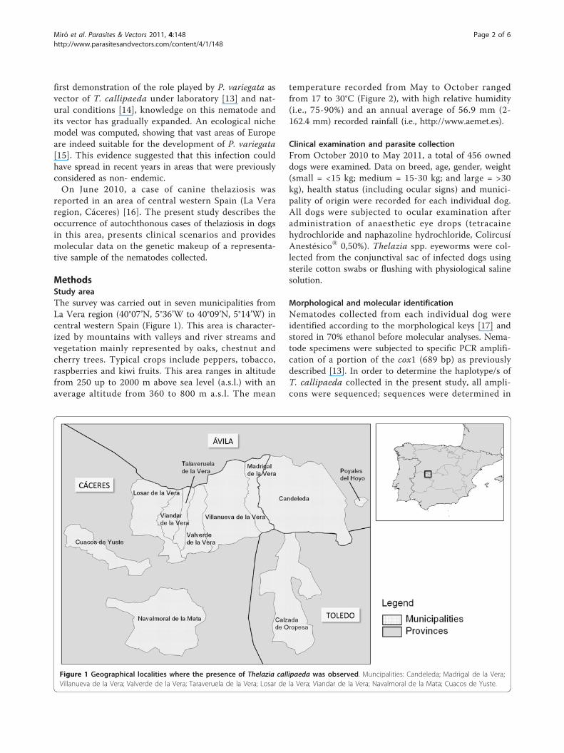
MethodsStudy areaThe survey was carried out in seven municipalities fromLa Vera region (40°07’N, 5°36’W to 40°09’N, 5°14’W) incentral western Spain (Figure 1). This area is character-ized by mountains with valleys and river streams andvegetation mainly represented by oaks, chestnut andcherry trees. Typical crops include peppers, tobacco,raspberries and kiwi fruits. This area ranges in altitudefrom 250 up to 2000 m above sea level (a.s.l.) with anaverage altitude from 360 to 800 m a.s.l. The mean
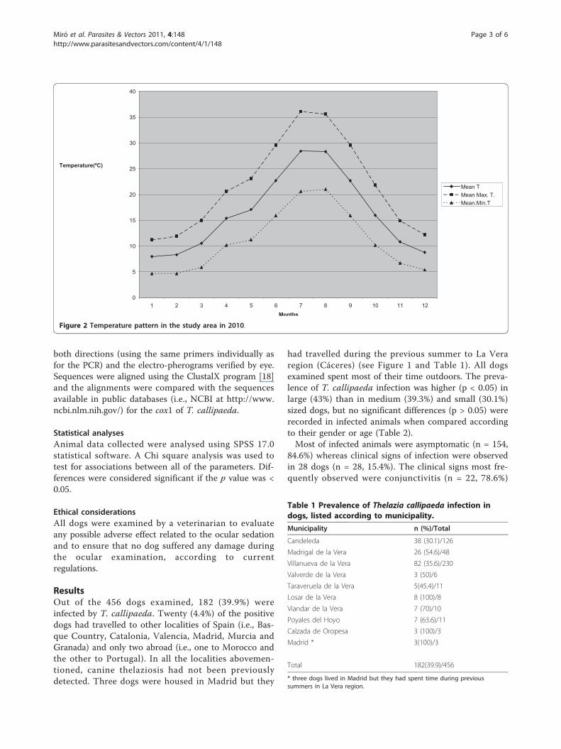
temperature recorded from May to October rangedfrom 17 to 30°C (Figure 2), with high relative humidity(i.e., 75-90%) and an annual average of 56.9 mm (2-162.4 mm) recorded rainfall (i.e., http://www.aemet.es).
Clinical examination and parasite collectionFrom October 2010 to May 2011, a total of 456 owneddogs were examined. Data on breed, age, gender, weight(small = <15 kg; medium = 15-30 kg; and large = >30kg), health status (including ocular signs) and munici-pality of origin were recorded for each individual dog.All dogs were subjected to ocular examination afteradministration of anaesthetic eye drops (tetracainehydrochloride and naphazoline hydrochloride, ColircusíAnestésico® 0,50%). Thelazia spp. eyeworms were col-lected from the conjunctival sac of infected dogs usingsterile cotton swabs or flushing with physiological salinesolution.
Morphological and molecular identificationNematodes collected from each individual dog wereidentified according to the morphological keys [17] andstored in 70% ethanol before molecular analyses. Nema-tode specimens were subjected to specific PCR amplifi-cation of a portion of the cox1 (689 bp) as previouslydescribed [13]. In order to determine the haplotype/s ofT. callipaeda collected in the present study, all ampli-cons were sequenced; sequences were determined in
Figure 1 Geographical localities where the presence of Thelazia callipaeda was observed. Muncipalities: Candeleda; Madrigal de la Vera;Villanueva de la Vera; Valverde de la Vera; Taraveruela de la Vera; Losar de la Vera; Viandar de la Vera; Navalmoral de la Mata; Cuacos de Yuste.
Miró et al. Parasites & Vectors 2011, 4:148http://www.parasitesandvectors.com/content/4/1/148
Page 2 of 6
both directions (using the same primers individually asfor the PCR) and the electro-pherograms verified by eye.Sequences were aligned using the ClustalX program [18]and the alignments were compared with the sequencesavailable in public databases (i.e., NCBI at http://www.ncbi.nlm.nih.gov/) for the cox1 of T. callipaeda.
Statistical analysesAnimal data collected were analysed using SPSS 17.0statistical software. A Chi square analysis was used totest for associations between all of the parameters. Dif-ferences were considered significant if the p value was <0.05.
Ethical considerationsAll dogs were examined by a veterinarian to evaluateany possible adverse effect related to the ocular sedationand to ensure that no dog suffered any damage duringthe ocular examination, according to currentregulations.
ResultsOut of the 456 dogs examined, 182 (39.9%) wereinfected by T. callipaeda. Twenty (4.4%) of the positivedogs had travelled to other localities of Spain (i.e., Bas-que Country, Catalonia, Valencia, Madrid, Murcia andGranada) and only two abroad (i.e., one to Morocco andthe other to Portugal). In all the localities abovemen-tioned, canine thelaziosis had not been previouslydetected. Three dogs were housed in Madrid but they
had travelled during the previous summer to La Veraregion (Cáceres) (see Figure 1 and Table 1). All dogsexamined spent most of their time outdoors. The preva-lence of T. callipaeda infection was higher (p < 0.05) inlarge (43%) than in medium (39.3%) and small (30.1%)sized dogs, but no significant differences (p > 0.05) wererecorded in infected animals when compared accordingto their gender or age (Table 2).Most of infected animals were asymptomatic (n = 154,
84.6%) whereas clinical signs of infection were observedin 28 dogs (n = 28, 15.4%). The clinical signs most fre-quently observed were conjunctivitis (n = 22, 78.6%)
0
5
10
15
20
25
30
35
40
1 2 3 4 5 6 7 8 9 10 11 12 Months
Mean T Mean Max. T. Mean.Min.T
Temperature(ºC)
Figure 2 Temperature pattern in the study area in 2010.
Table 1 Prevalence of Thelazia callipaeda infection indogs, listed according to municipality.
Municipality n (%)/Total
Candeleda 38 (30.1)/126
Madrigal de la Vera 26 (54.6)/48
Villanueva de la Vera 82 (35.6)/230
Valverde de la Vera 3 (50)/6
Taraveruela de la Vera 5(45.4)/11
Losar de la Vera 8 (100)/8
Viandar de la Vera 7 (70)/10
Poyales del Hoyo 7 (63.6)/11
Calzada de Oropesa 3 (100)/3
Madrid * 3(100)/3
Total 182(39.9)/456
* three dogs lived in Madrid but they had spent time during previoussummers in La Vera region.
Miró et al. Parasites & Vectors 2011, 4:148http://www.parasitesandvectors.com/content/4/1/148
Page 3 of 6
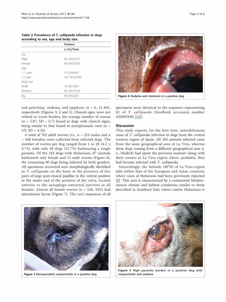
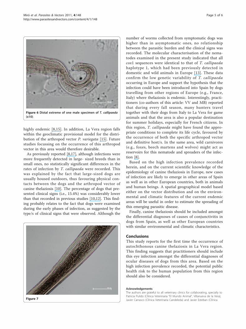
and petechiae, oedema, and epiphora (n = 6, 21.4%),respectively (Figures 3, 4 and 5). Clinical signs were notrelated to worm burden, the average number of worms(n = 5.87, SD = 6.7) found in dogs with clinical signs,being similar to that found in asymptomatic ones (n =3.9, SD = 4.35).A total of 762 adult worms (i.e., n = 214 males and n
= 548 females) were collected from infected dogs. Thenumber of worms per dog ranged from 1 to 28 (4.2 ±4.73), with only 58 dogs (31.7%) harbouring a singleparasite. Of the 182 dogs with thelaziosis, 87 animalsharboured only female and 15 male worms (Figure 6),the remaining 80 dogs being infected by both genders.All specimens recovered were morphologically identifiedas T. callipaeda on the basis of the presence of fivepairs of large post-cloacal papillae in the ventral positionin the males and of the position of the vulva, locatedanterior to the oesophago-intestinal junction in allfemales. Almost all female worms (n = 526, 95%) hadintrauterine larvae (Figure 7). The cox1 sequences of all
specimens were identical to the sequence representingh1 of T. callipaeda (GenBank accession numberAM042549; [13]).
DiscussionThis study reports, for the first time, autochthonouscases of T. callipaeda infection in dogs from the centralwestern region of Spain. All 182 animals infected camefrom the same geographical area of La Vera, whereasthree dogs coming from a different geographical area (i.e., Madrid) had spent the previous summer along withtheir owners in La Vera region where, probably, theyhad become infected with T. callipaeda.Interestingly, the latitude (40°N) of La Vera region
falls within that of the European and Asian countrieswhere cases of thelaziosis had been previously reported[8]. This area is characterized by a continental Mediter-ranean climate and habitat conditions similar to thosedescribed in Southern Italy where canine thelaziosis is
Figure 5 High parasitic burden in a positive dog withconjunctivitis and oedema.
Table 2 Prevalence of T. callipaeda infection in dogsaccording to sex, age and body size.
Positive
n (%)/Total
Sex
Male 99 (39.4)/251
Female 83 (40.5)/205
Age
< 1 year 15 (23.8)/63
>1 year 167 (42.5)/393
Body size
Small 19 (30.1)/63
Medium 64 (39.3)/163
Big 99 (43)/230
Figure 3 Mucopurulent conjunctivitis in a positive dog.
Figure 4 Oedema and chemosis in a positive dog.
Miró et al. Parasites & Vectors 2011, 4:148http://www.parasitesandvectors.com/content/4/1/148
Page 4 of 6
highly endemic [8,15]. In addition, La Vera region fallswithin the geoclimatic provisional model for the distri-bution of the arthropod vector P. variegata [15]. Futurestudies focussing on the occurrence of this arthropodvector in this area would therefore desirable.As previously reported [8,17], although infections were
more frequently detected in large- sized breeds than insmall ones, no statistically significant differences in therates of infection by T. callipaeda were recorded. Thiswas explained by the fact that large-sized dogs areusually housed outdoors, thus favouring physical con-tacts between the dogs and the arthropod vector ofcanine thelaziosis [10]. The percentage of dogs that pre-sented clinical signs (i.e., 15.4%) was considerably lowerthan that recorded in previous studies [10,12]. This find-ing probably relates to the fact that dogs were examinedduring the early phases of infection, as suggested by thetype/s of clinical signs that were observed. Although the
number of worms collected from symptomatic dogs washigher than in asymptomatic ones, no relationshipbetween the parasitic burden and the clinical signs wasrecorded. The molecular characterisation of the nema-todes examined in the present study indicated that allcox1 sequences were identical to that of T. callipaedahaplotype 1, which had been previously detected indomestic and wild animals in Europe [13]. These dataconfirm the low genetic variability of T. callipaedaoccurring in Europe and support the hypothesis that theinfection could have been introduced into Spain by dogstravelling from other regions of Europe (e.g., France,Italy) where thelaziosis is endemic. Interestingly, practi-tioners (co-authors of this article: VV and MB) reportedthat during every fall season, many hunters traveltogether with their dogs from Italy to La Vera for gameanimals and that the area is also a popular destinationfor summer holidays, especially for French citizens. Inthis region, T. callipaeda might have found the appro-priate conditions to complete its life cycle, favoured bythe occurrence of both the specific arthropod vectorand definitive host/s. In the same area, wild carnivores(e.g., foxes, beech martens and wolves) might act asreservoirs for this nematode and spreaders of the infec-tion [8].Based on the high infection prevalence recorded
herein, and on the current scientific knowledge of theepidemiology of canine thelaziosis in Europe, new casesof infection are likely to emerge in other areas of Spainas well as in other European countries, both in animalsand human beings. A spatial geographical model basedeither on the vector distribution and on the environ-mental and climatic features of the current endemicareas will be useful in order to estimate the spreading ofthis emerging parasitic disease.Finally, canine thelaziosis should be included amongst
the differential diagnoses of causes of conjunctivitis indogs from Spain, as well as other European countrieswith similar environmental and climatic characteristics.
ConclusionsThis study reports for the first time the occurrence ofautochthonous canine thelaziosis in La Vera region.This finding suggests that practitioners should includethis eye infection amongst the differential diagnoses ofocular diseases of dogs from this area. Based on thehigh infection prevalence recorded, the potential publichealth risk to the human population from this regionshould also be considered.
AcknowledgementsThe authors are grateful to all veterinary clinics for collaborating, specially toPatricia Pulido (Clínica Veterinaria “El Mundo Animal”, Villanueva de la Vera),Javier Carrasco (Clínica Veterinaria Candeleda) and Javier Esteban (ClínicaFigure 7
Figure 6 Distal extreme of one male specimen of T. callipaeda(x10).
Miró et al. Parasites & Vectors 2011, 4:148http://www.parasitesandvectors.com/content/4/1/148
Page 5 of 6
Veterinaria Ocaña, Móstoles, Madrid) for their valuable help recording someclinical cases.Artemisa James, Rocío Checa, Cristina Rupérez y Carmen Miranda areacknowledged for their assistance with the laboratory procedures andsampling animals in the field studies and Cinzia Cantacessi (University ofMelbourne, Australia) for her suggestions on the manuscript.
Author details1Departamento de Sanidad Animal, Facultad de Veterinaria, UniversidadComplutense, de Madrid, Spain. 2Clínica Veterinaria “El Mundo Animal”,Villanueva de la Vera, Cáceres, Spain. 3Clínica Veterinaria Candeleda,Candeleda, Ávila, Spain. 4Centro Veterinario Oftalmológico Goya, Madrid,Spain. 5Dipartimento di Sanità Pubblica Veterinaria, Facoltà di MedicinaVeterinaria, Università degli Studi di Messina, Messina, Italy. 6Dipartimento diSanità Pubblica e Zootecnia, Università degli Studi di Bari, Valenzano (Bari),Italy.
Authors’ contributionsGM conceived and coordinated the study, and participated in its design, thefield studies and drafted the manuscript. DO participated in carrying out themolecular assays and helped to draft the manuscript. LH and DDparticipated in the field studies and carrying out the diagnostic assays. MVVparticipated in the field studies and sampling the dogs. MB and MVparticipated in sampling the dogs. EB participated in data elaboration andhelped to draft the manuscript. AM participated in the field studies andcarrying out the diagnostic assays, performed the statistical analysis, andhelped to draft the manuscript. All authors read and approved the finalmanuscript.
Competing interestsThe authors declare that they have no competing interests.
Received: 10 June 2011 Accepted: 27 July 2011 Published: 27 July 2011
References1. Otranto D, Dantas-Torres F, Mallia E, DiGeronimo PM, Brianti E, Testini G,
Traversa D, Lia RP: Thelazia callipaeda (Spirurida, Thelaziidae) in wildanimals: report of new host species and ecological implications. VetParasitol 2009, 166:262-267.
2. Shen J, Gasser RB, Chu D, Wang Z, Yuan X, Cantacessi C, Otranto D: Humanthelaziosis- a neglected parasitic disease of the eye. J Parasitol 2006,92:872-875.
3. Otranto D, Eberhard ML: Zoonotic helminths affecting the human eye.Parasit Vectors 2011, 4:41.
4. Anderson RC: Nematode parasites of vertebrates: their development andtransmission. CABI Publishing, Guilford, UK 2000, 404-407.
5. Otranto D, Traversa D: Thelazia eyeworm: an original endo- and ecto-parasitic nematode. Trends Parasitol 2005, 21:1-4.
6. Rossi L, Bertaglia PP: Presence of Thelazia callipaeda Railliet & Henry,1910, in Piedmont, Italy. Parassitologia 1989, 31:167-172.
7. Otranto D, Dantas-Torres F: Canine and feline vector-borne diseases inItaly: current situation and perspectives. Parasit Vectors 2010, 3:2.
8. Otranto D, Ferroglio E, Lia RP, Traversa D, Rossi L: Current status andepidemiological observation of Thelazia callipaeda (Spirurida,Thelaziidae) in dogs, cats and foxes in Italy: a “coincidence” or aparasitic disease of the Old Continent? Vet Parasitol 2003, 116:315-325.
9. Dorchies P, Chaudieu G, Siméon LA, Cazalot G, Cantacessi C, Otranto D:Reports of autochthonous eyeworm infection by Thelazia callipaeda(Spirurida, Thelaziidae) in dogs and cat from France. Vet Parasitol 2007,149:294-297.
10. Malacrida F, Hegglin D, Bacciarini L, Otranto D, Nägeli F, Nägeli C,Bernasconi C, Scheu U, Balli A, Marenco M, Togni L, Deplazes P,Schnyder M: Emergence of canine ocular thelaziosis caused by Thelaziacallipaeda in southern Switzerland. Vet Parasitol 2008, 157:321-327.
11. Otranto D, Dutto M: Human thelaziasis, Europe. Emerg Infect Dis 2008,14:647-649.
12. Ruytoor P, Déan E, Pennant O, Dorchies P, Chermette R, Otranto D,Guillot J: Ocular thelaziosis in dogs, France. Emerg Infect Dis 2010,16:1943-1945.
13. Otranto D, Lia RP, Cantacessi C, Testini G, Troccoli A, Shen JL, Wang ZX:Nematode biology and larval development of Thelazia callipaeda
(Spirurida, Thelaziidae) in the drosophilid intermediate host in Europeand China. Parasitology 2005, 131:847-855.
14. Otranto D, Cantacessi C, Testini G, Lia RP: Phortica variegata as anintermediate host of Thelazia callipaeda under natural conditions:evidence for pathogen transmission by a male arthropod vector. Int JParasitol 2006, 36:1167-1173.
15. Otranto D, Brianti E, Cantacessi C, Lia RP, Máca J: The zoophilic fruitflyPhortica variegata: morphology, ecology and biological niche. Med VetEntomol 2006, 20:358.
16. Montoya A, Vázquez MV, Pulido P, Hernández L, Dado D, Otranto D, Miró G:Thelaziosis ocular canina. ¿Una parasitosis emergente en España?Consulta Difus Vet 2011, 178:43-48.
17. Otranto D, Lia RP, Traversa D, Giannetto S: -64. Thelazia callipaeda(Spirurida, Thelaziidae) of carnivores and humans: morphological studyby light and scanning electron microscopy. Parassitologia 2003,45:125-133.
18. Thompson JD, Gibson TJ, Plewniak F, Jeanmougin F, Higgins DG: TheClustal X windows interface: flexible strategies for multiple sequencealignment aided by quality analysis tools. Nucleic Acids Res 1997,24:4876-4882.
doi:10.1186/1756-3305-4-148Cite this article as: Miró et al.: Thelazia callipaeda: infection in dogs: anew parasite for Spain. Parasites & Vectors 2011 4:148.
Submit your next manuscript to BioMed Centraland take full advantage of:
• Convenient online submission
• Thorough peer review
• No space constraints or color figure charges
• Immediate publication on acceptance
• Inclusion in PubMed, CAS, Scopus and Google Scholar
• Research which is freely available for redistribution
Submit your manuscript at www.biomedcentral.com/submit
Miró et al. Parasites & Vectors 2011, 4:148http://www.parasitesandvectors.com/content/4/1/148
Page 6 of 6
Copyright © 2022 FDOKUMEN