THE USE OF PHOSPHORUS 32 IN STUDIES ON PLASMODIUM ...
13
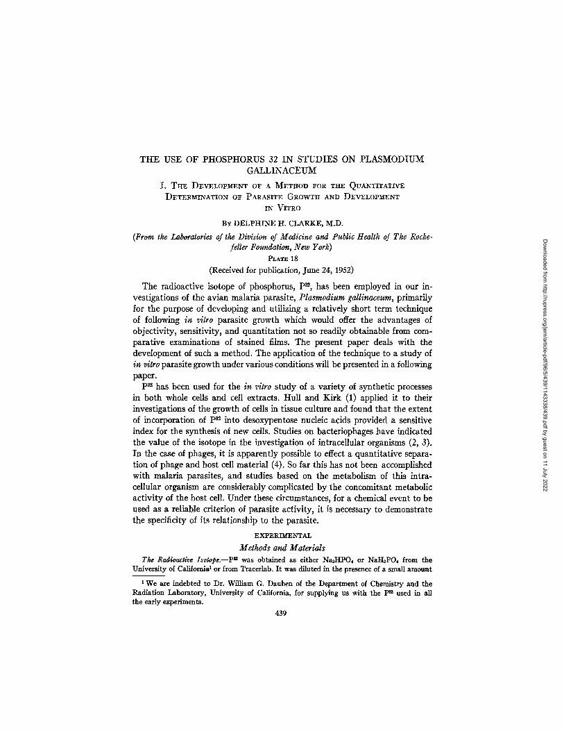
THE USE OF PHOSPHORUS 32 IN STUDIES ON PLASMODIUM GALLINACEUM I. Tm~ DEVELOPMENT OF A METHOD FOR THE QUANTITATrV~ DETERMINATION OF PARASITE GROWTH AND DEVELOPMENT IN VITRO BY DELPHINE H. CLARKE, M.D. (Prom the Laboratories of the Division of Medicine and Public Health of The Roche- feller Foundation, New York) PLAaX18 (Received for publication, June 24, 1952) The radioactive isotope of phosphorus, P*~, has been employed in our in- vestigations of the avian malaria parasite, Plasmodium gallinaceum, primarily for the purpose of developing and utilizing a relatively short term technique of following in vitro parasite growth which would offer the advantages of objectivity, sensitivity, and quantitation not so readily obtainable from com- parative examinations of stained films. The present paper deals with the development of such a method. The application of the technique to a study of in vitro parasite growth under various conditions will be presented in a following paper. P~ has been used for the in vitro study of a variety of synthetic processes in both whole cells and cell extracts. Hull and Kirk (1) applied it to their investigations of the growth of cells in tissue culture and found that the extent of incorporation of p82 into desoxypentose nucleic acids provided a sensitive index for the synthesis of new cells. Studies on bacteriophages have indicated the value of the isotope in the investigation of intracellular organisms (2, 3). In the case of phages, it is apparently possible to effect a quantitative separa- tion of phage and host cell material (4). So far this has not been accomplished with malaria parasites, and studies based on the metabolism of this intra- cellular organism are considerably complicated by the concomitant metabolic activity of the host cell. Under these circumstances, for a chemical event to be used as a reliable criterion of parasite activity, it is necessary to demonstrate the specificity of its relationship to the parasite. EXPEI~~ N T A L Methods and Materials The Radioactive Isotope.--P ~ was obtained as either Na~-IPO4 or NaI-I2PO4 from the University of California1 or from Tracerlab. It was diluted in the presence of a small amount We are indebted to Dr. William G. Danben of the Department of Chemistry and the Radiation Laboratory, University of California, for supplying us with the P~ used in all the early experiments. 439 Downloaded from http://rupress.org/jem/article-pdf/96/5/439/1143338/439.pdf by guest on 11 July 2022
-
Upload
khangminh22 -
Category
Documents
-
view
0 -
download
0
Transcript of THE USE OF PHOSPHORUS 32 IN STUDIES ON PLASMODIUM ...
THE USE OF PHOSPHORUS 32 IN STUDIES ON PLASMODIUM GALLINACEUM
I. Tm~ DEVELOPMENT OF A METHOD FOR THE QUANTITATrV~ DETERMINATION OF PARASITE GROWTH AND DEVELOPMENT
IN VITRO
BY DELPHINE H. CLARKE, M.D.
(Prom the Laboratories of the Division of Medicine and Public Health of The Roche- feller Foundation, New York)
PLAaX 18 (Received for publication, June 24, 1952)
The radioactive isotope of phosphorus, P*~, has been employed in our in- vestigations of the avian malaria parasite, Plasmodium gallinaceum, primarily for the purpose of developing and utilizing a relatively short term technique of following in vitro parasite growth which would offer the advantages of objectivity, sensitivity, and quantitation not so readily obtainable from com- parative examinations of stained films. The present paper deals with the development of such a method. The application of the technique to a study of in vitro parasite growth under various conditions will be presented in a following paper.
P~ has been used for the in vitro study of a variety of synthetic processes in both whole cells and cell extracts. Hull and Kirk (1) applied it to their investigations of the growth of cells in tissue culture and found that the extent of incorporation of p82 into desoxypentose nucleic acids provided a sensitive index for the synthesis of new cells. Studies on bacteriophages have indicated the value of the isotope in the investigation of intracellular organisms (2, 3). In the case of phages, it is apparently possible to effect a quantitative separa- tion of phage and host cell material (4). So far this has not been accomplished with malaria parasites, and studies based on the metabolism of this intra- cellular organism are considerably complicated by the concomitant metabolic activity of the host cell. Under these circumstances, for a chemical event to be used as a reliable criterion of parasite activity, it is necessary to demonstrate the specificity of its relationship to the parasite.
EXPEI~ ~ N T A L
Methods and Materials The Radioactive Isotope.--P ~ was obtained as either Na~-IPO4 or NaI-I2PO4 from the
University of California 1 or from Tracerlab. I t was diluted in the presence of a small amount
We are indebted to Dr. William G. Danben of the Department of Chemistry and the Radiation Laboratory, University of California, for supplying us with the P~ used in all the early experiments.
439
Dow
nloaded from http://rupress.org/jem
/article-pdf/96/5/439/1143338/439.pdf by guest on 11 July 2022
440 pS2 AND PLAS~ODIUM GALLINACEUM. I
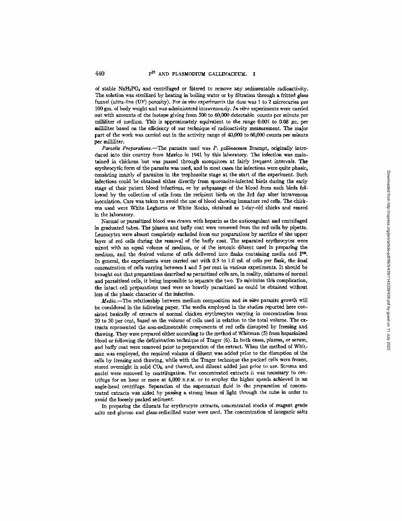
of stable NaHsPO, and centrifuged or filtered to remove any sedimentable radioactivity. The solution was sterilized by heating in boiling water or by filtration through a fritted glass funnel (ultra-fine (UF) porosity). For in v/vo experiments the dose was 1 to 2 microcuries per 100 gin. of body weight and was administered intravenously. In vitro experiments were carried out with amounts of the isotope giving from 500 to 60,000 detectable counts per minute per milliliter of medium. This is approximately equivalent to the range 0.001 to 0.08 /zc. per milliliter based on the efficiency of our technique of radioactivity measurement. The major part of the work was carried out in the activity range of 40,000 to 60,000 counts per minute per milliliter.
Para~te Preparations.--The parasite used was P. gallinaceum Brnmpt, originally intro- duced into this country from Mexico in 1941 by this laboratory. The infection was main- tained in chickens but was passed through mosquitoes at fairly frequent intervals. The erythrocytic form of the parasite was used, and in most cases the infections were quite phasic, consisting mainly of parasites in the trophozoite stage at the start of the experiment. Such infections could be obtained either directly from sporozoite-infected birds during the eariy stage d their patent blood infections, or by subpassage of the blood from such birds fol- lowed by the collection of cells from the recipient birds on the 3rd day after intravenous inoculation. Care was taken to avoid the use of blood showing immature red cells. The chick- ens used were White Leghorns or White Rocks, obtained as l-day-old chicks and reared in the laboratory.
Normal or parasitized blood was drawn with heparin as the anticoagulant and centrifuged in graduated tubes. The plasma and buffy coat were removed from the red cells by pipette. Leucocytes were almost completely excluded from our preparations by sacrifice of the upper layer of red cells during the removal of the buffy coat. The separated erythrocytes were mixed with an equal volume of medium, or of the isotonic diluent used in preparing the medium, and the desired volume of cells delivered into flasks containing media and P~. In general, the experiments were carried out with 0.5 to 1.0 ml. of cells per flask, the final concentration of cells varying between I and 5 per cent in various experiments. I t should be brought out that preparations described as parasitized cells are, in reality, mixtures d normal and parasitized cells, it being impossible to separate the two. To minimize this complication, the intact cell preparations used were as heavily parasitized as could be obtained without loss of the phasic character of the infection.
Media.--The relationship between medium composition and in dtro parasite growth will be considered in the following paper. The media employed in the studies reported here con- slsted basically of extracts of normal chicken erythrocytes varying in concentration from 20 to 30 per cent, based on the volume of cells used in relation to the total volume. The ex- tracts represented the non-sedimentable components of red cells disrupted by freezing and thawing. They were prepared either according to the method of Whitman (5) from heparinized blood or following the defibtination technique of Trager (6). In both cases, plasma, or serum, and buffy coat were removed prior to preparation of the extract. When the method of Whit- man was employed, the required volume of diluent was added prior to the disruption of the cells by freezing and thawing, while with the Trager technique the packed cells were frozen, stored overnight in solid CO~, and thawed, and diluent added just prior to use. Stroma and nuclei were removed by centrifugation. For concentrated extracts it was necessary to cen- trifuge for an hour or more at 4,000 R.I,.M. or to employ the higher speeds achieved in an angie-head centrifuge. Separation of the supernatant fluid in the preparation d concen- trated extracts was aided by passing a strong beam of light through the tube in order to avoid the loosely packed sediment.
In preparing the diluents for erythrocyte extracts, concentrated stocks of reagent grade salts and glucose and glass-redistilled water were used. The concentration of inorganic salts
Dow
nloaded from http://rupress.org/jem
/article-pdf/96/5/439/1143338/439.pdf by guest on 11 July 2022
DELPHINE H. CLARKE 441
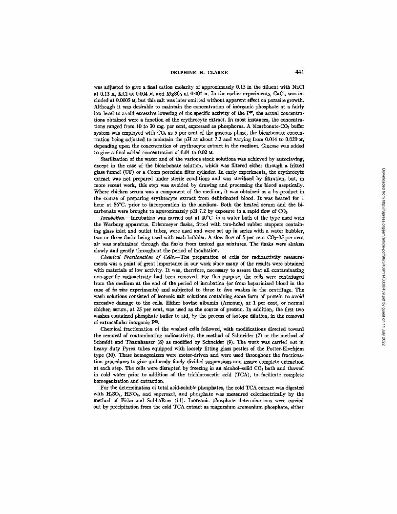
was adjusted to give a final cation molarity of approximately 0.15 in the diluent with NaCI at 0.13 xt, KC1 at 0.004 M, and MgSO4 at 0.001 x¢. In the earlier experiments, CaC12 was in- cluded at 0.0005 M, but this salt was later omitted without apparent effect on parasite growth. Although it was desirable to maintain the concentration of inorganic phosphate at a fairly low level to avoid excessive lowering of the specific activity of the 1 ~ , the actual concentra- tions obtained were a function of the erythrocyte extract. In most instances, the concentra- tions ranged from 10 to 30 rag. per cent, expressed as phosphorus. A bicarbonate-COs buffer system was employed with CO~ as 5 per cent of the gaseous phase, the bicarbonate concen- tration being adjusted to maintain the pH at about 7.2 and varying from 0.014 to 0.020 x¢, depending upon the concentration of erythrocyte extract in the medium. Glucose was added to give a final added concentration of 0.01 to 0.02 ~.
Sterilization of the water and of the various stock solutions was achieved by autoclaving, except in the case of the bicarbonate solution, which was filtered either through a fritted glass funnel (UF) or a Coors porcelain filter cylinder. In early experiments, the erythrocyte extract was not prepared under sterile conditions and was sterilized by filtration, but, in more recent work, this step was avoided by drawing and processing the blood aseptically. Where chicken serum was a component of the medium, it was obtained as a by-product in the course of preparing erythrocyte extract from defibrinated blood. I t was heated for 1 hour at 56°C. prior to incorporation in the medium. Both the heated serum and the bi- carbonate were brought to approximately pH 7.2 by exposure to a rapid flow of COs.
Incubation.--incubation was carded out at 40°C. in a water bath of the type used with the Warburg apparatus. Erlenmeyer flasks, fitted with two-holed rubber stoppers contain- ing glass inlet and outlet tubes, were used and were set up in series with a water bubbler, two or three flasks being used with each bubbler. A slow flow of 5 per cent COr-95 per cent air was maintained through the flasks from tanked gas mixtures. The flasks were shaken slowly and gently throughout the period of incubation.
Chemival Fravtionation of Cdls.--The preparation of cells for radioactivity measure- ments was a point of great importance in our work since many of the results were obtained with materials of low activity. I t was, therefore, necessary to assure that all contaminating non-specific radioactivity had been removed. For this purpose, the cells were centrifuged from the medium at the end of the period of incubation (or from heparinized blood in the case of in v/vo experiments) and subjected to three to five washes in the centrifuge. The wash solutions consisted of isotonic salt solutions containing some form of protein to avoid excessive damage to the cells. Either bovine albumin (Armour), at 1 per cent, or normal chicken serum, at 25 per cent, was used as the source of protein. In addition, the first two washes contained phosphate buffer to aid, by the process of isotope dilution, in the removal of extracellular inorganic pn.
Chemical fractionation of the washed cells followed, with modifications directed toward the removal of contaminating radioactivity, the method of Schneider (7) or the method of Schmidt and Thannhanser (8) as modified by Schneider (9). The work was carried out in heavy duty Pyrex tubes equipped with loosely fitting glass pestles of the Potter-Elvehjem type (10). These homogenizers were motor-driven and were used throughout the fractiona- tion procedures to give uniformly finely divided suspensions and insure complete extraction at each step. The cells were disrupted by freezing in an alcohol-solid COs bath and thawed in cold water prior to addition of the trichloroacetic acid (TCA), to facilitate complete homogenization and extraction.
For the determination of total acid-soluble phosphates, the cold TCA extract was digested with I-I2SO4, HNOa, and superoxol, and phosphate was measured colorimetrically by the method of Fiske and SubbaRow (11). Inorganic phosphate determinations were carded out by precipitation from the cold TCA extract as magnesium ammonium phosphate, either
Dow
nloaded from http://rupress.org/jem
/article-pdf/96/5/439/1143338/439.pdf by guest on 11 July 2022
442 p32 AND PLAS~ODIU~f GAI,LINACE~. I
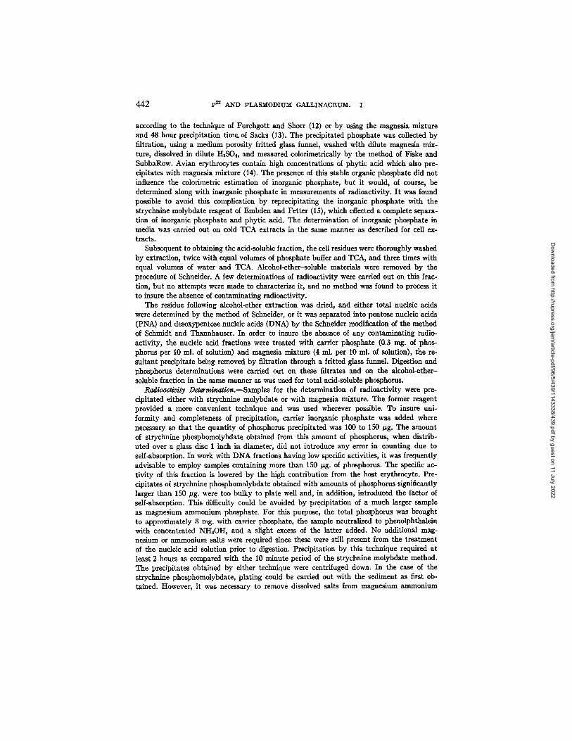
according to the technique of Furchgott and Shorr (12) or by using the magnesia mixture and 48 hour precipitation time. of Sacks (13). The precipitated phosphate was collected by filtration, using a medium porosity fritted glass funnel, washed with dilute magnesia mix- ture, dissolved in dilute H2S04, and measured colorimetrieally by the method of Fiske and SubbaRow. Avian erythrocytes contain high concentrations of phytic acid which also pre- cipitates with magnesia mixture (14). The presence of this stable organic phosphate did not influence the colorimetric estimation of inorganic phosphate, but it would, of course, be determined along with inorganic phosphate in measurements of radioactivity. I t was found possible to avoid this complication by reprecipitating the inorganic phosphate with the strychnine molybdate reagent of Embden and Fetter (15), which effected a complete separa- tion of inorganic phosphate and phytic acid. The determination of inorganic phosphate in media was carried out on cold TCA extracts in the same manner as described for cell ex- tracts.
Subsequent to obtaining the acid-soluble fraction, the cell residues were thoroughly washed by extraction, twice with equal volumes of phosphate buffer and TCA, and three times with equal volumes of water and TCA. Alcohol-ether-soluble materials were removed by the procedure of Schneider. A few determinations of radioactivity were carried out on this frac- tion, but no attempts were made to characterize it, and no method was found to process it to insure the absence of contaminating radioactivity.
The residue following alcohol-ether extraction was dried, and either total nucleic acids were determined by the method of Schneider, or it was separated into pentose nucleic acids (PNA) and desoxypentose nucleic acids (DNA) by the Schneider modification of the method of Schmidt and Thannhanser. In order to insure the absence of any contaminating radio- activity, the nucleic acid fractions were treated with carrier phosphate (0.3 rag. of phos- phorus per 10 ml. of solution) and magnesia mixture (4 ml. per 10 ml. of solution), the re- sultant precipitate being removed by filtration through a flitted glass funnel. Digestion and phosphorus determinations were carried out on these filtrates and on the alcohol-ether- soluble fraction in the same manner as was used for total acid-soluble phosphorus.
Radioactivity Determination.--Samples for the determination of radioactivity were pre- cipitated either with strychnine molybdate or with magnesia mixture. The former reagent provided a more convenient technique and was used wherever possible. To insure uni- formity and completeness of precipitation, carrier inorganic phosphate was added where necessary so that the quantity of phosphorus precipitated was 100 to 150 Izg. The amount of strychnine phosphomolybdate obtained from this amount of phosphorus, when distrib- uted over a glass disc 1 inch in diameter, did not introduce any error in counting due to serf-absorption. In work with DNA fractions having low specific activities, it was frequently advisable to employ samples containing more than 150 #g. of phosphorus. The specific ac- tivity of this fraction is lowered by the high contribution from the host erythrocyte. Pre- cipitates of strychnine phosphomolybdate obtained with amounts of phosphorus significantly larger than 150 /zg. were too bulky to plate well and, in addition, introduced the factor of self-absorption. This difficulty could be avoided by precipitation of a much larger sample as magnesium ammonium phosphate. For this purpose, the total phosphorus was brought to approximately 8 nag. with carrier phosphate, the sample neutralized to phenolphthalein with concentrated NH,OH, and a slight excess of the latter added. No additional mag- nesinm or ammonium salts were required since these were still present from the treatment of the nucleic acid solution prior to digestion. Precipitation by this technique required at least 2 hours as compared with the 10 minute period of the strychnine molybdate method. The precipitates obtained by either technique were centrifuged down. In the ease of the strychnine phosphomolybdate, plating could be carried out with the sediment as first ob- tained. However, it was necessary to remove dissolved salts from magnesium ammonium
Dow
nloaded from http://rupress.org/jem
/article-pdf/96/5/439/1143338/439.pdf by guest on 11 July 2022
DELPHINE H. CLARKE 443
phosphate precipitates by washing twice with dilute NI-I40H in order to obtain satisfactory plates. In either case, the precipitate was plated as a slurry in a small volume of water on uniform flat glass discs 1 inch in diameter, the tubes being successively rinsed with small amounts of water and finally with a few drops of dilute NaOH, for the strychnine phos- phomolybdate, or of dilute HCI, for magnesium ammonium phosphate. Even spreading was aided by the use of 1 to 2 drops of 10 per cent tween 80, t and the plates where dried under a heating lamp.
Counting was done with a Tracerlab autoscaler and thin mica end-window Geiger- Mueller tube which was lead-shielded. The sample geometry was such that the efficiency of our system was approximately 38 per cent, as estimated from results obtained with Na- tional Bureau of Standards P".
Method of Expr~si, g Re.cults.--In the initial data presented, the results are expressed as counts per minute per milliliter of ceils or as the specific activity (counts per minute per microgram of phosphorus) of each fraction of the cells. The remainder of the data of this paper and all the data of the following paper are presented as the micrograms of phosphorus incorporated into nucleic acids per 1 rag. of total nucleic acid phosphorus or per 1 nag. of DNA phosphorus. This value was obtained by dividing the counts per minute obtained on each sample by the specific activity of the intracellular inorganic phosphorus and referring the resultant figure to a standard sample size of 1 rag. of total nucleic acid or DNA phos- phorus by dividing by the nucleic acid phosphorus value obtained for each sample. Total nucleic acid or DNA phosphorus provides a convenient method of measuring the size of the cell sample actually assayed since the bulk of this value is host cell DNA. This method of expressing the results compensates for variations in inorganic phosphate specific activity and avoids errors due to differences in initial sample sizes or losses occurring during the processing of the material. It should be noted that although this permits an accurate com- parison of results within any one experiment, comparisons between experiments will be influenced by parasite density (the number of parasites per unit number of erythrocytes). Experiments initiated with more heavily parasitized preparations will show higher phos- phorus incorporation values per milligram of nucleic acid phosphorus than will less densely parasitized preparations although individual parasite growth may be the same. Since our studies were concerned with comparative results within experiments, this was not a serious objection although it was attempted to keep the degree of parasitization fairly uniform. This was, however, not always possible to realize.
RESULTS
Distribution of p32 in Fractions of Normal and of Parasitized Cells.--Pre- l iminary studies on the total levels of P~ found in normal and in parasitized
erythrocytes after exposure, either in vivo or in vitro, showed the act ivi ty of parasitized cells to be several times that of a similar volume of normal cells a t any time interval investigated. However, i t was realized that the exchange of highly active inorganic phosphorus between cells and surrounding media would tend to obscure possible significant metabolic differences between the two types of cells when comparison was based on total isotope levels. There- fore, such cells were subjected to fractionation according to the technique of Schneider, whereby the separation of the acid-soluble fraction, containing the highly active intracellular inorganic phosphate, from the other cell compo-
2 Atlas Powder Company.
Dow
nloaded from http://rupress.org/jem
/article-pdf/96/5/439/1143338/439.pdf by guest on 11 July 2022

444 p32 AND PLASMODIU~ GALLINACEUM. I
nents can be achieved. Table I gives the results of the division of normal and parasitized cells into acid-soluble, alcohol-ether-soluble, and nucleic acid frac- tions. In the in vivo experiment, p32 was administered to normal and para- sitized birds 22 hours prior to the collection of cells. The in vitro experiment was carried out with 1 per cent cell suspensions incubated for 22 hours in a medium of erythrocyte extract having a p3, activity of 636 counts per minute per milliliter. The data are presented as the activity of the total of each frac- tion obtained from 1 ml. of cells.
The pattern of distribution of the isotope is quite distinctive in the para- sitized cells in which the alcohol-ether-soluble and nucleic acid fractions make up more than half of the total. In contrast, the bulk of the p3~ of normal cells is to be found in the acid-soluble fraction. While the relative values for the alcohol-ether-soluble fractions are lower in the in vitro than in the in vivo
TABLE I F n Acgvity of Fractions of Normal and Parasitized Erytkrocytes after Exposure for gg Hours
in Vivo or in Vitro
~'n ~ 0
/*n ~tro
J~rytlaroeytes
Normal Parasit ized
Normal Parasit ized
pa~ activlty of cell fraction, counts/min./ral, ccg~
Acid-soluble
640 2100
440 860
Alcohol-ether- soluble
210 2230
60 340
Nucleic acid
30 1700
40 730
experiment, the similarity of the ratio of distribution of the isotope in the acid-soluble and nucleic acid fractions under the two conditions suggests that the metabolic status of the cells is not markedly altered under the in ~tro conditions used, at least with respect to the incorporation of the isotope into nucleic acids. As a consequence of the use of this type of fractionation, we find, then, differentials in favor of the parasitized preparation of considerable magnitude. This is especially true of the nucleic acid fraction in which the isotope concentration in normal cells is relatively very low.
Absence of Effect of p3~ on Cellular or Parasite Ac t i v i t y . -~ f considerable importance to the interpretation of all biological studies employing isotopes is the attainment of evidence that the isotope, per se, is not influencing the results by the production of abnormal effects. To this end, parallel prepara- tions were made upon several occasions which were identical in all respects save for the presence or absence of p32. Examination of stained films of the preparations after incubation for 18 to 22 hours failed to reveal any difference in host cell or parasite morphology or in apparent parasite growth attributable
Dow
nloaded from http://rupress.org/jem
/article-pdf/96/5/439/1143338/439.pdf by guest on 11 July 2022
DELPHINE ~I. CLARKE 445
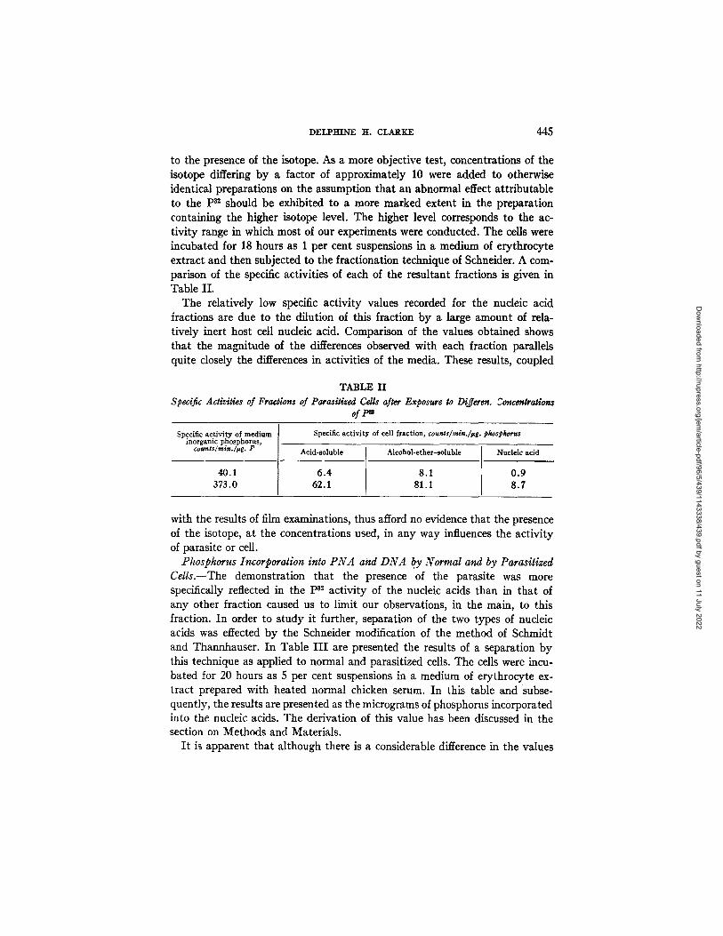
to the presence of the isotope. As a more objective test, concentrations of the isotope differing by a factor of approximately 10 were added to otherwise identical preparations on the assumption that an abnormal effect attributable to the ps2 should be exhibited to a more marked extent in the preparation containing the higher isotope level. The higher level corresponds to the ac- tivity range in which most of our experiments were conducted. The cells were incubated for 18 hours as 1 per cent suspensions in a medium of erythrocyte extract and then subjected to the fractionation technique of Schneider. A com- parison of the specific activities of each of the resultant fractions is given in Table II.
The relatively low specific activity values recorded for the nucleic acid fractions are due to the dilution of this fraction by a large amount of rela- tively inert host cell nucleic acid. Comparison of the values obtained shows that the magnitude of the differences observed with each fraction parallels quite closely the differences in activities of the media. These results, coupled
TABLE II Specific Activities of Fractions of Parasitized Cells after Exposure to Differen. ~oncentrations
ofF"
Specific activity of medium inorganic phosphorus,
counts/min.h~g. P
Specific activity of cell fraction, counts/rain.]~*g, phas#harus
Acid-soluble Alcohol-ether-soluble Nucleic acid
40.1 6 .4 8 .1 0 .9 373.0 62.1 81.1 8 .7
with the results of film examinations, thus afford no evidence that the presence of the isotope, at the concentrations used, in any way influences the activity of parasite or cell.
Phosphorus Incorporation into PNA and DNA b.y Normal and by Parasitized Cells.--The demonstration that the presence of the parasite was more specifically reflected in the p3~ activity of the nucleic acids than in that of any other fraction caused us to limit our observations, in the main, to this fraction. In order to study it further, separation of the two types of nucleic acids was effected by the Schneider modification of the method of Schmidt and Thannhauser. In Table I I I are presented the results of a separation by this technique as applied to normal and parasitized cells. The cells were incu- bated for 20 hours as 5 per cent suspensions in a medium of erythrocyte ex- tract prepared with heated normal chicken serum. In this table and subse- quently, the results are presented as the micrograms of phosphorus incorporated into the nucleic acids. The derivation of this value has been discussed in the section on Methods and Materials.
I t is apparent that although there is a considerable difference in the values
Dow
nloaded from http://rupress.org/jem
/article-pdf/96/5/439/1143338/439.pdf by guest on 11 July 2022
446 p32 AND PLASMODIUM GALLINACEUM. I
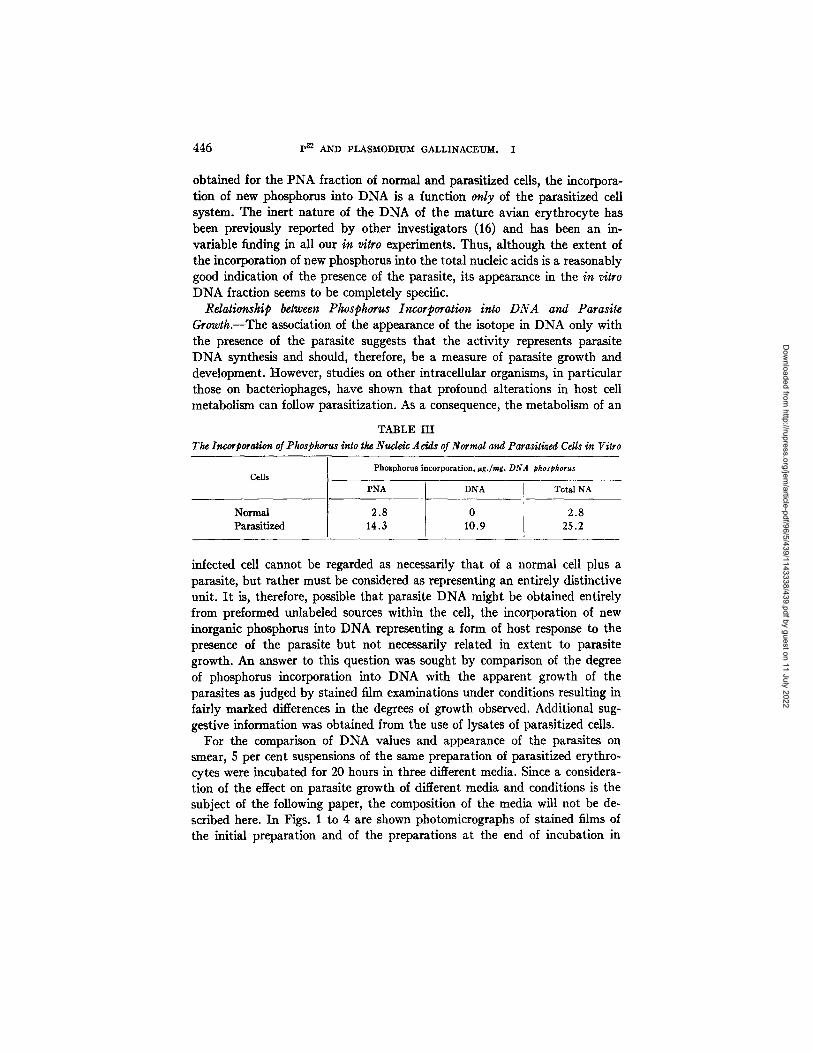
obtained for the PNA fraction of normal and parasitized cells, the incorpora- tion of new phosphorus into DNA is a function only of the parasitized cell system. The inert nature of the DNA of the mature avian erythrocyte has been previously reported by other investigators (16) and has been an in- variable finding in all our in vitro experiments. Thus, although the extent of the incorporation of new phosphorus into the total nucleic acids is a reasonably good indication of the presence of the parasite, its appearance in the in vitro DNA fraction seems to be completely specific.
Relationship belween Phosphorus Incorporation into DNA and Parasite Growth.--The association of the appearance of the isotope in DNA only with the presence of the parasite suggests that the activity represents parasite DNA synthesis and should, therefore, be a measure of parasite growth and development. However, studies on other intracellular organisms, in particular those on bacteriophages, have shown that profound alterations in host cell metabolism can follow parasitization. As a consequence, the metabolism of an
TABLE III The Invorporagon of Phosphorus into the Nude4v Adds of Normal and Parasitized Cells in Vitro
Cells
Normal Parasitized
Phosphorus incorporation, ~g./mg. DNA phosphorus
PNA DNA
2.8 0 14.3 10.9
Total NA
2.8 25.2
infected cell cannot be regarded as necessarily that of a normal cell plus a parasite, but rather must be considered as representing an entirely distinctive unit. I t is, therefore, possible that parasite DNA might be obtained entirely from preformed unlabeled sources within the cell, the incorporation of new inorganic phosphorus into DNA representing a form of host response to the presence of the parasite but not necessarily related in extent to parasite growth. An answer to this question was sought by comparison of the degree of phosphorus incorporation into DNA with the apparent growth of the parasites as judged by stained film examinations under conditions resulting in fairly marked differences in the degrees of growth observed. Additional sug- gestive information was obtained from the use of lysates of parasitized cells.
For the comparison of DNA values and appearance of the parasites on smear, 5 per cent suspensions of the same preparation of parasitized erythro- cytes were incubated for 20 hours in three different media. Since a considera- tion of the effect on parasite growth of different media and conditions is the subject of the following paper, the composition of the media will not be de- scribed here. In Figs. 1 to 4 are shown photomicrographs of stained films of the initial preparation and of the preparations at the end of incubation in
Dow
nloaded from http://rupress.org/jem
/article-pdf/96/5/439/1143338/439.pdf by guest on 11 July 2022
DELPHINE H. CLARKE 447
each medium, together with the corresponding DNA values obtained from the incubated cells.
The parasites in the initial preparation were, in general, in the trophozoite stage. After incubation in medium A, there was little change in parasite size, but there was evidence of beginning chromatin division and entrance into the schizont stage. The DNA value here was 3.2. The more favorable conditions found in medium B resulted in more advanced chromatin division and a general increase in parasite size, with an associated rise in the DNA value to 8.7. Medium C represented essentially optimal conditions in which the para- sites were becoming fully mature, numerous merozoites were being produced, and rupture from the containing erythrocytes was occurring. The DNA value under these conditions reached 24.4.
Subsequent to the description by Speck et al. (17) of the use of specific normal erythrocyte antiserum and complement as a means of effecting lysis of parasitized erythrocytes without apparent damage to the parasites, we in- cluded such preparations in our studies. Additional impetus was given to this work by the demonstration by Trager (6) that Plasmodium lophurae in similar preparations of duck erythrocytes could be shown to undergo growth and reproduction. Although we were not able to obtain results with P. gallinaceum comparable t,o those of Trager with P. lophurae, preparations consistently showing growth were finally obtained. This work will be described in more detail in the following paper. Suffice it to say here that studies of such para- sitized lysates with pn showed that the isotope was being incorporated into DNA despite rupture of the host cell by lysis. As in the case of the intact cell preparations, the extent of the incorporation paralleled the impressions gained from smear examinations.
DISCUSSION
The observation that parasitized erythrocytes take up more P~ than do normal cells is not especially surprising since it has been known for some time that such cells have a more active rate of metabolism than do normal cells as exhibited in a greatly enhanced rate of oxygen consumption. A concomitant increase in phosphorus metabolism was, therefore, to be expected.
As has been noted, the lack of incorporation of phosphorus into the DNA of the normal mature avian erythrocyte was not an original observation with us. In fact, the stability of this cell component has permitted its use in the determination of the life span of the cell (15).
Our concern has been with the demonstration that incorporation of inor- ganic phosphate into DNA does occur in the parasitized mature erythrocyte and with the correlation of the extent of this process with parasite growth. In this connection, it might be questioned why we have chosen to work with P~ rather than demonstrating a total increase in nucleic acid by one or the
Dow
nloaded from http://rupress.org/jem
/article-pdf/96/5/439/1143338/439.pdf by guest on 11 July 2022

448 P~ AND PLASMODIUM GALLINACEUM. I
other chemical means. Apart from the increased sensitivity of isotope methods, the answer is simply that direct chemical methods proved entirely unfeasible Attempts to follow parasite growth by increase in total DNA phosphorus or by the colorimetric method for DNA of Dische (18) were quite unsatisfactory. Due to the relatively large contribution of DNA by the erythrocytes, the magnitude of the differences to be determined was so small, relative to the total, as to fall well within the limits of experimental error for such determina- tions. A more favorable situation exists for the use of the orcinol method for PNA (19), since this nucleic acid is present in very low concentration in normal erythrocytes. However, although absolute increases in PNA were easily demonstrated by this method, the correlation between the magnitude of such increases and the parasite growth and development observed on smear was extremely poor. It seems possible that the level of PNA in the parasite varies at different stages of its development.
We feel that the appearance of labelled phosphorus in the DNA fraction of lysates of parasitized cells and the correlation of the extent of phosphorus incorporation with observed parasite growth in both intact cells and lymtes offer reasonable support to the belief that this represents the synthesis of parasite DNA. Our data do not permit us to imply, in the use of the word synthesis, more than that inorganic phosphorus has been recently incorporated into the nucleic acids. We have no knowledge of the route by which this in- corporation takes place nor of the nature of the immediate nucleic acid pre- cursors, and although there appears to be a quantitative relationship between the appearance of labelled DNA and parasite growth, it cannot be said whether this represents the whole of the DNA synthesized. However, these questions, although of fundamental interest, do not directly concern our interpretation that the degree of phosphorus incorporation into DNA in the erythrocyte-parasite system provides a sensitive, quantitative method of measuring parasite growth which has the advantage of complete objectivity and which should permit the detection of differences of a magnitude which would be impossible by comparative smear examinations.
SUMMARY
A quantitative method for following parasite growth in vitro is described. The method is based on the determination of the degree of incorporation oi
phosphorus into the nucleic acids, in particular desoxypentose nucleic acids, of the parasitized cell. Phosphorus incorporation is measured by utilizing the radioactive isotope, ps2.
The degree of phosphorus incorporation into DNA was found to be closely correlated with estimates of parasite growth based on stained film examina- tions of intact cell preparations. The isotope was likewise found to be in- corporated into the DNA of preparations in which the host cell had been
Dow
nloaded from http://rupress.org/jem
/article-pdf/96/5/439/1143338/439.pdf by guest on 11 July 2022
DELPRINE H. CLARKE 449
disrupted by lysis and the extent of this incorporation also paralleled the impressions gained from smear examinations.
I t is suggested that this method provides a sensitive and objective method of studying parasite growth.
BIBLIOGRAPHY
1. Hull, W., and Kirk, P. L., J. Gen. Physiol., 1949-50, 33, 335. 2. Cohen, S. S., J. Biol. Chem., 1948, 174, 295. 3. Kozloff, L. M., and Putnam, F. W., J. Biol. Chem., 1950, 182, 229. 4. Kozloff, L. M., and Putnam, F. W., J. Biol. Chem., 1949, 181, 207. 5. Whitman, L., J. Immunol., 1948, 59, 285. 6. Trager, W., J. Exp. Med., 1950, 92, 349. 7. Schneider, W. C., J. Biol. Chem., 1945, 161, 293, 8. Schmidt, G., and Thannhauser, S. J., J. Biol. Chem., 1945, 161, 83. 9. Schneider, W. C., J. Biol. Chem., 1946, 164, 747.
10. Umbreit, W. W., Burris, R. H., and Stauffer, J. F., Manometric Techniques and Related Methods for the Study of Tissue Metabolism, Minneapolis, Bur- gess Publishing Co., 1945.
11. Fiske, C. H., and SubbaRow, Y., J. Biol. Chem., 1925, 66, 375. 12. Furchgott, R. F., and Shorr, E., J. Biol. Chem., 1943, 151, 65. 13. Sacks, J., J. Biol. Chem., 1949, 181, 655. 14. Rapoport, S., Leva, E., and Guest, G. M., J. Biol. Chem., 1941, 139, 633. 15. Peters, J. P., and Van Slyke, D. D., Quantitative Clinical Chemistry. Methods,
Volume II, Baltimore, The Williams and Willrlns Company, 1932, 873. 16. Hevesy, G., and Ottesen, J., Nature, 1945, 156, 534. 17. Speck, J. F., Moulder, J. W., and Evans, E. A., Jr., J. Biol. Chem., 1946, 164,
119. 18. Dische, Z., Mikrochonie, 1930, 8, 4. 19. Meybaum, W., Z. physiol. Chem., 1939, 258, 117.
Dow
nloaded from http://rupress.org/jem
/article-pdf/96/5/439/1143338/439.pdf by guest on 11 July 2022
450 p82 AND PLAS~ODII/~ GALLINACEU~L. I
EXPLANATION OF PLATE 18
The relationship between parasite appearance in Giemsa-stained ~lms and the incorporation of phosphorus into DNA X 1345.
FIG. I. Appearance of parasites init~alIy as added to the media. FIG. 2. Appearance of parasites after 20 hours in medium A. DNA value = 3.2
~g./mg. DNA phosphorus. FIG. 3. Appearance of parasites after 20 hours in medium B. DNA value = 8.7
#g./mg. DNA phosphorus. FIG. 4. Appearance of parasites after 20 hours in medium C. DNA va|ue = 24.4
#g./mg. DNA phosphorus.
Dow
nloaded from http://rupress.org/jem
/article-pdf/96/5/439/1143338/439.pdf by guest on 11 July 2022

THE JOURNAL OF EXPERIMENTAL MEDICINE VOL. 96 PLATE 18
(Clarke: Pn and Plasmodium gallinaceum. I)
Dow
nloaded from http://rupress.org/jem
/article-pdf/96/5/439/1143338/439.pdf by guest on 11 July 2022