
The time for sex: A biennial life cycle in a marine planktonic diatom
9
The time for sex: A biennial life cycle in a marine planktonic diatom Domenico D’Alelio, 1,* Maurizio Ribera d’Alcala ` , Laurent Dubroca, Diana Sarno, Adriana Zingone, and Marina Montresor Stazione Zoologica Anton Dohrn, Villa Comunale, Napoli, Italy Abstract Whereas it is accepted that the population dynamics of higher organisms is strongly grounded in life history processes, such as the time taken to mature and reproduce, regulation in the abundance of aquatic protists is generally attributed to proximate properties of the environment in which they grow. We used 10 yr of data from the Gulf of Naples to determine the life history of a planktonic diatom, Pseudo-nitzschia multistriata, through the analysis of cell abundances and cell size patterns. Asexual and sexual phases recurred with remarkable regularity. Maximum abundance occurred annually, between the end of summer and the beginning of autumn, but cohorts of large cells, derived from sexual events, were found only every second year. We developed a model of population growth, based on parameters determined from laboratory cultures, with further tuning using information from natural populations. The model simulated the birth, maturation, and disappearance of age classes like those in natural populations, but only when we imposed seasonal variation in the rate of cell division and timed sexual reproduction during the stationary phase of a bloom. The model predicts that P. multistriata will become locally extinct if sexual reproduction does not occur within 4 yr. Our data and model show that coherent life cycle properties can emerge in natural populations of unicellular organisms, analogous to those in multicellular organisms, as a result of finely tuned regulation of cell division and sexual competence. Different life cycle strategies allow species to regulate their population dynamics, to cope with external con- straints, and to perpetuate the genetic pool, in tune with the environment. Conceptual models of the population dy- namics of multicellular organisms are strongly grounded in life cycle processes, which determine the spatial and temporal structure of natural populations (Caswell 2001). In these models, life history processes account for a strong tendency towards population synchrony in spite of the fact that environmental fluctuations can be both synchronic and chaotic (Benton et al. 2001; Bjørnstad and Grenfell 2001). Conversely, the dynamics of phytoplankton popu- lations are generally thought to be driven by external factors, such as the availability of nutrients and light, or grazing (Reynolds 2006; Beninca et al. 2008). Nonetheless, complex life cycles have been described for several microalgae, including stages with different morphology, ploidy, and function (Montresor and Lewis 2006). The adaptive role of this complexity is far from being eluci- dated, but the multiphased character of their life histories is suggestive of a strong organization and a fine tuning with the environment (Matrai et al. 2005; Frada et al. 2008). The complexity of the diatom life cycle has been well known since the beginning of the 19th century (Geitler 1932). As in the majority of unicellular microalgae, two main distinct but interconnected phases are recognized: a vegetative one, in which mitotic divisions lead to an increase in cell number, and a sexual one, in which meiosis and genetic recombination occur. Sexual reproduction has a major role in modulating the rate of adaptation (Colegrave 2002), pruning the genome from deleterious mutations (Kondrashov 1988), and coping with the evolution of parasites (Hamilton et al. 1990), which provides possible evolutionary explanations for its occur- rence in diatoms, as in the vast majority of unicellular and multicellular lineages. The most distinctive property of the diatom life cycle is a progressive reduction in cell size during the vegetative phase, caused by the way diatom cells divide (Round et al. 1990). Although some species have evolved vegetative cell enlargement to escape extreme miniaturization, many diatoms restore their largest size only during the sexual phase (Fig. 1; Chepurnov et al. 2004). In these cases, sex is the only means to avoid death, in contrast to other protists, in which cells can keep on dividing asexually for an unlimited period. The aging of cohorts of large cells originating from sexual events can be tracked from the pace of cell size reduction, which can be used to estimate the rate of cell division. Therefore, cell size dynamics in diatoms is a reliable proxy of life cycle processes (Mann 1988; Jewson 1992) and allows the use of a demographic approach similar to that applied to higher organisms (Caswell 2001). Our study concerns the marine planktonic diatom Pseudo-nitzschia multistriata (Takano) Takano, which is like most other diatoms in requiring sexual reproduction to regain maximum size (D’Alelio et al. 2009a,b). Pseudo- nitzschia species are needle-shaped, chain-forming diatoms widespread from polar to tropical latitudes and are significant contributors to phytoplankton blooms in coastal and oceanic waters (Hasle 2002). They thrive also in iron-poor regions, probably because of their capability to produce ferritin, a protein that stores iron inside the cell (Marchetti et al. 2009). The genus Pseudo-nitzschia has * Corresponding author: [email protected] 1 Present address: IASMA (Istituto Agrario di San Michele all’Adige) Research and Innovation Centre, Fondazione Edmund Mach, Environment and Natural Resources Area, S. Michele all’Adige (Trento) Italy Limnol. Oceanogr., 55(1), 2010, 106–114 E 2010, by the American Society of Limnology and Oceanography, Inc. 106
-
Upload
independent -
Category
Documents
-
view
1 -
download
0
Transcript of The time for sex: A biennial life cycle in a marine planktonic diatom

The time for sex: A biennial life cycle in a marine planktonic diatom
Domenico D’Alelio,1,* Maurizio Ribera d’Alcala, Laurent Dubroca, Diana Sarno, Adriana Zingone,and Marina Montresor
Stazione Zoologica Anton Dohrn, Villa Comunale, Napoli, Italy
Abstract
Whereas it is accepted that the population dynamics of higher organisms is strongly grounded in life historyprocesses, such as the time taken to mature and reproduce, regulation in the abundance of aquatic protists isgenerally attributed to proximate properties of the environment in which they grow. We used 10 yr of data fromthe Gulf of Naples to determine the life history of a planktonic diatom, Pseudo-nitzschia multistriata, through theanalysis of cell abundances and cell size patterns. Asexual and sexual phases recurred with remarkable regularity.Maximum abundance occurred annually, between the end of summer and the beginning of autumn, but cohortsof large cells, derived from sexual events, were found only every second year. We developed a model of populationgrowth, based on parameters determined from laboratory cultures, with further tuning using information fromnatural populations. The model simulated the birth, maturation, and disappearance of age classes like those innatural populations, but only when we imposed seasonal variation in the rate of cell division and timed sexualreproduction during the stationary phase of a bloom. The model predicts that P. multistriata will become locallyextinct if sexual reproduction does not occur within 4 yr. Our data and model show that coherent life cycleproperties can emerge in natural populations of unicellular organisms, analogous to those in multicellularorganisms, as a result of finely tuned regulation of cell division and sexual competence.
Different life cycle strategies allow species to regulatetheir population dynamics, to cope with external con-straints, and to perpetuate the genetic pool, in tune with theenvironment. Conceptual models of the population dy-namics of multicellular organisms are strongly grounded inlife cycle processes, which determine the spatial andtemporal structure of natural populations (Caswell 2001).In these models, life history processes account for a strongtendency towards population synchrony in spite of the factthat environmental fluctuations can be both synchronicand chaotic (Benton et al. 2001; Bjørnstad and Grenfell2001). Conversely, the dynamics of phytoplankton popu-lations are generally thought to be driven by externalfactors, such as the availability of nutrients and light, orgrazing (Reynolds 2006; Beninca et al. 2008). Nonetheless,complex life cycles have been described for severalmicroalgae, including stages with different morphology,ploidy, and function (Montresor and Lewis 2006). Theadaptive role of this complexity is far from being eluci-dated, but the multiphased character of their life historiesis suggestive of a strong organization and a fine tuning withthe environment (Matrai et al. 2005; Frada et al. 2008).
The complexity of the diatom life cycle has been wellknown since the beginning of the 19th century (Geitler1932). As in the majority of unicellular microalgae, twomain distinct but interconnected phases are recognized: avegetative one, in which mitotic divisions lead to anincrease in cell number, and a sexual one, in which meiosis
and genetic recombination occur. Sexual reproduction hasa major role in modulating the rate of adaptation(Colegrave 2002), pruning the genome from deleteriousmutations (Kondrashov 1988), and coping with theevolution of parasites (Hamilton et al. 1990), whichprovides possible evolutionary explanations for its occur-rence in diatoms, as in the vast majority of unicellular andmulticellular lineages. The most distinctive property of thediatom life cycle is a progressive reduction in cell sizeduring the vegetative phase, caused by the way diatom cellsdivide (Round et al. 1990). Although some species haveevolved vegetative cell enlargement to escape extrememiniaturization, many diatoms restore their largest sizeonly during the sexual phase (Fig. 1; Chepurnov et al.2004). In these cases, sex is the only means to avoid death,in contrast to other protists, in which cells can keep ondividing asexually for an unlimited period. The aging ofcohorts of large cells originating from sexual events can betracked from the pace of cell size reduction, which can beused to estimate the rate of cell division. Therefore, cell sizedynamics in diatoms is a reliable proxy of life cycleprocesses (Mann 1988; Jewson 1992) and allows the use ofa demographic approach similar to that applied to higherorganisms (Caswell 2001).
Our study concerns the marine planktonic diatomPseudo-nitzschia multistriata (Takano) Takano, which islike most other diatoms in requiring sexual reproduction toregain maximum size (D’Alelio et al. 2009a,b). Pseudo-nitzschia species are needle-shaped, chain-forming diatomswidespread from polar to tropical latitudes and aresignificant contributors to phytoplankton blooms incoastal and oceanic waters (Hasle 2002). They thrive alsoin iron-poor regions, probably because of their capabilityto produce ferritin, a protein that stores iron inside the cell(Marchetti et al. 2009). The genus Pseudo-nitzschia has
* Corresponding author: [email protected]
1 Present address: IASMA (Istituto Agrario di San Micheleall’Adige) Research and Innovation Centre, Fondazione EdmundMach, Environment and Natural Resources Area, S. Micheleall’Adige (Trento) Italy
Limnol. Oceanogr., 55(1), 2010, 106–114
E 2010, by the American Society of Limnology and Oceanography, Inc.
106

gained considerable attention over the last decade becausesome species can produce the neurotoxin domoic acid thatcauses amnesic shellfish poisoning (Fryxell and Hasle2003).
We present the results of a decade-long demographicalstudy carried out at the Long-Term Ecological Researchstation MareChiara (LTER-MC; Ribera d’Alcala et al.2004) in the Gulf of Naples, in which we integratedphysiological and morphometric information to decipherthe life cycle of the marine toxic diatom P. multistriata(Fig. 1). By using cell abundance and size, we tracked birth,aging, maturation, and sex in natural populations. Weconclude that the biennial timing of sexual reproductionevents and the predictable dynamics of the vegetative phaseprovide evidence for a coherent and periodic life cycle ofthis marine diatom.
Methods
Biological data—Water samples were collected weeklywith Niskin bottles in the upper layer (1 m) of the watercolumn at the LTER-MC station (40u48.59N, 14u159E;Ribera d’Alcala et al. 2004), located 2 miles offshore in theGulf of Naples (Tyrrhenian Sea, Mediterranean Sea) from1996 to 2006. Additional samples were collected weeklywith a net of 20-mm mesh size in 2005–2006. For P.multistriata cell counts and measurements, samples werefixed with neutralized formaldehyde solution (1.6% finalconcentration) and stored at 4uC. Enumerations were
performed with an inverted microscope at 4003 magnifi-cation, after sedimentation of variable volumes of seawater(1–100 mL), depending on cell concentration (Utermohl1958). The apical length of 200 cells was measured for eachsample in which P. multistriata was present using a ZeissAxiovert microscope at a magnification of 4003, approx-imating the measurements to the minimum unit of themicrometric ocular, 2.5 mm. A lower number of cells weremeasured only in a few samples collected during non-bloom periods, when cell abundances were extremely low.Cell size reduction in P. multistriata occurs mainly alongthe apical axis, and we thus used cell length as a proxy forcell size. Cell size frequency distributions were analyzedwith the open source program FisatII (http://www.fao.org/fi/statist/fisoft/fisat/index.htm). The Lomb-Scargle period-ogram (Press and Rybicki 1989) was used to detect periodicvariability in three distinct time series: (1) the mean cell sizefrom natural samples, (2) the mean cell size simulated bythe model, and (3) the values extracted from the time seriesof the mean cell size simulated by the model, for the periodof sampling of the natural populations. When two sta-tistically separated subpopulations occurred in the samesample, the highest mean cell size value was used for theanalysis of periodic variability. The significance of period-ogram peaks and associated frequencies was tested with themodified Scargle method under the null hypothesis that agiven peak is random (Glynn et al. 2006).
The growth rate (gr) of natural populations wascalculated by scaling the reduction rates of the mean cellsize between two sampling intervals with the cell sizereduction rates observed in cultured strains and expressedas mm 3 division21 (D’Alelio et al. 2009b). Cell sizereduction (r) was calculated as r 5 [mean size(time1) 2mean size(time2)] 3 (time2 2 time1)21. The division rate (D,division 3 d21) was calculated as D 5 (r 3 R21), where R(mm 3 division21) is the rate of cell size decrease associatedto each cell division as estimated in cultured strains. Theabsolute growth rate (gr, d21) was calculated as gr 5 D 30.6931, where the constant number is a factor of conversionbetween the unit ‘‘divisions d21’’ and d21.
Model implementation—The life cycle model is a matrixmodel in which growth, cell size reduction, and mortalityrates are parameterized for each size class based onobservations made in culture (D’Alelio et al. 2009b) unlessotherwise specified. The sensitivity of the model was testedby changing parameters and comparing the actual simula-tions with the real data. In this paper we illustratesimulations only from the final model and justify ourchoice of input parameters.
The parameters used in the model (Table 1) aredescribed in the following. (1) Size of the initial cells (Initsize): We used a fixed value of 80 mm, which was among thehighest values observed in mating experiments. (2) Size ofthe smallest cells (Small size): We used a fixed value of30 mm, below which cell death in culture was generallyobserved. (3) Parameterized growth rate (Pgr): The growthrate (d21) of each cell of size s was parameterized as Pgr 5k 3 gr, with gr being the range of growth capability of thepopulation during bloom or non-bloom phases and k being
Fig. 1. The life cycle of the pennate diatom P. multistriata(modified from D’Alelio et al. 2009b). The peculiar structure anddivision modality of diatom cells imposes a progressive reductionin the apical axis (5cell length) in the population along itsvegetative growth. Below the cell size threshold for gametogenesis(55 mm), gametangia differentiate, each generating two haploidgametes. Conjugation follows the pairing of gametangia ofopposite mating type and produces two diploid zygotes. Thezygote, now called an auxospore, elongates while depositingweakly silicified perizonial bands and hosts the formation of along initial cell.
A planktonic diatom biennial life cycle 107

the growth rate variation of P. multistriata with cell sizedescribed by the polynomial equation k 5 0.25 + 0.04 3 s2 0.0005 3 s2, where s is the cell size class. This trend wasinferred from experimental data on P. multistriata (D’Ale-lio et al. 2009b) and Pseudo-nitzschia delicatissima (Amatoet al. 2005). The dataset for the latter species was morecomplete and was hence used to find the best fit for theequation for k. According to the final formulation of theequation, the growth rate was very low in the longer cellsand gradually (linearly) increased until 60% of the specificcell length was reached. The parameter gr varied in thecourse of the simulations, reproducing the alternation ofperiods of fast and slow growth, which were inferred fromthe rate of decrease of cell size in natural samples. Thevalues for gr used in the final version of the model (i.e., theone that definitely matched the environmental data) were0.69–1.40 d21, corresponding to 1.0–2.0 divisions d21,during the bloom exponential phase, and 0.07–0.20 d21,corresponding to 0.1–0.3 divisions d21, during the station-ary and non-bloom phases. Higher growth rates (up to 2.5divisions d21) observed by D’Alelio et al. (2009b) were alsotested in the simulations, but they did not allow reproduc-tion of the natural patterns. A sudden collapse in growthrate was imposed for cells smaller than 40 mm, because theywere approaching the smallest size, 30 mm, at which growthrate was zero. In the course of model implementation, wealso used other equations to model the relation betweengrowth rate and cell size (i.e., linear positive and negative,constant relationship) but the results did not match thenatural patterns. (4) Parameterized death rate (Pdr): Themortality factor (d21) was calculated as Pdr 5 k9 3 dr,where k9 is the size-dependent mortality, k9 5 0.4 + 0.04 3 s2 0.0005 3 s2, with s being the cell size and dr the deathrate. In the final version of the model we computed Pdrusing an equation similar to the one used for Pgr. Thepolynomial equation used for both rates was in the form ofk 5 a + b 3 cell size 2 c 3 cell size2. The constant values band c were the same in both rates, whereas a was slightlyhigher in the Pdr equation (0.4 vs. 0.25), thus generating adeath rate lower than the growth rate, in order to avoid the
population’s going extinct in the model simulation. Valuesfor dr varied in parallel with gr over the simulation, rangingfrom 0.05 to 1 d21 (i.e., they were always below 50% of gr).To prevent an exponential increase of cell abundance andto reduce the simulation time, some cells were randomlyremoved at each time step (corresponding to 1 d), keepingthe total number of cells always below a preassignedthreshold of 10,000. Simulations with dr constant for all cellsizes failed to reproduce the observed cell size pattern. (5)Cell size reduction 3 generation21 (Red): The rate of cellsize (s) decrease, expressed as mm 3 division21, wasprovided by the equation Red 5 0.0000001 3 s2.5. Theequation takes into account that in P. multistriata cell sizereduction at each mitotic division decreased with cell size.Using other equations to model the relation between cellsize reduction and cell size (i.e., linear positive and negative,or constant relationship), the natural pattern was notreproduced. (6) Gametangia threshold size (Sex size): Weused 55 mm as the cell size threshold for sexual reproduc-tion, because it was the maximum length for gametangia inculture conditions. With a threshold size higher than55 mm, the model failed to reproduce the natural pattern.(7) Probability of forming gametes (Sex prob): Theproportion of cells below the fertile cell size threshold(55 mm) turning into gametangia was not more than 2%,which corresponds to the maximum production rate ofsexual stages in mating experiments performed in culture.With higher percentages of gamete formation, the output ofthe model did not fit the real pattern. In the model, thesuccess of auxospore development was considered to be100%, i.e., all auxospores developed into initial cells. Theprocess was also set to occur instantaneously, though it takes2–3 d in culture (D’Alelio et al 2009b). (8) Timing of sexualreproduction (Sex time): The model was run using differenttimings of sexual reproduction: continuous (sexual repro-duction always possible within Sex size), annual, biennial,and multiannual (sexual reproduction limited to the bloomperiod every year, in alternate years, or with longerperiodicity, respectively). Model simulations were run withsex occurring at the bloom exponential or stationary phases.
Table 1. Life cycle parameters used to implement the model, the abbreviation used in the text, and the literature reference.*
Parameter Abbreviation Value or formula Data source
1 Size of the initial cells Init size 580 mm D’Alelio et al. 2009b2 Size of the smallest cells Small size 530 mm D’Alelio et al. 2009b3 Parameterized growth rate Pgr 5gr 3 (0.25 + 0.04 3 s 2 0.0005 3 s2);
gr ranging from 0.07 to 1.40 d21, s (cell size)ranging from 30 to 80 mm
Amato et al. 2005;D’Alelio et al. 2009b
4 Parameterized death rate Pdr 5dr 3 (0.4 + 0.04 3 s 2 0.0005 3 s2);dr ranging from 0.05 to 1.00 d21,s (cell size) ranging from 30 to 80 mm
This paper
5 Rate of cell size decrease Red 50.0000001 3 s2.5 D’Alelio et al. 2009b6 Gametangia threshold size Sex size 555 mm D’Alelio et al. 2009b7 Probability of forming
gametesSex prob 52% D’Alelio et al. 2009b;
this paper8 Timing of sexual
reproductionSex time Continuous, annual, biennial, multiannual This paper
* gr, growth rate; dr, death rate.
108 D’Alelio et al.

The model reproduces the development of cell cohorts ofdifferent sizes over time. At each time step, a cell can eitherdivide (at a rate set by Pgr) or die (at a rate set by Pdr).Once a cell divides, two new cells are produced, one of thesame size and the other of reduced size according to Red.Among cells smaller than the size threshold for gameto-genesis (Sex size), a number of pairs are randomly selectedaccording to the parameter Sex prob, and two cells ofmaximum size are produced from each pair. Sexualreproduction occurs only within the temporal window forsex imposed by the parameter Sex time. Every 10 time steps(5every 10 d) the model provides a discrete size spectrumhaving a size step of 2.5 mm (corresponding to theresolution of the measurements, see above).
Results
P. multistriata was found in surface samples collectedbetween 1996 and 2006 in the Gulf of Naples throughoutthe year, with the exception of the periods March–May andfrom the end of July to the end of August. Cell abundanceranged from undetectable to 700 cells mL21 (July 2006),with long-lasting non-bloom periods and short bloomsgenerally occurring somewhere between the end of Augustand the end of October (Figs. 2, 3a).
Cell sizes ranged from 75 to 30 mm. Large-sized cells,pinpointing the occurrence of sex, appeared every 2 yr anddecreased in size over the following period (Fig. 3b). Therewas an alternation in successive years between bi- andunimodal size distributions (Fig. 3b). The maximum cellsize—i.e., the upper extreme of the cell size distribution—showed a biennial oscillation, with phases of reduction andsubsequent restoration occurring every 2 yr.
Intensified sampling in 2005–2006 allowed detection andmeasurement of P. multistriata cells during non-bloomperiods over these 2 yr (Fig. 3b). Over those 2 yr, sizereduction was slow and gradual over the non-bloom phase,whereas it increased during the bloom. Rates calculated onthe basis of observed reductions in cell size were notconstant over the annual cycle. Its values calculated based
on the observed cell size reduction were low over most ofthe year (non-bloom phase; mean growth rate 5 0.3 6 0.2divisions d21, n 5 6). Division rates increased steeply to 2.6divisions d21 in late summer until the annual peak wasreached (bloom exponential phase; mean 6 standarddeviation 5 1.1 6 0.6 divisions d21, n 5 11), and suddenlydecreased afterwards while cell concentrations were stillhigh (bloom stationary phase).
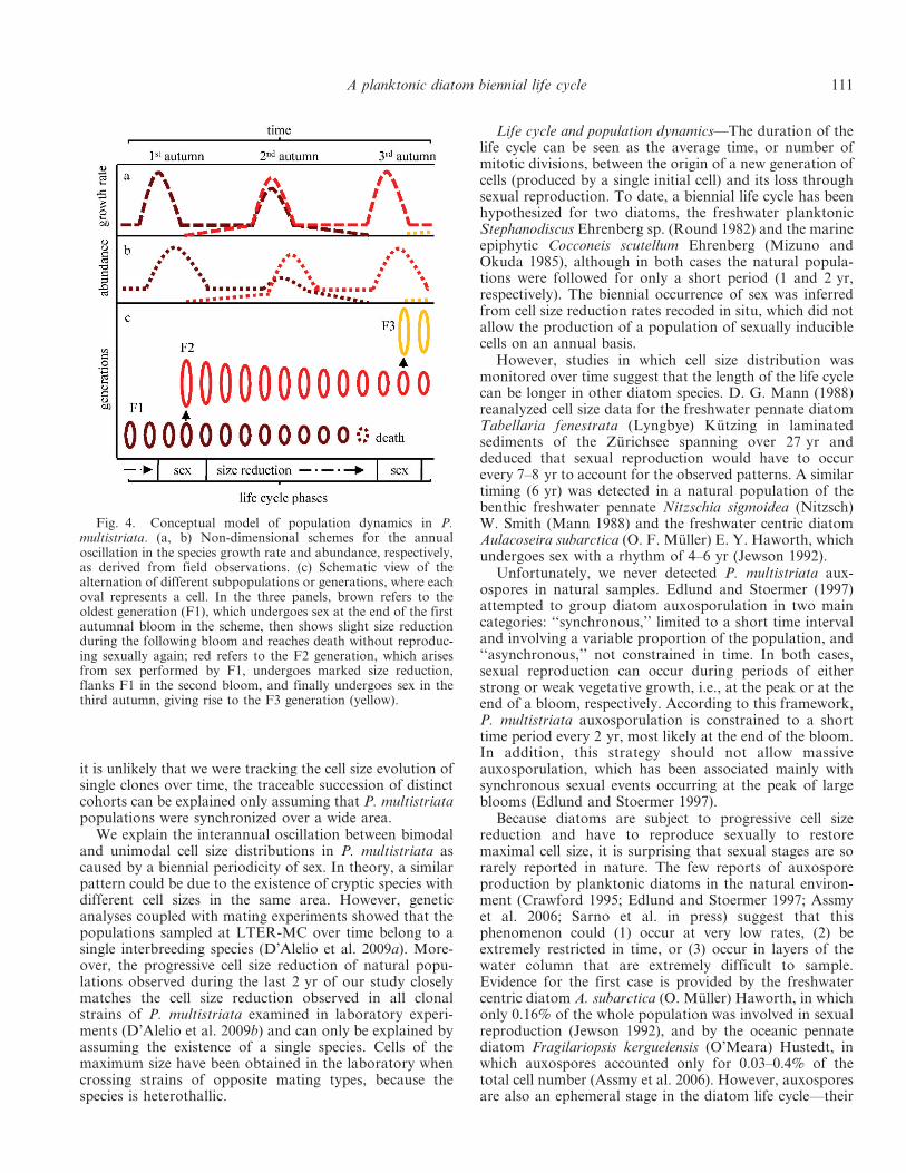
Given estimates of key life cycle processes obtained fromcultured strains (threshold cell size for sexual reproduction,size range of initial cells, cell size reduction rate over thespecies size range; D’Alelio et al. 2009b), the field data canbe interpreted as a biennial sequence, which can besummarized as follows (Fig. 4): (1) vegetative cells belong-ing to a population with a unimodal cell size produce thebloom in late summer; (2) during the bloom, a smallfraction of the population below the threshold size for sex(55 mm) differentiates into gametes, which eventuallyconjugate and form auxospores; (3) the auxospores givebirth to long initial cells (72–82 mm), rarely detected innatural samples; (4) over the long (8–10 months) subse-quent non-bloom season, the young large cells divide at avery low pace along with the old generation, bothnevertheless continuing to decrease in cell size; (5) duringthe bloom phase of the next year, both the new-generationcells, now in the range between 55 and 65 mm, and the oldpopulation with a smaller size enhance their growth rates;(6) over the next long non-bloom phase, the cohort with alarger cell size proceeds towards the next bloom period(phase 1), while the older cohort keeps on reducing in cellsize while dividing and eventually fades away.
We tested this conceptual scheme with the individual-based model described in the methods. The model wasinitialized with one cell of the maximum size and run forfive annual cycles with different combinations of thefollowing parameters: growth rates constantly high, low,or variable over the annual cycle, occurrence of sex duringthe exponential or stationary phase, and bloom timing ofsexual events (continuous, annual, biennial, or multi-annual). The model only fitted real data, producing analternation of uni- and bimodal cell size distributions, when(1) simulation started with one maximum size cell at theend of the bloom, (2) cell growth rate varied through theyear, and (3) biennial sexual reproduction was assumed totake place during the bloom stationary phase (Fig. 5a).Using this combination of life cycle parameters, we alsosimulated the dynamics of cell size over an 11-yr periodafter initializing the model with the cell size distributiondetected at the beginning of the time series (22 October1996). The model output matched natural size spectra welland reproduced the 10-yr sequence of seven cohorts of cells(Fig. 3c). Periodograms for (1) the mean cell size fromnatural samples, (2) the mean cell size simulated by themodel, and (3) values extracted from the mean cell sizesimulated by the model for the period of the actualsampling showed periods of oscillation of 110.7, 103.3, and107.7 weeks, respectively, which do not differ significantlyfrom 2 yr (p , 0.005).
There were two occasions in which the model failed toreproduce the real data: the average cell sizes of the smaller
Fig. 2. The annual timing of the P. multistriata bloom at LTER-MC. Logarithmically transformed abundance plot of P. multistriataover the period 1996–2006. White squares indicate no sampling.
A planktonic diatom biennial life cycle 109
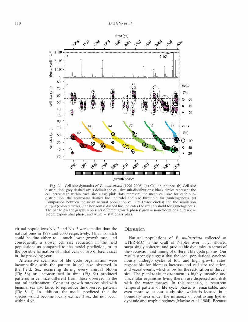
virtual populations No. 2 and No. 3 were smaller than thenatural ones in 1998 and 2000 respectively. This mismatchcould be due either to a much lower growth rate, andconsequently a slower cell size reduction in the fieldpopulations as compared to the model prediction, or tothe possible formation of initial cells of two different sizesin the preceding year.
Alternative scenarios of life cycle organization wereincompatible with the pattern in cell size observed inthe field. Sex occurring during every annual bloom(Fig. 5b) or unconstrained in time (Fig. 5c) producedpatterns in cell size different from those observed in thenatural environment. Constant growth rates coupled withbiennial sex also failed to reproduce the observed patterns(Fig. 5d–f). In addition, the model predicted that thespecies would become locally extinct if sex did not occurwithin 4 yr.
Discussion
Natural populations of P. multistriata collected atLTER-MC in the Gulf of Naples over 11 yr showedsurprisingly coherent and predictable dynamics in terms ofthe succession and timing of different life cycle phases. Ourresults strongly suggest that the local populations synchro-nously undergo cycles of low and high growth rates,responsible for biomass increase and cell size reduction,and sexual events, which allow for the restoration of the cellsize. The planktonic environment is highly unstable andunicellular organisms living therein are dispersed and driftwith the water masses. In this scenario, a recurrenttemporal pattern of life cycle phases is remarkable, andeven more so at our study site, which is located in aboundary area under the influence of contrasting hydro-dynamic and trophic regimes (Marino et al. 1984). Because
Fig. 3. Cell size dynamics of P. multistriata (1996–2006). (a) Cell abundance. (b) Cell sizedistribution: grey dashed ovals delimit the cell size sub-distributions; black circles represent thecell percentage within each size class; pink dots represent the mean cell size for each sub-distribution; the horizontal dashed line indicates the size threshold for gametogenesis. (c)Comparison between the mean natural population cell size (black circles) and the simulationoutput (colored circles); the horizontal dashed line indicates the size threshold for gametogenesis.The bar below the graphs represents different growth phases: grey 5 non-bloom phase, black 5bloom exponential phase, and white 5 stationary phase.
110 D’Alelio et al.
it is unlikely that we were tracking the cell size evolution ofsingle clones over time, the traceable succession of distinctcohorts can be explained only assuming that P. multistriatapopulations were synchronized over a wide area.
We explain the interannual oscillation between bimodaland unimodal cell size distributions in P. multistriata ascaused by a biennial periodicity of sex. In theory, a similarpattern could be due to the existence of cryptic species withdifferent cell sizes in the same area. However, geneticanalyses coupled with mating experiments showed that thepopulations sampled at LTER-MC over time belong to asingle interbreeding species (D’Alelio et al. 2009a). More-over, the progressive cell size reduction of natural popu-lations observed during the last 2 yr of our study closelymatches the cell size reduction observed in all clonalstrains of P. multistriata examined in laboratory experi-ments (D’Alelio et al. 2009b) and can only be explained byassuming the existence of a single species. Cells of themaximum size have been obtained in the laboratory whencrossing strains of opposite mating types, because thespecies is heterothallic.
Life cycle and population dynamics—The duration of thelife cycle can be seen as the average time, or number ofmitotic divisions, between the origin of a new generation ofcells (produced by a single initial cell) and its loss throughsexual reproduction. To date, a biennial life cycle has beenhypothesized for two diatoms, the freshwater planktonicStephanodiscus Ehrenberg sp. (Round 1982) and the marineepiphytic Cocconeis scutellum Ehrenberg (Mizuno andOkuda 1985), although in both cases the natural popula-tions were followed for only a short period (1 and 2 yr,respectively). The biennial occurrence of sex was inferredfrom cell size reduction rates recoded in situ, which did notallow the production of a population of sexually induciblecells on an annual basis.
However, studies in which cell size distribution wasmonitored over time suggest that the length of the life cyclecan be longer in other diatom species. D. G. Mann (1988)reanalyzed cell size data for the freshwater pennate diatomTabellaria fenestrata (Lyngbye) Kutzing in laminatedsediments of the Zurichsee spanning over 27 yr anddeduced that sexual reproduction would have to occurevery 7–8 yr to account for the observed patterns. A similartiming (6 yr) was detected in a natural population of thebenthic freshwater pennate Nitzschia sigmoidea (Nitzsch)W. Smith (Mann 1988) and the freshwater centric diatomAulacoseira subarctica (O. F. Muller) E. Y. Haworth, whichundergoes sex with a rhythm of 4–6 yr (Jewson 1992).
Unfortunately, we never detected P. multistriata aux-ospores in natural samples. Edlund and Stoermer (1997)attempted to group diatom auxosporulation in two maincategories: ‘‘synchronous,’’ limited to a short time intervaland involving a variable proportion of the population, and‘‘asynchronous,’’ not constrained in time. In both cases,sexual reproduction can occur during periods of eitherstrong or weak vegetative growth, i.e., at the peak or at theend of a bloom, respectively. According to this framework,P. multistriata auxosporulation is constrained to a shorttime period every 2 yr, most likely at the end of the bloom.In addition, this strategy should not allow massiveauxosporulation, which has been associated mainly withsynchronous sexual events occurring at the peak of largeblooms (Edlund and Stoermer 1997).
Because diatoms are subject to progressive cell sizereduction and have to reproduce sexually to restoremaximal cell size, it is surprising that sexual stages are sorarely reported in nature. The few reports of auxosporeproduction by planktonic diatoms in the natural environ-ment (Crawford 1995; Edlund and Stoermer 1997; Assmyet al. 2006; Sarno et al. in press) suggest that thisphenomenon could (1) occur at very low rates, (2) beextremely restricted in time, or (3) occur in layers of thewater column that are extremely difficult to sample.Evidence for the first case is provided by the freshwatercentric diatom A. subarctica (O. Muller) Haworth, in whichonly 0.16% of the whole population was involved in sexualreproduction (Jewson 1992), and by the oceanic pennatediatom Fragilariopsis kerguelensis (O’Meara) Hustedt, inwhich auxospores accounted only for 0.03–0.4% of thetotal cell number (Assmy et al. 2006). However, auxosporesare also an ephemeral stage in the diatom life cycle—their
Fig. 4. Conceptual model of population dynamics in P.multistriata. (a, b) Non-dimensional schemes for the annualoscillation in the species growth rate and abundance, respectively,as derived from field observations. (c) Schematic view of thealternation of different subpopulations or generations, where eachoval represents a cell. In the three panels, brown refers to theoldest generation (F1), which undergoes sex at the end of the firstautumnal bloom in the scheme, then shows slight size reductionduring the following bloom and reaches death without reproduc-ing sexually again; red refers to the F2 generation, which arisesfrom sex performed by F1, undergoes marked size reduction,flanks F1 in the second bloom, and finally undergoes sex in thethird autumn, giving rise to the F3 generation (yellow).
A planktonic diatom biennial life cycle 111

persistence lasts for only a few days—and they could beproduced asynchronically, thus making their detection inthe natural environment extremely challenging. Over morethan 20 yr of regular sampling at LTER-MC in the Gulf ofNaples, only once have we detected major production ofauxospores by planktonic diatoms (in this case by twodifferent Pseudo-nitzschia species), and this was restrictedto a single sampling (Sarno et al. in press). The finding thatPseudo-nitzschia species often aggregate in thin layers ofphysical discontinuity in the water column (Rines et al.2002; Velo-Suarez et al. 2008) suggests that these peculiarenvironments might provide the ideal conditions for sexualreproduction to occur. In the case of P. multistriata we onlyhave evidence of a regular appearance of cohorts of largecells over 2 yr. Cells in the larger size range, which arederived from auxospores, are not numerically abundant,and we therefore assume that auxosporulation in thisspecies occurs at relatively low rates. According to ourmodel, very low concentrations of initial cells, down to0.05% of the parental population, are also enough to
recruit new cohorts that become numerically comparable tothe parental ones at the following bloom, notwithstandingdeath rates accounting for 50% of the growth rates(Fig. 3c).
In their seminal papers, W. M. Lewis, Jr. (1983, 1984),D. G. Mann (1988), and D. H. Jewson (1992) presented aconceptual framework for diatom life histories rooted in (1)supra-annual duration of the life cycle (2–40 yr), (2) long-term asexual reproduction, (3) low-frequency, short-term,and synchronic sexual phase, and (4) cell size reductionprocess acting as a chronometer for sex. This chronometermight regulate both annual and supra-annual sexual cycles,and would be particularly effective in species with sexoccurring with a period longer than 1 yr, which isdecoupled from any predictable environmental periodicity.In most of the diatoms studied so far, sex can only beinduced within a species-specific cell size window (Geitler1932; Chepurnov et al. 2004). However, cell size is not theonly factor that allows sexual reproduction. Reasonably, athreshold cell concentration is required in order to allow
Fig. 5. Model outputs of cell size distribution in P. multistriata under different settings. (a–c) Growth rate modulated over the year(0.2, 1.0, and 0.2 divisions d21 during the non-bloom, bloom exponential, and bloom stationary phases, respectively) and (a) biennial, (b)annual, and (c) continuous timing of sex. (d–f) Biennial timing of sex and growth rates constant over the year of (d) 0.2, (e) 0.6, and (f) 1.0divisions d21. Arrows indicate cell size reduction trajectories; vertical dashed lines indicate the size threshold for gametogenesis.
112 D’Alelio et al.

vegetative cells’ encounter and subsequent gametogenesis.This requirement is met at the end of the exponentialgrowth phase of the bloom, when cell concentration is at itsmaximum. Considering that gamete production implies theinterruption of growth for a fraction of the population, thebest solution to balance this ‘‘cost of sex’’ is to localize thesexual phase after the growth pulse responsible for thebloom, when cells are not actively dividing any longer(Lewis 1983). Based on these considerations, we canhypothesize that the biennial periodicity of sex in P.multistriata is because of the fact that only once every 2 yr asufficient concentration of sexually mature cells occurs.This should happen when a single cohort of cells with amedium size—and thus in the size window for sex—wasdetected. Conversely, in the years when two cohorts werepresent, sex did not occur, possibly because the cohort witha largest size was not mature yet, whereas the one with thesmallest size—though within the size window for sex—wasconstituted of weak cells close to death. We do not havedirect evidence for our hypothesis; however, the productionof allelopathic metabolites (Cembella 2003) or pheromone-like compounds (Hache 2000) only within a specific cell sizeinterval might be responsible for providing aggregationand/or mutual coordination in diatom cells.
Ecological implications—The coherent pattern in P.multistriata population dynamics that we observed innature over a decade, and modeled with the support oflaboratory studies, reveals a fine-tuned organization of thelife cycle. In multicellular organisms, the synchronizationof growth, reproduction, and senescence phases is generallykept by entrainment to external cues showing annual orshorter periodicities (Schwartz 2003). On the other hand, insome cases of multiannual life cycle periodicities, such asreproduction of cicadas (Williams and Simon 1995) andflowering in bamboos (Janzen 1976), where the link toexogenous triggers is less clear, internal rather thanenvironmental control is invoked as a matter of regulation.Our results also indicate that growth in P. multistriata isnot constant along the annual cycle, with a briskacceleration of growth rate during the bloom phase. Thiswas shown by cell size variations over time and confirmedby the simulations, which reproduced the observed patternsonly when a time-dependent growth rate was imposed. Asimilar modulation of growth was inferred for fewfreshwater planktonic diatoms by applying the sameindirect approach (Mann 1988; Jewson 1992). The ephem-eral nature of phytoplankton species blooms, normallyinterspersed with long periods of apparent absence fromthe water column, is often explained with the formation ofresting stages that settle out of the photic layer, spendingmost of their life in the sediments (Marcus and Boero1998). However, not all species produce benthic restingstages, and the seasonal dynamics can be the result of adifferent strategy, such as the annual modulation of growthrates discovered in P. multistriata. Interestingly, this specieshad its minimum growth in spring–early summer, which isa favorable period for a large part of the phytoplanktonassemblage in the study area in terms of light, nutrients,and thermal stability (Ribera d’Alcala et al. 2004), whereas
in the laboratory it shows high growth rates under spring-simulating settings (D. D’Alelio unpubl.). This indicatesthat single-species seasonality may not be exclusivelyregulated by proximate factors such as light and nutrients,and that life cycle dynamics could play a major role indetermining population periodicity, although we cannotexclude that other factors not considered in our studies,e.g., allelopathic interaction, competition, predators, andother mortality agents, can contribute to the observedseasonal pattern.
We have provided evidence for a coherent and periodicpopulation dynamics in a planktonic diatom in the naturalenvironment, comprising a rhythmic alternation of fast andslow growth phases, regularly interspersed with sexualevents occurring on a biennial basis. The regularity of theseprocesses reveals a strong tendency towards order in thepace of the life cycle of this diatom. Our findings alsosuggest that unicellular organisms are tuned to theirenvironment through life cycle traits selected over evolu-tionary time scales and that their wax and wane is notmerely the response to short-term variability of proximatefactors.
AcknowledgmentsThe authors thank the Office for Coastal Management of
the Stazione Zoologica Anton Dohrn (SZN) for sampling at theLong Term Ecological Research station MareChiara. Thisresearch was supported by SZN and the European Commis-sion–funded project Seed (GOCE-CT-2005-003875) and has beencarried out in the frame of the EurOceans Network of Excel-lence (FP6, contract 511506). This work is part of the Ph.D.thesis of D.D’A. The authors gratefully acknowledge twoanonymous reviewers for providing constructive and detailedcomments.
References
AMATO, A., L. ORSINI, D. D’ALELIO, AND M. MONTRESOR. 2005.Life cycle, size reduction patterns, and ultrastructure of thepennate planktonic diatom Pseudo-nitzschia delicatissima(Bacillariophyceae). J. Phycol. 41: 542–556.
ASSMY, P., J. HENJES, V. SMETACEK, AND M. MONTRESOR. 2006.Auxospore formation in the silica-sinking oceanic diatomFragilariopsis kerguelensis (Bacillariophyceae). J. Phycol. 42:1002–1006.
BENINCA, E., AND oTHERS. 2008. Chaos in a long-term experimentwith a plankton community. Nature 451: 822–825.
BENTON, T. G., C. T. LAPSLEY, AND A. P. BECKERMAN. 2001.Population synchrony and environmental variation: Anexperimental demonstration. Ecol. Lett. 4: 236–243.
BJØRNSTAD, O. N., AND B. T. GRENFELL. 2001. Noisy clockwork:Time series analysis of population fluctuations in animals.Science 293: 638–643.
CASWELL, H. 2001. Matrix population models: Construction,analysis, and interpretation, 2nd ed. Sinauer.
CEMBELLA, A. D. 2003. Chemical ecology of eukaryotic micro-algae in marine ecosystems. Phycologia 42: 420–447.
CHEPURNOV, V. A., D. G. MANN, K. SABBE, AND W. VYVERMAN.2004. Experimental studies on sexual reproduction indiatoms. Int. Rev. Cytol. 237: 91–154.
COLEGRAVE, N. 2002. Sex releases the speed limit on evolution.Nature 420: 664–666.
CRAWFORD, R. M. 1995. The role of sex in the sedimentation of amarine diatom bloom. Limnol. Oceanogr. 40: 200–204.
A planktonic diatom biennial life cycle 113

D’ALELIO, D., A. AMATO, W.H.C.F. KOOISTRA, G. PROCACCINI, R.CASOTTI, AND M. MONTRESOR. 2009a. Internal transcribedspacer polymorphism in Pseudo-nitzschia multistriata (Bacil-lariophyceae) in the Gulf of Naples: Recent divergence orintraspecific hybridization? Protist 160: 9–20.
———, ———, A. LUEDEKING, AND M. MONTRESOR. 2009b.Sexual and vegetative phases in the planktonic diatomPseudo-nitzschia multistriata. Harmful Algae 8: 225–232.
EDLUND, M. B., AND E. F. STOERMER. 1997. Ecological,evolutionary, and systematic significance of diatom lifehistories. J. Phycol. 33: 897–918.
FRADA, M., I. PROBERT, M. J. ALLEN, W. H. WILSON, AND C. DE
VARGAS. 2008. The ‘‘Cheshire Cat’’ escape strategy of thecoccolithophore Emiliania huxleyi in response to viralinfection. Proc. Natl. Acad. Sci. USA 105: 15944–15949.
FRYXELL, G. A., AND G. R. HASLE. 2003. Taxonomy of harmfuldiatoms, p. 465–509. In G. M. Hallegraeff, D. M. Anderson,and A. D. Cembella [eds.], Manual on harmful marinemicroalgae. UNESCO.
GEITLER, L. 1932. The shape variability of pennate diatoms(Kieselalgen). Arch. Protistenkd. 78: 1–226. [In German.]
GLYNN, E. F., J. CHEN, AND A. R. MUSHEGIAN. 2006. Detectingperiodic patterns in unevenly spaced gene expression timeseries using Lomb-Scargle periodograms. Bioinformatics 22:310–316.
HACHE, S. 2000. Determination of a signal (pheromone) producedby the toxic diatom Pseudo-nitzschia multiseries and having arole in sexual communication. Honours thesis. Universite deMoncton. [In French.]
HAMILTON, W. D., R. AXELROD, AND R. TANESE. 1990. Sexualreproduction as an adaptation to resist parasites (a review).Proc. Natl. Acad. Sci. USA 87: 3566–3573.
HASLE, G. R. 2002. Are most of the domoic acid-producingspecies of the diatom genus Pseudo-nitzschia cosmopolites?Harmful Algae, 1: 137–146.
JANZEN, D. H. 1976. Why bamboos wait so long to flower. Annu.Rev. Ecol. Syst. 7: 347–391.
JEWSON, D. H. 1992. Size reduction, reproductive strategy and thelife strategy of a centric diatom. Philos. Trans. R. Soc.Lond. B Biol. Sci. 336: 191–213.
KONDRASHOV, A. S. 1988. Deleterious mutations and the evolutionof sexual reproduction. Nature 336: 435–440.
LEWIS, W. M., JR. 1983. Interruption of synthesis as a cost of sexin small organisms. Am. Nat. 121: 825–833.
———. 1984. The diatom sex clock and its evolutionaryimportance. Am. Nat. 123: 73–80.
MANN, D. G. 1988. Why didn’t Lund see sex in Asterionella? Adiscussion of the diatom life cycle in nature, p. 385–412. In F.E. Round [ed.], Algae and the aquatic environment. Biopress.
MARCHETTI, A., AND oTHERS. 2009. Ferritin is used for iron storagein bloom-forming marine pennate diatoms. Nature 456:467–470.
MARCUS, N. H., AND F. BOERO. 1998. Minireview: The importanceof benthic–pelagic coupling and the forgotten role of lifecycles in coastal aquatic systems. Limnol. Oceanogr. 43:763–768.
MARINO, D., M. MODIGH, AND A. ZINGONE. 1984. General featuresof phytoplankton communities and primary production in theGulf of Naples and adjacent waters, p. 89–100. In O. Holm-
Hansen, L. Bolis, and R. Gilles [eds.], Marine phytoplanktonand productivity. Lectures notes on coastal and estuarinestudies. Springer-Verlag.
MATRAI, P., B. THOMPSON, AND M. KELLER. 2005. Circannualexcystment of resting cysts of Alexandrium spp. from easternGulf of Maine populations. Deep-Sea Res. II 52: 2560–2568.
MIZUNO, M., AND K. OKUDA. 1985. Seasonal change in thedistribution of cell size of Cocconeis scutellum var. ornata(Bacillariophyceae) in relation to growth and sexual repro-duction. J. Phycol. 21: 547–553.
MONTRESOR, M., AND J. LEWIS. 2006. Phases, stages, and shifts inthe life cycles of marine phytoplankton, p. 91–129. In D. V.Subba Rao [ed.], Algal cultures, analogues of blooms andapplications. Science.
PRESS, W. H., AND G. B. RYBICKI. 1989. Fast algorithm forspectral analysis of unevenly sampled data. Astrophys. J. 338:277–281.
REYNOLDS, C. S. 2006. Ecology of phytoplankton. CambridgeUniv. Press.
RIBERA D’ALCALA, M., AND oTHERS. 2004. Seasonal patterns inplankton communities in a pluriannual time series at a coastalMediterranean site (Gulf of Naples): An attempt to discernrecurrences and trends. Sci. Mar. 68: 65–83.
RINES, J. E. B., P. L. DONAGHAY, M. M. DEKSHENIEKS, J. M.SULLIVAN, AND M. S. TWARDOWSKI. 2002. Thin layers andcamouflage: Hidden Pseudo-nitzschia spp. (Bacillariophyceae)populations in a fjord in the San Juan Islands, Washington,USA. Mar. Ecol. Prog. Ser. 225: 123–137.
ROUND, F. E. 1982. Auxospore structure, initial valves and thedevelopment of populations of Stephanodiscus in FarmoorReservoir. Ann. Bot. 49: 447–459.
———, R. M. CRAWFORD, AND D. G. MANN. 1990. The diatoms.Biology and morphology of the genera. Cambridge Univ.Press.
SARNO, D., A. ZINGONE, AND M. MONTRESOR. In press. A massiveand simultaneous sex event of two Pseudo-nitzschia species.Deep-Sea Res. II.
SCHWARTZ, M. D. 2003. Phenology: An integrative environmentalscience. Kluwer.
UTERMOHL, H. 1958. On the implementation of the quantitativephytoplankton methods. Mitt. Int. Ver. Limnol. 9: 1–38. [InGerman.]
VELO-SUAREZ, L., AND oTHERS. 2008. Thin layers of Pseudo-nitzschia spp. and the fate of Dinophysis acuminata during anupwelling–downwelling cycle in a Galician Rıa. Limnol.Oceanogr. 53: 1816–1834.
WILLIAMS, K. S., AND C. SIMON. 1995. The ecology, behavior, andevolution of periodical cicadas. Annu. Rev. Entomol. 40:269–295.
Associate editor: John Albert Raven
Received: 15 March 2009Amended: 09 September 2009Accepted: 10 September 2009
114 D’Alelio et al.