The Swedish new variant of Chlamydia trachomatis (nvCT) remains undetected by many European...
27
EUROPEAN CENTRE FOR DISEASE PREVENTION AND CONTROL Volume 14, Issue 32 - 13 August 2009 Rapid communications Cases of influenza A(H1N1)v reported in Turkey, May-July 2009 2 by MA Ciblak, N Albayrak, Y Odabas, A Basak Altas, M Kanturvardar, M Hasoksuz, B Sucaklı, G Korukluoglu, E Bal, M Ertek, S Badur Epidemiological and transmissibility analysis of influenza A(H1N1)v in a southern hemisphere setting: Peru 6 by CV Munayco, J Gómez, VA Laguna-Torres, J Arrasco, TJ Kochel, V Fiestas, J Garcia, J Perez, I Torres, F Condori, H Nishiura, G Chowell What will the next influenza season bring about: seasonal influenza or the new A(H1N1)v? An analysis of German influenza surveillance data 11 by H Uphoff, S Geis, A Grüber, AM Hauri The Swedish new variant of Chlamydia trachomatis (nvCT) remains undetected by many European laboratories as revealed in the recent PCR/NAT ring trial organised by INSTAND e.V., Germany 14 by U Reischl, E Straube, M Unemo Verocytotoxin-producing Escherichia coli O157 outbreak in Wrexham, North Wales, July 2009 18 by J Hart, G Smith Vibrio cholerae non-O1 non-O139 infection in an immunocompromised patient returning from Spain, July 2009 19 by W Rozemeijer, LA Korswagen, AE Voskuyl, AE Budding Research articles Changes in the epidemiology of hepatitis B virus infection following the implementation of immunisation programmes in northeastern Greece 21 by G Zacharakis, S Kotsiou, M Papoutselis, N Vafiadis, F Tzara, E Pouliou, E Maltezos, J Koskinas, K Papoutselis News EMCDDA publishes updated version of the drug-related infectious diseases testing guidelines in July 2009 27 by Editorial team
-
Upload
independent -
Category
Documents
-
view
0 -
download
0
Transcript of The Swedish new variant of Chlamydia trachomatis (nvCT) remains undetected by many European...

EUROPEAN CENTRE FOR DISEASE PREVENTION AND CONTROL
Volume 14, Issue 32 - 13 August 2009
Rapid communications
Cases of influenza A(H1N1)v reported in Turkey, May-July 2009 2 by MA Ciblak, N Albayrak, Y Odabas, A Basak Altas, M Kanturvardar, M Hasoksuz, B Sucaklı, G Korukluoglu, E Bal, M Ertek, S Badur
Epidemiological and transmissibility analysis of influenza A(H1N1)v in a southern hemisphere setting: Peru 6 by CV Munayco, J Gómez, VA Laguna-Torres, J Arrasco, TJ Kochel, V Fiestas, J Garcia, J Perez, I Torres, F Condori, H Nishiura, G Chowell
What will the next influenza season bring about: seasonal influenza or the new A(H1N1)v? An analysis of German influenza surveillance data 11by H Uphoff, S Geis, A Grüber, AM Hauri
The Swedish new variant of Chlamydia trachomatis (nvCT) remains undetected by many European laboratories as revealedin the recent PCR/NAT ring trial organised by INSTAND e.V., Germany 14 by U Reischl, E Straube, M Unemo
Verocytotoxin-producing Escherichia coli O157 outbreak in Wrexham, North Wales, July 2009 18 by J Hart, G Smith
Vibrio cholerae non-O1 non-O139 infection in an immunocompromised patient returning from Spain, July 2009 19 by W Rozemeijer, LA Korswagen, AE Voskuyl, AE Budding
Research articles
Changes in the epidemiology of hepatitis B virus infection following the implementation of immunisation programmesin northeastern Greece 21 by G Zacharakis, S Kotsiou, M Papoutselis, N Vafiadis, F Tzara, E Pouliou, E Maltezos, J Koskinas, K Papoutselis
News
EMCDDA publishes updated version of the drug-related infectious diseases testing guidelines in July 2009 27 by Editorial team
2 EUROSURVEILLANCE Vol . 14 · Issue 32 · 13 August 2009 · www.eurosurveillance.org
R ap i d com m uni ca ti on s
C a s e s o f i n f l u e n z a a (H1n1 ) v r e p o r t e d i n t u r k e y , M ay - J u ly 2009
M A Ciblak ([email protected])1, N Albayrak2, Y Odabas3, A Basak Altas2, M Kanturvardar1, M Hasoksuz4, B Sucaklı3, G Korukluoglu2, E Bal3, M Ertek2, S Badur1
1. Istanbul University, Istanbul Faculty of Medicine, National Influenza Reference Laboratory, Istanbul, Turkey2. Refik Saydam National Public Health Agency, National Influenza Center, Ankara, Turkey3. Ministry of Health Primary Health Care General Directory, Communicable Disease and Outbreak Control Department,
Ankara, Turkey4. Istanbul University, Faculty of Veterinary Medicine, Istanbul, Turkey
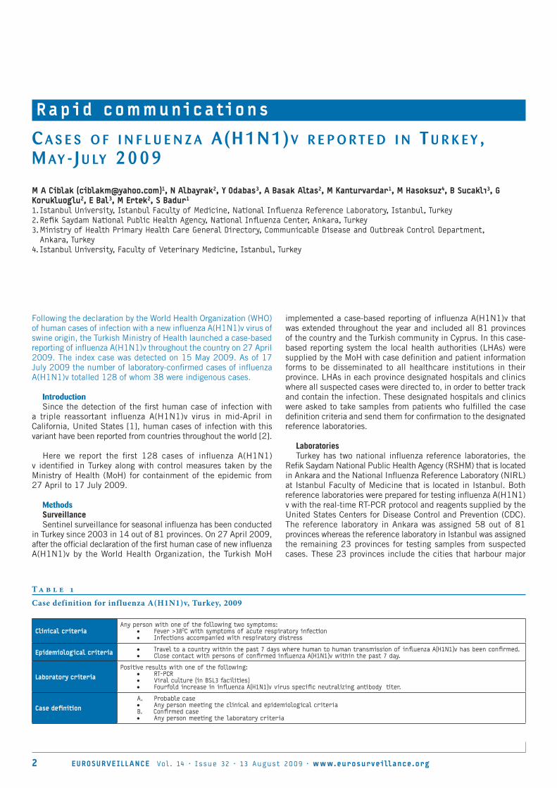
Following the declaration by the World Health Organization (WHO) of human cases of infection with a new influenza A(H1N1)v virus of swine origin, the Turkish Ministry of Health launched a case-based reporting of influenza A(H1N1)v throughout the country on 27 April 2009. The index case was detected on 15 May 2009. As of 17 July 2009 the number of laboratory-confirmed cases of influenza A(H1N1)v totalled 128 of whom 38 were indigenous cases.
Introduction Since the detection of the first human case of infection with
a triple reassortant influenza A(H1N1)v virus in mid-April in California, United States [1], human cases of infection with this variant have been reported from countries throughout the world [2].
Here we report the first 128 cases of influenza A(H1N1)v identified in Turkey along with control measures taken by the Ministry of Health (MoH) for containment of the epidemic from 27 April to 17 July 2009.
Methods SurveillanceSentinel surveillance for seasonal influenza has been conducted
in Turkey since 2003 in 14 out of 81 provinces. On 27 April 2009, after the official declaration of the first human case of new influenza A(H1N1)v by the World Health Organization, the Turkish MoH
implemented a case-based reporting of influenza A(H1N1)v that was extended throughout the year and included all 81 provinces of the country and the Turkish community in Cyprus. In this case-based reporting system the local health authorities (LHAs) were supplied by the MoH with case definition and patient information forms to be disseminated to all healthcare institutions in their province. LHAs in each province designated hospitals and clinics where all suspected cases were directed to, in order to better track and contain the infection. These designated hospitals and clinics were asked to take samples from patients who fulfilled the case definition criteria and send them for confirmation to the designated reference laboratories.
LaboratoriesTurkey has two national influenza reference laboratories, the
Refik Saydam National Public Health Agency (RSHM) that is located in Ankara and the National Influenza Reference Laboratory (NIRL) at Istanbul Faculty of Medicine that is located in Istanbul. Both reference laboratories were prepared for testing influenza A(H1N1)v with the real-time RT-PCR protocol and reagents supplied by the United States Centers for Disease Control and Prevention (CDC). The reference laboratory in Ankara was assigned 58 out of 81 provinces whereas the reference laboratory in Istanbul was assigned the remaining 23 provinces for testing samples from suspected cases. These 23 provinces include the cities that harbour major
T a b l e 1
Case definition for influenza A(H1N1)v, Turkey, 2009
Clinical criteriaAny person with one of the following two symptoms:
• Fever >380C with symptoms of acute respiratory infection• Infections accompanied with respiratory distress
Epidemiological criteria • Travel to a country within the past 7 days where human to human transmission of influenza A(H1N1)v has been confirmed. • Close contact with persons of confirmed influenza A(H1N1)v within the past 7 day.
Laboratory criteriaPositive results with one of the following:
• RT-PCR• Viral culture (in BSL3 facilities)• Fourfold increase in influenza A(H1N1)v virus specific neutralizing antibody titer.
Case definitionA. Probable case• Any person meeting the clinical and epidemiological criteria B. Confirmed case• Any person meeting the laboratory criteria
EUROSURVEILLANCE Vol . 14 · Issue 32 · 13 August 2009 · www.eurosurveillance.org 3
air and sea ports and resort towns. Laboratories report the results directly to the MoH immediately after the results are obtained. The MoH then informs the LHAs who contact the physicians and give necessary guidance to the physicians for the care of the patients.
Patients and samplesA probable case with influenza A(H1N1)v is defined as a person
with high fever (≥38 °C) and/or at least two acute respiratory symptoms along with epidemiological criteria listed in the case definition protocol published by WHO [2]. Table 1 summarises the case definition that was prepared in light of the information released by WHO. However, during the first month of the pandemic, in addition to probable cases, samples were also taken from individuals with no detectable symptoms but with either travel history to areas of high prevalence and/or close contact with a confirmed case, who presented in hospitals and asked to be tested. Nasal and/or nasopharyngeal samples along with patient information forms from suspected cases were transported to reference laboratories in a viral transport medium (Virocult, Medical Wire&Equipment, UK). A total of 977 samples from suspected cases were sent to the reference laboratories between 27 April and 17 July 2009 from various cities in Turkey (n=899) and from the Turkish Cypriot community (n=78).
Laboratory diagnosis (real-time RT-PCR)Both laboratories used the same “in-house” real-time PCR
protocol provided by CDC for detection of influenza A(H1N1)v. RNA extraction was done with QIAamp viral RNA mini kit (Qiagen, Valencia, CA, USA) or with a High Pure Viral RNA isolation kit from Roche. Real-time RT- PCR was performed on ABI 7000 and/or 7500 [3]. NA, HA and M genes of the isolate from the index case were partially sequenced and the resulting sequences were analysed by CLC Main Workbench 4.1.1 Software program (Denmark).
Control measures and patient managementAfter the declaration of the pandemic by WHO on 11 June,
the MoH held a meeting with its scientific advisory committee for revision of the pandemic plan. Revisions included the pandemic vaccination strategies (e.g. determining the priority order for vaccination), antiviral stockpiling and other measures. Two million doses of oseltamivir and 113,000 doses of zanamivir were distributed to all local healthcare centres. Four hundred thousand protective healthcare kits (each containing masks, gloves, hand disinfectant, goggles and foot covers) were distributed to healthcare providers, giving priority to those working at designated hospitals and clinics.
Special attention was given to the country points of entry such as airports and seaports. A thermal camera system was installed at airports and seaports in order to detect probable cases entering the country from regions of high prevalence. All travellers from abroad were requested to declare their health status and those captured by thermal camera system were further examined by physicians and suspected cases were isolated for transfer to the designated hospitals. Co-travellers sitting at close proximity (three seat lines in the front and back and on the sides) to confirmed cases were contacted by phone, informed about the situation and offered guidance on what they needed to do in case they developed symptoms and supplied with prophylactic doses of oseltamivir.
Two million pamphlets providing information on the flu pandemic were distributed to all flight crews and made available to travellers at airports and seaports. In addition, informative posters were posted at prominent places at ports and all public hospitals.
An interactive web page was designed to inform general public
F i g u r e 1
Figure 1. Number of travel-associated and indigenous cases of influenza A(H1N1)v, by week of laboratory confirmation, Turkey, May-July 2009 (n=125*)
*Number of cases with available date of the laboratory confirmationCase numbers collected by the Turkish Ministry of Health include cases from the Turkish Cypriot community.
2 2
7 7
4
10
13
28
14
1 1
11
14
11
0
5
10
15
20
25
30
20 21 22 23 24 25 26 27 28 29
Cases with travel history
No recent travel history
Week
F i g u r e 2
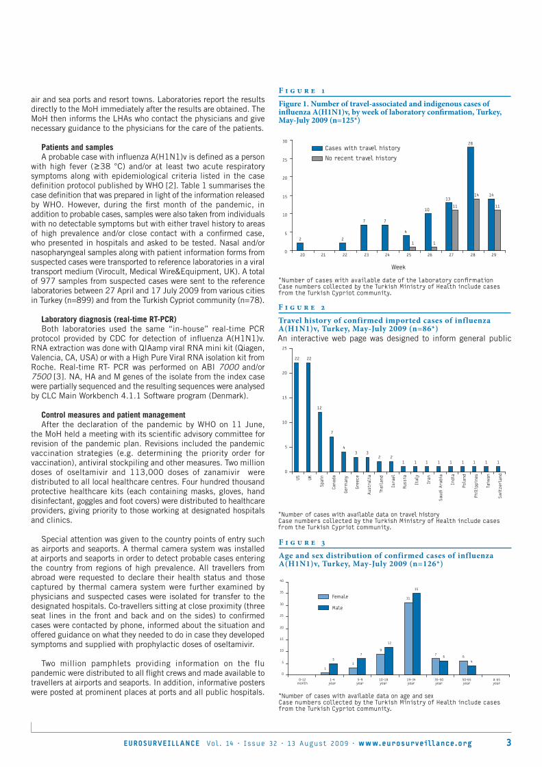
Travel history of confirmed imported cases of influenza A(H1N1)v, Turkey, May-July 2009 (n=86*)
*Number of cases with available data on travel historyCase numbers collected by the Turkish Ministry of Health include cases from the Turkish Cypriot community.
22 22
12
7
43 3
2 21 1 1 1 1 1 1 1 1
0
5
10
15
20
25
US UK
Spa
in
Cana
da
Germ
any
Gree
ce
Aus
tral
ia
Thai
land
Isra
el
Russ
ia
Ital
y
Iran
Sau
di A
rabi
a
Indi
a
Pola
nd
Phil
ippi
nes
Taiw
an
Sw
itze
rlan
d
F i g u r e 3
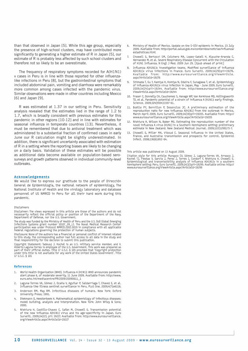
Age and sex distribution of confirmed cases of influenza A(H1N1)v, Turkey, May-July 2009 (n=126*)
*Number of cases with available data on age and sexCase numbers collected by the Turkish Ministry of Health include cases from the Turkish Cypriot community.
13
9
31
76
57
12
35
64
0
5
10
15
20
25
30
35
40
0-12month
1-4year
5-9year
10-18year
19-34year
35-50year
50-65year
≥ 65year
Female
Male

4 EUROSURVEILLANCE Vol . 14 · Issue 32 · 13 August 2009 · www.eurosurveillance.org
and professionals about the pandemic influenza which included information on individual care for protection from contacting and transmitting influenza (www.grip.saglik.gov.tr). A telephone hotline was launched to serve public inquiries seven days a week 24 hours a day (Alo 184 SABIM). Television spots were prepared mainly to emphasise the importance of hand washing and usage of disposable tissue papers in protecting against contracting and transmitting the influenza virus. Daily press briefings were held during the first month of the pandemic to keep public informed about the pandemic status in Turkey.
ResultsAll samples received before the index case was detected on 15
May 2009 were processed immediately and results were reported to the MoH regardless of the time of arrival of the sample to the laboratory. After 15 May both laboratories provided results seven days per week. The average time between the swabbing to final diagnosis was 24 hours.
The index case was a United States resident travelling from Tennessee to Iraq through Ataturk Airport in Istanbul where his high temperature was captured by thermal camera. He was hospitalised in a designated hospital in Istanbul and treated with oseltamivir until laboratory tests were negative for influenza A(H1N1)v. By 17 July influenza A(H1N1)v was detected in 128 (13%) out of 977
samples tested. Of these 128 positive samples, 17 were from the Turkish Cypriot community*, the remaining 111 were from various provinces in Turkey. The number of samples positive for influenza A(H1N1)v increased remarkably from June onward. Figure 1 presents the number of travel-associated and indigenous cases of influenza A(H1N1)v, by week of laboratory-confirmation. Of the 111 confirmed cases in Turkey, 25 were domestic secondary cases. Of the 17 confirmed cases in the Turkish Cypriot community, 13 were indigenous. The travel history of the imported confirmed cases is summarised in Figure 2 and the age and sex distribution of all confirmed cases is shown in Figure 3.
The partial sequence analysis results of matrix, HA and NA segments were submitted to the National Center for Biotechnology Information (NCBI) GenBank with accession numbers GQ200600, GQ200598, and GQ200599 respectively. According to the topological phylogenetic analysis, results obtained from the partial nucleic acid sequencing isolate from the index case were closely related to isolates from the US and A/Catalonia/10/2009 (H1N1).
The majority of influenza A(H1N1)v-positive cases (n=80) were detected in samples received from Istanbul (Figure 4) which also included the majority of indigenous cases (n=22) The remaining three indigenous cases in Turkey were from Denizli, Antalya and Eskisehir. Two indigenous cases from Istanbul were detected in healthcare workers, one in a physician examining a laboratory-confirmed patient and another in a nurse responsible for taking the patient’s sample in a private hospital setting. The physician and the nurse developed symptoms five days after contacting the patient; subsequent laboratory analysis confirmed these cases as influenza A(H1N1)v-positive.
Confirmed cases manifested moderate clinical symptoms. Three indigenous cases who contracted the virus from confirmed cases were asymptomatic. Clinical symptoms and their frequency in the confirmed cases are presented in Table 2.
The average time elapsed between the onset of the symptoms and the visit to the hospital (including those detected by thermal camera) was 1.68 days.
Of the 128 confirmed cases, 13 (10.2%) had received seasonal influenza vaccine in the past year. A similar proportion of vaccinated was found among patients who tested negative for influenza A(H1N1)v. All individuals who reported to the hospitals were closely monitored and those who were confirmed with influenza A(H1N1)v received antiviral treatment with oseltamivir. None of the confirmed cases developed any complications and no deaths occurred.
ConclusionInfluenza A(H1N1)v entered Turkey through travellers mainly
coming from the United States and the United Kingdom. While the majority of confirmed cases in Turkey had a travel history to highly affected areas, confirmed cases from the Turkish Cypriot community* were mostly indigenous cases with no history of travel. The majority of the confirmed cases consisted of young adults as reported from other countries. This could be related to the frequency of travel among the young population [4]. The clinical manifestation of A(H1N1)v infection in the confirmed cases was similar to that observed in seasonal influenza. All cases manifested moderate clinical symptoms similar to those reported in other countries [5]. Cough was the most frequent symptom (68.7%) followed by fever >38ºC (62.5%)**. None of the confirmed cases developed complications and no death was reported.
F i g u r e 4
Geographical distribution of confirmed cases of influenza A(H1N1)v, Turkey, May-July 2009 (n=128)
T a b l e 2
Clinical characteristics of confirmed cases of influenza A(H1N1)v, Turkey, May-July 2009 (n=128)*
Symptoms Number of cases with the symptom (%)
Cough 88 (68.7)
Fever (≥380C) 80 (62.5)
Sore throat 62 (48.4)
Headache 60 (46.8)
Coryza 59 (46.1)
Myalgia 56 (43.7)
Weakness 7 (5.5)
Pneumonia 3 (2.3)
*Case numbers collected by the Turkish Ministry of Health include cases from the Turkish Cypriot community.

EUROSURVEILLANCE Vol . 14 · Issue 32 · 13 August 2009 · www.eurosurveillance.org 5
Two confirmed indigenous cases were healthcare providers who contracted the disease in hospital while attending a confirmed case. This type of transmission in a hospital setting has been rare to date and it may require special attention [6].
After the detection of the index case on 15 May all confirmed cases were kept at the designated hospitals for treatment with oseltamivir and all contacts of these cases were traced and prophylactic oseltamivir doses were administered to these persons regardless of the symptoms. However, with increasing number of confirmed cases and individuals reporting to hospitals the MoH revised its policy on case investigation and management of the suspected cases on 5 June. With the new policy, confirmed patients with no signs of complications were put on oseltamivir therapy at home instead of hospitalisation, and prophylactic oseltamivir was no longer given to asymptomatic contacts of confirmed cases. Also, the practice of following up co-travellers of confirmed cases was ended by 5 June.
The amount of pandemic vaccine doses needed for vaccinating healthcare providers, public service providers and risk groups has been determined and necessary budget plans have been developed for purchasing 20 million doses to vaccinate 10 million individuals when the pandemic vaccine becomes available. Based on current knowledge of the pandemic, elderly people over 65 years were excluded from risk groups (in contrast with the seasonal vaccination recommendations) [7]. TV and radio spots have proven to be effective means of keeping the public calm and increasing awareness of pandemic influenza.
The MoH is planning to change its strategy and adopt measures for mitigation instead of containment of the pandemic in the coming weeks.
*Erratum: “Northern Cyprus” was replaced by “Turkish Cypriot community” throughout the text and the following information was added to the relevant tables and figures: Case numbers collected by the Turkish Ministry of Health include cases from the Turkish Cypriot community. These corrections were made on 17 August 2009.**Author’s correction: On request of the authors, the percentages in the sentence “Cough was the most frequent symptom (68.7%) followed by fever >38ºC (62.5%)” were corrected on 20 August 2009.
References
1. Centers for Disease Control and Prevention. Swine influenza A (H1N1) infection in two children-Southern California, March-April 2009. MMWR 2009;58:400–2.
2. World Health Organization. Pandemic (H1N1) 2009. Available from: http://www.who.int/csr/disease/swineflu/en/index.html
3. World Health Organization. CDC protocol of realtime RTPCR for influenza A (H1N1). Available from: www.who.int/csr/resources/publications/swineflu/CDCRealtimeRTPCR_SwineH1Assay-2009_20090430.pdf
4. Surveillance Group for New Influenza A (H1N1) Virus Investigation and Control in Spain. New Influenza A (H1N1) virus infections in Spain, April - May 2009. Eurosurv. 2009; 14(19):pii= 19209. Available from: http://www.eurosurveillance.org/ViewArticle.aspx?ArticleId=19209
5. New influenza A (H1N1) investigation teams. New Influenza A (H1N1) virus infections in France, April – May 2009. Eurosurv. 2009; 14(21): pii= 19221. Available from: http://www.eurosurveillance.org/ViewArticle.aspx?ArticleId=19221
6. Rizzo C, Declich S, Bella A, Caporali MG, Lana S, Pompa MG, et al. Enhanced epidemiological surveillance of Influenza A(H1N1)v in Italy. Eurosurv. 2009; 14(27): pii= 19266. Available from: http://www.eurosurveillance.org/ViewArticle.aspx?ArticleId=19266
7. World Health Organization. Strategic Advisory Group of Experts on Immunization - report of the extraordinary meeting on the influenza A (H1N1) 2009 pandemic, 7 July 2009. Wkly Epidemiol Rec. 2009 Jul 24;84(30):301-4. Available from: http://www.who.int/wer/2009/wer8430.pdf
This article was published on 13 August 2009.
Citation style for this article: Ciblak MA, Albayrak N, Odabas Y, Basak Altas A, Kanturvardar M, Hasoksuz M, Sucaklı B, Korukluoglu G, Bal E, Ertek M, Badur S. Cases of influenza A(H1N1)v reported in Turkey, May-July 2009. Euro Surveill. 2009;14(32):pii=19304. Available online: http://www.eurosurveillance.org/ViewArticle.aspx?ArticleId=19304

6 EUROSURVEILLANCE Vol . 14 · Issue 32 · 13 August 2009 · www.eurosurveillance.org
R ap i d com m uni ca ti on s
E p i d E m i o l o g i c a l a n d t r a n s m i s s i b i l i t y a n a ly s i s o f i n f l u E n z a a (H1n1 ) v i n a s o u t H E r n H E m i s p H E r E s E t t i n g : p E r u
C V Munayco ([email protected])1, J Gómez1, V A Laguna-Torres2, J Arrasco1, T J Kochel2, V Fiestas3, J Garcia2, J Perez2, I Torres3, F Condori3, H Nishiura4, G Chowell5,6
1. Dirección General de Epidemiología, Peru Ministry of Health, Lima, Peru2. United States Naval Medical Research Center Detachment, Lima, Peru3. National Institute of Health, Lima, Peru4. Theoretical Epidemiology, University of Utrecht, Utrecht, the Netherlands5. Mathematical and Computational Modeling Science Center, School of Human Evolution and Social Change Arizona State
University, Tempe, Arizona, United States6. Division of Population Studies, Fogarty International Center, National Institutes of Health, Bethesda, Maryland, United States
We present a preliminary analysis of 1,771 confirmed cases of influenza A(H1N1)v reported in Peru by 17 July 2009 including the frequency of the clinical characteristics, the spatial and age distribution of the cases and the estimate of the transmission potential. Age-specific frequency of cases was highest among school age children and young adults, with the lowest frequency of cases among seniors, a pattern that is consistent with reports from other countries. Estimates of the reproduction number lie in the range of 1.2 to 1.7, which is broadly consistent with previous estimates for this pandemic in other regions. Validation of these estimates will be possible as additional data become available.
IntroductionOn 24 April 2009, the World Health Organization (WHO)
informed about an epidemic caused by new swine-origin influenza A(H1N1)v virus originating from Mexico, and declared a public health emergency of international importance. The level of influenza pandemic alert was raised sequentially up to phase 6 on 11 June 2009 after global spread of the pandemic virus was confirmed [1].
In this study we present an analysis of 1,771 confirmed cases of influenza A(H1N1)v who developed the disease by 17 July 2009 and were reported to the National Surveillance Network in Peru, which since 2006 has conducted virological surveillance of influenza and other respiratory viruses by establishing sentinel sites throughout the country [2]. The patients’ age distribution, their clinical characteristics as well as their spatial distribution were studied. Estimates of transmission potential from the initial epidemic phase were also derived and compared with published estimates from other regions of the world.
Methods Surveillance systemOn 24 April 2009, the public health authorities of Peru
implemented new regulations for epidemiological surveillance and outbreak control of influenza A(H1N1)v defining the procedures of
F i g u r e 1
Geographical distribution of confirmed cases of influenza A(H1N1)v in Peru, as of 17 July 2009 (n=1,771)

EUROSURVEILLANCE Vol . 14 · Issue 32 · 13 August 2009 · www.eurosurveillance.org 7
detection, notification, investigation, follow-up and epidemiological control of A(H1N1)v cases in Peru.
An active surveillance system was established at all airports (especially in travellers returning from affected areas) and healthcare facilities, including private clinics. Also a telephone hotline (INFOSALUD) was made available by the Ministry of Health for citizens reporting influenza-like illness. A suspected case was defined as a person with a sudden onset of fever (>=38ºC) and respiratory symptoms. Suspected cases and their contacts were visited in their homes for clinical evaluation and nasal or pharyngeal specimens were taken from symptomatic persons and submitted to the National Institute of Health or the United States Naval Medical Research Center Detachment for RT-PCR as described by the Centers for Disease Control and Prevention (CDC). Suspected cases were informed about control measures to limit spread (voluntary isolation, use of face masks, and increased hygiene). Contacts of cases were monitored daily via phone calls or home visits. Symptomatic contacts were subjected to the same procedure as suspected cases. Clinical and epidemiological data were collected utilising a case report form (CRF) from all patients who met the case definition. Antivirals were given to all suspected cases until early July when the containment strategy was replaced by mitigation approach and treatment began to be administered only to high-risk groups.
Descriptive epidemiologyBased on the clinical and epidemiological data of the
National Surveillance Network, we characterised the descriptive
epidemiological features of influenza A(H1N1)v infection in Peru. First, we described the distribution of cases as a function of space, age and gender. Time-dependent characteristics were more analytically examined to estimate the transmission potential (see below). We also examined travel history of cases returning from countries with ongoing epidemics of A(H1N1)v infection, and the age-distributions between imported and indigenous cases were compared by means of non-parametric Mann-Whitney test. Second, we characterised frequency of symptoms reported for confirmed cases. The clinical-epidemiological forms were entered into a database created in Microsoft (MS) Office Access 2003, and data were analysed using MATLAB (The Mathworks, Inc.).
Estimation of transmission potentialA key epidemiological quantity which informs the expected
magnitude of an epidemic is the basic reproduction number (denoted by R0), defined as the average number of secondary cases generated by a primary case in an entirely susceptible population [3,4]. When R0>1 an epidemic can occur while R0<1 cannot support an epidemic. The reproduction number, R was estimated exploring time-evolution of confirmed cases. Statistical methods were based on pure birth process (to estimate the intrinsic growth rate r) and renewal process (to estimate R using r), and were identical to those given elsewhere [5]. Whereas we analysed the temporal distribution including all possible primary cases (i.e. including imported cases) as the number of imported cases was in a negligible order, we also examined the estimate excluding imported cases (as it can then exclude imported cases from the category of secondary cases).
F i g u r e 2
Age distribution of confirmed cases of influenza A(H1N1)v reported in Peru as of 17 July 2009 (n=1,765*)
7.08%
24.02%24.48%
10.42%
8.27%
5.72%5.10%
4.25%
3.00%2.61%
1.93% 1.81%1.30%
0.00%
5.00%
10.00%
15.00%
20.00%
25.00%
30.00%
0 - 4 5 - 9 10 -14 15 - 19 20 - 24 25 - 29 30 - 34 35 - 39 40 - 44 45 - 49 50 - 54 55 - 59 60 +
Age group
Perc
enta
ge
*Number of cases with available data on age

8 EUROSURVEILLANCE Vol . 14 · Issue 32 · 13 August 2009 · www.eurosurveillance.org
ResultsThe first influenza A(H1N1)v confirmed case in Peru was a
Peruvian citizen returning from New York on 9 May with a respiratory disease. Since then the pandemic has quickly spread throughout the country. As of 17 July 2009, a total of 1,771 cases, involving eight deaths, have been confirmed. This yields a crude case fatality ratio of 0.33 % (95% confidence interval: 0.14, 0.65). Of the 1,771 cases, 1,420 (80.1%) were from Lima, the capital city, 84 (4.7%) from Piura and 81 (4.6%) from La Libertad. Figure 1 shows the geographic distribution of confirmed cases of influenza A(H1N1)v in Peru.
A total of 78 (4.4%) confirmed cases had a history of recent travel to the United States, Dominican Republic or Argentina. Imported cases generated clusters of different sizes that established indigenous transmission in Peru. For example, between 8 and 30 May, 600 private high school students travelled to Punta Cana in the Dominican Republic for vacations. One student presented influenza-like illness before returning and other 11 students developed symptoms upon returning to Peru.
Females (52%) were slightly more affected than males (48%).
The most affected age group was that of 5-14 years (Figure 2). The age of the cases ranged from 0 to 87 years with a mean of 18.5 years and a median of 13 years. The mean age of the imported cases was 28 years while indigenous cases had a mean age of 18 years (Mann-Whitney test, P<0.001).
Figure 3 summarises the clinical characteristics of the confirmed cases of influenza A(H1N1)v infection. The most frequent symptoms were fever (94%), cough (93%), sore throat (77%), general malaise (77%) and rhinorrhoea (76%). Gastrointestinal symptoms including abdominal pain (28%), vomiting (26%) and diarrhoea (16%) were not uncommon.
Epidemic curve and transmissibilityFigure 4A shows the temporal distribution of confirmed cases
as a function of the date of onset. The number of cases greatly increased from mid-June to mid-July. It should be noted that cases in mid-July are likely underestimated due to reporting delay, and the temporal dynamics are also influenced by spatial spread from Lima to the rest of the country in the subsequent time periods. Based on the epidemic curve, the first three weeks (from 6 to 29 May) were considered as “random phase”. Informed by deviation of our simple model from the observed data (i.e. Akaike Information Criterion obtained from negative loglikelihood and a single parameter to be estimated), 30 May was assumed to be the starting time point of exponential growth (and called Day 1). We also assumed that the exponential growth phase continued up to 20 June (for three weeks which should capture the dynamics of the first 6-10 generations), while allowing plus/minus two days. Including all imported cases, the intrinsic growth rate, r was estimated at 0.117 (95% CI: 0.106, 0.128) per day. Excluding all imported cases, r was estimated at 0.135 (95% CI: 0.122, 0.149) per day. Assuming that the mean generation time = 2.8 days, and coefficient of variation (CV) = 47.1%, R for these settings was estimated at 1.37 (95% CI: 1.33, 1.41) and 1.44 (95% CI: 1.39, 1.49), respectively. Figure 4B compares observed and predicted epidemic curves. We also examined the sensitivity of R for different lengths of mean generation time (ranging from 1.6 to 4.0 days) (Figure 4C), and the maximum likelihood estimate of R ranged from 1.2 to 1.6. When we use different windows (18 June to 22 June as the latest time points of exponential growth), R appeared to range from 1.3 to 1.4 (Figure 4D).
DiscussionThe current pattern of spread of influenza A(H1N1)v in Peru is
dominated by a wave that emanates from the capital city, Lima, the early dynamics of which may most likely be associated with high frequency of international travel, thereby increasing the chances of a major epidemic in the capital city.
Our early findings indicate that public health interventions need to be in accord with the epidemiological behaviours (e.g. temporal and spatial increase) and moderate severity of the disease. For
F i g u r e 3
Clinical characteristics of confirmed cases of influenza A(H1N1)v reported in Peru as of 17 July 2009 (n=1,771)
0
10
20
30
40
50
60
70
80
90
100
Skin
eru
ptio
n
Aden
opat
hy
Ota
lgia
Phot
opho
bia
Diar
rhoe
a
Whe
ezin
g
Vom
itin
g
Abdo
min
al p
ain
Conju
nct
ival
con
gest
ion
Asth
enia
Expe
ctor
atio
n
Mia
lgia
Phar
ynge
al c
onge
stio
n
Hea
dach
e
Rhin
orrh
oea
Gen
eral
mal
aise
Sore
thr
oat
Coug
h
Feve
r
Perc
enta
ge

EUROSURVEILLANCE Vol . 14 · Issue 32 · 13 August 2009 · www.eurosurveillance.org 9
instance, while in some countries radical control measures aimed at rapid containment, such as contact tracing and complete proactive school closures, were conducted during the early phase of this pandemic, the epidemic in Peru without obvious school clusters during the early phase did not offer an opportunity to implement similar countermeasures. In such settings it may be more realistic to focus interventions on minimising mortality at the population level (e.g. early diagnosis and treatment of severe cases).
Despite the lack of obvious large clusters, the great majority of cases were documented among school age children and young adults, with the lowest frequency of cases among seniors, a pattern that is consistent with reports form other countries [5-8]. It should be noted that the age-distribution of cases could change as the epidemic develops. Also, it should be noted that the impact of high school and university students (i.e. those aged from 15 to 19 years) on the transmission dynamics is presumably smaller
F i g u r e 4
A) Epidemic curve of confirmed cases of influenza A(H1N1)v in Peru by date of symptoms onset, 8 May 2009 to 17 July 2009; B) Exponential growth fit to the early epidemic phase of influenza A(H1N1)v in Peru. Data are the black dots, the solid line is the exponential fit to the data, and dashed lines correspond to uncertainty bounds of the expectation based on the confidence limits of the intrinsic growth phase; C) The reproduction number estimates from the early epidemic phase of the epidemic curve of influenza A(H1N1)v cases in Peru as a function of plausible mean generation times and D) using different end dates of the initial growth phase.
A B
C D
1 0 EUROSURVEILLANCE Vol . 14 · Issue 32 · 13 August 2009 · www.eurosurveillance.org
than that observed in Japan [5]. While this age group, especially the presence of high-school clusters, may have contributed more significantly to generating a higher estimate of R in Japan [5], our estimate of R is probably less affected by such school clusters and therefore not so likely to be an overestimate.
The frequency of respiratory symptoms recorded for A(H1N1)v cases in Peru is in line with those reported for other influenza-like infections in Peru [8], but the gastrointestinal symptoms that included abdominal pain, vomiting and diarrhoea were remarkably more common among cases infected with the pandemic virus. Similar observations were made in other countries including Mexico [6] and Japan [9].
R was estimated at 1.37 in our setting in Peru. Sensitivity analysis revealed that the estimates lied in the range of 1.2 to 1.7, which is broadly consistent with previous estimates for this pandemic in other regions [10-12] and in line with estimates for seasonal influenza in temperate countries [13]. Nevertheless, it must be remembered that due to antiviral treatment which was administered to a substantial fraction of confirmed cases in early June our R calculation might be slightly underestimated. In addition, there is significant uncertainty associated with estimation of R in a setting where the reporting biases are likely to be changing on a daily basis. Validation of these estimates will be possible as additional data become available on population-based sero-surveys and growth patterns observed in individual community-level outbreaks.
AcknowledgementsWe would like to express our gratitude to the people of Dirección General de Epidemiología, the national network of epidemiology, The National Institute of Health and the virology laboratory and database personnel of US NMRCD in Peru for all their hard work during this pandemic.
Disclaimers
Disclaimer: The views expressed in this article are those of the authors and do not necessarily reflect the official policy or position of the Department of the Navy, Department of Defense, nor the U.S. Government.
The study was funded by the Ministry of Health of Peru and the U.S. DoD Global Emerging Infections Systems grant number 10107_09_LI. The Naval Medical Research Center participation was under Protocol NMRCD.2002.0019 in compliance with all applicable Federal regulations governing the protection of human subjects.
Disclosure: None of the authors has a financial or personal conflict of interest related to this study. The corresponding author had full access to all data in the study and final responsibility for the decision to submit this publication.
Copyright Statement: Tadeusz J. Kochel is an U.S. military service member, and V. Alberto Laguna-Torres is employee of the U.S. Government. This work was prepared as part of their official duties. Title 17 U.S.C. § 105 provides that ‘Copyright protection under this title is not available for any work of the United States Government’. Title 17 U.S.C. § 101
References
1. World Health Organization (WHO). Influenza A (H1N1): WHO announces pandemic alert phase 6, of moderate severity. 11 June 2009. Available from: http://www.euro.who.int/mediacentre/PR/2009/20090611_1
2. Laguna-Torres VA, Gómez J, Ocaña V, Aguilar P, Saldarriaga T, Chavez E, et al. Influenza-like illness sentinel surveillance in Peru. PLoS One. 2009;4(7):e6118.
3. Anderson RM, May RM. Infectious diseases of humans. New York: Oxford University Press; 1991.
4. Diekmann O, Heesterbeek H. Mathematical epidemiology of infectious diseases: model building, analysis and interpretation. New York: John Wiley & Sons; 2000.
5. Nishiura H, Castillo-Chavez C, Safan M, Chowell G. Transmission potential of the new influenza A(H1N1) virus and its age-specificity in Japan. Euro Surveill. 2009;14(22). pii: 19227. Available from: http://www.eurosurveillance.org/ViewArticle.aspx?ArticleId=19227
6. Ministry of Health of Mexico. Update on the S-OIV epidemic in Mexico. 23 July 2009. Available from: http://portal.salud.gob.mx/contenidos/noticias/influenza/estadisticas.html
7. Chowell G, Bertozzi SM, Colchero MA, Lopez-Gatell H, Alpuche-Aranda C, Hernandez M, et al. Severe Respiratory Disease Concurrent with the Circulation of H1N1 Influenza. N Engl J Med. 2009 Jun 29. [Epub ahead of print]
8. Influenza A(H1N1)v investigation teams. Modified surveillance of influenza A(H1N1)v virus infections in France. Euro Surveill. 2009;14(29):pii=19276. Available from: http://www.eurosurveillance.org/ViewArticle.aspx?ArticleId=19276
9. Shimada T, Gu Y, Kamiya H, Komiya N, Odaira F, Sunagawa T, et al. Epidemiology of influenza A(H1N1)v virus infection in Japan, May - June 2009. Euro Surveill. 2009;14(24):pii=19244. Available from: http://www.eurosurveillance.org/ViewArticle.aspx?ArticleId=19244
10. Fraser C, Donnelly CA, Cauchemez S, Hanage WP, Van Kerkhove MD, Hollingsworth TD, et al. Pandemic potential of a strain of influenza A (H1N1): early findings. Science. 2009;324(5934):1557-61.
11. Boëlle PY, Bernillon P, Desenclos JC. A preliminary estimation of the reproduction ratio for new influenza A(H1N1) from the outbreak in Mexico, March-April 2009. Euro Surveill. 2009;14(19):pii=19205. Available from: http://www.eurosurveillance.org/ViewArticle.aspx?ArticleId=19205
12. Nishiura H, Wilson N, Baker MG. Estimating the reproduction number of the novel influenza A virus (H1N1) in a Southern Hemisphere setting: preliminary estimate in New Zealand. New Zealand Medical Journal. 2009;122(1299):73-7.
13. Chowell G, Miller MA, Viboud C. Seasonal influenza in the United States, France, and Australia: transmission and prospects for control. Epidemiol Infect. 2008;136(6):852-64.
This article was published on 13 August 2009.
Citation style for this article: Munayco CV, Gómez J, Laguna-Torres VA, Arrasco J, Kochel TJ, Fiestas V, Garcia J, Perez J, Torres I, Condori F, Nishiura H, Chowell G. Epidemiological and transmissibility analysis of influenza A(H1N1)v in a southern hemisphere setting: Peru. Euro Surveill. 2009;14(32):pii=19299. Available online: http://www.eurosurveillance.org/ViewArticle.aspx?ArticleId=19299

EUROSURVEILLANCE Vol . 14 · Issue 32 · 13 August 2009 · www.eurosurveillance.org 11
R ap i d com m uni ca ti on s
W h at W i l l t h e n e x t i n f l u e n z a s e a s o n b r i n g a b o u t : s e a s o n a l i n f l u e n z a o r t h e n e W a (h1n1 ) v ? a n a n a ly s i s o f g e r m a n i n f l u e n z a s u r v e i l l a n c e d ata
H Uphoff ([email protected])1, S Geis1,2, A Grüber3, A M Hauri1
1. Hessisches Landesprüfungs- und Untersuchungsamt im Gesundheitswesen, Dillenburg, Germany2. Postgraduate Training in Applied Epidemiology (PAE), Department for Infectious Disease Epidemiology, Robert Koch-Institute,
Berlin, Germany3. Deutsches Grünes Kreuz (German Green Cross), Marburg, Germany
For the next influenza season (winter 2009-10) the relative contributions to virus circulation and influenza-associated morbidity of the seasonal influenza viruses A(H3N2), A(H1N1) and B, and the new influenza A(H1N1)v are still unknown. We estimated the chances of seasonal influenza to circulate during the upcoming season using data of the German influenza sentinel scheme from 1992 to 2009. We calculated type and subtype-specific indices for past exposure and the corresponding morbidity indices for each season. For the upcoming season 2009-10 our model suggests that it is unlikely that influenza A(H3N2) will circulate with more than a low intensity, seasonal A(H1N1) with more than a low to moderate intensity, and influenza B with more than a low to median intensity. The probability of a competitive circulation of seasonal influenza A with the new A(H1N1)v is low, increasing the chance for the latter to dominate the next influenza season in Germany.
Background A new influenza A(H1N1) variant has spread globally since its
first appearance in April 2009 [1] and its transmissibility has been estimated in a range similar to that known from seasonal influenza. Nevertheless it is unclear if this new influenza A(H1N1)v will replace seasonal influenza A or there may be co-circulation or successive circulation, in particular considering that A(H1N1)v has been circulating very early, ahead of the season. Cross immunity of the new influenza A(H1N1)v with seasonal influenza viruses is very low and probably negligible except for elderly people [2]. Hence a general susceptibility of the population to the new A(H1N1)v is assumed, even though not immunity-based mechanisms may additionally influence susceptibility [3,4]. For seasonal influenza A partial immunity of the population due to previous infections can be assumed. A rather constant drift with significant antigenetic changes - when a new successful lineage evolves - allows the virus to overcome this immunity [5]. The imbalance of population immunity and drift is seen as a driving force for intense virus circulation. The exact correlate of molecular or antigenic drift - as characterised by laboratory methods - on this balance is unknown.
In most European countries primary care sentinel surveillance systems are used to estimate the “intensity” of seasons and laboratory testing of a sub-sample indicates the viruses circulating. These data do not provide exact measurements of virus circulation
and subsequent population immunity. However, assuming a stable relationship between population immunity and virus circulation, the latter can serve as a proxy measure of type- and subtype-specific population immunity. For the upcoming season a seasonal influenza vaccine and a vaccine against the new influenza A(H1N1)v will be available with some remaining uncertainties regarding the amount of each. Therefore anticipating the circulation of the different seasonal influenza viruses - still present in the population - and the new A(H1N1)v virus may be helpful for setting up the vaccination strategy.
Materials and methodsWe calculated type- and subtype-specific indices of past
exposures and morbidity indices by season. We used data of the German influenza sentinel system (AGI) which has been registering acute respiratory tract infections in the winter seasons since 1992 (available at http://influenza.rki.de/). In this system the presence of influenza viruses is monitored through syndromic sentinel surveillance and, additionally, a sub-group of participating physicians swab patients with acute respiratory tract infections and send samples for testing to the national influenza reference centre (NIC).
In order to allow the calculation of indices for as many seasons as possible, in addition, virological data from NIC for the five seasons before 1992-3 were used.
For each season we estimated the total influenza-associated morbidity from weekly excess consultations (as percentage above baseline) during periods of laboratory-confirmed influenza activity [6]. Splitting this total excess morbidity by the percentages of detected influenza types and subtypes gave the type- and subtype-specific morbidity index for each season.
To calculate indices of past exposure we used the morbidity indices of the five preceding seasons. However, it is unclear for how long immunity acquired during past exposures persists. We therefore used weighting factors to adjust for the decreasing influence of more distant seasons. Each of the five included seasons was weighted with a factor that was kept constant for all calculations. The set of weighting factors giving the best linear

12 EUROSURVEILLANCE Vol . 14 · Issue 32 · 13 August 2009 · www.eurosurveillance.org
correlation for all seasons between the morbidity index of each season and the morbidity indices of the respective five preceding seasons was chosen. The sum of the five weighted morbidity indices gave the past exposure index for the respective season.
Results The figures show the distribution of the value pairs of the
estimated past exposure and morbidity indices for each season (1992-3 to 2008-9), by influenza type and subtype. Estimates obtained using data exclusively of the NIC are plotted in grey, estimates obtained using data of the AGI are in blue, and the estimate for the past exposure index for the upcoming season (2009-10) is plotted as a blue arrow. For influenza A(H3N2) and B the best linear correlation (-0.55 for A(H3N2) and -0.62 for B) was seen when the morbidity index of just the directly preceding
season was used to estimate the past exposure index. For seasonal influenza A(H1N1) the best correlation (-0.46) was obtained with weighting factors that left a greater relative contribution to more distant seasons (preceding season: weighting factor = 1; two years ago = 1.4-1; three years ago = 1.9-1; four years ago = 2.7-1; five years ago = 3.8-1).
For all seasonal influenza viruses the distribution pattern is similar: the probability of a high excess morbidity - as correlate of intense virus circulation - is low when the past exposure index is high. For median past exposure indices low to moderate seasons can be expected and for low past exposure indices severe seasons may but do not need to occur. These distributions of the value pairs (past exposure and morbidity indices) are typical of distributions which reflect a limiting influence, i.e the past exposure indices represent a kind of upper bound for the morbidity indices of the corresponding seasons. Seasons with no measurable intensity are rare for influenza A(H3N2), frequent for seasonal A(H1N1) and occur with intermediate frequency for influenza B.
Influenza A(H3N2) reaches the highest indices of past exposure and morbidity. However, direct comparability of past exposure indices is only given between influenza A(H3N2) and influenza B. Their respective past exposure indices are based on the same number of seasons and the identical weighing factors.
For the next influenza season in Germany the results of our model suggest that it is very unlikely that influenza A(H3N2) will circulate with more than a low intensity. The same can be concluded for seasonal A(H1N1) with a slight chance to reach a moderate intensity level. Influenza B may circulate with up to a median intensity.
Discussion Predictions of the circulation of influenza viruses in upcoming
seasons are highly desirable but generally accepted models are still lacking [7-9]. This is mainly due to the multitude of factors involved and limited data availability and quality. The data we used on
F i g u r e 3
Estimates of past exposure and morbidity indices of influenza B, Germany, 1992-3 to 2008-9
Note: Estimates obtained using only virological data from the national influenza reference centre (NIC) are plotted in grey, estimates obtained using data of the influenza sentinel system (AGI) are in blue, and the estimate for the past exposure index for the upcoming season (2009-10) is plotted as a blue arrow.
0
50
100
150
200
250
300
350
400
450
0 50 100 150 200 250 300 350 400 450
Morbidity index
Past
exposure
index
past exposure index of B for the season 09/10
92/93
F i g u r e 2
Estimates of past exposure and morbidity indices of seasonal influenza A(H1N1), Germany, 1992-3 to 2008-9
Note: Estimates obtained using only virological data from the national influenza reference centre (NIC) are plotted in grey, estimates obtained using data of the influenza sentinel system (AGI) are in blue, and the estimate for the past exposure index for the upcoming season (2009-10) is plotted as a blue arrow.
0
50
100
150
200
250
300
350
400
0 50 100 150 200 250 300 350 400
Morbidity index
Past
exposure
index
past exposure index of H1N1 for the season 09/10
96/97
95/9694/95
93/94
92/93
F i g u r e 1
Estimates of past exposure and morbidity indices of influenza A(H3N2), Germany, 1992-3 to 2008-9
Note: Estimates obtained using only virological data from the national influenza reference centre (NIC) are plotted in grey, estimates obtained using data of the influenza sentinel system (AGI) are in blue, and the estimate for the past exposure index for the upcoming season (2009-10) is plotted as a blue arrow.
0
100
200
300
400
500
600
700
800
0 100 200 300 400 500 600 700 800
Morbidity index
Past
exposure
index
past exposure index of H3N2 for the season 09/10
92/93

EUROSURVEILLANCE Vol . 14 · Issue 32 · 13 August 2009 · www.eurosurveillance.org 13
morbidity and virus circulation have been collected systematically for 17 years, thus providing a reasonable basis for the approach we used.
This analysis is based on the assumption of a type- and subtype-specific link between past exposure and virus circulation in the following season. In our results for influenza A(H3N2) and B a short lived “protection” of past exposure is suggested. These results are in line with a short-lived strain overlapping immunity as suggested by modelling studies [7].
We consider the chances for the seasonal influenza viruses to lead to considerable morbidity during the upcoming influenza season 2009-10 to be very low. Should the A(H1N1)v virus circulation during the upcoming season 2009-10 be high enough, the expected low seasonal activity may lead to a rapid total replacement, as seen in previous pandemics (except for the 1977 H1N1). However, if the activity of the A(H1N1)v during the season 2009-10 is a pre-wave and a severe circulation of A(H1N1)v will be seen in the following season 2010-1, the possibly low past exposure index for the 2010-1 season in Germany may hamper a total replacement [7].
This model has several limitations. Regional differences in virus circulation are not taken account of. The frequency of laboratory testing varies during one season and, additionally, depends on type- and subtype-specific disease severity, thus potentially biasing the relative contributions of the different virus types and subtypes. In addition, a relatively short time series (17 value pairs for each type/subtype) limit the applicability of complex statistics.
In conclusion, our systematic approach may reduce the unpredictability of influenza activity and thus contribute to strategic planning, e.g. regarding vaccination priorities. These results should be confirmed with data obtained from different surveillance systems. Further improvements of this model may then address its current limitations and additionally offer the possibility to include other factors, such as weather conditions [8], holidays or historical experiences regarding timing and trend [9].
References
1. Novel Swine-Origin Influenza A (H1N1) Virus Investigation Team, Dawood FS, Jain S, Finelli L, Shaw MW, Lindstrom S, et al. Emergence of a novel swine-origin influenza A (H1N1) virus in humans. N Engl J Med. 2009 Jun 18;360(25):2605-15.
2. Centers for Disease Control and Prevention (CDC). Serum cross-reactive antibody response to a novel influenza A(H1N1) virus after vaccination with seasonal influenza vaccine. MMWR 2009;58(19):521-4.
3. Albright FS, Orlando P. Pavia AT, Jackson GG, Cannon Albright LA. Evidence for a heritable predisposition to death due to influenza. J Infect Dis. 2008;197(1):18-24.
4. Trammell RA, Toth LA. Genetic susceptibility and resistance to influenza infection and disease in humans and mice. Expert Rev Mol Diagn. 2008;8(4):515-29.
5. Bush RM, Bender CA, Subbarao K, Cox NJ, Fitch WM. Predicting the evolution of human influenza A. Science. 1999;286(5446):1921-5.
6. Uphoff H, Heckler R, Schweiger B. Betrachtungen zur Durchseuchung und beobachteter Aktivität bei Influenza B [Reflections on the morbidity and observed activity of influenza B]. Bundesgesundheitsbl. 1998;11:469-473. German.
7. Ferguson N M, Galvani AP, Bush RM. Ecological and immunological determinants of influenza evolution. Nature 2003;422(6930):428-433.
8. Shoji M, Katayama H, Takahashi M. [Epidemic prediction of the influenza by Excel. (2) The epidemic prediction of the influenza in Japan. The reflection in validity and 2003-2004 season of the reason of the prediction]. Japanese Journal of Clinical and Experimental Medicine 2004;81(12):1991-2000. Japanese.
9. Viboud C, Boëlle PY, Carrat F, Valleron AJ, Flahault A. Prediction of the spread of influenza epidemics by the method of analogues. Am J Epidemiol. 2003;158(1):996-1006.
This article was published on 13 August 2009.
Citation style for this article: Upphoff H, Geis S, Grüber A, Hauri AM. What will the next influenza season bring about: seasonal influenza or the new A(H1N1)v? An analysis of German influenza surveillance data. Euro Surveill. 2009;14(32):pii=19303. Available online: http://www.eurosurveillance.org/ViewArticle.aspx?ArticleId=19303

14 EUROSURVEILLANCE Vol . 14 · Issue 32 · 13 August 2009 · www.eurosurveillance.org
R ap i d com m uni ca ti on s
T h e S w e d i S h n e w va r i a n T o f C h l a m y d i a T r a C h o m aT i S ( n v CT ) r e m a i n S u n d e T e C T e d b y m a n y e u r o p e a n l a b o r aTo r i e S a S r e v e a l e d i n T h e r e C e n T p C r/ naT r i n g T r i a l o r g a n i S e d b y i n STan d e .v. , g e r m a n y
U Reischl ([email protected])1, E Straube2, M Unemo3
1. Institute of Medical Microbiology and Hygiene, University Hospital of Regensburg, Regensburg, Germany2. Institute of Medical Microbiology, Friedrich-Schiller University, Jena, Germany3. National Reference Laboratory for Pathogenic Neisseria, Department of Laboratory Medicine, Clinical Microbiology, Örebro
University Hospital, Örebro, Sweden
The May 2009 round of INSTAND’s ring trial “Chlamydia trachomatis detection PCR/NAT” included a sample with high amount of the Swedish new variant of C. trachomatis (nvCT). A spectrum of at least 12 different commercial diagnostic nucleic acid amplification tests (NAATs) and many different in house NAATs were applied by the 128 participating laboratories which reported 152 results. Approximately 80% of the results correctly reported the presence of C. trachomatis in the nvCT specimen. The nvCT sample was mainly missed, as expected, by participants using the Roche COBAS Amplicor CT/NG (15.5% of reported results) but also by several participants using in house NAATs. The trend towards using nvCT-detecting NAATs is obvious and in addition to the new dual-target NAATs from Roche and Abbott, and BD ProbeTec ET, also a number of new CE mark-certified commercial tests from smaller diagnostic companies as well as many different in house NAATs were used. Laboratories using commercial or in house NAATs that do not detect the nvCT are encouraged to carefully monitor their C. trachomatis incidence, participate in appropriate external quality assurance and controls schemes, and consider altering their testing system. The reliable detection of low amounts of the wildtype C. trachomatis strain in other samples of the ring trial set indicates a good diagnostic performance of all applied commercial NAATs while also detecting the nvCT strain.
IntroductionWith the increasing acceptance of nucleic acid amplification
tests (NAATs) in the field of diagnostic microbiology and the broad availability of open platforms to perform an exponentially growing spectrum of in house and/or commercially prefabricated NAATs, there is a growing demand for appropriate internal and external quality control (QC) activities. One comprehensive external quality assessment scheme (EQAS) for diagnostic NAATs was established in 2002 by the German Society for Promotion of Quality Assurance in Medical Laboratories, INSTAND e.V. (www.instand-ev.de). This subscheme of INSTAND’s well-established quality control initiatives, named “bacterial genome detection PCR/NAT”, offers certified proficiency testing panels for prominent bacterial pathogens on a biannual basis. A detailed discussion of the current and the previous EQAS schemes can be found at: http://www-nw.uni-regensburg.de/~.reu24900.mmh.klinik.uni-regensburg.de/INSTAND_e.htm.
In 2006, a new variant of Chlamydia trachomatis (nvCT) was
identified in the Swedish county of Halland by Ripa and co-workers [1]. This mutant strain is characterised by a 377-bp deletion in ORF-1 of the multicopy cryptic plasmid, which includes the target region of both the Roche and Abbott C. trachomatis NAATs available at that time [2]. The currently available new redesigned dual-target assays, namely the Abbott RealTime CT/NG (CE mark-certified in January 2008) that targets another cryptic plasmid sequence in addition to the sequence affected by the nvCT deletion, and the Roche COBAS TaqMan CT v2.0 (CE mark-certified in June 2008) that detects the chromosomal ompA gene in addition to the sequence affected by the nvCT deletion, have replaced the former assays [3].
Immediately after the first report on the nvCT, international studies were conducted to determine whether the nvCT was present in different settings across Europe, the United States, Australia [3-5]. Only sporadic cases have so far been reported outside the Nordic countries [3-5], however, current knowledge regarding the presence and prevalence of nvCT in other countries is highly limited due to few recent studies and the fact that many European laboratories can still not detect the nvCT [4], and those that can are not aware of it because no nvCT-specific or other distinguishing NAATs are used. Ideally all laboratories should use NAATs that detect nvCT, because a wider geographic spread of this variant can not be excluded.
To supplement a recent Eurosurveillance publication on a United Kingdom National EQAS (UK NEQAS) distribution [4], the present report provides a concise reflection on diagnostic performance and NAATs used by European laboratories participating in the May 2009 INSTAND e.V. ring trial regarding detection of the nvCT. Assuming that most diagnostic laboratories are participating in one external QC scheme only, the intersection between UK NEQAS and INSTAND ring trials should be very limited and the present study represents an additional exploratory piece in the jigsaw puzzle of European C. trachomatis NAAT testing regimens.
Materials and methodsThe May 2009 round of INSTAND’s EQAS “bacterial genome
detection PCR/NAT” included two panels for C. trachomatis detection. One set of four lyophilised blinded samples was offered

EUROSURVEILLANCE Vol . 14 · Issue 32 · 13 August 2009 · www.eurosurveillance.org 1 5
for participants using combined detection of C. trachomatis and Neisseria gonorrhoeae (RV 530), and a separate set for those detecting C. trachomatis only (RV 531).
The latter set (C. trachomatis; RV 531) contained a sample with ~105 inclusion forming units (IFUs) of the nvCT strain per ml of reconstituted lyophilised specimen. This set was completed by two samples containing ~103 IFUs/ml of a wildtype C. trachomatis strain and one sample without C. trachomatis in a natural background of human and bacterial cells. The laboratories were requested to reconstitute the specimen in 300 µl of molecular grade water and analyse a 100 µl portion of the specimen, according to their routine protocols for detecting C. trachomatis from an endocervical swab.
Results Response rateNAAT results for distribution RV 531, which included the nvCT
sample, were returned by 128 laboratories (100% of participants), including 115 laboratories from Germany, 12 from nine other European countries and one from United Arab Emirates (Table). For unknown reasons, some laboratories applied more than one C. trachomatis NAAT, which probably does not reflect their routine diagnostic workup for C. trachomatis. Due to this reporting of results from multiple assays and/or lack of assay specifications in the reports, the effective number of results (n=152) is higher than the number of participants (n=128).
Nucleic acid amplification tests (NAATs) used for C. trachomatis diagnosticsThe change in the spectrum of NAATs applied by the participants
in the German INSTAND schemes from 2006 to 2009 is depicted in Figure 1. Especially in the current round (2009), the spectrum of NAATs used in the German INSTAND ring trial substantially differed from the recent UK NEQAS ring trial [4]. In 2009, Roche COBAS Amplicor CT/NG (15.5% of participants) and BD ProbeTec ET (Becton Dickinson; 15.5%) were the most commonly used main NAATs, followed by Roche Cobas TaqMan (14.8%) and Abbott RealTime CT (11.0%). Nevertheless, from 2006 to 2009, the use of Roche COBAS Amplicor CT/NG and Roche Amplicor
CT/NG rapidly decreased from 37.4% to 15.5%, and 6.8% to 0%, respectively. In contrast, the numbers of laboratories who have shifted to the new dual-target assays Abbott RealTime CT/NG and Roche COBAS TaqMan CT v2.0 significantly increased. Furthermore, especially in the recent years several new or at least less popular commercial C. trachomatis NAATs as well as many in house NAATs were in use (Figure 1).
Detection of the Swedish new variant of C. trachomatis (nvCT) Twelve different commercial assays were used for reporting
results (n=106) on the nvCT sample. Furthermore, use of “other commercial assays” was indicated in 19 results, in house real-time PCR assays in 23, and in four results the NAAT was not specified (Figure 2). In 80% (n=122) of the results the presence of C. trachomatis was reported correctly. As expected, the nvCT sample was missed by those using the Roche COBAS Amplicor CT/NG (n=15). One laboratory that used the Abbott system reported a negative result, which suggests that the older single-target RealTime CT/NG test (not detecting the nvCT) was used. Furthermore, participants using “other commercial kits” (n=5), in house PCRs (n=7), and completely unspecified assays (n=2) reported negative results (Figure 2).
Aside from the nvCT sample, a very good performance was observed for the detection of small amounts of the wildtype C. trachomatis strain in the other two positive specimens (~103 IFUs/ml) and the negative specimen of the QC panel RV 531. Ninety-two percent of all laboratories reported correct results for these three samples. A mean accuracy rate of 97% was observed among participants using commercial assays, whereas the mean accuracy rate was 86% when in house or “other” assay formats were used.
Discussion and conclusions Pathogen- and method-specific ring trials (EQAS) organised by
independent institutions have repeatedly proven to be valuable external quality control measures. In addition to assessing the diagnostic performance (analytical sensitivity and specificity) of different assays at individual laboratories, the statistical analysis of the results provides an actual snapshot on the technology and use of commercial or in house NAATs for detection of a given pathogen among the participants.
The results of the latest UK NEQAS [4] and German INSTAND (present study) quality assessment distributions for molecular detection of C. trachomatis clearly show that a substantial number of laboratories can still not detect the nvCT. A broader spectrum of NAATs, including many different internationally less popular and recognised commercial NAATs and in house NAATs, was applied in the INSTAND ring trial. This may reflect a trend, at least in Germany (90% of participants), towards the use of individual PCR assay formats and amplicon detection platforms mainly observed in smaller laboratories. These laboratories are typically facing a smaller number of samples per day but still try to keep the test frequency high enough to end up with short turn-around-times for their PCR results. Under these circumstances, diagnostic tests (or assay platforms) designed for really large sample numbers can usually not be operated economically and the use of customised kits and/or assay formats indeed makes sense, i.e. as long as they are thoroughly validated and reliable. As an aid to orientation, the inclusion of as many assays as possible from “smaller companies” in challenging EQAS schemes is appreciated in this respect.
T a b l e
Number and geographic location of the laboratories participating in the INSTAND scheme “bacterial genome detection PCR/NAT”, distribution RV 531, May 2009
Country Number of participants
Germany 115
Czech Republic 3
Slovakia 2
Austria 1
Bulgaria 1
Cyprus 1
France 1
Hungary 1
Russian Federation 1
Switzerland 1
United Arab Emirates 1
Total 128

16 EUROSURVEILLANCE Vol . 14 · Issue 32 · 13 August 2009 · www.eurosurveillance.org
F i g u r e 1
Main nucleic acid amplification tests (NAATs) used by participating laboratories in the German INSTAND schemes RV 530 and 531 for molecular detection of Chlamydia trachomatis from 2006 to 2009
0
5
10
15
20
25
30
35
40
Cobas Amplicor (Roche)
Amplicor (Roche)
Cobas TaqMan
(Roche)
RealTime (Abbott)
BD ProbeTec (BD)
artus (Qiagen)
Gen
oQuick (Hain
)
LightMix (TIB M
olbiol)
CT PCR kit (Gen
eProof)
Onar CT (M
inerva Biolabs)
Aptima (G
en-Probe)
Hyplex CT (BAG
)
CT PCR kit (Amplisen
s)
"comm
ercial" kits
In house PCR
Unspecified m
ethod
NAATs used for Chlamydia trachomatis diagnostics
Perc
enta
ge (%)
of
part
icip
ants
2006
2007
2008
2009
F i g u r e 2
Methods and corresponding results regarding detection of the new variant of Chlamydia trachomatis (nvCT). Data from the INSTAND’s RV 531 distribution were analysed (152 results from 128 laboratories in 11 countries)
34
6
19
13
2
1
1
1
1
1
14
16
2
7
2
1
11
0 5 10 15 20 25 30 35 40
Cobas Amplicor (Roche)
Cobas TaqMan (Roche)
RealTime (Abbott)
BD ProbeTec (BD)
GenoQuick (Hain)
artus (Qiagen)
CT PCR kit (GeneProof)
LightMix (TIB Molbiol)
Onar CT (Minerva Biolabs)
Aptima (Gen-Probe)
Hyplex CT (BAG)
CT PCR kit (Amplisens)
"commercial" kits
In house PCR
Unspec
NAA
Ts u
sed
for
Chla
myd
ia t
rach
omat
is d
iagn
osti
cs
Number of reported results
Positive
Negative
15
5

EUROSURVEILLANCE Vol . 14 · Issue 32 · 13 August 2009 · www.eurosurveillance.org 17
As also reported from the previous UK NEQAS study [4], the use of the former versions of Roche Cobas Amplicor CT/NG and Amplicor CT/NG, which do not identify the nvCT, has rapidly declined. However, a substantial number of laboratories are still using Roche Cobas Amplicor CT/NG [4, present study] and these laboratories should consider changing their testing system. Another worrying aspect revealed by the present study is the continued use of some in house NAATs, which were not specified in detail by the participants, that also miss the nvCT. In order to detect the nvCT, laboratories using these in house PCR assays are recommended to consider changing their testing system, altering the probe and/or primer set in their in house NAAT, introducing an additional target in their in house NAAT, or introducing an additional assay not affected by the mutation, i.e. for dual testing. Dual testing is however often restricted by a more complicated workup procedure, including specimen splitting, different methodological protocols, and additional costs. Considering the currently still presumed low prevalence of the nvCT strain outside northern Europe, routine diagnostic application of nvCT-specific NAATs is not necessary. Nevertheless, as already mentioned above, at present the true prevalence of the nvCT outside the Nordic countries is mainly unknown.
In conclusion, laboratories using commercial or in house NAATs that do not detect the nvCT are encouraged to (a) carefully monitor their C. trachomatis incidence for unexplained declines, (b) frequently participate in effective internal and external quality assurance and control schemes, and (c) ideally to consider changing their testing system. This is crucial for an early detection as well as reliable surveillance of the nvCT, but also of other possibly undetected mutants, and, accordingly, the first two points are advisable for all diagnostic laboratories.
The nvCT strain will certainly be included again in one of the future rounds of INSTAND’s PCR/NAT C. trachomatis-specific ring trials. It will be interesting to see whether the “affected” laboratories have learnt their lessons and switched to NAATs that also detect the nvCT.
AcknowledgementsWe would like to thank Markus Bollwein, Silvia Förster and Gisela Gaschler for their excellent technical assistance in the preparation of the corresponding QC specimens as well as our colleagues Hans-Jörg Linde, Klaus Janitschke and Hans Reinauer for their continuous support of our activities in the course of the German NAAT external quality assessment initiative.
References
1. Ripa T, Nilsson P. A variant of Chlamydia trachomatis with deletion in cryptic plasmid: implications for use of PCR diagnostic tests. Euro Surveill. 2006;11(45):pii=3076. Available from: http://www.eurosurveillance.org/ViewArticle.aspx?ArticleId=3076
2. Ripa T, Nilsson PA. A Chlamydia trachomatis strain with a 377-bp deletion in the cryptic plasmid causing false-negative nucleic amplification tests. Sex Transm Dis. 2007;34(5):255-6.
3. Hadad R, Fredlund H, Unemo M. Evaluation of the new COBAS TaqMan CT Test v2.0 and the impact on the proportion of the new variant of Chlamydia trachomatis (nvCT) by introduction of diagnostics detecting nvCT (LightMix 480HT PCR) in Örebro county, Sweden. Sex Transm Infect. 2009;85(3):190-3.
4. Unemo M, Rossouw A, James V, Jenkins C. Can the Swedish new variant of Chlamydia trachomatis (nvCT) be detected by UK NEQAS participants from seventeen European countries and five additional countries/regions in 2009? Euro Surveill. 2009;14(19):pii=19206. Available from: http://www.eurosurveillance.org/ViewArticle.aspx?ArticleId=19206
5. Herrmann B, Törner A, Low N, Klint M, Nilsson A, Velicko I, et al. Emergence and spread of Chlamydia trachomatis variant, Sweden. Emerg Infect Dis. 2008;14(9):1462-5.
This article was published on 13 August 2009.
Citation style for this article: Reischl U, Straube E, Unemo M. The Swedish new variant of Chlamydia trachomatis (nvCT) remains undetected by many European laboratories as revealed in the recent PCR/NAT ring trial organised by INSTAND e.V., Germany . Euro Surveill. 2009;14(32):pii=19302. Available online: http://www.eurosurveillance.org/ViewArticle.aspx?ArticleId=19302

18 EUROSURVEILLANCE Vol . 14 · Issue 32 · 13 August 2009 · www.eurosurveillance.org
R ap i d com m uni ca ti on s
V e r o c y toto x i n - p r o d u c i n g e s c h e r i c h i a c o l i o157 o u t b r e a k i n W r e x h a m , n o r t h W a l e s , J u ly 2009
J Hart ([email protected])1, G Smith2
1. North Wales Health Protection Team, National Public Health Service for Wales, Mold, Flintshire, United Kingdom2. Laboratory of Gastrointestinal Pathogens, Health Protection Agency Centre for Infections, London, United Kingdom
An outbreak of Escherichia coli O157 involving four people in North Wales is currently being investigated. Laboratory typing shows all the isolates belong to phage type 2. All four cases reported eating different products from a fast food outlet in the area. The possibility of other common exposures is being explored.
The National Public Health Service for Wales (NPHS) and Environmental Health Officers from Wrexham County Borough Council (WCBC) are currently investigating four cases of verocytotoxin-producing Escherichia coli O157 (VTEC O157) in the Wrexham area.
The cases are all females, aged 3, 23, 32 and 32 years. Case 1 had an onset date of 20 July and was reported to the NPHS on 22 July after a positive stool sample result. She later developed haemolytic uraemic syndrome and thrombocytopaenic purpura and was admitted to hospital on 28 July. She is currently receiving renal dialysis and ongoing plasmapheresis. Case 2 had an onset date of 21 July and was reported to the NPHS on 24 July. She is recovering at home. Case 3 and 4 are a mother and daughter, both with onset of symptoms on 21 July. The child was admitted to hospital on 27 July with haemolytic uraemic syndrome and required dialysis for five days. She has now been discharged. Samples were taken from mother and child at the hospital, and the results were reported to the NPHS on 30 July. All four cases reported eating different products (chicken, beef and vegetarian burgers) from a fast food outlet in the area in the week before becoming unwell. The possibility that the cases have links involving other common exposures is still being explored.
Faecal samples from all the cases were confirmed as positive for E. coli O157. Confirmation and typing at the Laboratory of Gastrointestinal Pathogens (LGP) at the Health Protection Agency in London have shown them all to belong to phage type (PT) 2 and to possess genes encoding verocytotoxin VT2. The isolates were indistinguishable from each other by pulsed field gel electrophoresis (PFGE) of XbaI fragments. Variable number tandem repeat typing showed that they had the same profile that was not found in other isolates of PT2 from 2009 tested so far.
The food outlet was visited by Environmental Health Officers from WCBC on 30 July. Several problems were identified, such as poor food handling techniques, lack of hand washing equipment, no evidence of food hygiene training for staff and no food safety management system in place. As a precaution the outlet is
currently the subject of a Hygiene Emergency Prohibition Order, and is closed until further notice. This means that the owners have to demonstrate that systems are in place to correct the deficiencies identified and satisfy the Environmental Health Officers that food handling practices will change before reopening. Food and environmental samples were taken from the food outlet for laboratory investigations. Results are pending.
Active case finding has been pursued using local general practitioners, but there have been no further cases reported to date.
VTEC O157 PT2 strains may be associated with the development of serious illness. They have represented around 10% of isolates in England and Wales since 2005, compared with the most prevalent type, PT21/28, that accounted for up to 40% of reports [1,2].
Twenty four isolates of VTEC O157 were confirmed from Welsh laboratories in 2009 up until 3 August. Prior to the cases reported here, there were only two sporadic infections with PT2 (in mid-March) and neither was from North Wales. Food or animal sources were not investigated for these unlinked cases.
References
1. Health Protection Agency. Verocytotoxin-producing Escherichia coli O157: 2006. Health Protection Report 2007;1(32). Available from: http://www.hpa.org.uk/hpr/archives/2007/hpr3207.pdf
2. Health Protection Agency. Verocytotoxin-producing Escherichia coli O157: 2007 and 2008. Health Protection Report 2009. Available from: http://www.hpa.org.uk/hpr/infections/enteric.htm#vtecO15
This article was published on 13 August 2009.
Citation style for this article: Hart J, Smith G. Verocytotoxin-producing Escherichia coli O157 outbreak in Wrexham, North Wales, July 2009. Euro Surveill. 2009;14(32):pii=19300. Available online: http://www.eurosurveillance.org/ViewArticle.aspx?ArticleId=19300

EUROSURVEILLANCE Vol . 14 · Issue 32 · 13 August 2009 · www.eurosurveillance.org 19
R ap i d com m uni ca ti on s
V i b r i o c h o l e r a e n o n -o1 n o n -o139 i n f e c t i o n i n a n i m m u n o c o m p r o m i s e d pat i e n t r e t u r n i n g f r o m s pa i n , J u ly 2009
W Rozemeijer (w.rozemeijer’vumc.nl)1, L A Korswagen2, A E Voskuyl2, A E Budding1
1. Department of Medical Microbiology and Infection Control, VU University Medical Center, Amsterdam, the Netherlands2. Department of Rheumatology, VU University Medical Center, Amsterdam, the Netherlands
We describe a severe gastroenteritis with non-O1, non-O139 Vibrio cholerae in an immunocompromised patient returning from a holiday in Spain in July 2009. Predisposing factors and possible cholera enterotoxin production could explain the unusually grave symptomatology. The patient recovered after doxycyclin treatment.
In July 2009, a Dutch man in his fifties presented to an emergency department in Amsterdam with profuse diarrhoea. He had recently been diagnosed with systemic sclerosis complicated by a renal crisis, myositis and reduced motility of the stomach and small bowel (especially the duodenum). His medication included prednisone and esomeprazole.
The patient became ill the day before presentation with severe diarrhoea (more than 30 evacuations per day), vomiting and abdominal cramps. One day before onset of symptoms he had returned from Canary Islands, Spain. He had not swum in natural water nor eaten seafood during his stay. None of his family members who had accompanied him on his holiday had symptoms of gastroenteritis. On examination he was afebrile with normal pulse and blood pressure. He was severely dehydrated having lost more than 10% of his bodyweight. Laboratory tests showed an acidosis, hypokalaemia and elevated creatinine and C-reactive protein. He was hospitalised and treated with intravenous fluids and potassium.
A faecal culture was sent to the microbiology department. Its rice water appearance guided the technician to include testing for Vibrio cholerae. A lactase-negative, oxidase-positive, Gram-negative rod was identified by the Vitek system (Biomerieux, France) as V. cholerae. Serotyping classified it as non-O1, non-O139 serotype. Disk diffusion results showed susceptibility to cefotaxime, ciprofloxacin, trimethoprim-sulfamethoxazole and tetracycline.
When culture results became available six days later, the patient was still having diarrhoea (diminished to 10 evacuations a day) and was feeling unwell. Antibiotic treatment was started with oral doxycycline 100mg for three days and this led to quick recovery from his gastroenteritis. Further worsening of the systemic sclerosis prevented the patient from being discharged in the following days.
In contrast to V. cholerae serotype O1 and O139, the non-O1, non-O139 V. cholerae (NCV) are not associated with cholera epidemics but with sporadic cases or small outbreaks
of gastrointestinal disease [1,2]. Occasionally these can cause extraintestinal disease including wound infections and septicaemia [1,2]. Few NCV strains produce cholera enterotoxin, the toxin responsible for massive dehydrating diarrhoea. Some strains can have other virulence genes leading to less severe intestinal symptoms [1,2]. The presence of typical choleric rice water stools and the extent of dehydration in our patient are uncommon in NCV infection and suggest cholera enterotoxin production [3]. The predisposition of this patient may have contributed to the severity of disease. He was on immunosuppressive medication, his gastric acid production was blocked by esomeprazole and the intestinal mobility was impaired [4,5].
NCVs are part of the normal bacterial ecosystem of estuaries and coastal areas and these strains seem to persist in the environment, similar to V. cholerae O1 and O139 strains [5]. NCVs are found in salt and fresh water in both the Mediterranean and temperate parts of Europe. Warm summer months favour Vibrio growth and it is in late summer and early fall that most cases occur, either through eating contaminated seafood or by direct contact with contaminated water [6-8]. A recent study in Italy, in a population with high dietary seafood intake, showed that 3.4% of the acute diarrhoea cases admitted to hospital were caused by NCV infection [7]. Most European countries do not routinely check for the presence of NCV in clinical samples, foodstuff or the environment. Our case underlines the importance of testing for V. cholerae in potentially exposed patients with acute diarrhoea, especially when predisposing factors like immunosuppression and acid-blocking medication are present.
References
1. Kaper JB, Morris JG, Jr., Levine MM. Cholera. Clinical microbiology reviews. 1995;8(1):48-86.
2. Sack DA, Sack RB, Nair GB, Siddique AK. Cholera. Lancet. 2004;363(9404):223-33.
3. Spira WM, Daniel RR, Ahmed QS, Huq A, Yusuf A, Sack DA. Clinical features and pathogenicity of O group 1 non-agglutinating Vibrio cholerae and other vibrios isolated from cases of diarrhea in Dacca, Bangladesh. In: Takeya J, Zinnaka Y, editors. Proceedings of the 14th Joint Conference US-Japan Cooperative Medical Sience Program Cholera Panel; 1978. Toho University: Tokyo; 1978. p. 137-53.
4. Nalin DR, Levine RJ, Levine MM, Hoover D, Bergquist E, McLaughlin J, et al. Cholera, non-vibrio cholera, and stomach acid. Lancet. 1978;2(8095):856-9.

2 0 EUROSURVEILLANCE Vol . 14 · Issue 32 · 13 August 2009 · www.eurosurveillance.org
5. Tobin-D’Angelo M, Smith AR, Bulens SN, Thomas S, Hodel M, Izumiya H, et al. Severe diarrhea caused by cholera toxin-producing vibrio cholerae serogroup O75 infections acquired in the southeastern United States. Clin Infect Dis. 2008;47(8):1035-40.
6. Lukinmaa S, Mattila K, Lehtinen V, Hakkinen M, Koskela M, Siitonen A. Territorial waters of the Baltic Sea as a source of infections caused by Vibrio cholerae non-O1, non-O139: report of 3 hospitalized cases. Diagnostic microbiology and infectious disease. 2006;54(1):1-6.
7. Ottaviani D, Leoni F, Rocchegiani E, Santarelli S, Masini L, Di Trani V, et al. Prevalence and virulence properties of non-O1 non-O139 Vibrio cholerae strains from seafood and clinical samples collected in Italy. International journal of food microbiology. 2009;132(1):47-53.
8. Stypulkowska-Misiurewicz H, Pancer K, Roszkowiak A. Two unrelated cases of septicaemia due to Vibrio cholerae non-O1, non-O139 in Poland, July and August 2006. Euro Surveill. 2006;11(48):pii=3088. Available from: http://www.eurosurveillance.org/ViewArticle.aspx?ArticleId=3088 .
This article was published on 13 August 2009.
Citation style for this article: Rozemeijer W, Korswagen LA, Voskuyl AE, Budding AE. Vibrio cholerae non-O1 non-O139 infection in an immunocompromised patient returning from Spain, July 2009. Euro Surveill. 2009;14(32):pii=19298. Available online: http://www.eurosurveillance.org/ViewArticle.aspx?ArticleId=19298

EUROSURVEILLANCE Vol . 14 · Issue 32 · 13 August 2009 · www.eurosurveillance.org 21
R esearch ar ti cles
C h a n g e s i n t h e e p i d e m i o l o g y o f h e pat i t i s B v i r u s i n f e C t i o n f o l l o w i n g t h e i m p l e m e n tat i o n o f i m m u n i s at i o n p r o g r a m m e s i n n o r t h e a s t e r n g r e e C e
G Zacharakis ([email protected])1, S Kotsiou2, M Papoutselis1, N Vafiadis1, F Tzara1, E Pouliou1, E Maltezos2, J Koskinas3, K Papoutselis1
1. Unit of Preventive Medicine, Social Security Institute, Alexandroupolis, Greece2. B Academic Department of Medicine, Democritus University of Thrace Medical School, Alexandroupolis, Greece3. Second Department of Internal Medicine, Athens University, School of Medicine, Hippokration General Hospital,
Athens, Greece
The objective of this study was to investigate changes in the epidemiology of hepatitis B virus infection in the general population and selected groups of immigrants in the region of northeastern Greece over the last decade in relation to the introduction of hepatitis B vaccination programmes. Two population-based seroprevalence surveys were carried out during the years 1992-1994 and 1998-2006. In total, 25,105 individuals were tested for the presence of hepatitis B virus markers: HBsAg, anti-HBs and anti-HBc. Childhood/adolescence immunisation programmes began early in 1994 in selected groups of immigrants and were complemented by the national vaccination programme in 1998. Between 1992-1994 and 1998-2006, the HBsAg carrier rate declined from 5.4% [95% CI: 4.5-5.9] in adults (20-60 years old) and 1.9% [95% CI: 1.6-2.4] in children/adolescents (5-19 years old) of indigenous residents to 3.4% [95% CI: 2.9-3.8] and 0.6% [95% CI: 0.2-1.4] respectively (p<0.05). In spite of a decrease compared with 1992-1994, the percentage of HBsAg carriers was still relatively high in 1998-2006 among the Muslim religious minority group (8.2% [95% CI: 8.0-8.7] in adults and 2% [95% CI: 1.7-2.4] in children/adolescents) and in immigrants from the former Soviet Union (4.3% [95% CI: 3.6-4.7] in adults and 1.1% [95% CI: 0.8-2.4] in children/adolescents) (p<0.05 for both selected groups versus general population). The decline of the prevalence of HBsAg in the general population and selected groups of immigrants in northeastern Greece over the last decade supports the effectiveness of the ongoing immunisation programme although the information on the actual number of cases of acute HBV infection is not available.
IntroductionAlthough hepatitis B virus (HBV) infection is a major public
health problem throughout the world, the geographic variation in the epidemiology of this infection is considerable.
The prevalence of hepatitis B surface antigen (HBsAg) in the general population varies widely between European countries with high to intermediate HBsAg carrier rates in Turkey (8%), Bulgaria (4%), and Greece (2%) [1]. Furthermore, the prevalence of HBV infection in the Russian Federation and Ukraine is classified as intermediate with prevalence of HBsAg ranging from 2% to 7% [2].
In Greece, several recent studies investigated the prevalence of hepatitis B virus markers in certain risk groups, such as blood donors, healthcare workers, injecting drug users, alcoholics, pregnant women and small size populations [3-8]. In addition, a few prevalence studies in the general population are available which show a geographic variation of HBsAg seropositivity from 1.9% to 5% [9-11].
In 1994, a comprehensive hepatitis B immunisation programme was implemented only in the region of Thrace in northeastern Greece which covers three prefectures: Xanthi, Rodopi and Evros. The programme included vaccination of first generation immigrants from former Soviet Union and of the Muslim religious minority group of all ages. In 1998, a national vaccination programme for hepatitis B was started, including vaccination of pregnant women, infants (0-2 years old), children 5-6 years old and adults at high-risk of infection such as hospital workers, sexually active heterosexuals (more than one partner in the past six months), men who have sex with men, individuals diagnosed with a sexually transmitted infection (STI), illicit drug users (injecting, inhaling, snorting, pill popping), sex contacts or close household members of an infected person, children adopted from countries where hepatitis B is common (in Asia, Eastern Europe, and the Middle East), families of children adopted from the countries listed above, immigrants from countries where hepatitis B is common (listed above), individuals born to parents who have emigrated from countries where hepatitis B is common (listed above), recipients of a blood transfusion before 1992, renal dialysis patients and those in early renal failure [12]. Both vaccination programmes (regional and national) are free of charge.
The aim of this study was to evaluate the effectiveness of the HBV vaccination strategies in Thrace by analysing the data from two large population sero-surveys that were conducted in this region, one before and one after the vaccination programmes. Another objective was to identify variables that were independently associated with HBV infection, such as age and origin of residents.

22 EUROSURVEILLANCE Vol . 14 · Issue 32 · 13 August 2009 · www.eurosurveillance.org
Material and methods Study populationTwo community-based sero-surveys were conducted during
the years 1992-1994 and 1998-2006 in Thrace. The estimated population of this region is 368,993 inhabitants [13]. A total of 25,105 individuals were investigated by physicians of the Unit of Preventive Medicine of Social Security Institute in Alexandroupolis: 14,483 during the first survey of 1992-1994 and 10,622 in the second survey of 1998-2006. People counted twice in the same serosurvey, foreigners or Greek visitors and adults older than 60 years were excluded from the sample population. The mobile survey unit visited almost all areas of the three prefectures of Thrace: Xanthi, Rodopi and Evros, including urban and rural areas, hard to reach mountains and plains. The adult population consisted of interested volunteers who were informed about the study by the local media. Young people 5-19 years old were included through screening organised stepwise by visiting all schools in the region and stratified according to geographic region (stratified random sampling).
The population sample was divided into the following groups: 1) immigrants born in countries of the former Soviet Union (‘immigrant group’), 2) indigenous residents of Greek origin (‘majority population group’ or ‘indigenous residents’) and, 3) Muslim Thracians of Turkish origin (‘Muslim religious minority group’). The above groups were further divided according to age into two groups: 5-19 and 20-60 years old. Children younger than 5 years of age were not included because the screening for children was organised in schools, while adults older than 60 years did not show interest to participate in the screening program.
Ethical approvalThe study protocol was approved by the Research and Ethics
Committee of the Unit of Preventive Medicine of the Social Security Institute of Greece and the Research Committee of the Ministry of Health and Welfare. The purpose and the protocol of the study were clearly explained. Informed consent was requested before a blood specimen was collected from each participant. For children and young participants <18 years old, informed consent was obtained from the parents or guardians. Only consenting volunteers, or children and young participants of consenting parents/guardians were included in the study.
The study was carried out in accordance with guidelines of the Declaration of Helsinki and was approved by the Hellenic Center for Infectious Diseases Control. All participants remained anonymous throughout the survey.
Serological testing and interpretationAll serum samples were stored at –200C until testing for
HBV markers. The number of samples collected from the study population at first and second survey period is shown in Tables 1, 2 and 3. All blood samples in both studies were tested for the presence of hepatitis B surface antigen (HBsAg). Testing for antibodies to HBsAg (anti-HBs) and antibodies to hepatitis B core antigen (anti-HBc) was performed on all samples in the second serosurvey and on samples from the age group 5-19 only in the first serosurvey. Adults in the first serosurvey were tested for the HBsAg marker only. The tools used were enzyme immunoassay (EIA, Abott Diagnostics, Germany) during the first survey period and a fully automated microparticle enzyme immunoassay (Abbott AxSYM System version 3.0, Abbott Diagnostics, Germany) during the second period.
Individuals with anti-HBs antibodies alone [HBsAg(-)/anti-HBc(-)/anti-HBs(+)] were considered to have evidence of post vaccination immunity whereas those with positive anti-HBc and anti-HBs antibodies [HBsAg(-)/anti-HBc(+)/anti-HBs(+)] were considered to have evidence of past infection. In case of individuals who tested only anti-HBc-positive it was assumed that HBsAg had disappeared in long-term virus-carriers and the titer of anti-HBs was very low (undetectable) or HBsAg seroconversion to anti-HBs would occur later. Finally, individuals with HBsAg (+) only were considered to have evidence of acute HBV infection.
Statistical analysisThe sample was considered representative of the general
population of Thrace between 5-60 years of age, as compared with age and other parameters. Statistical comparisons were performed using chi-squared (X2) test with SPSS software (SPSS Inc.). The level of significance was set at 5%.
The method of log-linear models, a type of stepwise logistic regression for discrete variables, was used to identify variables that were independently associated with HBV infection, such as age and origin of residents. The risk of infection for each variable was investigated by measuring both the unadjusted and the adjusted relative risk (RR).
Results First epidemiological survey (1992-1994): Prevalence of HBV markers in selected groups compared to general populationThe results of the first survey are shown in Tables 1, 3, and 4.
Among the indigenous residents, 205 of the 3,789 adults (5.4%) [95% CI: 4.5-5.9] and 152 of the 7,864 children/adolescents (1.9%) [95% CI: 1.6-2.4] were HBsAg (+). Seroprotection rate varied with age from 1.7% (58/3408) [95% CI: 1.4-2.2] in adolescents to 10% (45/4456) [95% CI: 8.5-12.6] in children (Table 4). Interestingly, the prevalence of HBsAg among adolescents was higher than among children - 2.9% (101/3408) [95% CI: 1.9-2.8] compared with 0.92% (41/4456) [95% CI: 0.7-1.4] (Table 4).
Among the Muslim religious minority group, the prevalence of HBsAg was 9.9% (116/1165) [95% CI: 8.5-9.9] in adults and 5.1% [95% CI: 4.1-5.9] (33/643) in children/adolescents. In the group of immigrants from the former Soviet Union the rates were 5.3% (32/610) in adults [95% CI: 4.7-5.8] and 1.7% (7/412) in children/adolescents [95% CI: 1.1-2.4] (Table 1). The HBsAg prevalence was higher in adults and children/adolescents of
T a b l e 1
Prevalence of hepatitis B surface antigen (HBsAg) before and after the introduction of vaccination programme: results of two seroprevalence studies in Thrace, northern Greece (period A: 1992-1994, period B: 1998-2006), according to ethnic origin and age (n=25,105)
HBsAg seroprevalence (%)
Age group 5-19 years old 20-60 years old
Period A B A B
Indigenous residents 1.9(152/7,864)
0.6(211/3,538)
5.4(205/3,789)
3.4(113/3,338)
Immigrants from the former Soviet Union
1.7(7/412)
1.1(4/363)
5.3(32/610)
4.3(20/463)
Muslim religious minority 5.1(33/643)
2(33/1,632)
9.9(116/1,165)
8.2(106/1,288)

EUROSURVEILLANCE Vol . 14 · Issue 32 · 13 August 2009 · www.eurosurveillance.org 23
Muslim religious minority group than in immigrants and indigenous residents (p<0.001) but the differences between the prevalence rates in immigrants and indigenous residents were not significant.
Seroprotection rate was low among children/adolescents of Muslim religious minority 7.7% (50/643) [95% CI: 6.0-8.4] and 4.6% (19/412) [95% CI: 4.1-5.4] among immigrants of the same age group (Table 3).
Second epidemiological survey (1998-2006): Prevalence of HBV markers in selected groups compared to general population
The findings of HBV markers of people screened in the second epidemiological survey are shown in Tables 1-4.
The prevalence of HBsAg was significantly higher in immigrants from the former Soviet Union both in adults and children/adolescents (p=0.03 in 5-19 and p=0.001 in 20-60 years age group) and in the Muslim religious minority group (p=0.0001 for adults and p=0.0002children/adolesdcents) compared to indigenous residents. In details, prevalence of HBsAg was 8.2 % [95% CI: 8.0-8.7] in adults and 2% [95% CI: 1.7-2.4] in children of Muslim religious minority group, 4.3% [95% CI: 3.6-4.7] and 1.1% [95% CI: 0.8-2.4] of immigrants from the former Soviet Union and, 3.4% [95% CI: 2.9-3.8] and 0.6% [95% CI: 0.2-1.4] of indigenous residents, respectively (Table 1). The prevalence of HBsAg was higher in adults and children/adolescents of Muslim
religious minority group than those of indigenous residents and of immigrants from the former Soviet Union (p=0.0001 and p=0.0007 for adults and p=0.0002 and p=0.006 for children/adolescents, respectively).
The highest proportion of individuals with anti-HBs only positivity was found in the group of 5-19-year-old members of the Muslim religious minority group (73%) [95% CI: 69.5-78.3], followed by children of the indigenous residents (45%) [95% CI: 42.9-48.8] and of the immigrants from the former Soviet Union (38%) [95% CI: 34.9-41.2], (p=0.001) (Table 3). No significant differences were observed among immigrants and indigenous residents (p=0.26).
Risk factors for HBV infectionMultivariate analysis showed that older age and the origin of
residents (immigrants from the former Soviet Union and residents from Muslim religious minority group) were independent risk factors for HBV infection (HBsAg positivity) (Table 5).
Effect of vaccination on pattern of HBV infectionIn the indigenous residents the prevalence of HBsAg dropped
significantly after the vaccination period, as shown from the two prevalence studies, from 5.4% (205/3789) [95% CI: 4.5-5.9] to 3.4% (113/3338) [95% CI: 2.9-3.8] in the adult group and from 1.9% (152/7864) [95% CI: 1.6-2.4] to 0.6% (211/3538) [95% CI: 0.2-1.4] in the 5-19 years age group (Table 1).
T a b l e 3
Prevalence of hepatitis B virus (HBV) markers in children and adolescents aged 5-19 years, divided by ethnic origin, in the first (period A: 1992-1994) and second (period B: 1998-2006) seroprevalence study in Thrace, northern Greece (n=14,452)
HBV markers
Study population HBsAg (+) anti-HBc+ only anti-HBc (+) andanti-HBs (+) anti-HBs (+)
Groups Period (n) (%) (n) (%) (n) (%) (n) (%)
Immigrants from the former Soviet Union
A(n=412) 7 1.7 7 1.7 14 3.4 19 4.6
B(n=363) 4 1.1 4 1.2 9 2.6 14 38
Muslim religious minority
A(n=643) 33 5.1 15 2.3 44 6.8 50 7.7
B(n=1,632) 33 2.0 13 0.8 49 3 1,197 73.3
Indigenous residents
A(n=7,864) 152 1.9 102 1.3 197 2.5 103 1.3
B(n=3,538) 21 0.6 30 0.85 68 1.9 1,581 44.7
T a b l e 2
Prevalence of hepatitis B virus (HBV) markers among adults aged 20-60 years, divided by ethnic origin, in the second seroprevalence study in Thrace, northern Greece, 1998-2006 (n=5,089)
HBV markers
Groups HBsAg (+) anti-HBc(+) only anti-HBc (+) and anti-HBs (+) anti-HBs (+)
Immigrants from the former Soviet Union (n=463)
(n) (%) (n) (%) (n) (%) (n) (%)
20 4.3 31 6.7 77 16.6 15 3.2
Muslim religious minority (n=1,288) 106 8.2 80 6.2 234 18.2 190 14.8
Indigenous residents(n=3,338) 113 3.4 200 6 551 16.5 213 6.4

24 EUROSURVEILLANCE Vol . 14 · Issue 32 · 13 August 2009 · www.eurosurveillance.org
Furthermore, despite the relatively low percentages of immunised immigrants of 5-19 years old the prevalence of HBsAg decreased from 1.7% (7/412) [95% CI: 1.1-2.4] to 1.1% (4/363) [95% CI: 0.8-2.4] with a concurrent increase in immunised children/adolescents (5-19 years old) from 4.6% (19/412) [95% CI: 4.1-5.4] up to 38% (14/363)[95% CI: 34.9-41.2] (Table 3).
Moreover, the percentage of immunised individuals in the 5-19 years old group of Muslim religious minority group has markedly increased from 7.7% (50/643) [95% CI: 6.4-8.4] to 73% (1197/1632) [95% CI: 69.5-78.3] while the prevalence of
HBsAg decreased from 5.1% (33/643) [95% CI: 4.1-5.9] to 2% (33/1632) [95% CI: 1.7-2.4] (Table 3).
DiscussionThis study describes the prevalence of HBV markers in the
general population of northeastern Greece (Thrace) and in selected immigrant groups and investigates the impact of vaccination programmes – a regional one started in 1994 in northeastern Greece and a national one implemented in 1998. Two large population-based surveys involving in total 25,105 individuals were carried out, the first one in the period 1992-1994 preceding the
T a b l e 5
Independent risk factors for HBsAg carrier state. Multivariate analysis of data from the second seroprevalence study in Thrace, northern Greece, 1998-2006
Risk factors Sample HBsAg(+) RR [95% CI] Adjusted p
Age groups
5-19 years 3538 21 (0.6%) 1*
20-30 years 800 7 (0.9%) 2.97 [0.19-13] >0.05
31-40 years 1010 15 (1.4%) 5.19 [0.19-19.2] <0.01
41-60 years 1528 91 (5.8%) 16.72 [2.34-57.3] <0.001
Age group 20- 60 years by ethnic origin
Indigenous residents 3338 113 (3.4%) 1*
Muslim religious minority 1288 106 (8.2%) 10.82 [1.78-89.2] <0.0001
Immigrants fromthe former Soviet Union 463 20 (4.3%) 1.98 [0.34-19.3] >0.05
Age group 5-19 years by ethnic origin
Indigenous residents 3538 21 (0.6%) 1*
Muslim religious minority 1632 33 (2%) 23 [4.9-136.2] <0.0001
Immigrants from the former Soviet Union 363 4 (1.1%) 17.9 [2.1-98] <0.001
*Reference group
T a b l e 4
Prevalence of hepatitis B virus (HBV) markers in different age groups of indigenous residents in the first (period A: 1992-1994) and second (period B: 1998-2006) seroprevalence study in Thrace, northern Greece (n=18,529)
HBV markers
Age groups (in years) Period (n) HBsAg (+) anti-HBc (+) anti-HBc (+) and anti-HBs(+) anti-HBs (+)
(n) (%) (n) (%) (n) (%) (n) (%)
5-12A 4,456 41 0.92 43 0.9 48 1.1 45 10
B 1,714 5 0.23 14 0.08 14 0.8 1,063 62
13-19A 3,408 101 2.9 59 1.7 148 4.3 58 1.7
B 1,824 16 0.9 16 0.8 54 3 518 28
20-30A 930 17 1.8 * * * * * *
B 800 7 0.9 7 1.3 64 8 41 5.1
31-40A 1,240 40 3.2 * * * * * *
B 1,010 15 1.4 60 5.9 158 15.6 77 7.6
41-50A 912 85 9.3 * * * * * *
B 930 57 6.1 67 7.2 207 22.3 70 7.5
51-60A 707 63 8.9 * * * * * *
B 598 34 5.5 66 11 122 20.4 25 4.1
*In the first seroprevalence study (period A: 1992-1994) the adult population was not tested for anti-HBc and anti-HBs

EUROSURVEILLANCE Vol . 14 · Issue 32 · 13 August 2009 · www.eurosurveillance.org 2 5
vaccination programmes and the second one in the period 1998-2006 following the implementation of vaccination programmes.
We have observed an impact of the immunisation programmes on the prevalence of HBsAg in all the study groups. Indeed, the HBsAg prevalence declined significantly in children and adolescents of all groups: indigenous residents (from 1.9% in 1992-1994 to 0.6% in 1998-2006), immigrants from the former Soviet Union (from 1.7% to 1.1%) and Muslim religious minority (from 5.1% to 2%). In the adult population, the HBsAg prevalence of indigenous residents also declined from 5.4% to 3.4%, of immigrants from 5.3% to 4.3% and of Muslim religious minority from 9.9% to 8.2%. However, this decline may also be related to factors other than vaccination, such as improvement of quality of life, use of disposable medical equipments and screening of blood donors and pregnant women.
The 3.4% prevalence of HBsAg in the adult indigenous residents (majority population group) obtained in the second survey was within the range reported by other studies (1.9%-5%) in Greece [9-11].
However, compared to other European countries, the burden of hepatitis B in northeastern Greece is higher. The prevalence of HBsAg in the general population varies widely between European countries with high to intermediate HBsAg carrier rates: in Turkey (8%), Romania (6%), Bulgaria (4%), Latvia (2%) and Greece (2%) [1]. In the Slovak Republic, Poland, Czech Republic, Belgium, Lithuania, Italy and Germany the HBsAg prevalence is 0.5%-1.5% and in the Netherlands, Estonia, Hungary, Slovenia and Norway below 0.5% [1,14]. Various studies in the past decade and recent years make comparisons difficult [1,2,14-19]. In France, the prevalence of anti-HBc and HBsAg in persons of French origin was 2.2% and 0.2% [16], in Belgium 6.9% and 0.7% [17], in Spain 10.2% and 0.9% [18], in Germany 8.71% and 0.62% [19], respectively. This might reflect differences in the epidemiology such as lower infection rates in newborns and infants corresponding to lower rates of chronicity.
The incidence of reported HBV cases in the European Union (EU) and European Economic Area / European Free Trade Association (EEA/EFTA) countries has declined over the past ten years from 6.7 cases per 100,000 population in 1995 to 1.5 cases per 100,000 population in 2006 [20]. However, although a notification system exists in Greece for acute and chronic HBV infection, there is significant underreporting because of the lack of compliance of doctors and unclear case definition and therefore changes in epidemiology of hepatitis B cannot rely on the reported incidence of acute hepatitis as in other countries.
In indigenous residents the HBsAg prevalence in age groups 5-19 and 20-60 years old (0.6% and 3.4% respectively) was somewhat higher than usually assumed (0.33-2.3%) in Greece [1]. However, a statistically higher prevalence of HBsAg was observed in age group 13-19 years (0.9%) compared with age group 5-12 years (0.23%), reflecting differences in the proportion of immunised children of these groups (28% and 62%, respectively). These findings support the fact that horizontal transmission from child to child and from mother to child has been eliminated due to vaccination and medical checks. Universal prenatal screening and infant immunisation will contribute to a further decline of HBV infection.
In many European countries immigrants from highly endemic regions are from 5 to 90 times more frequently affected by HBV
than the general population [21-23]. Indeed, in our study, the prevalence of HBsAg among both the adult (4.3%) and the 5-19 years old (1.1%) groups of immigrants from the former Soviet Union was higher compared to the rates in the general population (3.4% and 0.6%, respectively). However, the prevalence of HBsAg among the immigrants of age group 5-19 (1.1%) was lower than that among children of the Muslim religious minority group (2%). Two Greek studies reported prevalence of HBsAg of 2.8% and 2.7% in groups of immigrants aged 12-18 years [22,24]. Higher rates of HBsAg positivity have been reported in other studies such as from the United States and Israel which reported prevalence of HBsAg of 4% in immigrants aged <20 years and 9% in 20-70 years age group [25,26]. Moreover, the children of first generation immigrants continue to have high prevalence of HBV infection as those of Muslim religious minority group. The overcrowded families may facilitate child to child transmission of HBV in the family.
We have also found a high rate of HBsAg prevalence (8.2%) in adults of Muslim religious minority. This high rate may be explained by higher risk of exposure such as poor adherence to standard control measures such as absence of screening pregnant women, childbirth at home, early weddings at the age of 12 years.
With respect to the strength of our study we should clarify that the sample adult population consisted of all interested adults whereas for the younger participants aged 5-19 years old samples stratified by age and geographic area were collected without selection bias from the participants at schools. We consider this study group to be representative of the general population of Thrace as compared with age and other parameters. Finally, this standardised methodology has been widely used and allows future comparative analyses to be performed [27].
Our study has several limitations. The results relied on serological data collected in the two surveys. The registration reporting system from the results recorded did not distinguish acute from chronic HBV infection for adults in the first survey since they were only screened for the marker HBsAg. Therefore, also some adult patients with a resolved or past infection could not be in the register. Incidence data and data regarding complications of chronic HBV infection are lacking. However other studies from this area provide such information [28,29].
In conclusion this study indicates the decline of the prevalence of HBsAg in the general population and selected groups of northeastern Greece over the last decade reflecting the effectiveness of HBV vaccination. Despite that, HBsAg prevalence remains high in certain communities such as immigrants and Muslim religious minority. Prevention programmes based on education and specific precautions for transmission along with vaccination are important.
AcknowledgementsPreliminary results of this study were presented at the 36th Annual Meeting of The European Association for the Study of the Liver.
This study is supported by a grant from the Subprogram 5, “Quality of Life and Environment” of the European Community Project Interreg I (1992-1994) and II (1998-2002) and extended by a grant from the Prefecture of East Macedonia Thrace (2003-2006). This project was part of the inter-border cooperation between Greece and Bulgaria for elimination of HBV transmission.

2 6 EUROSURVEILLANCE Vol . 14 · Issue 32 · 13 August 2009 · www.eurosurveillance.org
References
1. Eurohep.net results [Internet]. Surveillance, epidemiology and prevention per country and disease. Available from: http://www.eurohep.net/default.asp?p=93&l=06.04
2. Maynard JE, Kane MA, Hadler SC. Global control of hepatitis B through vaccination: role of hepatitis B vaccine in the Expanded Program on Immunization. Rev Infect Dis 1989;11(suppl 3):S574-S578.
3. Zervou E, Dalekos GN, Gerolymatou A. Prevalence of hepatitis B and C markers in blood donors in Epirus. Acta Microbiologica Hellenica 1998;43(5):482-489.
4. Dalekos GN, Zervou E, Merkouropoulos MH, Tsianos EV. Prevalence of hepatitis B and C viruses infection in chronic alcoholics with or without liver disease in Ioannina, Greece: low incidence of HCV infection. Eur J Epidemiol. 1996;12(1):21-5.
5. Dardavessis T, Fotakeli E, Zaga P. Hepatitis B prevalence among hospital doctors of Thessaloniki. Acta Microbiologica Hellenica 1996:41(3):247-53.
6. Sypsa V, Linguri M, Hatzakis A. Prevalence and risk factors of hepatitis C and B virus assessment of a questionnaire-based screening strategy. In: Hadziyannis S, editor. Hepatitis C. Athens: Publ. Pashalidis; 1999. p. 151-158.
7. Roumeliotou-Karayannis A, Tassopoulos N, Karpodini E, Trichopoulou E, Kotsianopoulou M, Papaevangelou G. Prevalence of HBV, HDV and HIV infections among intravenous drug addicts in Greece. Eur J Epidemiol 1987;3(2):143-6.
8. Elefsiniotis IS, Glynou I, Pantazis KD, Fotos NV, Magaziotou I, Kada H. Prevalence of chronic HBV infection among 13,581 women at reproductive age in Greece. A prospective single center study. J Clin Virol. 2005;32(2):179-80.
9. Lionis C, Frangoulis E, Koulentakis M, Biziagos E, Kouroumalis E. Prevalence of hepatitis A, B, and C markers in school children of a rural area of Crete, Greece. Eur J Epidemiol 1997;13(4):417-20.
10. Gogos CA, Fouka KP, Nikiforidis G, Avgeridis K, Sakellaropoulos G, Bassaris H, et al. Prevalence of hepatitis B and C virus infection in the general population and selected groups in South-Western Greece. Eur J Epidemiol. 2003;18(6):551-7.
11. Stamouli M, Gizaris V, Totos G, Papaevangelou G. Decline of hepatitis B infection in Greece. Eur J Epidemiol. 1999;15(5):447-9.
12. Papaevangelou G. Hepatitis B immunization programme: lessons learnt in Greece. Vaccine 1998;16 Suppl:S45-7.
13. Antoniou DA. Muslim immigrants in Greece: religious organization and local responses, Immigrants & Minorities. 2003;22(2&3):155–74.
14. Nothdurft HD, Dahlgren AL, Gallagher EA, Kollaritsch H, Overbosch D, Rummukainen ML, et al. The risk of acquiring hepatitis A and B among travelers in selected Eastern and Southern Europe and non-European Mediterranean countries: review and consensus statement on hepatitis A and B vaccination. J Travel Med. 2007;14(3):181-7.
15. Tefanova V, Tallo T, Kutsar K, Priimgi L. Urgent action needed to stop spread of hepatitis B and C in Estonian drug users. EuroSurveill. 2006;11(4):pii=2883. Available from: http://www.eurosurveillance.org/ViewArticle.aspx?ArticleId=2883
16. Goudeau A, Dubois F. Incidence and prevalence of hepatitis B in France. Vaccine. 1995;13 Suppl 1:S22-5.
17. Beutels M, Van Damme P, Aelvoet W, Desmyter J, Dondeyne F, Goilav C, et al. Prevalence of hepatitis A, B and C in the Flemish population. Eur J Epidemiol. 1997;13(3):275-80.
18. Garcia-Fulgueiras A, Tormo MJ, Rodriguez T, Perez-Flores D, Chirlaque D, Navarro C. Prevalence of hepatitis B and C markers in the southeast of Spain: an unlinked community based serosurvey of 2,203 adults. Scand J Infect Dis. 1996;28(1):17-20.
19. Jilg W, Hottenträger B, Weinberger K, Schlottmann K, Frick E, Holstege A, et al. Prevalence of markers of hepatitis B in the adult German population. J Med Virol. 2001;63(2):96-102.
20. European Centre for Disease Control and Prevention (ECDC). Epidemiology of communicable diseases in Europe, 2006. In: Annual Epidemiological Report on Communicable Diseases in Europe, 2008. ECDC; 2008. Available from: http://www.ecdc.europa.eu/en/files/pdf/Publications/Chapter%203.pdf
21. Centers for Disease Control and Prevention. Acute hepatitis B among children and adolescents: United States 1990-2002. MMWR Morb Mortal Wkly Rep. 2004;53(43):1015-8.
22. Grigoriadis Α, Delidou-Tsogia Κ, Darvesis Th. Prevalence of Hepatitis B and C of repatriated people. Acta Microbiologica Hellenica. 1999;44(1):46-50.
23. Cowan SA. Denmark scales up hepatitis B screening and vaccination for risk groups. Euro Surveill. 2005;10(44):pii=2828. Available from: http://www.eurosurveillance.org/ViewArticle.aspx?ArticleId=2828
24. Pavlitou K, Polydorou F, Pastore F. Hepatitis (HBV) serum markers in students of a school for repatriated people. Acta Microbiologica Hellenica 1996;41(4):375-9.
25. Centers for Disease Control (CDC). Screening for hepatitis B virus infection among refugees arriving in the United States, 1979-1991. MMWR Morb Mortal Wkly Rep. 1991;40(45):784-6.
26. Almog R, Low M, Cohen D, Robin G, Ashkenazi S, Bercovier H, et al. Prevalence of anti-hepatitis A antibodies, hepatitis B viral markers, and anti-hepatitis C antibodies among immigrants from the former USSR who arrived in Israel during 1990-1991. Infection. 1999;27(3):212-7.
27. Edmunds WJ, Pebody RG, Aggerback H, Baron S, Berbers G, Conyn-van Spaendonck MA, et al. The seroepidemiology of diphtheria in Western Europe. ESEN project. European Seroepidemiology Network. Epidemiol Infect. 2000;125(1):113-25.
28. Zacharakis G, Koskinas J, Kotsiou S, Pouliou E, Papoutselis M, Tzara F, et al. Natural history of chronic hepatitis B virus infection in children of different ethnic origins: a cohort study with up to 12 years’ follow-up in northern Greece. J Pediatr Gastroenterol Nutr. 2007;44(1):84-91.
29. Zacharakis G, Koskinas J, Kotsiou S, Papoutselis M, Tzara F, Vafeiadis N, et al. Natural history of chronic HBV infection: a cohort study with up to 12 years follow-up in North Greece (part of the Interreg I-II/EC-project). J Med Virol. 2005;77(2):173-9.
This article was published on 13 August 2009.
Citation style for this article: Zacharakis G, Kotsiou S, Papoutselis M, Vafiadis N, Tzara F, Pouliou E, Maltezos E, Koskinas J, Papoutselis K. Changes in the epidemiology of hepatitis B virus infection following the implementation of immunisation programmes in northeastern Greece. Euro Surveill. 2009;14(32):pii=19297. Available online: http://www.eurosurveillance.org/ViewArticle.aspx?ArticleId=19297

EUROSURVEILLANCE Vol . 14 · Issue 32 · 13 August 2009 · www.eurosurveillance.org 27
N ews
E M C D DA p u b l i s h E s u p D At E D v E r s i o n o f t h E D r u g -r E l At E D i n f E C t i o u s D i s E A s E s t E s t i n g g u i D E l i n E s i n J u ly 2009
Editorial team ([email protected])11. European Centre for Disease Prevention and Control
An updated version of the European Monitoring Centre for Drugs and Drug Addiction (EMCDDA) Guidance on provider-initiated voluntary medical examination, testing and counselling for infectious diseases in injecting drug users (IDUs) was published on 22 July 2009 [1]. The document advocates preventing drug-related infectious diseases through evidence-based measures and issues a set of recommendations in order to create and ensure the necessary conditions for provider-initiated testing.
The Guidance recommends a health care provider-initiated approach that enables safe and ethical voluntary and confidential medical examination and testing and counselling of IDUs. The document is principally intended for health care providers in the public and private sectors which provide primary health care to IDUs. Typically these are general practitioners, substance abuse and drug rehabilitation centres, health care centres in prisons and hospital emergency services. The recommendations may be used by policy-makers, drug use and HIV programme planners and coordinators and non-governmental organisations providing services for drug users. The recommended methods, background and rationale behind them, and their implementation in health facilities, are described in the second part of the document.
In order for the implementation of the recommendations to be successful, the situation in each country should be assessed with regard to the epidemiological situation, the health care system, financial and human resources. The recommendations are primarily aimed at countries with low level or concentrated HIV epidemics where recorded infections are largely confined to individuals with risk behaviour, such as IDUs, as is the case for most European Union countries.
This Guidance is the result of discussions at annual EU expert meetings on drug-related infectious diseases (DRID), organised by the EMCDDA. The DRID testing guidelines are based on an ongoing review of many types of source materials such as clinical guidelines, research reports and journal articles to name a few.
References
1. Blystad H, Wiessing L. Guidance on Provider-initiated Voluntary Medical Examination, Testing and Counselling for Infectious Diseases in Injecting Drug Users. Pre-final unedited version 5.5. Lisbon, EMCDDA, 2009. Available from: http://www.emcdda.europa.eu/attachements.cfm/att_87147_EN_EMCDDA_GuidelinesIDUexamination_ver5-5_220709_finalpreedit.pdf
This article was published on 13 August 2009.
Citation style for this article: Editorial team. EMCDDA publishes updated version of the drug-related infectious diseases testing guidelines in July 2009. Euro Surveill. 2009;14(32):pii=19305. Available online: http://www.eurosurveillance.org/ViewArticle.aspx?ArticleId=19305


![(With E.V. Barsky) [The Concept of Shekhinah (šḵīntā) in the Theology of Isaac the Syrian] Понятие Шхины в богословии Исаака Сирина](https://static.fdokumen.com/doc/165x107/63243b4448d448ffa0070cef/with-ev-barsky-the-concept-of-shekhinah-sinta-in-the-theology-of-isaac.jpg)