using graphic organizer and context clues for - UIN Walisongo
Upload
independentCategory
view
1download
0
The Star-Nosed Mole Reveals Clues to the MolecularBasis of Mammalian TouchKristin A. Gerhold1,2., Maurizio Pellegrino1., Makoto Tsunozaki1, Takeshi Morita1,2, Duncan B. Leitch3,
Pamela R. Tsuruda1, Rachel B. Brem1, Kenneth C. Catania3, Diana M. Bautista1,2*
1Department of Molecular and Cell Biology, University of California, Berkeley, California, United States of America, 2Helen Wills Neuroscience Institute, University of
California, Berkeley, California, United States of America, 3Department of Biological Sciences, Vanderbilt University, Nashville, Tennessee, United States of America
Abstract
Little is known about the molecular mechanisms underlying mammalian touch transduction. To identify novel candidatetransducers, we examined the molecular and cellular basis of touch in one of the most sensitive tactile organs in the animalkingdom, the star of the star-nosed mole. Our findings demonstrate that the trigeminal ganglia innervating the star areenriched in tactile-sensitive neurons, resulting in a higher proportion of light touch fibers and lower proportion ofnociceptors compared to the dorsal root ganglia innervating the rest of the body. We exploit this difference usingtranscriptome analysis of the star-nosed mole sensory ganglia to identify novel candidate mammalian touch and paintransducers. The most enriched candidates are also expressed in mouse somatosesensory ganglia, suggesting they maymediate transduction in diverse species and are not unique to moles. These findings highlight the utility of examiningdiverse and specialized species to address fundamental questions in mammalian biology.
Citation: Gerhold KA, Pellegrino M, Tsunozaki M, Morita T, Leitch DB, et al. (2013) The Star-Nosed Mole Reveals Clues to the Molecular Basis of MammalianTouch. PLoS ONE 8(1): e55001. doi:10.1371/journal.pone.0055001
Editor: Michael N. Nitabach, Yale School of Medicine, United States of America
Received October 10, 2012; Accepted December 21, 2012; Published January 30, 2013
Copyright: � 2013 Gerhold et al. This is an open-access article distributed under the terms of the Creative Commons Attribution License, which permitsunrestricted use, distribution, and reproduction in any medium, provided the original author and source are credited.
Funding: The authors are supported by a U.S. National Institutes of Health Innovator Award DOD007123A, the Pew Scholars Program, and the McKnight ScholarsFund (to DMB) and NSF grant 0844743 (to KCC). The funders had no role in study design, data collection and analysis, decision to publish, or preparation of themanuscript.
Competing Interests: The authors have declared that no competing interests exist.
* E-mail: [email protected]
. These authors contributed equally to this work.
Introduction
Despite the ubiquitous importance of touch for all organisms,
understanding the molecular basis of mechanosensory trans-
duction in mammals remains a major challenge. This is due in
part to the diversity of touch-sensitive neurons that are specialized
to detect a variety of complex mechanical stimuli in the
environment and in part to the diffuse localization of primary
afferents throughout the body. Although forward genetic screens
have identified a number of molecules mediating invertebrate
mechanotransduction [1], we are only now beginning to uncover
molecules that mediate the unique functions of touch receptors in
mammals. Few mechanically-gated ion channels have been
identified, ensembles of proteins may be required for normal
mechanoreceptor function, and differences in transduction
molecules among fiber subtypes and across species remain
unknown.
Here we take a different approach to identifying candidate
molecules underlying mammalian touch by exploring one of
nature’s experiments in the enrichment and amplification of
mechanotransduction molecules, the tactile epidermis of the star-
nosed mole. Star-nosed moles (Condylura cristata) are renowned for
the unusual mechanosensory appendages that ring their nostrils,
collectively called the star (Figure 1A). These 22 tactile ‘‘rays’’ are
covered with tens of thousands of domed epidermal touch organs
called Eimer’s organs (Figure 1B). The star is only about
a centimeter across, yet is innervated by over 100,000 myelinated
nerve fibers, giving it the highest innervation density of any
mammalian skin surface [2,3]. Not surprisingly, the tactile
receptive fields on the star are the smallest reported for any
mammal, providing unparalleled tactile resolution [4]. These
features make star-nosed moles a promising, if unconventional
system in which to explore the molecular biology of mammalian
mechanotransduction.
What are the cellular and molecular building blocks that endow
the star with such high tactile sensitivity? To address this question,
we characterized the subtypes of somatosensory neurons that
innervate the star, identified molecules enriched in these neurons
and performed functional cellular and behavioral studies. Our
data show that the star organ is innervated by a large number of
touch-sensitive neurons in the trigeminal ganglia (TG). The
specialization of the star results in a higher number of light touch-
sensitive cells in the TG versus the dorsal root ganglia (DRG), and
conversely, a higher proportion of nociceptors in the DRG. We
exploit this difference to identify novel candidate mammalian
molecular force transducers, including cyclic nucleotide gated
channels, that mediate touch and pain.
Results
The Star is Innervated by a Plethora of Putative LightTouch NeuronsWe first characterized the fiber types that innervate the Eimer’s
organs of the star. In mammals, the detection of noxious chemical,
PLOS ONE | www.plosone.org 1 January 2013 | Volume 8 | Issue 1 | e55001

mechanical and thermal stimuli is thought to be mediated by
unmyelinated C-fibers and lightly myelinated Ad-fibers, both of
which terminate as free nerve endings in the skin. Detection of
innocuous tactile stimuli is thought to be mediated by heavily
myelinated Ab-fibers that innervate corpuscles and other special-
ized structures in the skin, Ad-fibers that innervate guard hairs,
and a small number of C-fibers [5]. Each Eimer’s organ contains
two classical light touch receptors, a lamellated corpuscle and
a Merkel cell, but is also innervated by a column of free nerve
endings that terminate in the superficial epidermis (Figure 1C; [2]).
Surprisingly, and consistent with a role in light touch detection, the
majority of these fibers stained heavily for NF200 (Figure 1D),
a marker of myelinated putative touch receptors [6], and myelin
basic protein (Figure S1A). Sparse substance P staining, a marker
Figure 1. The tactile organ of the star-nosed mole is preferentially innervated by putative light touch fibers. (A) Image of a star-nosedmole. (B) EM image of the surface of the star displaying specialized Eimer’s organs. (C) Schematics of an Eimer’s organ in cross-section showing thecolumn of central free nerve endings (CF) and finer peripheral free nerve endings (PF) which surround the central column. The epithelial cell layers arebracketed on the left (red = stratum corneum; orange= epidermis; green=dermis) and other mechanoreceptive complexes found in the Eimer’sorgan (MC=Merkel cell, PC= Pacinian corpuscle). (D) Confocal images of star-nosed mole tissue stained for NF200 (green) and substance P (red). Inthe enlarged box, a cross-section of the star and a single Eimer’s organ. Narrow arrow indicates a substance P-positive fiber, wide arrow indicatesNF200-positive fibers, scale bar = 50 mm. (E-F) Epifluorescence images of the TG (E) and DRG (F) stained for peripherin (red) and NF200 (green), withhistograms showing the number of cells stained in one field binned by average raw fluorescence units (RFU) per cell. Scale bar = 400 mm, n=5sections/tissue.doi:10.1371/journal.pone.0055001.g001
Digging for Transducers in the Star-Nosed Mole
PLOS ONE | www.plosone.org 2 January 2013 | Volume 8 | Issue 1 | e55001

of peptidergic C-fiber nociceptors [7], was observed in a few
smaller fibers at the periphery of the organ (Figure 1D). The low
levels of substance P staining are not simply due to poor antibody
affinity, as the mole cornea is densely innervated by substance P-
positive fibers (Figure S1B). Likewise and similar to other
mammals [8], the glabrous skin of the hindpaw displays robust,
non-overlapping substance P and NF200 staining (Figure S1C).
These results demonstrate that the star organ is unique in that
there is high-density innervation by light touch receptors and that
many of the myelinated fibers terminate as free nerve endings in
the epidermis.
The Star-nosed Mole has Specialized Trigeminal GangliaWe next asked whether the dense innervation of the star arises
from robust branching of TG neurons or from an enrichment of
light touch neurons originating in the TG. Preferential innervation
by somatosensory fiber subtypes is observed in other tissues, such
as the mouse cornea (Figure S1D), where the dense innervation of
nociceptors is achieved by robust branching of projections from
a small number of TG neurons (,1%) in the ophthalmic track [9].
As such, there is no enrichment of nociceptors in the mouse TG
(Figure S2), which contain similar numbers of myelinated light
touch receptors (49.2616.5%) and nociceptors (42.6610.7%).
Unlike mouse sensory ganglia, immunofluoresence analysis of
mole sensory ganglia showed a higher percentage of NF200-
positive neurons (57.269.6%) compared to peripherin-positive
neurons (13.467.4%) in the TG (Figure 1E). In contrast, the mole
DRG displayed similar numbers of NF200- and peripherin-
positive cells (36.166.2% vs 35.066.5%; Figure 1F). As in the
mouse, the star-nosed mole NF200-positive neurons possess
significantly larger somata than peripherin-positive neurons in
both the TG and DRG (TG: 180.465.97 mm2 vs 10566.29 mm2,
p= 0.03; DRG: 234614.1 mm2 vs 150623.7 mm2, p= 0.04, n= 3,
one-way ANOVA), a strong indication that these cells may
function in light touch detection. These data reveal a novel
anatomical specialization of the mole somatosensory system
whereby an enrichment of light touch fibers in the star arises
both from branching [2] and from an increased number of
putative light touch neurons in the TG.
The Star-nosed Mole Trigeminal Ganglion Contains MoreMechanosensitive Neurons than the Dorsal RootGanglionThese data predict that the star-nosed mole TG are functionally
enriched in cells that detect innocuous mechanical stimuli while
the DRG are enriched in capsaicin- and heat-sensitive neurons.
Thus, we used calcium imaging to compare the activity of cultured
neurons isolated from the mole TG and DRG (Figure 2A). We
subjected neurons to capsaicin, mustard oil and menthol
(Figure 2B, left), irritants that preferentially activate nociceptors
via TRPV1, TRPA1 and TRPM8, respectively [10–12], and
examined responses to hydroxy-alpha-sanshool (sanshool), hypo-
osmotic stimuli, and low-magnitude radial stretch (10%, Figure 2B,
right), which predominantly activate light touch neurons [13].
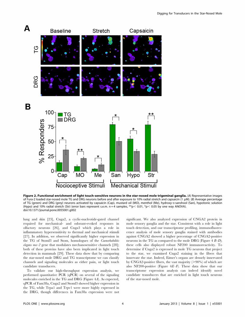
Consistent with the antibody staining, capsaicin, mustard oil, and
menthol activated a significantly higher percentage of neurons in
the DRG compared to the TG (DRG: 36.168.2% versus TG:
14.762.8% for capsaicin). Conversely, hypotonic solution, san-
shool and radial stretch activated a significantly higher percentage
of TG neurons than DRG neurons (TG: 79.665.9% versus DRG:
33.8662.9% for stretch). These results demonstrate that the star-
nosed mole TG are highly specialized anatomically and function-
ally to mediate high tactile sensitivity.
Using Transcriptome Profiling to Identify CandidateTouch TransducersThe enrichment of functional light touch neurons in the TG
and nociceptors in the DRG suggests that these tissues vary at
a molecular level. If this is indeed the case, comparing
transcripts may lead to the identification of molecular players
that mediate touch or pain transduction. We used mRNA-Seq
to detect transcripts expressed in mole TG and DRG, and we
compared expression levels between these two tissues. Currently
there is no available annotated genome for the star-nosed mole,
or any other member of the order Soricomorpha. As such, we
mapped mRNA-Seq reads to multiple known mammalian
transcriptomes iteratively. Using the Stampy algorithm [14],
reads from TG and DRG were first aligned to the Homo sapiens
transcriptome; unmapped reads were then aligned to the Canis
lupus familiaris transcriptome, and finally to the Mus musculus
transcriptome. This strategy successfully aligned 42%–65% of
sequenced reads per sample. This is quite high, given that reads
typically align at ,87% to a well annotated genome [15]. The
DESeq package [16] was employed to test for differential gene
expression between the TG and DRG samples (padj , 0.05).
We identified 3231 genes with significantly elevated expression
in the TG and 3033 in the DRG (Figure 3A). We hypothesized
that a number of these genes also differ between mouse TG and
DRG, due to the different organ structures, localization, target
organ innervation patterns, central and peripheral circuitry and
support cells of these ganglia. We carried out a parallel
transcriptional profiling analysis of TG and DRG in the mouse
and found 51 genes that show significantly higher expression in
both the mouse and mole TG compared to DRG and 72 genes
more highly expressed in both mouse and mole DRG. For
example, Hoxd1 is required for normal nociceptor innervation
of thoracic and cervical spinal cord levels [17], and shows
significantly higher expression in both mole and mouse DRG
(fold change in DRG vs TG: star-nosed mole = 4.76,
padj = 9.26*1025, mouse= 1.5, padj = 0.034).
To identify transduction molecules, we limited our analyses to
predicted membrane proteins that were not differentially ex-
pressed between mouse TG and DRG, and were more highly
expressed in mole TG (788 genes) or DRG (1159 genes). The
genes belong to a wide variety of classes as verified by Gene
Ontology analysis (Figure 3B). Consistent with a higher percentage
of large diameter myelinated neurons, the SNM TG showed
increased transcript numbers for a larger number of myelin
specific transmembrane proteins compared to DRG. Conversely,
DRG showed higher expression of a larger number of proteins
involved in vesicle trafficking and exocytosis, consistent with an
increased number of peptide releasing nociceptors.
In mechanosensory cells, ion channels underlie the transduction
of mechanical stimuli into electrical signals. We therefore focused
on genes that encode ion channels or channel modulators and are
significantly enriched in the DRG or TG (Figure 3C–D). The
DRG-enriched genes include a number of channels implicated in
nociception including: the tetrodotoxin-insensitive sodium chan-
nels Nav1.8 (Scn10a) and Nav1.9 (Scn11a; [18,19]), the cold-
sensitive ion channel Trpm8 [10], the heat-sensitive channel
Trpm3 [20], the irritant receptor Trpa1 [11,21], the heat- and
capsaicin-receptor Trpv1 [22] and the putative mechanosensitive
channel Piezo2 (Fam38b; [23,24]).
Several ion channels previously implicated in mechanotrans-
duction were enriched in the TG, including: ASIC3 (Accn3),
a homologue of the MEC4/10 channels which transduce
mechanosensitive currents in Caenorhabditis elegans [25], Piezo1
(Fam38a), a mechanosensitive channel expressed in mouse kidney,
Digging for Transducers in the Star-Nosed Mole
PLOS ONE | www.plosone.org 3 January 2013 | Volume 8 | Issue 1 | e55001
lung and skin [23], Cnga2, a cyclic-nucleotide-gated channel
required for mechanical- and odorant-evoked responses in
olfactory neurons [26], and Cnga3 which plays a role in
inflammatory hypersensitivity to thermal and mechanical stimuli
[27]. In addition, we observed significantly higher expression in
the TG of Stoml3 and Stom, homologues of the Caenorhabditis
elegans mec-2 gene that modulates mechanosensitive channels [28];
both of these proteins have also been implicated in light touch
detection in mammals [29]. These data show that by comparing
the star-nosed mole DRG and TG transcriptome we can classify
channels and signaling molecules as either pain, or light touch
candidate transducers.
To validate our high-throughput expression analysis, we
performed quantitative PCR (qPCR) on several of the signaling
molecules enriched in the TG and DRG (Figure 4A). As expected,
qPCR of Fam38a, Cnga2 and Stoml3 showed higher expression in
the TG, while Trpa1 and Trpv1 were more highly expressed in
the DRG, though differences in Fam38a expression were not
significant. We also analyzed expression of CNGA2 protein in
mole sensory ganglia and the star. Consistent with a role in light
touch detection, and our transcriptome profiling, immunofluores-
cence analysis of mole sensory ganglia stained with antibodies
against CNGA2 showed a higher percentage of CNGA2-positive
neurons in the TG as compared to the mole DRG (Figure 4 B–D);
these cells also displayed robust NF200 immunoreactivity. To
determine if Cnga2 is expressed in mole TG neurons that project
to the star, we examined Cnga2 staining in the fibers that
innervate the star. Indeed, Eimer’s organs are densely innervated
by CNGA2-positive fibers, the vast majority (.90%) of which are
also NF200-positive (Figure 4E–F). These data show that our
transcriptome expression analysis can indeed identify novel
candidate transducers that are enriched in light touch neurons
of the star-nosed mole.
Figure 2. Functional enrichment of light touch-sensitive neurons in the star-nosed mole trigeminal ganglia. (A) Representative imagesof Fura-2 loaded star-nosed mole TG and DRG neurons before and after exposure to 10% radial stretch and capsaicin (1 mM). (B) Average percentageof TG (green) and DRG (grey) neurons activated by capsaicin (Cap), mustard oil (MO), menthol (Me), hydroxy-a-sanshool (San), hypotonic solution(Hypo) and 10% radial stretch (Str) (error bars represent s.e.m. n = 4 samples, **p, 0.01, *p, 0.05 by one way ANOVA).doi:10.1371/journal.pone.0055001.g002
Digging for Transducers in the Star-Nosed Mole
PLOS ONE | www.plosone.org 4 January 2013 | Volume 8 | Issue 1 | e55001

Candidate Touch Transducers in the MouseSomatosensory SystemDefinitive evidence for the involvement of a gene in mechan-
otransduction must come from functional assays. Unfortunately,
such assays are not possible in star-nosed moles. We thus
examined expression of the most TG- and DRG-enriched
channels in mouse sensory ganglia. ,85% of these channels were
detected in mouse TG and DRG by RT-PCR (Figure 5A). Many
of these channels have not been previously implicated in
mammalian somatosensory transduction. qPCR of mouse ganglia
also demonstrated expression of the TG-enriched ion channels
Cnga2, Cnga3, Cnga4, and Fam38a, as well as the DRG-enriched
TRPA1 and TRPV1 (Figure 5B). Overall, these channels
represent excellent candidates to probe in the more tractable
mouse model.
Discussion
Despite intensive study in recent years, the molecular basis of
mechanotransduction remains poorly understood and few me-
chanically gated ion channels have been identified. Here we take
a new approach by investigating the hypertrophied mechanosen-
sory epidermis of the star-nosed mole. We showed that the dense
innervation of the star by myelinated light touch fibers originates
from a specialized trigeminal system. In general, the mammalian
somatosensory system is broadly tuned to detect a diverse array of
innocuous and noxious stimuli. Unlike other mammalian somato-
sensory ganglia, or even the star-nosed mole DRG that innervate
the body, the star-nosed mole TG contain relatively few classical
nociceptors (Figure 1E–F, 2B). Thus, the high tactile acuity of the
star may arise at the expense of thermal and chemical sensitivity,
as the lack of peptidergic C-fibers and nociceptive molecules (e.g.
TRPA1 and TRPV1) may affect the ability of the star to detect
irritants and other noxious stimuli. Consistent with this, topical
application of capsaicin to the hindpaw elicits nocifensive
responses similar to those reported in rodents, but application to
the star evokes no obvious behaviors (n = 3 animals).
The star-nosed mole has adapted a new strategy for achieving
high tactile acuity, namely a cellular and molecular specialization
of the trigeminal ganglia that innervate the star. We exploited this
specialization to identify molecules enriched in the DRG and TG.
Transcript analysis offers an unbiased approach to identify genes
of interest, but is usually restricted to organisms for which genome
information is widely available. Since there is no annotated
genome data for the star-nosed mole, or any close relative, we used
iterative mapping to assign the identity of reads. The use of this
approach to identify novel players in touch and pain is validated
by the differential expression of molecules already known to play
keys roles in pain transduction in the DRG, and genes implicated
in innocuous touch transduction in the TG. For example, TRPV1
in nociceptors and Stoml3 in touch neurons.
Figure 3. Transcriptome profiling of mole trigeminal and dorsal root ganglia. (A) Dot plot of expression levels of mapped genes in the star-nosed mole TG and DRG. Genes differentially expressed between TG or DRG (padj,=0.05) are shown in green and dark grey, respectively. (B) GeneOntology classification of transmembrane proteins differentially expressed between the star-nosed mole TG and DRG. (C) Dot plot of expressionlevels of known ion channels in the star-nosed mole TG versus DRG. Genes differentially expressed between TG or DRG (padj,= 0.05) are shown ingreen and dark grey, respectively. Black dots represent individual genes as labeled. (D) Top channels and channel regulators enriched in mole TG andDRG. This list excludes channels also differentially expressed between mouse ganglia. Table shows gene name in mouse, fold enrichment in mole,and putative ion conductance.doi:10.1371/journal.pone.0055001.g003
Digging for Transducers in the Star-Nosed Mole
PLOS ONE | www.plosone.org 5 January 2013 | Volume 8 | Issue 1 | e55001

RNASeq analysis revealed an enrichment of nociceptive
transcripts in the mole DRG versus the TG. For example,
TRPV1 expression is ,10-fold higher in the mole DRG than the
TG and Cnga2 levels are ,20-fold higher in the mole TG versus
DRG (Figure 3D). Some of this enrichment can be accounted for
by the varying numbers of neuronal subtypes in the DRG and TG
as measured by functional imaging and histology. However, while
we see 10–20 fold differences in transcript levels between the DRG
and TG, the number of TRPV1- or Cnga2-positive cells only
varies by ,3-fold (Figure 2B, 4D). These findings suggest that
expression differences between ganglia may arise from both a shift
in the number of nociceptors, as well as expression differences
within any given neuronal subtype.
We identified a number of novel candidate genes enriched in
mole TG that may mediate mammalian somatosensory mechan-
otransduction. Cnga2 and Cnga4, the most highly enriched
channels in the mole TG, play an important role in signal
transduction in both the visual and olfactory systems [30]. Cnga2
is proposed to mediate both chemosensory and mechanosensory
responses evoked by breathing in the mouse olfactory system [26].
If CNG channels play a role in somatosensory transduction, other
components of the cyclic nucleotide pathway should be expressed
in sensory neurons. This is indeed the case, as mouse sensory
ganglia also express two additional genes required for cAMP
signaling and olfactory transduction and behaviors in mice, the G
protein (Gnal) and adenylate cyclase 3 (Adcy3) (data not shown;
[31,32]). These data suggest that cyclic nucleotide signaling may
represent a new signaling pathway in touch transduction. More
broadly, these findings suggest that transduction mechanisms may
be conserved between divergent sensory systems. Indeed, tran-
scriptional analysis of the Droshophila Johnston’s organ recently
Figure 4. Expression analysis of candidate touch and pain transducers. (A) qPCR analysis of selected transcripts in TG (green) and DRG(grey). Results show average expression normalized to Gapdh (n = 3 samples/tissue). (B-F) Images of star-nosed mole tissue stained with antibodyagainst CNGA2 (green) and NF200 (red). (B) Trigeminal ganglia (TG) show stronger staining for CNGA2 than (C) dorsal root ganglia (DRG). (D)Percentage of total neurons stained positive for CNGA2 in TG (green) and DRG (grey) n = 4 sections/tissue. (E) Cross section of a single Eimer’s organand (F) nerve tracts innervating multiple Eimer’s organs show strong CNGA2 staining and high overlap with NF200 staining. Error bars represents.e.m. **p, 0.01, *p, 0.05, NS = p.0.05 by one way ANOVA).doi:10.1371/journal.pone.0055001.g004
Digging for Transducers in the Star-Nosed Mole
PLOS ONE | www.plosone.org 6 January 2013 | Volume 8 | Issue 1 | e55001

identified a number of chemo- and photo-transduction molecules
that also play a role in auditory transduction [33].
Fam38a (Piezo1) is an ion channel that induces mechanically-
evoked responses in many different cell types. Previous studies
showed high expression in mouse kidney, lung and skin, but
relatively low expression in sensory ganglia. Our RNASeq and
qPCR data shows expression in mole TG and DRG. This led us to
re-examine expression of Fam38a in mouse sensory ganglia.
Surprisingly, we amplified Fam38a from both mouse TG and
DRG and observed comparable levels of expression in mouse TG
and kidney (Figure 5B). Likewise, RNASeq of mouse ganglia also
shows expression of Fam38a, suggesting that this protein may play
a role in mediating light touch transduction in mice.
Finally, we identified a number of novel, uncharacterized genes
enriched in TG and DRG that contain one or more trans-
membrane domains and that were not previously classified as
organellar. These genes, along with CNG channels and Piezo1,
may participate in different aspects of transduction, acting as
detectors, modulators and/or amplifiers of mechanical stimuli.
Each subtype of light touch receptor has distinct mechanical
thresholds, sensory adaptation properties and action potential
waveforms, and it is predicted that such diversity is driven by
a unique repertoire of force transducers. Thus, future studies will
determine if these genes are expressed in distinct subsets of
somatosensory neurons, and what role they play in detecting
diverse somatosensory stimuli in vivo.
Our results emphasize the utility of examining both traditional
model organisms and less common species that may provide
important clues to sensory system function. August Krogh
articulated the principle that ‘‘for such a large number of problems
there will be some animal of choice, or a few such animals, on
which it can be most conveniently studied’’ [34]. In the spirit of
this approach, we exploited the high tactile acuity of the star-nosed
mole to identify a multitude of candidate molecules that may
mediate innocuous and noxious somatosensory stimuli. The
function of these molecules can now be probed in more traditional
model organisms.
Materials and Methods
AnimalsStar-nosed moles were collected in Potter County, Pennsylva-
nia, under permit COL00087. Wild type adult female mice C57/
Bl6 (Jackson Labs) were used for sectioning, RT-PCR and qPCR.
All procedures followed guidelines for the care and use of
laboratory animals from the National Institutes of Health and
were approved by the Vanderbilt University Institutional Animal
Care and Use Committee and the UC Berkeley Animal Care and
Use Committee.
ImmunohistochemistryFive star-nosed moles and two mice were anesthetized with
250 mg/kg i.p. injection of sodium pentobarbital and perfused
with 1X PBS (pH 7.3) followed by 4% paraformaldehyde (PFA) in
PBS. Trigeminal ganglia, dorsal root ganglia, star tissue, hindpaw
skin and cornea were collected. Tissue was post-fixed for .4 hours
in 4% PFA and incubated overnight in 20% sucrose at 4uC.Trigeminal ganglia, dorsal root ganglia, hindpaw skin and star
were embedded in Tissue-Tek optimal, sectioned at 10 or 15 mm,
and mounted onto glass coverslips for staining.
Sections were blocked for 90 minutes in PBST supplemented
with 10% normal goat serum and incubated overnight at 4uC with
primary antibody. Primary antibodies were diluted as follows:
mouse anti-NF200 (Sigma) and rabbit anti-peripherin 1:1000
(Millipore), guinea pig anti-substance P 1:700, rabbit anti-S100
1:300 (ThermoFisher), rabbit anti-CNGA2 1:500 (Alomone labs).
Sections were washed and incubated for one hour at room
temperature in 1:1000 secondary antibody. Secondary antibodies
(Invitrogen) were used as follows: anti-mouse Alexa 488 or 568 for
NF200, anti-rabbit Alexa 568 for peripherin, anti-guinea pig Alexa
568 for substance P, Alexa 488 anti-rabbit for S100, and Alexa
488 anti-rabbit for CNGA2. Intact corneas were stained directly
after sucrose incubation.
ImagingFluorescence images were captured using an inverted fluores-
cence microscope (IX71, Olympus), illuminated with a xenon light
source (Lambda LS-xl, Sutter) with a FITC excitation and
emission set for Alexa 488 (Chroma) and Texas Red excitation
and emission set for Alexa 568 (Chroma). Images were captured
using MetaMorph (Molecular Devices). Confocal images were
collected using Leica TCS SL confocal microscope. Summation
projections were compiled from a series of 8 images (1 mM Z-steps,
MetaMorph).
QuantificationFor determining percentages of positive cells, cell boundaries
were determined by hand using bright field images and the
average intensity of each region was calculated (MetaExpress).
Cells were defined as being positive for a specific marker if their
intensity was more than 30% above background.
Cell CultureDRG and TG cells were cultured as previously described [12].
Briefly, star-nosed moles were euthanized and dorsal root ganglia
and trigeminal ganglia were dissected. Ganglia were digested for
20 minutes in 7 mg/mL collagenase P (Roche) then for 2 minutes
in 0.25% trypsin. Cells were dissociated manually by trituration
and plated on glass coverslides coated with 1 mg/mL Poly-D-
Lysine and 0.1 mg/mL laminin. Cells were cultured overnight at
37uC before imaging.
Figure 5. Expression of candidate transducers in mouseganglia. (A) Ion channels enriched in mole TG and DRG that wereamplified by RT-PCR from mouse TG and DRG. All genes were amplifiedindependently from TG and DRG samples isolated from two mice.Channels shown in bold are candidates that have not been previouslyreported as expressed in somatosensory neurons. (B) qPCR analysis ofselected genes in mouse TG and kidney. Results show averageexpression normalized to Gapdh (n = 3). Error bars represent s.e.m.doi:10.1371/journal.pone.0055001.g005
Digging for Transducers in the Star-Nosed Mole
PLOS ONE | www.plosone.org 7 January 2013 | Volume 8 | Issue 1 | e55001

Calcium ImagingCalcium imaging was performed as previously described for
mouse neurons [10]. Cells were loaded for an hour at room
temperature with Fura-2AM (10 mM, Invitrogen) in Ringer’s
solution (140 mM NaCl, 5 mM KCl, 2 mM CaCl2, 1 mM
MgCl2, 10 mM D-glucose, 10 mM HEPES) supplemented with
0.2% pluronic acid (Invitrogen). Ratiometric calcium imaging was
performed using alternating 340 nm and 380 nm excitation filters
with images collected every three seconds. Responsive cells were
defined as those whose calcium concentration increased more than
10% above the baseline after application of the stimulus.
RNA ExtractionStar nosed mole or mouse tissues were removed and homog-
enized in Trizol (Invitrogen). Total RNA was extracted as per the
manufacturer’s instructions. Total RNA was either DNAsed, re-
purified (RNAeasy, Quiagen) and reverse transcribed (Super-
ScriptIII, Invitrogen) for qPCR or RT-PCR, or further purified
with two rounds of poly-A selection (Dynabeads mRNA purifica-
tion Kit, Invitrogen) for sequencing.
Illumina SequencingPoly-A selected mRNA from trigeminal ganglia and dorsal root
ganglia were fragmented using Fragmentation Reagents (Applied
Biosystems). Libraries were further prepared using the Genomic
Sample Prep Kit following a modified RNA-seq protocol [35] or
the TruSeq kit (Illumina). One to three lanes of each tissue’s
library were sequenced on a Genome Analyzer II or HighSeq 200
using the 76, 50 or 36 bp single-end sequencing protocol at the
Vincent J. Coates Genomics Sequencing Laboratory at UC
Berkeley.
Transcriptome AnalysisThree (mole) or two (mouse) TG and DRG were used. All
samples were derived from single individuals, except one mole
DRG, combined from two individuals. Mole reads were mapped
as described in the text. Mouse reads were aligned to Mus musculus
mRNA sequences. Homo sapiens database: Nov 25th 2010,
genome.ucsc.edu. Canis lupus familiaris and Mus musculus: Nov28th
2010, ncbi.nlm.nih.gov. Alignments were performed using Stampy
v1.0.11 [14] with a substitution rate = 0.03. The read count for
a given gene was the sum of reads aligning to the transcript(s)
assigned to an entry in the Entrez Gene database (ncbi.nlm.nih.-
gov, Jan 2011). For mole genes, read count was the sum of all
reads aligning to the homologs of that gene in the three organisms.
Homology was based on the HomoloGene database (build 64) and
manual annotation. Differential expression was assessed using the
DESeq package for R [16] which corrected for multiple
comparisons (padj).
Transmembrane Domain PredictionTransmembrane domains were identified by removing signal
peptides predicted with SignalP3.0 [36], and by analyzing the
remaining sequence with SOSUI [37], TMHMM2.0 [38], and
TMpred [39] programs. A protein was considered having
a transmembrane domain if at least two of the three programs
predicted at least one transmembrane region.
qPCRRNA was extracted from 3 moles as described above. qPCR
was performed using SYBR GreenER (Invitrogen) on a StepOne-
PlusTM Real-Time PCR system (Applied Biosystems). qPCR
reactions were run in 10 mL using 1 mL of product from the RT
reaction with the primers listed below. CT values were calculated
as the average of 2 wells. Fold expression for each was calculated
averaging normalized CT values from two runs, assuming 100%
efficiency. Final values were calculated as an average of the
biological replicates for each tissue type.
Fam38a: Fwd TCTTCCTCTTCCAGGGGTTC, Rev AC-
CAGGGACATGAAGAGCAG
Cnga2: Fwd CTGGAGACCAAGATGAAGCAGA, Rev AT-
TACGAAGACGGAGCCCTACA
Stoml3: Fwd GAGTCACCCATAGCCCTCCA, Rev GAC-
CACCCATGCCCTCTAGT
Trpv1: Fwd TGAAGACCTTGTTTGTGGACAG, Rev
CGGGTGTAGTAGAGCATGTTGG
Trpa1: Fwd CTTATTGGTTTGGCAGTTGGTG, Rev
CGGTTGGGGTAGACAGTGATG
Gapdh: Fwd CACGGCCACCCAGAAGAC, Rev TCA-
GATCCACGACGGACAC.
RT-PCRTrigeminal ganglia, dorsal root ganglia or a mix of tissues (liver,
heart, lungs, kidneys, pancreas and brain) were extracted and
cDNA was produced as described above with or without reverse
transcriptase (no RT control). PCR was performed on 1 mL of
product using Phusion Taq polymerase (NEB) and the primers
described in Table S1.
StatisticsAll values are presented as average 6 standard error of the
mean (s.e.m.). Unless otherwise noted, all p-values were calculated
using a one-way ANOVA with a Tukey’s Post Hoc analysis.
Supporting Information
Figure S1 Star-nosed mole NF200-positive fibers dis-play similar characteristics to those described in mouse.(A) All NF200-positive fibers (green) are myelin basic protein-
positive (red). Epifluorescence image of a section through a bundle
of fibers projecting through the nose. (B) Mole cornea shows strong
substance P staining. Confocal image of the mole cornea with
substance P (red) and NF200 (green), scale bar = 20 mm. (C) Mole
hindpaw shows robust staining of both substance P (red) and
NF200 (green). Confocal image, scale bar = 50 mm. (D) Mouse
cornea shows similar staining to mole cornea. Confocal image,
scale bar = 20 mm.
(TIF)
Figure S2 Mouse TG and DRG show similar staining forperipherin and NF200. Peripherin (red) and NF200 (green)
staining of the TG (A) and DRG (B). (C) Quantification of the
percent of NF200 and peripherin positive cells in these tissues
(NS=p.0.1; n = 3 sections, error bars represent s.e.m.).
(TIF)
Table S1 Primers for amplification of star-nosed moleenriched genes in mouse.
(DOCX)
Acknowledgments
We thank Dr. Allan Basbaum for generously providing the substance P
antibody and Dr. John Ngai and members of our laboratories for helpful
discussions.
Digging for Transducers in the Star-Nosed Mole
PLOS ONE | www.plosone.org 8 January 2013 | Volume 8 | Issue 1 | e55001

Author Contributions
Conceived and designed the experiments: KAG MP DMB. Performed the
experiments: KAG MP MT TM DMB. Analyzed the data: KAG MP MT
TM DMB. Contributed reagents/materials/analysis tools: KAG MP DBL
PRT RBB KCC DMB. Wrote the paper: KAG MP KCC DMB.
References
1. Lumpkin EA, Marshall KL, Nelson AM (2010) The cell biology of touch. J Cell
Biol 191: 237–248.2. Catania KC (1995) Structure and innervation of the sensory organs on the snout
of the star-nosed mole. J Comp Neurol 351: 536–548.3. Catania KC (1996) Ultrastructure of the Eimer’s organ of the star-nosed mole.
J Comp Neurol 365: 343–354.
4. Catania KC, Remple FE (2005) Asymptotic prey profitability drives star-nosedmoles to the foraging speed limit. Nature 433: 519–522.
5. Lumpkin EA, Caterina MJ (2007) Mechanisms of sensory transduction in theskin. Nature 445: 858–865.
6. Lawson SN, Waddell PJ (1991) Soma neurofilament immunoreactivity is related
to cell size and fibre conduction velocity in rat primary sensory neurons. J Physiol435: 41–63.
7. Lawson SN, Crepps BA, Perl ER (1997) Relationship of substance P to afferentcharacteristics of dorsal root ganglion neurones in guinea-pig. J Physiol 505 ( Pt
1): 177–191.8. Zylka MJ, Rice FL, Anderson DJ (2005) Topographically distinct epidermal
nociceptive circuits revealed by axonal tracers targeted to Mrgprd. Neuron 45:
17–25.9. Marfurt CF, Kingsley RE, Echtenkamp SE (1989) Sensory and sympathetic
innervation of the mammalian cornea. A retrograde tracing study. InvestOphthalmol Vis Sci 30: 461–472.
10. Bautista DM, Siemens J, Glazer JM, Tsuruda PR, Basbaum AI, et al. (2007) The
menthol receptor TRPM8 is the principal detector of environmental cold.Nature 448: 204–208.
11. Jordt SE, Bautista DM, Chuang HH, McKemy DD, Zygmunt PM, et al. (2004)Mustard oils and cannabinoids excite sensory nerve fibres through the TRP
channel ANKTM1. Nature 427: 260–265.
12. Tominaga M, Caterina MJ, Malmberg AB, Rosen TA, Gilbert H, et al. (1998)The cloned capsaicin receptor integrates multiple pain-producing stimuli.
Neuron 21: 531–543.13. Bhattacharya MR, Bautista DM, Wu K, Haeberle H, Lumpkin EA, et al. (2008)
Radial stretch reveals distinct populations of mechanosensitive mammaliansomatosensory neurons. Proc Natl Acad Sci U S A 105: 20015–20020.
14. Lunter G, Goodson M (2010) Stampy: A statistical algorithm for sensitive and
fast mapping of Illumina sequence reads. Genome Res 21: 936–939.15. Li H, Durbin R (2009) Fast and accurate short read alignment with Burrows-
Wheeler transform. Bioinformatics 25: 1754–1760.16. Anders S, Huber W (2010) Differential expression analysis for sequence count
data. Genome Biol 11: R106.
17. Guo T, Mandai K, Condie BG, Wickramasinghe SR, Capecchi MR, et al.(2011) An evolving NGF-Hoxd1 signaling pathway mediates development of
divergent neural circuits in vertebrates. Nat Neurosci 14: 31–36.18. Abrahamsen B, Zhao J, Asante CO, Cendan CM, Marsh S, et al. (2008) The cell
and molecular basis of mechanical, cold, and inflammatory pain. Science 321:702–705.
19. Djouhri L, Fang X, Okuse K, Wood JN, Berry CM, et al. (2003) The TTX-
resistant sodium channel Nav1.8 (SNS/PN3): expression and correlation withmembrane properties in rat nociceptive primary afferent neurons. J Physiol 550:
739–752.20. Vriens J, Owsianik G, Hofmann T, Philipp SE, Stab J, et al. (2011) TRPM3 is
a nociceptor channel involved in the detection of noxious heat. Neuron 70: 482–
494.
21. Kwan KY, Allchorne AJ, Vollrath MA, Christensen AP, Zhang DS, et al. (2006)
TRPA1 contributes to cold, mechanical, and chemical nociception but is not
essential for hair-cell transduction. Neuron 50: 277–289.
22. Caterina MJ, Leffler A, Malmberg AB, Martin WJ, Trafton J, et al. (2000)
Impaired nociception and pain sensation in mice lacking the capsaicin receptor.
Science 288: 306–313.
23. Coste B, Mathur J, Schmidt M, Earley TJ, Ranade S, et al. (2010) Piezo1 and
Piezo2 are essential components of distinct mechanically activated cation
channels. Science 330: 55–60.
24. Kim SE, Coste B, Chadha A, Cook B, Patapoutian A (2012) The role of
Drosophila Piezo in mechanical nociception. Nature 483: 209–212.
25. O’Hagan R, Chalfie M, Goodman MB (2005) The MEC-4 DEG/ENaC
channel of Caenorhabditis elegans touch receptor neurons transduces mechan-
ical signals. Nat Neurosci 8: 43–50.
26. Grosmaitre X, Santarelli LC, Tan J, Luo M, Ma M (2007) Dual functions of
mammalian olfactory sensory neurons as odor detectors and mechanical sensors.
Nat Neurosci 10: 348–354.
27. Heine S, Michalakis S, Kallenborn-Gerhardt W, Lu R, Lim HY, et al. (2011)
CNGA3: a target of spinal nitric oxide/cGMP signaling and modulator of
inflammatory pain hypersensitivity. J Neurosci 31: 11184–11192.
28. Goodman MB, Ernstrom GG, Chelur DS, O’Hagan R, Yao CA, et al. (2002)
MEC-2 regulates C. elegans DEG/ENaC channels needed for mechanosensa-
tion. Nature 415: 1039–1042.
29. Martinez-Salgado C, Benckendorff AG, Chiang LY, Wang R, Milenkovic N, et
al. (2007) Stomatin and sensory neuron mechanotransduction. J Neurophysiol
98: 3802–3808.
30. Biel M (2009) Cyclic nucleotide-regulated cation channels. J Biol Chem 284:
9017–9021.
31. Belluscio L, Gold GH, Nemes A, Axel R (1998) Mice deficient in G(olf) are
anosmic. Neuron 20: 69–81.
32. Wong ST, Trinh K, Hacker B, Chan GC, Lowe G, et al. (2000) Disruption of
the type III adenylyl cyclase gene leads to peripheral and behavioral anosmia in
transgenic mice. Neuron 27: 487–497.
33. Senthilan PR, Piepenbrock D, Ovezmyradov G, Nadrowski B, Bechstedt S, et al.
(2012) Drosophila auditory organ genes and genetic hearing defects. Cell 150:
1042–1054.
34. Krogh A (1929) The Progress of Physiology. Science 70: 200–204.
35. Marioni JC, Mason CE, Mane SM, Stephens M, Gilad Y (2008) RNA-seq: an
assessment of technical reproducibility and comparison with gene expression
arrays. Genome Res 18: 1509–1517.
36. Bendtsen JD, Nielsen H, von Heijne G, Brunak S (2004) Improved prediction of
signal peptides: SignalP 3.0. J Mol Biol 340: 783–795.
37. Hirokawa T, Boon-Chieng S, Mitaku S (1998) SOSUI: classification and
secondary structure prediction system for membrane proteins. Bioinformatics
14: 378–379.
38. Krogh A, Larsson B, von Heijne G, Sonnhammer EL (2001) Predicting
transmembrane protein topology with a hidden Markov model: application to
complete genomes. J Mol Biol 305: 567–580.
39. Hofmann K, Stoffel W (1993) TMbase - A database of membrane spanning
proteins segments. Biol Chem Hoppe-Seyler 347: 166.
Digging for Transducers in the Star-Nosed Mole
PLOS ONE | www.plosone.org 9 January 2013 | Volume 8 | Issue 1 | e55001
Copyright © 2022 FDOKUMEN