The sodium pump Ena1p provides mechanistic insight into the salt sensitivity of vacuolar protein...
11
The sodium pump Ena1p provides mechanistic insight into the salt sensitivity of vacuolar protein sorting mutants Katarina Logg a , Jonas Warringer b , Sayed Hossein Hashemi b, 1 , Mikael Käll a , Anders Blomberg b, ⁎ a Department of Applied Physics, Chalmers University of Technology, Göteborg, Sweden b Department of Cell and Molecular Biology, University of Gothenburg, Göteborg, Sweden article info abstract Article history: Received 18 July 2007 Received in revised form 10 February 2008 Accepted 26 February 2008 Available online 18 March 2008 The vacuolar/endosomal network has an important but as yet undefined role in the cellular tolerance to salt stress. We hypothesized that the mechanistic basis for the importance of vacuolar protein sorting (vps) components in salt tolerance is the targeting of the crucial sodium exporter Ena1p to the plasma membrane. The link between Ena1p and the vps components was established by the observation that overexpression of Ena1p could suppress the salt sensitivity of the ESCRT knockouts vps20Δ, snf7/vps32Δ and snf8/vps22Δ. To further investigate this functional interaction, fluorescence microscopy was utilized to monitor localization of GFP-tagged Ena1p. For all analyzed vps mutants, Ena1p seemed properly localized to the plasma membrane, even during saline growth. However, quantitative differences in plasma membrane localized Ena1p were recorded; e.g. the highly salt sensitive pep12Δ mutant exhibited substantially enhanced Ena1p levels. In addition, the kinetics of Ena1p localization to the plasma membrane was severely delayed in several vps mutants, and this delay correlated to the salt specific growth defect. This paper discusses potential mechanistic hypotheses, like Ena1p transporter activity or localization kinetics, or ESCRT component's influence on signaling, for linking endosomal sorting functions to cellular salt sensitivity. © 2008 Elsevier B.V. All rights reserved. Keywords: Vacuolar protein sorting Sodium export Yeast GFP vps deletion mutant Phenotype 1. Introduction Cells need to respond adequately to environmental changes. In particular, stress conditions where the external agent is toxic to the internal cell machinery put substantial strain on the plasma mem- brane transport systems to ensure low intracellular concentrations of the toxin. At high concentrations of NaCl, cells are dually challenged since sodium both constitutes a harsh ion for the intracellular ma- chinery as well as a potent dehydrating agent. A key factor in saline adaptation in yeast is the efficient P-type sodium ion efflux ATPase encoded by the ENA1 gene [1]. The Ena1p transporter is the main component of the sodium homeostasis system and ena1Δ displays extreme sensitivity to salt [2]. However, in addition to Ena1p, several additional proteins give more subtle contributions to yeast fitness in salt. In a genome-wide phenotypic screen we found that 210 deletion mutants displayed significant growth rate defects in salt tolerance [3]. The salt sensitivity of many of these knockouts makes physiological sense; the corresponding proteins are involved in processes directly linked to saline defense, such as stress-induced signal transduction, energy metabolism (ATP generation is essential for many of the adaptation mechanisms, e.g. sodium efflux), glycerol production and the maintenance of ion homeostasis. However, salt sensitivity was also scored for functions with less apparent links to ion/osmotic tolerance. Particularly surprising was the striking overrepresentation of proteins involved in vacuolar protein sorting (vps) [3]. We also pointed out that many of these components interact physically forming an important intracellular stress network. Several lines of physiological data point to a central role for the vacuole in osmoregulation (see refs in [4]). The importance of the vacuole in maintaining osmo-homeostasis has earlier been demon- strated in mutant screens that have identified a large proportion of the salt sensitive mutants to have defects in properties like vacuolar mor- phology and secretion of vacuolar enzymes. One mechanistic explana- tion for the salt sensitivity of the vps mutants is that the vacuole plays an important role in sequestering toxic ions, like sodium and chloride, or other compounds involved in osmoregulation. There are some similarities between the vacuole in plants and fungi. It is noteworthy that the tonoplast contains a specific Cl − transporter, which might indicate a selective sequestration of chloride ions in this compartment [5]. It has been shown though that during short-term adaptation to salt yeast preferentially accumulates sodium in the vacuole [6]. However, it is still unclear if there is any difference during steady-state conditions in the sodium or chloride concentration between the vacuole and the cytoplasm. Alternatively, it can be hypothesised that the vps mutants are defective in some of the sorting mechanisms related to the proper localization of the sodium efflux pump Ena1p to the plasma membrane. The importance of exocytosis in proper localization of Ena1p is clear. For Biochimica et Biophysica Acta 1783 (2008) 974–984 ⁎ Corresponding author. E-mail address: [email protected] (A. Blomberg). 1 Present address: Neurobiology Research Unit and Center for Integrated Molecular Brain Imaging, Rigshospitalet, Copenhagen, Denmark. 0167-4889/$ – see front matter © 2008 Elsevier B.V. All rights reserved. doi:10.1016/j.bbamcr.2008.02.022 Contents lists available at ScienceDirect Biochimica et Biophysica Acta journal homepage: www.elsevier.com/locate/bbamcr
-
Upload
independent -
Category
Documents
-
view
6 -
download
0
Transcript of The sodium pump Ena1p provides mechanistic insight into the salt sensitivity of vacuolar protein...

Biochimica et Biophysica Acta 1783 (2008) 974–984
Contents lists available at ScienceDirect
Biochimica et Biophysica Acta
j ourna l homepage: www.e lsev ie r.com/ locate /bbamcr
The sodium pump Ena1p provides mechanistic insight into the salt sensitivity ofvacuolar protein sorting mutants
Katarina Logg a, Jonas Warringer b, Sayed Hossein Hashemi b,1, Mikael Käll a, Anders Blomberg b,⁎a Department of Applied Physics, Chalmers University of Technology, Göteborg, Swedenb Department of Cell and Molecular Biology, University of Gothenburg, Göteborg, Sweden
a r t i c l e i n f o
⁎ Corresponding author.E-mail address: [email protected] (A. Blo
1 Present address: Neurobiology Research Unit and CBrain Imaging, Rigshospitalet, Copenhagen, Denmark.
0167-4889/$ – see front matter © 2008 Elsevier B.V. Aldoi:10.1016/j.bbamcr.2008.02.022
a b s t r a c t
Article history:Received 18 July 2007Received in revised form 10 February 2008Accepted 26 February 2008Available online 18 March 2008
The vacuolar/endosomal network has an important but as yet undefined role in the cellular tolerance to saltstress. We hypothesized that the mechanistic basis for the importance of vacuolar protein sorting (vps)components in salt tolerance is the targeting of the crucial sodium exporter Ena1p to the plasma membrane.The link between Ena1p and the vps components was established by the observation that overexpression ofEna1p could suppress the salt sensitivity of the ESCRT knockouts vps20Δ, snf7/vps32Δ and snf8/vps22Δ. Tofurther investigate this functional interaction, fluorescence microscopy was utilized to monitor localization ofGFP-tagged Ena1p. For all analyzed vps mutants, Ena1p seemed properly localized to the plasma membrane,even during saline growth. However, quantitative differences in plasma membrane localized Ena1p wererecorded; e.g. the highly salt sensitive pep12Δ mutant exhibited substantially enhanced Ena1p levels. Inaddition, the kinetics of Ena1p localization to the plasma membrane was severely delayed in several vpsmutants, and this delay correlated to the salt specific growth defect. This paper discusses potentialmechanistic hypotheses, like Ena1p transporter activity or localization kinetics, or ESCRT component'sinfluence on signaling, for linking endosomal sorting functions to cellular salt sensitivity.
© 2008 Elsevier B.V. All rights reserved.
Keywords:Vacuolar protein sortingSodium exportYeastGFPvps deletion mutantPhenotype
1. Introduction
Cells need to respond adequately to environmental changes. Inparticular, stress conditions where the external agent is toxic to theinternal cell machinery put substantial strain on the plasma mem-brane transport systems to ensure low intracellular concentrations ofthe toxin. At high concentrations of NaCl, cells are dually challengedsince sodium both constitutes a harsh ion for the intracellular ma-chinery as well as a potent dehydrating agent. A key factor in salineadaptation in yeast is the efficient P-type sodium ion efflux ATPaseencoded by the ENA1 gene [1]. The Ena1p transporter is the maincomponent of the sodium homeostasis system and ena1Δ displaysextreme sensitivity to salt [2]. However, in addition to Ena1p, severaladditional proteins give more subtle contributions to yeast fitness insalt. In a genome-wide phenotypic screen we found that 210 deletionmutants displayed significant growth rate defects in salt tolerance [3].The salt sensitivity of many of these knockouts makes physiologicalsense; the corresponding proteins are involved in processes directlylinked to saline defense, such as stress-induced signal transduction,energy metabolism (ATP generation is essential for many of theadaptation mechanisms, e.g. sodium efflux), glycerol production and
mberg).enter for Integrated Molecular
l rights reserved.
themaintenance of ion homeostasis. However, salt sensitivity was alsoscored for functions with less apparent links to ion/osmotic tolerance.Particularly surprising was the striking overrepresentation of proteinsinvolved in vacuolar protein sorting (vps) [3]. We also pointed out thatmany of these components interact physically forming an importantintracellular stress network.
Several lines of physiological data point to a central role for thevacuole in osmoregulation (see refs in [4]). The importance of thevacuole in maintaining osmo-homeostasis has earlier been demon-strated in mutant screens that have identified a large proportion of thesalt sensitive mutants to have defects in properties like vacuolar mor-phology and secretion of vacuolar enzymes. One mechanistic explana-tion for the salt sensitivity of the vps mutants is that the vacuole playsan important role in sequestering toxic ions, like sodium and chloride,or other compounds involved in osmoregulation. There are somesimilarities between the vacuole in plants and fungi. It is noteworthythat the tonoplast contains a specific Cl− transporter, which mightindicate a selective sequestration of chloride ions in this compartment[5]. It has been shown though that during short-term adaptation to saltyeast preferentially accumulates sodium in the vacuole [6]. However, itis still unclear if there is any difference during steady-state conditions inthe sodium or chloride concentration between the vacuole and thecytoplasm. Alternatively, it can be hypothesised that the vps mutantsare defective in some of the sorting mechanisms related to the properlocalization of the sodiumefflux pumpEna1p to the plasmamembrane.The importance of exocytosis inproper localization of Ena1p is clear. For

975K. Logg et al. / Biochimica et Biophysica Acta 1783 (2008) 974–984
example, it has been shown that the strong salt sensitivity of sro7Δresults from defective targeting of Ena1p to the plasma membrane [7].Exocytotic cargo can reach the plasma membrane by multiple path-ways, both in mammals and in yeast. At least one of these pathwaystransits through the endosomes before reaching the cell surface andinvolves the activity of vacuolar sorting proteins [8]. However, atpresent it is not known if the transport of Ena1p to the plasma mem-brane goes via or in some other way involve the endosome.
In this study we investigated the correct localization of Ena1p insalt sensitive vps-mutants, thereby testing the hypothesis that miss-localization of Ena1p constitutes the mechanistic basis for their saltdefect. Salt sensitive knockouts were selected and analyzed mainlyfrom the following four vps classes [9,10]: i) Vacuolar class E knock-outs, represented by the endosome associated proteins Vps4p,Vps20p, Snf8p (Vps22p) and Snf7p (Vps32p). These proteins playessential roles in the ESCRT machinery that is composed of threehetero-oligomeric complexes acting sequentially in recruitment ofubiquitinated cargoes into internal vesicles. Perturbations in theESCRT complexes have been shown to cause defects in growth controland development. Thus, the physiology of cells lacking this endosome-based pathway is also of broader interest. ii) A vacuolar class C mutantwith a deletion in VPS16. Loss of Vps16p results in cells defective invesicle/vacuolar fusion that display no identifiable vacuoles. Vps16p ispart of sorting and tethering complexes that play roles at severalstages of the Golgi-to-vacuole pathway. iii) Vacuolar class D mutants,represented mainly by pep12Δ, which result in a single large vacuole.Pep12p (Vps6p) is a endosomal target membrane receptor (t-SNARE)involved in vesicle traveling between the Golgi and the vacuole. It isthe yeast homolog of human syntaxin. iv) Vacuolar class B knockouts,represented by the two Golgi-proteins Vps52p and Vps53p, compo-nents of the GARP (Golgi-associated retrograde protein) complexwhich is required for the recycling of proteins from endosomes to thelate Golgi and involved in localization of actin and chitin. We utilizedfluorescence microscopy to monitor GFP-tagged Ena1p where ENA1expression was driven by the moderately strong promoter pMet25.For all analyzed vps mutants Ena1p seemed properly localized to theplasma membrane during steady-state saline growth. However, thekinetics of Ena1p plasma membrane localization was substantiallyaltered in several vps mutants. In addition, overexpression of Ena1psuppressed the salt sensitivity of the ESCRT knockouts vps20Δ, snf7Δand snf8Δ. Our data thus suggests a functional connection betweenendosomal sorting functions, the plasma membrane sodium exportpump, and cellular salt sensitivity.
2. Material and methods
2.1. Yeast strains and plasmids
The isogenic deletion mutants vps4Δ, pep12/vps6Δ, vps16Δ, vps20Δ, snf8/vps22Δ,snf7/vps32Δ, vps52Δ, and vps53Δ in the haploid BY4742 [11] strain background (wild-type genotype: MATalpha; his3Δ1; leu2Δ0; lys2Δ0; ura3Δ0) were used in this study.These mutants were earlier found to be salt sensitive [3]. Strains were transformedwiththe centromer based plasmid pUG35, with the URA3 marker, containing the sodiumtransporter gene ENA1 C-terminally tagged with GFP (a kind gift from Martha Cyert,Stanford University, USA) and expressed behind the methionine controlled MET25promoter (pMET25). TheMET25 promoter is effectively repressed by methionine in themedium and upon removal of methionine the promoter activity is substantiallyenhanced [12]. Thus, the pMET25 promoter enabled us to study the effect on Ena1plocalization during both salt stressed and non-stressed conditions.
Throughout the study synthetic defined (SD) medium was used (0.14% yeast nitro-gen base without amino acids, 0.5% ammoniumsulphate and 1% succinic acid; 2% (w/v)glucose, uracil 120 µg/ml, lysine120 µg/ml , leucine 120 µg/ml and histidine 120 µg/ml,pH 5.8). In case of plasmid-containing strains, uracil was omitted. Media was eitherused without salt or supplemented with NaCl (0.85 or 1.25 M) or 1.4 M KCl, and withand without 0.5 mM methionine.
2.2. Experimental growth conditions
Three types of experiments were performed: i) growth assay (see below), ii) studiesof Ena1p localization during exponential growth. Wild-type, vps4Δ, pep12Δ, vps16Δ,vps20Δ, snf8Δ, snf7Δ, vps52Δ, and vps53Δ were cultivated with and without 0.85 M
NaCl in 10ml test tube with vigorous shaking. During the exponential phase, samples of7 µl were withdrawn and put between two cover slides to be directly microscopicallyexamined, and iii) dynamic studies (time-course experiment) of Ena1p localization andproduction. Wild-type, vps4Δ, pep12Δ, vps20Δ and vps52Δ were cultivated as above, ina medium without NaCl supplemented with the addition of 0.5 mM methionine. Cellswere harvested in exponential phase, washed 2 times with methionine free (–Met)medium, by repetition of centrifugation and resuspension. Samples were withdrawnand directly investigated microscopically every 20 min. The wild-type and the pep12Δstrains were also analyzed for the production of ENA1-GFP (see below).
2.3. Growth assays
Strains deep-frozen (–80 °C) in 20% glycerol were inoculated in 350 µl of SDmedium (in case of plasmid-containing strains, uracil and methionine was omittedfrom the media) and incubated for ∼72 h at 30 °C. This procedure was repeated once(second incubation∼48 h). For experimental runs, strains were inoculated to an OD of0.03–0.1 in 350 µl of SD-Met medium and cultivated for 47 h in a Bioscreen analyzer C(Labsystems Oy, Finland). Optical density was automatically measured every 20 minusing a wide band (450–580 nm) filter. Incubation was at 30.0 °C (±0.1 °C) with 10 minpre-heating time. Plates were subjected to shaking at highest shaking intensity with60 s of shaking every other minute. Strains were run as five replicates in one singleexperiment with five wild-type strains (WT) included in each experiment (twoindependent experiments). Growth rates were calculated as earlier described [3]. Straingrowth defects as compared to theWT in a particular environment, termed LogarithmicStrain Coefficients (LSCs), were calculated as
LSCij ¼12
X2r¼1
15
X5k¼1
log wtrkj� �
� 15
X5k¼1
log xrkj� �$ %
where wtkj is the doubling time of the k: the measurement of the wild-type inenvironment j, xkj is the doubling time of strain i in environment j (with and withoutsalt) and r indicates the run. To distinguish a growth defect in salt from a generalgrowth defect also seen in basal medium a Logarithmic Phenotypic Index, LPI, wasformed for each strain i as
LPIi;NaCl ¼ LSCi;NaCl � LSCi;basal
We performed tests of the null hypothesis that LPI equals 0 separately for eachstrain, where statistical significance (pb0.001) was calculated as earlier described [3].
2.4. Microscopy, image generation and image analysis
Optical microscopy was performed on an inverted epi-fluorescence microscope(Nikon TE-2000E) using a 60× objective (Plan Apo, NA=1.40) for analysis of exponentiallygrowing cells or a 100× objective (Plan Apo, NA=1.45) for time-course experiments afterEna1 induction. A mercury lamp was used for fluorescence excitation and two differentfilter sets (31001 and 31000; from Chroma Corporation, USA) were used for GFP andvacuolar stain imaging, respectively. Imageswere recordedbyanelectron-multiplyingCCDcamera (Cascade 650, Roper Scientific, USA) at an integration time of 1 s and an excitationintensity of approximately 1 W/cm2 at the sample. Each fluorescence exposure wasfollowed by a white-light transmission exposure, using either standard bright fieldconditions or differential interference contrast (DIC). Datawere collected impartially, withno preference for fluorescence signal or cell morphology.
All images were analyzed manually and classified and quantified with regards toEna1p-GFP localization. The morphology of the cells was not considered in thisclassification. For the measurements on exponentially growing cells, between 100 and350 cells were imaged and analyzed in media with and without salt for each mutant(Fig. 2). For the time-course measurements, in the range 25 to 200 cells (mostly around100) were imaged per hour (Fig. 6). Data was further analyzed by measuring thefluorescence intensity of the plasma membrane for each cell. In the case of themeasurements on Ena1p in the plasma membrane, this was performed by manuallytracing a line along the plasmamembrane of each cell in the image (Fig. 5). Themedian ofthe pixel values defined by the line was taken as an estimate of the plasma membranesignal intensity for that particular cell. Cells that were out-of-focus or buried inside cellclusterswere excluded from the analysis. The intensity increase in Fig. 7Awas estimatedas the maximum intensity in the plasma membrane averaged over several images.
2.5. Vacuole staining
For verification of the localization of Ena1p in respect to the vacuole, a blue vacuolarlumen stain (Y-7531 CMAC, Molecular Probes) was used. The cells were first cultivated tolog phase in media without NaCl as described above and then centrifuged and re-suspended to approximately 106 cells/ml in 10 mM HEPES buffer, pH 7.4, containing 5%glucose. The stock solution of the stainwas then added to a final concentration of 100 µM.The cells were imaged after an incubation time of 15–30 min in room temperature.
2.6. Immunological detection of ENA1-GFP
To investigate the kinetics of Ena1 protein production after induction of theMET25 promoter (in the pMET25-ENA1-GFP construct), we analyzed the expression of

976 K. Logg et al. / Biochimica et Biophysica Acta 1783 (2008) 974–984
GFP-tagged Ena1p in wild-type as well as in pep12Δ cells by Western analysis using aGFP specific antibody. Wild-type and pep12Δ cells were cultivated (n=2) in 100 mlmethionine containing media (SD medium; as above) until mid-exponential phase(OD≈1.0), harvested by centrifugation at 4 °C, washed two times in 20 ml medialacking methionine and reinoculated in 100 ml of –Met SD medium at 30 °C. 20 mlsamples were harvested by centrifugation at 4 °C at 0, 20, 60, 120 and 240 min aftermethionine depletion and cells were washed 2 times in 20 ml of water. Protein wasextracted in 0.1 M NaOH, incubated for 5 min in room temperature, centrifuged andresuspended in 2xLaemmli buffer (100 mM Tris–HCl, 8% SDS, 40% glycerol, 2%mercaptoethanol, 0.005% bromphenolblue, ph 6.8), incubated for 15 min at 50 °C andcentrifuged. Protein extract, corresponding to 100 µg protein, was loaded onto a 7.5%polyacrylamide gel (“criterion” gels; Bio-Rad), resolved by SDS-PAGE and semidry-blotted onto nitrocellulose membranes. After transfer, filters were stained for 5 minwith 0.1% Ponceau S, washed with water to observe the quality of the filter and checkthe total amount of protein. In all cases the Ponceau S staining indicated that the sameamount of protein was loaded for each sample. GFP-tagged Ena1p was detected usingamonoclonalmouse anti-GFP antibody (cat# 11814460001; Roche) at 1:2000 dilutionas primary antibody, and anti-mouse antibody (cat# A4416; Sigma) at 1:4000 dilutionas secondary antibody. Blots were recorded using a Lumi-light chemoluminescencedetection system (Millipore). Band intensity was quantified using the softwareQuantity One (Bio-Rad).
3. Results
We earlier reported that a total of 210 gene knockouts display asignificant (LPIb0; pb0.001) growth rate defect in 0.85MNaCl medium[3]. Among these salt sensitive deletion strains there is a 13-fold over-representation of vacuolar protein sorting (vps) mutants. Also, withregards to sub-cellular localization of the encoded proteins the mosthighly enriched class is vacuolar and endosomal proteins [3]. These ob-servations strongly suggest a link between proper endosomal/vacuolarfunction and salt tolerance.
There are currently around 60 genes reported to play a role invacuolar protein sorting, and thus corresponding to vps mutants [10].The vps mutants are divided into 6 subgroups on the basis of vacuolarmorphology [9]. We found representatives for salt sensitive mutantsin all six vps classes (Table 1). However, salt sensitivity is dispro-portionally frequent in the different vps classes; 60% of the class Emutants were salt sensitive as compared to roughly 20% in the othervps classes. Nine of the salt sensitive vps mutants were selected forfurther analysis, including mutants from classes containing at leasttwo salt sensitive representatives or exhibiting a particularly stronggrowth defect. In addition, vps53Δ, a class D vps mutant that is not saltsensitive was included in the study as a negative control. It mightappear surprising that vps53Δ can be used as negative control since it
Table 1Number of salt sensitive vps deletion mutants [3] for each of the six vps classes (A–F;based on vacuolar morphology [9])
Vpsclass
Vacuolarmorphology
Total #mutantsin class
# salt sensitivemutantsin class
% saltsensitivemutants
Mutantsanalyzed inthis study
Class A As wild-type;3–10 spherical vacuoles
11 2 18% vps38Δa
Class B Fragmented vacuoles 15 3 20% vps52Δ,vps53Δ
Class C No identifiable vacuoles 4 1 25% vps16ΔClass D Single, large vacuole 9 2 22% pep12/vps6Δ,
vps3Δa
Class E Large, aberrantprevacuolar comp.
15 9 60% vps4Δ,vps20Δ, snf8/vps22Δ, snf7/vps32Δ
Class F One large and severalfragmented vacuoles
6 1 17% –
a These mutants were only included in the initial qualitative analysis of Ena1-GFPlocalization for exponentially growing cells in non-saline medium. The mutant vps38Δappeared as morphological class 1 (looked like wild-type) and vps3Δ belonged tomorphological class 3 (looked like pep12Δ).
is well established that the Vps52p, Vps53p and Vps54p worktogether in a 1:1:1 stoichiometric GARP complex, and vps52Δ is saltsensitive. However, this does not exclude that the individual com-ponents could play active cellular roles outside the context of theGARP complex. Indeed, the deletion phenotypes of these componentshave been shown to differ not only in salt phenotypes [3] but also intheir response to gentamicin toxicity [13].
3.1. ENA1 overexpression partially suppresses the salt growth defect ofthe ESCRT mutants vps20Δ, snf7Δ and snf8Δ
The selected vps strains were transformed with a GFP-taggedversion of the plasma membrane sodium transporter Ena1p, enablingthe detection of miss-localization of this important sodium effluxpump by fluorescence microscopy. This GFP-tagged Ena1p comple-ments the salt sensitivity of ena1Δ [7] and is thus fully functional. TheENA1 gene is normally strongly repressed during growth in basalconditions via a number of regulatory mechanisms [14]. In order to beable to control its production we used a plasmid in which the Ena1psodium transporter gene is expressed behind the MET25 promoter.This promoter is activated upon methionine depletion but stronglyrepressed when methionine is present [12]. The effect of Ena1poverexpression on the growth of the vps mutants was analyzed bothin high (1.25 M) and low (0.85 M) concentrations of NaCl andcompared to growth in basal medium.We also included 1.4 M KCl as anegative control since potassium is not preferentially transported byEna1p.1.4 M KCl influences growth in the wild-type to about the samedegree as 0.85 M NaCl as well as instigates the same decrease inwaterpotential as 1.25 M NaCl (data not shown), thus serving as a goodnegative control for sodium/chloride specific effects.
Overexpression of ENA1 slightly, but significantly (pb0.001),hampered growth of the wild-type in basal medium, resulting in28% increase in generation time (1.86 h vs. 2.40 h) (Fig. 1A; rightpanel). This prolonged generation time from ENA1 overexpression isin line with a recent report where we show that overexpression ofplasma membrane localized proteins in general leads to a slightgrowth reduction [15]. The negative effect on growth from over-expressing ENA1 was also apparent in 1.4 M KCl. The ENA1 dependentdefect on wild-type growth was, however, completely suppressed bythe addition of either low (0.85 M) or high (1.25 M) concentrations ofNaCl (Fig. 1A, left panel).
Next, we investigated the salt growth of the different mutantswithout overexpression of Ena1p. Mutant growth ratewas normalizedto the growth rate of wild-type strains included in each run, formingLogarithmic Strains Coefficients (LSCs) which provide a standardisedquantitative measure of strain growth defects. Furthermore, LSCwith salt was compared to LSC without salt, forming LogarithmicPhenotypic Indexes, LPIs, which provide a measure of the environ-ment specific growth defect linked to that mutation taking generalgrowth defects into account. For most of the vps mutants, e.g. forpep12Δ, vps16Δ and vps4Δ, overexpression of ENA1 did not suppressthe salt sensitivity (Fig. 1B). However, in the case of the ESCRTmutantssnf8Δ (ESCRT II complex) and vps20Δ and snf7Δ (ESCRT III complex),the salt growth defect was substantially reduced, although not fullyalleviated, when ENA1 was overexpressed. This positive effect fromENA1 overexpression was evident at both salt concentrations tested.In addition, the suppression effect was highly selective for sodiumsince in the KCl control no suppression from Ena1p overexpressionwas apparent. These data clearly establish that salt sensitivity ofvps20Δ, snf8Δ and snf7Δ is related to Ena1p. Surprisingly, the posi-tive effect from Ena1p overexpression was not observed in vps4Δ,despite the fact that Vps4p function in concert with the other ESCRTcomponents. Thus, Vps4p might have other ESCRT independentfunctions. Compared to knockouts of the other ESCRT components,vps4Δ displayed only a very subtle salt sensitivity, supporting thisconclusion.

Fig. 1. Overexpression of ENA1 suppresses the salt defect of some of the ESCRT knockouts. A) Growth curves of wild-type populations, with and without Ena1p overexpression andwith and without salt (0.85 M and 1.25 M NaCl, 1.4 M KCl). Two replicates of each experiment are displayed. B) Gene-by-environment specific growth rate defects (LogarithmicPhenotypic Index, LPI) in salt of vps knockouts, with and w/o overexpression of ENA1; blue bars indicate the growth rate defect for the deletion mutant of the indicated gene, whilered bars indicate the growth rate defect for the deletion mutants overexpressing the Ena1p sodium transporter. LPIb0 indicates a salt specific growth rate defect as compared to thewild-type and as compared to normal (no stress) conditions.
977K. Logg et al. / Biochimica et Biophysica Acta 1783 (2008) 974–984
3.2. Localization of the GFP-tagged Ena1p clearly distinguishes the variousvps mutants into morphological subgroups
To further examine the cause of the salt sensitivity of vps mutants,the Ena1-GFP expressing strains were examined for proper plasmamembrane Ena1p localization by high-resolution fluorescence micro-scopy during exponential growth with and without 0.85 M NaCl. In allbut one of the vps mutants Ena1p was localized to the plasmamembrane during exponential growth, regardless of salinity (Fig. 2).The exception was vps16Δ where Ena1p was only properly located tothe plasma membrane in non-saline conditions. However, in addition
to the plasmamembrane localization, Ena1p was in the wild-type alsofound in both the vacuolar membrane and in the vacuolar lumen andhere we observed significant differences between mutants. Overall,three morphological classes of Ena1-GFP localization patterns wereidentified, class 1–3 (see legend to Fig. 2 for description of themorphological classes). In the wild-type 85% of the cells belonged toclass 1, i.e. featuring a distinct Ena1-GFP signal in the plasmamembraneand in the vacuolar lumen. From selective staining of the vacuole, itwas evident that the GFP signal in thewild-type indeed correlated withthe position of the vacuole (Fig. 3). The vacuolar localization probablyreflects Ena1-GFP-fusion proteins that have been redirected from the

Fig. 2. Salt sensitive vps mutants segregate into different morphological classes with regards to Ena1-GFP localization. For each strain the class distribution is given as a pie-chart.Colors in the pie-chart indicate morphological class; class 1 (green) staining of plasma membrane and vacuolar lumen/membrane (the vacuolar membrane is not distinguishablefrom the lumen in this class), class 2 (blue) staining of plasmamembrane, vacuolar membrane and an undefined, punctuate organelle associating with the vacuolarmembrane, class 3(red) staining of plasmamembrane only. Black indicates cells with none or veryweak fluorescence staining or staining concentrated in small spots. Grey represents cells which do notfit in any of the classes above. The images of cells represent the morphology type most common in the exponential phase for both stressed and unstressed cells. Numbers indicate thefraction of cells belonging to the dominant class. Note that vps16Δ is the only mutant which significantly differs in morphology with and without salt. On the right hand side of thefigure growth curves for all mutants are shown during growth in 0.85 M NaCl medium [3]; (red curves = knockout, black curves = wild-type).
978 K. Logg et al. / Biochimica et Biophysica Acta 1783 (2008) 974–984
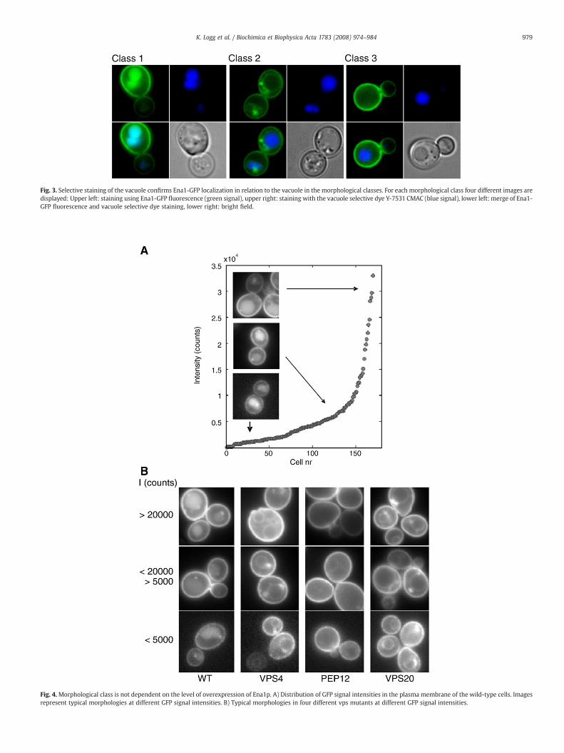
Fig. 3. Selective staining of the vacuole confirms Ena1-GFP localization in relation to the vacuole in the morphological classes. For each morphological class four different images aredisplayed: Upper left: staining using Ena1-GFP fluorescence (green signal), upper right: staining with the vacuole selective dye Y-7531 CMAC (blue signal), lower left: merge of Ena1-GFP fluorescence and vacuole selective dye staining, lower right: bright field.
Fig. 4.Morphological class is not dependent on the level of overexpression of Ena1p. A) Distribution of GFP signal intensities in the plasma membrane of the wild-type cells. Imagesrepresent typical morphologies at different GFP signal intensities. B) Typical morphologies in four different vps mutants at different GFP signal intensities.
979K. Logg et al. / Biochimica et Biophysica Acta 1783 (2008) 974–984

Fig. 5. Salt influences the Ena1-GFP localization to the plasma membrane differently inthe different vps mutants. The plasma membrane mean intensity was measured forexponentially growing cells (N30) of the wild-type and the mutants vps4Δ, pep12Δ,vps20Δ and vps52Δ. For each strain the left boxplot is for cells grown without saltand the right boxplot for cells grown with the addition of salt. The boxes represent 50%of the values with the lower line being the 25th percentile and the upper line is the 75thpercentile. The middle line is the mean intensity.
980 K. Logg et al. / Biochimica et Biophysica Acta 1783 (2008) 974–984
plasma membrane to the vacuole for degradation. The GFP proteinhas been reported to be especially stable and hard to degrade [16]. Thus,the vacuolar lumen signal may reflect GFP remaining after Ena1pdegradation. Two mutants, corresponding to the Golgi localized GARPcomplex components Vps52p and Vps53p, showed similar localizationpatterns as the wild-type, indicating that the GARP complex doesnot play a major role in the localization of Ena1p (class 1; Fig. 3). Insharp contrast, the salt sensitive mutants in ESCRT components,vps20Δ, snf8Δ, snf7Δ did not display any lumenal staining of thevacuole but staining of vacuolar membranes and pre-vacuolarmembrane structures (class 2; Fig. 3). The vacuolar mutants pep12Δand vps16Δ showed no indications of any vacuolar localization ofEna1p, either in the membrane or in the lumen and constituted a thirdmorphological class (class 3; Fig. 3). All GFP-staining in pep12Δ wasfound in the plasmamembrane, despite the fact that no drastic vacuolardefects were observed by the vacuole selective dye in these mutants(Fig. 3).
Cells expressing GFP-tagged proteins showcell-to-cell variation. Toenable strict analysis of Ena1p localization in the different vpsmutantsthe proportion of cells in each morphological class (class 1–3) wasdetermined (Fig. 2). For eachmutant there is onemajor morphologicalclass that constituted at least 70% of the cells in the case of non-salinegrowth. In addition, the GFP fluorescence intensities of individual cellsdiffered within a rather wide range (Fig. 4A). We examined cells atdifferent intensity range to rule out that some morphological featurescould be the result of overexpression of the GFP-fusion to non-physiological levels, resulting in miss-localization and the detection ofartifact features. No indications of intensity dependent classificationcould be observed; on the contrary, the dominant morphologicalclasses for each mutant were clearly apparent and most frequent alsoat low GFP intensities (Fig. 4B). We conclude that the observed Ena1plocalization in the various mutant backgrounds does not appear to bethe result of the overexpression per se.
Ena1p appeared to be properly localized to the plasma membranein all tested vps mutants both with and without salt addition. Thisexcludes the hypothesis that the salt sensitivity in these mutants iscaused by substantial spatial miss-localization of this crucial sodiumefflux pump. However, quantitative differences in Ena1p plasmamembrane localization could potentially cause the phenotypic dif-ferences in salt. To test this hypothesis we examined the amount ofEna1-GFP in the plasma membrane for the wild-type and the mutantsvps4Δ, vps20Δ, pep12Δ and vps52Δ (Fig. 5). All examined strains,except vps4Δ and vps20Δ, exhibited an enhanced level of Ena1-GFP inthe plasma membrane during saline growth. This tendency wasparticularly striking for the pep12Δ mutant which is defective in theendocytotic pathway and vacuolar breakdown. Apparently vps4Δ and
vps20Δ have difficulties to get the proper amount of Ena1p to theplasma membrane during salt growth.
3.3. Differences in the kinetics of Ena1p plasma membrane localizationamong vps mutants
Differences in the kinetics of Ena1p transport and incorporation tothe plasma membrane could potentially cause the observed differ-ences in steady-state amount of plasma membrane Ena1p (Fig. 5). Totest this kinetic hypothesis we examined Ena1p localization in thewild-type, vps20Δ, vps4Δ, pep12Δ and vps52Δ during the first 5 hafter induction of Ena1p expression via methionine depletion. Theseanalyses were conducted without salt to examine stress-independentdefects in exocytosis in the vps mutants. During the 5 h time-periodwild-type cells went from initially displaying no staining, to class 3(plasma membrane staining), class 2 (plasma and vacuolar membranestaining) and finally ending up in class 1 (plasma membrane, vacuolarmembrane and vacuolar lumen staining) (Fig. 6; upper right corner).The transport route from Golgi directly to the plasma membrane isbelieved to be the main exocytic pathway. Of the mutants, vps52Δexhibited essentially identical kinetics as the wild-type with a peak inthe accumulation of Ena1-GFP in the plasma membrane after 2 h.However, for all the other investigated mutants temporal defects ofEna1-GFP localization was observed, e.g. for both vps20Δ and vps4Δthe peak in plasma membrane localization was 1 h delayed comparedto the wild-type. The mutant with the slowest kinetics was pep12Δ forwhich the fraction of cells with plasma membrane localization ofEna1-GFP did not peak until the end of the 5-h period of theexperiment.
To further analyze the observed kinetic differences, Ena1p-GFPlocalization to the plasma membrane was quantified every 20 minafter pMET25 activation (Fig. 7A; see Supplementary Fig. SFig. 1 forcomplete set of representative images). It was clear that drasticdifferences in kinetics were recorded. In particular it is interesting tonote that the two ESCRT mutants vps4Δ and vps20Δ displayed cleartemporal differences. The most delayed of all mutants was pep12Δ forwhich no Ena1p plasma membrane localization was observed until180 min. To rule out any indirect effects on protein production as thecause of the delayed localization in the vps mutants, we analyzedEna1-GFP production by Western analysis with GFP specific anti-bodies (Fig. 7C and D). It is clear that even for the most severelyaffected mutant pep12Δ we see no difference in the kinetics of Ena1-GFP production compared to the wild-type. Apparently, the matura-tion of the fluorophore of GFP (which includes folding, cyclyzation,oxidation and dehydration; [17]) is dependent on the incorporation ofEna1p into its proper location in the plasma membrane. Interestingly,we found the response time of the various mutants (the time for theintensity in the plasma membrane to reach a set value) to correlatewith the generation time during saline growth. Thus, mutants with aninherently slow transport of Ena1p to the plasma membrane alsoexperienced difficulties during growth under salt stress (Fig. 7B).
4. Discussion
4.1. Salt phenotypes mechanistically differentiate the ESCRT components
This report high-lights the importance of a fully functional en-dosome/vacuolar protein sorting system in the detoxification ofsodium ions. The salt sensitive vps mutants analyzed in this studywere selected because of their gene products' reported physicalinteraction and all of them did not show a strong salt phenotype.However, the outstanding importance of the vps system in salt stressis supported by our earlier finding that among the top-10 most NaClsensitive mutants several components of the endosome/vacuolar sys-tem were identified, e.g. bro1Δ, bul1Δ and vps25Δ [3]. We show thatthe salt sensitivity of the mutants defective in endosome/vacuolar

981K. Logg et al. / Biochimica et Biophysica Acta 1783 (2008) 974–984
protein sorting is a Na+ specific effect. Particularly for mutants withdefects in the ESCRT components of the endosome/Multi VesicularBody (MVB), did sodium stress result in more severe phenotypes thanpotassium stress administered at equal osmolarity (Fig.1B). The strongsodium specificity of the phenotypes suggested a link to the mainsodium transporter Ena1p [1]. Such a link was confirmed by theobservation that the salt phenotypes of many of the ESCRT mutantscould be suppressed by Ena1p overexpression.
It is well established that the ESCRTcomponents Vps20p and Snf8p(ESCRT II complex), Snf7p (ESCRT III complex) and Vps4p are recruitedto the endosomes sequentially and that they also function sequentiallyin the formation of MVB [10,18]. Logically, it follows that thecorresponding deletion phenotypes should be identical. We foundthis not to be the case. All of the mutants exhibited salt sensitivity,however, vps20Δ, snf8Δ and snf7Δ were strongly suppressed byoverexpression of Ena1p whereas vps4Δ was not. Furthermore, vps4Δwas found to be substantially less salt sensitive than the other ESCRTmutants. This phenotypic discrepancy is in line with earlier reports
Fig. 6. Temporal differences in Ena1-GFP localization among salt sensitive vps mutants. KineGFP localization was investigated at different time points after induction of the MET25 proclasses (see Fig. 2 for definition of classes and color code). The time axis show data average
where the vps4 mutant differs from deletions in the other ESCRTcomponents: e.g. vps4Δ is less sensitive to Ca2+, Li+ and Mn2+ as com-pared to the ESCRT mutants snf7Δ and snf8Δ [19], and exhibits adramatic defect in the processing of carboxypeptidase S (CPS) com-pared to mutations like vps20Δ and snf7Δ [20]. In contrast to whatwas observed for the ESCRT mutants, the salt sensitivity of the class Cand D vps mutants was not suppressed by Ena1p overexpression.Thus, our results mechanistically differentiates the various salt sen-sitive vps mutants.
4.2. Other potential indirect factors affecting the salt sensitivity of the vpsmutants
The distinct salt sensitivity of the AAA-type ATPase Vps4pcompared to the other ESCRT components indicates that it may haveadditional functions besides those linked to the formation of the MVB.The key mode of action of AAA family members is energy dependentprotein unfolding and disassembly of protein complexes [21], and this
tics of Ena1p localization determined using the pMet25-ENA1-GFP construction. Ena1-moter via methionine removal and classified according to the different morphologicald over each hour as obtained from measurements taken at 20 min intervals.

Fig. 7. Kinetic differences in plasma membrane localization of Ena1-GFP between vps mutants correlate to growth rate in saline stress. A) The mean plasma membrane intensity as afunction of time after Ena1-GFP induction. B) Difference in Ena1p plasmamembrane localization kinetics relate to growth rate defect in saline conditions. The time it takes to reach anintensity of 1000 counts in A) is used as the response time in B). Growth rates are averages (n=10). C and D) Expression of GFP-tagged pMET25-Ena1p is not affected by deletion ofPEP12. C) Western blot of GFP-tagged Ena1p using a anti-GFP antibody. Strains were cultivated in presence of methionine to mid-log phase, washed, and re-inoculated in fresh mediawithout methionine to induce the pMET25 promotor. Samples were taken at specified time-points after induction. D) Quantification of GFP-tagged Ena1p expression from Westernblot data. Protein expression at specified time points was quantified as band intensity (total pixel intensity). Normalisation for background intensity was performed for each laneseparately.
982 K. Logg et al. / Biochimica et Biophysica Acta 1783 (2008) 974–984
is the proposed function of Vps4p in the disassembly of the ESCRTcomplex. However, its disassembly activity has also recently beenshown to be employed in breaking protein-protein interactions thatare part of sterol distribution into cellular vesicles [22]. Wang andcolleagues found that the cellular distribution of the sterol-bindingprotein Osh6 (amember of the oxysterol binding protein family, OSBP)was substantially altered in a vps4Δ strain. Importantly, the Osh6plocalization defect was detected in vps4Δ cells and not in other class Evpsmutants. It can thus be hypothesized that the suppressive effect onsalt sensitivity from Ena1p overexpression in the other class E vpsmutants might be dependent on incorporation of specific sterols intovesicle membranes, the latter being defective in vps4Δ. Interestingly,evidence for the importance of OSBP proteins and sterol distributionin salt stress resistance comes from a study on soybeans where it wasfound that a specific OSBP isogene was highly induced in root, stemand leaf during exposure to high salt concentrations [23].
Another connection to membrane lipid composition is that theproper function of many membrane channels and pumps is regulatedby certain phosphoinositides (in particular, PtdIns(4,5)P2 at theplasma membrane) [24]. The formation and turnover of these lipidsneeds to be tightly regulated which occurs both by membrane traf-ficking pathways and by PtdIns phosphatases and kinases. Vpsmutants might have perturbed levels of certain phophoinositides atthe plasma membrane. All forms of endocytosis are believed todepend on PtdIns(4,5)P2, so a deficient level of this lipid could in partlead to both an altered level of Ena1p at the cell surface as well as a
reduced Ena1 activity. The high level of Ena1p in the plasma mem-brane also at high salt concentrations in the salt sensitive mutantpep12Δ, points to a potential defect at the activity level of this sodiumtransporter in this strain.
There is also growing evidence in mammalian cells that endosomemetabolism couples to signaling [25,26], possibly by defects in endo-cytosis and breakdown of specific receptors. Also in yeast a mech-anism for the involvement of ESCRT components in signaling has beenput forward; Snf7p plays a role in signal transduction at high pH viainteraction with the Bro1-domain protein Rim20p, which affects thecleavage of the transcription factor Rim101 that regulated manydown-stream targets of importance in pH regulation [27]. At this stateof our analysis we can not exclude indirect mechanisms via lipidcomposition on transporter activity or signaling defects influencingdown-stream components for the involvement of some of the ESCRTcomponents in the salt stress response.
4.3. In all vps mutants Ena1p reaches the plasma membrane, but withdifferent kinetics
We found that Ena1p was properly localized to the plasma mem-brane in all salt sensitive vps mutants. Thus, we can exclude thatsubstantial miss-localization of the sodium transporter constitutes themechanistic basis of the salt sensitivity of vps mutants. However, bothtoo much and too little Ena1p in the plasma membrane might beharmful for the cell. For example, both vps20Δ and vps36Δ accumulate

983K. Logg et al. / Biochimica et Biophysica Acta 1783 (2008) 974–984
too high levels of Fur4p in the plasma membrane, which increasesuptake of 5-fluorouracil (5-FU), a toxic analog of uracil [28]. Indeed,we found differences in the levels as well as in the kinetics of Ena1pplasmamembrane localization in the various vps mutants (Figs. 5,6,7).The fine-balancing of the right amount of plasma membrane boundEna1p might involve breakdown via some of the salt sensitive vpscomponents. The study by Adler and colleagues [7] indicates that thepath to Ena1p breakdown in the vacuole does not pass via the plasmamembrane, but rather seem to involve direct rerouting at an earliersorting step that involves Sro7p. We report that the only two mutantsthat appear to be completely devoid of any vacuolar localization andbreakdown of Ena1p were pep12Δ and vps16Δ. We also report that thepep12Δ mutant exhibited higher amount of Ena1p in the plasmamembrane during steady-state growth compared to the othermutants, in line with a defect in the endocytotic pathway in thismutant. The highly salt sensitive pep12Δ strain was also the slowestmutant to distribute Ena1 to the plasma membrane, even though itsproduction of Ena1p was identical to that in the wild-type. In fact, wefound a correlation between the response time for Ena1p plasmamembrane localization and salt tolerance in the vps mutants (Fig. 7B),however, this delayed localization is unlikely to be the cause of saltsensitivity since the highly salt sensitive pep12Δ mutant displayedenhanced levels of Ena1p in the plasma membrane during steady-state saline growth (Fig. 5).
In yeast, as reported in mammalian cells, there appears to bemultiple exocytic transport routes from the Golgi to the plasmamembrane. One clear demonstration of this is that late secretorymutants like sec1, sec4 and sec6 accumulate two different density-types of 100 nm vesicles containing proteins to be secreted or incor-porated into the plasma membrane [29]. Interestingly, mutations ofsec6 in combinationwith various vps mutations affect the distributionof these secretory vesicles, indicating an involvement from vpscomponents in their formation [8]. Another manifestation of multipleexocytic paths comes from studies on different temperature sensitivemutants of the plasma membrane ATPase Pma1p with blocks atdifferent steps in the secretory pathway. These studies by Chang andcolleagues have indicated novel routes by which Pma1p reaches theplasma membrane [30], and by suppressor screens proposed theexistence of an endosome-to-surface pathway involving vps compo-nents [31]. Furthermore, the finding that the end4 mutation aloneresults in the accumulation of the more dense vesicles at therestrictive temperature [29], and that endocytosis is compromised inmany late sec mutants [32], supports the existence of a compartmentcommon to the endocytic and exocytic pathways. Direct support for arole of endosomes in exocytosis comes from the initial isolation of twoof the class E vps mutants in a screen for mutants unable to grow onsucrose as carbon source, so called Sucrose Non-Fermenting mutants(snf7/vps32Δ and snf8/vps22Δ). The sucrose growth defect appears todepend on the fact that the mutants secrete the sucrose metabolizingenzyme invertase to only 10–30% of the wild-type [33, 34], which insome of the mutants clearly involve post-transcriptional mechanismsindicating defects in exocytosis [33]. An endosome-to-surface trafficpathway is taken constitutively by many internalized membrane pro-teins in mammalian cells. One such example in yeast is Fet3p, the cellsurface ferroxidase involved in iron transport, which requires cupperfor its activity. In the vps41 mutant (a class B vps mutant), whichshows a growth defect on low-ironmedium, cupper loading of Fet3p isabsent and the protein nonfunctional [35]. Thus, this indicates thatselected plasma membrane proteins may pass through endosomes tofulfill specific processing requirements [30]. Whether endosome-linked maturation plays a role also for Ena1p is presently not known.
It is known that plasma membrane localization of Ena1p is de-pendent on Sro7p, a component that functions at both early and latestages of vesicular trafficking to the plasma membrane [7]. The strongsalt sensitivity of the sro7Δmutant results from the defective targetingof the Ena1p to the plasma membrane [7], and in the sro7 mutant
Ena1p is instead during salt stress targeted to the vacuole where it isdegraded. We here show that components in the vacuolar proteinsorting system can influence the kinetics of Ena1p plasma membranelocalization, while still displaying proper plasma membrane localiza-tion during both basal and salt stressed growth. We also show that thesalt phenotype of some of the vps mutants can be suppressed by ENA1overexpression, a phenomenon not observed in the sro7Δ strain. Thus,our data indicate that the ESCRT mutants vps20Δ, snf7Δ and snf8Δdisplay features that makes them distinct from the functionality of theSro7p-dependent pathway for Ena1p localization.
In conclusion, this study indicates that the salt sensitivity of someof the vps mutants can be linked to the sodium transporter Ena1pfrom phenotypic suppression by overexpression. However, for allanalyzed vps mutants, Ena1p seemed properly localized to the plasmamembrane, even during saline growth. At this stage we can thus notconclusively outline the mechanisms for this Ena1p dependent saltsensitivity. A number of explanations can be conceived as discussed inthe text, e.g. changes in transporter activity, localization kinetics orinfluence on signaling, hypotheses that have to be experimentallytested in future follow-up studies.
Acknowledgements
Comments from Lennart Adler during the course of this study andon the manuscript are highly appreciated. We also thank ananonymous reviewer for the insightful comments and suggestionsduring the review process. The technical support by Ellinor Petterssonin the immunological detection of Ena1-GFP is acknowledged. TheEna1-GFP plasmid was a kind gift from Martha Cyert. This work wassupported from Chalmers Foundation, the Swedish Research Council,and the Swedish Foundation for Strategic Research.
Appendix A. Supplementary data
Supplementary data associated with this article can be found, inthe online version, at doi:10.1016/j.bbamcr.2008.02.022.
References
[1] R. Haro, B. Garciadeblas, A. Rodriguéz-Navarro, A novel P-type ATPase from yeastinvolved in sodium transport, FEBS Lett. 291 (1991) 189–191.
[2] J. Wieland, A.M. Nitsche, J. Strayle, H. Steiner, H.K. Rudolph, The PMR2 gene clusterencodes functionally distinct isoforms of a putative Na+ pump in the yeast plasmamembrane, EMBO J. 14 (1995) 3870–3882.
[3] J. Warringer, E. Ericson, L. Fernandez, O. Nerman, A. Blomberg, High-resolutionyeast phenomics resolves different physiological features in the saline response,Proc. Natl. Acad. Sci. U. S. A. 100 (2003) 15724–15729.
[4] A. Blomberg, L. Adler, Physiology of osmotolerance in fungi, Adv. Microb. Physiol.33 (1992) 145–212.
[5] D.J. Klionsky, P.K. Herman, S.D. Emr, The fungal vacuole: composition, function, andbiogenesis, Microbiol. Rev. 54 (1990) 266–292.
[6] M.D. Ortega, A. Rodriguéz-Navarro, Sodium ion transport in Neurospora crassa,Physiol. Plant. 66 (1986) 705–711.
[7] I. Wadskog, A. Forsmark, G. Rossi, C. Konopka, M. Oyen, M. Goksor, H. Ronne, P.Brennwald, L. Adler, The yeast tumor suppressor homologue Sro7p is required fortargeting of the sodium pumping ATPase to the cell surface, Mol. Biol. Cell 17(2006) 4988–5003.
[8] E. Harsay, R. Schekman, A subset of yeast vacuolar protein sorting mutants isblocked in one branch of the exocytic pathway, J. Cell Biol. 156 (2002) 271–285.
[9] C.K. Raymond, I. Howald-Stevenson, C.A. Vater, T.H. Stevens, Morphologicalclassification of the yeast vacuolar protein sorting mutants: evidence for a pre-vacuolar compartment in class E vps mutants, Mol. Biol. Cell 3 (1992) 1389–1402.
[10] K. Bowers, T.H. Stevens, Protein transport from the late Golgi to the vacuole in theyeast Saccharomyces cerevisiae, Biochim. Biophys. Acta 1744 (2005) 438–454.
[11] C.B. Brachmann, A. Davies, G.J. Cost, E. Caputo, J. Li, P. Hieter, J.D. Boeke, Designerdeletion strains derived from Saccharomyces cerevisiae S288C: a useful set ofstrains and plasmids for PCR-mediated gene disruption and other applications,Yeast 14 (1998) 115–132.
[12] S.P. Solow, J. Sengbusch,M.W. Laird,Heterologousproteinproduction from the inducibleMET25 promoter in Saccharomyces cerevisiae, Biotechnol. Prog. 21 (2005) 617–620.
[13] M.C. Wagner, E.E. Molnar, B.A. Molitoris, M.G. Goebl, Loss of the homotypic fusionand vacuole protein sorting or golgi-associated retrograde protein vesicletethering complexes results in gentamicin sensitivity in the yeast Saccharomycescerevisiae, Antimicrob. Agents Chemother. 50 (2006) 587–595.
984 K. Logg et al. / Biochimica et Biophysica Acta 1783 (2008) 974–984
[14] S. Hohmann, Osmotic stress signalling and osmoadaptation in yeasts, Microbiol.Mol. Biol. Rev. 66 (2002) 300–372.
[15] M. Österberg, H. Kim, J.Warringer, K.Melen, A. Blomberg, G. vonHeijne, Phenotypiceffects ofmembrane protein overexpression in Saccharomyces cerevisiae, Proc. Natl.Acad. Sci. U. S. A. 103 (2006) 11148–11153.
[16] A. Sacchetti, V. Cappetti, P. Marra, R. Dell'Arciprete, T. El Sewedy, C. Crescenzi, S.Alberti, Green Fluorescent Protein variants fold differentially in prokaryotic andeukaryotic cells, J. Cell. Biochem. 81 (2001) 117–128.
[17] S.E. Jackson, T.D. Craggs, J.R. Huang, Understanding the folding of GFP usingbiophysical techniques, Expert Rev. Proteomics 3 (2006) 545–559.
[18] M. Babst, A protein's final ESCRT, Traffic 6 (2005) 2–9.[19] L. Eguez, Y.S. Chung, A. Kuchibhatla, M. Paidhungat, S. Garrett, Yeast Mn2+
transporter, Smf1p, is regulated by ubiquitin-dependent vacuolar protein sorting,Genetics 167 (2004) 107–117.
[20] M. Babst, D.J. Katzmann, E.J. Estepa-Sabal, T. Meerloo, S.D. Emr, Escrt-III: anendosome-associated heterooligomeric protein complex required for mvb sorting,Dev. Cell 3 (2002) 271–282.
[21] S. Patel, M. Latterich, The AAA team: related ATPases with diverse functions,Trends Cell Biol. 8 (1998) 65–71.
[22] P. Wang, Y. Zhang, H. Li, H.K. Chieu, A.L. Munn, H. Yang, AAA ATPases regulatemembrane association of yeast oxysterol binding proteins and sterol metabolism,EMBO J. 24 (2005) 2989–2999.
[23] D.Y. Li, H. Inoue, M. Takahashi, T. Kojima, M. Shiraiwa, H. Takahara, Molecularcharacterization of a novel salt-inducible gene for an OSBP (oxysterol-bindingprotein)-homologue from soybean, Gene (2007).
[24] M. Santarius, C.H. Lee, R.A. Anderson, Supervised membrane swimming: small G-protein lifeguards regulate PIPK signalling and monitor intracellular PtdIns(4,5)P2pools, Biochem. J. 398 (2006) 1–13.
[25] J. Samaj, J. Muller, M. Beck, N. Bohm, D. Menzel, Vesicular trafficking,cytoskeleton and signalling in root hairs and pollen tubes, Trends Plant Sci. 11(2006) 594–600.
[26] M.J. Clague, D.E. Hammond, Membrane traffic: catching the lysosome express,Curr. Biol. 16 (2006) R416–R418.
[27] J.H. Boysen, A.P. Mitchell, Control of Bro1-domain protein Rim20 localization byexternal pH, ESCRT machinery, and the Saccharomyces cerevisiae Rim101 pathway,Mol. Biol. Cell 17 (2006) 1344–1353.
[28] A. Bugnicourt, M. Froissard, K. Sereti, H.D. Ulrich, R. Haguenauer-Tsapis, J.M. Galan,Antagonistic roles of ESCRT and Vps class C/HOPS complexes in the recycling ofyeast membrane proteins, Mol. Biol. Cell 15 (2004) 4203–4214.
[29] E. Harsay, A. Bretscher, Parallel secretory pathways to the cell surface in yeast,J. Cell Biol. 131 (1995) 297–310.
[30] W. Luo, A. Chang, An endosome-to-plasma membrane pathway involved intrafficking of a mutant plasma membrane ATPase in yeast, Mol. Biol. Cell 11 (2000)579–592.
[31] M. Pizzirusso, A. Chang, Ubiquitin-mediated targeting of a mutant plasmamembrane ATPase, Pma1–7, to the endosomal/vacuolar system in yeast, Mol.Biol. Cell 15 (2004) 2401–2409.
[32] T.A. Vida, S.D. Emr, A new vital stain for visualizing vacuolar membrane dynamicsand endocytosis in yeast, J. Cell Biol. 128 (1995) 779–792.
[33] L.G. Vallier, M. Carlson, New SNF genes, GAL11 and GRR1 affect SUC2 expression inSaccharomyces cerevisiae, Genetics 129 (1991) 675–684.
[34] P. Yeghiayan, J. Tu, L.G. Vallier, M. Carlson, Molecular analysis of the SNF8 gene ofSaccharomyces cerevisiae, Yeast 11 (1995) 219–224.
[35] D.C. Radisky, W.B. Snyder, S.D. Emr, J. Kaplan, Characterization of VPS41, a generequired for vacuolar trafficking and high-affinity iron transport in yeast, Proc.Natl. Acad. Sci. U. S. A. 94 (1997) 5662–5666.