
The Signal Sequence Coding Region Promotes Nuclear Export of mRNA
13
The Signal Sequence Coding Region Promotes Nuclear Export of mRNA Alexander F. Palazzo 1 , Michael Springer 2 , Yoko Shibata 1 , Chung-Sheng Lee 1 , Anusha P. Dias 1 , Tom A. Rapoport 1* 1 Department of Cell Biology, Harvard Medical School, Boston, Massachusetts, United States of America, 2 Department of Systems Biology, Harvard Medical School, Boston, Massachusetts, United States of America In eukaryotic cells, most mRNAs are exported from the nucleus by the transcription export (TREX) complex, which is loaded onto mRNAs after their splicing and capping. We have studied in mammalian cells the nuclear export of mRNAs that code for secretory proteins, which are targeted to the endoplasmic reticulum membrane by hydrophobic signal sequences. The mRNAs were injected into the nucleus or synthesized from injected or transfected DNA, and their export was followed by fluorescent in situ hybridization. We made the surprising observation that the signal sequence coding region (SSCR) can serve as a nuclear export signal of an mRNA that lacks an intron or functional cap. Even the export of an intron-containing natural mRNA was enhanced by its SSCR. Like conventional export, the SSCR-dependent pathway required the factor TAP, but depletion of the TREX components had only moderate effects. The SSCR export signal appears to be characterized in vertebrates by a low content of adenines, as demonstrated by genome-wide sequence analysis and by the inhibitory effect of silent adenine mutations in SSCRs. The discovery of an SSCR- mediated pathway explains the previously noted amino acid bias in signal sequences and suggests a link between nuclear export and membrane targeting of mRNAs. Citation: Palazzo AF, Springer M, Shibata Y, Lee CS, Dias AP, et al. (2007) The signal sequence coding region promotes nuclear export of mRNA. PLoS Biol 5(12): e322. doi:10. 1371/journal.pbio.0050322 Introduction In eukaryotes, mRNAs are synthesized and processed in the nucleus before they are transported through the nuclear pores into the cytoplasm, where they are translated into proteins. Nuclear export of most mRNAs is mediated by the conserved transcription export (TREX) complex that is comprised of the Tho complex, UAP56, and Aly. In vertebrates, the TREX components are recruited to the 59 end of newly synthesized transcripts by the combined action of the 59 cap binding complex, CBP80/20, and factors that are loaded during the splicing of the intron closest to the 59 cap [1–4]. Once assembled, the TREX complex recruits the heterodimer TAP/p15 as an export factor [5,6]. TAP interacts with nucleoporins directly [7–9] or through the factor Rae1 [10,11] and may thus allow bound transcripts to enter and eventually pass through the nuclear pores. It remains unclear how mRNAs exit the pores on the cytoplasmic side, but RNA helicases, such as Dbp5, may be involved [12,13]. Although many details of the export mechanism remain to be clarified, it seems clear that the efficient export of most mRNAs requires both splicing and a functional cap. Not all mRNAs follow this canonical export pathway. In higher eukaryotes, transcripts coding for cyclin D [14] and other regulators of cell division [15] use elements in the 39 untranslated region (UTR) as well as the cap binding protein eIF4E to engage the exportin protein Crm1. A Crm1- dependent pathway is also used for the export of the intron-containing RNA genome of the human immunodefi- ciency virus (HIV) [16]. In macrophages, the export of interferon-induced transcripts is sensitive to the levels of Nup96, a component of the nuclear pore, whereas other transcripts are insensitive [17]. In Saccharomyces cerevisiae, the export of some mRNAs requires either the Aly ortholog Yra1p or the TAP ortholog Mex67p, but not both [18]. Transcripts coding for heat-shock proteins, such as Hsp70p, are also exported by an Aly-independent pathway that is stimulated under stress conditions [19]. Together, these data indicate that there may be distinct export pathways for certain classes of mRNAs, which may be related to the different functions of the final translation products. One specialized class of mRNAs codes for secretory proteins. These mRNAs are often translated by ribosomes that are targeted to the endoplasmic reticulum (ER) membrane. As a result, the mRNAs also become membrane- bound. In addition, it is possible that these mRNAs associate with the ER membrane in a ribosome-independent manner by interacting with RNA-binding proteins [20–22]. These proteins may be loaded onto the mRNAs in the nucleus, during their passage through the nuclear pores, or in the cytoplasm. Thus, the membrane localization of these mRNAs may require factors that mediate nuclear export and distribution in the cytoplasm, which are distinct from factors used by mRNAs translated on free ribosomes in the cytoplasm. A major characteristic of a secretory protein is a hydro- Academic Editor: Peter Walter, University of California San Francisco, United States of America Received May 11, 2007; Accepted October 19, 2007; Published December 4, 2007 Copyright: Ó 2007 Palazzo et al. This is an open-access article distributed under the terms of the Creative Commons Attribution License, which permits unrestricted use, distribution, and reproduction in any medium, provided the original author and source are credited. Abbreviations: CTE, constitutive transport element; D-PBS, Dulbecco’s modified phosphate-buffered saline; EJC, exon junction complex; ER, endoplasmic reticulum; FISH, fluorescence in situ hybridization; ftz, fushi tarazu; HA, hemagglutinin; MHC, major histocompatibility complex; ORF, open reading frame; SSCR, signal sequence coding region; TREX, transcription export * To whom correspondence should be addressed. E-mail: tom_rapoport@hms. harvard.edu PLoS Biology | www.plosbiology.org December 2007 | Volume 5 | Issue 12 | e322 2862 P L o S BIOLOGY
Transcript of The Signal Sequence Coding Region Promotes Nuclear Export of mRNA

The Signal Sequence Coding RegionPromotes Nuclear Export of mRNAAlexander F. Palazzo
1, Michael Springer
2, Yoko Shibata
1, Chung-Sheng Lee
1, Anusha P. Dias
1, Tom A. Rapoport
1*
1 Department of Cell Biology, Harvard Medical School, Boston, Massachusetts, United States of America, 2 Department of Systems Biology, Harvard Medical School, Boston,
Massachusetts, United States of America
In eukaryotic cells, most mRNAs are exported from the nucleus by the transcription export (TREX) complex, which isloaded onto mRNAs after their splicing and capping. We have studied in mammalian cells the nuclear export of mRNAsthat code for secretory proteins, which are targeted to the endoplasmic reticulum membrane by hydrophobic signalsequences. The mRNAs were injected into the nucleus or synthesized from injected or transfected DNA, and theirexport was followed by fluorescent in situ hybridization. We made the surprising observation that the signal sequencecoding region (SSCR) can serve as a nuclear export signal of an mRNA that lacks an intron or functional cap. Even theexport of an intron-containing natural mRNA was enhanced by its SSCR. Like conventional export, the SSCR-dependentpathway required the factor TAP, but depletion of the TREX components had only moderate effects. The SSCR exportsignal appears to be characterized in vertebrates by a low content of adenines, as demonstrated by genome-widesequence analysis and by the inhibitory effect of silent adenine mutations in SSCRs. The discovery of an SSCR-mediated pathway explains the previously noted amino acid bias in signal sequences and suggests a link betweennuclear export and membrane targeting of mRNAs.
Citation: Palazzo AF, Springer M, Shibata Y, Lee CS, Dias AP, et al. (2007) The signal sequence coding region promotes nuclear export of mRNA. PLoS Biol 5(12): e322. doi:10.1371/journal.pbio.0050322
Introduction
In eukaryotes, mRNAs are synthesized and processed in thenucleus before they are transported through the nuclearpores into the cytoplasm, where they are translated intoproteins. Nuclear export of most mRNAs is mediated by theconserved transcription export (TREX) complex that iscomprised of the Tho complex, UAP56, and Aly. Invertebrates, the TREX components are recruited to the 59
end of newly synthesized transcripts by the combined actionof the 59 cap binding complex, CBP80/20, and factors that areloaded during the splicing of the intron closest to the 59 cap[1–4]. Once assembled, the TREX complex recruits theheterodimer TAP/p15 as an export factor [5,6]. TAP interactswith nucleoporins directly [7–9] or through the factor Rae1[10,11] and may thus allow bound transcripts to enter andeventually pass through the nuclear pores. It remains unclearhow mRNAs exit the pores on the cytoplasmic side, but RNAhelicases, such as Dbp5, may be involved [12,13]. Althoughmany details of the export mechanism remain to be clarified,it seems clear that the efficient export of most mRNAsrequires both splicing and a functional cap.
Not all mRNAs follow this canonical export pathway. Inhigher eukaryotes, transcripts coding for cyclin D [14] andother regulators of cell division [15] use elements in the 39
untranslated region (UTR) as well as the cap binding proteineIF4E to engage the exportin protein Crm1. A Crm1-dependent pathway is also used for the export of theintron-containing RNA genome of the human immunodefi-ciency virus (HIV) [16]. In macrophages, the export ofinterferon-induced transcripts is sensitive to the levels ofNup96, a component of the nuclear pore, whereas othertranscripts are insensitive [17]. In Saccharomyces cerevisiae, theexport of some mRNAs requires either the Aly orthologYra1p or the TAP ortholog Mex67p, but not both [18].
Transcripts coding for heat-shock proteins, such as Hsp70p,are also exported by an Aly-independent pathway that isstimulated under stress conditions [19]. Together, these dataindicate that there may be distinct export pathways forcertain classes of mRNAs, which may be related to thedifferent functions of the final translation products.One specialized class of mRNAs codes for secretory
proteins. These mRNAs are often translated by ribosomesthat are targeted to the endoplasmic reticulum (ER)membrane. As a result, the mRNAs also become membrane-bound. In addition, it is possible that these mRNAs associatewith the ER membrane in a ribosome-independent mannerby interacting with RNA-binding proteins [20–22]. Theseproteins may be loaded onto the mRNAs in the nucleus,during their passage through the nuclear pores, or in thecytoplasm. Thus, the membrane localization of these mRNAsmay require factors that mediate nuclear export anddistribution in the cytoplasm, which are distinct from factorsused by mRNAs translated on free ribosomes in thecytoplasm.A major characteristic of a secretory protein is a hydro-
Academic Editor: Peter Walter, University of California San Francisco, United Statesof America
Received May 11, 2007; Accepted October 19, 2007; Published December 4, 2007
Copyright: � 2007 Palazzo et al. This is an open-access article distributed underthe terms of the Creative Commons Attribution License, which permits unrestricteduse, distribution, and reproduction in any medium, provided the original authorand source are credited.
Abbreviations: CTE, constitutive transport element; D-PBS, Dulbecco’s modifiedphosphate-buffered saline; EJC, exon junction complex; ER, endoplasmic reticulum;FISH, fluorescence in situ hybridization; ftz, fushi tarazu; HA, hemagglutinin; MHC,major histocompatibility complex; ORF, open reading frame; SSCR, signal sequencecoding region; TREX, transcription export
* To whom correspondence should be addressed. E-mail: [email protected]
PLoS Biology | www.plosbiology.org December 2007 | Volume 5 | Issue 12 | e3222862
PLoS BIOLOGY

phobic signal sequence close to its N terminus. The signalsequence is first recognized by the signal-recognition particle(SRP) as it emerges from the translating ribosome and is thentransferred into the protein-conducting channel formed bythe Sec61p complex [23–25]. In most cases, the signalsequence is cleaved during or shortly after the polypeptideis transferred into the ER. Although the major requirementfor a signal sequence is a stretch of at least 6–7 hydrophobicamino acids, there appear to be other, poorly definedproperties that may distinguish different signal sequences[26,27]. For example, some signal sequences require auxiliarytranslocation components [28,29] or are sensitive to the drugcotransin [27,30]. In addition, signal sequences have otherunexplained characteristics. For example, it has been notedthat signal sequences in humans tend to be rich in leucineand poor in isoleucine [31], despite the fact that these twoamino acids have similar hydrophobicities [32] and wouldthus be expected to function equally well to promotetranslocation. This leucine/isoleucine bias is not seen inprokaryotes. Another puzzling feature is that signal sequencesare often conserved across species to a higher degree thanexpected [33]. These observations raise the possibility thatnot only the signal sequence per se, but also the encodingnucleotide sequence (signal sequence coding region or SSCR)may have a function.
We report here on the nuclear export of mRNAs coding forsecretory proteins. We used microinjection of mRNAs andDNAs as well as transfection of DNA to demonstrate that theSSCR can promote the export of mRNAs that cannot use theconventional pathway because they lack an intron or afunctional cap. By using large-scale sequence analysis, wefound that vertebrate SSCRs have a low content of adenine, afeature that may in part be responsible for the preference ofleucine versus isoleucine in signal sequences. Consistent withthese observations, the incorporation of silent adeninemutations within the SSCR inhibits its nuclear export activity.Even the export of a natural RNA containing introns isfacilitated by its SSCR. The discovery of an SSCR-mediatedmRNA export pathway thus explains the previously noted
amino acid bias in signal sequences and suggests a linkbetween nuclear export and membrane targeting of mRNAs.
Results
Efficient Splicing and Translation of Microinjected t-ftzRNATo study the nuclear export of mRNA coding for a
secretory protein, we used a model mRNA that is derivedfrom a fragment of the fushi tarazu (ftz) gene. The originalconstruct contains an intron and was previously used tomonitor mRNA splicing and nuclear export in Xenopusoocytes [1,34]. The construct was modified by adding a Kozakconsensus sequence to allow efficient expression in mamma-lian cells. Sequences encoding FLAG and hemagglutinin (HA)epitopes were included at the 59 and 39 ends of the openreading frame (ORF), respectively, to monitor translation ofthe mRNA. Because the intron contains in-frame stop codons(Figure 1A; asterisks), the HA epitope will only be synthesizedif the mRNA is spliced. To target the translation product tothe ER, we attached an SSCR derived from the mouse majorhistocompatibility complex (MHC) class 2 molecule H2-K1.The final construct is called t-ftz-i (Figure 1A and Figure S1).We first tested the translation of the t-ftz-i mRNA in vitro.
When translated in reticulocyte lysate, a polypeptide of 13kDa was generated, consistent with the size expected from thelocation of the in-frame stop codon in the intron (Figure 1B,lane 3). When the intron was deleted, the resulting mRNA (t-ftz-Di) gave rise to a 17-kDa translation product (lane 4), againin agreement with the expected size of the ORF. With theoriginal ftz constructs that lacked translation initiationsignals, no translational products were detected (lanes 1, 2).When t-ftz-i mRNA was microinjected into nuclei of NIH
3T3 fibroblasts, it was efficiently spliced within 15 min, asshown by reverse-transcriptase (RT)-PCR (Figure 1C; lanes 2–4). As expected, no detectable splicing was observed when t-ftz-i was microinjected into the cytoplasm (lane 5). Next, wetested the translation of t-ftz-i transcripts that were micro-injected into the nuclei of NIH 3T3 fibroblasts. Nuclearinjection was confirmed by co-injecting fluorescently labeled70-kDa dextran, which is too large to passively cross thenuclear pores (Figure 1D and 1E; see insets). After 4 h, theFLAG and HA epitopes could be detected by immunofluor-escence microscopy in over 90% of the injected cells (Figure1D and 1E). In addition, both epitopes co-localized with theER resident protein TRAPa (Figure 1D and 1E), indicatingthat the translation product was translocated into the ER.The protein is probably not secreted efficiently, because itcontains only a fragment of ftz and therefore may not beproperly folded. When t-ftz-i transcripts were injected intothe cytoplasm, the expression of the FLAG but not of the HAepitope was observed (unpublished data), as expected fromthe presence of the unspliced intron. Both the FLAG and HAepitopes were expressed when t-ftz-Di transcripts wereinjected into nuclei or cytoplasm (unpublished data).
Nuclear mRNA Export Requires Splicing or the SSCRTo monitor the nuclear export of t-ftz-i mRNA, transcripts
were microinjected into nuclei of NIH 3T3 cells. The cellswere fixed at various time points, and the localization of theinjected RNA was probed by fluorescence in situ hybrid-ization (FISH). We optimized the FISH procedure, omitting
PLoS Biology | www.plosbiology.org December 2007 | Volume 5 | Issue 12 | e3222863
The SSCR Promotes mRNA Export
Author Summary
In eukaryotic cells, precursors of messenger RNAs (mRNAs) aresynthesized and processed in the nucleus. During processing,noncoding introns are spliced out, and a cap and poly-adenosinesequence are added to the beginning and end of the transcript,respectively. The resulting mature mRNA is exported from thenucleus to the cytoplasm by crossing the nuclear pore. Both theintrons and the cap help to recruit factors that are necessary fornuclear export of an mRNA. Here we provide evidence for a novelmRNA export pathway that is specific for transcripts coding forsecretory proteins. These proteins contain signal sequences thattarget them for translocation across the endoplasmic reticulummembrane. We made the surprising observation that the signalsequence coding region (SSCR) can serve as a nuclear export signalof an mRNA that lacks an intron or functional cap. Even the export ofan intron-containing natural mRNA was enhanced by its SSCR. TheSSCR export signal appears to be characterized in vertebrates by alow content of adenines. Our discovery of an SSCR-mediatedpathway explains the previously noted amino acid bias in signalsequences, and suggests a link between nuclear export andmembrane targeting of mRNAs.

harsh acid and ethanol treatments, such that intracellularmorphology was largely maintained. We estimate that 20,000to 50,000 transcripts were injected per cell, which is a smallnumber compared with the total number of transcripts in atypical mammalian cell (400,000 to 850,000 molecules) [35].Again, fluorescent 70-kDa dextran was co-injected to identify
nuclear-injected cells (Figure 2, insets). Immediately afterinjection, the transcripts were confined to the nucleus. Overtime, however, the majority of t-ftz-i transcripts (;80%)accumulated in the cytoplasm (Figure 2A). Quantitationshowed that the half-time of mRNA export was ;15 min(Figure 2C), similar to previous estimates [36,37]. About 50%
Figure 1. Microinjected t-ftz mRNA Is Spliced and Translated In Vivo
(A) Scheme of the t-ftz transcript. The regions complementary to the oligonucleotides used for FISH (‘‘FISH probe’’) and for RT-PCR (‘‘ftz-127F’’ and ‘‘ftz-444R’’) are indicated. Asterisks represent in-frame stop codons within the intron.(B) The indicated transcripts were translated in vitro using reticulocyte lysate in the presence of 35S-methionine. Samples were separated by SDS-PAGE,and newly synthesized proteins were detected by autoradiography.(C) t-ftz-i was microinjected into either the nuclei (lanes 2–4) or cytoplasm (lane 5) of 20 NIH 3T3 cells, which were then incubated for the indicatedtimes. RT-PCR was performed on the cell extracts using the intron flanking oligonucleotides indicated in (A). The lower band represents the splicedproduct, and the upper band represents the unspliced product. 18S rRNA was amplified and used as a loading control. Molecular weight markers (M)were loaded in lane 1.(D and E) NIH 3T3 cells were microinjected with t-ftz-i mRNA. FITC-conjugated 70-kDa dextran was co-injected to mark the injected compartment(insets). Cells were incubated for 4 h, fixed and immunostained for either FLAG (D) or HA (E) epitopes and for TRAPa. Overlays of the expressed epitope(green) and TRAPa (red) are shown in the last panel. Scale bar ¼ 10 lm.doi:10.1371/journal.pbio.0050322.g001
PLoS Biology | www.plosbiology.org December 2007 | Volume 5 | Issue 12 | e3222864
The SSCR Promotes mRNA Export

of the mRNA molecules remained intact after 4 h, as shownby the total level of FISH signal (Figure 2D).Surprisingly, when we injected t-ftz-Di mRNA into nuclei,
we also observed efficient export into the cytoplasm (Figure2B; quantitation in Figure 2C), despite the fact that theabsence of an intron should have prevented the recruitmentof TREX components and thus nuclear export [2,4]. Thekinetics of mRNA export was about the same for transcriptscontaining or lacking the intron (Figure 2C). In contrast to aprevious report [38], a large fraction of the intron-lesstranscripts remained stable over the time of analysis (Figure2D).To exclude the possibility that the nuclear export of the
intron-less transcript is caused by the introduction ofexogenously synthesized mRNAs, we tested the export ofmRNA after its synthesis in the nucleus. To this end, wemicroinjected plasmids containing the t-ftz-Di or t-ftz-i genesinto NIH 3T3 nuclei. After 30 min, the RNA Polymerase IIinhibitor a-amanitin was added to inhibit further tran-scription, and then the distribution of mRNA over time wasmonitored using FISH. Immediately after a-amanitin addi-tion, most transcripts were in the nucleus, but ;20% werealready found in the cytoplasm. Over time, the nuclearfraction was efficiently exported to the cytoplasm (Figure 2Eand 2F) with a rate that was slightly lower than that ofmicroinjected RNA. As before, we observed only minordifferences between intron-containing and intron-lacking t-ftz transcripts (Figure 2F). Pretreatment of cells with a-amanitin 5 min prior to DNA microinjection completelyinhibited mRNA synthesis, as assayed by FISH (unpublisheddata). From these results, we conclude that export of anintron-lacking mRNA can occur efficiently, regardless ofwhether or not export is coupled to transcription.Our results are in apparent contradiction to previous
observations showing that ftz constructs lacking introns arenot exported from nuclei of Xenopus oocytes [1]. A majordifference to the transcripts tested in Xenopus oocytes is thepresence of an SSCR in t-ftz-Di. We therefore tested whetherthe SSCR was required to promote nuclear export of thismRNA. Indeed, a transcript that lacked the SSCR, and thusencoded a cytoplasmic version of ftz (c-ftz-Di), remained inthe nucleus 30 min after injection (Figure 2G, quantitation inFigure 2C), a time during which most of the control t-ftz-DimRNA was exported. At later time points, much of the c-ftz-Di mRNA was degraded (Figure 2D). Because c-ftz-Di mRNAwas stable when injected directly into the cytoplasm(unpublished data), it is unlikely that nuclear-injected c-ftz-Di mRNA was exported and then rapidly degraded in thecytoplasm. The addition of an intron into c-ftz-Di mRNA(resulting in c-ftz-i mRNA) restored nuclear export andstability of the mRNA (Figure 2H, quantitation in Figure 2Cand 2D). A fraction of the exported c-ftz-i transcriptsaccumulated in stress granules (unpublished data). Together,these experiments suggest that either an SSCR or an introncan serve as a nuclear export signal.To exclude the possibility that the SSCR-mediated export
pathway is a peculiarity of NIH 3T3 cells, we microinjectedmRNA precursors into the nuclei of COS-7 cells. As before, t-ftz-i mRNA containing both the splicing and the SSCRsignals—as well as t-ftz-Di and c-ftz-i mRNAs, which eachcontain only one of the two signals—were exported efficientlyinto the cytoplasm (Figure 3A and Figure S2; quantitation in
Figure 2. The SSCR Promotes Nuclear Export of mRNA
(A and B) NIH 3T3 cells were microinjected with either t-ftz-i (A) or t-ftz-Di(B) and FITC-conjugated 70-kDa dextran (insets). After the indicated timepoints, cells were fixed and probed for ftz mRNA by FISH.(C and D) Quantitation of the cytoplasmic/total FISH fluorescence (C) andtotal FISH fluorescence (D) from cells microinjected with the indicatedmRNAs. Each data point represents the average of three experiments,each of which consisted of 15–30 cells. Error bars represent the standarddeviation between the three experiments. Relative fluorescence in (D)was calculated by normalizing the total fluorescence at a given timepoint to the fluorescence at 0 min in each experiment.(E) A plasmid containing the t-ftz-Di gene, and FITC-conjugated 70-kDadextran (inset) were microinjected into NIH 3T3 cells. After 30 min, a-amanitin was added, and the cells were incubated for an additional 120min. The cells were fixed and probed for ftz mRNA by FISH.(F) NIH 3T3 cells were microinjected with plasmids containing either the t-ftz-Di or t-ftz-i genes. After 30 min, the cells were treated with a-amanatinand incubated for the indicated time points. The cells were then fixed andprobed for ftz mRNA. Nuclear export was quantified as in (C).(G and H) NIH 3T3 cells were microinjected with either c-ftz-Di (G) or c-ftz-i (H) and FITC-conjugated 70-kDa dextran (insets). After the indicatedtime points, the cells were fixed, and probed for ftz mRNA by FISH. Scalebar¼ 15 lm.doi:10.1371/journal.pbio.0050322.g002
PLoS Biology | www.plosbiology.org December 2007 | Volume 5 | Issue 12 | e3222865
The SSCR Promotes mRNA Export

Figure 3B). In contrast, c-ftz-Di mRNA lacking both signalswas not exported (Figure 3B and Figure S2). Again, the c-ftz-Di mRNA was significantly less stable than the other mRNAs(Figure 3C). These data suggest that the SSCR-mediatednuclear export pathway may be present in many mammaliancell types.
In COS-7 cells, we were able to visualize the targeting of themRNAs to the ER membrane. All mRNAs containing an SSCRgave a typical reticular staining in the cytoplasm and co-localized with TRAPa, a marker of the ER membrane (Figure3A and Figure S2).
To test whether other SSCRs could promote mRNA export,we replaced the SSCR of the MHC class 2 molecule H2-K1with that of human insulin in the t-ftz-Di construct (ins-ftz-Di,see Figure S3). When injected into COS-7 cell nuclei, ins-ftz-Di mRNA was efficiently exported (Figure 3B and Figure S2).In fact, the export kinetics was faster than with t-ftz-Di mRNA(Figure 3B). Again, the mRNA was targeted to the ERmembrane (Figure S2). Taken together, these results indicatethat the SSCR-mediated pathway may be quite general.
Finally, we performed transfection experiments in COS-7cells with plasmids coding for t-ftz and c-ftz mRNAscontaining or lacking an intron. The steady-state distributionof the mRNAs between the nucleus and cytoplasm wasdetermined by FISH (Figure S4, quantitation in Figure 3D).The SSCR-containing mRNAs (t-ftz-i and t-ftz-Di) were mostlyfound in the cytoplasm, indicating that they are efficientlyexported from the nucleus. A significant fraction of mRNAslacking an SSCR (c-ftz-i and c-ftz-Di) were found in thenucleus, despite the fact that one contained an intron. In thecytoplasm, the SSCR-containing ftz constructs were targetedto the ER membrane, whereas c-ftz constructs partially co-localized with TIA-1 (Figure S4), a marker of stress granules
[39]. These data confirm the presence of an SSCR-mediatedmRNA export pathway and suggest that, at least undercertain conditions, it may be more efficient than the splicing-mediated export pathway.
Characterization of SSCR-Mediated mRNA ExportNext we characterized SSCR-mediated mRNA export, using
t-ftz-Di mRNA that contains the SSCR signal but lacks anintron. When this mRNA was injected into nuclei of NIH 3T3cells that had been depleted of ATP by treatment with azide[40], no export into the cytoplasm was observed (Figure 4A).This treatment did not compromise the viability of the cellsas judged by phase microscopy or immunostaining of themicrotubule network (unpublished data). Blocking the nu-clear pores by pre-injecting wheat germ agglutinin (WGA)[41,42] also inhibited the nuclear export of microinjected t-ftz-Di mRNA (Figure 4B). Thus the export of t-ftz-Di mRNArequires energy and functional nuclear pores.As expected, nuclear mRNA export was unidirectional.
When transcripts were injected into one of the nuclei of abinucleated cell, 80% of the mRNA was transported into thecytoplasm, but no fluorescence was detected in the uninjectednucleus (Figure S5), indicating that there is no re-uptake ofexported mRNA.Next we attempted to define the components required for
t-ftz-Di mRNA export. To test whether the export factor TAPis required, we took advantage of the viral constitutivetransport element (CTE), which binds to and sequesters TAP[43]. Nuclear export of t-ftz-Di mRNA was inhibited in cellspre-injected with CTE RNA (Figure 4C, arrow indicates a cellpre-injected with CTE, quantitation is shown in Figure 4D). Incontrast, cells that were not pre-injected with CTE (Figure 4C,arrowhead), or cells pre-injected with control buffer (Figure
Figure 3. The SSCR Promotes Nuclear Export and ER Targeting of mRNA in COS-7 Cells
(A) COS-7 cells were microinjected with t-ftz-Di mRNA and FITC-conjugated 70-kDa dextran (inset) and then incubated for 90 min. The cells were fixed,probed for ftz mRNA by FISH, and immunostained for TRAPa. An overlay of the mRNA (green) and TRAPa (red) staining is shown in the right panel.Scale bar ¼ 15 lm.(B and C) Quantitation of the cytoplasmic/total FISH fluorescence (C) and total FISH fluorescence (D) in COS-7 cells microinjected with the indicatedmRNAs, as described in Figure 2C and 2D.(D) COS-7 cells were transfected with plasmids containing the indicated genes, incubated for 12–18 h, fixed, and probed for ftz mRNA. Cells were thenimaged, and the cytoplasmic and nuclear fluorescence was quantified. Each bar represents the average of three experiments in which 20 cells wereanalyzed. Error bars represent the standard deviation between the three experiments.doi:10.1371/journal.pbio.0050322.g003
PLoS Biology | www.plosbiology.org December 2007 | Volume 5 | Issue 12 | e3222866
The SSCR Promotes mRNA Export
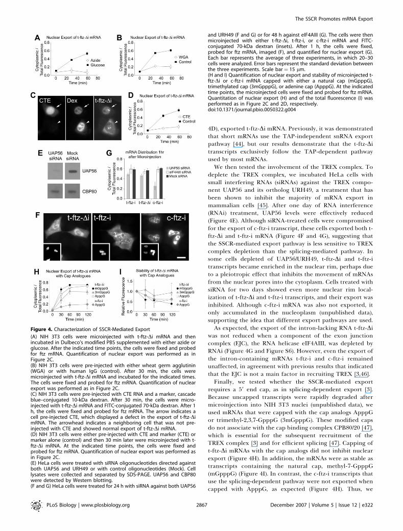
4D), exported t-ftz-Di mRNA. Previously, it was demonstratedthat short mRNAs use the TAP-independent snRNA exportpathway [44], but our results demonstrate that the t-ftz-Ditranscripts exclusively follow the TAP-dependent pathwayused by most mRNAs.We then tested the involvement of the TREX complex. To
deplete the TREX complex, we incubated HeLa cells withsmall interfering RNAs (siRNAs) against the TREX compo-nent UAP56 and its ortholog URH49, a treatment that hasbeen shown to inhibit the majority of mRNA export inmammalian cells [45]. After one day of RNA interference(RNAi) treatment, UAP56 levels were effectively reduced(Figure 4E). Although siRNA-treated cells were compromisedfor the export of c-ftz-i transcript, these cells exported both t-ftz-Di and t-ftz-i mRNA (Figure 4F and 4G), suggesting thatthe SSCR-mediated export pathway is less sensitive to TREXcomplex depletion than the splicing-mediated pathway. Insome cells depleted of UAP56/URH49, t-ftz-Di and t-ftz-itranscripts became enriched in the nuclear rim, perhaps dueto a pleiotropic effect that inhibits the movement of mRNAsfrom the nuclear pores into the cytoplasm. Cells treated withsiRNA for two days showed even more nuclear rim local-ization of t-ftz-Di and t-ftz-i transcripts, and their export wasinhibited. Although c-ftz-i mRNA was also not exported, itonly accumulated in the nucleoplasm (unpublished data),supporting the idea that different export pathways are used.As expected, the export of the intron-lacking RNA t-ftz-Di
was not reduced when a component of the exon junctioncomplex (EJC), the RNA helicase eIF4AIII, was depleted byRNAi (Figure 4G and Figure S6). However, even the export ofthe intron-containing mRNAs t-ftz-i and c-ftz-i remainedunaffected, in agreement with previous results that indicatedthat the EJC is not a main factor in recruiting TREX [3,46].Finally, we tested whether the SSCR-mediated export
requires a 59 end cap, as in splicing-dependent export [3].Because uncapped transcripts were rapidly degraded aftermicroinjection into NIH 3T3 nuclei (unpublished data), weused mRNAs that were capped with the cap analogs ApppGor trimethyl-2,3,7-GpppG (3mGpppG). These modified capsdo not associate with the cap binding complex CPB80/20 [47],which is essential for the subsequent recruitment of theTREX complex [3] and for efficient splicing [47]. Capping oft-ftz-Di mRNAs with the cap analogs did not inhibit nuclearexport (Figure 4H). In addition, the mRNAs were as stable astranscripts containing the natural cap, methyl-7-GpppG(mGpppG) (Figure 4I). In contrast, the c-ftz-i transcripts thatuse the splicing-dependent pathway were not exported whencapped with ApppG, as expected (Figure 4H). Thus, we
Figure 4. Characterization of SSCR-Mediated Export
(A) NIH 3T3 cells were microinjected with t-ftz-Di mRNA and thenincubated in Dulbeco’s modified PBS supplemented with either azide orglucose. After the indicated time points, the cells were fixed and probedfor ftz mRNA. Quantification of nuclear export was performed as inFigure 2C.(B) NIH 3T3 cells were pre-injected with either wheat germ agglutinin(WGA) or with human IgG (control). After 30 min, the cells weremicroinjected with t-ftz-Di mRNA and incubated for the indicated times.The cells were fixed and probed for ftz mRNA. Quantification of nuclearexport was performed as in Figure 2C.(C) NIH 3T3 cells were pre-injected with CTE RNA and a marker, cascadeblue–conjugated 10-kDa dextran. After 30 min, the cells were micro-injected with t-ftz-Di mRNA and FITC-conjugated 70-kDa dextran. After 1h, the cells were fixed and probed for ftz mRNA. The arrow indicates acell pre-injected CTE, which displayed a defect in the export of t-ftz-DimRNA. The arrowhead indicates a neighboring cell that was not pre-injected with CTE and showed normal export of t-ftz-Di mRNA.(D) NIH 3T3 cells were either pre-injected with CTE and marker (CTE) ormarker alone (control) and then 30 min later were microinjected with t-ftz-Di mRNA. At the indicated time points, the cells were fixed andprobed for ftz mRNA. Quantification of nuclear export was performed asin Figure 2C.(E) HeLa cells were treated with siRNA oligonucleotides directed againstboth UAP56 and URH49 or with control oligonucleotides (Mock). Celllysates were collected and separated by SDS-PAGE. UAP56 and CBP80were detected by Western blotting.(F and G) HeLa cells were treated for 24 h with siRNA against both UAP56
and URH49 (F and G) or for 48 h against eIF4AIII (G). The cells were thenmicroinjected with either t-ftz-Di, t-ftz-i, or c-ftz-i mRNA and FITC-conjugated 70-kDa dextran (insets). After 1 h, the cells were fixed,probed for ftz mRNA, imaged (F), and quantified for nuclear export (G).Each bar represents the average of three experiments, in which 20–30cells were analyzed. Error bars represent the standard deviation betweenthe three experiments. Scale bar ¼ 15 lm.(H and I) Quantification of nuclear export and stability of microinjected t-ftz-Di or c-ftz-i mRNA capped with either a natural cap (mGpppG),trimethylated cap (3mGpppG), or adenine cap (ApppG). At the indicatedtime points, the microinjected cells were fixed and probed for ftz mRNA.Quantitation of nuclear export (H) and of the total fluorescence (I) wasperformed as in Figure 2C and 2D, respectively.doi:10.1371/journal.pbio.0050322.g004
PLoS Biology | www.plosbiology.org December 2007 | Volume 5 | Issue 12 | e3222867
The SSCR Promotes mRNA Export

conclude that unlike the splicing-mediated mRNA export, theSSCR-mediated pathway does not require a natural cap.
The SSCR Nuclear Export Signal Is an RNA ElementIn principle, the SSCR nuclear export signal could either
be an RNA element or its translated amino acid sequence, thesignal sequence. To distinguish between these possibilities, wealtered five nucleotides within the SSCR such that threeencoded hydrophobic residues within the core of the signalsequence were changed to arginines (3R-ftz, see Figure S3).The mutated signal sequence should no longer target thetranslation product to the ER membrane. Upon injection intothe nuclei of COS-7 cells, 3R-ftz-Di mRNA was exported witha kinetics similar to that of t-ftz-Di (Figure 5A; quantitation inFigure 5B). As expected, the mRNA was no longer targeted tothe ER, and instead was distributed diffusely in the cytoplasm(Figure 5A). Its translation product was targeted to mito-chondria (unpublished data), possibly because the signalsequence was converted into an amphipatic helix typical ofmitochondrial targeting sequences [48].
To further ensure that the translation product of the SSCRdid not determine mRNA export from the nucleus, we alteredthe ORF of the SSCR. We added a nucleotide at the beginningof the sequence and deleted a nucleotide at the end, such thatthe coding region following the SSCR remained unchanged.The altered SSCR codes for a less hydrophobic sequence. Theframe-shifted ftz transcript (fs-ftz-Di, Figure S3) was againefficiently exported into the cytoplasm (Figure 5A and 5B). Asexpected, it was not targeted to the ER (Figure 5A), and itstranslation product remained in the cytoplasm (unpublisheddata). Transcripts that lacked a translation start codon at thebeginning of the SSCR (UUG-ftz-Di, see Figure S3) were alsoefficiently exported (Figure 5B and Figure S2).
To further confirm that translation of the t-ftz-Di tran-script was not required for its export, cells were pretreatedwith pactamycin, an inhibitor of translation initiation [49],before being microinjected. Although translation was effec-tively inhibited after 15 min of drug treatment, as assayed by35S-methionine incorporation (unpublished data), an evenlonger pretreatment with pactamycin (20 min) did not inhibitt-ftz-Di mRNA export or affect t-ftz-Di stability (Figure 5C–5E). Interestingly, after 1 h of pactamycin pretreatment,mRNA export was inhibited and nuclear rim staining wasseen, as in cells depleted of the TREX components UAP56and URH49, which suggests that in both conditions, thesynthesis of a factor required for the movement of the mRNAfrom the nuclear pores into the cytoplasm is impaired.From these experiments, we conclude that the nuclear
export signal of the SSCR does not require translation and isthus likely an RNA element.
SSCRs in Vertebrates Are Deficient in AdeninesTo identify features in SSCRs that might function as
nuclear export signals, we performed a large-scale sequenceanalysis of various genomes. We confined our analysis to thefirst 69 base pairs (bp) following the initiator methioninecodon, a length that covers most SSCRs. We used theannotation in Ensembl and PSORTdb to classify genes intoSSCR-containing and SSCR-lacking ORFs in genomes rang-ing from bacteria to humans. As an additional control, weanalyzed 69 bp in the central region of each ORF, whichshould reflect the overall base composition of the codingregion. This analysis showed that the SSCRs in all eukaryoteshave a marked deficiency of adenines, in contrast to those inbacteria (Figure 6A). Consistent with this observation,eukaryotic SSCRs have long nucleotide stretches devoid of
Figure 5. The SSRC Is an RNA Element That Promotes Nuclear Export
(A and B) COS-7 cells were microinjected with either 3R-ftz-Di, fs-ftz-Di, UUG-ftz-Di, or t-ftz-Di mRNA and FITC-conjugated 70-kDa dextran (insets) andthen incubated for 60 min (A) or the indicated time points (B). Cells were fixed and probed for ftz mRNA. Quantitation of the nuclear export (B) wasperformed as in Figure 2C.(C–E) COS-7 cells were pretreated with either pactamycin or DMSO (control) for 20 min and then microinjected with t-ftz-Di mRNA. After 1 h the cellswere fixed and probed for ftz mRNA (C). Quantitation of the nuclear export (D) and stability (E) were carried out as in Figure 2C and 2D, except that therelative fluorescence (E) was calculated by normalizing the fluorescence to that of the control treated cells. Scale bar¼ 15 lm.doi:10.1371/journal.pbio.0050322.g005
PLoS Biology | www.plosbiology.org December 2007 | Volume 5 | Issue 12 | e3222868
The SSCR Promotes mRNA Export

adenines (no-A tracts) (Figure 6B). The no-A tracks werelonger in vertebrates than in invertebrates.
Several factors account for the adenine deficiency inSSCRs. First, hydrophobic amino acids, which are enrichedin the signal sequence, have codons that contain fewadenines. Another factor is bias among amino acids withsimilar biochemical properties, such that amino acidsencoded by codons with fewer adenines are used. This is
illustrated by leucine, which has few adenine-containingcodons, and isoleucine, which has at least one adenine in eachof its codons. As previously noted, human signal sequenceshave significantly more leucine residues than equally hydro-phobic isoleucine residues [31]. We confirmed this leucine-versus-isoleucine bias for SSCRs in all vertebrates analyzed(Figure 6C). A similar analysis was performed on positivelycharged amino acids that are frequently found in the amino
Figure 6. Large-Scale Genomic Analysis of the SSCR
All analysis was performed on four data sets from each genome; the first 69 nucleotides (nts) after the start codon of SSCR-containing ORFs (red bars),the middle 69 nts of SSCR-containing ORFs (yellow bars), the first 69 nts from ORFs lacking a SSCR (dark blue bars), and the middle 69 nts from this setof ORFs (light blue). Each bar represents the average across the entire set of sequences within a given genome.(A and B) For each sequence the overall percentage of adenine (A) and the longest tract lacking an adenine (no-A-tract) (B) was calculated for each ORFand then averaged over each set.(C and D) The average ratio of encoded leucines to isoleucines (C), and encoded arginines to lysines (D) was calculated.(E and F) The average ratio of [CUU, CUC, CUG, UUG]/[CUA, UUA] codons for leucine (E), and [UCU, UCC, UCG]/[UCA, AGU, AGC] codons for serine (F) wascalculated.(G) The codons for leucine, valine, serine, proline, alanine, argenine, and glycine were compiled, and the average ratio between the adenine-lackingcodons to adenine-containing codons was calculated.doi:10.1371/journal.pbio.0050322.g006
PLoS Biology | www.plosbiology.org December 2007 | Volume 5 | Issue 12 | e3222869
The SSCR Promotes mRNA Export

acid sequence preceding the hydrophobic core of a signalsequence. Arginine is encoded by codons containing rela-tively few adenines, whereas lysine is encoded by AAA andAAG, and arginines were significantly more frequent thanlysines in vertebrate, but not invertebrate, SSCRs (Figure 6D).In total, about 15% to 25% of the adenine deficiency inSSCRs in vertebrates is caused by selection between similaramino acids for those encoded by codons with lower adeninecontent (Figure S7). The third factor that contributes to theadenine deficiency is a bias toward codons lacking adeninefor amino acids that are encoded by multiple codons (e.g.,CUC versus CUA). This is illustrated by the usage of codonsfor leucine and serine. In vertebrates, the adenine-lackingcodons for both amino acids are more frequently used thanthe ones containing adenines (Figure 6E and 6F). The biasagainst adenine-containing codons in vertebrate SSCRs couldalso be seen when the analysis was extended to all amino acidsthat are encoded by both adenine-lacking and -containingcodons (Figure 6G). In total, bias between synonymouscodons accounts for an additional 15% to 25% of theadenine deficiency in vertebrates (Figure S7). In humans, thebias between similar amino acids and between synonymouscodons together account for almost half of the total adeninedeficiency (Figure S7). Our analysis suggests that over thecourse of evolution, there was a strong selection againstadenines that was likely caused by some requirement of thenucleotide sequence, rather than of the encoded amino acidsequence.
It should be noted that the two SSCRs that were testedexperimentally, completely follow the rules established in thelarge-scale sequence analysis. The longest no-A track in theSSCRs derived from H2-K1 and insulin were 35 and 40nucleotides, respectively. Both SSCRs contain leucines andarginines, but no isoleucines or lysines, and both have a highbias against adenine-containing codons.
Adenine-Containing SSCRs Do Not Promote EfficientmRNA Export
To test whether the bias against adenines within SSCRs isimportant for promoting mRNA export, we mutated sevennucleotides of the SSCR that were derived from the MHCclass 2 molecule H2-K1 to adenines, without altering theencoded amino acid sequence (7A-ftz-Di, see Figure S3).Upon injection into COS-7 cell nuclei, the export of 7A-ftz-DimRNA into the cytoplasm was significantly less efficient thanthat mediated by the original SSCR (Figure 7A and 7B). Thenuclear export of an intron-containing version (7A-ftz-i) wasnot inhibited by the mutations (Figure 7A and 7B), which isconsistent with the expectation that this transcript can usethe splicing-dependent pathway. The exported 7A-ftz-Di and7A-ftz-i mRNAs were targeted to the ER (Figure 7A) andresulted in the expression of ER-bound t-ftz protein(unpublished data), indicating that translation of the mRNAswas largely unaffected by the silent mutations. The mutationsalso did not grossly affect the stability of the mRNAs (Figure7C). The nuclear export of 7A-ftz-Di mRNA was alsosignificantly less efficient than that of t-ftz-Di mRNA whentested in transfection experiments with plasmids coding forthese RNAs (Figure 3D and Figure S4). In fact, the export wasas inefficient as with RNA lacking an SSCR altogether,indicating that the adenine mutations severely affected theSSCR-export signal. In addition, a portion of the cytoplasmic
fraction of the 7A-ftz-Di mRNAs was localized to stressgranules, as determined by co-staining with the stress granulemarker TIA-1 (Figure S4).An inhibitory effect of adenines on the nuclear export
function of an SSCR could also be demonstrated for humaninsulin mRNA. We first used intronless transcripts, generatedby in vitro transcription of human insulin cDNA. Uponinjection into COS-7 cell nuclei, insulin mRNA with a wild-type SSCR was efficiently exported into the cytoplasm,whereas mutant mRNA, in which five silent adeninemutations were introduced into its SSCR, was significantlydelayed (Figure 7D and 7E). Again, the mutations did notsignificantly affect the stability of the mRNAs (Figure 7F).To address whether the SSCR contributed to the export of
a physiologically transcribed and spliced mRNA, we micro-injected plasmids that contained the insulin gene with its twointrons under the control of the CMV promoter (insulin-2i).After 30 min, transcription was blocked with a-amanitin, andnuclear export of the newly synthesized transcripts wasfollowed over the course of 2 h. The rate of mRNA exportwas significantly decreased when silent adenine mutationswere incorporated into the SSCR of intron-containing or-lacking mRNAs (5A-insulin-2i and 5A-insulin-Di) (Figure7G). On the other hand, deletion of the introns (insulin-Di)had no effect. All the tested transcripts were stable over thetested time course (Figure 7H). From these experiments weconclude that a functional SSCR can enhance the export ofmRNAs, regardless of whether they contain or lack introns.These results provide evidence that the SSCR-mediatedpathway operates within the context of a natural gene.
Discussion
Here we describe the surprising discovery of a nuclearexport pathway in mammalian cells that appears to bespecific for mRNAs coding for secretory proteins. We foundthat mRNAs lacking an intron or functional cap, whichcannot use the canonical, splicing-dependent pathway, canefficiently be exported into the cytoplasm by means of asignal in the SSCR. The signal is an RNA element that isdeficient in adenines. Thus, the SSCR not only codes for thehydrophobic amino acid sequence that targets the translationproduct to the ER membrane, but also functions at thenucleotide level to promote the export of the mRNA from thenucleus. The SSCR even enhances the export of naturaltranscripts containing introns. Like the splicing-dependentpathway, the SSCR-mediated export pathway requires theexport factor TAP, but it is less dependent, or perhaps evenindependent, of the TREX complex. Our sequence analysissuggests that the SSCR-mediated pathway may exist in allvertebrates.Why would mRNAs coding for secretory proteins use a
separate nuclear export pathway? One possibility is thatnuclear export of these mRNAs might be coupled with somedownstream event in the cytoplasm. For example, it ispossible that the mRNAs emerging from the nuclear poresare initially associated with proteins that keep them transla-tionally silenced and promote their distribution in thecytoplasm. These factors could be distinct from those usedby mRNAs coding for cytoplasmic proteins. For example,factors associated with SSCR containing transcripts couldhave affinity for ER membrane proteins or molecular motors.
PLoS Biology | www.plosbiology.org December 2007 | Volume 5 | Issue 12 | e3222870
The SSCR Promotes mRNA Export

The recent observation of an association of the TAPhomolog, NXF2, with kinesin lends support to the idea thatthe export and cytoplasmic distribution of mRNAs may becoupled [50]. Our own data show that in transfectionexperiments in which the steady-state distribution of mRNAswas investigated, transcripts with defective or lacking SSCRswere not only inefficiently exported but also often accumu-lated in cytoplasmic stress granules, indicating that theirnormal cytoplasmic distribution was disrupted (Figure S4).How nuclear mRNA export would be coupled with down-stream events remains to be elucidated. It is also possible thatthe export of mRNAs coding for secretory proteins isregulated differently from other mRNAs in certain situations.However, we have not found any changes of SSCR-mediatedmRNA export upon accumulation of misfolded proteins inthe ER (unpublished data). In yeast, a block in secretionresults in the relocalization of certain nuclear pore compo-nents to the cytoplasm and leads to the inhibition of proteintransport between the nucleus and the cytoplasm [51,52].Conceivably, this mechanism may also provide a feedback
signal between the demand of secretory protein synthesis inthe cytoplasm and the nuclear export of the correspondingSSCR-containing mRNAs.The mechanism by which SSCRs are recognized and trigger
nuclear mRNA export also remains to be investigated. A clearnucleotide motif that is common among all SSCRs is notobvious. One possibility is that a negative export regulatorwould bind to all adenine-containing, non-SSCR sequences.However, we favor models in which either the paucity ofadenines per se is recognized by an export-mediating proteinwith a similar RNA-binding specificity as the Muscleblindfamily of proteins [53], or that the adenine-lacking segment ofan SSCR folds into a conformation that would recruit anexport factor. Regardless of the precise signal, its position atthe 59 end of the mRNA would allow this part of the mRNA toemerge into the cytoplasm first, in the same direction ofmRNA export used in the splicing-dependent pathway [3,54].Although many SSCR-containing mRNAs also contain in-trons, the 59 end localization of the SSCR signal might allow itto recruit factors to the newly synthesized transcript before
Figure 7. Silent Adenine Mutations Inhibit SSCR-Dependent Nuclear Export of mRNA
(A–C) COS-7 cells were microinjected with t-ftz transcripts containing seven silent adenine mutations within the SSCR. FITC-conjugated 70-kDa dextranwas co-injected as a marker (insets). Cells were incubated for 1 h (A) or various time points (B and C) to allow for export, then fixed and probed for ftzmRNA (A). Quantification of nuclear export (B) and stability (C) were performed as in Figure 2C and 2D.(D–F) As in (A–C), except that COS-7 cells were microinjected with either wild-type insulin transcripts or an insulin transcript containing five silentadenine mutations within the SSCR (5A-insulin). FITC-conjugated 70-kDa dextran was co-injected (insets). Cells were incubated for 1 h (D) or varioustime points (E and F) to allow for export, then fixed and probed for ftz mRNA (D). Quantification of nuclear export (E) and stability (F) as in Figure 2C and2D. Scale bar ¼ 15 lm.(G and H) COS-7 cells were microinjected with plasmids containing the indicated genes. After 30 min, the cells were treated with a-amanatin andincubated for the indicated time points. The cells were then fixed and probed for ftz mRNA. Quantification of nuclear export (G) and stability (H) wereperformed as in Figure 2C and 2D, except that each data point represents the average of four experiments, each of which consisted of 20–40 cells. Errorbars represent the standard deviation between the four experiments.doi:10.1371/journal.pbio.0050322.g007
PLoS Biology | www.plosbiology.org December 2007 | Volume 5 | Issue 12 | e3222871
The SSCR Promotes mRNA Export

the introns are synthesized and thereby overrule the splicing-dependent signals.
Our data indicate that the SSCR recruits TAP, likelywithout involvement of TREX. One possibility is that theserine/arginine-rich (SR) proteins serve as adaptors for TAPbinding. SR proteins associate with mRNAs during tran-scription and/or splicing. They are required for splicing [55],can associate with TAP [56,57], and are involved in the exportof intron-lacking histone H2A mRNA [58]. We have not beenable to prevent SSCR-dependent mRNA export by pre-injection of an SR antibody that inhibits splicing [55](unpublished results), but further work is required to testthe possible role of the SR proteins in SSCR-mediated export.
Our analysis shows that the SSCRs have a nucleotide biasthat cannot be explained solely by the encoded amino acidsequence. Although these results indicate that the nucleotidesequence itself is important, there may be additionalvariability of signal sequences at the level of amino acids, assuggested by experiments in which different signal sequencesresponded differently to the accumulation of unfoldedproteins in the ER [27]. One would assume that the SSCR-mediated mRNA export pathway operates in all eukaryoticcells, but our large-scale sequence analysis showed a clear biasagainst adenine-containing codons only in vertebrate SSCRs.A slight bias against adenines was seen in lower eukaryotes,but it is possible that SSCRs have additional properties thatare found in all eukaryotes, for example, a common foldedstructure. The same argument might explain the absence ofany obvious adenine bias at the 59 end of genes coding formembrane proteins that lack a signal sequence (unpublisheddata), even though the corresponding mRNAs also need to betargeted to the ER and would likely take the same pathway asthose coding for secretory proteins. Obviously, the surprisingdiscovery of an SSCR-dependent export pathway for mRNAsraises a large number of interesting questions that need to beaddressed in the future.
Materials and Methods
RT-PCR and plasmid constructs. For RT-PCR, total RNA wasextracted from injected cells and analyzed using M-MLV reversetranscriptase (Invitrogen) and HiFi Taq polymerase (Invitrogen)according to the manufacturer’s protocol. PCR reactions werecarried out using gene-specific primer pairs, Ftz-127F and Ftz-444R(see Figure 1A), and for controls, we used mouse 18S rRNA-448F andmouse 18S rRNA-926R.
The ftz constructs were modified to generate t-ftz-i (Figure 1A andFigure S1), t-ftz-Di, and their derivatives (Figure S3). For in vivomammalian expression experiments, t-ftz constructs were digestedwith HindIII and XhoI, and then ligated into the pcDNA3 expressionvector. Human insulin cDNA was amplified from a cDNA library andthe insulin gene was amplified from HeLa genomic DNA using gene-specific primer pairs digested with HindIII and XhoI, and thenligated into the pcDNA3 expression vector. Insulin was modified withPCR primers to generate 5A-insulin (see Figure S3).
RNA synthesis, purification, and in vitro translation. In vitrotranscription was carried out using the T7 mMESSAGE mMACHINEtranscription kit containing excess cap (Ambion). To synthesize mRNAswith cap analogues, the reaction was carried out with 35 mM ApppG or3mGpppG (New England Biolabs); 10 mM ATP, UTP, and CTP; and 1mM GTP. Transcripts were poly-adenylated using Poly(A) tailing kit(Ambion), generating poly(A) tails of 200–300 nucleotides. mRNApurification was carried out using MEGAclear kit (Ambion). mRNA wasthen precipitated with 150 mM potassium acetate (pH 5.5) and 2.5volumes 100% ethanol. mRNAwas resuspended in injection buffer (100mM KCl, 10 mM HEPES, pH 7.4). mRNAs were translated in a TnTreticulocyte lysate system in the presence of 35S-methionine (Promega).
Cell culture, siRNA and DNA transfection, and microinjection.NIH 3T3 fibroblasts were maintained in DMEM supplemented with
10% calf serum. HeLa and COS-7 cells were maintained in DMEMsupplemented with 10% fetal bovine serum. Cells were platedovernight on 35-mm-diameter dishes with glass coverslip bottoms(MatTek Corp.). For RNAi experiments, HeLa cells were transfectedwith siRNA directed against human UAP56 and URH49 [45] oreIF4AIII [59]. 24 and 48 h post transfection, cells were either platedon 35-mm dishes (for microinjections) or collected to assess proteinlevels by SDS-PAGE and Western blot with rabbit anti-UAP56 serum[4], rabbit anti-eIF4AIII [59], or rabbit anti-CBP80 serum [3]. ForDNA transfections, cells were transfected with DNA and lipofect-amine (Invitrogen) using the manufacture’s protocol.
Microinjections were performed as previously described [60].mRNA was microinjected at 200 lg/ml along with fluoresceinisothiocyanate (FITC)–conjugated 70-kDa dextran (1 mg/ml; Invitro-gen). Insulin mRNA was heated to 70 8C for 10 min prior toinjections. DNA was injected at 50 lg/ml along with FITC-conjugated70-kDa dextran, and translation was inhibited with 50 lg/ml a-amanitin (Sigma). For export inhibition experiments, cells were firstmicroinjected with WGA (3 mg/ml; Sigma) or CTE RNA (200 lg/ml)along with cascade-blue–conjugated 10-kDa dextran (Invitrogen), andthen incubated for 30 min at 37 8C prior to mRNA microinjection.For azide treatments, microinjected cells were washed three timeswith Dulbecco’s modified PBS (D-PBS; 10 mM phosphate, pH 7.4, 140mM NaCl, 3 mM KCl, 0.8 mM CaCl2, 0.7 mM MgCl2) and incubated inD-PBS supplemented with 10 mM azide (Sigma) or 10 mM D-glucose(Sigma). For pactamycin treatments, cells were incubated in DMEM(10% fetal bovine serum) with 200 nM pactamycin 20 min beforemicroinjections.
Immunostaining and FISH. Microinjected cells were washed withD-PBS, fixed in 4% paraformaldehyde (Electron Microscope Scien-ces) in D-PBS, and permeabilized with 0.1% Triton X100 (Peirce) inPBS. For immunostaining, fixed samples were first incubated withprimary antibodies (rabbit polyclonal against TRAPa [61], goatpolyclonal antibody against TIA-1 [Santa Cruz Biotechnology],12CA5 monoclonal antibody against HA [Roche Applied Sciences],and M2 monoclonal antibody against FLAG [Sigma]) diluted 1:200 inimmunostain solution (PBS, 0.1% Triton X100, 2 mg/ml RNAse freeBSA; Ambion) for 30 min, washed three times with PBS, andincubated with various Alexa-conjugated secondary antibodies(Invitrogen), diluted 1:200 in immunostain solution. For FISH, fixedcells were washed with 50% formamide in 1X SSC (150 mM NaCl, 15mM NaCitrate, pH 7.10) and then incubated overnight at 37 8C in200-ml hybridization buffer (50% formamide, 100 mg/ml dextransulphate, 0.02 mg/ml RNAse free BSA, 1 mg/ml Escherichia coli tRNA, 5mM VRC, 1X SSC) containing 30–50 ng oligonucleotide probe(GTCGAGCCTGCCTTTGTCATCGTCGTCCTTGTAGTCACAACAGCCGGGACAACACCCCAT for ftz, GGTCCTCTGCCTCCCGGCGGGTCTTGGGTGTGTAGAAGAAGCCTCGTTCCCCGCACACTAfor insulin) labeled at their 59 end with Alexa-546 (Integrated DNATechnologies). Cells were washed in five times with 50% formamidein 1X SSC and images were captured using an EM-CCD Camera,Model C9100–12 (Hamamatsu) on an inverted microscope (200M,Carl Zeiss) using Metamorph software (Molecular Devices Corpo-ration). Unaltered 14-bit images were quantified in Metamorph andanalyzed in Excel (Microsoft). For each image the area (A) and theaverage intensity (I) of each injected nuclei (n) and cell body (b) wererecorded. For the background intensity, the average intensity of anun-injected cell (u) was used. The cytoplasmic fluorescence was equalto (Ab)(Ib – Iu) – (An)(In – Iu). The ratio of cytoplasmic/totalfluorescence equals [(Ab)(Ib – Iu) – (An)(In – Iu)]/[(Ab)(Ib – Iu)]. Forfigure production, the contrast and brightness of the aquired 14-bitmicrographs were adjusted to optimize the ability to view thefluorescence. The resulting images were converted to 8-bit files usingMetamorph.
Analysis of SSCR sequence. Nucleotide sequences were down-loaded from Ensembl Biomart (http://www.biomart.org/index.html)and the National Center for Biotechnology Information (NCBI) (E.coli. and Bacillus subtilis). Signal sequence containing proteins weredetermined by annontation in PSORTdb (E. coli and B. subtilis) andEnsembl Biomart. A Perl script (Protocol S1) was written to count thenucleotide content, amino acid content, and codon content, of thefirst 69 nucleotides, middle 69 nucleotides (offset toward the start tomaintain frame as needed), and last 69 nucleotides. The script alsodetermined the longest no-adenine and one-adenine tracks com-pletely contained in the first 69 nucleotides. Tabulated results wereexamined in Excel to determine nucleotide content, codon bias, andamino acid bias. Percent of adenine bias explained by codon bias(e.g., CUC versus CUA) and similar amino acid bias (e.g., isoleucineversus leucine) was calculated by comparing the number of adeninesin non–signal sequence containing proteins to the number of
PLoS Biology | www.plosbiology.org December 2007 | Volume 5 | Issue 12 | e3222872
The SSCR Promotes mRNA Export

adenines found in signal sequence containing proteins normalizedfor the number of proteins.
Supporting Information
Figure S1. Translocated ftz-i Sequence
Note that the sequence of the intron is underlined.
Found at doi:10.1371/journal.pbio.0050322.sg001 (20 KB DOC).
Figure S2. The SSCR Promotes Nuclear Export and ER Targeting ofmRNA in COS-7 Cells
COS-7 cells were microinjected with the indicated mRNA and FITC-conjugated 70-kDa dextran (inserts) and then incubated for 60 min.Cells were fixed and probed for ftz mRNA.
Found at doi:10.1371/journal.pbio.0050322.sg002 (6.8 MB EPS).
Figure S3. Nucleotide and Amino Acid Sequences of SSCR MutantConstructs
Note that the longest no-A tracts in the H2-K1 and insulin SSCRs areunderlined and that point mutations and resulting amino acidchanges are in bold. For fs-ftz, the nucleotide addition is in bold,whereas the position of the nucleotide deletion is represented by adash.
Found at doi:10.1371/journal.pbio.0050322.sg003 (24 KB DOC).
Figure S4. mRNAs Containing SSCRs Are Efficiently Exported andDo Not Accumulate in Cytoplasmic Stress Granules
COS-7 cells were transfected with plasmids containing the indicatedgenes, incubated for 12–18 h, fixed, and probed for ftz mRNA byFISH (top row, green in overlay) and TIA-1 protein by immunostain-ing (middle row, red in overlay). Note that a portion of thecytoplasmic c-ftz-Di, c-ftz-i, and 7A-ftz-Di accumulated in cytoplasmicfoci that were enriched in TIA-1. Scale Bar ¼ 15 lm.
Found at doi:10.1371/journal.pbio.0050322.sg004 (18.9 MB EPS).
Figure S5. mRNA Export Is Unidirectional
t-ftz-Di transcript and FITC-conjugated 70-kDa dextran (inset) weremicroinjected into a single nucleus of a binucleate NIH 3T3 cell.After 3 h, the cell was fixed, probed for ftz mRNA, and imaged. Notethat although t-ftz-Di mRNA was exported from the injected nucleus(labeled ‘‘Inj’’) into the cytoplasm, it did not get imported into theuninjected nucleus (labeled ‘‘U’’).Scale Bar ¼ 15 lm.
Found at doi:10.1371/journal.pbio.0050322.sg005 (2.5 MB EPS).
Figure S6. eIF4AIII siRNA Treatment
HeLa cells were treated with siRNA oligonucleotides directed againsteIF4AIII or with control oligonucleotides (Mock) for 48 h. Cell lysateswere collected and separated by SDS-PAGE. eIF4AIII and UAP56were detected by Western blotting.
Found at doi:10.1371/journal.pbio.0050322.sg006 (1.3 MB EPS).
Figure S7. The Percent Adenine Deficiency in the SSCR Ascribed toCodon Bias and Similar Amino Acid Bias
To calculate the decrease in adenine percentage in the first 69nucleotides of SSCR-containing ORFs due to codon bias, thedifference between the expected number of adenines in the SSCR,and the actual number of adenines caused by codon bias in the SSCR(see Figure 6G) was divided by the total decrease in adenines (seeFigure 6A). To calculate the decrease in adenine due to similar aminoacid bias, the drop in adenine levels caused by various amino acidsubstitutions in the SSCR (leucine for isoleucine [see Figure 6C],arginine for lysine [see Figure 6D], serine for threonine, aspartate forglutamate, glutamine for asparagine, and cystein for methionine[excluding the start codon]) was divided by the total decrease inadenines. The remaining drop in adenine percentage was ascribed tothe prevalence of hydrophobic amino acids in the SSCR.
Found at doi:10.1371/journal.pbio.0050322.sg007 (51.7 MB EPS).
Protocol S1. SSCR Analysis Program
Perl script to determine the nucleotide, amino acid, and codoncontent of the first 69 and middle 69 nucleotides (offset toward thestart to maintain frame as needed). The script also determined thelongest no-adenine and one-adenine tracks completely contained inthe each 69-nucleotide segment.
Found at doi:10.1371/journal.pbio.0050322.sd001 (11 KB TXT).
Accession Numbers
The GenBank (http://www.ncbi.nlm.nih.gov/Genbank) accession num-bers for genes investigated in the paper are as follows: H2-K1 (GI:14972), TAP (GI: 10482), UAP56 (GI: 7919), URH49 (GI: 10212), andeIF4AIII (GI: 9775). The GenBank accession number for humaninsulin cDNA is GI: 3630.
Acknowledgments
We thank Dr. J. Blenis for providing TIA-1 antibody; Dr. R. Hegde forproviding pactamycin; Dr. D. Moazed, Dr. N. Kubica, and Dr. A.Osborne for comments on the manuscript; and Dr. R. Reed forproviding ftz constructs, UAP56, eIF4AIII, and CBP80 antibodies andadvice.
Author contributions. AFP and TAR conceived and designed theexperiments. AFP, YS, CSL, and APD performed the experiments.AFP and MS analyzed the data. AFP, MS and TAR contributedreagents/materials/analysis tools. AFP, MS, and TAR wrote the paper.MS designed algorithms used for genome-wide analysis, andconceived and designed the bioinformatic analysis.
Funding. AFP is supported by a Jane Coffin Childs Memorial Fundfor Medical Research fellowship; MS is supported by the Helen HayWhitney Foundation; and TAR is supported by National Institutes ofHealth grant GM052586 and is a Howard Hughes Medical InstituteInvestigator. Thanks to Marc Kirschner, who is supported by NIHgrant GW026875.
Competing interests. The authors have declared that no competinginterests exist.
References1. Luo MJ, Reed R (1999) Splicing is required for rapid and efficient mRNA
export in metazoans. Proc Natl Acad Sci U S A 96: 14937–14942.2. Masuda S, Das R, Cheng H, Hurt E, Dorman N, et al. (2005) Recruitment of
the human TREX complex to mRNA during splicing. Genes Dev 19: 1512–1517.
3. Cheng H, Dufu K, Lee CS, Hsu JL, Dias A, et al. (2006) Human mRNAexport machinery recruited to the 59 end of mRNA. Cell 127: 1389–1400.
4. Luo ML, Zhou Z, Magni K, Christoforides C, Rappsilber J, et al. (2001) Pre-mRNA splicing and mRNA export linked by direct interactions betweenUAP56 and Aly. Nature 413: 644–647.
5. Strasser K, Hurt E (2000) Yra1p, a conserved nuclear RNA-binding protein,interacts directly with Mex67p and is required for mRNA export. Embo J19: 410–420.
6. Stutz F, Bachi A, Doerks T, Braun IC, Seraphin B, et al. (2000) REF, anevolutionary conserved family of hnRNP-like proteins, interacts with TAP/Mex67p and participates in mRNA nuclear export. Rna 6: 638–650.
7. Katahira J, Strasser K, Podtelejnikov A, Mann M, Jung JU, et al. (1999) TheMex67p-mediated nuclear mRNA export pathway is conserved from yeastto human. Embo J 18: 2593–2609.
8. Santos-Rosa H, Moreno H, Simos G, Segref A, Fahrenkrog B, et al. (1998)Nuclear mRNA export requires complex formation between Mex67p andMtr2p at the nuclear pores. Mol Cell Biol 18: 6826–6838.
9. Segref A, Sharma K, Doye V, Hellwig A, Huber J, et al. (1997) Mex67p, anovel factor for nuclear mRNA export, binds to both poly(A)þ RNA andnuclear pores. Embo J 16: 3256–3271.
10. Yoon JH, Love DC, Guhathakurta A, Hanover JA, Dhar R (2000) Mex67p ofSchizosaccharomyces pombe interacts with Rae1p in mediating mRNA export.Mol Cell Biol 20: 8767–8782.
11. Blevins MB, Smith AM, Phillips EM, Powers MA (2003) Complex formationamong the RNA export proteins Nup98, Rae1/Gle2, and TAP. J Biol Chem278: 20979–20988.
12. Weirich CS, Erzberger JP, Flick JS, Berger JM, Thorner J, et al. (2006)Activation of the DExD/H-box protein Dbp5 by the nuclear-pore proteinGle1 and its coactivator InsP6 is required for mRNA export. Nat Cell Biol 8:668–676.
13. Alcazar-Roman AR, Tran EJ, Guo S, Wente SR (2006) Inositol hexaki-sphosphate and Gle1 activate the DEAD-box protein Dbp5 for nuclearmRNA export. Nat Cell Biol 8: 711–716.
14. Rousseau D, Kaspar R, Rosenwald I, Gehrke L, Sonenberg N (1996)Translation initiation of ornithine decarboxylase and nucleocytoplasmictransport of cyclin D1 mRNA are increased in cells overexpressingeukaryotic initiation factor 4E. Proc Natl Acad Sci U S A 93: 1065–1070.
15. Culjkovic B, Topisirovic I, Skrabanek L, Ruiz-Gutierrez M, Borden KL(2006) eIF4E is a central node of an RNA regulon that governs cellularproliferation. J Cell Biol 175: 415–426.
PLoS Biology | www.plosbiology.org December 2007 | Volume 5 | Issue 12 | e3222873
The SSCR Promotes mRNA Export

16. Neville M, Stutz F, Lee L, Davis LI, Rosbash M (1997) The importin-betafamily member Crm1p bridges the interaction between Rev and thenuclear pore complex during nuclear export. Curr Biol 7: 767–775.
17. Faria AM, Levay A, Wang Y, Kamphorst AO, Rosa ML, et al. (2006) Thenucleoporin Nup96 is required for proper expression of interferon-regulated proteins and functions. Immunity 24: 295–304.
18. Hieronymus H, Silver PA (2003) Genome-wide analysis of RNA-proteininteractions illustrates specificity of the mRNA export machinery. NatGenet 33: 155–161.
19. Rollenhagen C, Hodge CA, Cole CN (2007) Following temperature stress,export of heat shock mRNA occurs efficiently in cells with mutations ingenes normally important for mRNA export. Eukaryot Cell 6: 505–513.
20. Deshler JO, Highett MI, Schnapp BJ (1997) Localization of Xenopus Vg1mRNA by Vera protein and the endoplasmic reticulum. Science 276: 1128–1131.
21. Decker CJ, Parker R (2006) CAR-1 and trailer hitch: driving mRNP granulefunction at the ER? J Cell Biol 173: 159–163.
22. Shepard KA, Gerber AP, Jambhekar A, Takizawa PA, Brown PO, et al.(2003) Widespread cytoplasmic mRNA transport in yeast: identification of22 bud-localized transcripts using DNA microarray analysis. Proc Natl AcadSci U S A 100: 11429–11434.
23. Sanders SL, Whitfield KM, Vogel JP, Rose MD, Schekman RW (1992) Sec61pand BiP directly facilitate polypeptide translocation into the ER. Cell 69:353–365.
24. Gorlich D, Prehn S, Hartmann E, Kalies KU, Rapoport TA (1992) Amammalian homolog of SEC61p and SECYp is associated with ribosomesand nascent polypeptides during translocation. Cell 71: 489–503.
25. Osborne AR, Rapoport TA, van den Berg B (2005) Protein translocation bythe Sec61/SecY channel. Annu Rev Cell Dev Biol 21: 529–550.
26. Hegde RS, Bernstein HD (2006) The surprising complexity of signalsequences. Trends Biochem Sci 31: 563–571.
27. Kang SW, Rane NS, Kim SJ, Garrison JL, Taunton J, et al. (2006) Substrate-specific translocational attenuation during ER stress defines a pre-emptivequality control pathway. Cell 127: 999–1013.
28. Fons RD, Bogert BA, Hegde RS (2003) Substrate-specific function of thetranslocon-associated protein complex during translocation across the ERmembrane. J Cell Biol 160: 529–539.
29. Voigt S, Jungnickel B, Hartmann E, Rapoport TA (1996) Signal sequence-dependent function of the TRAM protein during early phases of proteintransport across the endoplasmic reticulum membrane. J Cell Biol 134: 25–35.
30. Garrison JL, Kunkel EJ, Hegde RS, Taunton J (2005) A substrate-specificinhibitor of protein translocation into the endoplasmic reticulum. Nature436: 285–289.
31. Nielsen H, Engelbrecht J, Brunak S, von Heijne G (1997) Identification ofprokaryotic and eukaryotic signal peptides and prediction of their cleavagesites. Protein Eng 10: 1–6.
32. Hessa T, Kim H, Bihlmaier K, Lundin C, Boekel J, et al. (2005) Recognitionof transmembrane helices by the endoplasmic reticulum translocon.Nature 433: 377–381.
33. Williams EJ, Pal C, Hurst LD (2000) The molecular evolution of signalpeptides. Gene 253: 313–322.
34. Reed R, Maniatis T (1985) Intron sequences involved in lariat formationduring pre-mRNA splicing. Cell 41: 95–105.
35. Carter MG, Sharov AA, VanBuren V, Dudekula DB, Carmack CE, et al.(2005) Transcript copy number estimation using a mouse whole-genomeoligonucleotide microarray. Genome Biol 6: R61.
36. Audibert A, Weil D, Dautry F (2002) In vivo kinetics of mRNA splicing andtransport in mammalian cells. Mol Cell Biol 22: 6706–6718.
37. Snaar SP, Verdijk P, Tanke HJ, Dirks RW (2002) Kinetics of HCMVimmediate early mRNA expression in stably transfected fibroblasts. J CellSci 115: 321–328.
38. Tokunaga K, Shibuya T, Ishihama Y, Tadakuma H, Ide M, et al. (2006)Nucleocytoplasmic transport of fluorescent mRNA in living mammaliancells: nuclear mRNA export is coupled to ongoing gene transcription.Genes Cells 11: 305–317.
39. Anderson P, Kedersha N (2006) RNA granules. J Cell Biol 172: 803–808.40. Bershadsky AD, Gelfand VI (1981) ATP-dependent regulation of cytoplas-
mic microtubule disassembly. Proc Natl Acad Sci U S A 78: 3610–3613.41. Dargemont C, Kuhn LC (1992) Export of mRNA from microinjected nuclei
of Xenopus laevis oocytes. J Cell Biol 118: 1–9.42. Finlay DR, Newmeyer DD, Price TM, Forbes DJ (1987) Inhibition of in vitro
nuclear transport by a lectin that binds to nuclear pores. J Cell Biol 104:189–200.
43. Gruter P, Tabernero C, von Kobbe C, Schmitt C, Saavedra C, et al. (1998)TAP, the human homolog of Mex67p, mediates CTE-dependent RNAexport from the nucleus. Mol Cell 1: 649–659.
44. Masuyama K, Taniguchi I, Kataoka N, Ohno M (2004) RNA length definesRNA export pathway. Genes Dev 18: 2074–2085.
45. Kapadia F, Pryor A, Chang TH, Johnson LF (2006) Nuclear localization ofpoly(A)þmRNA following siRNA reduction of expression of the mamma-lian RNA helicases UAP56 and URH49. Gene 384: 37–44.
46. Zhang Z, Krainer AR (2007) Splicing remodels messenger ribonucleopro-tein architecture via eIF4A3-dependent and -independent recruitment ofexon junction complex components. Proc Natl Acad Sci U S A 104: 11574–11579.
47. Izaurralde E, Lewis J, McGuigan C, Jankowska M, Darzynkiewicz E, et al.(1994) A nuclear cap binding protein complex involved in pre-mRNAsplicing. Cell 78: 657–668.
48. Neupert W (1997) Protein import into mitochondria. Annu Rev Biochem66: 863–917.
49. Brodersen DE, Clemons WM Jr., Carter AP, Morgan-Warren RJ, WimberlyBT, et al. (2000) The structural basis for the action of the antibioticstetracycline, pactamycin, and hygromycin B on the 30S ribosomal subunit.Cell 103: 1143–1154.
50. Takano K, Miki T, Katahira J, Yoneda Y (2007) NXF2 is involved incytoplasmic mRNA dynamics through interactions with motor proteins.Nucleic Acids Res 35: 2513–2521.
51. Ryan KJ, Wente SR (2002) Isolation and characterization of newSaccharomyces cerevisiae mutants perturbed in nuclear pore complexassembly. BMC Genet 3: 17.
52. Nanduri J, Tartakoff AM (2001) The arrest of secretion response in yeast:signaling from the secretory path to the nucleus via Wsc proteins andPkc1p. Mol Cell 8: 281–289.
53. Pascual M, Vicente M, Monferrer L, Artero R (2006) The Muscleblind familyof proteins: an emerging class of regulators of developmentally pro-grammed alternative splicing. Differentiation 74: 65–80.
54. Visa N, Izaurralde E, Ferreira J, Daneholt B, Mattaj IW (1996) A nuclearcap-binding complex binds Balbiani ring pre-mRNA cotranscriptionallyand accompanies the ribonucleoprotein particle during nuclear export. JCell Biol 133: 5–14.
55. Fu XD, Maniatis T (1990) Factor required for mammalian spliceosomeassembly is localized to discrete regions in the nucleus. Nature 343: 437–441.
56. Huang Y, Gattoni R, Stevenin J, Steitz JA (2003) SR splicing factors serve asadapter proteins for TAP-dependent mRNA export. Mol Cell 11: 837–843.
57. Huang Y, Yario TA, Steitz JA (2004) A molecular link between SR proteindephosphorylation and mRNA export. Proc Natl Acad Sci U S A 101: 9666–9670.
58. Huang Y, Steitz JA (2001) Splicing factors SRp20 and 9G8 promote thenucleocytoplasmic export of mRNA. Mol Cell 7: 899–905.
59. Ferraiuolo MA, Lee CS, Ler LW, Hsu JL, Costa-Mattioli M, et al. (2004) Anuclear translation-like factor eIF4AIII is recruited to the mRNA duringsplicing and functions in nonsense-mediated decay. Proc Natl Acad Sci U SA 101: 4118–4123.
60. Palazzo AF, Cook TA, Alberts AS, Gundersen GG (2001) mDia mediatesRho-regulated formation and orientation of stable microtubules. Nat CellBiol 3: 723–729.
61. Gorlich D, Prehn S, Hartmann E, Herz J, Otto A, et al. (1990) The signalsequence receptor has a second subunit and is part of a translocationcomplex in the endoplasmic reticulum as probed by bifunctional reagents.J Cell Biol 111: 2283–2294.
PLoS Biology | www.plosbiology.org December 2007 | Volume 5 | Issue 12 | e3222874
The SSCR Promotes mRNA Export