Reconstruction of food conditions for North Atlantic bivalve species based on Dynamic energy Budgets
Upload
khangminh22Category
view
7download
0
The Shell of the Invasive Bivalve Species Dreissena
polymorpha: Biochemical, Elemental and Textural
Investigations.
Francoise Immel, Cedric Broussard, Bastien Catherinet, Laurent Plasseraud,
Gerard Alcaraz, Irina Bundeleva, Frederic Marin
To cite this version:
Francoise Immel, Cedric Broussard, Bastien Catherinet, Laurent Plasseraud, Gerard Alcaraz,et al.. The Shell of the Invasive Bivalve Species Dreissena polymorpha: Biochemical, Elementaland Textural Investigations.. PLoS ONE, Public Library of Science, 2016, 11 (5), pp.e0154264.<10.1371/journal.pone.0154264>. <inserm-01336157>
HAL Id: inserm-01336157
http://www.hal.inserm.fr/inserm-01336157
Submitted on 22 Jun 2016
HAL is a multi-disciplinary open accessarchive for the deposit and dissemination of sci-entific research documents, whether they are pub-lished or not. The documents may come fromteaching and research institutions in France orabroad, or from public or private research centers.
L’archive ouverte pluridisciplinaire HAL, estdestinee au depot et a la diffusion de documentsscientifiques de niveau recherche, publies ou non,emanant des etablissements d’enseignement et derecherche francais ou etrangers, des laboratoirespublics ou prives.

RESEARCH ARTICLE
The Shell of the Invasive Bivalve SpeciesDreissena polymorpha: Biochemical,Elemental and Textural InvestigationsFrançoise Immel1,2*, Cédric Broussard3,4, Bastien Catherinet2, Laurent Plasseraud5,Gérard Alcaraz6, Irina Bundeleva2, Frédéric Marin2*
1 Laboratoire de Biogenèse Membranaire UMR5200, CNRS, Université de Bordeaux, Villenave d'Ornon,France, 2 Biogéosciences UMR6282, CNRS, Université de Bourgogne Franche-Comté, Dijon, France,3 Institut Cochin, INSERMU1016, CNRS UMR8104, Université Paris Descartes, Paris, France, 4 Plate-forme Protéomique 3P5, Université Paris Descartes, Sorbonne Paris Cité, Paris, France, 5 ICMUBUMR6302, CNRS, Université de Bourgogne Franche-Comté, Dijon, France, 6 UPSP PROXISS,Département Agronomie Environnement AgroSupDijon, Dijon, France
* [email protected] (FI); [email protected] (FM)
AbstractThe zebra mussel Dreissena polymorpha is a well-established invasive model organism.
Although extensively used in environmental sciences, virtually nothing is known of the
molecular process of its shell calcification. By describing the microstructure, geochemistry
and biochemistry/proteomics of the shell, the present study aims at promoting this species
as a model organism in biomineralization studies, in order to establish a bridge with ecotoxi-
cology, while sketching evolutionary conclusions. The shell of D. polymorpha exhibits theclassical crossed-lamellar/complex crossed lamellar combination found in several hetero-
dont bivalves, in addition to an external thin layer, the characteristics of which differ from
what was described in earlier publication. We show that the shell selectively concentrates
some heavy metals, in particular uranium, which predisposes D. polymorpha to local biore-
mediation of this pollutant. We establish the biochemical signature of the shell matrix, dem-
onstrating that it interacts with the in vitro precipitation of calcium carbonate and inhibits
calcium carbonate crystal formation, but these two properties are not strongly expressed.
This matrix, although overall weakly glycosylated, contains a set of putatively calcium-bind-
ing proteins and a set of acidic sulphated proteins. 2D-gels reveal more than fifty proteins,
twenty of which we identify by MS-MS analysis. We tentatively link the shell protein profile
of D. polymorpha and the peculiar recent evolution of this invasive species of Ponto-Cas-
pian origin, which has spread all across Europe in the last three centuries.
IntroductionIn ecotoxicology, the usefulness of molluscs as sentinel organisms for tracing anthropic or nat-ural pollution is well established [1]. Molluscs are indeed widespread and versatile and can beused in almost all environments including marine [2], freshwater [3], terrestrial [4] and in
PLOSONE | DOI:10.1371/journal.pone.0154264 May 23, 2016 1 / 28
a11111
OPEN ACCESS
Citation: Immel F, Broussard C, Catherinet B,Plasseraud L, Alcaraz G, Bundeleva I, et al. (2016)The Shell of the Invasive Bivalve Species Dreissenapolymorpha: Biochemical, Elemental and TexturalInvestigations. PLoS ONE 11(5): e0154264.doi:10.1371/journal.pone.0154264
Editor: Thiyagarajan Vengatesen, University of HongKong, HONG KONG
Received: September 15, 2015
Accepted: April 11, 2016
Published: May 23, 2016
Copyright: © 2016 Immel et al. This is an openaccess article distributed under the terms of theCreative Commons Attribution License, which permitsunrestricted use, distribution, and reproduction in anymedium, provided the original author and source arecredited.
Data Availability Statement: All relevant data arewithin the paper and its Supporting Information files.
Funding: 'INSU 2013-InterrVie program (ComitéThématique 4) grant from CNRS' FM. '2013-2014recurrent yearly CNRS/uB budget' FI, FM. Thefunders had no role in study design, data collectionand analysis, decision to publish, or preparation ofthe manuscript.
Competing Interests: The authors have declaredthat no competing interests exist.

different manners, depending on their tolerance to pollutants. Some species like the freshwaterpearl musselMargaritifera margaritifera, which grows exclusively in pristine water, are unerr-ing indicators of water purity [5], since they rapidly disappear when the environment is pol-luted, even very temporarily [6, 7]. Other species withstand pollution and exhibit the capacityto accumulate high content of heavy metals or organic pollutants in their living tissues and intheir shell [8], without any apparent impairment of their physiological functions. An exampleof such shell accumulation is the edible musselMytilus galloprovincialis, whose shell nacreouslayer is a faultless recorder of lead contaminations in Galizian Rias [9].
Dreissena polymorpha (sp.), also referred as the freshwater zebra mussel, can withstand sig-nificant environmental variations (‘euryocious’) [10]. This small bivalve is an invasive speciesthat originates from the Ponto-Caspian area (the geographic zone covering the Caspian andBlack Seas) [11–12] and that spread all across Europe via the waterways network, dug in the18th-20th centuries to link Eastern and Western Europe [13]. It was first used to monitor cad-mium pollution in a lake near Hamburg (Germany) in 1985 [14] and, since then, has beenlargely employed to assess the contamination of freshwater systems by heavy metals [15], tomonitor organic anthropogenic substances like polycyclic aromatic hydrocarbons (PAHs) [16]and, more recently, to investigate environmental effects of nanoparticles [17]. It is now recog-nized as a key-model in ecotoxicology and, because of its high filtration capacity [18], hasbecome an accurate monitor for water quality management [19]. However, virtually nothing isknown about the protein cortege used by this species for building its shell. This situation con-trasts markedly to other model mollusc organisms, such as the pearl oyster [20–21], the giantlimpet [22], the edible oyster [23], for which the use of molecular biology techniques [24] andof high throughput screening approaches [25–27] has recently allowed the identification of sev-eral shell proteins.
We assume that, similarly to the models cited above, D. polymorpha elaborates a compositeshell made of calcium carbonate and of a minor organic fraction, according to a controlledbiomineralization process [28]. For these models, it is known indeed that the secretion of theshell takes place at the interface between a specific organ, the mantle, and the growing shellitself, in a compartment sealed from outside by the periostracum, the organic layer that coversthe outer part of the shell. In brief, the mantle extrudes the inorganic ionic precursors (mostlycalcium and bicarbonate) together with a mixture of proteins, glycoproteins and polysaccha-rides, collectively called the shell matrix. All these ingredients react according to a self-assem-bling process, and the mineral formation occurs via a transient amorphous phase [29].However, the successive steps of shell crystallites formation and packing are not yet elucidated.
The purpose of the present study is to establish, via a multi-approaches characterization, thebasis for correlating, whenever possible, the microstructural, geochemical and biochemical sig-natures of the shell of D. polymorpha to the level of pollution of its surrounding environment.Such attempts have been already performed on other aquatic organisms: for example, whenfreshwater mussels, Anodonta cygnea, were incubated with heavy metals (Cd, Cu, Cr, Zn orPb), Moura et al. [30] observed a decrease of protein, GAG and glucosamine concentrations inthe extrapallial fluid of the animals exposed to lead, zinc and chromium, which suggested areduction of the biomineralization process. For the purple sea urchin Strongylocentrotus pur-puratus, several genes implicated in biomineralization like P19 and SM50 were up-regulated inlithium-treated and zinc-treated embryos [31].
In addition to linking biomineralization to ecotoxicology, our study emphasizes the use ofthe ordinary Dreissena polymorpha as a new and original model in biomineralization studies.In parallel, it represents, a preliminary attempt to correlate the proteomic shell signature of thisinvasive species to its very peculiar evolutionary and biogeographical history.
Dreissena polymorpha: Characterization of Its Shell
PLOS ONE | DOI:10.1371/journal.pone.0154264 May 23, 2016 2 / 28
Material and Methods
Study area, shell sampling and ethics statementThe studied species is Dreissena polymorpha (Pallas), the zebra mussel, or wandering mussel.This species is a heterodont bivalve that belongs to the order Veneroida. It is a member of thesmall Dreissenidae family, which comprises only three extant genera,Mytilopsis, Congeria andDreissena [32]. Morphologically, the shell of D. polymorpha exhibits a typical mytiliform shape(mussel shaped), flattened at anterior margin and ventrally, with an acute angle of the anteriorpart (<45°), while the posterior dorsal part is rounded. The outer part of the valves is charac-teristic with dark herringbone patterns (zebra) that are radially striped. The internal part of thevalves is marked by the presence of a pallial line, rounded at posterior portion but with nosinus, and by the presence of two adductor muscle scars in posterior and anterior parts.
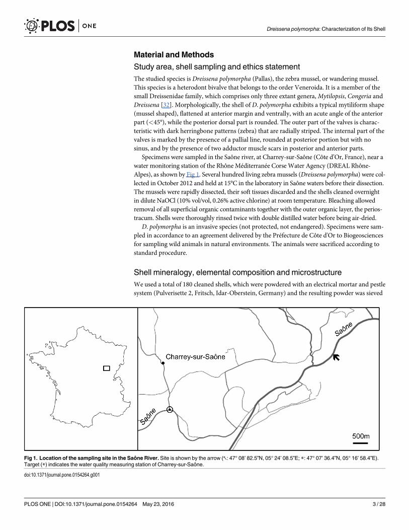
Specimens were sampled in the Saône river, at Charrey-sur-Saône (Côte d’Or, France), near awater monitoring station of the Rhône Méditerranée Corse Water Agency (DREAL Rhône-Alpes), as shown by Fig 1. Several hundred living zebra mussels (Dreissena polymorpha) were col-lected in October 2012 and held at 15°C in the laboratory in Saône waters before their dissection.The mussels were rapidly dissected, their soft tissues discarded and the shells cleaned overnightin dilute NaOCl (10% vol/vol, 0.26% active chlorine) at room temperature. Bleaching allowedremoval of all superficial organic contaminants together with the outer organic layer, the perios-tracum. Shells were thoroughly rinsed twice with double distilled water before being air-dried.
D. polymorpha is an invasive species (not protected, not endangered). Specimens were sam-pled in accordance to an agreement delivered by the Préfecture de Côte d'Or to Biogeosciencesfor sampling wild animals in natural environments. The animals were sacrificed according tostandard procedure.
Shell mineralogy, elemental composition and microstructureWe used a total of 180 cleaned shells, which were powdered with an electrical mortar and pestlesystem (Pulverisette 2, Fritsch, Idar-Oberstein, Germany) and the resulting powder was sieved
Fig 1. Location of the sampling site in the Saône River. Site is shown by the arrow (↖: 47° 08’ 82.5”N, 05° 24’ 08.5”E; ◉: 47° 07’ 36.4”N, 05° 16’ 58.4”E).Target (◉) indicates the water quality measuring station of Charrey-sur-Saône.
doi:10.1371/journal.pone.0154264.g001
Dreissena polymorpha: Characterization of Its Shell
PLOS ONE | DOI:10.1371/journal.pone.0154264 May 23, 2016 3 / 28

(200 μm) and homogenized. An aliquot of the shell powder was used for FT-IR characteriza-tion, in order to check the mineralogy of the shell (see below, § ‘Fourier Transform InfraRedspectroscopic characterization’ for further description).
Another aliquot was used for ICP-AES analysis. Minor and trace elements were determinedby FILAB (Dijon, France), according to an internal procedure. In brief, one gram of homogenizedcleaned powder was dissolved in 50 mL an aqueous solution containing in excess 3 mL of nitricacid and 9 mL of chlorhydric acid, and the solution was heated for ensuring complete dissolution.The solution was analysed on a ICP AES PERKIN 7300 system. In addition, the level of calciumwas determined by flame atomic absorption spectrometry (AAS 3300, AAnalyst 400 Perkin–Elmer, Rodgau, Germany) with an uncertainty of ±2% and a detection limit of 0.5 μM. In parallel,the physicochemical characteristics of the water column close to the shell sampling location wasobtained by DREAL Rhône-Alpes, as well as its heavy metal and metalloid contaminant content.Similarly, the content of the sediments in inorganic ions was also provided.
For microstructural investigation via scanning electron microscopy (SEM) observations,two shell valves (right or left) were included in Epoxy resin and the blocks were sliced (200 μm)with a saw microtome (Leica Biosystems, Newcastle, UK). Sections were parallel and perpen-dicular to the opening plan of the valves. The slices were then UV-glued on glass plates andpolished with alumina suspension (0.05 μm, ESCIL, Chassier, France). The preparations weresubsequent rinsed in an ultrasound bath, briefly etched with EDTA (1% wt/vol, 2min), rinsedwith water and dried. Shell microstructures were observed with a tabletop scanning electronmicroscopy (Hitachi TM1000) and with a Field Emission Scanning Electron Microscope(FE-SEM, JSM-7600 F). In this latter case, the preparations were carbon-coated (10 nm thick)with a Cressington 308R Desktop Advanced Coating System. A set of additional freshly frac-tured shells was directly observed with the tabletop SEM, without any specific preparation.
Matrix extractionThe shell powder was re-suspended in 10mL of distilled water and decalcified overnight byadding progressively cold dilute acetic acid (10% vol/vol) at a flow rate of 0.1 mL every 5 sec,according to our standardized procedure [33]. The final pH was 4.2. The clear solution wasthen centrifuged (3900 G, 30 min.). The supernatant containing the acid soluble matrix (ASM)was then filtered (5 μm) before being ultra-filtered for volume reduction on an Amicon cell(400 mL; cutoff 10kDa); the solution (10–15 mL) was then extensively dialyzed against Milli-Qwater for three days (6 water changes) and lyophilized. The pellet containing the acid insolublematrix (referred as AIM) was rinsed several times with Milli-Q water and lyophilized. In total,three batches of extracts (both AIM and ASM) were obtained. All the lyophilisates wereweighed on a precision balance (Precisa XT120A model) three to five times, and the meanvalue was calculated.
Fourier Transform InfraRed spectroscopic characterization (FT-IR)FT-IR spectroscopy was used for checking the shell mineralogy of Dreissena polymorpha, aswell as the overall chemical properties of the extracted matrices (ASM and AIM): to this end,in both cases, minute amounts of samples (shell powder or freeze-dried chips (< 1 mg) ofASM and of AIM) were analyzed with a Bruker Vector 22 instrument (Bruker Optics Sarl,Marne la Vallée, France) fitted with a Specac MKII Golden Gate Diamond Attenuated Totalreflectance (ATR) device equipped with ZnSe lenses (Specac Ltd, Orpington, UK) in the 4000–500 cm-1 wavenumber range (twelve scans at a spectral resolution of 4 cm-1). The qualitativeassignment of absorption bands was performed by comparison with previous spectra descrip-tions, achieved by our group or available in the bibliography [34, 35].
Dreissena polymorpha: Characterization of Its Shell
PLOS ONE | DOI:10.1371/journal.pone.0154264 May 23, 2016 4 / 28
Monosaccharide analysisThe monosaccharide content of AIM and ASM was obtained by suspension and homogeniza-tion of these lyophilisates in 2 M trifluoroacetic acid and subsequent hydrolysis at 105°C for 4h under nitrogen atmosphere. This hydrolytic procedure allows releasing most monosaccha-rides (neutral, aminated, acidic) from complex mixtures (without degrading them), exceptsialic acids, which are destroyed, and the acetylated forms of glucosamine and galactosamine,which are converted to their respective non-acetylated forms. Samples were then evaporated todryness before being dissolved in 100 μL of 20 mMNaOH. The neutral, amino and acidicsugar contents of hydrolysates were determined by High-Performance Anion-Exchange chro-matography with Pulsed-Amperometric-Detection (HPAE-PAD) on a CarboPac PA 100 col-umn (Dionex Corp., Sunnyvale, CA, USA). As blank controls, non-hydrolysed ASMs andAIMs were analysed, in order to detect potential free monosaccharides that may distort theresults and lead to an over-representation of some sugar residues.
Enzyme-Linked Lectin Assay (ELLA)The zebra mussel ASM was screened for its lectin-binding profile via Enzyme-Linked LectinAssay (ELLA) [36]. To this end, a set of 21 biotinylated lectins was used. Binding preferencesand specificities of each lectin were checked by a thorough survey of different bibliographicalsources [37–48]. The complete information on the binding specificities is summarized in theS1 Table.
The test is performed in 96-well plates (MaxiSorp, NuncTM, Roskilde, Denmark), as previ-ously described [49]: the zebra mussel ASM (50 ng/well) was incubated for 90 min at 37°C, fol-lowed by three washing steps with TBS/Tween20. The wells were then blocked with 1Xconcentrated Carbo-free blocking solution (Vector Laboratories, Orton Southgate, Peterbor-ough, UK; ref. SP-5040) for 60 min at 37°C. The lectins (Vector Laboratories, Orton Southgate,Peterborough, UK; ref. BK-1000, -2000, -3000) were all diluted 1:100 from the stock solutions(2 mg/mL) with TBS/Tween20 and incubated into the microplate for 90 min at 37°C. Afterthree washes with TBS/Tween20, Avidin-Alkaline Phosphatase (Avidin-AP, Sigma A7294,St. Louis, MO, USA) was used in a dilution of 1:70000 for 90 min at 37°C for the detection ofbound biotinylated lectins. The wells were then washed five times with TBS/Tween20. ELISAsubstrate solution (aqueous diethanolamine, 9.7% vol/vol, pH adjusted to 9.8 with HCl) con-taining Phosphatase Substrate (0.5 mg/mL, pNPP Tablet, Sigma, St. Louis, MO, USA) wasadded to the wells (100 μL/well) and incubated at 37°C. The microplate was read at 405 nmevery 15 min (BioRad Model 680). Different control experiments were included in the test:check of the background signal without ASM, lectin or Avidin-AP; negative control with ASMand lectin, but without Avidin-AP; negative control with ASM, without lectin, but with Avi-din-AP. None of the negative controls showed a reaction with the substrate solution. The testwas performed with triplicates of each lectin. The results were normalized and translated inpercentage of reactivity by subtracting the negative control (ASM without lectin but with Avi-din-AP) of all values and considering the highest response (Wheat germ agglutinin, WGA) as100%. The standard deviation was calculated from the variation of the triplicate measurements.The test was performed twice.
In vitro functional assays: inhibition and crystallizationThe effect of the zebra mussel ASM on calcium carbonate was tested according to two proce-dures: inhibition and interference (crystallization) assays. In the first case, the ASM waschecked for its capacity to inhibit the in vitro precipitation of calcium carbonate [50]. Two mLof 20 mM CaCl2 were rapidly added to 2 mL of 20 mMNaHCO3 containing variable amounts
Dreissena polymorpha: Characterization of Its Shell
PLOS ONE | DOI:10.1371/journal.pone.0154264 May 23, 2016 5 / 28
of protein extract (10, 20 and 30 μg). For each experiment, the pH was recorded with a miniglass electrode (6mm, Mettler-Toledo) coupled to a computer-connected pH-meter (Crison,GLP21). The pH was measured in continuous during 800 sec. Each ASM concentration wastested in duplicate. Between each experiment with a shell matrix aliquot, the electrode wasrefreshed with 0.5 M HCl, and negative controls were performed in the absence of protein, tocheck the stability of the electrode response.
In the second case, i.e., interference (crystallization) assay, the ASM was tested for its capac-ity to interfere with the growth of calcium carbonate crystals. The test was derived from thatpublished by Albeck and coworkers [51]. In brief, calcite crystals were grown by slow diffusionof ammonium bicarbonate vapours into a CaCl2 solution (200 μL). The following modificationwas applied: the 10 mM CaCl2 solutions containing different quantities of ASM (serial dilu-tions from 25 μg/mL to 0.39 μg/mL) were incubated in 16-well culture slides (Lab-Tek,NuncTM, Roskilde, Denmark). The slides were closed with their plastic cover, and sealed withparafilm. Prior to the experiments, the cover was pierced (1 mm diameter holes) in the middleof each well, to allow diffusion of the vapours only through the holes. The slides were placed ina 5 L closed desiccator containing crystals of ammonium bicarbonate, and incubated at 4°C for48 h. Blank tests (without any matrix) were performed in a similar manner. The solution wasthen gently removed using a blunt-ended syringe needle connected to a vacuum pump, and theculture slide was incubated further at 37°C. The glass slides were mechanically dissociatedfrom the plastic wells and directly observed (without carbon sputtering) with a tabletop SEM(Hitachi TM1000). The assay was repeated four times.
Gel electrophoresisMono-dimensional gels. The shell extracts were analysed by conventional mono-dimen-
sional SDS-PAGE (Bio-Rad, Mini Protean III gels), on precast gradient gels (Mini-PROTEANTGX Gel 4–20% acrylamide, 90 mm x 70 mm, BioRad). Prior migration, the samples weretreated as follows: lyophilisates of the ASM were dissolved in 2X Laemmli Sample Buffer (LSB)to a final matrix concentration of 5 μg/μL. One lyophilisate of the AIM was suspended in 2XLSB (5 mg in 500 μL). Both preparations were heat-denatured at 100°C: 5 min. for the ASMand 10 min. for the AIM. The LSB-solubilized fraction of the AIM is referred as LS-AIM(Laemmli soluble, acetic acid insoluble). The preparations were cooled down and gently centri-fuged before being applied on the top of the gel: 20 μg of ASM and 50 μg of LS-AIM wereapplied, respectively. After migration, the gels were stained with Instant Blue Coomassie (Expe-deon, Harston, UK), with silver nitrate [52], with Alcian Blue at low pH (pH 1) [33–53] andwith ‘Stains-all’ [54]. While silver nitrate is supposed to stain most of the macromolecular com-ponents of the shell, Alcian Blue in acidic conditions stains mostly sulphate groups of polyanio-nic polysaccharides. ‘Stains-all’ stains blue the putative calcium-binding proteins, while leavingthe non-calcium-binding proteins red or pink.
Bi-dimensional gels. ASM and AIM lyophilisates were prepared according to the manu-facturer’s instructions. The samples were migrated on a 2D-gel Protean IEF cell (Bio-Rad), inthe first dimension on strips before being fractionated on the second dimension on 4–20% gels.More precisely, a 7 cm linear pH 3–10 immobilized pH gradient IPG strip (ReadyStrip,BioRad) was re-hydrated overnight with 200 μg of protein sample in rehydration buffer (8Murea, 2M thiourea, 2% w/v CHAPS, 10mM DTT, 0.5% v/v pH 3–10 IPG buffer, 0.6% DeStreakreagent) and IEF was processed at 20°C (250 V for one hour, then a gradient voltage wasapplied to reach 4000 V in 3 hours followed by a constant step at 4000 V until 10,000 Vh). Thestrip was transferred successively in each two equilibration buffers, before being rinsed in TGSbuffer (25mM Tris-HCl pH8.3, 192 mM glycine 0.1% SDS), positioned on top of precast
Dreissena polymorpha: Characterization of Its Shell
PLOS ONE | DOI:10.1371/journal.pone.0154264 May 23, 2016 6 / 28
gradient gel (Mini-PROTEAN TGX Gel 4–20% acrylamide, 90 mm x 70 mm, BioRad), andfixed in place with an overlay solution of 0.5% agarose ⁄TGS (w/v). The ASM gel was stainedwith Instant Blue Coomassie. Because of the low amount of materials obtained from the AIM,the gel was stained with silver nitrate [52].
MS/MSMS/MS analyses were conducted on the two unfractionated bulk matrices, ASM and AIM,which were digested in-solution and in-gel, after a short migration in acrylamide gel. In-geldigestions were carried out with trypsin according to a published procedure with minor adjust-ments [55–56]: samples were destained twice with a mixture of 100 mM ammonium bicarbon-ate (ABC) and 50% (v/v) acetonitrile (ACN) for 45 min at 22°C and then dehydrated using100% ACN for 15 min, before being reduced with 25 mM ABC containing 10 mM DTT for 1 hat 60°C and alkylated with 55 mM iodoacetamide in 25 mM ABC for 30 min in the dark at22°C. Gel pieces were washed twice with 25 mM ABC and dehydrated (twice, 15 min) anddried (10 min) with 100% ACN. Gel cubes were incubated with sequencing grade modifiedtrypsin (Promega, USA; 12.5 ng/μl in 40 mM ABC with 10% ACN, pH 8.0) overnight at 37°C.After digestion, peptides were washed with 25 mM ABC, dehydrated with 100% ACN andextracted twice with a mixture of 50% ACN–5% formic acid (FA). Extracts were dried using avacuum centrifuge Concentrator plus (Eppendorf).
For in-solution digestion, the protocol was the same as in-gel digestion, with minor differ-ences: no destain, no washes and no extraction but with reduction, alkylation and digestionwith sequencing grade modified trypsin (Promega, USA; 0.1 μg/μl in 40 mM ABC with 10%ACN, pH 8.0). Extracts were dried as above and peptides were cleaned with ziptips C18 (Milli-pore) before MS analysis.
For MS and MS/MS ORBITRAP, analyses were performed using an Ultimate 3000 RapidSeparation Liquid Chromatographic (RSLC) system (Thermo Fisher Scientific) online with ahybrid LTQ-Orbitrap-Velos mass spectrometer (Thermo Fisher Scientific). Briefly, peptideswere dissolved in 4 μL of 10% ACN-0.1% FA. Peptides were loaded and washed on a C18
reverse phase pre-column (3 μm particle size, 100 Å pore size, 150 μm i.d., 1 cm length). Theloading buffer contained 98% H2O, 2% ACN and 0.1% TFA. Peptides were then separated on aC18 reverse phase resin (2 μm particle size, 100 Å pore size, 75 μm i.d., 15 cm length) with a 1hour gradient from 100% A (0.1% FA and 100% H2O) to 50% B (80% ACN, 0.085% FA and20% H2O).
The Linear Trap Quadrupole Orbitrap mass spectrometer acquired data throughout the elu-tion process and operated in a data dependent scheme with full MS scans acquired with theOrbitrap, followed by up to 20 LTQMS/MS CID spectra on the most abundant ions detectedin the MS scan. Mass spectrometer settings were: full MS (AGC: 1�106, resolution: 6�104, m/zrange 400–2000, maximum ion injection time: 500 ms); MS/MS (AGC: 5�103, maximum injec-tion time: 20 ms, minimum signal threshold: 500, isolation width: 2 Da, dynamic exclusiontime setting: 30 s). The fragmentation was permitted of precursor with a charge state of 2, 3, 4and up. For the spectral processing, the software used to generate.mgf files is Proteome discov-erer 1.3. The threshold of Signal to Noise for extraction values is 3.
Database searches were carried out using Mascot version 2.4 and 2.5 (MatrixScience, Lon-don, UK) on ‘Other Metazoa’ proteins (2,282,777 sequences) and on ‘Other Eukaryota’ pro-teins (1,030,911 sequences) from NCBInr databank containing 67,337,701 residues (May 2015)(www.ncbi.nlm.nih.gov/). A fusion database merging a homemade shell proteins databank(762 sequences containing 220545 residues) and three additional databases (DreissenaExpressed Sequence Tags (ESTs) extracted from NCBI; the translated Bathymodiolus azoricus,
Dreissena polymorpha: Characterization of Its Shell
PLOS ONE | DOI:10.1371/journal.pone.0154264 May 23, 2016 7 / 28
Pecten maximus and Laternula elliptica transcriptomes) described in an earlier publication[57] was also checked. Two specific proteins databank—one from the sea urchin Paracentrotuslividus with restricted access (see http://octopus.obs-vlfr.fr/), constructed and provided byLaboratoire de Biologie du Développement, UMR CNRS 7009, Villefranche sur Mer–and thesecond one–from the genus Crassostrea (62,050 sequences)–were also included in the in silicoanalysis. The search parameters were as follows: carbamidomethylation as a variable modifica-tion for cysteins and oxidation as a variable modification for methionines. Up to 3 missed tryp-tic cleavages were tolerated and mass accuracy tolerance of 10 ppm for precursors and 0.45 Dafor fragments were used for all tryptic mass searches. The protein score should be above 50 for‘Other Metazoa’ and ‘Other Eukaryota’ databases and 40 for the other databases while the indi-vidual ions score was above 20 for all databases.
Results
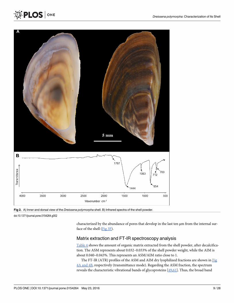
Mineralogy and chemistry of the shell of Dreissena polymorphaWe verified that the shell of Dreissena polymorpha is fully aragonitic (Fig 2B). The typicalabsorption bands of aragonite type structure are highlighted by the four characteristic vibrationmodes of CO3
2-: ν3(1445 cm-1), ν1(1082 cm
-1), ν2(854 cm-1) and ν4(712–699 cm
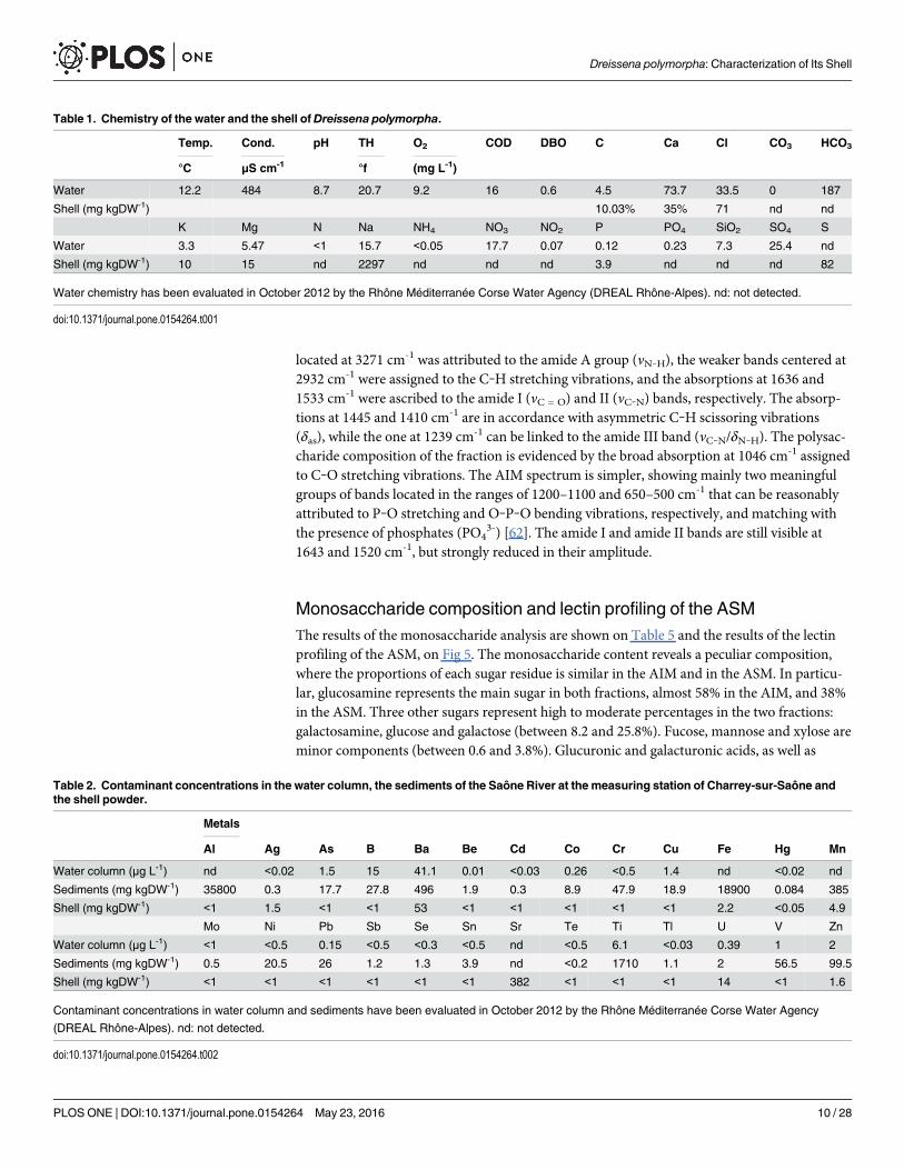
-1) [58].Tables 1, 2 and 3 show the elemental composition of the shell, the water column and the
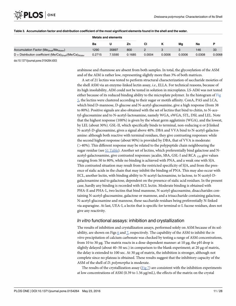
sediments. Most of the elements are present in low concentrations in the shell: except Ba(53 ppm), Sr (382 ppm) and U (14 ppm) (Table 2). Some elements are depleted in the shell butconcentrated in the sediments (B, As, Co, Cu, Mn, Ti, Ni and Pb) with the exception of ura-nium (Table 2), which is concentrated in the shell relative to the sediment with an accumula-tion factor of 35879 (Table 3). The most noticeable data is the concentration factor of uraniumin the shell.
MicrostructureFig 3 depicts the different microstructures observed in transverse section throughout the thick-ness of the shell (Fig 3A). The description below goes from top (outer) to bottom (internal).The uppermost layer is thin (< 10 μm, Fig 3B) and constitutes columnar chevron-like (i.e.,‘herringbone’) patterns, each column, of about 3–5 μm thick, being developed perpendicularlyto the outer surface. This layer corresponds to the simple lamellar structure described by Arch-ambault-Guezou [59]. Below this layer, one finds a zone of irregularly distributed pores (diam-eter about 200–300 nm). This transition layer exhibits a variable thickness: from a fewmicrometres (Fig 3B, left part) to more than 15 μm (Fig 3B, right part). In the larger part, poresare organized in successive plans (three or more), between which there are either sub-chevronpatterns or spherulites. Below this transition layer, the main layer has a typical crossed-lamellarstructure, characterized by alternating fibre bundles developed in two different plans (Fig 3C).At high magnification, each fibre (diameter about 200–300 nm), when slightly etched withEDTA, constitutes alignments of submicrometric granules (size from 100 to 300 nm, Fig 3D).The predominant crossed-lamellar layer is separated from the subjacent layer by a zone oflesser resistance, (marked by a fracture, Fig 3E), where the microstructure changes: this zone—4–5 μm thick—is characterized by irregular block-like rectangular to fusiform crystals. It isdescribed by Archambault-Guezou as the prismatic aggregate myostracal layer with a fibrousfracture pattern [59]. The underlying layer is the so-called complex crossed-lamellar, accordingto the description of Taylor et al. [60]. It is composed of thin fibres reminiscent of the crossed-lamellar layer, although the absence of general emerging patterns and the multiple orientationsof the fibres make it more difficult to describe (Fig 3E). The basal part of this layer is
Dreissena polymorpha: Characterization of Its Shell
PLOS ONE | DOI:10.1371/journal.pone.0154264 May 23, 2016 8 / 28
characterized by the abundance of pores that develop in the last ten μm from the internal sur-face of the shell (Fig 3F).
Matrix extraction and FT-IR spectroscopy analysisTable 4 shows the amount of organic matrix extracted from the shell powder, after decalcifica-tion. The ASM represents about 0.032–0.033% of the shell powder weight, while the AIM isabout 0.040–0.043%. This represents an ASM/AIM ratio close to 1.
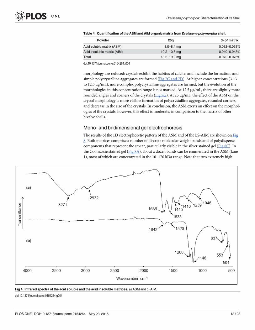
The FT-IR (ATR) profiles of the ASM and AIM dry lyophilized fractions are shown in Fig4A and 4B, respectively (transmittance mode). Regarding the ASM fraction, the spectrumreveals the characteristic vibrational bands of glycoproteins [49,61]. Thus, the broad band
Fig 2. A) Inner and dorsal view of the Dreissena polymorpha shell. B) Infrared spectra of the shell powder.
doi:10.1371/journal.pone.0154264.g002
Dreissena polymorpha: Characterization of Its Shell
PLOS ONE | DOI:10.1371/journal.pone.0154264 May 23, 2016 9 / 28
located at 3271 cm-1 was attributed to the amide A group (νN‒H), the weaker bands centered at2932 cm-1 were assigned to the C‒H stretching vibrations, and the absorptions at 1636 and1533 cm-1 were ascribed to the amide I (νC = O) and II (νC‒N) bands, respectively. The absorp-tions at 1445 and 1410 cm-1 are in accordance with asymmetric C‒H scissoring vibrations(δas), while the one at 1239 cm
-1 can be linked to the amide III band (νC‒N/δN‒H). The polysac-charide composition of the fraction is evidenced by the broad absorption at 1046 cm-1 assignedto C‒O stretching vibrations. The AIM spectrum is simpler, showing mainly two meaningfulgroups of bands located in the ranges of 1200–1100 and 650–500 cm-1 that can be reasonablyattributed to P‒O stretching and O‒P‒O bending vibrations, respectively, and matching withthe presence of phosphates (PO4
3-) [62]. The amide I and amide II bands are still visible at1643 and 1520 cm-1, but strongly reduced in their amplitude.
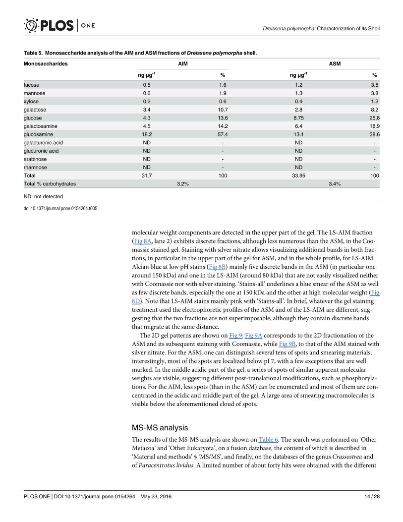
Monosaccharide composition and lectin profiling of the ASMThe results of the monosaccharide analysis are shown on Table 5 and the results of the lectinprofiling of the ASM, on Fig 5. The monosaccharide content reveals a peculiar composition,where the proportions of each sugar residue is similar in the AIM and in the ASM. In particu-lar, glucosamine represents the main sugar in both fractions, almost 58% in the AIM, and 38%in the ASM. Three other sugars represent high to moderate percentages in the two fractions:galactosamine, glucose and galactose (between 8.2 and 25.8%). Fucose, mannose and xylose areminor components (between 0.6 and 3.8%). Glucuronic and galacturonic acids, as well as
Table 1. Chemistry of the water and the shell ofDreissena polymorpha.
Temp. Cond. pH TH O2 COD DBO C Ca Cl CO3 HCO3
°C μS cm-1 °f (mg L-1)
Water 12.2 484 8.7 20.7 9.2 16 0.6 4.5 73.7 33.5 0 187
Shell (mg kgDW-1) 10.03% 35% 71 nd nd
K Mg N Na NH4 NO3 NO2 P PO4 SiO2 SO4 S
Water 3.3 5.47 <1 15.7 <0.05 17.7 0.07 0.12 0.23 7.3 25.4 nd
Shell (mg kgDW-1) 10 15 nd 2297 nd nd nd 3.9 nd nd nd 82
Water chemistry has been evaluated in October 2012 by the Rhône Méditerranée Corse Water Agency (DREAL Rhône-Alpes). nd: not detected.
doi:10.1371/journal.pone.0154264.t001
Table 2. Contaminant concentrations in the water column, the sediments of the Saône River at the measuring station of Charrey-sur-Saône andthe shell powder.
Metals
Al Ag As B Ba Be Cd Co Cr Cu Fe Hg Mn
Water column (μg L-1) nd <0.02 1.5 15 41.1 0.01 <0.03 0.26 <0.5 1.4 nd <0.02 nd
Sediments (mg kgDW-1) 35800 0.3 17.7 27.8 496 1.9 0.3 8.9 47.9 18.9 18900 0.084 385
Shell (mg kgDW-1) <1 1.5 <1 <1 53 <1 <1 <1 <1 <1 2.2 <0.05 4.9
Mo Ni Pb Sb Se Sn Sr Te Ti Tl U V Zn
Water column (μg L-1) <1 <0.5 0.15 <0.5 <0.3 <0.5 nd <0.5 6.1 <0.03 0.39 1 2
Sediments (mg kgDW-1) 0.5 20.5 26 1.2 1.3 3.9 nd <0.2 1710 1.1 2 56.5 99.5
Shell (mg kgDW-1) <1 <1 <1 <1 <1 <1 382 <1 <1 <1 14 <1 1.6
Contaminant concentrations in water column and sediments have been evaluated in October 2012 by the Rhône Méditerranée Corse Water Agency
(DREAL Rhône-Alpes). nd: not detected.
doi:10.1371/journal.pone.0154264.t002
Dreissena polymorpha: Characterization of Its Shell
PLOS ONE | DOI:10.1371/journal.pone.0154264 May 23, 2016 10 / 28
arabinose and rhamnose are absent from both samples. In total, the glycosylation of the ASMand of the AIM is rather low, representing slightly more than 3% of both matrices.
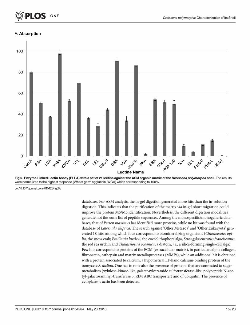
A set of 21 lectins was tested to perform structural characterization of saccharide moieties ofthe shell ASM via an enzyme-linked lectin assay, i.e., ELLA. For technical reasons, because ofits high insolubility, AIM could not be tested in solution in microplates. LS-AIM was not testedeither because of its reduced binding ability to the microplate polymer. In the histogram of Fig5, the lectins were clustered according to their sugar or motifs affinity. ConA, PAS and LCA,which bind D-mannose, D-glucose and N-acetyl-glucosamine, give a high response (from 38to 80%). Positive signals are also obtained with the set of lectins that bind to chitin, to N-ace-tyl-glucosamine and to N-acetyl-lactosamine, namely WGA, sWGA, STL, DSL and LEL. Notethat the highest response (100%) is given by the wheat germ agglutinin (WGA), and the lowest,by LEL (about 30%). GSL-II, which specifically binds to terminal, non-reducing α or β linkedN-acetyl-D-glucosamine, gives a signal above 40%. DBA and VVA bind to N-acetyl-galactos-amine: although both reactive with terminal residues, they give contrasting responses: whilethe second highest response (about 90%) is provided by DBA, that of VVA is moderate:(>40%). This different response may be related to the polypeptide chain neighbouring thesugar residue (see S1 Table). Another set of lectins, which preferentially bind galactose and N-acetyl-galactosamine, give contrasted responses: jacalin, SBA, GSL-I and RCA 120 give valuesranging from 50 to 80%, while no binding is achieved with PNA, and a weak one with SJA.This contrasted situation may result from the restricted specificity of SJA, and from the pres-ence of sialic acids in the chain that may inhibit the binding of PNA. This may also occur withECL, another lectin, with binding ability to N-acetyl-lactosamine, to lactose, to N-acetyl-D-galactosamine and to galactose, dependent on the presence of sialic acid residues. In the presentcase, hardly any binding is recorded with ECL lectin. Moderate binding is obtained withPHA-E and PHA-L, two lectins that bind mannose, N-acetyl-glucosamine, disaccharides con-taining N-acetyl-glucosamine, galactose or mannose, and a trisaccharide containing galactose,N-acetyl-glucosamine and mannose, these saccharide residues being preferentially N-linkedvia asparagine. At last, UEA-I, a lectin that is specific for terminal α-L fucose residues, does notgive any reactivity.
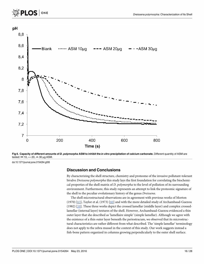
In vitro functional assays: inhibition and crystallizationThe results of inhibition and crystallization assays, performed solely on ASM because of its sol-ubility, are shown on Figs 6 and 7, respectively. The capability of the ASM to inhibit the invitro precipitation of calcium carbonate was checked by testing a range of ASM concentrations,from 10 to 30 μg. The matrix reacts in a dose-dependent manner: at 10 μg, the pH drop isslightly delayed (about 40–50 sec.) in comparison to the blank experiment; at 20 μg of matrix,the delay is extended to 100 sec. At 30 μg of matrix, the inhibition is stronger, although notcomplete since no plateau is obtained. These results suggest that the inhibitory capacity of theASM of the shell of D. polymorpha is moderate.
The results of the crystallization assay (Fig 7) are consistent with the inhibition experimentsat low concentrations of ASM (0.39 to 1.56 μg/mL), the effects of the matrix on the crystal
Table 3. Accumulation factor and distribution coefficient of the most significant elements found in the shell and the water.
Metals and elements
Ba U Zn Cl K Mg Na P
Accumulation Factor (MeShell/MeWater) 1290 35897 800 2 3 3 146 33
D = Distribution coefficient (Me/Ca)Shell/(Me/Ca)Water 0.2715 7.5590 0.1685 0.0004 0.0006 0.0006 0.0308 0.0068
doi:10.1371/journal.pone.0154264.t003
Dreissena polymorpha: Characterization of Its Shell
PLOS ONE | DOI:10.1371/journal.pone.0154264 May 23, 2016 11 / 28

Fig 3. Secondary electron images of the shell microstructure ofDreissena polymorpha. Black boxes show location of the other views. A) Transversalsection. B) Thin uppermost layer constitutes chevron-like columns. C) Crossed-lamellar layer. D) Crossed-lamellar fibres at high magnification. E) Crossed-lamellar layer based on a prismatic aggregate myostracal layer. The lower layer of the view is a complex crossed-lamellar layer. F) Innermost layer of theshell.
doi:10.1371/journal.pone.0154264.g003
Dreissena polymorpha: Characterization of Its Shell
PLOS ONE | DOI:10.1371/journal.pone.0154264 May 23, 2016 12 / 28
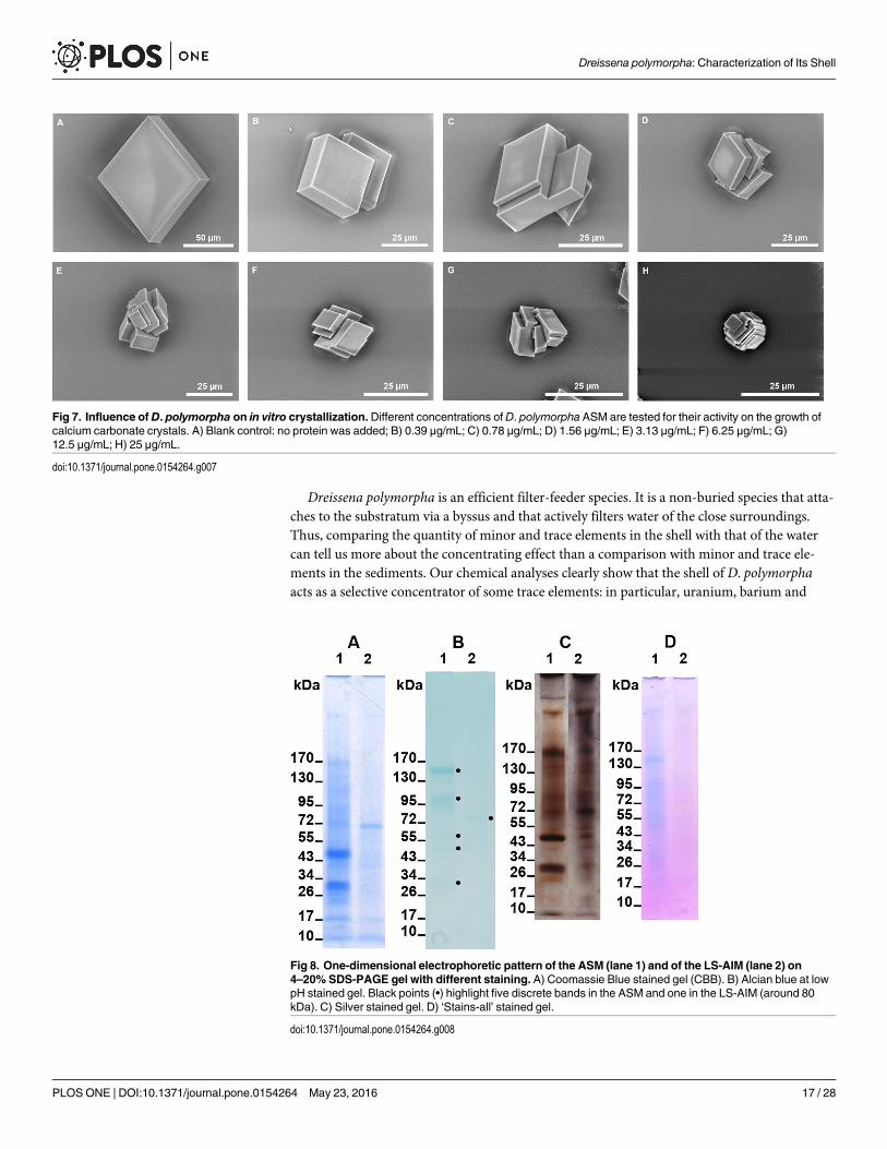
morphology are reduced: crystals exhibit the habitus of calcite, and include the formation, andsimple polycrystalline aggregates are formed (Fig 7C and 7D). At higher concentrations (3.13to 12.5 μg/mL), more complex polycrystalline aggregates are formed, but the evolution of themorphologies in this concentration range is not marked. At 12.5 μg/mL, there are slightly morerounded angles and corners of the crystals (Fig 7G). At 25 μg/mL, the effect of the ASM on thecrystal morphology is more visible: formation of polycrystalline aggregates, rounded corners,and decrease in the size of the crystals. In conclusion, the ASM exerts an effect on the morphol-ogies of the crystals; however, this effect is moderate, in comparison to the matrix of otherbivalve shells.
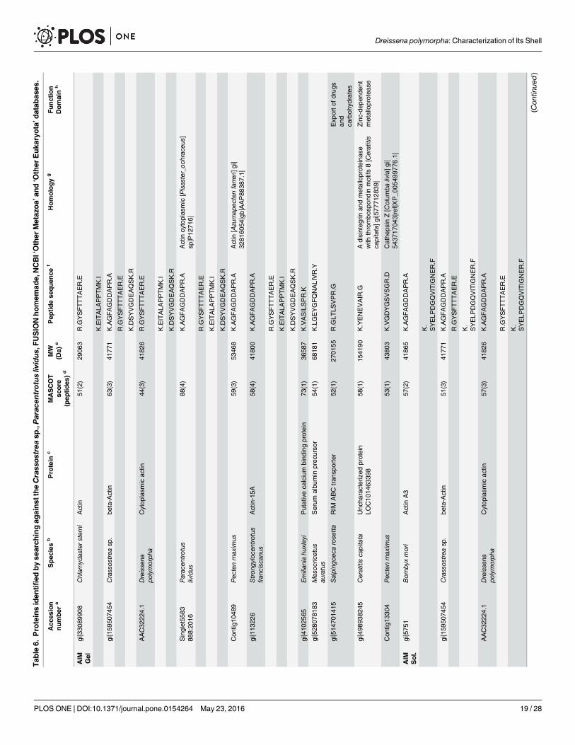
Mono- and bi-dimensional gel electrophoresisThe results of the 1D electrophoretic pattern of the ASM and of the LS-AIM are shown on Fig8. Both matrices comprise a number of discrete molecular weight bands and of polydispersecomponents that represent the smear, particularly visible in the silver stained gel (Fig 8C). Inthe Coomassie stained gel (Fig 8A), about a dozen bands can be enumerated in the ASM (lane1), most of which are concentrated in the 10–170 kDa range. Note that two extremely high
Table 4. Quantification of the ASM and AIM organic matrix fromDreissena polymorpha shell.
Powder 25g % of matrix
Acid soluble matrix (ASM) 8.0–8.4 mg 0.032–0.033%
Acid insoluble matrix (AIM) 10.2–10.8 mg 0.040–0.043%
Total 18.2–19.2 mg 0.072–0.076%
doi:10.1371/journal.pone.0154264.t004
Fig 4. Infrared spectra of the acid soluble and the acid insoluble matrices. a) ASM and b) AIM.
doi:10.1371/journal.pone.0154264.g004
Dreissena polymorpha: Characterization of Its Shell
PLOS ONE | DOI:10.1371/journal.pone.0154264 May 23, 2016 13 / 28
molecular weight components are detected in the upper part of the gel. The LS-AIM fraction(Fig 8A, lane 2) exhibits discrete fractions, although less numerous than the ASM, in the Coo-massie stained gel. Staining with silver nitrate allows visualizing additional bands in both frac-tions, in particular in the upper part of the gel for ASM, and in the whole profile, for LS-AIM.Alcian blue at low pH stains (Fig 8B) mainly five discrete bands in the ASM (in particular onearound 150 kDa) and one in the LS-AIM (around 80 kDa) that are not easily visualized neitherwith Coomassie nor with silver staining. ‘Stains-all’ underlines a blue smear of the ASM as wellas few discrete bands, especially the one at 150 kDa and the other at high molecular weight (Fig8D). Note that LS-AIM stains mainly pink with ‘Stains-all’. In brief, whatever the gel stainingtreatment used the electrophoretic profiles of the ASM and of the LS-AIM are different, sug-gesting that the two fractions are not superimposable, although they contain discrete bandsthat migrate at the same distance.
The 2D gel patterns are shown on Fig 9: Fig 9A corresponds to the 2D fractionation of theASM and its subsequent staining with Coomassie, while Fig 9B, to that of the AIM stained withsilver nitrate. For the ASM, one can distinguish several tens of spots and smearing materials;interestingly, most of the spots are localized below pI 7, with a few exceptions that are wellmarked. In the middle acidic part of the gel, a series of spots of similar apparent molecularweights are visible, suggesting different post-translational modifications, such as phosphoryla-tions. For the AIM, less spots (than in the ASM) can be enumerated and most of them are con-centrated in the acidic and middle part of the gel. A large area of smearing macromolecules isvisible below the aforementioned cloud of spots.
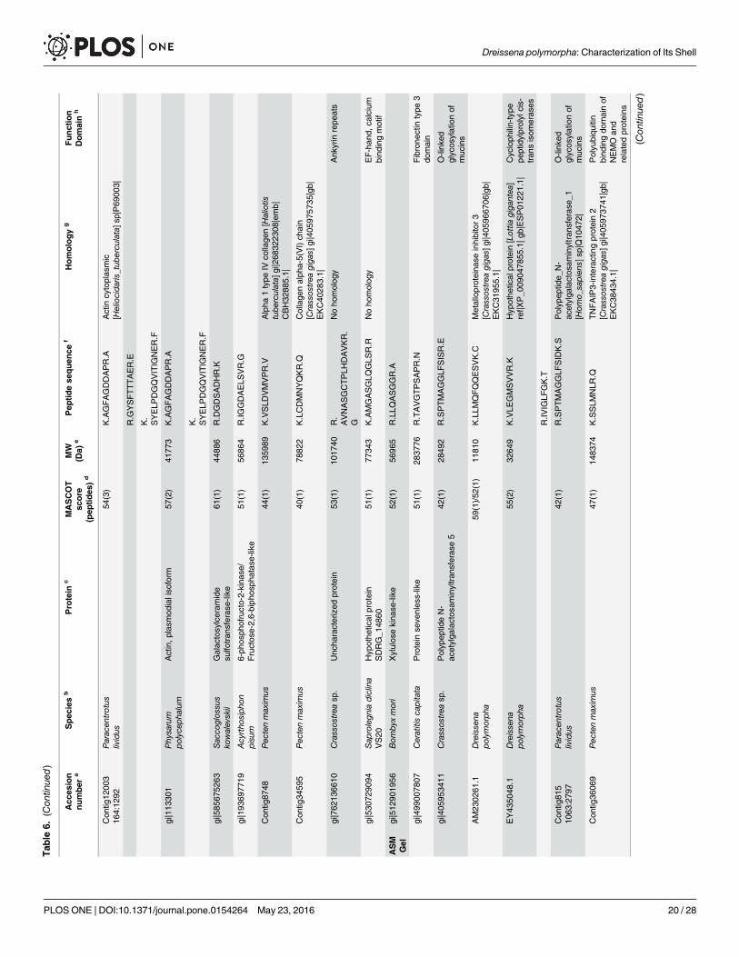
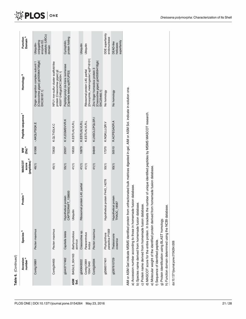
MS-MS analysisThe results of the MS-MS analysis are shown on Table 6. The search was performed on ‘OtherMetazoa’ and ‘Other Eukaryota’, on a fusion database, the content of which is described in‘Material and methods’ § ‘MS/MS’, and finally, on the databases of the genus Crassostrea andof Paracentrotus lividus. A limited number of about forty hits were obtained with the different
Table 5. Monosaccharide analysis of the AIM and ASM fractions ofDreissena polymorpha shell.
Monosaccharides AIM ASM
ng μg-1 % ng μg-1 %
fucose 0.5 1.6 1.2 3.5
mannose 0.6 1.9 1.3 3.8
xylose 0.2 0.6 0.4 1.2
galactose 3.4 10.7 2.8 8.2
glucose 4.3 13.6 8.75 25.8
galactosamine 4.5 14.2 6.4 18.9
glucosamine 18.2 57.4 13.1 38.6
galacturonic acid ND - ND -
glucuronic acid ND - ND -
arabinose ND - ND -
rhamnose ND - ND -
Total 31.7 100 33.95 100
Total % carbohydrates 3.2% 3.4%
ND: not detected
doi:10.1371/journal.pone.0154264.t005
Dreissena polymorpha: Characterization of Its Shell
PLOS ONE | DOI:10.1371/journal.pone.0154264 May 23, 2016 14 / 28
databases. For ASM analysis, the in-gel digestion generated more hits than the in-solutiondigestion. This indicates that the purification of the matrix via in-gel short migration couldimprove the protein MS/MS identification. Nevertheless, the different digestion modalitiesgenerate not the same list of peptide sequences. Among the monospecific/monogeneric data-bases, that of Pecten maximus has identified more proteins, while no hit was found with thedatabase of Laternula elliptica. The search against ‘Other Metazoa’ and ‘Other Eukaryota’ gen-erated 18 hits, among which four correspond to biomineralizing organisms (Chionoecetes opi-lio, the snow crab; Emiliania huxleyi, the coccolithophore alga, Strongylocentrotus franciscanus,the red sea urchin and Thalassiosira oceanica, a diatom, i.e., a silica-forming single-cell alga).Few hits correspond to proteins of the ECM (extracellular matrix), in particular, alpha collagen,fibronectin, cathepsin and matrix metalloproteases (MMPs), while an additional hit is obtainedwith a protein associated to calcium, a hypothetical EF-hand calcium-binding protein of theoomycete S. diclina. One has to note also the presence of proteins that are connected to sugarmetabolism (xylulose-kinase-like, galactosylceramide sulfotransferase-like, polypeptide N-ace-tyl-galactosaminyl-transferase 5, RIM ABC transporter) and of ubiquitin. The presence ofcytoplasmic actin has been detected.
Fig 5. Enzyme-Linked Lectin Assay (ELLA) with a set of 21 lectins against the ASM organic matrix of theDreissena polymorpha shell. The resultswere normalized to the highest response (Wheat germ agglutinin, WGA) which corresponding to 100%.
doi:10.1371/journal.pone.0154264.g005
Dreissena polymorpha: Characterization of Its Shell
PLOS ONE | DOI:10.1371/journal.pone.0154264 May 23, 2016 15 / 28
Discussion and ConclusionsBy characterizing the shell structure, chemistry and proteome of the invasive pollutant-tolerantbivalve Dreissena polymorpha this study lays the first foundation for correlating the biochemi-cal properties of the shell matrix of D. polymorpha to the level of pollution of its surroundingenvironment. Furthermore, this study represents an attempt to link the proteomic signature ofthe shell to the peculiar evolutionary history of the genus Dreissena.
The shell microstructural observations are in agreement with previous works of Morton(1970) [63], Taylor et al. (1973) [60] and with the more detailed study of Archambaud-Guezou(1982) [59]. These three works depict the crossed lamellar (middle layer) and complex crossed-lamellar (internal layer) textures of the shell. However, Archambaud-Guezou evidenced a thinouter layer that she described as ‘lamellaire simple’ (simple lamellar). Although we agree withthe existence of a thin outer layer beneath the periostracum, we observed that its microstruc-tural characteristics are rather different from what described. The ‘simple lamellar’ terminologydoes not apply to the zebra mussel in the context of this study. Our work suggests instead afish-bone pattern organized in columns growing perpendicularly to the outer shell surface.
Fig 6. Capacity of different amounts ofD. polymorphaASM to inhibit the in vitro precipitation of calcium carbonate.Different quantity of ASM aretested: ••• 10, --- 20, -•- 30 μg ASM.
doi:10.1371/journal.pone.0154264.g006
Dreissena polymorpha: Characterization of Its Shell
PLOS ONE | DOI:10.1371/journal.pone.0154264 May 23, 2016 16 / 28
Dreissena polymorpha is an efficient filter-feeder species. It is a non-buried species that atta-ches to the substratum via a byssus and that actively filters water of the close surroundings.Thus, comparing the quantity of minor and trace elements in the shell with that of the watercan tell us more about the concentrating effect than a comparison with minor and trace ele-ments in the sediments. Our chemical analyses clearly show that the shell of D. polymorphaacts as a selective concentrator of some trace elements: in particular, uranium, barium and
Fig 7. Influence of D. polymorpha on in vitro crystallization.Different concentrations of D. polymorpha ASM are tested for their activity on the growth ofcalcium carbonate crystals. A) Blank control: no protein was added; B) 0.39 μg/mL; C) 0.78 μg/mL; D) 1.56 μg/mL; E) 3.13 μg/mL; F) 6.25 μg/mL; G)12.5 μg/mL; H) 25 μg/mL.
doi:10.1371/journal.pone.0154264.g007
Fig 8. One-dimensional electrophoretic pattern of the ASM (lane 1) and of the LS-AIM (lane 2) on4–20% SDS-PAGE gel with different staining. A) Coomassie Blue stained gel (CBB). B) Alcian blue at lowpH stained gel. Black points (•) highlight five discrete bands in the ASM and one in the LS-AIM (around 80kDa). C) Silver stained gel. D) ‘Stains-all’ stained gel.
doi:10.1371/journal.pone.0154264.g008
Dreissena polymorpha: Characterization of Its Shell
PLOS ONE | DOI:10.1371/journal.pone.0154264 May 23, 2016 17 / 28

zinc. Iron, manganese, strontium and silver are also concentrated in the shell although the con-centration factors are unknown due to the lack of data on the concentration of each of theseelements in the surrounding water. We leave aside the high content of sodium of the shell,which might result from a contamination by our bleaching treatment with NaOCl, in spite ofrinsing the shells with milli-Q water. For the other divalent cations considered above, at leastfour mechanisms can be inferred to explain their occurrence in the shell [64–66]: incorporationinto the crystal lattice (by substitution to calcium cations), adsorption on crystal planes viaelectrostatic interactions, entrapment in crystal defects or complexation with the organicmatrix associated to the shell. Although we cannot exclude that these four mechanisms com-bine to different degrees for each of the aforementioned elements, it is well-documented thatone of them occurs for aragonite: this mineral tolerates the incorporation of divalent cationssuch as Sr, Ba and U that have a larger diameter than calcium, in its crystal lattice [67–69]. Spe-cific and targeted studies should be performed to check the contribution of the three othermechanisms, in particular, the ability of the shell matrix to bind metal ions. To conclude onthese geochemical aspects, the fact that the shell of D. polymorpha concentrates uranium effi-ciently is an interesting property that may be exploited for the bioremediation of this radioac-tive pollutant in natural environments. However, one has to keep in mind that D. polymorphais an invasive species and using large quantities of this bivalve for depolluting rivers may bedetrimental to other aquatic organisms. Another consequence of the uranium concentratingeffect is that the non-controlled utilization of D. polymorpha as a daily food source for poultryshould be banished [70].
The concentration of organic matrix is, at slightly above 0.07% of the weight of the shell,low in comparison to nacro-prismatic shells [33,71] but is in agreement with previous findingson crossed-lamellar shell microstructures sensu lato [72–73]. From Palmer [74–75], such a loworganic content associated with crossed-lamellar shells would correspond to a low energetic
Fig 9. Two-dimensional electrophoretic patterns of the ASM (A) and of the AIM (B). Protein samples were loaded on a 7 cm linear pH 3–10 IPG strip andsecond dimension was performed with a precast 4–20% SDS-PAGE gel. ASM gel was stained with Coomassie Blue and AIM one with silver nitrate.
doi:10.1371/journal.pone.0154264.g009
Dreissena polymorpha: Characterization of Its Shell
PLOS ONE | DOI:10.1371/journal.pone.0154264 May 23, 2016 18 / 28
Tab
le6.
Proteinsiden
tified
byse
arch
ingag
ainst
theCrass
ostrea
sp.,Parac
entrotus
lividus
,FUSIO
Nhomem
ade,
NCBI‘Other
Metaz
oa’
and‘O
ther
Euka
ryota’datab
ases
.
Acc
esion
number
aSpec
ies
bProtein
cMASCOT
score
(pep
tides
)d
MW
(Da)
ePep
tidese
quen
cef
Homology
gFunction
Domain
h
AIM
Gel
gi|330
8990
8Chlam
ydastersterni
Actin
51(2)
2906
3R.G
YSFTTTAER.E
K.EITALA
PPTMK.I
gi|159
5074
54Crassos
trea
sp.
beta-Actin
63(3)
4177
1K.AGFAGDDAPR.A
R.G
YSFTTTAER.E
K.DSYVGDEAQSK.R
AAC32
224.1
Dreisse
napo
lymorph
aCytop
lasm
icac
tin44
(3)
4182
6R.G
YSFTTTAER.E
K.EITALA
PPTMK.I
K.DSYVGDEAQSK.R
Singlet55
8388
8:20
16Parac
entrotus
lividus
88(4)
K.AGFAGDDAPR.A
Actin
cytoplas
mic
[Pisaster_oc
hrac
eus]
sp|P12
716|
R.G
YSFTTTAER.E
K.EITALA
PPTMK.I
K.DSYVGDEAQSK.R
Con
tig10
489
Pec
tenmaxim
us59
(3)
5346
8K.AGFAGDDAPR.A
Actin
[Azumap
ectenfarreri]gi|
3281
6054
|gb|AAP88
387
.1|
gi|113
226
Stron
gyloce
ntrotus
fran
cisc
anus
Actin-15A
58(4)
4180
0K.AGFAGDDAPR.A
R.G
YSFTTTAER.E
K.EITALA
PPTMK.I
K.DSYVGDEAQSK.R
gi|410
2565
Emilian
iahu
xley
iPutativeca
lcium
bind
ingprotein
73(1)
3658
7K.VASILSPR.K
gi|528
0781
83Mes
ocric
etus
auratus
Serum
albu
min
prec
urso
r54
(1)
6818
1K.LGEYGFQNALIVR.Y
gi|514
7014
15Salping
oeca
rose
tta
RIM
ABC
tran
sporter
52(1)
2701
55R.G
LTLS
VPR.G
Exp
orto
fdrugs
and
carboh
ydrates
gi|498
9382
45Ceratitisca
pitata
Unc
haracterized
protein
LOC10
1463
398
58(1)
1541
90K.YENEVAIR.G
Adisinteg
rinan
dmetalloproteina
sewith
thrombo
spon
dinmotifs
8[Ceratitis
capitata]g
i|577
7128
39|
Zinc-de
pend
ent
metalloprotea
se
Con
tig13
304
Pec
tenmaxim
us53
(1)
4380
3K.VGDYGSVSGR.D
Cathe
psin
Z[Colum
balivia]g
i|54
3717
043|ref|X
P_0
054
9977
6.1|
AIM
Sol.
gi|575
1Bom
byxmori
Actin
A3
57(2)
4186
5K.AGFAGDDAPR.A
K.
SYELP
DGQVITIG
NER.F
gi|159
5074
54Crassos
trea
sp.
beta-Actin
51(3)
4177
1K.AGFAGDDAPR.A
R.G
YSFTTTAER.E
K.
SYELP
DGQVITIG
NER.F
AAC32
224.1
Dreisse
napo
lymorph
aCytop
lasm
icac
tin57
(3)
4182
6K.AGFAGDDAPR.A
R.G
YSFTTTAER.E
K.
SYELP
DGQVITIG
NER.F
(Con
tinue
d)
Dreissena polymorpha: Characterization of Its Shell
PLOS ONE | DOI:10.1371/journal.pone.0154264 May 23, 2016 19 / 28
Tab
le6.
(Con
tinue
d)
Acc
esion
number
aSpec
ies
bProtein
cMASCOT
score
(pep
tides
)d
MW
(Da)
ePep
tidese
quen
cef
Homology
gFunction
Domain
h
Con
tig12
003
164:12
92Parac
entrotus
lividus
54(3)
K.AGFAGDDAPR.A
Actin
cytoplas
mic
[Heliocida
ris_tub
ercu
lata]s
p|P69
003|
R.G
YSFTTTAER.E
K.
SYELP
DGQVITIG
NER.F
gi|113
301
Phy
sarum
polyce
phalum
Actin,p
lasm
odialiso
form
57(2)
4177
3K.AGFAGDDAPR.A
K.
SYELP
DGQVITIG
NER.F
gi|585
6752
63Sac
coglos
sus
kowalevskii
Galac
tosylceram
ide
sulfo
tran
sferas
e-like
61(1)
4488
6R.DGDSADHR.K
gi|193
6977
19Acy
rtho
siph
onpisu
m6-ph
osph
ofructo-2-kina
se/
Fructos
e-2,6-bipho
spha
tase
-like
51(1)
5686
4R.IG
GDAELS
VR.G
Con
tig87
48Pec
tenmaxim
us44
(1)
1359
89K.VSLD
VMVPR.V
Alpha
1type
IVco
llage
n[Haliotis
tube
rculata]
gi|268
3223
08|emb|
CBH32
885.1|
Con
tig34
595
Pec
tenmaxim
us40
(1)
7882
2K.LCDMNYQKR.Q
Collage
nalph
a-5(VI)ch
ain
[Crassos
trea
giga
s]gi|405
9757
35|gb|
EKC40
283.1|
gi|762
1366
10Crassos
trea
sp.
Unc
haracterized
protein
53(1)
1017
40R.
AVNASGCTPLH
DAVKR.
G
Noho
molog
yAnk
yrin
repe
ats
gi|530
7290
94Sap
rolegn
iadiclina
VS20
Hyp
othe
tical
protein
SDRG_1
4860
51(1)
7734
3K.AMGASGLQ
GLS
R.R
Noho
molog
yEF-han
d,ca
lcium
bind
ingmotif
ASM
Gel
gi|512
9019
56Bom
byxmori
Xylulos
ekina
se-like
52(1)
5696
5R.LLQ
ASGGR.A
gi|499
0078
07Ceratitisca
pitata
Protein
seve
nles
s-like
51(1)
2837
76R.TAVGTPSAPR.N
Fibrone
ctin
type
3do
main
gi|405
9534
11Crassos
trea
sp.
Polyp
eptid
eN-
acetylga
lactos
aminyltran
sferas
e5
42(1)
2849
2R.SPTMAGGLF
SISR.E
O-link
edglycos
ylationof
muc
ins
AM23
0261
.1Dreisse
napo
lymorph
a59
(1)/52
(1)
1181
0K.LLM
QFQQESVK.C
Metalloproteina
seinhibitor3
[Crassos
trea
giga
s]gi|405
9667
06|gb|
EKC31
955.1|
EY43
5048
.1Dreisse
napo
lymorph
a55
(2)
3264
9K.VLE
GMSVVR.K
Hyp
othe
tical
protein[Lottia
giga
ntea
]ref|X
P_0
0904
7855
.1|g
b|ESP01
221.1|
Cycloph
ilin-type
peptidylprolyl
cis-
tran
sisom
eras
es
R.IV
IGLF
GK.T
Con
tig81
510
63:279
7Parac
entrotus
lividus
42(1)
R.SPTMAGGLF
SIDK.S
Polyp
eptid
e_N-
acetylga
lactos
aminyltran
sferas
e_1
[Hom
o_sapien
s]sp
|Q10
472|
O-link
edglycos
ylationof
muc
ins
Con
tig36
069
Pec
tenmaxim
us47
(1)
1483
74K.SSLM
NLR
.QTNFAIP3-interactingprotein2
[Crassos
trea
giga
s]gi|405
9737
41|gb|
EKC38
434.1|
Polyu
biqu
itin
bind
ingdo
mainof
NEMO
and
relatedproteins
(Con
tinue
d)
Dreissena polymorpha: Characterization of Its Shell
PLOS ONE | DOI:10.1371/journal.pone.0154264 May 23, 2016 20 / 28
Tab
le6.
(Con
tinue
d)
Acc
esion
number
aSpec
ies
bProtein
cMASCOT
score
(pep
tides
)d
MW
(Da)
ePep
tidese
quen
cef
Homology
gFunction
Domain
h
Con
tig19
881
Pec
tenmaxim
us46
(1)
6106
6-.AKQLP
SQK.E
Orig
inreco
gnition
complex
subu
nit1
[Crassos
trea
giga
s]gi|405
9647
38|gb|
EKC30
187.1|
Ubiqu
itin-
conjug
ating
enzymeE2,
catalytic
(UBCc)
domain
Con
tig34
493
Pec
tenmaxim
us45
(1)
7263
2K.Q
LTVDLK
.CNFU1iro
n-su
lfurclus
terscaffold-like
protein[Crassos
trea
giga
s]gi|
4059
7124
8|gb
|EKC36
094.1|
gi|443
7174
62Cap
itella
teleta
Hyp
othe
tical
protein
CAPTEDRAFT_1
5860
055
(1)
2505
7K.VLE
GMSVVR.K
Pep
tidyl-prolylc
is-trans
isom
eras
e[Cap
itella
teleta]s
p|R7U
PS3|
Cycloph
ilin,
proteinfolding
ASM
Sol.
BAGiLS_0
0416
3Bathy
mod
iolus
azoricus
Ubiqu
itin
41(1)
1953
3K.ESTLH
LVLR
.L
gi|406
4302
2Crassos
trea
sp.
Ribos
omal
proteinL4
0,pa
rtial
41(1)
1087
9K.ESTLH
LVLR
.LUbiqu
itin
Con
tig13
991
1554
:194
0Parac
entrotus
lividus
41(1)
K.ESTLH
LVLR
.LRibos
omal
proteinL4
0,pa
rtial
[Droso
phila_m
elan
ogaster]sp
|P18
101|
Ubiqu
itin
Con
tig34
594
Pec
tenmaxim
us41
(1)
9484
9K.LNSCLC
PQLS
R.I
Zincfing
erho
meo
boxprotein3
[Crassos
trea
giga
s]gi|405
9594
18|gb|
EKC25
460.1|
gi|566
0174
01Phy
toph
thora
parasitic
aP15
69Hyp
othe
tical
proteinF44
3_14
276
50(1)
1737
0K.NQKLL
LQR.V
Noho
molog
yDDEsu
perfam
ilyen
donu
clea
se
gi|397
6107
29Th
alassios
iraoc
eanica
Hyp
othe
tical
protein
THAOC_1
8587
93(1)
5051
0R.ACPEGADR.A
Noho
molog
yDEAD-like
helicas
essu
perfam
ily
AIM
orASM
Gel
indica
teMSMSiden
tified
proteinfrom
unfrac
tiona
tedbu
lkmatric
esdige
sted
in-gel,A
IMor
ASM
Sol.ind
icatein-solutionon
e.
a)Acces
sion
numbe
rac
cordingto
thos
ein
homem
adefusion
databa
se.
b)Spe
cies
namede
rived
from
homem
adefusion
databa
se.
c)Protein
namede
rived
from
homem
adefusion
databa
se.
d)MASCOTscorefortheen
tireprotein.
Inbrac
kets
thenu
mbe
rof
unique
iden
tified
peptides
byMSMSMASCOTrese
arch
.
e)Molec
ular
weigh
tofthe
iden
tified
proteinde
rived
from
homem
adefusion
databa
se.
f)Seq
uenc
eof
iden
tified
peptide.
g)Protein
iden
tifica
tionus
ingBLA
STho
molog
y.
h)Fun
ctiondo
mainde
term
ined
usingtheNCBId
atab
ase.
doi:10.1371/journal.pone.0154264.t006
Dreissena polymorpha: Characterization of Its Shell
PLOS ONE | DOI:10.1371/journal.pone.0154264 May 23, 2016 21 / 28
cost of synthesis of these microstructures, explaining why they tended to supplant, in thecourse of evolution (Cenozoic times), the matrix-enriched microstructural types, such as thenacro-prismatic ones. Another peculiarity of the shell matrix of D. polymorpha is that its solu-ble/insoluble ratio is close to 1. This ratio is unusual, since the insoluble shell matrix, with veryfew exceptions [73], tends to be more abundant than the soluble one, regardless of themicrostructure.
Our quantitative data on the saccharide moieties of the shell matrix of D. polymorpha sug-gest that the shell AIM and ASMmatrix is poorly glycosylated with overall glycosylation rateof about 3%. This tends to show that the solubility property of the shell matrix of D. polymor-pha is not controlled by its glycosylation rate, in contrast to other matrices associated with cal-cified tissues [49]. Another remarkable characteristic is that both fractions exhibit almostsuperimposable monosaccharide signatures, dominated by glucosamine, glucose and galactos-amine. By contrast, other shell matrices exhibit dissimilar monosaccharide signatures of theirsoluble versus insoluble fractions [72,76]. The high amount of glucosamine suggests, but doesnot demonstrate, the presence of chitin, while that of galactose and of galactosamine may indi-cate proteoglycans. Chitin has been putatively detected in several shell matrices, although itsdetection is generally indirect [77–78] and needs crossed approaches [79]. Although chitin isgenerally thought to be predominantly associated with nacreous shell layers [80], its intimateassociation with crossed-lamellar and complex crossed lamellar structures is likely, but shouldbe carefully demonstrated.
The paucity of protein hits can be explained by three combined limiting factors. Theabsence of a transcriptome from mantle tissues of D. polymorpha precludes any unambiguousidentification. Most of the proteins sequences available for Dreissena polymorpha come fromExpressed Sequence Tags of the foot and the gills, which are non-calcifying tissues, generatinga high risk of missing important transcripts specifically expressed in the mantle. Due to the lowavailability of transcriptome data, the closest representatives against which homology searchwas performed belong to the Pteriomorphia sub-class. However, this clade diverged from Het-erodonta (the sub-class which comprises Dreissena) deep in the Palaeozoic times [81]. Thismeans that any homologous proteins present in the two clades may have considerably driftedin term of sequence similarity.
A remarkable finding of our MS/MS analysis is that ASM and AIM do not exhibit similarpeptide/protein profiles, which strongly suggests that these two fractions are truly dissimilar,which, in other words, suggests that macromolecular components of these fractions performdifferent functions in biomineralization. This finding is also corroborated by the two differentFT-IR spectral signatures of the ASM and of the AIM, respectively. While ASM is characterizedby amide peaks of high amplitude (corresponding to protein signature), that of AIM featurestwo peaks at 1200 and 1146 cm-1 that may correspond to phosphate groups. Interestingly, weobserved similar FTIR fingerprints for other AIM matrices, studied in previous works andrelated to different marine organisms ([49] and unpublished observations). The finding thatdissimilar peptide compositions occur for AIM and ASM should not be generalized to othercalcifying models: we observed indeed the opposite phenomenon, i.e., overlapping peptidecompositions of the ASM and AIM, when analysing by proteomics the shell matrix of Nautilusmacromphalus and of Unio pictorum [82]. The reason for such contrasted situations in differ-ent mollusc models is not known, since the extraction protocol is completely standardized andreproducible.
The majority of the protein hits correspond to ‘housekeeping proteins’ (ubiquitous),involved to different degrees in biomineralization, such as cyclophilins, fibronectins, cathep-sins, collagens, metalloproteases and proteins that exhibit EF-hands. Such proteins havealready been identified in other calcifying mollusc models, including the giant limpet Lottia
Dreissena polymorpha: Characterization of Its Shell
PLOS ONE | DOI:10.1371/journal.pone.0154264 May 23, 2016 22 / 28
gigantea [22], the pearl oyster [20] and the Zhikong scallop [83]. Some protein hits are newand not found elsewhere: xylulose kinase-like, galactosylceramide sulfotransferases-like, RIMABC transporter (involved in carbohydrate export), polypeptide N-acetylgalactosaminyltrans-ferase (involved in O-glycosylation of mucins). Strangely, we did not detect any peptides/pro-teins that have a typical domain signature identified in several shell proteins, the RepetitiveLow Complexity Domains (defined as RLCDs). They comprise a large set of different domains,such as acidic (D/E-rich), hydrophobic (A/G-rich), basic (R/K-rich) and S/T-rich ones [84].
Finally, besides laying the foundation for correlating the biochemical properties of the shellmatrix of D. polymorpha to the level of pollution of its surrounding environment, our studyhas remarkable evolutionary implications. In particular, the weak similarity of our shell pep-tide/protein content with better-known mollusc models calls for an explanation. The molluscshell first appeared around 440 million year ago. Recent publications tend to show that shellprotein assemblages, also defined as the ‘molecular toolbox’ used for shell construction, is com-posed of a mixture of ancient and conserved proteins, such as carbonic anhydrase, and of pro-teins with a rapid rate of evolution [24,85,86]. There is the possibility that the shell of D.polymorpha is constructed from a very specific set of proteins that are not identified in othermollusc taxa. Such a finding may be compatible with the peculiar origin and evolution of thegroup: Dreissenids (Dreissenidae) represent a recent and small family of heterodont bivalvesthat comprises three extant (Congeria, Dreissena andMytilopsis) genera, which originates fromthe Lower Eocene (> 50 my). The genus Dreissena appeared during the Miocene, and wasapparently endemic of the Parathetys area, a large E-W shallow water sea extending from cen-tral Europe to Asia, and separated from the main Thetys Sea by the Dinarid/Anatolian massif/island. During the Late Miocene (11 my), due to Southern tectonic compressive forces relatedto the alpine cycle, the Parathetys, fragmented into Lake Pannon on the West (Croatia, Hun-gary, Romania and Serbia) and Eastern Parathetys, the remnants of which are nowadays theBlack and the Caspian Seas. The isolation of Lake Pannon was accompanied by a change in thewater chemistry, the water becoming brackish then slowly freshening; this corresponded to amajor radiation event of the genus Dreissena [11–12]. Later on, Dreissena could colonize againfreshwater environments on the East, when Lake Pannon vanished, concomitantly to the Mes-sinian episode (5 my). Only recently (18th-20th century), the species Dreissena polymorpha col-onized fresh waters from all Western Europe, with the construction of waterways [13]. Such anevolutionary pattern, which resembles a case of ‘island evolution’, may have led to considerablegenetic drift in the protein assemblage required for shell construction. If so, this would prove,once again, that proteins associated with shell construction, exhibit a remarkable plasticity andevolvability.
Supporting InformationS1 Table. Binding preferences and specificities of each lectin according to [36–46], EYLabs. [47] and Vector Laboratories [48].(PDF)
S2 Table. Details of MS/MS analysis.(XLSX)
AcknowledgmentsThis research was financially supported by the INSU-INTERRVIE program (Comité Théma-tique 4) from CNRS, for the year 2013. Complementary support was provided by the recurrentyearly CNRS/uB budget allocated to F.I. and F.M. in 2013 and 2014. B. C. contributed to the
Dreissena polymorpha: Characterization of Its Shell
PLOS ONE | DOI:10.1371/journal.pone.0154264 May 23, 2016 23 / 28
work via a training period during his studies at the University Technical Institute. The authorsthank Sébastien Motreuil (UMR 6282 Biogéosciences) for help with sampling of D. polymor-pha, Pascal Taubaty (UMR 6282 Biogéosciences) for the inclusion of the shell samples inEpoxy resin and Jérome Thomas (UMR 6282 Biogéosciences) for his contribution to themacro-photo of Fig 2. The authors would like to thank the anonymous reviewers for their con-structive comments.
Author ContributionsConceived and designed the experiments: FI CB FM. Performed the experiments: FI CB BC LPGA IB. Analyzed the data: FI LP FM. Contributed reagents/materials/analysis tools: FI CB BCLP GA IB. Wrote the paper: FI FM.
References1. Zuykov M, Pelletier E, Harper DAT. Bivalve mollusks in metal pollution studies: From bioaccumulation
to biomonitoring. Chemosphere. 2013; 93: 201–208. doi: 10.1016/j.chemosphere.2013.05.001 PMID:23751124
2. Leung PTY, Ip JCH, Mak SST, Qiu JW, Lam PKS, Wong CKC, et al. De novo transcriptome analysis ofPerna viridis highlights tissue-specific patterns for environmental studies. BMCGenomics. 2014; 15:804. doi: 10.1186/1471-2164-15-804 PMID: 25239240
3. Bouetard A, Noirot C, Besnard AL, Bouchez O, Choisne D, Robe E, et al. Pyrosequencing-based tran-scriptomic resources in the pond snail Lymnaea stagnalis, with a focus on genes involved in molecularresponse to diquat-induced stress. Ecotoxicology. 2012; 21: 2222–2234. doi: 10.1007/s10646-012-0977-1 PMID: 22814884
4. Notten MJM, Oosthoek AJP, Rozema J, Aerts R. Heavy metal pollution affects consumption and repro-duction of the landsnailCepaea nemoralis fed on naturally pollutedUrtica dioica leaves. Ecotoxicology.2006; 15: 295–304. PMID: 16622801
5. Webb K, Craft C, Elswick E. The evaluation of the freshwater western pearl mussel,Margaritifera fai-cata (Gould, 1850), as a bioindicator through the analysis of metal partitioning and bioaccumulation.Northwest Sci. 2008; 82: 163–173.
6. Young MR, Cosgrove PJ, Hastie LC. The extent of, and causes for, the decline of a highly threatenednaiad:Margaritifera margaritifera. In Ecological Studies vol. 145. Ecology and Evolutionary Biology ofFreshwater Mussels Unionoidea, Bauer G, Wachtler K, editors. Springer-Verlag, Berlin; 2001. pp.337–357.
7. Frank H, Gerstmann S. Declining populations of freshwater pearl mussels (Margaritifera margaritifera)are burdened with heavy metals and DDT/DDE. Ambio. 2007; 36: 571–574. PMID: 18074894
8. Walsh K, Dunstan RH, Murdoch RN. Differential bioaccumulation of heavy-metals and organopollu-tants in the soft-tissue and shell of the marine gastropod, Austrocochlea constricta. Arch Environ Con-tam Toxicol. 1995; 28: 35–39.
9. Puente X, Villares R, Carral E, Carballeira A. Nacreous shell ofMytilus galloprovincialis as a biomonitorof heavy metal pollution in Galiza (NW Spain). Sci Total Environ. 1996; 183: 205–211.
10. Jaouen A, Galap C, Minier C, Tutundjian R, Leboulenger F. Bioaccumulation of pollutants and mea-sures of biomarkers in the Zebra mussel (Dreissena polymorpha) from downstream river Seine. BullSoc Zool France. 2000; 125: 239–249.
11. Muller P, Geary DH, Magyar I. The endemic molluscs of the Late Miocene Lake Pannon: their origin,evolution, and family-level taxonomy. Lethaia. 1999; 32: 47–60.
12. Harzhauser M, Mandic O. Neogene dreissenids in Central Europe: evolutionary shifts and diversitychanges. Chapter 2. In: Van der Velde G, Rajagopal S, Bij de Vaate A, editors. The zebra mussel inEurope. Weikersheim: Backhuys Publishers, Leiden/Margraf Publishers; 2010. pp. 11–28.
13. Minchin D, Lucy F, Sullivan M. Zebra mussel: impact and spread. In: Leppäkoski E, Gollasch S, OleninS, editors. Aquatic Invasive Species of Europe. Distribution, Impacts and Management. Netherlands:Kluwer Publishers; 2002. pp. 135–146.
14. Bias R, Karbe L. Bioaccumulation and Partitioning of Cadmium within the Freshwater Mussel Dreissenapolymorpha Pallas. Int Revue ges Hydrobiol Hydrogr. 1985; 70: 113–125.
15. Camusso M, Balestrini R, Binelli A. Use of zebra mussel (Dreissena polymorpha) to assess trace metalcontamination in the largest Italian subalpine lakes. Chemosphere. 2001; 44: 263–270. PMID:11444309
Dreissena polymorpha: Characterization of Its Shell
PLOS ONE | DOI:10.1371/journal.pone.0154264 May 23, 2016 24 / 28
16. Hendriks AJ, Pieters H, de Boer J. Accumulation of metals, polycyclic (halogenated) aromatic hydrocar-bons, and biocides in zebra mussel and eel from the Rhine and Meuse rivers. Environ Toxicol Chem.1998; 17: 1885–1898.
17. Garaud M, Trapp J, Devin S, Cossu-Leguille C, Pain-Devin S, Felten V, et al. Multibiomarker assess-ment of cerium dioxide nanoparticle (nCeO2) sublethal effects on two freshwater invertebrates, Dreis-sena polymorpha andGammarus roeseli. Aquat Toxicol 2015; 158: 63–74. doi: 10.1016/j.aquatox.2014.11.004 PMID: 25461746
18. Reeders HH, de Vaate AB, Slim FJ. The fitration rate of Dreissena polymorpha (Bivalvia) in three Dutchlakes with reference to biological water quality management. Freshw Biol. 1989; 22: 133–141.
19. Reeders HH, de Vaate AB. Zebra mussels (Dreissena polymorpha): a new perspective for water qualitymanagement. Hydrobiologia. 1990; 200/201: 437–450.
20. Marie B, Joubert C, Tayale A, Zanella-Cleon I, Belliard C, Piquemal D, et al. Different secretory reper-toires control the biomineralization processes of prism and nacre deposition of the pearl oyster shell.Proc Natl Acad Sci U S A. 2012; 109: 20986–20991. doi: 10.1073/pnas.1210552109 PMID: 23213212
21. Shi Y, Yu C, Gu Z, Zhan X, Wang Y, Wang A. Characterization of the pearl oyster (Pinctada martensii)mantle transcriptome unravels biomineralization genes. Mar Biotechnol (NY). 2013; 15: 175–187.
22. Mann K, Edsinger-Gonzales E, Mann M. In-depth proteomic analysis of a mollusc shell: acid-solubleand acid-insoluble matrix of the limpet Lottia gigantea. Proteome Sci. 2012; 10: 28. doi: 10.1186/1477-5956-10-28 PMID: 22540284
23. Zhang G, Fang X, Guo X, Li L, Luo R, Xu F, et al. The oyster genome reveals stress adaptation andcomplexity of shell formation. Nature. 2012; 490: 49–54. doi: 10.1038/nature11413 PMID: 22992520
24. Marin F, Luquet G, Marie B, Medakovic D. Molluscan shell proteins: primary structure, origin, and evolu-tion. Curr Top Dev Biol. 2008; 80: 209–276. PMID: 17950376
25. Bettencourt R, Pinheiro M, Egas C, Gomes P, Afonso M, Shank T, et al. High-throughput sequencingand analysis of the gill tissue transcriptome from the deep-sea hydrothermal vent mussel Bathymodio-lus azoricus. BMCGenomics. 2010; 11: 559. doi: 10.1186/1471-2164-11-559 PMID: 20937131
26. Niu D, Wang L, Sun F, Liu Z, Li J. Development of molecular resources for an intertidal clam, Sinonova-cula constricta, using 454 transcriptome sequencing. PLoS ONE. 2013; 8: e67456. doi: 10.1371/journal.pone.0067456 PMID: 23935831
27. Artigaud S, Thorne MA, Richard J, Lavaud R, Jean F, Flye-Sainte-Marie J, et al. Deep sequencing ofthe mantle transcriptome of the great scallop Pecten maximus. Mar Genomics. 2014; 15: 3–4. doi: 10.1016/j.margen.2014.03.006 PMID: 24731930
28. Marin F, Le Roy N, Marie B. The formation and mineralization of mollusk shell. Front Biosci (Schol Ed).2012; 4: 1099–1125.
29. DeVol RT, Sun CY, Marcus MA, Coppersmith SN, Myneni SCB, Gilbert PUPA. Nanoscale transformingmineral phases in fresh nacre. J Am Chem Soc. 2015; 137: 13325–13333. doi: 10.1021/jacs.5b07931PMID: 26403582
30. Moura G, Vilarinho L, Machado J. The action of Cd, Cu, Cr, Zn, and Pb on fluid composition of Ano-donta cygnea (L.): organic components. Comp Biochem Physiol B BiochemMol Biol. 2000; 127: 105–112. PMID: 11126745
31. Poustka AJ, Kuhn A, Groth D, Weise V, Yaguchi S, Burke RD, et al. A global view of gene expression inlithium and zinc treated sea urchin embryos: new components of gene regulatory networks. GenomeBiology. 2007; 8: R85. PMID: 17506889
32. Therriaud TW, Docker MF, Orlova MI, Heath DD, MacIsaac HJ. Molecular resolution of the family Dreis-senidae (Mollusca: Bivalvia) with emphasis on Ponto-Caspian species, including forts report ofMytilop-sis leucophaeata in the Black Sea basin. Mol Phylogenet Evol. 2004; 30: 479–489. PMID: 15012933
33. Marie B, Luquet G, Pais De Barros JP, Guichard N, Morel S, Alcaraz G, et al. The shell matrix of thefreshwater musselUnio pictorum (Paleoheterodonta, Unionoida). Involvement of acidic polysaccha-rides from glycoproteins in nacre mineralization. FEBS J. 2007; 274: 2933–2945. PMID: 17488282
34. Balmain J, Hannoyer B, Lopez E. Fourier Transform Infrared Spectroscopy (FTIR) and X-Ray diffrac-tion analyses of mineral and organic matrix during heating of mother of pearl (nacre) from the shell ofthe mollusc Pinctada maxima. J Biomed Mater Res. 1999; 48: 749–754. PMID: 10490692
35. De Paula SM, Silveira M. Studies on molluscan shells: contributions frommicroscopic and analyticalmethods. Micron 2009; 40: 669–690. doi: 10.1016/j.micron.2009.05.006 PMID: 19524444
36. McCoy JP, Varani J, Goldstein IJ. Enzyme-linked lectin assay (ELLA): use of alkaline phosphatase-conjugatedGriffonia simplicifolia B4 isolectin for the detection of alpha-D-galactopyranosyl end groups.Anal Biochem. 1983; 130: 437–444. PMID: 6869832
37. Monsigny M, Sene C, Obrenovitch A, Roche AC, Delmotte F, Boschetti E. Properties of succinylatedwheat-germ agglutinin. Eur J Biochem. 1979; 98: 39–45. PMID: 467446
Dreissena polymorpha: Characterization of Its Shell
PLOS ONE | DOI:10.1371/journal.pone.0154264 May 23, 2016 25 / 28
38. Debray H, Decout D, Strecker G, Spik G, Montreuil J. Specificity of twelve lectins towards oligosaccha-rides and glycopeptides related to N-glycosylproteins. Eur J Biochem. 1981; 117: 41–55. PMID:7262089
39. Kaladas PM, Kabat EA, Kimura A, Ersson B. The specificity of the combining site of the lectin from Viciavillosa seeds which react with cytotoxic T-lymphoblasts. Mol Immunol. 1981; 18: 969–77. PMID:6176850
40. Cummings RD, Kornfeld S. Characterization of the structural determinants required for the high affinityinteraction of asparagine-linked oligosaccharides with immobilized Phaseolus vulgaris leukoagglutinat-ing and erythroagglutinating lectins. J Biol Chem. 1982; 257: 11230–11234. PMID: 7118880
41. Debray H, Montreuil J, Lis H, Sharon N. Affinity of four immobilized Erythrina lectins toward various N-linked glycopeptides and related oligosaccharides. Carbohydr Res. 1986; 151: 359–370. PMID:3768898
42. Hagiwara K, Collet-Cassart D, Kobayashi K, Vaerman JP. Jacalin: isolation, characterization, and influ-ence of various factors on its interaction with human IgA1, as assessed by precipitation and latex agglu-tination. Mol Immunol. 1988; 25: 69–83. PMID: 3343973
43. Kawashima H, Sueyoshi S, Li H, Yamamoto K, Osawa T. Carbohydrate binding specificities of severalpoly-N-acetyllactosamine-binding lectins. Glycoconj J. 1990; 7: 323–334. PMID: 2152329
44. Ameye L, De Becker G, Killian C, Wilt F, Kemps R, Kuypers S, et al. Proteins and saccharides of thesea urchin organic matrix of mineralization: Characterization and localization in the spine skeleton. JStruct Biol. 2001; 134: 56–66. PMID: 11469877
45. Kirkeby S, Moe D. Binding ofGriffonia simplicifolia 1 isolectin B4 (GS1 B4) to alpha-galactose antigens.Immunol Cell Biol. 2001; 79: 121–127. PMID: 11264704
46. Oguri S. Analysis of sugar chain-binding specificity of tomato lectin using lectin blot: recognition of highmannose-type N-glycans produced by plants and yeast. Glycoconj J. 2005; 22: 453–461. PMID:16311890
47. EY Laboratories. Available: http://eylabs.com/
48. Vector Laboratories. Available: https://www.vectorlabs.com/
49. Kanold JM, Guichard N, Immel F, Plasseraud L, Corneillat M, Alcaraz G, et al. Spine and test skeletalmatrices of the Mediterranean sea urchin Arbacia lixula—a comparative characterization of their sugarsignature. FEBS J. 2015; 282: 1891–1905. doi: 10.1111/febs.13242 PMID: 25702947
50. Wheeler AP, George JW, Evans CA. Control of calcium carbonate nucleation and crystal growth by sol-uble matrix of oyster shell. Science. 1981; 212: 1397–1398. PMID: 17746262
51. Albeck S, Aizenberg J, Addadi L, Weiner S. Interactions of various skeletal intracrystalline componentswith calcite crystals. J Am Chem Soc. 1993; 115: 11691–11697.
52. Morrissey JH. Silver stain for proteins in polyacrylamide gels: a modified procedure with enhanced uni-form sensitivity. Anal Biochem. 1981; 117: 307–310. PMID: 6172996
53. Wall RS, Gyi TJ. Alcian blue staining of proteoglycans in polyacrylamide gels using the “critical electro-lyte concentration” approach. Anal Biochem. 1988; 175: 298–299. PMID: 2469353
54. Campbell KP, MacLennan DH, Jorgensen AO. Staining of the Ca2+-binding proteins, calsequestrin, cal-modulin, troponin C, and S-100, with the cationic carbocyanine dye ‘Stains-all’. J Biol Chem. 1983;258:11267–11273. PMID: 6193121
55. Shevchenko A, Sunyaev S, Loboda A, Shevehenko A, Bork P, EnsW, et al. Charting the proteomes oforganisms with unsequenced genomes by MALDI-quadrupole time of flight mass spectrometry andBLAST homology searching. Analytical Chemistry. 2001; 73: 1917–1926. PMID: 11354471
56. Kanold JM, Immel F, Broussard C, Guichard N, Plasseraud L, Corneillat M, et al. The test skeletalmatrix of the black sea urchin Arbacia lixula. Comp Biochem Physiol Part D Genomics Proteomics.2015; 13C: 24–34.
57. Immel F, Marin F. Data mining approaches to identify biomineralization related sequences. Key Engi-neering Materials. 2015; 672:191–214.
58. Jones GC, Jackson B. Infrared Transmission Spectra of Carbonate Minerals. Chapman & Hall, Lon-don; 1993.
59. Archambault-Guezou J. Comparaison microstructurale des tests de diverses espèces actuelles desgenres Dreissena et Congeria (Dreissenidae: Mollusca: Bivalvia). Malacologia. 1982; 22: 325–332.
60. Taylor JD, KennedyWJ, Hall A. The shell structure and mineralogy of the Bivalvia. II Lucinacea–Clava-gellacea–Conclusions. Bull Br Mus (Nat Hist) Zool. 1973; 22: 255–294.
61. Barth A. Infrared spectroscopy of proteins. Biochim Biophys Acta Bioenergetics. 2007; 1767: 1073–1101.
Dreissena polymorpha: Characterization of Its Shell
PLOS ONE | DOI:10.1371/journal.pone.0154264 May 23, 2016 26 / 28
62. Jastrzebski W, Sitarz M, Rokita M, Bulat K. Infrared spectroscopy of different phosphates structures.Spectrochim Acta A Mol Biomol Spectrosc. 2011; 79: 722–727. doi: 10.1016/j.saa.2010.08.044 PMID:20869301
63. Morton B. The Evolution of the Heteromyarian Condition in the Dreissenacea (Bivalvia). Palaeontology.1970; 13: 563–572.
64. Theodory RG. The distribution of stable isotopes and heavy metals in Dreissena polymorpha (zebramussel)—chemical tracers for environmental contamination in Lake St. Clair. A thesis submitted to theCollege of Graduate Studies and Research through the Department of Earth Sciences in partial fulfil-ment of the requirements for the degree of Master of Science. Windsor, Ontario, Canada. 1999.
65. McIntire W. Trace element partition coefficients—a review of theory and applications to geology. Geo-chim Cosmochim Acta. 1963; 27: 1209–1264.
66. Zemann J. Crystal Chemistry. In: Wedepohl KH, editor. Handbook of Geochemistry, Volume l. Berlin:Springer-Verlag; 1969. pp. 12–36.
67. Loreau JP. Sédiments aragonitiques et leur diagenèse. Mémoire du MuséumNational d’Histoire Natur-elle. Nouvelle Série. Série C. Géologie tome XLVII. Paris: Editions du Muséum; 1982.
68. Boggs S Jr. Petrology of Sedimentary Rocks. 2nd ed. New York: Cambridge University Press; 2009.
69. Menadakis M, Maroulis G, Koutsoukos PG. Incorporation of Mg2+, Sr2+, Ba2+ and Zn2+ into aragoniteand comparison with calcite. J Math Chem. 2009; 46: 484–491.
70. McLaughlan C, Rose P, Aldridge DC. Making the best of a pest: The potential for using invasive zebramussel (Dreissena polymorpha) biomass as a supplement to commercial chicken feed. Environ Man-age. 2014; 54: 1102–1109. doi: 10.1007/s00267-014-0335-6 PMID: 25106778
71. Marin F, Narayanappa P, Motreuil S. Acidic shell proteins of the mediterranean fan mussel Pinna nobi-lis. Prog Mol Subcell Biol. 2011; 52: 353–395. doi: 10.1007/978-3-642-21230-7_13 PMID: 21877273
72. Pavat C, Zanella-Cléon I, Becchi M, Medakovic D, Luquet G, Guichard N, et al. The shell matrix of thepulmonate land snailHelix aspersa maxima. Comp Biochem Physiol B BiochemMol Biol. 2012; 161:303–314. doi: 10.1016/j.cbpb.2011.12.003 PMID: 22198121
73. Osuna-Mascaro A, Cruz-Bustos T, Benhamada S, Guichard N, Marie B, Plasseraud L, et al. The shellorganic matrix of the crossed lamellar queen conch shell (Strombus gigas). Comp Biochem Physiol BBiochemMol Biol. 2014; 168: 76–85. doi: 10.1016/j.cbpb.2013.11.009 PMID: 24291423
74. Palmer AR. Relative cost of producing skeletal organic matrix versus calcification: evidence frommarine gastropods. Mar Biol. 1983; 75: 287–292.
75. Palmer AR. Calcification in marine mollusks—How costly is it. Proc Natl Acad Sci U S A. 1992; 89:1379–1382. PMID: 11607278
76. Ramos-Silva P, Kaandorp J, Herbst F, Plasseraud L, Alcaraz G, Stern C, et al. The skeleton of the stag-horn coral Acropora millepora: Molecular and structural characterization. PLoS ONE. 2014; 9: e97454.doi: 10.1371/journal.pone.0097454 PMID: 24893046
77. Goffinet G, Jeuniaux C. Distribution et importance quantitative de la chitine dans les coquilles de mol-lusques. Cah Biol Mar. 1979; 20: 341–349.
78. Zentz F, Bedouet L, Almeida MJ, Milet C, Lopez E, Giraud M. Characterization and quantification ofchitosan extracted from nacre of the abaloneHaliotis tuberculata and the oyster Pinctada maxima. MarBiotechnol (NY). 2001; 3: 36–44.
79. Osuna-Mascaro AJ, Cruz-Bustos T, Marin F, Checa AG. Ultrastructure of the interlamellar membranesof the nacre of the bivalve Pteria hirundo, determined by immunolabelling. PLoS ONE. 2015; 10:e0122934. doi: 10.1371/journal.pone.0122934 PMID: 25909912
80. Addadi L, Joester D, Nudelman F, Weiner S. Mollusk shell formation: A source of new concepts forunderstanding biomineralization processes. Chemistry. 2006; 12: 981–987.
81. Morton B. The evolution and success of the heteromyarian form in the Mytiloida, in: Gosling E, editor.The musselMytilus: ecology, physiology, genetics and culture. Amsterdam: Elsevier; 1992. pp. 21–52.
82. Marie B, Le Roy N, Marie A, Dubost L, Milet C, Bedouet L, et al. Nacre evolution: a proteomic approach.Mater Res Soc Symp Proc. 2009; 1187: 3–8.
83. Shi M, Lin Y, Xu G, Xie L, Hu X, Bao Z, et al. Characterization of the zhikong scallop (Chlamys farreri)mantle transcriptome and identification of biomineralization-related genes. Mar Biotechnol (NY). 2013;15: 706–715.
84. Tompa P. Intrinsically unstructured proteins. Trends Biochem Sci. 2002; 27: 527–533. PMID:12368089
Dreissena polymorpha: Characterization of Its Shell
PLOS ONE | DOI:10.1371/journal.pone.0154264 May 23, 2016 27 / 28
85. McDougall C, Aguilera F, Degnan BM. Rapid evolution of pearl oyster matrix proteins with repetitive,low-complexity domains. J R Soc Interface. 2013; 10: 20130041. doi: 10.1098/rsif.2013.0041 PMID:23427100
86. Le Roy N, Jackson DJ, Marie B, Ramos-Silva P, Marin F. The evolution of metazoan alpha-carbonicanhydrases and their roles in calcium carbonate biomineralization. Front Zool. 2014; 11: 75.
Dreissena polymorpha: Characterization of Its Shell
PLOS ONE | DOI:10.1371/journal.pone.0154264 May 23, 2016 28 / 28
Copyright © 2022 FDOKUMEN


![Genotoxicity assessment and detoxification induction in Dreissena polymorpha exposed to benzo[a]pyrene](https://static.fdokumen.com/doc/165x107/6344d92703a48733920b14f7/genotoxicity-assessment-and-detoxification-induction-in-dreissena-polymorpha-exposed.jpg)