WMSDB -Worldwide Mollusc Species Data Base - Family CLAVATULIDAE

Seediscussions,stats,andauthorprofilesforthispublicationat:https://www.researchgate.net/publication/7416638
ThenervehemoglobinofthebivalvemolluscSpisulasolidissima-Molecularcloning,ligandbindingstudies,andphylogenetic...
ArticleinJournalofBiologicalChemistry·April2006
DOI:10.1074/jbc.M509486200·Source:PubMed
CITATIONS
21
READS
41
15authors,including:
Someoftheauthorsofthispublicationarealsoworkingontheserelatedprojects:
IhavenotreachedtheultimategoalwithResearchGate(RG)yet!!!Viewproject
SystemsProteomics:idiopathic/unexplainedrecurrentspontaneousabortionOrPCO/PCOS
developingadiagnostictestandPrecisionmedicinebycombiningTranscriptomics,Proteomics,
MetabolomicsandBioinformaticstechnologies.Viewproject
SylviaDewilde
UniversityofAntwerp
242PUBLICATIONS5,869CITATIONS
SEEPROFILE
EviVinck
UniversityofAntwerp
24PUBLICATIONS318CITATIONS
SEEPROFILE
KambizGilany
AvicennaResearchInstitute
29PUBLICATIONS513CITATIONS
SEEPROFILE
SabineVanDoorslaer
UniversityofAntwerp
175PUBLICATIONS2,174CITATIONS
SEEPROFILE
AllcontentfollowingthispagewasuploadedbyKambizGilanyon17December2013.
Theuserhasrequestedenhancementofthedownloadedfile.Allin-textreferencesunderlinedinblue
arelinkedtopublicationsonResearchGate,lettingyouaccessandreadthemimmediately.
The Nerve Hemoglobin of the Bivalve MolluscSpisula solidissimaMOLECULAR CLONING, LIGAND BINDING STUDIES, AND PHYLOGENETIC ANALYSIS*□S
Received for publication, August 29, 2005, and in revised form, December 7, 2005 Published, JBC Papers in Press, December 13, 2005, DOI 10.1074/jbc.M509486200
Sylvia Dewilde‡1, Bettina Ebner§, Evi Vinck¶2, Kambiz Gilany‡, Thomas Hankeln§, Thorsten Burmester�,Jill Kreiling**, Carol Reinisch**, Jacques R. Vanfleteren‡‡, Laurent Kiger§§, Michael C. Marden§§, Christian Hundahl¶¶,Angela Fago¶¶, Sabine Van Doorslaer¶, and Luc Moens‡3
From the Departments of ‡Biomedical Sciences and ¶Physics, University of Antwerp, 2610 Antwerp, Belgium, §Institute of MolecularGenetics and �Institute of Zoology, Johannes Gutenberg University, 55099 Mainz, Germany, **Marine Biological Laboratory,Woods Hole, Massachusetts 02543, ‡‡Department of Biology, Ghent University, 9000 Ghent, Belgium, §§Inserm U779, 94276 LeKremlin-Bicetre, France, and ¶¶Zoophysiology, Institute of Biological Sciences, University of Aarhus, DK-8000 Aarhus C, Denmark
Members of the hemoglobin (Hb) superfamily are present innerve tissue of several vertebrate and invertebrate species. In verte-brates they display hexacoordinate heme iron atoms and are typi-cally expressed at low levels (�M). Their function is still a matter ofdebate. In invertebrates they have a hexa- or pentacoordinate hemeiron, are mostly expressed at high levels (mM), and have been sug-gested to have a myoglobin-like function. The native Hb of the surfclam, Spisula solidissima, composed of 162 amino acids, does notshow specific deviations from the globin templates. UV-visible andresonance Raman spectroscopy demonstrate a hexacoordinateheme iron. Based on the sequence analogy, the histidine E7 is pro-posed as a sixth ligand. Kinetic and equilibrium measurementsshow a moderate oxygen affinity (P50 �0.6 torr) and no cooperativ-ity. The histidine binding affinity is 100-fold lower than in neuro-globin. Phylogenetic analysis demonstrates a clustering of the S.solidissima nerve Hb with mollusc Hbs and myoglobins, but notwith the vertebrate neuroglobins. We conclude that invertebratenerve Hbs expressed at high levels are, despite the hexacoordinatenature of their heme iron, not essentially different fromother intra-cellular Hbs. They most likely fulfill a myoglobin-like function andenhance oxygen supply to the neurons.
The recent discovery of globin proteins in the nerve cells ofmammalsand other vertebrates (1) has created much interest in unraveling thecellular functions and potential biomedical implications of these oxygenbinding molecules in the metabolism of the vertebrate nervous system(2). Historically, however, “nerve hemoglobins” (nHbs)4 were firstobserved in invertebrate taxa. In 1872, Lankester had already recorded
the brilliant red color of the ganglia of the polychaete annelidAphroditeaculeata (3). Since then, nHbs have also been found in, or associatedwith, nerve tissues of several other invertebrate taxa, such as molluscs,arthropods, nemerteans, and nematode species (4, 5). Of these, only thenHbs of A. aculeata (6) and the nemertean worm Cerebratulus lacteus(7) have been characterized at the molecular level.With few exceptions (e.g. in air-breathing gastropods), the inverte-
brate nHbs are expressed at high concentrations (in the mM range) andhave therefore been suggested to support nerve function by mediatingO2 storage and/or transport during temporary hypoxia (4). For example,in the gastropod mollusc Aplysia depilans, the oxygenation state of thenHb was correlated with the electrical activity of the neural ganglia, andfiring was shown to be proportional to the degree of oxygenation of theglobin (8). The nHb in the nerve bundles of the bivalve mollusc Tellinaalternata was reported to extend the time of O2 delivery to the nervesduring anoxic periods by as much as 30min, whereas this effect was notdemonstrable in the nerves of a related clam species Tagelius plebeius,which lack the nHb (9). Inmany cases, the nHbs appear to be specificallyexpressed in the glial cells surrounding neurons, as reported for theclams T. alternata and Spisula solidissima (10), the water-dwelling gas-tropods Lymnaea stagnalis and Planorbis corneus (11), the nemerteanC. lacteus (7), and the annelid A. aculeata (12). In Aplysia and in theair-breathing gastropods Helix pomatia and Cepaea nemoralis, how-ever, the nHbs have been detected within the neurons themselves (11,13). The physiological implications of this cell type-specific expressionare unclear.With P50 values between 1.1 and 4 torr (12), the oxygen affinities of
nHbs are moderate and quite similar to those of vertebrate myoglobins(Mbs), in agreement with the proposed oxygen supply function. How-ever, spectral analyses revealed two different types of nHbs based on thecoordination at the heme iron. The nHbs of Aplysia spec., A. aculeataand C. lacteus are pentacoordinate, like vertebrate hemoglobins (Hbs)and Mbs, whereas those of the bivalves T. alternata and S. solidissimaare hexacoordinate and exhibit a cytochrome b-type absorption spec-trum (9, 14). In hexacoordination, an internal protein ligand (mostly thedistal E7 histidine residue) occupies the sixth coordinating position ofthe heme iron and has to be displaced before an external gaseous ligandcan be bound. This results in complex ligand binding kinetics, of whichthe functional implications still have to be revealed (15, 16).Several of these features (e.g. hexacoordination, moderate oxygen
affinity) of invertebrate nHbs are shared with the neuroglobins (Ngb),which have been identified in many vertebrate taxa, including mam-mals, birds, amphibians, and fishes (17). Ngb is expressed in the central
* This study was supported in part by Inserm, University of Paris-XI, by European UnionGrant QLG3-CT-2002-01548, the Deutsche Forschungsgemeinschaft (Ha2103/3 andBu956/5), by the Fund for Scientific Research of Flanders Grant G.0468.03, and by theDanish Natural Science Research Council. The costs of publication of this article weredefrayed in part by the payment of page charges. This article must therefore behereby marked “advertisement” in accordance with 18 U.S.C. Section 1734 solely toindicate this fact.
□S The on-line version of this article (available at http://www.jbc.org) contains supple-mental Figs. SA and SB.
1 A postdoctoral fellow of the Fund for Scientific Research of Flanders.2 A research assistant of the Fund for Scientific Research of Flanders.3 To whom correspondence should be addressed: Dept. of Biomedical Sciences, Univer-
sity of Antwerp, Campus Drie eiken, Universiteitsplein 1, Antwerp B-2610, Belgium.Tel.: 32-3-8202323; Fax: 32-3-8202248; E-mail: [email protected].
4 The abbreviations used are: nHb, invertebrate nerve hemoglobin; Mb, myoglobin; Ngb,neuroglobin; Cygb, cytoglobin; HS, high spin; RR, resonance raman; RT, reverse tran-scription; P50, oxygen pressure at which half saturation of the hemoglobin isobtained.
THE JOURNAL OF BIOLOGICAL CHEMISTRY VOL. 281, NO. 9, pp. 5364 –5372, March 3, 2006© 2006 by The American Society for Biochemistry and Molecular Biology, Inc. Printed in the U.S.A.
5364 JOURNAL OF BIOLOGICAL CHEMISTRY VOLUME 281 • NUMBER 9 • MARCH 3, 2006
at Marine B
iological Laboratory on April 21, 2009
ww
w.jbc.org
Dow
nloaded from
http://www.jbc.org/cgi/content/full/M509486200/DC1Supplemental Material can be found at:

and peripheral nervous system, the retina, and in endocrine tissue, but itis specifically found in neurons and not in astrocytes or other glial cells(18–22). In contrast to most nHbs, Ngb is expressed at lower concen-trations (�M) in the vertebrate brain, whereas it is more stronglyexpressed in the neuronal retina (20). Ngb also displays a bis-His-Fehexacoordinate heme structure (1, 23–25). Ngb is thought to protectneurons against hypoxic damage in vitro and against ischemia/reperfu-sion injury in vivo (26, 27). However, its function is unclear, and severalputative physiological roles of Ngb are still discussed (2).For further understanding of both vertebrate Ngb and invertebrate
nHb functions, we have aimed at the molecular characterization of thenHb of the Atlantic surf clam S. solidissima (14, 28, 29). This protein isan example of a hexacoordinate nHb, which is abundantly expressed inglial cells and lends the nerves of this organism a bright red appearance.
MATERIALS AND METHODS
Animals—S. solidissima (Mollusca, Bivalvia) were obtained from theAquatic Resources Division, Marine Biological Laboratory, WoodsHole, MA. Nerve cords and foot and contractor muscle were dissected,frozen in liquid nitrogen, and stored at �80 °C until use.
Determination of the Partial Amino Acid Sequence of S. solidissimanHb—Nerves were thawed and homogenized in 50 mM Tris-HCl, pH7.5, containing the CompleteTM protease inhibitor mixture (RocheDiagnostics) and centrifuged 10min at 10,000 rpm; the red supernatantwas saved. The nHb was precipitated by differential ammonium sulfateprecipitation at 40 and 90% saturation, respectively. Desalted sampleswere made up to 50 mM Tris-HCl, pH 7.5, 6 M guanidinium hydrochlo-ride, 1% 2-mercapto-ethanol and heated for 5 min at 100 °C. Sampleswere acidified with 0.1% trifluoroacetic acid and separated by reversedphase chromatography on a Vydac C4 column (4.1 mm � 25 cm) usinga gradient 0.1% trifluoroacetic acid, 0.5% CH3CN to 0.1% trifluoroaceticacid, 75% CH3CN in 45 min at a flow rate of 1 ml/min. The nHb-containing fractions were vacuum dried and digested with trypsin at anenzyme/substrate ratio of 1/50. The resulting peptides were separatedon a Vydac C18 column (2.1 mm � 25 cm; flow rate, 200 �l/min) usingthe same gradient as for globin purification. Purified peptides weresequenced by automated Edman degradation using an ABI Prosice pro-tein sequencer operated according to the manufacturer’s instructions.Peptide sequences were aligned using the primary structure of spermwhale Mb and A. aculeata nHb as templates.
cDNA Library Construction—Freshly dissected nerve cords ofS. solidissima were immediately submerged and stored in RNAlaterRNA stabilization reagent (Qiagen, Hilden, Germany) at 4 °C. RNAextraction was performed on 40 nerve cords using the RNeasy kit (Qia-gen). Poly(A)� mRNAwas purified from�100�g of total RNA accord-ing to the protocol of the Oligotex mRNA kit (Qiagen). �0.5 �g ofpoly(A)� mRNA was employed for cDNA synthesis using the ZAPExpress cDNA synthesis kit (Stratagene, La Jolla, CA) and SephacrylS-500 spin columns (Stratagene) for size fractionation. The resultingdouble-stranded XhoI-EcoRI cDNA fractions were ligated to 1 �g ofZAP Express vector each. Packaging of the directionally cloned cDNAlibrary was performed using the Gigapack III Gold extract (Stratagene).The resulting phage library, consisting of 105 clones, was amplified priorto the isolation of globin gene cDNA sequences by vector-anchoredPCR (see below).
S. solidissima nHb cDNA Sequence—Degenerated primers weredesigned based on the partial sequence obtained at the protein level.S. solidissima total RNA was prepared using the TRIzol method(Invitrogen). cDNA was synthesized using random hexamer primersand Superscript RT enzyme from Invitrogen. Primer SpiF1 (5�-AAR-
CARGAYTGGAARACN) is a 20-mer with 64-fold redundancy, corre-sponding to the sense strand predicted by the peptide fragment KQD-WKTI. Primer SpiR1 (5�-GTYTGCATRTGNCCNCCDAT) is a20-mer with 192-fold redundancy, corresponding to the antisensestrand predicted by the peptide fragment IGGHMQ. PCR was carriedout for 5 min at 94 °C (1 min at 94 °C, 1 min at 50 °C, 1 min at 72 °C) for35 cycles and 10 min at 72 °C. The amplified product was purified andsequenced. Based on this sequence a 5�-RACEwas carried out using the5� Rapid Amplification of cDNA Ends system for rapid amplification ofcDNA ends (Invitrogen). The 3�-end of the S. solidissima globin cDNAwas obtained from the amplified phage cDNA library by a PCRapproach using the vector-specific oligonucleotide primer T3 andthe gene-specific forward (5�-GATGCCGTCGCTAACAGTTG-3�)primer. Globin gene-specific primers were designed according to thepartial cDNA obtained by RT-PCR and RACE. PCR amplicons wereeither sequenced directly or after cloning into the pGEM T-easy vector(Promega, Madison, WI) on both strands by DyeTerminator cyclesequencing chemistry (Applied Biosystems, Foster City, CA). Sequenc-ing reactions were loaded on an Applied Biosystems Prism 3730 capil-lary sequencer (GENterprise, Mainz, Germany). Sequences were fur-ther analyzed using Lasergene programs (DNAstar Inc.).
Isolation and Sequencing of the S. solidissima nHb Gene Region—Genomic DNA of S. solidissima foot and contractor muscle of severalanimals was prepared by the protocol of Schmidt et al. (30) with minormodifications. Sets of primer pairs were designed according to the com-plete nHb cDNA, and overlapping genomic fragments were amplified.Sequences were obtained as described above.
Expression of the Complete Coding Sequence of the nHb of S. solidis-sima in Escherichia coli—Recombinant S. solidissima nHb wereexpressed as described (15). In short, the cDNA of the nHb was clonedinto the expression vector pET3a. After expression in E. coliBL21(DE3)pLysS cells, the S. solidissima nHb was purified to homoge-neity by using: (i) ammonium sulfate precipitation (40–90% saturation)for which the 90% pellet was dissolved and dialyzed against 5 mM Tris-HCl, pH 8.5, (ii) a DEAE-Sepharose fast flow column (step elution in 5mM Tris-HCl, pH 8.5, 200 mM NaCl), and (iii) a Sephacryl S200 gelfiltration column in 5 mM Tris-HCl, pH 8.5. The globin fractions werepooled and concentrated.
Mass Spectrometry of Native and Recombinant S. solidissima nHb—S. solidissima nHb in 5 mM Tris-HCl, pH 8.5, was analyzed by massspectroscopy. The native or recombinant sample at 5mM concentrationwas dissolved in 50:50 methanol:water in 5% formic acid and injectedinto custom-made nanoelectrospray needles. Nano-electrospray massspectra were acquired on a Q-TOFII (Micromass, Wythwnshave, UK)over the mass-to-charge ratio (m/z) range 700–2000. The multiplycharged electrospray spectra were deconvoluted by MaxEnt1 softwareto produce the molecular mass of the protein. Before each measure-ment, the mass scale was calibrated with myoglobin.
Phosphoprotein Staining—Native and recombinant S. solidissimanHb were analyzed by standard SDS-PAGE (15%) and native gel elec-trophoresis. Pro-Q Diamond phosphoprotein gel staining (Invitrogen-Molecular Probes) was performed as described by themanufacturer. Aspositive controls for phosphorylated proteins, ovalbumin and the pep-permint marker (supplied with the staining kit) were used. Negativecontrols were Mb and bovine serum albumin.
Optical andResonance RamanSpectra—Opticalmeasurementsweredonewith aCary-5UV-VIS-NIR spectrophotometer. All optical spectrawere measured in a range from 350 to 700 nm. Resonance-Raman (RR)measurements were carried out on an 80-cmDilor XY-800 Raman scat-tering spectrometer consisting of a triple spectrograph operating in nor-
Spisula solidissima Nerve Hemoglobin
MARCH 3, 2006 • VOLUME 281 • NUMBER 9 JOURNAL OF BIOLOGICAL CHEMISTRY 5365
at Marine B
iological Laboratory on April 21, 2009
ww
w.jbc.org
Dow
nloaded from
mal mode and a liquid nitrogen-cooled CCD detector. The excitationsource was a Kr-ion laser (Spectra Physics 2020) at 413.1 nm. The pro-tein solutionwas stirred at 6,000 rpm to avoid local heating. Five spectra(120-s recording time) were acquired and averaged after the removal ofcosmic ray spikes by a program developed in-house. Laser powers of 2and 17 milliwatts were used.
Sample Preparation—The CO form of the sample was prepared byadding an excess of sodium dithionite and subsequently passing thesample through a PD10 column (Amersham Biosciences) equilibratedwith CO-flushed Tris-HCl buffer (5 mM, pH 8.5). The deoxyferrousform was obtained by equilibration under nitrogen and by adding anexcess of sodium dithionite. The concentration of the protein samplesused for optical and RR measurements was typically �60 �M in Tris-HCl buffer (5 mM, pH 8.5).
Kinetic and Equilibrium Measurements of Ligand Binding to Nativeand Recombinant S. solidissima nHb—Oxygen affinity and cooperativ-ity of recombinant nHb were measured in 0.1 M Tris buffer, 0.5 mM
EDTA, at pH 7.5 and 20 °C using a thin-layer gas diffusion chamber aspreviously described (25). Briefly, 4-�l samples were allowed to equili-brate with various O2/N2 gas mixtures, while the absorbance at 428 nmwas continuously monitored using a UV-VIS Cary 50 Probe spectro-photometer equipped with optic fibers. pH was measured in 100-�lsubsamples using a BMS 2 MK 2 thermostated microelectrode (Radi-ometer). Values for O2 affinity (P50, O2 tension at half-saturation) andcooperativity (n50) were interpolated from the zero-intercept and theslope, respectively, of Hill plots, log[Y/(1�Y)] versus logPO2, where Y isthe O2 saturation.Ligand recombination kinetics were measured by flash photolysis as
previously described (31). Photolysis of the CO form was achieved withlaser pulses at 532 nm (Quantel). The subsequent return to the CObound form involves a competitive binding of the CO and the sixth(protein) ligand. Several detection wavelengths were used to better sep-arate the different spectroscopic species. These kinetics allow the deter-
mination of theCOon-rate, and both the on and off rates for the proteinligand. In a similar fashion, a mixed atmosphere of CO and oxygen wasused to obtain the oxygen binding rates.
Molecular Phylogenetic Analysis—Amino acid sequences of molluscglobins and the S. solidissima nHb were added manually to an estab-lished alignment of globin sequences (1, 32, 33). The selected sequencesof mollusc Hbs and Mbs comprise Anadara trapezia (ark shell) HbIB(GenBankTM accession number P04251) andHbIIB (AtrHbIB, P14394);Barbatia lima (blood clam) HbA (BliHbA, S61520); Barbatia virescens(blood clam) HbI (BviHbI, AAB24577); Lucina pectinataHbI (LpeHbI,P41260), HbII (LpeHbII, P41261) and HbIII (LpeHbIII, P41262);Scapharca broughtonii HbI (SbrHbI, P02212); Scapharca inaequivalvis(ark clam) HbIIB (SinHbIIB, S09068); Dolabela auricularia (sea hare)Mb (DauMb, P09965); Nassa mutabilis (mutable nassa) Mb (NmuMb,P31331); Busycon canaliculatum (channeled whelk) Mb (BcaMb,P02214);Cerithidae rhizophorarum (water snail)Mb (CrhMb, P02215).The alignment further included the sequences: Homo sapiens NGB(HsaNGB, AJ245946), cytoglobin (HsaCYGB, AJ315162), Mb (HsaMB,M14603), and Hb � (HsaHBA, J00153), Hb � (HsaHBB, M36640), Hb �
(HsaHBD, AAH69307), Hb � (HsaHBE, P02100); Mus musculus Ngb(MmuNgb, AJ245946), Cygb (MmuCygb, AJ315163) and Mb(MmuMb, P04247); A. aculeata nHb (Aac nHb, U46754); Glyceradibranchiata (bloodworm) Hb1 (GdiHb1, GGNW1B); Ciona intestina-lis (sea squirt) Hbs 1 to 4 (CinHb1–4, AJ548500, AJ548501, AJ548502,AJ557135); C. lacteus (milky ribbon-worm) nHb (ClaHb, O76242);Medicago sativa (alfalfa) leghemoglobin (MsaLegHb, P09187); Lupinusluteus (alfalfa) leghemoglobin (LluLegHb, P02240); Casuarina glauca(swamp oak) Hb1 (CglHb1, P08054) and Hb2 (CglHb2, P23244);Oryzasativa (rice) Hb2 (OsaHb2, O04985).For the reconstruction of phylogenetic trees, the program packages
TREE-PUZZLE 5.0 (34), PHYLIP 3.6b (35), MEGA2 (36), and MrBayes3.0 (37) were used. Bayesian phylogenetic analyses were performedwithMrBayes 3.0 beta4, assuming the WAG substitution matrix model (38)
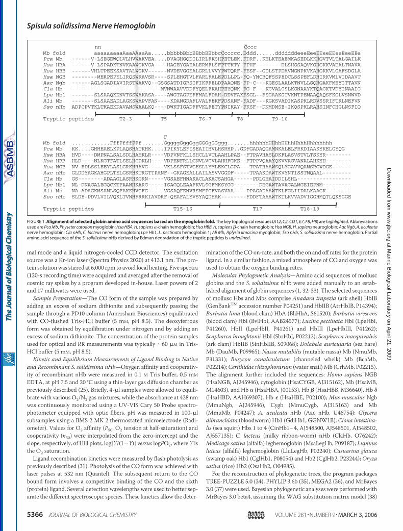
FIGURE 1. Alignment of selected globin amino acid sequences based on the myoglobin fold. The key topological residues (A12, C2, CD1, E7, F8, H8) are highlighted. Abbreviationsused are Pca Mb, Physeter catodon myoglobin; Hsa HBA, H. sapiens �-chain hemoglobin; Hsa HBB, H. sapiens �-chain hemoglobin; Hsa NGB, H. sapiens neuroglobin; Aac Ngb, A. aculeatanerve hemoglobin; Cla nHb, C. lacteus nerve hemoglobin; Lpe Hb1, L. pectinata hemoglobin 1; Ali Mb, Aplysia limacina myoglobin; Sso nHb, S. solidissima nerve hemoglobin. Partialamino acid sequence of the S. solidissima nHb derived by Edman degradation of the tryptic peptides is underlined.
Spisula solidissima Nerve Hemoglobin
5366 JOURNAL OF BIOLOGICAL CHEMISTRY VOLUME 281 • NUMBER 9 • MARCH 3, 2006
at Marine B
iological Laboratory on April 21, 2009
ww
w.jbc.org
Dow
nloaded from

of sequence evolution with gamma distribution of rates. Metropolis-coupled Markov chain Monte Carlo sampling was performed with onecold and three heated chains that were run for 600,000 generations.Starting trees were random, trees were sampled every 10th generation,and three independent runs were performed. The posterior probabili-ties were estimated on the final 50,000 trees (burn-in, 10,000).For neighbor-joining analyses (39), protein distances were calculated
using the PAM substitution matrix (40). The programs NEIGHBORfrom the PHYLIP package and MEGA version 2.1 were employed fortree reconstruction. In the case of applying the program NEIGHBOR,TREE-PUZZLE 5.2 was used for calculation of distance matrices. Thereliability of the trees was tested by bootstrap analyses (41) with 1,000replications.
RESULTS AND DISCUSSION
Primary Structure and Gene Organization—A part of the S. solidis-sima nHb primary structure was reconstructed from the sequence ofrelevant peptides generated by tryptic digestion of the purified protein(Fig. 1). The full sequence was completed by sequencing the corre-sponding cDNA of the nHb (GenBankTM accession numberAM086629). Fig. 1 also shows the alignment of the S. solidissima nHbwith related globin sequences. Key amino acid positions of the globinstructure such as NA2, B6, B14, C2, CD1, E7, F8, and H8 are conserved(42). At the N terminus there is an extension of�6 amino acid residues,the fourth one being a cysteine. A second cysteine is present at positionE12. Otherwise there are no remarkable amino acid substitutions com-pared with other mollusc hemoglobins (see Fig. 1). Conceptual transla-tion of the 162-amino acid-long sequence predicted amolecularmass of18,191 Da, excluding the N-terminal methionine. However, mass spec-
trometry data on the intact native nerve globin showed a molecularmass of 18,235 Da, which confirms the absence of the N-terminalmethionine and suggests an N-terminal mono-acetylation (�42 Da).This is not so unusual as N-terminal acetylation is one of the mostcommon protein modifications in eukaryotes, occurring on �85% ofthe different varieties of eukaryotic proteins but rarely on prokaryoticproteins (43, 44). We observed two additional molecular mass classes(18,333 and 18,430 Da) that differ from the native mono-acetylatedprotein by 98 and 196 Da, respectively (Fig. 2). These additional massescould agree with a binding of a mono- and a diphosphate to the nHb.Their physiological role, however, is unclear. The recombinant proteingave the single mass of 18,192 Da, with no additional acetylation. Therewas, however, an additional molecular mass class of 18,289 Da, suggest-ing again the presence of a phosphate. The binding of phosphate groupsto both native and recombinant S. solidissima nHb was confirmed byProQ Diamond phosphoprotein staining (data not shown).Genomic DNAwas extracted from S. solidissima foot and contractor
muscle and pooled frommultiple animals. The nHb gene was amplifiedby PCR and sequenced both directly and after cloning to resolveobserved sequence heterogeneities. The obtained sequence (Gen-BankTM accession number AM086630) contained differences in theintronic regions probably representing allelic polymorphisms, whereasonly two variations in the coding exons were observed. We noted 51single nucleotide polymorphisms and 4 insertion/deletion polymor-phisms in the intronic regions, varying in length from 1 to 284 bp. Thecoding region of the S. solidissima nHb gene is distributed on threeexons, consisting of 116, 226, and 144 bp (measured from the ATG startcodon and excluding the stop codon), validating the results from thecDNA. The two introns contain standard splice donor and acceptor
FIGURE 2. Mass spectrum of native S. solidissima nHb. Intensity of the peaks plotted against the molecular mass.
Spisula solidissima Nerve Hemoglobin
MARCH 3, 2006 • VOLUME 281 • NUMBER 9 JOURNAL OF BIOLOGICAL CHEMISTRY 5367
at Marine B
iological Laboratory on April 21, 2009
ww
w.jbc.org
Dow
nloaded from

motifs, and they are located at positions B12.2 (i.e. between the secondand third base of the 12th codon in helix B of the globin) and G7.0. Thepresence of the B12.2 and G7.0 intron positions is a conserved hallmarkof vertebrate (and many invertebrate) globin genes. The S. solidissimanHb gene does not contain a third intron within the E helix region,which is characteristic for the Ngb gene lineage in vertebrates and uro-chordates (1, 33, 45).
Optical and RR Spectra—The optical spectrum of the ferrous oxyformof recombinant S. solidissimanHb (see Fig. 3) displays a Soret bandat 414 nm. The � and � bands in the visible region are found at 537 and573 nm, respectively, as reported earlier by Strittmatter and Burch (14).Similar values were observed for the oxy form of Paramecium Hb (46)and sperm whale Mb (47). For nHbCO, the Soret band is shifted to aslightly higher wavelength (420 nm), which is in agreement with theoptical spectrum of the CO form of ParameciumHb (46). The � and �
bands of nHbCO are situated at 537 and 568 nm, again in good agree-ment with the CO form of Paramecium Hb (46). In the absorptionspectrum of deoxyferrous Spisula nHb the Soret band appears at 426nm. The � and � bands arise at 530 and 560 nm, respectively. Thesevalues were also found for non-symbiotic barleyHb (48), non-symbioticriceHb (49),Ngb (15), and cytoglobin (Cygb) (50). The values are typicalfor ferrous low spin heme complexes with a hexacoordination of theiron atom. The amino acid sequence of S. solidissima nHb indicates thatthe sixth ligand of the iron atom in the deoxy form is most likely E7His.The RR spectra of nHbO2, deoxyferrous nHb, and nHbCO are shown
in Fig. 4. It is well established that the marker lines �4, �3, and �2 in thehigh frequency region of the RR spectrum are sensitive to the oxidationstate, spin state, and coordination number of the iron at the center of theheme pocket. In the RR spectrum of nHbO2 (see Fig. 4, aA and bA), �4,�3, and �2 are situated at 1373, 1507, and 1578 cm�1, respectively, inagreement with earlier studies on heme proteins in the oxy form, e.g. theoxy complex of ParameciumHb (46). Upon increase of the laser powerto 17 milliwatts, a second spectral component appears. The �4 peak of
FIGURE 3. Optical spectra of S. solidissima nHbO2 (solid line), deoxy nHb (dotted line),and nHbCO (dashed line).
FIGURE 4. Low frequency (a) and high frequency (b) resonance Raman spectra of S. solidissima nHbO2 (laser power 2 milliwatts) (A), nHbO2 (laser power 17 milliwatts) (B),deoxy nHb (laser power 17 milliwatts) (C), and nHbCO (laser power 2 milliwatts) (D).
Spisula solidissima Nerve Hemoglobin
5368 JOURNAL OF BIOLOGICAL CHEMISTRY VOLUME 281 • NUMBER 9 • MARCH 3, 2006
at Marine B
iological Laboratory on April 21, 2009
ww
w.jbc.org
Dow
nloaded from

this new component is positioned at 1363 cm�1 (see Fig. 4, aB and bB),which agrees with the formation of the photoproduct. This is confirmedby the RR spectrum of ferrous deoxy nHb, obtained after reductionwithsodium dithionite (see Fig. 4, aC and bC). �4, �3, and �2 are situated at1360, 1491, and 1580 cm�1, respectively, typical for a hexacoordinatelow spin FeII heme form, confirming the absorption data (Fig. 3, dottedlines). Furthermore, a high spin (HS) component is present, as is indi-cated by the lines at 1470 (�3) and 1555 cm�1 (�2). Note that the inten-sity of the HS �3 marker line is lower than the intensity of the LS �3marker line. This suggests that there is only a very small HS populationpresent, as the intensity of �3 is very high for pentacoordinated speciesin comparison with hexacoordinated species. A small HS componenthas been found for other bis-histidine-coordinated globins (51).
In the spectrum of S. solidissima nHbCO �4, �3, and �2 are located at1373, 1500, and 1583 cm�1, respectively, characteristic for a hexacoor-dinated low spin complex. Again, the lines at 1460 (�4), 1469 (�3), and1556 cm�1 (�2) indicate that there is a small population of anHS ferrouscomplex present, which agrees with a small fraction of photodissocia-tion taking place. Two CO coordination configurations can be distin-guished, one with the Fe-CO stretching mode at 494 cm�1 (75%) andone with this mode at 513 cm�1 (25%) (see supplemental information).The first configuration (494 cm�1) was observed previously, e.g. inspermwhaleMb (491 cm�1) (52). InMb, this conformer becomes dom-inant at low pH values, when E7His swings out of the heme pocket. It isascribed to an open conformation of the heme pocket, with a weakinteraction between CO and the surrounding amino acids. The secondconformer (513 cm�1) was also observed in Mb (508 cm�1) (52) andCygb (510 cm�1) (50) and was assigned to a conformation in which theheme pocket is closed, due to the stabilization of COby E7His.Note thatno Fe-CO stretching modes at higher frequencies are observed, in con-
trast to several other heme proteins capable of a bis-histidine coordina-tion of the heme iron, such as Ngb (523 cm�1) (51), Cygb (518 cm�1)(50), or Barley Hb (534 cm�1) (53). Higher Fe-CO stretching frequen-cies correspond to a stronger stabilization of CO, e.g. due to interactionwith several surrounding amino acid residues. The line at 586 cm�1 canbe attributed to the Fe-C-O bendingmode. Note that the bendingmodeis weak, which is typical for an open conformation of the heme pocket.It was previously shown that the conformation of the heme pocket candiffer significantly for O2 and CO binding (46). Also, it has been shownthat when the distal His is able to interact with the bound oxygen closedconformation of the heme pocket autoxidation is greatly enhanced, asthe ferrous O2 acquires more ferric O2
. (superoxide) character (25).Kinetic and Equilibrium Measurements of Ligand Binding to Native
and Recombinant S. solidissima nHb—Fig. 5 shows the oxygen equilib-rium curve for recombinant S. solidissima nHb at 20 °C, pH 7.5 and thedata expressed as a Hill plot (inset). Under these conditions, the oxygenaffinity is high, with a P50 value of �0.6 torr, and oxygen is bound in anon-cooperative manner as indicated by a slope of �1 in the Hill plot.The P50 and n values found in our study on the recombinant protein aresignificantly lower than those reported from in situ measurements onintact nerves at high protein concentrations, where a P50 of 2.3 torr andan n value of 2.1 were found (28). The difference in P50 and n valuesmayarise because our measurements were carried out in vitro on a recom-binant protein, whereas the previous data refer to the native protein inits natural environment, indicating in vivo aggregation at high concen-tration and/or the existence of in vivo allosteric effectors that loweroxygen affinity. Acetylation of the native protein, but not the recombi-nant one, may also contribute to the observed functional differences.Indeed Lacan et al. (54) showed that a new �-chain variant, Hb Lyon-Byron, is characterized by the replacement of the N-terminal valine byan alanine that is N-terminal acetylated. This resulted in a decreasedoxygen affinity.
FIGURE 5. Oxygen equilibrium curve and Hill plot (inset) of S. solidissima nHb meas-ured at 20 °C in 0.1 M Tris buffer, pH 7.5. The protein concentration was 0.11 mM heme.PO2 is expressed in torr. Absence of cooperativity of oxygen binding can be inferred fromthe unitary slope of the Hill plot.
FIGURE 6. CO rebinding kinetics of S. solidissima nHb. After photodissociation, there iscompetitive binding of CO and the protein (E7 His) ligand. At high CO levels, oneobserves mainly a single phase of CO recombination. At lower CO levels, His bindingbecomes competitive and the second phase is more prominent. The rate of the secondphase corresponds to the rate of replacement of His by CO.
TABLE 1Ligand binding kineticsThe affinity P50 was calculated using the formula P50 � (KpentaO2)*(1 � KHis). The O2 solubility coefficient was taken equal to 1.82 �M/torr.
konCO konO2 koffO2 KpentaO2 konHis koffHis KHis P50 calculated P50measuredM/s � 107 M/s � 107 s nM s s torr
S. solidissima nHb 7.5 13 30 230 14000 1000 14 1.9 0.6M. musculus Ngb 5.5 20 0.4 2 1000 0.5 2000 2.2Horse Mb 0.5 1.6 24 1500 0.8
Spisula solidissima Nerve Hemoglobin
MARCH 3, 2006 • VOLUME 281 • NUMBER 9 JOURNAL OF BIOLOGICAL CHEMISTRY 5369
at Marine B
iological Laboratory on April 21, 2009
ww
w.jbc.org
Dow
nloaded from
Flash photolysis experiments show two kinetic components formonomers of recombinant S. solidissima nHb. This type of kinetics isthe result of competitive binding of two ligands, as previously observedfor other hexacoordinate globins such as vertebrate Ngb or Cygb (15,23). In the case of the S. solidissima nHb the separation of the kineticphases is less distinct, as the coefficient for His binding is only �10 ascompared with �1,000 for certain forms of Ngb.
The ligand binding rates for CO and oxygen are both quite high(Table 1, Fig. 6), with oxygen having the higher rate, as generallyobserved for heme proteins. The value for oxygen association is amongthe highest observed, approaching the diffusion limit for the bimolecu-lar globin ligand reaction.The histidine association rate is an order of magnitude higher than
that observed for other hexacoordinated globins, although there arerelatively few values available for comparison. Binding of the histidinerequires only 100 �s compared with 1 ms for Ngb; Cygb is relativelyslow, requiring �10 ms (15, 23). The His dissociation occurs in �1 ms,
resulting in a binding coefficient of �10 for the hexacoordination ofS. solidissima nHb. Because of these elevated rates forHis binding, thereis significant overlap with the CO binding phase and the two kineticphases are less distinct than for the case of Ngb. In the case of humanNgb, there is an influence of an internal disulfide bond on the His kinet-ics (55), which can be probed by addition of dithiothreitol to break thebond. There was no effect of the dithiothreitol on the S. solidissima nHbbinding kinetics. This was expected, as the 2 cysteine residues present(in positions NA4 and E12) are unlikely to form a disulfide bond.TheO2 affinity obtained from the kinetic data (P50, 1.9 torr) is in fairly
good agreement with the value determined by equilibrium measure-ments (P50, 0.6 torr). Small differences can be because of the differencein experimental approaches. From the kinetics, one can separate theintrinsic oxygen binding parameters from the contribution of the Hisaffinity (Table 1).We can conclude from the above described experiments (spectra and
ligand binding properties) that S. solidissima nHb can be classified as
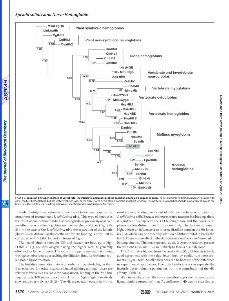
FIGURE 7. Bayesian phylogenetic tree of vertebrate, invertebrate, and plant globins based on amino acid sequence data. The S. solidissima nHb (shaded) clearly groups withother mollusc hemoglobins, but not with vertebrate Ngbs or the Ngb-related nerve globin from the annelid A. aculeata. The posterior probabilities of clade support are shown at thebranches. Three-letter species designations are specified under “Materials and Methods.”
Spisula solidissima Nerve Hemoglobin
5370 JOURNAL OF BIOLOGICAL CHEMISTRY VOLUME 281 • NUMBER 9 • MARCH 3, 2006
at Marine B
iological Laboratory on April 21, 2009
ww
w.jbc.org
Dow
nloaded from

normal intracellular hemoglobin having a “myoglobin-like” function.Themajor structural characteristic is the hexacoordinated nature of theiron atom. The structural differences between penta- and hexacoordi-nated hemoglobins might be, however, very small. Indeed, vertebrateHb andMb, normally pentacoordinated, shift toward hexacoordination(hemichrome) near their denaturation point because of a reorganiza-tion of their heme pocket (56–58). Application of high pressure (0.1–700 MPa) to penta- or partially hexacoordinated Hbs can also increasethe percentage of hexacoordination. This shift results in a difference ofligand binding characteristics (59).
Phylogenetic Analyses—The amino acid sequence of the S. solidis-sima nHb has an average identity of only 16–20% and a similarity (cal-culated using the PAM250matrix) of 30% to vertebrateNgbs andCygbs.In contrast, it displays 23–30% amino acid identity and 39–50% simi-larity to other mollusc Hbs and Mbs. The amino acid sequence of thenHb was added to an alignment of selected vertebrate and invertebrateglobin sequences (1, 32, 33), including representative mollusc globins.Phylogenetic trees were calculated using the Bayesian approach (Fig. 7)and the neighbor-joining distance method (not shown). Trees wererooted considering plant leghemoglobin as out-group. Regardless of thereconstruction method used, the S. solidissima nHb formed a commoncladewith themolluscHbs and consistently groupedwith the gill Hbs ofthe bivalve mollusc L. pectinata. These cytoplasmic Hbs are specializedto supply either O2 (Hb2 and 3) or sulfide (Hb1) for metabolism, prob-ably playing a role in the symbiosis of thesemolluscs and chemoautotro-phic bacteria (5).In contrast to the S. solidissima nHb, the nHb of the annelid
A. aculeata (6) grouped with vertebrate Ngb sequences as describedbefore (1). The highly derived and phylogenetically less informative nHbsequence from the nemertean worm C. lacteus (7) tended to group atvarious positions basal to the Ngb gene lineage and was thereforeexcluded from the final tree reconstruction (data not shown). Thisbehavior is most likely based on its structural shortening of the globinrather than on its phylogenetic position (7).
Implications for Vertebrate and Invertebrate Nerve Globin Func-tion—The nervous system of both vertebrates and invertebratesrequires huge amounts of metabolic energy and thus oxygen. It is there-fore not surprising that neurons are highly vulnerable to any shortage inoxygen supply as it occurs under pathological (e.g. ischemia) or naturalhypoxic conditions (60). There is no doubt that respiratory proteins thateither enhance oxygen flow under normoxic conditions or store oxygenfor short-term hypoxic states should be evolutionarily advantageous forthe survival of neurons and thus are frequently found in the nervoussystems of various invertebrate taxa (4). Strongly expressed, glial nHbslike the one in the bivalve mollusc S. solidissima are present in thosespecies that encounter substantial hypoxia in their natural habitats (4,5). Physiological experiments have convincingly demonstrated that glialnHbs enable these bivalves to maintain neuronal function while bur-rowing in O2-poor sediments (9, 29). Our biochemical characterizationof the S. solidissima nHb is generally in linewith the proposedO2 supplyfunction, e.g. by confirming the Mb-like O2 affinity of this respiratoryprotein. There are, however, additional unusual features of the S. solidis-sima nHb (phosphate binding, iron hexacoordination, etc.), the roles ofwhich in the nHb respiratory function are not yet clear.Originally, it had been proposed that invertebrate nHbs are phyloge-
netically orthologous to vertebrate Ngbs (1). This notion was based onthe finding that in phylogenetic analyses the nHb of the annelidA. aculeata consistently groups with human andmouse Ngb (Ref. 1 andthis report; Fig. 7). However, here we have shown that the nHb of thebivalve mollusc S. solidissima does not belong to the “Ngb plus
A. aculeata nHb” clade but groups with the othermolluscHbs andMbs.This finding demonstrates that a classical Hb or Mb has been recruitedduring evolution to fulfill the task of providing O2 to the nerve cells ofS. solidissima. Invertebrate nHbs are therefore of polyphyletic origin.Consequently, mollusc glial nHbs will probably have only modest rele-vance as amodel for studying the function of Ngb in the nervous systemof vertebrates (2). We note, however, that less prominently expressednHbs, coupled to a neuronal expression site, have been reported, e.g. forair-breathing gastropod molluscs (11), which like most vertebrates donot encounter substantial environmental hypoxia. This feature of a neu-ron-specific, low-expressed nHb is a clear parallel to vertebrate Ngbs. Itwill therefore be interesting to investigate the phylogenetic affiliation,biochemical features, and cellular function of invertebrate “neuronal”nHbs in comparison with the invertebrate glial nHbs and neuronal ver-tebrate Ngb.
Independent Origin of Hb Hexacoordination—From a biochemicalperspective, it is noteworthy that the S. solidissima nHb represents thefirst true respiratory Hb to be found in an iron-hexacoordinated form,resembling in this respect the vertebrate Ngb proteins (15, 61). Thisconfirms an earlier suggestion that a bis-His-Fe hexacoordinate hemestructure has arisen independently in distinct globin lineages (45). TheS. solidissima nHb will be instrumental as a model in studying the evo-lutionary determinants of this protein feature.
Acknowledgment—Thierry Backeljau is acknowledged for the helpwith dissec-tions of the Spisula nerve strains.
REFERENCES1. Burmester, T.,Weich, B., Reinhardt, S., andHankeln, T. (2000)Nature 407, 520–5232. Hankeln, T., Ebner, B., Fuchs, C., Gerlach, F., Haberkamp, M., Laufs, T. L., Roesner,
A., Schmidt, M., Weich, B., Wystub, S., Saaler-Reinhardt, S., Reuss, S., Bolognesi, M.,de Sanctis, D., Marden, M. C., Kiger, L., Moens, L., Dewilde, S., Nevo, E., Avivi, A.,Weber, R. E., Fago, A., and Burmester, T. (2005) J. Inorg. Biochem. 99, 110–119
3. Lankester E. R. (1872) Proc. R. Soc. Lond. B Biol. Sci. 21, 70–814. Wittenberg, J. B. (1992) in Advances in Comparative and Environmental Physiology
(Mangum, C. P., ed) pp. 59–85, Springer-Verlag Berlin, Heidelberg5. Weber, R. E., and Vinogradov, S. N. (2001) Physiol. Rev. 81, 569–6286. Dewilde, S., Blaxter, M., Van Hauwaert, M. L., Vanfleteren, J., Esmans, E. L., Marden,
M., Griffon, N., and Moens, L. (1996) J. Biol. Chem. 271, 19865–198707. Vandergon, T. L., Riggs, C. K., Gorr, T. A., Colacino, J. M., and Riggs, A. F. (1998)
J. Biol. Chem. 16998–170118. Chalazonitis, N., Gola, M., and Arvanitaki, A. (1966) C. R. Seances Soc. Biol. Fil. 160,
1020–10239. Kraus, D. W., and Colacino, J. M. (1986) Science 232, 90–9210. Kraus, D. W., Doeller, J. E., and Smith, P. R. (1988) Biol. Bull. 174, 54–6611. Schindelmeiser, I., Kuhlmann, D., and Nolte, A. (1979) Comp. Biochem. Physiol. B
64B, 149–15412. Wittenberg, B. A., Briehl, R.W., andWittenberg, J. B. (1965) Biochem. J. 96, 363–37113. Arvanitaki, A., and Chalazonitis, N. (1960) Bull. Inst. Oceanogr. Monaco 1164, 1–8314. Strittmatter, P., and Burch, H. B. (1963) Biochim. Biophys. Acta 78, 562–56315. Dewilde, S., Kiger, L., Burmester, T., Hankeln, T., Baudin-Creuza, V., Aerts, T., Mar-
den, M. C., Caubergs, R., and Moens, L. (2001) J. Biol. Chem. 276, 38949–3895516. Kiger, L., Uzan, J., Dewilde, S., Burmester, T., Hankeln, T., Moens, L., Hamdane, D.,
Baudin-Creuza, V., and Marden, M. (2004) IUBMB Life 56, 709–71917. Burmester, T., Haberkamp,M.,Mitz, S., Roesner, A., Schmidt,M., Ebner, B., Gerlach,
F., Fuchs, C., and Hankeln, T. (2004) IUBMB Life 56, 703–70718. Reuss, S., Saaler-Reinhardt, S.,Weich, B.,Wystub, S., Reuss,M.H., Burmester, T., and
Hankeln, T. (2002) Neuroscience 115, 645–65619. Geuens, E., Brouns, I., Flamez, D., Dewilde, S., Timmermans, J. P., and Moens, L.
(2003) J. Biol. Chem. 278, 30417–3042020. Schmidt,M., Giessl, A., Laufs, T., Hankeln, T.,Wolfrum,U., and Burmester, T. (2003)
J. Biol. Chem. 278, 1932–193521. Wystub, S., Laufs, T., Schmidt, M., Burmester, T., Maas, U., Saaler-Reinhardt, S.,
Hankeln, T., and Reuss, S. (2003) Neurosci. Lett. 346, 114–11622. Laufs, T. L.,Wystub, S., Reuss, S., Burmester, T., Saaler-Reinhardt, S., andHankeln, T.
(2004) Neurosci. Lett. 362, 83–8623. Pesce, A., Bolognesi, M., Bocedi, A., Ascenzi, P., Dewilde, S., Moens, L., Hankeln, T.,
and Burmester, T. (2002) EMBO Rep. 3, 1146–1151
Spisula solidissima Nerve Hemoglobin
MARCH 3, 2006 • VOLUME 281 • NUMBER 9 JOURNAL OF BIOLOGICAL CHEMISTRY 5371
at Marine B
iological Laboratory on April 21, 2009
ww
w.jbc.org
Dow
nloaded from
24. Moens, L., and Dewilde, S. (2000) Nature 407, 461–46225. Fago, A., Hundahl, C., Dewilde, S., Gilany, K., Moens, L., and Weber, R. E. (2004)
J. Biol. Chem. 279, 44417–4442626. Sun, Y., Jin, K., Mao, X. O., Zhu, Y., and Greenberg, D. A. (2001) Proc. Natl. Acad. Sci.
U. S. A. 98, 15306–1531127. Sun, Y., Jin, K., Peel, A., Mao, X. O., Xie, L., and Greenberg, D. A. (2003) Proc. Natl.
Acad. Sci. U. S. A. 100, 3497–350028. Kraus, D. W., and Doeller, J. E. (1988) Biol. Bull. 174, 346–35429. Doeller, J. E., and Kraus, D. W. (1988) Biol. Bull. 174, 67–7630. Schmidt, E. R., Vistorin, G., and Keyl, H. G. (1980) Chromosoma 76, 35–4531. Uzan, J., Dewilde, S., Burmester, T., Hankeln, T., Moens, L., Hamdane, D., Marden,
M. C., and Kiger, L. (2004) Biophys. J. 87, 1196–120432. Burmester, T., Ebner, B., Weich, B., and Hankeln, T. (2002) Mol. Biol. Evol. 19,
416–42133. Ebner, B., Burmester, T., and Hankeln, T. (2003)Mol. Biol. Evol. 20, 1521–152534. Strimmer, K., and von Haeseler, A. (1996)Mol. Biol. Evol. 13, 964–96935. Felsenstein, J. (2004) PHYLIP (Phylogeny Inference Package) Version 3.6b, Depart-
ment of Genetics, University of Washington, Seattle36. Kumar, S., Tamura, K., Jakobsen, I. B., and Nei, M. (2001) Bioinformatics 17,
1244–124537. Huelsenbeck, J. P., and Ronquist, F. (2001) Bioinformatics 17, 754–75538. Whelan, S., and Goldman, N. (2001)Mol. Biol. Evol. 18, 691–69939. Saitou, N., and Nei, M. (1987)Mol. Biol. Evol. 4, 406–42540. Dayhoff, M. O., Schwartz, R. M., and Orcutt, B. C. (1978) inAtlas of Protein Sequence
and Structure (Dayhoff, M. O., ed) pp. 345–352, National Biomedical Research Foun-dation, Washington, D. C.
41. Felsenstein, J. (1985) Evolution 39, 783–79142. Bashford, D., Chothia, C., and Lesk, A. M. (1987) J. Mol. Biol. 196, 199–21643. Boissel, J. P., Kasper, T. J., Shah, S. C., Malone, J. I., and Bunn, H. F. (1985) Proc. Natl.
Acad. Sci. U. S. A. 82, 8448–845244. Polevoda, B., and Sherman, F. (2000) J. Biol. Chem. 275, 36479–36482
45. Roesner, A., Fuchs, C., Hankeln, T., and Burmester, T. (2005) Mol. Biol. Evol. 22,12–20
46. Das, T. K., Weber, R. E., Dewilde, S., Wittenberg, J. B., Wittenberg, B. A., Yamauchi,K., Van Hauwaert, M. L., Moens, L., and Rousseau, D. L. (2000) Biochemistry 39,14330–14340
47. Antonini, E., and Brunori, M. (1971) Hemoglobin and Myoglobin in Their Reactionwith Ligands, North Holland Publishing, Amsterdam
48. Duff, S. M., Wittenberg, J. B., and Hill, R. D. (1997) J. Biol. Chem. 272, 16746–1675249. Arrendondo-Peter, R., Hargrove, M. S., Sarath, G., Moran, J. F., Lohrman, J., Olson,
J. S., and Klucas, R. V. (1997) Plant Physiol. 115, 1259–126650. Sawai, H., Kawada, N., Yoshizato, K., Nakajima, H., Aono, S., and Shiro, Y. (2003)
Biochemistry 42, 5133–514251. Couture, M., Burmester, T., Hankeln, T., and Rousseau, D. L. (2001) J. Biol. Chem.
276, 36377–3638252. Sage, J. T., Morikis, D., and Champion, P. M. (1991) Biochemistry 30, 1227–123753. Das, T. K., Lee, H. C., Duff, S. M., Hill, R. D., Peisach, J., Rousseau, D. L., Wittenberg,
B. A., and Wittenberg, J. B. (1999) J. Biol. Chem. 274, 4207–421254. Lacan, P., Souillet, G., Aubry, M., Prome, D., Richelme-David, S., Kister, J., Wajcman,
H., and Francina, A. (2002) Am. J. Hematol. 69, 214–21855. Hamdane, D., Kiger, L., Dewilde, S., Green, B. N., Pesce, A., Uzan, J., Burmester, T.,
Hankeln, T., Bolognesi, M., Moens, L., and Marden, M. C. (2004)Micron 35, 59–6256. Keilin, J. (1949) Biochem. J. 45, 440–44857. Morishima, I., Ogawa, S., and Yamada, H. (1980) Biochemistry 19, 1569–157558. Alden, R. G., Satterlee, J. D., Mintorovitch, J., Constantinidis, I., Ondrias, M. R., and
Swanson, B. I. (1989) J. Biol. Chem. 264, 1933–194059. Hamdane, D., Kiger, L., Hui Bon, H. G., Dewilde, S., Uzan, J., Burmester, T., Hankeln,
T., Moens, L., and Marden, M. C. (2005) J. Biol. Chem. 280, 36809–3681460. Lee, J. M., Grabb, M. C., Zipfel, G. J., and Choi, D. W. (2000) J. Clin. Investig. 106,
723–73161. Pesce, A., Dewilde, S., Nardini,M.,Moens, L., Ascenzi, P., Hankeln, T., Burmester, T.,
and Bolognesi, M. (2003) Structure 11, 1087–1095
Spisula solidissima Nerve Hemoglobin
5372 JOURNAL OF BIOLOGICAL CHEMISTRY VOLUME 281 • NUMBER 9 • MARCH 3, 2006
at Marine B
iological Laboratory on April 21, 2009
ww
w.jbc.org
Dow
nloaded from
Copyright © 2022 FDOKUMEN