The Schede and Modern Thoracoplasty - CORE
7
The Schede and Modern Thoracoplasty Benjamin J. Pomerantz, Joseph C. Cleveland, Jr, and Marvin Pomerantz THORACOPLASTY-GENERAL CONSIDERATIONS horacoplasty evolved as a procedure designed to T treat empyema, although modern day thoracic surgeons more commonly use this modality to obliterate persistent pleural spaces after resection. Thoracoplasty is the operative removal of the thoracic cage, which usually involves the subperiosteal resection of several ribs. The principle behind thoracoplasty is that by removing the skeletal support, the overlying chest wall will collapse to the visceral pleura.’ This apposition of chest wall to visceral pleura is indicated when the lung cannot be brought out to oppose the chest wall by the usual means. Thus, thoracoplasty seeks to collapse the chest wall to the remaining lung or mediastinum. We usually reserve this procedure for bronchopleural fistu- las or infected spaces after pulmonary resection when the patient will not tolerate further resection to correct the underlying pulmonary disorder. We no longer use this procedure as a primary modality for patients with far-advanced Mycobacterium tuberculosis or other my- cobacterial infections. The thoracoplasty technique most commonly used today is that described by John Alexander”.” in the 1920s and early 1930s. This tech- nique contrasts to that described by Schede4 in the 1890s. Schede’s technique involves removal of the ribs as well as the intercostal muscles and the often-thickened parietal pleura. Much credit is gjven to Dr Alexander, however, because the modern technique of thoracoplasty is essentially a modification of his original operation. SCHEDE THORACOPLASTY Schede4 described his thoracoplasty technique in 1890 at the Medical Conference in Vienna. The principle of this operation is to eliminate the residual cavity by resection of the thickened wall and supporting struc- tures of the cavity. The essential features of this operation are that a large, U-shaped skin incision is created and a flap of skin and soft tissue is raised. The ribs overlying the infected space are resected subperi- osteally. Resection begins by dividing the ribs at their midpoint. The ribs are then resected anteriorly at the costochondral junction, and posteriorly they are re- moved at the tubercle. Schede then advocated removing the thickened intercostal muscles , the neurovascular bundle, and parietal pleura over the entire ~avity.~J This represents a substantial difference from modern thoracoplasty techniques in which this step is omitted. The skin and soft tissue flap is then secured to the cavity, and the cavity is left open and packed with gauze. Schede’s thoracoplasty is of historical interest only, as the modern thoracoplasty has largely sup- planted the use of this particular thoracoplasty tech- nique. Schede’s technique increases the morbidity and mortality of thoracoplasty, especially with respect to intraoperative blood loss. MODERN THORACOPLASTY As previously noted, we have primarily applied modern thoracoplasty to deal with persistent space problems and bronchopleural fistulas that result from prior resection. We perform a large volume of niycobacterial- related resections; typically, these patients have already had a latissimus dorsi muscle flap rotated into their chest at the time of their original resection. Our technique for thoracoplasty begins with a posteriorly based periscapular incision. Dissection through skin and posterior chest wall muscles is performed until the chest wall is reached. Next, the subperiosteal resection of several ribs is performed. Typically 4 to 5 ribs are removed. Though some may not remove the first rib, we feel it is important to achieve maximum chest wall collapse. By dissecting on the upper surface of the periosteum first, the removal of this rib is facilitated. The first rib is intimately associated with the subclavian vessels and the brachial plexus. Care and meticulous technique must be used to avoid injury to these struc- tures. Once the first rib is removed, the other ribs are then removed in stepwise fashion. Posteriorly, the ribs are removed from the transverse process. Anteriorly, ribs are removed to “tailor” the collapse of the chest wall over the cavity. The end result is the obliteration of the bronchopleural fistula or infected cavity, because the chest wall and the visceral pleura are now opposed. When a thoracoplasty is performed for a bronchopleu- ral fistula or chronic empyema with active infection, it is necessary to place a large bore chest tube before closure. The tube is positioned in the area of the fistula, or in the case of an infected cavity, the tube is placed the full length of the cavity. Postoperatively, the tube is withdrawn slowly after fistula closure and after the 128 Operative Techniques in Thoracic and Cardiovascular SUrgerY, vol5, No 2 (May), 2000: pp 128-134
-
Upload
khangminh22 -
Category
Documents
-
view
0 -
download
0
Transcript of The Schede and Modern Thoracoplasty - CORE
The Schede and Modern Thoracoplasty
Benjamin J. Pomerantz, Joseph C. Cleveland, Jr, and Marvin Pomerantz
THORACOPLASTY-GENERAL CONSIDERATIONS
horacoplasty evolved as a procedure designed to T treat empyema, although modern day thoracic surgeons more commonly use this modality to obliterate persistent pleural spaces after resection. Thoracoplasty is the operative removal of the thoracic cage, which usually involves the subperiosteal resection of several ribs. The principle behind thoracoplasty is that by removing the skeletal support, the overlying chest wall will collapse to the visceral pleura.’ This apposition of chest wall to visceral pleura is indicated when the lung cannot be brought out to oppose the chest wall by the usual means. Thus, thoracoplasty seeks to collapse the chest wall to the remaining lung or mediastinum. We usually reserve this procedure for bronchopleural fistu- las or infected spaces after pulmonary resection when the patient will not tolerate further resection to correct the underlying pulmonary disorder. We no longer use this procedure as a primary modality for patients with far-advanced Mycobacterium tuberculosis or other my- cobacterial infections. The thoracoplasty technique most commonly used today is that described by John Alexander”.” in the 1920s and early 1930s. This tech- nique contrasts to that described by Schede4 in the 1890s. Schede’s technique involves removal of the ribs as well as the intercostal muscles and the often-thickened parietal pleura. Much credit is gjven to Dr Alexander, however, because the modern technique of thoracoplasty is essentially a modification of his original operation.
SCHEDE THORACOPLASTY Schede4 described his thoracoplasty technique in 1890 at the Medical Conference in Vienna. The principle of this operation is to eliminate the residual cavity by resection of the thickened wall and supporting struc- tures of the cavity. The essential features of this operation are that a large, U-shaped skin incision is created and a flap of skin and soft tissue is raised. The ribs overlying the infected space are resected subperi- osteally. Resection begins by dividing the ribs at their midpoint. The ribs are then resected anteriorly at the costochondral junction, and posteriorly they are re- moved at the tubercle. Schede then advocated removing the thickened intercostal muscles , the neurovascular bundle, and parietal pleura over the entire ~ a v i t y . ~ J
This represents a substantial difference from modern thoracoplasty techniques in which this step is omitted. The skin and soft tissue flap is then secured to the cavity, and the cavity is left open and packed with gauze. Schede’s thoracoplasty is of historical interest only, as the modern thoracoplasty has largely sup- planted the use of this particular thoracoplasty tech- nique. Schede’s technique increases the morbidity and mortality of thoracoplasty, especially with respect to intraoperative blood loss.
MODERN THORACOPLASTY
As previously noted, we have primarily applied modern thoracoplasty to deal with persistent space problems and bronchopleural fistulas that result from prior resection. We perform a large volume of niycobacterial- related resections; typically, these patients have already had a latissimus dorsi muscle flap rotated into their chest at the time of their original resection. Our technique for thoracoplasty begins with a posteriorly based periscapular incision. Dissection through skin and posterior chest wall muscles is performed until the chest wall is reached. Next, the subperiosteal resection of several ribs is performed. Typically 4 to 5 ribs are removed. Though some may not remove the first rib, we feel it is important to achieve maximum chest wall collapse. By dissecting on the upper surface of the periosteum first, the removal of this rib is facilitated. The first rib is intimately associated with the subclavian vessels and the brachial plexus. Care and meticulous technique must be used to avoid injury to these struc- tures. Once the first rib is removed, the other ribs are then removed in stepwise fashion. Posteriorly, the ribs are removed from the transverse process. Anteriorly, ribs are removed to “tailor” the collapse of the chest wall over the cavity. The end result is the obliteration of the bronchopleural fistula or infected cavity, because the chest wall and the visceral pleura are now opposed.
When a thoracoplasty is performed for a bronchopleu- ral fistula or chronic empyema with active infection, it is necessary to place a large bore chest tube before closure. The tube is positioned in the area of the fistula, or in the case of an infected cavity, the tube is placed the full length of the cavity. Postoperatively, the tube is withdrawn slowly after fistula closure and after the
128 Operative Techniques in Thoracic and Cardiovascular SUrgerY, vol5, No 2 (May), 2000: pp 128-134
THE SCHEDE AND MODERN THORACOPLASTY 129
ment, it should be apparent why it is not routinely used as a primary mode of treatment for mycobacterial and other primary pulmonary diseases.
infected cavity is obliterated. This process may take 6 to, 8 weeks or longer. Because of the morbidity associated with thoracoplasty and the protracted course of treat-
SURGICAL TECHNIQUE
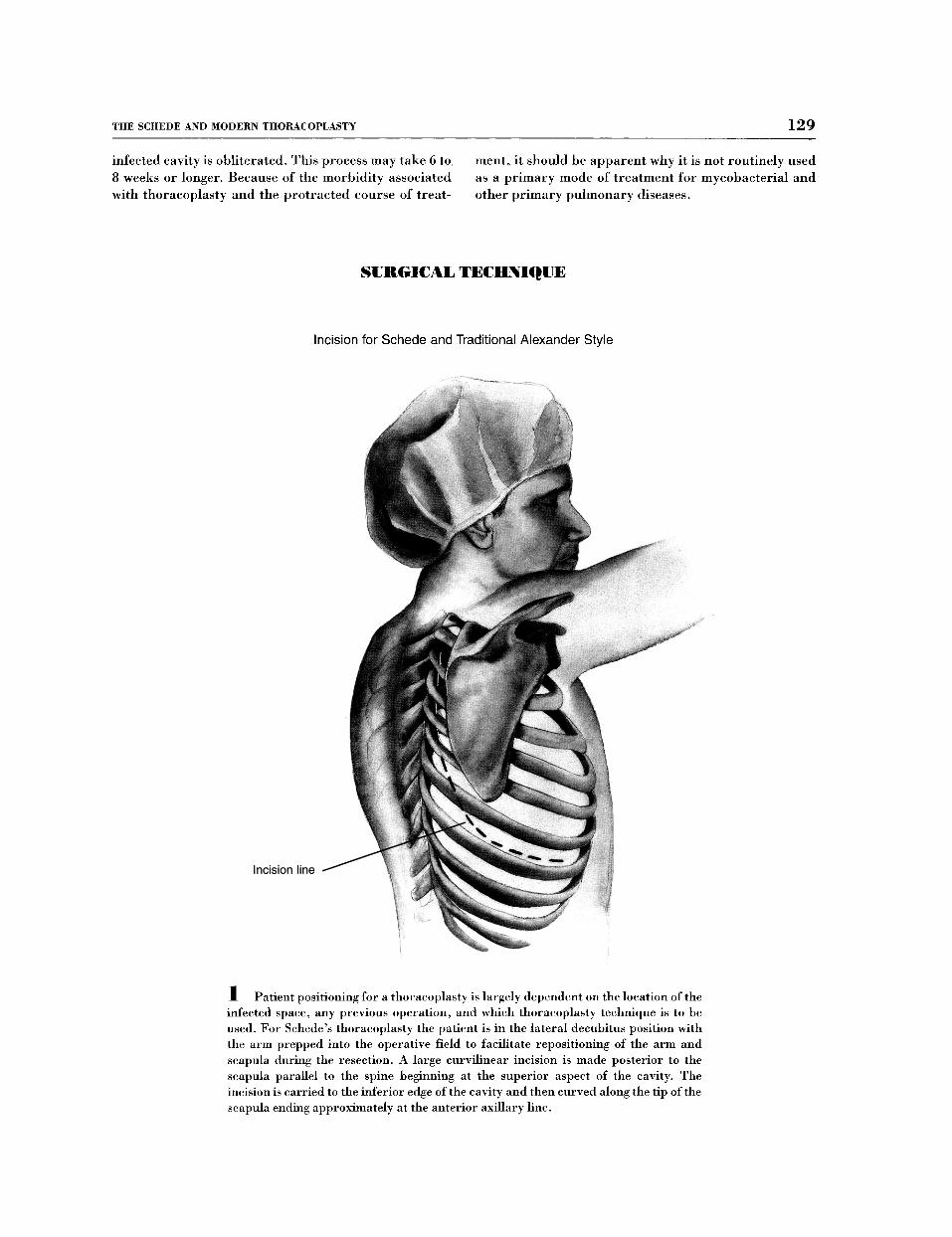
Incision for Schede and Traditional Alexander Style
1 Patient positioning for a thoracoplasty is largely dependent on the location of the infected space, any previous operation, and which thoracoplasty technique is to be used. For Schede’s thoracoplasty the patient is in the lateral decubitus position with the arm prepped into the operative field to facilitate repositioning of the arm and scapula during the resection. A large curvilinear incision is made posterior to the scapula parallel to the spine beginning at the superior aspect of the cavity. The incision is carried to the inferior edge of the cavity and then curved along the tip of the scapula ending approximately at the anterior axillary line.
130 POMERANTZ ET Al,
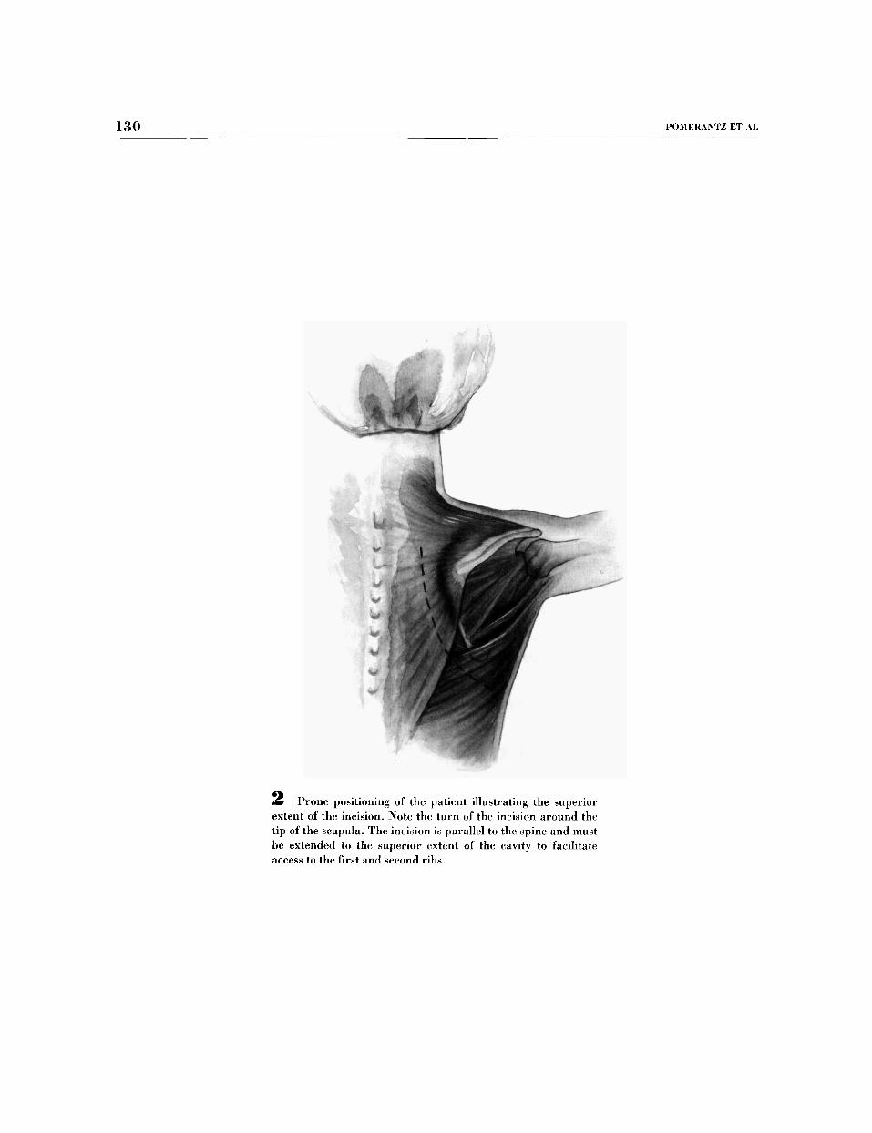
2 Prone positioning of the patient illustrating the superior extent of the incision. Note the turn of the incision around the tip of the scapula. The incision is parallel to the spine and must be extended to the superior extent of the cavity to facilitate access to the first and second ribs.
THE SCHEDE AND MODERN THORACOPLASTY 131
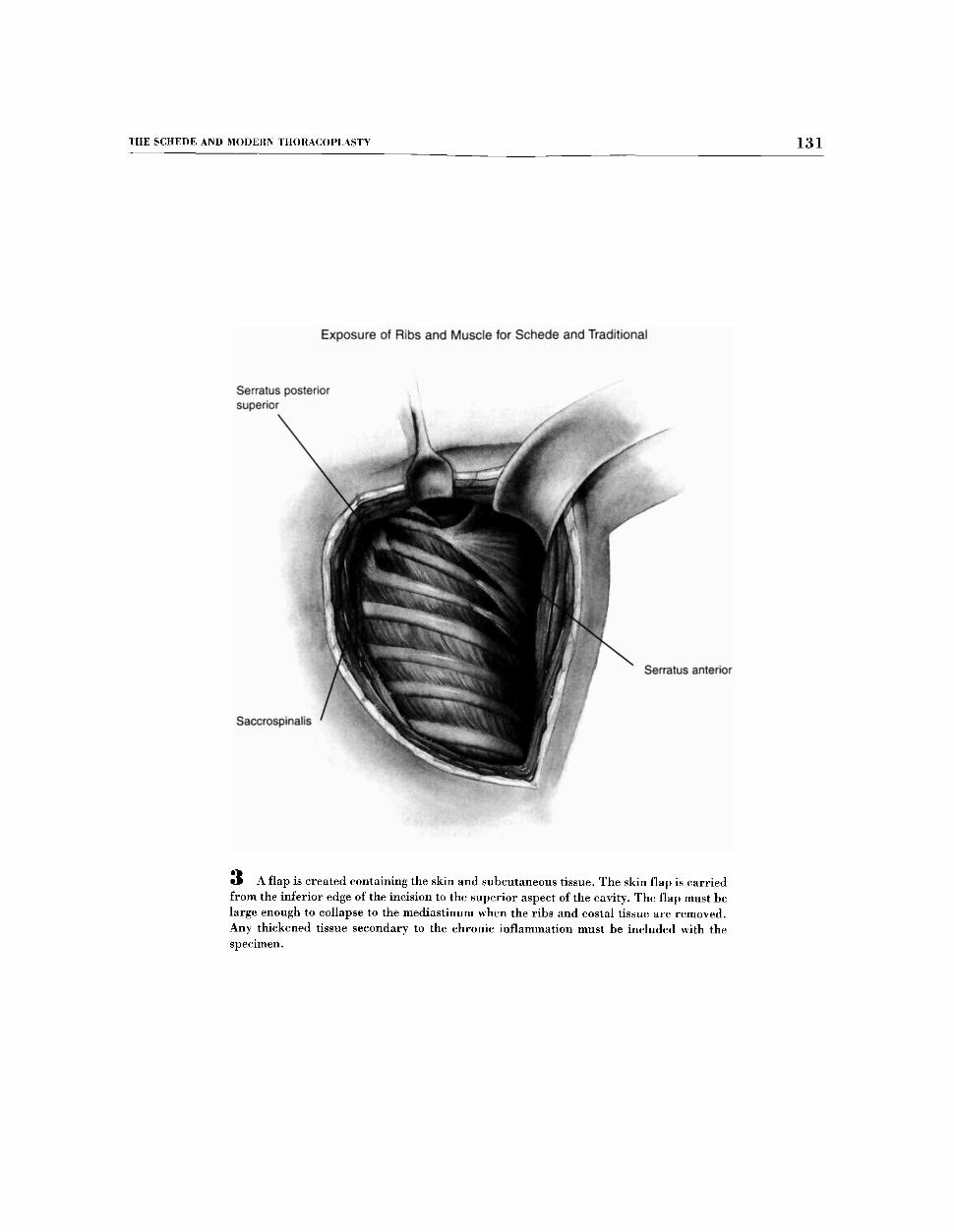
Exposure of Ribs and Muscle for Schede and Traditional
Serratus posterior supei
anterior
Saccr
3 A flap is created containing the skin and subcutaneous tissue. The skin flap is carried from the inferior edge of the incision to the superior aspect of the cavity. The flap must be large enough to collapse to the mediastinum when the ribs and costal tissue are removed. Any thickened tissue secondary to the chronic inflammation must be included with the specimen.
POMERANTZ ET 132 -
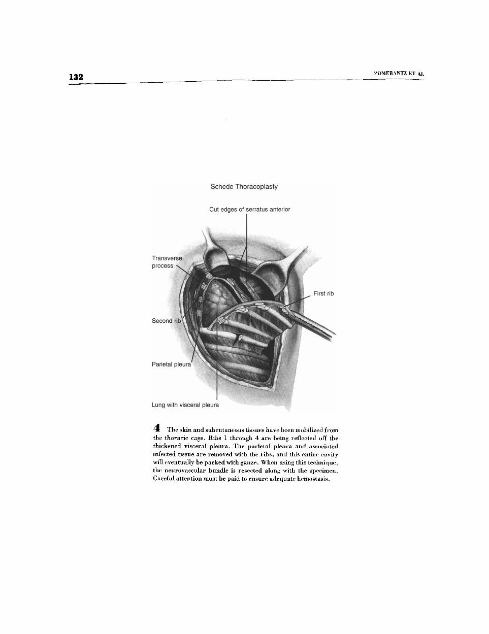
Schede Thoracoplasty
Cut edges of serratus anterior
I
Lung with visceral pleura
4 The skin and subcutaneous tissues have been mobilized from the thoracic cage. Ribs 1 through 4 are being reflected off the thickened visceral pleura. The parietal pleura and associated infected tissue are removed with the ribs, and this entire cavity will eventually be packed with gauze. When using this technique, the neurovascular bundle is resected along with the specimen. Careful attention must be paid to ensure adequate hemostasis.
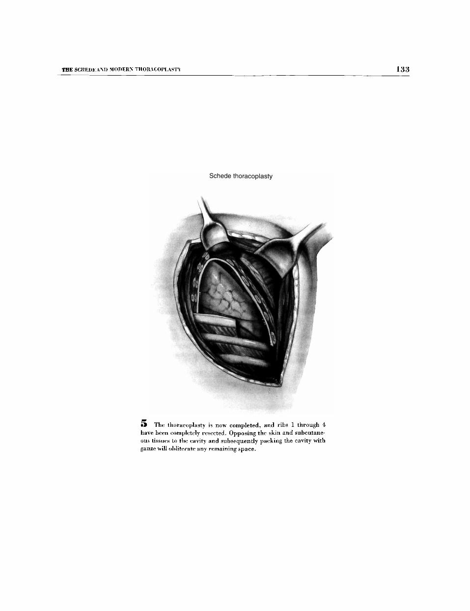
"HE SCHEDE AND .MODERN THORACOPLASTY 133
5 The thoracoplasty is now completed, and ribs 1 through 4 have been completely resected. Opposing the skin and subcutane- ous tissues to the cavity and subsequently packing the cavity with gauze will obliterate any remaining space.
134 POMERANTZ ET AL
Traditional-First Stage
First rib
Sen ,atus anterior (cut edge)
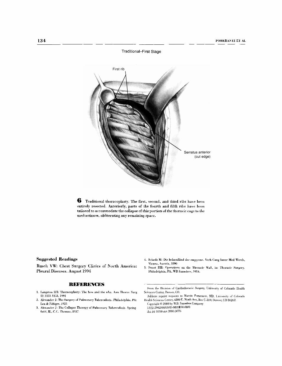
6 Traditional thoracoplasty. The first, second, and third ribs have been entirely resected. Anteriorly, parts of the fourth and fifth ribs have been tailored to accommodate the collapse of this portion of the thoracic cage to the mediastinurn, obliterating any remaining space.
Suggested Readings Rusch VW: Chest Surgery Clinics of North America: Pleural Diseases. August 1994
REFERENCES 1. Langston HT: Thoracoplasty: The how and the why. Ann Thorac Snrg
2. Alexander J: The Surgery of Pulmonary Tuberculosis. Philadelphia, PA:
3. Alexander J: The Collapse Therapy of Pulmonary Tuhercnlosis. Spring-
52:1351-1353,1991
Lea & Febiger, 1925
field, IL, C.C. Thomas, 1937
4.
5.
Schede M: Die hehandlind der empyeme. Verh Cong Inner Med Wersb, Vienna, Austria, 1890 Sweet RH: Operations on the Thoracic Wall, in: Thoracic Surgery. Philadelphia, PA, WIj Saunders, 1954.
From the Division of Cardiothoracic Surgery, University of Colorado Health
Address reprint requests to I v ~ u + ~ Pomerantz, MD, University of Colorado
Copyright 0 2000 by W.B. Saundm Company 1522-2942/00/0502-0005~~~.~~0
Sciences Center, Denver, CO.
Health Sciences Center, 4200 E. Ninth Ave, Box C-310, Denver, co 80262.
doi:lo. 1053/0t~t.2000.5076