MicroRNAs control hepatocyte proliferation during liver regeneration
Upload
khangminh22Category
view
2download
0
cells
Review
The Roles of the Let-7 Family of MicroRNAs in the Regulationof Cancer Stemness
Yuxi Ma 1,2 , Na Shen 1,3, Max S. Wicha 1,* and Ming Luo 1,*
�����������������
Citation: Ma, Y.; Shen, N.; Wicha,
M.S.; Luo, M. The Roles of the Let-7
Family of MicroRNAs in the
Regulation of Cancer Stemness. Cells
2021, 10, 2415. https://doi.org/
10.3390/cells10092415
Academic Editor: Giorgio Malpeli
Received: 10 August 2021
Accepted: 8 September 2021
Published: 14 September 2021
Publisher’s Note: MDPI stays neutral
with regard to jurisdictional claims in
published maps and institutional affil-
iations.
Copyright: © 2021 by the authors.
Licensee MDPI, Basel, Switzerland.
This article is an open access article
distributed under the terms and
conditions of the Creative Commons
Attribution (CC BY) license (https://
creativecommons.org/licenses/by/
4.0/).
1 Department of Internal Medicine, Division of Hematology and Oncology, University of Michigan,Ann Arbor, MI 48109, USA; [email protected] (Y.M.); [email protected] (N.S.)
2 Cancer Center, Union Hospital, Tongji Medical College, Huazhong University of Science and Technology,Wuhan 430022, China
3 Department of Breast and Thyroid Surgery, Union Hospital, Tongji Medical College, Huazhong University ofScience and Technology, Wuhan 430022, China
* Correspondence: [email protected] (M.S.W.); [email protected] (M.L.)
Abstract: Cancer has long been viewed as a disease of normal development gone awry. Cancer stem-like cells (CSCs), also termed as tumor-initiating cells (TICs), are increasingly recognized as a criticaltumor cell population that drives not only tumorigenesis but also cancer progression, treatmentresistance and metastatic relapse. The let-7 family of microRNAs (miRNAs), first identified in C.elegans but functionally conserved from worms to human, constitutes an important class of regulatorsfor diverse cellular functions ranging from cell proliferation, differentiation and pluripotency tocancer development and progression. Here, we review the current state of knowledge regardingthe roles of let-7 miRNAs in regulating cancer stemness. We outline several key RNA-bindingproteins, long non-coding RNAs (lncRNAs) and circular RNAs (circRNAs) involved in the regulationof let-7 biogenesis, maturation and function. We then highlight key gene targets and signalingpathways that are regulated or mutually regulated by the let-7 family of miRNAs to modulate CSCcharacteristics in various types of cancer. We also summarize the existing evidence indicating distinctmetabolic pathways regulated by the let-7 miRNAs to impact CSC self-renewal, differentiationand treatment resistance. Lastly, we review current preclinical studies and discuss the clinicalimplications for developing let-7-based replacement strategies as potential cancer therapeuticsthat can be delivered through different platforms to target CSCs and reduce/overcome treatmentresistance when applied alone or in combination with current chemo/radiation or molecularlytargeted therapies. By specifically targeting CSCs, these strategies have the potential to significantlyimprove the efficacy of cancer therapies.
Keywords: cancer stem cells (CSCs); tumor suppressor microRNAs; long non-coding RNAs(lncRNAs); circular RNAs (circRNAs); lin28; glucose metabolism
1. Introduction
MicroRNAs (miRNAs) are a small class of non-coding RNAs identified as evolu-tionary conserved, single-strand RNAs with the size of 18–25 nucleotides. Functionally,miRNAs repress target gene expression by binding to the 3′-untranslated regions (3′UTRs)of target messenger RNAs (mRNAs) to promote degradation and prevent their translationinto protein products. Since the initial discovery of C. elegans miRNA gene (lin-14) inthe early 1990s [1], over 2000 miRNAs have been identified, which regulate diverse cel-lular processes, including proliferation, differentiation, survival, stem cell maintenanceand tumorigenesis [2,3]. It has been well established that miRNAs play crucial roles intumor development and progression by regulating key cancer regulatory genes, includingoncogenes and/or tumor suppressor genes [2].
Let-7 (lethal-7) was initially identified as a heterochronic gene in C. elegans (the secondmiRNA gene identified after lin-14), where its expression determines adult cell fate in the
Cells 2021, 10, 2415. https://doi.org/10.3390/cells10092415 https://www.mdpi.com/journal/cells

Cells 2021, 10, 2415 2 of 20
worm [4].The let-7 family of miRNAs, which is comprised of multiple paralog genes locatedin different chromosomes, has been defined as one of the largest and most conserved familyof miRNAs across different species, ranging from worms to humans [5–7]. In humans,there are 10 mature let-7 miRNAs, including let-7a, let-7b, let-7c, let-7d, let-7e, let-7f, let-7g,let-7i, miR-98 and miR-202, which are derived from 13 precursor genes [8].
During mammalian embryonic development, let-7 expression is silent in embryonicstem cells (ESCs) but reactivated at later development stages [5]. This role of let-7 inESC differentiation is further validated by the findings that exogenous expression of let-7in miRNA-deficient ESCs rescues their defective differentiation [9]. In cancer cells, thelet-7 family of miRNAs generally function as tumor suppressor miRNAs to inhibit tumorgrowth and metastasis [8,10,11]. Let-7 miRNAs are also involved in suppressing CSCcharacteristics, including sphere/colony formation, tumor growth, differentiation andregenerative potential [8,10,11].
Cancer stem cells (CSCs), or tumor-initiating cells (TICs), represent a small subset ofcells in the tumor that maintain or acquire the capacity to indefinitely self-renew as well as todifferentiate into various tumor cell progenies constituting the bulk of a tumor mass [12,13].As CSCs are endowed with the intrinsic capacity for therapeutic resistance, they contribute notonly to tumorigenesis but also to disease progression and metastatic relapse. As a result of theseproperties, CSCs play a major role in the resistance of tumors to the conventional chemotherapyand radiation therapy [13].
miRNAs, especially the let-7 family of miRNAs and their regulatory proteins, arefrequently deregulated in a wide variety of cancer types and influence CSC maintenance,metabolism, tumorigenesis and metastasis. This review summarizes the current state ofknowledge on the roles of let-7 miRNAs and their regulatory proteins in regulating CSCsduring tumor development and cancer progression, the signaling pathways and networkthrough which let-7 miRNAs are involved in the regulation of CSCs. We also emphasizeexisting strategies that have been documented in the literature to harness this family ofmiRNAs for therapeutic benefit in preclinical models.
2. Let-7 as Key Tumor Suppressor miRNAs and Negative Regulators of CSCs
The CSC model posits that cancer arises from a rare population of tumor-initiating cells(TICs) or CSCs that possess dysregulated self-renewal capacity. Yu et al. presented the firstevidence that let-7 miRNAs are markedly reduced in TICs but increased in differentiatedtumor cells in breast cancer [14]. This increased let-7 expression in differentiated tumor cells(or non-TICs) inhibits the expression of H-Ras and HMGA2, known let-7 target genes thatpromote tumor self-renewal and proliferative capacity. In contrast, antagonizing let-7 byantisense oligonucleotides enhances self-renewal capacity of non-TICs, indicating a criticalrole of let-7 miRNAs in suppressing tumorigenicity by downregulating CSC properties [14].
In addition to a role in regulating breast CSCs (BCSCs), let-7 miRNAs are also impli-cated in CSC regulation in other tissue malignancies. For instance, let-7a is downregulatedin stem-like cells in head and neck cancers as evidenced by their expression of aldehyde de-hydrogenase (ALDH). Furthermore, increased let-7a expression represses chemoresistanceand tumourigenicity through the ablation of CSC-like properties [15].
In human pancreatic cancer cells, CD44+/CD133+/EpCAM+ CSC-like cells exhibitedlower levels of let-7 expression [16], and this suppressed let-7 expression promoted theexpression of pluripotency transcription factor, such as NANOG, SOX2, SOX9, OCT4, KLF4and c-myc, to maintain CSCs. In gastric cancer, let-7b was shown to directly suppressc-myc expression to promote gastric stem cell differentiation and drug sensitivity [17].
In urothelial carcinoma of the bladder or UCB, the activation of pro-inflammatoryCOX2/PGE2 signaling induces promoter methylation of let-7, resulting in its downregula-tion and subsequent upregulation of SOX2, which promotes the maintenance of urothelialCSCs [18]. In therapy-induced neuroendocrine prostate cancer (t-NEPC), upregulatedLin28B suppresses let-7 miRNA expression, which, in turn, promotes the de-repression ofHMGA2 and HMGA2-mediated SOX2 expression, leading to increased CD44+ and CD133+CSC-like cell populations [19].
Cells 2021, 10, 2415 3 of 20
Epithelial-mesenchymal transition or EMT, a cellular process facilitating tumor cellinvasion, migration and dissemination has been shown to associate with the acquisition ofmesenchymal CSC-like properties [20]. Increased Lin28A expression remarkably increasesE-cadherin but reduces Vimentin expression and, subsequently, increases the epithelialCSC marker ALDH activity and colony formation via the downregulation of let-7a in breastcancer [21].
The suppression of let-7d expression activates key EMT transcriptional factors, in-cluding Twist and Snail, and promotes chemo-resistant abilities of oral squamous cellcarcinoma [22]. In tongue squamous cell carcinoma (TSCC), the long noncoding RNA(lncRNA) H19, by binding to (sponging) let-7a to inhibit its function in repressing let-7target gene HMGA2, facilitates TSCC cell migration and invasion [23].
In advanced high grade serous ovarian cancer (HGSOC), knockdown of Snail up-regulates let-7 expression and reverses the cancer stemness phenotype [24]. Similarly, inHGSOC, low levels of let-7 expression were associated with increased EMT and higherspheroid-forming and tumor growth capacities [25]. The overexpression of mir-98, a mem-ber of let-7 family of miRNAs, reverses EMT through directed targeting of HMGA2 andtranscriptional repression of the EMT-related gene POSTN [26].
3. Regulatory Mechanisms of Let-7 Expression and Function3.1. Let-7 Biogenesis
The biogenesis of let-7 is similar to that of other miRNAs. Canonical let-7 miRNAs arefirst transcribed from the corresponding miRNA transcription units by RNA polymeraseII to produce primary miRNAs (pri-miRNAs), which are subsequently processed by acomplex containing an RNase III-like enzyme Drosha and its co-factor DGCR8, a double-stranded RNA-binding protein (RBP), into a 60–70nt-long precursor miRNAs (pre-miRNAs)in the nucleus [27–30].
The pre-miRNAs are then transported to the cytoplasm by exportin5 and further cleavedby Dicer, a cytoplasmic RNase III, to generate 18–25 bp long miRNA duplex [31–35]. One of themiRNA strands (called guide strand) in the duplex recruits Argonaute proteins (AGO1–4)to form RNA-induced silencing complex (RISC), while the other strand (passenger strand)is degraded. The RISC mediates post-transcriptional repression of miRNA (i.e., let-7) targetgenes, where the guide strand of miRNAs acts as a tether to the 3’UTR of specific mRNAsto cause their degradation and translational repression [36].
3.2. Post-Transcriptional Regulation of Let-7 by RNA-Binding/Processing Proteins
Let-7 constitutes a 13-member tumor suppressor miRNA family downregulated ina large fraction of tumor types [6,7]. Let-7 suppresses the translation of a number ofoncogenes, including Myc, K-Ras and HMGA2 [37–39]. The biogenesis of miRNAs, in-cluding let-7, is regulated at different stages in the nucleus and cytoplasm by miRNAprocessing proteins, such as Drosha and Dicer, respectively, and shRNA-mediated down-regulation of Drosha or Dicer1-enhanced tumor development in a K-Ras-induced mousemodel of lung cancer, suggesting that the abrogation of global miRNA processing promotestumorigenesis [40].
Subsequently, by examining the consequence of heterozygous vs. homozygous deletion ofDicer1 in mouse models, Kumar et al. demonstrated that the loss of a single allele of Dicer1enhanced tumorigenesis, while the loss of both alleles had the opposite effect [41]. Indeed,reduced Dicer expression has been shown to predict poor patient survival in a number ofcancer types, including chronic lymphocytic leukemia [42], breast [43], lung [44], ovarian [45],colorectal [46] and bladder [47,48] cancer.
In colorectal and breast cancer, low levels or the impairment of Dicer expressionpromotes epithelial-to-mesenchymal transition (EMT), metastasis formation and the ac-quisition of cancer stemness [49–51]. In parallel with the role of Dicer functioning asa haplo-insufficient tumor suppressor, recent studies also identified Dicer as a HIF-1α-interacting protein to promote CSC and metastatic activities in multiple types of cancercell lines and human tumors, where HIF-1α suppresses Dicer expression by promoting its

Cells 2021, 10, 2415 4 of 20
ubiquitination and autophagy-mediated degradation, leading to repressed maturation ofknown tumor suppressor miRNAs, including let-7 and miRNA-200b [52].
In contrast to Dicer1, DGCR8 and AGO RBPs that mediate let-7 (and other miRNAs)maturation and subsequent silencing of target mRNAs, a subset of RBPs can compete orinhibit miRNA-mediated mRNA silencing by binding sequences at or in close proximityto miRNA recognition elements (MREs) [53]. Of these, Lin28A and Lin28B (collectivelyreferred as Lin28) are best known to suppress let-7 expression and function and to influencestem cell maintenance, metabolism and tumorigenesis [54–58].
Lin28A and Lin28B, two highly related RBPs and proto-oncogenes, inhibit let-7 biogen-esis through distinct mechanisms by binding the let-7 precursor (pre-let-7) to interfere withthe miRNA-processing-machinery-mediated cleavage [59–61]. Lin28B binds to pre-let-7and prevents its processing by the miRNA-processing machinery in the nucleus, whereasLin 28A functions in the cytoplasm to recruit terminal uridylyl transferase (TUT4 or TUT7),which places a short oligo (U) stretch to the 3′-end of pre-let-7, which blocks its processingby Dicer, leading to pre-let-7 degradation [61–63].
Lin28A and Lin28B are aberrantly expressed in a plethora of tissue malignancies, andactivation of either Lin28A or Lin28B is responsible for the global post-transcriptionaldownregulation of let-7 miRNAs in many cancers, which is associated with advancedtumor stages and poor patient outcomes [55]. Through gain-or loss-of-function studies invarious cancer cell lines and mouse models, Lin28A/B has been shown to promote tumorgrowth, invasion and metastasis [19,57,64,65].
In addition to Lin28, other RBPs, such as the insulin-like growth factor 2 mRNA-bindingproteins (IMPs), have been characterized to compete with miRNAs for binding to MREson target mRNAs and protect these transcripts from miRNA-mediated degradation [66].In glioblastoma, IMP2 prevents let-7 target gene silencing by binding to let-7 MREs, therebyprotecting them from degradation, leading to the preservation of glioblastoma stem-likecells [67]. In addition to IMP2, other IMP paralogs, such as IMP1 and IMP3, have alsobeen shown to protect let-7 target genes from silencing [66]. Therefore, the IMPs fulfill afunction similar to Lin28 and may cooperate with Lin28 to induce or maintain stemness intumor cells.
3.3. Post-Transcriptional Regulation of Let-7 by LncRNAs and circRNAs
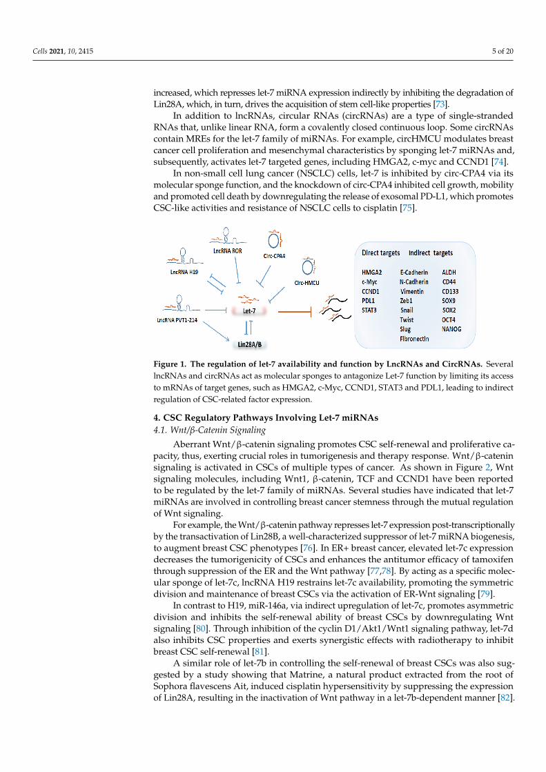
As depicted in Figure 1, a growing number of studies have shown that long non-codingRNAs (lncRNAs) and circular RNAs (circRNAs) play important roles in tumorigenesisand progression via the regulation of miRNAs, including the let-7 family. The human H19encodes a lncRNA that acts as a trans regulator of the imprinted gene network controllinggrowth in mice [68]. H19 harbors both canonical and non-canonical binding sites for thelet-7 family of miRNAs, which acts as a molecular sponge for let-7 [69].
Emerging evidence indicated that H19 plays important roles in regulating tumori-genicity and cancer stemness. For example, high levels of H19 are enriched in breast cancerstem cells (BCSCs), where it functions as a competing endogenous RNA to sponge let-7,leading to increased expression of the core pluripotency factor Lin28, thereby, promotingBCSC maintenance [70]. Through its sponging function, H19 also facilitates the migrationand invasion of tongue squamous cell carcinoma cells by suppressing let-7a, leading to theactivation of its target gene HMGA2, which, in turn, promotes EMT [23].
In esophageal cancer cells, elevated H19 expression promotes EMT and metastasisthrough activation of the STAT3/EZH2/β-catenin axis, and, interestingly, H19 was directlytargeted by let-7c, suggesting a mutual negative-feedback regulation between H19 andlet-7c [71]. Like H19, the long intergenic non-coding RNA or Linc-ROR, a regulator ofcell reprogramming, plays a key role in accelerating the self-renewal capacity of CSCs inpancreas cancer by sponging some members of the let-7 family [72].
In addition to serving as a molecular sponge by binding to let-7, some LncRNAs regulatethe let-7 family of miRNAs indirectly by modulating the expression of specific proteins thatsuppress let-7 maturation or function. In colorectal cancer, lncRNA PVT1-214 is markedly
Cells 2021, 10, 2415 5 of 20
increased, which represses let-7 miRNA expression indirectly by inhibiting the degradation ofLin28A, which, in turn, drives the acquisition of stem cell-like properties [73].
In addition to lncRNAs, circular RNAs (circRNAs) are a type of single-strandedRNAs that, unlike linear RNA, form a covalently closed continuous loop. Some circRNAscontain MREs for the let-7 family of miRNAs. For example, circHMCU modulates breastcancer cell proliferation and mesenchymal characteristics by sponging let-7 miRNAs and,subsequently, activates let-7 targeted genes, including HMGA2, c-myc and CCND1 [74].
In non-small cell lung cancer (NSCLC) cells, let-7 is inhibited by circ-CPA4 via itsmolecular sponge function, and the knockdown of circ-CPA4 inhibited cell growth, mobilityand promoted cell death by downregulating the release of exosomal PD-L1, which promotesCSC-like activities and resistance of NSCLC cells to cisplatin [75].
Figure 1. The regulation of let-7 availability and function by LncRNAs and CircRNAs. SeverallncRNAs and circRNAs act as molecular sponges to antagonize Let-7 function by limiting its accessto mRNAs of target genes, such as HMGA2, c-Myc, CCND1, STAT3 and PDL1, leading to indirectregulation of CSC-related factor expression.
4. CSC Regulatory Pathways Involving Let-7 miRNAs4.1. Wnt/β-Catenin Signaling
Aberrant Wnt/β-catenin signaling promotes CSC self-renewal and proliferative ca-pacity, thus, exerting crucial roles in tumorigenesis and therapy response. Wnt/β-cateninsignaling is activated in CSCs of multiple types of cancer. As shown in Figure 2, Wntsignaling molecules, including Wnt1, β-catenin, TCF and CCND1 have been reportedto be regulated by the let-7 family of miRNAs. Several studies have indicated that let-7miRNAs are involved in controlling breast cancer stemness through the mutual regulationof Wnt signaling.
For example, the Wnt/β-catenin pathway represses let-7 expression post-transcriptionallyby the transactivation of Lin28B, a well-characterized suppressor of let-7 miRNA biogenesis,to augment breast CSC phenotypes [76]. In ER+ breast cancer, elevated let-7c expressiondecreases the tumorigenicity of CSCs and enhances the antitumor efficacy of tamoxifenthrough suppression of the ER and the Wnt pathway [77,78]. By acting as a specific molec-ular sponge of let-7c, lncRNA H19 restrains let-7c availability, promoting the symmetricdivision and maintenance of breast CSCs via the activation of ER-Wnt signaling [79].
In contrast to H19, miR-146a, via indirect upregulation of let-7c, promotes asymmetricdivision and inhibits the self-renewal ability of breast CSCs by downregulating Wntsignaling [80]. Through inhibition of the cyclin D1/Akt1/Wnt1 signaling pathway, let-7dalso inhibits CSC properties and exerts synergistic effects with radiotherapy to inhibitbreast CSC self-renewal [81].
A similar role of let-7b in controlling the self-renewal of breast CSCs was also sug-gested by a study showing that Matrine, a natural product extracted from the root ofSophora flavescens Ait, induced cisplatin hypersensitivity by suppressing the expressionof Lin28A, resulting in the inactivation of Wnt pathway in a let-7b-dependent manner [82].

Cells 2021, 10, 2415 6 of 20
Paralleling its effects on breast cancer cells, Matrine was also reported to have an-titumor properties in other types of cancer through upregulation of let-7b, which inacti-vates the Wnt/CCND1 signaling pathway and inhibits EMT and lung CSC activities [83].In esophageal cancer, Wnt activation stimulates the self-renewal of CSC-like cells, whichis blocked by the administration of nano-let-7b via direct inhibition of TCF-4/β-catenincomplex activity to sensitize chemotherapeutic response [84].
In hepatocellular carcinoma cells, the overexpression of let-7a also inhibits the tumorsphere-forming ability by inhibition of EMT factors and the Wnt signaling pathway [85]. In NSCLC,oncogenic miR-367 promotes the self-renewal ability of CSC-like cells by degrading E3-Ligase Enzyme F-box and WD repeat domain-containing 7 (FBXW7) and repressing let-7c,leading to the activation of Wnt signaling [86]. Attenuation of Wnt/β-catenin activity bythe tankyrase inhibitor JW74 leads to the induction of let-7 miRNA, which contributes toosteosarcoma cell differentiation [87].
Figure 2. Let-7 miRNAs regulate CSCs through interaction with the Wnt, Hedgehog and NOTCHpathways. Members of Let-7 family of miRNAs interact with receptors and/or down-streamingeffector genes of Wnt, Hedgehog and NOTCH pathways to regulate CSC self-renewal, proliferationand differentiation.
4.2. NOTCH/Hedgehog Signaling
Aberrant activation of NOTCH signaling is associated with the stemness phenotypeof many different tissue malignancies. Let-7 miRNAs play a functional role in the devel-opment of human neural progenitors by suppressing HMGA2 and NOTCH to regulatesgliogenesis [88]. Recent studies have suggested mutual regulation of miRNAs and NOTCHsignaling in multiple tumor cell types (Figure 2).
For example, treatment of pancreatic cancer cells with metformin, an antidiabetic drug,decreased the expression of the CSC markers CD44, EpCAM, EZH2, NOTCH1, NANOGand OCT4 but elevated the expression of miRNAs, including let-7a, let-7b, miR-26a, miR-101, miR-200b, and miR-200c in a mouse xenograft model [89]. Conversely, overexpressionof NOTCH1 increased the expression of oncogenic miR-21 while decreasing the expressionof miR-200b, miR-200c, let-7a, let-7b and let-7c, promoting EMT and a CSC-like phenotypein pancreatic cancer cells [90].
In ER+ breast cancer, miR-129 inhibits estrogen receptor 1 (ESR1)-mediated NOTCHsignaling in CSC-like cells by suppressing cyclin D1/DICER1 mediated let-7 maturation,leading to increased NUMB expression to repress NOTCH signaling activity [91].
The hedgehog pathway is involved in embryonic development and maintenance ofCSCs through signal transmission via sonic hedgehog (Shh), PTCH1, SMO to activatetranscription factor GLI-driven downstream gene responses. As illustrated in Figure 2,
Cells 2021, 10, 2415 7 of 20
activation of the hedgehog pathway can suppress the expression of let-7 miRNAs, whichpromotes CSC properties.
In pancreatic cancer, highly metastatic cancer cells exhibit upregulation of Shh and CSCsurface marker expression, including CD133 and CXCR4, associated with lower levels oflet-7 expression as compared to parental BxPC-3 cells [92]. In NSCLC, a hedgehog inhibitorpromotes sensitization of tumor cells to erlotinib by increasing let-7c but suppressing theexpression of CSC-associated genes, including SOX2, NANOG and EpCAM [93].
4.3. STAT3/NFκB/Cytokines Signaling
The activation of transcription factors, such as the signal transducer and activator oftranscription 3 (STAT3) and NFκB, elicited by pro-inflammatory cytokines, such as IL1, IL6and IL8, secreted in the tumor microenvironment, promotes tumor epigenetic/phenotypicplasticity reflected by increased mesenchymal signature gene expression and cancer stem-ness. As illustrated in Figure 3, one of the key mechanisms by which STAT3/NFκBsignaling promotes cancer stemness is through their negative regulation of let-7 miRNAs.
In NSCLC, the oncogenic MUC1-C transmembrane protein induces EMT by promotingNFκB p65 binding to Lin28B chromatin to activate Lin28B expression, leading to thesuppression of let-7 [94]. As is the case for NFκB, the activation of STAT3 in breast cancerpromotes the transcription of Lin28B by directly binding to the Lin28B promoter, resultingin the repression of let-7 expression and concomitant upregulation of the let-7 target,HMGA2 [95].
Similarly, activation of the STAT3/NFκB pathway in breast cancer cells by M1 macrophagesecreted pro-inflammatory cytokines elicits Lin28B-let-7-HMGA2 axis to induce CD44+/CD24-and ALDH1+ CSCs through activation of EMT [96]. In esophageal cancer cells, elevatedlet-7c expression following knocking down of lncRNA H19 is reported to repress cellproliferation, migration, invasion as well as EMT and metastasis via inhibition of theSTAT3-EZH2-β-catenin pathway [71].
Overexpression of let-7c, which acts as an upstream suppressor of the Ras/NFκBsignal pathway, decreases malignant transformation and acquisition of cancer stem cell-likeproperties in normal adult skin keratinocytes [97]. In oral cancer cells, ectopic expression oflet-7c downregulates stemness and radio- and chemo-resistance through directly targetingIL8 [98]. In breast cancer cells, let-7a directly binds to the 3’UTR of the C-C chemokinereceptor type 7 (CCR7) to suppress its expression, leading to impaired migration andinvasion [99].
Let-7a is also reported to directly inhibit IL6 expression and activation of Src oncopro-tein promotes NFκB-Lin28 signaling to inhibit let-7a expression, leading to high level of IL6production and activation of NFκB, thereby, completing a positive feedback loop to facili-tate an epigenetic switch of immortalized breast epithelial cells into a stably transformedCSC-like state [100].
4.4. MAPK/ERK and PI3K/AKT Signaling
Receptor tyrosine kinases (RTKs), such as the epidermal growth factor receptor (EGFR)family, are activated by extracellular ligands such as EGF to activate MAPK/ERK (alsoknown as Ras-RAF-MEK-ERK) and PI3K-AKT-mTOR signaling pathways to regulate cellproliferation/self-renewal, survival and differentiation. The Ras small GTPases consist ofthree major isoforms, including H-, K- and N-Ras. Both H- and K-Ras are targeted by thelet-7 family of miRNAs to regulate CSC activities (Figure 3).
Specifically, Lin28A facilitates glioblastoma neurosphere formation through constitu-tively activating K-Ras and suppressing let-7b and let-7g [101]. In pancreatic cancer, K-Rasblocks let-7i maturation by phosphorylating Lin28B at S243 and promoting Lin28B nucleartranslocation [102]. The dietary agents sulforaphane, quercetin and catechins inhibit CSC-like activities of pancreatic cancer cells through let-7a induction and K-Ras inhibition [103].Inhibition of the RAS/MEK pathway decreases the expression of let-7 target gene HMGA2,which is required to maintain EMT and the mesenchymal phenotype [104].

Cells 2021, 10, 2415 8 of 20
Figure 3. Let-7 miRNAs regulate CSCs via regulation of STAT3/NFκB/cytokine signaling as wellas receptor tyrosine kinases (RTKs)-mediated signaling cascades, including the Ras-RAF-MEK-ERK and PI3K/Akt pathways. Members of Let-7 family of miRNAs interact with the effector genesof JAK-STAT and NFκB to regulate proinflammatory responses, including cytokine production(right part). Let-7 miRNAs, through interaction with down-streaming effectors or target genes ofRas-RAF-MEK-ERK and PI3K/Akt pathways to regulate CSC activities (left part).
In K-Ras-driven lung cancer, increased Lin28B expression induces CD44+/CD326+CSC-like cells by decreasing let-7 miRNAs and promoting AKT activity [105]. In Table 1,we listed most of the target genes involved in many different signaling cascades that aredirectly or indirectly repressed by let-7 family of miRNAs in various cancer types.
Table 1. Directly or indirectly repressed genes by the let-7 family of miRNAs in various cancer types
miRNA Cancer Signalling Pathways Target Genes Regulated by Stemness References
Let-7a Hepatocarcinoma Wnt/β-catenin Direct TCF-4 NA EMT, sphere formation [85]Let-7c NSCLC Wnt/β-catenin Indirect TCF-4 and Wnt1 mir-367,FBXW7 ALDH, CD133, Sphere formation [86]Let-7b NSCLC Wnt/β-catenin Indirect CCND1 and TCF-4 Matrine KLF4, CD133 [83]Let-7 Osteosarcoma Wnt/β-catenin NA JW74 c-myc [87]nano Let-7b Esophageal cancer Wnt/β-catenin Direct TCF-4 NA ALDH, CD133 [84]Let-7a, f Breast cancer Wnt/β-catenin Correlation Lin28B, β-catenin ALDH [76]Let-7c Breast cancer Wnt/β-catenin Direct TCF-4 NA ALDH, SOX2, NANOG, OCT4 [77]Let-7c Breast cancer Wnt/β-catenin Direct TCF-4 H19 ALDH [79]Let-7d Breast cancer Wnt/β-catenin Direct CCND1 NA OCT3/4, NANOG, SOX2, ALDH [81]Let-7c Breast cancer Wnt/β-catenin Direct ERa NA ALDH [78]Let-7c Breast cancer Wnt/β-catenin Direct TCF-4 mir-146a, Lin28A, H19 ALDH [80]Let-7b Breast cancer Wnt/β-catenin Indirect CCND1, TCF-4 Matrine, Lin28A CD133, KLF4 [82]Let-7 Breast cancer STAT3/NFκB Indirect HMGA2 M1 ALDH,CD44+/CD24-, KLF4,NANOG [96]Let-7c Keratinocytes STAT3/NFκB Indirect k-Ras, p-RelA Arsenite K5, CD34 [97]Let-7 NSCLC STAT3/NFκB Indirect HMGA2, TGFBR3 MUC1-C, NFκB, Lin28b Sphere formation [94]Let-7c Oral cancer STAT3/NFκB Direct IL8 NA ALDH, CD44+ [98]Let-7 Breast cancer Metabolism Direct HIF-1α, indirect PDK1 H19 ALDH, OCT4 [106]Let-7 Hepatocarcinoma Metabolism Indirect SREBP-1 Lin28A/B Fatty acid synthesis, tumor growth [107]Let-7 Neuroblastoma Metabolism NA DMFO, Lin28B, MYCN Neurosphere formation [108,109]Let-7c, d Breast cancer Metabolism Indirect PDK1, p-PDH CAIX, Lin28B, NFκB ALDH, NANOG [110]Let-7b, g Glioblastoma MAPK/PI3K Correlation p-MAPK Lin28A, K-Ras CD133, SOX2, Nestin, OLIG2, OCT4, Snail [101]Let-7a, f, g NSCLC MAPK/PI3K Indirect pAKT, cMYC,VEGFA Lin28B CD44+/CD326+, EMT [105]Let-7a Pancreatic cancer MAPK/PI3K HMGA2 MEK EMT [104]Let-7i Pancreatic cancer MAPK/PI3K Direct TET3 K-Ras→Lin28B OCT4, NANOG, SOX2, CD133 [102]Let-7a Pancreatic cancer MAPK/PI3K K-Ras Green tea catechins (GTC) ALDH, sphere formation [103]Let-7 Pancreatic cancer Hedgehog Correlation Shh NA CD133, CXCR4, sphere formation [92]Let-7c NSCLC Hedgehog NA Hedgehog inhibitor SOX2, NANOG, EpCAM [93]Let-7b Breast cancer NOTCH Direct NUMB mir-129→ESR1→DICER1 CD44+/CD24-, ALDH [91]Let-7a Breast cancer EMT Indirect ERa, pS2, CCND1 NA ALDH, SP population [21]Let-7d Oral cancer EMT Indirect Twist, Snail, CDH1, Fibronectin 1 NA ALDH [22]Let-7 Ovarian cancer EMT NA Snail CD117, CD133, CD44, sphere formation [24]Let-7 Ovarian cancer EMT Correlation Correlation CD44, CD117, CD133, OCT4, NANOG,Lin28A, HMGA2 [25]Let-7 Pancreatic cancer EMT Correlation LncRNA ROR CD133, CD44, ALDH [72]Let-7a Tongue squamous cell carcinoma EMT Direct HMGA2 H19 CDH1, CDH2, Vimentin, Snail, Zeb1, Twist1 [23]Let-7b Gastric cancer Pluripotency transcription factor Direct c-myc NA c-myc [17]Let-7 Prostate cancer Pluripotency transcription factor Direct HMGA2 Lin28B SOX2, CD44, CD133 [19]Let-7 Bladder cancer Pluripotency transcription factor Indirect SOX2 COX2, PGE2 SOX2, OCT4, NANOG, CD24, CD133 [18]Let-7 Pancreatic cancer Pluripotency transcription factor Correlation Correlation CD44+ /CD133+ /EpCAM+ cells, Snail, OCT4, NANOG, SOX2, EZH2 [16]
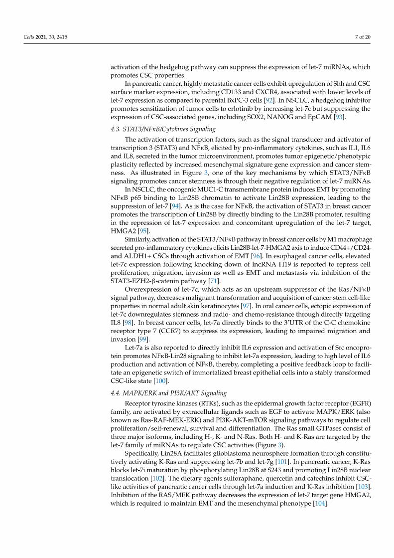
5. The Let-7/Lin28 Axis in Regulation of Stem Cell Metabolism
Earlier studies using transgenic mouse models demonstrated a role for the let-7/Lin28axis in regulation of glucose metabolism. Transgenic mice expressing Lin28A have in-creased body size, crown-rump length and delayed onset of puberty due to increasedglucose metabolism and insulin sensitivity [111]. This increased glucose metabolism isattributed to Lin28A/B-mediated repression of let-7, leading to an insulin-sensitized statethrough activation of the insulin-PI3K-mTOR pathway [111].

Cells 2021, 10, 2415 9 of 20
Such a role of the let-7/Lin28 axis in regulation of glucose metabolism is further demon-strated in transgenic mice with muscle-specific loss of Lin28A or overexpression of let-7, whichresults in insulin resistance, hyperglycemia and impaired glucose tolerance [112]. In additionto a role in regulating glucose metabolism during development, recent studies indicated thatLin28A and its closely related paralog, Lin28B, play critical roles in regulating pluripotent stemcells (PSCs).
Lin28, along with OCT4, SOX2, and NANOG, acts as important tissue factors to repro-gram human somatic fibroblasts into pluripotent stem cells [113], which exist in a highlyoxidative “naïve” state and a more glycolytic “primed” state [114,115]. Overexpression ofLin28A and Lin28B both enhance the derivation efficiency of induced PSCs (iPSCs), whileloss of endogenous Lin28A/B reduces reprogramming efficiency and traps the derivedmouse iPSCs in a more “naïve” oxidative state, suggesting that Lin28A and Lin28B facilitatetransition from naïve to primed pluripotency [116].
As reflected by their multipotent tumor cell state, CSCs exhibit a remarkable ability toadapt their metabolism to survive and proliferate under adverse environment conditions,such as hypoxia, acidosis, and nutrient starvation. As illustrated in Figure 4, recent studieshave provided strong evidence that the let-7/Lin28 axis regulates cancer progression andCSC activity through metabolic modulation.
In breast cancer, Lin28A and Lin28B enhance, whereas let-7 suppresses, aerobic glycol-ysis through regulation of pyruvate dehydrogenase kinase 1, or PDK1, in a hypoxia- orhypoxia-inducible factor-1 (HIF-1)-independent manner, suggesting direct regulation ofthe Lin28/let-7 axis in the modulation of glycolytic metabolism even at ambient oxygenlevels [106]. PDK1, by phosphorylating pyruvate dehydrogenase (PDH) to inhibit itsenzymatic function of promoting glucose flux to mitochondrial oxidative phosphorylation(OXPHOS), serves as a gatekeeper of aerobic glycolysis.
Interestingly, PDK1 is reported to be enriched in ALDH+ CSCs, and the depletionof PDK1 diminishes the sphere-forming ability and tumor growth by abrogating ALDH+CSCs [117]. This increased PDK1 activity in BCSCs is linked to elevated expression ofLncRNA H19, which acts as a molecular sponge sequestering let-7 miRNA, leading to theactivation of HIF1α to enhance its downstream target PDK1 expression [117].
The expression of carbonic anhydrase IX (CAIX), a pH regulator and hypoxia marker, is cor-related with poor prognosis and increased metastases in breast cancer [110]. In CAIX-suppressedbreast cancer cells, let-7 miRNAs are upregulated while Lin28 protein levels decreased, leadingto suppressed PDK1 activity and attenuated phosphorylation of PDH at Ser-232, which resultsin increased OXPHOS but reduced glycolysis and lactate production [110]. Thus, modulation ofthe Lin28/let-7 axis regulates tumor glycolytic metabolism through direct or indirect regulationof PDK1 activity, which directs glucose influx into aerobic glycolysis.
As mentioned above, overexpression of Lin28 and/or downregulation of let-7 miR-NAs has emerged as an oncogenic driver in many different cancers, which promotescancer stemness and poor patient outcome. The oncometabolite polyamine modulatesthe expression of eukaryotic translation initiation factor eIF-5A, which serves as a directregulator of the Lin28/let-7 axis [108]. In neuroblastoma cells, Difluoromethylornithine(DFMO), a drug targeting ornithine decarboxylase, the rate-limiting enzyme of polyaminebiosynthesis, reduces Lin28B protein expression while increasing let-7 miRNA, leading tothe inhibition of glycolytic metabolism and decreased neurosphere forming activity [109].
In addition to glucose metabolism, cancer cells also rely on de novo fatty acidbiosynthesis to maintain rapid cell proliferation and tumor growth [118]. Elevated ex-pression of Lin28A/B enhances de novo fatty acid synthesis to promote cancer progressionvia SREBP-1 [107]. By direct association with the mRNAs of both SREBP-1 and SCAP,Lin28A/B enhances the translation and maturation of SREBP-1, which, in turn, acceleratesmetabolic conversion of saturated and unsaturated fatty acids and protects cancer cells fromlipotoxicity [107].
Cells 2021, 10, 2415 10 of 20
Figure 4. The let-7/Lin28 axis regulates CSCs through modulation of glucose and fatty acidmetabolism. Lin28A and Lin28B enhance, whereas let-7 suppresses, aerobic glycolysis or fattyacid synthesis mainly through regulation of the PDK1/PDH enzymatic cascade or SREBP-1 to impactCSC activity and tumor progression.
The inhibition of Lin28B impairs cell growth and amino acid metabolism in acutemyeloid leukemia by de-repressing let-7a, although the downstream molecular mecha-nisms are not known [119]. Thus, targeting Lin28 and/or restoring let-7 expression, byinhibiting tumor metabolic activities in CSCs, may provide potential therapeutic strategiesfor cancer treatment.
6. Let-7 miRNAs as Potential Therapeutic Agents Targeting CSCs
As mentioned above, the let-7 family of miRNAs have been extensively documentedto serve as a class of tumor suppressor miRNAs to inhibit stem cell function in normaland cancerous tissues, and the enhanced expression of let-7 inhibits the maintenance ofCSCs, thus, inhibiting tumor growth and progression. In fact, let-7 miRNA replacementtherapy has now emerged as a powerful strategy to inhibit tumor growth and progressionand overcome treatment resistance.
Three approaches to harness tumor suppressor miRNAs, such as let-7, for therapeu-tic purposes have been described, including: (1) the utilization of recombinant lentivirusesor adenoviruses expressing let-7 miRNAs in tumor cells as a type of gene therapy,(2) the administration of small molecules that epigenetically enhances endogenous let-7expression, and (3) the administration of synthetic let-7 miRNA mimics in conjugationwith nanoparticle-based delivery.
6.1. Let-7 Replacement Therapy Employing Plasmid and Virus-Based Expression Systems
To restore let-7 miRNA expression, a plethora of studies have used plasmid and virus-based vector systems to either directly express let-7 or express regulatory proteins capable ofupregulating let-7 miRNA expression in tumor cells. The expression of let-7g is extremely lowin hepatocellular carcinoma (HCC) and re-expression of let-7g miRNA in HCC cells inhibitedtumor cell proliferation and migration via inhibition of K-Ras/HMGA2/Snail axis [120].
Moreover, the intra-tumoral injection of plasmid expressing let-7g inhibited tumorgrowth of HCC cells in nude mice [120]. Stable expression of let-7a in human breastcancer cells delivered by lentivirus infection significantly reduced mammosphere formationand tumorigenicity when implanted in immunodeficient mice [14]. Similarly, in ER+breast cancer cells, lentivirus-mediated let-7c expression has been shown to decreasetumorigenicity of estrogen-treated BCSCs in vivo by suppression of Wnt signaling [78].Over-expression of CDX2, a transcriptional factor called caudal type homeobox 2 in MCF7
Cells 2021, 10, 2415 11 of 20
breast cancer cells alleviates tumor growth and micro-metastasis by up-regulating let-7band inhibiting COL11A1 expression [121].
Although overexpression of let-7 miRNAs in cancer cells has been shown to inhibittumorigenicity and progression when implanted in mice, several studies have applied dif-ferent virus-based systems for the direct delivery of let-7 miRNAs locally or intratumorally.For example, intranasal administration of recombinant adenovirus expressing let-7a RNAhairpin has been shown to significantly reduce tumor formation in vivo in the lungs ofanimals that express a G12D activating mutation for the K-Ras oncogene [122].
The ectopic expression of let-7g in K-Ras(G12D)-expressing murine lung cancer cellsinduced cell cycle arrest and cell death and overexpression of let-7g by inducible lentiviralvectors led to significant growth reduction in tumor xenografts [123]. A single dose ofintratumor injection of lentiviruses expressing let-7c is capable of suppressing the tumorgrowth of prostate cancer xenografts [124].
6.2. Restoration of Endogenous Let-7 Expression to Enhance Treatment Responses
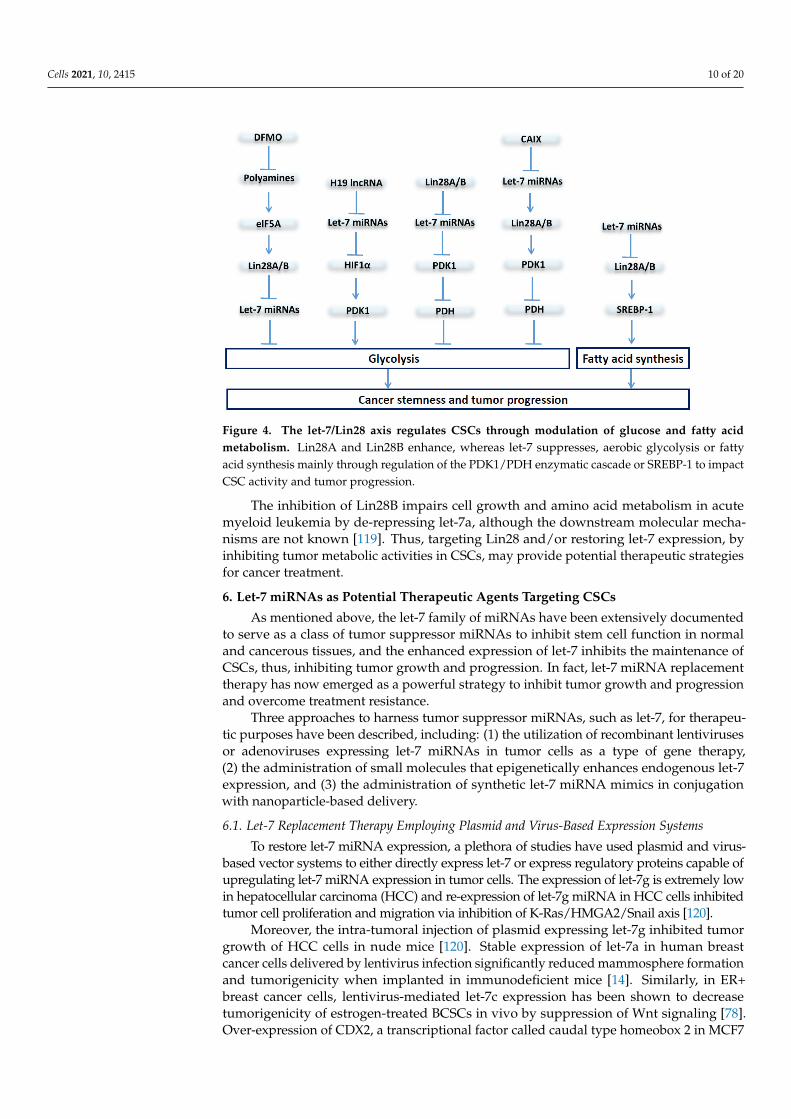
Current frontline therapies frequently fail to eradicate cancer due to their failure to eliminatecancer-stem-like cells, which confer resistance to traditional chemo/radiation or molecularlytargeted therapies, promoting metastasis and cancer relapse. As shown in Table 2, therapy-induced cancer stemness by widely used chemotherapeutic or molecularly targeted agents hasbeen reported to be abolished by restoration of let-7 miRNA expression in various types ofcancer, including breast cancer [82], NSCLC [83], head and neck cancer [15], ovarian cancer[24,25] and others. In pancreatic cancer, Gemcitabine-resistant tumor cells exhibit higher CSCmarker CD133 expression associated with lower levels of Let-7 [92,125].
Combined use of gemcitabine with thiostrepton, a natural cyclic oligopeptide antibi-otic, synergistically inhibits sphere formation of NSCLC by elevating miR-98 [126]. In ER+breast cancer, tamoxifen acts as an estrogen competitive antagonist, and Let-7c enhancesthe anticancer functions of tamoxifen and reduces the proportion of breast CSCs [78].The DNA alkylating agent temozolomide (TMZ) is one of the current standard treatmentfor glioblastoma patients. Dovitinib, a multi-kinase inhibitor, enhances TMZ-inducedcytotoxicity through modulation of Lin28/let-7/HMGA2 axis in vitro and in vivo [127].
In NSCLC, combined use of hedgehog inhibitor GDC-0449 leads to sensitization to erlotinib,an EGFR-TKI inhibitor through upregulating let-7c [93]. Let-7 restoration also exerts synergisticeffects with radiotherapy to target stem-like cells in breast cancer [81], oral squamous cellcarcinoma [98], and melanoma [128]. Both Let-7b [17,84] and let-7d [18,22], are involved inmultiple drug sensitization in several preclinical studies, suggesting that tumor delivery oflet-7 may be a promising therapeutic approach to enhance drug responses. In head and necksquamous cell carcinoma (HNSCC), the overexpression of let-7a/b miRNAs in combinationwith CTLA-4 antibody promotes anticancer immunity by accelerating PD-L1 degradation [129].
Table 2. Induction of Let-7 miRNAs to overcome therapeutic resistance by targeting CSCs.
Therapy miRNAs Regulated Stemness Markers Functions of miRNAs in Therapeutic Sensitization References
Mutiple drugs
Let-7 CD133, CD24 COX2 inhibitor→Let-7→SOX2→CD133, CD44 [18]Let-7d ALDH Let-7d→EMT→ALDH [22]Nano Let-7b CD133, ALDH Nano Let-7b→TCF4/Wnt→CD133, ALDH [84]Let-7b EMT Let-7b→cmyc→EMT [17]
Temozolomide Let-7 Sphere formation Dovitinib→Let-7/Lin28a/HMGA2→pSTAT3→Sphere formation [127]Tamoxifen Let-7c ALDH Let-7c→TCF4/Wnt→ALDH [78]Erlotinib/Cisplatin Let-7 SOX2, NANOG, EpCAM Hedgehog inhibotor→Let-7→EMT→SOX2, NANOG,EpCAM [93]Cisplatin Let-7b CD133 Matrine→Let-7b/Lin28A→CCDN1/Wnt→CD33 [82]
Let-7 CD133, CD44, CD117 shSnail/HGSOC→Let-7→CD133, CD44, CD117 [24]Cisplatin/Radiation Let-7c ALDH, CD44 Let-7c→IL8→ALDH, CD44 [98]
Radiation Let-7d ALDH Let-7d→CCND1/Wnt→ALDH [81]Let-7 Sphere formation Let-7/Lin28B→γH2AX [128]
Gemcitabine miR-98 CD133 Thiostrepton→miR-98→CD133 [126]Let-7 CD133 Hedgehog inhibitor→Let-7→CD133 [92,125]
CTLA-4 antibody Let-7a/b Wnt/β-catenin Let-7a/b→TCF-4→β-catenin/STT3→PDL1 degradation [129]

Cells 2021, 10, 2415 12 of 20
6.3. Let-7 miRNAs Encapsulated in Extracellular Vesicles for Potential Cancer Therapy
Secreted miRNAs encapsulated in extracellular vesicles (EVs) play an important rolein cell-cell communications. Exosomes are a subclass of EVs with the size ranging from30 to 150 nm in diameter containing proteins, RNAs, DNAs, lipids, and metabolites, whichmediate juxtacrine/paracrine signaling in the tumor microenvironment [130,131]. Recentstudies have indicated a role of exosomal let-7 miRNAs with strong antitumor activities.In non-small-cell lung cancer (NSCLC), let-7e encapsulated in serum exosomes suppressesthe metastatic activity of NSCLC cells by inhibiting the SUV39H2/LSD1/CDH1 axis [132].
In pancreatic cancer, exosome secretion from cancer cells promotes the recruitmentof pancreatic stellate cells (PSCs) to enhance the formation of distant metastases. Thiseffect is mediated by the transfer of exosomal protein Lin28B, a protein that suppressinglet-7 maturation, to PSCs to activate the Lin28B/let-7/HMGA2/PDGFβ signaling cas-cade [133]. Docosahexaenoic acid (DHA), a long-chain omega-3 polyunsaturated fatty acidwith known anticancer properties, promotes exosome secretion and increased exosomalmiRNAs, including let-7a, resulting in the inhibition of breast cancer angiogenesis [134].
In a metastatic gastric cancer cell line called AZ-P7a, exosomal release of let-7 miRNAsinto the extracellular environment is significantly increased as compared to non-metastaticgastric cancer cells. This increased exosomal transport of let-7 miRNAs is thought to resultin depletion of these tumor suppressor miRNAs in the cells, thereby maintaining theirinvasiveness and metastatic activity [135].
6.4. Let-7 Replacement Therapy Using Synthetic Let-7 miRNA Mimics
Use of miRNA mimics is an effective means to restore the normal function of tumorsuppressive miRNAs, including let-7 miRNAs. Transfection of chemically synthesized,double-stranded let-7 mimics into the cytoplasm of tumor cells has emerged as a potentialstrategy for cancer gene therapy. Intratumoral injection of let-7a mimics coupled with lipidvector lipofectamine 2000 has been shown to retard tumor growth in a glioma xenograftmodel [136].
Similarly, synthetic let-7b mimics intratumorally delivered into H460 lung tumorxenograft model successfully silenced let-7 targeted genes and repress tumor growth [137].Transfection of Let-7b mimics into gastric tumor cells also attenuate cisplatin resistance andtumor growth by targeting AURKB [138], and HepG2 cells transfected with let-7b mimicsdisplay decreased tumorigenic potential through the upregulated expression of p21 [139].
Let-7e expression was found to be significantly reduced in cisplatin-resistant humanepithelial ovarian cancer (EOC) cells, and the overexpression of let-7e by transfection oflet-7e Agomir, a type of chemically modified double-strand miRNA mimics, reduces theexpression of cisplatin-resistance-related proteins, including EZH2 and cyclin D1, andreverses cisplatin resistance when injected locally into EOC xenograft tumors [140].
Despite intensive studies indicating a role of let-7 family of miRNAs as tumor sup-pressors, the expression of hsa-let-7g (also known as miR-let-7g or let-7g) and its rolesin cancer are context-dependent. For example, let-7g exerts anti-tumor effects in gastriccancer and increases the sensitivity of gastric cancer cells to oxidative stress by inhibitingthe activation of DNA damage responses [141]. However, in osteosarcoma, patients withhigher expression of let-7g displayed a poorer prognosis and lower survival rate, and theoverexpression of let-7g promoted the occurrence of osteosarcoma by down-regulatingHOXB1 and activating the NFkB pathway [142].
A major challenge in RNA-based cancer therapies is the lack of safe and reliable meansfor specific delivery of miRNAs to cancer tissues with low cytotoxicity to normal cells.Tremendous progress has been made in restoring let-7 tumor suppressor miRNA functionusing let-7 miRNA mimics coupled with nanoparticle-based delivery systems, which havebeen shown to reduce tumor growth successfully in several animal studies [143–145].
In a K-Ras-activated autochthonous mouse model of NSCLC, synthetic let-7b mimicsconjugated with neural lipid emulsions (NLE) was systemically delivered to lung viaintravenous administration, leading to reduced tumor burden [146]. Dendrimer-based
Cells 2021, 10, 2415 13 of 20
polymeric vectors are another form of non-viral vectors with low toxicity and high deliverypotency. When administered to a MYC-driven liver tumor model by IV injection, modulardegradable dendrimer nanoparticles carrying let-7g miRNA mimic have been shown toinhibit tumor growth and dramatically extended mouse survival [147].
A form of nanocarriers called PEG5k-VE4-DET20 nano-assemblies is also used todeliver let-7b mimic and chemotherapy drug paclitaxel in the non-small cell lung can-cer (A549) model through intravenous injection, and paclitaxel and let-7b mimic-loadednano-assemblies markedly potentiated the cytotoxicity of paclitaxel and retarded tumorgrowth more efficaciously than Taxol alone [148]. Using an aptamer that binds to andantagonizes the oncogenic receptor tyrosine kinase Axl (GL21.T), Esposito et al. showedthat synthesized let-7g miRNA conjugated to the GL21.T aptamer reduced tumor growthin a xenograft model of lung adenocarcinoma [149].
7. Concluding Remarks
Let-7, one of the best studied miRNA families, plays critical roles in controlling various bio-logical processes, including self-renewal, proliferation, differentiation, apoptosis and the cellularmetabolism through the regulation of a plethora of target genes and signaling pathways. Let-7miRNAs generally function as tumor suppressors to target multiple oncogenes, including RAS,HMGA2, c-Myc and CCND1. Let-7 miRNAs also function as important immune regulators tocontrol cytokine- and chemokine-mediated inflammatory responses.
As the let-7 family members of miRNAs share a common seed sequence, localizing atnucleotide 2 to 8 in their 5′ ends, which is pivotal for target mRNA recognition [8], we predictthat individual members of let-7 miRNAs have shared or interchangeable functions in repressingtheir target genes to inhibit CSC activities.
As a well-recognized signaling node to regulate cancer stemness, Lin28 and let-7miRNAs form a double negative feedback loop to control tumorigenesis, cancer progressionand therapeutic resistance. In more than 15% of tissue malignancies, either Lin28A orLin28B is reactivated while let-7 is repressed, resulting in the elevated expression of let-7target genes as well as alterations in tumor cell metabolism.
Current evidence for the Lin28/let-7 axis in regulation of CSC metabolism centers onthe glucose metabolism especially aerobic glycolysis, which highlights a role of this axisin the modulation of glycolysis through direct or indirect regulation of PDK1. Althougha few studies indicated a role of Lin28/let-7 axis in the regulation of the amino acid andlipid metabolisms, future studies need to decipher the underlying mechanisms by whichthis axis regulates the metabolism of amino acids and lipids in cancer.
The roles of let-7 miRNAs in the development and diseases, particularly in cancer, havemade this family of miRNAs attractive candidates for the development of cancer therapeutics.One of the greatest challenges for RNA-based cancer therapy is to design suitable RNA deliveryvehicles to achieve specific and safe delivery of therapeutic miRNAs or miRNA mimics intotumor tissues while avoiding potential toxicities and off-target effects. In this regard, severalpreclinical nanoparticle-based platforms have been developed to deliver miRNA mimics totumor tissues with considerable efficacy and low toxicity [150]. For example, MRX34, a miR-34mimic encapsulated in a lipid carrier (developed by Mirna Therapeutics), has the advantageto become positively charged in the acidic tumor microenvironment, which allows the ad-herence of MRX34 specifically to tumor cells [151]. In a mouse xenograft model treated withMRX34 nanoparticles by intravenous injection, miR-34 was found to accumulate in tumortissues, leading to significant tumor regression [150,151]. In 2013, MRX34 was tested in a multi-center phase I trial in patients with various cancers, and a portion of these patients achievedprolonged partial responses or stable disease at the end of trial [150]. However, due to immune-related adverse effects resulting in patient deaths, this trial was prematurely terminated [150].As recent studies have identified a role of let-7 in regulating CD8 T cell differentiation andfunction [152,153], future studies in the development of let-7 based RNA therapeutics need toconsider the potential immune-related toxicities.

Cells 2021, 10, 2415 14 of 20
Author Contributions: Conceptualization: Y.M., M.S.W. and M.L.; Manuscript writing: Y.M. andM.L.; Manuscript review and editing: N.S. and M.S.W. All authors have read and agreed to thepublished version of the manuscript.
Funding: This work is supported by the NCI grant R35 CA197585 and the Breast Cancer ResearchFoundation grant BCRF-18–173 to M.S.W.
Institutional Review Board Statement: Not applicable.
Informed Consent Statement: Not applicable.
Data Availability Statement: Not applicable.
Conflicts of Interest: The authors declare no conflict of interest.
References1. Ambros, V. A hierarchy of regulatory genes controls a larva-to-adult developmental switch in C. elegans. Cell 1989, 57, 49–57.
[CrossRef]2. Peng, Y.; Croce, C.M. The role of MicroRNAs in human cancer. Signal Transduct. Target. Ther. 2016, 1, 15004. [CrossRef]3. Alles, J.; Fehlmann, T.; Fischer, U.; Backes, C.; Galata, V.; Minet, M.; Hart, M.; Abu-Halima, M.; Grasser, F.A.; Lenhof, H.P.; et al.
An estimate of the total number of true human miRNAs. Nucleic Acids Res. 2019, 47, 3353–3364. [CrossRef]4. Reinhart, B.J.; Slack, F.J.; Basson, M.; Pasquinelli, A.E.; Bettinger, J.C.; Rougvie, A.E.; Horvitz, H.R.; Ruvkun, G. The 21-nucleotide
let-7 RNA regulates developmental timing in Caenorhabditis elegans. Nature 2000, 403, 901–906. [CrossRef] [PubMed]5. Bussing, I.; Slack, F.J.; Grosshans, H. let-7 microRNAs in development, stem cells and cancer. Trends Mol. Med. 2008, 14, 400–409.
[CrossRef]6. Boyerinas, B.; Park, S.M.; Hau, A.; Murmann, A.E.; Peter, M.E. The role of let-7 in cell differentiation and cancer. Endocr. Relat.
Cancer 2010, 17, F19–F36. [CrossRef]7. Roush, S.; Slack, F.J. The let-7 family of microRNAs. Trends Cell Biol. 2008, 18, 505–516. [CrossRef] [PubMed]8. Sun, X.; Jiao, X.; Pestell, T.G.; Fan, C.; Qin, S.; Mirabelli, E.; Ren, H.; Pestell, R.G. MicroRNAs and cancer stem cells: The sword
and the shield. Oncogene 2014, 33, 4967–4977. [CrossRef] [PubMed]9. Melton, C.; Judson, R.L.; Blelloch, R. Opposing microRNA families regulate self-renewal in mouse embryonic stem cells. Nature
2010, 463, 621–626. [CrossRef]10. Sun, X.; Liu, J.; Xu, C.; Tang, S.C.; Ren, H. The insights of Let-7 miRNAs in oncogenesis and stem cell potency. J. Cell. Mol. Med.
2016, 20, 1779–1788. [CrossRef]11. Balzeau, J.; Menezes, M.R.; Cao, S.; Hagan, J.P. The LIN28/let-7 Pathway in Cancer. Front. Genet. 2017, 8, 31. [CrossRef] [PubMed]12. Wicha, M.S.; Liu, S.; Dontu, G. Cancer stem cells: An old idea—A paradigm shift. Cancer Res. 2006, 66, 1883–1890. [CrossRef]
[PubMed]13. Luo, M.; Clouthier, S.G.; Deol, Y.; Liu, S.; Nagrath, S.; Azizi, E.; Wicha, M.S. Breast cancer stem cells: Current advances and
clinical implications. Methods Mol. Biol. 2015, 1293, 1–49._1. [CrossRef] [PubMed]14. Yu, F.; Yao, H.; Zhu, P.; Zhang, X.; Pan, Q.; Gong, C.; Huang, Y.; Hu, X.; Su, F.; Lieberman, J.; et al. Let-7 regulates self renewal and
tumorigenicity of breast cancer cells. Cell 2007, 131, 1109–1123. [CrossRef]15. Yu, C.C.; Chen, Y.W.; Chiou, G.Y.; Tsai, L.L.; Huang, P.I.; Chang, C.Y.; Tseng, L.M.; Chiou, S.H.; Yen, S.H.; Chou, M.Y.; et al.
MicroRNA let-7a represses chemoresistance and tumourigenicity in head and neck cancer via stem-like properties ablation. OralOncol. 2011, 47, 202–210. [CrossRef] [PubMed]
16. Bao, B.; Ali, S.; Ahmad, A.; Li, Y.; Banerjee, S.; Kong, D.; Aboukameel, A.; Mohammad, R.; Van Buren, E.; Azmi, A.S.; et al.Differentially expressed miRNAs in cancer-stem-like cells: Markers for tumor cell aggressiveness of pancreatic cancer. Stem CellsDev. 2014, 23, 1947–1958. [CrossRef] [PubMed]
17. Yang, X.; Cai, H.; Liang, Y.; Chen, L.; Wang, X.; Si, R.; Qu, K.; Jiang, Z.; Ma, B.; Miao, C.; et al. Inhibition of c-Myc by let-7b mimicreverses mutidrug resistance in gastric cancer cells. Oncol. Rep. 2015, 33, 1723–1730. [CrossRef] [PubMed]
18. Ooki, A.; Del Carmen Rodriguez Pena, M.; Marchionni, L.; Dinalankara, W.; Begum, A.; Hahn, N.M.; VandenBussche, C.J.;Rasheed, Z.A.; Mao, S.; Netto, G.J.; et al. YAP1 and COX2 Coordinately Regulate Urothelial Cancer Stem-like Cells. Cancer Res.2018, 78, 168–181. [CrossRef]
19. Lovnicki, J.; Gan, Y.; Feng, T.; Li, Y.; Xie, N.; Ho, C.H.; Lee, A.R.; Chen, X.; Nappi, L.; Han, B.; et al. LIN28B promotes thedevelopment of neuroendocrine prostate cancer. J. Clin. Investig. 2020, 130, 5338–5348. [CrossRef] [PubMed]
20. Mani, S.A.; Guo, W.; Liao, M.J.; Eaton, E.N.; Ayyanan, A.; Zhou, A.Y.; Brooks, M.; Reinhard, F.; Zhang, C.C.; Shipitsin, M.; et al.The epithelial-mesenchymal transition generates cells with properties of stem cells. Cell 2008, 133, 704–715. [CrossRef]
21. Liu, Y.; Li, H.; Feng, J.; Cui, X.; Huang, W.; Li, Y.; Su, F.; Liu, Q.; Zhu, J.; Lv, X.; et al. Lin28 induces epithelial-to-mesenchymaltransition and stemness via downregulation of let-7a in breast cancer cells. PLoS ONE 2013, 8, e83083. [CrossRef]
22. Chang, C.J.; Hsu, C.C.; Chang, C.H.; Tsai, L.L.; Chang, Y.C.; Lu, S.W.; Yu, C.H.; Huang, H.S.; Wang, J.J.; Tsai, C.H.; et al. Let-7dfunctions as novel regulator of epithelial-mesenchymal transition and chemoresistant property in oral cancer. Oncol. Rep. 2011,26, 1003–1010. [CrossRef]
Cells 2021, 10, 2415 15 of 20
23. Kou, N.; Liu, S.; Li, X.; Li, W.; Zhong, W.; Gui, L.; Chai, S.; Ren, X.; Na, R.; Zeng, T.; et al. H19 Facilitates Tongue Squamous CellCarcinoma Migration and Invasion via Sponging miR-let-7. Oncol. Res. 2019, 27, 173–182. [CrossRef] [PubMed]
24. Hojo, N.; Huisken, A.L.; Wang, H.; Chirshev, E.; Kim, N.S.; Nguyen, S.M.; Campos, H.; Glackin, C.A.; Ioffe, Y.J.; Unternaehrer, J.J.Snail knockdown reverses stemness and inhibits tumour growth in ovarian cancer. Sci. Rep. 2018, 8, 8704. [CrossRef]
25. Chirshev, E.; Hojo, N.; Bertucci, A.; Sanderman, L.; Nguyen, A.; Wang, H.; Suzuki, T.; Brito, E.; Martinez, S.R.; Castanon, C.; et al.Epithelial/mesenchymal heterogeneity of high-grade serous ovarian carcinoma samples correlates with miRNA let-7 levels andpredicts tumor growth and metastasis. Mol. Oncol. 2020, 14, 2796–2813. [CrossRef]
26. Zhu, M.; Zhang, C.; Chen, D.; Chen, S.; Zheng, H. MicroRNA-98-HMGA2-POSTN signal pathway reverses epithelial-to-mesenchymaltransition in laryngeal squamous cell carcinoma. Biomed. Pharmacother. 2019, 117, 108998. [CrossRef] [PubMed]
27. Lee, Y.; Ahn, C.; Han, J.; Choi, H.; Kim, J.; Yim, J.; Lee, J.; Provost, P.; Radmark, O.; Kim, S.; et al. The nuclear RNase III Droshainitiates microRNA processing. Nature 2003, 425, 415–419. [CrossRef] [PubMed]
28. Landthaler, M.; Yalcin, A.; Tuschl, T. The human DiGeorge syndrome critical region gene 8 and Its D. melanogaster homolog arerequired for miRNA biogenesis. Curr. Biol. 2004, 14, 2162–2167. [CrossRef] [PubMed]
29. Gregory, R.I.; Yan, K.P.; Amuthan, G.; Chendrimada, T.; Doratotaj, B.; Cooch, N.; Shiekhattar, R. The Microprocessor complexmediates the genesis of microRNAs. Nature 2004, 432, 235–240. [CrossRef]
30. Denli, A.M.; Tops, B.B.; Plasterk, R.H.; Ketting, R.F.; Hannon, G.J. Processing of primary microRNAs by the Microprocessorcomplex. Nature 2004, 432, 231–235. [CrossRef]
31. Hutvagner, G.; McLachlan, J.; Pasquinelli, A.E.; Balint, E.; Tuschl, T.; Zamore, P.D. A cellular function for the RNA-interferenceenzyme Dicer in the maturation of the let-7 small temporal RNA. Science 2001, 293, 834–838. [CrossRef]
32. Bernstein, E.; Caudy, A.A.; Hammond, S.M.; Hannon, G.J. Role for a bidentate ribonuclease in the initiation step of RNAinterference. Nature 2001, 409, 363–366. [CrossRef]
33. Yi, R.; Qin, Y.; Macara, I.G.; Cullen, B.R. Exportin-5 mediates the nuclear export of pre-microRNAs and short hairpin RNAs. GenesDev. 2003, 17, 3011–3016. [CrossRef] [PubMed]
34. Bohnsack, M.T.; Czaplinski, K.; Gorlich, D. Exportin 5 is a RanGTP-dependent dsRNA-binding protein that mediates nuclearexport of pre-miRNAs. RNA 2004, 10, 185–191. [CrossRef] [PubMed]
35. Lund, E.; Guttinger, S.; Calado, A.; Dahlberg, J.E.; Kutay, U. Nuclear export of microRNA precursors. Science 2004, 303, 95–98.[CrossRef] [PubMed]
36. Brennecke, J.; Stark, A.; Russell, R.B.; Cohen, S.M. Principles of microRNA-target recognition. PLoS Biol. 2005, 3, e85. [CrossRef][PubMed]
37. Mayr, C.; Hemann, M.T.; Bartel, D.P. Disrupting the pairing between let-7 and Hmga2 enhances oncogenic transformation. Science2007, 315, 1576–1579. [CrossRef] [PubMed]
38. Johnson, S.M.; Grosshans, H.; Shingara, J.; Byrom, M.; Jarvis, R.; Cheng, A.; Labourier, E.; Reinert, K.L.; Brown, D.; Slack, F.J. RASis regulated by the let-7 microRNA family. Cell 2005, 120, 635–647. [CrossRef] [PubMed]
39. Sampson, V.B.; Rong, N.H.; Han, J.; Yang, Q.; Aris, V.; Soteropoulos, P.; Petrelli, N.J.; Dunn, S.P.; Krueger, L.J. MicroRNA let-7adown-regulates MYC and reverts MYC-induced growth in Burkitt lymphoma cells. Cancer Res. 2007, 67, 9762–9770. [CrossRef]
40. Kumar, M.S.; Lu, J.; Mercer, K.L.; Golub, T.R.; Jacks, T. Impaired microRNA processing enhances cellular transformation andtumorigenesis. Nat. Genet. 2007, 39, 673–677. [CrossRef]
41. Kumar, M.S.; Pester, R.E.; Chen, C.Y.; Lane, K.; Chin, C.; Lu, J.; Kirsch, D.G.; Golub, T.R.; Jacks, T. Dicer1 functions as ahaploinsufficient tumor suppressor. Genes Dev. 2009, 23, 2700–2704. [CrossRef] [PubMed]
42. Zhu, D.X.; Fan, L.; Lu, R.N.; Fang, C.; Shen, W.Y.; Zou, Z.J.; Wang, Y.H.; Zhu, H.Y.; Miao, K.R.; Liu, P.; et al. Downregulated Dicerexpression predicts poor prognosis in chronic lymphocytic leukemia. Cancer Sci. 2012, 103, 875–881. [CrossRef]
43. Khoshnaw, S.M.; Rakha, E.A.; Abdel-Fatah, T.M.; Nolan, C.C.; Hodi, Z.; Macmillan, D.R.; Ellis, I.O.; Green, A.R. Loss of Dicerexpression is associated with breast cancer progression and recurrence. Breast Cancer Res. Treat. 2012, 135, 403–413. [CrossRef][PubMed]
44. Karube, Y.; Tanaka, H.; Osada, H.; Tomida, S.; Tatematsu, Y.; Yanagisawa, K.; Yatabe, Y.; Takamizawa, J.; Miyoshi, S.; Mitsudomi, T.; et al.Reduced expression of Dicer associated with poor prognosis in lung cancer patients. Cancer Sci. 2005, 96, 111–115. [CrossRef][PubMed]
45. Merritt, W.M.; Lin, Y.G.; Han, L.Y.; Kamat, A.A.; Spannuth, W.A.; Schmandt, R.; Urbauer, D.; Pennacchio, L.A.; Cheng, J.F.;Nick, A.M.; et al. Dicer, Drosha, and outcomes in patients with ovarian cancer. N. Engl. J. Med. 2008, 359, 2641–2650. [CrossRef]
46. Faggad, A.; Kasajima, A.; Weichert, W.; Stenzinger, A.; Elwali, N.E.; Dietel, M.; Denkert, C. Down-regulation of the microRNAprocessing enzyme Dicer is a prognostic factor in human colorectal cancer. Histopathology 2012, 61, 552–561. [CrossRef]
47. Shu, G.S.; Yang, Z.L.; Liu, D.C. Immunohistochemical study of Dicer and Drosha expression in the benign and malignant lesionsof gallbladder and their clinicopathological significances. Pathol. Res. Pract. 2012, 208, 392–397. [CrossRef]
48. Wu, D.; Tao, J.; Xu, B.; Li, P.; Lu, Q.; Zhang, W. Downregulation of Dicer, a component of the microRNA machinery, in bladdercancer. Mol. Med. Rep. 2012, 5, 695–699. [CrossRef]
49. Grelier, G.; Voirin, N.; Ay, A.S.; Cox, D.G.; Chabaud, S.; Treilleux, I.; Leon-Goddard, S.; Rimokh, R.; Mikaelian, I.; Venoux, C.; et al.Prognostic value of Dicer expression in human breast cancers and association with the mesenchymal phenotype. Br. J. Cancer2009, 101, 673–683. [CrossRef]

Cells 2021, 10, 2415 16 of 20
50. Martello, G.; Rosato, A.; Ferrari, F.; Manfrin, A.; Cordenonsi, M.; Dupont, S.; Enzo, E.; Guzzardo, V.; Rondina, M.; Spruce, T.; et al.A MicroRNA targeting dicer for metastasis control. Cell 2010, 141, 1195–1207. [CrossRef]
51. Iliou, M.S.; da Silva-Diz, V.; Carmona, F.J.; Ramalho-Carvalho, J.; Heyn, H.; Villanueva, A.; Munoz, P.; Esteller, M. ImpairedDICER1 function promotes stemness and metastasis in colon cancer. Oncogene 2014, 33, 4003–4015. [CrossRef]
52. Lai, H.H.; Li, J.N.; Wang, M.Y.; Huang, H.Y.; Croce, C.M.; Sun, H.L.; Lyu, Y.J.; Kang, J.W.; Chiu, C.F.; Hung, M.C.; et al. HIF-1alphapromotes autophagic proteolysis of Dicer and enhances tumor metastasis. J. Clin. Investig. 2018, 128, 625–643. [CrossRef][PubMed]
53. van Kouwenhove, M.; Kedde, M.; Agami, R. MicroRNA regulation by RNA-binding proteins and its implications for cancer. Nat.Rev. Cancer 2011, 11, 644–656. [CrossRef]
54. Viswanathan, S.R.; Daley, G.Q.; Gregory, R.I. Selective blockade of microRNA processing by Lin28. Science 2008, 320, 97–100.[CrossRef] [PubMed]
55. Viswanathan, S.R.; Powers, J.T.; Einhorn, W.; Hoshida, Y.; Ng, T.L.; Toffanin, S.; O’Sullivan, M.; Lu, J.; Phillips, L.A.;Lockhart, V.L.; et al. Lin28 promotes transformation and is associated with advanced human malignancies. Nat. Genet. 2009, 41,843–848. [CrossRef]
56. Cho, J.; Chang, H.; Kwon, S.C.; Kim, B.; Kim, Y.; Choe, J.; Ha, M.; Kim, Y.K.; Kim, V.N. LIN28A is a suppressor of ER-associatedtranslation in embryonic stem cells. Cell 2012, 151, 765–777. [CrossRef] [PubMed]
57. Nguyen, L.H.; Robinton, D.A.; Seligson, M.T.; Wu, L.; Li, L.; Rakheja, D.; Comerford, S.A.; Ramezani, S.; Sun, X.; Parikh, M.S.; et al.Lin28b is sufficient to drive liver cancer and necessary for its maintenance in murine models. Cancer Cell 2014, 26, 248–261.[CrossRef]
58. Shyh-Chang, N.; Daley, G.Q. Lin28: Primal regulator of growth and metabolism in stem cells. Cell Stem Cell 2013, 12, 395–406.[CrossRef]
59. Newman, M.A.; Thomson, J.M.; Hammond, S.M. Lin-28 interaction with the Let-7 precursor loop mediates regulated microRNAprocessing. RNA 2008, 14, 1539–1549. [CrossRef]
60. Piskounova, E.; Polytarchou, C.; Thornton, J.E.; LaPierre, R.J.; Pothoulakis, C.; Hagan, J.P.; Iliopoulos, D.; Gregory, R.I. Lin28Aand Lin28B inhibit let-7 microRNA biogenesis by distinct mechanisms. Cell 2011, 147, 1066–1079. [CrossRef]
61. Michlewski, G.; Caceres, J.F. Post-transcriptional control of miRNA biogenesis. RNA 2019, 25, 1–16. [CrossRef] [PubMed]62. Heo, I.; Joo, C.; Cho, J.; Ha, M.; Han, J.; Kim, V.N. Lin28 mediates the terminal uridylation of let-7 precursor MicroRNA. Mol. Cell
2008, 32, 276–284. [CrossRef] [PubMed]63. Heo, I.; Joo, C.; Kim, Y.K.; Ha, M.; Yoon, M.J.; Cho, J.; Yeom, K.H.; Han, J.; Kim, V.N. TUT4 in concert with Lin28 suppresses
microRNA biogenesis through pre-microRNA uridylation. Cell 2009, 138, 696–708. [CrossRef] [PubMed]64. King, C.E.; Cuatrecasas, M.; Castells, A.; Sepulveda, A.R.; Lee, J.S.; Rustgi, A.K. LIN28B promotes colon cancer progression and
metastasis. Cancer Res. 2011, 71, 4260–4268. [CrossRef]65. Ji, J.; Wang, X.W. A Yin-Yang balancing act of the lin28/let-7 link in tumorigenesis. J. Hepatol. 2010, 53, 974–975. [CrossRef]66. Degrauwe, N.; Suva, M.L.; Janiszewska, M.; Riggi, N.; Stamenkovic, I. IMPs: An RNA-binding protein family that provides a link
between stem cell maintenance in normal development and cancer. Genes Dev. 2016, 30, 2459–2474. [CrossRef]67. Degrauwe, N.; Schlumpf, T.B.; Janiszewska, M.; Martin, P.; Cauderay, A.; Provero, P.; Riggi, N.; Suva, M.L.; Paro, R.; Stamenkovic, I.
The RNA Binding Protein IMP2 Preserves Glioblastoma Stem Cells by Preventing let-7 Target Gene Silencing. Cell Rep. 2016, 15,1634–1647. [CrossRef]
68. Gabory, A.; Ripoche, M.A.; Le Digarcher, A.; Watrin, F.; Ziyyat, A.; Forne, T.; Jammes, H.; Ainscough, J.F.; Surani, M.A.;Journot, L.; et al. H19 acts as a trans regulator of the imprinted gene network controlling growth in mice. Development 2009, 136,3413–3421. [CrossRef]
69. Kallen, A.N.; Zhou, X.B.; Xu, J.; Qiao, C.; Ma, J.; Yan, L.; Lu, L.; Liu, C.; Yi, J.S.; Zhang, H.; et al. The imprinted H19 lncRNAantagonizes let-7 microRNAs. Mol. Cell 2013, 52, 101–112. [CrossRef]
70. Peng, F.; Li, T.T.; Wang, K.L.; Xiao, G.Q.; Wang, J.H.; Zhao, H.D.; Kang, Z.J.; Fan, W.J.; Zhu, L.L.; Li, M.; et al. H19/let-7/LIN28reciprocal negative regulatory circuit promotes breast cancer stem cell maintenance. Cell Death Dis. 2017, 8, e2569. [CrossRef]
71. Chen, M.J.; Deng, J.; Chen, C.; Hu, W.; Yuan, Y.C.; Xia, Z.K. LncRNA H19 promotes epithelial mesenchymal transition andmetastasis of esophageal cancer via STAT3/EZH2 axis. Int. J. Biochem. Cell Biol. 2019, 113, 27–36. [CrossRef] [PubMed]
72. Fu, Z.; Li, G.; Li, Z.; Wang, Y.; Zhao, Y.; Zheng, S.; Ye, H.; Luo, Y.; Zhao, X.; Wei, L.; et al. Endogenous miRNA SpongeLincRNA-ROR promotes proliferation, invasion and stem cell-like phenotype of pancreatic cancer cells. Cell Death Discov. 2017, 3,17004. [CrossRef] [PubMed]
73. He, F.; Song, Z.; Chen, H.; Chen, Z.; Yang, P.; Li, W.; Yang, Z.; Zhang, T.; Wang, F.; Wei, J.; et al. Long noncoding RNA PVT1–214promotes proliferation and invasion of colorectal cancer by stabilizing Lin28 and interacting with miR-128. Oncogene 2019, 38,164–179. [CrossRef] [PubMed]
74. Song, X.; Liang, Y.; Sang, Y.; Li, Y.; Zhang, H.; Chen, B.; Du, L.; Liu, Y.; Wang, L.; Zhao, W.; et al. circHMCU Promotes Proliferationand Metastasis of Breast Cancer by Sponging the let-7 Family. Mol. Ther. Nucleic Acids 2020, 20, 518–533. [CrossRef] [PubMed]
75. Hong, W.; Xue, M.; Jiang, J.; Zhang, Y.; Gao, X. Circular RNA circ-CPA4/let-7 miRNA/PD-L1 axis regulates cell growth, stemness,drug resistance and immune evasion in non-small cell lung cancer (NSCLC). J. Exp. Clin. Cancer Res. 2020, 39, 149. [CrossRef][PubMed]
Cells 2021, 10, 2415 17 of 20
76. Cai, W.Y.; Wei, T.Z.; Luo, Q.C.; Wu, Q.W.; Liu, Q.F.; Yang, M.; Ye, G.D.; Wu, J.F.; Chen, Y.Y.; Sun, G.B.; et al. The Wnt-beta-cateninpathway represses let-7 microRNA expression through transactivation of Lin28 to augment breast cancer stem cell expansion.J. Cell Sci. 2013, 126, 2877–2889. [CrossRef]
77. Sun, X.; Xu, C.; Tang, S.C.; Wang, J.; Wang, H.; Wang, P.; Du, N.; Qin, S.; Li, G.; Xu, S.; et al. Let-7c blocks estrogen-activated Wntsignaling in induction of self-renewal of breast cancer stem cells. Cancer Gene Ther. 2016, 23, 83–89. [CrossRef]
78. Sun, X.; Xu, C.; Xiao, G.; Meng, J.; Wang, J.; Tang, S.C.; Qin, S.; Du, N.; Li, G.; Ren, H.; et al. Breast cancer stem-like cells aresensitized to tamoxifen induction of self-renewal inhibition with enforced Let-7c dependent on Wnt blocking. Int. J. Mol. Med.2018, 41, 1967–1975. [CrossRef]
79. Wang, M.; Li, Y.; Xiao, G.D.; Zheng, X.Q.; Wang, J.C.; Xu, C.W.; Qin, S.; Ren, H.; Tang, S.C.; Sun, X. H19 regulation of oestrogeninduction of symmetric division is achieved by antagonizing Let-7c in breast cancer stem-like cells. Cell Prolif. 2019, 52, e12534.[CrossRef]
80. Liang, R.; Li, Y.; Wang, M.; Tang, S.C.; Xiao, G.; Sun, X.; Li, G.; Du, N.; Liu, D.; Ren, H. MiR-146a promotes the asymmetricdivision and inhibits the self-renewal ability of breast cancer stem-like cells via indirect upregulation of Let-7. Cell Cycle 2018, 17,1445–1456. [CrossRef]
81. Sun, H.; Ding, C.; Zhang, H.; Gao, J. Let7 miRNAs sensitize breast cancer stem cells to radiationinduced repression throughinhibition of the cyclin D1/Akt1/Wnt1 signaling pathway. Mol. Med. Rep. 2016, 14, 3285–3292. [CrossRef]
82. Li, X.; Liang, T.; Chen, S.S.; Wang, M.; Wang, R.; Li, K.; Wang, J.C.; Xu, C.W.; Du, N.; Qin, S.; et al. Matrine suppression ofself-renewal was dependent on regulation of LIN28A/Let-7 pathway in breast cancer stem cells. J. Cell. Biochem. 2020, 121,2139–2149. [CrossRef]
83. Li, X.; Wang, M.; Du, N.; Liang, T.; Xiao, G.D.; Li, K.; Wang, J.C.; Xu, C.W.; Peng, Z.Y.; Tang, S.C.; et al. Matrine Inhibitory Effecton Self-renewal and Re-sensitization of 5-FU Resistant NSCLC Stem Cells were through Let-7b dependent Downregulation ofCCND1. Cell Cycle 2020, 19, 3249–3259. [CrossRef]
84. Pang, Y.; Liu, J.; Li, X.; Zhang, Y.; Zhang, B.; Zhang, J.; Du, N.; Xu, C.; Liang, R.; Ren, H.; et al. Nano Let7b sensitization ofeliminating esophageal cancer stemlike cells is dependent on blockade of Wnt activation of symmetric division. Int. J. Oncol.2017, 51, 1077–1088. [CrossRef]
85. Jin, B.; Wang, W.; Meng, X.X.; Du, G.; Li, J.; Zhang, S.Z.; Zhou, B.H.; Fu, Z.H. Let-7 inhibits self-renewal of hepatocellular cancerstem-like cells through regulating the epithelial-mesenchymal transition and the Wnt signaling pathway. BMC Cancer 2016, 16,863. [CrossRef]
86. Xiao, G.; Zhang, B.; Meng, J.; Wang, J.; Xu, C.; Tang, S.C.; Li, X.; Zhang, J.; Liang, R.; Ren, H.; et al. miR-367 stimulates Wntcascade activation through degrading FBXW7 in NSCLC stem cells. Cell Cycle 2017, 16, 2374-2385. [CrossRef]
87. Stratford, E.W.; Daffinrud, J.; Munthe, E.; Castro, R.; Waaler, J.; Krauss, S.; Myklebost, O. The tankyrase-specific inhibitor JW74affects cell cycle progression and induces apoptosis and differentiation in osteosarcoma cell lines. Cancer Med. 2014, 3, 36–46.[CrossRef] [PubMed]
88. Patterson, M.; Gaeta, X.; Loo, K.; Edwards, M.; Smale, S.; Cinkornpumin, J.; Xie, Y.; Listgarten, J.; Azghadi, S.; Douglass, S.M.; et al. let-7miRNAs can act through notch to regulate human gliogenesis. Stem Cell Rep. 2014, 3, 758–773. [CrossRef] [PubMed]
89. Bao, B.; Wang, Z.; Ali, S.; Ahmad, A.; Azmi, A.S.; Sarkar, S.H.; Banerjee, S.; Kong, D.; Li, Y.; Thakur, S.; et al. Metformin inhibitscell proliferation, migration and invasion by attenuating CSC function mediated by deregulating miRNAs in pancreatic cancercells. Cancer Prev. Res. 2012, 5, 355–364. [CrossRef] [PubMed]
90. Bao, B.; Wang, Z.; Ali, S.; Kong, D.; Li, Y.; Ahmad, A.; Banerjee, S.; Azmi, A.S.; Miele, L.; Sarkar, F.H. Notch-1 induces epithelial-mesenchymal transition consistent with cancer stem cell phenotype in pancreatic cancer cells. Cancer Lett. 2011, 307, 26–36.[CrossRef] [PubMed]
91. Xiao, G.; Li, X.; Li, G.; Zhang, B.; Xu, C.; Qin, S.; Du, N.; Wang, J.; Tang, S.C.; Zhang, J.; et al. MiR-129 blocks estrogen induction ofNOTCH signaling activity in breast cancer stem-like cells. Oncotarget 2017, 8, 103261–103273. [CrossRef]
92. Luo, G.; Long, J.; Cui, X.; Xiao, Z.; Liu, Z.; Shi, S.; Liu, L.; Liu, C.; Xu, J.; Li, M.; et al. Highly lymphatic metastatic pancreaticcancer cells possess stem cell-like properties. Int. J. Oncol. 2013, 42, 979–984. [CrossRef]
93. Ahmad, A.; Maitah, M.Y.; Ginnebaugh, K.R.; Li, Y.; Bao, B.; Gadgeel, S.M.; Sarkar, F.H. Inhibition of Hedgehog signaling sensitizesNSCLC cells to standard therapies through modulation of EMT-regulating miRNAs. J. Hematol. Oncol. 2013, 6, 77. [CrossRef]
94. Alam, M.; Ahmad, R.; Rajabi, H.; Kufe, D. MUC1-C Induces the LIN28B–>LET-7–>HMGA2 Axis to Regulate Self-Renewal inNSCLC. Mol. Cancer Res. 2015, 13, 449–460. [CrossRef]
95. Guo, L.; Chen, C.; Shi, M.; Wang, F.; Chen, X.; Diao, D.; Hu, M.; Yu, M.; Qian, L.; Guo, N. Stat3-coordinated Lin-28-let-7-HMGA2and miR-200-ZEB1 circuits initiate and maintain oncostatin M-driven epithelial-mesenchymal transition. Oncogene 2013, 32,5272–5282. [CrossRef] [PubMed]
96. Guo, L.; Cheng, X.; Chen, H.; Chen, C.; Xie, S.; Zhao, M.; Liu, D.; Deng, Q.; Liu, Y.; Wang, X.; et al. Induction of breast cancer stemcells by M1 macrophages through Lin-28B-let-7-HMGA2 axis. Cancer Lett. 2019, 452, 213–225. [CrossRef] [PubMed]
97. Jiang, R.; Li, Y.; Zhang, A.; Wang, B.; Xu, Y.; Xu, W.; Zhao, Y.; Luo, F.; Liu, Q. The acquisition of cancer stem cell-like properties andneoplastic transformation of human keratinocytes induced by arsenite involves epigenetic silencing of let-7c via Ras/NF-kappaB.Toxicol. Lett. 2014, 227, 91–98. [CrossRef] [PubMed]

Cells 2021, 10, 2415 18 of 20
98. Peng, C.Y.; Wang, T.Y.; Lee, S.S.; Hsieh, P.L.; Liao, Y.W.; Tsai, L.L.; Fang, C.Y.; Yu, C.C.; Hsieh, C.S. Let-7c restores radiosensitivityand chemosensitivity and impairs stemness in oral cancer cells through inhibiting interleukin-8. J. Oral Pathol. Med. 2018, 47,590–597. [CrossRef] [PubMed]
99. Kim, S.J.; Shin, J.Y.; Lee, K.D.; Bae, Y.K.; Sung, K.W.; Nam, S.J.; Chun, K.H. MicroRNA let-7a suppresses breast cancer cellmigration and invasion through downregulation of C-C chemokine receptor type 7. Breast Cancer Res. 2012, 14, R14. [CrossRef]
100. Iliopoulos, D.; Hirsch, H.A.; Struhl, K. An epigenetic switch involving NF-kappaB, Lin28, Let-7 MicroRNA, and IL6 linksinflammation to cell transformation. Cell 2009, 139, 693–706. [CrossRef] [PubMed]
101. Mao, X.G.; Hutt-Cabezas, M.; Orr, B.A.; Weingart, M.; Taylor, I.; Rajan, A.K.; Odia, Y.; Kahlert, U.; Maciaczyk, J.; Nikkhah, G.; et al. LIN28Afacilitates the transformation of human neural stem cells and promotes glioblastoma tumorigenesis through a pro-invasivegenetic program. Oncotarget 2013, 4, 1050–1064. [CrossRef]
102. Liu, Y.; Wang, D.; Zhou, M.; Chen, H.; Wang, H.; Min, J.; Chen, J.; Wu, S.; Ni, X.; Zhang, Y.; et al. The KRAS/Lin28B axis maintainsstemness of pancreatic cancer cells via the let-7i/TET3 pathway. Mol. Oncol. 2021, 15, 262–278. [CrossRef]
103. Appari, M.; Babu, K.R.; Kaczorowski, A.; Gross, W.; Herr, I. Sulforaphane, quercetin and catechins complement each otherin elimination of advanced pancreatic cancer by miR-let-7 induction and K-ras inhibition. Int. J. Oncol. 2014, 45, 1391–1400.[CrossRef] [PubMed]
104. Watanabe, S.; Ueda, Y.; Akaboshi, S.; Hino, Y.; Sekita, Y.; Nakao, M. HMGA2 maintains oncogenic RAS-induced epithelial-mesenchymal transition in human pancreatic cancer cells. Am. J. Pathol. 2009, 174, 854–868. [CrossRef] [PubMed]
105. Meder, L.; Konig, K.; Dietlein, F.; Macheleidt, I.; Florin, A.; Ercanoglu, M.S.; Rommerscheidt-Fuss, U.; Koker, M.; Schon, G.;Odenthal, M.; et al. LIN28B enhanced tumorigenesis in an autochthonous KRAS(G12V)-driven lung carcinoma mouse model.Oncogene 2018, 37, 2746–2756. [CrossRef] [PubMed]
106. Ma, X.; Li, C.; Sun, L.; Huang, D.; Li, T.; He, X.; Wu, G.; Yang, Z.; Zhong, X.; Song, L.; et al. Lin28/let-7 axis regulates aerobicglycolysis and cancer progression via PDK1. Nat. Commun. 2014, 5, 5212. [CrossRef] [PubMed]
107. Zhang, Y.; Li, C.; Hu, C.; Wu, Q.; Cai, Y.; Xing, S.; Lu, H.; Wang, L.; Huang, D.; Sun, L.; et al. Lin28 enhances de novo fatty acidsynthesis to promote cancer progression via SREBP-1. EMBO Rep. 2019, 20, e48115. [CrossRef]
108. Paz, E.A.; LaFleur, B.; Gerner, E.W. Polyamines are oncometabolites that regulate the LIN28/let-7 pathway in colorectal cancercells. Mol. Carcinog 2014, 53 (Suppl. 1), E96–E106. [CrossRef]
109. Lozier, A.M.; Rich, M.E.; Grawe, A.P.; Peck, A.S.; Zhao, P.; Chang, A.T.; Bond, J.P.; Sholler, G.S. Targeting ornithine decarboxylasereverses the LIN28/Let-7 axis and inhibits glycolytic metabolism in neuroblastoma. Oncotarget 2015, 6, 196–206. [CrossRef]
110. Gibadulinova, A.; Bullova, P.; Strnad, H.; Pohlodek, K.; Jurkovicova, D.; Takacova, M.; Pastorekova, S.; Svastova, E. CAIX-Mediated Control of LIN28/let-7 Axis Contributes to Metabolic Adaptation of Breast Cancer Cells to Hypoxia. Int. J. Mol. Sci.2020, 21, 4299. [CrossRef]
111. Zhu, H.; Shah, S.; Shyh-Chang, N.; Shinoda, G.; Einhorn, W.S.; Viswanathan, S.R.; Takeuchi, A.; Grasemann, C.; Rinn, J.L.;Lopez, M.F.; et al. Lin28a transgenic mice manifest size and puberty phenotypes identified in human genetic association studies.Nat. Genet. 2010, 42, 626–630. [CrossRef]
112. Zhu, H.; Shyh-Chang, N.; Segre, A.V.; Shinoda, G.; Shah, S.P.; Einhorn, W.S.; Takeuchi, A.; Engreitz, J.M.; Hagan, J.P.;Kharas, M.G.; et al. The Lin28/let-7 axis regulates glucose metabolism. Cell 2011, 147, 81–94. [CrossRef]
113. Yu, J.; Vodyanik, M.A.; Smuga-Otto, K.; Antosiewicz-Bourget, J.; Frane, J.L.; Tian, S.; Nie, J.; Jonsdottir, G.A.; Ruotti, V.;Stewart, R.; et al. Induced pluripotent stem cell lines derived from human somatic cells. Science 2007, 318, 1917–1920. [CrossRef]
114. Huang, K.; Maruyama, T.; Fan, G. The naive state of human pluripotent stem cells: A synthesis of stem cell and preimplantationembryo transcriptome analyses. Cell Stem Cell 2014, 15, 410–415. [CrossRef]
115. Takashima, Y.; Guo, G.; Loos, R.; Nichols, J.; Ficz, G.; Krueger, F.; Oxley, D.; Santos, F.; Clarke, J.; Mansfield, W.; et al. Resettingtranscription factor control circuitry toward ground-state pluripotency in human. Cell 2014, 158, 1254–1269. [CrossRef]
116. Zhang, J.; Ratanasirintrawoot, S.; Chandrasekaran, S.; Wu, Z.; Ficarro, S.B.; Yu, C.; Ross, C.A.; Cacchiarelli, D.; Xia, Q.;Seligson, M.; et al. LIN28 Regulates Stem Cell Metabolism and Conversion to Primed Pluripotency. Cell Stem Cell 2016, 19, 66–80.[CrossRef]
117. Peng, F.; Wang, J.H.; Fan, W.J.; Meng, Y.T.; Li, M.M.; Li, T.T.; Cui, B.; Wang, H.F.; Zhao, Y.; An, F.; et al. Glycolysis gatekeeperPDK1 reprograms breast cancer stem cells under hypoxia. Oncogene 2018, 37, 1062–1074. [CrossRef]
118. Currie, E.; Schulze, A.; Zechner, R.; Walther, T.C.; Farese, R.V., Jr. Cellular fatty acid metabolism and cancer. Cell Metab. 2013, 18,153–161. [CrossRef] [PubMed]
119. Zhou, J.; Bi, C.; Ching, Y.Q.; Chooi, J.Y.; Lu, X.; Quah, J.Y.; Toh, S.H.; Chan, Z.L.; Tan, T.Z.; Chong, P.S.; et al. Inhibition of LIN28Bimpairs leukemia cell growth and metabolism in acute myeloid leukemia. J. Hematol. Oncol. 2017, 10, 138. [CrossRef] [PubMed]
120. Chen, K.J.; Hou, Y.; Wang, K.; Li, J.; Xia, Y.; Yang, X.Y.; Lv, G.; Xing, X.L.; Shen, F. Reexpression of Let-7g microRNA inhibits theproliferation and migration via K-Ras/HMGA2/snail axis in hepatocellular carcinoma. Biomed. Res. Int. 2014, 2014, 742417.[CrossRef] [PubMed]
121. Wang, H.; Ren, Y.; Qian, C.; Liu, J.; Li, G.; Li, Z. Over-expression of CDX2 alleviates breast cancer by up-regulating microRNAlet-7b and inhibiting COL11A1 expression. Cancer Cell Int. 2020, 20, 13. [CrossRef]
122. Esquela-Kerscher, A.; Trang, P.; Wiggins, J.F.; Patrawala, L.; Cheng, A.; Ford, L.; Weidhaas, J.B.; Brown, D.; Bader, A.G.; Slack, F.J.The let-7 microRNA reduces tumor growth in mouse models of lung cancer. Cell Cycle 2008, 7, 759–764. [CrossRef] [PubMed]

Cells 2021, 10, 2415 19 of 20
123. Kumar, M.S.; Erkeland, S.J.; Pester, R.E.; Chen, C.Y.; Ebert, M.S.; Sharp, P.A.; Jacks, T. Suppression of non-small cell lung tumordevelopment by the let-7 microRNA family. Proc. Natl. Acad. Sci. USA 2008, 105, 3903–3908. [CrossRef] [PubMed]
124. Nadiminty, N.; Tummala, R.; Lou, W.; Zhu, Y.; Shi, X.B.; Zou, J.X.; Chen, H.; Zhang, J.; Chen, X.; Luo, J.; et al. MicroRNA let-7c isdownregulated in prostate cancer and suppresses prostate cancer growth. PLoS ONE 2012, 7, e32832. [CrossRef] [PubMed]
125. Li, Y.; VandenBoom, T.G., 2nd; Kong, D.; Wang, Z.; Ali, S.; Philip, P.A.; Sarkar, F.H. Up-regulation of miR-200 and let-7 by naturalagents leads to the reversal of epithelial-to-mesenchymal transition in gemcitabine-resistant pancreatic cancer cells. Cancer Res.2009, 69, 6704–6712. [CrossRef] [PubMed]
126. Huang, T.H.; Wu, A.T.H.; Cheng, T.S.; Lin, K.T.; Lai, C.J.; Hsieh, H.W.; Chang, P.M.; Wu, C.W.; Huang, C.F.; Chen, K.Y. In silicoidentification of thiostrepton as an inhibitor of cancer stem cell growth and an enhancer for chemotherapy in non-small-cell lungcancer. J. Cell. Mol. Med. 2019, 23, 8184–8195. [CrossRef] [PubMed]
127. Thanasupawat, T.; Natarajan, S.; Rommel, A.; Glogowska, A.; Bergen, H.; Krcek, J.; Pitz, M.; Beiko, J.; Krawitz, S.; Verma, I.M.; et al.Dovitinib enhances temozolomide efficacy in glioblastoma cells. Mol. Oncol. 2017, 11, 1078–1098. [CrossRef]
128. Park, S.J.; Heo, K.; Choi, C.; Yang, K.; Adachi, A.; Okada, H.; Yoshida, Y.; Ohno, T.; Nakano, T.; Takahashi, A. Carbon ionirradiation abrogates Lin28B-induced X-ray resistance in melanoma cells. J. Radiat. Res. 2017, 58, 765–771. [CrossRef]
129. Yu, D.; Liu, X.; Han, G.; Liu, Y.; Zhao, X.; Wang, D.; Bian, X.; Gu, T.; Wen, L. The let-7 family of microRNAs suppresses immuneevasion in head and neck squamous cell carcinoma by promoting PD-L1 degradation. Cell Commun. Signal. 2019, 17, 173.[CrossRef]
130. Minciacchi, V.R.; Freeman, M.R.; Di Vizio, D. Extracellular vesicles in cancer: Exosomes, microvesicles and the emerging role oflarge oncosomes. Semin. Cell Dev. Biol. 2015, 40, 41–51. [CrossRef]
131. Yuana, Y.; Sturk, A.; Nieuwland, R. Extracellular vesicles in physiological and pathological conditions. Blood Rev. 2013, 27, 31–39.[CrossRef] [PubMed]
132. Xu, S.; Zheng, L.; Kang, L.; Xu, H.; Gao, L. microRNA-let-7e in serum-derived exosomes inhibits the metastasis of non-small-celllung cancer in a SUV39H2/LSD1/CDH1-dependent manner. Cancer Gene Ther. 2021, 28, 250–264. [CrossRef] [PubMed]
133. Zhang, Y.F.; Zhou, Y.Z.; Zhang, B.; Huang, S.F.; Li, P.P.; He, X.M.; Cao, G.D.; Kang, M.X.; Dong, X.; Wu, Y.L. Pancreatic cancer-derived exosomes promoted pancreatic stellate cells recruitment by pancreatic cancer. J. Cancer 2019, 10, 4397–4407. [CrossRef][PubMed]
134. Hannafon, B.N.; Carpenter, K.J.; Berry, W.L.; Janknecht, R.; Dooley, W.C.; Ding, W.Q. Exosome-mediated microRNA signalingfrom breast cancer cells is altered by the anti-angiogenesis agent docosahexaenoic acid (DHA). Mol. Cancer 2015, 14, 133.[CrossRef] [PubMed]
135. Ohshima, K.; Inoue, K.; Fujiwara, A.; Hatakeyama, K.; Kanto, K.; Watanabe, Y.; Muramatsu, K.; Fukuda, Y.; Ogura, S.; Yamaguchi, K.; et al.Let-7 microRNA family is selectively secreted into the extracellular environment via exosomes in a metastatic gastric cancer cellline. PLoS ONE 2010, 5, e13247. [CrossRef]
136. Li, Y.; Zhang, X.; Chen, D.; Ma, C. Let-7a suppresses glioma cell proliferation and invasion through TGF-beta/Smad3 signalingpathway by targeting HMGA2. Tumour Biol. 2016, 37, 8107–8119. [CrossRef]
137. Trang, P.; Medina, P.P.; Wiggins, J.F.; Ruffino, L.; Kelnar, K.; Omotola, M.; Homer, R.; Brown, D.; Bader, A.G.; Weidhaas, J.B.; et al.Regression of murine lung tumors by the let-7 microRNA. Oncogene 2010, 29, 1580–1587. [CrossRef] [PubMed]
138. Han, X.; Zhang, J.J.; Han, Z.Q.; Zhang, H.B.; Wang, Z.A. Let-7b attenuates cisplatin resistance and tumor growth in gastric cancerby targeting AURKB. Cancer Gene Ther. 2018, 25, 300–308. [CrossRef]
139. Li, H.; Fang, Z.; Yuan, B.; Ma, S.-L.; Li, A.-J.; Zhou, W.-P.; Zhang, Y.-J.; Yin, L. MicroRNA let-7b inhibits cell proliferation viaupregulation of p21 in hepatocellular carcinoma. Cell Biosci. 2020, 10, 83. [CrossRef]
140. Cai, J.; Yang, C.; Yang, Q.; Ding, H.; Jia, J.; Guo, J.; Wang, J.; Wang, Z. Deregulation of let-7e in epithelial ovarian cancer promotesthe development of resistance to cisplatin. Oncogenesis 2013, 2, e75. [CrossRef]
141. Hu, H.; Zhao, X.; Jin, Z.; Hou, M. Hsa-let-7g miRNA regulates the anti-tumor effects of gastric cancer cells under oxidative stressthrough the expression of DDR genes. J. Toxicol. Sci. 2015, 40, 329–338. [CrossRef]
142. Zhou, J.L.; Deng, S.; Fang, H.S.; Yu, G.; Peng, H. Hsa-let-7g promotes osteosarcoma by reducing HOXB1 to activate NF-kBpathway. Biomed. Pharmacother. 2019, 109, 2335–2341. [CrossRef] [PubMed]
143. Hosseinahli, N.; Aghapour, M.; Duijf, P.H.G.; Baradaran, B. Treating cancer with microRNA replacement therapy: A literaturereview. J. Cell Physiol. 2018, 233, 5574–5588. [CrossRef]
144. Chen, Y.; Gao, D.Y.; Huang, L. In vivo delivery of miRNAs for cancer therapy: Challenges and strategies. Adv. Drug Deliv. Rev.2015, 81, 128–141. [CrossRef]
145. Chirshev, E.; Oberg, K.C.; Ioffe, Y.J.; Unternaehrer, J.J. Let-7 as biomarker, prognostic indicator, and therapy for precision medicinein cancer. Clin. Transl. Med. 2019, 8, 24. [CrossRef]
146. Trang, P.; Wiggins, J.F.; Daige, C.L.; Cho, C.; Omotola, M.; Brown, D.; Weidhaas, J.B.; Bader, A.G.; Slack, F.J. Systemic delivery oftumor suppressor microRNA mimics using a neutral lipid emulsion inhibits lung tumors in mice. Mol. Ther. 2011, 19, 1116–1122.[CrossRef]
147. Zhou, K.; Nguyen, L.H.; Miller, J.B.; Yan, Y.; Kos, P.; Xiong, H.; Li, L.; Hao, J.; Minnig, J.T.; Zhu, H.; et al. Modular degradabledendrimers enable small RNAs to extend survival in an aggressive liver cancer model. Proc. Natl. Acad. Sci. USA 2016, 113,520–525. [CrossRef]

Cells 2021, 10, 2415 20 of 20
148. Dai, X.; Fan, W.; Wang, Y.; Huang, L.; Jiang, Y.; Shi, L.; McKinley, D.; Tan, W.; Tan, C. Combined Delivery of Let-7b MicroRNAand Paclitaxel via Biodegradable Nanoassemblies for the Treatment of KRAS Mutant Cancer. Mol. Pharm. 2016, 13, 520–533.[CrossRef]
149. Esposito, C.L.; Cerchia, L.; Catuogno, S.; De Vita, G.; Dassie, J.P.; Santamaria, G.; Swiderski, P.; Condorelli, G.; Giangrande,P.H.; de Franciscis, V. Multifunctional aptamer-miRNA conjugates for targeted cancer therapy. Mol. Ther. 2014, 22, 1151–1163.[CrossRef] [PubMed]
150. Rupaimoole, R.; Slack, F.J. MicroRNA therapeutics: Towards a new era for the management of cancer and other diseases. Nat.Rev. Drug Discov. 2017, 16, 203–222. [CrossRef] [PubMed]
151. Bader, A.G. miR-34—A microRNA replacement therapy is headed to the clinic. Front. Genet. 2012, 3, 120. [CrossRef] [PubMed]152. Wells, A.C.; Daniels, K.A.; Angelou, C.C.; Fagerberg, E.; Burnside, A.S.; Markstein, M.; Alfandari, D.; Welsh, R.M.;
Pobezinskaya, E.L.; Pobezinsky, L.A. Modulation of let-7 miRNAs controls the differentiation of effector CD8 T cells. Elife 2017, 6.[CrossRef] [PubMed]
153. Gilles, M.E.; Slack, F.J. Let-7 microRNA as a potential therapeutic target with implications for immunotherapy. Expert Opin. Ther.Targets 2018, 22, 929–939. [CrossRef] [PubMed]
Copyright © 2022 FDOKUMEN