The role of the transcription factor Tcf-1 for the ... - Serval
107
Unicentre CH-1015 Lausanne http://serval.unil.ch Year : 2014 The role of the transcription factor Tcf-1 for the development and the function of NK cells Gehrig Jasmine Gehrig Jasmine, 2014, The role of the transcription factor Tcf-1 for the development and the function of NK cells Originally published at : Thesis, University of Lausanne Posted at the University of Lausanne Open Archive http://serval.unil.ch Document URN : urn:nbn:ch:serval-BIB_9807E4D058752 Droits d’auteur L'Université de Lausanne attire expressément l'attention des utilisateurs sur le fait que tous les documents publiés dans l'Archive SERVAL sont protégés par le droit d'auteur, conformément à la loi fédérale sur le droit d'auteur et les droits voisins (LDA). A ce titre, il est indispensable d'obtenir le consentement préalable de l'auteur et/ou de l’éditeur avant toute utilisation d'une oeuvre ou d'une partie d'une oeuvre ne relevant pas d'une utilisation à des fins personnelles au sens de la LDA (art. 19, al. 1 lettre a). A défaut, tout contrevenant s'expose aux sanctions prévues par cette loi. Nous déclinons toute responsabilité en la matière. Copyright The University of Lausanne expressly draws the attention of users to the fact that all documents published in the SERVAL Archive are protected by copyright in accordance with federal law on copyright and similar rights (LDA). Accordingly it is indispensable to obtain prior consent from the author and/or publisher before any use of a work or part of a work for purposes other than personal use within the meaning of LDA (art. 19, para. 1 letter a). Failure to do so will expose offenders to the sanctions laid down by this law. We accept no liability in this respect.
-
Upload
khangminh22 -
Category
Documents
-
view
1 -
download
0
Transcript of The role of the transcription factor Tcf-1 for the ... - Serval

Unicentre
CH-1015 Lausanne
http://serval.unil.ch
Year : 2014
The role of the transcription factor Tcf-1 for the development
and the function of NK cells
Gehrig Jasmine
Gehrig Jasmine, 2014, The role of the transcription factor Tcf-1 for the development and the function of NK cells Originally published at : Thesis, University of Lausanne Posted at the University of Lausanne Open Archive http://serval.unil.ch Document URN : urn:nbn:ch:serval-BIB_9807E4D058752 Droits d’auteur L'Université de Lausanne attire expressément l'attention des utilisateurs sur le fait que tous les documents publiés dans l'Archive SERVAL sont protégés par le droit d'auteur, conformément à la loi fédérale sur le droit d'auteur et les droits voisins (LDA). A ce titre, il est indispensable d'obtenir le consentement préalable de l'auteur et/ou de l’éditeur avant toute utilisation d'une oeuvre ou d'une partie d'une oeuvre ne relevant pas d'une utilisation à des fins personnelles au sens de la LDA (art. 19, al. 1 lettre a). A défaut, tout contrevenant s'expose aux sanctions prévues par cette loi. Nous déclinons toute responsabilité en la matière. Copyright The University of Lausanne expressly draws the attention of users to the fact that all documents published in the SERVAL Archive are protected by copyright in accordance with federal law on copyright and similar rights (LDA). Accordingly it is indispensable to obtain prior consent from the author and/or publisher before any use of a work or part of a work for purposes other than personal use within the meaning of LDA (art. 19, para. 1 letter a). Failure to do so will expose offenders to the sanctions laid down by this law. We accept no liability in this respect.

Ludwig Center for Cancer Research
The role of the transcription factor Tcf-1 for the development and the function of NK cells
Thèse de doctorat ès sciences de la vie (PhD)
présentée à la
Faculté de biologie et de médecine de l’Université de Lausanne
par
Jasmine Gehrig
Master de l’Université de Zürich
Jury
Prof. Luc Tappy, Président Prof. Werner Held, Directeur de thèse
Prof. Sanjiv Luther, expert Prof. Daniela Finke, expert
Prof. Fabienne Tacchini-‐Cottier, PhD program representative
Lausanne, May 2014

Summary
i
Summary
Natural Killer (NK) cells are innate immune cells that can eliminate malignant and
foreign cells and that play an important role for the early control of viral and fungal
infections. Further, they are important regulators of the adaptive and innate immune
responses. During their development in the bone marrow (BM) NK cells undergo
several maturation steps that directly establish an effector program. The
transcriptional network that controls NK cell development and maturation is still
incompletely understood. Based on earlier findings that NK cell numbers are reduced
in the absence of the transcription factor T cell factor-1 (Tcf-1), my thesis has
addressed the precise role of this transcription factor for NK cell development,
maturation and function and whether Tcf-1 acts as a nuclear effector of the canonical
Wnt signaling pathway to mediate its effects. It is shown that Tcf-1 is selectively
required for the emergence of mature BM NK cells. Surprisingly, the emergence of
BM NK cells depends on the repressor function of Tcf-1 and is independent of the
Wnt pathway. In BM and peripheral NK cells Tcf-1 is found to suppress Granzyme B
(GzmB) expression, a key cytotoxic effector molecule required to kill target cells. We
provide evidence that GzmB over-expression in the absence of Tcf-1 results in
accelerated spontaneous death of bone marrow NK cells and of cytokine stimulated
peripheral NK cells. Moreover, Tcf-1 deficient NK cells show reduced target cell
killing, which is due to enhanced GzmB-dependent NK cell death induced by the
recognition of tumour target cells. Collectively, these data provide significant new
insights into the transcriptional regulation of NK cell development and function and
suggest a novel mechanism that protects NK cells from the deleterious effects of
highly cytotoxic effector molecules.


Résumée
iii
Resumé Les cellules NK (de l’anglais Natural Killer) font partie du système immunitaire inné et
sont capables d’éliminer à elles seules les cellules cancéreuses ou infectées. Ces
cellules participent dans la régulation et la coordination des réponses innée et
adaptative. Lors de leur développement dans la moelle osseuse, les cellules NK vont
acquérir leurs fonctions effectrices, un processus contrôlé par des facteurs de
transcription mais encore peu connu. Des précédentes travaux ont montré qu’une
diminution du nombre de cellules NK corrélait avec l’absence du facteur de
transcription Tcf-1 (T cell factor-1), suggérant un rôle important de Tcf-1 dans le
développement de cellules NK. Cette thèse a pour but de mieux comprendre le rôle
du facteur de transcription Tcf-1 lors du développement et la maturation des cellules
NK, ainsi que son interaction avec la voie de signalisation Wnt. Nous avons montré
que Tcf-1 est essentiel pour la transition des cellules immatures NK (iNK) à des
cellules matures NK (mNK) dans la moelle osseuse, et cela de manière
indépendamment de la voie de signalisation Wnt. De manière intéressante, nous
avons observé qu’en absence du facteur de transcription Tcf-1, les cellules NK
augmentaient l’expression de la protéine Granzyme B (GzmB), une protéine
essentielle pour l’élimination des cellules cancéreuses ou infectées. Ceci a pour
conséquence, une augmentation de la mort des cellules mNK dans la moelle
osseuse ainsi qu’une diminution de leur fonction «tueuses». Ces résultats montrent
pour la première fois, le rôle répresseur du facteur de transcription Tcf-1 dans
l’expression de la protéine GzmB. L’ensemble de ces résultats apporte de nouveaux
éléments concernant le rôle de Tcf-1 dans la régulation du développement et de la
fonction des cellules NK et suggèrent un nouveau mécanisme cellulaire de protection
contre les effets délétères d’une dérégulation de l’expression des molécules
cytotoxique.


Acknowledgements
v
Acknowledgements
First of all, I want to thank Prof. Werner Held for giving me the opportunity to do work
on the fascinating subject of NK cell development and function in the absence of Tcf-
1. Throughout my doctoral work, Werner guided me whenever I needed advice on a
strategic decision. Werner has taught me to work independently and to think critically,
especially about any possible control before starting an experiment. In the individual
meetings we had, I always learned many new things and he taught me to look at a
problem from many different angles.
I further wish to express my gratitude to the members of my thesis committee, Prof.
Daniela Finke, Prof. Sanjiv Luther and Prof. Fabienne Tacchini-Cottier for their
advice and critical reading and commenting on the manuscript.
A very special thank you goes to all the present and past members of the Held lab:
Camille Grandclément, Beena Jeevan-Raj, Irene Pizzitola, Georgi Angelov,
Stéphanie Bessoles, Mika Kijma, Caroline Boudousquié, Noémie Gardiol, Elisenda
Alari Pahissa, Mélanie Charmony, Maxime Danilo, Laurène Pousse and Leo
Scarpellino for their help, the fruitful discussions and the cheerful atmosphere.
Stéphanie, thanks for your humor and for taking me under your "NK cell wing". I
almost adapted to the southern French accent while sharing the office with you and
the Ludwig gang. Mika, thanks for the wonderful self-made sushi and your capturing
explanations about Japanese culture and way of life. Camille, thanks for all the
shared scientific and non scientific thoughts, for helping me with the B-ALL model
and for taking care of my mice, while I was writing this manuscript. Thanks for
showing me the French Jura, especially the "course de chiens de traîneau et le ski
de fond". Beena, thanks for your help, especially for performing the Chip-seq analysis
to further unravel the Tcf-1 GzmB relationship. I enjoyed your introduction to indian
dance and to taste your delicious baked creations. Thanks for carefully reading this
manuscript. Irène, thanks for your cheerfulness, criticism and scientific spirit and for
always sharing some ideas on our projects and life over a cup of coffee. Georgi,
thanks your for your scientific input and for fixing everything that broke down in the
lab. Mélanie, thanks for correcting the French abstract.

Acknowledgements
vi
To the staff from the Ludwig Institute I owe my gratitude for all the help I received
over the years. I enjoyed the openness and interactions between the people from all
over the world. I felt at home thanks to the nice friendships I could experience. In
particular, I would like to thank, Rachel Perret for being my desk neighbor and for
patiently answering my questions concerning FACS analysis and statistics. Thanks
for the good moments and for always giving me a helping hand. I will never forget
what a real Kiwi is. Jan Dudda, thanks for sharing your scientific knowledge and for
taking the Ludwig gang on unforgetable ski trips. Thanks, for your great friendship
and for introducing me in the art of beekeeping and for the tasty honey. Julie
Leingardner, thanks for always being ready to share a protocol or a reagent and the
good discussions we had over “French-English” lunches. A special thanks to the
Ludwig-Biochemistry swim club: Jan, Jean-Paul, Katrin and Marie for the sporty spirit
and the good times we shared in the pool. Thanks to Anne Wilson, who always was
ready to answer my questions concerning the best FACS staining or antibody and for
correcting the English of my thesis. Danny Labes, thanks for your great FACS
assistance, training and tips whenever I needed them and for the good chats and for
making me laugh while cells were sorted. Danijel, thanks for always being there to
motivate me, whenever I stuck with a long experiment in the middle of the night.
Manuele, thanks for your support, for believing in me, the wonderful moments and for
sharing your passion for science. I wish you all the luck on this earth that you soon
might open your own lab. Stephanie, thanks for your technical advise and for the fun.
My deepest gratitude I owe to my fiancé Lukas. Thanks for your love, encouragement
and optimism. Thanks for your understanding and for cheering me up whenever I felt
desperate. Thanks for critical reading the manuscript and your support. Thanks for
the wonderful braids and for the best time ever.
Finally, I would like to thank my family for the help and support. Thanks to my parents
for teaching me the basic wisdom of life and for all their love. Huge thanks to my
sister and my two wonderful nieces for always cheering me up.

Table of Contents
vii
Table of Contents
Summary.................................................................................................................................. i
Resumé .................................................................................................................................. iii
Acknowledgements ...............................................................................................................v
Table of Contents.................................................................................................................vii
Abbreviations ........................................................................................................................ ix
1 Introduction ......................................................................................................................1 1.1 Cells and organs of the immune system .....................................................................1 1.2 Innate and adaptive immunity .....................................................................................2 1.3 NK cells in health and disease ....................................................................................4 1.4 NK cell development ...................................................................................................7 1.5 NK cell function ...........................................................................................................9 1.6 Cytokines in NK cell development and function ........................................................13 1.7 Transacting factors in NK cell development and function .........................................13
1.7.1 Ikaros ..................................................................................................................15 1.7.2 Ets-family transcription factors: Ets-1, PU.1 and Mef .........................................15 1.7.3 Id proteins and repression of E-box proteins ......................................................15 1.7.4 E4bp4 .................................................................................................................16 1.7.5 T-bet and Eomes ................................................................................................16 1.7.6 GATA-3 ...............................................................................................................18 1.7.7 Runx proteins......................................................................................................18 1.7.8 IRF-2 ...................................................................................................................18
1.8 Tcf/Lef transcription factor family ..............................................................................19 1.9 The canonical Wnt pathway ......................................................................................20 1.10 The Wnt pathway and cancer..................................................................................21 1.11 Role of Tcf-1 and the Wnt pathway for lymphocyte development and function ......22
1.11.1 Role of Tcf-1 in innate lymphoid cell development .............................................23 1.11.2 Role of Tcf-1 in T cell function ............................................................................23
1.12 Aim ..........................................................................................................................24
2 Results ............................................................................................................................25 2.1 Canonical Wnt signaling and Tcf-1 expression during NK cell development ............25 2.2 An essential role of Tcf-1 for NK cell development ...................................................27 2.3 A cell intrinsic role for Tcf-1 in NK cell development .................................................28

Table of Contents
viii
2.4 Normal NK cell development in the combined absence of β- and γ-catenin .............30 2.5 The repressor domain in Tcf-1 mediates NK cell development.................................32 2.6 Basis for impaired NK cell development in the absence of Tcf-1 ..............................35 2.7 Tcf-1 ensures the survival of BM and cytokine stimulated NK cells ..........................37 2.8 Tcf-1-deficient BM NK cells over-express Granzymes..............................................41 2.9 Absence of GzmB does not rescue development of Tcf-1-deficient NK cells ...........50 2.10 Absence of GzmB restores cytokine driven expansion of Tcf-1-deficient NK cells .53 2.11 NK cell function in the absence of Tcf-1..................................................................54 2.12 Granzyme B mediates target cell induced death of Tcf7-/- NK cells ........................60
3 Discussion ......................................................................................................................63 3.1 An essential role of Tcf-1 for NK cell development ...................................................63 3.2 Basis for impaired NK cell development....................................................................65 3.3 NK cell function in the absence of Tcf-1....................................................................72
4 Methods...........................................................................................................................75
5 References ......................................................................................................................81

Abbreviations
ix
Abbreviations
Ab Antibody
ADCC Antibody-dependent cell-mediated cytotoxicity
Ag Antigen
APC Antigen presenting cell
APC Adenomatous polyposis coli
ATF2 Activating transcription factor 2
BM Bone marrow
BCR B cell receptor
β-TRCP β-transducin-repeat-containing protein
BM Bone marrow
CBFb Core-binding factor b
CD Cluster of differentiation
CLP Common lymphoid progenitor
CML Chronic myeloid leukemia
CMP Common myeloid progenitor
CMV Cytomegalovirus
CTL Cytotoxic T lymphocyte
DC Dentridic cell
FDG Fluorescein di-β-galactopyranoside
DN Double negative
DP Double positive
DPP-I Dipeptidyl peptidase I
Dsh Dishevelled
Eomes Eomesodermin
E4bp4 E4-binding protein 4
ETP Early thymic precursor
Fz Frizzled receptor
GMP Granulocyte/macrophage precursor
Gro Groucho
GSK-3β Glycogen-synthase kinase 3β
GvT Graft versus tumor

Abbreviations
x
GVHD Graft versus host disease
GzmB Granzmye B
HLA Human leukocyte antigen
HMG High mobility group
HSC Hematopoietic stem cell
HSCT Hematopoietic stem cell transplants
Id2 Inhibitor of DNA-binding 2
IFNγ Interferon-γ
Ig Immunoglobulin
IL Interleukin
IL-2Rγ IL-2 receptor common γ chain
ILC Innate lymphoid cells
Irf Interferon regulatory factor
ITIM Intracytoplasmic immunoreceptor tyrosine-based inhibition motifs
INF Interferon
iNK Immature natural killer
ISP Immature single positive
ITAM Immunoreceptor tyrosine-based activation motif
JAK Janus kinase
KIR Killer Ig-like receptor
LAMP-1 Lysosome-associated membrane protein 1
LCMV Lymphocytic choriomeningitis virus
Lef-1 Lymphoid enhancer factor-1
LMPP Lymphoid-primed multipotent progenitor
LSK Lineage- c-Kit+ IL-7Ra+
LTi Lymphoid Tissue inducer
MCA Methylcholanthrene -induced sarcomas
MCMV The murine cytomegalovirus
MHC Major histocompatibilty complex
MHV Murine hepatitis virus
MEP Megakaryocyte/erythrocyte precursors
MFI Mean fluorescent intensity
MMP Matrix metalloproteinase
mNK Mature natural killer

Abbreviations
xi
NK Natural killer
NKp Natural killer precursor
NKT Natrual killer T cell
PRR Pattern-recognition receptor
Runx Runt-related transcription factor
PAMP Pathogen-associated molecular patterns
Prf1 Perforin
Poly IC Polyinosinic:polycytidylic acid
RAG Recombination-activating gene
SCID Severe combined immunodeficiency
SLAM Signaling lymphocyte activation molecule
SLT Secondary lymphoid tissues
Socs Suppressor of cytokine signaling
S1P Sphingosine 1-phosphate
S1P5 Drivespingosine-1-phosphate receptor 5
Spi-6 Serine protease inhibitor 6
T-bet T-box transcription factor TBX21
TCR T cell receptor
Tcf-1 T cell factor-1
Th1 T helper 1
TNF Tumor-necrosis factor
Tg Transgene
TLR Toll-like receptors
TNF Tumor-necrosis factor
Treg Regulatory T cells
Wnt Wingless/Integration-1
Wt Wild-type


Introduction
1
1 Introduction
1.1 Cells and organs of the immune system The lymphoid organs such as the thymus, spleen and lymph nodes are central
components of the mammalian immune system. The thymus and the bone marrow
(BM) are the primary (or central) lymphoid organs. They provide an appropriate
microenvironment for the development and maturation of lymphocytes. All blood cells
including all immune cells arise from haematopoietic stem cells (HSC). This process,
called haematopoiesis, takes place within the bone marrow throughout adult life.
HCS have the capacity for self-renewal and are maintained at a stable level. Early in
haematopoiesis, pluripotent stem cells can differentiate either into common lymphoid
progenitor (CLP) cells or common myeloid progenitor (CMP) cells. These cells have
lost the capacity of self-renewal and are committed to a particular cell lineage. CLP
cells give rise to B, and natural killer (NK) cells. CMP cells generate progenitors of
red blood cells (erythrocytes), many of the various white blood cells (neutrophils,
eosinophils, basophils, monocytes, mast cells), and platelet-generating cells called
megakaryocytes.
The cells of the adaptive immune system (B and T lymphocytes) undergo further
differentiation steps within primary lymphoid organs (T cells in the thymus and B cells
in the bone marrow) to acquire their specificity and to generate antigen (Ag)-receptor
diversity. This is achieved through the clonal expression of B cell receptors (BCRs)
and T cell receptors (TCRs) with distinct antigen specificities, the diversity of which
results from random somatic DNA rearrangements. The recombination events yield a
high number of antigen receptors, some of which are potentially self-reactive. A
central selection process eliminates potentially harmful T and B cells that possess
self-reactive receptors.
The secondary lymphoid tissues (SLT) including lymph nodes (LNs), the spleen,
Peyer`s patches and the gut- and bronchus-associated lymphoid tissues play an
important role during the development of an adaptive immune response. They are
highly organized structures, which trap and concentrate the Ag and allow the
interaction of Ag-bearing antigen-presenting cells (APCs) with the rare Ag specific T
and B cells.

Introduction
2
1.2 Innate and adaptive immunity
The mammalian immune system consists of the Ag-non-specific innate immunity and
the Ag-specific adaptive immunity. These two arms interact with each other. In
contrast to innate immunity, which develops within hours after encountering a
pathogen, the adaptive immunity develops over the course of days. Additionally, the
adaptive immunity exhibits memory that allows a faster and stronger response upon
re-exposure to the Ag.
The first defense against pathogens is our body surface, the skin and the epithelial
barriers of gastrointestinal, respiratory and urogenital tracts. These barriers are
populated by sensors of the innate immune system, such as dendritic cells (DCs) and
macrophages. As a component of the innate immune system, they screen their
environment for pathogens using germ line encoded receptors called pattern-
recognition receptors (PRRs). PRR recognize a limited but highly conserved set of
molecular structures, the pathogen-associated molecular patterns (PAMPs), which
are absent in the host (Imler and Hoffmann, 2001). If a pathogen is recognized by
components of the innate immune system, a cascade of events takes place within a
few hours and this results in an inflammatory response. This leads to the recruitment
of more cells of the innate immune system to this site of infection to eliminate the
pathogens.
The most prominent PRRs expressed by macrophages and DCs, are the scavenger
receptors and the Toll-like receptors (TLRs). The TLRs are capable of recognizing
organisms ranging from bacteria to fungi, protozoa and viruses, and they play a
major role in innate immunity (Uematsu and Akira, 2008). To date, 12 TLRs are
known in mice and 11 in humans. Stimulation of TLRs results in an increased
phagocytic activity, a release of inflammatory mediators and the secretion of
cytokines. The increased phagocytic activity of the APCs leads to a higher chance of
capturing Ags for presentation by major histocompatibility complex (MHC) molecules
(Reis e Sousa, 2006). The Ags taken up are cleaved into peptides in the endocytic
compartments and loaded onto MHC-II molecules (MHC-II) , which present the Ag on
the cell surface to CD4 T cells. In contrast MHC class I (MHC-I) presents antigens
derived from cytosolic or nuclear proteins. Exogenous antigenic peptides can also be
presented by MHC-I molecule using a process called cross-presentation. Cross-
presentation is essential for inducing cytotoxic T-lymphocyte (CTL) responses

Introduction
3
against most tumors and against viruses that do not readily infect antigen-presenting
cells and it is important for induction and maintenance of tolerance directed to self
antigens (Banchereau and Steinman, 1998; Bevan, 2006; Neefjes and Sadaka,
2012). DCs are the only APCs able to activate naïve T lymphocytes and are the most
powerful APCs in higher vertebrates. Following maturation induced by inflammatory
mediators and microbial products and the uptake of Ags, DCs migrate to the T-cell
area of the draining lymph nodes. During their journey they down-regulate their
capacity to take up Ags and up-regulate Ag-MHC-II complexes, co-stimulatory
molecules (e.g. cluster of differentiation (CD)40, CD80 and CD86) and the
chemokine receptor CCR7. The CCR7 is the main lymph-node homing receptor and
enables the DCs to migrate to lymph nodes through peripheral lymphatic vessels
(Forster et al., 1999). There they initiate the activation and expansion of T cells
recognizing their cognate Ag.
The central event in the generation of both humoral and cell-mediated immune
responses is the activation and clonal expansion of rare antigen-specific T and B
cells. Mature T cells are constantly circulating through the blood, lymph and SLTs
such as LNs and the spleen. SLs have evolved in order to trap and concentrate Ag
and to allow the interaction of Ag-laden APCs with rare Ag-specific T and B cells. The
highly organized SLTs contain defined compartments consisting of T cell areas and B
cell areas.
Circulating T lymphocytes enter the LN via the T cell area where they interact with
Ag-laden DCs. T lymphocytes specific for the foreign Ag are thus activated and
expand clonally. Upon differentiation, they either leave the LNs in order to migrate
through the body in search of their cognate Ag displayed by resident APCs, infected
or malignant cells. Alternatively they home to the B cell areas to assist in Ag-specific
antibody production. T lymphocytes only recognize processed peptide antigens
presented on MHC molecules. The cytotoxic CD8+ T cells react to peptides bound on
MHC-I that are expressed by virtually all cells of the body. The CD4+T cells recognize
MHC-II bound peptides. MHC-II molecules are only expressed by professional APCs.
A contact between an APC providing a suitable MHC/peptide complex and an
additional co-stimulatory signal is required for the activation of a CD4+ T cell, a
process called priming. The CD4+ T cells provide help during the process of CD8+ T
cell and B cell priming. Dependent on the cytokine environment the CD4+ T cells can
differentiate into various subset types with functions that depend on the array of

Introduction
4
cytokines they produce. For example, regulatory T cells that maintain peripheral
tolerance or Th17 cells that promote inflammation (Welsh and Selin, 2002).
Activated CD8+ T cells are able to directly destroy target cells that present their
cognate Ag on MHC-I molecules. They induce apoptosis of the target cell either via
the release of the cytotoxic molecules Perforin (Prf1) and Granzymes (Gzm) or
trough Fas-Fas-ligand interactions. Most of the clonally expanded CD8+ T cells die
once the pathogen is cleared, leaving behind a population of long-lived memory
CD8+ T cells.
1.3 NK cells in health and disease
The NK cells belong to the innate branch of the immune system. They were named
based on their ability to eliminate tumor cells, without the need for prior sensitization.
NK cells circulate in the blood and they can be found in the bone marrow, spleen,
liver, lung and lymph nodes. They are rapidly attracted to the sites of inflammation.
NK cells are able to directly kill infected or aberrant cells and to provide activating
signals to DCs and macrophages. Therefore they are important to maintain a first line
of defense while the adaptive immune response is building up (Hepworth and
Sonnenberg, 2014; Moretta et al., 2005; Vivier et al., 2011) (Fig. 1). They play a role
in the early control of viral infections in humans and mice. The control of several virus
infections in mice including influenza virus, murine hepatitis virus (MHV), ectromelia
poxvirus and murine cytomegalovirus (MCMV), depends on NK cells. Mice show
increased susceptibility or resistance to MCMV after NK cell depletion or NK cell and
adoptive transfer, respectively (Bukowski et al., 1983; Lee et al., 2007). In line with
those findings human patients lacking NK cells are susceptible to herpesvirus
infections that are otherwise harmless (Biron et al., 1989; Orange, 2006).
Recently, a critical role for NK immunity to systemic fungal infections has been
reported. NK cell deficiency causes high susceptibility to C. albicans infections, and
adoptive transfer of NK cells restores the ability of mice to control the infection (Bar et
al., 2014). The role of NK cells in the control of bacterial infections is less well defined
yet.
NK cells can also play a role in tumor control. Mouse NK cells are involved in the in
vivo rejection of transplanted tumors, in a manner dependent upon the presence or
absence of NK cell receptor ligands expressed by the tumor (Diefenbach et al., 2001;

Introduction
5
Marcus et al., 2014). Furthermore it has been shown that NK cells are able to
eliminate methylcholanthrene (MCA)-induced sarcomas (Smyth et al., 2001;
Yokoyama, 2005). NK cell infiltrates in human tumor biopsies, such as in colorectal
carcinoma and gastric carcinoma, are associated with favorable prognoses (Coca et
al., 1997; Ishigami et al., 2000). As a consequence, several recent therapeutic
approaches aim at boosting the tumoricidal activity of NK cells (Ames and Murphy,
2014).
NK cells also have the ability to reject bone marrow allografts in lethally irradiated
mice. Hematopoietic stem cell transplants (HSCT) using a MHC-I mismatch between
recipient and donor can be rejected by NK cells. Conversely, in the context of
hematological malignancies NK cells can improve the therapeutic success of partial
human leucocyte antigen (HLA) mismatched hematopoietic stem cell transplantation
by mediating a graft versus tumor (GvT) effect. A GvT occurs when cytotoxic immune
cells like NK cells or T cells of the graft recognize malignant cells of the host as
“foreign” and mount an immune response against them (Ruggeri et al., 2002). While
NK cells can mediate GvT, they do not mediate graft versus host disease (GVHD),
which is in contrast to T cells. GVHD occurs when grafted immune cells attack
normal host tissues such as the skin, liver, and lungs. Activated NK cells are an early source of interferon-γ (INFγ), which can directly affect
the survival of pathogens. Furthermore they can secrete the tumor-necrosis factor
(TNF) that together with INFγ can prevent the growth and spread of tumors.
NK cells also play an important role as regulators of adaptive immune responses.
They can kill activated T cells, APC and endothelial cells and thereby dampen
potentially harmful immune responses. Conversely NK cells can enhance adaptive
immune responses by mediating DC maturation and by promoting T helper 1 (Th1)
cell priming in lymph nodes via the secretion of INFγ (Laouar et al., 2005; Martin-
Fontecha et al., 2004) (Fig.1).

Introduction
6
Figure 1. NK cells play an important role in innate and adaptive immunity The table shows the most important functions of activated NK cells in innate and adaptive immunity. Picture was taken by confocal microscopy and shows an activated NK cell at day 5 of IL-2 stimulation. The green granules within the cytoplasm depict Granzyme B (GzmB). The nucleus was stained with DAPI and is shown in blue. The white reflections depict parts of the the cytoplasm.
Introduction
7
Regulatory T cells are important for the maintenance of peripheral tolerance. Their
presence and/or activity is reduced in autoimmune conditions and increased in
tumors. Regulatory T cells can inhibit NK cell activity by restricting the availability of
IL-2, which is released by activated CD4+ T cells. Conversely, NK cells can eliminate
regulatory T cells. It has been shown in several autoimmune diseases, like multiple
sclerosis, that NK cells can attack host tissues (Kaur et al., 2013). In contrast, certain
NK cell subsets may prevent autoimmunity, as they can secrete the regulatory
cytokines TGFβ and IL10 (Fig. 1) (Kerdiles et al., 2013; Poggi and Zocchi, 2014).
Alltogether, besides direct protective effects against infection, NK cells are
modulating many aspects of adaptive immunity (Fig. 1).
1.4 NK cell development
During embryogenesis NK cells develop mainly in the fetal liver, whereas in adult
mice the primary site for NK cell development is the BM. Nevertheless, a small
number of NK cells continue to develop in the liver and the thymus (Constantinides et
al., 2014; Daussy et al., 2014). NK cells develop from the common lymphoid
progenitor (CLP) that can be characterized as lineage-negative (Lin-) (negative for
mature cell lineage markers) and by the expression of the interleukin (IL)-
7Rα (CD127) and the growth factor receptor c-Kit (lineage- IL-7Rα+ c-Kit+). CLPs are
oligopotent and can also give rise to B cells and to a small extend to T cells, but they
have lost the myeloid lineage potential. Carotta et al. recently identified the earliest
committed NK cell progenitor with the help of an inhibitor of DNA binding-2 (Id2)-GFP
reporter mouse. They found a small population of GFP+ cells in the Fms-related
tyrosine kinase 3 ligand (Flt-3L)+ CLP fraction (Lin- Id2+ stem cell antigen-1 (Sca1)+
CD127+ c-Kitlow Flt-3L+ CD122-). These GFP+ cells efficiently differentiated into NK
cells in vitro while having lost T and B cell potential (Carotta et al., 2011). At the
same time Fathman et al. identified an NK cell progenitor within the CLP population
using the surface markers CD244 (2B4) and CD27 (Lin-CD27+CD244+CD127+). In
vivo and in vitro assays revealed that those cells were committed to the NK cell
lineage (Fathman et al., 2011).
Subsequently, NK cell committed progenitors up-regulate the IL-15 receptor complex,
composed of IL-15 receptor α (IL-15Rα), IL-2Rβ (CD122), and IL-2 receptor common
γ (IL-2Rγc) chains. At this stage they are named natural killer progenitor (NKP) and
they have lost the ability to differentiate into a B or T cell (FIg. 2). The NKP cells

Introduction
8
subsequently progress to an immature NK cell (iNK) stage defined by the acquisition
of NK1.1 as well as NKG2D receptors (Rosmaraki et al., 2001). The iNK cell stage
correlates with the expression of receptors for MHC class I-molecules including
CD94 in combination with NKG2A/C/E, and Ly49 receptors, a family of c-type lectin-
like receptors.
Figure 2. NK cell development in the bone marrow. NK cells arise from a common lymphoid progenitor (CLP). The first progenitors that are exclusively committed to the NK cell lineage are called NK cell precursors (NKP) and they can be identified by the expression of the IL-2/15Rβ receptor. Immature NK cells (iNK) cells can be characterized by the expression of NK1.1 and the lack of DX5. The later marks the mature (mNK) stage. During the iNK to mNK transition NK cell sequentially up-regulate the expression of activating and inhibitory receptors including the Ly49 family of receptors. The mNK stage correlates with extensive expansion and the up-regulation of maturation markers like CD11b, CD43 and KLRG1. During their maturation process NK cells are programmed for their effector potential. At the iNK stage they start to accumulate an mRNA pool for GzmB, Perforin and INFγ but the expression of these effector molecules is prevented until priming. The model shows the essential markers that we use in the result section to analyze BM development.
In the next developmental step, BM NK cells up-regulate the integrin DX5/CD49b and
are considered mature (mNK) cells. The hallmark of this phase is the extensive
expansion and the acquisition of effector functions such as cytotoxic activity and INFγ
production. NK cells then sequentially up-regulate the maturation markers CD11b
followed by CD43 and S1PR5, a receptor for sphingosine-1 phosphate that induces
their exit from the BM to the periphery (Jenne et al., 2009). During this process CD27,
a marker for immature NK cells, is down regulated. Few mNKs in the BM express
KLRG1, which correlates with end stage maturation. KLRG1 and CD27 are
expressed in a mutually exclusive fashion on NK cells (Huntington et al., 2007). In

Introduction
9
the periphery KLRG1 is up-regulated upon homeostatic proliferation or infection and
marks cellular senescence (Fig. 2) (Huntington et al., 2007; Robbins et al., 2004).
1.5 NK cell function
NK cells use germline-encoded receptors to distinguish altered or non-self cells from
normal host cells. NK cells employ two different categories of receptors: one that
delivers inhibitory signals and another that delivers activation signals. NK cells
constantly scan the cells of the body and integrate the activating and inhibitory
signals that they receive (Barton et al., 1998). Healthy cells express MHC-I
molecules that are recognized by specific inhibitory Ly49 (mouse), CD94/NKG2A
(mouse or human) or KIR family receptors (human). Moreover, they constitutively
express low amounts of self-ligands that are recognized by activating receptors on
NK cells. Contact with a healthy cell that expresses MHC class I molecules signals
balance and results in self-tolerant NK cells.
Figure 3. The regulation of NK cell function (adapted from (Vivier et al., 2011)). NK cells employ two different categories of receptors: one that delivers inhibitory signals and another that delivers activation signals. NK cells constantly scan the cells of the body and integrate the activation and inhibitory signals that they receive. (A) The contact with a healthy cell that expresses self-MHC-I molecules and low amounts of activating ligands leads to an excess of inhibitory signals (bold red arrow) and a self-tolerant NK cell. A cell that is perturbed by viral infection or cellular transformation can either down-regulate MHC-I molecules (B) what is called missing self or up-regulate stress induced activating ligands (induced self) (C). Both situations result in an excess of activating over inhibitory signals what triggers the activation of the NK cell and the subsequent killing of the target cell. +, activating receptors; –, inhibitory receptors. The tilt of the balance indicates if the integration of the inhibitory and activating signals that NK cell receives upon target cell recognition leads to inhibition (tilt to the right) or activation (tilt to the left) of the NK cell.

Introduction
10
A cell that is perturbed by viral infection or cellular transformation can either down-
regulate MHC-class I molecules and/or up-regulate stress-induced self molecules
such as NKG2D ligands. This results in excessive NK cell activation and leads to the
killing of the target cell (Karre et al., 1986; Vivier et al., 2004; Vivier et al., 2011).
The lack of MHC class I on a target cell leads to an NK cell activation due to the loss
of inhibitory signals. This phenomenon is named missing self-recognition. Conversely,
induced self-recognition defines NK cell activation due to strong activating signals
triggered by stress induced self-ligands. In this situation the strong activating signals
override the inhibitory signals delivered by MHC class I molecules (Fig. 3) (Long et
al., 2013; Poggi and Zocchi, 2014).
Mouse NK cells express a variety of MHC class I–specific lectin-like Ly49 inhibitory
receptors on their cell surface that function by signaling through intra-cytoplasmic
immune-receptor tyrosine-based inhibition motifs (ITIMs) (Karlhofer et al., 1992;
Vivier et al., 2004). Ly49 receptors are aquired during BM development in a
stochastic, variegated and overlapping manner, and are involved in a process that
has been referred to as 'NK cell education'. Mature NK cells that express at least one
inhibitory NK cell receptor that is engaged by self-MHC-I molecules show full
functional competence (Held, 2013; Held et al., 2011; Orr and Lanier, 2010; Raulet
and Vance, 2006). NK cells that do not recognize MHC-I are hypo-responsive.
However the Ly49 family of genes also encodes activating receptors like Ly49H,
which recognizes the mouse cytomegalovirus (MCMV) protein m157 (Sun et al.,
2009). Indeed MCMV clearance is dependent on Ly49H NK cells (Biron et al., 1999).
Other NK cell activating receptors like NKG2D detect self-molecules that are over-
expressed upon initiation of cellular distress (Raulet and Guerra, 2009; Smith et al.,
2002). Most NK cells express CD16 (FcγRIIIa), an activating low-affinity receptor for
the Fc domain of IgG-isotype antibodies. Through CD16, NK cells recognize and kill
antibody-coated cells, a process called antibody-dependent cell-mediated cytotoxicity
(ADCC).
NK cells start to express high levels of Perforin, GzmB and IFNγ mRNAs during their
development in the bone marrow (Fehniger et al., 2007; Stetson et al., 2003). The
trigger for the acquisition of this effector program during bone marrow maturation is
currently not known, eventhough IL-17 may play a role (Bar et al., 2014). Although
naïve NK cells constitutively express high levels of Perforin, GzmB and IFNγ mRNAs
the translation into protein does not occur (Fehniger et al., 2007; Stetson et al., 2003).

Introduction
11
The translation of preformed Perforin and GzmB mRNA into protein is dependent on
NK cell priming by cytokines such as IL-15 (Lucas et al., 2007). Indeed, NK cells in a
naïve mouse have poor cytotoxic activity. Early in an immune response, infected or
activated pDCs can activate NK cells in a type I IFN-α/β–dependent manner (Miyagi
et al., 2007; Schleicher et al., 2007). Activated dendritic cells and macrophages
produce the cytokines IL-15 and IL-18 (Degli-Esposti and Smyth, 2005; Newman and
Riley, 2007; Walzer et al., 2005). Dendritic cell–mediated IL-15 trans-presentation
results in the translation of pre-existing Perforin and GzmB mRNA pools in the NK
cell (Chaix et al., 2008; Fehniger et al., 2007; Gordon et al., 2012; Lucas et al., 2007;
Stetson et al., 2003).
NK cell killing of target cells is initiated by the pore-forming protein Perforin and
executed by the Granzyme family of serine proteases (Held, 2013; Masson and
Tschopp, 1987). Mice have 10 different Granzyme genes (A-G, K, M and N) that are
localized in clusters on three different chromosomes.
After expression, the mRNA is translated to a pro-GzmB, an inactive precursor
protein that carries an N-terminal signal peptide to ensure the packaging into
secretory granules. The cleaving of the dipeptide Gly-Glu at the N-terminus of pro-
GzmB by the cysteine protease cathepsin C generates the enzymatically active form
of GzmB. The principal cytoplasmic inhibitor of GzmB is the serine protease inhibitor
6 (Spi-6) (Ida et al., 2003; Laforge et al., 2006; Phillips et al., 2004; Zhang et al.,
2006). Spi-6 prevents the processing of the N-terminal pro-peptide by the protease
cathepsin and thereby protects the NK cell from GzmB mediated self inflected
apoptosis (Fig. 4). (Caputo et al., 1993; Pham and Ley, 1999; Sun et al., 1997;
Zhang et al., 2006).
Granzymes and Perforin are stored within secretory lysosomes were they are kept
inactive by low pH and calcium concentrations. Upon target cell recognition lytic
granules are polarized towards the immunological synapse, where they fuse with the
plasma membrane. Their content is relased into the synaptic cleft between effector
and target cell. Subsequently Granzmye B (GzmB) enters the target cells with the
help of Perforin, and rapidly induces apoptosis via both, caspase-dependent and
caspase-independent mechanisms. Perforin can either directly form pores in the
phospholipid bilayer of target cells to facilitate entry of granzymes or alternatively
disrupt the membranes of target-cell endosomes that contain Granzymes (Long et al.,
2013; Voskoboinik et al., 2006; Wang et al., 1996) (Fig. 4).

Introduction
12
The second way by which killer cells induce apoptosis in target cells is via the
expression of death ligands like TNF (tumour-necrosis factor), Fas-L and TRAIL
(TNF-related apoptosis inducing ligand) that are recognized by death receptors on
the target cell. This interaction activates caspase-mediated death pathways in the
target cell (Lodolce et al., 1998; Suzuki et al., 1997).
In addition to killing, NK cells produce inflammatory cytokines like INFγ, TNFα and
GM-CSF or suppressive cytokines as IL–10 and TGFβ. Further they secrete
chemokines, recruiting different effector cells (such as ATAC/lymphotactin, Mig, MIP-
1α and RANTES) (Fehniger et al., 2007; Lucas et al., 2007; McCartney et al., 2009;
Russell and Ley, 2002).
Figure 4. Target cell killing by NK cells. Following priming by cytokines, NK cells translate pre-formed mRNA for GzmB, INFγ and Perforin (small arrow within the NK cell). Subsequently, target cell recognition and activation triggers the polarization of lytic granules filled with GzmB and Perforin, towards the immunological synapse. There they fuse with the plasma membrane and release their contents into the synaptic cleft between the effector and the target cell. GzmB enters the target cells with the help of Perforin, and rapidly induces apoptosis via caspase-dependent and caspase-independent mechanisms. Pro-GzmB is an inactive precursor protein. Removal of a dipeptide at the N-terminus of pro-GzmB by the cysteine protease Cathepsin C generates the enzymatically active form of GzmB. The principal cytoplasmic inhibitor of GzmB is the serine protease inhibitor 6 (Spi-6). Spi-6 prevents the processing of Pro-GzmB by the protease Cathepsin C. This mechanism protects the NK cell from GzmB mediated self inflected apoptosis. A process that we will discuss in detail in this thesis.

Introduction
13
1.6 Cytokines in NK cell development and function
The NK cell committed progenitors up-regulate the IL-15 receptor complex,
consisting of the IL-15 receptor α (IL-15Rα), IL-2Rβ (CD122), and the IL-2 receptor
common γ chains (IL-2Rγc). This receptor complex is making the progenitor cells
responsive to IL-15, a cytokine necessary for NK development, survival and
proliferation. Mice that lack IL-15 or its high affinity receptor (IL-15Rα) display
dramatic defects in NK cell development and lack peripheral NK cells. Similar defects
have been observed for mice lacking the activator for the transcription of STAT5 a
downstream signaling component of the IL-15 receptor (Eckelhart et al., 2011;
Kennedy et al., 2000; Suzuki et al., 1997). During homeostasis IL-15 plays a major
role to ensure the survival and proliferation of peripheral NK cells, via the
maintenance of the antiapoptotic factors Bcl-2 and Bcl-XL (Ranson et al., 2003).
Developing NK cells express the cytokine receptor Fms-like tyrosine kinase-3 (FLT3).
NK cell development and NK cell effector functions are impaired in mice that lack the
Flt3 ligand (Cheng et al., 2009; McKenna et al., 2000). Recently it has been shown
that IL-17Rα is expressed in NKPs and iNK cells, but not in the mNK cells in the BM.
Mice that lack the IL17Rα have normal NK cell counts in the BM and spleen. Upon
priming IL17Rα deficient NK cells up-regulate CD69 but they show a defect in their
ability to produce IFNγ. Furthermore their cytotoxic activity towards YAC-1 target cells
is strongly impaired. NK cells that develop in the absence of IL-17 are unable to clear
the mouse cytomegalovirus (MCMV) (Bar et al., 2014). Thus IL-17Rα signalling in NK
cell precursors is a crucial trigger for the aquisition of their effector potential.
1.7 Transacting factors in NK cell development and
function
The maturation of NK cells is governed by different transcription factors. The
interplay between these transacting factors is still poorly understood and additional
factors are likely involved. In the following part some of the most important
transcription factors involved in NK cell development and function are discussed (Fig.
5 and 6).

Introduction
14
Figure 5. Transcription factors in NK cell development. Arrows indicate the stages where the transcription factors play a crucial role. CLP, common lymphoid precursors, NKP, NK cell precursors; iNK, immature NK cell; mNK, mature NK cells (compare Fig. 2).
Figure 6. Transcription factors in NK cell function. Indicated are the transcription factors that play an essential role for cytokine production or cytotoxicity during the process of target cell killing (compare Fig. 4).

Introduction
15
1.7.1 Ikaros
The zinc finger transcription factor Ikaros is crucial for the differentiation of HSC into
lymphoid progenitor cells and for their maintenance (Arranz et al., 2012; Yoshida et
al., 2006). Ikaros-deficient mice do not express the cytokine receptor Flt3 and
express reduced levels of the stem cell growth factor receptor c-kit. Ikaros-deficient
precursors fail to express CD122, which is crucial for IL-15 responses. Consequently,
Ikaros deficient mice do not possess fetal T, B and NK cells and are deficient for
adult T cells and NK cells (Wang et al., 1996).
1.7.2 Ets-family transcription factors: Ets-1, PU.1 and Mef The winged helix-turn-helix transcription factors Ets-1, PU.1 and the myocyte
enhancer factor (Mef) play an important role for NK cell development during the NKP
to iNK transition and beyond. Mature NK cells are strongly reduced and not functional
in mice deficient for any of those transcription factors (Barton et al., 1998; Colucci et
al., 2001). NK cell development in Ets-1 deficient mice is blocked at the iNK cell
stage or earlier as BM mNK cells are absent. Ets-1 deficient NK cells are not able to
kill target cells like RMAS or YAC (Barton et al., 1998). Mef-deficient NK cells have a
defect in INFγ production. Furthermore, they express reduced levels of Perforin. Mef
can bind to the Perforin locus and induce its expression. As a consequence the NK
cells that lack Mef show strongly reduced effector functions. PU.1 deficient mice kill
YAC-1 targets but are defective in their ability to proliferate. PU.1 deficient NK cells
do not expand after IL-2 stimulation due to a cell intrinsic cell cycle defect (Barton et
al., 1998; Colucci et al., 2001).
1.7.3 Id proteins and repression of E-box proteins Id2 belongs to the inhibitor of DNA-binding (Id) protein family which contains a helix-
loop-helix (HLH) dimerization domain but lacks a basic DNA-binding domain. Id
proteins can bind to E proteins that contain a basic DNA-binding region adjacent to a
HLH dimerization domain and thereby prevent E proteins from binding to DNA and
activating target genes (Kee, 2009). Regulation of the E and Id protein ratio allows
the control of differentiation programs. In the absence of Id2, peripheral NK cells are
strongly reduced and they have decreased effector function capability (Yokota et al.,
1999). The function of Id proteins in NK cell differentiation is to repress E proteins at
the NKP and mNK stage. Over-expression of E proteins inhibits NK cell development

Introduction
16
whereas the elimination of E proteins in Id2 deficient mice restored NK cell
development. The role of Id proteins in NK cell precursors might be to suppress the
expression of other lineage programs and thereby promote NK cell development (Ji
et al., 2008; Quong et al., 2002).
1.7.4 E4bp4 The transcription factor E4-binding protein 4 (E4bp4), encoded by the Nfil3 gene, is
highly expressed during the progression from the NKP to iNK and iNK to mNK stages
(Gascoyne et al., 2009; Male et al., 2011). E4bp4-deficient mice have a severe NK
cell deficiency whereas T and B cell numbers are normal. The NK cell development
in E4bp4-deficient mice is blocked at the NKP to iNK transition with the consequence
that the number of iNK and mature NK cells are drastically reduced (Gascoyne et al.,
2009; Kamizono et al., 2009). The NK cell differentiation defect is cell intrinsic, as
differentiation was not rescued when E4bp4-deficient bone marrow was transplanted
into irradiated wild-type recipients (Gascoyne et al., 2009; Male et al., 2011). NK cells
that lack E4bp4 express lower levels of the activation receptors 2B4, Ly49D, and
Nkp46 and the integrin CD11b. Moreover, they have a defect in their ability to
produce INFγ and to kill target cells.
E4bp4 induces the expression of inhibitor of DNA-binding 2 (Id2), which is essential
for the subsequent iNK-mNK transition (see above). Reconstitution of E4bp4-
deficient mice with ectopically expressed Id2 restores the development of NK cells
(Boos et al., 2007; Gascoyne et al., 2009; Yokota et al., 1999). Despite the near
absence of NK cells, these mice do not manifest increased development of
spontaneous cancers or other immune system disorders (Gascoyne et al., 2009).
1.7.5 T-bet and Eomes T-bet deficiency leads to a reduction in the numbers of mNK cells. It has been shown
that T-bet controls the repression of the immaturity markers CD27 and c-kit and the
induction of CD43 and KLRG1 by NK cells. The lack of T-bet leads to a strong
increase of iNK cell number in the bone marrow. Peripheral NK cell numbers are
strongly reduced with the exception of lymph nodes where the frequency of NK cells
is elevated (Townsend et al., 2004). The altered distribution of NK cells relies on the
fact that the absence of T-bet leads to a reduced expression of S1P5. S1P5 belongs
to a receptor family that controls lymphocyte trafficking and governs the egress of NK

Introduction
17
cells from BM and lymph nodes. S1P5 is a direct target of T-bet (Jenne et al., 2009).
NK cells display increased basal rates of proliferation and apoptosis in the absence
of T-bet. This might be an additional cause for the reduced numbers of NK cells
found in the periphery. T-bet represses CD27 and c-kit and induces CD43 and
KLRG1. Moreover, adult T-bet deficient mice lack a population of liver NK cells that
expresses the death receptor TRAIL (DX5-TRAIL+) (Gordon et al., 2012).
In contrast to T-bet deficient mice, where DX5+ mNK cells can develop, Eomes
deficient mice lack mature NK cells that are defined by high expression of DX5.
Further analysis revealed that bone marrow NK cell development is blocked at the
iNK stage.
In the absence of both, T-bet and Eomes, mice were nearly devoid of mature NK
cells. Both lineages depend on IL-15 and it has been shown that T-bet and Eomes
cooperate to induce high expression of CD122 (Gordon et al., 2012). Eomes and T-
bet double-deficient mice have a normal NKP population, however, they are not able
to acquire the NK cell lineage markers NK1.1 and NKp46 and to differentiate into
mature NK cells. Experiments with bone marrow chimeras have confirmed that the
observed effects for T-bet and Eomes are cell intrinsic.
A recent study suggests the existence of two alternative pathways of NK cell
development occurring in the BM and in the liver. An Eomes-GFP reporter has
shown that Eomes-GFP- NK cells preferentially home to the liver, whereas Eomes-
GFP+ NK cells mainly home to the spleen. Further experiments revealed that NK cell
precursors in the bone marrow express low levels of T-bet, which allows their
development into mature Eomes+Trail- NK cells. Reciprocally, the liver environment
permits high expression of T-bet early during NK cell development, which results in
Eomes repression and instructs the development of Eomes− Trail+ DX5− Itga1+ NK
cells. These NK cells mainly produce the cytokines IL-2 and TNF and may have
specific immunoregulatory functions that are crucial for the local protection of the liver.
The mutually exclusive expression of T-bet and Eomes in NK cell precursors is
thought to drive NK cell lineages with complementary functions (Daussy et al., 2014).
In the absence of T-bet, susceptibility to metastatic cancer, including melanoma is
increased. Most likely this is due to the combined effects of reduced cytotoxicity,
homing ability and IFNγ production (Werneck et al., 2008). However T-bet-deficient
NK cells still are able to express some INFγ and Perforin and they are only mildly
defective in killing target cells (Aberle et al.).

Introduction
18
1.7.6 GATA-3
The zinc-finger transcription factor GATA-3 binds the consensus (A/T)GATA(A/G)
motif and can activate or repress transcription. Peripheral NK cell counts in the
spleen and the blood of GATA-3 deficient mice are normal but hepatic and thymic
CD127+ NK cells do not develop (Samson et al., 2003). GATA-3 plays a crucial role
during the final maturation of mNK cells. In the absence of GATA-3, mNK cells
expand normally but they show reduced expression of the maturation markers
CD11b, CD43 and KLRG1. GATA-3 deficient NK cells show normal target cell killing.
However they have a reduced ability to produce INFγ (Samson et al., 2003).
1.7.7 Runx proteins The transcription factor Runt-related transcription factor 3 (Runx3) and its binding
partner Core-binding factor subunit β (CBFβ) are expressed during the NK cell
development from the NKP stage onwards. CBF can both, activate and repress
target genes by forming complexes with other transcription factors and by recruiting
histone acetyltransferases and histone deacetylases. Mice deficient for Runx3 are
embryonically lethal. Ohno et. al. produced transgenic (Tg) mice that express the
Runt domain under the control of the CD2 promoter. In contrast to the full-length
Runx the truncated form Runt contains the domain for both DNA binding and hetero-
dimerization with CBFβ, but lacks the trans-activating domain. This dominant
negative form binds with a higher affinity to CBFβ and the DNA target sequence than
to full-length Runx proteins. Mature NK cells in transgenic mice showed reduced
expression of the maturation markers CD11b and CD43 and of the Ly49 receptors.
IFNγ production was greatly enhanced in these mice. They show normal cytotoxicity
against YAC-1 target cells. Knock-in mice that express Runx3 together with a
fluorescent protein showed expression from the NKP stage onwards (Levanon et al.,
2014; Ohno et al., 2008).
1.7.8 IRF-2 The interferon regulatory factor (IRF) plays an important role for the survival of mNK
cells in the bone marrow. In IRF-2 deficient mice DX5+ NK cells are generated, but
they fail to acquire CD11b and to expand due to accelerated apoptosis. As a
consequence the peripheral NK cell numbers are strongly reduced. However these

Introduction
19
NK cells do not show a defect in cytotoxicity and they express normal levels of the
Ly49 receptor (Lohoff et al., 2000; Taki et al., 2005).
1.8 Tcf/Lef transcription factor family
Our laboratory has discovered that peripheral NK cell numbers are reduced in the
absence of T cell factor-1 (Tcf-1) (Held et al., 1999). This suggests a role for this
trans-acting factor in NK cell development. In vertebrates the Tcf/Lef family
comprises 4 members: Lef-1, Tcf-1 (Tcf7), Tcf-3 and Tcf-4 (Arce et al., 2006;
Archbold et al., 2012). Tcf-1 was discovered as a T-cell-specific transcription factor
that binds to the CD3ϵ enhancer (Oosterwegel et al., 1991b). Lef-1 was cloned from
early B cells by a subtraction strategy and was shown to bind and activate the T-cell
receptor α enhancer (Travis et al., 1991; Waterman et al., 1991). Tcf-1 and Lef-1
contain a HMG (high mobility group) box (van de Wetering et al., 1991), which binds
a specific DNA sequence in the minor groove of the double helix. Like other HMG
box-containing transcription factors Tcf-1 and Lef-1 enforce a bend between 90° and
127° in the DNA helix facilitating the formation of large nucleoprotein complexes
(Giese et al., 1995; Love et al., 1995). Tcf-1/Lef-1 possess no intrinsic ability to
modulate transcription but are usually associated with a transcriptional repressor
(Groucho (Gro) in Drosophila, Groucho-related gene in mouse or transducin-like
enhancer protein in humans) that is bound to a central domain of Tcf-1/Lef-1 and
inhibits the expression of target genes (Cavallo et al., 1998). A long isoform of Tcf-1
(termed p45), which is transcribed from a distal promoter, contains a β-catenin
interaction domain at its N-terminus. Association with β-catenin leads to replacement
of Gro and the transcriptional activation of target genes. The short isoform of Tcf-1
(termed p33) is transcribed from a proximal promoter, lacks the β -catenin binding
domain and may have dominant-negative effects (Fig. 7). In postnatal mice Tcf-1 is
mainly expressed by lymphoid cells and regulates the survival, proliferation and
differentiation of these cells (Ioannidis et al., 2001; Reya et al., 2003; Verbeek et al.,
1995). Tcf-1 is highly expressed in T cells, innate lymphoid cells and NK cells
whereas Lef-1 is expressed by T cells and by pre-B cells.

Introduction
20
Figure 7. Different isoforms of Tcf-1 Shown are the Tcf-1 isoforms named p33 and p45. They derive from alternative promoter usage and alternative splicing. Both isoforms contain a HMG (high mobility group) box (grey), which binds DNA via a specific consensus sequence and a central Groucho binding domain (red). The Tcf-1 isoforms possess no intrinsic ability to modulate gene transcription but they are usually associated with Groucho that represses target genes. The long p45 isoform of Tcf-1 contains a β/γ-catenin interaction domain at the N-terminus (green). Binding of β/γ-catenin to the N-terminus of p45 leads to the replacement of Groucho and the activation of Wnt target genes.
1.9 The canonical Wnt pathway
Tcf/Lef are nuclear effectors of the canonical Wnt signaling pathway. Wnt signaling
is initiated by secreted lipid-modified glycoproteins called Wingless/Integration-1
(Wnt) (Willert et al., 2003) that bind to a cell surface receptor complex composed of
the frizzled receptor (Fz) together with the lipoprotein receptor-related proteins LRP6
and LRP5. In the absence of a Wnt ligand β-catenin is sequestered in a multi-protein
complex and phosphorylated by the glycogen-synthase kinase 3 β (GSK3β).
Phosphorylated β-catenin is subsequently ubiquitinylated by the β-transducin-repeat-
containing protein (β-TRCP) and targeted for proteosomal degradation. In contrast,
the binding of a Wnt ligand to the Fz/LRP receptor leads to the disruption of the β-
catenin degradation complex (Mao et al., 2001a; Mao et al., 2001b; Tolwinski et al.,
2003). Consequently β-catenin is stabilized and can translocate to the nucleus,
where it binds to the N-terminus of the Tcf/Lef family members (Fig. 8). This
association induces the transient transcription of Tcf/Lef target genes such as c-myc,
cyclin D1 and Axin2 (conductin) (Kikuchi et al., 2006).
The canonical Wnt signaling pathway play essential roles during embryonic
development especially for the body axis formation and for the development of the
mesoderm that gives rise to the hematopoietic system. Furthermore, this pathway is
critically involved in the maintenance of self-renewing tissues including the skin, the

Introduction
21
gut, hair follicles and hematopoietic stem cells. Aberrant or constitutive Wnt signaling
is the cause of various cancers like colon cancer (see below) (Reya and Clevers,
2005).
Figure 8. Canonical Wnt pathway. (A) When Wnt is not present, β-catenin is phoyphorylated by a complex formed by adenomatous polyposis coli (APC), glycogen-synthase 3β (GSK3β), Axin and other proteins. Subsequently β-catenin is tagged with ubiquitin for proteasomal degradation. The nuclear effector of canonical Wnt signaling Tcf-1 is associated with Groucho (Gro) what leads to target cell repression. (B) Wnt binding to its receptors low-density-lipoprotein-receptor related protein 5/6 (LRP5/6)/Frizzled results in the inactivation of GSK3 by Dishevelled (Dvl). Stabilized β-catenin migrates to the nucleus where it binds to the N-terminus of Tcf-1 in order to activate target genes.
1.10 The Wnt pathway and cancer
Constitutive activation of the Wnt signaling pathway due to mutations in the multi-
domain protein APC or β-catenin is a hallmark of colon cancer. In the absence of Wnt
signaling, APC together with GSK-3β target β-catenin for degradation by the
proteasome. However in APC or β-catenin mutant colon cells, degradation is
disrupted and levels of free β-catenin rise dramatically. Free β-catenin can

Introduction
22
translocate to the nucleus, where it engages DNA-bound Tcf/Lef transcription factors
by binding to the N-terminus. This leads to the replacement of the repressor Groucho
(Gro) and the constitutive activation of genes involved in proliferation and/or in
antagonizing apoptosis (Korinek et al., 1997; Morin et al., 1997; Rubinfeld et al.,
1997) (Figure 8, above).
Furthermore, the removal of Tcf7 leads to the development of adenomas in the gut
and mammary glands of mice. Moreover Tcf7 deficient mice develop thymomas that
are characterized by high Lef-1 mRNA expression. In colon cells the isoform of Tcf-1
that is lacking the β-catenin interaction domain is expressed at much higher levels
than the full-length form of Tcf-1. It has been suggested that the loss of the short Tcf-
1 isoform is the cause for the development of adenomas in the gut and mammary
glands of Tcf7 defidient mice (Roose et al., 1999). Thus Tcf-1 might also function as
a tumor suppressor.
1.11 Role of Tcf-1 and the Wnt pathway for lymphocyte
development and function
A Tcf-1 deficiency results in partial blocks at multiple stages of T cell development.
These T cell development stages can be followed by the sequential expression of cell
surface markers: the double negative (DN)1 (CD4−CD8−CD44+CD25-) to the DN2
(CD4−CD8−CD44+CD25+) transition, the DN3 (CD4−CD8−CD44-CD25+) to DN4 (CD4-
CD8-CD44-CD25-) transition as well as the immature single positive (ISP) to the
double positive CD4+CD8+ (DP) transition (Goux et al., 2005; Ioannidis et al., 2001;
Jeannet et al., 2008b; Schilham et al., 1998; Verbeek et al., 1995). The
developmental blocks caused by Tcf-1 deficiency coincide with stages of extensive
cell proliferation (DN2 and DN4). Thymocytes lacking Tcf-1 normally produce and
express a functional pre-T cell receptor (TCR). Subsequently Tcf7 deficient DN3 cells
proliferate efficiently, however icTCRβ+ DN4 cells do not accumulate due to rapid cell
death (Goux et al., 2005). DP cells of Tcf-1 deficient mice undergo accelerated cell
death due to reduced levels of the anti apoptotic molecule Bcl-xL. Even though the
survival of DP thymocytes was rescued in Bcl-2−Tg Tcf7-/- mice, the thymus
cellularity did not improve (Ioannidis et al., 2001). The -catenin/Tcf-1 pathway
enhances DP thymocyte survival in a RORγt-dependent manner (Wang et al., 2011).
Introduction
23
The -catenin/Tcf-1 acts genetically upstream of the transcription factor RORγt that
up-regulates Bcl-xL to increase the lifespan of DP cells (Sun et al., 2000; Xie et al.,
2006). Taken together, Tcf-1 is required for the survival of proliferation of thymocytes
at the preTCR stage and for the survival of differentiating DP thymocytes. The
mechanims through which Tcf-1 regulates these processes might be different. A
genetic complementation approach showed that the N-terminal β-catenin-binding
domain of Tcf-1 (amino acids 1-117) was required to rescue thymocyte development
in Tcf7 deficient mice (Ioannidis et al., 2001). Importantly, T cell development is
normal in the combined absence of β- and γ-catenin (Cobas et al., 2004; Jeannet et
al., 2008b; Koch et al., 2008). That suggests the existence of either an additional Wnt
independent role for Tcf-1 or the presence of an additional transmitter of canonical
Wnt signals. Indeed it has been suggested that the transcription factor activating
transcription factor 2 (ATF2) can mediate Wnt signaling in leukemia and in lymphoma
cells by associating with the N-terminus of Tcf-1 and Lef-1 (Grumolato et al., 2013).
The expression of Tcf-1 is strongly induced upon activation of Notch signaling in
early thymic progenitors (ETP) and Tcf-1 subsequently drives the T cell specification
(Germar et al., 2011; Weber et al., 2011).
1.11.1 Role of Tcf-1 in innate lymphoid cell development Tcf-1 is essential for the development and/or the differentiation of several innate
lymphocyte subsets including NK cells, ILC2 and NKp46+ ILC3 (Held et al., 2003;
Mielke et al., 2013; Yang et al., 2013), innate lymphocytes that lack Ag-receptors but
functionally match subsets of T helper cells (Spits and Cupedo, 2012). As
mentionned above Tcf-1 deficiency results in a reduced number of peripheral NK
cells (Held et al., 2003; Held et al., 1999). NK cells, LTis and ILC share a common
progenitor (IL-7Rα+α4β7high) but the subsequent lineage specific developmental
commitment is distinct. Tcf-1 expression in ILC2 and NKp46+ ILC3 seems to be
Notch dependent (Lee et al., 2012; Ribeiro et al., 2010). There is no evidence for a
role of Notch in NK cell development (Ribeiro et al., 2010). Thus the exact role of Tcf-
1 for NK cell development remains to be defined.
1.11.2 Role of Tcf-1 in T cell function CD8+ T cells lacking Tcf-1 mount a normal primary response to infection with
lymphocytic choriomeningitis virus (LCMV). However, CD8+ LCMV immune T cells

Introduction
24
from Tcf-1-/- mice fail to expand upon secondary infection. Thus Tcf-1 is essential for
the generation of functional CD8 T cell memory. A corresponding defect is observed
in the absence of β- and γ-catenin, suggesting that memory formation depends on
Tcf-1 function in the context of the Wnt pathway (Jeannet et al., 2010). Furthermore it
has been shown that Tcf-1 and β-catenin positively regulate GATA-3 expression in
TCR-activated CD4+ T cells. This led to an increased IL-4 production by these
activated T cells resulting in the subsequent polarization into T helper type 2 (Th2)
cells (Yu et al., 2009). A further a role for Wnt/β-catenin signaling in mature T cells is
demonstrated by experiments using CD4+CD25+ regulatory T (Treg) cells that have
been transduced with stable β-catenin. These cells survived longer in vitro and
outcompeted control Treg cells in vivo (Ding et al., 2008). Endothelial cell derived
Wnt proteins have been shown to mediate the up-regulation of matrix
metalloproteinase 2 (MMP2) and MMP9 in effector T cells and increase T cell
transmigration. This process could be inhibited in vitro and in vivo by a soluble form
of the Wnt receptor Fz5 (sFz5-Fc) (Wu et al., 2007). Further Tcf-1 is required to
epigenetically maintain the IL-17 gene locus in a repressed state. In the absence of
Tcf-1 the chromatin of the IL-17 locus is less dense, resulting in a high potential for
differentiation of T cells into Th-17 cells (Ma et al., 2011). Taken together these
studies show that Tcf-1 plays an essential role to modulate T cell function.
1.12 Aim
The transcriptional network that controls NK cell development and function is
incompletely understood. The aim of my thesis was to define whether Tcf-1 plays a
role in NK cell development, homeostasis and/or function and to determine whether
Tcf-1 acts in the context of the canonical Wnt signaling pathway to mediate some or
all of its functions. Taken together the aim of my thesis was to obtain novel insights
into the control of the development and the function of NK cells.

Results
25
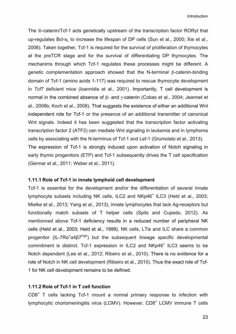
2 Results
2.1 Canonical Wnt signaling and Tcf-1 expression during
NK cell development
The first aim was to test if and when the T cell factor-1 (Tcf-1, encoded by the Tcf7
gene) is expressed during natural killer (NK) cell development and if we can detect
Wingless/Integration-1 (Wnt) signal transduction. By intracellular FACS we could not
detect Tcf-1 expression in the NK cell precursor (NKp) cells. However Tcf-1 was
abundantly expressed in immature NK (iNK) cells and slightly less in mature NK
(mNK) cells compared to Tcf7-/- controls (Fig. 9A). The lymphoid enhancer factor-1
(Lef-1) was absent in all three developmental stages (Fig. 9B). Furthermore, we
detected Tcf-1 in a subset of splenic NK cells from naïve mice whereas Tcf-1 levels
were reduced in NK cells from Polyinosinic:polycytidylic acid (Poly IC) primed mice
(Fig. 9C). Lef-1 was absent in naïve and primed NK cells (Fig. 9D). Therefore we
conclude that during bone marrow development Tcf-1 levels peak at the iNK stage
and are maintained in mNK cells. Upon activation by Poly IC the Tcf-1 levels
decrease.
To determine whether NK cells receive canonical Wnt signals during development,
we measured the expression of conductin/axin2. Conductin is induced in response to
Wnt signals (Jho et al., 2002; Lustig et al., 2002). Moreover, conductin is a direct Tcf-
1 target in CD8+ T cells (Zhou et al., 2010). We used mice in which the endogenous
open reading frame of one conductin allele was replaced with a bacterial β-
galactosidase reporter gene (termed conductinLacZ). Β-galactosidase activity was
used to measure Wnt signal transduction at the single cell level ex vivo. While we
detected significant β-galactosidase activity in T cells but not in B cells (Fig. 10A), we
failed to detect conductin expression in NKp cells. We detected strong reporter
activity in the iNK population and maintenance of the signal in a subpopulation of
mNK cells (Fig. 10B). Similar to bone marrow (BM) mNK cells, we detected
significant β-galactosidase activity in a subset of splenic NK cells (Fig 10A and B).
Importantly the conductin signal was entirely dependent on Tcf-1 both in splenic as
well as in BM mNK cells (Fig. 10B). Concordant with Tcf-1 expression, Wnt signaling

Results
26
is induced during the NKp to iNK transition and is maintained in a subpopulation of
mNK cells.
Figure 9. Tcf-1 is expressed in developing and mature NK cells. In A and B it was gated on BM NKp (CD3-IL-2/15Rβ+NK1.1-DX5-), iNK (CD3-IL-2/15Rβ+NK1.1+DX5-) and mNK (CD3-IL-2/15Rβ+NK1.1+DX5+) cells and in C and D on splenic NK cells (CD3-NK1.1+). Histograms show Tcf-1 (A and C) or Lef-1 expression (B and D) in Tcf7+/+ NK cells (black line) as compared to controls (filled grey) (i.e.Tcf7-/- NK cells in A and C and isotype controls in B and D. Histograms in C and D depict Tcf-1 (C) or Lef-1 (D) expression in either naïve (upper row) or Poly IC primed (lower row) splenic Tcf7+/+ NK cells (CD3-NK1.1+) as compared to controls (filled grey) (i.e.Tcf7-/- NK cells in C and an isotype control D.
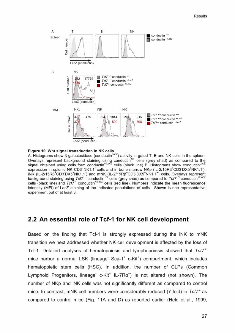
Results
27
Figure 10. Wnt signal transduction in NK cells A. Histograms show β-galactosidase (conductinLacZ) activity in gated T, B and NK cells in the spleen. Overlays represent background staining using conductin+/+ cells (grey shad) as compared to the signal obtained using cells form conductin+/LacZ cells (black line) B. Histograms show conductinLacZ expression in splenic NK CD3- NK1.1+ cells and in bone marrow NKp (IL-2/15Rβ+CD3-DX5+NK1.1-), iNK (IL-2/15Rβ+CD3-DX5+NK1.1-) and mNK (IL-2/15Rβ+CD3-DX5+NK1.1+) cells. Overlays represent background staining using Tcf7+/+ conductin+/+ cells (grey shad) as compared to Tcf7+/+ conductin+/LacZ cells (black line) and Tcf7-/- conductin+/LacZ cells (red line). Numbers indicate the mean fluorescence intensity (MFI) of LacZ staining of the indicated populations of cells. Shown is one representative experiment out of at least 3.
2.2 An essential role of Tcf-1 for NK cell development
Based on the finding that Tcf-1 is strongly expressed during the iNK to mNK
transition we next addressed whether NK cell development is affected by the loss of
Tcf-1. Detailed analyses of hematopoiesis and lymphopoiesis showed that Tcf7-/-
mice harbor a normal LSK (lineage- Sca-1+ c-Kit+) compartment, which includes
hematopoietic stem cells (HSC). In addition, the number of CLPs (Common
Lymphoid Progenitors, lineage- c-Kit+ IL-7Rα+) is not altered (not shown). The
number of NKp and iNK cells was not significantly different as compared to control
mice. In contrast, mNK cell numbers were considerably reduced (7 fold) in Tcf7-/- as
compared to control mice (Fig. 11A and D) as reported earlier (Held et al., 1999;

Results
28
Ioannidis et al., 2003). Splenic and liver NK cells were decreased 3 fold (Fig. 11 B, C
and E). Taken together, we found that Tcf-1 deficiency partially blocks NK cell
development at the mNK stage.
Figure 11. NK cell development in the absence of Tcf7. Density plots of Tcf7+/+ and Tcf7-/- BM (A), spleen (B) and liver (C) cells stained for the indicated markers. Numbers indicate the percentage of gated cells (for the left panel of A, the panels B and C the numbers are the percentage of total cells; for the right panel of A numbers are the percentage of gated IL-2/15Rβ +CD3- cells). D and E. Numbers of cells of the indicated population in the BM (D) and spleen (E) of Tcf7+/+ and Tcf7-/- mice. D. Bar graphs represent the mean number of NKp, iNK and mNK cells in one hind leg ± SEM of n=10 mice. The statistical significance of differences was calculated using two-tailed t-test and (*), (**) and (***) show significant differences (p<0.05), (p<0.01) and (p<0.001) respectively, compared to Tcf7+/+ mice. When nothing is marked, the values are not significantly different (p>0.05).
2.3 A cell intrinsic role for Tcf-1 in NK cell development
To address the basis for the impaired NK cell development in the absence of Tcf-1,
we next determined whether NK cell development depends on Tcf-1 expression by
NK cells or by the surrounding cells. Lethally irradiated wild type (wt) recipient mice
(CD45.1+) were reconstituted with a mixture of equal proportions of Tcf7-/- (CD45.2+)

Results
29
and wild type (CD45.1+) BM cells. Mixtures of Tcf7+/+ (CD45.2+) and wild type
(CD45.1+) BM cells served as controls. After stable engraftment, chimeric mice were
analyzed for the contribution of CD45.2+ cells to the NK cell compartment. As
expected, a sizeable CD45.2+ NK cell population was detected in the spleen and BM
of control chimeras receiving Tcf7+/+ BM (Fig. 12A, B and C). In contrast, very few
CD45.2+ splenic or BM NK cells were derived from Tcf-1-deficient BM precursors (Fig.
12A and B). Compared to controls, absolute numbers of Tcf7-/- NK cells per spleen
and BM were reduced approximately 10 and 15 fold, respectively (Fig. 12C).
Figure 12. Cell intrinsic role of Tcf-1 for NK cell development. Spleens from mixed BM chimeras were analyzed 2-3 months after reconstitution with a 1:1 mixture of competitor wt (CD45.1) and tester Tcf7+/+ (CD45.2) or wt (CD45.1) and Tcf7-/- (CD45.2) BM cells. Density plots show spleen cells stained with mAbs to CD45.1 and CD45.2. Gated CD45.1-CD45.2+
populations were analyzed for CD3 versus B220 expression (A) or CD3 versus NK1.1 expression in spleen or BM (B). Numbers indicate the percentage of cells in the respective region. (C) The bar graph shows the mean absolute number (±SD) of CD45.2+ cells per spleen in mixed chimeras of the indicated type. Data are derived from 8 – 10 chimeras. Contribution: Experiments shown in Figure 12. were performed and analyzed by Werner Held.

Results
30
T cell development from Tcf-1-deficient BM precursors was similarly impaired, in
agreement with a previous report (Weber et al., 2011). In contrast, the development
of other hematopoietic lineages such as B cells, dendritic cells (CD11c+ MHC class
II+) (Ferrero et al., 2002), Macrophages (CD11b+), Granulocytes (Gr-1+) and
nucleated erythroid cells (Ter119+) were not dependent on Tcf-1 (data not shown).
These data show that NK cell development depends on Tcf-1 expression by NK cells
and not by the surrounding cells. Indeed, Tcf-1-deficient hosts readily supported NK
cell development from Tcf-1 sufficient BM progenitors (W. Held, unpublished data).
Overall these data show that Tcf-1 ensures NK cell development in a cell
autonomous fashion.
2.4 Normal NK cell development in the combined absence
of β- and γ-catenin
Although Tcf-1 binds DNA, it lacks intrinsic transcriptional activity. Tcf-1 represses
target genes via its association with Groucho co-repressors. Target gene activation in
response to Wnt signals depends on the binding of the co-activators β-catenin and γ-
catenin to the N-terminus of Tcf-1 (Grigoryan et al., 2008; Jeannet et al., 2008a). We
analyzed NK cell development in mice lacking both β− and γ-catenin. Mice that have
a null mutation of β-catenin or γ-catenin are embryonic lethal (Huelsken et al., 2000;
Ruiz et al., 1996). Therefore we crossed mice harboring floxed alleles of β catenin
(Huelsken et al., 2001) and γ-catenin with mice expressing Cre recombinase under
the control of the hematopoietic-specific Vav regulatory elements (Katzav et al.,
1989; Ogilvy et al., 1999; Stadtfeld and Graf, 2005). We found that BM NK cell
numbers and their developmental progression are normal in the combined absence
of β-catenin and γ–catenin (Fig. 13A). Splenic NK cells were also normal (Fig. 13A).
BM and splenic NK cells showed normal expression of maturation markers and of
common inhibitory and activating receptors (data not shown). Moreover, the
expansion of NK cells in response to IL-2 was normal in the combined absence of β-
catenin and γ-catenin (Fig. 13B). Overall these data show that β-catenin and γ–
catenin are dispensable for NK cell development.

Results
31
Figure 13. NK cell development is normal in the absence of β and γ catenin. (A) Density plots of β+/+ and γ-/- BM (left) and spleen cells (right) stained for the indicated markers. Numbers indicate the percentage of gated cells (for BM left panel and the spleen panel, numbers are the percentage of total cells, whereas for the BM right panel numbers are the percentage of gated IL-2/15Rβ +CD3- cells). Bar graphs represent the mean number of NKp, iNK and mNK cells in one hind leg (left) and splenic NK cells (right) ± SEM of n=4 mice. (B) Nylon wool non-adherent splenocytes were labeled with CFSE and stimulated with IL2 for 5 days. Numbers indicate the percentage of cells in the indicated gates. These data are representative of 4 experiments. Bar Graphs show the fold expansion of NK cells after 5 days of stimulation + SEM. The statistical significance of differences was calculated using two-tailed t-test and (*), (**) and (***) show significant differences (p<0.05), (p<0.01) and (p<0.001) respectively, compared to Tcf7+/+ mice. When nothing is marked, the values are not significantly different (p>0.05).

Results
32
2.5 The repressor domain in Tcf-1 mediates NK cell
development
While Tcf-1 was crucial for NK cell development, the two known Wnt signaling
transmitters β-catenin and γ-catenin were dispensable. Thus we next tested whether
the N-terminal catenin-binding domain of Tcf-1 was needed for NK cell development.
To study the role of different domains of Tcf-1 for NK cell development, our
laboratory has used a genetic complementation approach. A Tcf-1 transgene (Tg)
containing the catenin-binding domain plus the repressor domain (p45) rescued T
cell development in Tcf7-/- mice while a Tg containing only the repressor domain
(p33) failed to do so (Ioannidis et al., 2001). We used the same approach to
determine the mechanism of Tcf-1 function in NK cell development. Intriguingly, BM
NK cell development is fully restored in p33 and in p45 Tg Tcf7-/- mice (Fig. 14). In
addition, the splenic NK cell compartment was restored to normal in the p33 and the
p45 Tg Tcf7-/- mice (Fig. 15). Data shown in Fig. 14 and 15, will be discussed in more
detail later on. These data show that the short Tcf-1 isoform (p33), that contains the
DNA- and Groucho-binding domains but lacks the β-catenin interaction site
(Oosterwegel et al., 1991a; Van de Wetering et al., 1996), is sufficient to restore NK
cell development.

Results
33
Figure 14. The repressor domain of Tcf-1 mediates BM NK cell development. (A) Shown are dot plots of Tcf7+/+, Tcf7-/-, p33 Tcf7-/-, p45 Tcf7-/- BM cells stained for the indicated markers. Numbers indicate the percentage of gated cells (for A left panel, numbers are the percentage of total cells whereas for the right panel of A numbers are the percentage of gated IL-2/15Rβ +CD3- cells). Histograms and density plots show Ly49A, GzmB, KLRG1, CD11b and CD27 expression in gated mNK (IL-2/15Rβ +CD3-NK1.1+DX5+) cells. Numbers indicate the percentage of gated cells and the mean fluorescence intensity (MFI) of GzmB staining. Shown is one representative experiment out of four independent experiments. Bar graphs show the mean number of NK cells in one hind leg ± SEM of n=4 mice (B). (C) The graph depicts the relative mean fluorescence intensity (MFI) ±SEM of Granzyme B expression in GzmB-/-, Tcf7-/-, p33 Tcf7-/-, p45 Tcf7-/- mNK cells normalized to Tcf7+/+ controls that were set to 100%. (n=4). Based on the two-tailed t-test (*), (**) and (***) depict significant differences (p<0.05), (p<0.01) and (p<0.001) respectively, compared to Tcf7+/+ controls. When nothing is marked, the values are not significantly different (p>0.05).

Results
34
Figure 15. The repressor domain of Tcf-1 restores the splenic NK cell compartment Shown are dot plots of Tcf7+/+, Tcf7-/-, p33 Tcf7-/-, p45 Tcf7-/- spleen cells stained for CD3 versus NK1.1. Numbers indicate the percentage of gated cells. Histograms and density plots show Ly49A, GzmB, KLRG1, CD11b and CD27 expression in gated NK (CD3-NK1.1+) cells. Numbers indicate the percentage of gated cells and the mean fluorescence intensity (MFI) of GzmB staining. Shown is one representative experiment of at least four independent experiments. (B) Bar graph represent the mean number of NK cells in the spleen ± SEM of n=4 mice. (C) The graph depicts the MFI ±SEM of GzmB expression in GzmB-/-, Tcf7-/-, p33 Tcf7-/-, p45 Tcf7-/- NK cells normalized to Tcf7+/+ controls that were set to 100%. (n=4). (D) The bar graph represents the fold expansion of Tcf7+/+, Tcf7-/-, p33 Tcf7-/-, p45 Tcf7-/- NK cells between day 0 and day 5 of IL-2 stimulation (n=5). (E) The graph depicts the MFI ±SEM of GzmB expression in GzmB-/-, Tcf7-/-, p33 Tcf7-/-, p45 Tcf7-/- NK cells at day 5 of IL-2 stimulation normalized to Tcf7+/+ controls, that were set to 100% (n=5). Based on the two-tailed t-test (*), (**) and (***) depict significant differences (p<0.05), (p<0.01) and (p<0.001) respectively, compared to Tcf7+/+ controls. If nothing is marked, the values are not significantly different (p>0.05).

Results
35
2.6 Basis for impaired NK cell development in the absence
of Tcf-1
During their development in the BM, NK cells up-regulate the IL-15 receptor complex
followed by inhibitory and activating surface receptors and finally maturation markers.
The next aim was to define the basis for impaired NK cell development in the
absence of Tcf-1. We first tested whether Tcf7 deficiency altered the expression of
activating receptors, which control NK cell function upon interaction with ligands on
target cells. We could not detect any significant changes in the expression levels of
NKG2D, NKp46, NK1.1 or CD16 in BM or spleen NK cells in the absence of Tcf-1
(Fig. 16A and B and data not shown). We also could not detect any significant
changes in the expression levels of the signaling lymphocyte activation molecule
(SLAM) family receptors (SLAM, 2B4, CD84, Ly-9, Ly-108 and CD2-like receptor
activating cytotoxic cells (CRACC)) in mNK or splenic NK cells in the absence of Tcf-
1 (Fig. 16A and B).
Figure 16. Activating receptors are normally expressed in the absence of Tcf-1. Histograms in show overlays of Tcf7+/+ (black line) and Tcf7-/- (filled grey) mNK (CD3-IL-2/15Rβ+NK1.1+DX5+) cells (A.) or splenic NK cells (CD3-NK1.1+) (B). Shown is the expression of different activating receptors including various SLAM family members.

Results
36
NK cells also acquire receptors for major histocompatibility complex (MHC) class I-
molecules including CD94 /NKG2A, and Ly49 receptors. The Ly49 receptor family
includes both inhibitory (Ly49A, Ly49C/I, Ly49F, Ly49G2, Ly49I) and activating
receptors (Ly49D and Ly49H). The expression of Ly49D is significantly decreased
wherease the expression of Ly49H, which recognizes the mouse cytomegalovirus
(MCMV) protein m157, is normal in the absence of Tcf7 (Table 1). Our group has
previously shown that the Ly-49a gene is a direct Tcf-1 target and that the Ly49A
receptor is essentially absent in the absence of Tcf7 (Held et al., 1999). Expression
of the Ly49A receptor in Tcf7-/- NK cells is fully and partially restored by the p45 and
the p33 Tgs, respectively (Ioannidis et al., 2003). The expression of the inhibitory
Ly49 receptors (Ly49C/I, Ly49G2 and Ly49I) was not affected by the loss of Tcf7,
whereas expression of the Ly49F, NKG2/ CD94 were decreased (Table 1). Thus,
expression of several inhibitory receptors was decreased in the absence of Tcf-1.
Table 1. Expression of Ly49 and CD94/NKG2 receptors.
Shown is the Mean % + SD of different inhibitory and activating Ly49 family receptors and CD94/NKG2 expressed by splenic NK cells of the indicated genotypes. Red numbers indicate p>0.05 vs B6. Black numbers indicate no significant difference from B6 controls. Contribution: This table was adapted and completed based on data provided by Werner Held.
We next tested whether the loss of Tcf-1 had an impact on NK cell maturation. The
iNK-mNK cell transition is associated with up-regulation of CD11b followed by CD43
(Kim et al., 2002; Rosmaraki et al., 2001). We found that Tcf7-/- mice have an
increased proportion (2 fold) of CD11bhi and CD11bhiCD43hi BM mNK cells compared
to wt (Fig. 14A). Likewise, the proportion of CD11bhiCD43hi NK cells was increased in
the liver and the spleen of Tcf-1-deficient mice (1.4 fold) (Fig. 15A and data not
shown). NK cells can be divided into CD11b− CD27+ (immature), CD11b+CD27+
(mature1; M1), and CD11b+ CD27− (mature2; M2) subsets. The CD11b+CD27+

Results
37
subset is the most potent in acquiring effector functions whereas the M2 population
mostly co-expresses KLRG1 that marks terminal maturation (Hayakawa and Smyth,
2006). The M2 CD11b+ CD27− subset was strongly increased in the BM and the
spleen of mice that lack Tcf-1 compared to wild type controls. Furthermore in the
absence of Tcf-1 the bulk of mNK (50-60%) and splenic NK cells (75%) expressed
KLRG1 compared to (5-8%) and (30%) of Tcf7+/+ NK cells (Fig. 14A). These data
show that Tcf-1 deficient NK cells have a more mature phenotype. The expression of
maturation markers in p33 Tg Tcf7-/- mice was comparable to wild type controls.
However p45 Tg Tcf7-/- NK cells showed a very immature phenotype. Around 90% of
the BM and 72% of splenic NK cells of p45 Tg Tcf7-/- mice were immature
(CD11b−CD27+) compared to wt controls BM (54.8%) and spleen (35.5%),
respectively. The mature M1 (CD11b+CD27+) NK cell fraction was strongly reduced
in the BM and the spleen and the M2 (CD11b+CD27-) population was completely
absent (Fig. 15A). Furthermore, the NK cells of p45 Tg Tcf7-/- mice did not up-
regulate KLRG1. We conclude that enforced Tcf-1 p45 signaling prevents maturation
whereas the absence of Tcf-1 accelerates maturation.
2.7 Tcf-1 ensures the survival of BM and cytokine
stimulated NK cells
The analysis of NK cell surface markers in the absence of Tcf7 did not directly reveal
why NK cell development was blocked at the mNK stage. The emergence of mNK
cells coincides with rapid cycling of precursor cells (Kim et al., 2002). Thus the
reasons for the reduced number of Tcf7-/- mNK cells could be a defect in cycling
and/or survival. To examine cell proliferation we measured the DNA content of the
cells using Hoechst staining. We found that Tcf-1-deficient BM mNK cells cycled
(S/G2/M) at least as efficiently as control mNK cells (Fig. 17A). There was also no
difference in the proportion of cycling NKp and iNK cells (Fig. 17A). Thus, even
though Tcf-1-deficient BM NK cells cycle normally, their number fails to increase,
suggesting that Tcf7-/- NK cells have a survival defect.
Freshly isolated Tcf7 deficient BM mNK cells did not show increased Annexin V
staining, which detects apoptotic cells (data not shown). However, following in vitro
culture (4h) in the absence of exogenous growth factors a significantly increased

Results
38
fraction of Tcf7-/- BM NK cells became Annexin V+ (44.0±1.4% n=5) as compared to
wild type BM NK cells (27.5±1.9% n=5) (Fig. 17B). This observation was specific to
BM NK cells since splenic NK cells did not show an increase in Annexin V+ cells
following in vitro culture (4h) (not shown). Overall this suggests that Tcf-1 ensures
the survival of BM NK cells.
We further tested whether cytokine stimulation of splenic NK cells resulted in a
corresponding impairment of expansion. In contrast to wild type NK cells, Tcf-1-
deficient NK cells failed to efficiently accumulate in number in response to either IL2
or IL-15 stimulation (Fig. 17C). In response to IL-2 (or IL-15) Tcf7-/- NK cells showed
normal cell cycle progression based on the combined analysis of BrdU incorporation
and 7-AAD staining (Fig. 17E). In addition, as shown by the efficient dilution of CFSE,
Tcf7-/- NK cells underwent as many cell divisions as wild type control NK cells (Fig.
17F). At day 5 of cytokine stimulation, significantly more Tcf7-/- NK cells were Annexin
V+ (19±2.0%) as compared to wild type (8±1.0%) (Fig. 17G). Corresponding data
were obtained at day 3 and based on TUNEL assays (Tcf7+/+ 5.3±0.3% Tunnel+,
Tcf7-/- 9.9±0.8% Tunnel +) (Fig.17F). When cell death (Annexin V) and cell division
(CFSE) were simultaneously monitored we noted increased rates of cell death at
each division both at day 3 and day 5 of cytokine stimulation, suggesting that at each
division a certain fraction of proliferating cells undergoes apoptosis (not shown).
Similar to BM NK cell development we found that the presence of the p33 Tg was
sufficient to restore NK cell accumulation upon cytokine stimulation (Fig. 15D). Taken
together, these data show that the poor accumulation of BM and cytokine stimulated
splenic NK cells is caused by increased rates of cell death. Furthermore, we found
that the suppressor activity of Tcf-1 prevent the apoptosis of cycling NK cells.
Next we determined whether the death of cytokine activated NK cells idepended on
Tcf-1 expression by NK cells or by the surrounding cells. Splenic Tcf7-/- (CD45.2) and
Tcf7 +/+ (CD45.1) were mixed at a ratio of 1:1 and cultured for 5 days in vitro in the
presence of IL-2. While Tcf-1 sufficient NK cells expanded normally, Tcf-1-deficient
NK cells failed to efficiently accumulate (Fig. 18). Taken together these data show
that Tcf-1 ensures the survival of proliferating BM mNK cells and cytokine stimulated
peripheral NK cells in a cell autonomous fashion.

Results
39
Figure 17. Tcf-1 is required for NK cell expansion. A. Hoechst (DNA) staining of the indicated NK cell populations in BM of Tcf7+/+ or Tcf7-/- mice. Hoechst positive cells are in S, G2, M phases of the cell cycle. Numbers show the mean percentage + SD of Hoechst positive cells in the different populations. These data are representative of 3 experiments. B. Annexin V (marker of apoptosis) staining of the indicated BM NK cell populations in Tcf7+/+ and Tcf7-/- mice cultured for 4 hours at 37°. Bar graphs depict the mean percentage (±SEM) of AnnexinV and 7AAD positive cells (n=5). The statistical significance of differences was calculated using two-tailed t-test and * shows statistical significance (p<0.05) compared to Tcf7+/+ mice. C. Nylon wool non-adherent splenocytes from Tcf7+/+ or Tcf7-/- mice were stimulated with IL-2 or IL-15. Shown is one representative dot plot of at least 10 experiments for IL-2 stimulation. Bar graphs show the fold expansion of NK cells between day 0 and day 5 of stimulation + SEM. These data are representative of at least 10 experiments for IL-2 stimulation and for 3 experiments for IL-15 stimulation. The statistical significance of differences was calculated using two-tailed t-test and * shows statistical significance (p<0.05) compared to Tcf7+/+ (C2) or Tcf7+/- (C3) mice. D. Scatter plots depict the cell cycle distribution of gated NK cells at day 5 after IL-2 stimulation following treatment with 1 BrdU for 45 min. Bivariate analysis of the DNA content (DAPI staining) and the incorporation of BrdU was performed. These data are representative of at least 3 experiments. E. Nylon wool non-adherent splenocytes were labeled with CFSE and stimulated with IL2 for 5 days. Numbers indicate the percentage of cells in the indicated gates. These data are representative of 9 experiments. F. Nylon wool non-adherent splenocytes were stimulated with IL2 for 5 days. The numbers indicate the percentage of cells in the indicated gates. These data are representative of at least 7 experiments. Bar Graphs: percentage of AnnexinV (left panel) or Tunnel (right panel) postive cells after 5 days of stimulation + SEM (n=4 for Annexin V and n=3 for Tunnel assays).

Results
40
Figure 18. Cell intrinsic role of Tcf-1 for NK cell expansion. Bar graph depicts fold expansion of Tcf7 +/+ (CD45.1+) and Tcf7 -/- (CD45.2+) NK cells were cultured separately (left) or as a 1:1 mixture (right) for 5 days with IL-2. NK cell numbers were determined by gating on CD45.1+CD3-NK1.1+ for Tcf7 +/+ and on CD45.2+CD3-NK1.1+ for Tcf7 -/- cells. Bar graphs represent the mean number of indicated NK cells ± SEM of 3 independent experiments. (**) depicts significant differences (p<0.01) on the student`s t-test.
NK cell development and survival is critically dependent on cytokines. IL-15
deficiency blocks NK cell development at the iNK stage (Vosshenrich et al., 2005).
This cytokine is also required for NK cell homeostasis and may be necessary for NK
cell expansion (Koka et al., 2003). The IL-15 receptor complex is composed of the IL-
15 receptor α (IL-15Rα), IL-2Rβ (CD122), and the IL-2 receptor common γ chains (IL-
2Rγc). Compared to their wild type counterparts, Tcf7-/- BM mNK and splenic NK cells
expressed equivalent amounts of IL-2/15Rβ (Fig. 19A). IL-15R signaling mediates
the survival of NK cells by promoting the expression of high levels of anti-apoptotic
Bcl-2 and Bcl-XL. Moreover it has been shown that Bcl-2 family members regulate
cytokine deprivation dependent apoptosis (Carson et al., 1997; Cooper et al., 2002;
Ranson et al., 2003). Intracellular staining showed that the expression of Bcl-2 and
Bcl-XL in freshly isolated or cytokine stimulated NK cells was unperturbed in the
absence of Tcf-1 (Fig. 19). Taken together we conclude that the survival defect of
Tcf-1 deficient NK cells is not caused by the reduced expression of the IL-15 receptor
complex or by a decrease of the anti-apoptotic molecules Bcl-2 and Bcl-XL.

Results
41
Figure 19. The IL-15 receptor and the anti-apoptotic molecules Bcl-2 and Bcl-X are normally expressed in the absence of Tcf-1. A. The graph depicts the MFI ±SEM of IL-2/15Rβ expression in Tcf7-/-, p33 Tcf7-/-, p45 Tcf7-/- NK cells normalized to Tcf7+/+ controls that were set to 100%. (n=5). B. Expression of Bcl-2 and Bcl-XL in freshly isolated BM and splenic NK cells or IL-2 day 7 stimulated NK cells. The open histogram represents the isotype controls. Numbers indicate the MFI of staining. These data are representative of 3 experiments for freshly isolated cells and from a single experiment for stimulated NK cells. Contribution: Experiment for figure 19B was done by Grégoire Jeannet.
2.8 Tcf-1-deficient BM NK cells over-express Granzymes
To elucidate the mechanisms underlying apoptosis of NK cells, we established the
global gene expression profiles of FACS-purified BM mNK cells (CD3-CD19-
CD122+NK1.1+DX5+) isolated from Tcf7+/+ and Tcf7-/- mice using Affymetrix arrays.
Based on three independent data sets, 364 of 28'000 genes were differentially
expressed (False discovery rate FDR (adjusted P value)< 10% and fold change (FC)
> 1.5). A total of 205 genes were up-regulated and 159 genes were down regulated
in Tcf7+/+ compared to Tcf7-/- BM NK mNK cells (Table 2).

Results
42
Table 2. Gene expression profile of mNK cells derived either form Tcf7-/- or Tcf7+/+ mice.
Cut-off UP DOWN
FDR < 5% 15 5
FDR < 10% and FC > 1.5 205 159
Based on three independent data sets, 20 FDR < 5% and 364 (False discovery rate FDR (adjusted P value)< 10% and fold change (FC) > 1.5) of 28'000 genes were differentially expressed). A total of 205 genes were significantly up-regulated (green) and 159 genes were down regulated (red) in Tcf7+/+ compared to Tcf7-/- BM NK mNK cells
As expected, Tcf7 transcripts were down regulated in NK cells, yet still detectable,
which is in agreement with data obtained by qRT-PCR. However, in Tcf7-/- NK cells,
Tcf-1 protein was not detected by Western blot (Held et al., 1999). Tcf7-/- NK cells
showed higher mRNA levels of genes encoding Minichromosome maintenance
factors (Mcm) (Mcm10, Mcm5, Mcm3), cyclin (cyclinF, cyclinA2, cdk14, cdk inhibitor
Cdnk2c (p18, inhibits Cdk4, repressed by c-Myc)), E2Fs (E2F8, E2F2) in agreement
with efficient cycling of Tcf7-/- mNK cells (Table 3, The Affimetrix microarray has been
done by the Center for Integrative Genomics at the University of Lausanne). The
transcript levels of the pro-apoptotic Bcl-2 family members Bim, Puma, Noxa or Bax
in Tcf7-/- mNK cells were comparable to wt controls. Amongst the most up-regulated
transcripts upon loss of Tcf7 was the gene encoding for GzmB (2.5-fold as
determined by microarray, p<0.5 and 8.7-fold as determined by qRT-PCR analysis
(Table 2 and Fig. 20D). Genes coding for other Granzyme family proteins, including
Granzyme C (2.5-fold, p<0.05) and Granzyme K (2-fold, p<0.1) were also up-
regulated (Table 3). GzmB is a serine protease and the main component of exocytic
granules used by cytotoxic T cells (CTL) and natural killer (NK) cells to destroy cells
infected with intracellular pathogens or tumor cells (Russell and Ley, 2002). Recently
several groups have reported GzmB-leakage-induced cell death in NK cells and
CTLs where GzmB escapes into the cytoplasm, and thereby provokes apoptosis of
the GzmB producing cell itself (Ida et al., 2003; Zhang et al., 2006).

Results
43
Table 3. Gene expression profile of mNK cells derived either form Tcf7-/- or Tcf7+/+ mice.
Gene accession nr. Gene Gene description
adj. P.Val
Tcf7-/- mean
Tcf7+/+ mean
Fold_ Change
NM_009640 Angpt1 angiopoietin 1 0.020 5.1 7.0 -3.7 NM_016970 Klrg1 killer cell lectin-like receptor subfamily G 0.030 9.7 7.6 4.1 NM_013473 Anxa8 annexin A8 0.036 7.3 4.3 7.8 NM_027127 Gpx8 glutathione peroxidase 8 (putative) 0.039 6.5 4.7 3.6 NM_139198 Plac8 placenta-specific 8 0.039 9.3 8.1 2.3 NM_009848 Entpd1 ectonucleoside triphosphate diphosphohydrolase 1 0.039 9.7 7.4 4.8
NM_013542 GzmB granzyme B 0.039 11.4 10.1 2.5 NM_009987 Cx3cr1 chemokine (C-X3-C) receptor 1 0.039 8.0 6.6 2.8 NM_010371 GzmC granzyme C 0.039 6.5 5.1 2.5 NM_024477 Ttc28 tetratricopeptide repeat domain 28 0.039 7.0 8.2 -2.4 NM_011311 S100a4 S100 calcium binding protein A4 0.039 11.2 10.2 2.0 NM_030712 Cxcr6 chemokine (C-X-C motif) receptor 6 0.039 10.3 8.6 3.3
NM_177660 Zbtb10 zinc finger and BTB domain containing 10 0.039 6.3 7.2 -1.9 NM_009858 Cd8b1 CD8 antigen, beta chain 1 0.040 9.3 5.6 13.1 NM_001163608 Plxdc1 plexin domain containing 1 0.044 5.4 4.5 1.8 NM_011920 Abcg2 ATP-binding cassette, sub-family G 0.048 6.4 7.8 -2.5 NM_008479 Lag3 lymphocyte-activation gene 3 0.050 6.6 5.6 2.1 NM_010740 Cd93 CD93 antigen 0.050 6.7 5.7 1.9
NM_011496 Aurkb aurora kinase B 0.050 9.4 8.5 1.9 NM_009331 Tcf7 transcription factor 7, T-cell specific 0.050 9.8 10.8 -2.0 NM_027290 Mcm10 minichromosome maintenance deficient 10 0.059 7.6 6.9 1.7 NM_026410 Cdca5 cell division cycle associated 5 0.059 8.4 7.5 1.8 NM_001013368 E2f8 E2F transcription factor 8 0.059 8.4 7.6 1.8 NM_007634 Ccnf cyclin F 0.059 7.6 6.8 1.8 NM_008196 GzmK granzyme K 0.059 9.3 8.3 2.0 NM_177733 E2f2 E2F transcription factor 2 0.059 7.6 6.8 1.8
NM_007671 Cdkn2c cyclin-dependent kinase inhibitor 2C (p18, inhibits CDK4) 0.059 9.0 8.2
1.7
NM_011074 Cdk14 cyclin-dependent kinase 14 0.059 7.2 6.5 1.6
NM_009828 Ccna2 cyclin A2 0.073 10.8 10.0 1.7
NM_008566 Mcm5 minichromosome maintenance deficient 5 0.073 8.8 8.2 1.4
NM_008563 Mcm3 minichromosome maintenance deficient 3 0.077 9.0 8.3 1.5
NM_011452 Serpinb9b serine (or cysteine) peptidase inhibitor, clade B, member 9b 0.497 7.9 7.7 1.2
NM_009256 Serpinb9 (Spi-6)
serine (or cysteine) peptidase inhibitor, clade B, member 9 0.808 9.5 9.4 1.1
NM_009982 Ctsc cathepsin C 0.812 9.3 9.2 1.1
NM_011453 Serpinb9c serine (or cysteine) peptidase inhibitor, clade B, member 9c
0.831 3.1 3.2 -1.0
NM_011456 Serpinb9e serine (or cysteine) peptidase inhibitor, clade B, member 9e 0.844 2.8 2.9 -1.1
NM_183197 Serpinb9f serine (or cysteine) peptidase inhibitor, clade B, member 9f 0.901 3.3 3.2 1.0
NM_011455 Serpinb9g serine (or cysteine) peptidase inhibitor, clade B, member 9g 0.963 2.8 2.8 1.0
cDNA from FACS sorted BM mNK cells was analyzed on an Affymetrix microarray. Table 2 shows the gene accession number, symbol and description, the adjusted p-value, the mean expression of three independent data sets for Tcf7+/+ and Tcf7-/- and the fold change. Adjusted p-values have been computed for each comparison by the Benjamini-Hochberg method, controlling for false discovery rate (FDR). Expression values are measured using RMA algorithm from the BioConductor affy package. The expression values are on the log 2 scale and range from 0 to 16. First section: annotated genes with an FDR < 5%. Second section: genes with an FDR < 10% relevant for this thesis. Red: genes that are increased; green: genes that are decreased in Tcf7-/-. The last panel shows genes of interest that do not show a significant change in expression. Genes that are mentioned in this thesis are highlighted in bold.

Results
44
Mature NK cells from naïve mice transcribe GzmB and Prf1 mRNA constitutively.
However, such NK cells are minimally cytotoxic because of a block in Prf1 and GzmB
mRNA translation that is only released by NK cell priming (Fehniger et al., 2007). In
agreement with these data, splenic NK cells express little GzmB but they abundantly
express GzmA as reported previously (Fehniger et al., 2007) (Fig. 20A). We also
could not detect GzmB protein in NKP, iNK and mNK cells from wt mice. In contrast,
Tcf7 deficient iNK and mNK cells abundantly expressed GzmB protein as determined
by intracellular FACS staining (Fig. 20B).
We decided to analyze the subcellular localization of GzmB in activated NK cells. We
detected a granular GzmB localization in the cytoplasm of wild type NK cells, which
co-localized with lysosome-associated membrane protein 1 (LAMP-1) (Fig. 21A). In
sharp contrast, the activated Tcf7-/- NK cells showed a diffuse cytoplasmic distribution
of GzmB and LAMP-1, which partly co-localized (Fig. 21A). Based on these
observations we hypothesized that GzmB mediated, self-inflected damage may
explain the increased cell death of proliferating Tcf-1 deficient NK cells.
A major determinant of GzmB-leakage-induced cell death is an excess of cytosolic
GzmB over the serine protease inhibitor 6 (Spi-6), its principal cytosolic inhibitor (Ida
et al., 2003; Laforge et al., 2006; Phillips et al., 2004; Zhang et al., 2006). Spi-6
prevents the processing of the N-terminal GzmB pro-peptide by the protease
cathepsin C (or DPPI), which is required to unlock the proteolytic activity of GzmB
(Caputo et al., 1993; Pham and Ley, 1999; Sun et al., 1997; Zhang et al., 2006).
Based on the Affymetrix arrays Cathepsin C (Ctsc) and Spi-6 (serpinb9) or
homologues (serpinb9b to serpinb9g) transcript levels were not different between
wild type and Tcf7 deficient mNK cells (Table 3). In the absence of Tcf-1, Spi-6 levels
in naïve splenic NK cells and in IL-2 activated NK cells were comparable to wild type
controls (Fig. 21B). Thus in the absence of Tcf7, activated NK cells show increased
levels of cytosolic GzmB, whereas Spi-6 levels remain unchanged. Spi-6 might
therefore not be able to prevent GzmB induced self-inflected apoptosis.
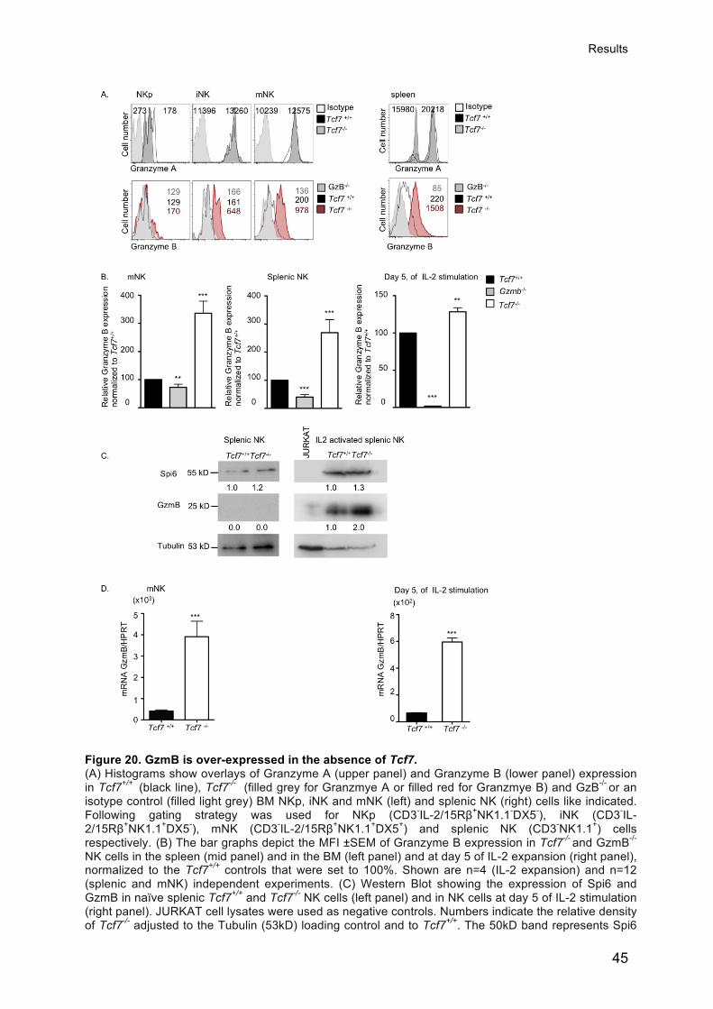
Results
45
Figure 20. GzmB is over-expressed in the absence of Tcf7. (A) Histograms show overlays of Granzyme A (upper panel) and Granzyme B (lower panel) expression in Tcf7+/+ (black line), Tcf7-/- (filled grey for Granzmye A or filled red for Granzmye B) and GzB-/- or an isotype control (filled light grey) BM NKp, iNK and mNK (left) and splenic NK (right) cells like indicated. Following gating strategy was used for NKp (CD3-IL-2/15Rβ+NK1.1-DX5-), iNK (CD3-IL-2/15Rβ+NK1.1+DX5-), mNK (CD3-IL-2/15Rβ+NK1.1+DX5+) and splenic NK (CD3-NK1.1+) cells respectively. (B) The bar graphs depict the MFI ±SEM of Granzyme B expression in Tcf7-/- and GzmB-/- NK cells in the spleen (mid panel) and in the BM (left panel) and at day 5 of IL-2 expansion (right panel), normalized to the Tcf7+/+ controls that were set to 100%. Shown are n=4 (IL-2 expansion) and n=12 (splenic and mNK) independent experiments. (C) Western Blot showing the expression of Spi6 and GzmB in naïve splenic Tcf7+/+ and Tcf7-/- NK cells (left panel) and in NK cells at day 5 of IL-2 stimulation (right panel). JURKAT cell lysates were used as negative controls. Numbers indicate the relative density of Tcf7-/- adjusted to the Tubulin (53kD) loading control and to Tcf7+/+. The 50kD band represents Spi6

Results
46
and the 22kD band shows GzmB. (D) Quantitative RT-PCR analysis of the GzmB gene expression Tcf7+/+ and Tcf7-/- mNK cells (left) and in IL-2 expanded NK cells (right) (n=3). Based on the two-tailed t-test (*), (**) and (***) depict significant differences (p<0.05), (p<0.01) and (p<0.001) respectively, compared to Tcf7+/+ controls. Contribution: RT-PCRs shown in (Fig. 20C) were performed and analyzed by Beena-Jeevan-Raj (IL-2 activated NK cells) and by Georgi Angelov (mNK).
It has previously been reported, using Spi-6 deficient cytotoxic T cells, that an excess
of GzmB disrupts lytic granules (Zhang et al., 2006). To see whether the absence of
Tcf-1 or an excess of GzmB influenced the granule stability in activated NK cells, we
analyzed Tcf7xGzmB double deficient NK cells. The granular morphology of LAMP-
1+ exocytic lysosomes was restored in Tcf7-deficient NK cells lacking GzmB (Fig. 21).
We estimated the average number of GzmB and LAMP-1 positive granules per NK
cell cross-section. A cytokine activated NK cell showed on average 5 GzmB positive
granules per cross-cell section that co-stained for LAMP-1 (Fig. 22A). In contrast, NK
cells lacking Tcf-1 had only 0.9 clear GzmB positive granules per section but showed
abundant diffuse GzmB staining that partly overlapped with similarly diffuse LAMP-1
staining (Fig. 22A and B). In the combined absence of GzmB and Tcf-1, LAMP-1+
lysosomes were back to normal in terms of morphology and number (Fig. 22 A and
B). Taken together, we find that GzmB is abundantly present in the cytoplasm of Tcf7
deficient NK cells. Moreover, the integrity of LAMP-1+ granules is disrupted, and this
was restored to normal by the absence of GzmB. This suggests that over-expression
of GzmB due to the absence of Tcf7 leads to abundant GzmB in the cytoplasm,
which may induce self-inflected NK cell death.
Based on the findings in IL-2 activated NK cells, we explored the localization of
GzmB in mNK cells. Translation of GzmB mRNA is prevented in wild type NK cells,
and GzmB was only occasionally detected by confocal microscopy. The absence of
Tcf-1 leads to enhanced GzmB mRNA expression and translation in mNK cells (Fig.
20 and 21). Similar to activated NK cells, BM NK cells lacking Tcf-1 showed diffuse
cytoplasmic/nuclear localization for GzmB. Only rare Lamp-1 positive granules could
be detected and they did not co-stain for GzmB (Fig. 23A and B). This suggested that
Tcf7-deficient mNK cells cannot compartmentalize GzmB in exocytic granules and
that this leads to a cytpolsamic/nuclear accumulation of GzmB. Taken together, in
the absence of Tcf-1 the GzmB gene is over-expressed. Furthermore, GzmB mRNA
is translated into GzmB protein that accumulates in the cytoplasm and nucleus of
mNK cells where it may induce apoptosis of the NK cell.

Results
47
Figure 21. GzmB leaks into the cytoplasm of IL-2 activated splenic Tcf7-/- NK cells. Purified Tcf7+/+Gzmb+/+, Tcf7+/+Gzmb-/-, Tcf7 -/-Gzmb+/+ and Tcf7-/-Gzmb-/- NK cells were expanded for 5 days in IL-2 and FACS sorted for (CD3-NK1.1+). Subsequently NK cells were stained for Granzyme B (green), Lamp-1 (red) and DAPI (nuclear dye) (blue). Shown are representative NK cell cross sections out of at least three independent experiments with GzmB and Lamp-1 visualized by Laser-scanning Confocal Microscopy. Images are as follows: from left to right, Granzyme B, Lamp1, DAPI and their overlay. Isotype matched mAbs were used as negative controls for the Lamp1 staining and GzmB-/- NK cells were used as a negative control for the GzmB staining.

Results
48
Figure 22. GzmB leaks into the cytoplasm of IL-2 activated splenic Tcf7-/- NK cells. (A) Bar graphs depict the number of granules per cell section for GzmB (left) and Lamp-1 (right) of (n=60) cells of the indicated genotypes. (B) A minimum of 80 cells per genotype (Tcf7 +/+Gzmb+/+, Tcf7 +/+Gzmb-/-, Tcf7-/-Gzmb+/+ and Tcf7-/-Gzmb-/-) where analyzed in detail for the expression of GzmB (green) and LAMP-1 (red) and their co-localization (yellow), which was either defined as granular (left) or diffuse (right) as exemplified in (B). Bar graphs in (B) (left side) show the percentage of cells that either depicts GzmB or Lamp-1 granules and the percentage of cells in which Lamp-1 and GzmB co-localize. Bar graphs in the right panel show the percentage of cells with diffuse GzmB or Lamp-1 expression and the percentage of cells were GzmB and Lamp-1 co-localize.

Results
49
Figure 23. Tcf7 deficient BM mNK cells express high levels of Granzyme B. (A) FACS sorted Tcf7+/+ and Tcf7-/- mNK cells stained for GzmB (green) and Lamp1 (red) and nucleus (blue). Shown are cross sections visualized by confocal microscopy. Images from left to right: GzmB, Lamp-1, nucleus and overlay. Shown are representative samples out of at least three independent experiments with n=6 mice, each. (B) Bar graphs depict the percentage of cells (n=70) that express GzmB, Lamp-1 or that show co-localized GzmB and Lamp-1 expression.

Results
50
While GzmB protein is not expressed during BM development and in unprimed
peripheral NK cells of wild type mice, we detected abundant GzmB protein in iNK,
mNK and naïve peripheral NK cells in the absence of Tcf-1. The short Tcf-1 isoform
(p33) was sufficient to suppress GzmB protein expression in Tcf7-/- iNK, mNK and
peripheral NK cells. GzmB was also expressed at lower levels in cytokine-activated
p33 Tg Tcf7-/- NK cells as compared to controls (Fig. 15E). These data show that p33
Tcf-1 is sufficient to prevent GzmB expression, which correlates with a rescue of BM
NK cell development.
2.9 Absence of GzmB does not rescue development of Tcf-
1-deficient NK cells
To verify the importance of GzmB repression for NK cell development, we used mice
deficient in GzmB. In the mouse a large number of Granzyme genes (including (5′-
>3′) GzmB, C, F, G, L, N, D, and E) are present in the GzmB gene cluster. Beside
GzmB, GzmC transcripts were also up regulated in Tcf-1 deficient mNK cells.
GzmB−/−PGK-neo mice are deficient in GzmB and express reduced levels of GzmC
and F due to the integration of the PGK-neo cassette (Pham et al., 1996; Revell et al.,
2005). We generated GzmB−/− Tcf7−/− mice. The mNK cell pool was similarly reduced
(6.5 fold) in GzmB−/− Tcf7−/− mice as in Tcf7 deficient mice (Fig. 24A and B). In
addition, peripheral NK cell numbers were also reduced (Fig. 25A and B). Thus,
impaired mNK development in Tcf7 deficient mice was not rescued by the deletion of
GzmB. Tcf-1 seems to control additional factors that are required for mNK cell
survival.

Results
51
Figure 24. The absence of GzmB is not sufficient to restore BM NK cell development in the absence of Tcf7. A. Dot plots of Tcf7 +/+Gzmb+/+, Tcf7 +/+Gzmb-/-, Tcf7 -/-Gzmb+/+ and Tcf7 -/-Gzmb-/- BM cells stained for the indicated markers. Numbers indicate the percentage of gated cells (for A left panel, numbers are the percentage of total cells whereas for the right panel of A numbers are the percentage of gated IL-2/15Rβ +CD3- cells). Histograms show Ly49A, KLRG1 and GzmB expression in gated mNK cells. Numbers indicate the percentage of gated cells and the MFI of GzmB staining. (B) Bar graphs represent the mean number of cells in one hind leg ± SEM of n=4 mice. Based on the two-tailed t-test (*), (**) and (***) depict significant differences (p<0.05), (p<0.01) and (p<0.001) respectively, compared to Tcf7 +/+Gzmb+/+ controls. When nothing is marked, the values are not significantly different (p>0.05).

Results
52
Figure 25. The absence of GzmB is not sufficient to restore splenic NK cell numbers in the absence of Tcf7. A. Dor plots of Tcf7 +/+Gzmb+/+, Tcf7 +/+Gzmb-/-, Tcf7 -/-Gzmb+/+ and Tcf7 -/-Gzmb-/- spleen cells stained for NK1.1 versus CD3. Histograms show Ly49A, KLRG1 and GzmB expression in gated NK (CD3-
NK1.1+) cells. Numbers indicate the percentage of gated cells and the MFI of GzmB staining. (B) Bar graphs represent the mean number of cells ± SEM of n=4 mice. Based on the two-tailed t-test (*), (**) and (***) depict significant differences (p<0.05), (p<0.01) and (p<0.001) respectively, compared to Tcf7 +/+Gzmb+/+ controls. When nothing is marked, the values are not significantly different (p>0.05).

Results
53
2.10 Absence of GzmB restores cytokine driven expansion
of Tcf-1-deficient NK cells
In contrast to wild type NK cells, Tcf-1-deficient NK cells failed to efficiently
accumulate in number in response to cytokine stimulation (Fig. 26). Intriguingly, in
the absence of GzmB, the expansion of Tcf7 deficient NK cells was restored (Fig. 26).
Compared to the wild type NK cells (9±2%), a significantly increased fraction of the
Tcf7-/- NK cells was Annexin V+ (20±2%). The absence of GzmB in Tcf7 deficient
mice led to a strong reduction of the apoptotic cell fraction (6±1% Annexin V+) (Fig.
26B,E).
Figure 26. The expansion of Tcf7 deficient NK cells is restored by the absence of GzmB. Tcf7 +/+Gzmb+/+, Tcf7 +/+Gzmb-/-, Tcf7 -/-Gzmb+/+ and Tcf7 -/-Gzmb-/- splenocytes at day 5 of IL-2 stimulation. (A) Numbers in dot plots, indicate the percentage of gated NK1.1+CD3- cells. (B) Scatter plots depict the expression of Annexin V versus 7AAD in the NK1.1+CD3- cells. Numbers indicate the

Results
54
percentage of cells in the respective quadrant. Shown is one representative sample of at least three independent experiments. Histograms in (C) show GzmB and Ly49A expression in gated NK1.1+CD3- cells. (D and E) Bar graphs show fold expansion (D) and the percentage of Annexin V positive NK1.1+CD3- cells (E) ± SEM of n=4 (D) and n=3 (E) experiments. The statistical significance of differences was calculated using two-tailed t-test and (*), (**) and (***) show significant differences (p<0.05), (p<0.01) and (p<0.001) respectively, compared to Tcf7 +/+Gzmb+/+ mice. When nothing is marked, the values are not significantly different (p>0.05).
These data show that the expansion of Tcf-1 deficient NK cells can be rescued by
the absence of GzmB. This suggests that the absence of Tcf7 and as a consequence
the up-regulation of GzmB induces apoptosis of the NK cells.
2.11 NK cell function in the absence of Tcf-1
We could not detect significant changes in the expression of activating NK cell
receptors while certain inhibitory receptors were significantly reduced in the absence
of Tcf-1. We next tested the ability of poly IC primed NK cells from Tcf7-/- mice to
mount an effector response. As compared to wild type NK cells, stimulation with
various tumor cells (RMA/S, RMA-H60, YAC-1) or monoclonal antibodies (mAbs) to
activating NK cell receptors (NKp46, NK1.1 or NKG2D) induced a comparable
production of the cytokine IFN-γ or of the chemokine MIP1α and a normal release of
secretory granules (LAMP-1 release) by Tcf7-/- NK cells (Figs. 27 and 28). The only
exception was a reduced IFNγ production in response to cross-linking with anti-NK1.1
antibody (Fig. 28).
We further tested the ability of Tcf7-/- NK cells to kill susceptible target cells in vitro.
As expected, the Poly IC primed splenocytes from normal mice efficiently killed
RMA/S tumor target cells while syngeneic MHC-I+ RMA cells were not killed (Figs.
28A, B and C). In contrast, Tcf7-/- mice had a 5-10 fold reduced ability to kill RMA/S
cells (Fig. 29B). Tcf7-/- mice also showed significantly reduced antibody-dependent
cell-mediated cytotoxicity (ADCC) towards Thy1 coated RMA cells. In addition, they
showed impaired killing of RMA-m157 (Ly49H) and of YAC-1 (in part NKG2D) target
cells (Fig. 29C). As an exception, Tcf7-/- NK cells killed RMA-H60 cells as efficiently
as wild type controls, suggesting that high levels of NK cell activation through
NKG2D can restore normal target cell killing by Tcf7-/- NK cells (Fig. 29C).

Results
55
Figure 27. Tcf7 deficient NK cells mount produce cytokine and chemokine upon target cell recognition. (A) Dot plots show splenocytes of poly IC primed mice gated for CD3- versus NK1.1+. (B) Density plots show representative examples of LAMP-1 release versus INFγ (left panel) or versus MIP1a (right panel) by poly IC primed Tcf7+/+ and Tcf7-/- NK cells in response to the MHC-I expressing RMA cells (negative control) or of different tumor target cell lines that are either MHC-I low (RMAS) (missing-self) or over-express activating NKG2D ligands (RMA-H60, YAC-1). As a positive control cells were stimulated with Phorbol 12-myristate 13-acetate (PMA) and Ionomycin (I) indicated as (P/I). Numbers represent the percentage of positive cells within the respective gates. Shown is one representative experiment out of at least 4.
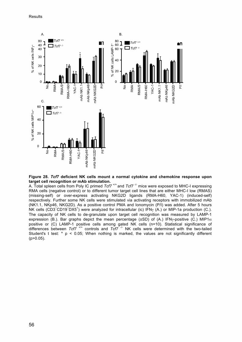
Results
56
Figure 28. Tcf7 deficient NK cells mount a normal cytokine and chemokine response upon target cell recognition or mAb stimulation. A. Total spleen cells from Poly IC primed Tcf7 +/+ and Tcf7 -/- mice were exposed to MHC-I expressing RMA cells (negative control) or to different tumor target cell lines that are either MHC-I low (RMAS) (missing-self) or over-express activating NKG2D ligands (RMA-H60, YAC-1) (induced-self) respectively. Further some NK cells were stimulated via activating receptors with immobilized mAb (NK1.1, NKp46, NKG2D). As a positive control PMA and Ionomycin (P/I) was added. After 5 hours NK cells (CD3−CD19−DX5+) were analyzed for intracellular (ic) IFNγ (A.) or MIP-1a production (C.). The capacity of NK cells to de-granulate upon target cell recognition was measured by LAMP-1 expression (B.). Bar graphs depict the mean percentage (±SD) of (A.) IFNγ-positive (C.) MIP1α positive or (C) LAMP-1 positive cells among gated NK cells (n=10). Statistical significance of differences between Tcf7 +/+ controls and Tcf7 -/- NK cells were determined with the two-tailed Student's t test: ∗ p < 0.05; When nothing is marked, the values are not significantly different (p>0.05).

Results
57
Figure 29. NK cells from Tcf7 deficient mice show reduced target cell killing. (A) The lytic activity of Poly IC-activated NK cells, from Tcf7 +/+ and Tcf7 -/- mice was tested against different tumor cell lines. Target cells were labeled with CFSE and cell death within the CFSE positive population was measured by 7AAD incorporation. B. Poly IC primed Tcf7 +/+ and Tcf7 -/- splenocytes were exposed to RMA (H-2b) or with RMA/S (MHC-Ilow) tumor cells at different effector:target ratios. Five hours later the percentage of dead target cells was determined by FACS Graph in B shows the mean percentage of specific lysis (spontanious death is subtracted) of at least 10 independent experiments. C. Bar graphs represent the percentage of specific lysis using the indicated target cells (RMA (H-2b), RMAS MHC-Ilow, RMA-H60 or RMA-m157). Antibody dependent cellular cytotoxicity (ADCC) was tested using anti-Thy1 (CD90.2) coated RMA cells. Bar graphs show the mean % of specific lysis ± SD of n=6 independent experiments. Based on the two-tailed t-test (*), (**) and (***) depict significant differences (p<0.05), (p<0.01) and (p<0.001) respectively, compared to Tcf7+/+
controls. When nothing is marked, the values are not significantly different (p>0.05).
Finally we tested the functionality of NK cells in Tcf7-/- mice using in vivo lysis of β2m-
deficient (MHC-Ilow) splenocytes. In contrast to wild type and Tcf7+/- mice, which
readily rejected β2m-deficient splenocytes, Tcf7-/- mice completely failed to reject
β2m-deficient splenocytes (Fig. 30A). We verified whether deficient rejection was due
to impaired NK cell priming. In response to Poly IC, the Tcf7-/- NK cells efficiently up-
regulated CD69 (Fig. 30B) (Lucas et al., 2007), and expressed intracellular GzmB
and Perforin (Fig. 30C) (Fehniger et al., 2007), thus indicating that Tcf7-/- NK cells

Results
58
were efficiently primed. Collectively these data suggest that Tcf7-/- NK cells can
efficiently respond to cellular targets by releasing cytokines and chemokines. Cell-
mediated lysis in vitro and particularly in vivo is impaired, despite proper priming and
the expression of Perforin and Granzmye B protein.
Figure 30. Tcf7 deficient mice fail to reject β2m-deficient cells in vivo. A. The functionality of NK cells in Tcf7-/- mice was tested using in vivo lysis of β2m-deficient (MHC-Ilow) splenocytes. Poly IC primed wilde type and deficient mice were injected with a 1:1 mixture of β2m (CFSE low) and H-2b (CFSE high) splenocytes. Bar graphs show the percentage of relative rejection calculated as follows {100-[(% final β2m cells / % final H-2b cells)/(% initial of β2m cells/ % initial H-2b
cells) x 100])}. The statistical significance of differences between groups of data was calculated using a one-way analysis of variance (one-way ANOVA) along with the Dunnet's post-test to adjust for multiple comparisons, using a 95% confidence interval. (***) depicts statistical significance (p<0.001) compared to Tcf7+/+ (n=5). B and C. Histograms show the expression of the activation marker CD69 (indicates that NK cell are primed) or Perforin (C upper panel) or GzmB (C lower panel) in gated NK (CD3-CD19- NK1.1+) in Poly IC primed (black line) or unprimed (filled grey) Tcf7+/+ and Tcf7-/- mice. Shown is one representative example out of at least 3 independent experiments.
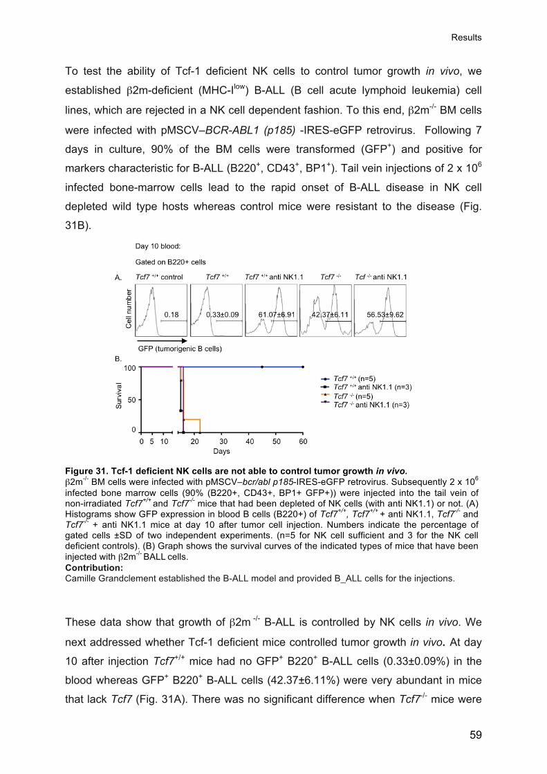
Results
59
To test the ability of Tcf-1 deficient NK cells to control tumor growth in vivo, we
established β2m-deficient (MHC-Ilow) B-ALL (B cell acute lymphoid leukemia) cell
lines, which are rejected in a NK cell dependent fashion. To this end, β2m-/- BM cells
were infected with pMSCV–BCR-ABL1 (p185) -IRES-eGFP retrovirus. Following 7
days in culture, 90% of the BM cells were transformed (GFP+) and positive for
markers characteristic for B-ALL (B220+, CD43+, BP1+). Tail vein injections of 2 x 10⁶
infected bone-marrow cells lead to the rapid onset of B-ALL disease in NK cell
depleted wild type hosts whereas control mice were resistant to the disease (Fig.
31B).
Figure 31. Tcf-1 deficient NK cells are not able to control tumor growth in vivo. β2m-/- BM cells were infected with pMSCV–bcr/abl p185-IRES-eGFP retrovirus. Subsequently 2 x 10⁶ infected bone marrow cells (90% (B220+, CD43+, BP1+ GFP+)) were injected into the tail vein of non-irradiated Tcf7+/+ and Tcf7-/- mice that had been depleted of NK cells (with anti NK1.1) or not. (A) Histograms show GFP expression in blood B cells (B220+) of Tcf7+/+, Tcf7+/+ + anti NK1.1, Tcf7-/- and Tcf7-/- + anti NK1.1 mice at day 10 after tumor cell injection. Numbers indicate the percentage of gated cells ±SD of two independent experiments. (n=5 for NK cell sufficient and 3 for the NK cell deficient controls). (B) Graph shows the survival curves of the indicated types of mice that have been injected with β2m-/- BALL cells. Contribution: Camille Grandclement established the B-ALL model and provided B_ALL cells for the injections.
These data show that growth of β2m -/- B-ALL is controlled by NK cells in vivo. We
next addressed whether Tcf-1 deficient mice controlled tumor growth in vivo. At day
10 after injection Tcf7+/+ mice had no GFP+ B220+ B-ALL cells (0.33±0.09%) in the
blood whereas GFP+ B220+ B-ALL cells (42.37±6.11%) were very abundant in mice
that lack Tcf7 (Fig. 31A). There was no significant difference when Tcf7-/- mice were

Results
60
NK cell depleted (56.53±9.62%), indicating that NK cells in mutant mice are entirely
unable to control B-ALL (Fig. 31A). In contrast to wild type mice, which remained
healthy, all Tcf7-/- mice had to be sacrificed around day 17-22 because they showed
severe disease symptoms (Fig. 31B). Symptoms included weight loss, weakness,
hunched posture and/or paralysis of the posterior legs, due to secondary tumors in
the spinal cord. Further analysis of sacrificed animals revealed a moderate
splenomegaly, enlarged LN and a dramatic expansion of cells with a pre-B cell
phenotype, B220+ CD43- BP-1+ (not shown). Taken together these data suggest that
the NK cells in Tcf7 deficient mice are not able to control tumor growth in vivo.
2.12 Granzyme B mediates target cell induced death of
Tcf7-/- NK cells
Tcf-1-deficient NK cells are unable to efficiently lyse tumor target cells in vitro and in
vivo (Figs. 29 and 31). Based on the over-expression of GzmB, we hypothesized that
NK cells undergo self- inflected damage upon activation by target cells. Therefore we
assessed NK cell death in response to target cells. Splenocytes from Poly IC primed
Tcf7+/+ and Tcf7-/- mice were co-cultured with RMA/S (MHC-I-) tumor target cells. As
compared to wild type NK cells (8.4±1.9% 7AAD+ n=5) a significantly increased
fraction of Tcf7-/- NK cells underwent cell death (21.1±1.8% 7AAD+ n=5) (Fig. 32) in
response to susceptible RMA/S target cells. In the absence of target cells or the
presence of NK cell resistant RMA cells we did not detect an increased fraction of
7AAD+ Tcf-1 deficient NK cells (Figs. 32A and B). Thus Tcf7 deficient NK cells
undergo increased cell death only upon target cell recognition.
Next we determined whether the increased NK cell death is mediated by GzmB. The
addition of a GzmB inhibitor (z-AAD-CMK) prevented the increased cell death of Tcf-
1 deficient NK cells in response to RMA/S targets whereas a control inhibitor (z-FA-
CMK) did not rescue NK cells (Figs. 31A and B). Furthermore, blockade of cathepsin
C (dipeptidyl peptidase I, DPP-I) whose activity is required for GzmB activation
(Meade et al., 2006) also reduced the death of Tcf7-/- NK cells (Figs. 32 A and B). On
the other hand z-VAD, which blocks the activity of several major caspases, did not
prevent the death of Tcf-1 deficient NK cells (Figs. 32A and B). Thus Poly IC
activated Tcf-1 deficient NK cells undergo accelerated cell death in response to

Results
61
susceptible target cells and this can be prevented by the inhibition of GzmB or
cathepsin C.
Figure 32. Accelerated cell death of Tcf7 deficient NK cells upon activation by target cells depends on GzmB. A. Histograms depict splenic NK cells (CD3-CD19-NK1.1+) from Poly IC primed Tcf7 +/+ and Tcf7 -/-
mice that have been exposed to susceptible RMA/S or resistant RMA tumor cells. Numbers show the percentage of dead (7AAD+) NK cells. The assays was performed n the presence of the GzmB inhibitor (z-AAD-CMK) (100uM), its negative control peptide (z-FA-CMK (100uM)), the caspase inhibitor (z-VAD (50uM) or the cathepsin C inhibitor (DPPI (15-25 uM)). These data are a representative of at least 3 independent experiments n=3-9. (B) Bar graphs depict the mean percentage (±SEM) of 7AAD-positive Tcf7+/+ and Tcf7-/- NK cells. (C) Histogram show the mean percentage (±SEM) of dead (7AAD+) Tcf7 +/+Gzmb+/+, Tcf7 +/+Gzmb-/-, Tcf7 -/-Gzmb+/+ and Tcf7 -/-
Gzmb-/- NK cells that have been primed exposed to susceptible RMA/S or resistant RMA tumor cells (*), (**) and (***) depict significant differences (p<0.05), (p<0.01) and (p<0.001), respectively based on the student`s t-test. When nothing is marked, the values are not significantly different (p>0.05).

Results
62
Finally we used Tcf7xGzmB double ko mice to verify whether NK cells undergo cell
death in response to susceptible tumor target cells. Indeed the absence of GzmB
rescued Tcf7 deficient NK cells from target induced cell death (Fig. 32C). We
conclude that Tcf-1 is essential for the killing of tumor target cells by protecting NK
cells from GzmB mediated self-inflected injury.
Data contribution: All experiments and analyses have been performed by Jasmine
Gehrig if not stated otherwise.

Discussion
63
3 Discussion
3.1 An essential role of Tcf-1 for NK cell development
In the first part of this thesis we defined the T cell factor-1 (Tcf-1) dependent stages
of NK cell development. We found that Tcf-1 is highly expressed at the immature NK
(iNK) and mature NK (mNK) cell stages. Next, we addressed the question of whether
NK cell development is associated with canonical Wingless/Integration-1 (Wnt)
signaling. Tcf-1 binds to DNA but lacks intrinsic transcriptional activity. Target gene
activation in response to Wnt signals depends on the co-activators -catenin and γ-
catenin, which bind to the N-terminus of Tcf-1. In contrast, target gene repression is
mediated via the association with the Groucho (Gro) co-repressors that bind to a
more central domain of Tcf-1 (Grigoryan et al., 2008; Jeannet et al., 2008a). We
found that Wnt signaling (assessed by conductin reporter activity) is strongly induced
during the NKp to iNK transition and is maintained in a subset of mNK cells.
Importantly, this signal was entirely dependent on Tcf-1 in both splenic, as well as
BM mNK cells. Thus the up-regulation of Tcf-1 at the iNK stage correlates with Tcf-1
mediated Wnt signaling.
In order to define if Wnt signaling drives NK cell development, we used a genetic
complementation approach. Surprisingly we found that the short Tcf-1 isoform (p33),
which only contains the DNA- and Gro-binding domains (Oosterwegel et al., 1991a;
Van de Wetering et al., 1996), is sufficient to restore NK cell development in Tcf-1-
deficient mice (Fig. 14 and 15). NK cell development is also restored by a p45
transgene (Tg), which includes the N-terminal β-catenin-bining domain of Tcf-1. In
agreement with these findings, we found that β- and γ-catenin are dispensable for NK
cell development (Fig. 13). Thus, NK cell development is dependent on binding of
Gro co-repressors to Tcf-1 at the DNA, whereas canonical Wnt signaling mediated by
the N-terminal domain of Tcf-1 is dispensable.
Our laboratory has previously shown that T cell development depends on the N-
terminus of Tcf-1, i.e. the p45 Tcf-1 isoform. Importantly, in this thesis we show that
the regulation of the NK cell development differs from T cell development. NK cell
development is restored by p33 and does not rely on signals mediated through the N-
terminal domain of Tcf-1. Thus, we found an important molecular difference between

Discussion
64
T and NK cell development. The two subsets depend on different Tcf-1 domains that
mediate different functions. T cell development depends on target cell activation via
the N terminal domain, whereas the repressor function of Tcf-1 is crucial for NK cell
development. Here we demonstrate for the first time that the repressor function of
Tcf-1 specifically drives an immunological process.
Recently it has been shown that Tcf-1 is essential for the development and/or
differentiation of innate lymphocyte subsets including group 2 innate lymphoid cells
(ILC2) and NKp46+ ILC3 cells (Mielke et al., 2013; Yang et al., 2013). However, the
precise role of Tcf-1 in this process still remains to be elucidated. As NK cells and the
ILC lineages are closely related, it would be interesting to test if the repressor p33
isoform of Tcf-1 is sufficient to restore ILC development as well.
Even though NK cell development is restored by both, Tcf-1 p33 and p45, there are
differences between the two: Enforced p45 expression in Tcf7 deficient mice reduces
the maturation of developing NK cells. Such mNK cells do not up-regulate KLRG-1,
CD11b and CD43, but maintain high levels of CD27, a hallmark of immature NK cells.
Conversely, in the absence of Tcf7, NK cells have a more mature phenotype. These
NK cells express KLRG-1, CD11b and CD43, while CD27 is reduced compared to
wild type. The maturation status of Tcf7 deficient NK cells that express the p33 Tg
was comparable to that of wild type (Fig. 14 and 15). Peripheral NK cells can be
divided into CD11b− CD27+ (immature), CD11b+CD27+ (mature1; M1), and
CD11b+CD27− (mature2; M2) cells, with the immature CD11b−CD27+ NK cell
population having a very low cytotoxic potential (Huntington et al., 2007). In p45 Tg
Tcf7 deficient mice, almost all NK cells show an immature CD11b−CD27+ phenotype.
We propose a model in which distinct stages of NK cell maturation depend on
different functions of Tcf-1. While enforced canonical Wnt signaling (p45 function)
keeps NK cells at a very immature stage, the p33 isoform allows normal maturation,
whereas total loss of Tcf-1 results in accelated maturation (Fig. 33).
In contrast to several other transcription factors that are required for NK cell
development and have been shown to promote NK cell maturation, e.g. the E4-
binding protein 4 (E4bp4), the interferon regulatory factor (IRF), GATA-3 and T-bet
(Gascoyne et al., 2009; Gordon et al., 2012; Lohoff et al., 2000; Male et al., 2011;
Samson et al., 2003; Taki et al., 2005), Tcf-1 dampens NK cell differentiation and
thus plays a unique role for NK cell maturation (Fig. 33). Those findings suggest that

Discussion
65
NK cell maturation is governed by the interplay of transcription factors that drive or
inhibit NK cell differentiation, respectively.
Figure 33. Enforced canonical Wnt signaling (p45 function) prevents maturation of NK cells. NK cells can be divided into CD11b− CD27+ (immature), CD11b+CD27+ (mature1; M1), and CD11b+ CD27− (mature2; M2) subsets. The CD11b+CD27+ subset is the most potent in acquiring effector functions whereas the M2 population mostly co-expresses KLRG1 that marks terminal maturation. In Tcf7+/+, and Tcf7-/-p33 mice the M2 subset represents the smallest population. In the absence of Tcf-1 maturation is accelerated and most NK cells show a terminal mature M2 CD11b+ CD27− KLRG+
phenotype. Conversely, in p45 Tg Tcf7 deficient mice, almost all NK cells show an immature CD11b−CD27+ phenotype, suggesting that enforced canonical Wnt signaling (p45 function) prevents maturation. The yellow bar represents the relative number of cells in the different maturation stages for the indicated genotype.
3.2 Basis for impaired NK cell development
The emergence of mNK cells coincides with the rapid cycling of BM NK cells (Kim et
al., 2002). We found that Tcf-1 deficient NK cells in the bone marrow cycle as
efficiently as in control mNK cells, but undergo accelerated apoptosis (Fig. 17).To
identify candidates that could mediate apoptosis of Tcf-1 deficient mNK cells, we
established the global gene expression profiles of BM mNK cells isolated from Tcf7+/+
and Tcf7-/- mice using Affymetrix arrays. Amongst the most up regulated transcripts in
the absence of Tcf-1, was the gene encoding Granzyme B (GzmB) (Fig. 34). Genes
coding for other Granzyme family proteins, including GzmC and GzmK were also up-

Discussion
66
regulated (Table 1 and 2). The Affimetrix analysis did not show any differences for
Perforin transcript levels.
Figure 34. The repressor function of the short p33 Tcf-1 isoform is sufficient to control GzmB expression and to ensure survival of proliferating NK cells. Shown is the sequential maturation of NK cells in the BM of Tcf7+/+ and Tcf7-/-p33, Tcf7-/- or Tcf7-/-p45 mice. The model proposes that the the suppressor activity of Tcf-1 (p33) is vital to ensure the survival of expanding mNK cells and for the suppression of GzmB (top panel). In the absence of Tcf7 NK cell maturation is accelerated, GzmB is up-regulated and translated and the expanding mNK cells undergo increased cell death (middle panel). Enfored signalling mediated by the N-terminal domain of Tcf-1 (present in p45) prevents maturation (bottom panel).
GzmB is a serine protease that together with Perforin is a main component of
exocytic granules in cytotoxic T cells (CTL) and NK cells. Such granules are a
hallmark of cell types that are able to kill cells infected with intracellular pathogens or

Discussion
67
tumor cells (Russell and Ley, 2002). Upon target cell recognition, lytic granules are
polarized towards the immunological synapse where they fuse with the plasma
membrane. Their content is relased into the synaptic cleft between effector and
target cell. Subsequently GzmB enters the target cells through pores in the plasma
membrane formed by Perforin (Long et al., 2013; Voskoboinik et al., 2006; Wang et
al., 1996). Once present in a target cell, GzmB can activate multiple caspases,
including caspases 1, 2, 3, 6, 7, 9, and 10 (Lieberman, 2003), but apoptosis induced
through the activation of caspase 3 seems to be the main pathway in GzmB
mediated cytotoxicity (Ewen et al., 2012). GzmB can directly activate some caspase
substrates as well, including PARP and NuMA (Beresford et al., 1999). Additionally,
GzmB has been shown to promote permeability of the mitochondrial outer membrane
and to activate t-BID as well as to cleave ICAD independently of caspases (Barry et
al., 2000; Thomas et al., 2001).
In addition to killing other cells, GzmB-leakage into the cytoplasm can induce cell
death in NK cells and CTLs (Ashton-Rickardt, 2013). It has previously been shown
that primed NK cells undergo increased cell death when they are stimulated with
antibodies against certain activating receptors (Ida and Anderson, 1998; Ida et al.,
1997). CD2-induced activation of IL-2 primed NK cells leads to GzmB mediated self-
inflected damage (Ida et al., 2003). In addition, T cell receptor-induced cell death
(TCR-ICD) of Th2 cells is mediated by GzmB-induced self-inflicted damage,
independently of caspase activation (Devadas et al., 2006). Moreover, it has been
shown that iNKT cells undergo increased GzmB mediated self-inflicted damage in
response to Ag stimulation (Ansari et al., 2010). Finally c-Myc and N-Myc deficient
HSCs over-express GzmB and undergo increased cell death (Laurenti et al., 2008).
Mature and BM NK cells in naïve mice express GzmB and Perforin mRNA
constitutively. Importantly, resting or BM NK cells are minimally cytotoxic, because
Perforin and GzmB mRNAs are not translated. In agreement with these data we
could not detect any GzmB and Perforin protein in developing NK cells from wild type
mice. The mechanisms that prevent the translation are currently not well understood.
This translational block is overcome upon NK cell priming by cytokines like IL-15, IL-2
and IL-18 that are produced early in an immune response by infected or activated
dendritic cells (DCs) and macrophages (Fehniger et al., 2007; Stetson et al., 2003;
Walzer et al., 2005). In contrast, in the absence of Tcf7, GzmB is detected in BM
(from the iNK stage onwards) and spleen NK cells even in the absence of priming,

Discussion
68
suggesting that Tcf-1 is involved in the regulatory mechanism responsive to NK cell
priming. The presence of the short p33 domain of Tcf-1 engaging Gro co-repressors
was sufficient to regain wild type like GzmB control (Fig. 34).
Upon cytokine stimulation NK cells increase in size and develop a high number of
GzmB containing cytotoxic granules in their cytoplasm. Activated NK cells that lack
Tcf-1 express much higher levels of GzmB mRNA and slightly more GzmB protein
than their wild type counterparts. We show that cytokine stimulation of Tcf-1 deficient
splenic NK cells results in the impairment of expansion. Similar to bone marrow mNK
cells, cytokine stimulated NK cells are cycling normally but undergo accelerated cell
death (Fig. 17). The presence of the short p33 domain of Tcf-1 was sufficient to
restore NK cell expansion in the absence of Tcf7. Furthermore, enforced p33
expression significantly decreased GzmB levels compared to wild type controls.
GzmB is produced as a pro-peptide and needs to be cleaved by the protease
cathepsin C (or DPPI) to gain proteolytic activity. In cytokine activated and primed
wild-type NK cells, GzmB is stored in exocytic Lamp-1 positive granules present in
the cytoplasm. Granzymes are packaged into the cytotoxic granules via the mannose
6-phosphate receptor (MPR) pathway (Lopez et al., 2012). In the granules,
Granzymes and Perforin are kept inactive by low pH and calcium concentrations. In
fact, activated NK cells and CD8 T cells contain small amounts of GzmB in their
cytoplasm (Ashton-Rickardt, 2013). This suggests that some GzmB might leak from
the granules or that some newly synthesized GzmB does not properly traffick to the
granules but may be secreted via the constitutive secretory pathway as described
previously (Isaaz et al., 1995). However, the serine protease inhibitor 6 (Spi-6) that is
present in the cytoplasm of activated cytotoxic cells captures free GzmB and
prevents its processing and activation as a protective mechanism (Fig. 35). Free
GzmB in the cytoplasm of Spi-6 deficient cytotoxic T cells disrupts lytic granules, this
results in GzmB-mediated apoptosis (Ansari et al., 2010; Zhang et al., 2006).
Therefore, the major determinant of GzmB-leakage-induced cell death seems to be
an excess of free active GzmB over its principal cytoplasmic inhibitor Spi-6 (Ida et al.,
2003; Laforge et al., 2006; Phillips et al., 2004; Zhang et al., 2006). Here, we show
that Spi-6 was expressed at normal levels in naïve and activated Tcf-1 deficient NK
cells, whereas GzmB levels were significantly elevated (Fig. 20).
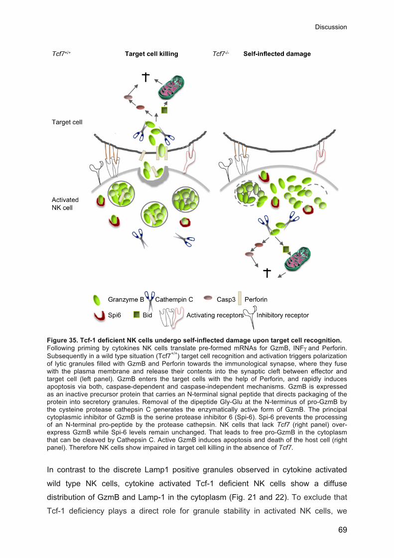
Discussion
69
Figure 35. Tcf-1 deficient NK cells undergo self-inflected damage upon target cell recognition. Following priming by cytokines NK cells translate pre-formed mRNAs for GzmB, INFγ and Perforin. Subsequently in a wild type situation (Tcf7+/+) target cell recognition and activation triggers polarization of lytic granules filled with GzmB and Perforin towards the immunological synapse, where they fuse with the plasma membrane and release their contents into the synaptic cleft between effector and target cell (left panel). GzmB enters the target cells with the help of Perforin, and rapidly induces apoptosis via both, caspase-dependent and caspase-independent mechanisms. GzmB is expressed as an inactive precursor protein that carries an N-terminal signal peptide that directs packaging of the protein into secretory granules. Removal of the dipeptide Gly-Glu at the N-terminus of pro-GzmB by the cysteine protease cathepsin C generates the enzymatically active form of GzmB. The principal cytoplasmic inhibitor of GzmB is the serine protease inhibitor 6 (Spi-6). Spi-6 prevents the processing of an N-terminal pro-peptide by the protease cathepsin. NK cells that lack Tcf7 (right panel) over-express GzmB while Spi-6 levels remain unchanged. That leads to free pro-GzmB in the cytoplasm that can be cleaved by Cathepsin C. Active GzmB induces apoptosis and death of the host cell (right panel). Therefore NK cells show impaired in target cell killing in the absence of Tcf7.
In contrast to the discrete Lamp1 positive granules observed in cytokine activated
wild type NK cells, cytokine activated Tcf-1 deficient NK cells show a diffuse
distribution of GzmB and Lamp-1 in the cytoplasm (Fig. 21 and 22). To exclude that
Tcf-1 deficiency plays a direct role for granule stability in activated NK cells, we

Discussion
70
analyzed Tcf7xGzmB double deficient NK cells and found granular LAMP-1 staining
to be restored. Therefore, we conclude that it is the excess of GzmB that destroys
the integrity of lytic granules and not the absence of Tcf-1 per se or different
downstream targets that lead to an accumulation of free GzmB in the cytoplasm.
To determine whether GzmB mediates accelerated NK cell death, we generated
mice deficient for both, GzmB and Tcf7. GzmB deficiency restored the expansion of
peripheral NK cells lacking Tcf7. Thus the observed self-inflicted cell death is indeed
caused by the elevated GzmB levels (Fig. 35). However, the absence of GzmB was
not sufficient to restore NK cell development in Tcf7-deficient mice. Besides GzmB,
GzmC was also up regulated in Tcf7 deficient mNK cells. GzmB deficient mice also
express reduced levels of GzmC and F, which are just downstream in the GzmB
gene cluster (Pham et al., 1996; Revell et al., 2005). It is thus not likely that GzmC or
F play a significant role at this stage of NK cell development.
Based on these findings we conclude that Tcf-1 regulates an additional factor that is
required for the survival of mNK cells. A third Granzyme that is increased in the
absence of Tcf7 is GzmK, which is located in a distinct cluster together with GzmA.
GzmK can also induce rapid caspase-independent cell death in target cells (Zhong et
al., 2012). So far it is not known whether GzmK is constitutively expressed in NK
cells like GzmA, or whether it is translated and/or transcribed upon priming like
GzmB. A single study with human NK cells showed that GzmK is mostly expressed in
the CD56high NK cell population. Conversely, GzmB is primarily expressed in the
CD56low NK cell subset, while GzmA is constitutively expressed in both subsets.
These findings suggest that similar to GzmB, GzmK expression is not constitutive,
but depends on specific triggers (Bratke et al., 2005). Although it is not known
whether GzmK can mediate self-inflected damage, it is tempting to speculate it might
act in a similar manner as GzmB when present in the cytoplasm. Therefore, further
experiments are needed to test if GzmK mediates accelerated cell death in Tcf-1
deficient mNK cells lacking GzmB. Employing a GzmK specific inhibitor or Tcf7,
GzmB and GzmK deficient mice would be a way to test if GzmK mediates self-
inflected damage of Tcf7 and GzmB deficient mNK cells.
Taken together, the data suggest that under steady state conditions GzmB
expression is suppressed by Tcf-1. Two reasons could account for the enhanced

Discussion
71
presence of GzmB protein in the absence of Tcf-1. First, the control mechanisms that
block translation are over-come by the elevated GzmB mRNA levels. Second, the
mediators that control GzmB translation are directly or indirectly affected by the loss
of Tcf-1. Control of GzmB translation is not well understood and further experiments
are needed to address these questions.Accordingly, it remains to be determined in
further experiments if GzmB is a direct target gene of Tcf-1 regulation. To test
whether Tcf-1 binds to the GzmB locus we are currently performing Tcf-1 chromatin
immunoprecipitation followed by parallel sequencing (ChIP-seq) analysis of NK cells.
The aim is to use the ChIP-seq data to identify Tcf-1 binding sites and to relate this
information to the presence of activating (histone H3 trimethylated at lysine 4,
H3K4me3) and repressive (H3K27me3) histone marks. Our hypothesis is that in wild
type NK cells, Tcf-1 is bound to the GzmB locus and that this is associated with
repressive histone marks. Consequently, in Tcf-1 deficient NK cells we would expect
the active histone marks to be more abundant. If we identify a Tcf-1 binding
sequence within the GzmB locus, we would need to perform reporter assays to
confirm that Tcf-1 binding to this site results in repression or activation of GzmB
transcription.
If direct binding of Tcf-1 to the GzmB promoter region cannot be shown, the
observed regulatory effects of Tcf-1 on GzmB expression must involve further, yet
unidentified molecular components. The iNK to mNK transition is impaired in the
absence of the two T-box transcription factors T-bet and Eomes (Gordon et al., 2012).
This process appears similar to our observations on Tcf-1 deficiency. Eomes is a
direct target of Tcf-1 in CD8 T cells (Zhou et al., 2010). Eomes and T-bet
coordinately regulate the expression of key effector molecules in cytotoxic
lymphocytes such as Perforin, Granzymes and INFγ (Cichocki et al., 2013; Pearce et
al., 2003; Szabo et al., 2000). Therefore, T-bet and Eomes may be downstream
targets of Tcf-1 in NK cell development. While Eomes levels in BM NK cells that lack
Tcf-1 did not significantly change, T-bet levels were slightly but significantly
increased (around 30%). T-bet expression levels were reduced to normal in the p33
and the p45 Tg Tcf7-/- NK cells, respectively (data not shown). Thus, in addition to a
possible direct effect, Tcf-1 might also control GzmB expression indirectly through
the repression of T-bet. Our Chip-seq analysis may shed light on this question as
well.
Discussion
72
3.3 NK cell function in the absence of Tcf-1
We tested whether Tcf7 deficient NK cells are able to mediate normal effector
functions. Upon priming of mice with poly IC, wild type and Tcf7 deficient NK cells
comparably induced CD69 and expressed GzmB and Perforin. In response to target
cells, Tcf7 deficient NK cells normally released cytokines, chemokines and granule
contents (Lamp-1). However, their ability to kill target cells in vitro and particularly in
vivo was impaired. Rather than killing target cells, NK cells themselves underwent
increased cell death upon the recognition of susceptible target cells. Using inhibitors,
we found that the increased cell death upon target cell recognition was prevented by
blocking the activity of GzmB or Cathepsin C. Moreover, the increased cell death
upon target cell exposure was prevented when Tcf-1 defcient NK cells also lacked
GzmB (Fig. 32).
Based on the data in this report our working model is that under steady state
conditions, Tcf-1 suppresses GzmB levels. In an inflammatory context, NK cells are
primed and this leads to a down-regulation of Tcf-1 and NK cells gain their full
cytotoxic potential through the expression of GzmB. Indeed, we found that Tcf-1 is
strongly decreased yet still expressed at low levels in NK cells of Poly IC primed mice
(Fig. 9). Low levels of Tcf-1 may restrict GzmB production and thus prevent GzmB
mediated self-inflected damage. If an inflammation is reaching an extent that is
dangerous for the organism, Tcf-1 expression may shut off, leading to more free
GzmB in the cytoplasm. This may induce apoptosis of highly activated NK cells that
potentially harm the host itself (Fig. 35 and 36).
It remains to be explored in future experiments if the enforced expression of the Tcf-1
Tgs prevents maturation (N-terminus, p45) and maintains GzmB repression (p33)
during viral infections such as the mouse cytomegalovirus (MCMV). Furthermore, it
has to be determined if such Tg expression leads to decreased cytotoxicity and/or
self inflected death. Since it has been suggested that immature NK cells have a very
low cytotoxic potential (Huntington et al., 2007), it remains to be tested if the p45 Tg
NK cells show a decreased functional competence to respond to tumor target cells
(Fig. 34). This would allow to better describe the relationship between maturation
markers and cytotoxic potential.

Discussion
73
GzmB mediated self-inflected damage may be a naturally occurring mechanism to
remove terminally differentiated effector cells. This mechanism may dampen immune
responses and prevent damage to host tissue by highly activated NK cells. Tcf-1
would represent a major regulator of this process. Controlling Tcf-1 expression is
thus expected to be key in the maturation and survival of NK cells during an immune
response. It will be important to elucidate the specific signals which regulate Tcf-1 in
vivo during the immune response. A Tcf-1 GFP reporter mouse would therefore
represent a useful tool.
Figure 36. Schematic representation of the model that we developed based on the results shown and discussed in this thesis. The Wnt signal mediating N-terminal domain of Tcf-1 is crucial to prevent the maturation of iNK cells and a subset of mNK cells, whereas the suppressor (p33) activity of Tcf-1 is vital to ensure the survival of expanding mNK cells and for the suppression of GzmB. In steady state conditions Tcf-1 suppresses GzmB, whereas in an inflammatory context, Tcf-1 levels may decrease. This leads to GzmB expression and up-regulation of the maturation markers KLRG1 and CD11b and the downregulation of CD27 (indicated in violet, purple and yellow, respectively) and subsequent target cell killing upon recognition. During this process some Tcf-1 is still present to prevent GzmB mediated self-inflected damage. In a persistent inflammatory context Tcf-1 levels further decline what leads to increased GzmB expression and self-inflected death. Curves indicate Wnt signaling (blue), Tcf-1 expression (red), maturation (yellow) and GzmB expression (green).

Discussion
74
NK cells that have been attracted to the sites of inflammation could represent a
highly desirable vehicle for viruses to ascertain rapid spread throughout the host.
Viruses that do not infect endothelial cells are unable to enter or leave the
bloodstream to infect peripheral tissues. Previously, it has been shown that GzmA
and GzmB double deficient mice have higher virus titers upon infection with the
cytopathic pox-viruses. The authors suggest that this observation is linked to the
enhanced survival of CTLs and NK cells due to their inability to commit GzmB-
mediated suicide. Therefore, the virus might use these cells as Trojan horses to
extravagate and infiltrate other organs (Balkow et al., 2001). This suggests that
GzmB mediated self-inflected death might also represent an important mechanism to
prevent viral spread by activated and highly mobile cytotoxic cells.
Based on the findings shown in my thesis we propose a model where Tcf-1 plays a
major role for the suppression of the highly cytotoxic effector molecule GzmB in NK
cells, thereby preventing self-inflected damage in these cells. Concordantly, the Wnt
signal mediating N-terminal domain of Tcf-1 is crucial to prevent the maturation of
iNK cells and of a subset of mNK cells. Our data suggest that upon inflammation,
Tcf-1 levels may decrease, that in turn would lead to GzmB expression and to
terminal maturation. During the course of an immune response, Tcf-1 levels might
further decline, triggering GzmB mediated self-inflected damage of highly activated
NK cells (Fig. 36). This mechanism might be important to remove terminally
differentiated effector NK cells and thus to dampen immune responses in order to
prevent collateral damage of the host tissue. Blocking Tcf-1 expression in highly
activated NK cells may therefore be a way to induce self-inflected cell death. In the
context of autoimmune inflammation this mechanism could be exploited to eliminate
effector cells that attack host tissue. The identification of the factors that modulate
Tcf-1 expression during inflammation may therefore allow the identification of
potential therapeutic targets.

Methods
75
4 Methods
Mice C57BL/6 (B6) mice were purchased in Harlan OLAC (The Netherlands). CD45.1
congenic B6 were bred in the specific pathogen free animal facility of our institut.
Tcf7-/- (Verbeek et al., 1995), Tcf7-p33 and Tcf7-p45 (Ioannidis et al., 2001),
conductinlacZ (Lustig et al., 2002), β-cateninlox/lox (Huelsken et al., 2001), γ-cateninlox/lox
(a kind gift from Prof. Jörg Huelsken), Vav-Cre transgenic (Almarza et al., 2004; de
Boer et al., 2003), GzmB−/− (GzmB−/−/ΔPGK-neo) (Pham et al., 1996; Revell et al.,
2005) (a kind gift form Prof. Christoph Borner) mice have been described. Β-catenin
Flox/Flox γ-catenin Flox/Flox, Vav-Cre+ mice have been crossed by Camille Grandclément
in our lab and the GzmB−/− (GzmB−/−/ΔPGK-neo) Tcf7-/- have been bred by myself.
For BM chimeras, 2:1 or 3:1 mixtures of experimental (CD45.2+) and wild type
(CD45.1+) BM cells were used (3-6x10⁶ cells total). Chimeras were tail bled at the
indicated time points and lymphoid tissues were analyzed 2-12 months after
reconstitution.
Flow cytometry Spleen and bone marrow cells were treated with ACK (NH4Cl 0.15M, KHCO3 1 mM,
EDTA 0.1 mM, pH 7.3) to remove erythrocytes. Subsequently cells were incubated
with Aquadead (Invitrogen) (to exclude dead cells) and anti-CD16/32 (2.4G2)
hybridoma supernatant to block Fc receptors before staining with fluorescent mAbs
to NK1.1 (PK136), CD49b (DX5), CD122 (TMB1), KLRG1 (2F1), CD27 (LG.7F9),
CD11b (M1/70), CD69 (VEA), CD84 (mCD84.7), CRACC/SLAMF7 (235614), Ly-108
(13G3-19D), Ly-9 (Ly9ab3, BioLegend), 2B4 (244F4), CD16 (93, BioLegend), (LFA-1
(FD44.8), Ly-49A (JR9/YE1/48.10.6/A1), Ly-49H (3D10), Ly-49D (4E5), Ly-49G
(4D11), Ly-49C/I (5E6), Ly49F (HBF), Ly49I (YLI90), NKp46 (29A1.4), CD3ε (17A2),
CD19 (6D5), BP-1 (6C3), B220 (RA3-6B2), CD45.1 (A20), CD45.2 (104.2), CD43
(S/7), CD8a (53-6.7), CD25 (GCG1), GR-1 (Ly6G), Sca1 (D7), Ter119 (Ter119), CD4
(RM4-5), F4/80 (F4/80), CD90.2 (BOH12), TCRβ (H57), TCRγδ (GL3) and CD117
(ACK2) (if not indicated otherwise from BD Biosciences). A cocktail of FITC-
conjugated anti-TCRβ, TCRγδ, CD3ε, CD4, CD8, CD11b, B220, NK1.1, Ter119 and
GR-1 mAbs was used to gate lineage-negative (lin-) cells. Intracellular staining for

Methods
76
GzmB (16G6, invitrogen), Perforin (OMAK-D, e-Bioscience), GzmA (3G8.5, Santa
Cruz) IFN-γ (XMG1.2, e-Bioscience), T-bet (eBio4B10, e-Bioscience), Eomes
(Dan11mag, e-Bioscience), MIP-1 alpha (39624, e-Bioscience), Tcf-1 (C63D9, Cell
Signal) and Lef (C12A5, Cell Signal) was performed using the BD Cytofix/Cytoperm
Fixation/Permeabilzation Kit (BD Biosciences) for IFN-γ, MIP-1α, Bcl-2 (PharMingen)
and Bcl-XL (7B2.5, SouthernBiotech) or the Transcription factor staining buffer set”
(eBioscience) for GzmB, GzmA, T-bet, Eomes, Tcf-1 and Lef according to the
manufacturer’s protocol. Data were acquired on an LSRII flow cytometer (BD
Biosciences) or a FACSCanto flow cytometer (Becton Dickinson, San Jose, CA) and
analyzed with FlowJo (Tree Star, Ashland, OR). Alternatively cells were subjected to
sorting on a FACSAria machine (Becton Dickinson, San Jose, CA).
LacZ staining For measuring LacZ activity, cells were counted and resuspended at 20x106 cells per
mL in HBSS (Gibco) containing 2% FCS, 10mM Hepes (Gibco) and 1% Pen-Strep
(Gibco)). 2x10⁶ cells were incubated for 10 minutes at 37°C and loaded with pre-
warmed Fluorescein Di-β-D-Galactopyranoside 2mM (FDG, Invitrogen) in H2O for 1
minute. Cells were then quenched with complete HBSS and left for 90min on ice
before washing. The cells were surface stained for flow cytometry as described
previously. The vital dye 7-AAD (Biolegend) was added 10 min prior to the FACS
analysis. Samples were run on LSRII or FACScanto flow cytometers (Becton
Dickinson, San Jose, CA) and analyzed using FlowJo software (Tree Star, Ashland,
OR).
NK cell expansion Splenic NK cells were purified by non-adherence to nylon wool followed by
depeleting non-NK cells using the NK Cell Isolation Kit II and a MACS separator
(Miltenyi Biotec). Purified NK cells were cultured for 5 days in DMEM with 10% FCS,
Gentamicin (10mg/ml), 2-Mercaptoethanol (50mM), MEM NEAA (0.1mM), L-
Glutamine (4 mM) and 500mg/ml recombinant human IL-2 (a gift from N. Rufer,
University of Lausanne). After 5 d, cells were harvested and stained for surface
antibodies and for Annexin V (PE Annexin V Apoptosis Detection Kit I, BD
Pharmigen) according to the manufacturer’s protocol. To identify DNA strand breaks
we performed a TUNEL staining (In Situ Cell Death Detection Kit, Roche) that labels
Methods
77
free 3’-OH termini with modified nucleotides in an enzymatic reaction. Data were
acquired on an LSRII flow cytometer (BD Biosciences).
In vitro NK cell activation to measure cytokine and chemokine release Spleen cells (2 × 10⁶) from polyinosinic-polycytidylic acid (Poly IC) primed mice were
stimulated with plastic immobilized mAbs to NK1.1 (PK136, BioXCell) (5mg/ml),
NKG2D (191004, R&D Systems) (5 mg g/ml), NKp46 (29A1.4 eBiosciences) (2.5
mg/ml), Ly49D (4E5, BD Pharmingen) (2.5 mg/ml) or by target cells RMA (H-2b),
RMA/S (MHC-Ilow), RMA-H60, RMA-m157 or YAC-1 cells (2 × 10⁶) or by the addition
of PMA (50 ng/ml) and Ionomycin (1 mg/ml). GolgiPlug, GolgiStop and LAMP mAb
(1D4B, eBiosciences) were added after 1 h of culture and the cells were harvested 4
h later. Cells were incubated with Aquadead (Invitrogen) (to exclude dead cells) and
anti-CD16/32 (2.4G2) hybridoma supernatant before staining with fluorescent mAbs
to NK1.1 (PK136), NKp46 (29A1.4), CD3ε (17A2), CD19 (6D5) (all from BD
Biosciences). Intracellular staining for IFNγ (XMG1.2, e-Bioscience) and MIP-1 alpha
(39624, e-Bioscience) was performed using the BD Cytofix/Cytoperm
Fixation/Permeabilzation Kit (BD Biosciences). Cells were analyzed on a FACSCanto
flow cytometer (BD Biosciences) and analyzed with FlowJo (Tree Star).
FLOKA ASSAY Mice were intravenously injected with 100ug polyinosinic-polycytidylic acid (Poly IC)
(Labforce, tlrl-pic) to prime the NK cells. 24 hours later the mice were sacrificed and
the spleens were harvested. The NK cell numbers (effectors) were determined by
FACS analysis. Target cells RMA (H-2b), RMAS MHC-Ilow, RMA-H60 or RMA-m157
were resuspended at a concentration of 1x10⁷ cells/ml in PBS containing 0.3 µM
CFSE (5- and 6-carboxyfluorescein diacetate succinimidyl ester, Molecular Probes)
and incubated for 5 min at 37°C in the dark. CFSE labelled cells were washed three
times in DMEM containing 10% FCS. Subsequently the effector cells were added
and the cells were cultured at 37° in a FACS tube with 500 ul of medium (DMEM and
10% FCS) at an effector: target ratio of 1:1, 3:1 or 10:1 (10 000 target cells were
used and the effectors were adjusted accordingly). As a control targets and effectors
were cultured separately. Four hours later 10 ul of 7AAD (PD Pharmigen) was added.
The percentage of death cells (7AAD+) cells within the CFSE positive target cell
population was determined by FACS analysis like described earlier. The

Methods
78
spontaneous cell death within the target cells that were cultured without effectors was
used to calculate the percentage of cells that underwent cell death due to killing.
Inhibitors used: Caspase inhibitor Z-VAD-FMK (Promega), GzmB inhibitor Z-AAD-
CMK (Enzo), Cathepsin C inhibitor, Gly-Phe-DMK (MP BIOMEDICALS)
In vivo target cell rejection Mice were injected intra peritoneal with 100 µg Poly IC (Sigma-Aldrich). 24 h later a
1:1 mixture of 1 × 10⁷ H-2b (labeled 2.0 µM CFSE [Invitrogen]) and 1 × 10⁷ β2m-
knockout (MHC-Ilow) splenocytes (labeled with 0.2 µM CFSE) were injected
intraveniously. Spleens of recipient mice were analyzed 24 h later for the presence of
transferred cells. The percentage of specific rejection was calculated as follows: 100
− [(percentage of H-2b cells (CFSEhi) final/percentage of β 2m-knockout cells
(CFSElow) final)/(percentage of H-2b cells (CFSEhi) initial/percentage of β2m-knockout
cells (CFSElow) initial)] × 100.
Confocal microscopy BM mNK cells or splenic NK cells at day 5 of IL-2 stimulation were FACS sorted to
get rid of non-NK cells. 5x10⁴ NK cell blasts or 1x10⁵ mNK cells/ 100uL were put on
Poly-L-Lysin (0.1%) coated Super-Frost slides (VWR International). Subsequently
cell were fixed for 10 min in 3.7% PFA at room temperature (RT). After three washing
steps with PBS cells were permeabilized for 10 min at RT with 0.2% Triton in PBS.
After washing cells were blocked for 1 hour with PBS+15% Goat serum (SIGMA) and
then 1 hour in 15% Rat serum (SIGMA). Following an over night incubation at 4C°
with the primary antibody (GzmB (BAF1865) (R&D); Goat anti-Mouse; biotynilated;
and Anti mLAMP1 (1D4B) (BioLegend) Rat anti-Mouse) cells were washed rigorously
with BPS and stained for 1 hour at RT with the secondary antibodies in blocking
solution (Alexa-Fluor-488 -Strepatvidin) (invitrogen) and Alexa-Fluor- 555 Goat anti-
Rat (Life technologies)). After being washed cells were mounted in ProLongGold
antifade reagent with DAPI (invitrogen) and covered with a glass. Samples were
examined with a LSM 510 confocal microscope (Zeiss) and images were recorded in
multitracking mode.

Methods
79
B-ALL induction
For B-ALL like disease induction BM cells from β2m deficient mice were infected with
the MCMV-bcr-ablp185/IRES-gfp virus. Before the viral supernatant was collected
after transient transfection of 293T cells. The β2m deficient BM cells were cultured
with the viral supernatant for 1 hour in the presence of 10ng/mL IL7, 10% WEHI
supernatant (as a souce of IL3) and 5µg/mL of Polybrene. The infected BM cells
were subsequently cultured for 7 days at 37°C, 5% CO2 in RPMI medium containing
5% FCS. After 7 days of culture, more than 90% of bone-marrow cells were B220+,
CD43+, BP1+ and GFP+. For tail vein injections, 2 x 10⁶ infected bone marrow cells
were re-suspended in 200µL of PBS and injected via the tail vein in B6 or Tcf7-/-
recipient mice. In some cases, NK cells were depleted with the NK-depleting
antibody PK136 (BioXcell, 200µg/mouse, IV) 2 days before the injection of B-ALL
cells and the depletion was then performed once in a week. Mice were checked daily
for the onset of the disease. Blood samples were analyzed 10 days after the injection
of B-ALL cells to follow the disease. Sick mice were killed and analyzed for spleen
weights, NK cell counts and the presence of leukemic cells.
Western Blot Western blots on total cell lysates from FACS sorted mNK, splenic NK or IL-2
stimulated NK cells (100 000-500 000 cells per lane) were performed as described
(Ioannidis et al., 2001). Blots were incubated overnight at 4°C with primaryantibodies:
Rb polyclonal Ab anti-GzmB (abcam), PI-9 (PI9-17) (Santa Cruz Biotechnology) or
anti-Tubulin (B-5-1-2, Sigma). Western blots were revealed with HRP- conjugated
anti-mouse and anti-rabbit Abs (Sigma) followed by enhanced chemiluminescence
detection (Pierce, Rockford, IL).
Microarray
Global gene expression profiles of FACS-purified BM mNK cells (CD3-CD19-
CD122+NK1.1+DX5+) isolated from Tcf7+/+ and Tcf7-/- mice were established using
Affymetrix arrays. A minimum of 3x6 Tcf7+/+ and Tcf7-/- mice were sorted. RNA was
extracted using Tryzol Reagent (Invitrogen). Gene expression profiling was
performed according to the manufacturer’’s protocol (Affymetrix). Labeled cRNA
(from total RNA) was generated and applied to Affymetrix muU74Av2 microarray. All
experiments were performed twice using RNA samples from two independently

Methods
80
sorted cell subsets. Results were analyzed using Microarray Analysis Software v4
and v5 (Affymetrix).
Quantitative PCR Total mRNA was isolated from sorted cells using RNA easy kit (Qiagen), SV Total
RNA Isolation System (Promega) or High Pure RNA Tissue Kit (Roche). First strand
DNA was synthesized with oligo-dT (Amersham Biosciences) and AMV reverse
transcriptase (Roche Diagnostics). RNA was digested with RNAse One (Promega)
and DNA purified with Mini Elute PCR purification Kit (Qiagen). Quantitative PCR
reactions were performed with the LightCycler FastStart DNA Master SYBR Green I
kit and run on the LightCycler II (Roche Diagnostics). Amplification plots were
analyzed using the second-derivative method with LC data analysis 3.5 software
(Roche), and the relative quantification was determined using the LightCycler relative
quantification software 1.0 (Roche).
Statistical Analysis Statistical significance was determined using an unpaired two-tailed Student t test for
unequal variances. P values are indicated as *P < 0.05, **P < 0.01, ***P < 0.001, and
ns, statistically not significant. The statistical significance of differences between
groups of data was calculated using a one-way analysis of variance (one-way
ANOVA) along with the Dunnet's post-test to adjust for multiple comparisons, using a
95% confidence interval.

References
81
5 References
Aberle, H., Bauer, A., Stappert, J., Kispert, A., and Kemler, R. (1997). beta-‐catenin is a target for the ubiquitin-‐proteasome pathway. Embo J 16, 3797-‐3804.
Almarza, E., Segovia, J.C., Guenechea, G., Gomez, S.G., Ramirez, A., and Bueren, J.A. (2004). Regulatory elements of the vav gene drive transgene expression in hematopoietic stem cells from adult mice. Exp Hematol 32, 360-‐364.
Ames, E., and Murphy, W.J. (2014). Advantages and clinical applications of natural killer cells in cancer immunotherapy. Cancer Immunol Immunother 63, 21-‐28.
Ansari, A.W., Temblay, J.N., Alyahya, S.H., and Ashton-‐Rickardt, P.G. (2010). Serine protease inhibitor 6 protects iNKT cells from self-‐inflicted damage. J Immunol 185, 877-‐883.
Arce, L., Yokoyama, N.N., and Waterman, M.L. (2006). Diversity of LEF/TCF action in development and disease. Oncogene 25, 7492-‐7504.
Archbold, H.C., Yang, Y.X., Chen, L., and Cadigan, K.M. (2012). How do they do Wnt they do?: regulation of transcription by the Wnt/beta-‐catenin pathway. Acta Physiol (Oxf) 204, 74-‐109.
Arranz, L., Herrera-‐Merchan, A., Ligos, J.M., de Molina, A., Dominguez, O., and Gonzalez, S. (2012). Bmi1 is critical to prevent Ikaros-‐mediated lymphoid priming in hematopoietic stem cells. Cell Cycle 11, 65-‐78.
Ashton-‐Rickardt, P.G. (2013). An emerging role for Serine Protease Inhibitors in T lymphocyte immunity and beyond. Immunol Lett 152, 65-‐76.
Balkow, S., Kersten, A., Tran, T.T., Stehle, T., Grosse, P., Museteanu, C., Utermohlen, O., Pircher, H., von Weizsacker, F., Wallich, R., et al. (2001). Concerted action of the FasL/Fas and perforin/granzyme A and B pathways is mandatory for the development of early viral hepatitis but not for recovery from viral infection. J Virol 75, 8781-‐8791.
Banchereau, J., and Steinman, R.M. (1998). Dendritic cells and the control of immunity. Nature 392, 245-‐252.
Bar, E., Whitney, P.G., Moor, K., Reis, E.S.C., and Leibundgut-‐Landmann, S. (2014). IL-‐17 Regulates Systemic Fungal Immunity by Controlling the Functional Competence of NK Cells. Immunity 40, 117-‐127.
Barry, M., Heibein, J.A., Pinkoski, M.J., Lee, S.F., Moyer, R.W., Green, D.R., and Bleackley, R.C. (2000). Granzyme B short-‐circuits the need for caspase 8 activity during granule-‐mediated cytotoxic T-‐lymphocyte killing by directly cleaving Bid. Mol Cell Biol 20, 3781-‐3794.
Barton, K., Muthusamy, N., Fischer, C., Ting, C.N., Walunas, T.L., Lanier, L.L., and Leiden, J.M. (1998). The Ets-‐1 transcription factor is required for the development of natural killer cells in mice. Immunity 9, 555-‐563.
Beresford, P.J., Xia, Z., Greenberg, A.H., and Lieberman, J. (1999). Granzyme A loading induces rapid cytolysis and a novel form of DNA damage independently of caspase activation. Immunity 10, 585-‐594.
Bevan, M.J. (2006). Cross-‐priming. Nat Immunol 7, 363-‐365. Biron, C.A., Byron, K.S., and Sullivan, J.L. (1989). Severe herpesvirus infections in an
adolescent without natural killer cells. N Engl J Med 320, 1731-‐1735. Biron, C.A., Nguyen, K.B., Pien, G.C., Cousens, L.P., and Salazar-‐Mather, T.P. (1999).
Natural killer cells in antiviral defense: function and regulation by innate cytokines. Annu Rev Immunol 17, 189-‐220.

References
82
Boos, M.D., Yokota, Y., Eberl, G., and Kee, B.L. (2007). Mature natural killer cell and lymphoid tissue-‐inducing cell development requires Id2-‐mediated suppression of E protein activity. J Exp Med 204, 1119-‐1130.
Bratke, K., Kuepper, M., Bade, B., Virchow, J.C., Jr., and Luttmann, W. (2005). Differential expression of human granzymes A, B, and K in natural killer cells and during CD8+ T cell differentiation in peripheral blood. Eur J Immunol 35, 2608-‐2616.
Bukowski, J.F., Woda, B.A., Habu, S., Okumura, K., and Welsh, R.M. (1983). Natural killer cell depletion enhances virus synthesis and virus-‐induced hepatitis in vivo. J Immunol 131, 1531-‐1538.
Caputo, A., Garner, R.S., Winkler, U., Hudig, D., and Bleackley, R.C. (1993). Activation of recombinant murine cytotoxic cell proteinase-‐1 requires deletion of an amino-‐terminal dipeptide. J Biol Chem 268, 17672-‐17675.
Carotta, S., Pang, S.H., Nutt, S.L., and Belz, G.T. (2011). Identification of the earliest NK-‐cell precursor in the mouse BM. Blood 117, 5449-‐5452.
Carson, W.E., Fehniger, T.A., Haldar, S., Eckhert, K., Lindemann, M.J., Lai, C.F., Croce, C.M., Baumann, H., and Caligiuri, M.A. (1997). A potential role for interleukin-‐15 in the regulation of human natural killer cell survival. J Clin Invest 99, 937-‐943.
Cavallo, R.A., Cox, R.T., Moline, M.M., Roose, J., Polevoy, G.A., Clevers, H., Peifer, M., and Bejsovec, A. (1998). Drosophila Tcf and Groucho interact to repress Wingless signalling activity. Nature 395, 604-‐608.
Chaix, J., Tessmer, M.S., Hoebe, K., Fuseri, N., Ryffel, B., Dalod, M., Alexopoulou, L., Beutler, B., Brossay, L., Vivier, E., et al. (2008). Cutting edge: Priming of NK cells by IL-‐18. J Immunol 181, 1627-‐1631.
Cheng, M., Charoudeh, H.N., Brodin, P., Tang, Y., Lakshmikanth, T., Hoglund, P., Jacobsen, S.E., and Sitnicka, E. (2009). Distinct and overlapping patterns of cytokine regulation of thymic and bone marrow-‐derived NK cell development. J Immunol 182, 1460-‐1468.
Cichocki, F., Miller, J.S., Anderson, S.K., and Bryceson, Y.T. (2013). Epigenetic regulation of NK cell differentiation and effector functions. Front Immunol 4, 55.
Cobas, M., Wilson, A., Ernst, B., Mancini, S.J., MacDonald, H.R., Kemler, R., and Radtke, F. (2004). Beta-‐catenin is dispensable for hematopoiesis and lymphopoiesis. J Exp Med 199, 221-‐229.
Coca, S., Perez-‐Piqueras, J., Martinez, D., Colmenarejo, A., Saez, M.A., Vallejo, C., Martos, J.A., and Moreno, M. (1997). The prognostic significance of intratumoral natural killer cells in patients with colorectal carcinoma. Cancer 79, 2320-‐2328.
Colucci, F., Samson, S.I., DeKoter, R.P., Lantz, O., Singh, H., and Di Santo, J.P. (2001). Differential requirement for the transcription factor PU.1 in the generation of natural killer cells versus B and T cells. Blood 97, 2625-‐2632.
Constantinides, M.G., McDonald, B.D., Verhoef, P.A., and Bendelac, A. (2014). A committed precursor to innate lymphoid cells. Nature.
Cooper, M.A., Bush, J.E., Fehniger, T.A., VanDeusen, J.B., Waite, R.E., Liu, Y., Aguila, H.L., and Caligiuri, M.A. (2002). In vivo evidence for a dependence on interleukin 15 for survival of natural killer cells. Blood 100, 3633-‐3638.
Daussy, C., Faure, F., Mayol, K., Viel, S., Gasteiger, G., Charrier, E., Bienvenu, J., Henry, T., Debien, E., Hasan, U.A., et al. (2014). T-‐bet and Eomes instruct the development of two distinct natural killer cell lineages in the liver and in the bone marrow. J Exp Med.
de Boer, J., Williams, A., Skavdis, G., Harker, N., Coles, M., Tolaini, M., Norton, T., Williams, K., Roderick, K., Potocnik, A.J., et al. (2003). Transgenic mice with hematopoietic and lymphoid specific expression of Cre. Eur J Immunol 33, 314-‐325.

References
83
Degli-‐Esposti, M.A., and Smyth, M.J. (2005). Close encounters of different kinds: dendritic cells and NK cells take centre stage. Nat Rev Immunol 5, 112-‐124.
Devadas, S., Das, J., Liu, C., Zhang, L., Roberts, A.I., Pan, Z., Moore, P.A., Das, G., and Shi, Y. (2006). Granzyme B is critical for T cell receptor-‐induced cell death of type 2 helper T cells. Immunity 25, 237-‐247.
Diefenbach, A., Jensen, E.R., Jamieson, A.M., and Raulet, D.H. (2001). Rae1 and H60 ligands of the NKG2D receptor stimulate tumour immunity. Nature 413, 165-‐171.
Ding, Y., Shen, S., Lino, A.C., Curotto de Lafaille, M.A., and Lafaille, J.J. (2008). Beta-‐catenin stabilization extends regulatory T cell survival and induces anergy in nonregulatory T cells. Nat Med 14, 162-‐169.
Eckelhart, E., Warsch, W., Zebedin, E., Simma, O., Stoiber, D., Kolbe, T., Rulicke, T., Mueller, M., Casanova, E., and Sexl, V. (2011). A novel Ncr1-‐Cre mouse reveals the essential role of STAT5 for NK-‐cell survival and development. Blood 117, 1565-‐1573.
Ewen, C.L., Kane, K.P., and Bleackley, R.C. (2012). A quarter century of granzymes. Cell Death Differ 19, 28-‐35.
Fathman, J.W., Bhattacharya, D., Inlay, M.A., Seita, J., Karsunky, H., and Weissman, I.L. (2011). Identification of the earliest natural killer cell committed progenitor in murine bone marrow. Blood.
Fehniger, T.A., Cai, S.F., Cao, X., Bredemeyer, A.J., Presti, R.M., French, A.R., and Ley, T.J. (2007). Acquisition of murine NK cell cytotoxicity requires the translation of a pre-‐existing pool of granzyme B and perforin mRNAs. Immunity 26, 798-‐811.
Ferrero, I., Held, W., Wilson, A., Tacchini-‐Cottier, F., Radtke, F., and MacDonald, H.R. (2002). Mouse CD11c(+) B220(+) Gr1(+) plasmacytoid dendritic cells develop independently of the T-‐cell lineage. Blood 100, 2852-‐2857.
Forster, R., Schubel, A., Breitfeld, D., Kremmer, E., Renner-‐Muller, I., Wolf, E., and Lipp, M. (1999). CCR7 coordinates the primary immune response by establishing functional microenvironments in secondary lymphoid organs. Cell 99, 23-‐33.
Gascoyne, D.M., Long, E., Veiga-‐Fernandes, H., de Boer, J., Williams, O., Seddon, B., Coles, M., Kioussis, D., and Brady, H.J. (2009). The basic leucine zipper transcription factor E4BP4 is essential for natural killer cell development. Nat Immunol 10, 1118-‐1124.
Germar, K., Dose, M., Konstantinou, T., Zhang, J., Wang, H., Lobry, C., Arnett, K.L., Blacklow, S.C., Aifantis, I., Aster, J.C., et al. (2011). T-‐cell factor 1 is a gatekeeper for T-‐cell specification in response to Notch signaling. Proc Natl Acad Sci U S A 108, 20060-‐20065.
Giese, K., Kingsley, C., Kirshner, J.R., and Grosschedl, R. (1995). Assembly and function of a TCR alpha enhancer complex is dependent on LEF-‐1-‐induced DNA bending and multiple protein-‐protein interactions. Genes Dev 9, 995-‐1008.
Gordon, S.M., Chaix, J., Rupp, L.J., Wu, J., Madera, S., Sun, J.C., Lindsten, T., and Reiner, S.L. (2012). The transcription factors T-‐bet and Eomes control key checkpoints of natural killer cell maturation. Immunity 36, 55-‐67.
Goux, D., Coudert, J.D., Maurice, D., Scarpellino, L., Jeannet, G., Piccolo, S., Weston, K., Huelsken, J., and Held, W. (2005). Cooperating pre-‐T-‐cell receptor and TCF-‐1-‐dependent signals ensure thymocyte survival. Blood 106, 1726-‐1733.
Grigoryan, T., Wend, P., Klaus, A., and Birchmeier, W. (2008). Deciphering the function of canonical Wnt signals in development and disease: conditional loss-‐ and gain-‐of-‐function mutations of beta-‐catenin in mice. Genes Dev 22, 2308-‐2341.
Grumolato, L., Liu, G., Haremaki, T., Mungamuri, S.K., Mong, P., Akiri, G., Lopez-‐Bergami, P., Arita, A., Anouar, Y., Mlodzik, M., et al. (2013). beta-‐Catenin-‐independent activation of TCF1/LEF1 in human hematopoietic tumor cells through interaction with ATF2 transcription factors. PLoS Genet 9, e1003603.

References
84
Hayakawa, Y., and Smyth, M.J. (2006). CD27 dissects mature NK cells into two subsets with distinct responsiveness and migratory capacity. J Immunol 176, 1517-‐1524.
Held, W. (2013). Inhibitory receptors and their mode of action: key insights from NK cells. J Immunol 191, 3489-‐3490.
Held, W., Clevers, H., and Grosschedl, R. (2003). Redundant functions of TCF-‐1 and LEF-‐1 during T and NK cell development, but unique role of TCF-‐1 for Ly49 NK cell receptor acquisition. Eur J Immunol 33, 1393-‐1398.
Held, W., Kijima, M., Angelov, G., and Bessoles, S. (2011). The function of natural killer cells: education, reminders and some good memories. Current opinion in immunology 23, 228-‐233.
Held, W., Kunz, B., Lowin-‐Kropf, B., van de Wetering, M., and Clevers, H. (1999). Clonal acquisition of the Ly49A NK cell receptor is dependent on the trans-‐acting factor TCF-‐1. Immunity 11, 433-‐442.
Hepworth, M.R., and Sonnenberg, G.F. (2014). Regulation of the adaptive immune system by innate lymphoid cells. Current opinion in immunology 27C, 75-‐82.
Huelsken, J., Vogel, R., Brinkmann, V., Erdmann, B., Birchmeier, C., and Birchmeier, W. (2000). Requirement for beta-‐catenin in anterior-‐posterior axis formation in mice. J Cell Biol 148, 567-‐578.
Huelsken, J., Vogel, R., Erdmann, B., Cotsarelis, G., and Birchmeier, W. (2001). beta-‐Catenin controls hair follicle morphogenesis and stem cell differentiation in the skin. Cell 105, 533-‐545.
Huntington, N.D., Tabarias, H., Fairfax, K., Brady, J., Hayakawa, Y., Degli-‐Esposti, M.A., Smyth, M.J., Tarlinton, D.M., and Nutt, S.L. (2007). NK cell maturation and peripheral homeostasis is associated with KLRG1 up-‐regulation. J Immunol 178, 4764-‐4770.
Ida, H., and Anderson, P. (1998). Activation-‐induced NK cell death triggered by CD2 stimulation. Eur J Immunol 28, 1292-‐1300.
Ida, H., Nakashima, T., Kedersha, N.L., Yamasaki, S., Huang, M., Izumi, Y., Miyashita, T., Origuchi, T., Kawakami, A., Migita, K., et al. (2003). Granzyme B leakage-‐induced cell death: a new type of activation-‐induced natural killer cell death. Eur J Immunol 33, 3284-‐3292.
Ida, H., Robertson, M.J., Voss, S., Ritz, J., and Anderson, P. (1997). CD94 ligation induces apoptosis in a subset of IL-‐2-‐stimulated NK cells. J Immunol 159, 2154-‐2160.
Imler, J.L., and Hoffmann, J.A. (2001). Toll receptors in innate immunity. Trends Cell Biol 11, 304-‐311.
Ioannidis, V., Beermann, F., Clevers, H., and Held, W. (2001). The beta-‐catenin-‐-‐TCF-‐1 pathway ensures CD4(+)CD8(+) thymocyte survival. Nat Immunol 2, 691-‐697.
Ioannidis, V., Kunz, B., Tanamachi, D.M., Scarpellino, L., and Held, W. (2003). Initiation and limitation of Ly-‐49A NK cell receptor acquisition by T cell factor-‐1. J Immunol 171, 769-‐775.
Isaaz, S., Baetz, K., Olsen, K., Podack, E., and Griffiths, G.M. (1995). Serial killing by cytotoxic T lymphocytes: T cell receptor triggers degranulation, re-‐filling of the lytic granules and secretion of lytic proteins via a non-‐granule pathway. Eur J Immunol 25, 1071-‐1079.
Ishigami, S., Natsugoe, S., Tokuda, K., Nakajo, A., Che, X., Iwashige, H., Aridome, K., Hokita, S., and Aikou, T. (2000). Prognostic value of intratumoral natural killer cells in gastric carcinoma. Cancer 88, 577-‐583.
Jeannet, G., Boudousquie, C., Gardiol, N., Kang, J., Huelsken, J., and Held, W. (2010). Essential role of the Wnt pathway effector Tcf-‐1 for the establishment of functional CD8 T cell memory. Proc Natl Acad Sci U S A 107, 9777-‐9782.

References
85
Jeannet, G., Scheller, M., Scarpellino, L., Duboux, S., Gardiol, N., Back, J., Kuttler, F., Malanchi, I., Birchmeier, W., Leutz, A., et al. (2008a). Long-‐term, multilineage hematopoiesis occurs in the combined absence of b-‐catenin and g-‐catenin. Blood 111, 142-‐149.
Jeannet, G., Scheller, M., Scarpellino, L., Duboux, S., Gardiol, N., Back, J., Kuttler, F., Malanchi, I., Birchmeier, W., Leutz, A., et al. (2008b). Long-‐term, multilineage hematopoiesis occurs in the combined absence of beta-‐catenin and gamma-‐catenin. Blood 111, 142-‐149.
Jenne, C.N., Enders, A., Rivera, R., Watson, S.R., Bankovich, A.J., Pereira, J.P., Xu, Y., Roots, C.M., Beilke, J.N., Banerjee, A., et al. (2009). T-‐bet-‐dependent S1P5 expression in NK cells promotes egress from lymph nodes and bone marrow. J Exp Med 206, 2469-‐2481.
Jho, E.H., Zhang, T., Domon, C., Joo, C.K., Freund, J.N., and Costantini, F. (2002). Wnt/beta-‐catenin/Tcf signaling induces the transcription of Axin2, a negative regulator of the signaling pathway. Mol Cell Biol 22, 1172-‐1183.
Ji, M., Li, H., Suh, H.C., Klarmann, K.D., Yokota, Y., and Keller, J.R. (2008). Id2 intrinsically regulates lymphoid and erythroid development via interaction with different target proteins. Blood 112, 1068-‐1077.
Kamizono, S., Duncan, G.S., Seidel, M.G., Morimoto, A., Hamada, K., Grosveld, G., Akashi, K., Lind, E.F., Haight, J.P., Ohashi, P.S., et al. (2009). Nfil3/E4bp4 is required for the development and maturation of NK cells in vivo. J Exp Med 206, 2977-‐2986.
Karlhofer, F.M., Ribaudo, R.K., and Yokoyama, W.M. (1992). MHC class I alloantigen specificity of Ly-‐49+ IL-‐2-‐activated natural killer cells. Nature 358, 66-‐70.
Karre, K., Ljunggren, H.G., Piontek, G., and Kiessling, R. (1986). Selective rejection of H-‐2-‐deficient lymphoma variants suggests alternative immune defence strategy. Nature 319, 675-‐678.
Katzav, S., Martin-‐Zanca, D., and Barbacid, M. (1989). vav, a novel human oncogene derived from a locus ubiquitously expressed in hematopoietic cells. Embo J 8, 2283-‐2290.
Kaur, G., Trowsdale, J., and Fugger, L. (2013). Natural killer cells and their receptors in multiple sclerosis. Brain 136, 2657-‐2676.
Kee, B.L. (2009). E and ID proteins branch out. Nat Rev Immunol 9, 175-‐184. Kennedy, M.K., Glaccum, M., Brown, S.N., Butz, E.A., Viney, J.L., Embers, M., Matsuki, N.,
Charrier, K., Sedger, L., Willis, C.R., et al. (2000). Reversible defects in natural killer and memory CD8 T cell lineages in interleukin 15-‐deficient mice. J Exp Med 191, 771-‐780.
Kerdiles, Y., Ugolini, S., and Vivier, E. (2013). T cell regulation of natural killer cells. J Exp Med 210, 1065-‐1068.
Kikuchi, A., Kishida, S., and Yamamoto, H. (2006). Regulation of Wnt signaling by protein-‐protein interaction and post-‐translational modifications. Exp Mol Med 38, 1-‐10.
Kim, S., Iizuka, K., Kang, H.S., Dokun, A., French, A.R., Greco, S., and Yokoyama, W.M. (2002). In vivo developmental stages in murine natural killer cell maturation. Nat Immunol 3, 523-‐528.
Koch, U., Wilson, A., Cobas, M., Kemler, R., Macdonald, H.R., and Radtke, F. (2008). Simultaneous loss of beta-‐ and gamma-‐catenin does not perturb hematopoiesis or lymphopoiesis. Blood 111, 160-‐164.
Koka, R., Burkett, P.R., Chien, M., Chai, S., Chan, F., Lodolce, J.P., Boone, D.L., and Ma, A. (2003). Interleukin (IL)-‐15R[alpha]-‐deficient natural killer cells survive in normal but not IL-‐15R[alpha]-‐deficient mice. J Exp Med 197, 977-‐984.

References
86
Korinek, V., Barker, N., Morin, P.J., van Wichen, D., de Weger, R., Kinzler, K.W., Vogelstein, B., and Clevers, H. (1997). Constitutive transcriptional activation by a beta-‐catenin-‐Tcf complex in APC-‐/-‐ colon carcinoma. Science 275, 1784-‐1787.
Laforge, M., Bidere, N., Carmona, S., Devocelle, A., Charpentier, B., and Senik, A. (2006). Apoptotic death concurrent with CD3 stimulation in primary human CD8+ T lymphocytes: a role for endogenous granzyme B. J Immunol 176, 3966-‐3977.
Laouar, Y., Sutterwala, F.S., Gorelik, L., and Flavell, R.A. (2005). Transforming growth factor-‐beta controls T helper type 1 cell development through regulation of natural killer cell interferon-‐gamma. Nat Immunol 6, 600-‐607.
Laurenti, E., Varnum-‐Finney, B., Wilson, A., Ferrero, I., Blanco-‐Bose, W.E., Ehninger, A., Knoepfler, P.S., Cheng, P.F., MacDonald, H.R., Eisenman, R.N., et al. (2008). Hematopoietic stem cell function and survival depend on c-‐Myc and N-‐Myc activity. Cell Stem Cell 3, 611-‐624.
Lee, J.S., Cella, M., McDonald, K.G., Garlanda, C., Kennedy, G.D., Nukaya, M., Mantovani, A., Kopan, R., Bradfield, C.A., Newberry, R.D., et al. (2012). AHR drives the development of gut ILC22 cells and postnatal lymphoid tissues via pathways dependent on and independent of Notch. Nat Immunol 13, 144-‐151.
Lee, S.H., Miyagi, T., and Biron, C.A. (2007). Keeping NK cells in highly regulated antiviral warfare. Trends Immunol 28, 252-‐259.
Levanon, D., Negreanu, V., Lotem, J., Bone, K.R., Brenner, O., Leshkowitz, D., and Groner, Y. (2014). Runx3 Regulates Interleukin-‐15 Dependent Natural Killer Cell Activation. Mol Cell Biol.
Lieberman, J. (2003). The ABCs of granule-‐mediated cytotoxicity: new weapons in the arsenal. Nat Rev Immunol 3, 361-‐370.
Lodolce, J.P., Boone, D.L., Chai, S., Swain, R.E., Dassopoulos, T., Trettin, S., and Ma, A. (1998). IL-‐15 receptor maintains lymphoid homeostasis by supporting lymphocyte homing and proliferation. Immunity 9, 669-‐676.
Lohoff, M., Duncan, G.S., Ferrick, D., Mittrucker, H.W., Bischof, S., Prechtl, S., Rollinghoff, M., Schmitt, E., Pahl, A., and Mak, T.W. (2000). Deficiency in the transcription factor interferon regulatory factor (IRF)-‐2 leads to severely compromised development of natural killer and T helper type 1 cells. J Exp Med 192, 325-‐336.
Long, E.O., Kim, H.S., Liu, D., Peterson, M.E., and Rajagopalan, S. (2013). Controlling natural killer cell responses: integration of signals for activation and inhibition. Annu Rev Immunol 31, 227-‐258.
Lopez, J.A., Brennan, A.J., Whisstock, J.C., Voskoboinik, I., and Trapani, J.A. (2012). Protecting a serial killer: pathways for perforin trafficking and self-‐defence ensure sequential target cell death. Trends Immunol 33, 406-‐412.
Love, J.J., Li, X., Case, D.A., Giese, K., Grosschedl, R., and Wright, P.E. (1995). Structural basis for DNA bending by the architectural transcription factor LEF-‐1. Nature 376, 791-‐795.
Lucas, M., Schachterle, W., Oberle, K., Aichele, P., and Diefenbach, A. (2007). Dendritic cells prime natural killer cells by trans-‐presenting interleukin 15. Immunity 26, 503-‐517.
Lustig, B., Jerchow, B., Sachs, M., Weiler, S., Pietsch, T., Karsten, U., van de Wetering, M., Clevers, H., Schlag, P.M., Birchmeier, W., et al. (2002). Negative feedback loop of Wnt signaling through upregulation of conductin/axin2 in colorectal and liver tumors. Mol Cell Biol 22, 1184-‐1193.
Ma, J., Wang, R., Fang, X., Ding, Y., and Sun, Z. (2011). Critical role of TCF-‐1 in repression of the IL-‐17 gene. PLoS One 6, e24768.

References
87
Male, V., Nisoli, I., Gascoyne, D.M., and Brady, H.J. (2011). E4BP4: an unexpected player in the immune response. Trends Immunol.
Mao, B., Wu, W., Li, Y., Hoppe, D., Stannek, P., Glinka, A., and Niehrs, C. (2001a). LDL-‐receptor-‐related protein 6 is a receptor for Dickkopf proteins. Nature 411, 321-‐325.
Mao, J., Wang, J., Liu, B., Pan, W., Farr, G.H., 3rd, Flynn, C., Yuan, H., Takada, S., Kimelman, D., Li, L., et al. (2001b). Low-‐density lipoprotein receptor-‐related protein-‐5 binds to Axin and regulates the canonical Wnt signaling pathway. Mol Cell 7, 801-‐809.
Marcus, A., Gowen, B.G., Thompson, T.W., Iannello, A., Ardolino, M., Deng, W., Wang, L., Shifrin, N., and Raulet, D.H. (2014). Recognition of tumors by the innate immune system and natural killer cells. Advances in immunology 122, 91-‐128.
Martin-‐Fontecha, A., Thomsen, L.L., Brett, S., Gerard, C., Lipp, M., Lanzavecchia, A., and Sallusto, F. (2004). Induced recruitment of NK cells to lymph nodes provides IFN-‐gamma for T(H)1 priming. Nat Immunol 5, 1260-‐1265.
Masson, D., and Tschopp, J. (1987). A family of serine esterases in lytic granules of cytolytic T lymphocytes. Cell 49, 679-‐685.
McCartney, S., Vermi, W., Gilfillan, S., Cella, M., Murphy, T.L., Schreiber, R.D., Murphy, K.M., and Colonna, M. (2009). Distinct and complementary functions of MDA5 and TLR3 in poly(I:C)-‐mediated activation of mouse NK cells. J Exp Med 206, 2967-‐2976.
McKenna, H.J., Stocking, K.L., Miller, R.E., Brasel, K., De Smedt, T., Maraskovsky, E., Maliszewski, C.R., Lynch, D.H., Smith, J., Pulendran, B., et al. (2000). Mice lacking flt3 ligand have deficient hematopoiesis affecting hematopoietic progenitor cells, dendritic cells, and natural killer cells. Blood 95, 3489-‐3497.
Meade, J.L., de Wynter, E.A., Brett, P., Sharif, S.M., Woods, C.G., Markham, A.F., and Cook, G.P. (2006). A family with Papillon-‐Lefevre syndrome reveals a requirement for cathepsin C in granzyme B activation and NK cell cytolytic activity. Blood 107, 3665-‐3668.
Mielke, L.A., Groom, J.R., Rankin, L.C., Seillet, C., Masson, F., Putoczki, T., and Belz, G.T. (2013). TCF-‐1 controls ILC2 and NKp46+RORgammat+ innate lymphocyte differentiation and protection in intestinal inflammation. J Immunol 191, 4383-‐4391.
Miyagi, T., Gil, M.P., Wang, X., Louten, J., Chu, W.M., and Biron, C.A. (2007). High basal STAT4 balanced by STAT1 induction to control type 1 interferon effects in natural killer cells. J Exp Med 204, 2383-‐2396.
Moretta, A., Marcenaro, E., Sivori, S., Della Chiesa, M., Vitale, M., and Moretta, L. (2005). Early liaisons between cells of the innate immune system in inflamed peripheral tissues. Trends Immunol 26, 668-‐675.
Morin, P.J., Sparks, A.B., Korinek, V., Barker, N., Clevers, H., Vogelstein, B., and Kinzler, K.W. (1997). Activation of beta-‐catenin-‐Tcf signaling in colon cancer by mutations in beta-‐catenin or APC. Science 275, 1787-‐1790.
Neefjes, J., and Sadaka, C. (2012). Into the intracellular logistics of cross-‐presentation. Front Immunol 3, 31.
Newman, K.C., and Riley, E.M. (2007). Whatever turns you on: accessory-‐cell-‐dependent activation of NK cells by pathogens. Nat Rev Immunol 7, 279-‐291.
Ogilvy, S., Metcalf, D., Gibson, L., Bath, M.L., Harris, A.W., and Adams, J.M. (1999). Promoter elements of vav drive transgene expression in vivo throughout the hematopoietic compartment. Blood 94, 1855-‐1863.
Ohno, S., Sato, T., Kohu, K., Takeda, K., Okumura, K., Satake, M., and Habu, S. (2008). Runx proteins are involved in regulation of CD122, Ly49 family and IFN-‐gamma expression during NK cell differentiation. Int Immunol 20, 71-‐79.
Oosterwegel, M., van de Wetering, M., Dooijes, D., Klomp, L., Winoto, A., Georgopoulos, K., Meijlink, F., and Clevers, H. (1991a). Cloning of murine TCF-‐1, a T cell-‐specific

References
88
transcription factor interacting with functional motifs in the CD3-‐epsilon and T cell receptor alpha enhancers. J Exp Med 173, 1133-‐1142.
Oosterwegel, M.A., van de Wetering, M.L., Holstege, F.C., Prosser, H.M., Owen, M.J., and Clevers, H.C. (1991b). TCF-‐1, a T cell-‐specific transcription factor of the HMG box family, interacts with sequence motifs in the TCR beta and TCR delta enhancers. Int Immunol 3, 1189-‐1192.
Orange, J.S. (2006). Human natural killer cell deficiencies. Curr Opin Allergy Clin Immunol 6, 399-‐409.
Orr, M.T., and Lanier, L.L. (2010). Natural killer cell education and tolerance. Cell 142, 847-‐856.
Pearce, E.L., Mullen, A.C., Martins, G.A., Krawczyk, C.M., Hutchins, A.S., Zediak, V.P., Banica, M., DiCioccio, C.B., Gross, D.A., Mao, C.A., et al. (2003). Control of effector CD8+ T cell function by the transcription factor Eomesodermin. Science 302, 1041-‐1043.
Pham, C.T., and Ley, T.J. (1999). Dipeptidyl peptidase I is required for the processing and activation of granzymes A and B in vivo. Proc Natl Acad Sci U S A 96, 8627-‐8632.
Pham, C.T., MacIvor, D.M., Hug, B.A., Heusel, J.W., and Ley, T.J. (1996). Long-‐range disruption of gene expression by a selectable marker cassette. Proc Natl Acad Sci U S A 93, 13090-‐13095.
Phillips, T., Opferman, J.T., Shah, R., Liu, N., Froelich, C.J., and Ashton-‐Rickardt, P.G. (2004). A role for the granzyme B inhibitor serine protease inhibitor 6 in CD8+ memory cell homeostasis. J Immunol 173, 3801-‐3809.
Poggi, A., and Zocchi, M.R. (2014). NK Cell Autoreactivity and Autoimmune Diseases. Front Immunol 5, 27.
Quong, M.W., Romanow, W.J., and Murre, C. (2002). E protein function in lymphocyte development. Annu Rev Immunol 20, 301-‐322.
Ranson, T., Vosshenrich, C.A., Corcuff, E., Richard, O., Muller, W., and Di Santo, J.P. (2003). IL-‐15 is an essential mediator of peripheral NK-‐cell homeostasis. Blood 101, 4887-‐4893.
Raulet, D.H., and Guerra, N. (2009). Oncogenic stress sensed by the immune system: role of natural killer cell receptors. Nat Rev Immunol 9, 568-‐580.
Raulet, D.H., and Vance, R.E. (2006). Self-‐tolerance of natural killer cells. Nat Rev Immunol 6, 520-‐531.
Reis e Sousa, C. (2006). Dendritic cells in a mature age. Nat Rev Immunol 6, 476-‐483. Revell, P.A., Grossman, W.J., Thomas, D.A., Cao, X., Behl, R., Ratner, J.A., Lu, Z.H., and Ley,
T.J. (2005). Granzyme B and the downstream granzymes C and/or F are important for cytotoxic lymphocyte functions. J Immunol 174, 2124-‐2131.
Reya, T., and Clevers, H. (2005). Wnt signalling in stem cells and cancer. Nature 434, 843-‐850.
Reya, T., Duncan, A.W., Ailles, L., Domen, J., Scherer, D.C., Willert, K., Hintz, L., Nusse, R., and Weissman, I.L. (2003). A role for Wnt signalling in self-‐renewal of haematopoietic stem cells. Nature 423, 409-‐414.
Ribeiro, V.S., Hasan, M., Wilson, A., Boucontet, L., Pereira, P., Lesjean-‐Pottier, S., Satoh-‐Takayama, N., Di Santo, J.P., and Vosshenrich, C.A. (2010). Cutting edge: Thymic NK cells develop independently from T cell precursors. J Immunol 185, 4993-‐4997.
Robbins, S.H., Tessmer, M.S., Mikayama, T., and Brossay, L. (2004). Expansion and contraction of the NK cell compartment in response to murine cytomegalovirus infection. J Immunol 173, 259-‐266.
Roose, J., Huls, G., van Beest, M., Moerer, P., van der Horn, K., Goldschmeding, R., Logtenberg, T., and Clevers, H. (1999). Synergy between tumor suppressor APC and the beta-‐catenin-‐Tcf4 target Tcf1. Science 285, 1923-‐1926.

References
89
Rosmaraki, E.E., Douagi, I., Roth, C., Colucci, F., Cumano, A., and Di Santo, J.P. (2001). Identification of committed NK cell progenitors in adult murine bone marrow. Eur J Immunol 31, 1900-‐1909.
Rubinfeld, B., Robbins, P., El-‐Gamil, M., Albert, I., Porfiri, E., and Polakis, P. (1997). Stabilization of beta-‐catenin by genetic defects in melanoma cell lines. Science 275, 1790-‐1792.
Ruggeri, L., Capanni, M., Urbani, E., Perruccio, K., Shlomchik, W.D., Tosti, A., Posati, S., Rogaia, D., Frassoni, F., Aversa, F., et al. (2002). Effectiveness of donor natural killer cell alloreactivity in mismatched hematopoietic transplants. Science 295, 2097-‐2100.
Ruiz, P., Brinkmann, V., Ledermann, B., Behrend, M., Grund, C., Thalhammer, C., Vogel, F., Birchmeier, C., Gunthert, U., Franke, W.W., et al. (1996). Targeted mutation of plakoglobin in mice reveals essential functions of desmosomes in the embryonic heart. J Cell Biol 135, 215-‐225.
Russell, J.H., and Ley, T.J. (2002). Lymphocyte-‐mediated cytotoxicity. Annu Rev Immunol 20, 323-‐370.
Samson, S.I., Richard, O., Tavian, M., Ranson, T., Vosshenrich, C.A., Colucci, F., Buer, J., Grosveld, F., Godin, I., and Di Santo, J.P. (2003). GATA-‐3 promotes maturation, IFN-‐gamma production, and liver-‐specific homing of NK cells. Immunity 19, 701-‐711.
Schilham, M.W., Wilson, A., Moerer, P., Benaissa-‐Trouw, B.J., Cumano, A., and Clevers, H.C. (1998). Critical involvement of Tcf-‐1 in expansion of thymocytes. J Immunol 161, 3984-‐3991.
Schleicher, U., Liese, J., Knippertz, I., Kurzmann, C., Hesse, A., Heit, A., Fischer, J.A., Weiss, S., Kalinke, U., Kunz, S., et al. (2007). NK cell activation in visceral leishmaniasis requires TLR9, myeloid DCs, and IL-‐12, but is independent of plasmacytoid DCs. J Exp Med 204, 893-‐906.
Smith, H.R., Heusel, J.W., Mehta, I.K., Kim, S., Dorner, B.G., Naidenko, O.V., Iizuka, K., Furukawa, H., Beckman, D.L., Pingel, J.T., et al. (2002). Recognition of a virus-‐encoded ligand by a natural killer cell activation receptor. Proc Natl Acad Sci U S A 99, 8826-‐8831.
Smyth, M.J., Crowe, N.Y., and Godfrey, D.I. (2001). NK cells and NKT cells collaborate in host protection from methylcholanthrene-‐induced fibrosarcoma. Int Immunol 13, 459-‐463.
Spits, H., and Cupedo, T. (2012). Innate lymphoid cells: emerging insights in development, lineage relationships, and function. Annu Rev Immunol 30, 647-‐675.
Stadtfeld, M., and Graf, T. (2005). Assessing the role of hematopoietic plasticity for endothelial and hepatocyte development by non-‐invasive lineage tracing. Development 132, 203-‐213.
Stetson, D.B., Mohrs, M., Reinhardt, R.L., Baron, J.L., Wang, Z.E., Gapin, L., Kronenberg, M., and Locksley, R.M. (2003). Constitutive cytokine mRNAs mark natural killer (NK) and NK T cells poised for rapid effector function. J Exp Med 198, 1069-‐1076.
Sun, J., Ooms, L., Bird, C.H., Sutton, V.R., Trapani, J.A., and Bird, P.I. (1997). A new family of 10 murine ovalbumin serpins includes two homologs of proteinase inhibitor 8 and two homologs of the granzyme B inhibitor (proteinase inhibitor 9). J Biol Chem 272, 15434-‐15441.
Sun, J.C., Beilke, J.N., and Lanier, L.L. (2009). Adaptive immune features of natural killer cells. Nature 457, 557-‐561.
Sun, Z., Unutmaz, D., Zou, Y.R., Sunshine, M.J., Pierani, A., Brenner-‐Morton, S., Mebius, R.E., and Littman, D.R. (2000). Requirement for RORgamma in thymocyte survival and lymphoid organ development. Science 288, 2369-‐2373.

References
90
Suzuki, H., Duncan, G.S., Takimoto, H., and Mak, T.W. (1997). Abnormal development of intestinal intraepithelial lymphocytes and peripheral natural killer cells in mice lacking the IL-‐2 receptor beta chain. J Exp Med 185, 499-‐505.
Szabo, S.J., Kim, S.T., Costa, G.L., Zhang, X., Fathman, C.G., and Glimcher, L.H. (2000). A novel transcription factor, T-‐bet, directs Th1 lineage commitment. Cell 100, 655-‐669.
Taki, S., Nakajima, S., Ichikawa, E., Saito, T., and Hida, S. (2005). IFN regulatory factor-‐2 deficiency revealed a novel checkpoint critical for the generation of peripheral NK cells. J Immunol 174, 6005-‐6012.
Thomas, D.A., Scorrano, L., Putcha, G.V., Korsmeyer, S.J., and Ley, T.J. (2001). Granzyme B can cause mitochondrial depolarization and cell death in the absence of BID, BAX, and BAK. Proc Natl Acad Sci U S A 98, 14985-‐14990.
Tolwinski, N.S., Wehrli, M., Rives, A., Erdeniz, N., DiNardo, S., and Wieschaus, E. (2003). Wg/Wnt signal can be transmitted through arrow/LRP5,6 and Axin independently of Zw3/Gsk3beta activity. Dev Cell 4, 407-‐418.
Townsend, M.J., Weinmann, A.S., Matsuda, J.L., Salomon, R., Farnham, P.J., Biron, C.A., Gapin, L., and Glimcher, L.H. (2004). T-‐bet regulates the terminal maturation and homeostasis of NK and Valpha14i NKT cells. Immunity 20, 477-‐494.
Travis, A., Amsterdam, A., Belanger, C., and Grosschedl, R. (1991). LEF-‐1, a gene encoding a lymphoid-‐specific protein with an HMG domain, regulates T-‐cell receptor alpha enhancer function [corrected]. Genes Dev 5, 880-‐894.
Uematsu, S., and Akira, S. (2008). Toll-‐Like receptors (TLRs) and their ligands. Handb Exp Pharmacol, 1-‐20.
Van de Wetering, M., Castrop, J., Korinek, V., and Clevers, H. (1996). Extensive alternative splicing and dual promoter usage generate Tcf-‐1 protein isoforms with differential transcription control properties. Mol Cell Biol 16, 745-‐752.
van de Wetering, M., Oosterwegel, M., Dooijes, D., and Clevers, H. (1991). Identification and cloning of TCF-‐1, a T lymphocyte-‐specific transcription factor containing a sequence-‐specific HMG box. EMBO J 10, 123-‐132.
Verbeek, S., Izon, D., Hofhuis, F., Robanus-‐Maandag, E., te Riele, H., van de Wetering, M., Oosterwegel, M., Wilson, A., MacDonald, H.R., and Clevers, H. (1995). An HMG-‐box-‐containing T-‐cell factor required for thymocyte differentiation. Nature 374, 70-‐74.
Vivier, E., Nunes, J.A., and Vely, F. (2004). Natural killer cell signaling pathways. Science 306, 1517-‐1519.
Vivier, E., Raulet, D.H., Moretta, A., Caligiuri, M.A., Zitvogel, L., Lanier, L.L., Yokoyama, W.M., and Ugolini, S. (2011). Innate or adaptive immunity? The example of natural killer cells. Science 331, 44-‐49.
Voskoboinik, I., Smyth, M.J., and Trapani, J.A. (2006). Perforin-‐mediated target-‐cell death and immune homeostasis. Nat Rev Immunol 6, 940-‐952.
Vosshenrich, C.A., Ranson, T., Samson, S.I., Corcuff, E., Colucci, F., Rosmaraki, E.E., and Di Santo, J.P. (2005). Roles for common cytokine receptor gamma-‐chain-‐dependent cytokines in the generation, differentiation, and maturation of NK cell precursors and peripheral NK cells in vivo. J Immunol 174, 1213-‐1221.
Walzer, T., Dalod, M., Robbins, S.H., Zitvogel, L., and Vivier, E. (2005). Natural-‐killer cells and dendritic cells: "l'union fait la force". Blood 106, 2252-‐2258.
Wang, J.H., Nichogiannopoulou, A., Wu, L., Sun, L., Sharpe, A.H., Bigby, M., and Georgopoulos, K. (1996). Selective defects in the development of the fetal and adult lymphoid system in mice with an Ikaros null mutation. Immunity 5, 537-‐549.
Wang, R., Xie, H., Huang, Z., Ma, J., Fang, X., Ding, Y., and Sun, Z. (2011). T cell factor 1 regulates thymocyte survival via a RORgammat-‐dependent pathway. J Immunol 187, 5964-‐5973.

References
91
Waterman, M.L., Fischer, W.H., and Jones, K.A. (1991). A thymus-‐specific member of the HMG protein family regulates the human T cell receptor C alpha enhancer. Genes Dev 5, 656-‐669.
Weber, B.N., Chi, A.W., Chavez, A., Yashiro-‐Ohtani, Y., Yang, Q., Shestova, O., and Bhandoola, A. (2011). A critical role for TCF-‐1 in T-‐lineage specification and differentiation. Nature 476, 63-‐68.
Welsh, R.M., and Selin, L.K. (2002). No one is naive: the significance of heterologous T-‐cell immunity. Nat Rev Immunol 2, 417-‐426.
Werneck, M.B., Lugo-‐Villarino, G., Hwang, E.S., Cantor, H., and Glimcher, L.H. (2008). T-‐bet plays a key role in NK-‐mediated control of melanoma metastatic disease. J Immunol 180, 8004-‐8010.
Willert, K., Brown, J.D., Danenberg, E., Duncan, A.W., Weissman, I.L., Reya, T., Yates, J.R., 3rd, and Nusse, R. (2003). Wnt proteins are lipid-‐modified and can act as stem cell growth factors. Nature 423, 448-‐452.
Wu, B., Crampton, S.P., and Hughes, C.C. (2007). Wnt signaling induces matrix metalloproteinase expression and regulates T cell transmigration. Immunity 26, 227-‐239.
Xie, H., Huang, Z., Wang, R., and Sun, Z. (2006). Regulation of thymocyte survival by transcriptional coactivators. Crit Rev Immunol 26, 475-‐486.
Yang, Q., Monticelli, L.A., Saenz, S.A., Chi, A.W., Sonnenberg, G.F., Tang, J., De Obaldia, M.E., Bailis, W., Bryson, J.L., Toscano, K., et al. (2013). T cell factor 1 is required for group 2 innate lymphoid cell generation. Immunity 38, 694-‐704.
Yokota, Y., Mansouri, A., Mori, S., Sugawara, S., Adachi, S., Nishikawa, S., and Gruss, P. (1999). Development of peripheral lymphoid organs and natural killer cells depends on the helix-‐loop-‐helix inhibitor Id2. Nature 397, 702-‐706.
Yokoyama, W.M. (2005). Natural killer cell immune responses. Immunol Res 32, 317-‐325.
Yoshida, T., Ng, S.Y., Zuniga-‐Pflucker, J.C., and Georgopoulos, K. (2006). Early hematopoietic lineage restrictions directed by Ikaros. Nat Immunol 7, 382-‐391.
Yu, Q., Sharma, A., Oh, S.Y., Moon, H.G., Hossain, M.Z., Salay, T.M., Leeds, K.E., Du, H., Wu, B., Waterman, M.L., et al. (2009). T cell factor 1 initiates the T helper type 2 fate by inducing the transcription factor GATA-‐3 and repressing interferon-‐gamma. Nat Immunol 10, 992-‐999.
Zhang, M., Park, S.M., Wang, Y., Shah, R., Liu, N., Murmann, A.E., Wang, C.R., Peter, M.E., and Ashton-‐Rickardt, P.G. (2006). Serine protease inhibitor 6 protects cytotoxic T cells from self-‐inflicted injury by ensuring the integrity of cytotoxic granules. Immunity 24, 451-‐461.
Zhong, C., Li, C., Wang, X., Toyoda, T., Gao, G., and Fan, Z. (2012). Granzyme K inhibits replication of influenza virus through cleaving the nuclear transport complex importin alpha1/beta dimer of infected host cells. Cell Death Differ 19, 882-‐890.
Zhou, X., Yu, S., Zhao, D.M., Harty, J.T., Badovinac, V.P., and Xue, H.H. (2010). Differentiation and persistence of memory CD8(+) T cells depend on T cell factor 1. Immunity 33, 229-‐240.