Jan Castrop-TCF-1 antilichaam
10
The Human TCF-1 Gene Encodes a Nuclear DNA-Binding Protein Uniquely Expressed in Normal and Neoplastic T-Lineage Lymphocytes By Jan Castrop, Dick van Wichen, Marieke Koomans-Bitter, Marc van de Wetering, Roe1 de Weger, Jacques van Dongen, and Hans Clevers The TCF-1 gene encodes a putative transcription factor with affinity for a sequence motif occurring in a numberof T-cell enhancers. TCF-1 mRNA was originally found to be ex- pressed in a T cell-specific fashion within a set of human and mouse cell lines. In contrast, expression reportedly occurs in multiple nonlymphoid tissues during murine embryogene- sis. We have now raised a monoclonal antibody to document expression and biochemistry of the human TCF-1 protein. As expected, the TCF-1 protein was detectable only in cell lines of T lineage. Its expression was always restricted to the nucleus. Immunohistochemistry on a panel of human tissues revealed that the TCF-1 protein was found exclu- sively in thymocytes and in CD3+ T cells in peripheral HE MAMMALIAN BONE marrow is one of the most widely studied model systems of vertebrate differentia- tion. It represents a multibranched differentiation pathway that persists in the adult animal. The study of bone marrow differentiation has provided a unique opportunity for the identification of cell type-specific transcription factors. Ery- throid- and B cell-specific transcription control (of the abun- dantly expressed globin and immunoglobulin genes, respec- tively) have been analyzed with great success. More recently, similar studies of genes encoding molecules of lower abun- dance, such as T-lymphocyte surface receptors, have become feasible. The best-studied T-lymphocyte receptor (TCR) in this re- spect is likely the TCWCD3 complex. This complex consists of a TCR heterodimer, occurring either as TCR-a//3 or as TCR-$S, noncovalently linked to the CD3 complex.’ CD3 is constituted by at least six invariable chains: the glycoproteins C D 3 9 and CD3-6, two copies of the nonglycosylated CD3-E chain, and the CD3-zeta family dimer. T lymphocyte-specific enhancers have been identified in all four T-cell receptor genes, as well as in the C D 3 4 and the CD3-E The subsequent analysis of proteins binding to these enhancers has led to the cloning of at least fivenovel transcription factors: TCF-1:5 LEF-1,6.7 SOX-4,’ GATA-3:-” and Ikaros.” These factors are candidate genes to control T-cell differentiation. TCF-1 was cloned on the basis of its affinity for the AAC- AAAG motif of the human CD3-E enhancer. The primary sequence of TCF-1 suggests a multidomain structure. The T From the Departments of Immunology and Pathology, University Hospital Utrecht, Utrecht, The Netherlands; and the Department of Immunology, Erasmus University, Rotterdam, The Netherlands. Submitted May 3, 1995; accepted June 13, 1995. Address reprint requests to Hans Clevers, MD, PhD, University Hospital Utrecht, P.O. Box 855500, 3508 GA Utrecht, The Nether- lands. The publication costs of this article were defrayed in part by page charge payment. This article must therefore be hereby marked “advertisement” in accordance with 18 U.S.C. section 1734 solely to indicate this fact. 0 1995 by The American Society of Hematology. OOO6-4971/95/8608-0021$3.00/0 3050 lymphoid tissues. Western blotting yielded a set of bands ranging from 25 kD to 55 kD, resulting from extensive alter- native splicing. The TCF-1 protein was detectable in all sam- ples of a set of 22 T-cell malignancies of various stages of maturation, but was absent from a large number of other hematologic neoplasms. These observations imply a T cell- specific function for TCF-1, a notion corroborated by recent observations on Tcf-l knock-out mice.In addition, thesere- sults indicate that nuclear TCF-1 expression can serve as a pan-T-lineage marker in the diagnosis of lymphoid malig- nancies. 0 1995 by The American Society of Hematology. N-terminal half is rich in prolines; the C-terminal half is highly basic and contains the DNA-binding domain. Four alternative splice forms of human TCF-1 were originally described; they differ at the extreme C-termin~s.~ TCF-1 is a member of a family of genes with homology to high mobility group I (HMG) proteins. Notably, this fam- ily also includes LEF-l and SOX-4. The region of homology within this family consists of an 80-amino acid domain termed the HMG box. We and others have shown that the HMG box mediates sequence-specific DNA binding to the minor groove of the A/T A/T C A A A G To identify potential target genes for TCF- 1, we compared the cognate motif of TCF-1 with motifs in other T-cell enhancers known to bind T cell-specific factors. We found that recom- binant TCF-1 displays high affinity towards previously de- scribed motifs in the TCR-a, the TCR-P, and the TCR-6 enhancer. Within a panel of human and mouse cell lines, the TCF- 1 mRNA was expressed uniquely in T cell^.^,^ Unexpectedly, in situ hybridization revealed a highly dynamic and complex expression pattern during embryogenesis of the mouse.I6 Thus, TCF-1 is diffusely expressed throughout all embryo- derived tissues at day 7.5 postcoitum. At later embryonic stages, TCF-l is transiently expressed in limbs, neural crest, pharyngeal arches, thymus, lung, kidney, sinus urogenitalis, genital eminence, tooth bud, the adrenal gland, the thoracic vertebrae, the choroid plexus, and meninges. At most of these expression sites, readily detectable levels of TCF-l mRNA are present in minor cell populations that are mostly of mesenchymal origin. Thus, TCF-l appears to be used by several nonlymphoid tissues during development. Neverthe- less, Northern blotting of whole tissue RNA of adult mice resulted in clear signals in lymphoid tissues only.’ These apparently contradictory observations might reflect drastic changes in expression patterns of TCF-1 Occurring around birth. Alternatively, TCF-1 mRNA signals in adult non- lymphoid tissues might have been missed due to the relative insensitivity of Northern blotting in comparison with in situ hybridization, particularly when gene expression occurs in minor cell populations. At the outset of this study, TCF-1 had only been studied at the mRNA level. The direct visualization of the translated Blood, Vol 86, No 8 (October 15), 1995: pp 3050-3059
-
Upload
umcutrecht -
Category
Documents
-
view
0 -
download
0
Transcript of Jan Castrop-TCF-1 antilichaam

The Human TCF-1 Gene Encodes a Nuclear DNA-Binding Protein Uniquely Expressed in Normal and Neoplastic T-Lineage Lymphocytes
By Jan Castrop, Dick van Wichen, Marieke Koomans-Bitter, Marc van de Wetering, Roe1 de Weger, Jacques van Dongen, and Hans Clevers
The TCF-1 gene encodes a putative transcription factor with affinity for a sequence motif occurring in a number of T-cell enhancers. TCF-1 mRNA was originally found to be ex- pressed in a T cell-specific fashion within a set of human and mouse cell lines. In contrast, expression reportedly occurs in multiple nonlymphoid tissues during murine embryogene- sis. We have now raised a monoclonal antibody to document expression and biochemistry of the human TCF-1 protein. As expected, the TCF-1 protein was detectable only in cell lines of T lineage. Its expression was always restricted to the nucleus. Immunohistochemistry on a panel of human tissues revealed that the TCF-1 protein was found exclu- sively in thymocytes and in CD3+ T cells in peripheral
HE MAMMALIAN BONE marrow is one of the most widely studied model systems of vertebrate differentia-
tion. It represents a multibranched differentiation pathway that persists in the adult animal. The study of bone marrow differentiation has provided a unique opportunity for the identification of cell type-specific transcription factors. Ery- throid- and B cell-specific transcription control (of the abun- dantly expressed globin and immunoglobulin genes, respec- tively) have been analyzed with great success. More recently, similar studies of genes encoding molecules of lower abun- dance, such as T-lymphocyte surface receptors, have become feasible.
The best-studied T-lymphocyte receptor (TCR) in this re- spect is likely the TCWCD3 complex. This complex consists of a TCR heterodimer, occurring either as TCR-a//3 or as TCR-$S, noncovalently linked to the CD3 complex.’ CD3 is constituted by at least six invariable chains: the glycoproteins C D 3 9 and CD3-6, two copies of the nonglycosylated CD3-E chain, and the CD3-zeta family dimer. T lymphocyte-specific enhancers have been identified in all four T-cell receptor genes, as well as in the CD34 and the CD3-E The subsequent analysis of proteins binding to these enhancers has led to the cloning of at least five novel transcription factors: TCF-1:5 LEF-1,6.7 SOX-4,’ GATA-3:-” and Ikaros.” These factors are candidate genes to control T-cell differentiation.
TCF-1 was cloned on the basis of its affinity for the AAC- AAAG motif of the human CD3-E enhancer. The primary sequence of TCF-1 suggests a multidomain structure. The
T
From the Departments of Immunology and Pathology, University Hospital Utrecht, Utrecht, The Netherlands; and the Department of Immunology, Erasmus University, Rotterdam, The Netherlands.
Submitted May 3, 1995; accepted June 13, 1995. Address reprint requests to Hans Clevers, MD, PhD, University
Hospital Utrecht, P.O. Box 855500, 3508 GA Utrecht, The Nether- lands.
The publication costs of this article were defrayed in part by page charge payment. This article must therefore be hereby marked “advertisement” in accordance with 18 U.S.C. section 1734 solely to indicate this fact.
0 1995 by The American Society of Hematology. OOO6-4971/95/8608-0021$3.00/0
3050
lymphoid tissues. Western blotting yielded a set of bands ranging from 25 kD to 55 kD, resulting from extensive alter- native splicing. The TCF-1 protein was detectable in all sam- ples of a set of 22 T-cell malignancies of various stages of maturation, but was absent from a large number of other hematologic neoplasms. These observations imply a T cell- specific function for TCF-1, a notion corroborated by recent observations on Tcf-l knock-out mice. In addition, these re- sults indicate that nuclear TCF-1 expression can serve as a pan-T-lineage marker in the diagnosis of lymphoid malig- nancies. 0 1995 by The American Society of Hematology.
N-terminal half is rich in prolines; the C-terminal half is highly basic and contains the DNA-binding domain. Four alternative splice forms of human TCF-1 were originally described; they differ at the extreme C-termin~s.~
TCF-1 is a member of a family of genes with homology to high mobility group I (HMG) proteins. Notably, this fam- ily also includes LEF-l and SOX-4. The region of homology within this family consists of an 80-amino acid domain termed the HMG box. We and others have shown that the HMG box mediates sequence-specific DNA binding to the minor groove of the A/T A/T C A A A G To identify potential target genes for TCF- 1, we compared the cognate motif of TCF-1 with motifs in other T-cell enhancers known to bind T cell-specific factors. We found that recom- binant TCF-1 displays high affinity towards previously de- scribed motifs in the TCR-a, the TCR-P, and the TCR-6 enhancer.
Within a panel of human and mouse cell lines, the TCF- 1 mRNA was expressed uniquely in T cell^.^,^ Unexpectedly, in situ hybridization revealed a highly dynamic and complex expression pattern during embryogenesis of the mouse.I6 Thus, TCF-1 is diffusely expressed throughout all embryo- derived tissues at day 7.5 postcoitum. At later embryonic stages, TCF-l is transiently expressed in limbs, neural crest, pharyngeal arches, thymus, lung, kidney, sinus urogenitalis, genital eminence, tooth bud, the adrenal gland, the thoracic vertebrae, the choroid plexus, and meninges. At most of these expression sites, readily detectable levels of TCF-l mRNA are present in minor cell populations that are mostly of mesenchymal origin. Thus, TCF-l appears to be used by several nonlymphoid tissues during development. Neverthe- less, Northern blotting of whole tissue RNA of adult mice resulted in clear signals in lymphoid tissues only.’ These apparently contradictory observations might reflect drastic changes in expression patterns of TCF-1 Occurring around birth. Alternatively, TCF-1 mRNA signals in adult non- lymphoid tissues might have been missed due to the relative insensitivity of Northern blotting in comparison with in situ hybridization, particularly when gene expression occurs in minor cell populations.
At the outset of this study, TCF-1 had only been studied at the mRNA level. The direct visualization of the translated
Blood, Vol 86, No 8 (October 15), 1995: pp 3050-3059

TCF-1 IS UNIQUELY EXPRESSED IN T LYMPHOCYTES
Fig 1. Mapping of the (rTCF-1 epitope. Graphic representation of the MBP/human (h)TCF-lA fusion protein, two common human splice forms, hTCF-1A and hTCF-16 protein, a truncated version of hTCF-l (hTCF 1 trunk), murine (m) TCF-16. and the related LEF-1 cDNA. Relevant amino acid positions are indi- cated. The HMG bars represent the DNA-binding do- main of the TCF-1 protein. Single amino acid differ- ences between mTCF-1 and hTCF-1 are indicated with small vertical bars below mTCF-1B. Reactivity with the fusion protein was scored by enzyme-linked immunosorbent assay IELISA). Reactivities with the other proteins were tested in the COS cell-screening assay. All the human TCF forms are positive, while the mTCF and LEF are negative. The epitope of the MoAb aTCF is located directly N-terminal to the HMG box.
MBP - hTCF 1A
hTCF 1A
hTCF 1B
hTCF ltrunk
mTCF 1B
hLEF 1
protein (either by gel retardation or by immunologic tech- niques) had remained elusive. Biochemical and biophysical analyses, particularly of the DNA-binding characteristics of TCF-I, had been conducted only with recombinant TCF-l protein produced in Escherichia To be able to study the native TCF-1 protein in cell extracts, in individual human cells, and in tissues, we have now generated and character- ized a monoclonal antibody (MoAb) directed against the human TCF-l protein.
MATERIALS AND METHODS
Production and purification of the TCF-l fusion protein. The 900-bp EcoRI insert of phage TCF-I ," which encodes the TCF-I A splice form from amino acid 102 to the C-terminus, was ligated into the EcoRI-digested pIH902 plasmid (New England Biolabs, Beverly, MA) to yield plH-TCF. After transformation into bacterial strain DH5a, bacteria were grown on Luna-Bertani Medium (LB) agar plates. Colonies were picked, and plasmids were screened for a correct TCF-I insert by sequence analysis. The pIH-TCF was then transformed into TB1 bacteria. Cells were grown in LB containing 1 0 0 pglmL ampicillin. After reaching mid-log phase, the bacteria were induced by isopropyl-&D-thiogalactoside (IPTG) to a final concentration of 10 mmolL to produce the maltose binding protein (MBP)TTCF fusion protein and allowed to grow for another 4 hours. Bacteria were harvested by centrifugation (10 minutes at 4,OOOg. 4°C) and resuspended in ice-cold lysis buffer (10 mmol/L NaP04. 15 mmolL NaCI, and 0.05% Tween 20, pH 7.4). Lysis was achieved by sonification (three times for 3 minutes), after which the cellular debris was removed by centrifugation ( I O minutes at 35,OOOg. 4°C). The MBP/TCF fusion protein was purified in a single affinity chro- matography step according to the manufacturer's instructions. Amy- lose-coated resin beads (New England Biolabs) were used to bind the fusion protein, and the protein was subsequently eluted with maltose (10 mmol/L). Purification of the fusion protein was tested by sodium dodecyl sulfate-polyacrylamide gel electrophoresis (SDS- PAGE) on a PhastSystem (Pharmacia, Uppsala, Sweden) using pre- cast 8% to 25% gradient gels. Protein bands were visualized by silver staining.
I I
I I
I l18
I I
l I
I I I I
l
I
3051
aTCF - reactivity
+ + + + I
Gel retardation and methylation interference footprinting assay. Experiments were performed as described elsewhere? In brief, oligo- nucleotide probes were labeled either at the positive or negative strand with [y3*P]-adenosine triphosphate (ATP) using T4-poly- nucleotide kinase. Probes were purified on a sequencing gel. After annealing, the probes were purified on a nondenaturing acrylamide gel. The labeled probes were partially methylated using dimethylsul- fate. Methylated probe (l00,OOO cpm) was used in a gel retardation reaction. In a binding reaction, recombinant protein (50 ng) and 25 ng poly(d1dC) were incubated in a volume of 75 pL containing 10 mmolL HEPES, 60 mmolL KCI, 1 mmol/L EDTA, 1 mmol/L dithiothreitol (DIT), and 15% glycerol. After addition of probe (equaling 2.5 ng), the reaction was left at room temperature for 30 minutes. Samples were electrophoresed through a 4% nondenaturing polyacrylamide gel in 0.25 X Tris-boratelEDTA (TBE) at room temperature.
Oligonucleotides used were as follows: MW€-l: GGGAGACTG- AGAACAAAGCGCTCTCACAC annealed to CCCGTGTGAGAG- CGCTTTGITCTCAGTCT; and MW€- I Sac: GGGAGACTGAGC- CGCGGTCGCTCTCACAC annealed to CCCGTGTGAGAG- CGACCGCGGCTCAGTCT. Oligonucleotides were from Isogen (Amsterdam, Holland).
After fractionation by gel retardation assay, the gel was subjected to autoradiography. Bound and free probes were cut out and recov- ered by electroelution. After cleavage at the G and A residues using NaOH, the reaction products were analyzed on a 12.5% polyacryl- amide-8 molL urea sequencing gel.
Generation of MoAhs. Six-week-old Balblc mice were immu- nized by subcutaneous injection of 1 0 0 pg fusion protein in Freund's complete adjuvant on day 1, followed by a similar injection in Freund's incomplete adjuvant at day 10, followed by repeated intra- peritoneal injections at weekly intervals of 1 0 0 pg fusion protein in phosphate-buffered saline (PBS). When an anti-TCF-l titer of 1:100 was obtained in the COS screening assay (see below), the spleen was isolated, and 1 X 10" splenocytes were fused to an equal number of Ag8 myeloma cells using the standard polyethylene glycol proto- col. Selection in hypoxanthinelaminopterinelthymidine was initiated directly after replating the cell suspension into 15 96-well, flat- bottom plates. Supernatants were screened 10 to 14 days after the hybridoma fusion. Positive hybridomas were repeatedly subcloned.

3052
F B W-." "
9'
2 C A C T C T C G C
G
5' +
i t C T G A C T
G
C
G
A
c c
G
A
G
T
G
5-
MW €-l
Fig 2. The fusion protein is capable of binding DNA as determined by methylation interference footprinting. The left strand represents interference analysis performed on the AACAAAG motif of the posi- tive strand of the M W 4 probe from the CD3-f enhancep; the right strand, that of t h e negative strand. F, cleavage products of the free probe eluted after gel retardation analysis; B, cleavage products of the bound probe eluted after gel retardation analysis. Arrows point to methylated bases that interfere with binding. The vertical bars indicate the binding motif of TCF-1. Interference is only seen with N- 3 methyl groups on adenosine residues within the heptamer motif.
Hybridoma screening assay. As the substrate for the screening of hybridoma supernatants, COS cells were transiently transfected with pCDM7 or pCDM8 vectors encoding human TCF-lA or TCF- 1B: mouse TCF-l (not shown), or human LEF-l (not shown) using diethyl aminoethyl @EAE)dextran, according to standard proce-
CASTROP ET AL
dures. Briefly, COS cells were trypsinized from petri dishes, washed in PBS, and incubated for 60 minutes at 37°C in RPM1 containing 0.4 mg/mL DEAWdextran (Phannacia) and 1 pg/mL of the relevant expression plasmid. Cells were then washed once and plated in 96- well. flat-bottom culture plates at a concentration of 1 0 4 per well. After 48 hours, the cells were washed once with PBS, fixed in the plates with 100% methanol for 20 minutes at mom temperature (RT), and stored under methanol at -20°C.
For screening, the plates were washed once with PBS, and then 50 pL of hybridoma supernatant was added. After overnight incuba- tion, the plates were washed once with PBS and incubated for 1 hour at RT with horseradish peroxidase-conjugated rabbit-antimouse Ig reagent (1:300 in PBS; RAMPO, Dakopatts, Glostrup, Denmark). After two more washes in PBS, a color reaction was performed with 0.02% 9-amino-3ethyIcarbazoVO. 1 % hydrogenperoxide in 0.1 mol/ L sodium acetate, pH 4.8. Individual wells were then examined for staining using an inverted microscope.
Western blotting. Approximately 3 X I d cells were used as protein sample. The cells were prepared as protein samples by wash- ing them with PBS and resuspending them in 30 pL sample buffer (15 mmoVL Tris pH 6.8, 2.5% SDS, 20% glycine, 0.005% bromo- phenol blue [BPB], 4% P-mercapthoethanol). After sonification for 20 seconds and boiling for 5 minutes, the protein samples were separated on SDS-PAGE. Gels were blotted to nitrocellulose mem- branes (Schleicher & Schuell, Keene, NH). The membranes were then blocked with 5% milk in washing buffer (PBS, 0.1% Tween- 20) for 2 hours, incubated with the monoclonal aTCF-l (culture supernatant) for 2 hours, washed for 15 minutes in washing buffer, and incubated with RAMPO (diluted 1:2,OOO) for 2 hours. The spe- cific antibody signals were detected using an enhanced chemilumi- nescence kit (ECL; Amersham, Buckinghamshire, UK).
Immunohistochemistry. Twelve organs of human origin (ieju- num, heart, liver, lung, muscle, stomach, skin, kidney, spleen, thy- mus, tonsils, lymph node), originally presented to the Department of Pathology (University Hospital Utrecht, Utrecht, The Nether- lands) for histopathologic diagnosis, were of normal architecture and cellular composition. Frozen sections (8 pm) were prepared from these tissues and dried overnight. Fixation was performed in absolute acetone for I O minutes, and then the sections were rinsed in PBS. Slides were incubated with the MoAb for 1 hour at RT. Dilutions of the MoAb were made in PBS supplemented with 1% human serum albumin (PBSMSA). The sections were then washed in PBS for 30 minutes. The second step was performed in RAMPO for 30 minutes at RT. followed by a 30-minute rinse in PBS. A third step was performed using horseradish peroxidase-labeled swine antirabbit Ig antibody (SWARPO, Dakopatts) for 30 minutes at RT. After a final rinse in PBS, peroxidase was visualized by incubation in a diaminobenzidin (DAB) solution with Ni-ammonium sulfate as an enhancer for 5 minutes. Sections were counterstained with nuclear fast red for 3 minutes. Slides were dehydrated using ethanol, cleared in xylol, and embedded in synthetic resin (DPX). For immunoenzy- matic double-labeling, incubation with aTCF-1 in PBSMSA was followed by RAMPO and SWARPO as described above. A second staining was then performed with biotinylated anti-CD3, -CM, or -CD8 antibodies, followed by alkaline phosphatase-labeled streptavi- din. TCF-l was subsequently visualized using DAB and nickel en- hancement, resulting in a black nuclear stain. Alkaline phosphatase was detected using Naphtol AS-B1 phosphate as substrate and Neu- fuchsin as chromogen.
Immunojluorescence. Cells were cytospun onto a glass slide, air- dried, and fixed in absolute, ice-cold methanol. After 20 minutes, the slides were transferred to PBS, and excess PBS was removed, followed by incubation with the first antibody for 1 hour at RT. The slides were then washed in PBS and incubated with a fluorescein isothiocyanate (FITC)-labeled rabbit-antimouse Ig (Becton Dickin-
TCF-1 IS UNIQUELY EXPRESSED IN T LYMPHOCYTES 3053
r! I
i
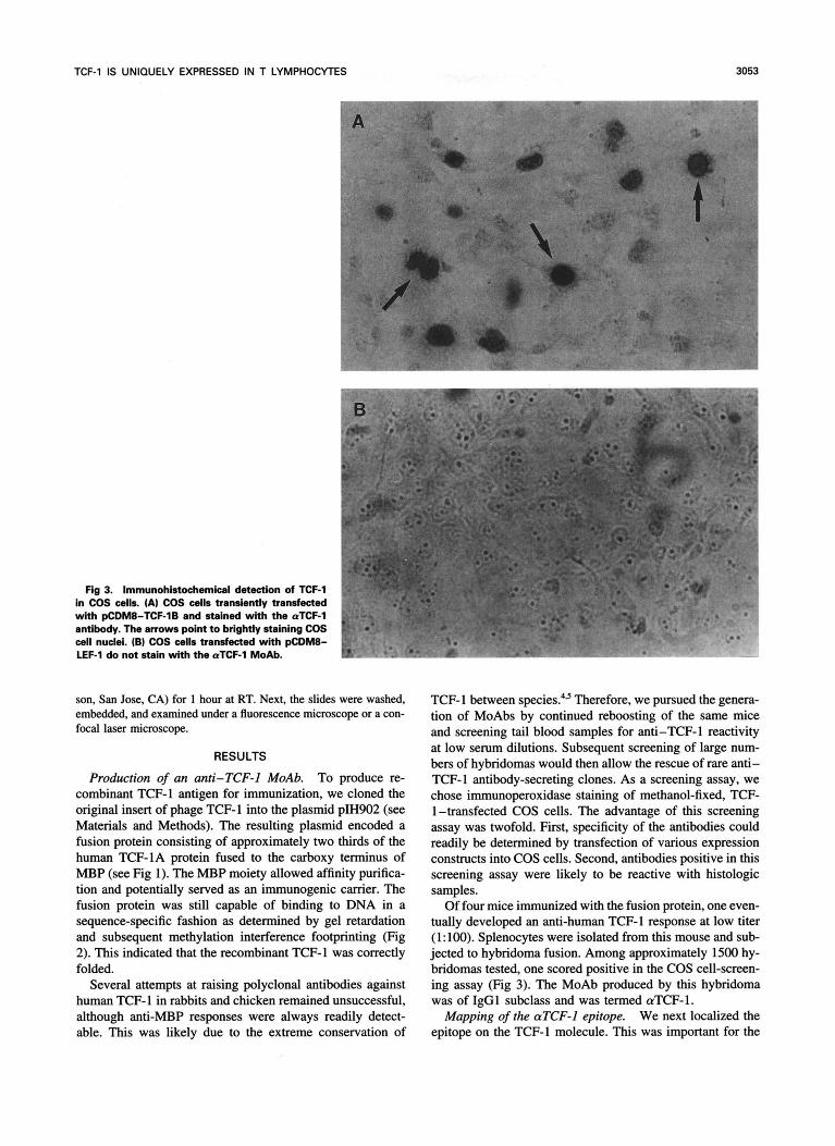
Fig 3. Immunohistochemicel detection of TCF-1 in COS cells. (A) COS cells transiently transfected with pCDM8-TCF-1B and stained with the aTCF-1 ' antibody. The arrows point to brightly staining COS p cell nuclei. (B) COS cells transfected with pCDM8- LEF-l do not stain with the aTCF-l MoAb.
F
son, San Jose, CA) for 1 hour at RT. Next, the slides were washed, embedded, and examined under a fluorescence microscope or a con- focal laser microscope.
RESULTS
Production of an anti-TCF-l MoAb. To produce re- combinant TCF-l antigen for immunization, we cloned the original insert of phage TCF-l into the plasmid pH902 (see Materials and Methods). The resulting plasmid encoded a fusion protein consisting of approximately two thirds of the human TCF-1A protein fused to the carboxy terminus of MBP (see Fig 1). The MBP moiety allowed affinity purifica- tion and potentially served as an immunogenic carrier. The fusion protein was still capable of binding to DNA in a sequence-specific fashion as determined by gel retardation and subsequent methylation interference footprinting (Fig 2). This indicated that the recombinant TCF-1 was correctly folded.
Several attempts at raising polyclonal antibodies against human TCF-l in rabbits and chicken remained unsuccessful, although anti-MBP responses were always readily detect- able. This was likely due to the extreme conservation of
I 1
l
TCF-l between species!J Therefore, we pursued the genera- tion of MoAbs by continued reboosting of the same mice and screening tail blood samples for anti-TCF-1 reactivity at low serum dilutions. Subsequent screening of large num- bers of hybridomas would then allow the rescue of rare anti- TCF-l antibody-secreting clones. As a screening assay, we chose immunoperoxidase staining of methanol-fixed, TCF- l-transfected COS cells. The advantage of this screening assay was twofold. First, specificity of the antibodies could readily be determined by transfection of various expression constructs into COS cells. Second, antibodies positive in this screening assay were likely to be reactive with histologic samples.
Of four mice immunized with the fusion protein, one even- tually developed an anti-human TCF-l response at low titer (1: 100). Splenocytes were isolated from this mouse and sub- jected to hybridoma fusion. Among approximately 1500 hy- bridomas tested, one scored positive in the COS cell-screen- ing assay (Fig 3). The MoAb produced by this hybridoma was of IgGl subclass and was termed aTCF-1.
Mapping of the crTCF-l epitope. We next localized the epitope on the TCF-1 molecule. This was important for the
3054 CASTROP ET AL
A
L I I I 1 l" 1 2 3 4 5 6 7 8 9
B M 1 2 3 4 5 6 7 8
m- T." _.V". " I_ " ._. ". .. 58-
48-
36-
26-
C vm
1 Ix + + X
.. ..
@U hXF-1
hXF-lB a hXF-1C
hXF"1E
hTCF-1C . . . ~KKKRRSREKHQESIT . DWSPKKCRARFGLNQQTDWCGPCR exon wr exon D[ exon X
1 1 1 1 1 1 1 1 1 1 1 1 1 1 1 1 1 1 1 1 1 1 1 1 1 1 1 1 1 1 1 1 1 1 1 1 1 1 1 1 1 hTCF-lE . . . OKKKRRSRJXHQESTT . DWSPKKCMRFOLNQQTDWCQPCR . RKKKCIRYLWEGRCPSPVPSDDS,WCPGSPAPQ
DSPSYHLLPRFF'TELLTSPAEPAPrSPGLSTALSLPT PGPPQAPRSTLQSTQVQQQESQRQVA

TCF-l IS UNIQUELY EXPRESSED IN T LYMPHOCYTES 3055
characterization of the MoAb, as at least four alternative splice products of TCF-1 exist: some of which might not be recognized. As expected, the MoAb was found to be reactive with the MBP/TCF fusion protein, which mapped the recognized epitope to the C-terminal two thirds of the TCF-1 molecule. Immunohistochemistry of transfected COS cells revealed that aTCF-1 was reactive with both the TCF- 1A and TCF- 1B splice forms of human TCF-1, indicating that the epitope was located in the common stretch of amino acids 1 to 243." Expression in COS cells of a small fragment of TCF-1 containing the DNA-binding domain (residues 118 to 235; see Fig 1) again resulted in strong nuclear staining. The antibody did not react with murine TCF-1, transfected into COS cells, or with endogenous TCF-1 in nuclei of mu- rine thymocytes (not shown). Murine TCF-1B and human TCF-1B differ only by four amino acids in the relevant region. As indicated in Fig 1, these sequence differences are clustered directly N-terminal to the HMG box. We, therefore, concluded that the epitope mapped to this region of the molecule. Importantly, this region is common to all splice alternatives of human TCF-1. The experimental data per- taining to the epitope mapping are summarized in Fig 1. To further characterize the specificity of the MoAb, we tested its reactivity with the most closely related member of the HMG box family, human LEF-1, transfected into COS cells. LEF- 1, which is also expressed in T lymphocytes, is 98% identical to TCF-1 in the HMG box region, but diverges significantly from TCF-1 in the remainder of the m~lecule."~'~ The aTCF- 1 MoAb did not react with transfected LEF-1 in the peroxidase staining assay, whereas a polyclonal anti-LEF serum readily yielded a bright nuclear staining (not shown).
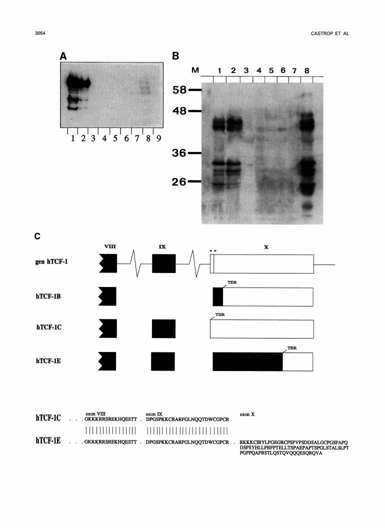
Western blotting. The antibody was found to be reactive in Western blotting of extracts prepared from T cell lines (Fig 4A), as well as from thymus and peripheral blood T cells (Fig 4B). All non-T-lineage cell lines tested were nega- tive, as were control tissue extracts. Two clusters of bands were consistently observed, one of approximately 25 kD to 32 kD and another of approximately 42 kD to 48 M). Significantly, the predicted molecular weight of the various splice forms of TCF-1 were all in the order of 26 kD to 28 kD." The banding pattern was insensitive to reduction of the samples before electrophoresis, excluding that some of the bands represented disulphide-linked homo- or heterodimers (not shown). The origin of the complexity of the banding pattern was, thus, not directly apparent. Reexamination of the known C-terminal splicing alternatives revealed that a hypothetical combination of alternative exon XI with exon XB4*" would result in a much longer open reading frame of approximately 45 kD (Fig 4C). Indeed, this splicing alterna- tive (termed TCF-1E) could be cloned and was found to
represent a rather abundant form of TCF-1. Subsequently, additional alternative exons in the N-terminal region of the protein were cloned. These novel splice alternatives are cur- rently being characterized in more detail. Theoretically, the TCF-l gene can encode over 100 protein i s o f o m (M. van de Wetering and H. Clevers, unpublished data, January 1995), all of which are predicted to react with the aTCF-1 antibody. Although the analysis is still ongoing, the presently available data indicate that the complexity of the Western blot pattern results from extensive alternative splicing of the TCF-1 pre-mRNA molecule.
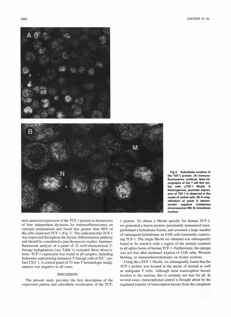
TCF-I is a nuclear protein uniquely expressed in T lym- phocytes. To define the subcellular localization of the TCF- 1 protein, several T cell lines were subjected to immunofluo- rescent staining. In all cases, a clear nuclear staining pattern was observed. This was in line with observations made on the transfected COS cells, where the TCF-1 protein also accumulated in the nucleus (Fig 3). Fhorescence confocal laser-microscopy allowed a high resolution analysis of the nuclear expression. A diffuse punctate pattern was observed in interphase nuclei. Neither nucleoli nor metaphase chromo- somes stained (Fig 5).
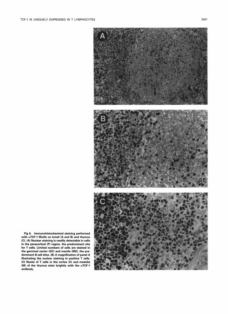
The tissue-specific expression pattern of the TCF- 1 protein was subsequently determined in a panel of adult human tis- sues consisting of jejunum, heart, liver, lung, muscle, stom- ach, skin, kidney, thymus, spleen, tonsil, and lymph node. Frozen sections of the relevant tissues were stained with the a-TCF-1 MoAb. As demonstrated in Fig 6, a nuclear staining pattern was readily detectable in lymphoid cells in normal thymus and in tonsils, spleen, and lymph nodes. In the latter three organs, TCF-1-positive nuclei were concentrated in the T-cell areas. Morphologically nonlymphoid cells in these tissues (eg, stromal elements) never displayed any staining. Double-staining with CD3, C M , and CD8 antibodies con- firmed that TCF-1-positive cells were always of T lineage (not shown). Interestingly, TCF-l expression in individual thymocytes varied dramatically, but appeared particularly high in a subset of cortical thymocytes.
The TCF-l protein was never detected in nuclei of non- lymphoid cells in any of the other tissues examined. In partic- ular, lung and kidney, the two most prominent sites of TCF- 1 mRNA expression in the mouse embryo, did not display any nuclear staining. Some extracellular staining (most likely of the basement membrane) was observed in the glomerulus of the kidney (not shown). T h i s was interpreted as crossreac- tivity to a basement membrane component. No other sites of such nonspecific reactivity were observed.
TCF-1 protein expression appeared to be completely re- stricted to the T lineage. TCF-1 expression could, thus, po- tentially serve as a useful diagnostic lineage marker. We
< Fig 4. Western blot analysis of TCF-1 protein in cell lines and tissues. (A) TCF-1 protein in cell lines. T cell lines: lane 1, Jurkat and lane 2,
H9. B call mess: lane 3, REH; lane 4, Daudi; lane 5, U266, and lane 6, APD. Lane 7, the hapatoma cell line H 4 2 ; lane 8, the prornyelocytic cell line HL60; Ime 9, the erythromyaloid line K562. (B) TCF-l protein in whole-tissue protein extracts. Lana 1, Jurkat T cell line control; lane 2, peripharal blood lymphocytes; lane 3, heart; lane 4, brain; lane 5, liver; lane 6, lung; lane 7, tongue; and Iena 8, thymus. Approximately equal amounts of protein wera loaded in each lane, evidenced by Ponseau S staining (not shown) after blotting to nitrocellulose. (C) Sequence and origin of the novel, long TCF-1E isoform. Indicated are exons Vlll to X, including tho two pertinant altarnative splice acceptor sides in exon X. Coding sequences are indicated in solid boxes.
3056 CASTROP ET AL
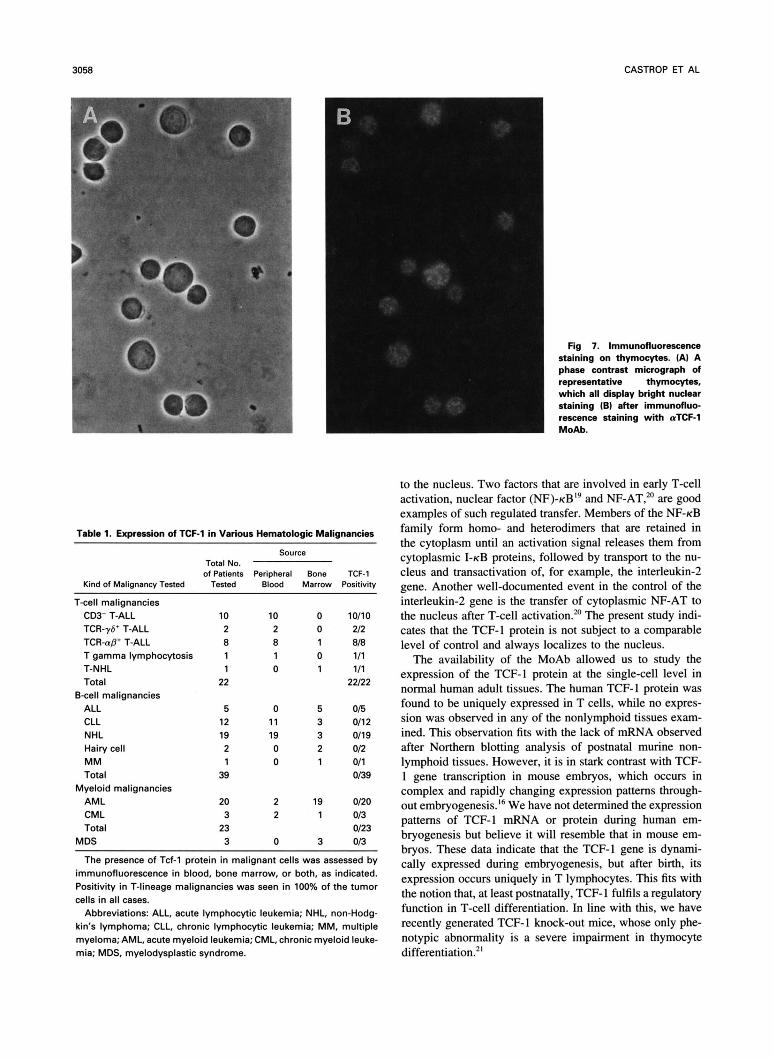
next analyzed expression of the TCF-l protein in thymocytes of four independent thymuses by immunofluorescence on cytospin preparations and found that greater than 98% of the cells expressed TCF-I (Fig 7). This indicated that TCF-I was expressed throughout the thymic differentiation pathway and should be considered a pan-thymocyte marker. Immuno- fluorescent analysis of a panel of 22 well-characterized T- lineage malignancies (see Table I ) extended these observa- tions. TCF-I expression was found in all samples, including leukemias representing immature T-lineage cells (CD2', sur- face CD3-). A control panel of 57 non-T hematologic malig- nancies was negative in all cases.
DISCUSSION
The present study provides the first description of the expression pattern and subcellular localization of the TCF-
Fig 5. Subcellular locah'on of the TCF-l protein. (AI Immuno- fluorescence confocal laser-mi- crographs of the T cell line Jur- kat with (rTCF-1 MoAb. A heterogenous, punctate expres- sion of TCF-1 is observed in the nuclei of Jurkat cells. (B) A mag- nification of panel A demon- strates negative metaphase chromosomes (M); N. interphase nucleus.
1 protein. To obtain a MoAb specific for human TCF-I, we generated a fusion protein, persistently immunized mice, performed a hybridoma fusion, and screened a large number of subsequent hybridomas on COS cells transiently express- ing TCF-l. The single MoAb we obtained was subsequently found to be reactive with a region of the protein common to all splice forms of human TCF- 1. Furthermore, the epitope was not lost after methanol fixation of COS cells, Western blotting, or immunohistochemistry on frozen sections.
Using the aTCF-l MoAb, we subsequently found that the TCF-l protein was located in the nuclei of normal as well as malignant T cells. Although most transcription factors localize to the nucleus, this is certainly not true for all. In several cases, transcriptional control is brought about by the regulated transfer of transcription factors from the cytoplasm
TCF-1 IS UNIQUELY EXPRESSED IN T LYMPHOCYTES 3057
Fig 6. Immunohistochemical staining performed with aTCF-l MoAb on tonsil (A and B) and thymus (C). (A) Nuclear staining is readily detectable in cells in the paracortical (P) region, the predominant site for T cells. Limited numbers of cells are stained in the germinal center (GC) and mantle (MZ), the p r e dominant B-cell sites. (B) A magnification of panel A illustrating the nuclear staining in positive T cells. (C) Nuclei of T cells in the cortex (C) and medulla (M) of the thymus stain brightly with the rrTCF-l antibody.
3058 CASTROP ET AL
Table 1. E x p m i o n of TCF-1 in Various Hematologic Malignancies
Source Total No. of Patients Peripheral Bone TCF-1
Kind of Malignann/ Tested Tested Blood Marrow Positivity
T-cell malignancies CD3- T-ALL TCR-y6* T-ALL TCRUD+ T-ALL T gamma lymphocytosis
Total T-NHL
Bcell malignancies ALL CLL NHL Hairy cell M M Total
Myeloid malignancies AML CML Total
MDS
10 10 0 2 2 0 8 8 1 1 1 0 1 0 1
22
5 0 5 12 11 3 19 19 3 2 0 2 1 0 1 39
20 2 19 3 2 1
23 3 0 3
10110 212 8/8 111 111
22122
0/5 011 2 011 9 0/2 011 OB9
0120 OB 0123 OB
The presence of Td-l protein in malignant cells was assessed by immunofluorescence in blood, bone marrow, or both, as indicated. Positivity in T-lineage malignancies was seen in 100% of the tumor cells in all cases.
Abbreviations: ALL, acute lymphocytic leukemia; NHL, non-Hodg- kin's lymphoma; CLL, chronic lymphocytic leukemia; MM, multiple myeloma; AML, acute myeloid leukemia; CML, chronic myeloid leuke- mia; MDS, myelodysplastic syndrome.
Fig 7. Immunofluorescence staining on thymocytes. (AI A phase contrast micrograph of representative thymocytes, which all display bright nuclear
1 staining (B) after immunofluo- 1 rescence staining with uTCF-1 MoAb.
to the nucleus. Two factors that are involved in early T-cell activation, nuclear factor (NF)-KB'~ and NF-AT:' are good examples of such regulated transfer. Members of the NF-KB family form homo- and heterodimers that are retained in the cytoplasm until an activation signal releases them from cytoplasmic I-KB proteins, followed by transport to the nu- cleus and transactivation of, for example, the interleukin-2 gene. Another well-documented event in the control of the interleukin-2 gene is the transfer of cytoplasmic NF-AT to the nucleus after T-cell activation.20 The present study indi- cates that the TCF-l protein is not subject to a comparable level of control and always localizes to the nucleus.
The availability of the MoAb allowed us to study the expression of the TCF-l protein at the single-cell level in normal human adult tissues. The human TCF-l protein was found to be uniquely expressed in T cells, while no expres- sion was observed in any of the nonlymphoid tissues exam- ined. This observation fits with the lack of mRNA observed after Northern blotting analysis of postnatal murine non- lymphoid tissues. However, it is in stark contrast with TCF- 1 gene transcription in mouse embryos, which occurs in complex and rapidly changing expression patterns through- out ernbryogenesis.I6 We have not determined the expression patterns of TCF-l mRNA or protein during human em- bryogenesis but believe it will resemble that in mouse em- bryos. These data indicate that the TCF-l gene is dynami- cally expressed during embryogenesis, but after birth, its expression occurs uniquely in T lymphocytes. This fits with the notion that, at least postnatally, TCF-1 fulfils a regulatory function in T-cell differentiation. In line with this, we have recently generated TCF-1 knock-out mice, whose only phe- notypic abnormality is a severe impairment in thymocyte differentiation.2'

TCF-1 IS UNIQUELY EXPRESSED IN T LYMPHOCYTES 3059
In addition to the expected molecular weight of 26 kD to 28 kD, an unexpected, prominent cluster of bands around 42 kD to 48 kD was consistently observed in Western blot analysis of primary T cells and T cell lines. This led to the identification of numerous novel splice forms of TCF- 1. One of the prominent novel forms resulted from the opening of a previously unidentified reading frame in exon X conserved between mouse and man (M. van de Wetering and H. Clevers unpublished data, January 1995). We are currently evaluat- ing the transcriptional control properties of these novel TCF- 1 forms, some of which are highly abundant. Polymerase chain reaction (PCR) analysis of mouse embryonic tissues and thymocytes, as well as stage-specific Western blotting of sorted thymocyte subpopulations, have so far not revealed a differential expression of the various splice alternatives.
This study illustrates that the expression of TCF-1 is com- pletely restricted to the T lineage and, vice versa, that all T- lineage cells express TCF-l. As revealed by gene disruption in the mouse germline, the expression of TCF- 1 is a prerequi- site for normal T-lineage differentiation.” It is, therefore, to be expected that not only normal but also malignant T cells will express the TCF-1 gene. This implies that the expression of TCF-l should be useful as a diagnostic marker for the T lineage. As demonstrated in Table 1, this notion was con- firmed within a panel of hematologic neoplasms. It appears to be a rational approach to determine cell lineage by directly assaying for proteins that control differentiation of that lin- eage. Thus, the availability of reagents such as the aTCF-1 MoAb should refine lineage determination of hematologic malignancies.
ACKNOWLEDGMENT
We thank Dr J. van de Winkel and the members of the Clevers laboratory for helpful discussion and comments on the manuscript and Dr A. Bloem for help with evaluating reactivity with various hematologic tumors.
REFERENCES 1. Clevers H, Alarcon B, Wileman T, Terhorst C: The TCWCD3
complex: A dynamic protein ensemble. Annu Rev Immunol 6:629, 1988
2. Clevers H, Owen M: Towards a molecular understanding of T cell differentiation. Immunol Today 12:86, 1991
3. Leiden L: Transcriptional regulation of T cell receptor genes. AMU Rev Immunol 11:539, 1993
4. van de Wetering M, Oosterwegel M, Dooijes D, Clevers H: Identification and cloning of TCF-1, a T lymphocyte-specific tran- scription factor containing a sequence-specific HMG box. EMBO J 10:123, 1991
5. Oosterwegel M, van de Wetering M, Dooijes D, Klomp L, Winoto A, Georgopoulos K, Meijlink F, Clevers H: Cloning of
murine TCF-1, a T cell-specific transcription factor interacting with functional motifs in the CD3-E and T cell receptor Q enhancers. J Exp Med 173:1133, 1991
6. Travis A, Amsterdam A, Belanger C, Grosschedl R: Lef-l, a gene encoding a lympoid-specific protein with an HMG domain, regulates T-cell receptor (Y enhancer function. Genes Dev 5:880, 1991
7. Watennan ML, Fischer WH, Jones KA: A thymus-specific member of the HMG protein family regulates the human T cell receptor C-a enhancer. Genes Dev 5:656, 1991
8. van de Wetering M, Oosterwegel M, van Norren K, Clevers H: sox-4, an Sry-like HMG box protein, is a transcriptional activator in lymphocytes. EMBO J 12:3847, 1993
9. Ho I, Voorhees P, Marin N, Oakley BK, Tsai S , Orkin SH, Leiden JM: Human GATA-3: A lineage restricted transcription fac- tor that regulates the expression of the T cell receptor Q gene. EMBO J 10:1187, 1991
10. KO LJ, Yamamoto M, Leonard MW, George KM, Ting P, Engel JD: Murine and human T-lymphocyte GATA-3 factors medi- ate transcription through a cis-regulatotry element within the human T cell receptor 6 gene enhancer. Mol Cell Biol 5:2778, 1991
11. Marine J, Winoto A: The human enhancer-binding protein GATA3 binds to several T-cell receptor regulatory elements. Proc Natl Acad Sci USA 88:7284, 1991
12. Georgopoulos K, Moore DD, Derfler B: Ikaros, an early lymphoid-specific transcription factor and a putative mediator for T cell commitment. Science 258:808, 1992
13. van de Wetering M, Clevers H: Sequence-specific interaction of the HMG box proteins TCF-1 and SRY occurs within the minor groove of a Watson-Crick double helix. EMBO J 11:3039, 1992
14. Giese K, Cox J, Grosschedl R The HMG domain of lymphoid enhancer factor 1 bends DNA and facilitates assembly of functional nucleoprotein structures. Cell 69:185, 1992
15. Oosterwegel M, van de Wetering M, Holstege F, Prossner H, Owen M, Clevers H: TCF-l, a T cell-specific transcription factor of the HMG box family, interacts with sequence motifs in the TCR-p and TCR-6 enhancers. Int Immunol 3: 1189, 1991
16. Oosterwegel M, van de Wetering M, Timmerman J, h i s - beek A, Destree 0, Meijlink F, Clevers H: Differential expression of the HMG box factors TCF-1 and LEF-I. Development 118:439, 1993
17. van de Wetering M, Oosterwegel M, Holstege F, Dooijes D, Suikerbuik R, Geurts van Kessel A, Clevers H: The human T cell transcription factor-l gene. J Biol Chem 267:8530, 1992
18. Castrop J, Hoevenagel R, Young JR, Clevers H: A common ancestor of the mammalian transcription factors TCF-1 and TCF- 1dLEF-I expressed in chicken T cells. Eur J Immunol22:327, 1992
19. Thanos D, Maniatis T: NF-KB: A lesson in family values. Cell 80529, 1995
20. Rao A: NF-ATP: A transcription factor required for the co- ordinate induction of several cytokine genes. Immunol Today 15:274, 1994
21. Verbeek S, Izon D, Hofhuis F, Robanus-Maandag E, te Riele H, van de Wetering M, Oosterwegel M, Wilson A, MacDonald HR, Clevers H: An HMG-box-containing T-cell factor required for thymocyte differentiation. Nature 374:70, 1995

![arXiv:2101.00190v1 [cs.CL] 1 Jan 2021](https://static.fdokumen.com/doc/165x107/633f79d7975a7a6cb40e2b28/arxiv210100190v1-cscl-1-jan-2021.jpg)
![arXiv:2105.10954v4 [physics.plasm-ph] 1 Jan 2022](https://static.fdokumen.com/doc/165x107/6318270fcf65c6358f01de99/arxiv210510954v4-physicsplasm-ph-1-jan-2022.jpg)