The Role of Coagulation and Haemostatic Factors in cancer ...
88
1 Haemostatic Issues in Cancer Development and Progression: “The Role of Coagulation and Haemostatic Factors in cancer Development and Metastasis” Ph.D. Thesis Hussain Alizadeh 2007
-
Upload
khangminh22 -
Category
Documents
-
view
3 -
download
0
Transcript of The Role of Coagulation and Haemostatic Factors in cancer ...

1
Haemostatic Issues in Cancer Development and Progression:
“The Role of Coagulation and Haemostatic Factors in cancer
Development and Metastasis”
Ph.D. Thesis
Hussain Alizadeh
2007
2
Contents
Table of Contents 2
Dedication 3
Foreword 4
Introduction 5-18
Goal and perspectives of study 19-31
Results of study 32-61
Discussion 62-74
References 75-86
List of Publications 87-89

3
Dedication
To my mother for her gifts of love and life,
My father for his eternal support and encouragement,
My parents, brothers, sisters and family whose enthusiasm
and dedication continue to inspire me throughout my life
And, my friends & colleagues for being just that.

4
Foreword
When asking young physicians why they wish to embark on haematology-oncology as a
speciality, a thoroughly rehearsed answer centres around their interest for a combination
of clinical and laboratory work. This, of course, is precisely what has attracted many of
us to haemato-oncology, and it has been a challenge to writers, editors and publishers.
The major changes that have occurred in all fields of medicine over the last decade have
been accompanied by an increased understanding of the biochemical, physiological,
immunological, and pathogenic processes involved in malignant transformation, distant
meastasis and disturbances that may occur in different diseases. At the same time, the
range of treatment available for patients with malignant haemato-oncological diseases
have widened and improved substantially as understanding of the disease process have
increased and new drugs and means of support care have been introduced. I can only take
pride in my association with Professor Shaker A. Mousa and Professor Hajna Losonczy
over several years, and it is my great pleasure to wish them further success they richly
deserves, and I sincerely thank both of them.

5
I. Introduction:
Thrombosis is a well-recognized and common complication in patients with malignant
disease and can contribute significantly to the morbidity and mortality of this disease.
The occurrence of thrombosis is heightened by therapeutic interventions such as
operations or the use of radio-chemotherapy. It occurs both spontaneously, after surgery,
after radiation therapy and in medical cancer patients receiving anti-cancer treatment. It
may also be the first manifestation of underlying malignant disease. The magnitude of the
risk for venous thromboembolism is well established for cancer surgery where rates twice
that for abdominal surgery in non-cancer patients are described. Venous
thromboembolism is the most common complication of cancer and the second most
common cause of death in cancer patients (1, 2). Up to 60% of patients with cancer
develop venous thromboembolism, depending on the type of cancer and the treatment
given (3-6). Although the close relationship between tumour growth and the activation of
blood coagulation has been known since 1865, when Professor Armand Trousseau first
described the clinical association between primary or idiopathic venous
thromboembolism and occult malignancy, only in the last two decade have significant
advances in this field been achieved.
The association between malignancy and thromboembolic disease is well established and
has been recognized in the medical literature for at least 135 years, since Professor
Armand Trousseau and T. Billroth first presented their observations in publications by
the New Syndenham Society (25). As Trousseau himself described, venous
thromboembolism (VTE) may be a manifestation of occult malignancy and/or may be a
complication of known malignancy. Billroth attributed the pathologic finding of tumour
cells imbedded in thrombus as evidence for the role of venous thromboembolism in the
metastatic process (25).
Several authors have demonstrated that patients with clinically apparent malignancy
commonly develop thromboembolic disease (7-13). Patients with thromboembolic
disease without a known diagnosis of malignancy have a higher rate of occult malignancy
than the general population (13-18). Although supporting data are limited, patients who
develop thromboembolic disease in association with malignant disease and are treated

6
with anticoagulation therapy are often thought to have a higher rate of recurrent
thromboembolic disease in comparison with patients with thromboembolic disease that is
not associated with malignancy (19-23).
It is now well known that the clinical manifestation of thrombosis in this condition can be
very different and vary from localized venous thromboembolism to disseminated
intravascular coagulation. In addition, a subclinical activation of blood coagulation or
“hypercoagulable state” is present in almost all cancer patients, even without symptoms
of thrombosis. A number of pathogenetic factors have been identified, showing that
activation of coagulation in cancer is a complex phenomenon, involving many different
pathways of the haemostatic system and numerous interactions of the tumour cells with
other blood cells, including platelets, monocytes and endothelial cells. The activation of
blood coagulation in those with malignant disease appears to be dependent upon the
elaboration of tumour-derived tissue factors resulting in an activation of extrinsic
pathway of coagulation cascade.
More recently prospective clinical trials have definitely demonstrated that patients with
idiopathic venous thromboembolism are at significantly higher risk for a subsequent
diagnosis of malignancy as compared to patients with secondary venous
thromboembolism (i.e. VTE due to known causes, such as congenital thrombophilia,
pregnancy, immobilization, and the use of oral contraceptives, among others).
Equally well demonstrated is the concept that patients with known cancer represent a
particularly high-risk group for the development of secondary venous thromboembolism.
These patients have been stratified into the highest risk category for the development of
secondary venous thromboembolism. This risk is significantly increased by the anti-
tumour interventions, including surgery and chemotherapy. In addition to the thrombotic
risk associated with cancer itself, therapeutic interventions such as cancer surgery,
chemotherapy and central venous catheters have all been associated withy an increased
risk of VTE (1-4). Evidence is accumulating to suggest the potential risk of cancer
therapy in increasing the cancer-associated tendency to develop thrombosis. First of all,
surgery, which is used to treat patients with localized tumours, is a well-known
precipitating factor for thromboembolic disease, since the haemostatic system becomes

7
more activated before and after surgery (99). Secondly, some chemotherapeutic agents,
such as L–asparaginase, mitomycin C, cisplatinum or high-dose chemotherapy-
conditioning regimens for haematopoietic stem cell transplantation (veno-occlusive
disease) have been associated with thrombo-embolic complications (100). Also the use of
haematopoietic growth factors (i.e. G-CSF or GM-CSF) may be implicated in the
hypercoagulation and increased thrombotic tendency of neoplastic patients (101, 102).
Therefore, clinical trials have been conducted or are currently ongoing to define the best
modalities to give thromboprophylaxis routinely to these patients at least during chemo-
radio therapy or surgery. Another open question in the clinics of thrombosis and cancer
concerns the treatment of thrombosis in those patients who have already experienced one
episode of venous thromboembolism. Cancer patients are indeed at increased risk of both
recurrences (even with adequate levels of anticoagulation) and bleeding complications.
Therefore, much effort is devoted to developing studies in these patients to define the
efficacy of anticoagulant drugs and the duration and intensity of anticoagulation.
Post-mortem studies have been demonstrated a markedly increased incidence of
thromboembolic disease in patients who died of cancer, particularly those with mucinous
carcinomas of the pancreas, lung, and gastrointestinal tract. Although VTE may be found
in upwards of 50% of cancer patients at autopsy (50, 51, 52), the optimal study design for
determining the true incidence of clinical VTE in cancer patients is a prospective cohort
study. Cohort studies of surgical patients showed that the incidence of deep vein
thrombosis was markedly higher in patients with malignant disorders than in patients
with other, non-malignant diseases.
Although its frequency in different tumour types has not been described in detail, there
appear to be tumour differences, with certain cancers associated with poor outcome (e.g.
pancreas, lung) attended by a higher frequency of thromboembolic complications. The
suggestion is that thrombosis may be associated with a poor outcome for patients with
cancer is further supported by large epidemiological databases indicating that cancer
patients who either develop a thromboembolic episode during the natural history of their
cancer or are diagnosed with a thrombotic episode when their cancer is diagnosed have a
poorer survival over time than cancer patients without thrombosis (80).

8
The first attempt at such a study was a retrospective analysis (53) of data derived from
randomized clinical trials of therapy in patients with breast cancer (54-66); data that was
collected prospectively.
Other patients with advanced cancers who are likely to be at higher risk for
thromboembolism include patients with brain tumours receiving chemotherapy, those
with locally recurrent rectal cancer receiving radiation, pancreatic cancer or advanced
gastrointestinal cancers (particularly adenocarcinomas) [53]. However, precise estimates
of thrombotic rates in these groups of patients are not available. Von Tempelhoff et al.
(67) reported a 10.6% rate of VTE in women with advanced ovarian cancer receiving
chemotherapy. The majority of data regarding the risk of VTE in patients with other
cancers come from small case series and retrospective reviews. Rates of 8.4% have been
reported in patients with germ cell tumours receiving chemotherapy (68), 24-60% in high
grade gliomas (69, 70, 71), and 5-10% in patients with Hodgkin’s or non-Hodgkin’s
lymphoma (72-75). In addition to chemotherapy, the use of haematopoietic growth
factors may increase the risk of VTE, although no prospective data s available and meta-
analysis by Barbui et al. (75) was inconclusive. Finally, cancer patients with indwelling
central venous catheters are at increased risk for thrombosis of the axillary/subclavian
vein (76, 77), with the catheters themselves susceptible to thrombotic occlusion despite
the use of routine heparin flushes.
Surgical intervention in patients with cancer places them at increased risk of
postoperative VTE (approximately 2-fold) in comparison to non-cancer patients
undergoing the same procedures (78, 79). The case for routine thrombo-prophylaxis in
patients receiving chemotherapy is less clear, and prospective studies investigating rates
of thrombosis by tumour types, stage of malignant disease, and chemotherapeutic
regimens are required.
In this thesis, the pathogenetic mechanisms of thrombosis in malignancy, thrombophilic
state in cancer patients, changes in haemostatic parameters and their relation to cancer
prognosis and also the thromboprophylaxis in the cancer patients are discussed and
analyzed.

9
Pathogenesis-Pathophysiology: Despite the fact that thromboembolism is a major problem in cancer patients, the study of
the prethrombotic conditions in cancer have been relatively neglected or just given a
phenomenological approach. Possible contributory mechanisms for blood clotting
activation in tumour patients include general factors related to the host’s response to the
tumour (acute phase reaction, abnormal protein metabolism, neovascularization, necrosis,
haemodynamic rearrangements) and more specific factors such as the activities expressed
by tumour cells and tumour-associated macrophages. The history of medicine again
reminds us that Virchow, over 150 years ago, postulated that three features may
predispose to thrombus formation, that are abnormalities in -1) blood flow, -2) blood
constituents and -3) vessel wall. Patients with cancer may show abnormalities of each of
the three components of Virchow’s triad, leading to a prethrombotic or hypercoagulable
state. Pathogenetic mechanisms of thrombogenesis in the cancer patients are complex and
involve multiple interdependent processes between the tumour and the patient’s
physiological response to the tumour. These act to promote a hypercoagulable state.
These mechanisms accounting for the development of thrombotic disorders in patients
affected by cancer were described by Virchow more than a century ago. They include
procoagulant activity (hypercoagulability due to tumour cell activation of clotting and the
pro-thrombotic properties of the tumour cells), host inflammatory responses, vessel wall
injury, stasis, and extrinsic factors.
Abnormalities of blood flow may be associated with changes in blood resistance or
viscosity. This may be due to risk factors occurring in malignancy, such as venous stasis
due to a lack of mobilization, sepsis or external compression from a bulky tumor. Blood
viscosity (high and low shear rates) measured preoperatively in cancer patients have been
correlated with the incidence of postoperative DVT (88). The possibility that abnormal
blood vessel formation (related to cancer angiogenesis and factors promoting it) may
cause flow disturbances also has to be considered (89). Venous stasis predisposes to
venous thrombosis by preventing activated coagulation factors from being diluted and
cleared by normal blood flow. Moreover, hypoxic damage to endothelial cells due to
stasis may produce pro-thrombotic alterations. Venous stasis develops as a consequence
of immobility in severely debilitated cancer patients, in conjunction with cancer surgery,

10
or as a result of venous obstruction due to extrinsic vascular compression in patients with
bulky tumour masses (49).
Cancer cells can activate coagulation directly through an interaction with platelets and/or
clotting and fibrinolytic systems to generate thrombin. Clotting activation may be
considered as a special type of inflammatory reaction to stimuli such as vessel wall
damage, or intravascular cell aggregation or entry in blood of abnormal cells such as
tumour cells (39, 90). The balance between the coagulation and the fibrinolytic system
can easily shift to a prethrombotic state in cancer, through an excess of tissue factor
(TF), other procoagulant proteins or of plasminogen activator inhibitor (PAI-1) or
through deficiencies in inhibitory molecules (antithrombin, protein C, protein S) or in
fibrinolytic principles (tissue plasminogen activator, t-PA). Consistent with the shift of
the haemostasis balance toward hypercoagulation in cancer, several studies have shown
reduced levels of natural inhibitors of coagulation such as antithrombin (AT), protein C
(PC) and protein S (PS) in plasma of cancer patients. These decreased levels of inhibitors
might result from an increased consumption as a consequence of the activation of
coagulation or from a defective hepatic synthesis or both mechanisms combined.
Chemotherapy and hormonal treatment can also induce an acquired deficiency in
naturally occurring anticoagulants-inhibitors. In breast cancer, patients receiving
cyclophosphamide, methotrexate and fluorouracil have decreased levels of PC and PS
antigen and activity soon after the beginning of the therapy while plasminogen activator
inhibitors increase (127). However, no relationship between these low levels of inhibitors
and markers of hypercoagulability or thrombotic events could be found in these studies.
Low levels of PC, PS and AT have also been reported after treatment with tamoxifen and
L-asparaginase (128, 129, 130, 131, 132). Activated protein C (APC) resistance is the
most common abnormality of the coagulation system in patients with hereditary venous
thrombosis. The APC resistance phenotype is associated in a majority of patients with
heterozygosity or homozygosity for a single-point mutation in the factor V gene which
substitutes G with A at nucleotide 1691 (factor V Leiden, FVL). This mutation modifies
one of the three APC cleavage sites. The slower degradation of the mutated activated
factor V by APC leads to a higher rate of thrombin generation and a hypercoagulable
state (133, 134, 135). Acquired resistance to APC is defined as a phenotypic resistance

11
that occurs in the absence of FV-Leiden mutation. Acquired resistance to APC was found
to be much more common in patients with cancer and thromboembolism than in patients
with thromboembolism without cancer (54% versus 19%). In contrast, resistance to APC
due to FVL mutation was more common in thrombophilic patients without cancer than
with cancer. These results provide evidence that FVL does not play a major role in the
hypercoagulable state of cancer while acquired APC resistance contributes to the
thrombotic resistance in these patients (136).
Venous thromboembolism is the most common complication of cancer and the second
most common cause of death in cancer patients. Several types of cancer, including
tumours of the pancreas, prostate, lung, breast, stomach, and colon, have been associated
with a prethrombotic state characterized by clotting factor abnormalities, endothelial
dysfunction, and abnormal blood flow due to increased viscosity or stasis. Among the
most frequently studied characteristic properties of malignant disorders are the; cancer
cell interactions with the haemostatic system and their procoagulant activities. Neoplastic
cells can activate the clotting system directly, thereby generating thrombin and fibrin
formation, or indirectly by stimulating mononuclear cells, platelets, and endothelial cells
to produce and express pro-coagulants (27, 91, 92).
Thrombin generation and fibrin formation are constantly determined in patients with
malignancy, who are at increased risk of thromboembolic complications. Most
importantly, fibrin formation is also involved in the processes of tumour spread and
metastasis. In addition to the thrombotic risk associated with cancer itself, therapeutic
interventions such as cancer surgery, chemotherapy and central venous catheters have all
been associated with hypercoagulable state and an increased risk of venous
thromboembolism.
Malignant cells can interact with the haemostatic system in multiple ways. The principal
pro-thrombotic properties of tumour cells are: -1) the capacity to produce and release pro-
coagulant activities, fibrinolytic proteins, and inflammatory cytokines, i.e. interleukin-1β
(IL-1β), tumour necrosis factor-α (TNF-α), and vascular endothelial growth factor
(VEGF); and, -2) the capacity to interact with host cells, including endothelial cells lining
the blood vessels, monocytes, platelets and neutrophils.

12
The best characterized tumour cell pro-coagulants are tissue factor (TF) and cancer
procoagulant (CP). These promote blood coagulation either directly, via activation of
factors VII and X of the blood coagulation cascade, or indirectly by initiating an
inflammatory response. The inflammatory response enhances the prethrombotic process
via the action of potent inflammatory mediators, which induce further expression of
procoagulants from tumour cells, activate platelets and induce endothelial expression of
prethrombotic factors.
Tissue factor (TF) is a 47-kDa trans-membrane (integral membrane) glycoprotein which
forms a complex with factor VII (FVII)/FVIIa: the TF/FVII complex triggers blood
coagulation by proteolytically activating factor IX and X (26, 27, 81). Tissue factor is the
cellular pro-coagulant found in normal cells, including endothelial cells and monocytes-
macrophages. However, these cells do not express TF in normal resting conditions, but
expose this pro-coagulant in response to pro-inflammatory stimuli, i.e. the cytokines IL-
1β and TNF-α and bacterial endotoxin (82). Differently from normal cells, malignant
cells constitutively express TF. In addition to activating blood coagulation, TF has the
capacity of regulating VEGF expression in tumour cells and in vascular endothelium
(83), which represents an important pro-angiogenetic mechanism. The expression of TF
has been identified in some acute leukaemias (28) and in solid tumours of the stomach,
ovary, and kidney (29). Direct factor X activation with the procoagulant cysteine
proteinase has been found in some patients with prostate, lung, breast, lung, and kidney
cancer and with leukaemia (30, 31). Mucin-secreting adenocarcinomas are frequently
associated with thrombosis because the sialic acid moiety can cause non-enzymatic
activation of factor X to its active form, factor Xa (32). Consequently, adenocarcinomas
of the pancreas, lung, gastrointestinal tract, and ovary are often associated with venous
thrombosis (33).
Cancer pro-coagulant (CP) is a 68 kDa cysteine proteinase with 674 amino acid
residues and no detectable carbohydrates (84). It activates factor X independently of FVII
and cleaves the factor X heavy chain at a different site compared to other known factor X
activators. Cancer pro-coagulant (CP) has been found in extracts of malignant cells or in
amnion-chorion tissues but not n extracts of normally differentiated cells (85). CP antigen
has been identified in the sera of cancer patients and found to be elevated in 85% of the

13
study subjects. TF and CP have been identified in several human and animal tumour
tissues and they are different in nature.
Fibrinolytic activities: Tumour cells can express all the proteins regulating the
fibrinolytic system, including the urokinase-type plasminogen activator (u-PA), the
tissue-type plasminogen activator (t-PA), and the fibrinolysis inhibitors plasminogen
activator inhibitor (PAI)-1 and PAI-2 (86). Among the activators, u-PA is the most
widely expressed within malignant diseases. Furthermore, cancer cells can carry the
specific plasminogen activator receptor (uPAR) on their membranes. The presence of
these receptors favours the assembly of all the fibrinolytic components on tumour cell
membranes, facilitating the activation of the fibrinolytic cascade. The fibrinolysis
proteins can play a role in tumour cell proliferation, invasion and metastasis.
Cytokines: Tumour cells produce inflammatory cytokines, including IL-1β, TNF-α and
VEGF. Tumour cell-derived IL-1β and TNF-α can induce the expression of TF pro-
coagulant activity by endothelial cells. The also down-regulate the expression of
endothelial cell thrombomodulin (TM), the surface high-affinity receptor for thrombin.
The TM-thrombin complex activates the protein C system, which in turn functions as a
potent anticoagulant. Tissue factor up-regulation and TM down-regulation lead to a pro-
thrombotic state of the vascular wall (86, 87). The same cytokines stimulate endothelial
cells to produce the fibrinolysis inhibitor PAI-1. Inhibition of fibrinolysis further
contributes to the pro-thrombotic potential of endothelial cells. In addition, the
production of VEGF by malignant cells may significantly affect the functions of
microvascular vessels in proximity of the tumour and may play an important role in
tumour neo-angiogenesis (83). Further, VEGF is chemotactic for macrophages and can
induce TF pro-coagulant activity of monocytes and endothelial cell. The expression of TF
by tumour cells up-regulates the transcription of VEGF in these cells. TF modulates the
expression of VEGF by endothelial cells, a function that can have important implications
in tumour neo-vascularization (83). Regulation of VEGF synthesis by TF in malignant
cells and vascular cells provides an important link in cancer patients between activation
of coagulation, inflammation, thrombosis and tumour progression and metastasis.

14
Tumour cell-Host cell interactions: Based on studies of laboratory parameters, it
has been demonstrated that there are activation/perturbation of several cellular systems
‘in vivo’ in cancer patients, i.e. elevated levels of plasma endothelial markers (e.g. von
Willebrand factor, TM, soluble E-selectin, t-PA, PAI-1) and this leads to the activation of
haemostasis at the endothelium site, especially during chemotherapy (83, 86). Further,
the increase in tissue factor pro-coagulant activity (PCA) expressed by circulating
mononuclear cells shows that this cellular compartment has become activated. Finally,
the detection of high levels of platelet membrane specific glycoproteins (e.g. CD62 and
CD63), which re exposed upon activation, provides evidence for platelet activation ‘in
vivo’ in malignant conditions (87). Patients with metastatic cancers may exhibit increased
activation as also indicated by enhancement of platelet turnover and a decrease in platelet
survival time. These platelet abnormalities may be due to a greater degree of destruction
of the lysosomal membranes and to involvement of platelets in the low-grade
intravascular activation of clotting detectable in the majority f cancer patients (93).
Tumour cells can interact with the endothelium essentially by their capacity to synthesize
and release inflammatory cytokines. These cells can also directly adhere to endothelial
cells and to the extracellular matrix through membrane adhesion molecules. Endothelial
cells activated by IL-1β or TNF-α increase the exposure on their membranes of counter-
receptors for the tumour adhesion molecules. The malignant cells attached to the vessel
wall may play a key role in promoting localized clotting activation and thrombus
formation by releasing their cytokine content and favouring the adhesion and arrest of
other cells, including leukocytes and platelets. The adhesion of tumour cells to leukocytes
or to vascular cells may also facilitate cell migration and extravasations. In addition, the
TF-induced expression of VEGF by endothelial cells may have implications in tumour
neovascularization.
Tumour cells can activate systemic coagulation by stimulating mononuclear cells to
synthesize and express various procoagulant substances, including tissue factor and factor
X activators. Normal monocytes and macrophages can be activated by tumour cells in the
presence of lymphocytes (34). In patients with cancer, endothelial cells may be activated
by cytokines such as tumour necrosis factor and interleukin-1 or interleukin-like
substances that may induce tissue factor production (35). A peptide produced by a human

15
bladder cancer cell line stimulates tissue factor expression in endothelial cells (36).
Clinical manifestation of increased thrombin generation may be accentuated by down-
regulation of endothelial cell counter-regulatory mechanisms, such as decreased hepatic
synthesis of antithrombin and protein C (37, 38, 41, 42). In addition, normal endothelial
cells function may be disrupted by various defects in platelet function (37, 38, 41, 42).
The enhanced clotting activation and the pro-thrombotic properties of the tumour cells in
patients with cancer are confirmed by the demonstration of increased levels of systemic
hypercoagulability markers, such as fibrinopeptide A, prothrombin fragment F1+2 and
thrombin-antithrombin complexes in most patients (38, 39, 40). As expected, the risk of
recurrent) venous thromboembolism is higher in those cancer patients who are also
carriers of thrombophilia, such as the factor V Leiden mutation (41).
The role of endothelium in mediating a prethrombotic state is well known. Endothelial
cells may be stimulated in cancer to produce procoagulant material (such as e.g. TF)
either directly by tumour-specific antigens or indirectly by cytokines (82, 94, 95). The
latter (IL-1, TNF-α) are able to suppress endothelial fibrinolytic activity, downregulate
thrombomodulin expression while increasing the endothelial expression of leukocyte
adhesion molecules, platelet-activating factor, as mentioned, TF (96, 97, 98).
There is increasing awareness that cancer cells can injure endothelium by direct vascular
invasion, resulting in the onset of a pro-thrombotic state. Moreover, tumour cells may
secrete vascular permeability factors which account for the extravascular accumulation of
fibrinogen and other clotting proteins around tumour growth (36, 37, 38). The adhesion
of tumour cells to endothelium was evaluated in vivo by Naschitz and associates, who
observed a complex interaction between tumour cells, endothelium, and platelets (43).
Direct vessel wall injury, in association with rheologic abnormalities and catheter-
associated thrombin generation, is most likely the explanation for the occurrence of the
upper extremity deep vein thrombosis arising as a complication of central venous lines
(44). Among mechanisms responsible for thrombotic events arising during the use of
chemotherapeutic drugs, vascular endothelium damage probably plays a major role
besides the reduction in the plasma concentration of natural anticoagulants (45-48).

16
Thrombosis and disseminated intravascular coagulation (DIC) are common complications
of cancer. Specific conditions associated with cancer such as stasis due to immobilization
or blood flow obstruction, surgery, infections, endothelium damage due to
chemotherapeutic agents, radiation therapy and abnormalities of blood coagulation
contribute to the hypercoagulable and thrombophilic state of cancer patients. Several
types of cancer, including tumours of the pancreas, prostate, lung, breast, stomach, and
colon, have been associated with a prethrombotic state characterized by clotting factor
abnormalities, endothelial dysfunction, and abnormal blood flow due to increased
viscosity or stasis (103). This procoagulant state in cancer patients arises mostly from the
capacity of tumour cells to express and release procoagulant activities, e.g.; TF and CP.
Additionally, there are decreased levels of inhibitors of coagulation, impaired
fibrinolysis, the presence of antiphospholipid antibodies and an acquired activated protein
C resistance (APCR) contribute to the hypercoagulable state. The activation of
coagulation is also implicated in tumour proliferation through interactions of coagulation
with inflammation and increased tissue factor pathway inhibitor (TFPI).
Laboratory diagnosis of the procoagulant-thrombophilic state includes:
1. elevation of clotting factors, fibrinogen/fibrin degradation products,
hyperfibrinogenaemia and thrombocytosis,
2. elevation of specific markers of activation of coagulation: fragment 1+2,
fibrinopeptide A, D-dimers, thrombin-antithrombin (TAT) complexes
It is a well-known fact that none of these tests has any predictive value for the occurrence
of thrombotic events in one individual patient. But, changes in these haemostatic
parameters have been known to be associated with the prognosis of cancer patients. In
patients with venous thromboembolism (VTE) a non-invasive screening for occult cancer
is able to detect a relatively high incidence of hidden cancers and the search for
thrombophilia seems important in patients without known cancer. Haemostatic
abnormalities are found in more than 90% of cancer patients and are clinically expressed
as DIC or VTE. The reported incidence of deep vein thrombosis (DVT) or pulmonary
embolism (PE) ranges from 1 to 15% in autopsy (96, 97). However, the relationship
between symptomatic VTE and the risk of subsequent occult malignant disease is still

17
controversial. Thrombosis is a multifactorial disease, where interactions between genetic
and environmental factors result in the formation of an obstructive thrombus at a specific
location. Among the acquired risk factors for VTE is the thrombophilic-hypercoagulable
state in cancer patients, e.g.; procoagulant activities (TF and CP), changes in natural
inhibitor levels (decreased antithrombin, Protein C and Protein S levels, increased tissue
factor pathway inhibitor=TFPI), acquired APCR, increased expression of activator of
fibrinolysis to a hyperfibrinolysis state, increased generation of thrombin-activatable
fibrinolysis inhibitor (TAFI) which leads to impairment of fibrinolysis and will produce a
hypercoagulable state in cancer patients, and development of antiphospholipid antibodies
which produce an acquired hypercoagulable state. All these changes in the haemostatic
parameters will lead to a hypercoagulable state in cancers patients which can be
measured by different laboratory tests. Tissue factor pathway inhibitor (TFPI) is the
physiological inhibitor of TF-induced coagulation by forming, in a two-step mechanism,
a quaternary complex with the coagulation factors Xa and VIIa and TF. TFPI levels are
high in cancer patients and increased levels have been found in advanced malignancy
(130). The reason for these high values of TFPI is unknown. It could be a consequence of
the hypercoagulability in cancer patients, since thrombin induces a re-distribution and
acute phase release of TFPI in vitro as well as in vivo in animals (131, 132).
Thrombosis is a complication in patients with solid tumour malignancy (104), and the
heightened risk is secondary to tumour elaboration of tissue factor, a physiologic
procoagulant that is responsible for the genesis of a systemic hypercoagulable state (105).
Once activated, coagulation proteases have a profound effect on tumour cell behaviour in
experimental models (106), enhancing tumour cell motility, invasion, angiogenesis, and
growth. Hence, interference with activated coagulation serine proteases may influence
tumour biology.
Clinical evidence in support of anticoagulants having an anti-tumour effect was first
reported in a multicenter, randomized, controlled trial in 1981 (107). In the Veterans
Affairs Research Service Cooperative Study 75, warfarin was found to be associated with
an improvement in median survival in patients with small-cell lung cancer who were
receiving chemotherapy. Similarly, a randomized trial in the same patient population
demonstrated a survival advantage for those patients treated with subcutaneous injections
18
of unfractionated heparin (108). However, despite compelling experimental evidence for
a pathogenic role of blood coagulation in tumour growth and metastasis (109, 110, 111,
112), other studies in patients with solid tumours have failed to confirm a survival benefit
for patients treated with anticoagulants (113, 114, 115, 116).
More recently, the question of whether anticoagulants can favourably influence the
natural history f cancer has received renewed attention. Randomized controlled trials and
meta-analyses of studies that compared low molecular weight heparins with
unfractionated heparin for the initial treatment of venous thromboembolism have reported
a reduction in the overall mortality of patients with cancer who were randomly assigned
to receive a low molecular weight heparin (117, 118, 119, 120, 121). Although the
reduction in mortality has been consistent across studies and could not be attributed to
differences in fatal pulmonary embolism or bleeding, the observation that 5 to 7 days of
low molecular weight heparin treatment reduced cancer mortality has been difficult to
explain. A plausible biologic mechanism, however, is now emerging from experimental
studies that show low molecular weight heparins can inhibit angiogenesis, a process that
is critical for tumour growth and metastasis, in a dose-dependent fashion (109, 110,122,
123).
To date, two randomized, placebo-controlled trials designed to evaluate whether low
molecular weight heparins can improve survival in patients with advanced or incurable
cancers have been completed (124, 125).To examine the influence of a low molecular
weight heparin relative to coumarin derivatives on the survival of cancer patients with
venous thromboembolism (FAMOUS study) and to investigate the hypothesis that low
molecular weight heparins have a greater impact on survival in cancer patients with
limited disease than in those with disseminated cancer (MALT trial), the CLOT study
was performed which was a posthoc analysis of the mortality data in patients with solid
tumours who participated in the Comparison of Low Molecular Weight Heparin Versus
Oral Anticoagulant Therapy for Long Term Anticoagulation in Cancer Patients With
Venous Thromboembolism (126).

19
II. Goal and perspective of Study: Relationship between
haemostatic parameters and cancer prognostic markers Although the relationship between malignant diseases and thromboembolic complications
has been convincingly demonstrated, the clinical implications of this association still
have to be thoroughly elucidated. The most common abnormalities described in cancer
patients are elevation of the clotting factors V, VIII, IX, and XI, increased
fibrinogen/fibrin degradation products (FDP), hyperfibrinogenaemia, and
thrombocytosis. The aim of this prospective study is to evaluate the changes in
haemostatic-clotting parameters in patients with different types of non-haematological
malignancies (solid tumours) and to asses the correlation between changes in coagulation
parameters and the stage of tumour, imaging findings and also with changes in
characteristic tumour markers. In our prospective study, we mainly focused on specific
abnormalities of haemostasis in these groups of patients, the changes in haemostatic
parameters and their relation to cancer prognosis. In addition, the thromboprophylaxis in
the cancer patients are discussed and analyzed. The relationship between the
hypercoagulable state and the tumour progression and the occurrence of metastasis has
not been well established. Larger studies and further investigations on tests that could
have a reliable predictive value in tumour progression are required to evaluate the
correlation between changes in haemostatic parameters, tumour stage and progression-
metastasis Much more information is needed and only large-scale clinical trials will
unequivocally establish whether these hypercoagulable parameters have strong predictive
values and whether the modulation of haemostatic system will modify the process of
tumour progression and metastatic dissemination. This study also provides a rationale for
the use anticoagulants for the prevention of thromboembolic complications and may
change the course of tumour progression.
Inclusion in the study required tissue diagnosis for histopathology classification, detailed
imaging techniques for exact staging, and absence of any medications that might interfere
with the results of hypercoagulation markers. The study design and outcomes evaluations
mirrored those used in prior studies, except that our study used more than one
coagulation parameter for a more detailed assessment of the changes in haemostatic

20
system in cancer patients.
Except for a slight difference in the gender ratio, the analysis of patients who met the pre-
specified criteria for evaluation, the hypercoagulable parameters were directly correlated
with tumour progression and rise in characteristic tumour markers. It is well known fact
that the tumour cells produce and express different procoagulant substances, such as
tissue factor, cancer procoagulant, plasminogen activators and these factors interact with
the vascular cells, blood cells, coagulation system and fibrinolytic system, and will lead
to a disturbance in the normal haemostasis, which finally will result in an abnormal
hypercoagulable status. We studied the prognostic values of F1 + 2, D-dimer, and natural
inhibitors of abnormal coagulation in patients with solid tumours, but so far no
convincing data have allowed identification of one of these hypercoagulability markers as
reliable disease prognostic marker (Gouin-Thibault and Samama, 1999).
Parallel with the haemostatic parameters, the characteristic tumour markers were also
measured. In all studied cases, there was a direct correlation between changes in the
haemostatic parameters, tumour markers and radiological-imaging findings, e.g.: rise in
D-dimer, F 1+2 was associated with a drop in AT, PS, PC, which was directly correlated
with a rise in tumour markers and a progression of the malignant diseases in imaging
findings.
We performed a prospective study to evaluate the relationship between changes in
haemostatic parameters and cancer prognostic markers.
Method in selection of patients: Patients were eligible for enrolment in the study if
they were over the age of 16 years and they had been diagnosed of having solid organ
cancer. For patients to be eligible for enrolment in the study, the following requirements
were necessary; tissue sample for establishment-confirmation of diagnosis and for
histopathologic classification, detailed imaging investigations-techniques for exact
staging (in some cases the imaging studies were reviewed by external radiologists and
nuclear medicine specialists and therefore their report was considered satisfactory and no
repeat examination was requested), and the patients could not take any medication or
treatment that might interfere with the results of hypercoagulation markers. The study
design and evaluations mirrored those used in prior studies, except that in our study we
used more than one coagulation parameter for a more detailed assessment of the changes

21
in haemostatic system in this group of cancer patients. The patients were enrolled in this
study prior to any type of treatment. Additional criteria for enrolment were absence of
previous thromboembolic event in the past 12 months, absence of any heparin
derivatives, oral anticoagulant agents and also anti-platelet drugs. Patients with suspected
distant metastasis were excluded. Patients who received any type of anticoagulant or
hormonal treatment in the past 6 months were also excluded. Patients having abnormal
kidney and liver function tests were also not enrolled.
Patients diagnosed as having different types of non-haematological, solid organ tumour
were enrolled in the study. Of the 54 patients enrolled in this study;
2 had oesophageal adenocarcinoma,
6 had gastric cancer,
16 had colorectal cancer,
4 had exocrine pancreatic carcinoma,
2 had adenocarcinoma of gallbladder,
6 had adenocarcinomatous type of non-small cell lung cancer, and
2 had small cell lung cancer,
12 had infiltrating breast carcinoma, and 2 had ovarian cancer.
Four patients were excluded because they developed VTE during the period of study.
Detailed monitoring upon admission and prior to any cancer-related intervention and on a
weekly basis post-intervention (chemotherapy, radiation or surgery) for up 18 weeks were
carried out. The most important natural inhibitors of abnormal coagulation (PC, PS, and
AT), and D-dimer and prothrombin activation peptide F 1 +2 as markers of the status of
fibrinolytic and coagulation systems were studied in these group of patients prior to any
form of therapy. These markers were repeatedly measured with each treatment course and
their results were correlated with other markers of tumour prognosis.
Coagulation markers: The most common abnormalities described in cancer patients
are the elevation of the clotting factors V, VIII, IX, and XI, increased fibrinogen/fibrin
degradation products (FDP), hyperfibrinogenaemia, and thrombocytosis. Therefore, the
following markers of coagulation system were examined; AT, PS, PC, D-dimer and
Prothrombin activation peptide F 1 + 2

22
Design of study: Patients were stratified according to ECOG (Zubrod) performance
status score and those with ECOG score of 0-1 were enrolled in the study. Baseline AT,
PS, PC, D-dimer and prothrombin activation peptide F 1+2 as haemostatic parameters
and characteristic tumour markers according to the type of tumour were measured (Green
and Silverstein, 1996; Manucci, 1997; Tripodi and Manucci, 1996). Staging of cancer at
various anatomic sites were done as developed by the American Joint Committee on
Cancer (AJCC) in cooperation with the TNM Committee of the International Union
Against Cancer (UICC). The International Histological Classification of Tumours
provided by the World Health Organization (WHO) was used for pathologic
classification and definition of tumour types. Physical examination, imaging, endoscopy,
biopsy, and surgical exploration were used for clinical classification and staging.
Histological grading was also used for qualitative assessment of the differentiation of the
tumours. All the patients were treated according to the internationally recommended
therapeutic regimens and they were closely followed up to evaluate the state of their
malignant disease. The haemostatic parameters, the tumour marker and the imaging
techniques were repeated after the completion of 2 full chemotherapeutic regimens. In
those cases where surgical intervention were indicated prior to the start of chemotherapy,
the coagulation parameters and tumour markers were measured prior to the surgery and
the haemostatic values were repeated after the end of surgery. None of these patients
were on any kind of medications which would interfere with the results of coagulation
studies. During each visit physical examination, vital signs and medication history were
taken and any changes in these findings were registered. If patient developed febrile
neutropaenia as a complication of chemotherapeutic agent, the haemostatic parameters
were measured at the onset of the diagnosis of febrile neutropaenia and thereafter.
At the end of study, 4 patients were excluded because they developed thromboembolic
complications during the study period and they were started on antithrombotic therapy.
The characteristics of the other patients are listed in Table 1. The chemotherapeutic
regimens that were used are all the standard internationally recommended protocols, as
listed in Table 2.

23
Table 1. Summary of tumour types & treatment
Tumour Type and Stage Treatment
Non-small-cell lung cancer: two
females with stage IIIA
Bronchioalveolar adenocarcinoma,
Stage IIIA (T3N1M0 & T2N2M0), 4
males; 2 with stage IIIA acinar
adenocarcinoma (T2N2M0 &
T3N1M0) and 2 with stage IIIB
papillary adenocarcinoma (T3N3M0)
Small-cell lung cancer: two males
with limited-stage disease (according
to the Veterans Administration Lung
Group staging system)
Breast cancer: 12 females
(infiltrating ductal carcinoma); 7 had
stage IIA (3 had T1N1M0 & 4 had
T2N0M0), 4 with stage IIB (2 with
T2N1M0 & 2 with T3N0M0) and
one had stage IIIA (T1N2M0)
Neo-adjuvant chemo- (radio-)
therapy followed by surgical
resection or/and surgical resection
followed by adjuvant chemo-(radio-)
therapy
Surgical resection followed by
adjuvant chemo (-radio) therapy and
prophylactic cranial irradiation (PCI)
Surgical intervention, radiation
treatment and adjuvant
chemotherapy
Neo-adjuvant chemotherapy
followed by surgical resection,
additional chemo (-radio) therapy

24
Ovarian cancer: two females with
stage IIA (FIGO staging system),
well-differentiated serous
adenocarcinoma
Colorectal adenocarcinoma,
tubulovillous adenoma, Dukes B2
stage (16 males)
Surgical resection followed by
systemic chemotherapy
Surgical resection followed by
adjuvant chemotherapy
Pancreatic adenocarcinoma, stage III
(2 females, 2 males)
Surgical resection followed by
systemic chemotherapy and
radiotherapy
Oesophageal adenocarcinoma, stage
IIB (2 males)
Primary systemic chemotherapy and
radiation followed by surgical
resection (oesophagectomy)
Gallbladder adenocarcinoma, stage II
(1 male, 1 female)
Surgical resection followed by
chemotherapy and local radiotherapy
Gastric adenocarcinoma, stage II
(6 males)
Surgical resection (with
lymphadenectomy) followed by
chemotherapy and radiation therapy

25
Table 2. Most commonly used chemotherapeutic regimens
Tumour type Regimen used
Non-small-cell lung cancer
Small-cell lung cancer
Colorectal adenocarcinoma
Taxanes + platinum derivative,
Vinorelbine and Gemcitabine every
3-4 weeks for maximum of 6 cycles
Platinum derivative (mainly
cisplatinum) + etoposide every 3-4
weeks for total of 4-6 cycles & PCI
5 (FU) + folinic acid for 8–12 cycles
every 2–4 weeks
Gastric adenocarcinoma Mainly 5 FU + Folinic acid +
cisplatinum or ECF (Epirubicin +
Cisplatinum + 5 FU), but ELF
(Etoposide + Folinic acid + 5 FU)
also used occasionally again every
3–4 weeks and for at least 6–8
courses (depending on the response)
Oesophageal adenocarcinoma Cisplatinum + 5 FU every 3–4 weeks
prior to surgery & irradiation for 2–3
cycles & after surgery to repeat the
same regimen for 2–3 additional

26
cycles; ECF protocol used in 1 case
Pancreatic adenocarcinoma
Breast cancer
Ovarian cancer
5 FU + Folinic acid every 4 weeks
(plus Gemcitabine) for at least 6
cycles with radiation therapy
Hormone (endocrine) therapy;
SERMs, CMF and modifications,
AC (Doxorubicin +
Cyclophosphamide) followed by
Taxanes, FAC (CAF) and
modifications [5-FU, Doxorubicin,
Cyclophosphamide], Trastuzumab,
and many other protocols
Taxanes (Paclitaxel) plus platinum
5 FU = 5 fluorouracil

27
The study was primarily designed to determine whether haemostatic parameters were
correlated with changes in imaging findings, tumour stage, and characteristic tumour
markers changes. The tumour markers which were used in this study are recommended in
follow up of patients with different types of solid tumour. These tumour markers are not
tumour-specific, but they might be used as a useful tool for both diagnosis and follow up
these patients and also to assess the efficacy of the treatment. The pre-specified criteria
for evaluation were usefulness of these hypercoagulability markers in the follow up of
this group of patients and also their use as reliable cancer prognostic markers.
The study design and outcomes evaluation mirrored those used in prior studies, except
that our study used more than 1 coagulation parameter for a more detailed assessment of
the changes in haemostatic system in cancer patients.
Except for a slight difference in the gender ratio, in the analysis of patients who met the
pre-specified criteria for evaluation, the hypercoagulable parameters were directly
correlated with tumour progression and rise in characteristic tumour markers. It is well
known that tumour cells produce and express different procoagulant substances (such as
TF, CP, and plasminogen activators) and that these factors interact with the vascular
cells, blood cells, coagulation system, and fibrinolytic system and will lead to a
disturbance in the normal haemostasis, which finally will result in an abnormal
hypercoagulable status.
We studied the prognostic values of F1 + 2, D-dimer, and natural inhibitors of abnormal
coagulation in patients with solid tumours, but so far no convincing data have allowed
identification of one of these hypercoagulability markers as reliable disease prognostic
marker; however, a recent study has shown that D-dimer’s property as a sensitive marker
of fibrinolysis (which is an unfavourable clinical sign) can be used specifically in the
prognosis and treatment of patients with lung cancer. To suppress the significant and
apparently cumulative haemostatic activation that results from chemotherapy in patients
with adenocarcinomatous malignancies, prophylactic dose of low-molecular-weight
heparin has been recommended by some.
Parallel with the haemostatic parameters, the characteristic (but not specific) tumour
markers were also measured. In all studied cases, there was a direct correlation between
changes in the haemostatic parameters, tumour markers, and radiological imaging

28
findings; for example, rise in D-dimer and F 1+2 was associated with a drop in AT, PS,
and PC, which was directly correlated with a rise in tumour markers and a progression of
the malignant diseases in imaging findings. Likewise, a high level of D-dimer and low
AT level was significantly (P <0.05) correlated with short survival, which suggest that
these may be a sign of poor prognosis in these group of patients.
The relationship between the hypercoagulable state and the tumour progression and the
occurrence of metastasis has not been well established. Larger studies on tests that could
have a reliable predictive value in tumour progression are needed. Much more
information is needed, and only large-scale clinical trials will unequivocally establish
whether these hypercoagulable parameters have strong predictive values and whether the
modulation of haemostatic system will modify the process of tumour progression and
metastatic dissemination.
The most important natural inhibitors of abnormal coagulation (protein C, protein S, and
antithrombin), and D-dimer and prothrombin activation peptide F 1 + 2 as markers of the
status of fibrinolytic and coagulation systems were studied in these group of patients prior
to any form of therapy. These markers were repeatedly measured with each treatment
course and their results were correlated with other marker of tumour prognosis. In all cases
the levels of hypercoagulable markers were elevated directly correlated with tumour stage,
tumour markers and radiological findings of cancer state. Additionally, the level of
naturally occurring coagulation inhibitors were all decreased with progression of tumour.
Also, these hypercoagulable parameters were raised after surgical resection of these
tumours. Based on our findings, determination of D-dimer, F1 + 2, protein C, protein S and
antithrombin levels can be used as markers to assess the tumour prognosis. Furthermore,
hypercoagulation and haemostatic imbalances might accelerate DVT/PE, tumour growth,
tumour (neo)-angiogenesis, and metastasis. This also suggests that early anticoagulation
prophylaxis might prevent VTE and impair tumour progression.
Baseline Characteristics of the Patients: A total of 54 patients were enrolled in
the study over a period of 18 months. Four patients were excluded from the final
evaluation of study because of the development of thromboembolic events during the
period of study. The baseline characteristics of the eligible patients are summarized in
Table 3.

29
Table 3: Baseline characteristics of eligible patients
Age (yr)
-Median 54
-Range 26–72
ECOG performance score 0–1
Average number of chemotherapy cycles 8
Family history of cancer
-Female 2
-Male 1
Smoking
-Female 2
-Male 14
Alcohol intake
-Female 0
-Male 4
Other malignancy
-Female 1
-Male 0

30
Statistical Analysis:
The statistical analysis was performed by two-way analysis of variance (ANOVA)
comparing the markers of haemostasis activation at admission to post-admission for each
subject and with respect to average control values; differences were considered
significant at p value of 0.05 or less.
The Cox proportional hazards regression model was used to adjust the treatment effect on
survival for baseline factors in all patients with solid tumours, and for the subgroups with
and without metastasis. The variables identified as potentially important predictors, and
recorded at the time of enrolment, included age, gender, ECOG performance status,
smoking status (ever vs. never), type of cancer treatment (radiation vs. none,
chemotherapy vs. none), and major primary site (breast, lung, colorectal, pancreas, and
gynaecologic).
D-dimer, prothrombin fragment 1 + 2 (F1 + 2), antithrombin, protein C, and protein S
activities were also measured at the onset of diagnosis, pre- and post surgery, and after
the completion of each chemotherapy course. Their levels were correlated with the levels
of tumour markers. The normal ranges of various haemostatic parameters and
abbreviations are summarized in Tables 4-5, respectively.
Table 4: Normal ranges of various haemostatic parameters
Haemostatic parameters Normal reference values
AT-activity Ref. range: 65–140%
PC activity Ref. range: 70–140%
Free PS antigen Ref. range: 65–140%
PS activity Ref. range: 60–140%
D-dimer Ref. range: 0.0–0.3 mg/l
Prothrombin fragment 1 + 2 Ref. range: 0.32–1.1 nmol/l

31
Table 5: Abbreviations used
AJCC: American Joint Committee on Cancer, UICC: International Union Against Cancer
AT: Antithrombin
CP: Cancer Procoagulant, TF: Tissue Factor
DIC: Disseminated Intravascular Coagulation
FPA: Fibrinopeptide A
PAP : Plasmin-Antiplasmin
PC: Protein C
PS: Protein S
TAT: Thrombin-Antithrombin
TFPI: Tissue Factor Pathway Inhibitor
VTE: Venous Thromboembolism
WHO: World Health Organization

32
III. Results of study: The Relationship between haemostatic
parameters & cancer prognostic markers
1. Gastric carcinoma (Stage II & IIIA)
Prothrombin F1 + 2 and D-dimer levels increased over time post-treatment in 4 of 5
gastric adenocarcinoma patients, with a peak increase at weeks 8–11 (Figure 1A, 1B). In
1 of the 5, the levels of F1 + 2 and D-dimer were normalized (Figure 1A, 1B). In contrast,
the natural anticoagulants PC-activity, PS-activity, and AT-activity levels showed
progressive decrease in 4 of 5 patients, with a peak decrease at 8–11 weeks (Figures 1C–
1E). In 1 of the 5 patients, the levels of those natural anticoagulants were normalized
(Figure 1C–1E).
Figure 1: Haemostatic activation markers at admission or at initial presentation or at time
of diagnosis (Week 1) and post-treatment on a weekly basis for up to 18 weeks in gastric
adenocarcinoma (stage II/IIIa) patients. Figure 1A: F1+2 (nmol/L), and Figure 1B: D-
dimer (mg/L). Natural anticoagulant markers at admission or at initial presentation or at
time of diagnosis (Week 1) and post-treatment on a weekly basis for up to 18 weeks are
shown in Figure 1C: PC activity (%), Figure 1D: PS activity (%), and Figure 1E: AT
activity (%).
Figure 1A.
Weeks
0 2 4 6 8 10 12 14 16 18
F1+2
(nm
ol/l)
0
2
4
6
8
10

33
Figure 1B.
Weeks
0 2 4 6 8 10 12 14 16 18
D-d
imer
(mg/
l)
0
2
4
6
8
10
12
Weeks
0 2 4 6 8 10 12 14 16 18
PC
-Act
ivity
(%)
20
40
60
80
100
120
140
Figure 1C.

34
Figure 1D.
Figure 1E.
Weeks
0 2 4 6 8 10 12 14 16 18
AT-A
ctiv
ity (%
)
20
40
60
80
100
120
140
Weeks
0 2 4 6 8 10 12 14 16 18
PS-A
ctiv
ity (%
)
20
40
60
80
100
120
140

35
CT imaging in gastric adenocarcinoma patients:
An excellent trend between tumour mass and haemostasis activation was also shown. A
representative case of preoperative CT in a male patient with stage II/IIIa showed the
existence of a large obstructive tumour blocking the gastric lumen and infiltrating the
stomach wall (Figures 1H-I). Early elevation in haemostasis activation markers was also
noticed. In Figures 1H-I, CT imaging after surgery and chemotherapy illustrates the
excellent response and the normalization of haemostasis markers.
Figures 1 F-G: At diagnosis—CT preoperative imaging in male patient with stage II/III
gastric adenocarcinoma. The tumour as shown is obstructing the gastric lumen and
infiltrating the wall of the stomach. Early changes in haemostatic activity markers were
shown. Figures 1 F-G are postoperative CT imaging after surgery and adjuvant
chemotherapy showing very good response.
Figure 1 F.

36
Figure 1 G.
Figure 1 H.

37
Figure 1 I.
2. Colorectal adenocarcinoma in male patients (Dukes B2 stage):
Plasma levels of F1+2 and D-dimer showed highest level at admission, with a significant
decline post-treatment. About 50% of the patients (6 of 12) started with normal ranges of
F1+2 and D-dimer, and the remainder showed 100% response post-treatment, with
normalized levels of both markers by weeks 6–10, depending on the starting deficit
(Figure 2A, 2B). In contrast, the natural anticoagulants demonstrated the reverse trend to
that of F1+2 and D-dimer (Figures 2C–2E). About 50% of the patients showed
significantly lower levels at admission, with 100% response to treatment and full
normalization by week 6 (Figures 2C–2E).
Figure 2: Haemostatic activation markers at admission or at initial presentation or at time
of diagnosis (Week 1) and post-treatment on a weekly basis for up to 18 weeks
Colorectal Adenocarcinoma in male patients (Dukes B2 stage). Figure 2A shows F1+2

38
(nmol/L), and Figure 2B shows D-dimer (mg/L). Natural anticoagulant markers at
admission or at initial presentation or at time of diagnosis (Week 1) and post-treatment on
a weekly basis for up to 18 weeks are shown in figures 2C, 2D, and 2E. Figure 2C: PC
activity (%), Figure 2D: PS activity (%), and Figure 2E: AT activity
Number of Measures
0 2 4 6 8 10 12 14 16 18 20
F1+2
(nm
ol/l)
0
2
4
6
8
10
Figure 2A.

39
Figure 2B.
Figure 2C.
Number of Measures
0 2 4 6 8 10 12 14 16 18 20
D-d
imer
Act
ivity
(mg/
l)
0
2
4
6
8
10
12
14
Number of Measures
0 2 4 6 8 10 12 14 16 18 20
PC-A
ctiv
ity (%
)
20
40
60
80
100
120
140

40
Figure 2D.
Figure 2E.
Number of Measures
0 2 4 6 8 10 12 14 16 18 20
PS-A
ctiv
ity (%
)
0
20
40
60
80
100
120
140
Number of Measures
0 2 4 6 8 10 12 14 16 18 20
AT-
Act
ivity
(%)
0
20
40
60
80
100
120
140
160
41
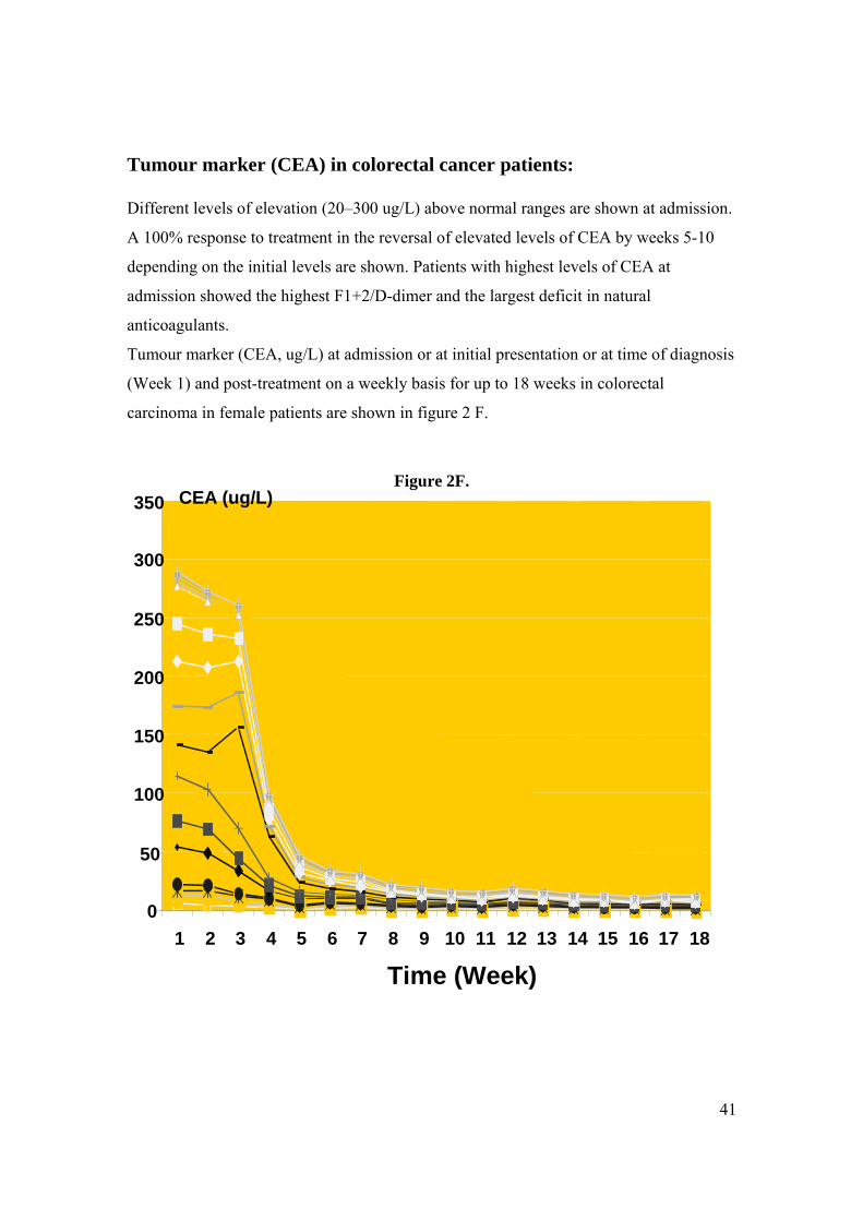
Tumour marker (CEA) in colorectal cancer patients:
Different levels of elevation (20–300 ug/L) above normal ranges are shown at admission.
A 100% response to treatment in the reversal of elevated levels of CEA by weeks 5-10
depending on the initial levels are shown. Patients with highest levels of CEA at
admission showed the highest F1+2/D-dimer and the largest deficit in natural
anticoagulants.
Tumour marker (CEA, ug/L) at admission or at initial presentation or at time of diagnosis
(Week 1) and post-treatment on a weekly basis for up to 18 weeks in colorectal
carcinoma in female patients are shown in figure 2 F.
Figure 2F.
0
50
100
150
200
250
300
350
1 2 3 4 5 6 7 8 9 10 11 12 13 14 15 16 17 18
Time (Week)
CEA (ug/L)

42
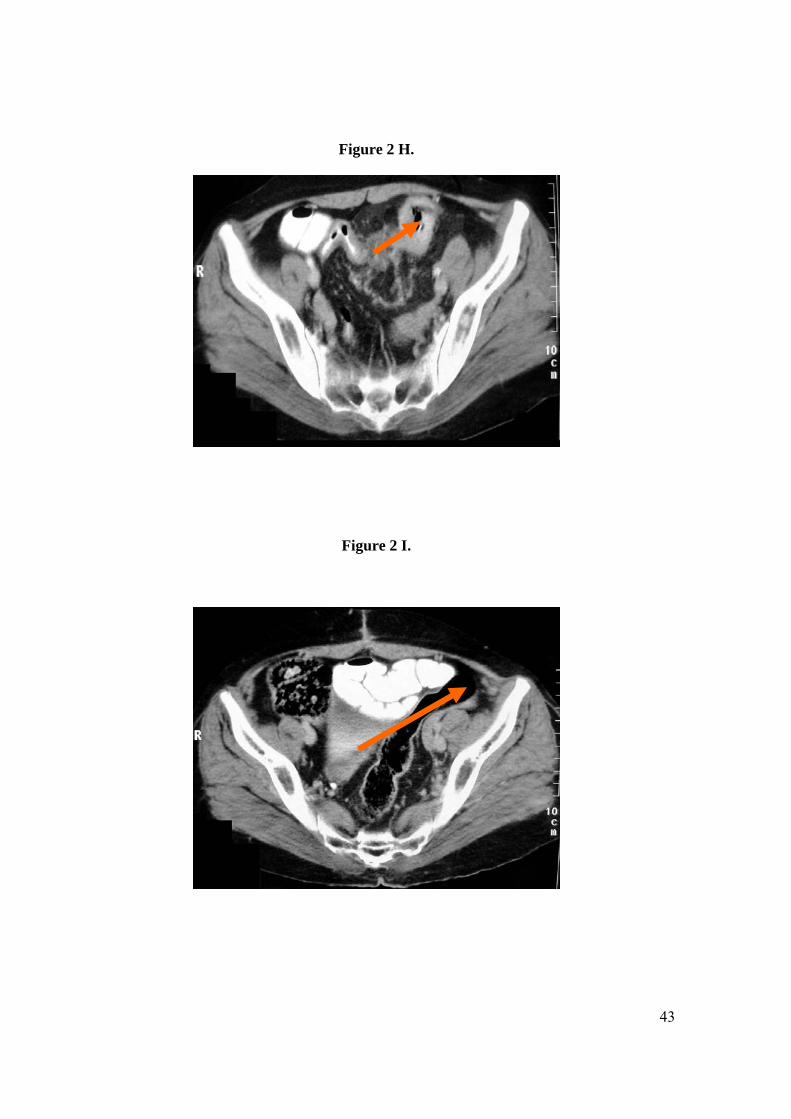
CT imaging in a female patient with Dukes Stage B2 colon adenocarcinoma:
This illustrates a large obstructive tumour mass (Figures 2 G-H) that was also seen with
colonoscopy. At 4 weeks post-surgery followed by chemotherapy, no tumour was
detected (Figures 2 I-J). Normalization of tumour marker and haemostasis marker levels
was also demonstrated at 4–18 weeks.
Figures 2 G-H; show representative CT images in woman with Dukes stage B2 colon
adenocarcinoma (tubular) preoperative, showing large obstructive tumour masses.
Elevated levels of tumour markers and haemostasis activation markers were documented
at admission. Figures 2 I-J; show representative CT images at 4 weeks post-surgery
showing successful removal of tumour, which coincides with normalization of tumour
markers and haemostasis markers.
Figure 2 G.
43
Figure 2 H.
Figure 2 I.

44
Figure 2 J.
3. Pancreatic adenocarcinoma (Stage III):
A progressive increase in F1+2 and D-dimer in 3 of 4 patients post-treatment was shown
(Figure 3A, 3B). In one case, a decline in both F1+2 and D-dimer were noticed up to the
12th weeks post-treatment, followed by rapid increase that caught up with other cases.
In contrast, the natural anticoagulants (PC, PS, and AT activities) demonstrated the
reverse trend to that of F1+2 and D-dimer and these are seen in Figures 3C–3E. Most of
these patients (3 out of 4) showed no response to treatment and full normalization in 1 of
the 4 by week 7, but this was followed by a rapid decline at weeks 12–14 and these
changes are nicely seen in Figures 3C–3E.

45
Figure 3A.
Figure 3 A-E; show haemostatic activation markers at admission or at initial presentation
or at time of diagnosis (Week 1) and post-treatment on a weekly basis for up to 18 weeks
in pancreatic adenocarcinoma patients (stage III). Figure 3A: F1+2 (nmol/L), and Figure
3B: D-dimer (mg/L). Natural anticoagulant markers at admission or at initial presentation
or at time of diagnosis (Week 1) and post-treatment on a weekly basis for up to 18 weeks
are shown in Figure 3C: PC activity (%), Figure 3D: PS activity (%), and Figure 3E: AT
activity (%)
Weeks
0 2 4 6 8 10 12 14 16 18 20
F1 +
2 A
ctiv
ity (n
mol
/l)
0
5
10
15
20

46
Figure 3B.
Figure 3C.
Figure 3D
Weeks
0 2 4 6 8 10 12 14 16 18 20
D-d
imer
Act
ivity
(mg/
l)
0
5
10
15
20
Weeks
0 2 4 6 8 10 12 14 16 18 20
PC
-Act
ivity
(%)
0
20
40
60
80
100
120
140
160

47
Figure 3D.
Figure 3E.
Weeks
0 2 4 6 8 10 12 14 16 18 20
PS-A
ctiv
ity (%
)
0
20
40
60
80
100
120
140
160
Weeks
0 2 4 6 8 10 12 14 16 18 20
AT A
ctiv
ity (%
)
0
20
40
60
80
100
120
140
160

48
Tumour marker (CA 19.9) in pancreatic cancer patients:
Different levels of progressive elevation above normal ranges are shown post-treatment.
An excellent response to treatment is shown in 1 of 4 patients. Patients with highest
levels of CA 19.9 showed the highest F1+2/D-dimer and the largest deficit in natural
anticoagulants.
The results of tumour marker (CA 19.9, U/ml) at admission or at initial presentation or at
time of diagnosis (Week 1) and post-treatment on a weekly basis for up to 18 weeks in
pancreatic cancer patients, are seen in figure 3 F.
Figure 3F.
0
500
1000
1500
2000
2500
1 2 3 4 5 6 7 8 9 10 11 12 13 14 15 16 17 18 Time (Weeks)
CA 19.9 (U/ml)

49
CT imaging of patient with (exocrine) pancreatic adenocarcinoma: The imaging techniques used for diagnosis and staging were based on international
recommendations for each disease category. These findings were reviewed by 2 different
radiologists.
Pancreatic cancer patients with highest tumour marker (CA19.9), highest F1+2 or D-
dimer, and lowest natural anticoagulants (PC, PS, AT activities) showed the largest
tumour mass. A representative CT imaging of a patient with a relatively large tumour
mass at admission is shown in Figure 3 G. This patient showed a good response to
treatment (week 8), as shown in Figure 3 H. This patient showed the lowest CA19.9
levels, as well as a normalized level of F1+2/D-dimer and PC/PS/AT activities at week 8.
Figure 3 G: At diagnosis—CT imaging in female patient with pancreatic
adenocarcinoma, large tumour mass, with highly elevated tumour marker CA19.9 and
haemostatic markers. Figure 3 H, shows the post-treatment CT imaging in a female
patient with pancreatic adenocarcinoma who showed reduction in tumour mass and the
tumour marker CA 19.9. Improvements in haemostatic markers are shown as well.
Figure 3 G.

50
Ffigure 3 H.
4. Lung carcinoma:
Patients diagnosed as having small and non-small cell lung cancer were also enrolled in the
study. Detailed monitoring upon admission and prior to any cancer-related intervention and
on a weekly basis post-intervention (chemotherapy, radiation or surgery) for up 18 weeks
were carried out as mentioned earlier. The laboratory investigation including haemostatic
parameters and tumour markers were repeatedly measured with each treatment course and
their results were correlated with other marker of tumour prognosis in a manner similar to
the previously mentioned gastrointestinal tract tumours. Ten patients were enrolled; 6 of
them had adenocarcinomatous type of non-small cell lung cancer and 4 had small cell lung
cancer. Two patients were excluded from the final evaluation of study because of the
development of thromboembolic events during the period of study. In all remaining cases,
the levels of hypercoagulable markers were elevated directly correlated with tumour stage
and radiological findings of cancer state. Additionally, the level of naturally occurring
coagulation inhibitors were all decreased with the progression of tumour. As mentioned

51
earlier, determination of D-dimer, F1 + 2, protein C, protein S, and antithrombin levels can
be used as markers to assess the tumour prognosis. This also suggests that early
anticoagulation prophylaxis might prevent VTE and impairs tumour progression.
The base-line characteristics of the types of cancers are shown in Table 1. And, the
chemotherapeutic regimens are shown in Table 2 .
Lung cancer is the leading cause of cancer death among men and women, AND
approximately 60%-65% of lung cancer patients present with advanced disease in which
the majority of newly diagnosed lung cancers are non-operable at time of diagnosis.
The markers of haemostatic system activation which included prothrombin F1 + 2 and D-
dimer were elevated in all except one patient (the patient had non-small cell lung
cancer=NSCLC) and this had a progressive course (the rise was by factor of 3–4 folds) at
6 to 18 weeks post-treatment. In contrast, the natural anticoagulants activity of PC-
activity, PS-activity, and AT-activity levels showed a reverse profile in these 7 of the 8
patients. In 5 patients with NSCLC, the levels of those natural anticoagulants were
progressively reduced by weeks 6–18 in parallel with disease progression.
The average values of haemostatic parameters in 7 patients
with lung cancer
Time in weeks AT PS-Ag PS-Act PC-Act D-dimer F1+2 Baseline 39 37 35 38 9.2 7.1 4th week 69 75 73 78 7.2 6.3 8th week 118 117 114 113 2.1 3.2 12th week 114 113 110 106 1.4 1.96 18th week 110 107 105 102 1.1 0.9

52
CT imaging: The non-small cell lung cancer patient with the highest F1+2 or D-dimer
and lowest natural anticoagulants (PC, PS, AT activity) showed the largest tumour mass.
A representative CT imaging of a patient with a relatively large tumour mass at
admission is shown in Figure 4 A. Unfortunately, no CT imaging was done at follow up,
but this patient showed a sustained high level of F1+2/D-dimer and low levels of PC, PS,
and AT activities at weeks 4–18.
Initial CT imaging at admission in a small cell lung cancer patient demonstrated a large
tumour mass (Figure 4 B.). In contrast, CT images post-surgical procedure and treatment
showed successful removal and remission (Figure 4C -1, 4C -2)
Figure 4 A.

53
Figure 4 B.

54
Figure 4 C-1, 4C-2

55
5. Breast and ovarian cancers:
Fourteen patients were enrolled in this study, 12 with infiltrating breast carcinoma and 2
with ovarian cancer. The laboratory work-up and follow-up were similar to other cases of
previously mentioned non-haematological malignancies. In all studies cases, the levels of
hypercoagulable markers were elevated in a direct correlation with tumour stage, tumour
markers, and radiological findings of cancer state. Additionally, the level of naturally
occurring coagulation inhibitors were all decreased with progression of tumour. These
hypercoagulable parameters were also raised after surgical resection of these tumours.
Breast cancer is the second-leading cause of cancer-related deaths in women in the
United States. About 20%–25% of patients with breast cancer have HER2/neu over-
expressing disease, which not only increases the risk of disease recurrence, metastasis,
and death, but also might lead to a poor response to conventional therapy with tamoxifen
and alkylating agents. Similarly, a significant percentage over-express epidermal growth
factor receptor (EGFR) also associated with poor prognostic features.
In case of beast cancer, the haemostatic parameters, the tumour marker, and the imaging
techniques were repeated after the completion of 2 full chemotherapeutic regimens. In
those cases in which surgical intervention was indicated prior to the start of
chemotherapy, the coagulation parameters and tumour markers were measured prior to
the surgery and the haemostatic values were repeated after the end of surgery. None of
these patients were on any kind of medications that would interfere with the results of
coagulation studies. During each visit physical examination, vital signs and medication
history were taken, and any changes in these findings were registered. If the patient
developed febrile neutropaenia as a complication of chemotherapeutic agent, the
haemostatic parameters were measured at the onset of the diagnosis of febrile
neutropaenia and thereafter. At the end of study, 2 patients were excluded because they
developed thromboembolic complications during the study period; they were started on
antithrombotic therapy. And, after a follow up of 18-20 months, 12 patients were eligible
for final evaluation.
The haemostatic parameters were measured at the onset of diagnosis, pre- and post
surgery, and after the completion of each chemotherapy course. Their levels were
correlated with the levels of tumour markers as in other cases.

56
CT imaging: With regard to the patients with ovarian cancer (stage IIA, well-
differentiated ovarian adenocarcinoma), no preoperative images were available (patients
were referred to us after surgical intervention). The postoperative CT scans that were
done as follow up after chemotherapy are normal, except for minor free fluid in the lesser
pelvis. The tumour marker CA 125 decreased significantly after surgery and remained
low thereafter; more significantly, the haemostatic parameters (mainly AT, PS, and PC)
normalized after surgery and with chemotherapy.
Patients with breast cancer always come to the care of the medical oncologist after
surgery and neo-adjuvant (before any surgery) or adjuvant chemotherapy; plain x-ray
imaging and mammography are performed at our institutions, but because CT scans are
only performed in cases in which there are metastases to lung, bone, or liver, there are no
CT scans with which to compare these. There was a clear correlation between tumour
marker CEA and CA15.3, and haemostatic parameter.
Fourteen patients were enrolled in the study over a period of 18-20 months. Two patients
were excluded from the final evaluation of study because of the development of
thromboembolic events during the period of study. Table 1 summarizes the base-line
characteristics of the types of cancers, whereas Table 2 summarizes the baseline
characteristics of the 12 eligible patients.
The most frequently used tumour markers were as follow: HER-2/neu expression,
ER/PgR status and CA 15-3 (mainly in breast cancer) and CA 125 (in ovarian cancer).
These markers were measured at the initial diagnosis and following each chemotherapy
cycle in manner mentioned earlier.
The chemotherapeutic regimens (either neo-adjuvant prior to surgery or adjuvant after
surgery or palliative after initial treatment failure) that were used included standard
internationally recommended regimens and they are mentioned in table 2.
At the time of study, Taxanes and targeted (Monoclonal antibody) therapy were not
frequently used and therefore regimens containing these new agents are not mentioned.
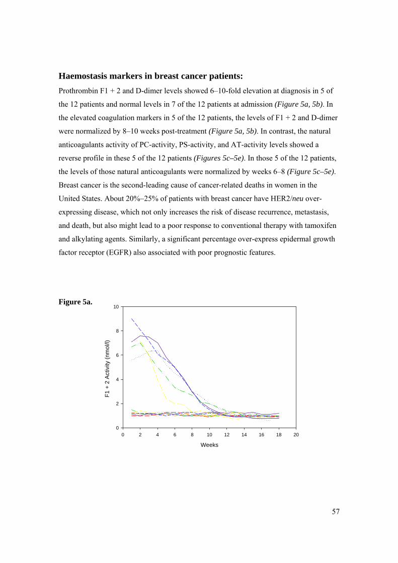
57
Haemostasis markers in breast cancer patients: Prothrombin F1 + 2 and D-dimer levels showed 6–10-fold elevation at diagnosis in 5 of
the 12 patients and normal levels in 7 of the 12 patients at admission (Figure 5a, 5b). In
the elevated coagulation markers in 5 of the 12 patients, the levels of F1 + 2 and D-dimer
were normalized by 8–10 weeks post-treatment (Figure 5a, 5b). In contrast, the natural
anticoagulants activity of PC-activity, PS-activity, and AT-activity levels showed a
reverse profile in these 5 of the 12 patients (Figures 5c–5e). In those 5 of the 12 patients,
the levels of those natural anticoagulants were normalized by weeks 6–8 (Figure 5c–5e).
Breast cancer is the second-leading cause of cancer-related deaths in women in the
United States. About 20%–25% of patients with breast cancer have HER2/neu over-
expressing disease, which not only increases the risk of disease recurrence, metastasis,
and death, but also might lead to a poor response to conventional therapy with tamoxifen
and alkylating agents. Similarly, a significant percentage over-express epidermal growth
factor receptor (EGFR) also associated with poor prognostic features.
Figure 5a.
Weeks
0 2 4 6 8 10 12 14 16 18 20
F1 +
2 A
ctiv
ity (n
mol
/l)
0
2
4
6
8
10

58
Figure 5b.
Figure 5c.
Weeks
0 2 4 6 8 10 12 14 16 18 20
D-d
imer
Act
ivity
(mg/
l)
0
2
4
6
8
10
12
14
16
Weeks
0 2 4 6 8 10 12 14 16 18 20
PC-A
ctiv
ity (%
)
0
20
40
60
80
100
120
140

59
Figure 5d.
Figure 5e.
Weeks
0 2 4 6 8 10 12 14 16 18 20
PS
-Act
ivity
(%)
20
40
60
80
100
120
140
Weeks
0 2 4 6 8 10 12 14 16 18 20
AT
Act
ivity
(%)
0
20
40
60
80
100
120
140
160
180
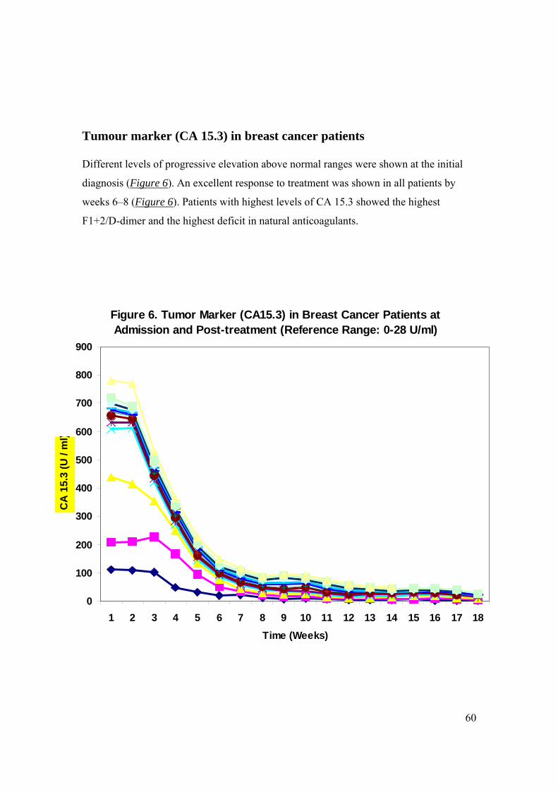
60
Tumour marker (CA 15.3) in breast cancer patients
Different levels of progressive elevation above normal ranges were shown at the initial
diagnosis (Figure 6). An excellent response to treatment was shown in all patients by
weeks 6–8 (Figure 6). Patients with highest levels of CA 15.3 showed the highest
F1+2/D-dimer and the highest deficit in natural anticoagulants.
Figure 6. Tumor Marker (CA15.3) in Breast Cancer Patients at Admission and Post-treatment (Reference Range: 0-28 U/ml)
0
100
200
300
400
500
600
700
800
900
1 2 3 4 5 6 7 8 9 10 11 12 13 14 15 16 17 18Time (Weeks)
CA
15.
3 (U
/ m
l)

61
Tumour marker (CA 125) in ovarian cancer patients
A variable level of CA125 elevation was shown at diagnosis, but a 100% response rate
was achieved by week 8 (Figure 7).
Figure 7. Tumor Marker (CA125) in ovarian cancer patients with well-differentiated Ovarian adenocarcinoma (stage IIA) at admission and Post-treatment - (Reference Range: 0 - 37 U/ml)
0
500
1000
1500
2000
2500
3000
1 2 3 4 5 6 7 8 9 10 11 12 13 14 15 16 17 18Time (Week)
CA
125
(U/m
l)

62
IV. Discussion:
In the nearly more than one century since Trousseau first described migratory
thrombophlebitis-thrombosis in patients with cancer, thromboembolism has become a
well-recognized initial presenting sign and complication of cancer. The clotting system is
activated in cancer patients and this is further amplified by different therapeutic
interventions, e.g.: surgery, systemic chemotherapy, hormonal treatment or radiation
therapy. Hypercoagulation is a well-documented in virtually all cancer types, albeit at
different ranges and severity, and is considered the second leading cause of mortality in
cancer patients. The relationship between the activation of clotting system and
carcinogenesis supports the idea of cancer as a hypercoagulable state and holds great
implications for the development of thrombosis, enhancement of tumour growth,
development of tumour progression-metastasis and risk of poor clinical outcomes and
response to treatment. Although it is well documented that cancer status can activate the
clotting cascade (the ability of tumour cells to produce and secrete
procoagulant/fibrinolytic substances and inflammatory cytokines), it is less known that
activation of the coagulation system may also support tumour progression and metastasis.
In addition, the physical interaction between tumour cells and blood elements
(neutrophils, monocytes, platelets) and/or vascular beds may play additional role in the
development and progression of tumour. The platelet activation in cancer patients and its
impact on tumour progression and metastasis further expand the role of the coagulation
system in malignancy. Other possible mechanism of thrombosis initiation and promotion
in malignancy include non-specific factors such as; abnormal protein metabolism (e.g.:
paraproteinaemia), generation of acute phase reactants, and haemodynamic compromise
(e.g.: stasis), and production and secretion of different substances which promote
generation of new and abnormal vasculature (neo-angiogenesis).
Different types of anti-tumour treatments may significantly increase the risk of
thromboembolic events by different mechanisms, e.g.: endothelial damage, release of
procoagulants, or stimulation of tissue factor production by host cells. Thrombo-embolic
complication in patients with cancer and in those with metastatic diseases is considered a
major problem for oncologists. A hypercoagulable or prothrombotic state of malignancy

63
occurs due to the ability of cancer cells to activate the clotting system and therefore the
hypercoagulation or the thromboembolism accounts for one of the most common
complications in cancer patients and it has serious consequences. The exact molecular
basis for cancer associated thromboembolic events remains unknown (although
considerable advances have been achieved in our understanding of pathomechanism-
pathobiology of thrombogenesis in cancers, particularly in the field of interaction
between the clotting cascade activation and the process of tumour growth and
metastasis), and the relative contribution of chemotherapeutic-cytostatic agents, tumour
cells, endothelial cells, and circulating procoagulants in promoting thrombus formation,
their attribution to tumour progression and dissemination continues to be investigated and
studied. Additionally, the thromboembolic events contribute for a significant percentage
of mortality and morbidity in cancer patients. Laboratory markers of coagulation
activation such as prothrombin fragments 1+2 or thrombin-antithrombin complex or D-
dimer support the premise that cancer is a hypercoagulable state and different coagulation
proteins, procoagulant microparticles and inflammatory cytokines may be raised in
patients with cancer. In parallel, the naturally occurring anti-thrombotic (anticoagulants)
proteins are decreased in malignancy. Despite the common occurrence of coagulation
abnormalities in patients with cancer, the utility of coagulation markers in determining
either the extent of malignant disease or their value in predicting those who will develop
thrombosis during therapy remains questionable. At present, it is difficult to identify the
relative impact of these interactions on the basis of the well-recognized clinical evidence
of hypercoagulable status in tumour patients. In term of disease progression, only
fibrinopeptide A levels have been shown to correlate with advancing disease and
increasing tumour burden. Although the relationship between malignant diseases and
venous thromboembolism has been convincingly demonstrated, the clinical implications
of this association still have to be thoroughly elucidated.
Based on these well-established data, we planned a study in which patients with different
types of solid tumours were enrolled and they were closely followed up from the time of
initial diagnosis till either the completion of planned treatment or until disease
progression or dissemination. Patients above 16 years of age, a life expectancy of more
than 3 months, and histopathologically proved tumour were enrolled in the study. The

64
study was approved by the regional Ethic Committee, and informed written consent was
obtained from patients. The changes in clotting parameters in patients with different types
of solid tumours were evaluated, and the correlation between these parameters and
tumour stage, changes in known characteristic tumour markers, imaging findings, and the
changes in haemostatic parameters which occur with different types of therapeutic
intervention were assessed. The markers of hypercoagulability were monitored from the
time of initial presentation and during each planned visit when the patients received the
due cycle of treatment. Our main focus was on specific abnormalities or changes of the
haemostatic parameters in this group of patients with different types of solid
malignancies. And, because it is not possible to accurately predict those with cancer who
will develop thrombosis, the results of this prospective study can be used as additional
clinical evidence to recommend routine thrombo-prophylaxis is cancer patients. It is very
important to mention that the magnitude of risk for development of venous
thromboembolic complication for a given anti-tumour therapy is sufficiently great, and
the thromboprophylaxis method is safe and effective. Patients were stratified according to
ECOG (Zubrod) performance status score, and those with ECOG score of 0-1 were
enrolled in the study. Baseline AT, PS, PC, D-dimer and prothrombin activation peptide
F1+2 as haemostatic parameters and characteristic tumour markers according to the type
of tumour were measured. Staging of cancer at various anatomic sites was done as
developed by the American Joint Committee on Cancer (AJCC) in cooperation with the
TNM Committee of the International Union Against Cancer (UICC). The International
Histological Classification or Tumours provided by the World Health Organization
(WHO) was used for pathologic classification and definition or tumour types. Physical
examination, imaging, endoscopy, bronchoscopy, biopsy, and surgical exploration were
used for clinical classification and staging. Histological grading was also used for
qualitative assessment of the differentiation of the tumours. All the patients were treated
according to the internationally recommended therapeutic regimens (the regimens were
those which were recommended during the period of study), and they were closely
followed up to evaluate the state of their malignant disease. The haemostatic parameters,
the tumour markers, and the imaging studies were repeated after the completion of each
full cycle of chemotherapeutic regimen. In those cases where surgical intervention was
65
indicated prior to the start of chemotherapy, the coagulation parameters and tumour
markers were measured prior to the surgery and the haemostatic values were repeated
after the end of surgery. None of these patients eligible for final analysis were on any
kind of medication which could interfere with the results of coagulation studies. During
each visit, physical examination, vital signs and medication history were taken and any
changes in these findings were registered. If the patient developed febrile neutropaenia as
a complication of a chemotherapeutic agent, the haemostatic parameters were measured
at the onset of the diagnosis of febrile neutropaenia and thereafter.
The statistical analysis was performed by two-way analysis of variance (ANOVA)
comparing the markers of haemostasis activation at admission to post-admission for each
subject and with respect to average control values; differences were considered
significant at p value of 0.05 or less.
Except for a slight difference in the gender ratio (in gynaecological cancer group), the
analysis of patients who met the pre-specified criteria for evaluation showed that the
hypercoagulable parameters were directly correlated with tumour progression, rise in
characteristic tumour markers and with progression of tumour on imaging investigations.
We studied the prognostic values of F1+2, D-dimer, and natural inhibitors of abnormal
coagulation in this group of patients with solid tumours, however, no convincing data
have thus far identified one of these hypercoagulability markers as a reliable prognostic
marker [144], except F1+2 which has been shown to correlate with advancing disease and
tumour burden. Parallel with the haemostatic parameters, we also measured the
characteristic tumour markers; in all cases, there was a direct correlation between
changes in the haemostatic parameters, radiological imaging findings, and tumour
markers (e.g., rise in D-dimer or F1+2 was associated with a drop in AT, PC, and PS,
which was directly correlated with a rise in characteristic tumour markers and a
progression of the malignant diseases in radiological imaging findings). In cases of breast
and ovarian cancers, data on the plasma levels of D-dimer illustrated excellent correlation
with the tumour markers CA15.3, CA125, respectively, and tumour volume. The
correlation between haemostatic parameters, radiological findings, tumour stage, and the
characteristic tumour markers were analyzed. The tumour markers used in the studies
cases were those which are recommended by different oncology societies. These tumour

66
markers are not tumour-specific, but they might be used as a useful tool for both
diagnosis and follow up of these patients and also to assess the efficacy of the treatment.
Additionally, the hypercoagulability parameters which were used in the study reflect a
broad spectrum combination of known markers of coagulation cascade activation and
each of these markers was used in previous studies separately. The following parameters
were measured in all enrolled patients; plasma levels of D-dimer and F1+2, activity of
Antithrombin (previously called ATIII), Protein C and protein S in addition to the
measurement of Free PS antigen level. All these parameters were determined and
measured at the; onset of diagnosis (at the time of enrolment into study), pre- and post-
surgical intervention, after the completion of each cycle of chemotherapy, and pre- and
pos-radiation therapy (depending on the type of therapeutic intervention which was
planned for each specific case). The levels of aforementioned haemostatic parameters
were correlated with the levels of characteristic tumour markers. Each blood sample
taken from the enrolled patients was tested for AT activity using thrombin (AT-IIa) as the
substrate; PC activity, using both clotting (PC-CI) and chromogenic (PC-Chr) methods;
PS activity; D-dimer; and F1+2 using immunoassays, ELISA, clotting, and chromogenic
methods [137-142]. The reagents and kits used for these different assays were obtained
from Dade Behring, Inc. (Deerfield, IL), Stago International (Parsippany, NJ), and R&D
Systems, Inc. (Minneapolis, MN).
We documented a direct correlation between the markers of hypercoagulable status,
tumour stage, changes in tumour markers, and the degree of radiological response in all
examined cases. This correlation was most obvious in all examined cases except that of
lung and gastric cancers. This can be explained by the fact that patients with gastric and
lung cancers usually present to oncologists at advanced stage and despite initial response
of tumour to treatment, most of these cases relapse or progress during or shortly after the
completion of treatment. These findings are mostly observed in patients with
adenocarcinoma, e.g.: breast, gastro-intestinal tract including pancreatic cancer, non-
small-cell lung cancer, prostate, and brain and these have been previously published in
multiple studies and trials. The imaging techniques that were used for diagnosis and
staging were based on international recommendations for each disease category. These

67
findings were reviewed by two different radiologists and in some cases additional help of
external radiologist when the patient was referred from a different hospital.
The relationship between the hypercoagulable state and both the tumour progression and
occurrence of metastasis has not been well established. Larger studies are needed to
determine those tests that could have a reliable predictive value in tumour progression.
Much more information is needed, and only large-scale clinical trials will unequivocally
establish whether these hypercoagulable parameters can be used reliably to predict the
progression of disease and whether modulating the haemostatic system will have an
impact on the process of tumour progression and metastatic dissemination.
Patients with cancer represent 15-20% of all patients with thrombosis. Furthermore,
about 10% of patients presenting with unprovoked or idiopathic thrombosis are
diagnosed with early or advanced malignancy within the next 1-2 years of the thrombosis
event (Prandoni et al, 1992; Lee & Levine, 2003). Hence, approximately one quarter of
all thrombosis cases are related to underlying malignancy. Given the ageing population
and the rising incidence of cancer in industrialized nations, venous thrombo-embolism in
patients with cancer will become an increasingly common health care issue.
According to a population-based study by Sorensen et el (2000), the 1-year survival rate
in patients diagnosed with cancer at the time of their thrombo-embolic event is 12%, as
compared with 36% in cancer patients who are free of thrombosis but otherwise are
matched for sex, age at the time of cancer diagnosis, tumour type and the duration of
cancer. The higher mortality rate in cancer patients with venous thrombo-embolism may
indicate that venous thrombo-embolism is a marker of aggressive malignancies or that
these patients are dying prematurely from thrombotic complications, or both.
Unfortunately, the true incidence of venous thrombo-embolism associated with various
tumour types remains unknown for the majority of cancers because the appropriate cohort
studies have not been conducted. On the contrary, the most common tumour types found
in patients with venous thrombo-embolism are cancers of the lung, colon, breast and
prostate. This reflects the high prevalence of these cancers in the general population.
Recent reports of very high rates of thrombotic complications in patients receiving
experimental therapy with inhibitors of vascular endothelial growth factor (Kuenen et al,
2002; Marx et al, 2002; Kbabbinavar et al, 2003) and other anti-angiogenic agents, such

68
as thalidomide (Zangari et al, 2001; Desai et al, 2002; Rajkumar et al, 2002), have re-
emphasized the importance of cancer therapy in the pathogenesis of the thrombosis in
cancer patients.
Effective and safe antithrombotic therapy- the mainstream in prophylaxis and treatment
of thromboembolism- remains very challenging clinical task in cancer patients- a
population with high rate of treatment failure, haemorrhagic and thromboembolic
complications recurrences and relapses. Prospective randomized clinical trials have
documented that with advent of LMWH, new possibilities for thrombosis treatment and
prevention with more convenient and safe antithrombotic have been emerged. But, the
problem of thromboembolic events in patients with disseminated and advanced stage of
malignancy remains a serious concern for haemato-oncologists. Based on the available
evidence-based clinical data regarding the role of coagulation system in cancer
development and progression, a new challenge for modern therapy in oncology has been
built and this is appreciated in the hypothesis of anti-neoplastic effects of antithrombotic-
anticoagulants which could influence the outcome of treatment in human cancers. Anti-
tumour effects of anticoagulants agents have been observed and documented in various
experimental models (cancer cell lines). Heparin and its derivatives mainly low-
molecular-weight heparin in addition to Warfarin, have been the most extensively studied
and the results are very promising in terms of; reduction in the primary tumour size, its
growth and metastatic progression. Joint evidences from basic research and from multiple
prospective, randomized clinical studies, showing beneficial impact of antithrombotic
therapy (mainly LMWHs) on cancer patients’ survival, confirm the need for further and
more extensive scientific steps to confirm the biologic benefits of antithrombotic therapy
in human cancers. Warfarin and unfractionated heparins (UFH) have been in clinical use
for many years, and both are effective anticoagulants, but they are associated with many
impediments, including the need for intensive monitoring of their effects, multiple drug-
drug interactions (mainly in case of Warfarin), wide variations in dose-response
relationships, and serious immune-mediated thrombocytopaenia (in the case of UFH).
The development of LMWH advanced anticoagulation therapy by enhancing efficacy and
eliminating the need for intensive monitoring. The evidences are started to accumulate,
that clinically approved LMWHs have different abilities to influence some steps of

69
metastatic spread. The experimental works toward the development of such heparin
derivatives with low anticoagulant activity, but with potential inhibitory effects on
tumour cells spread are in progress. At present, most clinical haemato-oncologists report
thromboprophylactic use of different types of antithrombotics in less than 5% of cancer
patients. One of the reasons for this very limited use of thromboprophylaxis is probably
the lack of haemato-oncology specific guidelines. The available excellent guidelines for
treatment of thromboembolism must be adapted to the specific context of cancer patients.
Anticoagulant prophylaxis is routinely recommended for patients undergoing major
surgery because the risk of post-operative thrombosis is substantial. For patients having
surgery for cancer, the risk of venous thrombo-embolism may be as high as 50% without
prophylaxis (Geerts et al, 2001). Compared with the thromboprophylaxis in the surgical
oncology setting, prophylaxis in cancer patients on chemotherapy has received even less
research attention. Clinical trials have not been performed to evaluate primary
prophylaxis specifically in hospitalized medical oncology patients. To date, multiple
randomized trials and meta-analyses of these trials have confirmed that, for initial
therapy, LMWHs are at least as efficacious as UFH in reducing recurrent thrombosis and
are likely to be associated with a lower risk of major bleeding (Koopman et al, 1996;
Levine et al, 1996; The Columbus Investigators, 1997; Gould et al, 199; Dolovich et al,
2000; Merli et al, 2001; van Dongen et al, 2004). In many developed countries,
outpatient LMWH has become the standard of care for the initial treatment in patients
with deep venous thrombosis or haemodynamically stable pulmonary embolism.
Several studies were conducted to evaluate the efficacy and safety of LMWH as initial
treatment, secondary prophylaxis and long-term therapy. Two meta-analyses of these
studies showed a statistically non-significant reduction of approximately 30% in the risk
of recurrent VTE favouring LMWH (van der Heijden et al, 2001; Iorio et al, 2003), while
one of these studies found a significant reduction of 62% in the risk of bleeding with
LMWH (van der Heijden et al, 2001). To date, three published clinical trials have
examined the use of long-term LMWH as an alternative to coumarin derivative therapy in
cancer patients with acute VTE (CANTHANOX, CLOT, and FRONTLINE). Two
unpublished studies have also evaluated LMWH for long-term use in cancer patients and
the results have been presented in abstract form. A subgroup analysis of the long-term

70
Innohep® treatment evaluation study reported improved efficacy with tinzaparin over
Warfarin in the 167 patients with cancer (Hull et al, 2003). Tinzaparin reduced the rate of
recurrent VTE by 50%, but this was not statically significant because of the small number
of patients.
Evidence from the CLOT trial is promising with respect to the efficacy of long-term
LMWH in the management of VTE in cancer patients, and its implications are addressed
in a new paradigm in the management of thrombosis in cancer patients. There is strong
evidence that long-term LMWH is efficacious and safe for preventing recurrent VTE in
cancer patients. Bleeding does not appear to be increased compared with Warfarin
therapy but this remains a major concern in cancer patients receiving long-term,
anticoagulant therapy because of their co-morbid conditions. Daily injections appeared to
be well tolerated but the main practical limitation of using LMWH for long-term use is
the drug cost. A cost-effective analysis has suggested that LMWH might be a cost-
effective drug for secondary prophylaxis of VTE, especially in patients at high risk of
recurrence and where the drug cost is lower (Marchetti et al, 2001). Although recurrent
VTE is relatively frequent in patients with cancer while on oral anticoagulant therapy,
treatment in this setting has not been investigated in clinical trials. Traditionally, four
options are available: continue with oral anticoagulant therapy aiming for higher INR
target; switch to UFH; use of LMWH; or insertion of an inferior vena caval filter. None
of these alternatives have been compared or rigorously evaluated. Duration of
anticoagulant therapy has not been addressed in cancer patients. Based on accepted
concept that the risk of recurrent VTE is increased in the presence of any ongoing risk
factor, it is generally recommended that patients with metastases continue with
“indefinite” therapy because metastatic malignancies are considered a persistent risk
factor. In those without metastases, anticoagulant treatment is recommended for a long as
the cancer is ‘active’ and while the patient is receiving anti-tumour therapy. In general, it
is advisable to re-evaluate frequently the risk-benefit ratio of ongoing anticoagulant
therapy in individual patients, taking into consideration the overall clinical status of the
patient, including the quality of life and life expectancy.
To date, the studies evaluating new anticoagulants have included few or no patients with
cancer. Given the differences in the natural history and response to therapy between

71
patients with and without cancer, research is needed to study the efficacy and safety of
these agents especially in the various oncology settings.
Fondaparinux, the first selective factor Xa inhibitor, represents yet another advance in
anticoagulation therapy. Efficient inhibition of factor Xa activity impairs the activation of
tissue factor/factor VIIa complex which finally leads to downregulation of procoagulant
state, pro-angiogenesis, and pro-inflammatory factors induced by TF/FVIIa complex
However, despite compelling experimental and clinical evidences for a pathogenic role of
blood coagulation in tumour growth and metastasis (Mousa SA, ref. 110), other studies in
patients with solid tumours have failed to confirm a survival benefit for patients treated
with anticoagulants (Smorenburg et al, ref. 116). More recently, this topic has received
renewed attention and several randomized controlled trials and meta-analyses of studies
have reported a reduction in the overall mortality of patients with cancer who were
randomly assigned to receive a low molecular weight heparin (Prandoni P. et al, ref. 117,
and Lensing AW et al, ref. 121). A plausible biologic mechanism is now emerging from
experimental and in vitro studies that show LMWHs can inhibit angiogenesis, a process
that is critical for tumour growth and metastasis, in a dose-dependent fashion (Nash GF.
et al, Lancet Oncol 2:608-613, 2001, Norrby K, Haemostasis 23:141-149, 1993, and
Hejna M. et al, J Natl Cancer Inst 91:22-36, 1999).
The findings in our study which show strong association between haemostatic parameters
from one side and the tumour progression-metastasis (tumour biology), and response to
treatment from other side, are consistent with previous observations from multiple
clinical studies, including two randomized, placebo-controlled trials designed to
investigate the influence of low molecular weight heparin on cancer survival (FAMOUS
trial by Kakkar et al, MALT trial by Klerk et al, and CLOT trial by Agnes YYL et al). In
all three studies, the use of low molecular weight heparin was associated with improved
survival in patients with relatively good prognosis. Finally, we attempted to describe the
current theory about the pathophysiology of the hypercoagulable status in cancer patients,
and we also tried to discuss whether or not to screen elder patients (patients above age of
45 years) with idiopathic deep venous thrombosis for an underlying malignancy, and
whether this would be potentially beneficial to patients and to the ongoing arguments
regarding economic background for prophylactic and therapeutic strategies in cancer

72
patients. We hope that a better and more scientific understanding of these mechanisms (to
be explored by further randomized clinical trials) will ultimately lead to the development
of more targeted treatments to prevent and to treat thromboembolic complications in
cancer patients. We also hope that guidelines for antithrombotic treatment may also have
a positive effect on the process of tumour growth and metastasis.
Based on the findings from this small cohort of patients, we may conclude that
antithrombotic therapy interfere with various processes involved in cancer development,
growth and dissemination. Based on available data from multiple clinical studies,
clinically relevant and improved efficacy of LMWHs as compared to Warfarin and UFH,
on the survival of cancer patients with thromboembolic events, have been well
documented and established. Our study has demonstrated a significant role for LMWH
on tumour growth, tumour progression and dissemination and currently we are working
on the role of LMWH on release of TFPI as a possible regulatory mechanism of
angiogenesis and on potent inhibition of matrix-degrading enzymes by LMWHs. The
anti-angiogenesis effect of LMWHs has been clearly documented and therefore LMWHs
have been used in those clinical conditions in which angiogenesis has been associated
with the clinical disorder, including human tumour growth and dissemination. Our
findings can be used as documented basis for beneficiary effect of LMWH in inhibition
of neo-angiogenesis in different cancer types in addition to their primary role as
antithrombotic agents. More studies are required in this population to look at
antithrombotic therapy, duration of therapy, bleeding, quality of life, cost-effectiveness
and the influence of anticoagulants on cancer survival. These results might be useful as
basis for future larger scale trials in which additional markers of hypercoagulable status
will be evaluated in order to identify the most sensitive marker with highest prognostic
impact on patients’ survival. The significant finding of the study in which a direct
correlation between hypercoagulability markers and tumour stage was confirmed, can be
further assessed in future trials to compare the efficacy of different doses of low
molecular weight heparins and to determine the effect of LMWHs on tumour
progression. In cancer patients with good prognosis and longer life expectancy, the
treatment with LMWHs could result in survival benefit. Based on experimental data,
there is a significant difference between treatment regimens (dose of LMWHs) and their

73
effect on tumour biology (tumour development and metastasis). The duration of
anticoagulation with LMWH in cancer patients is a very important issue which has
received great attention in the recent years. Further studies at large scale are needed to
evaluate the optimal duration and dosing of different anticoagulation therapy during the
active phase of anti-cancer therapy and after the end of all therapeutic interventions in
patients with different types of solid tumours especially in those with ECOG score of 0-1
and with no clinical evidence of secondary VTE. The relationship between the
hypercoagulable state and the tumour progression and the occurrence of metastasis has
not been well established. Much more information is needed and only large-scale clinical
trials will unequivocally establish whether these hypercoagulable parameters have strong
predictive values and whether the modulation of haemostatic system will modify the
process of tumour progression and metastatic dissemination. The mechanisms for a
potential anti-neoplastic effect of LMWHs remain unknown and will require further
investigations in well-designed experimental studies. An anti-angiogenic effect is an
appealing possibility and it is evident in patients with limited disease and could be
persistent beyond the administration of the agent. It can be hypothesized that in patients
with disseminated cancer, tumour-related vasculature is sufficiently developed so that an
anti-angiogenic agent would have minimal impact, whereas impairing the establishment
of such vasculature by an anti-angiogenic agent could exert an inhibitory effect on
tumour growth even beyond the time of drug exposure. Although it has been suggested
that the improvement in cancer survival associated with LMWHs observed in previous
studies may be due to a reduction in fatal pulmonary embolism as compared with
unfractionated heparin, the survival benefit beyond the period of administration of low
molecular weight heparin observed in CLOT trial would argue against this hypothesis. A
very strong association between cancer and disturbances in haemostatic parameters has
been demonstrated consistently in experimental and clinical trials. Our findings offer
additional evidence that the coagulation system and its cascade of activation, is
intrinsically involved in tumour development and/or tumour progression-metastasis. But,
additional, well-balanced studies designed to confirm the anti-tumour effects of low
molecular weight heparins and explore the pathophysiologic mechanisms are warranted
to further investigate these findings.

74
References: 1. Donati MB. Cancer and thrombosis. Haemostasis 1994; 24: 128-31 2. Falanga A, Donati MB. Pathogenesis of thrombosis in patients with malignancy. Int J Hematol 2001; 73: 137-44 3. Sutherland DE, Weitz IC, Liebman HA. Thromboembolic complications of cancer: epidemiology, pathogenesis, diagnosis, and treatment. Am J Hematol 2003; 72: 43-52 4. Otten HM, Prins MH, Smorenburg SM et al. Risk assessment and prophylaxis of venous thromboembolism in non-surgical patients: cancer as a risk factor. Haemostasis 2000; 30 (Suppl 2): 72-6 5. Clarke-Pearson DL, Synan IS, Coleman RE et al. The natural history of postoperative of venous thromboemboli in gynaecologic oncology: a prospective study of 382 patients. Am J Obstet Gynecol 1984; 148: 1051-4 6. Marras LC, Geerts WH, Perry JR. The risk of venous thromboembolism is increased throughout the course of malignant glioma. An evidence-based review. Cancer 2000; 89: 640-6 7. Edwards RL, Rickles FR. Haemostatic alterations in cancer patients. In: Honn KV, Loanie S, eds. Hemostatic mechanism and metastases. Boston: Nishoff, pp 342-54, 1984. 8. Liberman JJ, Borrero J, Urdaneta E. Thrombophlebitis and cancer. JAMA 177: 592-95, 1961. 9. Rickler FR, Edwards RL. Activation of blood coagulation in cancer: Trousseau’s syndrome revisited. Blood 62: 14-31, 1983. 10. Soon BC, Miller SP. Coagulation disorders in cancer fibrinolysis and inhibitors. Cancer 25: 867-74, 1970. 11. Trousseau A. Phlegmasia alba dolens; in Clinique medicale de l, Hotel-Dieu de Paris, Paris, Bailliere, vol 3, pp 654-712, 1865. 12. Dore R, Alerci M, D,Andrea F, , Di Giulio G, De Agostini A, Volpato G. Intracardiac extension of lung cancer via pulmonary veins: CT diagnosis. J Comput Assist Tomogr 12: 565-68, 1988. 13. Odero A, Giorgetti P, Cugnasca M, Rampoldi V, Bortolani EM, Ruberti U. Neoplastic thrombosis of inferior vena cava and right atrium due to kidney cancer. Three surgically treated cases. Panminerva Med 31: 140-43, 1989.

75
14. Takagi H, Yamada S, Abe T, Ichikawa K, Takezawa J, Nagamine T, Kobayashi S, Kataki S. Hepatocellular carcinoma growing in the pulmonary arteries, complicated with venous thrombosis, Jpn J Med 26: 388-92, 1987. 15. Aderka D, Brown A, Zelikovski A, Pinkhas J. Idiopathic deep vein thrombosis in an apparently healthy patient as a premonitory sign of occult cancer. Cancer 57: 1846-49, 1986. 16. Goldberg RJ, Seneff M, Gore JM, Anderson FA, Green HL, Wheeler HB, Dalen JE. Occult malignant neoplasm in patients with deep vein thrombosis. Arch Intern Med 147: 251-53, 1987. 17. Gore JM, Appelbaum JS, Green HL, Dexter L, Dalen JE. Occult cancer in patients with acute pulmonary embolism. Ann Intern Med 96: 556-60, 1982. 18. Griffin MR, Stanson AW, Brown ML, Hauser MF, O, Fallon WM, Anderson HM, Kazmeir FJ, Melton LJ. Deep vein thrombosis and pulmonary embolism: Risk of subsequent malignant neoplasms. Arch Intern Med 147: 1907-11, 1987. 19. Prandoni P, Lensing AWA, Cogo A, Cuppini S, Villalta S, Corta M, Catelan AM, Polistena P, Bernardi E, Prins MH. The long term clinical course of acute deep vein thrombosis. Ann Intern Med 125: 1-7, 1996. 20. Clarke-Pearson DL, Synam IS, Creasman WT. Anticoagulation therapy for venous thromboembolism in patients with gynaecologic malignancy. Am J Obstet Gynecol 147: 469-75, 1983. 21. Gitter MJ, Jaeger TM, Petterson TM, Gerssh BJ, Silverstein MD. Bleeding and thromboembolism during anticoagulant therapy: A population based study in Rochester, Minnesota. Mayo Clin Proc 70: 725-33, 1995. 22. Krauth D, Holden A, Knapic N, Leipman M, Ansell J. Safety and efficacy of long-term anticoagulant therapy in cancer patients. Cancer 59: 983-85, 1987. 23. Moore FD Jr, Osteen RT, Karp DD, Stein G, Wilson RE. Anticoagulants, venous thromboembolism, and the cancer patient. Arch Surg 116: 405-7, 1981. 24. Sarasin FP, Bounameaux H. Duration of oral anticoagulant therapy after proximal deep vein thrombosis: A decision analysis. Thromb Haemost 71: 286-91, 1994. 25. Billroth T. Lectures on Surgical Pathology and Therapeutics: A Handbook for Students and Practitioners, ed 8 (translated). London, The Syndenham Society, 1877-1878

76
26. Edward RL, Morgan DL, Rickles FR. Animal tumor procoagulants. Registry of the Subcommittee on Haemostasis and malignancy of the Scientific and Standardization Committee, International Society on Thrombosis and Haemostasis. Thromb Haemostasis 1993; 63: 133-8 27. Edwards RL, Silver J, Rickles FR. Human tumor procoagulants. Registry of the Subcommittee on Haemostasis and Malignancy of the Scientific and Standardization Subcommittee, International Society on Thrombosis and Haemostasis. Thromb Haemostasis 1993; 69: 205-13 28. Kubota T, Andoh K, Sedakata H, Tanaka H, Kobayashi N. Tissue factor released from leukemic cells. Thromb Haemostasis 1991; 65: 59-63 29. Francis JL. Haemostasis and cancer. Med Lab Sci 1989; 46: 331-46 30. Falanga A, Alessio MG, Donati MB, Barbui T. A new procoagulant in acute leukaemia. Blood 1988; 71: 870-5 31. Gordon SG, Benson B. Analysis of serum cancer procoagulant activity and its possible use as a tumor marker. Thromb Res 1989; 56: 431-40 32. Pineo GF, Brain MC, Gallus AS, Hirsh J, Hatton MWC, Regoeczi E. Tumors, mucus production and hypercoagulability. Ann NY Acad Sci 1974; 230: 262-72 33. Scates SM. Diagnosis and treatment of cancer-related thrombosis. Semin Thromb Hemostas 1992; 18: 373-9 34. Lando PA, Edginton TS. An innate host response to the neoplastic cell. syngeneic rat tumor cells can elicit a rapid de novo lymphoid procoagulant response. J Immunol 1985; 135: 3587-95 35. Miyauchi S, Moroyama T, Kyoizuni S, Asakawa J-I, Okamoto T, Takada K. Malignant tumor cell lines produce interleukin-1 like factor. In Vitro Cell Dev Biol 1988; 24: 753-8 36. Piccioli A, Prandoni P, Ewenstein BM, Goldhaber SZ. Cancer and venous thromboembolism. Am Heart J 1996; 132: 850-5 37. Dvorak HF. Abnormalities of haemostasis in malignant disease. In: Colman EW, Hirsh J, Marder VJ, Salzman EW, eds. Haemostasis and thrombosis: basic principles and clinical practice. Philadelphia: JB Lippincott C., 1993. p. 1238-54 38. Edwards RL, Rickles FR. Thrombosis and cancer. In: Hull R, Pineo GF, eds. Disorders of thrombosis. Philadelphia: WB Saunders Co., 1996. p. 374-82

77
39. Falanga A, Barbui T, Rickles FR, Levine MN. Guidelines for clotting studies in cancer patients. Thromb Haemostasis 1993; 70: 540-2 40. Otterson GA, Monahan BP, Harold N, Steinberg SM, Frame NJ, Kaye FJ. Clinical significance of the FV: Q506 mutation in unselected oncology patients. Am J Med 1996; 101: 406-12 41. Nand S, Fischer SG, Salgia R, Fisher RI. Hemostatic abnormalities in untreated cancer: incidence and correlation with thrombotic and hemorrhagic complications. J Clin Oncol 1987; 5: 1998-2003 42. Luzzatto G, Schafer Al. The prethrombotic state in cancer. Semin Oncol 1990; 17: 147-59 43. Naschitz JE, Yeshurun D, Lev LN. Thromboembolism in cancer: changing trends. Cancer 1993; 71: 1384-90 44. Francis CW, Felcher AH, White J, Braaten JV, Goss R. Thrombin activity associated with indwelling central venous catheters. Thrombos Haemostasis 1997; 77: 48-52 45. Nicolson GL, Custead SE. Effects of chemotherapeutic drugs on platelets and metastatic tumor cell-endothelial cell interactions as a model for assessing vascular endothelial integrity. Cancer Res 1985; 45: 331-6 46. Doll DC, Ringenberg QS, Yarbro JW. Vascular toxicity associated with antineoplastic agents. J Clin Oncol 1986; 4: 1405-10 47. Levine MN. Prevention of thrombotic disorders in cancer patients undergoing chemotherapy. Thromb Haemostasis 1997; 78: 133-6 48. Mitchell L, Hoogendoorn H, Giles AR, Vegh P, Andrew M. Increased endogenous thrombin generation in children with acute lymphoblastic leukaemia: risk of thrombotic complications in L-asparaginase-induced antithrombin III deficiency. Blood 1994; 83: 386-91 49. Salzman EX, Hirsh J. The epidemiology, pathogenesis, and natural history of venous thrombosis. In: Colman EW, Hirsh J, Marder VJ, Salzman EW, eds. Haemostasis and thrombosis: basic principles and clinical practice. Philadelphia: J.B. Lippincott Co., 1993.p. 1275-96 50. Sporul EE: Carcinoma and venous thrombosis. The frequency of association in the body or tail of the pancreas with multiple venous thrombosis. Am J Med Sci 1938; 34: 566-570
78
51. Thompson CM, Rodgers RL. Analysis of the autopsy records of 157 cases of carcinoma of the pancreas with particular reference to the incidence of thromboembolism. Am J Med Sci 1952; 223: 469-476 52. Peuscher FW. Thrombosis and bleeding in cancer patients. Neth J Med 1981;24:23-35 53. Rickles FR, Levine M, Dvorak HB. Abnormalities of haemostasis in malignancy; in Colman RW, Hirsh J, Marder VJ, Clowes A, George JN (eds): Haemostasis and Thrombosis. Philadelphia, Lippincott, Williams & Wilkins, 2000, chap 69, pp 1132-1152 54. Levine MN. Prevention of thrombotic disorders in cancer patients undergoing chemotherapy. Thromb Haemost 1997; 78: 133-136 55. Levine MN, Gent M, Hirsh J, Arnold A, Goodyear MD, Hryniuk W, DePauw S. The thrombogenic effect of anticancer drug therapy in women with stage II breast cancer. N Engl J Med 1988; 318: 404-407 56. Weiss RB, Tormey DC, Holland JF, Weinberg VE. Venous thrombosis during multimodal treatment of primary breast cancer. Cancer Treat Rep 1981; 65: 677-679 57. Goodnough LT, Satio H, Manni A, Jones PK, Pearson OH. Increased incidence of thromboembolism in stage IV breast cancer patients treated with a five-day chemotherapy regimen. A study of 159 patients. Cancer 1984; 54: 1264-1268 58. Saphner T, Tormey DC, Gray R. Venous and arterial thrombosis in patients who received adjuvant chemotherapy for breast cancer. J Clin Oncol 1991; 9: 286-294 59. Pritchard KI, Paterson AHG, Paul NA, Zee B, Fine S, Peter J, for the National Cancer Institute of Canada Clinical Trials Group. Increased thromboembolic complications with concurrent tamoxifen and chemotherapy in a randomized trial of adjuvant therapy for women with breast cancer. J Clin Oncol 1996; 14: 2731-2737 60. Wall JG, Weiss RB, Norton L. Arterial thrombosis associated with adjuvant chemotherapy for breast cancer: A Cancer and Leukaemia Group B study. Am J Med 1989; 87:”501-504 61. Von Tempelhoff GF, Dietrich M, Hommel G, Heilmann L. Blood coagulation during adjuvant epirubicin/cyclophosphamide chemotherapy in patients with primary operable breast cancer. J Clin Oncol 1996; 14: 2560-2568 62. Clahsen PC, van de Velde CJH, Julien JP, Floiras JL, Mignolet FY. Thromboembolic complications after preoperative chemotherapy in women with early breast cancer: A European Organization for Research and Treatment of Breast Cancer Cooperative Study. J Clin Oncol 1994; 12: 1266-1271

79
63. Fisher B, Redmond C, Legault-Poisson S, Dimitrov NV, Brown AM, Wickerham DL, Wolmark N, Margolese RG, Bowman D, Glass AG. Postoperative chemotherapy and tamoxifen compared with tamoxifen alone in the treatment of positive node breast cancer patents aged 50 years and older with tumour responsive to tamoxifen: Results from the National Surgical Adjuvant Breast and Bowel Project B16. J Clin Oncol 1990; 8: 1005-1018 64. Rivkin SE, Green S, Metch B, Cruz AB, Abeloff MD, Jewell WR, Costanzi JJ, Farrar WB, Minton JP, Osborne CK. Adjuvant CMFVP versus tamoxifen versus concurrent CMFVP and tamoxifen for postmenopausal node positive and oestrogen receptor positive breast cancer patients: A Southwest Oncology Group Study. J Clin Oncol 1994; 12: 2078-2085 65. Fisher B, Constantine J, Redmond C, Poisson R, Bowman D, Courture J, Dimitrov NV, Wolmark N, Wickerham DL, Fisher ER. A randomized clinical trial evaluating tamoxifen in the treatment of patients with node negative breast cancer who have oestrogen-receptor positive tumours. N Engl J Med 1989; 320: 479-484 66. Fisher B, Dignam J, Wolmark N, DeCillis A, Emir B, Wicherham DL, Bryant J, Dimitrov NV, Abramson N, Atkins JN, Shibata H, Deschenes L, Margolese RG. Tamoxifen and chemotherapy for lymph node negative, oestrogen receptor positive breast cancer. J Natl Cancer Inst 1997; 89: 1673-1682 67. Von Tempelhoff GF, Dietrich M, Niemann F, Schneider D, Hommel G, Heilmann L. Blood coagulation and thrombosis in patients with ovarian malignancy. Thromb Haemost 1997; 77: 456-461 68. Weijl NI, Rutten MFJ, Zwinderman AH, Aeilko H, Keizer HJ, Nooy MA, Rosendaal FR, Cleton FJ, Osanto S. Thromboembolic events during chemotherapy for germ cell cancer: A cohort study and review of the literature. J Clin Oncol 2000; 18: 2169-2178 69. Sawaya R, Zuccarello M, Elkalliny M, Nishiiyama H: Postoperative venous thromboembolism and brain tumours. I. Clinical profile. J Neurooncol 1992; 14: 119-125 70. Dhami MS, Bona RD, Calogero JA, Hellman RM. Venous thromboembolism and high-grade gliomas. Thromb Haemost 1993; 70: 393-396 71. Quevedo JF, Buckner JC, Schmidt JL, Dinapoli RP, O’Fallon JR. Thromboembolism in patients with high-grade glioma. Mayo Clin Proc 1994; 69: 329-332 72. Cantwell BMJ, Carmichael J, Ghani SE, Harris AL. Thromboses and thromboemboli in patients with lymphoma during cytotoxic chemotherapy. Br Med J 1998; 297: 179-180 73. Clarke CS, Otridge BW, Carney DN. Thromboembolism. A complication of weekly chemotherapy in the treatment of non-Hodgkin’s lymphoma. Cancer 1990; 66: 2027-2030

80
74. Ottinger H, Belka C, Kozole G, Engelhard M, Meusers P, Paar D, Metz KA, Leder L-D, Cyrus C, Gnoth S, Gerhartz H, Huhn D, Siegert W, Thiel E, Aydemir U, Tintrup W, Lennert K, Brittinger G. Deep venous thrombosis and pulmonary artery embolism in high-grade non-Hodgkin’s lymphoma: Incidence, causes and prognostic relevance. Eur J Haematol 1995; 54: 186-194 75. Barbui T, Finazzi G, Grassi A, Marchioli R. Thrombosis in cancer patients treated with haematopoietic growth factors- A meta-analysis. Thromb Haemost 1996; 75: 368-371 76. Bern MM, Lokich JJ, Wallach SR, Bothe A Jr, Benotti PN, Arkin CF, Greco FA, Huberman M, Moore C. Very low doses of warfarin can prevent thrombosis in central vein catheters: A randomized prospective trial. Ann Intern Med 1990; 112: 423-428 77. Monreal M, Alastrue A, Rull M, Mira X, Muxart J, Rosell R, Abad A. Upper extremity deep vein thrombosis in cancer patients with venous access devices- Prophylaxis with low molecular weight heparin (Fragmin). Thromb Haemost 1996; 75: 251-253 78. Prandoni P. Antithrombotic strategies in patients with cancer. Thromb Haemost 1997; 78: 141-144 79. Geerts WH, Heit JA, Clagett GP, Pinto GF, Colwell CW, Anderson FA, Wheeler HB. Prevention of venous thromboembolism. Chest 2001; 119 (suppl): 132-175 80. Sorensen HT, Mellemkjaer L, Olsen JH, Baron JA. Prognosis of cancers associated with venous thromboembolism. N Engl J Med 2000; 343: 1846-50 81. Nemerson Y. The tissue factor pathway of blood coagulation. Sem Hematol 1992; 29: 170-6 82. Semeraro N, Colucci M. Tissue factor in health and disease. Thromb Haemost 1997; 78: 759-64 83. Rickles FR, Falanga A. Molecular basis for the relationship between thrombosis and cancer. Thromb Res 2001; 102 (Suppl.): V215-V224 84. Falanga A, Gordon SG. Isolation and characterization of cancer procoagulant: A Cysteine proteinase from malignant tissues. Biochemistry 1985; 24: 5558-5567 85. Gale AJ, Gordon SG. Update on tumor cell procoagulant factors. Acta haematologica 2001; 106: 25-32 86. Falanga A, Rickles FR. Pathophysiology of the thrombotic state in the cancer patients. Sem Thromb Haemost 1999; 25: 173-82

81
87. Falanga A. Mechanism of hypercoagulation in malignancy and during chemotherapy. Haemostasis 1998; 28 (Suppl.): 50-60 88. Humphreys WV, Walker A, Charlesworth D. Altered viscosity and yield stress in patients with abdominal malignancy: Relationship to deep vein thrombosis. Br J Surg 1976;63:559-561 89. Shoji M, Abe K, Nawroth PP, Rickles FR. Molecular mechanisms linking thrombosis and angiogenesis in cancer. Trends Cardiovasc Med 1997;7:52-59 90. Falanga A, Ofosu FA, Delaini F, Oldani E, Dewar L, Lui L, Barbui T. The hypercoagulable state in cancer patients: Evidence for impaired thrombin inhibition. Blood Coagul Fibrinolysis 1994;5(suppl):S19-S23 91. Donati MB, Semeraro N. Cancer cell procoagulants and their pharmacological modulation. Haemostasis 1984;14:422-429 92. Falanga A, Consonni R, Marchetti M, Mielicki WP, Rambaldi A, Lanotte M, Gordon SG, Barbui T. Cancer procoagulant in the human promyelocytic cell line NB4 and its modulation by all-trans-retinoic acid. Leukemia 1994;8:156-159 93. Donati MB, Poggi A. Malignancy and haemostasis. B J Haematol 1980;44:173-182 94. Celi, Lorenzet R, Furie B, Furie BC: Platelet-leukocyte-endothelial cell interaction on the blood vessel wall. Semin Hematol 1997;34:327-335 95. Napoleone M, Di Santo A, Lorenzet R. Monocytes upregulate endothelial cell expression of tissue factor: A role for cell-cell contact and cross-talk. Blood 1997;89:541-549 96. Gramse M, Breviario, F, Pintucci G, Millet I, Dejana E, Van Damme J, Donati MB, Mussoni L. Enhancement by interleukin-1 (IL-1) of plasminogen activator inhibitor (PAI-1) activity in cultured human endothelial cells. Biochem Biophys Res Commun 1986;139:720-727 97. Bevilacqua MP, Pober JS, Majeau GR, Fiers W, Cotran RS, Gimbrone MA. Recombinant tumor necrosis factor induces procoagulant activity in cultured human vascular endothelium: Characterization and comparison with the action of interleukin-1. Proc Natl Acad Sci USA 1986;83:4533-4537 98. Moore KL, Esmon CT, Esmon NL. Tumor necrosis factor leads to the internalization and degradation of thrombomodulin from the surface of bovine aortic endothelial cells in culture. Blood 1989;73:159-165 99. Clagett GP, Reisch JS. Prevention of venous thromboembolism in general surgical patients. Results of meta-analysis. Am Surg 1988;208:227-240

82
100. Barbui T, Finazzi G, Donati MB, Falanga A. Antiblastic therapy and thrombosis; in Thrombosis: An update. Florence, Scientific Press, 1992, pp 305-314 101. Barbui T, Finazzi G, Grassi Am Marchioli M. Thrombosis in cancer patients treated with haematopoietic growth factors- A meta-analysis. Thromb Haemost 1996;75:368-371 102. Goldberg J. Venous thromboembolism and malignancy. Arch Intern Med 1987;147:1893-1894 103. Lip GY, Chin BS, Blann AD. Cancer and the prethrombotic state. Lancet Oncol 2002; 3: 27-34 104. Kakkar AK, Williamson RC. Antithrombotic therapy in cancer. Br Med J 318: 1571-1572, 1995 105. Kakkar AK, DeRuvo N, Chinswangwatanakul V, et al. Extrinsic-pathway activation in cancer with high factor VIIa and tissue factor. Lancet 346: 1004-1005, 1995 106. Sampson MT, Kakkar AK. Coagulation proteases and human cancer. Biochem Soc Trans 30: 201-207, 2002 107. Zacharski LR, Henderson WG, Rickles FR, et al. Effect of warfarin on survival in small cell carcinoma of the lung: Veterans Administration Study No. 75. JAMA 245: 831-835, 1981 108. Lebeau B, Chastang C, Brechot JM, et al. Subcutaneous heparin treatment increases survival in small cell lung cancer: “Petites Cellules” Group. Cancer 74: 38-45, 1994 109. Nash GF, Walsh DC, Kakkar AK. The role of the coagulation system in tumor angiogenesis. Lancet Oncol 2: 608-613, 2001 110. Mousa SA. Anticoagulants in thrombosis and cancer: The missing link. Semin Thromb Hemost 28: 45-52, 2002 111. Rickles FR, Patierno S, Fernandez PM. Tissue factor, thrombin, and cancer. Chest 124: 58S-78S, 2003 (suppl 3) 112. Rickles FR, Levine MN, Dvorak HF. Abnormalities of hemostasis in malignancy, in Colman RW, Hirsh J, Marder VJ, et al (eds). Hemostasis and Thrombosis. Philadelphia, PA, Lippincott Williams & Wilkins, 2001, pp 1131-1152 113. Maurer LH, Herndon JE, Hollis DR, et al. Randomized trial of chemotherapy and radiation therapy with or without warfarin for limited-stage small-cell lung cancer: A Cancer and Leukemia Group B study. J Clin Oncol 15: 3378-3387, 1997

83
114. Levine M, Hirsh J, Gent M, et al. Double-blind randomised trial of a very-low-dose warfarin for prevention of thromboembolism in stage IV breast cancer. Lancet 343: 886-889, 1994 115. Smorenburg SM, Hettiarachchi RJ, Vink R, et al. The effects of unfractionated heparin on survival in patients with malignancy: A systematic review. Thromb Haemost 82: 1600-1604, 1999 116. Smorenburg SM, Vink R, Otten HM, et al. The effects of vitamin K-antagonists on survival of patients with malignancy: A systematic analysis. Thromb Haemost 86: 1586-1587, 2001 117. Prandoni P, Lensing AW, Buller Hr, et al. Comparison of subcutaneous low-molecular weight heparin with intravenous standard heparin in proximal deep-vein thrombosis. Lancet 339: 441-444, 1992 118. Green D, Hull RD, Brant R, et al. Lower mortality in cancer patients treated with low-molecular weight heparin versus standard heparin. Lancet 339: 1476, 1992 119. Hettiarachchi RJ, Smorenburg SM, Ginsberg J, et al. Do heparins do more that just treat thrombosis? The influence of heparins on cancer spread. Thromb Haemost 82: 947-952, 1999 120. Gould MK, Dembitzer AD, Doyle RL, et al. Low-molecular weight heparins compared with unfractionated heparin for treatment of acute deep vein thrombosis: A meta-analysis of randomized, controlled trials. Ann Intern Med 130: 800-809, 1999 121. Lensing AW, Prins MH, Davidson BL, et al. Treatment of deep vein thrombosis with low-molecular-weight heparins: A meta-analysis. Arch Intern Med 155: 601-607, 1995 122. Norrby K: Heparin and angiogenesis. A low-molecular-weight heparin fraction inhibits and a high-molecular-weight fraction stimulates angiogenesis systemically. Haemostasis 23: 141-149, 1993 (suppl 1) 123. Hejna M, Raderer M, Zielinski CC. Inhibition of metastases by anticoagulants: J natl Cancer Inst 91: 22-36, 1999 124. Kakkar AK, Levine MN, Kadziola Z, et al. Low molecular weight heparin, therapy with dalteparin, and survival in advanced cancer: The Fragmin Advanced Malignancy Outcome Study (FAMOUS). J Clin Oncol 22: 1944-1948, 2004 125. Klerk CPW, Smorenburg SM, Otten JMMB, et al: Malignancy and low-molecular-weight heparin therapy. The MALT trial. Presented at International Society of Thrombosis and Haemostasis XIX International Congress, Birmingham, UK, July 12-18, 2003

84
126. Lee AYY, Rickles RF, Julian AJ, Gent M, Baker IR, Bowden C, Kakkar AJ, Prins M, and Levine NM. Randomized Comparison of Low Molecular Weight Heparin and Coumarin Derivatives on the Survival of Patients With Cancer and Venous Thromboembolism (CLOT). J Clin Oncol 23 (10), April 2005 127. Schmitt M, Kuhn W, Harbeck N, Graeff H. Thrombophilic state in breast cancer. Semin Thromb Hemost 1999;25/2:157-172 128. Lee AYY, Levine MN. The thrombophilic state induced by therapeutic agents in the cancer patients. Semin Thromb Hemost 1999;25/2: 137-146 129: Conard J, Horellou MH, Van Dreden P, Potevin F, Zittoun R, Samama M. Decrease in protein C in L-asparaginase-treated patients. Br J Haematol 1985;59:725-727 130. Lindahl AK, Sandset PM, Abildgaard U, Anderson TR, Harbitz TB. High plasma levels of extrinsic pathway inhibitors in advanced cancer. Acta Chir Scand 1989;155:389-393 131. Lupu C, Lupu F, Dennehy U, Kakkar VV, Scully MF. Thrombin induces the redistribution and acute release of tissue factor pathway inhibitor from specific granules within human endothelial cells in culture. Arterioscler Thromb Vasc Biol 1995;15:2055-2062 132: Lupu C, Kruithof EK, Kakkar VV, Lupu F. Acute release of tissue factor pathway inhibitor after in vivo thrombin generation in baboons. Thromb Haemost 1999;82:1652-1658 133. Dahlback B, Carlsson M, Svensson PJ. Familial thrombophilia due to a previously unrecognized mechanism characterized by poor anticoagulant response to activated protein C. Proc Natl Sci USA 1993;90:1004-1008 134. Bertina RM, Koeleman BPC, Koster T, Rosendaal FR, Dirven RJ, de Ronde H, van der Velden PA, Reitsma PH. Mutation in blood coagulation factor V associated with resistance to activated protein C. Nature 1994;369:64-67 135. Nicolaes GAF, Tans G, Thomassen MCLGD, Hemker HC, Pabinger I, Varadi K, Schwarz HP, Rosing J.: Peptide bond cleavages and loss of functional activity during inactivation of factor Va and factor Va R506Q by activated protein C. J Biol Chem 1995;270:21158-21166 136. Haim N, Lanir N, Hoffman R, Haim A, Tsalik M, Brenner B. Acquired activated protein C resistance is common in cancer patients and is associated with venous thromboembolism. Am J Med 2001;110:91-96

85
137. Sallah S, Hussain A, Sigounas V, Wan J, Turturro F, Sigounas G, et al. Plasma coagulation markers in patients with solid tumors and venous thromboembolic disease receiving oral anticoagulation therapy. Clin Cancer Res 2004, 10, 7238-7243 138. Caine GJ, Li GY, Stonelake PS, Ryan P, Blann AD. Platelet activation, coagulation and angiogenesis in breast and prostate carcinoma. Thromb Haemost 2004, 2, 185-190 139. Unsal E, Atalay F, Atikcan S, Yilmaz A. Prognostic significance of hemostatic parameters in patients with lung cancer. Respir Med 2004, 98, 93-98 140. Lind SE, Caprini JA, Goldshteyn S, Dohnal JC, Vesely SK, Shevrin DH. Correlates of thrombin generation in patients with advanced prostate cancer. Thromb Haemost 2003, 89, 185-189 141. Roselli M, Mineo TC, Basili S, Mariotti S, Martini F, Bellotti A, et al. Vascular endothelial growth factor (VEGF-A) plasma levels in non-small cell lung cancer: relationship with coagulation and platelet activation markers. Thromb Haemost 2003, 89, 177-184 142. Bozic M, Blinc A, Stegnar M. D-dimer, other markers of haemostasis activation and soluble adhesion molecules in patients with different clinical probabilities of deep vein thrombosis. Thromb Res 2002, 108, 107-114 143. Falanga A, Barbui T, Rickles FR, Levine MN. Guidelines for clotting studies in cancer patients. Thromb Haemost 1993, 70, 540-542 144. Gouin-Thibault I, Samama MM. Laboratory diagnosis of the thrombophilic state in cancer patients. Semin Thromb Hemost 1999, 25/2, 167-172 145. Manucci PM. Markers of hypercoagulability in cancer patients. Haemostasis 1997, 27(suppl 1), 25-31 146. Tripodi A, Manucci PM. Markers of activated coagulation and their usefulness in the clinical laboratory. Clin Chem 1996, 2, 664-669 147. Green K, Silverstein RL. Hypercoagulability in cancer. Hematol Oncol Clin Nrth Am 1996, 10, 499-530

86
List of publications 1. A Szomor, H Alizadeh H Losonczy, A Nagy. Anaplastic large cell lymphoma
(ALCL): clinical presentation and outcome of 40 patients, Ann. Onc. 10/3 suppl, 109, 1999 (abstr.) IF : 3.195
2. L Molnar, H Losonczy, H Alizadeh, L Pajor, G Kelenyi. Detection of TNF-alpha
expression in bone marrow, and determination of TNF-alpha production of peripheral blood mononuclear cells in myelodysplastic syndrome, Pathol. & Onc. Res. 6 2000;6 (1): 18-23., IF: 1.42
3. H Losonczy, M David, H Alizadeh. Longitudinal analysis of fibrinolysis in
healthy volunteers, Perfusion 7 (Suppl.2): 19-24, 1994 IF : 0.173 4. M David, H Losonczy, H Alizadeh. "Good and Bad" responders to stimulation of
fibrinolysis in healthy volunteers, Thrombos. Haemostasis 73 (Suppl.), 1147-1147, 1995 (abstract) IF : 1.684
5. H Alizadeh, A Szomor, H Losonczy, A Nagy. Alpha-2 IFN in the maintenance
therapy of multiple myeloma, Hungarian Journal of Medicine (MBA), 1999 6. H Alizadeh, H Losonczy, M David. Trends in Hemostasis 1995, ISBN: 963 05
6844 6 The functional abnormalities of platelets in chronic myeloproliferative diseases
7. H Losonczy, H Alizadeh. 10 years experience in the treatment of adult acute
myeloid leukaemia, Hungarian Journal of Medicine (MBA), 52:53-60, 1999 8. A Nagy, M Kecskes, H Alizadeh, H Losonczy. Genetic screening examinations
in the early diagnosis of blood coagulation disorders, Hungarian Journal of Medicine (MBA), 52:67-72, 1999
9. L Molnar, H Alizadeh, H Losonczy, G Kelenyi. Immunological abnormalities in
myelodysplastic syndrome, Hungarian Journal of Medicine (MBA), Suppl. 52:44, 1999
10. L Molnar, G Kelenyi, L Pajor, H Alizadeh. The role of TNF-alpha in
myelodysplastic syndrome: immuno-serologic and immuno-histochemical studies. Orv. Hetil. 2000 Aug. 13;141 (33): 1807-11
11. A Szomor, H Alizadeh, H Losonczy. Treatment of chronic myeloid leukaemia
with interferon-alpha. Orv. Hetil. 2000 Nov. 26; 141 (48): 2601-4 12. H Jaafar, H Alizadeh, F E Zwaan, J Kristensen. Recurrence of thrombocytopenia
in previously diagnosed of thrombotic thrombocytopenic purpura does not always

87
mean TTP recurrence, Annals of Saudi Medicine 2003 July, Volume 23: 228-229 IF: 0.124
13. H Alizadeh, SA Mousa, S Al-Tajer. The Haemostatic state in cancer patients:
The relationship between hypercoagulable markers and cancer prognostic markers. Abstract #4143, Blood Journal, American Society of Haematology, 45th annual meeting, December 2003, IF: 10.120
14. M Qari, H Abdel-Razeq, A Al-Zeer, H Alizadeh, J Kristensen, F Al-Sayegh, H
Qutub, M Marashi, S Husted, SA Mousa. Recent advances in the diagnosis and treatment of deep vein thrombosis: A regional consensus. Current Opinion in Investigational Drugs 2003; 4(3):309-315
15. H Abdel-Razeq, M Qari, J Kristensen, H Alizadeh, F Al-Sayegh, M Marashi, A
Alzeer, O Al-Amoudi, H Qutub, A Al-Humaidi, S Husted, SA Mousa; on behalf of the GCC Thrombosis Study Group. Guidelines for diagnosis and treatment of deep vein thrombosis and pulmonary embolism. Methods Mol Med. 2004; 93:267-92
16. L Csermely, B Hunyady, H Jaafar, H Alizadeh, AA Chebli, F Trab, W Gorka, A
Castella, J Kristensen. Life threatening gastrointestinal bleeding related to the treatment of strongyloidiasis hyperinfection in an immunocompromised patient. World Journal of Gastroenterology, 2006 October; 21;12(39):6401-4 IF: 3.318
17. H Alizadeh, M Szolics, S Al-Tajer, SA Mousa. Haemostatic State in Lung
Cancer Patients: Pilot Study, Int Journal of Cancer Prev., in press (July 2007) 18. M Ellis, U Hedstrom, H Alizadeh, J Kristensen. Significance of the CC
Chemokine RANTES in patients with haematological malignancy: Results from a prospective observational study, British Journal of Haematology 2005, February; 128(4):423-9 IF: 3.195
19. H Alizadeh, S Al-Tajer, SA Mousa. Gastric, Colorectal, and Pancreatic
Carcinoma: The relationship between haemostasis and cancer prognostic markers, Int Journal of Cancer Prevention, Vol. 2, No. 3, pp:157-168, May 2005
20. H Alizadeh, S Al-Tajer, SA Mousa. Haemostatic state in female patients with
breast and ovarian cancer, Int Journal of Cancer Prev., Vol. 2, No. 2, pp:77-86, March 2005
21. H Alizadeh, SA Mousa, S Al-Tajer. The haemostatic state in cancer patients: The
relationship between hypercoagulable markers and cancer prognostic markers. Abstract # 1001 Journal of Thrombosis and Haemostasis, International Society on Thrombosis & Haemostasis XXth Congress, Sydney-Australia August 2005,

88
IF: 4.831
22. M Ellis, B Al-Ramadi, U Hedstrom, H Alizadeh, V Shammas, J Kristensen. Invasive fungal infections are associated with severe depletion of circulating RANTES, Journal of Medical Microbiology (2005), 54, 1017-1022 IF: 2.484
23. Michael Ellis, Ulla Hedstrom, Chris Frampton, H Alizadeh, Jorgen Kristensen,
Victor Shammas, Basel Ramadi. Modulation of the systemic inflammatory response by recombinant human interleukin-11: A prospective randomized masked placebo controlled clinical study in patients with acute myeloid or lymphoblastic leukaemia and non-Hodgkin’s lymphoma Journal of Clinical Immunology 2006 August; 120(2):129-37, IF: 2.361
24. H Alizadeh, Kristensen J, El-Terraifi H, Malanin K. Urticarial vasculitis and
Castleman’s disease, Journal of the European Academy of Dermatology and Venerology (ms. No. JEADV-2006-0005.R1). In press IF: 1.40
25. M Ellis, U Hedstrom, B Al-amadi, H Alizadeh, J Kristensen, S Kshirsagar, T
Blaschke, D A Stevens, L Klingspor, L Poughias. Pharmacokinetics and efficacy of 3 mg/kg/day versus 10 mg/kg on day 1 followed by 5 mg/kg on days 3 and 6 of liposomal amphotericin B (Ambisome) in febrile neutropenia. Abstract for the 8th Congress of the European Association for Clinical Pharmacology and Therapeutics. In press (In European Journal of Clinical Pharmacology) IF: 2.298