The response of the human circulatory system to an acute 200-μT, 60-Hz magnetic field exposure
11
ORIGINAL ARTICLE The response of the human circulatory system to an acute 200-lT, 60-Hz magnetic field exposure David A. McNamee • Michael Corbacio • Julie K. Weller • Samantha Brown • Robert Z. Stodilka • Frank S. Prato • Yves Bureau • Alex W. Thomas • Alexandre G. Legros Received: 1 February 2010 / Accepted: 4 May 2010 Ó Springer-Verlag 2010 Abstract Purpose Recent research by the authors on the effects of extremely low-frequency (ELF) magnetic field (MF) exposure on human heart rate (HR), heart rate variability (HRV), and skin blood perfusion found no cardiovascular effects of exposure to an 1,800-lT, 60-Hz MF. Research from our group using rats, however, has suggested a mi- crocirculatory response to a 200-lT, 60-Hz MF exposure. The present pilot study investigated the effects of 1 h of exposure to a 200-lT, 60-Hz MF on the human circulation. Microcirculation (as skin blood perfusion) and HR were measured using laser Doppler flowmetry. Mean arterial pressure was monitored with a non-invasive blood pressure system. Methods Ten volunteers were recruited to partake in a counterbalanced, single-blinded study consisting of two testing sessions (real and sham exposure) administered on separate days. Each session included four consecutive measurement periods separated by rest, allowing assess- ment of cumulative and residual MF effects. Results A within-subjects analysis of variance did not reveal session by time period interactions for any of the parameters which would have been suggestive of a MF effect (p [ 0.05). Perfusion, HR, and skin surface tem- perature decreased over the course of the experiment (p \ 0.05). Conclusions The MF used in this experiment did not affect perfusion, HR, or mean arterial pressure. Decreasing perfusion and HR trends over time were similar to our previous results and appear to be associated with a com- bination of inactivity (resulting in decreasing body tem- peratures) and reduced physiological arousal. Keywords Magnetic field Á 60 Hz Á Microcirculation Á Heart rate Á Blood pressure Introduction Research into the possible physiological effects of expo- sure to extremely low-frequency (ELF) magnetic fields (MF) is critical in light of current re-evaluation of exposure guidelines being conducted by international organizations (European Union; EU and the International Committee for Non-Ionizing Radiation Protection; ICNIRP). Particular concern has centered on the potential negative health effects of exposure, which may be exacerbated in an occupational environment, where workers are often sub- jected to relatively intense MFs for prolonged exposure periods. The debate regarding the potential health risks of ELF MF exposure commenced with the findings of D. A. McNamee Á M. Corbacio Á J. K. Weller Á S. Brown Á R. Z. Stodilka Á F. S. Prato Á A. W. Thomas Á A. G. Legros (&) Bioelectromagnetics, Imaging Program, Lawson Health Research Institute, 268 Grosvenor Street, London, Ontario N6A 4V2, Canada e-mail: [email protected] D. A. McNamee Á M. Corbacio Á R. Z. Stodilka Á F. S. Prato Á Y. Bureau Á A. W. Thomas Á A. G. Legros Department of Medical Biophysics, Schulich School of Medicine and Dentistry, The University of Western Ontario, London, Canada R. Z. Stodilka Á F. S. Prato Á A. W. Thomas Department of Medical Imaging, Schulich School of Medicine and Dentistry, The University of Western Ontario, London, Canada Y. Bureau Department of Psychology, The University of Western Ontario, London, Canada 123 Int Arch Occup Environ Health DOI 10.1007/s00420-010-0543-1
Transcript of The response of the human circulatory system to an acute 200-μT, 60-Hz magnetic field exposure

ORIGINAL ARTICLE
The response of the human circulatory system to an acute 200-lT,60-Hz magnetic field exposure
David A. McNamee • Michael Corbacio • Julie K. Weller • Samantha Brown •
Robert Z. Stodilka • Frank S. Prato • Yves Bureau • Alex W. Thomas •
Alexandre G. Legros
Received: 1 February 2010 / Accepted: 4 May 2010
� Springer-Verlag 2010
Abstract
Purpose Recent research by the authors on the effects of
extremely low-frequency (ELF) magnetic field (MF)
exposure on human heart rate (HR), heart rate variability
(HRV), and skin blood perfusion found no cardiovascular
effects of exposure to an 1,800-lT, 60-Hz MF. Research
from our group using rats, however, has suggested a mi-
crocirculatory response to a 200-lT, 60-Hz MF exposure.
The present pilot study investigated the effects of 1 h of
exposure to a 200-lT, 60-Hz MF on the human circulation.
Microcirculation (as skin blood perfusion) and HR were
measured using laser Doppler flowmetry. Mean arterial
pressure was monitored with a non-invasive blood pressure
system.
Methods Ten volunteers were recruited to partake in a
counterbalanced, single-blinded study consisting of two
testing sessions (real and sham exposure) administered on
separate days. Each session included four consecutive
measurement periods separated by rest, allowing assess-
ment of cumulative and residual MF effects.
Results A within-subjects analysis of variance did not
reveal session by time period interactions for any of the
parameters which would have been suggestive of a MF
effect (p [ 0.05). Perfusion, HR, and skin surface tem-
perature decreased over the course of the experiment
(p \ 0.05).
Conclusions The MF used in this experiment did not
affect perfusion, HR, or mean arterial pressure. Decreasing
perfusion and HR trends over time were similar to our
previous results and appear to be associated with a com-
bination of inactivity (resulting in decreasing body tem-
peratures) and reduced physiological arousal.
Keywords Magnetic field � 60 Hz � Microcirculation �Heart rate � Blood pressure
Introduction
Research into the possible physiological effects of expo-
sure to extremely low-frequency (ELF) magnetic fields
(MF) is critical in light of current re-evaluation of exposure
guidelines being conducted by international organizations
(European Union; EU and the International Committee for
Non-Ionizing Radiation Protection; ICNIRP). Particular
concern has centered on the potential negative health
effects of exposure, which may be exacerbated in an
occupational environment, where workers are often sub-
jected to relatively intense MFs for prolonged exposure
periods. The debate regarding the potential health risks of
ELF MF exposure commenced with the findings of
D. A. McNamee � M. Corbacio � J. K. Weller � S. Brown �R. Z. Stodilka � F. S. Prato � A. W. Thomas � A. G. Legros (&)
Bioelectromagnetics, Imaging Program, Lawson Health
Research Institute, 268 Grosvenor Street, London,
Ontario N6A 4V2, Canada
e-mail: [email protected]
D. A. McNamee � M. Corbacio � R. Z. Stodilka �F. S. Prato � Y. Bureau � A. W. Thomas � A. G. Legros
Department of Medical Biophysics, Schulich School of
Medicine and Dentistry, The University of Western Ontario,
London, Canada
R. Z. Stodilka � F. S. Prato � A. W. Thomas
Department of Medical Imaging, Schulich School of Medicine
and Dentistry, The University of Western Ontario,
London, Canada
Y. Bureau
Department of Psychology, The University of Western Ontario,
London, Canada
123
Int Arch Occup Environ Health
DOI 10.1007/s00420-010-0543-1
Wertheimer and Leeper’s (1979) research on domestic
electrical wiring configurations and childhood cancer.
Following the work done on an ELF MF association with
cancer, epidemiological studies were conducted on the
relationship between ELF MF exposure and mortality from
cardiovascular causes (Savitz et al. 1999; Hakansson et al.
2003). These studies coincided with literature reporting an
association between altered heart rate variability (HRV)
and certain types of cardiovascular disease (Tsuji et al.
1996; Liao et al. 1997; Dekker et al. 1997). Combining
these concepts, researchers began to investigate the effects
of MF exposure on cardiovascular parameters including
heart rate (HR) and HRV.
A consensus opinion regarding the mechanism by which
ELF MF interact with biological systems continues to
remain elusive. Time-varying electrical currents are
induced in electrically excitable tissue in accordance with
Faraday’s law; however, the specific interaction mecha-
nisms and physiological consequences of this induced
current remain unclear. International regulating commit-
tees, ICNIRP and IEEE, currently use an induced current
density model for determining recommended human
exposure guidelines. Repacholi and Greenebaum (1999)
have proposed the extracellular space as a location of MF
biological interaction. Specific extracellular mechanistic
theories include changes in ion transport, gating of voltage-
sensitive ion channels, the structure or function of mem-
brane proteins, and binding of hormones or other signaling
molecules on the cell surface (Repacholi and Greenebaum
1999).
From a cardiovascular perspective, researchers have
proposed that the central nervous system, through the
action of dendritic arborizations and dendritic spines,
may be susceptible to MF-induced changes (Sastre and
Kavet 2002). Dosimetric research has suggested that
these locations are not only electrically excitable but also
have relatively low thresholds and are imbued with a
high number of membrane receptors and ion channels.
Sastre and Kavet (2002) hypothesized that modulation of
pre-existing neural activity at these sites may be
responsible for cardiac regulation through parasympa-
thetic (vagal) and sympathetic efferents to the heart. On a
more minute scale, Baureus Koch et al. (2003) have
focused their efforts on demonstrating the interaction of
ELF MF with Ca2?-dependent nitrous oxide release.
Nitrous oxide has previously been demonstrated to reg-
ulate arterial blood pressure through decreasing vessel
resistance and blood pressure fluctuation in a study on
the effects of exposure to increased geomagnetic field
activity in rabbits by Gmitrov and Gmitrova (2004). In a
study investigating static MF exposure, Okano and
Ohkubo (2003) found that the up-regulation of nitric
oxide synthase additionally plays a large role in
regulating the microcirculation through the improvement
of L-arginine conversion to nitric oxide.
Lednev et al. (2008) have demonstrated that fields tuned
to different atomic properties may result in differences in
HRV modulation. They found MF frequencies in tune with
the nuclear spins of hydrogen atoms (76 Hz, 1.6 lT)
decreased the Baevsky stress index on the heart (decreased
sympathetic control), while MFs tuned to the magnetic
moments formed by orbiting electrons (3,000 Hz, 0.192 lT)
increased this index (increased sympathetic control). The
interaction mechanism possibilities for cardiovascular MF
effects appear to exist on many levels, and the exposure
effects of different MF frequencies and flux densities remain
to be explored.
The human circulatory system is composed of the heart
and systemic vessels that transport both blood and lymph
throughout the body. It can be divided into both a macro-
circulatory component and a microcirculatory component.
The macrocirculation was described by Caro and Parker
(1990) as the portion of the circulation in which inertial
forces are not negligible relative to viscous forces. In the
human, this would apply to the heart and the major vessels
leading to and from the heart. The function of the macro-
circulation is to pump and maintain a transport cycle for
the blood throughout the body. At the tissue level, the
microcirculation is a network of arterioles, venules, capil-
laries, and arteriovenous anastomoses that function to
supply cells with oxygenated blood and nutrients, remove
by-products of metabolism, and to assist in thermoregula-
tion (Schmid-Schonbein 1999; Li et al. 2006; McKay et al.
2007). The macro- and microcirculation are closely related
through control of peripheral resistance, which is modu-
lated by the large number of small vessels in the micro-
circulation (Schmid-Schonbein 1999). Resistance to blood
flow through peripheral vascular beds has a regulating
effect on cardiovascular function which can be simplified
using a biological model of Darcy’s law, which states that
flow is the result of pressure divided by resistance. Cardiac
output (or flow) is the product of stroke volume and HR;
thus in the circulation, the product of stroke volume and
HR equals the result of aortic pressure divided by total
peripheral resistance. It can be observed from this rela-
tionship how changes in blood pressure or microcircula-
tion, as modulated by peripheral resistance, might affect
HR and vice versa as mentioned by Korpinen and Partanen
(1996) in a study investigating the pulse rate and blood
pressure responses of human exposure to 50-Hz power line
fields.
In spite of the relationship between HR and microcir-
culation, very few studies have investigated the human
microcirculatory response to ELF MF exposure. Xu et al.
(2000), however, exposed 74 anesthetized mice to a whole
body, 50-Hz (100 lT) MF for 10 min and found increases
Int Arch Occup Environ Health
123

in peak blood velocities after exposure. In a recent study
(McNamee et al. 2010), our group investigated microcir-
culation (as skin blood perfusion) in 41 subjects who were
exposed for 1 h to a whole body, 60-Hz, 1,800-lT MF and
found a perfusion decrease throughout exposure that was
attributed to a reduction in arousal levels induced by the
experimental procedure. However, no MF effect on the
skin blood perfusion was observed in this study.
Several studies have looked at the effects of MF expo-
sure on the human macrocirculation. Researchers at The
Midwest Research Institute (Cook et al. 1992; Graham
et al. 1994) used a whole body, 60-Hz electric and magnetic
field (9 kV/m, 20 lT) to expose human subjects and
observed a slight yet significant drop in resting HR of 3–5
beats per minute. However, stronger (12 kV/m, 30 lT) or
weaker (6 kV/m, 10 lT) combinations of electric and MFs
failed to produce similar results. It should be mentioned
that HR effects were more pronounced at times when the
field generating system was switched on or off. Korpinen
et al. (1993) also provided evidence of an MF effect on HR,
demonstrating decreased HR in some cases for humans
exposed to a 50 Hz, whole body field with flux densities
ranging from 1.02 to 15.43 lT. The same researchers,
however, reported no MF effect of 50-Hz (1.4 and 6.6 lT)
exposure on HR in human subjects, offering support
instead for an MF amplitude window effect (Korpinen and
Partanen 1994). Kurokawa et al. (2003) conducted a broad
cardiovascular investigation into a wide range of ELF MF
exposure combinations. Fifty volunteers were whole-body
exposed for sessions ranging from 2 min to 12 h to a base
field of 50 Hz (applied with a combination of two super-
imposed harmonics and a 1-kHz component), with flux
densities from 20 to 100 lT. Field polarity was also altered
in the study. No significant effects of MF exposure were
observed in 6 different indices of HR and HRV for any of
the MF combinations. Our group also examined HR and
HRV in 43 human subjects whole-body exposed to a
60-Hz, 1,800-lT MF for 1 h and found no effects of MF
exposure (McNamee et al. 2010).
Looking into the response of blood pressure to MF
exposure, Korpinen and Partanen (1996) subjected 26
volunteers to MFs generated by 400-kV transmission lines.
No effects were observed for blood pressure after 1 h of
whole body exposure to MFs with a flux density ranging
from 1.4 to 6.6 lT. In contrast, Ghione and colleagues
exposed 10 volunteers for 60 min to a head only, 37-Hz
MF at a flux density of 80 lT. They observed a significant
increase in systolic blood pressure during MF exposure;
however, no HR effects were seen during this same time
frame (Ghione et al. 2004). In a subsequent study, the same
authors examined the effects of head only exposure for
90 min to a 50-Hz field with a flux density of either 40 or
80 lT. No MF exposure effects were observed for HR or
blood pressure in either of the exposure groups (Ghione
et al. 2005).
At present, the body of literature is suggestive of an
effect of ELF MF exposure on the circulatory system;
however, efforts to replicate results have met with varied
success. It remains unclear why effects are seen in some
studies but not others and this might be attributed to flux
density/frequency windows. Though the possibility of a
range of frequency and intensity effect windows needs to
be investigated further, the objective of this pilot study
was to follow-up on previous studies involving the
power-line frequency range. Previous preliminary work in
our laboratory has suggested an effect of a pulsed 60-Hz,
200-lT (peak) ELF MF exposure session on microcir-
culation in rats (Hensel et al. 2003). Additionally, beyond
the positive results from Hensel et al. (2003), our 1,800-
lT study indicated that skin surface temperature might
have been modulated by MF exposure conditions
(McNamee et al. 2010). In the absence of an alternative
explanation, it was not possible to rule out an MF
exposure effect on skin surface temperature. This phe-
nomenon needed to be examined under a lower level of
exposure intensity to avoid the introduction of coil
heating as a potential confounding factor. The present
study is a pilot investigation into the combined response
of the human microcirculatory systems to ELF MF
exposure at 200 lT. Based on previously reported find-
ings, it is hypothesized that MF exposure will have a
subtle stimulatory effect on both the skin blood perfusion
and blood pressure.
Materials and methods
Subjects
The study participants were 10 healthy volunteers between
the ages of 18 and 55 inclusive recruited from the local
community (mean age 24.0 ± 3.0 years; 7 men, 4 women).
Subjects were informed with a description of the purpose,
procedures, risks, and benefits of participation. They
completed a mandatory questionnaire prior to participating
in the experiment and were excluded if they: suffered from
chronic illnesses requiring medication, were experiencing
psychiatric disability, had a history of epileptic seizures,
had metal implants or pacemakers, had a permanent
piercing, were recreational drug users, or were under the
influence of alcohol or caffeine. The study protocol was
reviewed and approved by the Health Sciences Research
Ethics Board of the University of Western Ontario
(HSREB #15103E), and written informed consent was
obtained from each volunteer before their participation in
the study.
Int Arch Occup Environ Health
123

Instruments
Skin blood perfusion was measured using laser Doppler
flowmetry with a Perimed Laser Doppler Perfusion system
(PeriFlux 5010, Perimed, Sweden) and probe (Probe 407,
Perimed, Sweden). The 407 probe was fixed to the ventral
surface of the tip of the middle (second) finger of the non-
dominant hand using a probe holder (PH 07-4) and adhe-
sive tape provided by Perimed. The system sampled data
for the duration of the experiment continuously at 33.3 Hz.
The main Perimed unit was connected to a laptop, and
perfusion data was saved using provided software for off-
line data analysis (PeriSoft 2.5, Perimed, Sweden). The
perfusion data was recorded as Perfusion Units, which are
arbitrary units composed of the product of mean concen-
tration of blood cells and blood cell velocity as measured
by the probe.
Mean HR was calculated in beats per minute (bpm) from
the offline analysis of 1 min of the pulsatile perfusion
signal in the Perisoft software. Mean arterial pressure was
measured using an ambulatory blood pressure monitor
(Nonin Avant 2120, Nonin Medical Inc. USA) attached to
an automated inflation brachial cuff. The cuff was attached
to the upper arm on the non-dominant hand side and was
inflated during each recording. Skin surface temperature
was continuously monitored by way of a temperature
measuring unit (Series 400, Yellowstone Scientific Indus-
tries, USA), which was attached to a thermistor (401,
Yellowstone Scientific Industries, USA) taped to the ven-
tral surface of the forearm on the non-dominant hand with
medical quality tape. Room temperature was recorded
using a hand held digital thermometer (Climomaster 6511,
Kanomax, Japan). The measurements were taken at the
experimenter’s control desk, located 1.5 m from the coils.
Exposure chamber
The MF was generated using two vertical and parallel
octagonal Helmholtz-like coils each 1.6 m in diameter and
separated by 1.2 m so that the MF was generated parallel to
the floor between the coils (Fig. 1). Each coil contained 80
turns of AWG10 wiring and water tubing connected to a
temperature-controlled water pump to mitigate coil heat
generation. This system was used to create a 60-Hz, 200-
lT (root mean square) MF between the coils that was
homogenous at head level (mean flux density between 190
and 210 lT).
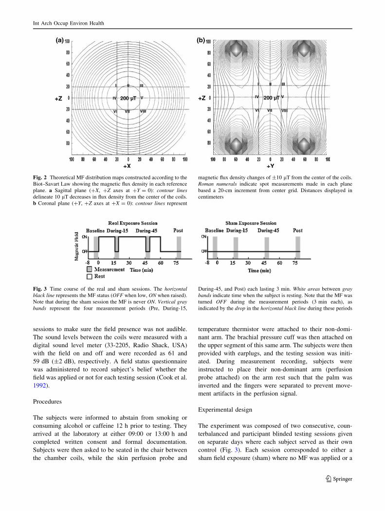
MF variation throughout the area around the subject’s
head was ±5% as determined by theoretical calculations
(Fig. 2). A grid was constructed between the coils to verify
the theoretical calculations with spot measurements made
using a magnetometer (CSA-1VG-SO, Sentron AG, Swit-
zerland). The magnetometer sensitivity was ±15 lT. Nine
spot measurements (8 peripheral plus a center) were taken
in the grid in 20-cm increments from the middle of the
coils for each plane (Fig. 2). These measurements con-
firmed that the MF in a 40-cm3 volume surrounding the
subject’s head was 200 lT (±5%).
The subject was seated in a padded chair located in the
middle of the coils so that they received whole body
exposure with their head in the center of the coils. As such,
possible effects of field exposure could be either central or
peripheral in nature. The ambient geomagnetic static field
was measured in the chamber as 47.6 lT (±0.5%) using a
fluxgate magnetometer (Fluxgate FGM 3D2, Walker Sci-
entific Inc, USA). The ambient time-varying MF was
recorded as 0.139 lT rms (±0.5%), also measured with a
fluxgate magnetometer (Mag-03, Bartington Instruments,
England). Additionally, background vibration noise was
recorded with a seismic accelerometer (Model 393A03,
PCB Piezotronics, USA) between the coils. With the MF
off, the background vibration was 1.70 9 10-2 m/s2,
compared to 1.74 9 10-2 m/s2 (±4.9 9 10-5 m/s2) when
the MF was on. This difference is well below reported
values for human linear acceleration detection thresholds
(Kingma 2005), and thus we were satisfied that the subject
was not exposed to vibrations as a result of the field gen-
eration. The subject was fitted with ear plugs during testing
Fig. 1 Helmholtz-like MF exposure chamber. Subject was seated in
the chair between the parallel octagonal coils. The illustrated ?X, ?Y,
and ?Z vectors denote normal vectors to the coronal, sagittal, and
transverse reference planes, respectively
Int Arch Occup Environ Health
123
sessions to make sure the field presence was not audible.
The sound levels between the coils were measured with a
digital sound level meter (33-2205, Radio Shack, USA)
with the field on and off and were recorded as 61 and
59 dB (±2 dB), respectively. A field status questionnaire
was administered to record subject’s belief whether the
field was applied or not for each testing session (Cook et al.
1992).
Procedures
The subjects were informed to abstain from smoking or
consuming alcohol or caffeine 12 h prior to testing. They
arrived at the laboratory at either 09:00 or 13:00 h and
completed written consent and formal documentation.
Subjects were then asked to be seated in the chair between
the chamber coils, while the skin perfusion probe and
temperature thermistor were attached to their non-domi-
nant arm. The brachial pressure cuff was then attached on
the upper segment of this same arm. The subjects were then
provided with earplugs, and the testing session was initi-
ated. During measurement recording, subjects were
instructed to place their non-dominant arm (perfusion
probe attached) on the arm rest such that the palm was
inverted and the fingers were separated to prevent move-
ment artifacts in the perfusion signal.
Experimental design
The experiment was composed of two consecutive, coun-
terbalanced and participant blinded testing sessions given
on separate days where each subject served as their own
control (Fig. 3). Each session corresponded to either a
sham field exposure (sham) where no MF was applied or a
Fig. 2 Theoretical MF distribution maps constructed according to the
Biot–Savart Law showing the magnetic flux density in each reference
plane. a Sagittal plane (?X, ?Z axes at ?Y = 0): contour linesdelineate 10 lT decreases in flux density from the center of the coils.
b Coronal plane (?Y, ?Z axes at ?X = 0): contour lines represent
magnetic flux density changes of ±10 lT from the center of the coils.
Roman numerals indicate spot measurements made in each plane
based a 20-cm increment from center grid. Distances displayed in
centimeters
Fig. 3 Time course of the real and sham sessions. The horizontalblack line represents the MF status (OFF when low, ON when raised).
Note that during the sham session the MF is never ON. Vertical graybands represent the four measurement periods (Pre, During-15,
During-45, and Post) each lasting 3 min. White areas between graybands indicate time when the subject is resting. Note that the MF was
turned OFF during the measurement periods (3 min each), as
indicated by the drop in the horizontal black line during these periods
Int Arch Occup Environ Health
123

real field exposure (real), during which the MF was applied
for 1 h. To permit baseline, cumulative exposure, and
residual measurements to be recorded, each session inclu-
ded 4 periods of measurements, each lasting 3 min. Each
session followed the same timeframe. First, a baseline
measurement (Pre) was recorded after 5 min of seated rest
before the onset of the MF. At the conclusion of this
measurement (minute 0 in Fig. 3), MF was either generated
(real) or not (sham) for the next 60 min of the session. Two
during exposure measurements were recorded at minutes
15 (During-15) and 45 (During-45). During each of these
3-min measurement periods, the MF was switched off to
preclude any confounding effects upon equipment perfor-
mance. Finally, a postexposure measurement was recorded
at minute 75 (Post; 15 min after the end of MF exposure).
The perfusion data from the first minute of each of the
four measurement periods was analyzed in this study. The
first minute was selected as it was prior to inflation and
reading of the blood pressure cuff, which occurred during
the second and third minute (commencing immediately
after the first minute of perfusion data was obtained and
allocated 2 min to complete) of each 3-min measurement
period. During this time, subjects were instructed to refrain
from moving to prevent artifacts in signal measurement.
The Perisoft software returned mean perfusion values for
user-defined periods, which could then be analyzed in
statistical software. HR and skin surface temperature data
were also analyzed from the first minute of each mea-
surement period to correspond with the perfusion data.
Mean arterial pressure data was collected during the second
minute of each measurement period after cuff inflation to
prevent compromising the perfusion signal.
Statistical analysis
A complete set of data (perfusion, HR, and pressure) was
obtained from 10 subjects for statistical analysis. Room
temperature data was available for 9 of the 10 subjects. The
room temperature data for one subject was lost due to
battery failure in the digital thermometer. A chi-square
analysis was conducted on the responses from the field
status questionnaire to determine if participants could
detect the presence of the MF. Physiological data was
tested for normality using the Shapiro–Wilks test in SPSS
(SPSS 16.0, SPSS, USA) and when it did not meet nor-
mality, it was transformed (log10 transformation) to obtain
normal distributions. Since the transformed data statistics
did not differ in significant findings from the non-trans-
formed results, we chose to report the statistics on the non-
transformed data in this paper to preserve data integrity.
Repeated measures analysis of variance tests (ANOVA)
were conducted using SPSS (SPSS 16.0, SPSS, USA). All
results were interpreted using the Greenhous–Geisser
correction to reduce the probability of obtaining a signifi-
cant result by chance alone. Independent within subject
variables consisted of session (real or sham) and time (Pre,
During-15, During-45, and Post) resulting in a 2 9 4
ANOVA. Data was analyzed for session and time main
effects and for session by time interactions. Results were
considered statistically significant if p \ 0.05. MF effect
would be indicated by a significant session by time inter-
action. Bonferroni-adjusted pairwise comparisons were
performed in addition (although not required by the repe-
ated measures statistical model used here) to help illustrate
data trends over time in each session. Finally, analyses of
covariance (ANCOVA) were conducted to remove the
influence of temperature indices on cardiovascular
parameters.
Results
In order to determine if participants were able to detect the
MF, a test of independence between guessing field pres-
ence and actual field presence was conducted using chi-
square analysis from the field status questionnaire. Only the
session in which the MF was presented was evaluated for
correctness of guesses to determine whether the subject
was aware of the field status. Analyzing the chi-square data
in this manner provides an indication about whether the
subjects could correctly detect the field when it was pre-
sented rather then correctly detect when it was not pre-
sented. The chi-square test revealed that the differences
between the feedback conditions (on and off) were not
significantly associated with guessing [v2 (1, n =
10) = 3.6, p = 0.058]. The responses were weighted
toward being ‘‘off’’ (80%), which is what would be
expected in the absence of any cues perceived by the
subject about the presence of the field.
A significant main effect for time (F3,27 = 7.881,
p \ 0.05) was found for skin blood perfusion with perfu-
sion means decreasing throughout the session. No session
by time interaction was found (Fig. 4, Table 1). Bonfer-
roni-adjusted pairwise comparisons were performed
(although not required by our statistical model) that
showed the baseline time period was significantly greater
than the During-15 time period (p \ 0.05). Similarly, a
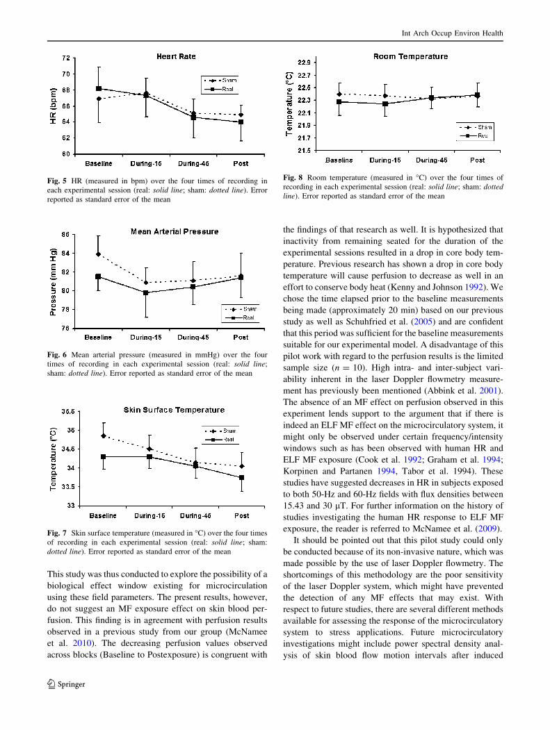
significant main effect for time was observed for HR
(F3,27 = 3.425, p \ 0.05) with HR means decreasing
throughout the session, although no session by time inter-
action was found (Fig. 5, Table 1). Bonferroni-adjusted
pairwise comparisons did not show any significantly dif-
ferences between time periods (p [ 0.05). No main effects
or interactions were observed for mean arterial pressure
(Fig. 6, Table 1). Skin surface temperature demonstrated a
significant main effect for time (F3,27 = 8.176, p \ 0.01)
Int Arch Occup Environ Health
123

with temperature means decreasing throughout the session.
No session by time interaction was observed (Fig. 7,
Table 1). Bonferroni-adjusted pairwise comparisons did
not show any differences between time periods (p [ 0.05).
No main effects or interactions were demonstrated in the
room temperature analysis (Fig. 8, Table 1).
As a biological zero was not obtained for the skin blood
perfusion baseline measurements, inter-subject variability
in the perfusion unit measurements was reduced by also
analyzing the perfusion data as a percent change from
baseline. No main or interaction effects were observed with
the data organized in this normalized manner (Table 2). As
a result, the non-transformed data was used for statistical
analysis and discussion in the remainder of this paper, and
the normalized data is not discussed further.
To determine if either of the temperature indices (skin or
room) could be covering possible MF effects in the car-
diovascular parameters, they were analyzed to determine if
either might be a covariate. Both skin temperature and
room temperature were reduced to factors in SPSS and
were observed against the cardiovascular parameters (per-
fusion, HR, and pressure) for correlations. The skin tem-
perature factor correlated strongly (p \ 0.05) with 6 out of
8 of the perfusion time periods, and so an ANCOVA was
conducted. The resulting ANCOVA demonstrated that the
skin temperature factor was not a significant covariate
(p [ 0.05) with perfusion for either session, time or the
session by time interaction confirming that our ANOVA
results were well adapted to our model. The room tem-
perature factor was not significantly correlated (p \ 0.05)
with any of the indices, and thus, analyses of covariance
were not indicated.
Discussion
Skin blood perfusion
The primary motivation behind the present study was based
on results from previous work conducted in our laboratory
indicating an exposure effect on microcirculation in rats
when subjected to a 60-Hz, 200-lT MF (Hensel et al. 2003).
Fig. 4 Skin blood perfusion (measured in PU) over the four times of
recording in each experimental session (real: solid line; sham: dottedline). Error reported as standard error of the mean. * Indicates a
significant Bonferroni-adjusted pairwise comparison regarding the
main time effect (difference of the averaged sham and real perfusion
means between time periods; p \ 0.05)
Table 1 Results from ANOVA (n = 10; perfusion, HR, pressure, skin surface temperature, n = 9; room temperature)
Analysis of variance results
Measurement Effect F p pg2 Power
Skin blood perfusion Session 0.648 (df 1, 9) p [ 0.05 0.067 0.112
Time 7.881 (df 1.217, 10.957) p < 0.05* 0.467 0.771
Session 9 Time 0.230 (df 3, 27) p [ 0.05 0.025 0.088
HR Session 0.001 (df 1, 9) p [ 0.05 0.000 0.050
Time 3.425 (df 3, 27) p < 0.05* 0.276 0.705
Session 9 Time 0.469 (df 3, 27) p [ 0.05 0.050 0.132
Mean arterial pressure Session 0.947 (df 1, 9) p [ 0.05 0.095 0.141
Time 1.940 (df 3, 27) p [ 0.05 0.177 0.444
Session 9 Time 0.583 (df 3, 27) p [ 0.05 0.061 0.154
Skin surface temperature Session 1.525 (df 1, 9) p [ 0.05 0.145 0.198
Time 8.176 (df 1.377, 12.395) p < 0.01* 0.476 0.826
Session 9 Time 0.581 (df 1.121, 10.092) p [ 0.05 0.061 0.109
Room temperature Session 0.061 (df 1, 8) p [ 0.05 0.008 0.056
Time 0.711 (df 3, 24) p [ 0.05 0.082 0.178
Session 9 Time 0.937 (df 1.265, 10.118) p [ 0.05 0.105 0.150
Partial eta squared (pg2) and observed power (power) are included as indices of effect size. Results considered statistically significant if p B 0.05
and are indicated in boldface font
Int Arch Occup Environ Health
123
This study was thus conducted to explore the possibility of a
biological effect window existing for microcirculation
using these field parameters. The present results, however,
do not suggest an MF exposure effect on skin blood per-
fusion. This finding is in agreement with perfusion results
observed in a previous study from our group (McNamee
et al. 2010). The decreasing perfusion values observed
across blocks (Baseline to Postexposure) is congruent with
the findings of that research as well. It is hypothesized that
inactivity from remaining seated for the duration of the
experimental sessions resulted in a drop in core body tem-
perature. Previous research has shown a drop in core body
temperature will cause perfusion to decrease as well in an
effort to conserve body heat (Kenny and Johnson 1992). We
chose the time elapsed prior to the baseline measurements
being made (approximately 20 min) based on our previous
study as well as Schuhfried et al. (2005) and are confident
that this period was sufficient for the baseline measurements
suitable for our experimental model. A disadvantage of this
pilot work with regard to the perfusion results is the limited
sample size (n = 10). High intra- and inter-subject vari-
ability inherent in the laser Doppler flowmetry measure-
ment has previously been mentioned (Abbink et al. 2001).
The absence of an MF effect on perfusion observed in this
experiment lends support to the argument that if there is
indeed an ELF MF effect on the microcirculatory system, it
might only be observed under certain frequency/intensity
windows such as has been observed with human HR and
ELF MF exposure (Cook et al. 1992; Graham et al. 1994;
Korpinen and Partanen 1994, Tabor et al. 1994). These
studies have suggested decreases in HR in subjects exposed
to both 50-Hz and 60-Hz fields with flux densities between
15.43 and 30 lT. For further information on the history of
studies investigating the human HR response to ELF MF
exposure, the reader is referred to McNamee et al. (2009).
It should be pointed out that this pilot study could only
be conducted because of its non-invasive nature, which was
made possible by the use of laser Doppler flowmetry. The
shortcomings of this methodology are the poor sensitivity
of the laser Doppler system, which might have prevented
the detection of any MF effects that may exist. With
respect to future studies, there are several different methods
available for assessing the response of the microcirculatory
system to stress applications. Future microcirculatory
investigations might include power spectral density anal-
ysis of skin blood flow motion intervals after induced
Fig. 5 HR (measured in bpm) over the four times of recording in
each experimental session (real: solid line; sham: dotted line). Error
reported as standard error of the mean
Fig. 6 Mean arterial pressure (measured in mmHg) over the four
times of recording in each experimental session (real: solid line;
sham: dotted line). Error reported as standard error of the mean
Fig. 7 Skin surface temperature (measured in �C) over the four times
of recording in each experimental session (real: solid line; sham:
dotted line). Error reported as standard error of the mean
Fig. 8 Room temperature (measured in �C) over the four times of
recording in each experimental session (real: solid line; sham: dottedline). Error reported as standard error of the mean
Int Arch Occup Environ Health
123

ischemic conditions. This technique could provide valuable
insight into different hierarchal levels of circulatory control
and may provide a different level of measurement sensi-
tivity to MF exposure.
HR and mean arterial pressure
No MF effects were observed for either HR or mean
arterial pressure in the present study. The absence of HR
effects lends support to the body of evidence, suggesting
ELF MF do not affect the HR. These findings are consistent
with the results observed in our previous work using an
1,800-lT MF exposure chamber (McNamee et al. 2010) as
well as others (Graham et al. 2000; Sait et al. 2006; Ghione
et al. 2005; Kurokawa et al. 2003). It should be pointed out
that in the current paper, the HR is recorded during a 1-h
period of MF exposure at both 15 and 30 min after the field
has been turned on. Since the field is temporarily switched
off for the 3 min of measurement, we were not able to
observe HR behavior prior to the measurements being
taken during the MF exposure. Previous research has
pointed to an effect being observed at the point where the
field switches on and off (Cook et al. 1992; Graham et al.
1994), and this may be an avenue to consider for future
replication attempts. The decrease in HR across blocks also
matches the response observed in our previous work and
may be related to a decrease in state anxiety (increase in
relaxation) brought on from the laboratory experience. This
state anxiety would be expected to subside as the subject
acclimated to the experiment protocol, which would result
in a concomitant decrease in physiological arousal. Several
authors (Miu et al. 2009; Moses et al. 2007) have reported
findings supporting an increase in HR associated with
stress, thus it can be postulated that as stress recedes, HR
would as well, explaining the HR trend reported here and
previously (McNamee et al. 2010).
The absence of MF effects on blood pressure is consis-
tent with the findings of Ghione et al. (2005) and Korpinen
and Partanen (1996). Ghione et al. (2005) investigated the
human blood pressure response to a 90-min exposure to a
50-Hz MF at either 40 or 80 lT, while Korpinen and Par-
tanen and colleagues used a 50-Hz MF source ranging from
1.4 to 6.6 lT for exposure. The absence of an effect on
pressure is in contrast, however, with the earlier results of
Ghione et al. (2004) who used a 37-Hz, 80-lT MF and
found a slight increase in systolic blood pressure. It should
be pointed out that Ghione et al. (2004) looked at systolic
blood pressure rather than the mean arterial pressure
recorded in the present study. The mean arterial pressure is
the average arterial pressure over the cardiac cycle and may
not be as susceptible to possible MF-induced sympathetic
stimulation of the cardiac cycle. Additionally, the experi-
ment conducted by Korpinen and Partanen (1996) was
conducted outdoors that may have led to difficulties con-
trolling for confounding variables such as temperature
fluctuation, geomagnetic EMF variations, and the fact that
subjects were required to walk 200 m out of the exposure
site before having their blood pressure measured.
Although the present results do not suggest an effect of
ELF MF exposure on mean arterial pressure, several
questions remain regarding the response of blood pressure
to MF exposure. Future research might consider artificially
altered blood pressure to see if regulating mechanisms such
as chemoreceptors or baroreceptors are affected by MF
exposure. For instance, Sait et al. (2006) used a 60 degree
from supine, head up tilt procedure to invoke changes in
the sympathovagal cardiovascular balance while investi-
gating the cardiovascular system. They postulated the
sympathetic nervous system needed to be activated
(through the tilting procedure) to detect a cardiovascular
response from MF exposure. Although they found no
exposure effects when subjects were tested this way, this
avenue of research should be explored further. Barore-
ceptor research by Gmitrov (2007) using static magnets has
suggested that these sites may play an important role in
modulating changes in the cardiovascular system. Their
study found increases in skin blood flow were positively
correlated with static MF-induced increases in baroreceptor
sensitivity. It would be of interest to study if a time-varying
MF application could be designed to investigate this phe-
nomenon. Finally, as a review of pharmacological inter-
ventions in blood flow research is beyond the scope of this
paper, the reader is referred to a comprehensive review by
McKay et al. (2007) for developments in this area.
Table 2 Results from normalized perfusion ANOVA (n = 10)
Normalized perfusion ANOVA
Measurement Effect F p pg2 power
Skin blood perfusion Session 0.912 (df 1, 9) p [ 0.05 0.092 0.137
Time 0.327 (df 1.079, 9.708) p [ 0.05 0.035 0.082
Session 9 Time 0.780 (df 1.040, 9.359) p [ 0.05 0.080 0.126
Partial eta squared (pg2) and observed power (power) are included as indices of effect size
Int Arch Occup Environ Health
123

Although HR and blood pressure were additionally
monitored in this pilot, the primary objective of this study
was to investigate the response of the peripheral blood
perfusion to MF exposure. Although several authors have
previously demonstrated effects of MF exposure on HRV
(Tabor et al. 2004 and Sastre et al. 2000), the methodology
used in this study to obtain the HR signal did not permit
accurate measurement of the beat peak to peak time
interval, which is necessary for the determination of the
variability measure and thus it was not investigated in this
paper. In order to allow the computation and analysis of
HRV indices, future studies might consider using longer
data measurement periods. HRV remains an important area
for future full-length studies, and a detailed review of the
history of ELF MF HRV studies can be found in McNamee
et al. (2009).
Skin surface temperature
The results of the present study do not suggest an effect of
the MF on skin temperature. The trend of the skin tem-
perature to decrease over time is consistent with the theory
proposed in describing the perfusion results that the core
temperature is dropping in the subjects due to inactivity.
This theory would be supported by the research of Akata
et al. (2004) who have previously described a high corre-
lation between finger tip blood flow measured by laser
Doppler flowmetry and forearm skin surface temperature.
The present skin temperature findings, however, do not
match those of our previous study, which demonstrated an
interaction of the fields over exposure time (McNamee
et al. 2010). An explanation for the failure of the skin
temperature to decrease during the real session of that
study was that the water cooling system that cooled the
coils gave off heat while operating thus raising the room
temperature and by extension, skin temperature as well.
The present results lend support to this hypothesis, how-
ever, as the difference in the flux density of the coils
between papers is almost an order magnitude. The smaller
flux density used in the present study was not of sufficient
intensity to require significant cooling if any, and thus, no
skin temperature interaction was observed. It is still sug-
gested that future MF research involving skin temperature
and stronger exposures measures the air temperature
proximal to the skin measurement site to avoid possible
temperature confounds.
Finally, since the results of this pilot study might be due
to the small sample sized used, sample power analyses
were run on each of the circulatory parameters in the study
using GPower (GPower 3.0.10, GPower, Germany). An
a priori 2 9 4 design was tested using the ANOVA: Fixed
effects, special, main effects, and interactions test under the
F test family option. The results of these power analyses
revealed that to obtain significant interactions with a power
of 0.80, between 172 (for pressure) and 430 (for perfusion),
subjects would be required. These power analyses hardly
support an effect of MF exposure on the circulatory
parameters, thus tending to confirm the results of the pilot
study.
Conclusions
In conclusion, the present protocol does not suggest an
effect of a 60-Hz, 200-lT ELF MF exposure on the human
circulatory system. It appears the protocol was sensitive
enough to discern the effects of inactivity on skin blood
perfusion, HR, and skin surface temperature, and thus if an
MF effect does exist, it must be proportionally smaller then
this. Importantly, the findings of the present paper support
our hypothesis that the water cooling system used to cool
the 1,800-lT coils in our previous study caused an increase
in room temperature, which would have affected skin
temperature results. The flux density differences between
these studies must be kept in mind, however, as we are still
unable to conclusively rule out an MF effect with regard to
this trend. The absence of MF effects supports previously
published literature offering support against an effect of
MF exposure on HR and blood pressure. This is important
in an occupational health context as well as for the re-
evaluation of international exposure guidelines.
Acknowledgments The authors wish to extend thanks to Lynn
Keenliside for his technical expertise and assistance with this project.
Conflict of interest statement The authors declare that they have
no conflict of interest. Funding for this project was supplied by Hydro
Quebec—TransEnergie and Electricite de France—Reseau de
Transport d’Electricite. This study was also funded in part by Cana-
dian Institutes of Health Research grants (FRN 85217 and MOP
43874), the Canadian Foundation for Innovation (11358) and the
Ontario Research and Development Challenge Fund (MAR-01-0936).
References
Abbink EJ, Wollersheim H, Netten PM, Smits P (2001) Reproduc-
ibility of skin microcirculatory measurements in humans, with
special emphasis on capillaroscopy. Vasc Med 6:203–210
Akata T, Kanna T, Yoshino J, Higashi M, Fukui K, Takahashi S
(2004) Reliability of fingertip skin-surface temperature and its
related thermal measures as indices of peripheral perfusion in the
clinical setting of the operating theatre. Anaesth Intensive Care
32(4):519–529
Baureus Koch CLM, Sommarin M, Persson BRR, Salford LG,
Eberhardt JL (2003) Interaction between weak low frequency
magnetic fields and cell membranes. Bioelectromagnetics
24:395–402
Caro CG, Parker KH (1990) Mechanics and imaging of the
macrocirculation. Magn Reson Med 14(2):179–186
Int Arch Occup Environ Health
123

Cook MR, Graham C, Cohen HD, Gerkovich MM (1992) A
replication study of human exposure to 60-Hz fields: effects on
neurobehavioral measures. Bioelectromagnetics 13(4):261–285
Dekker JM, Schouten EG, Klootwijk P, Pool J, Swenne CA,
Kromhout D (1997) Heart rate variability from short electrocar-
diographic recordings predicts mortality from all causes in
middle-aged and elderly men. Am J Epidemiol 145:899–908
Ghione S, Del Seppia C, Mezzasalma L, Emdin M, Luschi P (2004)
Human head exposure to a 37 Hz electromagnetic field: effects
on blood pressure, somatosensory perception, and related
parameters. Bioelectromagnetics 25:167–175
Ghione S, Del Seppia C, Mezzasalma L, Bonfiglio L (2005) Effects of
50 Hz electromagnetic fields on electroencephalographic alpha
activity, dental pain threshold and cardiovascular parameters in
humans. Neurosci Lett 382:112–117
Gmitrov J (2007) Static magnetic field effect on the arterial
baroreflex-mediated control of microcirculation: implications
for cardiovascular effects due to environmental magnetic fields.
Radiat Environ Biophys 46(3):281–290
Gmitrov J, Gmitrova A (2004) Geomagnetic field effect on cardio-
vascular regulation. Bioelectromagnetics 25:92–101
Graham C, Cook MR, Cohen HD, Gerkovich MM (1994) Dose
response study of human exposure to 60 Hz electric and
magnetic fields. Bioelectromagnetics 15(5):447–463
Graham C, Cook MR, Sastre A, Gerkovich MM, Kavet R (2000)
Cardiac autonomic control mechanisms in power-frequency
magnetic fields: a multistudy analysis. Environ Health Perspect
108:737–742
Hakansson N, Gustavsson P, Sastre A, Floderus B (2003) Occupa-
tional exposure to extremely low frequency magnetic fields and
mortality from cardiovascular disease. Am J Epidemiol
158:534–542
Hensel JM, Bohnert R, Tyml K, Prato FS, Thomas AW (2003) Effects
of specific pulsed and oscillating (30 Hz, 60 Hz) magnetic fields
on acetylcholine-induced vasodilation in rats. Poster session
presented at: BEMS 25th Annual Meeting. 2003 Jun 22–27;
Maui, Hawaii
Kenny WL, Johnson JM (1992) Control of skin blood flow during
exercise. Med Sci Sports Exerc 24(3):303–312
Kingma H (2005) Thresholds for perception of direction of linear
acceleration as a possible evaluation of the otolith function.
BMC Ear Nose Throat Disord 5:5
Korpinen L, Partanen J (1994) Influence of 50 Hz electric and
magnetic fields on the pulse rate of human heart. Bioelectro-
magnetics 15:503–512
Korpinen L, Partanen J (1996) Influence of 50-Hz electric and
magnetic fields on human blood pressure. Radiat Environ
Biophys 35:199–204
Korpinen L, Partanen J, Uusitalo A (1993) Influence of 50 Hz electric
and magnetic fields on the human heart. Bioelectromagnetics
14(4):329–340
Kurokawa Y, Nitta H, Imai H, Kabuto M (2003) Can extremely low
frequency alternating magnetic fields modulate heart rate or its
variability in humans? Auton Neurosci 105:53–61
Lednev VV, Belova NA, Ermakov AM, Akimov EB, Tonevitsky AG
(2008) Modulation of cardiac rhythm in the humans exposed to
extremely weak alternating magnetic fields. Biophysics
53(6):648–654
Li L, Mac-Mary S, Marsaut D, Sainthillier JM, Nouveau S, Gharbi T,
de Lacharriere O, Humbert P (2006) Age-related changes in
skin topography and microcirculation. Arch Dermatol Res
297:412–416
Liao D, Cai J, Rosamond WD, Bames RW, Hutchinson RG, Whitsel
EA, Rautaharju P, Heiss G (1997) Cardiac autonomic function
and incident coronary heart disease: a population based case-
cohort study. Am J Epidemiol 145:696–706
McKay JC, Prato FS, Thomas AW (2007) A literature review: the
effects of magnetic field exposure on blood flow and blood
vessels in the microvasculature. Bioelectromagnetics 28:81–98
McNamee DA, Legros AG, Krewski DR, Wisenberg G, Prato FS,
Thomas AW (2009) A literature review: the cardiovascular
effects of exposure to extremely low frequency electromagnetic
fields. Int Arch Occup Environ Health 82(8):919–933
McNamee DA, Corbacio M, Weller JK, Brown S, Prato FS, Thomas
AW, Legros AG (2010) The cardiovascular response to an acute
1,800 lT, 60 Hz magnetic field exposure in humans. Int Arch
Occup Environ Health 83(4):441–454
Miu AC, Heilman RM, Miclea M (2009) Reduced heart rate
variability and vagal tone in anxiety: trait versus state, and the
effects of autogenic training. Auton Neurosci 145(1–2):99–103
Moses ZB, Luecken LJ, Eason JC (2007) Measuring task-related
changes in heart rate variability. Conf Proc IEEE Eng Med Biol
Soc. 2007:644–647
Okano H, Ohkubo C (2003) Anti-pressor effects of whole body
exposure to static magnetic field on pharmacologically induced
hypertension in conscious rabbits. Bioelectromagnetics 24:139–
147
Repacholi MH, Greenebaum B (1999) Interaction of static and
extremely low frequency electric and magnetic fields with living
systems: health effects and research needs. Bioelectromagnetics
20:133–160
Sait ML, Wood AW, Kirsner RLG (2006) Effects of 50 Hz magnetic
field exposure on human heart rate variability with passive
tilting. Physiol Meas 27:73–83
Sastre A, Kavet R (2002) Candidate sites of action for microdosi-
metry associated with exposure to extremely-low-frequency
magnetic fields, electric fields and contact currents. Health Phys
83(3):387–394
Sastre A, Graham C, Cook MR (2000) Brain frequency magnetic
fields alter cardiac autonomic control mechanisms. Clin Neuro-
physiol 111:1942–1948
Savitz DA, Liao D, Sastre A, Kleckner RC, Kavet R (1999) Magnetic
field exposure and cardiovascular disease mortality among
electric utility workers. Am J Epidemiol 149:135–142
Schmid-Schonbein GW (1999) Biomechanics of microcirculatory
blood perfusion. Annu Rev Biomed Eng 01:73–102
Schuhfried O, Vacariu G, Rochowanski H, Serek M, Fialka-Moser V
(2005) The effects of low-dosed and high-dosed low-frequency
electromagnetic fields on microcirculation and skin temperature
in healthy subjects. Int J Sports Med 26:886–890
Tabor Z, Michalski J, Rokita E (2004) Influence of 50 Hz magnetic
field on human heart rate variability: linear and nonlinear
analysis. Bioelectromagnetics 25:474–480
Tsuji H, Larson MG, Venditti FJ, Manders ES, Evans JC, Feldman
CL, Levy D (1996) Impact of reduced heart rate variability on
risk for cardiac events. The Framingham heart study. Circulation
94(11):2850–2855
Wertheimer N, Leeper E (1979) Electrical wiring configurations and
childhood cancer. Am J Epidemiol 109(3):273–284
Xu S, Okano H, Ohkubo C (2000) Acute effects of whole-body
exposure to static magnetic fields and 50-Hz electromagnetic
fields on muscle microcirculation in anesthetized mice. Bioelec-
trochemistry 53:127–135
Int Arch Occup Environ Health
123