The Regulatory Repertoire of Pseudomonas aeruginosa AmpC ß-Lactamase Regulator AmpR Includes...
22
The Regulatory Repertoire of Pseudomonas aeruginosa AmpC ß-Lactamase Regulator AmpR Includes Virulence Genes Deepak Balasubramanian 1 , Lisa Schneper 2 , Massimo Merighi 3¤a , Roger Smith 3¤b , Giri Narasimhan 4 , Stephen Lory 3 , Kalai Mathee 2 * 1 Department of Biological Sciences, College of Arts and Science, Florida International University, Miami, Florida, United States of America, 2 Molecular Microbiology and Infectious Diseases (Herbert Werthiem College of Medicine), Florida International University, Miami, Florida, United States of America, 3 Microbiology and Molecular Genetics, Harvard Medical School, Boston, Massachussetts, United States of America, 4 School of Computing and Information Science, College of Engineering and Computing, Florida International University, Miami, Florida, United States of America Abstract In Enterobacteriaceae, the transcriptional regulator AmpR, a member of the LysR family, regulates the expression of a chromosomal b-lactamase AmpC. The regulatory repertoire of AmpR is broader in Pseudomonas aeruginosa, an opportunistic pathogen responsible for numerous acute and chronic infections including cystic fibrosis. In addition to regulating ampC, P. aeruginosa AmpR regulates the sigma factor AlgT/U and production of some quorum sensing (QS)- regulated virulence factors. In order to better understand the ampR regulon, we compared the transcriptional profile generated using DNA microarrays of the prototypic P. aeruginosa PAO1 strain with its isogenic ampR deletion mutant, PAODampR. Transcriptome analysis demonstrates that the AmpR regulon is much more extensive than previously thought, with the deletion of ampR influencing the differential expression of over 500 genes. In addition to regulating resistance to b- lactam antibiotics via AmpC, AmpR also regulates non-b-lactam antibiotic resistance by modulating the MexEF-OprN efflux pump. Other virulence mechanisms including biofilm formation and QS-regulated acute virulence factors are AmpR- regulated. Real-time PCR and phenotypic assays confirmed the microarray data. Further, using a Caenorhabditis elegans model, we demonstrate that a functional AmpR is required for P. aeruginosa pathogenicity. AmpR, a member of the core genome, also regulates genes in the regions of genome plasticity that are acquired by horizontal gene transfer. Further, we show differential regulation of other transcriptional regulators and sigma factors by AmpR, accounting for the extensive AmpR regulon. The data demonstrates that AmpR functions as a global regulator in P. aeruginosa and is a positive regulator of acute virulence while negatively regulating biofilm formation, a chronic infection phenotype. Unraveling this complex regulatory circuit will provide a better understanding of the bacterial response to antibiotics and how the organism coordinately regulates a myriad of virulence factors in response to antibiotic exposure. Citation: Balasubramanian D, Schneper L, Merighi M, Smith R, Narasimhan G, et al. (2012) The Regulatory Repertoire of Pseudomonas aeruginosa AmpC ß- Lactamase Regulator AmpR Includes Virulence Genes. PLoS ONE 7(3): e34067. doi:10.1371/journal.pone.0034067 Editor: Pierre Cornelis, Vrije Universiteit Brussel, Belgium Received November 8, 2011; Accepted February 27, 2012; Published March 29, 2012 Copyright: ß 2012 Balasubramanian et al. This is an open-access article distributed under the terms of the Creative Commons Attribution License, which permits unrestricted use, distribution, and reproduction in any medium, provided the original author and source are credited. Funding: This work has been supported by National Institutes of Health-Minority Biomedical Research Support SCORE (S06 GM08205 and 5SC1AI081376; to KM), Florida International University (FIU) Teaching Assistantship (Biological Sciences; to DB), FIU Research Assistantship (Herbert Werthiem College of Medicine; to DB) and the Cystic Fibrosis Foundation Student Traineeship Award (BALASU08H0; to DB). The funders had no role in study design, data collection and analysis, decision to publish, or preparation of the manuscript. Competing Interests: The authors have declared that no competing interests exist. * E-mail: [email protected] ¤a Current address: Glycosyn Inc., Medford, Massachussetts, United States of America ¤b Current address: Semprus Biosciences, Cambridge, Massachussetts, United States of America Introduction Pseudomonas aeruginosa is one of the leading opportunistic Gram- negative nosocomial pathogens. This is particularly true in critically ill patients, where multi-drug resistant P. aeruginosa is a severe problem. It is the leading pathogen in ventilator-associated pneumonia with a mortality rate of 40–60% [1]. P. aeruginosa is also a primary cause of urinary tract infections in the US and Europe [2], wound infections leading to bacteremia with one-third to two- thirds mortality rate [3,4], pulmonary infections including cystic fibrosis (CF) [5], lung cancer patients [6] and in pediatric and adult AIDS patients [7]. Inability to eradicate the infection is partly due to intrinsic and acquired antibiotic resistance of P. aeruginosa. Antibiotic resistant isolates of P. aeruginosa are selectively favored in vivo in CF patients [8,9]. Resistance of P. aeruginosa to the b-lactam class of antibiotics, currently used to treat P. aeruginosa infections, is partly mediated by a group of genes belonging to the amp system. The amp genes were first discovered in Enterobacter cloacae to confer resistance to b-lactams [10] and later in other members of Enterobacteriaceae [11,12,13,14]. The products of amp genes in E. cloacae and other organisms include the AmpC b-lactamase, the AmpG permease, a putative AmpE permease, the AmpD cytoplasmic amidase, and the transcriptional regulator AmpR [10,11,12,13,14]. Recent studies have identified another perme- ase, AmpP that is required for b-lactamase induction in P. PLoS ONE | www.plosone.org 1 March 2012 | Volume 7 | Issue 3 | e34067
Transcript of The Regulatory Repertoire of Pseudomonas aeruginosa AmpC ß-Lactamase Regulator AmpR Includes...

The Regulatory Repertoire of Pseudomonas aeruginosaAmpC ß-Lactamase Regulator AmpR Includes VirulenceGenesDeepak Balasubramanian1, Lisa Schneper2, Massimo Merighi3¤a, Roger Smith3¤b, Giri Narasimhan4,
Stephen Lory3, Kalai Mathee2*
1 Department of Biological Sciences, College of Arts and Science, Florida International University, Miami, Florida, United States of America, 2 Molecular Microbiology and
Infectious Diseases (Herbert Werthiem College of Medicine), Florida International University, Miami, Florida, United States of America, 3 Microbiology and Molecular
Genetics, Harvard Medical School, Boston, Massachussetts, United States of America, 4 School of Computing and Information Science, College of Engineering and
Computing, Florida International University, Miami, Florida, United States of America
Abstract
In Enterobacteriaceae, the transcriptional regulator AmpR, a member of the LysR family, regulates the expression of achromosomal b-lactamase AmpC. The regulatory repertoire of AmpR is broader in Pseudomonas aeruginosa, anopportunistic pathogen responsible for numerous acute and chronic infections including cystic fibrosis. In addition toregulating ampC, P. aeruginosa AmpR regulates the sigma factor AlgT/U and production of some quorum sensing (QS)-regulated virulence factors. In order to better understand the ampR regulon, we compared the transcriptional profilegenerated using DNA microarrays of the prototypic P. aeruginosa PAO1 strain with its isogenic ampR deletion mutant,PAODampR. Transcriptome analysis demonstrates that the AmpR regulon is much more extensive than previously thought,with the deletion of ampR influencing the differential expression of over 500 genes. In addition to regulating resistance to b-lactam antibiotics via AmpC, AmpR also regulates non-b-lactam antibiotic resistance by modulating the MexEF-OprN effluxpump. Other virulence mechanisms including biofilm formation and QS-regulated acute virulence factors are AmpR-regulated. Real-time PCR and phenotypic assays confirmed the microarray data. Further, using a Caenorhabditis elegansmodel, we demonstrate that a functional AmpR is required for P. aeruginosa pathogenicity. AmpR, a member of the coregenome, also regulates genes in the regions of genome plasticity that are acquired by horizontal gene transfer. Further, weshow differential regulation of other transcriptional regulators and sigma factors by AmpR, accounting for the extensiveAmpR regulon. The data demonstrates that AmpR functions as a global regulator in P. aeruginosa and is a positive regulatorof acute virulence while negatively regulating biofilm formation, a chronic infection phenotype. Unraveling this complexregulatory circuit will provide a better understanding of the bacterial response to antibiotics and how the organismcoordinately regulates a myriad of virulence factors in response to antibiotic exposure.
Citation: Balasubramanian D, Schneper L, Merighi M, Smith R, Narasimhan G, et al. (2012) The Regulatory Repertoire of Pseudomonas aeruginosa AmpC ß-Lactamase Regulator AmpR Includes Virulence Genes. PLoS ONE 7(3): e34067. doi:10.1371/journal.pone.0034067
Editor: Pierre Cornelis, Vrije Universiteit Brussel, Belgium
Received November 8, 2011; Accepted February 27, 2012; Published March 29, 2012
Copyright: � 2012 Balasubramanian et al. This is an open-access article distributed under the terms of the Creative Commons Attribution License, which permitsunrestricted use, distribution, and reproduction in any medium, provided the original author and source are credited.
Funding: This work has been supported by National Institutes of Health-Minority Biomedical Research Support SCORE (S06 GM08205 and 5SC1AI081376; to KM),Florida International University (FIU) Teaching Assistantship (Biological Sciences; to DB), FIU Research Assistantship (Herbert Werthiem College of Medicine; to DB)and the Cystic Fibrosis Foundation Student Traineeship Award (BALASU08H0; to DB). The funders had no role in study design, data collection and analysis,decision to publish, or preparation of the manuscript.
Competing Interests: The authors have declared that no competing interests exist.
* E-mail: [email protected]
¤a Current address: Glycosyn Inc., Medford, Massachussetts, United States of America¤b Current address: Semprus Biosciences, Cambridge, Massachussetts, United States of America
Introduction
Pseudomonas aeruginosa is one of the leading opportunistic Gram-
negative nosocomial pathogens. This is particularly true in
critically ill patients, where multi-drug resistant P. aeruginosa is a
severe problem. It is the leading pathogen in ventilator-associated
pneumonia with a mortality rate of 40–60% [1]. P. aeruginosa is also
a primary cause of urinary tract infections in the US and Europe
[2], wound infections leading to bacteremia with one-third to two-
thirds mortality rate [3,4], pulmonary infections including cystic
fibrosis (CF) [5], lung cancer patients [6] and in pediatric and
adult AIDS patients [7]. Inability to eradicate the infection is
partly due to intrinsic and acquired antibiotic resistance of P.
aeruginosa. Antibiotic resistant isolates of P. aeruginosa are selectively
favored in vivo in CF patients [8,9]. Resistance of P. aeruginosa to the
b-lactam class of antibiotics, currently used to treat P. aeruginosa
infections, is partly mediated by a group of genes belonging to the
amp system.
The amp genes were first discovered in Enterobacter cloacae to
confer resistance to b-lactams [10] and later in other members of
Enterobacteriaceae [11,12,13,14]. The products of amp genes in E.
cloacae and other organisms include the AmpC b-lactamase, the
AmpG permease, a putative AmpE permease, the AmpD
cytoplasmic amidase, and the transcriptional regulator AmpR
[10,11,12,13,14]. Recent studies have identified another perme-
ase, AmpP that is required for b-lactamase induction in P.
PLoS ONE | www.plosone.org 1 March 2012 | Volume 7 | Issue 3 | e34067

aeruginosa [15]. Expression of ampC is regulated by AmpR. The
ampR gene is located adjacent to ampC and is divergently
transcribed in C. freundii and E. cloacae, as well as in P. aeruginosa
[16,17,18]. AmpR of C. freundii and E. cloacae can cross-
complement each other [19] and P. aeruginosa AmpR is similar to
that found in C. freundii (58%) and E. cloacae (62%) [20].
In C. freundii, AmpR binds to a 15 bp sequence 59 TCTG-
CTGCAAATTT 39 [16,20] and there is a nearly identical
putative AmpR binding site (59 TCTGCTCCAAATTT 39) in the
ampR-ampC intergenic region in P. aeruginosa [21]. AmpR has a
helix-turn-helix motif that is typical of DNA-binding proteins and
the C. freundii AmpR binds DNA using this motif [16]. The AmpR-
AmpC system is also conserved in many other pathogens including
Burkholderia cenocepacia [22], Yersinia enterocolitica [23], and Stenotro-
phomonas maltophilia [24].
AmpR belongs to the LysR family of transcriptional regulators
that typically autorepress their own expression [25] which has
been demonstrated in C. freundii [16]. In P. aeruginosa, however,
there is no evidence of autoregulation [21]. It has been postulated
that the signals mediating ampC regulation by AmpR are
peptidoglycan degradation products that function as effector
molecules and are brought into the cell cytoplasm from their
point of origin in the periplasm via the AmpG permease [26]. In
vitro studies have demonstrated that C. freundii AmpR can both
activate and repress ampC expression depending on its interaction
with specific peptidoglycan degradation products [27]. Thus the
levels of these cell wall intermediates dictate AmpR regulation of
ampC and the known regulatory repertoire of AmpR in
Enterobacteriaceae have been limited to regulating ampC expres-
sion [11,26,27]. Previous studies comparing the properties of P.
aeruginosa PAO1 with its isogenic ampR insertion mutant,
PAOampR::aacC1, have shown that AmpR regulates ampC as well
as some quorum sensing (QS) genes [21]. This led us to
hypothesize that the regulatory role of P. aeruginosa AmpR is more
extensive than previously thought.
To test the hypothesis that AmpR regulates different pathways
in P. aeruginosa and to identify the AmpR regulon, we compared
the expression profile of wild-type PAO1 and that of an in-frame
ampR deletion mutant, PAODampR, with and without sub-MIC b-
lactam stress. Our data suggests that P. aeruginosa AmpR is a master
regulator affecting the expression of over 500 genes. Functional
analyses demonstrate the negative regulatory role of AmpR of
multiple virulence mechanisms including biofilm formation and
the MexEF-OprN multidrug efflux pump. Further, we demon-
strate that AmpR positively regulates multiple acute virulence
factors. Using a C. elegans model, we demonstrate that AmpR is
required for pathogenesis in P. aeruginosa. This study establishes the
critical regulatory role that AmpR plays in antibiotic resistance,
virulence and general metabolism in P. aeruginosa.
Results
A. Deletion of ampR reduces b-lactam resistance of PAO1In contrast to previous studies that looked at the role of P.
aeruginosa AmpR using an insertion mutant, this study employed
PAODampR, an in-frame deletion mutant in the prototypic P.
aeruginosa PAO1. The presence of the ampR deletion was confirmed
by PCR and restriction digestion of the amplicons (data not
shown). AmpR is a known positive regulator of the chromosomal
AmpC b-lactamase in different bacterial species [12,20,28].
Consequently, after constructing PAODampR, the strains were
tested for altered production of b-lactamase. The resistance profile
of b-lactam antibiotics for PAO1, PAODampR and PAODampR
(pAmpR) shows that, as expected, loss of ampR enhances strain
sensitivity to b-lactams and expressing ampR in trans on a low-copy
plasmid can restore this defect with multiple b-lactam antibiotics
(Fig. 1A). Loss of ampR seems to have a stronger effect on
penicillins (ampicillin, amoxicillin and piperacillin, with and
without b-lactamase inhibitors), imipenem and tazobactam than
the cephalosporins tested. This finding is interesting because
AmpC is a cephalosporinase. Overexpression of ampC under Ptac
control, however, results in enhanced resistance to the cephalo-
sporin ceftazidime (D. Zincke, personal communication). b-
Lactamase quantification showed that PAODampR produced
significantly lower amounts in response to b-lactam stress
compared to PAO1 (PAO1: 11.27 mU vs. PAODampR: 6.5 mU,
p value 0.0003; Fig. 1B), which is in agreement with the E-test
data. The loss of induction was recovered by expressing ampR from
a low-copy plasmid (PAODampR: 6.5 mU vs. PAODampR
(pAmpR): 11.35 mU, p value 0.004; Fig. 1B). These data clearly
reiterate the role of AmpR in b-lactam resistance in P. aeruginosa as
previously described [21]. The PAODampR strain was used for all
further assays.
B. Loss of ampR affects ability of PAO1 to kill C. elegansThe importance of ampR in virulence was determined in a C.
elegans model, as reported previously [29,30]. Using the fast killing
(paralytic) assay, we monitored the ability of PAO1 and its isogenic
ampR mutant, PAODampR to kill C. elegans over eight hours.
PAODampR showed reduced pathogenicity, killing only 15% of the
nematodes compared to the 38% killed by the wild-type PAO1 at
the end of the study period (p value,0.05 at all time points; Fig. 2).
The results indicate that a functional AmpR is required for full
pathogenicity of P. aeruginosa in the nematode model. To
characterize the full extent of AmpR-mediated regulation of P.
aeruginosa pathogenesis, we analyzed PAODampR further.
C. AmpR regulates numerous genes in P. aeruginosaUsing DNA microarrays, we compared the expression profiles
of PAO1 and PAODampR, without (uninduced) and with (induced)
sub-MIC b-lactam stress to identify genes that are regulated under
the different conditions. Pair-wise comparisons of the datasets of
significantly differentially regulated genes (p value#0.01, $two-
fold) either with or without sub-MIC b-lactam stress led to the
identification of 32 genes (PAO1 uninduced vs. PAO1 induced;
Condition A), 258 genes (PAODampR uninduced vs. PAODampR
induced; Condition B), 345 genes (PAO1 uninduced vs. PAO-
DampR uninduced; Condition C) and 338 genes (PAO1 induced
vs. PAODampR induced; Condition D) (Fig. 3). As seen in Figure 3,
the expression of 345 genes is altered in the absence of AmpR
(Condition C), clearly indicating that AmpR influences the
expression of numerous genes in P. aeruginosa.
Quantitative real-time PCR (qPCR) was used to verify the
microarray results. Genes for the verification analysis were selected
across the spectrum of regulation, based on the raw microarray
reads after normalization, including both up and downregulated
genes. Twelve genes were selected for initial qPCR analysis, six
each from the up and downregulated sets, using clpX (PA1802) as
the reference control gene since clpX expression did not change in
our microarray data between the strains and conditions tested.
qPCR data showed the same trend of either up- or downregulation
of the genes as in the microarray, validating our microarray
observations, notwithstanding the variations expected due to
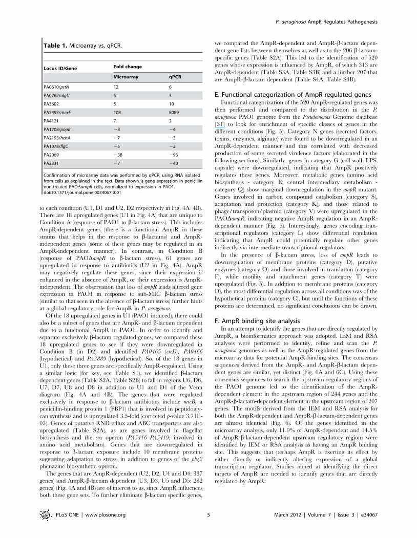
differences in the sensitivity of the two assays (Table 1).
P. aeruginosa AmpR Regulates Pathogenesis
PLoS ONE | www.plosone.org 2 March 2012 | Volume 7 | Issue 3 | e34067

D. AmpR regulates genes both in the absence andpresence of b-lactam stress
Subsets of genes that are differentially regulated either due to
loss of ampR or due to b-lactam antibiotic exposure or both (Fig. 3)
could potentially be regulated under more than one condition and
this overlap would be misinterpreted in the total number of genes
regulated in each condition. To address this issue, the 973
differentially regulated genes (p#0.01, FC$2-fold) in the four pair-
wise comparisons (Conditions A–D in Fig. 3) were plotted in 4-way
Venn diagrams and separated into upregulated (Fig. 4A) and
downregulated (Fig. 4B) genes.
From these two Venn diagrams, genes that were dependent
exclusively on either AmpR or b-lactams or on both were
identified. Comparison of Conditions A and B yield genes unique
Figure 1. Antibiotic resistance profile of PAODampR. A clean in-frame deletion of ampR was generated in P. aeruginosa PAO1 as described inthe methods section to generate PAODampR. Figure 1A shows the resistance profile of the strains to the four major classes of b-lactam antibiotics.Representative data from three different biological replicate trials are shown. The amount of b-lactamase produced was quantified (Fig. 1B) in thepresence (+) and absence (2) of sub-MIC concentration of a b-lactam inducer.doi:10.1371/journal.pone.0034067.g001
P. aeruginosa AmpR Regulates Pathogenesis
PLoS ONE | www.plosone.org 3 March 2012 | Volume 7 | Issue 3 | e34067

Figure 2. Effect of ampR deletion on pathogenicity to C. elegans. The fast killing assay was used to test the effect of loss of ampR on the C.elegans killing ability of PAO1. p-value,0.05 at all time points.doi:10.1371/journal.pone.0034067.g002
Figure 3. Scatter plots of significantly regulated genes. Only genes that showed significant (p#0.01) differential regulation under the variousconditions are depicted as colored squares. The colors represent the extent of gene expression from low (blue) to high (red) in either condition, asdepicted in the color scale. The two outer green diagonal lines in each plot represent the two-fold cutoff. Each sub-plot depicts the differential geneexpression between two strains/conditions (shown along the plot axes): Condition A- PAO1 uninduced vs. PAO1 induced; Condition B- PAODampRuninduced vs. PAODampR induced; Condition C- PAO1 uninduced vs. PAODampR uninduced; Condition D- PAO1 induced vs. PAODampR induced.doi:10.1371/journal.pone.0034067.g003
P. aeruginosa AmpR Regulates Pathogenesis
PLoS ONE | www.plosone.org 4 March 2012 | Volume 7 | Issue 3 | e34067
to each condition (U1, D1 and U2, D2 respectively in Fig. 4A–4B).
There are 18 upregulated genes (U1 in Fig. 4A) that are unique to
Condition A (response of PAO1 to b-lactam stress). This includes
AmpR-dependent genes (there is a functional AmpR in these
strains that helps in the response to b-lactams) and AmpR-
independent genes (some of these genes may be regulated in an
AmpR-independent manner). In contrast, in Condition B
(response of PAODampR to b-lactam stress), 61 genes are
upregulated in response to antibiotics (U2 in Fig. 4A). AmpR
may negatively regulate these genes, since their expression is
enhanced in the absence of AmpR, or their expression is AmpR-
independent. The observation that loss of ampR leads altered gene
expression in PAO1 in response to sub-MIC b-lactam stress
(similar to that seen in the absence of b-lactam stress) further hints
at a global regulatory role for AmpR in P. aeruginosa.
Of the 18 upregulated genes in U1 (PAO1 induced), there could
also be a subset of genes that are AmpR- and b-lactam dependent
due to a functional AmpR in PAO1. In order to identify and
separate exclusively b-lactam regulated genes, we compared these
18 upregulated genes to see if they were downregulated in
Condition B (in D2) and identified PA0465 (creD), PA0466
(hypothetical) and PA3889 (hypothetical). So, of the 18 genes in
U1, only these three genes are specifically AmpR-regulated. Using
a similar logic (for key, see Table S1), we identified b-lactam
dependent genes (Table S2A, Table S2B) to fall in regions U6, D6,
U7, D7, U8 and D8 in addition to U1 and D1 of the Venn
diagram (Fig. 4A and 4B). The genes that were regulated
exclusively in response to b-lactam antibiotics include mrcB, a
penicillin-binding protein 1 (PBP1) that is involved in peptidogly-
can synthesis and is upregulated 3.5-fold (corrected p-value 3.71E-
03). Genes of putative RND efflux and ABC transporters are also
upregulated (Table S2A), as are genes involved in flagellar
biosynthesis and the sox operon (PA5416–PA5419; involved in
amino acid metabolism). Genes that are downregulated in
response to b-lactam exposure include 10 membrane proteins
suggesting adaptation to stress, in addition to genes of the phz2
phenazine biosynthetic operon.
The genes that are AmpR-dependent (U2, D2, U4 and D4: 387
genes) and AmpR-b-lactam dependent (U3, D3, U5 and D5: 282
genes) (Fig. 4A and 4B) are of interest to us, since AmpR influences
both these gene sets. To further eliminate b-lactam specific genes,
we compared the AmpR-dependent and AmpR-b-lactam depen-
dent gene lists between themselves as well as to the 206 b-lactam-
specific genes (Table S2A). This led to the identification of 520
genes whose expression is influenced by AmpR, of which 313 are
AmpR-dependent (Table S3A, Table S3B) and a further 207 that
are AmpR-b-lactam dependent (Table S4A, Table S4B).
E. Functional categorization of AmpR-regulated genesFunctional categorization of the 520 AmpR-regulated genes was
then performed and compared to the distribution in the P.
aeruginosa PAO1 genome from the Pseudomonas Genome database
[31] to look for enrichment of specific classes of genes in the
different conditions (Fig. 5). Category N genes (secreted factors,
toxins, enzymes, alginate) were found to be downregulated in an
AmpR-dependent manner and this correlated with decreased
production of some secreted virulence factors (elaborated in the
following sections). Similarly, genes in category G (cell wall, LPS,
capsule) were downregulated, indicating that AmpR positively
regulates these genes. Moreover, metabolic genes (amino acid
biosynthesis - category E; central intermediary metabolism -
category Q) show marginal downregulation in the ampR mutant.
Genes involved in carbon compound catabolism (category S),
adaptation and protection (category K), and those related to
phage/transposon/plasmid (category V) were upregulated in the
PAODampR, indicating negative AmpR regulation in an AmpR-
dependent manner (Fig. 5). Interestingly, genes encoding tran-
scriptional regulators (category L) show differential regulation
indicating that AmpR could potentially regulate other genes
indirectly via intermediate transcriptional regulators.
In the presence of b-lactam stress, loss of ampR leads to
downregulation of membrane proteins (category D), putative
enzymes (category O) and those involved in translation (category
F), while motility and attachment genes (category T) were
upregulated (Fig. 5). In addition to membrane proteins (category
D), the most differential regulation across all conditions was of the
hypothetical proteins (category C), but until the functions of these
proteins are determined, no significant conclusions can be drawn.
F. AmpR binding site analysisIn an attempt to identify the genes that are directly regulated by
AmpR, a bioinformatics approach was adopted. IEM and RSA
analyses were performed to identify, refine and scan the P.
aeruginosa genomes as well as the AmpR-regulated genes from the
microarray data for potential AmpR-binding sites. The consensus
sequences derived from the AmpR- and AmpR-b-lactam depen-
dent genes are similar, yet distinct (Fig. 6A and 6C). Using these
consensus sequences to search the upstream regulatory regions of
the PAO1 genome led to the identification of the AmpR-
dependent element in the upstream region of 244 genes and the
AmpR-b-lactam-dependent element in the upstream region of 207
genes. The motifs derived from the IEM and RSA analysis for
both the AmpR-dependent and AmpR-b-lactam-dependent genes
are almost identical (Fig. 6). Of the genes identified in the
microarray analysis, only 11.9% of AmpR-dependent and 14.5%
of AmpR-b-lactam-dependent upstream regulatory regions were
identified by IEM or RSA analysis as having an AmpR binding
site. This suggests that perhaps AmpR is exerting its effect by
either directly or indirectly altering expression of a global
transcription regulator. Studies aimed at identifying the direct
targets of AmpR are needed to identify genes that are directly
regulated by AmpR.
Table 1. Microarray vs. qPCR.
Locus ID/Gene Fold change
Microarray qPCR
PA0610/prtN 12 6
PA0762/algU 5 3
PA3602 5 10
PA2493/mexE 108 8089
PA4121 7 2
PA1708/popB 28 24
PA2193/hcnA 27 23
PA1078/flgC 25 22
PA2069 238 293
PA2331 27 240
Confirmation of microarray data was performed by qPCR, using RNA isolatedfrom cells as explained in the text. Data shown is gene expression in penicillinnon-treated PAODampR cells, normalized to expression in PAO1.doi:10.1371/journal.pone.0034067.t001
P. aeruginosa AmpR Regulates Pathogenesis
PLoS ONE | www.plosone.org 5 March 2012 | Volume 7 | Issue 3 | e34067

G. Regulation of the amp genes by AmpRb-Lactam resistance in P. aeruginosa is mediated, in part, by the
amp genes that are also tied in with the cell wall recycling process
[32]. The genes involved in this process include the regulator
AmpR [21,33], the chromosomal b-lactamase AmpC [11,12,
28,34], the permeases AmpG and AmpP [15,35] and the amidases
AmpD, AmpDh2 and AmpDh3 [36,37]. In addition, the
hydrolase NagZ also plays a role in b-lactam resistance [38,39].
Since AmpR is known to positively regulate AmpC expression, we
hypothesized that AmpR also regulates the other amp genes. qPCR
analysis revealed downregulation of the amp genes in PAODampR,
normalized to expression in PAO1 (Fig. 7), indicating a positive
regulatory role for AmpR in the expression of ampG (RQ -
uninduced: 0.5960.01, p-value 0.006; induced: 0.6360.01, p-
Figure 4. Venn diagram of the differentially regulated genes. Distribution of significantly (p#0.01) differentially regulated (.2-fold) genes inPAO1 and PAODampR with (induced) and without (uninduced) b-lactam treatment showing upregulated (Fig. 4A) and downregulated (Fig. 4B)genes. The transcriptional regulators, sigma factors and small RNAs in each group are identified either by their gene names or PA numbers.Annotations are from the Pseudomonas Genome database [31].doi:10.1371/journal.pone.0034067.g004
P. aeruginosa AmpR Regulates Pathogenesis
PLoS ONE | www.plosone.org 6 March 2012 | Volume 7 | Issue 3 | e34067

value 0.04), ampP (RQ - uninduced: 0.160.01, p-value 0.004;
induced- 0.8660.01, p-value NS), ampD (RQ - uninduced:
0.8260.01, p-value NS; induced 0.6760.01, p-value 0.04), ampDh2
(RQ - uninduced: 1.0160.05, p-value NS; induced- 0.5660.01, p-
value 0.002), ampDh3 (RQ - uninduced: 0.9860.007, p-value NS;
induced: 0.6660.04, p-value 0.02) and nagZ (RQ - uninduced:
0.2760.06, p-value 0.002; induced: 0.5160.01, p-value 0.0002).
Specifically, when the cells are exposed to b-lactams, they need
amidase activity to help in the peptidoglycan recycling process.
This is reflected in upregulation of the amidases, (AmpD,
AmpDh2, and AmpDh3) and AmpC only when the cells are
exposed to b-lactams. Simultaneously, AmpG, which functions to
transport the degraded peptidoglycan material into the cytoplasm
[15] is upregulated by AmpR (downregulated in PAODampR) in
an inducer-independent manner. This shows that AmpR upregu-
lates the amidases and AmpC b-lactamase in response to b-
lactams while upregulating AmpG (Fig. 7), and agrees with the
proposed model for peptidoglycan recycling in P. aeruginosa [15].
The data, thus, demonstrates the central role of AmpR in
influencing expression of the cell wall recycling/AmpC-mediated
b-lactam resistance machinery in P. aeruginosa.
H. AmpR regulates the expression of antibiotic resistanceand virulence systems
It has previously been shown that AmpR regulates the
expression of genes related to QS and protease production [40].
Microarray analyses from this study show that AmpR affects the
expression of multiple virulence systems in P. aeruginosa, as
explained further in this section.
Resistance-Nodulation-Division(RND)effluxsystems. RND
transporters are tripartite pumps present in Gram-negative
bacteria that are involved in the efflux of antibiotics and several
other compounds decreasing cytoplasmic retention and thus
conferring resistance [41]. All of the P. aeruginosa strains
sequenced so far carry 12 known and putative RND efflux
pumps, suggesting that the efflux pumps are an integral part of
the P. aeruginosa genome [42,43]. The MexEF-OprN efflux system
that is primarily concerned with resistance to fluoroquinolones,
chloramphenicol and trimethoprim [44] was significantly
upregulated in PAODampR. The genes mexE (seven-fold), mexF
(89-fold) and oprN (103-fold) are overexpressed in the ampR
mutant in the absence of antibiotic stress in microarray studies
(Table S3A) and overexpression of the first gene of the operon,
mexE, was confirmed by qPCR (Table 1). MexT, an activator of
Figure 5. Enrichment of functional categories. Functional categorization of the AmpR-dependent and AmpR-b-lactam dependent genes wasperformed to identify enrichment of specific classes of genes relative to their distribution in PAO1. ‘+’ and ‘2’ refers to upregulation anddownregulation of genes, respectively in either an AmpR-dependent or AmpR-b-lactam-dependent manner. The most differentially regulatedcategories are labeled in the figure using the corresponding code, mentioned in the figure table. Functional categories, and their codes andpercentages in PAO1 are from the Pseudomonas Genome database [31].doi:10.1371/journal.pone.0034067.g005
P. aeruginosa AmpR Regulates Pathogenesis
PLoS ONE | www.plosone.org 7 March 2012 | Volume 7 | Issue 3 | e34067

this efflux system [44,45], is not significantly differentially
regulated (1-fold) in the microarray studies, but is upregulated
in an inducer-independent manner when tested by qPCR (RQ:
uninduced 7.560.11, p-value 0.02; induced 7.060.25, p-value
0.02). However, the negative regulator MexS shows no
differential regulation either in microarray or qPCR analysis
(data not shown). Carbapenems use the outer membrane porin
OprD to gain entry into the cell [46] and MexT negatively
regulates this porin both at the transcriptional and post-
transcriptional level [26,47,48]. Indeed, qPCR analysis shows
that with upregulation of mexT, there is a downregulation of oprD
expression in the ampR mutant compared to PAO1 (RQ:
uninduced 0.3160.004, p-value 0.001; induced 0.0860.000002,
p-value,0.0001). Surprisingly, OprD downregulation did not
lead to increased resistance to imipenem and meropenem
(Fig. 1A). In addition, we did not see differential regulation of
the other known MexT regulator, MvaT [49] in our microarray
analysis.
To further investigate whether upregulation of this pump
translates into a resistance phenotype, we determined the MICs
for MexEF-OprN substrate antibiotics by the standard broth
microdilution method [50]. PAODampR showed enhanced
resistance to four of the antibiotics tested when compared to the
resistance profile of wild-type PAO1, correlating microarray and
qPCR data with the phenotype (Table 2). This suggests that
AmpR is involved in resistance to b-lactam antibiotics by
regulating AmpC as shown earlier, and non-b-lactam antibiotics
via the MexEF-OprN efflux pump. Analysis of the promoter
regions of the genes of this pump and their regulators (MexS and
MexT) using the putative AmpR-binding site [51] as a query
sequence did not reveal signs of AmpR binding, suggesting
indirect regulation by AmpR.
MexAB-OprM was the first RND-type efflux pump to be
reported in P. aeruginosa and has very broad substrate specificity
including b-lactam antibiotics and non-antibiotic substrates [52].
In fact, it has been implicated to play a more significant role in
resistance to b-lactam antibiotics than b-lactamases [53,54]. Using
the putative P. aeruginosa AmpR binding site [51], we identified a
potential AmpR binding site upstream of the MexR repressor of
this pump in the MexR-MexA intergenic region (59 AAGCCTG-
CAAATGT 39) indicating possible regulation of this pump by
AmpR. qPCR analysis of mexR expression revealed downregula-
Figure 6. AmpR binding site analysis. The putative AmpR binding site was refined using IEM algorithm and used to search the upstreampromoter elements of AmpR-regulated and AmpR-b-lactam regulated genes (listed in Tables S3, S4) in PAO1 and other P. aeruginosa strains. DOORdatabase [142,143] was used to identify the operons. The RSAT tool [137] was used to identify PAO1 promoter sequences containing the identifiedAmpR- and AmpR-b-lactam-regulated genes. The output is represented using WebLogo [136] for the AmpR-dependent genes from IEM (A) and RSA(B), and AmpR-b-lactam dependent genes from IEM (C) and RSA (D).doi:10.1371/journal.pone.0034067.g006
P. aeruginosa AmpR Regulates Pathogenesis
PLoS ONE | www.plosone.org 8 March 2012 | Volume 7 | Issue 3 | e34067

tion of this gene in PAODampR compared to PAO1 (RQ:
uninduced 0.4660.006, p-value 0.002; induced 0.460.007, p-
value 0.01). It is thus interesting to note that AmpR not only
positively regulates AmpC b-lactamase but potentially also
MexAB-OprM, two different mechanisms of resistance to b-
lactams. The MexAB-OprM efflux can also pump out fluoro-
quinolones [26] and the enhanced quinolone resistance of
PAODampR seen in the MIC studies is potentially due to a
combined effect of upregulation of the MexEF-OprN and the
MexAB-OprM efflux pumps. In addition, AmpR negatively
regulates a two-gene putative RND efflux operon PA1435–
PA1436 (nine-fold and four-fold respectively in microarray) that
codes for a membrane fusion protein and efflux transporter,
respectively. Potential AmpR regulation of the MexGHI-OpmD
efflux pump is discussed in the QS section.
QS-regulated virulence factors. Many of P. aeruginosa
virulence factors are QS-regulated and form a critical
component of pathogenesis [55,56]. In our previous meta-
analysis of published P. aeruginosa transcriptomes, we identified
differentially regulated sets of system-specific and condition-
specific genes, including QS-regulated genes [57]. Using this as
our knowledge base, the ampR microarray profile was compared to
identify differentially regulated QS-specific genes. The microarray
data shows that AmpR influences expression of many QS-
regulated genes (Table S5).
To further verify AmpR-mediated regulation of QS virulence
factors, we quantified the production of pyocyanin, LasA protease,
and LasB elastase. Pyocyanin is a redox active exotoxin pigment
that contributes to lung pathophysiology of chronic P. aeruginosa
infections [58] and interferes with multiple host cellular functions
[59]. Genes in the locus of the phz1 operon that is involved in QS-
regulated phenazine synthesis, including phzA1 (PA4210, 4-fold
down), phzB1 (PA4211, 21-fold down), phzS (PA4217, 28-fold
down), phzM (PA4209, 4-fold down), and the MexGHI-OpmD
efflux pump (PA4205–PA4208, 11–30-fold down) that plays a role
in pumping out the pigments [60], show decreased expression in
PAODampR. In agreement with this data, there was a statistically
significant (p-value,0.0001) reduction in pyocyanin production by
the ampR mutant, compared to PAO1 and this effect was
independent of b-lactam stress on the cells (Table 3). The data
indicates that AmpR influences pyocyanin production which is in
agreement with the C. elegans killing data (Fig. 2), since phenazines
are major players in C. elegans mortality in the fast-killing assay
[30].
Elastases (pseudolysins) are highly toxic zinc metalloproteases
that play a critical role in immunomodulation [61,62], host tissue
damage aiding invasion [63] and biofilm formation [64]. The
LasB elastase production was also severely affected due to the loss
of ampR (p-value#0.02) in an inducer-independent manner
(Table 3). Along with LasB, a zinc metalloendopeptidase, LasA
plays a major role in P. aeruginosa-induced keratitis [65]. Reduction
in LasA protease production, however, was significantly lower (p-
value,0.05) in PAODampR only when the strains were exposed to
sub-MIC b-lactam stress (Table 3), and this is in agreement with
data from microarray (8-fold downregulated) and qPCR (RQ:
uninduced- NS; induced 0.2160.07, p-value,0.0001) analysis.
Microarray data shows that loss of ampR also affects other QS-
regulated virulence genes, such as the hcn operon PA2193–PA2195
(5 to 7-fold downregulated) that is involved in the production of
hydrogen cyanide. Cyanide toxicity is the primary mode of fast
killing of C. elegans by P. aeruginosa PAO1 [66]. The downregulation
of the hcn operon concurs with reduced killing by PAODampR. The
expression of the galactophilic lectin lecA (PA2570) is also
Figure 7. Expression of the amp genes in PAODampR. RNA was isolated from cells exposed to b-lactam antibiotic, reverse transcribed to cDNAand tested in triplicate by qPCR with gene-specific primers, as described in the text. The expression of ampC (b-lactamase), ampD, ampDh2, andampDh3 (amidases), ampG and ampP (permeases), and nagZ (glycoside hydrolase) were tested in the ampR mutant relative to PAO1. Values havebeen normalized to expression in PAO1 under the same conditions (log10 RQ = 1) and bars above or below the line represent up- and down-regulation, respectively. * p value,0.05.doi:10.1371/journal.pone.0034067.g007
Table 2. ampR deletion affects susceptibility to MexEF-OprNsubstrates.
Antibiotic MIC (mg/ml)
PAO1 PAODampR
Ofloxacin 4 32
Ciprofloxacin 0.25 2
Chloramphenicol 128 512
Trimethoprim 200 .200
The minimum inhibitory concentrations (in mg/ml) of MexEF-OprN substrateantibiotics to PAO1 and PAODampR were determined by broth microdilutionmethod (see text for details).doi:10.1371/journal.pone.0034067.t002
P. aeruginosa AmpR Regulates Pathogenesis
PLoS ONE | www.plosone.org 9 March 2012 | Volume 7 | Issue 3 | e34067

downregulated in PAODampR (RQ: uninduced- 0.2260.0005, p-
value 0.0001; induced: 0.1660.003, p-value 0.0005) indicating
positive AmpR regulation. LecA facilitates bacterial entry into host
cells by aiding in adhesion to endothelia and epithelia [67] and is
involved in biofilm formation [68]. It has previously been shown
that lecA expression is regulated by the sigma factor RpoS [69] and
by the QS regulator RhlR [69]. Thus, the effect of AmpR on lecA
expression could be mediated indirectly via RpoS and/or RhlR.
As predicted, RpoS was downregulated (RQ: uninduced-
0.5560.08, p-value 0.01; induced 0.5160.02, p-value 0.0003),
indicating AmpR positive regulation in a b-lactam-independent
manner. Also, since we see downregulation of multiple QS
phenotypes, RhlR is also potentially involved in AmpR-mediated
lecA regulation.
QS activates the operon PA2327–PA2331 [70] coding for a
probable ABC transporter. Wolfgang et al. found this operon to be
repressed when P. aeruginosa was grown in CF respiratory liquid
containing media [71]. Genes of the operon are significantly
upregulated (6- to 18-fold) in PAODampR indicating that AmpR
negatively regulates this operon and further connects AmpR to QS
regulation, adding another regulatory player in this complex
regulatory network.
Biofilms. Successful biofilm formation is dependent on
nutrient availability, motility and QS [72]. Comparison of gene
expression profiles from this study with the biofilm-specific gene
list generated from our previous analysis of P. aeruginosa
transcriptomes [57] revealed the differential regulation of many
biofilm genes in PAODampR (Table S6). This suggests a role for
AmpR in biofilm regulation, either directly or indirectly. Testing
the tube biofilm-forming ability of the strains revealed that
PAODampR formed better biofilms, compared to PAO1 (Fig. 8).
The difference was significant at all time points tested (p-
value#0.03) over a period of 72 hours indicating that AmpR is
a negative regulator of biofilm formation.
Microarray data also revealed upregulation of PA4651 (encod-
ing a probable pili assembly protein; 11.6-fold), and PA4306
(coding for Flp, Type IVb pilin, 36-fold) in PAODampR, in the
absence of antibiotics. Since type IV pili-mediated twitching and
flagella-mediated swimming motilities are known proponents for
biofilm formation at different stages [73], we tested for these
phenotypes with PAO1 and PAODampR. In the absence of b-
lactam antibiotic stress, the PAODampR strain demonstrated
enhanced twitching ability (PAODampR: 15.1 mm61.1 mm;
PAO1: 2 mm60.1 mm; p value,0.0001) potentially explaining
its ability to form better biofilms. Moreover, unlike in PAO1,
under sub-MIC b-lactam stress, PAODampR showed a marginal
but statistically significant increase in twitching zones (uninduced:
15.1 mm61.1 mm; induced: 16.5 mm60.8 mm; p-value 0.04).
This observation is in agreement with the enrichment of the gene
set for the ‘motility and attachment’ functional category under b-
lactam stress in PAODampR (see section E above). There was,
however, no difference in the swimming motility of the strains
(data not shown), although fleR of the FleSR two-component
system that is involved in the flagella biosynthesis regulatory
pathway [74] was differentially regulated in the ampR mutant (RQ:
uninduced- 0.4560.1, p-value 0.0007; induced- 0.3260.01, p-
value 0.0006).
The Pel polysaccharide is a glucose-rich exopolymer, encoded
by the pel operon (PA3058–PA3064) that along with the mannose-
rich Psl polysaccharide plays a major role in pellicle formation
[75,76]. PAODampR forms darker red colonies on Tryptone-
Congo red agar plates compared to PAO1 (data not shown)
signifying higher Pel production [75]. This is consistent with the
observation that the ampR mutant produces better biofilms.
Collectively, these data suggest that AmpR negatively influences
biofilm formation in P. aeruginosa either directly or indirectly.
Recently, a novel efflux pump that confers antibiotic resistance
in P. aeruginosa biofilms has been identified [77]. Deletion of the
operon (PA1874–1877) encoding this pump in PAO1 enhances
sensitivity to gentamycin, tobramycin and ciprofloxacin, especially
in biofilm cells. Genes in this operon are downregulated four-fold
in the ampR mutant strain. This observation, though not verified
separately, suggests that AmpR positively regulates this operon,
thus possibly contributing to non-b-lactam antibiotic resistance in
biofilms.
Secretory systems. The type III secretion system (T3SS) is
essential for P. aeruginosa not only for contact-dependent toxin
delivery to host cells but also for phagocyte evasion [78,79]. Using
a burn mouse model, it was shown that loss of T3SS results in
reduced virulence [80]. The genes encoding the regulatory and
structural components of the T3SS in P. aeruginosa are
concentrated in one locus (PA1690–PA1725) whereas the
effectors and their chaperones are scattered in the genome [31].
Deletion of ampR from PAO1 led to the downregulation of a few
T3SS genes in the regulatory-structural gene cluster (Fig. 9)
indicating positive AmpR-regulation. These include the genes
encoding the regulators PcrH (involved in regulating ExoS
synthesis) and ExsE (a secreted regulator of the ExsCED
regulatory cascade), and the translocator proteins PopB and
PopD. qPCR confirmation of the microarray findings for the
T3SS genes was done using RNA isolated from Ca2+ depleted
inducing media (MinS-NTA), since Ca2+ is a known inhibitor of
Table 3. Effect of deletion of ampR on QS-regulated virulence phenotypes.
Strain Pyocyanin productiona LasA activityb LasB activityc
Uninduced Inducedd Uninduced Inducedd Uninduced Inducedd
PAO1 22.3160.18 24.3061.10 0.3760.005 0.3960.004 1.6560.25 1.5460.15
PAODampR 2.2960.11e 5.7460.56f 0.2660.002 0.1960.002g 0.560.03h 0.5760.08i
aPyocyanin concentrations were expressed as micrograms of pyocyanin produced per microgram of protein.bLasA activity was expressed as change in OD600 per hour per microgram of protein.cLasB elastase activity was expressed as change in OD495 per microgram of protein.dInduction was carried out with 100 mg/ml of penicillinG.ep-value,0.0001 when comparing PAO1 and PAODampR.fp-value,0.0001 when comparing PAO1 and PAODampR.g,hp-value of 0.02 when comparing PAO1 and PAODampR.ip-value of 0.01 when comparing PAO1 and PAODampR.doi:10.1371/journal.pone.0034067.t003
P. aeruginosa AmpR Regulates Pathogenesis
PLoS ONE | www.plosone.org 10 March 2012 | Volume 7 | Issue 3 | e34067

T3SS [81]. Since pcrH, popB and popD are the last three genes of a
12-gene operon, we tested expression of only pcrH by qPCR,
which showed a downregulation in PAODampR compared to
PAO1 (RQ: uninduced- 0.7160.06, p-value 0.02; induced-
0.8860.03, p-value 0.003). Similarly, exsE, the second gene of
the exsCEB operon was also downregulated in PAODampR (RQ:
uninduced- 0.5760.01, p-value 0.001; induced- 0.6960.18, p-
value NS) indicating positive regulation of these genes by AmpR.
Transcriptional regulation of T3SS in P. aeruginosa is a complex
process and involves multiple tiers of regulation [81]. One of the
mechanisms of control involves a small RNA-binding protein
RsmA and non-coding small RNAs (sRNA), rsmY and rsmZ.
Sequestration of RsmA by the sRNAs inhibits its activity. RsmA
has an extensive virulence regulon that is tied in with the GacSA
two-component system regulatory cascade [82,83]. In PAO-
DampR, rsmZ is downregulated (RQ: uninduced- 0.2160.03, p-
value 0.01; induced- 0.7860.09, p-value NS) and corresponds with
Figure 8. AmpR affects biofilm formation. AmpR regulates many genes involved in biofilm formation (Table S6) that is reflected in the tubebiofilm assay monitored for 72 hours. The strains tested were wild type PAO1, PAODampR, PAODretS (positive control), and PAODpelDpslDalgD(negative control). The inset, taken at 72 hours, demonstrates the superior biofilm formation capacity of PAODampR compared to PAO1. p-valuescomparing PAODampR with PAO1: 24 hrs- 0.002; 48 hrs- 0.03; 72 hrs- 0.007.doi:10.1371/journal.pone.0034067.g008
Figure 9. Regulation of secretion genes by AmpR. Fold changes (FC) as determined by microarray experiments in the absence of b-lactamstress of the Type VI secretion (m, HSI-I) and Type III secretion (&) genes in the ampR mutant strain, normalized to expression in the wild-type strainPAO1. Gene annotations are from the Pseudomonas Genome database [31].doi:10.1371/journal.pone.0034067.g009
P. aeruginosa AmpR Regulates Pathogenesis
PLoS ONE | www.plosone.org 11 March 2012 | Volume 7 | Issue 3 | e34067

an upregulation of RsmA (in microarray, uninduced - 2.3 fold;
corrected p-value 0.009; induced- NS). Some of the T3SS genes
are, however, downregulated in the ampR mutant (Fig. 9). RsmA is
a positive regulator of T3SS. This suggests that the effect of AmpR
on T3SS is probably not via RsmA, or involves an additional step
of post-transcriptional regulation. PtrB represses T3SS gene
expression [84] and since AmpR regulates PtrB expression (see
Section H below), AmpR regulation of T3SS is potentially via
PtrB.
P. aeruginosa T6SS is involved in chronic CF infections [85].
RsmA also negatively regulates genes of the Type 6 secretion
system (T6SS), particularly of the HSI-I system [83]. Thus,
downregulation of genes of the T6SS in PAODampR (Fig. 9) is
possibly through an indirect effect of AmpR on RsmA.
I. AmpR regulates genes found in regions of genomeplasticity (RGP)
Comparative analysis of five P. aeruginosa chromosomes
identified RGPs that are strain-specific [43]. These are genome
segments that can be acquired by horizontal gene transfer,
bacteriophages or transposons [43]. We wanted to determine
whether an endogenous transcriptional regulator, such as AmpR,
could regulate expression of genes acquired by the strain, such as
RGP genes. Microarray analysis revealed that in the PAODampR
mutant, 31 RGP genes are regulated in an AmpR-dependent
manner and an additional eight RGP genes under sub-MIC b-
lactam stress (Table S7).
Most of the RGP03 (PA0612–PA0628) and RGP04 (PA0641–
PA0648) ORFs belong to functional category V (related to phage,
transposon and plasmid) and code for the two classes of high
molecular weight pyocins, types R and F. Pyocins R and F are
related to bacteriophage tails and kill susceptible cells thus
conferring a survival advantage on the producing strain [86].
Thirty-two percent of the RGP genes regulated in an AmpR-
dependent manner are clustered and localized to RGP03. The
expression of 10 RGP03 genes and two RGP04 genes (59% and
20% of genes in RGP03 and RGP04, respectively) are significantly
upregulated (6–8-fold) in PAODampR as seen in microarray data
(Table S7). This suggests that AmpR is involved in negatively
regulating these genes, either directly or indirectly.
PrtN (PA0610), and the product of the first gene in RGP03, ptrB
(PA0612), is a positive regulator of pyocin production, both of
which are repressed by PrtR (PA0611) [87]. Upon DNA damage,
RecA (PA3617) degrades PrtR, thus inducing pyocin production
[87]. In PAODampR subjected to sub-MIC b-lactam stress
compared to PAO1, prtN (12-fold), recA (3-fold) and ptrB (20-fold)
are upregulated, while the negative regulator PrtR is downregu-
lated (qPCR relative quantity: uninduced- 0.8860.4, p-value p-
value 0.004; induced- 0.7660.109 p-value 0.0002) implying that
AmpR is a negative regulator of pyocin production.
J. AmpR regulates other transcriptional regulatorsThe AmpR-regulation of hundreds of genes (Fig. 4A and 4B)
could be by direct binding to the promoters or in a subset of genes,
indirectly through intermediate transcriptional regulators, sigma
factors or regulatory RNAs. In a preliminary attempt to further
elucidate the AmpR regulon, we looked at the transcriptional
regulators and sigma factors that are AmpR-regulated. Of the 430
transcriptional regulators in P. aeruginosa PAO1 [31], 19 met the
selection criteria in our microarray analysis (see materials and
methods) and are over 2-fold significantly differentially regulated
in the ampR mutant (Table 4). This suggests that AmpR regulates a
proportion of the genes through intermediate transcriptional
regulators.
Three of the 24-known/putative sigma factors [88] are also over
2-fold differentially regulated, including RpoS and AlgT/U
(Table 4). The stationary phase/stress sigma factor, RpoS controls
virulence in different bacteria including P. aeruginosa [89,90,91].
Since rpoS expression is upregulated in the stationary phase [88,90]
and the RNA used for microarray analysis was harvested two
hours post-b-lactam induction (at OD ,3.0), some of the
phenotypic changes seen in PAODampR is likely to be RpoS-
mediated. To investigate this possibility, RNA was harvested
40 minutes post-induction (OD600 of ,1.0) and the expression of
known RpoS-dependent and RpoS-independent genes was
compared between PAO1 and PAODampR. As expected, RpoS
expression was significantly higher at 2 hours than at 40 minutes
in PAODampR compared to PAO1 (p-value: uninduced 0.0049,
induced 0.0023; Fig. 10), and this increase in RpoS expression
corresponded with a growth phase-dependent increase in the
expression of two of the RpoS-dependent genes, lecA (p-value:
uninduced 0.0061; induced 0.0043) and lecB (p-value: uninduced
NS; induced 0.0002) (Fig. 10). However, expression of the MexEF-
OprN activator, MexT, which is regulated in an RpoS-
independent manner, did not change at the different time points
tested (Fig. 10). This suggests that AmpR, via RpoS, regulates
genes of the RpoS regulon in P. aeruginosa in a growth phase- and
stress-dependent manner, which is in agreement with previous
studies [90,91]. Moreover, the MexT data suggests that harvesting
the cells either after 40 minutes or 2 hours of b-lactam treatment
does not affect AmpR-mediated gene expression for non-RpoS-
dependent genes (Fig. 10).
The sigma factor AlgT/U is a master regulator of alginate
biosynthesis [92,93] and we have shown previously with an ampR
insertion mutant that there is crosstalk between AmpR and AlgT/
U [40]. Our microarray data shows upregulation of AlgT/U in
PAODampR (5.4-fold, corrected p-value 3.35E-03), indicating
negative AmpR regulation (Table 4) in agreement with previous
findings [40].
K. Subtractive transcriptomics of the AmpR regulonMeta analysis of 18 condition-specific P. aeruginosa transcrip-
tomes led to the identification of an expression core gene set of 303
genes that are significantly differentially regulated under all the
different conditions analyzed and were proposed to be involved in
maintaining cell homeostasis [57]. Comparing the 520 genes
whose regulation is AmpR-dependent to the core led to the
identification of 57 genes (of the 520) that were part of the
expression core genes (Fig. 11). Further, we wanted to identify
genes that are specifically under AmpR-regulation and those that
are not involved in other pathways. To derive this list, we
compared the 463 AmpR-dependent genes (520 minus 57 core
genes) with the 1726 genes that are regulated in other conditions/
by other regulators (1598 condition-specific genes [57], and 128
genes that are either RpoS [94] or AlgT/U-regulated [95]). This
comparison reduced the number of AmpR-dependent genes to
133 (from 313) and the AmpR-b-lactam-dependent genes to 86
(from 207) (Fig. 11; Table S8).
The exclusively AmpR-dependent genes include the O antigen
chain regulator wzz, the pca genes involved in carbon compound
catabolism and creB, which codes for the response regulator of the
CreBC TCS (Table S8). The CreBC TCS has been demonstrated
to be involved in b-lactam resistance in Aeromonas spp. [96]. CreBC
positively regulates expression of an inner membrane protein
CreD in E. coli [97] and in P. aeruginosa the CreBCD system
contributes to b-lactam resistance only in a DPBP4 background
[98]. Microarray analysis of PAODampR in the presence b-lactam
stress shows reduced creB expression (22.1, corrected p-value 8.4E-
P. aeruginosa AmpR Regulates Pathogenesis
PLoS ONE | www.plosone.org 12 March 2012 | Volume 7 | Issue 3 | e34067

03). Expression of creD, however, is significantly increased in
PAODampR compared to PAO1 under b-lactam stress as
determined by qPCR (RQ: induced 11.0460.0001, p-val-
ue,0.0001). This is in agreement with a previous report that also
showed creD overexpression in cefoxitin-treated PAODampR cells
[98]. AmpR thus positively regulates creB while negatively
regulating creD expression suggesting potential AmpR regulation
of creD in a CreB-independent manner.
L. Phenotypic microarray analysis of PAODampRSince loss of ampR led to dysregulation of over 500 genes as seen
in DNA microarrays, we decided to characterize its metabolic
effect using phenotypic microarrays. Biolog analysis was per-
formed with PAO1 and PAODampR in the absence of antibiotic
stress. In all, seven phenotypes were gained and 47 phenotypes
were lost (Table S9). PAODampR grew marginally better in media
containing nutritional supplements including citrulline, histidine,
shikimic acid, leucine, serine, spermidine and pyridoxal. This
indicates that AmpR is a negative regulator of utilization of these
agents. Of the 47 observed phenotypes that were lost in
PAODampR, 45 belong to the sensitivity panel. Fourteen of the
45 observed phenotypes were associated with antibiotics, further
supporting the role of AmpR as a major regulator of antibiotic
resistance in P. aeruginosa.
Discussion
P. aeruginosa AmpR, a LysR-type transcriptional regulator and a
positive regulator of the chromosomal ampC b-lactamase expres-
sion, has been shown previously to play a role in regulating a few
QS-dependent phenotypes and the alginate master regulator,
AlgT/U [21,40]. In this study, we determined the whole genome
expression profiles of a clean in-frame deletion mutant of ampR in
P. aeruginosa PAO1 under normal conditions and under sub-MIC
b-lactam stress, using DNA microarrays. The results demonstrate
that AmpR influences the expression of 313 genes in the absence
of b-lactam stress and an additional 207 genes when exposed to
sub-MIC b-lactam stress. The AmpR regulon is thus much more
Table 4. Transcriptional regulators and sigma factors regulated by AmpR.
Transcriptional regulators
Locus Tag Gene Name Product Name Corrected p-value Fold change
PA0463 creB two-component response regulatorCreB
8.39E-03 22.1
PA0479 probable transcriptional regulator 9.90E-03 22.1
PA0610 prtN transcriptional regulator PrtN 3.81E-03 11.6
PA0611 prtR transcriptional regulator PrtR 6.10E-03 23.9
PA0612 ptrB repressor, PtrB 3.44E-03 19.5
PA1196 probable transcriptional regulator 9.89E-03 2.6
PA1663 probable transcriptional regulator 5.13E-03 23.9
PA1707 pcrH regulatory protein PcrH 3.98E-03 23.8
PA2281 probable transcriptional regulator 9.89E-03 22.1
PA2588 probable transcriptional regulator 3.77E-03 227.9
PA3508 probable transcriptional regulator 1.60E-03 3.5
PA3604 probable two-component responseregulator
6.96E-03 22.2
PA4296 pprB two-component response regulator,PprB
6.01E-03 7.4
PA4781 probable two-component responseregulator
9.10E-03 2.8
PA4853 fis DNA-binding protein Fis 6.65E-03 23.3
PA5059 probable transcriptional regulator 6.33E-03 4.0
PA5105 hutC histidine utilization repressor HutC 8.27E-03 23.1
PA5380 gbdR GbdR 5.75E-03 2.6
PA5483 algB two-component response regulatorAlgB
7.83E-03 2.4
Sigma factors
Locus Tag Gene Name Product Name Corrected p-value Fold change
PA0762 algT/U sigma factor AlgU 3.35E-03 5.4
PA1300 probable sigma-70 factor, ECFsubfamily
9.59E-03 2.5
PA3622 rpoS sigma factor RpoS 6.65E-03 2.2
AmpR influences the expression of other transcriptional regulators and sigma factors in P. aeruginosa PAO1, as seen in the microarray analyses. A negative sign in thefold change column indicates downregulation. ORF annotations are from the Pseudomonas Genome database [31].doi:10.1371/journal.pone.0034067.t004
P. aeruginosa AmpR Regulates Pathogenesis
PLoS ONE | www.plosone.org 13 March 2012 | Volume 7 | Issue 3 | e34067

extensive than previously thought including virulence, antibiotic-
resistance and metabolic genes.
Multi-drug resistant P. aeruginosa isolates are a frequent
occurrence in many acute and chronic infections [99]. b-
lactamases and efflux pumps are major mediators of antibiotic
resistance in P. aeruginosa [26]. We show that in addition to
positively regulating the ampC and potentially the MexAB-OprM
efflux pump by modulating expression of the MexR repressor,
AmpR also mediates non-b-lactam resistance by negatively
regulating the MexEF-OprN efflux pump. The PAO1 strain used
for constructing PAODampR was the strain used in the genome-
sequencing project [42], which has an 8 bp insertion in the
MexEF-OprN activator mexT leading to premature mexT termi-
nation [100] and consequent non-inducibility of the MexEF efflux
pump. Strains with a nfxC mutation, however, have different ways
to overcome this, including secondary mutations and deletion of
the 8 bp insertion [45,100], leading to activation of the MexEF-
OprN efflux. This was also observed in nfxC mutants isolated in a
mouse model [101]. However, there was no differential expression
of the MexT activator in both these studies [100,101]. With
PAODampR, even though mexT expression is upregulated in a b-
lactam-independent manner, this will still not be able to overcome
Figure 10. Gene expression in PAODampR at 40 minutes and 2 hrs post-b-lactam exposure. RNA was isolated from PAO1 and PAODampRcells exposed to b-lactams for either 40 minutes or 2 hours and reverse transcribed to cDNA. The expression of the sigma factor rpoS, lecA and lecB(galactophilic lectin genes known to be RpoS-regulated), and mexT (MexEF-OprN efflux pump regulator that is not RpoS-regulated) were tested byqPCR with gene-specific primers, as described in the text. Values have been normalized to expression in PAO1 under the respective conditions.** p-value,0.006; *** p-value 0.0002 as determined by unpaired t test.doi:10.1371/journal.pone.0034067.g010
Figure 11. Comparison of the AmpR transcriptome with othertranscriptomes. AmpR-dependent and AmpR-b-lactam-dependentgenes were compared with the 303 genes of the expression core andthe 1726 condition-specific genes identified previously as part of ameta-analysis of 18 P. aeruginosa transcriptomes [57].doi:10.1371/journal.pone.0034067.g011
P. aeruginosa AmpR Regulates Pathogenesis
PLoS ONE | www.plosone.org 14 March 2012 | Volume 7 | Issue 3 | e34067

the effect of the 8 bp deletion. It has, however been suggested that
there is a putative LacI-like repressor binding site in the mexT-mexE
intergenic region [102] and that there is a second repressor that
binds this site regulating expression of mexEF-oprN [103]. This
suggests that the LTTR AmpR potentially regulates this LacI-type
repressor, leading to induction of the MexEF-OprN pump in a
MexT-independent manner. The outer membrane porin OprD
serves as a conduit for the entry of carbapenems into the cell [46].
Although we see decreased expression of oprD, the strain is still
sensitive to imipenem and meropenem, which is contrary to
expectation. However, our finding is in agreement with a previous
observation where mexEF-oprN overexpressing strains showed no
altered imipenem susceptibility [104], the associated mechanism
remains to be elucidated. Previous studies have also demonstrated
an inverse correlation between b-lactam resistance and biofilm
formation, both in vitro and in CF isolates [105,106,107]. Our data
supports these findings as far as b-lactam antibiotics are
concerned, since AmpR positively regulates production of AmpC
b-lactamase while negatively regulating biofilm formation. How-
ever, negative regulation of the MexEF-OprN efflux (providing
resistance to fluoroquinolones, chloramphenicol and trimetho-
prim) by AmpR (Table 2, Fig. 12) suggests that the antagonistic
regulation of antibiotic resistance and biofilm formation is
dependent on the class of antibiotics. The physiological advantage
to the bacteria in this context is unclear. Co-regulation of b-lactam
and fluoroquinolone resistance by AmpR is significant in itself,
since this puts AmpR among one of the few proteins that regulates
resistance to multiple classes of antibiotic [108]. In addition, since
fluoroquinolones are part of the current systemic antibiotic
treatment regimen for P. aeruginosa infections [99], this finding
could potentially have important therapeutic implications.
QS is at the heart of the virulence regulatory network in P.
aeruginosa with multiple regulators feeding into the regulation
process [109]. We have shown previously that AmpR is also part
of the QS regulatory process and regulates production of proteases
and pyocyanin [21,40], but the determined extent of the
regulation was limited due to the experimental approaches
adopted. Using whole genome transcriptome, we show here that
the AmpR-influenced QS regulon is much more extensive than
previously thought (Table S5). QS regulated phenotypes, such as
pyocyanin and protease production are positively regulated by
AmpR in the current study (Fig. 12), which is in contrast to that
seen in our previous analyses [21]. We believe that this difference
stems from the fact that in our previous studies, we used an
insertion mutant (aacC1 cassette inserted into the PstI site of ampR)
whereas in this study, we have used a clean in-frame deletion
mutant of ampR. One potential reason for the discrepancy may be
that the gentamycin cassette insertion at the PstI site (554 bases
into the ampR coding region) [21] did not disrupt the N-terminus
HTH motif of AmpR. Since LysR members are known to bind
DNA even in the absence of inducer signals [110], this intact HTH
motif might have somehow interfered with the regulatory process.
The P. aeruginosa PAO1 insertion ampR mutant in the previous
study produced more pyocyanin compared to the wild-type PAO1
[21]. In contrast, in this study, we find that PAODampR produces
significantly lower amounts of phenazines as compared to PAO1
(Table 3). The differences in phenazine production were also
translated into differential susceptibilities in the C. elegans paralytic
assays (Fig. 2; [40]) since phenazines are one of the major
contributors to C. elegans toxicity in this assay [111] explaining the
reduced killing of C. elegans with this strain (Fig. 2). Furthermore,
with PAODampR, the microarray, qPCR and phenotypic data
concur, and support our current findings.
Figure 12. AmpR is a master regulator of gene expression in P. aeruginosa PAO1. AmpR positively regulates resistance to b-lactamantibiotics by upregulating expression of the amp genes, nagZ and downregulating creD. In addition, AmpR affects fluoroquinolone resistance bynegatively regulating expression of mexT, the positive regulator of the MexEF-OprN efflux pump. Expression of the virulence and stress responsesigma factor, RpoS and QS-regulated acute virulence factors is downregulated in PAODampR, indicating positive AmpR regulation. AmpR alsonegatively regulates biofilm formation via an unknown mechanism. AmpR modulates levels of the small RNA rsmZ, whose levels are lower inPAODampR with a corresponding enhanced expression of RsmA. Downregulation of some of the T3SS genes in the ampR mutant is possibly byregulating ptrB expression, via PrtR. Further, two major regulators of the alginate biosynthetic pathway, AlgT/U and AlgB are negatively regulated byAmpR, thereby potentially also regulating alginate production. Whether these AmpR interactions are direct or indirect needs to be investigated.doi:10.1371/journal.pone.0034067.g012
P. aeruginosa AmpR Regulates Pathogenesis
PLoS ONE | www.plosone.org 15 March 2012 | Volume 7 | Issue 3 | e34067
The MexGHI-OpmD (PA4205–PA4208) efflux pump is
involved in the efflux of acriflavin, ethidium bromide, novobiocin,
rhodamine, and vanadium, and in maintaining QS homeostasis
[60]. Mutants of mexI and opmD have been demonstrated to be
impaired in QS-related phenotypes including swarming motility,
production of elastase, rhamnolipids, pyocyanin and pyoverdine
[60]. This pump is downregulated in PAODampR indicating
positive AmpR regulation and correlates with some of the
observed phenotypes, such as decreased production of elastase
and pyocyanin. Furthermore, SoxR is known to regulate this
pump [112], but is not differentially regulated in the microarray
data.
AlgR regulates the hcnABC operon genes (PA2193–PA2195)
involved in hydrogen cyanide synthesis in P. aeruginosa [113]. In the
ampR mutant, these genes are downregulated five- to seven-fold
without a corresponding significant differential regulation of algR
(,two-fold). AmpR, however, negatively regulates AlgT/U (5.4-
fold upregulated in PAODampR in the absence of antibiotics) in
agreement with previous findings [40], and AlgT/U regulates algR
[114,115]. Thus the regulation of the hcnABC operon in
PAODampR is potentially through AlgT/U-mediated regulation
of AlgR (Fig. 12). Moreover, RpoS, in conjunction with the Gac-
Rsm regulatory system, has been shown to regulate oxidative
stress-mediated resistance in P. fluorescens [116]. In our study, both
RpoS and the sRNA, rsmZ are regulated in an AmpR-b-lactam-
dependent and AmpR-dependent manner, respectively. This
could also be a potential mode of regulation of the hcn operon in
PAODampR, provided the regulatory mechanism is conserved in P.
aeruginosa. AmpR also negatively influences expression of the
galactophilic lectin LecA (PA2570) (Fig. 10) that is RpoS and QS-
regulated. LecA has been shown to be critical to the gut
pathogenicity of P. aeruginosa and to enhance cytotoxic effects of
exotoxins by inducing a permeability defect [117]. These results
further support the hypothesis that AmpR regulates virulence in P.
aeruginosa.
P. aeruginosa produces bacteriocins called pyocins that kill
susceptible cells through either pore-formation and subsequent
leakage of cytoplasmic contents, or by endonuclease activity [86].
The two high molecular weight pyocins, types R and F, are
proposed to be remnants of lysogenic phages and resemble phage
tails. Like lysogenic phages, they are induced in response to DNA
damage, linked to the RecA-mediated SOS response [118] and
other stress conditions such as exposure to hydrogen peroxide
[119] or ciprofloxacin [120]. AmpR influences the expression of
many of these genes including the regulators PrtR, PtrN, PtrB, and
the SOS response mediator RecA (Table 4; section I in results).
Specific and significant differential regulation of these genes under
b-lactam stress in PAODampR implies a role for AmpR in
influencing expression of these genes under stress conditions.
Moreover, pyocin production confers a survival advantage by
killing neighboring susceptible cells and 97% of the P. aeruginosa CF
isolates tested showed bacteriocin-like killing activity [121]. The
pyocin genes are located in RGP03 and RGP04 and are acquired
by P. aeruginosa [43]. Though some RGP loci contain regulators
that control expression of RGP genes [122], it is interesting to note
that an endogenous regulator such as AmpR is able to regulate
acquired genes and highlights the transcriptional versatility of P.
aeruginosa.
Negative regulation of the genes involved in biofilm formation
and pyocin biosynthesis by AmpR (Fig. 12) fits with the profile of
AmpR as a negative regulator of chronic infection phenotypes and
positive regulator of acute infection, as seen with the QS-regulated
phenotypes. RsmA, a small RNA-binding protein, is a global
regulator of virulence in P. aeruginosa and is intricately tied in with
the GacSA TCS [82,83]. Two small RNAs, rsmZ and rsmY, which
bind and sequester RsmA, keep RsmA activity in check in
Pseudomonas and other bacteria [83,123,124,125,126]. In P.
aeruginosa, RsmA positively regulates T3SS and negatively
regulates biofilm formation [127,128]. AmpR positively regulates
rsmZ (downregulated in PAODampR), resulting in downregulation
of RsmA (upregulated in PAODampR). PAODampR forms
enhanced biofilms (Fig. 8) and shows decreased production of
QS regulated phenotypes compared to PAO1 (Table 3). These
data suggest that regulation of some of the phenotypes seen in an
ampR mutant is not via modulating the activity of rsmZ and
consequently RsmA. However, analysis of the rsmZ promoter for
potential AmpR binding sites using a previously identified putative
AmpR binding site [51] revealed a weak AmpR consensus (59
CCCGCGCCTTTTGT 39). The possibility of direct AmpR
regulation of rsmZ remains to be elucidated.
In conclusion, unlike AmpR found in other Enterobacteriaceae
members, the targets of P. aeruginosa AmpR are widely distributed
in the genome and consist of over 500 genes. Since the genes
regulated include transcriptional regulators, sigma factors and
non-coding RNAs, a subset of these genes are possibly regulated
indirectly by AmpR via intermediate regulators. It is also
interesting to note that AmpR regulates genes that have been
acquired by horizontal gene transfer, which reflects on the
transcriptional versatility of P. aeruginosa. Although the exact
AmpR regulatory mechanisms are as yet unclear, this study
highlights the complexity and multi-tiered regulatory processes by
which P. aeruginosa controls expression of genes of diverse
functions. Teasing apart the AmpR regulatory network will
involve differentiating direct and indirect AmpR-regulated genes
that will advance our understanding of how this bacterium
regulates multiple different pathogenesis and metabolic mecha-
nisms. This understanding will potentially lead to identifying
mechanistic targets that can help in dealing with intractable P.
aeruginosa infections, and other bacterial pathogens that harbor
similar systems.
Materials and Methods
Bacterial strains, nematodes, media and primersAll experiments were performed with either the wild type or
derivatives of P. aeruginosa PAO1 [42] and Escherichia coli DH5acultivated in Luria Bertani (LB) broth (Difco, USA) incubated at
37uC unless specified otherwise. The strains and plasmids used in
this study are listed in Table 5. Nematode assays were performed
with Caenorhabditis elegans strain N2 (Bristol) and E. coli OP50
obtained from the Caenorhabditis Genetics Center, which is
funded by the NIH National Center for Research Resources
(NCRR). E. coli OP50 was grown in nematode growth medium
(1.7% agar, 0.35% peptone, 0.34% K2HPO3, 0.3% NaCl, 0.012%
MgSO4, 0.011% CaCl2, 0.0005% cholesterol) whereas the P.
aeruginosa strains used in the nematode assays were grown on brain-
heart infusion agar (Difco, USA). Biofilm assays were performed in
T-broth (tryptone 10.0 g/L, NaCl 5.0 g/L). T-agar plates were
prepared by addition of 1% agar to T-broth. MinS-NTA minimal
media [129] was used for RNA isolation to determine expression
of T3SS genes. Cation-adjusted Mueller Hinton broth and agar
(Difco, USA) was used in MIC assays. For pyocyanin production,
strains were cultivated in King A medium (Difco, USA).
Antibiotics were supplemented, when needed, at the following
concentrations for E. coli: ampicillin (Ap) 100 mg/ml, gentamycin
(Gm) 15 mg/ml, tetracycline (Tc) 15 mg/ml; for P. aeruginosa: Gm
75 mg/ml, Tc 60 mg/ml. Primers used in this study are listed in
Table S10.
P. aeruginosa AmpR Regulates Pathogenesis
PLoS ONE | www.plosone.org 16 March 2012 | Volume 7 | Issue 3 | e34067

Construction of deletion mutants, complementationclones
An unmarked ampR null mutant of P. aeruginosa was generated
by gene splicing [130] using primers KMampRUF1 and KMam-
pRUR1 (to generate the upstream product P1, flanked by EcoRI
and NheI sites), and primers KMampRDF1 and KMampRDR2 (to
generate the downstream product P2, flanked by NheI and BamHI
sites). After sequencing to ensure absence of mutations, P1 and P2
were spliced together to obtain a 1520 bp deletion fragment of
ampR containing stop codons in all three reading frames at their
junction (inserted as part of the NheI sites in the primer). This was
then sequenced and subcloned into a P. aeruginosa non-replicative
plasmid pEXG2 [131] as an EcoRI-BamHI fragment and moved
into PAO1 by allelic exchange [132] using pRK600 [133] as the
helper plasmid. Double crossover mutants were selected for the
loss of plasmid (gentamycin-sensitive, sucrose counter-selection).
The presence of deletion in PAODampR was confirmed using
standard molecular methods (PCR and restriction analysis of
amplicons) and biological assays (antibiotic sensitivity pattern and
b-lactamase assays). Complementation of the ampR deletion was
achieved by amplifying the ampR ORF along with the ampR-ampC
intergenic region using primers DBS_ampRF2 and DBS_ampRR2
and cloning into pCR2.1 TOPO using TA cloning technique.
After confirming absence of mutations by sequencing, the 944 bp
ampR ORF was moved into pMMB67EH-Gm [134] as an EcoRI-
SacI fragment. The plasmid was then moved into PAODampR by
electroporation [135], selecting for gentamycin-resistant colonies.
C. elegans virulence assayThe P. aeruginosa - C. elegans standard paralysis assay was
modified from previous protocols [66]. Overnight bacterial
cultures were diluted 1:1000 and plated onto brain heart infusion
agar plates. The plates were incubated for 18–24 hours at 37uCfor the formation of bacterial lawns. Meanwhile, a synchronized
culture of L4 stage larvae hermaphrodite Bristol N2 C. elegans was
washed off E. coli OP50-seeded nematode growth medium plates
using M9 media. The nematodes were centrifuged at 13006g for
two minutes and washed twice with M9 medium to remove
residual E. coli bacteria. Thirty to 35 nematodes were then added
to each of the P. aeruginosa bacterial lawns. Both live and paralyzed
nematodes were scored at two-hour intervals for eight hours via
microscopic observation. Nematodes were considered dead when
they did not respond to physical stimuli. Each strain was tested in
triplicate.
Table 5. Strains and plasmids used in this study.
Strain/plasmid Relevant characteristics Source
Strains
Escherichia coli
DH5a General purpose cloning strain; D(lacZ)M15 New England Biolabs
OP50 Food source for culturing C. elegans Caenorhabditis Genetics Center
DBS7 DH5a harboring pEXG2 carrying the 1520 bp DampR fragment (pDBS7) This study
DBS220 DH5a harboring the 944 bp promoterless ampR ORF cloned into pCR2.1 TOPO(pDBS220)
This study
DBS271 DH5a containing the 944 bp ampR ORF subcloned as a EcoRI-SacI fragmentfrom pDBS220 into pMMB67EHGm
This study
Pseudomonas aeruginosa
PAO1 Wild-type [42]
PKM314 PAO1 with pMMB67EH-Gm (empty vector) This study
PKM315 PAODampR; In-frame deletion of ampR (PA4109) This study
PKM316 PAODampR with pMMB67EH-Gm (empty vector) This study
PKM317 PAODampR (pAmpR); ampR ORF on pMMB67EH-Gm (pDBS271) moved intoPAODampR; IPTG-inducible; GmR; ampR complementing clone
This study
PAOretS::aacC1 Gm insertion mutant of retS; hyper biofilm-former S. Lory
PAODpelDpslDalgD In-frame triple deletion of the algD ORF and pel and psl operons; biofilm non-former S. Lory
Staphylococcus aureus
DBS116 Wild type S. aureus used for LasA assays Lab collection
Plasmids
pCR2.1 TOPO TA cloning vector for PCR products; ApR, KmR; ColE1 f1 ori lacZa Invitrogen
pRK600 Helper plasmid for conjugation [133]
pEXG2 Allelic exchange vector; GmR, colE1 ori, oriT, sacB [131]
pMMB67EH-Gm GmR; IncQ, RSF1010, lacIq Ptac expression vector [134]
pDBS7 pEXG2 with the DampR fragment, flanked by EcoRI and BamHI sites This study
pDBS220 pCR2.1 TOPO containing the 944 bp ampR ORF, PCR amplified from the PAO1genome using DBS_ampRF2 and DBS_ampRR2
This study
pDBS271 pMMB67EH-Gm containing the ampR ORF, subcloned as an EcoRI-SacI fragmentfrom pDBS220
This study
doi:10.1371/journal.pone.0034067.t005
P. aeruginosa AmpR Regulates Pathogenesis
PLoS ONE | www.plosone.org 17 March 2012 | Volume 7 | Issue 3 | e34067

RNA isolation, generation of cDNA probes, microarrayexperiments and data analysis
P. aeruginosa strains PAO1 and PAODampR, with and without b-
lactam antibiotic treatment were used for RNA extraction. The
cells were subcultured at 37uC, 300 rpm from overnight cultures
grown in LB broth to an OD600 of 0.01. When the cells reached
an OD600 of 0.5–0.6, they were split into two flasks. One of the
flasks was treated with sub-MIC concentration of penicillin
(100 mg/ml penicillin G Potassium salt, Fisher Bioreagents) for
two hours. The RNA was stabilized by addition of phenol-ethanol
mixture [82] prior to extraction (RNeasy Mini Kit, Qiagen)
following manufacturer protocols. Residual DNA was removed
using RQ1 RNase-free DNase (Promega) and the RNA repurified
using the kit. After quantification by measuring absorbance at
260 nm (Beckman DU640, Beckman Coulter), and preliminary
quality check on a denaturing agarose gel (NorthernMax Gly,
Ambion), the RNA integrity was analyzed using RNA Nano chips
in an Agilent Bioanalyzer 2100. cDNA was then synthesized by
annealing NS5 random primers to total purified RNA, spiked with
Bacillus subtilis internal control RNAs and subsequent extension
carried out using SuperScript III reverse transcriptase (Invitrogen).
Fragmentation of cDNA was performed using RQ1 DNase
(Promega), and the fragments were biotin labeled. Hybridization
with GeneChip P. aeruginosa genome array (Affymetrix) and
scanning were performed according to manufacturer specifica-
tions.
Data analyses from three biological replicates for each of the
conditions tested were performed after normalizing and summa-
rizing probe level measurements using Guanine Cytosine - Robust
Multiarray Average (GC-RMA). Only genes that fit stringent
criteria (expression cutoff: 50–100% stringency; p-value#0.01 of
one-way ANOVA data corrected by Benjamini Hochberg FDR;
fold-change $2.0) were selected for further analysis. All micro-
array data analysis was performed using GeneSpring GX 10.0
Expression Analysis software (Silicon Genetics). The microarray
data is MIAME compliant and has been deposited in NCBI GEO
(accession number GSE33188).
Bioinformatics analysis of the AmpR-binding siteThe promoters of AmpR-dependent and AmpR-b-lactam-
dependent genes (listed in Tables S3 and S4) were used to refine
the AmpR binding motif using the Iterative Enhancement of
Motifs (IEM) algorithm [51]. The AmpR binding site in the
promoter of ampC (59-TCTGCTCCAAATTT-39) was used to
search the AmpR-dependent or AmpR-b-lactam-dependent
promoters from P. aeruginosa PAO1 and their orthologs from P.
aeruginosa strains PA14, PA2192, C3719 and PACS2. The output
of IEM was a motif matrix. WebLogo [136] was used to
graphically represent the multiple sequence alignment of the
output. The Regulatory Sequence Analysis Matrix Scan Tool
(RSA) [137] was then used to identify PAO1 promoter sequences
containing the identified AmpR-dependent or AmpR-b-lactam-
dependent motifs in the respective gene sets. The RSA output was
then used to generate a WebLogo for the gene sets.
Quantitative real-time PCRSpecific genes that were significantly, over two-fold up or down
regulated between P. aeruginosa PAO1 and PAODampR as seen in
microarray experiments were verified by qPCR. Total RNA
isolation and reverse transcription into cDNA was as described for
the microarrays without addition of spike transcripts, fragmenta-
tion or labeling. For qPCR, the ABI Step One (Applied
Biosystems) cycler was used with PowerSYBR Green PCR
MasterMix with ROX (Applied Biosystems). Expression was
normalized to clpX (PA1802), whose expression was determined
to remain constant between the samples and conditions tested.
Assays were performed at least in biological triplicate, each with
technical triplicates, for every gene analyzed. Melt curves were
determined to ensure primer specificity. The cycling conditions
used were 95uC/2 minutes (holding); 40 cycles of 95uC/15 sec,
60uC/1 min (cycling); 95uC/15 sec, 60uC/1 min, 95uC/15 sec
(0.6uC ramp) (melt curve). Differential regulation of the T3SS
genes, including rsmZ was determined using RNA isolated from
cells grown in MinS-NTA inducing media [129] from PAO1 and
PAODampR, essentially as described above.
Gene expression in PAODampR were normalized to the
corresponding PAO1 values, for both the b-lactam uninduced
and induced conditions and is presented as relative expression in
PAODampR 6 standard error.
Determination of minimum inhibitory concentration(MIC)
MICs were determined one of two ways. For testing the MIC of
b-lactams, E-test was used following manufacturer protocols
(BioMerieux). The MICs of the MexEF-OprN efflux pump
substrates (ofloxacin, chloramphenicol, ciprofloxacin, and trimeth-
oprim) were determined by standard broth microdilution method
[50]. Briefly, serial two-fold dilutions of the antibiotics were
incubated at 37uC for 16–18 hrs with 56105 CFU/ml of bacteria
in a total volume of 200 ml in 96-well flat bottom polystyrene
plates. The highest dilution of antibiotic that prevented bacterial
growth was considered as the MIC. The assays were performed at
least in triplicate, each with technical triplicate, for each antibiotic
in cation-adjusted Mueller Hinton broth.
Quantifying b-lactamase activityb-lactamase activity was quantified as described previously [21].
Briefly, cells in LB broth at an OD600 of 0.5–0.6 were treated with
100 mg/ml Penicillin G for two hours at 37uC. The cells were then
harvested, OD normalized, and lysed with BugBuster Protein
Extraction Reagent (Novagen) and r-Lysozyme (Novagen), and
treated with Benzonase nuclease (Novagen). The amount of b-
lactamase was quantified in the soluble fraction by determining
hydrolyzing activity on nitrocefin (Oxoid, England). Protein
concentrations in the samples were determined by Bradford assay.
Enzyme activity was expressed as milliunits of b-lactamase
(nanomoles of nitrocefin hydrolyzed per minute per microgram
of protein).
Biofilm assaysTime course biofilm assays were performed in 12675 mm
round-bottom glass culture tubes (VWR) as described previously
[75]. Briefly, fresh overnight T-agar plate cultures of the test
strains were scraped into 1 ml T-broth and diluted to a final
OD600 of 0.0025. Aliquots of 1 ml per tube were made at time
zero and incubated static at room temperature for 24, 48, 72 and
96 hours. To assay for pellicle formation, the tubes were washed
with running tap water five times, after discarding the cultures and
stained with 1% crystal violet for 20 minutes. After pouring off the
dye, the tubes were washed thoroughly with running tap water 10
to 15 times. Quantification of the attached and stained cells was
done at 590 nm after solubilization of the dye with absolute
ethanol.
P. aeruginosa AmpR Regulates Pathogenesis
PLoS ONE | www.plosone.org 18 March 2012 | Volume 7 | Issue 3 | e34067

Motility assaysTwitching and swimming assays were performed on 1% and
0.3% agar plates, respectively, as described previously [138].
Protease assaysLasA protease activity was measured by the ability of the strain
supernatant to lyse boiled Staphylococcus aureus cells as described
[139]. Overnight culture supernatants of the test strains (100 ml)
were mixed with 900 ml of a Tris-HCl (pH 8.5) suspension of
boiled S. aureus culture diluted to a final OD600 of 0.8. The lysis
was monitored over an hour and LasA activity was expressed as
the change in OD600 per hour per mg protein.
LasB elastolytic assay was performed with an elastin-congo red
(ECR, Sigma) conjugate [140]. The overnight culture superna-
tants (100 ml) were mixed with 900 ml of ECR buffer (100 mM
Tris, 1 mM CaCl2, pH 7.5) containing 20 mg of ECR. Tubes were
incubated shaking for one hour at 37uC, and the supernatant was
read at 495 nm. LasB activity was expressed as change in A495
per mg protein compared to an LB control.
Pyocyanin productionThe amount of pyocyanin produced was determined by
extracting the pigment from overnight King A culture superna-
tants. A 5 ml 18-hour supernatant was mixed with 3 ml of
chloroform to extract pyocyanin into the chloroform phase.
Pyocyanin was then extracted with 0.2 N HCl, the absorbance
measured at 520 nm, and the pyocyanin concentration expressed
as mg pyocyanin produced per mg of protein [141].
Phenotypic Microarray (PM)PM profiles of PAODampR were compared to that of wild-type
PAO1 in the absence of antibiotic stress to test the effect of ampR
deletion. PM arrays (Biolog Inc., Hayward, CA, USA) comprise of
about 2000 tests spanning 20 96-well plates and include ,800 tests
for carbon, nitrogen, phosphorous and sulfur utilization, ,100
tests each for pH growth range and osmotic sensitivity, and ,1000
tests for chemical sensitivity. Suspensions of control and test
strains, in duplicate, were prepared in inoculating fluid containing
0.01% tetrazolium violet and transferred to the PM plates. After
incubation for 24 hours, growth differences between the strains
were determined from the kinetic response curves obtained by
measuring changes in the color of the redox dye in each well in the
OmniLogH incubator reader. Better growth of PAODampR in the
presence of a specific test compound compared to PAO1 indicates
gain of the phenotype (AmpR negatively regulates the phenotype)
whereas poorer growth of the mutant compared to the wild type in
a specific well indicates loss of phenotype due to ampR deletion
(AmpR positively regulates the phenotype). OmniLogH PM
software was used for data analysis.
Statistical analysisAll data were analyzed for statistical significance using t-test on
GraphPad statistical analysis software, except for the microarray
data, which was performed on GeneSpring GX 10.0 as mentioned
earlier.
Supporting Information
Table S1 Key to identifying the AmpR-dependent, andAmpR- and ß-lactam dependent genes from the venndiagram (Fig. 4). Condition A: PAO1 uninduced vs PAO1
induced; Condition B: PAODampR uninduced vs PAODampR
induced; Condition C: PAO1 uninduced vs PAODampR unin-
duced; Condition D: PAO1 induced vs PAODampR induced. NA-
not applicable.
(XLSX)
Table S2 ß-lactam-dependent genes. The 206 ß-lactam
stress-dependent genes are separated into upregulated and
downregulated genes with the corresponding corrected p-values
for the fold change (FC) observed. The genes are arranged based
on either (A) functional categorization, or (B) fold change. Locus
tag annotations are from the Pseudomonas Genome database [31].
(XLSX)
Table S3 AmpR-dependent genes. The 313 AmpR-depen-
dent genes are separated into upregulated and downregulated
genes with the corresponding corrected p-values for the fold
change (FC) observed. The genes are arranged based on either (A)
functional categorization, or (B) fold change. Locus tag annota-
tions are from the Pseudomonas Genome database [31].
(XLSX)
Table S4 AmpR, ß-lactam-dependent genes. The 207
genes that are specifically dependent on AmpR, and ß-lactam
stress are separated into upregulated and downregulated genes
with the corresponding corrected p-values for the fold change (FC)
observed. The genes are arranged based on either (A) functional
categorization, or (B) fold change. Locus tag annotations are from
the Pseudomonas Genome database [31].
(XLSX)
Table S5 Regulation of QS-related genes by AmpR. List
of genes that are significantly differentially regulated in PAO1 and
PAODampR in the presence and absence of ß-lactam stress. For
sake of clarity, only significant fold changes (FC) and their
corresponding corrected p-values in the various conditions are
shown. Annotations for the locus tags are from the Pseudomonas
Genome database [31].
(XLSX)
Table S6 Regulation of biofilm-specific genes by AmpR.Significantly differentially regulated genes in PAO1 and PAO-
DampR in the presence and absence of ß-lactam antibiotics. For
sake of clarity, only significant fold changes (FC) and their
corresponding corrected p-values are shown. Locus tag annota-
tions are from the Pseudomonas Genome database [31].
(XLSX)
Table S7 Genes in RGPs that are AmpR-regulated.AmpR regulates genes that are part of the different RGPs both in
the presence and absence of a ß-lactam antibiotic, listed here with
the corresponding fold changes (FC) and p-value. Locus tag
annotations are from the Pseudomonas Genome database [31].
(XLSX)
Table S8 Exclusively AmpR-, and AmpR-ß-lactam-de-pendent genes. The list of 219 genes, derived from the lists in
Tables S3A and 4A, that are specifically dependent on AmpR,
both without and with ß-lactam stress, and are not differentially
regulated in any of the 20 other published transcriptome studies
(see text for details). Genes are listed as either up- or down-
regulated with the corresponding corrected p-values for the fold
change (FC) observed. Locus tag annotations are from the
Pseudomonas Genome database [31].
(XLSX)
Table S9 Phenotypic microarray analysis of PAO1 andPAODampR. Phenotypic microarray analysis was performed
using Biolog plates, as explained in the text. Phenotypes gained
indicate negative regulation by AmpR of these phenotypes,
P. aeruginosa AmpR Regulates Pathogenesis
PLoS ONE | www.plosone.org 19 March 2012 | Volume 7 | Issue 3 | e34067

whereas phenotypes lost are those that are positively regulated by
AmpR.
(XLSX)
Table S10 Primers used in this work. All primers were
designed as part of this study. qRT in the primer name indicates
that the primer was designed for qPCR.
(XLSX)
Acknowledgments
We thank Melita Jaric (Narasimhan Lab) for help with the IEM analysis,
Keith Turner (Dove Lab, Children’s Hospital Boston) for help with the
initial qPCR analysis, and Hassan Sakhtah and Lars Dietrich (Columbia
University) for helpful discussions about phenazine assays and Hansi
Kumari (Mathee Lab) for critical reading of the manuscript.
Author Contributions
Conceived and designed the experiments: DB KM. Performed the
experiments: DB MM RS. Analyzed the data: DB LS GN SL KM.
Contributed reagents/materials/analysis tools: KM SL GN. Wrote the
paper: DB KM.
References
1. Obritsch MD, Fish DN, MacLaren R, Jung R (2004) National surveillance of
antimicrobial resistance in Pseudomonas aeruginosa isolates obtained from
intensive care unit patients from 1993 to 2002. Antimicrob Agents Chemother48: 4606–4610.
2. Bouza E, Burillo A, Munoz P (2002) Catheter-related infections: diagnosis and
intravascular treatment. Clin Microbiol Infect 8: 265–274.
3. Gallagher PG, Watanakunakorn C (1989) Pseudomonas bacteremia in a
community teaching hospital, 1980–1984. Rev Infect Dis 11: 846–852.
4. Collin BA, Leather HL, Wingard JR, Ramphal R (2001) Evolution, incidence,and susceptibility of bacterial bloodstream isolates from 519 bone marrow
transplant patients. Clin Infect Dis 33: 947–953.
5. Davies JC (2002) Pseudomonas aeruginosa in cystic fibrosis: pathogenesis and
persistence. Paediatr Respir Rev 3: 128–134.
6. Vento S, Cainelli F, Temesgen Z (2008) Lung infections after cancer
chemotherapy. Lancet Oncol 9: 982–992.
7. Manfredi R, Nanetti A, Ferri M, Chiodo F (2000) Pseudomonas spp.complications in patients with HIV disease: an eight-year clinical and
microbiological survey. Eur J Epidemiol 16: 111–118.
8. Chen HY, Yuan M, Livermore DM (1995) Mechanisms of resistance to beta-
lactam antibiotics amongst Pseudomonas aeruginosa isolates collected in the UK in1993. J Med Microbiol 43: 300–309.
9. Bonfiglio G, Laksai Y, Franchino L, Amicosante G, Nicoletti G (1998)
Mechanisms of beta-lactam resistance amongst Pseudomonas aeruginosa isolated in
an Italian survey. J Antimicrob Chemother 42: 697–702.
10. Hennessey TD (1967) Inducible beta-lactamase in Enterobacter. J Gen Microbiol49: 277–285.
11. Lindberg F, Normark S (1986) Contribution of chromosomal beta-lactamases
to beta-lactam resistance in enterobacteria. Rev Infect Dis 8 Suppl 3:
S292–304.
12. Normark S, Lindquist S, Lindberg F (1986) Chromosomal beta-lactamresistance in enterobacteria. Scand J Infect Dis Suppl 49: 38–45.
13. Hanson ND, Sanders CC (1999) Regulation of inducible AmpC beta-
lactamase expression among Enterobacteriaceae. Curr Pharm Des 5: 881–894.
14. Poirel L, Guibert M, Girlich D, Naas T, Nordmann P (1999) Cloning,
sequence analyses, expression, and distribution of ampC-ampR from Morganella
morganii clinical isolates. Antimicrob Agents Chemother 43: 769–776.
15. Kong KF, Aguila A, Schneper L, Mathee K (2010) Pseudomonas aeruginosa beta-lactamase induction requires two permeases, AmpG and AmpP. BMC
Microbiol 10: 328.
16. Lindquist S, Lindberg F, Normark S (1989) Binding of the Citrobacter freundii
AmpR regulator to a single DNA site provides both autoregulation and
activation of the inducible ampC beta-lactamase gene. J Bacteriol 171:
3746–3753.
17. Lindberg F, Lindquist S, Normark S (1988) Genetic basis of induction andoverproduction of chromosomal class I beta-lactamase in nonfastidious Gram-
negative bacilli. Rev Infect Dis 10: 782–785.
18. Honore N, Nicolas MH, Cole ST (1986) Inducible cephalosporinase
production in clinical isolates of Enterobacter cloacae is controlled by a regulatorygene that has been deleted from Escherichia coli. EMBO J 5: 3709–3714.
19. Lindberg F, Lindquist S, Normark S (1987) Inactivation of the ampD gene
causes semiconstitutive overproduction of the inducible Citrobacter freundii beta-
lactamase. J Bacteriol 169: 1923–1928.
20. Lodge JM, Minchin SD, Piddock LJ, Busby JW (1990) Cloning, sequencingand analysis of the structural gene and regulatory region of the Pseudomonas
aeruginosa chromosomal ampC beta-lactamase. Biochem J 272: 627–631.
21. Kong KF, Jayawardena SR, Indulkar SD, Del Puerto A, Koh CL, et al. (2005)
Pseudomonas aeruginosa AmpR is a global transcriptional factor that regulatesexpression of AmpC and PoxB beta-lactamases, proteases, quorum sensing,
and other virulence factors. Antimicrob Agents Chemother 49: 4567–4575.
22. Proenca R, Niu WW, Cacalano G, Prince A (1993) The Pseudomonas cepacia 249
chromosomal penicillinase is a member of the AmpC family of chromosomalbeta-lactamases. Antimicrob Agents Chemother 37: 667–674.
23. Seoane A, Francia MV, Garcia Lobo JM (1992) Nucleotide sequence of the
ampC-ampR region from the chromosome of Yersinia enterocolitica. AntimicrobAgents Chemother 36: 1049–1052.
24. Okazaki A, Avison MB (2008) Induction of L1 and L2 beta-lactamase
production in Stenotrophomonas maltophilia is dependent on an AmpR-type
regulator. Antimicrob Agents Chemother 52: 1525–1528.
25. Schell MA (1993) Molecular biology of the LysR family of transcriptional
regulators. Annu Rev Microbiol 47: 597–626.
26. Lister PD, Wolter DJ, Hanson ND (2009) Antibacterial-resistant Pseudomonas
aeruginosa: clinical impact and complex regulation of chromosomally encoded
resistance mechanisms. Clin Microbiol Rev 22: 582–610.
27. Jacobs C, Frere JM, Normark S (1997) Cytosolic intermediates for cell wall
biosynthesis and degradation control inducible beta-lactam resistance in Gram-
negative bacteria. Cell 88: 823–832.
28. Lindberg F, Lindquist S, Normark S (1986) Induction of chromosomal beta-
lactamase expression in Enterobacteria. J Antimicrob Chemother 18 Suppl C:
43–50.
29. Adonizio A, Leal SM Jr, Ausubel FM, Mathee K (2008) Attenuation of
Pseudomonas aeruginosa virulence by medicinal plants in a Caenorhabditis elegans
model system. J Med Microbiol 57: 809–813.
30. Tan MW, Rahme LG, Sternberg JA, Tompkins RG, Ausubel FM (1999)
Pseudomonas aeruginosa killing of Caenorhabditis elegans used to identify P. aeruginosa
virulence factors. Proc Natl Acad Sci U S A 96: 2408–2413.
31. Winsor GL, Van Rossum T, Lo R, Khaira B, Whiteside MD, et al. (2009)
Pseudomonas Genome Database: facilitating user-friendly, comprehensive
comparisons of microbial genomes. Nucleic Acids Res 37: D483–488.
32. Kong KF, Schneper L, Mathee K (2010) Beta-lactam antibiotics: from
antibiosis to resistance and bacteriology. APMIS 118: 1–36.
33. Lodge J, Busby S, Piddock L (1993) Investigation of the Pseudomonas aeruginosa
ampR gene and its role at the chromosomal ampC beta-lactamase promoter.
FEMS Microbiol Lett 111: 315–320.
34. Olsson O, Bergstrom S, Lindberg FP, Normark S (1983) ampC beta-lactamase
hyperproduction in Escherichia coli: natural ampicillin resistance generated by
horizontal chromosomal DNA transfer from Shigella. Proc Natl Acad Sci U S A
80: 7556–7560.
35. Zhang Y, Bao Q, Gagnon LA, Huletsky A, Oliver A, et al. (2010) ampG gene of
Pseudomonas aeruginosa and its role in beta-lactamase expression. Antimicrob
Agents Chemother 54: 4772–4779.
36. Juan C, Moya B, Perez JL, Oliver A (2006) Stepwise upregulation of the
Pseudomonas aeruginosa chromosomal cephalosporinase conferring high-level
beta-lactam resistance involves three AmpD homologues. Antimicrob Agents
Chemother 50: 1780–1787.
37. Schmidtke AJ, Hanson ND (2008) Role of ampD homologs in overproduction
of AmpC in clinical isolates of Pseudomonas aeruginosa. Antimicrob Agents
Chemother 52: 3922–3927.
38. Zamorano L, Reeve TM, Deng L, Juan C, Moya B, et al. (2010) NagZ
inactivation prevents and reverts beta-lactam resistance, driven by AmpD and
PBP 4 mutations, in Pseudomonas aeruginosa. Antimicrob Agents Chemother 54:
3557–3563.
39. Asgarali A, Stubbs KA, Oliver A, Vocadlo DJ, Mark BL (2009) Inactivation of
the glycoside hydrolase NagZ attenuates antipseudomonal beta-lactam
resistance in Pseudomonas aeruginosa. Antimicrob Agents Chemother 53:
2274–2282.
40. Balasubramanian D, Kong KF, Jayawardena SR, Leal SM, Sautter RT, et al.
(2011) Co-regulation of {beta}-lactam resistance, alginate production and
quorum sensing in Pseudomonas aeruginosa. J Med Microbiol 60: 147–156.
41. Nikaido H, Takatsuka Y (2009) Mechanisms of RND multidrug efflux pumps.
Biochim Biophys Acta 1794: 769–781.
42. Stover CK, Pham XQ, Erwin AL, Mizoguchi SD, Warrener P, et al. (2000)
Complete genome sequence of Pseudomonas aeruginosa PA01, an opportunistic
pathogen. Nature 406: 959–964.
43. Mathee K, Narasimhan G, Valdes C, Qiu X, Matewish JM, et al. (2008)
Dynamics of Pseudomonas aeruginosa genome evolution. Proc Natl Acad Sci U S A
105: 3100–3105.
44. Kohler T, Michea-Hamzehpour M, Henze U, Gotoh N, Curty LK, et al.
(1997) Characterization of MexE-MexF-OprN, a positively regulated multi-
drug efflux system of Pseudomonas aeruginosa. Mol Microbiol 23: 345–354.
P. aeruginosa AmpR Regulates Pathogenesis
PLoS ONE | www.plosone.org 20 March 2012 | Volume 7 | Issue 3 | e34067

45. Kohler T, Epp SF, Curty LK, Pechere JC (1999) Characterization of MexT,the regulator of the MexE-MexF-OprN multidrug efflux system of Pseudomonas
aeruginosa. J Bacteriol 181: 6300–6305.
46. Trias J, Nikaido H (1990) Outer membrane protein D2 catalyzes facilitateddiffusion of carbapenems and penems through the outer membrane of
Pseudomonas aeruginosa. Antimicrob Agents Chemother 34: 52–57.
47. Ochs MM, McCusker MP, Bains M, Hancock RE (1999) Negative regulationof the Pseudomonas aeruginosa outer membrane porin OprD selective for
imipenem and basic amino acids. Antimicrob Agents Chemother 43:1085–1090.
48. Kohler T, Epp SF, Curty LK, Pechere JC (1999) Characterization of MexT,
the regulator of the MexE-MexF-OprN multidrug efflux system of Pseudomonas
aeruginosa. J Bacteriol 181: 6300–6305.
49. Westfall LW, Carty NL, Layland N, Kuan P, Colmer-Hamood JA, et al. (2006)
mvaT mutation modifies the expression of the Pseudomonas aeruginosa multidrugefflux operon mexEF-oprN. FEMS Microbiol Lett 255: 247–254.
50. Clinical and Laboratory Standards Institute (2006) Methods for dilution
antimicrobial susceptibility tests for bacteria that grow aerobically; Approvedstandards. Wayne (Pennsylvania): Clinical and Laboratory Standards Institute
26: M7–A8.
51. Zeng E, Mathee K, Narasimhan G (2007) IEM: An Algorithm for IterativeEnhancement of Motifs Using Comparative Genomics Data. Comput Syst
Bioinformatics Conf 6: 227–235.
52. Poole K, Krebes K, McNally C, Neshat S (1993) Multiple antibiotic resistancein Pseudomonas aeruginosa: evidence for involvement of an efflux operon.
J Bacteriol 175: 7363–7372.
53. Masuda N, Gotoh N, Ishii C, Sakagawa E, Ohya S, et al. (1999) Interplaybetween chromosomal beta-lactamase and the MexAB-OprM efflux system in
intrinsic resistance to beta-lactams in Pseudomonas aeruginosa. Antimicrob AgentsChemother 43: 400–402.
54. Nakae T, Nakajima A, Ono T, Saito K, Yoneyama H (1999) Resistance to
beta-lactam antibiotics in Pseudomonas aeruginosa due to interplay between theMexAB-OprM efflux pump and beta-lactamase. Antimicrob Agents Che-
mother 43: 1301–1303.
55. Kohler T, Buckling A, van Delden C (2009) Cooperation and virulence ofclinical Pseudomonas aeruginosa populations. Proc Natl Acad Sci U S A 106:
6339–6344.
56. Ng WL, Bassler BL (2009) Bacterial quorum-sensing network architectures.Annu Rev Genet 43: 197–222.
57. Balasubramanian D, Mathee K (2009) Comparative transcriptome analyses of
Pseudomonas aeruginosa. Hum Genomics 3: 349–361.
58. Caldwell CC, Chen Y, Goetzmann HS, Hao Y, Borchers MT, et al. (2009)
Pseudomonas aeruginosa exotoxin pyocyanin causes cystic fibrosis airwaypathogenesis. Am J Pathol 175: 2473–2488.
59. Lau GW, Hassett DJ, Ran H, Kong F (2004) The role of pyocyanin in
Pseudomonas aeruginosa infection. Trends Mol Med 10: 599–606.
60. Aendekerk S, Diggle SP, Song Z, Hoiby N, Cornelis P, et al. (2005) TheMexGHI-OpmD multidrug efflux pump controls growth, antibiotic suscepti-
bility and virulence in Pseudomonas aeruginosa via 4-quinolone-dependent cell-to-cell communication. Microbiology 151: 1113–1125.
61. Tamura Y, Suzuki S, Kijima M, Takahashi T, Nakamura M (1992) Effect of
proteolytic enzyme on experimental infection of mice with Pseudomonas
aeruginosa. J Vet Med Sci 54: 597–599.
62. Tamura Y, Suzuki S, Sawada T (1992) Role of elastase as a virulence factor in
experimental Pseudomonas aeruginosa infection in mice. Microb Pathog 12:237–244.
63. Liu PV (1974) Extracellular toxins of Pseudomonas aeruginosa. J Infect Dis 130:
S94–99.
64. Kamath S, Kapatral V, Chakrabarty AM (1998) Cellular function of elastase in
Pseudomonas aeruginosa: role in the cleavage of nucleoside diphosphate kinase and
in alginate synthesis. Mol Microbiol 30: 933–941.
65. Matsumoto K (2004) Role of bacterial proteases in pseudomonal and serratial
keratitis. Biol Chem 385: 1007–1016.
66. Gallagher LA, Manoil C (2001) Pseudomonas aeruginosa PAO1 kills Caenorhabditis
elegans by cyanide poisoning. J Bacteriol 183: 6207–6214.
67. Kirkeby S, Hansen AK, d’Apice A, Moe D (2006) The galactophilic lectin (PA-
IL, gene LecA) from Pseudomonas aeruginosa. Its binding requirements and thelocalization of lectin receptors in various mouse tissues. Microb Pathog 40:
191–197.
68. Diggle SP, Stacey RE, Dodd C, Camara M, Williams P, et al. (2006) Thegalactophilic lectin, LecA, contributes to biofilm development in Pseudomonas
aeruginosa. Environ Microbiol 8: 1095–1104.
69. Winzer K, Falconer C, Garber NC, Diggle SP, Camara M, et al. (2000) ThePseudomonas aeruginosa lectins PA-IL and PA-IIL are controlled by quorum
sensing and by RpoS. J Bacteriol 182: 6401–6411.
70. Schuster M, Lostroh CP, Ogi T, Greenberg EP (2003) Identification, timing,
and signal specificity of Pseudomonas aeruginosa quorum-controlled genes: a
transcriptome analysis. J Bacteriol 185: 2066–2079.
71. Wolfgang MC, Jyot J, Goodman AL, Ramphal R, Lory S (2004) Pseudomonas
aeruginosa regulates flagellin expression as part of a global response to airway
fluid from cystic fibrosis patients. Proc Natl Acad Sci U S A 101: 6664–6668.
72. Harmsen M, Yang L, Pamp SJ, Tolker-Nielsen T (2010) An update on
Pseudomonas aeruginosa biofilm formation, tolerance, and dispersal. FEMS
Immunol Med Microbiol 59: 253–268.
73. Klausen M, Aaes-Jorgensen A, Molin S, Tolker-Nielsen T (2003) Involvement
of bacterial migration in the development of complex multicellular structures in
Pseudomonas aeruginosa biofilms. Mol Microbiol 50: 61–68.
74. Dasgupta N, Wolfgang MC, Goodman AL, Arora SK, Jyot J, et al. (2003) A
four-tiered transcriptional regulatory circuit controls flagellar biogenesis in
Pseudomonas aeruginosa. Mol Microbiol 50: 809–824.
75. Friedman L, Kolter R (2004) Genes involved in matrix formation in
Pseudomonas aeruginosa PA14 biofilms. Mol Microbiol 51: 675–690.
76. Friedman L, Kolter R (2004) Two genetic loci produce distinct carbohydrate-rich structural components of the Pseudomonas aeruginosa biofilm matrix.
J Bacteriol 186: 4457–4465.
77. Zhang L, Mah TF (2008) Involvement of a novel efflux system in biofilm-
specific resistance to antibiotics. J Bacteriol 190: 4447–4452.
78. Hauser AR (2009) The type III secretion system of Pseudomonas aeruginosa:
infection by injection. Nat Rev Microbiol 7: 654–665.
79. Journet L, Hughes KT, Cornelis GR (2005) Type III secretion: a secretory
pathway serving both motility and virulence. Mol Membr Biol 22: 41–50.
80. Holder IA, Neely AN, Frank DW (2001) Type III secretion/intoxication
system important in virulence of Pseudomonas aeruginosa infections in burns.
Burns 27: 129–130.
81. Yahr TL, Wolfgang MC (2006) Transcriptional regulation of the Pseudomonas
aeruginosa type III secretion system. Mol Microbiol 62: 631–640.
82. Brencic A, McFarland KA, McManus HR, Castang S, Mogno I, et al. (2009)
The GacS/GacA signal transduction system of Pseudomonas aeruginosa acts
exclusively through its control over the transcription of the RsmY and RsmZ
regulatory small RNAs. Mol Microbiol 73: 434–445.
83. Brencic A, Lory S (2009) Determination of the regulon and identification of
novel mRNA targets of Pseudomonas aeruginosa RsmA. Mol Microbiol 72:
612–632.
84. Wu W, Jin S (2005) PtrB of Pseudomonas aeruginosa suppresses the type III
secretion system under the stress of DNA damage. J Bacteriol 187: 6058–6068.
85. Mougous JD, Cuff ME, Raunser S, Shen A, Zhou M, et al. (2006) A virulence
locus of Pseudomonas aeruginosa encodes a protein secretion apparatus. Science
312: 1526–1530.
86. Michel-Briand Y, Baysse C (2002) The pyocins of Pseudomonas aeruginosa.
Biochimie 84: 499–510.
87. Matsui H, Sano Y, Ishihara H, Shinomiya T (1993) Regulation of pyocin genes
in Pseudomonas aeruginosa by positive (prtN) and negative (prtR) regulatory genes.
J Bacteriol 175: 1257–1263.
88. Potvin E, Sanschagrin F, Levesque RC (2008) Sigma factors in Pseudomonas
aeruginosa. FEMS Microbiol Rev 32: 38–55.
89. Dong T, Schellhorn HE (2010) Role of RpoS in virulence of pathogens. Infect
Immun 78: 887–897.
90. Suh SJ, Silo-Suh L, Woods DE, Hassett DJ, West SE, et al. (1999) Effect of rpoS
mutation on the stress response and expression of virulence factors in
Pseudomonas aeruginosa. J Bacteriol 181: 3890–3897.
91. Sonnleitner E, Hagens S, Rosenau F, Wilhelm S, Habel A, et al. (2003)
Reduced virulence of a hfq mutant of Pseudomonas aeruginosa O1. Microb Pathog
35: 217–228.
92. DeVries CA, Ohman DE (1994) Mucoid-to-nonmucoid conversion in alginate-
producing Pseudomonas aeruginosa often results from spontaneous mutations in
algT, encoding a putative alternate sigma factor, and shows evidence for
autoregulation. J Bacteriol 176: 6677–6687.
93. Martin DW, Schurr MJ, Mudd MH, Govan JR, Holloway BW, et al. (1993)
Mechanism of conversion to mucoidy in Pseudomonas aeruginosa infecting cystic
fibrosis patients. Proc Natl Acad Sci U S A 90: 8377–8381.
94. Schuster M, Hawkins AC, Harwood CS, Greenberg EP (2004) The Pseudomonas
aeruginosa RpoS regulon and its relationship to quorum sensing. Mol Microbiol
51: 973–985.
95. Wood LF, Ohman DE (2009) Use of cell wall stress to characterize sigma 22
(AlgT/U) activation by regulated proteolysis and its regulon in Pseudomonas
aeruginosa. Mol Microbiol 72: 183–201.
96. Avison MB, Niumsup P, Nurmahomed K, Walsh TR, Bennett PM (2004) Role
of the ‘cre/blr-tag’ DNA sequence in regulation of gene expression by the
Aeromonas hydrophila beta-lactamase regulator, BlrA. J Antimicrob Chemoth 53:
197–202.
97. Avison MB, Horton RE, Walsh TR, Bennett PM (2001) Escherichia coli CreBC
is a global regulator of gene expression that responds to growth in minimal
media. J Biol Chem 276: 26955–26961.
98. Moya B, Dotsch A, Juan C, Blazquez J, Zamorano L, et al. (2009) Beta-lactam
resistance response triggered by inactivation of a nonessential penicillin-
binding protein. PLoS Pathog 5: e1000353.
99. Kerr KG, Snelling AM (2009) Pseudomonas aeruginosa: a formidable and ever-
present adversary. J Hosp Infect 73: 338–344.
100. Maseda H, Saito K, Nakajima A, Nakae T (2000) Variation of the mexT gene, a
regulator of the MexEF-OprN efflux pump expression in wild-type strains of
Pseudomonas aeruginosa. FEMS Microbiol Lett 192: 107–112.
101. Michea-Hamzehpour M, Auckenthaler R, Regamey P, Pechere JC (1987)
Resistance occurring after fluoroquinolone therapy of experimental Pseudomonas
aeruginosa peritonitis. Antimicrob Agents Chemother 31: 1803–1808.
102. Maseda H, Uwate M, Nakae T (2010) Transcriptional regulation of the mexEF-
oprN multidrug efflux pump operon by MexT and an unidentified repressor in
nfxC-type mutant of Pseudomonas aeruginosa. FEMS Microbiol Lett 311: 36–43.
P. aeruginosa AmpR Regulates Pathogenesis
PLoS ONE | www.plosone.org 21 March 2012 | Volume 7 | Issue 3 | e34067

103. Kumar A, Schweizer HP (2011) Evidence of MexT-independent overexpres-
sion of MexEF-OprN multidrug efflux pump of Pseudomonas aeruginosa inpresence of metabolic stress. PloS one 6: e26520.
104. Li XZ, Barre N, Poole K (2000) Influence of the MexA-MexB-oprM multidrug
efflux system on expression of the MexC-MexD-oprJ and MexE-MexF-oprNmultidrug efflux systems in Pseudomonas aeruginosa. J Antimicrob Chemother 46:
885–893.105. Vettoretti L, Plesiat P, Muller C, El Garch F, Phan G, et al. (2009) Efflux
unbalance in Pseudomonas aeruginosa isolates from cystic fibrosis patients.
Antimicrob Agents Chemother 53: 1987–1997.106. De Kievit TR, Parkins MD, Gillis RJ, Srikumar R, Ceri H, et al. (2001)
Multidrug efflux pumps: expression patterns and contribution to antibioticresistance in Pseudomonas aeruginosa biofilms. Antimicrob Agents Chemother 45:
1761–1770.107. Sivaneson M, Mikkelsen H, Ventre I, Bordi C, Filloux A (2011) Two-
component regulatory systems in Pseudomonas aeruginosa: an intricate network
mediating fimbrial and efflux pump gene expression. Mol Microbiol 79:1353–1366.
108. Yeung AT, Bains M, Hancock RE (2011) The sensor kinase CbrA is a globalregulator that modulates metabolism, virulence, and antibiotic resistance in
Pseudomonas aeruginosa. J Bacteriol 193: 918–931.
109. Schuster M, Greenberg EP (2006) A network of networks: quorum-sensinggene regulation in Pseudomonas aeruginosa. Int J Med Microbiol 296: 73–81.
110. Maddocks SE, Oyston PC (2008) Structure and function of the LysR-typetranscriptional regulator (LTTR) family proteins. Microbiology 154:
3609–3623.111. Mahajan-Miklos S, Tan MW, Rahme LG, Ausubel FM (1999) Molecular
mechanisms of bacterial virulence elucidated using a Pseudomonas aeruginosa-
Caenorhabditis elegans pathogenesis model. Cell 96: 47–56.112. Dietrich LE, Teal TK, Price-Whelan A, Newman DK (2008) Redox-active
antibiotics control gene expression and community behavior in divergentbacteria. Science 321: 1203–1206.
113. Carterson AJ, Morici LA, Jackson DW, Frisk A, Lizewski SE, et al. (2004) The
transcriptional regulator AlgR controls cyanide production in Pseudomonas
aeruginosa. J Bacteriol 186: 6837–6844.
114. Wozniak DJ, Ohman DE (1994) Transcriptional analysis of the Pseudomonas
aeruginosa genes algR, algB, and algD reveals a hierarchy of alginate gene
expression which is modulated by algT. J Bacteriol 176: 6007–6014.115. Martin DW, Schurr MJ, Yu H, Deretic V (1994) Analysis of promoters
controlled by the putative sigma factor AlgU regulating conversion to mucoidy
in Pseudomonas aeruginosa: relationship to sigma E and stress response. J Bacteriol176: 6688–6696.
116. Heeb S, Valverde C, Gigot-Bonnefoy C, Haas D (2005) Role of the stresssigma factor RpoS in GacA/RsmA-controlled secondary metabolism and
resistance to oxidative stress in Pseudomonas fluorescens CHA0. FEMS Microbiol
Lett 243: 251–258.117. Laughlin RS, Musch MW, Hollbrook CJ, Rocha FM, Chang EB, et al. (2000)
The key role of Pseudomonas aeruginosa PA-I lectin on experimental gut-derivedsepsis. Ann Surg 232: 133–142.
118. Nakayama K, Takashima K, Ishihara H, Shinomiya T, Kageyama M, et al.(2000) The R-type pyocin of Pseudomonas aeruginosa is related to P2 phage, and
the F-type is related to lambda phage. Mol Microbiol 38: 213–231.
119. Chang W, Small DA, Toghrol F, Bentley WE (2005) Microarray analysis ofPseudomonas aeruginosa reveals induction of pyocin genes in response to hydrogen
peroxide. BMC Genomics 6: 115.120. Brazas MD, Hancock RE (2005) Ciprofloxacin induction of a susceptibility
determinant in Pseudomonas aeruginosa. Antimicrob Agents Chemother 49:
3222–3227.121. Bakkal S, Robinson SM, Ordonez CL, Waltz DA, Riley MA (2010) Role of
bacteriocins in mediating interactions of bacterial isolates taken from cysticfibrosis patients. Microbiology 156: 2058–2067.
122. Qiu X, Gurkar AU, Lory S (2006) Interstrain transfer of the large
pathogenicity island (PAPI-1) of Pseudomonas aeruginosa. Proc Natl AcadSci U S A 103: 19830–19835.
123. Kay E, Humair B, Denervaud V, Riedel K, Spahr S, et al. (2006) Two GacA-
dependent small RNAs modulate the quorum-sensing response in Pseudomonas
aeruginosa. J Bacteriol 188: 6026–6033.
124. Heeb S, Blumer C, Haas D (2002) Regulatory RNA as mediator in GacA/
RsmA-dependent global control of exoproduct formation in Pseudomonas
fluorescens CHA0. J Bacteriol 184: 1046–1056.
125. Valverde C, Heeb S, Keel C, Haas D (2003) RsmY, a small regulatory RNA, isrequired in concert with RsmZ for GacA-dependent expression of biocontrol
traits in Pseudomonas fluorescens CHA0. Mol Microbiol 50: 1361–1379.
126. Liu MY, Gui G, Wei B, Preston JF, III, Oakford L, et al. (1997) The RNAmolecule CsrB binds to the global regulatory protein CsrA and antagonizes its
activity in Escherichia coli. J Biol Chem 272: 17502–17510.127. Mulcahy H, O’Callaghan J, O’Grady EP, Macia MD, Borrell N, et al. (2008)
Pseudomonas aeruginosa RsmA plays an important role during murine infection byinfluencing colonization, virulence, persistence, and pulmonary inflammation.
Infect Immun 76: 632–638.
128. Mulcahy H, O’Callaghan J, O’Grady EP, Adams C, O’Gara F (2006) Theposttranscriptional regulator RsmA plays a role in the interaction between
Pseudomonas aeruginosa and human airway epithelial cells by positively regulatingthe type III secretion system. Infect Immun 74: 3012–3015.
129. Nicas TI, Iglewski BH (1985) The contribution of exoproducts to virulence of
Pseudomonas aeruginosa. Can J Microbiol 31: 387–392.130. Horton RM, Cai ZL, Ho SN, Pease LR (1990) Gene splicing by overlap
extension: tailor-made genes using the polymerase chain reaction. Biotechni-ques 8: 528–535.
131. Rietsch A, Vallet-Gely I, Dove SL, Mekalanos JJ (2005) ExsE, a secretedregulator of type III secretion genes in Pseudomonas aeruginosa. Proc Natl Acad
Sci U S A 102: 8006–8011.
132. Schweizer HP, Hoang TT (1995) An improved system for gene replacementand xylE fusion analysis in Pseudomonas aeruginosa. Gene 158: 15–22.
133. Kessler B, de Lorenzo V, Timmis KN (1992) A general system to integrate lacZ
fusions into the chromosomes of Gram-negative eubacteria: regulation of the
Pm promoter of the TOL plasmid studied with all controlling elements in
monocopy. Mol Gen Genet 233: 293–301.134. Furste JP, Pansegrau W, Frank R, Blocker H, Scholz P, et al. (1986) Molecular
cloning of the plasmid RP4 primase region in a multi-host-range tacP
expression vector. Gene 48: 119–131.
135. Choi KH, Kumar A, Schweizer HP (2006) A 10-min method for preparationof highly electrocompetent Pseudomonas aeruginosa cells: application for DNA
fragment transfer between chromosomes and plasmid transformation.
J Microbiol Methods 64: 391–397.136. Crooks GE, Hon G, Chandonia JM, Brenner SE (2004) WebLogo: a sequence
logo generator. Genome Res 14: 1188–1190.137. Turatsinze JV, Thomas-Chollier M, Defrance M, van Helden J (2008) Using
RSAT to scan genome sequences for transcription factor binding sites and cis-
regulatory modules. Nature Protoc 3: 1578–1588.138. Deziel E, Comeau Y, Villemur R (2001) Initiation of biofilm formation by
Pseudomonas aeruginosa 57RP correlates with emergence of hyperpiliated andhighly adherent phenotypic variants deficient in swimming, swarming, and
twitching motilities. J Bacteriol 183: 1195–1204.139. Kessler E, Safrin M, Olson JC, Ohman DE (1993) Secreted LasA of
Pseudomonas aeruginosa is a staphylolytic protease. J Biol Chem 268: 7503–7508.
140. Ohman DE, Cryz SJ, Iglewski BH (1980) Isolation and characterization ofPseudomonas aeruginosa PAO mutant that produces altered elastase. J Bacteriol
142: 836–842.141. Essar DW, Eberly L, Hadero A, Crawford IP (1990) Identification and
characterization of genes for a second anthranilate synthase in Pseudomonas
aeruginosa: interchangeability of the two anthranilate synthases and evolutionaryimplications. J Bacteriol 172: 884–900.
142. Mao F, Dam P, Chou J, Olman V, Xu Y (2009) DOOR: a database forprokaryotic operons. Nucleic acids research 37: D459–463.
143. Dam P, Olman V, Harris K, Su Z, Xu Y (2007) Operon prediction using both
genome-specific and general genomic information. Nucleic Acids Res 35:288–298.
P. aeruginosa AmpR Regulates Pathogenesis
PLoS ONE | www.plosone.org 22 March 2012 | Volume 7 | Issue 3 | e34067