Genetic Variability among ampC Genes from Acinetobacter Genomic Species 3
8
ANTIMICROBIAL AGENTS AND CHEMOTHERAPY, Mar. 2009, p. 1177–1184 Vol. 53, No. 3 0066-4804/09/$08.000 doi:10.1128/AAC.00485-08 Copyright © 2009, American Society for Microbiology. All Rights Reserved. Genetic Variability among ampC Genes from Acinetobacter Genomic Species 3 Alejandro Beceiro, 1 Astrid Pe ´rez, 1 Felipe Ferna ´ndez-Cuenca, 2 Luis Martínez-Martínez, 3 Alvaro Pascual, 2 Jordi Vila, 4 Jesu ´s Rodríguez-Ban ˜o, 5 Jose Miguel Cisneros, 6 Jero ´nimo Pacho ´n, 6 Germa ´n Bou, 1 * and the Spanish Group for Nosocomial Infection (GEIH) Servicio de Microbiología, Unidad de Investigacio ´n, Complejo Hospitalario Universitario Juan Canalejo, 15006 La Corun ˜a, 1 Servicios de Microbiología 2 y Enfermedades Infecciosas, 5 Hospital Virgen Macarena, 41071 Sevilla, Servicio de Microbiología, Hospital Universitario Marque ´s de Valdecilla, Santander, 3 Servei de Microbiología, Hospital Clinic, 08036 Barcelona, 4 and Servicio de Enfermedades Infecciosas, Hospital Universitario Virgen del Rocío, 41013 Sevilla, 6 Spain Received 14 April 2008/Returned for modification 24 June 2008/Accepted 13 November 2008 As a part of a nationwide study in Spain, 15 clinical isolates of Acinetobacter genomic species 3 (AG3) were analyzed. The main objective of the study was to characterize the ampC genes from these isolates and to determine their involvement in -lactam resistance in AG3. The 15 AG3 isolates showed different profiles of resistance to ampicillin (range of MICs, 12 to >256 g/ml). Nucleotide sequencing of the 15 ampC genes yielded 12 new AmpC enzymes (ADC-12 to ADC-23). The 12 AG3 enzymes showed 93.7 to 96.1% amino acid identity with respect to the AmpC enzyme from Acinetobacter baumannii (ADC-1 enzyme). Eight out of fifteen ampC genes were expressed in Escherichia coli cells under the control of a common promoter, and with the exception of one isolate (isolate 65, which showed lower -lactam MICs), significant differences in overall -lactam MICs for E. coli cells expressing AG3 ampC genes were not revealed. No significant differences in ampC gene expression in AG3 clinical isolates were revealed by reverse transcription-PCR analysis. A detailed analysis of the 12 AmpC protein sequences revealed that amino acid replacements (in comparison with those of ADC-1) occurred mainly in the same positions, although none were located in important functional domains such as the - loop or conserved -lactamase motifs. Kinetic experiments performed with three representative AmpC enzymes (ADC-14, -16, and -18) in some cases revealed dramatic changes in K m and k cat values for -lactams. No ISAba1 was detected upstream of the ampC genes. Our results reveal 12 new ampC genes in AG3. The enzymes showed a moderate degree of variability, and they are tentatively named ADC-12 to ADC-23. Species belonging to the genus Acinetobacter are widely dis- tributed in nature (2, 3) and are reported to be the cause of ever-increasing numbers of nosocomial infections. Molecular methods based on DNA-DNA hybridization or sequencing of the 16S subunit of the ribosome have been described for up to 33 different groups (31). Groups 1, 2, 3, and 13 are phenotyp- ically similar and traditionally known as the Acinetobacter cal- coaceticus-Acinetobacter baumannii complex. Except for those in group 1, these genomic species are important nosocomial pathogens that frequently cause outbreaks of infection in in- tensive care units and burn units (18, 20, 32). Although mech- anisms of antibiotic resistance in A. baumannii have been de- scribed (4, 6, 13, 14, 19, 23, 24, 31, 34, 35), there are few descriptions of the mechanisms of resistance in Acinetobacter genomic species 3 (AG3) (1, 10, 25, 29, 35). With regard to -lactam resistance in AG3, two metalloenzymes, VIM-2 and IMP-4, and a chromosomal cephalosporinase have been de- scribed for this species (1, 10, 34, 35). As a part of a nationwide, multicenter study in Spain, which included analysis of 244 Acinetobacter sp. isolates (226 A. bau- mannii, 15 AG3, and 3 unidentified isolates), we aimed to determine the molecular basis of -lactam resistance and spe- cifically ampicillin resistance in 15 AG3 clinical isolates. For this purpose, the ampC genes from all isolates were sequenced and further characterized to assess their activities and speci- ficities toward -lactams. Overall, 12 new ampC genes were discovered in AG3. Following a classification that is currently under development, the genes were designated ADC-12 to ADC-23. MATERIALS AND METHODS Bacterial strains. In November 2000, all A. baumannii isolates from clinical samples were assembled from 28 hospitals in Spain. A total of 244 isolates of Acinetobacter spp. were collected: 226 A. baumannii, 15 AG3, and 3 unidentified Acinetobacter sp isolates. The 15 AG3 isolates used for further studies were isolates 14, 20, 21, 52, 56, 60, 65, 67, 69, 90, 103, 109, 128, 195, and 243, which were all isolated from different hospitals. Escherichia coli DH5 [F 80dlacZM15 (lacZYA-argF)U169 deoR recA1 endA1 hsdR17(r k m k ) phoA supE44 thi-1 gyrA96 relA1] and E. coli BL21 [F ompT hsdS B (r B m B ) gal dcm] were used for determining antibiotic MICs and for analysis of expression and purification of proteins, respectively. Bacterial strains were frozen in Brucella glycerol broth (10%) (BBL Microbi- ology Systems, Cockeysville, MD) and were maintained at 80°C until analysis. Strains of E. coli were grown at 37°C in Luria-Bertani (LB) medium. When necessary, LB medium was supplemented with ampicillin (20 g/ml) or kana- mycin (50 g/ml) (Sigma-Genosys Ltd., United Kingdom). Antimicrobial agents and determination of MICs. Antibiotic susceptibility profiles were determined by Etest according to the manufacturer’s instructions (AB Biodisk, Solna, Sweden). The following antibiotics were purchased from Sigma-Aldrich (Madrid, Spain): ampicillin, piperacillin, cephalothin, cefoxitin, cefuroxime, ceftazidime, and cefotaxime. Cefepime was obtained from Sigma- * Corresponding author. Mailing address: Servicio de Microbiología, Complejo Hospitalario Universitario Juan Canalejo-INIBIC, C/Xubias de Arriba 84, 15006 La Corun ˜a, Spain. Phone: 34-981-176087. Fax: 34-981- 17697. E-mail: [email protected]. Published ahead of print on 24 November 2008. 1177
-
Upload
independent -
Category
Documents
-
view
5 -
download
0
Transcript of Genetic Variability among ampC Genes from Acinetobacter Genomic Species 3

ANTIMICROBIAL AGENTS AND CHEMOTHERAPY, Mar. 2009, p. 1177–1184 Vol. 53, No. 30066-4804/09/$08.00�0 doi:10.1128/AAC.00485-08Copyright © 2009, American Society for Microbiology. All Rights Reserved.
Genetic Variability among ampC Genes from AcinetobacterGenomic Species 3�
Alejandro Beceiro,1 Astrid Perez,1 Felipe Fernandez-Cuenca,2 Luis Martínez-Martínez,3 Alvaro Pascual,2Jordi Vila,4 Jesus Rodríguez-Bano,5 Jose Miguel Cisneros,6 Jeronimo Pachon,6
German Bou,1* and the Spanish Group for Nosocomial Infection (GEIH)Servicio de Microbiología, Unidad de Investigacion, Complejo Hospitalario Universitario Juan Canalejo, 15006 La Coruna,1
Servicios de Microbiología2 y Enfermedades Infecciosas,5 Hospital Virgen Macarena, 41071 Sevilla, Servicio deMicrobiología, Hospital Universitario Marques de Valdecilla, Santander,3 Servei de Microbiología,
Hospital Clinic, 08036 Barcelona,4 and Servicio de Enfermedades Infecciosas,Hospital Universitario Virgen del Rocío, 41013 Sevilla,6 Spain
Received 14 April 2008/Returned for modification 24 June 2008/Accepted 13 November 2008
As a part of a nationwide study in Spain, 15 clinical isolates of Acinetobacter genomic species 3 (AG3) wereanalyzed. The main objective of the study was to characterize the ampC genes from these isolates and todetermine their involvement in �-lactam resistance in AG3. The 15 AG3 isolates showed different profiles ofresistance to ampicillin (range of MICs, 12 to >256 �g/ml). Nucleotide sequencing of the 15 ampC genesyielded 12 new AmpC enzymes (ADC-12 to ADC-23). The 12 AG3 enzymes showed 93.7 to 96.1% amino acididentity with respect to the AmpC enzyme from Acinetobacter baumannii (ADC-1 enzyme). Eight out of fifteenampC genes were expressed in Escherichia coli cells under the control of a common promoter, and with theexception of one isolate (isolate 65, which showed lower �-lactam MICs), significant differences in overall�-lactam MICs for E. coli cells expressing AG3 ampC genes were not revealed. No significant differences inampC gene expression in AG3 clinical isolates were revealed by reverse transcription-PCR analysis. A detailedanalysis of the 12 AmpC protein sequences revealed that amino acid replacements (in comparison with thoseof ADC-1) occurred mainly in the same positions, although none were located in important functional domainssuch as the �- loop or conserved �-lactamase motifs. Kinetic experiments performed with three representativeAmpC enzymes (ADC-14, -16, and -18) in some cases revealed dramatic changes in Km and kcat values for�-lactams. No ISAba1 was detected upstream of the ampC genes. Our results reveal 12 new ampC genes in AG3.The enzymes showed a moderate degree of variability, and they are tentatively named ADC-12 to ADC-23.
Species belonging to the genus Acinetobacter are widely dis-tributed in nature (2, 3) and are reported to be the cause ofever-increasing numbers of nosocomial infections. Molecularmethods based on DNA-DNA hybridization or sequencing ofthe 16S subunit of the ribosome have been described for up to33 different groups (31). Groups 1, 2, 3, and 13 are phenotyp-ically similar and traditionally known as the Acinetobacter cal-coaceticus-Acinetobacter baumannii complex. Except for thosein group 1, these genomic species are important nosocomialpathogens that frequently cause outbreaks of infection in in-tensive care units and burn units (18, 20, 32). Although mech-anisms of antibiotic resistance in A. baumannii have been de-scribed (4, 6, 13, 14, 19, 23, 24, 31, 34, 35), there are fewdescriptions of the mechanisms of resistance in Acinetobactergenomic species 3 (AG3) (1, 10, 25, 29, 35). With regard to�-lactam resistance in AG3, two metalloenzymes, VIM-2 andIMP-4, and a chromosomal cephalosporinase have been de-scribed for this species (1, 10, 34, 35).
As a part of a nationwide, multicenter study in Spain, whichincluded analysis of 244 Acinetobacter sp. isolates (226 A. bau-mannii, 15 AG3, and 3 unidentified isolates), we aimed to
determine the molecular basis of �-lactam resistance and spe-cifically ampicillin resistance in 15 AG3 clinical isolates. Forthis purpose, the ampC genes from all isolates were sequencedand further characterized to assess their activities and speci-ficities toward �-lactams. Overall, 12 new ampC genes werediscovered in AG3. Following a classification that is currentlyunder development, the genes were designated ADC-12 toADC-23.
MATERIALS AND METHODS
Bacterial strains. In November 2000, all A. baumannii isolates from clinicalsamples were assembled from 28 hospitals in Spain. A total of 244 isolates ofAcinetobacter spp. were collected: 226 A. baumannii, 15 AG3, and 3 unidentifiedAcinetobacter sp isolates. The 15 AG3 isolates used for further studies wereisolates 14, 20, 21, 52, 56, 60, 65, 67, 69, 90, 103, 109, 128, 195, and 243, whichwere all isolated from different hospitals. Escherichia coli DH5� [F�
�80dlacZ�M15 �(lacZYA-argF)U169 deoR recA1 endA1 hsdR17(rk� mk
�) phoAsupE44 �� thi-1 gyrA96 relA1] and E. coli BL21 [F� ompT hsdSB(rB
� mB�) gal
dcm] were used for determining antibiotic MICs and for analysis of expressionand purification of proteins, respectively.
Bacterial strains were frozen in Brucella glycerol broth (10%) (BBL Microbi-ology Systems, Cockeysville, MD) and were maintained at �80°C until analysis.Strains of E. coli were grown at 37°C in Luria-Bertani (LB) medium. Whennecessary, LB medium was supplemented with ampicillin (20 �g/ml) or kana-mycin (50 �g/ml) (Sigma-Genosys Ltd., United Kingdom).
Antimicrobial agents and determination of MICs. Antibiotic susceptibilityprofiles were determined by Etest according to the manufacturer’s instructions(AB Biodisk, Solna, Sweden). The following antibiotics were purchased fromSigma-Aldrich (Madrid, Spain): ampicillin, piperacillin, cephalothin, cefoxitin,cefuroxime, ceftazidime, and cefotaxime. Cefepime was obtained from Sigma-
* Corresponding author. Mailing address: Servicio de Microbiología,Complejo Hospitalario Universitario Juan Canalejo-INIBIC, C/Xubias deArriba 84, 15006 La Coruna, Spain. Phone: 34-981-176087. Fax: 34-981-17697. E-mail: [email protected].
� Published ahead of print on 24 November 2008.
1177
Genosys Ltd. (United Kingdom), imipenem was obtained from Merck Sharp andDohme (Madrid, Spain), and meropenem was obtained from AstraZeneca (Ma-drid, Spain).
ARDRA. The species were identified by amplified ribosomal DNA restrictionanalysis (ARDRA) (30). AG3 was also identified by sequencing of the 16S rRNAgene with oligonucleotides P1 and P2 (Table 1).
REP-PCR. Repetitive extragenic palindromic sequence (REP)-based PCR(REP-PCR) was used to evaluate the possible clonal relationship between thedifferent isolates of AG3 used in the study. The REP-PCR sequence allowsamplification of the localized regions between the REP zones. The primers usedare described in Table 1 (primers P3 and P4). The amplification reaction wascarried out as previously described (5). We consider that two isolates wereepidemiologically unrelated when two or more different bands were detected inthem (5, 33).
Cellular extract preparation and IEF. �-Lactamases were obtained by soni-cation of cultures of all isolates of AG3 grown overnight at 37°C in LB mediumand centrifugation at 14,000 rpm (MiniSpin microcentrifuge; Eppendorf, Ham-burg, Germany) for 10 min (22). The pI values were determined as previouslyreported with commercial isoelectric focusing (IEF) gels (pH 3.5 to 9.5; Phar-macia LKB, Piscataway, NJ) by using a PhastSystem electrophoresis system(Pharmacia). Sonicated extracts of microorganisms expressing �-lactamases ofknown pIs were used as controls.
Cloning experiments and recombinant plasmids. Total DNA was extractedfrom 15 AG3 clinical isolates with the MasterPure DNA purification kit (Epi-centre, Madison, WI) according to the manufacturer’s instructions. The ampCgenes from the 15 AG3 clinical isolates described above were cloned by PCR byuse of oligonucleotides P5 and P6 (Table 1) for isolates 21, 52, 60, 67, 90, 128,and 243. For the remaining AG3 clinical isolates, oligonucleotides P5 and P7were used. The Expand High Fidelity PCR system (Roche Diagnostics, India-napolis, IN) was used for the amplification procedure under the following ex-perimental conditions: denaturation at 94°C for 30 s, annealing at 55°C for 30 s,and extension at 72°C for 1.5 min for a total of 28 cycles; an initial cycle of 2 minat 94°C; and a final cycle of 10 min at 72°C. The amplicons were purified with aHigh Pure PCR product purification kit (Roche Diagnostics, GmbH, Mannheim,Germany). In 8 out of 15 isolates (isolates 14, 20, 56, 65, 69, 109, and 195), andfor expression and MIC studies, the amplified fragment was cloned into BamHIand HindIII sites in pBGS18 (harboring a kanamycin resistance marker) underthe control of the strong promoter of the CTX-M-14 �-lactamase gene (positions1501 to 1740 [GenBank accession number AF252622]) (13). With the remainingseven AG3 isolates, the amplified fragment was directly cloned into pBGS18 atthe same restriction sites. In both cases, ligation was carried out with a RapidDNA ligation kit (Roche Diagnostics, Indianapolis, IN). The DNA was electro-porated in E. coli DH5� cells, and the clones were selected on LB plates with 20�g/ml of ampicillin and 50 �g/ml of kanamycin. Plasmids from selected trans-formants were purified and examined to check the accuracy of the cloningprocedure. Two clones from each gene transformation were selected, and nu-
cleotide sequencing was carried out. Sequencing of nucleotides was performedby use of the Taq DyeDeoxiTerminator cycle sequencing kit before analysis usingan automatic DNA sequencer (377 Abi-Prism; Perkin-Elmer). Each gene wassequenced on both strands. The ClustalW program (http://infobiogen.fr) wasused to align the multiple protein sequences (28).
Detection of ISAba1 in AG3 isolates. ISAba1-like sequences were previouslyidentified immediately upstream of the blaAmpC gene in ceftazidime-resistant A.baumannii isolates, where the strong promoter of the ISAba1 insertion increasedthe expression of the blaAmpC gene (16). To find out whether the ISAba1 elementwas present in AG3, a PCR assay was performed with primer pairs for thiselement and the ampC gene from AG3 (primer pair P8/P9 was used to detectISAba1, and primer pair P8/P10 was used to detect ISAba1 upstream of theampC gene).
�-Lactamase purification and kinetic experiments. Three representativeAmpC enzymes from isolates 65, 103, and 195 were purified for kinetic experi-ments. For this, the ampC genes were cloned into vector pGEX-6P-1, whichallows the production of a fusion protein from glutathione S-transferase (GST)and the AmpC enzyme. The primer pairs used for PCR amplification and cloninginto pGEX-6P-1 were P11/P14 for isolate 65, P12/P14 for isolate 103, andP13/P14 for isolate 195 (Table 1). �-Lactamase was purified to homogeneity byuse of the GST gene fusion system (Amersham Pharmacia Biotech, Europe,GmbH) according to the manufacturer’s instructions. The mature purified pro-teins lacking the GST fusion protein appeared on sodium dodecyl sulfate-poly-acrylamide gels as a band of 43 kDa (�95% purity). Kinetic experiments wereperformed at 25°C using a Nicolete Evolution 300 spectrophotometer (ThermoElectron Corporation, Waltham, MA) with quartz cuvettes of optical pathlengths of 1 and 0.2 cm. The tests were each repeated three times with phos-phate-buffered saline (PBS) with 20 mg/liter bovine serum albumin. The kineticparameters kcat, Km, and kcat/Km were studied for the antibiotics ampicillin,cephalothin, cefoxitin, cefuroxime, cefotaxime, ceftazidime, aztreonam, and imi-penem. The Km values were calculated as Ki values in competitive assays withCenta (Calbiochem, Merck, Darmstadt, Germany) as the substrate, as previouslydescribed for putative poor substrates such as imipenem and meropenem. TheVmax was calculated by considering an antibiotic concentration six times the Km
and by use of the Michaelis-Menten equation, as previously described (26).Studies of the 50% inhibitory concentration (IC50) were conducted by incubatingthe purified proteins (1 �g/ml) for 10 min in the presence of inhibitors of classA �-lactamases (clavulanic acid and sulbactam).
Semiquantitative RT-PCR. To detect ampC gene expression, reverse trans-criptase PCR (RT-PCR) was carried out with 15 AG3 clinical isolates as well aswith an Acinetobacter sp. isolate with a high level of ampC gene expression (as apositive control). Total RNA was extracted from cultures grown overnight in LBmedium at 37°C with the Trizol Max bacterial RNA isolator kit (Invitrogen,Carlsbad, CA), and the RNA was then treated with DNase (Sigma-Genosys Ltd.,United Kingdom). The Qiagen OneStep RT-PCR kit was used for RT-PCRanalysis with a 200-ng sample of total RNA. The primers used for gene ampli-
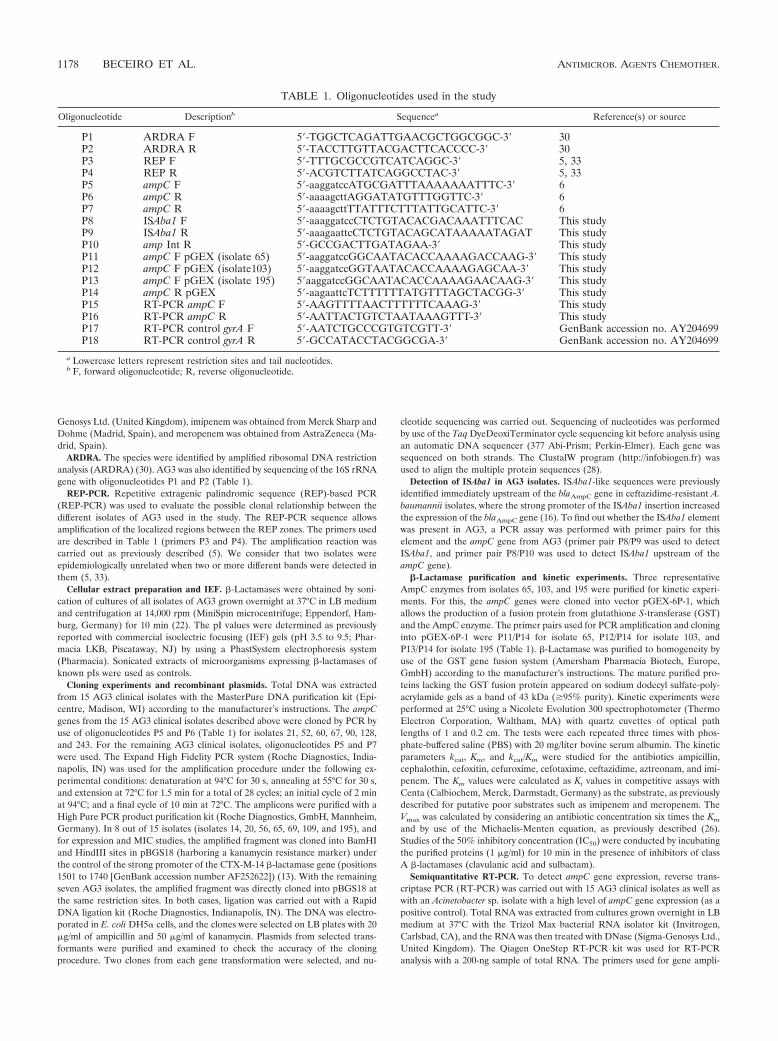
TABLE 1. Oligonucleotides used in the study
Oligonucleotide Descriptionb Sequencea Reference(s) or source
P1 ARDRA F 5-TGGCTCAGATTGAACGCTGGCGGC-3 30P2 ARDRA R 5-TACCTTGTTACGACTTCACCCC-3 30P3 REP F 5-TTTGCGCCGTCATCAGGC-3 5, 33P4 REP R 5-ACGTCTTATCAGGCCTAC-3 5, 33P5 ampC F 5-aaggatccATGCGATTTAAAAAAATTTC-3 6P6 ampC R 5-aaaagcttAGGATATGTTTGGTTC-3 6P7 ampC R 5-aaaagcttTTATTTCTTTATTGCATTC-3 6P8 ISAba1 F 5-aaaggatccCTCTGTACACGACAAATTTCAC This studyP9 ISAba1 R 5-aaagaattcCTCTGTACAGCATAAAAATAGAT This studyP10 amp Int R 5-GCCGACTTGATAGAA-3 This studyP11 ampC F pGEX (isolate 65) 5-aaggatccGGCAATACACCAAAAGACCAAG-3 This studyP12 ampC F pGEX (isolate103) 5-aaggatccGGTAATACACCAAAAGAGCAA-3 This studyP13 ampC F pGEX (isolate 195) 5aaggatccGGCAATACACCAAAAGAACAAG-3 This studyP14 ampC R pGEX 5-aagaattcTCTTTTTTATGTTTAGCTACGG-3 This studyP15 RT-PCR ampC F 5-AAGTTTTAACTTTTTTCAAAG-3 This studyP16 RT-PCR ampC R 5-AATTACTGTCTAATAAAGTTT-3 This studyP17 RT-PCR control gyrA F 5-AATCTGCCCGTGTCGTT-3 GenBank accession no. AY204699P18 RT-PCR control gyrA R 5-GCCATACCTACGGCGA-3 GenBank accession no. AY204699
a Lowercase letters represent restriction sites and tail nucleotides.b F, forward oligonucleotide; R, reverse oligonucleotide.
1178 BECEIRO ET AL. ANTIMICROB. AGENTS CHEMOTHER.
fication were designed to hybridize in highly conserved fragments in all se-quences of ampC genes (P15 and P16 in Table 1), which amplified an internalproduct of 470 bp. As an internal control for the RT-PCR, the gyrA gene fromAG3 was used as a template with oligonucleotides P17 and P18 (Table 1), whichamplified the 344 bp of this gene. The conditions of the RT-PCR were as follows:an initial cycle of reverse transcription at 50°C for 30 min, followed by amplifi-cation of the DNA with a initial cycle of 15 min at 95°C, 23 cycles at 95°C for 1min, 50°C for 1 min, and 72°C for 1 min and a final cycle of 10 min at 72°C.Aliquots were removed during the amplification process at cycles 14, 18, and 22(exponential phase of amplification). The bands were revealed in agarose gels, asdescribed above. The intensity of ampC gene bands was compared with that ofgyrA gene bands.
Western blot analysis. Western blot analysis was used to detect and assessAmpC expression in the AG3 isolates with polyclonal antibodies raised againstADC-7 (19). Bacterial extracts were obtained as described above for pI analysisand were loaded onto sodium dodecyl sulfate-polyacrylamide gels (12%) in aminigel apparatus (Bio-Rad, Hercules, CA). The proteins were transferred ontoImmobilon-P membranes (Millipore, Billerica, MA). The membranes wereblocked with 5% (wt/vol) blocking agent (skim milk) in PBS-Tween. After themembranes were washed with PBS-Tween, they were incubated with a mixture ofthe polyclonal rabbit anti-ADC-7 antiserum diluted 1:1,500. The membraneswere then washed and incubated with secondary conjugated rabbit antiserumdiluted 1:2,000 (ECL Western blotting reagent; Amersham Pharmacia Biotech,United Kingdom). All incubations were done for 1 h at 25°C. The Western blotwas revealed by overlaying the membranes with luminol, a substrate of thehorseradish peroxidase enzyme, ligated to secondary antibody.
OMP purification. Purification and analysis of outer membrane proteins(OMPs) were performed as previously described (13, 14) with bacterial AG3isolates 20 and 103 (with clear ampicillin resistance) and isolates 21, 56, and 128(ampicillin susceptible).
Determination of antibiotic MICs in the presence of cloxacillin and the effluxpump inhibitors PAN and CCCP. To assess the putative role of an efflux pumpmechanism involved in �-lactam resistance in AG3, antibiotic MICs in thepresence and absence of chemical efflux pump inhibitors were determined. Thus,MICs were determined in the presence of 150 �g/ml of cloxacillin as an inhibitorof cephalosporinase activity (7) and of efflux pump inhibitors, such as carbonylcyanide m-chlorophenylhydrazone (CCCP; Sigma) at 25 �M (5.1 �g/ml) (15)and Phe-Arg �-naphthylamide dihydrochloride (PAN; Sigma) at 25 �g/ml (8),alone and with antibiotics.
Theoretical modeling of the three-dimensional structure of AmpC enzymes.The theoretical three-dimensional structure of the ADC-12 enzyme was obtainedby theoretical homology modeling with different computer software packages:BLAST (to align sequences), Deep View (to obtain the theoretical structurefrom previously aligned genes), and UCSF Chimera (protein model edition). Forthis, the ADC-12 sequence was compared with the previously published structureof CMY-10 (Protein Data Bank accession number 1ZKJ) (21), which showed thehighest amino acid identity (45%). We used the Deep View program in combi-nation with the Swiss-model server to generate a homology-based model of theenzyme. As part of the default pipeline of the ProModII modeling program(implemented in Swiss-model), a final step of structure “dumping” was per-formed, resulting in an unrefined, fast energy minimization process, mainly withthe purpose of avoiding atomic clashes. No further minimization or moleculardynamic equilibration was executed.
Nucleotide sequence accession numbers. The nucleotide sequences of theADC-type enzymes have been submitted to the GenBank database underaccession numbers AM283529 (ADC-12), AM283528 (ADC-13), AM283527
(ADC-14), AM283526 (ADC-15), AM283525 (ADC-16), AM283524 (ADC-17), AM283523 (ADC-18), AM283522 (ADC-19), AM283521 (ADC-20),AM283520 (ADC-21), AM283519 (ADC-22), and AM283518 (ADC-23).
RESULTS
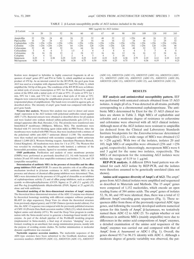
IEF analysis and antimicrobial susceptibility pattern. IEFwas performed with sonicated extracts obtained from 15 AG3isolates. A single pI of ca. 9 was detected in all strains, probablycorresponding to a chromosomal cephalosporinase. The anti-biotic MICs determined by Etest for the 15 AG3 clinical iso-lates are shown in Table 2. High MICs of cephalothin andcefoxitin and a moderate degree of resistance to cefuroximeand cefotaxime were observed with all AG3 clinical isolates.Although most of the AG3 isolates were resistant to ampicillin(as deduced from the Clinical and Laboratory StandardsInstitute breakpoints for the Enterobacteriaceae determinedfor ampicillin) (12), a wide range of MICs was obtained (12to 256 �g/ml). With two of the isolates, isolates 20 and103, high MICs of ampicillin were obtained (256 and 256�g/ml, respectively). Interestingly, meropenem MICs were 6and 3 �g/ml for the same two isolates, respectively. TheMICs of meropenem for the remaining AG3 isolates werewithin the range of 0.19 to 1 �g/ml.
REP-PCR analysis. A different DNA band pattern was ob-tained for each AG3 isolate by REP-PCR, and the isolateswere therefore assumed to be genetically unrelated (data notshown).
Amino acid sequence diversity of AmpCs of AG3. The ampCgenes from AG3 clinical isolates were amplified and sequencedas described in Materials and Methods. The 15 ampC geneswere composed of 1,152 nucleotides, which encode an openreading frame of 384 amino acids. The ampC genes of isolates52, 56, 60, and 195 were identical; thus, there was a total of 12different AmpC-encoding gene sequences (Fig. 1). These se-quences differ from those of the previously reported ADC-typegenes, and following the recently developed uniform numericalsystem for this family of AmpC �-lactamases, we tentativelynamed them ADC-12 to ADC-23. To explain whether or notdifferences in antibiotic MICs (mainly ampicillin) were due todifferences in the amino acid compositions of AmpC enzymes,a detailed examination of the amino acid sequences of theAmpC enzymes was carried out and compared with that ofAmpC from A. baumannii or ADC-1 (Fig. 1). Overall, thegenes showed 93.7 to 96.1% identity with ADC-1. Although amoderate degree of genetic variability was observed, the pat-
TABLE 2. �-Lactam susceptibility profile of AG3 isolates included in the study
�-LactamMIC (�g/ml) for AG3 isolate:
14 20 21 52 56 60 65 67 69 90 103 109 128 195 243
Ampicillin 32 256 24 48 24 32 32 48 32 12 256 48 24 32 12Piperacillin 16 24 24 12 12 24 16 16 16 12 24 16 12 24 32Cephalothin 256 256 256 256 256 256 256 256 256 256 256 256 256 256 256Cefoxitin 256 256 256 256 64 256 128 256 256 128 192 256 128 256 48Cefuroxime 32 48 48 32 16 64 24 48 32 16 48 32 32 64 32Ceftazidime 3 4 6 3 1.5 3 4 3 4 6 4 3 3 4 4Cefotaxime 24 16 24 12 4 24 12 16 16 12 16 16 12 24 16Cefepime 3 4 12 2 2 4 3 4 3 16 3 2 2 6 3Imipenem 0.25 1 0.38 0.25 0.19 0.25 0.19 0.19 0.25 0.38 0.75 0.19 0.12 0.19 0.19Meropenem 0.50 6 1 0.50 0.38 0.75 0.50 0.50 0.25 0.75 3 0.38 0.19 1 0.25
VOL. 53, 2009 ampC GENES FROM ACINETOBACTER GENOMIC SPECIES 3 1179
tern does not appear to follow a random profile, as somepositions are more likely to be replaced than others. Indeed,some amino acid positions were replaced in at least four of theADC-type enzymes analyzed with more than one residue (Fig.1) and are shown in Fig. 3. Graphical analysis revealed nochanges in the relationships with important domains or cata-lytic regions of the AmpC enzyme. Analysis of amino acidsequences of ADC-13 and ADC-16 (isolated from AG3 iso-lates 20 and 103, for which the highest ampicillin MICs wereobtained) did not reveal any significant differences (in aminoacid composition or position) with respect to the remainingADC-type enzymes.
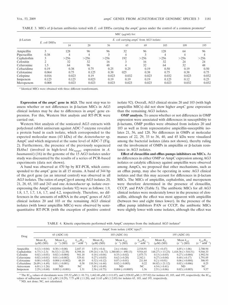
Cloning and expression of ampC genes in the E. coli host. Toconfirm whether or not amino acid changes have any effect onthe phenotype of ampicillin or �-lactam resistance, we clonedand expressed several ampC genes in an E. coli host. For this,ampC genes from clinical isolates 20 and 103 (ampicillin MICsof 256 and 256 �g/ml) and those from clinical isolates 14, 56,65, 69, 109, and 195 (ampicillin MIC ranges of 24 to 48 �g/ml)were cloned into pBGS18 under the control of a commonexternal CTX-M-14 gene promoter and were then transformedinto a common E. coli DH5� host, and the �-lactam MICswere determined (Table 3). The MICs of different �-lactams,including ampicillin and meropenem, were similar for all E.coli transformants except the transformant that expresses the�-lactamase AmpC of AG3 isolate 65 (ADC-14), for which theMICs were slightly lower.
Kinetic experiments. To confirm that differences in AmpCamino acid sequence are related to differences in the catalytic
efficiency of AmpC enzymes toward �-lactams, three represen-tative AmpC enzymes were chosen for further biochemicalexperiments. Those of AG3 isolates 65 (ADC-14), 103 (ADC-16), and 195 (ADC-18) were purified to homogeneity, and thekinetic parameters Km, kcat, and kcat/Km were determined (Ta-ble 4). The three AmpC enzymes showed almost identicalcatalytic efficiencies (kcat/Km) toward ampicillin and cephalo-thin, although ADC-14 showed important differences regard-ing the Km and kcat values, thus revealing differences in thebiochemical behavior. Indeed, the kcat for ampicillin of ADC-14was between 13 and 8 times lower than the correspondingvalues for ADC-18 and -16, respectively, and also 25 and 24times lower for cephalothin, respectively. Moreover, ADC-14showed a lower catalytic efficiency for cefoxitin than ADC-18and ADC-16 (2.1 and 1.9 times, respectively), cefuroxime (3.4and 4.4 times, respectively), and cefotaxime (6.4 and 4.4 times,respectively), which are consistent with MICs obtained with E.coli harboring ampC genes (Table 3). With regard to imi-penem, the three enzymes had similar Km values, although thekcat/Km values for ADC-18 were almost seven times higher. Nohydrolysis was detected with aztreonam.
Regarding the inhibition studies, IC50s showed a typical classC profile, with high clavulanic acid IC50s. However, there wasa moderate degree of sulbactam inhibition, and the IC50s forclavulanic acid and sulbactam with ADC-14 were lower, whichindicates that differences in amino acid sequence (primarystructure of the enzyme) are related to differences in the cat-alytic properties of the ADC-type enzymes.
FIG. 1. Amino acid sequence alignment among the 12 AmpC �-lactamases from AG3 (ADC-12 to -23) with respect to that of A. baumannii(ADC-1). Amino acid differences are indicated. The typical �-lactamase domains (SVSK, YSN, and KTG) and the �-loop are double underlined.The amino acid replacements present in at least four or more proteins (relative to that of ADC-1) with more than one residue are underlined. Thevertical arrow indicates the position of the �1 amino acid (after cleavage of signal peptide). ADC-1, AmpC from A. baumannii (see reference 6).
1180 BECEIRO ET AL. ANTIMICROB. AGENTS CHEMOTHER.
Expression of the ampC gene in AG3. The next step was toassess whether or not differences in �-lactam MICs in AG3clinical isolates may be due to differences in ampC gene ex-pression. For this, Western blot analysis and RT-PCR werecarried out.
Western blot analysis of the sonicated AG3 extracts withpolyclonal rabbit antiserum against ADC-7 enzyme revealeda protein band in each isolate, which corresponded to theexpected molecular mass (43 kDa) of the Acinetobacter sp.AmpC and which migrated at the same level of ADC-7 (Fig.2). Furthermore, the presence of the previously sequencedISAba1 (involved in high-level blaAmpC expression in A.baumannii) (16) in the genome of the 15 AG3 isolates understudy was discounted by the results of a series of PCR-basedexperiments (data not shown).
A band was observed at 470 bp by RT-PCR, which corre-sponded to the ampC gene in all 15 strains. A band of 344 bpof the gyrA gene (as an internal control) was observed in allAG3 isolates. The ratios of ampC/gyrA among AG3 isolates 20,21, 28, 65, 103 and 243 and one Acinetobacter sp. isolate over-expressing the AmpC enzyme (isolate 92) were as follows: 1.9,1.8, 1.7, 1.7, 1.6, 1.7, and 4.2, respectively. Therefore, no dif-ferences in the amounts of mRNA in the ampC genes of AG3clinical isolates 20 and 103 or the remaining AG3 clinicalisolates (with lower ampicillin MICs) were observed by semi-quantitative RT-PCR (with the exception of positive control
isolate 92). Overall, AG3 clinical strains 20 and 103 (with highampicillin MICs) did not show higher ampC gene expressionthan the remaining AG3 isolates.
OMP analysis. To assess whether or not differences in OMPexpression were associated with differences in susceptibility to�-lactams, OMP profiles were obtained from isolates 20 and103 as well as from representative ampicillin-susceptible iso-lates 21, 56, and 128. No differences in OMPs at molecularmasses of 22, 29, 33 to 36, 40, and 45 kDa were visualizedamong the bacterial isolates (data not shown), thereby rulingout the involvement of OMPs in ampicillin or �-lactam resis-tance in AG3 isolates.
Effect of cloxacillin and efflux pumps inhibitors on MICs. Asno differences in either OMP or AmpC expression among AG3isolates or catalytic efficiency against ampicillin were observedamong AmpCs, we proposed that other mechanisms, such asan efflux pump, may also be operating in some AG3 clinicalisolates and that this may account for differences in �-lactamMICs. The MICs of ampicillin, cephalothin, and meropenemwere therefore determined in the presence of cloxacillin,CCCP, and PAN (Table 5). The antibiotic MICs for all AG3clinical isolates were moderately lower in the presence of clox-acillin, although the effect was most apparent with ampicillin(between two and eight times lower). In the presence of theefflux pump inhibitors PAN or CCCP, the antibiotic MICswere slightly lower with some isolates, although the effect was
TABLE 3. MICs of �-lactam antibiotics tested with E. coli DH5� carrying the ampC genes under the control of a common promotera
�-Lactam
MIC (�g/ml) for:
E. coli DH5�E. coli carrying ampC from AG3 isolate:
14 20 56 65 69 103 109 195
Ampicillin 3 128 96 96 32 96 128 64 96Piperacillin 0.38 4 4 3 4 3 4 4 4Cephalothin 3 256 256 256 192 256 256 256 256Cefoxitin 2 32 32 16 3 16 32 24 24Cefuroxime 1.5 64 96 48 4 32 64 32 48Ceftazidime 0.19 0.38 0.75 0.25 0.25 0.19 0.50 0.19 0.50Cefotaxime 0.064 0.75 0.125 0.38 025 0.38 0.75 0.38 0.75Cefepime 0.016 0.023 0.19 0.023 0.032 0.023 0.032 0.023 0.032Imipenem 0.125 0.125 0.023 0.19 0.19 0.19 0.125 0.12 0.25Meropenem 0.008 0.023 0.023 0.023 0.023 0.023 0.023 0.032 0.032
a Identical MICs were obtained with three different transformants.
TABLE 4. Kinetic experiments performed with AmpC enzymes from the indicated AG3 isolatesa
Drug
AmpC from isolate (ADC type)b:
65 (ADC-14) 103 (ADC-16) 195 (ADC-18)
Mean Km(�M) (SD)
Mean kcat(s�1) (SD)
kcat/Km(s�1 mM�1)
Mean Km(�M) (SD)
Mean kcat(s�1) (SD)
kcat/Km(s�1 mM�1)
Mean Km(�M) (SD)
Mean kcat(s�1) (SD)
kcat/Km(s�1 mM�1)
Ampicillin 0.12 (�0.04) 0.30 (�0.08) 2,447.15 1.03 (�0.4) 2.6 (�0.66) 2,518.91 1.5 (�0.47) 4.05 (�1.06) 2,700.94Cephalothin 6.73 (�2.3) 56.52 (�22.38) 8,398.16 139.24 (�16.72) 1,380.80 (� 490.13) 9,916.65 148.27 (�13.23) 1,418.36 (�318.51) 9,566.33Cefoxitin 0.02 (�0.01) 0.02 (�0.004) 941.18 0.18 (�0.04) 0.33 (�0.02) 1,875.71 0.13 (�0.03) 0.27 (�0.004) 1,992.54Cefuroxime 0.02 (�0.01) 0.01 (�0.002) 529.41 0.27 (�0.03) 0.62 (�0.20) 2,342.1 0.27 (�0.04) 0.48 (�0.17) 1,791.05Cefotaxime 0.08 (�0.02) 0.004 (�0.002) 48.19 0.32 (�0.12) 0.07 (�0.01) 211.84 0.2 (�0.01) 0.06 (�0.004) 306.93Ceftazidime 26.09 (�4.49) 0.01 (�0.001) 0.38 110.34 (�6.44) 0.02 (�0.003) 0.22 84.02 (�21.12) 0.02 (�0.006) 0.25Aztreonam 6.01 (�1.26) ND NC 3.85 (�0.49) ND NC 4.54 (�0.41) ND NCImipenem 2.29 (�0.60) 0.003 (�0.001) 1.31 2.56 (�0.73) 0.004 (�0.0005) 1.56 2.51 (�0.86) 0.02 (�0.003) 9.57
a The IC50 values of clavulanate were 235.52 �M (� 01.71), 1,462.48 �M (�111.07), and 1,928.02 �M (�357.02) for isolates 65, 103, and 195, respectively; the IC50values of sulbactam were 1.12 �M (�0.29), 7.75 �M (�1.20), and 11.65 �M (�2.03) for isolates 65, 103, and 195, respectively.
b ND, not done; NC, not calculated.
VOL. 53, 2009 ampC GENES FROM ACINETOBACTER GENOMIC SPECIES 3 1181
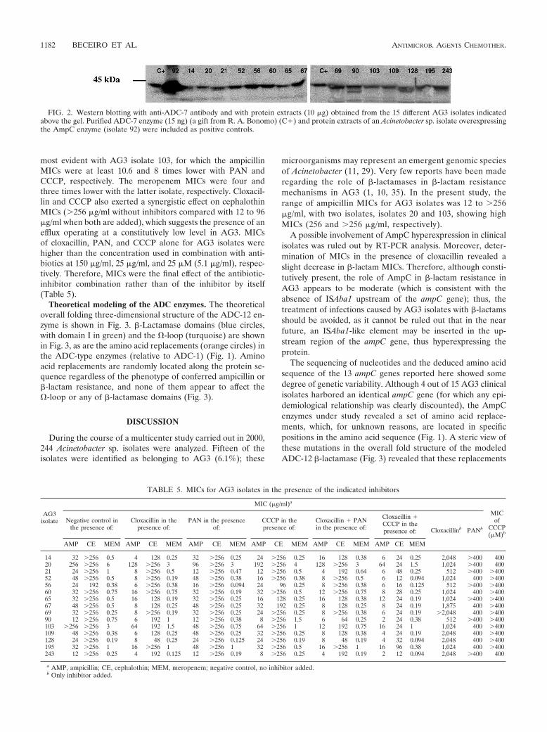
most evident with AG3 isolate 103, for which the ampicillinMICs were at least 10.6 and 8 times lower with PAN andCCCP, respectively. The meropenem MICs were four andthree times lower with the latter isolate, respectively. Cloxacil-lin and CCCP also exerted a synergistic effect on cephalothinMICs (256 �g/ml without inhibitors compared with 12 to 96�g/ml when both are added), which suggests the presence of anefflux operating at a constitutively low level in AG3. MICsof cloxacillin, PAN, and CCCP alone for AG3 isolates werehigher than the concentration used in combination with anti-biotics at 150 �g/ml, 25 �g/ml, and 25 �M (5.1 �g/ml), respec-tively. Therefore, MICs were the final effect of the antibiotic-inhibitor combination rather than of the inhibitor by itself(Table 5).
Theoretical modeling of the ADC enzymes. The theoreticaloverall folding three-dimensional structure of the ADC-12 en-zyme is shown in Fig. 3. �-Lactamase domains (blue circles,with domain I in green) and the �-loop (turquoise) are shownin Fig. 3, as are the amino acid replacements (orange circles) inthe ADC-type enzymes (relative to ADC-1) (Fig. 1). Aminoacid replacements are randomly located along the protein se-quence regardless of the phenotype of conferred ampicillin or�-lactam resistance, and none of them appear to affect the�-loop or any of �-lactamase domains (Fig. 3).
DISCUSSION
During the course of a multicenter study carried out in 2000,244 Acinetobacter sp. isolates were analyzed. Fifteen of theisolates were identified as belonging to AG3 (6.1%); these
microorganisms may represent an emergent genomic speciesof Acinetobacter (11, 29). Very few reports have been maderegarding the role of �-lactamases in �-lactam resistancemechanisms in AG3 (1, 10, 35). In the present study, therange of ampicillin MICs for AG3 isolates was 12 to 256�g/ml, with two isolates, isolates 20 and 103, showing highMICs (256 and 256 �g/ml, respectively).
A possible involvement of AmpC hyperexpression in clinicalisolates was ruled out by RT-PCR analysis. Moreover, deter-mination of MICs in the presence of cloxacillin revealed aslight decrease in �-lactam MICs. Therefore, although consti-tutively present, the role of AmpC in �-lactam resistance inAG3 appears to be moderate (which is consistent with theabsence of ISAba1 upstream of the ampC gene); thus, thetreatment of infections caused by AG3 isolates with �-lactamsshould be avoided, as it cannot be ruled out that in the nearfuture, an ISAba1-like element may be inserted in the up-stream region of the ampC gene, thus hyperexpressing theprotein.
The sequencing of nucleotides and the deduced amino acidsequence of the 13 ampC genes reported here showed somedegree of genetic variability. Although 4 out of 15 AG3 clinicalisolates harbored an identical ampC gene (for which any epi-demiological relationship was clearly discounted), the AmpCenzymes under study revealed a set of amino acid replace-ments, which, for unknown reasons, are located in specificpositions in the amino acid sequence (Fig. 1). A steric view ofthese mutations in the overall fold structure of the modeledADC-12 �-lactamase (Fig. 3) revealed that these replacements
FIG. 2. Western blotting with anti-ADC-7 antibody and with protein extracts (10 �g) obtained from the 15 different AG3 isolates indicatedabove the gel. Purified ADC-7 enzyme (15 ng) (a gift from R. A. Bonomo) (C�) and protein extracts of an Acinetobacter sp. isolate overexpressingthe AmpC enzyme (isolate 92) were included as positive controls.
TABLE 5. MICs for AG3 isolates in the presence of the indicated inhibitors
AG3isolate
MIC (�g/ml)a
MICof
CCCP(�M)b
Negative control inthe presence of:
Cloxacillin in thepresence of:
PAN in the presenceof:
CCCP in thepresence of:
Cloxacillin � PANin the presence of:
Cloxacillin �CCCP in thepresence of: Cloxacillinb PANb
AMP CE MEM AMP CE MEM AMP CE MEM AMP CE MEM AMP CE MEM AMP CE MEM
14 32 256 0.5 4 128 0.25 32 256 0.25 24 256 0.25 16 128 0.38 6 24 0.25 2,048 400 40020 256 256 6 128 256 3 96 256 3 192 256 4 128 256 3 64 24 1.5 1,024 400 40021 24 256 1 8 256 0.5 12 256 0.47 12 256 0.5 4 192 0.64 6 48 0.25 512 400 40052 48 256 0.5 8 256 0.19 48 256 0.38 16 256 0.38 8 256 0.5 6 12 0.094 1,024 400 40056 24 192 0.38 6 256 0.38 16 256 0.094 24 96 0.25 8 256 0.38 6 16 0.125 512 400 40060 32 256 0.75 16 256 0.75 32 256 0.19 32 256 0.5 12 256 0.75 8 28 0.25 1,024 400 40065 32 256 0.5 16 128 0.19 32 256 0.25 16 128 0.25 16 128 0.38 12 24 0.19 1,024 400 40067 48 256 0.5 8 128 0.25 48 256 0.25 32 192 0.25 8 128 0.25 8 24 0.19 1,875 400 40069 32 256 0.25 8 256 0.19 32 256 0.25 24 256 0.25 8 256 0.38 6 24 0.19 2,048 400 40090 12 256 0.75 6 192 1 12 256 0.38 8 256 1.5 6 64 0.25 2 24 0.38 512 400 400103 256 256 3 64 192 1.5 48 256 0.75 64 256 1 12 192 0.75 16 24 1 1,024 400 400109 48 256 0.38 6 128 0.25 48 256 0.25 32 256 0.25 8 128 0.38 4 24 0.19 2,048 400 400128 24 256 0.19 8 48 0.25 24 256 0.125 24 256 0.19 8 48 0.19 4 32 0.094 2,048 400 400195 32 256 1 16 256 1 48 256 1 32 256 0.5 16 256 1 16 96 0.38 1,024 400 400243 12 256 0.25 4 192 0.125 12 256 0.19 8 256 0.25 4 192 0.19 2 12 0.094 2,048 400 400
a AMP, ampicillin; CE, cephalothin; MEM, meropenem; negative control, no inhibitor added.b Only inhibitor added.
1182 BECEIRO ET AL. ANTIMICROB. AGENTS CHEMOTHER.
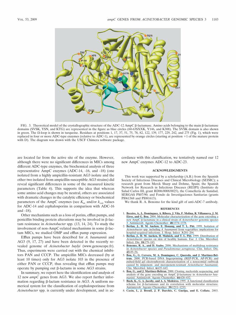
are located far from the active site of the enzyme. However,although there were no significant differences in MICs amongdifferent ADC-type enzymes, the biochemical analysis of threerepresentative AmpC enzymes (ADC-14, -16, and -18) (oneisolated from a highly ampicillin-resistant AG3 isolate and theother two isolated from ampicillin-susceptible AG3 strains) didreveal significant differences in some of the measured kineticparameters (Table 4). This supports the idea that whereassome amino acid changes may be neutral, others are associatedwith dramatic changes in the catalytic efficiency or biochemicalparameters of the AmpC enzymes (see Km and/or kcat valuesfor ADC-14 and cephalosporins in comparison with ADC-16and -18).
Other mechanisms such as a loss of porins, efflux pumps, andpenicillin binding protein alterations may be involved in �-lac-tam resistance in Acinetobacter spp. (13, 14, 24). To study theinvolvement of non-AmpC-related mechanisms in some �-lac-tam MICs, we studied OMP and efflux pump expression.
Efflux pumps have been described for A. baumannii andAG3 (9, 17, 27) and have been detected in the recently re-vealed genome of Acinetobacter baylyi (www.genoscope.fr).Thus, experiments were carried out with the chemical inhibi-tors PAN and CCCP. The ampicillin MICs decreased (by atleast 10 times) only for AG3 isolate 103 in the presence ofeither PAN or CCCP, thus revealing that efflux pumps mayoperate by pumping out �-lactams in some AG3 strains.
In summary, we report here the identification and analysis of12 new ampC genes from AG3. We also report further infor-mation regarding �-lactam resistance in AG3. A uniform nu-merical system for the classification of cephalosporinase fromAcinetobacter spp. is currently under development, and in ac-
cordance with this classification, we tentatively named our 12new AmpC enzymes ADC-12 to ADC-23.
ACKNOWLEDGMENTS
This work was supported by a scholarship (A.B.) from the SpanishSociety of Infectious Diseases and Clinical Microbiology (SEIMC); aresearch grant from Merck Sharp and Dohme, Spain; the SpanishNetwork for Research in Infectious Diseases (REIPI) (Instituto deSalud Carlos III, grant RD06/0008/0025), the Consellería de Sanidad,SERGAS PS07/90; and Fondo de Investigaciones Sanitarias (grantsPI061368 and PI081613).
We thank R. A. Bonomo for the kind gift of anti-ADC-7 antibody.
REFERENCES
1. Beceiro, A., L. Dominguez, A. Ribera, J. Vila, F. Molina, R. Villanueva, J. M.Eiros, and G. Bou. 2004. Molecular characterization of the gene encoding anew AmpC �-lactamase in a clinical strain of Acinetobacter genomic species3. Antimicrob. Agents Chemother. 48:1374–1378.
2. Berlau, J., H. M. Aucken, E. Houang, and T. L. Pitt. 1999. Isolation ofAcinetobacter spp. including A. baumannii from vegetables: implications forhospital-acquired infections. J. Hosp. Infect. 3:201–204.
3. Berlau, J., H. M. Aucken, H. Malnick, and T. L. Pitt. 1999. Distribution ofAcinetobacter species on skin of healthy humans. Eur. J. Clin. Microbiol.Infect. Dis. 18:179–183.
4. Bonomo, R. A., and D. Szabo. 2006. Mechanisms of multidrug resistancein Acinetobacter species and Pseudomonas aeruginosa. Clin. Infect. Dis.43:49–56.
5. Bou, G., G. Cervero, M. A. Dominguez, C. Quereda, and J. Martinez-Bel-tran. 2000. PCR-based DNA fingerprinting (REP-PCR, AP-PCR) andpulsed-field gel electrophoresis characterization of a nosocomial outbreakcaused by imipenem- and meropenem-resistant Acinetobacter baumannii.Clin. Microbiol. Infect. 6:635–643.
6. Bou, G., and J. Martinez-Beltran. 2000. Cloning, nucleotide sequencing, andanalysis of the gene encoding an AmpC �-lactamase in Acinetobacter bau-mannii. Antimicrob. Agents Chemother. 44:428–432.
7. Bush, K., G. A. Jacoby, and A. A. Medeiros. 1995. A functional classificationscheme for �-lactamases and its correlation with molecular structure.Antimicrob. Agents Chemother. 39:1211–1233.
8. Casin, I., J. Breuil, J. P. Darchis, C. Guelpa, and E. Collatz. 2003.
FIG. 3. Theoretical model of the crystallographic structure of the ADC-12 AmpC �-lactamase. Amino acids belonging to the main �-lactamasedomains (SVSK, YSN, and KTG) are represented in the figure as blue circles (60-63SXXK, Y146, and K308). The SVSK domain is also shownin green. The �-loop is shown in turquoise. Residues at positions 1, 17, 37, 51, 75, 78, 82, 122, 139, 177, 229, 242, and 275 (Fig. 1), which werereplaced in four or more ADC-type enzymes (relative to ADC-1), are represented by orange circles (starting at position �1 of the mature proteinwith D). The diagram was drawn with the USCF Chimera software package.
VOL. 53, 2009 ampC GENES FROM ACINETOBACTER GENOMIC SPECIES 3 1183
Fluoroquinolone resistance linked to GyrA, GyrB, and ParC mutations inSalmonella enterica Typhimurium isolates in humans. Emerg. Infect. Dis.11:1455–1457.
9. Chau, S.-L., Y. W. Chu, and T. S. Houang. 2004. Novel resistance-nodula-tion-cell division efflux system AdeDE in Acinetobacter genomic DNA group3. Antimicrob. Agents Chemother. 48:4054–4055.
10. Chu, Y. W., M. Afzal-Shah, E. T. Houang, M. I. Palepou, D. J. Lyon, N.Woodford, and D. M. Livermore. 2001. IMP-4, a novel metallo-�-lactamasefrom nosocomial Acinetobacter spp. collected in Hong Kong between 1994and 1998. Antimicrob. Agents Chemother. 45:710–714.
11. Chu, Y. W., C. M. Leung, E. T. Houang, K. C. Ng, C. B. Leung, H. Y. Leung,and A. F. Cheng. 1999. Skin carriage of acinetobacters in Hong Kong. J. Clin.Microbiol. 37:2962–2967.
12. Clinical and Laboratory Standards Institute. 2006. Performance standardsfor antimicrobial susceptibility testing. Approved standard M100-S16, vol.26, no. 1. Clinical and Laboratory Standards Institute, Wayne, PA.
13. del Mar Tomas, M., A. Beceiro, A. Perez, D. Velasco, R. Moure, R. Villan-ueva, J. Martínez-Beltran, and G. Bou. 2005. Cloning and functional analysisof the gene encoding the 33- to 36-kilodalton outer membrane proteinassociated with carbapenem resistance in Acinetobacter baumannii. Antimi-crob. Agents Chemother. 49:5172–5175.
14. Fernandez-Cuenca, F., L. Martinez-Martinez, M. C. Conejo, J. A. Ayala,E. J. Perea, and A. Pascual. 2003. Relationship between beta-lactamaseproduction, outer membrane protein and penicillin-binding protein profileson the activity of carbapenems against clinical isolates of Acinetobacter bau-mannii. J. Antimicrob. Chemother. 51:565–574.
15. Giovanetti, E., A. Brenciani, R. Burioni, and P. E. Varaldo. 2002. A novelefflux system in inducibly erythromycin-resistant strains of Streptococcus pyo-genes. Antimicrob. Agents Chemother. 46:3750–3755.
16. Heritier, C., L. Poirel, and P. Nordmann. 2006. Cephalosporinase over-expression resulting from insertion of ISAba1 in Acinetobacter baumannii.Clin. Microbiol. Infect. 12:123–130.
17. Higgins, P. G., H. Wisplinghoff, D. Stefanik, and H. Seifert. 2004. Selectionof topoisomerase mutations and overexpression of adeB mRNA transcriptsduring an outbreak of Acinetobacter baumannii. J. Antimicrob. Chemother.54:821–823.
18. Horrevorts, A., K. Bergman, L. Kollee, I. Breuker, I. Tjernberg, and L.Dijkshoorn. 1995. Clinical and epidemiological investigations of Acineto-bacter genomospecies 3 in a neonatal intensive care unit. J. Clin. Microbiol.33:1567–1572.
19. Hujer, K. M., N. S. Hamza, A. M. Hujer, F. Perez, M. S. Helfand, C. R.Bethel, J. M. Thomson, V. E. Anderson, M. Barlow, L. B. Rice, F. C.Tenover, and R. A. Bonomo. 2005. Identification of a new allelic variantof the Acinetobacter baumannii cephalosporinase, ADC-7 �-lactamase:defining a unique family of class C enzymes. Antimicrob. Agents Che-mother. 49:2941–2948.
20. Jeong, S. H., I. K. Bae, K. O. Park, Y. J. An, S. G. Sohn, S. J. Jang, K. H.Sung, K. S. Yang, K. Lee, D. Young, and S. H. Lee. 2006. Outbreaks ofimipenem-resistant Acinetobacter baumannii producing carbapenemases inKorea. J. Microbiol. 44:423–431.
21. Kim, J. Y., H. I. Jung, Y. J. An, J. H. Lee, S. J. Kim, S. H. Jeong, K. J. Lee,P. G. Suh, H. S. Lee, S. H. Lee, and S. S. Cha. 2006. Structural basis for the
extended substrate spectrum of CMY-10, a plasmid-encoded class C beta-lactamase. Mol. Microbiol. 60:907–916.
22. Matthew, M., A. M. Harris, M. J. Marshall, and G. W. Ross. 1975. The useof analytical isoelectric focusing for detection and identification of �-lacta-mases. J. Gen. Microbiol. 88:169–178.
23. Nordmann, P., and L. Poirel. 2002. Emerging carbapenemases in gram-negative aerobes. Clin. Microbiol. Infect. 86:321–331.
24. Poirel, L., and P. Nordmann. 2006. Carbapenem resistance in Acinetobacterbaumannii: mechanisms and epidemiology. Clin. Microbiol. Infect. 12:826–836.
25. Ribera, A., F. Fernandez-Cuenca, A. Beceiro, G. Bou, L. Martínez-Martínez,A. Pascual, J. M. Cisneros, J. Rodríguez-Bano, J. Pachon, J. Vila, and theSpanish Group for Nosocomial Infection. 2004. Antimicrobial susceptibilityand mechanisms of resistance to quinolones and �-lactams in Acinetobactergenospecies 3. Antimicrob. Agents Chemother. 48:1430–1432.
26. Santillana, E., A. Beceiro, G. Bou, and A. Romero. 2007. Crystal structure ofthe carbapenemase OXA-24 reveals insights into the mechanism of carbap-enem hydrolysis. Proc. Natl. Acad. Sci. USA 104:5354–5359.
27. Su, X.-Z., J. Chen, T. Mizushima, T. Kuroda, and T. Tsuchiya. 2005. AbeM,an H�-coupled Acinetobacter baumannii multidrug efflux pump belonging tothe MATE family of transporters. Antimicrob. Agents Chemother. 49:4362–4364.
28. Thompson, J. D., D. G. Higgins, and T. J. Gibson. 1994. CLUSTAL W:improving the sensitivity of progressive multiple sequence alignment throughsequence weighting, position-specific gap penalties and weight matrix choice.Nucleic Acids Res. 22:4673–4680.
29. Traub, W. H., and D. Bauer. 2000. Surveillance of nosocomial cross-infections due to three Acinetobacter genospecies (Acinetobacter bauman-nii, genospecies 3 and genospecies 13) during a 10-year observationperiod: serotyping, macrorestriction analysis of genomic DNA and anti-biotic susceptibilities. Chemotherapy 46:282–292.
30. Vaneechoutte, M., L. Dijkshoorn, I. Tjernberg, A. Elaichouni, P. de Vos, G.Claeys, and G. Verschraegen. 1995. Identification of Acinetobacter genomicspecies by amplified ribosomal DNA restriction analysis. J. Clin. Microbiol.33:11–15.
31. van Looveren, M., H. Goossens, and the ARPAC Steering Group. 2004.Antimicrobial resistance of Acinetobacter spp. in Europe. Clin. Microbiol.Infect. 10:684–704.
32. Vegas, E. Z., B. Nieves, M. Araque, E. Velasco, J. Ruiz, and J. Vila. 2006.Outbreak of infection with Acinetobacter strain RUH 1139 in an intensivecare unit. Infect. Control Hosp. Epidemiol. 27:397–403.
33. Vila, J., M. A. Marcos, and M. T. Jimenez de Anta. 1996. A comparativestudy of different PCR-based DNA fingerprinting techniques for typing ofthe Acinetobacter calcoaceticus-A. baumannii complex. J. Med. Microbiol.44:482–489.
34. Walsh, T. R., M. A. Toleman, L. Poirel, and P. Nordmann. 2005. Metallo-�-lactamases: the quiet before the storm? Clin. Microbiol. Rev. 182:306–325.
35. Yum, J. H., K. Yi, H. Lee, D. Yong, K. Lee, J. M. Kim, G. M. Rossolini, andY. Chong. 2002. Molecular characterization of metallo-beta-lactamase-pro-ducing Acinetobacter baumannii and Acinetobacter genomospecies 3 fromKorea: identification of two new integrons carrying the bla(VIM-2) genecassettes. J. Antimicrob. Chemother. 49:837–840.
1184 BECEIRO ET AL. ANTIMICROB. AGENTS CHEMOTHER.


![[Distribution of blaOXA genes in Acinetobacter baumannii strains: a multicenter study]](https://static.fdokumen.com/doc/165x107/6337b5c66f78ac31240eb601/distribution-of-blaoxa-genes-in-acinetobacter-baumannii-strains-a-multicenter.jpg)