The rat ponto-medullary network responsible for paradoxical sleep onset and maintenance: a combined...
15
The rat ponto-medullary network responsible for paradoxical sleep onset and maintenance: a combined microinjection and functional neuroanatomical study Romuald Boissard, 1 Damien Gervasoni, 1 Markus H. Schmidt, 2 Bruno Barbagli, 1 Patrice Fort 1 and Pierre-Herve ´ Luppi 1 1 CNRS FRE 2469, Institut Fe ´de ´ ratif des Neurosciences de Lyon (IFR 19), Universite ´ Claude Bernard Lyon I, 8 Avenue Rockefeller, 69373 LYON Cedex 08, France 2 Ohio Sleep Medicine and Neuroscience Institute, 4975 Bradenton Ave., Dublin, Ohio 43017, USA Keywords: C-Fos, GABA, glycine, REM sleep, subcoeruleus Abstract The neuronal network responsible for paradoxical sleep (PS) onset and maintenance has not previously been identified in the rat, unlike the cat. To fill this gap, this study has developed a new technique involving the recording of sleep–wake states in unanaesthetized head-restrained rats whilst locally administering pharmacological agents by microiontophoresis from glass multibarrel micropipettes, into the dorsal pontine tegmentum and combining this with functional neuroanatomy. Pharmacological agents used for iontophoretic administration included carbachol, kainic acid, bicuculline and gabazine. The injection sites and their efferents were then identified by injections of anterograde (phaseolus vulgaris leucoagglutinin) or retrograde (cholera toxin B subunit) tracers through an adjacent barrel of the micropipette assembly and by C-Fos immunostaining. Bicuculline, gabazine and kainic acid ejections specifically into the pontine sublaterodorsal nucleus (SLD) induced within a few minutes a PS-like state characterized by a continuous muscle atonia, low voltage EEG and a lack of reaction to stimuli. In contrast, carbachol ejections into the SLD induced wakefulness. In PHA-L, glycine and C-Fos multiple double-labelling experiments, anterogradely labelled fibres originating from the SLD were seen apposed on glycine and C-Fos positive neurons (labelled after 90 min of pharmacologically induced PS-like state) from the ventral gigantocellular and parvicellular reticular nuclei. Altogether, these data indicate that the SLD nuclei contain a population of neurons playing a crucial role in PS onset and maintenance. Furthermore, they suggest that GABAergic disinhibition and glutamate excitation of these neurons might also play a crucial role in the onset of PS. Introduction In the middle of the last century, a series of historical observations lead to the discovery of a vigilance state in humans and other mammals paradoxically characterized by cortical activation and rapid eye movements, but associated with a complete disappearance of muscle tone (Aserinsky & Kleitman, 1953; Dement, 1958; Jouvet & Michel, 1959). This phase of sleep, coined paradoxical sleep (PS) or rapid eye movement (REM) sleep was then demonstrated to correlate with dream activity (Aserinsky & Kleitman, 1953; Dement & Kleitman, 1957). Through a series of lesion, electrophysiological, local pharmaco- logical and neuroanatomical studies, it has been shown in cats that a small area of the dorsal rostral pontine reticular nucleus (PRNr), named peri-locus coeruleus a (peri-LCa) by Sakai et al. (1979, 1981), contains the neurons responsible for PS. They are composed of presumed cholinergic and noncholinergic neurons, with an activity specific to PS (PS-on neurons) (Sakai et al., 1981; Sakai et al., 2001). Some of these neurons project to the intralaminar nuclei of the thalamus (ILM), the posterior hypothalamus and the basal forebrain and may be responsible for the cortical activation during PS, while others project caudally to the ventromedial medullary reticular formation (Sakai et al., 1979; Sakai et al., 1981; Review in Jones, 1991) and may generate the muscle atonia observed during this sleep state (Sakai et al., 2001). With respect to the muscle atonia, the inhibitory amino acid glycine (gly) appears to play a major role in the tonic hyperpolariza- tion of motoneurons during PS (Chase et al., 1989). Based on a number of neuroanatomical results (Luppi et al., 1988; Fort et al., 1990, 1993), we have proposed that noncholinergic peri-LCa neurons induce the muscle atonia during PS via an excitatory projection to glycinergic premotoneurons localized in the ventromedial medullary reticular formation. It has been suggested that the onset of the noncholinergic PS-on peri-LCa cells is due to an excitatory projection from cholinergic PS- on cells located in the peri-LCa, laterodorsal tegmental (LDT) and pedunculopontine nuclei (PPN), as well as the removal of inhibition from monoaminergic PS-off cells (Hobson et al., 1975; Siegel & McGinty, 1977; Sakai et al., 1981). This hypothesis is supported by the observation that carbachol, administered into the peri-LCa, induces PS in the cat (Vanni-Mercier et al., 1989) and that carbachol activates noncholinergic PS-on neurons in this region (Sakai & Koyama, 1996). In rats however, pressure injections of carbachol into the different parts of the pontine reticular nucleus or the Correspondence: Dr Pierre-Herve ´ Luppi, as above. E-mail: [email protected] Received 10 June 2002, revised 26 August 2002, accepted 4 September 2002 European Journal of Neuroscience, Vol. 16, pp. 1959–1973, 2002 ª Federation of European Neuroscience Societies
-
Upload
universite-lyon -
Category
Documents
-
view
0 -
download
0
Transcript of The rat ponto-medullary network responsible for paradoxical sleep onset and maintenance: a combined...

The rat ponto-medullary network responsible forparadoxical sleep onset and maintenance: a combinedmicroinjection and functional neuroanatomical study
Romuald Boissard,1 Damien Gervasoni,1 Markus H. Schmidt,2 Bruno Barbagli,1 Patrice Fort1 andPierre-Herve Luppi11CNRS FRE 2469, Institut FeÂdeÂratif des Neurosciences de Lyon (IFR 19), Universite Claude Bernard Lyon I, 8 Avenue
Rockefeller, 69373 LYON Cedex 08, France2Ohio Sleep Medicine and Neuroscience Institute, 4975 Bradenton Ave., Dublin, Ohio 43017, USA
Keywords: C-Fos, GABA, glycine, REM sleep, subcoeruleus
Abstract
The neuronal network responsible for paradoxical sleep (PS) onset and maintenance has not previously been identi®ed in the rat,
unlike the cat. To ®ll this gap, this study has developed a new technique involving the recording of sleep±wake states inunanaesthetized head-restrained rats whilst locally administering pharmacological agents by microiontophoresis from glass
multibarrel micropipettes, into the dorsal pontine tegmentum and combining this with functional neuroanatomy. Pharmacological
agents used for iontophoretic administration included carbachol, kainic acid, bicuculline and gabazine. The injection sites and
their efferents were then identi®ed by injections of anterograde (phaseolus vulgaris leucoagglutinin) or retrograde (cholera toxin Bsubunit) tracers through an adjacent barrel of the micropipette assembly and by C-Fos immunostaining. Bicuculline, gabazine and
kainic acid ejections speci®cally into the pontine sublaterodorsal nucleus (SLD) induced within a few minutes a PS-like state
characterized by a continuous muscle atonia, low voltage EEG and a lack of reaction to stimuli. In contrast, carbachol ejectionsinto the SLD induced wakefulness. In PHA-L, glycine and C-Fos multiple double-labelling experiments, anterogradely labelled
®bres originating from the SLD were seen apposed on glycine and C-Fos positive neurons (labelled after 90 min of
pharmacologically induced PS-like state) from the ventral gigantocellular and parvicellular reticular nuclei. Altogether, these dataindicate that the SLD nuclei contain a population of neurons playing a crucial role in PS onset and maintenance. Furthermore,
they suggest that GABAergic disinhibition and glutamate excitation of these neurons might also play a crucial role in the onset of
PS.
Introduction
In the middle of the last century, a series of historical observations
lead to the discovery of a vigilance state in humans and other
mammals paradoxically characterized by cortical activation and rapid
eye movements, but associated with a complete disappearance of
muscle tone (Aserinsky & Kleitman, 1953; Dement, 1958; Jouvet &
Michel, 1959). This phase of sleep, coined paradoxical sleep (PS) or
rapid eye movement (REM) sleep was then demonstrated to correlate
with dream activity (Aserinsky & Kleitman, 1953; Dement &
Kleitman, 1957).
Through a series of lesion, electrophysiological, local pharmaco-
logical and neuroanatomical studies, it has been shown in cats that a
small area of the dorsal rostral pontine reticular nucleus (PRNr),
named peri-locus coeruleus a (peri-LCa) by Sakai et al. (1979,
1981), contains the neurons responsible for PS. They are composed of
presumed cholinergic and noncholinergic neurons, with an activity
speci®c to PS (PS-on neurons) (Sakai et al., 1981; Sakai et al., 2001).
Some of these neurons project to the intralaminar nuclei of the
thalamus (ILM), the posterior hypothalamus and the basal forebrain
and may be responsible for the cortical activation during PS, while
others project caudally to the ventromedial medullary reticular
formation (Sakai et al., 1979; Sakai et al., 1981; Review in Jones,
1991) and may generate the muscle atonia observed during this sleep
state (Sakai et al., 2001).
With respect to the muscle atonia, the inhibitory amino acid
glycine (gly) appears to play a major role in the tonic hyperpolariza-
tion of motoneurons during PS (Chase et al., 1989). Based on a
number of neuroanatomical results (Luppi et al., 1988; Fort et al.,
1990, 1993), we have proposed that noncholinergic peri-LCa neurons
induce the muscle atonia during PS via an excitatory projection to
glycinergic premotoneurons localized in the ventromedial medullary
reticular formation.
It has been suggested that the onset of the noncholinergic PS-on
peri-LCa cells is due to an excitatory projection from cholinergic PS-
on cells located in the peri-LCa, laterodorsal tegmental (LDT) and
pedunculopontine nuclei (PPN), as well as the removal of inhibition
from monoaminergic PS-off cells (Hobson et al., 1975; Siegel &
McGinty, 1977; Sakai et al., 1981). This hypothesis is supported by
the observation that carbachol, administered into the peri-LCa,
induces PS in the cat (Vanni-Mercier et al., 1989) and that carbachol
activates noncholinergic PS-on neurons in this region (Sakai &
Koyama, 1996). In rats however, pressure injections of carbachol into
the different parts of the pontine reticular nucleus or the
Correspondence: Dr Pierre-Herve Luppi, as above.E-mail: [email protected]
Received 10 June 2002, revised 26 August 2002, accepted 4 September 2002
European Journal of Neuroscience, Vol. 16, pp. 1959±1973, 2002 ã Federation of European Neuroscience Societies

sublaterodorsal nucleus, the rat neuroanatomical equivalent of the cat
peri-LCa (SLD, Swanson, 1992; also named dorsal and alpha
subcoeruleus nuclei by Paxinos & Watson, 1997), induce only a small
PS hypersomnia (Gnadt & Pegram, 1986; Velazquez-Moctezuma
et al., 1989; Bourgin et al., 1995; Deurveilher et al., 1997; Datta et al.,
1998; Kubin, 2001). Moreover, PS-on neurons have not been
recorded in this region and their medullary projections have not
been demonstrated. As the rat is now the animal of choice for
experimental neurophysiology, it was decided to identify the pontine
neurons, as well as their projections, responsible for PS in this species
by using a new experimental model combining single unit recording,
precise and limited local pharmacology by microiontophoresis,
anterograde and retrograde tracing, with C-Fos and neurotransmitter
identi®cation of labelled-cells.
Materials and methods
Fixation of the head-restraining system
This procedure has in part been described previously (Darracq et al.,
1996; Gervasoni et al., 1998, 2000; Souliere et al., 2000). Male
Sprague-Dawley rats (280±320 g, n = 25; IFFA Credo, France) were
anaesthetized with pentobarbital (45 mg/kg, i.p) and mounted
conventionally in a stereotaxic frame (David Kopf, CA, USA), i.e.
with ear- and nose-bars. The bone was exposed and cleaned. The
skull was placed at a 15° angle (nose tilted down) to avoid the
transverse sinus overlying the pontine reticular formation during the
subsequent electrode penetrations. Three stainless steel screws were
®xed in the parietal and frontal parts of the skull and three wire
electrodes inserted into the neck muscles to monitor the electro-
encephalogram (EEG) and the electromyogram (EMG), respectively.
In some animals (n = 11), two electrodes were implanted on both
sides in the orbits for eye movement recordings (EOG). Penile
erections (n = 3) were recorded according to a technique previously
described (Schmidt et al., 1994, 2000), involving chronic pressure
monitoring within the bulb of the corpus spongiosum of the penis
(CSP).
The bone was then covered with a thin layer of acrylic cement
(Superbond, Sun Medical Co, Japan), except the region overlying the
sublaterodorsal nucleus (SLD) and the lambdoid suture. At this time,
the head-restraining system was put in place. It consisted of a `U'
shaped piece of aluminium with four bolts in each angle cemented to
the skull of the rat, that can be easily ®xed to a carriage, itself
fastened to a commercial stereotaxic apparatus with dummy ear-bars.
This device allows a painless stereotaxic restraint with high
mechanical stability. The `U' piece ®xed to the carriage with four
bolts was centred above the SLD entry region and embedded in a
mount of dental cement with the EEG screws and wires, and their 6-
pin connector. After the dental cement dried out, the four bolts were
then unscrewed from the U, now ®rmly jointed to the rat's skull. The
animal was removed from the stereotaxic apparatus and allowed to
recover from surgery and anaesthesia for 48 h, before beginning the
habituation to the head-restrained apparatus. While in the head-
restrained apparatus, rats are easily able to move their extremities.
The head restraining system (5 g weight) was well tolerated by the
rats and they were able to feed and drink normally in their home
cages.
Training and habituation
During 8±10 successive days, repetitive trials of increasing duration
were done to habituate the rats to the restraining and recording
systems. A hammock comfortably supported the rat with the head
painlessly secured to the restraining frame. At the end of the training
period, the rat could stay calm for periods of 5±7 h during which an
ultradian rhythm of quiet wake (W), slow wave sleep (SWS) and PS
was routinely observed. Under pentobarbital anaesthesia, a 4-mm
hole was made in the skull above the SLD and the dura was removed
under microscopic control the day before the ®rst recording session.
The brain surface was cleaned at the beginning of each daily
recording session under local lidocaine anaesthesia. All animals were
housed and cared for according to the National Institute of Health
`Guide for the Care and Use of Laboratory Animals' (NIH
Publication 80±23). The protocol of this study has been approved
by our local ethical committee and the French Ministry of Agriculture
(Authorisation no. 03±505), and efforts were made to reduce the
number of animals used.
Local pharmacology
For the majority of the local pharmacology experiments, a four-barrel
micropipette assembly was used. For the bicuculline or gabazine
experiments, one barrel was ®lled with bicuculline methiodide
(10 mM, pH 4; Sigma, France) or gabazine (SR-95531, 5 mM,
pH 3.8; Sigma), a second one with carbachol (100 mM, pH 4;
Sigma), a third one with 2.5% phaseolus vulgaris leucoaglutinin
(PHA-L; Vector Laboratories, CA, USA), or 0.5% cholera-toxin B
subunit (CTb, List Biological Laboratories, CA, USA) and the last
one with NaCl 0.9%.
To determine whether the PS-like state obtained by bicuculline
iontophoresis was due to an underlying constant excitation by
glutamate of the SLD neurons, three barrels were ®lled with
bicuculline, kynurenic acid (200 mM, pH 8; Sigma) and NaCl 0.9%.
In the kainic acid experiments, one barrel was ®lled with
carbachol, the second with kainic acid (500 mM, pH 8±9; Sigma), a
third with PHA-L or CTb and the last with NaCl 0.9%.
The pharmacological agents were dissolved in distilled water, CTb
in phosphate buffer 100 mM pH 6.0 and PHA-L in 10 mM phosphate-
buffered saline pH 8.0. In all cases, small retention currents (2±5 nA)
were used to avoid leakage of the active substances by diffusion.
Current balancing techniques and current tests (Stone, 1985) were
routinely done via the saline-containing barrel.
Before starting the local pharmacology, a single recording glass
micropipette (3±5 mm tip diameter) was lowered at the stereotaxic
coordinates of the right SLD (Swanson, 1992). The micropipettes
were placed on the brain surface 3.5 mm posterior to the lambda,
1.2 mm lateral to the midline. The SLD was found 6500±7000 mm
below brain surface, and 500 mm ventral to rostral locus coeruleus
nucleus (LC) or mesencephalic trigeminal nucleus (MEV). LC
noradrenergic neurons were characterized by their speci®c activity
during waking (Gervasoni et al., 1998). The activity of the MEV
neurons was correlated with jaw movements.
After the initial period of localization of the target area, requiring
at least one day of recording, a multibarrel micropipette was lowered
into the right SLD every day. In the pilot studies, the current
intensities and durations of kainic acid, carbachol, bicuculline and
gabazine iontophoretic ejections giving rise to reproducible and
reversible effects were determined. It was found that applications for
5 min of kainic acid and for 15 or 90 min of gabazine and bicuculline
with a current intensity of 100 nA induced a sustained PS-like state,
while 50 nA applications did not reliably induced such effect.
Applications of kainic acid, bicuculline or gabazine with 200 nA
current induced a PS-like state rapidly disrupted by agitated waking.
Kainic acid ejections during 5 min induced a PS-like state followed
by a long-lasting agitated waking. Carbachol applications with 50 nA
current induced no effect whereas waking was induced using 100±
1960 R. Boissard et al.
ã 2002 Federation of European Neuroscience Societies, European Journal of Neuroscience, 16, 1959±1973

400 nA current. In view of these results, kainic acid, bicuculline and
gabazine were subsequently ejected with 100 nA and carbachol with
100±200 nA.
In the subsequent experiments, bicuculline, gabazine, carbachol or
kainic acid were ejected in the SLD and surrounding regions after one
hour of rest. When the ejection induced a PS-like state, the animal
was left for one hour after the end of the effect, then, either a second
injection was made 500±1000 mm away from the ®rst site, ventro-
dorsally, rostro-caudally or medio-laterally, or carbachol was ejected
in the positive site. In some cases, carbachol ejections were done
prior to bicuculline or gabazine applications in order to avoid
desensitizing effects. When the ®rst ejection gave rise to a negative
result, a second ejection was made one hour later 500 mm away in
one direction from the ®rst site. Successive ejections were done until
the positive site was localized.
In the set of experiments which included coapplication of
bicuculline and kynurenic acid, bicuculline was ®rst applied alone
in the SLD for at least 5 min, then kynurenic acid was ejected over 3±
10 min, leaving an interval of at least 11 min between two ejections.
The ®rst time a positive effect was obtained, PHA-L was ejected
before moving the electrode with a current of 5 mA 7-s on/off for
15 min and continuously for another 15 min. During experiments on
subsequent days, one of the barrels of the micropipette was ®lled with
CTb. The CTb was ejected with a current of 1 mA 7-s on/off during
15 min after a positive effect was obtained. During the next seven
days, no tracer was put into the multibarrels used for local
pharmacology. Around the eighth day after the ejection of CTb,
one barrel of the micropipette assembly was ®lled with 2% (w/v)
pontamine sky blue (PSB) in 0.5 M sodium acetate solution. It was
then ejected (50% duty cycle for 10 min, ±10 mA) after the next
positive effect was obtained. The following day, a multibarrel
containing only bicuculline or gabazine was lowered into the same
site. A single 90 min ejection was made with a current intensity of
100 nA. At the end of this ejection, the animal was removed from the
restraining frame and perfused. As controls, other animals received
90 min injection of NaCl 0.9% with a current intensity of 100±
200 nA before perfusion.
Neuroanatomical experiments
The experimental protocol of the tract-tracing methods has been
described in detail in previous papers (Luppi et al., 1990, 1995;
Peyron et al., 1996, 1998). Nine to 15 days after the CTb injection,
the animals were perfused with a Ringer's lactate solution containing
0.1% heparin, followed by 500 mL of a ®xative composed of 4%
paraformaldehyde and 0.2% picric acid in 0.1 M phosphate buffer
(PB, pH 7.4). The brains were post®xed 2 h in the same ®xative at
4 °C. They were then stored at 4 °C for at least 2 days in 30%
sucrose in 0.1 M PB. The brains were then rapidly frozen with CO2
gas. Coronal sections (25 mm) were obtained with a cryostat and
stored in 0.1 M PB, pH 7.4 containing 0.9% NaCl, 0.3% Triton X-100
(PBST) and 0.1% sodium azide (PBST-Az).
The free-¯oating sections were then successively incubated in: (i) a
goat antiserum to CTb (1 : 40 000; List Biological Laboratories), a
rabbit antiserum to PHA-L (1 : 5000; DAKO, Denmark) or a rabbit
antiserum to C-Fos (1 : 5000; Oncogene, CA, USA) in PBST-Az
over 3±4 days at 4 °C; (ii) a biotinylated rabbit antigoat or goat
antirabbit IgG (1 : 2000; Vector Laboratories) for 90 min at room
temperature; and (iii) a ABC-HRP solution (1 : 1000; Elite kit,
TABLE 1. EEG band amplitudes obtained from spectral analysis and EMG amplitude of 5-s epochs across control W, SWS, PS and during drugs ejections
EEG band amplitudes (percentage of total amplitude) EMG absoluteamplitude(mV)d q q/d s b1 b2 g
Bic/Gbz ejections, PS-like with q (n = 11)Bic/Gbz effect 0.16 6 0.02 0.38 6 0.02 2.83 6 0.34 0.16 6 0.01 0.07 6 0.01 0.12 6 0.01 0.08 6 0.01 1902.63 6 179.68PS 0.08 6 0.01*** 0.56 6 0.02*** 9.68 6 1.52*** 0.12 6 0.01** 0.05 6 0.00*** 0.1 6 0.01 0.08 6 0.01 1873.68 6 191.19W 0.14 6 0.01 0.26 6 0.01*** 1.93 6 0.14* 0.14 6 0.00 0.09 6 0.01* 0.18 6 0.01*** 0.16 6 0.01*** 8598.68 6 973.56***SWS 0.29 6 0.02*** 0.3 6 0.01*** 1.17 6 0.12*** 0.19 6 0.01* 0.08 6 0.01 0.07 6 0.00*** 0.03 6 0.00*** 3789.47 6 289.87***
Bic/Gbz ejections, PS-like without q (n = 17)Bic/Gbz effect 0.13 6 0.02 0.24 6 0.01 2.23 6 0.18 0.15 6 0.01 0.08 6 0.00 0.19 6 0.01 0.18 6 0.02 2044.57 6 215.93PS 0.07 6 0.01*** 0.47 6 0.02*** 7.95 6 0.61*** 0.13 6 0.00** 0.06 6 0.00*** 0.14 6 0.01** 0.12 6 0.01** 1946.74 6 221.75W 0.12 6 0.01 0.25 6 0.01 2.25 6 0.12 0.14 6 0.01 0.09 6 0.01 0.19 6 0.01 0.17 6 0.01 8695.65 6 965.42***SWS 0.28 6 0.02*** 0.29 6 0.01*** 1.1 6 0.06*** 0.18 6 0.01** 0.1 6 0.01 0.07 6 0.00*** 0.04 6 0.00*** 3260.87 6 276.51**
Carb ejections (n = 9)Carb effect 0.15 6 0.02 0.36 6 0.03 2.75 6 0.37 0.13 6 0.01 0.06 6 0.01 0.14 6 0.02 0.12 6 0.02 12902.78 6 3030.38PS 0.05 6 0.01*** 0.52 6 0.04** 13.01 6 2.52** 0.12 6 0.01 0.05 6 0.01 0.13 6 0.02 0.1 6 0.02 1322.22 6 150.72**W 0.13 6 0.01 0.29 6 0.02* 2.63 6 0.44 0.14 6 0.01 0.07 6 0.00 0.17 6 0.01* 0.17 6 0.02* 10755.56 6 1958.4SWS 0.27 6 0.04* 0.31 6 0.02 1.41 6 0.23** 0.19 6 0.02** 0.09 6 0.01 0.07 6 0.01*** 0.03 6 0.00*** 3161.11 6 521.85**
Bic/Kyn ejections (n = 6)Kyn effect 0.24 6 0.01 0.35 6 0.02 1.48 6 0.12 0.11 6 0.01 0.05 6 0.00 0.12 6 0.01 0.1 6 0.01 7141.67 6 872.12Bic effect 0.21 6 0.02 0.38 6 0.03 1.95 6 0.27 0.12 6 0.01 0.05 6 0.00 0.12 6 0.01 0.09 6 0.01 2041.67 6 269.03***PS 0.1 6 0.01*** 0.58 6 0.03*** 6.26 6 0.91*** 0.1 6 0.01 0.04 6 0.00 0.08 6 0.01 0.06 6 0.01* 2127.78 6 270.08***W 0.17 6 0.01*** 0.27 6 0.02* 1.64 6 0.11 0.14 6 0.01** 0.08 6 0.01** 0.17 6 0.01** 0.13 6 0.02 6769.44 6 775.64SWS 0.35 6 0.01*** 0.3 6 0.01 0.86 6 0.05*** 0.16 6 0.01*** 0.05 6 0.00 0.06 6 0.01*** 0.03 6 0.01*** 3744.44 6 328.1**
KA ejections (n = 5)KA effect 0.19 6 0.03 0.29 6 0.01 1.61 6 0.19 0.14 6 0.02 0.08 6 0.00 0.15 6 0.01 0.11 6 0.02 2425 6 383.16PS 0.09 6 0.01** 0.54 6 0.02*** 6.39 6 0.67*** 0.17 6 0.01 0.6 6 0.00* 0.07 6 0.01*** 0.05 6 0.00** 3155 6 203.01W 0.15 6 0.01 0.25 6 0.02 1.68 6 0.08 0.13 6 0.01 0.1 6 0.01* 0.2 6 0.01* 0.14 6 0.01 7785 6 1050.61**SWS 0.28 6 0.02* 0.31 6 0.01 1.12 6 0.08* 0.18 6 0.01 0.07 6 0.01 0.08 6 0.02** 0.05 6 0.01** 4040 6 226.47**
A one-way analysis of variance (ANOVA) was performed with an EEG or EMG measure as the dependant variable and the state as the independent variable. Fisher'sleast signi®cant differences test was used for post hoc comparisons. *P < 0.05, **P < 0.01, ***P < 0.001, drugs compared with PS, W or SWS. Abbreviations:Bic/Gbz, bicuculline or gabazine; Carb, carbachol; Bic/Kyn, bicuculline and kynurenic acid coapplications; and KA, kainic acid.
REM sleep brainstem structures in rats 1961
ã 2002 Federation of European Neuroscience Societies, European Journal of Neuroscience, 16, 1959±1973

Vector Laboratories) for 90 min at room temperature. Then, the
sections were immersed in a 0.05 M Tris-HCl buffer (pH 7.6)
containing 0.025% 3,3¢-diaminobenzidine-4 HCl (DAB; Sigma),
0.003% H2O2 and 0.6% nickel ammonium sulphate for 15 min at
room temperature. The reaction was stopped by two rinses in PBST-
Az. In single staining experiments, the sections were then counter-
stained with neutral red, mounted on gelatin-coated slides, dried,
dehydrated and coverslipped with Depex.
In double immunostaining experiments, PHA-L or C-Fos stained
sections were further successively incubated in (i) a rabbit antiserum
to glycine (1 : 10 000; courtesy of D. Pow, Australia), a rabbit
antiserum to C-Fos (1 : 5000; Oncogene) or a goat antiserum to
choline-acetyltransferase (1 : 2500; Chemicon, USA) in PBST-Az
over 3±4 days at 4 °C; (ii) a biotinylated donkey antirabbit IgG or
rabbit antigoat IgG (1 : 1000; Vector Laboratories); and (iii) ABC-
HRP (1 : 1000; Elite kit, Vector Laboratories) both for 90 min at
room temperature. Finally, the sections were immersed for 15 min at
room temperature in the same DAB solution as above without nickel.
All incubations and rinses were made in PBST-Az except for the
DAB.
Data analysis
Analysis of iontophoretic applications
The polygraphic recordings before, during and after the ejections
were analysed off-line by visual assessment of the EMG, EEG, EOG
traces and EEG spectrum during 10-s epochs. These epochs were
scored as W, SWS, PS or PS-like using the following criteria. The W
epoch was characterized by desynchronized (or activated) low
amplitude EEG accompanied by a sustained EMG activity with
phasic bursts (twitches). The SWS epoch was clearly distinguished by
high voltage slow waves and spindles and disappearance of phasic
muscular activity in immobile animals with eyes closed. A decrease
in the EEG amplitude characterized by a pronounced theta rhythm
associated with a ¯at EMG (i.e. muscle atonia) signalled the onset of
PS episodes. Long periods characterized by a ¯at EMG associated
with a desynchronized low amplitude EEG with or without theta
activity obtained following kainic acid, bicuculline or gabazine
iontophoresis were scored as PS-like periods. The hypnogram was
drawn directly in a special channel of the Spike 2 recorded ®le using
a script designed for this purpose.
For each ejection, the absolute value of the EMG (recti®ed
amplitude) was computed and then exported from Spike 2 ®le for
periods of 5 s of control W, SWS, PS and for one hour from the
beginning of the ejection. The mean and standard deviation of the
recti®ed value of the EMG was then calculated for control PS from 10
extracted values. The onset of the effect (latency) was de®ned as the
time interval between the onset of the bicuculline, gabazine or kainic
acid application and the moment at which the mean recti®ed value of
the EMG reached a value inferior to the mean control PS value plus
one standard deviation. The recovery time was de®ned as the time-
interval between the offset of the ejection and the moment at which
the mean recti®ed value of the EMG had returned to a stationary level
TABLE 2. Number of C-Fos and C-Fos/gly labelled cells counted per section in the principal efferents of the SLD in animals with a 90-min ejection of
bicuculline, gabazine or NaCl in the SLD
Numbers of labelled cells
C-Fos + (Bic/Gbz ejections) C-Fos + (NaCl ejection)
Structures Ipsilateral Contralateral Ipsilateral Contralateral
ILM 86 6 19* 59.8 6 16.1** 3.3 6 1.5 1.5 6 1.1LHA 37.8 6 9.9 35 6 7.3 38.7 6 13.5 36.3 6 8.9ZI 21 6 15.6 16.7 6 6.2 4.3 6 3.4 8.3 6 7.3PF 31.7 6 10.7 31.7 6 8.8 15.3 6 11 8.3 6 4.1MRN 99.3 6 41.9 56.7 6 22.3 23.3 6 15.1 27.7 6 12.2DR 15 6 9 7 6 3Ventral PAG 32.3 6 8.1 29 6 11 25.7 6 8.8 20.7 6 4.8LDT 79 6 17.1* 8 6 4.9 11.3 6 6.6 5 6 3.6PRNr 106.3 6 33.3 24.3 6 10.8 19 6 8.7 22.7 6 15.6SLD 186.5 6 72.5* 6 6 4.2 1.3 6 0.3 2 6 1PRNc 47.3 6 12.2* 24.7 6 11.6 1.7 6 0.9 1.3 6 0.9
(12.7 6 3*) (6.3 6 3) (0.3 6 0.3) (0.3 6 0.3)RM 19.7 6 9.9 0
(15 6 7.2) (0)PARNA 19 6 7.6 8.7 6 3 1.3 6 0.7 0.7 6 0.3
(2.7 6 1.5) (3.7 6 1.5) (0) (0.3 6 0.3)PARN 24.1 6 3.5* 33.8 6 6.4* 2.6 6 1.2 1.3 6 1
(5.1 6 1.1*) (9 6 1.7*) (0.8 6 0.4) (0.1 6 0.1)MARN 23.9 6 3.1** 14.2 6 3.5 0.7 6 0.2 0.9 6 0.5
(10.1 6 2*) (6.4 6 1.7) (0.3 6 0.2) (0.1 6 0.1)GRN 2.8 6 0.7** 0.3 6 0.2 0 0.2 6 0.2
(1.5 6 0.5**) (0.3 6 0.2) (0) (0.2 6 0.2)PGRNd 5.5 6 0.8** 4.7 6 2.2 0.3 6 0.3 0.2 6 0.2
(2.3 6 0.6*) (1.5 6 0.8) (0) (0.2 6 0.2)PGRNl 21 6 3.2* 23.7 6 5.6* 4.3 6 2.8 0.7 6 0.7
(5.7 6 3.2) (9.3 6 1.7**) (0.7 6 0.3) (0.3 6 0.3)
Values are mean numbers (6 SEM) of C-Fos and C-Fos and glycine double-labelled cells counted on one section in three animals per condition, ipsilaterally andcontralaterally in the principal efferents of the SLD. Counts of double-labelled cells were made only in the nuclei at caudal pontine and medullary levels. They aredisplayed in parentheses below the total number of C-Fos labelled neurons counted in these nuclei. An analysis of variance (ANOVA) was performed with thenumber of C-Fos or C-Fos/gly labelled cells as the dependant variable and the condition as the independent variable. Post-hoc comparisons were performed usingFisher's least signi®cant differences test. *P < 0.05, **P < 0.01 compared with control conditions (ejection of NaCl) on the same side.
1962 R. Boissard et al.
ã 2002 Federation of European Neuroscience Societies, European Journal of Neuroscience, 16, 1959±1973

superior to the mean control PS value plus one standard deviation.
The duration of the effect corresponds to the period between its onset
and its offset.
EEG frequency band activities were computed using a Spike 2
script, executing a fast Fourier transform, on 5-s EEG epochs with
512 points corresponding to a resolution of 0.4 Hz. All values were
then exported from Spike 2 to Microsoft Excel. A seven-point
smoothing window was ®rst applied to the values. The sum of all
values in each frequency band was then calculated. Frequency bands
were de®ned as: delta (1.6±4 Hz), theta (4.4±8.8 Hz), sigma (9.2±
14.4 Hz), beta 1 (15.2±19.2 Hz), beta 2 (19.6±31.2 Hz) and gamma
(31.6±47.6 Hz, eliminating frequencies > 47.6 Hz to avoid any
possible contamination from AC noise). The ratio of theta/delta
was also calculated. To establish the nature of the effects and
illustrate them, ®gures showing delta, theta, sigma, gamma, theta/
delta ratio, muscle activity and the hypnogram before, during and
after the drug application were generated using Microsoft Excel
scripts.
For statistical analysis, the average of 10 absolute values of the
EMG taken during control W, SWS, PS and the effect was calculated
for each ejection. The values corresponding to the ejections were
taken 300 s after the beginning of the effect. For EEG analysis the
mean of 10 relative values for each band of the EEG (delta, theta,
sigma, beta 1, beta 2, gamma and the theta/delta ratio), taken at the
same time as the EMG values. Values were calculated as mean 6SEM and one-way analyses of variance (ANOVAs) followed by
posthoc comparisons were performed (Table 1).
Analysis of neuroanatomical results
Drawings of double-immunostained sections (C-Fos/gly, PHA-L/C-
Fos) were made with a Leitz Orthoplan microscope equipped with an
X/Y sensitive stage and a video camera connected to a computerized
image analysis system (Historag, BIOCOM, France). To precisely
determine the location and number of C-Fos and C-Fos/gly stained
neurons following 90 min bicuculline, gabazine or NaCl injections
immediately before perfusion, C-Fos labelled cells in the major
efferents of the SLD were bilaterally plotted and counted in three
experimental and three control rats on 10 sections from the
hypothalamus to the medulla oblongata. C-Fos and C-Fos/gly
labelled cells were counted on four medullary sections, the mean
(6 SEM) numbers of C-Fos and C-Fos/gly labelled cells were
determined and a one-way analysis of variance (ANOVA) and posthoc
comparison was performed (Table 2).
In one animal, the PHA-L anterogradely-labelled ®bres were
drawn, C-Fos-labelled neurons were plotted and neuronal structures
were delineated from preoptic to medullary levels on double-
immunostained sections. C-Fos-labelled neurons were plotted ®rst
and neuronal structures were delineated from preoptic to medullary
levels using Historag. PHA-L labelled ®bres visualized on the same
sections, were then drawn with a pencil separately with the camera
lucida technique. The drawn ®bres were then scanned and superim-
posed on the C-Fos labelled neurons and neuronal structures using
Adobe illustrator 9. The number of labelled ®bres in each efferent
structure of the SLD was then counted using a ®xed window of
500 3 300 mm. In the following text, the term `large number of
FIG. 1. EEG, EOG, pressure within the corpus spongiosum of the penis(CSP) and EMG traces during a control PS episode (A) and during the PS-like state (B) induced by a 15-min bicuculline ejection (100 nA) in theSLD. The penile erection during spontaneous PS is characterized by anincrease in baseline CSP pressure from 10 to 15 mmHg to a tumescencebaseline of 50±70 mmHg, in addition to brief large CSP pressure peaks(peaks have been truncated so that the lower magnitude pressure can easilybe seen). Note the presence during the effect of bicuculline of a thetaactivity and a muscle atonia similar to that seen during control PS.However, ocular movements and penile erections generally are absentduring this PS-like state.
120
20250
40250
40170
7070
15
1250
125
gabazine
FIG. 2. Hypnogram (bottom), EMG and EEG frequency band activities (top)per 5-s epoch before, during and following a 15-min gabazine ejection(100 nA) into the SLD (rat SLD59). Delta (1.6±4.0 Hz), theta (4.4±8.8 Hz),sigma (9.2±14.4 Hz), gamma (31.6±47.60 Hz) frequency band activitiesfrom the frontal cortex, theta/delta ratio and EMG are displayed asamplitude units (mV) or ratios scaled to maximum. Two vertical barsindicate the onset and offset of the gabazine ejection. Note the completedisappearance of the muscular activity and the increased theta and gammaactivities during the gabazine effect.
REM sleep brainstem structures in rats 1963
ã 2002 Federation of European Neuroscience Societies, European Journal of Neuroscience, 16, 1959±1973
®bres' is used when the number of ®bres counted was superior to 40,
`substantial' when the number was between 40 and 20, `small' for a
number between 20 and 8 and a `few' for numbers between 8 and 1.
Results
Effect of bicuculline, gabazine or carbachol microiontophoresisinto the SLD
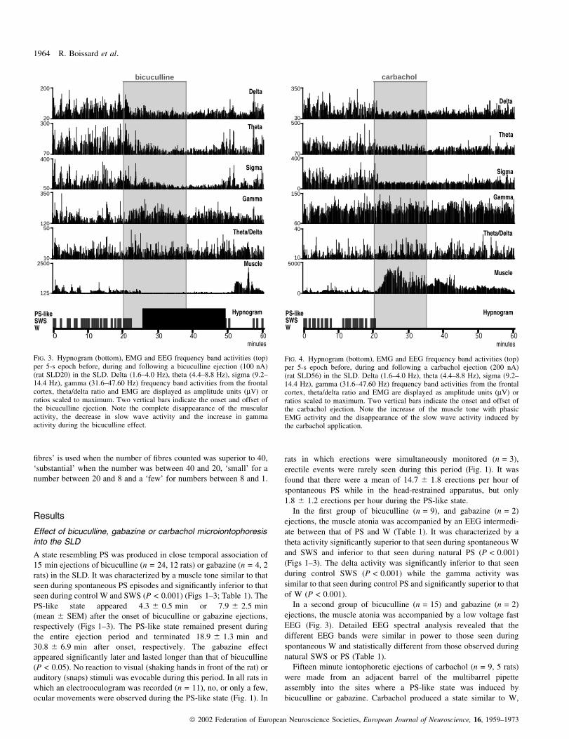
A state resembling PS was produced in close temporal association of
15 min ejections of bicuculline (n = 24, 12 rats) or gabazine (n = 4, 2
rats) in the SLD. It was characterized by a muscle tone similar to that
seen during spontaneous PS episodes and signi®cantly inferior to that
seen during control W and SWS (P < 0.001) (Figs 1±3; Table 1). The
PS-like state appeared 4.3 6 0.5 min or 7.9 6 2.5 min
(mean 6 SEM) after the onset of bicuculline or gabazine ejections,
respectively (Figs 1±3). The PS-like state remained present during
the entire ejection period and terminated 18.9 6 1.3 min and
30.8 6 6.9 min after onset, respectively. The gabazine effect
appeared signi®cantly later and lasted longer than that of bicuculline
(P < 0.05). No reaction to visual (shaking hands in front of the rat) or
auditory (snaps) stimuli was evocable during this period. In all rats in
which an electrooculogram was recorded (n = 11), no, or only a few,
ocular movements were observed during the PS-like state (Fig. 1). In
rats in which erections were simultaneously monitored (n = 3),
erectile events were rarely seen during this period (Fig. 1). It was
found that there were a mean of 14.7 6 1.8 erections per hour of
spontaneous PS while in the head-restrained apparatus, but only
1.8 6 1.2 erections per hour during the PS-like state.
In the ®rst group of bicuculline (n = 9), and gabazine (n = 2)
ejections, the muscle atonia was accompanied by an EEG intermedi-
ate between that of PS and W (Table 1). It was characterized by a
theta activity signi®cantly superior to that seen during spontaneous W
and SWS and inferior to that seen during natural PS (P < 0.001)
(Figs 1±3). The delta activity was signi®cantly inferior to that seen
during control SWS (P < 0.001) while the gamma activity was
similar to that seen during control PS and signi®cantly superior to that
of W (P < 0.001).
In a second group of bicuculline (n = 15) and gabazine (n = 2)
ejections, the muscle atonia was accompanied by a low voltage fast
EEG (Fig. 3). Detailed EEG spectral analysis revealed that the
different EEG bands were similar in power to those seen during
spontaneous W and statistically different from those observed during
natural SWS or PS (Table 1).
Fifteen minute iontophoretic ejections of carbachol (n = 9, 5 rats)
were made from an adjacent barrel of the multibarrel pipette
assembly into the sites where a PS-like state was induced by
bicuculline or gabazine. Carbachol produced a state similar to W,
200
20300
70400
50350
12050
102500
125
bicuculline
FIG. 3. Hypnogram (bottom), EMG and EEG frequency band activities (top)per 5-s epoch before, during and following a bicuculline ejection (100 nA)(rat SLD20) in the SLD. Delta (1.6±4.0 Hz), theta (4.4±8.8 Hz), sigma (9.2±14.4 Hz), gamma (31.6±47.60 Hz) frequency band activities from the frontalcortex, theta/delta ratio and EMG are displayed as amplitude units (mV) orratios scaled to maximum. Two vertical bars indicate the onset and offset ofthe bicuculline ejection. Note the complete disappearance of the muscularactivity, the decrease in slow wave activity and the increase in gammaactivity during the bicuculline effect.
350
30500
70400
0150
6040
105000
0
carbachol
FIG. 4. Hypnogram (bottom), EMG and EEG frequency band activities (top)per 5-s epoch before, during and following a carbachol ejection (200 nA)(rat SLD56) in the SLD. Delta (1.6±4.0 Hz), theta (4.4±8.8 Hz), sigma (9.2±14.4 Hz), gamma (31.6±47.60 Hz) frequency band activities from the frontalcortex, theta/delta ratio and EMG are displayed as amplitude units (mV) orratios scaled to maximum. Two vertical bars indicate the onset and offset ofthe carbachol ejection. Note the increase of the muscle tone with phasicEMG activity and the disappearance of the slow wave activity induced bythe carbachol application.
1964 R. Boissard et al.
ã 2002 Federation of European Neuroscience Societies, European Journal of Neuroscience, 16, 1959±1973

characterized by an increased muscle tone with or without phasic
activity and an EEG close to that seen during W and signi®cantly
different from that seen during SWS and PS (Fig. 4, Table 1). The
bicuculline ejection site successful in generating a PS-like state was
highly restricted for each rat. Deviation from the effective site
(n = 14, 8 rats) by only 0.5±1.0 mm ventro-dorsal, medio-lateral or
rostro-caudal from a positive site obtained with the same multibarrel
assembly produced a state indistinguishable from W, characterized by
low delta, sigma and theta activities and a tonic muscle activity with
or without active body movements.
Following 15 min ejection of NaCl (15 min, 100±200 nA) (n = 4,
4 rats), no change in the muscle tone or sleep-wake architecture was
seen.
A continuous injection of either bicuculline or gabazine was made
into the SLD of eight rats in order to determine if the PS-like state
could be maintained throughout the duration of this ejection period,
and to induce C-Fos expression. Ejections of bicuculline (n = 6) or
gabazine (n = 2) for 90 min induced a PS-like state with a latency of
2.38 6 1.0 min or 6.9 6 2.1 min, respectively. This PS-like state
continued throughout the 90 min ejection period of bicuculline or
gabazine for 86.9 6 3.4 min or 75.5 6 11.0 min, respectively
(Fig. 5). For all rats, the muscle tone was similar to that seen during
control PS episodes and statistically inferior from that occurring
during W and SWS (P < 0.01, Table 1). In four of the rats, the EEG
power spectrum was intermediate between PS and W (Table 1, SP-
like with theta) and in the four other rats it was close to that of W and
signi®cantly different from PS and SWS (Table 1, SP-like without
0 10 20 30 40 50 60 70 80 90 100 110 120
300
452500
250
bicuculline
FIG. 5. Hypnogram (bottom), EMG and theta frequency band activities (top)per 5-s epoch before, during and following a 90-min bicuculline ejection(100 nA) into the SLD (rat SLD18). The theta (4.4±8.8 Hz) frequency bandactivity from the frontal cortex and the EMG are displayed as amplitudeunits (mV). Two vertical bars indicate the onset and offset of the bicucullineejection. Note the complete disappearance of muscle activity and theincreased theta activity induced by the prolonged bicuculline administration.
kainic acid100
25120
40130
30100
5030
1011250
1250
FIG. 7. Hypnogram (bottom), EMG and EEG frequency band activities (top)per 5-s epoch before, during and following a kainic acid ejection (100 nA,5 min) (rat SLD09) into the SLD. Delta (1.6±4.0 Hz), theta (4.4±8.8 Hz),sigma (9.2±14.4 Hz), gamma (31.6±47.60 Hz) and theta/delta ratiofrequency band activities from the frontal cortex and EMG are displayed asamplitude units (mV) or ratios scaled to maximum. Two vertical barsindicate the onset and offset of the kainic acid ejection. Note the completedisappearance of the muscle tone and slow wave activity induced by theapplication of kainic acid. Note also the long-lasting rebound of themuscular activity after the muscle atonia.
200
2400
4400
2200
7040
102500
0
bicuculline
kynurenic acid
FIG. 6. Hypnogram (bottom), EMG and EEG frequency band activities (top)per 5-s epoch before, during and following bicuculline (100 nA, 37 min50 s) (rat SLD50) and kynurenic acid (100 nA, 3 min) ejections into theSLD. Delta (1.6±4.0 Hz), theta (4.4±8.8 Hz), sigma (9.2±14.4 Hz), gamma(31.6±47.60 Hz) and theta/delta ratio frequency band activities from thefrontal cortex and EMG are displayed as amplitude units (mV) or ratiosscaled to maximum. The vertical bars indicate the onset and offset of theejections. Note the complete muscle atonia and the decrease in the slowwave activity induced by the bicuculline application. When kynurenic acidis applied in addition to bicuculline, the muscle tone rapidly increased backto its prebicuculline value while the EEG is not modi®ed.
REM sleep brainstem structures in rats 1965
ã 2002 Federation of European Neuroscience Societies, European Journal of Neuroscience, 16, 1959±1973

theta). Rats which received a saline (100±200 nA) (n = 4) ejection in
the SLD during the 90 min, showed no decrease in muscle tone. They
spent 45.4 6 12.7 min in W, 33.5 6 11.7 min in SWS and
6.9 6 2.5 min in PS.
All rats used for examination of C-Fos expression were removed
from the restraining device immediately after the end of the
bicuculline, gabazine or NaCl ejection period.
Effect of bicuculline and kynurenic acid coadministration
The iontophoretic administration of bicuculline (100nA) into the SLD
in this set of experiments (n = 6, 4 rats) also induced a PS-like state
with a latency of 4.9 6 0.9 min characterized by an EMG similar to
that seen during control PS episodes and statistically inferior to that
seen during control SWS and W (Table 1). The EEG power activity
was either intermediate between that seen during PS and W (n = 4) or
similar to that of W (n = 2) (Table 1). At least 5 min after the
beginning of the bicuculline effect, kynurenic acid (100 nA) was
coadministered from an adjacent barrel for 3±10 min (Fig. 6).
Following kynurenic acid application, the EMG muscle tone
increased and reached a value similar to that seen during W and
statistically superior to that seen during PS (P < 0.001) with a latency
and duration of 2.3 6 0.9 min and 7.0 6 0.8 min, respectively. With
continued bicuculline administration, the EMG then returned to the
previous muscle atonia with an amplitude similar to that seen during
control PS episodes (Fig. 7). During the increase of muscle tone
induced by kynurenic acid coadministration, no signi®cant change in
the EEG pattern was observed as compared to bicuculline alone
(Table 1).
Effect of kainic acid microiontophoresis in the SLD
Iontophoretic application of kainic acid (100 nA) in the SLD (n = 5,
3 rats) for 5 min induced a PS-like state characterized by a complete
disappearance of muscle tone within 2.1 6 0.7 min, for a duration of
9.3 6 1.2 min (Fig. 7, Table 1). The muscle atonia was associated
with an activation of the EEG. The delta and theta activities during
the kainic acid effect were similar to those seen during control W and
statistically different from that of PS and SWS (P < 0.05, Table 1).
The PS-like state obtained with kainic acid was followed in all cases
by a large increase in muscle tone associated with active movements
of the extremities and a low voltage EEG activity, resembling
wakefulness, for a duration of 62.2 6 28.56 min
Localization of the injection sites
The bicuculline, gabazine or kainic acid positive injection sites were
localized by means of CTb, PHA-L, PSB or C-Fos staining. For all
injections that induced a complete muscle atonia with an EEG close
to W or intermediate between W and PS, the sites were intermingled
in, or adjacent to, the SLD (Figs 8A and B, and Fig 9) as de®ned by
Swanson (1992). The majority of the sites (n = 9) were localized in
the portion of the nucleus just rostral to the motor trigeminal nucleus
(V) (Figs 8B and 9B). A smaller number of positive sites (n = 7)
were localized in the SLD region at the level of the rostral portion of
the V (Figs 8A, and 9C and D).
The CTb, PHA-L or PSB injection sites localized in the pontine
periaqueductal grey (PAG) region, encompassing the rostral pole of
the LC, Barrington nucleus, and MEV (Fig. 9C and D) or in the
caudal pontine reticular nucleus, medial to the V, the medial
parabrachial nucleus (PBm) or the rostral pontine reticular nucleus
corresponded to ejections that induced W characterized by an
increase in muscle activity and an activated EEG.
The distribution of C-Fos-labelled neurons in the injection site was
studied in animals that received a 90-min ejection of bicuculline
(n = 6) or gabazine (n = 2) immediately prior to transcardial
perfusion, as well as in NaCl control (n = 4) ejected animals. In
the case of bicuculline or gabazine ejections that induced a 90 min
sustained period of a PS-like state, a large number of C-Fos-labelled
neurons were localized in the SLD and the adjacent ventral LDT,
dorsal pontine reticular and PPN nuclei within a radius of 350±
500 mm around the injection point (Fig. 10A, Table 2). On sections
double-stained with choline acetyltransferase (ChAT) and C-Fos,
only occasional (1±2 per section) C-Fos/ChAT neurons were
observed in the SLD and LDT. In control animals injected with
NaCl, only a few C-Fos labelled neurons were seen in the SLD and
surrounding nuclei at the same level (Table 2).
Projections of the SLD
Following PHA-L or CTb injections restricted to the SLD, in which a
positive effect was obtained, the anterograde labelling seen was
bilateral with a slight ipsilateral predominance.
FIG. 8 (A) Photomicrograph illustrating a PHA-L injection site in the caudal part of the SLD at the level of the rostral V (rat SLD21). (B) Photomicrographillustrating a CTb injection site in the rostral SLD just rostral to the trigeminal motor nucleus (rat SLD24). Bars, 1 mm. Abbreviations: LDT, laterodorsaltegmental nucleus; MEV, mesencephalic trigeminal nucleus; scp, superior cerebellar peduncle; V, trigeminal motor nucleus.
1966 R. Boissard et al.
ã 2002 Federation of European Neuroscience Societies, European Journal of Neuroscience, 16, 1959±1973

A large bundle of descending ®bres was localized in the dorso-
lateral part of the caudal pontine reticular nucleus medial to the V. The
bundle continued caudally ventral to the genu of the facial nerve and
then terminated dorso-medially to the rostral facial motor nucleus. At
the medullary level, a large number of anterogradely labelled varicose
®bres were observed in the gigantocellular reticular (GRN), magno-
cellular reticular (MARN) (Figs 10C and D) and parvicellular reticular
(PARN) nuclei (Fig. 11D). A substantial number of varicose ®bres
were also localized in the raphe magnus (RM), parvicellular reticular
alpha (PARNA), and dorsal and lateral paragigantocellular reticular
nuclei (PGRNd, PGRNl) (Figs 11C and D).
At the level of the site of injection, a large number of varicose
®bres were seen in the contralateral SLD and in the caudal pontine
reticular nucleus (PRNc). Slightly rostral to the site, a very large
number of ®bres were localized in the PRNr region ventrolateral to
the caudal ventral tegmental nucleus of Gudden. At this level and
more rostrally, a substantial number of varicose ®bres were diffusely
distributed bilaterally in the other parts of the PRNr and to a lesser
extent the LDT and the lateral parabrachial nucleus (PBl). A small
number of ®bres were distributed in the median raphe nucleus.
A bundle of ascending ®bres was clustered in the dorsal tegmental
bundle (dtg) all along its rostro-caudal extension. All along its path,
the bundle left a large number of varicose ®bres in the mesencephalic
reticular nucleus (MRN) caudally and also more rostrally in the
caudal interstitial nucleus. At this level, a substantial number of
varicose ®bres were localized in the ventrolateral PAG and to a lesser
extent the dorsal raphe nucleus. A small number of ®bres were seen in
the red nucleus. The bundle ended in the rostral part of the interstitial
and parafascicular thalamic nuclei, in which a large number varicose
®bres was observed. More rostrally, a large number of passing and
varicose ®bres was seen in the zona incerta (ZI). A substantial
number of varicose ®bres were seen in the lateral hypothalamic area
and the perifornical nucleus. At this level, a large number of varicose
®bres were localized in the central medial, paracentral and
centrolateral thalamic nuclei. Finally, a small number of ®bres were
seen in the lateral preoptic area and the nucleus of the horizontal limb
of the diagonal band.
Distribution of the C-Fos labelled cells
The distribution of C-Fos-labelled neurons in the main SLD efferents
was studied in three animals that received a continuous ejection of
bicuculline or gabazine for 90 min immediately prior to perfusion
and in three control, NaCl ejected animals.
At caudal pontine and medullary level, the PARN, PARNA,
MARN, cRM and PGRNl contained a substantial to large number of
C-Fos labelled cells. The labelling was bilateral for the cRM, PGRNl
and PARN and predominantly ipsilateral for the PARNA and MARN
(Table 2, Figs 11C and D). In control animals, only occasional C-
Fos-labelled cells were observed in these structures (Table 2). The
difference in number of cells between drug and control condition
reached statistical signi®cance for the PARN, MARN and PGRNl
(P < 0.05, Table 2). Following drug application, a small number of
C-Fos-labelled cells statistically superior to control condition was
also localized in the PGRNd and the GRN (Table 2).
In the major rostral efferents of the SLD, a substantial to large
number of neurons was seen in the ventrolateral PAG, MRN,
parafascicular thalamic nucleus (PF), ZI, lateral hypothalamic area
and the intralaminar thalamic nuclei in drug treated animals
(Fig. 11A). The number of cells in these structures was generally
higher compared to control condition (Table 2) but it reached
statistical signi®cance only for the intralaminar thalamic nuclei.
Localization of the glycinergic neurons activated during PS-likestate
In order to localize the glycinergic neurons responsible for muscle
atonia during PS, C-Fos/gly and PHA-L/gly immunohistochemistry
°****
°
PAGAQ
DR LDT
VTN
CUN
NLL
TRN
PG
CSI rust
cst
ml
mlf
CSmPRNr
PPN
scp
ICecic
°°° °°**
°
°°
°
ICcICe
ICd
V4
AQc
CUN
scp
MEV
KFPPN
PRNr
TRN
NTBPOR
mcp
sV
Vn
py
ml
CSI
CSm
LDTDTN
mlf
II
NLL
°
°
**
*°LDT scp
SUT
V
sctv
rust
POR
PRNc
RM
py
NTB
POR
ml
TRN
PSV sptV
mcp
FL
VCOa
tb
Vn
GV
DTN
DRmlf
KF
LC
°° °°* *
**°
LDT
LC
MEV
SUT
mlf
TRN
PRNc
VIInrust
POR
NTB
py
ml
SOCl
V
RM
PSV
sptVVCOa
VIIIn
mcp
Vn
scpPBmB
-8.30 -8.85
-9.25 -9.50
°°*
PS-like with theta
PS-like without theta
Waking
C D
A B
°
FIG. 9. Schematic representation of bicuculline, gabazine, kainic acid andcarbachol injection sites. Line drawings of coronal sections at four differentlevels of the brainstem are from the stereotaxic atlas of Swanson (1992).Each symbol represents one injection site labelled with CTb, PHAL, PSB orC-Fos. Black circles indicate the sites where bicuculline or gabazine induceda PS-like state with theta, and open circles indicate the sites wherebicuculline or gabazine induced a PS-like state without theta. Stars indicatethe sites where bicuculline or gabazine induced waking. Abbreviations: AQ,cerebral aqueduct; AQc, cerebral aqueduct, collicular recess; B, Barrington'snucleus; cic, commissure of the inferior colliculus; CSl, lateral superiorcentral nucleus rapheÂ; CSm, medial superior central nucleus rapheÂ; cst,corticospinal tract; CUN, cuneiform nucleus; DR, dorsal nucleus rapheÂ;DTN, dorsal tegmental nucleus; ICd, inferior colliculus, dorsal nucleus; ICe,inferior colliculus, external nucleus; FL, ¯occulus; GV, trigeminal ganglion;KF, KoÃlliker-Fuse subnucleus; LC, locus coeruleus nucleus; LDT,laterodorsal tegmental nucleus; ll, lateral lemniscus; mcp, middle cerebellarpeduncle; MEV, mesencephalic nucleus of the trigeminal; ml, mediallemniscus; mlf, medial longitudinal fascicle; NLL, nucleus of the laterallemniscus; NTB, nucleus of the trapezoid body; PAG, periaqueductal grey;PBm, medial parabrachial nucleus; PG, pontine grey; POR, periolivarynuclei; PPN, pedunculopontine nucleus; PRNc, caudal pontine reticularnucleus; PRNr, rostral pontine reticular nucleus; PSV, principal sensorynucleus of the trigeminal; py, pyramidal tract; RM, nucleus raphe magnus;rust, rubrospinal tract; scp, superior cerebellar peduncle; sctv, ventralspinocerebellar tract; SOCl, lateral superior olivary complex; sptV, spinaltract of the trigeminal nerve; SUT, supratrigeminal nucleus; sV, sensory rootof the trigeminal nerve; tb, trapezoid body; TRN, tegmental reticular nucleus;V, motor nucleus of the trigeminal nerve; V4, 4th ventricle; VCOa, anteriorventral cochlear nucleus; VIIn, facial nerve; VIIIn, vestibulocochlear nerve;Vn, trigeminal nerve; VTN, ventral tegmental nucleus.
REM sleep brainstem structures in rats 1967
ã 2002 Federation of European Neuroscience Societies, European Journal of Neuroscience, 16, 1959±1973

were combined. On PHA-L/gly double-labelled sections, a large
number of PHA-L varicose ®bres were seen in close vicinity to
glycine-immunoreactive neurons in the ipsilateral MARN (Figs 10D
and E), cRM and bilaterally in the PARN and PARNA. On C-Fos/gly
double-immunostained sections from rats injected with bicuculline or
gabazine for 90 min before perfusion, a substantial number of
double-labelled neurons was seen with an ipsilateral predominance in
the PRNc, MARN, and bilaterally in the cRM (Fig. 10B), PGRNl and
PARN (Fig. 12)(Table 2). In addition, in these animals a small
number of double-labelled cells were seen in the PARNA, and
PGRNd. In the control condition, no, or only occasional, cells were
seen in these nuclei. The difference between drug and control
conditions was statistically signi®cant for the MARN, PGRNl, PARN
and PGRNd (P < 0.05, Table 2).
Discussion
This study demonstrates for the ®rst time that a long-lasting PS-like
hypersomnia can be pharmacologically induced with a short latency in
rats by iontophoretically administering bicuculline, gabazine or kainic
acid speci®cally into the SLD. Carbachol, however, was not effective
in this regard. It was also shown that the bicuculline effect could be
reversed by the application of kynurenic acid. It was further
demonstrated that ventromedial and lateral medullary reticular struc-
tures receive a strong projection from the SLD and contain numerous
C-Fos and glycine double-stained neurons, following prolonged
application of bicuculline or gabazine in the SLD. These results
suggest that PS executive neurons, localized in the SLD in the rat, play
a role in PS generation and the maintenance of muscle atonia via the
FIG. 10 (A) Photomicrograph showing a frontal section double-labelled with ChAT (in brown) and C-Fos (in black) immunohistochemistry in an animal(SLD43), which received a 90-min bicuculline injection into the SLD just before perfusion. Numerous C-Fos labelled cells are visible in the SLD just ventralto the LDT, which contains single stained ChAT neurons. Bar, 500 mm. (B) Photomicrograph showing the magnocellular reticular nucleus MARN on afrontal section double-labelled with C-Fos (in black) and glycine (in brown) immunohistochemistry in an animal (SLD 51) with a 90-min bicuculline injectionin the SLD just before perfusion. One double-labelled (double arrow) and several C-Fos singly labelled neurons (arrows) are visible. Bar, 50 mm. (C)Photomicrograph showing the magnocellular reticular nucleus on a frontal section double-labelled with C-Fos (in brown) and PHA-L (in black)immunohistochemistry in an animal with a PHA-L injection in a bicuculline positive site in the SLD and a 90-min injection in the same site 10 days laterjust before perfusion. Numerous anterogradely labelled varicose ®bres cover the MARN, which also contained several C-Fos immunoreactive neurons. Severalvaricosities can be seen over a C-Fos labelled nucleus. Bar, 50 mm. (D and E) Photomicrographs showing the MARN on two different frontal sectionsdouble-labelled with glycine (in brown) and PHA-L (in black) immunohistochemistry in two different animals with a PHA-L injection in a bicucullinepositive site in the SLD. In D, a large contingent of anterogradely labelled varicose ®bres are apposed on glycinergic neurons localized in the MARN justdorsal to the inferior olivary complex (SLD 25). In E, varicosities of anterogradely labelled ®bres are apposed on the proximal dendrite of a glycine-immunoreactive neuron and one varicosity is apposed on the soma of the neuron (E11). Bars, 100 mm in D and 20 mm in E. Abbreviations: IO, inferior olive;LDT, laterodorsal tegmental nucleus; scp, superior cerebellar peduncle.
1968 R. Boissard et al.
ã 2002 Federation of European Neuroscience Societies, European Journal of Neuroscience, 16, 1959±1973

following mechanisms: (i) they are tonically inhibited by GABA
during waking and slow wave sleep; (ii) they receive a tonic
glutamatergic input during all states of vigilance; and (iii) they
directly project to intralaminar thalamic neurons and glycinergic
neurons localized in the ventromedial and lateral medullary reticular
formation.
There are numerous differences between rats and cats regarding the
effectiveness of carbachol administration into the pontine reticular
formation, and most studies have had limited success replicating in
rats the strong PS hypersomnia obtained in cats (Baghdoyan et al.,
1987; Vanni-Mercier et al., 1989; Lai & Siegel, 1990; Yamamoto
et al., 1990; GarzuÁn et al., 1998). The enhancement of PS in rats was
of small magnitude (Gnadt & Pegram, 1986; Shiromani & Fishbein,
1986; Velazquez-Moctezuma et al., 1989; Bourgin et al., 1995; Datta
et al., 1998) or not reliably obtained (Deurveilher et al., 1997). The
increase in PS, when obtained, was less than 100% compared to the
300% increase obtained in cats. In addition, the ®rst PS episode
appeared at least 50 min after the carbachol injection and the duration
of the episodes were similar to natural, spontaneous PS. In cats,
however, PS is induced almost immediately after the injection and the
episodes last longer than in control PS. The effective sites in rats were
widely distributed in the pontine reticular formation including the
PRNr, PRNc and the SLD. In contrast, the effective sites in cats were
either localized just ventral to the locus coeruleus alpha nucleus, in an
area called peri-LCa by Sakai et al. (1979, 1981), or in the ventral
part of the PRNr (GarzuÁn et al., 1998). In the present experiments,
carbachol applications were unable to induce a PS-like state when
applied to the dorsal parts of the PRNr, PRNc or the SLD. Instead
carbachol induced a W state with an increase in muscle tone. These
results suggest a strong species difference between rats and cats. The
absence of an effect of carbachol does not rule out a role of
cholinergic processes in PS onset and maintenance in the rat. It
nevertheless suggests that cholinergic input to SLD neurons plays at
most a minor role in their regulation in this species. It remains to be
determined whether the cholinergic system plays an important role in
PS control in rats via a different population of neurons located in
another pontine region. It is also possible that PS-on neurons in the
SLD region express muscarinic and/or nicotinic receptors, but that the
FIG. 11. Schematic drawings of PHA-L labelled ®bres (in black) and C-Fos labelled neurons (red dots) on frontal sections from hypothalamic to medullarylevels from an animal which received a PHA-L injection in a bicuculline positive site in the SLD and a 90-min injection of bicuculline in the same site justbefore perfusion (rat SLD 50). Abbreviations: AQ, aqueduct of Sylvius; CEA, central nucleus amygdala; CM, central medial nucleus thalamus; CSm, medialsuperior central nucleus rapheÂ; DTN, dorsal tegmental nucleus; GPm, medial globus pallidus; GRN, gigantocellular reticular nucleus; int, internal capsule; IO,inferior olivary complex; LC, locus coeruleus; LDT, laterodorsal tegmental nucleus; MARN, magnocellular reticular nucleus; mcp, middle cerebellarpeduncle; MEV, mesencephalic trigeminal nucleus; ml, medial lemniscus; MV, medial vestibular nucleus; NTB, nucleus of the trapezoid body; NTS, nucleusof the solitary tract; opt, optic tract; PARN, parvicellular reticular nucleus; PARNA, parvicellular reticular nucleus alpha; PBl, lateral parabrachial nucleus;PGRNd, dorsal paragigantocellular reticular nucleus; PGRNl, lateral paragigantocellular reticular nucleus; PRNc, caudal pontine reticular nucleus; py,pyramidal tract; RM, nucleus raphe magnus; RT, reticular nucleus thalamus; scp, superior cerebellar peduncle; SLD, sublaterodorsal nucleus; SOCm, medialsuperior olivary complex; SPIV, spinal vestibular nucleus; sptV, spinal tract of the trigeminal nerve; SPVO, spinal trigeminal nucleus, oral part; tb, trapezoidbody; V3, 3rd ventricle; V4, 4th ventricle; VI, abducens nucleus; VIIn, facial nerve.
REM sleep brainstem structures in rats 1969
ã 2002 Federation of European Neuroscience Societies, European Journal of Neuroscience, 16, 1959±1973

activation of these receptors by carbachol is unable to modify their
activity. This effect could be due to the strong GABAergic tonic
inhibition revealed by these results, indeed, microiontophoresis of
bicuculline or gabazine, two different GABAA antagonists, in the
SLD region induced a state closed to PS. These data rule out the
possibility that the sustained PS-like state is due to a nonspeci®c
neuronal activation by bicuculline, as previously suggested under
different experimental conditions (Debarbieux et al., 1998). It
strongly suggests that GABA tonically inhibits PS-on neurons
localized in the SLD during W and SWS. The results reported here
are in agreement with a recent study in cats showing that pressure
application of bicuculline in the peri-LCa region induces a strong
increase in PS quantities with short latencies, whereas the application
of GABA strongly decreases PS (Xi et al., 1999).
The iontophoretic application of kainic acid into the SLD was
shown to be consistently associated with a transient PS-like state
followed by W and an increase in muscle activity. In agreement with
our results, it has been shown in cats that the administration of kainic
acid in the peri-LCa using microdialysis induces a PS-like state (Onoe
& Sakai, 1995). Finally, the present research showed that the
application of kynurenic acid, an excitatory amino acid antagonist,
reversed the PS-like state induced by bicuculline application in the
SLD. Altogether, these results suggest that PS-on neurons in the SLD
region receive a tonic glutamatergic input during all sleep-waking
states.
Altogether, these data imply that the onset of PS, at least in rats, is
mainly due to the removal of a tonic GABAergic input present during
W and SWS. The GABAergic neurons responsible for this tonic
inhibition could be either interneurons or long projection neurons.
Indeed, GABAergic interneurons have been localized in the SLD
(Ford et al., 1995) and GABAergic neurons with long projections to
brainstem nuclei have been identi®ed as well (Gervasoni et al., 2000).
In the dorsal pontine reticular formation, neurons responsible for
PS onset and maintenance seem to be clustered in the SLD. Indeed,
injections just 500 mm away from the positive sites induced W with
increased muscle activity. Moreover, C-Fos-labelled neurons at the
site of injection, following long bicuculline applications, occupied
less than 1 mm3. The neurochemical nature of the neurons in the SLD
remains to be determined. It is unlikely that they are cholinergic as it
was shown that the great majority of the C-Fos positive neurons in the
injection site were not immunoreactive to ChAT. One likely
possibility is that these neurons are glutamatergic neurons, as the
C-Fos experiments highly suggest that they provide an excitatory
pathway to glycinergic neurons in the medullary reticular formation.
Additional studies are necessary to test this hypothesis.
Bicuculline or gabazine administration into the SLD induced a PS-
like state characterized by the presence of muscle atonia, EEG
activation with or without theta activity, and nonreactivity to
surrounding stimuli, but with the absence of ocular movements or
penile erections. The absence of rapid eye movements and penile
erections may be due to the fact that, unlike pressure ejections used in
previous cat or rat studies, our iontophoretic ejections are very limited
and therefore involve only a portion of the neurons responsible for PS
onset. Additional populations of neurons surrounding the SLD may
participate in the theta genesis and be responsible for phasic activities.
In particular, cholinergic and noncholinergic neurons from the LDT
FIG. 12. Schematic drawings, rostrocaudaly arranged, of C-Fos singly (open circles) and C-Fos/glycine double labelled cells (®lled circles) on frontalmedullary sections from an animal (rat SLD 43) which received just before perfusion a 90-min bicuculline injection into the SLD. Abbreviations: AMB,nucleus ambiguus; CVL, caudoventrolateral reticular nucleus; icp, inferior cerebellar peduncle; IO, inferior olivary complex; MARN, magnocellular reticularnucleus; MV, medial vestibular nucleus; NTB, nucleus of the trapezoid body; NTS, nucleus of the solitary tract; PARNA, parvicellular reticular nucleusalpha; PGRNd, dorsal paragigantocellular reticular nucleus; py, pyramidal tract; scp, superior cerebellar peduncle; SOCm, medial superior olivary complex;sptV, spinal tract of the trigeminal nerve; SPVO, oral spinal trigeminal nucleus; SPIV, spinal vestibular nucleus; tb, trapezoid body; tsp, tectospinal pathway;V4, 4th ventricle; VII, facial nucleus; VIIn, facial nerve; vVIIIn, vestibular nerve; XII, hypoglossal nucleus.
1970 R. Boissard et al.
ã 2002 Federation of European Neuroscience Societies, European Journal of Neuroscience, 16, 1959±1973

could be responsible for the missing tonic or phasic events. Supporting
this hypothesis, neurons speci®cally active during PS-related erections
have been recently recorded in the LDT (Koyama et al., 2001).
Moreover, an increased number of C-Fos positive neurons have been
observed in the LDT following PS rebound (Maloney et al., 1999). In
the experiments described here, bicuculline injections into the LDT
induced W in apparent contradiction with this hypothesis. This ®nding
may be explained by the fact that the majority of the LDT neurons
exhibited a high ®ring rate both during W and PS, whereas only a
minority were speci®cally active during PS (El Mansari et al., 1989;
Steriade et al., 1990).
In this study, it was also attempted to determine the neuronal
network responsible for the cortical activation and the muscle atonia
seen following bicuculline, gabazine or kainic acid applications in the
SLD. Concerning the muscle atonia, it was demonstrated that
numerous glycine-immunoreactive neurons contained C-Fos in the
cRM, MARN, PARN and PARNA following bicuculline or gabazine
application in the SLD region. It is likely that that at least some of these
glycinergic neurons are responsible for the muscle atonia seen during
PS. Indeed, intracellular recordings of motoneurons combined with
strychnine applications show that glycine is responsible for the tonic
hyperpolarization of the spinal, hypoglossal and trigeminal motoneur-
ons during PS (Chase et al., 1989; Soja et al., 1991; Kohlmeier et al.,
1996; Yamuy et al., 1999). It has also been shown that glycinergic
neurons from the MARN directly project to spinal motoneurons
(Holstege & Bongers, 1991) while glycinergic neurons from the PARN
and PARNA directly project to the trigeminal motor nucleus (Li et al.,
1996; Rampon et al., 1996). Moreover, it has been shown in cats
following induction of PS by carbachol injections in the peri-LCa that
cells in the MARN labelled with C-Fos project to the V (Morales et al.,
1999). Altogether, these results indicate that glycinergic neurons from
the MARN, PARN and PARNA could be responsible for the
hyperpolarization of spinal and cranial motoneurons during PS.
The idea that glycinergic neurons, within the PARN, RM and
MARN, may be involved in the generation of muscle atonia, is also
supported by the ®nding that, there is a strong projection from the SLD
to these particular nuclei. Indeed, previous studies in cats demonstrate
a strong projection from the peri-LCa to the magnocellular reticular
nucleus (Sakai et al., 1979; Luppi et al., 1988). In contrast, only a
minor projection from the peri-LCa to the PARN has been described
(Fort et al., 1994). The apparent discrepancy between this and the
present study might be due to the fact that the strongest projection from
the SLD to the PARN is destined to the caudal and lateral parts of the
nucleus, a region not examined in the previous study.
In addition to descending projections to the medullary reticular
formation, it was also found that the SLD region projects to rostral
structures like the intralaminar thalamic nuclei, intrafascicular
thalamic nucleus and zona incerta, in agreement with Jones & Yang
(1985). The ascending projection to the intralaminar thalamic nuclei
could be responsible for the cortical activation obtained during the
administration of bicuculline, gabazine or kainic acid into the SLD as
numerous C-Fos labelled neurons were localized in these nuclei
following long lasting bicuculline or gabazine ejections into the SLD.
Conclusions
In the present study, a pontine PS-like-inducing region, similar to that
previously found in cats, was localized for the ®rst time in rats. The
results provide further strong evidence that, neurons in the SLD are
tonically inhibited by GABA during both W and SWS, and that they
are under constant glutamatergic control and are unresponsive to
carbachol. Using anterograde tracing with PHA-L combined with C-
Fos and glycine immunohistochemistry, it was also shown that the
SLD neurons provide excitatory projections to glycinergic neurons
from the ventromedial and lateral medullary reticular formation.
From these results, it may be proposed that the onset and maintenance
of PS could be due to the activation of PS-on neurons localized in the
SLD. The activation of these neurons would primarily be due to the
removal of a tonic GABAergic inhibitory input, present during W and
SWS in combination with a constant tonic glutamatergic input.
Further studies are now necessary to test this new hypothesis.
Acknowledgements
This work was supported by INSERM (U480), CNRS (CNRS FRE2469),Universite C. Bernard Lyon I, the Ohio Sleep Medicine and NeuroscienceInstitute, and the Sleep Medicine Research Foundation. Romuald Boissardreceived a PhD grant from the Fondation pour la Recherche MeÂdicale. Theauthors wish to thank C. Guillemort (GFG Co, Pierre-BeÂnite, France) for hishelp in designing the head-restraining system.
Abbreviations
ChAT, choline acetyltransferase; cRM, caudal nucleus raphe magnus; CSP,corpus spongiosum of the penis; CTb, cholera-toxin B subunit; DAB, 3,3¢-diaminobenzidine-4 HCl; dtg, dorsal tegmental bundle; EEG, electroencepha-logram; EMG, electromyogram; EOG, electrooculogram; gly, glycine; GRN,gigantocellular reticular nucleus; ILM, intralaminar nuclei of the dorsalthalamus; LC, locus coeruleus nucleus; LDT, laterodorsal tegmental nucleus;LHA, lateral hypothalamic area; MARN, magnocellular reticular nucleus;MEV, mesencephalic nucleus of the trigeminal; MRN, mesencephalicreticular nucleus; PAG, periaqueductal grey; PARN, parvicellular reticularnucleus; PARNA, parvicellular reticular nucleus alpha; PB, phosphate buffer;PBl, lateral parabrachial nucleus; PBm, medial parabrachial nucleus; PBST,phosphate buffer saline Triton X; PBST-Az, phosphate buffer saline Triton Xazide; peri-LCa, peri-locus coeruleus a; PF, parafascicular thalamic nucleus;PGRNd, dorsal paragigantocellular reticular nucleus; PGRNl, lateral para-gigantocellular reticular nucleus; PHA-L, phaseolus vulgaris leucoaglutinin;PPN, pedunculopontine nucleus; PRNc, caudal pontine reticular nucleus;PRNr, rostral pontine reticular nucleus; PS, paradoxical sleep; PSB, pontaminesky blue; REM, rapid eye movement sleep; RM, nucleus raphe magnus; SLD,sublaterodorsal nucleus; SWS, slow wave sleep; V, trigeminal motor nucleus;W, waking; ZI, zona incerta.
References
Akaoka, H., Charlety, P.J., Saunier, C.F., Buda, M. & Chouvet, G. (1992)Combining in vivo volume -controlled pressure microinjection withextracellular unit recording. J. Neurosci. Meth, 42, 119±129.
Aserinsky, E. & Kleitman, N. (1953) Regularly occurring periods of eyemotility and concomitant phenomena during sleep. Science, 118, 273±274.
Baghdoyan, H.A., Rodrigo-Angulo, M.L., McCarley, R.W. & Hobson, J.A.(1987) A neuroanatomical gradient in the pontine tegmentum for thecholinoceptive induction of desynchronized sleep signs. Brain Res., 414,245±261.
Bourgin, P., Escourrou, P., Gaultier, C. & Adrien, J. (1995) Induction of rapideye movement sleep by carbachol infusion into the pontine reticularformation in the rat. Neuroreport, 6, 532±536.
Chase, M.H., Soja, P.J. & Morales, F.R. (1989) Evidence that glycine mediatesthe postsynaptic potentials that inhibit lumbar motoneurones during theatonia of active sleep. J. Neurosci., 9, 743±751.
Darracq, L., Gervasoni, D., SoulieÁre, F., Lin, J.S., Fort, P., Chouvet, G. &Luppi, P.H. (1996) Effect of strychnine on rat locus coeruleus neuronesduring sleep and wakefulness. Neuroreport, 8, 351±355.
Datta, S., Siwek, D.F., Patterson, E.H. & Cipolloni, P.B. (1998) Localizationof pontine PGO wave generation sites and their anatomical projections inthe rat. Synapse, 30, 409±423.
Debarbieux, F., Brunton, J. & Charpak, S. (1998) Effect of bicuculline onthalamic activity: a direct blockade of IAHP in reticularis neurones. J.Neurophysiol., 79, 2911±2918.
Dement, W. (1958) The occurrence of low voltage, fast, electroencephalogram
REM sleep brainstem structures in rats 1971
ã 2002 Federation of European Neuroscience Societies, European Journal of Neuroscience, 16, 1959±1973

patterns during behavioral sleep in the cat. Electroencephalogr. Clin.Neurophysiol., 10, 291±296.
Dement, W. & Kleitman, N. (1957) Cyclic variations of EEG during sleep andtheir relation to eye movement., body motility. & dreaming.Electroencephalogr. Clin. Neurophysiol., 9, 673±690.
Deurveilher, S., Hars, B. & Hennevin, E. (1997) Pontine microinjection ofcarbachol does not reliably enhance paradoxical sleep in rats. Sleep, 20,593±607.
El Mansari, M., Sakai, K. & Jouvet, M. (1989) Unitary characteristics ofpresumptive cholinergic tegmental neurones during the sleep-waking cyclein freely moving cats. Exp. Brain. Res., 76, 519±529.
Ford, B., Holmes, C.J., Mainville, L. & Jones, B.E. (1995) GABAergicneurones in the rat pontomesencephalic tegmentum: codistribution withcholinergic and other tegmental neurones projecting to the posterior lateralhypothalamus. J. Comp. Neurol., 363, 177±196.
Fort, P., Luppi, P.H. & Jouvet, M. (1993) Glycine-immunoreactive neuronesin the cat brain stem reticular formation. Neuroreport, 4, 1123±1126.
Fort, P., Luppi, P.H. & Jouvet, M. (1994) Afferents to the nucleus reticularisparvicellularis of the cat medulla oblongata: a tract-tracing study withcholera toxin B subunit. J. Comp. Neurol., 342, 603±618.
Fort, P., Luppi, P.H., Wenthold, R. & Jouvet, M. (1990) [Glycineimmunoreactive neurones in the medulla oblongata in cats] Neuronesimmunoreactifs a la glycine dans le bulbe rachidien du chat. C. R. Acad.Sci., III, 311, 205±212.
GarzuÁn, M., De AndreÁs, I. & Reinoso-Suarez, F. (1998) Sleep patterns aftercarbachol delivery in the ventral oral pontine tegmentum of the cat.Neuroscience, 83, 17±31.
Gervasoni, D., Darracq, L., Fort, P., SoulieÁre, F., Chouvet, G. & Luppi, P.H.(1998) Electrophysiological evidence that noradrenergic neurones of the ratlocus coeruleus are tonically inhibited by GABA during sleep. Eur. J.Neurosci., 10, 964±970.
Gervasoni, D., Peyron, C., Rampon, C., Barbagli, B., Chouvet, G., Urbain, N.,Fort, P. & Luppi, P.H. (2000) Role and origin of the GABAergicinnervation of dorsal raphe serotonergic neurones. J. Neurosci., 20, 4217±4225.
Gnadt, J.W. & Pegram, G.V. (1986) Cholinergic brainstem mechanisms ofREM sleep in the rat. Brain Res., 384, 29±41.
Hobson, J.A., McCarley, R.W. & Wyzinski, P. (1975) Sleep cycle oscillation:reciprocal discharge by two brainstem neuronal groups. Science, 189, 55±58.
Holstege, J.C. & Bongers, C.M. (1991) A glycinergic projection from theventromedial lower brainstem to spinal motoneurones. An ultrastructuraldouble labeling study in rat. Brain Res., 566, 308±315.
Jones, B.E. (1991) Paradoxical sleep and its chemical/structural substrates inthe brain. Neuroscience, 40, 637±656.
Jones, B.E. & Yang, T.Z. (1985) The efferent projections from the reticularformation and the locus coeruleus studied by anterograde and retrogradeaxonal transport in the rat. J. Comp. Neurol., 242, 56±92.
Jouvet, M. & Michel, F. (1959) CorreÂlations eÂlectromyographiques dusommeil chez. le chat deÂcortique et meÂsenceÂphalique chronique. C. R.Soc. Biol., 153, 422±425.
Kohlmeier, K.A., Lopez-Rodriguez, F., Liu, R.H., Morales, F.R. & Chase,M.H. (1996) State-dependent phenomena in cat masseter motoneurones.Brain Res., 722, 30±38.
Koyama, Y. & Sakai, K. (2000) Modulation of presumed cholinergicmesopontine tegmental neurones by acetylcholine and monoaminesapplied iontophoretically in unanesthetized cats. Neuroscience, 96, 723±733.
Koyama, Y., Schmidt, M.H., Takahashi, K. & Kayama, Y. (2001) Cholinergicneurones in the laterodorsal tegmental nucleus regulate penile erectionduring paradoxical sleep. Soc. Neurosci. Abstr., 522.20.
Kubin, L. (2001) Carbachol models of REM sleep: Recent developments andnew directions. Arch. Ital. Biol., 139, 147±168.
Lai, Y.Y. & Siegel, J.M. (1990) Cardiovascular and muscle tone changesproduced by microinjection of cholinergic and glutamatergic agonists indorsolateral pons and medial medulla. Brain Res., 514, 27±36.
Li, Y.Q., Takada, M., Kaneko, T. & Mizuno, N. (1996) GABAergic andglycinergic neurones projecting to the trigeminal motor nucleus: a doublelabeling study in the rat. J. Comp. Neurol., 373, 498±510.
Luppi, P.H., Aston-Jones, G., Akaoka, H., Chouvet, G. & Jouvet, M. (1995)Afferent projections to the rat locus coeruleus demonstrated by retrogradeand anterograde tracing with cholera-toxin B subunit and Phaseolus vulgarisleucoagglutinin. Neuroscience, 65, 119±160.
Luppi, P.H., Fort, P. & Jouvet, M. (1990) Iontophoretic application ofunconjugated cholera toxin B subunit CTb combined with
immunohistochemistry of neurochemical substances: a method fortransmitter identi®cation of retrogradely labelled neurones. Brain Res.,534, 209±224.
Luppi, P.H., Sakai, K., Fort, P., Salvert, D. & Jouvet, M. (1988) The nuclei oforigin of monoaminergic., peptidergic. & cholinergic afferents to the catnucleus reticularis magnocellularis: a double-labeling study with choleratoxin as a retrograde tracer. J. Comp. Neurol., 277, 1±20.
Maloney, K.J., Mainville, L. & Jones, B.E. (1999) Differential c-Fosexpression in cholinergic., monoaminergic and GABAergic cell groups ofthe pontomesencephalic tegmentum after paradoxical sleep deprivation andrecovery. J. Neurosci., 19, 3057±3072.
Morales, F.R., Sampogna, S., Yamuy, J. & Chase, M.H. (1999) C-Fosexpression in brainstem premotor interneurones during cholinergicallyinduced active sleep in the cat. J. Neurosci., 19, 9508±9518.
Onoe, H. & Sakai, K. (1995) Kainate receptors: a novel mechanism inparadoxical (REM) sleep generation. Neuroreport, 6, 353±356.
Paxinos, G. & Watson, C. (1997) The Rat Brain in Stereotaxic Coordinates.Academic Press, Sydney.
Peyron, C., Luppi, P.H., Fort, P., Rampon, C. & Jouvet, M. (1996) Lowerbrainstem catecholamine afferents to the rat dorsal raphe nucleus. J. Comp.Neurol., 364, 402±413.
Peyron, C., Petit, J.M., Rampon, C., Jouvet, M. & Luppi, P.H. (1998)Forebrain afferents to the rat dorsal raphe nucleus demonstrated byretrograde and anterograde tracing methods. Neuroscience, 82, 443±468.
Rampon, C., Peyron, C., Petit, J.M., Fort, P., Gervasoni, D. & Luppi, P.H.(1996) Origin of the glycinergic innervation of the rat trigeminal motornucleus. Neuroreport, 7, 3081±3085.
Sakai, K., Crochet, S. & Onoe, H. (2001) Pontine structures and mechanismsinvolved in the generation of paradoxical (REM) sleep. Arch. Ital. Biol.,139, 93±107.
Sakai, K. & Koyama, Y. (1996) Are there cholinergic and non-cholinergicparadoxical sleep-on neurones in the pons? Neuroreport, 7, 2449±2453.
Sakai, K., Sastre, J.P., Kanamori, N. & Jouvet, M. (1981) State-speci®cneurones in the ponto-medullary reticular formation with specialreference to the postural atonia during paradoxical sleep in the cat. InPompeiano, O. & Aimone Marsan, C. (eds), Brain Mechanisms ofPerceptual Awareness and Purposeful Behavior. Raven Press, NewYork, pp. 405±429.
Sakai, K., Sastre, J.P., Salvert, D., Touret, M., Tohyama, M. & Jouvet, M.(1979) Tegmentoreticular projections with special reference to the muscularatonia during paradoxical sleep in the cat: an HRP study. Brain Res., 176,233±254.
Schmidt, M.H., Valatx, J.L., Sakai, K., Fort, P. & Jouvet, M. (2000) Role ofthe lateral preoptic area in sleep-related erectile mechanisms and sleepgeneration in the rat. J. Neurosci., 20, 6640±6647.
Schmidt, M.H., Valatx, J.L., Schmidt, H.S., Wauquier, A. & Jouvet, M. (1994)Experimental evidence of penile erections during paradoxical sleep in therat. Neuroreport, 5, 561±564.
Shiromani, P.J. & Fishbein, W. (1986) Continuous pontine cholinergicmicroinfusion via mini-pump induces sustained alterations in rapid eyemovement (REM) sleep. Pharmacol. Biochem. Behav., 25, 1253±1261.
Siegel, J.M. & McGinty, D.J. (1977) Pontine reticular formation neurones:relationship of discharge to motor activity. Science, 196, 678±680.
Soja, P.J., Lopez-Rodriguez, F., Morales, F.R. & Chase, M.H. (1991) Thepostsynaptic inhibitory control of lumbar motoneurones during the atonia ofactive sleep: effect of strychnine on motoneuron properties. J. Neurosci., 11,2804±2811.
Souliere, F., Urbain, N., Gervasoni, D., Schmitt, P., Guillemort, C., Fort, P.,Renaud, B., Luppi, P.H. & Chouvet, G. (2000) Single-unit and polygraphicrecordings associated with systemic or local pharmacology: a multi-purposestereotaxic approach for the awake., anaesthetic-free. & head-restrained rat.J. Neurosci. Res., 61, 88±100.
Steriade, M., Datta, S., PareÂ,D., Oakson, G. & Curro Dossi, R.C. (1990)Neuronal activities in brain-stem cholinergic nuclei related to tonicactivation processes in thalamocortical systems. J. Neurosci., 10, 2541±2559.
Stone, T.W. (1985) Microiontophoresis and pressure ejection. In Smith, A.D.(ed), Methods in the Neurosciences. Wiley, New York, pp. xxx±xxx.
Swanson, L.W. (1992) Structure of the Rat Brain. Elsevier Science Publishers,Location, pp. xxx±xxx.
Vanni-Mercier, G., Sakai, K., Lin, J.S. & Jouvet, M. (1989) Mapping ofcholinoceptive brainstem structures responsible for the generation ofparadoxical sleep in the cat. Arch. Ital. Biol., 127, 133±164.
Velazquez-Moctezuma, J., Gillin, J.C. & Shiromani, P.J. (1989) Effect of
1972 R. Boissard et al.
ã 2002 Federation of European Neuroscience Societies, European Journal of Neuroscience, 16, 1959±1973

speci®c Ml., M2 muscarinic receptor agonists on REM sleep generation.Brain Res., 503, 128±131.
Xi, M.C., Morales, F.R. & Chase, M.H. (1999) Evidence that wakefulness andREM sleep are controlled by a GABAergic pontine mechanism. J.Neurophysiol., 82, 2015±2019.
Yamamoto, K., Mamelak, A.N., Quattrochi, J.J. & Hobson, J.A. (1990) A
cholinoceptive desynchronized sleep induction zone in the anterodorsalpontine tegmentum: locus of the sensitive region. Neuroscience, 39, 279±293.
Yamuy, J., Fung, S.J., Xi, M., Morales, F.R. & Chase, M.H. (1999)Hypoglossal motoneurones are postsynaptically inhibited duringcarbachol-induced rapid eye movement sleep. Neuroscience, 94, 11±15.
REM sleep brainstem structures in rats 1973
ã 2002 Federation of European Neuroscience Societies, European Journal of Neuroscience, 16, 1959±1973