
The Pursuit of Parenthood: Expanding Horizons of - KEEP
315
The Pursuit of Parenthood: Expanding Horizons of Reproductive Physiology and Assisted Reproductive Technologies by Shelbi Marie Peck A Dissertation Presented in Partial Fulfillment of the Requirements for the Degree Doctor of Philosophy Approved April 2021 by the Graduate Supervisory Committee: Debra Page Baluch, Co-Chair Jane Maienschein, Co-Chair Karen Sweazea Karin Ellison ARIZONA STATE UNIVERSITY August 2021
-
Upload
khangminh22 -
Category
Documents
-
view
0 -
download
0
Transcript of The Pursuit of Parenthood: Expanding Horizons of - KEEP

The Pursuit of Parenthood: Expanding Horizons of
Reproductive Physiology and Assisted Reproductive Technologies
by
Shelbi Marie Peck
A Dissertation Presented in Partial Fulfillment of the Requirements for the Degree
Doctor of Philosophy
Approved April 2021 by the Graduate Supervisory Committee:
Debra Page Baluch, Co-Chair Jane Maienschein, Co-Chair
Karen Sweazea Karin Ellison
ARIZONA STATE UNIVERSITY
August 2021

i
ABSTRACT
The desire to start a family is something millions of people around the globe
strive to achieve. However, many factors such as the societal changes in family planning
due to increasing maternal age, use of birth control, and ever-changing lifestyles have
increased the number of infertility cases seen in the United States each year. Infertility
can manifest as a prolonged inability to conceive, or inability to carry a pregnancy full-
term. Modern advancements in the field of reproductive medicine have begun to promote
the use of Assisted Reproductive Technologies (ART) to circumvent reduced fertility in
both men and women. Implementation of techniques such as In Vitro Fertilization,
Intracytoplasmic Sperm Injection, and Pre-Implantation Genetic Testing have allowed
many couples to conceive. There is continual effort being made towards developing more
effective and personalized fertility treatments. This often begins in the form of animal
research—a fundamental step in biomedical research.
This dissertation examines infertility as a medical condition through the
characterization of normal reproductive anatomy and physiology in the introductory
overview of reproduction. Specific pathologies of male and female-factor infertility are
described, which necessitates the use of ARTs. The various forms of ARTs currently
utilized in a clinical setting are addressed including history, preparations, and protocols
for each technology. To promote continual advancement of the field, both animal studies
and human trials provide fundamental stepping-stones towards the execution of new
techniques and protocols. Examples of research conducted for the betterment of human
reproductive medicine are explored, including an animal study conducted in mice
exploring the role of tyramine in ovulation. With the development and implementation of

ii
new technologies and protocols in the field, this also unearths ethical dilemmas that
further complicate the addition of new technologies in the field. Combining an extensive
review in assisted reproduction, research and clinical fieldwork, this study investigates
the history and development of novel research conducted in reproductive medicine and
explores the broader implications of new technologies in the field.

iii
ACKNOWLEDGEMENTS
There are so many wonderful people in my life that I would like to express
gratitude to for their kindness, guidance, and love. To my parents, Jack, and Lynn—thank
you for nurturing my passion for science from such a young age and encouraging me to
“go all the way” with my schooling. I never took a break from being a student and have
often looked back and wished that I had—especially when I have felt burnt out and
discouraged. Your unconditional love and support over the years has motivated me to
always keep looking forward and to finish strong.
This work also would not have been possible without my advisor, Dr. Debra Page
Baluch. Her unwavering support of my research and professional development over the
years has granted me opportunities that I will never take for granted for the rest of my
career. Page, you are an exemplary scientist, mentor and visionary and I feel so fortunate
to have been able to learn from (and with) you since I began working in your laboratory
as an undergraduate student. I could not have asked for a better advisor and have an
incredible committee to thank for their support as well: Dr. Maienschein, Dr. Sweazea,
and Dr. Ellison. Your guidance has been extremely helpful throughout my writing
process, and your support to pursue opportunities at the fertility clinic has made my time
as a graduate student truly one of a kind. I would also like to acknowledge Dr. Jason
Robert for his continual encouragement during my time working at the fertility clinic—so
often I just needed to vent about my experiences there, and you have always been there to
listen. My experiences over the past three years have shaped my career path in
unimaginable ways, and I will be forever grateful for everything you all have done for
me.

iv
TABLE OF CONTENTS
Page
LIST OF TABLES ............................................................................................................ ix
LIST OF FIGURES ........................................................................................................... x
ABBREVIATIONS ......................................................................................................... xii
INTRODUCTION ........................................................................................................... xv
CHAPTER
1 ASSISTED REPRODUCTION: WHAT IS IT, WHAT ARE THE COSTS,
AND WHAT ARE THE ISSUES ......................................................................... 1
Synopsis .................................................................................................................. 1
Reproduction and Society ....................................................................................... 1
Health and Reproductive Medicine in the United States ....................................... 3
Fertility Treatment Cost and Coverage in the United States ................................. 5
Cycle Data Reporting: Defining “Success” ........................................................... 8
Conclusion ............................................................................................................ 12
2 REPRODUCTION: AN OVERVIEW ................................................................. 13
Synopsis ................................................................................................................ 13
Early Embryogenesis and Development of Reproductive Anatomy ................... 13
Human Reproductive Anatomy & Physiology .................................................... 18
Male Reproductive Anatomy & Physiology ................................................ 20
Female Reproductive Anatomy & Physiology ............................................. 24
Fertility and “Normal” Conception ...................................................................... 31

v
CHAPTER Page
Subfertility, Infertility and Sterility ...................................................................... 34
Conclusion ............................................................................................................ 38
3 DIAGNOSIS AND CHARACTERIZATION OF REDUCED FERTILITY .... 39
Synopsis ................................................................................................................ 39
Who is at Fault? .................................................................................................... 40
Male Fertility Testing ........................................................................................... 41
Male-Factor Infertility .......................................................................................... 47
Female Fertility Testing ........................................................................................ 58
Female-Factor Infertility ....................................................................................... 64
Risk Factors Impacting Both Men and Women ................................................... 73
Conclusion ............................................................................................................ 85
4 FERTILITY TREATMENTS & ASSISTED REPRODUCTIVE
TECHNOLOGIES ................................................................................................ 86
Synopsis ................................................................................................................ 86
Beginning Treatment ............................................................................................ 87
Fertility Treatment Medications and Stimulated Cycles ...................................... 89
Timed Intercourse ................................................................................................. 94
Intrauterine Insemination ...................................................................................... 98
In Vitro Fertilization ........................................................................................... 104
Intracytoplasmic Sperm Injection ............................................................... 117
Oocyte/Embryo Vitritification and Thawing .............................................. 122
Conclusion .......................................................................................................... 123

vi
CHAPTER Page
5 ETHICAL DILEMMAS IN FERTILITY ......................................................... 126
Synopsis .............................................................................................................. 126
The Fertility Business ......................................................................................... 127
Sperm Donation in the United States ................................................................. 131
Legislation of Sperm Donation ................................................................... 135
Protection of Sperm Donors Through Sperm Banking .............................. 137
Sperm Banks and the Ethics of Donor Profiling ........................................ 141
Compromising Sperm Donor Anonymity .................................................. 143
Oocyte Donation in the United States ................................................................ 146
Development of Oocyte Donation in Fertility Medicine ........................... 148
Donor Screening and Retrieval Process ..................................................... 150
Compensation of Oocyte Donors ................................................................ 152
Illegal Price Fixing of Oocyte Donor Compensation ................................. 153
A Growing Oocyte Economy ..................................................................... 155
Pre-Implantation Genetic Diagnostics and Testing ............................................ 156
The PGT Process ......................................................................................... 159
A Controversial Technique with a Contentious Future .............................. 163
Opinions on PGT ........................................................................................ 165
Goals and Implications of PGT .................................................................. 168
Conclusion .......................................................................................................... 169
6 CURRENT AND FUTURE FERTILITY RESEARCH .................................. 171
Synopsis .............................................................................................................. 171

vii
CHAPTER Page
Contributions and Categories of Research ......................................................... 171
Uterine Transplantation ...................................................................................... 174
Ovarian Tissue Cryopreservation ....................................................................... 182
Stimulating Spermatogenesis using FSH/HCG Therapy ................................... 188
Artificial Intelligence in IVF .............................................................................. 194
At-Home Fertility Testing .................................................................................. 201
Conclusion .......................................................................................................... 210
7 ROLE OF TYRAMINE IN THE MOUSE OVARY ......................................... 211
Synopsis .............................................................................................................. 211
Introduction ......................................................................................................... 212
Results ................................................................................................................. 217
Discussion & Conclusion ................................................................................... 223
Materials & Methods .......................................................................................... 226
Animals ....................................................................................................... 226
Superovulation & Tissue Procurement ....................................................... 226
Immunohistochemistry of Frozen Ovarian Tissue Sections ...................... 228
Paraffin-Embedded Histology .................................................................... 230
High-Performance Liquid Chromatography ............................................... 232
Statistical Analyses ..................................................................................... 233
8 CONCLUSION .................................................................................................. 234
REFERENCES ............................................................................................................. 239

viii
Page
APPENDIX
A TABLES .............................................................................................................. 278
B APPROVED IACUC PROTOCOLS FOR TYRAMINE STUDY ................... 292

ix
LIST OF TABLES
Table Page
1. Relevant Hormones & Molecules in the Male Reproductive System ......... 279
2. Relevant Hormones in the Female Reproductive System ............................ 280
3. Macroscopic Semen Analysis Parameters .................................................... 281
4. Microscopic Semen Analysis Parameters ..................................................... 282
5. Fertility Blood Serum Assay Ranges by Cycle Day .................................... 286
6. Breakdown of IUI/IVF Costs ........................................................................ 287
7. Common Medications Used in Fertility Medicine ....................................... 288
8. Types of Pre-Implantation Genetic Testing .................................................. 290
9. Role of Tyramine in the Mouse Ovary: Experimental Conditions .............. 291

x
LIST OF FIGURES
Figure Page
1. IVF Coverage by State ...................................................................................... 8
2. Live Birth Rates in the U.S.: Fresh Autologous IVF Cycles (1995-2016) .... 11
3. Early Blastocyst ............................................................................................... 14
4. Male Anatomy ................................................................................................. 21
5. Internal Female Reproductive Anatomy ......................................................... 27
6. Stages of the Menstrual Cycle ........................................................................ 33
7. Andrology Laboratory Setting ........................................................................ 43
8. Pathology of Polycystic Ovarian Syndrome (PCOS) ..................................... 65
9. Endometriosis .................................................................................................. 71
10. Intrauterine Insemination (IUI) .................................................................... 104
11. Conventional In Vitro Fertilization Procedure ............................................ 117
12. Intracytoplasmic Sperm Injection Setup ...................................................... 121
13. Long-Term Cryostorage of Sperm and Oocytes in a Fertility Clinic .......... 140
14. Embryo Post-Laser-Assisted Hatching ........................................................ 161
15. Catecholamine Biosynthesis Pathway ......................................................... 214
16. Structural Elements of the Ovarian Follicle ................................................ 216
17. Localization of TAAR1 in Mouse Ovaries .................................................. 220
18. Effect of Tyramine on Mouse Ovaries ........................................................ 221
19. Histological Ovary Sections Co-Labeled with TAAR1 .............................. 222
20. Quantification of Tyramine in Mouse Ovaries ............................................ 222
21. Superovulation and Ovarian Tissue Procurement ....................................... 228

xi
Figure Page
22. Example Slide of Ovarian Sections ............................................................. 229
23. H&E Staining of Paraffin-Embedded Tissue Sections ................................ 231

xii
ABBREVIATIONS
ACOG American College of Obstetricians and Gynecologists
AI Artificial Intelligence
AINSEM Artificial Insemination
AIDS Acquired Immunodeficiency Syndrome
AMH Anti-Mullerian Hormone
APA American Pregnancy Association
ART Assisted Reproductive Technologies
BSA Bovine Serum Albumin
CAP College of American Pathologists
CAVD Congenital Absence of the Vas Deferens
CDC Centers for Disease Control and Prevention
CLIA Clinical Laboratory Improvement Amendments
DAB 3,3’-Diaminobenzidine
DHEA Dehydroepiandrosterone
DNA Deoxyribonucleic Acid
EDO Ejaculatory Duct Obstruction
ERICA Embryo Ranking Intelligent Classification Algorithm
FDA Food and Drug Administration
FSH Follicle Stimulating Hormone
FSHR Follicle Stimulating Hormone Receptor
GnRH Gonadotropin-Releasing Hormone
hCG Human Chorionic Gonadotropin

xiii
HH Hypertrophic Hypogonadism
HHS Health and Human Services (U.S. Department of)
HIV Human Immunodeficiency Virus
hMG Human Menopausal Gonadotropin
HPLC High-Performance Liquid Chromatography
HSG Hysterosalpingogram
ICM Inner Cellular Mass
ICSI Intracytoplasmic Sperm Injection
INSEM Conventional Insemination
IP Intraperitoneal
IUI Intrauterine Insemination
IVF In Vitro Fertilization
LH Luteinizing Hormone
MAC Monitored Anesthesia Care
NASS National Assisted Reproductive Technology Surveillance System
NIAAA National Institute on Alcohol Abuse and Alcoholism
NICHD National Institute of Child Health and Human Development
NHSR National Health Statistics Report
NSDUH National Survey on Drug Use and Health
NSV No-Scalpel Vasectomy
OBGYN Obstetrician Gynecologist
OCP Oral Contraceptive Pill
OHSS Ovarian Hyperstimulation Syndrome

xiv
OI Ovulation Induction
OTC Ovarian Tissue Cryopreservation
PBS Phosphate-Buffered Saline
PCOS Polycystic Ovarian Syndrome
PESA Percutaneous Epididymal Sperm Aspiration
PMDS Persistent Mullerian Duct Syndrome
PMSG Pregnant Mare Serum Gonadotropin
POI Primary Ovarian Insufficiency
PVP Polyvinylpyrrolidone
RPL Recurrent Pregnancy Loss
SART Society for Assisted Reproductive Technology
SRY Sex Determining Region
STI Sexually Transmitted Infection
T4 Thyroxine
TAAR1 Trace Amine Associated Receptor 1
TDF Testis Determining Factor
TESE Testicular Sperm Extraction
THC Delta-9-Tetrahydrocannabinol
TIC Timed Intercourse
TSH Thyroid Stimulating Hormone
TURED Transurethral Resection of the Ejaculatory Duct
UTx Uterine Transplant
WHO World Health Organization

xv
Reproduction is a complex and essential function that ensures the survival of a
species over time. At its core, human reproduction involves the fusion of gametes—an
oocyte from a female and a single sperm cell from a male. While the male and female
reproductive systems function independently, they must ultimately work in tandem to
achieve successful fertilization during sexual reproduction. Underlying the many
anatomical complexities of the male and female reproductive system are hormones and
other molecules that facilitate the processes of reproduction. Dysfunction of these
mechanisms can lead to an inability to reproduce, clinically referred to as infertility.
Development of a more comprehensive understanding of the pathology of
infertility has resulted in numerous improvements in human reproductive medicine—
particularly through the establishment of assisted reproductive technologies [ARTs].
Initially, ARTs were created for use in the agricultural industry as a means for optimizing
animal reproduction. Successful application of these protocols in animals led to the
adaption of ARTs as a clinical approach to addressing human infertility. Since its
inception, millions of babies have been born using ARTs across the globe. Still, there is
continual effort being made towards improving upon these protocols and expanding
treatment options available to patients. Foundational studies for the betterment of human
reproductive medicine begin with laboratory research using animal models, with the
eventual goal of implementing these practices in humans. There are four main aims of
this dissertation: 1) to describe how the reproductive system works to assess and treat
disorders, 2) examine the development and use of fertility treatments and ARTs in
reproductive medicine, 3) to analyze prominent ethical issues I observed in the

xvi
field of reproductive medicine, and 4) review novel methodologies that may advance the
field of reproductive medicine.
I implemented various methodologies to explore each of my aims: literature
review, clinical fieldwork, and laboratory benchwork. To set up my dissertation, I
conducted an extensive literature review to understand the climate of reproductive
medicine within the United States [U.S.] and to establish a solid foundation of
reproductive anatomy and physiology. This included review of landmark legislation that
shaped reproductive rights for women in the U.S., as well as review of both anatomy and
physiology literature to understand the biological processes related to reproduction. I
used primary literature to identify original research published in the fields of reproductive
biology, obstetrics and gynecology and urology, and utilized tertiary literature in the form
of anatomy and physiology textbooks and dictionaries to characterize what constitutes
“normal reproduction.” This comprehensive review provides the foundation for Chapter
1: Assisted Reproduction: What is it, What are the Costs and What are the Issues,
Chapter 2: Reproduction: An Overview, and Chapter 3: Diagnosis and Characterization
of Reduced Fertility.
To better understand the inner workings of fertility medicine, I worked at a local
fertility clinic for a year. The name of the clinic that I worked at will not be disclosed in
this dissertation due to confidentiality and a non-disclosure agreement. I began my
training as an andrologist where I learned numerous protocols and techniques including
how to perform a semen analysis, to write diagnostic reports for male fertility testing and
to conduct various methods for preparation and storage of semen for fertility treatments.

xvii
Andrology laboratory staff are also responsible for conducting blood serum
immunoassays to analyze fertility hormones for both male and female patients.
Conducting these analyses further enhanced my understanding of reproductive
physiology and the process of medicated ovulation induction that is often used during
fertility treatment. My responsibilities as an andrologist at the fertility clinic also included
assisting local urologists with outpatient procedures to address cases of male-factor
infertility—particularly varicocelectomies and testicular biopsies. After mastering the
protocols and procedures performed in the andrology lab, I was moved into the
embryology lab at the clinic as an embryology trainee. Senior embryologists trained me
on performing oocyte collection during patient retrievals, as well as techniques for oocyte
handling, stripping, freezing, and thawing. I also gained exposure to micromanipulation
of oocytes under the microscope for laser-assisted hatching and intracytoplasmic sperm
injection. Under the direction of the embryology laboratory staff, I assisted with daily
fertilization checks on embryos and began practicing embryo grading, biopsy, freezing
and thawing—protocols which share many similarities with those used for oocytes.
The clinic performs between 60-80 patient cycles each month which expedited
my training and allowed me to become proficient in many techniques within a few
weeks. I draw upon my clinical experience in both andrology and embryology in Chapter
3: Diagnosis and Characterization of Reduced Fertility and in Chapter 4: Fertility
Treatments and Assisted Reproductive Technologies. The clinical work I conducted was
invaluable throughout my writing process and contributed to my ability to chronicle the
history, development and techniques of different ARTs utilized in fertility medicine.

xviii
Specific preparations and protocols involved for each are described in detail, including
the roles and responsibilities of patients, laboratory staff and clinicians, where applicable.
My experience as a laboratory staff member at the fertility clinic allowed me to
garner substantial technical expertise in the field. In addition, I was also responsible for
communicating results of a patient’s fertility testing back to the patient (this is not to be
confused with interpreting a patient’s results, which is performed by the physician).
Through my interactions with patients, I became aware of the current gaps in the overall
understanding of human reproduction by patients seeking reproductive care. Chapters 1-4
are written for a general audience, particularly for people that are potentially interested in
pursuing fertility treatment. These chapters are intended to outline the scope of diagnostic
testing and treatments commonly performed at fertility clinics, characterize the pathology
of infertility using medical terminology, and describe how different ARTs are carried out
in the lab using written protocols and procedures.
As a member of the laboratory staff, I also noticed prominent ethical dilemmas
present in the field of reproductive medicine that resurfaced often during my time at the
clinic. Chapter 5: Ethical Dilemmas in Fertility explores several of these dilemmas,
specifically regarding donation of sperm, compensation of oocyte donors, and the use and
implications of pre-implantation genetic testing of embryos. This chapter combines a
review of previous work with my unique perspective from a clinical standpoint.
Understanding and addressing the current ethical challenges present in the field are
instrumental in the process of developing and implementing new techniques in the field
through scientific research.

xix
Research for the betterment of human reproductive medicine is an ongoing effort.
Scientific research can be conducted to improve upon an existing technique, develop a
novel protocol, or to improve our understanding of the mechanisms of infertility. In
Chapter 6: Current and Future Fertility Research, ongoing research in the field of
reproductive medicine is explored and I examine the future directions of each study. This
chapter covers research in several major areas of the female reproductive system, and a
significant study conducted in the male reproductive system. These studies are at various
stages of development and execution, but all have reached the stage of preliminary
human trials. The process of developing novel therapies and treatment protocols for use
in human medicine requires extensive testing prior to being approved for even
preliminary human trials. Scientific research begins as pre-clinical animal or cell-line
studies to establish safety and efficacy before receiving approval to advance to clinical
trials in humans.
Even studies that do not reach implementation in humans still provide us valuable
information and may even challenge what we believe know about human reproduction.
The final method utilized in my research is laboratory benchwork in the form of animal
research. Outside of the fertility clinic, I conducted an animal study at Arizona State
University on the biogenic amine tyramine and its role in ovulation in mice, which is
described in Chapter 7: Role of Tyramine in the Mouse Ovary. I utilized both qualitative
and quantitative methods to compare the effects of physiological doses of tyramine on
follicular maturation and ovulation including techniques such as High-Performance
Liquid Chromatography, histology, confocal microscopy, and immunohistochemistry.
Key findings and future directions of my study are described in this chapter, with

xx
additional studies needed using human ovarian tissue to determine the potential role that
tyramine may play in humans.
To conclude, the final chapter of my dissertation highlights the contributions that
my unique set of knowledge and experiences provides to the field of reproductive
medicine and the scientific field at large. While history, ethics, and scientific research all
play important roles in the advancement of human medicine, my clinical experience was
invaluable to the preparation of my dissertation. This work concludes with personal
insight and interpretations into the future of fertility medicine as ARTs become a more
prevalent method for conception.

1
CHAPTER 1
ASSISTED REPRODUCTION: WHAT IS IT, WHAT ARE THE COSTS,
AND WHAT ARE THE ISSUES?
Synopsis
Chapter 1 provides an overview of the concept of reproduction—as a
societal norm, an aspect of general health, and as a complex and essential biological
function. First, I contextualize the sociological importance of reproduction and describe
the evolution of reproductive rights in the United States over time. This provides the
necessary framework for discussing the need for and use of fertility treatments as a means
for promoting reproductive health in both men and women. After conducting a year of
clinical fieldwork in fertility medicine, I became aware of the significant cost and limited
coverage of these fertility treatments. I also noticed prices of receiving fertility services
increasing, despite reported decline in success rates. The inverse relationship between
cost and success was disconcerting to me and was not a topic discussed with patients at
the clinic—which will be discussed in this chapter as well.
Reproduction and Society
Starting a family is something millions of people around the globe strive to
achieve, which can be tied to personal, religious, cultural, and even family traditions.
Over time, the definition of “family” has evolved to include far more than the traditional
nuclear family. The term now encompasses single parent, blended, childless, grandparent,
stepparent, and non-related families as well. While all these definitions of what constitute
a family are valid, this dissertation will focus on building a family by means of having
children through pregnancy or surrogacy. Millions of babies are born in the United States

2
each year with over 3.7 million babies born in 2018 alone (Martin et al., 2019). While
this number seems substantial, the birthrate per 1000 women in the United States is the
lowest it has been since 1986. Numerous factors have contributed to the decline in births
over the last 35 years such as women waiting longer to get married, waiting longer to
have children, or having smaller families altogether (Schmelz, 1976).
According to a study conducted by the Pew Research Center in 2018, there has
been a significant shift in priorities for young adults over time (Fry et al., 2018).
Beginning with the Silent Generation (individuals born between 1928-1945), only 15% of
men and 9% of women ages 21-36 had completed at least bachelor’s degree, compared to
22% of men and 20% of women in the Boomer Generation (born between 1946-1964)
(Fry et al., 2018). The dramatic increase in educated individuals in the U.S. is further
exemplified for Gen X. For the first time in U.S. history, more women between the ages
of 21-36 had attained at least a bachelor’s degree than men. This trend continues for
Millennial women who are now four times as likely to have at least a bachelor’s degree
than women from the Silent Generation (Fry et al., 2018). The evolving role and
perception of women in America from the traditional domestic role to a modern, career-
focused one has also shaped the demographic landscape of the U.S. A combined analysis
of Centers for Disease Control and Prevention [CDC] data and National Vital Statistics
Reports conducted by Guzzo & Payne, reported that the average age of a woman’s first
childbirth in 1970 was 21.4 years, with a total fertility rate of approximately 2.48 births
per woman. By comparison, in 2017 the average age of a woman’s first childbirth was
26.8 years with a total fertility rate of 1.78 (Guzzo & Payne, 2018).

3
In addition to the pursuit of higher education as a factor impacting the birth rate in
the U.S., normalization of oral and intrauterine contraceptive methods starting in the
1960’s gave women more reproductive autonomy. Despite fervent disapproval from the
Catholic Church, two landmark cases: Griswold v. Connecticut (1965) and Eisenstadt v.
Baird (1972) established the right to contraception for married and unmarried couples.
Several years later in 1972, Carey v. Population Services International (1977) granted the
right of juveniles to have access to contraception as well. Being granted legal access to
contraceptive methods did not diminish any of the social animosity experienced by the
women who chose to utilize them, especially by members of secularized groups.
Nevertheless, these cases were monumental in paving the way for women to have access
to essential services needed to promote their overall health.
Health and Reproductive Medicine in the United States
The World Health Organization [WHO] defines health as “a state of complete
physical, mental and social well-being and not merely the absence of disease or
infirmity” (WHO, 1947). Health is regarded by the WHO as a fundamental human right,
regardless of an individual’s race, religion, political alignment, or socio-economic status.
Incorporated into the definition of health is the right of both men and women to have
equal access to safe, affordable, and effective fertility regulation, and the right for
pregnant women to access appropriate reproductive health services throughout their
pregnancy (WHO, 1947). Reproductive medicine is a growing field that encompasses all
aspects of both male and female reproduction. This includes topics of physical well-being
such as maternal and infant health, pregnancy, abortion, maternal mortality,
contraception, and infertility as well as psychological well-being (CDC, 2019a). The

4
ultimate goal of the field of reproductive medicine is to improve sexual and reproductive
health outcomes for both men and women, promote infant health, and educate patients on
the ways in which they can optimize their health and understand their options for seeking
care or treatment.
Following the groundbreaking cases regarding the legality of contraceptive
methods, reproductive medicine was no longer limited to maternal-fetal medicine.
Women could seek counsel regarding their reproductive health regardless of their
childbearing status, openly discuss options for family planning with a healthcare provider
and even delay starting a family using more reliable forms of medical contraception.
Since 1970, worldwide use of contraceptives has nearly doubled—from just 35% of
women in 1970 to 64% in 2015 (United Nations, 2015). Through their study, the United
Nations demonstrated that the desire of women to have control over their own
reproductive health is not unique to the U.S., or solely in developed countries. However,
desire, access, and affordability of reproductive services are not equal everywhere.
Differences can be found when comparing access to reproductive health services across
geographical space—even in the U.S. where contraception is legal. A lack of access
translates to a lack of reproductive rights. In 2015, the CDC reported that there were no
significant differences in contraceptive use across varying education levels, and only an
8% variation across non-Hispanic white, non-Hispanic black, and Hispanic races. Still,
sociodemographic characteristics largely contribute to whether an individual has access
to these services at all, which is influenced by numerous factors including an individual’s
education, insurance status, and race/ethnicity (Krings et al., 2008). The Title X Family
Planning Program was created in 1970 to help underprivileged women access

5
reproductive care (Kreitzer et al., 2021). Although administrative policies enacted since
then have more often undermined the effectiveness of the program than improved it,
leaving 19 million U.S. women in counties that are considered “contraceptive deserts.”
Contraceptive deserts are areas in which there is no reasonable access to a reproductive
health center that offers a full range of contraceptive methods (Saunders et al., 2018).
Equal and affordable reproductive healthcare for women in the United States has
been a long-fought battle—one that has yet to be resolved in its entirety. Ultimately the
disparities seen in reproductive healthcare most significantly impact minorities and
couples living at or below the poverty line (Saunders et al., 2018). Nonprofit
organizations such as Planned Parenthood have stepped in to help bring access to these
demographics but offer a more limited range of services compared to options provided at
private clinics. Services provided by Planned Parenthood are centered around education
regarding sexual health, providing birth control, emergency contraception and abortion
services (Silver & Kapadia, 2017). However, there are not programs available to help
women who are ready to start families but are struggling to conceive or maintain a
pregnancy (Saunders et al., 2018).
Fertility Treatment Cost and Coverage in the United States
Infertility impacts roughly one in ten women of reproductive age in the U.S., but
fertility treatment is seen as a luxury by insurers and very few states require any degree of
coverage for fertility treatments at all, shown in Figure 1 (Devine et el., 2014). Initial
consults, scans, and bloodwork that precede any medical intervention for fertility can cost
hundreds of dollars out of pocket. At the clinic where I worked, baseline values are
established for the patient first before fertility treatment can begin. This process that can

6
take up to a month to complete, depending on when the patient has their first
appointment. Many diagnostic fertility tests must be completed on a specific day of the
menstrual cycle, so patients are asked to return when their cycle starts over to begin
testing. For women ready to conceive, Intrauterine Insemination [IUI] and In Vitro
Fertilization [IVF] are the most common assisted reproductive technologies [ARTs]
utilized in clinics. Hopeful couples experiencing less severe infertility or who are looking
for a more cost-effective treatment may choose to pursue IUI’s. IUI’s are a minimally
invasive procedure with an average cost of $3,000 depending on clinic location, use of
artificial hormones, monitoring, and additional bloodwork performed (CNY Fertility,
2020a). In a report released by the American Pregnancy Association [APA], success rates
for IUI’s “may reach as high as 20% per cycle depending on variables such as female
age, reason for infertility, use of fertility drugs, among other variables,” although fertility
clinics realistically report roughly 10% success for IUI’s even when using fertility
medications (APA, 2017).
Couples experiencing more severe infertility, or who have had repeated IUI
failures may be recommended to pursue IVF—a more labor-intensive and invasive
procedure with an even more substantial price tag. The average cost of a single IVF cycle
in the U.S. is $20,000, which excludes the cost of any additional ARTs used such as
intracytoplasmic sperm injection [ICSI], embryo biopsy for genetic screening, and yearly
long-term storage fees for embryos (CNY Fertility, 2020a). Despite the considerable
difference in cost, the CDC reports that on average the success after just one IVF cycle
can range from 50-60%, which takes into consideration background, diagnosis, and
obstetric history (CDC, 2019a).

7
For couples fortunate enough to not be omitted from receiving fertility treatment
based on socioeconomic disparities, additional factors make reproductive medicine a
difficult field to navigate for patients. Since fertility treatments are not typically covered
by health insurance, patients are not limited to in-network providers or confined by state
boundaries when searching for a physician to help them start their family. Research often
begins with local fertility clinics using crowd-sourced reviews of each facility, physician
and experiences with the supporting staff that are shared through online platforms. I have
found that patients desire a personal relationship with their provider and do not want to
feel as if the clinic is just an expensive revolving door.
The clinic that I worked with provided IUI and IVF cycles for costs that fall well
below average. The clinic’s IUI package included bloodwork, ultrasounds, injection
teaching, and the IUI procedure itself for $750. A basic IVF package included
bloodwork, ultrasounds, injection teaching, oocyte retrieval, assisted hatching, and all
embryo transfer costs plus one year of embryo storage included for $5,000. A la carte
options for IVF treatment included: intracytoplasmic sperm injection, embryo
biopsy/genetic testing, and additional years of embryo storage. These cost-effective
treatment plans offered by the clinic attracted patients from across the country, with many
traveling to and from Arizona to receive treatment.

8
Figure 1. IVF Coverage by State. This map illustrates the specific states that have
mandated some form of fertility coverage (CNY Fertility, 2020a).
Cycle Data Reporting: Defining “Success”
Even greater significance is placed on the “success rate” each clinic has—a value
that complicates their search even further whether patients realize it or not. Without a
concrete understanding of how success is defined by fertility clinics, patients can quickly
be misled about how accurate a clinic’s success rate truly is and misinterpret their
chances of becoming the clinic’s next testimony of success (Wiecki, 2018). So, what
constitutes “success” and why are the definitions dissimilar between the clinics
performing the work and the institutions reporting it? From the perspective of a fertility
clinic, what is considered success of an IUI or IVF cycle is synonymous with pregnancy
rate (Wiecki, 2018).
In the field of reproductive medicine, pregnancy rate is just one element of what
constitutes an individual’s obstetric history, commonly referred to as the GPA system.
The GPA system is an acronym that stands for gravidity, parity, and abortion. Gravidity
represents the number of times a woman has been pregnant; para, or parity refers to live

9
or still births that occur after 20 weeks of gestation; and abortion denotes fetal death in
utero prior to 20 weeks of gestation (Creinin & Simhan, 2009). The GPA system holds
merit in both the obstetric and fertility fields for diagnosing and treating reproductive
disorders, but within the field of fertility specifically, an institution’s role in the process
influences which aspect of the GPA system holds the most value when determining what
constitutes success (Creinin & Simhan, 2009). Fertility clinics use gravidity as their
benchmark of success, while data reporting agencies see a live birth, or “para” as success,
which more closely aligns with how patients would view success of their IUI or IVF
cycle. Though it seems disconcerting that success is measured and reported differently in
the same field, there is reasoning behind this approach. Variations in personal health and
individual lifestyles from person to person can include factors known to impact
pregnancy such as maternal diet, preexisting conditions, smoking, substance abuse, etc.
(Sharma et al., 2013). Therefore, fertility clinics often use gravidity as their benchmark
for clinical efficacy to reflect their expertise most accurately in using ARTs. This is
because parity incorporates the reality that variations in lifestyle and obstetric history,
largely out of a fertility clinic’s control, could have been the reason for a pregnancy loss.
Thus, in cases of pregnancies established through use of ARTs, it is assumed that
pregnancy rate provides the most accurate and unabated reflection of the success of the
laboratory staff at a clinic, as well as the success rate of the ART techniques themselves
(Gleicher, 2018).
Establishing a pregnancy is a monumental milestone for patients struggling to
conceive naturally and is indicative that the ART used has been successful. However,
patients view confirmation of pregnancy as a mere steppingstone in the nine-month

10
waiting period to determine if their treatment was truly successful. When reporting
annual fertility data, the CDC, and Society for Assisted Reproductive Technology
[SART] regard success of IUIs and IVF cycles in the same way—conception is progress,
but birth is a success. To distinguish between the definitions of success held by reporting
agencies versus fertility clinics, the CDC and SART use “cumulative success,” i.e. live
birth rate, to describe success for ARTs (Kieu & Polyakov, 2021). Annual IVF data is
compiled from all reporting U.S. fertility clinics, documenting age, race, diagnosis/reason
for IVF and other relevant demographics. This information is used to generate an annual
IVF success rate report—typically published two years after the reporting year (CDC,
2020).
Publishing these reports in arrears gives clinics ample time to follow up with
patients to gather complete cycle outcome data (CDC, 2020). Data analysts from the
CDC then generate tables for each clinic and for the U.S. overall. Information presented
in fertility reports can include data such as: number of retrievals and transfers performed,
live birth rate by age, fresh/frozen transfer data, and donor/non-donor information (CDC,
2020). There is some overlap between information published in CDC reports and SART
reports, but SART’s reports tend to focus more heavily on national birth data related to
embryo transfer, while the CDC emphasizes patient volume for retrievals and transfers,
and reason for using ARTs from clinic to clinic (CDC, 2021a).

11
Figure 2. Live Birth Rates in the U.S.: Fresh Autologous IVF Cycles (1995-2016).
(Gleicher et al., 2019).
Since the CDC began reporting this data in 1995, their number of fresh donor and
nondonor retrieval cycles per year has nearly quadrupled (Gleicher et al., 2019). Using
raw cycle data published by the CDC through the National ART Surveillance System
[NASS], Gleicher, Kushnir, and Barad documented live birth rates from fresh autologous
cycles from 1995-2016 (Figure 2). They noted: “Live birth rates demonstrate almost
steady improvements until 2002, a decline between 2003 and 2007, reaching a new peak
similar to that in 2002 between 2008 and 2010, only to again decline by 2016 to rates not
seen since 1998” (Gleicher et al., 2019). This sudden decline in live birth rates from IVF
cycles is largely unexplained. Based on the extensive reporting of cycle data in the
United States from the CDC, the increased cost of IVF with lower success rates should be
a cause of concern for patients interested in pursuing IVF.

12
Conclusion
Reproductive health is one of numerous aspects that is encompassed by the
description of “health” according to the WHO. However, treating the various forms of
reproductive dysfunction is often perceived as a luxury in the U.S. Despite coverage of
fertility treatments being limited, more individuals are relying on assisted reproductive
technologies such as IUI and IVF to expand their families. In this chapter, I established
that reliance upon ARTs as a method for conception has evolved because of changing
sociological factors that have led to women waiting longer to have children, as well as
improved diagnostic and treatment capabilities in fertility clinics that allow physicians to
address forms of reproductive dysfunction more accurately in both sexes.

13
CHAPTER 2
REPRODUCTION: AN OVERVIEW
Synopsis
Assisted reproduction is a complex topic. To understand and address the issues of
infertility, the reader needs to have a good understanding of the biology involved. The
diagnosis and treatment of reduced fertility requires a comprehensive understanding of
reproductive anatomy and physiology—often called the “biology of reproduction.” In this
chapter, I describe the current understanding of the biology of reproduction beginning
with early formation of the reproductive system during embryonic and fetal development.
This will include post-pubertal reproductive anatomy and physiology to provide a
foundation of understanding for how reproduction works under normal circumstances.
Understanding what constitutes “normal” reproduction, will set the stage for discussing
when reproduction does not go as planned—and reduced fertility becomes a reality
requiring assisted technologies.
Early Embryogenesis and Development of Reproductive Anatomy
From the moment of fertilization where a single sperm and egg fuse to form a
genetically unique zygote, fundamental processes of embryogenesis begin to occur. The
initial mitotic divisions of the zygote catalyze a cascade of exponential cellular growth.
The first division forms a 2-cell zygote, then 4-cell, and 8-cell within the first 72 hours
following fertilization (Patrizio et al, 2003). When the zygote undergoes its fourth
division resulting in 16 cells, it is then referred to as a morula, a Latin term meaning
mulberry, given due to its resemblance to the small fruit (Hill, 2016). Division continues
and the embryo enters the compaction stage which is characterized by the binding and

14
polarization of cells as they begin to differentiate and organize into distinct layers
(Wolpert, 2007). The most superficial cells of the morula form what appears as an almost
indistinguishable monolayer of cells called the trophoblast or trophectoderm, which will
become part of the placenta. The remaining cells form a clump adhered to the
trophectoderm called the inner cell mass [ICM] which will develop into the embryo
(Patrizio et al, 2003). Following compaction of the trophectoderm and subsequent
formation of the ICM, a fluid-filled cavity forms inside the embryo referred to as the
blastocoele. The trophectoderm, ICM, and blastocoele are defining characteristics of the
progression from the morula stage to the blastocyst stage (Figure 3). As the cells of the
blastocyst continue to divide, the blastocyst becomes larger which puts increasing strain
on the zona pellucida. As a result, the zona thins from the pressure of the growing
blastocyst until a small hole forms in the zona, allowing the blastocyst to hatch out (Betts
et al., 2013). Hatching is an important step and often facilitated manually in assisted
reproduction as discussed later.
Figure 3. Early Blastocyst. This labeled diagram illustrates the different distinct cell
types present in an early blastocyst. Created with BioRender.com

15
No longer under the space-constraints of the zona, the blastocyst can expand
freely and is able to adhere to the uterine epithelium, then fully implant into the wall of
the uterus to establish a pregnancy (Betts et al., 2013). Adhesion of the blastocyst to the
lining of the uterus occurs between day six and seven post-fertilization (Betts et al.,
2013). Small cilia present on the uterine lining roll the blastocyst over the surface of the
epithelium until the ICM is closest to the lining. Once the ICM is orientated properly,
complex endocrine signaling between the uterine epithelium and trophectoderm of the
blastocyst initiate the implantation process, which is completed around day nine (Wilcox
et al., 1999). Successful implantation of the embryo into the uterine wall between day
seven and day nine post-fertilization is associated with a decrease in the likelihood of
early pregnancy loss and initiates a surge in production of the hormone Human Chorionic
Gonadotropin, or hCG (Wilcox et al., 1999). hCG plays a role in numerous
developmental processes within the developing fetus such as: inciting angiogenesis,
prompting maternal immunosuppression during invasion of trophectoderm cells into the
uterine lining, blastocyst cell differentiation, and one of the most crucial roles in early
development: promotion of placental growth (Cole, 2010). The placenta delivers
vitamins, nutrients and water to the developing fetus and assists with maternal-fetal
respiratory gas exchange, excretion, immune and endocrine functions necessary for
healthy development, and is not fully formed until 14 weeks of embryonic development.
(Kay et al., 2011).
During the third week of embryonic development, the first major differentiation
of the ICM occurs called gastrulation (Bates & Bowling, 2012). Primarily associated with
the formation of the gut, the process of gastrulation results in three layers of cells: the

16
ectoderm, endoderm, and mesoderm. Ectoderm cells contribute to the development of the
nervous system, epidermis of the skin, adrenal gland, and both sensory and early
endocrine structures (Hill, 2016). The endoderm is responsible for the development of
both the gastrointestinal and respiratory tracts, auditory and urinary systems, as well as
endocrine glands and organs such as the thyroid, liver, and pancreas (Gilbert, 2000).
Between the endoderm and ectoderm lies the mesoderm. This layer forms a wide variety
of connective tissues, the embryo’s circulatory system, all three types of muscle tissue,
the kidneys, and the reproductive system (Betts et al., 2013). While the mesoderm is
formed during week three of development, further differentiation and specification of the
human reproductive system does not begin until week nine (Hill, 2021). Up until this
point, embryos possess an undifferentiated urogenital ridge, derived from intermediate
mesoderm (Ortega et al., 2018). The urogenital ridge has two sets of ducts, one which
would become the male reproductive system called the Wolffian duct, and the other
would differentiate into the female reproductive system, the Mullerian duct (Ortega et al.,
2018). This capacity of the urogenital ridge to become either the male or female
reproductive systems is referred to as bipotentiality (Nef et al., 2019; Wilhelm et al.,
2007). Further differentiation of this bipotential gonad into more definitive precursors for
specific male or female reproductive structures relies on genetic sex-determination.
Like many other mammals, humans follow an XY sex-determination system.
Humans have 46 individual chromosomes that contain all their genetic information—22
pairs of autosomal/non-sex chromosomes, and one pair of sex chromosomes (Betts et al.,
2013). The sex of an embryo is usually determined upon conception—individuals with
two X chromosomes are biologically female, while individuals with one X and one Y

17
chromosome are biologically male. The Y chromosome is smaller and contains fewer
genes than the X chromosome; however the Y chromosome contains the single gene that
controls sex determination—the sex-determining region Y [SRY] gene (Ortega et al.,
2018). The SRY gene is responsible for the initiation of the production of testis-
determining factor [TDF]. TDF promotes differentiation of the primitive sex chords
formed by the urogenital ridge to become the testis, Wolffian/mesonephric duct, and
other structures associated with the male internal genital tract (Jin et al., 2016). In the
case of an embryo that is female with two X chromosomes and no Y chromosome, since
the SRY gene is not present, TDF is not produced and the urogenital ridge differentiates
into the Mullerian/paramesonephric ducts, ovaries, and other associated female
reproductive structures (Wilhelm et al., 2007). Therefore, biological sex determination in
humans is dependent upon the presence of a Y chromosome and subsequent activation of
the SRY gene (Ortega et al., 2018).
Having two sex chromosomes is considered normal in humans, but in rare cases,
abnormal sex chromosome combinations may occur. Turner Syndrome, also called
Monosomy X is a condition characterized by one X sex chromosome and an incomplete
or completely missing second sex chromosome, the child is born with female internal and
external genitalia (U.S. National Library of Medicine, 2020). Turner syndrome leads to
abnormal gonadal development which often results in severe infertility, other physical
abnormalities such as stunted growth, skeletal and cardiac defects, swelling of extremities
at birth, and potential learning disabilities (U.S. National Library of Medicine, 2020).
Abnormal chromosome combinations can also include individuals with three or more sex
chromosomes, such as: Klinefelter Syndrome where an individual can have sex

18
chromosomes XXY, XXYY, or XXXY; Supernumerary Y Syndrome/XYY Syndrome;
Triple X Syndrome; or Pentasomy X Syndrome, a severe condition characterized by
having five X sex chromosomes. Genetic disorders such as these and cases of
hermaphrodites in humans do impact an individual’s reproductive health and often their
ability to conceive as well (Goncalves et al., 2017). These uncommon circumstances
further complexify reproductive medicine and are important to acknowledge and take
into consideration when developing care plans for patients seeking fertility treatment.
However, discussing the individual genetic idiosyncrasies of underlying chromosomal
abnormalities is beyond the scope of this dissertation. Going forward, the term male will
be used to describe an individual with XY sex chromosomes possessing typical male
reproductive anatomy comprised of only male reproductive organs, while the term female
will describe an individual with two X sex chromosomes displaying typical female
reproductive anatomy with solely female reproductive organs.
Human Reproductive Anatomy & Physiology
Development of the male and female reproductive systems continues in the fetus
throughout pregnancy and is not complete until the third trimester (Betts et al., 2013).
The sex of an embryo cannot be determined via ultrasound until 14 weeks of gestation at
the earliest, but it is common practice to wait until 18-20 weeks’ gestation to predict the
sex more confidently (Odeh et al., 2009). Even after the internal and external
reproductive structures have fully formed, the human body is not capable of sexual
reproduction until the completion of puberty. Puberty occurs during adolescence in
humans and is defined as “the morphological and physiological changes that occur in the
growing boy or girl as the gonads change from the infantile to the adult state” (Marshall

19
& Tanner, 1986). Females typically enter and complete puberty earlier than males, but
the onset of puberty is an individualized process that is influenced by both intrinsic and
extrinsic factors, so timing and duration of puberty is highly variable (Sørensen et al.,
2012). Changes associated with entrance into the pubertal stage are induced by natural
production and conversion of sex hormones in the body by the adrenal glands and sex-
specific gonads (Betts et al., 2013). Production of sex hormones promotes development
of secondary sex characteristics such as pubic and facial hair, widening of hips and breast
development in women, as well as enlargement of the Adam’s apple in men (Hill, 2019).
Once an individual reaches the puberty stage, the hypothalamus in the brain
increases production of gonadotropin-releasing hormone [GnRH]. GnRH acts on the
pituitary gland to begin the release of luteinizing hormone [LH] and follicle-stimulating
hormone [FSH] (Hill, 2019). Depending on the sex of the individual, LH and FSH will
either trigger production of testosterone in the testes or estrogen and progesterone in the
ovaries (Betts et al., 2013). Testosterone is the primary sex hormone for males and in
addition to its role in the initiation of puberty, testosterone also plays a continual role in
spermatogenesis for men throughout their lifetime. The average age of the first ejaculate
containing mature sperm in males is around 13 years of age, which falls within the age
range that boys experience puberty (Laron et al., 1979; Sørensen et al., 2012). For
females, estrogens and progestogens are the fundamental sex hormones initiating changes
during puberty—both of which are primarily produced in the ovaries. Normal
menstruation in females follows a cyclic monthly pattern controlled by fluctuating levels
of sex hormones and sex steroids. The first menarche in females is indicative that
sufficient levels of estrogen are being produced in the body to initiate the growth and

20
shedding of the uterine lining, which typically begins between ages 12 and 13 (Likis &
Schuiling, 2016). Variations of androgens, estrogens, and progestogens can be found in
both sexes at all times; however, the functions and quantities of these sex hormones
change depending on sex, age, and stage of the menstrual cycle if applicable.
Reproductive hormones and other supporting molecules within the male and female
reproductive systems will be described in further detail in Tables 1 and 2 in the following
subheadings.
Male Reproductive Anatomy & Physiology
Following activation of SRY, primitive testes begin to develop from the Wolffian
ducts (Wilhelm et al., 2007). Production of testosterone and anti-Mullerian hormone
within the testes leads to degradation of the Mullerian duct, which otherwise would have
become female reproductive structures (Betts et al., 2013). Once the Mullerian duct
begins to degrade, the internal and external male reproductive organs begin to develop
(Ortega et al., 2018). The penis, scrotum, testes, and epididymis are the major external
male reproductive organs, with the vas deferens and other accessory organs housed
internally (Figure 4).

21
Figure 4. Male Anatomy. From Male Anatomy [Photograph], by Tsaitgaist, 2009,
Wikimedia Commons (https://commons.wikimedia.org/wiki/File:Male_anatomy_en.svg).
CC BY-SA 3.0
Each reproductive structure serves a unique role in the process of reproduction,
but the principal function of the male reproductive system is to produce mature sperm
that are capable of fertilization. Spermatozoa, or mature sperm, are the smallest human
cell type and have a unique shape and chemical composition that aides in their ability to
move and burrow into the zona of the oocyte (Millan et al., 2012). After formation of the
testes is complete at 22 weeks of gestation, the testes descend from the pelvic cavity
down to the scrotum—a process that requires the remainder of the gestational period to
complete. Successful descent of the testes to the scrotal sack is an important milestone in
male development (Betts et al., 2013). The scrotum is a skin-covered muscular sack that
helps keep the testes at an optimal temperature for sperm development. The typical
human body temperature is roughly 98 to 99°F, while the optimal temperature for sperm
production is several degrees lower at 95°F (Fox & Van De Graff, 1992). Variations in

22
environmental temperature cause the scrotum to relax or contract—relaxing to move the
testes further from the body if it is too warm or contracting and bringing the testes closer
to the body when cold (Fox & Van De Graff, 1992).
Spermatogenesis, or the production of sperm within the testes is not initiated until
the onset of puberty. Prior to the pubertal stage, the testes contain only diploid
progenitors for sperm called spermatogonium, which contain the same number of cells as
body cells—46 chromosomes (Betts et al., 2013). These precursors of sperm production
lie dormant until LH and FSH levels rise, indicating that these hormones are actively
being released by the pituitary gland—one of the major hormonal changes in puberty (De
Kretser et al., 1998). Production of sperm takes place in the seminiferous tubules of the
testes, with the initial mitotic division of the spermatogonium to form primary
spermatocytes—these cells are also diploid and only half continue to divide, which
ensures that there are always diploid cells serving as a reserve for spermatogenesis (Betts
et al., 2013). Primary spermatocytes not serving as a reproductive reserve undergo
meiosis to form haploid secondary spermatocytes. The term haploid means that the cell
contains half of the number of chromosomes that a body cell has. A second round of
meiosis occurs; meiosis II, yielding haploid spermatids. Spermatids will not undergo any
further divisions but are still considered immature and incapable of fertilization. To
become capable of fertilization, spermatids must undergo the process of maturation
spermiogenesis (Betts et al., 2013). Spermiogenesis occurs in the epididymal ducts that
connect to the seminiferous tubules and is characterized by the development of sperm
polarity and subsequent transformation of spermatids into mature spermatozoa
(Nishimura & L’Hernault, 2017). Spermatozoa possess a characteristic oval-shaped head

23
with a distinct acrosomal cap and flagellum tail (Nishimura & L’Hernault, 2017).
Spermatozoa are stored in the epididymis in preparation to be ejaculated as a component
of semen. However, if no ejaculation occurs within 24-36 hours, the spermatozoa will be
naturally broken down by the body. A single complete cycle of spermatogenesis takes
approximately 64 days with a new cycle beginning every 16 days (Betts et al., 2013). The
cycle of sperm production begins during puberty and continues throughout the entire
male lifespan.
In the event of sexual arousal, the penis becomes erect, triggered by the
parasympathetic division of the autonomic nervous system (Alwaal et al., 2015; Hsu &
Liu, 2018). The autonomic nervous system regulates numerous bodily functions, but
erections are primary influenced by increased cardiac and vasomotor activity (Hsu & Liu,
2018). Together, increased heart rate and dilation of the arteries in the penis intensify
blood flow, causing the penis to become erect and initiate muscle spasms. These
contractions radiate through the reproductive tract back to the epididymis, pushing the
stored spermatozoa up into the vas deferens, or ductus deferens (Alwaal et al., 2015). The
ampullar region of the vas deferens is lined with secretory seminal vesicles that produce
seminal fluid, the primary component of semen by volume that is rich with fructose, a
source of energy for motile sperm (Betts et al., 2013). After lubrication from the seminal
vesicles, further contractions move the semen through the ejaculatory duct. Enzyme-rich
secretions from the prostate gland mix with the semen and increase its pH to help
neutralize the acidity of the vagina (Barrett et al., 2019). The semen then travels through
the urethra in the penis to be ejaculated (Hsu & Liu, 2018).

24
The penis is the prominent sexual organ in males that is composed of highly
vascularized and innervated tissue that serves two primary functions: expulsion of semen
and excretion of urine (Hsu & Liu, 2018). Both urine and semen pass through the urethra
to exit the body. Urethral sphincters controlled by the autonomic nervous system prevent
semen and urine from mixing during ejaculation—an involuntary yet crucial protective
measure performed to protect sperm (Alwaal et al., 2015). Urine is a naturally acidic
liquid waste product generated by the kidneys with a typical pH of around 6.2 (Rose et
al., 2015). With a pH of around 6.0, urine is considerably more acidic than semen which
typically has a pH of between 7.2-8.0 (WHO, 2010). If the urethra contains any residual
urine when the ejaculate passes through, this can lead to a drop in semen pH and
subsequent decrease in the viability of the sperm. Secretions from the prostate and
bulbourethral glands add alkalinity to semen, protecting the sperm if any residual urine is
left in the urethra prior to ejaculation (Alwaal et al., 2015).
Ultimately, regulation of the male reproductive system begins at the molecular
level. Underlying all the anatomical complexities of the male reproductive system are
hormones and other molecules that are working in unison to facilitate or suppress
spermatogenesis, degradation of sperm, maintenance of secondary sex characteristics,
and erections. These molecules, their site of production within the human body and their
unique roles within the male reproductive system are outlined in Table 1.
Female Reproductive Anatomy & Physiology
In contrast to the development of the male reproductive system, embryos with two
X chromosomes develop female reproductive structures derived from the Mullerian duct.
Due to the absence of the SRY gene, degradation of the Wolffian duct follows (Ortega et

25
al., 2018). Most structures in the female reproductive system are housed within the pelvic
cavity and do not move during development or puberty. Situated externally is the vulva, a
collective term that encompasses several structures which serve protective, stimulatory,
and lubricative functions that are considered accessory to the major internal structures
(Betts et al., 2013).
The most external structures of the vulva are the labia majora and minora—two
sets of lips that sit on either side of the vaginal opening. Together, the two sets of lips
protect the urethra and entrance into the female reproductive tract (Betts et al., 2013).
Also included as part of the vulva is the clitoris, a highly innervated organ covered by a
thin fold of skin called the prepuce (Puppo, 2012). The clitoris is the primary source of
sexual sensation for women and is derived from the same cells that would have formed
the tip of the penis in males (Puppo, 2012). Anterior to the clitoris sits the urethral
opening flanked by the Skene’s glands. As with the male urethra, the female urethra
serves the same excretory function, however the female the urethra connects solely to the
urinary bladder, separate from the vagina (Betts et al., 2013). Skene’s glands are small
secretory glands that produce a clear lubricant which keeps the urethra from contracting
any infections (Berkeley Wellness, 2013). Directly below the urethra and skene’s glands
is the vaginal opening and adjacent Bartholin’s glands. The vagina serves as both the
entrance and exit of the female reproductive tract. It is capable of expansion and
contraction due its structure comprised of columns of muscular tissue—its unique
composition is essential to accommodate intercourse and childbirth (Betts et al., 2013).
During sexual arousal, the vaginal opening is lubricated by secretions of mucus by the
Bartholin’s glands, homologous to the bulbourethral gland in men (Lee et al., 2014).

26
Moving into the vaginal canal, the pH drops to an acidic 4.5 due to the presence
of lactic acid (Betts et al., 2013). The vagina is a host for a plethora of beneficial bacterial
flora, but is dominated by the presence of Lactobacillus bacteria, which secrete lactic
acid as a byproduct of the metabolism of glucose. Lactobacillus bacteria play an
important role in the self-cleansing properties of the vagina by preventing colonization of
bacterial pathogens (Gong et al., 2014). At the top of the vaginal canal sits the cervical
canal, which serves as the entry point of sperm into the uterus (see Figure 5). The cervix
produces mucus that changes in consistency during different times of the menstrual cycle.
During the ovulatory phase, cervical mucus becomes thinner due to a higher
concentration of estrogen in the body (Ludmir & Sehdev, 2000). This facilitates
movement of sperm in the semen from the vaginal canal, through the cervix and into the
uterus (Betts et al., 2013). The body of the uterus is comprised of three layers of smooth
muscle fibers that possess remarkable elasticity that can grow with and nourish a
developing fetus. At the top of the uterine body, two fallopian tubes join the uterus on
each side. These tubes are the passageway for oocytes, the female gamete, to pass from
the ovary to the uterine body (Betts et al., 2013). Fertilization of the oocyte occurs in the
portion of the fallopian tube closest to the uterus, as an unfertilized oocyte lasts a very
short period of time after being ovulated (Bates & Bowling, 2012). The process of
fertilization and “normal” reproduction will be described in more detail in the next
subheading.

27
Figure 5. Internal Female Reproductive Anatomy. This labeled diagram shows the
various internal structures of the female reproductive system. Created with
BioRender.com
At the proximal end of the fallopian tube are numerous finger-like projections that
extend towards the ovary called fimbriae (Bates & Bowling, 2012). These projections
sweep the ovulated oocyte into the fallopian tube, where contractions in the smooth
muscle in the fallopian tube and beating of cilia that line the tube move the oocyte away
from the ovary towards the uterus (Bates & Bowling, 2012). The ovaries are complex,
vascularized organs that are fully formed in the embryo by 22 weeks of gestation. There
are two dynamic sets of processes that occur within the ovary: oogenesis and
folliculogenesis. Oogenesis refers to the production of female gametes, oocytes, while
folliculogenesis is a related process involving the development of ovarian follicles (Betts
et al., 2013). As with sperm, oocytes are haploid cells that contain only 23

28
chromosomes—only after penetration by a spermatozoon will the genetic information
contained in the newly formed zygote be complete (Patrizio et al., 2003).
During fetal development, progenitors of oocytes called oogonia form. Oogonia,
like spermatogonium in the testes are diploid cells that first undergo a round of mitosis
(Bates & Bowling, 2012). This initial mitotic division of oogonia yields diploid primary
oocytes that begin the process of meiosis but are arrested in prophase and do not
complete meiosis (Gilbert, 2000). Primary oocytes in the fetal ovary remain arrested in
prophase of meiosis I until after puberty and are finite in number (Bates & Bowling,
2012). Years later following the onset of puberty, cyclic increases of LH within the ovary
corresponding with the ovulatory phase of the menstrual cycle will initiate resumption of
meiosis in a handful of primary oocytes at a time (Betts et al., 2013). The primary oocyte
continues through meiosis from prophase and divides to form a single secondary oocyte
and one first polar body. At this point, this secondary oocyte is now haploid with 23
chromosomes and arrests in metaphase II until fertilization following ovulation from the
ovary (Patrizio et al, 2003). The accompanying first polar body may complete meiosis on
its own to form two second polar bodies, but regardless of any further division, polar
bodies generated through meiosis will eventually disintegrate (Betts et al., 2013).
Development of each ovarian follicle is closely linked to the process of oogenesis.
From birth, each individual primary oocyte is surrounded by a flat and sparse monolayer
of granulosa cells that will lie dormant until puberty (Bates & Bowling, 2012). These
inactive follicles are referred to as primordial follicles and can remain inactive until
menopause if they do not receive any hormonal signaling to begin maturation (Betts et
al., 2013). Primordial follicles are the dominant follicle type found in the human ovary

29
throughout life, as only a handful of follicles mature at a time (Bates & Bowling, 2012).
Following activation, the primordial follicle begins the process of maturation which lasts
around 14 days (Betts et al, 2013). The majority of follicles that begin maturation will not
end with the rupture of the follicle resulting in ovulation—rather, the cycle often ends
prematurely due to follicular atresia where most follicles break down and one dominant
follicle survives to the point of ovulation (Bates & Bowling, 2012; Betts et al., 2013).
Follicular atresia is a mechanism of reproductive regulation, limiting the likelihood of
multiple gestation by preventing multiple oocytes being ovulated at once (Hsueh et al.,
1994). Breakdown of the follicle can occur at any time during folliculogenesis, and the
remnants of the follicle and oocyte are reabsorbed.
Transition of the inactive primordial follicle to an active primary follicle involves
several morphological changes that are influenced by complex signaling pathways of
hormones and growth factors (Bates & Bowling, 2012). First, the thin, flat monolayer of
granulosa cells begins to grow in size and rearrange into a denser formation of one or two
layers of tightly packed cuboidal cells (Betts et al., 2013). The immature oocyte within
the follicle starts to secrete a glycoprotein polymer that will eventually form the zona
pellucida that surrounds the oocyte (Wolgemuth et al., 1984). Advancement into the
secondary follicle stage involves intricate signaling within the oocyte that initiates
recruitment of theca cells to the outermost layer of the oocyte and vascularization of the
follicle. Theca cells are endocrine cells that produce precursors necessary for the
synthesis of androgens and provides structural integrity, while vascularization forms a
capillary net which transports nutrient-rich blood to and from the developing follicle
(Young & McNeilly, 2010). The zona pellucida that began to take shape during the

30
primary follicle stage grows in thickness and is fully formed by the end of the secondary
stage (Patrizio et al, 2003).
The final stage of follicular maturation has been given several names: the tertiary,
antral, or Graafian follicle. The last milestone folliculogenesis is characterized by the
presence of a large fluid-filled cavity adjacent to the oocyte called the antrum (Betts et
al., 2013). Tertiary follicles contain no new cell types, and the oocyte is arrested in
metaphase II awaiting ovulation. The end of the follicular stage is marked by a surge in
production of LH by the anterior pituitary gland that triggers ovulation (Bates &
Bowling, 2012). During ovulation, the follicular border closest to the surface of the ovary
ruptures and the oocyte is expelled (Bates & Bowling, 2012). Left behind are theca cells
that luteinize to form a small clump of progesterone-secreting cells called the corpus
luteum during the luteal phase which lasts roughly two weeks. Higher levels of
progesterone serve as an indicator for the uterus to prepare for implantation of a zygote—
if no implantation occurs, progesterone levels decline as the corpus luteum degrades and
the uterus sheds its lining as a part of menstruation.
Menstruation and the later stages of folliculogenesis are reliant upon routine
cycling of FSH, LH, estrogen, and progesterone (Bates & Bowling, 2012). There are
three stages of the menstrual cycle: the menses, proliferative and secretory stages (Betts
et al., 2013). The menses and proliferative stages correspond with the follicular phase of
the ovulation cycle. Shedding of the uterine lining or endometrium during the menses
phase lasts two to seven days, followed by rebuilding of a new uterine lining during the
proliferative phase lasting an additional week. The secretory phase of the menstrual cycle
is the last and longest phase. As a response to the surge in progesterone levels produced

31
by the corpus luteum in the ovary, the endometrial lining of the uterus prepares for
implantation of a fertilized oocyte. Lining the endometrium are tubular glands that
secrete glycogen during the secretory phase. Glycogen serves as a glucose reservoir for
nourishing an implanting zygote and is crucial during early embryonic development
(Dean, 2019; Betts et al., 2013). If no implantation occurs, the corpus luteum degrades
into ovarian scar tissue, the corpus albicans, until it can be broken down fully.
Progesterone and estrogen levels decline and the lining sheds, starting the menstrual
cycle over at the menses stage. Functional roles of the various hormones and molecules
that regulate oogenesis, folliculogenesis and menstruation within the female reproductive
system are outlined in Table 2.
Fertility and “Normal” Conception
For natural conception to be possible without use of ART or injectable
medications influencing the likelihood of conception, both the male and female partners
must be fertile. One of the most common misconceptions in reproductive medicine is that
a patient can only be fertile or infertile, when in actuality there are four clinical
descriptors of reproductive health: fertile, sub-fertile, infertile, and sterile. The latter three
terms will be discussed in greater detail in the following subheading. However, it is easier
to understand what makes a patient infertile by characterizing what constitutes “normal”
fertility and conception beforehand. Fertility is the natural capacity for an individual to
reproduce. Even under the best of circumstances, conception requires precise
endocrinological timing to be feasible, made apparent by the complexities of
spermatogenesis, oogenesis, folliculogenesis and menstruation discussed in this chapter.

32
Within the average 28-day span of the menstrual cycle, there are roughly five to
six days in which a woman can become pregnant—although this value can vary slightly
depending on how long a woman’s menstrual cycle lasts (Wilcox, 2000). Therefore,
women are fertile for approximately 60 days per year, while men do not experience the
same cyclic regulation of their fertility. The capacity of an individual to reproduce also
declines with age for both men and women, however this “fertility drop-off” is far less
severe for men than it is for women. Men can produce mature sperm until the end of life,
assuming no unforeseen disorders or diseases that would adversely affect their fertility.
Women however undergo a total cessation of the menstrual cycle resulting in complete
infertility, menopause. Most women enter the menopausal phase between 49 and 52 years
of age (Takahashi & Johnson, 2015). Given this timeframe, between the onset of puberty
and completion of menopause there is a roughly 40-year span that constitutes the period
of a woman’s “childbearing years.” During this time when a woman is experiencing
regular menstrual cycles, the four days leading up to ovulation and the day of ovulation
itself are referred to as the fertile window (see figure 6)—the time in which a woman is
most likely to conceive (Wilcox, 2000).

33
Figure 6. Stages of the Menstrual Cycle. The 5-day “Fertile Window” is marked in red
on the timeline between, corresponding to days 9-14. Created with BioRender.com
Conception during the fertile window begins with sexual intercourse and
ejaculation of semen into the vagina. The small motile sperm have a long journey to
reach the ovulated oocyte—through the cervical canal, into the uterine body and up to the
distal portion of the fallopian tube where fertilization occurs (Bates & Bowling, 2012).
Many of the sperm cells in the ejaculate are killed off prematurely by the acidity of the
vagina or are not moving swiftly or unidirectionally to be able to pass through the
cervical mucus (Betts et al., 2013). The more limited number of sperm that do make it
through the cervix undergo priming, or capacitation—an important process that involves
thinning of the sperm’s membrane to promote the acrosomal reaction between the sperm
and oocyte during fertilization (Patrizio et al, 2003). Capacitation, combined with uterine

34
contractions facilitate movement of the sperm towards the fallopian tubes which can take
30 minutes to several hours (Suarez & Pacey, 2005; Betts et al., 2013). Sperm can
survive in the fallopian tube for several days awaiting ovulation of an oocyte, but unused
sperm will ultimately be phagocytosed in the distal portion of the fallopian tube (Suarez
& Pacey, 2005).
After ovulation of a mature oocyte, the oocyte is swept into the fallopian tube
towards to uterine body. Once inside the fallopian tube, cilia that line the fallopian tube
roll the oocyte towards the uterus (Bates & Bowling, 2012). Sperm that have made their
way to the distal portion of the fallopian tube rush towards the oocyte and burrow
through the follicular cells still adhered to the outside of the oocyte. The first sperm to
burrow through and begin penetration of the zona pellucida of the oocyte triggers the
acrosomal reaction, releasing a digestive enzyme that dissolves the zona below it
(Patrizio et al., 2003). Fusing of the sperm and oocyte triggers a chemical change in the
composition of the zona, making it hard and impenetrable by any other sperm to prevent
polyspermy (Patrizio et al, 2003). The fertilized oocyte is then referred to as a zygote and
can complete meiosis from its arrested point in metaphase II (Betts et al., 2013). A
woman’s capacity to conceive and carry a pregnancy to full-term are controlled by
numerous dynamic processes. Complications with either may lead to a diagnosis of
reduced fertility.
Subfertility, Infertility & Sterility
With a foundational understanding of how the human reproductive system works,
we can transition to sub-fertility, infertility and sterility which are clinically referred to as
reduced fertility. Any extended period of time without successful conception or

35
maintenance of a pregnancy would be regarded as reduced fertility. Until fertility testing
is conducted for both partners, the extent of reduced fertility cannot be explicitly defined,
as there are distinct clinical differences between each condition. For subfertile patients,
natural conception is still a possibility, but it is taking longer than expected to conceive
(Gnoth et al., 2005). Mistakenly used interchangeably with subfertility, infertility is a
more severe form of reduced fertility, defined as the inability to conceive after a year or
longer of unprotected sexual intercourse, where medical intervention will be necessary to
conceive (Chandra et al., 2013). In a 2013 National Health Statistics Report [NHSR] on
impaired fecundity in the U.S. from 1982-2010, over 12% of women between the ages of
15-44 have experienced impaired fecundity, and this percentage increases with age. The
NHSR also reported that in the United States alone, over 7.3 million women experience
consistent complications with conception and are considered infertile. However,
infertility is not solely a “woman’s issue”—approximately half of all recorded infertility
cases are due to male-factor infertility, and the other half are related to female-factor
infertility (Kumar & Singh, 2015). Subfertility and infertility are quite similar in that both
conditions can be caused by varying severity of some of the same reproductive disorders
and dysfunctions. Sources of reduced fertility can include anatomical, endocrine, genetic
or environmental factors. In general, reduced fertility is a result of one form of
reproductive dysfunction, although in more severe cases there can be more than one
source of an individual’s diagnosed sub- or infertility. There may also be couples that
concurrently experience reproductive dysfunction, or even numerous complications that
may explain their inability to conceive, which will be discussed further in Chapter 3.

36
Finally, sterility the inability to produce children, often done intentionally through
a medical procedure, and is the most popular method of contraception used by couples in
the U.S. (Bartz & Greenburg, 2008). Sterilization procedures can be surgical or
nonsurgical but serve the same purpose—to prevent reproduction. The most common
form of sterilization for women is a tubal ligation, commonly referred to as getting one’s
“tubes tied.” Tubal ligation is an effective method of surgical sterilization that involves
blocking or completely removing the fallopian tubes (Bartz & Greenburg, 2008). Another
form of surgical sterilization for women is a hysterectomy, or removal of the uterine
body. The hysterectomy procedure is not often used for sterilization purposes unless a
woman has other chronic medical problems in which a hysterectomy may prove to be
more beneficial, for example in severe cases of endometriosis or adenomyosis, uterine
fibroids, recurrent uterine prolapse, gynecologic cancer prevention, and other severe
reproductive disorders (HHS, 2019a). Hysterectomies can also involve removal of other
internal structures of the female reproductive system such as the ovaries, fallopian tubes,
or cervix, but is not required. Non-surgical methods of sterilization have been more
contentious in the U.S. The nonsurgical Essure procedure used a catheter to insert coils of
polyethylene terephthalate into the fallopian tubes that induced fibrosis and eventual
tubal occlusion (Hurskainen et al., 2010). Essure was approved by the Food and Drug
Administration [FDA] in 2002, marketed as a less invasive and significantly cheaper
method of sterilization that was still just as effective as tubal ligation. However, due to
many women reporting complications with Essure long-term, the FDA withdrew Essure
from the market in 2018 and all unused units were required to be returned (Shuren,

37
2020). Currently there are no reliable, FDA approved nonsurgical forms of female
sterilization.
For men, vasectomies are the longstanding method used for male sterilization.
Typically performed under a simple local anesthetic, the conventional vasectomy
prevents sperm from entering the urethra during ejaculation by cutting and sealing off the
tubes that deliver sperm to seminal fluid—the vas deferens (Bartz & Greenburg, 2008).
Over time, several adaptations to the conventional vasectomy have been introduced, with
the most popular new technique being the no-scalpel vasectomy [NSV], or “key-hole”
vasectomy (Bartz & Greenburg, 2008). The NSV procedure is still as effective as a
conventional vasectomy but is less invasive and has a much shorter recovery period. The
NSV uses a pinhole split in the scrotum to access the vas deferens and requires no
stitches for proper healing, compared to the conventional vasectomy that requires a
longer slit and one to two sutures to close the wound that are removed by a urologist
several weeks later (Bartz & Greenburg, 2008). Use of a smaller slit shortens healing
time, decreases likelihood of infection, and lowers the chance of post-operative
complications. Vasectomies are currently the only method of sterilization utilized for
men.
Patients who have pursued sterilization typically do not regret their decision to
become sterile (Bartz & Greenburg, 2008). Still, there are instances in which patients
change their mind later and would like to have children. Depending on the method of
sterilization used, it may be possible to undergo a reversal. Vasectomies and tubal
ligations can be reversed in an attempt to restore an individual’s ability to conceive.
However, patients that undergo a sterilization reversal are not guaranteed to regain their

38
fertility, and the likelihood of having a successful reversal resulting in regained fertility
decreases over time—the longer a patient has been sterilized and more invasive their
sterilization procedure was, the less likely they are to regain their ability to conceive
naturally after a reversal (Bartz & Greenburg, 2008). Some patients may still experience
some extent of subfertility, or more extreme cases, infertility even following their
reversal. In cases of infertility or prolonged subfertility, couples would then be
encouraged to pursue fertility treatment using ARTs to conceive.
Conclusion
The human reproductive system is complex and many timepoints during
development can be disrupted leading to dysfunction and resulting in infertility.
Individuals experiencing prolonged inability to conceive may seek medical advice to
understand the reason why they have been unable to conceive on their own. For patients,
physicians and the scientific community, the benefits of understanding the underlying
causes of reduced fertility are multifaceted: patients are more informed regarding their
personal health, physicians can diagnose and treat patients with various forms of reduced
fertility, and the scientific community can use this information to develop novel therapies
and treatments to help address reduced fertility. In the following chapter, I characterize
the different forms of male-factor and female-factor infertility, as well as factors that can
negatively impact fertility regardless of sex.

39
CHAPTER 3
DIAGNOSIS AND CHARACTERIZATION OF REDUCED FERTILITY
Synopsis
Normal function of the male and female reproductive systems involves numerous
complex and dynamic processes. These processes work on both individual and
cooperative levels to orchestrate creation and maturation of gametes, movement of
gametes through the reproductive system, as well as fertilization and pregnancy. During
any step of conception or pregnancy, the entire reproductive process can be terminated by
one or more complications: hormone imbalances, genetic anomalies, anatomical
abnormalities, infection, or environmental factors (Ford & Schust, 2009). This can
manifest as reduced fertility for either sex, or as recurrent pregnancy loss [RPL] for
women (Garrisi et al., 2009; Ford & Schust, 2009). For couples with no known
reproductive dysfunction who are struggling to start a family naturally, many turn to the
counsel of a licensed obstetrician-gynecologist [OBGYN].
New patients at the fertility clinic are required to undergo basic fertility testing to
determine the source and severity of their impaired fertility. Fertility testing can reveal
numerous conditions affecting an individual’s ability to conceive—some of which may
be necessary (or potentially beneficial) to address prior to proceeding with fertility care.
In this chapter, I cover routine testing and examinations conducted in fertility clinics for
both men and women, aimed to elucidate potential sources of reduced fecundity. I
learned about each of these tests during my tenure as an andrologist and embryology
trainee at the fertility clinic through discussions with the attending nurses and physician
that curated treatment plans for patients based on the results of their diagnostic testing.

40
For each testing parameter, I describe normal/expected ranges and the potential clinical
significance of an abnormal result. This chapter is indented to describe how fertility is
assessed in the clinic to articulate what is meant by the pathology of infertility—
specifically what dysfunction is occurring within the human body that gives rise to
abnormal results, which necessitate the use of Assisted Reproductive Technologies that
will be covered in Chapter 4.
Who is at Fault?
The entire reproductive process is a complex dynamic between two individuals.
Dysfunction within the reproductive system is equally complex to diagnose, which can be
caused by one or more factors that may influence one another during conception. Sources
of reduced fertility can come from one or both partners, and as a result, diagnosing
reduced fertility is not always a straightforward process. In some cases, reproductive
dysfunction cannot be diagnosed with complete certainty. Idiopathic infertility has been
reported to affect roughly 30% of infertile couples worldwide (Sadeghi, 2015). Patients
who seek fertility treatment often expect to be told a definitive cause and guaranteed
treatment for their reduced fertility—but this is not always the case and is seldom as
simple as blood test or ultrasound. The initial attempt to diagnose underling reduced
fertility can be a time-consuming, extensive, and invasive process, and for patients who
have been trying to conceive for over a year without success, visiting a fertility clinic
seldom feels like the “first step” of their family planning.
If treating the underlying condition may improve a patient’s capacity to conceive
naturally or maintain a pregnancy, the patient is encouraged to pursue treatment of the
reproductive dysfunction beforehand (ACOG, 2019). Addressing reproductive

41
dysfunction through medications and/or medical procedures can push conception off for
several months even under the best of circumstances. If the source of reproductive
dysfunction is rectified, patients may no longer need to seek care at a fertility clinic.
However, recommended treatments are not a guaranteed fix even if they seem promising.
If the condition is not treated successfully, this can further delay fertility treatment and
pose an even more significant financial burden on patients trying to conceive (ACOG,
2019).
To determine the best path to parenthood for each patient, clinics typically begin
with extensive paperwork that patients are required to fill out. These forms ask for
information such as: contact information for the patient as well as an emergency contact,
insurance information for billing purposes, and personal and family medical history
questions. Patient’s answers to these questions help physicians determine which forms of
diagnostic fertility testing will provide the best information regarding their reproductive
health and construct a comprehensive plan for treating each patient.
Male Fertility Testing
Fertility testing for men begins with blood work to check for sexually transmitted
infections [STIs] and testosterone levels. As previously discussed, testosterone is a
fundamental steroid hormone necessary for the production of sperm (Betts et al., 2013).
According to the American Urological Association, in men of reproductive age, total
testosterone levels detected through blood work should not fall below 300 nanograms per
deciliter (Mulhall, 2018). If total testosterone levels appear to be low, a repeat
testosterone measurement will be used to confirm testosterone deficiency which may be a
potential cause for male infertility (Surbone et al., 2019). Conversely, high testosterone

42
levels typically seen in conjunction with testosterone therapy can lead to reduced
fecundity, as high levels of testosterone suppress production of FSH (Table 1). The
presence of STIs in men has a minor impact on male fertility in Western countries, as
STIs are often treated and not left to worsen over time (Ochsendorf, 2008). However,
some studies link immunodeficiency with lower sperm quality, often due to the
production of antisperm antibodies (Ochsendorf, 2008). Antisperm antibodies are
proteins that attach to sperm cells, hindering their movement (Vickram et al., 2019). This
impedes the ability of a sperm cell to navigate through the reproductive system and
prevents them from being able to fertilize an oocyte (Shibahara et al., 2020). The body
recognizes sperm with antisperm antibodies attached as foreign material. The immune
system then targets these antibodies and attempts to remove them, which damages or kills
the sperm. Immunodeficiency disorders can be genetically inherited through genetic
mutations, infections, or exposure to harsh toxins (Vickram et al., 2019). While there are
many different symptoms and treatments for immunodeficiency disorders, a
compromised immune system can be a cause for reduced fertility (Vickram et al., 2019).
The second component of fertility testing for men is a standard semen analysis
which is used to assess the quality and quantity of sperm found in the ejaculate. A sample
of semen is provided to the fertility clinic’s andrology laboratory where it is evaluated
based on standardized criteria. The most widely utilized and accepted set of criteria for
semen analyses has been established by the WHO, published in the Laboratory Manual
for the Examination and Processing of Human Semen. Use of standardized criteria for a
semen analysis is important for accurate and uniform diagnoses which can be understood
by many clinics rather than a select few (WHO, 2010). The WHO’s standards for semen

43
analyses evaluate the sample using both macroscopic and microscopic parameters.
Laboratory technicians are trained to evaluate semen based on these parameters within a
reasonable degree of accuracy and reproducibility and conduct these analyses in a
certified clinical laboratory setting (see figure 7).
Figure 7. Andrology Laboratory Setting. This is a photo taken of my workspace in the
andrology laboratory at the fertility clinic where I worked. Photo Credit: Shelbi Peck,
2019.
However, it is important to bear in mind that many semen analysis parameters are
qualitative assessments presented on a spectrum, so there may be intermediate or
combinative results. Also, “normal” is a relative term and semen samples vary not only
from person to person, but also to a small degree from collection to collection as well. An
initial semen analysis with a significant abnormal result or several values that are out of
the normal range typically require a repeat analysis to confirm the results. One or two
abnormal results with little clinical significance, or borderline normal results may not

44
impact the clinical diagnosis as they would have little to no effect on the male partner’s
fertility. In this case it is often the clinician who makes suggestions for a couple’s
treatment plan based on the overall results of both the male and female partner’s fertility
testing results.
A semen sample is initially assessed using macroscopic parameters for variables
that are considered informative, specifically liquefaction, appearance, volume, viscosity,
and pH (WHO, 2010). Liquefaction time is measured first, then the remaining
macroscopic observations and measurements are recorded (Baskaran et al., 2020). These
parameters will be discussed in detail in Table 3 along with general variations and the
potential clinical significance of each. The term “potential clinical significance” is used
because an abnormal result is not always indicative of reproductive disorder/dysfunction.
Repeated abnormal results seen through additional testing are used to confirm a diagnosis
of reproductive dysfunction. For example, if low volume is reported for a patient’s first
semen analysis, this may be indicative of an obstruction of the ejaculatory duct, partial
retrograde ejaculation, androgen deficiency, or can merely be a result of undisclosed loss
of part of the sample during collection. Patients are asked to document any loss of the
sample and specify which part of the sample was lost: first, middle, or last (Baskaran et
al., 2020). The first part of the ejaculate is known to have the highest quality and quantity
of sperm, so loss of the first part of the sample can negatively affect other parameters of a
semen analysis such as total sperm number, concentration, and morphology (Hebles et
al., 2014). In the clinic, male patients receive verbal and written instructions that state
they must collect a complete sample and note any sample loss. They are likely unaware
of the importance of collecting a complete sample, and that loss of the first portion of the

45
ejaculate in particular may have a significant impact on the results of the analysis.
Reporting a poor collection can also be embarrassing, so patients may not be forthcoming
with laboratory staff about losing of a portion of the sample. Therefore, in this case and
for most other instances of poor or abnormal results, a repeat semen analysis is important
for determining if the counts observed are typical for that patient or were a result of a
poor collection that day.
It is important to keep in mind that patients who are visiting a fertility center are
often there because they are struggling to conceive naturally and will likely require some
degree of fertility treatment. Taking this into account, abnormal results for semen
analyses are anticipated in a fertility clinic setting. Values that are slightly out of range
may not require treatment if the patient intends to use an ART to conceive. However,
male patients with recurring severe abnormal values may be recommended to follow up
with a urologist who can more confidently diagnose and treat urogenital conditions.
Following completion of the macroscopic analysis, a more thorough observation
of the sperm inside of the ejaculate is conducted through a microscopic analysis, where a
small aliquot of unstained semen is observed under a phase-contrast microscope.
Microscopic analysis of the sample can reveal qualities such as: mucus strand formation,
agglutination, round cells, sperm motility, count, and morphology (WHO, 2010). The
sample is gently but thoroughly mixed using a pipette to homogenize the semen. This
ensures that a representative aliquot is taken from the sample for assessment. Several
tools can be utilized for conducting a microscopic analysis on a semen sample; the
aliquot can be mounted on a glass slide covered by a thin coverslip, loaded into a
disposable hemocytometer counting chamber, or onto a washable Makler Counting

46
Chamber. Regardless of the technique used, a standardized volume of semen, typically
10µl is loaded into the counting apparatus. Table 4 outlines the standard microscopic
aspects of a semen analysis, and similar to Table 3, shows general variations and the
potential clinical significance of each.
Both macroscopic and microscopic evaluations of semen give comprehensive
insight into male fertility and contribute to a couple’s fertility treatment plan (Baskaran et
al., 2020). Abnormalities in a semen analysis can reveal reproductive dysfunction on a
larger scale—anatomical abnormalities, obstruction in the reproductive system, infection,
or complications with the immune system, which will be discussed in section 2. The
process of diagnosing reproductive dysfunction in men is much shorter than it is for
women. This is due to male-factor infertility testing being less invasive and requiring
fewer tests in general, compared to the elaborate regimen of testing performed to
diagnose female infertility. The effects of male infertility play a more detrimental role
early in the scope of reproduction—fertilization is impacted, but upon successful
conception, male reproductive dysfunction does not impact the pregnancy further.
My training as an andrology laboratory technician at the clinic began with
understanding male fertility testing. First, I was required to conduct side-by-side semen
analyses with a senior andrologist to ensure that my ability to analyze sperm motility,
progression, count, and morphology was accurate and consistent with the other laboratory
staff. My training required 40 side-by-side semen analyses with accurate results before I
received approval to conduct analyses independently and report results to patients. After
becoming proficient in conducting semen analyses, I was tasked with understanding the
results of a semen analysis and the clinical significance that abnormal results may have—

47
such as azoospermia potentially due to use of testosterone, or antibiotic use and high
round cell count. Understanding the techniques and clinical significance of the results
was extremely valuable and gave me insight into the pathology of male infertility.
Male-Factor Infertility
There are numerous conditions and factors that can influence fertility potential in
men. The results of a standard semen analysis are often utilized as the first tool for
determining reproductive capacity of male patients. Data analyses on the distribution of
the source of reduced fertility by sex have shown that male, female, and either
unexplained or reduced fertility in both sexes represent equal proportions of the infertility
cases reported in the U.S. (Chandra et al., 2013). Determining which partner(s) have
reduced fecundity as well as the source and severity of their condition or disorder are
crucial steps for formulating a plan for conception. Some of these conditions are
irreparable, meaning some form of use of ART will be necessary to conceive, while
others may require preemptive treatment to correct or abate symptoms. The Eunice
Kennedy Shriver National Institute of Child Health and Human Development [NICHD]
categorizes sources of reduced male fertility into two distinct groups: conditions that
affect how sperm is made and conditions that affect the way sperm is transported
(NICHD, 2017).
As discussed in chapter 2, spermatogenesis is a complex, cyclic process which
under normal circumstances results in mature spermatids with a haploid genome—only
one set of chromosomes (Betts et al., 2013). Errors and complications during any step of
spermatogenesis can lead to chromosomal defects, morphological deformities, and other
abnormal semen pathologies such as asthenozoospermia, ologizoospermia and

48
necrozoospermia. There are numerous underlying issues that can cause defective
spermatogenesis, but according to Mayo Clinic, the most common reversible cause of
male factor infertility is a varicocele (Mayo Clinic, 2018). Varicoceles are enlarged veins
in the scrotum surrounding the testicles. While there is currently no consensus regarding
the exact reason why varicoceles and infertility are so often seen together, there have
been proposed theories involving hyperthermia, blood flow, buildup of reactive oxygen
species, and hormonal imbalances (Agarwal et al., 2006; Lipshultz & Eisenberg, 2011).
Patients with one or more varicoceles often exhibit poor semen parameters for
morphology and sperm count. Varicoceles can be repaired through an outpatient
microsurgical procedure called a varicocelectomy (Lipshultz & Eisenberg, 2011).
Pending no complications during recovery or additional conditions impairing fecundity, a
successful varicocelectomy restores normal blood flow to the testes and normal sperm
production can resume.
In some men, the testes themselves may not have descended fully during fetal
development. When the testes are still housed within the abdominal cavity, they are
exposed to excessive heat, which inhibits spermatogenesis and leads to azoospermia or
oligozoospermia (Minuto et al., 2011). Clinically this condition is referred to as
cryptorchidism and is present in roughly 3% of full-term and nearly 30% of preterm male
infants (Minuto et al., 2011). The testes can be surgically relocated from the abdomen
into the scrotum to help prevent pain and reduced fertility later in life, but likelihood of a
successful testicular relocation to restore fertility is low, especially if this condition
remains unaddressed into adulthood. Even if the testes descend properly during
development, other problems with the testes such as testicular trauma or swelling due to

49
infections may also lead to reduced fertility (Redmond et al., 2018). Testicular trauma is
most likely to occur during contact sports but can be caused by other severe accidents as
well. Bruising of the scrotum is the most common side effect of testicular trauma and can
resolve itself naturally—but heavy bleeding within the scrotum, termed scrotal
hematoma, is more severe and can require surgery (Mayo Clinic, 2020b). Trauma to the
testes can negatively impact the production of testosterone and hinder production of
sperm (Redmond et al., 2018). Swelling of the testes due to infection can also have
similar effects on spermatogenesis (Trojian et al., 2009). Both bacterial and viral
infections of the testes can lead to swelling, although both forms of infection can be
treated within a matter of a few weeks. Sexually transmitted infections are the leading
cause of bacterial orchitis, which is treated through prescribed antibiotics (Trojian et al.,
2009). However, if any form of testicular infection is left untreated, patients may
experience pain, discomfort and ultimately a drop in production of testosterone.
Any condition that impacts the way hormones are produced, utilized, or stored
within the body can affect male fertility. Spermatogenesis is under tight hormonal
regulation, and excessive (or insufficient) levels of even one hormone circulating in the
body can disrupt production of sperm. Just as testicular trauma can cause low levels of
testosterone which slows spermatogenesis, there are conditions in which high levels of
hormones can negatively impact sperm production. Hyperprolactinemia is a thyroid
condition characterized by an abnormally high presence of prolactin in the bloodstream.
Prolactin inhibits secretion of GnRH from the hypothalamus, which then inhibits
production of other hormones necessary for spermatogenesis such as FSH, LH and
testosterone (De Rosa et al., 2003). Normally, levels of each hormone rise and fall in a

50
cyclic pattern throughout spermatogenesis, but in cases of hyperprolactinemia, sperm
production is arrested, resulting in low sperm count, decreased sperm motility and low
ejaculate volume (De Rosa et al., 2003). In some cases, side effects of hypo- and
hyperthyroidism can be remedied—for instance, hyperthyroidism brought on by a
treatable condition such as varicoceles can be resolved through a successful
varicocelectomy (Lipshultz & Eisenberg, 2011). However, not all cases of hypo- and
hyperthyroidism can be resolved—nearly all other thyroid diseases cannot be cured, their
symptoms can only be managed. Patients with preexisting thyroid disorders unrelated to
reproductive dysfunction may require variations in treatment. Understanding a patient’s
full personal and family medical history provides the physician with the most
comprehensive picture of overall health, which ultimately affects the reproductive
system. Patients may not be fully aware of the extent that different organ systems
contribute to overall healthy reproductive function and may need to be prompted with
more specific questions during a consultation. Rather than relying solely on a new patient
information form to guide a consult, it is helpful to expound upon the medical history
component to discuss with the patient how reproductive health can be viewed as a
reflection of overall health. Informative conversations with specific questions can help
the patient more thoroughly evaluate their personal and family medical history which
makes for more helpful consultations.
The second category of conditions that can cause reduced male fertility are those
related to the transport of sperm. The ejaculatory process is largely reliant on two factors:
production of a sufficient volume of seminal fluid to carry the sperm, and unimpaired
reproductive ducts that are free of damage or blockage so sperm can pass through readily.

51
In table 3, the macroscopic parameters of a standard semen analysis are listed along with
their respective ranges and significance. The normal ejaculate volume ranges between
1.5-5.5mL, with any volume below 1.5mL classifying as abnormal. Low or no ejaculate
volume (anejaculation) can be caused by several different conditions and presents a
significant barrier to proper fertilization (WHO, 2010). Chapter 2 provided a sequence of
semen production, transport out of the male body, and through the female reproductive
system to the point of fertilization of an ovulated oocyte. Throughout this process, many
of the sperm are filtered out, specifically, those that are immotile or have low motility
and sperm that are abnormally shaped. By the time the sperm reach the fallopian tube,
only the most promising, healthy sperm remain (Suarez & Pacey, 2005; Betts et al.,
2013). If the semen volume is low, the number of sperm trying to reach the oocyte will
often be lower, decreasing the likelihood that any sperm will make it to the oocyte in time
for fertilization (WHO, 2010). The most widespread cause of low semen volume is due to
utilization of different medications--specifically antidepressants (Brody & Gu, 2020). In a
data briefing published by the CDC’s National Center for Health Statistics, between
2015-2018 approximately 13.2% of adults in the U.S. had used antidepressant
medications within the past month (CDC, 2020). In regard to fertility, antidepressants are
most commonly associated with causing low sperm count, decreased motility and low
semen volume in men (Beeder & Samplaski, 2019). There are several classes of
antidepressant medications, which all affect semen parameters in different ways. Most
medications for anxiety and depression work to inhibit reuptake of neurotransmitters in
the brain, but these changes within the chemistry of the brain are speculated to impact
endocrine function as well (Koyuncu et al., 2012). Manipulation of hormone production

52
can hinder secretion of seminal fluid from the seminal vesicles and prostate, resulting in
low semen volume, or no ejaculate volume at all (Beeder & Samplaski, 2019). Beeder
and Samplaski’s study in 2019 concluded that discontinuing use of antidepressants can
allow for normal regulation of the reproductive system. Within a month, some patients
reported normal semen analyses following cessation of antidepressant use. However,
despite promising improvements in semen analysis parameters when antidepressants are
not influencing hormonal regulation, this may not be feasible for all patients (Beeder &
Samplaski, 2019). Other medications such as antifungals, painkillers, blood pressure
medications and chemotherapy medications can alter hormone synthesis and utilization in
the body, which can also impact reproductive function (Beeder & Samplaski, 2019).
Retrograde ejaculation is a condition influenced by improper hormonal regulation
in the body that can result in low or no semen volume. Related to anejaculation,
retrograde ejaculation is caused by incomplete constriction of the muscular sphincter that
contracts during ejaculation to prevent backflow of semen into the bladder—causing
semen to enter the bladder and mix with urine (Mayo Clinic, 2019a). Retrograde
ejaculation is sometimes associated with nerve damage or surgery on the bladder or
prostate but is most commonly seen as a side effect of certain blood pressure medications
and antidepressants (Mayo Clinic, 2019a). Semen mixing with urine in the bladder is not
toxic or harmful to the body but does lead to infertility and may require use of ART to
conceive so sperm can be retrieved from the urine and washed before use. In cases where
specific medications may be causing reduced male fertility, patients are typically
recommended to consult the physician who prescribed them the medication to discuss a

53
plan: a temporary change in dosage, complete discontinuation, or no change in the
prescription can be made.
Hormonal regulation is integral to many physiological aspects necessary for the
transport of sperm during ejaculation (Betts et al., 2013). Sexual arousal in men that leads
to an erection is a complex process that requires feedback from numerous body systems:
endocrine, neural, and cardiac. Malfunction in any of those systems can lead to erectile
dysfunction—a relatively common disorder characterized by inability to get or maintain
an erection (Mayo Clinic, 2020a). Erectile dysfunction is associated with reduced fertility
because ejaculation becomes improbable when there is not enough blood flow to the
penis to facilitate ejaculation and expulsion of semen (Mayo Clinic, 2020a). Medications,
health conditions, injuries, and lifestyle choices such as heavy drinking or smoking often
contribute to the development of erectile dysfunction (Razdan et al., 2017). While
occasional erectile dysfunction is not typically a reason for concern, persistent erectile
dysfunction can cause reduced fertility and is also known to cause psychological effects
(Mayo Clinic, 2020a). Treatment of this condition must be approached on a case-by-case
basis. Medications such as Cialis, Viagra, and Stendra help to encourage erections
through trapping more blood in the penis but may not be suitable for patients with
underlying medical conditions related to cardiovascular health. Patients with heart
disease, high blood pressure, diabetes, or high cholesterol may need to seek an alternative
form of treatment if medications for erectile dysfunction may negatively interact with
their current prescribed medications (Razdan et al., 2017). Erectile dysfunction is a
complex yet treatable condition that can be managed or resolved starting with a proper
diagnosis (Turek et al., 2018).

54
Obstruction can also be a cause of male infertility. Ejaculatory duct obstruction,
or EDO, is a rare cause of several abnormally low semen parameters that can be a result
of genetic factors, trauma, infection, or formation of cysts within the ejaculatory pathway.
EDO is a physical blockage which does not impact hormonal regulation in the body—
therefore diagnosis is confirmed through an ultrasound rather than a blood test. There are
three types of EDO: partial, complete, and functional (Modgil et al., 2015). A “partial
obstruction” is incomplete but still impacts semen analysis parameters—semen volume is
low but there are some motile sperm present in the ejaculate. Complete obstruction is the
most severe case where expulsion of sperm is blocked, resulting in low volume and
azoospermia (Modgil et al., 2015; Lawler et al., 2006). The last type of obstruction is a
functional obstruction. Functional obstructions signify suboptimal transport of semen into
the urethra, but ultrasounds reveal no complete or partial blockage (Modgil et al., 2015).
Patients typically seek medical advice after repeated low volume of semen or pain during
ejaculation. Depending on the results of the semen analysis, the physician can rule out or
identify EDO as a source of infertility and use an ultrasound to confirm the extent of the
obstruction (Modgil et al., 2015).
There are three possible treatment routes for patients with obstructions in the
ejaculatory duct: medication, surgery, or sperm aspiration. Medications may be able to
stimulate normal function in cases of functional obstruction, although cases of partial or
complete obstruction will not likely benefit from medication alone. Partial or complete
obstructions are better addressed through transurethral resection of the ejaculatory duct
[TURED]—a process that reopens the ejaculatory duct and removes any blockages
preventing passage of semen (Mekhaimar et al., 2020). The TURED procedure is

55
relatively invasive and has numerous potential complications during and after surgery, so
this surgery is recommended for more severe cases or when the patient is experiencing
pain related to the obstruction (Mekhaimar et al., 2020). The last method for overcoming
EDO is sperm aspiration (Jarow, 1994). Since EDO does not impact production of sperm,
only delivery, mature sperm can be retrieved through a testicular sperm extraction
[TESE]. The TESE procedure uses a small needle to extract some of the seminiferous
tubules in the testicle, after which the tubules can be teased apart so sperm can be
removed. TESEs have fewer postoperative complications than the TURED procedure but
is not a fix for EDO—just a way around it to conceive (Shah, 2011; Mekhaimar et al.,
2020). Patients who would like to fully resolve their EDO for quality of life or to improve
chances of natural conception would be encouraged to pursue TURED, but patients using
ART to conceive can use either procedure to improve their chances of successful
conception (Mekhaimar et al., 2020).
There is a final category of male infertility that can cause issues with both
production and transport of sperm, so it merits being discussed on its own. Congenital
abnormalities of the reproductive system and other genetic causes of infertility have
diverse effects on the male reproductive health. Testicular dysgenesis syndrome is a
largely gene-related disorder that is characterized by cryptorchidism, hypospadias, and
testicular cancer (Skakkebæk et al., 2001). Cryptorchidism, as discussed previously, is a
condition where one or both testes have not descended into the scrotum during fetal
development, leading to poor quality of semen and decreased sperm production (Minuto
et al., 2011). Patients with cryptorchidism are also more likely to be diagnosed with
testicular cancer later in life (Cheng et al., 2018). The last condition associated with

56
testicular dysgenesis syndrome is hypospadias which is characterized by abnormal
location of the urethral opening of the penis (Thorup et al., 2010). Rather than exiting
through the top of the penis, in patients with hypospadias, both urine and semen exit
through distal portion of the penis: the underside of the corona, along the midshaft, or
where the shaft meets the perineal raphe of the scrotum. The symptoms of testicular
dysgenesis are associated with mutations in androgen receptors that contribute to
development of the penis and testes (Thorup et al., 2010). Both cryptorchidism and
hypospadias can be corrected surgically, but treatment of testicular cancer is more
complex and may require a combination of surgery, radiation therapy and chemotherapy
which can have severe impacts on fertility.
Genetic abnormalities and abnormal sexual development can also lead to absence
of male reproductive structures altogether such as anorchidism, penile agenesis,
congenital bilateral absence of the vas deferens and persistent Mullerian duct syndrome.
Each structure of the male reproductive system plays an essential functional role in
fertility and absence of any structure can lead to severe infertility. Anorchidism is a rare
testicular disorder that is different from congenital cryptorchidism—despite having a
normal XY karyotype, in anorchic patients, the testes failed to develop properly during
fetal development and regressed after the critical gonadal determination time period,
leading to no testes being present at birth. Closely related to anorchidism is penile
agenesis—absence of the penis at birth in a child with an XY karyotype. Anorchidism
and penile agenesis can occur independently or in conjunction, but in cases of concurrent
disorders, penile agenesis often manifests as micropenis rather than complete agenesis.
Occurrence of anorchidism is significantly more common than penile agenesis, with

57
unilateral anorchidism affecting 1 in 5000 males and bilateral anorchidism affecting
roughly 1 in 20000 males (Melmed et al., 2016). Testicular implants are available as a
cosmetic surgery to help improve body image, but there are no treatments that will allow
for restoration of fertility if both testes are absent, or the one testis present (unilateral
anorchidism) does not function properly. Penile agenesis affects only 1 in 30 million
births and can be corrected through phalloplasty and urethroplasty—although there are
very few urologists qualified to perform these procedures (Melmed et al., 2016; Castro et
al., 2007).
Congenital absence of the vas deferens (unilateral and bilateral) [CAVD] is a rare
sexual disorder that is reported to occur in roughly 1-2% of men that experience
infertility (Hussein et al., 2011). CAVD is caused by a genetic mutation in the CFTR
gene, which is most commonly associated with cystic fibrosis. A study conducted in 1995
showed that 95% of male patients with cystic fibrosis subsequently had CAVD as well
(Chillón et al., 1995). Without the vas deferens, the ejaculate will not contain any sperm
and the patient will be diagnosed with azoospermia (Bieth et al., 2020). There are
currently no treatments for CAVD to restore normal reproductive potential for patients,
although sperm can be retrieved from the epididymis via a percutaneous epididymal
sperm aspiration [PESA], or through a TESE during infertility treatment (Shah, 2011).
The final noteworthy genetic condition that can affect male fertility is Persistent
Mullerian Duct Syndrome [PMDS]. During early embryonic development of genetically
male embryos, the Mullerian duct degrades and the Wolffian duct persists to give rise to
the structures of the male reproductive system (Ortega et al., 2018). PMDS is a
congenital autosomal recessive disorder that affects the way anti-Mullerian hormone is

58
produced and utilized in the body (Picard et al., 2017). Insufficient levels of anti-
Mullerian hormone during development of the reproductive system causes the Mullerian
duct to persist rather than degrade, leaving the embryo with a mixture of both male
reproductive structures some primitive female reproductive structures (Betts et al, 2013;
Ortega et al., 2018). The most common condition associated with PMDS is
cryptorchidism—the testes were unable to descend because a primitive uterus developed
as well, blocking the testes from descending to the scrotum (Picard et al., 2017). A
hysterectomy can be performed to remove the uterus to allow for relocation of the testes.
This surgery is precarious due to these reproductive structures adhering to one another
during development and is ideally performed prior to puberty but can also be performed
later in life (Sherwani et al., 2014).
Infertility is a multifactorial disorder that can be a result of one or a combination
of anatomical, physiological, and genetic conditions. In addition to these factors, there are
also environmental and lifestyle factors that contribute to an individual’s reproductive
capacity as well. These factors influence both sexes in different ways and will be
discussed in section 3.7: Risk Factors Impacting Both Men and Women.
Female Fertility Testing
Maternal health plays a much more significant and long-lasting role in the
reproductive process, despite the seemingly equal contributions of the male and female
reproductive systems during conception. A hormone imbalance in women can cause
ovulatory dysfunction, irregular menstrual cycles, and even spontaneous abortion well
into gestation. The additional complexities and contributions of female anatomy during

59
reproduction, pregnancy, and birth make diagnosing and treating reproductive disorders
in females a more convoluted process.
In light of this, fertility testing for women is significantly more extensive and
invasive. The standard format for new patient fertility testing begins with a consultation,
pelvic exam, a full panel of bloodwork to assess baseline hormone values, a
sonohysterogram and a hysterosalpingogram. Bloodwork is conducted on a routine basis
to track hormone cycling, and additional tests may be required if the patient has abnormal
bloodwork or exams. The additional complexities and long-term contributions of the
female reproductive system call for a more thorough testing regimen. Consultation with
an OBGYN establishes the patient’s basic medical history, including known medical
conditions, medications, drug/alcohol use, and any exposure to environmental toxins or
radiation. The patient is also asked about her sexual history: use and method(s) of birth
control, past pregnancies and/or miscarriages, information about her menstrual cycle,
history of sexually transmitted infections [STI’s], pain or discomfort during sexual
intercourse and frequency of intercourse.
Following completion of the consultation, the physician conducts a pelvic exam
on the patient to assess general reproductive health through an external and internal
visual exam (Mayo Clinic, 2019c). The patient’s vulva is inspected first for any signs of
redness or irritation, and to ensure there is no abnormal discharge coming from the
vagina (Mayo Clinic, 2019c). Then, the physician inserts a vaginal speculum tool into the
vagina so the vaginal walls and cervix can be examined for any abnormalities. At this
point, the physician can decide whether the patient needs a Papanicolaou test or pap
smear, which uses a swab to collect cells from the cervix to check for cervical cancer

60
(Mayo Clinic, 2019c). According to the American College of Obstetricians and
Gynecologists, cervical cancer is primarily caused by infection with Human
Papillomavirus (ACOG, 2016). While most women in low-risk groups have normal pap
smears every 3-5 years, an abnormal pap smear will require treatment which takes
precedence over addressing infertility (Arbyn et al., 2010). If cervical cancer is caught at
an early stage, the patient may be recommended to freeze her eggs for future use with a
surrogate, although later more aggressive stages may need to be treated immediately,
with no time for the patient to pursue fertility preservation (Inhorn et al., 2013).
Treatment of cervical cancer varies depending on the severity of the case, but potential
treatment options include but are not limited to hysterectomy and/or use of
chemotherapy—both of which would cause permanent infertility, making normal cervical
health an important component of fertility treatment and general health screening.
The next step of fertility assessment for women is conducting a blood serum assay
of female reproductive hormones and molecules. The roles of many of these hormones
and molecules within the female reproductive system have been discussed in Table 2.
While these elements are present in the female body simultaneously, quantities of each
molecule found in blood serum vary throughout the menstrual cycle. An initial panel is
typically conducted on day 3 of the patient’s menstrual cycle which include: estradiol,
FSH, LH, progesterone, prolactin, Anti-Mullerian Hormone [AMH], Thyroid Stimulating
Hormone [TSH], Free/Total Thyroxine [T4] and hCG. A follow up blood draw of
progesterone is conducted on day 21—assuming the patient follows the typical 28-day
menstrual cycle. In cases of abnormal menses, the OBGYN will adjust the timing of
blood draws that most closely mimics the patient’s individual cycle. By re-quantifying

61
these values at both the beginning and end of the cycle, the physician can gain a more
comprehensive understanding of hormone cycling in that patient to establish a baseline.
Table 5 demonstrates normal assay values for each hormone and molecule by cycle day,
if applicable. The “Result” column also describes potential implications of low/high
values for each—though some abnormal results may appear simultaneously, as
production of these molecules is tightly regulated by positive and feedback with other
molecules and organ systems. Ranges for each molecule listed in Table 5 are reflective of
healthy, unstimulated patients—"unstimulated” meaning the patient is not taking any
form of oral or injectable fertility medication. Under the influence of ovulation induction
medications, blood serum assay values may not always fall within the ranges listed
below.
Blood serum assays are invaluable for tracking a patient’s individual menstrual
cycle and for discerning whether hormonal imbalances may be an underlying cause of
subfertility. For patients seeking treatment at a fertility clinic, bloodwork becomes an
integral component for monitoring a patient’s receptivity to fertility medications during
their treatment. Although clinics often utilize different systems to quantify the presence
of these hormones and molecules in blood serum, the reported normal ranges are
standardized so results can be understood from clinic to clinic. Automated immunoassay
equipment used in fertility clinic laboratories are calibrated for each molecule and are
routinely re-calibrated to ensure accurate readings over time. Quality control tests are
also performed daily to ensure that the calibrations are still precise.
After conducting thorough bloodwork to analyze the essential molecular
mechanisms regulating the menstrual cycle, the next step is to examine the internal

62
reproductive anatomy more closely. Information gathered via a pelvic exam is limited—
which makes the sonohysterogram and hysterosalpingogram important methods for
observing internal reproductive health (Mayo Clinic, 2019c). The sonohysterogram is a
procedure used to evaluate the shape, depth, and overall health of the uterine cavity. A
catheter is used to deliver a saline solution into the uterine body which causes distension
of the uterus (Mayo Clinic, 2019c). The physician observes the appearance of the uterine
lining using a transvaginal ultrasound probe in order to determine if there is scar tissue
present, uterine polyps, or fibroids that would adversely affect fertility and a woman’s
ability to carry a pregnancy to full-term (Mayo Clinic, 2019c). Assessing the health of the
uterus is important for both conception and pregnancy, but a sonohysterogram alone is
often not sufficient to develop a complete picture of a patient’s reproductive health. A
hysterosalpingogram [HSG] is a procedure utilized to take an x-ray of the uterus and
fallopian tubes (Mayo Clinic, 2019c). Like the sonohysterogram, uterine abnormalities
can also be observed through an HSG. However, being able to ensure that the fallopian
tubes are unobstructed is unique to the HSG procedure. A catheter is passed through the
cervix into the uterus where an iodine-based dye is injected into the uterus to create
contrast on the x-ray (Mayo Clinic, 2019c). If the dye is unable to pass through the
fallopian tube towards the ovary, this signifies a possible tubal obstruction, which will be
discussed in the following section titled “Female-Factor Infertility.”
At the clinic where I worked, they provided care for between 40-60 patients each
month. Most of these patients lived in Arizona and had all their fertility testing performed
in-house—during the diagnostic phase as well as during treatment. Blood serum
immunoassays were typically the first method of fertility testing. The initial panel of

63
bloodwork included the following analytes: Estradiol, Progesterone, FSH, LH, PRL, TSH
& T4. Initial panels were always conducted on day 3 of the patient’s menstrual cycle—
which meant that some patients had their initial consult and bloodwork performed during
separate appointments. Conducting an initial bloodwork panel is an imperative first step
to understanding reduced fertility and preparing for fertility treatment. The initial panel
establishes baseline values for each individual patient. This allows the clinic staff to
observe patient-specific data on their responses to fertility medications over time. As an
andrology laboratory technician, I ran patient’s samples for one or more analytes ordered
by the physician and recorded the results in the patient’s chart using our Electronic
Health Record system, DrChrono. Diagnostic sonohysterogram and hysterosalpingogram
images were collected by the on-site sonographer.
Visits to the clinic for diagnostic testing mark the very beginning of a patient’s
treatment. The process of fertility testing can be time consuming and even uncomfortable
for many patients—both physically and psychologically. These tests may reveal one or
more sources of reduced fertility, or can uncover no discernable cause, called “unknown
infertility.” I noticed a trend that many couples seeking treatment at the clinic wanted to
pinpoint the exact blame for their infertility—either on a part of their own body
(hormones, ovarian dysfunction, uterine dysfunction) or on their partner. Due to the many
intricacies of female reproductive anatomy and physiology, reduced fertility can take
many different forms and can sometimes be difficult to uncover one definitive cause of
reduced fertility. Working at the clinic, I became very comfortable with the “hard
conversations” about reduced fertility and infertility. However, for patients, accepting
this diagnosis as their “reproductive reality” was not always as easy.

64
Female-Factor Infertility
Reduced fertility in women can be broken down into four different categories:
ovarian, fallopian tube, uterine and cervical infertility. In each area of the female
reproductive system there are anatomical, endocrine, and genetic elements that can all
equally influence reproductive potential. The leading cause of reduced fertility in women
is related to ovulatory dysfunction (Mayo Clinic, 2019d). Ovulatory dysfunction is a
broad term that encompasses Polycystic Ovarian Syndrome [PCOS], as well as thyroid
and adrenal dysfunction (Mayo Clinic, 2019d). Problems with ovulation can cause
irregular or missed menstrual cycles, making natural conception more challenging and in
some cases, impossible.
PCOS is the most common form of ovulatory dysfunction affecting 5-10% of
reproductive age women and is characterized by both anovulatory cycles and the
presence of numerous cysts within the ovary observed via ultrasound (Palomba et al.,
2009). This condition is confirmed through a transvaginal ultrasound of the ovaries to
check for the presence of cysts, but cysts are not always indicative of PCOS (Palomba et
al., 2009). Ovaries can naturally develop small functional cysts within a single follicle or
the corpus luteum during the menstrual cycle, which are most often harmless, do not
cause the patient any pain and disappear without treatment (see Figure 8). Although cysts
unrelated to the menstrual cycle such as dermoid cysts and cystadenomas can be larger
and more painful and lead to reduced blood flow or ovarian torsion which may require
surgical removal (Murakami et al., 2013). The size and quantity of cysts present are
observed during a transvaginal ultrasound, followed by bloodwork to confirm PCOS.

65
Blood serum assays conducted to confirm a diagnosis of PCOS often include FSH/LH,
Dehydroepiandrosterone [DHEA]/testosterone, a thyroid panel and prolactin test (Laven
et al., 2002).
Figure 8. Pathology of Polycystic Ovarian Syndrome (PCOS). This diagram shows the
external and internal differences between normal and polycystic ovaries. Created with
BioRender.com
FSH and LH are integral hormones for regulation of ovulation and a persistent
imbalance throughout the 28-day menstrual cycle can be indicative of ovulatory
dysfunction. DHEA and testosterone are both androgens that are present in greater
quantities in males but can cause irregular menstrual cycles in women if these molecules
are circulating in the bloodstream, as they disrupt normal ovulatory function. Normal

66
thyroid and pituitary function also play a significant role in the menstrual cycle.
Dysfunction of the production or metabolism of TSH, free/total T4 and prolactin can
negatively impact the menstrual cycle due to the regulatory impact these hormones have
on ovulation. Abnormal levels of TSH and free/total T4 may lead to a diagnosis of hypo-
or hyperthyroidism, and high levels of prolactin in the bloodstream (hyperprolactinemia)
can impede ovulatory function (Mayo Clinic Laboratories, n.d.-e). Addressing potential
hormonal imbalances through thyroid medications or pharmaceuticals to treat
hyperprolactinemia can be used to induce ovulatory cycles (Guzick, 2007). Overweight
or obese patients who are experiencing anovulatory cycles that may have PCOS are also
encouraged to lose weight, if possible, as weight and the presence of excessive amounts
of fat tissue can alter ovulatory cycles. These mechanisms of symptom management are
considered the first logical steps towards treating infertility potentially caused by PCOS
(Guzick, 2007). Especially in the case of unmanaged hypo-/hyperthyroidism and
hyperprolactinemia, unaddressed hormonal imbalances can put PCOS patients at risk of
ovarian hyperstimulation syndrome during the traditional ovulation induction protocol
used for stimulation for intrauterine insemination or IVF (Mayo Clinic Laboratories, n.d.-
e).
The same set of blood serum tests can also be utilized to diagnose a second
ovulatory disorder called Primary Ovarian Insufficiency [POI]. Synonymous with
Premature Ovarian Failure, this condition is characterized by a partial or complete loss of
oocytes in the ovary before the age of 40 which causes anovulatory cycles and premature
onset of menopause (Santoro & Cooper, 2016). POI can be induced by chromosomal
defects such as Turner and fragile X syndrome, exposure to reproductive teratogens, a

67
result of an underlying autoimmune disease or can be idiopathic (Mayo Clinic, 2019b).
Depending on the severity of POI, patients may be able to ovulate sporadically using
standard ovulation induction protocols with low success or may resort to using an oocyte
donor to conceive if they have no oocytes remaining. Patients with POI typically
experience the normal symptoms of the onset of menopause: hot flashes, dry skin,
irregular or absent menstrual periods, and vaginal dryness (Santoro & Cooper, 2016). As
with blood serum assays conducted to diagnose PCOS, patients with probable POI have
their FSH & LH levels tested. Repeated results of abnormally high FSH and LH in the
bloodstream can be indicative of POI (Mayo Clinic, 2019b). Mayo Clinic also reported
that other important blood serum tests for possible cases of POI include prolactin and
AMH. Prolactin levels evaluate for anovulation related to hyperprolactinemia, while low
levels of AMH may indicate low follicle count and subsequently suggest POI, but further
validation is required (Mayo Clinic, 2019b). Due to the similarities in both blood serum
panels and expected results used to diagnose both PCOS and POI, this stresses the
importance of conducting a multifaceted analysis. Relying on hormone testing alone can
lead to misdiagnosis and improper treatment. Validation of POI requires a transvaginal
ultrasound to reveal the number and size of follicles in the ovary, if any are present at all
(Baker, 2011). If the ultrasound reveals no follicles or only a few small follicles, POI can
be confirmed.
Successful ovulation is the first step towards natural conception—although there
are many other barriers to successful fertilization and implantation. Tubal factor
infertility can prevent movement of the ovulated oocyte towards the body of the uterus
and create a barrier for sperm to reach the oocyte. Examination of the fallopian tubes is

68
commonplace in an initial fertility workup—typically conducted by an HSG. The HSG
exam tracks the flow of radiographic dye through the fallopian tubes and reveals any
tubal occlusions present. Obstruction of one or both fallopian tubes is most often caused
by an untreated bacterial infection. The most frequently reported form of sexually
transmitted infection caused by bacteria is Chlamydia, and if left untreated can lead to
pelvic inflammatory disease and tubal factor infertility (Briceag et al., 2015; CDC, 2016).
One of the side effects of Chlamydial infection is the formation of a hydrosalpinx—a
fluid-filled blockage in the fallopian tube that must be surgically removed (Gorwitz et al.,
2017). Removing a hydrosalpinx can be accomplished through a procedure called a
tuboplasty where patency is restored, making natural conception possible again (Grisaru
et al., 1996). Obstruction can also be caused by pelvic adhesions or scar tissue found on
or near the fallopian tubes or ovaries. Pelvic adhesions can cause torsion of the fallopian
tubes that obstruct movement of the oocyte but can be removed surgically through
laparoscopy (Robertson et al., 2017). Infertility due to tubal obstruction can be bypassed
using IVF. However, IVF is not a true solution for tubal-factor infertility.
The largest category of sources of reduced fertility in women is related to uterine
pathology, which can impose detrimental effects on both implantation of the embryo and
healthy fetal development. The uterus, or womb, is a hollow, pear-shaped muscular organ
that is the largest structure in the female reproductive system. Congenital abnormalities
of the uterus can lead to amenorrhea, higher rates of miscarriages, and infertility (Chan et
al., 2011). The most severe form of congenital uterine malformation is Mullerian
agenesis—complete absence of the uterus with partial or incomplete development of the
vaginal canal (Nakhal & Creighton, 2012). Mullerian agenesis is caused by a mutation in

69
the Wnt4 gene which controls suppression of androgen production. Loss of function of
the Wnt4 gene leads to the presence of excess androgens that inhibit normal development
of female reproductive structures in utero (Sultan et al., 2009). Mullerian agenesis affects
1 in every 4,500-5,000 women and often remains undiagnosed until pubertal age when
women experience amenorrhea, as Mullerian agenesis does not inhibit development of
secondary sex characteristics (ACOG, 2018). Currently, options for conception for
patients with Mullerian agenesis are to pursue both IVF and a gestational carrier. Other
anomalies in uterine development include septate, unicornuate and bicornuate uterus, and
uterine didelphys (Robbins et al, 2014; Pellerito et al., 1992). Each of these conditions are
a result of abnormal development or fusion of the Mullerian ducts and are commonly
diagnosed using a HSG exam. Septate uterus is the most common abnormality in uterine
development, affecting 0.2-2.3% of reproductive-age women (Rikken et al., 2020). The
septum inside the uterus can be classified as partial or complete and is caused by
improper fusion of the Mullerian ducts during development. Women with a septate uterus
are at higher risk of recurrent pregnancy loss, preterm labor, and postpartum
complications (Rikken et al., 2020). A surgical septum resection, or metroplasty can be
used to surgically remove the septum but this procedure has not been linked to higher live
birth rates (Rikken et al., 2020). A septate uterus can also be an improperly diagnosed
bicornuate uterus, given their similar morphology. A bicornuate uterus is also a result of
improper fusion of the Mullerian ducts, however this condition is described as a “heart-
shaped” uterus and does not impact pregnancy outcomes as severely as the septate uterus
condition (Kaur, 2021).

70
Unicornuate uterus is a condition characterized by development of a single uterine
horn. In this instance, one Mullerian duct fully formed during development, but the
second Mullerian duct degraded or halted development at a rudimentary state (Reichman
et al, 2009). This condition often goes undiagnosed and does not pose a significant threat
to fertility potential, but women with a unicornuate uterus may requiring more extensive
monitoring during pregnancy due to increased likelihood of preterm labor (Reichman et
al, 2009). Related to unicornuate uterus, uterine didelphys is a uterine malformation
where each Mullerian duct develops independently and do not fuse at all during
development (Heinonen, 1984). Patients with uterine didelphys have two uteri, each with
one fallopian tube, ovary, cervix, and vaginal canal. Uterine didelphys can also go
undiagnosed because it has little impact on pregnancy outcome, however, the condition
can lead to painful/abnormal menstrual cycles and pain during intercourse. Correction of
a didelphys uterus can be performed surgically if desired, but there is currently
insufficient data on the effectiveness of this procedure for reproductive purposes (Rezai
et al., 2015).
Endometriosis is one of the most prevalent conditions in women experiencing
reduced fertility. According to a study conducted in 2010, roughly one-third of women
with reduced fertility have some degree of endometriosis (Bulletti et al., 2010).
Endometriosis is a heritable condition characterized by the development of uterine tissue
outside of the body of the uterus—typically on or around the ovaries, fallopian tubes, or
uterine ligaments, illustrated in Figure 9 (Matalliotaki et al., 2019). This condition can
spread to other areas in the pelvic region as well but is less common. The exact link
between endometriosis and infertility is unclear, but studies have shown that conditions

71
associated with endometriosis such as uterine adhesions and endometriotic cysts can
negatively impact fertility potential (Arora & Mukhopadhaya, 2019). As with the harmful
impact that pelvic adhesions can have on the fallopian tubes, uterine adhesions can bind
up the uterus and cause uterine torsion, increasing the likelihood of miscarriage.
Endometriotic cysts, called endometriomas release ROS and other inflammatory
molecules that put stress on a developing embryo and induce deoxyribonucleic acid
[DNA] damage (Sanchez et al., 2013). Removal of endometriotic tissue via laparoscopy
is beneficial for maternal health and is highly recommended prior to pursuing fertility
treatment (Tanbo & Fedorcsak, 2017). While many women with varying degrees of
endometriosis have conceived naturally and produced healthy children, addressing
endometriosis in patients struggling to conceive on their own increases the likelihood of
successful treatment (Sun et al, 2020).
Figure 9. Endometriosis. From Blausen Endometriosis [Photograph], by BruceBlaus,
2014. Wikimedia Commons.
(https://commons.wikimedia.org/wiki/File:Blausen_0349_Endometriosis.png). CC BY
3.0

72
Distinct from endometriosis, endometrial polyps are masses found in the uterine
lining. While studies have not definitively concluded that presence of polyps leads to
reduced fertility, there is speculation that uterine polyps may prevent proper embryo
implantation into the uterine wall, leading to recurrent pregnancy loss (Taylor & Gomel,
2007). Most polyps are benign and cause few symptoms, if any at all (Sternberg et al.,
2004). It is possible that polyps can naturally regress and disappear without treatment, but
persistent uterine polyps may require removal and may still reoccur after regression or
removal. Surgical removal of polyps is called a polypectomy and can be performed with
or without anesthesia. Polypectomies are routine procedures performed in fertility clinics,
especially for patients anticipating an embryo transfer from a previous IVF cycle. Despite
the lack of conclusive studies on implantation rate and presence of polyps, some
physicians opt to perform the procedure to give the patient the best possible chance for
successful implantation (Ghaffari et al., 2016).
The final category of reduced fertility in women is cervical infertility. Cervical-
factor infertility by means of stenosis or mucus hostility represent a small percentage of
cases of diagnosed reduced fertility (Hull et al., 1985). Stenosis of the cervix can be
caused by prior medical procedures, preexisting conditions or from genetic conditions.
Reproductive cancers, recurrent dilation and curettage following miscarriages and
cervical infections can cause narrowing or closing of the cervix, inhibiting the passage of
sperm into the uterus. The cervix can be softened and dilated by an OBGYN or use of an
ART can bypass the cervix for conception (Arora & Mishra, 2018). Cervical mucus
hostility is a term that encompasses abnormal consistency or composition of cervical
mucus. Cervical mucous that is too acidic, thick, dry, or contains anti-sperm antibodies

73
can cause sperm to die before they are able to pass through the cervix (Al-Daghistani,
2020). This can be caused by natural cycling of hormones during the menstrual cycle,
medications, or the body’s immune system. These forms of cervical-factor infertility are
difficult to diagnose given the wide range of possible problems that can make the cervix
and the mucous it produces less than ideal for conception. Typical treatment for cervical
mucus hostility and cervical stenosis involves a standard ovulation induction protocol and
artificial insemination (Felemban et al., 2018). The cervix can also have polyps that
resemble uterine polyps which can prevent sperm from entering the uterus. Cervical
polyps are most often benign and can be removed through a polypectomy and are less
likely to reoccur thereafter (Budak & Kanmaz, 2019).
Human reproduction is a cooperative effort between both the male and female
reproductive systems. Anatomy, physiology, and genetics influence sexual development
and future reproductive potential of an embryo long before the reproductive system is
fully formed. Throughout life there are some inevitable risk factors that impact fertility,
as well as lifestyle choices that can undermine an individual’s capacity to reproduce.
These risk factors impact both men and women in different ways and play a much greater
role in reproductive health than many patients realize.
Risk Factors Impacting Both Men and Women
Biological sex and health of the reproductive system play substantial roles in an
individual’s capacity to reproduce. There are additional risk factors unrelated to sex that
can also impair fertility such as: age, diet, weight, tobacco/drug use, and alcohol
consumption (Sharma et al., 2013). As discussed in Chapter 1, women are shifting
traditional timeline of when they are starting families—lifestyle choices such as going to

74
college and entering the workforce have led to women becoming mothers later in life.
The five-year increase seen in the age of first-time mothers from 1970 to 2017 does not
typically correspond with a significant drop in fertility but does impact future family
planning. Pregnancy rates have been shown to slightly decrease from ages 27-34 before
dropping significantly from age 35-39 (Liu et al., 2011). Women are now starting their
families at the upper threshold of their most fertile years, heading towards reduced
fertility (Dunson et al., 2004). With the current age of first-time childbearing being
approximately 26 years of age, if a woman decides to have a second or third child, her
age will put her at risk for experiencing age-related reduced fertility with subsequent
pregnancies (Guzzo & Payne, 2018). Whereas in the 1970’s when women were starting
their families in their early 20’s, women could have two or three children before their age
posed a risk to their fertility.
Women experiencing reduced fertility caused by other factors in conjunction with
increasing maternal age may undergo earlier, more rapid loss of reproductive function
manifesting as POI. Typically, women are diagnosed as sub- or infertile following one
year of inability to conceive naturally—the standard definition of reduced fertility.
However, after the age of 35, this window of time drops to just six months, making
women over the age of 35 more likely to receive a poor prognosis on their fertility. As
both the number and quality of oocytes decline, patients with advanced maternal age are
more likely to have chromosomally abnormal embryos, and pregnancies more likely to
end in miscarriage (Makhijani & Grow, 2020). Oocyte quality is traditionally assessed
through visual parameters: appearance of the zona, ooplasm, polar body, and cumulus-
oocyte complex morphology (Wang & Sun, 2007). Using visual methods to analyze

75
oocyte quality can roughly predict the likelihood of successful fertilization, but only in
more extreme cases of obvious oocyte abnormalities. This method is not regarded as a
reliable method of assessment; therefore, most clinics do not analyze oocytes for their
quality, only for maturity(Wang & Sun, 2007). If maternal age has significantly
compromised the ovarian reserve, or the quality of oocytes are too poor to fertilize and
divide normally, use of an oocyte donor may be required to conceive (Makhijani &
Grow, 2020).
Advanced paternal age also impacts male fertility, but to a far less severe extent.
As with female fertility, overall fertility in men remains the same until their mid-30’s.
Studies have shown that semen quality remained relatively the same until around 34
years of age, but likelihood of experiencing erectile dysfunction did increase with age
(Stone et al., 2013). Nearing age 40, overall semen quality began to decline--parameters
such as sperm morphology, motility, concentration, and total semen volume declined
each subsequent year (Stone et al., 2013). The observed decline in sperm quality
associated with age is more likely to impact fertilization than pregnancy outcome. Sperm
that are unable to swim properly due to morphological defects that impact their forward
progression will be unable to pass through the cervix. If the ejaculate volume is low or
contains very few sperm, it is less likely that the sperm will reach the unfertilized oocyte
(Baskaran et al., 2020). In cases such as these, patients will often be recommended to
pursue a method of fertility treatment such as IUI or IVF where sperm can be processed
and utilized in its best condition. Donor sperm can also be used for patients with
advanced age who have been unable to conceive—however, usage of donor sperm for
cases of age-related infertility are less common than usage of donor oocytes for women.

76
In addition to a standard semen analysis, male patients over the age of 40 may
also be asked to provide a semen sample for DNA fragmentation analysis—a parameter
often associated with age (Ali & Parekh, 2020). A DNA fragmentation analysis reveals
the percentage of broken DNA strands found in the sperm. Fragmentation of DNA in
sperm is caused by an imbalance between reactive oxygen species levels and antioxidants
found within the ejaculate fluid (Dorostghoal et al., 2017). Disturbance in this delicate
balance leads to oxidative stress—the primary reason for strand breaks occurring in
DNA. Short term oxidative stress can be a result of the immune system fighting an
infection, but prolonged oxidative stress can cause toxic effects in the body and has been
linked to the development of neurodegenerative diseases, cardiovascular disease, and
age-related development of certain cancers (Patel & Chu. 2011). The results from a DNA
fragmentation analysis alone cannot be used to diagnose a patient’s reduced fertility,
rather, it is used as a measure of fertility potential that can aide in narrowing down
possible causes of reduced fertility. High percentages of DNA fragmentation in sperm
can be a result of several different factors aside from advanced age, such as varicocele,
chlamydia infection, poor diet, tobacco usage and drug abuse (Humm & Sakkas, 2013;
Martínez-Soto et al., 2016; Sepaniak et al., 2006).
Increasing age is an inevitable part of life that cannot be changed, but most
lifestyle choices are conscious decisions that can also impact fertility for men and
women. Dietary choices and an individual’s weight are often viewed as a cause and
effect—poor diet leads to weight gain, and a healthy diet leads to weight loss or
maintenance in healthy individuals. These factors are closely related, but an individual’s
weight is affected by much more than their diet alone. The relationship between diet and

77
fertility is a growing subject of study, but currently there are no specific clinical
guidelines for patients trying to improve their fertility through dietary means. A healthy,
varied diet that incorporates fresh fruits, vegetables, seafood, poultry, and nuts has been
shown to improve semen quality in men, and antioxidant supplements are often
recommended for men experiencing reduced fertility, especially in cases where advanced
age is a factor (Nassan et al., 2018). Women are encouraged to pursue a similar diet,
adding a folic acid supplement to their diet as well. Studies on the effect of folic acid
during ovulation, conception and pregnancy have shown that embryo quality and
conception rate improved with folic acid supplementation, and ovulatory dysfunction
rates decreased (Chavarro et al., 2008). These recommendations are not only for men and
women with reduced fertility—dietary patterns impact all organ systems in the human
body and can lead to development of other medical conditions that will have an additive
effect on a couple’s inability to conceive naturally.
While dietary choices do contribute to an individual’s weight, weight is regarded
as a multi-faceted issue that can be tied to socioeconomic status, genetics, ethnicity, and
other preexisting medical conditions as well. According to the National Center for Health
Statistics, in 2017 over 42% of Americans classified as obese, and over the past 20 years,
the severity of obesity has increased by nearly 5% (Hales et al., 2020). Obesity is a
condition characterized by an abnormally high percentage of body fat—typically over
20% of the individual’s normal body weight. Development of conditions as a result of
obesity include type 2 diabetes, heart disease, high blood pressure, sleep disorders, and
infertility. Fat tissue, also called adipose tissue, is a major site of energy storage and
hormone production in the body. When present in excess, adipose tissue can over-

78
produce hormones that down-regulate reproductive function (Mitchell & Fantasia, 2016).
In men, this can lead to oxidative stress and low levels of free testosterone in the body,
which subsequently slows production of sperm and decreases the quality of the sperm
that is produced (Katib, 2015). In women, obesity can also cause oxidative stress which
impairs follicular development, leads to inflammation, and is also linked to the
development or worsening of PCOS symptoms (Bhattacharya et al., 2010).
Use of legal and illegal drugs can have negative effects on both male and female
fertility—including some medications advertised as fertility supplements that have not
been approved by the Food and Drug Administration. Disclosing usage of drugs and
supplements during a fertility treatment consultation can shape a patient’s treatment plan
and reveal potential sources of reduced fertility. Illicit substances such as
methamphetamine, cocaine, and opioids can alter thyroid activity in the body which
affects the menstrual cycle in women and sperm production and viability in men
(Anderson et al., 2010). In addition to the negative impact illicit drugs have on
conception, drug misuse is also commonly associated with more frequent miscarriages
and fetal abnormalities (Arora & Tayade, 2018).
Depending on specific state legislature, cannabis may also be considered an illicit
substance as well. Cannabis, or marijuana has a much more complicated and evolving
legal history in the United States. At the federal level, cannabis is still classified as an
illicit substance—although individual states have legalized cannabis for medicinal and
recreational purposes. Marijuana use for medical purposes may help patients manage
some of the symptoms associated with Human Immunodeficiency Virus [HIV] and
Acquired Immunodeficiency Syndrome [AIDS], cancer, chronic migraines, epilepsy,
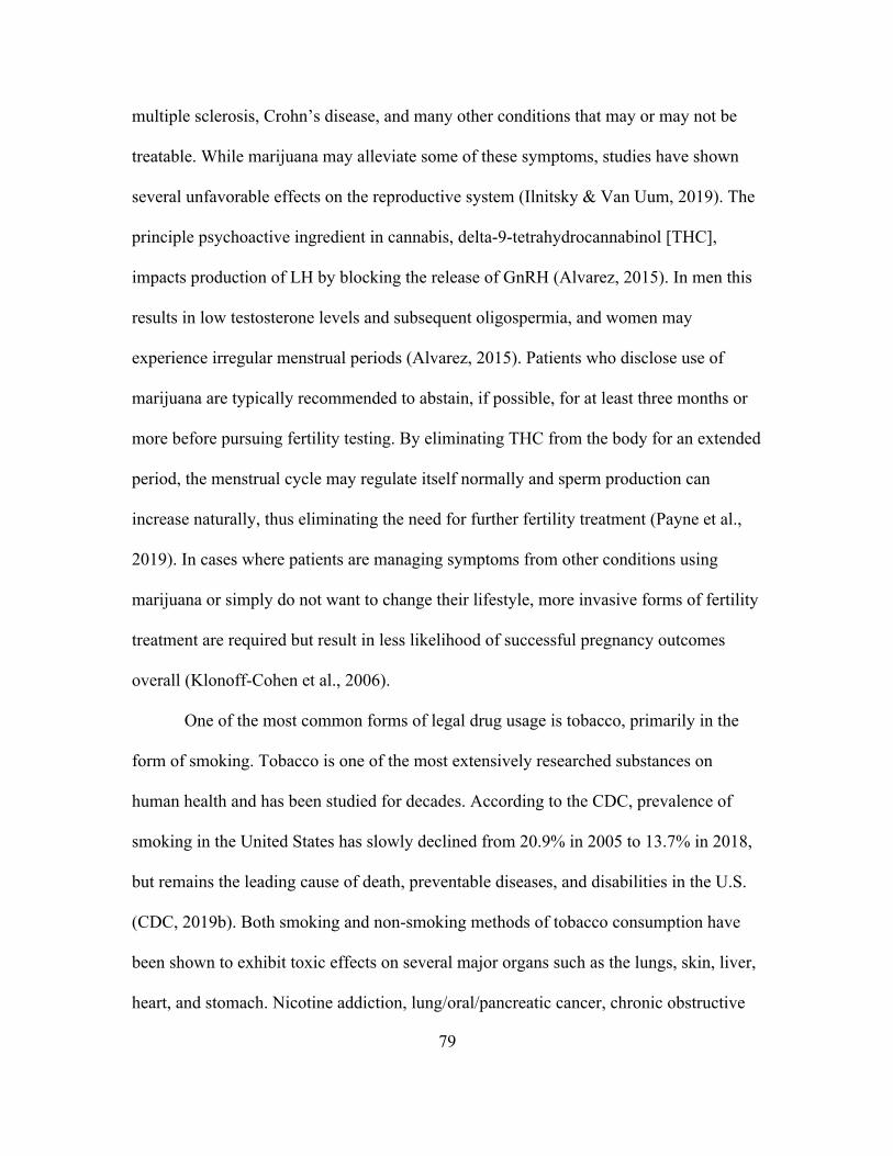
79
multiple sclerosis, Crohn’s disease, and many other conditions that may or may not be
treatable. While marijuana may alleviate some of these symptoms, studies have shown
several unfavorable effects on the reproductive system (Ilnitsky & Van Uum, 2019). The
principle psychoactive ingredient in cannabis, delta-9-tetrahydrocannabinol [THC],
impacts production of LH by blocking the release of GnRH (Alvarez, 2015). In men this
results in low testosterone levels and subsequent oligospermia, and women may
experience irregular menstrual periods (Alvarez, 2015). Patients who disclose use of
marijuana are typically recommended to abstain, if possible, for at least three months or
more before pursuing fertility testing. By eliminating THC from the body for an extended
period, the menstrual cycle may regulate itself normally and sperm production can
increase naturally, thus eliminating the need for further fertility treatment (Payne et al.,
2019). In cases where patients are managing symptoms from other conditions using
marijuana or simply do not want to change their lifestyle, more invasive forms of fertility
treatment are required but result in less likelihood of successful pregnancy outcomes
overall (Klonoff-Cohen et al., 2006).
One of the most common forms of legal drug usage is tobacco, primarily in the
form of smoking. Tobacco is one of the most extensively researched substances on
human health and has been studied for decades. According to the CDC, prevalence of
smoking in the United States has slowly declined from 20.9% in 2005 to 13.7% in 2018,
but remains the leading cause of death, preventable diseases, and disabilities in the U.S.
(CDC, 2019b). Both smoking and non-smoking methods of tobacco consumption have
been shown to exhibit toxic effects on several major organs such as the lungs, skin, liver,
heart, and stomach. Nicotine addiction, lung/oral/pancreatic cancer, chronic obstructive

80
pulmonary disease, and high blood pressure represent only a small fraction of the number
of health complications that can arise as a consequence of tobacco use (Gallaway et al.,
2018). One of the less commonly discussed repercussions of tobacco usage is the toxic
effect posed on the reproductive system. Studies investigating the correlation between
smoking and female fertility have shown a decrease in oocyte quality and accelerated
depletion of the ovarian reserve, ultimately leading to premature menopause (Budani &
Tiboni 2017; Hyland et al., 2015). Suboptimal oocyte quality can impair fertilization,
lead to abnormal embryo development, and may be a physical manifestation of
chromosomal abnormalities present in the embryo. Depletion of the ovarian reserve and
poor oocyte quality are irreversible consequences which may render a patient incapable
of having biological children—conception would then require use of an oocyte donor.
The male reproductive system is similarly impacted by tobacco usage. Findings
from several observational studies comparing semen analysis parameters in smoking
versus non-smoking patients found that use of tobacco products decreased viability,
motility, and sperm concentration (Calogero et al., 2009; Harlev et al., 2015). Decreased
motility has been linked to oxidative stress caused by spermatotoxic reagents in cigarette
smoke, which negatively impact the volume and viscosity of seminal fluid (Harlev et al.,
2015). The frequency of tobacco usage can also have a more deleterious impact on semen
parameters. Patients who self-reported their tobacco usage as “heavy” often exhibited
moderate to severe levels of teratozoospermia, asthenozoospermia, and oligozoospermia,
whereas “light” smokers typically experienced only slight to moderate
asthenozoospermia (Gaur et al., 2007). The adverse effects of smoking and non-smoking
tobacco products on overall semen quality can be reduced over time and eventually

81
eliminated by quitting utilization of these products. Since the cycle of spermatogenesis is
continual, over the course of several months all the defective sperm will either be
ejaculated or reabsorbed by the body, and new healthy sperm can be produced again
(Betts et al., 2013). Quitting tobacco consumption in any form is recommended not only
for improving general quality of life, but preventing further damage to the body,
including the reproductive system.
The relationship between alcohol consumption and reproductive health has long
emphasized the teratogenic effects of alcohol on fetal development. The specific effects
of alcohol on fertility in men and women have not been studied to the same extent, but
studies have shown that routine alcohol consumption, especially in excess, can have
detrimental effects on reproductive health in both sexes. Data from the 2018 National
Survey on Drug Use and Health [NSDUH] showed that 86.3% of adults over the age of
18 have consumed alcohol at some point within their lifetime, and over half of those
adults had drank within the last 30 days (NSDUH, 2019b). On its own, low to moderate
consumption of alcohol has not been linked to significant reduction in fertility but is
considered a contributing factor in reduced fertility (Gaskins & Chavarro, 2018). In cases
of binge drinking and heavy alcohol consumption, alcoholism can impact hormonal
regulation of estrogen in men which negatively impacts spermatogenesis (Grover et al.,
2014). In addition to reducing sperm quantity in semen, alcoholism also affects the
quality of sperm produced. In a 2017 study conducted on alcohol, smoking, and the
additive effects that these factors have on fertility, men who routinely consumed alcohol
had higher levels of DNA fragmentation and decondensation than the control group,

82
likely caused by heightened levels of reactive oxygen species in their semen
(Aboulmaouahib et al., 2017).
Perspectives on the extent to which alcohol affects reproductive health in women
has evolved and complexified significantly over the past 30 years. In the 1990’s, several
studies examined how alcohol impacted synthesis and utilization of estrogen,
testosterone, LH and FSH in patients self-reporting regular alcohol consumption. Women
who drank more frequently were more likely to exhibit higher levels of each hormone in
their blood serum, which was reported to manipulate the menstrual cycle and even lead to
anovulation (Muti et al., 1998; Reichman et al., 1993). These studies were later deemed
inconclusive, as the reported values varied too greatly, and anovulation was rarely due to
alcohol consumption alone. Due to the complexity of hormonal studies as well as the
inconsistencies reported in them, researchers began to study alcohol in a different
context: age. Studying the effect of alcohol on different age groups was presumed to
yield more reliable results for two reasons: the relationship between age and fertility had
already been established, and subjects could be classified in more clear-cut groups by
their age rather than generalizing data from a large group of individuals based on
fluctuating hormones.
In 2003, a Danish study examined prevalence of infertility in groups of women
who consumed alcohol on a weekly basis. Their study concluded that alcohol did not
affect female fertility in women below the age of 30 but was a significant predictor of
infertility beyond age 30 (Tolstrup et al., 2003). This seemed to provide a more definitive
answer than previous studies that reported their findings based on fluctuating hormone
values. However, this study was quickly called into question when an 18-year analysis

83
conducted in Sweden between 1969-1987 was published the year after [2004]. Using
hospitalization records, this study analyzed data from women between the ages of 18-28
and noted a strong trend between alcohol consumption and increased hospital visits for
infertility and high-risk births (Eggert et al., 2004). These findings directly contradicted
the Tolstrup et al. study published the year prior—the effect of alcohol was regarded as
dose-dependent in the Eggert et al. study, whereas Tolstrup et al. stated that age
determined the degree of toxicity that alcohol posed on the reproductive system.
Following these publications, researchers have continued to make cases for both age-
dependent and dose-dependent relationships between alcohol and infertility, but in more
recent years, studies have advocated more for the dose-dependent relationship.
Researchers have attempted to establish a quantitative amount of alcohol that is “safe” for
consumption without affecting fertility. A Danish study published in 2016 observed
menstrual cycle data on over 6,000 female patients between the ages of 21-45 and
reported that less than 14 servings of alcohol per week had little to no impact on the time
to conception (Mikkelsen et al., 2016). But the concept of a “serving” varies from
country to country, and the amount of alcohol in a standard serving depends on the drink
being consumed. Different varieties of beer, wine and spirits have varying alcohol
contents. According to NIH’s National Institute on Alcohol Abuse and Alcoholism
[NIAAA], 14 grams of alcohol is the standard serving of alcohol in the U.S. (NIAAA,
n.d.). By comparison, a standard drink in Denmark has only 12 grams of alcohol, and
standard grams of alcohol present in drinks across Europe ranges from 8 grams up to 20
grams (Mongan & Long, 2015). As a result, discussing the relationship between alcohol
and fertility on a global scale using servings of alcohol as a unit of measurement is not a

84
safe nor reliable method. Few studies have quantitatively analyzed alcohol and its effect
on fertility using grams as the unit of measurement. Most recently, a meta-analysis
published in 2017 reviewed data from over 98,000 women across eight countries to
compare fertility across groups of nondrinkers, light, moderate and heave-drinkers (Fan
et al., 2017). Through analysis, lower fertility rates were linked to consumption of
alcohol that exceeded 12.5 grams per day (Fan et al., 2017). Currently, support for a
dose-dependent relationship between alcohol and fertility outweighs those advocating for
an age-dependent correlation. Decades of inconsistent and controversial publications on
reproductive toxicity of alcohol still merit much further investigation. However, despite
the lack of consensus, clinicians still recommend that women seeking fertility treatment
abstain from drinking during treatment and pregnancy to mitigate any repercussions
associated with alcohol consumption and reproductive health (ESHRE Task Force on
Ethics and Law et al., 2010).
Reproductive anatomy, general/sexual health and lifestyle choices contribute
substantially to fertility potential. Patients seeking fertility treatment are encouraged to
disclose as much information as possible regarding their health to an OBGYN. Open
discussions about all aspects of a patient’s health can shape their treatment plan and
potentially narrow down the source of reduced fertility. It is imperative that patients are
made aware that there are factors outside of anatomy and physiology that may impact
their fertility such as preexisting conditions, medications, and lifestyle choices. Adopting
a healthier lifestyle benefits many areas of the human body, and the reproductive system
is no exception.

85
Conclusion
The capacity of an individual to reproduce begins with proper formation of the
reproductive system during early embryogenesis and continues into pubertal
development. Improper development of the reproductive system can lead to reproductive
dysfunction that may go unnoticed until later in life when an individual is struggling to
conceive naturally. In this chapter I reviewed various diagnostic tests that are used in
fertility clinics, including the clinic where I worked, to discern the source of reduced
fertility in an individual. The blood serum immunoassay, standard semen analysis,
hysterosalpingogram, and sonohysterogram are the most commonly performed methods
of diagnostic fertility testing that provide insight into a patient’s reproductive health.
However, a patient’s care does not end after they receive a diagnosis. Receiving a
diagnosis helps the attending physician curate a personalized treatment plan for the
patient that takes their specific needs and type/severity of reproductive dysfunction into
consideration.
To conceive in the presence of reproductive dysfunction, fertility clinics provide
treatment plans that utilize various forms of Assisted Reproductive Technologies, or
ARTs, which can bypass certain forms of reduced fertility to allow a patient to conceive.
Each form of reproductive technology will be described in Chapter 4: Fertility
Treatments & Assisted Reproductive Technologies, including the average cost of
treatment, commonly used medications, and the roles that laboratory staff and clinic staff
play during the treatment process.

86
CHAPTER 4
FERTILITY TREATMENTS & ASSISTED REPRODUCTIVE TECHNOLOGIES
Synopsis
Fertility clinics specialize in providing medical assistance to patients who have
been struggling to bear children naturally, typically using assisted reproductive
technologies, or ARTs. Procedures such as ovulation induction, intrauterine insemination,
and in vitro fertilization each aim to artificially bypass various forms of reduced fertility
to help couples conceive. At the clinic where I worked, we provided fertility treatment
services to patients from all walks of life—different ages, races, ethnicities, sexualities,
socioeconomic statuses, family structures, etc. In this chapter, I discuss fertility
treatments currently available for patients at the clinic where I conducted my clinical
work, as well as those offered by most fertility clinics nationwide. I focused on the most
common scenarios that I observed at the clinic: sub-fertile heterosexual couples, fertile
homosexual couples, and infertile or sub-fertile single female patients. Starting with
medications, I describe the various degrees of medicated treatment plans and how these
medications play a role in the fertility treatment process. This will be followed by a
description of the treatments themselves, in order of the least to most invasiveness
treatment for the patient.
When describing each treatment, I wrote this chapter with patients in mind
specifically—as a “what to expect” guide that outlines the various paths to parenthood for
individuals interested in pursuing fertility treatment. As a member of the laboratory staff,
my interactions with patients were not as extensive as the relationships fostered between
patients and the nurses, medical assistants, and physician at the clinic. However, even in

87
my limited interactions, I noticed that many were interested in the process but didn’t
know how to ask about it. How did they receive a diagnosis of reduced fertility, but this
clinic was able to work its magic and provide them with a child? This chapter answers
precisely that question. Andrologists and embryologists at the clinic are often too busy to
explain the intricacies of what goes on in the lab to every patient who is interested. In
light of this, I wanted to help bridge the gap that exists between publicly available
resources about fertility treatments for patients and the complex clinical terminology used
amongst staff and in protocols at the clinic, to provide a resource that describes what goes
on behind the scenes in the laboratory, where the so-called “magic” happens.
Beginning Treatment
Based on the results of the patient’s fertility testing, the attending physician
constructs a personalized fertility treatment plan for the patient(s) that takes into
consideration the source(s) of infertility if applicable, the patient’s treatment goals and
their budget. The inevitable financial burden of receiving fertility treatment continues to
be a limiting factor for the type of treatment patients choose to pursue. However, patients
should always be informed of all their treatment options and be made aware of any
methods that would not likely be successful for them. Severe forms of infertility may
require more invasive forms of treatment, in which case less invasive methods are not
likely to be successful. In some cases, a patient or couple may need to utilize additional
services on top of the standard procedure to conceive, such as use of donor oocytes,
donor sperm, or a gestational carrier. Fertility treatments vary in both cost and
invasiveness, and there is a linear relationship between the two: the more invasive and
involved a procedure is, the more costly the treatment becomes.

88
Regardless of the sexual orientation of the patient/couple, the overall procedure
for each treatment is the same. Depending on the circumstances, what may change is the
source of sperm or oocytes utilized for conception, or which individual will carry the
pregnancy to term. Homosexual female couples and single female patients may not
always be experiencing reduced fertility but still require a sperm donor to conceive,
whereas homosexual male couples would minimally require both an oocyte donor and
surrogate to have a child of their own. Heterosexual couples may be able to use their own
sperm and oocytes, but in cases of severe infertility may be in a position where they
require use of a sperm or oocyte donor, or perhaps a surrogate. Based on the initial
consultation, the physician will determine if there is suspected infertility for one or both
partners and order specific fertility tests to rule out reproductive disorders, and if needed,
discuss options for using a sperm/oocyte donor.
Given the general limitations of insurance coverage for fertility treatments in the
U.S., patients tend to prefer the lower cost options, although repeated failures of cheaper
fertility treatments add up and eventually can cost more than if the patient had undergone
one more expensive (but successful) cycle. Table 6: Breakdown of IUI/IVF costs
provides an average price range for many aspects of IUI and IVF treatments. Success of
any fertility treatment depends upon many factors, but in general, treatments that utilize
ARTs to conceive tend to have higher success rates than those that do not. According to
the American Pregnancy Association, the average success rate for an IUI cycle is 20% or
less, compared to the average success rate of an IVF cycle that ranges from 23-43% for
women who are less than 35 to 40 years of age (APA, 2017; APA, 2019). While both an
IUI and IVF cycle are fertility treatments, there is an important distinction between a

89
fertility treatment versus an ART. A common misconception about ARTs is that any form
of fertility treatment is considered an ART. This is because many people assume that a
fertility treatment is a protocol or procedure that “assists” with reproduction. However,
the way that the CDC and SART define what constitutes an ART is based on a U.S.
Congressional Act passed in 1992 that states “The term ‘assisted reproductive
technology’ means all treatments or procedures which include the handling of human
oocytes or embryos, including in vitro fertilization, gamete intrafallopian transfer, zygote
intrafallopian transfer, and such other specific technologies” (Fertility Clinic Success
Rate and Certification Act, 1992). By this definition, fertility treatments such as timed
intercourse, ovulation induction and IUI are not considered ARTs because no oocytes or
embryos are handled—only sperm. Therefore, ARTs are a form of fertility treatment, but
not all fertility treatments are classified as ARTs (CDC, 2021b).
Fertility Treatment Medications and Stimulated Cycles
With any fertility treatment plan comes the option to utilize fertility medications.
They are not always required but may come as a strong recommendation from the
physician depending on the patient’s reproductive health status. The term “fertility
medications” encompasses the numerous oral and injectable drugs used to control
production and utilization of reproductive hormones within the body. Most fertility
medications are taken by female patients and are self-administered, but there are several
different medications that male patients can utilize as well. During a patient’s
consultation, they will receive information regarding the three different classifications of
fertility treatment plans regarding medication usage: a natural cycle, minimal stimulation
or mini-stim, or medicated ovulation induction [OI].

90
A “natural cycle” follows the patient’s menstrual cycle and will either require no
medication at all or a short protocol of medications used at the end of their cycle to
prevent premature ovulation (Allersma et al., 2013; Pelinck, 2002). This low or no-dose
protocol is ideal for patients who ovulate normally on their own, those with high FSH
levels, or patients that would like to reduce the overall cost of fertility treatment by not
using medications (Schimberni et al., 2009). However, depending on the source and
severity of reduced fertility, a natural cycle may not be successful for some patients—
especially those who have a low chance of ovulating without use of a medication regimen
(Von Wolff, 2019). Patients who may not be successful using the natural cycle protocol
are those with irregular or anovulatory menstrual cycles, or patients with polycystic
ovarian syndrome [PCOS], fallopian tube blockages, or suffering from primary ovarian
insufficiency [POI] (Von Wolff, 2019).
If use of the natural cycle protocol is deemed insufficient for a patient, the next
treatment plan option is a mini-stim cycle. The mini-stim protocol utilizes low, continual
dosages of fertility medications throughout the entire cycle to regulate follicular
development and ovulation (Zhang et al., 2016). Benefits of using the mini-stim IVF or
other related treatment is a decreased cost of treatment, and fewer and less-severe side
effects or decreased risk of the patient experiencing hyper-stimulation from the
medications (Zhang et al., 2010). The proper dosage for each medication is determined
by the attending nurses and physicians. Dosages can vary depending on the patient’s age,
weight, reason for treatment and previous cycle history. Patients utilizing fertility
medications of any kind are required to undergo continual monitoring of their cycle
through both bloodwork and ultrasounds to assess how the patient is responding. Dosages

91
of each medication can be adjusted during the cycle depending on how well the patient is
progressing under the current regimen. The mini-stim protocol tends to be a safe middle-
ground for patients experiencing reduced fertility who do not want to undergo the
traditional medication-intensive protocol. Through the mini-stim protocol, fewer follicles
will grow and mature, limiting the risk of overstimulation and cycle cancellation, but
does guarantee that the oocytes retrieved will be of good quality or have a high potential
for fertilization (Zhang et al., 2016). The only protocol that will promote development of
many follicles is medicated ovulation induction [OI].
Out of the three different treatment plans, medicated ovulation induction requires
both the greatest number and highest dosage of fertility medications. Medicated OI is
recommended for patients with anovulatory cycles or other severe forms of reduced
fertility. Medicated OI is the most commonly utilized protocol for patients undergoing
IVF, where it is considered optimal to retrieve as many oocytes as possible at one time.
Increased use of fertility medications through the OI protocol stimulates a more
significant number of follicles to undergo growth and maturation at a given time, and
there are consequently more complications associated with this protocol than there are for
a natural cycle or mini-stim protocol (Schimberni et al., 2009; Zhang et al., 2010).
Complications and dangers of using self-administered injectable medications include
pain, redness/swelling, infection at the injection site, accidental injection of air rather
than the medication, and one of the most serious side effects, ovarian hyperstimulation
syndrome [OHSS] (Rizk & Smitz, 1992). Due to continual blood and ultrasound
monitoring of patients utilizing oral or injectable fertility medications for ovarian
stimulation, cases of OHSS are often mild and treatable. However, if left untreated,

92
symptoms can become severe and in rare cases lead to patient fatality. Patients
experiencing mild symptoms of OHSS may report nausea, fullness, and weight gain, but
more severe complications can include vomiting, diarrhea, dark urine, shortness of
breath, chest pain and abdominal distension (Gardner et al., 2001).
As with the mini-stim protocol, patients using the fully medicated ovulation
induction protocol receive both verbal and written instructions about the medications they
will be taking for their specific treatment plan and timeline. This includes information
regarding proper medication storage, method of administration, timetables for
medications and dosages for each medication that are calculated out for the patient. There
is always potential for a patient to overuse OI stimulation medications which puts the
patient at risk for developing OHSS, but this condition can also be a result of how the
patient’s body responds to certain medications. On average, incidence of OHSS in IVF
patients is roughly 3-8%, but this value climbs to 10-20% for patients who have been
diagnosed with PCOS (Farquhar et al., 2002). The exact link between PCOS and higher
prevalence of OHSS is not fully understood, however, speculation suggests that Anti-
Mullerian Hormone [AMH] levels before treatment indicate how the ovaries will respond
to standard stimulation protocols. As a result, patients with high levels of AMH like those
seen in patients with PCOS are more likely to experience ovarian hyperstimulation
(Stracquadanio et al., 2017). Prevention of OHSS starts with the treatment plan:
developing a customized, optimal stimulation protocol for each patient that uses the
smallest amount of stimulation medications possible, but still induces sufficient follicular
development and ovulation.

93
If OHSS is detected, the patient must stop taking all fertility medications which
leads to cancellation of that cycle. This is done for the safety and wellbeing of the patient
but is a devastating outcome of a treatment cycle. To the patient, a cancelled cycle is
often viewed as either a failure by the fertility clinic treating them, by the patient’s body,
or both. A cancelled cycle will ultimately prolong a patient’s journey to parenthood, but
is a precautionary measure used to protect both the patient from serious health problems
and the fertility clinic from medical malpractice.
However, OHSS is not the only reason for cycle cancellation. The need to cancel
a patient’s cycle can be a result of poor ovarian response (leading to too few follicles), or
excessive response leading to too many follicles for patients who are pursuing IUI or TIC
where there would be a high risk for multiple gestation (Kailasam, 2004). In the event
that a patient’s cycle needs to be cancelled, this information must be included in the cycle
data reported annually to the CDC and SART. Most physicians will use previous cycle
data to formulate new stimulation protocols for unsuccessful IVF patients if the patient
decides to undergo treatment again. Cycle cancellations are not ideal for the clinic or
patient, so proper dosage calculations/adjustments and meticulous monitoring help ensure
the patient can avoid cancellation and complete their cycle.
Oral and injectable medications routinely used for cycle regulation and ovulation
induction are outlined in Table 7: Common Medications Used in Fertility Medicine.
Medications are broken down into four different categories based on the stage of the
patient’s cycle at which they are used: Suppression, Stimulation, Trigger, and Other:
Post-OI. Within each category, different potential medications can be administered, with
possibly more than one medication from a particular category. For example, during the

94
stimulation stage, one patient may use a combination of both injectable FSH and LH
medications, while another patient may only use injectable Human Menopausal
Gonadotropin. Variations in the medication protocols can be a matter of the
physician’s/patient’s preference or decided upon based on the patient’s history with
fertility medications. Determining the appropriate dosage of each medication is
dependent upon numerous factors such as age, weight, type and concentration of
medication, and any other medications being taken that may interfere with the
effectiveness of fertility treatments. Nurses and physicians determine what dosages the
patient will administer for each medication. Specific dosages for each patient reflect the
high level of variability and number of medications prescribed, type, and dosage used as
well as differences in response variability from patient to patient. In Table 7, example
medication protocols for each treatment plan are listed in their corresponding
subheadings.
Timed Intercourse
Timed Intercourse [TIC] is the least invasive and most affordable fertility
treatment option for patients and tends to be more effective for patients with no known
source of reduced fertility or for women with irregular/absent menstrual cycles. This
simple treatment plan utilizes the patient’s natural menstrual cycle to optimize sexual
intercourse and increase the likelihood of conception (Agarwal & Haney, 1994). At the
clinic where I worked, we did not implement a TIC protocol very often, as most of our
patients needed more involved forms of treatment. Within a regular menstrual cycle,
ovulation occurs around day 14—with a 6 day “fertile window” where conception is most
likely, which begins four or five days before ovulation and ends on the day ovulation

95
occurs (Manders et al., 2015). Women with consistent menstrual cycles can more easily
predict their fertile window without testing, but for those with inconsistent menstrual
cycles, patients may need to use a combination of cycle tracking, basal body temperature
tracking and ovulation test kits to determine when ovulation will occur (Manders et al.,
2015).
The basal body temperature method uses daily temperature readings to determine
when ovulation has occurred. Correlations have been made between a woman’s basal
body temperature and the time ovulation, typically characterized by an increase in body
temperature by 1ºF or less (Su et al., 2017). The rise in more reliable and convenient
ovulation test kits available at drug stores and local pharmacies in the U.S. has made the
basal body temperature method less frequently used. Ovulation test kits function similarly
to urine pregnancy tests—both are rapid, qualitative tests that measure the presence of
specific molecules in urine. Pregnancy test strips analyze for the presence of hCG, while
ovulation test kits test for the surge in LH that occurs post-ovulation (Su et al., 2017).
Over the past decade, keeping track of the many details of a patient’s menstrual
cycle has become a largely digitized process. Menstrual cycle calendar applications,
commonly called “Period Tracker Apps” are available on nearly all smartphones in use
today. Within these applications, patients can chart their menstrual cycle data: when their
period begins and ends, the amount of discharge, basal body temperature, changes in
cervical mucus, fluctuations in mood and other cycle symptoms. Some also have
algorithms that compile cycle information and can help to predict the patient’s fertile
window based on previous months of cycle tracking. The convenience and individualized

96
nature of these applications makes TIC a treatment plan that requires little if any medical
intervention.
One benefit of the TIC treatment plan is that it follows the patient’s natural cycle
and does not require medications—this saves the patient both money and time since there
are no trips to the fertility clinic for monitoring, nor trips to the pharmacy to pick up
medications. An unmedicated TIC cycle is typically called “Natural Family Planning”
since the entire cycle relies solely on the patient’s natural menstrual cycle. While Natural
Family Planning may work for some patients, a completely unmedicated cycle may not
be sufficient for patients to conceive. Patients who are struggling to become pregnant
using Natural Family Planning, especially those with reduced fertility related to PCOS,
may be more successful using fertility medications for ovulation induction alongside
timed intercourse (Abu Hashim et al., 2011). Clomiphene Citrate, or Clomid is the most
common medication recommended for TIC patients, as mild doses of Clomid can
encourage just one or two oocytes to mature at a time as opposed to dozens in a fully
medicated cycle (Progyny, 2020). Some facilities may offer patients the option to use
injectable medications for TIC, but in most cases, if injectable medications are needed
physicians will encourage patients to pursue intrauterine insemination to maximize their
cycle further.
Clomid is taken orally for a period of five days, starting three days after a
woman’s menstrual period begins and works by stimulating production of FSH and LH
from the pituitary gland by inhibiting production of estrogen (Advanced Fertility Center
of Chicago, 2020a). Higher levels of FSH and LH in the body induce production and
growth of dominant follicles in the ovary, and the size of these follicles are monitored via

97
transvaginal ultrasound. Patients who are unresponsive to Clomid may take Letrozole
instead (often sold under the brand name Femara), which is an orally administered
aromatase inhibitor that works similarly to Clomid by inhibiting production of estrogen to
encourage the pituitary to produce more FSH and LH (Kar, 2013). Femara tends to be the
preferred medication used for ovulation induction in patients with polycystic ovarian
syndrome but can also be used for patients who do not respond well to Clomid (Legro et
al., 2014). Even though Clomid and Femara are taken only for a brief period and are not
given in high doses, any use of stimulation medications requires monitoring through
transvaginal ultrasound and/or bloodwork. The clinic where I worked scheduled blood
draws every other day, or every three days at most when one of our patients was taking
oral or injectable stimulation medications. Stimulation of the ovaries can lead to too
many follicles developing at once or put the patient at risk for OHSS if hyperstimulation
goes undetected. Ultimately the use of medications for ovulation induction come with
increased cost, time commitment and physical risk but may increase the likelihood of
conception for the patient.
Regardless of whether medications are used, properly timing sexual intercourse
around ovulation is crucial for optimizing the possibility of successful conception
(Manders et al., 2015). A common misconception surrounding frequency of intercourse
and conception is that “more is better.” Couples having intercourse every day, even
within the fertile window are not actually more likely to conceive. While the cycle of
spermatogenesis is rapid enough to replenish sperm daily, patients are recommended to
have between 2-7 days of abstinence before having sexual intercourse to optimize semen
parameters according to the WHO (WHO, 2010). Since sperm can also survive in the

98
fallopian tube for several days before fertilization occurs, having intercourse every day
will not significantly improve chances of conception (Suarez & Pacey, 2005). Two weeks
following ovulation, a urine pregnancy test can confirm conception—testing prematurely
may yield a false negative because the patient’s hCG level is too low to be detected
through urine (APA, 2020). Quantitative blood serum assays can detect much smaller
amounts of hCG and can confirm a pregnancy several days earlier than a urine test (APA,
2020).
Intrauterine Insemination
Many protocols utilized in fertility medicine today were originally developed for
use in the agricultural industry. Artificial Insemination [AINSEM] is one of the earliest
techniques used in fertility clinics that was initially popularized by its early success in
agricultural animals. AINSEM is a process by which semen is loaded into a long catheter,
fed through the cervix into the uterus and directly placed in the uterine cavity. This
process was first performed in animals successfully in the 1700’s and has been used for
centuries since then as a more efficient and safe means for breeding animals (Ombelet &
Robays, 2015). Results of artificial insemination were not always efficient and reliable,
however. An early lack of knowledge regarding both human and animal reproduction in
the 18th and 19th centuries meant many failed AINSEM’s. Over time, AINSEM protocols
improved with more knowledge from human and animal studies. In 1920, Russian
biologist Ilya Ivanoff published a protocol that most closely resembles the modern
AINSEM protocol utilized in fertility clinics today, and he coined the term Intrauterine
Insemination, or “IUI” (Ombelet & Robays, 2015). In humans, IUI may be beneficial or
even necessary for someone to conceive. Female patients with anovulatory or irregular

99
cycles, endometriosis, or cervical factor infertility may benefit from IUI, as well as
couples struggling to conceive due to mild male-factor infertility—though it is not a
replacement for a more invasive procedure like IVF (ESHRE Capri Workshop Group,
2009). The IUI procedure bypasses two of the largest barriers the sperm must overcome:
the vaginal canal and cervix. By placing sperm directly into the uterine cavity, a greater
number of sperm have a chance to fertilize the oocyte, as the distance that the sperm must
travel to reach fallopian tube is decreased.
As with TIC, an IUI can follow a patient’s natural menstrual cycle and use no
form of fertility medications, with what is called a “natural cycle,” or can use medications
to induce ovulation. Medicated IUI cycles are more common than natural IUI’s, despite
the increased cost and risk associated with using fertility medications. Patients
undergoing controlled ovarian stimulation for their IUI begin their stimulation protocol or
“stim” on day 3 of their menstrual cycle (Barad, 2018). The average duration of an IUI
stim is 8 days but can range from 5-14 days depending on how the patient responds to the
medications. Most clinics perform a baseline transvaginal ultrasound on day 3 to observe
the state of the uterus, fallopian tubes, and ovaries, with blood work to establish a
hormonal baseline for the patient. If the tests do not reveal any abnormalities or reasons
for concern, the patient is approved to begin ovarian stimulation. The patient is given
instructions on the timing and dosage of medications they will be using for their cycle.
Numerous different combinations of medications can be used to stimulate follicle
development and ovulation for an IUI, but the most common combinations are: Clomid
or Femara alone, Clomid/Femara with an hCG trigger shot, Follistim/hCG, hMG/hCG
(Barad, 2018). All three methods of controlled ovarian stimulation have been proven

100
effective and the physician will typically decide the best stim protocol for each patient
based on age, cycle history and reason for reduced fertility.
Stim medications are taken/administered daily, and routine bloodwork and
ultrasound monitoring are conducted to ensure proper ovarian response, monitor for
OHSS, and ensure no cysts are forming from the stim medications. Timelines are at the
discretion of the clinic, but at the clinic where I worked, blood draws to monitor levels of
estrogen and FSH were conducted every other day until the patient’s IUI appointment. At
the clinic, ultrasound monitoring of follicular number and size began around day 7-8 of
the patient’s cycle—about 4-5 days after starting the stimulation protocol. Follicle
number is especially important for IUI cycles as there is a significant risk of multiple
gestation if too many follicles are stimulated at once. For a typical IUI cycle, only one or
two follicles should be growing at once (Practice Committee of the American Society for
Reproductive Medicine, 2012). The likelihood of the patient’s cycle being cancelled
increases when three or more follicles are developing at the same time. Once the
dominant follicle(s) reaches between 19-20mm in diameter, the patient is instructed to
self-administer a Human Chorionic Gonadotropin (hCG) trigger shot to trigger ovulation
and the patient is asked to return to the clinic 36 hours later for her IUI procedure (Maher
et al., 2017).
If the patient is using a fresh semen sample for the procedure (either from the
patient’s partner, or from a donor), the sample can be collected at the fertility clinic, or at
home as long as the fertility clinic laboratory receives it no later than one hour after
collection. The laboratory staff analyzes the raw sample to observe its initial quality
before processing it to optimize the sample for the IUI procedure. Semen samples IUI are

101
typically processed in one of three ways: a simple wash, the swim-up method, or with
density gradient centrifugation. The simple sperm wash and sperm swim-up methods are
more common for n9ormal semen samples, whereas density gradient centrifugation is
optimal for samples with low concentration, abnormal sperm, or high amounts of debris
(WHO, 2010). Washing sperm is the simplest procedure which involves diluting semen
in sperm wash media, centrifugation, and discarding the supernatant--the liquid that sits
above the pellet of sperm after centrifugation. The swim-up method is a bit more
involved—semen may be washed before performing a swim-up, depending on the initial
quality of the semen initially. The sample is then loaded into a test tube and briefly
centrifuged to form a loose pellet of sperm at the bottom of the tube (Jameel, 2008).
Then, clean, pre-warmed culture medium is floated on top of the sperm.
This hospitable, nutrient-rich medium encourages the motile, healthy sperm to
swim up into it, leaving the nonmotile sperm and debris at the bottom of the tube. The
tube is incubated for an hour, giving the sperm time to swim up into the medium before
the medium is collected and washed again prior to use for the IUI (Jameel, 2008). The
last protocol is density gradient centrifugation. At the clinic, this is the method of sperm
prep we utilized most often. Out protocol for density gradient centrifugation used either a
45% gradient, or a combination of 45% and 90% gradient. The gradient is composed of
microscopic silica beads (either 45% of the solution, or 90% of the solution) that are
suspended in a HEPES-buffered media (Shivani Scientific, 2014). When preparing semen
for an IUI, we would typically process semen on the lower-density 45% gradient only.
Depending on the quality of the sample, however, if extra debris needed to be removed,
we would run the sample on a combination of both 45% and 90% gradient to yield a

102
better sample for the patient. A single or combination gradient helps removes debris,
white blood cells, and nonmotile and abnormal sperm that are present in the raw sample,
leaving only the motile, good-quality sperm behind.
To load the gradient, we would prepare a clean 15mL conical falcon tube labeled
with all pertinent patient information and pipette 1mL of the 45% gradient is into the
bottom of the empty tube. Then, if needed, we carefully pipetted 1mL of 90% gradient
underneath the 45% gradient so two distinct layers form with the 45% on top and the
90% at the bottom. If the layers mix, or the gradient will not be effective. From that
point, our protocol allowed up to 4mL of raw semen to be floated on top of the gradient.
In the event that we had >4ml of raw semen, we prepared a second gradient using the
same method, and the remaining semen was placed on the second gradient. Floating more
than 4mL of semen on a single gradient prevents all the sperm from being pulled through
the gradient properly. Density gradient centrifugation works by separating out cells in a
liquid sample based on their size and mass (Coumans et al., 2017). After centrifugation,
debris and broken sperm are caught in the uppermost portion of the gradient and can be
discarded—leaving the healthy, motile sperm at the bottom of the tube. The denser the
gradient, the fewer sperm will be able to make it through the gradient. However, semen
run on a denser gradient will typically yield fewer but higher quality sperm than semen
run through a less dense gradient—although this can vary greatly depending on the initial
quality of the raw semen sample. If we needed to use two gradients due to a high-volume
sample, the resulting pellets at the bottom of each tube are combined into one tube, then
the sample is washed one final time with pre-warmed sperm wash medium before being
placed in the incubator awaiting the patient’s IUI procedure.

103
While using fresh semen is best, some patients had IUI’s using frozen vials of
sperm that were frozen weeks, months or even years before the procedure. Frozen semen
can come from previous collections from the patient’s partner or using sperm from a
known or anonymous sperm donor. Semen can be frozen in its raw, unprocessed form
which requires processing after thawing. If the patient knows they will be pursuing an
IUI, they can have sperm frozen for them pre-processed as IUI-ready vials (or purchased
from a donor as an IUI-ready vial) that can be thawed, washed, and used without
performing any additional processing. Vials frozen as IUI-ready are not necessarily
advantageous in any way for the patient, they are just pre-processed and require less
prep-time for the laboratory staff on the day of the IUI.
When the patient arrives for her IUI, she is taken into a private exam room for the
procedure. At our clinic, both the physician and nurse practitioner would perform IUI
procedures, accompanied by a medical assistant who set up the room beforehand. The
patient lies on the exam table with her feet placed in the attached stirrups. A clean
speculum inserted into the patient’s vaginal canal by the physician/nurse practitioner,
opens the vagina for the IUI procedure. The patient is asked to confirm her name and date
of birth, which is verified with what is written on the tube of sperm processed by the
laboratory staff to make sure the correct sample is being used for the patient. The
processed sperm is drawn into a long, flexible sterile catheter using a syringe. The tube of
the catheter is then fed through the patient’s cervix and the sperm sample is expelled
directly into the uterine cavity (Figure 10). After the sperm is inside the uterus, the
catheter can be removed, followed by the speculum and the IUI procedure is complete.
Overall, our lab required 1-2 hours of lead time to prepare a semen sample for an IUI

104
procedure, but the IUI procedure itself takes 10-15 minutes. No earlier than two weeks
following the IUI procedure, the patient can determine if the IUI was successful through
an at-home urine-based pregnancy test, or through a blood draw at the clinic to measure
hCG levels.
Figure 10. Intrauterine Insemination (IUI). From IUI [Photograph], by BruceBlaus, 2017,
Wikimedia Commons (https://commons.wikimedia.org/wiki/File:IUI.png). CC BY-SA
4.0
In Vitro Fertilization
In Vitro Fertilization, or IVF is the most complex and costly fertility treatment
currently available for patients. The phrase “in vitro” is a Latin phrase that means “in
glass”—meaning IVF is a procedure by which oocytes are physically removed from the
body and fertilized inside of a sterile dish in the laboratory (Zhu, 2009). This treatment
may be utilized if couples have experienced repeated IUI cycle failures, recurrent

105
pregnancy loss following an IUI, if their infertility is severe enough that IUI’s are not a
viable option of treatment, or in more recent years as a means for gender-selection. IVF
was first successfully performed in the late 1970’s when Patrick Steptoe and Robert
Edwards, respectively a gynecologist and professor of human reproduction from the
United Kingdom worked together to create embryos in a laboratory for infertile couple
Lesley and John Brown (Hartshorne, 2008). After successful fertilization of Lesley’s
oocytes using John’s sperm, Steptoe and Edwards transferred an embryo into Lesley who
gave birth to Louise Brown on July 25th, 1978. Since then, millions of babies have been
born using IVF across the globe.
The goal of an IVF cycle is to retrieve as many oocytes as possible. Since the
oocytes are fertilized in the lab and only one or two embryos are transferred back into the
uterus at a time, having numerous follicles growing at once does not pose a risk for
multiple gestation like it would during an IUI cycle. Patients have ultimate control over
whether they want one or two embryos transferred, and any excess embryos allow the
patient to undergo additional later transfers if their first was unsuccessful. Most IVF
patients follow the conventional ovarian stimulation protocol for IVF which uses several
injectable medications throughout the cycle to stimulate as many follicles as possible—
but there are different treatment plans for different patient’s needs.
Natural IVF is exactly like natural family planning or a natural IUI cycle—
treatment follows the patient’s menstrual cycle, and no medications are used. Abstaining
from using any fertility medications decreases the likelihood of developing OHSS and
cuts down the cost of receiving IVF treatment. However, since only one follicle grows to
maturity and is ovulated during a typical menstrual cycle, that means that only one oocyte

106
can be potentially retrieved and fertilized in the lab, significantly decreasing the
likelihood of having a healthy embryo to transfer into the uterus later on. Most clinics do
not perform natural cycle IVF due to its extremely low success rates and encourage the
“mini-stim” protocol instead. A mini-stim cycle uses more medications than a natural
cycle, but fewer (and in smaller doses) than conventional ovarian stimulation for IVF
(Progyny, 2020). The mini-stim protocol either involves oral medications to stimulate
and a single trigger shot injection similar to a medicated IUI cycle or utilizes a
combination of oral and injectable medications: Clomid or Femara for the first 5 days and
an FSH injection for the last 10 days before triggering ovulation (Progyny, 2020).
Regardless of the mini-stim protocol used, a mini-stim cycle is still cheaper than
conventional IVF and requires fewer daily injections. Although similar to a natural cycle,
using few (if any) medications will not yield the same number of oocytes that would be
expected for a patient that chooses to undergo the full IVF stimulation protocol (Progyny,
2020).
Conventional IVF is costly, requires numerous daily injections and does not
typically use any oral medications other than oral contraceptive pills at the start of their
cycle. There are three protocols used for conventional IVF: the long Lupron protocol,
antagonist protocol and microdose Lupron flare protocol (Advanced Fertility Center of
Chicago, 2020b). The Long Lupron protocol, also called Luteal Lupron protocol is the
longest protocol that takes roughly 42-44 days from the start of contraceptive
downregulation to the retrieval date—with some variation from patient to patient.
Following the Luteal Lupron protocol, upon the start of the patient’s menstrual cycle,
they are instructed to come into the clinic for a baseline blood draw on day 3 of their

107
cycle and will begin to take a daily oral contraceptive pill [OCP] for up to 21 days (USC
Fertility, 2019; Coastal Fertility, 2021). Taking an OCP seems counterintuitive to many
patients who are seeking fertility treatment to do the exact opposite of the goal of birth
control. However, OCPs serve several important purposes in the Luteal Lupron protocol:
for clinic scheduling (so only a few patients are undergoing oocyte-retrievals in a single
day), to prevent cysts from forming later in the patient’s cycle, and to begin the process
of downregulation (USC Fertility, 2019). Downregulation allows for better control over
the patient’s cycle, inhibits premature ovulation and ensures that all follicles in the ovary
will start off at the same size for the stimulation phase of treatment. With all follicles in
the ovary at the same size before starting stimulation, each follicle will be stimulated at
roughly the same rate so all active follicles should ovulate mature oocytes during the
trigger and can be retrieved at the same time.
Lupron is the most commonly used downregulation medication that effectively
turns off production of reproductive hormones in the pituitary gland so the LH surge that
occurs during ovulation cannot occur (Advanced Fertility Center of Chicago, 2020c;
Coastal Fertility, 2021). On day 15 of taking an OCP, the patient begins self-
administering a daily Lupron injection as well (Coastal Fertility, 2021). The patient will
continue both medications until their OCP runs out on day 21 and then administer only
Lupron for another 5-10 days until their menstrual period begins—this specific timing of
the Lupron start is clinically referred to as “mid-luteal” timing. A blood draw is
scheduled to assess estrogen levels following the start of the patient’s menses, and after
the baseline is established, the patient administers one more Lupron-only injection before
stimulation begins. The following day is “Stim Day 1”—the patient’s daily Lupron dose

108
is cut in half and the patient begins taking an injectable form of FSH such as Follistim or
Gonal-F, or an hMG injectable such as Menopur (Coastal Fertility, 2021). Daily low-dose
Lupron and either FSH or hMG injections continue until stim day 10, with bloodwork
and ultrasounds conducted every other day to monitor follicle growth (USC Fertility,
2019). When the majority of the follicles present in the ovary fall between 18-22mm in
size, the patient is instructed to administer their trigger shot of hCG and return to the
clinic 36 hours later for their oocyte retrieval (Advanced Fertility Center of Chicago,
2020c; Coastal Fertility, 2021). If the follicles are not large enough yet, the patient will
continue their low-dose Lupron and FSH/hMG injectables until instructed to stop and can
administer their trigger injection (USC Fertility, 2019). Given the extended period of time
that patients are taking injectable fertility medications, this requires extreme flexibility
with the patient’s schedule and are at much higher risk for “over-suppression” by the
Lupron protocol which may cause the patient to respond poorly to stimulation
medications and few follicles will mature.
To reduce the likelihood of over-suppression and use fewer injections, the
antagonist protocol was developed as a slightly shorter protocol for conventional IVF.
Similar to the Luteal Lupron protocol, upon the start of the patient’s menstrual cycle, the
patient has a baseline blood draw performed on day 3 of their menstrual cycle and begins
to take a daily OCP. In the antagonist protocol, the patient takes an OCP for only 17 days,
then stops taking the OCP for four days before starting their stim protocol (San Diego
Fertility Center, 2006). On stim day one, the patient begins to self-administer a daily
injection of FSH and have bloodwork and ultrasound monitoring every other day until the
date of her retrieval, depending on the physician’s preference (Progyny, 2020). Starting

109
on stim day six, the patient must also begin a daily Ganirelix or Cetrotide injection as
well. Ganirelix and Cetrotide are GnRH antagonists that block GnRH binding sites in the
pituitary which prevents the LH surge during ovulation (Advanced Fertility Center of
Chicago, 2020d). Daily injections of FSH and Ganirelix/Cetrotide continue until the
follicles reach maturity and the trigger shot can be administered (Advanced Fertility
Center of Chicago, 2020d). An hCG trigger shot can be utilized to trigger ovulation but is
not recommended for the antagonist protocol by itself—a Lupron injection is more often
used as a trigger for the antagonist protocol as it causes a sufficient surge in FSH and LH
to trigger ovulation without inducing hyperstimulation and can also be administered as a
dual trigger shot as a combination of both Lupron and hCG to ensure that ovulation
occurs (Lin et al., 2019). Following the trigger shot, the patient will return to the clinic
for her scheduled oocyte retrieval. The antagonist protocol takes roughly 32-34 days from
the start of OCPs to oocyte retrieval and is significantly shorter than the Luteal Lupron
protocol (Advanced Fertility Center of Chicago, 2020e).
The shortest stim protocol for IVF is the microdose Lupron flare protocol. This
protocol provides a jump start to ovarian stimulation and may be the preferred protocol
for women with increased maternal age or who are poor responders to longer protocols
(Progyny, 2020). As always, this protocol begins with OCPs—14 days of OCP’s starting
on day 3 of the patient’s menstrual cycle. After completing two weeks of daily OCP’s,
the patient stops their contraceptive for two days before starting their brief
downregulation period (Advanced Fertility Center of Chicago, 2020f). The second day
without OCP’s marks the start of bloodwork and ultrasound monitoring every other day.
On day three without contraceptives, the patient begins to self-administer a diluted

110
Lupron dose twice a day—half of the normal dose in the morning and the other half at
night (Advanced Fertility Center of Chicago, 2020f). By taking half of the dose twice a
day, the patient is still administering a full dose of Lupron each day but provides a more
continual dose throughout the day. Twice-daily injections of diluted Lupron continue for
a second day and on day three the patient begins their daily stim injection of FSH
(Advanced Fertility Center of Chicago, 2020f). A typical stim for the microdose Lupron
flare lasts 8-11 days and the patient will continue their dilute Lupron and FSH injections
until the follicles in the ovary reach maturity. At that point the patient is instructed to stop
administering Lupron and FSH and administer their hCG trigger shot that evening and
return to the clinic 36 hours later for their oocyte retrieval, for a total time of 25-27 days
between the start of OCPs to oocyte retrieval (Progyny, 2020).
The day of the oocyte retrieval, the patient arrives at the clinic and prepares for
the retrieval procedure. Transvaginal oocyte retrievals are performed by the OGBYN,
accompanied by an anesthesiologist that has given the patient an intravenously
administered anesthetic—Monitored Anesthesia Care [MAC] (Nagarajan & Lew, 2018).
MAC is a form of localized anesthetic that is combined with sedation and analgesia
(Kwan et al., 2013). Patients receiving this propofol-based anesthetic are aware but
relaxed and do not need respiratory support during their procedure (Nagarajan & Lew,
2018). This allows for a faster post-operative recovery time and there are fewer side
effects associated with MAC versus traditional general anesthesia—patients are
instructed to not eat or drink the night before or morning of their retrieval procedure
(Kwan et al., 2013).

111
To begin the oocyte retrieval process, the physician cleans the vaginal opening
and cervix. A transvaginal ultrasound probe is used to visualize the ovarian follicles and
serves as a guide for the physician throughout the retrieval (CNY Fertility, 2020b). The
probe is fitted with a long sterile needle for the procedure that is connected to a sterile
test tube filled with warm media for oocyte collection. The transvaginal probe is inserted
into the vagina and the follicles come into view on a screen inside of the procedure room
(Mayo Clinic, 2019e). Using the view of the follicles as a guide, the physician moves the
needle over to a follicle, punctures the wall of the ovary with the needle tip and aspirates
the follicular fluid until the follicle walls visibly collapse on the screen (CNY Fertility,
2020b). Individual oocytes are not visible on the screen, but once the follicle borders
collapse, the fluid inside of the follicle is aspirated and the oocyte will be floating in the
fluid in the collection tube. Once the tube is nearly full, it is capped and passed through
into the lab where an embryologist will empty the tube into a dish and begin collecting
oocytes out of the follicular fluid (CNY Fertility, 2020b). In doing this, the embryologist
can count the exact number of oocytes retrieved and separate the oocytes out of the fluid
that may contain blood clots or torn tissue which may stick to the oocytes over time and
make them difficult to see.
Removing the oocytes from the follicular fluid and placing them in a more pH-
stable media is also important to prevent contamination and any cytotoxic changes in the
osmolarity of the oocytes (Mayo Clinic, 2019e). The process of follicular aspiration and
oocyte counting continues until the final mature follicle of the patient’s ovary has been
punctured, aspirated, and collapsed. Following completion of the retrieval, the
intravenous sedative is removed, and the patient is monitored by the anesthesiologist until

112
she regains consciousness (CNY Fertility, 2020b). Attending nurses move the patient to a
recovery room so they can relax and continue to regain their strength for a few hours until
they leave the clinic. Patients undergoing a fresh embryo transfer will begin to take
progesterone suppositories the evening following their retrieval to prepare their uterine
lining for transfer. Fresh embryo transfers are not common practice today and were
seldom performed at the clinic where I worked—the overall process of downregulation,
stimulation and oocyte retrieval is very taxing on the body, so many physicians prefer to
freeze embryos for at least one cycle so the patient’s body can recover fully before an
embryo transfer.
Back in the laboratory, embryologists finalize the number of oocytes collected
during the patient’s retrieval procedure and report the final count to the physician. At the
clinic where I worked, during “retrieval week”, a new patient was in the procedure room
for a retrieval every 30 minutes—so the turnaround was very quick. After the retrieval is
complete and the oocytes had been counted and reported, embryologists in our lab would
close each patient’s dish of oocytes, then place them into a warm, humidified chamber for
3-6 hours so the oocytes could continue to mature and equilibrate to the surrounding
culture media until all the retrievals for the day were over. During that window of time,
the lab reviewed the patient’s signed consent forms and orders. “Orders” referred to
documents signed by the patient (and their partner, if applicable) that state what the
patient would like performed on their oocytes/embryos, who the sperm source will be if
the oocytes are to be fertilized in the lab, and what will be done with extra embryos
(frozen for future use, ethical discard, donation, etc.). The clinic required signed and
notarized permissions to be scanned into the patient’s file before any procedure could be

113
performed using their oocytes. and must be completed prior to the patient’s retrieval, as
orders cannot be signed after the patient has undergone anesthesia.
Based upon the patient’s signed orders, the retrieved oocytes can be used in
several ways: oocytes could be frozen for the patient to use later on (called “autologous
oocyte banking”), used fresh or frozen for another patient’s use as donor oocytes, or
could be inseminated the day of the retrieval to create embryos for the patient. The
intended use of the oocytes determines which protocols to use to fertilize and/or preserve
them. Conventional insemination [INSEM] was one of the most common protocols used
to fertilize oocytes in vitro and is the method that was used to conceive Louise Brown in
the late 1970’s. Our physician often recommended patients with only female-factor
infertility or mild male-factor infertility to use INSEM. On the day of the patient’s
retrieval, the lab asked that a fresh semen sample be collected by the male partner in-
office unless the patient is using donor sperm or a frozen vial of sperm, TESE or PESA
sample. We always encouraged a fresh sample for IVF: however, if frozen sperm is being
used for any reason, the vial is thawed the morning of the retrieval and can be processed
using the same protocols as a fresh sample. Depending on the quality of the sample,
modifications could be made to sperm washing/gradient protocols might be adapted on a
patient-by-patient basis.
Typically, sperm used for INSEM were processed using either a swim-up or
density gradient protocol at the clinic where I worked. The WHO also suggests using a
density gradient rather than the swim-up method because gradients tend to yield better
quality samples with higher concentrations—ideal for INSEM (WHO, 2010). Per our
laboratory Standard Operating Procedures, after making initial assessments for a standard

114
semen analysis, we would load the sample onto a 45/90 gradient, centrifuged and washed
twice using sperm wash media to remove all white blood cells, debris, and nonmotile
sperm. The post-processing parameters we required in order to use INSEM for a patient
were: 14M/mL concentration or greater, >40% motility, forward progression of 3+ or
higher, and a minimum morphology score of a 4. If the sample decreases in quality after
processing (rare, but does occur), the lab informs the physician that the sample is not
good enough for INSEM and a more aggressive protocol may be necessary for
fertilization—intracytoplasmic sperm injection [ICSI]. Consent forms for IVF at our
facility had several options for fertilization that the patient could choose: INSEM only,
INSEM unless the sample is not good enough, then use ICSI, 50/50 INSEM and ICSI, or
ICSI only. If the patient has not signed off for ICSI under any circumstances but the
semen sample does not meet the requirements for INSEM, our physician would contact
the patient and discusses options. The patient could sign new consents permitting the use
of ICSI and agree to pay the additional charges for that service, attempt INSEM with the
current sample and expect few successes or a “no-fert,” which would render their entire
cycle a failure, or choose not to fertilize the oocytes and freeze them to be fertilized later.
Most patients choose to allow ICSI under these circumstances or will freeze the oocytes
for future use rather than risking no fertilization occurring with the current sample.
After the oocytes have spent 3-6 hours in the humidified chamber to continue
maturation and the retrievals for the day were complete, the INSEM process could begin.
Standard INSEM protocol requires that oocytes were left in their cumulus complex for
fertilization—this more closely mimics how fertilization would occur in vivo. Our clinic
also followed this method of leaving the cumulus complex attached for INSEM cases.

115
We would move the oocytes from their post-retrieval holding dish into a new dish that
was labeled with the patient’s name and date of birth and filled with warmed fertilization
media. If the patient chose INSEM and the processed sperm met all of the qualifications
for INSEM, all of the patient’s oocytes would be exposed to sperm, regardless of their
observed maturity. Depending on the concentration of the patient’s sperm sample, we
would add a volume of 1-15µL of processed sperm to the fertilization media to fertilize
the oocytes. After adding the sperm, the dish is closed and returned to the incubator until
the following day (16-20 hours later) to check whether fertilization has occurred. The
following morning, we would perform a “fert check” and document how many oocytes
fertilized overnight, and how many did not. Some unfertilized oocytes can be exposed to
sperm the following day to attempt fertilization again, but most often the unfertilized
oocytes ended up being discarded.
A fertilized oocyte is then referred to as an embryo, which continues to grow and
divide over the course of several days. Embryos are not checked or moved into new
dishes on a daily basis. It is standard to check embryos on day 1 for successful
fertilization, then to leave them in the humidified chamber to divide until day 3 before
being checked again. Our lab checked and graded embryos only on days 3 and 5, then
every day until all the patient’s embryos had been transferred to the mother, frozen, or
discarded. Checking the embryos as little as possible avoids removing them from the
humidified chamber very often. Having a set schedule of when to observe embryos also
allows better scheduling in the embryology lab when there are many patients with
embryos being cultured on any given day. Given the clinic where I worked saw such a

116
high-volume of patients each month, there were often over 100 embryos in the lab being
cultured or prepared for transfer during our busy weeks.
By day 3 post-fertilization, the embryo should have reached the cleavage stage
and have between 6-10 cells inside. Embryologists observe the embryos under a
microscope, count the embryo’s cells, and observe the overall quality of the embryo. We
used an embryo grading chart to provide grades for the embryos and would document the
number of oocytes with each grade. At this stage, an embryo should ideally have 8
uniform cells inside with little to no fragmentation or vacuoles. Depending on the number
of embryos in the dish, sometimes day 3 embryos were moved into a new, clean dish to
replenish nutrients as the embryos continue to grow. On day 5, we would give the
embryos a second grade based on how much they have expanded, the quality of the inner
cellular mass, and quality of the trophectoderm. Day 5 embryos should have two distinct
layers: the outer trophectoderm that will form the placenta and inner cellular mass that
will become the fetus.
Ideally on day 5 the embryo qualifies as a blastocyst, with many uniform, tightly
packed cells and is beginning to hatch out of the zona. Starting on day 5, we would begin
to freeze embryos for the patient to use in the future (see Figure 11). If a patient was
having a fresh transfer, this is the point at which those embryos could begin to be
transferred. However, as mentioned previously, the clinic where I worked did not
perform fresh transfers often, so most embryos were frozen starting on day 5.
Trophectoderm biopsy for genetic screening of embryos was also performed on day 5/6, a
technique discussed in more detail in Chapter 5. By day 7, all the patient’s embryos
should be frozen or have been transferred—our clinic did not freeze embryos beyond day

117
7. After day 7, blastocysts require more nutrients to survive each day and cannot be
maintained on culture media alone.
Figure 11. Conventional In Vitro Fertilization Procedure. Major steps of the IVF process
are covered in this diagram, from ovulation induction to embryo transfer. Created with
BioRender.com.
Intracytoplasmic Sperm Injection
In cases of lower semen quality, ICSI might be needed to manually inject a single sperm
into the oocyte to increase the likelihood of successful fertilization. ICSI is used most
often with patients who have few sperm in their ejaculate, sperm with low forward
progression scores or very abnormal morphology. At our clinic, patients with known low
sperm quality would be encouraged to sign consent forms to use ICSI only. Patients with
mediocre-quality sperm who have a chance of qualifying to use INSEM may be told to

118
sign to use INSEM, with consent to use ICSI if the sample is not good enough. However,
there were occasions where patients with no male-factor infertility at all choose to use
ICSI over INSEM out of a matter of preference or under the assumption that a more
expensive technique like ICSI will yield better results. The primary benefit of ICSI is that
the highest-quality sperm are hand-selected by trained embryologists and are individually
inserted into each oocyte—sperm that otherwise would not be able to successfully
fertilize an oocyte in their own.
The first critical step of ICSI begins before sperm are even in the dish. Following
the maturation period in the humidified chamber, oocytes being inseminated through
ICSI must undergo a process called “stripping,” which uses an enzyme called
hyaluronidase to remove the sticky cumulus cells that surround each oocyte. At our
clinic, stripping was performed one-hour post-retrieval. This allowed the oocytes to
equilibrate in the media where they were being held before undergoing stripping, which
is an abrasive process for the oocytes. When the oocytes were ready to be stripped in
preparation for ICSI, we would remove them from their holding dish and place them in a
separate dish containing several individual drops of warmed hyaluronidase diluted in
culture media. The oocytes are mixed around in the hyaluronidase solution for a few
seconds and the surrounding cumulus cells begin to slough off. The stripped oocytes are
then removed from the hyaluronidase solution and rinsed in several drops of warm
culture media to remove any trace of the enzyme before being returned to their holding
dish. Stripping is a quick process that takes only a few moments and there is no set
amount of time that the oocytes must spend in the solution—it is based entirely on how
quickly the cumulus cells come off on their own. During the stripping process, the

119
oocytes are observed constantly under the microscope, because if the oocytes are left in
the hyaluronidase solution for too long, the enzyme will begin to eat through the zona
which can cause irreparable damage and potentially kill the oocyte.
After the cumulus cells have been removed from the exterior of the oocyte, we
would return the oocytes to the incubator while the microscope is prepped for ICSI. Since
oocytes are extremely sensitive to changes in temperature, osmolarity and pH, the less
time they spend outside of the incubator and uncovered, the better off they are. There are
two different needles used for ICSI—a blunt holding needle for the oocyte and tapered
injection needle for the sperm. The side that each needle is on (left or right) is a matter of
preference for the embryologist performing the ICSI procedure—however, the
manufacturer specifies the angle for the needles’ positioning. That must be done properly,
or the needles will be more likely to break as they are moved vertically into and out of the
dish. Angles for either needle can vary based on the manufacturer but typically range
from 20º to 45º angles. The needles are connected by thin tubing to the microinjector
which allows the embryologist to control the strength of suction or expulsion from each
needle. Both needles are attached to the microinjector and brought into focus before
moving on to the sperm preparation.
To prepare for ICSI, we would always add the sperm to the dish first—since
oocytes are much more limited in quantity (compared to sperm) and are more sensitive to
changes in temperature, we would only add the oocytes to the ICSI dish moments before
we were ready to do ICSI. Using a micropipette, we loaded a small aliquot of sperm into
an L-shaped droplet of media in the ICSI dish. Since sperm tend to swim along the outer
edge of a droplet, the L-shape allows sperm to swim in different cardinal directions

120
which makes them easier to pick up with the needle. Under the microscope fitted with the
microinjector needles, we look for the best quality sperm that are morphologically normal
and are swimming rapidly in a relatively straight path. The best-looking sperm are picked
up and deposited into a separate drop in the dish made of polyvinylpyrrolidone [PVP].
PVP is a viscous solution that decreases sperm motility, lubricates the injection needle,
and prevents sperm from sticking to the inside of the needle and gives the embryologist
greater control over movement of the sperm (Cooper Surgical, 2016). Once several sperm
have been deposited into the PVP, they appear sluggish in comparison to their once-rapid
movement in the L-bend of media. The sperm can then be immobilized by using the
injection needle to quickly strike their tail (Patrizio et al, 2003). Immobilization is
important for the ICSI procedure because sperm must be injected into the oocyte head-
first. When loading, sperm are picked up by their tails, so they are expelled into the
oocyte head-first. Immobile sperm are not dead, just stunned, and unable to turn around
in the needle before injection.
Unlike with INSEM, only mature oocytes can be used for ICSI, so mature oocytes
are loaded into a drop of media on the ICSI dish, leaving the immature and germinal
vesicle-stage oocytes in the holding dish. Under the microscope, one oocyte is picked up
at a time using the holding needle—the suction of the microinjector holding onto the
oocyte is firm enough that the oocyte will not slip during injection, but light enough to
not rupture the zona. I was trained to position the oocyte so that its polar body was
positioned near 6 or 12 o’clock—as far away from the injection site as possible without
being directly under the suction of the holding needle, which is demonstrated in Figure
12 below. With a single sperm loaded tail-first into the tip of the injection needle, the

121
needle is inserted through the zona, and the cytoplasm of the oocyte called the
“oolemma” is aspirated just until it ruptures so the sperm cell can be injected inside. Most
embryologists aspirate and expel a small portion of the oolemma once the sperm has been
deposited to mix the sperm with the oocyte’s cytoplasm—this ensures that the sperm is
not stuck to the tip of the needle and will not be removed when the needle is withdrawn.
Figure 12. Intracytoplasmic Sperm Injection Setup. Created using BioRender.com
Once the injection is complete, the embryologist rolls the oocyte out of the way so
ICSI can be performed on the remaining oocytes. As an embryology trainee, I would only
work with a couple of oocytes at a time to prevent the oocytes from being out of the
incubator for too long, while the more senior embryologists could perform ICSI much
faster. Oocytes that have been fertilized via ICSI are then moved to a new labeled dish of
media to culture for another 5-7 days until transfer or vitrification. The culturing process
of ICSI and INSEM embryos is identical, with embryo/blastocyst grading is performed
on days 3 and 5, possible trophectoderm biopsy on day 5/6, and potential vitrification of
the blastocysts between days 5-7 until all blasts have been transferred, frozen or
discarded.

122
Oocyte/Embryo Vitrification and Thawing
Vitrification is a process of rapid cooling used to quickly cryopreserve embryos
and oocytes. This is not considered an ART or even a fertility treatment, but is a frequent
protocol used in fertility clinics that merits discussion. Since only one or two embryos are
transferred back into the uterus at a given time, any additional embryos must be
cryopreserved and stored in liquid nitrogen until they can be transferred. The process of
freezing embryos at the end of an IVF cycle has been performed for many years—with
some patients returning 5-10 years after their cycle to transfer a previously-frozen
embryo. Autologous oocyte banking is a much newer service initially used to help
patients undergoing cancer treatment to preserve their fertility. Oocyte cryopreservation
has recently gained popularity for patients who would like to preserve oocytes during
their younger years so age-related decline in oocyte quality does not impede conception
(Mertes & Pennings, 2011). Oocyte cryopreservation has also become a method for
transgender patients to preserve their ability to have biological children even after
hormone-replacement therapy and transitioning (Mitu, 2016).
The overall process of vitrification is the same for both—the oocyte/embryo is
slowly moved out of the media into increasing concentrations of equilibration solution,
then out of equilibration solution into increasing concentrations of vitrification solution
until the oocyte/embryo is in 100% vitrification solution—a process that takes roughly 15
minutes from start to finish per our protocol at the clinic. After reaching 100%
vitrification solution, the oocyte/embryo is loaded onto a labeled cryo-straw and dipped
into liquid nitrogen and flash-frozen for long-term storage. If frozen and stored properly,
these embryos and oocytes can be stored indefinitely using the vitrification technique.

123
The only differences between vitrification for oocytes versus embryos is that oocytes
must be stripped one hour after the retrieval if they are being vitrified and not fertilized.
Our protocols for thawing oocytes and embryos utilized different solutions than
vitrification, but still followed a similar process. Thawing begins in 100% thawing
solution before introducing increasing concentrations of diluent solution. Once the
oocyte/embryo is in 100% diluent solution, washing solution is gradually introduced until
the embryo can be moved into culture media. The process of thawing embryos and
oocytes also took roughly 15 minutes from start to finish, at which point an embryo
would be used for transfer, or we would begin the INSEM or ICSI procedure to fertilize
the thawed oocyte.
Conclusion
Many different protocols and techniques are utilized in fertility clinics today to
assist patients with starting or expanding their families. The multitude of available
methods for both stimulation and conception gives patients more autonomy in how
invasive and involved their care will be. Regardless of which method or treatment a
patient undergoes, reproduction at the fertility clinic is a collaborative effort between
numerous participants or “key players.” Each of these participants plays a unique and
essential role in the reproductive process. In this chapter, I described the roles,
responsibilities, and contributions of the key players at a fertility clinic: physicians,
nurses, medical assistants, patients, and laboratory staff.
I have emphasized the role of laboratory staff in particular, as patients typically
have fewer interactions with laboratory staff compared to other staff at the clinic, and my
expertise primarily lies in my laboratory training at the clinic. Despite having fewer

124
interactions with patients as a member of the laboratory staff, I noticed that some patients
visiting the clinic expressed genuine interest in understanding the work of the
andrologists and embryologists in the lab. Most laboratories housed within fertility clinics
are out of view from patients and visitors at the clinic, so patients wonder how we were
able to create embryos for patients who had tried for years to conceive naturally and were
not successful. Through these patient interactions, I realized that the inner workings of
the lab are not communicated well to patients. Patients wanted to know what was going
to take place in the lab either before or during their treatment—how these technologies
could do something that was otherwise impossible.
Disclosing these laboratory processes can help patients feel better informed about
what is happening to their sperm, oocytes, and embryos during the fertility treatment
process. Patients understanding workflow in the lab can also benefit the clinic, as patients
become more knowledgeable about the techniques, skills and supplies that are involved
in the preparation of samples, insemination of oocytes, embryo
culture/biopsy/cryopreservation, and all other tasks that are carried out in the lab during a
patient’s cycle. If patients are better informed about what the laboratory is responsible
for, I believe clinics can justify some of the costs associated with receiving treatment
more easily. Few clinics itemize laboratory costs when advertising a fertility treatment
package, so the cost of maintaining a clinical laboratory and the volume of supplies
utilized goes largely unnoticed.
Cost aside, ARTs have undoubtedly changed the way reduced fertility is viewed
from a clinical standpoint, as well as in the eyes of individuals struggling to conceive.
The implementation of ARTs in reproductive medicine as a means to circumvent reduced

125
fertility has also paved the way for ethical dilemmas involving misuse of these
technologies, and the use of ARTs for unintended purposes. But how far is too far? Have
we reached the limits of what the technology should be used for, even if we could use it
for more? What ethical challenges have arisen in the fertility field to date, and have they
been addressed or are these issues still prevalent in the field? These questions will be
addressed in Chapter 5, Ethical Dilemmas in Fertility.

126
CHAPTER 5
ETHICAL DILEMMAS IN FERTILITY
Synopsis
How far is too far? Just because we can, should we? Where do we draw the line?
These types of questions routinely arise in the field of reproductive medicine, especially
with the implementation of novel treatments and therapies. Since its inception, the use of
Assisted Reproductive Technologies [ARTs] in fertility medicine has been met with
challenges. Initially, terms such as “playing God” and “test tube baby” were used to
describe the use of ARTs in reproductive medicine and the children who are born as a
result. While the societal acceptance of ARTs has improved over the years, the field still
faces many unresolved ethical issues. Although there are many ethical controversies in
assisted reproduction that have been highlighted more recently in the news such as using
CRISPR gene editing to “fix” or “enhance” an individual’s genetic makeup, sex
selection, savior siblings, or embryo ownership/disposal, this chapter will focus on a set
of ethical dilemmas encountered within the clinic that I worked. In this chapter, I discuss
three of the prominent ethical quandaries that I observed while working in the field of
reproduction. These issues concern profiling and anonymity of sperm donors, oocyte
donor compensation, and the ramifications of specialized forms of pre-implantation
genetic testing, incorporating including personal anecdotes and observations. I describe
the historical context of each issue, discuss what classifies it as an “ethical dilemma,” and
relate these situations back to my time work at the clinic.

127
The Fertility Business
Significant growth of the fertility sector has led to rapid development and
implementation of new assisted reproductive technologies [ART]. The net worth of the
global fertility market in 2020 is estimated to be around $20 billion but is expected to
double by 2026 to roughly $41.4 billion (Tomkins, 2020). To date, market growth of the
fertility sector has largely been attributed to declining fertility rate, improved access to
local fertility specialists, and increased success of IVF cycles due to the implementation
of ICSI (Tomkins, 2020). Going forward, the use of genetic testing of embryos, more
widely available at-home fertility testing, and use of artificial intelligence in fertility
medicine are projected to have a positive impact on the fertility industry as well (Nayot,
2020).
Fertility clinics function as a business—providing consultation, diagnosis, and
conception service packages for patients. In the U.S., businesses are typically regulated
through various state and government-made laws that outline expectations with which the
business must comply. These can include laws such as how businesses tax their services,
how they must pay and provide benefits to their employees, ways to protect workers from
discrimination, workplace safety laws, and anti-trust laws that prevent businesses from
reducing competition through forming monopolies or fixing prices (USAGov, 2021).
Business laws have been implemented for decades to protect customers,
employees, and the business itself. However, fertility clinics (as a service-providing
business) in the U.S. have been described as underregulated, and many of the existing
guidelines put in place by federal and industry regulatory agencies are not strictly
followed, even though they should be (Ollove, 2015). There is a significant lack of

128
regulation in fertility medicine in the United States across individual states, counties, and
clinics regarding the way that data is reported from clinics to reporting agencies, how
specimens are stored both short and long-term, and how laboratory errors and
inaccuracies are handled for re-accreditation purposes (Fox, 2018). In an interview
conducted in 2015, executive director of the Center for Genetics and Society Marcy
Darnovsky described the regulatory environment of the United States fertility industry as
“the Wild West” (Ollove, 2015). This idiom refers to the rough, lawless nature of early
pioneering times in the United States—a rather uncomplimentary description of how
fertility medicine is monitored. Dr. Dov Fox of the University of San Diego’s School of
Law remarked that:
Few… specialties in the United States are as opaque as assisted reproductive
technology. ART operates free of regulation about serious and preventable kinds
of errors that might be called ART “never events”: the destruction, contamination,
misdiagnosis, and switching of materials that cannot be chalked up to inevitable
slips of hand or reasonable lapses in judgment. Elsewhere in health care delivery,
these kinds of mistakes…are publicly reported by mandate…But no system exists
to track similar such transgressions when they take place at fertility clinics, sperm
banks, egg vendors, or surrogacy agencies (Fox, 2018).
Labs located inside fertility centers follow a few common procedures of
operation. At minimum, andrology and embryology laboratories are required by the U.S.
federal government to be certified by the American College of Pathologists, called “CAP
certification.” Additional certification through the Clinical Laboratory Improvement
Amendments [CLIA] is recommended but not required. These laboratory-specific

129
accreditation programs are designed to promote better practices for specimen handling
and laboratory techniques but provide no protection for patients (customers) from
accidents or negligence involving the handling of their specimens in the lab (CDC,
2021a; College of American Pathologists, 2021). Laboratories are also required to report
general cycle data so annual reports can be made to calculate the number and type of
cycles that were conducted, and report live births from those cycles (CDC, 2021a).
The fertility clinic where I worked was both CAP and CLIA certified. Members
of the andrology and embryology laboratories were responsible for ensuring that we
maintained compliance with the regulations put in place by both accreditation programs.
Our laboratory director at the clinic was off-site but made trips to our clinic every few
months to ensure that we were keeping up with records, saving and storing patient
information in compliance with the Health Insurance Portability and Accountability Act,
properly conducting daily quality assurance checks, and numerous other requirements
held by the CAP and CLIA programs. Since our laboratory conducted blood serum
immunoassays on-site using an immunoassay analyzer (the TOSOH AIA900), we had
additional biochemical proficiency testing from CAP. This was to ensure that our
analyzer was calibrated properly, and that we were keeping up with the required annual
inspections and preventative maintenance.
Upkeep and accreditation of the laboratory is one aspect of the work carried out
by the clinic. The interactions between physicians and patients and the overall ART
process is “self-regulated” by The American Society for Reproductive Medicine
[ASRM]. The ASRM does provide guidelines for health and laboratory professionals
working in reproductive medicine but does not reprimand or in any way sanction those

130
who do not adhere to those guidelines (Ollove, 2015). Rather, ASRM markets its
membership as a tool for continuing education, networking, and receiving discounted
rates on annual conferences and journal subscriptions. General lack of accountability
within U.S. fertility clinics has created discrepancies in diagnosis, treatment plans, costs,
data reporting, and quality of care across clinics (IVF Authority, 2020).
Absence of regulatory oversight in fertility medicine in the U.S. has allowed for
rapid growth. However, there are far more disadvantages to this “fertility free-for-all”
than there are advantages. The lack of consistent and firm regulation of the industry has
allowed for mishaps in the clinic and during annual reporting that go unnoticed more
easily and has allowed numerous ethical dilemmas to arise regarding current use and
future implications of ART. For the three prominent ethical issues I explore, namely
profiling and anonymity of sperm donors, compensation of oocyte donors, and use and
implications of genetic testing performed on embryos, I begin with the history of how the
practice or technique was developed and how it became a common occurrence in the
clinic today. After the historical context, I discuss why each of these three examples
constitutes an ethical dilemma. I accomplish this through the description of scenarios that
illustrate why lack of regulation has led to these ethical issues. The scenarios that I
analyze in this chapter come from both literature review, news in reproductive medicine,
and my own experience in the clinic—demonstrating that the ethical implications of lack
of regulatory oversight in fertility medicine has been and continues to be problematic for
clinics and patients alike.

131
Sperm Donation in the United States
The development and implementation of sperm donation practices in the U.S. has
a rather problematic history. The first reported case of sperm donation dates back to the
19th century—long before the convenience of online sperm banks, donor selection
guidelines and semen quality standards were even imagined. In this section, I begin with
the development of sperm donation as an element of fertility medicine. This includes
documenting the history and initial ethical issues in sperm donation practices in the U.S.,
using notable cases that have been publicized over time. After describing the complicated
historical framework that surrounds sperm donation practices today, I describe how
legislation (or lack thereof) has led to the development of additional ethical dilemmas in
the practice. Then, I address the more modern ethical dilemmas of sperm donation in the
U.S. by assessing current donor profiling practices utilized by clinics and sperm banks. I
analyze the standards these facilities set for their donors and compare how banks describe
what makes an “ideal” sperm donor. After establishing “ideal” standards for donors, I
describe the ethical implications of profiling sperm donors and differentiate between
standards for donors versus standards for sperm, which are two very different measures
of donor quality.
This section also includes a personal account from my clinical training of how
donor sperm is handled and processed in an IVF laboratory, highlighting the importance
of anonymity and how clinics protect sperm donor anonymity in the clinic. The final
ethical issue I cover in this section is a more current issue promoting sperm donation on
the grounds of remaining anonymous. I describe the meaning of anonymity and

132
disclosure of donor information in the clinic and discuss how popularization of consumer
genetic testing can easily compromise sperm donor anonymity.
In 1884 at Jefferson Medical College in Pennsylvania, a husband and wife sought
advice from Dr. William Pancoast on how to conceive after months without success. The
wife underwent an initial physical exam from Dr. Pancoast, who found no reason for her
infertility. Dr. Pancoast then conducted a medical examination on the husband and
determined that there was no sperm in his ejaculate, and diagnosed him as azoospermatic
(Hard, 1909). Rather than disclosing the husband’s unfortunate results, Dr. Pancoast
asked that the wife return for a more intensive examination. The female patient was
anesthetized using chloroform, and unbeknownst to the couple, the wife was inseminated
using semen from the “best looking member of the class”—a medical student (Gregoire
& Mayer, 1965). When the wife became pregnant, the husband questioned how this was
possible, and Dr. Pancoast divulged the true father of the child. The husband asked that
his wife never find out that the child was not related to him (Gregoire & Mayer, 1965).
This incident was kept a secret for over two decades until Addison Hard, one of the
medical students present for the procedure published a letter in The Medical World in
1909 following the death of Dr. Pancoast. Hard’s letter revealed all the details of the first
reported insemination using donor sperm (Hard, 1909; Gregoire & Mayer, 1965).
Despite success of the procedure, there was much controversy in the medical
community over Dr. Pancoast’s inadequate medical disclosure to the patients, and the
extreme lapse in judgment regarding his encouragement of one of his own students to
serve as a sperm donor. Decades following the publication of Hard’s letter in 1901, few
instances of sperm donation were reported. It was not until 1945 that London obstetrician

133
Dr. Mary Barton and her husband Dr. Bertold Wiesner revisited the concept of sperm
donation for artificial insemination—specifically for women whose husbands had an
incurable form of infertility (Barton et al., 1945).
In Barton et al.’s study, donors were kept completely anonymous—the donor was
never described to the couple receiving the sperm to prevent future identification of the
donor, and the same level of secrecy was promised for the receiving couple (Barton et al.,
1945). The intended parents were only guaranteed that donors had been meticulously
selected by Drs. Barton and Wiesner, and that the donors were of good health. Additional
selection criteria for donors remained undisclosed to patients. In order to donate sperm,
men were required to have reasonable sperm count/quality, two legitimate healthy
children prior to donation, and aged 30 to 45 (Barton et al., 1945). Barton and Wiesner
then chose a donor from their small pool of hand-selected donors that they believed best
matched the intending parent’s race, blood type, intellectual achievement, and social
competence, which led to over 1,500 babies being born as a result of the program
(Fricker, 2012; Smith, 2012).
Decades later, the study conducted by Barton and Wiesner began to unravel. Two
men who were conceived through the sperm donation program at the clinic discovered
through DNA testing that they had the same father—Dr. Wiesner (Fricker, 2012).
Wiesner was making sperm donations to his own program, and there is speculation that
he fathered around 600 children out of the 1,500 babies born through his clinic (Smith,
2012). Consumer genetic testing has also connected over 100 children to a British
neuroscientist that participated in the program and 150 to an American donor from the
clinic as well (Fricker, 2012).

134
Similar stories have surfaced over time regarding other doctors using their sperm
to inseminate infertile patients. Dr. Peven, a physician who practiced in Detroit in the
1940’s fathered hundreds of children during his time as a fertility specialist (Parry &
Harvey, 2020). A second noteworthy case concerns Virginia physician Dr. Cecil
Jacobson, who reportedly helped to conceive between 15-75 children during his time as a
physician in the 1980’s (Parry & Harvey, 2020). In 2014, a third case was discovered
using DNA testing, which revealed that Indiana fertility clinician Dr. Donald Cline used
his sperm to impregnate patients at his clinic, resulting in the birth of roughly 50 babies
between 1970-1980 (Zhang, 2019). Finally, in an interview published by USA Today in
2021, consumer DNA testing showed Dr. Martin Greenburg, a former fertility specialist
in New York City had used his own sperm to inseminate one of his patients in 1983
(McCoy, 2021).
These disturbing and unethical cases of “fertility fraud” have come to light
because of advancements made in DNA testing for the public. The ethical issue in this
case is failure to disclose the true source of sperm, lying about using sperm the patient
purchased and requested to be used, and the physician committing what could be
considered medical rape on the patient by using his own sperm without receiving consent
from the patient to do so. While DNA testing has helped to curb this kind of medical
malpractice going forward, DNA testing has resolved some ethical issues, but created
others at the same time, particularly for sperm donors that donate through licensed sperm
banks that wish to remain anonymous—a topic that I will discuss in greater detail in
section 5.3.4.

135
Legislation of Sperm Donation in the United States
Sperm donation in the U.S. also has a complicated legislative history that has
shaped the way sperm donors are viewed, treated, and protected. Understanding the way
that sperm donor legislation has changed over time is important for setting the stage for
discussing how sperm donors are profiled and protected in the U.S. The legal framework
of sperm donation in the U.S. was nearly nonexistent until 1954 (California Cryobank,
2021a). In most states, children born through donor insemination were declared
illegitimate, regardless of whether the husband consented to the procedure (California
Cryobank, 2021a). The claim that donor children were illegitimate regardless of the
husband’s consent was dismissed first by the state of Georgia in 1964, legitimizing
children conceived through donor insemination permitting both the husband and wife
gave consent (California Cryobank, 2021a). Several other states began to follow suit, and
in 1973 the Uniform Parentage Act was drafted by the National Conference of
Commissioners on Uniform State Laws and was approved and recommended for all
states (Krause, 1974). The Uniform Parentage Act of 1973 established the early
framework of sperm donation, stating that if a couple equally consented to use donor
sperm to conceive a child, the law will treat the husband as the natural father of the child
(California Cryobank, 2021a). The act also protected donors from being financially or
legally obligated to provide for children conceived using donated sperm (Krause, 1974).
Outlining the rights, roles and responsibilities for each party involved in
conceiving a child using donor sperm was a monumental step for regulation of fertility
medicine—until that point, there was little provision for children conceived using donor
sperm. Still, the Uniform Parentage Act began with vague guidelines for “who is the

136
father” that excluded situation of divorce, parental rights of non-marital fathers,
unmarried women, or women using donor sperm not under the supervision of a licensed
physician (Luetkemeyer & West, 2015). The act only applied to a single scenario:
married women using donor sperm inseminated by a licensed physician. In any other
case, the paternity of a child conceived using donor sperm could be drawn into question.
The first draft of the act adopted by some states was not comprehensive nor inclusive
enough given the diverse family dynamics present across the country and required
revision.
The first revision of the Uniform Parentage Act attempted to provide clearer
descriptions of child paternity to delineate legal responsibilities for children conceived
using donor sperm. The 2002 revision of the Uniform Parentage Act removed the
requirement of insemination by a licensed physician and prevented the donor from suing
for parental rights later on, but did allow sperm donors to fight for custody if they lived
with the child they helped to conceive within the child’s first two years of life
(Luetkemeyer & West, 2015). The Uniform Parentage Act was amended again in 2017
to add additional legislation to protect children conceived through ART, surrogacy
agreements, unmarried couples, same-sex couples, and to give legal rights to parents not
biologically related to the child (Parness, 2018). However, since adoption of the Uniform
Parentage Act was a recommendation for states, not a requirement, this led to a lack of
congruence in sperm donor rights and parental rights between states, which was further
complicated by cases where donors and intended parents lived in different states.
With discrepancies between states on determining child paternity, as well as who
holds legal guardianship over a donor-conceived child, ill-defined legislation still left

137
loopholes for sperm donors to be pursued by the intended parents for child support in
certain states, and under certain circumstances. In 2009, a man in Kansas, William
Marotta, donated sperm to a lesbian couple he found through an online advertisement.
The couple performed the insemination procedure at home by themselves and a healthy
baby girl was born to the couple (Pekarsky, 2016). The couple separated the following
year and the birth mother had primary custody of the daughter. While seeking financial
assistance for herself and her daughter after becoming ill, the birth mother disclosed the
name of the sperm donor as the biological father of the child on her Medicaid application.
The state of Kansas pursued Marotta for child support because the sperm was used at
home and not at a licensed fertility clinic, which in the state of Kansas made Marotta the
legal father of the child (Pekarsky, 2016). Despite Marotta having no contact with the
couple or child since his donation, the judge presiding over his case ruled in 2014 that
Marotta was financially responsible for the child since the sperm was used at home and
not at a clinic (Pekarsky, 2016). The state of Kansas had not adopted the Uniform
Parentage Act that would have prevented Mr. Marotta from being considered the child’s
legal or financial responsibility in any way. In 2016, however, Marotta’s case was
reopened, and the same judge who ruled against him in 2014 changed the case ruling and
released Marotta from his obligation to support the daughter he helped to conceive
(Pekarsky, 2016).
Protection of Donors Through Sperm Banking
To circumvent the various risks and complications associated with donating
sperm, sperm banking facilities began appearing in many states. Sperm banks serve as
intermediary enterprises that help both donors and recipients. Donors are often financially

138
compensated for each donation they make to a sperm bank. Depending on the sperm
bank, donors can be compensated anywhere between $20-$125 per sample provided
(Cryos International, 2021; The Sperm Bank of California, n.d.). The identity of sperm
donors is protected by these banks through “anonymous” donation—the clinic associates
vials of sperm with a serial number or fake name, but any personally identifying
information is kept confidential.
Donors can choose to have their identity released to children conceived using
their sperm once the child becomes a legal adult at the age of 18. If donors choose the
“identity release” option, the sperm bank will reach out to the donor child at the
appropriate time and provide them information about their donor (Cryos International,
n.d.). Regardless of whether a donor chooses to donate anonymously or have their
identification released to donor children later in life, donors legally waive their parental
rights to any children conceived using their sperm (Cryos International, n.d.). By doing
this, intending parents are prohibited from seeking child support from the donor, thus
preventing cases such as Mr. Marotta’s legal battle over donor child support in 2009
(Cryos International, n.d.).
Intended parents are also protected by sperm banks. The signed legal document
required prior to donation of sperm prevents donors from seeking custody of any children
conceived using their sperm. Purchases made by patients through a sperm bank are also
backed by a quality guarantee. When thawed, the sperm bank guarantees that the sperm
frozen in the vial will meet a set of parameters (that they establish), regarding sperm
motility, total motile sperm count, and volume. These parameters vary depending on the
type of vial the patient ordered, but nevertheless, the bank has written quality standards to

139
ensure that if a vial thaws poorly, the patient can get their money back or receive credit to
purchase a new vial from the same donor, or a different donor if the patient prefers.
Sperm banks also establish their own standards for the “quality” of donors they will allow
to donate to their bank. Banks provide patients with reports on the donor’s medical and
sexual history, age, interests, occupation, education, and many other qualities so patients
can make informed decisions on who the sperm donor will be for their child (Xytex,
2021). However, while these criteria are beneficial for the patient to know, they can also
exclude certain men from donating sperm, which will be discussed in section 5.3.3.
My experience in recordkeeping and the process of documenting sperm storage is
limited to my experience at a single fertility clinic. However, many clinics use cryotanks
and cryochambers to store samples, so the method should be the same. At the clinic
where I worked, embryos and sperm were kept in separate tanks—there were no tanks
that contained a mixture of vials of frozen sperm and vials of frozen oocytes or embryos.
When our clinic received a tank of donor sperm for a patient from an outside lab, we
would retrieve the packing slip from the donor sperm package and verify that the donor
sperm ID matched the receipt that the patient uploaded to our Electronic Health Record
System. We also checked that the vial type matched what the patient ordered. For
example, if the patient ordered a vial of sperm processed for an IVF, but received a raw
IUI vial instead, we would contact the patient and inform them that the wrong vial was
sent and would either refuse the package and send the vial back or contact the bank and
receive a price adjustment for the type of vial received. From my experience, we never
encountered a vial that did not match the patient’s chosen donor ID, nor did we receive a
vial that was processed in a way the patient did not specify.

140
Figure 13. Long-Term Cryostorage of Sperm and Oocytes at the Fertility Clinic where I
worked. Photo Credit: Shelbi Peck, 2019.
After confirming the vial ID, patient ID and vial type, I would prepare to store the
sperm in our facility (pictured in Figure 13 above). We stored sperm on a labeled metal
cane (tube holder) that had space to hold up to six vials of sperm. Each cane was labeled
with a colored metal tab at the top that would stick out of the liquid nitrogen. When we
received a vial of sperm, we would write the donor ID and the intended parent on the tab,
so we did not have to remove vials of sperm from the liquid nitrogen just to check the ID.
Doing this helped to prevent premature thawing of vials of sperm when we opened the
cryo tank. When storing sperm, I would find an open space in a cryo tank for donor
sperm and record the tank and canister number that the cane would be placed in, as well
as the color of metal tab we used to label the cane, and what was written as the
identifying information on the tab, which was typically the patient’s first initial, last
name, date of birth, and donor ID. Once the sperm was safely stored in the tank, I would
scan the packing slip with the vial location written on it and upload it to the patient’s

141
online health portal. I would reach out to the patient via our telehealth communication
platform, Klara, to inform the patient that their sperm had arrived and that the packing
slip for their order has been uploaded to their health portal. For the laboratory’s records, I
would also add the donor ID and number of vial(s) that we received in that shipment to
our cryo inventory spreadsheet to keep track of who we have vials for, and how many
remain.
Sperm Banks and the Ethics of Donor Profiling
Introduction of sperm banks in the U.S. seemed to provide much-needed
continuity and provision for the process of sperm donation. However, there are still
significant flaws in the procedures and ethics of how sperm banks are operated, starting
with the way donors are screened prior to approval for donation. The screening process
begins with basic age and health requirements. Across five major sperm banks in the
U.S.; California Cryobank, Seattle Sperm Bank, Fairfax Cryobank, Phoenix Sperm Bank
and Cryos International, accepted donor age ranges from 18 to 44. All banks require that
donors have no personal or family history of infectious disease or genetic disorders, and
the donor must consent to more thorough genetic screening if they pass the initial
screening. California Cryobank, Seattle Sperm Bank and Fairfax Cryobank explicitly
require that applicants have graduated from (or be currently enrolled in) college to be
considered for donation and must disclose their field of study (Seattle Sperm Bank,
2021). This information is provided to patients who are looking for a sperm donor, so
they can choose a donor with a similar education level and field of study to their partner.
Two clinics also have minimum height requirements for their donors: 5’7” for Cryos
International donors, and 5’8” for California Cryobank donors (Cryos International, n.d.;

142
California Cryobank, 2021b). Some of the standards that sperm banks use to initially
filter out potential donors are discriminatory—specifically height and education
requirements which may prevent perfectly healthy men from donating sperm. By
restricting donors based on these parameters, banks have essentially labeled certain men
undesirable based upon presence of physical traits they cannot control and by labeling
non-college graduates inadequate for reproduction.
If the potential donor meets all the preliminary requirements, donors are then
asked to fill out comprehensive questionnaires about themselves which are used as a
second round of screening and to build up their “donor profile.” Applicants are asked
questions that range from hobbies, interests and aspirations to the donor’s religion,
astrological sign, alcohol use, and whether they have tattoos or piercings (Fairfax
Cryobank, 2020). Patients use basic profiles to find a donor that most closely aligns with
their beliefs or desired traits/characteristics. Sperm banks list prices for donor sperm on
their profile, often with increasing preparation and price such as: At-Home Intracervical
preparations, IUI-specific vials, or IVF vials. One vial of sperm can range from $400 to
over $1,000 depending on the bank and preparation of the vial. Extended information
packages are also offered to patients for an additional cost which can include
supplemental information about the donor such as audio recordings of the donor,
childhood and adult photos, creative essays/drawings/recipes/songs produced by the
donor, or a completed personality test. Fairfax CryoBank even offers a face-matching
service that uses artificial intelligence to find donors that resemble their partner,
themselves, or relatives. While these supplementary pieces of information may be ideal
for couples trying to be incredibly thorough in their selection of a sperm donor, this can

143
lead to identification of a donor. The donor may not want to be identified until the child
reaches adulthood or may not have wanted to be known at all. Providing prospective
parents with information that may compromise donor anonymity can lead to a breach in
the agreement made between the sperm bank and confidential donor. Clinics and sperm
banking services should disclose that their practices and policies that benefit patients may
compromise their anonymity and discuss implications of this with prospective donors.
Omitting this information from patients should be considered wrong—especially for the
sake of generating a small profit on top of what the clinic already receives as payment for
a single vial of sperm.
Compromising Sperm Donor Anonymity
Many sperm donors only feel comfortable with their decision to help others
conceive using their sperm because they can remain unidentified. The standards that
sperm banks use to screen potential donors are already exclusionary and can discriminate
against donors who possess traits that banks perceive as undesirable—even for
characteristics that are not under the donor’s control. Furthermore, banks that allow
patients to pay for access to materials that may reveal the identity of the donor presents a
serious threat to donor anonymity. While not all banks hide donor information behind a
pay wall, placing a price on a donor’s anonymity should not be permitted from any sperm
banking facility. In conjunction with the questionable codes of conduct within sperm
banks, loss of donor anonymity is becoming a more prevalent concern with the recent
uptick in consumer genetic testing.
In a study conducted in 2019, it was estimated that over 29 million consumers had
their DNA tested and their genetic information stored in databases through companies

144
such as AncestryDNA and 23andMe (Regalado, 2019). These companies use genetic
information to build large family trees that show relatives across generations that may be
living in different areas of the world. As more individuals have their DNA tested for
various reasons, the database can connect individuals more easily. As the number of
individuals missing from the database shrinks, so does the capacity to remain anonymous.
For anonymous donors, this means that one of their close relatives who has their DNA
stored in these databases can unknowingly expose the donor’s identity in the process.
Donors need to be informed that anonymity cannot be guaranteed anymore due to widely
available consumer genetic testing. While donors are still protected from legal
responsibility of any children conceived using their sperm, the lack of privacy may be
off-putting for men considering donation in the future. With fewer donors willing to
provide sperm samples to banks, this could lead to price-gouging of existing vials of
sperm, or banks that offer men more money to donate repeatedly, which can become
exploitative.
DNA testing has also revealed regulatory deficiencies within sperm banks
regarding sperm use. Within the U.S. there is no legal limit for the number of children
that can be conceived using a single donor. Practice Committees of the ASRM and SART
jointly recommended a guideline for donor use in 2020 that suggests there should be no
more than 25 children born from a single donor per 800,000 individuals in the population.
Despite this guideline, sperm banks are not required to perform follow-ups with patients
to record pregnancy outcomes, so this recommendation is largely disregarded in practice.
As a result of insufficient reporting, there is no current statistic on exactly how many
births using donor-sperm have occurred. Over the past several years, numerous cases

145
have been reported of donor children finding out they have dozens of half-siblings,
thanks to consumer genetic testing (Cha, 2018).
Parents of donor children and donor children themselves are significantly
impacted by loss of donor anonymity. This may allow parents of children conceived
using donor sperm to identify their sperm donor more easily, but this has a much larger
impact on the resulting children. A significant proportion of donor-conceived children are
not aware that they were conceived using someone else’s sperm. In a longitudinal study
conducted by Golombok et al. in 2012, the psychological adjustment of children
conceived through sperm donation was documented alongside the adjustment of their
parents. Parents of donor children stated that they withheld information on their child’s
paternity out of fear that their children would resent them or their decision or would want
the donor to be involved in their life, which might disrupt their current family structure
(Golombok et al., 2012). As a result of parents withholding this information from their
children, there is also concern that children may be traumatized when they learn about
their donor-conceived status (Harper et al., 2016).
The overall definition of anonymity remains the same—it indicates a person’s
need for privacy (Pennings, 2019). In cases of sperm donation, this definition also
encompasses the individual’s desire to be omitted from the family dynamic and legal
obligation to provide for a donor child. However, given the widespread interest in
consumer genetic testing, there is a critical need for guidelines to be established in
fertility clinics and sperm banks on how genetic information is shared with patients, as
providing the option of donating sperm anonymously may not be realistic anymore.

146
Oocyte Donation in the United States
Addressing infertility in women is much more complex given the long-term role
female reproductive organs play in the conception, gestation, and birth of a child.
Compromised reproductive health in women can impact all stages of pregnancy and even
cause complications during birth. For women with increased maternal age, diminished
ovarian reserve, severe genetic disease, or fertility lost through chemotherapy and
radiation, conception using their own oocytes may not be possible (Baetens et al., 2000).
Until recently, women experiencing these severe forms of infertility were unlikely or
unable to become pregnant and either remained childless or grew their families using
adoption services.
Globally, the U.S. adopts the greatest number of children each year—out of
260,000 annual adoptions worldwide, over 127,000 were reported from the U.S. alone
(United Nations, 2009). Adoption can be viewed as filling a need that already exists—
there are children of all ages in foster care across the country who are awaiting a family
to adopt them. For couples who would prefer to adopt very young children, there is also
the option of infant adoption versus child adoption. Adoptive parents can choose the
degree of communication they would like to have with the child’s biological parents, if
any (Acosta, 2013). Biological parents forfeit their legal guardianship over a child they
give up for adoption, making the adoptive parents the child’s new legal guardians
(Acosta, 2013). Even with legal provisions that protect adoptive parents, adopting a child
comes with unique circumstances that differ from “traditional” parenthood. The adoptive
mother will not experience pregnancy, childbirth, or breastfeeding, and neither adoptive
parent will be genetically related to the child. Social stigmas surrounding adoption

147
presented through popular culture and media have also negatively impacted the way
society views the adoption process (Wegar, 2000). Families created through adoption can
be viewed as not real, and adopted children can be portrayed as less desirable,
problematic, and even maladjusted (Wegar, 2000). Both the biological realities of
adoption and invented “downsides” can sometimes lead to couples refraining from
adopting children, even if that is their only means for building their family.
For decades, adoption has been the only choice for women experiencing severe
infertility due to low oocyte quantity or quality to build their families. However,
increasing acceptance and effectiveness of IVF services following the birth of Louise
Brown in 1978 encouraged scientists and clinicians to explore additional applications of
IVF. By this time, donation of sperm was more widely accepted and practiced in the U.S.,
and several sperm banks were in operation across the country, providing semen samples
to couples in need of a donor. But what about patients in need of oocytes? Collection of
sperm for donation is undoubtedly an easier process than retrieving oocytes for donation,
but the technique for retrieving oocytes was already in place for IVF, so why not use the
same process to retrieve oocytes to donate to another patient?
In this section, I document the history of oocyte donation practices in the U.S.,
including early examples of successes and failures using donor oocytes to conceive.
Through literature review and my own experience in the clinic, I describe how oocyte
donors are screened and profiled prior to donation and provide a personal anecdote on the
oocyte donation process as it occurred during my time at the clinic. Profiling of oocyte
donors resembles the process of profiling sperm donors. However, oocyte donation is
even more selective, and presents even greater ethical dilemmas—what makes a quality

148
donor? Who should and shouldn’t donate oocytes? Oocyte donation is also a much more
involved process than sperm donation. There are much more potential health risks
associated with the oocyte donation process, so how do we compensate women for their
time and the risk associated with oocyte donation, without encouraging oocyte donation
for income rather than altruism? Compensation has been and continues to be a
significant challenge behind the ethics of oocyte donation. I provide key examples of how
price-fixing of oocyte donation has become both an ethical and legal matter and conclude
with my personal thoughts on how the regulatory aspect of oocyte donation should be
reworked for the benefit and protection of donors.
Development of Oocyte Donation in Fertility Medicine
The first reported pregnancy using donated oocytes was reported by Monash
University’s Department of Obstetrics and Gynecology in 1983 (Trounson et al., 1983).
In Trounson et al.’s study, a 42-year-old female patient was undergoing an IVF cycle to
generate embryos for her and her husband. The patient had five oocytes retrieved, kept
four for her and her husband to use, and donated the last oocyte to a 38-year-old recipient
who also struggled with infertility (Trounson et al., 1983). The recipient requested that
the donated oocyte be inseminated using donor sperm. Roughly 12 hours after
insemination, the recipient had a single embryo transfer and had a positive pregnancy test
two weeks later (Trounson et al., 1983). Unfortunately, the patient experienced a
miscarriage at 10 weeks’ gestation (Trounson et al., 1983). Trounson et al. attributed this
to the increased age of the donor but stated that the technique still showed promise and
was the first successful transfer using an embryo created using donated oocytes.
Since 1983 over 50,000 births have occurred as a result of oocyte donation in the

149
United States, and each year, cycles using donor oocyte cycles account for over 10% of
all IVF cycles conducted (Lindheim & Klock, 2018). Use of donor oocytes allows
couples to choose oocytes from a donor that most closely resembles the mother and to
use sperm from the male partner, so the child is still genetically related to one partner. On
top of the costs associated with using ARTs to conceive, the addition of using donor
oocytes for a patient’s cycle raises the cost even higher. Couples conceiving using donor
oocytes can expect to pay around $38,000 on average (CNY Fertility, 2020c). This value
can vary significantly from bank to bank because many different factors influence cost:
type of donor, medications, use of donor sperm if needed, embryo transfer, and
short/long-term storage of embryos. Some clinics offer shared oocyte donor programs;
women currently undergoing IVF for their own reproductive purposes donate half of their
retrieved oocytes to another woman (Oppenheimer et al., 2018). In return, the recipient of
the shared oocytes offers financial assistance to help pay for the donor’s IVF cycle costs
(Oppenheimer et al., 2018). Overall, costs are reduced for both parties involved, but
shared donation is relatively unreliable in practice. If the patient undergoing the cycle
decides to keep her oocytes or does not have enough oocytes retrieved to reasonably
share half of them, this can dismantle the initial plan of sharing oocytes. For these
reasons, shared oocyte donations are infrequent within fertility clinics.
Outside of the infrequent instances of shared donation, most oocytes are donated
through independent cycles coordinated with egg banks. Oocyte donors go through
ovarian stimulation for the sole purpose of donating those oocytes to someone else. There
are three other kinds of oocyte donors: designated/known donors, semi-anonymous
donors, and anonymous donors (CNY Fertility, 2020c). Designated or “known” donors

150
are women who are donating specifically to another couple—most often a family friend
or relative. Semi-anonymous donors are equivalent to identity-release donors, who have
consented to have their identity released to the parents and/or offspring later on in the
child’s life (CNY Fertility, 2020c). The majority of women who donate oocytes fall into
the final category: anonymous donors. Anonymous donors relinquish all rights to any
children created using their oocytes, similar to sperm donation conducted at a sperm
bank. Anonymous oocyte donors wish to remain unknown to both the parents and any
children conceived using their eggs—although personal genetic testing has compromised
modern capabilities of remaining truly anonymous.
Donor Screening and Retrieval Process
The protocol for donating oocytes begins with extensive screening. Prospective
oocyte donors meet with staff from the oocyte bank (more often called an “egg bank”) for
a consultation. Potential oocyte donors provide information about their family history,
personal health history, lifestyle, and mental health. Oocyte donation programs have rigid
requirements for donor age and will only accept donors who are between the ages of 21-
29. Egg banks also require their donors to have a healthy weight—as underweight and
overweight women respond to fertility medications differently and may have suboptimal
egg quality (Bhattacharya et al., 2010).
After completion of the initial screening, donors have bloodwork and ultrasounds
completed to ensure they are physically capable of undergoing the oocyte donation
process and possess no underlying genetic conditions (California IVF Fertility Center,
2019). Once donors are medically cleared, donors meet with the egg banking facility’s
donor coordinator to discuss the next steps of the donation process. If the donor is

151
undergoing a cycle for a specific patient, there may be a donor contract drafted by an
attorney for both the donor and potential recipients, intended to protect both parties
(Falletta & Klein, 2021). The next step of the donation process is medicated ovulation
induction. Most oocyte donation banks partner with local fertility clinics that control the
donor’s stimulation, cycle monitoring, retrieval and oocyte freezing process (California
IVF Fertility Center, 2019). The bank is merely the facility that stores donated frozen
eggs and handles clients looking to purchase donor oocytes.
During my time at the clinic, several women went through the donation process
and had oocytes stored on-site for other patients. Our clinic referred to these women as
“Donor egg vit” patients, meaning donor oocyte vitrification. Most donors at the clinic
were anonymous, so we assigned an identification number to their profile, and all their
oocytes were stored using that specific donor ID. In doing this, the embryology
laboratory was able to keep track of the oocytes from each individual donor. This ensured
that oocytes that were used, discarded, or purchased by another couple were documented
without any risk of disclosing the donor identity to the patient unintentionally.
The stimulation protocol for oocyte donation mirrors the stimulation protocols for
IVF: suppression, stimulation, and trigger. When the donor’s follicles have reached the
appropriate size during the stimulation phase, our physician instructs the donor to
administer her trigger shot that evening and return to the clinic in 36 hours for her oocyte
retrieval procedure. After the donor’s retrieval is complete, the embryology lab counts the
oocytes and reports the count back to the physician. All donor oocytes at the clinic are
stripped of their cumulus cells prior to being frozen. We froze donor oocytes both
individually and in pairs (depending on the number of oocytes retrieved) at the tip of a

152
vitrification device. Many kinds of vitrification devices are used in embryology labs, but
we used Cryotop cryopreservation straws labeled with the donor ID and donor’s date of
birth. Since most donor oocytes will be frozen months or even years before a patient
purchases them, we stored donor oocytes in our large cryochamber.
Compensation of Oocyte Donors
Since the inception of egg donation, compensation for donated oocytes has been
widely contested worldwide. The word “donation” implies an altruistic gift with no
reimbursement or reward. However, oocyte donation requires a significant amount of the
donor’s time for screening, monitoring, and the oocyte retrieval procedure, and comes
with substantial medical risk for the donor. For these reasons, the ASRM decided that
compensation of oocyte donors was ethically justified (Daar et al., 2016). In 1993, the
initial payment structure proposed for donation of sperm and oocytes suggested an equal
amount of pay for time spent on the donation process (Seibel & Kiessling, 1993). Seibel
& Kiessling stated that sperm donors spend roughly one hour of their time on the entire
donation process, versus roughly 56 hours for an oocyte donor. At the time, if a male
patient received $25 for a single sperm sample that took roughly one hour of the patient’s
time to collect, this translated to $1,400 of compensation for an oocyte donor (Seibel &
Kiessling, 1993). This figure was reevaluated in 2000 when compensation for sperm
donors had reached $60-$70 per sample, which subsequently raised the anticipated
compensation for oocyte donors to $3,360-$4,200 (Daar et al., 2016). This preliminary
payment scheme accounted for differences in the amount of time required for sperm and
oocyte donation. However, what this estimate did not take into consideration are the
significant risks associated with oocyte donation that do not exist for donation of sperm.

153
As with women undergoing IVF, oocyte donors are at risk of developing ovarian
hyperstimulation syndrome, infection at the site where stimulation medications are
injected, or experiencing internal bleeding after the oocyte retrieval (Rizk & Smitz,
1992). Considering the potential side effects of controlled ovarian stimulation, the ASRM
ethics committee constructed new guidelines in 2000 for oocyte donors. These guidelines
encourage reasonable compensation for donors to acknowledge both the time and risk
associated with donation (Daar et al., 2016). Amount of remuneration should not be
related to the quality of the donor, number of oocytes donated, or quality of oocytes. The
guidelines in 2000 also stated that clinics and egg banks that compensate a donor $5,000
or more for a single cycle needed to be able to provide justification, and that no donor
should be paid more than $10,000 for any given cycle (Daar et al., 2016). Facilities
affiliated with ASRM or SART were expected to abide by the guidelines or risk losing
their membership with either society—which would lead to the facility being removed
from approved donor agency listings.
Illegal Price Fixing of Oocyte Donor Compensation
Despite increasing demand for oocyte donors over time, the compensation
guidelines established in 2000 by the ASRM remained unchallenged for over a decade. In
2011 a lawsuit was filed against the ASRM and SART by an egg donor who accused the
societies of illegal price-fixing (Kamakahi, et al. v. American Society for Reproductive
Medicine, 2011). By limiting compensation of oocyte donors to $5,000, fewer women are
inclined to donate when compensation rates reflect oocyte donation practices from ten
years prior when the demand for donated oocytes was lower (Daar et al., 2016). If less
women are donating oocytes, there will be a decrease in supply, and potential clients will

154
have fewer donors to choose from. With a shortage in available donor oocytes, banking
facilities can charge extravagant prices for the oocytes they do have available—to a point
where some patients may be unable to afford them at all. Yet the donors do not benefit
from this scarcity.
Lower compensation rates can also further divide oocyte donors by their
education and economic status. Most egg banking facilities do not have the same
educational requirements that sperm banks have for their donors. Using a basic, unfiltered
donor search on The World Egg Bank website, I found that many of their oocyte donors
have a high school diploma or some college experience but have yet to complete a degree
(The World Egg Bank, 2020). By comparison, there are very few oocyte donors who
have completed a bachelor’s degree or graduate-level education (The World Egg Bank,
2020). This trend was also true for Donor Egg Bank USA, where many available egg
donors have a high school diploma, some college, or have completed trade school (Donor
Egg Bank USA, 2021). The negative relationship between oocyte donation and education
level has been connected to income threshold (Krawiec, 2014). The U.S. Bureau of Labor
Statistics reported in 2020 that the median weekly earnings for workers with only a high
school diploma was $746/week and workers with “some college” earned $833/week.
Workers who had a bachelor’s degree earned $1,248/week, and post-graduate weekly
earnings ranged from $1,497-$1,883 (U.S. Bureau of Labor Statistics, 2020). Individuals
with higher socioeconomic status are arguably in a better position to weigh the pros and
cons of oocyte donation (Krawiec, 2014). Women with higher education levels and
higher income may refrain from donating at all, as the reimbursement amount for time
and effort involved in oocyte donation is not sufficiently attractive. Without an increase

155
in compensation, this will further exacerbate the discrepancies seen in socioeconomic
status of oocyte donors and may engender exploitation of women of lower economic
status.
The guidelines put in place by the ASRM and SART have served as guidelines for
many years, with few sanctions for the clinics and sperm/oocyte banks who do not
choose to follow them. Despite the arguments made during the Kamakahi, et al. v.
American Society for Reproductive Medicine case, the ASRM still stood behind their
initial set of guidelines, as the intent of the guidelines was to prevent commodification of
oocytes. However, the settlement ultimately ended in 2016 with the ASRM revoking their
outdated guidelines. This allowed facilities to decide their own payment structure for
oocyte donation.
A Growing Oocyte Economy
Following withdrawal of the ASRM/SART guidelines for oocyte compensation,
oocyte donation has evolved into a marketplace shaped by consumer behavior. Many
clinics and egg banking facilities have increased their reimbursement plans to
compensate donors more than $5,000 per cycle to attract more donors to their programs.
Compensation for oocyte donors contracted through traditional egg banking facilities
ranges from around $3,500-$30,000 per cycle (Egg Donation Inc., 2020). First-time
oocyte donors are compensated less, but after a successful cycle, banks encourage donors
to donate multiple times with the incentive of higher compensation for each subsequent
donation (Egg Donation Inc., 2020). Infertile women have also begun to seek private
donors through forums, social media, and online advertising to find a donor to provide
oocytes for them. Patients desperate to become pregnant are willing to pay tens of

156
thousands of dollars for a direct donor. Each of these practices is problematic, as they
incentivize exploitation of women for their oocytes. Oocyte donation is not intended to be
a primary form of income for any woman, and current legislation does not protect or
prevent oocyte donors from undergoing repeated cycles for higher incentives. The
ASRM/SART does recommend limiting donations to six cycles or fewer (PC-
ASRM/SART, 2020).
This newly formed oocyte economy is an uncharted territory that epitomizes the
"Wild West" narrative of the fertility industry described by Marcy Darnovsky of the
Center for Genetics and Society (Ollove, 2015). There is an urgent need for development
of present-day regulation of oocyte donation by the ASRM and SART in order to protect
oocyte donors from exploitation. I agree with compensating donors for time and risk
associated with oocyte donation rather than quality or number of oocytes. However,
“time” and “risk” are relative terms, and the definition varies between donors, intended
parents, and clinics. Time and risk also have different definitions to lawyers and
insurance companies as well. The previous “guidelines” made by these self-regulated
entities have proven to be insufficient, which necessitates increased regulatory oversight
to protect the well-being of donors, intending parents, and children conceived through
donor gametes.
Pre-Implantation Genetic Diagnostics and Testing
Since the inception of ARTs, physicians and scientists have revised protocols and
techniques used for IVF to provide the best possible care to patients. Existing protocols
may require modifications when new information is shared within the medical
community, or when new technologies become available. The continual flow of new and

157
improved information into the field of fertility medicine has led to rapid advancements
previously not considered possible. One of the newest techniques attributed to increased
success of IVF cycles is the implementation of genetic testing. Genetic testing, also
called “genetic screening” is used to detect variations present in an individual’s genetic
makeup. Some genetic variations may be insignificant, while others can cause heritable
diseases and disorders that affect an individual throughout their lifetime.
In this section, I describe the history of genetic screening in reproductive
medicine, starting with carrier screening performed through amniocentesis and chorionic
villus sampling in maternal-fetal medicine, leading to the development of pre-
implantation genetic diagnostics performed on embryos in the laboratory. The term pre-
implantation genetic diagnostics has been re-named “pre-implantation genetic testing”
[PGT], then broken into smaller categories based on what each test is screening for,
which I describe in detail. During my time at the clinic, I began to learn embryo biopsy
technique, and how to interpret the results of genetic testing we received. I provide my
personal account of “The PGT Process” as it occurred in our laboratory, before exploring
why PGT has become ethically controversial issue. To understand the opinions of “key
players” in the fertility field, I examine public, clinical, and laboratory opinions about
PGT through review of interviews and statements given by each group. To conclude, I
discuss the goals and future implications of PGT in reproductive medicine.
Our understanding of human genetics has swiftly evolved over the past 60 years.
The structure of DNA was first described as a double helix in 1953 (Watson & Crick,
1953). This monumental discovery initiated a cascade of studies on the human genome.
In 1966, Tjio and Levan correctly identified the number of human chromosomes as 46,

158
which allowed researchers to study the structure and function of human chromosomes
more closely (Tjio & Levan, 1966). From this point forward, scientists began to use
human genes as a diagnostic tool. Abnormally shaped, missing, or extra chromosomes
are associated with specific disorders such as Turner and Klinefelter syndrome, Down
syndrome and XXY disorder. As additional disorders were characterized, a deeper
understanding of how genes were organized on chromosomes led to more detailed
diagnostic capabilities. A patient’s own genetic information could be used to determine
the likelihood that their child would inherit a disease or disorder from them, called
carrier screening.
Genetic screening practices were also implemented early on in maternal-fetal
medicine in the 1960’s with the development of amniocentesis (Levy & Stosic, 2019).
Amniocentesis is an invasive procedure that withdraws a sample of amniotic fluid from
the womb during pregnancy which is used for genetic screening (Levy & Stosic, 2019).
Within the same timeframe, an equally invasive alternative to amniocentesis was made
available: chorionic villus sampling. Chorionic villus sampling, or CVS uses a thin
needle to remove cells from the placenta for genetic testing (Alfirevic et al., 2017).
Despite the invasive protocol, CVS was the most accurate method of fetal genetic
screening throughout the 1980’s and 1990’s and could be performed up to six weeks
earlier than amniocentesis (Levy & Stosic, 2019).
However, with IVF becoming a more prevalent technique in reproductive
medicine, there was an unfilled need for genetic screening before the fetal stage. In 1990,
Handyside et al. reported the first application of genetic screening in embryos. Two
couples at risk for transmission of X-linked genetic disorders volunteered to have their

159
embryos analyzed using a new technique: Pre-Implantation Genetic Diagnostics [PGD]
(Handyside et al., 1990). The initial intent of PGD was to screen embryos for debilitating
sex-linked disorders. This screening technique ultimately improved pregnancy outcomes
for these patients, as embryos with genetic abnormalities tend to have a lower chance of
successful implantation (El-Toukhy et al., 2008). Advancements in screening techniques
and diagnostic potential diversified the application of PGD, and today term “PGD” is
considered outdated. Clinics now refer to screening of embryos for genetic abnormalities
as PGT or Pre-Implantation Genetic Testing. There are three different types of PGT that
examine embryos for different disorders (outlined in Table 8). Patients can select
embryos based on a combination of morphology grading and genetic screening, giving
them the best possible chance at conception on their first embryo transfer. Throughout
this section, the term “PGT” will be used to encompass all three types of PGT testing:
PGT-A, PGT-M and PGT-SR.
The PGT Process
There are three major steps of PGT: what is called assisted hatching, biopsy, and
screening. As an embryology trainee, I began performing these steps on embryos under
the supervision of senior embryology staff. In most cases, a patient donated some oocytes
during their IVF cycle and designated them for research purposes. I inseminated those
donated oocytes using the patient’s partner’s sperm (or donor sperm sample) and cultured
the fertilized oocytes into blastocysts. At this point, I could begin the steps involved for
carrying out PGT. The process of preparing for PGT begins on day three with laser-
assisted hatching (LAH). Laser-assisted hatching is a procedure that utilizes a quick pulse
of an infrared laser to bore a hole in the outer shell of the embryo, also called the zona

160
pellucida (Hammadeh et al., 2011). The hole in the zona facilitates embryo hatching—a
step that is crucial for successful implantation, but it also useful for embryo biopsy to
perform PGT. Embryo biopsy can be performed on hatched embryos, or non-hatched
embryos, but our lab always used LAH to manually facilitate hatching which made
aspiration of the trophectoderm easier (Aoyama & Kato, 2020). Performing LAH was a
relatively quick process. I used the laser to make one to three pulses on the zona of each
embryo, enough to create a small pinhole in the zona. After completing the assisted
hatching process, I would cover the dish of embryos and return the dish to the
temperature-controlled, humidified incubator. Over the next several days, the embryo
would begin to “hatch out” through the hole that I made in the zona.
Five days post-fertilization, the cells within the embryo have divided several
times and have begun to form two distinctly different cell lines in what is referred to as a
blastocyst. The inner cellular mass (ICM/embryoblast) are the cells that will become the
fetus, and the Trophectoderm Epithelium (TE/trophoblast) cells become the placenta
(Wolpert, 2007). Blastocysts that have hatched successfully will have cells from the
trophectoderm present outside of the zona, with cells of the embryoblast still housed
within the zona. Day five blastocysts are preferred for PGT because they are more
resilient to the biopsy procedure than earlier-stage blastocysts (Scott et al., 2013). It has
also been reported that embryos with higher degrees of mosaicism, or mixed
normal/abnormal cells are more likely to self-correct if PGT is conducted on day three
rather than day five (Barbash-Hazan et al., 2009). The term “self-correct” refers to an
embryo’s ability to expel debris and cellular fragments (Orvieto et al., 2020). Cellular
debris and fragmentation within an embryo are elements that would make an embryo

161
“lower quality” in the eyes of an embryologist. For these reasons, it is preferential to
conduct genetic testing on blastocysts that have had more time to grow and begin to
compartmentalize before removing cells for testing. An example of a blastocyst hatching
out from the zona is demonstrated in Figure 14. In this photograph, the trophectoderm has
hatched out, but the embryoblast is still encapsulated by the zona.
Figure 14. Embryo Post-Laser-Assisted Hatching. This is a photo of a blastocyst that I
had performed laser-assisted hatching on several days prior. The trophectoderm cells are
“hatching” out of the embryo. Photo Credit: Shelbi Peck, 2019.
Next, the biopsy process can begin. First, I moved each blastocyst into its own
individual micro-drop of nutrient-rich media in a shallow dish. This step is crucial for
ensuring that the biopsied clump of cells stays with the corresponding blastocyst. Having
more than one blastocyst in a single drop could lead to confusion regarding which biopsy
was taken from which blastocyst when the biopsies are being sent off for testing. If
biopsies and embryos are mixed up, this could lead to the lab inaccurately diagnosing

162
embryos as genetically abnormal when they may actually be normal, or vice versa. After
I moved each blastocyst into its own micro drop in the dish, the embryos are returned to
the incubator while the microscope is set up for biopsy. The microinjectors we used for
embryo biopsy are the same microinjectors we used to perform embryo biopsies. My
preferred setup was to have the holding needle on my left, which holds the blastocyst in
place during biopsy, and the blunt biopsy needle on my right that is pre-loaded with a
thick oil. This oil gave me better control with the needle when I aspirated a clump of cells
off the trophectoderm during biopsy. I used the blunt needle to pull the trophectoderm
cells taught and used several pulses of the non-contact laser to separate off a small clump
of the cells. Once the biopsied cells separate, I placed the biopsy next to the blastocyst in
the drop and repeated the process for the other blastocysts. After I completed all the
biopsies, I numbered each embryo and biopsy for our records and for the screening
facility, and individually loaded the biopsies into labeled tubes before sending them out
for genetic testing.
PGT is typically performed through Polymerase Chain Reaction (PCR), or
Fluorescence In-Situ Hybridization (FISH) (Fasouliotis et al., 1998). PCR amplifies the
DNA from the blastocyst so the genetic code can be read, and genetic disorders can be
detected (McArthur et al., 2005). FISH utilizes small fluorescent probes that adhere to
translocation sites on chromosomes, or places where chromosomes have broken apart and
bound to other chromosomes (McArthur et al., 2005). Depending on which PGT test is
being used, one or both methods may be performed. Once we receive the testing results
for the biopsies, we upload the patient’s PGT Report on our Electronic Health Record
system, where the patient can see each of their blastocyst’s results. The appearance of the

163
PGT report varies depending on which company performed the screening. However, the
information the report provides is the same. Each blastocyst is given a normal or
abnormal result with a list of the genetic abnormalities present, if any. At our clinic, we
most sent embryo biopsies for PGT to Invitae or CooperGenomics for screening, as
opposed to screening embryos in-house. Once we uploaded the patient’s results to their
profile, our physician would contact the patient to help them understand their PGT report.
The patients then decide if they would like to keep or discard certain blastocysts based
upon the results of their testing.
A Controversial Technique with a Contentious Future
Applications of PGT in reproductive medicine have been met with mixed
reactions. PGT uses well-established protocols to help patients with reduced fertility to
decide which embryos have the best possible chance at implanting and becoming a
healthy child—the fundamental purpose of IVF and ultimate goal of patients that seek
this service (McArthur et al., 2005). One of the greatest concerns about PGT is the
invasiveness of the entire process—particularly the use of the infrared laser during
hatching, pulling on the hatching trophectoderm, and use of the laser again to dislodge a
clump of cells for testing. The intensity of the PGT procedure has raised concerns that
this technique may have negative impacts on growth and development later in life. While
these assumptions have not been proven, PGT is still a relatively new technique and there
is a lack of long-term studies available that examine the potential negative impacts
related to the procedure that may occur later in life.
In addition, numerous ethical issues surround the use of PGT in reproductive
medicine. The initial debate among ethicists, scientists, and physicians about PGT was

164
whether discarding a genetically abnormal embryo constituted an abortion. Depending on
how an individual defines the start of life, some argued that the termination of life could
also include the disposition of an embryo (Cameron & Williamson, 2003). Debate over
the moral status of embryos ensued at reproductive medicine conferences around the
world, but it was ultimately determined that discarding embryos (normal and abnormal)
is not a form of abortion. In more recent years, use of PGT is being challenged once
more, as scientists and ethicists grapple with the potential implementation of genome-
editing technologies into embryology. Genome-editing technologies present a new way to
“edit” sequences of DNA using specialized proteins. There are several different methods
for genome-editing: Zinc-finger nucleases, transcription activator-like effector nucleases,
and the newest (and currently the most highly contested) method CRISPR/Cas9
(Cavaliere, 2017). The CRISPR method guides Cas9 proteins to specific sites on a strand
of DNA and cuts the strand. The open strand of DNA can then be altered, and new or
modified genes can be inserted at that site (Cavaliere, 2017).
This technology could have a profound impact on reproductive medicine, as
genetic disorders could essentially be “cut out” or modified out of an embryo’s genome.
CRISPR could arguably be considered therapeutic because it can treat embryo for a
genetic disorder; however, this could very well lead to patients wanting to select embryos
for non-medical purposes—such as selecting desirable physical traits, and genetically
modifying their embryos to meet certain standards. This deviates from the intended use of
PGT which solely provides information on an embryo based on its genetic quality
(Cavaliere, 2017). Research conducted on the use of CRISPR in fertility is still in its
infancy but has received intense criticism from ethicists and the public alike. Studies

165
using CRISPR on human embryos will likely be limited, as the NIH released a statement
in 2015 disapproving of the use of genome-editing technologies on human embryos and
stated they will not fund projects that propose this kind of work (Collins, 2015).
Opinions on PGT
The entire PGT process requires significant physical and emotional contributions
from multiple parties: the physician, scientist, and patient. The physician contributes a
comprehensive treatment plan, provides counseling to patients, and performs the oocyte
retrieval procedure. Laboratory scientists provide technical expertise for culturing
oocytes and embryos, fertilization in vitro, and performing the biopsy procedure. Finally,
the patient’s (both partners) contributions include time, a significant emotional/financial
investment, and the physical investment of their oocytes and sperm. If one role is not
fulfilled or is performed improperly, this could put the patient’s entire cycle at risk of
failure. Due to these different roles, rights and responsibilities of each group, physicians,
scientists, and patients each have their own opinions about who should use PGT, how the
process is carried out, if there should be limits on the use of PGT, and if so, what those
limits should be.
Doctors have long been held to four foundational ethical principles of care:
autonomy, beneficence, nonmaleficence, and justice (Beauchamp & Childress, 2001).
These standards are designed to instruct physicians in making the most ethical and
responsible decisions for patients while practicing medicine and are vital in decision-
making for PGT. Obstetricians and Gynecologists (OGBYN’s) tend to be the most well-
informed physicians regarding the purpose, benefits, and risks of PGT. Physicians seeing
patients suffering from infertility view PGT as a method for understanding the underlying

166
causes of infertility and unsuccessful embryo transfers. Dr. Cynthia Austin, Director of
Cleveland Clinic’s IVF program stated, “For someone who has had to face the agonizing
decision to abort a previous pregnancy due to a problem, or faced a loss due to one, the
decision is clear.” (Cleveland Clinic, 2017). To OBGYN’s, the resounding opinion on
PGT is that patient autonomy outweighs the physician’s personal opinion of genetic
testing of embryos. Properly informing the patient about the risks, benefits, results, and
options for genetic testing is crucial, and patients can make informed decisions to pursue
or abstain from PGT on their embryos (Cleveland Clinic, 2017). Discarding genetically
abnormal embryos is also seen as a more ethical decision for both the patient and
physician than aborting a genetically abnormal fetus, but either way, the decision should
always the up to the patient (Cleveland Clinic, 2017).
Laboratory scientists, particularly embryologists, have fewer interactions with
patients but provide technical expertise and the physical act of performing embryo biopsy
for PGT. With advancements in culturing and biopsy of embryos, PGT is a relatively safe
and effective procedure (Maxwell & Grifo, 2018). In a study conducted by the Society
for Assisted Reproductive Technology (SART), implantation and delivery rates were
higher for patients who chose to have PGT conducted on their embryos and experienced
fewer miscarriages. Their dataset from 2016 also demonstrated that PGT was a very
effective tool for predicting pregnancy outcomes in women over the age of 38 (SART,
2018). Yury Verlinsky, former director of the Reproductive Genetics Institute in Chicago
also acknowledged PGT as a beneficial technique in IVF for multiple reasons: PGT
expedites successful conception, as implantation rates double for genetically normal
embryos. Secondly, PGT can reduce the risk of multiple births in IVF if the embryo’s

167
genetic status is known prior to transfer (Hunter, 2004). For many scientists, the benefits
of PGT outweigh the clinical risks and is in the best interest of both patients and
physicians, as it increases the likelihood of successful implantation and delivery and
reduces miscarriage rates (Maxwell & Grifo, 2018). Where the benefits and ethical
justification of PGT stop, however, is when it is being used for societal engineering, such
as the selection of physical traits rather than to prevent inheriting medical conditions.
The party that is ultimately affected by PGT the most are the patients who use it.
Patients deserve to have full reproductive autonomy. They should also be properly
informed of the benefits of PGT, associated risks, and be provided counsel on how to
cope with unfavorable results. If patients feel like they have more control to make
informed decisions, PGT has received favorable reviews from patients (Järvholm et al.,
2017). In a three-year follow up interview with 22 different couples, many discussed how
PGT has allowed them to have early insight into their chances of a conceiving a healthy
child and was a better option than aborting a genetically abnormal fetus mid-pregnancy.
One participant commented “It was the best decision we have ever made; I think. Now
we know. Our child is healthy… and if he wants to have children, he doesn't have to
think about this.” (Järvholm et al., 2017).
Use of PGT enables couples to make informed decisions regarding which embryo
they should transfer and empowers them because they know that their child will be born
without any genetic disorders. Although not all patients receive encouraging results from
PGT, couples with all abnormal embryos that could not undergo transfer still saw value
in PGT. One couple remarked that “—you seldom regret something that you do, more
likely the things that you don't do” (Järvholm et al., 2017). Patients understood that

168
genetic testing never guaranteed normal embryos but did prevent false hope of
transferring embryos with unknown genetic status, which decreases the likelihood of
experiencing a miscarriage later on.
Goals and Implications of PGT
Use of PGT in reproductive medicine allows for greater patient autonomy, as
patients can use their results to make more informed decisions in their family planning.
Sometimes patients will choose not to have PGT performed on their embryos initially
because the idea of having six embryos of unknown genetic quality seems more
promising than knowing that some (or all) of their embryos are genetically abnormal.
This can create a false sense of hope and lead to failed transfers, but patients can have
their remaining embryos biopsied for PGT at any time. At the clinic, some of our patients
chose not to have PGT performed on their embryos, but after having a failed embryo
transfer, decided to have their remaining embryos tested to see if they have any embryos
that are genetically normal. Patients also have a choice of which company performs and
reports their PGT results to them and can change companies for different cycles if
preferred. Educated medical autonomy should always be paramount, including the pursuit
of genetic testing of a patient’s own embryos.
For now, the overarching goal of PGT remains to improve IVF cycle outcomes
for patients, namely through avoiding transfer of genetically abnormal embryos that have
a smaller chance of successful implantation (Maxwell & Grifo, 2018). With the
international interest of incorporating gene-editing technologies into human medicine, if
left unregulated, PGT could eventually develop into a method for non-medical embryo
selection, a purpose it was not initially intended for. Going forward, gene editing

169
technologies and PGT should remain separate entities, and legislators should anticipate
that certain groups may want to take advantage of the current gray area, especially
outside of the U.S. Technologies introduced into the field of fertility medicine have
changed the way infertility is diagnosed and treated, which has benefitted couples across
the globe and led to the births of millions of babies over time. PGT in its current form has
improved cycle outcomes while posing minimal risk to embryonic development.
However, there is an urgent need for studies and firm legislation regarding future
implications of genetic testing. Primarily to prevent PGT from being used as a step
toward genetic manipulation as gene editing technologies become more available in
medical research.
Note: Portions of section 5.5 were submitted for credit as a part of BIO516: Foundations
of Bioethics, with permission to utilize in this dissertation from Dr. Jason Scott Robert.
Conclusion
Many techniques used in fertility have problematic beginnings and even more
precarious implications going forward. The ethical issues surrounding sperm and oocyte
donor profiling/anonymity, compensation for oocyte donors, and the uses and
implications of genetic testing became very apparent to me once I played a more active
role in the fertility field. The capacity to control human reproduction has far surpassed
the current regulations in place that protect patients and the children born using ARTs.
Resolving ethical problems surrounding some of the questionable practices in fertility
medicine will require adopting new policies, imposing restrictions, and enforcing
sanctions upon clinics who do not comply. I believe that in a field like fertility that is
currently so underregulated, effective legislation will need to be put in place at the federal

170
and state level and be fully endorsed by the SART and ASRM. Closing the loopholes that
have allowed for ethical issues to arise, and having coherent legislature will provide the
clarity, consistency, and accountability that the field needs to more effectively protect all
parties involved in reproductive medicine.

171
CHAPTER 6
CURRENT AND FUTURE RESEARCH AIMED AT
IMPROVING FERTILITY MEDICINE AND ARTS
Synopsis
Reproductive medicine is a complex, specialized field with two overarching
goals: to treat patients experiencing reproductive dysfunction, and to improve upon
existing diagnostic methods and treatment options for patients. To accomplish these
goals, research is needed to broaden our knowledge of reproduction. Research lays the
groundwork for the development of new technologies, better diagnostic capabilities, and
ultimately improvement in the scope and quality of care provided to patients. In this
chapter, I describe several areas of research that are in the early stages of clinical
implementation aimed to address sources of infertility. This includes uterine transplants,
ovarian tissue cryopreservation, and FSH therapy for spermatogenesis. Then, moving into
the IVF laboratory, I discuss artificial intelligence as a mechanism for improving oocyte
and embryo culture, and give a personal account as to how these technologies would be
implemented and beneficial at the fertility clinic where I worked. Finally, I discuss
modern fertility testing methods for men and women as a form of remote fertility testing.
Contributions and Categories of Research
Over time, studies within the field of reproductive medicine have deepened our
understanding of the intricate relationship between the anatomical and physiological
mechanisms underlying conception and gestation. This has led to new diagnoses, more
efficient stim protocols, improved quality of care, and new advanced reproductive
technologies available to help patients conceive. Conditions such as endometriosis,

172
primary ovarian insufficiency, azoospermia and PCOS are relatively common, well-
studied reproductive disorders that can be treated or managed using current fertility
treatments and Assisted Reproductive Technologies [ARTs]. However, the idiopathic or
“unexplained” infertility is still a prevalent diagnosis in fertility medicine that accounts
for roughly one-third of all cases of infertility (Chandra et. al, 2013). Diagnosing a
patient’s source of infertility can be challenging for even the most experienced physician
due to the complexity of human biology. Infertility can manifest as a single-factor
disorder, but more often exists as a multi-factorial condition where generic fertility
testing may be insufficient to definitively diagnose.
Fertility treatment plans are individually curated for each patient to encourage the
best results while minimizing the likelihood of adverse side effects. An effective and
ethical fertility treatment plan also takes into consideration the patient’s physical
wellbeing, emotional health, and financial limitations. Even for patients experiencing a
known, well-studied cause of reduced fertility, trying to conceive can be a frustrating,
costly, and emotionally draining journey and the many events leading up to conception
require a significant commitment of both money and time. Frequent trips to the clinic for
ultrasounds and blood draws, daily injections, and the procedure itself (IUI, IVF) take
weeks of preparation. This process is further exacerbated for patients with no clear reason
for infertility where typical stimulation protocols and treatment options may have lower
success rates and require several cycles before a pregnancy is achieved. Without a clear
diagnosis, the “treatment plan” for unknown infertility is not as clear-cut and becomes a
process of trial and error until the patient has a successful cycle.

173
Research conducted in the field of reproductive medicine can lead to
advancements that change the way we treat patients experiencing known infertility
disorders, uncover additional causes of reduced fertility, enhance current ART protocols,
or improve future ART cycle outcomes. The process of implementing new methods for
overcoming reduced fertility takes years of pre-clinical studies, clinical trials, and long-
term studies before physicians can incorporate a new protocol or ART into their practice.
Most studies conducted on reproduction and fertility for both men and women fall into
one of three broad categories: infertility diagnostics, treatments, and fertility preservation.
Infertility diagnostics include the improvement of current diagnostic techniques as well
as the development of new ways to diagnose infertility. Infertility treatments encompass
the development of new surgical procedures, bioengineering techniques and new
medications or therapies aimed to treat cases of known or idiopathic infertility.
The final category of fertility research is fertility preservation—a process by
which oocytes and sperm are cryopreserved outside of the body for future use. Fertility
preservation was initially developed for patients undergoing radiation or chemotherapy
for cancer treatment. These treatments destroy malignant cancer cells in the body through
targeted beams of high-energy or potent chemotherapeutic drugs. However, radiation and
chemotherapy can wreak havoc on the reproductive system, rendering a patient infertile
or with severely damaged reproductive organs. When a patient receives a cancer
diagnosis, especially for cancer located within or near the reproductive system, they are
often encouraged to preserve sperm or oocytes prior to undergoing treatment. Through
fertility preservation, patients can preserve their capacity to have biological children even
if chemotherapy or radiation destroys their natural ability to conceive. In more recent

174
years, fertility preservation has taken on a contemporary meaning for women as a tool to
circumvent the adverse effect that increasing maternal age has on fertility. Oocytes are
frozen in their current state, halting their natural decline in quality over time. Using
cryopreservation extends the number of years that a woman can conceive, giving her
more reproductive freedom through the nullification of the “biological clock.”
Uterine Transplantation
The uterus plays several different roles in the process of conception. In this
section, I cover a new form of addressing severe uterine-factor infertility—the uterine
transplant. First, I contextualize uterine-factor infertility by briefly describing the role of
the uterus in conception and document the prevalence of uterine-factor infertility
worldwide. Next, I discuss the process of uterine transplantation, noting early successes
and failures of the procedure. Using a thorough literature review, I report on public
opinions regarding uterine transplants as a form of fertility treatment and discuss the
reproductive implications of using uterine transplantation as a temporary form of
treatment in fertility medicine, using a personal account from my time at the clinic.
Small contractions facilitate movement of sperm up to the fallopian tube for
fertilization of the ovulated oocyte (Betts et al., 2013). Following ovulation, a surge of
progesterone produced by the ruptured ovarian follicle initiates proliferation of the
uterine lining in preparation for implantation (Betts et al., 2013). The endometrial lining
becomes thicker through a process called proliferation, or rapid division of cells (Brosens
et al., 2002). As the lining thickens, it becomes vascularized with additional blood
capillaries and tightly coiled uterine glands begin to elongate and release secretions of
proteins and growth factors that help facilitate implantation (Brosens et al., 2002). If

175
implantation does not occur, the lining is shed as discharge and the uterus can begin to
generate a new lining during the next cycle (Betts et al., 2013). If implantation does
occur, the uterus plays the most significant and long-lasting role in human reproduction,
providing structure and nutrients throughout roughly 40 weeks of fetal gestation, as well
as strong contractions during labor and delivery.
Uterine factor infertility is a known contributor of increased time-to-pregnancy
and a significant cause of recurrent pregnancy loss in women (Flyckt et al., 2017). Over
1.5 million women worldwide have been diagnosed with uterine factor infertility, either
due to congenital absence of the uterus or having a non-functioning uterus (Flyckt et al.,
2017). Conditions such as septate uterus and bicornate uterus may be surgically corrected
through a procedure called metroplasty, which removes the tissue dividing the uterine
body to make the uterus pear-shaped again (Haydardedeoğlu et al., 2018). Previous
research has shown a positive correlation between the metroplasty procedure and
pregnancy outcomes (Haydardedeoğlu et al., 2018). However, there are few effective
treatment options for the numerous other causes of uterine factor infertility, and even
fewer for women with absolute uterine factor infertility (Hur et al., 2019). Due to the
long-term role of the uterus in gestation, infertility related to uterine dysfunction can be
difficult to treat effectively. Compromised uterine health impacts conception. An absent
or non-functional uterus reduces the efficacy of traditional less-invasive fertility
treatments such as timed intercourse and IUIs. Even if conception is successful initially,
uterine factor infertility negatively impacts the patient’s ability to carry a pregnancy to
full term. Recurrent pregnancy loss due to uterine dysfunction often requires that a
patient use a gestational carrier to carry the pregnancy for them.

176
Use of a gestational carrier has shown to improve pregnancy outcomes for women
with uterine-factor infertility (Murugappan et al., 2018). In order to have biological
children through the use of a gestational carrier, patients first undergo an IVF cycle to
generate their own embryos before having them transferred into the carrier who will carry
the pregnancy and deliver the baby for them. While the use of a gestational carrier makes
starting a family possible for these patients, it is not a medical solution for uterine factor
infertility and comes at a substantial financial cost on top of the IVF cycle itself (CNY
Fertility, 2020a). The cost of having another woman carry the pregnancy for a patient
varies from agency to agency, but CNY Fertility estimates the cost of using a gestational
carrier to be roughly $100,000 on top of the cost of the patient undergoing IVF (CNY
Fertility, 2020a). Being a gestational carrier is not currently legal in all countries but is
legal within the U.S. Some states allow compensation for gestational carriers, while
others prohibit it, making the service purely altruistic (Hur et al., 2019). Regardless of the
availability of gestational carriers in each state, some patients may object to using a
carrier due to ethical or religious reasons.
For many years, undergoing IVF and using a gestational carrier was the only
solution for patients with severe/absolute uterine factor infertility to have biological
children. However, a promising new treatment has emerged for absolute uterine factor
infertility: the uterine transplant [UTx] (Hur et al., 2019). The UTx procedure is the first
proposed treatment for absolute uterine factor infertility, and the process closely
resembles transplantation of other organs that are more routinely performed today
(Brännström et al., 2015). Organs such as the heart, kidneys, lungs, liver, and pancreas
have been transplanted from donors to recipients for decades (Cleveland Clinic, 2021).

177
Despite the numerous risks and side effects associated with organ transplantation, the
benefits of receiving a vital organ transplant tend to outweigh the risks. Transplantation
of a non-vital organ such as the uterus is regarded as a non-essential, elective surgery that
is still highly experimental. To prepare for an organ transplant or UTx, the recipient must
receive a comprehensive evaluation of their medical history and attend consultations with
transplantation physicians, surgeons, pharmacists, and financial coordinators (HHS,
2019c). All consultations and exams are completed before the patient can be placed on
the waiting list to find a blood-type match to a donor (HHS, 2019c).
A UTx can be performed using a living or deceased donor, similar to kidney or
lung transplants (Hur et al., 2019). A living donor can still thrive without a uterus, with a
single kidney or after donating a portion of their lung. Being able to accept living or
deceased donor organs can shorten the amount of time that a patient spends on the
waiting list to receive a transplant but finding a match can still take months or even years.
Once a match is found, the patient is placed on immunosuppressant medications to
weaken their immune system, which facilitates acceptance of the new organ in the
recipient’s body (Allison, 2016). Surgical procedures for both the donor and recipient are
coordinated within just a few hours of one another so the organ spends as little time
outside of the body as possible. Following transplantation of these vital organs, the
patient typically remains on immunosuppressant medications for the remainder of their
life to prevent rejection, toxicity, and potential infection of the organ (Ensor et al., 2011).
Unlike transplantation of a vital organ, the UTx procedure is not intended to be
permanent (Hur et al., 2019). Assuming there are no complications, the transplanted
uterus could be removed through a hysterectomy procedure after the patient has no more

178
than two pregnancies resulting in a live birth (Hur et al., 2019). Once the transplanted
uterus has been removed the patient can stop immunosuppressive therapy (Berkane et al.,
2020).
The UTx has been anecdotally attempted several times throughout history but has
lacked effective protocols, controls, and recordkeeping. The earliest recorded case of
UTx was performed in Saudi Arabia in 2000 (Taneja et al., 2018). Saudi Arabian media
shared a story of a 26-year-old patient who received a transplant from a living donor that
resulted in a hysterectomy three months after due to uterine tissue decay (Nair et al.,
2008). Due to the lack of success of the procedure, as well as the legal, ethical, and social
pushback further study on the UTx procedure stopped for over a decade (Taneja et al.,
2018). In 2011, a 22-year-old Turkish woman reportedly received a uterine transplant and
experienced several miscarriages before undergoing an IVF cycle that led to a single birth
in 2020 (Catsanos et al, 2013; Malasevskaia & Al-Awadhi, 2021). Although these cases
were anecdotal and do not demonstrate thorough and established protocols, they do serve
an important role in the history and development of the procedure itself. The first
scientifically published study on uterine transfer was published in 2014 by Dr. Mats
Brännström in Sweden (Taneja et al., 2018). Nine female patients received transplants, all
of whom were experiencing uterine factor infertility due to congenital absence of the
uterus or previous hysterectomy (Brännström et al., 2014). Transplant patients were
approved to begin embryo transfers within six months if no postoperative complications
occurred, and in this study only two patients experienced complications that required an
emergency hysterectomy (Brännström et al., 2014). The remaining seven women
experienced normal menstrual cycles post-transplant and underwent embryo transfers. In

179
2017, Brännström reported that six babies had been born between the seven women so
far, with two additional ongoing pregnancies expected to deliver later in the year
(Brännström, 2017).
A year later, Brännström and his team published a follow-up report for one of
their previous UTx procedures involving the transfer from a 61-year-old donor to a 35-
year-old patient (Brännström et al., 2015). The recipient underwent an embryo transfer
after IVF and had a single birth in 2015 (Brännström et al., 2015). This was the first study
that documented an actual live birth that resulted from a transplanted uterus. Following
documented success of the procedure in Sweden, live births from UTx patients have been
reported in the People’s Republic of China, India, and the U.S. The first transplant of a
uterus performed in the U.S. occurred at the Cleveland Clinic in 2016. Unfortunately,
one-week post-transplant the 26-year-old patient developed a severe yeast infection that
required a hysterectomy to remove the transplanted uterus (Flyckt et al., 2017). Despite
overall failure of the procedure, this case was unique in that it was the first UTx to take
place in the U.S., and the uterus was retrieved from a deceased donor rather than a living
donor (Flyckt et al., 2017). While complications with the procedure may have been
unique to the patient, the outcome of the Cleveland Clinic case revealed that there are still
many unknowns regarding the success of UTx and highlighted one of the many potential
side effects that immunosuppressant medications may have on the efficacy of
transplantation (Flyckt et al., 2017).
Reports of the Cleveland Clinic UTx made headlines around the country, which
led to the development of the important question: should we be doing uterine transplants
in the first place? In 2017, results of a cross-sectional public survey addressed public

180
opinions and attitudes on the use of UTx as a means for addressing infertility in women.
Over 78% of their 1,247 survey respondents stated that they believed transplantation of a
uterus from one woman to another is ethical (Hariton et al, 2018). Only 4% of
respondents opposed UTx, and the remainder of survey respondents were neutral
regarding the ethicality of the procedure (Hariton et al, 2018). Hariton et al.’s study also
found that support of the procedure was higher in female respondents, as well as those
with higher yearly income and higher education levels. Alternatively, opposition towards
the procedure was related to religion, cost, or indifference towards having biological
children (Hariton et al, 2018).
Clinical trials for UTx’s continued, and in 2020 the Baylor College of Medicine
published a three-year study that tracked pregnancy outcomes of 20 women who had
received a UTx from living donors. Prior to their transplant, all 20 patients had undergone
an IVF cycle to generate viable embryos (Putman et al., 2020). Fourteen out of 20
patients had no postoperative complications and were able to begin embryo transfers
(Putman et al., 2020). Following development and expansion of the embryonic cavity,
good-quality embryos were used for transfers. Out of the 14 successful UTx procedures,
13 patients gave birth or became pregnant during the timeframe of the study (Putman et
al., 2020).
The live births reported over the past five years using UTx are encouraging for
both physicians and patients. Even if the procedure is used as a last resort option for the
most severe cases of uterine factor infertility, UTx is still considered experimental at this
point and is costly to perform. I nonetheless chose to discuss the development of UTx
because I feel that the procedure presents a promising avenue for restoring fertility in

181
women with severe uterine dysfunction. During my time at the clinic, we would fill out a
National Assisted Reproductive Technology Surveillance System [NASS] worksheet for
each patient who went through an IVF cycle at the clinic. That information we filled out
about each patient on the NASS worksheet was entered into the CDC’s data collection
site using a unique ID that was generated for each patient. If a patient went through an
additional IVF cycle, her data would be reported to the NASS so all her information
would be recorded in one place.
Basic information for our patients included demographics, obstetric history, prior
treatment, and the patient’s reason for infertility. We recorded information about their
current cycle, specifically when they began taking stimulation medications and what type
of stimulation protocol was used. The NASS also requires laboratory information for
each patient. This included what type of IVF cycle they were going through (for their
own use, donor, shared donor, embryo transfer), details about their retrieval and
andrology information (sperm source and how the sample was collected). These sheets
were kept for years following the patient’s retrieval, because we also filled out embryo
transfer details and pregnancy outcomes for each IVF patient, for every cycle they had.
Many of the patients I saw at the clinic were pursuing IVF for one of three reasons:
oocyte banking, embryo banking, or an ovulation disorder.
During the year that I worked at the clinic, we did not have any patients seeking
treatment due to uterine factor infertility—even though it was listed under “Reason for
Infertility” on our NASS worksheets. I think this may be because there are so few options
for fertility treatments for women who are unable to conceive due to uterine factor
infertility—especially in more severe cases. Undergoing an IVF cycle may help women

182
with other forms of infertility to conceive, but women with abnormal uterine pathologies
may not benefit from only IVF in the same way. So, I chose to discuss the UTx procedure
because I think there is a need for additional methods of treatment for women
experiencing any form of uterine-factor infertility, as this appeared to be an
underrepresented group at the clinic. As mentioned in Hur et al.’s review of uterine factor
infertility, transplantation of functional uteri into patients diagnosed with total uterine
dysfunction is currently the only protocol proposed to truly treat one of the most
enigmatic forms of infertility in women (Hur et al., 2019). However, due to the long-term
role that the uterus plays in fetal gestation, additional studies are needed to develop more
reliable protocols for transplantation, postoperative care, and immunosuppressant usage.
Ovarian Tissue Cryopreservation
Within the field of reproductive medicine, reduced fertility as a result of ovulatory
dysfunction accounts for roughly 40% of infertility cases worldwide (Castillón et al.,
2018). In this section, I describe ovarian tissue cryopreservation, an experimental form of
fertility preservation for female patients. I begin by describing forms of ovulatory
dysfunction and reproductive cancer that would lead a patient to consider ovarian tissue
cryopreservation. Next, I discuss how the procedure is performed, as well as some current
studies documenting use of ovarian tissue cryopreservation. Finally, I describe how this
method of fertility preservation would fit into the clinical setting where I worked, and
future implications of the technique.
Ovulatory dysfunction is a multifaceted problem that can be caused by many
different underlying conditions such as: PCOS, hyperprolactinemia, pituitary tumors,
hypothyroidism, and numerous genetic disorders (Fluker & Fisher, 2007). Some mild

183
cases of irregular or absent menstrual cycles may be corrected through hormone therapy
or surgical procedures. However, persistent ovulatory dysfunction often leads to patients
relying on ARTs such as IVF in order to conceive, which begins with controlled ovarian
stimulation.
Infertility related to ovulatory dysfunction can require alterations in traditional
stimulation protocols and treatment plans used for IVF. For example, patients diagnosed
with PCOS are at a much higher risk for developing ovarian hyperstimulation syndrome
[OHSS] from stimulation medications used for IVF, which can be extremely dangerous
for the patient (Stracquadanio et al., 2017). As a result, physicians typically use the
antagonist stim protocol for PCOS patients to minimize the likelihood of
hyperstimulation by using LH to trigger ovulation rather than hCG (Lin et al., 2014;
Pundir et al., 2012). The antagonist protocol tends to require a shorter stimulation
protocol and smaller doses of medications (Pundir et al., 2012). The LH trigger shot
alone may not guarantee a surge sufficient to induce ovulation, which would result in no
oocytes being retrieved (Beall et al., 2012). However, studies have shown that the LH-
only trigger shot can reduce the likelihood of a patient experiencing hyperstimulation
(Gunnala et al., 2017). As an intermediate option, some physicians may prescribe a
combination of LH and hCG to trigger ovulation. Using a combination method reduces
the risk of the patient developing OHSS but can increase the likelihood that the trigger
shot will be sufficient to induce ovulation (Gunnala et al., 2017). These alternative
methods to trigger ovulation should be evaluated on a patient-by-patient basis to ensure
the best chance at successful ovulation without putting the patient at risk for developing
OHSS.

184
Primary ovarian insufficiency [POI] is another severe disorder under the larger
umbrella of ovarian dysfunction. Patients with a diminished number of oocytes remaining
and who are entering menopause early have a limited timeframe in which they can
undergo fertility treatment (Santoro & Cooper, 2016). Patients diagnosed with POI have
often been described as “poor responders” during controlled ovarian stimulation for IUI
and IVF, with fewer follicles responding to stimulation (Santoro & Cooper, 2016). For
POI patients undergoing IVF, this means there are fewer oocytes retrieved during their
cycle, which decreases the overall number of embryos generated for the patient. With
fewer embryos, this limits the number of embryo transfers the patient can have, or the
number of “chances” that patient has to conceive. Severe POI cases where no oocytes
remain, or if the patient has experienced multiple failed IVF cycles will require the use of
an oocyte donor in order to conceive.
In recent years, the experimental ovarian tissue cryopreservation [OTC] protocol
has gained traction as a means of fertility preservation and restoration. OTC was initially
developed as an emergency fertility preservation method for women diagnosed with
cancer (Kristensen & Andersen, 2018). After receiving a cancer diagnosis, most
reproductive-age women can undergo controlled ovarian stimulation to retrieve oocytes
prior to beginning cancer treatment. This ensures that the patient still has a chance at
having biological children post-treatment, even if their reproductive organs are
compromised. Depending on the severity and location of the cancer, it may not be
possible to delay treatment to pursue fertility preservation.
An urgent form of fertility preservation was needed to help patients preserve their
fertility before undergoing life-saving cancer treatment, which led to the development of

185
the OTC protocol (Hoekman et al., 2020). One whole ovary or several small biopsies of
ovarian tissue are taken from the patient, cut down into smaller fragments and
cryopreserved using slow freezing. The protocol requires no stimulation of the ovaries
and has negligible lead time compared to pursuing IVF treatment. In a study conducted
by Shapira et al. in 2020, approximately 1,314 OTC procedures were performed between
January 2004 (when the first OTC took place) and June 2018. Out of the 1,314
cryopreservation procedures conducted, only 70 patients had returned for re-
transplantation. Within this small cohort, roughly 50.0% of OTC cases led to conception,
and 41.6% resulted in live births (Shapira et al., 2020).
A second longitudinal case study conducted by Hoekman et al. followed 69
women between July 2002 to December 2015 who underwent OTC for fertility
preservation (Hoekman et al., 2020). During that timeframe, 51 women participated in
the follow up after 12 were lost to recurrence of cancer, and six were unable to be
reached (Hoekman et al., 2020). Of the 51 remaining participants, seven women ended up
having their ovarian tissue transplanted back, four of whom conceived and had at least
one live birth after transplant (Hoekman et al., 2020). To date, over 130 live births have
been reported following transplantation of previously cryopreserved ovarian tissue (Rivas
Leonel et al, 2019 & Shapira et al., 2020). Despite many successful procedures, the
“experimental” label of OTC has yet to be lifted. This is likely due to the small sample
size of these studies. There is a wide margin of time between when a patient has ovarian
tissue preserved and when the patient has the tissue transplanted again. Shapira et al.
reported a median of 7 years between the time of OTC and transplantation, which does
not include recovery time, nor the time to conception, pregnancy, and subsequent live

186
birth. Determining efficacy of the OTC protocol is highly variable because the history of
patient follow up has spanned decades (Shapira et al., 2020).
The amount of time elapsed between the OTC procedure being performed and the
tissue being re-transplanted is further exacerbated in OTC studies in children and young
adolescents with cancer. Prepubertal children and adolescents are unable to pursue
traditional methods of fertility preservation, as prepubertal ovaries do not respond to
controlled ovarian stimulation (Arian et al., 2018). Based upon the initial success of OTC
and re-transplantation in reproductive-age women, clinical studies are being conducted
on the efficacy of tissue cryopreservation in prepubertal girls as well and is truly the only
method for emergency fertility preservation in young girls (Christianson & Lindheim,
2018; Arian et al., 2018). Depending on the age of the patient at the time of their
procedure, ovarian tissue may be frozen for several decades before being transplanted,
meaning follow-up studies with these patients will take even longer. However, OTC has
proven to be an emerging technique for fertility preservation for women in the studies
conducted so far (Arian et al., 2018).
Past studies that have documented encouraging results of OTC in cancer patients
have raised the possibility of other potential benefits of the procedure, especially for
women experiencing other forms of ovarian dysfunction (Kristensen & Andersen, 2018).
Other conditions that could benefit include benign ovarian tumors that would require
removal of the ovaries, patients at risk of POI or who have genetic disorders related to
POI, and patients with autoimmune diseases who need bone marrow transplants
(Hoekman et al., 2020). Fragments of ovarian tissue can contain hundreds of primordial
follicles (Bates & Bowling, 2012). While these primordial follicles are incapable of being

187
fertilized in their immature state, the process of follicular maturation has been replicated
in vitro—meaning immature follicles can be induced to mature outside of the body to the
point of being capable of fertilization, called in vitro maturation (Segers et al., 2020). In
vitro maturation of oocytes followed by in vitro fertilization can generate viable embryos
without the need for re-transplantation of ovarian tissue (Segers et al., 2020). The first
live human birth using primordial oocytes matured and fertilized in vitro was recently
reported in 2020 by Segers et al., demonstrating the true potential for ovarian tissue
oocyte in vitro maturation. OTC combined with in vitro maturation and in vitro
fertilization would not require a regimen of ovarian stimulation medications and might be
ideal for patients with ovarian dysfunction that are poor responders or at higher risk for
OHSS.
As I discussed in the previous section, one of the most cited reasons for infertility
for patients undergoing treatment at my clinic was ovulatory dysfunction. For a fertility
clinic, I view the potential benefit of OTC as two-fold. First, for patients with PCOS,
fragments of ovarian tissue could be removed from one of their ovaries, and the lab could
use an in vitro maturation protocol paired with IVF to yield embryos for the patient. The
patient could then return to the clinic for their embryo transfer, avoiding the risk of
developing dangerous symptoms of OHSS during traditional IVF. A second option is
using OTC for patients with POI as a means of preemptive fertility preservation. These
patients could undergo surgical removal of one of their ovaries, and we could
cryopreserve their tissue until they were ready to conceive. This would effectively halt
premature loss of their oocytes and give the patient more autonomy to choose when they
would like to build a family. In the lab, we would hope to be able to extract primordial

188
follicles from the tissue when the patient is ready to begin trying to conceive, use in vitro
maturation, IVF, and perform an embryo transfer.
At the clinic, we did have a protocol for in vitro maturation of oocytes; however,
we did not provide that service in the lab. Immature oocytes were often discarded a day
or two after the patient’s retrieval if they did not reach maturity in vitro. If in vitro
maturation becomes a more routinely offered service for patients, this might allow
patients with immature oocytes to stand a better chance at having successful fertilization
in the lab and improve cycle outcomes for patients with many immature oocytes. This
could decrease the number of patients with no fertilized oocytes during their cycle—what
we referred to as a “no fert.” I believe the OTC protocol and use of in vitro maturation
has implications beyond fertility preservation for cancer patients and has numerous other
clinical applications. Use of these protocols at the clinic can potentially improve cycle
outcomes for patients with other forms of reduced fertility.
Stimulating Spermatogenesis using FSH therapy
Analyses conducted on cases of infertility reported in the U.S. have shown that
male-factor infertility is just as prevalent as female-factor infertility (Chandra et al.,
2013). Although the burden of infertility is shared equally between both sexes, there is
disproportionate emphasis placed on development of new therapeutic options for treating
female factor infertility over male factor infertility. In this section, I discuss a novel form
of fertility therapy that holds promise for stimulating spermatogenesis in men
experiencing reduced fertility due to low sperm count. I begin by describing the various
clinical categories of reduced fertility in male patients, focusing on reduced fertility
caused by hormonal imbalance. Next, I briefly outline the role and importance of follicle-

189
stimulating hormone [FSH] in spermatogenesis, before discussing studies on FSH
supplementation as a form of fertility treatment for male patients. I conclude by
describing the ways I believe this form of fertility therapy will benefit patients in the
clinic through a personal account of my time working as an andrologist.
In a clinical setting, men diagnosed with reduced fertility often experience one or
a combination of conditions such as reduced sperm count (oligozoospermia), no sperm
present in the ejaculate (azoospermia) or decreased sperm motility or quality. The results
of a semen analysis are used to identify whether a patient can still use their own sperm to
conceive using ART as a work-around for their reduced fertility, or if the patient will
need to use donor sperm. Based upon the patient’s medical history and test results, male
infertility cases are grouped into one of three overarching categories: obstructive, sexual
dysfunction, and non-obstructive infertility (Bhasin, 2007). Within each of these broad
categories, numerous potential disorders and dysfunctions may be present, as described in
Chapter 3. Most fertility clinics only provide treatments for women and the various forms
of female-factor infertility. For a couple undergoing treatment at a fertility clinic, if the
physician believes that the male patient’s sperm count and quality is sufficient for IUI or
an IVF cycle, he will not need to receive any treatment. However, if male-factor
infertility is a substantial barrier to conception and the partner’s sperm count and quality
are extremely low, the male patient would be directed to a urology office for treatment
before the couple could undergo IVF.
Urologists can more effectively diagnose reduced fertility in men, and in some
cases, treat the underlying condition as well. The first category of reduced fertility that
urologists diagnose and treat is obstructive infertility. Obstructive infertility encompasses

190
any form of infertility related to a blockage, including anatomical defects and blockage
from scar tissue or previous infection (Raheem & Ralph, 2011). Impediment of the
epididymis, vas deferens, or ejaculatory duct can cause low or zero sperm in the ejaculate
and cause low semen volume as well—all factors that contribute to low fertility potential
(Raheem & Ralph, 2011). Other conditions that may lead to obstructive infertility are
unilateral and bilateral congenital absence of the vas deferens, which prevent sperm
produced in the testes from mixing with the ejaculate (Bieth et al., 2020).
The second category of male infertility is infertility due to sexual dysfunction.
Clinically, this form of infertility is referred to as “coital infertility.” Examples of coital
infertility include conditions such as premature and retrograde ejaculation, deformities of
the penis, and erectile dysfunction (Bieth et al, 2020). Globally, sexual dysfunction is
estimated to impact between 20-30% of all adult men (Lewis et al., 2010). However, only
2% of men have been diagnosed as infertile because of severe sexual dysfunction
(Raheem & Ralph, 2011). Numerous health and psychological factors can lead to coital
infertility, and they can be complex to effectively treat. The most common approaches to
treating sexual dysfunction and coital infertility involve medications, psychological
treatment, or a combination of both (Heiman, 2002; Lotti & Maggi, 2018).
The final category of male infertility is non-obstructive infertility. This category
of infertility is broad and includes conditions such as varicocele, hormonal imbalance,
genetics, abnormal development of internal and external reproductive organs, damage to
the testes, and anti-sperm antibodies (Raheem & Ralph, 2011). Due to the limited number
of treatment options available, non-obstructive infertility can be difficult to address.
However, recent studies have shown promise for one particular etiology of non-

191
obstructive male infertility—hormonal imbalance. One of the first indications that male
infertility may be caused by a hormonal imbalance is low sperm quality detected through
a semen analysis. While there are sperm present in the ejaculate, there may be very few
(oligozoospermia), and their shape, size, and movement are atypical. Reduced sperm
quality is ultimately a reflection of defective or abnormal spermatogenesis. The process
of sperm production is largely regulated by FSH, LH, and testosterone (outlined in Table
1, pg. 276). These hormones work both independently and in unison to promote
production and maturation of spermatogonia during spermatogenesis (Betts et al., 2013).
Levels of FSH, LH, and testosterone can also be used determine whether the patient has
gonadotropin deficiency, primary testicular failure, spermatogenic failure or androgen
resistance which can determine the method of treatment a patient can potentially receive
(Bhasin, 2007).
FSH plays a particularly important role in the induction Sertoli cells in the testes
which nourish and support developing sperm and also initiates the first meiotic division
of primary spermatocytes (Betts et al., 2013). For FSH to actively induct Sertoli cells and
initiate meiosis, FSH must bind to a specific receptor on the Sertoli cells called the
follicle-stimulating hormone receptor [FSHR] (Oduwole et al., 2018). The FSHR is also
present in women but can be found on granulosa cells, waiting binding of FSH to initiate
development of ovarian follicles (Dierich et al., 1998). A single gene encodes FSHR, and
it has been analyzed at the molecular level over the past several years to better understand
the functional role of FSH and its receptor (Oduwole et al., 2018). Men with mutations in
the gene that encodes FSHR or in the formation of FSH experience azoospermia or
suppressed sperm production (Oduwole et al., 2018). The study conducted by Oduwole et

192
al. demonstrated the crucial role that FSH and its receptor play in development and
maturation of sperm.
Given the current success with FSH to stimulate maturation of follicles in women
for ART, researchers began to study the potential implications of FSH supplementation in
men to stimulate spermatogenesis. FSH therapy was first tested as a method for restoring
fertility potential in men diagnosed with hypogonadotropic hypogonadism [HH] (Santi et
al, 2020). This condition can be congenital or acquired and is characterized by
insufficient production of FSH and LH from the pituitary gland (Santi et al, 2020). Many
patients with HH subsequently experience reduced non-obstructive infertility in the form
of low sperm count or complete azoospermia (Santi et al, 2020). Supplementation
through injectable FSH medications was expected to augment the body’s natural cycling
of hormones needed for spermatogenesis, ultimately increasing sperm production
(Valenti et al., 2013). In a 2014 meta-analysis of 44 published gonadotropin therapy
studies, gonadotropin supplementation in the form of FSH or as a combination of
FSH/hCG demonstrated 69% overall efficacy in restoring spermatogenesis (Rastrelli et
al., 2014). Use of FSH therapy has shown to be most effective in patients with low sperm
count but less successful in men with complete azoospermia (Valenti et al., 2013). Most
studies conducted using FSH supplementation were completed on a short-term basis of
only 3 months. Studies reporting more significant results of gonadotropin therapy were
conducted over the course of four months or longer—noting significant improvements in
sperm concentration, motility, and morphology (Ding et al., 2015; Paradisi et al., 2006).
The favorable results of FSH therapy in male patients with HH have encouraged
researchers to explore the possibility of using the same methods to treat idiopathic male

193
infertility. Due to the complex and enigmatic nature of idiopathic infertility, results of
FSH therapy have not been consistent. A study conducted in 2017 concluded that after
just one month of FSH therapy, sperm maturity had risen significantly in male patients
with idiopathic infertility and documented marked improvement in sperm quality
parameters for count, motility, and morphology (Casamonti et al., 2017). The study
conducted by Casamonti et al. also reported even better results in these patients after
three months of treatment. The following year, a correlational investigation conducted by
Colacurci et al. noted an increase in sperm quality and reduction in DNA fragmentation
of sperm following three months of FSH therapy (Colacurci et al., 2018). However,
inconsistent with previous studies, a 2019 study revealed that only 40% of male patients
regained normal semen parameters after three months of gonadotropin therapy and the
remainder of patients studied saw no improvement in semen parameters at all (La
Vignera et al., 2019). The current lack of consensus regarding validity of FSH
supplementation merits further investigation. Inconsistent results can be due to numerous
factors such as study design, sample size, or merely the unpredictable nature of idiopathic
infertility. Further research is needed to determine if this treatment has potential for
patients with idiopathic infertility.
Even for patients with HH, FSH therapy is not currently utilized as a routine
method for treating reduced fertility. To compile a specific set of criteria to help identify
male patients that may benefit from FSH therapy, more comprehensive analyses on both
short- and long-term use of gonadal therapy are needed. Future studies may be improved
through the implementation of pharmacogenomics—a field of medicine that studies how
different individuals respond to medications based on their genetic makeup. Using a

194
patient’s individual genetic information, researchers may be able to study variation of the
FSHR-encoding gene and its individualized response to FSH therapy.
My training at the clinic began in the andrology department where I learned about
the different parameters of a semen analysis and how abnormal values translated to
various forms of male factor infertility. Thankfully for most patients, even if their sperm
count was low or there were other abnormal parameters, we could still use very poor-
quality samples and process them to be usable for IVF, and in some cases, for IUI as
well. However, using IUI or IVF did not resolve the source of male-factor infertility, only
bypassed it temporarily. Contingent upon additional long-term studies on the efficacy of
FSH therapy as a form of treatment for low sperm count or quality, it would be beneficial
for patients to have the option of having their natural fertility restored rather than
undergoing multiple rounds of IUIs or IVF. If a couple was undergoing fertility treatment
solely for male-factor infertility related to a hormonal imbalance, treating the root cause
of infertility would be a much safer protocol than repeated rounds of controlled ovarian
stimulation (and possible oocyte retrieval and embryo transfer as well for IVF) for the
female patient.
Artificial Intelligence in IVF
The term "artificial intelligence" [AI] refers to a computer or machine's ability to
emulate human thought. AI technologies are designed to execute complex functions using
examples, experience, recognition, and decision-making. Using mathematical algorithms,
actions and decisions made by AI are replicable and not influenced by human bias,
except insofar as the programming reflects human input. But the developers work hard to
overcome such bias. These features are particularly advantageous in the field of fertility

195
medicine—a field that still relies heavily on observations made by the human eye and are
subject to cognitive bias. Bias in fertility medicine can be present within decisions
regarding diagnoses, treatment plans, stim protocols, sperm quality assessments and
embryo/oocyte grading.
In this section, I describe novel forms of AI in the early stages of clinical
implementation in fertility medicine. The three major systems of AI that I discuss are the
Embryo Ranking Intelligent Classification Algorithm, Life Whisperer AI, and Violet AI
Software. These systems and programs are intended to reduce human bias in embryology,
limit variation during testing, expedite diagnostic/treatment capabilities, and improve
cycle outcomes overall by providing more reliable care to patients. I document the
development of each system, discuss how these systems work and how they could have a
positive impact in the fertility clinic laboratory setting, and describe my personal
experience in the embryology lab and how we would have benefitted from systems such
as these at our facility.
A study published in 2019 developed an AI system called the Embryo Ranking
Intelligent Classification Algorithm [ERICA] to classify blastocysts using pattern
recognition. The database used to train the ERICA system used still images of 1,231
blastocysts with a known outcome—either implanted successfully or failed to implant
(Chavez-Badiola et al., 2020). These images were classified either “good quality”
exemplary blastocysts that were euploid and did implant successfully, or “poor quality”
blastocysts that were aneuploid or did not implant successfully (Chavez-Badiola et al.,
2020). The model was trained to recognize what constitutes good quality versus poor
prognosis based on the collection of example blastocyst images. To test the accuracy of

196
the ERICA system, senior embryologists were tasked with grading blastocysts based
upon their knowledge and experience and comparing their results to the new system
(Chavez-Badiola et al., 2020). ERICA was able to identify a “good prognosis” blastocyst
in 78.9% of the cases—8.2% more often than the best-scoring embryologist (Chavez-
Badiola et al, 2020). The system was also capable of grading four blastocysts in under 25
seconds, faster than the average embryologist, according to Chavez-Badiola et al. Based
on this study, the ERICA system was able to rapidly grade embryos without
compromising on accuracy, which is ideal for embryos, patients, and the embryologists.
For an embryologist, there is a delicate balance between speed and accuracy of
grading. When working with human embryos, it is crucial to minimize the amount of
time they spend outside of the incubator. Yet prioritizing speed over accuracy can lead to
inaccurate embryo grading. Embryos are 3-dimensional structures that are depicted as
only a 2-dimensional image when viewed under a microscope. Embryos are graded based
on the number and uniformity of cells present, the presence of their cytoplasm, degree of
compaction, presence of vacuoles and fragmentation, amount of expansion or hatching,
and quality of the inner cellular mass and trophectoderm (Lewis, 2020). Assessing each
of these variables as quickly and accurately as possible takes a lot of practice. A newer
embryologist may be slower to assign grades or may give harsher gradings because they
don’t want to give a generous grading and have the embryo they deemed “good quality”
not survive or not implant successfully in a patient. Variations in training from clinic to
clinic can also lead to embryos being graded differently. This can be problematic because
patients work with embryologists to determine which embryo they should transfer and
will often starting with the embryo of best quality. If an embryo is given an inaccurate

197
grading because the process was rushed to minimize time out of the incubator, a patient
may transfer or discard embryos with grades that do not reflect their actual quality.
Ideally, AI systems will be improved to use time-lapse imaging to assess embryos in real-
time rather than static 2-dimensional images. However, even the current capabilities of
automated embryo-assessment have great potential, despite its limitations.
Similar to the automated grading system implemented through ERICA, improved
embryo selection is also being optimized by Life Whisperer, a newly commercialized
technology that utilizes AI to assess viability of embryos (Perugini, 2020). The Life
Whisperer application is designed to serve as a “clinical decision support tool that
removes subjectivity and provides an additional objective assessment of embryo
quality—to make the best decision about the order of embryos to transfer” (VerMilyea et
al., 2020). The AI system was trained using still, 2-dimensional images of nearly 400
day-5 embryos with known pregnancy outcomes (VerMilyea et al., 2020). According to
VerMilyea et al., the Life Whisperer model showed 70.1% accuracy in predicting
embryos with positive pregnancy outcomes and 60.5% accuracy for embryos with
negative pregnancy outcomes.
Through establishing morphological patterns seen in normal embryos, Life
Whisperer technology has been able to detect morphological features associated with
abnormalities in chromosomes 16 and 21 in day 5 embryos (VerMilyea et al., 2019).
Genetic diagnostics using embryo biopsy is currently the only method for screening
embryos for chromosomal abnormalities, which is costly and invasive to the embryo.
Using AI to non-invasively detect genetic anomalies in embryos has the potential to be
far more cost-effective, safe, and efficient than traditional genetic screening. Additional

198
studies being conducted using Life Whisperer AI are intended to increase precision of the
system and investigate other chromosomal abnormalities that may be associated with
embryonic morphology (VerMilyea et al., 2019).
As a result of initial success using the ERICA and Life Whisperer systems for
grading embryos, there is now speculation that AI can eventually be utilized to address a
largely unmet need in embryology, specifically standardized oocyte assessment. Unlike
embryo grading, there is no current scoring system for assessing oocyte quality (Wang &
Sun, 2007). Oocytes can be generally regarded as normal or abnormal based on their
shape, size, and cytoplasmic/extra-cytoplasmic characteristics (Wang & Sun, 2007).
However, the lack of any widely accepted scoring system means that many clinics do not
spend time performing oocyte quality assessments. From a clinical standpoint, mature
oocytes are treated as if they all possess the same fertilization potential. But in reality,
oocyte quality is multifaceted and highly individualized, and not all oocytes have an
identical likelihood to fertilize successfully. There is no universally accepted standard in
embryology for assessing oocyte quality based upon morphological features (Wang &
Sun, 2007). Despite the complexity of quality determination, knowing the quality of each
individual oocyte can be indicative of fertilization potential (Krisher, 2004). Therefore,
developing and implementing a standardized method for assessing oocyte quality would
be a significant advancement in fertility medicine. This not only applies to patients using
their own oocytes, but also for patients using egg donors as well. Once a reliable method
of oocyte assessment is established, patients seeking donor oocytes can review quality
data and select oocytes with the highest fertilization potential.

199
Finally, Future Fertility is a Canadian startup company that has recently begun
piloting their AI software named Violet, which functions similar to the ERICA system.
Violet is designed to rapidly and non-invasively assess oocytes for quality and provide
instantaneous feedback to embryologists, clinicians and even patients (Nayot, 2020). The
Violet system uses 2D images to provide nearly instantaneous feedback regarding oocyte
classification, and prediction of embryo implantation success (Nayot, 2020). Violet has
been tested in over 20,000 cases with known outcomes and demonstrated 91.2% accuracy
in predicting fertilization, and 99% accuracy in predicting negative or unsuccessful
fertilization (Nayot, 2020). Nayot also reported that in comparison to assessments made
by unaffiliated senior embryologists, the Violet system was over 21% more accurate in
predicting both fertilization and blastocyst outcomes and completed 300 oocyte
assessments in under five minutes versus 1-2 hours required by embryologists.
Employing AI systems such as ERICA, Life Whisperer, and Violet in the clinic
can come with some up-front costs for installing specific programs. However, these AI
systems have been developed to be compatible with the light microscopes and desktop
computers commonly utilized in fertility clinics today. Clinics will not be required to
additionally invest in new equipment to bring AI into their labs. Automated methods for
assessing grades and viability of oocytes and embryos will not only create a consistent
standard unaffected by individual bias, but it will also save time, minimize cellular stress,
and provide both rapid and invaluable information regarding potential cycle outcomes
before fertilization and embryo transfer have taken place.
My clinic performed between 60-80 IVF retrievals each month which were
typically conducted within a two-week span. This meant there were hundreds of oocytes

200
and embryos present in our lab during some of our busiest points in the month. Given the
numerous responsibilities of the embryology staff that include quality control
assessments, embryo, and oocyte dish preparation, performing retrievals, fertilizing
oocytes, assigning embryo grades, culturing, recordkeeping, reporting, biopsying,
freezing, and transferring, the embryologist could greatly benefit by having any part of
this process automated and with high accuracy. Understaffed laboratories that function
with only two or three embryologists and experience waves of high patient volumes
would also benefit. Additionally, busy fertility clinic laboratories such as the lab I worked
with could begin giving both oocytes and embryos quality grades without compromising
on accuracy or requiring more time.
With technologies such as these, oocyte and embryo grades could be directly
passed on to the physician and patients, relieving the embryologists from having to scan
in embryo grading sheets for patients or calling patients to inform them of the status of
their embryos. Everything could be done seamlessly and allow for a more continual feed
of information about a patient’s embryos. Many times, during a busy cycle, a patient
would call the clinic and ask to speak with someone from the lab about “how things are
looking” with their embryos. With a limited number of staff, we relied heavily on
medical assistants and nurses to relay information about embryos to patients if we had not
had the chance to upload a report to the patient’s profile yet or couldn’t take the patient’s
call at that moment. I believe that implementation of AI in fertility can improve
transparency between laboratory staff and patients by keeping the patients more informed
and involved throughout the IVF process.

201
At-Home Fertility Testing
Options for testing and monitoring fertility outside of the clinic have been around
for many years, largely aimed towards giving women greater autonomy with their own
reproductive health. In this section, I cover methods of at-home fertility testing available
for both men and women. I focus on the three overarching goals of at-home testing—
accuracy, affordability, and convenience, and analyze if and how different companies
accomplish those goals. I address remote fertility testing for women first, discussing
hormone testing services provided by Modern Fertility, EverlyWell, and
LetsGetChecked. Then, I describe the different forms of fertility testing available for
men, grouped by the service they provide. These include remote fertility hormone testing,
semen analysis, or sperm cryopreservation. For each company and service, I describe
how their testing works, how they report results and communicate those results to
patients, and the cost associated with each service. I conclude by providing my thoughts
about the benefits of offering and accepting remote fertility testing based on the
interactions I had with patients during my time at the clinic.
The first methods of at-home fertility monitoring were techniques associated with
natural family planning which include monitoring basal body temperature, noting
changes in cervical mucus, and using urine-based test strips to detect presence of LH and
hCG (Manders et al., 2015; Miller & Soules, 1996; Gnoth & Johnson, 2014). The
primary benefit of at-home testing has been convenience. Test strips can be found at
grocery stores, pharmacies, convenience stores, and even ordered online and delivered
straight to the patient’s home. Yet the convenience of these tests comes with the cost of
low accuracy. The accuracy of results from qualitative test strips and self-monitoring are

202
not as reliable as quantitative tests that use blood samples to detect various
concentrations. While qualitative tests may work for patients with no source of reduced
fertility, the information provided by self-monitoring and other qualitative testing
methods may be insufficient to help a couple conceive.
At the clinic, ovulation and pregnancy are monitored through quantitative blood-
testing and ultrasonography. These more accurate methods of diagnostic fertility testing
require a trip to a fertility clinic or outside lab and are considerably more expensive than
their drugstore alternatives. There are undeniable pros and cons of at-home fertility tests
versus in-house diagnostic fertility testing. Over time, the availability of options has
created a demand for new methods that meet all the desired criteria of fertility testing:
accurate, affordable, and convenient testing for both women and men. With the many
technological advancements used in fertility medicine today, developing a novel form of
testing that meets these criteria sounds like it would be relatively easy to carry out.
However, numerous challenges arise with accomplishing all three goals.
This novel fertility testing service must provide detailed, accurate reports that can
be easily understood by the patient. Results must be securely stored in digital form, and
able to be transmitted to a patient’s healthcare provider if they decide to pursue treatment
at a clinic. Directions for collecting a sample for the test need to be simple yet effective.
The patient needs to be able to independently collect the sample without a face-to-face
consultation. After the sample is collected, it must be stored in packaging that keeps the
sample stable as it is shipped through various environmental conditions before being
received and analyzed. Finally, the results of the testing need to be reliable and presented

203
in a format that can be understood by fertility clinics nationwide if the patient decides to
seek treatment at a clinic.
Tasked with developing a superior method to assess fertility, companies such as
Modern Fertility, EverlyWell, and LetsGetChecked have developed a series of at-home
test kits that assess a wide range of reproductive health aspects, namely fertility potential
and ovarian reserve testing. Since blood samples are the preferred method for analyzing
fertility hormones, the first hurdle for developing an at-home test kit was to develop a
method for patients to safely collect a blood sample and preserve it for shipping. Single-
use lancets, or finger-prick devices similar to those used for monitoring blood glucose are
provided for the patient to create a small cut in the skin. This allows a small blood sample
to be collected and is far less invasive than traditional venous blood sample collection.
Drops of the patient’s blood are placed on a marked collection card. Once the sample
collection card is full, the blood is allowed to fully dry before being sealed in a biohazard
bag for shipping. The sample is shipped to a certified lab for analysis and the patient’s
results are uploaded into a secure portal.
The cost of at-home fertility testing ranges from $130-$150 depending on the
company and number of hormones being analyzed. By comparison, having a full panel of
fertility testing conducted by a clinic may cost upwards of $1,000, so there is a
substantial difference in the cost of testing. Modern Fertility provides the largest number
of potential hormones that can be analyzed: AMH, FSH, Estradiol, Prolactin, TSH,
Free/Total T4, and LH. Prior to ordering the kit, Modern Fertility asks a series of
questions to determine which hormones should be analyzed, which can vary based upon
age, medications, and the form of birth control being used by the patient. Regardless of

204
the number of hormones being measured, the overall price of the test remains the same
and each patient receives results through the Modern Fertility dashboard. Results can be
tracked over time, which is reported to “help people understand how their fertility
changes over time so that they can take action and be proactive about their fertility
planning” (Modern Fertility, 2021). Patients are given access to an online forum of other
Modern Fertility users and provides one-on-one consultations with a fertility nurse. Both
of these features are unique to Modern Fertility.
EverlyWell’s fertility test functions in a similar way but is slightly less expensive
and measures fewer hormones: Estradiol, LH, FSH, TSH and Total Testosterone. There is
no customization of the EverlyWell fertility test—all five hormones are measured and
reported. Patients are given access to their results online but receive no consultation
regarding the meaning of their results. Instead, the patient can share their EverlyWell
results with their physician to analyze what their individual results mean. For a greater
selection on testing kit packages, LetsGetChecked offers three different tests:
Progesterone, Ovarian Reserve, and Female Hormones. The progesterone test only
measures levels of progesterone which may be adequate for determining whether
ovulation has occurred. The ovarian reserve test kit solely measures AMH levels to assess
the patient’s ovarian reserve. The final kit is the female hormone kit, which analyzes
FSH, LH, prolactin, and estradiol. For comprehensive testing, an initial workup may
require multiple kits from LetsGetChecked which is not as cost-effective as the Modern
Fertility or EverlyWell kits. Various packages provided by LetsGetChecked may be
sufficient for patients repeating results or who are on birth control and not actively trying
to conceive.

205
Currently, these at-home fertility tests for women are advertised as “proactive
wellness products” designed to inform patients of their reproductive health but not to
replace the comprehensive diagnostic capabilities of a fertility clinic. Patients are still
encouraged to consult a physician and discuss their results and treatment options if
necessary. Laboratories that analyze samples from Modern Fertility, EverlyWell, and
LetsGetChecked are all accredited by both the College of American Pathologists [CAP]
and Clinical Laboratory Improvement Amendments [CLIA], meaning all are held to the
same federal standards that fertility clinics are held to in the U.S. Results garnered
through at-home fertility testing for women are becoming more commonplace and may
provide sufficient information for couples to conceive naturally, as long as no source of
reduced fertility is detected. Patients with suboptimal results from at-home fertility
testing that subsequently struggle to conceive may still be asked to have in-house
diagnostic fertility testing completed at the clinic, as not all physicians accept at-home
fertility testing as an acceptable baseline. As more patients use these methods for
independently assessing their own fertility, more physicians may begin to accept reports
from at-home fertility tests as a part of a patient’s comprehensive medical history which
can ultimately save patients money up front in the early diagnostic phase of their
infertility treatment.
Most at-home tests and applications used to monitor fertility are marketed
towards women, leaving male-factor infertility largely unaddressed outside of a
scheduled visit to a clinic. However, some men feel uncomfortable visiting a fertility
clinic and may struggle to collect an adequate sample for analysis at the clinic (Parekh et
al., 2020). The present need for accessible methods of self-monitoring male fertility has

206
begun to be addressed in similar ways, prioritizing convenient, secure services to assess
male hormones, sperm quality and even store sperm for future use. LetsGetChecked
(blood-based) and EverlyWell (saliva-based) have testosterone-only test kits available
which cost between $50-$70. In most cases, testosterone is the only hormone measured in
relation to male fertility.
The most essential test for male fertility is the quality and quantity of sperm
present in their ejaculate. At a clinic, patients can expect to spend $125-$268 for a
complex semen analysis. SpermCheck Fertility is the cheapest method for at-home male
fertility testing which retails for $40 and is similar to urine-based pregnancy/ovulation
tests (Parekh et al., 2020). The SpermCheck fertility test is a single-use device that tests a
small sample of semen to detect a sperm count of >20 million/mL. Drops of semen are
loaded into a single-window device that is read the same way as a urine-based pregnancy
test: a visible control line and test line indicate a positive result of >20 million/mL (DNA
Diagnostics Center, 2020). Comparable to the way the SpermCheck fertility testing
works, the Trak Fertility device comes as a packaged set that also detects sperm count
using a small plastic device, retailing for $90, or $45/test (TrakFertility, 2019). Trak
Fertility uses a small centrifuge-like device to condense sperm into one end of the device
and has readable lines that indicate the estimated sperm count (Parekh et al., 2020).
Sperm count is a valuable piece of information that can indicate male fertility potential
but is just one of many parameters analyzed in a standard semen analysis. SpermCheck
and Trak provide no information regarding quality of sperm in the semen sample, which
has very limited use for fertility applications.

207
For a more detailed view of a patient’s sperm, YO home sperm test uses a mobile
application and desktop connection to analyze a sample of semen (YO Home Sperm Test,
2021). The test kit is only available online and includes the YO device and supplies for
two semen tests for $80 (YO Home Sperm Test, 2021). The patient collects a sample at
home and loads a small volume into the provided slide. After connecting YO to a
computer, the slide is inserted into the YO device. The YO device functions like a small
microscope to view the sample and applies algorithms to calculate motility and
concentration of the sample, provided as numerical values (Agarwal et al., 2018). A 2018
study conducted by the Cleveland Clinic revealed that YO demonstrated a high level of
accuracy and correctly identified normal/abnormal sperm motility, and below-threshold
sperm concentration (Agarwal et al., 2018). YO also saves a video of the patient’s motile
sperm which can be referenced by a physician during consultation for treatment (YO
Home Sperm Test, 2021). Although the test does have limitations. While the YO sperm
test does provide significantly more detail than simple qualitative concentration tests such
as SpermCheck and Trak, there are no details on sperm morphology which is an
important factor taken into consideration when suggesting a patient pursue IUI or IVF
treatment at the clinic. Without evaluating morphology, physicians cannot definitively
assess whether ICSI must be used for fertilization in IVF cases. However, YO Home
Sperm Test could be a valuable first step in a patient’s decision to seek fertility treatment
in the first place.
The most comprehensive at-home fertility tests for men are produced by Fellow,
Legacy, and Dadi. For each company, the patient orders a complete kit online that is
delivered to their home. Standard procedure for these kits include the patient collecting a

208
sample into the labeled specimen cup, mixing a provided preservation solution with the
sample, sealing the cup and shipping it off to the company’s CAP/CLIA accredited lab
for analysis. Once the sample has been received by the lab, the patients should receive an
electronic copy of their semen analysis results within 24-72 hours (Fellow, 2021). Fellow
offers an at-home complex semen analysis for $189, which includes a detailed report
documenting the sample volume, concentration, total/motile sperm count, motility, and
morphology (Fellow, 2021). Results are authorized by a licensed physician and patients
have the option of scheduling a virtual consultation with Fellow if they need assistance
understanding their results.
Dadi and Legacy provide identical services for mail-in complex semen analyses,
but also offer sperm cryopreservation services. Patients collect and mail in their sample
using a kit, the sample is analyzed in an accredited lab, and depending on the quality of
the sample, Dadi will freeze up to 3 vials of sperm, and Legacy will freeze up to 4 vials
for the patient (Dadi, 2021; Legacy, 2021). A basic collection kit from Dadi costs $199
and includes a basic fertility report, 3 vials of sperm stored free for 1 year (Dadi, 2021).
For every year after, Dadi charges $99/year for storage, with a $300 sample withdrawal
fee (Dadi, 2021). Dadi does not offer fertility testing without storage, so their services are
not recommended for patients looking for a fertility report alone. Features of the premium
packages by Dadi include advanced fertility reports with nursing consultations, and
additional vials stored (Dadi, 2021). Legacy’s pricing is based off urgency and need, for
example—how long will the patient need their sample stored? For $195, Legacy will
conduct a complex semen analysis and store the patient’s sample for one week or offer a
monthly or annual plan at $14.95/month or $145/year (Legacy, 2021). For additional

209
deposits and long-term storage, prices range from $995-$3,995 depending on the
patient’s timeline (Legacy, 2021). Legacy does not offer fertility testing without storage,
however, the month-to-month payment option for cryogenic storage with Legacy makes
their services more affordable for patients that only need storage on a short-term basis.
Options for at-home fertility tests have grown substantially over the past several
years, for male and female patients alike. I believe that as remote fertility testing becomes
more widely available and accepted at fertility clinics, the benefits will be two-fold. The
first benefit is that patients will be able to take a first step towards understanding their
reproductive health from the comfort of their home. For patients who want to understand
their fertility but not visit a clinic and set up multiple appointments, remote fertility
testing is a great option. During my time at the clinic, I noticed that some patients felt
ashamed about their infertility and did not feel comfortable talking about it, even with
healthcare professionals. Using remote fertility testing, patients can begin to take steps
towards beginning treatment at their own pace.
The second benefit is for the clinic staff. Better integration between applications,
remote fertility testing services, and patient healthcare profiles can allow patients to share
fertility testing results with their clinic of choice before their consultation with the
physician. This can streamline the diagnostic process since the patient has already had
various fertility tests performed. Even if the patient needs to have the values repeated at
the clinic for verification, previous test results using at-home fertility testing can confirm
a diagnosis more quickly. With more reliable and comprehensive at-home fertility
testing, patients can be more proactive in their reproductive health, and physicians can
more quickly diagnose and treat new patients at the clinic.

210
Conclusion
Continual efforts made by researchers have contributed to the development of
new therapies, treatments, and technologies in the field of reproductive medicine. Each of
the research topics and new technologies I discussed in this chapter contributes to the two
overarching goals of reproductive medicine: to treat patients experiencing reproductive
dysfunction and to improve our ability to diagnose forms of reduced fertility more
efficiently and effectively. I described the individual contributions of each to the field of
reproductive medicine at large, as well as the clinic where I worked. I believe offering
new services and improving upon the services currently offered at fertility clinics will
enhance our ability to provide high-quality, comprehensive care to patients.

211
CHAPTER 7
ROLE OF TYRAMINE IN THE MOUSE OVARY
Synopsis
Until this point, the content of my dissertation has focused on reproduction in
humans. I drew upon extensive literature review and personal experience working at a
fertility clinic to describe how reproduction works, how we diagnose and treat
reproductive dysfunction in a clinical setting, and what research is being implemented in
humans to advance the field of reproductive medicine. While implementation in humans
is the long-term goal, the path from an idea to an approved therapeutic begins long before
humans are a part of the equation. Biomedical research begins with in vitro or in vivo pre-
clinical trials, most often using animals as research models. Animal studies play an
integral role in the development of new therapies and treatments, as well as
demonstrating efficacy and safety.
My initial interest in reproductive medicine developed through animal research I
conducted at Arizona State University. In this chapter, I describe the project that
jumpstarted my interest in reproductive medicine—my animal study on the biogenic
amine tyramine and its role in mouse ovulation. I describe the methods I used to compare
the effects of tyramine on follicular maturation and ovulation, specifically High-
Performance Liquid Chromatography, histology, confocal microscopy, and
immunohistochemistry. Finally, I cover the key findings during my experiments and the
future directions and implications of my study.

212
Introduction
Complications with ovulation are associated with nearly 40% of infertility cases
in women each year, yet little is known about the causes of ovarian dysfunction
(Castillón et al., 2018). Ovulatory disorders typically manifest as irregular or absent
menstrual cycles that make it difficult for a patient to become pregnant. Problems with
ovulation are more commonly seen in conjunction with polycystic ovarian syndrome
[PCOS], primary ovarian insufficiency [POI], hyperprolactinemia, malfunction of the
hypothalamus, and other risk factors that can negatively impact an individual’s ability to
conceive. (Palomba et al., 2009; Santoro & Cooper, 2016). Patients who struggle to
conceive naturally for an extended period may choose to pursue treatment at a fertility
clinic. At the fertility clinic, treatment often begins with medicated ovulation induction
prior to undergoing intrauterine insemination, or an in vitro fertilization cycle.
Through oral and injectable medications, medicated ovulation induction can be
used to stimulate one or more follicles in the ovaries to mature simultaneously. However,
ovulation induction protocols are not ideal for all patients, and will not be effective for all
forms of infertility. Since ovulatory dysfunction is an umbrella term that manifests in
numerous ways, there’s no “one size fits all” stimulation protocol that works for all forms
of ovulatory dysfunction. For example, patients diagnosed with PCOS are at a much
higher risk for developing ovarian hyperstimulation syndrome [OHSS], a condition that
can be extremely dangerous or even fatal for the patient if left unaddressed
(Stracquadanio et al., 2017). Similarly, patients experiencing POI may be poor
responders to stimulation medications and have very few follicles respond to stimulation,
if any at all (Santoro & Cooper, 2016). Complications associated with ovulation can be

213
multifaceted, therefore, developing a more comprehensive understanding of the signaling
pathways involved in the regulation of ovulation may reveal new methods for controlling
ovulation—specifically for women with anovulatory or irregular menstrual cycles.
As previously described in Chapter 2: Reproduction: An Overview, female
reproductive physiology is regulated by through feedback loops involving numerous
hormones and molecules that tightly control maturation and rupture of follicles in the
ovary, maintenance and shedding of the uterine lining, and implantation of an embryo
during conception. The first feedback loop that influences reproductive processes is the
hypothalamic-gonadal-pituitary axis. The hypothalamus is responsible for the production
of gonadotropin-releasing hormone [GnRH] (Hill, 2019). GnRH then acts on the pituitary
gland to begin the release of luteinizing hormone [LH] and follicle-stimulating hormone
[FSH] (Hill, 2019). Other hormones such as estrogen and progesterone are predominantly
produced by the uterus and ovary, respectively (Betts et al., 2013). The second feedback
loop is the hypothalamic-pituitary-adrenal axis, which is responsible for production of
prohormones, peptides, and synthesis of catecholamines (Nussey & Whitehead, 2001).
Crosstalk between these two axes can up-regulate or down-regulate production of
hormones and molecules, which is essential for normal reproductive function.
Catecholamine biosynthesis is an integral function of the hypothalamic-pituitary-
adrenal axis (figure 15). During catecholamine biosynthesis, dopamine, tyramine,
octopamine, and norepinephrine are all derived from a precursor of tyrosine (Nussey &
Whitehead, 2001; Cole et al., 2005). One of the many molecules derived from the amino
acid tyrosine is Tyramine. Tyramine is a small, volatile molecule derived from tyrosine
as part of the catecholamine biosynthesis pathway. Throughout history, the majority of
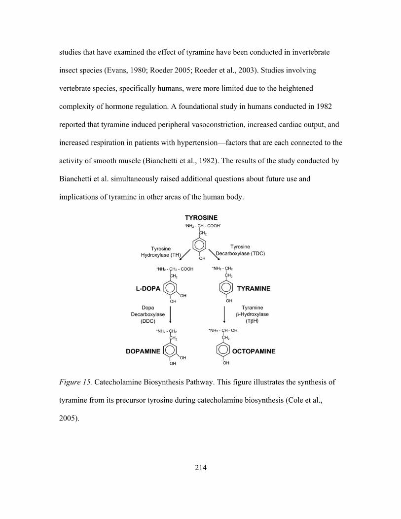
214
studies that have examined the effect of tyramine have been conducted in invertebrate
insect species (Evans, 1980; Roeder 2005; Roeder et al., 2003). Studies involving
vertebrate species, specifically humans, were more limited due to the heightened
complexity of hormone regulation. A foundational study in humans conducted in 1982
reported that tyramine induced peripheral vasoconstriction, increased cardiac output, and
increased respiration in patients with hypertension—factors that are each connected to the
activity of smooth muscle (Bianchetti et al., 1982). The results of the study conducted by
Bianchetti et al. simultaneously raised additional questions about future use and
implications of tyramine in other areas of the human body.
Figure 15. Catecholamine Biosynthesis Pathway. This figure illustrates the synthesis of
tyramine from its precursor tyrosine during catecholamine biosynthesis (Cole et al.,
2005).

215
Within the last ten years a few additional studies have revisited the role that
tyramine may play within the human body. Most of these focused on the role of tyramine
in the diet. Elevated levels of tyramine can be introduced to the body through diet by
consuming large amounts of aged meat, soy proteins and cheese. The body naturally
breaks down proteins through metabolism into amino acids. The amino acid tyrosine is in
large quantities especially in cheese. Tyrosine is further broken down into biogenic
monoamines such as tyramine which was discovered to cause hypertension. This
condition became well known as the “cheese effect.” The body will normally break down
tyramine using an enzyme called monoamine oxidase [MAO] but this can be inhibited in
individuals who are treated with a monoamine oxidase inhibitor [MAOI] that is
prescribed for anxiety, depression, Parkinson’s disease, and bipolar disorder (Finberg and
Gillman, 2011). In recent years, because of the advancement of new research tools and
advancements in genomics, researchers began to examine the role of tyramine throughout
the body. One study focused on the male reproductive system in rats and found that
elevated levels of tyramine could induce continuous peristalsis of the vas deferens
(Finberg and Tenne, 1982). More recently, a study conducted by Obayomi et al. in 2017
demonstrated that the biogenic monoamine tyramine was capable of inducing
contractions of uterine smooth muscle in mice at levels comparable to estrogen (Obayomi
et al., 2017). These studies not only revisited the role of tyramine, but also proposed that
tyramine could act as a modulator of reproductive function in mammals such as rats and
mice. This research inspired me to study the role of tyramine in mouse ovulation.
Ovarian follicles possess numerous cell types that share common morphological
features with smooth muscle such as the tunica albuginea, chordae, and theca externa,
216
illustrated in figure 16 (Peck et al., 2019). These smooth muscle-like cells play a role in
the cycling of oocytes within the ovary, and ultimately their expulsion from the follicle.
Smooth muscle contraction is modulated by many sources such as neurotransmitters (i.e.
dopamine), hormones (i.e. epinephrine) and chemicals (i.e. nitrous oxide) (Obayomi et
al., 2017).
Figure 16. Structural Elements of the Ovarian Follicle. Histological Section of Secondary
ovarian follicle stimulated with PMSG, with major structural elements labeled. This
section is labeled with TAAR1 (brown) and counterstained with Hematoxylin (blue).
Insert: Detail of theca layer versus follicular epithelium. Imaged using EVOS Imaging
System using 20x Objective.

217
Since tyramine can modulate uterine contractions, my project investigated what
affect tyramine may have on the ovary and ovulatory process, based on the shared
morphological similarities between ovarian follicles and smooth muscle. To explore this
hypothesis, I conducted research using mice (Mus Musculus) as an animal model to
garner further insight into the regulatory mechanisms of ovulation.
Results
My study analyzed the role of tyramine in mouse ovaries using High-Performance
Liquid Chromatography, histology, confocal microscopy, and immunohistochemistry
protocols described in section 7.5: Materials & Methods. I analyzed ovaries from five
different experimental conditions: non-injected, non-injected + tyramine, PMSG
(pregnant mare serum gonadotrophin) only, PMSG + tyramine, and complete super
ovulation using PMSG and hCG (human chorionic gonadotropin). Non-injected ovary
samples served as my negative control, to demonstrate the normal state of the ovary with
many follicles at various stages of development and maturation. The remaining samples
received in vivo injections, in vitro treatments, or a combination of both injections and
treatments. Ovaries isolated from mice that received PMSG only and those who had
undergone complete superovulation were used to make comparisons with my samples
that had been treated with tyramine. The importance of the PMSG only and completely
super-ovulated samples was to show the changes in ovarian morphology after the first
and second injections used in standard superovulation protocols—demonstrating what is
“normal” during the process of superovulation.
In the fourth experimental condition I examined non-injected ovaries that were
treated with tyramine in vitro. Observing the effect of tyramine on its own without any

218
hormonal injections was important to establish an understanding of whether tyramine
would have an effect on its own, and if there was an effect, the extent tyramine
influenced follicle rearrangement in the ovary. My final experimental condition was
PMSG + tyramine. For these samples, mice were injected with PMSG only, and
euthanized 48 hours afterwards. The ovaries taken from these mice were treated with
tyramine in vitro. Examining ovarian samples that had been partially super-ovulated
before the addition of tyramine allowed me to determine if tyramine influenced follicle
maturation during the middle of standard superovulation protocol, or if tyramine had a
stronger effect on its own.
Superovulation using injections of PMSG and hCG initiate rapid maturation of
oocytes from meiotic metaphase I to meiotic metaphase II (Peck et al., 2019). After the
follicle reaches maturity, the follicle border ruptures, expelling the oocyte into the
oviduct. During superovulation, a mouse ovary can release between 6-16 oocytes at once
(Silver, 2000). With numerous follicles rupturing and collapsing during ovulation, this
left the ovary with a deflated appearance, as demonstrated in Figure 17C. Using confocal
microscopy, I was able to identify regions specifically labeled in the mouse ovary for
tyramine’s receptor, Trace Amine-Associated Receptor 1 [TAAR1]. In non-injected
mouse ovaries, the distribution of TAAR1 appears uniform throughout the sample
(Figure 17A). However, upon stimulation with PMSG, hCG, and/or tyramine, TAAR1
localized in the corpus luteum of ruptured follicles and the perimeter of the ovary where
oocytes had been released during ovulation (Figure 17B-D).
Figure 18 shows a cross-section of a non-injected ovary (A) and a non-injected
ovary treated with tyramine (B) that has been labeled with a phalloidin dye that

219
selectively binds to actin, the primary component of smooth muscle. Based upon the
initial results of my immunohistochemical labeling of TAAR1 in ovarian sections, I
predicted that stimulating ovarian tissue using a physiological dose of tyramine could
alter the organization of follicles within the ovary and potentially initiate ovulation (Peck
et al., 2019). Following exposure to tyramine, follicle borders did appear disrupted
(Figure 18B) in comparison to the control (Figure 18A), and there were more
tertiary/final-stage follicles present in tyramine-treated ovaries than observed in the
experimental control. Using a similar technique that produces thinner histological
sections, I am able to stain the entire tissues to more clearly visualize the ovarian tertiary
follicles (Figure 19). In both completely super-ovulated mice and mice exposed to PMSG
and tyramine, localization of TAAR1 was enriched within each individual follicle and in
the oocyte itself (Peck et al., 2019). Additionally, the TAAR1 receptors were found to
localize predominantly near tertiary follicles. The collective findings between my
immunohistochemical and histological data suggests that tyramine might play an active
role in the process of follicular maturation and rupture in mice.
Finally, I quantified the levels of tyramine in the ovary using High Performance
Liquid Chromatography [HPLC]. This method uses a column that can separate, identify,
and quantify select molecules in a sample. Non-injected and completely super-ovulated
(injected) ovaries were prepared into a solution and injected through a column to quantify
how the levels of tyramine present in the tissue changed before and after superovulation.
HPLC analysis demonstrated that non-injected ovaries contained higher levels of
tyramine than ovaries that had undergone superovulation using PMSG/hCG (Figure 20)

220
(Peck et al., 2019). The results obtained using HPLC provided valuable insight into the
potential way that tyramine may be utilized during the ovulatory process in mice.
Figure 17. Localization of TAAR1 in Mouse Ovaries. Mouse ovaries are either non-
injected (A), injected with PMSG only (B), completely super-ovulated using both PMSG
and hCG (C), or post-treated with a physiological dose of tyramine after ovary removal
(D). Actin within follicle borders is labeled with phalloidin (red). TAAR1 localization is
visualized in green, showing localization around the perimeter of each follicle, and
enriched in the corpus luteum.

221
Figure 18. Effect of Tyramine on Mouse Ovaries. Image A shows a cross section of a
non-injected ovary. Follicles in image A are at various stages of the maturation process,
with actin/phalloidin labeling in green and DNA/DAPI in gray. Image B shows a cross
section of a non-injected ovary that was exposed to a physiological dose of tyramine for
45 minutes in vitro. The ovary in image B that was stimulated with tyramine shows a
disruption of follicles that resembles the appearance of an ovary that has undergone
complete superovulation. The inset in image A is a histological preparation showing the
usual pattern of DNA organization around a primary follicle. Scale bar is 200µm.

222
Figure 19. Histological Ovary Sections Co-Labeled with TAAR1. Histological sections
of an ovary highlighting a tertiary follicle in non-injected (A), injected with PMSG (B) or
PMSG hCG (C) or PMSG followed by treatment with tyramine (D). Localization of
TAAR1 is found to be enriched not only around the follicle but at the oocyte and in the
cumulus cells.
Figure 20. Quantification of Tyramine in Mouse Ovaries. Tyramine levels in the ovary
were present in higher concentrations in mice that received no hormonal injections,
compared to completely superovulated mice (p=<0.05, n = 10).
0
0.05
0.1
0.15
0.2
0.25
0.3
pg/u
l
Non-Injected Injected
Ovary Tyramine Levels

223
Discussion & Conclusion
The aim of my laboratory benchwork was to identify if the biogenic amine
tyramine was present within the mouse ovary and explore its potential effect on the
ovulatory process in mice. Previous research has shown that tyramine and its receptor
TAAR1 are both present in uterine smooth muscle tissue in mice (Obayomi et al., 2017).
It is also known that the cells of the theca externa that surround ovarian follicles share
common morphological features with smooth muscle (Peck et al., 2019). This led to the
formation of my hypothesis, that if tyramine was present within the mouse ovary, then
female mice undergoing hormonal superovulation would exhibit heightened localization
of both tyramine and TAAR1. The results of my study confirm the presence of tyramine
within the mouse ovary and provide preliminary data suggesting that tyramine may have
role in follicular maturation and ovulation. Further research into the actions of this
monoamine may provide additional insight into understanding the signaling mechanisms
involved in ovary function.
Standard protocols for superovulation in mice entail two intraperitoneal
injections: PMSG and hCG. These sequential injections encourage follicular maturation
(PMSG) and expulsion of oocytes out of their follicles during ovulation (hCG). Using
immunohistochemistry, I observed a localization pattern of TAAR1 throughout the
superovulation process. Non-injected ovaries showed no pattern of localization, however,
after beginning ovulation induction with the first injection of PMSG, localization of
TAAR1 appeared at the perimeter of the ovary, particularly along the membranous sac
that surrounds the ovary, called the bursa. Localization was also observed within some of
the activated follicles at the beginning of the maturation process (Figure 17B). After

224
addition of the second injection, hCG, localization of TAAR1 was observed in the corpus
luteum of ruptured follicles, and along the perimeter of the ovary where oocytes were
released during ovulation (Figure 17C). Ovaries that were not super-ovulated but were
exposed to only tyramine in vitro appeared to have increased localization of TAAR1
within ovarian follicles. Localization of tyramine’s receptor in non-injected ovaries
exposed to tyramine in vitro resembled the pattern of localization of TAAR1 in ovaries
undergoing hormonal superovulation, particularly the effect that PMSG had on TAAR1
localization (Figure 17D & Figure 18B).
To observe patterns of localization of tyramine’s receptor further, I performed
histological counter-staining to visualize oocyte-containing follicles at different stages of
hormonal superovulation, and after exposure to tyramine. Similar to the observations
made using immunohistochemistry, histologically prepared non-injected ovaries showed
no definitive pattern (Figure 19A). After receiving the first injection of PMSG, TAAR1
was recruited to the perimeter of the tertiary follicles, which was even more prominent
following injection of hCG (Figure 19B & 19C). The final experimental condition I
analyzed using histology was PMSG with exposure to tyramine in vitro. The goal of this
experimental condition was to observe if the localization of TAAR1 in PMSG + tyramine
ovaries would resemble PMSG + hCG (standard superovulation). In this case, there was
strong enrichment of TAAR1 in the theca externa of the follicle, the oocyte itself, and the
cumulus cells that support the oocyte (Figure 19D). The pattern of localization between
completely super-ovulated ovaries and PMSG + tyramine ovaries were similar. However,
exposure to tyramine in vitro after injection of PMSG resulted in much stronger
enrichment in those areas. The unique pattern of TAAR1 labeling observed through

225
immunohistochemistry and histology during different points of superovulation suggest
that tyramine may be involved with cycling of oocytes during follicle maturation, due to
the presence of tyramine’s receptor within both the follicle and oocyte.
In the last set of experiments, I quantified the presence of tyramine in non-
injected and completely super-ovulated (PMSG/hCG) ovaries using HPLC to determine
if there was a relationship between levels of tyramine and ovulation. Determining a
quantitative relationship between tyramine and ovulation would allow us to conclude
how tyramine impacts cycling and ovulation of oocytes, or if there was no relationship
between the two. After performing HPLC on these two experimental conditions, I noted
that levels of tyramine were higher prior to ovulation and were reduced after. These
HPLC results further confirm that tyramine does play a role in the ovulatory process.
Performing additional trials using PMSG only, PMSG + tyramine, and non-injected +
tyramine-treated ovaries would help specify when tyramine is being depleted.
Understanding if tyramine is being depleted throughout superovulation, or only after
ovulation will provide insight into whether tyramine has an ongoing role in both
maturation and expulsion of follicles, or if tyramine’s role is substantial during a brief
period of superovulation.
Ultimately, the results of this study may provide foundational knowledge needed
to begin exploring the role of tyramine in oocyte maturation and ovulation in humans.
However, ovarian anatomy of Mus Musculus differs slightly from that of humans. In
mice, a membranous sac called the bursa envelops the ovary and connects directly to the
oviduct (Hillier, 2012). This membrane facilitates the movement of ovulated oocytes into
the oviduct. The bursa has also demonstrated rapid accumulation and reabsorption of

226
fluid during ovulation in mice which suggests a possible role of the membrane
controlling fluid homeostasis during ovulation and ovarian repair following ovulation
(Zhang et al., 2013).
The bursa structure is not present in human ovarian anatomy. Rather, the
movement of ovulated oocytes into the fallopian tube in humans is facilitated by fimbriae
present at the distal end of the tube that “sweep” the oocyte through the fallopian tube
towards the uterine body. The anatomical differences between mice and humans
necessitate additional studies using both mouse and human tissue. In addition to
conducting further experiments using mouse ovaries, performing identical trials using
human ovarian tissue would determine the extent of tyramine’s role in maturation and
ovulation in humans. By better understanding the mechanisms of ovulation, this
information could augment the existing protocols we use at the clinic for controlled
ovarian stimulation, and even merit the development of new protocols to more effectively
treat patients who are experiencing infertility related to ovulatory dysfunction.
Materials & Methods
Animals
Use of animals in this project was approved through the IACUC protocols 18-
1606R and 20-1801R at Arizona State University (see Appendix B). All mouse tissues
utilized for experiments were obtained from female C57BL/6J wild-type mice (Bar
Harbor, ME) approximately eight weeks of age.
Superovulation & Tissue Procurement
For my experiments, I compared five different experimental conditions which are
described in Table 9 below. These specific experimental conditions were utilized to

227
observe the additive effects of super-ovulatory injections and to establish baseline values
that I could compare to conditions involving the addition of tyramine.
I followed standard protocols for superovulation for my study, which involved a
regimen of pregnant mare serum gonadotropin [PMSG] and human chorionic
gonadotropin [hCG] injections (The Jackson Laboratory, 1998). Female mice undergoing
partial or complete superovulation were treated using standard intraperitoneal [IP]
injection protocols, illustrated in Figure 21. An initial IP injection of 5 IU of PMSG was
administered, followed 48 hours later with an IP injection of 5 IU of hCG (The Jackson
Laboratory, 1998). Approximately 14 hours after hCG was administered, completely
super-ovulated mice were anesthetized using isoflurane and euthanized through cervical
dislocation. Following cervical dislocation, the mouse is prepped for dissection by
applying 70% ethyl alcohol to the abdomen. To remove the ovary, I made a midline
incision down the ventral side of the mouse, starting below the ribcage down to the
urethral orifice. Once the body cavity was opened, the fat pad is lifted away exposing the
reproductive system. The ovary can be identified proximally to the oviduct and uterine
horn.
Whole ovaries for experimental conditions that did not utilize tyramine were
immediately placed in a fixative solution of 4% paraformaldehyde made with phosphate-
buffered saline [PBS] and left to fix overnight at 4ºC to stabilize and preserve the tissue.
For experimental conditions that underwent tyramine exposure: non-injected + tyramine
& partial superovulation + tyramine, I warmed KSOM medium to approximately 37º, and
added a physiological dose of 1nM tyramine to the solution. The ovaries were incubated
in the tyramine-containing media for 45 minutes, and once the exposure time was

228
complete, they were removed from the media and fixed using 4% paraformaldehyde
solution and left to fix overnight at 4ºC.
Figure 21. Superovulation and Ovarian Tissue Procurement. Created with
BioRender.com
Immunohistochemistry of Frozen Ovarian Tissue Sections
After fixing isolated ovarian tissue in 4% paraformaldehyde, I prepared samples
from each experimental condition for immunohistochemistry using antibodies that
specifically bind Tyramine and its receptor: Trace Amine Associated Receptor 1
[TAAR1]. The fixative solution was removed and replaced it with 30% sucrose solution.

229
The tissues were incubated in the sucrose solution overnight at 4ºC. The next day, the
tissue are positioned in molds filled with Tissue-Tek O.C.T. (VWR). The molds were
lowered into 2-propanol that was cooled with liquid-nitrogen until the Tissue-Tek had
solidified. The samples were stored in a -80ºC freezer. I sectioned each block at 40µm
using the Leica CM1950 Cryostat onto charged slides before labeling the samples for
immunohistochemistry.
To prepare for labeling, I drew a hydrophobic barrier around the perimeter of each
slide containing ovarian sections (see Figure 22). This barrier prevented solutions from
running off the slide during each step of the immunohistochemistry protocol.
Figure 22. Example Slide of Ovarian Sections. Five sections of ovarian tissue are present
on the slide, with the hydrophobic barrier around the perimeter in blue. Created with
BioRender.com.
I blocked and permeabilized the ovary sections prior to labeling. Permeabilizing
solution was made using 2% formaldehyde and 1% Tween-20 in 1x PBS. I incubated the
tissues in permeabilizing solution overnight at 4ºC, which created spaces in the
membrane so antibodies could bind inside the cells of the tissue for labeling. The
following day, I discarded the permeabilizing solution and rinsed the slides in buffer

230
containing 1% Bovine Serum Albumin [BSA] diluted in PBS, or PBS/BSA three times
for five minutes each. After the tissues were rinsed completely, I added primary antibody
to the tissues to begin the immunolabeling process. Rabbit anti-TAAR1 primary antibody
(ThermoFisher, OSR00119W) was added at approximately 1:1000 in PBS/BSA solution
and incubated overnight at 4ºC. Following incubation in the primary antibody, I rinsed
the tissues in PBS/BSA buffer three times for five minutes each, to flush away any
residual primary antibody.
Next, I added light-sensitive secondary antibodies fused to fluorophores to the
tissue for fluorescent visualization. Anti-rabbit Alexa 488 (ThermoFisher Scientific,
1:1000 in PBS/BSA) was added as the secondary antibody to the anti-TAAR1. Phalloidin
(ThermoFisher, A12380) was also added into the secondary antibody solution—a toxin
from the Amanita phalloides mushroom that selectively binds F-actin and is conjugated
to Alexa 568. I wrapped the tissues in foil and incubated them overnight at 4ºC. The
following day, I rinsed the tissues three times for five minutes each in 1x PBS and sealed
each slide with Vectashield and a coverslip for imaging. Scans of the ovary sections were
taken as a set of tile images using a Leica SP5 confocal microscope, housed in the Keck
Bioimaging Lab at Arizona State University, with a 20x objective at a depth of 20µm in
1µm steps.
Paraffin-Embedded Histology
Mouse ovaries from each experimental condition were obtained as described
above and fixed in 4% paraformaldehyde overnight at 4ºC. Following fixation, I rinsed
the tissues using 1x PBS to remove all fixative solution. I repeated this process three
times before beginning the dehydration process for rapid dehydration and paraffin

231
embedding. To dehydrate the tissue, I moved the tissues through increasing
concentrations of ethanol: 70%, 80%, 90%, 95%, 100% (twice), before moving into a
50/50 solution of ethanol/ClearRite (Thermo Scientific), 100% ClearRite (twice), then
ClearRite/Paraffin and finally, into 100% paraffin wax. After reaching 100% paraffin
wax, I placed the tissues in embedding molds to allow the paraffin to solidify completely
before I sectioned them.
I cut the embedded ovaries into 5µm sections using the Leica RM1950 microtome and
collected the sections onto charged microscope slides. I cleared the slides of excess wax
using ClearRite and rehydrated the tissues using the series of steps outlined in Figure 23.
Figure 23. H&E Staining of Paraffin-Embedded Tissue Sections. Credit: Shelbi Peck
2017.

232
After completion of step 9 in deionized water, I incubated my ovarian sections in
rabbit anti-TAAR1 antibody (ThermoFisher; 1:1000) overnight in a humidified chamber.
Following the incubation period, I rinsed my slides three times for five minutes each
using PBS/BSA solution before adding anti-rabbit horseradish peroxidase (Pierce; 1:500)
and incubated the slides in the humidified chamber overnight. The following day, I rinsed
the slides again and added 3,3’-Diaminobenzidine (VectorLabs), or “DAB” for ten
minutes. DAB quickly oxidizes in the presence of horseradish peroxidase and produces a
visible brown precipitate. Once a visible brown precipitate had formed, I rinsed the slides
thoroughly with Barnstead water to prevent excessive staining. I proceeded through the
remaining steps of the H&E staining procedure in Figure 21 starting at step 10 with the
hematoxylin counterstain and ending with the slides being sealed using permount. Once
the slides had dried completely, I visualized the slides using the ThermoFisher Scientific
Evos FL auto live cell imaging system, housed in the WM Keck Bioimaging Laboratory
at Arizona State University.
High-Performance Liquid Chromatography
High-Performance Liquid Chromatography [HPLC] is a quantitative technique
that identifies and quantifies the components of a mixture by separating out each element
as it flows through a pressurized column. Mouse ovaries from non-injected and
completely super-ovulated experimental conditions were obtained as described above.
Immediately following dissection of the ovaries, I flash-froze the tissues in 1.5mL
Eppendorf tubes and stored at -80ºC until I analyzed them using HPLC. The first step of
preparation for HPLC is to digest the tissues using a perchloric acid solution. I added
each tissue to a small Eppendorf tube containing perchloric acid and left them in an ice

233
bath for a total of thirty minutes to facilitate disruption of the tissue. Next, I pulverized
the partially dissolved tissue using an Eppendorf tissue grinder and pestle, before
centrifuging the tubes for ten minutes to ensure that any remaining pieces of tissue that
had not dissolved formed a pellet at the bottom of the tube. Centrifuging the tubes to
separate out undissolved tissue reduced the likelihood that pieces of tissue would block
the lines of the HPLC system during analysis.
Using a micropipette, I loaded 10µL of each sample into the ESA CoulArray
HPLC system. The system generated characteristic chromatographic peaks for the amines
that flowed through the column, which I compared between non-injected ovarian samples
and completely super-ovulated ovarian samples. The U.S. Department of Agriculture’s
Arid-Land Agricultural Research Center in Maricopa, AZ graciously allowed for
temporary use of the ESA CoulArray HPLC System for this project.
Statistical analyses
Quantification of the HPLC samples were obtained from 10 mice. Unless indicated
otherwise results are shown as the mean ± SD. Samples that passed the normal
distribution test were analyzed by a student’s t test and were considered significantly
different if p < 0.05.

234
CHAPTER 8
CONCLUSION
Reproductive medicine is a specialized branch of medicine that aims to diagnose
and treat patients who are experiencing reproductive dysfunction. In Chapter 1 & 2 of my
dissertation, I described normal reproductive function to establish a standard for how
reproduction works under the best of circumstances. This provided the necessary
framework for addressing the first aim of my dissertation, to describe how the
reproductive system works in order to diagnose and treat reproductive disorders.
Through decades of research, numerous pathologies of infertility have been uncovered
that span anatomical, physiological, and genetic origins. Both men and women can
experience reproductive dysfunction which impacts their ability to conceive naturally.
During my time working at the fertility clinic, I noticed patients coming to the
clinic with various forms of infertility, and some who were unsure why they were unable
to conceive on their own. Using the knowledge and experience I attained through
working at the clinic, I characterize these forms of reproductive dysfunction in Chapter 3:
Diagnosis and Characterization of Reduced Fertility. In men, reduced fertility typically
manifests as either defective spermatogenesis, or problems with the transport of sperm
through the reproductive tract (NICHD, 2017). For women, reproductive dysfunction
falls into one of four categories: ovarian, fallopian tube, uterine, or cervical infertility.
Within each of these broad categories there are anatomical, endocrine, and genetic factors
that can individually impact a woman’s ability to become pregnant and carry a pregnancy
to term.

235
Many patients who have received a concrete diagnosis for their reproductive
dysfunction have sought treatment at a fertility clinic. At fertility clinics such as the one
where I worked, we can attempt to address a patient’s infertility using treatment options
such as timed intercourse, ovulation induction, and Assisted Reproductive Technologies
[ARTs] like intrauterine insemination, in vitro fertilization, and intracytoplasmic sperm
injection. This brings us to the second aim of my dissertation, to examine the
development and use of fertility treatments and ARTs in reproductive medicine. In
Chapter 4: Fertility Treatments and Assisted Reproductive Technologies, I began by
describing the history of how the treatments utilized at fertility clinics were developed.
Then, using the unique perspective I have from working in the field of reproductive
medicine, I describe why a patient would use each treatment, treatment effectiveness, and
discuss protocols I performed within the fertility clinic laboratory.
I wrote the first four chapters of my dissertation with patients in mind--
specifically, to serve as a resource for individuals experiencing reduced fertility. These
chapters provide foundational information for patients who want to learn more about
reproductive health, understand what treatments are offered at a fertility clinic and their
associated costs, and how laboratory staff work with patient samples to help individuals
overcome their reduced fertility. As a member of the laboratory staff, I noticed that many
patients were interested in what went on in the lab, but we had very little time to explain
all the details of the kind of work we did. The information available online for patients to
learn more about the inner workings of a fertility clinic laboratory is not very detailed.
Clinic websites typically describe the patient’s role and how the treatment process works
for the patient (diagnostics, medicated ovulation induction, retrieval, embryo transfer,

236
etc.), but leave out information on how the laboratory specifically facilitates the
reproductive process. Therefore, I wanted to help bridge the gap that existed between
publicly available resources for patients seeking fertility treatment and the intricate work
of fertility clinic laboratory staff.
Across the globe, millions of babies have been born through fertility treatments
and ARTs provided by fertility clinics. However, implementation of these techniques and
technologies in the field has had unanticipated ethical repercussions. As a field,
reproductive medicine has not been regulated effectively in the United States. Lack of
sufficient regulatory oversight reveals loopholes that can lead to misguidance,
discrimination and even exploitation of patients and donors. This led to the development
of the third aim of my dissertation, to analyze prominent ethical issues I observed in the
field of reproductive medicine. In Chapter 5: Ethical Dilemmas in Fertility, I examined
three prominent ethical quandaries that I observed while working at the fertility clinic:
profiling and anonymity of sperm donors, oocyte donor compensation, and the
ramifications of specialized forms of pre-implantation genetic testing [PGT]. I discussed
how these issues arose at the clinic where I worked, why each constitutes as an ethical
issue, and the gaps in regulations and legislation that led to these issues.
The three ethical dilemmas I chose to discuss are by no means the only ethical
issues in fertility medicine; however, these were the prominent issues I observed during
my time at the clinic. Our ability to manipulate the reproductive process has surpassed
the current regulations in place that protect patients and the children born using ARTs,
which allows these ethical dilemmas to arise. Ultimately, I call for the adoption of new
policies, procedures, restrictions and sanctions against clinics and sperm/oocyte banks

237
that do not comply. Establishing a more rigid regulatory framework for fertility medicine
will help to protect the best interests of patients, donors, fertility clinics, and
sperm/oocyte banks alike.
Despite everything we know about reduced fertility, in over 30% of cases,
physicians are unable to pinpoint a reason for an individual’s reduced fertility, which
leads to a diagnosis of idiopathic infertility (Chandra et. al, 2013; Sadeghi, 2015). As
ARTs become a more prevalent method for conception, it is crucial to continue
developing more effective methods to diagnose and treat infertility. This led to the
development of the final aim of my dissertation, to review novel methodologies that may
advance the field of reproductive medicine. Research helps to fill in the gaps present in
current knowledge of reproductive physiology and allow for the continual advancement
of fertility medicine. During my time working at the clinic, I became aware of some
specific pathologies of infertility that we were unable to treat, and where technological
limitations could be improved to make our workflow in the lab smoother. In Chapter 6, I
covered current and future research in the field of fertility medicine. This included novel
therapies and remote fertility testing options for both female and male patients, as well as
the use of artificial intelligence in the embryology laboratory. For each area of research, I
gave personal insight into the ways these therapies and technologies could be
implemented at my clinic and who would benefit from them the most.
The studies described in Chapter 6 had all progressed to implementation in human
clinical trials. However, before any protocol can be implemented in humans, numerous
phases of clinical and pre-clinical trials are conducted to ensure safety and efficacy of
new techniques (Faggion, 2015). The final chapter covers an animal trial that I conducted

238
to explore the role of the biogenic amine tyramine on the process of ovulation in mice.
The results of my study suggest that tyramine may influence maturation and ovulation of
oocytes from the ovary, resembling the effect of standard superovulation protocols. This
preliminary study raises fundamental questions regarding the role that tyramine may play
in the human reproductive system and demonstrated that there are other molecules that
interact with reproductive function that have not been taken into consideration
previously. If tyramine exhibits similar effects on oocyte maturation and ovulation in
humans, this may lead to the development of shortened protocols used for ovarian
stimulation, which has implications in both agriculture and human medicine.
ARTs have revolutionized the way infertility is perceived from a clinical
standpoint. Once thought of as an insurmountable burden for women alone, infertility is
now recognized as both a man and woman’s issue and, in most cases, can be more
successfully treated now than ever before. A diagnosis of reduced fertility or infertility
presents a mere hurdle to procreation rather than a complete termination of a couple’s
aspirations to start a family. Fertility clinics have become beacons of hope for individuals
who are struggling to overcome infertility by providing treatments and ART services to
patients. My dissertation has accomplished each of its intended goals, utilizing a
combination of literature review, clinical experience, and laboratory research. It is my
hope that through personal experience working at a fertility clinic and conducting original
research have generated a unique perspective to provide a much-needed resource to
patients seeking fertility treatment.

239
REFERENCES
Aboulmaouahib, S., Madkour, A., Kaarouch, I., Sefrioui, O., Saadani, B., Copin, H., . . . Cadi, R. (2017). Impact of alcohol and cigarette smoking consumption in male fertility potential: Looks at lipid peroxidation, enzymatic antioxidant activities and sperm DNA damage. Andrologia, 50(3). doi:10.1111/and.12926
Abu Hashim, H., Ombar, O., & Abd Elaal, I. (2011). Intrauterine insemination versus timed intercourse with clomiphene citrate in polycystic ovary syndrome: a randomized controlled trial. Acta Obstetricia Et Gynecologica Scandinavica, 90(4), 344–350. https://doi.org/10.1111/j.1600-0412.2010.01063.x
Acosta, L. (2013). Adoption Law: United States. Adoption Law: United States | Law Library of Congress. https://www.loc.gov/law/help/adoption-law/unitedstates.php.
Advanced Fertility Center of Chicago. (2020a, December 15). Clomid Fertility Medication Treatment for Women That Already Ovulate and for Unexplained Infertility. https://advancedfertility.com/2020/09/17/clomid-fertility-medication-treatment-for-women-that-already-ovulate-and-for-unexplained-infertility/.
Advanced Fertility Center of Chicago. (2020b, December 15). IVF Protocols for Stimulation with Medications and Drugs. Ovarian Stimulation IVF Protocols Medications and Drugs for In Vitro Fertilization. https://advancedfertility.com/ivf-in-detail/ovarian-stimulation/.
Advanced Fertility Center of Chicago. (2020c, December 15). Lupron Down-Regulation Ovarian Stimulation Protocol for IVF. IVF Medication Protocols for Ovarian Stimulation with Antagonists. https://advancedfertility.com/2020/09/21/lupron-down-regulation-ovarian-stimulation-protocol-for-ivf/.
Advanced Fertility Center of Chicago. (2020d, December 15). IVF Medication Protocols for Ovarian Stimulation with Antagonists. https://advancedfertility.com/2020/09/18/ivf-medication-protocols-for-ovarian-stimulation-with-antagonists/.
Advanced Fertility Center of Chicago. (2020e, December 15). Ganirelix (Antagonist) IVF Protocol Calendar. Antagonist IVF Protocol Sample Calendar. https://advancedfertility.com/2020/09/22/antagonist-ivf-protocol-sample-calendar/.
Advanced Fertility Center of Chicago. (2020f, December 15). Flare IVF Protocol Calendar. Microdose Lupron Flare Protocol for IVF - Sample CalendarIn Vitro Fertilization Microflare Protocol. https://advancedfertility.com/2020/09/21/microdose-lupron-flare-protocol-for-ivf-sample-calendarin-vitro-fertilization-microflare-protocol/.

240
Agarwal, A., Panner Selvam, M. K., Sharma, R., Master, K., Sharma, A., Gupta, S., & Henkel, R. (2018). Home sperm testing device versus laboratory sperm quality analyzer: Comparison of motile sperm concentration. Fertility and Sterility, 110(7), 1277-1284. doi:10.1016/j.fertnstert.2018.08.049
Agarwal, A., Prabakaran, S., & Allamaneni, S. S. (2006). Relationship between oxidative stress, varicocele and infertility: A meta-analysis. Reproductive BioMedicine Online, 12(5), 630-633. doi:10.1016/s1472-6483(10)61190-x
Agarwal, S. K., & Haney, A. F. (1994). Does recommending timed intercourse really help the infertile couple?. Obstetrics and gynecology, 84(2), 307-310.
Al-Daghistani H. I. (2020). Staphylococcusaureus protein A as a means of assessing sperm penetrability in cervical mucus in vitro. Clinical and experimental reproductive medicine, 47(3), 186–193. https://doi.org/10.5653/cerm.2020.03279
Alfirevic, Z., Navaratnam, K., & Mujezinovic, F. (2017). Amniocentesis and chorionic villus sampling for prenatal diagnosis. Cochrane Database of Systematic Reviews. https://doi.org/10.1002/14651858.cd003252.pub2
Ali, M., & Parekh, N. (2020). Male Age and Andropause. Male Infertility, 469–477. https://doi.org/10.1007/978-3-030-32300-4_36
Allersma, T., Farquhar, C., & Cantineau, A. E. P. (2013). Natural cycle in vitro fertilisation (IVF) for subfertile couples. Cochrane Database of Systematic Reviews. https://doi.org/10.1002/14651858.cd010550.pub2
Allison T. L. (2016). Immunosuppressive Therapy in Transplantation. The Nursing clinics of North America, 51(1), 107–120. https://doi.org/10.1016/j.cnur.2015.10.008
Alvarez, S. (2015). Do some addictions interfere with fertility? Fertility and Sterility, 103(1), 22-26. doi:10.1016/j.fertnstert.2014.11.008
Alwaal, A., Breyer, B. N., & Lue, T. F. (2015). Normal male sexual function: emphasis on orgasm and ejaculation. Fertility and Sterility, 104(5), 1051–1060. https://doi.org/10.1016/j.fertnstert.2015.08.033
American College of Obstetricians and Gynecologists [ACOG]. (2016). Abnormal Cervical Cancer Screening Test Results. Retrieved September 20, 2020, from https://www.acog.org/patient-resources/faqs/gynecologic-problems/abnormal-cervical-cancer-screening-test-results
American College of Obstetricians and Gynecologists [ACOG]. (2018). Müllerian Agenesis: Diagnosis, Management, and Treatment. Retrieved November 18, 2020,

241
from https://www.acog.org/clinical/clinical-guidance/committee-opinion/articles/2018/01/mullerian-agenesis-diagnosis-management-and-treatment
American College of Obstetricians and Gynecologists [ACOG]. (2019). Treating Infertility. ACOG. https://www.acog.org/womens-health/faqs/treating-infertility.
American Pregnancy Association [APA]. (2017, April 24). Intrauterine insemination (IUI). Retrieved November 22, 2020, from https://americanpregnancy.org/getting-pregnant/intrauterine-insemination-70967/
American Pregnancy Association [APA]. (2019, April 24). In Vitro Fertilization (IVF): American Pregnancy Association. Retrieved November 22, 2020, from https://americanpregnancy.org/getting-pregnant/infertility/in-vitro-fertilization-70966/
American Pregnancy Association [APA]. (2020, January 11). Understanding Pregnancy Tests. American Pregnancy Association. https://americanpregnancy.org/getting-pregnant/understanding-pregnancy-tests-70933/.
Anderson, K., Niesenblat, V., & Norman, R. (2010). Lifestyle factors in people seeking infertility treatment - A review. Australian and New Zealand Journal of Obstetrics and Gynaecology, 50(1), 8–20. https://doi.org/10.1111/j.1479-828x.2009.01119.x
Aoyama, N., & Kato, K. (2020). Trophectoderm biopsy for preimplantation genetic test and technical tips: A review. Reproductive Medicine and Biology, 19(3), 222–231. https://doi.org/10.1002/rmb2.12318
Arbyn, M., Anttila, A., Jordan, J., Ronco, G., Schenck, U., Segnan, N., Wiener, H., Herbert, A., & von Karsa, L. (2010). European Guidelines for Quality Assurance in Cervical Cancer Screening. Second Edition—Summary Document. Annals of Oncology, 21(3), 448–458. https://doi.org/10.1093/annonc/mdp471
Arian, S. E., Flyckt, R. L., Herman, R., Erfani, H., & Falcone, T. (2018). Fertility preservation in pediatric female cancer patients. Fertility and Sterility, 109(5), 941. doi:10.1016/j.fertnstert.2018.02.112
Arora, M., & Mukhopadhaya, N. (2019). Intrauterine Adhesions. In Recurrent pregnancy loss (pp. 140-145). New Delhi ; London ; Panama: Jaypee the Health Sciences Publisher.
Arora, M., & Tayade, S. (2018). Recurrent Miscarriage. Donald School Textbook of Human Reproductive & Gynecological Endocrinology, 364.

242
Arora, P., & Mishra, V. (2018). Difficult embryo transfer: A systematic review. Journal of Human Reproductive Sciences, 11(3), 229. https://doi.org/10.4103/jhrs.jhrs_59_18
Baetens, P., Devroey, P., Camus, M., Van Steirteghem, A. C., & Ponjaert-Kristoffersen, I. (2000). Counselling couples and donors for oocyte donation: the decision to use either known or anonymous oocytes. Human Reproduction, 15(2), 476–484. https://doi.org/10.1093/humrep/15.2.476
Baker, V. (2011, July). Life plans and family-building options for women with primary ovarian insufficiency. In Seminars in reproductive medicine (Vol. 29, No. 04, pp. 362-372). © Thieme Medical Publishers.
Banjoko, S., & Adeseolu, F. (2013). Seminal Plasma pH, Inorganic Phosphate, Total and Ionized Calcium Concentrations In The Assessment of Human Spermatozoa Function. Journal Of Clinical And Diagnostic Research, 7483-7486. doi:10.7860/jcdr/2013/6194.3585
Barad, D. (2018, October 1). Pre-Cycle Patient Information on IUI Treatment: CHR. Center For Human Reproduction. https://www.centerforhumanreprod.com/patients/training/iui/.
Barbash-Hazan, S., Frumkin, T., Malcov, M., Yaron, Y., Cohen, T., Azem, F., . . . Ben-Yosef, D. (2009). Preimplantation aneuploid embryos undergo self-correction in correlation with their developmental potential. Fertility and Sterility,92(3), 890-896. doi:10.1016/j.fertnstert.2008.07.1761
Barrett, K., Barman, S., Yuan, J., & Brooks, H. (2019). Ganong's Review of Medical Physiology (26th ed.). Mcgraw-Hill Education.
Barton, M., Walker, K., & Wiesner, B. P. (1945). Artificial insemination. BMJ, 1(4384), 40-43. doi:10.1136/bmj.1.4384.40
Bartz, D., & Greenberg, J. A. (2008). Sterilization in the United States. Reviews in obstetrics & gynecology, 1(1), 23–32.
Baskaran, S., Finelli, R., Agarwal, A., & Henkel, R. (2020). Diagnostic value of routine semen analysis in clinical andrology. Andrologia, 53(2). https://doi.org/10.1111/and.13614
Bates, G. W., & Bowling, M. (2012). Physiology of the female reproductive axis. Periodontology 2000, 61(1), 89–102. https://doi.org/10.1111/j.1600-0757.2011.00409.x

243
Beall, S., Moon, K., Widra, E., DeCherney, A., Chang, F., & Richter, K. (2012). Characteristics predictive of failed leuprolide acetate (LA) trigger among patients undergoing gonadotropin-releasing hormone (GnRH) antagonist suppression for vitro fertilization (IVF). Fertility and Sterility, 98(3), S172.
Beauchamp, T. L., & Childress, J. F. (2001). Principles of biomedical ethics. Oxford University Press, USA.
Beeder, L. A., & Samplaski, M. K. (2019). Effect of antidepressant medications on semen parameters and male fertility. International Journal of Urology, 27(1), 39-46. doi:10.1111/iju.14111
Berkane, N., Braneti, R., Streuli, I., Martinez De Tejada, B., & Dubuisson, J. (2020). Uterine transplant: Clinical and ethical challenges in Switzerland. Revue medicale suisse, 16(676-7), 42-46.
Berkeley Wellness. (2013, April 18). Do Women Have a Prostate Gland? Retrieved August 24, 2020, from https://www.berkeleywellness.com/self-care/sexual-health/article/female-prostate
Betts, J. G., Desaix, P., Johnson, E., Johnson, J. E., Korol, O., Kruse, D., . . . Young, K. A. (2013). Anatomy & Physiology. Houston, TX: OpenStax College, Rice University.
Bhasin S. (2007). Approach to the infertile man. The Journal of clinical endocrinology and metabolism, 92(6), 1995–2004. https://doi.org/10.1210/jc.2007-0634
Bhattacharya, S., Pandey, S., Pandey, S., & Maheshwari, A. (2010). The impact of female obesity on the outcome of fertility treatment. Journal of Human Reproductive Sciences, 3(2), 62. doi:10.4103/0974-1208.69332
Bianchetti, M. G., Minder, I., Beretta-Piccoli, C., Meier, A., & Weidmann, P. (1982). Effects of tyramine on blood pressure and plasma catecholamines in normal and hypertensive subjects. Klinische Wochenschrift, 60(9), 465-470. doi:10.1007/bf01720361
Bieth, E., Hamdi, S. M., & Mieusset, R. (2020). Genetics of the congenital absence of the vas deferens. Human Genetics, 140(1), 59–76. https://doi.org/10.1007/s00439-020-02122-w
Brännström, M. (2017). Uterus transplantation and beyond. Journal of Materials Science: Materials in Medicine, 28(5), 70.

244
Brännström, M., Johannesson, L., & Dahm-Kähler, P. (2014). First clinical uterus transplantation trial: A six-month report. Fertility and Sterility, 101(5), 1128-1236. doi:doi.org/10.1016/j.fertnstert.2014.02.024
Brännström, M., Johannesson, L., Bokström, H., Kvarnström, N., Mölne, J., Dahm-Kähler, P., . . . Nilsson, L. (2015). Livebirth After Uterus Transplantation. The Lancet, 385(9986), 607-616. doi:doi.org/10.1016/S0140-6736(14)61728-1
Briceag, I., Costache, A., Purcarea, V. L., Cergan, R., Dumitru, M., Briceag, I., Sajin, M., & Ispas, A. T. (2015). Fallopian tubes--literature review of anatomy and etiology in female infertility. Journal of medicine and life, 8(2), 129–131.
Brody, D., & Gu, Q. (2020, September 04). Antidepressant Use Among Adults: United States, 2015–2018. Retrieved November 10, 2020, from https://www.cdc.gov/nchs/products/databriefs/db377.htm
Broekmans, F., Kwee, J., Hendriks, D., Mol, B., & Lambalk, C. (2006). A systematic review of tests predicting ovarian reserve and IVF outcome. Human Reproduction Update, 12(6), 685-718. doi:10.1093/humupd/dml034
Brosens, J. J., Pijnenborg, R., & Brosens, I. A. (2002). The myometrial junctional zone spiral arteries in normal and abnormal pregnancies. American Journal of Obstetrics and Gynecology, 187(5), 1416-1423. doi:10.1067/mob.2002.127305
BruceBlaus (2014). Endometriosis [Photograph]. Wikimedia Commons. https://commons.wikimedia.org/wiki/File:Blausen_0349_Endometriosis.png
BruceBlaus (2017). IUI [Photograph]. Wikimedia Commons. https://commons.wikimedia.org/wiki/File:IUI.png
Budak, A., & Kanmaz, A. G. (2019). Role of endometrial sampling in cases with asymptomatic cervical polyps. Journal of Gynecology Obstetrics and Human Reproduction, 48(3), 207–211. https://doi.org/10.1016/j.jogoh.2019.01.005
Budani, M. C., & Tiboni, G. M. (2017). Ovotoxicity of cigarette smoke: A systematic review of the literature. Reproductive Toxicology, 72, 164-181. doi:10.1016/j.reprotox.2017.06.184
Bulletti, C., Coccia, M. E., Battistoni, S., & Borini, A. (2010). Endometriosis and infertility. Journal of assisted reproduction and genetics, 27(8), 441–447. https://doi.org/10.1007/s10815-010-9436-1
California Cryobank. (2021a). Sperm Banking History. Sperm Banking History | California Cryobank. https://www.cryobank.com/learning-center/sperm-banking-101/sperm-banking-history/.

245
California Cryobank. (2021b). Become a sperm donor. Retrieved April 20, 2021, from https://www.cryobank.com/services/become-a-sperm-donor/
California IVF Fertility Center. (2019). Donor egg program – Donor oocytes. Retrieved March 25, 2021, from https://www.californiaivf.com/donor-eggs/
Calogero, A., Polosa, R., Perdichizzi, A., Guarino, F., Vignera, S. L., Scarfia, A., . . . Vicari, E. (2009). Cigarette smoke extract immobilizes human spermatozoa and induces sperm apoptosis. Reproductive BioMedicine Online, 19(4), 564-571. doi:10.1016/j.rbmo.2009.05.004
Cameron, C., & Williamson, R. (2003). Is there an ethical difference between preimplantation genetic diagnosis and abortion? Journal of Medical Ethics, 29(2), 90-92. doi:10.1136/jme.29.2.90
Carey v. Population Services International, 431 U.S. 678 (1977). https://supreme.justia.com/cases/federal/us/431/678/
Casamonti, E., Vinci, S., Serra, E., Fino, M. G., Brilli, S., Lotti, F., . . . Krausz, C. (2017). Short-term fsh treatment and sperm maturation: A prospective study in idiopathic infertile men. Andrology, 5(3), 414-422. doi:10.1111/andr.12333
Castillón, G., Muñiz, N., Simón, C., & Calaf-Alsina, J. (2018). Clinical evaluation of the subfertile female. Encyclopedia of Reproduction, 4, 2-8. doi:10.1016/b978-0-12-801238-3.64723-2
Castro, R. D., Merlini, E., Rigamonti, W., & Macedo, A. (2007). Phalloplasty and Urethroplasty in Children With Penile Agenesis: Preliminary Report. Journal of Urology, 177(3), 1112-1117. doi:10.1016/j.juro.2006.10.095
Catsanos, R., Rogers, W., & Lotz, M. (2013). The ethics of uterus transplantation. Bioethics, 27(2), 65–73. https://doi.org/10.1111/j.1467-8519.2011.01897.x
Cavaliere, G. (2017). Genome editing and assisted reproduction: Curing embryos, society or prospective parents? Medicine, Health Care and Philosophy, 21(2), 215-225. doi:10.1007/s11019-017-9793-y
Centers for Disease Control and Prevention [CDC], US Dept of Health and Human Services. (2020). 2018 Assisted Reproductive Technology Fertility Clinic Success Rates Report (Rep.). Atlanta, GA.
Centers for Disease Control and Prevention [CDC]. (2016, October 04). Detailed STD Facts - Chlamydia. Retrieved November 17, 2020, from https://www.cdc.gov/std/chlamydia/stdfact-chlamydia-detailed.htm

246
Centers for Disease Control and Prevention [CDC]. (2019a). About CDC's Division of Reproductive Health. Retrieved June 05, 2020, from https://www.cdc.gov/reproductivehealth/drh/about-us/index.htm
Centers for Disease Control and Prevention [CDC]. (2019b). Current Cigarette Smoking Among Adults in the United States. Retrieved October 20, 2020, from https://www.cdc.gov/tobacco/data_statistics/fact_sheets/adult_data/cig_smoking/index.htm
Centers for Disease Control and Prevention [CDC]. (2021a, March 12). National art Surveillance. Retrieved March 25, 2021, from https://www.cdc.gov/art/nass/index.html
Centers for Disease Control and Prevention [CDC]. (2021b, April 20). ART Success Rates. Assisted Reproductive Technology (ART). https://www.cdc.gov/art/artdata/index.html.
Cha, A. E. (2018, September 13). Children of sperm donors find their half siblings and demand change. Retrieved March 08, 2021, from https://www.chicagotribune.com/lifestyles/health/ct-children-of-sperm-donors-20180913-story.html
Chan, Y. Y., Jayaprakasan, K., Zamora, J., Thornton, J. G., Raine-Fenning, N., & Coomarasamy, A. (2011). The prevalence of congenital uterine anomalies in unselected and high-risk populations: a systematic review. Human reproduction update, 17(6), 761–771. doi:doi.org/10.1093/humupd/dmr028
Chandra, A., Copen, C., & Stephen, E. (2013). Infertility and Impaired Fecundity in the United States, 1982–2010: Data From the National Survey of Family Growth. National Health Statistics Reports, (Publication No. 67).
Chavarro, J. E., Rich-Edwards, J. W., Rosner, B. A., & Willett, W. C. (2008). Use of multivitamins, intake of B vitamins, and risk of ovulatory infertility. Fertility and Sterility, 89(3), 668-676. doi:10.1016/j.fertnstert.2007.03.089
Chavez-Badiola, A., Flores-Saiffe-Farías, A., Mendizabal-Ruiz, G., Drakeley, A. J., & Cohen, J. (2020). Embryo ranking intelligent classification algorithm (ERICA): Artificial intelligence clinical assistant predicting embryo ploidy and implantation. Reproductive BioMedicine Online, 41(4), 585-593. doi:10.1016/j.rbmo.2020.07.003
Cheng, L., Albers, P., Berney, D. M., Feldman, D. R., Daugaard, G., Gilligan, T., & Looijenga, L. H. (2018). Testicular cancer. Nature Reviews Disease Primers, 4(1). doi:10.1038/s41572-018-0029-0

247
Chillón, M., Casals, T., Mercier, B., Bassas, L., Lissens, W., Silber, S., Romey, M. C., Ruiz-Romero, J., Verlingue, C., & Claustres, M. (1995). Mutations in the cystic fibrosis gene in patients with congenital absence of the vas deferens. The New England journal of medicine, 332(22), 1475–1480. doi:doi.org/10.1056/NEJM199506013322204
Christianson, M. S., & Lindheim, S. R. (2018). Pediatric ovarian tissue cryopreservation: Time to lift the experimental label? Fertility and Sterility, 109(5), 805-806. doi:10.1016/j.fertnstert.2018.03.035
Cleveland Clinic. (2017, January 23). Preimplantation genetic testing can touch on sensitive ethical issues. Retrieved March 03, 2021, from https://consultqd.clevelandclinic.org/preimplantation-genetic-testing-can-touch-on-sensitive-ethical-issues/
Cleveland Clinic. (2021). Organ Donation and Transplantation: How it works. Cleveland Clinic. https://my.clevelandclinic.org/health/articles/11750-organ-donation-and-transplantation.
CNY Fertility. (2020a, October 13). Ivf cost: Analyzing the true cost of in vitro fertilization. Retrieved March 24, 2021, from https://www.cnyfertility.com/ivf-cost/
CNY Fertility. (2020b, July 15). IVF Egg Retrieval: Process, Procedure, Recovery, Risks, and More. CNY Fertility. https://www.cnyfertility.com/ivf-egg-retrieval/.
CNY Fertility. (2020c, October 12). Donor Egg Cost: Analyzing The Average Cost of Donor Egg IVF &... CNY Fertility. https://www.cnyfertility.com/donor-egg-cost/.
Coastal Fertility. (2021, February 23). IVF Process Timeline & Calendar: How Long Does IVF Take? IVF Process Timeline – How Long Does IVF Take? https://coastalfertility.com/ivf-process-timeline-and-cycle-detail/.
Colacurci, N., De Leo, V., Ruvolo, G., Piomboni, P., Caprio, F., Pivonello, R., . . . Lombardo, F. (2018). Recombinant fsh improves sperm dna damage in male infertility: A phase ii clinical trial. Frontiers in Endocrinology, 9. doi:10.3389/fendo.2018.00383
Cole L. A. (2010). Biological functions of hCG and hCG-related molecules. Reproductive biology and endocrinology : RB&E, 8, 102. https://doi.org/10.1186/1477-7827-8-102
Cole, S. H., Carney, G. E., McClung, C. A., Willard, S. S., Taylor, B. J., & Hirsh, J. (2005). Two functional but noncomplementing Drosophila tyrosine decarboxylase

248
genes: distinct roles for neural tyramine and octopamine in female fertility. Journal of Biological Chemistry, 280(15), 14948-14955.
College of American Pathologists. (2021). Reproductive Accreditation Program. https://www.cap.org/laboratory-improvement/accreditation/reproductive-accreditation-program.
Collins, F. (2015, April 28). Statement on NIH funding of research using gene-editing technologies in human embryos. National Institutes of Health. https://www.nih.gov/about-nih/who-we-are/nih-director/statements/statement-nih-funding-research-using-gene-editing-technologies-human-embryos.
Cooper Surgical. (2016, February 15). Polyvinylpyrrolidone - PVP 7% w/v in HEPES-HTF. [Material Safety Data Sheet]. https://coopersurgical.marketport.net/MarketingZone/MZDirect/Source/e68b4128-798e-48f7-902b-ab45852ff1ac
Cooper Surgical. (2018, October 17). Genetic Testing Products & Screening Services for Family Planning. Genetic Testing Products. https://www.coopergenomics.com/products/.
Coumans, F., Brisson, A. R., Buzas, E. I., Dignat-George, F., Drees, E., El-Andaloussi, S., Emanueli, C., Gasecka, A., Hendrix, A., Hill, A. F., Lacroix, R., Lee, Y., van Leeuwen, T. G., Mackman, N., Mäger, I., Nolan, J. P., van der Pol, E., Pegtel, D. M., Sahoo, S., Siljander, P., … Nieuwland, R. (2017). Methodological Guidelines to Study Extracellular Vesicles. Circulation research, 120(10), 1632–1648. https://doi.org/10.1161/CIRCRESAHA.117.309417
Creinin, M. D., & Simhan, H. N. (2009). Can we communicate gravidity and parity better? Obstetrics & Gynecology, 113(3), 709-711. doi:10.1097/aog.0b013e3181988f8f
Cryos International. (2021). How Much is a Sperm Donor Compensated. Retrieved April 20, 2021, from https://www.cryosinternational.com/en-us/us-shop/become-a-donor/become-a-sperm-donor/your-compensation/
Cryos International. (n.d.). Becoming a Non-ID Release or ID Release Sperm Donor at Cryos. Retrieved April 20, 2021, from https://www.cryosinternational.com/en-us/us-shop/become-a-donor/become-a-sperm-donor/how-to-become-a-sperm-donor/id-release-or-non-id-release-sperm-donor/
Daar, J., Benward, J., Collins, L., Davis, J., Francis, L., Gates, E., . . . Westphal, L. (2016). Financial compensation of OOCYTE donors: An Ethics Committee opinion. Fertility and Sterility, 106(7). doi:10.1016/j.fertnstert.2016.09.040

249
Dadi. (2021). The sperm storage, at-home male fertility kit. Dadi. https://www.dadikit.com/.
De Kretser, D. M., Loveland, K. L., Meinhardt, A., Simorangkir, D., & Wreford, N. (1998). Spermatogenesis. Human Reproduction, 13, 1–8. https://doi.org/10.1093/humrep/13.suppl_1.1
De Rosa, M., Zarrilli, S., Di Sarno, A., Milano, N., Gaccione, M., Boggia, B., Lombardi, G., & Colao, A. (2003). Hyperprolactinemia in men: clinical and biochemical features and response to treatment. Endocrine, 20(1-2), 75–82. https://doi.org/10.1385/ENDO:20:1-2:75
Dean, M. (2019). Glycogen in the uterus and fallopian tubes is an important source of glucose during early pregnancy. Biology of Reproduction, 101(2), 297-305. doi:10.1093/biolre/ioz102
Devine, K., Stillman, R. J., & Decherney, A. H. (2014). The Affordable Care Act: Early implications for fertility medicine. Fertility and Sterility, 101(5), 1224-1227. doi:10.1016/j.fertnstert.2014.01.024
Dierich, A., Sairam, M. R., Monaco, L., Fimia, G. M., Gansmuller, A., LeMeur, M., & Sassone-Corsi, P. (1998). Impairing follicle-stimulating hormone (FSH) signaling in vivo: targeted disruption of the FSH receptor leads to aberrant gametogenesis and hormonal imbalance. Proceedings of the National Academy of Sciences of the United States of America, 95(23), 13612–13617. https://doi.org/10.1073/pnas.95.23.13612
Ding, Y. M., Zhang, X. J., Li, J. P., Chen, S. S., Zhang, R. T., Tan, W. L., & Shi, X. J. (2015). Treatment of idiopathic oligozoospermia with recombinant human follicle-stimulating hormone: a prospective, randomized, double-blind, placebo-controlled clinical study in Chinese population. Clinical endocrinology, 83(6), 866–871. https://doi.org/10.1111/cen.12770
DNA Diagnostics Center. (2020). Using SPERMCHECK fertility. Retrieved February 18, 2021, from https://spermcheck.com/fertility/how-to-use/
Donor Egg Bank USA. (2021). Browse Our Egg Donor Registry Online. Egg Donor Database - Search Online. https://donoreggbankusa.com/our-egg-donors/egg-donor-search.
Dorostghoal, M., Kazeminejad, S. R., Shahbazian, N., Pourmehdi, M., & Jabbari, A. (2017). Oxidative stress status and sperm DNA fragmentation in fertile and infertile men. Andrologia, 49(10). doi:10.1111/and.12762

250
Dunson, D. B., Baird, D. D., & Colombo, B. (2004). Increased Infertility With Age in Men and Women. Obstetrics & Gynecology, 103(1), 51-56. doi:10.1097/01.aog.0000100153.24061.45
Earl, J. A., & Kim, E. D. (2019). Enclomiphene citrate: A treatment that maintains fertility in men with secondary hypogonadism. Expert Review of Endocrinology & Metabolism, 14(3), 157-165. doi:10.1080/17446651.2019.1612239
Egg Donation Inc. (2020, February 26). Selling Your Eggs, Egg Donation Pay: Egg Donation, Inc. Egg Donation, Inc. | Leading Database of Elite Egg Donors | Los Angeles California Agency | Making your dreams of becoming a parent come true. https://eggdonor.com/donors/egg-donor-compensation/.
Eggert, J., Theobald, H., & Engfeldt, P. (2004). Effects of alcohol consumption on female fertility during an 18-year period. Fertility and Sterility, 81(2), 379-383. doi:10.1016/j.fertnstert.2003.06.018
Eisenstadt v. Baird, 405 U.S. 438 (1972). https://supreme.justia.com/cases/federal/us/405/438/
El-Toukhy, T., Williams, C., & Braude, P. (2008). The ethics of preimplantation genetic diagnosis. The Obstetrician & Gynaecologist, 10(1), 49-54. doi:10.1576/toag.10.1.049.27378
Ensor, C. R., Trofe-Clark, J., Gabardi, S., McDevitt-Potter, L. M., & Shullo, M. A. (2011). Generic Maintenance Immunosuppression in Solid Organ Transplant Recipients. Pharmacotherapy, 31(11), 1111–1129. https://doi.org/10.1592/phco.31.11.1111
ESHRE Capri Workshop Group. (2009). Intrauterine insemination. Human Reproduction Update, 15(3), 265–277. https://doi.org/10.1093/humupd/dmp003
ESHRE Task Force on Ethics and Law, Dondorp, W., de Wert, G., Pennings, G., Shenfield, F., Devroey, P., Tarlatzis, B., & Barri, P. (2010). Lifestyle-related factors and access to medically assisted reproduction. Human Reproduction, 25(3), 578–583. https://doi.org/10.1093/humrep/dep458
Evans, P. D. (1980). Biogenic Amines in the Insect Nervous System. Advances in Insect Physiology Volume 15, 317–473. https://doi.org/10.1016/s0065-2806(08)60143-5
Faggion, C. M. (2015). Animal research as a basis for clinical trials. European Journal of Oral Sciences, 123(2), 61-64. doi:10.1111/eos.12175
Fairfax Cryobank. (2020). Donor search. Retrieved April 20, 2021, from https://fairfaxcryobank.com/search/

251
Falletta & Klein. (2021). Egg Donor Contracts. Falletta & Klein. https://www.kleinfertilitylaw.com/egg-donor-lawyer/egg-donor-contracts.
Fan, D., Liu, L., Xia, Q., Wang, W., Wu, S., Tian, G., . . . Liu, Z. (2017). Female alcohol consumption and fecundability: A systematic review and dose-response meta-analysis. Scientific Reports, 7(1). doi:10.1038/s41598-017-14261-8
Farquhar, C. M., Williamson, K., Gudex, G., Johnson, N. P., Garland, J., & Sadler, L. (2002). A randomized controlled trial of laparoscopic ovarian diathermy versus gonadotropin therapy for women with clomiphene citrate-resistant polycystic ovary syndrome. Fertility and sterility, 78(2), 404–411. https://doi.org/10.1016/s0015-0282(02)03225-9
Fasouliotis, S. J., & Schenker, J. G. (1998). Preimplantation genetic diagnosis principles and ethics. Human Reproduction,13(8), 2238-2245. doi:10.1093/humrep/13.8.2238
Felemban, A., Hassonah, S. M., Najla Felimban, K. D., Hadeel Alkhelb, M. D., Samar Hassan, M. D., & Alsalman, F. (2018). Sperm surface antibodies: IUI vs. IVF treatment. Obstetrics and Gynecology Research, 1(3), 80-84.
Fellow. (2021). Sperm Testing: Male Fertility: Semen Analysis. Fellow. https://www.meetfellow.com/.
Fertility Clinic Success Rate and Certification Act, H.R. 4773, 102nd Cong. (1992) (enacted). https://www.congress.gov/bill/102nd-congress/house-bill/4773
Finberg, J. P. M., & Gillman, K. (2011). Selective inhibitors of monoamine oxidase type B and the “cheese effect.” International Review of Neurobiology, 169–190. https://doi.org/10.1016/b978-0-12-386467-3.00009-1
Finberg, J. P., & Tenne, M. (1982). Relationship between tyramine potentiation and selective inhibition of monoamine oxidase types A and B in the rat vas deferens. British journal of pharmacology, 77(1), 13.
Fluker, M., & Fisher, S. (2007). Anovulation and ovulatory dysfunction. Falcone T, Hurd WW. Clinical reproductive medicine and surgery. New York, 277-86.
Flyckt, R., Kotlyar, A., Arian, S., Eghtesad, B., Falcone, T., & Tzakis, A. (2017). Deceased donor uterine transplantation. Fertility and Sterility, 107(3). doi:10.1016/j.fertnstert.2016.12.009
Ford, H. B., & Schust, D. J. (2009). Recurrent pregnancy loss: etiology, diagnosis, and therapy. Reviews in obstetrics & gynecology, 2(2), 76–83.

252
Fox, D. (2018, February 02). Transparency Challenges in Reproductive Health Care [Abstract]. Retrieved March 07, 2021, from https://ssrn.com/abstract=3116265
Fox, S. I., & Van De Graff, K. M. (1992). Concepts of Human Anatomy and Physiology. Dubuque, IA: W.C. Brown.
Fricker, M. (2012, December 3). Grand daddy: Sperm donor scientist may have fathered 1,000 babies at clinic he ran. Mirror. https://www.mirror.co.uk/news/uk-news/sperm-donor-may-have-fathered-1000-babies-784615.
Fritz, M., & Speroff, L. (2011). Clinical Gynecologic Endocrinology and Infertility (8th ed.). Philadelphia, PA: Lippincott, Williams & Wilkins.
Fry, R., Igielnik, R., & Patten, E. (2018, March 16). How Millennials today compare with their grandparents 50 years ago. Retrieved August 2, 2020, from https://www.pewresearch.org/fact-tank/2018/03/16/how-millennials-compare-with-their-grandparents/
Gallaway, M. S., Henley, S. J., Steele, C. B., Momin, B., Thomas, C. C., Jamal, A., Trivers, K. F., Singh, S. D., & Stewart, S. L. (2018). Surveillance for Cancers Associated with Tobacco Use - United States, 2010-2014. Morbidity and mortality weekly report. Surveillance summaries (Washington, D.C. : 2002), 67(12), 1–42. https://doi.org/10.15585/mmwr.ss6712a1
Gardner, D. K., Howles, C. M., Shoham, Z., & Weissman, A. (2001). Textbook of assisted reproductive technology: Laboratory and clinical perspectives. London, U.K.: Martin Dunitz.
Garrisi, J. G., Colls, P., Ferry, K. M., Zheng, X., Garrisi, M. G., & Munné, S. (2009). Effect of infertility, maternal age, and number of previous miscarriages on the outcome of preimplantation genetic diagnosis for idiopathic recurrent pregnancy loss. Fertility and Sterility, 92(1), 288–295. https://doi.org/10.1016/j.fertnstert.2008.05.056
Gaskins, A. J., & Chavarro, J. E. (2018). Diet and fertility: a review. American Journal of Obstetrics and Gynecology, 218(4), 379–389. https://doi.org/10.1016/j.ajog.2017.08.010
Gaur, D. S., Talekar, M., & Pathak, V. P. (2007). Effect of cigarette smoking on semen quality of infertile men. Singapore medical journal, 48(2), 119–123.
Ghaffari, F., Arabipoor, A., Bagheri Lankarani, N., Hosseini, F., & Bahmanabadi, A. (2016). Hysteroscopic polypectomy without cycle cancellation in IVF/ICSI cycles: a cross-sectional study. European Journal of Obstetrics & Gynecology and Reproductive Biology, 205, 37–42. https://doi.org/10.1016/j.ejogrb.2016.08.019

253
Gilbert, S. F. (2000). Developmental biology (6th ed.). Sunderland, MA: Sinauer. Retrieved August 24, 2020, from https://www.ncbi.nlm.nih.gov/books/NBK10008/
Gleicher, N. (2018, October 8). IVF Pregnancy Rates & IVF Outcomes: CHR. Center For Human Reproduction. https://www.centerforhumanreprod.com/services/infertility-treatments/ivf/pregnancy-rates/.
Gleicher, N., Kushnir, V. A., & Barad, D. H. (2019). Worldwide decline of IVF birth rates and its probable causes. Human Reproduction Open, 2019(3). doi: 10.1093/hropen/hoz017
Gnoth, C., & Johnson, S. (2014). Strips of Hope: Accuracy of Home Pregnancy Tests and New Developments. Geburtshilfe und Frauenheilkunde, 74(7), 661–669. https://doi.org/10.1055/s-0034-1368589
Gnoth, C., Godehardt, E., Frank-Herrmann, P., Friol, K., Tigges, J., & Freundl, G. (2005). Definition and prevalence of subfertility and infertility. Human reproduction (Oxford, England), 20(5), 1144–1147. https://doi.org/10.1093/humrep/deh870
Golombok, S., Blake, L., Casey, P., Roman, G., & Jadva, V. (2012). Children born through reproductive donation: A longitudinal study of psychological adjustment. Journal of Child Psychology and Psychiatry, 54(6), 653-660. doi:10.1111/jcpp.12015
Goncalves, C., Cunha, M., Rocha, E., Fernandes, S., Silva, J., Ferraz, L., Oliveira, C., Barros, A., & Sousa, M. (2017). Y-chromosome microdeletions in nonobstructive azoospermia and severe oligozoospermia. Asian journal of andrology, 19(3), 338–345. https://doi.org/10.4103/1008-682X.172827
Gong, Z., Luna, Y., Yu, P., & Fan, H. (2014). Lactobacilli Inactivate Chlamydia trachomatis through Lactic Acid but Not H2O2. PLoS ONE, 9(9). doi:10.1371/journal.pone.0107758
Gorwitz, R. J., Wiesenfeld, H. C., Chen, P., Hammond, K. R., Sereday, K. A., Haggerty, C. L., . . . Geisler, W. M. (2017). Population-attributable fraction of tubal factor infertility associated with chlamydia. American Journal of Obstetrics and Gynecology, 217(3). doi:10.1016/j.ajog.2017.05.026
Gregoire, A., & Mayer, R. C. (1965). The impregnators. Fertility and Sterility, 16(1), 130-134. doi:10.1016/s0015-0282(16)35476-0
Grisaru, D., Lessing, J. B., Brazowski, E., Botchan, A., Daniel, Y., & Reuben Peyser, M. (1996). Case report: Mucocele formation 20 years after an appendiceal uterine transplantation for infertility mistaken for hydrops tubae profluens. Human

254
Reproduction, 11(7), 1433–1434. https://doi.org/10.1093/oxfordjournals.humrep.a019414
Griswold v. Connecticut, 381 U.S. 479 (1965). https://supreme.justia.com/cases/federal/us/381/479/
Grover, S., Mattoo, S. K., Pendharkar, S., & Kandappan, V. (2014). Sexual dysfunction in patients with alcohol and opioid dependence. Indian Journal of Psychological Medicine, 36(4), 355-365. doi:10.4103/0253-7176.140699
Gunnala, V., Melnick, A., Irani, M., Reichman, D., Schattman, G., Davis, O., & Rosenwaks, Z. (2017). Sliding scale HCG trigger yields equivalent pregnancy outcomes and reduces ovarian hyperstimulation syndrome: Analysis of 10,427 IVF-ICSI cycles. PLoS One, 12(4), e0176019.
Guzick, D. S. (2007). Ovulation Induction Management of PCOS. Clinical Obstetrics and Gynecology, 50(1), 255-267. doi:10.1097/grf.0b013e31802f361e
Guzzo, K., & Payne, K. (2018). Average age at First Birth, 1970 & 2017. Family Profiles, National Center for Family & Marriage Research, 25. doi:10.25035/ncfmr/fp-18-25
Hales, C., Carroll, M., Fryar, C., Ogden, C. (2020). Prevalence of Obesity and Severe Obesity Among Adults: United States 2017-2018. NCHS Data Brief, no 360. Hyattsville, MD: National Center for Health Statistics. https://www.cdc.gov/nchs/data/databriefs/db360-h.pdf
Hammadeh, M. E., Fischer-Hammadeh, C., & Ali, K. R. (2011). Assisted hatching in assisted reproduction: a state of the art. Journal of assisted reproduction and genetics, 28(2), 119-128.
Handyside, A. H., Kontogianni, E. H., Hardy, K., & Winston, R. M. (1990). Pregnancies from biopsied human preimplantation embryos sexed by y-specific dna amplification. Nature, 344(6268), 768-770. doi:10.1038/344768a0
Hard, D. A. (1909). Artificial impregnation. Med World, 27, 163-164.
Hariton, E., Bortoletto, P., Goldman, R. H., Farland, L. V., Ginsburg, E. S., & Gargiulo, A. R. (2018). A survey of public opinion in the United States Regarding Uterine Transplantation. Journal of Minimally Invasive Gynecology, 25(6), 980-985. doi:10.1016/j.jmig.2018.03.001
Harlev, A., Agarwal, A., Gunes, S. O., Shetty, A., & Plessis, S. S. (2015). Smoking and Male Infertility: An Evidence-Based Review. The World Journal of Men's Health, 33(3), 143. doi:10.5534/wjmh.2015.33.3.143

255
Harper, J. C., Kennett, D., & Reisel, D. (2016). The end of donor anonymity: How genetic testing is likely to drive anonymous gamete donation out of business. Human Reproduction, 31(6), 1135-1140. doi:10.1093/humrep/dew065
Hartshorne, G. (2008). Thirty years of IVF. Human Fertility, 11(2), 77–83. https://doi.org/10.1080/14647270801965954
Haydardedeoğlu, B., Doğan Durdağ, G., Şimşek, S., Çağlar Aytaç, P., Çok, T., & Bulgan Kılıçdağ, E. (2018). Reproductive outcomes of office hysteroscopic metroplasty in women with unexplained infertility with dysmorphic uterus. Turkish journal of obstetrics and gynecology, 15(3), 135–140. https://doi.org/10.4274/tjod.30111
Hebles, M., Dorado, M., Gallardo, M., González-Martínez, M., & Sánchez-Martín, P. (2014). Seminal quality in the first fraction of ejaculate. Systems Biology in Reproductive Medicine, 61(2), 113-116. doi:10.3109/19396368.2014.999390
Heiman, J. R. (2002). Sexual dysfunction: Overview of prevalence, etiological factors, and treatments. Journal of sex research, 39(1), 73-78.
Heinonen P. K. (1984). Uterus didelphys: a report of 26 cases. European journal of obstetrics, gynecology, and reproductive biology, 17(5), 345–350. https://doi.org/10.1016/0028-2243(84)90113-8
Hill, M. (2021). Implantation. Embryology. https://embryology.med.unsw.edu.au/embryology/index.php/Implantation.
Hill, M. A. (2016, December 31). Gestational Age. Retrieved July 08, 2020, from https://embryology.med.unsw.edu.au/embryology/index.php/Gestational_Age
Hill, M. A. (2019, June 3). Puberty Development. Embryology. https://embryology.med.unsw.edu.au/embryology/index.php/Puberty_Development.
Hillier, S. G. (2012). Nonovarian Origins of Ovarian Cancer. Proceedings of the National Academy of Sciences, 109(10), 3608-3609. doi:10.1073/pnas.1201029109
Hoekman, E. J., Louwe, L. A., Rooijers, M., Westerlaken, L. A., Klijn, N. F., Pilgram, G. S., Kroon, C. D., & Hilders, C. G. (2019). Ovarian tissue cryopreservation: Low usage rates and high live‐birth rate after transplantation. Acta Obstetricia Et Gynecologica Scandinavica, 99(2), 213–221. https://doi.org/10.1111/aogs.13735
Hsu, G.-L., & Liu, S.-P. (2018). Penis Structure. Encyclopedia of Reproduction, 357–366. https://doi.org/10.1016/b978-0-12-801238-3.64602-0

256
Hsueh, A. J., Billig, H., & Tsafriri, A. (1994). Ovarian Follicle Atresia: A Hormonally Controlled Apoptotic Process*. Endocrine Reviews, 15(6), 707-724. doi:10.1210/edrv-15-6-707
Hull, M. G., Glazener, C. M., Kelly, N. J., Conway, D. I., Foster, P. A., Hinton, R. A., Coulson, C., Lambert, P. A., Watt, E. M., & Desai, K. M. (1985). Population study of causes, treatment, and outcome of infertility. BMJ, 291(6510), 1693–1697. https://doi.org/10.1136/bmj.291.6510.1693
Humm, K. C., & Sakkas, D. (2013). Role of increased male age in IVF and egg donation: is sperm DNA fragmentation responsible? Fertility and Sterility, 99(1), 30–36. https://doi.org/10.1016/j.fertnstert.2012.11.024
Hunter, P. (2004). Preimplantation Genetic Diagnosis. Retrieved March 03, 2021, from https://www.the-scientist.com/research/preimplantation-genetic-diagnosis-49906
Hur, C., Rehmer, J., Flyckt, R., & Falcone, T. (2019). Uterine factor infertility: a clinical review. Clinical obstetrics and gynecology, 62(2), 257-270.
Hurskainen, R., Hovi, S., Gissler, M., Grahn, R., Kukkonen-Harjula, K., Nord-Saari, M., & Mäkelä, M. (2010). Hysteroscopic tubal sterilization: A systematic review of the Essure system. Fertility and Sterility, 94(1), 16-19. doi:10.1016/j.fertnstert.2009.02.080
Hussein, T. M., Zakaria, N. H., & Zahran, A. M. (2011). Clinical, laboratory and genetic assessment of patients with congenital bilateral absent vas deferens. Andrologia, 43(1), 16–22. https://doi.org/10.1111/j.1439-0272.2009.01001.x
Hyland, A., Piazza, K., Hovey, K. M., Tindle, H. A., Manson, J. E., Messina, C., . . . Wactawski-Wende, J. (2015). Associations between lifetime tobacco exposure with infertility and age at natural menopause: The Women's Health Initiative Observational Study. Tobacco Control, 25(6), 706-714. doi:10.1136/tobaccocontrol-2015-052510
Ilnitsky, S., & Van Uum, S. (2019). Marijuana and fertility. CMAJ, 191(23), E638-E638.
Inhorn, M. C., Birenbaum-Carmeli, D., Westphal, L. M., Doyle, J., Gleicher, N., Meirow, D., Raanani, H., Dirnfeld, M., & Patrizio, P. (2018). Medical egg freezing: How cost and lack of insurance cover impact women and their families. Reproductive Biomedicine & Society Online, 5, 82–92. https://doi.org/10.1016/j.rbms.2017.12.001
IVF Authority. (2020, December 03). Fertility Clinic Rankings in the United States. Retrieved March 25, 2021, from https://www.ivfauthority.com/fertility-clinic-rankings/

257
Jameel, T. (2008). Sperm swim-up: a simple and effective technique of semen processing for intrauterine insemination. JPMA. The Journal of the Pakistan Medical Association, 58(2), 71.
Jarow, J. P. (1994). Seminal Vesicle Aspiration in the Management of Patients with Ejaculatory Duct Obstruction. Journal of Urology, 152(3), 899–901. https://doi.org/10.1016/s0022-5347(17)32603-4
Järvholm, S., Thurin-Kjellberg, A., & Broberg, M. (2017). Experiences of Pre-implantation genetic Diagnosis (PGD) in SWEDEN: A Three-Year follow-up of men and women. Journal of Genetic Counseling, 26(5), 1008-1016. doi:10.1007/s10897-017-0078-7
Jin, Z. W., Abe, H., Hinata, N., Li, X. W., Murakami, G., & Rodríguez-Vázquez, J. F. (2016). Descent of mesonephric duct to the final position of the vas deferens in human embryo and fetus. Anatomy & Cell Biology, 49(4), 231. doi:10.5115/acb.2016.49.4.231
Kailasam, C. (2004). Defining poor ovarian response during IVF cycles, in women aged <40 years, and its relationship with treatment outcome. Human Reproduction, 19(7), 1544–1547. https://doi.org/10.1093/humrep/deh273
Kamakahi, et al. v. American Society for Reproductive Medicine, No. 3:11-cv-01781-JCS (U.S. District Court for the Northern District of California April 12, 2011).
Kar S. (2013). Current evidence supporting "letrozole" for ovulation induction. Journal of human reproductive sciences, 6(2), 93–98. https://doi.org/10.4103/0974-1208.117166
Katib, A. (2015). Mechanisms linking obesity with male infertility. Central European Journal of Urology, 68, 79-85. doi:10.5173/ceju.2015.01.435
Kaur, P. (2021, March 26). Bicornuate Uterus. StatPearls [Internet]. https://www.ncbi.nlm.nih.gov/books/NBK560859/.
Kay, H. H., Nelson, D. M., & Wang, Y. (2011). Chapter 9: Placental Transfer in Health and Disease. In The placenta: From development to disease (pp. 66-74). Chichester, West Sussex: Wiley-Blackwell. doi:10.1002/9781444393927
Kieu, V., & Polyakov, A. (2021, March 5). Reporting success in ART: what is the best measure? O&G Magazine. https://www.ogmagazine.org.au/23/1-23/reporting-success-in-art-what-is-the-best-measure/#easy-footnote-bottom-5-12062.
Klonoff-Cohen, H. S., Natarajan, L., & Victoria Chen, R. (2006). A prospective study of the effects of female and male marijuana use on in vitro fertilization (IVF) and

258
gamete intrafallopian transfer (GIFT) outcomes. American Journal of Obstetrics and Gynecology, 194(2), 369–376. https://doi.org/10.1016/j.ajog.2005.08.020
Koyuncu, H., Serefoglu, E. C., Ozdemir, A. T., & Hellstrom, W. J. (2012). Deleterious effects of selective serotonin reuptake inhibitor treatment on semen parameters in patients with lifelong premature ejaculation. International Journal of Impotence Research, 24(5), 171-173. doi:10.1038/ijir.2012.12
Krause, H. (1974). The Uniform Parentage Act. Family Law Quarterly, 8(1), 1-25. Retrieved March 7, 2021, from http://www.jstor.org/stable/25739078
Krawiec, K. (2014). Egg-Donor price fixing AND KAMAKAHI v. American society for Reproductive Medicine. AMA Journal of Ethics, 16(1), 57-62. doi:10.1001/virtualmentor.2014.16.1.pfor1-1401
Kreitzer, R. J., Smith, C. W., Kane, K. A., & Saunders, T. M. (2021). Affordable but Inaccessible? Contraception Deserts in the US States. Journal of Health Politics, Policy and Law, 46(2), 277–304. https://doi.org/10.1215/03616878-8802186
Krings, K. M., Matteson, K. A., Allsworth, J. E., Mathias, E., & Peipert, J. F. (2008). Contraceptive choice: How do oral contraceptive users differ from condom users and women who use no contraception? American Journal of Obstetrics and Gynecology, 198(5). doi:10.1016/j.ajog.2007.12.025
Krisher, R. L. (2004). The effect of oocyte quality on development. Journal of animal science, 82(suppl_13), E14-E23.
Kristensen, S. G., & Andersen, C. Y. (2018). Cryopreservation of Ovarian Tissue: Opportunities Beyond Fertility Preservation and a Positive View Into the Future. Frontiers in endocrinology, 9, 347. https://doi.org/10.3389/fendo.2018.00347
Kumar, N., & Singh, A. (2015). Trends of male factor infertility, an important cause of infertility: A review of literature. Journal of Human Reproductive Sciences, 8(4), 191–196. doi: 10.4103/0974-1208.170370
Kwan, I., Bhattacharya, S., Knox, F., & McNeil, A. (2013). Pain relief for women undergoing oocyte retrieval for assisted reproduction. The Cochrane database of systematic reviews, (1), CD004829. https://doi.org/10.1002/14651858.CD004829.pub3
La Vignera, S., Condorelli, R. A., Duca, Y., Mongioi, L. M., Cannarella, R., Giacone, F., & Calogero, A. E. (2019). FSH therapy for idiopathic male infertility: four schemes are better than one. The aging male : the official journal of the International Society for the Study of the Aging Male, 1–6. Advance online publication. https://doi.org/10.1080/13685538.2019.1590696

259
Laron, Z., Arad, J., Gurewitz, R., & Dickerman, Z. (1979). Age at first conscious ejaculation: A milestone in male puberty. Pediatric Research, 13(10), 1197-1197. doi:10.1203/00006450-197910000-00103
Laven, J. S., Imani, B., Eijkemans, M. J., & Fauser, B. C. (2002). New Approach to Polycystic Ovary Syndrome and Other Forms of Anovulatory Infertility. Obstetrical & Gynecological Survey, 57(11), 755–767. https://doi.org/10.1097/00006254-200211000-00022
Lawler, L. P., Cosin, O., Jarow, J. P., & Kim, H. S. (2006). Transrectal US–guided Seminal Vesiculography and Ejaculatory Duct Recanalization and Balloon Dilation for Treatment of Chronic Pelvic Pain. Journal of Vascular and Interventional Radiology, 17(1), 169-173. doi:10.1097/01.rvi.0000186956.00155.26
Lee, M. Y., Dalpiaz, A., Schwamb, R., Miao, Y., Waltzer, W., & Khan, A. (2014). Clinical Pathology of Bartholin's Glands: A Review of the Literature. Current Urology, 8(1), 22-25. doi:10.1159/000365683
Legacy. (2021, June 14). Home Sperm Test Kits & Home Sperm Freezing Kits. Legacy. https://www.givelegacy.com/.
Legro, R. S., Brzyski, R. G., Diamond, M. P., Coutifaris, C., Schlaff, W. D., Casson, P., … Zhang, H. (2014). Letrozole versus Clomiphene for Infertility in the Polycystic Ovary Syndrome. New England Journal of Medicine, 371(2), 119–129. doi: 10.1056/nejmoa1313517
Levy, B. & Stosic, M. (2019). Prenatal Diagnosis. In Prenatal diagnosis (Vol. 1885, pp. 3-22). New York, NY: Humana Press. doi:https://doi.org/10.1007/978-1-4939-8889-1_1
Lewis, R. (2020, June 20). All About IVF Embryo Grading - Healthline. All About IVF Embryo Grading. https://www.healthline.com/health/infertility/embryo-grading.
Lewis, R. W., Fugl-Meyer, K. S., Corona, G., Hayes, R. D., Laumann, E. O., Moreira Jr, E. D., ... & Segraves, T. (2010). Definitions/epidemiology/risk factors for sexual dysfunction. The journal of sexual medicine, 7(4), 1598-1607.
Likis, F. E., & Schuiling, K. (2016). Women's Gynecologic Health (3rd ed.). Burlington, Massachusetts: Jones & Bartlett Learning.
Lin, H., Li, Y., Li, L., Wang, W., Yang, D., & Zhang, Q. (2014). Is a GnRH antagonist protocol better in PCOS patients? A meta-analysis of RCTs. PloS one, 9(3), e91796.

260
Lin, M., Wu, F. S., Hwu, Y., Lee, R. K., Li, R., & Li, S. (2019). Dual trigger with gonadotropin releasing hormone agonist and human chorionic gonadotropin significantly improves live birth rate for women with diminished ovarian reserve. Reproductive Biology and Endocrinology, 17(1). doi:10.1186/s12958-018-0451-x
Lindheim, S. R., & Klock, S. C. (2018). Oocyte donation: Lessons from the Past, directions for the future. Fertility and Sterility, 110(6), 979-980. doi:10.1016/j.fertnstert.2018.09.019
Lipshultz, L., & Eisenberg, M. (2011). Varicocele-induced infertility: Newer insights into its pathophysiology. Indian Journal of Urology, 27(1), 58. doi:10.4103/0970-1591.78428
Liu, K., Case, A., Cheung, A. P., Sierra, S., Alasiri, S., Carranza-Mamane, B., . . . Wong, B. C. (2011). Advanced Reproductive Age and Fertility. Journal of Obstetrics and Gynaecology Canada, 33(11), 1165-1175. doi:10.1016/s1701-2163(16)35087-3
Lotti, F., & Maggi, M. (2018). Sexual dysfunction and male infertility. Nature Reviews Urology, 15(5), 287-307.
Ludmir, J., & Sehdev, H. M. (2000). Anatomy and Physiology of the Uterine Cervix. Clinical obstetrics and gynecology, 43(3), 433-439.
Luetkemeyer, L., & West, K. “Paternity Law: Sperm Donors, Surrogate Mothers and Child Custody.” Missouri medicine vol. 112,3 (2015): 162-5.
Maher, M. A., Abdelaziz, A., & Shehata, Y. A. (2017). Effect of follicular diameter at the time of ovulation triggering on pregnancy outcomes during intrauterine insemination. International journal of gynaecology and obstetrics: the official organ of the International Federation of Gynaecology and Obstetrics, 139(2), 174–179. https://doi.org/10.1002/ijgo.12291
Makhijani, R., & Grow, D. R. (2020). Donor egg is the best second choice for many infertile couples: real progress in overcoming age-related fertility is not here yet. Journal of Assisted Reproduction and Genetics, 37(7), 1589–1591. https://doi.org/10.1007/s10815-020-01880-w
Malasevskaia, I., & Al-Awadhi, A. A. (2021). A New Approach for Treatment of Woman With Absolute Uterine Factor Infertility: A Traditional Review of Safety and Efficacy Outcomes in the First 65 Recipients of Uterus Transplantation. Cureus, 13(1), e12772. https://doi.org/10.7759/cureus.12772

261
Manders, M., McLindon, L., Schulze, B., Beckmann, M. M., Kremer, J. A. M., & Farquhar, C. (2015). Timed intercourse for couples trying to conceive. Cochrane Database of Systematic Reviews. https://doi.org/10.1002/14651858.cd011345.pub2
Marshall W.A., Tanner J.M. (1986) Puberty. In: Falkner F., Tanner J.M. (eds) Postnatal Growth Neurobiology. Springer, Boston, MA. https://doi.org/10.1007/978-1-4899-0522-2_8
Martin, J., Hamilton, B., Osterman, M., & Driscoll, A. (2019, November 27). Births: Final Data for 2018. Retrieved August 5, 2020, from https://www.cdc.gov/nchs/data/nvsr/nvsr68/nvsr68_13_tables-508.pdf
Martínez-Soto, J. C., Domingo, J. C., Cordobilla, B., Nicolás, M., Fernández, L., Albero, P., Gadea, J., & Landeras, J. (2016). Dietary supplementation with docosahexaenoic acid (DHA) improves seminal antioxidant status and decreases sperm DNA fragmentation. Systems Biology in Reproductive Medicine, 62(6), 387–395. https://doi.org/10.1080/19396368.2016.1246623
Matalliotaki, C., Matalliotakis, M., Rahmioglu, N., Mavromatidis, G., Matalliotakis, I., Koumantakis, G., Zondervan, K., Spandidos, D., Goulielmos, G., & Zervou, M. (2019). Role of FN1 and GREB1 gene polymorphisms in endometriosis. Molecular Medicine Reports. https://doi.org/10.3892/mmr.2019.10247
Maxwell, S. M., & Grifo, J. A. (2018). Should every embryo undergo preimplantation genetic testing for aneuploidy? A review of the modern approach to in vitro fertilization. Best Practice & Research Clinical Obstetrics & Gynaecology, 53, 38–47. https://doi.org/10.1016/j.bpobgyn.2018.07.005
Mayo Clinic Laboratories. (n.d.-a). Test Catalog. Retrieved September 20, 2020, from https://www.mayocliniclabs.com/index.html
Mayo Clinic Laboratories. (n.d.-b). Estrogens, Estrone (E1) and Estradiol (E2), Fractionated, Serum. Retrieved September 20, 2020, from https://www.mayocliniclabs.com/test-catalog/Clinical+and+Interpretive/84230
Mayo Clinic Laboratories. (n.d.-c). Progesterone, Serum. Retrieved September 20, 2020, from https://www.mayocliniclabs.com/test-catalog/Clinical+and+Interpretive/8141
Mayo Clinic Laboratories. (n.d.-d). Luteinizing Hormone (LH), Serum. Retrieved September 21, 2020, from https://www.mayocliniclabs.com/test-catalog/Clinical+and+Interpretive/602752
Mayo Clinic Laboratories. (n.d.-e). Prolactin, Serum. Retrieved September 23, 2020, from https://www.mayocliniclabs.com/test-catalog/Clinical+and+Interpretive/85670

262
Mayo Clinic Laboratories. (n.d.-f). T4 (Thyroxine), Total and Free, Serum. Retrieved September 23, 2020, https://www.mayocliniclabs.com/test-catalog/Clinical+and+Interpretive/36108
Mayo Clinic Laboratories. (n.d.-g). Thyroid-Stimulating Hormone-Sensitive (s-TSH), Serum. Retrieved September 20, 2020, from https://www.mayocliniclabs.com/test-catalog/Clinical+and+Interpretive/8939
Mayo Clinic. (2018). Male infertility. Retrieved November 05, 2020, from https://www.mayoclinic.org/diseases-conditions/male-infertility/symptoms-causes/syc-20374773
Mayo Clinic. (2019a). Retrograde ejaculation. Retrieved November 10, 2020, from https://www.mayoclinic.org/diseases-conditions/retrograde-ejaculation/symptoms-causes/syc-20354890
Mayo Clinic. (2019b). Primary ovarian insufficiency. Retrieved November 17, 2020, from https://www.mayoclinic.org/diseases-conditions/premature-ovarian-failure/symptoms-causes/syc-20354683
Mayo Clinic. (2019c). Infertility. Mayo Clinic. https://www.mayoclinic.org/diseases-conditions/infertility/diagnosis-treatment/drc-20354322.
Mayo Clinic. (2019d). Female infertility. Mayo Clinic. https://www.mayoclinic.org/diseases-conditions/female-infertility/symptoms-causes/syc-20354308.
Mayo Clinic. (2019e, June 22). In vitro fertilization (IVF). Mayo Clinic. https://www.mayoclinic.org/tests-procedures/in-vitro-fertilization/about/pac-20384716.
Mayo Clinic. (2020a). Erectile dysfunction. Retrieved November 10, 2020, from https://www.mayoclinic.org/diseases-conditions/erectile-dysfunction/symptoms-causes/syc-20355776
Mayo Clinic. (2020b). Scrotal masses. Retrieved November 05, 2020, from https://www.mayoclinic.org/diseases-conditions/scrotal-masses/symptoms-causes/syc-20352604
McArthur, S. J., Leigh, D., Marshall, J. T., de Boer, K. A., & Jansen, R. P. S. (2005). Pregnancies and live births after trophectoderm biopsy and preimplantation genetic testing of human blastocysts. Fertility and Sterility, 84(6), 1628–1636. https://doi.org/10.1016/j.fertnstert.2005.05.063

263
McCoy, K. (2021, June 8). When your biological father is your mother's fertility doctor: DNA tests reveal cases of 'fertility fraud'. USA Today. https://www.usatoday.com/story/news/2021/06/08/fertility-fraud-doctors-used-their-sperm-artificial-insemination/5243857001/.
Mekhaimar, A., Goble, M., Brunckhorst, O., Alnajjar, H. M., Ralph, D., Muneer, A., & Ahmed, K. (2020). A systematic review of transurethral resection of ejaculatory ducts for the management of ejaculatory duct obstruction. Turkish journal of urology, 46(5), 335–347. https://doi.org/10.5152/tud.2020.20228
Melmed, S., Polonsky, K. S., Larsen, P. R., & Kronenberg, H. (2016). Chapter 19 - Testicular Disorders. In Williams textbook of endocrinology (pp. 694-784). Philadelphia, PA: Elsevier. doi:https://doi.org/10.1016/B978-0-323-29738-7.00019-8
Mertes, H., & Pennings, G. (2011). Social egg freezing: for better, not for worse. Reproductive BioMedicine Online, 23(7), 824–829. https://doi.org/10.1016/j.rbmo.2011.09.010
Mikkelsen, E. M., Riis, A. H., Wise, L. A., Hatch, E. E., Rothman, K. J., Cueto, H. T., & Sørensen, H. T. (2016). Alcohol consumption and fecundability: Prospective Danish cohort study. Bmj, I4262. doi:10.1136/bmj.i4262
Millan, N. M., Lau, P., Hann, M., Ioannou, D., Hoffman, D., Barrionuevo, M., ... & Tempest, H. G. (2012). Hierarchical radial and polar organisation of chromosomes in human sperm. Chromosome research, 20(7), 875-887.
Miller, P., & Soules, M. (1996). The Usefulness of a Urinary LH Kit for Ovulation Prediction During Menstrual Cycles of Normal Women. Obstetrics & Gynecology, 87(1), 13–17. https://doi.org/10.1016/0029-7844(95)00352-5
Minuto F.M., Boschetti M., Goglia U. (2011) Undescended Testis: Prevalence and Clinical Features. In: Bertolotto M., Trombetta C. (eds) Scrotal Pathology. Medical Radiology. Springer, Berlin, Heidelberg. https://doi.org/10.1007/174_2011_193
Mitchell, A., & Fantasia, H. C. (2016). Understanding the Effect of Obesity on Fertility Among Reproductive-Age Women. Nursing for Women's Health, 20(4), 368–376. https://doi.org/10.1016/j.nwh.2016.07.001
Mitu, K. (2016). Transgender reproductive choice and fertility preservation. AMA journal of ethics, 18(11), 1119-1125.
Modern Fertility. (2021). Modern Fertility Hormone Test. Retrieved February 18, 2021, from https://modernfertility.com/shop/hormone-test/

264
Modgil, V., Rai, S., Ralph, D. J., & Muneer, A. (2015). An update on the diagnosis and management of ejaculatory duct obstruction. Nature Reviews Urology, 13(1), 13–20. https://doi.org/10.1038/nrurol.2015.276
Mongan, D., & Long, J. (2015). Standard drink measures throughout Europe; peoples’ understanding of standard drinks and their use in drinking guidelines, alcohol surveys and labelling [Pamphlet]. Dublin, IRL: Reducing Alcohol Related Harm.
Mulhall, J. P., Trost, L. W., Brannigan, R. E., Kurtz, E. G., Redmon, J. B., Chiles, K. A., . . . Lewis, R. W. (2018). Evaluation and Management of Testosterone Deficiency: AUA Guideline. Journal of Urology, 200(2), 423-432. doi:10.1016/j.juro.2018.03.115
Murakami, M., Takiguchi, E., Hayashi, S., Nakagawa, Y., & Iwasa, T. (2013). Ovarian torsion associated with cessation of hormonal treatment for polycystic ovarian syndrome: a case report. Clinical and experimental obstetrics & gynecology, 40(4), 609–611.
Murugappan, G., Farland, L. V., Missmer, S. A., Correia, K. F., Anchan, R. M., & Ginsburg, E. S. (2018). Gestational carrier in assisted reproductive technology. Fertility and Sterility, 109(3), 420–428. https://doi.org/10.1016/j.fertnstert.2017.11.011
Muti, P., Trevisan, M., Micheli, A., Krogh, V., Bolelli, G., Sciajno, R., Schünemann, H. J., & Berrino, F. (1998). Alcohol consumption and total estradiol in premenopausal women. Cancer epidemiology, biomarkers & prevention : a publication of the American Association for Cancer Research, cosponsored by the American Society of Preventive Oncology, 7(3), 189–193.
Nagarajan, S., & Lew, E. (2018). Anesthetic Choices in IVF Practice. Practical Problems in Assisted Conception, 97.
Nair, A., Stega, J., Smith, J. R., & Del Priore, G. (2008). Uterus transplant: evidence and ethics. Annals of the New York Academy of Sciences, 1127, 83–91. https://doi.org/10.1196/annals.1434.003
Nakhal, R. S., & Creighton, S. M. (2012). Management of Vaginal Agenesis. Journal of Pediatric and Adolescent Gynecology, 25(6), 352–357. https://doi.org/10.1016/j.jpag.2011.06.003
Nassan, F. L., Chavarro, J. E., & Tanrikut, C. (2018). Diet and men's fertility: Does diet affect sperm quality? Fertility and Sterility, 110(4), 570-577. doi:10.1016/j.fertnstert.2018.05.025

265
National Institute on Alcohol Abuse and Alcoholism [NIAAA]. (n.d.). What Is A Standard Drink? Retrieved November 03, 2020, from https://www.niaaa.nih.gov/alcohols-effects-health/overview-alcohol-consumption/what-standard-drink
National Institutes of Health, US Department of Health and Human Services, Eunice Kennedy Shriver National Institute of Child Health and Human Development [NICHD]. (2017, January 31). What are some possible causes of male infertility? Retrieved April 02, 2021, from https://www.nichd.nih.gov/health/topics/infertility/conditioninfo/causes/causes-male
Nayot, D. (2020, May 19). Using Artificial Intelligence to Assess Egg Quality - Innovative Technology and Clinical Applications. Lecture presented at International IVF Initiative in Virtual.
Nef, S., Stévant, I., & Greenfield, A. (2019). Characterizing the bipotential mammalian gonad. Current topics in developmental biology, 134, 167–194. https://doi.org/10.1016/bs.ctdb.2019.01.002
Nishimura, H., & L’Hernault, S. W. (2017). Spermatogenesis. Current Biology, 27(18). https://doi.org/10.1016/j.cub.2017.07.067
Nussey, S. S., & Whitehead, S. A. (2001). Endocrinology: an integrated approach.
Obayomi, S., Peck, S., & Baluch, D. (2017). Imaging live Uterine Tissue Modulation Using Confocal Microscopy. Microscopy and Microanalysis, 23(S1), 1176-1177. doi:10.1017/s1431927617006547
Ochsendorf, F. R. (2008). Sexually transmitted infections: Impact on male fertility. Andrologia, 40(2), 72-75. doi:10.1111/j.1439-0272.2007.00825.x
Odeh, M., Granin, V., Kais, M., Ophir, E., & Bornstein, J. (2009). Sonographic fetal sex determination. Obstetrical & gynecological survey, 64(1), 50–57. https://doi.org/10.1097/OGX.0b013e318193299b
Oduwole, O. O., Peltoketo, H., & Huhtaniemi, I. T. (2018). Role of follicle-stimulating hormone in spermatogenesis. Frontiers in Endocrinology, 9. doi:10.3389/fendo.2018.00763
Ollove, M. (2015, March 18). States Not Eager to Regulate Fertility Industry. Retrieved March 07, 2021, from https://www.pewtrusts.org/en/research-and-analysis/blogs/stateline/2015/3/18/states-not-eager-to-regulate-fertility-industry

266
Ombelet, W., & Van Robays, J. (2015). Artificial insemination history: hurdles and milestones. Facts, views & vision in ObGyn, 7(2), 137–143.
Oppenheimer, D., Oppenheimer, A., Vilhena, S., & Von Atzingen, A. (2018). Shared Oocyte Donation: Ideas and Expectations in a Bioethical Context Based on a Qualitative Survey of Brazilian Women. Revista Brasileira De Ginecologia e Obstetrícia / RBGO Gynecology and Obstetrics, 40(09), 527–533. https://doi.org/10.1055/s-0038-1672160
Ortega, C., Verheyen, G., Raick, D., Camus, M., Devroey, P., & Tournaye, H. (2011). Absolute asthenozoospermia and ICSI: What are the options? Human Reproduction Update, 17(5), 684-692. doi:10.1093/humupd/dmr018
Ortega, E. A., Salvador, Q., Fernandez, M., & Ward, M. A. (2018). Alterations of sex determination pathways in the genital ridges of males with limited Y chromosome genes. Biology of Reproduction, 100(3), 810–823. https://doi.org/10.1093/biolre/ioy218
Orvieto, R., Shimon, C., Rienstein, S., Jonish-Grossman, A., Shani, H., & Aizer, A. (2020). Do human embryos have the ability of self-correction? Reproductive Biology and Endocrinology, 18(1). https://doi.org/10.1186/s12958-020-00650-8
Palomba, S., Pasquali, R., Orio, F., & Nestler, J. E. (2009). Clomiphene citrate, metformin or both as first-step approach in treating anovulatory infertility in patients with polycystic ovary syndrome (PCOS): A systematic review of head-to-head randomized controlled studies and meta-analysis. Clinical Endocrinology, 70(2), 311-321. doi:10.1111/j.1365-2265.2008.03369.x
Paradisi, R., Busacchi, P., Seracchioli, R., Porcu, E., & Venturoli, S. (2006). Effects of high doses of recombinant human follicle-stimulating hormone in the treatment of male factor infertility: results of a pilot study. Fertility and sterility, 86(3), 728–731. https://doi.org/10.1016/j.fertnstert.2006.02.087
Parekh, N., Vij, S. C., & Agarwal, A. (2020). Novel Home-Based Devices for Male Infertility Screening. In Male Infertility (pp. 831-837). Springer, Cham.
Parness, J. A. (2018). Unnatural Voluntary Parentage Acknowledgments Under the 2017 Uniform Parentage Act. U. Tol. L. Rev., 50, 25.
Parry, E., & Harvey, I. (2020, December 14). Doctor 'used his own sperm to father hundreds of babies over four decades'. The US Sun. https://www.the-sun.com/news/1936463/detroit-doctor-sperm-fathered-hundreds-babies-patients-michigan/.

267
Patel, V. P., & Chu, C. T. (2011). Nuclear transport, oxidative stress, and neurodegeneration. International journal of clinical and experimental pathology, 4(3), 215–229.
Patrizio, P., Tucker, M., & Guelman, V. (Eds.). (2003). A Color Atlas for Human Assisted Reproduction. Lippincott Williams & Wilkins.
Payne, K. S., Mazur, D. J., Hotaling, J. M., & Pastuszak, A. W. (2019). Cannabis and Male Fertility: A Systematic Review. Journal of Urology, 202(4), 674–681. https://doi.org/10.1097/ju.0000000000000248
Peck, S., Obayomi, S., & Baluch, D. (2019). The Role of Tyramine Within the Mouse Ovary. Microscopy and Microanalysis, 25(S2), 1150-1151. doi:10.1017/s1431927619006482
Pekarsky, M. (2016, November 30). Kansas judge RULES sperm donor does not have to pay child support. Retrieved March 07, 2021, from https://fox4kc.com/news/kansas-judge-rules-sperm-donor-does-not-have-to-pay-child-support/
Pelinck, M. J. (2002). Efficacy of natural cycle IVF: a review of the literature. Human Reproduction Update, 8(2), 129–139. https://doi.org/10.1093/humupd/8.2.129
Pellerito, J. S., McCarthy, S. M., Doyle, M. B., Glickman, M. G., & DeCherney, A. H. (1992). Diagnosis of uterine anomalies: relative accuracy of MR imaging, endovaginal sonography, and hysterosalpingography. Radiology, 183(3), 795–800. https://doi.org/10.1148/radiology.183.3.1584936
Pennings, G. (2019). Genetic databases and the future of donor anonymity. Human Reproduction, 34(5), 786-790. doi:10.1093/humrep/dez029
Perugini, M. (2020, May 19). AI Technology to Improve Embryo Selection Using Still Images. Lecture presented at International IVF Initiative in Virtual.
Picard, J., Cate, R. L., Racine, C., & Josso, N. (2017). The Persistent Müllerian Duct Syndrome: An Update Based Upon a Personal Experience of 157 Cases. Sexual Development, 11(3), 109-125. doi:10.1159/000475516
Practice Committee of the American Society for Reproductive Medicine. (2012). Multiple gestation associated with infertility therapy: an American Society for Reproductive Medicine Practice Committee opinion. Fertility and Sterility, 97(4), 825–834. https://doi.org/10.1016/j.fertnstert.2011.11.048
Practice Committee of the American Society for Reproductive Medicine, & Practice Committee of the Society for Assisted Reproductive Technology [PC-

268
ASRM/SART]. (2020). Repetitive oocyte donation: A committee opinion. Fertility and Sterility,113(6), 1150-1153. doi:10.1016/j.fertnstert.2020.03.030
Progyny. (2020, February 3). Which IVF Protocol is Right for You? =. https://progyny.com/education/ivf-facts/which-ivf-protocol/.
Pundir, J., Sunkara, S. K., El-Toukhy, T., & Khalaf, Y. (2012). Meta-analysis of GnRH antagonist protocols: do they reduce the risk of OHSS in PCOS?. Reproductive biomedicine online, 24(1), 6-22.
Puppo, V. (2012). Anatomy and physiology of the clitoris, vestibular bulbs, and labia minora with a review of the female orgasm and the prevention of female sexual dysfunction. Clinical Anatomy, 26(1), 134–152. https://doi.org/10.1002/ca.22177
Putman, J. M., Zhang, L., Testa, G., Gregg, A., Gunby, R. T., & Johannesson, L. (2020). IN VITRO FERTILIZATION AND PREGNANCY OUTCOMES AFTER UTERUS TRANSPLANTATION: DUETS (DALLAS UTERUS TRANSPLANT STUDY). Fertility and Sterility, 114(3), e471. https://doi.org/10.1016/j.fertnstert.2020.08.1357
Raheem, A. A., & Ralph, D. (2011). Male infertility: causes and investigations. Trends in Urology & Men's Health, 2(5), 8-11.
Rastrelli, G., Corona, G., Mannucci, E., & Maggi, M. (2014). Factors affecting spermatogenesis upon gonadotropin-replacement therapy: A meta-analytic study. Andrology, 2(6), 794-808. doi:10.1111/andr.262
Razdan, S., Greer, A. B., Patel, A., Alameddine, M., Jue, J. S., & Ramasamy, R. (2017). Effect of prescription medications on erectile dysfunction. Postgraduate Medical Journal, 94(1109), 171–178. https://doi.org/10.1136/postgradmedj-2017-135233
Redmond, E. J., Mac Namara, F. T., Giri, S. K., & Flood, H. D. (2018). Blunt testicular trauma – is surgical exploration necessary? Irish Journal of Medical Science, 187(4), 1109–1113. https://doi.org/10.1007/s11845-017-1724-7
Regalado, A. (2019, February 11). More than 26 million people have taken an at-home ancestry test. Retrieved March 08, 2021, from https://www.technologyreview.com/2019/02/11/103446/more-than-26-million-people-have-taken-an-at-home-ancestry-test/
Reichman, D., Laufer, M. R., & Robinson, B. K. (2009). Pregnancy outcomes in unicornuate uteri: A review. Fertility and Sterility, 91(5), 1886-1894. doi:https://doi.org/10.1016/j.fertnstert.2008.02.163

269
Reichman, M. E., Judd, J. T., Longcope, C., Schatzkin, A., Clevidence, B. A., Nair, P. P., . . . Taylor, P. R. (1993). Effects of Alcohol Consumption on Plasma and Urinary Hormone Concentrations in Premenopausal Women. JNCI Journal of the National Cancer Institute, 85(9), 722-727. doi:10.1093/jnci/85.9.722
Rezai, S., Bisram, P., Alcantara, I. L., Upadhyay, R., Lara, C., & Elmadjian, M. (2015). Didelphys Uterus: A Case Report and Review of the Literature. Case Reports in Obstetrics and Gynecology, 2015, 1-5. doi:10.1155/2015/865821
Rikken, J., Verhorstert, K., Emanuel, M. H., Bongers, M. Y., Spinder, T., Kuchenbecker, W., Jansen, F. W., van der Steeg, J. W., Janssen, C., Kaƒpiteijn, K., Schols, W. A., Torrenga, B., Torrance, H. L., Verhoeve, H. R., Huirne, J., Hoek, A., Nieboer, T. E., van Rooij, I., Clark, T. J., Robinson, L., … Goddijn, M. (2020). Septum resection in women with a septate uterus: a cohort study. Human reproduction (Oxford, England), 35(7), 1578–1588. https://doi.org/10.1093/humrep/dez284
Rivas Leonel, E. C., Lucci, C. M., & Amorim, C. A. (2019). Cryopreservation of Human Ovarian Tissue: A Review. Transfusion medicine and hemotherapy : offizielles Organ der Deutschen Gesellschaft fur Transfusionsmedizin und Immunhamatologie, 46(3), 173–181. https://doi.org/10.1159/000499054
Rizk, B., & Smitz, J. (1992). REVIEW: Ovarian hyperstimulation syndrome after superovulation using GnRH agonists for IVF and related procedures. Human Reproduction, 7(3), 320–327. https://doi.org/10.1093/oxfordjournals.humrep.a137642
Robbins, J. B., Broadwell, C., Chow, L. C., Parry, J. P., & Sadowski, E. A. (2014). Müllerian duct anomalies: Embryological development, classification, and MRI assessment. Journal of Magnetic Resonance Imaging, 41(1), 1–12. https://doi.org/10.1002/jmri.24771
Robertson, J. J., Long, B., & Koyfman, A. (2017). Myths in the Evaluation and Management of Ovarian Torsion. The Journal of Emergency Medicine, 52(4), 449–456. https://doi.org/10.1016/j.jemermed.2016.11.012
Roeder T, Seifert M, Kähler C, Gewecke M. Tyramine and octopamine: antagonistic modulators of behavior and metabolism. Arch Insect Biochem Physiol. 2003 Sep;54(1):1-13. doi: 10.1002/arch.10102. PMID: 12942511.
Roeder T. (2005). Tyramine and octopamine: ruling behavior and metabolism. Annual review of entomology, 50, 447–477. https://doi.org/10.1146/annurev.ento.50.071803.130404
Rose, C., Parker, A., Jefferson, B., & Cartmell, E. (2015). The Characterization of Feces and Urine: A Review of the Literature to Inform Advanced Treatment

270
Technology. Critical Reviews in Environmental Science and Technology,45(17), 1827-1879. doi:10.1080/10643389.2014.1000761
Sadeghi M. R. (2015). Unexplained infertility, the controversial matter in management of infertile couples. Journal of reproduction & infertility, 16(1), 1–2.
San Diego Fertility Center. (2006). IVF Calendar: San Diego Fertility Center: Library. IVF Calendar. https://www.sdfertility.com/resources/infertility-treatments/ivf-calendar.
Sanchez, A., Viganò, P., Somigliana, E., Panina-Bordignon, P., Vercellini, P., & Candiani, M. (2013). The distinguishing cellular and molecular features of the endometriotic ovarian cyst: From pathophysiology to the potential endometrioma-mediated damage to the ovary. Human Reproduction Update, 20(2), 217-230. doi:https://doi.org/10.1093/humupd/dmt053
Santi, D., Crépieux, P., Reiter, E., Spaggiari, G., Brigante, G., Casarini, L., . . . Simoni, M. (2020). Follicle-stimulating hormone (fsh) action on spermatogenesis: A focus on physiological and therapeutic roles. Journal of Clinical Medicine, 9(4), 1014. doi:10.3390/jcm9041014
Santoro, N., & Cooper, A. R. (2016). Primary ovarian insufficiency: A clinical guide to early menopause. Cham: Springer.
Saunders, T., Kreitzer, R. J., & Smith, C. (2018, August 14). Variation in Title X Leads to Contraception Deserts. Retrieved September 25, 2020, from https://genderpolicyreport.umn.edu/variation-in-title-x-leads-to-contraception-deserts/
Schimberni, M., Morgia, F., Colabianchi, J., Giallonardo, A., Piscitelli, C., Giannini, P., Montigiani, M., & Sbracia, M. (2009). Natural-cycle in vitro fertilization in poor responder patients: a survey of 500 consecutive cycles. Fertility and Sterility, 92(4), 1297–1301. https://doi.org/10.1016/j.fertnstert.2008.07.1765
Schmelz, J. (1976). Rising Aspirations of American Women and the Declining Birth Rate. International Journal of Sociology of the Family, 6(2), 179-196. Retrieved April 2, 2021, from http://www.jstor.org/stable/23027711
Scott, R. T., Upham, K. M., Forman, E. J., Zhao, T., & Treff, N. R. (2013). Cleavage-stage biopsy significantly impairs human embryonic implantation potential while blastocyst biopsy does not: A randomized and paired clinical trial. Fertility and Sterility, 100(3), 624-630. doi:10.1016/j.fertnstert.2013.04.039
Seattle Sperm Bank. (2021). Becoming a Sperm Donor. Retrieved April 19, 2021, from https://www.seattlespermdonor.com/sperm-donor-faq/

271
Segers, I., Bardhi, E., Mateizel, I., Van Moer, E., Schots, R., Verheyen, G., . . . De Vos, M. (2020). Live births following fertility preservation using in-vitro maturation of ovarian tissue oocytes. Human Reproduction, 35(9), 2026-2036. doi:10.1093/humrep/deaa175
Seibel, M. M., & Kiessling, A. (1993). Compensating egg donors: equal pay for equal time?. The New England journal of medicine, 328(10), 737. https://doi.org/10.1056/NEJM199303113281017
Sepaniak, S., Forges, T., Gerard, H., Foliguet, B., Bene, M.-C., & Monnier-Barbarino, P. (2006). The influence of cigarette smoking on human sperm quality and DNA fragmentation. Toxicology, 223(1-2), 54–60. https://doi.org/10.1016/j.tox.2006.03.001
Shah R. (2011). Surgical sperm retrieval: Techniques and their indications. Indian journal of urology : IJU : journal of the Urological Society of India, 27(1), 102–109. https://doi.org/10.4103/0970-1591.78439
Shapira, M., Dolmans, M., Silber, S., & Meirow, D. (2020). Evaluation of ovarian tissue transplantation: Results from three clinical centers. Fertility and Sterility, 114(2), 388-397. doi:10.1016/j.fertnstert.2020.03.037
Sharma, R., Biedenharn, K. R., Fedor, J. M., & Agarwal, A. (2013). Lifestyle factors and reproductive health: taking control of your fertility. Reproductive biology and endocrinology : RB&E, 11, 66. https://doi.org/10.1186/1477-7827-11-66
Sherwani, A. Y., Shah, A. Q., Wani, A. M., Bashir, A. C., Bashir, A. K., Sofi, F. A., . . . Sharma, R. R. (2014). Hysterectomy in a male? A rare case report. International Journal of Surgery Case Reports, 5(12), 1285-1287. doi:10.1016/j.ijscr.2014.10.020
Shibahara, H., Wakimoto, Y., Fukui, A., & Hasegawa, A. (2020). Anti‐sperm antibodies and reproductive failures. American Journal of Reproductive Immunology, 85(4). https://doi.org/10.1111/aji.13337
Shivani Scientific. (2014). IVF Consumables: LifeGlobal. LifeGlobal. https://shivaniivf.com/consumables/brandwise.php?val=1.
Shuren, J. (2020, January 10). FDA In Brief: FDA provides updates on discontinuation of Essure and ongoing postmarket activities. Retrieved August 15, 2020, from https://www.fda.gov/news-events/fda-brief/fda-brief-fda-provides-updates-discontinuation-essure-and-ongoing-postmarket-activities

272
Silver, D., & Kapadia, F. (2017). Planned Parenthood Is Health Care, and Health Care Must Defend It: A Call to Action. American Journal of Public Health, 107(7), 1040–1041. https://doi.org/10.2105/ajph.2017.303867
Silver, L. M. (2000). Mouse Genetics: Concepts and Applications. Oxford University Press.
Skakkebæk, N., Meyts, E. R., & Main, K. (2001). Testicular dysgenesis syndrome: An increasingly common developmental disorder with environmental aspects: Opinion. Human Reproduction, 16(5), 972-978. doi:10.1093/humrep/16.5.972
Smith, R. (2012, April 8). British man 'fathered 600 children' at own fertility clinic. The Telegraph. https://www.telegraph.co.uk/news/9193014/British-man-fathered-600-children-at-own-fertility-clinic.html.
Society for Assisted Reproduction [SART]. (2018). Final National Summary Report for 2016. Retrieved March 03, 2021, from https://www.sartcorsonline.com/rptCSR_PublicMultYear.aspx?reportingYear=2016
Sørensen, K., Mouritsen, A., Aksglaede, L., Hagen, C. P., Mogensen, S. S., & Juul, A. (2012). Recent Secular Trends in Pubertal Timing: Implications for Evaluation and Diagnosis of Precocious Puberty. Hormone Research in Paediatrics, 77(3), 137–145. https://doi.org/10.1159/000336325
Sternberg, S. S., Mills, S. E., & Carter, D. (2004). Sternberg's diagnostic surgical pathology (Vol. 1). Lippincott Williams & Wilkins.
Stone, B. A., Alex, A., Werlin, L. B., & Marrs, R. P. (2013). Age thresholds for changes in semen parameters in men. Fertility and Sterility, 100(4), 952-958. doi:10.1016/j.fertnstert.2013.05.046
Stracquadanio, M., Ciotta, L., & Palumbo, M. A. (2017). Relationship between serum anti-Mullerian hormone and intrafollicular AMH levels in PCOS women. Gynecological Endocrinology, 34(3), 223–228. https://doi.org/10.1080/09513590.2017.1381838
Su, H.-W., Yi, Y.-C., Wei, T.-Y., Chang, T.-C., & Cheng, C.-M. (2017). Detection of ovulation, a review of currently available methods. Bioengineering & Translational Medicine, 2(3), 238–246. https://doi.org/10.1002/btm2.10058
Suarez, S. & Pacey, A. (2005). Sperm transport in the female reproductive tract. Human Reproduction Update,12(1), 23-37. doi:10.1093/humupd/dmi047

273
Sultan, C., Biason-Lauber, A., & Philibert, P. (2009). Mayer–Rokitansky–Kuster–Hauser syndrome: Recent clinical and genetic findings. Gynecological Endocrinology, 25(1), 8-11. doi:10.1080/09513590802288291
Sun, T.-T., Chen, S.-K., Li, X.-Y., Zhang, J.-J., Dai, Y., Shi, J.-H., Jia, S.-Z., Wu, Y.-S., & Leng, J.-H. (2020). Fertility Outcomes After Laparoscopic Cystectomy in Infertile Patients with Stage III–IV Endometriosis: a Cohort with 6–10 years of Follow-up. Advances in Therapy, 37(5), 2159–2168. https://doi.org/10.1007/s12325-020-01299-w
Surbone, A., Vaucher, L., Primi, M.-P., Leyvraz, C., Pitteloud, N., Ballabeni, P., Mathevet, P., & Vulliemoz, N. (2019). Clomiphene citrate effect on testosterone level and semen parameters in 18 infertile men with low testosterone level and normal/low gonadotropines level. European Journal of Obstetrics & Gynecology and Reproductive Biology, 238, 104–109. https://doi.org/10.1016/j.ejogrb.2019.05.011
Takahashi, T. A., & Johnson, K. M. (2015). Menopause. Medical Clinics of North America, 99(3), 521-534. doi:10.1016/j.mcna.2015.01.006
Tanbo, T., & Fedorcsak, P. (2017). Endometriosis-associated infertility: aspects of pathophysiological mechanisms and treatment options. Acta Obstetricia Et Gynecologica Scandinavica, 96(6), 659–667. https://doi.org/10.1111/aogs.13082
Taneja, A., Das, S., Hussain, S. A., Madadin, M., Lobo, S. W., Fatima, H., & Menezes, R. G. (2018). Uterine Transplant: A Risk to Life or a Chance for Life? Science and Engineering Ethics, 25(2), 635–642. https://doi.org/10.1007/s11948-018-0018-4
Taylor, E., & Gomel, V. (2007). The uterus and fertility. Fertility and Sterility, 89(1), 1-16. doi:https://doi.org/10.1016/j.fertnstert.2007.09.069
The Jackson Laboratory. (1998, July 1). Superovulation Technique. The Jackson Laboratory. https://www.jax.org/news-and-insights/1998/july/superovulation-technique#.
The Sperm Bank of California. (n.d.). The sperm Bank of California: Why Donate? Retrieved April 20, 2021, from https://donors.thespermbankofca.org/
The World Egg Bank. (2020). TWEB Donor Search. https://recipients.theworldeggbank.com/.
Thorup, J., McLachlan, R., Cortes, D., Nation, T. R., Balic, A., Southwell, B. R., & Hutson, J. M. (2010). What is new in cryptorchidism and hypospadias—a critical review on the testicular dysgenesis hypothesis. Journal of Pediatric Surgery, 45(10), 2074–2086. https://doi.org/10.1016/j.jpedsurg.2010.07.030

274
Tjio, J. H., & Levan, A. (1956). The chromosome number of man. Problems of Birth Defects, 112-118. doi:10.1007/978-94-011-6621-8_13
Tolstrup, J. S., Kjaer, S. K., Holst, C., Sharif, H., Munk, C., Osler, M., Schmidt, L., Andersen, A. M., & Grønbaek, M. (2003). Alcohol use as predictor for infertility in a representative population of Danish women. Acta obstetricia et gynecologica Scandinavica, 82(8), 744–749. https://doi.org/10.1034/j.1600-0412.2003.00164.x
Tomkins, C. (2020, January 17). Council post: Five Fertility industry predictions for 2020. Retrieved March 05, 2021, from https://www.forbes.com/sites/forbesbusinesscouncil/2020/01/17/five-fertility-industry-predictions-for-2020/?sh=3e886eb572eb
TrakFertility. (2019, November 19). How Trak Male Fertility Works. Trak Fertility. https://trakfertility.com/how-trak-works/.
Trojian, T. H., Lishnak, T. S., & Heiman, D. (2009). Epididymitis and orchitis: an overview. American family physician, 79(7), 583–587.
Trounson, A., Leeton, J., Besanko, M., Wood, C., & Conti, A. (1983). Pregnancy established in an infertile patient after transfer of a donated embryo fertilised in vitro. British medical journal (Clinical research ed.), 286(6368), 835–838. https://doi.org/10.1136/bmj.286.6368.835
Tsaitgaist (2009). Male Anatomy [Photograph]. Wikimedia Commons. https://commons.wikimedia.org/wiki/File:Male_anatomy_en.svg
Turek, P., Ramasamy, R., Kaufmann, M., & Singla, N. (2018). Medical Student Curriculum: Erectile Dysfunction. Retrieved November 10, 2020, from https://www.auanet.org//education/auauniversity/for-medical-students/medical-students-curriculum/medical-student-curriculum/ed
U.S. Bureau of Labor Statistics. (2020, May). Learn more, earn more: Education leads to higher wages, lower unemployment : Career outlook. Retrieved March 11, 2021, from https://www.bls.gov/careeroutlook/2020/data-on-display/education-pays.htm
U.S. Department of Health & Human Services [HHS]. (2019c). The Organ Transplant Process. Organ Donor. https://www.organdonor.gov/about/process/transplant-process.html.
U.S. Department of Health and Human Services [HHS]. (2019a). Hysterectomy. Retrieved August 15, 2020, from https://www.womenshealth.gov/a-z-topics/hysterectomy

275
U.S. Department of Health and Human Services, Substance Abuse and Mental Health services Administration, Center for Behavioral Health Statistics and Quality [NSDUH]. (2019b). National Survey on Drug Use and Health 2018 (NSDUH-2018-DS0001). Retrieved from https://datafiles.samhsa.gov/
U.S. National Library of Medicine. (2020, July 28). Turner syndrome - Genetics Home Reference - NIH. Retrieved August 03, 2020, from https://ghr.nlm.nih.gov/condition/turner-syndrome
United Nations, Department of Economic and Social Affairs (2009). Child Adoption: Trends and Policies. New York, NY: United Nations. Retrieved March 09, 2021, from https://www.un.org/en/development/desa/population/publications/pdf/policy/child-adoption.pdf.
United Nations, Department of Economic and Social Affairs, Population Division. (2015). Trends in Contraceptive Use Worldwide 2015. Retrieved August 5, 2020, from https://www.un.org/en/development/desa/population/publications/pdf/family/trendsContraceptiveUse2015Report.pdf
USAGov. (2021). Labor Laws and Issues. USAGov. https://www.usa.gov/labor-laws.
USC Fertility. (2019, August 3). IVF Cycle Details - In Vitro Process - Fertility Treatment in Los Angeles. IVF Cycle Details. https://uscfertility.org/fertility-treatments/ivf-cycle-details/.
Valenti, D., La Vignera, S., Condorelli, R. A., Rago, R., Barone, N., Vicari, E., & Calogero, A. E. (2013). Follicle-stimulating hormone treatment in normogonadotropic infertile men. Nature reviews. Urology, 10(1), 55–62. https://doi.org/10.1038/nrurol.2012.234
VerMilyea, M. D., Hall, J. M., Perugini, D., Murphy, A. P., Ngyuen, T., Rios, C., . . . Perugini, M. (2019). Artificial Intelligence: NON-INVASIVE detection of morphological features associated with abnormalities in Chromosomes 21 and 16. Fertility and Sterility, 112(3). doi:10.1016/j.fertnstert.2019.07.1366
VerMilyea, M., Hall, J. M., Diakiw, S. M., Johnston, A., Nguyen, T., Perugini, D., . . . Perugini, M. (2020). Development of an artificial intelligence-based assessment model for prediction of embryo viability using static images captured by optical light microscopy during ivf. Human Reproduction, 35(4), 770-784. doi:10.1093/humrep/deaa013
Vickram, A., Dhama, K., Chakraborty, S., Samad, H., Latheef, S. K., Sharun, K., . . . Chaicumpa, W. (2019). Role of Antisperm Antibodies in Infertility, Pregnancy, and

276
Potential for Contraceptive and Antifertility Vaccine Designs: Research progress and Pioneering Vision. Vaccines, 7(3), 116. doi:10.3390/vaccines7030116
Von Wolff, M. (2019). The role of Natural Cycle IVF in assisted reproduction. Best Practice & Research Clinical Endocrinology & Metabolism, 33(1), 35–45. https://doi.org/10.1016/j.beem.2018.10.005
Wang, Q., & Sun, Q. (2007). Evaluation of oocyte quality: Morphological, cellular and molecular predictors. Reproduction, Fertility and Development, 19(1), 1. doi:10.1071/rd06103
Watson, J. D., & Crick, F. H. (1953). The structure of DNA. Cold Spring Harbor Symposia on Quantitative Biology, 18(0), 123-131. doi:10.1101/sqb.1953.018.01.020
Wegar, K. (2000). Adoption, Family Ideology, and Social Stigma: Bias in Community Attitudes, Adoption Research, and Practice. Family Relations, 49(4), 363-370. Retrieved March 10, 2021, from http://www.jstor.org/stable/585831
Wiecki, A. (2018, October 20). IVF Success Rates figures and myths revealed (1287214032 948934223 N. Szlarb, Ed.). Retrieved April 02, 2021, from https://www.eggdonationfriends.com/ivf-success-rates-myths-figures-revealed/
Wilcox, A. J. (2000). The timing of the "fertile window" in the menstrual cycle: day specific estimates from a prospective study. BMJ, 321(7271), 1259–1262. https://doi.org/10.1136/bmj.321.7271.1259
Wilcox, A. J., Baird, D. D., & Weinberg, C. R. (1999). Time of implantation of the conceptus and loss of pregnancy. The New England journal of medicine, 340(23), 1796–1799. https://doi.org/10.1056/NEJM199906103402304
Wilhelm, D., Palmer, S., & Koopman, P. (2007). Sex Determination and Gonadal Development in Mammals. Physiological Reviews, 87(1), 1–28. https://doi.org/10.1152/physrev.00009.2006
Wolgemuth, D. J., Celenza, J., Bundman, D. S., & Dunbar, B. S. (1984). Formation of the rabbit zona pellucida and its relationship to ovarian follicular development. Developmental Biology, 106(1), 1–14. https://doi.org/10.1016/0012-1606(84)90055-1
Wolpert, L. (2007). Principles of development (3rd ed.). Oxford: Oxford Univ. Press.
World Health Organization [WHO]. (1947). Constitution of the World Health Organization (45th ed.). NY: World Health Organization, Interim Commission. Retrieved June 05, 2020.

277
World Health Organization [WHO]. (2010). WHO Laboratory Manual for the Examination and Processing of Human Semen (5th ed.). Geneva: World Health Organization.
Xytex. (2021, March 17). Sperm donor requirements: Faqs: Donate sperm. Retrieved April 20, 2021, from https://donatesperm.com/faq/
YO Home Sperm Test. (2021, April 26). At Home Male Fertility Test Kit: Semen Analysis From Home. YO Home Sperm Test. https://yospermtest.com/.
Young, J. M., & McNeilly, A. S. (2010). Theca: The Forgotten Cell of the Ovarian Follicle. Reproduction, 140(4), 489-504. doi:10.1530/rep-10-0094
Zhang, H., Zhang, Y., Zhao, H., Zhang, Y., Chen, Q., Peng, H., . . . Jin, Y. (2013). Hormonal regulation of OVARIAN bursa fluid in mice and involvement Of aquaporins. PLoS ONE, 8(5). doi:10.1371/journal.pone.0063823
Zhang, J. J., Merhi, Z., Yang, M., Bodri, D., Chavez-Badiola, A., Repping, S., & Van Wely, M. (2016). Minimal stimulation IVF vs conventional IVF: a randomized controlled trial. American Journal of Obstetrics and Gynecology, 214(1). https://doi.org/10.1016/j.ajog.2015.08.009
Zhang, J., Chang, L., Sone, Y., & Silber, S. (2010). Minimal ovarian stimulation (mini-IVF) for IVF utilizing vitrification and cryopreserved embryo transfer. Reproductive BioMedicine Online, 21(4), 485–495. https://doi.org/10.1016/j.rbmo.2010.06.033
Zhang, S. (2019, May 9). A Decades-Old Doctor's Secret Leads to New Fertility-Fraud Law. The Atlantic. https://www.theatlantic.com/science/archive/2019/05/cline-fertility-fraud-law/588877/.
Zhu, T. (2009, July 22). In Vitro Fertilization. Embryo Project Encyclopedia ISSN: 1940-5030 http://embryo.asu.edu/handle/10776/1665.

278
APPENDIX A
TABLES

279
Table 1
Relevant Hormones & Molecules in the Male Reproductive System
Molecule Site of Production Role within the Reproductive System Gonadotropin-Releasing Hormone [GnRH]
Hypothalamus GnRH levels are low prior to the onset of puberty but is responsible for the later production of FSH and LH.
Follicle-Stimulating Hormone [FSH]
Anterior Pituitary FSH is essential for the initiation of the first meiotic division of primary spermatocytes, as well as the sequestering of testosterone for maintenance of spermatogenesis.
Luteinizing Hormone [LH]
Anterior Pituitary LH works in conjunction with FSH to promote both synthesis and secretion of testosterone in Leydig cells.
Testosterone Leydig cells within the Testes primarily and adrenal gland
Testosterone plays numerous roles in the male reproductive system. Initially, testosterone is essential for development of male reproductive anatomy. Following puberty, testosterone plays an active role in libido and the maintenance of spermatogenesis. Presence of testosterone negatively regulates production of FSH and LH by the pituitary.
Estradiol Leydig cells, fat, liver, adrenal glands
Present in much smaller quantities in men than in women, estradiol prevents premature apoptosis of spermatozoa.
Activin & Inhibin Sertoli cells Activin and Inhibit help to regulate sperm count by increasing (activin) or decreasing (inhibin) levels of FSH.
Note. The information provided in Table 1 is an adaption of information from Anatomy and Physiology published by Betts et al. in 2013. I also conducted blood serum immunoassays using the TOSOH AIA900 system at the clinic on male fertility hormones, although these hormones and molecules were not assessed as frequently as female hormones were.

280
Table 2
Relevant Hormones in the Female Reproductive System
Molecule Site of Production Role within the Reproductive System Gonadotropin-Releasing Hormone [GnRH]
Hypothalamus GnRH levels are low prior to the onset of puberty but is responsible for the later production of FSH and LH.
Follicle-Stimulating Hormone [FSH]
Anterior Pituitary Stimulates activation of multiple primordial follicles in the ovary. FSH production is suppressed by secretion of estrogen from follicles.
Luteinizing Hormone [LH]
Anterior Pituitary Stimulates the rupture of a tertiary follicle, leading to ovulation. Luteinization of theca cells post-ovulation induces formation of the corpus luteum.
Estrogen Adrenal glands, ovaries, fat tissue
Promotes development of secondary sex characteristics. Tertiary follicles produce high amounts of estrogen as a part of a negative feedback loop in relation to FSH and LH, causing only the dominant follicle to survive. Low levels of estradiol promote shedding and production of a new uterine lining.
Progesterone Corpus luteum of ovaries
Acts as a signal to the uterus to initiate thickening of the uterine lining to prepare for embryo implantation, or initiates shedding of the uterine lining if no implantation occurs.
Human Chorionic Gonadotropin [hCG]
Embryo Produced during implantation. Urine Pregnancy Tests are commonly used to qualitatively detect hCG as an indication of the establishment of a pregnancy.
Note. The information provided in Table 2 is an adaption of information from Anatomy and Physiology published by Betts et al. in 2013. I also drew upon my clinical fieldwork to generate this table, largely from experience conducting blood serum immunoassays on the above hormones and molecules using the TOSOH AIA900 system. Female fertility hormone assays were performed most often in the lab, with upwards of 150-200 samples analyzed in a single day during the busy weeks of patient cycles.

281
Table 3
Macroscopic Semen Analysis Parameters
Parameter Variations Potential Clinical Significance Liquefaction (@Room Temperature)
15-30 minutes Normal
30-60 minutes Normal but less ideal
60+ minutes Abnormal; prostatic dysfunction
Appearance Grey Normal Clear Low sperm count which can be due to numerous medical, environmental or
lifestyle choices such as varicocele, infection, anabolic steroid usage, exposure to radiation, smoking/drinking habits, and weight.
Red/Brown Red blood cells in ejaculate (hemospermia) which can change pH of semen. Possible urinary tract infection or STI.
Yellow Urine in semen or can be a result of vitamins/drugs (WHO, 2010). Volume Low (<1.5mL) Ejaculatory duct obstruction, or loss of sample during collection (hypospermia)
High (<5.5mL) Inflammation of accessory glands (hyperspermia)
Viscosity 1+ / Slight stringing Normal 2+ / Moderate stringing
Normal
3+ / 4+ Abnormal; Semen hyperviscosity which can lead to decreased sperm motility. pH < 7.0 Potential ejaculatory duct obstruction, blockage/poor development of seminal
vesicles, or congenital bilateral absence of vas deferens (WHO, 2010). 7.1-8.0 Normal range (Banjoko & Adeseolu, 2013) >8.0 Infection
Note. The information provided in Table 3 is an adaption of information from the World Health Organization’s laboratory manual for the Evaluation and processing of human semen: 5th Edition and Banjoko & Adeseolu, 2013. I also drew upon personal experience from my time at the clinic where on average, I conducted ~10 semen analyses each day using the same criteria provided by the WHO. Macroscopic parameters were always observed first, followed by analysis of microscopic parameters described in Table 4.

282
Table 4
Microscopic Semen Analysis Parameters
Parameter Variations Potential Clinical Significance Motility Progressive Normal
Non-Progressive Abnormal. This condition is also called asthenozoospermia, which can be a result of metabolic deficiencies or abnormal tail shape of sperm. There is also a correlation between asthenozoospermia and increased DNA fragmentation of sperm (Ortega et al., 2011)
Immotile Abnormal. Caused by the same factors as non-progressive motility, except the majority of sperm are not moving—referred to as necrozoospermia.
Concentration <14.99 x 10⁶ per mL Low sperm concentration is referred to as oligozoospermia. There are three grades of oligozoospermia: mild, moderate, and severe. This condition can occur due to an obstruction, infection, Y chromosome abnormality, androgen deficiency, or from drug/alcohol usage.
>15 x 106 per mL Normal
Sperm Count No sperm present
Abnormal. Azoospermia. Repeat semen analysis is required to confirm azoospermia. A follow up with a urologist may be needed to determine the exact cause of the condition.
0 < count < 38.99 x 10⁶ per ejaculate
Abnormal. Related: oligozoospermia.
>39 x 106 per ejaculate
Normal
Total Motile Sperm Count
<39.99% Abnormal—see asthenozoospermia and/or necrozoospermia.
>40% Normal

283
Parameter Variations Potential Clinical Significance Agglutination (head-to-head, tail-to-tail or mixed clusters)
None Normal Slight/Isolated: <9.99 spermatozoa
Any degree of agglutination is abnormal—the more severe the agglutination is, the more this impacts male fertility. Sperm that are stuck together are unable to travel through the female reproductive system. Causes: bacterial infection, inflammation of accessory glands, high concentration.
Moderate: 10-49.99 Large: >50 Gross: All interconnected
Aggregation (clumping of sperm and debris to mucus strands)
None Normal Few By itself, this parameter is not a huge cause for concern, however if it is seen in tandem
with agglutination, this may be due to the same underlying issues. Many
Round Cells (spermatids, spermatocytes, or leukocytes)
None Normal
<1 million per mL Less ideal but normal >1 million per mL Abnormal; pyospermia/leukocytospermia. Potential inflammation, infection,
varicocele, or STI.
Morphology (Typically given an overall score calculated as the # of normal sperm/100 observed)
<4% normal forms Sperm may have poor motility or experience other deformities that render them incapable of reaching or fertilizing an oocyte. Some of these deformities include: Head defects: tapered, pyriform, round, or amorphous, multiple heads, or heads with >20% occupied by vacuoles. Acrosomal defects: No acrosome or very small acrosome. Neck and Midpiece: Bent or asymmetrical neck, too thick or thin midpiece. Tail defects: Short, bent, coiled, multiple tails.
>4% normal forms Normal

284
Table 4 Note. The information provided in Table 4 is an adaption of information from the World Health Organization’s laboratory manual for the Evaluation and processing of human semen: 5th Edition; Ortega et al., 2011. I also drew upon personal experience from my time at the clinic, where on average I conducted ~10 semen analyses each day using the same criteria provided by the WHO. In a typical semen analysis, microscopic parameters are observed after the microscopic parameters, with a small aliquot of the sample observed in a Makler Counting Chamber.

285
Table 5
Fertility Blood Serum Assay Ranges by Cycle Day
Molecule Cycle Day
(if applicable)
Result
Estradiol Day 3 Normal range: 25-200pg/mL. Abnormally high estradiol levels may indicate diminished ovarian reserve/primary ovarian insufficiency, or presence of a functional ovarian cyst. Postmenopausal estradiol is <10pg/mL (Mayo Clinic Laboratories, n.d.-b)
Progesterone Day 3 Day 3 corresponds to the follicular phase when progesterone is low. Normal range: < or = 0.89ng/mL (Mayo Clinic Laboratories, n.d.-c)
Day 21 Following ovulation and entrance into the luteal stage, progesterone levels increase which is used as confirmation of ovulation and increases throughout pregnancy. Normal range: 1.8-24 ng/mL (Mayo Clinic Laboratories, n.d.-c).
Follicle-Stimulating Hormone [FSH]
Day 3 Normal range: 5-20 mIU/mL. For FSH values that are < or = 10 mIU/mL, this may indicate that the patient will respond poorly to ovarian stimulation medications (Broekmans et al., 2006).
Luteinizing Hormone [LH]
Day 3 Normal range: 1.9-14.6iU/L. During the follicular phase, LH is low and follows a similar pattern as FSH. A mid-cycle surge of LH triggers ovulation within 48 hrs (Mayo Clinic Laboratories, n.d.-d)
Prolactin Day 3 Normally, prolactin levels are between 4.8-23.3ng/mL (Mayo Clinic Laboratories, n.d.-e). In cases of hyperprolactinemia where levels of prolactin are high, this may lead to anovulatory cycles and subfertility.
Anti-Mullerian Hormone [AMH]
N/A In reproductive age women: 0.9-9.5 ng/mL is considered normal. Low AMH may indicate poor response to stimulation using gonadotropins (Fritz & Speroff, 2011).
Thyroid Stimulating Hormone [TSH]
Day 3 Normal levels of TSH in the blood stream range from 0.3-4.2mIU/L in women > or = 20 years of age. (Mayo Clinic Laboratories, n.d.-f). There is an inverse relationship between levels of TSH and free T4—if TSH levels are high, this indicates hypothyroidism where the thyroid gland is producing more TSH to compensate for a deficiency in thyroid hormones. When TSH levels are low, this indicates hyperthyroidism—the thyroid gland is overproducing hormones which suppresses production of TSH.

286
Molecule Cycle Day
(if applicable)
Result
Free and Total [T4] (Thyroxine)
Day 3 Free and total T4 measurements show how thyroid hormones are produced and utilized by the body. In reproductive age women, the normal range of Total T4 is 5.9-13.2 mcg/dL, and Free T4 between 1.0-1.6ng/dL (Mayo Clinic Laboratories, n.d.-g). T4 is synthesized from TSH, so these tests are ordered together to show a more complete picture of how thyroid hormones are cycled.
Low levels of Free and/or total T4 indicate hypothyroidism. This can lead to development of ovarian cysts and increase production of prolactin in the body (see Prolactin). Conversely,
hyperthyroidism can cause high levels of Free/Total T4, which has been linked to increased miscarriage.
Human Chorionic Gonadotropin (hCG)
N/A A value of >50 mIU/mL observed at least 3 weeks since the patients last menstrual period indicates the patient is pregnant. Less than 50 mIU/mL indicates no pregnancy, miscarriage, or ectopic pregnancy.
Note: The numerical values listed in Table 5 were collected using Mayo Clinic Laboratory’s test catalog (Mayo Clinic Laboratories, n.d.-a; Cited as Mayo Clinic Laboratories, n.d.-b-g). The clinical interpretations and applications described in this table were adaptations of information from publications by Broekmans et al. (2006), Fritz & Speroff (2011), as well as from my work in the clinic where I conducted blood serum immunoassays on the above hormones and molecules using the TOSOH AIA900 system and charted patient’s results in our electronic health record system.

287
Table 6
Breakdown of IUI/IVF Costs
Aspect of Treatment Cost
Stimulation Medications $200-$5,000 depending on stim protocol
Ultrasounds/Bloodwork $1,000-$3,000
Sperm Prep + Insemination, IUI only $600-1,500
Retrieval Procedure + Anesthesia IVF only $300-$1,500
ICSI IVF only $500-$2,000
Sperm/Oocyte Donor Use Sperm: $500-$1,000
Oocyte: $10,000-$20,000
Genetic Testing/Embryo Screening IVF only $2,000-$7,000
*Varies by company, type of testing, # of embryos tested
Frozen Embryo Transfer (FET) IVF only $700-$6,800
*First FET is often included
Surrogacy IVF only $50,000-$150,000
Embryo Storage IVF only $350-$1,200 (annually)
Note. Ranges provided in Table 6 are estimates based upon observations made using online fertility clinic price charts in 2020 across the U.S., as well as billing references from the clinic where I worked. Prices for fertility treatments are not presented in the same format across clinics—some itemize costs, while others present their prices as packages that include multiple items on this list. The values presented in this table are estimates for itemized aspects of receiving fertility treatment.

288
Table 7
Common Medications Used in Fertility Medicine
Stage of Cycle Medication(s) Function
Suppression (Also called “Down Regulation”)
Oral Contraceptive Pill [OCP]: Desogen
OCP’s use progesterone or a combination of both estrogen and progesterone to suppress follicular development and inhibit ovulation.
GnRH Agonist: Lupron GnRH agonists induce production of GnRH by the hypothalamus. This suppresses production of FSH and LH from the pituitary gland and prevents ovulation.
GnRH Antagonist: Cetrotide/Ganirelix
GnRH antagonists regulate production of FSH/LH to prevent the LH surge that occurs prior to ovulation.
Stimulation Progonadotropin: Clomiphene Citrate (Clomid), Serophene
Progonadotropins are estrogen-blocking medication that promotes the release of GnRH so FSH and LH production begins. In men: Clomiphene is prescribed to help stimulate production of FSH and LH to aid spermatogenesis for men with low sperm count (Earl & Kim, 2019).
Aromatase Inhibitor: Femara, Letrozole
Femara and Letrozole are orally administered aromatase inhibitors that lower the production of estrogen, which causes the body to begin producing more FSH/LH to compensate.
Human Menopausal Gonadotropin [hMG]: Menopur, Repronex, Pergonal, Humegon
hMG medications contain FSH and LH, which provides a continual supply of both essential hormones needed for growth of ovarian follicles. In men: hMG is used to encourage sperm production in men with reduced fertility related to hypogonadism
FSH: Gonal-F, Follistim, Bravelle, Fertinex
FSH injections supplement the active role that naturally produced FSH plays in promoting development of dominant follicles. These medications can also be used to treat FSH deficiency.
LH: Luveris
LH injections can be prescribed in tandem with FSH injections to stimulate production of follicles. These medications can also be used to treat LH deficiency.

289
Stage of Cycle Medication(s) Function
Trigger hCG: Pregnyl, Novarel, Ovidrel, Profasi
hCG is used to trigger ovulation at the end of a stimulation cycle. In men: hCG is used to encourage sperm production in men with reduced fertility related to hypogonadism
Other: Post-OI Progesterone suppositories Progesterone suppositories assist with the thickening of the uterine lining and embryo implantation process.
Note. Medication information provided in Table 7 came from experience garnered during my time working at the clinic. This was primarily from communications with the licensed practical nurses and registered nurses at the clinic who were responsible for ordering medications and providing “med teach” lessons to instruct patients on how and when to take each medication during their cycle. Hormone dosage determination is typically determined using a resource such as the MicroMedex.

290
Table 8
Types of Pre-Implantation Genetic Testing
Test Name What the test analyzes Who would use this type of test
PGT-A Sex selection Aneuploidy: Abnormal number of chromosomes
Patients who would like to know the sex of their embryos for family planning, experienced recurrent pregnancy loss or repeated IVF cycle failures, those who have a child with a genetic disorder, or in cases of advanced maternal age.
PGT-M Monogenic disorders: Disorders caused by an abnormality in a single gene.
Couples with one or more partners that have or carry a single gene disorder such as muscular dystrophy, cystic fibrosis, Huntington’s Disease, etc.
PGT-SR Structural Rearrangements: Complex genetic disorders due to translocation or inversion of genetic material on a chromosome
Patients who have or are carriers for a chromosomal inversion, reciprocal translocation or Robertsonian translocation.
Note. Information for Table 8 was compiled using information provided by Cooper Surgical Fertility solutions regarding the types of PGT they offer to clients, and what the tests screen for specifically (Cooper Surgical, 2018).

291
Table 9
Role of Tyramine in the Mouse Ovary: Experimental Conditions
Name Stimulation Protocol
Non-Injected (NI) Unstimulated
Non-Injected (NI) + Tyramine Tyramine in vitro only
Partial Superovulation PMSG only
Partial Superovulation + Tyramine PMSG + Tyramine in vitro
Completely Superovulated PMSG + hCG
Note. Experimental conditions used during experiments. PMSG = Pregnant Mare Serum Gonadotropin. hCG = Human Chorionic Gonadotropin.

292
APPENDIX B
APPROVED IACUC PROTOCOLS FOR TYRAMINE STUDY

293

294