Journal - Pt. Deendayal Upadhyay Memorial Health Sciences ...
Upload
khangminh22Category
view
4download
0
NEW HORIZONS IN THE HEALTH SCIENCESEDITOR: Hülya ÇİÇEK
AUTHORS Abdullah Izzeddin KARABULUTAli Berkan URALAyse Sebnem ERENLER Baki TÜRKOĞLUBenan YAZICI KARABULUT Burak ÜNBüşra ÇETİNKAYA ÜNBüşra MUSLU DINÇ Çiğdem KARAKAYALI AYIbrahim BAYHANİrem BİGAT
Kübra AYDIN BAHAT Mehmet Irfan YESILNACARNurten ÖZÇALKAP Enes Mustafa AŞARPınar CELEPLİ Salih CELEPLİSuleyman Hilmi AKSOYTuba UNVERUğur ERAYZümrüt BAHAT

Copyright © 2022 by iksad publishing house
All rights reserved. No part of this publication may be reproduced,
distributed or transmitted in any form or by
any means, including photocopying, recording or other electronic or
mechanical methods, without the prior written permission of the publisher,
except in the case of
brief quotations embodied in critical reviews and certain other
noncommercial uses permitted by copyright law. Institution of Economic
Development and Social
Researches Publications®
(The Licence Number of Publicator: 2014/31220)
TURKEY TR: +90 342 606 06 75
USA: +1 631 685 0 853
E mail: [email protected]
www.iksadyayinevi.com
It is responsibility of the author to abide by the publishing ethics rules.
Iksad Publications – 2022©
ISBN: 978-625-8405-09-5
Cover Design: İbrahim KAYA
February / 2022
Ankara / Turkey
Size = 16x24 cm

CONTENTS
PREFACE
Hülya ÇİÇEK………………………………………………………….……..1
CHAPTER 1
APPROACH TO PROTEINURIA
Kübra AYDIN BAHAT......................................................................….3
CHAPTER 2
CLINICAL AND COST EFFECTIVENESS OF SURGERY,
FOAM SCLEROTHERAPY AND LASER ABLATION IN THE
TREATMENT OF VARICOSE VEINS
Suleyman Hilmi AKSOY ………………………………..….…...…..….…15
CHAPTER 3
INVESTIGATION OF MICROBIOLOGICAL QUALITY OF
DRINKING AND USAGE WATER IN A FAST-GROWING
METROPOLITAN CITY
Ibrahim BAYHAN , Benan YAZICI KARABULUT
Mehmet Irfan YESILNACAR , Abdullah Izzeddin KARABULUT...…31
CHAPTER 4
DIAGNOSIS AND TREATMENT
OF DYSPHONIA AFTER THYROIDECTOMY
Salih CELEPLİ , İrem BİGAT ……………………………………..……71

CHAPTER 5
MESENCHYMAL STEM CELLS TREATMENT
IN PREMATURE OVARIAN FAILURE
Büşra ÇETİNKAYA ÜN , Burak ÜN …...………..……………………107
CHAPTER 6
THE VAGINAL MICROENVIRONMENT: THE DOMINANT
SPECIES IN THE VAGINAL TRACT
Tuba UNVER , Ayse Sebnem ERENLER ……………………....... ….123
CHAPTER 7
COMPLEMENTARY AND ALTERNATIVE MEDICINE IN
THE TREATMENT OF INFERTILITY
Çiğdem KARAKAYALI AY , Nurten ÖZÇALKAP …………...…...…145
CHAPTER 8
BREAST NEOPLASMS
Nurten ÖZÇALKAP , Çiğdem KARAKAYALI AY …….….…………157
CHAPTER 9
TUMOR MICROENVIRONMENT
IN TRIPLE NEGATIVE BREAST CANCER
Pınar CELEPLİ , Baki TÜRKOĞLU …………………………………169
CHAPTER 10
USE OF RADIOTHERAPY IN ONCOLOGICAL
EMERGENCIES
Zümrüt BAHAT ….………………..……………………...………………187

CHAPTER 11
REGENERATIVE DENTISTRY AND ENDODONTICS
APPROACHES
Enes Mustafa AŞAR , Büşra MUSLU DINÇ ……………………...…195
CHAPTER 12
THE ROLE OF SILVER DIAMINE FLUORIDE IN
PEDIATRIC DENTISTRY
Büşra MUSLU DINÇ , Enes Mustafa AŞAR…………………………213
CHAPTER 13
PSYCHIATRIC DATA ANALYSIS AND INTERPRETATION
WITH ARTIFICIAL INTELLIGENCE, MACHINE LEARNING
AND DEEP LEARNING
Ali Berkan URAL , Uğur ERAY ……………….…….…………..……231

1
PREFACE
Dear health workers and all our readers who care about health,
We have published this book thanks to our dear friends who
walk a long road called science. When starting a job, it should be
remembered that it is extremely important to maintain it. For this
reason, our high-level stakeholders bear great responsibilities in
scientific platforms. Their guidance to the scientists who came after
them should be an inspiration for them to take their scientific activities
further. Knowledge and fondness grow when shared. In addition to
being in a very busy work pace in our age, the COVID-19 pandemic,
which has affected the world for the last few years, has made our
living conditions difficult. In addition, this situation increased the
importance of the concept of health even more. We reminded once
again of the importance of not only healthcare professionals but also
all individuals making efforts to solve their health problems. For this
reason, it has become mandatory for all scientists outside the field of
health to work with us and produce multidisciplinary solutions. In the
age of technology we live in, distances have become closer, sharing
has increased, and our chance of integrating each branch of science
with others became easier.
I would like to thank the scientists who work not only for
themselves but for all humanity, and to our team of authors who
supported our book, with the hope of opening new horizons together
in health science.
Prof. Dr. Hülya ÇİÇEK1
1 Department of Medical Biochemistry/Gaziantep University School of Medicine..

2 NEW HORIZONS IN THE HEALTH SCIENCES

3
CHAPTER 1
APPROACH TO PROTEINURIA
Kübra AYDIN BAHAT1
1 Spec. Dr., Istanbul Kartal Dr. Lütfi Kırdar City Hospital, Nephrology BD, İstanbul, Turkey.
[email protected], ORCID ID :0000-0002-2620-9991

4 NEW HORIZONS IN THE HEALTH SCIENCES

5
INTRODUCTION
Total urinary protein excretion should be less than 150 mg/day
(usually 40 to 80 mg) in normal adult physiological conditions. Daily albumin
loss in urine is less than 20 mg (15 mcg/min). Persistent albumin excretion of
30 to 300 mg/day (20 to 200 mcg/min) in the urine is called moderately
increased albuminuria (microalbuminuria). Albumin excretion greater than
300 mg/day (200 mcg/min) daily in the urine is considered overt proteinuria
or severely increased albuminuria (macroalbuminuria), the level at which the
standard dipstick becomes positive. At this level, most of the protein in the
urine consists of albumin (1).
The amount of proteinuria is one of the important factors that play a
role in the preservation of kidney functions. Therefore, it is important to know
the amount of proteinuria in the follow-up and treatment of proteinuria.
1. METHODS OF DETECTION OF PROTEINURIA
1.1. Semiquantitative methods:
They are the preferred methods because they are easy to apply and
give fast results.
1.1.1 Standard urine dipstick stick
The standard urine dipstick primarily detects albumin, relatively
insensitive to non-albumin proteins. Therefore, a positive dipstick usually
reflects glomerular proteinuria. The lower limit of detection is a urinary
albumin concentration of about 10 to 20 mg/dL. Therefore, patients with
moderately increased albuminuria (microalbuminuria) will usually not be
identified by this method unless the urine is very concentrated.
Urinalysis performed in the first 24 hours after the use of iodinated
contrast material, high alkaline urine (pH greater than 8), presence of gross
hematuria, and some antiseptics (chlorhexidine, benzalkonium, etc.) used
before the urine sample is given may cause false positives (2-6).
6 NEW HORIZONS IN THE HEALTH SCIENCES
1.1.2 Precipitation of urine proteins with sulfosalicylic acid
(SSA)
The SSA test usually shows the presence of non-albumin proteins,
mostly immunoglobulin light chains, in the urine. SSA detects all proteins in
the urine with a sensitivity of 5 to 10 mg/dL (7). It provides the detection of
significant protein excretion in patients who have not detected proteinuria
with a standard urine dipstick. When proteinuria is detected in the SSA test, it
requires attention especially for the detection of multiple myeloma and other
monoclonal gammopathies.
In the SSA test, proteinuria amounts are determined according to the
following scheme (7):
• Trace proteinuria = 1- 10 mg/dL
• 1+ = 15- 30 mg/dL
• 2+ = 40- 100 mg/dL
• 3+ = 150- 350 mg/dL
• 4+ = >500 mg/dL.
False positive results may occur if the urine sample taken for the SSA
test is given in the first 24 hours after the use of iodinated radiocontrast
agents, if penicillin group antibiotics are used, sulfisoxazole is used, and if
gross hematuria is present (2-5).
1.2 Quantitative measurements
Determining the degree of protein excretion is a central part of the
assessment of patients with acute and chronic kidney disease and patients who
are incidentally identified as having persistent proteinuria by a semi-
quantitative method. The amount of protein excretion is very important in
determining renal progression.
1.2.1 Measurement of protein in 24-hour urine
Patients with persistent proteinuria should undergo a quantitative
measurement of total protein excretion. It is the gold standard test for the
measurement of protein excretion. However, it is troublesome for patients and
is often collected incorrectly. (Over- and under-collection are common).

7
1.2.2 Ratio of spot urine protein to creatinine or spot urine
albumin to creatinine:
They are the preferred tests because they are more practical and
applicable than the 24-hour urine test. The urine protein-creatinine ratio in the
first or second urine sample in the morning, often after exercise is avoided,
can be used to predict 24-hour proteinuria in patients with protein uric kidney
disease and to monitor the effects of treatment. However, the variation of
urinary protein excretion throughout the day causes controversy over its
correct estimation of urinary excretion (8).
2. TYPES OF PROTEINURIA
There are four main types of proteinuria (7):
Glomerular proteinuria
Tubular proteinuria
Overflow proteinuria
Post-renal proteinuria
2.1 Glomerular proteinuria
Glomerular proteinuria results from increased filtration of
macromolecules (such as albumin) through the glomerular capillary wall. This
is a sensitive marker for the presence of glomerular disease. Diabetic
nephropathy, glomerular diseases, orthostatic or exercise-induced proteinuria
fall into this category.
2.2 Tubular proteinuria
Proximal tubular reabsorption is impaired by various tubulointerstitial
diseases or even by some primary glomerular diseases. The tubular damage
that occurs can lead to increased excretion of low molecular weight proteins
(<25,000 Daltons), such as beta2-microglobulin, immunoglobulin light
chains, retinol-binding protein, and polypeptides derived from the degradation
of albumin (9- 11). The increased excretion of immunoglobulin light chains
(or Bence Jones proteins) in tubular proteinuria is mild, polyclonal (both
kappa and lambda) and does not damage the kidney.
8 NEW HORIZONS IN THE HEALTH SCIENCES
2.3 Overflow proteinuria
It occurs when marked overproduction of a particular protein exceeds
the resorption capacity of the proximal tubule. This is almost always due to
immunoglobulin light chains in multiple myeloma, but can also be caused by
non-haptoglobin-bound lysozyme (in acute myelomonocytic leukemia),
myoglobin (in rhabdomyolysis), or free hemoglobin (in intravascular
hemolysis) (12). It should be considered in cases where there is proteinuria
with other methods and proteinuria cannot be detected with urine dipstick
cavity.
2.4 Postrenal proteinuria (Tissue proteinuria)
Structural disorders occur as a result of urinary tract tumors, kidney
stone disease or inflammatory urinary system disorders. This condition, which
usually occurs with urinary tract infection, may cause an increase in urinary
protein excretion, although the mechanism is not clear. The excreted proteins
are usually not albumin (often IgA or IgG) and only small amounts are
excreted. Leukocyturia is often found in such patients.
3. OTHER DEFINITIONS OF PROTEINURIA
3.1. Transient proteinuria
It is especially common in young individuals. It has been reported in 8
to 12 percent of individuals younger than 18 years and in about 4 percent of
college-aged adults [52-54]. It may occur with fever and exercise and
symptomatic urinary tract infection (9,13-15). Proteinuria is usually less than
1 g/day in these patients. If repeated qualitative test is not positive for
proteinuria, transient proteinuria is diagnosed. These patients do not need
further evaluation.
3.2 Orthostatic proteinuria
Orthostatic proteinuria is a relatively common finding in adolescents
(occurring in 2 to 5 percent) but an uncommon disorder in adults over 30
years of age (13,16,17). It is characterized by increased protein excretion in
the upright position but normal protein excretion when the patient lies supine.
The mechanism by which orthostatic proteinuria occurs is unclear. Total
protein excretion is usually less than 1 g/day in orthostatic proteinuria, but

9
may exceed 3.5 g/day in selected patients (16,18). It is a benign condition that
does not require specific treatment (18). However, renal function and
proteinuria should be monitored annually to monitor for any evidence of
progression.
3.3 Isolated proteinuria
It is defined as the absence of abnormalities (hematuria, lipid urea,
casts, etc.) in the urinary sediment together with preserved glomerular
filtration rate in patients without hypertension and diabetes. In most cases of
isolated proteinuria, the patient is asymptomatic. Proteinuria is detected
incidentally during routine check-ups. Benign causes of isolated proteinuria
usually progress with proteinuria less than 1-2 g/day. If proteinuria is
persistent during follow-up, it is called persistent isolated proteinuria.
Persistent isolated proteinuria can usually occur with an underlying
cause such as diabetes mellitus, malignancy, systemic autoimmune disease, or
previous history of kidney disease. Therefore, if persistent isolated proteinuria
is considered, comprehensive evaluation is required to determine the etiology
of proteinuria.
Most patients with persistent proteinuria should have the
following tests:
Amount of urinary protein excretion with a quantitative
measurement
Measurement of serum creatinine (with estimation of GFR).
A urine protein immunoelectrophoresis to assess the excretion
of monoclonal light chains
A kidney ultrasound examination to exclude structural causes
(e.g., reflux nephropathy, polycystic kidney disease).
Antinuclear antibody (ANA), antineutrophil cytoplasmic
antibody (ANCA), complement component C3 and C4 levels
and hepatitis serologies.

10 NEW HORIZONS IN THE HEALTH SCIENCES
4. APPROACH TO PROTEINURIA PATIENTS
It is very important to pay attention to the urinary sediment in a
patient with proteinuria, if no features are detected in the detailed history and
physical examination. Transient proteinuria and orthostatic proteinuria should
be evaluated in patients with isolated proteinuria without any feature in the
urinary sediment. These two conditions are usually benign and require follow-
up. Significant increases in the amount of proteinuria in the future require
detailed examination. In addition, in the presence of leukocyturia with
hematuria in the complete urine analysis, it is important to investigate the
patient for urinary system infection and to evaluate the urinary sediment after
treatment if urinary system infection is considered. The amount of proteinuria
detected by quantitative methods can give an idea about the etiology of kidney
disease (Table 1).
Table 1: Possible Causes of Kidney Disease by Amount of Proteinuria (19)
Mild proteinuria (<500 mg/day) Obstructive nephropathy, tubular diseases,
kidney tumors, stone disease, prerenal
azotemia, polycystic kidney disease,
hypokalemic nephropathy, hypercalcemic
nephropathy
Moderate proteinuria (500-3000
mg/day)
Urinary tract infection, benign
nephrosclerosis, vesicourethral reflux,
pyelonephritis, interstitial nephritis, acute
tubular necrosis, hepatorenal syndrome,
transplant rejection, postural proteinuria,
lupus nephritis, primary glomerular diseases,
diabetic nephropathy
Severe proteinuria (>3000 mg/day) Acute glomerulonephritis, multiple
myeloma, amyloidosis, preeclampsia, lupus
nephritis, drug toxicity, diabetic
nephropathy, malignant nephrosclerosis,
renal vein thrombosis, primary glomerular
diseases
Renal biopsy should generally be performed in all patients with
proteinuria greater than 3.5 g/day or in cases with proteinuria <3.5 g with
active urinary sediment or with decreased glomerular filtration rate. However,

11
biopsy is not recommended in some patients who are very likely to have
nephrotic proteinuria (>3.5 g/day) due to diabetic nephropathy.
In patients with isolated proteinuria, if the degree of proteinuria
increases at follow-up and persists above 1 g/day, or if the patient develops
new glomerular hematuria, hypertension, or a decrease in glomerular filtration
rate, a kidney biopsy is usually performed. In addition, in patients with isolated
proteinuria, a kidney biopsy may be helpful to diagnose a suspected systemic
process if the diagnosis cannot be made reliably in any other way (20).
Depending on the underlying cause, specific treatments are applied to
patients diagnosed with kidney biopsy. Blood pressure control is important in
patients with proteinuria, although biopsy is not considered appropriate. In
this patient group, angiotensin converting enzyme inhibitors and angiotensin
receptor blockers are the first choice, which are known to reduce renal protein
excretion (21).
CONCLUCSION
Whatever the cause, the presence of proteinuria is closely related to
the lifespan of the kidney. Therefore, it is clinically very important to carry
out detailed clinical examinations in patients with proteinuria.
12 NEW HORIZONS IN THE HEALTH SCIENCES
REFERENCES
Dickson, L. E., Wagner, M. C., Sandoval, R. M., & Molitoris, B. A. (2014). The
proximal tubule and albuminuria: Really! Journal of the American Society of
Nephrology, 25(3), 443–453. https://doi.org/10.1681/asn.2013090950
Morcos, S. K., el-Nahas, A. M., Brown, P., & Haylor, J. (1992). Effect of iodinated
water soluble contrast media on urinary protein assays. BMJ, 305(6844), 29.
https://doi.org/10.1136/bmj.305.6844.29-a
Carroll, M. F., & Temte, J. L. (2000). Proteinuria in adults: a diagnostic approach.
American Family Physician, 62(6)(1333–40).
Simerville JA, Maxted WC., & Pahira, JJ. (2005). Urinalysis: a
comprehensive review. American Family Physician,
71(6)(1353–62).
Tapp, D. C., & Copley, J. B. (1988). Effect of Red Blood Cell Lysis on Protein
Quantitation in Hematuric States. American Journal of Nephrology, 8(3),
190–193. https://doi.org/10.1159/000167581
Rudensky, B. (1981). False-Positive Test for Protein Using Dipsticks: Contamination
With Chlorhexidine Antiseptic. JAMA: The Journal of the American Medical
Association, 246(10), 1089.
https://doi.org/10.1001/jama.1981.03320100025016
Pathophysiology of renal disease. (1987). McGraw Hill.
Naresh, C. N., Hayen, A., Craig, J. C., & Chadban, S. J. (2012). Day-to-Day
Variability in Spot Urine Protein-Creatinine Ratio Measurements. American
Journal of Kidney Diseases, 60(4), 561–566.
https://doi.org/10.1053/j.ajkd.2012.04.010
Carter, J. L., Tomson, C. R. V., Stevens, P. E., & Lamb, E. J. (2006). Does urinary
tract infection cause proteinuria or microalbuminuria? A systematic review.
Nephrology Dialysis Transplantation, 21(11), 3031–3037.
https://doi.org/10.1093/ndt/gfl373
Portman, R. J., Kissane, J. M., Robson, A. M., Peterson, W. T. T. A. O. L. J., &
Richardson, A. (1986). Use of β2 microglobulin to diagnose tubulo-
interstitial renal lesions in children. Kidney International, 30(1), 91–98.
https://doi.org/10.1038/ki.1986.156
Sesso, R. (1992). Prediction of Steroid Responsiveness in the Idiopathic Nephrotic
Syndrome Using Urinary Retinol-binding Protein and Beta-2-Microglobulin.
Annals of Internal Medicine, 116(11), 905. https://doi.org/10.7326/0003-
4819-116-11-905
13
Barratt, J., & Topham, P. (2007). Urine proteomics: the present and future of
measuring urinary protein components in disease. Canadian Medical
Association Journal, 177(4), 361–368. https://doi.org/10.1503/cmaj.061590
Robinson, R. R. (1980). Isolated proteinuria in asymptomatic patients. Kidney
International, 18(3), 395–406. https://doi.org/10.1038/ki.1980.151
Poortmans, J. R. (1985). Postexercise Proteinuria in Humans. JAMA, 253(2), 236.
https://doi.org/10.1001/jama.1985.03350260088032
Poortmans, J. R., Brauman, H., Staroukine, M., Verniory, A., Decaestecker, C., &
Leclercq, R. (1988). Indirect evidence of glomerular/tubular mixed-type
postexercise proteinuria in healthy humans. American Journal of Physiology-
Renal Physiology, 254(2), F277–F283.
https://doi.org/10.1152/ajprenal.1988.254.2.f277
SPRINGBERG, P. D. (1982). Fixed and Reproducible Orthostatic Proteinuria:
Results of a 20-Year Follow-up Study. Annals of Internal Medicine, 97(4),
516. https://doi.org/10.7326/0003-4819-97-4-516
Sebestyen, J. F., & Alon, U. S. (2010). The Teenager With Asymptomatic
Proteinuria: Think Orthostatic First. Clinical Pediatrics, 50(3), 179–182.
https://doi.org/10.1177/0009922810380904
Rytand, D. A., & Spreiter, S. (1981). Prognosis in Postural (Orthostatic) Proteinuria.
New England Journal of Medicine, 305(11), 618–621.
https://doi.org/10.1056/nejm198109103051105
Akpolat, T., Utaş C., & Süleymanlar G. (2007). Nefroloji El Kitabi. ISTANBUL,
Türkiye: Akademisyen Kitabevi.
Fuiano, G., Mazza, G., Comi, N., Caglioti, A., de Nicola, L., Iodice, C., . . .
Andreucci, V. E. (2000). Current indications for renal biopsy: A
questionnaire-based survey. American Journal of Kidney Diseases, 35(3),
448–457. https://doi.org/10.1016/s0272-6386(00)70197-1
Vehaskari, V. M., & Rapola, J. (1982). Isolated proteinuria: Analysis of a school-age
population. The Journal of Pediatrics, 101(5), 661–668.
https://doi.org/10.1016/s0022-3476(82)80287-4

14 NEW HORIZONS IN THE HEALTH SCIENCES

15
CHAPTER 2
CLINICAL AND COST EFFECTIVENESS OF SURGERY,
FOAM SCLEROTHERAPY AND LASER ABLATION IN THE
TREATMENT OF VARICOSE VEINS
Suleyman Hilmi AKSOY1
1Asst Prof .,Hisar Intercontinental Hospital, Department of Radiology, Istanbul, Turkey,
[email protected] .ORCID ID : 0000-0002-2356-0268

16 NEW HORIZONS IN THE HEALTH SCIENCES

17
INTRODUCTION
Varicose veins is among the types of venous insufficiency with
enşarged and tortuous superficial veins. The treatment and complications of
varicose veins bring a significant burden on healthcare systems. These
conditions may impair patients’ health-related quality of life. Although
varicose veins usually cause few symptoms, treatment should be initiated if
the patient is complaining of symptoms such as, venous ulcers, external
bleeding, itching, swelling, skin changes, ulceration and pain. Today, there are
several conservative and surgical methods utilized to treat varicose veins.
Although among these treatment options ultrasound-guided foam
sclerotherapy (UGFS) and endovenous laser ablation (EVLA) have become
more popular, surgical treatment remains a valuable technique when
performed appropriately. This chapter focuses on the clinical effectiveness
and cost-effectiveness of foam sclerotherapy, endovenous laser ablation and
in line with the current literature.
1. VARICOSE VEINS
According to one of the most accepted definitions, varicose veins are
“any elongated, dilated or tortuous veins of any size” (Ghosh et al., 2021)
(Figure 1). Varicose vein is “a vein, which has lost its valvular efficiency,
permanently” (Ghosh et al. 2021). Approximately 25% of the general
population suffer from varicose veins (Biemans et al. 2013). Visible varicose
veins affect 40% of male and 32% of female patients (Ghosh et al., 2021).

18 NEW HORIZONS IN THE HEALTH SCIENCES
Figure 1. Healthy (left) and varicose (right) veins
The treatment of complications related to varicose veins, including
eczema, ulceration, acute bleeding from one of the small varices and
superficial thrombophlebitis cause a significant economic burden and
workload to healthcare systems by consuming limited health resources,
especially during the ongoing pandemic.
Several intrinsic and extrinsic factors are reported as the factors
predisposing to develop varicose veins, including age, gender, weight, height,
race, pregnancy, genetics, previous DVT and climate (Janugade et al. 2017).
Symptoms of varicose veins can be discomforting and painful, leading
to an increase in the risk of developing ulcer in severe cases (Onida, Lane &
Davies, 2013). The most common clinical symptoms of varicose veins include
bleeding, visible veins, itching, aching, pain, itching, skin changes, swelling,
and ulceration. When the patient with this condition is complaining of
heaviness in legs, ankle hyperpigmentation, superficial thrombophlebitis,
bleeding and venous leg ulcers, treatment should be considered and initiated.
Current options for the treatment of varicose veins can significantly
improve QoL. Although there are numerous methods in the market used for
treatment, the well-established methods include conservative cares such as
compression stockings, high-ligation surgery, radiofrequency ablation (RFA),
19
endovenous laser ablation (EVLA), ultrasound-guided foam sclerotherapy
(UGFS) and high-ligation surgery (HL/S) (Epstein et al., 2018). In addition,
there are other newer options such as mechanochemical ablation (MOCA) and
cyanoacrylate glue occlusion (CAE) (Almeida et ak., 2013). Among these
therapeutic options, foam sclerotherapy and EVLA and UGFS are involved in
the modern treatment of varicose veins. Yet surgical methods also remain
valuable if performed appropriately by skilled hands (Kabnick & Sadeck,
2014). On the other hand, a surge of research on clinical and cost-
effectiveness of these methods is ongoing.
2. ULTRASOUND-GUIDED FOAM
SCLEROTHERAPY (UGFS)
Today, (UGFS) is one of the frequently used treatment methods for
the management of varicose veins, including truncal veins, perforating veins
and saphenous junctions. In a survey conducted among the members of the
Vascular Society of Great Britain and Ireland, it was reported that UGFS was
performed by 27% of surgeons (Winterborn and Corbett, 2008). Foam
sclerotherapy was introduced for the first time by Orbach in 1950 to treat
varicose veins (Orbach and Petretti, 1950). This procedure became popular
when Tessari reported a method in which the foam was prepared using a
three-way stopcock and two disposable syringes (Tessari, 2001). Ultrasound
guidance has revolutionized the method. Ultrasound-guided foam
sclerotherapy has been used with very low complication rates and success rate
as high as 90% (Myers et al. 2007).
2.1. Technique
Although there are various alterations in the technical methods used
for UGFS, usually the great saphenous vein (GSV) or small saphenous vein
(SSV) under ultrasound guidance. The leg is elevated and advancement of the
foam through the vein is monitored using ultrasound imaging. The foam is
mostly prepared using the method described by Tessari by mixing one part
sclerosant and sodium tetradecyl sulphate (STS) 0.5 - 3% with four air using
two-disposable syringes and a three-way stopcock (Tessari, 2000). The foam
is used in varying concentrations depending on thesize of the target vein. The
foam is milked along target vein using surface pressure. In a survey study
20 NEW HORIZONS IN THE HEALTH SCIENCES
conducted among foam surgeons, 63% of the participants stated that they
routinely blocked the saphenopopliteal and saphenofemoral junction during
injection (O’Hare & Earnshaw, 2007). The most commonly used technique
was first to treat the main incomponent main and then any remaining smaller
varicose veins with a separate session. After the foam sclerotherapy, either
compression bandaging or graduated compression is used to promote healing.
Patients are usually advised to wear compression stockings for 7 to 14 days
after the treatment.
2.2. Clinical results
Complete occlusion of the target vein is considred a successful
outcome in the management of varicose veins and this is confirmed with
Color Doppler Ultrasonography at follow-up controls. Darke et al., evaluated
efficacy and complications of UGFS were evaluated in 197 truncal veins. At
the end of the controls, complete occlusion of the varicose veins occurred in
163 legs after the 1st treatment, 33 legs after the 2nd treatment and one leg
after the 3rd treatment (Darke & Baker, 2006). In another study by Smith et
al., obliteration was achieved in 82% of the SSVs and 88% of the GSVs at the
end of the 6-month follow-up (Smith, 2006). On the other hand, technical
failure with UGFS was reported as 16.3% (Rasmussen et al. 2011). In another
study with a longer follow-up period investigating the efficacy of UGFS in
391 limbs of 285 patients, after 5 to 8-year follow-up, disease specific QoL
was significantly improved in 82% of the patients and 91% stated that they
would recommend this therapy to their family. The need for re-treatment due
to recurrence was reported as 15.3% by 5 years (Darvall, Bate and Bradbury,
2014).
In a systematic review of 1023 studies including 10819 patients,
venous thromboembolism was found in 11 (1.07%) patients, migraines in 8
(0.78%) patients and visual impairment in 7 (0.68%) patients (Belramman et
al., 2018). In addition, other complications such as skin discoloration, tissue
necrosis and telangiectasia matting have also been reported (Gosh, 2021).

21
3. ENDOVENOUS LASER ABLATION (EVLA)
EVLA is among the endovenous thermal ablation techniques that
have replaced conventional surgical techniques in many countries (Proebstle
et al. 2015). The use of EVLA procedure for treatment of varicose veins was
approved by the FDA in 2000 for the first time (Gibson, Ferris & Pepper,
2007). On the other hand, EVLA is not a standardized method anc can be used
with many modifications in numerous settings. With the results of long-term
follow-up studies, there has been increased variations in the devices and
settings used to perform the EVLA procedure (Malksat et al., 2019). The
working mechanism of EVLA is not fully understood, although it is known
that heat produced by the laser fiber is transferred to the surrounding tissue
via various mechanisms such as heat conduction, direct contact and steam
bubbles (Malskat et al. 2014). The primary mechanism of action is
provocation of thermal reaction, and this can be altered with setting various
parameters including type of energy and wavelength, (de Araujo et al., 2017).
3.1. Technique
The goal of EVLA treatment is the destruction of the incompetent
vein through laser ablation. GSV is cannulated at the lower part of the
insufficiency. During the procedure, the leg is flexed and rotated in the
direction of the hip. The knee is then slightly flexed. In the next step, a
guidewire was entered and a 5 Fr catheter is advanced over the wire. Position
of the catheter tip should be 0.5 cm to 1cm distal to the junction. A laser fiber
is then placed and the tip is withdrawn about 2 cm to enable laser fiber
protrusion beyond the catheter (Venermo et al., 2016). In the Trendelenburg
position, cold saline tumescent is infiltrated along the trunk vein to provide
anesthesia and prevent heat absorption. The laser fibre is set at 12-watt power
to deliver a 70 joules/cm vein, and then fired continuously. Laser fiber can be
used at different wavelengths with varying results, but studies on this issue
lack in the literature. EVLA is usually performed under tumescent local
anesthesia. The patient receives a light sedative before and during the EVLA
procedure. Post-treatment compression is recommended at least for 10 days
(Brittenden et al., 2019).
22 NEW HORIZONS IN THE HEALTH SCIENCES
3.2. Clinical results
In the first clinical study by Navarro et al. on EVLA procedure, 44
GSVs were treated with EVLA and at the end of a mean 4.2 months follow-
up, 100% of GSVs were occluded without major complications (Navarro, Min
and Bone, 2001). Venermo et al. reported 71 patients undergoing EVLA due
to GSV varicose veins, the GSV was occluded or absent in 97% of the
patients after 1 year (Venermo et al., 2016). EVLA of the GVS with
wavelengths >810 nm causes thrombotic occlusion od the vein and is related a
reduction in reduced postoperative pain Belramman et al., 2018). In a study
by Min et al., EVLA was performed on 499 limbs with varicose veins were
followed-up to 17 months. Occlusion rates were found as 97% at in 1 month,
97% in one year and 93.4% in 2 years. Twenty-four percent of the patients
developed bruising and 5% thrombophlebitis (Min & Khilnani, 2003). In a
study by Brittenden et al. with , QoL significantly improved in patients
undergoing EVLA, at the 5-year follow-up period (Brittenden et al., 2019). In
a study by Biemans et al. (2013) on 76 patients undergoing EVLA, the
anatomic success rate, namely occlusion was found as 88.5% of the patients at
at the end of 1-year follow-up without developing a clinically significant
complication.
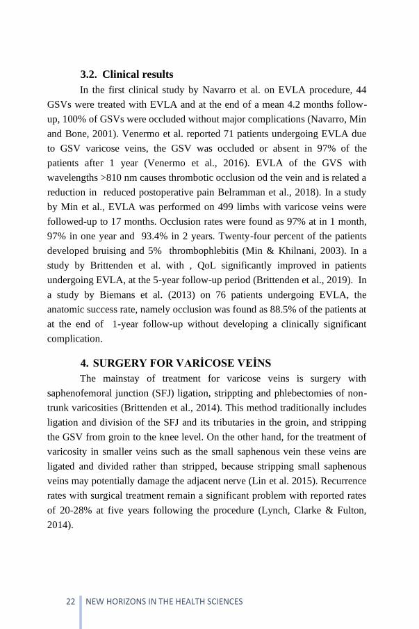
4. SURGERY FOR VARİCOSE VEİNS
The mainstay of treatment for varicose veins is surgery with
saphenofemoral junction (SFJ) ligation, strippting and phlebectomies of non-
trunk varicosities (Brittenden et al., 2014). This method traditionally includes
ligation and division of the SFJ and its tributaries in the groin, and stripping
the GSV from groin to the knee level. On the other hand, for the treatment of
varicosity in smaller veins such as the small saphenous vein these veins are
ligated and divided rather than stripped, because stripping small saphenous
veins may potentially damage the adjacent nerve (Lin et al. 2015). Recurrence
rates with surgical treatment remain a significant problem with reported rates
of 20-28% at five years following the procedure (Lynch, Clarke & Fulton,
2014).

23
4.1. Technique
High ligation with short stripping (HLSS) is carried out under either
spinal or general anesthesia. After flush of the saphenofemoral junction (SFJ)
is performed, all tributaries are ligated to the second branch of the GSV at the
level of the knee. The procedure is completed by closing the cribriform fascia
layer, superficial fascia layer and skin, respectively.
An ambulatory compression bandage is applied following the
procedure for 48 hours and compression stockings are used at least for 2
weeks. Patients wait under observation at least for 30 minutes in the clinic
before discharge.
4.2. Clinical results
In a study by Venermo et al., 61 patients with varicose veins were
treated traditionally with surgery and the occlusion rate was found as 97% at 1
year. No major complication was seen, while 5% of the patients developed
wound site infection (Venermo et al., 2016). Lawaetz et al., found recurrence
rate as 34.6% at five years in patients undergoing surgery (Lawaetz et al.,
2017). It was reported in a study by Rasmussen et al. that sick leave was
relatively shorter with surgery (7.6 days), which was thought to be resulted
from utilization ofin the procedures (Rasmussen et al. 2007). Osman, El-
Heeny and El-Razeq reported the complication rate as 37.5% in 20 patients
undergoing stripping of the GSV, with the most common complication being
hematoma (12.5%) followed by ecchymosis (8.3%) (Osman, El-Heeny and
El-Razeq, 2019). In another study, the most common complications following
stripping of the GSV were reported as mild inflammation, serous wound
discharge, hematoma and wound breaking, all of which resolved
spontaneously (Subramonia and Lees, 2010).
5. SURGERY, UGFS AND EVLA PROCEDURES:
CLİNİCAL EFFECTİVENESS
A number of factors affect success of the different treatment method
utilized. All these methods have their own advantages and disadvantages, and
so far there is no standardized and widely adopted method for this purpose.
The commonly used factors to determine treatment success are given in
Figure 2.

24 NEW HORIZONS IN THE HEALTH SCIENCES
Figure 2. Commonly measured factors to determine success in the treatment of
varicose veins.
Clinical effectiveness of UGFS, EVLA and Surgery have been
investigated and compared in numerous studies. In a meta-analysis of 13
studies with a mean follow-up duration ≥ 5 years, anatomical success rate was
significantly lower with UGFS (%34) compared to surgery (87%) and EVLA
(88%) procedures (Hamann et al., 2017). In the same analysis, pooled
recurrence rate was found as 12% with surgery, 22% with EVLA and 29%
with UGFS procedures (Hamann et al., 2017). In a recent study by Brittenden
et al., higher success was obtained in treatment with surgery and ELVA
compared with UGFS (Brittenden et al., 2019). Biemans et al. reported no
significant difference between the UGFS and surgery methods in terms of
clinician noted recurrence (Biemans et al. 2013). Similarly, Rasmussen et al.
found similar recurrence rates (14.8% vs 13.8%) for surgery and UGFS
procedures (Rasmussen 2011).
In a study by Brittenden et al., QoL (measured by AVVQ scores) was
higher in the patients undergoing EVLA compared to surgery and UGFS
(p<0.001). In that study, pain (measured by VAS score) was found as 2.6 with
laser ablation, 3.2 with foam sclerotherapy, and 2.4 with surgery. In addition,
5-year rates of complete success were found as 64.0% with EVLA, 33.3%
with UGFS, and 75.9% with surgery (Brittenden et al., 2019). In another
study by van der Velden et al., disease specific QoL was higher in patients
who underwent EVLA compared to those who underwent UGFS for the
25
treatment of varicose veins. However, no significant difference was found
between the patients undergoing UGFS and surgery (van der Velden et al.,
2015).
6. SURGERY, UGFS AND EVLA PROCEDURES: COST
EFFECTIVENESS
Quality-adjusted life-years (QALY) is an important factor used in the
analysis of cost-effectiveness with a certain treatment method, taking in
account long term (e.g. 5 years) effects. QALY takes into account both
quantity and quality of life generated by healthcare interventions (Wichman et
al. 2020). Tassie et al. compared cost-effectiveness of UGFS, EVLA and
Surgery by examining 5-year outcomes, and reported that EVLA was the
potentially preferred option in terms of cost-effectiveness with £3640 per
QALY gained. The authors of this study stated that cost savings related to
EVLA procedure increased to £368 using the same staff and further increased
when patients with good results following the initial EVLA were not followed
up at for 6 weeks (Tassie et al. 2014). In a Markov model developed by Gohel
et al., EVLA performed under local anesthesia was found to be the most cost-
effective method with £20,000 per QALY (Gohel, Epstein & Davies, 2010).
It is difficult to make a healthy comparison among studies due to
several variable factors such as the surgical staff, operational setting,
anesthesia type, sclerosing agents used in UGFS and wavelengths used in
EVLA etc. However, a general review of the published studies gave an idea
about the effectiveness. In a study by Brittenden et al., the most cost-effective
method was found as EVLA with a willingness-to-pay ratio of £20,000
($28,433) per QALY compared to UGFS and surgery (Brittenden et al.,
2019).
CONCLUSION
The management of varicose veins has dramatically evolved
especially with advancement in endovenous techniques. EVLA and UGFS
appear safe and effective alternatives to surgery. However, patients should be
given clear instructions about the importance of adherence to compression
following treatment. Most data in the recent literature indicate that both
clinical effectiveness and cost effectiveness are better with EVLA compared
to UGFS and surgery, which is evident by the fact that EVLA has replaced

26 NEW HORIZONS IN THE HEALTH SCIENCES
surgery in numerous countries worldwide. On the other hand, follow-up
studies with longer durations will continue to provide a better insight into the
costs and clinical effectiveness of these three methods and other novel
techniques.

27
REFERENCES
Almeida, J. I., Javier, J. J., Mackay, E., Bautista, C., & Proebstle, T. M. (2013). First
human use of cyanoacrylate adhesive for treatment of saphenous vein
incompetence. Journal of vascular surgery. Venous and lymphatic disorders,
1(2), 174–180.
Belramman, A., Bootun, R., Lane, T., & Davies, A. H. (2019). Endovenous
Management of Varicose Veins. Angiology, 70(5), 388–396.
Biemans, A. A., Kockaert, M., Akkersdijk, G. P., van den Bos, R. R., de Maeseneer,
M. G., Cuypers, P., Stijnen, T., Neumann, M. H., & Nijsten, T. (2013).
Comparing endovenous laser ablation, foam sclerotherapy, and conventional
surgery for great saphenous varicose veins. Journal of vascular surgery,
58(3), 727–34.e1.
Brittenden, J., Cotton, S. C., Elders, A., Ramsay, C. R., Norrie, J., Burr, J., Campbell,
B., Bachoo, P., Chetter, I., Gough, M., Earnshaw, J., Lees, T., Scott, J.,
Baker, S. A., Francis, J., Tassie, E., Scotland, G., Wileman, S., & Campbell,
M. K. (2014). A randomized trial comparing treatments for varicose veins.
The New England journal of medicine, 371(13), 1218–1227.
Brittenden, J., Cooper, D., Dimitrova, M., Scotland, G., Cotton, S. C., Elders, A.,
MacLennan, G., Ramsay, C. R., Norrie, J., Burr, J. M., Campbell, B.,
Bachoo, P., Chetter, I., Gough, M., Earnshaw, J., Lees, T., Scott, J., Baker, S.
A., Tassie, E., Francis, J., … Campbell, M. K. (2019). Five-Year Outcomes
of a Randomized Trial of Treatments for Varicose Veins. The New England
journal of medicine, 381(10), 912–922.
Darke, S. G., & Baker, S. J. (2006). Ultrasound-guided foam sclerotherapy for the
treatment of varicose veins. The British journal of surgery, 93(8), 969–974.
Darvall, K. A., Bate, G. R., & Bradbury, A. W. (2014). Patient-reported outcomes 5-
8 years after ultrasound-guided foam sclerotherapy for varicose veins. The
British journal of surgery, 101(9), 1098–1104.
de Araujo, W., Erzinger, F. L., Caron, F. C., Nejm, C. S., Junior, & Timi, J. (2017).
Influência da termoablação com baixa e alta densidade de energia na junção
safeno-femoral, utilizando laser endovenoso 1470 nm. Jornal vascular
brasileiro, 16(3), 220–226.
Epstein, D., Onida, S., Bootun, R., Ortega-Ortega, M., & Davies, A. H. (2018). Cost-
Effectiveness of Current and Emerging Treatments of Varicose Veins. Value
in health : the journal of the International Society for Pharmacoeconomics
and Outcomes Research, 21(8), 911–920.
Ghosh, S.K., Al Mamun, A., & Majumder, A. (2021). Clinical Presentation of
Varicose Veins. Indian Journal of Surgery, 1 - 8.

28 NEW HORIZONS IN THE HEALTH SCIENCES
Gibson, K. D., Ferris, B. L., & Pepper, D. (2007). Endovenous laser treatment of
varicose veins. The Surgical clinics of North America, 87(5), 1253–xii.
Gohel, M. S., Epstein, D. M., & Davies, A. H. (2010). Cost-effectiveness of
traditional and endovenous treatments for varicose veins. The British journal
of surgery, 97(12), 1815–1823.
Hamann, S., Giang, J., De Maeseneer, M., Nijsten, T., & van den Bos, R. R. (2017).
Editor's Choice - Five Year Results of Great Saphenous Vein Treatment: A
Meta-analysis. European journal of vascular and endovascular surgery : the
official journal of the European Society for Vascular Surgery, 54(6), 760–
770.
Janugade, H.B., Patil, B.P., Tata, N.H., Saygaonkar, H.V., Janugade, D.H., Dokania,
V. (2017) Clinical profile and management of lower limb varicose veins. J
Evol Med Dent Sci 6(20):1615–1622.
Kabnick, L.S., Sadek, M. (2014). Varicose veins: endovenous ablation and
sclerotherapy. In: Cronenwett JL, Johnston KW (eds) Rutherford’s Vascular
Surgery. Eighth edition, Chapter- 58, pp 885–901. Elsevier Saunders,
Philadelphia, PA, USA
Lawaetz, M., Serup, J., Lawaetz, B., Bjoern, L., Blemings, A., Eklof, B., &
Rasmussen, L. (2017). Comparison of endovenous ablation techniques, foam
sclerotherapy and surgical stripping for great saphenous varicose veins.
Extended 5-year follow-up of a RCT. International angiology : a journal of
the International Union of Angiology, 36(3), 281–288.
Lin, F., Zhang, S., Sun, Y., Ren, S., & Liu, P. (2015). The management of varicose
veins. International surgery, 100(1), 185–189.
Lynch, N. P., Clarke, M., & Fulton, G. J. (2015). Surgical management of great
saphenous vein varicose veins: A meta-analysis. Vascular, 23(3), 285–296.
Malskat, W. S., Poluektova, A. A., van der Geld, C. W., Neumann, H. A., Weiss, R.
A., Bruijninckx, C. M., & van Gemert, M. J. (2014). Endovenous laser
ablation (EVLA): a review of mechanisms, modeling outcomes, and issues
for debate. Lasers in medical science, 29(2), 393–403.
Malskat, W., Engels, L. K., Hollestein, L. M., Nijsten, T., & van den Bos, R. R.
(2019). Commonly Used Endovenous Laser Ablation (EVLA) Parameters
Do Not Influence Efficacy: Results of a Systematic Review and Meta-
Analysis. European journal of vascular and endovascular surgery : the
official journal of the European Society for Vascular Surgery, 58(2), 230–
242.
Min, R. J., & Khilnani, N. M. (2003). Endovenous laser treatment of saphenous vein
reflux. Techniques in vascular and interventional radiology, 6(3), 125–131.

29
Myers, K. A., Jolley, D., Clough, A., & Kirwan, J. (2007). Outcome of ultrasound-
guided sclerotherapy for varicose veins: medium-term results assessed by
ultrasound surveillance. European journal of vascular and endovascular
surgery : the official journal of the European Society for Vascular Surgery,
33(1), 116–121.
Navarro, L., Min, R. J., & Boné, C. (2001). Endovenous laser: a new minimally
invasive method of treatment for varicose veins--preliminary observations
using an 810 nm diode laser. Dermatologic surgery : official publication for
American Society for Dermatologic Surgery [et al.], 27(2), 117–122.
O'Hare, J. L., & Earnshaw, J. J. (2007). The use of foam sclerotherapy for varicose
veins: a survey of the members of the Vascular Society of Great Britain and
Ireland. European journal of vascular and endovascular surgery : the official
journal of the European Society for Vascular Surgery, 34(2), 232–235.
Onida, S., & Davies, A. H. (2013). Varicose veins: diagnosis and management.
Nursing times, 109(41), 16–17.
ORBACH, E. J., & PETRETTI, A. K. (1950). The thrombogenic property of foam of
a synthetic anionic detergent (sodium tetradecyl sulfate N.N.R.). Angiology,
1(3), 237–243.
Osman, O.A., El-Heeny, A.E., El-Razeq, M.A. (2019). Management of primary
uncomplicated varicose veins, endovenous laser ablation with sclerotherapy
versus traditional surgery: which is the best option?. Egypt J Surg, 38:319-27
Proebstle, T. M., Alm, B. J., Göckeritz, O., Wenzel, C., Noppeney, T., Lebard, C.,
Sessa, C., Creton, D., & Pichot, O. (2015). Five-year results from the
prospective European multicentre cohort study on radiofrequency segmental
thermal ablation for incompetent great saphenous veins. The British journal
of surgery, 102(3), 212–218.
Rasmussen, L. H., Bjoern, L., Lawaetz, M., Blemings, A., Lawaetz, B., & Eklof, B.
(2007). Randomized trial comparing endovenous laser ablation of the great
saphenous vein with high ligation and stripping in patients with varicose
veins: short-term results. Journal of vascular surgery, 46(2), 308–315.
Rasmussen, L. H., Lawaetz, M., Bjoern, L., Vennits, B., Blemings, A., & Eklof, B.
(2011). Randomized clinical trial comparing endovenous laser ablation,
radiofrequency ablation, foam sclerotherapy and surgical stripping for great
saphenous varicose veins. The British journal of surgery, 98(8), 1079–1087.
Smith P. C. (2006). Chronic venous disease treated by ultrasound guided foam
sclerotherapy. European journal of vascular and endovascular surgery : the
official journal of the European Society for Vascular Surgery, 32(5), 577–
583.

30 NEW HORIZONS IN THE HEALTH SCIENCES
Subramonia, S., & Lees, T. (2010). Randomized clinical trial of radiofrequency
ablation or conventional high ligation and stripping for great saphenous
varicose veins. The British journal of surgery, 97(3), 328–336.
Tassie, E., Scotland, G., Brittenden, J., Cotton, S. C., Elders, A., Campbell, M. K.,
Campbell, B., Gough, M., Burr, J. M., Ramsay, C. R., & CLASS study team
(2014). Cost-effectiveness of ultrasound-guided foam sclerotherapy,
endovenous laser ablation or surgery as treatment for primary varicose veins
from the randomized CLASS trial. The British journal of surgery, 101(12),
1532–1540.
Tessari, L. (2001). Extemporary sclerosing foam according to personal method:
experimental clinical data and catheter usage. Int Angiol Suppl 1:54.
van der Velden, S. K., Biemans, A. A., De Maeseneer, M. G., Kockaert, M. A.,
Cuypers, P. W., Hollestein, L. M., Neumann, H. A., Nijsten, T., & van den
Bos, R. R. (2015). Five-year results of a randomized clinical trial of
conventional surgery, endovenous laser ablation and ultrasound-guided foam
sclerotherapy in patients with great saphenous varicose veins. The British
journal of surgery, 102(10), 1184–1194.
Venermo, M., Saarinen, J., Eskelinen, E., Vähäaho, S., Saarinen, E., Railo, M., Uurto,
I., Salenius, J., Albäck, A., & Finnish Venous Study Collaborators (2016).
Randomized clinical trial comparing surgery, endovenous laser ablation and
ultrasound-guided foam sclerotherapy for the treatment of great saphenous
varicose veins. The British journal of surgery, 103(11), 1438–1444.
Wichmann, A. B., Goltstein, L., Obihara, N. J., Berendsen, M. R., Van Houdenhoven,
M., Morrison, R. S., Johnston, B. M., Engels, Y., & Radboud Honours
Academy Think Tank (2020). QALY-time: experts' view on the use of the
quality-adjusted LIFE year in COST-effectiveness analysis in palliative care.
BMC health services research, 20(1), 659.
Winterborn, R. J., Campbell, W. B., Heather, B. P., & Earnshaw, J. J. (2004). The
management of short saphenous varicose veins: a survey of the members of
the vascular surgical society of Great Britain and Ireland. European journal
of vascular and endovascular surgery : the official journal of the European
Society for Vascular Surgery, 28(4), 400–403.

31
CHAPTER 3
INVESTIGATION OF MICROBIOLOGICAL QUALITY OF
DRINKING AND USAGE WATER IN A FAST-GROWING
METROPOLITAN CITY
Ibrahim BAYHAN1, Benan YAZICI KARABULUT2
Mehmet Irfan YESILNACAR3 , Abdullah Izzeddin KARABULUT4
1 Dr., Provincial Health Directorate, Public Health Services Department, Environmental Health Unit, Sanliurfa, Turkey, [email protected], ORCID: 0000-0001-5516-6241 2 Harran University, Faculty of Engineering, Department of Environmental Engineering,
Sanliurfa, Turkey, [email protected], ORCID: 0000-0002-0140-204X 3 Prof. Dr.,Harran University, Faculty of Engineering, Department of Environmental Engineering, Sanliurfa, Turkey, [email protected], ORCID: 0000-0001-9724-8683 4 Harran University, Graduate School of Natural and Applied Sciences, Remote Sensing and
Geographic Information Systems, 100/2000 CoHE PhD Scholar, Sanliurfa, Turkey,
[email protected], ORCID: 0000-0002-9784-5549

32 NEW HORIZONS IN THE HEALTH SCIENCES

33
INTRODUCTION
Water is one of the vital elements for human beings. Access to
healthy drinking water is considered one of the most basic human rights.
Unhealthy drinking water and the inadequate hygiene it causes lead to about
7% of diseases in the world (Toroglu, 2009; Bozkurt, 2010). Diarrhea that
stems from unhealthy water is among the leading causes of morbidity and
mortality in the world. About 20% of the world's population use unsafe
drinking water, around 200 million people suffer from water-related diseases
annually, and more than two million people die of diseases caused by polluted
water (Çankaya et al., 2017). Problems in accessing clean drinking water in
many developing countries have increased in recent years. The economic
situation and the lack of adequate infrastructure cause a large part of the
population to be adversely affected by this situation (Rochelle-Neval et al.,
2015). In addition, contamination of surface waters and groundwater with raw
manure and sewage remains a serious environmental problem (Park et al.,
2015). The mucilage problem faced in the Marmara Sea in 2021 is a result of
nutrient/uncontrolled discharges. In the areas of food, water safety and public
health, the detection of pathogenic microorganisms has become a primary
concern (Arya et al., 2011). Especially in drinking water, used by people for
various daily and basic purposes, fecal pollution induced by the transport of
pathogenic microorganisms poses an important threat. The presence of
pathogenic microorganisms in the waters brings the risk of epidemics. That's
why it's important to detect them.
Surface waters, including freshwater and seawater, drinking water and
food sources are a habitat for many organisms. They are also used as a
recreation area, as well. These environments are susceptible to microbial
contamination stemming from streams, animal feces and wastewater
discharges (Boehm et al., 2018). Microbial contamination of recreational
waters such as rivers, lakes, streams, and beaches occur when processed or
raw fecal matters are carried into waterways through some input sources.
Point sources of fecal contamination, such as wastewater discharge from
wastewater treatment plants or seepage from local livestock production
facilities are contaminations that are relatively easier to identify, characterize
and eliminate. Conversely, non-point sources of contamination, such as
rainwater runoff, are more difficult to identify as contamination from sources
34 NEW HORIZONS IN THE HEALTH SCIENCES
may be likely carried over great distances (Unno et al., 2018). Microbial
contamination of water resources from fecal matter is a significant threat to
public health in both industrialized and developing countries. According to
the World Health Organization (WHO), 9% of the world's population do not
have access to safe drinking water. For this reason, improving the quality and
conditions of drinking water in accordance with health has become one of the
important human requirements (Jofre et al., 2016).
Investigating the origin of fecal contamination is a crucial point in
evaluating the actions necessary to address the health risks associated with it.
The origin of this pollution can be traced genotypically and phenotypically
and this is called microbial resource tracking. Characterizing fecal sources is
necessary to control the major pollutants that cause water pollution. This leads
to identifying a variety of chemical and microbial markers that help
distinguish between human and non-human fecal sources (Devane et al.,
2018).
Water quality needs to be carefully monitored, as anthropogenic fecal
contamination of waters poses a major health and environmental threat.
Various methods based on culturing, such as fecal indicators, have been used
for a long time to determine the amount and presence of bacteria. However,
these methods yield results slowly and the diagnostic information required to
identify the source is insufficient in some cases (Jiang et al., 2018). Therefore,
to investigate and prevent the transmission of waterborne pathogens, there is a
need for faster testing methods in the field of microbiological water quality
assessment that can be applied to monitor potential fecal inputs in waters
(Bayhan et al., 2020).
In order for water sources to be hygienically reliable, it is necessary to
determine whether the water is exposed to fecal contamination. The presence
of a high number of bacteria in the water increases the risk of contracting
enteric pathogens, especially in infants and children (Nwachuku and Gerba,
2004). Due to the water with poor hygienic quality, 1.7 million people die
mainly of infectious diarrhea per year worldwide. On these deaths, 90% are in
children and almost all are in developing countries (Ashbolt, 2004). The total
mesophilic aerobic microorganism, known as standard plate count, is now
called heterotrophic plate count (HPC). It is determined by counting after
culturing aerobic and facultative anaerobic live bacteria to measure changes

35
during processing and distribution of water. Heterotrophic bacteria include
some bacteria considered primary and secondary pathogens, as well as
indicator bacteria such as coliform (Allen et al., 2004). The allowed limits for
the number of HPC bacteria in the quality characteristics of drinking water are
between 100-500 Cfu/ml in the world (Pavlov et al., 2004). Pepper et al.
(2004) report that the number of HPC bacteria increases significantly from the
distribution system to the consumer's tap. They also claim that bacteria
originate from bacteria inside household faucets or household distribution
systems rather than central distribution systems or water sources. Therefore,
they conclude that consumers regularly receive more than 500 units/ml of
HPC bacteria as a result of using household taps as drinking water.
Sanitary and quality water is one of the indispensable pre-requisites of
food safety, nutrition, and public health. For this reason, the importance of
water for human health, which does not contain pathogenic microorganisms,
chemical and physical contaminants, and contains a balanced mineral
distribution in terms of nutrition, has been proven by many researches. The
most important feature of water suitable for health is its microbial quality.
Due to the use of unsanitary water, 30.000‒40.000 people die of cholera in the
world every year. Likewise, 25.000 people die of typhoid and paratyphoid, 4
million of diarrheal diseases (especially children aged between 0‒4), 25.000
of poliomyelitis, 20.000 of ascariasis, 200.000 of schistosomiasis, 1‒2 million
of malaria, and 20.000-50.000 of onchocerciasis (WHO, 2014). The
microorganisms that are most common in drinking and usage water and pose a
health problem are coliforms, the intestinal flora bacteria. Microbial analysis
of water is also based on investigating the presence of coliform bacteria in
water (Rompré et al., 2002). Fecal coliform bacteria and E. coli are considered
as indicators of fecal contamination in water. Particularly, the pathogenic
types of E. coli cause deadly diarrhea, nephropathy, meningitis, septicemia,
arteriosclerosis, Hemolytic Uremic Syndrome (HUS), wound infections and
various immunological diseases in humans and animals. Therefore, water
containing total or fecal coliform bacteria should not be consumed by human
without purification (Bayhan, 2021).
In this study, it is aimed to reveal the effects of environmental
pressure on the changes in drinking and usage water quality. To do so, first,
the conditions of the source, storage and network of drinking water and the

36 NEW HORIZONS IN THE HEALTH SCIENCES
water supply methods in thirteen districts of the city were examined. Between
January and September 2018, seasonal samples were taken, and the
microbiological quality standards of the consumed water were determined.
Analysis results were evaluated within the framework of ITASHY (Turkish
Regulations on Water for Human Consumption), which is in line with the
European Union Drinking Water Directive. In addition, obtained results were
also evaluated in terms of environmental health.
1. MATERIALS AND METHODS
1.1. Study Area
The maps of the study area were produced from the topographic base
maps of the region via ArcMap 10.8 software, one of the Geographical
Information Systems software (Figure 1). Sanliurfa province is one of the fast-
growing metropolitan city located in the center of the Southeastern Anatolia
Region. The area of Sanliurfa land is 19,242 km2 and it consists of plateaus
(61.7%), mountains (22%) and arable plains (16%).
Figure 1: Location Map of The Study Area

37
Since Sanliurfa has mostly limestone rocks in terms of geological
structure, karst structures are quite common. This provides a great advantage
to the region in the feeding of groundwater during periods of sufficient
rainfall. The largest surface water resources of Sanliurfa are the Euphrates
River and the Ataturk and Birecik Dam Lakes built on it. Sanliurfa consists of
Middle‒Upper Eocene aged Birecik, Middle Eocene‒Oligocene aged
Gaziantep, Oligocene‒Lower Miocene aged Tektek mountains, and Lower
Miocene aged Euphrates formations, from bottom to top, respectively. It has a
continental climate with hot and dry summers and rainy and relatively warm
winters. Like many provinces in Turkey, Sanliurfa, which is under the
influence of different climatic zones in summer and winter periods, is under
the influence of Basra low pressure in summer.
In the metropolitan districts of Sanliurfa, the water coming from the
exit mouth of the Sanliurfa tunnels in the Ataturk Dam Lake is used as a
source of drinking and usage water. These tunnels were built to irrigate the
Harran Plain and their construction was completed in 1994. Ataturk Dam
Lake is also used as a water source in Hilvan district. There is no drinking
water treatment plant in Hilvan district. In Suruç and Birecik districts, the
caisson wells drilled in the water tanks of Birecik and Karkamış Dam Lake
are used as drinking water sources. In Siverek, Viranşehir and Halfeti
districts, deep boreholes and springs from groundwater are used. However, in
Akçakale, Harran, Ceylanpınar and Bozova districts, deep boreholes are used
as drinking water sources.
1.2. Sampling Points
From 77 sampling points created in thirteen district centers, 308
samples were taken to represent the entire network in winter, spring, summer
and autumn seasons between January and September 2018. The samples were
taken from the active fountains and taps directly connected to the network via
the monitoring points determined by the Provincial and District Health
Directorates. The samples were taken in accordance with the methods
determined in the "Handbook on Sample Collection, Transport and Analysis
of Water for Human Consumption" published by the Ministry of Health in
2008.
38 NEW HORIZONS IN THE HEALTH SCIENCES
1.3. Material
Microbiological samples were taken in 500 ml pure thiosulfate
polypropylene bottles, and physicochemical samples were taken in 1.5 liter
polyethylene bottles. Hach brand device (colorimetric method) was used for
free chlorine measurements, Portable Hach-Lange HQ40d multi-measurement
device was used for pH and electrical conductivity.
Statistical analyzes were performed using the SPSS® Statistics
software. Central tendency (mode, median, arithmetic mean, maximum and
minimum values) and measures of distribution (variance, standard deviation,
coefficient of variance) were found. By considering the maximum, minimum
and arithmetic mean values of the data, they were compared with the
ITASHY limit values, which are the standards related to drinking and usage
water in Turkey. Normality tests were performed to determine whether the
values of the measured parameters were normally distributed. In order to
examine whether the difference between the seasonal values of the parameters
is significant, Kruskal Wallis H test, which is one of the non-parametric tests,
was applied. Distribution charts of microbiological parameters were created
with Microsoft Office 365 Excel.
1.4. Method
Before taking samples for microbiological analysis, the spout of the
faucet was wiped with a clean cloth and the strainer equipment etc. has been
removed. Water was poured until the water temperature was stable. Then the
tap was turned off and its mouth and surroundings were flamed. After the
sample bottle was filled to the marked spot, its cap was quickly closed, and
the bottle was labelled. Microbiological samples were taken in 500 ml
polypropylene bottles with pure thiosulfate and transferred to the laboratory in
cold chain (+4 oC) within 4‒6 hours.
1.5. Drinking and Usage Water Quality Standards
Since drinking water is an important issue in terms of environment and
public health, it has attracted a lot of attention by environmental and public
health researches in Turkey and in the world. According to the report
published by the World Health Organization in 2017, more than one billion
people in the world do not have access to healthy and safe drinking water

39
(WHO, 2017). In a report of UNESCO published in 2016, it was emphasized
that water should be protected at its source and that it should be managed
correctly. National and international standards for drinking-usage water are
presented in Table 1.
Table 1: National and Some International Quality Standards for Drinking and Usage
Water Belonging to The Investigated Parameters
2. RESULTS
2.1. Microbiological Analysis
Microbiological analyzes were carried out at SUSKI (General
Directorate of Water and Sewerage Administration of Sanliurfa Metropolitan
Municipality) Drinking Water Treatment Plant Laboratory with the financial
support of HUBAP (The Coordinating Unit for Scientific Research Projects at
Harran University, Project No: 17198). Total coliform (TC), E. coli (TS EN
ISO 9308-1), and Enterococci (EC) (TS EN ISO 7899-2) measurements were
performed via membrane filter method. The water samples were gently
shaken to ensure homogeneity. 100 ml water sample was filtered from three
membrane filters prepared for TC, E. coli and EC. After filtering process, TC
and E. coli were incubated in chronogeneous agar broth at 37 oC for 24 hours
whereas EC was incubated in Avida broth at 37 oC for 48 hours. Colonies
formed at the end of the incubation period were counted. It was observed that
red colonies were formed for TC, blue for E. coli and cherry color colonies for
EC (Figure 2).
Parameter Analysis Method Unit ITASHY
(2005)
WHO
(2017)
USEPA
(2018)
Total coliform
membrane filtration
number/100 ml 0/100 ml 0/100 ml 0/100 ml
E. coli membrane filtration
number/100 ml 0/100 ml 0/100 ml 0/100 ml
Enterococci membrane filtration
number/100 ml 0/100 ml 0/100 ml 0/100 ml

40 NEW HORIZONS IN THE HEALTH SCIENCES
Figure 2: Microbiology Laboratory and Some Analysis Images
Detected anomalies are nonconformities caused by external
contamination. It has been reported that nitrate and conductivity problems are
more common particularly in irrigated agriculture areas (Kahraman et al.,
2016, Yazici-Karabulut et al., 2019, Yazici-Karabulut et al., 2021). As a result
of this preliminary data, it was determined to examine TC, E. coli, and EC
within microbiological parameters.
2.1.1. Sanliurfa
The sampling points of Sanliurfa city center are shown in Figure 3.
41
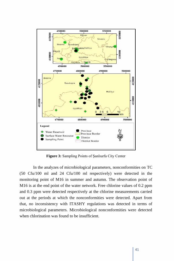
Figure 3: Sampling Points of Şanlıurfa City Center
In the analyzes of microbiological parameters, nonconformities on TC
(50 Cfu/100 ml and 24 Cfu/100 ml respectively) were detected in the
monitoring point of M16 in summer and autumn. The observation point of
M16 is at the end point of the water network. Free chlorine values of 0.2 ppm
and 0.3 ppm were detected respectively at the chlorine measurements carried
out at the periods at which the nonconformities were detected. Apart from
that, no inconsistency with ITASHY regulations was detected in terms of
microbiological parameters. Microbiological nonconformities were detected
when chlorination was found to be insufficient.

42 NEW HORIZONS IN THE HEALTH SCIENCES
2.1.2. Akçakale
The sampling points of Akçakale district center are shown in Figure 4.
Figure 4: Sampling Points of Akçakale District
Focusing on seasonal microbiological values, no nonconformity was
detected in winter. At the sampling point of A3, TC (100 Cfu/250 ml), E. coli
(25 Cfu/250 ml) and EC (35 Cfu/250 ml) were detected in spring. In summer,
TC (27 Cfu/250 ml) at the sampling point of A4 and TC (250 Cfu/250 ml) and
E. coli (250 Cfu/250 ml) at the sampling point of A5 were detected. In
autumn, TC (27 Cfu/250 ml) at the sampling point of A2 and TC (261
Cfu/250 ml) and EC (10 Cfu/250 ml) at the sampling point of A4 were
observed. Depending on the observations, much more nonconformities were
detected in summer and autumn (Figure 5).

43
Figure 5: Seasonal Microbiological Analysis Values of Akçakale District: (a) Spring,
(b) Summer, (c) Autumn, (d) Winter
2.1.3. Birecik
The sampling points of Birecik district center are shown in Figure 6.
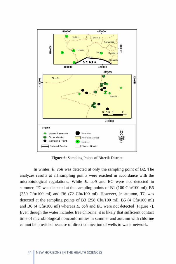
44 NEW HORIZONS IN THE HEALTH SCIENCES
Figure 6: Sampling Points of Birecik District
In winter, E. coli was detected at only the sampling point of B2. The
analyzes results at all sampling points were reached in accordance with the
microbiological regulations. While E. coli and EC were not detected in
summer, TC was detected at the sampling points of B1 (100 Cfu/100 ml), B5
(250 Cfu/100 ml) and B6 (72 Cfu/100 ml). However, in autumn, TC was
detected at the sampling points of B3 (258 Cfu/100 ml), B5 (4 Cfu/100 ml)
and B6 (4 Cfu/100 ml) whereas E. coli and EC were not detected (Figure 7).
Even though the water includes free chlorine, it is likely that sufficient contact
time of microbiological nonconformities in summer and autumn with chlorine
cannot be provided because of direct connection of wells to water network.

45
Figure 7: Seasonal Microbiological Analysis Values of Birecik District: (a) Spring,
(b) Summer, (c) Autumn, (d) Winter
2.1.4. Bozova
The sampling points of Bozova district center are shown in Figure 8.
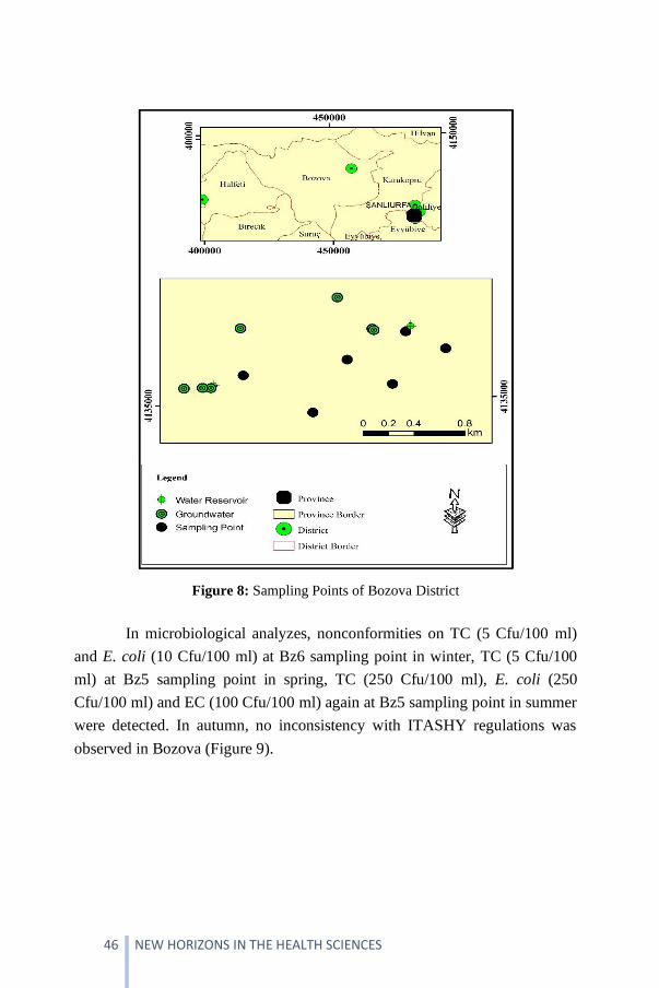
46 NEW HORIZONS IN THE HEALTH SCIENCES
Figure 8: Sampling Points of Bozova District
In microbiological analyzes, nonconformities on TC (5 Cfu/100 ml)
and E. coli (10 Cfu/100 ml) at Bz6 sampling point in winter, TC (5 Cfu/100
ml) at Bz5 sampling point in spring, TC (250 Cfu/100 ml), E. coli (250
Cfu/100 ml) and EC (100 Cfu/100 ml) again at Bz5 sampling point in summer
were detected. In autumn, no inconsistency with ITASHY regulations was
observed in Bozova (Figure 9).

47
Figure 9: Seasonal Microbiological Analysis Values of Bozova District: (a) Spring,
(b) Summer, (c) Autumn, (d) Winter
2.1.5. Ceylanpınar
The sampling points of Ceylanpınar district center are shown in
Figure 10.
48 NEW HORIZONS IN THE HEALTH SCIENCES
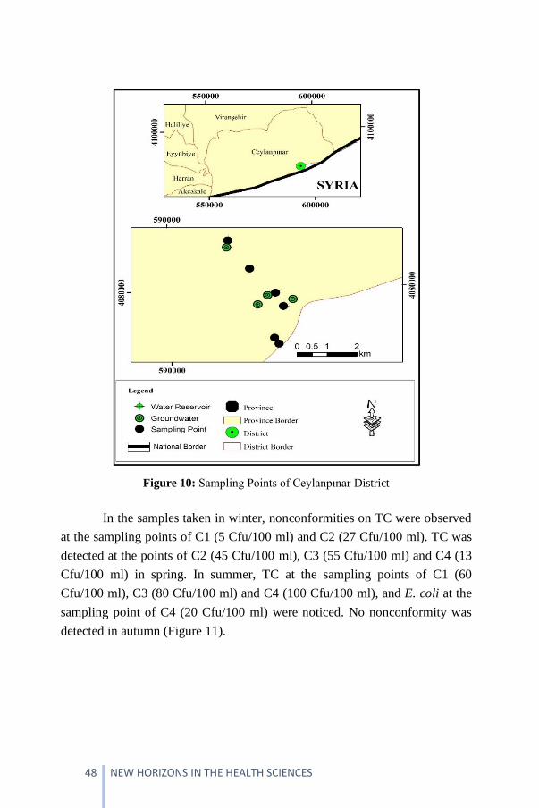
Figure 10: Sampling Points of Ceylanpınar District
In the samples taken in winter, nonconformities on TC were observed
at the sampling points of C1 (5 Cfu/100 ml) and C2 (27 Cfu/100 ml). TC was
detected at the points of C2 (45 Cfu/100 ml), C3 (55 Cfu/100 ml) and C4 (13
Cfu/100 ml) in spring. In summer, TC at the sampling points of C1 (60
Cfu/100 ml), C3 (80 Cfu/100 ml) and C4 (100 Cfu/100 ml), and E. coli at the
sampling point of C4 (20 Cfu/100 ml) were noticed. No nonconformity was
detected in autumn (Figure 11).

49
Figure 11: Seasonal Microbiological Analysis Values of Ceylanpınar District: (a)
Spring, (b) Summer, (c) Autumn, (d) Winter
2.1.6. Halfeti
The sampling points of Halfeti district center are shown in Figure 12.

50 NEW HORIZONS IN THE HEALTH SCIENCES
Figure 12: Sampling Points of Halfeti District
In microbiological analysis carried out in Halfeti district in winter, TC
(5 Cfu/100 ml) and E. coli (5 Cfu/100 ml) were detected at the sampling point
of H2. In Summer, TC (24 Cfu/100 ml) was observed at the sampling point of
H4, but for all samples, no microbiological nonconformity was noticed in
spring and autumn (Figure 13).
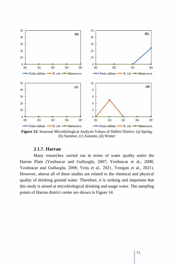
51
Figure 13: Seasonal Microbiological Analysis Values of Halfeti District: (a) Spring,
(b) Summer, (c) Autumn, (d) Winter
2.1.7. Harran
Many researches carried out in terms of water quality entire the
Harran Plain (Yesilnacar and Gulluoglu, 2007; Yesilnacar et al., 2008;
Yesilnacar and Gulluoglu, 2008; Yetiş et al., 2021, Yenigun et al., 2021).
However, almost all of these studies are related to the chemical and physical
quality of drinking ground water. Therefore, it is striking and important that
this study is aimed at microbiological drinking and usage water. The sampling
points of Harran district center are shown in Figure 14.
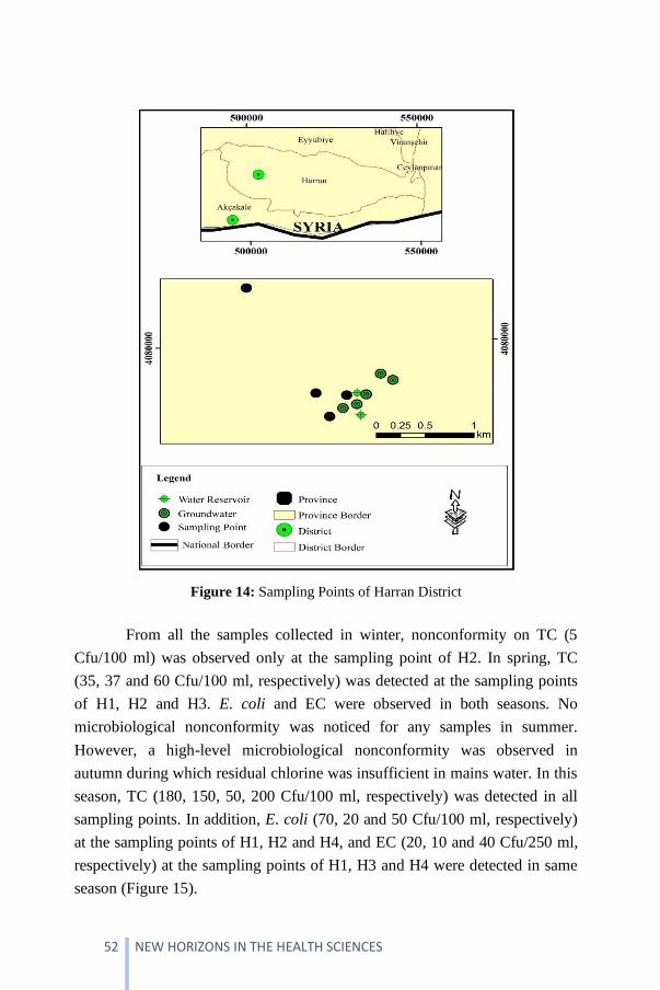
52 NEW HORIZONS IN THE HEALTH SCIENCES
Figure 14: Sampling Points of Harran District
From all the samples collected in winter, nonconformity on TC (5
Cfu/100 ml) was observed only at the sampling point of H2. In spring, TC
(35, 37 and 60 Cfu/100 ml, respectively) was detected at the sampling points
of H1, H2 and H3. E. coli and EC were observed in both seasons. No
microbiological nonconformity was noticed for any samples in summer.
However, a high-level microbiological nonconformity was observed in
autumn during which residual chlorine was insufficient in mains water. In this
season, TC (180, 150, 50, 200 Cfu/100 ml, respectively) was detected in all
sampling points. In addition, E. coli (70, 20 and 50 Cfu/100 ml, respectively)
at the sampling points of H1, H2 and H4, and EC (20, 10 and 40 Cfu/250 ml,
respectively) at the sampling points of H1, H3 and H4 were detected in same
season (Figure 15).

53
Figure 15: Seasonal Microbiological Analysis Values of Harran District: (a) Spring,
(b) Summer, (c) Autumn, (d) Winter
2.1.8. Hilvan
The sampling points of Harran district center are shown in Figure 16.

54 NEW HORIZONS IN THE HEALTH SCIENCES
Figure 16: Sampling Points of Hilvan District
Based on the samples taken from Hilvan district in winter, TC (5
Cfu/100 ml) and EC (100 Cfu/250 ml) were detected at the sampling point of
Hi1 at which free chlorine was measured as 0 ppm. In the water tank in which
free chlorine was detected as 0 in spring, TC (50 Cfu/100 ml) and E. coli ( 20
Cfu/100 ml) were observed. Additionally, in summer when no sufficient free
chlorine was detected all across the water network. TC (250 Cfu/100 ml) and
E. coli ( 250 Cfu/100 ml) were detected at both the sampling points of Hi3
and Hi5. However, no microbiological nonconformity was observed in
autumn at which there was sufficient free chlorine in water network (Figure
17).
55
Figure 17: Seasonal Microbiological Analysis Values of Hilvan District: (a) Spring,
(b) Summer, (c) Autumn, (d) Winter
2.1.9. Siverek
The sampling points of Siverek district center are shown in Figure 18.

56 NEW HORIZONS IN THE HEALTH SCIENCES
Figure 18: Sampling Points of Siverek District
According to all microbiological analyzes, it was detected that there
were TC in 15 samples (42%), E. coli in 9 samples (25%) and EC in 3
samples (0.8%) respectively. The highest-level nonconformities were detected
in summer and autumn. The level of free chlorine in water was observed as
“0” at the sampling points at which microbiological nonconformities were
high. Another feature of those sampling points was that they were the fields
which were supported by the wells directly connected to the network without
any transferring to tanks (Figure 19).

57
Figure 19: Seasonal Microbiological Analysis Values of Siverek District: (a) Spring,
(b) Summer, (c) Autumn, (d) Winter
2.1.10. Suruç
The sampling points of Suruç district center are shown in Figure
20.

58 NEW HORIZONS IN THE HEALTH SCIENCES
Figure 20: Sampling Points of Suruç District
The maximum numbers of nonconformities were noted in Suruç
district based on all the province-wide analyzes. In winter, TC (5 Cfu/100 ml)
was observed at the sampling points of Src1, Src2 and Src3. At the sampling
point of Src2, TC at the value of 250 Cfu/100 ml was detected in winter. The
value of TC for other sampling points were 100 Cfu/100 ml again in winter.
E. coli at the value of 50 Cfu/100 ml was noted at the sampling point of Src6,
and for other points, it was 100 Cfu/100 ml. As to EC, the detected the value
for the sampling point Src2 was 90 Cfu/100 ml, and it was 60 Cfu/100 ml for
Src6. However, at the other sampling points, EC (100 Cfu/100 ml) was
observed. In summer, TC (250, 250, 250, 100, 120, 90 Cfu/100 ml,
respectively), E. coli (100, 50, 120, 50, 70, 25 Cfu/100 ml, respectively) and
EC (60, 50, 30, 30, 30, 20 Cfu/100 ml, respectively) were detected at all the
59
sampling points. In autumn, TC (240, 323, 50, 220, 200, 376 Cfu/100 ml,
respectively) and EC (20, 20, 10, 25, 10, 1 Cfu/100 ml, respectively) were
detected again at all the sampling points (Figure 21).
Figure 21: Seasonal Microbiological Analysis Values of Suruç District: (a) Spring,
(b) Summer, (c) Autumn, (d) Winter
2.1.11. Viranşehir
The sampling points of Viranşehir district center are shown in
Figure 22.
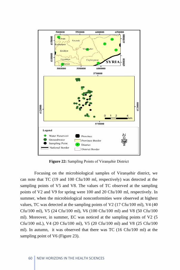
60 NEW HORIZONS IN THE HEALTH SCIENCES
Figure 22: Sampling Points of Viranşehir District
Focusing on the microbiological samples of Viranşehir district, we
can note that TC (19 and 100 Cfu/100 ml, respectively) was detected at the
sampling points of V5 and V8. The values of TC observed at the sampling
points of V2 and V9 for spring were 100 and 20 Cfu/100 ml, respectively. In
summer, when the microbiological nonconformities were observed at highest
values, TC was detected at the sampling points of V2 (17 Cfu/100 ml), V4 (40
Cfu/100 ml), V5 (24 Cfu/100 ml), V6 (100 Cfu/100 ml) and V8 (50 Cfu/100
ml). Moreover, in summer, EC was noticed at the sampling points of V2 (5
Cfu/100 mL), V4 (20 Cfu/100 ml), V5 (20 Cfu/100 ml) and V8 (25 Cfu/100
ml). In autumn, it was observed that there was TC (16 Cfu/100 ml) at the
sampling point of V6 (Figure 23).

61
Figure 23: Seasonal Microbiological Analysis Values of Viranşehir District: (a)
Spring, (b) Summer, (c) Autumn, (d) Winter
3. DISCUSSION
Sanliurfa has the surface water sources like Birecik and Ataturk Dam
and also underground sources like Ceylanpınar plain which has the capacity to
meet one fifth of Turkey’s overall water need (Yetiş, 2013). Despite all, water
cut is often experienced due to insufficiency in all districts apart from the
metropolitan ones. These water cuts are seriously problematic for public
health. Due to old and detracted water network, the negative pressure
stemming from those cuts leads the leaks of foul waters to drinking-water
network, and hereby it often causes many diseases such as diarrhea, typhoid,
cholera and dysentery.
The tanks and water networks of all districts, except for metropolitan
ones, are proved to be insufficient to meet the water needs of district centers
whose populations have gradually increased due to the factors such as rural-
urban migration, high birthrate and vice versa. The water need of new
neighborhoods is tried to meet via the direct connection of numerous wells in

62 NEW HORIZONS IN THE HEALTH SCIENCES
street alleys to the network. It was observed that chlorinators were fixed to
wells to chlorinate the water before transferring it to the network, and
problematic control and failure in chlorination were likely to emerge since
there were numerous wells even though chlorine was usually detected at the
sampling points. In addition, microbiological nonconformities were observed
in some samples because adequate contact time was not able to provide for
disinfection even though chlorine was found in network.
The contaminations in drinking and usage waters were mostly noticed
after diseases or epidemics emerged. The authorities started working out the
details and reasons of contamination and took the precautions even after
noticing the problems. Most of the community were affected from the
epidemic till the precautions were taken since the microbiological analyzes
took much time. According to Sorensen et al. (2015), due to the water
pollution, many people catch the diseases that are contaminated with water
and even some of them die. It is stated that most of those diseases stem from
the enteric noxas that are leaked to water in the places where infrastructure of
drinking water and sewage is old and in bad condition. It is noted that
hydrochemical and physical analyzes on nitrate, ammonium and water
turbidity, and also microbiological analyzes for detection of indicator
organisms like E. coli were conducted in order to detect whether there is any
infection. It is emphasized that a special lab, qualified staff and more than
eighteen hours of work are needed for concluding the analyzes. Moreover,
another remarked point is that process of concluding analyzes takes a very
long time in critical conditions which are likely to cause an epidemic. At this
stage, it is emphasized that keeping the pace with technological developments
and specifically using mobile devices for on-site measurement will be faster
and more economic.
According to Sahan et al. (2016)’s field researches which were carried
out in Elbistan District of Kahramanmaraş Province upon the increasing
complaints of naussa-vomitting, stomachache and diarrhea, norovirus was
detected in patients’ stool and in drinking and usage water of district. It is
noted that no chlorine was detected in residual chlorine measurements in
periods in which augmentations in case numbers were observed, and again
based on the analyzes results in same periods, no physicochemical
nonconformity was observed regarding ITASHY regulations. However, a high

63
level of microbiological nonconformity was noticed in same measurements. It
is emphasized that a very big number of people (77.510 patients according to
inquisitions in healthcare facilities) with the complaints of diarrhea and
vomiting applied the hospitals until the detection process was concluded.
New wells have been drilled in street alleys since the current drinking
and usage water network is proved to be insufficient to meet the water need of
increasing population of city centers which have rapidly grown due to
constant rural-urban migration. These wells are in danger of pollution
stemming from surface run-off and the leaks of sewage systems. Number of
such wells is very high in Siverek and Viranşehir districts, and there,
microbiological nonconformities were detected due to insufficient contact
time for disinfection even though chlorination was performed in those
districts. These microbiological nonconformities have always been seriously
problematic in terms of public health. According to the fieldwork carried out
by Gemci (2016) in Kahramanmaraş, it is noted that there is the risk of mixing
of sewage to water sources and thus, some precautions must be taken
immediately even though no nonconformity has been detected in
physicochemical parameters. As similar to fieldwork above, Khan et al.
(2013) detected microbiological nonconformities in the fieldworks on
drinking-water supplying wells in Charsadda region of Pakistan. Another
significant work was conducted by Çankaya et al. (2017) in Trabzon. Water
samples for this work were taken from public taps in city center. According to
analyzes, 11 percent of samples were found to be not convenient regarding pH
parameters regulations. In addition, microbiological nonconformities were
detected in those samples.
CONCLUSION
The tanks and drinking water network is new, and water sources are
sufficient in metropolitan districts. However, water shortages emerge in dry
periods since water tunnels of Sanliurfa which are utilized as drinking water
sources are also used in irrigation for Harran plain. According to the analyzes
results, TC (50 Cfu/100 ml and 24 Cfu/100 ml, respectively) was observed in
only one monitoring point in late summer and autumn, and apart from that, no
microbiological nonconformity was detected. Besides, in Akcakale, no
microbiological nonconformity was detected in sampling points which are

64 NEW HORIZONS IN THE HEALTH SCIENCES
directly supported by the tank since chlorination is implemented sufficiently.
However, again in Akcakale, due to both disruption in chlorination and
insufficient contact time, microbiological nonconformities of TC (25%), E.
coli (10%) and EC (10%) were observed at the points which were supported
by two wells connected directly to network of district.
As to Birecik district, there are six tanks, and the water provided from
some of caisson wells is supplied by being transferred to these tanks. It was
observed that this condition led a set of water disinfection problems since
sufficient contact time could not be reached. TC (25%) and E. coli (4%) were
detected particularly in summer and autumn. Bozova is one of the districts
that are not far from city center, and there the water taken from five bores is
transferred to network by being supplied to constitutively-active tank of
district. Hence, it was observed that there was sufficient free chlorine in
sampling periods. Additionally, it was observed that the ratios of
nonconformities on TC (8%), E. coli (8%), and EC (4%) were less when
compared to other districts according to microbiological analyzes.
In Ceylanpınar, farthest district to city center, there are three wells,
and the water taken from these wells is transferred to the network by being
supplied to two tanks of the district. The nonconformities of TC (33%) and E.
coli (4%) were detected in microbiological analyzes. As to the
microbiological analyzes of Halfeti, TC (10%) and E. coli (5%) were noted.
Located in a wide agriculture and historical site area, Harran has five bores
and two tanks, one of which is elevated. Irrigation canals and flumes of
Sanliurfa were completed, and hereby irrigated farming began in the region.
Thus, underground water quality of the region was profoundly decreased.
According to microbiological analyzes, TC was observed in one sampling
point in winter, but in spring and autumn, it was observed in all points. In
autumn, four sampling points were in scope, and E. coli and EC were detected
in three of them.
Hilvan district is used as drinking and usage water source of Ataturk
Dam Lake. Nonconformities on TC, E. coli and EC were detected at the
sampling points at which chlorination was not implemented, or sufficient free
chlorine was not observed. In Siverek, nonconformities on TC, E. coli and EC
were detected in sampling points at which sufficient chlorination was not
implemented. In addition, in some periods, microbiological nonconformities

65
were observed in sampling points which were supplied by wells directly
connected to network even though there was free chlorine in water. The
reason of this was insufficient contact time of water with chlorine.
As to Suruç district, due to excessive precipitation in spring, spates
led the water to mix the wells, and hence the district faced water turbidity for
a while. That’s why, very high degree of nonconformities on TC, E. coli and
EC were detected in the samples taken in this period. In Viranşehir, the
second largest district of Sanliurfa, water network is insufficient, and hence
many houses have their own private wells. In this area, samples were taken in
all seasons, and it was observed that there were microbiological
nonconformities in all samples.
To solve the problems on water quality, water management plans
must be formed, and these plans must involve rural areas of province and also
be sustainable. Sanliurfa severely experiences climate change and therefore
droughts. Thus, it is urgently advised that actions like water balance sheet,
immediate action plans should be initiated for a sustainable water
management.
Acknowledgments
This study was funded by the Scientific Research Projects
Coordinating of Harran University (HÜBAP) under Grant No. 17198.

66 NEW HORIZONS IN THE HEALTH SCIENCES
REFERENCES
Allen, M. J., Edberg, S. C., Reasoner, D. J. (2004). Heterotrophic Plate Count
Bacteria-What is Their Higher Significance in Drinking Water, International
Journal of Food Microbiology, Vol.92, pp 265–274
Arya, S. K., Singh, A., Naidoo, R., Wu, P., McDermott, M. T., Evoy, S. (2011).
Chemically Immobilized T4-bacteriophage for Specific Escherichia coli
Detection Using Surface Plasmon Resonance, Analyst, Vol.136, pp 486–492
Ashbolt, N. J. (2004). Microbial Contamination of Drinking Water and Disease
Outcomes in Developing Regions, Toxicology, Vol.198, pp 229–238
Bayhan, İ. (2021). Şanlıurfa İli İçme Kullanma Sularının Fiziksel Kimyasal ve
Mikrobiyolojik Kalitesinin Araştırılması. Harran Üniversitesi, Fen Bilimleri
Enstitüsü, Doktora Tezi, Şanlıurfa, pp 225 (in Turkish)
Bayhan, İ., Yeşilnacar, M. İ., Demir Yetiş, A., Tutkun, E. (2020). An Evaluation of
Drinking-Usage Water Quality in terms of Environmental Health: A Case
Study of Siverek (Sanliurfa), Turkey, Turkish Bulletin of Hygiene &
Experimental Biology, Vol.77, pp 107–120
Boehm, A. B., Graham, K. E., Jennings, W. C. (2018). Can we swim yet? Systematic
Review, Meta-Analysis, and Risk Assessment of Aging Sewage in Surface
Waters, Environmental Science & Technology, Vol.52, pp 9634–9645,
https://doi.org/10.1021/acs.est.8b01948
Bozkurt, A. İ. (2010). Kentlerde Sağlıklı Içme ve Kullanma Suyu Yönetimi, Kentli
Dergisi, Vol.2, pp 50–52 (in Turkish)
Çankaya, S., Topbaş, M., Yavuzyılmaz, A., Yeşilbaş-Üçüncü, Ş., Karakullukçu, S.,
Kolaylı, C. C., Dilaver, İ., Acar, G., Parlak, B., Şahin, K., Hamzaoğlu, K.,
Bölükbaş, C., Çan, G., Beyhun, N. E. (2017). Trabzon Halk Çeşmelerinin
Fiziksel, Kimyasal, Mikrobiyolojik Yönden Değerlendirilmesi. Türk Hijyen
ve Deneysel Biyoloji Dergisi, Vol.7, No.41, pp 21–28 (in Turkish)
Devane, M. L., Weaver, L., Singh, S. K., Gilpin, B. J. (2018). Fecal Source Tracking
Methods to Elucidate Critical Sources of Pathogens and Contaminant
Microbial Transport Through New Zealand Agricultural Watersheds, A
review. Journal of Environmental Management, Vol.222, pp 293–303
Gemci, E. (2016). Kahramanmaraş İline İçme Suyu Sağlayan Pınarbaşı, Karasu ve
Ayvalı Kaynaklarının İçme Suyu Kalitesinin Araştırılması. Kahramanmaraş
Sütçü İmam Üniversitesi, Fen Bilimleri Enstitüsü, Yüksek Lisans Tezi,
Kahramanmaraş, pp 84 (in Turkish)
ITASHY (2005). Resmi Gazete 2005. İnsani Tüketim Amaçlı Sular Hakkında
Yönetmelik. Resmi Gazete 17 Şubat 2005-Sayı: 25730 (in Turkish)
67
Jiang, Y. S., Riedel, T. E., Popoola, J. A., Morrow, B. R., Cai, S., Ellington, A. D.,
Bhadra, S. (2018). Portable Platform for Rapid in-field Identification of
Human Fecal Pollution in Water. Water Research, Vol.131, pp 186–195
Jofre, J., Lucena, F., Blanch, A. R., Muniesa, M. (2016). Coliphages as Model
Organisms in The Characterization and Management of Water Resources,
Water, Vol.8, pp 199, http://dx.doi.org/10.3390/w8050199
Kahraman, N., Yazici-Karabulut, B., Atasoy, A. D., Yeşilnacar, M. İ. (2016). Harran
Ovası Serbest Akiferinde Yaz ve Kış Dönemleri Nitrat Kirliliğinin
Araştırılması (2014–2015), Harran University Journal of Engineering,
Vol.2, pp 01–08 (in Turkish)
Khan, S., Shahnaz, M., Jehai, N., Rehman, S., Shah, M. T., Din, S. (2013). Drinking
Water Quality and Human Health Risk in Charsadda District, Pakistan.
Journal of Cleaner Production, Vol.60, pp 93–101
Nwachuku, N., Gerba, C. P. (2004). Microbial Risk Assessment: Don’t Forget the
Children, Current Opinion in Microbiyology, Vol.7, pp 206–209
Park, S., Lee, S., Kim, M. (2015). Modified Multiple Antibiotic Resistance Analysis
for The Nonpoint Source Tracking of Fecal Pollution. KSCE Journal of Civil
Engineering, Vol.19, pp 2017–2023, https://doi.org/10.1007/s12205-015-
0507-9
Pavlov, D., Wet, C. M. E. de, Grabow, W. O. K. Ehlers, M. M. (2004). Potentially
Pathogenic Features of Heterotrophic Plate Count Bacteria Isolated from
Treated and Untreated Drinking Water, International Journal of Food
Microbiology, Vol.92, pp 275–287
Pepper, I. L., Rusin, P., Quintanar, D. R., Haney, C., Josephson, K. L., Gerba, C. P.
(2004). Tracking The Concentration of Heterotrophic Plate Count Bacteria
from The Source to The Consumer’s Tap, International Journal of Food
Microbiology, Vol.92, pp 289–295
Rochelle-Newall, E., Nguyen, T. M. H., Le, T. P. Q., Sengtaheuanghoung, O.,
Ribolzi, O. (2015). A Shortreview of Fecal Indicator Bacteria in Tropical
Aquaticecosystems: Knowledge Gaps and Future Directions, Frontiers in
Microbiology, Vol.6, pp 308
Rompré, A., Servais, P., Baudart, J., de-Roubin, M. R., Laurent, P. (2002). Detection
and Enumeration of Coliforms in Drinking Water: Current Methods and
Emerging Approaches. Journal of Microbiological Methods, Vol.49, No.1,
pp 31–54
Şahan, S., Yılmaz, Ş., Topal, S., Özarslan, F., Çeliker Yenice, A., Cemil-Göktaş, D.,
Temel, F., Göktepe, A., Şencan, İ. (2017). Kahramanmaraş Ili Elbistan
Ilçesinde Görülen AKut BArsak Enfeksiyonu Vaka Artışı Incelemesi,

68 NEW HORIZONS IN THE HEALTH SCIENCES
Ağustos 2016. Türk Hijyen ve Deneysel Biyoloji Dergisi, Vol.74, pp 13–20
(in Turkish)
Sorensen, J. P. R., Lapworth, D. J., Marchant, B. P., Nkhuwa, D. C. W., Pedley, S.,
Stuart, M. E., Bell, R. A., Chirwa, M., Kabika, J., Liemisa, M., Chibesa, M.
(2015). In-situ Tryptophan-like Fluorescence: A Real-time Indicator of
Faecal Contamination in Drinking Water Supplies, Water Research, Vol.81,
pp 38–46
Toroglu, E., Toroglu, S. (2009). Microbial Pollution of Water in Golbasi Lake in
Adiyaman, Turkey. Journal of Environmental Biology, Vol.30, No.1, pp 33–
38
Unno, T., Staley, C., Brown, C. M., Han, D., Sadowksy, M. J., Hur, H-G. (2018).
Fecal Pollution: New Trends and Challenges in Microbial Source Tracking
Using Next-Generation-Sequencing. Environmental Microbiology,
doi:https://doi.org/10.1111/1462-2920.14281
USEPA (United States Environmental Protection Agency), (2018). 2018 edition of
the drinking water standards and health advisories, EPA 822-S-12-001.
Washington, DC: Office of Water US Environmental Protection Agency
WHO, (2014). Antimicrobial Resistance: Global Report on Surveillance. Geneva,
Switzerland: World Health Organization
WHO, (2017). Guidelines for Drinking-Water Quality: Fourth Edition Incorporating
the First Addendum. World Health Organization
Yazici-Karabulut, B., Atasoy, A. D., Can, O. T., Yesilnacar, M. I. (2021).
Electrocoagulation for Nitrate Removal in Groundwater of Intensive
Agricultural Region: A Case Study of Harran Plain, Turkey. Environmental
Earth Science, Vol.80, pp 190 https://doi.org/10.1007/s12665-021-09488-8
Yazici-Karabulut, B., Atasoy, A.D., Yesilnacar, M. I. (2019). Removal of Nitrate
from Aqueous Solutions by Batch Electrocoagulation Process Using Al and
Fe Plate Electrodes. Harran University Journal of Engineering, Vol.4 pp 79–
88
Yenigun, I., Bilgili, A.V., Yesilnacar, M. I., Yalcin, H. (2021). Seasonal and Spatial
Variations in Water Quality of Deep Aquifer in The Harran Plain, GAP
Project, Southeastern Anatolia, Turkey, Environmental Earth Sciences,
Vol.80, pp 568, https://doi.org/10.1007/s12665-021-09858-2
Yesilnacar, M. I., Gulluoglu, M. S. (2007). The Effects of The Largest Irrigation of
GAP Project on Groundwater Quality, Sanliurfa-Harran Plain, Turkey.
Fresenius Environmental Bulletin, Vol.16, No.2, pp 206–211

69
Yesilnacar, M. I., Gulluoglu, M. S. (2008). Hydrochemical Characteristics and The
Effects of Irrigation on Groundwater Quality in Harran Plain, GAP Project,
Turkey. Environmental Geology, Vol.54, pp 183–196
Yesilnacar, M. I., Sahinkaya, E., Naz, M., Ozkaya, B. (2008). Neural Network
Prediction of Nitrate in Groundwater of Harran Plain, Turkey, Environmental
Geology, Vol.56, pp 19–25
Yetis, A. D., Kahraman, N., Yesilnacar, M. I., Kara, H. (2021). Groundwater Quality
Assessment Using GIS Based on Some Pollution Indicators over The Past 10
Years (2005–2015): a Case Study from Semi-Arid Harran Plain, Turkey,
Water Air Soil Pollution, Vol.232, pp 11, https://doi.org/10.1007/s11270-
020-04963-7
Yetiş, R., Atasoy, A. D., Demir Yetiş, A., Yesilnacar, M. I. (2013).
Hydrogeochemical Characteristics and Quality Assessment of Groundwater
in Balikligol Basin, Sanliurfa, Turkey, Environmental Earth Sciences,
Vol.78, pp 3310

70 NEW HORIZONS IN THE HEALTH SCIENCES

71
CHAPTER 4
DIAGNOSIS AND TREATMENT
OF DYSPHONIA AFTER THYROIDECTOMY
Salih CELEPLİ1 , İrem BİGAT2
1 Dr.,Gülhane Training and Research Hospital, Department of General Surgery, Ankara,
Turkey. [email protected], ORCID: 0000-0002-3596-7938 2 TOBB University of Economics and Technology, Graduate School of Science and
Technology, Department of Biomedical Engineering, Ankara, Turkey. [email protected], ORCID: 0000-0003-0067-1675

72 NEW HORIZONS IN THE HEALTH SCIENCES

73
INTRODUCTION
The injury of the recurrent laryngeal nerve (RLN), superior laryngeal
nerve (SLN) or parathyroid glands after surgery in patients undergoing
thyroidectomy can lead to serious consequences that impair their quality of
life. Since thyroidectomies performed in the first half of the 19th century had a
mortality rate of up to 40% and resulted in serious complications, it was
recommended to perform this procedure only in emergencies during that time
(Hanbury & Boyd, 1963; Hegner, 1932). However, with further studies in the
field of anatomy, which has provided a better understanding of the thyroid
gland, parathyroid gland, laryngeal nerve and vascular structures, as well as
technological developments, thyroidectomy procedures have now become
safer (Hegner, 1932).
In a retrospective cohort study on the database of the American
College of Surgeons National Surgical Quality Improvement Program, a total
of 40,025 patients that had undergone thyroidectomy were evaluated, and the
early complication rate was reported as 7.74%. Preoperative factors affecting
the complication rates of thyroidectomies were determined as an age of 70
years or over, Caucasian race, dependent functional status, history of
congestive heart failure, smoking history, bleeding disorder and preoperative
sepsis, and malignant thyroid pathology (P < 0.05) (Caulley et al., 2017). In
the literature, factors that increase the risk of complication development after
a thyroidectomy are listed as male gender, thyrotoxicosis, presence of
malignancy, prevalence of resection and revision surgery (Balentine & Sippel,
2016; Doran et al., 2012; Sørensen & Klug, 2015; Stack et al., 2012), previous
cervical surgery (Lefevre et al., 2007), cervical or central lymph node
dissection (Flynn et al., 1994), presence of thyroid pathologies, such as
thyroiditis and cancer (Goldfarb et al., 2011), size of the thyroid gland
(Stavrakis et al., 2007), and surgeon experience (Duclos et al., 2012).
The most common complications after thyroidectomy are
hypocalcemia, dysphonia, and hemorrhage. Hypocalcemia is the most
common complication and can be treated with the administration of vitamin D
in addition to calcium for at least 10 days. It has been reported that
hypoparathyroidism may be temporary in 20-30% of cases and permanent in
1-4% (Thomusch et al., 2000).

74 NEW HORIZONS IN THE HEALTH SCIENCES
The adduction of the vocal cords due to bilateral recurrent laryngeal
nerve palsy, which is seen in less than 0.1% of cases after thyroidectomy,
requires urgent intervention as it causes life-threatening dyspnea. It is stated
that dysphonia, which is a complication specific to thyroidectomy, may be
temporary in 5-11% of cases and permanent in 1-3.5%. Since most cases of
RLN paralysis resolve spontaneously, it is recommended not to perform any
invasive treatment for at least six months, except for emergency applications.
In patients with RLN paralysis, laryngeal surgical techniques can be applied if
phonation or respiratory sequelae last longer than six months, and significant
improvement can be achieved with these techniques (Christou & Mathonnet,
2013).
Bleeding complications are usually due to the slippage of the ligament
in one of the main artery pedicles, bleeding from the cut parenchymal surface,
or injury to the jugular vein. These complications can develop even after the
first six hours postoperatively. The incidence of postoperative bleeding ranges
from 0% to 6.5%. Compressive hematoma in the neck compartment can be
life-threatening and requires emergency surgical decompression.
Postoperative compressive hematoma with acute dyspnea is a rare but serious
complication that can cause death or serious long-term sequelae. Factors that
may cause increased bleeding risk include male gender, presence of thyroid
cancer, history of surgery, and surgeon experience (Christou & Mathonnet,
2013).
CLINICOPATHOLOGICAL FEATURES OF
DYSPHONIA
Although dysphonia is the main symptom of verbal communication
disorders, it also manifests with other symptoms, such as difficulty in
maintaining voiced speech (asthenia), vocal fatigue, change in habitual vocal
fundamental frequency, hoarseness, lack of voice volume and projection, loss
of vocal effectiveness, and weakness during speech. Dysphonia can be
etiologically classified as functional, organofunctional, organic, and work-
related voice disorder.
After hypocalcemia, which is the most common complication after
thyroidectomy, dysphonia (Kuhn et al., 2013) is the most common cause of
malpractice cases postoperatively (Abadin et al., 2010; Rosenthal et al.,
75
2007). Thyroidectomies are responsible for 33% of iatrogenic unilateral vocal
cord immobility and 80% of iatrogenic bilateral vocal cord immobility
(Rosenthal et al., 2007). Studies have reported that dysphonia develops in
87% of individuals after thyroidectomy and more than 80,000 people who
undergo thyroidectomy each year in the United States develop a potential
disability due to postoperative voice disorders (PTVDs) (Harness et al., 1995;
Sinagra et al., 2004). Kuhn et al. determined that the frequency of PTVDs
after thyroid cancer surgery was 51.1%. The authors noted that hoarseness
was temporary in 85.9% of these cases, and patients described deterioration in
their voices as loss of loudness and shouting or inability to sing most
frequently. A quarter of the patients stated that the deterioration in their voices
had negative effects on their professional or personal lives. Voice therapy was
recommended in only 3.4% of these patients with PTVDs, and it was reported
that only 73.7% of those who received therapy achieved partial recovery
(Kuhn et al., 2013).
Unilateral RLN damage presents with unilateral vocal fold
immobility, dyspnea due to laryngeal paralysis, and swallowing problems,
especially for liquids. In the presence of bilateral RLN damage, the dramatic
consequences of life-threatening and acutely developing dyspnea can be seen.
In a study by Rosato et al., the frequency of life-threatening dyspnea
associated with bilateral RLN was estimated to be 0.4% of all thyroidectomies
(Rosato et al., 2004).
Causes of Dysphonia
In the literature, the causes of dysphonia reported after thyroidectomy
include injury to the RLN and/or external branch of the superior laryngeal
nerve (EBSLN), intubation, laryngotracheal fixation of the strap muscles, and
altered laryngeal vein drainage (Debruyne et al., 1997; Echternach et al.,
2009; Hong & Kim, 1997).
In a previous, study the incidence of RLN damage was reported to be
9.8% in the early postoperative period and 2.3% at the end of the first year. In
that study, it was stated that the use of indirect mirror laryngoscopy and
fiberoptic laryngoscopy as postoperative diagnostic tools resulted in a
difference in the rate of patients that developed RLN, with the incidence

76 NEW HORIZONS IN THE HEALTH SCIENCES
varying between 2 and 6% depending on the diagnostic tool (Jeannon et al.,
2009).
In a study by Bhattacharyya et al., it was reported that the rate of RLN
damage in dysphonia after thyroidectomy varied between 1 and 13.3%
(Bhattacharyya & Fried, 2002; Roy et al., 1956). In dysphonia cases, true
nerve transection is rare dysphonia, and RLN is usually intact. Bergenfelz A.
et al., evaluating a thyroidectomy series of 3,660 cases, detected unilateral
RLN palsy in 142 (3.9%) of the patients in the first month after surgery, but
nerve damage was only present in 14 (11.4%) (Bergenfelz et al., 2008). Some
studies in the literature reported a 0-28% incidence of RLN damage during
thyroidectomy (Dackiw et al., 2002; Marcus et al., 2003). In other studies, the
rate of PTVD was reported to range from 16 to 89% among patients that did
not evidence of RLN damage (Sinagra et al., 2004; Stojadinovic et al., 2008).
The symptoms of EBSLN injury are hoarseness, decreased pitch or
voice level, and vocal fatigue, and the risk of injury in thyroid surgery is
reported to vary between 37 and 72% (Hurtado-Lopez & Zaldivar-Ramírez,
2002).
Risk Factors for the Development of Dysphonia
In the literature, factors responsible for the increase in the incidence
of RLN damage are described as reoperative thyroidectomy, surgery for
malignant thyroid disease and neck dissection (Chan et al., 2006; de Roy van
Zuidewijn et al., 1995), revision surgery, enlargement of the thyroid gland
extending to the retrosternal region, abnormal anatomy (Affleck et al., 2003;
Cernea et al., 2009; More et al., 2013), and presence of cancer invading
adjacent structures (Duclos et al., 2010). In a meta-analysis including 14,934
patients, the incidence of RLN paralysis was determined as 3.4% for all
thyroid pathologies, and it was stated that RLN paralysis was more common
among patients with malignant tumors (5.7%) compared to those with benign
pathologies. In the same study, the incidence of RLN paralysis was reported
as 1.4% for differentiated cancers, depending on the histopathological type of
thyroid cancer, but this rate increased up to 16.5% in pathologies where RLN
was invaded, such as anaplastic and undifferentiated cancers (Rosato et al.,
2004). In another study, it was stated that the coexistence of chronic
thyroiditis and Graves’ disease increased the risk of RLN damage (Dralle et
77
al., 2004). In addition, it has been reported that the volume of the operated
thyroid lobe increases and the risk of RLN damage increases, especially in the
substernal goiter (Hartl et al., 2005). There are studies in the literature
indicating that the experience of the surgeon is also effective in the risk of
RLN damage (Acun et al., 2004; Duclos et al., 2010). Other factors that
increase the risk of RLN damage include neck hyperextension resulting in
nerve traction (de Roy van Zuidewijn et al., 1995), the degree of RLN
branching (Sancho et al., 2008) (anterior branches are always motor nerves)
(Hartl et al., 2005), and the size of the recurrent nerve (Serpell et al., 2009).
Histopathological Features in Laryngeal Nerve Injuries
Nerve injuries can be classified as neuropraxia, axonotmesis, and
neurotmesis (Seddon, 1942). Neurapraxia (injury to the myelin sheath) or
axonotmesis (axonal rupture with Wallerian degeneration) may occur despite
the meticulous dissection of the recurrent nerve (Hartl et al., 2005).
Neurapraxia is the temporary loss of function of an intact nerve and is
recovered within days or weeks. It is caused by the compression or stretching
of RLN during thyroidectomy. In axonotmesis, unlike neuropraxia, there is a
macroscopic injury to the myelin sheath, which is caused by compression or
stretching, similar to neuropraxia. Neurotmesis is caused by severe
stretching/crushing and can cause damage to the endoneural, perineural and
epineural sheaths. In this type of damage, full recovery is not possible in
RLN. It can cause a lack of proper coordination in the abductor and adductor
muscles innervated by the nerve, resulting in weak or no contraction. The
term synkinesis refers to the simultaneous contraction of the antagonist
muscles, which explains the immobility of the vocal folds despite re-
innervation (Flint et al., 1991).
While the spontaneous recovery of function can occur in just a few
days with a “firm nerve” in neuropraxia, in cases of axonotmesis, recovery
may take up to one year, and even when the healing process is complete,
recovery in muscle function may be incomplete (Hartl et al., 2005; Lynch &
Parameswaran, 2017). These lesions are called paresis rather than paralysis.
Axonal regrowth and healing are poor if the neural sheath is torn or if the
nerve is split completely, as in neurothymesis.

78 NEW HORIZONS IN THE HEALTH SCIENCES
DIAGNOSTIC METHODS USED IN DYSPHONIA
Vocal dysfunction is closely related to the patient's quality of life, and
one of the most common causes is RLN damage during thyroidectomy
(Kasemsuwan & Nubthuenetr, 1997). This damage results in impaired
mobility and atrophy in the vocal cords (Abboud et al., 1999), decreased
phonation time, and increased perturbation parameters (Hong & Kim, 1997).
In addition, injury to the EBSLN during thyroidectomy causes the paralysis of
the cricothyroid muscle, resulting in a lower pitched and hoarse voice, fatigue,
and reduced range (Robinson et al., 2005).
Studies on voice dysfunction after thyroidectomy in patients without
significant palsy in RLN or EBSLN suggest that possible causes of voice
change are transient neural conduction disorder of RLN and EBSLN,
movement disorder of the cricothyroid muscle, temporary disruption of strep
muscles resulting from the surgical approach, laryngotracheal fixation (Hong
& Kim, 1997), mucosal damage, hematoma, inflammation, consolidation of
vocal folds due to intubation (Peppard & Dickens, 1983; Tanaka et al., 2003),
and laryngeal edema caused by venous and/or lymphatic drainage disorders
(Akyildiz et al., 2008).
Postoperative voice complaints can be predicted with voice
assessment tools, such as the Consensus Auditory-Perceptual Sound
Evaluation and Voice Handicap Index (VHI) (Stojadinovic et al., 2008). The
Dysphonia Severity Index, a multi-parameter objective tool, can predict vocal
complaints immediately after surgery, but it does not correlate with
dysfunctions lasting more than one month. It is stated that VHI and other
objective measurements are not sensitive in detecting voice changes that
affect patients' quality of life. In the absence of laryngeal nerve injury, there is
no correlation between PTVD and videostroboscopic laryngeal findings (Van
Lierde et al., 2010).
Acoustic Voice Analysis
Acoustic voice analysis is the examination of a sound sample taken
from the patient using various software. In patients with a history of
thyroidectomy, five acoustic parameters are generally examined to measure
voice quality: mean fundamental frequency (F0), jitter (Jitt), shimmer (Shim),
noise-harmonic ratio (NHR), and maximum phonation time (MPT). F0 is the
79
lowest vibration rate of the vocal folds and is affected by the size and tension
of the vocal folds. MPT is the measure of voice capacity, and a decrease in
MPT is observed in case of vocal dysfunction or laryngeal pathology. Jitt
shows frequency variability, Shim shows amplitude variability, and NHR
shows increasing noise component (Kanase et al., 2019).
Studies show that Jitt, Shim and NHR values increase in the early
period after thyroidectomy, while MPT decreases, and they all return to their
baseline values in the late period. While some studies show a decrease in F0,
others indicate no change (Lang et al., 2016; Maeda et al., 2013). Although
the movement disorder of the cricothyroid muscle and laryngotracheal
fixation do not cause a decrease in F0, there is no significant change in F0 if
EBSLN and the cricothyroid muscle are preserved during the operation (Hong
& Kim, 1997). Decreased MPT in thyroidectomy patients suggests incomplete
glottic closure (Gidaye et al., 2020). Vocal cord mucosal stiffness may shorten
the glottal closure time, which may require extra airflow in phonation.
Therefore, it can cause frequency irregularity and amplitude variation. If RLN
and EBSLN during thyroidectomy, are preserved and there is no significant
damage to the vocal cords, the deterioration in these parameters at different
rates is usually temporary (Maeda et al., 2013).
Voice Signal Processing Method
Signal processing techniques that aim to objectively evaluate the
effect of voice in thyroid diseases have been the focus of attention of many
researchers in recent years due to the advantages they can provide. Sound
recordings taken using this method can be examined at the time-frequency
level through various algorithms, and thus changes and losses in frequencies
can be detected in the time-frequency domain.

80 NEW HORIZONS IN THE HEALTH SCIENCES
Figure 1. Voice disorder evaluation steps with voice signal processing
Figure 1 presents the various steps involved in the detection and
classification of voice disturbance by voice signal processing. For
classification, first, the data set is divided into training and test sets, and then a
model is created on the dataset. In this algorithm, both healthy and
pathological voice samples are removed from the database and separated as
the training set and the test set. In the training phase, a classifier model is
developed using normal and pathological sound samples. In the test set, sound
samples are tested using a classifier model. The dataset is preprocessed
because the data are often incomplete, noisy, and inconsistent. Amplitude
normalization, framing and windowing steps are applied in the preprocessing
stage. Signals obtained after preprocessing are decomposed into different
levels using appropriate transforms. Energy and statistical measurements are
extracted from the details and approximation coefficients of each level.
Optimal features are selected. Normalization is used to bring all the features
to the same scale before they are fed to the classifier. This process
standardizes the dataset. After normalizing the training data, the minimum and
maximum values of each feature are used to normalize the test data. After the
training and testing phases, the decision phase starts (Gidaye et al., 2020). The
transforms, selected features and classifier used are very important in terms of
the specificity and accuracy of the system. Since an algorithm that can be
accepted as a standard has not been established yet, studies in this area are
continuing.

81
Interventional Diagnostic Methods
With the electromyography (EMG) of the intrinsic and extrinsic
laryngeal muscles, RLN and/or SLN injuries can be diagnosed and prognostic
information can be provided. However, this diagnostic method is rarely used
in clinical practice due to the invasive nature of the procedure. The main
muscles to be evaluated in this method are the cricothyroid and thyroarytenoid
muscles. Electrical models are analyzed for their spontaneous and evoked
potentials. The first sign of denervation after injury is electrical silence.
Fibrillation potentials and sharp positive waves appear after about 10 days and
remain silent indefinitely if there is no regrowth (Min et al., 1994).
Reinnervation begins at least two months after damage to motor units and
may last more than a year. In the first six months, 60% of patients with action
potential induced by voluntary activity regain laryngeal motility. However,
after six months, the prognostic value of EMG decreases as the correlation
between the EMG signal and function differs. Fiber optic flexible endoscopy,
another diagnostic tool used in patients with dysphonia, allows for the
excellent visualization of the vocal cords and observation of patients who
perform breathing and phonation exercises (Lynch & Parameswaran, 2017).
In addition, another diagnostic method, laryngeal electromyography, is the
gold standard method for the diagnosis of injury in the presence of EBSLN
injury suspicion (Sulica, 2004).
METHODS OF PROTECTION FROM LARYNGEAL
NERVE INJURY
If there are findings suggestive of vocal cord abnormality in the
examination of patients and if they have previously undergone cervical
surgery, to prevent RLN damage, the examination of the vocal cords should
be performed to detect laryngeal dysfunction caused by previous surgery. In a
previous study, 761 patients with 365 RLNs at risk of injury underwent
systematic preoperative and postoperative laryngoscopy. At the end of the
study, it was shown that 20% of patients with postoperative laryngeal
dysfunction had either a change in the vocal cord (18%) or RLN damage
(1.8%) preoperatively (Echternach et al., 2009). In addition to the
preoperative examination of the vocal cords, it is necessary to carefully

82 NEW HORIZONS IN THE HEALTH SCIENCES
dissect RLN, avoid excessive traction, and choose a reasonable hemostatic
technique during the operation (Richer & Randolph, 2009).
Laryngeal dysfunction is not always due to RLN damage. Vocal cord
trauma from the endotracheal tube, mucosal erosion, edema, or subluxation of
the arytenoid cartilage (0.4-3% of cases) may also cause dysphonia (Lang et
al., 2016). When difficulties in endotracheal intubation are foreseen in cases
of tracheal compression or deviation due to patient anatomy, such as a short
neck and a large goiter, intubation should be performed under the guidance of
fiberoptic imaging (Laccourreye et al., 2009).
Surgical Laryngeal Nerve Anatomy
In the development process of surgery, a thorough understanding of
the thyroid and parathyroid is the most important factor in increasing the
safety and efficacy of thyroid surgery (Mohebati & Shaha, 2012). Since RLN
and EBSLN, which are affected during thyroid surgery, are the motor and
sensory nerves of the larynx, damage to these nerves leads to life-threatening
consequences in larynx functions. Functionally, the larynx is a sphincter that
closes during swallowing, protecting the airway and allowing for phonation
(Negus, 1957). The motor and sensory innervation of the larynx originates
from RLN and SLN branching from the nervus vagus. SLN arises from the
vagal ganglion and receives a branch from the superior cervical sympathetic
ganglion. It descends along the pharynx and divides into internal and external
laryngeal nerves. The sensation of the larynx mucosa up to the level of the
vocal cord is provided by the internal laryngeal nerve. The outer branch of
SLN innervates the cricothyroid muscle, which has an important function in
phonation at high frequencies (Sakorafas et al., 2012).
Recurrent Laryngeal Nerve Anatomy
The course of RLN is anterior to the aortic arches. During embryonic
development, RLNs are pulled down by the aortic arch as the heart and great
vessels descend and the neck lengthens. The right RLN recurs around the
fourth arch, the right subclavian artery, while the left recurs around the sixth
arch, the ligamentum arteriosum (Langman & Sadler, 1990). During thyroid
surgery, RLN is classically defined based on the Simon triangle formed by the
esophagus medially, the carotid artery laterally, and the inferior thyroid artery
superiorly (Simon, 1943). As the right RLN curves around the right

83
subclavian artery, it enters the base of the neck in a more lateral position and
its course is less predictable than the left RLN (Hunt et al., 1968). The length
of the left RLN from the aorta to the cricothyroid joint is approximately 12
cm, while that of the right RLN from the subclavian joint to the cricothyroid
joint is approximately 5-6 cm (Weisberg et al., 1997).
RLN has both motor and sensory components and innervates the
intrinsic muscles of the larynx. Although RLN provides the sensory
innervation of the glottic larynx, it divides into inner branches that provide
motor function for the four intrinsic laryngeal muscles other than the
cricothyroid muscle, an outer branch, vocal cords, and sensation to the
subglottic region (Ardito et al., 2004). RLN in the neck is supplied by
branches of the inferior thyroidal artery (ITA), which supplies the trachea and
part of the esophagus, while its distal part is supplied by a branch of the
inferior laryngeal artery (Monfared et al., 2002). Studies have concluded that
it may be more reliable to identify the nerve from the distal, and this can also
reduce the risk of disrupting the blood flow to RLN (Mohebati & Shaha,
2012).
In a study conducted by Nemiroff et al., a total of 153 RLN variations
were observed, of which 41.2% were divided into the extralaryngeal branches
of different sizes (Nemiroff & Katz, 1982). In another study in the literature,
of the 1,177 RLNs examined in 719 patients, 63% were bifurcated or tripartite
0.5 cm below the cricoid cartilage. A total of 170 patients were reported to
have bilateral nerve bifurcation (Katz & Nemiroff, 1993). Yalçın reported that
he observed 20 different configurations according to the location of the main
body of RLN and its branches entering the larynx. He also noted that while
RLN was mostly anterior to the artery on the right, it was often behind two
branches of the artery on the left (Yalçin, 2006).
In a study by Lekacos et al., which included 172 thyroidectomy cases,
191 RLNs were defined, and it was stated that 82.6% of the left RLNs and
85.4% of the right RLNs were either posterior to or between the branches of
the inferior thyroidal artery, with only a small percentage of these nerves
being anterior to the artery. Most of the nerves were found within 3 mm of the
Berry ligament. As a result of the study, the authors concluded that the
relationship of RLN with ITA and the Berry ligament did not follow a fixed
anatomical pattern (Lekacos et al., 1992). In a cadaver study conducted by

84 NEW HORIZONS IN THE HEALTH SCIENCES
Leow and Webb at the level of the cricoid cartilage, the mean distance
between the attachment of the Berry ligament to the cricoid cartilage and the
entry point of RLN to the larynx was determined as 1.9 mm (Leow & Webb,
1998).
The Zukerkandl tubercle is a critical anatomical point that can be
useful in identifying RLN. In a study of 104 lobectomies by Pelizzo et al., the
Zukerkandl tubercle was detected in 78.2% of the patients that underwent
right lobectomies and 75.5% of those that underwent left lobectomies. The
authors concluded that the identification of this tubercle would facilitate the
detection of RLN (Pelizzo et al., 1998).
Non-Recurrent Nerve Anatomy
During embryological development, when a segment of the fourth
right aortic arch is lost between the right common carotid and the right
subclavian, this results in a break in the primitive arterial rings. A fracture in
the ring causes the left aortic arch to form and the right subclavian artery to
protrude under the left subclavian artery (Langman & Sadler, 1990). Due to
this abnormal origin, the right subclavian artery must cross the midline behind
the esophagus to reach the right arm (Myssiorek, 2004), Leading to
esophageal compression and dysphagia. As a result of this atresia, there is no
innominate artery under which the right RLN can recur to rise above the neck;
therefore, the right RLN originates from the vagus in the cervical region
(Randolph, 2020; Sanders et al., 1983).
Henry et al. observed 31 cases of right non-recurrent laryngeal nerve
in 4,921 dissections (0.63%). In addition, two (0.04%) of 4,673 dissections
describe a left-sided non-recurrent laryngeal nerve; however, the authors
stated that both patients had a right aortic arch associated with situs inversus
(Henry et al., 1988). In the literature, it is reported that the preoperative
diagnosis of non-recurrent laryngeal nerve can be made by defining an
abnormal right subclavian artery or “arteria lusoria” using ultrasonography
and computed tomography. It is stated that this preoperative diagnosis may be
beneficial for operation planning (Iacobone et al., 2008; Wang et al., 2011).
Another pitfall during thyroid surgery concerns the branches of
communication between the cervical sympathetic nervous system and RLN.
These branches may arise from the middle cervical chain ganglion and the
85
lower cervical chain ganglion. In a series of 656 right-sided dissection, it was
reported that the rate of anastomotic branches between the sympathetic
nervous system and the non-recurrent laryngeal nerve was 0.45% and the rate
of those between the sympathetic nervous system and RLN was 1.5%.
Although these sympathetic branches may be similar in diameter to RLN, they
may be confused with the non-recurrent laryngeal nerve during thyroid
surgery and neck dissection (Raffaelli et al., 2000).
Superior Laryngeal Nerve Anatomy
EBSLN sends motor fibers to the cricothyroid muscle and innervates
portions of the laryngeal mucous membrane (Moran & Castro, 1951). It has
been reported that the risk of injury to EBSLN during thyroidectomy is 37-
72% (Hurtado-Lopez & Zaldivar-Ramírez, 2002). SLN is one of the first
branches of the vagus that departs from the carotid bifurcation at
approximately 4 cm of the nodose ganglion and descends posterior and medial
to the carotid sheath (Randolph, 2020). During this descent, it passes in front
of the superior sympathetic cervical ganglion (Monfared et al., 2002) and
divides into internal and external branches approximately 1.5 cm below the
ganglion (Randolph, 2020).
Understanding the relationship between EBSLN and the upper thyroid
pole and STA is crucial for the preservation of this nerve during surgery (88).
EBSLN almost always approaches the larynx through the
sternothyrolaryngeal (Joll's) triangle. The borders of this triangle are the lower
laryngeal constrictor and cricothyroid muscle medially, the sternothyroid
muscle anteriorly, and the upper thyroid pole anteriorly (Randolph, 2020). In
a study by Kierner et al., it is reported that EBSLN passes STA 1 cm above
the upper pole of the thyroid gland in 42% of cases, 1 cm near the upper pole
in 30%, and under the upper pole or just above the upper pole of the thyroid
gland in 28% (Kierner et al., 1998). In another study by Cernea et al., it was
reported that 60% of EBSLNs were detected 1 cm above the thyroid upper
pole in 15 cadavers, 17% were less than 1 cm above the upper pole, and 20%
were crossing the vessels below the thyroid upper pole (Cernea et al., 1992).
The inner branch of the superior laryngeal nerve (IBSLN), together
with STA, pierces the thyrohyoid membrane and provides the sensory
innervation of the laryngeal mucosa. IBSLN is divided into three divisions:
86 NEW HORIZONS IN THE HEALTH SCIENCES
upper, middle and lower (Sulica, 2004). The upper section supplies the
mucous membrane of the laryngeal surface of the epiglottis, the middle
section supplies the mucosa of the true and false vocal cords and the
aryepiglottic fold, and the lower section supplies the mucosa of the arytenoid
region, subglottis, anterior wall of the hypopharynx, and upper esophageal
sphincter (Sanders & Mu, 1998). It has been suggested that some fibers of
IBSLN provide motor innervation for the interarytenoid muscle (Wu et al.,
1994).
Use of Neuromonitoring for Protection from Laryngeal
Nerve Injury
The precise role of intraoperative neuromonitoring in RLN
preservation in thyroidectomies remains unclear. This technology was first
developed in the 1960s to allow for the intraoperative evaluation of nerve
integrity and predict postoperative vocal cord mobility (Flisberg & Lindholm,
1969). Neuromonitoring is also used to elucidate the mechanisms of
postoperative RLN damage (Chiang et al., 2010).
In a prospective series of 447 patients in the literature, it was reported
that the use of neuromonitoring had a 40% positive predictive value and 100%
negative predictive value, with 63% sensitivity and 97% specificity for nerve
damage (Cernea et al., 2012). In a study by Barczynski et al. evaluating 1,000
patients, standard RLN dissection and RLN dissection with neuromonitoring
were compared, and the latter was shown to reduce the incidence of transient
RLN paresis. In the same study, the incidence of paresis was found to be 2.9%
for high-risk patients and 0.9% for low-risk patients (Barczyński et al., 2009).
In a meta-analysis by Higgins et al. that included seven randomized and
comparative studies, the rate of RLN damage was 3.25% when
neuromonitoring was used, compared to 3.12% for nerve visualization. The
authors concluded that the best technique to prevent nerve injury was still the
visualization of RLN (Higgins et al., 2011). In another study, Alesina PF et al.
reported that the use of neuromonitoring did not reduce the risk of RLN
damage in cases of recurrent thyroidectomy (Alesina et al., 2012).
Although the use of neuromonitoring has a long learning curve in the
literature, it has been reported that it changes the surgeon's nerve dissection
technique (Abboud et al., 1999) and helps identify the nerve, even if the nerve

87
is not classically recurrent (the incidence of non-recurrent laryngeal nerve
may be as high as 6%) (Brauckhoff et al., 2011; Donatini et al., 2013; Duclos
et al., 2011). A recent meta-analysis compared neuromonitoring versus
dissection alone in six randomized controlled trials involving a total of 1,602
patients and 3,064 RLNs. This meta-analysis reported no statistically
significant reduction in the risk of RLN damage with the use of
neuromonitoring, but there was a reduction in EBSLN injury (Sanabria et al.,
2013). However, it is also suggested that the use of neuromonitoring can help
surgeons with less experience during thyroidectomy (Dralle et al., 2008;
Hermann et al., 2004); thus, it can contribute to the improvement of surgeons'
RLN dissection performance. Another benefit of neuromonitoring is its value
in predicting normal postoperative cord function. The positive predictive
value and sensitivity of neuromonitoring in the presence of a normal signal
have been reported as 11.6% and 45.9%, respectively (Thomusch et al.,
2004).
Appropriate Surgical Technique for Protection from
Laryngeal Nerve Injury
RLN can be damaged through a variety of mechanisms, including
stretching, crushing, thermal injury, electrical injury, and transection (Affleck
et al., 2003). The surgical technique is important in preventing injury, and
studies initiated by Hague and Hoover in the 1930s for the identification and
dissection of RLN continue (Lahey & Hoover, 1938). To achieve hemostasis
during thyroidectomy, hemostasis techniques have been enriched with
classical clamp and ligature techniques, metallic clips, and newer modalities,
such as monopolar or bipolar electrocautery, thermofusion, and ultrasonic
hemostasis. In the literature, two meta-analyses have been undertaken to
compare traditional hemostasis techniques with thermofusion (Yao et al.,
2009) and ultrasound (Ecker et al., 2010) techniques in terms of the incidence
of RLN damage during thyroidectomy. As a result of the meta-analysis,
although the incidence of RLN damage for traditional and new modalities was
similar, it was found to be 2.5% for temporary nerve damage and 0.5% for
permanent nerve damage. Energy devices are generally reported to be safe to
use, as these devices operate at low temperatures and have minimal lateral
thermal dissipation (Hoenig et al., 1996). In a recent study, Thunderbeat was
used on a pig model, and the results indicated that the device could be safely
88 NEW HORIZONS IN THE HEALTH SCIENCES
used up to 3 mm from RLN but for less than 8 seconds in closer positions
(Kwak et al., 2016).
Three techniques have been described for the dissection of RLN:
lateral approach (defines RLN in its middle part), inferior approach (defines
RLN at the exit level from the rib cage), and upper approach (defines RLN at
the entrance level to the larynx). The advantage of the lateral approach is that
surgeons are familiar with this approach and RLN is protected by Berry's
suspensory ligament. The disadvantage is that the extralaryngeal branches of
the RLN are usually distant and pose risks for reoperative surgery. The
advantage of the latter method is that it provides an easy approach to RLN for
reoperative thyroidectomy and allows for the nerve to be identified and
isolated before branching. The disadvantage is that it requires the dissection of
RLN over a long distance and increases the risk of nerve devascularization. It
also increases the risk of devascularization of the inferior parathyroid glands.
The upper approach is indicated for patients with a large volume or substernal
goiter. The most stable anatomical localization facilitates the identification of
the anterior branch of RLN and SLN. The disadvantage of the upper approach
is that dissection is sometimes difficult due to possible adhesions between
RLN and the suspensory ligament of Berry. The most common technique used
by surgeons is the lateral approach, while the lower approach (detecting the
nerve below the level of the thyroid gland) may be safer if neck exploration
has been previously undertaken,. All three define RLN systematically and
significantly reduce damage to the nerve (Lahey & Hoover, 1938).
Future Technologies in Nerve Protection
Fluorescently labeled markers that will provide complete nerve
preservation in future are promising for the development of surgical
techniques (Nguyen & Tsien, 2013). Indocyanine green (van der Vorst et al.,
2012) and fluorescein sodium are the only probes currently used in the
intraoperative setting (Gurtner et al., 2013). In their experimental models,
Cotero VE et al. reported that GE3111 fluorescence showed the nerves very
clearly (Cotero et al., 2012). However, to date, no study has been conducted to
evaluate these techniques for thyroid surgery.
89
TREATMENT MODALITIES IN DYSPHONIA
Spontaneous Recovery
Recovery in patients with RLN damage without nerve incision after
thyroidectomy is adversely affected by factors such as age, diabetes, smoking,
and systemic disease (Affleck et al., 2003). The spontaneous recovery of
nerve function and axonal regrowth are reported to be worse in elderly
patients, smokers, diabetics, and patients with severe comorbidities compared
to those without these conditions (Hartl et al., 2005).
Voice Therapy
In a seven-year study on 74 patients with unilateral vocal fold
paralysis who followed a specific vocal exercise program, it was reported that
68.9% regained vocal fold motility (Mattioli et al., 2011). Similarly, other
studies show an improvement in objective and subjective assessment of vocal
quality and an increased rate of glottic closure. Patients with minor glottic
insufficiency (<1 mm in phonation) without major symptoms are good
candidates for vocal cord augmentation using speech therapy (D'Alatri et al.,
2008; Schindler et al., 2008).
Approach to Unilateral RLN Damage
Unilateral RLN damage may present with minimal symptoms, and
therefore the visualization of vocal cord function should be performed
postoperatively systematically, even in the absence of dysphonia. It is
recommended that the treatment should be symptomatic and the patient is
referred to an otolaryngologist after diagnosis (Hartl et al., 2005; Rubin &
Sataloff, 2008). First, the patient's tolerance and the effects of nerve palsy on
the patient's quality of life, respiratory function and phonation should be
evaluated (de Roy van Zuidewijn et al., 1995). The spontaneous recovery of
vocal function by nerve regeneration or contralateral vocal cord compensation
is possible for up to 12 months postoperatively, even if milder functional
impairment persists. Emergency response should be reserved for patients at
risk of severe aspiration or who poorly tolerate dysphonia (Hartl et al., 2005).
90 NEW HORIZONS IN THE HEALTH SCIENCES
Approach to Bilateral RLN Nerve Injury
In bilateral vocal fold paralysis, the vocal cords are adducted.
Clinically, patients present with bilateral vocal fold paralysis present with
acute dyspnea and dyspnea during extubation. The first step is to maintain and
reapply ventilation through the reintubation of the patient for emergency
airway management. In addition, high-dose corticosteroid therapy is
administered for 48 hours to reduce laryngeal edema. In this treatment, 20 to
60 mg solumedrol or 4 to 10 mg dexamethasone daily is administered through
a slow intravenous infusion for 30 minutes every eight to 12 hours. In addition
to corticotherapy, broad-spectrum antibiotics and proton pump inhibitors
should be given. In the literature, it is stated that this treatment reduces the
risk of laryngeal granuloma formation (Dispenza et al., 2012). The removal of
the endotracheal tube should be performed in the operating room under
laryngoscopic control.
Approach to Superior Laryngeal Nerve Injury
EBSLN is very close to the thyroid vessels in the upper pole and is
seen during surgery in only 2.5-10% of cases. This nerve innervates the
cricothyroid muscle, which stretches the vocal cords. In 0.4-3% of
thyroidectomies, injury to this nerve causes a decrease in the stretching ability
of the vocal cord and a decrease in vocal power and the ability to sing at
higher pitches (Hartl et al., 2005). In most cases, these problems regress
within a month, through cross-innervation from the other branches of the ansa
hypoglossi that anastomose with the upper and lower laryngeal nerves
(Migueis et al., 1989). Voice re-education and administration of aerosolized
corticosteroids may be of some benefit (Laccourreye et al., 2009).
Surgical Treatment Methods in Dysphonia
Tracheostomy is required in cases of persistent vocal cord paralysis
and severe dyspnea after thyroidectomy. The aim of long-term treatment is to
restore the vocal cord and swallowing function after the removal of the
tracheostomy at the end of treatment, as well as to provide the patient with a
satisfactory quality of life. Although the spontaneous recovery of vocal cord
mobility is unusual, recovery has been reported 12 months after surgery
(Dispenza et al., 2012).
91
If the patient exhibits signs of unsafe swallowing, immediate
intervention is required to reduce the risk of aspiration pneumonia. The chin
tilt technique is useful in situations where aspiration has occurred. The risk of
aspiration can be reduced by enlarging the vocal cord with temporary
injections for the first few months. Definitive treatment is usually delayed for
six to 12 months to allow time for the re-innervation of RLN. Early
medialization procedures may result in vocal cord atrophy due to continued
RLN degeneration and consequently lead to glottal regurgitation. However,
EMG evidence revealing severe neuronal degeneration with no history of
complete nerve severing or evidence of recovery suggests the need for
medialization procedures within three to nine months (Rosen & Simpson,
2008).
Surgical options can be categorized as vocal cord augmentation by a
temporary or permanent injection, laryngeal framework surgery to medialize
and improve vocal cord mechanical functionality (either by an external
approach with an implant or by traction on the arytenoid), and reinnervation
(Lynch & Parameswaran, 2017).
Posterior vocal cord resection and arytenoidectomy, which are
surgical treatment options for bilateral vocal cord paralysis, provide a space
for air passage and respiratory function at the expense of temporary aspiration
and severe vocal sequelae during swallowing (Dispenza et al., 2012).
Vocal Cord Injection Strengthening Method
Vocal cord strengthening is a minimally invasive procedure consisting
of a lateral injection into the thyroarytenoid muscle in the paraglottic space to
restore glottic competence (Rosen & Simpson, 2008). Injection is effective in
eliminating the risk of aspiration and improving voice quality (Hartl et al.,
2001; Mallur & Rosen, 2010). Vocal cord strengthening can be performed
within the first six months after RLN damage. Many studies suggest that early
injection (within the first three months) can avoid laryngoplasty (Alghonaim
et al., 2013; Friedman et al., 2010). The objective markers of voice
improvement increase with an early injection (Prendes et al., 2012). The
complications of this procedure are rare, with the most common being
infection or hematoma at the injection site (Mathison et al., 2009).

92 NEW HORIZONS IN THE HEALTH SCIENCES
Medialization Laryngoplasty Method
Medialization laryngoplasty (type I thyroplasty) involves opening a
window in the thyroid ala cartilage to place an implant to permanently
medialize the vocal cord. It is the gold standard method for the permanent
treatment of patients with vocal cord paralysis and paresis (Payr, 1915). Ideal
candidates for surgery are those with moderate to severe glottic insufficiency
(more than 2 mm in phonation), weak breathing dysphonia, and dysphagia.
Patients with minor glottic insufficiency (<1 mm in phonation) without major
symptoms are likely to be better candidates for vocal cord reinforcement with
an injection or speech therapy. Contraindications include prior radiation
surgery to the larynx, underlying malignant disease, and poor abduction of the
contralateral fold (Lynch & Parameswaran, 2017).
The formation of a permanent posterior glottic space, inadequate
medialization, and improper sizing or misplacement of the implant can lead to
poor surgical outcomes (Hartl et al., 2001). Other rare complications that can
be seen include airway obstruction (13.8%), shortness of breath due to the
development of hematoma or edema, and extrusion or migration of the
implant (0.8%) (Lynch & Parameswaran, 2017).
Arytenoid Adduction Method
Arytenoid adduction acts through direct traction on the arytenoid
cartilage in the muscle process (using a suture). The aim is to improve closure
of the posterior glottis by placing the vocal process in a more natural position
(Hess et al., 2011). It can often be used in conjunction with laryngoplasty and
injection to achieve better posterior vocal closure in selected patients. The
disadvantages of the combined procedure include the need for greater
dissection, a longer operative time, and the risk of reducing the size of the
glottic airway to an unhealthy degree during inspiration. In a systematic
review of the literature, Chester and Stewart found the combined approach to
be of only little use (Chester & Stewart, 2003). In the literature, it is suggested
that there is a slight improvement in symptoms and a higher risk of
complications with the combined technique (Abraham et al., 2002;
McCulloch et al., 2000).

93
Cricothyroid Subluxation Method
Cricothyroid subluxation aims to resolve the problem of shortened
vocal cords. Zeitels et al. suggested that the vocal range was limited by a
loose denervated cord (Zeitels et al., 1999) and recommended increasing the
distance from the cricoarytenoid joint to the anterior commissure of the
thyroid cartilage by subluxing the cricothyroid joint on the side of the cord
palsy. This causes the anterior commissure to rotate in the opposite direction
from the midline to the side of the cord paralysis, providing additional length
for the cord. Although this method is occasionally performed with arytenoid
adduction, medialization can also be performed together with laryngoplasty.
Reinnervation Procedures
Although normal function is unlikely to return due to synkinesis in
reinnervation procedures, dysphonic voice due to damaged RLN can be
improved in tone and strength. Using the reinnervation method allows for the
better medialization of the vocal cord position and prevents the progressive
atrophy of the thyroarytenoid muscle. In addition, it provides the opportunity
to perform larynx surgery in future, and it can also eliminate the need for
implants.
Different methods are available to re-innervate RLN. Techniques used
to repair the nerve in the case of a transection injury are primary end-to-end
anastomosis, ansa cervicalis-to-RLN anastomosis, and primary interposition
graft. However, when there is a gap larger than 5 mm, grafts can be taken
from the ansa cervicalis, transverse cervical nerve or supraclavicular nerve
(Sanuki et al., 2010). Techniques in which the ansa cervicalis is used are
mostly preferred (Zheng et al., 1996). It is stated that the primary
transposition of the ansa cervicalis to the distal RLN should be considered in
cases where a part of the nerve needs to be removed for oncological reasons
(Dzodic et al., 2016). Case series show generally favorable outcomes with
reinnervation procedures (Lee et al., 2007; Lorenz et al., 2008). Zabrodsky et
al. reported that in cases where they performed the immediate revision of
suspected bilateral RLN damage with anastomosis, none of the anastomized
nerves regained their function, but a good muscle tone was observed
(Zábrodský et al., 2012).

94 NEW HORIZONS IN THE HEALTH SCIENCES
In a multicenter randomized controlled study evaluating medialization
versus reinnervation surgery, Paniello RC et al. reported that there was no
significant difference between the groups in terms of general symptoms at 12
months, but symptoms improved in the reinnervation group compared to the
medialization group in patients younger than 55 years old (Paniello et al.,
2011).
CONCLUSION
In patients who are planned to undergo thyroidectomy, first, the vocal
cords should be evaluated preoperatively using the mirror or fiberoptic
laryngoscopy method. The risk factors and comorbidities of patients for
dysphonia should be reviewed. It seems imperative that the surgeon who will
perform thyroidectomy has a good knowledge of anatomy. The specialist who
will administer anesthesia to the patient should determine the anatomically
difficult cases in detail and use appropriate technological support and
methods. During surgery, thermal injury should be avoided and devices with
modern technology should be used. The visualization of nerves during
thyroidectomy is the most reliable method of avoiding damage, and
intraoperative neuromonitoring should be used in recurrent cases and if the
surgeon has less experience. Intraoperative neuromonitoring may be
beneficial in high-risk patients (such as patients undergoing reoperative
procedures or those with abnormal anatomy), but there is no strong evidence
for the routine use of this method. Fluorescence-guided surgery techniques
using myelin-specific agents, which are still in the experimental stage, may be
a valuable option in future for the better visualization and preservation of
nerves.
Although various indices are used in the diagnosis and evaluation of
dysphonia, intensive studies are continuing on software that makes sound
quality measurement and evaluation in the frequency-time interval in order to
make more objective evaluations and predictions. While there is a lack of
evidence for the positive effects of voice therapy techniques in dysphonic
patients, it is considered that this therapy, at least, does not impede recovery.
It seems likely that an early injection into the vocal cords will result in a better
prognosis and it will reduce the incidence of frame surgery requirement. It is
stated that an early injection is beneficial in the management of dyspnea

95
caused by fluid aspiration due to vocal cord paralysis. Laryngoplasty and
arytenoid adduction are effective and can be complementary. Cricothyroid
subluxation is a new procedure whose role has not yet been clearly defined.
Reinnervation procedures require specialist skills but appear to be effective.

96 NEW HORIZONS IN THE HEALTH SCIENCES
REFERENCES
Abadin, S. S., Kaplan, E. L., & Angelos, P. (2010). Malpractice litigation after
thyroid surgery: the role of recurrent laryngeal nerve injuries, 1989-2009.
Surgery, 148(4), 718-722; discussion 722-713.
Abboud, B., Tabchy, B., Jambart, S., Hamad, W. A., & Farah, P. (1999). Benign
disease of the thyroid gland and vocal fold paralysis. J Laryngol Otol,
113(5), 473-474.
Abraham, M. T., Bains, M. S., Downey, R. J., Korst, R. J., & Kraus, D. H. (2002).
Type I thyroplasty for acute unilateral vocal fold paralysis following
intrathoracic surgery. Ann Otol Rhinol Laryngol, 111(8), 667-671.
Acun, Z., Cihan, A., Ulukent, S. C., Comert, M., Ucan, B., Cakmak, G. K., & Cesur,
A. (2004). A randomized prospective study of complications between
general surgery residents and attending surgeons in near-total
thyroidectomies. Surg Today, 34(12), 997-1001.
Affleck, B. D., Swartz, K., & Brennan, J. (2003). Surgical considerations and
controversies in thyroid and parathyroid surgery. Otolaryngol Clin North
Am, 36(1), 159-187, x.
Akyildiz, S., Ogut, F., Akyildiz, M., & Engin, E. Z. (2008). A multivariate analysis of
objective voice changes after thyroidectomy without laryngeal nerve injury.
Arch Otolaryngol Head Neck Surg, 134(6), 596-602.
Alesina, P. F., Rolfs, T., Hommeltenberg, S., Hinrichs, J., Meier, B., Mohmand, W.,
Hofmeister, S., & Walz, M. K. (2012). Intraoperative neuromonitoring does
not reduce the incidence of recurrent laryngeal nerve palsy in thyroid
reoperations: results of a retrospective comparative analysis. World J Surg,
36(6), 1348-1353.
Alghonaim, Y., Roskies, M., Kost, K., & Young, J. (2013). Evaluating the timing of
injection laryngoplasty for vocal fold paralysis in an attempt to avoid future
type 1 thyroplasty. J Otolaryngol Head Neck Surg, 42(1), 24.
Ardito, G., Revelli, L., D'Alatri, L., Lerro, V., Guidi, M. L., & Ardito, F. (2004).
Revisited anatomy of the recurrent laryngeal nerves. Am J Surg, 187(2), 249-
253.
Balentine, C. J., & Sippel, R. S. (2016). Outpatient Thyroidectomy: Is it Safe? Surg
Oncol Clin N Am, 25(1), 61-75.
Barczyński, M., Konturek, A., & Cichoń, S. (2009). Randomized clinical trial of
visualization versus neuromonitoring of recurrent laryngeal nerves during
thyroidectomy. Br J Surg, 96(3), 240-246.
Bergenfelz, A., Jansson, S., Kristoffersson, A., Mårtensson, H., Reihnér, E., Wallin,
G., & Lausen, I. (2008). Complications to thyroid surgery: results as reported

97
in a database from a multicenter audit comprising 3,660 patients.
Langenbecks Arch Surg, 393(5), 667-673.
Bhattacharyya, N., & Fried, M. P. (2002). Assessment of the morbidity and
complications of total thyroidectomy. Arch Otolaryngol Head Neck Surg,
128(4), 389-392.
Brauckhoff, M., Machens, A., Sekulla, C., Lorenz, K., & Dralle, H. (2011). Latencies
shorter than 3.5 ms after vagus nerve stimulation signify a nonrecurrent
inferior laryngeal nerve before dissection. Ann Surg, 253(6), 1172-1177.
Caulley, L., Johnson-Obaseki, S., Luo, L., & Javidnia, H. (2017). Risk factors for
postoperative complications in total thyroidectomy: A retrospective, risk-
adjusted analysis from the National Surgical Quality Improvement Program.
Medicine (Baltimore), 96(5), e5752.
Cernea, C. R., Brandão, L. G., Hojaij, F. C., De Carlucci, D., Jr., Brandão, J.,
Cavalheiro, B., & Sondermann, A. (2012). Negative and positive predictive
values of nerve monitoring in thyroidectomy. Head Neck, 34(2), 175-179.
Cernea, C. R., Ferraz, A. R., Nishio, S., Dutra, A., Jr., Hojaij, F. C., & dos Santos, L.
R. (1992). Surgical anatomy of the external branch of the superior laryngeal
nerve. Head Neck, 14(5), 380-383.
Cernea, C. R., Hojaij, F. C., De Carlucci, D., Jr., Gotoda, R., Plopper, C., Vanderlei,
F., & Brandão, L. G. (2009). Recurrent laryngeal nerve: a plexus rather than
a nerve? Arch Otolaryngol Head Neck Surg, 135(11), 1098-1102.
Chan, W. F., Lang, B. H., & Lo, C. Y. (2006). The role of intraoperative
neuromonitoring of recurrent laryngeal nerve during thyroidectomy: a
comparative study on 1000 nerves at risk. Surgery, 140(6), 866-872;
discussion 872-863.
Chester, M. W., & Stewart, M. G. (2003). Arytenoid adduction combined with
medialization thyroplasty: an evidence-based review. Otolaryngol Head
Neck Surg, 129(4), 305-310.
Chiang, F. Y., Lee, K. W., Chen, H. C., Chen, H. Y., Lu, I. C., Kuo, W. R., Hsieh, M.
C., & Wu, C. W. (2010). Standardization of intraoperative neuromonitoring
of recurrent laryngeal nerve in thyroid operation. World J Surg, 34(2), 223-
229.
Christou, N., & Mathonnet, M. (2013). Complications after total thyroidectomy. J
Visc Surg, 150(4), 249-256.
Cotero, V. E., Siclovan, T., Zhang, R., Carter, R. L., Bajaj, A., LaPlante, N. E., Kim,
E., Gray, D., Staudinger, V. P., Yazdanfar, S., & Tan Hehir, C. A. (2012).
Intraoperative fluorescence imaging of peripheral and central nerves through
a myelin-selective contrast agent. Mol Imaging Biol, 14(6), 708-717.

98 NEW HORIZONS IN THE HEALTH SCIENCES
D'Alatri, L., Galla, S., Rigante, M., Antonelli, O., Buldrini, S., & Marchese, M. R.
(2008). Role of early voice therapy in patients affected by unilateral vocal
fold paralysis. J Laryngol Otol, 122(9), 936-941.
Dackiw, A. P., Rotstein, L. E., & Clark, O. H. (2002). Computer-assisted evoked
electromyography with stimulating surgical instruments for
recurrent/external laryngeal nerve identification and preservation in thyroid
and parathyroid operation. Surgery, 132(6), 1100-1106; discussion 1107-
1108.
de Roy van Zuidewijn, D. B., Songun, I., Kievit, J., & van de Velde, C. J. (1995).
Complications of thyroid surgery. Ann Surg Oncol, 2(1), 56-60.
Debruyne, F., Ostyn, F., Delaere, P., & Wellens, W. (1997). Acoustic analysis of the
speaking voice after thyroidectomy. J Voice, 11(4), 479-482.
Dispenza, F., Dispenza, C., Marchese, D., Kulamarva, G., & Saraniti, C. (2012).
Treatment of bilateral vocal cord paralysis following permanent recurrent
laryngeal nerve injury. Am J Otolaryngol, 33(3), 285-288.
Donatini, G., Carnaille, B., & Dionigi, G. (2013). Increased detection of non-recurrent
inferior laryngeal nerve (NRLN) during thyroid surgery using systematic
intraoperative neuromonitoring (IONM). World J Surg, 37(1), 91-93.
Doran, H. E., England, J., & Palazzo, F. (2012). Questionable safety of thyroid
surgery with same day discharge. Ann R Coll Surg Engl, 94(8), 543-547.
Dralle, H., Sekulla, C., Haerting, J., Timmermann, W., Neumann, H. J., Kruse, E.,
Grond, S., Mühlig, H. P., Richter, C., Voss, J., Thomusch, O., Lippert, H.,
Gastinger, I., Brauckhoff, M., & Gimm, O. (2004). Risk factors of paralysis
and functional outcome after recurrent laryngeal nerve monitoring in thyroid
surgery. Surgery, 136(6), 1310-1322.
Dralle, H., Sekulla, C., Lorenz, K., Brauckhoff, M., & Machens, A. (2008).
Intraoperative monitoring of the recurrent laryngeal nerve in thyroid surgery.
World J Surg, 32(7), 1358-1366.
Duclos, A., Lifante, J. C., Ducarroz, S., Soardo, P., Colin, C., & Peix, J. L. (2011).
Influence of intraoperative neuromonitoring on surgeons' technique during
thyroidectomy. World J Surg, 35(4), 773-778.
Duclos, A., Peix, J. L., Colin, C., Kraimps, J. L., Menegaux, F., Pattou, F., Sebag, F.,
Touzet, S., Bourdy, S., Voirin, N., & Lifante, J. C. (2012). Influence of
experience on performance of individual surgeons in thyroid surgery:
prospective cross sectional multicentre study. Bmj, 344, d8041.
Duclos, A., Voirin, N., Touzet, S., Soardo, P., Schott, A. M., Colin, C., Peix, J. L., &
Lifante, J. C. (2010). Crude versus case-mix-adjusted control charts for
safety monitoring in thyroid surgery. Qual Saf Health Care, 19(6), e17.
99
Dzodic, R., Markovic, I., Santrac, N., Buta, M., Djurisic, I., & Lukic, S. (2016).
Recurrent Laryngeal Nerve Liberations and Reconstructions: A Single
Institution Experience. World J Surg, 40(3), 644-651.
Echternach, M., Maurer, C. A., Mencke, T., Schilling, M., Verse, T., & Richter, B.
(2009). Laryngeal complications after thyroidectomy: is it always the
surgeon? Arch Surg, 144(2), 149-153; discussion 153.
Ecker, T., Carvalho, A. L., Choe, J. H., Walosek, G., & Preuss, K. J. (2010).
Hemostasis in thyroid surgery: harmonic scalpel versus other techniques--a
meta-analysis. Otolaryngol Head Neck Surg, 143(1), 17-25.
Flint, P. W., Downs, D. H., & Coltrera, M. D. (1991). Laryngeal synkinesis following
reinnervation in the rat. Neuroanatomic and physiologic study using
retrograde fluorescent tracers and electromyography. Ann Otol Rhinol
Laryngol, 100(10), 797-806.
Flisberg, K., & Lindholm, T. (1969). Electrical stimulation of the human recurrent
laryngeal nerve during thyroid operation. Acta Otolaryngol Suppl, 263, 63-
67.
Flynn, M. B., Lyons, K. J., Tarter, J. W., & Ragsdale, T. L. (1994). Local
complications after surgical resection for thyroid carcinoma. Am J Surg,
168(5), 404-407.
Friedman, A. D., Burns, J. A., Heaton, J. T., & Zeitels, S. M. (2010). Early versus late
injection medialization for unilateral vocal cord paralysis. Laryngoscope,
120(10), 2042-2046.
Gidaye, G., Nirmal, J., Ezzine, K., & Frikha, M. (2020). Wavelet sub-band features
for voice disorder detection and classification. Multimedia Tools and
Applications, 79(39), 28499-28523.
Goldfarb, M., Perry, Z., R, A. H., & Parangi, S. (2011). Medical and surgical risks in
thyroid surgery: lessons from the NSQIP. Ann Surg Oncol, 18(13), 3551-
3558.
Gurtner, G. C., Jones, G. E., Neligan, P. C., Newman, M. I., Phillips, B. T., Sacks, J.
M., & Zenn, M. R. (2013). Intraoperative laser angiography using the SPY
system: review of the literature and recommendations for use. Ann Surg
Innov Res, 7(1), 1.
Hanbury, E. M., Jr., & Boyd, D. P. (1963). HISTORY OF THYROID SURGERY.
Surgery, 54, 550-556.
Harness, J. K., Organ, C. H., Jr., & Thompson, N. W. (1995). Operative experience of
U.S. general surgery residents in thyroid and parathyroid disease. Surgery,
118(6), 1063-1069; discussion 1069-1070.
100 NEW HORIZONS IN THE HEALTH SCIENCES
Hartl, D. M., Hans, S., Vaissière, J., Riquet, M., Laccourreye, O., & Brasnu, D. F.
(2001). Objective voice analysis after autologous fat injection for unilateral
vocal fold paralysis. Ann Otol Rhinol Laryngol, 110(3), 229-235.
Hartl, D. M., Travagli, J. P., Leboulleux, S., Baudin, E., Brasnu, D. F., &
Schlumberger, M. (2005). Clinical review: Current concepts in the
management of unilateral recurrent laryngeal nerve paralysis after thyroid
surgery. J Clin Endocrinol Metab, 90(5), 3084-3088.
Hegner, C. F. (1932). A History of Thyroid Surgery. Ann Surg, 95(4), 481-492.
Henry, J. F., Audiffret, J., Denizot, A., & Plan, M. (1988). The nonrecurrent inferior
laryngeal nerve: review of 33 cases, including two on the left side. Surgery,
104(6), 977-984.
Hermann, M., Hellebart, C., & Freissmuth, M. (2004). Neuromonitoring in thyroid
surgery: prospective evaluation of intraoperative electrophysiological
responses for the prediction of recurrent laryngeal nerve injury. Ann Surg,
240(1), 9-17.
Hess, M., Schroeder, D., & Püschel, K. (2011). Sling arytenoid adduction. Eur Arch
Otorhinolaryngol, 268(7), 1023-1028.
Higgins, T. S., Gupta, R., Ketcham, A. S., Sataloff, R. T., Wadsworth, J. T., &
Sinacori, J. T. (2011). Recurrent laryngeal nerve monitoring versus
identification alone on post-thyroidectomy true vocal fold palsy: a meta-
analysis. Laryngoscope, 121(5), 1009-1017.
Hoenig, D. M., Chrostek, C. A., & Amaral, J. F. (1996). Laparosonic coagulating
shears: alternative method of hemostatic control of unsupported tissue. J
Endourol, 10(5), 431-433.
Hong, K. H., & Kim, Y. K. (1997). Phonatory characteristics of patients undergoing
thyroidectomy without laryngeal nerve injury. Otolaryngol Head Neck Surg,
117(4), 399-404.
Hunt, P. S., Poole, M., & Reeve, T. S. (1968). A reappraisal of the surgical anatomy
of the thyroid and parathyroid glands. Br J Surg, 55(1), 63-66.
Hurtado-Lopez, L. M., & Zaldivar-Ramírez, F. R. (2002). Risk of injury to the
external branch of the superior laryngeal nerve in thyroidectomy.
Laryngoscope, 112(4), 626-629.
Iacobone, M., Viel, G., Zanella, S., Bottussi, M., Frego, M., & Favia, G. (2008). The
usefulness of preoperative ultrasonographic identification of nonrecurrent
inferior laryngeal nerve in neck surgery. Langenbecks Arch Surg, 393(5),
633-638.

101
Jeannon, J. P., Orabi, A. A., Bruch, G. A., Abdalsalam, H. A., & Simo, R. (2009).
Diagnosis of recurrent laryngeal nerve palsy after thyroidectomy: a
systematic review. Int J Clin Pract, 63(4), 624-629.
Kanase, N. V., Pangaonkar, S. A., & Panat, A. R. (2019, 29-30 March 2019).
Approaches of Detecting and Analyzing the Thyroid Disease: A Review.
2019 2nd International Conference on Signal Processing and Communication
(ICSPC),
Kasemsuwan, L., & Nubthuenetr, S. (1997). Recurrent laryngeal nerve paralysis: a
complication of thyroidectomy. J Otolaryngol, 26(6), 365-367.
Katz, A. D., & Nemiroff, P. (1993). Anastamoses and bifurcations of the recurrent
laryngeal nerve--report of 1177 nerves visualized. Am Surg, 59(3), 188-191.
Kierner, A. C., Aigner, M., & Burian, M. (1998). The external branch of the superior
laryngeal nerve: its topographical anatomy as related to surgery of the neck.
Arch Otolaryngol Head Neck Surg, 124(3), 301-303.
Kuhn, M. A., Bloom, G., & Myssiorek, D. (2013). Patient perspectives on dysphonia
after thyroidectomy for thyroid cancer. J Voice, 27(1), 111-114.
Kwak, H. Y., Dionigi, G., Kim, D., Lee, H. Y., Son, G. S., Lee, J. B., Bae, J. W., &
Kim, H. Y. (2016). Thermal injury of the recurrent laryngeal nerve
by THUNDERBEAT during thyroid surgery: findings from continuous
intraoperative neuromonitoring in a porcine model. J Surg Res, 200(1), 177-
182.
Laccourreye, O., Malinvaud, D., Ménard, M., & Bonfils, P. (2009). Immobilités
laryngées unilatérales après chirurgie de la glande thyroïde. Journal de
Chirurgie, 146(6), 553-558.
Lahey, F. H., & Hoover, W. B. (1938). INJURIES TO THE RECURRENT
LARYNGEAL NERVE IN THYROID OPERATIONS: THEIR
MANAGEMENT AND AVOIDANCE. Ann Surg, 108(4), 545-562.
Lang, B. H., Wong, C. K., & Ma, E. P. (2016). A systematic review and meta-analysis
on acoustic voice parameters after uncomplicated thyroidectomy.
Laryngoscope, 126(2), 528-537.
Langman, J., & Sadler, T. W. (1990). Langman’s medical embryology. Philadelphia,
1990, 7.
Lee, W. T., Milstein, C., Hicks, D., Akst, L. M., & Esclamado, R. M. (2007). Results
of ansa to recurrent laryngeal nerve reinnervation. Otolaryngol Head Neck
Surg, 136(3), 450-454.
Lefevre, J. H., Tresallet, C., Leenhardt, L., Jublanc, C., Chigot, J. P., & Menegaux, F.
(2007). Reoperative surgery for thyroid disease. Langenbecks Arch Surg,
392(6), 685-691.

102 NEW HORIZONS IN THE HEALTH SCIENCES
Lekacos, N. L., Tzardis, P. J., Sfikakis, P. G., Patoulis, S. D., & Restos, S. D. (1992).
Course of the recurrent laryngeal nerve relative to the inferior thyroid artery
and the suspensory ligament of Berry. Int Surg, 77(4), 287-288.
Leow, C. K., & Webb, A. J. (1998). The lateral thyroid ligament of Berry. Int Surg,
83(1), 75-78.
Lorenz, R. R., Esclamado, R. M., Teker, A. M., Strome, M., Scharpf, J., Hicks, D.,
Milstein, C., & Lee, W. T. (2008). Ansa cervicalis-to-recurrent laryngeal
nerve anastomosis for unilateral vocal fold paralysis: experience of a single
institution. Ann Otol Rhinol Laryngol, 117(1), 40-45.
Lynch, J., & Parameswaran, R. (2017). Management of unilateral recurrent laryngeal
nerve injury after thyroid surgery: A review. Head Neck, 39(7), 1470-1478.
Maeda, T., Saito, M., Otsuki, N., Morimoto, K., Takahashi, M., Iwaki, S., Inoue, H.,
Tomoda, C., Miyauchi, A., & Nibu, K. (2013). Voice quality after surgical
treatment for thyroid cancer. Thyroid, 23(7), 847-853.
Mallur, P. S., & Rosen, C. A. (2010). Vocal fold injection: review of indications,
techniques, and materials for augmentation. Clin Exp Otorhinolaryngol, 3(4),
177-182.
Marcus, B., Edwards, B., Yoo, S., Byrne, A., Gupta, A., Kandrevas, J., Bradford, C.,
Chepeha, D. B., & Teknos, T. N. (2003). Recurrent laryngeal nerve
monitoring in thyroid and parathyroid surgery: the University of Michigan
experience. Laryngoscope, 113(2), 356-361.
Mathison, C. C., Villari, C. R., Klein, A. M., & Johns, M. M., 3rd. (2009).
Comparison of outcomes and complications between awake and asleep
injection laryngoplasty: a case-control study. Laryngoscope, 119(7), 1417-
1423.
Mattioli, F., Bergamini, G., Alicandri-Ciufelli, M., Molteni, G., Luppi, M. P., Nizzoli,
F., Grammatica, A., & Presutti, L. (2011). The role of early voice therapy in
the incidence of motility recovery in unilateral vocal fold paralysis. Logoped
Phoniatr Vocol, 36(1), 40-47.
McCulloch, T. M., Hoffman, H. T., Andrews, B. T., & Karnell, M. P. (2000).
Arytenoid adduction combined with Gore-Tex medialization thyroplasty.
Laryngoscope, 110(8), 1306-1311.
Migueis, A., Ucelay, I., Migueis, J., Urtasun, A., & Traissac, L. (1989). [Galen's
anastomosis, an anatomic study in man]. Rev Laryngol Otol Rhinol (Bord),
110(4), 423-425. (Anse de Galien, étude anatomique chez l'homme.)
Min, Y. B., Finnegan, E. M., Hoffman, H. T., Luschei, E. S., & McCulloch, T. M.
(1994). A preliminary study of the prognostic role of electromyography in
laryngeal paralysis. Otolaryngol Head Neck Surg, 111(6), 770-775.

103
Mohebati, A., & Shaha, A. R. (2012). Anatomy of thyroid and parathyroid glands and
neurovascular relations. Clin Anat, 25(1), 19-31.
Monfared, A., Gorti, G., & Kim, D. (2002). Microsurgical anatomy of the laryngeal
nerves as related to thyroid surgery. Laryngoscope, 112(2), 386-392.
Moran, R. E., & Castro, A. F. (1951). The superior laryngeal nerve in thyroid surgery.
Ann Surg, 134(6), 1018-1021.
More, Y., Shnayder, Y., Girod, D. A., Sykes, K. J., Carlisle, M. P., Chalmers, B.,
Kraemer, C., & Tsue, T. T. (2013). Factors influencing morbidity after
surgical management of malignant thyroid disease. Ann Otol Rhinol
Laryngol, 122(6), 398-403.
Myssiorek, D. (2004). Recurrent laryngeal nerve paralysis: anatomy and etiology.
Otolaryngol Clin North Am, 37(1), 25-44, v.
Negus, V. (1957). The mechanism of the larynx. Laryngoscope, 67(10), 961-986.
Nemiroff, P. M., & Katz, A. D. (1982). Extralaryngeal divisions of the recurrent
laryngeal nerve. Surgical and clinical significance. Am J Surg, 144(4), 466-
469.
Nguyen, Q. T., & Tsien, R. Y. (2013). Fluorescence-guided surgery with live
molecular navigation--a new cutting edge. Nat Rev Cancer, 13(9), 653-662.
Paniello, R. C., Edgar, J. D., Kallogjeri, D., & Piccirillo, J. F. (2011). Medialization
versus reinnervation for unilateral vocal fold paralysis: a multicenter
randomized clinical trial. Laryngoscope, 121(10), 2172-2179.
Payr, E. (1915). Plastik am schildknorpel zur Behebung der Folgen einseitiger
Stimmbandlahmung. Dtsch Med Wochenschr, 43(1), 65-61.
Pelizzo, M. R., Toniato, A., & Gemo, G. (1998). Zuckerkandl's tuberculum: an arrow
pointing to the recurrent laryngeal nerve (constant anatomical landmark). J
Am Coll Surg, 187(3), 333-336.
Peppard, S. B., & Dickens, J. H. (1983). Laryngeal injury following short-term
intubation. Ann Otol Rhinol Laryngol, 92(4 Pt 1), 327-330.
Prendes, B. L., Yung, K. C., Likhterov, I., Schneider, S. L., Al-Jurf, S. A., & Courey,
M. S. (2012). Long-term effects of injection laryngoplasty with a temporary
agent on voice quality and vocal fold position. Laryngoscope, 122(10), 2227-
2233.
Raffaelli, M., Iacobone, M., & Henry, J. F. (2000). The "false" nonrecurrent inferior
laryngeal nerve. Surgery, 128(6), 1082-1087.
Randolph, G. W. (2020). Surgery of the Thyroid and Parathyroid Glands E-Book.
Elsevier Health Sciences.

104 NEW HORIZONS IN THE HEALTH SCIENCES
Richer, S. L., & Randolph, G. W. (2009). Management of the recurrent laryngeal
nerve in thyroid surgery. Operative Techniques in Otolaryngology-Head and
Neck Surgery, 20(1), 29-34.
Robinson, J. L., Mandel, S., & Sataloff, R. T. (2005). Objective voice measures in
nonsinging patients with unilateral superior laryngeal nerve paresis. J Voice,
19(4), 665-667.
Rosato, L., Avenia, N., Bernante, P., De Palma, M., Gulino, G., Nasi, P. G., Pelizzo,
M. R., & Pezzullo, L. (2004). Complications of thyroid surgery: analysis of a
multicentric study on 14,934 patients operated on in Italy over 5 years.
World J Surg, 28(3), 271-276.
Rosen, C. A., & Simpson, C. B. (2008). Operative techniques in laryngology.
Springer Science & Business Media.
Rosenthal, L. H., Benninger, M. S., & Deeb, R. H. (2007). Vocal fold immobility: a
longitudinal analysis of etiology over 20 years. Laryngoscope, 117(10),
1864-1870.
Roy, A. D., Gardiner, R. H., & Niblock, W. M. (1956). Thyroidectomy and the
recurrent laryngeal nerves. Lancet, 270(6930), 988-990.
Rubin, A. D., & Sataloff, R. T. (2008). Vocal fold paresis and paralysis: what the
thyroid surgeon should know. Surgical oncology clinics of North America,
17(1), 175-196.
Sakorafas, G. H., Kokoropoulos, P., Lappas, C., Sampanis, D., & Smyrniotis, V.
(2012). External branch of the superior laryngeal nerve: applied surgical
anatomy and implications in thyroid surgery. Am Surg, 78(9), 986-991.
Sanabria, A., Ramirez, A., Kowalski, L. P., Silver, C. E., Shaha, A. R., Owen, R. P.,
Suárez, C., Khafif, A., Rinaldo, A., & Ferlito, A. (2013). Neuromonitoring in
thyroidectomy: a meta-analysis of effectiveness from randomized controlled
trials. Eur Arch Otorhinolaryngol, 270(8), 2175-2189.
Sancho, J. J., Pascual-Damieta, M., Pereira, J. A., Carrera, M. J., Fontané, J., &
Sitges-Serra, A. (2008). Risk factors for transient vocal cord palsy after
thyroidectomy. Br J Surg, 95(8), 961-967.
Sanders, G., Uyeda, R. Y., & Karlan, M. S. (1983). Nonrecurrent inferior laryngeal
nerves and their association with a recurrent branch. Am J Surg, 146(4), 501-
503.
Sanders, I., & Mu, L. (1998). Anatomy of the human internal superior laryngeal
nerve. Anat Rec, 252(4), 646-656.
Sanuki, T., Yumoto, E., Minoda, R., & Kodama, N. (2010). The role of immediate
recurrent laryngeal nerve reconstruction for thyroid cancer surgery. J Oncol,
2010, 846235.

105
Schindler, A., Bottero, A., Capaccio, P., Ginocchio, D., Adorni, F., & Ottaviani, F.
(2008). Vocal improvement after voice therapy in unilateral vocal fold
paralysis. J Voice, 22(1), 113-118.
Seddon, H. J. (1942). A Classification of Nerve Injuries. Br Med J, 2(4260), 237-239.
Serpell, J. W., Yeung, M. J., & Grodski, S. (2009). The motor fibers of the recurrent
laryngeal nerve are located in the anterior extralaryngeal branch. Ann Surg,
249(4), 648-652.
Simon, M. M. (1943). Recurrent laryngeal nerve in thyroid surgery: Triangle for its
recognition and protection. The American Journal of Surgery, 60(2), 212-
220.
Sinagra, D. L., Montesinos, M. R., Tacchi, V. A., Moreno, J. C., Falco, J. E.,
Mezzadri, N. A., Debonis, D. L., & Curutchet, H. P. (2004). Voice changes
after thyroidectomy without recurrent laryngeal nerve injury. J Am Coll
Surg, 199(4), 556-560.
Sørensen, K. R., & Klug, T. E. (2015). Routine outpatient thyroid surgery cannot be
recommended. Dan Med J, 62(2).
Stack, B. C., Jr., Spencer, H. J., Lee, C. E., Medvedev, S., Hohmann, S. F., &
Bodenner, D. L. (2012). Characteristics of inpatient thyroid surgery at US
academic and affiliated medical centers. Otolaryngol Head Neck Surg,
146(2), 210-219.
Stavrakis, A. I., Ituarte, P. H., Ko, C. Y., & Yeh, M. W. (2007). Surgeon volume as a
predictor of outcomes in inpatient and outpatient endocrine surgery. Surgery,
142(6), 887-899; discussion 887-899.
Stojadinovic, A., Henry, L. R., Howard, R. S., Gurevich-Uvena, J., Makashay, M. J.,
Coppit, G. L., Shriver, C. D., & Solomon, N. P. (2008). Prospective trial of
voice outcomes after thyroidectomy: evaluation of patient-reported and
clinician-determined voice assessments in identifying postthyroidectomy
dysphonia. Surgery, 143(6), 732-742.
Sulica, L. (2004). The superior laryngeal nerve: function and dysfunction.
Otolaryngol Clin North Am, 37(1), 183-201.
Tanaka, A., Isono, S., Ishikawa, T., Sato, J., & Nishino, T. (2003). Laryngeal
resistance before and after minor surgery: endotracheal tube versus
Laryngeal Mask Airway. Anesthesiology, 99(2), 252-258.
Thomusch, O., Machens, A., Sekulla, C., Ukkat, J., Lippert, H., Gastinger, I., &
Dralle, H. (2000). Multivariate analysis of risk factors for postoperative
complications in benign goiter surgery: prospective multicenter study in
Germany. World J Surg, 24(11), 1335-1341.

106 NEW HORIZONS IN THE HEALTH SCIENCES
Thomusch, O., Sekulla, C., Machens, A., Neumann, H. J., Timmermann, W., &
Dralle, H. (2004). Validity of intra-operative neuromonitoring signals in
thyroid surgery. Langenbecks Arch Surg, 389(6), 499-503.
van der Vorst, J. R., Schaafsma, B. E., Verbeek, F. P., Hutteman, M., Mieog, J. S.,
Lowik, C. W., Liefers, G. J., Frangioni, J. V., van de Velde, C. J., &
Vahrmeijer, A. L. (2012). Randomized comparison of near-infrared
fluorescence imaging using indocyanine green and 99(m) technetium with or
without patent blue for the sentinel lymph node procedure in breast cancer
patients. Ann Surg Oncol, 19(13), 4104-4111.
Van Lierde, K., D'Haeseleer, E., Wuyts, F. L., Baudonck, N., Bernaert, L., &
Vermeersch, H. (2010). Impact of thyroidectomy without laryngeal nerve
injury on vocal quality characteristics: an objective multiparameter approach.
Laryngoscope, 120(2), 338-345.
Wang, Y., Ji, Q., Li, D., Wu, Y., Zhu, Y., Huang, C., Shen, Q., Wang, Z., Zhang, L.,
& Sun, T. (2011). Preoperative CT diagnosis of right nonrecurrent inferior
laryngeal nerve. Head Neck, 33(2), 232-238.
Weisberg, N. K., Spengler, D. M., & Netterville, J. L. (1997). Stretch-induced nerve
injury as a cause of paralysis secondary to the anterior cervical approach.
Otolaryngol Head Neck Surg, 116(3), 317-326.
Wu, B. L., Sanders, I., Mu, L., & Biller, H. F. (1994). The human communicating
nerve. An extension of the external superior laryngeal nerve that innervates
the vocal cord. Arch Otolaryngol Head Neck Surg, 120(12), 1321-1328.
Yalçin, B. (2006). Anatomic configurations of the recurrent laryngeal nerve and
inferior thyroid artery. Surgery, 139(2), 181-187.
Yao, H. S., Wang, Q., Wang, W. J., & Ruan, C. P. (2009). Prospective clinical trials
of thyroidectomy with LigaSure vs conventional vessel ligation: a systematic
review and meta-analysis. Arch Surg, 144(12), 1167-1174.
Zábrodský, M., Bouček, J., Kastner, J., Kuchař, M., Chovanec, M., & Betka, J.
(2012). Immediate revision in patients with bilateral recurrent laryngeal
nerve palsy after thyroid and parathyroid surgery. How worthy is it? Acta
Otorhinolaryngol Ital, 32(4), 222-228.
Zeitels, S. M., Hillman, R. E., Desloge, R. B., & Bunting, G. A. (1999). Cricothyroid
subluxation: a new innovation for enhancing the voice with laryngoplastic
phonosurgery. Ann Otol Rhinol Laryngol, 108(12), 1126-1131.
Zheng, H., Li, Z., Zhou, S., Cuan, Y., & Wen, W. (1996). Update: laryngeal
reinnervation for unilateral vocal cord paralysis with the ansa cervicalis.
Laryngoscope, 106(12 Pt 1), 1522-1527.

107
CHAPTER 5
MESENCHYMAL STEM CELLS TREATMENT
IN PREMATURE OVARIAN FAILURE
Büşra ÇETİNKAYA ÜN1 , Burak ÜN2
1 MD.,University of Health Sciences Adana City Training and Research Hospital, Adana,
Turkey [email protected]. ORCID ID: 0000-0001-9466-4278 2 MD.,University of Health Sciences Adana City Training and Research Hospital, Adana,
[email protected]. ORCID ID: 0000-0002-8885-7062

108 NEW HORIZONS IN THE HEALTH SCIENCES
109
INTRODUCTION
Infertility is defined as the inability to achieve pregnancy despite
unprotected sexual intercourse for one year. With regular unprotected sexual
intercourse, 90% of couples achieve pregnancy at the end of one year (Rowe
et al. 1993, Gnoth et al. 2005). Obtaining pregnancy more than one year after
pregnancy planning is called subfertility (Gnoth et al. 2005). Fecundability is
defined as the probability of pregnancy achieved per cycle, while fecundity is
defined as the probability of obtaining a live birth after one cycle. Sterility is
defined as the absence of the possibility of spontaneous pregnancy (Rowe et
al. 1993, Wang et al. 2003, Zinaman et al. 1996). In the world, 8-12% of
couples of reproductive age are thought to be infertile (Johnson et al. 2012).
As age progresses, oocyte quality decreases, follicular atresia increases, and
ovarian reserve decreases (Hendershot et al. 1982). The percentage of women
who cannot have children after unprotected intercourse is 6% between the
ages of 20-24, 9% between the ages of 25-29, 15% between the ages of 30-34,
30% between the ages of 35-39 and 64% between the ages of 40-44 (Menken
et al. 1986). The main causes of infertility; male factor, decreased ovarian
reserve, ovulatory factor, tubal factor, uterine factor, pelvic factor and
unexplained infertility. In infertile cases, there are 15-20% ovulation
disorders, 30-40% pelvic factors, and 30-40% male factors. No cause could be
found in 20-30% of infertile couples and it was defined as unexplained
infertility (Bradshaw, Carr. 1993, Ray et al. 2012).
Ovarian Reserve and Reduced Ovary Reserve
Primordial germ cells that appear on the wall of the yolk sac at the
end of the 3rd week of the embryo migrate from here and reach the gonad
drafts at the beginning of the 5th week and turn into oogoniums. Their number
reaches 7 million by the 5th month of intrauterine life. Although most oogonia
have transformed into primary oocytes at 7 months of gestation, most primary
oocytes also undergo atresia by programmed cell death and the number of
oocytes decreases. Thus, at puberty, there are approximately 300,000 oocytes
in the ovaries. The primary function of the ovaries is to produce mature
oocyte capable of fertilization. The definition of ovarian reserve indicates the
quantity and quality of oocytes that can provide dominant follicle formation at
any age, in the late phase of the follicular phase of the menstrual cycle. Most

110 NEW HORIZONS IN THE HEALTH SCIENCES
of the women with decreased ovarian reserve have regular menses and it has
been determined that the number of ovarian follicles is reduced. Depending on
this situation, limited response to ovarian stimulation and decreased fecundity
were detected in the treatment of infertility. Age-related infertility is closely
related to loss of viable oocytes (Hoffman et al. 2016, Ross, Pawlina. 2016,
Hall. 2015, Kierszenbaum, Tres. 2015, Committee on Gynecologic P. 2015).
With aging, the risk of genetic anomalies and mitochondrial deletion increases
significantly in remaining oocytes. These factors cause a decrease in
pregnancy rates and an increase in pregnancy losses both spontaneously and
treatment cycles. The general indicator of low ovarian reserve can be defined
as the development of less than 3-5 follicles in IVF cycles. Advanced
reproductive age (over 35 years), familial history of early menopause, genetic
diseases (eg, 45X mosaicism), FMR1 premutation (Fragile X), conditions that
cause ovarian injury (eg, endometriosis, pelvic infection), previous ovarian
surgery (eg, endometrioma) Surgery), oophorectomy, gonadotoxic treatment
for cancer and history of pelvic radiation, history of gonadotoxic treatment
and smoking are risk factors for decreased ovarian reserve (Hoffman et al.
2016, Committee on Gynecologic P. 2015, Billig et al. 1996, Laml et al. 2000,
Amsterdam et al. 1997).
Premature Ovarian Insufficiency
Premature ovarian failure or primary ovarian failure was first
described by Fuller Albright in 1942 as hypergonadotropic hypogonadism in
women younger than 40 years. It is defined as the presence of amenorrhea for
four months or longer in a woman under the age of 40 and the level of follicle
stimulating hormone (FSH) measured at two different intervals in the
menopausal range (FSH> 40 mIU/L). Physiological and psychological
problems arise with hypergonadism and the decrease in the level of sex
steroids in the blood. Ovarian insufficiency may occur as a result of decreased
follicular reserve and accelerated follicular atresia (Nelson. 2009, Albright et
al. 1942). Premature ovarian failure; It may occur due to chromosomal
etiology (eg Turner syndrome), FMR1 premutation (Fragile X carrier),
infections (Mumps, HIV, CMV), autoimmune lymphocytic oophoritis,
galactosemia, Perrault syndrome. Iatrogenic causes constitute 80-90% of the
cases. Pelvic surgery, which is one of the iatrogenic causes, can lead to POF

111
by disrupting the ovarian blood supply, causing inflammation or directly
causing follicle loss. Among the iatrogenic causes, the most common causes
of POF are radiotherapy and chemotherapy. POF can occur at radiation doses
above 6 Gy, and sterility develops at doses above 8 Gy. Alkylating
chemotherapeutics such as cyclophosphamide may cause POF by affecting
non-dividing oocytes and primordial follicles (Cooper et al. 2011, Rebar.
2009). Infertility is common in women with POF, and only 5-10% of women
can become pregnant (van Kasteren, Schoemaker, 1999). ART techniques
such as oocyte and embryo cryopreservation are used in patients at risk of
developing POF. However, the methods that can be used after POF are
limited. Today, the highest pregnancy probability in POF patients can be
achieved with oocyte donation. (Cobo et al. 2010, Oktay, Oktem. 2010).
Advances in stem cell therapy and studies on this subject suggest that stem
cell and stem cell conditioned media will be a new treatment option for these
patients (Zhang. 2020, Sheikhansari. 2018)
Mesenchymal Stem Cells and Conditioned Medium
In the 21st century, as stem cell technology develops, hope for
regenerative medicine is increasing day by day. As the characteristics of stem
cells are defined, it has been shown that stem cells can differentiate into
various tissue types (Majumdar. 1998). Regeneration of damaged cells, tissues
or structures after disease, damage or aging is the goal of regenerative
medicine. Nowadays, embryonic development and tissue restoration are tried
to be understood with the help of stem cell biology (Alonso, Fuchs. 2003,
Harada, Rodan. 2003, Radtke, Clevers. 2005, Stocum. 2012). Stem cells are
unique cells that can proliferate for a long time and differentiate into various
cell types with their own signals (Odorico. 2001). Stem cells can reproduce by
symmetrical cell division, preserving the characteristics of the parent cell. In
addition, they can differentiate into progenitor cells of different tissues by
asymmetric division (Stocum. 2001). Friedenstein et al. showed that when
they transplanted the bone marrow stroma to another tissue in mice, it could
differentiate into cells such as bone, fat, and cartilage (Friedenstein et al.
1966). Thus, it has been stated that there are other non-hematopoietic
precursor cells in the bone marrow, and these cells have been reported to be
the precursors of fibroblasts (Friedenstein et al. 1970). Because these cells can

112 NEW HORIZONS IN THE HEALTH SCIENCES
transform into different cell lines (multipotency) and renew themselves, these
cells are defined as mesenchymal stem cells (MSC) (Caplan. 1991, Prockop.
1997). Because MSCs are heterogeneous, specific cell surface markers alone
are not sufficient for their identification. Instead, the functional properties of
MSCs are defined by the combination of their differentiation into various cells
and their phenotypic characters (Liu et al. 2009). The minimal criteria for the
cell population to be considered as MSC are: (1) The isolated cells have the
ability to adhere to plastic, (2) the expression of cell surface markers such as
CD105, CD73 and CD90 is greater than 95%, (3) CD34, CD45, CD14 ,
Cd11b, CD79α or CD19 and HLA-DR markers have very low or no
expression, (4) MSCs need to differentiate into cells such as bone, fat and
cartilage (Dominici, 2006). Although MSCs are usually isolated from bone
marrow, they can also be obtained from adipose tissue, umbilical cord,
periosteum, skeletal muscle, placenta, tooth, pericyte, trabecular bone,
articular cartilage, liver and thymus, or various other tissues (Bianco et al.
2008, Zuk et al. 2002).
MSCs are present in the bone marrow at a rate of 0.01-0.1%
(Pittenger et al. 1999). MSCs have a fibroblastic morphology and are spindle-
shaped. After the MSCs are isolated, media supplemented with serum are
usually used for their proliferation, and growth factors can be added for the
differentiation of these cells (Barry, Murphy. 2004). Even after prolonged
migration and initiation of the immune response, these cells can become
established in a variety of tissues. MSCs can migrate to the damaged area via
factors released after tissue damage. The therapeutic value of MSCs increases
with their high potential for regenerative medicine (Minguell et al. 2001).
In studies, growth factors and paracrine activity of cytokines released
by transplanted stem cells explain the therapeutic potential of stem cells
(Baglio et al. 2012, Makridakis. 2013). Studies have determined that stem
cells in degenerative diseases can exert their beneficial effects on damaged
tissues by their ability to secrete trophic factors. Different studies have shown
that factors released by stem cells can regenerate tissues under different
conditions in the absence of stem cells (Yang et al. 2013). There are factors
released in the medium in which stem cells, called secretome, are cultured,
and this medium is called conditioned medium. Hepatocyte growth factor
(HGF) is a cytokine that provides follicle maturation and suppresses apoptosis
113
in ovarian follicles and granulosa cells (Fu et al. 2008). Another growth
factor, basic fibroblastic growth factor (bFGF), plays a role as initiator of
folliculogenesis, inducing primordial follicle development (Wang et al. 2013).
Other studies have also shown that stem cells secrete these cytokines and
growth factors into their conditioned media. Despite the benefit of stem cells,
conditioned media containing secretome has many advantages such as being
packable, can be frozen for a long time, transported more easily, and does not
require donor-recipient matching (Pawitan. 2014, Bhang et al. 2014).
Mesenchymal Stem Cells and Conditioned Medium Therapy
in Ovarium Damage
In a study, the reactivity of damaged ovaries was investigated. For
this, first of all, an ovarian damage model was created using the anticancer
agent cyclophosphamide (CTX). In a study, the adipose-derived MSCs (AD-
MSCs) on ovarian function were then investigated. It has been shown that
MSCs can induce angiogenesis and increase the number of follicles and
corpus lutenin in the ovary. No deformation, tumor formation or death was
observed in MSC transplanted animals. It has been observed that AD-MSCs
secrete higher levels of vascular endothelial cell growth factor (VEGF),
insulin-like growth factor-1 (IGF-1), and hepatocyte growth factor (HGF)
from fibroblast cells. High VEGF, IGF-1 and HGF expression levels were
determined in the ovaries. These findings suggest that MSCs can promote
damaged ovarian function and may be beneficial for regenerative medicine
(Takehara et al. 2013).
Human amniotic fluid stem cells (hAFCs) are able to differentiate into
multiple cell lines and thus have the potential to be a source of donor cells for
regenerative medicine. Following cell injection into the ovaries of mice
sterilized by intraperitoneal injection of CTX and busulfan, ovarian
morphology was restored and many follicle-containing oocytes were
identified at each stage of development. It has been shown that stem cells
labeled with GFP survive and differentiate into granulosa cells that enable
oocyte maturation (Lai et al. 2013).
MSCs that are immunologically weak and have potent
immunosuppressive activity are described as promising cellular therapeutics
for the treatment of many diseases. Mesenchymal-like cells called umbilical

114 NEW HORIZONS IN THE HEALTH SCIENCES
cord matrix stem cells (UCMSCs) obtained from Wharton gel are reported to
secrete cytokines and growth factors. UCMSCs used for the treatment of POF
have been reported to have reduced apoptosis of cumulus cells and improved
ovarian function in an ovarian injury model after transplantation. Thus, it
shows that UCMSCs can ameliorate ovarian tissue and inhibit apoptosis of
granulosa cells (Wang et al. 2013).
Skin-derived MSCs (SMSCs) are seen as a new source for use in
tissue therapy due to their ability to differentiate into three embryonic germ
layers. Recent research has shown that skin-derived stem cells can
differentiate into cells expressing germ cell specific markers in vitro and form
oocytes in vivo. To determine the ability of SMSCs to reactivate the damaged
ovary, a mouse model of damaged ovarian damage via busulfan and CTX was
established. Real-time PCR showed decreased expression levels of pro-
inflammatory cytokines such as TNFα, TGF-β, IL-8, IL-6, IL-1β and IFNγ
after administration of SMSCs to mice with ovarian damage model. In
addition, it was determined that the expression of oogenesis marker genes
Nobox, Nanos3, and Lhx8 increased in the ovaries of mice treated with
SMSC. Thus, it suggests that SMSCs may play a role in restoring the function
of the damaged ovary (Lai et al. 2014).
Female patients receiving chemotherapy often face premature ovarian
failure (POF). Menstrual-derived stem cells (MenSCs) have been reported to
show MSC-like properties. Therefore, MenSCs are thought as a new source of
stem cells that for regenerative medicine. In the study by Wang et al, a mouse
model of POF was established by cisplatin, and then MenSCs or MenSC-
conditioned medium (MenSC-CM) was injected into tail vein. After 7 or 21
days of treatment, MenSC transplantation has been shown to ameliorate the
ovarian niche by inhibiting apoptosis in granulosa cells and ovarian
interstitium fibrosis. It has also been reported to increase the number of
follicles and sex hormone levels to normal values. In addition, it has been
reported that MenSCs and CM partially secrete FGF2 and exert a protective
effect on ovarian damage (Wang et al. 2017).
In a study, the chemotherapeutic agent Cyclophosphamide was
administered to rats and ovarian damage occurred. Bone marrow-derived
MSCs (BM-MSC) and their conditioned medium were directly injected into
the damaged ovaries with Cyclophosphamide. The restorative effect of BM-

115
MSCs has been determined. In addition, in this study, when the effect of BM-
MSCs was compared with the effect of the conditioned medium, it was shown
that both had healing and restorative effects and there was no significant
difference between them (Khanmohammadi et al. 2018).
Human amniotic epithelial cells (hAECs) are important candidates for
regenerative medicine and have the potential to be replaced by dysfunctional
cells and can achieve functional recovery after injury. Previous studies have
shown that hAECs effectively reduce ovarian damage by inhibiting apoptosis
of granulosa cells in animal models of premature ovarian failure/deficiency
(POF/POI) (Zhang et al. 2015, Zhang et al. 2017). To investigate the effects
of cytokines secreted by hAECs on chemotherapy-induced ovarian damage,
hAECs or hAEC culture medium (hAEC-CM) were injected into the ovaries
of mice with a POF/POI model. It was determined that there are 507 human
cytokines in serum-free medium obtained from hAECs, and 109 cytokines in
hAEC-CM can participate in various biological processes such as apoptosis,
angiogenesis, cell cycle and immune response. The presence of healthy and
mature follicles was observed in ovaries treated with hAECs or hAEC-CM.
However, fibrosis and many atretic follicles were found in the untreated
ovaries. These findings suggested that the paracrine effect of hAECs in animal
models of POF may play a key role in the recovery of ovarian injury (Zhang
et al. 2017).
In the study by Zhang and his team, they examined the mechanisms
on the ovarian reserve capacity of human placental MSCs (hPMSC) in an
autoimmune drug-induced POF model. The effect of hPMSCs on granulosa
cell apoptosis and expressions of anti-Müllerian hormone (AMH), follicle
stimulating hormone receptor (FSHR) was determined. After hPMSCs
injection, and serum levels of anti-zona pellucida antibody (AzpAb), FSH,
LH, E2, AMH were measured. As a result, after HPMSC transplantation,
serum FSH, LH and AzpAb levels decreased significantly, but E2 and AMH
levels increased significantly. It was also shown that granulosa cell apoptosis
decreased. Thus, it has been reported that hPMSC transplantation can increase
folliculogenesis, prevent follicular atresia and apoptosis of granulosa cell, and
promote ovarian reserve capacity (Zhang et al. 2018).

116 NEW HORIZONS IN THE HEALTH SCIENCES
Human amnion-derived MSCs (hAD-MSCs) hold promise for
regenerative medicine, as there is currently no effective treatment for POF.
Therefore, to determine the effects of hAD-MSCs and conditioned medium
(hAD-MSC-CM) injected into both ovaries of POF rats. After hAD-MSC
transplantation, ovarian injury and ovarian dysfunction were reduced in POF
rats. hAD-MSC-CM injection decreased Bax expression, and increased Bcl-
2 and endogenous VEGF expression. In addition, granulosa cell apoptosis was
inhibited, which is increased due to chemotherapy (Ling et al. 2019).
CONCLUSION
Female infertility is a serious problem and different ways of treatment
are being investigated. Today, ART is widely used for couples who cannot
have children. However, in some cases such as ovarian damage, ART may be
insufficient. Research has focused on regenerative medicine, and the
development of stem cell technology seems to be a hope for the treatment of
female infertility. MSCs from many sources have attracted the attention of
researchers because of their potential for both proliferation and differentiation.
It shows that MSCs may have a role in restoring damaged ovarian function
and may be useful for regenerative medicine. Some studies have shown that
MSCs are transplanted into experimental animals and alleviate ovarian
damage and that MSCs can differentiate into granulosa cells in the ovary. In
addition to the proliferation of MSCs in the injured area, the paracrine
properties of MSCs can also heal damaged tissue. MSCs secrete chemokines,
cytokines, growth factors and these factors increase the number of follicles by
affecting the ovarian tissue. In conclusion, MSCs and conditioned stem cell
medium have brought a new perspective to the treatment of female infertility.

117
REFERENCES
Albright, F., Smith, P., & Fraser, R. (1942). A syndrome characterized by primary
ovarian insufficiency and decreased stature: report of 11 cases with a
digression on hormonal control of axillary and pubic hair. The American
Journal of the Medical Sciences, 204(5), 625-648.
Alonso, L., & Fuchs, E. (2003). Stem cells of the skin epithelium. Proceedings of the
National Academy of Sciences, 100(suppl 1), 11830-11835.
Amsterdam, A., Dantes, A., Selvaraj, N., & Aharoni, D. (1997). Apoptosis in
steroidogenic cells: structure-function analysis. Steroids, 62(1), 207-211.
Baglio, S. R., Pegtel, D. M., & Baldini, N. (2012). Mesenchymal stem cell secreted
vesicles provide novel opportunities in (stem) cell-free therapy. Frontiers in
physiology, 3, 359.
Barry, F. P., & Murphy, J. M. (2004). Mesenchymal stem cells: clinical applications
and biological characterization. The international journal of biochemistry &
cell biology, 36(4), 568-584.
Bhang, S. H., Lee, S., Shin, J. Y., Lee, T. J., Jang, H. K., & Kim, B. S. (2014).
Efficacious and clinically relevant conditioned medium of human adipose-
derived stem cells for therapeutic angiogenesis. Molecular Therapy, 22(4),
862-872.
Bianco, P., Robey, P. G., & Simmons, P. J. (2008). Mesenchymal stem cells:
revisiting history, concepts, and assays. Cell stem cell, 2(4), 313-319.
Billig, H., Chun, S. Y., Eisenhauer, K., & Hsueh, A. J. (1996). Gonadal cell
apoptosis: hormone-regulated cell demise. Human reproduction update, 2(2),
103-117.
Bradshaw, K. D., & Carr, B. R. (1993). Modern diagnostic evaluation of the infertile
couple. Textbook of Reproductive Medicine. East Norwalk, CT: Appleton &
Lange, 443-452.
Caplan, A. I. (1991). Mesenchymal stem cells. Journal of orthopaedic research, 9(5),
641-650.
Cobo, A., Meseguer, M., Remohí, J., & Pellicer, A. (2010). Use of cryo-banked
oocytes in an ovum donation programme: a prospective, randomized,
controlled, clinical trial. Human reproduction, 25(9), 2239-2246.
Committee on Gynecologic Practice. (2015). Committee opinion no. 618: Ovarian
reserve testing. Obstetrics and gynecology, 125(1), 268-273.
Cooper, A. R., Baker, V. L., Sterling, E. W., Ryan, M. E., Woodruff, T. K., & Nelson,
L. M. (2011). The time is now for a new approach to primary ovarian
insufficiency. Fertility and sterility, 95(6), 1890-1897.

118 NEW HORIZONS IN THE HEALTH SCIENCES
Dominici, M. L. B. K., Le Blanc, K., Mueller, I., Slaper-Cortenbach, I., Marini, F. C.,
Krause, D. S., ... & Horwitz, E. M. (2006). Minimal criteria for defining
multipotent mesenchymal stromal cells. The International Society for
Cellular Therapy position statement. Cytotherapy, 8(4), 315-317.
Friedenstein, A. J., Chailakhjan, R. K., & Lalykina, K. (1970). The development of
fibroblast colonies in monolayer cultures of guinea‐pig bone marrow and
spleen cells. Cell Proliferation, 3(4), 393-403.
Friedenstein, A. J., Piatetzky-Shapiro, I. I., & Petrakova, K. V. (1966). Osteogenesis
in transplants of bone marrow cells.
Fu, X. F., He, Y. L., Xie, C. H., & Liu, W. (2008). Bone marrow mesenchymal stem
cell transplantation improves ovarian function and structure in rats with
chemotherapy-induced ovarian damage. Cytotherapy, 10(4), 353-363.
Gnoth, C., Godehardt, E., Frank-Herrmann, P., Friol, K., Tigges, J., & Freundl, G.
(2005). Definition and prevalence of subfertility and infertility. Human
reproduction, 20(5), 1144-1147.
Hall, J. E., & Hall, M. E. (2015). Guyton and Hall textbook of medical physiology e-
Book. Elsevier Health Sciences.
Harada, S. I., & Rodan, G. A. (2003). Control of osteoblast function and regulation of
bone mass. Nature, 423(6937), 349-355.
Hendershot, G. E., Mosher, W. D., & Pratt, W. F. (1982). Infertility and age: An
unresolved issue. Family planning perspectives, 14(5), 287-289.
Hoffman, B. L., Schorge, J. O., Bradshaw, K. D., Halvorson, L. M., Schaffer, J. I., &
Corton, M. M. (2016). Williams gynecology. cGraw-Hill Education.
Johnson, J. A., Tough, S., Wilson, R. D., Audibert, F., Blight, C., BrockS, J. A., ... &
Okun, N. (2012). Delayed child-bearing. Journal of obstetrics and
gynaecology Canada, 34(1), 80-93.
Khanmohammadi, N., Sameni, H. R., Mohammadi, M., Pakdel, A.,
Mirmohammadkhani, M., Parsaie, H., & Zarbakhsh, S. (2018). Effect of
transplantation of bone marrow stromal cell-conditioned medium on ovarian
function, morphology and cell death in cyclophosphamide-treated rats. Cell
Journal (Yakhteh), 20(1), 10.
Kierszenbaum, A. L., & Tres, L. (2015). Histology and cell biology: an introduction
to pathology E-book. Elsevier Health Sciences.
Lai, D., Wang, F., Chen, Y., Wang, L., Wang, Y., & Cheng, W. (2013). Human
amniotic fluid stem cells have a potential to recover ovarian function in mice
with chemotherapy-induced sterility. BMC developmental biology, 13(1), 1-
13.

119
Lai, D., Wang, F., Dong, Z., & Zhang, Q. (2014). Skin-derived mesenchymal stem
cells help restore function to ovaries in a premature ovarian failure mouse
model. PLoS one, 9(5), e98749.
Laml, T., Schulz-Lobmeyr, I., Obruca, A., Huber, J. C., & Hartmann, B. W. (2000).
Premature ovarian failure: etiology and prospects. Gynecological
endocrinology, 14(4), 292-302.
Ling, L., Feng, X., Wei, T., Wang, Y., Wang, Y., Wang, Z., ... & Xiong, Z. (2019).
Human amnion-derived mesenchymal stem cell (hAD-MSC) transplantation
improves ovarian function in rats with premature ovarian insufficiency (POI)
at least partly through a paracrine mechanism. Stem cell research &
therapy, 10(1), 1-18.
Liu, Z. J., Zhuge, Y., & Velazquez, O. C. (2009). Trafficking and differentiation of
mesenchymal stem cells. Journal of cellular biochemistry, 106(6), 984-991.
Majumdar, M. K., Thiede, M. A., Mosca, J. D., Moorman, M., & Gerson, S. L.
(1998). Phenotypic and functional comparison of cultures of marrow‐derived
mesenchymal stem cells (MSCs) and stromal cells. Journal of cellular
physiology, 176(1), 57-66.
Makridakis, M., Roubelakis, M. G., & Vlahou, A. (2013). Stem cells: insights into the
secretome. Biochimica et Biophysica Acta (BBA)-Proteins and
Proteomics, 1834(11), 2380-2384.
Menken, J., Trussell, J., & Larsen, U. (1986). Age and infertility. Science, 233(4771),
1389-1394.
Minguell, J. J., Erices, A., & Conget, P. (2001). Mesenchymal stem
cells. Experimental biology and medicine, 226(6), 507-520.
Nelson, L. M. (2009). Primary ovarian insufficiency. New England Journal of
Medicine, 360(6), 606-614.
Odorico, J. S., Kaufman, D. S., & Thomson, J. A. (2001). Multilineage differentiation
from human embryonic stem cell lines. Stem cells, 19(3), 193-204.
Oktay, K., & Oktem, O. (2010). Ovarian cryopreservation and transplantation for
fertility preservation for medical indications: report of an ongoing
experience. Fertility and sterility, 93(3), 762-768.
Pawitan, J. A. (2014). Prospect of stem cell conditioned medium in regenerative
medicine. BioMed research international, 2014.
Pittenger, M. F., Mackay, A. M., Beck, S. C., Jaiswal, R. K., Douglas, R., Mosca, J.
D., ... & Marshak, D. R. (1999). Multilineage potential of adult human
mesenchymal stem cells. science, 284(5411), 143-147.
Prockop, D. J. (1997). Marrow stromal cells as stem cells for nonhematopoietic
tissues. Science, 276(5309), 71-74.
120 NEW HORIZONS IN THE HEALTH SCIENCES
Radtke, F., & Clevers, H. (2005). Self-renewal and cancer of the gut: two sides of a
coin. Science, 307(5717), 1904-1909.
Ray, A., Shah, A., Gudi, A., & Homburg, R. (2012). Unexplained infertility: an
update and review of practice. Reproductive biomedicine online, 24(6), 591-
602.
Rebar, R. W. (2009). Premature ovarian failure. Obstetrics & Gynecology, 113(6),
1355-1363.
Ross, M. H., & Pawlina, W. (2016). Histologya text and atlas: with correlated cell and
molecular biology.
Rowe, P. J., Comhaire, F., Hargreave, T., & Mellows, H. (1993). WHO manual for
the standardized investigation and diagnosis of the infertile couple. Press
Syndicate of the University of Cambridge, Cambridge, 1993.
Sheikhansari, G., Aghebati-Maleki, L., Nouri, M., Jadidi-Niaragh, F., & Yousefi, M.
(2018). Current approaches for the treatment of premature ovarian failure
with stem cell therapy. Biomedicine & Pharmacotherapy, 102, 254-262.
Stocum, D. L. (2001). Stem cells in regenerative biology and medicine. Wound repair
and regeneration, 9(6), 429-442.
Stocum, D. L. (2012). Regenerative biology and medicine. Academic Press.
Takehara, Y., Yabuuchi, A., Ezoe, K., Kuroda, T., Yamadera, R., Sano, C., ... & Kato,
O. (2013). The restorative effects of adipose-derived mesenchymal stem cells
on damaged ovarian function. Laboratory Investigation, 93(2), 181-193.
van Kasteren, Y. M., & Schoemaker, J. (1999). Premature ovarian failure: a
systematic review on therapeutic interventions to restore ovarian function
and achieve pregnancy. Human reproduction update, 5(5), 483-492.
Wang, L., Ying, Y. F., Ouyang, Y. L., Wang, J. F., & Xu, J. (2013). VEGF and bFGF
increase survival of xenografted human ovarian tissue in an experimental
rabbit model. Journal of assisted reproduction and genetics, 30(10), 1301-
1311.
Wang, S., Yu, L., Sun, M., Mu, S., Wang, C., Wang, D., & Yao, Y. (2013). The
therapeutic potential of umbilical cord mesenchymal stem cells in mice
premature ovarian failure. Biomed research international, 2013.
Wang, X., Chen, C., Wang, L., Chen, D., Guang, W., French, J., & Reproductive
Health Study Group. (2003). Conception, early pregnancy loss, and time to
clinical pregnancy: a population-based prospective study. Fertility and
sterility, 79(3), 577-584.
Wang, Z., Wang, Y., Yang, T., Li, J., & Yang, X. (2017). Study of the reparative
effects of menstrual-derived stem cells on premature ovarian failure in
mice. Stem cell research & therapy, 8(1), 1-14.

121
Yang, D., Wang, W., Li, L., Peng, Y., Chen, P., Huang, H., ... & Zeng, C. (2013). The
relative contribution of paracine effect versus direct differentiation on
adipose-derived stem cell transplantation mediated cardiac repair. PloS
one, 8(3), e59020.
Zhang, C. (2020). The roles of different stem cells in premature ovarian
failure. Current stem cell research & therapy, 15(6), 473-481.
Zhang, H., Luo, Q., Lu, X., Yin, N., Zhou, D., Zhang, L., ... & Yuan, W. (2018).
Effects of hPMSCs on granulosa cell apoptosis and AMH expression and
their role in the restoration of ovary function in premature ovarian failure
mice. Stem cell research & therapy, 9(1), 1-12.
Zhang, Q., Bu, S., Sun, J., Xu, M., Yao, X., He, K., & Lai, D. (2017). Paracrine
effects of human amniotic epithelial cells protect against chemotherapy-
induced ovarian damage. Stem cell research & therapy, 8(1), 1-13.
Zhang, Q., Xu, M., Yao, X., Li, T., Wang, Q., & Lai, D. (2015). Human amniotic
epithelial cells inhibit granulosa cell apoptosis induced by chemotherapy and
restore the fertility. Stem cell research & therapy, 6(1), 1-10.
Zinaman, M. J., Clegg, E. D., Brown, C. C., O’Connor, J., & Selevan, S. G. (1996).
Estimates of human fertility and pregnancy loss. Fertility and sterility, 65(3),
503-509.
Zuk, P. A., Zhu, M., Ashjian, P., De Ugarte, D. A., Huang, J. I., Mizuno, H., ... &
Hedrick, M. H. (2002). Human adipose tissue is a source of multipotent stem
cells. Molecular biology of the cell, 13(12), 4279-4295.

122 NEW HORIZONS IN THE HEALTH SCIENCES
123
CHAPTER 6
THE VAGINAL MICROENVIRONMENT: THE DOMINANT
SPECIES IN THE VAGINAL TRACT
Tuba UNVER1 , Ayse Sebnem ERENLER2
1 Res. Ast. Dr. Inonu University, Faculty of Pharmacy, Department of Pharmaceutical Microbiology, Malatya, Turkey, [email protected] ORCID: 0000-0002-8655-2716 2 Assoc. Prof. Malatya Turgut Ozal University, Faculty of Medicine, Department of Medical
Biology, Malatya, Turkey, [email protected] ORCID: 0000-0002-1786-5022

124 NEW HORIZONS IN THE HEALTH SCIENCES
125
INTRODUCTION
Vaginal microenvironment changes depending on the glycogen
content of vaginal epithelial cells, body hormone levels, age, vaginal pH,
sexual intercourse, birth control methods, giving birth, and antibiotic therapy.
In recent years, using new generation DNA sequencing methods, it has been
reported that the vaginal microflora is a complex habitat in which
microorganisms from more than 250 different species besides the dominant
Lactobacillus species. Among the Lactobacillus species found in the vaginal
microenvironment, especially Lactobacillus cripatus, Lactobacillus gasseri,
Lactobacillus iners and Lactobacillus jensenii are found in ∼75 % of women.
In the remaining ∼25 %, there are species mainly composed of anaerobic
bacteria, and no dominant species is observed (Ravel et al., 2011).
Microorganisms such as Aerococcus, Actinomyces, Allisonella,
Anaerococcus, Alloscardovia, Atopobium, Arcanobacterium, Balneimonas,
Bacteroides, Blastococcus, Bifidobacterium, Blautia, Bulleidia,
Campylobacter, Coriobacteriacea, Corynebacterium, Citrobacter,
Escherichia, Enterobacter, Faecalibacterium, Facklamia, Finegoldia,
Gemella, Gardnerella, Haemophilus, Lachnospiracea, Megasphera, Massilia,
Mollicutes, Moryella, Mobiluncus, Olsinella, Parvimonas, Prevotella,
Peptinophilus, Peptostreptococcus, Proteobacteria, Porphyromonas,
Providencia, Ruminococcaceae, Rhizobialis, Shigella, Salmonella, Sneathia,
Solobacterium, Shuttleworthia, Streptococcus, Staphylococcus, Ureaplasma,
Veillonella were also detected in the vaginal microflora (Figure 1) (Shipitsyna
et al., 2013; Gajer et al., 2012).
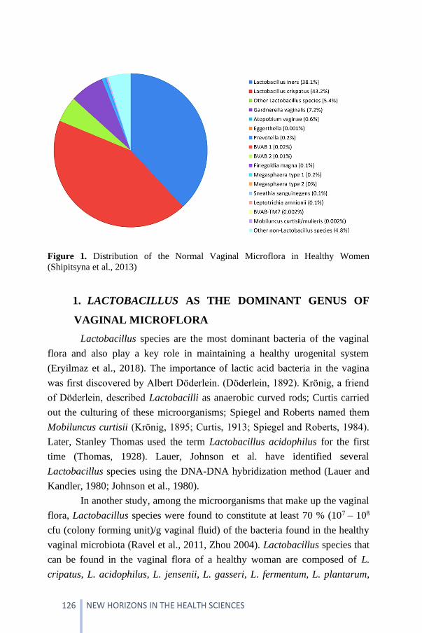
126 NEW HORIZONS IN THE HEALTH SCIENCES
Figure 1. Distribution of the Normal Vaginal Microflora in Healthy Women
(Shipitsyna et al., 2013)
1. LACTOBACILLUS AS THE DOMINANT GENUS OF
VAGINAL MICROFLORA
Lactobacillus species are the most dominant bacteria of the vaginal
flora and also play a key role in maintaining a healthy urogenital system
(Eryilmaz et al., 2018). The importance of lactic acid bacteria in the vagina
was first discovered by Albert Döderlein. (Döderlein, 1892). Krönig, a friend
of Döderlein, described Lactobacilli as anaerobic curved rods; Curtis carried
out the culturing of these microorganisms; Spiegel and Roberts named them
Mobiluncus curtisii (Krönig, 1895; Curtis, 1913; Spiegel and Roberts, 1984).
Later, Stanley Thomas used the term Lactobacillus acidophilus for the first
time (Thomas, 1928). Lauer, Johnson et al. have identified several
Lactobacillus species using the DNA-DNA hybridization method (Lauer and
Kandler, 1980; Johnson et al., 1980).
In another study, among the microorganisms that make up the vaginal
flora, Lactobacillus species were found to constitute at least 70 % (107 – 108
cfu (colony forming unit)/g vaginal fluid) of the bacteria found in the healthy
vaginal microbiota (Ravel et al., 2011, Zhou 2004). Lactobacillus species that
can be found in the vaginal flora of a healthy woman are composed of L.
cripatus, L. acidophilus, L. jensenii, L. gasseri, L. fermentum, L. plantarum,

127
L. iners, L. minutus, L. brevis, L. catenaforme, L. leichmannii, L. salyarius, L.
vaginalis, L. casei, L. delbrueckii, L. rhamnosus, and L. reuteri in varying
proportions. (Figure 2) (Beamer et al., 2017; Ventolini, 2015; Cribby et al.,
2008).
Figure 2. Species Distribution of Lactobacillus Microorganisms in Vaginal Flora. A)
Healthy Woman (HW), B) C. Trachomatis C) Vaginal Candidiasis (VC), D) Bacterial
Vaginosis (BV) (Ceccarani et al., 2019).
Before menarche, vaginal flora elements consist of some Lactocillus
genus microorganisms and skin and intestinal microorganisms (Vicariotto et
al., 2012). Estrogens and progesterone hormones regulate environmental
conditions for microorganisms of the genus Lactobacillus from the beginning
of the female reproductive phase. Lactobacilli and other bacteria metabolize
glycogen into glucose, maltose, and lactic acid. This metabolism creates a
vaginal pH of 3.8-4.4, which is described as usual (Mendling, 2016). To date,
more than 120 Lactobacillus bacterial species have been identified. More than

128 NEW HORIZONS IN THE HEALTH SCIENCES
ten different Lactobacillus species can be found in the vaginal flora of women
after their regular menstrual cycles begin. However, flora dominated by one
or two species is usually encountered (Beamer et al., 2017; Ventolini, 2015;
Cribby et al., 2008; Larsen and Monif, 2001).
Lactobacillus species constitute an essential part of lactic acid
bacteria. They are microaerophilic or anaerobic, non-spore-forming, acid-
tolerant, bacillus, or cocci-shaped Gram-positive bacteria. They are usually of
varying lengths and sizes (0.5-1.2 x 1-10 µm), but they exist in pairs or
chains. At the same time, although some of them are catalase-negative, some
of them can produce pseudo-catalase (Liu et al., 2014). Its optimum growth
temperatures are 30-40 °C, and its pH is in the range of 5.5-6.2. The primary
metabolism of Lactobacilli is primarily the breakdown of carbohydrates and
related compounds by producing lactic acid and energy (Gülel, 2014). They
are divided into two groups as heterofermentative and homofermentative
according to their glucose fermentation. While homofermentative produce
lactic acid as the outcome from carbohydrates, heterofermentative can also
produce products such as ethanol, carbon dioxide, and acetic acid besides
lactic acid (Florou-Paneri et al., 2013). Organic acids, hydrogen peroxide,
reuterin, antifungal peptides, and bacteriocins from metabolites produced by
Lactobacillus species are compounds with antimicrobial activity (Aldunate et
al., 2015; Dobson et al., 2012; Holzapfel et al., 1995).
The microflora in the female urogenital system is of serious
importance for the health and functioning of this system (Larsen and Monif,
2001). The normal flora of the human vagina plays a crucial role in the
prevention of many urogenital diseases such as bacterial vaginosis, urinary
tract infections, yeast infections (Wiesenfeld et al., 2003; Cherpes et al., 2003;
Pybus and Onderdonk, 1999; Martin et al., 1999; Gupta et al., 1998). This
protection is mainly attributed to Lactobacillus species which are dominant in
the vaginal flora of healthy women. Lactobacillus species play essential
protective roles through various mechanisms (Ravel et al., 2011). These
include the production of various antimicrobial compounds such as lactic
acid, bacteriocins and biosurfactants, hydrogen peroxide, co-aggregation,
competitive exclusion, immunomodulation, and signaling between bacteria
(Reid et al., 2011; Kaewsrichan et al., 2006). In a study, it was shown that
Lactobacillus species adhered to the vaginal epithelial cells by covering their

129
entire surface, physically inhibiting the adsorption of pathogenic
microorganisms, and a high number of Lactobacillus species adhered to the
Hela cells and thus inhibited the attachment of Chlamydia trachomatis by
binding to cell surface receptors. In another in vitro study, it was proven that
there is an inhibitory effect on C. trachomatis, depending on the
concentrations of Lactobacillus species in the vaginal flora (Nardini et al.,
2016). The degree of adhesion of Lactobacillus species to Hela cells varies
between species (Figure 3) (Mastromarino et al., 2014).
Figure 3. Gram Stain Sample Showing Adhesion of Lactobacillus Species to the
Surface of Hela Cells at 1st and 24th Hours (Mastromarino et al., 2014)
1.1. Lactobacillus acidophilus
L. acidophilus is the most widely known and used probiotic bacteria
(Ouwehand et al., 2014; Homayouni et al., 2014; Vicariotto et al., 2012). This
species lives in the vagina and intestines, protecting against pathogenic
microorganisms that cause disease. L. acidophilus is a rod-shaped
microorganism, 0.6-0.9 x 1.5-6 µm in size, in pairs and short chains. The G+C
ratio of L. acidophilus DNA is 32-37%, and this strain is obligate
homofermentative. Since it is microaerophilic, it can also reproduce in an

130 NEW HORIZONS IN THE HEALTH SCIENCES
aerobic environment, but it reproduces better in anaerobic conditions
containing 10% water (H2O), 5% carbon dioxide (CO2), and 85% nitrogen (N)
(Robinson and Itsaranuwat, 2005). L. acidophilus produces bacteriocins such
as lactosine B, lactacin F, acidosine A and acidosine B. Many strains of the
Lactobacillus genus have been shown to provide prophylactic and therapeutic
benefits in humans (Gopal, 2011).
In 1980, microorganisms previously identified as L. acidophilus were
reported to be heterogeneous (Johnson et al., 1980; Lauer et al., 1980). It has
been discovered that the physiological properties of L. acidophilus strains
differ in terms of the sugar pattern of the cell wall, the type of lactic acid
produced, the G+C content of DNAs, and DNA homology values. Therefore,
they renamed the Lactobacillus genus microorganisms as L. acidophilus, L.
cripatus, Lactobacillus amilovorus, Lactobacillus gasseri, Lactobacillus
gallinarum, and Lactobacillus johnsonii (Johnson et al., 1980; Lauer et al.,
1980).
Since L. acidophilus species are very similar, it is impossible to
distinguish them by simple biochemical and physiological tests, and L.
acidophilus species are misidentified because they are closely related.
Genotypic identification methods are needed to define these species
definitively. Precise identification was achieved by applying more than one
molecular characterization method and combining the results. Cell proteins of
L. acidophilus strains were differentiated using SDS-PAGE, RAPD-PCR
methods (Klein et al., 1998; Pot et al., 1993). Additionally, identification
studies were carried out using PCR and DNA sequencing methods. (Nour,
1998; Tilsala-Timisjarvi and Alatossava 1997; Schleifer and Ludwig, 1995;
Hertel et al, 1993; Hensiek et al, 1992).
1.2. Lactobacillus. crispatus
L. crispatus is a straight to slightly curved rod-shaped microorganism
measuring 0.8-1.6 x 2.3-11 μm and forms short chains. They are obligate
homofermentative, and the DNA G+C ratio is 35-38%. In addition to being
found in the vaginal flora, they can be isolated from human feces and buccal
cavities.

131
L. crispatus is seen as one of the most dominant species among
Lactobacilli in the vaginal flora in studies (Beamer et al., 2017; Ventolini,
2015; Cribby et al., 2008; Larsen and Monif, 2001). The main reasons for this
dominance of L. crispatus were listed by van der Veer et al. in 4 items as
stated below (van der Veer et al., 2019).
• Ability to form an extracellular matrix (biofilm) on the
vaginal mucosa surface
• Production of antimicrobials such as lactic acid, bacteriocins
and their ability to produce H2O2, which inhibits the growth
and adhesion of urogenital pathogens.
• Efficient use of available nutrients such as glycogen, which is
the primary carbon source in the vaginal lumen
• Modulation of the host -immunogenic responses
L. crispatus supports the stability of the normal vaginal microbiota
and the re-dominance of this microbiota in the vagina following antibiotic
treatment and thus plays a role in protecting the vagina from bacterial
vaginosis (Petrova et al., 2015; Verstraelen et al., 2009; Cruciani et al., 2015).
It has been reported that L. crispatus strains inhibit the growth and adhesion
of uropathogens to vaginal epithelial cells in vitro (Figure 4) (Kwok et al.,
2006; Osset et al., 2001). L. crispatus has also been shown to reduce the
adhesion of Neisseria gonorrhoeae and C. trachomatis to HeLa cells
(Mastromarino et al., 2014; Vielfort et al., 2008).
Figure 4. Gram Stain Sample Showing Adhesion of L. crispatus to the Surface of
Vaginal Epithelial Cells at 1st (A) and 24th (B) Hours (Kwok et al., 2006).
132 NEW HORIZONS IN THE HEALTH SCIENCES
1.3. Lactobacillus jensenii
L. jensenii is one of the most commonly found Lactobacillus species
that dominate the normal vaginal microflora (Beamer et al., 2017; Cribby et
al., 2008). In addition to the vaginal microflora, it is also found in the bladder
microflora (Thomas-White et al., 2018). L. jensenii was found to have a
bactericidal effect, preventing the colonization of pathogenic bacteria,
including the uropathogenic E. coli, in the vagina and female bladder
(Kalyoussef et al., 2012). While only two strains of L. jensenii from the
bladder were characterized, Putonti et al. identified 11 new L. jensenii strains
from urine samples taken from women (Thomas-White et al., 2018; Putonti et
al., 2019).
L. jensenii metabolizes glycogen anaerobically. The increased
estrogen level during pregnancy causes an increase in glycogen by affecting
the metabolism of L. jensenii and thus contributes to the acidic vaginal
environment. This acidic environment is a natural mechanism to prevent
infections that can cause premature birth and suppress bacteria growth that
causes bacterial vaginosis (O'Hanlon et al., 2011). L. jensenii is thought to
produce enzymes from the liver that cause the release of hydrolase, which
aids in the digestion of food in the gastrointestinal tract. Moreover, L. jensenii
produces Lactacin F, reducing the population of other Enterococcus and
Lactobacillus bacteria. Furthermore, L. jensenii is also used in the production
of fermented foods. L. jensenii can be used in the supportive treatment of
newborns to facilitate the digestion of breast milk. (Prince et al., 2014).
1.4. Lactobacillus gasseri
L. gasseri is one of the most common Lactobacillus species that make
up the vaginal microbiome. L. gasseri is a rod-shaped microorganism in short
chains, 0.6-0.8 x 3.0-5.0 μm in size, and they are obligate heterofermentative.
Furthermore, L. gasseri does not hydrolyze arginine, and their DNA has a
G+C ratio of 33-35% and is found in the vaginal and oral flora (Gülel, 2014).
L. gasseri, one of the most dominant Lactobacillus species thought to be
present in approximately 75% of women, is thought to be the most stable
community over time and rarely transitions to other species (Gajer et al.,
2012; Boris and Barbés, 2000). Therefore, it is thought that it is important to
133
elucidate the mechanism of protecting the host against pathogens due to its
stability.
Glycogen is the main carbohydrate released when epithelial cells die
and stored in the intermediate and superficial layers of vaginal epithelial cells.
By secreting α-amylase from the host, L. gasseri is probably able to use
glycogen as a carbon source (Spear et al., 2014; Anderson et al., 2014).
Studies have shown that L. gasseri significantly inhibits the adhesion of
Trichomonas vaginalis to vaginal cells (Phukan et al., 2018). L. gasseri was
also found to significantly inhibit the colonization of Candida albicans by
reducing the expression of genes involved in biofilm formation (Matsuda et
al., 2018).
1.5. Lactobacillus fermentum
L. fermentum is best known for its probiotic properties. L. fermentum
is a dominant microflora element in the vagina and intestine. Therefore, it can
be used as a probiotic in the intestine and vagina. L. fermentum, like most
Lactobacillus species, can prevent pathogenic infections in the intestine and
vagina by producing inhibitory compounds such as lactic acid, short-chain
fatty acids, bacteriocin, biosurfactants, and hydrogen peroxide (H2O2). (Kaur
et al, 2013; Anukam and Reid, 2007; Boris and Barbés, 2000). L. fermentum
shows antimicrobial activity by competing with pathogenic microorganisms
for nutrients, adhering to intestinal and vaginal epithelial cell surfaces,
preventing the adhesion of pathogens to these surfaces, and stimulating the
immune system (Stöber et al., 2010; Lebeer et al., 2008; Reid and Burton,
2002). In a clinical study, L. fermentum decreased intestinal pathogens and
increased the number of probiotic microorganisms in healthy people (Shieh et
al., 2011). Oral use of probiotic microorganisms has normalized the vaginal
microbiota (Vitali et al., 2012). In another study, as a result of keeping L.
fermentum strain in the same environment with E. coli and Gardnerella
vaginalis, an inhibitory effect of L. fermentum on these strains was observed
(Figure 5) (Kaewnopparat et al., 2013).

134 NEW HORIZONS IN THE HEALTH SCIENCES
Figure 5. Scanning Electron Microscopy (SEM) Images of (A) E. coli and (C) G.
vaginalis, And Damaged (B) E. Coli and (D) G. Vaginalis (In the Arrows) After
Treatment with L. fermentum (Kaewnopparat et al., 2013).
L. fermentum is thought to increase humans' immunological response
and prevent community-acquired gastrointestinal and upper respiratory tract
infections. L. fermentum can be used as a food preservative and produces
various and potent antimicrobial peptides that can be applied as an alternative
to antibiotics. In addition, this microorganism can reduce bloodstream
cholesterol levels and help prevent liver disease and colorectal cancer
(Naghmouchi et al., 2020). Finally, L. fermentum is an essential
microorganism in dough technology that contributes to flavor, texture, or
health-promoting dough ingredients. All this food technology-related
availability has been used to develop new foodstuffs and limit the use of
antibiotics in feed and food (Naghmouchi et al., 2020).
135
1.6. Lactobacillus iners
L. iners, like the other Lactobacillus mentioned above, is an essential
part of the normal vaginal flora (Jakobsson and Forsum, 2007). Intensive
studies have been carried out in the last few years, especially on identifying L.
iners and their biological activation in the vaginal flora. Since L. iners has
been found to be present in vaginal dysbiosis such as bacterial vaginosis, its
role in vaginal health is not clear. L. iners has a small genome of about 1
Mbp, which indicates a symbiotic or parasitic lifestyle (Petrova et al., 2017).
There is much debate about whether L. iners is beneficial or harmful to the
host microbiota. A study reported that the vaginal microbiota dominated by L.
crispatus was dominated by L. iners during menstruation (Gajer et al., 2012).
Therefore, it has been reported that L. iners is a species that contributes to the
improvement of the vaginal microbiota (Srinivasan et al., 2010). In addition, it
has been reported that most pregnant women with an L. iners-dominant
vaginal microbiota delivered on time without adverse pregnancy outcomes
(Kindinger et al., 2017, Petricevic et al., 2014).

136 NEW HORIZONS IN THE HEALTH SCIENCES
REFERENCES
Aldunate, M., Srbinovski, D., Hearps, A. C., Latham, C. F., Ramsland, P. A.,
Gugasyan, R., Cone, R. A., & Tachedjian, G. (2015). Antimicrobial and
immune modulatory effects of lactic acid and short chain fatty acids
produced by vaginal microbiota associated with eubiosis and bacterial
vaginosis, Frontiers in physiology, 6, 164.
Beamer, M. A., Austin, M. N., Avolia, H. A., Meyn, L. A., Bunge, K. E., & Hillier, S.
L. (2017). Bacterial species colonizing the vagina of healthy women are not
associated with race, Anaerobe, 45, 40–43.
Ceccarani, C., Foschi, C., Parolin, C., D'Antuono, A., Gaspari, V., Consolandi, C.,
Laghi, L., Camboni, T., Vitali, B., Severgnini, M., & Marangoni, A. (2019).
Diversity of vaginal microbiome and metabolome during genital
infections, Scientific reports, 9(1), 14095.
Cherpes, T. L., Meyn, L. A., Krohn, M. A., Lurie, J. G., & Hillier, S. L. (2003).
Association between acquisition of herpes simplex virus type 2 in women
and bacterial vaginosis, Clinical infectious diseases: an official publication
of the Infectious Diseases Society of America, 37(3), 319–325.
Cribby, S., Taylor, M., & Reid, G. (2008). Vaginal microbiota and the use of
probiotics, Interdisciplinary perspectives on infectious diseases, 2008,
256490.
Curtis, A. H. (1913). A motile curved anaerobic bacillus in uterine discharges, The
Journal of Infectious Diseases, 165-169.
Dobson, A., Cotter, P. D., Ross, R. P., & Hill, C. (2012). Bacteriocin production: a
probiotic trait?, Applied and environmental microbiology, 78(1), 1–6.
Döderlein, A. (1892). Das Scheidensekret und seine Bedeutung für das
Puerperalfieber, Besold, Leipzig.
Eryilmaz, M., Gurpinar, S. S., Palabiyik, I. M., Guriz, H., & Gerceker, D. (2018).
Molecular Identification and Antimicrobial Activity of Vaginal Lactobacillus
sp, Current pharmaceutical biotechnology, 19(15), 1241–1247.
Florou-Paneri, P., Christaki, E. & Bonos, E. (2013). Lactic acid bacteria as source of
functional ingredients. In J. M. Kongo (Eds.), Lactic Acid Bacteria-R & D
for Food, Health and Livestock Purposes (25, pp.589-614). InTech.
Gajer, P., Brotman, R. M., Bai, G., Sakamoto, J., Schütte, U. M., Zhong, X., Koenig,
S. S., Fu, L., Ma, Z. S., Zhou, X., Abdo, Z., Forney, L. J., & Ravel, J. (2012).
Temporal dynamics of the human vaginal microbiota, Science translational
medicine, 4(132), 132ra52.
137
Gopal, P.K. (2011). Lactic Acid Bacteria | Lactobacillus spp.: Lactobacillus
acidophilus, In. J.W. Fuquay (Eds.), Encyclopedia of Dairy Sciences (2nd
ed., pp. 91-95). Mississippi: Academic Press.
Gupta, K., Stapleton, A. E., Hooton, T. M., Roberts, P. L., Fennell, C. L., & Stamm,
W. E. (1998). Inverse association of H2O2-producing lactobacilli and
vaginal Escherichia coli colonization in women with recurrent urinary tract
infections, The Journal of infectious diseases, 178(2), 446–450.
Gülel, Ş. (2014). Molecular identification and probiotic properties of ‘lactobacillus
acidophilus group’ isolates from turkish kefir (Master’s thesis). Retrieved
from https://tez.yok.gov.tr/UlusalTezMerkezi/tezSorguSonucYeni.jsp
Holzapfel, W. H., Geisen, R., & Schillinger, U. (1995). Biological preservation of
foods with reference to protective cultures, bacteriocins and food-grade
enzymes, International journal of food microbiology, 24(3), 343–362.
Homayouni, A., Bastani, P., Ziyadi, S., Mohammad-Alizadeh-Charandabi, S.,
Ghalibaf, M., Mortazavian, A. M., & Mehrabany, E. V. (2014). Effects of
probiotics on the recurrence of bacterial vaginosis: a review, Journal of
lower genital tract disease, 18(1), 79–86.
Johnson, J. L., Phelps, C. F., Cummins, C. S., London, J., & Gasser, F. (1980).
Taxonomy of the Lactobacillus acidophilus group, International Journal of
Systematic Bacteriology, 30(1), 53-68.
Kaewsrichan, J., Peeyananjarassri, K. & Kongprasertkit, J. (2006). Selection and
identification of anaerobic lactobacilli producing inhibitory compounds
against vaginal pathogens, FEMS Immunol Med Microbiol, 48(1):75–83.
Klein, G., Pack, A., Bonaparte, C., & Reuter, G. (1998). Taxonomy and physiology of
probiotic lactic acid bacteria, International Journal of Food Microbiology,
41(2), 103-125.
Krönig, B. (1895). Über die Natur der Scheidenkeime, speciell über das Vorkommen
anaerober Streptokokken im scheidensekret Schwangerer, Centrbl Gynäkol,
16, 409–412.
Larsen, B., & Monif, G. R. (2001). Understanding the bacterial flora of the female
genital tract, Clinical infectious diseases: an official publication of the
Infectious Diseases Society of America, 32(4), e69–e77.
Lauer, E., & Kandler, O. (1980). Lactobacillus gasseri sp. nov., a new species of the
subgenus Thermobacterium. Zentralblatt für Bakteriologie: I. Abt. Originale
C: Allgemeine, angewandte und ökologische Mikrobiologie, 1(1), 75-78.
Liu, W., Pang, H., Zhang, H. & Cai, Y. (2014). Biodiversity of lactic acid bacteria. In
H. Zhang, Y. Cai, (Eds.), Lactic Acid Bacteria. (pp. 103-203). Dordrecht:
Springer.

138 NEW HORIZONS IN THE HEALTH SCIENCES
Martin, H. L., Richardson, B. A., Nyange, P. M., Lavreys, L., Hillier, S. L., Chohan,
B., Mandaliya, K., Ndinya-Achola, J. O., Bwayo, J., & Kreiss, J. (1999).
Vaginal lactobacilli, microbial flora, and risk of human immunodeficiency
virus type 1 and sexually transmitted disease acquisition, The Journal of
infectious diseases, 180(6), 1863–1868.
Mastromarino, P., Di Pietro, M., Schiavoni, G., Nardis, C., Gentile, M., & Sessa, R.
(2014). Effects of vaginal lactobacilli in Chlamydia trachomatis
infection, International journal of medical microbiology: IJMM, 304(5-6),
654–661.
Mendling, W. (2016). Vaginal Microbiota. In A. Schwiertz (Eds.), Microbiota of
the Human Body. Advances in Experimental Medicine and Biology (Vol.
902, pp.83-93). Springer, Cham.
Nardini, P., Ñahui Palomino, R. A., Parolin, C., Laghi, L., Foschi, C., Cevenini, R.,
Vitali, B., & Marangoni, A. (2016). Lactobacillus crispatus inhibits the
infectivity of Chlamydia trachomatis elementary bodies, in vitro
study, Scientific reports, 6, 29024.
Ouwehand, A. C., ten Bruggencate, S. J., Schonewille, A. J., Alhoniemi, E., Forssten,
S. D., & Bovee-Oudenhoven, I. M. (2014). Lactobacillus acidophilus
supplementation in human subjects and their resistance to enterotoxigenic
Escherichia coli infection, The British journal of nutrition, 111(3), 465–473.
Pot, B., Hertel, C., Ludwig, W., Descheemaeker, P., Kersters, K., & Schleifer, K. H.
(1993). Identification and classification of Lactobacillus acidophilus, L.
gasseri and L. johnsonii strains by SDS-PAGE and rRNA-targeted
oligonucleotide probe hybridization, Journal of General Microbiology,
139(3), 513-517.
Pybus, V., & Onderdonk, A. B. (1999). Microbial interactions in the vaginal
ecosystem, with emphasis on the pathogenesis of bacterial
vaginosis, Microbes and infection, 1(4), 285–292.
Ravel, J., Gajer, P., Abdo, Z., Schneider, G. M., Koenig, S. S., McCulle, S. L.,
Karlebach, S., Gorle, R., Russell, J., Tacket, C. O., Brotman, R. M., Davis,
C. C., Ault, K., Peralta, L., & Forney, L. J. (2011). Vaginal microbiome of
reproductive-age women, Proceedings of the National Academy of Sciences
of the United States of America, 108(Suppl 1), 4680–4687.
Reid, G., Younes, J. A., Van der Mei, H. C., Gloor, G. B., Knight, R., & Busscher, H.
J. (2011). Microbiota restoration: natural and supplemented recovery of
human microbial communities, Nature reviews. Microbiology, 9(1), 27–38.
139
Robinson, R. & Itsaranuwat, P. (2005). In R.K. Robinson (Eds.), Dairy Microbiology
Handbook: The Microbiology of Milk and Milk Products (3rd. ed.). New
York: John Wiley & Sons.
Shipitsyna, E., Roos, A., Datcu, R., Hallén, A., Fredlund, H., Jensen, J. S., Engstrand,
L., & Unemo, M. (2013). Composition of the vaginal microbiota in women
of reproductive age--sensitive and specific molecular diagnosis of bacterial
vaginosis is possible?, PloS one, 8(4), e60670.
Spiegel, C. A., & Roberts, M. (1984). Mobiluncus gen. nov., Mobiluncus curtisii
subsp. curtisii sp. nov., Mobiluncus curtisii subsp. holmesii subsp. nov., and
Mobiluncus mulieris sp. nov., curved rods from the human vagina,
International Journal of Systematic and Evolutionary Microbiology, 34(2),
177-184.
Thomas, S. (1928). Döderlein's Bacillus: Lactobacillus Acidophilus, The Journal of
Infectious Diseases, 43(3), 218–227.
Ventolini, G. (2015). Vaginal Lactobacillus: biofilm formation in vivo - clinical
implications, International journal of women's health, 7, 243–247.
Vicariotto, F., Del Piano, M., Mogna, L., & Mogna, G. (2012). Effectiveness of the
association of 2 probiotic strains formulated in a slow release vaginal
product, in women affected by vulvovaginal candidiasis: a pilot
study, Journal of clinical gastroenterology, 46 Suppl, S73–S80.
Wiesenfeld, H. C., Hillier, S. L., Krohn, M. A., Landers, D. V., & Sweet, R. L.
(2003). Bacterial vaginosis is a strong predictor of Neisseria gonorrhoeae and
Chlamydia trachomatis infection, Clinical infectious diseases : an official
publication of the Infectious Diseases Society of America, 36(5), 663–668.
Zhou, X., Bent, S. J., Schneider, M. G., Davis, C. C., Islam, M. R., & Forney, L. J.
(2004). Characterization of vaginal microbial communities in adult healthy
women using cultivation-independent methods, Microbiology (Reading,
England), 150(Pt 8), 2565–2573.
Nour, M. (1998). 16S-23S and 23S-5S intergenic spacer regions of lactobacilli:
nucleotide sequence, secondary structure and comparative analysis, Research
in microbiology, 149(6), 433–448.
Tilsala-Timisjärvi, A., & Alatossava, T. (1997). Development of oligonucleotide
primers from the 16S-23S rRNA intergenic sequences for identifying
different dairy and probiotic lactic acid bacteria by PCR, International
journal of food microbiology, 35(1), 49–56.
Schleifer, K. H., & Ludwig, W. (1995). Phylogeny of the genus Lactobacillus and
related genera, Systematic and Applied Microbiology,18(4), 461-467.

140 NEW HORIZONS IN THE HEALTH SCIENCES
Hertel, C., Ludwig, W., Pot, B., Kersters, K., & Schleifer, K. H. (1993).
Differentiation of lactobacilli occurring in fermented milk products by using
oligonucleotide probes and electrophoretic protein profiles, Systematic and
Applied Microbiology, 16(3), 463-467.
Hensiek, R., Krupp, G., & Stackebrandt, E. (1992). Development of diagnostic
oligonucleotide probes for four Lactobacillus species occurring in the
intestinal tract, Systematic and Applied Microbiology, 15(1), 123-128.
van der Veer, C., Hertzberger, R. Y., Bruisten, S. M., Tytgat, H., Swanenburg, J., de
Kat Angelino-Bart, A., Schuren, F., Molenaar, D., Reid, G., de Vries, H., &
Kort, R. (2019). Comparative genomics of human Lactobacillus crispatus
isolates reveals genes for glycosylation and glycogen degradation:
implications for in vivo dominance of the vaginal microbiota,
Microbiome, 7(1), 49.
Petrova, M. I., Lievens, E., Malik, S., Imholz, N., & Lebeer, S. (2015). Lactobacillus
species as biomarkers and agents that can promote various aspects of vaginal
health, Frontiers in physiology, 6, 81.
Petrova, M. I., Reid, G., Vaneechoutte, M., & Lebeer, S. (2017). Lactobacillus iners:
Friend or Foe?, Trends in microbiology, 25(3), 182–191.
Verstraelen, H., Verhelst, R., Claeys, G., De Backer, E., Temmerman, M., &
Vaneechoutte, M. (2009). Longitudinal analysis of the vaginal microflora in
pregnancy suggests that L. crispatus promotes the stability of the normal
vaginal microflora and that L. gasseri and/or L. iners are more conducive to
the occurrence of abnormal vaginal microflora, BMC microbiology, 9, 116.
Cruciani, F., Biagi, E., Severgnini, M., Consolandi, C., Calanni, F., Donders, G.,
Brigidi, P., & Vitali, B. (2015). Development of a microarray-based tool to
characterize vaginal bacterial fluctuations and application to a novel
antibiotic treatment for bacterial vaginosis, Antimicrobial agents and
chemotherapy, 59(5), 2825–2834.
Kwok, L., Stapleton, A. E., Stamm, W. E., Hillier, S. L., Wobbe, C. L., & Gupta, K.
(2006). Adherence of Lactobacillus crispatus to vaginal epithelial cells from
women with or without a history of recurrent urinary tract infection, The
Journal of urology, 176(5), 2050–2054.
Osset, J., Bartolomé, R. M., García, E., & Andreu, A. (2001). Assessment of the
capacity of Lactobacillus to inhibit the growth of uropathogens and block
their adhesion to vaginal epithelial cells, The Journal of infectious
diseases, 183(3), 485–491.
141
Vielfort, K., Sjölinder, H., Roos, S., Jonsson, H., & Aro, H. (2008). Adherence of
clinically isolated lactobacilli to human cervical cells in competition with
Neisseria gonorrhoeae, Microbes and infection, 10(12-13), 1325–1334.
Thomas-White, K., Forster, S. C., Kumar, N., Van Kuiken, M., Putonti, C., Stares, M.
D., Hilt, E. E., Price, T. K., Wolfe, A. J., & Lawley, T. D. (2018). Culturing
of female bladder bacteria reveals an interconnected urogenital
microbiota, Nature communications, 9(1), 1557.
Kalyoussef, S., Nieves, E., Dinerman, E., Carpenter, C., Shankar, V., Oh, J., Burd, B.,
Angeletti, R. H., Buckheit, K. W., Fredricks, D. N., Madan, R. P., Keller, M.
J., & Herold, B. C. (2012). Lactobacillus proteins are associated with the
bactericidal activity against E. coli of female genital tract secretions, PloS
one, 7(11), e49506.
Putonti, C., Ahmad, A., Baddoo, G., Diaz, J., Do, M., Gallian, N., Lorentzen, C.,
Mohammed, H., Murphy, J., Olu-Ajeigbe, A., Yang, T., Miller-Ensminger,
T., Stark, N., Maskeri, L., Van Dusen, J., & Wolfe, A. J. (2019). Draft
Genome Sequences of 11 Lactobacillus jensenii Strains Isolated from the
Female Bladder, Microbiology resource announcements, 8(35), e00970-19.
O'Hanlon, D. E., Moench, T. R., & Cone, R. A. (2011). In vaginal fluid, bacteria
associated with bacterial vaginosis can be suppressed with lactic acid but not
hydrogen peroxide, BMC infectious diseases, 11, 200.
Prince, A. L., Antony, K. M., Chu, D. M., & Aagaard, K. M. (2014). The
microbiome, parturition, and timing of birth: more questions than
answers, Journal of reproductive immunology, 104-105, 12–19.
Boris, S., & Barbés, C. (2000). Role played by lactobacilli in controlling the
population of vaginal pathogens, Microbes and infection, 2(5), 543–546.
Anderson, D. J., Marathe, J., & Pudney, J. (2014). The structure of the human vaginal
stratum corneum and its role in immune defense, American journal of
reproductive immunology, 71(6), 618–623.
Spear, G. T., French, A. L., Gilbert, D., Zariffard, M. R., Mirmonsef, P., Sullivan, T.
H., Spear, W. W., Landay, A., Micci, S., Lee, B. H., & Hamaker, B. R.
(2014). Human α-amylase present in lower-genital-tract mucosal fluid
processes glycogen to support vaginal colonization by Lactobacillus, The
Journal of infectious diseases, 210(7), 1019–1028.
Phukan, N., Brooks, A., & Simoes-Barbosa, A. (2018). A Cell Surface Aggregation-
Promoting Factor from Lactobacillus gasseri Contributes to Inhibition of
Trichomonas vaginalis Adhesion to Human Vaginal Ectocervical Cells,
Infection and immunity, 86(8), e00907-917.

142 NEW HORIZONS IN THE HEALTH SCIENCES
Matsuda, Y., Cho, O., Sugita, T., Ogishima, D., & Takeda, S. (2018). Culture
Supernatants of Lactobacillus gasseri and L. crispatus Inhibit Candida
albicans Biofilm Formation and Adhesion to HeLa
Cells, Mycopathologia, 183(4), 691–700.
Kaur, B., Balgir, P., Mittu, B., Chauhan, A., Kumar B., (2013). Purification and
physicochemical characterization of anti-gardnerella vaginalis bacteriocin
hv6b produced by Lactobacillus fermentum isolate from human vaginal
ecosystem, American Journal of Biochemistry and Molecular Biology, 3(1),
91-100.
Anukam, K. C. & Reid G. (2007). Lactobacillus plantarum and Lactobacillus
fermentum with probiotic potentials isolated from the vagina of healthy
Nigerian women, Research Journal of Microbiology, 2, 81-87
Stöber, H., Maier, E., & Schmidt, H. (2010). Protective effects of Lactobacilli,
Bifidobacteria and Staphylococci on the infection of cultured HT29 cells
with different enterohemorrhagic Escherichia coli serotypes are strain-
specific, International journal of food microbiology, 144(1), 133–140.
Lebeer, S., Vanderleyden, J., & De Keersmaecker, S. C. (2008). Genes and molecules
of lactobacilli supporting probiotic action, Microbiology and molecular
biology reviews : MMBR, 72(4), 728–764.
Reid, G., & Burton, J. (2002). Use of Lactobacillus to prevent infection by pathogenic
bacteria, Microbes and infection, 4(3), 319–324.
Shieh, M.-J., Shang, H.-f., Liao, F.-H., Zhu, J.-S. & Chien Y.-W. (2011).
Lactobacillus fermentum improved intestinal bacteria flora by reducing
Clostridium perfringens, E-Spen the European e-Journal of Clinical
Nutrition and Metabolism, 6, e59-e63
Vitali, B., Cruciani, F., Baldassarre, M. E., Capursi, T., Spisni, E., Valerii, M. C.,
Candela, M., Turroni, S., & Brigidi, P. (2012). Dietary supplementation with
probiotics during late pregnancy: outcome on vaginal microbiota and
cytokine secretion, BMC microbiology, 12, 236.
Kaewnopparat, S., Dangmanee, N., Kaewnopparat, N., Srichana, T., Chulasiri, M., &
Settharaksa, S. (2013). In vitro probiotic properties of Lactobacillus
fermentum SK5 isolated from vagina of a healthy woman, Anaerobe, 22, 6–
13.
Naghmouchi, K., Belguesmia, Y., Bendali, F., Spano, G., Seal, B. S., & Drider, D.
(2020). Lactobacillus fermentum: a bacterial species with potential for food
preservation and biomedical applications, Critical reviews in food science
and nutrition, 60(20), 3387–3399.

143
Jakobsson, T., & Forsum, U. (2007). Lactobacillus iners: a marker of changes in the
vaginal flora?, Journal of clinical microbiology, 45(9), 3145.
Srinivasan, S., Liu, C., Mitchell, C. M., Fiedler, T. L., Thomas, K. K., Agnew, K. J.,
Marrazzo, J. M., & Fredricks, D. N. (2010). Temporal variability of human
vaginal bacteria and relationship with bacterial vaginosis, PloS one, 5(4),
e10197.
Kindinger, L. M., Bennett, P. R., Lee, Y. S., Marchesi, J. R., Smith, A., Cacciatore,
S., Holmes, E., Nicholson, J. K., Teoh, T. G., & MacIntyre, D. A. (2017).
The interaction between vaginal microbiota, cervical length, and vaginal
progesterone treatment for preterm birth risk, Microbiome, 5(1), 6.
Petricevic, L., Domig, K. J., Nierscher, F. J., Sandhofer, M. J., Fidesser, M.,
Krondorfer, I., Husslein, P., Kneifel, W., & Kiss, H. (2014). Characterisation
of the vaginal Lactobacillus microbiota associated with preterm
delivery, Scientific reports, 4, 5136.

144 NEW HORIZONS IN THE HEALTH SCIENCES
145
CHAPTER 7
COMPLEMENTARY AND ALTERNATIVE MEDICINE IN THE
TREATMENT OF INFERTILITY
Çiğdem KARAKAYALI AY1 , Nurten ÖZÇALKAP2
1 Res. Asistant ,Inonu University, Faculty of Health Sciences, Midwifery Department, Malatya,[email protected], ORCID ID: 0000-0002-5282-973X. 2 Expert Midwifery, Inonu University, Institute of Health Sciences, Department of Midwifery,
PhD Student, Malatya, Turkey. E-posta: [email protected] ORCID ID: 0000-0003-
2440-968X

146 NEW HORIZONS IN THE HEALTH SCIENCES
147
INTRODUCTION
According to the World Health Organization (WHO), infertility; It is
defined as not being able to get pregnant, not being able to continue the
pregnancy or not giving birth to a living baby as a result of pregnancy.
Infertility is also defined as the inability to conceive for one year despite
regular sexual intercourse, but WHO extends this period up to two years
(İpekoğlu & Oral, 2019).
NICE (National Institute for Health and Clinical Excellence) defined
infertility as the inability to conceive after 2 years of unprotected and regular
sexual intercourse as a result of the absence of any known reproductive
pathology. The American Society for Reproductive Medicine defines
infertility as the inability to achieve a successful pregnancy after 12 months or
more of regular and unprotected sexual intercourse (Szamatowicz &
Szamatowicz, 2020). More than 186 million people worldwide suffer from
infertility, mostly in developing countries (Inhorn & Patrizio, 2015).
It is estimated that infertility affects %8 to %12 of couples of
reproductive age worldwide (Vander Borght & Wyns, 2018). In our country
(Turkey), there are 1.5-2 million infertile couples, % 55-%75 of whom suffer
from primary infertility and %25-% 40 from secondary infertility. These
numbers are increasing gradually due to the career expectations of women and
the increasing age of having children (Özturk et al., 2021).
When infertility cases are examined in the world, it is known that
many communities use traditional treatment approaches. In some cases, it is
determined that these methods and applications are used for support purposes
in addition to modern medicine, and in some cases, only traditional methods
are preferred (İpekoğlu & Oral, 2019).
Most of the treatments other than modern and scientific treatments are
generally expressed as Complementary and Alternative Medicine (CAM).
Although the terms alternative therapy and complementary therapy are often
used interchangeably, the two terms have different meanings. While
alternative therapy, is a type of treatment used instead of modern medicine,
complementary medicine is a form of treatment used in addition to modern
medicine (Emul et al.,2020)..
Currently, CAM for infertility continues to be effective by bringing
up traditional treatments again. CAM is stated to be able to harmonize the

148 NEW HORIZONS IN THE HEALTH SCIENCES
condition of the woman with a holistic approach in order to achieve a more
effective therapeutic effect, and is increasingly used by infertile women.
However, in some randomized controlled studies, it was stated that CAM did
not have a significant effect on infertility and more studies were needed (Feng
et al.,2021).
According to a meta-analysis study on the prevalence of infertility,
potential needs and demands for treatment and care, with a sample of 172 413
women covering 25 countries at the international level, the incidence of
infertility is %3.5-%16.7 in developed countries and %6.9-%9.3 in
underdeveloped countries. In underdeveloped countries, the frequency has
been reported as %5-%25. The lifetime infertility frequency was reported as
%6.6 - %26.4. (Boivin et al.,2007).
It is reported that approximately %30-%60 of couples undergoing
infertility treatment resort to CAM methods (Avcıbay & Kızılkaya Beji,
2003).
Although studies on the approaches of people with infertility
problems towards complementary/alternative medicine practices are limited,
according to the results of the study in which 157 (120 women, 37 men) from
private clinics in England and 181 (124 women, 57 men) from national health
centers were sampled, %63 of women, %25 of men stated that they were not
indifferent to complementary/alternative medicine practices. %10 of these
women stated that the complementary/alternative medicine method they used
in the treatment of infertility was beneficial, %13 stated that they got the
opinion that everything is possible by relaxing psychologically, and %22
helped them relax (Boivin et al.,2007).
In this section, it is aimed to explain the CAM approaches used in
infertile women and to provide information by systematically reviewing the
literature on the effectiveness and safety of these approaches.

149
1.COMPLEMENTARY AND ALTERNATIVE
APPROACHES
1.1. Acupuncture
Acupuncture, a Latin word; It consists of a combination of the words
acus (needle) and puncture (penetration). Acupuncture, which has been used
since 4000 years ago, is a complementary and alternative treatment approach
that aims to get results by stimulating certain points on the body with needles.
The philosophy of acupuncture argues that there are two energy systems in the
body called "Yin" and "Yang", which are opposite to each other but in a
constant state of balance. It is reported that diseases occur when the balance
between these energies is disturbed.
Acupuncture channels are located where these energies circulate
freely. Each acupuncture channel has points localized on the body where it
opens to the skin. By pinning these points, it is possible to balance the
energies and help the treatment of diseases with acupuncture. Among the
diseases in which the therapeutic properties of acupuncture are shown, female
infertility is also included (Çayır & Gürsoy, 2018).
Acupuncture has been one of the most studied CAM approaches for
improving fertility outcomes (Miner et al., 2018). Acupuncture has a long
history of treating gynecological problems. With an increasing number of
studies, it has been reported that acupuncture can regulate menstruation and
help women conceive without the risk of multiple pregnancy (Miner et al.,
2018; Zhou & Qu, 2019, Cochrane et al., 2014).
In a scoping study, it was reported that acupuncture used in the
treatment of infertility had an effect on the fertility outcomes of men and
women. While the evidence in this scoping study to show that acupuncture
improves male and female fertility outcomes is high, there is no conclusive
evidence that it is effective in the treatment of infertility, as other studies
taken have not shown improvement (Miner et al., 2018).
1.2. Homeopathy
Founded by Samuel Hahnemann, a German physician and pharmacist,
homeopathy is a CAM system that naturally heals the body non-toxic.
“Homoios” as the word meaning; similar and “pathos”; to suffer from the

150 NEW HORIZONS IN THE HEALTH SCIENCES
disease. This method aims to treat like with like by stimulating a self-healing
mechanism (Yıllar Erkek ve Pasinlioğlu, 2016; SHGM, 2021; Bhatt, 2021).
The basic principle in homeopathy is the minimum number of doses.
Hahnemann states that the human body has natural healing powers that only
need a little stimulus to initiate and maintain the healing process. After the
healing process has begun, it has been argued that it is not necessary to repeat
the drug doses (Steinberg & Beal, 2003).
Homeopathy, which is accepted as one of the first applications of
CAN approaches, is used in many countries around the world, especially in
high-income countries. It is reported that homeopathic medicines are also
effective on some gynecological diseases such as bleeding, cysts, vaginal
discharge and infertility (Alizadeh Charandabi et al., 2016).
In a case series examining the effect of individualized homeopathic
medicine use in the treatment of infertility, it was determined that the use of
homeopathic medicines had positive effects on infertile couples and
contributed significantly to positive pregnancy results (RajachandraSekar et
al., 2021).
1.3. Ayurveda
The history of Ayurveda, which is similar to traditional Chinese
medicine and is based on a traditional medicine system and used in the
management of diseases, dates back to 3000 years (Kumar, 2017; Sodhi,
2020). In the Sanskrit word Ayurveda, the term “Ayu” can be translated as
life and “Veda” as knowledge (Ramaswamy, 2010).
Ayurveda is a TAT defined in harmony with the laws of nature and
the art of daily living. At the heart of this approach is the concept of the five
elements. These elements are ether, air, fire, water and earth. In Ayurvedic
science, these five elements are accepted as the building blocks of all creation
and material existence, both organic and inorganic, both physically and
symbolically (Ramaswamy, 2010). It states that human, a microcosm of
nature, also exists from these five elements and the health of man and the
universe depends on the balance and harmony of these elements
(Ramaswamy, 2010; Posmontier & Teitelbaum, 2009).

151
The Ayurvedic approach, which perceives the disease as an
imbalance, provides a holistic health service and a balance in a lifestyle with
both its preventive and curative features (Ramaswamy, 2010; Sodhi, 2020).
In gynecological diseases, it is preferred to use ayurvedic drugs
instead of the existing hormonal treatment. WHO confirms that the ayurvedic
medicines used are safe and effective (WHO, 1993; Atul N. Jadhav &
Bhutani, 2005).
Women are frequently exposed to stress factors this stress increases
the formation of reactive oxygen species (ROS). Therefore, this situation
impairs reproductive health by inducing oxidative stress (OS). Increased OS
may cause infertility by affecting ovarian physiology and oocyte quality. It
has been reported that ayurvedic drugs should be used frequently in order to
overcome female reproductive health complications such as stress-mediated
hormonal imbalance, polycystic ovary syndrome (PMS), failure in follicular
growth and development, low oocyte quality and infertility. Because it is
stated that these drugs can reduce the OS level and increase the antioxidant
level in the body and improve reproductive health disorders (Pandey et al.,
2018).
1.4. Hypnosis
"Hypnos", derived from the word Hypnosis means sleep in Greek.
However, hypnosis is not a state of sleep, but rather a state of wakefulness
(Ceyhan & Tasa Yiğit, 2016). In hypnosis, although the person is not
unconscious, the person does not react to external stimuli (Taştan et al., 2015).
At the same time, hypnosis is a special state of consciousness that provides
physical relaxation by focusing attention on a certain point, and its use for
treatment is defined as hypnotherapy (Peksoy et al., 2018).
There are not many evidence-based studies on the effects of hypnosis
on reproductive health. In a study of women with hypothalamic amenorrhea,
it was reported that with a single session of hypnosis, % 85experienced
menstruation after 12 weeks. The mechanism of hypnosis on reproductive
health is that it will help to relax and reduce the negative effect of anxiety on
fertility (Avcıbay & Kızılkaya Beji, 2003).
152 NEW HORIZONS IN THE HEALTH SCIENCES
In a scoping study examining the effect of CAM approaches on
infertility treatment, it was reported that there are limited studies evaluating
the use of hypnosis and fertility treatment, and that hypnosis has positive
effects on infertility treatment, however, the level of evidence is low to
moderate and needs to be supported by more studies (Miner et al., 2018).
1.5. Reflexology
Reflexology is the CAM approach based on the application of
pressure to specific areas of the feet or hands (reflex points) resulting in
stimulation of nerve points that mediate electrochemical messages (Levy et
al., 2020).
In reflexology, it is aimed to provide the body with a state of balance
and increase comfort by applying pressure to the reflex points of the soles of
the feet and palms (Moghimi-Hanjani et al., 2015). It is reported that
reflexology applied to the soles of the feet affects the psychological responses
of people and causes healing in different organs of the body (Ferrer de Dios,
2005).
Reflexology is a complementary treatment method that improves
women's quality of life and provides positive results without any side effects
in premenstrual syndrome (PMS), dysmenorrhea, reproductive health and
menopause problems (Bolsoy, 2016).
In the study evaluating the effectiveness of foot reflexology to induce
ovulation, it was reported that reflexology did not provide a significant result
on the induction rate compared to the placebo application and more studies
are needed (Holt et al., 2009).
2. CONCLUSION
There are many CAM techniques used in infertility. We have
explained some of them. However, the mechanisms of action of most of them
are largely unknown today. In this direction, couples who receive adequate
support for CAM for infertility management can adapt as long as they have
psychological and physiological symptoms. As a result, complementary and
alternative medicine approaches, the use of which is increasing day by day in
the treatment of infertility, should be included in health services with
scientifically valid studies with high level of evidence.
153
REFERENCES
Alizadeh Charandabi, S. M., Biglu, M. H., & Yousefi Rad, K. (2016). Effect of
Homeopathy on Pain Intensity and Quality Of Life of Students With Primary
Dysmenorrhea: A Randomized Controlled Trial. Iranian Red Crescent
medical journal, 18(9), e30902. https://doi.org/10.5812/ircmj.30902
Avcıbay, B., & Kızılkaya Beji, N. (2003). İnfertilite tedavisinde
tamamlayıcı/alternatif tıp uygulamaları. Androloji Bülteni, 15(52), 71-74
Bhatt, V. K. (2021). Mother and child health care with homeopathy management of
postpartum depression with homeopathy.Clınıcal and experimental
homeopathy.A peer-rewieved International Online e-Journal, 7, 1-19. SNN:
0974-9799.
Boivin, J., Bunting, L., Collins, J. A., & Nygren, K. G. (2007). International estimates
of infertility prevalence and treatment-seeking: potential need and demand
for infertility medical care. Human reproduction (Oxford, England), 22(6),
1506–1512. https://doi.org/10.1093/humrep/dem046
Bolsoy, N. (2016). The Use of Reflexology in Women's Health Reflexology. In:
Çamlı,A.A., Ak, B., Arabacı, R., Efe, R., editors.editors.Research
Advancements In Health Scıences. St. Kliment Ohridski University Press;
2016.p. 373-385
Ceyhan, D., & Tasa Yiğit, T. (2016). Güncel tamamlayıcı ve alternatif tıbbi
tedavilerin sağlık uygulamalarındaki yeri. Düzce Ünversitesi Sağlık Bilimleri
Enstitüsü Dergisi, 6(3), 178-189.
Cochrane, S., Smith, C. A., Possamai-Inesedy, A., & Bensoussan, A. (2014).
Acupuncture and women's health: an overview of the role of acupuncture and
its clinical management in women's reproductive health. International
journal of women's health, 6, 313–325.
https://doi.org/10.2147/IJWH.S38969
Çayır, Y., & Gürsoy,P.G. (2018). İn vitro fertilizasyon ve akupunktur. Konuralp
Medical Journal, 10(3), 420-423. https://doi.org/10.18521/ktd.442446
Emul, T. G., Yilmaz, D. V., & Buldum, A. (2020). İnfertilite tedavisinde kullandiklari
tamamlayici alternatif tedavi uygulamalarinin belirlenmesi. Journal of
Education and Research in Nursing, 17(4), 317-322.
Feng, J., Wang, J., Zhang, Y., Zhang, Y., Jia, L., Zhang, D., Zhang, J., Han, Y., &
Luo, S. (2021). The Efficacy of Complementary and Alternative Medicine in
the Treatment of Female Infertility. Evidence-based complementary and
alternative medicine : eCAM, 2021, 6634309.
https://doi.org/10.1155/2021/6634309

154 NEW HORIZONS IN THE HEALTH SCIENCES
Ferrer de Dios R. (2005). Reflexología [Reflexology]. Revista de enfermeria
(Barcelona, Spain), 28(3), 42–46.
Holt, J., Lord, J., Acharya, U., White, A., O'Neill, N., Shaw, S., & Barton, A. (2009).
The effectiveness of foot reflexology in inducing ovulation: a sham-
controlled randomized trial. Fertility and sterility, 91(6), 2514–2519.
https://doi.org/10.1016/j.fertnstert.2008.04.016
Inhorn, M. C., & Patrizio, P. (2015). Infertility around the globe: new thinking on
gender, reproductive technologies and global movements in the 21st
century. Human reproduction update, 21(4), 411–426.
https://doi.org/10.1093/humupd/dmv016
İpekoğlu, H. Y., & Oral, H. B.(2019). İnfertilitede Kullanılan Geleneksel Tedavi
Yöntemleri ve Çeşitli Uygulamalar: Isparta Bölgesi. Motif Akademi
Halkbilimi Dergisi, 12(25), 105-119.
https://doi.org/10.12981/mahder.523760.
Jadhav, A. N., & Bhutani, K. K. (2005). Ayurveda and gynecological
disorders. Journal of ethnopharmacology, 97(1), 151–159.
https://doi.org/10.1016/j.jep.2004.10.020
Kumar, S., Dobos, G.J., Rampp, T. (2017) The significance of Ayurvedic medicinal
plants. Journal of evidence-based complementary & alternative
medicine, 22(3), 494-501. https://doi.org/10.1177/2156587216671392.
Levy, I., Attias, S., Stern Lavee, T., Avneri, O., Cohen, G., Balachsan, S., Sagi, S., &
Schiff, E. (2020). The effectiveness of foot reflexology in reducing anxiety
and duration of labor in primiparas: An open-label randomized controlled
trial. Complementary therapies in clinical practice, 38, 101085.
https://doi.org/10.1016/j.ctcp.2019.101085
Miner, S. A., Robins, S., Zhu, Y. J., Keeren, K., Gu, V., Read, S. C., & Zelkowitz, P.
(2018). Evidence for the use of complementary and alternative medicines
during fertility treatment: a scoping review. BMC complementary and
alternative medicine, 18(1), 158. https://doi.org/10.1186/s12906-018-2224-7
Moghimi-Hanjani, S., Mehdizadeh-Tourzani, Z., & Shoghi, M. (2015). The Effect of
Foot Reflexology on Anxiety, Pain, and Outcomes of the Labor in
Primigravida Women. Acta medica Iranica, 53(8), 507–511.
Ozturk, A., Aba, Y. A., & Sik, B. A. (2021). The relationship between stigma,
perceived social support and depression in infertile Turkish women
undergoing in vitro fertilization-embryo transfer. Archives of psychiatric
nursing, 35(5), 434-440.
Pandey, A. K., Gupta, A., Tiwari, M., Prasad, S., Pandey, A. N., Yadav, P. K.,
Sharma, A., Sahu, K., Asrafuzzaman, S., Vengayil, D. T., Shrivastav, T. G.,
155
& Chaube, S. K. (2018). Impact of stress on female reproductive health
disorders: Possible beneficial effects of shatavari (Asparagus
racemosus). Biomedicine & pharmacotherapy = Biomedecine &
pharmacotherapie, 103, 46–49. https://doi.org/10.1016/j.biopha.2018.04.003
Peksoy, S., Demirhan, İ., Kaplan, S., Şahin, S. ve Arıöz-Düzgün, A. (2018).
Tamamlayıcı ve alternatif tedavinin jinekolojik kanserlerde kullanımı.
Türkiye Sağlık Bilimleri ve Araştırmaları Dergisi, 1(1), 36-47.
Posmontier, B., & Teitelbaum, M. (2009). An Ayurvedic approach to postpartum
depression. Holistic nursing practice, 23(4), 201–216.
https://doi.org/10.1097/HNP.0b013e3181aece5a
RajachandraSekar, B., Nair, J., Sunny, A., & Manoharan, A. (2021). Individualised
Homeopathic Medicine in the Treatment of Infertility: A Case
Series. Homeopathy : the journal of the Faculty of Homeopathy, 10.1055/s-
0041-1725040. Advance online publication. https://doi.org/10.1055/s-0041-
1725040
Ramaswamy S. Ayurveda-An ancient healing system’s gifts to the modern woman.
https://www.ayurvedacollege.com/wp
content/uploads/2017/06/AyurvedaSandhiyaRamaswamy.pdf.2010;p:3-4.
Retrieved on from, 10 January 2022.
Sağlık Hizmetleri Genel Müdürlüğü (SHGM). Geleneksel, Tamamlayıcı ve
Fonksiyonel Tıp Uygulamaları Dairesi Başkanlığı. Homeopati Uygulaması
Hakkında Bilgilendirme https://shgmgetatdb.saglik.gov.tr/TR,24680/
homeopati-uygulamasi-hakkinda-bilgilendirme.html Erişim Tarihi:
10.01.2022.
Sodhi, V. (2020).Ayurveda: the science of life and mother of the healing arts. Joseph
E Pizzorno, Michael T Murray (Fifth Edition), Textbook of Natural Medicine
(p:257-263.e1) https://doi.org/10.1016/B978-0-323-43044-9.00033-9
Steinberg, D., & Beal, M. W. (2003). Homeopathy and women's health care. Journal
of obstetric, gynecologic, and neonatal nursing: JOGNN, 32(2), 207–214.
https://doi.org/10.1177/0884217503252125.
Szamatowicz, M., & Szamatowicz, J. (2020). Proven and unproven methods for
diagnosis and treatment of infertility. Advances in medical sciences, 65(1),
93–96. https://doi.org/10.1016/j.advms.2019.12.008
Taştan, K., Çayır, Y., Makas, Y. N., Set, T., Hülakü, A., & Tekşan, T. (2015). Atatürk
Üniversitesi Tıp Fakültesi‟nde çalışan doktorların hipnoterapi ile ilgili
görüşleri, eğitim durumları ve bilgi edinme kaynakları. Smyrna Tıp Dergisi,
(2), 1-6.
156 NEW HORIZONS IN THE HEALTH SCIENCES
Vander Borght, M., & Wyns, C. (2018). Fertility and infertility: Definition and
epidemiology. Clinical biochemistry, 62, 2–10. https://doi.org/10.1016/
.clinbiochem.2018.03.012
WHO. Research guidelines for evaluating the safety and efficacy of herbal medicines.
Regional office for the Western Pacific, Manila.1993. World Health
Organisation.
Yıllar Erkek, Z., & Pasinlioğlu, T. (2016). Doğum ağrısında kullanılan tamamlayıcı
tedavi yöntemleri. Anadolu Hemşirelik ve Sağlık Bilimleri Dergisi, 19(1), 71-
77.
Zhou, J., & Qu, F. (2009). Treating gynaecological disorders with traditional Chinese
medicine: a review. African journal of traditional, complementary, and
alternative medicines: AJTCAM, 6(4), 494–517. https://doi.org/10.4314
/ajtcam.v6i4.57181

157
CHAPTER 8
BREAST NEOPLASMS
Nurten ÖZÇALKAP1 , Çiğdem KARAKAYALI AY2
1Expert Midwifery,Inonu University, Institute of Health Sciences Department of Midwifery,
Malatya/Turkey. E-posta: [email protected], ORCID ID: 0000-0003-2440-968X 2 Res.Asistant.,Inonu University, Faculty of Health Sciences, Midwifery
Department,Malatya/Turkey. E-posta: [email protected], ORCID ID: 0000-
0002-5282-973X.

158 NEW HORIZONS IN THE HEALTH SCIENCES
159
INTRODUCTION
Breast neoplasms are divided into two groups as benign (benign) and
maning (malignant). Breast cancer is among the most important causes of
morbidity and mortality in women. Most clinical breast changes in women are
benign; only 3% to 6% of cases are due to breast cancer. However, there are
no current, evidence-based treatment recommendations for various benign
differential diagnoses (Taşkın, 2020). The statistics on breast cancer are
frightening. Breast cancer is the most common type of cancer among women
in the world and in Turkey (SB, 2018). The incidence of breast cancer is high
in Caucasian women aged 40 years and over. In recent years, the incidence of
new cases of breast cancer has been found to be similar, especially in white
and African American women aged 50-59 years (Jemal, et al.2011). It has
been determined that 44.5% of women with new case breast cancer in Turkey
are in the age range of 50-69, and 40.4% are in the age range of 25-49 (SB,
2018). Breast cancer accounts for 28% of cancers and 18% of causes of death
in women (American cancer society,2019 & Al‐Shamsi et al.2021 & Jemalet
al., 2011).
Due to early diagnosis and more effective treatment combinations,
breast cancer mortality rates have been decreasing since 1991. It is estimated
that the decrease in mortality rates is due to early diagnosis and increased
awareness, as well as better treatment methods. Health professionals should
raise awareness about breast cancer neoplasms, and should undertake the roles
of educator, counselor, therapeutic and care in helping women gain healthy
life behaviors in awareness, prevention and screening programs (Taşkın 2020
& Najjar, et al. 2010 & Jemalet al., 2011).
1. BENIGN BREAST DISEASE
Benign breast changes can be definitively distinguished from
malignant lesions through the selective use of available diagnostic research
and interdisciplinary collaboration. Prospective studies are desired for early
detection of breast cancer in lesions with malignancy risk (Taşkın, 2020 &
American cancer society, 2019).

160 NEW HORIZONS IN THE HEALTH SCIENCES
1.1.Fibrocystic Diseases
Fibrocystic disease of the breast is multiple cysts formed in the breast
tissue. Multiple cysts are seen in approximately 1/3 of premenopausal women.
It is rare for new cysts to form after menopause. In the premenopausal period,
when progesterone is not secreted, excessive stimulation of estrogen
accelerates the formation of multiple cysts (Paepke et al., 2018).
Clinically, they are usually found as bilateral multiple pillar cysts and
sometimes as unilateral multiple pillar cysts. These cysts, which increase in
the premenstrual period, show signs of pain, fullness and tenderness in the
breasts. However, these cysts can be found without these symptoms. Cysts are
located especially in the upper outer quadrant of the breast. They are palpable
as soft and irregular thickenings on palpation (Stachs et al., 2019).
Preliminary diagnosis with history and physical examination, x-ray
control of breast tissue with mammography, and differentiation of cysts from
solid tumors with ultrasound. A definitive diagnosis is made by biopsy (Amin
et al., 2013).
Many applications are recommended for the symptomatic treatment of
fibrocystic disease. The most common is avoidance of caffeine and nicotine
and supplementing the diet with vitamins. Other treatment options are
hormone therapy and surgery (Taşkın, 2020).
1.2. Fibroadenoma
Fibroadenoma is the most common benign neopalzyme. It is more
common in young women. They are painless, mobile and hard masses. These
lesions are mostly located in the upper quadrant of the breast. These lesions
are usually discovered incidentally. The changes in fibroadenomas during the
menstrual phase are unlike fibrocystic changes. In contrast, they can grow
rapidly during pregnancy and exogenous estrogen therapy (Amin et al., 2013).
Although the incidence of malignant change in these lesions is very
low, the tumor is simply removed in the treatment. Because no lesion is
benign. In addition, their growth over time causes constant anxiety in women
(Paepke et al., 2018).

161
1.3. Ectasia in the Breast Canal
It is the inflammation of the canal in the nipple. Symptoms; There is
pain in the nipple, itching, a sticky, dark, colored discharge and redness
around the nipple. If there is an abscess, it is drained. Antibiotics are used for
treatment (Taşkın, 2020).
1.4. Duct Papilloma
It is a rare disease. It develops in the last part of the nipple canals. The
first symptom is a bloody or serous discharge from the nipple. Since the mass
is very small, it may not be palpable. Care should be taken in terms of its
malingnea. In the treatment, the papilloma and its duct system are excised
(Amin et al., 2013).
2. BREAST CANCER (MALIGN NEOPLASIS)
Breast cancer is a complex and systemic disease in which genetic
and/or epigenetic malignant changes play an important role. On the basis of
breast cancer; genetic mutations cause loss or reduction of function. As a
result of activation of oncogenes and inactivation of tumor suppressor genes,
malignant cells begin to invade and proliferate. Genomic instability may
develop with tumor suppressor gene and prorooncogene mutations. Tumor
suppressor genes that are effective in the development of breast cancer; p53,
BRCA1, BRCA2, PTEN and ATM. Oncogenes are formed by mutation and
transformation of protooncogenes, causing the malignant phenotype to occur.
Proto-oncogenes effective in the development of breast cancer; HER28neu,
EGFR, Ras, Myc and β-catenin (Liedtke et al., 2017 & Arkoob et al. 2010).
2.1. Breast Cancer Risk factors
The cause of breast cancer is unknown. There are many risk factors
with the increase in its incidence. These; Being over 40 years old, family
history of breast cancer (mother and sister), nulliparity, primiparous over 30
years of age, not breastfeeding, alcohol and smoking, long time between
menarche and first pregnancy, early menarche, late menopause, estrogen and
progestin and menopausal hormone treatment, previous breast, colon, ovarian,
thyroid, and endometrial cancer, a history of atypical hyperplasia, dense
breasts on mammography, certain ethnicities, known harmful gene mutation,
excessive exposure of breasts to radiation, prior breast biopsy with specific

162 NEW HORIZONS IN THE HEALTH SCIENCES
pathology, high-fat diet after menopause, past benign breast neoplasia and
excessive alcohol consumption. These factors can identify only 25% of
women who may develop breast cancer. The presence of a long latent phase
of 15-20 years before the clinical development of carcinoma precludes the
identification of many risk factors (ACOG, 2017 & Miaskowski et al. 2014).
2.2. Breast Cancer Physiopathology
Breast cancer is divided into 3 large groups according to
histopathology. Ductal-ductal, lobular, and nipple-nipple (Paget's disease)
carcinomas. Cancer can be both invasive and non-invasive (in-situ). It is
common opinion that micrometastases will occur as soon as breast cancer is
palpable (1 cm in diameter) (Taşkın, 2020).
2.2.1. Ductal (duct) carcinoma
It takes its origin in the lactiferous ducts. Breast cancers are most
commonly ductal carcinoma. The tumor staff is immobile, unilateral, and has
limited pain (Paepke et al. 2018).
2.2.2. lobular carcinoma
It originates from the mammary lobes. It is rarely palpable in the in-
situ stage. It is most common in the premenstrual period. They are found
bilaterally. Invasive lesions have a high incidence of hormone receptors
(Stachs et al. 2018-9).
2.2.3. Nipple carcinoma (paget's defect)
It takes origin from the nipple. It usually occurs in association with
ductal carcinoma. The most common symptom is nipple discharge and
bleeding. Erosion of the nipple can also be seen (Zhu et al., 2021).
2.3. Breast Cancer Symptoms
The most common symptom of breast cancer is the presence of a
lump or mass in the breast. A painless, firm and irregularly sided mass is
likely to be malignant. The majority (49%) of cancer-related masses are
located in the upper-outer region, 17% in the central region, 16% in the upper-
inner region, 12% in the lower outer region, and 6% in the lower-inner region
(Taşkın, 2020 & Miaskowski et al., 2014). The structure of the mass is usually

163
hard and immobile; It moves with the surrounding breast tissue and can be
easily differentiated from fibroadenoma in this respect. Tumor infiltrating
Cooper ligaments may cause skin retraction (Kalli et al., 2018). With the
slowing of the lymph flow, the breast skin may have the appearance of an
orange peel (Peau D'orange). Erythema and ulceration of the breast skin may
be observed due to lymphatic obstruction. If the tumor is located in the central
quadrant, the nipple can be retracted towards the central quadrant where the
tumor is located (Miaskowski et al., 2014). The presence of discharge from
the nipple, unilateral discharge, spontaneous, serous and/or bloody discharge
are important in terms of malignancy. Pain and asymmetry in the breast,
swelling in the armpit, and enlarged lymph nodes are other symptoms of
breast cancer (Arkoob et al., 2010 & Amin et al. 2013).
2.4. Diagnosis in Breast Cancer
While some women are diagnosed with symptoms, breast cancer often
does not show any symptoms. Therefore, it is important for women to have
regular screenings. Treatment success is higher in breast cancers diagnosed in
the early stages. Early diagnosis methods; breast self-exam (BSE), clinical
breast examination (CBE), and mammography (Najjar et al., 2010).
2.5. Breast Cancer Stages
The stage of breast cancer, tumor size after cancer diagnosis, lymph
node metastasis and whether there is a disease in a region other than the breast
are determined. Determining the cancer stage is an important indicator for
cancer recurrence or metastasis to other organs. The TNM system is used in
the staging of Breast Cancer (Table 1). Accordingly, T represents the primary
tumor, N represents the axillary lymph nodes, and M represents distant
metastases (Akyol, 2018 & Ljuslinder, 2009).

164 NEW HORIZONS IN THE HEALTH SCIENCES
Tablo.1 Staging İn Breast Cancer (Ljuslinder, 2009)
2.6. Treatment in Breast Cancer
Different treatment modalities are used in the treatment of breast
cancer. These; surgical treatment, chemotherapy, radiotherapy, hormonal
therapy and targeted therapies. In the selection of treatment modality;
Strategies for disease and symptom management, prolonging life expectancy
and increasing quality of life are important. The primary purpose of surgical
treatment; is to remove the tumor from the breast and determine the stage of
the disease (Paepke et al., 2018). These surgical approaches; It can be
examined in two main groups as breast-conserving surgery and non-breast-
sparing surgery (mastectomy) (Miaskowski et al. 2014). Chemotherapy can be
given adjuvant, neoadjuvant or palliative depending on the clinical condition
of the patient. In chemotherapy, docetaxel, paclitaxel, platinagens (cisplatin,
carboplatin), Vinorelbine (Navelbin), Capecitabine (Xeloda), Liposomal
doxorubicin (Doxil), Cyclophosphamide (Cytoxan), Carboplatin (Paraplatin)
etc. drugs are used. Radiotherapy is one of the cornerstones of cancer
treatments. It can be applied in patients with breast cancer, usually after the
recovery period of 6-8 weeks after surgical treatment. However, patients with
large tumor size can also be given radiotherapy before surgical treatment to
reduce the tumor size and make it operable. Hormone therapy drugs used in

165
breast cancer; tamoxifen, raloxifene, aromatase inhibitors and fulvestrant
(Akyol, 2018 & Greenwalt et al. 2019).
Hormonal and physical changes in women due to breast cancer
treatments can cause vaginal dryness. Vaginal dryness can cause problems
such as decreased sexual desire due to pain during sexual intercourse. In
improving vaginal dryness; smoking cessation, use of vaginal lubricant and
moisturizer, kegel exercises and patient education may be recommended
(Greenwalt et al. 2019).
Targeted therapies aim to inhibit the growth and spread of cancer cells
and are designed to directly destroy the tumor. In one in 5 women diagnosed
with breast cancer, cancer cells overproduce the HER-2 protein, which
promotes growth on their surface. Known as HER2-positive breast cancer,
these cancers tend to grow and spread more aggressively. Trastuzumab
(Herceptin), Pertuzumab (Perjeta), Ado-trastuzumab emtansine (Kadcyla,
TDM-1), Lapatinib (Tykerb) and Neratinib (Nerlynx) are drugs used in
targeted therapies. Drugs that are monoclonal antibodies (Trastuzumab,
Pertuzumab, Ado-trastuzumab) are given parenterally in the treatment of both
early and late stage breast cancer. Kinase inhibitors (Lapatinib, Neratinib)
drugs can be used in combination treatments (Kalli et al., 2018 & Ljuslinder,
2009).
2.7. Breast Cancer Prognosis
The natural course of breast cancer after diagnosis shows clinical and
biological differences between patients depending on prognostic factors such
as tumor size, lymph node involvement, histological features of the tumor,
hormone receptors, patient's age, weight and pregnancy status. Especially in
early stage breast cancer patients, determining prognostic factors, survival,
whether adjuvant chemotherapy can be given and the type of chemotherapy
can be determined (Zhu et al. 2021).
Inflammatory breast cancers are seen at a rate of 1-2% among other
breast cancers and have a poor prognosis. Invasive ductal carcinomas account
for 75% of all breast cancers. It is the most common type of breast cancer. It
has a worse prognosis than other types of cancer by metastasizing to axillary
lymph nodes (Paepke et al., 2018 & Al‐Shamsi et al., 2021).

166 NEW HORIZONS IN THE HEALTH SCIENCES
2.8. Care in Breast Cancer
Breast cancer patients face many physical, emotional and social
problems. At the same time, the intense and long-term continuation of cancer
treatments and the side effects caused by these treatments negatively affect
the daily life activities of the patients. In this process, health professionals
should provide holistic and supportive care to improve the quality of life of
patients and their relatives (Akyol, 2018). It has been determined that the
education and counseling provided by health professionals provide support,
coordination and effective communication, and reduce the symptoms of
patients diagnosed with cancer. The effects and side effects of chemotherapy
drugs should be known, patients should be questioned, evaluated and
appropriate interventions should be made according to chemotherapy agents.
In the management of the surgical treatment process, patients should provide
quality physical care, education and psychosocial support in the perioperative
period. In the postoperative period, body image changes, pain and sensation
changes, infection or seroma in the operation area, lymphedema, home care
and discharge planning should be done. Health professionals should provide
adequate information and support to patients about what surgical treatments
will lead to, the aesthetic result of the surgery, its effect on sexual harmony
and body image, and its functional results (Taşkın, 2020 & Najjar et al.2010).
3. CONCLUSION
As a result the name is no longer acceptable as cancer. With early
diagnosis and life, life increases and life expectancy is extended. those who
can lead normal lives of their students, be productive and happy.
167
REFERENCES
Akyol, k. (2018). Meme kanseri tanısı için özniteliklerin öneminin değerlendirilmesi
üzerine bir çalışma. Academic Platform Journal of Engineering and Science,
6(2), 109-115.
Al‐Shamsi, H. O. Abu‐Gheida, I. Abdulsamad, A. S. Awadhi, A. A. Alrawi, A.
Musallam K. M. Arun, B. & Nuhad K. Ibrahimcorresponding, N. K. (2021).
Molecular spectra and frequency patterns of somatic mutations in arab
women with breast cancer. Oncologist, 26(11), 2086–e2089
American cancer society. Non-cancerous Breast Conditions. Last Revised: September
10, 2019.
American College Of Obstetricians And Gynaecologists (2021). Breast cancer risk
Assessment and Screening in Average-Risk Women July 2017.
Amin, A. L. Purdy, A. C. Mattingly, J.D. & Kong,A. L. (2013). Benign Breast
Disease. Surg Clin N Am, 93, 299–308.
Arkoob, K. Nsour, M. Al Nemry, O. & Al Hajawi, B. (2010). Epidemiology of breast
cancer in women in Jordan: patient characteristics and survival analysis.
Eastern Mediterranean Health Journal, 16(10), 1032-1038, 2010.
Greenwalt, L. Zaza, N. Das, S. & Li, B. D. (2019). Precision medicine and targeted
therapies in breast cancer, surg Oncol Clin N Am, 29(1), 51-62.
Jemal, A. F. Bray, M. M. Center, Ferlay, J. E. & Ward, D. (2011). Forman, “Global
cancer statistics”, CA Cancer J. Clin. 61(2), 69–90.
Kalli, S. Semine, A. Cohen, S. Naber, S. P. Makim, S.S. & Bahl, M. (2018).
American Joint Committee on Cancer’s Staging System for Breast Cancer,
Eighth Edition: What the Radiologist Needs to Know. Radio Graphics, 38,
1921–1933.
Liedtke, C. & Kolberg, H. C.,(2017). Breast cancer and genomic testing. British
Journal of Surgery, 104(7), 799–801.
Ljuslinder, I.(2009). Studies of LRIG1 and the ERBB receptor family in breast and
colorectal cancer. : https://www.researchgate.net/publication/41034402
Miaskowski, C., Paul, S.M., Cooper, B., West, C., Jon, D., Levine, J. D., Elboim,
C., Hamolsky, D., Abrams, D., Luce, J., Dhruva, A., Langford, D. J.,
Merriman, J. D., Kober, K., Baggott, C., Leutwyler, H., & Aouizerat, B. E.
(2014). Identification of patient subgroups and risk factors for persistent
arm/shoulder pain following breast cancer surgery. Eur J Oncol Nurs, 2014
Jun;18(3):242-53.
Najjar, H. & Easson, A. (2010). Age at diagnosis of breast cancer in Arab nations,
International Journal of Surgery, 8(6), 448-452.

168 NEW HORIZONS IN THE HEALTH SCIENCES
Paepke, S. Metz, S. Salvago, A. B. & Ohlinger, R. (2018). Benign Breast Tumours -
Diagnosis and Management. Breast Care, 13, 403-412.
Stachs, A. Stubert, J. Reimer, T. & Hartmann, S. (2019). Benign breast disease in
women. Dtsch Arztebl Int, 116(33-34), 565–574.
Taşkın, L. (2020). Doğum ve kadın sağlığı hemşireliği, Ankara: Akademisyen
kitapevi.
T.C. Sağlık Bakanlığı. (2018). Sağlık İstatistikleri Yıllığı, Ankara 2019.
Zhu, H. & Doğan B. E. (2021). American Joint Committee on Cancer’s Staging
System for Breast Cancer, Eighth Edition: Summary for Clinicians. Eur J
Breast Health, 17(3), 234–238.

169
CHAPTER 9
TUMOR MICROENVIRONMENT
IN TRIPLE NEGATIVE BREAST CANCER
Pınar CELEPLİ1 , Baki TÜRKOĞLU2
1 Dr.,Ankara Training and Research Hospital, Department of Pathology, Ankara, Turkey,
[email protected], ORCID: 0000-0001-7643-6263 2 Dr.,Gülhane Training and Research Hospital, Department of General Surgery, Ankara,
Turkey, [email protected], ORCID: 0000-0002-5777-7545

170 NEW HORIZONS IN THE HEALTH SCIENCES

171
INTRODUCTION
Breast cancer is the most common malignancy in women. Since it is a
heterogeneous disease with different histological and molecular features,
clinical treatment and prognosis vary among patients. The identification of
molecular subtypes is important for clinical prognosis and patient survival,
and thus the implementation of effective treatment modalities. There are four
basic molecular subtypes according to the hormone receptor and human
epidermal growth factor receptor-2 (HER2) expression status: Luminal A,
Luminal B, HER2-positive, and triple-negative (Goldhirsch et al., 2013).
Triple-negative breast cancer (TNBC) is defined as a breast cancer subtype
presenting with the lack of estrogen receptor (ER), progesterone receptor (PR)
and HER2 expression (Wolff et al., 2014). According to gene expression
profile analysis, TNBC and basal-like breast cancer profiles show a high
overlap of 60-90%. According to its expression features, TNBC is divided
into the following subtypes: basal-like, immunomodulatory, mesenchymal,
mesenchymal stem-like, luminal androgen receptor, and unstable (Abramson
et al., 2015). While a tumor being triple-negative is based on the absence of
hormone receptors (ER/PR) and HER-2, basal-like breast cancer is associated
with cytokeratins with a basal epithelial cell gene expression pattern (CK5,
CK6, CK14, and CK17) and epidermal growth factor receptor (EGFR). The
gold standard in the diagnosis of basal-like breast cancer is the determination
of gene expression profile using complementary DNA microarrays (Kuo et
al., 2012).
TNBC accounts for approximately 15-20% of all breast cancer cases
and usually occurs in premenopausal women under 40 years (Morris et al.,
2007). TNBC is the most aggressive subtype of breast cancer due to its drug
resistance, high probability of metastasis, and lack of specific targets and
targeted therapies (Anders & Carey, 2008). The incidence of TNBC has
increased in recent years, and it also has higher morbidity and mortality
compared to the other subtypes of breast cancer (Hwang et al., 2019). TNBC
has attracted the attention of clinicians and researchers due to its large tumor
size, lymph node involvement at the time of diagnosis, progression to high
grade, lung/bone/brain metastases, and most importantly the lack of a suitable
target for treatment. Recent studies have shown that tumors in TNBC include
not only neoplastic cells but also a significantly altered surrounding stroma.

172 NEW HORIZONS IN THE HEALTH SCIENCES
The tumor microenvironment plays a critical role in tumor development and
progression, and is also considered a measurable parameter of response to
therapy. Understanding the tumor microenvironment (TME) and its regulators
in TNBC will be useful for designing immune-based targeted therapies.
1. TUMOR MICROENVIRONMENT
Unlike other breast tumor subtypes, TNBC is characterized by a
highly complex and heterogeneous TME. The rapid proliferation of cancer
cells and reprogramming in TME induces hypoxia. As a result, tumor cells
and those surrounding TME constantly adapt to new conditions and promote
tumor growth. TME creates a niche in touch with tumor consists of
endothelial and immune cells, as well as fibroblasts interacting each other;
thus, it can be used as a potential therapeutic target for TNBC (Deepak et al.,
2020). It induces the transformation of TME epithelial cells into TNBC stem
cells. Interactions between these cells in TME facilitate a variety of biological
events that promote cancer growth, invasion, angiogenesis, and metastasis
(Liu et al., 2014). TME consists of cancer cells, cancer-associated fibroblasts
(CAFs), immune cells, cancer stem cells (CSCs), extracellular matrix (ECM)
components, blood vessels, adipocytes, and differentiated cells (Otranto et al.,
2012).
Figure 1: Tumor Microenvironment
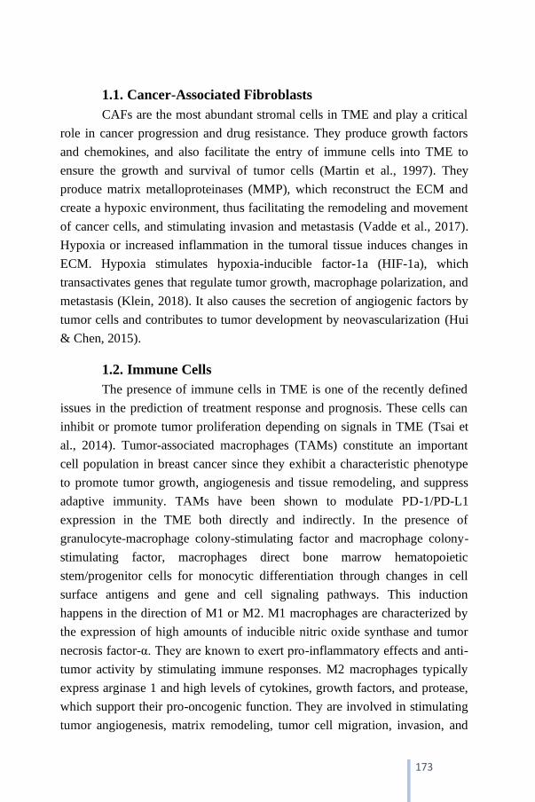
173
1.1. Cancer-Associated Fibroblasts
CAFs are the most abundant stromal cells in TME and play a critical
role in cancer progression and drug resistance. They produce growth factors
and chemokines, and also facilitate the entry of immune cells into TME to
ensure the growth and survival of tumor cells (Martin et al., 1997). They
produce matrix metalloproteinases (MMP), which reconstruct the ECM and
create a hypoxic environment, thus facilitating the remodeling and movement
of cancer cells, and stimulating invasion and metastasis (Vadde et al., 2017).
Hypoxia or increased inflammation in the tumoral tissue induces changes in
ECM. Hypoxia stimulates hypoxia-inducible factor-1a (HIF-1a), which
transactivates genes that regulate tumor growth, macrophage polarization, and
metastasis (Klein, 2018). It also causes the secretion of angiogenic factors by
tumor cells and contributes to tumor development by neovascularization (Hui
& Chen, 2015).
1.2. Immune Cells
The presence of immune cells in TME is one of the recently defined
issues in the prediction of treatment response and prognosis. These cells can
inhibit or promote tumor proliferation depending on signals in TME (Tsai et
al., 2014). Tumor-associated macrophages (TAMs) constitute an important
cell population in breast cancer since they exhibit a characteristic phenotype
to promote tumor growth, angiogenesis and tissue remodeling, and suppress
adaptive immunity. TAMs have been shown to modulate PD-1/PD-L1
expression in the TME both directly and indirectly. In the presence of
granulocyte-macrophage colony-stimulating factor and macrophage colony-
stimulating factor, macrophages direct bone marrow hematopoietic
stem/progenitor cells for monocytic differentiation through changes in cell
surface antigens and gene and cell signaling pathways. This induction
happens in the direction of M1 or M2. M1 macrophages are characterized by
the expression of high amounts of inducible nitric oxide synthase and tumor
necrosis factor-α. They are known to exert pro-inflammatory effects and anti-
tumor activity by stimulating immune responses. M2 macrophages typically
express arginase 1 and high levels of cytokines, growth factors, and protease,
which support their pro-oncogenic function. They are involved in stimulating
tumor angiogenesis, matrix remodeling, tumor cell migration, invasion, and
174 NEW HORIZONS IN THE HEALTH SCIENCES
immunosuppression (Solinas et al., 2009). CD163+ M2 macrophages, which
are highly expressed in breast cancers, have been shown to be correlated with
greater proliferation, poor differentiation, hormone receptor negativity, and
ductal-type histology (Helfen et al., 2018).
Tumor-infiltrating lymphocytes (TILs) are one of the main
components of TEM. Most TILs are T cells that can divide into CD4+ helper
cells, CD4+, CD25+, FOXP3+ phenotype cells, and natural killer and CD8+
T cells. (Allen & Louise Jones, 2011). If the environment surrounding tumor
cells is rich in inflammatory cells, the immune system cannot recognize and
eliminate cancer cells. On the other hand, lymphoid lineage B cells and
regulatory T cells that regularly infiltrate TME cause immunosuppression by
releasing cytokines and support tumor progression (Helfen et al., 2018; Liu et
al., 2014). TIL evaluation in breast cancer is gaining importance as a
prognostic marker. Higher TIL levels in TNBC and HER2+ breast cancer are
associated with better outcomes and response to neoadjuvant therapy. TILs
have a strong prognostic value in predicting recurrence-free/disease-free
survival and overall survival in early stage TNBC treated with standard
adjuvant or neoadjuvant chemotherapy (Tan et al., 2020). Immune cells can
be adapted for use in immunotherapies with the combination of other targeted
therapies.
1.3. Cancer Stem Cells
CSCs have the ability to self-renew and differentiate. They are
responsible for radiation and drug resistance, as well as playing a role in
cancer onset and metastasis processes (Liu et al., 2017). As a result of the
asymmetric division of CSCs, a stem cell and a progenitor cell are formed. At
the end of this asymmetric division, both CSCs renew themselves and
precursor cancer cells that will acquire phenotypically different characteristics
are formed. Progenitor tumor cells also have the ability to proliferate rapidly.
Progenitor tumor cells, on the other hand, have the ability to proliferate
rapidly (Yoo & Kwon, 2015).
According to one of the hypotheses on the origin of CSCs, these cells
originate from normal stem cells (Hartwig et al., 2014). With the deregulation
of the self-renewal and differentiation pathways of normal stem cells, cancer
cells with self-renewal and differentiation capacity are formed (Bozorgi et al.,
175
2015). This hypothesis is supported by the similarities between normal stem
cells and CSCs, their susceptibility to mutation and oncogenic transformation
due to their long lifespan, and the similarity of surface marker properties of
breast cancer stem cells and basal mammary stem cells/progenitor cells
(Tomita et al., 2016). In addition, it has been suggested that CSCs develop
through the epithelial-mesenchymal transition of normal cells. Cells
undergoing this transition are prone to transformation, and also they have
similar characteristics to normal and neoplastic stem cells (Martelotto et al.,
2014). In breast cancer, CSCs have the features of self-renewal,
differentiation, resistance to treatment, and metastasis and mammosphere
formation. Signaling pathways, such as Notch, Hedgehog, and Wnt play a role
in the self-renewal of CSCs in this cancer (Borah et al., 2015).
CSCs remodel their TMEs by promoting immune cells and
interactions with other cells; thus, they provide a supporting niche. They are
known to express three main stem cell markers in breast cancer, namely
CD44, CD24, and ALDH1. TNBCs have abundant CD44+/CD24- stem cells
compared to other breast cancer subtypes. It has been reported that CSCs
contribute to chemotherapy resistance and tumor metastasis in TNBC
(O'Conor et al., 2018). TNBC stem cells are more resistant to conventional
treatments than differentiated cells; therefore, recent studies aim to identify
strategies that target CSCs in TNBC (Jamdade et al., 2015).
1.4. Extracellular Matrix
ECM is a complex protein network that surrounds and stabilizes cells.
It consists of three major extracellular proteins: structural proteins (e.g.,
collagen and elastin), glycoproteins (e.g., fibronectin), and proteoglycans
(e.g., chondroitin sulfate). Maintaining tissue homeostasis plays a critical role
in cell proliferation and differentiation (Rigoglio et al., 2020). While initially
considered a stable structure that only provides support, studies have shown
that ECM is surprisingly dynamic and versatile, and it also has a key role in
cellular processes, such as cell growth, proliferation, and migration. The main
protein component of ECM is collagen, and the degradation of collagen IV by
proteases leads to cancer cell invasion through the basement membrane;
therefore, its integrity is important in cancer development. ECM also
176 NEW HORIZONS IN THE HEALTH SCIENCES
facilitates intercellular communication by guiding the passage of cytokines
and growth factors between cells (Lu et al., 2012).
ECM is at the forefront of all stages of in tumor development from
initiation, growth and progression to metastasis. Many cellular components of
TME (especially CAFs) contribute to metastatic cell proliferation through
different mechanisms that force ECM to remodel. CAFs facilitates this via the
accumulation of ECM proteins. TAMs produce various matrix
metalloproteinases (MMP-2, MMP-9) and cause the formation of tumoral
ECM. Cancer cells also produce and release reactive oxygen species that
initiate the activation of these enzymes. In addition, MMPs stimulate the
formation of new blood vessels and contribute to angiogenesis by causing the
release of factors embedded in ECM, such as vascular endothelial growth
factor (VEGF) (Najafi et al., 2019).
1.5. Blood Vessels
Tumor-associated blood and lymphatic vessels are the primary
pathways of innate and adaptive immune cells that play both pro and anti-
tumoral roles during tumor development and response to therapy (Fridman et
al., 2012). Angiogenesis is a critical process required for tumor progression.
Newly formed blood vessels supply nutrients and oxygen that contribute to
the growth and development of the tumor. In addition, the endothelium plays
a role in the entry of cancer cells into the blood vessel (intravasation), which
is an important stage in cancer metastasis.
Endothelial cells are one of the main sources of CAFs. The
heterogeneous CAF group is the main inducer of the migration and invasion
abilities of cancer cells; therefore, it is indirectly responsible for endothelial
metastasis (Sobierajska et al., 2020). A denser tumor vasculature is associated
with increased tumor growth and hematogenous metastasis. This is due to
increased oxygen supply, nutrients, and metastatic spread pathways (Nico et
al., 2008). Important markers of blood and lymphatic vascular network
include lymphatic vessel endothelial hyaluronan receptor (LYVE-1),
VEGFR3, CD31, and CD34. Lymphatic vessels predominantly secrete
LYVE-1 (Wang et al., 2017). The application of anti-angiogenic gene therapy
targeting angiogenesis may hold more ground in the prevention and treatment
of early-stage cancer in future.
177
1.6. Adipocytes
Adipocytes are the largest components of breast tissue; therefore, they
are involved in breast cancer development and treatment. Obesity is now
considered to be a complex physiological condition associated with multiple
molecular changes that can modulate the behavior of breast tumor cells, as
well as the surrounding microenvironment. Abnormal adipocytes, such as
cancer-associated adipocytes (CAAs) adjacent to the tumor are predominantly
found in the invasive portions of breast cancer. In addition, there are many
studies showing that CAAs exhibit different molecular and cellular properties
and can increase the proliferation and invasion capacity of cancer cells by
providing metabolic advantages. In breast cancer, invasive cancer cells
dramatically affect surrounding adipocytes and cause phenotypic changes
both in vitro and in vivo (Muller, 2013). CAAs play an important role in
breast cancer proliferation, angiogenesis, invasion, dissemination, and
metastasis by secreting multiple adipokines, such as leptin, adiponectin,
interleukin 6, chemokine ligand 2, and chemokine ligand 5 (Wu et al., 2019).
2. TUMOR MICROENVIRONMENT REGULATORS
In TNBC, TME is regulated by signaling pathways, growth factors,
chemokines, exosomes, epigenetic regulators, and microRNAs (Deepak et al.,
2020), which are discussed under separate headings in this section.
2.1. Signaling Pathways
In TNBC, there are various signaling pathways, such as Notch,
Hedgehog (SHH), Wnt/ β-catenin, JAK-STAT3, Sphingosine 1-phosphate
(S1P), stimulator of interferon genes (STING), chemokine-like interleukin-1β
(IL-1β), and TGF-β (Irey et al., 2019; Nallanthighal et al., 2019; Rostami et
al., 2019). Notch is a highly conserved signaling pathway that regulates
cellular processes important for cell-to-cell communication. Notch signal
modulates and improves the immune system by communicating between
immune cells and cancer cells in TME (Al-Hussaini et al., 2011). Speiser et
al. reported that TNBC overexpressed Notch 1 and Notch 4 receptors in
vascular endothelial cells and tumors with a subcellular location distinct from
hormone-positive breast cancers (Speiser et al., 2012). SHH signaling is a
regulator of angiogenesis in TNBC. Canonical SHH signaling enhances tumor

178 NEW HORIZONS IN THE HEALTH SCIENCES
angiogenesis via metalloproteases, CYR61 and VEGF receptor 2 (VEGFR2),
resulting in TNBC outgrowth and metastasis. In addition, SHH produced by
CAFs regulates cancer progression and TME (Di Mauro et al., 2017). The
activation of Wnt/β-catenin signaling in TME is associated with
immunomodulation and suppression of the antitumor immune response
(Goldsberry et al., 2019). JAK-STAT3 signaling potently suppresses the
antitumor immune response while stimulating proliferation, survival, invasion
and metastasis of tumor cells (Johnson et al., 2018). Cancer-derived factors
within TME also activate macrophage-mediated therapeutic resistance via the
JAK-STAT3 pathway (Irey et al., 2019). S1P has been found to promote
tumor growth, angiogenesis, chemoresistance and metastasis through
intracellular and extracellular signaling, and it also modulates anticancer
immune response (Schneider, 2020). S1P and its receptor S1PR1 confer
immunological tolerance in TME, and the immunosuppressive function of
S1PR1 leads to development of premetastatic niches (Rostami et al., 2019).
STING is important in TME remodeling associated with an increase in
antigen-presenting cells and tumor antigen-specific CD8+ T cells (Zhang et
al., 2019). Lastly, TGF-β acts as a potent inhibitor of various signaling
pathways in cancer cells. In addition, there are studies showing that TGF-β
stimulates angiogenesis, induces extracellular matrix degradation, and
suppresses antitumor immune response (Basolo et al., 1994; McEarchern et
al., 2001).
2.2. Growth Factors and Chemokines
Excess growth factor receptors and chemokines in TME are
associated with an aggressive course of cancer and poor prognosis (Nakai et
al., 2016). Therefore, clinical studies are conducted to target TME with EGFR
inhibitors in the treatment of TNBC. Recent studies have reported that EGFR
mediates the escape of tumor cells from immune cells in TME by
upregulating the PD-L1 and TGF-β immunosuppressive pathways (Concha-
Benavente & Ferris, 2017).

179
2.3. Exosomes
Exosomes are 50-100 nm-sized nanovesicles, which have recently
been seen as promising mediators in tumor-host interaction. They are
recognized as key regulators of TME and tumor progression (Kalluri, 2016).
Tumor-derived exosomes accelerate the activation of CAFs during TME
remodeling and enhance the migration of cancer cells via Wnt signaling. They
increase glycolysis within TME by modulating mitochondrial functions. In
addition, they develop resistance to endocrine therapy in breast cancer cells
through the mitochondrial DNA-mediated activation of oxidative
phosphorylation (Li & Nabet, 2019). Tumor-derived exosomes activate
oncogenic signals by stimulating natural killer cells, macrophages, and B and
T lymphocytes. They increase immune suppression by inhibiting cytotoxic
cells (Moroishi et al., 2016).
2.4. Epigenetic Regulators
Studies have reported that the communication between cancer and
non-cancer host cells in TME is driven by epigenetically regulated genes
(Garcia-Gomez et al., 2018). With DNA methylation and histone
modifications, miRNAs support TME by regulating gene expression and
translation (Mathot et al., 2017).
2.5. MicroRNAs
MicroRNAs regulate multiple signaling pathways of TME that
promote tumor progression, but their role in this regulation is not yet fully
understood. Endogenously processed small oncogenic miRNAs that control
angiogenesis in TME with both paracrine and autocrine signaling have been
identified in breast cancer (Hunter et al., 2019).
CONCLUSION
TNBC cells reside in a complex TME consisting of blood vessels,
fibroblasts, adipocytes, immune cells, stem cells, and genetically aberrant
cells surrounded by ECM. The dynamic interaction between these various
components ultimately determines the prognosis of breast cancer. TME is
increasingly recognized as an important player in tumor progression and a
promising therapeutic target in TNBC.
180 NEW HORIZONS IN THE HEALTH SCIENCES
REFERENCES
Abramson, V. G., Lehmann, B. D., Ballinger, T. J., & Pietenpol, J. A. (2015).
Subtyping of triple‐negative breast cancer: implications for therapy. Cancer,
121(1), 8-16.
Al-Hussaini, H., Subramanyam, D., Reedijk, M., & Sridhar, S. S. (2011). Notch
signaling pathway as a therapeutic target in breast cancer. Mol Cancer Ther,
10(1), 9-15.
Allen, M., & Louise Jones, J. (2011). Jekyll and Hyde: the role of the
microenvironment on the progression of cancer. The Journal of pathology,
223(2), 163-177.
Anders, C., & Carey, L. A. (2008). Understanding and treating triple-negative breast
cancer. Oncology (Williston Park, NY), 22(11), 1233.
Basolo, F., Fiore, L., Ciardiello, F., Calvo, S., Fontanini, G., Conaldi, P. G., &
Toniolo, A. (1994). Response of normal and oncogene-transformed human
mammary epithelial cells to transforming growth factor beta 1 (TGF-beta 1):
lack of growth-inhibitory effect on cells expressing the simian virus 40 large-
T antigen. Int J Cancer, 56(5), 736-742.
Borah, A., Raveendran, S., Rochani, A., Maekawa, T., & Kumar, D. (2015).
Targeting self-renewal pathways in cancer stem cells: clinical implications
for cancer therapy. Oncogenesis, 4(11), e177-e177.
Bozorgi, A., Khazaei, M., & Khazaei, M. R. (2015). New findings on breast cancer
stem cells: a review. Journal of breast cancer, 18(4), 303-312.
Concha-Benavente, F., & Ferris, R. L. (2017). Reversing EGFR Mediated
Immunoescape by Targeted Monoclonal Antibody Therapy. Front
Pharmacol, 8, 332.
Deepak, K., Vempati, R., Nagaraju, G. P., Dasari, V. R., Nagini, S., Rao, D., & Malla,
R. R. (2020). Tumor microenvironment: Challenges and opportunities in
targeting metastasis of triple negative breast cancer. Pharmacological
research, 153, 104683.
Di Mauro, C., Rosa, R., D'Amato, V., Ciciola, P., Servetto, A., Marciano, R., Orsini,
R. C., Formisano, L., De Falco, S., Cicatiello, V., Di Bonito, M., Cantile, M.,
Collina, F., Chambery, A., Veneziani, B. M., De Placido, S., & Bianco, R.
(2017). Hedgehog signalling pathway orchestrates angiogenesis in triple-
negative breast cancers. Br J Cancer, 116(11), 1425-1435.
Fridman, W. H., Pages, F., Sautes-Fridman, C., & Galon, J. (2012). The immune
contexture in human tumours: impact on clinical outcome. Nature Reviews
Cancer, 12(4), 298-306.

181
Garcia-Gomez, A., Rodríguez-Ubreva, J., & Ballestar, E. (2018). Epigenetic interplay
between immune, stromal and cancer cells in the tumor microenvironment.
Clin Immunol, 196, 64-71.
Goldhirsch, A., Winer, E. P., Coates, A., Gelber, R., Piccart-Gebhart, M.,
Thürlimann, B., Senn, H.-J., Albain, K. S., André, F., & Bergh, J. (2013).
Personalizing the treatment of women with early breast cancer: highlights of
the St Gallen International Expert Consensus on the Primary Therapy of
Early Breast Cancer 2013. Annals of oncology, 24(9), 2206-2223.
Goldsberry, W. N., Londoño, A., Randall, T. D., Norian, L. A., & Arend, R. C.
(2019). A Review of the Role of Wnt in Cancer Immunomodulation. Cancers
(Basel), 11(6).
Hartwig, F., Nedel, F., Collares, T., Tarquinio, S., Nör, J., & Demarco, F. (2014).
Oncogenic somatic events in tissue-specific stem cells: a role in cancer
recurrence? Ageing research reviews, 13, 100-106.
Helfen, A., Roth, J., Ng, T., & Eisenblaetter, M. (2018). In vivo imaging of pro-and
antitumoral cellular components of the tumor microenvironment. Journal of
Nuclear Medicine, 59(2), 183-188.
Hui, L., & Chen, Y. (2015). Tumor microenvironment: Sanctuary of the devil. Cancer
letters, 368(1), 7-13.
Hunter, S., Nault, B., Ugwuagbo, K. C., Maiti, S., & Majumder, M. (2019). Mir526b
and Mir655 Promote Tumour Associated Angiogenesis and
Lymphangiogenesis in Breast Cancer. Cancers (Basel), 11(7).
Hwang, K.-T., Kim, J., Jung, J., Chang, J. H., Chai, Y. J., Oh, S. W., Oh, S., Kim, Y.
A., Park, S. B., & Hwang, K. R. (2019). Impact of breast cancer subtypes on
prognosis of women with operable invasive breast cancer: a population-
based study using SEER database. Clinical Cancer Research, 25(6), 1970-
1979.
Irey, E. A., Lassiter, C. M., Brady, N. J., Chuntova, P., Wang, Y., Knutson, T. P.,
Henzler, C., Chaffee, T. S., Vogel, R. I., & Nelson, A. C. (2019). JAK/STAT
inhibition in macrophages promotes therapeutic resistance by inducing
expression of protumorigenic factors. Proceedings of the National Academy
of Sciences, 116(25), 12442-12451.
Jamdade, V. S., Sethi, N., Mundhe, N. A., Kumar, P., Lahkar, M., & Sinha, N. (2015).
Therapeutic targets of triple‐negative breast cancer: a review. British journal
of pharmacology, 172(17), 4228-4237.
Johnson, D. E., O'Keefe, R. A., & Grandis, J. R. (2018). Targeting the IL-
6/JAK/STAT3 signalling axis in cancer. Nat Rev Clin Oncol, 15(4), 234-
248.

182 NEW HORIZONS IN THE HEALTH SCIENCES
Kalluri, R. (2016). The biology and function of exosomes in cancer. J Clin Invest,
126(4), 1208-1215.
Klein, D. (2018). The tumor vascular endothelium as decision maker in cancer
therapy. Frontiers in oncology, 8, 367.
Kuo, W.-H., Chang, Y.-Y., Lai, L.-C., Tsai, M.-H., Hsiao, C. K., Chang, K.-J., &
Chuang, E. Y. (2012). Molecular characteristics and metastasis predictor
genes of triple-negative breast cancer: a clinical study of triple-negative
breast carcinomas.
Li, I., & Nabet, B. Y. (2019). Exosomes in the tumor microenvironment as mediators
of cancer therapy resistance. Mol Cancer, 18(1), 32.
Liu, S., Cong, Y., Wang, D., Sun, Y., Deng, L., Liu, Y., Martin-Trevino, R., Shang,
L., McDermott, S. P., & Landis, M. D. (2014). Breast cancer stem cells
transition between epithelial and mesenchymal states reflective of their
normal counterparts. Stem cell reports, 2(1), 78-91.
Liu, Y., Burness, M. L., Martin-Trevino, R., Guy, J., Bai, S., Harouaka, R., Brooks,
M. D., Shang, L., Fox, A., & Luther, T. K. (2017). RAD51 mediates
resistance of cancer stem cells to PARP inhibition in triple-negative breast
cancer. Clinical Cancer Research, 23(2), 514-522.
Lu, P., Weaver, V. M., & Werb, Z. (2012). The extracellular matrix: a dynamic niche
in cancer progression. Journal of cell biology, 196(4), 395-406.
Martelotto, L. G., Ng, C. K., Piscuoglio, S., Weigelt, B., & Reis-Filho, J. S. (2014).
Breast cancer intra-tumor heterogeneity. Breast Cancer Research, 16(3), 1-
11.
Martin, L. J., Brambrink, A. M., Lehmann, C., Portera‐Cailliau, C., Koehler, R.,
Rothstein, J., & Traystman, R. J. (1997). Hypoxia—ischemia causes
abnormalities in glutamate transporters and death of astroglia and neurons in
newborn striatum. Annals of Neurology: Official Journal of the American
Neurological Association and the Child Neurology Society, 42(3), 335-348.
Mathot, P., Grandin, M., Devailly, G., Souaze, F., Cahais, V., Moran, S., Campone,
M., Herceg, Z., Esteller, M., Juin, P., Mehlen, P., & Dante, R. (2017). DNA
methylation signal has a major role in the response of human breast cancer
cells to the microenvironment. Oncogenesis, 6(10), e390.
McEarchern, J. A., Kobie, J. J., Mack, V., Wu, R. S., Meade-Tollin, L., Arteaga, C.
L., Dumont, N., Besselsen, D., Seftor, E., Hendrix, M. J., Katsanis, E., &
Akporiaye, E. T. (2001). Invasion and metastasis of a mammary tumor
involves TGF-beta signaling. Int J Cancer, 91(1), 76-82.

183
Moroishi, T., Hayashi, T., Pan, W. W., Fujita, Y., Holt, M. V., Qin, J., Carson, D. A.,
& Guan, K. L. (2016). The Hippo Pathway Kinases LATS1/2 Suppress
Cancer Immunity. Cell, 167(6), 1525-1539.e1517.
Morris, G. J., Naidu, S., Topham, A. K., Guiles, F., Xu, Y., McCue, P., Schwartz, G.
F., Park, P. K., Rosenberg, A. L., & Brill, K. (2007). Differences in breast
carcinoma characteristics in newly diagnosed African–American and
Caucasian patients: A single‐institution compilation compared with the
National Cancer Institute's Surveillance, Epidemiology, and end results
database. Cancer: Interdisciplinary International Journal of the American
Cancer Society, 110(4), 876-884.
Muller, C. (2013). Tumour-surrounding adipocytes are active players in breast cancer
progression. Annales d'endocrinologie,
Najafi, M., Farhood, B., & Mortezaee, K. (2019). Extracellular matrix (ECM)
stiffness and degradation as cancer drivers. Journal of cellular biochemistry,
120(3), 2782-2790.
Nakai, K., Hung, M. C., & Yamaguchi, H. (2016). A perspective on anti-EGFR
therapies targeting triple-negative breast cancer. Am J Cancer Res, 6(8),
1609-1623.
Nallanthighal, S., Heiserman, J. P., & Cheon, D.-J. (2019). The Role of the
Extracellular Matrix in Cancer Stemness [Mini Review]. Frontiers in Cell
and Developmental Biology, 7(86).
Nico, B., Benagiano, V., Mangieri, D., Maruotti, N., Vacca, A., & Ribatti, D. (2008).
Evaluation of microvascular density in tumors, pro and contra. Histology and
histopathology.
O'Conor, C. J., Chen, T., González, I., Cao, D., & Peng, Y. (2018). Cancer stem cells
in triple-negative breast cancer: a potential target and prognostic marker.
Biomarkers in medicine, 12(7), 813-820.
Otranto, M., Sarrazy, V., Bonté, F., Hinz, B., Gabbiani, G., & Desmouliere, A.
(2012). The role of the myofibroblast in tumor stroma remodeling. Cell
adhesion & migration, 6(3), 203-219.
Rigoglio, N. N., Rabelo, A. C. S., Borghesi, J., Sá Schiavo Matias, G. d., Fratini, P.,
Prazeres, P. H. D. M., Pimentel, C. M. M. M., Birbrair, A., & Miglino, M. A.
(2020). The tumor microenvironment: focus on extracellular matrix. Tumor
Microenvironment, 1-38.
Rostami, N., Nikkhoo, A., Ajjoolabady, A., Azizi, G., Hojjat-Farsangi, M.,
Ghalamfarsa, G., Yousefi, B., Yousefi, M., & Jadidi-Niaragh, F. (2019).
S1PR1 as a Novel Promising Therapeutic Target in Cancer Therapy. Mol
Diagn Ther, 23(4), 467-487.
184 NEW HORIZONS IN THE HEALTH SCIENCES
Schneider, G. (2020). S1P Signaling in the Tumor Microenvironment. Adv Exp Med
Biol, 1223, 129-153.
Sobierajska, K., Ciszewski, W. M., Sacewicz-Hofman, I., & Niewiarowska, J. (2020).
Endothelial cells in the tumor microenvironment. Tumor Microenvironment,
71-86.
Solinas, G., Germano, G., Mantovani, A., & Allavena, P. (2009). Tumor‐associated
macrophages (TAM) as major players of the cancer‐related inflammation.
Journal of leukocyte biology, 86(5), 1065-1073.
Speiser, J., Foreman, K., Drinka, E., Godellas, C., Perez, C., Salhadar, A., Erşahin, Ç.,
& Rajan, P. (2012). Notch-1 and Notch-4 biomarker expression in triple-
negative breast cancer. Int J Surg Pathol, 20(2), 139-145.
Tan, P. H., Ellis, I., Allison, K., Brogi, E., Fox, S. B., Lakhani, S., Lazar, A. J.,
Morris, E. A., Sahin, A., Salgado, R., Sapino, A., Sasano, H., Schnitt, S.,
Sotiriou, C., van Diest, P., White, V. A., Lokuhetty, D., Cree, I. A., & Board,
f. t. W. C. o. T. E. (2020). The 2019 World Health Organization
classification of tumours of the breast. Histopathology, 77(2), 181-185.
Tomita, H., Tanaka, K., Tanaka, T., & Hara, A. (2016). Aldehyde dehydrogenase 1A1
in stem cells and cancer. Oncotarget, 7(10), 11018.
Tsai, M.-J., Chang, W.-A., Huang, M.-S., & Kuo, P.-L. (2014). Tumor
microenvironment: a new treatment target for cancer. International Scholarly
Research Notices, 2014.
Vadde, R., Vemula, S., Jinka, R., Merchant, N., Bramhachari, P. V., & Nagaraju, G.
P. (2017). Role of hypoxia-inducible factors (HIF) in the maintenance of
stemness and malignancy of colorectal cancer. Critical reviews in
oncology/hematology, 113, 22-27.
Wang, M., Zhao, J., Zhang, L., Wei, F., Lian, Y., Wu, Y., Gong, Z., Zhang, S., Zhou,
J., & Cao, K. (2017). Role of tumor microenvironment in tumorigenesis.
Journal of Cancer, 8(5), 761.
Wolff, A. C., Hammond, M. E. H., Hicks, D. G., Dowsett, M., McShane, L. M.,
Allison, K. H., Allred, D. C., Bartlett, J. M., Bilous, M., & Fitzgibbons, P.
(2014). Recommendations for human epidermal growth factor receptor 2
testing in breast cancer: American Society of Clinical Oncology/College of
American Pathologists clinical practice guideline update. Archives of
Pathology and Laboratory Medicine, 138(2), 241-256.
Wu, Q., Li, B., Li, Z., Li, J., Sun, S., & Sun, S. (2019). Cancer-associated adipocytes:
key players in breast cancer progression. Journal of hematology & oncology,
12(1), 1-15.

185
Yoo, Y. D., & Kwon, Y. T. (2015). Molecular mechanisms controlling asymmetric
and symmetric self-renewal of cancer stem cells. Journal of analytical
science and technology, 6(1), 1-6.
Zhang, C. X., Ye, S. B., Ni, J. J., Cai, T. T., Liu, Y. N., Huang, D. J., Mai, H. Q.,
Chen, Q. Y., He, J., Zhang, X. S., Zeng, Y. X., Li, J., & Cui, J. (2019).
STING signaling remodels the tumor microenvironment by antagonizing
myeloid-derived suppressor cell expansion. Cell Death Differ, 26(11), 2314-
2328.

186 NEW HORIZONS IN THE HEALTH SCIENCES

187
CHAPTER 10
USE OF RADIOTHERAPY IN ONCOLOGICAL
EMERGENCIES
Zümrüt BAHAT1
1 Dr.Lecturer, Zümrüt BAHAT. KTU Faculty of Medicine, Department of Radiation Oncology,
Trabzon, Turkey. [email protected] ORCID ID: 0000-0002-1636-9393

188 NEW HORIZONS IN THE HEALTH SCIENCES

189
INTRODUCTION
Although the aim of cancer treatment is to provide cure, about 60% of
patients cannot be cured. Palliative treatments can be applied to these patients,
who are expected die due to cancer. Thereby, the patient's quality of life is
aimed to be improved, symptom control can be provided, and even life
expectancy can be extended.
Palliative treatment modalities used in oncological practice
include chemotherapy, radiotherapy and surgical interventions (Aydoğan ve
Uygun, 2006). In general, radiotherapy (RT) is applied in about 60% of
cancer patients. 50-30% of the radiotherapy applications are applied for
palliative purposes. The main indications for palliative radiotherapy are pain,
bone metastasis, brain metastasis, spinal cord compression, bronchial,
esophageal and ureteral obstructions, bleeding gynecological cancers and
treatment of superficial wounds that are due to cancer metastasis. In some
cases, palliative RT should be applied urgently (Kapoor et all., 2001). The
oncological emergencies that should prompt palliative RT application are as
follows:
• Vena Cava Superior Syndrome
• Brain metastases
• Spinal cord compression
• Bone metastases
1.VENA CAVA SUPERIOR SYNDROME (VCSS)
The vena cava superior (VCS) has a thin vessel wall and is
surrounded by many lymph nodes, sternum, right main bronchus, aorta,
pulmonary artery, perihilar and paratracheal lymph nodes, from which the
lymphatics of the right lung and partly the lower part of the left thorax are
poured. Any pathology that reduces VCS blood circulation can cause VCSS.
On the other hand, in about 70-90% of VCSS cases, the obstruction occurs
due to mass compression originated from a malignancy. In 90% of cases, lung
cancer, esp. those located in the right upper lobe/ right hilar region is the
reason of cancer-related VCSS cases. The most common cases are small cell
lung carcinoma and squamous cell lung carcinoma subtypes
190 NEW HORIZONS IN THE HEALTH SCIENCES
histopathologically. Other malignant causes are non-Hodgkin lymphomas and
metastatic diseases to the mediastinum (e.g., breast cancer, testicular cancer,
gastrointestinal cancer, thyroid cancer, malignant melanoma, sarcoma,
…etc.). Non-tumor causes can be listed as VCS thrombosis (-can develop
after central venous catheter or pacemaker implantation), benign masses (e.g.,
aortic aneurysm, goiter, …etc.) and granulation or fibrosis (-can be due to
infections such as histoplasmosis, tuberculosis, syphilis, …etc.).
On physical examination, dyspnea is the most common sign. This is
due to both the primary compression by the mass itself and the pleural
effusion caused by the venous block due to the compression of the mass.
Venous engorgement in the neck, edema in the face, orbit and arms, and even
tracheal edema can be seen. Plethora and cyanosis may be observed on the
face due to blood pooling. Cough, chest pain, difficulty in swallowing can be
seen due to the compression. The superficial veins in the upper part of the
body may become more prominent on inspection, which occurs due to use of
these veins to compensate and restore reduced venous blood return to the
heart. The severity of the symptoms related to VCSS depends on the
underlying pathology and the rate at which the obstruction of the VCS
occurred. If the obstruction develops slowly, collateral formation could take
place and the symptoms will be less.
If there is a known malignant disease, especially lung cancer, and
symptoms specific to VCSS are present in the diagnosis, anamnesis and
physical examination findings easily guide the clinicians. Postero-enterior and
lateral chest X-rays serve as simple and quick imaging modalities to exclude
other causes. In the chest X-ray; enlargement of the upper mediastinum,
pleural effusion and hilar mass can be seen. Further examinations can be
performed with CT and MRI. MRI is the most sensitive method for diagnosis.
The diagnosis can be made with PET/CT as well and this modality will also
provide whole body scan for metastasis.
In treatment, airway safety should be ensured first. Depending on the
severity of the symptoms, supplemental oxygen can be required. By raising
the head of the bed, pooling of blood in the upper part of the body can be
reduced. High dose diuretics can be administered to reduce edema. In benign
cases, treatment for the etiology is required. In malignant cases, RT should be
started immediately. In tumors sensitive to chemoradiotherapy (CRT), such as

191
small cell lung cancer (SCLC), lymphoma and germ cell tumors, it is
recommended to start chemotherapy first. In these tumors, the chemotherapy
response is sometimes so rapid and pronounced that in some cases,
radiotherapy may not be needed (Straka et all., 2016).
2. BRAIN METASTASES
Brain metastases are observed in 25-35% of the cases during the
treatment/follow-up of malignant diseases. However, this rate rises to 80-90%
in autopsy series. Brain metastases constitute the majority of adult brain
tumors. They are seen 8 times more frequently than the primary brain tumors.
The most common cancers that metastasize to the brain are lung, breast,
kidney, and malignant melanoma. Metastasis is often seen in the gray and
white matter junction. It is most common in the cerebral hemispheres (70-
90%). Metastases to the cerebellum is observed in 10-15% and to the brain
stem is 5%, but it is rare in the meninges. It is usually multiple, especially in
lung cancer metastases. However, when cancers with single metastasis are
examined, colon, kidney and breast cancers dominate as the origin of
metastases.
Symptoms related to brain metastases vary according to the location
of the metastases. The most common symptoms are headache due to increased
intracranial pressure (50-70%), nausea-vomiting, and visual disturbances.
However, hemiplegia – hemiparesis, mental changes, speech disorder, papilla
edema, convulsions, syncope and dizziness can be observed according to the
lesion location.
Anamnesis, physical examination and imaging methods guide
diagnosis. CT and MRI are recommended as imaging modalities. MRI is more
sensitive than CT. On the other hand, brain metastases can be missed with
PET-CT due to the high physiological FDG uptake of the brain tissue.
High-dose steroids (i.e., dexamethasone) should be administered in
the treatment. Depending on the severity of the patients' symptoms, after
starting 16-24 mg, it can be reduced to 2x8 mg for maintenance. If the
symptoms are severe and the response to steroid therapy is inadequate, a
diuretic therapy can be added. However, in some patients, the symptoms are
mild and the steroid dose can be reduced more. Anti-edema treatment can be
modified according to the patient's symptoms and response to treatment. If

192 NEW HORIZONS IN THE HEALTH SCIENCES
there is an epileptic attack, lorazepam 0.1 mg/kg iv, 2mg/min; Phenytoin 20
mg/kg iv, 50 mg/min are recommended. The must-be treatment is
radiotherapy. Palliation of symptoms are accomplished in 60-70% of the
cases.
Survival is prolonged by 3-5 months in average with radiotherapy. If
the brain metastasis is solitary, surgery is also an option to be considered.
However, radiotherapy must be applied after surgery. In these cases, survival
is higher than in cases treated with radiotherapy alone (Tsao et all., 2012).
3. SPINAL CORD COMPRESSION
Spinal cord compression is observed in 5-10% of cancer patients. It is
most commonly (in 75%) observed due to the collapse of the vertebral bone.
In 25%, it occurs due to the extension of the epidural tumor. It is most
frequently observed in breast, lung, multiple myeloma and prostate cancers.
Thoracic vertebrae are involved in about 70%, lumbar vertebrae in 20% and
cervical vertebra in 10%.
Pain is present in 80-90% of cases and is the earliest sign. Pain
typically increases when patients lies on back. Loss of strength occurs in 1-2
weeks after pain. Loss of sensation begins to occur. In the advanced cases,
bladder obstruction, constipation, impotence and myelopathy – paraplegia can
be seen.
Anamnesis, physical examination, imaging methods (direct X-rays,
myelography, CT, MRI, PET/CT and scintigraphy) can be used for diagnosis.
Direct radiographs can lead to the diagnosis in 70-80%. Erosion, pedicle loss
and vertebral collapse can be observed. However, MRI is the gold standard.
For the treatment of spinal cord compression, dexamethasone should
be started at a loading dose (10-16 mg) and continued with a maintenance
dose. The time between symptom onset and initiation of treatment is
important in response to treatment. With radiotherapy, 70-80% pain palliation,
50% improvement in motor functions, 10-15% improvement in paraplegia can
be achieved. However, prognosis correlates with pre-treatment neurologic
deficit. In cases where surgical stabilization is required, decompression and
laminectomy and vertebral corpus resection may be considered. In addition,
surgical treatment can be considered as a priority if there is no role of
radiotherapy, i.e. in RT insensitive tumors, upper cervical cord compressions,

193
and tumors that have been treated previously with radiotherapy.
Chemotherapy is one of the options that should be added to the treatment in
chemo-sensitive tumors (Rasool et all., 2016).
4. BONE METASTASES
The most common malignant disease in bone is bone metastases. 15-
20% of cancer treatments consist of patients with bone metastases. Its
morbidity is high due to its complications. The most common tumors that
metastasize to the bone are breast, lung, prostate, kidney, and multiple
myeloma.
Metastatic lesions can be osteolytic, osteoblastic or mixed. Osteolytic
metastases have the highest risk of fracture. The risk of fracture is highest
when the weight-bearing bones are affected, and the metastases involve larger
areas of the bone. In addition to anamnesis and physical examination, imaging
methods are used for diagnosis. Osteolytic or osteosclerotic areas and
pathological fractures can be seen with direct radiographs. Bone scintigraphy
may also detect non-symptomatic lesions. Scintigraphy is sensitive to
osteoblastic lesions and does not show pure osteolytic lesions. CT or MRI are
also used in diagnosis. The most sensitive examination detecting bone
metastases is MRI. PET-CT scans the whole body and is sensitive also to
osteolytic lesions. Bone biopsy can be performed to distinguish solitary
meatastatic lesions from primary bone lesions. Treatment modalities can be
used alone or in combination. RT is the most commonly used method and
main treatment modality in bone metastases. Among patients referred to
palliative RT, less than half has a measurable response to prescribed
analgesics. RT is given in symptomatic patients, in asymptomatic patients
who has metastases at weight-bearing bones and have a risk of fracture (such
as femoral neck, vertebra). Pain palliation is achieved in 80-90% and re-
ossification in 65-85%. In patients with high survival expectation (prostate
and breast cancer, solitary lesions, etc.), surgical treatment can be applied as
internal fixation, either prophylactically or when fracture occurs. Medical
treatment includes analgesics and bisphosphonates. Bisphosphonates inhibit
osteoclastic activity in the bone. Bisphosphonates achieve re-ossification in
10-20% (Yang et all., 2016).
194 NEW HORIZONS IN THE HEALTH SCIENCES
REFERENCES
Aydoğan, F. ve Uygun, K. (2006). Kanser hastalarında palyatif tedaviler. Klinik
Gelişim, 4-9. http://www.klinikgelisim.org.tr/kg_243/2.pdf
Kapoor, A., Singhal, M. K., Kumar, N., Kalwar, A., Bagri, P. K., Narayan, S.,…
Kumar, H. S. (2015). Analysis of patterns of palliative radiotherapy in north
west India: a regional cancer center experience. Indian J Palliat Care, May-
Aug, 21(2), 168-73. doi: 10.4103/0973-1075.156482
Rasool, M. T., Fatima, K., Manzoor, N.A., Mustafa S.A., Maqbool, L. M., Qamar, W.
S.,… Shah, M. (2016). Profile of Malignant Spinal Cord Compression: One
Year Study at Regional Cancer Center. Indian J Palliat Care, Apr-Jun,
22(2), 125–129. doi: 10.4103/0973-1075.179605
Straka, C., Ying, J., Kong, F. M., Willey, C. D., Kaminski, J., Kim, J. W. N. (2016).
Review of evolving etiologies, implications and treatment strategies for the
superior vena cava syndrome. Springerplus, 5, 229. doi: 10.1186/s40064-
016-1900-7
Tsao, M.N., Rades, D., Wirth, A., Lo, S.S., Danielson, B.L., Gaspar, L. E.,… Chang,
E. L (2012). Radiotherapeutic and surgical management for newly diagnosed
brain metastasis(es): An American Society for Radiation Oncology evidence-
based guideline. Pract Radiat Oncol, Jul, 2(3), 210–225. doi:
10.1016/j.prro.2011.12.004
Yang, Y., Ma, Y., Sheng, J., Huang, Y., Zhao, Y., Fang W.,… Zhang, L. (2016). A
multicenter, retrospective epidemiologic survey of the clinical features and
management of bone metastatic disease in China. Chin J Cancer, 35-40. doi:
10.1186/s40880-016-0102-6

195
CHAPTER 11
REGENERATIVE DENTISTRY AND ENDODONTICS
APPROACHES
Enes Mustafa AŞAR1 , Büşra MUSLU DINÇ2
1 Research Assistant , Selçuk University Faculty of Dentistry, Department of Pediatric
Dentistry, Konya, Turkey, [email protected] .ORCID ID : 0000-0003-3432-8584 2 Research Assistant , Selçuk University Faculty of Dentistry, Department of Pediatric
Dentistry, Konya, Turkey, [email protected] .ORCID ID: 0000-0002-0230-5241

196 NEW HORIZONS IN THE HEALTH SCIENCES

197
INTRODUCTION
Regeneration is the differentiation of progenitor cells to repair
impaired tissue functions and restore damaged cells(Nelson et al., 2008).
Thus, regenerative medicine aims to restore the functionality of damaged
tissues and organs(Harink et al., 2013).
Some of the first tissue replacement treatments performed thousands
of years ago also included teeth(Craig & Marcus, 1996). Loss and failure of
tissues are common problems not only in medicine but also in dentistry(Baum
& Mooney, 2000). Over the years, various advances have been made in oral
and dental health with the contributions of regenerative medicine(Baum,
2002). Replacing pathological tissues with healthy tissues(Kaigler & Mooney,
2001), developing vaccines against viruses(Baum & O'CONNELL, 1995),
genetic modification of disease-causing agents in periodontitis and
caries(Hillman, 2002), stimulating endodontic regeneration are some of these
advances.
Teeth and surrounding tissues are responsible for speech, chewing,
aesthetics, respiration, and expressions in the mouth. Oral tissues are prone to
various diseases that may be caused by physical, chemical, and microbial
factors throughout our lives(Galler & D’Souza, 2011). For this reason,
pathologies, malignancies, and autoimmune diseases that may occur in the
mouth should not be neglected(Singh et al., 2012). Apart from these, one of
the most common diseases in milk and permanent teeth of all age groups is
dental caries(Beltrán-Aguilar et al., 2005). Although restorative materials are
used effectively for the treatment of caries in traditional dentistry, they have
many disadvantages against developing and advancing regenerative
medicine(Majumdar, 2002). In the light of regenerative developments in
recent years, modern dentistry focuses on the use of stem cells, biomaterials,
scaffolds, and growth factors(Amrollahi et al., 2016).
1. TISSUE ENGINEERING
Regenerative medicine is advancing with the contributions of tissue
engineering. Tissue engineering aims to create similar tissues that can replace
tissues that have lost their health or cannot function(Berthiaume et al., 2011).
Tissue engineering is defined as “understanding the principles of tissue
growth and applying this to produce functional replacement tissue for clinical
198 NEW HORIZONS IN THE HEALTH SCIENCES
use”(Langer & Vacanti, 1993). This definition may change over time with
advances in tissue engineering(Murray et al., 2007). Tissue engineering
advances using stem cells, growth factors, and scaffolds(Ahmed et al., 2020).
1.1. Stem Cells
Cells that can differentiate into different cells and tissues are known
as stem cells(Rao, 2004). In the studies, many advances have been made
about human embryonic and adult stem cells(Fan et al., 2011). Every tissue
and organ in the human body is derived from stem cells(Smith, 2001). Stem
cells are of two different types, embryonic and postnatal(Fortier, 2005). The
differentiation ability of embryonic stem cells is much greater than postnatal
stem cells(Menasché, 2005). Despite many unanswered questions, postnatal
stem cells hold promise for regenerative medicine(Murray et al., 2007). After
the use of bone marrow transplantation in immune therapy in 1968, postnatal
stem cells began to be used in various treatments(Kenny & Hitzig, 1979). For
many years, postnatal stem cells in the bone marrow have been used as a
treatment for genetic disorders, lymphoma, leukemia, and some
anemia(Barrett & McCarthy, 1990).
Postnatal stem cells are found in various tissues of the body, including
dental pulp(Gimble & Guilak, 2003). Neural crest-derived dental stem cells
(DSC) are easily available and safe stem cells(Kerkis et al., 2006). These cells
can differentiate into mesodermal, ectodermal, and endodermal
cells(Nakamura et al., 2009). DSC can be isolated from pulp tissue,
periodontal connective tissue, apical papilla, dental follicle, and even
gingiva(Grawish, 2018). DSCs can differentiate into many cells such as
odontoblasts, osteoblasts, chondroblasts, vascular cells, glial cells(Yang et al.,
2018). Stem cells obtained from the person are called autologous stem cells
and these cells are the stem cells that show the best compatibility with the
tissue to be treated(Le Blanc & Ringdén, 2005). However, there are some
disadvantages such as the difficulty of obtaining autologous stem cells with
age and the infection of the donor area(Murray et al., 2007). Autologous
postnatal stem cells show promise for endodontic regeneration(Téclès et al.,
2005). Dental pulp stem cells (DPSC), which can be obtained even from
extracted teeth, have a great potential in regenerative treatments in dentistry
199
because they have high differentiation potential and can be easily
cultured(Tsutsui, 2020).
1.2. Growth Factors
Growth factors are proteinaceous messengers that stimulate cell
differentiation and proliferation(Wingard & Weeks, 1999). Dentin tissue is
rich in growth factors. Dentin tissue by acid conditioning, restoration
materials, caries tissue in teeth can lead to the release of growth
factors(Murray & Smith, 2002). Growth factors play an important role in the
repair of tertiary dentin of pulp tissue(Tziafas et al., 1995). Transforming
growth factor (TGF) stimulated by odontoblasts provides stimulation of the
dentin matrix when necessary(Niwa et al., 2018).
1.3. Scaffold
Tissue engineering uses three-dimensional scaffolds that can facilitate
tissue regeneration and restoration when needed. These scaffolds can be
composed of biodegradable or non-biodegradable polymers and
ceramics(Abdollahiyan et al., 2021). The scaffolds should degrade over time
and replace them with the new tissue(Puri, 2020). To regenerate the pulp
tissue, a scaffold should be used that can support the cell organization seeded
with pulp stem cells and the vascular structure(Nakashima, 2005). In addition,
if the scaffolds are supported with growth factors, the proliferation and
differentiation process can be accelerated(Oringer, 2002). Scaffolds should be
able to provide an environment suitable for stem cell proliferation, adhere
well to dentin walls, slow degradable, and provide structural support(Chrepa
et al., 2017).
2. ADVANCES IN REGENERATIVE ENDODONTICS
Components of tissue engineering such as stem cells, growth factors,
scaffolding expand the field of regenerative endodontics(Murray et al., 2007).
2.1. Revascularization
Revascularization, which will ensure the continuity of root
development, is the most desired treatment, especially in immature
teeth(Iwaya et al., 2001). Revascularization is the formation of a blood clot
via overinstrumentation in the root canals after disinfecting the root canals
200 NEW HORIZONS IN THE HEALTH SCIENCES
with antibiotics and various irrigation agents(Banchs & Trope, 2004). Growth
factors and mesenchymal stem cells enter the root canals with bleeding(Kim
et al., 2018). However, when the periapical tissues are not healthy, bleeding
may not be achieved, in which case the treatment can be delayed until the
periapical tissues heal(Nosrat et al., 2012). This treatment method is often
preferred in young patients and immature teeth. Because the regeneration and
healing capacity is higher in young patients(Amler, 1977). In addition, the
root opening amount of at least 1.1 mm in apical bleeding increases the
success of revascularization of the teeth(Kling et al., 1986).
Root canal disinfection is one of the most important factors affecting
the success of revascularization(Iwaya et al., 2001). Since dentin walls are
weak in immature teeth, chemical debridement with agents such as sodium
hypochlorite, chlorhexidine, ethylenediaminetetraacetic acid (EDTA) is
recommended instead of mechanical debridement(Kim et al., 2018). After
chemical debridement, antibiotic pastes or calcium hydroxide are placed in
the root canals for sterilization for several weeks(Cehreli et al., 2011). Some
of the agents used for root canal disinfection in revascularization may also
play an important role in regeneration. EDTA used in root canal irrigation
reveals dentin-derived growth factors that allow cells to survive, proliferate,
and differentiate(de Carvalho Deluca et al., 2021). In addition, antibiotics
containing tetracycline increase cell growth by revealing collagen fibers and
growth factors embedded in the dentin tissue(Terranova et al., 1989).
It is aimed to regenerate living tissues within the root canals of teeth
that have lost their vitality as a result of revascularization(Kim et al., 2018).
However, in studies, it has been observed that the tissue in the root canals is
not the same as the pulp tissue, but bone, cementum, and periodontal tissue-
like connective tissue(Conde et al., 2017). However, studies have shown that
as a result of revascularization, the teeth regain their vitality, maintain root
development and dentin thickening. (Banchs & Trope, 2004; Iwaya et al.,
2001). Because this treatment approach is simple and practical, it is often
preferred in regenerative endodontics. In addition, this method reduces the
risk of immune reaction and contamination(Vemuri et al., 2013).

201
2.2. Postnatal Stem Cell Therapy
Postnatal stem cells can be found in various regions of the body such
as skin, retina, bone marrow, dental epithelial tissue(Jo et al., 2007).
Regeneration is aimed by placing stem cells into disinfected root canals(Rai et
al., 2013). Postnatal stem cells can be obtained from various tissues such as
dental follicles, pulp tissue, periodontal ligament, gums, alveolar bone in the
mouth(Egusa et al., 2012). Especially DPSCs are frequently used in dentistry
due to their easy access and high differentiation capacity(Nuti et al., 2016).
The biggest advantage of this method is that these cells can be easily
obtained and can support the regeneration of the pulp(Murray et al., 2007).
Although easy to obtain, it is difficult to keep cells alive, and they can also
migrate to undesirable tissues, forming pathological mineralizations(Brazelton
& Blau, 2005). Signaling molecules are very important in the differentiation
of postnatal stem cells. Therefore, if they cannot interact with signaling
molecules, they cannot show differentiation potential(Schmalz & Smith,
2014). Due to its disadvantages, its use is not recommended by everyone and
therefore clinical studies are limited(Bakhtiar et al., 2018; Murray et al.,
2007).
2.3. Pulp Implantation
It is the placement of cultured pulp tissue into the pulp cavity after the
root canal system has been disinfected. Pulp tissue can be produced from
healthy stem cell sources or purified in vitro(Murray et al., 2007). In addition,
extracted primary teeth into pulp tissue can also be a source(Huang et al.,
2019). Pulp tissue can be grown using resorbable polymer nanofibers after
cultured(Venugopal & Ramakrishna, 2005). In addition, extracellular protein
layers such as collagen I or fibronectin can be used to grow cultured pulp
tissue(Fukuda et al., 2006).
2.4. Scaffold Implantation
Scaffolds, which have been involved in tissue engineering studies for
many years, have been successfully used in surgical fields of medicine for
wound dressing and resorbable sutures(Athanasiou et al., 1996). These
biodegradable scaffolds can be in natural or synthetic structures(Taylor et al.,
1994). These structures guide tissue regeneration in regenerative medicine

202 NEW HORIZONS IN THE HEALTH SCIENCES
with stem cells and growth factors that can be found on them. It is then
resorbed and replaced by regenerated tissues. Despite the benefits, there are
also some disadvantages such as difficulty obtaining high porosity and regular
pore size. Such negativities accelerated the efforts to obtain nano-sized
scaffolds(Tuzlakoglu et al., 2005). A nano‐hydroxyapatite/chitosan composite
scaffold was used to repair damaged enamel tissue, and its mineralization was
shown to be induced(Zaharia et al., 2017). At the same time, it has been
observed that hydroxyapatite-based veneer has an antimicrobial effect on
caries-causing microorganisms(Abdollahiyan et al., 2021). In another study,
dental pulp regeneration with hydrogel scaffolds showed promising
results(Fukushima et al., 2019). Scaffolding systems, which is a large research
area, may make many contributions to regenerative endodontics
therapeutically in the near future. Therefore, more research and studies are
needed(Galler et al., 2011).
2.5. Injectable Scaffolding
In non-rigid tissues such as pulp, rigid tissue scaffolds are not
required as in hard tissues. Injectable three-dimensional scaffolds such as
polymer hydrogels can be used here(Trojani et al., 2005). Although liquid
tissue scaffolds such as platelet-rich plasma are used in pulp regeneration,
they have disadvantages such as difficulty in obtaining blood from the
child(He et al., 2009). For this reason, injectable polymer or hydrogel
scaffolds in synthetic structures can also be preferred(Chrepa et al., 2017).
Injectable synthetic scaffolds are as biodegradable and biocompatible as
natural ones. However, it is not as effective in cellular behavior as natural
scaffolds, as it lacks the chemicals responsible for cell communication(Galler
et al., 2011). As another option, hyaluronic acid scaffolds are especially
suitable for pulp regeneration due to their effects on dental hard tissue
formation(Chen et al., 2016; Inuyama et al., 2010). Studies have found that
hyaluronic acid plays a primary role in the development of the enamel-dentin
matrix and supports angiogenesis by degrading over time(Inuyama et al.,
2010; Pardue et al., 2008). In addition, in another study, restylane based on
hyaluronic acid was tested as an injectable scaffold. This scaffold has been
shown to promote odontoblast-like tissue growth and stimulate mineralization
when cultured with apical papilla stem cells(Chrepa et al., 2017).

203
2.6. Three-Dimensional Cell Printing
Three-dimensional bioprinting accelerates the progress of
regenerative medicine, especially in the field of tissue and organ
transplantation(Murphy & Atala, 2014). Today, bioprinting is widely used in
regenerative and reconstructive treatments in many tissues such as skin, bone,
cartilage, heart, and retina(Li et al., 2016; Murphy & Atala, 2014). In the light
of these technological advances, three-dimensional bioprinting can be
achieved as a functional tissue complex with living cells and supporting
components. Great advances have been made in the reconstruction of teeth
and supporting tissues using three-dimensional bioprinting in regenerative
dentistry(Ma et al., 2019). In a tissue such as pulp, the creation of blood
vessels for the continuation of tissue perfusion is one of the most important
points in ensuring regeneration. In a study with mice, it was shown that
vascular structures designed in three dimensions with this method maintain
their metabolic functions(Miller et al., 2012). Despite its advantages,
difficulties may be experienced in the placement of the pulp tissue, which is
formed in three dimensions, following the root canal anatomy(Murray et al.,
2007).
2.7. Gene Therapy
This treatment is an innovative treatment method that includes the
creation of functional proteins by changing the genetic structure and the
correction of incorrectly coded genes(Siddique et al., 2016). Genetic studies
show that gene therapy can help treat various diseases such as cancers,
infectious diseases, and autoimmune disorders(Misra, 2013). In gene therapy,
genes can be transferred to various target tissues with viral or non-viral
vectors(Li et al., 2004). Viral vectors such as herpes simplex virus, retrovirus,
adenovirus have been genetically modified. Although it is used in a modified
form, there is a risk of infection in gene transfer with viral vectors(Heller et
al., 2005). To avoid this risk, non-viral vectors such as plasmids, peptides,
electroporation can be used(Nakashima & Reddi, 2003).
In dentistry, researchers aim to produce natural teeth that can replace
missing teeth in the laboratory environment(Prabhakar et al., 2011). In
endodontics, tissue regeneration can be achieved by transferring genes that
provide mineralization to vital pulp cells in necrotic immature teeth(Lovelace

204 NEW HORIZONS IN THE HEALTH SCIENCES
et al., 2011). The health hazards and risks of studies on all these treatments
have not been adequately explored. Most of the information written is only
theoretically correct and clinical studies are needed to pave the way for this
treatment(Mallishery & Shah, 2020).
CONCLUSION
The horizon of regenerative medicine is expanding day by day.
Regenerative approaches have provided and continue to offer innovative
treatments for many diseases in the past. Therefore, every research and study
in this field is very valuable.
205
REFERENCES
Abdollahiyan, P., Oroojalian, F., Hejazi, M., de la Guardia, M., & Mokhtarzadeh, A.
(2021). Nanotechnology, and scaffold implantation for the effective repair of
injured organs: An overview on hard tissue engineering. Journal of
Controlled Release.
Ahmed, G. M., Abouauf, E. A., AbuBakr, N., Dörfer, C. E., & El-Sayed, K. F.
(2020). Tissue engineering approaches for enamel, dentin, and pulp
regeneration: an update. Stem Cells International, 2020.
Amler, M. (1977). The age factor in human extraction wound healing. Journal of Oral
Surgery (American Dental Association: 1965), 35(3), 193-197.
Amrollahi, P., Shah, B., Seifi, A., & Tayebi, L. (2016). Recent advancements in
regenerative dentistry: A review. Materials Science and Engineering: C, 69,
1383-1390.
Athanasiou, K. A., Niederauer, G. G., & Agrawal, C. M. (1996). Sterilization,
toxicity, biocompatibility and clinical applications of polylactic
acid/polyglycolic acid copolymers. Biomaterials, 17(2), 93-102.
Bakhtiar, H., Mazidi, A., Asl, S. M., Ellini, M., Moshiri, A., Nekoofar, M., &
Dummer, P. (2018). The role of stem cell therapy in regeneration of dentine-
pulp complex: a systematic review. Progress in biomaterials, 7(4), 249-268.
Banchs, F., & Trope, M. (2004). Revascularization of immature permanent teeth with
apical periodontitis: new treatment protocol? Journal of endodontics, 30(4),
196-200.
Barrett, J., & McCarthy, D. (1990). Bone marrow transplantation for genetic
disorders. Blood reviews, 4(2), 116-131.
Baum, B. J. (2002). Biomedical research, oral medicine, and the future. Oral surgery,
oral medicine, oral pathology, oral radiology, and endodontics, 94(2), 141-
142.
Baum, B. J., & Mooney, D. J. (2000). The impact of tissue engineering on dentistry.
The Journal of the American Dental Association, 131(3), 309-318.
Baum, B. J., & O'CONNELL, B. C. (1995). The impact of gene therapy on dentistry.
The Journal of the American Dental Association, 126(2), 179-189.
Beltrán-Aguilar, E. D., Barker, L. K., Canto, M. T., Dye, B. A., Gooch, B. F., Griffin,
S. O., . . . Nowjack-Raymer, R. (2005). Surveillance for dental caries, dental
sealants, tooth retention, edentulism, and enamel fluorosis; United States,
1988-1994 and 1999-2002.
Berthiaume, F., Maguire, T. J., & Yarmush, M. L. (2011). Tissue engineering and
regenerative medicine: history, progress, and challenges. Annual review of
chemical and biomolecular engineering, 2, 403-430.

206 NEW HORIZONS IN THE HEALTH SCIENCES
Brazelton, T. R., & Blau, H. M. (2005). Optimizing techniques for tracking
transplanted stem cells in vivo. Stem cells, 23(9), 1251-1265.
Cehreli, Z. C., Isbitiren, B., Sara, S., & Erbas, G. (2011). Regenerative endodontic
treatment (revascularization) of immature necrotic molars medicated with
calcium hydroxide: a case series. Journal of endodontics, 37(9), 1327-1330.
Chen, K.-L., Yeh, Y.-Y., Lung, J., Yang, Y.-C., & Yuan, K. (2016). Mineralization
effect of hyaluronan on dental pulp cells via CD44. Journal of endodontics,
42(5), 711-716.
Chrepa, V., Austah, O., & Diogenes, A. (2017). Evaluation of a commercially
available hyaluronic acid hydrogel (Restylane) as injectable scaffold for
dental pulp regeneration: an in vitro evaluation. Journal of endodontics,
43(2), 257-262.
Conde, M., Chisini, L., Sarkis‐Onofre, R., Schuch, H., Nör, J., & Demarco, F. (2017).
A scoping review of root canal revascularization: relevant aspects for clinical
success and tissue formation. International endodontic journal, 50(9), 860-
874.
Craig, R., & Marcus, L. (1996). Restorative dental materials. Saint Louis, Mosby year
book. In: Inc.
de Carvalho Deluca, M. C., Scarparo, R. K., Aspesi, M., Matte, B. F., Brand, L. M.,
Grecca, F. S., . . . Kopper, P. M. P. (2021). Cytotoxic, Migration, and
Angiogenic Effects of Photodynamic Therapy and Photobiomodulation
Associated with a Revascularization Protocol. Journal of endodontics, 47(1),
69-77.
Egusa, H., Sonoyama, W., Nishimura, M., Atsuta, I., & Akiyama, K. (2012). Stem
cells in dentistry–part I: stem cell sources. Journal of prosthodontic
research, 56(3), 151-165.
Fan, C.-G., Zhang, Q.-j., & Zhou, J.-r. (2011). Therapeutic potentials of mesenchymal
stem cells derived from human umbilical cord. Stem Cell Reviews and
Reports, 7(1), 195-207.
Fortier, L. A. (2005). Stem cells: classifications, controversies, and clinical
applications. Veterinary Surgery, 34(5), 415-423.
Fukuda, J., Khademhosseini, A., Yeh, J., Eng, G., Cheng, J., Farokhzad, O. C., &
Langer, R. (2006). Micropatterned cell co-cultures using layer-by-layer
deposition of extracellular matrix components. Biomaterials, 27(8), 1479-
1486.
Fukushima, K., Marques, M., Tedesco, T., Carvalho, G., Gonçalves, F., Caballero-
Flores, H., . . . Moreira, M. (2019). Screening of hydrogel-based scaffolds for

207
dental pulp regeneration—a systematic review. Archives of oral biology, 98,
182-194.
Galler, K. M., D’souza, R., Hartgerink, J., & Schmalz, G. (2011). Scaffolds for dental
pulp tissue engineering. Advances in Dental Research, 23(3), 333-339.
Galler, K. M., & D’Souza, R. N. (2011). Tissue engineering approaches for
regenerative dentistry. Regenerative medicine, 6(1), 111-124.
Gimble, J. M., & Guilak, F. (2003). Adipose-derived adult stem cells: isolation,
characterization, and differentiation potential. Cytotherapy, 5(5), 362-369.
Grawish, M. E. (2018). Gingival-derived mesenchymal stem cells: an endless
resource for regenerative dentistry. World journal of stem cells, 10(9), 116.
Harink, B., Le Gac, S., Truckenmüller, R., van Blitterswijk, C., & Habibovic, P.
(2013). Regeneration-on-a-chip? The perspectives on use of microfluidics in
regenerative medicine. Lab on a Chip, 13(18), 3512-3528.
He, L., Lin, Y., Hu, X., Zhang, Y., & Wu, H. (2009). A comparative study of platelet-
rich fibrin (PRF) and platelet-rich plasma (PRP) on the effect of proliferation
and differentiation of rat osteoblasts in vitro. Oral Surgery, Oral Medicine,
Oral Pathology, Oral Radiology, and Endodontology, 108(5), 707-713.
Heller, L. C., Ugen, K., & Heller, R. (2005). Electroporation for targeted gene
transfer. Expert opinion on drug delivery, 2(2), 255-268.
Hillman, J. D. (2002). Genetically modified Streptococcus mutans for the prevention
of dental caries. Antonie Van Leeuwenhoek, 82(1), 361-366.
Huang, Y., Tang, X., Cehreli, Z. C., Dai, X., Xu, J., & Zhu, H. (2019). Autologous
transplantation of deciduous tooth pulp into necrotic young permanent teeth
for pulp regeneration in a dog model. Journal of International Medical
Research, 47(10), 5094-5105.
Inuyama, Y., Kitamura, C., Nishihara, T., Morotomi, T., Nagayoshi, M., Tabata, Y., .
. . Terashita, M. (2010). Effects of hyaluronic acid sponge as a scaffold on
odontoblastic cell line and amputated dental pulp. Journal of Biomedical
Materials Research Part B: Applied Biomaterials: An Official Journal of The
Society for Biomaterials, The Japanese Society for Biomaterials, and The
Australian Society for Biomaterials and the Korean Society for Biomaterials,
92(1), 120-128.
Iwaya, S. i., Ikawa, M., & Kubota, M. (2001). Revascularization of an immature
permanent tooth with apical periodontitis and sinus tract. Dental
Traumatology, 17(4), 185-187.
Jo, Y.-Y., Lee, H.-J., Kook, S.-Y., Choung, H.-W., Park, J.-Y., Chung, J.-H., . . .
Choung, P.-H. (2007). Isolation and characterization of postnatal stem cells
from human dental tissues. Tissue engineering, 13(4), 767-773.

208 NEW HORIZONS IN THE HEALTH SCIENCES
Kaigler, D., & Mooney, D. (2001). Tissue engineering's impact on dentistry. Journal
of dental education, 65(5), 456-462.
Kenny, A., & Hitzig, W. (1979). Bone marrow transplantation for severe combined
immunodeficiency disease. European journal of pediatrics, 131(3), 155-177.
Kerkis, I., Kerkis, A., Dozortsev, D., Stukart-Parsons, G. C., Massironi, S. M. G.,
Pereira, L. V., . . . Cerruti, H. F. (2006). Isolation and characterization of a
population of immature dental pulp stem cells expressing OCT-4 and other
embryonic stem cell markers. Cells Tissues Organs, 184(3-4), 105-116.
Kim, S., Malek, M., Sigurdsson, A., Lin, L., & Kahler, B. (2018). Regenerative
endodontics: a comprehensive review. International endodontic journal,
51(12), 1367-1388.
Kling, M., Cvek, M., & Mejare, I. (1986). Rate and predictability of pulp
revascularization in therapeutically reimplanted permanent incisors. Dental
Traumatology, 2(3), 83-89.
Langer, R., & Vacanti, J. P. (1993). Tissue engineering. Science, 260(5110), 920-926.
Le Blanc, K., & Ringdén, O. (2005). Immunobiology of human mesenchymal stem
cells and future use in hematopoietic stem cell transplantation. Biology of
Blood and Marrow Transplantation, 11(5), 321-334.
Li, J., Chen, M., Fan, X., & Zhou, H. (2016). Recent advances in bioprinting
techniques: approaches, applications and future prospects. Journal of
translational medicine, 14(1), 1-15.
Li, J., Zheng, C., Zhang, X., Liu, X., Zhang, C., Goldsmith, C. M., . . . Wang, S.
(2004). Developing a convenient large animal model for gene transfer to
salivary glands in vivo. The Journal of Gene Medicine: A cross‐disciplinary
journal for research on the science of gene transfer and its clinical
applications, 6(1), 55-63.
Lovelace, T. W., Henry, M. A., Hargreaves, K. M., & Diogenes, A. (2011).
Evaluation of the delivery of mesenchymal stem cells into the root canal
space of necrotic immature teeth after clinical regenerative endodontic
procedure. Journal of endodontics, 37(2), 133-138.
Ma, Y., Xie, L., Yang, B., & Tian, W. (2019). Three‐dimensional printing
biotechnology for the regeneration of the tooth and tooth‐supporting tissues.
Biotechnology and bioengineering, 116(2), 452-468.
Majumdar, S. K. (2002). History of dentistry: an overview. Bull Indian Inst Hist Med
Hyderabad, 32(1), 31-42.
Mallishery, S., & Shah, T. (2020). Regenerative Endodontics–Looking Inward.
Journal of Advances in Medicine and Medical Research, 83-98.

209
Menasché, P. (2005). The potential of embryonic stem cells to treat heart disease.
Current opinion in molecular therapeutics, 7(4), 293-299.
Miller, J. S., Stevens, K. R., Yang, M. T., Baker, B. M., Nguyen, D.-H. T., Cohen, D.
M., . . . Yu, X. (2012). Rapid casting of patterned vascular networks for
perfusable engineered three-dimensional tissues. Nature materials, 11(9),
768-774.
Misra, S. (2013). Human gene therapy: a brief overview of the genetic revolution. J
Assoc Physicians India, 61(2), 127-133.
Murphy, S. V., & Atala, A. (2014). 3D bioprinting of tissues and organs. Nature
biotechnology, 32(8), 773-785.
Murray, P. E., Garcia-Godoy, F., & Hargreaves, K. M. (2007). Regenerative
endodontics: a review of current status and a call for action. Journal of
endodontics, 33(4), 377-390.
Murray, P. E., & Smith, A. J. (2002). Saving pulps—a biological basis. An overview.
Primary Dental Care(1), 21-26.
Nakamura, S., Yamada, Y., Katagiri, W., Sugito, T., Ito, K., & Ueda, M. (2009). Stem
cell proliferation pathways comparison between human exfoliated deciduous
teeth and dental pulp stem cells by gene expression profile from promising
dental pulp. Journal of endodontics, 35(11), 1536-1542.
Nakashima, M. (2005). Tissue engineering in endodontics. Australian Endodontic
Journal, 31(3), 111-113.
Nakashima, M., & Reddi, A. H. (2003). The application of bone morphogenetic
proteins to dental tissue engineering. Nature biotechnology, 21(9), 1025-
1032.
Nelson, T. J., Behfar, A., & Terzic, A. (2008). Strategies for therapeutic repair: the
“R3” regenerative medicine paradigm. Clinical and translational science,
1(2), 168-171.
Niwa, T., Yamakoshi, Y., Yamazaki, H., Karakida, T., Chiba, R., Hu, J. C.-C., . . .
Margolis, H. C. (2018). The dynamics of TGF-β in dental pulp, odontoblasts
and dentin. Scientific reports, 8(1), 1-14.
Nosrat, A., Homayounfar, N., & Oloomi, K. (2012). Drawbacks and unfavorable
outcomes of regenerative endodontic treatments of necrotic immature teeth: a
literature review and report of a case. Journal of endodontics, 38(10), 1428-
1434.
Nuti, N., Corallo, C., Chan, B., Ferrari, M., & Gerami-Naini, B. (2016). Multipotent
differentiation of human dental pulp stem cells: a literature review. Stem Cell
Reviews and Reports, 12(5), 511-523.
210 NEW HORIZONS IN THE HEALTH SCIENCES
Oringer, R. J. (2002). Biological mediators for periodontal and bone regeneration.
Compendium of continuing education in dentistry (Jamesburg, NJ: 1995),
23(6), 501-504, 506.
Pardue, E. L., Ibrahim, S., & Ramamurthi, A. (2008). Role of hyaluronan in
angiogenesis and its utility to angiogenic tissue engineering. Organogenesis,
4(4), 203-214.
Prabhakar, A., Paul, J. M., & Basappa, N. (2011). Gene therapy and its implications
in dentistry. International journal of clinical pediatric dentistry, 4(2), 85.
Puri, P. (2020). Pediatric Surgery: General Principles and Newborn Surgery.
Springer.
Rai, S., Kaur, M., & Kaur, S. (2013). Applications of stem cells in interdisciplinary
dentistry and beyond: an overview. Annals of medical and health sciences
research, 3(2), 245.
Rao, M. S. (2004). Stem sense: a proposal for the classification of stem cells. Stem
cells and development, 13(5), 452-455.
Schmalz, G., & Smith, A. J. (2014). Pulp development, repair, and regeneration:
challenges of the transition from traditional dentistry to biologically based
therapies. Journal of endodontics, 40(4), S2-S5.
Siddique, N., Raza, H., Ahmed, S., Khurshid, Z., & Zafar, M. S. (2016). Gene
therapy: A paradigm shift in dentistry. Genes, 7(11), 98.
Singh, K., Mishra, N., Kumar, L., Agarwal, K. K., & Agarwal, B. (2012). Role of
stem cells in tooth bioengineering. Journal of oral biology and craniofacial
research, 2(1), 41-45.
Smith, A. G. (2001). Embryo-derived stem cells: of mice and men. Annual review of
cell and developmental biology, 17(1), 435-462.
Taylor, M., Daniels, A., Andriano, K., & Heller, J. (1994). Six bioabsorbable
polymers: in vitro acute toxicity of accumulated degradation products.
Journal of applied biomaterials, 5(2), 151-157.
Téclès, O., Laurent, P., Zygouritsas, S., Burger, A.-S., Camps, J., Dejou, J., & About,
I. (2005). Activation of human dental pulp progenitor/stem cells in response
to odontoblast injury. Archives of oral biology, 50(2), 103-108.
Terranova, V. P., Odziemiec, C., Tweden, K. S., & Spadone, D. P. (1989).
Repopulation of dentin surfaces by periodontal ligament cells and endothelial
cells: effect of basic fibroblast growth factor. Journal of periodontology,
60(6), 293-301.
Trojani, C., Weiss, P., Michiels, J.-F., Vinatier, C., Guicheux, J., Daculsi, G., . . .
Rochet, N. (2005). Three-dimensional culture and differentiation of human

211
osteogenic cells in an injectable hydroxypropylmethylcellulose hydrogel.
Biomaterials, 26(27), 5509-5517.
Tsutsui, T. W. (2020). Dental pulp stem cells: advances to applications. Stem cells
and cloning: advances and applications, 13, 33.
Tuzlakoglu, K., Bolgen, N., Salgado, A., Gomes, M. E., Piskin, E., & Reis, R. (2005).
Nano-and micro-fiber combined scaffolds: a new architecture for bone tissue
engineering. journal of materials science: materials in medicine, 16(12),
1099-1104.
Tziafas, D., Alvanou, A., Panagiotakopoulos, N., Smith, A., Lesot, H., Komnenou,
A., & Ruch, J. (1995). Induction of odontoblast-like cell differentiation in
dog dental pulps after in vivo implantation of dentine matrix components.
Archives of oral biology, 40(10), 883-893.
Vemuri, S., Kotha, R. S., Raghunath, R. G., & Kandregula, C. R. (2013). Root canal
revascularization via blood clotting in regenerative endodontics: Essentials
and expectations. Journal of Dr. NTR University of Health Sciences, 2(4),
235.
Venugopal, J., & Ramakrishna, S. (2005). Applications of polymer nanofibers in
biomedicine and biotechnology. Applied biochemistry and biotechnology,
125(3), 147-157.
Wingard, J. R., & Weeks, F. M. (1999). Stem Cell Collection for Hematopoietic
Transplantation: Stem Cell Sources, Mobilization Strategies, and Factors that
Influence Yield. In Clinical Applications of Cytokines and Growth Factors
(pp. 341-356). Springer.
Yang, X., Li, L., Xiao, L., & Zhang, D. (2018). Recycle the dental fairy’s package:
overview of dental pulp stem cells. Stem cell research & therapy, 9(1), 1-8.
Zaharia, A., Muşat, V., Anghel, E. M., Atkinson, I., Mocioiu, O.-C., Buşilă, M., &
Pleşcan, V. G. (2017). Biomimetic chitosan-hydroxyapatite hybrid
biocoatings for enamel remineralization. Ceramics International, 43(14),
11390-11402.

212 NEW HORIZONS IN THE HEALTH SCIENCES

213
CHAPTER 12
THE ROLE OF SILVER DIAMINE FLUORIDE IN PEDIATRIC
DENTISTRY
Büşra MUSLU DINÇ1 , Enes Mustafa AŞAR2
1 Research Assistant ,Selçuk University Faculty of Dentistry, Department of Pediatric Dentistry, Konya, Turkey, [email protected] .ORCID ID: 0000-0002-0230-5241 2Research Assistant , Selçuk University Faculty of Dentistry, Department of Pediatric
Dentistry, Konya, Turkey, [email protected] .ORCID ID : 0000-0003-3432-8584

214 NEW HORIZONS IN THE HEALTH SCIENCES
215
INTRODUCTION
Some barriers to traditional restorative treatment, such as age, limited
cooperation, access to care, and financial constraints, have led to the search
for alternative treatments for caries management. Preventing the formation of
caries and the treatment of existing caries in line with the philosophy of
minimal invasive dentistry are becoming increasingly important.
Silver diamine fluoride (SDF); It is a colorless solution that contains
silver and fluoride ions, has an alkaline structure and can be applied topically
to the teeth. Silver compounds have been used in medicine and dentistry for
many years due to their varying levels of antimicrobial effect (Knight et al.,
2005). Fluoride, on the other hand, is routinely used in various proportions
and forms as a gold standard for the prevention and stopping of caries (Chu et
al., 2010). It has been said that silver and fluoride can stop the caries process
and prevent the development of caries by creating a synergistic effect
(Rosenblatt et al., 2009).
SDF has various application areas in dentistry. Treatment of cases of
dentin sensitivity; stopping coronal caries in children and root caries in adults;
prevention of secondary caries and pit and fissure caries are among the areas
of use. It has also been suggested to be used for strengthening endodontically
treated teeth and for irrigation of infected root canals (Mei et al., 2016;
Yokoyama et al., 2001).
Clinical studies and laboratory studies show that the effect of SDF is
higher than fluorine varnishes, which are considered an effective fluorine
preparation. In a review made; it has been reported that SDF can be an
effective, efficient and reliable caries control agent, and it is also stated that it
is more effective in preventing caries than fluorine varnish application
(Rosenblatt et al., 2009).
USAGE AREAS OF SILVER COMPOUNDS IN
DENTISTRY
Silver compounds entered dental practice in the 1840s, using them in
the form of silver nitrate preparations to prevent caries formation in primary
teeth (Stebbins, 1891). Silver nitrate has been used as an anti-caries agent in
permanent molars as well as for the treatment of dentin sensitivity and cavity
disinfection (Everett et al., 1966; James & Parfitt, 1954). In the 1960s, it was

216 NEW HORIZONS IN THE HEALTH SCIENCES
suggested that the co-administration of silver nitrate with fluorine compounds
to prevent dental caries stopped the lateral spread of dental caries (Nishino,
1969). The US Food and Drug Administration (FDA) approved the use of the
first SDF product in 2014.
Combination of silver compounds and fluoride in dentistry; It is
applied in two different chemical forms as silver fluoride and silver diamine
fluoride. A 40% silver fluoride preparation (60,000 ppm) is used in Australia
to stop dental caries (Gotjamanos, 1996). SDF contains ammonia and silver
fluoride. Ammonia combined with silver ions forms a complex ion and this
silver diamine compound shows a more stable structure than silver fluoride
(May Lei Mei et al., 2013). SDF is also called diamine silver fluoride. It has
been suggested that compared to silver fluoride, SDF is more stable and can
remain at constant concentrations over a period of time. SDF is commonly
used in the form of a 38% solution for caries control and dentin sensitivity
treatment. Some commercial SDF products are shown in Table 1.
Table 1. Some commercial SDF products.
Name of
the
Product
SDF
Concen-
tration
Fluoride
Concen-
tration
Company Name Country
Advantage
ArrestTM
%38
44,800 ppm Elevate Oral Care ABD
FAgamin %38 44,800 ppm
Tedequim S, R, L Argentina
Cariestop %12
14,000 ppm Biodinamica Quimica
Farmaceutica Ltda.
Brazil
Cariestop %30
35,400 ppm Biodinamica Quimica
Farmaceutica Ltda.
Brazil
Saforide %38
44,800 ppm J,Morita; Toyo Seiyaku
Kasei Ltd.
Japan
Rivastar %35-40 44,800 ppm SDI Dental Ltd.
Australia
Fluoroplat %38 44,800 ppm
Laboratorios Naf Argentina

217
SDF application as a preventive treatment for dental caries has a
number of advantages:
1. Silver acts on bacteria in the cariogenic biofilm by showing
antimicrobial activity (Chu et al., 2012; May L Mei, Chun H Chu,
Edward CM Lo, et al., 2013; May L Mei, Chun H Chu, Kan H
Low, et al., 2013). Silver ions are metal cations with bactericidal
properties (Wu et al., 2007). Silver ion; It has been shown in some
studies that it affects hydrogen bonds by interacting with
sulfhydryl groups of DNA and different proteins, and prevents
cellular respiration, causing DNA helix to separate, disrupting cell
wall synthesis and cell division. Thanks to these interactions,
bacterial death is ensured and biofilm formation is prevented
(OPPERMANN & JOHANSEN, 1980).
2. Fluoride is effective in remineralization of caries lesion. It is
known that fluoride reacts with hydroxyapatite crystals in the tooth
structure to form calcium fluoride and this structure works as a
fluoride reservoir (Ten Cate et al., 1998). In a study conducted; It
has been reported that the microhardness of a caries stopped by
applying SDF is close to the microhardness of the intact dentin
surface (C. Chu & E. C. Lo, 2008). In another study, a highly
remineralized area was observed on the recessed caries surface in a
tooth treated with SDF (Mei et al., 2014).
3. It has been reported that SDF inhibits some collagenases in dentin
and prevents the destruction of dentin collagens (Zhao et al.,
2018).
When bacteria, whose life ends by interacting with silver ions,
encounter live bacteria, the silver ion is activated again. Thus, dead bacteria
can kill the other live bacteria with the 'zombie effect'. This reservoir effect
helps explain the long-term antimicrobial effect of the silver ion (Horst et al.,
2016).
SDF MECHANISM OF ACTION
There is not enough information in the literature about the effects of
SDF on the dentin-pulp complex (Korwar et al., 2015; Rossi et al., 2017). In a

218 NEW HORIZONS IN THE HEALTH SCIENCES
study, SDF was applied to rat molars after cavity preparation and SDF was
applied to human deciduous molar with enamel-dentin caries and these teeth
were evaluated histologically (Rossi et al., 2017). It has been reported that the
tubule structure has a normal appearance, the SDF has a limited penetration
depth, there is no silver precipitate in the pulpal tissue adjacent to the caries,
and tertiary dentin formation is observed on the tooth surfaces treated with
SDF. Likewise, another published study reported that while tertiary dentin
deposition was observed in SDF-treated teeth, no signs of inflammation were
observed (Korwar et al., 2015).
The biggest disadvantage of SDF is that the caries lesion is stained
black after application. SDF prevents the progression of caries by forming a
black, hard structure, caries-resistant and impermeable layer on the tooth
surface. The darker this coloration, which is seen as a disadvantage, is thought
to have stopped the activity of the caries lesion. Some patients or their parents
may not be aesthetically satisfied with this coloration result. The discoloration
is caused by residual silver ions, and to prevent this, the use of potassium
iodide, which reacts with residual silver ions, has been suggested (Hamama et
al., 2015; Knight et al., 2005). However, there are also studies reporting that
the black coloration of the stopped caries lesions cannot be prevented by this
method (Li et al., 2015). The use of ammonium hexafluoro-silicate has been
suggested as an alternative to SDF; however, it was observed that the acid
resistance of the teeth after the application was lower than after the SDF
treatment (Kawasaki et al., 2005). In another study, nano silver fluoride was
used and it was reported that no significant coloration was observed after the
treatment (dos Santos Jr et al., 2014; Targino et al., 2014). However, more
clinical studies and laboratory studies are needed before nano silver fluoride
can enter into clinical application routinely.
The guidelines of the American Pediatric Dental Association reported
that the benefits of SDF are greater in the light of the available evidence,
besides its negative effects such as black discoloration, and they recommend
the use of GDF in the caries management of children and adolescents,
including individuals who need special care (Crystal, Marghalani, et al.,
2017).
219
As another disadvantage of SDF, it has been reported that it should be
stored in a dark bottle because it is sensitive to light. It has also been said that
the high fluoride concentration (44,800 ppm) in SDF is another disadvantage
that applying too much in young children can lead to fluorosis. In a study, it
was stated that the amount of SDF applied per session was 27.5 μg/mm2 on
average. However, it is recommended to take precautions and avoid frequent
applications in very young children (C. Chu & E. Lo, 2008).
USE OF SDF IN DENTAL TREATMENTS AND CARIES
CONTROL
Although SDF has been approved by the FDI for use in the treatment
of tooth sensitivity, it is generally used in caries prevention and non-
restorative treatment. SDF solution is applied directly to an intact tooth
surface to protect the tooth from caries or over the caries lesion to stop
existing caries (Tan et al., 2010; Yee et al., 2009). In addition, SDF does not
cause discoloration on solid enamel.
Case selection and procedural protocols, indications and
contraindications in SDF application have been published by the American
Pediatric Dentists Association as a guide (Crystal, Marghalani, et al., 2017;
Horst et al., 2016).
Caries management indications with SDF application:
Patients with high caries risk and decayed teeth that will require
multiple sessions.
• Patients with behavioral problems who are indicated to be treated
under general anesthesia.
• Patients with limited or no access to dental care.
• Patients where the caries lesion is in a position to be reached with
the help of a micro brush for SDF to be applied.
• Patients with treatable carious lesions extending to the dentin for
both anterior and posterior teeth.
Caries management contraindications with SDF application:
▪ Individuals with a silver allergy.
▪ Deep caries lesions that are clinically and radiographically close to
the dental pulp.
220 NEW HORIZONS IN THE HEALTH SCIENCES
▪ Presence of spontaneous or severe pain due to caries in the tooth to
be treated.
▪ Situations where the patient or parent does not accept the black
discoloration of the tooth after the application from an aesthetic
point of view.
The gingival damage that may develop after SDF treatment can be
seen at a very low rate (<6%) and resolves spontaneously within 2 days (Mei
et al., 2016). It is recommended to protect the gingival surface with vaseline
or rubber dam before SDF application. Although caries removal seems to be a
logical option before SDF application, the results of a clinical study; It has
been reported that in the treatment performed using 38% SDF, there is no
difference in terms of cessation of caries in the teeth where the caries is
removed or not (Chu et al., 2002). Application of SDF without caries removal
simplifies the treatment and has advantages such as easy adaptation in young
children or the elderly; however, it is predicted that it will take more time for
the caries to stop in this way (Wong et al., 2011).
There are different opinions in the literature about how often SDF will
be applied. In clinical studies, it is seen that 38% SDF is applied once or twice
a year (Duangthip et al., 2016; Yee et al., 2009; Zhi et al., 2012). Some
researchers applied 38% SDF solution once a week for 3 weeks in order to
accelerate the process of stopping the caries in the treatment of rampant caries
and reported that this method was effective (Chu et al., 2014). It has been
reported that applying SDF once a year is more effective in stopping dental
caries than applying 5.0% sodium fluoride varnish 4 times a year (Lo et al.,
2001). It has been reported that the frequency of SDF application increases
the rate of caries arrest. Application twice a year was found to be more
effective than once a year (Ballikaya et al., 2018; Fung et al., 2016; Llodra et
al., 2005). Recommended application frequency; 3 months after the first
application of 38% SDF and every 6 months for the following 2 years (Horst
et al., 2016).

221
SDF Application
• Soft tissue attachments and debris may need to be removed from
the tooth surface for better penetration of the SDF.
• The contact of SDF with soft tissues should be prevented by
providing isolation with cotton rolls, vaseline application or
rubber dam.
• No more than one drop of SDF solution should be administered
to a patient per appointment. (1 drop of SDF solution is sufficient
for approximately 5 teeth.)
• After the related tooth is dried with compressed air, the SDF
solution should be applied directly to the caries lesion for 1
minute, if possible, with the help of an applicator.
• At the end of the application, the caries lesion should be wiped
with a damp gauze or cotton pellet to remove excess unreacted
SDF.
• Following the SDF application, it is recommended to apply a thin
layer of 5.0% sodium fluoride varnish to all teeth (Hu et al.,
2018).
SDF Application Follow-up
▪ If the application of SDF is not only used to stop superficial
dentin caries, the activity of the lesions should be evaluated by
giving a control appointment 2-4 weeks after the start of the
treatment.
▪ In cases where the activity of carious lesions still continues at
follow-up appointments, SDF should be applied again.
▪ Depending on the child's cooperation, the carious lesion can be
treated with conventional restorations at future appointments.
▪ It may need to be repeated every 6 months for the effect of SDF
to continue (Hu et al., 2018).
In carious lesions stopped with SDF, conventional restorative
treatments can be performed at later appointments. Some studies reported that
SDF application did not affect the bonding to dentin and gave aesthetically
222 NEW HORIZONS IN THE HEALTH SCIENCES
acceptable results (Quock et al., 2012; Wu et al., 2016). At this point,
Modified Atraumatic Restorative Treatment (SMART) with silver has started
to gain popularity again recently (Gotjamanos, 1996). This method is a
combination of ART treatment and SDF application (Frencken et al., 1996).
However, there are some reservations as long-term data on the effectiveness
of this technique are not available. In addition, it was stated in a study that the
bond of glass ionomer cement used for restoration to dentin decreased after
SDF application (Knight & McIntyre, 2006).
It has been stated that glass ionomer restorations made in the SMART
technique can become discolored and lose aesthetics after contact with SDF.
However, in addition to this disadvantage, SDF's antibacterial effect destroys
most of the bacteria in the caries lesion and thanks to ART restorations that
are chemically bonded to the tooth, this drawback is may seem insignificant
due to its advantages such as stopping the caries by cutting the relationship
between the bacteria and the oral environment, realizing remineralization and
thus protecting the tooth tissue and pulp vitality (Alvear Fa et al., 2016).
DENTIN SENSITIVITY TREATMENT
Tooth sensitivity can occur on an exposed dentin surface (Chu et al.,
2011). It is characterized by varying degrees of pain; It can start with thermal,
chemical, volatile or osmotic stimuli (Castillo et al., 2011). SDF solution can
be used in the treatment of tooth sensitivity and its clinical application is the
same as in carious lesions (Castillo et al., 2011; Craig et al., 2012). In a study,
it was reported that dentin permeability decreased at a higher level when
potassium iodide was applied after topical fluoride application after SDF
application (Knight et al., 2005).
ROOT CANAL DISINFECTION IN ENDODONTIC
TREATMENT
In endodontic treatment, it is essential to destroy the microorganisms
in the root canals for a successful treatment. Despite all the existing
antibacterial agents and techniques used in root canal disinfection, the
resistance of Enterococcus faecalis is still mentioned (Law & Messer, 2004).
It has been said that a 3.8% solution of SDF (3.8% in Saforide, Toyo Seiyaku
Kasei Co. Ltd.) can be used for root canal treatment (Ballikaya & Çehreli,
223
2020). Saforide is a 1/10 diluted version of 38% SDF solution and it is
recommended to be applied 3 times at 24 hour intervals according to the
manufacturer's recommendation. After 60 minutes of contact with 3.8% SDF,
100% reduction in E. faecalis was observed (Law & Messer, 2004). It has
been said that SDF can also be used as a root canal irrigant or dressing
material (Hiraishi et al., 2010; Mathew et al., 2012). SDF has also been
reported to cause discoloration in root canals. More clinical studies are needed
to support laboratory findings.
RELIABILITY OF SDF
SDF finds use in many countries to stop dental caries (C. Chu & E.
Lo, 2008). It is used as a therapeutic agent in dental treatments, approved by
the Ministry of Health in Japan.
Similar to the use of silver-containing amalgam in dentistry, SDF has
proven success and no significant complications have been reported in the
literature. Gum irritation was reported in two studies examining the response
of oral tissues to 38% SDF; however, it was stated that this was temporary
and no serious reaction was observed (Nishino, 1969; Okuyama, 1974). In a
study conducted; It was determined that gingival irritation resolved
spontaneously within 2 days (Llodra et al., 2005). In a study examining the
presence of gingival erythema after SDF application, it was reported that no
patient had severe erythema (Castillo et al., 2011). In another study, SDF was
applied to preschool children; however, no serious tissue reaction was found
(Chu et al., 2002). 38% SDF (44,800 ppm) containing high concentrations of
fluoride may cause concern for fluorosis in young children. The severity of
fluorosis depends on the fluoride concentration in the plasma (DenBesten &
Li, 2011). Although there is no study conducted with children on this subject,
plasma silver and fluoride concentrations were measured after topical SDF
application in adults and the results showed that the exposed fluoride dose
was below the concentrations associated with toxicity (Vasquez et al., 2012).
One drop (25 μL) of SDF can treat approximately 5 teeth and contains 9.5 mg
of silver diamine fluoride. Assuming the lightest child to be treated weighs 10
kg, it appears that the maximum dose of SDF would be 1 drop per 10 kg (0.95
mg/kg) (Horst et al., 2016).

224 NEW HORIZONS IN THE HEALTH SCIENCES
PARENT AND PATIENT SATISFACTION
Among the main advantages of topical SDF application are; It
requires minimal patient cooperation, does not require local anesthesia, and is
more cost-effective than traditional dental treatment. Simple treatment
protocol; It provides the opportunity to treat dental caries in patients who need
special care with low cooperation, young children with high dental anxiety
and elderly patients who have difficulty in adapting (C. Chu & E. Lo, 2008).
It is also known that the level of compliance and satisfaction is generally good
when a clear and satisfying explanation is given to the patient about the
treatment results and advantages.
A dark black coloration is observed in carious lesions after SDF
treatment. Especially in anterior teeth, this black discoloration may be
negatively perceived by the patient and their parents for aesthetic reasons.
Therefore, before the procedure, patients and their parents should be informed
in detail about the results of the treatment. In a survey study, 30% of the
families considered the discoloration in the anterior teeth and 68% in the
posterior teeth as acceptable. However, if the patient's dental treatment should
be performed under general anesthesia, it has been reported that 60% of the
families prefer the black discoloration of the anterior teeth to the treatment to
be performed under general anesthesia (Crystal, Janal, et al., 2017). The
perception of the family about coloration has also been seen to be related to
the cultural structure. 61-71% of families from Hong Kong reported the
appearance of their children after SDF treatment as acceptable. Before the
treatment, a written consent form containing all the details should be obtained
(Duangthip et al., 2018).
SDF can stain skin, cornea and clothing. Protective glasses must be
used during the application. While it does not cause serious damage or pain,
SDF that stains the skin is not easily removed and may take about a week to
completely wear off. The stain formed as a result of contact with clothes is
permanent (C. Chu & E. C. Lo, 2008).
SDF solution has an unpleasant metallic taste. It can irritate the
mucosa or gums. The affected gingival area or mucosa becomes white;
however, this situation heals spontaneously within 1-2 days (Chu et al., 2002).

225
CONCLUSION
Clinical reports and studies published to date show that SDF has a
wide application in dentistry. Superficial SDF application is a simple,
practical and reliable treatment, and it is effective in preventing new caries
formation and stopping existing caries.
226 NEW HORIZONS IN THE HEALTH SCIENCES
REFERENCES
Alvear Fa, B., Jew, J., Wong, A., & Young, D. (2016). Silver Modified Atraumatic
Restorative Technique (SMART): an alternative caries prevention tool.
Stoma Edu J, 3(2), 18-24.
Ballikaya, E., & Çehreli, Z. C. (2020). Gümüş Diamin Florürün Çürük Yönetimindeki
Yeri. Turkiye Klinikleri. Dishekimligi Bilimleri Dergisi, 26(2).
Ballikaya, E., Dogan, B. G., Onay, O., & Tekcicek, M. U. (2018). Oral health status
of children with mouth breathing due to adenotonsillar hypertrophy.
International journal of pediatric otorhinolaryngology, 113, 11-15.
Castillo, J., Rivera, S., Aparicio, T., Lazo, R., Aw, T.-C., Mancl, L., & Milgrom, P.
(2011). The short-term effects of diammine silver fluoride on tooth
sensitivity: a randomized controlled trial. Journal of dental research, 90(2),
203-208.
Chu, C., Lam, A., & Lo, E. (2011). Dentin hypersensitivity and its management.
General dentistry, 59(2), 115-122; quiz 123.
Chu, C., & Lo, E. (2008). Promoting caries arrest in children with silver diamine
fluoride: a review. Oral health & preventive dentistry, 6(4).
Chu, C., Lo, E., & Lin, H. (2002). Effectiveness of silver diamine fluoride and sodium
fluoride varnish in arresting dentin caries in Chinese pre-school children.
Journal of dental research, 81(11), 767-770.
Chu, C., & Lo, E. C. (2008). Microhardness of dentine in primary teeth after topical
fluoride applications. Journal of Dentistry, 36(6), 387-391.
Chu, C., Mei, M. L., & Lo, E. (2010). Use of fluorides in dental caries management.
General dentistry, 58(1), 37-43; quiz 44.
Chu, C.-H., Lee, A. H. C., Zheng, L., Mei, M. L., & Chan, G. C.-F. (2014). Arresting
rampant dental caries with silver diamine fluoride in a young teenager
suffering from chronic oral graft versus host disease post-bone marrow
transplantation: a case report. BMC research notes, 7(1), 1-14.
Chu, C. H., Mei, L., Seneviratne, C. J., & Lo, E. C. M. (2012). Effects of silver
diamine fluoride on dentine carious lesions induced by Streptococcus mutans
and Actinomyces naeslundii biofilms. International journal of paediatric
dentistry, 22(1), 2-10.
Craig, G., Knight, G., & McIntyre, J. (2012). Clinical evaluation of diamine silver
fluoride/potassium iodide as a dentine desensitizing agent. A pilot study.
Australian dental journal, 57(3), 308-311.

227
Crystal, Y. O., Janal, M. N., Hamilton, D. S., & Niederman, R. (2017). Parental
perceptions and acceptance of silver diamine fluoride staining. The Journal
of the American Dental Association, 148(7), 510-518. e514.
Crystal, Y. O., Marghalani, A. A., Ureles, S. D., Wright, J. T., Sulyanto, R., Divaris,
K., Fontana, M., & Graham, L. (2017). Use of silver diamine fluoride for
dental caries management in children and adolescents, including those with
special health care needs. Pediatric dentistry, 39(5), 135E-145E.
DenBesten, P., & Li, W. (2011). Chronic fluoride toxicity: dental fluorosis. Fluoride
and the oral environment, 22, 81-96.
dos Santos Jr, V. E., Vasconcelos Filho, A., Targino, A. G. R., Flores, M. A. P.,
Galembeck, A., Caldas Jr, A. F., & Rosenblatt, A. (2014). A new “Silver-
Bullet” to treat caries in children–Nano Silver Fluoride: a randomised
clinical trial. Journal of Dentistry, 42(8), 945-951.
Duangthip, D., Chu, C., & Lo, E. (2016). A randomized clinical trial on arresting
dentine caries in preschool children by topical fluorides—18 month results.
Journal of Dentistry, 44, 57-63.
Duangthip, D., Fung, M., Wong, M., Chu, C., & Lo, E. (2018). Adverse effects of
silver diamine fluoride treatment among preschool children. Journal of
dental research, 97(4), 395-401.
Everett, F. G., Hall, W., & Phatak, N. M. (1966). Treatment of hypersensitive dentin.
Journal of oral therapeutics and pharmacology, 2(4), 300-310.
Frencken, J. E., Pilot, T., Songpaisan, Y., & Phantumvanit, P. (1996). Atraumatic
restorative treatment (ART): rationale, technique, and development. Journal
of public health dentistry, 56(3), 135-140.
Fung, M., Duangthip, D., Wong, M., Lo, E., & Chu, C. (2016). Arresting dentine
caries with different concentration and periodicity of silver diamine fluoride.
JDR Clinical & Translational Research, 1(2), 143-152.
Gotjamanos, T. (1996). Pulp response in primary teeth with deep residual caries
treated with silver fluoride and glass ionomer cement
(‘atraumatic’technique). Australian dental journal, 41(5), 328-334.
Hamama, H., Yiu, C., & Burrow, M. (2015). Effect of silver diamine fluoride and
potassium iodide on residual bacteria in dentinal tubules. Australian dental
journal, 60(1), 80-87.
Hiraishi, N., Yiu, C. K., King, N. M., Tagami, J., & Tay, F. R. (2010). Antimicrobial
efficacy of 3.8% silver diamine fluoride and its effect on root dentin. Journal
of endodontics, 36(6), 1026-1029.
Horst, J. A., Ellenikiotis, H., Milgrom, P. M., & Committee, U. S. C. A. (2016).
UCSF protocol for caries arrest using silver diamine fluoride: rationale,
228 NEW HORIZONS IN THE HEALTH SCIENCES
indications, and consent. Journal of the California Dental Association, 44(1),
16.
Hu, S., Meyer, B., & Duggal, M. (2018). A silver renaissance in dentistry. European
Archives of Paediatric Dentistry, 19(4), 221-227.
James, P., & Parfitt, G. (1954). A clinical note on the use of silver nitrate in the
prevention of fissure caries in newly erupted first permanant molars. Br.
Dent. J, 96, 35-36.
Kawasaki, A., Suge, T., Ishikawa, K., Ozaki, K., Matsuo, T., & Ebisu, S. (2005).
Ammonium hexafluorosilicate increased acid resistance of bovine enamel
and dentine. Journal of Materials Science: Materials in Medicine, 16(5),
461-466.
Knight, G., McIntyre, J., Craig, G., Zilm, P., & Gully, N. (2005). An in vitro model to
measure the effect of a silver fluoride and potassium iodide treatment on the
permeability of demineralized dentine to Streptococcus mutans. Australian
dental journal, 50(4), 242-245.
Knight, G. M., & McIntyre, J. (2006). The effect of silver fluoride and potassium
iodide on the bond strength of auto cure glass ionomer cement to dentine.
Australian dental journal, 51(1), 42-45.
Korwar, A., Sharma, S., Logani, A., & Shah, N. (2015). Pulp response to high
fluoride releasing glass ionomer, silver diamine fluoride, and calcium
hydroxide used for indirect pulp treatment: An in-vivo comparative study.
Contemporary clinical dentistry, 6(3), 288.
Law, A., & Messer, H. (2004). An evidence-based analysis of the antibacterial
effectiveness of intracanal medicaments. Journal of endodontics, 30(10),
689-694.
Li, R., Lo, E., Chu, C., & Liu, B. (2015). Arresting root caries through fluoride
applications: 2-year result. Journal of Dental Research Meeting Abstracts,
Llodra, J., Rodriguez, A., Ferrer, B., Menardia, V., Ramos, T., & Morato, M. (2005).
Efficacy of silver diamine fluoride for caries reduction in primary teeth and
first permanent molars of schoolchildren: 36-month clinical trial. Journal of
dental research, 84(8), 721-724.
Lo, E., Chu, C., & Lin, H. (2001). A community-based caries control program for pre-
school children using topical fluorides: 18-month results. Journal of dental
research, 80(12), 2071-2074.
Mathew, V. B., Madhusudhana, K., Sivakumar, N., Venugopal, T., & Reddy, R. K.
(2012). Anti-microbial efficiency of silver diamine fluoride as an endodontic
medicament–An ex vivo study. Contemporary clinical dentistry, 3(3), 262.

229
Mei, M. L., Chu, C. H., Lo, E. C., & Samaranayake, L. P. (2013). Preventing root
caries development under oral biofilm challenge in an artificial mouth.
Medicina oral, patologia oral y cirugia bucal, 18(4), e557.
Mei, M. L., Chu, C. H., Lo, E. C. M., & Samaranayake, L. P. (2013). Fluoride and
silver concentrations of silver diammine fluoride solutions for dental use.
International journal of paediatric dentistry, 23(4), 279-285.
Mei, M. L., Chu, C. H., Low, K. H., Che, C. M., & Lo, E. C. (2013). Caries arresting
effect of silver diamine fluoride on dentine carious lesion with S. mutans and
L. acidophilus dual-species cariogenic biofilm. Medicina oral, patologia oral
y cirugia bucal, 18(6), e824.
Mei, M. L., Ito, L., Cao, Y., Lo, E. C., Li, Q., & Chu, C. (2014). An ex vivo study of
arrested primary teeth caries with silver diamine fluoride therapy. Journal of
Dentistry, 42(4), 395-402.
Mei, M. L., Lo, E., & Chu, C.-H. (2016). Clinical use of silver diamine fluoride in
dental treatment. Compend Contin Educ Dent, 37(2), 93-98.
Nishino, M. (1969). Studies on the topical application of ammoniacal silver fluoride
for the arrestment of dental caries. J Osaka Univ Dent Sch, 14, 1-14.
Okuyama, T. (1974). On the penetration of diammine silver fluoride into the carious
dentin of deciduous teeth (author's transl). Shigaku= Odontology; journal of
Nihon Dental College, 61(6), 1048-1071.
OPPERMANN, R. V., & JOHANSEN, J. R. (1980). Effect of fluoride and non‐
fluoride salts of copper, silver and tin on the acidogenicity of dental plaque
in vivo. European Journal of Oral Sciences, 88(6), 476-480.
Quock, R., Barros, J., Yang, S., & Patel, S. (2012). Effect of silver diamine fluoride
on microtensile bond strength to dentin. Operative dentistry, 37(6), 610-616.
Rosenblatt, A., Stamford, T., & Niederman, R. (2009). Silver diamine fluoride: a
caries “silver-fluoride bullet”. Journal of dental research, 88(2), 116-125.
Rossi, G., Squassi, A., Mandalunis, P., & Kaplan, A. (2017). Effect of silver diamine
fluoride (SDF) on the dentin-pulp complex: ex vivo histological analysis on
human primary teeth and rat molars. Acta Odontol Latinoam, 30(1), 5-12.
Stebbins, E. (1891). What value has argenti nitras as a therapeutic agent in dentistry?
Int Dent J, 12, 661-670.
Tan, H., Lo, E., Dyson, J., Luo, Y., & Corbet, E. (2010). A randomized trial on root
caries prevention in elders. Journal of dental research, 89(10), 1086-1090.
Targino, A. G. R., Flores, M. A. P., dos Santos Junior, V. E., Bezerra, F. d. G. B., de
Luna Freire, H., Galembeck, A., & Rosenblatt, A. (2014). An innovative
approach to treating dental decay in children. A new anti-caries agent.
Journal of Materials Science: Materials in Medicine, 25(8), 2041-2047.

230 NEW HORIZONS IN THE HEALTH SCIENCES
Ten Cate, J., Damen, J., & Buijs, M. (1998). Inhibition of dentin demineralization by
fluoride in vitro. Caries research, 32(2), 141-147.
Vasquez, E., Zegarra, G., Chirinos, E., Castillo, J. L., Taves, D. R., Watson, G. E.,
Dills, R., Mancl, L. L., & Milgrom, P. (2012). Short term serum
pharmacokinetics of diammine silver fluoride after oral application. BMC
Oral Health, 12(1), 1-7.
Wong, M. C., Lam, K., & Lo, E. C. (2011). Analysis of multilevel grouped survival
data with time‐varying regression coefficients. Statistics in Medicine, 30(3),
250-259.
Wu, D. I., Velamakanni, S., Denisson, J., Yaman, P., Boynton, J. R., & Papagerakis,
P. (2016). Effect of silver diamine fluoride (SDF) application on microtensile
bonding strength of dentin in primary teeth. Pediatric dentistry, 38(2), 148-
153.
Wu, M.-Y., Suryanarayanan, K., Van Ooij, W., & Oerther, D. B. (2007). Using
microbial genomics to evaluate the effectiveness of silver to prevent biofilm
formation. Water science and technology, 55(8-9), 413-419.
Yee, R., Holmgren, C., Mulder, J., Lama, D., Walker, D., & van Palenstein
Helderman, W. (2009). Efficacy of silver diamine fluoride for arresting
caries treatment. Journal of dental research, 88(7), 644-647.
Yokoyama, K., Kimura, Y., Matsumoto, K., Fujishima, A., & Miyazaki, T. (2001).
Preventive effect of tooth fracture by pulsed Nd: YAG laser irradiation with
diamine silver fluoride solution. Journal of clinical laser medicine &
surgery, 19(6), 315-318.
Zhao, I. S., Gao, S. S., Hiraishi, N., Burrow, M. F., Duangthip, D., Mei, M. L., Lo, E.
C.-M., & Chu, C.-H. (2018). Mechanisms of silver diamine fluoride on
arresting caries: a literature review. International dental journal, 68(2), 67-
76.
Zhi, Q. H., Lo, E. C. M., & Lin, H. C. (2012). Randomized clinical trial on
effectiveness of silver diamine fluoride and glass ionomer in arresting
dentine caries in preschool children. Journal of Dentistry, 40(11), 962-967.

231
CHAPTER 13
PSYCHIATRIC DATA
ANALYSIS AND INTERPRETATION WITH ARTIFICIAL
INTELLIGENCE, MACHINE LEARNING AND DEEP
LEARNING
Ali Berkan URAL1*, Uğur ERAY2
1 PhD, Department of Electrical Electronics Engineering, Circuit and Systems/Biomedical, Kafkas University, Kars, Turkey. [email protected] 2 PhD, MD, Department of Medical School, Psychiatry, Kafkas University, Kars,
*Corresponding author: Ali Berkan [email protected]

232 NEW HORIZONS IN THE HEALTH SCIENCES
233
INTRODUCTION
Artificial Intelligence (AI), Machine Learning (ML) and Deep
Learning (DL) methods have been soaringly chosen and used in data set to
distinguish patients with psychiatric and neurological problems. There have
not been any quantitative methods to be applied in psychiatric disorders yet.
Deep learning is generally a subbranch of Machine Learning and today’s core
applications in health care have gained popularity. Since the arrival of
computational psychiatry, studies based on functional magnetic resonance
imaging (f-MRI), EEG signals, Polysomnography (PSG) signals have
achieved with important results, but these tools can sometimes be too
expensive for everday clinical usage. Indeed, DL and its models can be
expressed as powerful tools for analyzing, classfication and interpretation for
large scale data analysis instead of small scale data analysis. Today,
psychiatry has to gain an ultimate understanding of the total mechanism for
psychiatric disorders to deliver more effective treatments. Modern algorithms
and models from AI, ML and DL, especially DL, provide access to investigate
these issues given their amazing prediction performance in Medicine and
Biomedical Engineering areas. The strength of DL can be expressed that DL
models can be very complicated models and this power comes from big
training and test samples to analyze. Generally, in psychiatry, big data
analysis and obtaining better classification, prediction results (accuracy,
eccentricity etc.) are commonly important. For this study, the aim is giving an
detailed overview on how such models have been used in psychiatry. Also we
review AI, ML and DL methods and applications compare to more traditional
statistical approaches and which aspects we can achieve to do optimally use
the powerful techniques of AI, ML and DL in psychiatric neuroscience.
Some of the current diagnostic procedures in psychiatry and
neuroimaging need improvement. While today’s these procedures are
optimized for reliability, the underlying cases can be more complex, because
the psychiatric disorders have a big diversity because of the different
behaviour and the feedback of the brain. It is important that changes in
mechanism of neurons may cause different outputs/behaviors depending on
the neurological case. These phenomenon often explains why a specific group
of individuals respond to the psychiatric drugs and psychotherapeutic
treatment for any psychiatric and neurological problems/disorders. According

234 NEW HORIZONS IN THE HEALTH SCIENCES
to this situation, a categorical conventional diagnoses has been required for
personalized forms of therapy.
There were novel achievements that Artifical Intelligence (AI)
algorithms, in particularly from DL and ML fields, can overcome these
challanges. With using DL methods, when gathered with large data set, these
models might have an amazing potential for heathcare areas. For example,
Deep Neural Network (DNN) based models can give highly successful results
rather than expert knowledge in medical and biomedical engineering areas.
This ability is came from the automatic feature extraction part and this aspect
can gain more popularity to DL models in these areas. Some DL models have
already been used and adopted to automatically detect the breast cancer, brain
cancer and more areas have been chosen and used. Attempts to collect big
data, including for instance data on brain function and imaging, behaviour on
cognitive task, have been specifically and publicly given by different
consortia and funders (ABIDE, OASIS, ASD, SZ etc.).
In the analyzing part, we can sometimes ask ourselves that how much
data do we need for “big data”. In medical fields, data set can be small in
complex designed experiments. Also, some methods to generate a large data,
are generally applied to a limited number of human participants. Can AI, ML
and DL models be used efficiently on such comparatively different small data
bases?
In the next part, some important reviews of Autism Spectrum
Disorder, Schizophrenia, Attention Deficit Hyperactivity Disorder, Other
Psychiatric Disorders were given in detail, respectively.
1. REVIEW OF IMPORTANT STUDIES FOR COMMON
PSYCHIATRIC DISEASES
1.1. Literature Review Of Autism Spectrum Disorder (ASD)
Autism spectrum disorders (autism) are a group of
neurodevelopmental disorders with pervasive impairments in social
communication and interaction, and limited and repetitive behavioral patterns.
The worldwide prevalence of autism is nearly 1% and estimates are
higher in high-income regions. Although major brain pathology is not
characteristic of autism, functional and anatomical changes have been
observed in postmortem neuroimaging and electrophysiological studies.

235
Despite MRI does not provide definitive results in autism, structural and
functional MRI can facilitate the understanding of how the brain develops
structurally and functionally differently in people with autism.
In the future, MRI could be well suited to categorize subgroups of
autism and to differentiate commonalities and distinctions between other
developmental disorders.
Early diagnosis and intervention in autism is seen as an important
priority. Because many young children with autism struggle to communicate
and interact with others, learning opportunities are restricted, and it affects
parents who find their child's behavior confusing and difficult to manage.
Analyzing neuroimaging with artificial intelligence techniques will
facilitate clinical diagnosis and treatment orientations.
It is now clear that autism is primarily a genetic disorder and the
behaviors seen are caused by the complex interaction between common or
rare genetic variants.
According to the literature, most of the data set were obtained from
Autism Brain Imaging Data Exchange (ABIDE) and the general features of
the data set was over 2000 (structural and functional Magnetic Resonance
Imaging (MRI) scans of autistic children). Three studies have used s-MRI as
input of AI, ML and DL models. Li et al. (2018) used 3D CNNs on structural
MRIs (s-MRIs) of the National database of autism research data set with
76.2% accuracy. Pinaya et al. (2019) avhieved a deep autoencoder to obtain
the changes in ABIDE set with 64% ROC accuracy. Ismail et al (2017)
developed a stacked auto encoder on Cumulative Distribution Function with
the highest accuracy as 92.8%.
Indeed, there were three studies that have used both structural and
functional MRI as input to the DL model (Sen et al., 2018; Aghdam et al.,
2019; Mellema et al., 2019) reported the highest accuracy of 80.4% on a large
dataset (n = 915) data set into an MLP. Finally, there were three studies that
have applied on 3D input data (Khosla et al., 2019; Li X. et al., 2018; Li G. et
al., 2018). Khosla et al. (2019) used the largest and heterogeneous dataset (n =
774, sites = 17) and achieved 73.5% by using an ensemble Convolutional
Neural Networks.
236 NEW HORIZONS IN THE HEALTH SCIENCES
1.2. Review Of Schizophrenia (SZ)
Schizophrenia is an often chronic and disabling disease characterized
by heterogeneous clusters of positive and negative symptoms. Positive
symptoms reflect an excess or disruption of normal function (eg, delusions,
hallucinations, disorganized behavior), while negative symptoms indicate a
decrease or absence of normal behaviors related to motivation and interest
(eg, aversion, anhedonia, asociality)
Schizophrenia is one of the top 20 causes of disability in the world,
and the lifetime prevalence of schizophrenia is estimated to be about 0.7%.
Comorbidity with other psychiatric or organic disorders, including substance
use disorders, is common in patients with schizophrenia, contributing to
morbidity and mortality in patients. In addition, approximately 4-10% of
patients with schizophrenia die by suicide.
Many neuroimaging studies have tried to relate the symptoms of
schizophrenia to the altered structure or function of certain brain regions and
circuits. Progress has been made in associating some aspects of the disorder
with the specific underlying neurobiology, and several lines of evidence imply
the involvement of the prefrontal cortex specifically in certain cognitive
deficits (for example, working memory and executive control). However, gray
matter reductions and white matter abnormalities have been found in many
brain regions and circuits. Decreased gray matter progresses with disease
duration, particularly in the temporal lobe, and appears to be related to
antipsychotic therapy. However, volume reductions are seen (although not as
markedly as in treated patients), particularly in the caudate nucleus and
thalamus, even in drug-naïve patients.
Schizophrenia is a complex neuropsychiatric syndrome with
heterogeneous genetic, neurobiological and phenotypic profiles. Currently, no
objective biological measures, ie biomarkers, are available to make diagnostic
or treatment decisions. Neuroimaging is well positioned for biomarker
development in schizophrenia, as it can capture phenotypic variations in brain
circuits. Effective biomarkers can validate new treatment targets or pathways,
predict response, assist in patient selection for treatment, identify treatment
regimens, and provide a rationale for personalized treatments.
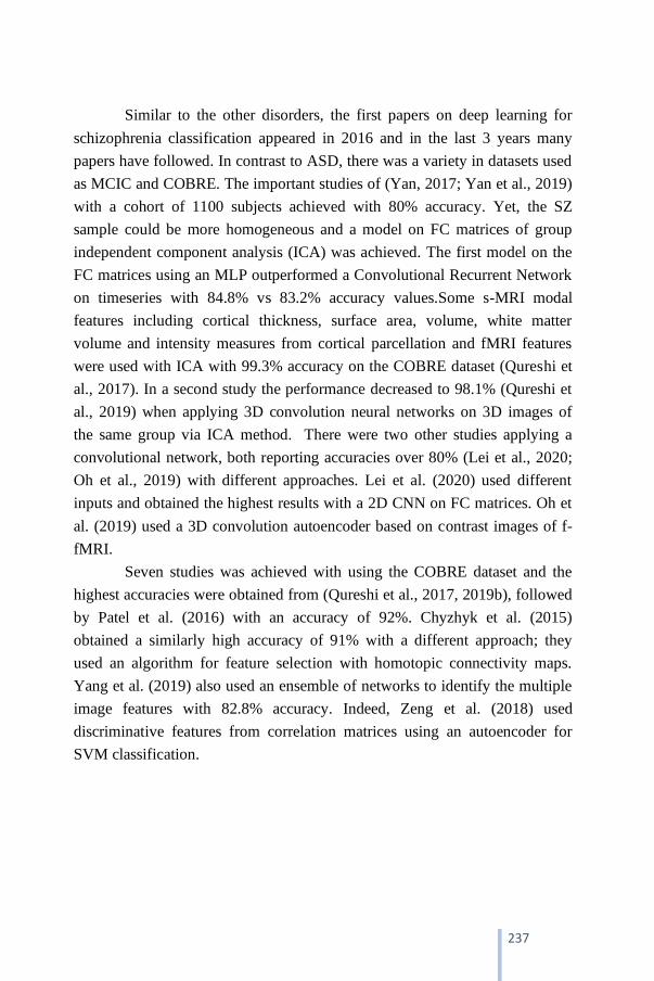
237
Similar to the other disorders, the first papers on deep learning for
schizophrenia classification appeared in 2016 and in the last 3 years many
papers have followed. In contrast to ASD, there was a variety in datasets used
as MCIC and COBRE. The important studies of (Yan, 2017; Yan et al., 2019)
with a cohort of 1100 subjects achieved with 80% accuracy. Yet, the SZ
sample could be more homogeneous and a model on FC matrices of group
independent component analysis (ICA) was achieved. The first model on the
FC matrices using an MLP outperformed a Convolutional Recurrent Network
on timeseries with 84.8% vs 83.2% accuracy values.Some s-MRI modal
features including cortical thickness, surface area, volume, white matter
volume and intensity measures from cortical parcellation and fMRI features
were used with ICA with 99.3% accuracy on the COBRE dataset (Qureshi et
al., 2017). In a second study the performance decreased to 98.1% (Qureshi et
al., 2019) when applying 3D convolution neural networks on 3D images of
the same group via ICA method. There were two other studies applying a
convolutional network, both reporting accuracies over 80% (Lei et al., 2020;
Oh et al., 2019) with different approaches. Lei et al. (2020) used different
inputs and obtained the highest results with a 2D CNN on FC matrices. Oh et
al. (2019) used a 3D convolution autoencoder based on contrast images of f-
fMRI.
Seven studies was achieved with using the COBRE dataset and the
highest accuracies were obtained from (Qureshi et al., 2017, 2019b), followed
by Patel et al. (2016) with an accuracy of 92%. Chyzhyk et al. (2015)
obtained a similarly high accuracy of 91% with a different approach; they
used an algorithm for feature selection with homotopic connectivity maps.
Yang et al. (2019) also used an ensemble of networks to identify the multiple
image features with 82.8% accuracy. Indeed, Zeng et al. (2018) used
discriminative features from correlation matrices using an autoencoder for
SVM classification.

238 NEW HORIZONS IN THE HEALTH SCIENCES
1.3. Review Of Attention Deficit Hyperactivity Disorder
(ADHD)
Attention deficit/hyperactivity disorder (ADHD) is among the most
commonly diagnosed neurodevelopmental disorders, affecting approximately
8-12% of children worldwide, and up to 65% continue to experience ADHD
symptoms and neuropsychological disorders into adulthood.
The diagnosis of ADHD is typically made by a clinician based on the
number, severity, and duration of behavioral symptoms observed by
parents/caregivers and teachers. Currently, the diagnosis of ADHD is
characterized by age-inappropriate inattention and/or hyperactivity-
impulsivity symptoms, categorized into three presentations: predominantly
inattentive, predominantly hyperactive/impulsive, and combined presentation
ADHD is often accompanied by comorbid disorders such as
oppositional defiant disorder and conduct disorder. At the neuropsychological
level, children with ADHD are known to perform worse than their typically
developing peers in measures of reaction time variability,
intelligence/achievement, alertness, working memory, and response
inhibition.
Several major neural networks have been associated with ADHD,
including the default mode network (DMN), dorsal and ventral attention
networks, salience networks, and frontostriatal and mesocorticolimbic circuits
(or dopaminergic mesolimbic system), although neuroimaging findings are
not yet conclusive in diagnosis. Functional neuroimaging studies have shown
reduced connectivity in the DMN of children aged 6-17 years with ADHD, a
pattern suggestive of delayed DMN neuromaturation, consistent with previous
structural MRI studies thought to indicate delayed maturation of the cerebral
cortex.
Early recognition and treatment of ADHD and its comorbidities have
the potential to alter the course of psychiatric morbidity later in life.Children
with ADHD have a high risk of accompanying depression, anxiety disorders,
and substance use disorders in adult life if they are not properly diagnosed and
treated.
239
Currently, there is no neural biomarker that can be used in the
diagnosis, but neuroimaging studies aimed to characterize the structural and
functional brain correlates of ADHD, and it is thought that this will help
clinicians.
Some studies were given for ADHD classification. Three studies have
achieved classification of the ADHD with accuracies ranging from 27 to 65
(for 4 different groups). The highest performance was reported by Hao et al.
(2015) that achieved 64.7% via Bayesian network on rs-fMRI data. For
bivariate classification of ADHD the highest accuracy was reported by
Deshpande et al. (2015). They used a fully connected cascade neural network
and obtained 90% accuracy.
Wang et al. (2019) applied 3D convolutions and obtained an accuracy
of 77.6%. They also tested their model on SZ data and obtained an accuracy
of 82.2%. Sen et al. (2018) achieved the model that was tested on two
psychiatric disorders; they developed an autoencoder to learn features from s-
MRI and ICA from fMRI. These learned features were classified by an SVM
classifier and obtain accuricies of 68% and 63%.
Three other studies used convolutions for classification (Wang and
Kamata, 2019; Zou et al., 2017), all with different inputs: AAL timeseries
(Riaz, et al., 2018), a combination of ReHo, fALFF and VHMC (Zou et al.,
2017) or 3D structural maps 68.
2.REVIEW OF DEEP LEARNING AND COMMON
MACHINE LEARNING STUDIES
In this review some important studies were analyzed and some DL
models against standard machine learning methods were compared. The
results of these studies are given as a graph form in Fig. 1.
Among the higher performance metrics of DL, only three studies had
lower performance for DL (Lei et al., 2020; Miholca and Onicas , 2017;
Vyskovsky et al., 2019): Lei et al. (2020) compared many different models of
SVM. In Vyskovsky et al. (2019) was achieved an ensemble of MLPs with
ensemble SVMs for schizophrenia classification. Finally, in Miholca and
Onicas (2017) a new ML technique was achieved and better accuracy score
was obtained than a MLP.
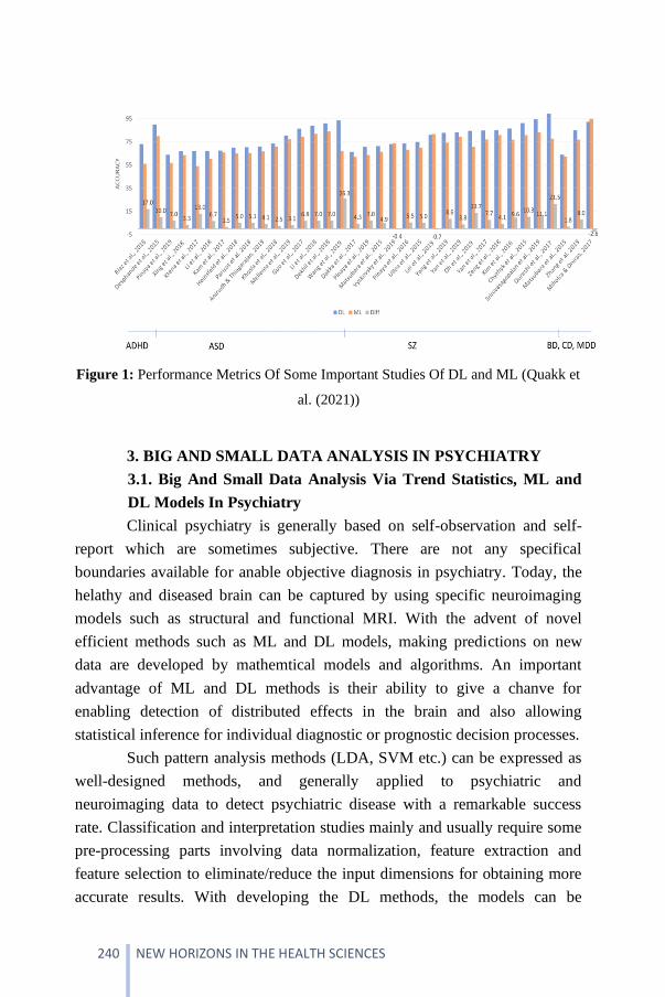
240 NEW HORIZONS IN THE HEALTH SCIENCES
Figure 1: Performance Metrics Of Some Important Studies Of DL and ML (Quakk et
al. (2021))
3. BIG AND SMALL DATA ANALYSIS IN PSYCHIATRY
3.1. Big And Small Data Analysis Via Trend Statistics, ML and
DL Models In Psychiatry
Clinical psychiatry is generally based on self-observation and self-
report which are sometimes subjective. There are not any specifical
boundaries available for anable objective diagnosis in psychiatry. Today, the
helathy and diseased brain can be captured by using specific neuroimaging
models such as structural and functional MRI. With the advent of novel
efficient methods such as ML and DL models, making predictions on new
data are developed by mathemtical models and algorithms. An important
advantage of ML and DL methods is their ability to give a chanve for
enabling detection of distributed effects in the brain and also allowing
statistical inference for individual diagnostic or prognostic decision processes.
Such pattern analysis methods (LDA, SVM etc.) can be expressed as
well-designed methods, and generally applied to psychiatric and
neuroimaging data to detect psychiatric disease with a remarkable success
rate. Classification and interpretation studies mainly and usually require some
pre-processing parts involving data normalization, feature extraction and
feature selection to eliminate/reduce the input dimensions for obtaining more
accurate results. With developing the DL methods, the models can be
241
explained as specific models that achieve higher complexity by using
nonlinear transformations. This specific ability makes DL more suitable for
neuroimaging such as psychiatric disorders/problems are sometimes
characterised by different complex patterns. Indeed, an important difference
between ML and DL is that learning process of optimal feature representation
from the raw data is achieved in DL. The whole results are more objective and
less bias susceptible process in DL.
However, there are different techniques and processing steps for
analyzing psychiatric disorders within DL studies. A majör challange of DL in
this area is the high dimensionalty of data. Variety of studies showed that
using hand crafted input features with different feature extraction steps to
reduce the input dimensionality.
Given the popularity of ML and DL methods within the psychiatry
and some approaches, this review aims to give an ultimate overview of
methods and models that have applied ML and DL to neuroimaging data fort
he classification of psychiatric disorders.
Such methods of imaging are widely chosen in laboratories of
experimental psychology and for research in neuroscience. The
Electroencephalography (EEG) is much cheaper and common method than
other methods. Indeed, this method can be defined as a practically non-
invasive technique. EEG and its methods have been developed for many
years. Recently, there has been an advance in the use of Brain-Computer
interfaces in which the acquisition of electrical activity of the specifically
selected areas of brain cortex. Today, development of the neurocomputing, AI
and its subbranches have gained more popularity. The computational
approach can express some behaviour characteristic of complex systems.
In current psychiatry, the common tool for diagnosing is still the
clinical interview progress. This process allows psychiatristt to choose the
optimal method of treatment. There are variety of EEG signal representation,
but one of the oldest method is its characteristics in frequency bands that
describes the case of its waves. Indeed, there are some different bands that in
the literature (δ—delta band (< 4 Hz), θ—theta (4–7 Hz), α—alpha (8–15
Hz), β—beta (16– 31 Hz), γ —gamma (> 31 Hz) and sometimes μ—mu (8–12
Hz).
242 NEW HORIZONS IN THE HEALTH SCIENCES
In general, neuroimaging data classification may not be an easy try.
Accordingly, Deep Neural Networks (DNNs) have shown important results in
detecting psychiatric disorders. Most studies have focused on investigating
dementia and attention deficit hyperactivity disorder with using large
publically available neuroimaging data sets (OASIS, ADHD-200 databases
etc.). Notably, a few of these studies also investigated the ability to identify
Alzheimer’s disease (AD), which is essential to detect disease at an early
stage. Studies classifying other mental disorders such as schizophrenia,
autism, Parkinson’s disease, depression, substance abuse disorder, and
epilepsy are slowly investigating as well.
Sometimes psychiatric symtomps cause from multiple processes and
also more traditional classifiers like Suuport Vector Machines (SVMs) and
Linear Discriminant Analysis (LDA) have been fed with multiple features
with modalities in the studies. However, it is too early to say how DL model
and data modality affect classification performance.
According to the low sample size issues and multi-modal integration,
the data is usually used as high-dimensional data in neurostudies. For a single
imaging modality, current sample sizes are in the modest range of 102–104.
Training models in DL with some parameters on small sample sizes create a
severe challenge to find solutions fort he population.
Apart from the customized SVM and other classifiers, Deep Learning
technologies, which are increasingly popular, have been used as another step
in the classification approach and this technology has been followed in
software development. For Deep Learning methods, a specific n-layer
convolutional neural network can be prepared and based on the parameters
mentioned above, the psychiatric disease can be estimated and interpreted as a
preliminary diagnosis. One advantage of Deep Learning methods is that they
increase interface rendering rate by minimizing the attribution data extraction
step for audience regions.
For instance, Convolutional Neural Networks (CNNs) which are
particularly suited for processing raw imaging data eliminate the number of
parameters through “weight sharing” and pooling of unit responses. Trials
indicate that CNNs could be achieved with different architectures when
predicting mental health related aspects from raw imaging data. Other studies
have reduced the dimensionality of the input data. While some approaches

243
may be less effective for clinical applications, they obtain acceptable results
for small samples (of n < 100).
CNN is a deep learning model that has continued and evolved over
the past decade. The CNN structure can automatically learn features from the
input dataset, especially images. Convolutional layers are used to bend the
input image with weights to obtain a feature map. The weights of the kernels
can link the feature map units to the previous layer. Convolutional Neural
Networks consist of neurons with learnable weights and pre-states. Each
neuron takes some input, performs an inner product, and optionally follows it
non-linearly.
Figure 2. A Common CNN Architecture
Convolution is the first layer that extracts features from an input
image. Convolution preserves the relationship between pixels by learning
image features using small squares input data. It is a mathematical operation
that takes two inputs, such as an image matrix and a filter or kernel.
The pooling layers section reduces the number of parameters when
images are very large. Spatial pooling is also called downsampling and
reduces the dimensionality of each map but preserves important information.
Spatial pooling can be of different types:
• Maximum Pooling
• Average Pooling
• Total Docking

244 NEW HORIZONS IN THE HEALTH SCIENCES
Maximum pooling takes the largest item from the adjusted feature
map. Getting the largest element can also take the average pooling. The sum
of all items in the feature map is called the aggregate pooling.
According to the diagram given above, the process was followed by
applying the special pooling method covering the maximum, average and total
pooling variations. In this approach, the mean pooling method was chosen and
applied due to its effectiveness. Average pooling generally reduced the
dimensions of the feature map while keeping the most significant feature
values by shifting the kernels on the corrected map and capturing the mean
values. Pooling has been implemented to make the data highly manageable
with size reduction.
In the fully connected layer, the matrix is flattened to vector and fed
into a fully connected layer, such as a neural network, where the feature map
matrix will be converted into vector (x1, x2, x3, etc.). In the last part, there is
the activation function.
There are mainly two types of CNN based structures that are
frequently used in the MATLAB environment: AlexNet and Visual Geometry
Group (VGG) were chosen for the classification and interpretation stages. The
AlexNet model was chosen because of its useful modification situations and
its ability to finish faster than other simple models.
Typically, Deep Learning is a sub-branch of machine learning like the
human brain. In recent years, Deep Learning field and methods have gained
popularity in many fields of study. The use of Artificial Intelligence and Deep
Learning is very common in medicine, and with their use, medical data can
become more meaningful to scientists and doctors.
There are many studies on Deep Learning in the literature. On the
contrary, the biggest problem in analyzing medical dataset is that there is a
limited number of datasets available to doctors. Unlike Artificial Intelligence,
Deep Learning methods and models require a particularly large amount of
data. Labeling these data one by one is very laborious and time consuming.
The biggest alternative of Transfer Learning to using Deep Learning models
is that it allows the training process with less data.
With new versions and versions of MATLAB, AlexNet has become
increasingly important and it is an improved version of the KSA model.
AlexNet has 5 convolutional and 3 fully connected layers. In addition, it is
245
also referred to as the model that used the Redirected Activation Function
(ReLU) for the first time.
Additionally, the ResNet-50 model is a more advanced version of
CNN and AlexNet. When the network deepens and grows; this model avoids
diffuse distortion [51]. ResNet solves this problem by not only connecting the
previous layer to the current layer, but also connecting the layer behind the
previous layer. By including this, each layer can now see more than the
previous layer's observations. In this model, the process connects the input
layer to a 2D convolution (Conv2D) layer with specified filters and then adds
a BatchNormalization layer to which a Rectified Activation (ReLU) layer is
added and then another Conv2D layer is stacked. This stacked output is then
added to the first input, but the first input is transformed by passing it through
a Conv2D layer of the given filters. This step is done to match the output sizes
of both layers to be added. Then, having a MaxPooling2D layer, that layer is
added, and if any Dropout value is also given, a Dropout layer is also added to
the network, and finally a BatchNormalization layer and Activation layer are
added. Overall, ResNet50 is a complex 50-layer network trained on an
ImageNet database. The models input has an image size of 255x255 (x3) and
the output is mainly disease classes.
As given in previous section, psychiatric researches need to achieve a
variety of challanges, including the identification for robust diagnosis and the
interpretation of disease types of patients.
For a classification problem, predicting responses or symptom
severity or to distinguish problems between diagnosed with psychiatric
disorders and healthy individuals. Fundamentally, the input variables come
from these situations given above and the maight come from several
modalities. If we assume that the outputs are class labels (clinical diagnoses),
this is called a regression/classification problem and also a training-set.
Generally, when the model training or in statistical terms, models
estimation/interpretation, such a loss function can be. Instead of this, the
Mean-Squared Error (MSE), the average sum of squared deviations between
true phenomenon and predicted results etc. In supervised case, a loss function
may be type of measure that defines the structure in the data.
Moreover, when we investigate similarities or differences between
Hypothesis testing versus Prediction parts, there are not any differences
246 NEW HORIZONS IN THE HEALTH SCIENCES
between statistical models and ML&DL. The main discrimination between
hypothesis testing and prediction consists from important cases. While
common statistics has been more concerned with testing, ML or DL softwares
have been more interested in the prediction/interpretation processes.
In classical hypothesis testing part, a probabilistic situation of the data
has been evaluated and sometimes this has been formulated in term of the
parameters of the model and the main purpose is to obtain a probability for
how likely a case which is related to the hypothesis holds in the whole
population according to the given the model assumptions (finite or infinite).
Other important issue can be defined as the stated models may be
probabilistic or deterministic. For hypothesis testing progress, there may be
some random variables and probability distributions involved. Sometimes, the
relationships between input and output variables are formulated with the
probability functions. According to the modern ML and DL models, they can
provide a hybrid sense of the uncertainty related to the predictions. However,
this makes formal testing is fundamental, these models given above and their
probability distributions are generally difficult to handle.
3.2. Big And Small Data Analysis Via Trend AI Models In
Psychiatry
After image processing, generally, pattern recognition and
binarization processes, Feature Extraction has begun and the features have
been used as the inputs in AI models. For this part, generally, Feature
Extraction has been achieved via graycomatrix, grayprops, Wavelet
Transform etc.
In the part of identification and interpretation of data, firstly, a two-
stage cyclical system is taken as reference in the interpretation of
psychological signals. Feature extraction step includes obtaining numerical
data for the problem regions determined according to medical and
image/signal processing regions (Contrast, Correlation, Homogeneity,
Variance, Energy, Entropy, Mean, Standard Deviation, etc.) and analyzing
parametric variables. The output of the feature extraction step is a numeric
matrix containing the features listed above.
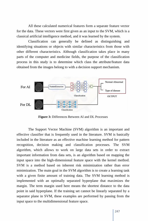
247
All these calculated numerical features form a separate feature vector
for the data. These vectors were first given as an input to the SVM, which is a
classical artificial intelligence method, and it was learned by the system.
Classification can generally be defined as distinguishing and
identifying situations or objects with similar characteristics from those with
other different characteristics. Although classification takes place in many
parts of the computer and medicine fields, the purpose of the classification
process in this study is to determine which class the attribute/feature data
obtained from the images belong to with a decision support mechanism.
Figure 3: Differences Between AI and DL Processes
The Support Vector Machine (SVM) algorithm is an important and
effective classifier that is frequently used in the literature. SVM is basically
included in the literature as an effective machine learning method for pattern
recognition, decision making and classification processes. The SVM
algorithm, which allows to work on large data sets in order to extract
important information from data sets, is an algorithm based on mapping the
input space into the high-dimensional feature space with the kernel method.
SVM is a method based on inherent risk minimization rather than error
minimization. The main goal in the SVM algorithm is to create a learning task
with a given finite amount of training data. The SVM learning method is
implemented with an optimally separated hyperplane that maximizes the
margin. The term margin used here means the shortest distance to the data
point in said hyperplane. If the training set cannot be linearly separated by a
separator plane in SVM, these examples are performed by passing from the
input space to the multidimensional feature space.
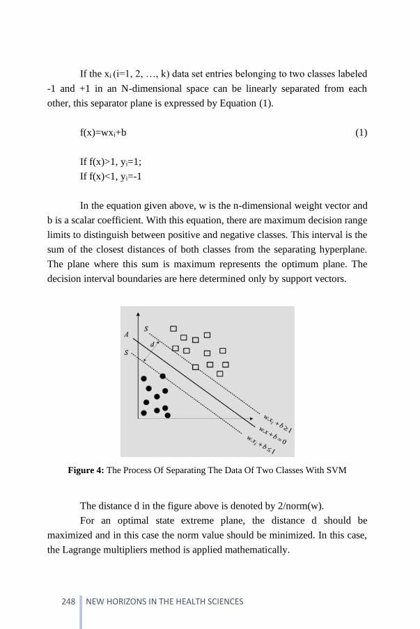
248 NEW HORIZONS IN THE HEALTH SCIENCES
If the xi (i=1, 2, …, k) data set entries belonging to two classes labeled
-1 and +1 in an N-dimensional space can be linearly separated from each
other, this separator plane is expressed by Equation (1).
f(x)=wxi+b (1)
If f(x)>1, yi=1;
If f(x)<1, yi=-1
In the equation given above, w is the n-dimensional weight vector and
b is a scalar coefficient. With this equation, there are maximum decision range
limits to distinguish between positive and negative classes. This interval is the
sum of the closest distances of both classes from the separating hyperplane.
The plane where this sum is maximum represents the optimum plane. The
decision interval boundaries are here determined only by support vectors.
Figure 4: The Process Of Separating The Data Of Two Classes With SVM
The distance d in the figure above is denoted by 2/norm(w).
For an optimal state extreme plane, the distance d should be
maximized and in this case the norm value should be minimized. In this case,
the Lagrange multipliers method is applied mathematically.
249
Indeed, k-Nearest-Neighbour classifier is basically a non-parametric
learning algorithm (no assumptions are made on the data distribution). Many
cases in theory and in the literature do not fit the Gaussian and linearly
separable case. In addition, this algorithm does not use the points of the
training data to introduce a generalization case. The training phase is a highly
unlikely situation.
The k-NN is used to perform a basic classification based on the
distance, and the mentioned neighborhood determination is based on the
minimum distance from the test samples to the training samples. The class of
test samples is determined by the majority of the nearest neighbors. In this
algorithm, it is based on deciding that the class of test samples will be the
same as the nearest neighbor. The situation that the data observed close to
each other will have the same class is seen as a highly compatible choice.
Commonly, Euclidean distance is used in this algorithm as the
measure distance. Euclidean distance in N-dimensional space is given in
Equation (2).
𝑑(𝑎, 𝑏) = √∑ (𝑎𝑖 − 𝑏𝑖)2𝑁𝑖=1 (2)
In this equation, pi, qi and i are coordinates, and the k parameter
should be chosen very carefully in classification with this algorithm. It is not
clear which one will be used and work best at distance based on learning.
For each type of disease, signals of patients were used in the
classification stage with the k-NN mentioned above. As required by the k-NN
structure, the attribute matrices of features of the disease types in a network
cluster and their related names were recorded as tags, and the interpretation of
a population detected in a different test image uploaded to the system was
made according to these data.
AdaBoost, short for Adaptive Boosting, is a machine learning meta-
algorithm formulated by Yoav Freund and Robert Schapire, whose work won
the 2003 Gödel Prize. AdaBoost is a collective learning method (also known
as "meta-learning") that was originally created to increase the efficiency of
binary classifiers. AdaBoost uses an iterative approach to learn from the

250 NEW HORIZONS IN THE HEALTH SCIENCES
mistakes of weak classifiers and transform them into strong ones. Every
learning algorithm tends to suit some types of problems better than others and
typically has many different parameters and configurations to adjust before
reaching optimum performance on a dataset. In general, Adaboost is referred
to as the best classifier. When used with decision tree learning, information
about the relative 'hardness' of each training sample collected at each stage of
the AdaBoost algorithm is fed into the tree-growing algorithm, so later trees
tend to focus on examples that are more difficult to classify.
AdaBoost refers to a specific method of training a powered classifier
and its classifier form is given in Equation (3).
𝑓(𝑥) = ∑ 𝑓𝑖(𝑥)𝑇𝑡=1 (3)
𝑥1 ∈ 𝑅𝑛, 𝑦1 ∈ {−1, 1}
Here, n is the size of the real numbers or the number of features in the
dataset. “x” is the set of data points. “y” is the target variable that is -1 or 1.
AdaBoost assigns weights to each training sample to determine its
importance in the training dataset. When the assigned weights are high, this
set of training data points is likely to have more say in the training set.
𝑤 =1
𝑁∈ [0, 1] (4)
In Equation (4), N is the total number of data points. The sum of the
weighted samples is always 1, so the value of each weight will always be
between 0 and 1. After that, the calculation of the actual effect on the
classification of the data points for this classifier is provided by Equation (5).
𝛼 =1
2𝑙𝑛
1−𝑡𝑜𝑝𝑙𝑎𝑚 ℎ𝑎𝑡𝑎
𝑡𝑜𝑝𝑙𝑎𝑚 ℎ𝑎𝑡𝑎 (5)
After entering the actual values of the total error for each data, the
process of updating the sample weights, which we initially took as 1/N for
each data point, is performed with Equation (6).
251
𝑤 = 𝑤𝑖−1 ∗ 𝑒±𝑎 (6)
Two states (positive or negative) for alpha indicate:
Alpha is positive when the predicted and actual output match
(meaning the sample has been correctly classified).
Alpha is negative when the predicted output does not match the actual
class (meaning the sample was misclassified). In this case, it is necessary to
increase the sample weight for achieve-ing the misclassification.
In the above-mentioned Adaboost classification stage, signal of
psychiatric patients were used for each type of disease. As required by the
structure of the Adaboost algorithm, the attribute matrices of defined
attributes of the disease types in a network cluster and their related names
were recorded as labels, and the interpretation of a disease detected in a
different test signals/images uploaded to the system was made according to
these data.
4. TRAINING ALGORITHM OF MODELS
There are some steps that DL models can take to improve finding an
acceptable solution. The first main progress can be defined as initialization.
Then the next progress is called model/parameter selection. In this step, user
of DL has to be alter parameters to obtain a better model. Indeed, the loss
function is gradually adaptized or modified after training process. For this
case, model complexity and sample size are crucial fort he parts given above.
The most aspects of a training part are the scaling with data size and
parameters and steps for taking to escape local minima value and dealing with
regions of slope in the loss function. According to the literature, the
popular/trend approach of DNNs is stochastic gradient descent (SGD) via
moving a local minimum of a function.
Moreoever, there are some extra ways to alleviate on the model side
and improve solutions that obtain better results. One of these is reducing the
dimensionality of the raw data or using the data adter pre-processing step.
However, there may be cons of the preprocessing and feature selection parts,
selection according to domain knowledge would be important and highly

252 NEW HORIZONS IN THE HEALTH SCIENCES
predictive. For achieving this dilemma, DL models can be used to find useful
data representations and features.
Sometimes, reducing the input dimension may not give better results,
so we can also increase the sample size and variation within the sample. This
approach is called data augmentation. For the data set there are several ways
to do this; rotations, translations, rescaling, flipping, adding noise to data etc.
To sum up, the sample size needed to succesfully train a AI, ML or DL model
will depend on some factors such as type of data, size of network model, the
of stochasticity in the data set, dimension parameters of the feature space,
regularization cases etc.
5. CONCLUSION
Accurate diagnosis of psychiatric disorders is crucial for initiation and
case of effective treatment. This review confirms that AI, ML and DL on
neuroimaging are promising tools for development of biological diagnostic
models for diagnosis. Despite still in its early stages, the application of DL in
neuroimaging for psychiatric disorders has given important results.
Moreoever, several improvements are needed before the full potential of DL
in psychiatric neuroimaging can be achieved.
In conclusion, neuroimaging research in psychiatry using AI, ML and
DL is still evolving to achieve better performance. While there are important
challenges to overcome, our findings provide preliminary evidence supporting
the promising role of DL in the future development for psychiatric disorders.

253
REFERENCES
Aghdam, M.A., Sharifi, A., Pedram, M.M. (2019). Diagnosis of Autism Spectrum
Disorders in Young Children Based on Resting-State Functional Magnetic
Resonance Imaging Data Using Convolutional Neural Networks. J Digit
Imaging, 32 (6), 899–918.
Albajara Sáenz, A., Villemonteix, T., & Massat, I. (2019). Structural and functional
neuroimaging in attention‐deficit/hyperactivity disorder. Developmental
Medicine & Child Neurology, 61(4), 399-405.
Bourgeron, T. (2016). Current knowledge on the genetics of autism and propositions
for future research. Comptes rendus biologies, 339(7-8), 300-307.
Chyzhyk, D., Savio, A., Gran a, M. (2015). Computer aided diagnosis of
schizophrenia on resting state fMRI data by ensembles of ELM. Neural
Networks, 68, 23–33.
Correll, C. U., & Schooler, N. R. (2020). Negative symptoms in schizophrenia: a
review and clinical guide for recognition, assessment, and
treatment. Neuropsychiatric disease and treatment, 16, 519.
Deshpande, G., Wang, P., Rangaprakash, D., Wilamowski, B. (2015). Fully
Connected Cascade Artificial Neural Network Architecture for Attention
Deficit Hyperactivity Disorder Classification From Functional Magnetic
Resonance Imaging Data. IEEE Trans. Cybern, 45, 2668–2679.
Durstewitz, D. (2017). Advanced data analysis in neuroscience: integrating statistical
and computational models, NY, USA: Springer.
Durstewitz, D., Koppe, G., Toutounji, H. (2016). Computational models as statistical
tools. Curr Opin Behav Sci, 11:93–99.
Edition, F. (2013). Diagnostic and statistical manual of mental disorders. Am
Psychiatric Assoc, 21.
Huys, Q. J. M., Maia, T. V., Frank, M. J. (2016). Computational psychiatry as a
bridge from neuroscience to clinical applications. Nat Neurosci, 19:404–13.
Katzman, M. A., Bilkey, T. S., Chokka, P. R., Fallu, A., & Klassen, L. J. (2017).
Adult ADHD and comorbid disorders: clinical implications of a dimensional
approach. BMC psychiatry, 17(1), 1-15.
Keepers, G. A., Fochtmann, L. J., Anzia, J. M., et al. (2020). The American
Psychiatric Association practice guideline for the treatment of patients with
schizophrenia. American Journal of Psychiatry, 177(9), 868-872.
Khosla, M., Jamison, K., Kuceyeski, A., Sabuncu, M.R. (2019). Ensemble learning
with 3D convolutional neural networks for functional connectome-based
prediction. NeuroImage, 199, 651–662.

254 NEW HORIZONS IN THE HEALTH SCIENCES
Koppe, G., Meyer-Lindenberg, A., Dursewitz, D. (2021). Deep Learning for Small
and Big Data In Psychiatry, Neuropsychopharmacology Reviews, 46:176-
190.
Kraguljac, N. V., McDonald, W. M., Widge, A. S., Rodriguez, C. I., Tohen, M., &
Nemeroff, C. B. (2021). Neuroimaging biomarkers in
schizophrenia. American Journal of Psychiatry, appi-ajp.
Lei, D., et al. (2020). Detecting schizophrenia at the level of the individual: relative
diagnostic value of whole-brain images, connectome-wide functional
connectivity and graph-based metrics. Psychol. Med, 50 (11), 1852–1861.
Li X. et al. (2018). 2-Channel convolutional 3D deep neural network (2CC3D) for
fMRI analysis: ASD classification and feature learning. In: 2018 IEEE 15th
International Symposium on Biomedical Imaging (ISBI 2018), 1252–1255.
Li, G., Liu, M., Sun, Q., Shen, D., Wang, L. (2018). Early Diagnosis of Autism
Disease by Multi-channel CNNs. Mach. Learn. Med. İmaging (MLMI),
11046, 303–309.
Lord, C., Charman, T., Cusack, J., Guillaume, D., Frazier, et al. (2020). Autism
spectrum disorder (Primer). Nature Reviews: Disease Primers, 6(1).
Luo, Y., Weibman, D., Halperin, J. M., & Li, X. (2019). A review of heterogeneity in
attention deficit/hyperactivity disorder (ADHD). Frontiers in human
neuroscience, 13, 42.
Mellema, C., Treacher, A., Nguyen, K. & Montillo, A. (2019). Multiple Deep
Learning Architectures Achieve Superior Performance Diagnosing Autism
Spectrum Disorder Using Features Previously Extracted From Structural
And Functional MRI. in 2019 IEEE 16th International Symposium on
Biomedical Imaging (ISBI 2019).
Miholca, D., Onicas , A. (2017). Detecting depression from fMRI using relational
association rules and artificial neural networks. In: 2017 13th IEEE
International Conference on Intelligent Computer Communication and
Processing (ICCP),
pp. 85–92.
Oh, K., Kim, W., Shen, G., Piao, Y., Kang, N.-I., Oh, I.-S., Chung, Y.C. (2019).
Classification of schizophrenia and normal controls using 3D convolutional
neural network and outcome visualization. Schizophr. Res, 212, 186–195.
Owen, M. J., Sawa, A., & Mortensen, P. B. (2016). Schizophrenia. Lancet (London,
England), 388(10039), 86–97.
Patel, P., Aggarwal, P., Gupta, A. (2016). Classification of schizophrenia versus
normal subjects using deep learning. In: ACM International Conference
Proceeding Series (Association for Computing Machinery).

255
Pinaya, W.H., Mechelli, A., Sato, J. R. (2019). Using deep autoencoders to identify
abnormal brain structural patterns in neuropsychiatric disorders: A large
scale multi sample study. Hum Brain Mapp. 40:944–54.
Posner, J., Polanczyk, G. V., & Sonuga-Barke, E. (2020). Attention-deficit
hyperactivity disorder. The Lancet, 395(10222), 450-462.
Sen, B., Borle, N.C., Greiner, R., Brown, M.R.G., Bernhardt, B.C. (2018). A general
prediction model for the detection of ADHD and Autism using structural and
functional MRI. PLoS ONE 13, e0194856.
Ural, A. B. (2017). A Computer-Based Brain Tumor Detection Approach With Image
Processing And Probabilistic Neural Network. Journal of Medical and
Biological Engineering, 19 December 2017.
Ural, A. B., Özışık, P. & Hardalaç, F. (2019). An improved computer based diagnosis
system for early detection of abnormal lesions in the brain tissues with using
magnetic resonance and computerized tomography images, Multimed Tools
Appl (2019).
Ural, A. B., Özışık, P., Hardalaç, F. (2021). Early Diagnosis System For Common
Brain Masses, PhD Thesis, Gazi University, Electrical Electronics
Engineering, Biomedical.
Quakk, M., Mortel, L. van de, Thomas, R. M., Wingen, G. Van (2021). Deep
Learning Applications For the Classification Of Psychiatric Disorders Using
Neuroimaging Data: Systematic Review and Meta-analysis, NeuroImage:
Clinical, 30, 102584.
Qureshi, M.N.I., Oh, J., Cho, D., Jo, H.J., Lee, B. (2017). Multimodal discrimination
of schizophrenia using hybrid weighted feature concatenation of brain
functional connectivity and anatomical features with an extreme learning
machine. Front. Neuroinform, 11, 1–14.
Qureshi, M.N.I., Oh, J., Lee, B. (2019). 3D-CNN based discrimination of
schizophrenia using resting-state fMRI. Artif. Intell. Med, 98, 10–17.
Wang, T., Kamata, S. (2019). Classification of Structural MRI Images in Adhd Using
3D Fractal Dimension Complexity Map. In: 2019 IEEE International
Conference on Image Processing (ICIP), pp. 215–219.
Yan, W., et al. (2017). Discriminating schizophrenia from normal controls using
resting state functional network connectivity: A deep neural network and
layer-wise relevance propagation method. In: 2017 IEEE 27th International
Workshop on Machine Learning for Signal Processing (MLSP), pp. 1–6.
Wang, Z., Sun, Y., Shen, Q., Cao, L. (2019). Dilated 3D Convolutional Neural
Networks for Brain MRI Data Classification. IEEE Access 7, 134388–
134398.

256 NEW HORIZONS IN THE HEALTH SCIENCES
Yan, W., et al. (2019). Discriminating schizophrenia using recurrent neural network
applied on time courses of multi-site FMRI data. EBioMedicine, 47, 543–
552.
Zou, L., Zheng, J., Miao, C., Mckeown, M.J., Wang, Z.J. (2017). 3D CNN Based
Automatic Diagnosis of Attention Deficit Hyperactivity Disorder Using
Functional and Structural MRI. IEEE Access 5, 23626–23636.

ISBN: 978-625-8405-09-5
Copyright © 2022 FDOKUMEN